استهداف البلعميات المرتبطة بالأورام من نوع M2 هو نهج علاجي محتمل للتغلب على مقاومة الأدوية المضادة للأورام Targeting M2-like tumor-associated macrophages is a potential therapeutic approach to overcome antitumor drug resistance

المجلة: npj Precision Oncology، المجلد: 8، العدد: 1
DOI: https://doi.org/10.1038/s41698-024-00522-z
PMID: https://pubmed.ncbi.nlm.nih.gov/38341519
تاريخ النشر: 2024-02-10

استهداف البلعميات المرتبطة بالأورام من نوع M2 هو نهج علاجي محتمل للتغلب على مقاومة الأدوية المضادة للأورام

شوجينغ وانغ جينغروي وانغ تشيانغ تشين جيامين لو وي غوو لينغ لينغ صن وليزو لين

الملخص

تظهر مقاومة الأدوية في الأورام من تفاعل عاملين حاسمين: تباين الخلايا الورمية وطبيعة البيئة الدقيقة المثبطة للمناعة للأورام. تشكل البلعميات المرتبطة بالورم (TAMs) مكونات أساسية في البيئة الدقيقة للورم. تعتبر البلعميات من النوع M2 ضرورية في تسهيل انتشار الورم بالإضافة إلى تعزيز مقاومة الأورام للأدوية. تلخص هذه المراجعة الآليات التي تستخدمها البلعميات من النوع M2 لتعزيز مقاومة الأدوية في الأورام. كما نصف الاستراتيجيات العلاجية الناشئة التي تستهدف حاليًا البلعميات من النوع M2 بالتزامن مع أدوية مضادة للأورام أخرى، مع وجود بعضها لا يزال قيد التقييم في التجارب السريرية. علاوة على ذلك، نقوم بتلخيص وتحليل مختلف الأساليب الحالية لتطوير أدوية جديدة تستهدف البلعميات من النوع M2 للتغلب على مقاومة الأورام، مع تسليط الضوء على كيفية أن استهداف البلعميات من النوع M2 يمكن أن يوقف فعليًا نمو الورم وانتشاره ويتغلب على مقاومة الأدوية في الأورام.

كشفت بيانات عام 2020 عن حوالي 19.3 مليون حالة جديدة من الأورام وحوالي 10 ملايين وفاة مرتبطة بالأورام على مستوى العالم. وقد ارتفعت معدلات الإصابة والوفاة بسرعة على مستوى العالم. لا تزال العلاجات الدوائية هي العلاج السريري الأساسي للأورام الخبيثة. ومع ذلك، كانت قضايا النتائج العلاجية الضعيفة، وتقدم الورم، والتوقعات السيئة الناتجة عن مقاومة الأدوية دائمًا صعبة الحل. وبالتالي، أصبحت دراسة مقاومة الأدوية للأورام بارزة في الإدارة السريرية للأورام اليوم. وجدت الأبحاث المعاصرة أن مقاومة الأدوية للأورام تنبع من تفاعل عاملين حاسمين، وهما التباين داخل الورم والطبيعة المثبطة للمناعة للبيئة المجهرية للورم. يتكون بيئة الورم المجاورة (TME) من عناصر خلوية متنوعة، بما في ذلك اللمفاويات، والمصفوفة خارج الخلوية، وعوامل النمو، والسيتوكينات، والكيموكينات، واللمفاويات، وخلايا المناعة المرتبطة بالورم (TAMs)، والخلايا الشجرية، والخلايا القاتلة الطبيعية، وخلايا مثبطة مشتقة من النخاع. العناصر الأساسية في TME هي خلايا TAMs، التي تتكون من نوعين متضادين يمكن تمييزهما: خلايا TAMs من نوع M1 وخلايا TAMs من نوع M2. تعتبر خلايا TAMs من نوع M2 ضرورية للانتشار الخبيث، والغزو، ومقاومة العلاج. دورهم الكبير يبرز إمكانياتهم كأهداف للتغلب على مقاومة الأدوية للأورام. تقدم هذه المراجعة تلخيصًا للآليات التي تُعدل بها البلعميات الشبيهة بـ M2 لتجاوز مقاومة العلاج المضاد للأورام، بالإضافة إلى نظرة عامة على التجارب السريرية المستندة إلى هذه الأمور ذات الصلة.
آليات. كما نقوم بتحليل إمكانيات التطوير المستقبلية لهذه الاستراتيجية العلاجية الجديدة.

الميزة البيولوجية لخلايا المناعة المرتبطة بالورم
أصل TAMs

عندما تتفاعل خلايا الورم مع السائل الخلالي أثناء نموها، فإنها تخلق بيئة معينة تعزز من تكاثرها. تُعرف هذه البيئة باسم TME. TME هي شبكة متعددة الأوجه ومترابطة تحتوي على خلايا ومكونات متنوعة، بما في ذلك المصفوفة خارج الخلوية، اللمفاويات، البلعميات المرتبطة بالورم، الخلايا الشجرية، عوامل النمو، السيتوكينات، الكيموكينات، خلايا القاتل الطبيعي، وخلايا المثبطة المشتقة من النخاع (الشكل 1). تؤثر TME ليس فقط على التأثير العلاجي لعلاجات الورم الأولي ولكن أيضًا على تطور وتقدم انتشار الورم. . ضمن سوق إدارة التكنولوجيا، تبرز وحدات إدارة التكنولوجيا كأهم مكون، حيث تشكل حوالي وزن نسيج الورم . تنشأ TAMs من سلالتين مختلفتين، وهما الماكروفاجات المقيمة في الأنسجة والماكروفاجات المشتقة من وحيدات النواة. خلال المراحل الأولية من تطور الورم، تتجمع الماكروفاجات المقيمة في الأنسجة وتوزع حول نسيج الورم لتحفيز استجابات خلايا T التنظيمية، وتعزيز هروب خلايا الورم من المناعة، واستفزاز الانتقال الظهاري-الم mesenchymal
الشكل 1 | تعتبر الخلايا المناعية المرتبطة بالورم (TAMs) المكونات الأكثر أهمية في بيئة الورم (TME). تعزز الخلايا المناعية المرتبطة بالورم العديد من التقدمات الخبيثة للأورام من خلال تنظيم بيئة الورم، مثل تكاثر الورم، التهرب المناعي من قبل خلايا الورم، التحول الظهاري، غزو الورم، انتشار الورم، مقاومة الأدوية للورم. (بواسطة Figdraw).
(EMT) وزيادة تسلل الخلايا الورمية وانتشارها تجذب العديد من السيتوكينات والبروتينات الكيميائية، التي تنتجها خلايا الدم الجذعية المكونة للدم البالغة داخل الجهاز الدوري، البلعميات المشتقة من وحيدات النوى إلى البيئة المجهرية للورم. . بعد ذلك، تعزز العديد من المحفزات تمايز هذه البلعميات المشتقة من وحيدات النواة إلى ماكروفاجات مرتبطة بالورم (TAMs). إنترلوكين (IL) عامل الربط C-C (CCL) 2، عامل نمو البطانة الوعائية (VEGF)، وعامل مشتق من الخلايا stroma (SDF)- تنتج في الأورام تجذب البلعميات المؤيدة لتكوين الأوعية إلى أعضاء الورم. شميت وآخرون. أظهر أن تفاعل الإنتجرين- مع تالين وباكسيلين، عادة ما يتم تعزيزها بواسطة IL- و SDF- يمكن عرقلة الإشارات المستحثة من خلال الاستفادة من مضادات ضد الإنتغرين- بينما لم يتم توضيح التوقيت الدقيق لتحويل المونوسيتات المجندة إلى TAMs، هناك أدلة قوية تدعم التغيرات التي تحدث في الأنسجة في الملفات التعبيرية لهذه المونوسيتات المجندة. .

تنظيم اللدونة والاستقطاب للخلايا المناعية المرتبطة بالورم (TAMs) وتسميتها

في الواقع، توجد TAMs كاستمرارية مع مستويات عالية من اللدونة، ويمكن أن تتواجد أنواع تعبيرها غالبًا أو تتغير مع تقدم الورم. قام علي ن. شمس الدين وآخرون بتلخيص اللدونة والتنوع في TAMs واقترحوا مفهوم استقطاب TAM كاستقطاب مستمر وديناميكي. على الرغم من أن طبيعة وآلية وتسمية استقطاب TAM لا تزال بحاجة إلى مزيد من الاستكشاف، فإن الوصف الاصطناعي لـ TAMs الشبيهة بـ M1 و TAMs الشبيهة بـ M2 ، الواقعة في نهايات متباينة ضمن محور الاستقطاب الديناميكي المستمر لـ TAMs والمستمدة من تلخيص قائم على تجارب في المختبر، تم تأكيدها بالإجماع .
في الوقت الحاضر، فرضية مثبط الورم M1 – محفز الورم M2 التي اقترحها ألبرت مانتوفاني لا يزال النموذج الأكثر استخدامًا لدراسة تباين الخلايا المناعية المرتبطة بالأورام (TAM)، على الرغم من أنه يبسط بشكل مفرط النمط الظاهري الحقيقي لهذه الخلايا. ومع ذلك، كشفت الدراسات المتعمقة الأخيرة حول البلعميات عن اختلافات كبيرة في التصميم الهيكلي، والقدرات التشغيلية، والتعبير عن محددات السطح الخلوي بين البلعميات المقيمة في الأنسجة (TRMs) عبر مختلف الأعضاء. . علاوة على ذلك، يتم التعبير عن الجينات المرتبطة بملفات TAM الشبيهة بـ M1 و M2 في الوقت نفسه في جميع أنواع مجموعات الماكروفاج السرطانية تقريبًا. مؤخراً، تم استخدام تحليل استنتاج الشبكة التنظيمية على مستوى الخلية الواحدة والتجميع (SCENIC) ، وقد حدد الباحثون خمس مجموعات فرعية محددة من TAM في أنواع مختلفة من السرطان. تُسمى هذه المجموعات الفرعية TAMs HES1 وC1Q TAMs، TAMs TREM2
IL4I1 TAMs، وTAMs المتكاثرة. باستخدام الجينوميات أحادية الخلية، ما وآخرون. تم تصنيف TAMs إلى سبع مجموعات فرعية بناءً على الجينات المميزة، والمسارات الغنية، والوظائف المتوقعة. تُسمى هذه المجموعات الفرعية IFN-TAMs، Reg-TAMs، Inflam-TAMs، LA-TAMs، Angio-TAMs، RTM-TAMs، وProlif-TAMs.
على الرغم من أنه تم الاعتقاد أن البلعميات من النوع M2 لها وظائف مضادة للالتهابات بينما البلعميات من النوع M1 لها وظائف مؤيدة للالتهابات، إلا أن واقع سلوكها قد يكون أكثر تعقيدًا. خصائص وأدوار البلعميات التشغيلية قابلة للتغيير استجابةً للتغيرات البيئية، ويمكن أن تختلف أدوارها عبر حالات مرضية مختلفة. وبالتالي، بينما تميل الأبحاث الحالية إلى تقسيم البلعميات إلى فئات بناءً على أدوارها المتعلقة بالالتهابات، حيث تكون واحدة مؤيدة للالتهابات (غالبًا ما يشار إليها باسم M1) والأخرى مضادة للالتهابات (المعروفة عمومًا باسم M2)، قد لا يعكس هذا التقسيم الثنائي تنوعها البيولوجي بالكامل.

الأنماط الظاهرية ووظائف TAMs

توجد خلايا المناعة المرتبطة بالورم من نوع M1 وخلايا المناعة المرتبطة بالورم من نوع M2 في طرفي محور استقطاب خلايا المناعة المرتبطة بالورم الديناميكي المستمر، حيث تمتلك كل منهما علامات سطح خلوية وعوامل وظيفية فريدة، وتلعب أدوارًا مختلفة في بيئة الورم (الشكل 2).
تمتلك الخلايا البلعمية الشبيهة بـ M1 خصائص مؤيدة للالتهابات. يتم تحفيز الخلايا البلعمية الشبيهة بـ M1 بواسطة السيتوكينات مثل الإنترفيرون (IFN) وعامل تحفيز المستعمرات (CSF) وعامل نخر الورم (TNF). علاوة على ذلك، تمتلك الليبوسكريات (LPS) القدرة على التفاعل مع مستقبلات تول (TLR)4 الموجودة على سطح الخلايا البلعمية وتعزيز استقطاب الخلايا البلعمية الشبيهة بـ M1 من خلال التأثير على عامل النسخ كابا-B (NF-кB) وعامل تنظيم الإنترفيرون 3 (IRF3). من الخصائص البارزة للخلايا البلعمية الشبيهة بـ M1 تقديم المستضدات بشكل قوي وإفراز العديد من السيتوكينات المؤيدة للالتهابات. تفرز الخلايا البلعمية الشبيهة بـ M1 عددًا كبيرًا من الجزيئات المساعدة مثل مجموعة التمايز (CD)86 وCD60 وCD80، بالإضافة إلى المؤشرات الحيوية المؤيدة للالتهابات بما في ذلك TNF- IL-1 ، IL-6، IL-12، IL-23. كما أنها تعبر عن جزيئات معقد التوافق النسيجي الرئيسي (MHC) من النوع الثاني بشكل كبير. ومن الجدير بالذكر أنها تعبر عن IL-10، على الرغم من مستوى أقل. تقوم البلعميات الشبيهة بـ M1 بإفراز ميتالوبروتينازات المصفوفة (MMPs) بما في ذلك MMP1، MMP2، MMP7، MMP9، وMMP12. هذه الإنزيمات متخصصة في تكسير مكونات المصفوفة خارج الخلوية (ECM). تنتج البلعميات الشبيهة بـ M1 كيموكينات مثل CCL2، CCL3،
الشكل 2 | يتم عرض TAMs الشبيهة بـ M1 و M2 في
طرفي TAM الديناميكي المستمر
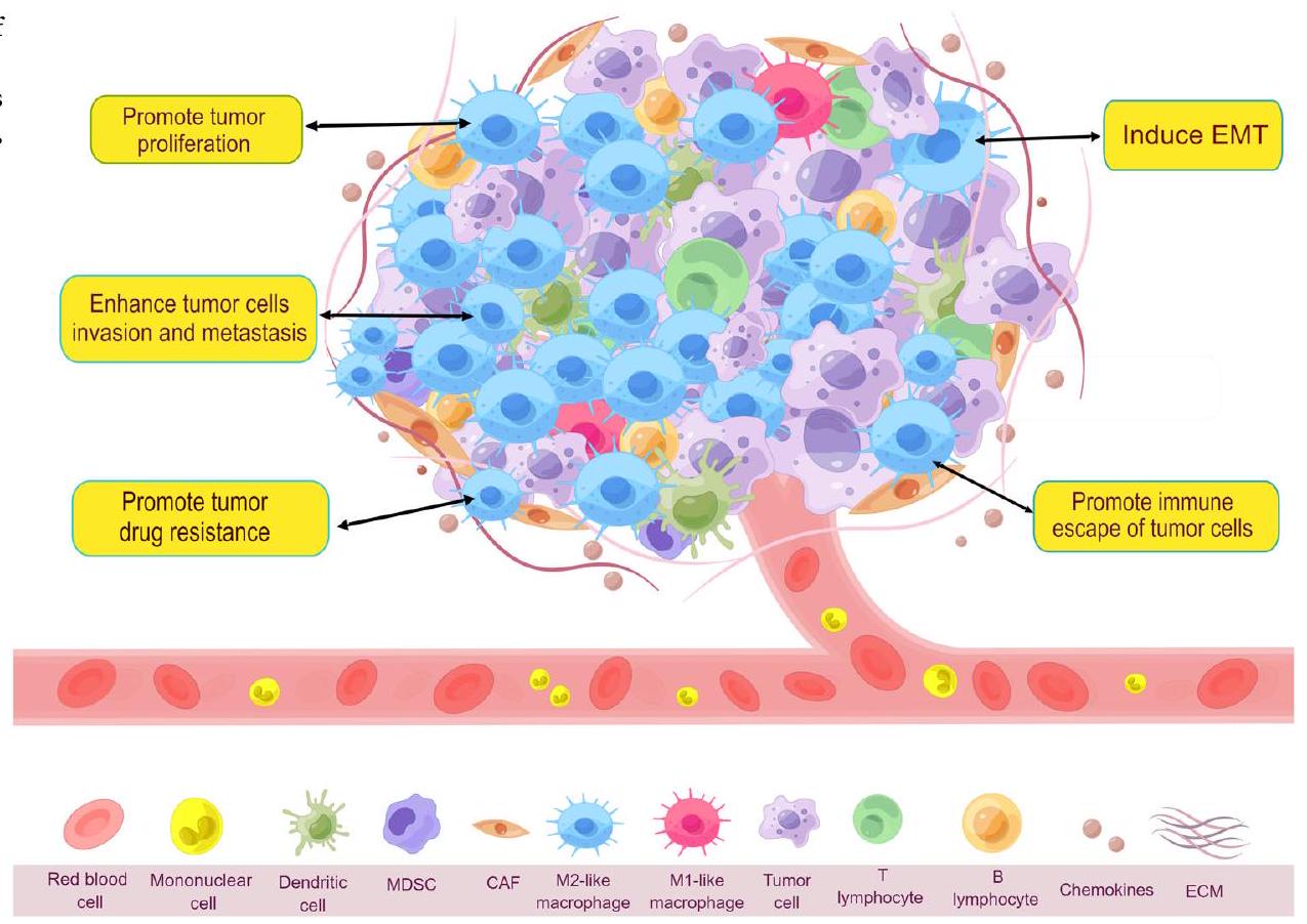
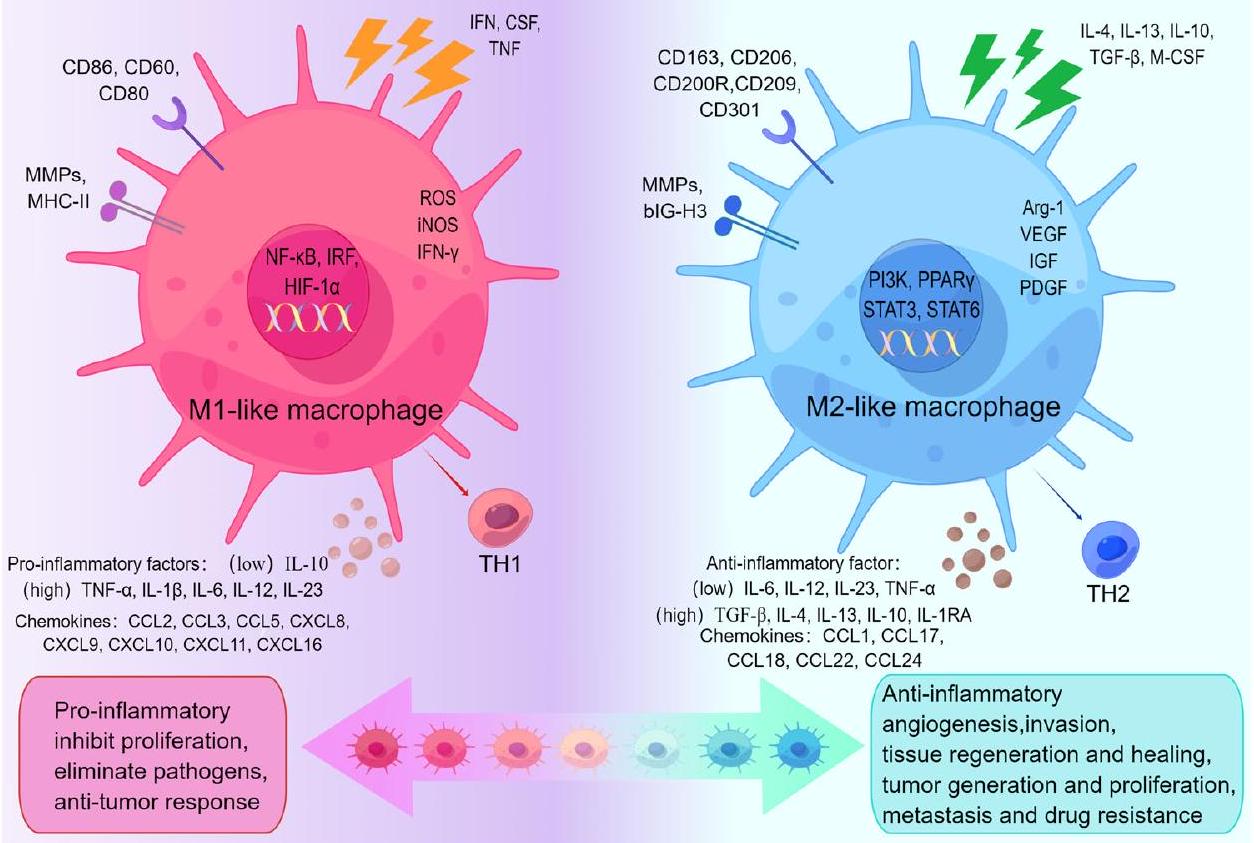
محور الاستقطاب، موضحًا أدوارها الفريدة في البيئة المجهرية للورم. كل من TAMs الشبيهة بـ M1 وTAMs الشبيهة بـ M2 لها علامات سطح خلوية محددة وعوامل وظيفية. تتولى TAMs الشبيهة بـ M1 في البيئة المجهرية للورم أدوارًا في تعزيز الالتهاب، تثبيط التكاثر، القضاء على مسببات الأمراض، والاستجابات المضادة للورم. بينما تشارك TAMs الشبيهة بـ M2 في الأنشطة المضادة للالتهابات، وتعزيز تكوين الأوعية الدموية، وتأثيرها على تجديد الأنسجة والشفاء، وتعزيز توليد الورم، وتكاثره، وانتشاره، ومقاومته للأدوية. (بواسطة Figdraw).
CCL5، كيموكين ليد (CXCL) 8، CXCL9، CXCL10، CXCL11، CXCL16. كما أنهم يطلقون IFN- إنزيم أكسيد النيتريك المحفز (iNOS) وأنواع الأكسجين التفاعلية (ROS). تقوم البلعميات الشبيهة بـ M1 بتنشيط استجابة مناعية قوية من نوع T-helper1 (Th1) من خلال إفراز هذه الوسائط الالتهابية لتعزيز استجابة التهابية تعيق تكاثر الخلايا وتقتل مسببات الأمراض وخلايا الورم في جسم الإنسان، مما يؤدي إلى تأثيرات مضادة للورم. .
تتميز الخلايا البلعمية من النوع M2، التي تقدم المستضدات بشكل ضعيف، بأنها مضادة للالتهابات، على عكس النمط M1. لا توجد إجراءات محددة يجب استخدامها لبدء تنشيط الخلايا البلعمية من النوع M2. يتم تنشيط الخلايا البلعمية من النوع M2 بشكل أساسي عن طريق تحفيز السيتوكينات، والتي تشمل عامل النمو المحول (TGF)- IL-4، IL-13، IL-10، وعامل تحفيز مستعمرات البلعميات (M-CSF) تعبّر البلعميات الشبيهة بـ M2 عن CD163 و CD206 و CD200R و CD209 و CD301 ومواد كيميائية مثل CCL1 و CCL17 و CCL18 و CCL22 و CCL24 بشكل كبير. تطلق العديد من العوامل المضادة للالتهابات، بما في ذلك TGF- “، IL-4، IL-13، IL-10، و IL1RA. بالإضافة إلى ذلك، تعبر TAMs الشبيهة بـ M2 عن السيتوكينات الالتهابية IL-6، IL-12، IL-23 و TNF- على المستويات الأدنى تعبّر البلعميات الشبيهة بـ M2 عن مستويات عالية من MMPs ومكونات ECM ذات التأثير الذاتي مثل الفيبروكتين وبيتا-إي إتش 3 (BIG-H3) وإنزيمات ربط ECM وبروتينات الربط العظمي. ، وبالتالي المشاركة في التصاق الخلايا. تعزز البلعميات الشبيهة بـ M2 تعبير الأرجيناز (Arg)-1 وVEGF، اللذان يشاركان في تخليق البروتين والبولي أمينات. يعزز البروتين بناء مصفوفة extracellular (ECM)، وتشارك البولي أمينات في تكاثر الخلايا. . عوامل أخرى تفرزها البلعميات الشبيهة بـ M2 التي تعزز تكاثر الخلايا، مثل عامل نمو الصفائح الدموية (PDGF) وعامل النمو الشبيه بالأنسولين (IGF)، تشارك في تكوين الأوعية الدموية تقوم البلعميات الشبيهة بـ M2 بإفراز مواد كيميائية مثبطة للمناعة لمنع نشاط المناعة من نوع Th1 وتعزيز نشاط المناعة من نوع Th2. تقلل هذه النشاطات من السيطرة على التفاعلات الالتهابية بينما تعزز نمو خلايا الورم، ومقاومة الأدوية، وتكوين الأوعية الدموية، وشفاء الأنسجة. .
سيكا وآخرون ملخص واقترح النظرية التي تفيد بأن الخلايا المناعية المرتبطة بالورم (TAMs) كانت مهيمنة من نوع M1 في المرحلة المبكرة من الورم. مع تطور الورم، شهدت البيئة المجهرية للورم (TME) تغييرات أدت تدريجياً إلى تحول الخلايا المناعية المرتبطة بالورم من نوع M1 إلى نوع M2. لذلك، فإن الخلايا المناعية المرتبطة بالورم في معظم الأورام تمثل مجموعة سكانية مهيمنة من نوع M2. البيئة المجهرية للورم في هذه الأورام تكون عمومًا مثبطة للمناعة. مؤثرًا على تقدم الورم ومقاومة العلاج الكيميائي.
علاوة على ذلك، استجابةً لمحفزات معينة وتغيرات في تعبير الجينات داخل البيئة المجهرية للورم، يمكن تصنيف البلعميات الشبيهة بـ M2 إلى عدة مجموعات فرعية، وهي M2a وM2b وM2c وM2d. . كل من المجموعات الفرعية تظهر خصائصها الفريدة (الجدول 1). البلعميات M2a تتلقى إشارات تنشيطها الأولية من السيتوكينات IL-4 و IL-13، التي تنتجها خلايا Th2. تساهم هذه البلعميات في العمليات المضادة للالتهابات من خلال إنتاجها لـ IL-10 و TGF- . علاوة على ذلك، فإنها تلعب دورًا حيويًا في الحساسية، شفاء الجروح، تكاثر الخلايا، والتليف. داخل البيئة المجهرية للورم، تساهم في تقدم الأورام من خلال تعزيز نمو وسلوك الخلايا السرطانية المتسللة وتكوين الأوعية الدموية. تنشيط البلعميات M2b يتم تحفيزه عمومًا من خلال تفاعل تآزري بين المجمعات التي تشمل الجهاز المناعي والروابط لمستقبلات Toll-like. ومن المعروف أنها تولد كمية كبيرة من السيتوكينات، بما في ذلك IL-10 و TNF- ، مما يعزز الاستجابات الالتهابية ويشارك في تنظيم المناعة. داخل بيئة الورم، تقوم البلعميات M2b النشطة بإفراز IL-6، الذي بدوره ينشط خلايا Th2، مما يؤدي إلى خلق بيئة مؤيدة للالتهابات تسهم في تقدم الورم. يحدث تنشيط البلعميات M2c بشكل أساسي استجابةً لـ IL-10 و TGF- ، أو الجلوكوكورتيكويدات. تساهم بشكل فعال في تعديل التفاعلات الالتهابية من خلال إفراز جزيئات مضادة للالتهابات. بالإضافة إلى ذلك، تلعب دورًا أساسيًا في تطوير التليف المرضي وشفاء الجروح. كما أن لديها وظائف تنظيمية مناعية، قادرة على التأثير على الاستجابات المناعية من خلال إفراز عوامل محددة والانخراط في تفاعلات مع خلايا مناعية أخرى. ماكروفاجات M2d تمثل نوعًا فرعيًا تم التعرف عليه مؤخرًا نسبيًا يتم تحفيزه بواسطة منبهات TLR و IL-6. ومن المعروف أنها تعدل البيئة الالتهابية المحلية من خلال إطلاق عوامل محددة تمتلك خصائص مؤيدة للالتهاب ومضادة للالتهاب. تدعم البلعميات M2d هروب خلايا الورم من المناعة عن طريق إفراز مواد مثبطة للمناعة مثل IL-10 و PD-1، مما يعيق الاستجابات المناعية الطبيعية. من خلال تشجيع نمو الورم وغزوه وتكوين الأوعية الدموية، تساعد في تقدم الأورام.

آليات تنظيم استقطاب البلعميات من نوع M1 / M2

أظهرت الدراسات الحالية أن العديد من السيتوكينات ومسارات الإشارة داخل البيئة المجهرية للورم تؤثر على استقطاب M1/M2. تشوهات الـ كيناز (IKK قد يؤدي مسار NF-кB إلى استقطاب TAM . العملية بواسطة
الجدول 1 | مقارنة بين العلامات والوظائف البيولوجية بين أنماط الماكروفاج المختلفة
الظواهر وظائف عوامل التحفيز علامات إخراج المراجع
M1 مقاومة الورم، مؤيد للالتهابات، تثبيط التكاثر، القضاء على مسببات الأمراض إنترفيرون- ، LPS ، GM-CSF ، TNF- iNOS، ROS IL-12 (مرتفع)/IL-10 (منخفض)، CD80، CD86، CD60، MMPs، MHCII CCL2، CCL3، CCL5، CXCL8، CXCL9، IL-1 IL-6، IL-23 32
M2a الحساسية، البروفيبروتيك، تكاثر الخلايا، مضاد الالتهاب، شفاء الجروح، تقدم الورم IL-4، IL-10، IL-13، (PPAR- ) CD11b، CD45، CD86، CD14، CD206، CD163، CD209، IL-1R، Dectin-1، YM1، RELM IGF1، DCIR، Stabilin 1، Factor XIII-A، Ly6C، TREM-2، DCSIGN، MHCII، Fizz1، Arg-1، YM1/2 TGF- IL-4، IL-10، VEGF، CCL1، CCL17، CCL18، CCL22، TNF- ، IGF، EPGF ٣٢-٣٥
M2b تنشيط Th2، تنظيم المناعة، الالتهاب، تقدم الورم IL-1 ، LPS، TLR CD163، CD14، CD86، MHC-II، IL-10 (مرتفع)، IL-12 (منخفض)، IL-6R TNF- IL-1، IL-6، IL-10، CCL1، SPHK1، LIGHT ٣٢،٣٥
M2c تنظيم المناعة، الاستجابة الالتهابية، شفاء الجروح، التليف IL-6، IL-10، الجلوكوكورتيكويدات، TGF- TNF- CD163، CD206، CD14، CD16، CD86، CXCR4، MerTK، TLR-1، TLR-8 IL-10، TGF- CCL16، CCL18، CXCL13 ٣٢،٣٥
M2d تكاثر الورم، الغزو، وتكوين الأوعية الدموية، قمع المناعة IL-6، TLR، ريجادينوسون، LPS IL-10 (مرتفع)، IL-12 (منخفض)، VEGF، TNF- (منخفض)، CD163، CD14، CD86 IL-10، IL-6، TGF- VEGF، CCL18، M-CSF 32
الذي يرتبط به IL-4 بمستقبله يمكن أن يعزز فسفرة ناقل الإشارة ومفعل النسخ (STAT)6، مما يؤدي إلى استقطاب البلعميات الشبيهة بـ M2 عبر مسار إشارة كيناز التيروزين جانوس (JAK)/ STAT6. في الوقت نفسه، يمكن أن يرتبط STAT6 المفسفر بعامل كروبل الشبيه بـ 4 (KLF)4 ومستقبل المنشط للبروكسيسوم. (PPAR- للترويج لهذه الاستقطابية عندما يتم حظر تفاعل IL-4 مع مستقبلاته المرتبطة، يتم تحفيز البلعميات الشبيهة بـ M2 للقطبية إلى بلعميات شبيهة بـ M1. . مجموعة متنوعة من الإشارات، بما في ذلك IL-4، TGF- ، IL-10، وبروتين العظام المورفوجيني-7 (BMP-7)، تعزز استقطاب M2 من خلال مسار إشارة كيناز الفوسفاتيديلينوزيتول 3 (PI3K)/ كيناز البروتين B (Akt) يؤدي تقليل التعبير عن RNA غير المشفر الطويل (LncRNA)-Xist داخل البلعميات الشبيهة بـ M1، أو زيادة التعبير عن miR-101 إلى تثبيط إنتاج بروتين ربط CCAAT/enhancer a (C/EBPa) و KLF6، مما يحفز التحول من نمط M1 إلى نمط M2. استراتيجيات العلاج المضاد للأورام التي تنظم استقطاب M1/M2 واعدة بسبب المرونة الفائقة لخلايا المناعة المرتبطة بالورم (TAMs).

تمتلك الخلايا المناعية الشبيهة بـ M2 دورًا في تعزيز مقاومة الأدوية المتعددة في خلايا الورم

أظهر الباحثون من خلال الدراسات المستمرة على البلعميات من النوع M2 أن لديها القدرة على التفاعل مع خلايا الورم بطريقة مباشرة أو غير مباشرة، مما يؤثر على تحمل الأدوية المضادة للسرطان (الجدول 2) مثل العلاج الكيميائي، والعلاج المستهدف، والعلاج المناعي. تتضح الآليات التنظيمية المحتملة التي تؤثر بها البلعميات الشبيهة بـ M2 على مقاومة الأدوية في الأورام من خلال العديد من الدراسات.

تقوم خلايا المناعة الشبيهة بـ M2 بتعديل مسارات الإشارة لتعزيز مقاومة الورم

من خلال إنتاج وإطلاق الوسائط، تعزز البلعميات من نوع M2 مقاومة خلايا السرطان للأدوية عن طريق تنظيم مسارات PI3K/Akt وJAK/STAT وMAPK وغيرها من المسارات ذات الصلة.
مسار إشارة PI3K، وهو نظام إشارة داخل الخلايا يتأثر بكينازات التيروزين المستقبلية، ينظم العديد من العمليات الخلوية بما في ذلك النمو، والانقسام، والنضوج، والتمثيل الغذائي، والاستماتة. لقد حددت الدراسات الحديثة أن مسار PI3K/Akt المفرط النشاط هو عامل رئيسي في دفع نمو الأورام وظهور مقاومة العلاج في الأورام. أظهرت الأبحاث السريرية فعالية المثبطات التي تستهدف مسار PI3K/Akt في السرطان. وقد وجد الباحثون أن إفراز السيتوكينات والكيموكينات وعوامل النمو بواسطة الخلايا المناعية الشبيهة بـ M2 (TAMs) يعدل إشارة PI3K/Akt في الورم ويغير البيئة المجاورة للورم (TME) لتعزيز نمو الورم وتمايزه وغزوه ومقاومته للأدوية. تزيد الخلايا المناعية المرتبطة بالورم في أنسجة سرطان الثدي من قدرة خلايا الورم على مقاومة الموت المبرمج عبر مسار الإشارات CCL2/PI3K/Akt/الهدف الثديي من الراباميسين (mTOR)، كما وجده لي وآخرون. . هذا أدى إلى تحفيز خلايا الورم للخضوع للاوتوفاجي، مما أدى إلى مقاومة التاموكسيفين. تقوم البلعميات الشبيهة بـ M2 بتنشيط خلايا الثدي السرطانية عبر مسار إشارة مستقبل عامل النمو الظهاري (EGFR)/PI3K/Akt. يؤدي هذا التحفيز إلى زيادة التغذية الراجعة في ناقلات الصوديوم والجلوكوز (SGLT) 1 ويعدل عملية التحلل السكري، مما يعزز مقاومة التاموكسيفين ويسرع نمو الورم سواء في المختبر أو في الجسم الحي. .
مع وجود أكثر من 50 سيتوكين وعوامل نمو معروفة لتحفيز الإشارات اللاحقة، تم تأسيس مسار الإشارات JAK/STAT كونه محور التواصل الرئيسي في وظيفة الخلايا. يؤثر على عدة عمليات بيولوجية حيوية مثل تعديل المناعة، وتطور الخلايا، والاستماتة. كما أن تعزيز تطور الورم، والانتقال، ومقاومة الأدوية يتأثر بشكل كبير بالتفعيل غير الطبيعي والمستمر لبروتينات مسار الإشارات JAK-STAT. . إنه هدف فعال لعلاج الأورام. من خلال دراسات متعمقة حول البلعميات الشبيهة بـ M2، وجد الباحثون أن العوامل المستمدة من الورم يمكن أن تستقطب البلعميات عبر تنشيط JAK/STAT. علاوة على ذلك، يمكن لبعض الوسائط المرتبطة بالبلعميات تنشيط إشارة JAK/STAT داخل الأورام، مما يساهم بعد ذلك في مقاومة الأدوية في هذه الأورام. قد تقلل البلعميات الشبيهة بـ M2 من فعالية الباكليتاكسيل ضد سرطان الثدي من خلال استجابة سلسلة IL-10/STAT3/Bcl-2، مما يؤدي إلى تحفيز مقاومة الباكليتاكسيل ضد سرطان الثدي. . المرتبطة بـ Yes
الجدول 2 | آليات تحفيز مقاومة الأدوية للأورام من خلال TAMs الشبيهة بـ M2
هدف خلية مضيفة استراتيجية آلية الحكام
مسار الإشارة PI3K/ Akt MCF7 تاموكسيفين تنشيط إشارة PI3K/Akt/mTOR بواسطة CCL2 المفرز من TAM يعزز حلقة التغذية الراجعة لمقاومة الغدد الصماء في بيئة الورم ٤٥
MCF7 تاموكسيفين تنشيط خلايا سرطان الثدي عبر إشارات EGFR/PI3K/Akt من خلال زيادة تنظيم SGLT1 كاستجابة راجعة ٤٦
جاك/ ستات بي تي 549، تي 47 دي باكليتاكسيل تعديل مسار إشارة IL-10/STAT3/bcl-2 ٤٩
MKN45 5-FU إفراز CCL8 لتنشيط فسفرة إشارة JAK1/STAT3 50
جاجد1/نوتش MCF7 مثبط الأروماتاز إعادة برمجة TAMs من خلال التعبير العالي لمسار Jagged1-Notch ٥٩
NF-κB تي إف كيه-1 جيمسيتابين مشتقات TAMs من نوع M2 tgf- يحفز EMT ومقاومة الأدوية في خلايا CCA عبر مسار إشارة aPKCι/NF-κB 63
فرس النهر جي بي إم هنا تعزيز التحفيز على استقطاب M2 بواسطة SOH 66
إكسوسوم miR-21 MFC، MGC-803 سيسبلاتين تعديل نقل إشارة PTEN/PI3K/Akt بين الخلايا المناعية المرتبطة بالورم (TAMs) وخلايا السرطان بواسطة miR-21 المشتق من M2 عبر ApoE المحدد لـ M2 81
OVCAR3، HO-8910 العلاج الكيميائي المعزز miR-21 الذي تم توصيله بواسطة M2 زاد من مقاومة OCA عبر إشارة PI3K/Akt 82
MSTRG.292666.16 H1975 أوسيمرتينيب المستخلص المستمد من M2 MSTRG.292666.16 عزز مقاومة الأوزيمرتينيب من خلال تنظيم محور miR-6386-5p / MAPK8IP3 83
ميكرو RNA-155-5p DLD1، HCT-8، HT-29، LoVo 5-FU تنشيط إشارة IL-6R/STAT3/miR-204-5p بواسطة miR-155-5p في TAMs من خلال تنظيم C/EBP في CRC 84
DLBCL-exo OCI-LY1. OCI-LY3 إبيروبيسين يمكن أن تعزز الإكسوزومات الناتجة عن DLBCL استقطاب M2 من خلال تنشيط مسار الإشارات GP130/STAT3 والتعبير العالي عن IL-10 وCD206 وCD163. 85
miR-588 SGC7901 سيسبلاتين تحفيز مسار إشارة NF-kB بواسطة miR-588 من خلال استهداف جزئي لمرض السيليندروماتوز في سرطان المعدة لمنع الموت الخلوي المبرمج 86
MCF7-exo MCF7/S، MCF7/DOC دوستكسل توصيل الإكسوزومات وإطلاق P-gp يقومان بتصدير العوامل الكيميائية العلاجية خارج خلايا الورم ٨٨
miR-365 K989 جيمسيتابين نقل miR-365 في الخلايا المناعية المرتبطة بالورم أدى إلى مقاومة الجيمسيتابين 89
miR-1246 هيA8، سكوف3يب1، A2780 باكليتاكسيل الميكرو RNA-1246 ينشط من خلال استهداف مسار الإشارة الخاص بخلايا البلعمة من نوع Cav1/P-gP/PRPS2/M2-like لتثبيط امتصاص ونقل الباكليتاكسيل 90
SOX2-OT H1975 مثبطات إنزيمات مستقبلات عامل نمو البشرة SOX2-OT، كحاجز للميكرو RNA، استهدف نشاط miR-627-3p وزاد من تعبير Smads، مما أعاد برمجة TAMs. 93
SNHG7 H1299، SPC-A1 دوستكسل يحفز SNHG7 تقليل مستوى PTEN من خلال استقطاب CUL4A، مما ينشط مسار إشارة PI3K/Akt لتحفيز البلعمة والتوجه نحو M2. 94
إنك-تالك LN229، GL261، HMC3، BV-2 تموزولوميد يمكن أن يرتبط TALC الذي يتم توصيله بواسطة GBM بـ ENO1 لتنشيط إشارة p38MAPK، مما يزيد من إفراز C5/C5a لتعزيز استقطاب M2. 96
LINC00337 MCF7، MDA-MB-231 باكليتاكسيل تجنيد TAMs شبيهة بـ M2 بواسطة LINC00337 يحفز تطور الورم ومقاومة العلاج الكيميائي 98
HCG18 SW620 سيتوكسيماب تعزيز استقطاب M2 بواسطة HCG18 عبر محور miR-365a-3p/FOXO1/CSF-1 99
MIR155HG كاكو2، إتش تي 29 أوكساليبلاتين تسريع تطور CRC بواسطة MIR155HG من خلال تعديل محور miR-650/ANXA2 يعزز مقاومة الأوكساليبلاتين ١٠١
CRNDE MFC، SGC7901 سيسبلاتين CRNDE الذي يتم توصيله بواسطة M2 يمنع يوبكويتين PTEN لتقليل قابلية السيكلوفسفاميد. ١٠١
سيتوكين TNF- LM2 العلاج الكيميائي TNF- تراكم بشكل كبير في TME، يعزز تنظيم CXCL1 و CXCL2 من خلال تنشيط NF-k مسار ١٠٩
MDAMB231، 4T1، E0771 بيفاسيزوماب M2b TAMs تعزز انتشار الورم عبر TNF- ، وتفعيل IDO1 ١١٠
الجدول 2 (مستمر) | آليات تحفيز مقاومة الأدوية للأورام من خلال TAMs الشبيهة بـ M2
هدف خلية مضيفة استراتيجية آلية الحكام
SMMC-7721 دواء مضاد للأورام تروج TAMs من نوع M2 لعملية التحول الظهاري والسرطانات الجذعية عبر مسار Wnt/ مسار -كاتينين 111
إنترلوكين هيب جي 2، إس إم إم سي 7721 أوكساليبلاتين تنشيط مسار إشارات CMA بواسطة TAMs شبيهة M2 عبر مسار IL-17/IL-17R ١١٦
MCF7 دوكسوروبيسين تزيد استقطابية TAMs الشبيهة بـ M2 من حلقة IL-6 الجانبية بين TAMs وخلايا الورم ١١٧
M109، H1975، PC-9 أوسيمرتينيب نشطت طفرة EGFR T790M-cis-L792F مسار JAK/STAT3 لتعزيز الاستقطاب M2 ١١٨
كيموكين HCT-8، HCT-116، SW620، SW480، DLD1 CT26، HT-29 إيسي تنشيط إشارة p65/STAT3-CSN5-PD-L1 بواسطة CCL5 المفرز من TAM يثبط استجابات خلايا T CD8 + في خلايا الورم ١٢٢
DLD1، HT29 5-FU إفراز CCL22 ينشط برنامج EMT، ومسار PI3K/Akt، والتموت الخلوي المسبب بواسطة الكاسبيز 123
IGF SUIT2، ميا-باكا-2 جيمسيتابين تنشيط إشارات البقاء للأنسولين/IGF1R بواسطة TAMs الشبيهة بـ M2 أو بواسطة IGF، التي يتم تعديلها بواسطة M2، يعزز المقاومة للجيمسيتابين ١٢٥
تي إم إي CCL5 DU145، PC-3 باكليتاكسيل، دوكسوروبيسين تنشيط مسار الإشارة المرتبط بـ STAT3 بواسطة CCL5 المفرز من TAM يزيد من مستوى Nanog 128
تريغ CNE1، CNE2، 5-8 ف هنا تقوم الخلايا المناعية الشبيهة بـ M2 بتجنيد خلايا Treg الناضجة عن طريق إفراز CCL22 وCCL18 وتعزز تحويل الخلايا التائية الساذجة إلى Treg عن طريق إفراز TGF- IL-10 ١٣٠
عامل مؤيد لتكوين الأوعية VEGF يو87-إم جي بيفاسيزوماب نقص VEGF يسبب تقليل MIF ويعزز استقطاب M2 ١٣٧
LN229، U251 تموزولوميد إفراز VEGF بواسطة البلعميات الشبيهة بـ M2 في ظروف نقص الأكسجين من خلال تنشيط مسار PI3K/Akt/Nrf2 138
VEGF-A شركة ذات مسؤولية محدودة سيكلوفوسفاميد، سيسبلاتين إفراز VEGF-A بواسطة TAMs الشبيهة بـ M2 لتعزيز فسفرة VEGFR2 ١٣٩
A549 دوكسوروبيسين تعمل البلعميات الشبيهة بـ M2 على تعزيز تعبير VEGF-C و VEGFR3 مما يؤدي إلى تثبيط تعبير p53 و PTEN ١٤٠
مراكز خدمة العملاء GSC20، GSC267 جي بي إم هنا إفراز GDEs لتحفيز تحول وحيدات النوى إلى ماكروفاجات شبيهة بـ M2 عبر إشارة STAT3 ١٤٤
CSC كال27 فينكريستين تعمل الخلايا المناعية الشبيهة بـ M2 على تعزيز خلايا OSCC لإنتاج خلايا شبيهة بالخلايا الجذعية وزيادة التعبير عن الجينات المرتبطة بالخصائص الجذعية. 149
جي إس سي جي بي إم العلاج الكيميائي تعمل البلعميات الشبيهة بـ M2 على تعزيز خلايا السرطان الجذعية للتعبير عن ميزات الجذعية من خلال تحفيز إشارات باراكرين PTN – PTPRZ1. 150
البروتين (YAP)1 يفرط في التعبير عن IL-3، الذي يتم إفرازه بواسطة سرطان المعدة (GC). يؤدي هذا الإفراط في التعبير إلى استقطاب كبير لخلايا المناعة المرتبطة بالورم نحو النمط الظاهري M2، مما يبدأ بعد ذلك برنامج التحلل السكري المعتمد على GLUT3. في الوقت نفسه، من خلال التعبير العالي عن CCL8 وتحفيز تنشيط مسار JAK1/STAT3، تزيد البلعميات الشبيهة بـ M2 المستقطبة من مقاومة خلايا الورم لـ 5-فلورويوراسيل (5-FU). .
مسار إشارة MAPK ينقل الإشارات عبر سلسلة كيناز مكونة من ثلاث خطوات محفوظة بشكل كبير: في البداية، يقوم كيناز MAP (MKKK) بفوسفة وتنشيط كيناز MAP (MKK)؛ بعد ذلك، يقوم MKK المنشط بفوسفة وتنشيط كيناز MAP (MAPK). تمثل أربع عائلات فرعية من كينازات السيرين والثريونين المحفوظة تطورياً، وهي كيناز MAPK – كيناز الإشارة الخارجية المنظمة (ERK)، p38MAPK، كيناز JNK (c-Jun N-terminal)، وكيناز البروتين المنبه الكبير 1 (BMK1) – مسارات MAPK متميزة. كنظام إشارات حيوي ضمن شبكة الإشارات حقيقية النواة، فإن مسار إشارات MAPK ضروري لنمو الخلايا، والتمايز، والاستماتة، واستجابة الإجهاد.
وفقًا لنظريات معينة، فإن مسار MAPK يغير البيئة المجهرية للورم (TME) مما يؤدي إلى تكاثر وغزو الأورام الخبيثة البشرية. كما أنه يقطب البلعميات إلى أنماط M2 ومع ذلك، لا يزال غير واضح ما إذا كانت الخلايا المناعية الشبيهة بـ M2 قد تحفز مقاومة الأدوية في الأورام من خلال تنشيط مسار MAPK، مما يستدعي مزيدًا من الاستكشاف من قبل الباحثين.
تظهر الأورام الصلبة المختلفة والأورام الدموية الخبيثة تعبيرًا غير طبيعي عن مسار نوتش. تم تلخيص دورها في TAM بواسطة تانابات بالاجا وآخرون. ، الذي أشار إلى أن أدوارًا مختلفة لإشارات نوتش موجودة في تعزيز أو تثبيط تقدم الورم، اعتمادًا على سياق TAM المحدد. قد يوفر استهداف إشارات نوتش في أنواع مختلفة من البلعميات approaches علاجية متنوعة لتعديل مناعة المضيف المضادة للورم. إذا كانت إشارات نوتش في TAMs تلعب دورًا في تعزيز الورم من خلال الوساطة في استقطاب البلعميات وتعديل البيئة المجهرية للورم، فقد يؤدي تثبيطها إلى معالجة مقاومة الأدوية للورم. وفقًا لـ ليو وآخرون عندما يتم تنشيط مسار Jagged1/Notch في ورم الثدي الخبيث، تتجه الخلايا المناعية المرتبطة بالورم (TAMs) بقوة نحو نمط M2، مما يزيد من مقاومة مثبطات الأروماتاز. وعلى العكس، إذا كانت الخلايا المناعية المرتبطة بالورم تعتمد على إشارات Notch للتمايز إلى ماكروفاجات مضادة للورم التهابية، فإن تحفيز هذا المسار المحدد لـ Notch قد يكون حيويًا لتحقيق نتائج مثبطة للورم. .
عامل النسخ NF- مهم للتفاعلات الالتهابية ويعمل كجزيء رئيسي يربط بين السرطان والالتهاب المزمن. يتم تنظيم وظائفه بشكل دقيق من خلال عمليات مختلفة. تتضمن تقدم العديد من الأورام الصلبة NF غير الطبيعي. تنشيط المسار “. من المعروف الآن أنه من خلال التحكم في NF- قد تعزز المسارات، البلعميات، تقدم الورم والمقاومة في الكوليجيكارسينوما، تقوم الخلايا المناعية الشبيهة بـ M2 بتحفيز التحول الظهاري-المتوسط (EMT) ومقاومة الجيمسيتابين في خلايا الورم من خلال بروتين كيناز غير نمطي. مسار الإشارة .
من خلال التحكم في نمو الخلايا وتجديد الخلايا الجذعية، ينظم الآلية المحفوظة تطورياً المعروفة بإشارات هيبو نمو الأعضاء. قد يسهم عدم تنظيم هذا المسار في تطور السرطان. تشين وآخرون لوحظ أن خلايا سرطان الرئة الغدي (LUAD) تلقت LINC00273 من TAMs الشبيهة بـ M2. وقد أدى ذلك إلى ubiquitination لمثبط الورم الكبير 2 (LATS2)، مما أدى إلى تعطيل مسار هيبو ومن ثم تنشيط بروتين YAP downstream من LATS2. شجعت هذه السلسلة من الأحداث دمج miR-19b-3p في الإكسوزومات المستمدة من خلايا LUAD، مما زاد من السلوك الخبيث لخلايا LUAD. في استنتاجهم، يعتبر YAP هدفًا قابلاً للتطبيق في العلاجات التي تهدف إلى استهداف الأورام. تحليل كيم EH استخدمت قاعدة بيانات أطلس جينوم السرطان (TCGA) للتركيز على عينات الورم الدبقي.
هدفت هذه التحليل إلى التنبؤ والتحقق من الفروق بين مسار هيبو الصامت (SOH) ومسار هيبو النشط. المجموعات. أظهرت النتائج أن البلعميات من النوع M2 كانت مرتفعة في مجموعة SOH. كانت هذه الزيادة مرتبطة بتوقع سيء لورم الدبقيات متعدد الأشكال (GBM)، مما يشير إلى إمكانية أن تسبب SOH مقاومة المناعة لورم الدبقيات.

الإكسوزومات المشتقة من TME تستهدف TAMs الشبيهة بـ M2 لتعزيز مقاومة الورم

تعمل الإكسوزومات كموصلات إشارات خلوية. تنتج جميع أنواع الخلايا تقريبًا الإكسوزومات؛ ومع ذلك، تختلف كمياتها وتكون محددة للخلايا. في بيولوجيا البلعميات، نميل إلى الإشارة إليهم بشكل عام كحويصلات خارج خلوية مشتقة من البلعميات (Mp-EVs). تعتبر Mp-EVs، وخاصة تلك التي تتراوح أحجامها بين 40 إلى 160 نانومتر (بمتوسط حوالي 100 نانومتر). ، تعمل كحاملات للنقل. تنقل أنواعًا مختلفة من المواد الحيوية النشطة، بما في ذلك البروتينات والمواد الأيضية والأحماض النووية (DNA، mRNA، lncRNA، miRNA) بين خلايا متنوعة داخل البيئة المجاورة للورم. يمكن أن تحفز الإكسوزومات المستمدة من الورم تمايز الخلايا الداعمة إلى خلايا مرتبطة بالورم، مما يحول البيئة المضادة للورم إلى بيئة مؤيدة للورم، ويمكن أن تمنح مقاومة للورم وتعزز انتشار الورم من خلال تحفيز تنفيذ EMT. .
الميكرو RNA الإكسوزومي. RNA غير مشفر قصير، يسمى الحمض النووي الريبي الجزيئي الصغير (miRNA)، يتحكم في تعبير عدة جينات مستهدفة. هذا يعدل العمليات البيولوجية الحيوية الهامة مثل غزو الخلايا، والتمايز، ومقاومة الأدوية. يمكن أن تحول الميكرو RNA المستخرج من الحويصلات إلى نوع ماكروفاج M2 أو M1 على عكس miRNA في البلازما، فإن miRNA الموجود في الإكسوسومات محاط بغشاء دهني مزدوج، مما يحميه من التحلل بواسطة إنزيمات RNA الهيدرولاز في البيئة خارج الخلوية، وبالتالي يعزز استقراره. . في البيئة المجهرية للورم، تعزز الميكروبات الصغيرة المستخرجة من الحويصلات (miRNAs) تكوين الأوعية الدموية للورم، وهجرة الخلايا غزو، نقيلة ومقاومة الأدوية عن طريق إعادة برمجة TAMs الشبيهة بـ M2. يرتبط مقاومة الأدوية المضادة للسرطان ارتباطًا وثيقًا بالتعبير غير الطبيعي عن PI3K/Akt وSTAT3 وMAPK وغيرها من مسارات الإشارة. يمكن أن تعدل الإكسوزومات من الخلايا المناعية المرتبطة بالورم هذه المسارات الإشارية لتعديل مقاومة العلاج. تقوم الميكرو RNA-21 المستخرجة من الخلايا المناعية الشبيهة بـ M2 (TAMs) بتنظيم نقل الفوسفاتاز ونظير التنسين (PTEN)/مسار PI3K/Akt بين TAMs وخلايا السرطان من خلال بروتين الأبوليبوبروتين E (ApoE) الخاص بـ M2. هذا يعزز مقاومة خلايا GC للسيكلوفسفاميد (DDP). في سرطان المبيض (OCA)، لا تعزز الميكرو RNA-21 فقط استقطاب TAMs الشبيهة بـ M2، بل تعزز أيضًا، عند نقلها بواسطة TAMs الشبيهة بـ M2 عبر مسار PI3K/Akt، مقاومة العوامل الكيميائية العلاجية. . قد يساهم تنظيم محور MSTRG.292666.16/miR-6386-5p/MAPK8IP3 بواسطة الإكسوزومات المشتقة من البلعميات الشبيهة بـ M2 في تطوير مقاومة الأوزيميرتينيب لدى المرضى المصابين بسرطان الرئة غير صغير الخلايا (NSCLC) . miR-155-5p في TAMs يمكن أن ينشط مسار IL-6R/STAT3/miR-204-5p في سرطان القولون والمستقيم (CRC) لتحفيز مقاومة الأورام للعلاج الكيميائي من خلال تنظيم . لينغ وآخرون. أثبتوا أن الإكسوزومات المنتجة من لمفوما الخلايا B المنتشرة الكبيرة (DLBCL) قد تثبط موت الخلايا المبرمج الناتج عن الإبيروبيسين في خلايا DLBCL، وتحفز مسار GP130/STAT3، وتزيد من تعبير IL-10 وCD206 وCD163، وتوجه TAMs نحو نمط M2 من أجل تعزيز تحمل الإبيروبيسين في DBCL. قد تستهدف الإكسوزومات المشتقة من M2 عناصر مثبطة للورم، وتقلل من موت الخلايا، وتسرع من توسع الورم، وتؤدي إلى مقاومة العلاج الكيميائي. على سبيل المثال، يمكن للإكسوزومات أن تزيد بنجاح من مقاومة خلايا الورم لـ DDP من خلال استهداف جزئي للسيليندروماتوز في خلايا GC، وتحفيز مسار إشارات NF-kB، ومنع موت الخلايا .
بالإضافة إلى ذلك، يمكن للإكسوزومات من TAMs أن تغير ليس فقط مسارات الإشارات المختلفة لتعديل مقاومة العلاج، ولكن أيضًا تعديل مقاومة الأدوية من خلال نقل أدوية العلاج الكيميائي خارج خلايا الورم. مساهم آخر مهم في مقاومة العلاج في خلايا الورم هو النقل الأفقي للإكسوزومات التي تحتوي على مضخات طرد الأدوية . بروتين P-glycoprotein (P-gp)، المعروف عمومًا باسم جين مقاومة الأدوية المتعددة (MDR1)، يمثل بروتين نقل معروف جيدًا يشارك في طرد أدوية السرطان. توصيل الإكسوزومات وإطلاق P-gp تصدر العوامل الكيميائية العلاجية خارج خلايا الورم، مما يؤدي إلى مقاومة العلاج الكيميائي . الإكسوزومات المشتقة من TAMs تعطل تجمع الجيمسيتابين وتعزز مقاومة الجيمسيتابين في خلايا سرطان الغدة البنكرياسية (PDAC) عبر نقل miR-365، وزيادة نيكليوتيد ثلاثي الفوسفات وتحفيز سيتيدين دياميناز في خلايا PDAC . في OC، وجد بينار وآخرون. أن miR-1246 ينشط P-gP من خلال استهداف مسار إشارة Cav1/P-gP/PRPS2/البلاعم الشبيهة بـ M2 لمنع امتصاص ونقل باكليتاكسيل.
الإكسوزومات IncRNAs. RNAs غير المشفرة الطويلة (lncRNAs) هي RNAs غير مشفرة تتجاوز 200 نيوكليوتيد في الطول وتلعب أدوارًا محورية في التعبير الجيني والتنظيم. وهي مرتبطة ارتباطًا وثيقًا بعدة أنواع من الأمراض، وخاصة تطور الورم، والتقدم، ومقاومة العلاج. تؤثر lncRNAs الإكسوزومية، المشتقة من TAMs، على TME. تعزز تكاثر الورم، والانتقال، وتكوين الأوعية. علاوة على ذلك، تساهم بشكل كبير في مقاومة الأدوية وإقامة بيئة ميكروية مثبطة للمناعة. تعمل على خلايا الهدف المحددة من خلال الإشارات بين الخلايا الخبيثة والخلايا غير المتحولة .
سطر NCI-H1975 من NSCLC، الذي يتميز بطفرات EGFR، يطلق الإكسوزومات SOX2-OT إلى البلعميات. يعمل SOX2-OT مثل إسفنجة miRNA، ويستهدف ويمتص miR-627-3p لتثبيط نشاط miR-627-3p وزيادة تعبير Smads، مما يشجع بدوره على استقطاب البلعميات نحو M2 بينما يثبط البلعميات الشبيهة بـ M1. وبالتالي، تعزز البلعميات الشبيهة بـ M2 مقاومة خلايا H1975 لمثبطات EGFR-tyrosine kinase (TKIs) . الإكسوزومات SNHG7 تقلل من PTEN من خلال تجنيد كولين 4 A (CUL4A)، مما يحفز لاحقًا مسار PI3K/Akt. بناءً على ذلك، تحفز الإكسوزومات SNHG7 استقطاب TAMs الشبيهة بـ M2 والالتهام الذاتي لتعزيز مقاومة دوكيتاكسيل في خلايا LUAD . مكون المكمل 5 (C5) هو مكون من نظام المكمل وسيتوكين يشارك في إصلاح تلف الحمض النووي . ينظم C5 استقطاب TAMs الشبيهة بـ M2 ويعيد تشكيل البيئة الميكروية المثبطة للمناعة في GBM. وجد لي وآخرون. أن Inc-TALC، الذي تم نقله إلى الخلايا الدبقية بواسطة الإكسوزومات المشتقة من GBM، ارتبط بـ ENO1 في الخلايا الدبقية. أدى هذا الارتباط إلى تنشيط إشارة p38MAPK، وزيادة إفراز C5/C5a، وتعزيز استقطاب البلعميات الشبيهة بـ M2 في الخلايا الدبقية، وزيادة تحمل خلايا GBM ضد علاج التيموزولوميد. يظهر هذا أن التفاعل بين الخلايا الدبقية وخلايا GBM عبر lnc-TALC يمكن أن يقلل من تأثير العلاج الكيميائي. بالإضافة إلى ذلك، أظهر لي أن العلاج المناعي المستهدف لـ C5a قلل بشكل كبير من مقاومة التيموزولوميد التي تتوسطها lnc-TALC. من هذه النتائج، يمكن الاستنتاج أن استراتيجيات علاجية جديدة لوقف التفاعل بين الخلايا الدبقية وخلايا GBM عبر Inc-TALC لها قيمة محتملة خاصة. LINC00337 هو lncRNA جديد نسبيًا يرتبط مستوى تعبيره التفاضلي ارتباطًا وثيقًا بمقاومة الورم . شينغ وآخرون. اكتشفوا أنه في سرطان الثدي (BC)، يتم التعبير عن LINC00337 بشكل مفرط. يعزز هذا التعبير المفرط تكاثر خلايا BC، والهجرة، وEMT، ومقاومة العلاج الكيميائي PAX من خلال دفع استقطاب البلعميات الشبيهة بـ M2. يعزز HCG18 استقطاب TAMs الشبيهة بـ M2 من خلال التأثير على محور miR-365a-3p/ صندوق forkhead-1 (FoxO1)/CSF-1، مما يعزز بدوره مقاومة السيتوكسيماب في خلايا CRC. هذه آلية جديدة تمامًا لمقاومة السيتوكسيماب في CRC . لوحظ أن lncRNA MIR155HG تنافس مع الأنكسين A2 (ANXA2)، وهو بروتين يعزز تقدم الورم، ليرتبط بـ miRNA ويعزز تعبير ANXA2 للمساعدة في تشكيل الورم الدبقي . زو وآخرون. وجدوا أن MIR155HG يستهدف ANXA2 بشكل محدد بالإضافة إلى التنافس معه للارتباط بـ miR-650. تم تثبيط استقطاب البلعميات الشبيهة بـ M2 من خلال تقليل تعبير MIR155HG أو ANXA2. أدى ذلك إلى انخفاض في التكاثر، والهجرة، والغزو، ومقاومة الأوكساليبلاتين في خلايا CRC. بناءً على هذه الملاحظات، استنتج زو أن MIR155HG يمكن أن يسرع من تطور CRC ويعزز مقاومة الأوكساليبلاتين من خلال تغيير محور miR-650/ ANXA2 في CRC. وفقًا لزين وآخرون. . تم التعبير عن IncRNA CRNDE بشكل مفرط في كل من أنسجة السرطان وTAMs لمرضى GC. وجدوا أن النقل الوسيط للإكسوزومات لـ CRNDE من البلعميات الشبيهة بـ M2 إلى خلايا GC يمكن أن يثبط تعبير PTEN، مما يقلل من قابلية خلايا GC لـ DDP. أدى تقليل تعبير lncRNA CRNDE في الإكسوزومات M2 إلى عكس مقاومة DDP في خلايا GC التي تسببها الإكسوزومات M2.

تعدل TAMs الشبيهة بـ M2 السيتوكينات لتعزيز مقاومة الورم

السيتوكينات هي ببتيدات صغيرة، بروتينات، أو جليكوبروتينات يتم تصنيعها وإفرازها بواسطة خلايا مختلفة . من خلال الارتباط بمستقبلات على أغشية خلايا الهدف، تنقل السيتوكينات إشارات إلى داخل الخلية، منسقة العديد من أنواع الوظائف البيولوجية، بما في ذلك تعديل المناعة، والالتهاب، وإصلاح الأنسجة . يمكن تصنيف السيتوكينات إلى
المجموعات التالية بناءً على وظائفها الأساسية العديدة: (1) ILs هي جزيئات حيوية مسؤولة عن نقل المعلومات المناعية بين الكريات البيضاء. من بين السيتوكينات في علم المناعة، تعتبر ILs الأكثر شيوعًا وأهمية؛ (2) CSFs هي سيتوكينات تحفز بشكل انتقائي تكاثر خلايا السلف المكونة للدم في الجسم الحي وكذلك في المختبر، مما يؤدي إلى تمايزها وتشكيل مستعمرات من سلالات خلايا محددة. تُسمى عوامل تحفيز المستعمرات بناءً على نطاق عملها مثل GCSF، M-CSF، GM-CSF؛ (3) هناك حوالي 7 أشكال مختلفة من IFNs: IFN- , IFN- , IFN- , IFN- , IFN- , IFN- , و IFN- . يتم إنتاجها بواسطة الكريات البيضاء، والألياف، والخلايا التائية المنشطة تحت تأثير الفيروس، أو المحفز، أو الحمض النووي الريبي مزدوج الشريطة، على التوالي. تلعب أدوارًا حاسمة في وظائف مضادة للورم، ومضادة للفيروسات، وتعديل المناعة ; (4) تتكون عائلة TNF من العديد من المستقبلات وحوالي 19 ليفاند. هناك فئتان رئيسيتان ضمن عائلة TNF، كلاهما يحفز النخر في أنسجة الورم ويمتلك نشاطًا مضادًا للورم: TNF- , الذي يتم إنتاجه بواسطة البلعميات الأحادية، وTNF- , الذي يتم إنتاجه بواسطة الخلايا التائية المنشطة؛ (5) عائلة TGF، بشكل أساسي TGF وBMP، يتم إنتاجها بواسطة خلايا متعددة؛ (6) عامل النمو (GF)، مثل VEGF، PDGF، IGF، TGF , عامل نمو البشرة (EGF)، عامل نمو الألياف (FGF)، عامل نمو الكبد (HGF)؛ (7) الكيموكينات، التي تحتوي على أربعة سيستين تقع في مواقع محفوظة بشدة، تصنف إلى أربع عائلات فرعية متميزة وفقًا للكمية والمسافة بين هذه السيستينات الطرفية المحفوظة بشدة: CC، CXC، CX3C، وXC. تفرز البلعميات الشبيهة بـ M2 مجموعة متنوعة من السيتوكينات التي تعزز مقاومة الأدوية المتعددة في خلايا الورم.
TNF- . TNF- هو بروتين عبر الغشاء من النوع الثاني يمارس عدة تأثيرات بيولوجية من خلال الارتباط بمستقبلين متميزين. TNF- , الارتباط بمستقبل عامل نخر الورم (TNFR)-1، ينشط مسارين إشاريين متميزين ومعقدين: أحدهما يحافظ على بقاء الخلايا ويعزز التعبير عن السيتوكينات الالتهابية، والآخر يؤدي إلى موت الخلايا والنخر. من ناحية أخرى، TNF- الارتباط بـ TNFR-2 عادة ما يبدأ تنظيم المناعة، ويعزز تعبير الخلايا الظهارية وتجديد الأنسجة . كأهم وسيط التهابي في الشبكة الالتهابية المرتبطة بالورم , TNF- ضروري لمسارات إشارات الورم وتنظيم المناعة. على وجه التحديد، TNF- يعدل تطور الورم، والغزو، والنقائل، ومقاومة الأدوية المكتسبة، وتحفيز الاستجابات المناعية التكيفية والفطرية . TNF- تراكمه في TME قد يتوسط مقاومة العلاج الكيميائي في مرضى الأورام الذين يخضعون للعلاج الكيميائي، وفقًا لأبحاث حديثة . من خلال تنشيط مسار NF-k ، يتراكم TNF- بشكل كبير في TME، مما يعزز تنظيم CXCL1 وCXCL2. تلعب هاتان الكيموكينتان أدوارًا محورية في الوساطة بين النقائل ومقاومة العلاج الكيميائي . تفرز البلعميات الشبيهة بـ M2 TNF- وسيتوكينات أخرى مؤيدة للورم وتفعيل indolea-mine2,3-dioxygenase1 (IDO1) لتعزيز مقاومة خلايا الورم لـ bevacizumab . تثير البلعميات الشبيهة بـ M2 EMT وتعزز مقاومة الورم في سرطان الكبد الخلوي (HCC) من خلال إفراز TNF- عبر مسار Wnt/ -catenin .
عائلة الإنترلوكين. الإنترلوكين هو سيتوكين يشارك في تنظيم الالتهاب الجهازي ونظام المناعة. يتم إنتاجه بواسطة مجموعة واسعة من الخلايا في جميع أنحاء الكائن الحي ويمكن أن يؤثر على العديد من أنواع الخلايا المختلفة. تم تحديد ما لا يقل عن 40 إنترلوكين وتم تسميتها من IL-1 إلى IL-40 . يؤثر IL بشكل كبير على نمو السرطان، وتطوره، وتكوين الأوعية، والنقائل، وحساسية الأدوية المضادة للورم. يعمل كوسيط حاسم لنقل المعلومات بين خلايا المناعة الفطرية والتكيفية وكذلك الخلايا والأنسجة غير المناعية . وجد Guo وآخرون أن البلعميات الشبيهة بـ M2 يمكن أن تنشط مسار الإشارات المعتمد على الشابيرون (CMA) من خلال مسار IL-17/IL17R، مما يقلل من حساسية الموت الخلوي ويحفز مقاومة الأوكساليبلاتين في خلايا سرطان الكبد الخلوي. اكتشف Li وآخرون أنه في سرطان الثدي، يمكن أن تعزز البلعميات الشبيهة بـ M2 وخلايا الورم مقاومة الدوكسوروبيسين (DOX) عبر مسار IL-6/IL-6R الباراكرايني. كشف Sun وآخرون أن EGFR T790M-cis-L792F حفز
مسار JAK/STAT3، مما يعزز ارتباط p-STAT3 (Tyr705) بشكل محدد بمروج IL-4. عزز هذا الارتباط تعبير IL-4 وإفرازه، مما يعزز استقطاب البلعميات الشبيهة بـ M2، وبالتالي تعزيز المقاومة للأوزيميرتينيب في خلايا NSCLC. بالإضافة إلى ذلك، وجدوا أن حجب STAT3/IL-4 يمنع استقطاب البلعميات الشبيهة بـ M2، متغلبًا على مقاومة الأوزيميرتينيب الناتجة عن EGFR T790M-cis-L792F.
عائلة الكيموكين. تتكون الكيموكينات من حوالي خمسين نوعًا من البروتينات الصغيرة المفرزة. يمكن تصنيفها إلى أربع مجموعات: عائلة CC، عائلة CXC، عائلة CX3C وعائلة XC. ترتبط الكيموكينات بمستقبلات مرتبطة بالبروتين G مع سبعة مجالات هيكلية عبر الغشاء على سطح الخلية لزيادة مستويات الكالسيوم داخل الخلايا وتنشيط MAPK وPI3K ومسارات إشارات أخرى، مما يحفز هجرة الخلايا، ويحفز التصاق الخلايا، ويعزز تكاثر الخلايا وتمايزها . تشارك في العملية المناعية للورم وتحفيز هروب المناعة من الورم، تعتبر الكيموكينات عناصر حاسمة في TME . يمكن أن تحفز CCL5 المشتقة من TAMs تعبير CSN5 من خلال مسار p65/STAT3 وتعزز التعبير المستقر عن Ligand 1 لموت الخلايا المبرمج (PD-L1) في خلايا CRC، مما يمارس تأثيرات مثبطة للمناعة. CCL22 المفرز بواسطة TAMs الشبيهة بـ M2 يثبط حساسية 5-FU لـ CRC من خلال بدء EMT، وتحفيز مسار PI3K/Akt، وتثبيط الموت الخلوي المعتمد على كاسبيز .
IGF. IGF-1 وIGF-2 وIGF-3، بالإضافة إلى مستقبلاتها المقابلة IGF-1R وIGF-2R وIGFBP، هي الروابط التي تشكل نظام IGF. تشبه بنية ووظيفة IGF الأنسولين. يمكن أن يعزز IGF تكاثر الخلايا، وتمايزها وإفرازها. يمكن أن يعزز أيضًا إصلاح الجروح وبناء العظام، وزيادة امتصاص الجلوكوز والأحماض الأمينية، وتعزيز تخليق الجليكوجين وإفراز اللاكتات. حاليًا، يرتفع IGF في أنواع مختلفة من السرطانات ويرتبط مباشرة بمقاومة الورم . وجد Ireland وآخرون أن البلعميات الشبيهة بـ M2 تعزز مقاومة الجيمسيتابين في سرطان البنكرياس من خلال التأثير مباشرة على IGF لبدء مسار باراكرايني يسمى مسار إشارات البقاء الأنسولين/IGF1R. أظهرت دراستهم أن مستويات مرتفعة من IGF-1/2 يتم التعبير عنها بواسطة TAMs المرتبطة بـ BRCA، مما يحفز إشارات مستقبل الأنسولين/IGF-1 في خلايا الورم. يرتبط التحفيز بمرحلة متقدمة من الورم وزيادة تسلل البلعميات.

تعدل البلعميات الشبيهة بـ M2 البيئة الدقيقة المناعية مما يسهل TME المثبط للمناعة لتعزيز مقاومة الورم

تعتبر البيئة الدقيقة المناعية للورم عاملاً حاسمًا يؤثر على تطور الورم ويؤثر بشكل عميق على توقعات العلاج المضاد للورم. في البيئة الدقيقة المناعية للورم، تعتبر نسبة البلعميات الشبيهة بـ M1/ M2 حاسمة لتنظيم استجابات العلاج المناعي. تعزز البلعميات الشبيهة بـ M1 المناعة المضادة للورم من خلال إنتاج سيتوكينات مثل IFN- وTNF- ، التي تنشط خلايا T وخلايا المناعة الأخرى، ومن خلال تقديم المستضدات بكفاءة لمساعدة خلايا T في استهداف والقضاء على خلايا الورم. في المقابل، تقوم البلعميات الشبيهة بـ M2 بزيادة البروتينات السطحية المثبطة للمناعة والعوامل المضادة للالتهابات ذات الصلة، بما في ذلك PD-L1 وIL-10 وTGF- وIL-4. تمتلك هذه العوامل القدرة على إعاقة وظيفة خلايا T الفعالة وتسهيل تطوير وتنشيط خلايا تنظيمية (Tregs)، مما يعزز البيئة المثبطة للمناعة. بالإضافة إلى ذلك، يمكنهم إفراز عوامل تجذب Tregs، تثبيط تكاثر خلايا T الفعالة والوحيدات، وتسريع استنفاد خلايا T الفعالة . يضعف هذا الإجراء قدرة خلايا T على قمع السرطانات داخل TME ويقلل من الاستجابة المناعية المضادة للورم، مما يساعد خلايا الورم على التهرب من الهجوم المناعي وتعزيز كل من تطور الورم ومقاومة الأدوية. لذلك، تعكس نسبة M1/M2 الأعلى هيمنة البلعميات الشبيهة بـ M1 المؤيدة للالتهاب، مما يعزز استجابات المناعة لخلايا T تجاه الأورام ويزيد من فعالية العلاج المناعي. في المقابل، تشير نسبة M1/M2 الأقل إلى زيادة نسبية في البلعميات الشبيهة بـ M2 المثبطة للمناعة، مما يعزز هروب المناعة من الورم، ويضعف
الاستجابات المناعية، ويسهل مقاومة أدوية الورم. علاوة على ذلك، فإن وجود PD-1 في TAMs يثبط نشاطها البلعمي والسمي ضد خلايا الورم، ويؤثر على استقطاب TAMs، مما يؤدي إلى نمط ظاهري M2 المثبط للمناعة، ويعزز آليات هروب المناعة لخلايا الورم، ويؤثر على البيئة الدقيقة للورم . لذلك، فإن تنظيم نسبة M1/M2 واستهداف TAMs الشبيهة بـ M2 للتحكم في مسار PD-1/PD-L1 مهم بشكل خاص في الأساليب لعلاج الأورام والتغلب على مقاومة أدوية الورم.
وجد Ma وآخرون أن CCL5 المفرز من TAM ينشط مسار إشارات STAT3 لتحفيز EMT وزيادة عامل النسخ Nanog. تعزز هذه العملية المقاومة لأدوية العلاج الكيميائي في سرطان البروستاتا. وجد Roy وآخرون في مراجعتهم التي تم الاستشهاد بها بشكل كبير حول الآلية العلاجية لـ TAM، يذكرون أيضًا أن TAMs تعبر عن PD-L1 و ligands المرتبطة بمستقبلات الخلايا التائية السامة (CTLA)-4، والتي تعيق استجابة الخلايا التائية المناعية التكيفية وتقلل من تأثيرات العلاج المناعي المضادة للسرطان من خلال الارتباط بـ PD-1 و CTLA-4 على سطح الخلايا التائية. تقوم البلعميات الشبيهة بـ M2 بإفراز السيتوكينات مثل CCL22 و CCL18 لتجنيد Treg الناضجة. بالإضافة إلى ذلك، فإنها تعزز تحويل الخلايا التائية الساذجة إلى Treg من خلال إفراز TGF- و IL-10. تعزز هذه الأفعال من قبل البلعميات الشبيهة بـ M2 تطوير مقاومة الورم .

تعدل الخلايا المناعية الشبيهة بـ M2 في الورم تكوين الأوعية الدموية في الورم لتعزيز مقاومة الورم

من خلال تنظيم MMPs، بروتينات السيرين والكاتيبسينات، تقوم TAMs بتحفيز تكوين الأوعية الدموية، وتفكيك الأغشية القاعدية، وإفراز عوامل تحفيز تكوين الأوعية، والسيتوكينات والكيموكينات بما في ذلك VEGF، CXCL8، MMP7، MMP9 وMMP12، مما يسهل تشكيل الشبكات الوعائية للورم. VEGF هو جليكوبروتين وظيفي عالي النشاط الحيوي يرتبط بنوعين من كينازات التيروزين المستقبلية (RTK): مستقبل عامل نمو بطانة الأوعية الدموية (VEGFR)-1 وVEGFR-2. داخل جسم الإنسان، يتم التعبير عن أعضاء مختلفة من عائلة VEGF، بما في ذلك VEGF-A وVEGF-B وVEGF-C وVEGF-D وVEGF-E وعامل نمو المشيمة (PLGF). VEGF محدد في تأثيره على خلايا البطانة الوعائية. إنه يحسن نفاذية الأوردة الصغيرة والأوردة بعد الشعيرية، مما يتسبب في تسرب بروتينات البلازما، ويحفز إنتاج عامل نمو الأوعية الدموية الجديد. يتم تحفيز خلايا البطانة التي تنشأ من الأنظمة الشريانية والوريدية واللمفاوية للتكاثر والهجرة وتشكيل التجاويف. يعمل VEGF كمنظم أساسي لتكوين الأوعية الدموية في الأورام. الظروف الناجمة عن نقص الأكسجين السائدة في الأورام الصلبة تعزز إنتاج VEGF، مما يعزز تكوين الأوعية الدموية في الورم وتقدم الورم اللاحق. . VEGF يمكن أن يعزز استقطاب البلعميات الشبيهة بـ M2 ويتعاون مع البلعميات الشبيهة بـ M2 لتعزيز تكوين الأوعية الدموية في الأورام ذات التروية المنخفضة. قد prolong هذه العملية توصيل الأدوية المضادة للورم عبر الأوعية إلى أنسجة الورم، مما يزيد من تحمل الورم للعوامل العلاجية. يمكن أن تؤثر مثل هذه الأفعال على فعالية العلاج المضاد للورم. كاسترو وآخرون. لوحظ أنه في الورم الدبقي، أدى نقص VEGF الناتج عن البيفاسيزوماب إلى تقليل عامل تثبيط هجرة البلعميات (MIF) عند حافة الورم، مما أدى إلى التوسع التكاثري للبلعميات الشبيهة بـ M2. وعززت هذه العملية نمو الورم ومقاومة البيفاسيزوماب. زانغ وآخرون. لقد وجدت أن نقص الأكسجين يعزز استقطاب البلعميات من النوع M2 في وجود عامل النسخ HIF-1 -اعتمادًا على ذلك. في الورم الدبقي، تعزز البلعميات الشبيهة بـ M2 في الظروف منخفضة الأكسجين إفراز VEGF من خلال تحفيز مسار PI3K/Akt/Nrf2، وهي عملية تزيد بعد ذلك من مقاومة الورم، وتكوين الأوعية الدموية، وعدوانية السرطان. الغالبية العظمى من البلعميات الموجودة داخل الأورام تظهر نمط M2 وتفرز VEGF-A، وهو عامل يعزز التطور الوعائي الشاذ في الورم. وهذا يؤدي إلى تشوهات وعائية نهائية، وتفرع مفرط، وزيادة في نفاذية الأوعية الدموية، مما يؤثر بشكل جماعي على الديناميات الدموية للورم ونقل العوامل العلاجية. وُجد أن إزالة VEGFA من البلعميات منعت مستوى الفسفرة لمستقبل VEGFR2 في الورم، مما أدى إلى العودة إلى التطور الوعائي الطبيعي، وزيادة حساسية خلايا سرطان الرئة لويس (LLC) للأدوية السامة للخلايا مثل السيكلوفوسفاميد والسيستين. أكدت هذه النتيجة أن البلعميات يمكن أن تعزز المقاومة للعوامل الكيميائية من خلال إفراز VEGF- في LUAD، تقوم TAMs الشبيهة بـ M2 بتقليل تنظيم
تعبير p53 و PTEN مع زيادة تعبير VEGF-C و VEGFR3، مما يقلل من موت الخلايا المبرمج في خلايا السرطان ويؤدي إلى مقاومة الدوكسوروبيسين. .

تقوم خلايا المناعة الشبيهة بـ M2 وخلايا السرطان الجذعية بتعديل بعضها البعض لتعزيز مقاومة الورم

خلايا السرطان الجذعية (CSCs) تمتلك خلايا الورم الجذعية، وهي نوع متخصص من خلايا الورم، القدرة على التجدد الذاتي، وتظهر مقاومة واسعة للأدوية، وتظهر حساسية لإشارات التمايز، وتحافظ على التوازن داخل الورم. خصائص خلايا الورم الجذعية تشمل القدرة على التجديد الذاتي غير المحدد، والمقاومة الواسعة للأدوية، والحساسية للتمايز، وهي عوامل رئيسية تساهم في ميل الأورام الخبيثة للمقاومة الكيميائية. تشير الدراسات الحالية إلى أن التفاعل بين خلايا السرطان الجذعية وخلايا المناعة المرتبطة بالورم أمر حاسم في تطوير المقاومة للعلاجات المضادة للأورام. يمكن أن تعزز خلايا الساق السرطانية مقاومة الأدوية للورم من خلال تحفيز استقطاب البلعميات نحو النمط الظاهري M2. على سبيل المثال، فإن الإكسوزومات المستمدة من خلايا الساق الدبقية (GDEs)، التي تم إفرازها بواسطة خلايا الساق الدبقية (GSCs)، من خلال إطلاق عوامل بيولوجية متنوعة، تتوسط مسارات التنظيم المناعي المثبطة STAT3. ، مما يحفز وحيدات النوى على الت polarize إلى TAMs شبيهة بـ M2، وبالتالي يعزز مقاومة الأدوية. في الوقت نفسه، تعزز TAMs شبيهة بـ M2 تكاثر الورم، والغزو، والنقائل، والمقاومة من خلال تحفيز خصائص الخلايا الجذعية لـ CSCs عبر إفراز عوامل مرتبطة بـ CSC. . CD105 (إندوغلين) و CD44 تعتبر جزيئات سطح الخلية الرئيسية أدوات حيوية في تكوين الأوعية الدموية للأورام وتحديد خصائص خلايا السرطان الجذعية، على التوالي. يشارك CD105 في تطور الورم، والالتهاب، وتراكم الخلايا الليفية المرتبطة بالسرطان (CAFs)، بينما يسهل CD44 التصاق خلايا الورم، وهجرتها، ومقاومتها للعلاج الكيميائي من خلال التفاعل مع حمض الهيالورونيك. في سرطان الخلايا الحرشفية الفموية (OSCC)، تعزز الخلايا المناعية الشبيهة بـ M2 تكوين خلايا شبيهة بخلايا السرطان الجذعية التي تحفزها OSCC، مع زيادة التعبير عن جينات Sox2 وOct4 وNanog، مما يؤدي إلى زيادة معدلات التعبير الإيجابي عن CD44 وCD105، وتقليل موت الخلايا في OSCC، وتعزيز هجرة الخلايا، وتعزيز المقاومة للفينكريستين. تقوم البلعميات الشبيهة بـ M2 بإفراز بروتين بليوتروفين (PTN)، الذي يتفاعل مع مستقبل بروتين الفوسفاتاز التيروزين من النوع Z1 (PTPRZ1) على سطح خلايا السرطان الجذعية (CSCs). يحفز هذا الارتباط مسار Fyn-Akt، مما يؤدي إلى التعبير المستمر عن خصائص الجذعية في خلايا CSCs وزيادة مقاومة العلاج الكيميائي في خلايا OSCC. .

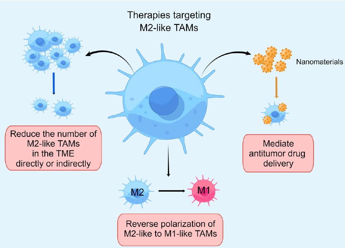
استهداف الخلايا المناعية الشبيهة بـ M2 للتغلب على مقاومة الورم

نظرًا للدور الحاسم الذي تلعبه الخلايا المناعية الشبيهة بـ M2 في مقاومة الأدوية السرطانية، فقد برز البحث عن أساليب علاجية تستهدف هذه الخلايا كمنطقة تركيز حيوية في أبحاث العلاج المضاد للسرطان. حاليًا، تشمل الاستراتيجيات الرئيسية المضادة للأورام المعتمدة على البلعميات الشبيهة بـ M2 (الجدول 3، 4) ما يلي: (1) تقليل عدد البلعميات الشبيهة بـ M2 في البيئة المجهرية للورم بشكل مباشر أو غير مباشر. (2) استخدام البلعميات الشبيهة بـ M2 كوسائط لتوصيل الأدوية المضادة للأورام. (3) إعادة قطبية البلعميات الشبيهة بـ M2 إلى النمط الظاهري M1. استنادًا إلى هذه الاستراتيجيات لمقاومة الأدوية المضادة للأورام التي تستهدف الخلايا المناعية الشبيهة بـ M2، قام الباحثون بإجراء العديد من التجارب التجريبية في كل من الإعدادات السريرية وما قبل السريرية (الشكل 3).

تقليل عدد TAMs الشبيهة بـ M2 في TME بشكل مباشر أو غير مباشر

من أجل استقطاب البلعميات إلى البيئة المجهرية للورم، تقوم خلايا الورم بإفراز مجموعة متنوعة من الكيموكينات، بما في ذلك CSF-1 وCCL2 وCCL5 وCX3CL1 وCXCL12. بمجرد دخولها إلى البيئة المجهرية للورم، تخضع البلعميات لتعديل بواسطة مجموعة من السيتوكينات والمواد الأيضية والإكسوزومات التي تطلقها خلايا الورم، مما يغير نشاط البلعميات المرتبطة بالورم ويعزز استقطابها. وبالتالي، يمكن أن يؤدي تقليل استقطاب البلعميات المرتبطة بالورم إلى تقليل غير مباشر للبلعميات الشبيهة بـ M2 في البيئة المجهرية للورم، مما يساعد على تثبيط تقدم الورم والتغلب على مقاومة الأدوية للورم.
بيكسيدارتينيب، مثبط لمستقبل عامل تحفيز المستعمرات 1 (CSF-1R)، قادر على الاستخدام بأمان مع السيرووليموس لتثبيط نمو الساركوما غير القابلة للاستئصال وأورام غمد الأعصاب الطرفية الخبيثة من خلال تقليل عدد الخلايا المناعية الشبيهة بـ M2. إيمكتوزوماب، مثبط آخر لمستقبل CSF1، سواء تم استخدامه كعامل منفرد أو بالاشتراك مع
أظهر الباكليتاكسيل في المرضى الذين يعانون من أورام صلبة متقدمة/نقيلية نقصًا في الخلايا المناعية المثبطة من نوع M2 في البيئة المجهرية للورم. ومع ذلك، لم ينتج عن استخدامه بمفرده أو بالاشتراك مع الباكليتاكسيل أي نشاط مضاد للورم ذي صلة سريريًا. لسوء الحظ، لم تجد هذه التجربة من المرحلة الأولى نشاطًا مضادًا للأورام ذا صلة سريرية لعقار إماكتوزوماب، على الرغم من أنها اقترحت فوائد علاجية محتملة تستحق المزيد من التحقيق. جوان وآخرون. وجد أن مثبطات CSF-1R تقلل من تجنيد TAMs عن طريق تثبيط محور الإشارة CXCL12/CXCR4، مما يقاوم استقطاب البلعميات ويزيد من حساسية سرطان البروستاتا لعلاج دوكسيتاكسل. يانغ وآخرون. اكتشف أن خلايا سرطان المبيض المقاومة للباكليتاكسيل زادت بشكل كبير من هجرة البلعميات وزادت من تعبير علامات البلعميات من النوع M2 عبر إفراز كبير لـ CCL2. لذلك، فإن استهداف محور CCL2/CCR2 يمكن أن يثبط كيمياء البلعميات، مما قد يحسن من حساسية الباكليتاكسيل لدى مرضى سرطان المبيض. يمكن لمثبط JAK1/2 ركسوليتينيب (RUX) التغلب على تحمل المايلوما للعقار المناعي المعدل ليناليدوميد. أظهرت الدراسات الإضافية أن Ruxolitinib تغلب على مقاومة الأدوية من خلال تثبيط استقطاب البلعميات من النوع M2 عن طريق تقليل التعبير عن TRIB1 وMUC1 وCD44 وCXCL12 وCXCR4 ضمن محور JAK/STAT. يمكن لخلايا الورم استقطاب البلعميات إلى بيئة الورم عن طريق تنشيط مسار الإشارات CCL5/CCR5 وزيادة كمية البلعميات الشبيهة بـ M2. وقد وُجد أن مضادات CCR5 يمكن أن تعطل استقطاب TAM عبر مسار CCL5/CCR5، مما يعزز النتائج السريرية لمجموعة متنوعة من السرطانات، بما في ذلك سرطان المعدة والبنكرياس، من خلال كبح تقدم الورم وإطالة بقاء المرضى. تشير الدراسات إلى أن مضادات الكيموكين، مثل مضادات CCR5، عند استخدامها مع العوامل الكيميائية التقليدية، يمكن أن تساعد في التغلب على المقاومة في علاج الأورام. تجري حاليًا تجربة PICCASSO المرحلة الأولى التي تحقق في الجمع بين بيمبروليزوماب ومارافيروك في سرطان القولون والمستقيم النقيلي. وقد وُجد أنها قابلة للتطبيق وأظهرت ملف أمان إيجابي.
علاوة على ذلك، من خلال استهداف البلعميات الشبيهة بـ M2 المؤيدة للموت المبرمج، يمكن تقليل عدد البلعميات الشبيهة بـ M2 في بيئة الورم الدقيقة (TME) بشكل مباشر، مما يقلل من مقاومة الورم للأدوية. على سبيل المثال، وصفة تشانغ وي تشينغ. لقد تم إظهار أنه يعزز النشاط المبرمج للخلايا الميتة من نوع M2 ويقلل من إنتاج VEGF و TGF- وبالتالي تقليل تحمل خلايا الورم للأوكساليبلاتين.

أدوية نانوية مضادة للورم تستهدف الخلايا المناعية الشبيهة بـ M2

تمتلك المواد النانوية إمكانيات كبيرة لتعزيز فعالية العلاج المناعي للأورام مع تقدم النانوبيوتكنولوجيا، وذلك بفضل فوائدها في نقل الأدوية المستهدفة بشكل محدد، وتحديد موقع إطلاق الدواء بدقة، وسهولة تعديل السطح، وارتفاع توافر الأدوية حيوياً. .
الأدوية النانوية المضادة للورم التي تستخدم الخلايا المناعية المرتبطة بالورم كوسيلة للتوصيل. تتمتع هذه الخلايا بخصائص توجيهية ملحوظة، حيث يمكنها الهجرة بشكل موجه والتجمع في الأنسجة الورمية من خلال اكتشاف إشارات محددة في البيئة المجهرية للورم. دورها الأساسي في تعزيز تقدم الورم والغزو والنقائل مثبت بشكل جيد. علاوة على ذلك، تمتلك هذه الخلايا القدرة على العمل كوسائط لتوصيل العلاجات المضادة للورم، بما في ذلك الأدوية والليبوزومات والجزيئات النانوية، مباشرة إلى مواقع الورم وحتى داخل خلايا الورم نفسها. تحدد قدرات التوجيه والتوصيل لهذه الخلايا إمكانياتها لاستخدامها كناقلات خلوية للتوصيل العلاجي. إن الاستفادة من هذه الخلايا لتوصيل الأدوية النانوية المضادة للورم بشكل مستهدف هو طريق واعد ضمن المشهد المتطور للعلاج المضاد للورم.
تُعرف هذه الاستراتيجية لتوصيل الأدوية المعتمدة على TAM أيضًا باسم نهج “الحصان الطروادي”. . تتضمن هذه التقنية تحميل الأدوية النانوية خارج الجسم في الخلايا المناعية المرتبطة بالورم، والتي يتم إعادة إدخالها لاحقًا إلى المريض. تهاجر هذه الخلايا إلى مواقع الالتهاب ثم يتم تجنيدها في نسيج الورم لإطلاق الأدوية النانوية المحملة. يمكن تطبيق هذه الاستراتيجية على الاستئصال الحراري والعلاج الإشعاعي للأورام. على سبيل المثال، تعتبر قذائف الذهب النانوية جزيئات نانوية تحتوي على نواة من السيليكا وقشرة رقيقة من الذهب. بعد زراعتها مع الخلايا المناعية المرتبطة بالورم، يتم ابتلاع قذائف الذهب النانوية، وداخل الخلايا المناعية المرتبطة بالورم، تحمل هذه الخلايا وتراكمها في القلب الناقص الأكسجين.
الجدول 3 | أبحاث الأدوية النانوية المضادة للورم التي تستهدف TAMs الشبيهة بـ M2.
استراتيجية عامل علاجي نموذج الورم آلية علاجية الحكام
نانو مان سور سورافينيب نموذج الفأر O.T. لسرطان الكبد توصيل مشترك لثاني أكسيد المنغنيز المنتج للأكسجين وسورافينيب لسرطان الكبد لتقليل تسلل الأورام الناتج عن نقص الأكسجين لخلايا المناعة المرتبطة بالورم. 170
ليبوبسوم محمل بـ C6-سيراميد (LipC6) سيراميد نموذج الفأر O.T. لسرطان الكبد (HCC) تنشيط المناعة المضادة للأورام من خلال تحفيز إعادة برمجة الخلايا المناعية المرتبطة بالورم عن طريق تنظيم إشارات ROS. 171
معقد الجسيمات النانوية/البكتيريا (Ec-PR848) ريسيكيمود (R848) نموذج الفأر O.T. 4T1 لسرطان الثدي إعادة برمجة TAMs من خلال تنشيط TRL7/8 172
مجس يستهدف TAM يتكون من جسم مضاد CD206 مرتبط بصبغة الفثالوسيانين القريبة من الأشعة تحت الحمراء (IRD- CD206) سورافينيب نموذج الفأر M.T. 4T1 لسرطان الثدي نقصان الخلايا المناعية المرتبطة بالورم بواسطة العلاج الضوئي 173
مناطق الأورام، تحسين استهداف جزيئات الذهب النانوية وزيادة فعالية علاج الاستئصال الحراري للأورام .
شهدت السنوات الأخيرة تراجعًا في استكشاف البلعميات الشبيهة بـ M2 كوسائط لتوصيل النانوميدicine. يمكن أن يُعزى هذا التحول في النموذج إلى الدور المناعي المثبط للبلعميات الشبيهة بـ TAMs في بيئة الورم والتحديات المرتبطة بالاستهداف الانتقائي. لا تؤدي هذه القيود فقط إلى تعقيد نقل العلاجات، بل تعرض أيضًا لخطر زيادة تقدم الورم. على الرغم من هذه التحديات، لا يزال تقدم أساليب البحث والتقنيات الجديدة يحمل إمكانيات لتحقيق اختراقات في استراتيجيات العلاج المستقبلية.
أدوية نانوية مضادة للورم تعمل على الخلايا البلعمية الشبيهة بـ M2. تمتلك الخلايا البلعمية المسببة للالتهابات، المعروفة باسم الخلايا البلعمية الشبيهة بـ M1، قدرات بلعومية قوية والقدرة على قتل خلايا الورم بشكل مباشر. تحت تأثير الكيموكينات ذات الصلة، يمكن للخلايا البلعمية المسببة للالتهابات أن تهاجر بكفاءة إلى آفات الورم لممارسة هذه الوظائف. لذلك، فإن المواد النانوية التي تستهدف الخلايا البلعمية الشبيهة بـ M2 لإعادة تشكيل نمط الخلايا البلعمية، وتحفيز الخلايا البلعمية الشبيهة بـ M2 على الاستقطاب نحو الخلايا البلعمية الشبيهة بـ M1، وتثبيط نمو الورم تحمل أيضًا قيمة بحثية كبيرة. تم حالياً وضع عدة استراتيجيات نانوية للتأثير على البلعميات الشبيهة بـ M2، بما في ذلك إنهاء تجنيد البلعميات الشبيهة بـ M2، واستهدافها بشكل محدد والقضاء عليها، وتحويل البلعميات الشبيهة بـ M2 إلى بلعميات شبيهة بـ M1. لقد أظهرت التقدمات الأخيرة في تعزيز الاستجابات المناعية المضادة للأورام بشكل صريح من خلال استهداف البلعميات الشبيهة بـ M2 باستخدام المواد النانوية وعدًا كبيرًا.
تشانغ وآخرون صمموا حاملة دواء نانوية تستهدف الأورام، NanoMnSor، التي قامت بشكل فعال بتوصيل أكسيد المنغنيز (MnO2) المنتج للأكسجين والسورافينيب إلى سرطان الكبد (HCC). قام مكون MnO2 بتحفيز تحلل بيروكسيد الهيدروجين (H2O2) إلى أكسجين، مما ساهم في تخفيف الظروف الناقصة للأكسجين داخل بيئة الورم. بعد ذلك، عززت NanoMnSor وجود خلايا T السامة CD8+ من خلال تحويل البلعميات الشبيهة بـ M2 إلى بلعميات مناعية تحفيزية شبيهة بـ M1 داخل الأورام، مما يساهم في عكس مقاومة السورافينيب. لي وآخرون. أظهر أن نانو ليبوزوم C6-سيراميد (LipC6) يمكن أن يستنفد TAMs ويقلل من مسارات إشارات ROS التي تنظمها TAM. عززت هذه التعديلات نشاط خلايا CD8+ T وعززت إفراز السيتوكينات الالتهابية M1، بما في ذلك IL-12 و IFN- ، وTNF- “، مما يؤدي إلى تحفيز تنشيط البلعميات الشبيهة بـ M1. في الوقت نفسه، قام LipC6 بتثبيط إفراز السيتوكينات المرتبطة بـ M2 مثل IL-4 وFizz وYm1، مما أدى بدوره إلى قمع تنشيط البلعميات الشبيهة بـ M2، وبالتالي تقليل التسامح المناعي داخل بيئة سرطان الكبد الخلوي. يستخدم العلماء بشكل متزايد الألبومين المعدل هيكليًا كوسيلة لأنظمة توصيل الأدوية، بهدف تنشيط مسارات TLR بشكل انتقائي لاستهداف وتغيير نمط TAMs. تم تحديد TLR7/8 كهدف واعد لتعزيز الاستجابة المناعية. وقد ثبت أن منبه TLR7/8 المعروف باسم ريسيكيمود (R848) يحفز استقطاب البلعميات الشبيهة بـ M2 إلى البلعميات الشبيهة بـ M1. قام الباحثون بتطوير جزيئات نانوية محملة بـ R848 من حمض البولي (لاكتيد-كو-جليكوليك) (PLGA)، والتي يتم امتصاصها بعد ذلك كهربائيًا على سطح سلالة الإشريكية القولونية MG1655. يعزز النظام موت خلايا الورم من خلال الاقتران مع DOX، ويزيد من كمية خلايا T داخل بيئة الورم، مما يعزز فعالية العلاج المضاد للورم. “. بالإضافة إلى قدرتها على توصيل الجزيئات الصغيرة بدقة، يمكن أن تتوسط الأدوية النانوية أيضًا في الديناميات الضوئية للعمل مباشرة على البلعميات الشبيهة بـ M2. مجس يستهدف TAM (IRD- تم إنشاء CD206 بواسطة زانغ وآخرون. من خلال اقتران جسم مضاد CD206 بصبغة الفثالوسيانين القريبة من الأشعة تحت الحمراء. أظهر هذا المركب فعالية كبيرة في تثبيط كل من تكاثر وانتشار خلايا سرطان الثدي 4T1 المقاومة للسورافينيب.

إعادة استقطاب البلعميات من نوع M2 إلى نوع M1

تظهر TAMs مرونة ملحوظة، قادرة على تغيير النمط الظاهري تحت تأثير عوامل مختلفة. لذلك، فإن إعادة برمجة TAMs المستهدفة من نمط M2 إلى نمط M1 تمثل مجالًا حاسمًا في البحث السرطاني. لتخفيف مقاومة الأدوية في الأورام، قام الباحثون بالقيام بالعديد من
الجدول 4 | دراسة سريرية حول استراتيجيات علاج TAMs الشبيهة بـ M2 للتغلب على مقاومة الأدوية في الأورام.
تدخل نوع ورم الحالة حالة معرف الحكومة الحكام
تقليل الخلايا المناعية الشبيهة بـ M2 في بيئة الورم
مثبط JAK روكسوليتينيب مرضى المايلوما المتعددة الذين انتكسوا/مقاومون للعلاج مكتمل NCT03311854 155
مثبط CSF-1R بيكسيدارتينيب الساركومة غير القابلة للاستئصال و MPNST مكتمل NCT02584647 151
إيمكتوزوماب الأورام الصلبة المتقدمة/النقيلية مكتمل NCT01494688 ١٥٢
AMG 820 الأورام الصلبة المتقدمة مكتمل NCT02713529 196
كابيراليزوماب الميلانوما، سرطان الخلايا الكلوية، أو سرطان الرئة غير صغير الخلايا المقاوم لمضادات PD-1/PD-L1 مكتمل NCT03502330 ١٩٧
مثبط CCR5 مارافيروك سرطان القولون المستقيمي النقيلي المقاوم للعلاج MMRp/MSS جارٍ NCT03274804 152
مثبط TAM سيترافاتينيب سرطان الخلايا الكلوية الصافي المتقدم مكتمل NCT03015740 198
مثبط VEGFR2 أنلوتينيب سرطان الرئة غير صغير الخلايا المتقدم مكتمل NCT03628521 199
إعادة استقطاب TAMs من نوع M2 إلى نوع M1
مثبط CSF-1R بيكسيدارتينيب الأورام الصلبة المتقدمة والمقاومة للعلاج مكتمل NCT01525602 ١٧٥
ARRY-382 الأورام الصلبة المتقدمة مكتمل NCT02880371 ٢٠٠
BLZ945 الأورام الصلبة المتقدمة جارٍ NCT02829723 ١٧٥
LY3022855 الأورام الصلبة المتقدمة مكتمل NCT02718911 ٢٠١
مثبط CSF-1 لاكوتوزوماب سرطان الثدي الثلاثي السلبي المتقدم لم يبدأ التوظيف بعد NCT02435680 ٢٠٢
PD-0360324 الأورام الصلبة المتقدمة محليًا أو المنتشرة جارٍ NCT02554812 ١٧٥
مثبط BRD4 AZD5153 الأورام الصلبة المتكررة/المقاومة أو اللمفوما مكتمل NCT03205176 ٢٠٣
CD47-SIRP مثبط إيفورباست HNSCC مكتمل NCT03013218 178
منشط CD40 APX005M الميلانوما، سرطان الرئة غير صغير الخلايا، أو سرطان الكلى مكتمل NCT03502330 ١٩٧
SEA-CD40 الأورام الصلبة المتقدمة واللمفوما مكتمل NCT02376699. 181
PI3K مثبط IPI-549 الأورام الصلبة المتقدمة مكتمل NCT02637531 204
مثبط Src داساتينيب سرطان الرئة غير صغير الخلايا جارٍ NCT00826449 ٢٠٥
مثبطات HDAC توسيدينوسات ENKTCL في المراحل المبكرة ذات المخاطر المتوسطة والعالية مكتمل NCT04511351 ٢٠٦
مثبطات STAT 3 دانفاتيرسن الأورام الصلبة المتقدمة مكتمل NCT03394144 ٢٠٧
تي جي إف مثبط غالونيسيرتيب سرطان البنكرياس غير القابل للجراحة مكتمل NCT02734160 ٢٠٨
منبه TLR فيدوتوليمود الميلانوما النقيلي مكتمل NCT03084640 ٢٠٩
الشكل 3 | الأساليب الرئيسية الحالية لمكافحة الأورام التي تستهدف TAMs الشبيهة بـ M2 للتغلب على مقاومة الأدوية في الأورام. تشمل هذه الأساليب بشكل رئيسي ثلاثة جوانب: تقليل عدد TAMs الشبيهة بـ M2 في بيئة الورم من خلال طرق مباشرة أو غير مباشرة، مما يقلل من دورها في تعزيز نمو الورم؛ استخدام TAMs الشبيهة بـ M2 كوسائط لتوصيل الأدوية المضادة للأورام؛ وتحفيز إعادة قطبية TAMs الشبيهة بـ M2 إلى النمط الظاهري M1، مما يعيد نشاطها المضاد للأورام ويعزز الاستجابة المناعية في بيئة الورم. تشكل هذه الاستراتيجيات مجتمعة نهجًا علاجيًا شاملاً للتغلب على مقاومة الأدوية في الأورام من خلال استهداف TAMs الشبيهة بـ M2. (بواسطة Figdraw).

التجارب وأنشأت مجموعة متنوعة من الأدوية ذات الصلة التي تحفز الاستقطاب من ماكروفاجات شبيهة بـ M2 إلى ماكروفاجات شبيهة بـ M1 بما في ذلك على سبيل المثال لا الحصر مضادات CSF-1R، PI3K مثبطات، مثبطات بروتين 4 المحتوي على برومودومين (BRD4)، مثبطات بروتين الإشارة التنظيمية ألفا (SIRPa) ومثبطات هيستون ديأسيتيلز (HDAC).
بيكسيدارتينيب هو مثبط متعدد الجينات لإنزيم التيروزين كيناز يستهدف CSF-1R لتقليل تسلل الأورام بواسطة البلعميات بشكل كبير. تم إثبات فعالية بيكسيدارتينيب بواسطة أومستيد لزيادة تنظيم بروتين BCL-2 المرتبط بالبروتين X (BAX)، بروتين مرتبط بـ CRISPR 3 (Cas3)، TNF- IFN- ، و IL-6، وتقليل Ki-67، IL-13، IL-10، TGF- “، و Arg-1. بالإضافة إلى ذلك، عزز Pexidartinib تسلل خلايا CD3 + CD8 + T إلى البيئة المجهرية للورم (TME) من خلال تثبيط محور CSF-1/CSF-1R. وبالتالي، يمكن أن يقلل Pexidartinib من استقطاب TAMs الشبيهة بـ M2، مما قد يتغلب على مقاومة نموذج سرطان المريء الغدي (EAC) لحجب محور PD-1/PD-L1. AZD5153، مثبط محدد لـ BRD4، يعيد برمجة TAMs من نمط M2 إلى نمط M1، مما يعزز بدوره إفراز السيتوكينات المسببة للالتهابات. هذه السلسلة من الإفرازات تنشط الخلايا اللمفاوية التائية السامة CD8+ (CTLs)، مما يعزز استجابة سرطان المبيض الحليمي عالي الدرجة (HGSOC) للعلاج المضاد لـ PD-L1. إيفورباست، مثبط CD47-SIRPa، قد أظهر تعزيز الاستجابة المناعية المضادة للأورام في النماذج ما قبل السريرية من خلال تعزيز البلعمة بواسطة البلعميات، ودفع تحول النمط الظاهري من TAMs شبيهة M2 إلى TAMs شبيهة M1، وزيادة وظائف الخلايا التائية السامة. كما أظهر إيفورباست نشاطًا مشجعًا في العلاج المركب في المرحلة الأولى من التجارب السريرية للأورام الصلبة. تشير البيانات ما قبل السريرية إلى أن الجمع بين منبه CD40 ومثبطات PD-1/PD-L1 يحسن البقاء في نماذج الأورام في الفئران مقارنة باستخدام أي منهما بمفرده. إن الإعطاء المشترك لمنبه CD40 ومثبطات PD-1/PD-L1 يرفع من تعبير PD-L1 في وحيدات النوى المتسللة إلى الورم وخلايا المناعة المرتبطة بالورم، ويميل بتوزيع خلايا المناعة المرتبطة بالورم نحو النمط الالتهابي M1، مما يثبط مقاومة المناعة الناتجة عن الورم. كانيدا وآخرون وجدت في الدراسات ما قبل السريرية أن مثبط PI3K- يمكن أن يؤدي المثبط إلى تغيير في نوع TAMs من M2 إلى M1، مما يعيد تنشيط خلايا CD8 + T السامة، وبالتالي يثبط نمو الورم المقاوم لمثبطات نقاط التفتيش. في تجربة سريرية من المرحلة الأولى، PI3K- المثبط IPI-549، المستخدم بالاشتراك مع نيفولوماب، أظهر علامات على نشاط سريري جيد التحمل ومعدل مناعي. يتم حالياً إجراء التوظيف لتجربة سريرية لاحقة من المرحلة الثانية لتقييم الفعالية والسلامة بشكل أكبر. أظهرت التجارب في المختبر أن الداساتينيب، وهو مثبط لـ Src، يعزز استقطاب البلعميات الشبيهة بـ M2 نحو البلعميات الشبيهة بـ M1 في خطوط خلايا سرطان الرئة المقاومة للسيستين A549R و H460R من خلال تعديل محور Src/CD155/MIF وتقليل تعبير علامات الخلايا الجذعية Notch1 و -كاتين. أظهرت الأبحاث أن مثبطات Src فعالة في علاج الأفراد الذين يعانون من سرطان الرئة المقاوم للسيستين من خلال استهداف الماكروفاجات الشبيهة بـ M2 بشكل محدد. أدت الجرعات المحسّنة من مثبط HDAC توسييدينستات إلى استقطاب الخلايا البلعمية من النوع M2 إلى النوع M1 في ثلاثة نماذج أورام في الفئران من خلال تنشيط مسار إشارة NF-κB وزيادة التعبير عن CCL5. وقد أدى ذلك إلى زيادة وجود خلايا CD8 +T في بيئة الورم وتقليل مقاومة الورم للأجسام المضادة المضادة لـ PD-L1. غياب SHP-2 في الخلايا النخاعية يمكن أن ينشط TAMs المؤيدة للالتهابات، مما يحول البيئة المجهرية للورم من مثبطة للمناعة إلى محفزة للمناعة، وبالتالي يعزز فعالية العلاج المناعي. البحث في الجسم الحي لجين غاو يظهر أن مثبط SHP2 SHP099 يستهدف SHP-2، وينظم مسار STING ويعزز النوع الأول من الإنترفيرون، مما يعيد تشكيل بيئة الورم ويتغلب على مقاومة الورم. دانفاتيرسين، وهو أوليغونوكليوتيد مضاد للعلاج (ASO) محدد لـ STAT3، يعدل بيئة الورم من خلال إعادة توازن البلعميات المثبطة والمحفزة للالتهاب. هذه العملية تعزز فعالية حجب نقاط التفتيش المناعية وتتغلب على مقاومة العلاج المناعي. . يوي وآخرون وجدت أن الأانوليتينب تغلب على مقاومة البورتيزوميب من خلال تعزيز استقطاب الخلايا المناعية الشبيهة بـ M2 إلى خلايا مناعية شبيهة بـ M1، وتقليل وظيفة الأوعية الدموية في الورم، وتسريع موت الخلايا المبرمج في خلايا المايلوما PDX.
لقد كشفت مجموعة واسعة من التحقيقات التي أجريت في السنوات الأخيرة أن الطب الصيني التقليدي (TCM) له دور في تنظيم استقطاب TAM وعكس مقاومة الأدوية للأورام. يوفر الطب الصيني التقليدي تشي لينغ (QL) فوائد قوية ضد أورام البروستاتا. كاو وآخرون. تم جمع مصل الفئران التي تم إعطاؤها QL.
تم الإشارة إليه باسم مصل QL. اكتشفوا أنه من خلال مسار الإشارة IL-6/STAT3، زاد مصل QL من تعبير iNOS وTNF- ، وانخفضت تعبير IL-10 و CCL22 في نظام زراعة مشترك من TAMs مع خلايا سرطان البروستاتا المقاومة للباكليتاكسيل، وهي DU145-TxR و PC-3-TxR. وبالتالي، تم تحفيز استقطاب البلعميات من النوع M2 نحو البلعميات من النوع M1، وتم تقليل مقاومة خلايا سرطان البروستاتا البشرية للباكليتاكسيل. تم تطبيق وصفة شيو لي التجريبية (XLEP) في علاج سرطان الرئة غير صغير الخلايا (NSCLC)، والتي تتكون أساسًا من جذور الأدينوفورا، وجذور جليهنيا، وجذور الهليون، وجذور الأوفيوبوجون، والشيساندرا، وثمرة البريفيت، والأستراجالوس، والزدواري. شيو وآخرون. اكتشف أن XLEP يمكن أن يعزز استقطاب البلعميات الشبيهة بـ M2 إلى بلعميات شبيهة بـ M1 ويزيد من نسبة البلعميات الشبيهة بـ M1 إلى البلعميات الشبيهة بـ M2 في خلايا سرطان الرئة غير صغير الخلايا الإيجابية لمستقبلات EGFR. كان هذا التأثير ناتجًا عن تقليل التعبير عن mTOR و IL-10 و TGF- ، وCCL22 وزيادة تنظيم IL6 وCCL2 وCCL3 وTNF- “، مما أدى إلى تأخير المقاومة لمثبط EGFR-TKI الجيفيتينيب. استهدفت هيديوتيس ديفوزا ويلد (HDW) ونظمت TGF- مسار الإشارة في خلايا CRC للتغلب على مقاومة العلاج الكيميائي. ومع ذلك، لا تزال الدراسات التجريبية الإضافية مطلوبة لتحديد ما إذا كانت HDW تنظم استقطاب البلعميات من النوع M2. تمكنت صيغة Zhi Zhen من منع خلايا CRC من تطوير مقاومة لـ 5-FU من خلال تقليل إنتاج p-STAT3، وهو بروتين حاسم في مسار STAT3 داخل TAMs. تم تحقيق هذا التأثير من خلال تثبيط استقطاب TAM نحو النمط الظاهري M2، ومن خلال قمع تنشيط مسار إشارة Hedgehog في خلايا CRC. تريبتيريجيم لاكتون (TP)، مثبط مستهدف لمسار PI3K/Akt/NF-кB، قلل من تعبير MMP-2 وMMP-9 وVEGF، مما حفز استقطاب TAM من النمط M2 إلى النمط M1. هذا التحول زاد من نسبة M1/M2 وقلل من مقاومة خلايا OCA للسيستين. .

نقاش

استخدام العلاجات الدوائية، بما في ذلك العلاج الكيميائي، والعلاج المستهدف، والعلاج المناعي، أمر حاسم لعلاج الأورام. ومع ذلك، في الممارسة السريرية، سيطور غالبية المرضى مقاومة للأدوية في المرحلة المتأخرة من العلاج، مما يؤدي إلى فشل العلاج المضاد للأورام. تقليل مقاومة الورم وتعزيز فعالية العلاج المضاد للسرطان لا يزالان يمثلان تحديات كبيرة. لقد أوضحت التقدمات الأخيرة في دراسة البيئة المجهرية للورم (TME) والخلايا المناعية المرتبطة بالورم (TAMs) أن الخلايا المناعية من النوع M2 تساهم في بيئة مجهرية مناعية مثبطة، مما يعزز بدوره مقاومة الأدوية. يمكن أن تعزز الخلايا المناعية من النوع M2 مقاومة الورم من خلال عدة آليات مثل تنظيم السيتوكينات، ومسارات الإشارة، والحويصلات خارج الخلوية، والبيئة المجهرية المناعية، وتكوين الأوعية الدموية، وخلايا جذع الورم. استنادًا إلى هذه الآليات ذات الصلة، من الممكن صياغة أدوية تستهدف الخلايا المناعية من النوع M2 بشكل محدد. إن دمج هذه الأدوية مع العلاج الكيميائي، والعلاج المستهدف، والعلاج المناعي، وأدوية مضادة للورم الأخرى في العلاج السريري يحمل وعدًا في التغلب على مقاومة الأدوية الورمية وتعزيز التوقعات. سيتطلب الأمر مزيدًا من التجارب السريرية للتحقق من إمكانية استهداف الخلايا المناعية من النوع M2 بالاشتراك مع علاجات مختلفة.

ملخص التقرير

معلومات إضافية حول تصميم البحث متاحة في ملخص تقارير الأبحاث في مجلة Nature المرتبط بهذه المقالة.
تاريخ الاستلام: 15 أغسطس 2023؛ تاريخ القبول: 19 يناير 2024؛ تاريخ النشر على الإنترنت: 10 فبراير 2024

References

  1. Sung, H. et al. Global cancer statistics 2020: GLOBOCAN estimates of incidence and mortality worldwide for 36 cancers in 185 countries. CA. Cancer J. Clin. 71, 209-249 (2021).
  2. Bayik, D. & Lathia, J. D. Cancer stem cell-immune cell crosstalk in tumour progression. Nat. Rev. Cancer 21, 526-536 (2021).
  3. Rey-Giraud, F., Hafner, M. & Ries, C. H. In vitro generation of monocyte-derived macrophages under serum-free conditions improves their tumor promoting functions. Plos One 7, e42656 (2012).
  4. Ruffell, B. & Coussens, L. M. Macrophages and therapeutic resistance in cancer. Cancer Cell. 27, 462-472 (2015).
  5. Altorki, N. K. et al. The lung microenvironment: an important regulator of tumour growth and metastasis. Nat. Rev. Cancer 19, 9-31 (2019).
  6. Morrison, C. Immuno-oncologists eye up macrophage targets. Nat. Rev. Drug Discov. 15, 373-374 (2016).
  7. Casanova-Acebes, M. et al. Tissue-resident macrophages provide a pro-tumorigenic niche to early NSCLC cells. Nature 595, 578-584 (2021).
  8. Yang, Q. et al. The role of tumor-associated macrophages (TAMs) in tumor progression and relevant advance in targeted therapy. Acta Pharm. Sin. B. 10, 2156-2170 (2020).
  9. Schmid, M. C. et al. Combined blockade of integrin-alpha4beta1 plus cytokines SDF-1alpha or IL-1beta potently inhibits tumor inflammation and growth. Cancer Res. 71, 6965-6975 (2011).
  10. Christofides, A. et al. The complex role of tumor-infiltrating macrophages. Nat. Immunol. 23, 1148-1156 (2022).
  11. Chamseddine, A. N., Assi, T., Mir, O. & Chouaib, S. Modulating tumor-associated macrophages to enhance the efficacy of immune checkpoint inhibitors: A TAM-pting approach. Pharmacol. Ther. 231, 107986 (2022).
  12. Locati, M., Curtale, G. & Mantovani, A. Diversity, mechanisms, and significance of macrophage plasticity. Annu. Rev. Pathol. 15, 123-147 (2020).
  13. Murray, P. J. et al. Macrophage activation and polarization: nomenclature and experimental guidelines. Immunity 41, 14-20 (2014).
  14. Mantovani, A., Sica, A. & Locati, M. Macrophage polarization comes of age. Immunity 23, 344-346 (2005).
  15. Bleriot, C., Chakarov, S. & Ginhoux, F. Determinants of resident tissue macrophage identity and function. Immunity 52, 957-970 (2020).
  16. Mulder, K. et al. Cross-tissue single-cell landscape of human monocytes and macrophages in health and disease. Immunity 54, 1883-1900 (2021).
  17. Aibar, S. et al. SCENIC: single-cell regulatory network inference and clustering. Nat. Methods 14, 1083-1086 (2017).
  18. Ma, R. Y., Black, A. & Qian, B. Z. Macrophage diversity in cancer revisited in the era of single-cell omics. Trends Immunol. 43, 546-563 (2022).
  19. Schultze, J. L. & Schmidt, S. V. Molecular features of macrophage activation. Semin. Immunol. 27, 416-423 (2015).
  20. Jeannin, P., Paolini, L., Adam, C. & Delneste, Y. The roles of CSFs on the functional polarization of tumor-associated macrophages. FEBS J. 285, 680-699 (2018).
  21. Qian, B. Z. & Pollard, J. W. Macrophage diversity enhances tumor progression and metastasis. Cell 141, 39-51 (2010).
  22. Lin, Y., Xu, J. & Lan, H. Tumor-associated macrophages in tumor metastasis: biological roles and clinical therapeutic applications. J. Hematol. Oncol. 12, 76 (2019).
  23. Li, X. et al. Harnessing tumor-associated macrophages as aids for cancer immunotherapy. Mol. Cancer 18, 177 (2019).
  24. Pan, Y., Yu, Y., Wang, X. & Zhang, T. Tumor-associated macrophages in tumor immunity. Front. Immunol. 11, 583084 (2020).
  25. Gratchev, A. et al. Alternatively activated macrophages differentially express fibronectin and its splice variants and the extracellular matrix protein betalG-H3. Scand. J. Immunol. 53, 386-392 (2001).
  26. Hesse, M. et al. Differential regulation of nitric oxide synthase-2 and arginase-1 by type 1/type 2 cytokines in vivo: granulomatous pathology is shaped by the pattern of L-arginine metabolism. J. Immunol. 167, 6533-6544 (2001).
  27. Sunderkotter, C., Steinbrink, K., Goebeler, M., Bhardwaj, R. & Sorg, C. Macrophages and angiogenesis. J. Leukoc. Biol. 55, 410-422 (1994).
  28. Moeini, P. & Niedzwiedzka-Rystwej, P. Tumor-associated macrophages: Combination of therapies, the approach to improve cancer treatment. Int. J. Mol. Sci. 22, 7239 (2021).
  29. Yao, Z. et al. Imatinib prevents lung cancer metastasis by inhibiting M2-like polarization of macrophages. Pharmacol. Res. 133, 121-131 (2018).
  30. Sica, A. & Mantovani, A. Macrophage plasticity and polarization: in vivo veritas. J. Clin. Invest. 122, 787-795 (2012).
  31. DeNardo, D. G. & Ruffell, B. Macrophages as regulators of tumour immunity and immunotherapy. Nat. Rev. Immunol. 19, 369-382 (2019).
  32. Gharavi, A. T., Hanjani, N. A., Movahed, E. & Doroudian, M. The role of macrophage subtypes and exosomes in immunomodulation. Cell. Mol. Biol. Lett. 27, 83 (2022).
  33. Zhang, Q. & Sioud, M. Tumor-associated macrophage subsets: Shaping polarization and targeting. Int. J. Mol. Sci. 24, 7493 (2023).
  34. Wang, Y. et al. Macrophage-derived extracellular vesicles: diverse mediators of pathology and therapeutics in multiple diseases. Cell Death Dis. 11, 924 (2020).
  35. Wang, L. X., Zhang, S. X., Wu, H. J., Rong, X. L. & Guo, J. M2b tumorassociated macrophage subsets: Shaping polarization and targeting. Dis. J. Leukoc. Biol. 106, 345-358 (2019).
  36. Hagemann, T. et al. Re-educating” tumor-associated macrophages by targeting NF-kappaB. J. Exp. Med. 205, 1261-1268 (2008).
  37. Daniel, B. et al. The IL-4/STAT6/PPARgamma signaling axis is driving the expansion of the RXR heterodimer cistrome, providing complex ligand responsiveness in macrophages. Nucleic Acids Res. 46, 4425-4439 (2018).
  38. Liu, H., Amakye, W. K. & Ren, J. Codonopsis pilosula polysaccharide in synergy with dacarbazine inhibits mouse melanoma by repolarizing M2-like tumor-associated macrophages into M1-like tumor-associated macrophages. Biomed. Pharmacother. 142, 112016 (2021).
  39. Vergadi, E., leronymaki, E., Lyroni, K., Vaporidi, K. & Tsatsanis, C. Akt signaling pathway in macrophage activation and M1/M2 polarization. J. Immunol. 198, 1006-1014 (2017).
  40. Zhao, Y. et al. IncRNA-Xist/miR-101-3p/KLF6/C/EBPalpha axis promotes TAM polarization to regulate cancer cell proliferation and migration. Mol. Ther. Nucleic Acids 23, 536-551 (2021).
  41. Schoumacher, M. & Burbridge, M. Key roles of AXL and MER receptor tyrosine kinases in resistance to multiple anticancer therapies. Curr. Oncol. Rep. 19, 19 (2017).
  42. Fruman, D. A. et al. The PI3K pathway in human disease. Cell 170, 605-635 (2017).
  43. Jiang, N. et al. Role of PI3K/AKT pathway in cancer: The framework of malignant behavior. Mol. Biol. Rep. 47, 4587-4629 (2020).
  44. Su, P. et al. Crosstalk between tumor-associated macrophages and tumor cells promotes chemoresistance via CXCL5/PI3K/AKT/ mTOR pathway in gastric cancer. Cancer Cell Int. 22, 290 (2022).
  45. Li, D. et al. Tumor-associated macrophages secrete CC-chemokine ligand 2 and induce tamoxifen resistance by activating PI3K/Akt/ mTOR in breast cancer. Cancer Sci. 111, 47-58 (2020).
  46. Niu, X. et al. Sodium/glucose cotransporter 1-dependent metabolic alterations induce tamoxifen resistance in breast cancer by promoting macrophage M2 polarization. Cell Death Dis. 12, 509 (2021).
  47. Owen, K. L., Brockwell, N. K. & Parker, B. S. JAK-STAT signaling: A double-edged Sword of immune regulation and cancer progression. Cancers 11, 2002 (2019).
  48. Hu, X., Li, J., Fu, M., Zhao, X. & Wang, W. The JAK/STAT signaling pathway: From bench to clinic. Signal Transduct. Target. Ther. 6, 402 (2021).
  49. Yang, C. et al. Increased drug resistance in breast cancer by tumorassociated macrophages through IL-10/STAT3/bcl-2 signaling pathway. Med. Oncol. 32, 352 (2015).
  50. He, Z. et al. Yes associated protein 1 promotes resistance to 5-fluorouracil in gastric cancer by regulating GLUT3-dependent glycometabolism reprogramming of tumor-associated macrophages. Arch. Biochem. Biophys. 702, 108838 (2021).
  51. Burotto, M., Chiou, V. L., Lee, J. M. & Kohn, E. C. The MAPK pathway across different malignancies: A new perspective. Cancer 120, 3446-3456 (2014).
  52. Vogelstein, B. et al. Cancer genome landscapes. Science 339, 1546-1558 (2013).
  53. Yang, S. H., Sharrocks, A. D. & Whitmarsh, A. J. MAP kinase signalling cascades and transcriptional regulation. Gene 513, 1-13 (2013).
  54. Zhou, D. et al. Macrophage polarization and function with emphasis on the evolving roles of coordinated regulation of cellular signaling pathways. Cell. Signal. 26, 192-197 (2014).
  55. Takebe, N., Nguyen, D. & Yang, S. X. Targeting notch signaling pathway in cancer: clinical development advances and challenges. Pharmacol. Ther. 141, 140-149 (2014).
  56. Palaga, T., Wongchana, W. & Kueanjinda, P. Notch signaling in macrophages in the context of cancer immunity. Front. Immunol. 9, 652 (2018).
  57. Huang, Y. H. et al. CREBBP/EP300 mutations promoted tumor progression in diffuse large B-cell lymphoma through altering tumor-associated macrophage polarization via FBXW7-NOTCHCCL2/CSF1 axis. Signal Transduct. Target. Ther. 6, 10 (2021).
  58. Tao, S., Chen, Q., Lin, C. & Dong, H. Linc00514 promotes breast cancer metastasis and M2 polarization of tumor-associated macrophages via Jagged1-mediated notch signaling pathway. J. Exp. Clin. Cancer Res. 39, 191 (2020).
  59. Liu, H. et al. Jagged1 promotes aromatase inhibitor resistance by modulating tumor-associated macrophage differentiation in breast cancer patients. Breast Cancer Res. Treat. 166, 95-107 (2017).
  60. Huang, F. et al. miR-148a-3p mediates notch signaling to promote the differentiation and M1 activation of macrophages. Front Immunol. 8, 1327 (2017).
  61. Rasmi, R. R., Sakthivel, K. M. & Guruvayoorappan, C. NF-kappaB inhibitors in treatment and prevention of lung cancer. Biomed. Pharmacother. 130, 110569 (2020).
  62. Usman, M. W. et al. Macrophages confer resistance to PI3K inhibitor GDC-0941 in breast cancer through the activation of NF-kappaB signaling. Cell Death Dis. 9, 809 (2018).
  63. Yang, T. et al. Macrophages-aPKC(i)-CCL5 feedback loop modulates the progression and chemoresistance in cholangiocarcinoma. J. Exp. Clin. Cancer Res. 41, 23 (2022).
  64. Badouel, C. & McNeill, H. SnapShot: The hippo signaling pathway. Cell 145, 484 (2011).
  65. Chen, J. et al. Tumor-derived exosomal miR-19b-3p facilitates M2 macrophage polarization and exosomal LINC00273 secretion to promote lung adenocarcinoma metastasis via Hippo pathway. Clin. Transl. Med. 11, e478 (2021).
  66. Kim, E. H. et al. Silence of hippo pathway associates with protumoral immunosuppression: Potential therapeutic target of glioblastomas. Cells 9, 1761 (2020).
  67. Urbanelli, L. et al. Signaling pathways in exosomes biogenesis, secretion and fate. Genes 4, 152-170 (2013).
  68. Kalluri, R. & LeBleu, V. S. The biology, function, and biomedical applications of exosomes. Science 367, eaau6977 (2020).
  69. Balaji, S., Kim, U., Muthukkaruppan, V. & Vanniarajan, A. Emerging role of tumor microenvironment derived exosomes in therapeutic resistance and metastasis through epithelial-to-mesenchymal transition. Life Sci. 280, 119750 (2021).
  70. Tan, S. et al. Exosomal miRNAs in tumor microenvironment. J. Exp. Clin. Cancer Res. 39, 67 (2020).
  71. Mashouri, L. et al. Exosomes: composition, biogenesis, and mechanisms in cancer metastasis and drug resistance. Mol. Cancer 18, 75 (2019).
  72. Baig, M. S. et al. Tumor-derived exosomes in the regulation of macrophage polarization. Inflamm. Res. 69, 435-451 (2020).
  73. Mori, M. A., Ludwig, R. G., Garcia-Martin, R., Brandao, B. B. & Kahn, C. R. Extracellular miRNAs: From biomarkers to mediators of physiology and disease. Cell Metab. 30, 656-673 (2019).
  74. Ludwig, N., Yerneni, S. S., Razzo, B. M. & Whiteside, T. L. Exosomes from HNSCC promote angiogenesis through reprogramming of endothelial cells. Mol. Cancer Res. 16, 1798-1808 (2018).
  75. Cheng, Z. et al. Tumor-derived exosomes induced M2 macrophage polarization and promoted the metastasis of osteosarcoma cells through Tim-3. Arch. Med. Res. 52, 200-210 (2021).
  76. Mi, X. et al. M2 macrophage-derived exosomal IncRNA AFAP1-AS1 and MicroRNA-26a affect cell migration and metastasis in esophageal cancer. Mol. Ther. Nucleic Acids 22, 779-790 (2020).
  77. Dong, X. et al. Exosomes and breast cancer drug resistance. Cell Death Dis. 11, 987 (2020).
  78. Xin, L. et al. Transfer of LncRNA CRNDE in TAM-derived exosomes is linked with cisplatin resistance in gastric cancer. EMBO Rep. 22, e52124 (2021).
  79. Chen, S. W. et al. Cancer cell-derived exosomal circUSP7 induces CD8(+) T cell dysfunction and anti-PD1 resistance by regulating the miR-934/SHP2 axis in NSCLC. Mol. Cancer 20, 144 (2021).
  80. Wang, X. et al. Targeting feedback activation of signaling transduction pathways to overcome drug resistance in cancer. Drug Resist. Update 65, 100884 (2022).
  81. Zheng, P. et al. Exosomal transfer of tumor-associated macrophage-derived miR-21 confers cisplatin resistance in gastric cancer cells. J. Exp. Clin. Cancer Res. 36, 53 (2017).
  82. An, Y. & Yang, Q. MiR-21 modulates the polarization of macrophages and increases the effects of M2 macrophages on promoting the chemoresistance of ovarian cancer. Life Sci. 242, 117162 (2020).
  83. Wan, X. et al. Exosomes derived from M2 type tumor-associated macrophages promote osimertinib resistance in non-small cell lung cancer through MSTRG.292666.16-miR-6836-5p-MAPK8IP3 axis. Cancer Cell Int. 22, 83 (2022).
  84. Yin, Y. et al. The immune-microenvironment confers chemoresistance of colorectal cancer through macrophage-derived IL6. Clin. Cancer Res. 23, 7375-7387 (2017).
  85. Ling, H. Y. et al. Diffuse large B-cell lymphoma-derived exosomes push macrophage polarization toward M2 phenotype via GP130/ STAT3 signaling pathway. Chem. Biol. Interact. 352, 109779 (2022).
  86. Cui, H. Y. et al. Exosomal microRNA-588 from M2 polarized macrophages contributes to cisplatin resistance of gastric cancer cells. World J. Gastroenterol. 27, 6079-6092 (2021).
  87. Robey, R. W. et al. Revisiting the role of ABC transporters in multidrug-resistant cancer. Nat. Rev. Cancer 18, 452-464 (2018).
  88. Lv, M. M. et al. Exosomes mediate drug resistance transfer in MCF-7 breast cancer cells and a probable mechanism is delivery of P-glycoprotein. Tumour Biol. 35, 10773-10779 (2014).
  89. Binenbaum, Y. et al. Transfer of miRNA in macrophage-derived exosomes induces drug resistance in pancreatic adenocarcinoma. Cancer Res. 78, 5287-5299 (2018).
  90. Kanlikilicer, P. et al. Corrigendum to ‘Exosomal miRNA confers chemo resistance via targeting Cav1/p-gp/M2-type macrophage axis in ovarian cancer’ [EBioMedicine 38 (2018) 100-112]. Ebiomedicine 52, 102630 (2020).
  91. Akhade, V. S., Pal, D. & Kanduri, C. Long noncoding RNA: Genome organization and mechanism of action. Adv. Exp. Med. Biol. 1008, 47-74 (2017).
  92. Chen, F. et al. The functional roles of exosomes-derived long noncoding RNA in human cancer. Cancer Biol. Ther. 20, 583-592 (2019).
  93. Zhou, D. et al. Exosomal long non-coding RNA SOX2 overlapping transcript enhances the resistance to EGFR-TKIs in non-small cell lung cancer cell line H1975. Hum. Cell. 34, 1478-1489 (2021).
  94. Zhang, K. et al. Exosome-mediated transfer of SNHG7 enhances docetaxel resistance in lung adenocarcinoma. Cancer Lett. 526, 142-154 (2022).
  95. Meng, X. et al. DNA damage repair alterations modulate M2 polarization of microglia to remodel the tumor microenvironment via the p53-mediated MDK expression in glioma. Ebiomedicine 41, 185-199 (2019).
  96. Li, Z. et al. Glioblastoma cell-derived IncRNA-containing exosomes induce microglia to produce complement C5, promoting chemotherapy resistance. Cancer Immunol. Res. 9, 1383-1399 (2021).
  97. Yousefi, H. et al. Long noncoding RNAs and exosomal IncRNAs: classification, and mechanisms in breast cancer metastasis and drug resistance. Oncogene 39, 953-974 (2020).
  98. Xing, Z. et al. LINC00337 induces tumor development and chemoresistance to paclitaxel of breast cancer by recruiting M2 tumor-associated macrophages. Mol. Immunol. 138, 1-9 (2021).
  99. Gao, C., Hu, W., Zhao, J., Ni, X. & Xu, Y. LncRNA HCG18 promotes M2 macrophage polarization to accelerate cetuximab resistance in colorectal cancer through regulating miR-365a-3p/FOXO1/CSF-1 axis. Pathol. Res. Pract. 240, 154227 (2022).
  100. Wu, W. et al. The miR155HG/miR-185/ANXA2 loop contributes to glioblastoma growth and progression. J. Exp. Clin. Cancer Res. 38, 133 (2019).
  101. Zhou, L., Li, J., Liao, M., Zhang, Q. & Yang, M. LncRNA MIR155HG induces M2 macrophage polarization and drug resistance of colorectal cancer cells by regulating ANXA2. Cancer Immunol. Immunother. 71, 1075-1091 (2022).
  102. Ramani, T. et al. Cytokines: The good, the bad, and the deadly. Int. J. Toxicol. 34, 355-365 (2015).
  103. Kelso, A. Cytokines and their receptors: An overview. Ther. Drug Monit. 22, 40-43 (2000).
  104. Asadullah, K., Sterry, W. & Trefzer, U. Cytokines: Interleukin and interferon therapy in dermatology. Clin. Exp. Dermatol. 27, 578-584 (2002).
  105. Yang, S., Wang, J., Brand, D. D. & Zheng, S. G. Role of TNF-TNF receptor 2 signal in regulatory T cells and its therapeutic implications. Front. Immunol. 9, 784 (2018).
  106. Tan, W. et al. TNF-alpha is a potential therapeutic target to overcome sorafenib resistance in hepatocellular carcinoma. Ebiomedicine 40, 446-456 (2019).
  107. Laha, D., Grant, R., Mishra, P. & Nilubol, N. The role of tumor necrosis factor in manipulating the immunological response of tumor microenvironment. Front. Immunol. 12, 656908 (2021).
  108. Cruceriu, D., Baldasici, O., Balacescu, O. & Berindan-Neagoe, I. The dual role of tumor necrosis factor-alpha (TNF-alpha) in breast cancer: molecular insights and therapeutic approaches. Cell. Oncol. 43, 1-18 (2020).
  109. Acharyya, S. et al. A CXCL1 paracrine network links cancer chemoresistance and metastasis. Cell 150, 165-178 (2012).
  110. Liu, Y. et al. Tumor necrosis factor alpha inhibition overcomes immunosuppressive M2b macrophage-induced bevacizumab resistance in triple-negative breast cancer. Cell Death Dis. 11, 993 (2020).
  111. Chen, Y. et al. TNF-alpha derived from M2 tumor-associated macrophages promotes epithelial-mesenchymal transition and cancer stemness through the Wnt/beta-catenin pathway in SMMC7721 hepatocellular carcinoma cells. Exp. Cell Res. 378, 41-50 (2019).
  112. Catalan-Dibene, J., McIntyre, L. L. & Zlotnik, A. Interleukin 30 to Interleukin 40. J. Interferon Cytokine Res. 38, 423-439 (2018).
  113. Gelfo, V. et al. Roles of IL-1 in cancer: From tumor progression to resistance to targeted therapies. Int. J. Mol. Sci. 21, 6009 (2020).
  114. Kumari, N., Dwarakanath, B. S., Das, A. & Bhatt, A. N. Role of interleukin-6 in cancer progression and therapeutic resistance. Tumour Biol. 37, 11553-11572 (2016).
  115. Zhang, Y. et al. Interleukin-17-induced neutrophil extracellular traps mediate resistance to checkpoint blockade in pancreatic cancer. J. Exp. Med. 217, e20190354 (2020).
  116. Bin, G. Mechanism of M2-TAMs Induced Chemotherapy resistant in Hepatocellular Carcinoma Cells Through IL-17/IL-17R-CMA Pathway. North China University of Science and Technology, 2018.
  117. Li, J., He, K., Liu, P. & Xu, L. X. Iron participated in breast cancer chemoresistance by reinforcing IL-6 paracrine loop. Biochem. Biophys. Res. Commun. 475, 154-160 (2016).
  118. Sun, Y. et al. Blockade of STAT3/IL-4 overcomes EGFR T790M-cis-L792F-induced resistance to osimertinib via suppressing M2 macrophages polarization. Ebiomedicine 83, 104200 (2022).
  119. Hughes, C. E. & Nibbs, R. A guide to chemokines and their receptors. FEBS J. 285, 2944-2971 (2018).
  120. Lopez-Cotarelo, P., Gomez-Moreira, C., Criado-Garcia, O., Sanchez, L. & Rodriguez-Fernandez, J. L. Beyond chemoattraction: Multifunctionality of chemokine receptors in leukocytes. Trends Immunol. 38, 927-941 (2017).
  121. Susek, K. H., Karvouni, M., Alici, E. & Lundqvist, A. The role of CXC chemokine receptors 1-4 on immune cells in the tumor microenvironment. Front. Immunol. 9, 2159 (2018).
  122. Liu, C. et al. Macrophage-derived CCL5 facilitates immune escape of colorectal cancer cells via the p65/STAT3-CSN5-PD-L1 pathway. Cell Death Differ. 27, 1765-1781 (2020).
  123. Wei, C. et al. M2 macrophages confer resistance to 5-fluorouracil in colorectal cancer through the activation of CCL22/PI3K/AKT signaling. Oncotargets Ther. 12, 3051-3063 (2019).
  124. Nwabo, K. A. et al. Insulin-like growth factor-1 signaling in the tumor microenvironment: Carcinogenesis, cancer drug resistance, and therapeutic potential. Front. Endocrinol. 13, 927390 (2022).
  125. Ireland, L. et al. Chemoresistance in pancreatic cancer is driven by stroma-derived insulin-like growth factors. Cancer Res. 76, 6851-6863 (2016).
  126. Gordon, S. R. et al. PD-1 expression by tumour-associated macrophages inhibits phagocytosis and tumour immunity. Nature 545, 495-499 (2017).
  127. Li, W. et al. Correlation between PD-1/PD-L1 expression and polarization in tumor-associated macrophages: A key player in tumor immunotherapy. Cytokine Growth Factor Rev. 67, 49-57 (2022).
  128. Ma, J. et al. Tumor-associated macrophage-derived CCL5 promotes chemotherapy resistance and metastasis in prostatic cancer. Cell Biol. Int. 45, 2054-2062 (2021).
  129. Noy, R. & Pollard, J. W. Tumor-associated macrophages: from mechanisms to therapy. Immunity 41, 49-61 (2014).
  130. Wang, J. et al. Tumor cells induced-M2 macrophage favors accumulation of Treg in nasopharyngeal carcinoma. Int. J. Clin. Exp. Pathol. 10, 8389-8401 (2017).
  131. Fu, L. Q. et al. The roles of tumor-associated macrophages in tumor angiogenesis and metastasis. Cell. Immunol. 353, 104119 (2020).
  132. Ferrara, N., Gerber, H. P. & LeCouter, J. The biology of VEGF and its receptors. Nat. Med. 9, 669-676 (2003).
  133. Itatani, Y., Kawada, K., Yamamoto, T. & Sakai, Y. Resistance to antiangiogenic therapy in cancer-alterations to Anti-VEGF pathway. Int. J. Mol. Sci. 19, 1232 (2018).
  134. Siveen, K. S. et al. Vascular endothelial growth factor (VEGF) signaling in tumour vascularization: Potential and challenges. Curr. Vasc. Pharmacol. 15, 339-351 (2017).
  135. Ma, F. et al. Hypoxic macrophage-derived VEGF promotes proliferation and invasion of gastric cancer cells. Dig. Dis. Sci. 64, 3154-3163 (2019).
  136. Wheeler, K. C. et al. VEGF may contribute to macrophage recruitment and M2 polarization in the decidua. Plos One 13, e191040 (2018).
  137. Castro, B. A. et al. Macrophage migration inhibitory factor downregulation: a novel mechanism of resistance to anti-angiogenic therapy. Oncogene 36, 3749-3759 (2017).
  138. Zhang, G., Tao, X., Ji, B. & Gong, J. Hypoxia-driven M2-polarized macrophages facilitate cancer aggressiveness and temozolomide resistance in glioblastoma. Oxid. Med. Cell. Longev. 2022, 1614336 (2022).
  139. Stockmann, C. et al. Deletion of vascular endothelial growth factor in myeloid cells accelerates tumorigenesis. Nature 456, 814-818 (2008).
  140. Li, Y. et al. VEGFR3 inhibition chemosensitizes lung adenocarcinoma A549 cells in the tumor-associated macrophage microenvironment through upregulation of p53 and PTEN. Oncol. Rep. 38, 2761-2773 (2017).
  141. Wang, T. et al. JAK/STAT3-regulated fatty acid beta-oxidation is critical for breast cancer stem cell self-renewal and chemoresistance. Cell Metab. 27, 136-150 (2018).
  142. Wang, Q. et al. Cancer stem-like cells-oriented surface selfassembly to conquer radioresistance. Adv. Mater. 35, e2302916 (2023).
  143. Najafi, M., Mortezaee, K. & Majidpoor, J. Cancer stem cell (CSC) resistance drivers. Life Sci. 234, 116781 (2019).
  144. Gabrusiewicz, K. et al. Glioblastoma stem cell-derived exosomes induce M2 macrophages and PD-L1 expression on human monocytes. Oncoimmunology 7, e1412909 (2018).
  145. Vahidian, F. et al. Interactions between cancer stem cells, immune system and some environmental components: Friends or foes? Immunol. Lett. 208, 19-29 (2019).
  146. Li, L. et al. CD105: tumor diagnosis, prognostic marker and future tumor therapeutic target. Clin. Transl. Oncol. 24, 1447-1458 (2022).
  147. Hassn, M. M., Syafruddin, S. E., Mohtar, M. A. & Syahir, A. CD44: A multifunctional mediator of cancer progression. Biomolecules 11, 1850 (2021).
  148. Yaghobi, Z. et al. The role of CD44 in cancer chemoresistance: A concise review. Eur. J. Pharmacol. 903, 174147 (2021).
  149. Li, X. et al. CXCL12/CXCR4 pathway orchestrates CSC-like properties by CAF recruited tumor associated macrophage in OSCC. Exp. Cell Res. 378, 131-138 (2019).
  150. Shi, Y. et al. Tumour-associated macrophages secrete pleiotrophin to promote PTPRZ1 signalling in glioblastoma stem cells for tumour growth. Nat. Commun. 8, 15080 (2017).
  151. Manji, G. A. et al. A phase I study of the combination of pexidartinib and sirolimus to target tumor-associated macrophages in unresectable sarcoma and malignant peripheral nerve sheath tumors. Clin. Cancer Res. 27, 5519-5527 (2021).
  152. Gomez-Roca, C. A. et al. Phase I study of emactuzumab single agent or in combination with paclitaxel in patients with advanced/ metastatic solid tumors reveals depletion of immunosuppressive M2-like macrophages. Ann. Oncol. 30, 1381-1392 (2019).
  153. Guan, W. et al. Tumor-associated macrophage promotes the survival of cancer cells upon docetaxel chemotherapy via the CSF1/ CSF1R-CXCL12/CXCR4 axis in castration-resistant prostate cancer. Genes 12, 773 (2021).
  154. Yang, Y. I., Wang, Y. Y., Ahn, J. H., Kim, B. H. & Choi, J. H. CCL2 overexpression is associated with paclitaxel resistance in ovarian cancer cells via autocrine signaling and macrophage recruitment. Biomed. Pharmacother. 153, 113474 (2022).
  155. Berenson, J. R. et al. A phase 1 study of ruxolitinib, steroids and lenalidomide for relapsed/refractory multiple myeloma patients. Hematol. Oncol. 40, 906-913 (2022).
  156. Chen, H. et al. JAK1/2 pathway inhibition suppresses M2 polarization and overcomes resistance of myeloma to lenalidomide by reducing TRIB1, MUC1, CD44, CXCL12, and CXCR4 expression. Br. J. Haematol. 188, 283-294 (2020).
  157. Nie, Y. et al. Breast phyllodes tumors recruit and repolarize tumorassociated macrophages via secreting CCL5 to promote malignant progression, which can be inhibited by CCR5 inhibition therapy. Clin. Cancer Res. 25, 3873-3886 (2019).
  158. Halama, N. et al. Tumoral immune cell exploitation in colorectal cancer metastases can be targeted effectively by anti-CCR5 therapy in cancer patients. Cancer Cell. 29, 587-601 (2016).
  159. Huang, H. et al. The CCR5 antagonist maraviroc causes remission of pancreatic cancer liver metastasis in nude rats based on cell cycle inhibition and apoptosis induction. Cancer Lett. 474, 82-93 (2020).
  160. Yue, L. Chang Weiqing enhances chemosensitivity of colon cancer oxaliplatin by inhibiting M2 macrophages., Shanghai University of Traditional Chinese Medicine, 2019.
  161. Zhang, Z., Deng, Q., Xiao, C., Li, Z. & Yang, X. Rational design of nanotherapeutics based on the five features principle for potent elimination of cancer stem cells. Acc. Chem. Res. 55, 526-536 (2022).
  162. Li, Z. et al. Influence of nanomedicine mechanical properties on tumor targeting delivery. Chem. Soc. Rev. 49, 2273-2290 (2020).
  163. Ramesh, A., Brouillard, A. & Kulkarni, A. Supramolecular nanotherapeutics for macrophage immunotherapy. Acs Appl. Bio Mater. 4, 4653-4666 (2021).
  164. Choi, M. R. et al. A cellular Trojan Horse for delivery of therapeutic nanoparticles into tumors. Nano Lett. 7, 3759-3765 (2007).
  165. Kennedy, L. C. et al. A new era for cancer treatment: gold-nanoparticle-mediated thermal therapies. Small 7, 169-183 (2011).
  166. Cheng, Y. et al. Tumor associated macrophages and TAMs-based anti-tumor nanomedicines. Adv. Heathc. Mater 10, e2100590 (2021).
  167. Wang, Y. et al. Engineering endogenous tumor-associated macrophage-targeted biomimetic nano-rbc to reprogram tumor immunosuppressive microenvironment for enhanced chemoimmunotherapy. Adv. Mater. 33, e2103497 (2021).
  168. Wu, L. et al. Multiwalled carbon nanotubes prevent tumor metastasis through switching M2-polarized macrophages to M1 via TLR4. Activat. J. Biomed. Nanotechnol. 15, 138-150 (2019).
  169. Wei, Z., Zhang, X. & Zhang, Z. Engineered Iron-Based nanoplatform amplifies repolarization of M2-like tumor-associated macrophages for enhanced cancer immunotherapy. Chem. Eng. J. 433, 133847 (2022).
  170. Chang, C. C. et al. Nanoparticle delivery of and antiangiogenic therapy to overcome hypoxia-driven tumor escape and suppress hepatocellular carcinoma. Acs Appl. Mater. Interfaces 12, 44407-44419 (2020).
  171. Li, G. et al. Nanoliposome C6-ceramide increases the anti-tumor immune response and slows growth of liver tumors in mice. Gastroenterology 154, 1024-1036 (2018).
  172. Wei, B. et al. Polarization of tumor-associated macrophages by nanoparticle-loaded escherichia coli combined with immunogenic cell death for cancer immunotherapy. Nano Lett. 21, 4231-4240 (2021).
  173. Zhang, C. et al. Inhibition of tumor growth and metastasis by photoimmunotherapy targeting tumor-associated macrophage in a sorafenib-resistant tumor model. Biomaterials 84, 1-12 (2016).
  174. Zhang, S. Y. et al. Tumor-associated macrophages: A promising target for a cancer immunotherapeutic strategy. Pharmacol. Res. 161, 105111 (2020).
  175. Wesolowski, R. et al. Phase lb study of the combination of pexidartinib (PLX3397), a CSF-1R inhibitor, and paclitaxel in patients with advanced solid tumors. Ther. Adv. Med. Oncol. 11, 432498018 (2019).
  176. Omstead, A. N. et al. CSF-1R inhibitor, pexidartinib, sensitizes esophageal adenocarcinoma to PD-1 immune checkpoint blockade in a rat model. Carcinogenesis 43, 842-850 (2022).
  177. Li, X. et al. BRD4 inhibition by AZD5153 promotes antitumor immunity via depolarizing M2 macrophages. Front. Immunol. 11, 89 (2020).
  178. Kauder, S. E. et al. ALX148 blocks CD47 and enhances innate and adaptive antitumor immunity with a favorable safety profile. Plos One 13, e201832 (2018).
  179. Djureinovic, D., Wang, M. & Kluger, H. M. Agonistic CD40 antibodies in cancer treatment. Cancers 13, 1302 (2021).
  180. Kaneda, M. M. et al. PI3Kgamma is a molecular switch that controls immune suppression. Nature 539, 437-442 (2016).
  181. Coveler, A. L. et al. Phase 1 dose-escalation study of SEA-CD40: a non-fucosylated CD40 agonist, in advanced solid tumors and Iymphomas. J. Immunother. Cancer 11, e005584 (2023).
  182. Huang, W. C., Kuo, K. T., Wang, C. H., Yeh, C. T. & Wang, Y. Cisplatin resistant lung cancer cells promoted M2 polarization of tumorassociated macrophages via the Src/CD155/MIF. Funct. Pathw. J. Exp. Clin. Cancer Res. 38, 180 (2019).
  183. Zhang, P. et al. Optimized dose selective HDAC inhibitor tucidinostat overcomes anti-PD-L1 antibody resistance in experimental solid tumors. BMC Med. 20, 435 (2022).
  184. Christofides, A. et al. SHP-2 and PD-1-SHP-2 signaling regulate myeloid cell differentiation and antitumor responses. Nat. Immunol. 24, 55-68 (2023).
  185. Gao, J. et al. Allosteric inhibition reveals SHP2-mediated tumor immunosuppression in colon cancer by single-cell transcriptomics. Acta Pharm. Sin. B. 12, 149-166 (2022).
  186. Proia, T. A. et al. STAT3 antisense oligonucleotide remodels the suppressive tumor microenvironment to enhance immune activation in combination with Anti-PD-L1. Clin. Cancer Res. 26, 6335-6349 (2020).
  187. Yue, Y. et al. Novel myeloma patient-derived xenograft models unveil the potency of anlotinib to overcome bortezomib resistance. Front. Oncol. 12, 894279 (2022).
  188. Cao, H., Wang, D., Gao, R., Feng, Y. & Chen, L. Qi Ling decreases paclitaxel resistance in the human prostate cancer by reversing tumor-associated macrophages function. Aging (Albany Ny.). 14, 1812-1821 (2022).
  189. Xu, B., Zhang, H., Wang, Y., Huang, S. & Xu, L. Mechanism of Xu Li’s experiential prescription for the treatment of EGFR-positive NSCLC. Evid. -Based Complement Altern. Med. 2020, 8787153 (2020).
  190. Lai, Z. et al. Hedyotis diffusa Willd suppresses metastasis in 5-fluorouracil-resistant colorectal cancer cells by regulating the TGF-beta signaling pathway. Mol. Med. Rep. 16, 7752-7758 (2017).
  191. J, F. Zhi Zhen Fomula Regulating Tumor-associated Macrophages to Inhibit Drug Resistance of Colorectal Cancer., Shanghai University of Traditional Chinese Medicine, 2020.
  192. H, H. Study on the mechanism of triptolide inhibition on Cisplatin resistant epithelial ovarian cancer based on the polarization of TAMs., Nanchang University, 2020.
  193. Haider, T., Pandey, V., Banjare, N., Gupta, P. N. & Soni, V. Drug resistance in cancer: mechanisms and tackling strategies. Pharmacol. Rep. 72, 1125-1151 (2020).
  194. Zhong, L. et al. Small molecules in targeted cancer therapy: advances, challenges, and future perspectives. Signal Transduct. Target. Ther. 6, 201 (2021).
  195. Bagchi, S., Yuan, R. & Engleman, E. G. Immune checkpoint inhibitors for the treatment of cancer: Clinical impact and mechanisms of response and resistance. Annu. Rev. Pathol. 16, 223-249 (2021).
  196. Razak, A. R. et al. Safety and efficacy of AMG 820, an anti-colonystimulating factor 1 receptor antibody, in combination with pembrolizumab in adults with advanced solid tumors. J. Immunother. Cancer 8, e001006 (2020).
  197. Weiss, S. A. et al. A Phase I Study of APX005M and Cabiralizumab with or without Nivolumab in Patients with Melanoma, Kidney Cancer, or Non-Small Cell Lung Cancer Resistant to Anti-PD-1/PDL1. Clin. Cancer Res. 27, 4757-4767 (2021).
  198. Msaouel, P. et al. A phase 1-2 trial of sitravatinib and nivolumab in clear cell renal cell carcinoma following progression on antiangiogenic therapy. Sci. Transl. Med. 14, eabm6420 (2022).
  199. Chu, T. et al. Phase 1b study of sintilimab plus anlotinib as first-line therapy in patients with advanced NSCLC. J. Thorac. Oncol. 16, 643-652 (2021).
  200. Johnson, M. et al. ARRY-382 in combination with pembrolizumab in patients with advanced solid tumors: Results from a phase 1b/ 2 study. Clin. Cancer Res. 28, 2517-2526 (2022).
  201. Falchook, G. S. et al. A phase 1a/1b trial of CSF-1R inhibitor LY3022855 in combination with durvalumab or tremelimumab in patients with advanced solid tumors. Invest. New. Drugs 39, 1284-1297 (2021).
  202. Kuemmel, S. et al. A randomized phase II sudy of anti-CSF1 monoclonal antibody lacnotuzumab (MCS110) combined with gemcitabine and carboplatin in advanced triple-negative breast cancer. Clin. Cancer Res. 28, 106-115 (2022).
  203. Hamilton, E. P. et al. First-in-human study of AZD5153, a small molecule inhibitor of bromodomain protein 4, in patients with relapsed/refractory malignant solid tumors and lymphoma. Mol. Cancer Ther. 22, 1154-1165 (2023).
  204. Tolcher, A. et al. Abstract CT089: IPI-549-01 – A Phase 1/1b, first-inhuman study of IPI-549, a PI3K- inhibitor, as monotherapy and in combination with nivolumab in patients with advanced solid tumors. Cancer Res. 13, CT89 (2017).
  205. Gold, K. A. et al. A phase I/II study combining erlotinib and dasatinib for non-small cell lung cancer. Oncologist 19, 1040-1041 (2014).
  206. Chai, Y. et al. First-line chemoradiation with or without chidamide (tucidinostat) in patients with intermediate- and high-risk early-stage extranodal nasal-type natural killer/T-cell lymphoma: A randomized phase 2 study in China. Int. J. Radiat. Oncol. Biol. Phys. 113, 833-844 (2022).
  207. Nishina, T. et al. Safety, tolerability, pharmacokinetics and preliminary antitumour activity of an antisense oligonucleotide targeting STAT3 (danvatirsen) as monotherapy and in combination with durvalumab in Japanese patients with advanced solid malignancies: A phase 1 study. BMJ Open. 12, e55718 (2022).
  208. Melisi, D. et al. Galunisertib plus gemcitabine vs. gemcitabine for first-line treatment of patients with unresectable pancreatic cancer. Br. J. Cancer 119, 1208-1214 (2018).
  209. Ribas, A. et al. Overcoming PD-1 blockade resistance with CpG-A toll-like receptor 9 agonist vidutolimod in patients with metastatic melanoma. Cancer Discov. 11, 2998-3007 (2021).

شكر وتقدير

تم دعم هذه الدراسة من خلال تمويل من مشروع العلوم والتكنولوجيا في مدينة قوانغتشو (2023A03J0300).

مساهمات المؤلفين

قدم S.W. و J.W. مساهمة كبيرة في تصميم المقالة، والبحث في الأدبيات، وكتابة المسودة الأصلية. ساهم Z.C. و J.L. و W.G. و L.S. في كتابة المسودة الأصلية. ساهم S.W. و J.W. و Z.C. و J.L. و W.G. و L.S. و L.L. في الكتابة، والتحرير، وقدموا مراجعة نقدية للمحتوى الفكري المهم. قرأ جميع المؤلفين ووافقوا على المخطوطة النهائية وهم مسؤولون عن جميع جوانب العمل.

المصالح المتنافسة

يعلن المؤلفون عدم وجود مصالح متنافسة.

معلومات إضافية

معلومات إضافية النسخة الإلكترونية تحتوي على
المواد التكميلية متاحة على
https://doi.org/10.1038/s41698-024-00522-z.
يجب توجيه المراسلات والطلبات للحصول على المواد إلى ليزهو لين.
معلومات إعادة الطباعة والتصاريح متاحة على
http://www.nature.com/reprints
ملاحظة الناشر: تظل شركة سبرينغر ناتشر محايدة فيما يتعلق بالمطالبات القضائية في الخرائط المنشورة والانتماءات المؤسسية.
الوصول المفتوح هذه المقالة مرخصة بموجب رخصة المشاع الإبداعي النسب 4.0 الدولية، التي تسمح بالاستخدام والمشاركة والتكيف والتوزيع وإعادة الإنتاج بأي وسيلة أو صيغة، طالما أنك تعطي الائتمان المناسب للمؤلفين الأصليين والمصدر، وتوفر رابطًا لرخصة المشاع الإبداعي، وتوضح ما إذا كانت هناك تغييرات قد أُجريت. الصور أو المواد الأخرى من طرف ثالث في هذه المقالة مشمولة في رخصة المشاع الإبداعي الخاصة بالمقالة، ما لم يُشار إلى خلاف ذلك في سطر الائتمان للمواد. إذا لم تكن المادة مشمولة في رخصة المشاع الإبداعي الخاصة بالمقالة وكان استخدامك المقصود غير مسموح به بموجب اللوائح القانونية أو يتجاوز الاستخدام المسموح به، فستحتاج إلى الحصول على إذن مباشرة من صاحب حقوق الطبع والنشر. لعرض نسخة من هذه الرخصة، قم بزيارةhttp://creativecommons.org/licenses/by/4.0/.
© المؤلف(ون) 2024

  1. أول كلية طبية سريرية لجامعة غوانغتشو للطب الصيني، غوانغتشو، الصين. المستشفى الأول التابع لجامعة قوانغتشو للطب الصيني، قوانغتشو، الصين. أكاديمية قوانغدونغ للبحوث السريرية في الطب الصيني، قوانغتشو، الصين. مركز لينغنان للبحوث الطبية، جامعة الطب الصيني في قوانغتشو، قوانغتشو، الصين. البريد الإلكتروني: gzucmlinlz@163.com
    معهد هورميل
    جامعة مينيسوتا

Journal: npj Precision Oncology, Volume: 8, Issue: 1
DOI: https://doi.org/10.1038/s41698-024-00522-z
PMID: https://pubmed.ncbi.nlm.nih.gov/38341519
Publication Date: 2024-02-10

Targeting M2-like tumor-associated macrophages is a potential therapeutic approach to overcome antitumor drug resistance

Shujing Wang , Jingrui Wang , Zhiqiang Chen , Jiamin Luo , Wei Guo , Lingling Sun & Lizhu Lin

Abstract

Tumor drug resistance emerges from the interaction of two critical factors: tumor cellular heterogeneity and the immunosuppressive nature of the tumor microenvironment (TME). Tumorassociated macrophages (TAMs) constitute essential components of the TME. M2-like TAMs are essential in facilitating tumor metastasis as well as augmenting the drug resistance of tumors. This review encapsulates the mechanisms that M2-like TAMs use to promote tumor drug resistance. We also describe the emerging therapeutic strategies that are currently targeting M2-like TAMs in combination with other antitumor drugs, with some still undergoing clinical trial evaluation. Furthermore, we summarize and analyze various existing approaches for developing novel drugs that target M2-like TAMs to overcome tumor resistance, highlighting how targeting M2-like TAMs can effectively stop tumor growth, metastasis, and overcome tumor drug resistance.

The 2020 data revealed approximately 19.3 million new instances of tumors and around 10 million deaths linked to tumors worldwide. Both incidence and death rates have been quickly rising globally . Drug therapy is still a primary clinical treatment for malignant tumors. However, the issues of the poor treatment outcomes, tumor progression, and poor prognosis caused by drug resistance have always been difficult to solve. Consequently, the study of tumor drug resistance has risen to prominence in the clinical management of tumors today. Contemporary research found that tumor drug resistance stems from the interaction of two critical factors, the intratumor heterogeneity and the immunosuppressive nature of the TME . TME is composed of diverse cellular elements, including lymphocytes, extracellular matrix, growth factors, cytokines, chemokines, lymphocytes, TAMs, dendritic cells, natural killer cells, and myeloid-derived suppressor cells. The essential elements of TME are TAMs, which comprise two mutually polarizable subtypes: M1-like TAMs and M2-like TAMs. M2-like TAMs are essential for malignant metastasis, invasion and treatment resistance . Their significant role emphasizes their potential as targets to overcoming tumor drug resistance. This review provides a synthesis of the mechanisms for modulating M2-like macrophages to surmount resistance to antitumor therapy, along with an overview of clinical trials based on these related
mechanisms. Also, we analyze the future development potential of this novel therapeutic strategy.

Biological feature of TAMs
The origin of TAMs

As tumor cells interact with the extracellular interstitium during their growth, they create a particular environment conducive to their proliferation. This environment is known as the TME. TME is a multifaceted and interconnected network containing various cells and components, including extracellular matrix, lymphocytes, tumor-associated macrophages, dendritic cells, growth factors, cytokines, chemokines, natural killer cells, myeloid-derived suppressor cells (Fig. 1). TME affects not only the therapeutic effect of primary tumor treatments but also the evolution and advancement of tumor metastasis . Within the TME, TAMs stand out as the most critical component, constituting approximately of the tumor tissue’s weight . TAMs originate from two different lineages, tissue-resident macrophage-derived and monocyte-derived. During the initial phases of tumor development, tissue-resident macrophages accumulate and distribute around tumor tissue to induce regulatory T-cell responses, promote tumor cell immune escape, provoke epithelial-mesenchymal transition
Fig. 1 | TAMs are the most critical components of the TME. TAMs promote many malignant progressions of tumors by regulating the TME, such as tumor proliferation, immune evasion by tumor cells, EMT, tumor invasion, tumor metastasis, tumor drug resistance. (By Figdraw).
(EMT), and enhance tumor cell infiltration and spread . Numerous cytokines and chemotactic proteins, generated by adult hematopoietic stem cells within the circulatory system, attract monocyte-derived macrophages into the TME . Subsequently, multiple stimuli promote the differentiation of these monocyte-derived macrophages into TAMs. Interleukin (IL) , C-C motif ligand (CCL) 2, vascular endothelial growth factor (VEGF), and stromal cell-derived factor (SDF)- produced in tumors recruit proangiogenic macrophages to the tumor organs. Schmid et al. demonstrated that the interaction of integrin- with talin and paxillin, typically enhanced by IL- and SDF- -induced signaling, can be obstructed by leveraging antagonists against integrin- . While the exact timing of the conversion of recruited monocytes into TAMs has not been clarified, there is robust evidence supporting tissue-mediated alterations in the transcriptional profiles of these recruited monocytes .

The plasticity and polarization regulation of TAMs and their nomenclature

In reality TAMs exist as a continuum with high levels of plasticity and their expression types can often coexist or change as the tumor progresses. Ali N . Chamseddine et al. summarized the plasticity and diversity of TAMs and proposed the concept of TAM polarization as a continuous, dynamic polarization . Although the nature, mechanism and nomenclature of TAM polarization still need further exploration, the artificial description of M1like TAMs and M2-like TAMs , located at distinct opposite ends within the continuous dynamic polarization axis of the TAMs and derived from summarization based on in vitro experiments, is unanimously confirmed .
At present, the hypothesis of tumor suppressor M1-tumor promoter M2 proposed by Albert Mantovani is still the most commonly used model for studying TAM heterogeneity, although it oversimplifies the true phenotype of TAMs. However, recent in-depth studies on macrophages have revealed significant differences in structural design, operational capabilities, and expression of cellular surface identifiers among tissue-resident macrophages (TRMs) across various organs . Furthermore, genes linked to M1like and M2-like TAM profiles are expressed simultaneously in almost all types of cancer macrophage subgroups . Recently, employing Single-Cell Regulatory Network Inference and Clustering (SCENIC) analysis , researchers have discerned five specific TAM subgroups in various cancers. These subgroups are named HES1 TAMs, C1Q TAMs, TREM2 TAMs,
IL4I1 TAMs, and proliferative TAMs. Utilizing single-cell genomics, Ma et al. classified TAMs into seven subgroups based on characteristic genes, enriched pathways, and predicted functions. These subgroups are named IFN-TAMs, Reg-TAMs, Inflam-TAMs, LA-TAMs, Angio-TAMs, RTMTAMs, and Prolif-TAMs.
Although M2-like macrophages have been thought to have antiinflammatory functions while M1-like macrophages have proinflammatory functions, the reality of their behavior may be more complex. The characteristics and operational roles of macrophages are subject to change in response to environmental variations, and their roles can differ across various pathological states. Consequently, while present research tends to divide macrophages into categories based on their inflammationrelated roles, with one being pro-inflammatory (often referred to as M1) and the other being anti-inflammatory (commonly known as M2), this binary division might not fully capture their biological diversity.

The phenotypes and functions of TAMs

The M1-like TAMs and M2-like TAMs are located at the two ends of the continuous dynamic TAM polarization axis, each possessing unique cell surface markers and functional factors, and play different roles in the TME (Fig. 2).
The M1-like TAMs have pro-inflammatory properties. M1-like macrophages are stimulated by cytokines like interferon (IFN), colonystimulating factor (CSF), tumor necrosis factor (TNF). Furthermore, lipopolysaccharides (LPS) have the capacity to interact with and engage the tolllike receptor (TLR)4 located on the macrophage surface and promote M1like macrophage polarization by acting on nuclear factor kappa-B (NF-кB) and interferon regulatory factor 3 (IRF3). Strongly presenting antigens and secreting many pro-inflammatory cytokines are two traits of M1-like macrophages. M1-like macrophages secrete an extensive number of costimulatory molecules like cluster of differentiation (CD)86, CD60, and CD80, along with pro-inflammatory biomarkers including TNF- , IL-1 , IL-6, IL-12, IL-23. They also highly express major histocompatibility complex (MHC) II molecules. Notably, they do express IL-10, although at a lower level. M1-like macrophages release matrix metalloproteinases (MMPs) including MMP1, MMP2, MMP7, MMP9, and MMP12. These enzymes are specialized in breaking down extracellular matrix (ECM) constituents. M1-like macrophages generate chemokines like CCL2, CCL3,
Fig. 2 | M1-like and M2-like TAMs are shown at
opposite ends of a continuous dynamic TAM
polarization axis, illustrating their unique roles in the TME. Both M1-like TAMs and M2-like TAMs have specific cell surface markers and functional factors. M1-like TAMs in the TME undertake roles in promoting inflammation, inhibiting proliferation, eliminating pathogens, and anti-tumor responses. M2-like TAMs in the TME are involved in anti-inflammatory activities, promoting angiogenesis, influencing tissue regeneration and healing, and fostering tumor generation, proliferation, metastasis, and drug resistance. (By Figdraw).
CCL5, C-X-C motif chemokine ligand (CXCL) 8, CXCL9, CXCL10, CXCL11, CXCL16. Also, they release IFN- , inducible nitric oxide synthase (iNOS), and reactive oxygen species (ROS). M1-like macrophages activate a strong T-helper1(Th1)-type immune response by releasing these inflammatory mediators to promote an inflammatory response that inhibits cell proliferation and kills pathogens and tumor cells in the human body, thereby exerting antitumor effects .
The M2-like TAMs, which present antigens poorly, are anti-inflammatory, unlike the M1 phenotype. There is no specific procedure that must be used to initiate the activation of M2-like macrophages. The primary activation of M2-like macrophages is caused by triggering cytokines, which include transforming growth factor (TGF)- , IL-4, IL-13, IL-10, and macrophage colony-stimulating factor (M-CSF) . M2-like macrophages highly express CD163, CD206, CD200R, CD209, CD301 and chemokines like CCL1, CCL17, CCL18, CCL22, and CCL24. They release numerous anti-inflammatory factors, including TGF- , IL-4, IL-13, IL-10, and IL1RA. Additionally, M2-like TAMs express the inflammatory cytokines IL-6, IL-12, IL-23 and TNF- at lower levels . M2-like macrophages highly express MMPs and autocrine ECM components such as fibronectin, betaigh3 (BIG-H3), ECM cross-linking enzymes, transglutaminase and bone bridging proteins , thus participating in cell adhesion. M2-like macrophages promote Arginase (Arg)-1 and VEGF expression, which are involved in the biosynthesis of proline and polyamines. Proline promotes the construction of ECM, and polyamines are involved in cell proliferation . Other factors secreted by M2-like macrophages that promote cell proliferation, such as platelet-derived growth factor (PDGF) and insulin-like growth factor (IGF), are involved in angiogenesis . M2-like macrophages release immunosuppressive chemicals to block the Th1-type immune activity as well as boost the Th2-type immune activity. This activity reduces the control over inflammatory reactions while promoting tumor cell growth, drug resistance, angiogenesis, and tissue healing .
Sica et al. summarized and proposed the theory that TAMs were dominated by M1 phenotype in the early stage of tumor. As the tumor developed, the TME underwent changes that progressively led to the transformation of TAMs from the M1 to the M2 phenotype. Therefore, TAMs in most tumors present a dominant population with the M2 phenotype. TME in these tumors are generally immunosuppressive , affecting tumor progression and chemotherapy resistance.
Moreover, in response to particular triggers and changes in gene expression within the TME, M2-like macrophages can be classified toward multiple subsets, namely M2a, M2b, M2c, and M2d . Each of the subsets exhibits its own unique properties (Table 1). M2a macrophages receive their primary activation signals from the cytokines IL-4 and IL-13, which are produced by Th2 cells. These macrophages contribute to anti-inflammatory processes through their production of IL-10 and TGF- . Furthermore, they are instrumental in allergy, wound healing, cell proliferation, profibrotic. Within the TME, they contribute to the advancement of tumors by enhancing the growth and infiltrative behavior of tumor cell and angiogenesis. Activation of M2b macrophages is generally triggered by a synergistic interaction between complexes involving the immune system and ligands of Toll-like receptors. They are known to generate a significant quantity of cytokines, including IL-10 and TNF- , thus promoting inflammatory responses and participating in immune regulation. Within the TME, activated M2b macrophages secrete IL-6, which in turn activates Th2 cells, culminating in the creation of a pro-inflammatory milieu conducive to tumor progression. The activation of M2c macrophages predominantly occurs in response to IL-10, TGF- , or glucocorticoids. They actively contribute to modulating inflammatory reactions by the release of anti-inflammatory molecules. Additionally, they are instrumental in the development of pathological fibrosis and the healing of wounds. They also possess immunoregulatory functions, capable of influencing immune responses by releasing specific factors and engaging in interactions with other immune cells. M2d macrophages represent a relatively recently identified subtype that is triggered by TLR agonists and IL-6. They are known to modulate the local inflammatory environment by releasing specific factors that possess both pro-inflammatory and anti-inflammatory properties. M2d macrophages support tumor cell immune evasion via emitting immunosuppressive substances like IL-10 and PD-1, inhibiting normal immune responses. By encouraging tumor growth, invasion, and angiogenesis, they aid in the advancement of tumors.

The regulation mechanisms of M1-like /M2-like macrophages polarization

Current studies have shown that several cytokines and signaling pathways within TME influence M1/M2 polarization. Abnormalities of the kinase (IKK )/ NF-кB pathway may induce TAM polarization . The process by
Table 1 | Comparison of markers and biological functions among different macrophage phenotypes
Phenotypes Functions Stimulator factors Markers Excretion References
M1 Tumor resistance, pro-inflammatory, inhibit proliferation, eliminate pathogens IFN- , LPS, GM-CSF, TNF- , iNOS, ROS IL-12 (high)/IL-10(low), CD80, CD86, CD60, MMPs, MHCII CCL2, CCL3, CCL5, CXCL8, CXCL9, IL-1 , IL-6, IL-23 32
M2a Allergy, profibrotic, cell proliferation, anti-inflammatory, wound healing, tumor progression IL-4, IL-10, IL-13, (PPAR- ) CD11b, CD45, CD86, CD14, CD206, CD163, CD209, IL-1R, Dectin-1, YM1, RELM , IGF1, DCIR, Stabilin 1, Factor XIII-A, Ly6C, TREM-2, DCSIGN, MHCII, Fizz1, Arg-1, YM1/2 TGF- , IL-4, IL-10, VEGF, CCL1, CCL17, CCL18, CCL22, TNF- , IGF, EPGF 32-35
M2b Th2 activation, immunoregulation, inflammation, tumor progression IL-1 , LPS, TLR CD163, CD14, CD86, MHC-II, IL-10 (high), IL-12 (low), IL-6R TNF- , IL-1, IL-6, IL-10, CCL1, SPHK1, LIGHT 32,35
M2c Immunoregulation, inflammatory response, wound healing, fibrosis IL-6, IL-10, glucocorticoids, TGF- , TNF- CD163, CD206, CD14, CD16, CD86, CXCR4, MerTK, TLR-1、TLR-8 IL-10, TGF- , CCL16, CCL18, CXCL13 32,35
M2d Tumor proliferation, invasion, and angiogenesis, immune suppression IL-6, TLR, Regadenoson, LPS IL-10 (high), IL-12 (low), VEGF, TNF- (low), CD163, CD14, CD86 IL-10, IL-6, TGF- , VEGF, CCL18, M-CSF 32
which IL-4 binds to its receptor can promote signal transducer and activator of transcription (STAT)6 phosphorylation, inducing M2-like macrophage polarization via the janus protein tyrosine kinase (JAK)/ STAT6 signaling pathway. Concurrently, phosphorylated STAT6 can bind to krüppel-like factor 4 (KLF)4 and peroxisome proliferator-activated receptor- (PPAR- ) to promote this polarization . When IL-4’s interaction to its associated receptor is blocked, M2-like macrophages are induced to polarize to M1-like macrophages . A variety of signals, including IL-4, TGF- , IL-10, and bone morphogenetic protein-7 (BMP-7), promote M2 polarization through the phosphatidylinositol 3 kinase (PI3K)/ protein kinase B (Akt) signaling pathway . Long non-coding RNA (LncRNA)-Xist knockdown within M1-like macrophages, or overexpression of miR-101 inhibits CCAAT/enhancer binding protein a (C/EBPa) and KLF6 production, which induces polarization from M1 to M2 phenotype . Antitumor therapeutic strategies regulating M1/M2 polarization are promising due to the superior plasticity of TAMs.

M2-like TAMs have involvement in promoting multidrug resistance in tumor cells

Researchers have shown via ongoing studies on M2-like macrophages that their potential to interact with tumor cells in a direct or indirect way, influencing anticancer medication therapeutic tolerance (Table 2) like chemotherapy, targeted therapy, and immunotherapy . The possible regulatory mechanisms by which M2-like macrophages affect tumor drug resistance are increasingly elucidated through numerous studies.

M2-like TAMs modulate signaling pathways to enhance tumor resistance

By producing and releasing mediators, M2-like macrophages enhance cancer cell drug resistance by regulating PI3K/Akt, JAK/STAT, mitogenactivated protein kinases (MAPK) and other related pathways.
The PI3K signaling pathway, an intracellular signaling system influenced by receptor tyrosine kinases, modulates numerous cell processes including growth, division, maturation, metabolism and apoptosis . Recent studies have identified the overactive PI3K/Akt pathway as a major factor in driving tumor growth and the emergence of treatment resistance in tumors . Clinical research has demonstrated the efficacy of inhibitors targeting the PI3K/Akt pathway in cancer. Researchers have found that the release of cytokines, chemokines and GFs by M2-like TAMs modulate tumor PI3K/Akt signaling and modify the TME to advance tumor growth, differentiation, invasion, and drug resistance . TAMs in breast cancer tissues increase tumor cells’ capacity to resist apoptosis via the CCL2/PI3K/ Akt/ mammalian target of rapamycin (mTOR) signaling pathway, as found by Li et al. . This induced tumor cells to undergo autophagy, leading to tamoxifen resistance. M2-like macrophages activate cancerous breast cells via the epidermal growth factor receptor (EGFR)/PI3K/Akt signaling pathway. This stimulation leads to the feedback increase in Sodium-glucose Co-transporters (SGLT) 1 and modulates glycolysis, thereby promoting tamoxifen resistance and accelerating both in vitro and in vivo tumor growth .
With over 50 cytokines and growth factors (GFs) known to induce downstream signaling, the JAK/STAT signaling pathway has been established to be a primary communication hub in cellular function. It influences several critical biological processes like immunological modulation, cell development, and apoptosis. The promotion of tumor development, metastasis, and drug resistance is significantly aided by the abnormal and persistent activation of JAK-STAT signaling pathway proteins . It’s an effective target for treating tumors. With in-depth studies of M2-like macrophages, researchers found that tumor-derived factors can polarize macrophages via JAK/STAT activation. Furthermore, certain mediators associated with macrophages can activate JAK/STAT signaling within tumors, subsequently contributing to drug resistance in these tumors . M2-like macrophages may reduce the efficacy of paclitaxel against breast cancer through the IL-10/STAT3/Bcl-2 cascade response, thereby inducing resistance of paclitaxel against breast carcinoma . The Yes-associated
Table 2 | Mechanisms of inducing tumor drug resistance through M2-like TAMs
Target Host cell Strategy Mechanism Refs
Signaling pathway PI3K/ Akt MCF7 Tamoxifen Activation of PI3K/Akt/mTOR signaling by TAM-secreted CCL2 promotes the TME endocrine resistance feedback loop 45
MCF7 Tamoxifen Activation of breast cancer cells via EGFR/PI3K/Akt signaling by feedback upregulation of SGLT1 46
JAK/ STAT BT549, T47D Paclitaxel Modulation of IL-10/STAT3/bcl-2 signaling pathway 49
MKN45 5-FU Secretion of CCL8 to activate JAK1/STAT3 signaling phosphorylation 50
Jagged1/Notch MCF7 Aromatase inhibitor Reprogramming TAMs through high expression of the Jagged1-Notch pathway 59
NF-кВ TFK-1 Gemcitabine M2-like TAMs-derived tgf- induces EMT and drug resistance in CCA cells via the aPKCı/NF-кВ signaling pathway 63
Hippo GBM ICI Promotion of promotes M2 polarization by SOH 66
Exosome miR-21 MFC, MGC-803 Cisplatin Modulation of the transfer of PTEN/PI3K/Akt signaling between TAMs and cancer cells by M2derived miR-21 via the M2-specific ApoE 81
OVCAR3, HO-8910 Chemotherapy M2-delivered miR-21 enhanced OCA resistance via PI3K/Akt signaling 82
MSTRG.292666.16 H1975 Osimertinib M2-derived MSTRG.292666.16 promoted osimertinib resistance by regulating the miR-6386-5p/ MAPK8IP3 axis 83
miR-155-5p DLD1, HCT-8, HT-29, LoVo 5-FU Activation of the IL-6R/STAT3/miR-204-5p signaling by miR-155-5p in TAMs through regulating C/EBP in CRC 84
DLBCL-exo OCI-LY1. OCI-LY3 Epirubicin DLBCL-generated exosomes may promote M2 polarization through activating the GP130/STAT3 signaling pathway and highly expressing IL-10, CD206 and CD163 expression 85
miR-588 SGC7901 Cisplatin Stimulation of the NF-kB signaling pathway by miR-588 through partially targeting cylindromatosis in GC to prevent apoptosis 86
MCF7-exo MCF7/S, MCF7/DOC Docetaxel Exosomal delivery and the release of P-gp export the chemotherapeutic agents outside tumor cells 88
miR-365 K989 Gemcitabine Adoptive transfer of miR-365 in TAMs induced gemcitabine resistance 89
miR-1246 HeyA8, Skov3ip1, A2780 Paclitaxel miR-1246 actives by targeting Cav1/P-gP/PRPS2/M2-like macrophages signaling pathway to inhibit paclitaxel uptake and transport 90
SOX2-OT H1975 EGFR-TKIs SOX2-OT, as a miRNA sponge, targeted miR-627-3p activity and upregulated Smads expression, thereby reprogramming TAMs 93
SNHG7 H1299, SPC-A1 Docetaxel SNHG7 induces PTEN downregulation iva recruiting CUL4A, thus stimulates the PI3K/Akt signaling pathway to induce autophagy and M2 polarization. 94
Inc-TALC LN229, GL261, HMC3, BV-2 Temozolomide GBM-delivered Inc-TALC can bound to ENO1 to activate p38MAPK signaling, thus increasing C5/ C5a secretion to promote M2 polarization 96
LINC00337 MCF7,MDA-MB-231 Paclitaxel Recruitment of M2-like TAMs by LINC00337 induces tumor development and chemoresistance 98
HCG18 SW620 Cetuximab Promotion of M2 polarization by HCG18 via the miR-365a-3p/FOXO1/CSF-1 axis 99
MIR155HG Caco2, HT29 Oxaliplatin Acceleration of the CRC evolution by MIR155HG through modulating the miR-650/ANXA2 axis enhances oxaliplatin resistance 101
CRNDE MFC, SGC7901 Cisplatin M2-delivered CRNDE inhibits PTEN ubiquitination to reduce the susceptibility of cisplatin 101
Cytokine TNF- LM2 Chemotherapy TNF- accumulated heavily in the TME, promotes the upregulation of CXCL1 and CXCL2 through activating the NF-k pathway 109
MDAMB231, 4T1, E0771 Bevacizumab M2b TAMs promote tumor metastasis via TNF- , and activate IDO1 110
Table 2 (continued) | Mechanisms of inducing tumor drug resistance through M2-like TAMs
Target Host cell Strategy Mechanism Refs
SMMC-7721 Anti-tumor drug M2-like TAMs promote EMT and CSCs via the Wnt/ -catenin pathway 111
Interleukin HepG2, SMMC7721, Oxaliplatin Activation of CMA signaling pathway by M2-like TAMs via IL-17/IL-17R pathway 116
MCF7 Doxorubicin The polarization of M2-like TAMs enhances the IL-6 paracrine loop between TAMs and tumor cells 117
M109, H1975, PC-9 Osimertinib EGFR T790M-cis-L792F activated the JAK/STAT3 pathway to promote the M2 polarization 118
Chemokine HCT-8, HCT-116, SW620, SW480, DLD1 CT26, HT-29 ICI Activation of p65/STAT3-CSN5-PD-L1 signaling by TAM-secreted CCL5 inhibits CD8 + T cell responses in tumor cells 122
DLD1, HT29 5-FU Secretion of CCL22 activates the EMT program, PI3K/Akt pathway and Caspase-mediated apoptosis 123
IGF SUIT2, MIA-PaCa-2 Gemcitabine Activation of insulin/IGF1R survival signaling by M2-like TAMs or by IGF, which is modulated by M2, enhances the resistance to gemcitabine 125
TME CCL5 DU145, PC-3 Paclitaxel, Doxorubicin Activation of STAT3-related signaling pathway by TAM-secreted CCL5 upregulates Nanog 128
Treg CNE1, CNE2, 5-8 F ICI M2-like TAMs recruit mature Treg by secreting CCL22, CCL18 and promote the conversion of naive T cells to Treg by secreting TGF- , IL-10 130
Proangiogenic factor VEGF U87-MG Bevacizumab Depletion of VEGF causes MIF downregulation and promotes M2 polarization 137
LN229, U251 Temozolomide Secretion of VEGF by hypoxic M2-like macrophages through activating the PI3K/Akt/Nrf2 pathway 138
VEGF-A LLC Cyclophosphamide, Cisplatin Secretion of VEGF-A by M2-like TAMs to promote VEGFR2 phosphorylation 139
A549 Doxorubicin M2-like macrophages promote VEGF-C and VEGFR3 expression thereby inhibiting p53 and PTEN expression 140
CSCs GSC20,GSC267 GBM ICI secretion of GDEs to induce monocyte polarize into M2-like macrophages via STAT3 signaling 144
CSC Cal27 Vincristine M2-like TAMs promote OSCC cells to produce csc-like cells and overexpress stemnessrelated genes 149
GSC GBM Chemotherapy M2-like macrophages promote CSCs to express stemness features by mediating PTN – PTPRZ1 paracrine signaling induction 150
protein (YAP)1 overexpress IL-3, which is secreted by gastric cancer (GC). This overexpression prompts a significant TAM polarization toward M2 phenotype, subsequently initiating a GLUT3-mediated glycolytic program. Concurrently, by highly expressing CCL8 and triggering activation of the JAK1/STAT3 pathway, polarized M2-like macrophages increase tumor cells’ resistance to 5-fluorouracil (5-FU) .
The MAPK signaling pathway relays signals via a highly conserved three-step kinase cascade: Initially, MAP kinase kinase kinase (MKKK) phosphorylates and activates MAP kinase kinase (MKK); subsequently, activated MKK phosphorylates and activates MAP kinase (MAPK). Four subfamilies of the evolutionarily conserved serine-threonine kinasesnamely, MAPK- extracellular signal-regulated kinase (ERK), p38MAPK, c-Jun N-terminal kinase (JNK), and big mitogen-activated protein kinase 1 (BMK1) – represent distinct MAPK pathways . As a crucial signaling pathway within the eukaryotic signaling network, the MAPK signaling pathway is essential for cell growth, differentiation, apoptosis, and stress response.
According to certain theories, the MAPK pathway alters the TME to cause the proliferation and invasion of human malignancies , as well as polarizes macrophages into M2 phenotypes . However, it remains unclear if M2-like TAMs may induce drug resistance in tumors by activating the MAPK pathway, warranting further exploration by researchers.
Various solid tumors and hematologic malignancies have abnormal expression of the Notch pathway . Its role in TAM was summarized by Tanapat Palaga et al. , who pointed out that different roles for Notch signaling exist in promoting or inhibiting tumor progression, depending on the specific TAM context. Targeting Notch signaling in different macrophages might offer varied therapeutic approaches to modulate host antitumor immunity. If Notch signaling in TAMs plays a tumor-promoting role by mediating macrophage polarization and modulating the TME, its inhibition might address tumor drug resistance . According to Liu et al. , when the Jagged1/Notch pathway is activated in a malignant breast tumor, TAMs polarize strongly toward M2 phenotype, subsequently increasing resistance to aromatase inhibitors. Conversely, if TAMs depend on Notch signaling to differentiate into inflammatory antitumor macrophages, stimulating this particular Notch pathway could be vital to achieve tumorsuppressive outcomes .
The transcription factor NF- is crucial for inflammatory reactions and serves as a key molecule that connects cancer and chronic inflammation. The functions of it are tightly modulated through various processes. The progression of many solid tumors involves abnormal NF- pathway activation . It is now known that through controlling the NF- pathway, macrophages may foster tumor progression and resistance . In cholangiocarcinoma, M2-like TAMs induce EMT and gemcitabine resistance in tumor cells through the atypical protein kinase signaling pathway .
By controlling cell growth and stem cell self-renewal, the evolutionarily conserved mechanism known as Hippo signaling regulates organ growth. Dysregulation of this route may contribute to cancer development . Chen et al. observed that lung adenocarcinoma (LUAD) cells received exosomal LINC00273 from M2-like TAMs. This triggered the ubiquitination of large tumor suppressor 2 (LATS2), deactivating the Hippo pathway and subsequently activating the YAP protein downstream of LATS2. This sequence of events encouraged the incorporation of miR-19b-3p into LUAD cellderived exosomes, exacerbating the malignant behavior of LUAD cells. In their conclusion, YAP is a viable target in therapies aimed at targeting tumors. Kim EH’s analysis used the Cancer Genome Atlas (TCGA) database to focus on glioblastoma specimens.
This analysis aimed to predict and validate differences between the silenced Hippo pathway (SOH) and the active Hippo pathway groups. The findings revealed that M2-like macrophages were up-regulated in the SOH group. This up-regulation was linked to a bad prognosis for glioblastoma multiforme (GBM), indicating the possibility that SOH might cause GBM to become immunity resistant.

TME-derived exosomes target M2-like TAMs to enhance tumor resistance

Exosomes function as cell signal transducers. Almost all types of cells produce exosomes; however, their quantities vary and are cell-specific . In macrophage biology, we tend to refer to them broadly as macrophagederived extracellular vesicles (Mp-EVs). Mp-EVs, particularly those with sizes ranging from 40 to 160 nm (average about 100 nm ) , act as shuttle carriers. They transport various kinds of bioactive materials, including proteins, metabolites and nucleic acids (DNA, mRNA, lncRNA, miRNA) between diverse cells within the TME. Tumor-derived exosomes can induce stromal cell differentiation into tumor-associated cells, transforming the antitumor environment into a pro-tumor one, and can confer tumor resistance and promote tumor metastasis by triggering EMT execution .
Exosomal miRNAs. A short non-coding RNA, called small molecule ribonucleic acid (miRNA), controls the expression of several target genes. This modulates critical biological processes like cell invasion, differentiation, and medication resistance . Exosomal miRNAs can convert macrophages into either the M2 or M1 type . Unlike plasma miRNA, exosomal miRNA is enveloped by a lipid bilayer, which protects it from degradation by RNA hydrolases in the extracellular milieu, thus enhancing its stability . In the TME, exosomal miRNAs promote tumor angiogenesis, cell migration , invasion, metastasis and drug resistance by reprogramming M2-like TAMs. Anticancer medication resistance is strongly associated with abnormal expression of PI3K/Akt, STAT3, MAPK, and other signaling pathways . Exosomes from TAMs can modify these signaling pathways to modulate therapeutic resistance. In , exosomal miRNA-21 generated by M2-like TAMs regulates the transfer of phosphatase and tensin homolog (PTEN)/PI3K/Akt signaling between TAMs and cancer cells through the M2-specific apolipoprotein E (ApoE). This enhances GC cells’ resistance to cisplatin (DDP). In ovarian cancer (OCA), miRNA-21 not only promotes M2-like TAMs polarization but also, when delivered by M2-like TAMs through PI3K/ Akt pathway, enhances chemotherapeutic agent resistance . The regulation of the MSTRG.292666.16/miR-6386-5p/MAPK8IP3 axis by exosomes derived from M2-like macrophages may potentially contribute to the development of resistance to Osimertinib in patients with nonsmall cell lung cancer (NSCLC) . MiR-155-5p in TAMs can activate the IL-6R/STAT3/miR-204-5p pathway in colorectal cancer (CRC) to induce tumor chemoresistance by regulating . Ling et al. proved that exosomes produced from large diffuse B-cell lymphoma (DLBCL) might inhibit epirubicin-induced apoptosis in DLBCL cells, stimulate the GP130/STAT3 pathway, upregulate IL-10, CD206, and CD163 expression, and polarize TAMs toward M2 phenotype in order to enhance epirubicin tolerance in DBCL. M2-derived Exosomes may target tumor suppressor elements, curtail cell apoptosis, accelerate tumor expansion, and lead to chemotherapy resistance. For example, exosomes can successfully increase the tumor cell resistance to DDP by partially targeting cylindromatosis in GC cells, stimulating the NF-kB signaling pathway, and preventing apoptosis .
In addition, exosomes from TAMs can not only alter various signaling pathways to modulate therapeutic resistance, but also modulate drug resistance by transferring chemotherapy drugs outside tumor cells. Another significant contributor to treatment resistance in tumor cells is the horizontal transfer of exosomes harboring drug efflux pumps . P-glycoprotein (P-gp), commonly referred to the multidrug resistance gene (MDR1), represents a well characterized transport protein involved in anticancer drug efflux. Exosomal delivery and the release of P-gp export chemotherapeutic agents out of tumor cells, leading to chemoresistance . TAMs-derived exosomes inactivate the gemcitabine pool and promote gemcitabine resistance in pancreatic ductal adenocarcinoma (PDAC) cells via miR-365 transfer, upregulating nucleotide triphosphates and inducing cytidine deaminase in PDAC cells . In OC, Pinar et al. found that miR-1246 activates P-gP by targeting Cav1/P-gP/PRPS2/M2-like macrophages signaling pathway to inhibit paclitaxel uptake and transport.
Exosomal IncRNAs. Long non-coding RNAs (lncRNAs) are noncoding RNAs exceeding 200 nucleotides in length and play pivotal roles in genomic expression and regulation. They are closely linked to several kinds of diseases, particularly tumor development, progression, and treatment resistance. Exosomal lncRNAs, originating from TAMs, influence the TME. They promote tumor proliferation, metastasis, and angiogenesis. Moreover, they contribute significantly to drug resistance and the establishment of an immunosuppressive microenvironment. They act on specific target cells through signaling between malignant cells and non-transformed cells .
The NCI-H1975 line of NSCLC, characterized by EGFR mutations, releases exosomal SOX2-OT to macrophages. SOX2-OT, functioning like the miRNA sponge, targets and absorbs miR-627-3p to inhibit miR-627-3p activity and upregulate Smads expression, which in turn encourages M2 polarization of macrophages while inhibiting M1-like macrophages. Consequently, the induced M2-like macrophages enhance the resistance of H1975 cells to EGFR-tyrosine kinase inhibitors (TKIs) . Exosomal SNHG7 downregulates PTEN by recruiting cullin 4 A (CUL4A), subsequently stimulating the PI3K/Akt pathway. Based on this, exosomal SNHG7 induces M2-like TAMs polarization and autophagy to enhance docetaxel resistance in LUAD cells . Complement Component 5 (C5) is a complement system component and a cytokine involved in DNA damage repair . C5 regulates M2-like TAMs polarization and reshapes the immunosuppressive GBM microenvironment. Li et al. found that Inc-TALC, which was transferred to microglia by GBM-derived exosomes, bound to ENO1 in microglia. This binding activated p38MAPK signaling, increase C5/C5a secretion, promoted M2-like macrophage polarization in microglia, and enhanced GBM cell tolerance against temozolomide treatment. This shows that the interaction between microglia and GBM cells via lnc-TALC can reduce the impact of chemotherapy. In addition, Li demonstrated that C5a-targeted immunotherapy significantly reduced lnc-TALC-mediated temozolomide resistance. From these findings, it can be concluded that novel therapeutic strategies for blocking the interaction between microglia and GBM cells via Inc-TALC have particular potential value. LINC00337 is a relatively novel lncRNA whose differential expression level is closely related to tumor resistance . Xing et al. discovered that in breast cancer (BC), LINC00337 is markedly overexpressed. This overexpression fosters BC cell proliferation, migration, EMT, and resistance to PAX chemotherapy by driving M2-like macrophage polarization. HCG18 promotes M2-like TAMs polarization by impacting the miR-365a-3p/ forkhead box-1(FoxO1)/CSF-1 axis, which in turn enhances cetuximab resistance in CRC cells. This is a brand-new cetuximab resistance mechanism in CRC . The lncRNA MIR155HG was observed to compete with annexin A2 (ANXA2), a protein that promotes tumor progression, to bind miRNA and promote the expression of ANXA2 to aid in the formation of glioblastoma . Zhou et al. found that MIR155HG specifically targeted ANXA2 in addition to competing with it to bind miR-650. M2-like macrophage polarization was suppressed by the knockdown of MIR155HG or ANXA2. This led to a decline in proliferation, migration, invasion, and oxaliplatin resistance of CRC cells. Based on these observations, Zhou concluded that MIR155HG could accelerate the evolution of CRC and enhance oxaliplatin resistance via altering the miR-650/ ANXA2 axis in CRC. According to Xin et al. . IncRNA CRNDE was overexpressed in both cancer tissues and TAMs of GC patients. They found that the exosome-mediated transfer of CRNDE from M2-like macrophages to GC cells could inhibit PTEN expression, thereby reducing the susceptibility of GC cells to DDP. Silencing lncRNA CRNDE expression in M2 exosomes effectively reversed the DDP resistance in GC cells induced by M2 exosomes.

M2-like TAMs modulate cytokines to enhance tumor resistance

Cytokines are small molecule peptides, proteins, or glycoproteins synthesized and secreted by various cells . By binding to receptors on target cell membranes, cytokines transmit signals into the cell’s interior, orchestrating numerous kinds of biological functions, including immune modulation, inflammation, and tissue repair . Cytokines can be categorized into the
following groups based on their many primary functions: (1) ILs are biomolecules responsible for transmitting immunomodulatory information between leukocytes. Among cytokines in immunology, ILs are the most prevalent and significant; (2) CSFs are cytokines that selectively stimulate the hematopoietic progenitor cell proliferation in vivo as well as in vitro, causing them to differentiate and form colonies of specific cell lineages. The colony-stimulating factors are named based on their range of action as GCSF, M-CSF, GM-CSF; (3) There are approximately 7 different forms of IFNs: IFN- , IFN- , IFN- , IFN- , IFN- , IFN- , and IFN- . They are created by leukocytes, fibroblasts, and activated T cells under the stimulation of virus, mitogen or double-stranded RNA, respectively. They play crucial roles in antitumor, antiviral, and immunomodulatory functions ; (4) The TNF superfamily comprises numerous receptors and approximately 19 ligands. There are two mainly categories within the TNF superfamily, both of which induce necrosis in tumor tissues and possess tumoricidal activity: TNF- , produced by monocyte-macrophages, and TNF- , produced by activated T cells; (5) TGF-family, primarily TGF and BMP, are created by multiple cells; (6) Growth factor (GF), such as VEGF, PDGF, IGF, TGF ,epidermal growth factor (EGF), fibroblast growth factor (FGF), hepatocyte growth factor (HGF); (7) Chemokines, which have four cysteines located at highly conserved positions, are categorized as four distinct subfamilies according to the quantity and spacing of these highly conserved N-terminal cysteines family: CC, CXC, CX3C, and XC. M2-like macrophages secrete various cytokines that enhance multi-drug resistance in tumor cells.
TNF- . TNF- is a type II transmembrane protein that exerts multiple biological actions by binding to two distinct receptors. TNF- , binding to tumor necrosis factor receptor (TNFR)-1, activates two distinct and complex signaling pathways: one that maintains cell survival and promotes inflammatory cytokine expression, and another that leads to apoptosis and necrosis. On the other hand, TNF- binding to TNFR-2 typically initiates immune regulation, promotes epithelial cells expression and tissue regeneration . As the most important inflammatory mediator in the tumor-associated inflammatory network , TNF- is essential for tumor signaling routes and immunity regulation. Specifically, TNF- modulates tumor development, invasion, metastasis, acquired drug resistance and the induction of adaptive and innate immune responses . TNF- buildup in TME might mediate chemotherapy resistance in tumor patients undergoing chemotherapy, according to recent research . Through activation of the NF-k pathway, TNF- accumulates heavily in the TME, thereby promoting the CXCL1 and CXCL2 upregulation. These two chemokines play pivotal roles in mediate metastasis and chemoresistance . M2-like TAMs secrete TNF- and other pro-tumor cytokines and activate indolea-mine2,3-dioxygenase1 (IDO1) to enhance tumor cell resistance to bevacizumab . M2-like macrophages induce EMT and enhance tumor resistance in hepatocellular carcinoma (HCC) by secreting TNF- via the Wnt/ -catenin pathway .
Interleukin family. Interleukin is a cytokine involved in regulating systemic inflammation and the immune system. It is generated by an extensive variety of cells throughout the organism and can act on many different types of cells. At least 40 ILs have been identified and are named IL-1 to IL-40 . IL significantly influences cancer growth, development, angiogenesis, metastasis, and antitumor drug sensitivity. It serves as a critical mediator for information transfer between innate and adaptive immune cells as well as non-immune cells and tissues . Guo et al. found that M2-like macrophages can activate the molecular chaperonemediated autophagy (CMA) signaling pathway through the IL-17/IL17R pathway, which decreases apoptosis sensitivity and induces oxaliplatin resistance in hepatocellular carcinoma cells. Li et al. discovered that in breast cancer, M2-like macrophages and tumor cells can enhance doxorubicin (DOX) resistance via the IL-6/IL-6R paracrine pathway. Sun et al. revealed that the EGFR T790M-cis-L792F induced the
JAK/STAT3 pathway, promoting p-STAT3 (Tyr705) to bind specifically to the IL-4 promoter. This binding enhanced IL-4 expression and secretion, promoting the M2-like macrophage polarization, thereby enhancing resistance to osimertinib in NSCLC cells. In addition, they found that blocking STAT3/IL-4 inhibited the polarization of M2-like TAMs, overcoming the EGFR T790M-cis-L792F-induced resistance to osimertinib.
Chemokine family. Chemokines comprise roughly fifty species of small molecule secreted proteins. They can be classified into four groups: CC family, CXC family, CX3C family and XC family. Chemokines bind to G protein-coupled receptors with seven transmembrane structural domains on the cell surface to increase intracellular calcium levels and activate MAPK, PI3K and other signaling pathways, thereby stimulating cell migration, inducing cell adhesion, and promoting cell proliferation and differentiation . Involved in the tumor immunological process and inducing tumor immune escape, chemokines are crucial elements of the TME . CCL5 derived from TAMs can induce CSN5 expression through the p65/STAT3 pathway and promote stable Programmed Cell Death-Ligand 1 (PD-L1) expression in CRC cells, thus exerting immunosuppressive effects. CCL22 released by M2-like TAMs inhibits the 5-FU sensitivity of CRC by initiating EMT, stimulating the PI3K/Akt pathway, and inhibiting Caspase-mediated apoptosis .
IGF. IGF-1, IGF-2, and IGF-3, in addition to their corresponding receptors IGF-1R, IGF-2R, and IGFBP, are the ligands that make up the IGF system. The structure and function of IGF bear similarities to insulin. IGF can promote cell proliferation, differentiation and cell secretion. It can also promote wound repair and bone anabolism, enhance glucose absorption and amino acids, and foster glycogen synthesis and lactate secretion. IGF is currently elevated in various kinds of cancers and directly correlates with tumor resistance . Ireland et al. found that M2-like macrophages enhance gemcitabine resistance in pancreatic cancer by directly influencing IGF to initiate a paracrine pathway called the insulin/IGF1R survival signaling pathway. Their study showed that elevated levels of IGF-1/2 are expressed by BRCA-associated TAMs, which stimulates insulin/IGF-1 receptor signaling in tumor cells. The stimulation is linked to an advanced tumor stage and enhanced macrophage infiltration.

M2-like TAMs modulate the immunological microenvironment facilitating immunosuppressive TME to enhance tumor resistance

The tumor immune microenvironment is a critical factor influencing tumor development and profoundly affects the antitumor therapy’s prognosis. In the tumor immune microenvironment, the ratio of M1-like/M2-like TAMs is crucial for regulating immune therapy responses. M1-like TAMs enhance anti-tumor immunity by producing cytokines like IFN- and TNF- , which activate T cells and other immune cells, and by efficiently presenting antigens to assist T cells in targeting and eliminating tumor cells. In contrast, M2-like TAMs upregulate immunosuppressive surface proteins and related anti-inflammatory factors, including PD-L1, IL-10, TGF- , and IL-4. These factors have the ability to impede the functionality of effector T cells and concurrently facilitate the development and activation of regulatory cells (Tregs), thereby augmenting the immunosuppressive environment. Additionally, they can secrete factors that recruit Tregs, inhibit the proliferation of effector T cells and monocytes, and accelerate the depletion of effector T cells . This action weakens the ability of T cells to suppress cancers within the TME and diminishes the anti-tumor immune response, thus aiding tumor cells in evading immune attack and promoting both tumor development and drug resistance. Therefore, a higher M1/M2 ratio reflects the dominance of pro-inflammatory M1-like TAMs, enhancing T cell immune responses to tumors and increasing the effectiveness of immunotherapy. In contrast, a lower M1/M2 ratio indicates a relative increase in immunosuppressive M2-like TAMs, promoting tumor immune escape, weakening
immune responses, and facilitating tumor drug resistance. Furthermore, the presence of PD-1 in TAMs suppresses their phagocytic and cytotoxic activity against tumor cells, affects the polarization of TAMs, leading to an immunosuppressive M2 phenotype, enhances the immune escape mechanisms of tumor cells, and affects the tumor microenvironment . Therefore, regulating the M1/M2 ratio and targeting M2-like TAMs to control the PD-1/PD-L1 pathway is particularly important in approaches for treating tumors and overcoming tumor drug resistance.
Ma et al. found that TAM-secreted CCL5 activates STAT3 signaling pathway to induce EMT and to upregulate the transcription factor Nanog. This process promotes resistance to chemotherapy drugs in prostate cancer. Roy et al. in their highly cited review on the therapeutic mechanism of TAM also mention that TAMs express PD-L1 and Cytotoxic T-lymphocyte-associated antigen (CTLA)-4 ligands, which block the T lymphocytes’ adaptive immune response and reduce the anticancer effects of immunotherapy by binding to PD-1 and CTLA-4 on the surface of T cells. M2-like macrophages secrete cytokines like CCL22 and CCL18 to recruit mature Treg. Additionally, they promote the conversion of naive T cells to Treg by secreting TGF- and IL-10. These actions by M2-like macrophages enhance the development of tumor resistance .

M2-like TAMs modulate tumor angiogenesis to enhance tumor resistance

Through the regulation of MMPs, serine proteases and cathepsins, TAMs induce angiogenesis, degrade basement membranes, and secrete proangiogenic factors, cytokines and chemokines including VEGF, CXCL8, MMP7, MMP9 and MMP12, facilitating the formation of the tumor vascular networks . VEGF is a highly bioactive functional glycoprotein that binds to two receptor tyrosine kinases (RTK): Vascular endothelial growth factor receptor (VEGFR)-1 and VEGFR-2 . Within the human body, various members of the VEGF family are expressed, including VEGF-A, VEGF-B, VEGF-C, VEGF-D, VEGF-E and Placental Growth Factor (PLGF) . VEGF is specific in its action on vascular endothelial cells. It improves the permeability of postcapillary and small veins, causing plasma proteins to leak, and stimulating the production of neovascular growth factor. Endothelial cells originating from arterial, venous, and lymphatic systems are stimulated to proliferate, migrate, and form lumens . VEGF acts as an essential regulator of tumor angiogenesis. The hypoxic conditions prevalent in solid tumors enhances VEGF production, thereby promoting tumor angiogenesis and subsequent tumor progression . VEGF could promote M2-like macrophage polarization and synergize with M2-like macrophages to promote hypoperfused tumor angiogenesis. This process may prolong transvascular delivery of antitumor drugs to tumor tissues, thereby increasing the tumor tolerance to therapeutic agents. Such actions could affect the efficacy of antitumor therapy. Castro et al. observed that in glioblastoma, depletion of VEGF induced by bevacizumab downregulated macrophage migration inhibitory factor (MIF) at the tumor margin, leading to the proliferative expansion of M2-like TAMs. This process consequently promoted tumor growth and bevacizumab resistance. Zhang et al. have found that hypoxia promotes M2-like macrophage polarization in a transcription factor HIF-1 -dependent manner. In glioblastoma, hypoxic M2like macrophages enhance VEGF secretion by stimulating the PI3K/Akt/ Nrf2 pathway, a process that subsequently increases tumor resistance, angiogenesis, and cancer aggressiveness. The majority of macrophages present within tumors exhibit the M2 phenotype and secrete VEGF-A, a factor that promotes aberrant vascular development in tumor. This results in terminal vascular malformation, excessive branching, and increased vascular permeability, which collectively affect tumor hemodynamics and the transport of therapeutic agents. It was found that the removal of VEGFA from macrophages inhibited the phosphorylation level of tumor VEGFR2, which led to a reversion to normal vascular development, and improved the sensitivity of Lewis Lung Carcinoma (LLC) cells to the cytotoxic drugs cyclophosphamide and cisplatin. This finding confirmed that macrophages could enhance resistance to chemotherapeutic agents by secreting VEGF- . In LUAD, M2-like TAMs downregulate the
expression of p53 and PTEN while upregulating the expression of VEGF-C and VEGFR3, thereby attenuating apoptosis in cancer cells and inducing DOX resistance .

M2-like TAMs and CSCs modulate each other to enhance tumor resistance

Cancer Stem Cells (CSCs) , a specialized type of tumor cells, possess the capacity to self-renew, exhibit extensive drug resistance, demonstrate sensitivity to differentiation signals, and maintain intratumoral homeostasis. CSC traits include indefinite self-renewal capacity, extensively drug resistance, and sensitivity to differentiation, are major contributing factors to the propensity of malignant tumors for chemoresistance. Current studies indicate that the interaction between CSCs and TAMs is crucial in the development of resistance to antitumor therapies . CSCs can promote tumor drug resistance by inducing macrophage polarization toward the M2 phenotype. For example, GSC-derived exosomes (GDEs), which were secreted by Glioblastoma Stem Cells (GSCs), through the release of various biological factors, mediate the STAT3 immunosuppressive regulatory pathways , inducing monocyte to polarize into M2-like TAMs, thereby promoting drug resistance. Concurrently, M2-like TAMs promote tumor proliferation, invasion, metastasis, and resistance by stimulating the stem cell properties of CSCs via the secretion of CSC-associated factors . CD105 (Endoglin) and CD44 are key cell surface molecules instrumental in tumor angiogenesis and defining the properties of CSCs, respectively. CD105 is involved in tumor development, inflammation, and the accumulation of CAFs (cancer-associated fibroblasts), while CD44 facilitates tumor cells adhesion, migration, and chemoresistance by interacting with hyaluronic acid. In Oral Squamous Cell Carcinoma (OSCC), M2-like TAMs promote the formation of CSC-like cells induced by OSCC, with overexpression of the Sox2, Oct4, and Nanog genes, leading to increased positive expression rates of CD44 and CD105, reducing OSCC apoptosis, enhancing cell migration, and promoting resistance to vincristine . M2-like macrophages secrete Pleiotrophin (PTN), which interacts with the protein tyrosine phosphatase receptor type Z1 (PTPRZ1) receptor on the surface of CSCs. This binding stimulates the Fyn-Akt pathway, resulting in the sustained expression of stemness characteristics in CSCs and enhancing chemoresistance in OSCC cells .

Targeting M2-like TAMs to overcome tumor resistance

Considering M2-like TAMs’ critical role in tumor drug resistance, the search for therapeutic approaches targeting these cells has emerged as a critical area of focus in anticancer therapy research. Currently, the main antitumor strategies based on M2-like macrophages (Tables 3, 4) include: (1) Directly or indirectly reducing the number of M2-like macrophages in TME. (2) Using M2-like macrophages as antitumor drug delivery mediums. (3) Repolarizing M2-like macrophages to the M1 phenotype. Based on these antitumor drug resistance strategies targeting M2-like TAMs, researchers have carried out numerous experimental experiments in both preclinical and clinical settings (Fig. 3).

Reduce the number of M2-like TAMs in the TME directly or indirectly

In order to recruit macrophages into the TME, tumor cells secrete various chemokines, including CSF-1, CCL2, CCL5, CX3CL1, and CXCL12. Once within the TME, macrophages are subject to modification by various tumor cell-released cytokines, metabolites, and exosomes, which change the activity of TAMs and promote their polarization. Thus, decreasing TAMs recruitment can indirectly reduce M2-like macrophages in the TME, thereby helping to inhibit tumor progression and overcome tumor drug resistance.
Pexidartinib, a colony-stimulating factor 1 receptor (CSF-1R) inhibitor, is capable of being safely used with sirolimus to inhibit the growth of unresectable sarcoma and malignant peripheral nerve sheath tumors by decreasing the number of M2-like TAMs . Emactuzumab, another CSF1 R inhibitor, whether used as a single agent or in combination with
paclitaxel in patients with advanced/metastatic solid tumors, demonstrated immunosuppressive M2-like TAMs depletion in the TME. However, neither in isolation nor in combination with paclitaxel did it result in clinically relevant antitumor activity . Unfortunately, this Phase I trial did not find clinically relevant antitumor activity of emactuzumab, although it did suggest potential therapeutic benefits that merit further investigation. Guan et al. found that CSF-1R inhibitors reduced TAMs recruitment by inhibiting the CXCL12/CXCR4 signaling axis, thereby resisting macrophages polarization and increasing the sensitivity of prostate cancer to docetaxel treatment. Yang et al. discovered that paclitaxel-resistant ovarian cancer cells significantly increased macrophage migration and upregulated M2-like macrophage marker expression via massive secretion of CCL2. Therefore, targeting the CCL2/CCR2 axis could inhibit macrophage chemotaxis, potentially improving paclitaxel sensitivity in ovarian cancer patients. The JAK1/2 inhibitor ruxolitinib (RUX) can overcome myeloma’s tolerance to the immunomodulatory drug lenalidomide . Further studies revealed that Ruxolitinib overcame drug resistance by inhibiting M2-like macrophage polarization through the downregulation of TRIB1, MUC1, CD44, CXCL12, and CXCR4 within the JAK/STAT axis . Tumor cells can recruit macrophages into TME by triggering the CCL5/CCR5 signaling pathway and raising the quantity of M2-like macrophages. It was found that CCR5 antagonists can disrupt TAM recruitment via the CCL5/CCR5 pathway, enhancing the clinical outcomes for various cancers, including gastric and pancreatic, by curbing tumor progression and extending patient survival . Studies indicate that chemokine antagonists, such as CCR5 antagonists, in combination with conventional chemotherapeutic agents, can help overcome resistance in tumor therapy. A PICCASSO Phase I trial investigating the combination of Pembrolizumab and Maraviroc in metastatic colorectal cancer is currently underway. It has been found feasible and has demonstrated a favorable safety profile.
Furthermore, by targeting M2-like macrophage pro-apoptosis, the number of M2-like macrophages in TME can be directly decreased, thus attenuating tumor drug resistance. For instance, the Chang Wei Qing decoction has been shown to enhance the apoptotic activity of M2-like TAMs and diminish the production of VEGF and TGF- , consequently reducing tumor cell tolerance to oxaliplatin.

Antitumor nanodrugs targeting M2-like TAMs

Nanomaterials have significant potential for enhancing the effectiveness of tumor immunotherapy with the advancement of nanobiotechnology due to their benefits in specific targeted drug transport, accurate localization of drug release, ease of surface functionalization, and high bioavailability of pharmaceuticals .
Antitumor nanodrugs utilizing TAMs as a delivery medium. TAMs exhibit significant homing properties, able to migrate directionally and accumulate in tumor tissue through the detection of specific signals in the TME. Their integral role in promoting tumor progression, invasion, and metastasis is well-established. Moreover, TAMs also have the capacity to function as vehicles for the delivery of antitumor therapeutics, including drugs, liposomes, and nanoparticles, directly to tumor sites and even into the tumor cells themselves. The homing and delivery capabilities of TAMs determine their potential use as cellular vectors for therapeutic delivery. Leveraging TAMs for the targeted delivery of antitumor nanomedicines is a prospective avenue within the evolving landscape of antitumor therapy.
This TAM-mediated drug delivery strategy is also known as the “Trojan Horse” approach . This technique entails ex vivo loading of nanodrugs into TAMs, which are subsequently reintroduced into the patient. These TAMs migrate to inflammatory sites and are then recruited into the tumor tissue to release the loaded nanomedicines. This strategy can be applied to thermal ablation and radiotherapy of tumors. For instance, Au nanoshells are nanoparticles with a silica core and a thin Au shell. After coculturing with TAMs, Au nanoshells are phagocytized, internalized within TAMs, and these TAMs carry and accumulate them in the hypoxic core
Table 3 | Antitumor nanodrug research targeting M2-like TAMs.
Strategy Therapeutic agent Tumor model Therapeutic mechanism Refs
NanoMnSor sorafenib O.T. mouse model of HCC Co-deliver of oxygen-producing manganese dioxide and sorafenib to HCC to reduce the hypoxia-induced tumor infiltration of TAMs 170
a nanoliposome-loaded C6-ceremide (LipC6) ceramide O.T. mouse model of HCC Activation of antitumor immunity by inducing TAMs reprogramming through regulating ROS signaling. 171
nanoparticles/bacteria complex (Ec-PR848) Resiquimod (R848) O.T. 4T1 mouse model of breast cancer Reprogramming of TAMs by activating TRL7/8 172
a TAM-targeting probe consisting of CD206 antibody coupled to nearinfrared phthalocyanine dye (IRD- CD206) sorafenib M.T. 4T1 mouse model of breast cancer Depletion of TAMs by phototherapy 173
regions of tumors, improving the targeting of gold nanoparticles and enhancing the efficacy of tumor thermal ablation therapy .
Recent years have seen a downtrend in the exploration of M2-like macrophages as vectors for nanomedicine delivery. This paradigm shift can be attributed to M2-like TAMs’ immunosuppressive role in the TME and the difficulties associated with selective targeting. Such constraints not only compound the sophistication of therapeutic conveyance but also risk augmenting tumor advancement. Despite these challenges, the advancement of new research methods and technologies still holds the potential for breakthroughs in future treatment strategies.
Antitumor nanodrugs acting on M2-like TAMs. Pro-inflammatory macrophages, known as M1-like TAMs, possess potent phagocytic capabilities and the ability to directly kill tumor cells. Under the influence of relevant chemokines, pro-inflammatory macrophages can efficiently migrate to tumor lesions to exert these functions. Therefore, nanomaterials that target M2-like macrophages to reshape the TAMs phenotype, induce M2-like macrophages to polarize toward M1-like macrophages, and inhibit tumor growth also hold significant research value . Currently, several nanotechnological strategies for acting on M2-like macrophages have been established, including termination of M2-like macrophages recruitment, specific targeting and eliminating M2-like macrophages, and converting M2-like macrophages into M1-like macrophages . Recent advances in explicitly enhancing antitumor immune responses by targeting M2-like macrophages with nanomaterials have demonstrated considerable promise.
Chang et al. designed a tumor-targeting nanoparticle drug carrier, NanoMnSor, which effectively co-delivered oxygen-producing manganese dioxide (MnO2) and sorafenib to HCC. The MnO2 component catalyzed the decomposition of hydrogen peroxide (H2O2) to oxygen, thereby mitigating hypoxic conditions within the TME. Subsequently, NanoMnSor enhanced the presence of CD8+ cytotoxic T cells by polarizing M2-like macrophages into immunologically stimulating M1-like macrophages within tumors, which contributes to the reversal of sorafenib resistance. Li et al. demonstrated that nanoliposome C6-Ceramide (LipC6) could deplete TAMs and downregulate TAM-regulated ROS signaling pathways. This modulation enhanced CD8+ T cells activity and promoted the release of pro-inflammatory M1 cytokines, including IL-12, IFN- , and TNF- , thereby inducing the activation of M1-like macrophages. Concurrently, LipC6 inhibited the secretion of M2-associated cytokines IL-4, Fizz, and Ym1, which in turn suppressed the activation of M2-like macrophages, thereby diminishing immune tolerance within the hepatocellular carcinoma microenvironment. Scientists are increasingly using structurally modified albumin as a vehicle for drug delivery systems, aimed at selectively activating TLR pathways to target and alter the phenotype of TAMs. TLR7/8 has been identified as a promising target for enhancing the immune response. A TLR7/8 agonist called resiquimod (R848) has been proven to induce the polarization of M2-like to M1-like macrophages. Researchers have developed R848-loaded poly (lactic-co-glycolic acid) (PLGA) nanoparticles, which are subsequently electrostatically adsorbed onto the surface of Escherichia coli strain MG1655. The system promotes tumor cell death by conjugation with DOX, and increases the quantity of T cells within the tumor milieu, thus potentiating antitumor therapeutic efficacy . Beyond their capacity for delivering tiny molecules with precision, nanodrugs can also mediate photodynamics to act directly on M2-like macrophages. A TAM-targeting probe (IRD- CD206) was created by Zhang et al. through the conjugation of a CD206 antibody with a near-infrared phthalocyanine dye. This complex exhibited significant efficacy in inhibiting both the proliferation and metastasis of sorafenib-resistant 4T1 breast cancer cells.

Repolarize M2-like to M1-like macrophages

TAMs exhibit remarkable plasticity, capable of phenotype switching under various factors influences. Therefore, the targeted reprogramming of TAMs from M2 to M1 phenotype represents a critical area of oncological research. To attenuate tumor drug resistance, researchers have undertaken numerous
Table 4 | Clinical study on M2-like TAMs treatment strategies to overcome tumor drug resistance.
Intervention Statustumor type Status gov identifier Refs
Reduce M2-like TAMs in TME
JAK inhibitor Ruxolitinib Relapsed/refractory multiple myeloma patients Completed NCT03311854 155
CSF-1R inhibitor Pexidartinib Unresectable sarcoma and MPNST Completed NCT02584647 151
Emactuzumab Advanced/metastatic solid tumors Completed NCT01494688 152
AMG 820 Advanced solid tumors Completed NCT02713529 196
Cabiralizumab Melanoma, RCC, or NSCLC resistant to anti-PD-1/PD-L1 Completed NCT03502330 197
CCR5 inhibitor Maraviroc refractory MMRp/MSS metastatic CRC Ongoing NCT03274804 152
TAM inhibitor Sitravatinib Advanced clear cell renal cell carcinoma Completed NCT03015740 198
VEGFR2 inhibitor Anlotinib Advanced NSCLC Completed NCT03628521 199
Repolarize M2-like to M1-like TAMs
CSF-1R inhibitor Pexidartinib Advanced, treatment refractory solid tumors Completed NCT01525602 175
ARRY-382 Advanced solid tumors Completed NCT02880371 200
BLZ945 Advanced solid tumors Ongoing NCT02829723 175
LY3022855 Advanced solid tumors Completed NCT02718911 201
CSF-1 inhibitor Lacnotuzumab Advanced triple-negative breast cancer Not yet recruiting NCT02435680 202
PD-0360324 Locally advanced or metastatic solid tumors Ongoing NCT02554812 175
BRD4 inhibitor AZD5153 relapsed/refractory solid tumors or lymphoma Completed NCT03205176 203
CD47-SIRP inhibitor Evorpacept HNSCC Completed NCT03013218 178
CD40 agonist APX005M Melanoma, NSCLC, or RCC Completed NCT03502330 197
SEA-CD40 Advanced solid tumors and lymphomas Completed NCT02376699. 181
PI3K inhibitor IPI-549 Advanced solid tumors Completed NCT02637531 204
Src inhibitor Dasatinib NSCLC Ongoing NCT00826449 205
HDAC inhibitor Tucidinostat intermediate- and high-risk early-stage ENKTCL Completed NCT04511351 206
STAT 3 inhibitors Danvatirsen Advanced solid tumors Completed NCT03394144 207
TGF inhibitor Galunisertib Unresectable pancreatic cancer Completed NCT02734160 208
TLR agonist Vidutolimod Metastatic melanoma Completed NCT03084640 209
Fig. 3 | Current major antitumor approaches targeting M2-like TAMs to overcome tumor drug resistance. These approaches mainly encompass three aspects: reducing the number of M2-like TAMs in the TME through direct or indirect methods, thereby diminishing their role in promoting tumor growth; using M2-like TAMs as antitumor drug delivery mediums; and inducing the re-polarization of M2-like TAMs to the M1 phenotype, thus restoring their anti-tumor activity and enhancing the immune response in the TME. These strategies collectively form a comprehensive therapeutic approach to overcoming tumor drug resistance by targeting M2 TAMs. (By Figdraw).

experiments and created a variety of related drugs that induce the polarization from M2-like to M1-like macrophages , including but not limited to CSF-1R antagonists, PI3K inhibitors, bromodomain-containing protein 4 (BRD4) inhibitors, Signal-regulatory protein alpha (SIRPa) inhibitors and histone deacetylase (HDAC) inhibitors.
Pexidartinib is a polygenic tyrosine kinase inhibitor that targets CSF-1R to significantly mitigate macrophage tumor infiltration . Pexidartinib was shown by Omstead to upregulate BCL-2-associated X protein (BAX), CRISPR-associated protein 3 (Cas3), TNF- , IFN- , and IL-6, and downregulate Ki-67, IL-13, IL-10, TGF- , and Arg-1. Additionally, Pexidartinib enhanced CD3 + CD8 + T cells infiltration in the TME by inhibiting the CSF-1/CSF-1R axis. Consequently, Pexidartinib could attenuate the polarization of M2-like TAMs, potentially overcoming the esophageal adenocarcinoma (EAC) model’s resistance to PD-1/PD-L1 axis blockade. AZD5153, a specific BRD4 inhibitor, reprograms TAMs from M2 to M1 phenotype, which in turn promotes the secretion of pro-inflammatory cytokines. This secretion cascade activates CD8+ cytotoxic T lymphocytes (CTLs), thereby enhancing the responsiveness of high-grade serous ovarian cancer (HGSOC) to anti-PD-L1 therapy . Evorpacept, a CD47-SIRPa inhibitor, has been shown to enhance antitumor immune response in preclinical models by promoting phagocytosis of macrophages, driving the phenotype shift of M2-like to M1-like TAMs, and boosting cytotoxic T cell effector functions. Evorpacept has also shown encouraging initial combination therapy activity in Phase I clinical trial for solid tumors . Preclinical data suggest that the combination of a CD40 agonist and anti-PD-1/anti-PD-L1 inhibitors improves survival in mouse tumor models compared with the use of either alone. The co-administration of a CD40 agonist and anti-PD-1/anti-PD-L1 inhibitors elevates PD-L1 expression in tumor-infiltrating monocytes and TAMs, biases the TAM populations toward the inflammatory M1 phenotype, thereby inhibiting tumor-induced immune resistance . Kaneda et al. found in preclinical studies that a PI3K- inhibitor could induce a shift in TAMs from M2 to M1 phenotype, restored CD8 + T cell activation and cytotoxicity, thereby inhibiting the growth of checkpoint inhibitor-resistant tumor. In a Phase 1 clinical trial, the PI3K- inhibitor IPI-549, used in combination with nivolumab, showed signs of well-tolerated and immunomodulatory clinical activity. Recruitment is presently underway for a subsequent Phase 2 clinical trial to further evaluate efficacy and safety . In vitro experiments demonstrated that dasatinib, a Src inhibitor, promoted M2-like macrophages to polarize toward M1-like macrophages in cisplatin-resistant lung cancer cell lines A549R and H460R by modulating the Src/CD155/MIF axis and reducing the expression of the stem cell markers Notch1 and -catenin. Research has shown that Src inhibitors are effective in treating individuals with cisplatin-resistant lung cancer by specifically targeting M2like macrophages . Optimized doses of the HDAC inhibitor Tucidinostat significantly polarized M2-like to M1-like macrophages in three mouse tumor models by activating the NF-B signaling pathway and upregulating CCL5. This led to an increased presence of CD8 +T cells in the TME and reduced tumor resistance to anti-PD-L1 antibodies . The absence of SHP-2 in myeloid cells can activate pro-inflammatory TAMs, transforming the TME from immunosuppressive to immune-stimulatory, thereby enhancing the effectiveness of immunotherapy . Jian Gao’s in vivo research shows that the SHP2 inhibitor SHP099 targets SHP-2, regulating the STING pathway and boosting type I IFNs, thus remodeling the TME and overcoming tumor resistance. Danvatirsen, a therapeutic antisense oligonucleotide (ASO) specific for STAT3, modifies the TME by rebalancing suppressive and proinflammatory macrophages. This action amplifies the efficacy of immune checkpoint blockade and overcomes immunotherapy resistance . Yue et al. found that anlotinib overcomed bortezomib resistance by promoting the polarization of M2-like TAMs to M1-like TAMs, decreasing tumor vascular function, and accelerating apoptosis in myeloma PDX cells.
A wide range of investigations conducted in recent years have revealed that Traditional Chinese Medicine (TCM) is implicated in the regulation of TAM polarization and the reversal of tumor drug resistance. Traditional Chinese medicine Qi Ling (QL) provides potent anti-prostate tumor benefits. Cao et al. collected the serum of rats that had been administered QL.
It was referred to as QL-serum. They discovered that through the IL-6/ STAT3 signaling pathway, QL-serum increased the expression of iNOS and TNF- , and decreased the expression of IL-10 and CCL22 in a co-culture system of TAMs with paclitaxel-resistant prostate cancer cells, namely DU145-TxR and PC-3-TxR. Thus, M2-like macrophage polarization toward M1-like macrophages was induced, and the resistance of human prostate cancer cells to paclitaxel was diminished. Xu Li’s experiential prescription (XLEP) has been applied in the treatment of NSCLC, which is mainly composed of Radix adenophorae, Radix Glehniae, Radix Asparagi, Radix Ophiopogonis, Schisandra, Privet fruit, Astragalus, Zedoary. Xu et al. discovered that XLEP could promote M2-like macrophage polarization to M1-like macrophages and increase the M1-like /M2-like macrophage ratio in EGFR-positive NSCLC cells. This effect was mediated by the downregulation of mTOR, IL-10, TGF- , and CCL22 and the upregulation of IL6, CCL2, CCL3, and TNF- , thereby delaying resistance to the EGFR-TKI inhibitor gefitinib. Hedyotis diffusa Willd (HDW) targeted and modulated the TGF- signaling pathway in CRC cells to overcome chemoresistance. However, additional experimental studies are still required to establish whether HDW regulates M2-like macrophage polarization . Zhi Zhen Formula was able to prevent CRC cells from developing resistant to 5-FU through reducing p-STAT3 production, a critical protein in the STAT3 pathway within TAMs. This effect was accomplished by inhibiting TAM polarization towards the M2 phenotype. and by suppressing the activation of the Hedgehog signaling pathway in CRC cells . Tripterygium lactone (TP), a targeted inhibitor of the PI3K/Akt/NF-кB pathway, decreased the expression of MMP-2, MMP-9, and VEGF, which stimulated TAM polarization from the M2 to M1 phenotype. This shift increased the M1/M2 ratio and reduced the resistance of OCA cells to cisplatin .

Discussion

The use of drug therapies, including chemotherapy, targeted therapy, and immunotherapy, is crucial for tumor treatment. However, in clinical practice, a majority of patients will develop drug resistance at the later stage of treatment, leading to the failure of antitumor therapy . Reducing tumor resistance and enhancing anticancer treatment efficacy remain significant challenges. Recent advances in the study of the TME and TAMs have elucidated that M2-like TAMs contribute to an immunosuppressive TME, subsequently promoting drug resistance. M2-like TAMs can promote tumor resistance through several mechanisms such as regulating cytokines, signaling pathways, exosomes, the immune microenvironment, angiogenesis, and tumor stem cells. Based on these related mechanisms, it’s feasible to formulate drugs targeting M2-like TAMs specifically. Combining these drugs with chemotherapy, targeted therapy, immunotherapy, and other antitumor medications in clinical treatment holds promise for overcoming tumor drug resistance and enhancing prognosis. It will take further clinical trials to validate the potential of targeting M2-like TAMs in combination with different therapies.

Reporting summary

Further information on research design is available in the Nature Research Reporting Summary linked to this article.
Received: 15 August 2023; Accepted: 19 January 2024; Published online: 10 February 2024

References

  1. Sung, H. et al. Global cancer statistics 2020: GLOBOCAN estimates of incidence and mortality worldwide for 36 cancers in 185 countries. CA. Cancer J. Clin. 71, 209-249 (2021).
  2. Bayik, D. & Lathia, J. D. Cancer stem cell-immune cell crosstalk in tumour progression. Nat. Rev. Cancer 21, 526-536 (2021).
  3. Rey-Giraud, F., Hafner, M. & Ries, C. H. In vitro generation of monocyte-derived macrophages under serum-free conditions improves their tumor promoting functions. Plos One 7, e42656 (2012).
  4. Ruffell, B. & Coussens, L. M. Macrophages and therapeutic resistance in cancer. Cancer Cell. 27, 462-472 (2015).
  5. Altorki, N. K. et al. The lung microenvironment: an important regulator of tumour growth and metastasis. Nat. Rev. Cancer 19, 9-31 (2019).
  6. Morrison, C. Immuno-oncologists eye up macrophage targets. Nat. Rev. Drug Discov. 15, 373-374 (2016).
  7. Casanova-Acebes, M. et al. Tissue-resident macrophages provide a pro-tumorigenic niche to early NSCLC cells. Nature 595, 578-584 (2021).
  8. Yang, Q. et al. The role of tumor-associated macrophages (TAMs) in tumor progression and relevant advance in targeted therapy. Acta Pharm. Sin. B. 10, 2156-2170 (2020).
  9. Schmid, M. C. et al. Combined blockade of integrin-alpha4beta1 plus cytokines SDF-1alpha or IL-1beta potently inhibits tumor inflammation and growth. Cancer Res. 71, 6965-6975 (2011).
  10. Christofides, A. et al. The complex role of tumor-infiltrating macrophages. Nat. Immunol. 23, 1148-1156 (2022).
  11. Chamseddine, A. N., Assi, T., Mir, O. & Chouaib, S. Modulating tumor-associated macrophages to enhance the efficacy of immune checkpoint inhibitors: A TAM-pting approach. Pharmacol. Ther. 231, 107986 (2022).
  12. Locati, M., Curtale, G. & Mantovani, A. Diversity, mechanisms, and significance of macrophage plasticity. Annu. Rev. Pathol. 15, 123-147 (2020).
  13. Murray, P. J. et al. Macrophage activation and polarization: nomenclature and experimental guidelines. Immunity 41, 14-20 (2014).
  14. Mantovani, A., Sica, A. & Locati, M. Macrophage polarization comes of age. Immunity 23, 344-346 (2005).
  15. Bleriot, C., Chakarov, S. & Ginhoux, F. Determinants of resident tissue macrophage identity and function. Immunity 52, 957-970 (2020).
  16. Mulder, K. et al. Cross-tissue single-cell landscape of human monocytes and macrophages in health and disease. Immunity 54, 1883-1900 (2021).
  17. Aibar, S. et al. SCENIC: single-cell regulatory network inference and clustering. Nat. Methods 14, 1083-1086 (2017).
  18. Ma, R. Y., Black, A. & Qian, B. Z. Macrophage diversity in cancer revisited in the era of single-cell omics. Trends Immunol. 43, 546-563 (2022).
  19. Schultze, J. L. & Schmidt, S. V. Molecular features of macrophage activation. Semin. Immunol. 27, 416-423 (2015).
  20. Jeannin, P., Paolini, L., Adam, C. & Delneste, Y. The roles of CSFs on the functional polarization of tumor-associated macrophages. FEBS J. 285, 680-699 (2018).
  21. Qian, B. Z. & Pollard, J. W. Macrophage diversity enhances tumor progression and metastasis. Cell 141, 39-51 (2010).
  22. Lin, Y., Xu, J. & Lan, H. Tumor-associated macrophages in tumor metastasis: biological roles and clinical therapeutic applications. J. Hematol. Oncol. 12, 76 (2019).
  23. Li, X. et al. Harnessing tumor-associated macrophages as aids for cancer immunotherapy. Mol. Cancer 18, 177 (2019).
  24. Pan, Y., Yu, Y., Wang, X. & Zhang, T. Tumor-associated macrophages in tumor immunity. Front. Immunol. 11, 583084 (2020).
  25. Gratchev, A. et al. Alternatively activated macrophages differentially express fibronectin and its splice variants and the extracellular matrix protein betalG-H3. Scand. J. Immunol. 53, 386-392 (2001).
  26. Hesse, M. et al. Differential regulation of nitric oxide synthase-2 and arginase-1 by type 1/type 2 cytokines in vivo: granulomatous pathology is shaped by the pattern of L-arginine metabolism. J. Immunol. 167, 6533-6544 (2001).
  27. Sunderkotter, C., Steinbrink, K., Goebeler, M., Bhardwaj, R. & Sorg, C. Macrophages and angiogenesis. J. Leukoc. Biol. 55, 410-422 (1994).
  28. Moeini, P. & Niedzwiedzka-Rystwej, P. Tumor-associated macrophages: Combination of therapies, the approach to improve cancer treatment. Int. J. Mol. Sci. 22, 7239 (2021).
  29. Yao, Z. et al. Imatinib prevents lung cancer metastasis by inhibiting M2-like polarization of macrophages. Pharmacol. Res. 133, 121-131 (2018).
  30. Sica, A. & Mantovani, A. Macrophage plasticity and polarization: in vivo veritas. J. Clin. Invest. 122, 787-795 (2012).
  31. DeNardo, D. G. & Ruffell, B. Macrophages as regulators of tumour immunity and immunotherapy. Nat. Rev. Immunol. 19, 369-382 (2019).
  32. Gharavi, A. T., Hanjani, N. A., Movahed, E. & Doroudian, M. The role of macrophage subtypes and exosomes in immunomodulation. Cell. Mol. Biol. Lett. 27, 83 (2022).
  33. Zhang, Q. & Sioud, M. Tumor-associated macrophage subsets: Shaping polarization and targeting. Int. J. Mol. Sci. 24, 7493 (2023).
  34. Wang, Y. et al. Macrophage-derived extracellular vesicles: diverse mediators of pathology and therapeutics in multiple diseases. Cell Death Dis. 11, 924 (2020).
  35. Wang, L. X., Zhang, S. X., Wu, H. J., Rong, X. L. & Guo, J. M2b tumorassociated macrophage subsets: Shaping polarization and targeting. Dis. J. Leukoc. Biol. 106, 345-358 (2019).
  36. Hagemann, T. et al. Re-educating” tumor-associated macrophages by targeting NF-kappaB. J. Exp. Med. 205, 1261-1268 (2008).
  37. Daniel, B. et al. The IL-4/STAT6/PPARgamma signaling axis is driving the expansion of the RXR heterodimer cistrome, providing complex ligand responsiveness in macrophages. Nucleic Acids Res. 46, 4425-4439 (2018).
  38. Liu, H., Amakye, W. K. & Ren, J. Codonopsis pilosula polysaccharide in synergy with dacarbazine inhibits mouse melanoma by repolarizing M2-like tumor-associated macrophages into M1-like tumor-associated macrophages. Biomed. Pharmacother. 142, 112016 (2021).
  39. Vergadi, E., leronymaki, E., Lyroni, K., Vaporidi, K. & Tsatsanis, C. Akt signaling pathway in macrophage activation and M1/M2 polarization. J. Immunol. 198, 1006-1014 (2017).
  40. Zhao, Y. et al. IncRNA-Xist/miR-101-3p/KLF6/C/EBPalpha axis promotes TAM polarization to regulate cancer cell proliferation and migration. Mol. Ther. Nucleic Acids 23, 536-551 (2021).
  41. Schoumacher, M. & Burbridge, M. Key roles of AXL and MER receptor tyrosine kinases in resistance to multiple anticancer therapies. Curr. Oncol. Rep. 19, 19 (2017).
  42. Fruman, D. A. et al. The PI3K pathway in human disease. Cell 170, 605-635 (2017).
  43. Jiang, N. et al. Role of PI3K/AKT pathway in cancer: The framework of malignant behavior. Mol. Biol. Rep. 47, 4587-4629 (2020).
  44. Su, P. et al. Crosstalk between tumor-associated macrophages and tumor cells promotes chemoresistance via CXCL5/PI3K/AKT/ mTOR pathway in gastric cancer. Cancer Cell Int. 22, 290 (2022).
  45. Li, D. et al. Tumor-associated macrophages secrete CC-chemokine ligand 2 and induce tamoxifen resistance by activating PI3K/Akt/ mTOR in breast cancer. Cancer Sci. 111, 47-58 (2020).
  46. Niu, X. et al. Sodium/glucose cotransporter 1-dependent metabolic alterations induce tamoxifen resistance in breast cancer by promoting macrophage M2 polarization. Cell Death Dis. 12, 509 (2021).
  47. Owen, K. L., Brockwell, N. K. & Parker, B. S. JAK-STAT signaling: A double-edged Sword of immune regulation and cancer progression. Cancers 11, 2002 (2019).
  48. Hu, X., Li, J., Fu, M., Zhao, X. & Wang, W. The JAK/STAT signaling pathway: From bench to clinic. Signal Transduct. Target. Ther. 6, 402 (2021).
  49. Yang, C. et al. Increased drug resistance in breast cancer by tumorassociated macrophages through IL-10/STAT3/bcl-2 signaling pathway. Med. Oncol. 32, 352 (2015).
  50. He, Z. et al. Yes associated protein 1 promotes resistance to 5-fluorouracil in gastric cancer by regulating GLUT3-dependent glycometabolism reprogramming of tumor-associated macrophages. Arch. Biochem. Biophys. 702, 108838 (2021).
  51. Burotto, M., Chiou, V. L., Lee, J. M. & Kohn, E. C. The MAPK pathway across different malignancies: A new perspective. Cancer 120, 3446-3456 (2014).
  52. Vogelstein, B. et al. Cancer genome landscapes. Science 339, 1546-1558 (2013).
  53. Yang, S. H., Sharrocks, A. D. & Whitmarsh, A. J. MAP kinase signalling cascades and transcriptional regulation. Gene 513, 1-13 (2013).
  54. Zhou, D. et al. Macrophage polarization and function with emphasis on the evolving roles of coordinated regulation of cellular signaling pathways. Cell. Signal. 26, 192-197 (2014).
  55. Takebe, N., Nguyen, D. & Yang, S. X. Targeting notch signaling pathway in cancer: clinical development advances and challenges. Pharmacol. Ther. 141, 140-149 (2014).
  56. Palaga, T., Wongchana, W. & Kueanjinda, P. Notch signaling in macrophages in the context of cancer immunity. Front. Immunol. 9, 652 (2018).
  57. Huang, Y. H. et al. CREBBP/EP300 mutations promoted tumor progression in diffuse large B-cell lymphoma through altering tumor-associated macrophage polarization via FBXW7-NOTCHCCL2/CSF1 axis. Signal Transduct. Target. Ther. 6, 10 (2021).
  58. Tao, S., Chen, Q., Lin, C. & Dong, H. Linc00514 promotes breast cancer metastasis and M2 polarization of tumor-associated macrophages via Jagged1-mediated notch signaling pathway. J. Exp. Clin. Cancer Res. 39, 191 (2020).
  59. Liu, H. et al. Jagged1 promotes aromatase inhibitor resistance by modulating tumor-associated macrophage differentiation in breast cancer patients. Breast Cancer Res. Treat. 166, 95-107 (2017).
  60. Huang, F. et al. miR-148a-3p mediates notch signaling to promote the differentiation and M1 activation of macrophages. Front Immunol. 8, 1327 (2017).
  61. Rasmi, R. R., Sakthivel, K. M. & Guruvayoorappan, C. NF-kappaB inhibitors in treatment and prevention of lung cancer. Biomed. Pharmacother. 130, 110569 (2020).
  62. Usman, M. W. et al. Macrophages confer resistance to PI3K inhibitor GDC-0941 in breast cancer through the activation of NF-kappaB signaling. Cell Death Dis. 9, 809 (2018).
  63. Yang, T. et al. Macrophages-aPKC(i)-CCL5 feedback loop modulates the progression and chemoresistance in cholangiocarcinoma. J. Exp. Clin. Cancer Res. 41, 23 (2022).
  64. Badouel, C. & McNeill, H. SnapShot: The hippo signaling pathway. Cell 145, 484 (2011).
  65. Chen, J. et al. Tumor-derived exosomal miR-19b-3p facilitates M2 macrophage polarization and exosomal LINC00273 secretion to promote lung adenocarcinoma metastasis via Hippo pathway. Clin. Transl. Med. 11, e478 (2021).
  66. Kim, E. H. et al. Silence of hippo pathway associates with protumoral immunosuppression: Potential therapeutic target of glioblastomas. Cells 9, 1761 (2020).
  67. Urbanelli, L. et al. Signaling pathways in exosomes biogenesis, secretion and fate. Genes 4, 152-170 (2013).
  68. Kalluri, R. & LeBleu, V. S. The biology, function, and biomedical applications of exosomes. Science 367, eaau6977 (2020).
  69. Balaji, S., Kim, U., Muthukkaruppan, V. & Vanniarajan, A. Emerging role of tumor microenvironment derived exosomes in therapeutic resistance and metastasis through epithelial-to-mesenchymal transition. Life Sci. 280, 119750 (2021).
  70. Tan, S. et al. Exosomal miRNAs in tumor microenvironment. J. Exp. Clin. Cancer Res. 39, 67 (2020).
  71. Mashouri, L. et al. Exosomes: composition, biogenesis, and mechanisms in cancer metastasis and drug resistance. Mol. Cancer 18, 75 (2019).
  72. Baig, M. S. et al. Tumor-derived exosomes in the regulation of macrophage polarization. Inflamm. Res. 69, 435-451 (2020).
  73. Mori, M. A., Ludwig, R. G., Garcia-Martin, R., Brandao, B. B. & Kahn, C. R. Extracellular miRNAs: From biomarkers to mediators of physiology and disease. Cell Metab. 30, 656-673 (2019).
  74. Ludwig, N., Yerneni, S. S., Razzo, B. M. & Whiteside, T. L. Exosomes from HNSCC promote angiogenesis through reprogramming of endothelial cells. Mol. Cancer Res. 16, 1798-1808 (2018).
  75. Cheng, Z. et al. Tumor-derived exosomes induced M2 macrophage polarization and promoted the metastasis of osteosarcoma cells through Tim-3. Arch. Med. Res. 52, 200-210 (2021).
  76. Mi, X. et al. M2 macrophage-derived exosomal IncRNA AFAP1-AS1 and MicroRNA-26a affect cell migration and metastasis in esophageal cancer. Mol. Ther. Nucleic Acids 22, 779-790 (2020).
  77. Dong, X. et al. Exosomes and breast cancer drug resistance. Cell Death Dis. 11, 987 (2020).
  78. Xin, L. et al. Transfer of LncRNA CRNDE in TAM-derived exosomes is linked with cisplatin resistance in gastric cancer. EMBO Rep. 22, e52124 (2021).
  79. Chen, S. W. et al. Cancer cell-derived exosomal circUSP7 induces CD8(+) T cell dysfunction and anti-PD1 resistance by regulating the miR-934/SHP2 axis in NSCLC. Mol. Cancer 20, 144 (2021).
  80. Wang, X. et al. Targeting feedback activation of signaling transduction pathways to overcome drug resistance in cancer. Drug Resist. Update 65, 100884 (2022).
  81. Zheng, P. et al. Exosomal transfer of tumor-associated macrophage-derived miR-21 confers cisplatin resistance in gastric cancer cells. J. Exp. Clin. Cancer Res. 36, 53 (2017).
  82. An, Y. & Yang, Q. MiR-21 modulates the polarization of macrophages and increases the effects of M2 macrophages on promoting the chemoresistance of ovarian cancer. Life Sci. 242, 117162 (2020).
  83. Wan, X. et al. Exosomes derived from M2 type tumor-associated macrophages promote osimertinib resistance in non-small cell lung cancer through MSTRG.292666.16-miR-6836-5p-MAPK8IP3 axis. Cancer Cell Int. 22, 83 (2022).
  84. Yin, Y. et al. The immune-microenvironment confers chemoresistance of colorectal cancer through macrophage-derived IL6. Clin. Cancer Res. 23, 7375-7387 (2017).
  85. Ling, H. Y. et al. Diffuse large B-cell lymphoma-derived exosomes push macrophage polarization toward M2 phenotype via GP130/ STAT3 signaling pathway. Chem. Biol. Interact. 352, 109779 (2022).
  86. Cui, H. Y. et al. Exosomal microRNA-588 from M2 polarized macrophages contributes to cisplatin resistance of gastric cancer cells. World J. Gastroenterol. 27, 6079-6092 (2021).
  87. Robey, R. W. et al. Revisiting the role of ABC transporters in multidrug-resistant cancer. Nat. Rev. Cancer 18, 452-464 (2018).
  88. Lv, M. M. et al. Exosomes mediate drug resistance transfer in MCF-7 breast cancer cells and a probable mechanism is delivery of P-glycoprotein. Tumour Biol. 35, 10773-10779 (2014).
  89. Binenbaum, Y. et al. Transfer of miRNA in macrophage-derived exosomes induces drug resistance in pancreatic adenocarcinoma. Cancer Res. 78, 5287-5299 (2018).
  90. Kanlikilicer, P. et al. Corrigendum to ‘Exosomal miRNA confers chemo resistance via targeting Cav1/p-gp/M2-type macrophage axis in ovarian cancer’ [EBioMedicine 38 (2018) 100-112]. Ebiomedicine 52, 102630 (2020).
  91. Akhade, V. S., Pal, D. & Kanduri, C. Long noncoding RNA: Genome organization and mechanism of action. Adv. Exp. Med. Biol. 1008, 47-74 (2017).
  92. Chen, F. et al. The functional roles of exosomes-derived long noncoding RNA in human cancer. Cancer Biol. Ther. 20, 583-592 (2019).
  93. Zhou, D. et al. Exosomal long non-coding RNA SOX2 overlapping transcript enhances the resistance to EGFR-TKIs in non-small cell lung cancer cell line H1975. Hum. Cell. 34, 1478-1489 (2021).
  94. Zhang, K. et al. Exosome-mediated transfer of SNHG7 enhances docetaxel resistance in lung adenocarcinoma. Cancer Lett. 526, 142-154 (2022).
  95. Meng, X. et al. DNA damage repair alterations modulate M2 polarization of microglia to remodel the tumor microenvironment via the p53-mediated MDK expression in glioma. Ebiomedicine 41, 185-199 (2019).
  96. Li, Z. et al. Glioblastoma cell-derived IncRNA-containing exosomes induce microglia to produce complement C5, promoting chemotherapy resistance. Cancer Immunol. Res. 9, 1383-1399 (2021).
  97. Yousefi, H. et al. Long noncoding RNAs and exosomal IncRNAs: classification, and mechanisms in breast cancer metastasis and drug resistance. Oncogene 39, 953-974 (2020).
  98. Xing, Z. et al. LINC00337 induces tumor development and chemoresistance to paclitaxel of breast cancer by recruiting M2 tumor-associated macrophages. Mol. Immunol. 138, 1-9 (2021).
  99. Gao, C., Hu, W., Zhao, J., Ni, X. & Xu, Y. LncRNA HCG18 promotes M2 macrophage polarization to accelerate cetuximab resistance in colorectal cancer through regulating miR-365a-3p/FOXO1/CSF-1 axis. Pathol. Res. Pract. 240, 154227 (2022).
  100. Wu, W. et al. The miR155HG/miR-185/ANXA2 loop contributes to glioblastoma growth and progression. J. Exp. Clin. Cancer Res. 38, 133 (2019).
  101. Zhou, L., Li, J., Liao, M., Zhang, Q. & Yang, M. LncRNA MIR155HG induces M2 macrophage polarization and drug resistance of colorectal cancer cells by regulating ANXA2. Cancer Immunol. Immunother. 71, 1075-1091 (2022).
  102. Ramani, T. et al. Cytokines: The good, the bad, and the deadly. Int. J. Toxicol. 34, 355-365 (2015).
  103. Kelso, A. Cytokines and their receptors: An overview. Ther. Drug Monit. 22, 40-43 (2000).
  104. Asadullah, K., Sterry, W. & Trefzer, U. Cytokines: Interleukin and interferon therapy in dermatology. Clin. Exp. Dermatol. 27, 578-584 (2002).
  105. Yang, S., Wang, J., Brand, D. D. & Zheng, S. G. Role of TNF-TNF receptor 2 signal in regulatory T cells and its therapeutic implications. Front. Immunol. 9, 784 (2018).
  106. Tan, W. et al. TNF-alpha is a potential therapeutic target to overcome sorafenib resistance in hepatocellular carcinoma. Ebiomedicine 40, 446-456 (2019).
  107. Laha, D., Grant, R., Mishra, P. & Nilubol, N. The role of tumor necrosis factor in manipulating the immunological response of tumor microenvironment. Front. Immunol. 12, 656908 (2021).
  108. Cruceriu, D., Baldasici, O., Balacescu, O. & Berindan-Neagoe, I. The dual role of tumor necrosis factor-alpha (TNF-alpha) in breast cancer: molecular insights and therapeutic approaches. Cell. Oncol. 43, 1-18 (2020).
  109. Acharyya, S. et al. A CXCL1 paracrine network links cancer chemoresistance and metastasis. Cell 150, 165-178 (2012).
  110. Liu, Y. et al. Tumor necrosis factor alpha inhibition overcomes immunosuppressive M2b macrophage-induced bevacizumab resistance in triple-negative breast cancer. Cell Death Dis. 11, 993 (2020).
  111. Chen, Y. et al. TNF-alpha derived from M2 tumor-associated macrophages promotes epithelial-mesenchymal transition and cancer stemness through the Wnt/beta-catenin pathway in SMMC7721 hepatocellular carcinoma cells. Exp. Cell Res. 378, 41-50 (2019).
  112. Catalan-Dibene, J., McIntyre, L. L. & Zlotnik, A. Interleukin 30 to Interleukin 40. J. Interferon Cytokine Res. 38, 423-439 (2018).
  113. Gelfo, V. et al. Roles of IL-1 in cancer: From tumor progression to resistance to targeted therapies. Int. J. Mol. Sci. 21, 6009 (2020).
  114. Kumari, N., Dwarakanath, B. S., Das, A. & Bhatt, A. N. Role of interleukin-6 in cancer progression and therapeutic resistance. Tumour Biol. 37, 11553-11572 (2016).
  115. Zhang, Y. et al. Interleukin-17-induced neutrophil extracellular traps mediate resistance to checkpoint blockade in pancreatic cancer. J. Exp. Med. 217, e20190354 (2020).
  116. Bin, G. Mechanism of M2-TAMs Induced Chemotherapy resistant in Hepatocellular Carcinoma Cells Through IL-17/IL-17R-CMA Pathway. North China University of Science and Technology, 2018.
  117. Li, J., He, K., Liu, P. & Xu, L. X. Iron participated in breast cancer chemoresistance by reinforcing IL-6 paracrine loop. Biochem. Biophys. Res. Commun. 475, 154-160 (2016).
  118. Sun, Y. et al. Blockade of STAT3/IL-4 overcomes EGFR T790M-cis-L792F-induced resistance to osimertinib via suppressing M2 macrophages polarization. Ebiomedicine 83, 104200 (2022).
  119. Hughes, C. E. & Nibbs, R. A guide to chemokines and their receptors. FEBS J. 285, 2944-2971 (2018).
  120. Lopez-Cotarelo, P., Gomez-Moreira, C., Criado-Garcia, O., Sanchez, L. & Rodriguez-Fernandez, J. L. Beyond chemoattraction: Multifunctionality of chemokine receptors in leukocytes. Trends Immunol. 38, 927-941 (2017).
  121. Susek, K. H., Karvouni, M., Alici, E. & Lundqvist, A. The role of CXC chemokine receptors 1-4 on immune cells in the tumor microenvironment. Front. Immunol. 9, 2159 (2018).
  122. Liu, C. et al. Macrophage-derived CCL5 facilitates immune escape of colorectal cancer cells via the p65/STAT3-CSN5-PD-L1 pathway. Cell Death Differ. 27, 1765-1781 (2020).
  123. Wei, C. et al. M2 macrophages confer resistance to 5-fluorouracil in colorectal cancer through the activation of CCL22/PI3K/AKT signaling. Oncotargets Ther. 12, 3051-3063 (2019).
  124. Nwabo, K. A. et al. Insulin-like growth factor-1 signaling in the tumor microenvironment: Carcinogenesis, cancer drug resistance, and therapeutic potential. Front. Endocrinol. 13, 927390 (2022).
  125. Ireland, L. et al. Chemoresistance in pancreatic cancer is driven by stroma-derived insulin-like growth factors. Cancer Res. 76, 6851-6863 (2016).
  126. Gordon, S. R. et al. PD-1 expression by tumour-associated macrophages inhibits phagocytosis and tumour immunity. Nature 545, 495-499 (2017).
  127. Li, W. et al. Correlation between PD-1/PD-L1 expression and polarization in tumor-associated macrophages: A key player in tumor immunotherapy. Cytokine Growth Factor Rev. 67, 49-57 (2022).
  128. Ma, J. et al. Tumor-associated macrophage-derived CCL5 promotes chemotherapy resistance and metastasis in prostatic cancer. Cell Biol. Int. 45, 2054-2062 (2021).
  129. Noy, R. & Pollard, J. W. Tumor-associated macrophages: from mechanisms to therapy. Immunity 41, 49-61 (2014).
  130. Wang, J. et al. Tumor cells induced-M2 macrophage favors accumulation of Treg in nasopharyngeal carcinoma. Int. J. Clin. Exp. Pathol. 10, 8389-8401 (2017).
  131. Fu, L. Q. et al. The roles of tumor-associated macrophages in tumor angiogenesis and metastasis. Cell. Immunol. 353, 104119 (2020).
  132. Ferrara, N., Gerber, H. P. & LeCouter, J. The biology of VEGF and its receptors. Nat. Med. 9, 669-676 (2003).
  133. Itatani, Y., Kawada, K., Yamamoto, T. & Sakai, Y. Resistance to antiangiogenic therapy in cancer-alterations to Anti-VEGF pathway. Int. J. Mol. Sci. 19, 1232 (2018).
  134. Siveen, K. S. et al. Vascular endothelial growth factor (VEGF) signaling in tumour vascularization: Potential and challenges. Curr. Vasc. Pharmacol. 15, 339-351 (2017).
  135. Ma, F. et al. Hypoxic macrophage-derived VEGF promotes proliferation and invasion of gastric cancer cells. Dig. Dis. Sci. 64, 3154-3163 (2019).
  136. Wheeler, K. C. et al. VEGF may contribute to macrophage recruitment and M2 polarization in the decidua. Plos One 13, e191040 (2018).
  137. Castro, B. A. et al. Macrophage migration inhibitory factor downregulation: a novel mechanism of resistance to anti-angiogenic therapy. Oncogene 36, 3749-3759 (2017).
  138. Zhang, G., Tao, X., Ji, B. & Gong, J. Hypoxia-driven M2-polarized macrophages facilitate cancer aggressiveness and temozolomide resistance in glioblastoma. Oxid. Med. Cell. Longev. 2022, 1614336 (2022).
  139. Stockmann, C. et al. Deletion of vascular endothelial growth factor in myeloid cells accelerates tumorigenesis. Nature 456, 814-818 (2008).
  140. Li, Y. et al. VEGFR3 inhibition chemosensitizes lung adenocarcinoma A549 cells in the tumor-associated macrophage microenvironment through upregulation of p53 and PTEN. Oncol. Rep. 38, 2761-2773 (2017).
  141. Wang, T. et al. JAK/STAT3-regulated fatty acid beta-oxidation is critical for breast cancer stem cell self-renewal and chemoresistance. Cell Metab. 27, 136-150 (2018).
  142. Wang, Q. et al. Cancer stem-like cells-oriented surface selfassembly to conquer radioresistance. Adv. Mater. 35, e2302916 (2023).
  143. Najafi, M., Mortezaee, K. & Majidpoor, J. Cancer stem cell (CSC) resistance drivers. Life Sci. 234, 116781 (2019).
  144. Gabrusiewicz, K. et al. Glioblastoma stem cell-derived exosomes induce M2 macrophages and PD-L1 expression on human monocytes. Oncoimmunology 7, e1412909 (2018).
  145. Vahidian, F. et al. Interactions between cancer stem cells, immune system and some environmental components: Friends or foes? Immunol. Lett. 208, 19-29 (2019).
  146. Li, L. et al. CD105: tumor diagnosis, prognostic marker and future tumor therapeutic target. Clin. Transl. Oncol. 24, 1447-1458 (2022).
  147. Hassn, M. M., Syafruddin, S. E., Mohtar, M. A. & Syahir, A. CD44: A multifunctional mediator of cancer progression. Biomolecules 11, 1850 (2021).
  148. Yaghobi, Z. et al. The role of CD44 in cancer chemoresistance: A concise review. Eur. J. Pharmacol. 903, 174147 (2021).
  149. Li, X. et al. CXCL12/CXCR4 pathway orchestrates CSC-like properties by CAF recruited tumor associated macrophage in OSCC. Exp. Cell Res. 378, 131-138 (2019).
  150. Shi, Y. et al. Tumour-associated macrophages secrete pleiotrophin to promote PTPRZ1 signalling in glioblastoma stem cells for tumour growth. Nat. Commun. 8, 15080 (2017).
  151. Manji, G. A. et al. A phase I study of the combination of pexidartinib and sirolimus to target tumor-associated macrophages in unresectable sarcoma and malignant peripheral nerve sheath tumors. Clin. Cancer Res. 27, 5519-5527 (2021).
  152. Gomez-Roca, C. A. et al. Phase I study of emactuzumab single agent or in combination with paclitaxel in patients with advanced/ metastatic solid tumors reveals depletion of immunosuppressive M2-like macrophages. Ann. Oncol. 30, 1381-1392 (2019).
  153. Guan, W. et al. Tumor-associated macrophage promotes the survival of cancer cells upon docetaxel chemotherapy via the CSF1/ CSF1R-CXCL12/CXCR4 axis in castration-resistant prostate cancer. Genes 12, 773 (2021).
  154. Yang, Y. I., Wang, Y. Y., Ahn, J. H., Kim, B. H. & Choi, J. H. CCL2 overexpression is associated with paclitaxel resistance in ovarian cancer cells via autocrine signaling and macrophage recruitment. Biomed. Pharmacother. 153, 113474 (2022).
  155. Berenson, J. R. et al. A phase 1 study of ruxolitinib, steroids and lenalidomide for relapsed/refractory multiple myeloma patients. Hematol. Oncol. 40, 906-913 (2022).
  156. Chen, H. et al. JAK1/2 pathway inhibition suppresses M2 polarization and overcomes resistance of myeloma to lenalidomide by reducing TRIB1, MUC1, CD44, CXCL12, and CXCR4 expression. Br. J. Haematol. 188, 283-294 (2020).
  157. Nie, Y. et al. Breast phyllodes tumors recruit and repolarize tumorassociated macrophages via secreting CCL5 to promote malignant progression, which can be inhibited by CCR5 inhibition therapy. Clin. Cancer Res. 25, 3873-3886 (2019).
  158. Halama, N. et al. Tumoral immune cell exploitation in colorectal cancer metastases can be targeted effectively by anti-CCR5 therapy in cancer patients. Cancer Cell. 29, 587-601 (2016).
  159. Huang, H. et al. The CCR5 antagonist maraviroc causes remission of pancreatic cancer liver metastasis in nude rats based on cell cycle inhibition and apoptosis induction. Cancer Lett. 474, 82-93 (2020).
  160. Yue, L. Chang Weiqing enhances chemosensitivity of colon cancer oxaliplatin by inhibiting M2 macrophages., Shanghai University of Traditional Chinese Medicine, 2019.
  161. Zhang, Z., Deng, Q., Xiao, C., Li, Z. & Yang, X. Rational design of nanotherapeutics based on the five features principle for potent elimination of cancer stem cells. Acc. Chem. Res. 55, 526-536 (2022).
  162. Li, Z. et al. Influence of nanomedicine mechanical properties on tumor targeting delivery. Chem. Soc. Rev. 49, 2273-2290 (2020).
  163. Ramesh, A., Brouillard, A. & Kulkarni, A. Supramolecular nanotherapeutics for macrophage immunotherapy. Acs Appl. Bio Mater. 4, 4653-4666 (2021).
  164. Choi, M. R. et al. A cellular Trojan Horse for delivery of therapeutic nanoparticles into tumors. Nano Lett. 7, 3759-3765 (2007).
  165. Kennedy, L. C. et al. A new era for cancer treatment: gold-nanoparticle-mediated thermal therapies. Small 7, 169-183 (2011).
  166. Cheng, Y. et al. Tumor associated macrophages and TAMs-based anti-tumor nanomedicines. Adv. Heathc. Mater 10, e2100590 (2021).
  167. Wang, Y. et al. Engineering endogenous tumor-associated macrophage-targeted biomimetic nano-rbc to reprogram tumor immunosuppressive microenvironment for enhanced chemoimmunotherapy. Adv. Mater. 33, e2103497 (2021).
  168. Wu, L. et al. Multiwalled carbon nanotubes prevent tumor metastasis through switching M2-polarized macrophages to M1 via TLR4. Activat. J. Biomed. Nanotechnol. 15, 138-150 (2019).
  169. Wei, Z., Zhang, X. & Zhang, Z. Engineered Iron-Based nanoplatform amplifies repolarization of M2-like tumor-associated macrophages for enhanced cancer immunotherapy. Chem. Eng. J. 433, 133847 (2022).
  170. Chang, C. C. et al. Nanoparticle delivery of and antiangiogenic therapy to overcome hypoxia-driven tumor escape and suppress hepatocellular carcinoma. Acs Appl. Mater. Interfaces 12, 44407-44419 (2020).
  171. Li, G. et al. Nanoliposome C6-ceramide increases the anti-tumor immune response and slows growth of liver tumors in mice. Gastroenterology 154, 1024-1036 (2018).
  172. Wei, B. et al. Polarization of tumor-associated macrophages by nanoparticle-loaded escherichia coli combined with immunogenic cell death for cancer immunotherapy. Nano Lett. 21, 4231-4240 (2021).
  173. Zhang, C. et al. Inhibition of tumor growth and metastasis by photoimmunotherapy targeting tumor-associated macrophage in a sorafenib-resistant tumor model. Biomaterials 84, 1-12 (2016).
  174. Zhang, S. Y. et al. Tumor-associated macrophages: A promising target for a cancer immunotherapeutic strategy. Pharmacol. Res. 161, 105111 (2020).
  175. Wesolowski, R. et al. Phase lb study of the combination of pexidartinib (PLX3397), a CSF-1R inhibitor, and paclitaxel in patients with advanced solid tumors. Ther. Adv. Med. Oncol. 11, 432498018 (2019).
  176. Omstead, A. N. et al. CSF-1R inhibitor, pexidartinib, sensitizes esophageal adenocarcinoma to PD-1 immune checkpoint blockade in a rat model. Carcinogenesis 43, 842-850 (2022).
  177. Li, X. et al. BRD4 inhibition by AZD5153 promotes antitumor immunity via depolarizing M2 macrophages. Front. Immunol. 11, 89 (2020).
  178. Kauder, S. E. et al. ALX148 blocks CD47 and enhances innate and adaptive antitumor immunity with a favorable safety profile. Plos One 13, e201832 (2018).
  179. Djureinovic, D., Wang, M. & Kluger, H. M. Agonistic CD40 antibodies in cancer treatment. Cancers 13, 1302 (2021).
  180. Kaneda, M. M. et al. PI3Kgamma is a molecular switch that controls immune suppression. Nature 539, 437-442 (2016).
  181. Coveler, A. L. et al. Phase 1 dose-escalation study of SEA-CD40: a non-fucosylated CD40 agonist, in advanced solid tumors and Iymphomas. J. Immunother. Cancer 11, e005584 (2023).
  182. Huang, W. C., Kuo, K. T., Wang, C. H., Yeh, C. T. & Wang, Y. Cisplatin resistant lung cancer cells promoted M2 polarization of tumorassociated macrophages via the Src/CD155/MIF. Funct. Pathw. J. Exp. Clin. Cancer Res. 38, 180 (2019).
  183. Zhang, P. et al. Optimized dose selective HDAC inhibitor tucidinostat overcomes anti-PD-L1 antibody resistance in experimental solid tumors. BMC Med. 20, 435 (2022).
  184. Christofides, A. et al. SHP-2 and PD-1-SHP-2 signaling regulate myeloid cell differentiation and antitumor responses. Nat. Immunol. 24, 55-68 (2023).
  185. Gao, J. et al. Allosteric inhibition reveals SHP2-mediated tumor immunosuppression in colon cancer by single-cell transcriptomics. Acta Pharm. Sin. B. 12, 149-166 (2022).
  186. Proia, T. A. et al. STAT3 antisense oligonucleotide remodels the suppressive tumor microenvironment to enhance immune activation in combination with Anti-PD-L1. Clin. Cancer Res. 26, 6335-6349 (2020).
  187. Yue, Y. et al. Novel myeloma patient-derived xenograft models unveil the potency of anlotinib to overcome bortezomib resistance. Front. Oncol. 12, 894279 (2022).
  188. Cao, H., Wang, D., Gao, R., Feng, Y. & Chen, L. Qi Ling decreases paclitaxel resistance in the human prostate cancer by reversing tumor-associated macrophages function. Aging (Albany Ny.). 14, 1812-1821 (2022).
  189. Xu, B., Zhang, H., Wang, Y., Huang, S. & Xu, L. Mechanism of Xu Li’s experiential prescription for the treatment of EGFR-positive NSCLC. Evid. -Based Complement Altern. Med. 2020, 8787153 (2020).
  190. Lai, Z. et al. Hedyotis diffusa Willd suppresses metastasis in 5-fluorouracil-resistant colorectal cancer cells by regulating the TGF-beta signaling pathway. Mol. Med. Rep. 16, 7752-7758 (2017).
  191. J, F. Zhi Zhen Fomula Regulating Tumor-associated Macrophages to Inhibit Drug Resistance of Colorectal Cancer., Shanghai University of Traditional Chinese Medicine, 2020.
  192. H, H. Study on the mechanism of triptolide inhibition on Cisplatin resistant epithelial ovarian cancer based on the polarization of TAMs., Nanchang University, 2020.
  193. Haider, T., Pandey, V., Banjare, N., Gupta, P. N. & Soni, V. Drug resistance in cancer: mechanisms and tackling strategies. Pharmacol. Rep. 72, 1125-1151 (2020).
  194. Zhong, L. et al. Small molecules in targeted cancer therapy: advances, challenges, and future perspectives. Signal Transduct. Target. Ther. 6, 201 (2021).
  195. Bagchi, S., Yuan, R. & Engleman, E. G. Immune checkpoint inhibitors for the treatment of cancer: Clinical impact and mechanisms of response and resistance. Annu. Rev. Pathol. 16, 223-249 (2021).
  196. Razak, A. R. et al. Safety and efficacy of AMG 820, an anti-colonystimulating factor 1 receptor antibody, in combination with pembrolizumab in adults with advanced solid tumors. J. Immunother. Cancer 8, e001006 (2020).
  197. Weiss, S. A. et al. A Phase I Study of APX005M and Cabiralizumab with or without Nivolumab in Patients with Melanoma, Kidney Cancer, or Non-Small Cell Lung Cancer Resistant to Anti-PD-1/PDL1. Clin. Cancer Res. 27, 4757-4767 (2021).
  198. Msaouel, P. et al. A phase 1-2 trial of sitravatinib and nivolumab in clear cell renal cell carcinoma following progression on antiangiogenic therapy. Sci. Transl. Med. 14, eabm6420 (2022).
  199. Chu, T. et al. Phase 1b study of sintilimab plus anlotinib as first-line therapy in patients with advanced NSCLC. J. Thorac. Oncol. 16, 643-652 (2021).
  200. Johnson, M. et al. ARRY-382 in combination with pembrolizumab in patients with advanced solid tumors: Results from a phase 1b/ 2 study. Clin. Cancer Res. 28, 2517-2526 (2022).
  201. Falchook, G. S. et al. A phase 1a/1b trial of CSF-1R inhibitor LY3022855 in combination with durvalumab or tremelimumab in patients with advanced solid tumors. Invest. New. Drugs 39, 1284-1297 (2021).
  202. Kuemmel, S. et al. A randomized phase II sudy of anti-CSF1 monoclonal antibody lacnotuzumab (MCS110) combined with gemcitabine and carboplatin in advanced triple-negative breast cancer. Clin. Cancer Res. 28, 106-115 (2022).
  203. Hamilton, E. P. et al. First-in-human study of AZD5153, a small molecule inhibitor of bromodomain protein 4, in patients with relapsed/refractory malignant solid tumors and lymphoma. Mol. Cancer Ther. 22, 1154-1165 (2023).
  204. Tolcher, A. et al. Abstract CT089: IPI-549-01 – A Phase 1/1b, first-inhuman study of IPI-549, a PI3K- inhibitor, as monotherapy and in combination with nivolumab in patients with advanced solid tumors. Cancer Res. 13, CT89 (2017).
  205. Gold, K. A. et al. A phase I/II study combining erlotinib and dasatinib for non-small cell lung cancer. Oncologist 19, 1040-1041 (2014).
  206. Chai, Y. et al. First-line chemoradiation with or without chidamide (tucidinostat) in patients with intermediate- and high-risk early-stage extranodal nasal-type natural killer/T-cell lymphoma: A randomized phase 2 study in China. Int. J. Radiat. Oncol. Biol. Phys. 113, 833-844 (2022).
  207. Nishina, T. et al. Safety, tolerability, pharmacokinetics and preliminary antitumour activity of an antisense oligonucleotide targeting STAT3 (danvatirsen) as monotherapy and in combination with durvalumab in Japanese patients with advanced solid malignancies: A phase 1 study. BMJ Open. 12, e55718 (2022).
  208. Melisi, D. et al. Galunisertib plus gemcitabine vs. gemcitabine for first-line treatment of patients with unresectable pancreatic cancer. Br. J. Cancer 119, 1208-1214 (2018).
  209. Ribas, A. et al. Overcoming PD-1 blockade resistance with CpG-A toll-like receptor 9 agonist vidutolimod in patients with metastatic melanoma. Cancer Discov. 11, 2998-3007 (2021).

Acknowledgements

This study was supported by funding from the Guangzhou Municipal Science and Technology project (2023A03J0300).

Author contributions

S.W. and J.W. have made substantial contribution to the design of the article, literature search and writing original draft. Z.C., J.L., W.G., and L.S. contributed to writing original draft. S.W., J.W., Z.C., J.L., W.G., L.S., and L.L. contributed to writing, editing and provided critical revision for important intellectual content. All authors read and approved the final manuscript and are accountable for all aspects of the work.

Competing interests

The authors declare no competing interests.

Additional information

Supplementary information The online version contains
supplementary material available at
https://doi.org/10.1038/s41698-024-00522-z.
Correspondence and requests for materials should be addressed to Lizhu Lin.
Reprints and permissions information is available at
http://www.nature.com/reprints
Publisher’s note Springer Nature remains neutral with regard to jurisdictional claims in published maps and institutional affiliations.
Open Access This article is licensed under a Creative Commons Attribution 4.0 International License, which permits use, sharing, adaptation, distribution and reproduction in any medium or format, as long as you give appropriate credit to the original author(s) and the source, provide a link to the Creative Commons licence, and indicate if changes were made. The images or other third party material in this article are included in the article’s Creative Commons licence, unless indicated otherwise in a credit line to the material. If material is not included in the article’s Creative Commons licence and your intended use is not permitted by statutory regulation or exceeds the permitted use, you will need to obtain permission directly from the copyright holder. To view a copy of this licence, visit http://creativecommons.org/licenses/by/4.0/.
© The Author(s) 2024

  1. The First Clinical Medical School of Guangzhou University of Chinese Medicine, Guangzhou, China. The First Affiliated Hospital of Guangzhou University of Chinese Medicine, Guangzhou, China. Guangdong Clinical Research Academy of Chinese Medicine, Guangzhou, China. Lingnan Medical Research Center, Guangzhou University of Chinese Medicine, Guangzhou, China. e-mail: gzucmlinlz@163.com
    The Hormel Institute
    University of Minnesota