التخليق الأخضر لجزيئات أكسيد الزنك النانوية باستخدام المستخلص المائي من الشيلجيت ونشاطها المضاد للسرطان ضد خلايا هيلا Green synthesis of zinc oxide nanoparticles using aqueous extract of shilajit and their anticancer activity against HeLa cells
في الدراسة الحالية، تم تخليق جزيئات نانوية من ZnO باستخدام مستخلص مائي من الشيلجيت. تم توصيف الجزيئات النانوية باستخدام تقنيات مختلفة مثل UV (مقياس الطيف فوق البنفسجي-المرئي)، FTIR (تحليل الطيف الكهرومغناطيسي بالأشعة تحت الحمراء)، XRD (تحليل حيود الأشعة السينية)، تحليل حجم الجسيمات، SEM (الميكروسكوب الإلكتروني الماسح) وتحليل EDAX (الأشعة السينية المشتتة للطاقة). تم ملاحظة ذروة امتصاص UV عند 422.40 نانومتر لجزيئات ZnO النانوية. أظهر تحليل SEM أن شكل الجزيئات النانوية كروي، وأكد طيف FTIR وجود ذرات الزنك، وأظهر تحليل حجم الجسيمات حجم الجزيئات النانوية، وأكد EDAX نقاء جزيئات ZnO النانوية، بينما أكد نمط XRD المشابه لبطاقة JCPDS لـ ZnO وجود جزيئات ZnO النانوية النقية. أظهرت النشاط المضاد للسرطان في المختبر لجزيئات ZnO النانوية ضد خط خلايا HeLa قيمة لـبالمقارنة مع معيار المرجعية سيسبلاتين. تؤكد هذه النتيجة أن جزيئات ZnO المستخلصة من مستخلص الشيلجيت لها تأثير سمي قوي على خطوط خلايا سرطان عنق الرحم البشرية.
السرطان عبء خطير على صحة الإنسان وهو السبب الثاني للوفاة في جميع أنحاء العالم. وفقًا لمنظمة الصحة العالمية (WHO)، يُقدّر أن الوفيات المرتبطة بالسرطان أكثر شيوعًا في البلدان المتقدمة مقارنة بالبلدان النامية. يتطور سرطان عنق الرحم في عنق الرحم لدى المرأة وهو رابع أكثر أنواع السرطان شيوعًا بين النساء في جميع أنحاء العالم. في عام 2020، تم تشخيص 604,000 امرأة بسرطان عنق الرحم في جميع أنحاء العالم وتوفيت 342,000 نتيجة للمرض.عادةً ما يتم علاج السرطان بواسطة العلاجات التقليدية مثل العلاج الكيميائي، والعلاج الإشعاعي، والجراحة. على الرغم من أن جميع هذه العلاجات فعالة جداً في قتل خلايا السرطان، إلا أنها تؤدي أيضاً إلى العديد من الآثار الجانبية الخطيرة.
مؤخراً، أظهرت الأنظمة النانوية المعتمدة على تكنولوجيا النانو، ذات التوافق الحيوي العالي، وسهولة التعديل السطحي، وقدرتها على استهداف السرطان وتوصيل الأدوية، القدرة على التغلب على هذه الآثار الجانبية. في الآونة الأخيرة، حازت تكنولوجيا النانو على اهتمام عالمي بسبب تطبيقاتها الواسعة في مجالات متنوعة، بما في ذلك الإلكترونيات الضوئية، والعلوم الطبية الحيوية، والميكانيكا، والصناعة الكيميائية، وصناعات الفضاء، والتحفيز، وتوصيل الأدوية والجينات.تعتبر النانو تكنولوجيا الخضراء مجالًا ناشئًا في تطوير المنتجات النانوية الصديقة للبيئة واستخدامها لتحقيق التنمية المستدامة.اعتماد الطرق الاصطناعية الخضراء
لقد حظيت باهتمام كبير بسبب الطلب المتزايد على أساليب فعالة من حيث التكلفة، نظيفة، غير سامة، متوافقة حيوياً، وصديقة للبيئة تعزز بيئة آمنة.
على عكس الطرق الاصطناعية الأخرى، فإن المسار الحيوي للتخليق يلغي استخدام المواد الكيميائية السامة، ومتطلبات الطاقة العالية، والحاجة إلى ضغوط ودرجات حرارة مرتفعة.تشمل الموارد البيولوجية المستخدمة في هذا النهج الصديق للبيئة مستخلصات نباتية، بكتيريا، فطريات، سيانوبكتيريا، دياتومات، طحالب، وإنزيمات لتخليق جزيئات النانو المعدنية. تستفيد تقنيات التخليق الأخضر من العوامل الطبيعية مثل مستخلصات النباتات، السكريات، الفيتامينات، البوليمرات القابلة للتحلل، والميكروبات، التي تعمل كعوامل مختزلة وعوامل تغليف. علاوة على ذلك، يقلل هذا النهج من استخدام المواد غير العضوية ويستخدم بشكل أساسي جزيئات النانو المعدنية، وأكاسيد المعادن، والأملاح لتصنيع جزيئات النانو.تقدم التخليق الأخضر عدة مزايا على الطرق الاصطناعية البديلة، بما في ذلك البساطة، والفعالية من حيث التكلفة، وقابلية التكرار، وثبات نسبي أعلى.تم إجراء أبحاث واسعة حول استخدام الموارد المتاحة طبيعياً لتخليق جزيئات نانوية معدنية متنوعة، مثل الذهب والفضة والزنك والنحاس وأكسيد التيتانيوم والبلاتين والمغنتيت والنيكل.تُستخدم أجزاء مختلفة من النبات في الطب التقليدي الهندي لعلاج التشنجات العضلية المؤلمة، والإسهال، والحمى، والروماتيزم، والربو، وكطارد للبلغم ومليّن.المواد الأولية للمواد النانوية المعدنية هي أيونات المعادن ثنائية وثلاثية التكافؤ. هناك طرق مختلفة لتحضير جزيئات المعادن النانوية مثل الطرق الكيميائية أو الضوئية. من خلال استخدام عوامل الاختزال، يتم اختزال أيونات المعادن إلى جزيئات المعادن النانوية. تتمتع هذه الجزيئات بسطح كبير ولها قدرة جيدة على امتصاص الجزيئات الصغيرة. تُستخدم على نطاق واسع في مجالات البحث المختلفة، والدراسات البيئية ودراسات التصوير الحيوي. ليس فقط جزيء نانوي واحد، ولكن يمكن أيضًا تحقيق خلط جزيئات نانوية من اثنين أو أكثر مع التحكم في الحجم. تعمل مستخلصات النباتات كعوامل اختزال واستقرار للتخليق الحيوي. تتضمن عملية الاختزال الحيوي اختزال أيونات المعادن أو أكاسيد المعادن إلى جزيئات نانوية معدنية ذات تكافؤ صفري بمساعدة المواد الكيميائية النباتية مثل التانينات، والبوليسكاريدات، والمركبات الفينولية، والفيتامينات، والأحماض الأمينية.من بين المواد النانوية المختلفة، لا تعتبر أي من الجسيمات النانوية آمنة سواء في المختبر أو في الكائنات الحية. نظرًا لطبيعتها القابلة للتوافق الحيوي والتحلل البيولوجي العالية، يمكن اختيار جسيمات أكسيد الزنك (ZnO) كمنصات نانوية فعالة لعلاج السرطان.لقد جذبت استخدامات جزيئات ZnO النانوية في علاج السرطان اهتمام الباحثين، وحاليًا، تم نشر العديد من الدراسات التي تُظهر النشاط المضاد للسرطان لجزيئات ZnO النانوية..
شيلاجيت، المعروف أيضًا باسم الأسفلت، البيتومين الأسود، سيلاراس، سالاجيت، وشيلاجاتو، هو مسحوق بني داكن أو إفراز من الصخور الجبلية العالية، خاصة في جبال الهيمالايا. يُشار أحيانًا إلى شيلاجيت كقطران معدني أو راتنج، وهو مادة لزجة للغاية قابلة للذوبان بسهولة في الماء. على الرغم من أنه يتم الحصول عليه الآن من العديد من البلدان الأخرى، إلا أن شيلاجيت كان يُستخرج تقليديًا في الهند و التبت. تختلف فوائد شيلاجيت الصحية من منطقة إلى أخرى، اعتمادًا على المكان الذي يتم استخراجه منه. يحتوي شيلاجيت على حمض الفولفيك، وحمض الهيوميك، و3-هيدروكسي ديفينزو--بيرون، حمض الهيبوريك مع بقايا فينولية عاليةالذي له استخدامات كبيرة محتملة في بعض الأمراض بسبب خصائصه المتنوعة، بما في ذلك مضادات الأكسدة، ومضادات الالتهابات، ومضادات القرحة، ومضادات الفطريات، ومضادات السكري، ومهدئات القلق، ومضادات الحساسية، ومسكنات الألم، ومحسنات الذاكرة الإدراكية، وخصائص تعديل المناعة.يمتلك الشيلجيت أيضًا القدرة على التفاعل بشكل إيجابي مع أدوية أخرى ويعمل كعامل واقي عصبي ضد الاضطرابات المعرفية. استنادًا إلى هذه الأدبيات، تم إجراء البحث الحالي لتخليق جزيئات نانوية من ZnO تعتمد على الشيلجيت وفحص تأثيرها على خطوط خلايا سرطان عنق الرحم. على حد علمنا، تم استخدام نهج بيولوجي باستخدام مستخلص الشيلجيت لأول مرة كمواد مختزلة وأيضًا كعامل مثبت للسطح لتخليق جزيئات ZnO-NPs ذات الشكل الكروي.
نتائج ومناقشة تحليل الأشعة فوق البنفسجية لجزيئات ZnO النانوية
تمت دراسة خصائص جزيئات ZnO النانوية التي تم تصنيعها بشكل منهجي وفحصها من أجل النشاط المضاد للسرطان. في البداية، تم تحليل الجزيئات النانوية بواسطة الأشعة فوق البنفسجية. تظهر التحليلات الطيفية للأشعة فوق البنفسجية المرئية لاستخراج الشيلجيت وجزيئات ZnO النانوية المستخلصة من الشيلجيت في الشكل 1B وC. عرضت جزيئات ZnO النانوية التي تم تصنيعها باستخدام استخراج الشيلجيت قمم امتصاص عند 357.90 نانومتر، بينما يظهر استخراج الشيلجيت قمم امتصاص عند 422.40 نانومتر. يشير انتقال قمة الامتصاص نحو طول موجي أعلى إلى تقليل الحجم من الجزيئات الكبيرة إلى نطاق النانو. قد يكون ذلك بسبب انتقالات المواد، عندما يحصل إلكترون على طاقة، ينتقل من مستوى طاقة أدنى إلى مستوى طاقة أعلى..
تحليل FTIR لجزيئات ZnO النانوية
تظهر طيف FTIR لاستخراج الشيلجيت وجزيئات ZnO النانوية المُصنّعة من استخراج الشيلجيت في الشكل 2. أظهر FTIR لاستخراج الشيلجيت قمة عريضة عند حواليالذي يمكن أن يُعزى إلى اهتزاز الشد لمجموعة OH وقمة عندأكدت اهتزاز التمدد للأليفاتيةوجود قمم فيأكدت وجودمجموعة الكربونيل ونظام عطري، على التوالي. تم امتصاص اهتزاز C-O فيالتي أظهرت بدورها جزيئات أكسيد الزنك النانوية المُصنّعة من الشيلجيت قمة مميزة عند، والذي يشبه مستخلص الشيلجيت. ذروة الامتصاص المرتبطة بـحزام التمدد يتم امتصاصه بوضوح عند، مما يؤكد إنشاء ZnO -NPs. كشفت تحليل FTIR لجزيئات الزنك النانوية عن وجود اهتزازات للألكانات، والفينول، والكحوليات، والعطرية، والألكينات، والهاليدات الألكيلية، والأمينات الأليفاتية.. علاوة على ذلك، ، و أظهرت الاهتزازات التمددية توليد قمم في الأحماض الكربوكسيلية، والبوليمرات السكرية، والأحماض الأمينية، على التوالي..
تحليل حيود الأشعة السينية لجزيئات ZnO النانوية
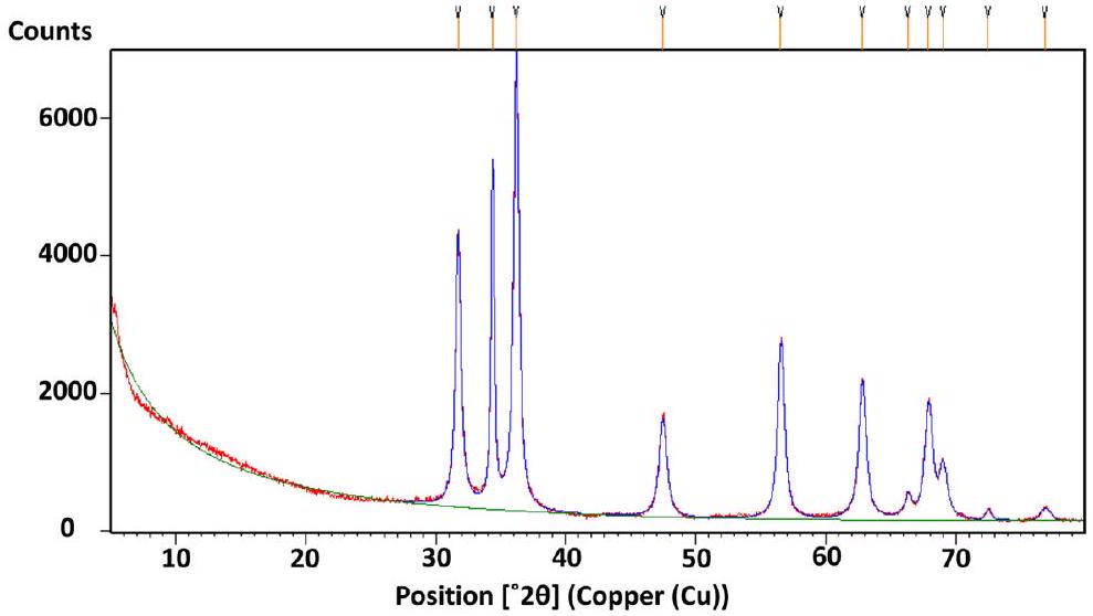
يعد تحليل حيود الأشعة السينية (XRD) تقنية راسخة تُستخدم للتعرف الهيكلي وتحديد حجم البلورات في جزيئات ZnO النانوية المُصنَّعة. في هذه الدراسة، يتم تقديم نمط XRD لجزيئات ZnO النانوية المُصنَّعة في الشكل 3. تم ملاحظة قمم حيود الأشعة السينية، التي تتميز بشدتها العالية، عند نقاط محددة.القيم، وهي، و . هذه
ب)
ج)
الشكل 1. (أ) تخليق جزيئات ZnO النانوية. (ب) طيف الامتصاص للأشعة فوق البنفسجية والمرئية لاستخراج الشيلجيت. (ج) طيف الامتصاص للأشعة فوق البنفسجية والمرئية لجزيئات ZnO من استخراج الشيلجيت.
الشكل 2. طيف FTIR لمستخلص الشيلاجيت (A) ومستخلص جزيئات ZnO من مستخلص الشيلاجيت (B).
الشكل 3. نمط حيود الأشعة السينية لجزيئات ZnO النانوية (المصنوعة من مستخلص مائي من الشيلجيت).
تتوافق القمم مع مستويات الشبكة (100)، (002)، (101)، (102)، (110)، (103)، (112)، و(201) على التوالي. من خلال مقارنة نمط حيود الأشعة السينية المرصود مع بيانات JCPDS القياسية (اللجنة المشتركة لمعايير حيود المساحيق)، تم العثور على أن جميع القمم تتطابق مع بطاقة JCPDS القياسية رقم 76-0704. هذا يؤكد أن جزيئات ZnO النانوية التي تم تصنيعها تمتلك بنية بلورية من نوع وورتزيت سداسي. يوفر تحليل حيود الأشعة السينية رؤى مهمة حول الطبيعة البلورية والخصائص الهيكلية لجزيئات ZnO النانوية التي تم تصنيعها. تطابق قمم الحيود مع البيانات القياسية يثبت البنية البلورية ويؤكد نجاح تصنيع جزيئات ZnO النانوية في الهيكل السداسي المطلوب. تم حساب حجم البلورة (D) باستخدام صيغة ديباي-شيرر:
حيث، D — حجم بلورات أكسيد الزنك؛ —طول موج مصدر الأشعة السينية 0.15406 نانومتر في (XRD)، —عرض كامل عند نصف الحد الأقصى لقمة الانكسار – ثابت شيرر، s (0.94)، زاوية براج.
تحليل حجم الجسيمات لجزيئات أكسيد الزنك النانوية
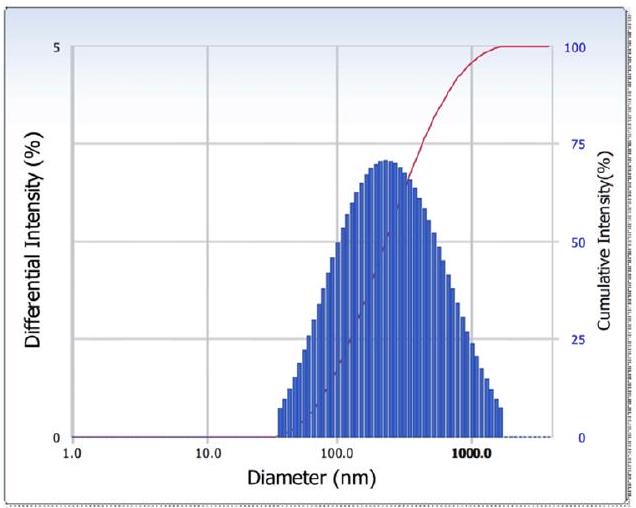
البيانات التي تم الحصول عليها من محلل حجم الجسيمات قدمت دليلاً واضحاً على أن جزيئات ZnO النانوية التي تم تصنيعها باستخدام مستخلص الشيلجيت كانت أصغر. تم قياس الحجم المتوسط للجزيئات النانوية التي تم الحصول عليها باستخدام مستخلص الشيلجيت ليكون 348 نانومتر، وتم العثور على مؤشر التوزيع المتعدد ليكون 0.322 (الشكل 4). تشير هذه النتائج إلى أن مستخلص الشيلجيت أنتج جزيئات ZnO النانوية بحجم إجمالي أصغر.
تحليل SEM لجزيئات ZnO النانوية
تم تحديد الشكل، بما في ذلك الحجم والشكل، لجزيئات ZnO النانوية التي تم تصنيعها بطريقة خضراء من خلال المجهر الإلكتروني الماسح. قدمت صور SEM رؤى حول الخصائص الهيكلية للجزيئات النانوية المصنعة. على وجه التحديد، أظهرت جزيئات ZnO النانوية المصنعة من مستخلص الشيلجيت شكلًا يشبه الرقائق النانوية بأحجام تتراوح من 75 إلى 400 نانومتر. تسلط هذه النتائج الضوء على الهياكل الشبيهة بالرقائق النانوية لجزيئات ZnO مع مستخلص الشيلجيت التي تنتج جزيئات نانوية كروية (الشكل 5).
تحليل EDAX لجزيئات ZnO النانوية
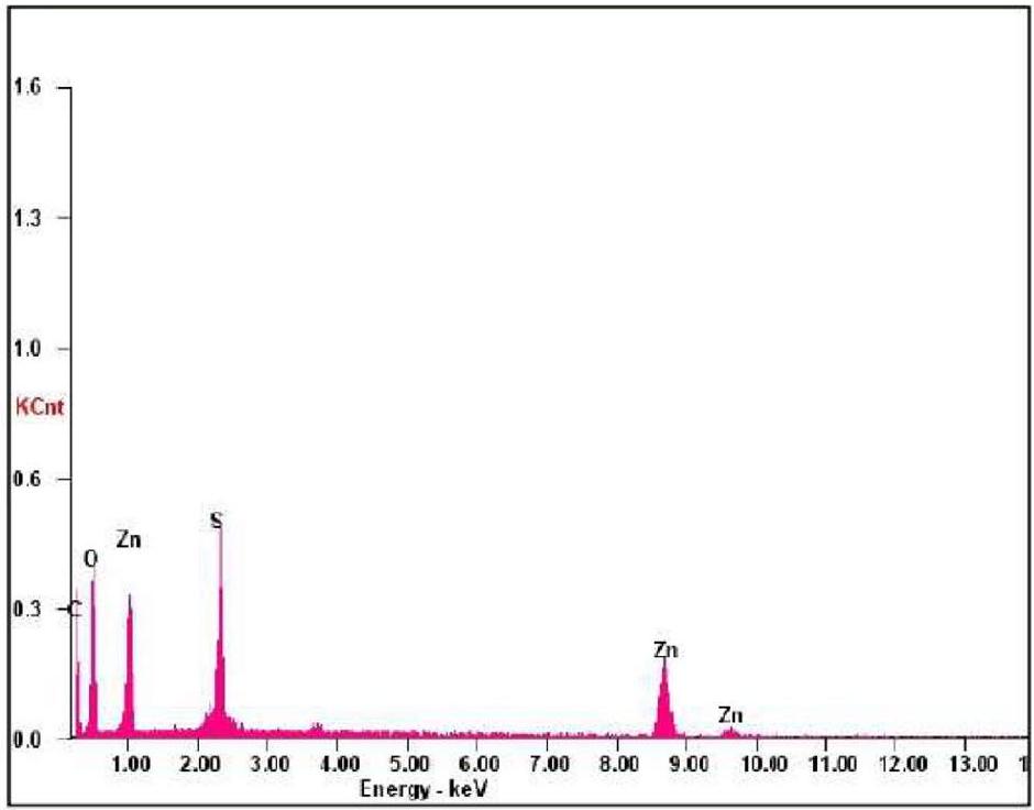
تؤكد طيفيات EDAX لجزيئات النانو نقاء جزيئات ZnO النانوية المُصنَّعة وتظهر أن العينة تحتوي على الطور الضروري من الزنك والأكسجين. كان الوزن الذري للزنك هوبينما كان وزنه الحالي. في نفس الوقت، كان الوزن الذري للأكسجين بينما كان وزنه الحالي، بينما كانت المكونات الثانوية الأخرى الموجودة في جزيئات ZnO بسبب وجود مستخلص الشيلجيت، كما هو موضح في الشكل 6. عرضت طيف EDAX ثلاثة قمم للزنك حول و 9.6 keV، على التوالي وقمة فريدة للأكسجين عند، والتي تعتبر نموذجية لـ.
التأثير المضاد للسرطان في المختبر لجزيئات أكسيد الزنك النانوية
تم استخدام مستخلص الشيلجيت بنجاح لتخليق جزيئات نانوية من ZnO، والتي أظهرت نشاطًا مضادًا للسرطان كبيرًا في المختبر ضد خطوط خلايا سرطان عنق الرحم البشرية (HeLa). وُجد أن النشاط المضاد للسرطان يعتمد على الجرعة، حيث تراوحت نسبة البقاء من 84.47 إلى عند تركيزات من مل إلىعلى التوالي (الأشكال 7A، 8A). ومن الجدير بالذكر أن نشاط جزيئات ZnO النانوية كان قابلاً للمقارنة مع نشاط سيسبلاتين، وهو عامل مضاد للسرطان قياسي، الذي أظهر نسب نشاط تبلغإلى بنفس التركيزات (الأشكال 7A، 8A). علاوة على ذلك، القيمة، التي تمثل التركيز الذي تمنع عنده جزيئات ZnO النانويةلنمو خلايا السرطان، تم تحديده على أنه. هذا كانت القيمة معتدلة مقارنة بالتحكم الإيجابي سيسبلاتين، الذي كان لديه قيمة لـ. هذه النتائج أبرزت النشاط المضاد للسرطان القوي لجزيئات ZnO النانوية التي تم تصنيعها باستخدام مستخلص الشيلجيت. تم أيضًا دراسة تأثير جزيئات ZnO النانوية على خط خلايا طبيعي، وهو خط خلايا فيرو. أظهر اختبار MTT أن
توزيع الشدة
ACF
نتائج التوزيع (تابع)
نتائج الكومولات القطر
: 192.9
(ن م)
ذروة
القطر (نانومتر)
الانحراف المعياري
مؤشر التوزيع المتعدد (P.I.) : 0.322
1
٣٤٨.٨
٣١١.٩
ثابت الانتشار
I’m sorry, but it seems that there is no text provided for translation. Please provide the text you would like to have translated.
( )
2
0.0
0.0
٣
0.0
0.0
حالة القياس
٤
0.0
0.0
درجة الحرارة
25.0
٥
0.0
0.0
اسم المذيب
: ماء
متوسط
٣٤٨.٨
٣١١.٩
معامل الانكسار
1.3328
لزوجة
: 0.8878
(cP)
البقايا :
(حسناً)
شدة التشتت
: 32284
(cps)
مخفف 1
: 0.72
(% )
الشكل 4. تحليل حجم الجسيمات لجزيئات ZnO النانوية المستخرجة من مستخلص الشيلجيت.
الشكل 5. صور SEM لجزيئات ZnO النانوية بتكبيرات مختلفة من مستخلص الشيلجيت.
جزيئات نانو أكسيد الزنكالقيمة كانت فوق (الأشكال 7B، 8B). هذه البيانات أكدت أن جزيئات ZnO النانوية المختبرة آمنة بخلاف خط الخلايا الطبيعي.
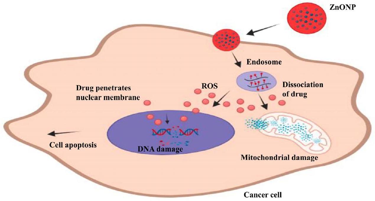
يمكن أن يُعزى النشاط المضاد للسرطان لجزيئات ZnO النانوية (ZnONPs) على الخلايا السرطانية إلى عدة آليات محتملة. أولاً، تم الإبلاغ عن أن جزيئات ZnO النانوية تزيد من تراكم أنواع الأكسجين التفاعلية داخل الخلايا السرطانية. تعتبر أنواع الأكسجين التفاعلية جزيئات شديدة التفاعل يمكن أن تسبب إجهادًا أكسديًا وت disrupt العمليات الخلوية، مما يؤدي إلى تلف الحمض النووي وفي النهاية موت الخلايا (الشكل 9).علاوة على ذلك، يمكن لجزيئات ZnO النانوية أن تحفز عملية الموت الخلوي المبرمج، وهو عملية موت الخلايا المبرمجة. يعتبر الموت الخلوي المبرمج آلية حاسمة للحفاظ على توازن الخلايا والتخلص من الخلايا التالفة أو غير الطبيعية. تشير تحفيز الموت الخلوي المبرمج بواسطة جزيئات ZnO النانوية إلى أنها يمكن أن تحفز مسارات الإشارة التي تعزز موت الخلايا في خلايا السرطان..
الشكل 6. تحليل EDAX لجزيئات ZnO النانوية المستخرجة من مستخلص الشيلجيت.
عنصر
نسبة الوزن %
في المئة
سي كي
٤٥.٧٠
٥٨.٩٥
حسناً
٣٦.١٨
٣٥.٠٤
SK
06.97
03.37
ZnK
11.15
02.64
ماتريكس
تصحيح
زاف
الشكل 7. السمية الخلوية التي تم تقييمها بواسطة اختبار MTT بعد 24 ساعة من العلاج. (A) خلايا هيلا بعد العلاج بتركيزات مختلفة ( و ) من جزيئات ZnO النانوية المستخرجة من مستخلص الشيلجيت مقارنةً بالمعيار المرجعي سيسبلاتين. (B) خلايا فيرو الطبيعية بعد المعالجة بتركيزات مختلفة ( و ) من جزيئات ZnO النانوية المستخرجة من مستخلص الشيلجيت.
علاوة على ذلك، أظهرت جزيئات ZnO النانوية أنها تسبب ضررًا للميتوكوندريا وإجهادًا مؤكسدًا بشكل خاص في خلايا السرطان. تلعب الميتوكوندريا دورًا حيويًا في تنظيم عمليات خلوية متنوعة، بما في ذلك موت الخلايا المبرمج وعمليات الأيض للطاقة. من خلال استهداف الميتوكوندريا، تعطل جزيئات ZnO النانوية الوظيفة الطبيعية لهذه العضيات، مما يؤدي إلى ضعف إنتاج الطاقة وزيادة إجهاد الأكسدة داخل خلايا السرطان. يمكن أن يؤدي هذا الضرر الميتوكوندري في النهاية إلى تفعيل مسارات موت الخلايا الخاصة بخلايا السرطان.. في الملخص، الآليات الكامنة وراء النشاط المضاد للسرطان لجزيئات ZnO النانوية تشمل تحفيز تراكم ROS داخل الخلايا، وتحفيز موت الخلايا المبرمج، وتعطيل وظيفة الميتوكوندريا. تساهم هذه التأثيرات مجتمعة في السمية الانتقائية لجزيئات ZnO النانوية تجاه خلايا السرطان، مما يجعلها مرشحًا واعدًا لعلاج السرطان.
الخاتمة
تمت دراسة جزيئات ZnO النانوية المستخلصة من الشيلجيت لخصائصها المضادة للسرطان. لقد استدعت التكلفة العالية والآثار الجانبية السلبية المرتبطة بالعوامل الكيميائية التقليدية استكشاف المنتجات الطبيعية كبدائل محتملة أو مساعدات للأدوية المضادة للسرطان التقليدية. وقد أبرزت العديد من الدراسات الإمكانات الكبيرة للشيلجيت كعامل مضاد للسرطان. أظهرت النتائج أن جزيئات أكسيد الزنك النانوية المستخلصة من الشيلجيت أظهرت خصائص مضادة للسرطان واعدة. ومع ذلك، هناك حاجة إلى مزيد من البحث لتحديد
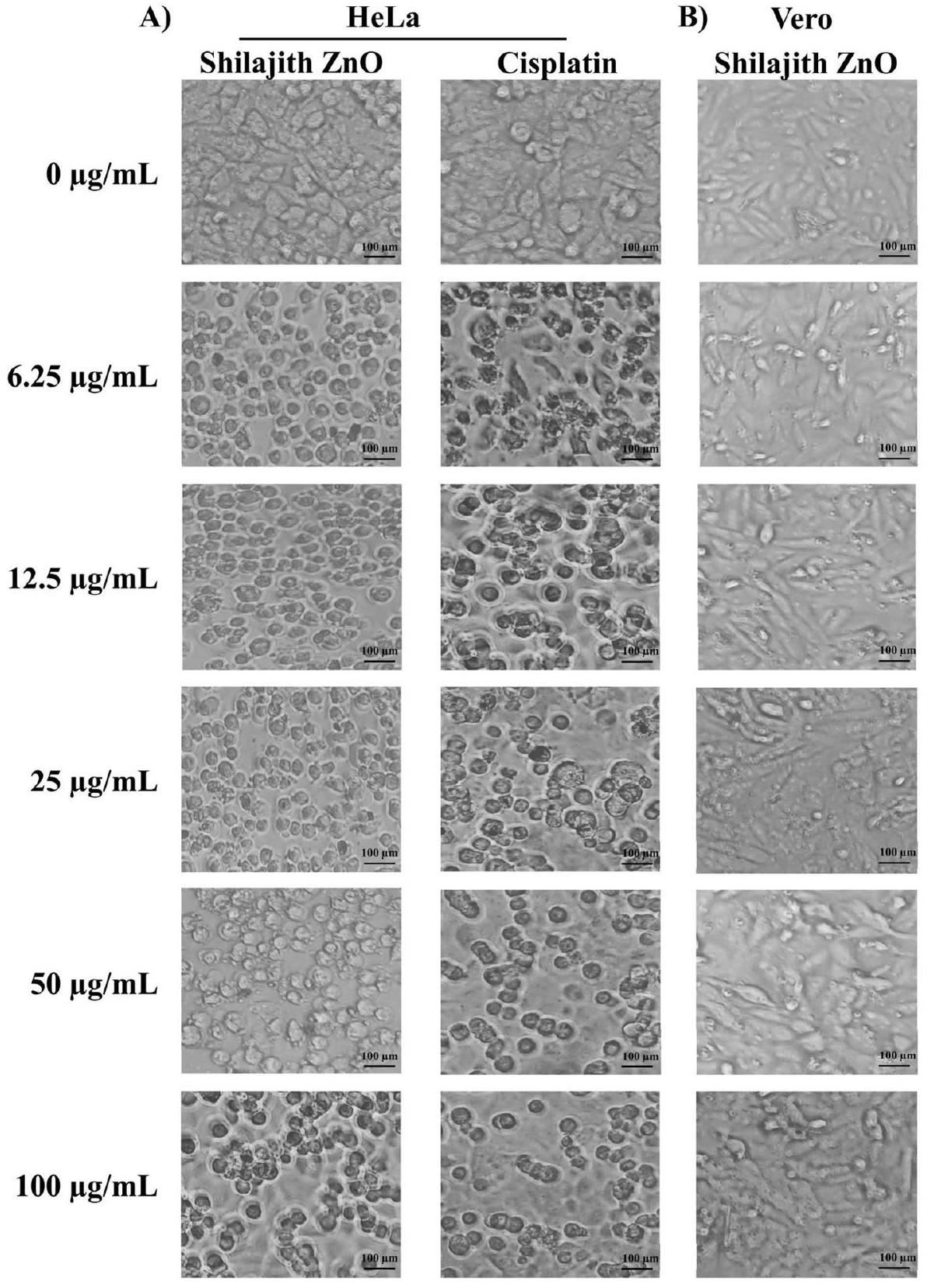
الشكل 8. صور مجهرية للخلايا المعالجة بتركيزات مختلفة بعد 24 ساعة. (أ) خلايا هيلا بعد المعالجة بتركيزات مختلفة ( و) من جزيئات ZnO النانوية المستخلصة من الشيلجيت والمعيار المرجعي سيسبلاتين. (ب) خلايا فيرو الطبيعية بعد المعالجة بتركيزات مختلفة ( و) من جزيئات ZnO النانوية (المصنوعة من مستخلص مائي من الشيلجيت). تم ملاحظة الخلايا وتصويرها بواسطة المجهر المقلوب ().
تحديد الآلية الدقيقة للعمل ضد سرطان عنق الرحم، بحيث يمكن التحقق من هذه التركيبة النانوية الجديدة من ZnO في نموذج حيواني محدد واستخدامها لصالح المجتمع العالمي.
المواد والطرق
المواد الكيميائية والمركبات
تم شراء الشيلجيت من شركة يوككا، الهند. تم الحصول على أسيتات الزنك من مختبرات سيسكو للأبحاث الخاصة المحدودة (SRL)، الهند و تم شراء أوراق الترشيح من ميلبورو. تم شراء الإيزوبروبانول من الدرجة التحليلية من شركة ثيرمو فيشر العلمية الهند الخاصة المحدودة. كانت جميع المذيبات والمواد الكيميائية الأخرى المستخدمة من الدرجة التحليلية. تم الحصول على السيسبلاتين من المتجر الطبي المحلي. تم استخدام وسط دلبوكو المعدل (DMEM)، مصل الجنين البقري (FBS)، تحضيرات المضادات الحيوية ومضادات الفطريات لزراعة الخلايا، تريبسين EDTA
الشكل 9. الآلية السامة المحتملة لجزيئات أكسيد الزنك النانوية ضد خلايا السرطان.
محلول و 3-(4، 5-ثنائي ميثيل ثيازول-2-يل)-2، 5-ثنائي فينيل تيترازوليوم بروميد) (MTT) تم الحصول عليها من مختبرات هاي ميديا الخاصة المحدودة، الهند.
تحضير مستخلص الشيلجيت
تم إضافة حوالي 5 جرام من مسحوق الشيلجيت إلى 100 مل من الماء المقطر المزدوج في دورق سعة 250 مل وتم تسخينه عند لمدة 15 دقيقة. تم تبريد الخليط في درجة حرارة الغرفة وتم ترشيحه باستخدام ورق ترشيح واطمان رقم 1 للحصول على محلول واضح عن طريق إزالة الحطام الصلب. تم تخزين السائل الناتج عند لمزيد من الدراسات.
التخليق الأخضر لجزيئات أكسيد الزنك النانوية
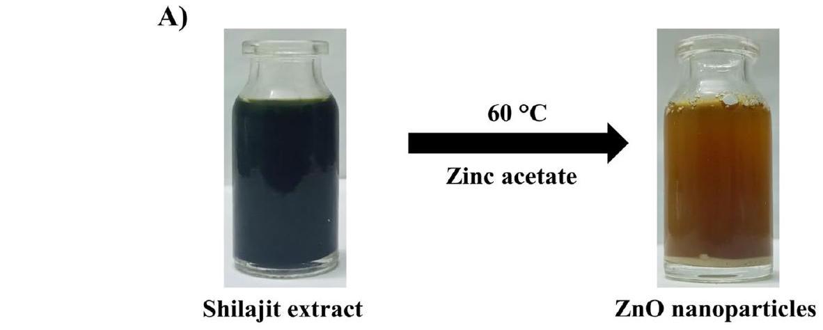
في البداية، تم أخذ 35 مل من محلول أسيتات الزنك (200 مليمول) في دورق نظيف. تم إضافة 15 مل من مستخلص الشيلجيت إلى محلول أسيتات الزنك بطريقة قطرة قطرة. بعد الإضافة، تم تحريك الخليط لمدة 6 ساعات لضمان الخلط الجيد. بعد 6 ساعات، تمت إضافة محلول هيدروكسيد الصوديوم (2 م) وتم الاحتفاظ به على محرك مغناطيسي عند طوال الليل. تم السماح للخليط بالتبريد وتم الطرد المركزي عند لمدة 15 دقيقة. تم غسل جزيئات ZnO النانوية الناتجة بالماء المقطر المزدوج تليها الإيزوبروبانول وتم تجفيفها عند لمدة ساعتين (الشكل 1A).
توصيف جزيئات ZnO النانوية
تحليل الطيف الضوئي UV-Vis
تم تأكيد تشكيل جزيئات الزنك النانوية عن طريق قياس ماكس باستخدام مطياف ضوئي UV-Visible (شركة شيمادزو، كيوتو، اليابان).
تحليل حيود الأشعة السينية (XRD)
تم تسجيل قياس XRD بواسطة جهاز حيود الأشعة السينية (X’Pert PANalytical) الذي يعمل عند جهد 40 كيلو فولت وتيار 30 مللي أمبير مع إشعاع لتحديد المرحلة البلورية وتحديد المادة.
تحليل FTIR
تم تحليل المساحيق الدقيقة لجزيئات ZnO النانوية بواسطة مطياف FTIR ((شركة شيمادزو، كيوتو، اليابان) لتحديد المجموعات الوظيفية الموجودة في الجزيئات النانوية المصنعة.
تحليل SEM
تم التحقيق في شكل الجسيمات وخصائص السطح لتراكيب الجسيمات النانوية المعدة حديثًا بواسطة المجهر الإلكتروني الماسح (SEM) (جهاز JEOL JMS-6390) عند.
تحليل حجم الجسيمات
تم استخدام محلل حجم الجسيمات (Zetasizer Nano ZS، شركة مالفرن إنسترومنتس المحدودة، ورسيستير، المملكة المتحدة) لتحديد توزيع حجم الجسيمات والشحنة السطحية للجزيئات النانوية.
تحليل EDAX
تم تسجيل التحليل العنصري للتكوين بواسطة مطياف الأشعة السينية المشتت للطاقة (نموذج SUTW-SAPHIRE).
النشاط المضاد للسرطان في المختبر
خطوط الخلايا ووسط الزراعة
تم الحصول على خط خلايا هيلا (خط خلايا سرطان عنق الرحم) وخط خلايا فيرو من NCCS، بوني، الهند، وتم زراعة الخلايا في وسط DMEM معزز بـ 10% من مصل الجنين البقري غير النشط (FBS)، بنسلين (100 وحدة دولية/مل)، ستربتوميسين () في جو رطب عند عند حتى تكتمل.
اختبار MTT
تم قياس حيوية خلايا ZnO النانوية المعالجة باستخدام اختبار MTT كما هو موصوف سابقًا. تم تريبسين الخلايا في زراعة الخلايا أحادية الطبقة وتم ضبط عدد الخلايا على خلايا باستخدام وسط DMEM الذي يحتوي على FBS. إلى كل بئر من صفيحة الميكروتيتر ذات 96 بئرًا، تم إضافة من تعليق الخلايا المخفف ( خلايا/بئر). بعد 24 ساعة، عندما تم تشكيل طبقة أحادية جزئية، تم التخلص من السائل الفائق، وغسل الطبقة الأحادية مرة واحدة بالوسط و من تركيزات مختلفة (6.25، 12.5، 25، 50 و من عينات الاختبار تمت إضافتها إلى الطبقة الأحادية الجزئية في صفائح الميكروتيتر. ثم تم حضانة الصفيحة عند لمدة 24 ساعة في جو. بعد الحضانة، تم التخلص من المحاليل الاختبارية في الآبار وتم إضافة من MTT ( من MTT في PBS) إلى كل بئر. تم حضانة الصفيحة لمدة 4 ساعات عند في جو. تمت إزالة السائل الفائق، وتم إضافة من DMSO، وتم هز الصفيحة برفق لذوبان الفورمازان المتكون. تم قياس الامتصاص باستخدام قارئ الميكروتيتر عند طول موجي 570 نانومتر. تم حساب نسبة الحيوية باستخدام الصيغة التالية:
توفر البيانات
تتوفر مجموعات البيانات التي تم إنشاؤها وتحليلها خلال الدراسة الحالية في مستودع زينودو، https://doi.org/10.5281/zenodo. 8329357.
تاريخ الاستلام: 9 أغسطس 2023؛ تاريخ القبول: 16 يناير 2024
تم النشر عبر الإنترنت: 25 يناير 2024
References
Sung, H. et al. Global cancer statistics 2020: GLOBOCAN estimates of incidence and mortality worldwide for 36 cancers in 185 countries. CA Cancer J. Clin. 71, 209-249 (2021).
Baig, N., Kammakakam, I. & Falath, W. Nanomaterials: A review of synthesis methods, properties, recent progress, and challenges. Mater. Adv. 2, 1821-1871 (2021).
Faisal, S. et al. Green synthesis of zinc oxide ( ZnO ) nanoparticles using aqueous fruit extracts of Myristica fragrans: Their characterizations and biological and environmental applications. ACS Omega 6, 9709-9722 (2021).
Rizwan, M. et al. Enterobacter hormaechei-driven novel biosynthesis of tin oxide nanoparticles and evaluation of their anti-aging, cytotoxic, and enzyme inhibition potential. ACS Omega 8, 27439-27449 (2023).
Faisal, S. et al. Curcuma longa mediated synthesis of copper oxide, nickel oxide and bimetallic hybrid nanoparticles: Characterization and evaluation for antimicrobial, anti-parasitic and cytotoxic potentials. Coatings 11, 849 (2021).
Abdullah, Al-Radadi, N. S., Hussain, T., Faisal, S. & Ali Raza Shah, S. Novel biosynthesis, characterization and bio-catalytic potential of green algae (Spirogyra hyalina) mediated silver nanomaterials. Saudi J. Biol. Sci. 29, 411-419 (2022).
Abdullah, et al. Green synthesis and characterization of copper and nickel hybrid nanomaterials: Investigation of their biological and photocatalytic potential for the removal of organic crystal violet dye. J. Saudi Chem. Soc. 26, 101486 (2022).
Kolahalam, L. A. et al. Review on nanomaterials: Synthesis and applications. Mater. Today Proc. 18, 2182-2190 (2019).
Shah, R., Shah, S. A., Shah, S., Faisal, S. & Ullah, F. Green synthesis and antibacterial activity of gold nanoparticles of Digera muricata. Pharm. Sci. https://doi.org/10.36468/pharmaceutical-sciences.659 (2020).
Ullah, R. et al. In vitro and in vivo applications of Euphorbia wallichii shoot extract-mediated gold nanospheres. Green Process. Synth. 10, 101-111 (2021).
Faisal, S. et al. In vivo analgesic, anti-inflammatory, and anti-diabetic screening of Bacopa monnieri-synthesized copper oxide nanoparticles. ACS Omega 7, 4071-4082 (2022).
Faisal, S. et al. Edible mushroom (Flammulina velutipes) as biosource for silver nanoparticles: From synthesis to diverse biomedical and environmental applications. Nanotechnology 32, 065101 (2021).
Imran, M. et al. In vitro examination of anti-parasitic, anti-Alzheimer, insecticidal and cytotoxic potential of Ajuga bracteosa Wallich leaves extracts. Saudi J. Biol. Sci. 28, 3031-3036 (2021).
Kainat, et al. Exploring the therapeutic potential of Hibiscus rosa sinensis synthesized cobalt oxide ( -NPs) and magnesium oxide nanoparticles (MgO-NPs). Saudi J. Biol. Sci. 28, 5157-5167 (2021).
Theiss, F. L., Ayoko, G. A. & Frost, R. L. Synthesis of layered double hydroxides containing and layer cations by co-precipitation methods-A review. Appl. Surf. Sci. 383, 200-213 (2016).
Cao, D. et al. Nonmetal sulfur-doped coral-like cobalt ferrite nanoparticles with enhanced magnetic properties. J. Mater. Chem. C 4, 951-957 (2016).
Khan, M. I. et al. Monotheca buxifolia driven synthesis of zinc oxide nano material its characterization and biomedical applications. Micromachines 13, 668 (2022).
Shah, S. et al. Engineering novel gold nanoparticles using Sageretia thea leaf extract and evaluation of their biological activities. J. Nanostruct. Chem. 12, 129-140 (2022).
Jan, H. et al. The Aquilegia pubiflora (Himalayan columbine) mediated synthesis of nanoceria for diverse biomedical applications. RSC Adv. 10, 19219-19231 (2020).
Dong, H. et al. Lanthanide nanoparticles: From design toward bioimaging and therapy. Chem. Rev. 115, 10725-10815 (2015).
Faisal, S. et al. Fagonia cretica-mediated synthesis of manganese oxide nanomaterials their characterization and evaluation of their bio-catalytic and enzyme inhibition potential for maintaining flavor and texture in apples. Catalysts 12, 558 (2022).
Zafar, S. et al. Development of iron nanoparticles (FeNPs) using biomass of enterobacter: Its characterization, antimicrobial, antiAlzheimer’s, and enzyme inhibition potential. Micromachines 13, 1259 (2022).
Faisal, S. et al. Biofabrication of silver nanoparticles employing biomolecules of Paraclostridium benzoelyticum strain: Its characterization and their in-vitro antibacterial, anti-aging, anti-cancer and other biomedical applications. Microsc. Res. Tech. 86, 846-861 (2023).
Bai, D.-P., Zhang, X.-F., Zhang, G.-L., Huang, Y.-F. & Gurunathan, S. Zinc oxide nanoparticles induce apoptosis and autophagy in human ovarian cancer cells. IJN 12, 6521-6535 (2017).
Dhivya, R. et al. Biocompatible curcumin loaded PMMA-PEG/ZnO nanocomposite induce apoptosis and cytotoxicity in human gastric cancer cells. Mater. Sci. Eng. C 80, 59-68 (2017).
George, D., Maheswari, P. U. & Begum, K. M. M. S. Synergic formulation of onion peel quercetin loaded chitosan-cellulose hydrogel with green zinc oxide nanoparticles towards controlled release, biocompatibility, antimicrobial and anticancer activity. Int. J. Biol. Macromol. 132, 784-794 (2019).
Huang, X., Zheng, X., Xu, Z. & Yi, C. ZnO-based nanocarriers for drug delivery application: From passive to smart strategies. Int. J. Pharm. 534, 190-194 (2017).
Zahin, N. et al. Nanoparticles and its biomedical applications in health and diseases: Special focus on drug delivery. Environ. Sci. Pollut. Res. 27, 19151-19168 (2020).
Abdullah, et al. Multifunctional Spirogyra-hyalina-mediated barium oxide nanoparticles (BaONPs): Synthesis and applications. Molecules 28, 6364 (2023).
Faisal, S. et al. Exploring the antibacterial, antidiabetic, and anticancer potential of Mentha arvensis extract through in-silico and in-vitro analysis. BMC Complement. Med. Ther. 23, 267 (2023).
Costa, D., Savio, L. & Pradier, C.-M. Adsorption of amino acids and peptides on metal and oxide surfaces in water environment: A synthetic and prospective review. J. Phys. Chem. B 120, 7039-7052 (2016).
Pudukudy, M. & Yaakob, Z. Facile synthesis of quasi spherical ZnO nanoparticles with excellent photocatalytic activity. J. Clust. Sci. 26, 1187-1201 (2015).
Scimeca, M., Bischetti, S., Lamsira, H. K., Bonfiglio, R. & Bonanno, E. Energy Dispersive X-ray (EDX) microanalysis: A powerful tool in biomedical research and diagnosis. Eur. J. Histochem. https://doi.org/10.4081/ejh.2018.2841 (2018).
Zarrabi, M., Haghighi, M., Alizadeh, R. & Mahboob, S. Solar-light-driven photodegradation of organic dyes on sono-dispersed ZnO nanoparticles over graphene oxide: Sono vs. conventional catalyst design. Sep. Purif. Technol. 211, 738-752 (2019).
Al-Radadi, N. S. et al. Zingiber officinale driven bioproduction of ZnO nanoparticles and their anti-inflammatory, anti-diabetic, anti-Alzheimer, anti-oxidant, and anti-microbial applications. Inorganic Chem. Commun. 140, 109274 (2022).
Faisal, S. et al. Paraclostridium benzoelyticum bacterium-mediated zinc oxide nanoparticles and their in vivo multiple biological applications. Oxid. Med. Cell. Longev. 2022, 1-15 (2022).
Faisal, S. et al. Bio-catalytic activity of novel Mentha arvensis intervened biocompatible magnesium oxide nanomaterials. Catalysts 11, 780 (2021).
Aminul, I. et al. Biotransformation of 3-hydroxydibenzo-a-pyrone into 3,8 dihydroxydibenzo-a-pyrone and aminoacyl conjugates by Aspergillus niger isolated from native shilajit. Electron. J. Biotechnol. 11, 0-0 (2008).
Kapoor, V. K., Dureja, J. & Chadha, R. Herbals in the control of ageing. Drug Discov. Today 14, 992-998 (2009).
Carrasco-Gallardo, C., Guzmán, L. & Maccioni, R. B. Shilajit: A natural phytocomplex with potential procognitive activity. Int. J. Alzheimer’s Dis. 2012, 1-4 (2012).
Khanna, R., Witt, M., Khalid Anwer, Md., Agarwal, S. P. & Koch, B. P. Spectroscopic characterization of fulvic acids extracted from the rock exudate Shilajit. Organic Geochem. 39, 1719-1724 (2008).
Alamdari, S. et al. Preparation and characterization of zinc oxide nanoparticles using leaf extract of Sambucus ebulus. Appl. Sci. 10, 3620 (2020).
Song, Z. et al. Characterization of optical properties of ZnO nanoparticles for quantitative imaging of transdermal transport. Biomed. Opt. Express 2, 3321 (2011).
Stanciu, G. D. et al. Alzheimer’s disease pharmacotherapy in relation to cholinergic system involvement. Biomolecules 10, 40 (2019).
Xiong, H.-M., Xu, Y., Ren, Q.-G. & Xia, Y.-Y. Stable aqueous ZnO@polymer core-shell nanoparticles with tunable photoluminescence and their application in cell imaging. J. Am. Chem. Soc. 130, 7522-7523 (2008).
Jan, H. et al. Plant-Based synthesis of zinc oxide nanoparticles (ZnO-NPs) using aqueous leaf extract of Aquilegia pubiflora: Their antiproliferative activity against HepG2 cells inducing reactive oxygen species and other in vitro properties. Oxid. Med. Cell. Longev. 2021, 1-14 (2021).
Chaudhuri, S. K. & Malodia, L. Biosynthesis of zinc oxide nanoparticles using leaf extract of Calotropis gigantea: Characterization and its evaluation on tree seedling growth in nursery stage. Appl. Nanosci. 7, 501-512 (2017).
Mosmann, T. Rapid colorimetric assay for cellular growth and survival: Application to proliferation and cytotoxicity assays. J. Immunol. Methods 65, 55-63 (1983).
مساهمات المؤلفين
صمم S.R. وأشرف على الدراسة. قام P.P. وN.A.S. وK.K. وR.R. بإجراء التجارب. قام J.J.S. وV.N. وM.G. وN.S. بصياغة المخطوطة. قام P.P. وS.G.P. وV.S.P. وV.G. وP.P.T. وS.G. بتحليل البيانات وتحسين المخطوطة. قرأ جميع المؤلفين ووافقوا على المخطوطة.
المصالح المتنافسة
يعلن المؤلفون عدم وجود مصالح متنافسة.
معلومات إضافية
يجب توجيه المراسلات والطلبات للحصول على المواد إلى S.R.
معلومات إعادة الطبع والتصاريح متاحة علىwww.nature.com/reprints.
ملاحظة الناشر تظل شركة سبرينجر ناتشر محايدة فيما يتعلق بالمطالبات القضائية في الخرائط المنشورة والانتماءات المؤسسية.
قسم علم الأحياء الجزيئي والخلايا، مختبرات غرينسميد، ثورا باكام، تشيناي 600097، الهند. قسم الكيمياء الصيدلانية، كلية EGS بيلاي للصيدلة، ناغاباتينام 611002، الهند.قسم الصيدلة، كلية سانت ماري للصيدلة، سيكندر آباد، تيلانجانا 500025، الهند.قسم الكيمياء الصيدلانية، كلية الصيدلة، معهد بهارات للتعليم العالي والبحث (BIHER)، تشيناي 600044، الهند.قسم الكيمياء الصيدلانية، كلية باناي للصيدلة (تابعة لجامعة تاميل نادو د. م. ج. ر. الطبية، تشيناي)، دينديغول 624005، الهند.قسم العقاقير، كلية سي. إل. بايد ميثا للصيدلة، تشيناي 600096، الهند.مركز الدراسات المتقدمة في البلورات والبيوفيزياء، حرم غويندي، جامعة مدراس، تشيناي 600025، الهند.البريد الإلكتروني: gmlbioprojects@gmail.com
In the present study, ZnO nanoparticles have been synthesized using an aqueous extract of shilajit. The nanoparticles were characterized using different techniques such as UV (ultraviolet-visible spectrophotometer), FTIR (Fourier transform infrared), XRD (X-ray diffraction), particle size analysis, SEM (scanning electron microscope) and EDAX (Energy-dispersive X-ray) analysis. The UV absorption peak at 422.40 nm was observed for ZnO nanoparticles. SEM analysis showed the shape of nanoparticles to be spherical, FTIR spectrum confirmed the presence of zinc atoms, particle size analysis showed the nanoparticle size, EDAX confirmed the purity of ZnO nanoparticles whereas XRD pattern similar to that of JCPDS card for ZnO confirmed the presence of pure ZnO nanoparticles. The in vitro anticancer activity of ZnO nanoparticles against the HeLa cell line showed the value of compared to reference standard cisplatin. This finding confirms that ZnO nanoparticles from shilajit extract have potent cytotoxic effect on human cervical cancer cell lines.
Cancer is a serious burden on human health and is the second leading cause of death around the world. According to the World Health Organization (WHO) estimated that cancer-related deaths are more common in developed countries than in developing countries. Cervical cancer develops in a woman’s cervix and is the fourth most common cancer type in women worldwide. In 2020, 604,000 women were diagnosed with cervical cancer worldwide and 342,000 died as a result of the disease . Cancer is typically treated by conventional therapies such as chemotherapy, radiotherapy, and surgery. Although all these therapies are very effective in killing, cancer cells but also result in many serious side effects.
Recently, nanotechnology-based nanosystems, with high biocompatibility, easy surface functionalization, cancer targeting and drug delivery capacity, have demonstrated the potential to overcome these side effects. In recent times, nanotechnology has gained global attention due to its wide-ranging applications in diverse fields, including optoelectronics, biomedical sciences, mechanics, chemical industry, space industries, catalysis, and drug-gene delivery . Green nanotechnology is an emerging field in the development of environmentally friendly nanoproducts and their utilization to achieve sustainable development . The adoption of green synthetic methods
has gained significant attention due to the growing demand for cost-effective, clean, non-toxic, biocompatible, and eco-friendly approaches that promote a safe environment.
Unlike other synthetic methods, the biosynthetic route eliminates the use of toxic chemicals, high energy requirements, and the need for elevated pressures and temperatures . The biological resources employed in this eco-friendly approach include plant extracts, bacteria, fungi, cyanobacteria, diatoms, seaweed, and enzymes for the synthesis of metal nanoparticles. Green synthesis techniques leverage natural agents such as plant extracts, sugars, vitamins, biodegradable polymers, and microbes, which act as both reductants and capping agents. Moreover, this approach minimizes the use of inorganic materials and primarily utilizes metal nanoparticles (NPs), metal oxides, and salts for nanoparticle fabrication . Green synthesis offers several advantages over alternative synthetic methods, including simplicity, cost-effectiveness, reproducibility, and relatively higher stability . Extensive research has been conducted on the use of naturally available resources to synthesize various metal nanoparticles, such as gold, silver, zinc, copper, titanium oxide, platinum, magnetite, and nickel . The different parts of the plant are used in Indian traditional medicine for the treatment of painful muscular spasm, dysentery, fever, rheumatism, asthma and as an expectorant and purgative . The starting materials of the metal nanomaterials are divalent and trivalent metal ions. There are different methods for the preparation of metal nanoparticles like chemical or photochemical methods. By using reducing agents, the metal ions are reduced to the metal nanoparticles. These have a high surface area and have the good adsorption ability of small molecules. They are widely used in different research areas, environmental and bioimaging studies. Not only a single nanoparticle but also the mixing of two or more nanoparticles with the size control can also be achieved. The plant extracts act as reducing and stabilizing agents for biosynthesis. Bio-reduction involves reducing metal ions or metal oxides to zero valence metal NPs with the help of phytochemicals like tannins, polysaccharides, polyphenolic compounds, vitamins and amino acids . Among different nanomaterials, no nanoparticles are considered to be safe both in vitro as well as in vivo. Due to its highly biocompatible and biodegradable nature, zinc oxide ( ZnO ) nanoparticles can be selected as potent nano-platforms for cancer treatment . The use of ZnO nanoparticles in cancer treatment has grabbed the interest of researchers and currently, a lot of studies have been published demonstrating the anti-cancerous activity of ZnO nanoparticles .
Shilajit, also known as Asphaltum, Black Bitumen, Silaras, salajit, and shilajatu, is a blackish-brown powder or an exudate from high mountain rocks especially in the Himalayas. Shilajit, sometimes mentioned as a mineral tar or resin, is a highly viscous substance readily soluble in water. Though it is now obtained from many other countries, Shilajit was traditionally sourced in India and Tibet. The health benefits of shilajit differ from region to region, depending on the place from which it is extracted. Shilajit contains fulvic acid, humic acid, 3 -hydroxydibenzo- -pyrone, hippuric acid along with a high phenolic residue that has great potential uses in certain diseases due to its various properties, including antioxidant, anti-inflammatory, anti-ulcerogenic, anti-fungal, anti-diabetic, anxiolytic, antiallergic, analgesic, cognitive memory enhancer, and immunomodulatory properties . Shilajit also possesses the ability to interact positively with other drugs and acts as a neuroprotective agent against cognitive disorders. Based on this literature, the current research was undertaken to synthesize shilajit-based ZnO nanoparticles and screened their effect on cervical cancer cell lines. To the best of our knowledge, a biological approach using shilajit extract has been used for the first time as a reducing material as well as a surface stabilizing agent for the synthesis of spherical-shaped ZnO-NPs.
Results and discussion UV analysis of ZnO NPs
The synthesized ZnO nanoparticles were systematically characterized and screened for anticancer activity. At first, the nanoparticles were analyzed by UV. The UV-visible spectral analysis of shilajit extract and ZnO nanoparticles from shilajit extract are shown in Fig. 1B,C. ZnO nanoparticles synthesized using shilajit extract displayed absorbance peaks at 357.90 nm and shilajit extract shows absorbance peaks at 422.40 nm . The shift of the absorbance peak towards a higher wavelength indicates the reduction in size from bulk molecules to the nano range. This may be due to material transitions, when an electron obtains energy, it transitions from a lower to a higher energy level .
FTIR analysis of ZnO NPs
The FTIR spectra of the shilajit extract and the synthesized ZnO nanoparticles from the shilajit extract are shown in Fig. 2. FTIR of the shilajit extract exhibited a broad peak at about which can be attributed to the stretching vibration of OH group and a peak at confirmed the stretching vibration of aliphatic , the presence of peaks at confirmed the presence of of carbonyl group and of aromatic system, respectively. C-O stretching was absorbed in which is further the zinc oxide nanoparticles synthesized from shilajit exhibited a characteristic peak at , and which was resembles of shilajit extract. The absorption peak associated with the stretching band is clearly absorbed at , which confirms the creation of ZnO -NPs. FTIR analysis of zinc nanoparticles revealed the presence of alkanes, phenol, alcohols, aromatics, alkenes, alkyl halides, and aliphatic amines vibrations . Furthermore, , and stretching vibrations were shown to generate maxima in carboxylic acid, polysaccharide, and amino acid, respectively .
XRD analysis of ZnO NPs
X-ray diffraction (XRD) is a well-established technique used for the structural identification and determination of crystalline size in synthesized ZnO nanoparticles. In this study, the XRD pattern of the synthesized ZnO nanoparticles is presented in Fig. 3. The X-ray diffraction peaks, characterized by their high intensity, were observed at specific values, namely , and . These
B)
C)
Figure 1. (A) Synthesis of ZnO nanoparticles. (B) UV-visible absorption spectrum of shilajit extract. (C) UVvisible absorption spectrum of ZnO nanoparticles from shilajit extract.
Figure 2. FTIR spectrum of (A) shilajit extract and (B) ZnO nanoparticles from shilajit extract.
Figure 3. X-ray diffraction pattern of ZnO nanoparticles (synthesized from an aqueous extract of shilajit).
peaks correspond to the lattice planes (100), (002), (101), (102), (110), (103), (112), and (201), respectively. By comparing the observed XRD pattern with the standard JCPDS (Joint Committee on Powder Diffraction Standards) data, it was found that all the peaks matched with the standard JCPDS card no. 76-0704. This confirms that the synthesized ZnO nanoparticles possess a wurtzite hexagonal-type crystal structure. The XRD analysis provides important insights into the crystalline nature and structural characteristics of the synthesized ZnO nanoparticles. The matching of the diffraction peaks with the standard data validates the crystal structure and confirms the successful synthesis of ZnO nanoparticles in the desired hexagonal structure. The crystallite size (D) was calculated using Debye-Scherrer’s formula:
where, D —crystalline size of zinc oxide; —wavelength of X-ray source 0.15406 nm in (XRD), —full width at half maximum of the diffraction peak-Scherer, s constant (0.94), 0-Bragg angle.
Particle size analysis of ZnO NPs
The data obtained from the particle size analyzer provided clear evidence that ZnO nanoparticles synthesized using shilajit extract were smaller. The average size of the nanoparticles obtained with shilajit extract was measured to be 348 nm and the polydispersity index was found to be 0.322 (Fig. 4). These results indicated that the shilajit extract yielded ZnO nanoparticles with a smaller overall size.
SEM analysis of ZnO NPs
The morphology, including the size and shape, of the green synthesized ZnO nanoparticles was determined through scanning electron microscopy. The SEM images provided insights into the structural characteristics of the synthesized nanoparticles. Specifically, ZnO nanoparticles synthesized from shilajit extract exhibited a morphology resembling nano-flakes with sizes ranging from 75 to 400 nm . These findings highlight the nano-flake-like structures of ZnO nanoparticles with shilajit extract yielding spherical nanoparticles (Fig. 5).
EDAX analysis of ZnO NPs
The EDAX spectra of the NPs confirm the purity of the synthesised ZnO NPs and show that the sample contains the necessary phase of Zn and O . The atomic weight of zinc was , while its weight present was . At the same time, the atomic weight of oxygen was , while its weight present was , while the other minor constituents present in the ZnO nanoparticles were due to the presence of shilajit extract, as shown in Fig. 6. The EDAX spectra displayed three peaks for zinc around and 9.6 keV , correspondingly and a singular peak for oxygen at , which are typical for .
In vitro anticancer effect of ZnO NPs
The shilajit extract has been successfully used to synthesize ZnO nanoparticles, which exhibited significant in vitro anticancer activity against human cervical cancer (HeLa) cell lines. The anticancer activity was found to be dose-dependent, with the percentage of viability ranging from 84.47 to at concentrations of mL to , respectively (Figs. 7A, 8A). Notably, the activity of ZnO nanoparticles was comparable to that of cisplatin, a standard anticancer agent, which displayed activity percentages of to at the same concentrations (Figs. 7A, 8A). Moreover, the value, which represents the concentration at which the ZnO nanoparticles inhibit of cancer cell growth, was determined to be . This value was moderate to that of the positive control cisplatin, which had an value of . These results highlighted the potent anticancer activity of the ZnO nanoparticles synthesized using the shilajit extract. The effect of the ZnO nanoparticles on a normal cell line i.e., the Vero cells line was also studied. The MTT assay revealed that
Intensity Distribution
ACF
Distribution Results (Contin)
Cumulants Results Diameter
: 192.9
(nm)
Peak
Diameter (nm)
Std. Dev.
Polydispersity Index (P.I.) : 0.322
1
348.8
311.9
Diffusion Const
:
( )
2
0.0
0.0
3
0.0
0.0
Measurement Condition
4
0.0
0.0
Temperature
: 25.0
5
0.0
0.0
Diluent Name
: WATER
Average
348.8
311.9
Refractive Index
: 1.3328
Viscosity
: 0.8878
(cP)
Residual :
(O.K)
Scattering Intensity
: 32284
(cps)
Attenuator 1
: 0.72
(%)
Figure 4. Particle size analysis of ZnO nanoparticles from shilajit extract.
Figure 5. SEM images of ZnO nanoparticles at different magnification from the shilajit extract.
the ZnO nanoparticle value was above (Figs. 7B, 8B). This data confirmed that the tested ZnO nanoparticles are safe apart from the normal cell line.
The anticancer activity of ZnO nanoparticles (ZnONPs) on cancerous cells can be attributed to several potential mechanisms. Firstly, ZnO nanoparticles have been reported to increase the accumulation of intracellular reactive oxygen species (ROS) within cancer cells. ROS are highly reactive molecules that can cause oxidative stress and disrupt cellular processes, leading to DNA damage and ultimately cell death (Fig. 9) . Furthermore, ZnO nanoparticles can induce apoptosis, which is a programmed cell death process. Apoptosis is a crucial mechanism for maintaining cellular homeostasis and eliminating damaged or abnormal cells. The stimulation of apoptosis by ZnO nanoparticles suggests that they can trigger signaling pathways that promote cell death in cancer cells .
Figure 6. EDAX analysis of ZnO nanoparticles from shilajit extract.
Element
Wt%
At%
CK
45.70
58.95
OK
36.18
35.04
SK
06.97
03.37
ZnK
11.15
02.64
Matrix
Correction
ZAF
Figure 7. Cytotoxicity assessed by MTT assay after 24 h treatment. (A) HeLa cells after treatment with various concentrations ( and ) of the ZnO nanoparticles from shilajit extract compared with reference standard cisplatin. (B) Normal Vero cells after treatment with various concentrations ( and ) of the ZnO nanoparticles from shilajit extract.
Moreover, ZnO nanoparticles have been shown to induce mitochondrial damage and oxidative stress specifically in cancer cells. Mitochondria play a vital role in regulating various cellular processes, including apoptosis and energy metabolism. By targeting mitochondria, ZnO nanoparticles disrupt the normal functioning of these organelles, leading to impaired energy production and further amplification of oxidative stress within cancer cells. This mitochondrial damage can ultimately trigger cell death pathways specific to cancer cells . In summary, the mechanisms underlying the anticancer activity of ZnO nanoparticles involve the induction of intracellular ROS accumulation, stimulation of apoptosis, and disruption of mitochondrial function. These effects collectively contribute to the selective toxicity of ZnO nanoparticles toward cancer cells, making them a promising candidate for cancer therapy .
Conclusion
The shilajit extract-derived ZnO nanoparticles were studied for their anticancer properties. The high cost and adverse side effects associated with conventional chemotherapeutic agents have necessitated the exploration of natural products as potential alternatives or adjuvants to conventional anticancer drugs. Numerous studies have highlighted the significant potential of shilajit as an anticancer agent. The results demonstrated that zinc oxide nanoparticles of shilajit exhibited promising anticancer properties. However, further research is needed to
Figure 8. Microscopic images of cells treated with various concentrations after 24 h . (A) HeLa cells after treatment with various concentrations ( and ) of the ZnO nanoparticles from shilajit extract and reference standard cisplatin. (B) Normal Vero cells after treatment with various concentrations ( and ) of the ZnO nanoparticles (synthesized from an aqueous extract of shilajit). Cells were observed and photographed by inverted microscopy ( ).
determine the precise mechanism of action against the cervical cancer, so that this novel ZnO nano-formulation can be verified in a specific in vivo model and further utilized for the good of global society.
Materials and methods
Chemicals and reagents
Shilajit was purchased from Yucca Enterprises, India. Zinc acetate was procured from Sisco Research Laboratories Pvt. Ltd. (SRL), India and filter papers were purchased from Millipore. Isopropanol of analytical grade was purchased from Thermo Fisher Scientific India Pvt. Ltd. All other solvents and chemicals used were of analytical grade. The cisplatin was obtained from the local medical store. Dulbecco’s Modified Eagles Medium (DMEM), Fetal Bovine Serum (FBS), antibiotic and anti-mycotic preparations for cell culture, Trypsin EDTA
Figure 9. Possible toxic mechanism of zinc oxide nanoparticles against cancer cells.
solution and 3-(4, 5-Dimethylthiazol-2-yl)-2, 5-Diphenyltetrazolium bromide) (MTT) were obtained from Hi Media Laboratories Pvt. Ltd., India.
Preparation of shilajit extract
About 5 g of shilajit powder was added to 100 mL double distilled water in a 250 mL beaker and heated at for 15 min . The mixture was cooled at room temperature and filtered using Whatman No. 1 filter paper to obtain a clear solution by removing the solid debris. The filtrate thus obtained was stored at for further studies.
Green synthesis of zinc oxide nanoparticles
Initially, 35 mL of zinc acetate solution ( 200 mM ) was taken in a clean beaker. 15 mL of shilajit extract was added to the zinc acetate solution in a dropwise manner. After addition, the mixture was stirred for 6 h to ensure thorough mixing. After 6 h , sodium hydroxide solution ( 2 M ) was added and kept on a magnetic stirrer at overnight. The mixture was allowed to cool down and centrifuged at for 15 min . The ZnO nanoparticles thus obtained were washed with double distilled water followed by isopropanol and dried at for 2 h (Fig. 1A) .
Characterization of ZnO NPs
UV-Vis spectroscopy analysis
The formation of zinc nanoparticles was confirmed by measuring the max with a UV-Visible spectrophotometer (Shimadzu Corporation, Kyoto, Japan).
X-ray diffraction (XRD) analysis
XRD measurement was recorded by an X-ray diffractometer (X’Pert PANalytical) instrument operating at a voltage of 40 kV and current of 30 mA with radiation to determine the crystalline phase and material identification.
FTIR analysis
The fine powders of ZnO nanoparticles were analyzed by FTIR spectroscopy ((Shimadzu Corporation, Kyoto, Japan) to determine the functional groups present in the synthesized nanoparticles.
SEM analysis
The particle shape and surface characteristics of the freshly prepared nanoparticle formulations were investigated by scanning electron microscope (SEM) Scanning electron microscopy (SEM; JEOL JMS-6390 apparatus) at .
Particle size analysis
A particle size analyzer (Zetasizer Nano ZS, Malvern Instruments Limited, Worcestershire, United Kingdom) was used to determine the particle size distribution and surface charge of the Nanoparticles.
EDAX analysis
The elemental analysis of composition was recorded by energy dispersive X-ray spectrometry (SUTW-SAPHIRE Model detector).
In vitro anticancer activity
Cell lines and culture medium
HeLa cell line (cervical cancer cell line) and Vero cell line was procured from NCCS, Pune, India stock cell was cultured in DMEM medium supplemented with 10% inactivated Fetal Bovine Serum (FBS), penicillin (100 IU/ mL ), streptomycin ( ) in a humidified atmosphere of at until confluent.
MTT assay
The cell viability of ZnO nanoparticles treated cells was measured using the MTT assay as previously described . The monolayer cell culture was trypsinized and the cell count was adjusted to cells using DMEM media containing FBS. To each well of the 96 -well microtiter plate, of the diluted cell suspension ( cells/well) was added. After 24 h , when a partial monolayer was formed, the supernatant was flicked off, washed the monolayer once with medium and of different concentrations (6.25, 12.5, 25, 50 and of test samples were added onto the partial monolayer in microtiter plates. The plate was then incubated at for 24 h in atmosphere. After incubation, the test solutions in the wells were discarded and of MTT ( of MTT in PBS) was added to each well. The plate was incubated for 4 h at in a atmosphere. The supernatant was removed, of DMSO was added, and the plate was gently shaken to solubilize the formed formazan. The absorbance was measured using a microplate reader at a wavelength of 570 nm . The percentage of viability was calculated using the following formula:
Data availability
The datasets generated and analyzed during the current study are available in the Zenodo repository, https:// doi.org/10.5281/zenodo. 8329357.
Received: 9 August 2023; Accepted: 16 January 2024
Published online: 25 January 2024
References
Sung, H. et al. Global cancer statistics 2020: GLOBOCAN estimates of incidence and mortality worldwide for 36 cancers in 185 countries. CA Cancer J. Clin. 71, 209-249 (2021).
Baig, N., Kammakakam, I. & Falath, W. Nanomaterials: A review of synthesis methods, properties, recent progress, and challenges. Mater. Adv. 2, 1821-1871 (2021).
Faisal, S. et al. Green synthesis of zinc oxide ( ZnO ) nanoparticles using aqueous fruit extracts of Myristica fragrans: Their characterizations and biological and environmental applications. ACS Omega 6, 9709-9722 (2021).
Rizwan, M. et al. Enterobacter hormaechei-driven novel biosynthesis of tin oxide nanoparticles and evaluation of their anti-aging, cytotoxic, and enzyme inhibition potential. ACS Omega 8, 27439-27449 (2023).
Faisal, S. et al. Curcuma longa mediated synthesis of copper oxide, nickel oxide and bimetallic hybrid nanoparticles: Characterization and evaluation for antimicrobial, anti-parasitic and cytotoxic potentials. Coatings 11, 849 (2021).
Abdullah, Al-Radadi, N. S., Hussain, T., Faisal, S. & Ali Raza Shah, S. Novel biosynthesis, characterization and bio-catalytic potential of green algae (Spirogyra hyalina) mediated silver nanomaterials. Saudi J. Biol. Sci. 29, 411-419 (2022).
Abdullah, et al. Green synthesis and characterization of copper and nickel hybrid nanomaterials: Investigation of their biological and photocatalytic potential for the removal of organic crystal violet dye. J. Saudi Chem. Soc. 26, 101486 (2022).
Kolahalam, L. A. et al. Review on nanomaterials: Synthesis and applications. Mater. Today Proc. 18, 2182-2190 (2019).
Shah, R., Shah, S. A., Shah, S., Faisal, S. & Ullah, F. Green synthesis and antibacterial activity of gold nanoparticles of Digera muricata. Pharm. Sci. https://doi.org/10.36468/pharmaceutical-sciences.659 (2020).
Ullah, R. et al. In vitro and in vivo applications of Euphorbia wallichii shoot extract-mediated gold nanospheres. Green Process. Synth. 10, 101-111 (2021).
Faisal, S. et al. In vivo analgesic, anti-inflammatory, and anti-diabetic screening of Bacopa monnieri-synthesized copper oxide nanoparticles. ACS Omega 7, 4071-4082 (2022).
Faisal, S. et al. Edible mushroom (Flammulina velutipes) as biosource for silver nanoparticles: From synthesis to diverse biomedical and environmental applications. Nanotechnology 32, 065101 (2021).
Imran, M. et al. In vitro examination of anti-parasitic, anti-Alzheimer, insecticidal and cytotoxic potential of Ajuga bracteosa Wallich leaves extracts. Saudi J. Biol. Sci. 28, 3031-3036 (2021).
Kainat, et al. Exploring the therapeutic potential of Hibiscus rosa sinensis synthesized cobalt oxide ( -NPs) and magnesium oxide nanoparticles (MgO-NPs). Saudi J. Biol. Sci. 28, 5157-5167 (2021).
Theiss, F. L., Ayoko, G. A. & Frost, R. L. Synthesis of layered double hydroxides containing and layer cations by co-precipitation methods-A review. Appl. Surf. Sci. 383, 200-213 (2016).
Cao, D. et al. Nonmetal sulfur-doped coral-like cobalt ferrite nanoparticles with enhanced magnetic properties. J. Mater. Chem. C 4, 951-957 (2016).
Khan, M. I. et al. Monotheca buxifolia driven synthesis of zinc oxide nano material its characterization and biomedical applications. Micromachines 13, 668 (2022).
Shah, S. et al. Engineering novel gold nanoparticles using Sageretia thea leaf extract and evaluation of their biological activities. J. Nanostruct. Chem. 12, 129-140 (2022).
Jan, H. et al. The Aquilegia pubiflora (Himalayan columbine) mediated synthesis of nanoceria for diverse biomedical applications. RSC Adv. 10, 19219-19231 (2020).
Dong, H. et al. Lanthanide nanoparticles: From design toward bioimaging and therapy. Chem. Rev. 115, 10725-10815 (2015).
Faisal, S. et al. Fagonia cretica-mediated synthesis of manganese oxide nanomaterials their characterization and evaluation of their bio-catalytic and enzyme inhibition potential for maintaining flavor and texture in apples. Catalysts 12, 558 (2022).
Zafar, S. et al. Development of iron nanoparticles (FeNPs) using biomass of enterobacter: Its characterization, antimicrobial, antiAlzheimer’s, and enzyme inhibition potential. Micromachines 13, 1259 (2022).
Faisal, S. et al. Biofabrication of silver nanoparticles employing biomolecules of Paraclostridium benzoelyticum strain: Its characterization and their in-vitro antibacterial, anti-aging, anti-cancer and other biomedical applications. Microsc. Res. Tech. 86, 846-861 (2023).
Bai, D.-P., Zhang, X.-F., Zhang, G.-L., Huang, Y.-F. & Gurunathan, S. Zinc oxide nanoparticles induce apoptosis and autophagy in human ovarian cancer cells. IJN 12, 6521-6535 (2017).
Dhivya, R. et al. Biocompatible curcumin loaded PMMA-PEG/ZnO nanocomposite induce apoptosis and cytotoxicity in human gastric cancer cells. Mater. Sci. Eng. C 80, 59-68 (2017).
George, D., Maheswari, P. U. & Begum, K. M. M. S. Synergic formulation of onion peel quercetin loaded chitosan-cellulose hydrogel with green zinc oxide nanoparticles towards controlled release, biocompatibility, antimicrobial and anticancer activity. Int. J. Biol. Macromol. 132, 784-794 (2019).
Huang, X., Zheng, X., Xu, Z. & Yi, C. ZnO-based nanocarriers for drug delivery application: From passive to smart strategies. Int. J. Pharm. 534, 190-194 (2017).
Zahin, N. et al. Nanoparticles and its biomedical applications in health and diseases: Special focus on drug delivery. Environ. Sci. Pollut. Res. 27, 19151-19168 (2020).
Abdullah, et al. Multifunctional Spirogyra-hyalina-mediated barium oxide nanoparticles (BaONPs): Synthesis and applications. Molecules 28, 6364 (2023).
Faisal, S. et al. Exploring the antibacterial, antidiabetic, and anticancer potential of Mentha arvensis extract through in-silico and in-vitro analysis. BMC Complement. Med. Ther. 23, 267 (2023).
Costa, D., Savio, L. & Pradier, C.-M. Adsorption of amino acids and peptides on metal and oxide surfaces in water environment: A synthetic and prospective review. J. Phys. Chem. B 120, 7039-7052 (2016).
Pudukudy, M. & Yaakob, Z. Facile synthesis of quasi spherical ZnO nanoparticles with excellent photocatalytic activity. J. Clust. Sci. 26, 1187-1201 (2015).
Scimeca, M., Bischetti, S., Lamsira, H. K., Bonfiglio, R. & Bonanno, E. Energy Dispersive X-ray (EDX) microanalysis: A powerful tool in biomedical research and diagnosis. Eur. J. Histochem. https://doi.org/10.4081/ejh.2018.2841 (2018).
Zarrabi, M., Haghighi, M., Alizadeh, R. & Mahboob, S. Solar-light-driven photodegradation of organic dyes on sono-dispersed ZnO nanoparticles over graphene oxide: Sono vs. conventional catalyst design. Sep. Purif. Technol. 211, 738-752 (2019).
Al-Radadi, N. S. et al. Zingiber officinale driven bioproduction of ZnO nanoparticles and their anti-inflammatory, anti-diabetic, anti-Alzheimer, anti-oxidant, and anti-microbial applications. Inorganic Chem. Commun. 140, 109274 (2022).
Faisal, S. et al. Paraclostridium benzoelyticum bacterium-mediated zinc oxide nanoparticles and their in vivo multiple biological applications. Oxid. Med. Cell. Longev. 2022, 1-15 (2022).
Faisal, S. et al. Bio-catalytic activity of novel Mentha arvensis intervened biocompatible magnesium oxide nanomaterials. Catalysts 11, 780 (2021).
Aminul, I. et al. Biotransformation of 3-hydroxydibenzo-a-pyrone into 3,8 dihydroxydibenzo-a-pyrone and aminoacyl conjugates by Aspergillus niger isolated from native shilajit. Electron. J. Biotechnol. 11, 0-0 (2008).
Kapoor, V. K., Dureja, J. & Chadha, R. Herbals in the control of ageing. Drug Discov. Today 14, 992-998 (2009).
Carrasco-Gallardo, C., Guzmán, L. & Maccioni, R. B. Shilajit: A natural phytocomplex with potential procognitive activity. Int. J. Alzheimer’s Dis. 2012, 1-4 (2012).
Khanna, R., Witt, M., Khalid Anwer, Md., Agarwal, S. P. & Koch, B. P. Spectroscopic characterization of fulvic acids extracted from the rock exudate Shilajit. Organic Geochem. 39, 1719-1724 (2008).
Alamdari, S. et al. Preparation and characterization of zinc oxide nanoparticles using leaf extract of Sambucus ebulus. Appl. Sci. 10, 3620 (2020).
Song, Z. et al. Characterization of optical properties of ZnO nanoparticles for quantitative imaging of transdermal transport. Biomed. Opt. Express 2, 3321 (2011).
Stanciu, G. D. et al. Alzheimer’s disease pharmacotherapy in relation to cholinergic system involvement. Biomolecules 10, 40 (2019).
Xiong, H.-M., Xu, Y., Ren, Q.-G. & Xia, Y.-Y. Stable aqueous ZnO@polymer core-shell nanoparticles with tunable photoluminescence and their application in cell imaging. J. Am. Chem. Soc. 130, 7522-7523 (2008).
Jan, H. et al. Plant-Based synthesis of zinc oxide nanoparticles (ZnO-NPs) using aqueous leaf extract of Aquilegia pubiflora: Their antiproliferative activity against HepG2 cells inducing reactive oxygen species and other in vitro properties. Oxid. Med. Cell. Longev. 2021, 1-14 (2021).
Chaudhuri, S. K. & Malodia, L. Biosynthesis of zinc oxide nanoparticles using leaf extract of Calotropis gigantea: Characterization and its evaluation on tree seedling growth in nursery stage. Appl. Nanosci. 7, 501-512 (2017).
Mosmann, T. Rapid colorimetric assay for cellular growth and survival: Application to proliferation and cytotoxicity assays. J. Immunol. Methods 65, 55-63 (1983).
Author contributions
S.R. designed and supervised the study. P.P., N.A.S., K.K. and R.R. performed the experiments. J.J.S., V.N., M.G. and N.S. draft the manuscript. P.P., S.G.P., V.S.P., V.G., P.P.T. and S.G. analyzed the data and improved the manuscript. All the authors read and approved the manuscript.
Competing interests
The authors declare no competing interests.
Additional information
Correspondence and requests for materials should be addressed to S.R.
Reprints and permissions information is available at www.nature.com/reprints.
Publisher’s note Springer Nature remains neutral with regard to jurisdictional claims in published maps and institutional affiliations.
Department of Molecular and Cell Biology Lab, Greensmed Labs, Thoraipakkam, Chennai 600097, India. Department of Pharmaceutical Chemistry, EGS Pillay College of Pharmacy, Nagapattinam 611002, India. Department of Pharmaceutics, St. Mary’s College of Pharmacy, Secunderabad, Telangana 500025, India. Department of Pharmaceutical Chemistry, Faculty of Pharmacy, Bharath Institute of Higher Education and Research (BIHER), Chennai 600044, India. Department of Pharmaceutical Chemistry, Pannai College of Pharmacy (Affiliated to the Tamil Nadu Dr. M.G.R. Medical University, Chennai), Dindigul 624005, India. Department of Pharmacognosy, C. L. Baid Metha College of Pharmacy, Chennai 600096, India. Centre of Advanced Study in Crystallography and Biophysics, Guindy Campus, University of Madras, Chennai 600025, India. email: gmlbioprojects@gmail.com