التطعيم بالحمض النووي الريبي عبر الأنف يعزز استجابة قوية للخلايا التائية لعلاج سرطان الرئة Intranasal prime-boost RNA vaccination elicits potent T cell response for lung cancer therapy

المجلة: Signal Transduction and Targeted Therapy، المجلد: 10، العدد: 1
DOI: https://doi.org/10.1038/s41392-025-02191-1
PMID: https://pubmed.ncbi.nlm.nih.gov/40122855
تاريخ النشر: 2025-03-24

التطعيم بالحمض النووي الريبي عبر الأنف يعزز استجابة قوية للخلايا التائية لعلاج سرطان الرئة

هونغجيان , ياتين , جينغشوان , جيا هي (ب) , قوقان يوي® , جياشينغ وانغ و شين لين

الملخص

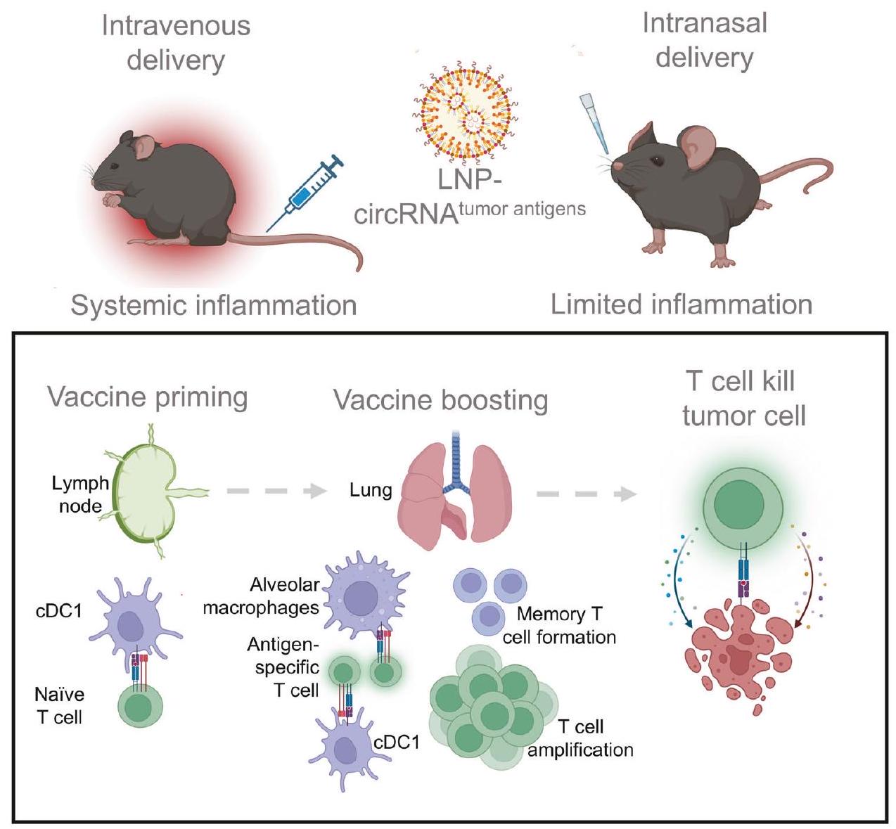
لقد أثار النجاح السريع للقاحات RNA في الوقاية من SARS-CoV-2 اهتمامًا باستخدامها في العلاج المناعي للسرطان. على الرغم من أن العديد من السرطانات تنشأ في الأنسجة المخاطية، إلا أن لقاحات السرطان RNA الحالية تُعطى بشكل رئيسي بطريقة غير مخاطية. هنا، قمنا بتطوير لقاح سرطان غير جراحي عبر الأنف باستخدام RNA دائري محاط بجزيئات دهنية لتحفيز استجابات مناعية موضعية. هذه الاستراتيجية أثارت استجابات قوية للخلايا التائية المضادة للأورام في نماذج سرطان الرئة قبل السريرية مع تقليل الآثار الجانبية النظامية المرتبطة عادةً بالتطعيم الوريدي RNA. على وجه التحديد، كانت الخلايا التغصنية التقليدية من النوع 1 ضرورية لتفعيل الخلايا التائية بعد التطعيم، حيث كانت كل من البلعميات الهوائية والخلايا التغصنية التقليدية من النوع 1 تعزز استجابات الخلايا التائية المحددة للمستضد في أنسجة الرئة. علاوة على ذلك، سهل التطعيم توسيع كل من الخلايا التائية المحددة للمستضد الذاتية والمُنتقلة، مما أدى إلى فعالية قوية ضد الأورام. أظهرت تسلسلات RNA أحادية الخلية أن التطعيم يعيد برمجة الخلايا التائية الذاتية، مما يعزز سميتها ويحفز نمط ذاكرة شبيه. بالإضافة إلى ذلك، يمكن للقاح عبر الأنف تعديل استجابة خلايا CAR-T لزيادة الفعالية العلاجية ضد خلايا الورم التي تعبر عن مستضدات مرتبطة بالورم محددة. بشكل جماعي، تمثل استراتيجية لقاح RNA عبر الأنف نهجًا جديدًا واعدًا لتطوير لقاحات RNA تستهدف الأورام المخاطية.

إشارات النقل والعلاج المستهدف (2025)10:101
; https://doi.org/10.1038/s41392-025-02191-1

المقدمة

العلاج المناعي للسرطان هو نهج واعد يستفيد من الجهاز المناعي لاستهداف والقضاء على خلايا السرطان. على عكس العلاجات التقليدية مثل العلاج الكيميائي والإشعاعي، التي تقتل خلايا الورم مباشرة، يعزز العلاج المناعي أو يستعيد وظيفة خلايا المناعة. من بين استراتيجيات العلاج المناعي المختلفة، حظيت لقاحات السرطان باهتمام كبير. تقدم لقاحات السرطان مستضدات الورم إلى خلايا تقديم المستضد، مما يحفز استجابة مناعية للخلايا التائية محددة للورم. تحمل هذه الاستراتيجية إمكانية تحقيق تأثيرات مضادة للأورام دائمة، تعالج كل من تقدم الورم وتكراره. تشمل حوامل اللقاحات التقليدية البكتيريا، الفيروسات، البروتينات وخلايا الورم. التحدي الرئيسي للقاحات التقليدية هو توصيل مستضدات الورم بفعالية وتنشيط استجابة مناعية قوية. لقد تسارعت الأبحاث في هذا المجال بفضل التقدم الأخير في منصات اللقاحات الجديدة، وخاصة لقاحات RNA.
لقد ظهرت لقاحات RNA كاستراتيجية تطعيم ثورية، محققة نجاحًا ملحوظًا خلال جائحة SARS-CoV-2، مما يبرز إمكانياتها في الوقاية السريعة والفعالة من الأمراض. مقارنةً بمنصات اللقاحات التقليدية، تقدم لقاحات RNA نهجًا واعدًا من خلال تمكين التعبير عن مستضدات مستهدفة متعددة مع توفير المناعية، مما يبدأ تقديم المستضدات القوية واستجابات الخلايا التائية. من منظور إنتاج الأدوية والتطبيق السريري، تقدم لقاحات RNA مزايا مميزة، بما في ذلك الإنتاج الفعال على نطاق واسع، نقاء أعلى، تقليل الشوائب، بروتوكولات مراقبة الجودة المعمول بها، وتكاليف تصنيع أقل.
التكاليف. حاليًا، يمكن تصنيف منصات اللقاحات المعتمدة على RNA إلى ثلاثة أنواع رئيسية: RNA خطي أو RNA خطي معدل، RNA دائري (circRNA)، وRNA ذاتي التكاثر. بينما تعتمد لقاحات RNA المعتمدة بشكل أساسي على RNA خطي معدل، فإن متطلبات التخزين الصارمة لها تبرز الحاجة إلى تحسين الاستقرار لتمكين تطبيق أوسع. تواجه RNA ذاتي التكاثر، على الرغم من تعبيرها المطول عن البروتين، تحديات في الإنتاج والتخزين بسبب حجمها الجزيئي الأكبر. بالمقارنة، يظهر circRNA استقرارًا متفوقًا بفضل هيكله الدائري المغلق تساهميًا، مما يمنح ثباتًا حراريًا معززًا ونصف عمر ممتد في خلايا الثدييات. بالإضافة إلى ذلك، فإن إنتاج circRNA أقل تعقيدًا، حيث يلغي الحاجة إلى تعديلات النوكليوتيدات، والتغطية، وذيل polyA. أظهرت الدراسات قبل السريرية أن لقاحات circRNA، عند إعطائها عبر الحقن العضلي، فعالة ضد العدوى الفيروسية والأورام تحت الجلد. ومع ذلك، هناك حاجة إلى مزيد من البحث لاستكشاف طرق إعطاء بديلة للقاحات circRNA لتوسيع تطبيقاتها العلاجية.
تم تصميم الأنواع التقليدية من لقاحات السرطان بشكل رئيسي لتُعطى عبر الحقن الوريدي. بالنسبة للقاحات المعتمدة على RNA، يتم إعطاء لقاحات SARS-CoV-2 المرخصة حاليًا عبر الحقن العضلي. كما استكشف الباحثون خيارات وريدية لتوصيل RNA إلى أنسجة الطحال باستخدام جزيئات نانوية محسّنة لعلاج السرطان. ومع ذلك، تتطلب هذه الطريقة تنشيط المناعة النظامية، مما يحمل مخاطر محتملة، كما لوحظ في ردود الفعل السلبية المبلغ عنها للقاحات RNA SARS-CoV-2. على النقيض من ذلك، تقدم طرق التسليم المخاطية غير الجراحية، مثل الطرق عبر الأنف أو القابلة للاستنشاق،
بديلاً واعدًا. هذه الطرق تحفز مباشرة الاستجابات المناعية في موقع الغشاء المخاطي التنفسي، دون تحفيز الاستجابة المناعية النظامية. تقدم هذه الاستراتيجية إمكانيات كبيرة للعلاج المناعي للسرطان، خاصة للأورام التي تنشأ في الأنسجة المخاطية. علاوة على ذلك، قد تقلل هذه الطرق غير الجراحية من الانزعاج والمخاطر والمضاعفات المرتبطة بالحقن، مثل الإصابات أو العدوى المرتبطة بالإبر. وبالتالي، فإن تطوير لقاحات RNA غير جراحية تستهدف الأورام المخاطية يحمل إمكانية تجاوز لقاحات السرطان الحالية من حيث الفعالية والسلامة. على الرغم من هذه التقدمات، لا تزال الآليات المناعية الكامنة وراء تحفيز الخلايا التائية المحددة للمستضد بعد التطعيم RNA، خاصة عبر الطريق المخاطي، غير مفهومة جيدًا. يمكن أن يؤدي فهم أكثر شمولاً لهذه العمليات إلى تصميم عقلاني لقاحات أكثر فعالية وعلاجات مركبة لتعزيز الاستجابات المناعية المضادة للأورام.
في هذه الدراسة، استخدمنا circRNA المستقر للغاية المحاط بجزيئات دهنية مستهدفة للرئة (LNPs) لتقييم إمكانياتها كلقاح سرطان عبر الأنف لعلاج سرطانات الرئة. تظهر نتائجنا أن اللقاح يحفز بنجاح تعبير البروتين في أنسجة الرئة مع آثار جانبية طفيفة. كما أظهرت هذه الاستراتيجية فعالية مضادة للأورام كبيرة في نماذج سرطان الرئة، حتى في وجود خلايا الورم التي تعبر عن مستضدات نيوانتجين ذاتية. من الناحية الآلية، تعتمد النشاط المضاد للأورام للقاح على الخلايا التغصنية التقليدية من النوع 1 (cDC1s)، التي تلعب دورًا حاسمًا في تفعيل الخلايا التائية المحددة للمستضد. كل من cDC1s والبلعميات الهوائية مسؤولة عن تعزيز الخلايا التائية المحددة للمستضد في أنسجة الرئة. علاوة على ذلك، يتعاون اللقاح مع علاج نقل الخلايا التائية المتبنية، مما يعزز القضاء على الورم من خلال زيادة أعداد كل من الخلايا التائية المحددة للمستضد الذاتية والمُنتقلة. بالإضافة إلى ذلك، يمكن للقاح تغيير مشهد الخلايا التائية الذاتية من خلال تعزيز أنماط الذاكرة والفاعلية. بناءً على الخصائص الفريدة لاستراتيجية التطعيم عبر الأنف هذه، قمنا بتصميم خلايا CAR-T المستجيبة للقاح، المصممة لتعزيزها من خلال التطعيم عبر الأنف، لتحقيق فعالية مضادة للأورام محسّنة ضد خلايا الورم التي تعبر عن مستضدات مرتبطة بالورم. بشكل عام، توفر هذه الدراسة نهجًا جديدًا لعلاج الأورام المخاطية من خلال التسليم غير الجراحي للقاحات RNA.

النتائج

تسليم RNA عبر الأنف من خلال جزيئات دهنية يبدأ تعبير البروتين في أنسجة الرئة
استنادًا إلى استراتيجية تحضير circRNA المنشورة سابقًا، تضمنت قوالب النسخ في المختبر زوجًا من الإكسونات والإنترونات، CVB3 IRES، الفواصل، الأذرع وتسلسلات الترميز. تم الحصول على circRNAs من خلال تفاعل التقطيع الخلفي تلاه تنقية عبر الكروماتوغرافيا السائلة. نظرًا لأن circRNA معروف بأنه أكثر استقرارًا من mRNA، فقد كان هدفنا أولاً تحديد ما إذا كان لقاح circRNA يظهر استقرارًا حراريًا أفضل مقارنةً بنظيره الخطي عند احتوائه في جزيئات نانوية دهنية (LNP). استخدمنا تركيبة LNP المعتمدة سريريًا التي تحتوي على SM102 و DSPC و DMG-PEG و الكوليسترول. تم ترميز RNAs بـ D2GFP، وهو بروتين GFP غير مستقر له نصف عمر لا يتجاوز ساعتين، لتقييم تعبير البروتين. أظهرت نتائجنا أن circRNA أظهر مستويات تعبير بروتين أعلى من تركيبات RNA الخطية عند تخزينها في كل من و ، مما يدعم التطبيق الأوسع وملاءمة لقاحات circRNA. (الشكل التكميلي 1).
للتوصيل عبر الأنف، اختبرنا كفاءة اثنين من الدهون الأيونية المعتمدة سريريًا (SM102 و ALC-0315) لتشكيل LNPs مع DSPC و DMG-PEG و الكوليسترول. قمنا بتوصيف حجم وإمكانات زتا لاثنين من LNPs المحتوية على صبغة (الشكل التكميلي 2a، b) ثم قمنا بإجراء الاختبار في الجسم الحي. لاحظنا
أن LNP المعتمد على SM102 أظهر كثافة فلورية أعلى لتتبع الصبغة مقارنةً بـ ALC-0315 (الشكل التكميلي 2c، d). وفقًا للدراسات الحديثة، يمكن أن تعزز إضافة الدهون الكاتيونية، مثل 1،2-ديوليويل-3-تريميثيل أمونيوم-بروبان (DOTAP)، كفاءة استهداف الرئة بعد الإدارة الوريدية والأنفية. قمنا بتحسين نظام التوصيل عن طريق إضافة DOTAP إلى LNPs المعتمدة على SM102، مما أدى إلى مستوى أعلى من تعبير البروتين (الشكل التكميلي 2e، f). وبالتالي، تم استخدام هذه الاستراتيجية في التجارب التالية. تظهر عملية التصنيع الكاملة للقاح circRNA في الشكل 1a. باختصار، تم إعداد circRNA من خلال النسخ والتفاعل التقطيعي في المختبر، تلاه تنقية عبر الكروماتوغرافيا السائلة. تم خلط العينات المنقاة مع خمسة مكونات دهنية عبر الميكروفلويديات لتشكيل مركب circRNA-LNP. لتوصيف لقاح RNA، كان حجم الجسيمات حوالي 71.69 نانومتر (الشكل 1b). كانت إمكانات الزتا 8.41 مللي فولت (الشكل 1c). لمراقبة توزيع لقاح RNA في الجسم، تم خلط circRNA المشفر لليوسيفيراز و LNPs مع صبغة دهنية DiR. بعد الإدارة عبر الأنف، تم تأكيد التسليم الناجح عبر تصوير فلوري لتتبع الصبغة (الشكل 1d). علاوة على ذلك، كان هدفنا مقارنة توزيع لقاح RNA عبر طرق الإدارة المختلفة (الشكل 1e). قمنا بعزل الأعضاء الحيوية بعد 4 ساعات من التطعيم ووجدنا أن اللقاح الأنفي أدى إلى تراكم LNP وتعبير البروتين في أنسجة الرئة (الشكل 1f، g والشكل التكميلي 2g، h). ومع ذلك، يمكن اكتشاف LNPs وتعبير البروتين في أنسجة الكبد والرئة والطحال بعد الحقن الوريدي. مجتمعة، أظهرت هذه البيانات أن منصة circRNA-LNP يمكن أن تبدأ تعبير البروتين المحلي في أنسجة الرئة بعد الإدارة عبر الأنف.
لقاح circRNA الأنفي يحقق استجابة مضادة للورم قوية مع آثار جانبية محدودة
لمقارنة كفاءة اللقاح المضاد للورم المعطى عبر طرق مختلفة بشكل منهجي، أنشأنا نموذج ورم B16 النقوي في الرئة يعبر عن مستضد OVA. تلقت الفئران لقاحات circRNA المشفرة SIINFEKL (الجزء المحدود من مستضد OVA من الفئة I (Kb))-RFP في نقاط زمنية محددة (الشكل 2a). مقارنةً بالمجموعة غير المعالجة، أظهرت جميع طرق الإدارة الثلاثة فعالية مضادة للورم قابلة للمقارنة إحصائيًا (الشكل 2b، c). ومع ذلك، بدا أن نسبة أنسجة الرئة التي تحتلها بؤر النقائل كانت أقل في المجموعات الأنفية والوريدية مقارنةً بالمجموعة العضلية. ثم قمنا بتحليل السمية الحادة للقاح circRNA المعطى عبر طرق مختلفة. أشارت الدراسات السابقة إلى أن لقاحات RNA يمكن أن تحفز إفراز السيتوكينات المؤيدة للالتهابات، مما يؤدي إلى آثار جانبية. قمنا أولاً بتقييم مستوى IFN-a في المصل بعد التطعيم بسبب دوره الرئيسي في تحفيز الالتهاب (الشكل 2d). مقارنةً بمجموعات الحقن الوريدي والعضلي، كانت مستويات IFN- في المصل أقل بكثير في المجموعات الأنفية (الشكل 2e). ثم قمنا بتحليل سيتوكينات أخرى ووجدنا بالمثل أن المجموعات الأنفية أظهرت مستوى سيتوكين أقل مقارنةً بالحقن الوريدي. (الشكل 2f والشكل التكميلي 3). بالنسبة لاختبارات الدم الروتينية، أظهرت انخفاضًا أكثر أهمية في خلايا الدم والخلايا المناعية في مجموعة الحقن الوريدي مقارنةً بالمجموعة الأنفية (الشكل 2g). للتحقق من سلامة الإدارة الأنفية، قمنا بمراقبة تغييرات وزن الجسم في الفئران طوال فترة العلاج (الشكل التكميلي 4). على الرغم من ملاحظة انخفاض طفيف في وزن الجسم في مجموعات التطعيم، إلا أنه تم استعادته بعد بضعة أيام.
قمنا أيضًا بتقييم كفاءة اللقاح المضاد للورم الأنفي في نماذج مختلفة (الشكل 2h-j). في نماذج النقائل الرئوية الوقائية، تلقت الفئران جرعتين من لقاح RNA، تلاه تحدي خلايا ورم B16-OVA بعد 30 أو 60 يومًا. أظهرت صور البيولومينسنس (الشكل التكميلي 5a، b) ومنحنيات البقاء (الشكل 2k والشكل التكميلي 5c) أن التحصين الأنفي أوقف نمو الورم وأطالت بشكل كبير
الشكل 1 توصيل circRNA عبر الأنف عبر جزيئات نانوية دهنية يبدأ تعبير البروتين في أنسجة الرئة. أ مخطط تجريبي لعملية تصنيع لقاح RNA-LNP. ب توزيعات حجم لقاح RNA-LNP. ج إمكانات زتا لقاح RNA-LNP. د صور فلورية تمثيلية لتتبع الصبغة في أنسجة الرئة. تم إعطاء الفئران LNPs المسمى DiR عبر الإدارة الأنفية وتم عزل أنسجة الرئة بعد 4 ساعات. مقياس الرسم، . هـ الجدول الزمني للتجربة المصممة لتقييم توزيع التعبير البروتيني لليوسيفيراز ( circRNA لكل فأر). نتائج التصوير (و) والتحليل الإحصائي (ز) لتعبير بروتين اليوسيفيراز بين الأعضاء المختلفة. I.v.، مجموعة الحقن الوريدي. I.n.، مجموعة الإدارة الأنفية
البقاء، مما يشير إلى تشكيل خلايا T الذاكرة بعد التطعيم. في نماذج النقائل الرئوية العلاجية، تم استخدام خطوط خلايا B16luciferase و LL/2 دون التعبير المفرط عن مستضدات OVA. بناءً على الطفرات غير المتناظرة المنشورة لـ و ، صممنا لقاحات RNA مشفرة لمستضد B16 (الشكل التكميلي 5d-f) ولقاحات RNA مشفرة لمستضد LL/2 (الشكل التكميلي 6a، b)، وتلقت الفئران ثلاث جرعات من التطعيم بعد إنشاء النقائل الرئوية. في نموذج B16، أظهرت منحنيات البقاء أن التحصين الأنفي أوقف نمو الورم بشكل كبير وأطالت البقاء (الشكل 2l). في نموذج LL/2، لاحظنا أن الفئران المحصنة أظهرت سمية خلوية مع عدد أقل من بؤر الورم (الشكل التكميلي 6c، d) وبقاء أطول (الشكل 2m). في كلا النموذجين، لاحظنا نشاط خلايا T المستهدفة لمستضدات مشفرة بواسطة اللقاح في أنسجة الرئة (الشكل التكميلي 5f و6e)، مما يبرز إمكانيات circRNA الأنفي لتطبيقات لقاح النيوانتجين. باختصار، تشير هذه البيانات إلى
أن لقاح circRNA الأنفي يثير استجابة مضادة للورم قوية مع آثار جانبية أقل مقارنةً بطرق الإدارة الأخرى.
كفاءة اللقاح المضاد للورم للقاح circRNA الأنفي تعتمد على مستضد الورم و cDC1
استلهمًا من الإمكانيات العلاجية للقاح الأنفي، هدفنا إلى التحقيق بشكل أكبر في المبادئ الأساسية وراء استجابته المناعية المضادة للورم. أولاً، أنشأنا نموذج نقائل الرئة B16-OVA-ليوسيفيراز وأكدنا الاستجابة المضادة للورم (الشكل 3a). تم استخدام RNA مشفر RFP كتحكم لمستضد غير ذي صلة. كما أعددنا لقاحات RNA خطية و circRNA مشفرة لـ SIINFEKL-RFP للتطعيم. أظهرت نتائج صور البيولومينسنس (الشكل 3b والشكل التكميلي 7a) ومنحنيات البقاء (الشكل 3c) أن لقاحات RNA المشفرة لمستضد الورم فقط يمكن أن توقف نمو خلايا الورم، دون ملاحظة فرق كبير بين لقاحات mRNA و circRNA في هذا
الشكل 2 لقاح circRNA عن طريق الأنف يحقق استجابة قوية مضادة للأورام مع آثار جانبية محدودة. أ. الجدول الزمني للتجربة المصممة لتقييم القدرة المضادة للأورام لطرق مختلفة من لقاح circRNA. ب. صور لأورام النقائل الرئوية في نقطة النهاية في مجموعات مختلفة. NT، مجموعة غير المعالجة. I.m.، مجموعة الحقن العضلي. I.v.، مجموعة الحقن الوريدي. I.n.، مجموعة الإدارة عن طريق الأنف. لمجموعة NT. للمجموعات الأخرى. ج النسبة المئوية للمساحة التي تشغلها الأورام النقيليّة بين المجموعات المختلفة عند نقطة النهاية. تم تحليل البيانات باستخدام تحليل التباين الأحادي مع اختبار المقارنات المتعددة لتوكاي. د الجدول الزمني للتجربة المصممة لتقييم الآثار الجانبية النظامية لطرق مختلفة من لقاح circRNA. إطلاق السيتوكينات في المصل بعد إعطائها مع اللقاح المختبر بواسطة IFN- الفأر مجموعة إليسا (e) ومجموعة التهاب الفأر (f). تم تحليل البيانات باستخدام اختبار t لطلبة لمقارنة مجموعة i.n. مع مجموعة i.v. أو i.m. في e. تم تحليل البيانات باستخدام تحليل التباين الأحادي مع اختبار المقارنات المتعددة لتوكاي في f. g تحليل روتين الدم بعد 24 ساعة من التطعيم الثالث. تم تحليل البيانات باستخدام اختبار t لطلبة لمقارنة مجموعة i.n. مع مجموعة i.v. جدول زمني للتجارب لتقييم القدرة المضادة للورم للجرعة الأنفية في نماذج مختلفة. تم اعتبار الفئران التي تلقت LNP بدون RNA كمجموعة لقاح وهمية. تم تحصين الفئران مع circRNA المشفر لـ SIINFEKL مرتين، تلاها إعادة تحدي خلايا B16-OVA-luciferase بعد 60 يومًا من التطعيم. تم تحدي الفئران بخلايا B16luciferase وتلقت ثلاث جرعات من لقاح circRNA المشفر لمستضد B16. تم تحدي الفئران بخلايا LL/2 ثم تلقت ثلاث جرعات من لقاح circRNA المشفر لمستضد LL/2. منحنيات البقاء للتجارب المتعلقة بـ و ، على التوالي. لكل مجموعة. تم تحليل البيانات من خلال تحليل كابلان-ماير. بالنسبة للفئران التي تلقت التطعيم، كانت كل جرعة تحتوي على circRNA. جميع البيانات ممثلة كمتوسط ± الانحراف المعياري للخطأ
الشكل 3 القدرة المضادة للورم للقاح circRNA عن طريق الأنف تعتمد على مستضد الورم و cDC1. أ. الجدول الزمني للتجربة المصممة لتقييم القدرة المضادة للورم للقاح RNA عن طريق الأنف. تم تحدي الفئران من النوع البري (WT) بخلايا B16-OVA-luciferase ثم تركت دون علاج (NT) أو تم علاجها بـ RNA مشفر لـ RFP أو RNA مشفر لـ SIINFEKL (ببتيد OVA)-RFP. تم مراقبة نمو الورم من خلال تصوير الإضاءة الحيوية. ب. التحليل الإحصائي لمتوسط شدة الإضاءة الحيوية مع علاجات مختلفة. تم تحليل البيانات باستخدام ANOVA ثنائية الاتجاه مع اختبار المقارنات المتعددة لتوكاي. لمجموعة NT. للمجموعات الأخرى. ج منحنى البقاء للفئران WT مع علاجات مختلفة. تم تحليل البيانات عبر تحليل كابلان-ماير. د الجدول الزمني للتجربة المصممة لتقييم القدرة المضادة للورم للقاح RNA عن طريق الأنف في Batf3 الفئران. منحنى البقاء لباتف3 فئران مع علاجات مختلفة. تم تحليل البيانات من خلال تحليل كابلان-ماير. لكل مجموعة. جدول زمني للتجربة المصممة لتقييم تحفيز خلايا T المحددة لمستضد بعد تطعيم circRNA. WT أو Batf3 تم تحصين الفئران عن طريق الأنف باستخدام circRNA المشفر لـ SIINFEKL-RFP، تلا ذلك تقييم خلايا T المحددة للمستضد في أنسجة الرئة والطحال. ، تكرر ثلاث مرات). تحليل إحصائي لنسبة خلايا T المحددة بمستضد. تم تحليل البيانات باستخدام تحليل التباين الثنائي مع اختبار المقارنات المتعددة توكي. جميع البيانات ممثلة كمتوسط ± الانحراف المعياري للخطأ.
نموذج. لتوصيف خلايا T المستحثة بواسطة التطعيم بشكل أكبر، قمنا بتحليل خلايا T المحددة بمستضد في أنسجة الرئة بعد نظام تطعيم يتكون من جرعتين. كشفت نتائجنا عن زيادة في كل من خلايا T المحددة بمستضد الكلي وفئة خلايا T المحددة بمستضد الإيجابية CD103، مما يؤكد النجاح في استحثاث خلايا T المحددة بمستضد (الشكل التوضيحي 7b-e). باستخدام اختبار صبغ داخل الأوعية، أكدنا أن خلايا T المحددة بمستضد المستحثة بواسطة اللقاح الأنفي تأتي بشكل أساسي من أنسجة الرئة بدلاً من الدم المحيطي (الشكل التوضيحي 7f-h). علاوة على ذلك، أظهر اختبار استنفاد CD8 في الجسم الحي أن خلايا T الإيجابية CD8 ضرورية لفعالية اللقاح المضادة للأورام (الشكل التوضيحي 8a-d).
نظرًا للدور البارز لخلايا CD8 T والأنتيجينات التي يتم توصيلها بواسطة اللقاح في بدء المناعة المضادة للأورام، هدفنا إلى اختبار تأثير تقديم الأنتيجين على القدرة المضادة للأورام. حيث تلعب خلايا cDC1 دورًا رئيسيًا في التقاط وتقديم الأنتيجينات على MHC-I لتعزيز مناعة خلايا CD8 +T، قمنا بتكرار تجربة المناعة المضادة للأورام في cDC1-
فئران knockout (الشكل 3d). من المثير للاهتمام أن كفاءة اللقاح الدائري RNA المضاد للورم عن طريق الأنف قد انخفضت، مما يشير إلى أن cDC1 ضروري للاستجابة المناعية المضادة للورم الناتجة عن اللقاح (الشكل 3e). قمنا أيضًا بتحليل خلايا CD8 + T المحددة للمستضد التي تم تحفيزها بواسطة اللقاح (الشكل 3f). WT و Batf3 تلقت الفئران جرعتين من التطعيم، وتم الكشف عن خلايا T في أنسجة الرئة والطحال. وجدنا أن نقص cDC1 قلل بشكل كبير من مستوى خلايا CD8 + T الخاصة بالمستضد في أنسجة الرئة (الشكل 3 ج والشكل التكميلية). لتأكيد أن خلايا cDC1 تقدم المستضد بعد التطعيم، قمنا بعزل خلايا cDC1 من العقد اللمفاوية draining للرئة ثم قمنا بزراعتها مع خلايا T المحددة للمستضد (الشكل التوضيحي 8 ج). إن تنشيط خلايا T بعد الزراعة المشتركة يوفر دليلاً جديداً على أن خلايا cDC1 تقدم المستضدات وتلعب دوراً رئيسياً في تحفيز خلايا T. وبالتالي، فإن التعبير عن مستضد الورم ووجود خلايا cDC1 أمران لا غنى عنهما للاستجابة المناعية لخلايا T المضادة للورم التي يسببها لقاح circRNA عن طريق الأنف.
الشكل 4 AMs و cDC1s مسؤولون بشكل رئيسي عن تعزيز خلايا T المحددة لمستضد في أنسجة الرئة بعد التطعيم عن طريق الأنف. أ جدول زمني للتجربة لتقييم دور APCs CD11c+ في تعزيز خلايا T المحددة لمستضد. WT، Batf3 أو تم معالجة فئران CD11c-DTR مسبقًا بالسيكلوفوسفاميد (CTX) وسم الدفتيريا (DT) من أجل تقليل عدد الخلايا اللمفاوية في جميع الفئران وتقليل عدد خلايا CD11c+ في فئران CD11c-DTR. تم وسم خلايا OT-1 المنشطة بـ CFSE ثم تم نقلها إلى الفئران التي تم تحصينها بلقاح circRNA عن طريق الأنف. تم تحليل تكاثر خلايا T في أنسجة الرئة في اليوم الثالث. ب، ج تمثيل بياني لهيستوجرامات تدفق الخلايا (ب) والنتائج الإحصائية (ج) لتكاثر خلايا T مع علاجات مختلفة. تم تحليل البيانات باستخدام ANOVA أحادي الاتجاه مع اختبار المقارنات المتعددة لتوكاي. للمجموعة التجريبية؛ للمجموعات الأخرى. لتحليل امتصاص LNP، تم إعطاء الفئران LNP مع علامة DiD عن طريق الأنف وتم تحليل أنسجة الرئة بعد 4 ساعات. د، هـ تمثيل بياني لنتائج تدفق الخلايا (د) ونتائج إحصائية (هـ) لامتصاص LNP بين أنواع الخلايا المختلفة. و مخطط التجربة لاختبار APCs المحتملة التي يمكن أن تعزز خلايا T المحددة لمستضد. تم تحصين الفئران بـ LNP عن طريق الأنف بدون RNA (وهمي) أو لقاح circRNA وبعد 24 ساعة، تم جمع أنسجة الرئة وتم فرز APCs وزرعها مع خلايا OT-1 لمدة 36 ساعة أخرى في IFN- آبار الإيليسبوت. للمجموعة التجريبية؛ لمجموعة لقاح circRNA، تم تكرارها ثلاث مرات) تم إنشاؤه باستخدام BioRender.com. صور تمثيلية ونتائج إحصائية لـ IFN- اختبار Elispot. تم تحليل البيانات باستخدام اختبار t لستودنت. i الجدول الزمني للتجربة لاستكشاف ما إذا كان اللقاح يمكن أن يعزز خلايا T في أنسجة الرئة. تم معالجة الفئران بـ FTY720 خلال عملية التحفيز-التعزيز أو فقط خلال عملية التعزيز أو محلول ملحي عادي كمجموعة تحكم. تم تحليل عدد خلايا T المحددة بمستضد في أنسجة الرئة. رسوم تمثيلية ) ونتائج إحصائية ( ) لخلايا T المحددة بمستضد مع علاجات مختلفة. تم تحليل البيانات باستخدام تحليل التباين الأحادي مع اختبار المقارنات المتعددة لتوكاي. جميع البيانات ممثلة كمتوسط ± الانحراف المعياري للخطأ
تتحمل الخلايا التغصنية (AMs) والخلايا التغصنية من النوع الأول (cDC1s) المسؤولية الرئيسية عن تعزيز الخلايا التائية المحددة للمستضدات في أنسجة الرئة بعد التطعيم عن طريق الأنف. يتطلب التحفيز الناجح لمناعة الخلايا التائية القوية والمستمرة ليس فقط تهيئة الخلايا التائية ولكن أيضًا عملية التعزيز من خلال التطعيم المتكرر. لذلك، هدفنا إلى
استكشاف عملية تعزيز خلايا T بعد التحصين عن طريق الأنف. استخدمنا فئران WT، وفئران بدون cDC1، وفئران بدون جميع خلايا CD11c+ لاختبار قدرتها على تعزيز خلايا T المحددة لل antigene المنشطة (الشكل 4a). على الرغم من أن استنفاد cDC1 قلل من تكاثر خلايا T، إلا أن نقص خلايا CD11c+
أوقف تمامًا تكاثر خلايا T الناتج عن اللقاح، مما يشير إلى أن كل من cDC1 وغيرها من خلايا تقديم antigene CD11c+ تشارك في تعزيز خلايا T (الشكل 4b، c والشكل التكميلي 9a). لتحديد أنواع الخلايا التي تدعم تكاثر خلايا T، تم وسم LNPs بصبغة DiD المحبة للدهون وتم إعطاؤها عن طريق الأنف للفئران. ثم قمنا بصبغ المجموعات الرئيسية من خلايا CD11c+ في أنسجة الرئة، بما في ذلك البلعميات الهوائية (AMs)، cDC1s، CD11b + DCs، وخلايا CD11c- CD45- كمجموعة تحكم. وجدنا أن LNPs تم ابتلاعها بشكل رئيسي بواسطة AMs، مع قدرة خلايا أخرى أيضًا على ابتلاع LNPs ولكن بأعداد أقل (الشكل 4d، e، الشكل التكميلي 9b-d). علاوة على ذلك، تم تعزيز إنتاج IFN- عندما تم زراعة AMs المعزولة وcDC1s مع خلايا OT-1، بينما لم يكن هناك فرق كبير في مجموعات CD11b + أو CD45- (الشكل 4f-h). كما قمنا بإجراء صبغ المناعية (IF) لأنسجة الرئة للتحقيق في التفاعلات بين خلايا تقديم antigene (APCs) وخلايا T المحددة لل antigene (الشكل التكميلي 10). في مجموعة التحصين، لاحظنا تفاعلات بين خلايا T المحددة لل antigene وخلايا CD11c +، وكذلك بين خلايا T المحددة لل antigene وAMs. توفر هذه النتائج أدلة جديدة على أن كل من AMs وخلايا التغصن (DCs) تساهم في تنشيط وتعزيز خلايا T المحددة لل antigene. بعد ذلك، قمنا بتقييم ما إذا كان اللقاح عن طريق الأنف قد قام بتحفيز وتعزيز خلايا T مباشرة في أنسجة الرئة. للقيام بذلك، تم استخدام FTY720 لمنع خروج خلايا T من الأنسجة اللمفاوية. تم علاج الفئران بـ FTY720 يوميًا بدءًا من قبل عملية التحفيز أو التعزيز، وتم الكشف عن خلايا T المحددة لل antigene في أنسجة الرئة في نهاية التجربة (الشكل 4i). خلال عملية التعزيز، لم يكن لعلاج FTY720 أي تأثير على عدد خلايا T المحددة لل antigene في أنسجة الرئة، مما يشير إلى أن تعزيز اللقاح أدى إلى تقديم antigene وتكاثر خلايا T داخل أنسجة الرئة المحلية (الشكل والشكل التكميلي 9d). ومع ذلك، انخفض عدد خلايا T المحددة لل antigene في أنسجة الرئة بشكل كبير (الشكل 4k)، دون تأثير على العقدة اللمفاوية المنصفية عندما تم علاج الفئران بـ FTY720 بدءًا من قبل التحصين الأول (الشكل التكميلي 11). وهذا يشير إلى أن التحفيز الأولي لخلايا T المحددة لل antigene حدث خارج أنسجة الرئة، معتمدًا على العقد اللمفاوية المتدفقة. وبالتالي، تشير هذه البيانات إلى أن لقاح circRNA عن طريق الأنف يمكن أن يعزز خلايا T مباشرة في أنسجة الرئة من خلال أفعال AMs وcDC1s.
لقاح circRNA عن طريق الأنف يعزز كل من خلايا T المحددة لل antigene الذاتية والمُنتقلة لتعزيز الاستجابة المناعية المضادة للأورام
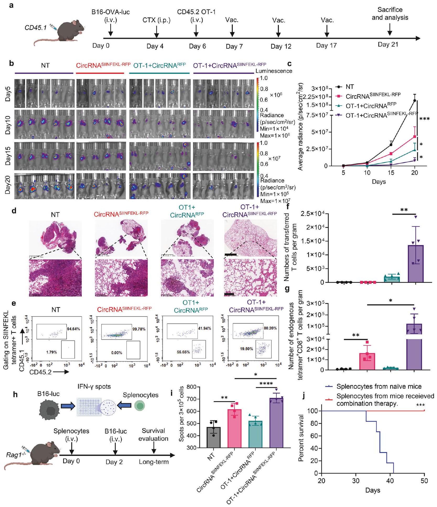
استنادًا إلى النتائج المذكورة أعلاه، افترضنا أن لقاح RNA عن طريق الأنف قد يتعاون مع خلايا T المحددة لل antigene المُنتقلة لتعزيز القدرة المضادة للأورام. أجرينا تجارب باستخدام نموذج فأر WT. تم تحدي الفئران في البداية بخلايا ورم B16-OVAluciferase وخضعت لاستنفاد اللمف مع CTX قبل تلقي العلاج بلقاح circRNA، أو العلاج الخلوي التبني (ACT)، أو مجموعة من كلا العلاجين (الشكل 5a). أظهرت تصوير البيولومينسنس وفحص الأنسجة (صبغة HE) أنه بينما يمكن أن يمنع اللقاح بمفرده أو خلايا OT-1 مع اللقاح الوهمي نمو الورم، فإن العلاج المركب أدى إلى أقوى تحكم في الورم (الشكل 5b-d). كما قمنا بتحديد عدد خلايا T المحددة لل antigene لكل من خلايا T الذاتية والمُنتقلة، مما يكشف أن اللقاح وسع كلا المجموعتين بفعالية (الشكل 5e-g).
نظرًا للتأثيرات الثانوية المحتملة لاستجابة خلايا T الناتجة عن اللقاح، من الممكن أن يتم تحفيز الاستجابات المناعية ضد antigene ثانوية، تختلف عن الهدف الأصلي للقاح. في هذه الدراسة، يؤدي الجمع بين التحصين وACT إلى تدمير خلايا الورم التي تعبر عن antigene OVA بواسطة خلايا T. يحرر هذا التدمير antigene إضافية للورم، والتي قد يتم التقاطها بواسطة خلايا تقديم antigene، مما يؤدي لاحقًا إلى تنشيط خلايا الذاتية المستهدفة لأورام فقدان antigene OVA. لقد لوحظت هذه الظاهرة في انتشار antigene في كل من الدراسات السريرية وما قبل السريرية
المعنية بالعلاج المناعي. لذلك، قمنا بتقييم القدرة المضادة للأورام للخلايا الطحالية من الفئران المحصنة عند زراعتها مع خلايا B16 التي تفتقر إلى antigene OVA (الشكل 5h). بشكل ملحوظ، أظهرت الخلايا الطحالية من الفئران المحصنة زيادة في إنتاج IFN , مما يشير إلى أن اللقاح يمكن أن يحفز انتشار antigene لاستهداف خلايا الورم التي فقدت antigene (الشكل . علاوة على ذلك، منعت الخلايا الطحالية المُنتقلة من الفئران في مجموعة العلاج المركب تكوين الورم عند إعادة التحدي مع خلايا B16 التي تفتقر إلى antigene OVA (الشكل 5h، j)، مما يشير إلى أن العلاج المركب قد يسهل توليد خلايا T الذاكرة القادرة على استهداف خلايا الورم من خلال antigene بديلة. تؤكد هذه النتائج على إمكانية الجمع بين لقاح RNA عن طريق الأنف وACT لتعزيز المناعة المضادة للأورام من خلال توسيع كل من خلايا T الذاتية والمُنتقلة، وتحفيز انتشار antigene، وربما تعزيز تشكيل خلايا الذاكرة ضد خلايا الورم التي تعبر عن antigene أخرى.
لقاح circRNA عن طريق الأنف يحفز تغييرات وظيفية وشكلية في خلايا T الذاتية
استنادًا إلى نتائجنا السابقة، هدفنا إلى فحص شامل للتغيرات في خلايا T الذاتية باستخدام تحليل RNAseq للخلايا الفردية من خلايا CD3+ من أنسجة الرئة في نماذج الفئران الحاملة للورم (الشكل 6a). حدد التجميع غير المراقب لبيانات النسخ تسعة مجموعات رئيسية من خلايا T: خلايا CD4 وCD8 الساذجة (المعلمة بتعبير Lef1 وTcf7 مع مستويات II7r وSell منخفضة نسبيًا)، خلايا CD4 وCD8 الذاكرة (التي تعبر عن II7r وSell)، خلايا CD8 التكاثرية (المميزة بتعبير Top2a وMki67)، خلايا CD4 وCD8 الفعالة (مع وGzmb التعبير)، خلايا Treg (التي تتميز بـ oxp 3 و ), وخلايا CD4 T المعززة بـ IFN (المعلمة بتعبير Ifit1 وIfit3 وIsg15) (الشكل 6b، c). حددنا بشكل خاص مجموعة خلايا T السامة المميزة بتوقيع من الجينات المرتبطة بالسُمية (Gzma، Gzmb، Gzmc، Gzmd، Gzme، Gzmf، Gzmg، Gzmk، Gzmm)، والتي أظهرت نشاطًا سميًا مرتفعًا بشكل ملحوظ في مجموعة اللقاح مقارنةً بالمجموعة الضابطة (الشكل 6d). أظهر التحليل الإضافي أن القدرات السمية كانت مرتفعة بشكل ملحوظ في مجموعات خلايا T الذاكرة والفعالة ضمن المجموعة الملقحة (الشكل 6e). بالإضافة إلى ذلك، قمنا بتحليل وفرة ونسب كل مجموعة من خلايا T ضمن مجموعات CD4 وCD8 بشكل منفصل، ووجدنا زيادة في الأعداد والنسب من خلايا الذاكرة والفعالة في مجموعة اللقاح (الشكل 6f-i). من المهم أن وظيفة خلايا CD8 T الفعالة المضادة للأورام كانت معززة بشكل كبير في مجموعة اللقاح، كما يتضح من مستويات التعبير المرتفعة لـ Gzma وGzmb (الشكل 6j). أشار تحليل إثراء Gene Ontology (GO) أيضًا إلى أن المسارات المتعلقة بوظيفة خلايا T والسُمية المضادة للأورام في خلايا CD4 وCD8 الفعالة كانت غنية في مجموعة اللقاح (الشكل 6k، I). قمنا بالتحقق من وظيفة كل من خلايا T الذاتية والمُنتقلة من خلال صبغ IFN- داخل الخلايا (الشكل التكميلي 12). لوحظ مستوى مرتفع من IFN- في كلا المجموعتين، مما يشير إلى زيادة النشاط السمي لخلايا T عند مواجهتها مع خلايا الورم. بشكل عام، يظهر هذا التحليل الشامل أن لقاح circRNA عن طريق الأنف يدفع تغييرات كبيرة في مشهد خلايا T الذاتية، مما يعزز سميتها ووظائف الذاكرة وبالتالي يعزز الاستجابة المناعية المضادة للأورام.
لقاح circRNA عن طريق الأنف يعزز فعالية العلاج بخلايا CAR-T المضادة للأورام ضد خلايا الورم التي تعبر عن antigene مرتبطة بالورم المحدد
بالإضافة إلى دراسة تأثير اللقاح على خلايا T الذاتية، قمنا أيضًا بالتحقيق في تأثيره المباشر على خلايا T المنقولة بالتبني. لتصور التوزيع الحيوي الديناميكي والأداء طويل الأمد لخلايا T المنقولة المعززة باللقاح خلال عملية مكافحة الورم، استخدمنا B16-OVA الحاملة لـ Rag1. فئران تم حقنها بخلايا OT-1 التي تعبر عن اللوسيفيراز، تلتها تحصين باستخدام لقاح circRNA عن طريق الأنف (الشكل 7أ). في الجسم الحي
الشكل 5 لقاح circRNA عن طريق الأنف يعزز خلايا T المستهدفة المحددة المضادة للورم المنقولة بالتبني لاستجابة مناعية مضادة للورم محسّنة. أ جدول زمني للتجربة لتقييم القدرة المضادة للورم للعلاج المركب مع لقاح circRNA عن طريق الأنف وخلايا OT-1 المنقولة. ب، ج صور بيولومينسنس (ب) ونتيجة إحصائية للسطوع المتوسط (ج) في نقاط زمنية مختلفة. تم تحليل البيانات باستخدام ANOVA ثنائية الاتجاه مع اختبار المقارنات المتعددة لتوكاي. د صور تمثيلية لصبغة HE لتقييم تكوين الورم في اليوم 21. شريط القياس، (علوي) أو (أدنى). تمثيلات تمثيلية لخلايا T المحددة لمستضدات المنقولة والداخلية بين المجموعات المختلفة. تحليل إحصائي لخلايا T المحددة بمستضد في e. تم تحليل البيانات بواسطة اختبار t لستودنت. h مخطط التجارب لتقييم دور خلايا T الذاتية في قتل خلايا الورم التي فقدت المستضد. تم زراعة الخلايا الطحالية مع خلايا B16-luciferase (بدون مستضد OVA) من أجل IFN. اختبار Elispot. تم نقل الخلايا الطحالية من الفئران في مجموعة العلاج المركب إلى Rag1 تبعها تحدي خلايا B16-luciferase. تم إنشاؤه باستخدام BioRender.com. i النتائج الإحصائية لعدد IFN- بقع. تم تحليل البيانات باستخدام تحليل التباين الأحادي ANOVA مع اختبار المقارنات المتعددة توكي. ج منحنى البقاء لـ Rag1 فئران لكل مجموعة. تم تحليل البيانات باستخدام تحليل كابلان-ماير. جميع البيانات ممثلة كمتوسط ± الانحراف المعياري للخطأ

كشفت تصوير الإضاءة الحيوية أن اللقاح أدى إلى توسيع خلايا T في أنسجة الرئة، وأن الجرعات المتكررة حافظت على أعداد خلايا T عند مستوى مرتفع نسبيًا مقارنة بمجموعة اللقاح الوهمي (الشكل 7ب، ج). عند تقييم الكفاءة المضادة للورم لعلاجات مختلفة، لاحظنا أن التطعيم عن طريق الأنف زاد بشكل كبير من بقاء الفئران التي تم نقل خلايا T إليها (الشكل 7د). هذه
توضح النتائج أن تعزيز لقاح RNA عن طريق الأنف يمكّن خلايا T المحددة بمستضد من الانتشار في أنسجة الرئة، مما يعزز فعاليتها في علاج سرطان الرئة.
لتوسيع تطبيق لقاح RNA داخل الأنف، استغللنا قدرتها على تعزيز خلايا T المستجيبة للمستضد. من خلال ذلك، طورنا استراتيجية خلايا CAR-T المستجيبة للقاح التي لا
الشكل 6 تظهر خلايا T الذاتية تغييرات في التعبير الجيني مع زيادة في الوظيفة السامة للخلايا ونمط أكثر شبهًا بالذاكرة استجابةً للقاح عن طريق الأنف. أ جدول زمني للتجربة لعزل خلايا T الذاتية لتسلسل RNA على مستوى الخلية الواحدة. تلقت الفئران LNP بدون RNA (وهمي، ) أو مجموعة لقاح circRNA المشفر SIINFEKL-RFP (vac., ) لجرعتين. تم خلط خلايا من أنسجة الرئة للتلوين والفرز في نفس المجموعة. تم إرسال الخلايا المفروزة للتسلسل RNA أحادي الخلية. ب رسم UMAP لخلايا CD3 +T ملونة حسب المجموعات. رسوم نقطية تظهر التعبير التفاضلي لجينات العلامة في خلايا CD3 + T الذاتية. د رسم UMAP لخلايا T ذات الوظيفة السامة في مجموعة الشاهد (mock) ومجموعة اللقاح (vac.). هـ التحليل الإحصائي لتعبير الجينات السامة في مجموعات مختلفة. تم تحليل البيانات من خلال اختبار ويلكوكسون للرتب الموقعة. رسم بياني UMPA لخلايا T CD4 + في مجموعتين. التحليل الإحصائي لعدد خلايا CD4 + في مجموعات مختلفة مقارنة بخلاياها الساذجة في مجموعة الشاهد ومجموعة اللقاح. رسم UMAP لخلايا CD8 + T في مجموعتين. i التحليل الإحصائي لعدد خلايا CD8 + في مجموعات مختلفة مقارنة بخلاياها الساذجة في مجموعة الشاهد ومجموعة اللقاح. j التحليل الإحصائي للتعبير عن Gzma و Gzmb في مجموعات CD8 الفعالة. تم تحليل البيانات من خلال اختبار ويلكوكسون للرتب الموقعة. k، I تحليل إثراء GO يظهر المصطلحات الغنية لمجموعة اللقاح مقارنة بمجموعة الشاهد في مجموعات CD4 الفعالة (k) ومجموعات CD8 الفعالة (I).
لا تستجيب فقط للقاح ولكن تستهدف أيضًا خلايا الورم التي تعبر عن مستضدات سطحية مرتبطة بالورم محددة. هذه المستضدات عادة ما تكون تحديًا لأساليب لقاح السرطان التقليدية التي تقتصر على استهداف الببتيدات المناعية المعروضة بواسطة MHC. تهدف هذه الطريقة إلى القضاء على خلايا الورم من خلال جزيء CAR بينما تعزز في الوقت نفسه تعزيز الاستجابات المناعية في الموقع من خلال آلية TCR (الشكل 7e). أظهرت خلايا CART القدرة على قتل خلايا ورم B16 التي تعبر عن EGFR بشكل مفرط في ظروف مختلفة. نسبة في المختبر (الشكل 7f). ثم قمنا بفحص تنشيط خلايا CAR-T من خلال CAR أو TCR الخاص بها. تم نقل DCs مسبقًا بجرعات متفاوتة من circRNA المشفر لـ EGFR أو SIINFEKL-RFP، وتم زراعة خلايا CAR-T مع هذه DCs، تلا ذلك تنشيط خلايا T وتحليل السيتوكينات. تم إفراز IFN- زاد (الشكل 7g)، وتم تنظيم تعبير CD25 و CD69 مع زيادة جرعة RNA (الشكل التكميلي 13). أظهرت هذه البيانات أن خلايا CAR-T يمكن أن تظهر وظيفة محددة بعد التحفيز من خلال كل من EGFR-CAR و TCR المحدد مسبقًا. بعد ذلك، تم إنشاء نموذج ورم نقائل الرئة B16 الذي يعبر عن EGFR في الفئران WT. ثم تم نقل خلايا CAR-T إلى هذه الفئران، تلاها إعطاء لقاح circRNA عن طريق الأنف (الشكل 7h). أظهرت صور التصوير الحيوي (الشكل 7i)، والتحليل الإحصائي، ومنحنيات البقاء (الأشكال 7j، k) أن علاج خلايا CAR-T المدمج مع لقاح وهمي بدأ فقط استجابة مناعية ضد الورم بشكل معتدل. بالمقابل، أدى علاج خلايا CAR-T المدمج مع لقاح مشفر لـ SIINFEKL إلى تثبيط نمو الورم بشكل كبير وإطالة البقاء. تشير هذه النتائج إلى أن لقاحات RNA عن طريق الأنف فعالة في تعزيز خلايا CAR-T في الموقع لاستهداف والقضاء على خلايا الورم التي تعبر عن مستضدات سطحية محددة.

نقاش

تتمثل إحدى التحديات الكبيرة في تطوير لقاحات فعالة ضد السرطان في تحفيز استجابات مناعية قوية من خلايا T، والتي غالبًا ما تعيقها عدم كفاية امتصاص المستضدات وعرضها. تقدم لقاحات RNA القدرة على توصيل المستضدات المرغوبة وتنشيط خلايا تقديم المستضدات بفعالية لتحفيز خلايا T، وهو منظور جديد لعلاج السرطان. ومع ذلك، تركز الدراسات الحالية بشكل أساسي على لقاحات RNA الوريدية، التي يمكن أن تحفز استجابات التهابية جهازية لتعزيز المناعة الفطرية وإعادة برمجة بيئة الورم الدقيقة (TME) لتحسين استجابات خلايا T المضادة للورم. على الرغم من كفاءتها، قد تشكل هذه الأساليب النظامية مخاطر لآثار سلبية، خاصة في المرضى الذين يعانون من التهاب موجود مسبقًا. لمعالجة هذه التحديات، نقترح استراتيجيات لقاح جديدة تحافظ على كفاءة مكافحة الأورام مع تقليل الالتهاب الجهازي، وخاصة من خلال التطعيم المخاطي للأورام المخاطية. في دراستنا، قدمنا استراتيجية تطعيم عبر الأنف باستخدام circRNA لعلاج سرطان الرئة. هذه الاستراتيجية تنشط الاستجابات المناعية المحلية وتثبط تقدم الورم بفعالية مع الحد الأدنى من الالتهاب الجهازي. في السابق، تم الإبلاغ عن أن سرطانات الرأس والعنق أو الرئة الموضعية تم تثبيطها بواسطة لقاح سرطان قائم على الببتيد عبر الطريق المخاطي الأنفي وليس الطريق العضلي. من المثير للاهتمام، على الرغم من أن نتائجنا تظهر أن الإعطاء عن طريق الأنف والوريد
تظهر حقن لقاح circRNA تحكمًا أفضل في الورم مقارنةً بالحقن العضلي، كما أن اللقاح العضلي يثبط أيضًا نمو الورم. قد يُعزى هذا التباين عن الدراسات السابقة إلى مستويات مختلفة من تنشيط خلايا T الجهازية التي تسببها تركيبات اللقاح المختلفة بعد إعطاء الحقن. مجتمعة، تشير هذه النتائج جميعها إلى أن التطعيم غير الجراحي عن طريق الأنف مفيد بالفعل لعلاج سرطان الرئة المحلي مع الحد الأدنى من الالتهاب الجهازي.
في تطوير لقاحات السرطان، تعتبر خلايا T المستهدفة لمستضدات الورم محورية للمناعة الفعالة ضد الورم. ومع ذلك، لا تزال الآليات الكامنة وراء تنشيط خلايا T وتعزيزها، خاصة مع لقاحات RNA، غير مفهومة جيدًا. وقد سلطت دراسة حول لقاح Pfizer-BioNTech SARS-COV2 mRNA الضوء على دور cDC1 في تحفيز خلايا T. وبالمثل، تؤكد أبحاثنا على الدور الحاسم للخلايا التغصنية من النوع الأول (cDC1) في الوساطة بين تأثيرات اللقاحات القائمة على RNA ضد الأورام، حيث أظهرت استجابات ضعيفة لخلايا CD8 T في الفئران التي تفتقر إلى cDC1. بينما تتضمن مرحلة التحفيز أنواعًا محددة من الخلايا التغصنية، قد يشمل تعزيز خلايا T المحددة لمستضد أنواعًا خلوية إضافية. على سبيل المثال، وُجد أن البلعميات الهوائية (AMs) ضرورية لتوسع خلايا CD8 T بعد التطعيم بالبروتين عن طريق الأنف. في بياناتنا الحالية، يمكن لكل من AMs وcDC1 تعزيز خلايا CD8 T المستجيبة للمستضد بعد التطعيم عن طريق الأنف. هذا ليس متسقًا تمامًا مع الدراسة السابقة، مما يشير إلى أن أساليب اللقاح المختلفة تعدل استجابة خلايا T في المضيف بمبادئ تقديم مستضدات مميزة. علاوة على ذلك، قد تؤثر مدة تعبير المستضد على الاستجابات المناعية الناتجة عن اللقاح بشكل مختلف. على سبيل المثال، أظهرت لقاحات circRNA التي تشفر مستضدات فيروسية أنها تحفز استجابات مناعية خلوية أعلى مقارنة بلقاحات mRNA التقليدية. في السابق لدينا تظهر الدراسات الحالية أن mRNA المضاد للورم وcircRNA يكشفان عن كفاءة مضادة للورم مشابهة، مما يتماشى مع تحليل مقارن آخر لـ mRNA وRNA ذاتية التضخيم مع مدة تعبير بروتيني أطول. لقد أظهروا أن RNA المعزز ذاتيًا وmRNA يحفزان استجابة مضادة للورم مع اختلافات قليلة. تشير هذه النتائج إلى أن التعبير المطول عن المستضد قد يفضل المناعة الخلطية مع تأثير ضئيل على استجابة خلايا CD8 + T. ومع ذلك، مقارنةً بـ mRNA، يحتفظ لقاح circRNA بميزته الفريدة من حيث الاستقرار، خاصة عند التخزين في كما هو موضح في بياناتنا ودراسة سابقة، التي دعمت توسيع قابلية تطبيق وراحة لقاحات circRNA.
بشكل عام، تم تصميم لقاحات السرطان بشكل رئيسي لتحفيز خلايا T الذاتية للتعرف على خلايا الورم ومهاجمتها من خلال المستضدات الخاصة بالورم أو النيوأنتيجينات المعروضة على جزيئات MHC-I. هذه العملية، على الرغم من فعاليتها، تتطلب عادةً وقتًا لتأسيس مناعة دائمة. بالمقابل، تستخدم علاجات خلايا T المتبناة، مثل علاجات TCR-T وCAR-T، خلايا T المنشطة خارج الجسم لاستهداف مجمعات MHC-peptide أو المستضدات المرتبطة بالورم على السطح، مما يتيح استجابة علاجية مباشرة وسريعة. ومع ذلك، تواجه هذه الأساليب غالبًا تحديات في تحقيق استمرارية طويلة الأمد، خاصة في الأورام الصلبة. تشير هذه القيود إلى إمكانية التكامل التآزري مع اللقاحات وعلاجات خلايا T المتبناة لتعزيز كل من الفعالية والدوام لاستجابات المناعة. أظهرت الدراسات السابقة أن لقاحات mRNA الوريدية أو

تعزز اللقاحات الببتيدية تحت الجلد وظيفة خلايا CAR-T و TCR-T المنقولة، تتردد نتائجنا حول circRNA داخل الأنف التي تعزز تراكم وتوسع خلايا T المنقولة في أنسجة الرئة، مما يطيل البقاء في العلاج المركب. بالإضافة إلى ذلك، قمنا بتصميم تركيبة
استراتيجية العلاج في نموذج مستضد مرتبط بالورم مع خلايا CAR-T واللقاح عن طريق الأنف. توفر هذه الطريقة ميزتين هامتين، حيث تعزز من بقاء خلايا CAR-T وتمكن من إعادة توجيه خلايا T لاستهداف المستضدات المرتبطة بالورم بشكل مستقل عن تقديم MHC على خلايا الورم.

الملخص

الشكل 7 لقاح circRNA عن طريق الأنف يعزز فعالية العلاج بالخلايا CAR-T ضد خلايا الورم التي تعبر عن مستضدات مرتبطة بالورم محددة. أ مخطط للتجربة لتتبع هجرة وتوسع خلايا OT-1 المنقولة. تم تنشيط الخلايا، وإصابتها بفيروس ريتروفي، ثم تم نقلها إلى الفئران الحاملة للورم Rag1. الفئران. تم مراقبة تكاثر خلايا T في أنسجة الرئة بواسطة تصوير الإضاءة الحيوية. تم إنشاؤه بـبايو ريندر.كومصور البيولومينسنس للفئران في نقاط زمنية مختلفة. كانت خلايا T الوهمية خلايا معزولة من فئران WT، circRNA تم استخدامه كتحكم غير ذي صلة للقاح. ج النتائج الإحصائية للسطوع المتوسط في أنسجة الرئة في نقاط زمنية مختلفة. تم تحليل البيانات باستخدام تحليل التباين الثنائي مع اختبار المقارنات المتعددة توكي. د منحنى البقاء للفئران بعد علاجات مختلفة. تم تحليل البيانات من خلال تحليل كابلان-ماير. هـ مخطط تصميم خلايا CAR-T المستجيبة للقاح. تم تنشيط خلايا OT-1 وإصابتها بفيروس Retrovirus الذي يشفر CAR المضاد لـ EGFR. يمكن لخلايا CAR-T قتل خلايا الورم التي تعبر عن EGFR من خلال جزيء CAR الخاص بها ويمكن تحفيزها بواسطة APCs المحملة بمستضد SIINFEKL من خلال آلية TCR الخاصة بها.بايو ريندر.كوم. تحليل إحصائي لتحلل خلايا الورم بعد زراعة مشتركة مع خلايا B16 التي تعبر عن EGFR وخلايا CAR-T بنسب E:T مختلفة. تم تحليل البيانات باستخدام تحليل التباين الأحادي مع اختبار المقارنات المتعددة لتوكاي. تحليل IFN- إفراز بعد زراعة مشترك مع خلايا CAR-T المستجيبة للقاح وخلايا DC2.4. تم نقل خلايا DC2.4 مسبقًا مع جرعات مختلفة من RNA قبل 24 ساعة من الزراعة المشتركة. الجدول الزمني للتجربة لتقييم القدرة المضادة للورم للعلاج المركب مع لقاح RNA عن طريق الأنف وخلايا CAR-T المنقولة. صور الإضاءة الحيوية (i) والنتائج الإحصائية للسطوع المتوسط (j) في نقاط زمنية مختلفة. تم تحليل البيانات باستخدام تحليل التباين الثنائي مع اختبار المقارنات المتعددة توكي. (k) منحنى البقاء للفئران مع علاجات مختلفة. تم تحليل البيانات عبر تحليل كابلان-ماير. جميع البيانات ممثلة كمتوسط ± الانحراف المعياري للخطأ

من المعروف جيدًا أن المستضدات التي يتم توصيلها بواسطة اللقاحات يمكن أن تحفز استجابات خلايا T المحددة للمستضد. بالإضافة إلى ذلك، يمكن أن تتفاعل خلايا T المعززة بواسطة اللقاحات واللقاحات نفسها مع جهاز المناعة لدى المضيف. وقد أظهرت دراسة أن اللقاحات يمكن أن تعزز كفاءة خلايا T لدى المضيف في علاج الأورام ذات التباين في المستضد. لقد لاحظنا ظاهرة مشابهة في دراستنا. أظهرت الخلايا الطحالية من الفئران الملقحة زيادة في السمية الخلوية ضد خلايا الأورام التي فقدت مستضد OVA وحافظت على استجابات الذاكرة التي حمت الفئران من إعادة التحدي بخلايا الأورام التي فقدت المستضد. قد يعزز هذا النمط من انتشار المستضد الكفاءة العلاجية في البيئات السريرية عند علاج الأورام التي تعاني من فقدان المستضد أو التباين المستضدي. تشير دراسة أخرى إلى أن توصيل الفيروس عن طريق الأنف يمكن أن يعيد تشكيل البيئة المناعية المحلية ويعزز وظيفة السمية الخلوية لخلايا T. وبالمثل، كشفت بياناتنا عن تغييرات نسخية في خلايا T الذاتية، تتميز بزيادة القدرات السامة للخلايا وزيادة عدد خلايا T الذاكرة في أنسجة الرئة.
باختصار، تُظهر دراستنا أن لقاحات circRNA المعطلة عن طريق الأنف يمكن أن تحفز استجابات مناعية قوية من خلايا T ضد سرطانات الرئة مع تقليل الالتهاب الجهازي. نحن نوضح أدوار CDC1 وAMs في عمليات تهيئة وتعزيز خلايا T ونوضح أن استراتيجية اللقاح تتعاون بشكل فعال مع العلاج الخلوي، مما يعزز كفاءة مكافحة الأورام (الشكل 8). تسهم نتائجنا في التصميم العقلاني وتحسين لقاحات السرطان المخاطية، مقدمة نهجًا جديدًا لتحقيق نشاط مضاد للأورام المحلي القوي مع آثار جهازي محدودة.

المواد والأساليب

الحيوانات وخطوط الخلايا

تم شراء إناث الفئران C57BL/6 J (بعمر 6-8 أسابيع) وفئران CD45.1 C57BL/6 J (بعمر 6-8 أسابيع) من مركز موارد الحيوانات المخبرية بجامعة تسينغhua. تم شراء إناث الفئران Balb/c (بعمر 6 أسابيع) من شركة بكين فيتال ريفر لتكنولوجيا الحيوانات المخبرية. تم تزويدنا بفئران OT-1 (رقم مخزون JAX 003831) من مختبر تشين دونغ (جامعة تسينغhua). Batf3 (رقم مخزون JAX 013755) تم توفير الفئران بلطف من مختبر يانغشين فو (جامعة تسينغhua). CD11c-DTR (رقم مخزون JAX 004509) و Rag1 (رقم مخزون JAX 002216) تم شراء الفئران من مختبر جاكسون. تم الاحتفاظ بجميع الفئران تحت ظروف من الدرجة SPF في منشأة الحيوانات بجامعة تسينغhua. تم الالتزام بجميع تجارب الفئران بدقة بمعايير الامتثال للجنة رعاية واستخدام الحيوانات المؤسسية بجامعة تسينغhua. تم شراء خطوط خلايا HEK293T و B16 و LL/2 من ATCC. تم تزويد خلايا B16-OVA بلطف من مختبر مينغ شو (جامعة تسينغhua). تم تزويد خلايا B16-luciferase و DC2.4 بلطف من مختبر روي كواي (جامعة تسينغhua). تم زراعة جميع الخلايا في وسط DMEM (مع 10% FBS و 1% محلول بنسلين-ستربتوميسين) في و .

بلازميد

لظهر النسخ في المختبر (IVT)، يحتوي قالب البلازميد على محفز T7، ذراع التماثل، عناصر من بناء الإنترون-الإكسون المعكوس (PIE)، الفاصل، IRES من CVB3، وتسلسل الترميز لليوسيفيراز. تم استنساخ تسلسل الترميز لليوسيفيراز في ناقل فيروس Retroviral لتغليف الفيروس. تم استنساخ المجال الخارجي لمستقبلات EGFR البشرية، المجال العابر للغشاء، وتسلسل الترميز لليوسيفيراز في ناقل فيروس Retroviral لتغليف الفيروس. لتوليد CAR-T، يحتوي البناء على ببتيد إشارة GM-CSF، scfv من سيتوكسيماب، المجال العابر للغشاء والمجال الداخلي لـ CD28، المجال الداخلي لـ CD3، IRES وتسلسل الترميز لـ RFP. جميع التسلسلات مرفقة في الجدول التكميلي 1.

تحضير RNA

تم تضخيم قوالب IVT بواسطة تفاعل البوليميراز المتسلسل (بولي مراز DNA PrimeSTAR Max، تاكارا)، تلا ذلك إجراء الرحلان الكهربائي في الأجاروز واستخراج الجل (مجموعة استخراج الجل GeneJET، ثيرمو). تم تصنيع CircRNA كما في السابق. باختصار، تم إجراء تفاعل IVT باستخدام مجموعة نسخ RNA عالية العائد T7 (Syngenebio). تم تنقية RNA عن طريق هضم DNase I وترسيب كلوريد الليثيوم. لدائرة RNA، تم إجراء التفاعل عند لمدة 15 دقيقة في محلول تفاعل T4 RNA Ligase (NEB) و 2 مللي مول من GTP (ApexBio). لإزالة المنتجات الثانوية، تم تنقية RNA بشكل إضافي عبر الكروماتوغرافيا السائلة باستخدام عمود استبعاد الحجم بحجم جزيئات وحجم المسام 2000 Å (تقنيات سيباكس، 215980-7830). تم ترسيب circRNA الغني بعد ذلك بواسطة أسيتات الأمونيوم وتم تخزينه في تم إجراء النسخ الخطي لـ mRNA وتغطيته باستخدام مجموعة تخليق RNA المرافقة T7 (Syngenebio) وتم استبدال اليوريدين بـ N1methylpseudouridine (m1)). تم تنقية المنتجات عبر هضم DNase I، وترسيب كلوريد الليثيوم، تلاه إضافة ذيل بولي A عبر بوليميراز بولي (A) من E. coli (Novoprotein). تم ترسيب المنتجات بواسطة أسيتات الأمونيوم وتم تخزينها في جميع لقاحات RNA المستخدمة في الدراسة مرفقة في الجدول التكميلي 2.

تحضير جزيئات الدهون النانوية (LNP)

تم شراء الدهون القابلة للتأين SM102 و ALC-0135 من Sinopeg. تم شراء DOTAP و DMG-PEG 2000 و الكوليسترول و DSPC من MCE. ما لم يُذكر خلاف ذلك في النص، فإن المرحلة الكحولية من جزيئات الدهون النانوية تحتوي على SM102 و DOTAP و DSPC و الكوليسترول و DMG-PEG 2000 بنسبة مولية تبلغ 40:10:10:38:2. تم إذابة RNA في مرحلة مائية تتكون من 100 مليمول من محلول سترات. . كانت نسبة الكتلة بين RNA والدهون 1:30. تم تحضير LNPs عن طريق خلط الطور المائي وطور الإيثانول بنسبة 3:1 عبر أجهزة الخلط الميكروفلودية (تقنيات الميكرو والنانو). تم غسيل LNPs المستحصلة ضد PBS لمدة 4 ساعات (MWCO ، ثيرمو). لتوصيف LNP، تم إعادة تعليق العينات في وتم تحليله باستخدام
الشكل 8 نموذج مقترح للقاح circRNA عن طريق الأنف لعلاج السرطان. بالمقارنة مع اللقاح التقليدي عن طريق الوريد، فإن توصيل لقاح circRNA عن طريق الأنف يؤدي إلى آثار جانبية نظامية أقل. بالنسبة لآلية عمل اللقاح، فإن cDC1s مسؤولة عن تنشيط خلايا T في العقد اللمفاوية خلال مرحلة التحضير. ثم، تلعب كل من cDC1s وAMs أدوارًا مهمة في تضخيم خلايا T المحددة لمستضدات الموجودة مسبقًا أو المنقولة بالتبني مباشرة داخل أنسجة الرئة. علاوة على ذلك، تعزز استراتيجية التطعيم التحضيري-التعزيزي من إنتاج خلايا T الذاكرة، التي تعتبر حاسمة للمراقبة والاستجابة المناعية على المدى الطويل. وبالتالي، فإن خلايا T الذاتية أو المنقولة بالتبني التي تم تدريبها بواسطة اللقاح قادرة على بدء استجابة مناعية مضادة للأورام قوية وموجهة. بشكل عام، يستفيد توصيل لقاح circRNA عن طريق الأنف من تنشيط المناعة المحلي والتضخيم، مما يقدم نهجًا واعدًا لعلاج السرطان المناعي مع تقليل الآثار الجانبية النظامية.
تشتت الضوء الديناميكي (DLS) تم إجراؤه على جهاز Zetasizer Nano (مالفرن إنسترومنتس، مالفرن، المملكة المتحدة).

توزيع LNP في الجسم

لمراقبة امتصاص وتوزيع LNP في الجسم الحي، تم إضافة DiR أو DiD إلى مرحلة الإيثانول لتغليف circRNA المشفر لليوسيفيراز من أجل IVIS أو تحليل تدفق الخلايا، على التوالي. كانت نسبة الكتلة بين DiR/DiD والدهون 1:1000. تم تصفية LNPs من خلال تم تصفيته ثم تم إعطاؤه عن طريق الأنف إلى فئران C57BL/6 J التي تم معالجتها مسبقًا بالإيزوفلورين RNA في ” لكل فأر). بعد 4 ساعات، تم حقن الفئران داخل الصفاق بـ D-luciferin ( ، ياسين) وتم التضحية به بعد 5 دقائق. تم جمع الأعضاء الحيوية وتم قياس اللمعان البيولوجي (تعبير لوكفيراز) والفلورية (توزيع LNP) بواسطة نظام تصوير IVIS Spectrum. بالنسبة لاختبار امتصاص الخلايا، تم معالجة قطع أنسجة الرئة بـ كولاجيناز IV (جيبكو) و دي إنز I (سيغما) لمدة 30 دقيقة في وسط RPMI 1640. تم تصفية العينات من خلال تم تصفية الخلايا ومعالجتها مسبقًا باستخدام جسم مضاد مضاد للفأر CD16-CD32 (BD، 553142) لمدة 15 دقيقة. تم غسل الخلايا مرة واحدة وتقسيمها إلى جزئين، تم صبغ أحدهما بصبغة حيوية قابلة للإصلاح eF506 (eBioscience، 65-0866-18) وجسم مضاد FITC antiCD45 (eBioscience، 11-0451-82). الجزء الآخر تم صبغه بجسم مضاد PE-Cy7 anti-CD11c (Biolegend، 117318)، eFluor450 anti-CD103 (eBioscience، 48-1031-82) وجسم مضاد PE-CF594 anti-Siglec-F (BD، 562757) وجسم مضاد FITC anti-CD11b (Biolegend، 101206). AMs
تم تصنيفها كخلايا CD11c+ و Siglec-F+. تم تصنيف cDC1s كخلايا CD11c+ و Siglec-F- و CD103+. تم تصنيف خلايا CD11b+ كخلايا CD11c+ و Siglec-F- و CD103- و CD11b+. تم تحليل العينات باستخدام مقياس التدفق الخلوي (BD Fortessa أو LSRII) وتم تقييم امتصاص LNP باستخدام قناة APC.
تقييم السمية في الجسم الحي
تم معالجة فئران بالبي/سي مسبقًا بالإيزوفلوران وتم إعطاؤها LNP عن طريق الحقن العضلي أو الوريدي أو الأنفي بجرعة من RNA في LNP لكل فأر. لمراقبة إفراز السيتوكينات النظامية، تم جمع المصل بعد 6 ساعات و24 ساعة من التحصين. تلقت الفئران جرعة ثانية من LNP بعد 7 أيام وتم جمع المصل كما هو موضح أعلاه. تم إجراء الجرعة الأخيرة من الإدارة المتكررة في اليوم 14. تم التضحية بالفئران وجمع الدم الكامل في نهاية التجربة لإجراء فحص روتيني للدم (Servicebio). تم تحليل السيتوكينات باستخدام مجموعة CBA لالتهاب الفئران (BD، 552364) ومجموعة IFN-a Elisa (Neobioscience، EMC035a). لتقييم وزن الجسم، تم تحصين فئران C57BL/6J عن طريق الأنف باستخدام LNP المحتوي على SIINFEKL-RFP circRNA. تمت معالجة كل فأر بـ RNA (لكل فأر) أو تركه دون علاج وتم مراقبة وزن جسم كل فأر في النقطة الزمنية المحددة.
بناء نموذج الورم والتطعيم
في دراسات تحدي الورم الوقائية، تم تحصين فئران C57BL/6J باستخدام LNP تحتوي على SIINFEKL-RFP circRNA RNA
لكل فأر) لجرعتين بفاصل 7 أيام. بعد 30 أو 60 يومًا، تم تحدي الفئران بـ تم حقن خلايا B16-OVA-lوسيفيراز عن طريق الحقن الوريدي. تم مراقبة تكوين الورم عبر نظام تصوير IVIS Spectrum وتم تسجيل وقت بقاء كل فأر. بالنسبة لنموذج الورم العلاجي، تلقت فئران C57BL/6 J ب16-أوفا بي 16-أو في إيه-لوكفيراز بي 16-لوكفيراز الخلايا عن طريق الحقن الوريدي. تم تحصين الفئران باستخدام LNP التي تحتوي على SIINFEKLRFP، أو مستضدات B16 الجديدة، أو مستضدات LL/2 الجديدة circRNAs تم قياس circRNA لكل فأر) في النقاط الزمنية المحددة. تم مراقبة تكوين الورم عبر نظام تصوير IVIS Spectrum وتم تسجيل وقت البقاء في التجارب المحددة. تم حساب نسبة المساحة التي تشغلها النقائل الرئوية بواسطة Image J. تم إجراء صبغة HE لأنسجة الرئة الحاملة للورم بواسطة Servicebio.
تقييم خلايا T المستجيبة لمستضد معين
لتحقيق دور cDC1 في تنشيط خلايا T، WT و Batf3 تم تحصين الفئران بجرعتين من LNP تحتوي على SIINFEKLRFP circRNA RNA لكل فأر). في التجربة التي تهدف إلى تثبيط خروج اللمفاويات من الأنسجة اللمفاوية، FTY720 (MCE، ) (أو حجم متساوي من محلول ملحي عادي) تم حقنه داخل الصفاق يوميًا قبل يوم واحد من تحصين RNA. في التجربة لتتبع تكاثر خلايا T، WT، Batf3 أو تم معالجة فئران CD11C-DTR مسبقًا بالسيكلوفوسفاميد (CTX، سيغما، ) وسمّ الدفتيريا (DT، سيغما، ). تم وسم خلايا OT-1 المفعلة بـ CFSE (سيلك) لمدة 15 دقيقة عند وغُسلت بمحلول PBS مرتين. الخلايا الموسومة ( “تم نقل الخلايا التائية من الفئران عبر الحقن الوريدي. بعد 6 ساعات، تم تحصين الفئران بلقاح circRNA المشفر لـ SIINFEKL-RFP. تم جمع أنسجة الرئة من الفئران المحصنة في النقاط الزمنية المحددة كما هو موضح في الأشكال. تم هضم جميع أنسجة الرئة من الفئران الحاملة للأورام أو الفئران الطبيعية إلى خلايا مفردة وتمت معالجتها مسبقًا بمثبط Fc مضاد للفئران. تم صبغ العينات بصبغة حيوية eF506 (eBioscience، 65-0866-18)، FITC مضاد-CD45 (eBioscience، 11-0451-82)، PE مضاد-CD8 (Biolegend، 100708) وOVA Tetramer-SIINFEKL-APC (MBL، TS-5001-2C). بالنسبة للعينات المنقولة OT-1، تم صبغ الخلايا بـ AF780 مضاد-CD8 (eBioscience، 47-0081-80) وOVA Tetramer-SIINFEKL-PE (MBL، TS-5001-1C). تم تحليل جميع العينات على مقياس تدفق الخلايا (BD Fortessa أو LSRII). للتحقق من أصل خلايا T المحددة لمستضد، تم إعطاء الفئران جرعتين من اللقاح عن طريق الأنف بفاصل خمسة أيام. عند نقطة النهاية، تم حقن الفئران بمضاد PE antiCD3 (Biolegend، 100308). بعد ثلاث دقائق، تم التضحية بالفئران، وغسلها بمحلول PBS، وتم تشريح أنسجة الرئة بسرعة، وغسلها من الأجسام المضادة الحرة، ثم تم هضمها إلى خلايا مفردة. تم حجب الخلايا بمثبط FC ثم تم صبغها بصبغة حيوية قابلة للإصلاح eFluor 780 (eBioscience، 65-0865-14)، OVA Tetramer-SIINFEKL-APC (MBL، TS-5001-2C)، FITC مضاد-CD8 (MBL، K0227-4). تم تحليل العينات على مقياس تدفق الخلايا (BD

علاج نقل الخلايا المتبناة

للعلاج المركب مع نقل خلايا T المتبناة واللقاح، CD45.1، Rag1 أو تم تحدي فئران WT بـ B16-OVAluciferase، خلايا B16-OVA خلايا EGFR-B16-luciferase، على التوالي. OT-1، OT-1-lوسيفيراز أو تم حقن خلايا CAR-T عن طريق الوريد في الفئران الحاملة للأورام CD45.1، Rag1 أو فئران WT بعد CTX ) المعالجة المسبقة. تم إجراء التطعيم عن طريق الأنف في النقاط الزمنية المحددة. في تجربة الفئران CD45.1، تم جمع أنسجة الرئة في النقطة الزمنية كما هو موضح في الأشكال. للكشف عن خلايا T المحددة للمستضد، تم صبغ العينات باستخدام تيترامر-SIINFEKL-PE (MBL، TS-5001-1C)، FITC مضاد-CD8 (MBL، K0227-4)، APC مضاد-CD45.1 (eBioscience، 17-0453-81)، AF780 مضاد-CD45.2 (eBioscience، 47-0454-82). للتحقيق في نمط الذاكرة للخلايا الطحالية من الفئران، تم نقل الخلايا الطحالية إلى Rag1 فئران، تليها إعادة تحدي الورم باستخدام B16-luciferase. تم مراقبة البقاء على قيد الحياة على المدى الطويل.

التحليل الإحصائي

تم إجراء جميع التحليلات الإحصائية باستخدام برنامج GraphPad Prism 8. جميع البيانات مقدمة كمتوسط ± SEM. تم إجراء اختبار t لطلبة، وتحليل التباين الأحادي، أو تحليل التباين الثنائي مع اختبار المقارنات المتعددة لتوكاي لتحليل البيانات. بالنسبة لمنحنيات بقاء الفئران، تم تحليل البيانات عبر تحليل كابلان-ماير. تم تحديد مستوى الدلالة عند ، ، ****ص < 0.0001 .

توفر البيانات

تم رفع بيانات تسلسل RNA أحادي الخلية المقدمة في المقال إلى قاعدة بيانات GSA بالمعرف CRA022966. البيانات الأخرى متاحة في النص الرئيسي أو المواد التكميلية.

شكر وتقدير

تم دعم هذا العمل من قبل المؤسسة الوطنية للعلوم الطبيعية في الصين (82294366062 إلى X.L.)، لجنة العلوم والتكنولوجيا في بكين (Z231100007223007 إلى G.C.Y. وX.L.) ومؤسسة العلوم الطبيعية في مقاطعة شاندونغ (ZR2023LSW006 إلى X.L.).

مساهمات المؤلفين

صمم X.L. و H.L. و J.W. الدراسات. قام H.L. و J.L. و J.H. بإجراء التجارب وتحليل البيانات. قام Y.H. بمعالجة وتحليل بيانات تسلسل RNA أحادي الخلية. ساهم G.Y. في تصنيع وتوصيف LNP. كتب H.L. و J.W. و Y.H. المخطوطة. قام X.L. و H.L. و J.W. بمراجعة المخطوطة. قرأ جميع المؤلفين المقالة ووافقوا عليها.

معلومات إضافية

معلومات إضافية النسخة الإلكترونية تحتوي على مواد إضافية متاحة فيhttps://doi.org/10.1038/s41392-025-02191-1.
المصالح المتنافسة: يعلن المؤلفون عدم وجود مصالح متنافسة.
ملاحظة الناشر: تظل شركة سبرينجر ناتشر محايدة فيما يتعلق بالمطالبات القضائية في الخرائط المنشورة والانتماءات المؤسسية.

REFERENCES

  1. Waldman, A. D., Fritz, J. M. & Lenardo, M. J. A guide to cancer immunotherapy: from T cell basic science to clinical practice. Nat. Rev. Immunol. 20, 651-668 (2020).
  2. Lin, M. J. et al. Cancer vaccines: the next immunotherapy frontier. Nat. Cancer 3, 911-926 (2022).
  3. Blass, E. & Ott, P. A. Advances in the development of personalized neoantigenbased therapeutic cancer vaccines. Nat. Rev. Clin. Oncol. 18, 215-229 (2021).
  4. Sayour, E. J., Boczkowski, D., Mitchell, D. A. & Nair, S. K. Cancer mRNA vaccines: clinical advances and future opportunities. Nat. Rev. Clin. Oncol. 21, 489-500 (2024).
  5. Baden, L. R. et al. Efficacy and Safety of the mRNA-1273 SARS-CoV-2 Vaccine. N. Engl. J. Med. 384, 403-416 (2021).
  6. Polack, F. P. et al. Safety and Efficacy of the BNT162b2 mRNA Covid-19 Vaccine. N. Engl. J. Med. 383, 2603-2615 (2020).
  7. Miao, L., Zhang, Y. & Huang, L. mRNA vaccine for cancer immunotherapy. Mol. Cancer 20, 41 (2021).
  8. Pardi, N., Hogan, M. J., Porter, F. W. & Weissman, D. mRNA vaccines – a new era in vaccinology. Nat. Rev. Drug Discov. 17, 261-279 (2018).
  9. Rohner, E. et al. Unlocking the promise of mRNA therapeutics. Nat. Biotechnol. 40, 1586-1600 (2022).
  10. Kristensen, L. S. et al. The biogenesis, biology and characterization of circular RNAs. Nat. Rev. Genet. 20, 675-691 (2019).
  11. Qu, L. et al. Circular RNA vaccines against SARS-CoV-2 and emerging variants. Cell 185, 1728-1744 (2022).
  12. Li, H. et al. Circular RNA cancer vaccines drive immunity in hard-to-treat malignancies. Theranostics 12, 6422-6436 (2022).
  13. Baharom, F. et al. Systemic vaccination induces CD8(+) T cells and remodels the tumor microenvironment. Cell 185, 4317-4332 (2022).
  14. Lynn, G. M. et al. Peptide-TLR-7/8a conjugate vaccines chemically programmed for nanoparticle self-assembly enhance CD8 T-cell immunity to tumor antigens. Nat. Biotechnol. 38, 320-332 (2020).
  15. Deng, W. et al. Recombinant Listeria promotes tumor rejection by CD8(+) T celldependent remodeling of the tumor microenvironment. Proc. Natl. Acad. Sci. USA 115, 8179-8184 (2018).
  16. Baharom, F. et al. Intravenous nanoparticle vaccination generates stem-like TCF1(+) neoantigen-specific CD8(+) T cells. Nat. Immunol. 22, 41-52 (2021).
  17. Ramirez-Valdez, R. A. et al. Intravenous heterologous prime-boost vaccination activates innate and adaptive immunity to promote tumor regression. Cell Rep 42, 112599 (2023).
  18. Reinhard, K. et al. An RNA vaccine drives expansion and efficacy of claudin-CAR-T cells against solid tumors. Science 367, 446-453(2020).
  19. Kranz, L. M. et al. Systemic RNA delivery to dendritic cells exploits antiviral defence for cancer immunotherapy. Nature 534, 396-401 (2016).
  20. Sahin, U. et al. An RNA vaccine drives immunity in checkpoint-inhibitor-treated melanoma. Nature 585, 107-112 (2020).
  21. Mendez-Gomez, H. R. et al. RNA aggregates harness the danger response for potent cancer immunotherapy. Cell 187, 2521-2535 (2024).
  22. Beatty, A. L. et al. Analysis of COVID-19 Vaccine Type and Adverse Effects Following Vaccination. JAMA Netw. Open 4, e2140364 (2021).
  23. Kelly, J. D. et al. Incidence of Severe COVID-19 Illness Following Vaccination and Booster With BNT162b2, mRNA-1273, and Ad26.COV2.S Vaccines. JAMA 328, 1427-1437 (2022).
  24. Russell, M. W., Moldoveanu, Z., Ogra, P. L. & Mestecky, J. Mucosal Immunity in COVID-19: A Neglected but Critical Aspect of SARS-CoV-2 Infection. Front. Immunol. 11, 611337 (2020).
  25. Künzli, M. et al. Route of self-amplifying mRNA vaccination modulates the establishment of pulmonary resident memory CD8 and CD4 T cells. Sci. Immunol. 7, eadd3075 (2022).
  26. Mao, T. et al. Unadjuvanted intranasal spike vaccine elicits protective mucosal immunity against sarbecoviruses. Science 378, eabo2523 (2022).
  27. Lavelle, E. C. & Ward, R. W. Mucosal vaccines – fortifying the frontiers. Nat. Rev. Immunol. 22, 236-250 (2022).
  28. Illum, L. Nasal drug delivery: new developments and strategies. Drug Discov. Today 7, 1184-1189 (2002).
  29. Wesselhoeft, R. A., Kowalski, P. S. & Anderson, D. G. Engineering circular RNA for potent and stable translation in eukaryotic cells. Nat. Commun. 9, 2629 (2018).
  30. Wesselhoeft, R. A. et al. RNA Circularization Diminishes Immunogenicity and Can Extend Translation Duration In Vivo. Mol. Cell 74, 508-520 (2019).
  31. Lokugamage, M. P. et al. Optimization of lipid nanoparticles for the delivery of nebulized therapeutic mRNA to the lungs. Nat. Biomed. Eng. 5, 1059-1068 (2021).
  32. Li, B. et al. Combinatorial design of nanoparticles for pulmonary mRNA delivery and genome editing. Nat. Biotechnol. 41, 1410-1415 (2023).
  33. Kauffman, K. J. et al. Rapid, Single-Cell Analysis and Discovery of Vectored mRNA Transfection In Vivo with a loxP-Flanked tdTomato Reporter Mouse. Mol. Ther. Nucleic Acids 10, 55-63 (2018).
  34. Simon, D. et al. SARS-CoV-2 vaccination responses in untreated, conventionally treated and anticytokine-treated patients with immune-mediated inflammatory diseases. Ann. Rheum. Dis. 80, 1312-1316 (2024).
Intranasal prime-boost RNA vaccination elicits potent T cell response for… Li et al.
35. Lee, S. et al. Assessing the impact of mRNA vaccination in chronic inflammatory murine model. NPJ Vaccines 9, 34 (2024).
36. Tahtinen, S. IL-1 and IL-1ra are key regulators of the inflammatory response to RNA vaccines. Nat. Immunol. 23, 532-542 (2022).
37. Castle, J. C. et al. Exploiting the mutanome for tumor vaccination. Cancer Res 72, 1081-1091 (2012).
38. Zhang, R. et al. Personalized neoantigen-pulsed dendritic cell vaccines show superior immunogenicity to neoantigen-adjuvant vaccines in mouse tumor models. Cancer Immunol. Immunother. 69, 135-145 (2020).
39. Gulley, J. L. et al. Role of Antigen Spread and Distinctive Characteristics of Immunotherapy in Cancer Treatment. J. Nat.I Cancer Inst. 109, djw261 (2017).
40. Sandoval, F. et al. Mucosal imprinting of vaccine-induced T cells is crucial to inhibit the growth of mucosal tumors. Sci. Transl. Med. 5, 172ra20 (2013).
41. Nizard, M. et al. Induction of resident memory T cells enhances the efficacy of cancer vaccine. Nat. Commun. 24, 15221 (2017).
42. Li, C. et al. Mechanisms of innate and adaptive immunity to the Pfizer-BioNTech BNT162b2 vaccine. Nat. Immunol. 23, 543-555 (2022).
43. Kawasaki, T. et al. Alveolar macrophages instruct CD8(+) T cell expansion by antigen cross-presentation in lung. Cell Rep 41, 111828 (2022).
44. Silva, M. et al. Single immunizations of self-amplifying or non-replicating mRNALNP vaccines control HPV-associated tumors in mice. Sci. Transl. Med. 15, eabn3464 (2023).
45. Ma, L. et al. Vaccine-boosted CAR T crosstalk with host immunity to reject tumors with antigen heterogeneity. Cell 186, 3148-3165 (2023).
46. Birtel, M. et al. A TCR-like CAR Promotes Sensitive Antigen Recognition and Controlled T-cell Expansion Upon mRNA Vaccination. Cancer Res. Commun. 2, 827-841 (2022).
47. Ji, D. et al. An engineered influenza virus to deliver antigens for lung cancer vaccination. Nat. Biotechnol. 42, 518-528 (2024).
Open Access This article is licensed under a Creative Commons Attribution 4.0 International License, which permits use, sharing, adaptation, distribution and reproduction in any medium or format, as long as you give appropriate credit to the original author(s) and the source, provide a link to the Creative Commons licence, and indicate if changes were made. The images or other third party material in this article are included in the article’s Creative Commons licence, unless indicated otherwise in a credit line to the material. If material is not included in the article’s Creative Commons licence and your intended use is not permitted by statutory regulation or exceeds the permitted use, you will need to obtain permission directly from the copyright holder. To view a copy of this licence, visit http:// creativecommons.org/licenses/by/4.0/.
© The Author(s) 2025

  1. Institute for Immunology and School of Basic Medical Sciences, Tsinghua University, Beijing 10084, China; College of Future Technology, Peking University, Beijing 10084, China;
    School of Pharmaceutical Sciences, Tsinghua University, Beijing 10084, China; Key Laboratory of Bioorganic Phosphorus Chemistry and Chemical Biology, Department of Chemistry, Tsinghua University, Beijing 10084, China; Changping Laboratory, Beijing 10084, China and Tsinghua-Peking Center for Life Sciences, Beijing 10084, China Correspondence: Xin Lin (linxin307@tsinghua.edu.cn)

Journal: Signal Transduction and Targeted Therapy, Volume: 10, Issue: 1
DOI: https://doi.org/10.1038/s41392-025-02191-1
PMID: https://pubmed.ncbi.nlm.nih.gov/40122855
Publication Date: 2025-03-24

Intranasal prime-boost RNA vaccination elicits potent T cell response for lung cancer therapy

Hongjian , Yating , Jingxuan , Jia He (B) , Guocan Yu® , Jiasheng Wang and Xin Lin

Abstract

The rapid success of RNA vaccines in preventing SARS-CoV-2 has sparked interest in their use for cancer immunotherapy. Although many cancers originate in mucosal tissues, current RNA cancer vaccines are mainly administered non-mucosally. Here, we developed a non-invasive intranasal cancer vaccine utilizing circular RNA encapsulated in lipid nanoparticles to induce localized mucosal immune responses. This strategy elicited potent anti-tumor T cell responses in preclinical lung cancer models while mitigating the systemic adverse effects commonly associated with intravenous RNA vaccination. Specifically, type 1 conventional dendritic cells were indispensable for T cell priming post-vaccination, with both alveolar macrophages and type 1 conventional dendritic cells boosting antigen-specific T cell responses in lung tissues. Moreover, the vaccination facilitated the expansion of both endogenous and adoptive transferred antigen-specific T cells, resulting in robust anti-tumor efficacy. Single-cell RNA sequencing revealed that the vaccination reprograms endogenous T cells, enhancing their cytotoxicity and inducing a memory-like phenotype. Additionally, the intranasal vaccine can modulate the response of CAR-T cells to augment therapeutic efficacy against tumor cells expressing specific tumor-associated antigens. Collectively, the intranasal RNA vaccine strategy represents a novel and promising approach for developing RNA vaccines targeting mucosal malignancies.

Signal Transduction and Targeted Therapy (2025)10:101
; https://doi.org/10.1038/s41392-025-02191-1

INTRODUCTION

Cancer immunotherapy is a promising approach that leverages the immune system to target and eliminate cancer cells. Unlike conventional treatments such as chemotherapy and radiation, which directly kill tumor cells, immunotherapy enhances or restores the functionality of immune cells. Among various immunotherapeutic strategies, cancer vaccines have garnered significant attention. Cancer vaccines present tumor antigens to the antigen-presenting cells, inducing a tumor-specific T cell immune response. This strategy holds the potential for durable anti-tumor effects, addressing both tumor progression and recurrence. Tranditional vaccine carriers includes bacterial, virus, protein and tumor cells. A key challenge of traditional vaccines is effectively delivering tumor antigens and activating a robust immune response. Recent advancements in novel vaccine platforms, particularly RNA vaccines, have accelerated research in this field.
RNA vaccines have emerged as a revolutionary immunization strategy, achieving remarkable success during the SARS-CoV-2 pandemic and underscoring their potential for rapid and effective disease prevention. Compared with conventional vaccine platforms, RNA vaccines offer a promising approach by enabling the expression of multiple target antigens while providing immunogenicity, thereby initiating robust antigen presentation and T cell responses. From the perspectives of drug production and clinical application, RNA vaccines present distinct advantages, including efficient large-scale production, higher purity, reduced impurities, established quality control protocols, and lower manufacturing
costs. Currently, RNA-based vaccine platforms can be categorized into three main types: linear or linear modified RNA, circular RNA (circRNA), and self-amplifying RNA. While the approved RNA vaccines predominantly rely on linear modified RNA, their strict storage requirements highlight the need for improved stability to enable broader application. Self-amplifying RNA, despite its prolonged protein expression, faces challenges in production and storage due to its larger molecular size. In comparison, circRNA exhibits superior stability owing to its covalently closed ring structure, which confers enhanced thermostability and prolonged half-life in mammalian cells. Additionally, the production of circRNA is less complex, as it eliminates the need for nucleotide modifications, capping, and polyA tailing. Preclinical studies have demonstrated that circRNA vaccines, when administered via intramuscular injection, are effective against viral infections and subcutaneous tumors. However, further research is needed to explore alternative administration routes of circRNA vaccines to expand their therapeutic applications.
Traditional types of cancer vaccines are mainly designed to be administrated through intravenous injection. For RNA-based vaccines, the currently licensed SARS-CoV-2 vaccines are administered via intramuscular injection. Researchers have also explored intravenous options for delivering RNA to spleen tissues using optimized nanoparticles for cancer therapy. However, this approach necessitates systemic immune activation, which carries potential risks, as observed in adverse reactions reported for SARS-CoV-2 RNA vaccines. In contrast, non-invasive respiratory mucosal delivery methods, such as intranasal or inhalable routes,
offer a promising alternative. These methods directly stimulate immune responses at the respiratory mucosal site, without the induction of systemic immune response. This strategy presents considerable potential for cancer immunotherapy, particularly for malignancies originating in mucosal tissues. Moreover, these non-invasive routes may mitigate the discomfort, risks, and complications linked to injections, such as needlerelated injuries or infections. Consequently, the development of non-invasive RNA vaccines targeting mucosal tumors holds the potential to surpass current cancer vaccines in terms of efficacy and safety. Despite these advancements, the immunological mechanisms underlying antigen-specific T cell induction following RNA immunization, particularly via the mucosal route, remain poorly understood. A more comprehensive understanding of these processes could inform the rational design of more effective vaccines and combination therapies to enhance anti-tumor immune responses.
In this study, we utilized highly stable circRNA encapsulated in lung-targeting lipid nanoparticles (LNPs) to assess their potential as an intranasal cancer vaccine for treating lung cancers. Our findings demonstrate that the vaccine successfully induces protein expression in lung tissues with minimal adverse effects. This strategy also exhibited significant anti-tumor efficacy in lung cancer models, even in the presence of tumor cells expressing endogenous neoantigens. Mechanistically, the anti-tumor activity of the vaccine relies on conventional dendritic cells type 1 (cDC1s), which play a critical role in antigen-specific T cell priming. Both cDC1s and alveolar macrophages are responsible for boosting antigen-specific T cells in lung tissues. Moreover, the vaccine synergizes with adoptive T cell transfer therapy, augmenting tumor eradication by increasing the populations of both endogenous and transferred antigen-specific T cells. In addition, the vaccine can alter the endogenous T cell landscape by enhancing memory and effector phenotypes. Based on the unique properties of this intranasal vaccination strategy, we engineered vaccine-responsive CAR-T cells, designed to be enhanced by intranasal vaccination, for improved anti-tumor efficacy against tumor cells expressing tumor-associated antigens. Overall, this study provides a novel approach for treating mucosal malignancies through the non-invasive delivery of RNA vaccines.

RESULTS

Intranasal RNA delivery via lipid nanoparticles initiates protein expression at lung tissues
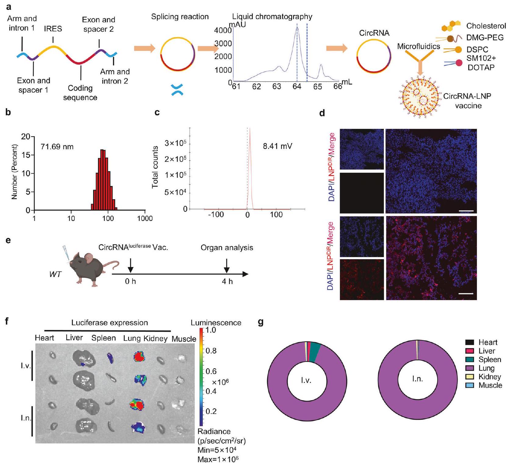
Based on the previously published circRNA preparation strategy, the in-vitro transcription templates included a pair of exons and introns, CVB3 IRES, spacers, arms and the coding sequences. CircRNAs were obtained through the back-splicing reaction followed by purification through liquid chromatography. As circRNA is known to have better stability than mRNA, we first aimed to determine whether the circRNA vaccine exhibited superior thermostability compared to its linear counterpart when encapsulated in lipid nanoparticles (LNP). We utilized the clinically approved LNP formulation containing SM102, DSPC, DMG-PEG, and cholesterol. The RNAs were encoded with D2GFP, a destabilized GFP protein with a half-life of only two hours, to evaluate protein expression. Our results showed that circRNA exhibited higher protein expression levels than the linear RNA formulations when stored at both and , supporting the broader applicability and convenience of circRNA vaccines. (Supplementary Fig. 1).
For intranasal delivery, we tested the efficiency of two clinically approved ionizable lipids (SM102 and ALC-0315) to form LNPs with DSPC, DMG-PEG and cholesterol. We characterized the size and zeta potential of two dye-containing LNPs (Supplementary Fig. 2a, b) and then we performed the in vivo assay. We observed
that SM102-based LNP showed higher dye-tracking fluorescence intensity compared to ALC-0315 (Supplementary Fig. 2c, d). According to recent studies, the addition of cationic lipids, such as 1,2-dioleoyl-3-trimethylammonium-propane (DOTAP), can enhance lung-targeting efficiency after intravenous and intranasal administration. We optimized the delivery system by adding DOTAP to the SM102-based LNPs, resulting in a higher protein expression level (Supplementary Fig. 2e, f). Thus, this delivery strategy was used in the following experiments. The entire manufacturing process of circRNA vaccine is shown in Fig. 1a. Briefly, circRNA was prepared through in-vitro transcription and splicing reaction, followed by purification via liquid chromatography. Purified samples were mixed with five lipid components via microfluidics to form the circRNA-LNP complex. For the characterization of the RNA vaccine, the particle size was around 71.69 nm (Fig. 1b). The zeta potential was 8.41 mV (Fig. 1c). To monitor the biodistribution of the RNA vaccine, luciferasecoding circRNA and LNPs were mixed with DiR lipophilic dye. After intranasal administration, successful delivery was confirmed via dye-tracking fluorescent imaging (Fig. 1d). Furthermore, we aimed to compare the biodistribution of the RNA vaccine via different administration routes (Fig. 1e). We isolated vital organs 4 h after vaccination and found that the intranasal vaccine led to LNP accumulation and protein expression in lung tissues (Fig. 1f, g and supplementary Fig. 2g, h). However, the LNPs and protein expression could be detected in liver, lung, and spleen tissues after intravenous injection. Taken together, these data showed that the circRNA-LNP platform can initiate local protein expression at lung tissues after intranasal administration.
Intranasal circRNA vaccine mediates potent anti-tumor response with limited adverse effects
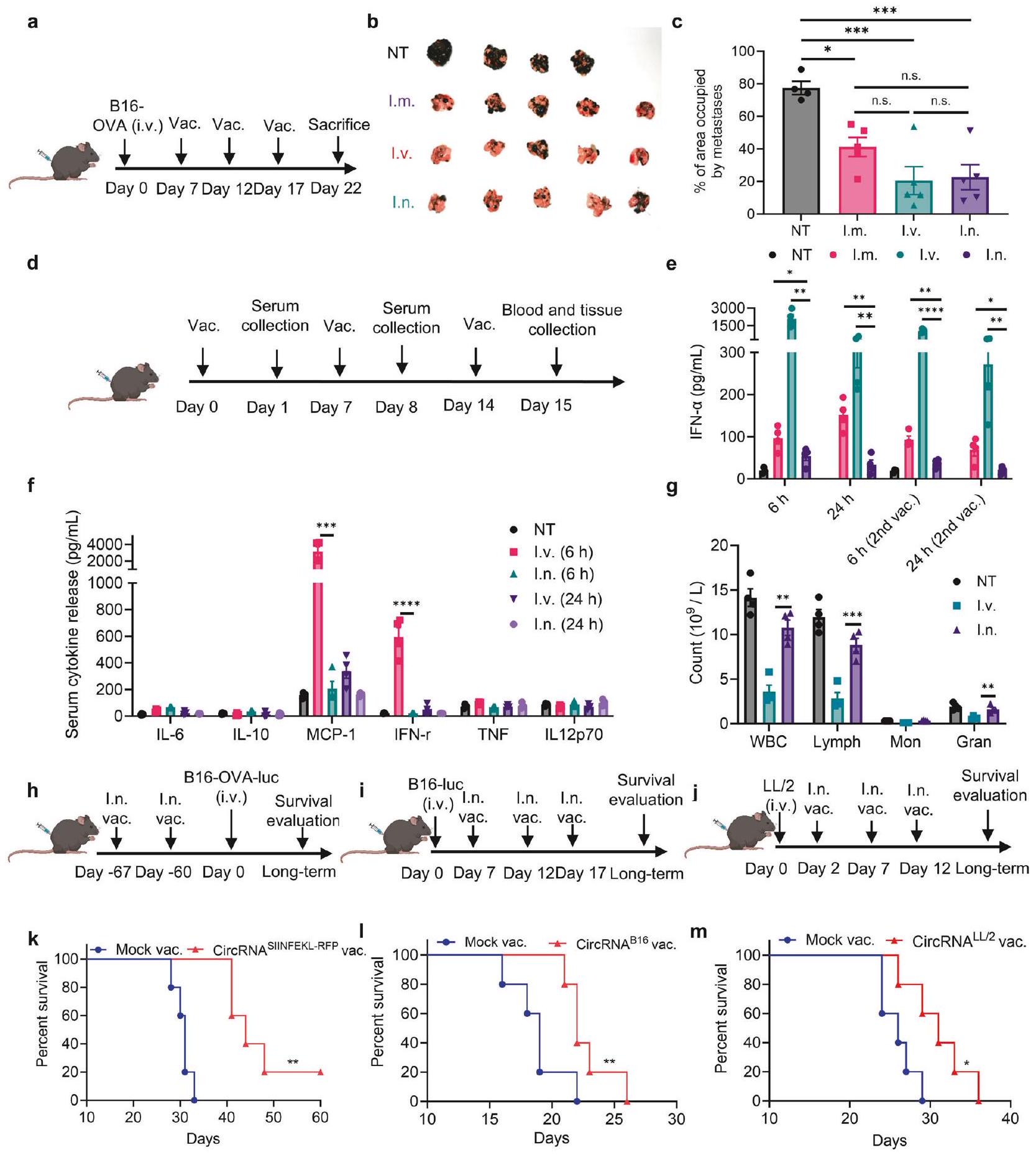
To systematically compare the anti-tumor efficiency of the circRNA vaccine administered through different routes, we established a lung metastasis B16 tumor model expressing the OVA antigen. Mice received SIINFEKL (class I (Kb)-restricted epitope of OVA antigen)-RFP-coding circRNA vaccines at specified time points (Fig. 2a). Compared to the untreated group, all three administration routes exhibited statistically comparable antitumor efficacy (Fig. 2b, c). However, the percentage of lung tissue occupied by metastatic foci appeared lower in the intranasal and intravenous groups compared to the intramuscular group. Then, we analyzed the acute toxicity of the circRNA vaccine administered through different routes. Previous studies have indicated that RNA vaccines can induce the secretion of proinflammatory cytokines, resulting in adverse effects. We first evaluated the IFN-a level in serum post-vaccination due to its key role in inflammation induction (Fig. 2d). Compared with intravenous and intramuscular injection groups, the serum levels of IFN- were significantly lower in the intranasal groups (Fig. 2e). Then, we further analyzed other cytokines and similarly we found that intranasal groups showed lower cytokine level compared with intravenous injection. (Fig. 2f and supplementary Fig. 3). For the blood routine tests, it indicated a more significant reduction in blood and immune cells in the intravenous injection group compared to the intranasal group (Fig. 2g). To further verify the safety of intranasal administration, we monitored the body weight changes in mice throughout the treatment period (Supplementary Fig. 4). Although a slight decrease in body weight was observed in the vaccination groups, it was restored after a few days.
We further assessed the anti-tumor efficiency of the intranasal vaccine in different models (Fig. 2h-j). In prophylactic lung metastasis models, mice received two doses of the RNA vaccine, followed by a B16-OVA tumor cell challenge after 30 or 60 days. Bioluminescence images (Supplementary Fig. 5a, b) and survival curves (Fig. 2k and Supplementary Fig. 5c) showed that intranasal immunization inhibited tumor growth and significantly prolonged
Fig. 1 Intranasal circRNA delivery via lipid nanoparticles initiates protein expression at lung tissues. a Experimental diagram of the RNA-LNP vaccine manufacturing process. b Size distributions of the RNA-LNP vaccine. c Zeta potential of the RNA-LNP vaccine. d Representative dyetracking fluorescent images of lung tissues. Mice were given with DiR-labeled LNPs via intranasal administration and lung tissues were isolated after 4 h . Scale bar, . e Timeline of the experiment designed to evaluate the biodistribution of luciferase protein expression ( circRNA per mouse). Imaging results (f) and statistical analysis (g) of luciferase protein expression among various organs. I.v., intravenous injection group. I.n., intranasal administration group
survival, suggesting the formation of memory T cells after vaccination. In therapeutic lung metastasis models, B16luciferase and LL/2 cell lines without overexpressing OVA antigens were employed. Based on the published non-synonymous mutations of and cells, we designed B16-antigencoding (Supplementary Fig. 5d-f) and LL/2-antigen-coding (Supplementary Fig. 6a, b) RNA vaccines, and mice received three doses of vaccination after the establishment of lung metastasis. In the B16 model, the survival curves showed that intranasal immunization significantly inhibited tumor growth and prolonged survival (Fig. 2l). In the LL/2 model, we observed that the immunized mice exhibited cytotoxicity with fewer tumor foci (Supplementary Fig. 6c, d) and more prolonged survival (Fig. 2m). In both models, we observed specific T cell activity targeting vaccine-encoded antigens in lung tissues (Supplementary Fig. 5 f and 6e), highlighting the potential of intranasal circRNA for neoantigen vaccine applications. In summary, these data indicate
that the intranasal circRNA vaccine elicits a potent anti-tumor response with fewer side effects compared to other routes of administration.
The anti-tumor efficiency of intranasal circRNA vaccine is tumor antigen- and cDC1-dependent
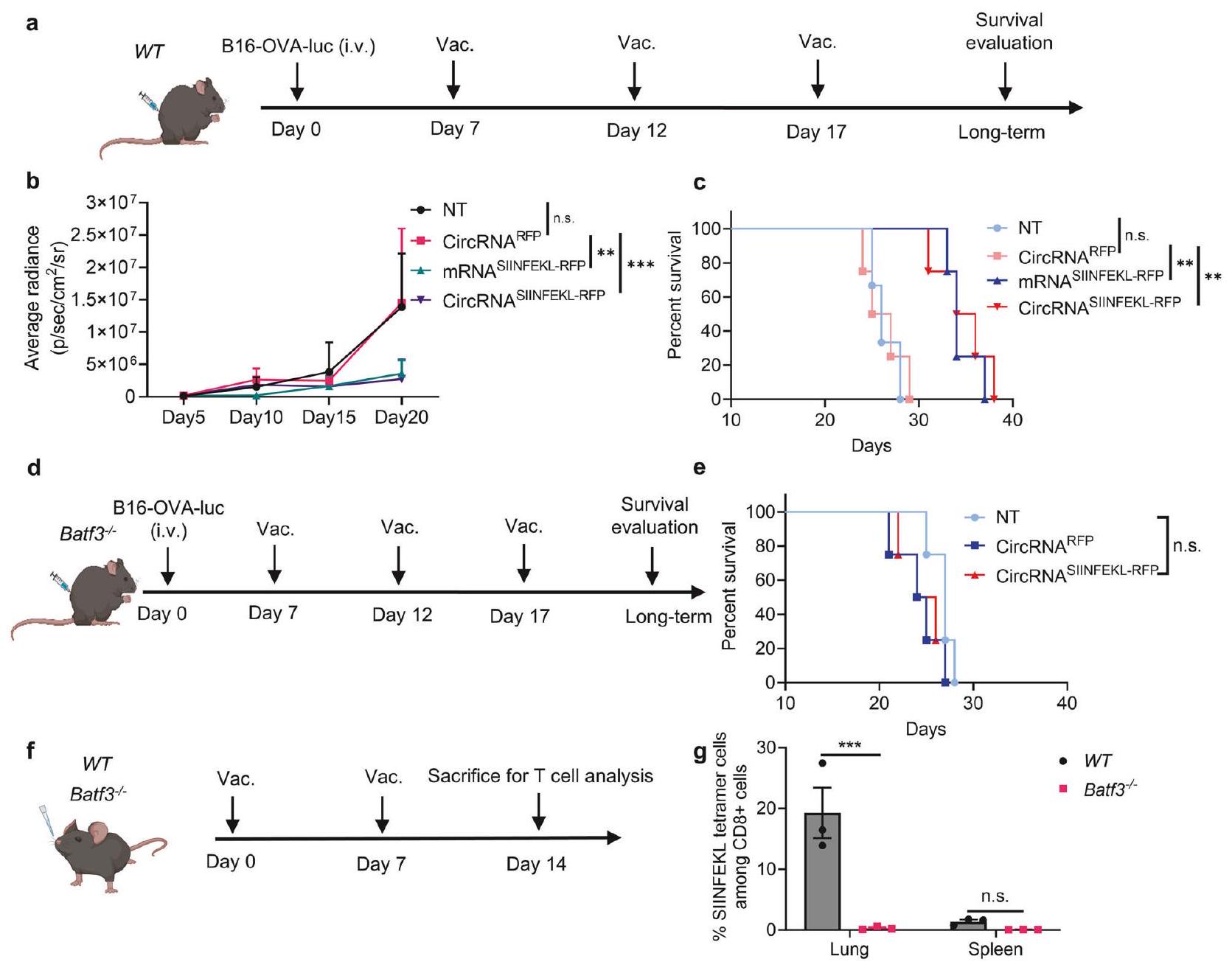
Inspired by the therapeutic potential of the intranasal vaccine, we aimed to further investigate the principles underlying its antitumor immune response. We first established a B16-OVAluciferase lung metastasis model and confirmed the anti-tumor response (Fig. 3a). An RFP-coding RNA was used as an irrelevant antigen control. We also prepared linear mRNA and circRNA vaccines encoding SIINFEKL-RFP for vaccination. Bioluminescence image results (Fig. 3b and supplementary Fig. 7a) and survival curves (Fig. 3c) demonstrated that only tumor antigen-coding RNA vaccines could inhibit tumor cell growth, with no significant difference observed between mRNA and circRNA vaccines in this
Fig. 2 Intranasal circRNA vaccine mediates potent anti-tumor response with limited adverse effects. a Timeline of the experiment designed to evaluate the anti-tumor ability of different routes of circRNA vaccine. b Images of lung metastasis tumors at the endpoint in different groups. NT, not-treated group. I.m., intramuscular injection group. I.v., intravenous injection group. I.n., intranasal administration group. for NT group. for other groups. c The percentage of area occupied by metastatic tumor among various groups at the end point. Data were analyzed by one-way ANOVA with Tukey’s multiple comparisons test. d Timeline of the experiment designed to evaluate the systemic adverse effects of different routes of circRNA vaccine. Serum cytokine release after administrated with the vaccine tested by mouse IFN- Elisa kit (e) and mouse inflammation kit (f). Data were analyzed by Student’s t test to compare i.n. group with i.v. or i.m. group in e. Data were analyzed by one-way ANOVA with Tukey’s multiple comparisons test in f. g Blood routine analysis 24 h after the third vaccination. Data were analyzed by Student’s t test to compare i.n. group with i.v. group. Timeline of the experiments to evaluate the anti-tumor ability of intranasal in various models. Mice received with LNP without RNA were considered as mock vaccine group. h Mice were immunized with SIINFEKL-coding circRNA twice, followed by rechallenging B16-OVA-luciferase cells 60 days after vaccination. i Mice were challenged with B16luciferase cells and received three doses of B16-antigen-coding circRNA vaccine. j Mice were challenged with LL/2 cells and then received three doses of LL/2-antigen-coding circRNA vaccine. Survival curves of the experiments related to and , respectively. for each group. Data were analyzed via Kaplan-Meier analysis. For mice received with vaccination, each dose contained circRNA. All data are represented as mean ± SEM
Fig. 3 The anti-tumor ability of the intranasal circRNA vaccine is tumor antigen and cDC1 dependent. a Timeline of the experiment designed to evaluate the anti-tumor ability of the intranasal RNA vaccine. Wild-type (WT) mice were challenged with B16-OVA-luciferase cells and then left untreated (NT), treated with RFP-coding or SIINFEKL (OVA peptide)-RFP-coding RNA. Tumor growth was monitored through bioluminescence imaging. b Statistical analysis of the average bioluminescence radiance with different treatments. Data were analyzed with two-way ANOVA with Tukey’s multiple comparisons test. for NT group. for other groups. c Survival curve of the WT mice with different treatments. Data were analyzed via Kaplan-Meier analysis. d Timeline of the experiment designed to evaluate the anti-tumor ability of the intranasal RNA vaccine in Batf3 mice. e Survival curve of the Batf3 mice with different treatments. Data were analyzed via KaplanMeier analysis. for each group. Timeline of the experiment designed to evaluate the antigen-specific T cell induction after circRNA vaccination. WT or Batf3 mice were intranasally immunized with SIINFEKL-RFP-coding circRNA followed by antigen-specific T cell evaluation at lung and spleen tissues ( , repeated for three times). Statistical analysis of the ratio of antigen-specific T cells. Data were analyzed with two-way ANOVA with Tukey’s multiple comparisons test. All data are represented as mean ± SEM
model. To further characterize the T cells induced by vaccination, we analyzed antigen-specific T cells in lung tissues following a two-dose vaccination regimen. Our results revealed an increase in both total antigen-specific T cells and CD103-positive antigenspecific T cell subset, confirming the successful induction of antigen-specific T cells (Supplementary Fig. 7b-e). Using an intravascular staining assay, we confirmed that the antigenspecific T cells induced by the intranasal vaccine primarily originate from the lung tissue rather than peripheral blood (Supplementary Fig. 7f-h). Furthermore, an in vivo CD8 depletion assay demonstrated that CD8 + T cells are indispensable for the vaccine’s anti-tumor efficacy (Supplementary Fig. 8a-d).
Given the preeminent role of CD8 T cells and vaccinedelivered antigens for initiating anti-tumor immunity, we aimed to test the influence of antigen presentation on antitumor ability. As cDC1 plays a major role in capturing and presenting antigens on MHC-I to promote CD8 +T cell immunity, we repeated the anti-tumor experiment in cDC1-
knockout mice (Fig. 3d). Interestingly, the anti-tumor efficiency of the intranasal circRNA vaccine was diminished, suggesting that cDC1 is crucial for the vaccine-induced anti-tumor immune response (Fig. 3e). We further analyzed the antigenspecific CD8 + T cells induced by the vaccine (Fig. 3f). WT and Batf3 mice received two doses of immunization, and T cells were detected in lung and spleen tissues. We found that cDC1 deficiency significantly decreased the level of antigen-specific CD8 + T cells at the lung tissue (Fig. 3 g and supplementary Fig. . To confirm that cDC1 presents the antigen after vaccination, we isolated the cDC1 from lung draining lymph node and coculture then with antigen-specific T cells (supplementary Fig. 8 g ). The activation of T cells following co-culture provides new evidence that cDC1 cells present antigens and play a key role in T cell induction. Thus, the expression of tumor antigen and the presence of cDC1 are indispensable for the anti-tumor T cell immune response induced by the intranasal circRNA vaccine.
Fig. 4 AMs and cDC1s are mainly responsible for boosting antigen-specific T cells in lung tissues after intranasal vaccination. a Timeline of the experiment to evaluate the role CD11c+ APCs on boosting antigen-specific T cells. WT, Batf3 or CD11c-DTR mice were pre-treated with cyclophosphamide (CTX) and diphtheria toxin (DT) for lymphodepletion in all mice and CD11c+ cells depletion in CD11c-DTR mice. Activated OT-1 cells were labeled with CFSE and then transferred to mice immunized with intranasal circRNA vaccine. T cell proliferation at lung tissues was analyzed at day 3. b, c Representative flow cytometry histograms (b) and statistical results (c) of T cell proliferation with different treatments. Data were analyzed by one-way ANOVA with Tukey’s multiple comparisons test. for mock vac. group; for other groups. For LNP uptake analysis, mice received intranasal DiD-labeled LNP and lung tissues were analyzed 4 h later. d, e Representative flow cytometry histograms (d) and statistical results (e) of LNP uptake among different cell types. f Scheme of the experiment to test the potential APCs that can boost antigen-specific T cells. Mice were immunized with intranasal LNP without RNA (mock) or circRNA vaccine and 24 h later, lung tissues were collected and APCs were sorted and co-cultured with OT-1 cells for another 36 h in IFN- Elispot wells. ( for mock group; for circRNA vaccine group, repeated for three times) Created with BioRender.com. Representative images and statistical results of IFN- Elispot assay. Data were analyzed by Student’s t test. i Timeline of the experiment to explore whether vaccine can boost T cells at lung tissues. Mice were treated with FTY720 during the prime-boost process or only the boosting process or normal saline as the control group. The number of antigen-specific T cells at lung tissues was analyzed. Representative plots ( ) and statistical results ( ) of the antigen-specific T cells with different treatments. Data were analyzed by one-way ANOVA with Tukey’s multiple comparisons test. All data are represented as mean ± SEM
AMs and cDC1s are mainly responsible for boosting antigenspecific T cells in lung tissues after intranasal vaccination Successful induction of robust and sustained T cell immunity requires not only the priming of T cells but also the boosting process through repeated vaccination. Therefore, we aimed to
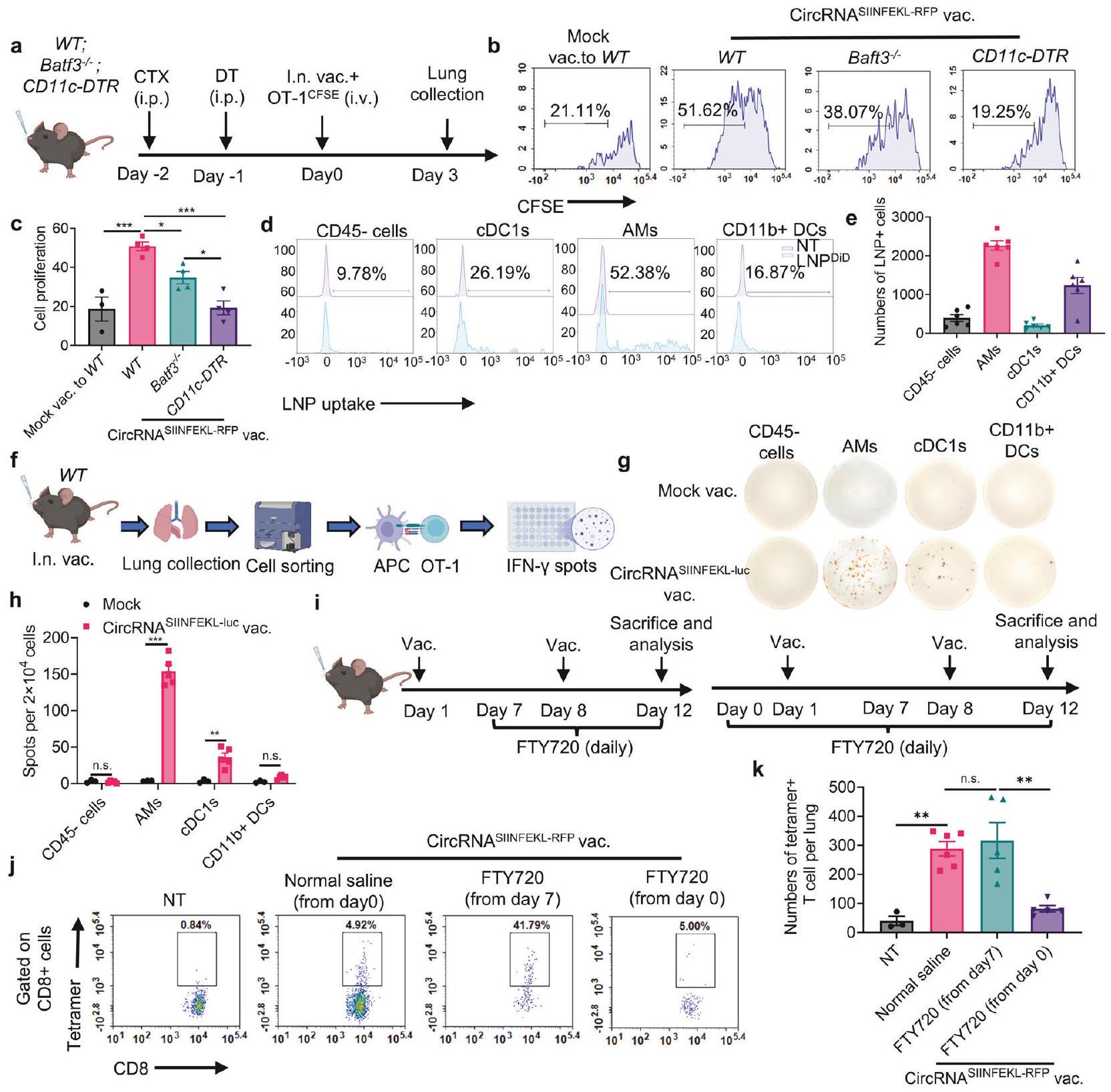
further explore the T cell boosting process following intranasal immunization. We employed WT mice, mice without cDC1, and mice without all CD11c+ cells to test their ability to boost activated antigen-specific T cells (Fig. 4a). Although depletion of cDC1 reduced T cell proliferation, the deficiency of CD11c+ cells
completely blocked vaccine-induced T cell proliferation, suggesting that both cDC1 and other CD11c+ antigen-presenting cells are involved in boosting T cells (Fig. 4b, c and supplementary Fig. 9a). To identify the cell types that support T cell proliferation, LNPs were labeled with DiD lipophilic dye and intranasally administered to mice. We then stained the major populations of CD11c+ cells at lung tissues, including alveolar macrophages (AMs), cDC1s, CD11b + DCs, and CD11c- CD45- cells as the control group. We found that LNPs were predominantly engulfed by AMs, with other cells also capable of engulfing LNPs but in smaller numbers (Fig. 4d, e, supplementary Fig. 9b-d). Moreover, the production of IFN- was augmented when isolated AMs and cDC1s were co-cultured with OT-1 cells, while there was no significant difference in the CD11b + or CD45- groups (Fig. 4f-h). We also performed immunofluorescence (IF) staining of lung tissues to investigate the interactions between antigen-presenting cells (APCs) and antigen-specific T cells (Supplementary Fig. 10). In the vaccination group, we observed interactions between antigen-specific T cells and CD11c + cells, as well as between antigen-specific T cells and AMs. These findings provide new evidence that both AMs and dendritic cells (DCs) contribute to the activation and enhancement of antigenspecific T cells. We next assessed whether the intranasal vaccine primed and boosted T cells directly in lung tissues. To do this, FTY720 was employed to block T-cell egress from lymphoid tissues. Mice were treated with FTY720 daily starting before the priming or boosting process, and antigen-specific T cells were detected in lung tissues at the endpoint (Fig. 4i). During the boosting process, FTY720 treatment had no impact on the number of antigen-specific T cells in lung tissues, indicating that the vaccine boost led to antigen presentation and T cell proliferation within the local lung tissues (Fig. and supplementary Fig. 9d). However, the number of antigen-specific T cells in lung tissues significantly decreased (Fig. 4k), with no impact on the mediastinal lymph node when mice were treated with FTY720 starting before the first immunization (Supplementary Fig. 11). It suggested that the initial priming of antigenspecific T cells occurred outside lung tissues, relying on draining lymph nodes. Thus, these data indicate that the intranasal circRNA vaccine can boost T cells directly in lung tissues through the actions of AMs and cDC1s.
Intranasal circRNA vaccine boosts both endogenous and transferred antigen-specific T cells to promote anti-tumor immune response
Based on the above findings, we hypothesized that intranasal RNA vaccine may synergize with transferred antigen-specific T cells for enhanced anti-tumor ability. We conducted experiments using a WT mouse model. Mice were initially challenged with B16-OVAluciferase tumor cells and underwent lymphodepletion with CTX before receiving treatment with circRNA vaccine, adoptive cell therapy (ACT), or a combination of both therapies (Fig. 5a). Bioluminescence imaging and histopathological examination (HE staining) demonstrated that while vaccine alone or OT-1 cells with mock vaccine could inhibit tumor growth, the combination therapy resulted in the most pronounced tumor control (Fig. 5b-d). We also quantified antigen-specific T cell numbers for both endogenous and transferred T cells, revealing that the vaccine effectively expanded both populations (Fig. 5e-g).
Given the potential secondary effects of the vaccine-induced T cell response, it is possible that immune responses against secondary antigens, distinct from the original vaccine target, could be triggered. In this study, the combination of vaccination and ACT induces T cell-mediated destruction of tumor cells expressing the OVA antigen. This destruction releases additional tumor antigens, which may be captured by antigen-presenting cells, subsequently leading to the activation of endogenous cells targeting OVA antigen-loss tumors. This antigen-spreading phenomenon has been observed in both preclinical and clinical
studies involving immunotherapy. Thus, we assessed the antitumor capability of splenocytes from vaccinated mice when cocultured with B16 cells lacking the OVA antigen (Fig. 5h). Remarkably, splenocytes from immunized mice exhibited increased production of IFN , indicating that the vaccine can induce antigen spreading to target antigen-loss tumor cells (Fig. . Moreover, transferred splenocytes from the mice in the combination therapy group prevented tumor formation upon rechallenge with B16 cells lacking the OVA antigen (Fig. 5h, j), suggesting that the combination therapy may facilitate the generation of memory T cells capable of targeting tumor cells through alternative antigens. These findings underscore the potential of combining intranasal RNA vaccine with ACT to enhance anti-tumor immunity by expanding both endogenous and adoptively transferred T cells, inducing antigen spreading, and potentially fostering the formation of memory cells against tumor cells expressing other antigens.
Intranasal circRNA vaccine induces functional and phenotypic changes in endogenous T cells
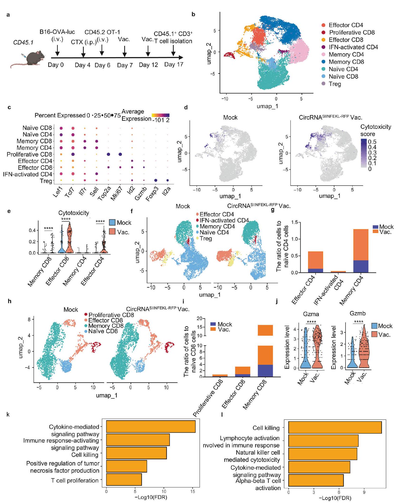
Building on our previous findings, we aimed to comprehensively examine alterations in endogenous T cells using single-cell RNAseq analysis of CD3+ cells from lung tissues in tumor-bearing mouse models (Fig. 6a). Unsupervised clustering of the transcriptome data identified nine major T cell subsets: naïve CD4 and CD8 cells (marked by Lef1 and Tcf7 expression with relatively low II7r and Sell levels), memory CD4 and CD8 cells (expressing II7r and Sell), proliferative CD8 cells (characterized by Top2a and Mki67 expression), effector CD4 and CD8 cells (with and Gzmb expression), Treg cells (featuring oxp 3 and ), and IFNactivated CD4 T cells (marked by Ifit1, Ifit3, and Isg15 expression) (Fig. 6b, c). We specifically identified a cytotoxic T cell cluster characterized by a signature of cytotoxicity-associated genes (Gzma, Gzmb, Gzmc, Gzmd, Gzme, Gzmf, Gzmg, Gzmk, Gzmm), which showed significantly heightened cytotoxic activity in the vaccine group compared to controls (Fig. 6d). Further analysis revealed that cytotoxic capabilities were notably upregulated in memory and effector T cell clusters within the vaccinated group (Fig. 6e). Additionally, we analyzed the abundance and relative proportions of each T cell cluster within CD4 and CD8 populations separately, finding increased numbers and ratios of effector and memory cells in the vaccine group (Fig. 6f-i). Importantly, the antitumor function of effector CD8 T cells were significantly enhanced in the vaccine group, as evidenced by elevated expression levels of Gzma and Gzmb (Fig. 6j). Gene Ontology (GO) enrichment analysis further indicated that pathways related to T cell function and anti-tumor cytotoxicity in CD4 and CD8 effector cells were enriched in the vaccine group (Fig. 6k, I). We validated the functionality of both endogenous and transferred T cells through intracellular IFN- staining (Supplementary Fig. 12). An increased level of IFN- was observed in both populations, indicating enhanced cytotoxic activity of the T cells upon encountering tumor cells. Overall, this comprehensive analysis demonstrates that the intranasal circRNA vaccine drives substantial changes in the landscape of endogenous T cells, enhancing their cytotoxicity and memory functions and thereby bolstering the anti-tumor immune response.
Intranasal circRNA vaccine augments the anti-tumor efficacy of CAR-T cell therapy against tumor cells expressing specific tumorassociated antigens
In addition to examining the effects of the vaccine on endogenous T cells, we also investigated its direct impact on adoptive transferred T cells. To visualize the dynamic biodistribution and long-term performance of vaccine-boosted transferred T cells during the anti-tumor process, we used B16-OVA bearing Rag1 mice injected with OT-1 cells co-expressing luciferase, followed by immunization with the intranasal circRNA vaccine (Fig. 7a). In vivo
Fig. 5 Intranasal circRNA vaccine boosts adoptive transferred antigen-specific T cells for enhanced anti-tumor immune response. a Timeline of the experiment to evaluate the anti-tumor ability of combination therapy with intranasal circRNA vaccine and transferred OT-1 cells. b, c Bioluminescence images (b) and statistical result of average radiance (c) at different time points. Data were analyzed with two-way ANOVA with Tukey’s multiple comparisons test. d Representative images of HE staining to evaluate the tumor formation at day 21. Scare bar, (upper) or (lower). e Representative plots of the transferred and endogenous antigen-specific T cells among different groups. Statistical analysis of antigen-specific T cells in e. Data were analyzed by Student’s t test. h Scheme of the experiments to evaluate the role of endogenous T cells in killing antigen-loss tumor cells. Splenocytes were co-cultured with B16-luciferase cells (without OVA antigen) for IFN Elispot assay. Splenocytes from the mice in the combination therapy group were transferred to Rag1 followed by B16-luciferase cells challenge. Created with BioRender.com. i Statistical results of the number of IFN- spots. Data were analyzed by one-way ANOVA with Tukey’s multiple comparisons test. j Survival curve of the Rag1 mice, for each group. Data were analyzed via Kaplan-Meier analysis. All data are represented as mean ± SEM

bioluminescence imaging revealed that the vaccine drove T cell expansion in lung tissues, and repeated doses sustained T cell numbers at a relatively high level compared to the mock vaccine group (Fig. 7b, c). Assessing the anti-tumor efficiency of various treatments, we observed that intranasal vaccination significantly prolonged the survival of T cell-transferred mice (Fig. 7d). These
findings illustrate that intranasal RNA vaccine boosting enables transferred antigen-specific T cells to expand in lung tissues, thereby enhancing their efficiency in lung cancer treatment.
To broaden the application of intranasal RNA vaccine, we leveraged their capacity to boost antigen-specific T cells. In doing so, we developed a vaccine-responsive CAR-T cell strategy that not
Fig. 6 Endogenous T cells show transcriptional changes with enhanced cytotoxic function and more memory-like phenotype in response to intranasal vaccine. a Timeline of the experiment to isolate the endogenous T cells for single-cell RNA sequencing. Mice received LNP without RNA (mock, ) or SIINFEKL-RFP-coding circRNA vaccine group (vac., ) for two doses. Cells from lung tissues were mixed for staining and sorting in the same group. Sorted cells were sent for single-cell RNA sequencing. b UMAP plot of CD3 +T cells colored by clusters. Dot plots showing differential expression of marker genes in endogenous CD3 + T cells. d UMAP plot of the T cells with cytotoxic function in mock group (mock) and vaccine group (vac.). e Statistical analysis of the cytotoxic gene expression in different clusters. Data were analyzed through the Wilcoxon signed-rank test. UMPA plot of CD4 + T cells in two groups. Statistical analysis of the CD4 + cell number of various clusters compared to their naïve cells in mock and vac group. UMAP plot of CD8 + T cells in two groups. i Statistical analysis of the CD8+cell number of various clusters compared to their naïve cells in mock and vac group. j Statistical analysis of Gzma and Gzmb expression in effector CD8 clusters. Data were analyzed through the Wilcoxon signed-rank test. k, I GO enrichment analysis showing the enriched terms of vac. group compared with mock group in effector CD4 (k) and effector CD8 (I) clusters
only responds to the vaccination but also targets tumor cells expressing specific surface tumor-associated antigens. These antigens are typically challenging for conventional cancer vaccine approaches which are limited to target MHC-presented immunogenic peptides. This approach aims to eliminate tumor cells via the CAR molecule while simultaneously promoting in-situ boosting of immune responses through the TCR machinery (Fig. 7e). The CART cells showed the ability to kill EGFR-overexpressing B16 tumor cells at different ratio in vitro (Fig. 7f). We then examined the activation of CAR-T cells through their CAR or TCR. DCs were pretransfected with varying doses of EGFR- or SIINFEKL-RFP-coding circRNA, and CAR-T cells were co-cultured with these DCs, followed by T cell activation and cytokine analysis. The secretion of IFN- increased (Fig. 7g), and the expression of CD25 and CD69 was upregulated as the RNA dose increased (Supplementary Fig. 13). These data demonstrated the CAR-T cells could exhibit specific function after stimulation through both EGFR-CAR and predefined TCR. Subsequently, an EGFR-expressing B16 lung metastasis tumor model was established in WT mice. CAR-T cells were then transferred to these mice, followed by the administration of an intranasal circRNA vaccine (Fig. 7h). Bioluminescence imaging (Fig. 7i), statistical analysis, and survival curves (Fig. 7j, k) indicated that CAR-T cell therapy combined with a mock vaccine initiated only a modest anti-tumor response. In contrast, CAR-T cell therapy paired with a SIINFEKL-encoding vaccine significantly inhibited tumor growth and prolonged survival. These findings suggest that intranasal RNA vaccines are effective for in-situ boosting of CAR-T cells to target and eliminate tumor cells expressing specific surface antigens.

DISCUSSION

A significant challenge in developing effective cancer vaccines lies in eliciting potent T cell immune responses, which are often hindered by insufficient antigen uptake and presentation. RNA vaccines offer the ability to deliver desired antigens and effectively activate antigen-presenting cells for T cell induction, which is a novel perspective for cancer treatment. However, existing studies predominantly focus on intravenous RNA vaccines, which can induce systemic inflammatory responses to boost innate immunity and reprogram the tumor microenvironment (TME) for enhanced anti-tumor T cell responses. Despite their efficiency, these systemic approaches may pose risks of adverse effects, especially in patients with pre-existing inflammation. To address these challenges, we propose novel vaccine strategies that preserve anti-tumor efficiency while minimizing systemic inflammation, particularly through mucosal vaccination for mucosal malignancies. In our study, we provided a strategy of intranasal circRNA vaccination for lung cancer treatment. This strategy activates local immune responses and effectively suppresses tumor progression with minimal systemic inflammation. Previously, orthotopic head and neck or lung cancers were reported to be inhibited by a peptide-based cancer vaccine via intranasal mucosal but not the intramuscular rout. Interestingly, although our findings show that intranasal and intravenous
injections of the circRNA vaccine appear better tumor control compared with intramuscular injection, the intramuscular vaccine also inhibit tumor growth. This discrepancy from previous studies may be attributed to differing levels of systemic T cell activation induced by distinct vaccine formulations following injection administration. Taken together, these results all suggest that the noninvasive intranasal immunization is indeed beneficial for local lung cancer treatment with minimal systemic inflammation.
In cancer vaccine development, tumor antigen-specific T cells are pivotal for effective anti-tumor immunity. However, the mechanisms underlying T cell priming and boosting, particularly with RNA vaccines, remain poorly understood. A study on the Pfizer-BioNTech SARS-COV2 mRNA vaccine has highlighted the role of cDC1 in T-cell induction. Similarly, our research underscores the critical role of cDC1 in mediating the anti-tumor effects of RNA vaccines, demonstrating diminished CD8 T cell responses in cDC1-deficient mice. While the priming phase involves specific DC subtypes, boosting antigen-specific T cells may engage additional cell types. For instance, alveolar macrophages (AMs) were found to be essentially for CD8 T cell expansion following intranasal protein vaccination. In our current data, both AMs and cDC1 can boost antigen-specific CD8 T cells after intranasal vaccination. This is not entirely consistent with the previous study, indicating that different vaccine approaches modulate host T-cell response with distinct antigen-presentation principles. Moreover, antigen expression duration may influence vaccine-induced immune responses differently. For instance, circRNA vaccines encoding viral antigens have been shown to induce higher humoral immune responses compared to conventional mRNA vaccines. In our previous and current studies, tumor antigencoding mRNA and circRNA reveal similar anti-tumor efficiency, aligning with another comparative analysis of mRNA and selfamplifying RNA with longer protein expression duration. They showed self-amplifying RNA and mRNA drove anti-tumor response with few differences. These results indicate that prolonged antigen expression may favor humoral immunity with minimal impact on CD8 + T cell response. Nevertheless, compared with mRNA, the circRNA vaccine retains its unique advantage in terms of stability, especially when storage at as showed in our data and a previous study, which supported the broaden applicability and convenience of circRNA vaccines.
In general, cancer vaccines are mainly designed to stimulate endogenous T cells to recognize and attack tumor cells through tumor-specific antigens or neoantigens presented onto MHC-I molecules. This process, although effective, typically requires time to establish durable immunity. In contrast, adoptive T cell therapies, such as TCR-T and CAR-T therapies, use ex vivo activated T cells to target MHC-peptide complexes or surface tumor-associated antigens, enabling a direct and rapid therapeutic response. However, these approaches often face challenges in achieving sustained long-term persistence, particularly in solid tumors. These limitations suggest the potential for synergistic integration with vaccines and adoptive T cell therapies to enhance both efficacy and durability of immune responses. Previous studies have shown that intravenous mRNA vaccines or

subcutaneously peptide vaccines enhance the function of transferred CAR-T and TCR-T cells, echoing our findings of intranasal circRNA promoting the accumulation and expansion of transferred T cells in lung tissues, thus prolonging survival in combination therapy. In addition, we designed a combination
treatment strategy in a tumor-associate-antigen model with CAR-T cells and the intranasal vaccine. The approach provides two significant advantages, enhancing CAR-T cell persistence and enabling redirection of T cells to target tumor-associated antigens independent of MHC presentation on tumor cells.

Abstract

Fig. 7 Intranasal circRNA vaccine augments the anti-tumor efficacy of CAR-T cell therapy against tumor cells expressing specific tumorassociated antigens. a Scheme of the experiment to track the migration and expansion of transferred OT-1 cells. Cells were activated, infected with luciferase-coding retrovirus and then transferred to tumor-bearing Rag1 mice. T cell proliferation at lung tissues was monitored by bioluminescence imaging. Created with BioRender.com. b Bioluminescence images of the mice at different time points. Mock T cells were cells isolated from WT mice, circRNA was used as an irrelevant vaccine control. c Statistical results of average radiance at lung tissues at different time points. Data were analyzed with two-way ANOVA with Tukey’s multiple comparisons test. d Survival curve of the mice after different treatments. Data were analyzed via Kaplan-Meier analysis. e Scheme of the vaccine-responsive CAR-T cell design. OT-1 cells were activated and infected with anti-EGFR CAR-coding retrovirus. The CAR-T cells can kill EGFR-expressing tumor cells through their CAR molecule and can be stimulated with SIINFEKL antigen-loaded APCs through their TCR-machinery. Created with BioRender.com. Statistical analysis of tumor cell lysis after coculture with EGFR-expressing B16 cells and the CAR-T cells at different E:T ratio. Data were analyzed by one-way ANOVA with Tukey’s multiple comparisons test. Analysis of IFN- secretion after coculture with the vaccine-responsive CAR-T cells and DC2,4 cells. DC2.4 cells were pre-transfected with different doses of RNA 24 h before coculture. h Timeline of the experiment to evaluate the anti-tumor ability of combination therapy with intranasal RNA vaccine and transferred CAR-T cells. Bioluminescence images (i) and statistical result of average radiance (j) at different time points. Data were analyzed with two-way ANOVA with Tukey’s multiple comparisons test. (k) Survival curve of the mice with different treatments. Data were analyzed via Kaplan-Meier analysis. All data are represented as mean ± SEM

It is well-established that antigens delivered by vaccines can elicit antigen-specific T-cell responses. Additionally, vaccineboosted T cells and the vaccines themselves can interact with the host immune system. A study has demonstrated that vaccines can enhance the efficiency of host T cells in treating tumors with antigen heterogeneity. We have observed a similar phenomenon in our study. Splenocytes from vaccinated mice exhibited increased cytotoxicity against OVA antigen-loss tumor cells and maintained memory responses that protected mice from rechallenging with antigen-loss tumors. This antigen-spreading phenotype may enhance therapeutic efficiency in clinical settings when treating tumors with antigen-loss or antigenic heterogeneity. Another study suggests that intranasal virus delivery can reshape the local immune microenvironment and enhance the cytotoxic function of T cells. Similarly, our data revealed transcriptional changes in endogenous T cells, characterized by enhanced cytotoxic abilities and an increased number of memory T cells in lung tissues.
In summary, our study demonstrates that intranasal circRNA vaccines can induce potent T-cell immune responses against lung cancers while minimizing systemic inflammation. We elucidate the roles of CDC1 and AMs in T cell priming and boosting processes and illustrate that the vaccine strategy synergizes effectively with cell therapy, enhancing anti-tumor efficiency (Fig. 8 ). Our findings contribute to the rational design and optimization of mucosal cancer vaccines, offering a novel approach to achieving robust local anti-tumor activity with limited systemic effects.

MATERIALS AND METHODS

Animals and cell lines

Female C57BL/6 J mice (6-8-week-old) and CD45.1 C57BL/6 J mice (6-8-week-old) were purchased from the laboratory animal resources center of Tsinghua University. Female Balb/c mice (6-week-old) were purchased from Beijing Vital River Laboratory Animal Technology. OT-1 (JAX stock no. 003831) mice were kindly provided by Chen Dong’s lab (Tsinghua University). Batf3 (JAX stock no.013755) mice were kindly provided by Yangxin Fu’s lab (Tsinghua University). CD11c-DTR (JAX stock no. 004509) and Rag1 (JAX stock no. 002216) mice were purchased from Jackson Laboratory. All mice were kept under SPF-grade conditions in the animal facility of Tsinghua University. All the mouse experiments were strictly adhered to the compliance standards of the Institutional Animal Care and Use Committee of Tsinghua University. HEK293T, B16 and LL/2 cell lines were purchased from ATCC. B16-OVA cells were kindly provided by Meng Xu’s lab (Tsinghua University). B16-luciferase and DC2.4 cells were kindly provided by Rui Kuai’s lab (Tsinghua University). All cells were cultured in DMEM medium (with 10% FBS and 1% penicillinstreptomycin solution) at and .

Plasmid

For in-vitro transcription (IVT) backbone, the plasmid template contains T7 promoter, homology arm, elements from permuted intron-exon (PIE) construct, spacer, CVB3 IRES, and coding sequence. The luciferase coding sequence was cloned into a retroviral vector for virus packaging. The extracellular domain of human EGFR, transmembrane domain and luciferase coding sequence was cloned into a retroviral vector for virus packaging. For CAR-T generation, the construct contains GM-CSF signal peptide, the scfv of cetuximab, CD28 transmembrane and intracellular domain, CD3 intracellular domain, IRES and RFP coding sequence. All the sequences are attached at Supplementary Table 1.

Preparation of RNA

The IVT templates were amplified by PCR (PrimeSTAR Max DNA Polymerase, Takara), followed by agarose electrophoresis and gel extraction (GeneJET Gel Extraction Kit, Thermo). CircRNA was manufactured as before. Briefly, IVT reaction was performed by using the T7 High Yield RNA Transcription Kit (Syngenebio). RNA was purified by DNase I digestion and lithium chloride precipitation. To circularize RNA, the reaction was performed at for 15 min in T4 RNA Ligase Reaction Buffer (NEB) and 2 mM GTP (ApexBio). To remove byproducts, RNA was further purified via liquid chromatography using a size-exclusion column with a particle size of and pore size of 2000 Å (Sepax Technologies, 215980-7830). Enriched circRNA was subsequently precipitated by ammonium acetate and stored at . Linear mRNA transcription and capping were performed by using the T7 Co-transcription RNA Synthesis Kit (Syngenebio) and uridine was replaced by N1methylpseudouridine (m1)). Products were purified via DNase I digestion, lithium chloride precipitation, followed by polyAtailing via the E. coli Poly (A) Polymerase (Novoprotein). Products were precipitated by ammonium acetate and stored at . All the RNA vaccines used in the study are attached at Supplementary Table 2.

Preparation of lipid nanoparticle (LNP)

The ionizable lipid SM102 and ALC-0135 were purchased from Sinopeg. DOTAP, DMG-PEG 2000, cholesterol, and DSPC were purchased from MCE. Unless specifically mentioned in the text, the ethanol phase of lipid nanoparticle contains SM102, DOTAP, DSPC, cholesterol and DMG-PEG 2000 at the molar ratio of 40:10:10:38:2. RNA was dissolved in an aqueous phase composed of 100 mM citrate buffer . The RNA: lipid mass ratio was 1:30. LNPs were prepared by mixing the aqueous phase and ethanol phase at a 3:1 ratio via microfluidic mixing devices (Micro&Nano Technologies). Obtained LNPs were dialyzed against PBS for 4 h (MWCO , Thermo). For LNP characterization, samples were resuspended in and analyzed using
Fig. 8 Proposed model of the intranasal circRNA vaccine for cancer therapy. Compared with conventional intravenous vaccine, intranasal delivery of circRNA vaccine results in fewer systemic adverse effects. For the mechanism of action of the vaccine, cDC1s are responsible for activating T cells at lymph nodes in the priming phase. Then, both cDC1s and AMs play significant roles in amplifying pre-existing endogenous or adoptive transferred antigen-specific T cells directly within lung tissues. Moreover, the priming-boosting vaccination strategy enhances the generation of memory T cells, which are critical for long-term immune surveillance and response. Consequently, the endogenous or adoptive transferred T cells trained by the vaccine are capable of initiating a robust and targeted anti-tumor immune response. Overall, the intranasal delivery of the circRNA vaccine leverages localized immune activation and amplification, presenting a promising approach for cancer immunotherapy with minimized systemic adverse effects. Created with BioRender.com
dynamic light scattering (DLS) performed on a Zetasizer Nano (Malvern Instruments, Malvern, UK).

Biodistribution of LNP

To monitor the uptake and biodistribution of LNP in vivo, DiR or DiD were added to the ethanol phase to encapsulate luciferasecoding circRNA for IVIS or flow cytometry, respectively. The DiR/ DiD: lipid mass ratio was 1:1000. LNPs were filtered through a filter and then intranasally administrated into C57BL/6 J mice pre-treated with isoflurane ( RNA in per mouse). After 4 h , mice were injected intraperitoneally with D-luciferin ( , Yeasen) and sacrificed 5 min later. Vital organs were collected and bioluminescence (luciferase expression) and fluorescence (LNP distribution) were measured by an IVIS Spectrum imaging system. For cell uptake assay, the lung tissue fragments were treated with collagenase IV (Gibco) and DNase I (Sigma) for 30 min in RPMI 1640 medium. Samples were filtered through filter and pre-treated with an anti-mouse CD16-CD32 antibody (BD, 553142) for 15 min . Cells were washed once and divided into two portions, one of which was stained with Fixable Viability Dye eF506 (eBioscience, 65-0866-18) FITC antiCD45 (eBioscience, 11-0451-82) antibody. The other part was stained with PE-Cy7 anti-CD11c (Biolegend, 117318), eFluor450 anti-CD103 (eBioscience, 48-1031-82) PE-CF594 anti-Siglec-F (BD, 562757) and FITC anti-CD11b (Biolegend,101206) antibodies. AMs
were gated as CD11c+ and Siglec-F+ cells. cDC1s were gated as CD11c + , Siglec-F- and CD103 + cells. CD11b + cells were gated as CD11c + , Siglec-F-, CD103- and CD11b+ cells. Samples were analyzed on a flow cytometer (BD Fortessa or LSRII) and cellular uptake of LNP was evaluated using APC channel.
In vivo toxicity evaluation
Balb/c mice were pre-treated with isoflurane and administrated with LNP via i.m., i.v. or i.n. at a dose of RNA in LNP per mouse. To monitor the systemic cytokine release, the serum was collected 6 h and 24 h after immunization. Mice received a second dose of LNP 7 days later and serum was collected as described above. The last dose of repeated administration was performed at day 14 . Mice were sacrificed and whole blood was collected at the endpoint for blood routine examination (Servicebio). Analysis of cytokines was performed using a mouse inflammation CBA kit (BD, 552364) and IFN-a Elisa kit (Neobioscience, EMC035a). For the body weight evaluation, C57BL/6J mice were intranasal immunized with LNP containing SIINFEKL-RFP circRNA ( RNA per mouse) or left untreated and the body weight of each mouse was monitored at the indicated time point.
Tumor model construction and immunization
For prophylactic tumor challenge studies, C57BL/6J mice were immunized with LNP containing SIINFEKL-RFP circRNA ( RNA
per mouse) for two doses at an interval of 7 days. 30 or 60 days later, mice were challenged with B16-OVA-luciferase cells by intravenous injection. The tumor formation was monitored via IVIS Spectrum imaging system and the survival time of each mouse was recorded. For the therapeutic tumor model, C57BL/6 J mice received B16-OVA, B16-OVA-luciferase, B16-luciferase or cells via intravenous injection. Mice were immunized with LNP containing SIINFEKLRFP, B16 neoantigens, or LL/2 neoantigens circRNAs circRNA per mouse) at the indicated time points. The tumor formation was monitored via IVIS Spectrum imaging system and the survival time at indicated experiments was recorded. The percentage of area occupied by lung metastases was calculated by Image J. The HE staining of tumor-bearing lung tissues was performed by Servicebio.
Antigen-specific T-cell evaluation
To investigate the role of cDC1 on T cell priming, WT and Batf3 mice were immunized with two doses of LNP containing SIINFEKLRFP circRNA ( RNA per mouse). In the experiment to inhibit lymphocyte egress from lymphoid tissues, FTY720 (MCE, ) (or equal volume of normal saline) was intraperitoneal injected daily 1 day before RNA immunization. In the experiment to trace T cell proliferation, WT, Batf3 or CD11C-DTR mice were pre-treated with cyclophosphamide (CTX, Sigma, ) and diphtheria toxin (DT, Sigma, ). Activated OT-1 cells were labeled with CFSE (Selleck) for 15 min at and washed with PBS two times. Labeled cells ( /mouse) were transferred to mice via intravenous injection. After 6 h , mice were immunized with the SIINFEKL-RFP-coding circRNA vaccine. The lung tissues from vaccinated mice were collected at indicated time points as described in the figures. All the lung tissues from tumor-bearing or normal mice were digested into single cells and pre-treated with an anti-mouse Fc blocker. Samples were stained with Viability Dye eF506 (eBioscience, 65-0866-18), FITC anti-CD45 (eBioscience, 11-0451-82), PE anti-CD8 (Biolegend, 100708) and OVA Tetramer-SIINFEKL-APC (MBL, TS-5001-2C). For OT-1 transferred samples, cells were stained with AF780 anti-CD8 (eBioscience, 47-0081-80) and OVA Tetramer-SIINFEKL-PE (MBL, TS-5001-1C). All samples were analyzed on a flow cytometer (BD Fortessa or LSRII). To verify the origin of antigen-specific T cells, mice were intranasally administrated with two doses of vaccine at an interval of five days. At the endpoint, mice were intravenous injected with PE antiCD3 (Biolegend, 100308) antibody. Three minutes later, mice were sacrificed, perfused with PBS, and lung tissues were quickly dissected, rinsed of free Ab , followed by digested into single cell. Cells were blocked with FC blocker and then stained with Fixable Viability Dye eFluor 780 (eBioscience, 65-0865-14), OVA Tetramer-SIINFEKL-APC (MBL, TS-5001-2C), FITC anti-CD8 (MBL, K0227-4). Samples were analyzed on a flow cytometer (BD).

Adoptive cell transfer therapy

For combination therapy with adoptive T cell transfer and vaccine, CD45.1, Rag1 or WT mice were challenged with B16-OVAluciferase, B16-OVA cells or EGFR-B16-luciferase cells, respectively. OT-1, OT-1-luciferase or CAR-T cells were intravenously injected into tumor-bearing CD45.1, Rag1 or WT mice after CTX ( ) pre-treatment. Intranasal immunization was performed at the indicated time points. In the experiment of CD45.1 mice, the lung tissues were collected at the time point as described in the figures. For antigenspecific T cell detection, samples were stained with tetramer-SIINFEKL-PE (MBL, TS-5001-1C), FITC anti-CD8 (MBL, K0227-4), APC anti-CD45.1 (eBioscience, 17-0453-81), AF780 anti-CD45.2 (eBioscience, 47-0454-82). To investigate the memory phenotype of splenocytes from mice, splenocytes were transferred to Rag1 mice, followed by B16-luciferase tumor rechallenge. The long-term survival was monitored.

Statistical analysis

All the statistical analysis were performed by using GraphPad Prism 8. All data are presented as the mean ± SEM. The student’s ttest, one-way ANOVA, or two-way ANOVA with Tukey’s multiple comparisons test were performed for data analysis. For mouse survival curves, data were analyzed via Kaplan-Meier analysis. The level of significance was defined at , , ****p < 0.0001 .

DATA AVAILABILITY

The scRNA-seq data presented in the article have been uploaded to the GSA database with the identifier CRA022966. Other data are available in the main text or the supplementary materials.

ACKNOWLEDGEMENTS

This work was supported by the National Natural Science Foundation of China ( 82294366062 to X.L.), Beijing Municipal Science and Technology Commission (Z231100007223007 to G.C.Y. and X.L.) and Shandong Provincial Natural Science Foundation (ZR2023LSW006 to X.L.).

AUTHOR CONTRIBUTIONS

X.L., H.L. and J.W. designed the studies. H.L., J.L. and J.H. performed the experiments and analyzed the data. Y.H. performed the single-cell RNA sequencing data processing and analysis. G.Y. contributed to the LNP manufacturing and characterization. H.L., J.W. and Y.H. wrote the manuscript. X.L., H.L. and J.W. revised the manuscript. All authors have read and approved the article.

ADDITIONAL INFORMATION

Supplementary information The online version contains supplementary material available at https://doi.org/10.1038/s41392-025-02191-1.
Competing interests: The authors declare no competing interests.
Publisher’s note Springer Nature remains neutral with regard to jurisdictional claims in published maps and institutional affiliations.

REFERENCES

  1. Waldman, A. D., Fritz, J. M. & Lenardo, M. J. A guide to cancer immunotherapy: from T cell basic science to clinical practice. Nat. Rev. Immunol. 20, 651-668 (2020).
  2. Lin, M. J. et al. Cancer vaccines: the next immunotherapy frontier. Nat. Cancer 3, 911-926 (2022).
  3. Blass, E. & Ott, P. A. Advances in the development of personalized neoantigenbased therapeutic cancer vaccines. Nat. Rev. Clin. Oncol. 18, 215-229 (2021).
  4. Sayour, E. J., Boczkowski, D., Mitchell, D. A. & Nair, S. K. Cancer mRNA vaccines: clinical advances and future opportunities. Nat. Rev. Clin. Oncol. 21, 489-500 (2024).
  5. Baden, L. R. et al. Efficacy and Safety of the mRNA-1273 SARS-CoV-2 Vaccine. N. Engl. J. Med. 384, 403-416 (2021).
  6. Polack, F. P. et al. Safety and Efficacy of the BNT162b2 mRNA Covid-19 Vaccine. N. Engl. J. Med. 383, 2603-2615 (2020).
  7. Miao, L., Zhang, Y. & Huang, L. mRNA vaccine for cancer immunotherapy. Mol. Cancer 20, 41 (2021).
  8. Pardi, N., Hogan, M. J., Porter, F. W. & Weissman, D. mRNA vaccines – a new era in vaccinology. Nat. Rev. Drug Discov. 17, 261-279 (2018).
  9. Rohner, E. et al. Unlocking the promise of mRNA therapeutics. Nat. Biotechnol. 40, 1586-1600 (2022).
  10. Kristensen, L. S. et al. The biogenesis, biology and characterization of circular RNAs. Nat. Rev. Genet. 20, 675-691 (2019).
  11. Qu, L. et al. Circular RNA vaccines against SARS-CoV-2 and emerging variants. Cell 185, 1728-1744 (2022).
  12. Li, H. et al. Circular RNA cancer vaccines drive immunity in hard-to-treat malignancies. Theranostics 12, 6422-6436 (2022).
  13. Baharom, F. et al. Systemic vaccination induces CD8(+) T cells and remodels the tumor microenvironment. Cell 185, 4317-4332 (2022).
  14. Lynn, G. M. et al. Peptide-TLR-7/8a conjugate vaccines chemically programmed for nanoparticle self-assembly enhance CD8 T-cell immunity to tumor antigens. Nat. Biotechnol. 38, 320-332 (2020).
  15. Deng, W. et al. Recombinant Listeria promotes tumor rejection by CD8(+) T celldependent remodeling of the tumor microenvironment. Proc. Natl. Acad. Sci. USA 115, 8179-8184 (2018).
  16. Baharom, F. et al. Intravenous nanoparticle vaccination generates stem-like TCF1(+) neoantigen-specific CD8(+) T cells. Nat. Immunol. 22, 41-52 (2021).
  17. Ramirez-Valdez, R. A. et al. Intravenous heterologous prime-boost vaccination activates innate and adaptive immunity to promote tumor regression. Cell Rep 42, 112599 (2023).
  18. Reinhard, K. et al. An RNA vaccine drives expansion and efficacy of claudin-CAR-T cells against solid tumors. Science 367, 446-453(2020).
  19. Kranz, L. M. et al. Systemic RNA delivery to dendritic cells exploits antiviral defence for cancer immunotherapy. Nature 534, 396-401 (2016).
  20. Sahin, U. et al. An RNA vaccine drives immunity in checkpoint-inhibitor-treated melanoma. Nature 585, 107-112 (2020).
  21. Mendez-Gomez, H. R. et al. RNA aggregates harness the danger response for potent cancer immunotherapy. Cell 187, 2521-2535 (2024).
  22. Beatty, A. L. et al. Analysis of COVID-19 Vaccine Type and Adverse Effects Following Vaccination. JAMA Netw. Open 4, e2140364 (2021).
  23. Kelly, J. D. et al. Incidence of Severe COVID-19 Illness Following Vaccination and Booster With BNT162b2, mRNA-1273, and Ad26.COV2.S Vaccines. JAMA 328, 1427-1437 (2022).
  24. Russell, M. W., Moldoveanu, Z., Ogra, P. L. & Mestecky, J. Mucosal Immunity in COVID-19: A Neglected but Critical Aspect of SARS-CoV-2 Infection. Front. Immunol. 11, 611337 (2020).
  25. Künzli, M. et al. Route of self-amplifying mRNA vaccination modulates the establishment of pulmonary resident memory CD8 and CD4 T cells. Sci. Immunol. 7, eadd3075 (2022).
  26. Mao, T. et al. Unadjuvanted intranasal spike vaccine elicits protective mucosal immunity against sarbecoviruses. Science 378, eabo2523 (2022).
  27. Lavelle, E. C. & Ward, R. W. Mucosal vaccines – fortifying the frontiers. Nat. Rev. Immunol. 22, 236-250 (2022).
  28. Illum, L. Nasal drug delivery: new developments and strategies. Drug Discov. Today 7, 1184-1189 (2002).
  29. Wesselhoeft, R. A., Kowalski, P. S. & Anderson, D. G. Engineering circular RNA for potent and stable translation in eukaryotic cells. Nat. Commun. 9, 2629 (2018).
  30. Wesselhoeft, R. A. et al. RNA Circularization Diminishes Immunogenicity and Can Extend Translation Duration In Vivo. Mol. Cell 74, 508-520 (2019).
  31. Lokugamage, M. P. et al. Optimization of lipid nanoparticles for the delivery of nebulized therapeutic mRNA to the lungs. Nat. Biomed. Eng. 5, 1059-1068 (2021).
  32. Li, B. et al. Combinatorial design of nanoparticles for pulmonary mRNA delivery and genome editing. Nat. Biotechnol. 41, 1410-1415 (2023).
  33. Kauffman, K. J. et al. Rapid, Single-Cell Analysis and Discovery of Vectored mRNA Transfection In Vivo with a loxP-Flanked tdTomato Reporter Mouse. Mol. Ther. Nucleic Acids 10, 55-63 (2018).
  34. Simon, D. et al. SARS-CoV-2 vaccination responses in untreated, conventionally treated and anticytokine-treated patients with immune-mediated inflammatory diseases. Ann. Rheum. Dis. 80, 1312-1316 (2024).
Intranasal prime-boost RNA vaccination elicits potent T cell response for… Li et al.
35. Lee, S. et al. Assessing the impact of mRNA vaccination in chronic inflammatory murine model. NPJ Vaccines 9, 34 (2024).
36. Tahtinen, S. IL-1 and IL-1ra are key regulators of the inflammatory response to RNA vaccines. Nat. Immunol. 23, 532-542 (2022).
37. Castle, J. C. et al. Exploiting the mutanome for tumor vaccination. Cancer Res 72, 1081-1091 (2012).
38. Zhang, R. et al. Personalized neoantigen-pulsed dendritic cell vaccines show superior immunogenicity to neoantigen-adjuvant vaccines in mouse tumor models. Cancer Immunol. Immunother. 69, 135-145 (2020).
39. Gulley, J. L. et al. Role of Antigen Spread and Distinctive Characteristics of Immunotherapy in Cancer Treatment. J. Nat.I Cancer Inst. 109, djw261 (2017).
40. Sandoval, F. et al. Mucosal imprinting of vaccine-induced T cells is crucial to inhibit the growth of mucosal tumors. Sci. Transl. Med. 5, 172ra20 (2013).
41. Nizard, M. et al. Induction of resident memory T cells enhances the efficacy of cancer vaccine. Nat. Commun. 24, 15221 (2017).
42. Li, C. et al. Mechanisms of innate and adaptive immunity to the Pfizer-BioNTech BNT162b2 vaccine. Nat. Immunol. 23, 543-555 (2022).
43. Kawasaki, T. et al. Alveolar macrophages instruct CD8(+) T cell expansion by antigen cross-presentation in lung. Cell Rep 41, 111828 (2022).
44. Silva, M. et al. Single immunizations of self-amplifying or non-replicating mRNALNP vaccines control HPV-associated tumors in mice. Sci. Transl. Med. 15, eabn3464 (2023).
45. Ma, L. et al. Vaccine-boosted CAR T crosstalk with host immunity to reject tumors with antigen heterogeneity. Cell 186, 3148-3165 (2023).
46. Birtel, M. et al. A TCR-like CAR Promotes Sensitive Antigen Recognition and Controlled T-cell Expansion Upon mRNA Vaccination. Cancer Res. Commun. 2, 827-841 (2022).
47. Ji, D. et al. An engineered influenza virus to deliver antigens for lung cancer vaccination. Nat. Biotechnol. 42, 518-528 (2024).
Open Access This article is licensed under a Creative Commons Attribution 4.0 International License, which permits use, sharing, adaptation, distribution and reproduction in any medium or format, as long as you give appropriate credit to the original author(s) and the source, provide a link to the Creative Commons licence, and indicate if changes were made. The images or other third party material in this article are included in the article’s Creative Commons licence, unless indicated otherwise in a credit line to the material. If material is not included in the article’s Creative Commons licence and your intended use is not permitted by statutory regulation or exceeds the permitted use, you will need to obtain permission directly from the copyright holder. To view a copy of this licence, visit http:// creativecommons.org/licenses/by/4.0/.
© The Author(s) 2025

  1. Institute for Immunology and School of Basic Medical Sciences, Tsinghua University, Beijing 10084, China; College of Future Technology, Peking University, Beijing 10084, China;
    School of Pharmaceutical Sciences, Tsinghua University, Beijing 10084, China; Key Laboratory of Bioorganic Phosphorus Chemistry and Chemical Biology, Department of Chemistry, Tsinghua University, Beijing 10084, China; Changping Laboratory, Beijing 10084, China and Tsinghua-Peking Center for Life Sciences, Beijing 10084, China Correspondence: Xin Lin (linxin307@tsinghua.edu.cn)