التعديل الكيميائي لتحسين قابلية ذوبان الكيتوزان وتطبيقاته، طريقة التحضير، السمية كجزيئات نانوية The Chemical Modification to Improve Solubility of Chitosan and Its Derivatives Application, Preparation Method, Toxicity as a Nanoparticles
سورياني سوريانيأنيس يوهانا تشايرونيسالقد صنعت جونيروسلين روسلينفيكا أسبادياهأنتون أنتونأري سارتيناهلا أود أحمد نور رمضان (1)¹برنامج دراسة دكتور صيدلة، كلية الصيدلة، جامعة بادجادجاران، سوميدانغ، إندونيسيا؛قسم الصيدلة، كلية الصيدلة، جامعة هالو أوليو، كينداري، إندونيسيا؛قسم الصيدلة وتكنولوجيا الأدوية، كلية الصيدلة، جامعة بادجادجاران، سوميدانغ، إندونيسيا؛مركز أبحاث تطوير أشكال الجرعات، كلية الصيدلة، جامعة بادجادجاران، سوميدانغ، إندونيسيا؛قسم الفيزياء، كلية الرياضيات والعلوم الطبيعية، جامعة بادجادجاران، سوميدانغ، إندونيسيا؛مركز التميز لعلوم النانو الوظيفية، جامعة بادجادجاران، سوميدانغ، إندونيسيا؛قسم البيولوجيا، كلية الرياضيات والعلوم الطبيعية، جامعة هالو أوليو، كينداري، إندونيسيا؛قسم الكيمياء، كلية الرياضيات والعلوم الطبيعية، جامعة هالو أوليو، كينداري، إندونيسيا
الكيتوزان هو بوليمر وظيفي في مجال الأدوية، بما في ذلك أنظمة توصيل الأدوية باستخدام الجسيمات النانوية. تعتبر الجسيمات النانوية المعتمدة على الكيتوزان حاملة واعدة لمجموعة واسعة من العوامل العلاجية ويمكن إدارتها بطرق مختلفة. الذوبانية هي المشكلة الرئيسية في إنتاجه واستخدامه في الصناعات الكبيرة. تم استخدام تعديلات الكيتوزان لتعزيز ذوبانيته، بما في ذلك التعديل الكيميائي. وقد أبلغت العديد من المراجعات عن التعديل الكيميائي ولكن لم تركز على الخصائص المحددة التي تم الحصول عليها. ركزت هذه المراجعة على التعديل لتحسين ذوبانية الكيتوزان. بالإضافة إلى ذلك، ركزت هذه المراجعة أيضًا على تطبيق مشتقات الكيتوزان في أنظمة توصيل الأدوية باستخدام الجسيمات النانوية حيث تم الإبلاغ عن عدد قليل جدًا من المراجعات المماثلة. كما تم الإبلاغ عن الطريقة المحددة للجسيمات النانوية المعتمدة على مشتقات الكيتوزان، وتم وصف أحدث التقارير عن الكيتوزان ومشتقات الكيتوزان وسميته.
الكلمات المفتاحية: بوليمر، مشتقات الكيتوزان، تعزيز الذوبانية، نظام توصيل الدواء
مقدمة
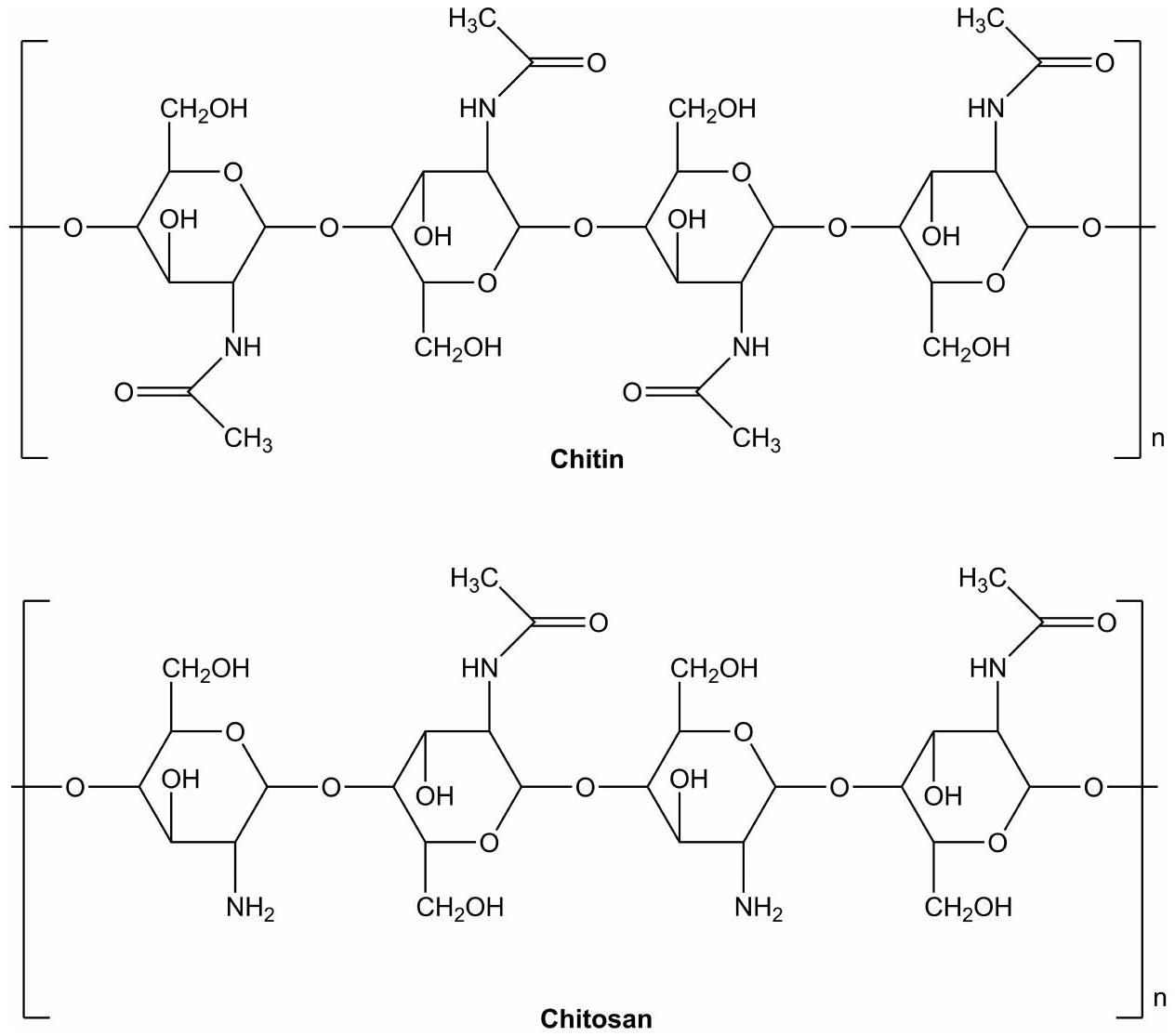
الشيتوزان، وهو منتج مزيل الأسيتيل من الكيتين، هو بوليمر وظيفي في المجال الصيدلاني بسبب قابليته للتحلل الحيوي، وتوافقه الحيوي، وخصائصه اللاصقة حيوياً، وغير السامة. تم استخدام الشيتوزان في أنظمة توصيل الأدوية باستخدام الجسيمات النانوية. يمكن تحميل أدوية متنوعة في الجسيمات النانوية المعتمدة على الشيتوزان بما في ذلك الأدوية ذات الجزيئات الصغيرة، والبولينوكليوتيدات، والبروتينات.تم الإبلاغ عن جزيئات نانوية قائمة على الكيتوزان للإفراج المنضبط عن عوامل مكافحة السرطان.يمكن أن يزيد الحجم الصغير للجزيئات النانوية من قدرة الدواء على اختراق الخلايا بشكل سلبي أو نشط.تسبب disruption الوصلات الضيقة في الأغشية التي تتوسطها الكيتوزان في زيادة تدفق الأدوية عبر الخلية.جزيئات الكيتوزان النانوية هي حاملة واعدة لتوصيل الجزيئات الكبيرة عن طريق الفم.تم الإبلاغ عن طرق مختلفة لإدارة الأدوية باستخدام جزيئات الكيتوزان كمادة، بما في ذلك الطرق الفموية والرئوية والعبر الجلدية والعينية والأنفية.تطبيق الكيتوزان في أنظمة توصيل الأدوية باستخدام الجسيمات النانوية محدود بسبب عدم قابليته للذوبان عند درجة الحموضة المحايدة. الكيتوزان قابل للذوبان فقط في المحاليل الحمضية، وهو ما لا يناسب بعض الأدوية.الذوبانية هي أكبر قيد على الكيتوزان في عملية التطوير واستخدامه في الصناعة على نطاق واسع. بالنسبة لإنتاج الجسيمات النانوية، فإن معظم الجسيمات النانوية التي تم إعدادها باستخدام البوليمر غير القابل للذوبان تتضمن المذيبات العضوية، والحرارة، وقوة القص العالية التي يمكن أن تؤدي إلى تدهور المادة الفعالة، بينما الجسيمات النانوية التي تم إعدادها باستخدام البوليمر القابل للذوبان تقدم تحضيرًا بسيطًا دون الحاجة إلى مذيبات عضوية، أو حرارة، أو قوة قص عالية.علاوة على ذلك، فيسيتكون الكيتوزان من ترسبات ويشكل تجمعات. لذلك، عند إعطائه تحت درجة حموضة فسيولوجية محايدة أو قاعدية، سيتكون ترسب وسيؤدي إلى
الملخص الرسومي
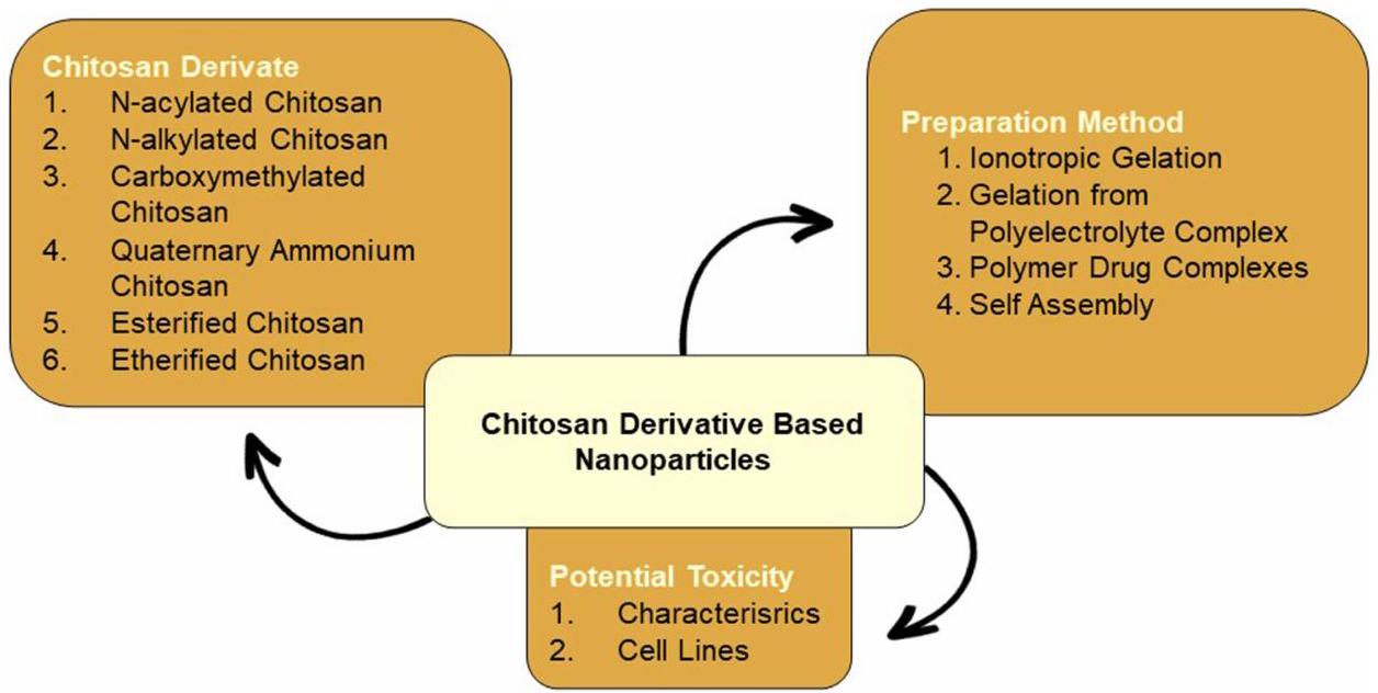
آثار جانبية.تم استخدام تعديلات الكيتوزان لتحسين خصائصه الفيزيائية والكيميائية والبيولوجية، بما في ذلك التعديل الكيميائي للكيتوزان. تتيح مجموعاته النشطة من الأمين والهيدروكسيل إمكانية التعديلات المحتملة باستخدام تقنيات الطلاء والتفاعلات الأيونية.لقد أبلغت العديد من المقالات عن تعديل الكيتوزان ولكنها لا تركز على الخصائص المستهدفة المحددة التي تم الحصول عليها. في هذه المراجعة، يركز المؤلف على تعديل الكيتوزان لتحسين قابليته للذوبان. تشمل التعديلات المستخدمة لتعزيز قابلية ذوبان الكيتوزان، الأسيلا، الألكيلا، الكربوكسيلة، الكواترنة، الإستر، والإيثر. كما تستعرض هذه التقرير تطبيق مشتقات الكيتوزان في إنتاج الجسيمات النانوية، بينما تم الإبلاغ عن عدد قليل جداً من المراجعات من قبل. كما تقدم هذه المراجعة الطريقة المحددة المستخدمة في تحضير الجسيمات النانوية المعتمدة على مشتقات الكيتوزان وسمية الكيتوزان ومشتقات الكيتوزان وجسيمات الكيتوزان النانوية. تعرض الشكل 1 هياكل الكيتين والكيتوزان.
تحسين قابلية ذوبان الكيتوزان
كيتوزان أسيل
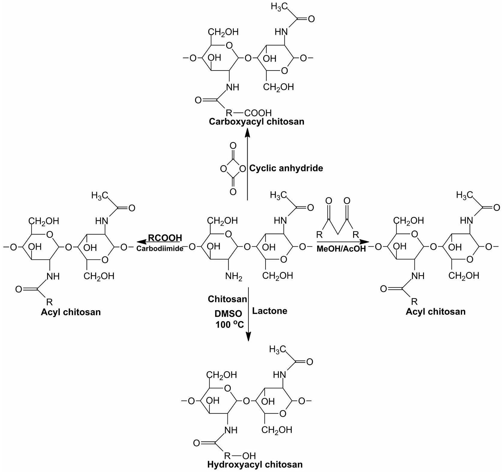
تدمير تفاعل الهيدروجين في الكيتوزان من خلال الأسيلاسيون يحسن من قابلية ذوبان الكيتوزان.مجموعة الأمين في الكيتوزان أكثر تفاعلاً من المجموعة الهيدروكسيلية الأولية؛ لذلك، فإن تفاعل الأسيل هو في الغالب في مجموعة الأمين.يتم إجراء أسيلينغ الكيتوزان عن طريق إدخال مجموعة أسيل عطرية أو أليفاتية.تفاعل التخليق لـ N-acylated chitosan موضح في الشكل 2.
تم تخليق الكيتوزان N-acylated عن طريق إدخال الأنهيدريدات إلى الكيتوزان مثل الأنهيدريد السكسينيك، والأنيهدريد البروبيونيك، والأنيهدريد اللوريك، والأنيهدريد البوتيريك، والأنيهدريد الستاريك، والأنيهدريد الإيتاكونيك، والأنيهدريد الميرستيك. يظهر الكيتوزان قصير السلسلة المأكولة قابلية ذوبان أعلى من الكيتوزان طويل السلسلة المأكولة.يمكن أن يؤدي إدخال مجموعة متنوعة من أنهدريدات الأحماض الدائرية إلى الكيتوزان إلى تحسين قابلية ذوبان الكيتوزان في الماء في مختلف مستويات الحموضة؛ حيث يذوب الكيتوزان الأصلي فقط في مستويات الحموضة التي تقل عن 6.5، بينما تظهر المشتقات قابلية ذوبان في مستويات الحموضة التي تزيد عن ذلك.أعد هيرانو وآخرون الكيتوزان N-دهني القابل للذوبان في الماء، من خلال معالجته مع مجموعة متنوعة من الأنهدريدات.
تم تطبيق N-acylation للكتين في أنظمة توصيل الجسيمات النانوية. وقد تم الإبلاغ عن أن الكتين المعالج بـ N-acylated يتم تفعيله بجسيمات الذهب النانوية للعلاج الجيني. أظهر اختبار النقل الجيني أن نسبة النقل للجسيمات النانوية المعتمدة على الكتين المعالج بـ N-acylated كانت أعلى مقارنة بالكتين.أظهر تشو وآخرون تطبيق جزيئات النانو من الكيتوزان المعالج بالأحماض الدهنية كحامل للبروتين.تم استخدام جزيئات الكيتوزان المشتقة من الأسيلا لتحرير فيتامين C بشكل محكوم. أظهرت الدراسة أن خصائص التحرير المحكوم لفيتامين C عند درجة حموضة 1.3 وتم استخدام جزيئات الكيتوزان المثيلة -أسيتيل كحامل للثيموكينون لحماية الكلى بعد العلاج الكيميائي باستخدام السيكلوفوسفاميد. أظهرت الدراسة أن جزيئات الكيتوزان المثيلة N-ألكيلية للثيموكينون تم توصيلها بفعالية إلى الكلى وحسنت من فعاليتها الوقائية ضد التأثيرات الناتجة عن السيكلوفوسفاميد.
الشكل I التركيب الكيميائي للكيتين والكيتوزان.
التهاب المثانة النزفي.تم استخدام الكيتوزان المشتق -acylated لاستقرار الجلبرين، وهو مركب بوليفينولي معزول من جذر العرقسوس من خلال تشكيل معقد نانو كيتوزان. وقد أظهرت الدراسات أن مشتقات الكيتوزان يمكن استخدامها كم stabilizer للمركبات البوليفينولية ويمكن تطبيقها بشكل محتمل على عوامل تبييض البشرة.-الكيتوزان المؤسيلي يتفاعل مع الحويصلات الدهنية بفعالية، مكونًا حويصلات الكيتوزان.تظهر الخصائص المهمة لنانوجزيئات الكيتوزان المعالج بالأسيليت في الجدول 1.
كيتوزان ألكيلي
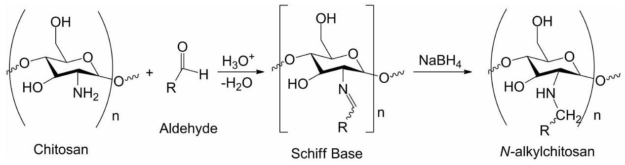
التفاعل باستخدام الألكانات الهالوجينية وقاعدة شيف هما طريقتان شائعتان تستخدمان لتخليق الكيتوزان N-ألكيلي.تتم التفاعل باستخدام الألكانات الهالوجينية عن طريق إدخال ألكيل إلى الكيتوزان. تعتمد القابلية للذوبان على طول مجموعة الألكيل المدخلة إلى الكيتوزان؛ كلما كان طول مجموعة الألكيل أقصر، زادت قابلية ذوبان الكيتوزان المكلور.يحدث تقليل الروابط الهيدروجينية بين جزيئات الكيتوزان بسبب وجود مجموعة الألكيل، مما يزيد من قابلية الذوبان في الماء.تفاعل التخليق للكتوزان N-ألكيلي موضح في الشكل 3.
تم إجراء قاعدة شيف من خلال تفاعل قاعدة شيف مع الألدهيد وتقليل، مما يؤدي إلى الكيتوزان N-ألكيلي. تفاعل قاعدة شيف مع يوديد الميثيل يؤدي إلى كواترنات الكيتوزان مما يحسن من قابلية الذوبان في الماء.تم أيضًا تخليق الكيتوزان المكلور باستخدام تفاعل إضافة مايكل بين أكريلات الهيدروكسي إيثيل والكيتوزان لتحسين قابلية ذوبان الكيتوزان في الماء.إن إدخال ثنائي السكريات إلى الكيتوزان ينتج كيتوزان N-ألكيلي قابل للذوبان في الماء.وجد بالاسيو وآخرون أن الكيتوزان الميثيلي N-alkylated يحسن من قابلية الذوبان في الماء بسبب الشحنة الإيجابية الناتجة عن مجموعة الأمونيوم الرباعية، وأن المشتقات قابلة للذوبان في نطاق واسع من الرقم الهيدروجيني من 3 إلى 14، في حين أن الكيتوزان غير المشتق قابل للذوبان فقط في الأحماض.تمت دراسة الكيتوزان المؤلكل بواسطة تشين وآخرين الذين ذكروا أن الكيتوزان المؤلكل تم تصميمه باستخدام
الشكل 2 التفاعل المُركب للكتوزان N-أسيل.
عامل ربط ‘-كاربونيليدياميدازول) في مذيب سائل أيوني. أظهر اختبار الذوبانية للكتوزان الميثيلاتي تحسينًا في الذوبانية في محلول حمض الأسيتيك المائي
تم الإبلاغ عن تطبيق الكيتوزان المكلس في أنظمة توصيل الأدوية النانوية بواسطة روبلز وآخرين. تم استخدام الكيتوزان المكلس لتحضير جزيئات الأنسولين، وأظهر أن سعة تحميل الأنسولين كانت أعلى مقارنة بالكيتوزان غير المعدل. كانت عملية إطلاق الأنسولين من الجزيئات النانوية أبطأ، ولها إمكانيات لتطبيقات الإطلاق المستدام.تم استخدام الكيتوزان O-الكيل لتحضير مادة أمفيبيلية-مشتق الكيتوزان O-ألكيل ثلاثي الميثيل (TMAC). تم استخدام TMAC لتحضير الجسيمات النانوية ذاتية التجميع لتوصيل الحمض النووي الببتيدي (PNA) إلى الخلايا. أظهرت الدراسة أن امتصاص PNA داخل الخلايا كان أعلى من الجسيمات النانوية غير القائمة على TMAC.تم استخدام الكيتوزان N-N-alkyl لتحضير أفلام نانوية مركبة تحتوي على جزيئات فضية للنشاط المضاد للميكروبات.تظهر الخصائص المهمة لشيكوسان الألكيل المعتمد على النانو جزيئات في الجدول 2.
الجدول I الخصائص المهمة للكتوزان المعتمد على الجسيمات النانوية
جزيئات نانو الكيتوزان المؤسيلي
الوزن الجزيئي (MW) / درجة إزالة الأسيتيل (DD) / درجة الاستبدال (SD)
خصائص الجسيمات النانوية
مرجع
جسيمات الذهب النانوية الكيتوزانية المشتقة
ميغاواتدي دي 70%
حجم جزيئات النانو الحمض النووي هووإمكان الزاوية هو 40.2 مللي فولت
[16]
جزيئات نانوية معدلة بـ BSA N-acyl
ميغاوات DD 84.9% SD 74.4-81.6%
تتراوح أحجام جزيئات ألبومين مصل البقر (BSA) منوكفاءة تحميل الدواء تتراوح بين 63.2-81.9%
[17]
جزيئات نانو الكيتوزان N-acyl فيتامين C
ميغاواتدي دي 84.9%
حجم جزيئات نانو فيتامين C 500 ملغ يتراوح من 216-288 نانومتر، جهد زتا، وكفاءة تحميل الدواء تتراوح من إطلاق الدواء هو إطلاق مفاجئ أولي (0.5 ساعة): 10-34% (عند pH 7.4) 20-40% (عند pH 1.3) ويطلق ببطء على مدى الوقت.
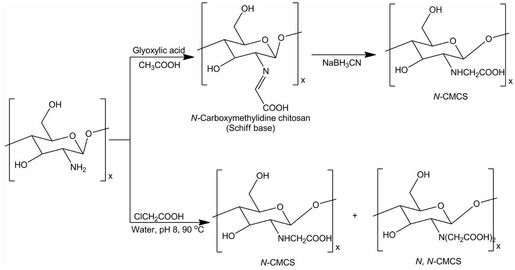
الكيتوزان المكلور هو قابل للذوبان في الماء في درجة الحموضة المحايدة والقلوية. يتم الحصول على الكيتوزان المكلور عن طريق إدخال حمض الكربوكسيليك (حمض الجلايوكسيليك أو حمض الكلورو ألكانوك).مجموعة أومجموعة من الكيتوزان.الكربوكسيميثيليشن هو التفاعل الأكثر شيوعًا للحصول على الكيتوزان المكلور. تعتمد قابلية ذوبان الكيتوزان المكلور في الماء على درجة الكربوكسيميثيليشن للكيتوزان.أنواع الكيتوزان الكاربوكسي ميثيل (CMCS) التي تم الحصول عليها يمكن أن تكون N-CMCS، O-CMCS، N، O-CMCS و N، N-CMCS.تفاعل الكاربوكسي ميثيليشن موضح في الشكل 4. تم تحضير N-كاربوكسي ميثيل كيتوزان باستخدام حمض الجلايوكسيليك وصوديوم سيانوبوروهيدرايد.أعد سونغ وآخرون أيضًا N-كربوكسي ميثيل كيتوزان باستخدام حمض الكلورو أسيتيك في الماء (بـ pH محايد)، وتم استخدامه للتحكم في نظام التفاعل عند درجة حموضة محايدة منتم وصف الكيتوزان O-كربوكسي ميثيل بواسطة محمد وآخرون باستخدام حمض المونوكلوروأسيتيك.إعدادتم الإبلاغ عن الكيتوزان -كربوكسي ميثيل بواسطة باتالي وآخرون.تم تحضير -كربوكسي ميثيل كيتوزان عن طريق إضافة الكحول الإيزوبروبيلي إلى الكيتوزان. ثم أضيفت محلول هيدروكسيد الصوديوم إلى المعلق الناتج وتم التحريك لمدة 45 دقيقة. بعد ذلك، أضيفت حمض أحادي الكلور الأسيتي الصلب إلى المعلق القلوي وتم التحريك لمدة 20 دقيقة.تم الإبلاغ عن تحضير قواعد شيف المستندة إلى الهيدرازيد O-CMCS القابلة للذوبان في الماء من خلال تفاعل في الموقع بواسطة مانموهان وآخرين.تفاعل الكاربوكسي ميثيليشن موضح في الشكل 4.
لقد استكشفت عدة دراسات التطبيقات المختلفة للكيتوزان الكاربوكسي ميثيل في تطوير الجسيمات النانوية. على سبيل المثال، تم استخدام الكيتوزان الكاربوكسي ميثيل في تحضير جسيمات نانوية من الكيتوزان الكاربوكسي ميثيل-ZnO لإظهار نشاط مضاد للبكتيريا ضد S. aureus و E. coli.بالإضافة إلى ذلك، تم استخدام الكيتوزان O-كربوكسي ميثيل في تصنيع
الشكل 3 التفاعل المُركب للكتوزان N-المُركب بالألكيل.
الجدول 2 الخصائص المهمة للشيتوزان الألكيلي المعتمد على الجسيمات النانوية
جزيئات الكيتوزان المكلورة
الوزن الجزيئي (MW) / درجة إزالة الأسيتيل (DD) / درجة الاستبدال (SD)
خصائص الجسيمات النانوية
مرجع
جزيئات PNA-شي نانومترية
الوزن الجزيئي 52 كيلودالتون، درجة النقاء 93.5%، الانحراف المعياري 10.9-15.2%
حجم جزيئات الأنسولينجهد زتا، وكفاءة تحميل الدواء II-27% (pH 5.3) وإطلاق الدواء هو (الأول )، تليها إطلاق أبطأ لبضعة أيام
[28]
جزيئات O-carboxymethyl chitosan/Fucoidan (O-CMCS-F) لتوصيل الكركمين. أظهرت هذه الدراسة أن هذه الجزيئات عززت امتصاص الكركمين على مستوى الخلايا، مما يشير إلى إمكانية استخدامها في أنظمة التوصيل الفموية والإفراج المنضبط عن المركب.في دراسة أخرى، تم استخدام جزيئات الكاربوكسي ميثيل كيتوزان لتوصيل الكاربامازيبين، وهو دواء مضاد للاختلاج، عن طريق الأنف، بهدف تعزيز توصيل الدواء إلى الدماغ. أشارت النتائج إلى أن هذه الجزيئات تحمل وعدًا للتطبيقات الأنفية.أظهر فنغ وآخرون استخدام الكيتوزان / O-كربوكسي ميثيل كيتوزان في إنشاء جزيئات نانوية من هيدروكلوريد دوكسوروبيسين لتوصيل الأدوية المضادة للسرطان. وأشارت أبحاثهم إلى أن جزيئات CS/CMCS كانت آمنة وفعالة في توصيل هيدروكلوريد دوكسوروبيسين.أبلغ محجوب وآخرون عن تحضير جزيئات نانوية مكونة من N-trimethyl-O-carboxymethyl chitosan لتوصيل الإينوكسابارين عن طريق الفم، مما يبرز إمكانيات TMCMC في هذا التطبيق.تم استخدام تقنيات الموجات فوق الصوتية في دراسة أخرى لإنشاء جزيئات نانوية قائمة على الكاربوكسي ميثيل كيتوزان لتوصيل كليندامايسين HCL، مما يوضح فعالية هذه الطريقة في التحكم في إطلاق الدواء لكليندامايسين.قدم شيو وآخرون طريقة لتحضير الجسيمات النانوية باستخدام CMCS المشحون سلبًا وملح الكيتوزان الرباعي الأمونيوم المشحون إيجابًا لتحقيق تأثير مناعي محفز. اقترحت دراستهم أن جزيئات HACC النانوية لديها إمكانيات كعامل مساعد مناعي.توضح الجدول 3 الخصائص الهامة للجزيئات النانوية المستندة إلى الكيتوزان المكلور.
الشكل 4 تفاعل الكيتوزان الناتج عن الكربوكسيميثيليشن.
الجدول 3 الخصائص المهمة للكيتوزان المحمّل بالجسيمات النانوية
جزيئات الكيتوزان الكربوكسيلي
الوزن الجزيئي (MW) / درجة إزالة الأسيتيل (DD) / درجة الاستبدال (SD)
خصائص الجسيمات النانوية
مرجع
كيتوزان كربوكسي ميثيل – زنك أوكسيد
MW 9 كيلودالتون DD 95%
حجم الجسيمات النانوية هو 100 نانومتر
[37]
جزيئات O-Carboxymethyl شي/فوكويدين
الوزن الجزيئي 192 كيلودالتون، درجة النقاء 75%، الانحراف المعياري 0.315
حجم جزيئات الكركمين 269.4 نانومتر، جهد زتا 30 مللي فولت، وكفاءة تحميل الدواءإطلاق الدواء أقل من 10% / إطلاق بطيء محكوم (pH 2.5)؛ إطلاق مستدام (pH 6) وإطلاق سريع (pH 7)
[38]
جزيئات الكيتوزان الكاربوكسيميثيلية الكاربامازيبين
SD 0.384
حجم جزيئات الكاربامازيبين 218.76 نانومتر، جهد زتا -33.43 مللي فولت، وكفاءة تحميل الدواءإطلاق الدواء هوبعد 25 ساعة
حجم جزيئات دوكسوروبيسين هيدروكلوريد 248.9-362.7 نانومتر، جهد زتا -27.6 إلى -42.2 مللي فولت وكفاءة تحميل الدواء 65.48-72.87%، يتم إطلاق الدواء بعد 96 ساعة: و
[٤٠]
جزيئات ن-ثلاثي ميثيل-أو-كربوكسي ميثيل الكيتوزان
DD 98% SD 36.8%
حجم جزيئات الهيبارين 235 نانومتر، جهد زتا 18.6 مللي فولت، وكفاءة تحميل الدواءإطلاق الدواء هوبعد 600 دقيقة
[4I]
جزيئات كليندامايسين HCL-CMCS
دي دي 90%
حجم جزيئات كليندامايسين HCL 318.4 نانومتر وكفاءة تحميل الدواءإطلاق الدواء هو4.70 بعد 24 ساعة
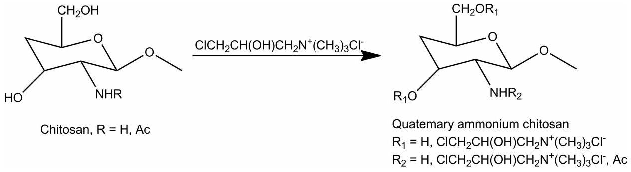
عملية الرباعية هي طريقة شائعة تستخدم لإنتاج الكيتوزان الرباعي الأمونيوم. تحدث هذه التفاعل عادة في مجموعات الأمين الموجودة في الكيتوزان.هناك ثلاث طرق رئيسية للتأين الرباعي: استبدال الأمونيوم الرباعي المباشر، تفاعل الحلقة المفتوحة لمشتقات الإيبوكسي، وN-ألكلة.تُعزى القابلية المحسّنة للذوبان لشيطوزان الأمونيوم الرباعي إلى شحنته الإيجابية.التركيب الأولي لـتم إجراء دراسة على يوديد الكيتوزان ثلاثي الميثيل (TMC)، وهو مشتق من كيتوزان الأمونيوم الرباعي، باستخدام الفورمالديهايد، وبوروهيدريد الصوديوم، وقدم دورمارد طريقة تعرف باسم طريقة دورمارد، تتضمن N-methyl-2-pyrrolidone و NaOH ولتركيب مشتقات الكيتوزان الرباعية الأمونيوم.تم الحصول على البوليمرات المشتركة عن طريق ربط دوديكانال (DDA) و5-بروموميثيل ثلاثي ميثيل الأمونيوم بروميد (BPTA) على الكيتوزان. أظهر المشتق الأمفيلي للكيتوزان قدرة على التجميع الذاتي، مما قد يكون مفيدًا لأنظمة توصيل الأدوية.قام فان وآخرون بتخليق كيتوزان الأمونيوم الرباعي باستخدام (كلوريد ثلاثي ميثيل الأمونيوم N-(3-كلورو-2-هيدروكسي بروبيل)) وهيدروكسيد الصوديوم.نجح ساجومسانغ وآخرون في إنتاج كيتوزان رباعي الأمونيوم القابل للذوبان في الماء والذي يحتوي على أحادي/ثنائي السكريات باستخدام عامل كواترنيز يسمى كلوريد ن-(3-كلورو-2-هيدروكسي بروبيل) تريميثيل أمونيوم.تظهر عملية تخليق الكيتوزان الرباعي الأمونيوم في الشكل 5.
تم استخدام الكيتوزان ثلاثي الميثيل (TMC) لتطوير جزيئات نانوية محملة بالأنسولين باستخدام بولي (-حمض الغلوتاميك) كعامل معقد.تم استخدام مشتق الكيتوزان القابل للذوبان في الماء، N -(2-hydroxy) propyl 3 trimethyl ammonium chitosan chloride (HCTT)، للحصول على جزيئات نانوية محملة بالريبافيرين. أظهرت الدراسة أن HCTT يعد واعدًا لإطلاق محكوم للأدوية المحبة للماء.تم الإبلاغ أيضًا عن تحضير جزيئات النانو HCTT المحملة بالألبومين.تم استخدام TMC أيضًا لتطوير جزيئات نانوية من TMC-ليبوزوم-دوكسيسيكلين. الجزيئات النانوية
الشكل 5 التفاعل المُركب للكاتين الشيتوزان الرباعي الأمونيوم.
أظهرت تثبيطًا ممتازًا للبكتيريا والبيوفيلم وقد يمكن استخدامها في علاج أمراض اللثة.أبلغ هوانغ وآخرون عن تشكيل جزيئات نانوية من الكيتوزان-فوكويدين رباعية الأمونيوم محملة بالإيبيغالوكاتشين غالات (EGCG) لتعزيز نفاذية ونشاط EGCG المضاد للبكتيريا. اقترحت الدراسة أن الجزيئات النانوية زادت من نفاذية غشاء البكتيريا، مما زاد من النشاط المضاد للبكتيريا لـ EGCG.تظهر الخصائص المهمة لشيكوسان المقسم القائم على النانو جزيئات في الجدول 4.
استرification الكيتوزان
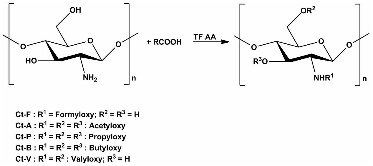
تشمل استرة الكيتوزان تفاعل الكيتوزان مع الأحماض الكربوكسيلية أو مشتقاتها.وثق وان وآخرون تخليق استر الكيتوزان القابل للذوبان في الماء (استر الكيتوزان p-أمينوبنزويلي) باستخدام طريقة قاعدة شيف.طريقة أخرى تم الإبلاغ عنها تضمنت إنشاء كيتوزان مكلور بواسطة معالجة الكيتوزان بمزيج من DMF وحمض ثنائي كلورو الأسيتيك وحمض الكلوروسولفونيك.تم الحصول على أسيتات الكيتوزان عن طريق تغيير نسبة حمض الأسيتيك إلى الكيتوزان، بينما تم تحضير جلوتامات الكيتوزان عن طريق إذابة الكيتوزان في حمض الجلوتاميك.في براءة اختراع من يون وآخرين، تم تفصيل عملية استرification للكيتوزان باستخدام أحماض كربوكسيلية متنوعة (حمض الأسيتيك، حمض الفورميك، حمض البروبيونيك، حمض البوتيريك، وحمض الفاليريك) بالإضافة إلى أنهدريد ثلاثي فلور الأسيتيك (TFAA).تم توضيح التمثيل التخطيطي لتفاعل الإسترification للكيتوزان في الشكل 6.
تم الإبلاغ عن تطبيق استر الكيتوزان في أنظمة توصيل الأدوية باستخدام الجسيمات النانوية بواسطة بهاتاري وآخرين. وقد أبلغوا عن تحضير بروتين مصل البقر المحمّل بجسيمات نانوية من كيتوزان لاكتات، وكانت الجسيمات الناتجة بقطر 10 نانومتر. كما وجدوا أن التغليف كانوأظهرت نمط إطلاق مستدام.الدراسة
الجدول 4 الخصائص المهمة للشيتوزان الرباعي القائم على الجسيمات النانوية
جزيئات الكيتوزان الرباعية
الوزن الجزيئي (MW) / درجة إزالة الأسيتيل (DD) / درجة الاستبدال (SD)
خصائص الجسيمات النانوية
مرجع
جزيئات TMC/Y-PGA النانوية
وزن جزيئي 60 كيلو دالتون نسبة 85%
SD 55%
حجم جزيئات الأنسولين 148.6-522.4 نانومتر، جهد زتا I4.2-36.6 مللي فولت بعد 96 ساعة، وكفاءة تحميل الدواءإطلاق الدواء هوبعد 700 دقيقة
[49]
جزيئات ريبافيرين HTCC
ميغاواتدي دي 90%
SD 0.33-0.83%
حجم جزيئات ريبافيرين 211 نانومتر، جهد زتا 48.6 مللي فولت بعد 96 ساعة، وكفاءة تحميل الدواءإطلاق الدواء يزيد عن 80% بعد 70 ساعة (DS 32%)
[50]
جزيئات نانو الكيتوزان دوكسيسيكلين
تم تحميل الدوكسيسيكلين باستخدام كيتوزان الأمونيوم الرباعي. حجم الجسيمات النانوية هو 176 نانومتر، والجهد الكهربائي هو 12.31 مللي فولت، وكفاءة تحميل الدواء هي،
[52]
جزيئات الإبيغالاتشين غالات
حجم جزيئات الإبيغالاتشين غالاتي 208.5 نانومتر، جهد زتا 27.6 مللي فولت بعد 96 ساعة، وكفاءة تحميل الدواءإطلاق الدواء هوبعد 360 دقيقة (pH 2.0) وبعد 360 دقيقة (pH 6.8)
[53]
الشكل 6 تفاعل الإسترification المُركب من الكيتوزان.
كما وُجد أن جزيئات النانو من الكيتوزان اللاكتات المحملة بـ siRNA المحدد لـ CD73 لها تأثير مضاد للورم.تم تحضير جزيئات نانوية من ألبومين مصل البقر محملة بملح الكيتوزان (اللاكتيت، الأسبارتات، الجلوتامات، والجلايكولات) باستخدام طريقة التجلط الأيوني، وكانت خصائص الجزيئات النانوية تعتمد على أنواع الملح.قدم كولونا وآخرون تطوير جزيئات نانو الكيتوزان غلوتامات لتوصيل إنزيم البروليل داز لعلاج الاضطرابات الذاتية.تم تطوير جزيئات الكيتوزان الكبريتية – أمفوتيريسين ب لعلاج عدوى الكانديدا جلابراتا.تظهر الخصائص المهمة للكتوزان المشتق من النانو جزيئات في الجدول 5.
إيثرية الكيتوزان
يتم الحصول على مشتق ميثيل الكيتوزان عن طريق تفاعل عامل الألكلة مع الكيتوزان.مشتق الإيثير من الكيتوزان قابل للذوبان في الماء وله قدرة جيدة على الاحتفاظ بالماء، غير سام، بكتيريا مثبط، وله تطبيقات أوسع في المجال الصيدلاني.تم تحضير الكيتوزان الميثيلي عن طريق تفاعل ثنائي ميثيل الكبريتات مع الكيتوزان في محلول قلوى/يوريا مائي.تم اشتقاق الكاربوكسي ميثيل كيتوزان من الكيتوزان عن طريق إضافة هيدروكسيد الصوديوم وحمض المونوكلوروأسيتيك. تم إجراء عملية الإيثرية في درجات حرارةتم تخليق مشتقات الكيتوزان الكاتيونية ذات السلاسل الطويلة من الألكيل (HDCC) باستخدام نوعين من عوامل الإيثر، وهما كلوريد ثنائي ميثيل أمونيوم دوديكي (2،3-إيبوكسي بروبوكسي) وكلوريد ثلاثي ميثيل أمونيوم جليكيدي.وجد يانغ وآخرون أن العوامل المستخدمة بشكل شائع لإيثرية الكيتوزان هي كلوريد 3-كلورو-2-هيدروكسي بروبيل ثلاثي ميثيل الأمونيوم (CTA) وكلوريد 2.3-إيبوكسي بروبيل ثلاثي ميثيل الأمونيوم (ETA) ومجموعات الكربوكسي إيثيل.هيدروكسي بيوتيل كيتوزان (HBC) هو
الجدول 5 الخصائص المهمة للكيتوزان المشتق من الجسيمات النانوية
جزيئات الكيتوزان المستعصرة
الوزن الجزيئي (MW) / درجة إزالة الأسيتيل (DD) / درجة الاستبدال (SD)
خصائص الجسيمات النانوية
مرجع
BSP-Chi
لاكتات
جسيمات نانوية
وزن جزيئي 190 كيلودالتون
درجة إزالة الأسيتيل 88%
درجة الاستبدال 13.2%
حجم جسيمات بروتين مصل البقر (BSP) 10 نانومتر وكفاءة تحميل الدواء، إطلاق الدواء هوعلى مدى 4 أسابيع
[59]
بروتين بروتياز-شي
جسيمات نانوية
وزن جزيئي 300 كيلودالتون
درجة إزالة الأسيتيل 85%
حجم جسيمات بروتياز 365.5 نانومتر، جهد زتا 17.94 مللي فولت، وكفاءة تحميل الدواء، إطلاق الدواء هوبعد 1 ساعة، إطلاق ضخم بعد، وإطلاق مستمر بعد 48 ساعة
[6I]
جسيمات نانوية من أمفوتيريسين ب مع الكيتوزان
حجم جسيمات أمفوتيريسين ب 310 نانومتر، جهد زتا -41.5 مللي فولت، وكفاءة تحميل الدواء، إطلاق الدواء هو إطلاق مستمربعد 72 ساعة
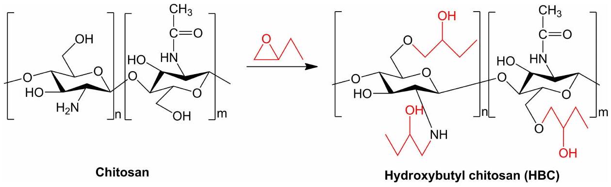
الكيتوزان الإيثرية القابلة للذوبان في الماء، والتي يتم إنتاجها عبر الإيثرية من خلال تفاعل مجموعة هيدروكسي بيوتيل مع الكيتوزان.تم تحضير الكيتوزان هيدروكسي بروبيل من خلال تفاعل الكيتوزان مع الإيبوكسيد البروبيلي تحت ظروف قلوية. الكيتوزان الإيثرية الناتجة لها قابلية ذوبان جيدة في الماء.الشكل التالي (الشكل 7) هو شكل لتفاعل الكيتوزان (الخط الأسود) مع الإيبوكسيد (الصورة الحمراء) والذي يمكن أن ينتج إيثر وأمين ثانوي (الخط الأسود والأحمر).
لقد حظيت تطبيقات الكيتوزان الإيثرية في أنظمة توصيل الأدوية بالجسيمات النانوية باهتمام كبير بسبب خصائصها مثل القابلية للذوبان في الماء، والتحلل البيولوجي، والتوافق الحيوي. تم الإبلاغ عن تطبيق الكيتوزان هيدروكسي بيوتيل في نظام توصيل الأدوية بالجسيمات النانوية من قبل صن وآخرون، حيث تم تصنيع جسيمات نانوية من الكيتوزان المحملة بالسيتيريزين (CTZ: CDHBCs-NP) كوسائل نانوية أنفية لعلاج التهاب الأنف التحسسي. تمتلك جسيمات CTZ: CDHBCs-NP سعة تحميل عالية، وسلوك إطلاق الدواء قد يكون محتملًا كوسيلة نانوية أنفية مضادة للحساسية.قام لو وآخرون بتصنيع جسيمات نانوية من الكيتوزان هيدروكسي بروبيل باستخدام طريقة التجلط الأيوني. كانت الجسيمات النانوية الناتجة بقطر 40 نانومتر، وكان جهد الزتاتظهر الخصائص المهمة لجسيمات الكيتوزان الإيثرية في الجدول 6.
تحضير جسيمات نانوية من مشتقات الكيتوزان
تستخدم عملية تحضير جسيمات نانوية من مشتقات الكيتوزان تقنيات متعددة، بما في ذلك التجلط الأيوني، والتجلط من تشكيل المركب متعدد الإلكتروليت، ومركبات البوليمر-دواء، والتجميع الذاتي.
التجلط الأيوني
تم تقديم تقنية التجلط الأيوني في البداية من قبل كالفو وآخرون في عام 1997. هذه الطريقة قادرة على إنتاج جسيمات نانوية من خلال التفاعلات الكهروستاتيكية بين البوليمرات المشحونة بشكل متعاكس تحت ظروف تحريك ميكانيكية محددة.تُعرف الجسيمات النانوية بأنها مواد تتراوح أحجامها عادةً بين 1 إلى 100 نانومتر. ومع ذلك، فإن الجسيمات النانوية الناتجة باستخدام طريقة التجلط الأيوني عادةً ما تظهر أحجامًا أكبر منتم استخدام العديد من الكيتوزانات المؤهلة بشحنات إيجابية مستقرة وغير معتمدة على الرقم الهيدروجيني من خلال هذه الطريقة، وأشهرها هو الكيتوزان ثلاثي الميثيل (TMC).
أجرى غونزاليس وآخرون تحضير جسيمات نانوية من الذهب مع الكيتوزان، والكيتوزان N-acylated، وكيتوزان أوليغوسكاريد كحاملات للحمض النووي. تم إجراء أسيتيل الكيتوزان (وزن جزيئي منخفض) من خلال اتباع الطريقة التي أبلغ عنها ريمان بهادور وآخرون مع تعديلات طفيفة. أظهرت المحلول الغروي استقرارًا ممتازًا، مما يشير إلى التطبيق العملي للكيتوزان كمثبت. بالإضافة إلى ذلك، عمل كيتوزان أوليغوسكاريد ككلا
الجدول 6 الخصائص المهمة للكيتوزان الإيثرية المعتمدة على الجسيمات النانوية
جسيمات نانوية من الكيتوزان المسترطبة
الوزن الجزيئي (MW)/ درجة إزالة الأسيتيل (DD)/ درجة الاستبدال (SD)
خصائص الجسيمات النانوية
مرجع
جسيمات نانوية من الكيتوزان السيتيريزين
وزن جزيئي 700 كيلودالتون درجة إزالة الأسيتيل 85%
حجم جسيمات السيتيريزين 129 نانومتر، جهد زتا 4 مللي فولت، كفاءة التحميل، وإطلاق الدواء هوفيعند الرقم الهيدروجيني 5.5
[7I]
عامل مختزل ومثبت، بينما عززت المجموعات الأمينية في الكيتوزان تقاربها مع الحمض النووي البلازميدي من خلال إدخال شحنات إيجابية. تراوحت توزيعات أحجام جسيمات الذهب المنتجة من 3 إلى
ومع ذلك، بينما تنتج العديد من طرق تخليق الجسيمات النانوية توزيعًا ضيقًا لحجم الجسيمات، قد لا تحقق طريقة التجلط الأيوني ذلك باستمرار. غالبًا ما يتجاوز مؤشر التشتت المتعدد (PDI) 0.1، وأحيانًا يصل إلى 0.5. وبالتالي، فإن عدم التجانس في حجم الجسيمات لا يؤثر فقط على تحميل الدواء ولكن يمكن أيضًا أن يقيد تفاعل الجسيمات مع الهياكل البيولوجية. بالإضافة إلى ذلك، فإن الحفاظ على سرعة ثابتة في محلول السقوط والتحريك المنضبط هما متطلبات أساسية لهذه الطريقة.
دراسة أخرى أجراها ساين وآخرون طورت جسيمات نانوية من الكيتوزان أحادي-N-كربوكسي ميثيل (MCC) وجسيمات نانوية من الكيتوزان ثلاثي الميثيل (TMC) لتوصيل اللقاحات غير الغازية. تم تحضير الأنظمة الجسيمية باستخدام مشتقات الكيتوزان ذات الشحنات المختلفة، الكيتوزان ثلاثي الميثيل (TMC، بولي كاتيون) والكيتوزان أحادي-N-كربوكسي ميثيل (MCC، بولي أمفوليتيك) للتطعيم المخاطي. تم تحضير الجسيمات النانوية باستخدام طريقة التجلط الأيوني وتحميلها بتوكسيد الكزاز (TT). تم الحصول على جسيمات نانوية ذات كفاءة تحميل عالية () وحجم الجسيمات ضمن نطاقنانومتر بشحنة سطحية سالبة لـ MCC وشحنة سطحية موجبة لـ TMC والكيتوزان. تم اكتشاف أن كل من أنظمة التشتت والجسيمات النانوية التي تم تشكيلها بمشتقات الكيتوزان تعزز الاستجابات المناعية المخاطية. وأبرزت النتائج أن الشحنة السطحية وحجم الجسيمات للحاملات المنتجة باستخدام الكيتوزان تلعب دورًا حاسمًا في تحقيق استجابة مناعية محسنة. أثار MCC استجابات مناعية أقل مقارنة بـ TMC والكيتوزان، ومع ذلك، أنتجت أصغر الجسيمات النانوية مع توزيع حجم ضيق بشكل ملحوظ وسعة تحميل عالية. تمتلك جسيمات MCC الخصائص المطلوبة اللازمة لتكون وسائل توصيل فعالة لمجموعة واسعة من الأدوية، وكذلك لتوصيل الجينات/البروتينات.
التجلط من المركب متعدد الإلكتروليت
تضمنت هذه الطريقة التجلط الأيوني، تمامًا كما تم وصفه في القسم السابق، فقط في الحالة الحالية كان الرابط المتقاطع بوليمر متعدد الأنيونات بشحنات معاكسة لتلك الخاصة بمشتق الكيتوزان، والذي شكل معه مركب PEC.
في دراسة حديثة، قام محجوب وآخرون بتحضير وتحسين جسيمات نانوية من الكيتوزان ثلاثي الميثيل-O-كربوكسي ميثيل لتوصيل الهيبارين منخفض الوزن الجزيئي. تم تصنيع الجسيمات النانوية باستخدام تقنية تشكيل المركب متعدد الإلكتروليت. تم إجراء تحسين الجسيمات النانوية باستخدام منهجية تصميم سطح الاستجابة Box-Behnken. تم تصنيف الجسيمات النانوية حسب حجمها، جهد الزتا، مؤشر التشتت المتعدد، كفاءة الاحتجاز، وكفاءة التحميل، والتي تم قياسها كـ، و، وعلى التوالي. أظهرت دراسة الشكل وجود جسيمات نانوية كروية فردية دون أي مؤشر على التكتل معًا. كشفت دراسات الإفراج في المختبر أن
من الإينوكسابارين تم إطلاقه من الجسيمات النانوية بعد 600 دقيقة من الحضانة. أظهرت البيانات المكتسبة أن الجسيمات النانوية المشتقة من الكيتوزان ثلاثي الميثيل هي خيار واعد للإدارة الفموية للإينوكسابارين.درس تشين وآخرون تشكيل وخصائص مركب متعدد الإلكتروليت جديد لنظام حامل دواء لتوصيل دوكسوروبيسين (DOX)، والذي يتكون من حمض الهيالورونيك (HA) المغلف بالكيتوزان المعدل هيدروفوبياً. تم تحديد أحجام الجسيمات لمركبات متعددة الإلكتروليت المصنعة (PCNs) لتكون ضمن نطاق، والتي هي أكبر مقارنة بأحجام جزيئات النانو غير المغلفة، والتي تبلغ حوالي 150 نانومتر. تتمتع PCNs بجهود زيتا كبيرة تبلغ حوالي 26 مللي فولت، مما يساهم في استقرارها. لم يتم ملاحظة أي تغييرات في الحجم. تم تحقيق دمج DOX في PCNs بكفاءة تغليف عالية ( )، مما أدى إلى ملف إطلاق مستدام دون أي تأثير انفجاري مفاجئ عند التعرض لـ PBS (درجة الحموضة 7.4) في
معقدات الأدوية البوليمرية
طور تراباني وآخرون نظامًا نانويًا باستخدام السيكلودكسترين (CD) – الكيتوزان (CS) مع سلفوبوتيل إيثر--CD (SBE- ). يحتوي هذا النظام على نواة داخلية غنية بـ CD وطبقة خارجية غنية بـ CS، والتي يمكن أن تحبس كل من الأدوية المحبة للماء والماء الكارهة. بعد ذلك، قاموا بالتحقيق في إدارة ببتيد الجلوتاثيون (GSH) عن طريق الفم باستخدام هذا النظام النانوي. ثم قاموا بمقارنة النتائج مع الجسيمات النانوية المصنوعة فقط من علوم الحاسوب أو مع مزيج من علوم الحاسوب والجزيئات النانوية. كشفت دراسة طيف الكترونات الأشعة السينية عن تكوين تفاعل مستقر بين الببتيد وSBE--CD في النواة الداخلية للجزيئات النانوية. ومع ذلك، من الجدير بالذكر أن هذا التعقيد أظهر استقرارًا استثنائيًا، حيث لم يظهر أي إطلاق لـ GSH تحت ظروف تحاكي نشاط المعدة.
أنشأ ماستريلي وآخرون ناقل دواء نانوي جديد يتكون من جزيئات الكيتوزان وجزيئات السيكلودكسترين. يسمح التفاعل مع السيكلودكسترين بحل وحماية الأدوية الحساسة، بينما من المتوقع أن تعزز إدماجها في إطار الكيتوزان امتصاصها. تم تشكيل الأدوية مع السيكلودكسترين، مما عزز احتجازها داخل الجزيئات النانوية. أدى ذلك إلى زيادة كبيرة في تحميل الدواء النهائي للجزيئات النانوية، حيث زاد حتى 4 مرات للتركلوسان و10 مرات للفوروسيميد. تشير نتائج هذه الدراسة إلى أن معقد الدواء-السيكلودكسترين تم الحفاظ عليه بفعالية داخل الهيكل الجزيئي النانوي. عُرّف ملف إطلاق هذه الجزيئات النانوية في المختبر بمرحلة إطلاق سريعة أولية ومرحلة إطلاق متأخرة لاحقة.
التجميع الذاتي
طور هك وآخرون جزيئات نانوية مشتقة من الكيتوزان تحتوي على الأنسولين للإدارة الفموية. تم إنشاء جزيئات نانوية قائمة على مشتقات الكيتوزان تحتوي على الأنسولين من خلال التجميع الذاتي، باستخدام التفاعلات الكهروستاتيكية بين الدواء المشحون سلبًا والبوليمرات المشحونة إيجابًا. تم استبدال مجموعات الأمين في مشتقات الكيتوزان بدرجات متفاوتة (33، 52، أو ) بواسطة مجموعات 2-هيدروكسيبروبيل-3-تريميثيل أمونيوم. أدت هذه التعديلات إلى أن تكون البوليمرات مشحونة بشكل إيجابي بشكل مستمر، بغض النظر عن مستوى الحموضة. تعتبر استقرار الجسيمات النانوية أمرًا حيويًا لهذا النظام المتقدم لتوصيل الأدوية بسبب مستويات الحموضة المتغيرة في الجهاز الهضمي وأهمية التفاعلات الكهروستاتيكية. يتم تعزيز الالتصاق المخاطي بشكل أكبر من خلال وجود شحنات إيجابية دائمة. من ناحية أخرى، تعتمد الشحنات الكهربائية لجزيئات الكيتوزان على مستوى الحموضة في الوسط المحيط. نتيجة لإدخال مجموعات الأمونيوم الرباعية، تم تعزيز قابلية ذوبان مشتقات الكيتوزان. لتعزيز الجسيمات النانوية وضمان استقرارها، تم إدخال فوسفات الصوديوم ثلاثي البولي (TPP) في الأنظمة لتوليد روابط إضافية. أثر وجود TPP على كل من تفكك مصفوفة البوليمر وحركية الإفراج اللاحقة. تم اكتشاف أن عمليات إطلاق الدواء كانت أكثر تعقيدًا من مجرد الانتشار تحت ظروف مستقرة، وقد تشمل تفاعلات أيونية وتفكك المصفوفة أيضًا. كانت التركيبة الأكثر ملاءمة تستخدم مشتق كيتوزان بدرجة استبدال . كانت هذه التركيبة مميزة بمتوسط Z يبلغ جهد زتاوكفاءة تغليفومن الجدير بالذكر أن إفراز الأنسولين استمر لأكثر من 210 دقائق.
طور يان وآخرون نوعًا جديدًا من الجسيمات النانوية ذات التجميع الذاتي المعلمة بتات والمعدلة بحمض الفوليك (Tat-Suc-FA) لتوفير ناقل جديد لعلاج جينات الأورام. تم تصنيع الجسيمات النانوية عن طريق توزيع كمية معينة من البوليمر المشترك Tat-Suc-FA في محلول حمض الأسيتيك (حجم/حجم) ثم تعريضه للتسونيد باستخدام جهاز تسونيد من نوع البروبي (معالج سونكس التراساونيك، سونيكس-600) بقوة إخراج 12 واط. تم تمديد عملية التسونيد لمدة دقيقتين. تم تصفية المحلول المتجمع ذاتيًا باستخدام غشاء بحجم مسامثم تم الاحتفاظ بها في درجة حرارة الغرفة. قامت الدراسة بتخليق وتوصيف بوليمرات جديدة من Tat-Suc-FA بشكل فعال. شكلت بوليمرات Tat-Suc-FA مجمعات مكثفة مع الحمض النووي البلازميدي، مما أدى إلى تكوين مجمعات مشحونة إيجابيًا بعرض يتراوح من 54 إلى 106 نانومتر. كانت السمية الخلوية لبوليمرات Tat-Suc-FA في خلايا K562 أقل بكثير مقارنة بتلك الخاصة بالكيتوزان.
السمية المحتملة لجزيئات الكيتوزان النانوية
الشيتوزان، وهو بوليمر طبيعي، معروف بقابليته للتحلل البيولوجي، وتوافقه الحيوي، وفعاليته من حيث التكلفة، وخصائصه غير السامة. يُلاحظ استخدامه الواسع في دول مختلفة بما في ذلك اليابان وإيطاليا وفنلندا. في الولايات المتحدة، يتم تصنيف الشيتوزان على أنه معترف به عمومًا كآمن (GRAS) من قبل إدارة الغذاء والدواء (FDA).على الرغم من أن إدارة الغذاء والدواء الأمريكية قد وافقت على استخدام بوليمر الكيتوزان في مجموعة متنوعة من منتجات الرعاية الصحية مثل الضمادات المانعة للنزيف، وطلاءات العدسات اللاصقة، والضمادات، إلا أن هناك تقارير محدودة حول اعترافه وتطبيقه كنظام لتوصيل الأدوية.لقد أفادت عدة دراسات بسمية الكيتوزان ومشتقاته وجزيئات الكيتوزان النانوية، لكن لم يتم الإبلاغ على نطاق واسع عن سمية مشتقات جزيئات الكيتوزان النانوية. تتأثر سمية جزيئات الكيتوزان النانوية عمومًا بخصائص الجزيئات وخطوط الخلايا.
خصائص الجسيمات
يمكن ملاحظة سمية جزيئات الكيتوزان، المتأثرة بخصائصها، في الكائنات الحية باستخدام حيوانات اختبار مختلفة مثل الجرذان والأرانب والكلاب والأسماك الزرد. يُعتبر اختيار الأسماك الزرد أكثر فائدة لأنه لا يتطلب تكاليف باهظة. بالإضافة إلى ذلك، تشترك الأسماك الزرد في تشابه جينومي مع البشر، مما يجعلها حيوان اختبار لتقييم سمية الكيتوزان. علاوة على ذلك، تحتوي الأسماك الزرد على غشاء خارجي بحجم مسام يتراوح من 0.5 إلىمما يسمح لجزيئات الكيتوزان بالدخول من خلال الانتشار الساكن.
يمكن أن يؤثر تعديل الكيتوزان في شكل نانو جزيئات على التغيرات في ملف التوزيع والدوائية للنانو جزيئات. يتأثر ذلك بالحجم وخصائص السطح، حيث يؤثر الشحنة الموجبة للكيتوزان على التفاعلات السطحية التي لها تداعيات على الملف الحركي لامتصاص الخلايا وسمية نانو جزيئات الكيتوزان. هذه الشحنة الإيجابية تحفز أيضًا تجمع الكيتوزان وتجمع كريات الدم الحمراء، مما يؤدي إلى التجلط. يُعتبر هذا التجمع سامًا لأنه عندما يحدث تجمع الصفائح الدموية، يمكن أن يؤدي إلى تكوين جلطات دموية ويحفز thrombosis.علاوة على ذلك، أفاد سفيشيفسكايا وآخرون أن الكيتوزان ومشتقات الكيتوزان الموجبة الشحنة (الكيتوزان المعالج بالماء والكيتوزان الرباعي) عند حقنها في فأر بتركيزات عالية )، تسبب في انسداد الأوعية الدموية الصغيرة وأدى إلى الوفاة خلال دقائق بعد الحقن. بالإضافة إلى ذلك، تم تحديد الكيتوزان والمشتقات الكارهة للماء من الكيتوزان على غشاء الخلية. تم تحديد الكيتوزان الرباعي ووجد داخل الخلايا، بينما اخترق المشتق السالب الشحنة من الكيتوزان (N -سكسينيل كيتوزان) الخلايا بسرعة ولم ينشط تجميع الصفائح الدموية، مما يجعله يُعتبر أكثر أمانًا، خاصةً لخلايا الكلى الجنينية البشرية (HEK293).
ومع ذلك، فإن هذه الخاصية السامة تتأثر بعدة عوامل، بما في ذلك الحجم، والجرعة، ودرجة إزالة الأسيتيل، والوزن الجزيئي. أفاد هو وآخرون أن الكيتوزان بدرجة معتدلة من إزالة الأسيتيل (كان لديه وقت تجلط أسرع مقارنة بالكيتوزان مع نسبة نزع الأسيتيل العالية ) وانخفاض إزالة الأسيتيل ( ). تحفز مجموعات الأمين في الكيتوزان بالكمية المناسبة تفاعلات أقوى مع خلايا الدم، بينما يرتبط ارتفاع درجة إزالة الأسيتيل بمزيد من مجموعات الأمين والهيدروكسيل في جزيئات الكيتوزان، مما يؤدي إلى تشكيل بنية بلورية بسبب الروابط الهيدروجينية، وبالتالي تقليل التفاعل المحتمل بين الكيتوزان وخلايا الدم.
يمكن أن تعزز تعديل الكيتوزان بوزن جزيئي متوسط الاستجابة المناعية في سمك الزرد ولكنه يقلل من بقاء وقدرة السباحة للجنين.بالإضافة إلى ذلك، فإن ذوبان الكيتوزان في HCl الحمضي وتظهر الوسائط ذات الرقم الهيدروجيني الأعلى اختلافات في ملفات السمية. في الوسائط الحمضية، لا يُلاحظ سمية كبيرة، بينما التعرض لـتسبب الوسائط لمدة 0.5 ساعة على يرقات سمك الزرد في تلف المح، والذي يعتبر مؤشراً على السمية.في دراسة حية أجراها هو وآخرون باستخدام سمك الزرد، لوحظ أن الجسيمات بحجم 340 نانومتر عند تركيزات منوأدت إلى تقليل كبير في الفقس، بينما أدت تركيزات منمن الجسيمات النانوية (340 نانومتر) إلى معدل وفيات كبير لجنين سمك الزرد. أدى التعرض لجسيمات بحجم 200 نانومتر عند تركيزات منوإلى تأثيرات مماثلة.أظهرت دراسة أخرى أجراها وانغ وآخرون نتائج متناقضة، حيث لم تتسبب تركيزات الكيتوزان من، حتى مع جسيمات أصغر من، في معدل وفيات كبير.بالإضافة إلى ذلك، في دراسة أجراها شاله وآخرون، لم تُظهر جسيمات الكيتوزان النانوية التي تتراوح من 100 إلى 150 نانومتر عند تركيزات منأي تأثيرات سامة في أجنة سمك الزرد، على الرغم من أن التركيزات الأعلى أدت إلى انخفاض في حجم الكبد، مما يشير إلى إمكانية السمية.
خطوط الخلايا
تشير ملاحظة سمية الكيتوزان على خلايا الورم أيضًا إلى أن الكيتوزان يظهر سمية أقل مقارنة بجسيمات الكيتوزان النانوية بحجم 40 نانومتر، مع قيمة IC50 منأكثر سمية من مشتقات الكيتوزان (بالإضافة إلى ذلك، يمكن قياس هذه السمية باستخدام نشاط إنزيم ديهيدروجيناز الميتوكوندريا في الملاحظات الخلوية، خاصة في خلايا الكبد البشرية ثنائية القدرة (BHAL). من المعروف أن الكيتوزان وجسيمات الكيتوزان النانوية عند تركيزات منخفضة، تحديدًا، لا تظهر أي علامات على السمية بعد 24 ساعة من الحضانة عند الرقم الهيدروجيني الفسيولوجي 6.0. ومع ذلك، عند تركيزات أعلى من، مع فترة حضانة لمدة 24 ساعة عند الرقم الهيدروجيني 6.0، وُجد أن الكيتوزان لديه نشاط إنزيمي أعلى بكثير مقارنة بجسيمات الكيتوزان النانوية. وهذا يوضح أن وقت الحضانة يؤثر أيضًا على سمية الكيتوزان، بينما يمكن أن تتأثر سمية جسيمات الكيتوزان النانوية بتركيز التعرض.
لم تُظهر اختبارات سمية مشتق الكيتوزان في شكل جسيمات نانوية من بيريدوكسال أي سمية محتملة في اختبار MTT. تم إجراء هذا التقييم السمي من خلال فحص نسبة بقاء الخلايا لأفضل صيغة للجسيمات النانوية عند تركيزات من 10,300 و 3000 جزء في المليون، مما أسفر عن نسب بقاء قدرها 98 و 93 و، على التوالي.
الخاتمة
تظهر مشتقات الكيتوزان إمكانات كبيرة في تطوير أنظمة توصيل الأدوية بالجسيمات النانوية. يجب أن تركز التحقيقات المستقبلية على توحيد الخصائص الفيزيائية والكيميائية لمشتقات الكيتوزان مثل الوزن الجزيئي، ودرجة إزالة الأسيتيل، ودرجة الاستبدال، والذوبانية نظرًا للاختلافات المتنوعة في النتائج من العديد من الدراسات المبلغ عنها. يجب استكشاف إعداد جسيمات الكيتوزان المشتقة باستخدام طرق متنوعة للحصول على الطريقة المثلى لإنتاج جسيمات الكيتوزان المشتقة فيما يتعلق بتغيير الخصائص الفيزيائية والكيميائية لمشتقات الكيتوزان مقارنة بالكيتوزان الأصلي. يجب إجراء تطبيقات للجسيمات النانوية المعتمدة على مشتقات الكيتوزان في مجالات متنوعة. كما يجب استكشاف سمية الجسيمات النانوية المعتمدة على مشتقات الكيتوزان. يجب إجراء دراسة عن الملف الدوائي الذي يتعلق بتأثيرات الصيدلة للجسيمات النانوية المعتمدة على مشتقات الكيتوزان. يجب مراعاة استقرار الجسيمات النانوية المعتمدة على الكيتوزان بسبب مشكلة التكتل والعوامل البيئية. من خلال معالجة هذه القيود من خلال الأبحاث المستقبلية، ستحصل الجسيمات النانوية المعتمدة على مشتقات الكيتوزان على مزيد من الاهتمام وآفاق أكبر لتطبيقها في المجال الصيدلاني.
الشكر
يود المؤلفون أن يشكروا رئيس جامعة بادجادجاران، عبر إدارة البحث والمشاركة المجتمعية على تمويل APC.
الإفصاح
يبلغ المؤلفون عن عدم وجود تضارب في المصالح في هذا العمل.
References
Tiyaboonchai W. Chitosan nanoparticles: a promising system for drug delivery. Naresuan Univ J. 2003;11:51-66.
Wang W, Chen S, Zhang L, et al. Poly(lactic acid)/chitosan hybrid nanoparticles or controlled release of anticancer drug. Mater Sci Eng C. 2015;46:514-520. doi:10.1016/j.msec.2014.10.048
Saheb M, Fereydouni N, Nemati S, et al. Chitosan-based delivery systems for curcumin: a review of pharmacodynamic and pharmacokinetic aspects. J Cell Physiol. 2019’2019:1-16.
Rashki S, Asgarpour K, Tarrahimofrad H, et al. Chitosan-based nanoparticles against bacterial infections. Carbohydr Polym. 2020;251:1-12.
Chen MC, Mi FL, Liao ZX, et al. Recent advances in chitosan-based nanoparticles for oral delivery of macromolecules. Adv Drug Deliv Rev. 2013;65:865-879. doi:10.1016/j.addr.2012.10.010
Trapani A, Garcia-Fuentes M, Alonso MJ. Novel drug nanocarriers combining hydrophilic cyclodextrins and chitosan. Nanotechnology. 2008;1:185101.
Sogias IA, Khutoryanskiy V, Williams AC. Exploring the factors affecting the solubility of chitosan in water. Macromol Chem Phys. 2010;211 (4):426-433. doi:10.1002/macp. 200900385
Gyu-yoon C, Korean Intellectual Property Office., assignee. Chitosan. Chitosan ester derivatives and methods for preparing the same (Chitosan Ester derivatives and method for preparation of the same). South Korea Patent KR20110111197A; 2010.
Suryani S, Chaerunisaa AY, Joni IM, et al. Production of low molecular weight chitosan using a combination of weak acid and ultrasonication methods. Polymers. 2022;14:1-15. doi:10.3390/polym14163417
Wang W, Meng Q, Li Q, et al. Chitosan derivatives and their application in biomedicine. Int J Mol Sci. 2020;21:1-26.
Mendoza JL, Monal WMA, Valencia FMG. Chemical Characteristics and Functional Properties of Chitosan. Cambridge Massachussets: Academic Press; 2016:3-31.
Zhao D, Yu S, Sun B, et al. Biomedical applications of chitosan and its derivative nanoparticles. Polymers. 2018;10(4):1-10. doi:10.3390/ polym10040462
Bashir S, Teo YY, Ramesh S, et al. N-succinyl chitosan preparation, characterization, properties, and biomedical applications: a state of the art review. Rev Chem Eng. 2015;31:563-597.
Sashiwa H, Kawasaki N, Nakayama A, et al. Chemical modification of Chitosan. Part 15: synthesis of novel Chitosan derivatives by substitution of hydrophilic amine using N-carboxyethylchitosan ethyl ester as an intermediate. Carbohydr Res. 2003;338:557-561. doi:10.1016/S0008-6215(02) 00492-5
Hirano S, Yamaguchi Y, Kamiya M. Water-soluble N-(n-Fatty acyl)chitosans. Macromol Biosci. 2003;3:629-631. doi:10.1002/mabi.200350029
Gonzalez PA, Justo JAZ, Lopez AS, et al. Gold nanoparticles with chitosan, N-acylated chitosan, and chitosan oligosaccharide as DNA carriers. Nanoscale Res Lett. 2019;258:1-14.
Yang Z, Peng H, Wang W, et al. Preparation, characterization, and protein loading properties of N-acyl chitosan nanoparticles. J Appl Polym Sci. 2010;16:1365-1371.
Cho Y, Kim JT, Park HJ. Size-controlled self-aggregated N-acyl chitosan nanoparticles as a vitamin C carrier. Carbohydr Polym. 2012;88:1087-1092. doi:10.1016/j.carbpol.2012.01.074
Prajapti C, Agrawal YO, Agnihotri VV, et al. Development and biological evaluation of protective effect of kidney targeted N – Acetylated Chitosan nanoparticles containing thymoquinone or the treatment of DNA damage in cyclophosphamide- induced haemorrhagic cystitis. Int J Biol Macromol. 2022;214:391-401. doi:10.1016/j.ijbiomac.2022.06.070
Park YS, Park HJ, Lee J. Stabilization of Glabridin by chitosan nano-complex. J Korean Soc Appl Biol Chem. 2012;5:457-462.
Naberezhnykh GA, Gorbach VI, Likhatskaya GN, et al. Interaction of N -acylated and N -alkylated chitosans included in liposomes with lipopolysaccharide of gram-negative bacteria. Biochem. 2013;78:301-308.
Chen Q, Qi Y, Jiang Y, et al. Progress in research of chitosan chemical modification technologies and their applications. Mar Drugs. 2022;20:1-36. doi:10.3390/md20080536
Pokhrel S, Yadav PN. Functionalization of Chitosan polymer and their applications. J Macromol Sci Part a Pure Appl Chem. 2019;56:450-475. doi:10.1080/10601325.2019.1581576
Ma G, Yang D, Zhou Y, et al. J preparation and characterization of water-soluble N -alkylated chitosan. Carbohydr Polym. 2008;74:121-126.
Yang TC, Chou CC, Li CF. Preparation, water solubility and rheological property of the N-alkylated mono or disaccharide chitosan derivatives. Food Res Int. 2002;35:707-713. doi:10.1016/S0963-9969(02)00064-9
Palacio DA, Urbano BF, Palencia M, et al. Preparation of alkylated chitosan-based polyelectrolyte hydrogels: the effect of monomer charge on polymerization. Eur Polym J. 2019;118:551-560. doi:10.1016/j.eurpolymj.2019.06.024
Chen H, Cui S, Zhao Y, et al. O-alkylation of chitosan for gene delivery by using ionic liquid in an in- situ reactor. Engineering. 2012;04 (10):114-117. doi:10.4236/eng.2012.410B029
Robles E, Villar E, Alatorre-Meda M, et al. Effects of the hydrophobization on chitosan-insulin nanoparticles obtained by an alkylation reaction on Chitosan. J Appl Polym Sci. 2013;129:822-834. doi:10.1002/app. 38870
Liu C, Wang J, Huang S, et al. Self-assembled nanoparticles for cellular delivery of peptide nucleic acid using amphiphilic N, N, N-trimethyl-O-alkyl chitosan derivatives. J Mater Sci Mater Med. 2018;29:1-14. doi:10.1007/s10856-018-6120-y
Pinto RJB, Fernandes SCM, Freire CSR, et al. Antibacterial activity of optically transparent nanocomposite films based on chitosan or its derivatives and silver nanoparticles. Carbohydr Res. 2012;348:77-83. doi:10.1016/j.carres.2011.11.009
Jimtaisong A, Saewan N. Utilization of carboxymethyl chitosan in cosmetics. Int J Cosmet Sci. 2014;36:12-21. doi:10.1111/ics.12102
Boamah P, Huang Y, Hua M, et al. Sorption of heavy metal ions into carboxylate chitosan derivatives-a mini-review. Ecotoxicol Environ Saf. 2015;116:113-120. doi:10.1016/j.ecoenv.2015.01.012
Song Q, Zhang Z, Gao J, et al. Synthesis and property studies of N-carboxymethyl chitosan. J Appl Polym Sci. 2010;116:2658-2667.
Mohamed NA, Mohamed RR, Seoudi RS. Synthesis and characterization of some novel antimicrobial thiosemicarbazone O-carboxymethyl chitosan derivatives. Int J Biol Macromol. 2014;63:63-69. doi:10.1016/j.ijbiomac.2013.10.044
Patale RL, Patravale VB. O, N-carboxymethyl chitosan-zinc complex: a novel chitosan complex with enhanced antimicrobial activity. Carbohydr Polym. 2011;85:105-110. doi:10.1016/j.carbpol.2011.02.001
Manimohan M, Pugalmani S, Sithique MA. Biologically active water soluble novel biopolymer/hydrazide based O-carboxymethyl chitosan Schiff bases: synthesis and characterisation. J Inorg Organomet Polym Mater. 2020;30:3658-3676. doi:10.1007/s10904-020-01487-9
Wang H, Gong X, Miao Y, et al. Preparation and characterization of multilayer films composed of chitosan, sodium alginate and carboxymethyl Chitosan-Zno nanoparticles. Food Chem. 2019;283:397-403. doi:10.1016/j.foodchem.2019.01.022
Liu S, Yang S, Ho PC. Intranasal administration of carbamazepine-loaded carboxymethyl chitosan nanoparticles for drug delivery to the brain. Asian J Pharm Sci. 2018;13:72-81. doi:10.1016/j.ajps.2017.09.001
Feng C, Wang Z, Jiang C, et al. Chitosan/O-carboxymethyl chitosan nanoparticles for efficient and safe oral anticancer drug delivery: in vitro and in vivo evaluation. Int J Pharm. 2013;457:158-167. doi:10.1016/j.ijpharm.2013.07.079
Mahjub RL, Heidari ST, Radmehr M, et al. Preparation and optimization of N-trimethyl-O-carboxymethyl chitosan nanoparticles for delivery of low-molecular-weight heparin. Pharm Dev Technol. 2016;21:14-25. doi:10.3109/10837450.2014.965320
Chaiwarit T, Sommano SRP, Kantrong N, et al. Development of carboxymethyl chitosan nanoparticles prepared by ultrasound-assisted technique for a clindamycin HCl carrier. Polymers. 2022:14. doi:10.3390/polym15010014
Xu C, Xing R, Liu S, et al. The immunostimulatory effects of hydroxypropyltrimethyl ammonium chloride chitosan-carboxymethyl chitosan nanoparticles. Int J Biol Macromol. 2021;181:398-409. doi:10.1016/j.ijbiomac.2021.03.148
Pedro RDO, Schmitt CC, Neumann MG. Syntheses and characterization of amphiphilic quaternary ammonium chitosan derivatives. Carbohydr Polym. 2016;147:97-103. doi:10.1016/j.carbpol.2016.03.083
Andreica B, Cheng X, Marin L. Quaternary ammonium salts of Chitosan. A critical overview of the synthesis and properties generated by quaternization. Eur Polym J. 2020;139:1-16. doi:10.1016/j.eurpolymj.2020.110016
Fan L, Yang J, Wu H, et al. Preparation and characterization of quaternary ammonium chitosan hydrogel with significant antibacterial activity. Int J Biol Macromol. 2015;79:830-836. doi:10.1016/j.ijbiomac.2015.04.013
Sajomsang W, Gonil P, Tantayanon S. Antibacterial activity of quaternary ammonium chitosan containing mono or disaccharide moieties: preparation and characterization. Int J Biol Macromol. 2009;44:419-427. doi:10.1016/j.ijbiomac.2009.03.003
Naskar S, Koutsu K, Sharma S. Chitosan-based nanoparticles s drug delivery systems: a review n two decades of research. J Drug Target. 2019;27:379-393. doi:10.1080/1061186X.2018.1512112
Mi F-L, Wu -Y-Y, Lin Y-H, et al. Oral delivery of peptide drugs using nanoparticles self-assembled by poly(「-glutamic acid) and A chitosan derivative functionalized by trimethylation. Bioconjug Chem. 2008;19(6):1248-1255. doi:10.1021/bc800076n
Li SD, Li PW, Yang ZM, et al. Synthesis and characterization of chitosan quaternary ammonium salt and its application as drug carrier for ribavirin. Drug Deliv. 2014;21:548-552. doi:10.3109/10717544.2013.853708
Ahmed TA, Aljaeid BM. Preparation, characterization, and potential application of chitosan, chitosan derivatives, and chitosan metal nanoparticles in pharmaceutical drug delivery. Drug Des Devel Ther. 2016;10:483-507. doi:10.2147/DDDT.S99651
Hu F, Zhou Z, Xu Q, et al. Novel pH-responsive quaternary ammonium chitosan-liposome nanoparticles for periodontal treatment. Int J Biol Macromol. 2019;129:1113-1119. doi:10.1016/j.ijbiomac.2018.09.057
Huang TW, Ho YC, Tsai TN, et al. Enhancement of the permeability and activities of epigallocatechin gallate y quaternary ammonium chitosan/ fucoidan nanoparticles. Carbohydr Poly. 2020;242:1-9. doi:10.1016/j.carbpol.2020.116312
Wang J, Wang H. Preparation of soluble P-aminobenzoyl chitosan ester by schiff’s base and antibacterial activity of the derivatives. Int J Biol Macromo. 2011;48:523-529. doi:10.1016/j.ijbiomac.2011.01.016
Jayakumar R, Nwe N, Tokura S, et al. Sulfated chitin and chitosan as novel biomaterials. Int J Biol Macromol. 2007;40(3):175-181. doi:10.1016/j. ijbiomac.2006.06.021
Li Y, Chen XG, Liu N, et al. Physicochemical characterization and antibacterial property of chitosan acetates. Carbohydr Polym. 2007;67:227-232. doi:10.1016/j.carbpol.2006.05.022
Luangtana-anan M, Nunthanid J, Limmatvapirat S. Potential of different salt forming agents on the formation of chitosan nanoparticles as carriers for protein drug delivery systems. J Pharm Investi. 2019;49:37-44. doi:10.1007/s40005-017-0369-x
Al RZ, Abulateefeh SR, Taha MO. Synthesis and characterization of chitosan-lactate – phthalate and evaluation of the corresponding zinc- and aluminum- crosslinked beads as potential controlled release matrices. Eur Polym. 2015;73:402-412. doi:10.1016/j.eurpolymj.2015.11.004
Bhattari N. Preparation of lactid acid-grafted chitosan nanoparticles as carriers for prolonged drug delivery. Int Nanomed. 2006;1:181-187. doi:10.2147/nano.2006.1.2.181
Jadidi-Niaragh F, Atyabi F, Rastegari A, et al. CD73 specific siRNA loaded chitosan lactate nanoparticles potentiate the antitumor effect of a dendritic cell vaccine in 4T1 breast cancer bearing mice. Control Release. 2017;246:46-59. doi:10.1016/j.jconrel.2016.12.012
Colonna C, Conti B, Perugini P, et al. Chitosan glutamate nanoparticles for protein delivery: development and effect on prolidase stability. J Microencapsul. 2007;24:553-564. doi:10.1080/02652040701449608
Sandhya M, Aparna V, Maneesha KS, et al. Amphotericin B loaded sulfonated chitosan nanoparticles for targeting macrophages to treat intracellular candida glabrata infections. Int J Biol Macromol. 2018;110:133-139. doi:10.1016/j.ijbiomac.2018.01.028
Ding N. Homogeneous etherification modification of chitosan and preparation of high-strength hydrogel. J Phys Conf Ser. 2022;2261:1-8. doi:10.1088/1742-6596/2261/1/012011
Wang J, Zhuang S. Chitosan-based materials: preparation, modification and application. J Clean Prod. 2022;355:1-10. doi:10.1016/j. jclepro.2022.131825
Cao J, You J, Zhang L, et al. Homogeneous synthesis and characterization of chitosan ethers prepared in aqueous alkali/urea solutions. Carbohydr Polym. 2018;185:138-144. doi:10.1016/j.carbpol.2018.01.010
Putra P, Husni A, Puspita ID. Characterization and application of N, O-carboxy methyl chitosan produced at different temperature of etherification. Int J Pharm Clin Res. 2016;8:1493-1498.
Zou X, Zhao X, Ye L. Synthesis of cationic chitosan hydrogel with long chain alkyl and its controlled glucose-responsive drug delivery behavior. RSC Adv. 2015;5:96230-96241. doi:10.1039/C5RA16328E
Yang R, Li H, Huang M, et al. A review on chitosan-based flocculants and their applications in water treatment. Water Res. 2016;95:59-89. doi:10.1016/j.watres.2016.02.068
Sun M, Wang T, Pang J, et al. Hydroxybutyl chitosan centered biocomposites for potential curative applications: a critical review. Biomacromolecules. 2020;21(4):1351-1367. doi:10.1021/acs.biomac.0c00071
Xie W. Preparation and antibacterial activity of a water-soluble chitosan derivative. Carbohydr Polym. 2002;50(1):35-40. doi:10.1016/S0144-8617(01)00370-8
Sun M, Yu X, Wang T, et al. Nasal adaptive chitosan-based nano-vehicles for anti-allergic drug delivery. Int Biol Macromol. 2019;135:1182-1192. doi:10.1016/j.jjbiomac.2019.05.188
Lu YH, Liu ZM, Cheng DH. Preparation of hydroxypropyl chitosan nanoparticles and their application in Antheraea pernyi silk treatment. Adv Mater Res. 2013;796:380-384. doi:10.4028/www.scientific.net/AMR.796.380
Hoang NH, Thanh TL, Sangpueak R, et al. Chitosan nanoparticles-based ionic gelation method: a promising candidate for plant disease management. Polymers. 2022;14(4):1-28. doi:10.3390/polym14040662
Algharib SA, Dawood A, Zhou K, et al. Preparation of chitosan nanoparticles by ionotropic gelation technique: effects of formulation parameters and in vitro characterization. J Mol Struct. 2022;1252:67-78. doi:10.1016/j.molstruc.2021.132129
Abrica-González P, Zamora-Justo JA, Sotelo-López A, et al. Gold nanoparticles with chitosan, N-acylated chitosan, and chitosan oligosaccharide as DNA carriers. Nanoscale Res Lett. 2019;14(1):258. doi:10.1186/s11671-019-3083-y
Canepa C, Imperiale JC, Berini CA, et al. Development of drug delivery SYSTEM based on chitosan nanoparticles for oral administrations of interferon-alpha. Bio Macromolecules. 2017;18:3302-3309. doi:10.1021/acs.biomac.7b00959
Sayın B, Somavarapu S, Li XW, et al. Mono-N-Carboxymethyl Chitosan (MCC) and N-Trimethyl Chitosan (TMC) nanoparticles for non-invasive vaccine delivery. Int j Pharm. 2008;363:139-148.
Maestrelli F, Garcia-Fuentes M, Mura P, et al. A new drug nanocarrier consisting of chitosan and hydoxypropylcyclodextrin. Eur J Pharm Biopharm. 2006;63:79-86. doi:10.1016/j.ejpb.2005.12.006
Hecq J, Siepmann F, Siepmann J, et al. Development and evaluation of chitosan and chitosan derivative nanoparticles containing insulin for oral administration. Drug Dev Indust Pharm. 2015;41(12):2037-2044. doi:10.3109/03639045.2015.1044904
Yan CY, Gu JW, Hou DP, et al. Synthesis of tat tagged and folate modified N-succinyl-chitosan self-assembly nanoparticles as a novel gene vector. Int J Bio Macromol. 2015;72:751-756. doi:10.1016/j.ijbiomac.2014.09.031
Rostami E. Progresses in targeted drug delivery system using chitosan nanoparticles in cancer therapy: mini review. J Drug Delivery Sci Technol. 2020;20:1-28.
Shahbazi Y, Shavisi N. Current advancements in application of chitosan based nano metal oxide as food preservative materials. Nanomed Res Journal. 2019;4:122-129.
Fako VE, Furgeson DY. Zebrafish as a corelative and predictive model for assessing biomaterial nanotoxicity. Adv. Drug Delivery Rev. 2020;61:478-486. doi:10.1016/j.addr.2009.03.008
Dou T, Wang J, Han C, et al. Cellular uptake and transport characteristics of chitosan modified nanoparticles in Caco-2 cell monolayers. Int J Biol Macromol. 2019;138:791-799. doi:10.1016/j.ijbiomac.2019.07.168
Zoe LH, David SR, Rajabalaya R. Chitosan nanoparticles toxicity: a comprehensive literature review of in vivo and in vitro assessments for medical applications. Toxicol Rep. 2023;11:83-106. doi:10.1016/j.toxrep.2023.06.012
Svirshchevskaya EV, Zubareva AA, Boyko AA, et al. Analysis of toxicity and biocompatibility of chitosan derivatives with different physico-chemical properties. Appl Biochem Microbiol. 2016;52:483-490. doi:10.1134/S000368381605015X
Hu Z, Cheng SLY, Kong S, et al. Investigation of the effect of molecular parameters on the hemostatic properties of chitosan. Molecules. 2018;23:1-14. doi:10.3390/molecules23123147
Nikapitiya C, Dananjaya SHS, Silva BCJD, et al. Chitosan nanoparticles: a positive immune response modulator as displays in zebrafish larvae against Aeromonas hydrophila infection. Fish Shellfish Immunol. 2018;76:240-246. doi:10.1016/j.fsi.2018.03.010
Chou CM, Mi FL, Horng JL, et al. Characterization and toxicology evaluation of low molecular weight chitosan on zebrafish. Carbohydr Polym. 2020;240:1-12. doi:10.1016/j.carbpol.2020.116164
Hu YL, Qi W, Han F, et al. Toxicity evaluation of biodegradable chitosan nanoparticles using a zebrafish embryo model. Int j Nanomed. 2011;6:3351-3359. doi:10.2147/IJN.S25853
Wang Y, Zhou J, Liu L, et al. Characterization and toxicology evaluation of chitosan nanoparticles on the embryonic development of zebrafish, Danio rerio. Carbohydr. Polym. 2016;141:204-210. doi:10.1016/j.carbpol.2016.01.012
Abou-Saleh H, Younes N, Rasool K, et al. Impaired liver size and compromised neurobehavioral activity are elicited by chitosan nanoparticles in the zebrafish embryo model. Nanomaterials. 2019;9(1):1-13. doi:10.3390/nano9010122
Qi L, Xu Z, Jiang X, et al. Cytotoxic activity of chitosan nanoparticles and copper loaded nanoparticles. Bioorg. Med. Chem. Lett. 2005;15:1397-1399. doi:10.1016/j.bmcl.2005.01.010
Loh JW, Yeoh G, Saunders M, et al. Uptake and cytotoxicity of chitosan nanoparticles in human liver cells. Toxicol Appl Pharmacol. 2010;249 (2):148-157. doi:10.1016/j.taap.2010.08.029
Kritchenkov AS, Kurasova MN, Godzishevskaya AA, et al. High antibacterial activity and low toxicity of pyridoxal derivatives of chitosan and their nanoparticles. Mendeleev Commun. 2021;31(4):504-506. doi:10.1016/j.mencom.2021.07.022
Jafernik K, Ladniak A, Blicharska E, et al. Chitosan-based nanoparticles as effective drug delivery systems-a review. Molecules. 2023;28(4):1-17. doi:10.3390/molecules28041963
Chen L, Zheng Y, Feng L. Novel hyaluronic acid coated hydrophobically modified chitosan polyelectrolyte complex for the delivery of doxorubicin. Int J Biol Macromol. 2018;12:215.
تكنولوجيا النانو، العلوم والتطبيقات
دوفيبرس
انشر عملك في هذه المجلة
تكنولوجيا النانو، العلوم والتطبيقات هي مجلة دولية محكمة ومفتوحة الوصول تركز على علم تكنولوجيا النانو في مجموعة واسعة من التطبيقات الصناعية والأكاديمية. تتميز بالتقارير السريعة عبر جميع القطاعات، بما في ذلك الهندسة، والبصريات، والطب الحيوي، ومستحضرات التجميل، والنسيج، واستدامة الموارد والعلوم. تشكل الأبحاث التطبيقية في المواد النانوية، والجسيمات، والهياكل النانوية، والتصنيع، والتشخيص والتحليلات، وتوصيل الأدوية والسمية الاتجاه الرئيسي للمجلة. نظام إدارة المخطوطات بالكامل عبر الإنترنت ويشمل نظام مراجعة الأقران سريع وعادل، وهو سهل الاستخدام. قم بزيارةhttp://www.dovepress.com/testimonials.php لقراءة اقتباسات حقيقية من المؤلفين المنشورين.
The Chemical Modification to Improve Solubility of Chitosan and Its Derivatives Application, Preparation Method, Toxicity as a Nanoparticles
Suryani Suryani , Anis Yohana Chaerunisaa , I Made Joni , Ruslin Ruslin , Vica Aspadiah , Anton Anton , Ari Sartinah , La Ode Ahmad Nur Ramadhan (1) ¹Doctor of Pharmacy Study Program, Faculty of Pharmacy, Padjadjaran University, Sumedang, Indonesia; Department of Pharmacy, Faculty of Pharmacy, Halu Oleo University, Kendari, Indonesia; Department of Pharmaceutics and Pharmaceutical Technology, Faculty of Pharmacy, Padjadjaran University, Sumedang, Indonesia; Dosage Form Development Research Centre, Faculty of Pharmacy, Padjadjaran University, Sumedang, Indonesia; Department of Physics, Faculty of Mathematics and Natural Sciences, Padjadjaran University, Sumedang, Indonesia; Functional Nano Powder University Centre of Excellence, Padjadjaran University, Sumedang, Indonesia; Department of Biology, Faculty of Mathematics and Natural Sciences, Halu Oleo University, Kendari, Indonesia; Department of Chemistry, Faculty of Mathematics and Natural Sciences, Halu Oleo University, Kendari, Indonesia
Chitosan is a functional polymer in the pharmaceutical field, including for nanoparticle drug delivery systems. Chitosanbased nanoparticles are a promising carrier for a wide range of therapeutic agents and can be administered in various routes. Solubility is the main problem for its production and utilization in large-scale industries. Chitosan modifications have been employed to enhance its solubility, including chemical modification. Many reviews have reported the chemical modification but have not focused on the specific characteristics obtained. This review focused on the modification to improve chitosan solubility. Additionally, this review also focused on the application of chitosan derivatives in nanoparticle drug delivery systems since very few similar reviews have been reported. The specific method for chitosan derivative-based nanoparticles was also reported and the latest report of chitosan, chitosan derivative, and chitosan toxicity were also described.
Keywords: polymer, chitosan derivatives, solubility enhancement, drug delivery system
Introduction
Chitosan, a deacetylated product of chitin, is a functional polymer in the pharmaceutical field due to its biodegradability, biocompatibility, bioadhesive, and non-toxic properties. Chitosan has been employed in nanoparticle drug delivery systems. Various drugs can be loaded into chitosan-based nanoparticles including drugs with small molecules, polynucleotides, and proteins. Chitosan-based nanoparticles for the controlled release of anticancer agents have been reported. The small size of the nanoparticle can increase the drug’s capacity to penetrate cells passively or actively. Chitosan-mediated membrane tight junction disruption increases the infusion of drugs across the cell. Chitosan nanoparticles are a promising carrier for oral delivery macromolecules. Various drug administration routes using chitosan nanoparticles as a material have been reported, including oral, pulmonary, transdermal, ocular, and nasal routes. Application of chitosan in nanoparticle drug delivery systems is limited because of its insolubility at neutral pH . Chitosan is only soluble in acid solution which is not conducive for some drugs. Solubility is the biggest limitation of chitosan for the development process and its utilization in the large-scale industry. For nanoparticle production, most of the nanoparticles prepared by using insoluble polymer involve organic solvent, heat, and high shear force can lead to the degradation of active substance, otherwise the nanoparticles prepared using soluble polymer offer simple preparation without organic solvent, heat, and high shear force. Furthermore, at , chitosan will precipitate and form aggregation. Therefore, when administered under neutral or basic physiological pH will form precipitation and lead to
Graphical Abstract
adverse effects. Chitosan modifications have been employed to improve its physicochemical and biological properties, including chemical modification of chitosan. Its reactive amino and hydroxyl groups allow for potential modifications using grafting techniques and ionic interactions. Many articles have reported the chitosan modification but do not focus on the specific target characteristics obtained. In this review, the author focuses on the chitosan modification to improve its solubility. The modification employed to enhance chitosan solubility, includes acylation, alkylation, carboxylation, quaternization, esterification, and etherification. This report also reviews the application of chitosan derivatives in nanoparticle production, while very few reviews have been reported before. This review also reports the specific method used for chitosan derivative-based nanoparticle preparation and the toxicity of chitosan, chitosan derivatives, and chitosan nanoparticles. Figure 1 displays the structures of chitin and chitosan.
Solubility Improvement of Chitosan
Acylated Chitosan
Acylation of chitosan destroys the hydrogen bonding of chitosan and improves the solubility of chitosan. Amine group of chitosan is more reactive than the primary hydroxyl group; therefore, the acylation reaction is mostly in the amine group. Acylation of chitosan is performed by introducing aromatic acyl or aliphatic group. The synthesized reaction of N -acylated chitosan is shown in Figure 2.
N -acylated chitosan was synthesized by introducing anhydrides to chitosan such as succinic, propionic, lauric, butyric, stearic, itaconic, and myristic anhydrides. Short-chain acylated chitosan shows higher solubility than longchain acylated chitosan. Introduction of various cyclic acid anhydrides to chitosan could improve the water solubility of chitosan in various pH ; the initial chitosan only dissolves in pH below 6.5, while the derivatives show a solubility of pH above Hirano et al prepared N-Fatty acyl chitosan, which is water soluble, by treatment with various anhydrides.
N -acylation of chitosan has been applied to nanoparticle delivery systems. It has been reported that N -acylated chitosan is functionalized with gold nanoparticles for gene therapy. The transfection test showed that the percentage of transfection of N -acylated chitosan-based nanoparticles was higher compared to chitosan. Cho et al showed the application of N -acylated chitosan nanoparticles as a protein carrier. -acylated chitosan nanoparticles were applied for the controlled release of Vitamin C . The study showed that controlled release properties of vitamin C at pH 1.3 and -acetylated chitosan nanoparticles have been employed as a carrier for thymoquinone for kidney protection after cyclophosphamide chemotherapy. The study showed that the thymoquinone-N-alkylated chitosan nanoparticles were effectively delivered to the kidney and improved its protective efficacy against cyclophosphamide-induced
Figure I The chemical structure of chitin and chitosan.
hemorrhagic cystitis. -acylated chitosan was used for the stabilization of glabridin, a polyphenolic compound isolated from licorice root by forming a chitosan nano-complex. It has been shown that chitosan derivatives can be used as a stabilizer for polyphenolic compounds and potentially applied to skin-whitening agents. -acylated chitosan interacts with liposomes effectively, forming Chito-liposome. The important characteristics of nanoparticles-based acylated chitosan are shown in Table 1.
Alkylated Chitosan
The reaction using halogenated alkanes and Schiff’s base are two common methods used to synthesize N -alkylated chitosan. The reaction using halogenated alkanes is performed by introducing an alkyl to chitosan. The solubility depends on the length of the alkyl group introduced to chitosan; the lower the length of the alkyl group, the higher the solubility of alkylated chitosan. Reduction of hydrogen bonding between molecules of chitosan occurs because of the presence of the alkyl group, thereby increasing water solubility. The synthesized reaction of N -alkylated chitosan is shown in Figure 3.
The Schiff’s base was performed by the Schiff’s base reaction with an aldehyde and the reduction of , resulting in N -alkylated chitosan. The Schiff’s base reaction with methyl iodide results in the quaternization of chitosan which improves water solubility. -alkylated chitosan was also synthesized using Michael’s addition reaction of hydroxyethyl acrylate and chitosan to improve the water solubility of chitosan. The introduction of disaccharides to chitosan produces water-soluble N -alkylated chitosan. Palacio et al found that N -alkylated chitosan by methyl groups improves water solubility due to the positive charge caused by the quaternary ammonium group and the derivatives are soluble in a wide range of pH from 3 to 14, whereas non-derivative chitosan is only soluble in acid O-alkylated chitosan was studied by Chen et al who stated that the alkylated chitosan was designed using
Figure 2 The synthetized reaction of N -acylated chitosan.
a bonding agent ( ‘-carbonyldiimidazole) in an ionic liquid solvent. The solubility test of O-alkylated chitosan showed solubility improvement in aqueous acetic acid solution
The application of alkylated chitosan in nano drug delivery systems was reported by Robles et al. The alkylated chitosan was employed for insulin nanoparticle preparation, and it showed that the insulin loading capacity was higher compared to that of unmodified chitosan. The release of insulin from nanoparticles was slower, and it has potential for sustained release application. The O -alkyl chitosan was used to prepare an amphiphilic -trimethyl-O-alkyl chitosan derivative (TMAC). TMAC was used to prepare the self-assembled nanoparticle for peptide nucleic acid (PNA) cellular delivery. The study showed that the cellular uptake of PNA was higher than non-TMAC-based nanoparticles. The N-N-alkyl chitosan was used to prepare nanocomposite films containing silver nanoparticles for antimicrobial activity. The important characteristics of nanoparticles-based alkylated chitosan are shown in Table 2.
Table I The Important Characteristics of Nanoparticle-Based Acylated Chitosan
The DNA nanoparticle size is and the zeta potential is 40.2 mV
[16]
BSA N-acyl modified CSNps
MW DD 84.9% SD 74.4-81.6%
Bovine Serum Albumin (BSA) nanoparticles size ranges from and drug loading efficiency ranges from 63.2-81.9%
[17]
Vitamin C N-acyl Chitosan Nanoparticles
MW DD 84.9%
500 mg Vitamin C nanoparticles size ranges from 216-288 nm , zeta potential , and drug loading efficiency ranges from , The drug release is initial burst release ( 0.5 h ): 10-34% ( pH 7.4 ) 20-40% (at pH 1.3) and slowly release over the time
[18]
Thymoquinone N-acyl Chitosan Nanoparticles
The nanoparticle size is 272.6 nm , zeta potential -20.7 /22.6 mV (before/after lyophilization) mV, and drug loading efficiency is ,
[19]
Glabirin Chitosan Nanoparticles
DD 74.42% SD 28.02%
I% Glabirin nanoparticles size ranges from , zeta potential
[20]
Carboxylated Chitosan
Carboxylated chitosan is water soluble in neutral and alkaline pH . Carboxylated chitosan is obtained by introducing carboxylic acid (glyoxylic or chloroalkanoic acid) to the group or group of chitosan. Carboxymethylation is the most common reaction to obtain the carboxylated chitosan. The solubility of carboxylated chitosan in water depends on the degree of the carboxymethylation of chitosan. The types of the obtained carboxymethyl chitosan (CMCS) can be N-CMCS, O-CMCS, N, O-CMCS and N, N-CMCS. The reaction of carboxymethylation is shown in Figure 4. N -carboxymethyl chitosan was prepared using glyoxylic acid and sodium cyanoborohydride. Song et al also prepared N -carboxymethyl chitosan using chloroacetic acid in water (neutral pH ), and was used to control the reaction system at neutral pH of The O-carboxymethyl chitosan was described by Mohamed et al using monochloroacetic acid. The preparation of -carboxymethyl chitosan was reported by Patale et al. The -carboxymethyl chitosan was prepared by adding isopropyl alcohol to chitosan. To the resulting slurry, sodium hydroxy solution was then added and stirred for 45 minutes. The solid monochloroacetic acid was then added to the alkaline slurry and stirred over 20 minutes. The preparation of hydrazide-based O-CMCS Schiff’s bases that are soluble in water through in situ reaction was reported by Manmohan et al. The reaction of carboxymethylation is shown in Figure 4.
Several studies have explored the various applications of carboxymethyl chitosan in nanoparticle development. For instance, carboxymethyl chitosan was utilized in the preparation of carboxymethyl chitosan-ZnO nanoparticles to exhibit antibacterial activity against S. aureus and E. coli. Additionally, O-carboxymethyl chitosan was employed in fabricating
Figure 3 The synthetized reaction of N -alkylated chitosan.
Table 2 The Important Characteristics of Nanoparticle-Based Alkylated Chitosan
Poly Nucleic Acid (PNA) nanoparticles size 100 nm , zeta potential after 96 hours, and drug loading efficiency 75%,
[29]
Insulin Chitosan Nanoparticles
MW DD 90% SD 10%
Insulin nanoparticles size , zeta potential , and drug loading efficiency II-27% ( pH 5.3 ) and , The drug release is (first ), followed by a slower release for few days
[28]
O-carboxymethyl chitosan/Fucoidan (O-CMCS-F) nanoparticles for the delivery of curcumin. This study demonstrated that these nanoparticles enhanced the cellular uptake of curcumin, suggesting their potential application in oral delivery systems and controlled release of the compound. In another study, carboxymethyl chitosan nanoparticles were employed for the intranasal delivery of carbamazepine, an antiepileptic drug, aiming to enhance drug delivery to the brain. The findings suggested that these nanoparticles have promise for nasal formulation. Feng et al illustrated the use of chitosan/ O-carboxymethyl chitosan in creating doxorubicin hydrochloride nanoparticles for anticancer drug delivery. Their research indicated that CS/CMCS nanoparticles were both safe and effective for doxorubicin hydrochloride delivery. Mahjub et al reported the preparation of nanoparticles composed of N-trimethyl-O-carboxymethyl chitosan for oral delivery of enoxaparin, showcasing the potential of TMCMC in this application. Ultrasound-assisted techniques were utilized in another study to create carboxymethyl chitosan-based nanoparticles for the delivery of clindamycin HCL, demonstrating the effectiveness of this method in controlling the drug release of clindamycin. Xu et al presented a nanoparticle preparation method employing negatively charged CMCS and positively charged chitosan quaternary ammonium salt for an immunostimulatory effect. Their study suggested that HACC nanoparticles had potential as an immunological adjuvant. Table 3 outlines the significant characteristics of nanoparticles based on carboxylated chitosan.
Figure 4 The synthetized reaction of carboxymethylation chitosan.
Table 3 The Important Characteristics of Nanoparticle-Based Carboxylated Chitosan
Doxorubicin Hydrochloride nanoparticles size 248.9-362.7 nm , zeta potential -27.6 to -42.2 mV and drug loading efficiency 65.48-72.87%, The drug release is after 96 h : and
Heparin nanoparticles size 235 nm , zeta potential 18.6 mV , and drug loading efficiency , The drug release is after 600 minutes
[4I]
Clindamycin HCL-CMCS Nanoparticles
DD 90%
Clindamycin HCL nanoparticles size 318.4 nm and drug loading efficiency , The drug release is 4.70 after 24 h
[42]
HACC-Carboxymethyl Chitosan Nanoparticles
MW 1820 kDa DD 86%
Antigen nanoparticles size 162.40-332.8 nm, zeta potential , and drug loading efficiency ,
[43]
Quaternization
The process of quaternization is a common method used to produce quaternary ammonium chitosan. This reaction typically occurs in the amine groups present in chitosan. There are three primary methods for quaternization: direct quaternary ammonium substitution, the open-loop reaction of epoxy derivatives, and N -alkylation. The enhanced solubility of quaternary ammonium chitosan is attributed to its positive charge. The initial synthesis of trimethyl chitosan iodide (TMC), a derivative of quaternary ammonium chitosan, was conducted using formaldehyde, sodium borohydride, and . Dormard introduced a method known as the Dormard method, involving N -methyl-2-pyrrolidone, NaOH , and , to synthesize quaternary ammonium chitosan derivatives. Copolymers were obtained by grafting dodecyl aldehyde (DDA) and 5-bromomethyl trimethylammonium bromide (BPTA) onto chitosan. The resulting amphiphilic chitosan derivative displayed self-aggregation ability, potentially useful for drug delivery systems. Fan et al synthesized quaternary ammonium chitosan using (N-(3-chloro-2-hydroxypropyl) trimethyl ammonium chloride) and sodium hydroxide. Sajomsang et al successfully produced water-soluble 9 -quaternary ammonium chitosan containing mono/disaccharides using a quaternizing agent called N -(3-chloro-2-hydroxypropyl) trimethylammonium chloride. The process of synthesizing quaternary ammonium chitosan is shown in Figure 5.
Trimethyl chitosan (TMC) was used to develop nanoparticles loaded with insulin using poly ( -glutamic acid) as a complexing agent. The hydro-soluble chitosan derivative, the N -(2-hydroxy) propyl 3 trimethyl ammonium chitosan chloride (HCTT), was used to obtain ribavirin-loaded nanoparticles. The study showed that the HCTT is promising for a controlled release of hydrophilic drugs. The preparation of HCTT nanoparticles loaded with albumin was also reported. TMC was also used to develop TMC-liposome-doxycycline nanoparticles. The nanoparticles
Figure 5 The synthetized reaction of quaternary ammonium chitosan.
showed superb inhibition of bacteria and biofilm and could potentially be used for periodontal treatment. Huang et al reported the formation of quaternary ammonium chitosan-fucoidan nanoparticles loaded with epigallocatechin gallate (EGCG) for enhancing the permeability and antibacterial activity of EGCG. The study suggested that the nanoparticles increased the bacterial membrane permeability, thus increasing the antibacterial activity of EGCG. The important characteristics of nanoparticles-based quartered chitosan are shown in Table 4.
Chitosan Esterification
Chitosan esterification involves the reaction of chitosan with carboxylic acid or its derivatives. Wang et al documented the synthesis of water-soluble chitosan ester ( p -aminobenzoyl chitosan ester) using Schiff’s base method. Another reported method involved creating sulfated chitosan by treating chitosan with a DMF-dichloroacetic acid mixture and chlorosulfonic acid. Chitosan acetate was obtained by varying the ratio of acetic acid to chitosan, while chitosan glutamate was prepared by dissolving chitosan in glutamic acid. In a patent by Yoon et al, the esterification process of chitosan was detailed using various carboxylic acids (acetic acid, formic acid, propionic acid, butyric acid, and valeric acid) along with trifluoroacetic anhydride (TFAA). The schematic representation of the esterification reaction of chitosan is illustrated in Figure 6.
The application of chitosan ester in nanoparticle drug delivery systems was reported by Bhattari et al. They reported the preparation of bovine serum protein loaded by chitosan lactate nanoparticle and the resulting nanoparticles were 10 nm in diameter. They also found that the encapsulation was and exhibited a sustained release pattern. The study
Table 4 The Important Characteristics of Nanoparticle-Based Quaternized Chitosan
Insulin nanoparticles size 148.6-522.4 nm, zeta potential I4.2-36.6 mV after 96 hours, and drug loading efficiency , The drug release is after 700 minutes
[49]
Ribavirin HTCC Nanoparticles
MW DD 90%
SD 0.33-0.83%
Ribavirin nanoparticles size 211 nm , zeta potential 48.6 mV after 96 hours, and drug loading efficiency , The drug release is >80% after 70 h (DS 32%)
[50]
Doxycycline Chitosan Nanoparticles
Doxycycline was loaded using quaternary ammonium chitosan. The nanoparticle size is 176 nm , the zeta potential is 12.31 mV , and the drug loading efficiency is ,
Epigallocatechin gallate nanoparticles size 208.5 nm , zeta potential 27.6 mV after 96 hours, and drug loading efficiency , The drug release is after 360 minutes ( pH 2.0 ) and after 360 minutes ( pH 6.8 )
[53]
Figure 6 The synthetized reaction of esterification chitosan.
also found that CD73-specific siRNA-loaded chitosan lactate nanoparticles have an antitumor effect. Bovine serum albumin-loaded chitosan salt (lactate, aspartate, glutamate, and glycolate) nanoparticles were prepared using the ionic gelation method and the characteristics of the nanoparticles depended on the types of salt. Colonna et al presented the development of chitosan glutamate nanoparticles for prolidase enzyme delivery to treat autosomal disorders. The sulfated chitosan-amphotericin B nanoparticles were developed for treating Candida glabrata infections. The important characteristics of nanoparticles-based esterified chitosan are shown in Table 5.
Chitosan Etherification
A chitosan-etherified derivative is obtained by reacting the alkylating agent with chitosan. A chitosan-etherified derivative is water soluble and has good water retention, is nontoxic, bacteriostatic, and has wider applications in the pharmaceutical field. The methyl chitosan was prepared by reacting dimethyl sulfate and chitosan in an aqueous alkali/urea solution. , O-carboxymethyl chitosan was derivatized from chitosan by adding NaOH and monochloroacetic acid. The etherification process was conducted in temperatures of Cationic chitosan derivatives with long-chain alkyl (HDCC) were synthesized by using 2 kinds of etherifying agents, which are (2,3-epoxypropoxy) dodecyl dimethyl ammonium chloride and glycidyl trimethylammonium chloride. Yang et al found that the commonly used etherifying agents to produce etherified chitosan are 3-chloro-2-hydroxypropyl trimethylammonium chloride (CTA), 2.3,-epoxypropyl trimethyl ammonium chloride (ETA), and carboxyethyl groups. Hydroxy butyl chitosan (HBC) is
Table 5 The Important Characteristics of Nanoparticle-Based Esterified Chitosan
Bovine Serum Protein (BSP) nanoparticles size 10 nm and drug loading efficiency , The drug release is over 4 weeks
[59]
Prolidase-Chi
Nanoparticles
MW 300 kDa
DD 85%
Prolidase nanoparticles size 365.5 nm , zeta potential 17.94 mV , and drug loading efficiency , The drug release is after 1 h , massive release after , and sustained release upon 48 h
[6I]
Amphotericin B Chitosan Nanoparticles
Amphotericin B nanoparticles size 310 nm , zeta potential -41.5 mV , and drug loading efficiency , The drug release is sustained release after 72 hour
[62]
Figure 7 The synthetized reaction of etherification chitosan. chitosan (black line) and epoxide (red line.
water-soluble etherified chitosan, which is produced via etherification by reacting hydroxy butyl group and chitosan. The hydroxy-propyl chitosan was prepared by reacting chitosan and propylene epoxide under alkali conditions. The etherified chitosan obtained has good solubility in water. The following (Figure 7) is a figure of the interaction of chitosan (Black Line) with epoxide (Red Image) which can produce an ether and secondary amine (Black and Red Line).
The application of etherified chitosan in nanoparticle drug delivery systems has garnered substantial attention due to its characteristics such as water solubility, biodegradability, and biocompatibility. Hydroxy butyl chitosan application in nanoparticle drug delivery system was reported by Sun et al, in which cetirizine-loaded deoxycholate-hydroxy butyl chitosan nanoparticles (CTZ: CDHBCs-NP) were synthesized as nasal nano vehicles to treat allergic rhinitis. The CTZ: CDHBCs-NP possesses high loading capacity, and drug release behavior might be potential as an anti-allergic nasal nano vehicle. Lu et al synthetized hydroxy-propyl chitosan nanoparticles using the ionic gelation method. The obtained nanoparticles were 40 nm in diameter, and the zeta potential was The important characteristics of nanoparticles-based etherified chitosan are shown in Table 6.
Chitosan Derivatives Nanoparticle Preparation
The process of preparing chitosan derivative nanoparticles commonly employs multiple techniques, including ionotropic gelation, gelation from Polyelectrolyte Complex (PEC) formation, Polymer-Drug Complexes, and self-assembly.
Ionotropic Gelation
The ionotropic gelation technique was initially introduced by Calvo et al in 1997. This method is capable of producing nanoparticles through electrostatic interactions between oppositely charged polymers under specific mechanical stirring conditions. Nanoparticles are defined as materials with sizes typically ranging from 1 to 100 nm . However, the nanoparticles generated using the ionic gelation method typically exhibit sizes larger than Several quaternized chitosans with stable, pH -independent positive charges have been used through this method, with the most well-known being N-trimethyl chitosan (TMC).
Gonzalez et al conducted the preparation of gold nanoparticles with chitosan, N -acylated chitosan, and chitosan oligosaccharide as DNA carriers. Chitosan (low molecular weight) acylation was performed by following the method reported by Remant Bahadur et al with slight modifications. The colloidal solution demonstrated excellent stability, which indicates the practical application of chitosan as a stabilizer. Additionally, chitosan oligosaccharide served as both
Table 6 The Important Characteristics of Nanoparticle-Based Etherified Chitosan
Cetirizine nanoparticles size 129 nm , zeta potential 4 mV , loading efficiency , and drug release is in at pH 5.5
[7I]
a reducing agent and stabilizer, while the amino groups in chitosan enhanced its affinity with plasmid DNA by introducing positive charges. The size distributions of the produced gold nanoparticles ranged from 3 to
However, while many nanoparticle synthesis methods yield a narrow particle size distribution, the ionotropic gelation method may not consistently achieve this. The polydispersity index (PDI) often exceeds 0.1 , sometimes reaching up to 0.5 . Consequently, the heterogeneity in particle size not only affects drug loading but can also restrict the interaction of particles with biological structures. Additionally, maintaining a constant speed in the dropping solution and controlled stirring are essential requirements for this method.
Another study by Sayin et al developed Mono-N-carboxymethyl chitosan (MCC) and N-trimethyl chitosan (TMC) nanoparticles for non-invasive vaccine delivery. The nanoparticulate systems were prepared by using differently charged chitosan derivatives, N-trimethyl chitosan (TMC, polycationic), and mono-N-carboxymethyl chitosan (MCC, polyampholytic) for mucosal immunization. Nanoparticles were prepared using the ionic gelation method and loaded with tetanus toxoid (TT). Nanoparticles with high loading efficacy ( ) and particle size within the range of nm with a negative surface charge for MCC and positive surface charge for TMC and chitosan were obtained. Both dispersion and nanoparticle systems formulated with chitosan derivatives were discovered to augment mucosal immune responses. The findings highlighted that the surface charge and particle size of carriers produced with chitosan play a crucial role in achieving an improved immune response. MCC elicited comparatively diminished immunological responses for TT in comparison to TMC and chitosan, however, it yielded the smallest nanoparticles with a significantly narrower size distribution and a high loading capacity. MCC nanoparticles have the desired properties necessary for serving as effective delivery vehicles for a wide variety of medicines, as well as for gene/protein delivery.
Gelation from Polyelectrolyte Complex
This method involved ionotropic gelation, just as described in the preceding section, only in the present case the crosslinker was a polyanionic polymer with charges opposite to those of the chitosan derivative, with which it formed a PEC.
In a recent study, Mahjub et al conducted the preparation and optimization of N-trimethyl-O-carboxymethyl chitosan nanoparticles for the delivery of low-molecular-weight heparin. Nanoparticles were synthesized by the polyelectrolyte complexation technique. The optimization of nanoparticles was conducted using the Box-Behnken response surface experimental design methodology. The nanoparticles were characterized by their size, zeta potential, poly-dispersity index, entrapment efficiency, and loading efficiency, which were measured as , and , respectively. Examination of the morphology indicated the presence of individual spherical nanoparticles without any indication of clumping together. The in vitro release studies revealed that of enoxaparin was released from the nanoparticles after 600 minutes of incubation. The data gained has demonstrated that nanoparticles derived from trimethyl carboxymethyl chitosan are a promising option for the oral administration of enoxaparin.
Chen et al examine the formation and properties of a novel polyelectrolyte complex of a drug carrier system for the delivery of doxorubicin (DOX), which consists of hyaluronic acid (HA) coated hydrophobically modified chitosan (CS). Various batches of different polyelectrolyte complexes by mixing deoxycholic acid (DCA) and chitosan (CS) in different amounts, namely , and 0.3 . These complexes were named CS-DCA10, CS-DCA20, and CS-DCA30, respectively. The particle sizes of the manufactured polyelectrolyte complex nanoparticles (PCNs) were determined to be within the range of , which is bigger compared to the particle sizes of uncoated nanoparticles, which are approximately 150 nm . The PCNs have significant zeta potentials at approximately 26 mV , which contribute to their stability. No alterations in size were observed. The incorporation of DOX into the PCNs was achieved with high encapsulation efficiency ( ), which resulted in a sustained release profile without any sudden burst effect when exposed to PBS ( pH 7.4 ) at
Polymer Drug-Complexes
Trapani et al developed a nanoparticulate system using Cyclodextrin (CD)-Chitosan (CS) with sulphobutylether- -CD (SBE- ). This system has an inner core rich in CD and an outer layer rich in CS, which can encapsulate both hydrophilic and hydrophobic medicines. Subsequently, they investigated the administration of the glutathione (GSH) peptide through oral means using this nanoparticulate system. They then compared the results with nanoparticles made
solely of CS or with a combination of CS and nanoparticles. The X-ray photoelectron spectroscopy investigation revealed the formation of a stable interaction between the peptide and SBE- -CD at the inner core of the nanoparticles. However, it is noteworthy that this complexation exhibited exceptional stability, as it did not exhibit any release of GSH under conditions that simulate stomach activity.
Maestrelli et al created a novel drug nanocarrier composed of chitosan and cyclodextrin nanoparticles. The interaction with cyclodextrin allows for the dissolution and safeguarding of delicate medicines, while their incorporation into the chitosan framework is anticipated to enhance their uptake. The medications were complexed with cyclodextrin, which promoted their entrapment into the nanoparticles. This resulted in a significant increase in the final drug loading of the nanoparticles, up to 4 times for triclosan and 10 times for furosemide. The findings of this study indicate that the drugcyclodextrin complex was effectively preserved within the nanoparticulate structure. An initial rapid release phase and a subsequent delayed release phase defined the release profile of these nanoparticles in vitro.
Self-Assembly
Hecq et al developed chitosan derivative nanoparticles containing insulin for oral administration. Chitosan derivativebased nanoparticles containing insulin were created through self-assembly, utilizing electrostatic interactions between the negatively charged medication and the positively charged polymers. The amine groups in chitosan derivatives were substituted to various extents ( 33,52 , or ) by 2 -hydroxypropyl- 3 trimethyl ammonium groups. This modification resulted in the polymers being persistently positively charged, regardless of the pH . The stability of the nanoparticles is vital for this sophisticated drug delivery system due to the varying pH levels in the gastrointestinal tract and the significance of electrostatic interactions. Mucoadhesion is further enhanced by the presence of permanent positive charges. Conversely, the electric charges of chitosan molecules are contingent upon the pH level of the surrounding medium. As a result of incorporating quaternary ammonium groups, the solubility of the chitosan derivatives was enhanced. To further strengthen the nanoparticles and ensure their stability, sodium tripolyphosphate (TPP) was introduced into the systems to generate additional cross-links. The presence of TPP affected both the breakdown of the polymer matrix and the subsequent release kinetics. The drug release processes were discovered to be more intricate than mere diffusion under stable conditions, potentially involving ionic interactions and matrix disintegration as well. The most favorable formulation utilized a chitosan derivative with a substitution degree of . This formulation was characterized by a Z-average of , a zeta potential of , and an encapsulation efficacy of . Notably, the release of insulin was sustained for over 210 minutes.
Yan et al developed a novel type of Tat tagged and folate-modified N-succinyl-chitosan (Tat-Suc-FA) self-assembly nanoparticles, to provide a new vector for tumor gene therapy. The nanoparticles were synthesized by distributing a certain quantity of Tat-Suc-FA copolymer into a acetic acid solution (V/V) and then subjecting it to sonication using a probe-type sonicator (Sigma Ultrasonic Processor, Sonix-600) with a power output of 12 W . The sonication was prolonged for 2 minutes. The self-aggregated solution was filtered using a membrane with a pore size of and then kept at room temperature. The study effectively synthesized and characterized new Tat-Suc-FA copolymers. Tat-Suc -FA polymers effectively formed condensed complexes with plasmid DNA, resulting in positively charged complexes with widths ranging from 54 to 106 nm . The cytotoxicity of Tat-Suc-FA polymers in K562 cells was much decreased compared to that of chitosan.
Potential Toxicity of Chitosan Nanoparticles
Chitosan, a natural polymer, is known for its biodegradability, biocompatibility, cost-effectiveness, and nontoxic properties. Its widespread use is notable in various countries including Japan, Italy, and Finland. In the United States, chitosan is classified as Generally Recognized as Safe (GRAS) by the Food and Drug Administration (FDA). Although the FDA has approved chitosan polymer for use in various healthcare products such as hemostatic dressings, contact lens coatings, and bandages, there are limited reports on its recognition and application as a drug delivery system. Several studies have reported the toxicity of chitosan, chitosan derivatives, and chitosan nanoparticles, but the toxicity of chitosan nanoparticle derivatives has not been widely reported. The toxicity of chitosan nanoparticles is generally influenced by the characteristics of the particles and cell lines
Particles Characteristic
The toxicity of chitosan nanoparticles, influenced by their characteristic, can be observed in vivo using various test animals such as rats, rabbits, dogs, and zebrafish. The selection of zebrafish is considered more advantageous because it does not require expensive costs. Additionally, zebrafish shares genomic similarities with humans, making it a test animal for assessing chitosan toxicity. Furthermore, zebrafish has a chorion with pore sizes ranging from 0.5 to , allowing chitosan nanoparticles to enter through passive diffusion.
Modification of chitosan in the form of nanoparticles can influence changes in the distribution profile and pharmacokinetics of the nanoparticles. This is influenced by the size and surface properties, where the positive charge of chitosan affects surface interactions that have implications on the kinetic profile of cellular absorption and chitosan nanoparticle toxicity. This positive charge also induces chitosan aggregation and red blood cell aggregation, leading to coagulation. This aggregation is considered toxic because when platelet aggregation occurs, it can result in thrombus formation and trigger thrombosis. Furthermore, Svirshchevskaya et al reported that chitosan and positively charged chitosan derivatives (Hydrophobizated and Quaternized Chitosan), when injected into a mouse at high concentrations ( ), caused embolism in small blood vessels and triggered death within minutes post-injection. Additionally, chitosan and hydrophobic derivatives of chitosan were localized on the cell membrane. Quaternized chitosan was localized and found inside the cells, while the negatively charged chitosan derivative ( N -Succinyl Chitosan) penetrated rapidly into the cells and did not activate platelet aggregation, making it assessed as safer, especially for Human Embryonic Kidney (HEK293) cells.
However, this toxic property is influenced by several factors, including size, dosage, degree of deacetylation, and molecular weight. Hu et al reported that chitosan with a moderate degree of deacetylation ( ) had a faster coagulation time compared to chitosan with high deacetylation ( ) and low deacetylation ( ). The amine groups in chitosan in the right amount trigger stronger interactions with blood cells, while a higher degree of deacetylation is associated with more amine and hydroxyl groups in chitosan molecules, leading to the formation of a crystalline structure due to hydrogen bonding, thereby reducing the potential interaction between chitosan and blood cells.
Modification of chitosan with a medium molecular weight can enhance the immune response in zebrafish but reduces the survival and swimming ability of embryos. Additionally, the dissolution of chitosan in acidic HCl and media with higher pH shows differences in toxicity profiles. In acidic media, no significant toxicity is observed, while exposure to media for 0.5 hours on zebrafish larvae causes damage to the yolk, considered an indicator of toxicity. In an in vivo study by Hu et al using zebrafish, it was observed that particles sized 340 nm at concentrations of and led to a considerable reduction in hatching, whereas concentrations of of nanoparticles ( 340 nm ) resulted in significant mortality of zebrafish embryos. Exposure to 200 nm particles at concentrations of and resulted in similar effects. Another study by Wang et al showed contrasting results, where chitosan concentrations of , even with smaller particles of , did not cause significant mortality. Additionally, in a study by Shaleh et al, chitosan nanoparticles ranging from 100 to 150 nm at concentrations of demonstrated no toxic effects in zebrafish embryos, although higher concentrations led to a decrease in liver size, suggesting the potential for toxicity.
Cell Lines
The observation of chitosan toxicity on tumor cells also indicates that chitosan exhibits lower toxicity compared to chitosan nanoparticles with a size of 40 nm , with an IC50 value of being more toxic than chitosan derivatives ( Additionally, this toxicity can be measured using mitochondrial dehydrogenase enzyme activity in cellular observations, particularly in Bipotential Human Liver (BHAL) cells. It is known that chitosan and chitosan nanoparticles at low concentrations, specifically , show no signs of toxicity after 24 hours of incubation at physiological pH 6.0 . However, at higher concentrations of , with a 24 -hour incubation period at pH 6.0 , chitosan is found to have significantly higher enzyme activity compared to chitosan nanoparticles. This illustrates that the incubation time also influences chitosan toxicity, while the toxicity of chitosan nanoparticles can be influenced by the concentration of exposure.
Toxicity testing of chitosan derivative in the form of pyridoxal chitosan nanoparticles did not show any potential toxicity in the MTT assay. This toxicity assessment was conducted by examining the cell viability percentage of the best nanoparticle formula at concentrations of 10,300 , and 3000 ppm , resulting in viability percentages of 98,93 , and , respectively.
Conclusion
Chitosan derivatives exhibit significant potential in the development of nanoparticle drug delivery systems. Future investigations should focus on the standardization of physicochemical properties of chitosan derivatives such as molecular weight, deacetylation degree, degree of substitution, and solubility since the various results from many studies reported. The derivative chitosan nanoparticles preparation using various methods to obtain the optimum method to produce chitosan derivative nanoparticles regarding the alteration of physicochemical properties of chitosan derivatives compared to native chitosan needs to be explored. The application of chitosan derivative-based nanoparticles in various fields needs to be conducted. The toxicity of chitosan derivative-based nanoparticles also needs to be explored. The pharmacokinetic profile which is related to the pharmacology effect of chitosan derivative-based nanoparticles and clinical trial should be conducted. The stability of chitosan-based nanoparticles needs to be considered due to the issue of agglomeration and environmental factors. By addressing these limitations through future research, chitosan derivativebased nanoparticles will gain more attention and more prospects to be applied in the pharmaceutical field.
Acknowledgments
The authors would like to thank the Rector of Universitas Padjadjaran, via the Directorate of Research and Community Engagement for APC funding.
Disclosure
The authors report no conflicts of interest in this work.
References
Tiyaboonchai W. Chitosan nanoparticles: a promising system for drug delivery. Naresuan Univ J. 2003;11:51-66.
Wang W, Chen S, Zhang L, et al. Poly(lactic acid)/chitosan hybrid nanoparticles or controlled release of anticancer drug. Mater Sci Eng C. 2015;46:514-520. doi:10.1016/j.msec.2014.10.048
Saheb M, Fereydouni N, Nemati S, et al. Chitosan-based delivery systems for curcumin: a review of pharmacodynamic and pharmacokinetic aspects. J Cell Physiol. 2019’2019:1-16.
Rashki S, Asgarpour K, Tarrahimofrad H, et al. Chitosan-based nanoparticles against bacterial infections. Carbohydr Polym. 2020;251:1-12.
Chen MC, Mi FL, Liao ZX, et al. Recent advances in chitosan-based nanoparticles for oral delivery of macromolecules. Adv Drug Deliv Rev. 2013;65:865-879. doi:10.1016/j.addr.2012.10.010
Trapani A, Garcia-Fuentes M, Alonso MJ. Novel drug nanocarriers combining hydrophilic cyclodextrins and chitosan. Nanotechnology. 2008;1:185101.
Sogias IA, Khutoryanskiy V, Williams AC. Exploring the factors affecting the solubility of chitosan in water. Macromol Chem Phys. 2010;211 (4):426-433. doi:10.1002/macp. 200900385
Gyu-yoon C, Korean Intellectual Property Office., assignee. Chitosan. Chitosan ester derivatives and methods for preparing the same (Chitosan Ester derivatives and method for preparation of the same). South Korea Patent KR20110111197A; 2010.
Suryani S, Chaerunisaa AY, Joni IM, et al. Production of low molecular weight chitosan using a combination of weak acid and ultrasonication methods. Polymers. 2022;14:1-15. doi:10.3390/polym14163417
Wang W, Meng Q, Li Q, et al. Chitosan derivatives and their application in biomedicine. Int J Mol Sci. 2020;21:1-26.
Mendoza JL, Monal WMA, Valencia FMG. Chemical Characteristics and Functional Properties of Chitosan. Cambridge Massachussets: Academic Press; 2016:3-31.
Zhao D, Yu S, Sun B, et al. Biomedical applications of chitosan and its derivative nanoparticles. Polymers. 2018;10(4):1-10. doi:10.3390/ polym10040462
Bashir S, Teo YY, Ramesh S, et al. N-succinyl chitosan preparation, characterization, properties, and biomedical applications: a state of the art review. Rev Chem Eng. 2015;31:563-597.
Sashiwa H, Kawasaki N, Nakayama A, et al. Chemical modification of Chitosan. Part 15: synthesis of novel Chitosan derivatives by substitution of hydrophilic amine using N-carboxyethylchitosan ethyl ester as an intermediate. Carbohydr Res. 2003;338:557-561. doi:10.1016/S0008-6215(02) 00492-5
Hirano S, Yamaguchi Y, Kamiya M. Water-soluble N-(n-Fatty acyl)chitosans. Macromol Biosci. 2003;3:629-631. doi:10.1002/mabi.200350029
Gonzalez PA, Justo JAZ, Lopez AS, et al. Gold nanoparticles with chitosan, N-acylated chitosan, and chitosan oligosaccharide as DNA carriers. Nanoscale Res Lett. 2019;258:1-14.
Yang Z, Peng H, Wang W, et al. Preparation, characterization, and protein loading properties of N-acyl chitosan nanoparticles. J Appl Polym Sci. 2010;16:1365-1371.
Cho Y, Kim JT, Park HJ. Size-controlled self-aggregated N-acyl chitosan nanoparticles as a vitamin C carrier. Carbohydr Polym. 2012;88:1087-1092. doi:10.1016/j.carbpol.2012.01.074
Prajapti C, Agrawal YO, Agnihotri VV, et al. Development and biological evaluation of protective effect of kidney targeted N – Acetylated Chitosan nanoparticles containing thymoquinone or the treatment of DNA damage in cyclophosphamide- induced haemorrhagic cystitis. Int J Biol Macromol. 2022;214:391-401. doi:10.1016/j.ijbiomac.2022.06.070
Park YS, Park HJ, Lee J. Stabilization of Glabridin by chitosan nano-complex. J Korean Soc Appl Biol Chem. 2012;5:457-462.
Naberezhnykh GA, Gorbach VI, Likhatskaya GN, et al. Interaction of N -acylated and N -alkylated chitosans included in liposomes with lipopolysaccharide of gram-negative bacteria. Biochem. 2013;78:301-308.
Chen Q, Qi Y, Jiang Y, et al. Progress in research of chitosan chemical modification technologies and their applications. Mar Drugs. 2022;20:1-36. doi:10.3390/md20080536
Pokhrel S, Yadav PN. Functionalization of Chitosan polymer and their applications. J Macromol Sci Part a Pure Appl Chem. 2019;56:450-475. doi:10.1080/10601325.2019.1581576
Ma G, Yang D, Zhou Y, et al. J preparation and characterization of water-soluble N -alkylated chitosan. Carbohydr Polym. 2008;74:121-126.
Yang TC, Chou CC, Li CF. Preparation, water solubility and rheological property of the N-alkylated mono or disaccharide chitosan derivatives. Food Res Int. 2002;35:707-713. doi:10.1016/S0963-9969(02)00064-9
Palacio DA, Urbano BF, Palencia M, et al. Preparation of alkylated chitosan-based polyelectrolyte hydrogels: the effect of monomer charge on polymerization. Eur Polym J. 2019;118:551-560. doi:10.1016/j.eurpolymj.2019.06.024
Chen H, Cui S, Zhao Y, et al. O-alkylation of chitosan for gene delivery by using ionic liquid in an in- situ reactor. Engineering. 2012;04 (10):114-117. doi:10.4236/eng.2012.410B029
Robles E, Villar E, Alatorre-Meda M, et al. Effects of the hydrophobization on chitosan-insulin nanoparticles obtained by an alkylation reaction on Chitosan. J Appl Polym Sci. 2013;129:822-834. doi:10.1002/app. 38870
Liu C, Wang J, Huang S, et al. Self-assembled nanoparticles for cellular delivery of peptide nucleic acid using amphiphilic N, N, N-trimethyl-O-alkyl chitosan derivatives. J Mater Sci Mater Med. 2018;29:1-14. doi:10.1007/s10856-018-6120-y
Pinto RJB, Fernandes SCM, Freire CSR, et al. Antibacterial activity of optically transparent nanocomposite films based on chitosan or its derivatives and silver nanoparticles. Carbohydr Res. 2012;348:77-83. doi:10.1016/j.carres.2011.11.009
Jimtaisong A, Saewan N. Utilization of carboxymethyl chitosan in cosmetics. Int J Cosmet Sci. 2014;36:12-21. doi:10.1111/ics.12102
Boamah P, Huang Y, Hua M, et al. Sorption of heavy metal ions into carboxylate chitosan derivatives-a mini-review. Ecotoxicol Environ Saf. 2015;116:113-120. doi:10.1016/j.ecoenv.2015.01.012
Song Q, Zhang Z, Gao J, et al. Synthesis and property studies of N-carboxymethyl chitosan. J Appl Polym Sci. 2010;116:2658-2667.
Mohamed NA, Mohamed RR, Seoudi RS. Synthesis and characterization of some novel antimicrobial thiosemicarbazone O-carboxymethyl chitosan derivatives. Int J Biol Macromol. 2014;63:63-69. doi:10.1016/j.ijbiomac.2013.10.044
Patale RL, Patravale VB. O, N-carboxymethyl chitosan-zinc complex: a novel chitosan complex with enhanced antimicrobial activity. Carbohydr Polym. 2011;85:105-110. doi:10.1016/j.carbpol.2011.02.001
Manimohan M, Pugalmani S, Sithique MA. Biologically active water soluble novel biopolymer/hydrazide based O-carboxymethyl chitosan Schiff bases: synthesis and characterisation. J Inorg Organomet Polym Mater. 2020;30:3658-3676. doi:10.1007/s10904-020-01487-9
Wang H, Gong X, Miao Y, et al. Preparation and characterization of multilayer films composed of chitosan, sodium alginate and carboxymethyl Chitosan-Zno nanoparticles. Food Chem. 2019;283:397-403. doi:10.1016/j.foodchem.2019.01.022
Liu S, Yang S, Ho PC. Intranasal administration of carbamazepine-loaded carboxymethyl chitosan nanoparticles for drug delivery to the brain. Asian J Pharm Sci. 2018;13:72-81. doi:10.1016/j.ajps.2017.09.001
Feng C, Wang Z, Jiang C, et al. Chitosan/O-carboxymethyl chitosan nanoparticles for efficient and safe oral anticancer drug delivery: in vitro and in vivo evaluation. Int J Pharm. 2013;457:158-167. doi:10.1016/j.ijpharm.2013.07.079
Mahjub RL, Heidari ST, Radmehr M, et al. Preparation and optimization of N-trimethyl-O-carboxymethyl chitosan nanoparticles for delivery of low-molecular-weight heparin. Pharm Dev Technol. 2016;21:14-25. doi:10.3109/10837450.2014.965320
Chaiwarit T, Sommano SRP, Kantrong N, et al. Development of carboxymethyl chitosan nanoparticles prepared by ultrasound-assisted technique for a clindamycin HCl carrier. Polymers. 2022:14. doi:10.3390/polym15010014
Xu C, Xing R, Liu S, et al. The immunostimulatory effects of hydroxypropyltrimethyl ammonium chloride chitosan-carboxymethyl chitosan nanoparticles. Int J Biol Macromol. 2021;181:398-409. doi:10.1016/j.ijbiomac.2021.03.148
Pedro RDO, Schmitt CC, Neumann MG. Syntheses and characterization of amphiphilic quaternary ammonium chitosan derivatives. Carbohydr Polym. 2016;147:97-103. doi:10.1016/j.carbpol.2016.03.083
Andreica B, Cheng X, Marin L. Quaternary ammonium salts of Chitosan. A critical overview of the synthesis and properties generated by quaternization. Eur Polym J. 2020;139:1-16. doi:10.1016/j.eurpolymj.2020.110016
Fan L, Yang J, Wu H, et al. Preparation and characterization of quaternary ammonium chitosan hydrogel with significant antibacterial activity. Int J Biol Macromol. 2015;79:830-836. doi:10.1016/j.ijbiomac.2015.04.013
Sajomsang W, Gonil P, Tantayanon S. Antibacterial activity of quaternary ammonium chitosan containing mono or disaccharide moieties: preparation and characterization. Int J Biol Macromol. 2009;44:419-427. doi:10.1016/j.ijbiomac.2009.03.003
Naskar S, Koutsu K, Sharma S. Chitosan-based nanoparticles s drug delivery systems: a review n two decades of research. J Drug Target. 2019;27:379-393. doi:10.1080/1061186X.2018.1512112
Mi F-L, Wu -Y-Y, Lin Y-H, et al. Oral delivery of peptide drugs using nanoparticles self-assembled by poly(「-glutamic acid) and A chitosan derivative functionalized by trimethylation. Bioconjug Chem. 2008;19(6):1248-1255. doi:10.1021/bc800076n
Li SD, Li PW, Yang ZM, et al. Synthesis and characterization of chitosan quaternary ammonium salt and its application as drug carrier for ribavirin. Drug Deliv. 2014;21:548-552. doi:10.3109/10717544.2013.853708
Ahmed TA, Aljaeid BM. Preparation, characterization, and potential application of chitosan, chitosan derivatives, and chitosan metal nanoparticles in pharmaceutical drug delivery. Drug Des Devel Ther. 2016;10:483-507. doi:10.2147/DDDT.S99651
Hu F, Zhou Z, Xu Q, et al. Novel pH-responsive quaternary ammonium chitosan-liposome nanoparticles for periodontal treatment. Int J Biol Macromol. 2019;129:1113-1119. doi:10.1016/j.ijbiomac.2018.09.057
Huang TW, Ho YC, Tsai TN, et al. Enhancement of the permeability and activities of epigallocatechin gallate y quaternary ammonium chitosan/ fucoidan nanoparticles. Carbohydr Poly. 2020;242:1-9. doi:10.1016/j.carbpol.2020.116312
Wang J, Wang H. Preparation of soluble P-aminobenzoyl chitosan ester by schiff’s base and antibacterial activity of the derivatives. Int J Biol Macromo. 2011;48:523-529. doi:10.1016/j.ijbiomac.2011.01.016
Jayakumar R, Nwe N, Tokura S, et al. Sulfated chitin and chitosan as novel biomaterials. Int J Biol Macromol. 2007;40(3):175-181. doi:10.1016/j. ijbiomac.2006.06.021
Li Y, Chen XG, Liu N, et al. Physicochemical characterization and antibacterial property of chitosan acetates. Carbohydr Polym. 2007;67:227-232. doi:10.1016/j.carbpol.2006.05.022
Luangtana-anan M, Nunthanid J, Limmatvapirat S. Potential of different salt forming agents on the formation of chitosan nanoparticles as carriers for protein drug delivery systems. J Pharm Investi. 2019;49:37-44. doi:10.1007/s40005-017-0369-x
Al RZ, Abulateefeh SR, Taha MO. Synthesis and characterization of chitosan-lactate – phthalate and evaluation of the corresponding zinc- and aluminum- crosslinked beads as potential controlled release matrices. Eur Polym. 2015;73:402-412. doi:10.1016/j.eurpolymj.2015.11.004
Bhattari N. Preparation of lactid acid-grafted chitosan nanoparticles as carriers for prolonged drug delivery. Int Nanomed. 2006;1:181-187. doi:10.2147/nano.2006.1.2.181
Jadidi-Niaragh F, Atyabi F, Rastegari A, et al. CD73 specific siRNA loaded chitosan lactate nanoparticles potentiate the antitumor effect of a dendritic cell vaccine in 4T1 breast cancer bearing mice. Control Release. 2017;246:46-59. doi:10.1016/j.jconrel.2016.12.012
Colonna C, Conti B, Perugini P, et al. Chitosan glutamate nanoparticles for protein delivery: development and effect on prolidase stability. J Microencapsul. 2007;24:553-564. doi:10.1080/02652040701449608
Sandhya M, Aparna V, Maneesha KS, et al. Amphotericin B loaded sulfonated chitosan nanoparticles for targeting macrophages to treat intracellular candida glabrata infections. Int J Biol Macromol. 2018;110:133-139. doi:10.1016/j.ijbiomac.2018.01.028
Ding N. Homogeneous etherification modification of chitosan and preparation of high-strength hydrogel. J Phys Conf Ser. 2022;2261:1-8. doi:10.1088/1742-6596/2261/1/012011
Wang J, Zhuang S. Chitosan-based materials: preparation, modification and application. J Clean Prod. 2022;355:1-10. doi:10.1016/j. jclepro.2022.131825
Cao J, You J, Zhang L, et al. Homogeneous synthesis and characterization of chitosan ethers prepared in aqueous alkali/urea solutions. Carbohydr Polym. 2018;185:138-144. doi:10.1016/j.carbpol.2018.01.010
Putra P, Husni A, Puspita ID. Characterization and application of N, O-carboxy methyl chitosan produced at different temperature of etherification. Int J Pharm Clin Res. 2016;8:1493-1498.
Zou X, Zhao X, Ye L. Synthesis of cationic chitosan hydrogel with long chain alkyl and its controlled glucose-responsive drug delivery behavior. RSC Adv. 2015;5:96230-96241. doi:10.1039/C5RA16328E
Yang R, Li H, Huang M, et al. A review on chitosan-based flocculants and their applications in water treatment. Water Res. 2016;95:59-89. doi:10.1016/j.watres.2016.02.068
Sun M, Wang T, Pang J, et al. Hydroxybutyl chitosan centered biocomposites for potential curative applications: a critical review. Biomacromolecules. 2020;21(4):1351-1367. doi:10.1021/acs.biomac.0c00071
Xie W. Preparation and antibacterial activity of a water-soluble chitosan derivative. Carbohydr Polym. 2002;50(1):35-40. doi:10.1016/S0144-8617(01)00370-8
Sun M, Yu X, Wang T, et al. Nasal adaptive chitosan-based nano-vehicles for anti-allergic drug delivery. Int Biol Macromol. 2019;135:1182-1192. doi:10.1016/j.jjbiomac.2019.05.188
Lu YH, Liu ZM, Cheng DH. Preparation of hydroxypropyl chitosan nanoparticles and their application in Antheraea pernyi silk treatment. Adv Mater Res. 2013;796:380-384. doi:10.4028/www.scientific.net/AMR.796.380
Hoang NH, Thanh TL, Sangpueak R, et al. Chitosan nanoparticles-based ionic gelation method: a promising candidate for plant disease management. Polymers. 2022;14(4):1-28. doi:10.3390/polym14040662
Algharib SA, Dawood A, Zhou K, et al. Preparation of chitosan nanoparticles by ionotropic gelation technique: effects of formulation parameters and in vitro characterization. J Mol Struct. 2022;1252:67-78. doi:10.1016/j.molstruc.2021.132129
Abrica-González P, Zamora-Justo JA, Sotelo-López A, et al. Gold nanoparticles with chitosan, N-acylated chitosan, and chitosan oligosaccharide as DNA carriers. Nanoscale Res Lett. 2019;14(1):258. doi:10.1186/s11671-019-3083-y
Canepa C, Imperiale JC, Berini CA, et al. Development of drug delivery SYSTEM based on chitosan nanoparticles for oral administrations of interferon-alpha. Bio Macromolecules. 2017;18:3302-3309. doi:10.1021/acs.biomac.7b00959
Sayın B, Somavarapu S, Li XW, et al. Mono-N-Carboxymethyl Chitosan (MCC) and N-Trimethyl Chitosan (TMC) nanoparticles for non-invasive vaccine delivery. Int j Pharm. 2008;363:139-148.
Maestrelli F, Garcia-Fuentes M, Mura P, et al. A new drug nanocarrier consisting of chitosan and hydoxypropylcyclodextrin. Eur J Pharm Biopharm. 2006;63:79-86. doi:10.1016/j.ejpb.2005.12.006
Hecq J, Siepmann F, Siepmann J, et al. Development and evaluation of chitosan and chitosan derivative nanoparticles containing insulin for oral administration. Drug Dev Indust Pharm. 2015;41(12):2037-2044. doi:10.3109/03639045.2015.1044904
Yan CY, Gu JW, Hou DP, et al. Synthesis of tat tagged and folate modified N-succinyl-chitosan self-assembly nanoparticles as a novel gene vector. Int J Bio Macromol. 2015;72:751-756. doi:10.1016/j.ijbiomac.2014.09.031
Rostami E. Progresses in targeted drug delivery system using chitosan nanoparticles in cancer therapy: mini review. J Drug Delivery Sci Technol. 2020;20:1-28.
Shahbazi Y, Shavisi N. Current advancements in application of chitosan based nano metal oxide as food preservative materials. Nanomed Res Journal. 2019;4:122-129.
Fako VE, Furgeson DY. Zebrafish as a corelative and predictive model for assessing biomaterial nanotoxicity. Adv. Drug Delivery Rev. 2020;61:478-486. doi:10.1016/j.addr.2009.03.008
Dou T, Wang J, Han C, et al. Cellular uptake and transport characteristics of chitosan modified nanoparticles in Caco-2 cell monolayers. Int J Biol Macromol. 2019;138:791-799. doi:10.1016/j.ijbiomac.2019.07.168
Zoe LH, David SR, Rajabalaya R. Chitosan nanoparticles toxicity: a comprehensive literature review of in vivo and in vitro assessments for medical applications. Toxicol Rep. 2023;11:83-106. doi:10.1016/j.toxrep.2023.06.012
Svirshchevskaya EV, Zubareva AA, Boyko AA, et al. Analysis of toxicity and biocompatibility of chitosan derivatives with different physico-chemical properties. Appl Biochem Microbiol. 2016;52:483-490. doi:10.1134/S000368381605015X
Hu Z, Cheng SLY, Kong S, et al. Investigation of the effect of molecular parameters on the hemostatic properties of chitosan. Molecules. 2018;23:1-14. doi:10.3390/molecules23123147
Nikapitiya C, Dananjaya SHS, Silva BCJD, et al. Chitosan nanoparticles: a positive immune response modulator as displays in zebrafish larvae against Aeromonas hydrophila infection. Fish Shellfish Immunol. 2018;76:240-246. doi:10.1016/j.fsi.2018.03.010
Chou CM, Mi FL, Horng JL, et al. Characterization and toxicology evaluation of low molecular weight chitosan on zebrafish. Carbohydr Polym. 2020;240:1-12. doi:10.1016/j.carbpol.2020.116164
Hu YL, Qi W, Han F, et al. Toxicity evaluation of biodegradable chitosan nanoparticles using a zebrafish embryo model. Int j Nanomed. 2011;6:3351-3359. doi:10.2147/IJN.S25853
Wang Y, Zhou J, Liu L, et al. Characterization and toxicology evaluation of chitosan nanoparticles on the embryonic development of zebrafish, Danio rerio. Carbohydr. Polym. 2016;141:204-210. doi:10.1016/j.carbpol.2016.01.012
Abou-Saleh H, Younes N, Rasool K, et al. Impaired liver size and compromised neurobehavioral activity are elicited by chitosan nanoparticles in the zebrafish embryo model. Nanomaterials. 2019;9(1):1-13. doi:10.3390/nano9010122
Qi L, Xu Z, Jiang X, et al. Cytotoxic activity of chitosan nanoparticles and copper loaded nanoparticles. Bioorg. Med. Chem. Lett. 2005;15:1397-1399. doi:10.1016/j.bmcl.2005.01.010
Loh JW, Yeoh G, Saunders M, et al. Uptake and cytotoxicity of chitosan nanoparticles in human liver cells. Toxicol Appl Pharmacol. 2010;249 (2):148-157. doi:10.1016/j.taap.2010.08.029
Kritchenkov AS, Kurasova MN, Godzishevskaya AA, et al. High antibacterial activity and low toxicity of pyridoxal derivatives of chitosan and their nanoparticles. Mendeleev Commun. 2021;31(4):504-506. doi:10.1016/j.mencom.2021.07.022
Jafernik K, Ladniak A, Blicharska E, et al. Chitosan-based nanoparticles as effective drug delivery systems-a review. Molecules. 2023;28(4):1-17. doi:10.3390/molecules28041963
Chen L, Zheng Y, Feng L. Novel hyaluronic acid coated hydrophobically modified chitosan polyelectrolyte complex for the delivery of doxorubicin. Int J Biol Macromol. 2018;12:215.
Nanotechnology, Science and Applications
Dovepress
Publish your work in this journal
Nanotechnology, Science and Applications is an international, peer-reviewed, open access journal that focuses on the science of nanotechnology in a wide range of industrial and academic applications. It is characterized by the rapid reporting across all sectors, including engineering, optics, bio-medicine, cosmetics, textiles, resource sustainability and science. Applied research into nano-materials, particles, nano-structures and fabrication, diagnostics and analytics, drug delivery and toxicology constitute the primary direction of the journal. The manuscript management system is completely online and includes a very quick and fair peer-review system, which is all easy to use. Visit http://www.dovepress.com/ testimonials.php to read real quotes from published authors.