التفاعل بين المعدن والدعم المحدد بالموقع لتغيير نشاط ذرات الإيريديوم الفردية في تفاعل تطور الأكسجين Site-specific metal-support interaction to switch the activity of Ir single atoms for oxygen evolution reaction

المجلة: Nature Communications، المجلد: 15، العدد: 1
DOI: https://doi.org/10.1038/s41467-024-44815-0
PMID: https://pubmed.ncbi.nlm.nih.gov/38228626
تاريخ النشر: 2024-01-16

التفاعل بين المعدن والدعم المحدد بالموقع لتغيير نشاط ذرات الإيريديوم الفردية في تفاعل تطور الأكسجين

تاريخ الاستلام: 31 مايو 2023
تم القبول: 5 يناير 2024
نُشر على الإنترنت: 16 يناير 2024
(أ) التحقق من التحديثات

جي وي هوا تانغ لي شينغ رويانغ وانغ مينغهوي فان جيا لي وان يوهينغ وو زهرونغ تشانغ شيمينغ زو وجي زينغ

الملخص

يمكن أن تحدد التفاعلات بين المعدن والداعم (MSI) بشكل كبير الخصائص الإلكترونية لمحفزات الذرات الفردية، مما يؤثر على الأداء التحفيزي. ومع ذلك، فإن الطريقة النموذجية لتنظيم MSI عادة ما تعاني من تداخل تغير الداعمين أو التضحية باستقرار المحفزات. هنا، نقوم بتنظيم MSI المحدد للموقع لذرات Ir الفردية المثبتة على هيدروكسيد النيكل المزدوج الطبقات بشكل فعال من خلال استراتيجية ترسيب كيميائي كهربائي. يدفع الترسيب الكاثودي ذرات Ir لتتواجد في مواقع فراغية ثلاثية الأبعاد ذات مركز مكعب مع MSI قوي، بينما يدفع الترسيب الأنودي ذرات Ir لترسيبها على مواقع فراغ الأكسجين ذات MSI ضعيف. النشاط الكتلي والنشاط الجوهري لمحفزات الذرات الفردية من Ir ذات MSI القوي تجاه تفاعل تطور الأكسجين هما 19.5 و5.2 مرة مقارنة بتلك ذات MSI الضعيف، على التوالي. تكشف دراسة الآلية أن MSI القوي بين ذرات Ir والداعم يحفز نشاط مواقع Ir من خلال تحفيز التحول من مواقع Ni إلى مواقع Ir ويعمل على تحسين قوة الامتصاص للوسائط، مما يعزز النشاط.

لقد جذبت المحفزات ذات الذرة الواحدة (SACs) مؤخرًا اهتمامًا واسعًا في مجال تحفيز الطاقة بسبب نشاطها التحفيزي الممتاز الناتج عن الاستخدام العالي للذرات والبيئة الإلكترونية الفريدة. في SACs، تتفاعل ذرات المعدن الموزعة بشكل فردي على الدعائم مباشرة مع الدعائم، مما يزيد من واجهة المعدن والدعم. يمكن أن تحدد تفاعلات الدعم المعدني (MSI) بشكل كبير الخصائص الإلكترونية من خلال تعديل نقل الشحنة بين ذرات المعدن والدعائم، مما يؤثر بالتالي على الأداء التحفيزي لـ لذلك، فإن ضبط MSI له أهمية كبيرة في تطوير SACs عالية الكفاءة.
النهج النموذجي الحالي لتنظيم MSI هو تعديل التركيب والبنية للدعائم، مثل تغيير الأنواع.
دعم وإجراء علاج التخفيض باستخدام الهيدروجين على سبيل المثال، التفاعلات القوية بين الإلكترونات والمعادن الداعمة في مصممة خصيصًا حالات ذرات البلاتين، مما يؤدي إلى نشاط أعلى بكثير من ذرات البلاتين الفردية على والجرافين في إزالة الهيدروجين من الأمونيا بوران . بالإضافة إلى ذلك، التفاعلات بين ذرات البلاتين و يمكن أن تضعف الدعائم من خلال معالجة تقليل بسيطة، مما عزز بشكل كبير تنشيط الـ رابطة نحو احتراق الميثان . ومع ذلك، فإن تنوع الدعائم سيؤثر أيضًا على النشاط التحفيزي، مما أعاق التحقيق الشامل في السبب الجوهري لتحسين الأداء التحفيزي. بالإضافة إلى ذلك، على الرغم من العدد المخصص من روابط المعدن-الأكسجين في الدعائم بعد الاختزال
في العلاج، يتم عادة التضحية باستقرار المحفزات، مما يؤدي إلى تجمع الذرات الفردية. لذا، من المرغوب بشدة وضع استراتيجية لتنظيم MSI من خلال تعديل هيكل التنسيق للذرات الفردية دون تغيير الدعامات. نظرًا لأن هيكل التنسيق للذرات الفردية مرتبط ارتباطًا وثيقًا بالمواقع المحددة على سطح الدعامات، فإن التنظيم الدقيق لمواقع MSI المحددة يحمل وعدًا كبيرًا للحصول على فهم عميق للعلاقة بين MSI ونشاط SACs.
في هذا العمل، نقوم بتنظيم MSI المحدد للموقع بشكل فعال لذرات Ir SACs من خلال تثبيت دقيق لذرات Ir الفردية على مواقع محددة من هيدروكسيد النيكل المزدوج الطبقات (Ni LDH) من خلال استراتيجية ترسيب كيميائي كهربائي. يدفع ترسيب كيميائي كهربائي كاثودي ذرات Ir المعزولة للارتباط بمواقع فراغية ثلاثية الأبعاد (3-fold) على شكل مكعب مركزي فارغ (fcc) على Ni LDH. LDH-T)، بينما يؤدي الترسيب الأنودي إلى دفع ذرات الإريديوم المتصلة بموقع فراغ الأكسجين من خلال ذرة أكسجين واحدة في القمة. LDH-V). مقارنة مع LDH-V كان لدى LDH-T روابط تساهمية أكثر بين مواقع Ir والأكسجين المنسق من Ni LDH، مما أدى إلى ظهور MSI أقوى في LDH-T. تم استكشاف تأثيرات مختلف MSI على الأداء التحفيزي في تفاعل تطور الأكسجين (OER). نجد أن يظهر LDH-T جهد زائد لتفاعل الأكسدة. في ، وهو أقل بمقدار 73 مللي فولت من LDH-V. تكشف القياسات الطيفية وحسابات نظرية الكثافة الوظيفية (DFT) أن التفاعل القوي بين ذرات الإريديوم وNi LDH يحفز نشاط مواقع الإريديوم من خلال تحفيز انتقال المواقع النشطة الحفازة من مواقع النيكل إلى مواقع الإريديوم ويعمل على تحسين قوة الامتزاز للوسطاء، مما يعزز نشاط تفاعل الأكسدة الكهربائية.

النتائج

التركيب والتوصيفات

تم تصنيع Ni LDH فائق النحافة كدعامات باستخدام طريقة الترسيب المشترك أظهرت صور المجهر الإلكتروني الناقل (TEM) أن عينات Ni LDH تتكون من نانوصفائح رقيقة للغاية (الشكل التوضيحي 1a). كان نمط حيود الأشعة السينية (XRD) ل Ni LDH متطابقًا جيدًا مع النمط القياسي. (رقم JCPDS 14-0117) (الشكل التوضيحي التكميلي 1b). كانت سماكة رقائق Ni LDH المحضرة حوالي تم قياسها بواسطة مجهر القوة الذرية (AFM؛ الشكل التوضيحي 2). استخدمنا طريقة الترسيب الكهروكيميائي لتثبيت ذرات الإيريديوم الفردية بشكل انتقائي على مواقع محددة على سطح Ni LDH. LDH-T و تم تحضير LDH-V من خلال الترسيب الكاثودي والترسيب الأنودي مع كمقدمة للحديد، على التوالي (الشكل التكميلي 3). أنماط حيود الأشعة السينية لكل من LDH-T و كانت LDH-V مشابهة لتلك الخاصة بـ Ni LDH النقي (الأشكال التكميلية 4 و 5). أشار المجهر الإلكتروني الناقل عالي الدقة (HR-TEM) إلى عدم وجود جزيئات معدنية أو أكسيد معدني ملحوظة في المحفزين (الشكل التكميلية 6). أكدت النقاط الساطعة المعزولة في مجهر TEM الماسح ذو الحقل المظلم ذو الزاوية العالية المصحح للانحراف (HAADF-STEM) على التشتت الذري للـ Ir في كلاهما. LDH-T و LDH-V (الشكل 1أ، ب). بالإضافة إلى ذلك، أظهرت نتائج رسم الخرائط العنصرية باستخدام التحليل الطيفي للطاقة المشتتة (EDS) التوزيع المتجانس لذرات Ir عبر مصفوفة Ni LDH بالكامل (الشكل 1ج، د). يمكن التحكم في كتلة تحميل Ir من خلال تغيير التركيز لـ (الجدول التكميلي 1). لتجنب تجمع ذرات الإيريديوم الفردية في كتل عند تركيزات عالية من (الشكل التوضيحي 7)، قمنا بتحديد تركيز كـ . محتويات Ir في LDH-T و تم تحديد LDH-V ليكون 2.18 و عن طريق مطيافية الانبعاث الذري باستخدام البلازما المقترنة بالحث (ICP-AES)، على التوالي.
البنية الإلكترونية المفصلة وبيئة التنسيق لـ LDH-T و تم الكشف عن LDH-V بشكل أكبر من خلال طيف الامتصاص بالأشعة السينية بالقرب من الحافة (XANES) ودراسات الهيكل الدقيق الممتد لامتصاص الأشعة السينية (EXAFS). تم تطبيع Ir -طيف XANES و EXAFS عند الحافة لـ LDH-T قبل وبعد تنشيط الأكسدة الكهربائية أكد أن تم تحويل السند إلى الرابطة بعد عملية تفعيل OER (الشكل التوضيحي التكميلي 8). الإيريديوم -طيف XANES عند الحافة LDH-T و LDH-V معًا
مع رقائق الإيريديوم و كما تم توضيحه في الشكل 1e. شدة الخط الأبيض لـ LDH-T و كانت LDH-V أقوى من ذلك لـ مما يشير إلى أن حالات التكافؤ للروثينيوم في كلا العينتين كانت أعلى من . بالإضافة إلى ذلك، فإن شدة الخط الأبيض الأضعف قليلاً لـ LDH-T بالنسبة لـ يمكن أن يكون LDH-V ناتجًا عن نقل الإلكترونات من Ni LDH إلى Ir. الـ Ir -طيف EXAFS عند حافة إيريديوم LDH-T و أظهر LDH-V كلاهما قمة رئيسية عند Åأ ، الذي تم تعيينه إلى القشرة الأولى الترابط كما هو موجود في (الشكل 1f). غياب الترابط بين Ir-Ir عند حوالي Åاستبعد تشكيل تجمعات أو جزيئات نانوية قائمة على الإيريديوم، مما يؤكد المزيد من تشتت الذرات الفردية. في كلا العينتين. من خلال ملاءمة طيف EXAFS التجريبي، تم تحديد تنسيق القشرة الأولى لكلا LDH-T و تم تحديد LDH-V على أنه مع عدد تنسيق قدره (الجدول التكميلي 2). علاوة على ذلك، فإن طيف EXAFS للكاتاليزتين أظهر كلاهما قمة صغيرة عند حوالي Å، الذي تم تعيينه لمسار Ir-Ni من تنسيق القشرة الثانية. ومن الجدير بالذكر أن عدد التنسيق لـ مسار لـ تم تقدير LDH-T بحوالي 2.8، وهو أكبر من ذلك لـ LDH-V ( ). وقد تم عزو هذا الاختلاف إلى الهياكل التنسيقية المختلفة لذرات الإيريديوم في LDH-T و LDH-V.
لتوضيح هيكل التنسيق لذرات الإيريديوم، قمنا بدراسة المواقع السطحية لـ Ni LDH في عملية الترسيب الكهروكيميائي. تحت تأثير جهد الأقطاب، سيؤدي المجال الكهربائي السالب أو الموجب إلى تحفيز ترسيب الكاتيونات القائمة على الإيريديوم أو الأنيونات القائمة على الإيريديوم على القطب من خلال الامتصاص الكهروستاتيكي، على التوالي. يوفر Ni LDH المعيب كدعامة مواقع مختلفة محتملة لتثبيت الذرات الفردية، بما في ذلك الثلاثي. موقع الأكسجين المجوف، موقع الأكسجين المجوف ثلاثي الأبعاد ذو التعبئة السداسية الكثيفة (hcp)، وموقع فراغ الأكسجين (الشكل التكميلي 9). توفر ذرات الأكسجين السطحية في المواقع المجوفة ثلاثية الأبعاد إلكترونات زوجية وحمولة سالبة لربط الكاتيونات المعتمدة على الإيريديوم، بينما يؤدي نقص الأكسجين إلى تحفيز شحنة موجبة موضعية لدمج الأنيونات المعتمدة على الإيريديوم. LDH-T، النوع المودع في بيئة قلوية يجب أن يكون الأيونات الموجبة نشأت من السابقة. تم تحديد حالة التكافؤ للـ Ir في الأنواع المودعة لتكون +4 بواسطة -طيف XANES عند حافة الطاقة، الذي أظهر شدة مماثلة للخط الأبيض مع لـ LDH-T قبل التفعيل (الشكل التوضيحي 8). علاوة على ذلك، فإن نتائج التوافق لـ LDH-T قبل التفعيل تشير إلى تنسيق القشرة الأولى لـ ، منذ الأيونات في البيئة القلوية كانت عرضة للهجوم في الحل أو على . بالإضافة إلى ذلك، وجود Cl في تم تأكيد LDH-T بشكل إضافي بواسطة طيف XPS (الشكل التكميلي 10). قمنا أيضًا بإجراء حسابات DFT لتقدير طاقة التكوين ( ) من ، و على أسطح مواقع Ni LDH (الشكل التكميلي 11). من الجدير بالذكر أن من على الثلاثي موقع الحفرة هو -4.97 إلكترون فولت، وهو أقل من تلك الموجودة في موقع الحفرة الثلاثي الأبعاد ( -3.12 إلكترون فولت ) وموقع فراغ الأكسجين ( 0 إلكترون فولت ). تم الحصول على نتائج مماثلة مع و كأنواع مودعة، تظهر أقل طاقة تشكيل على الثلاثي موقع مجوف. الطاقة التكوينية الأعلى لـ على قد يكون الموقع ناتجًا عن التنافر الكهروستاتيكي بين ذرة النيكل في الطبقة الداخلية وذرات الإيريديوم بسبب المسافة الأقرب، مما يؤدي إلى انخفاض الاستقرار. لذلك، تم ربط الكاتيونات بثلاثة ذرات أكسجين من 3 أضعاف موقع فارغ لـ LDH-T، تليها تنشيط الأكسدة الكهربائية لتشكيل أوكتاهيدرا مرتبطة بـ Ni LDH. أما بالنسبة لـ LDH-V، الأنيونات القائمة على الإيريديوم ذات الشحنة السلبية كانت بتنسيق كامل مع بسبب البيئة القلوية . الـ من على موقع فراغ الأكسجين (-3.32 eV) هو أقل بكثير من تلك الموجودة على الثلاثي. و المواقع المجوفة، مما يظهر الميل إلى للارتكاز على موقع فراغ الأكسجين (الشكل التكميلي 12). وبالتالي، فإن ذرات الإيريديوم في تم ربط LDH-V بمواقع فراغات الأكسجين من خلال ذرة أكسجين قمة واحدة. كلما زادت الروابط التساهمية بين مواقع Ir والأكسجين المنسق في سيؤدي LDHT إلى ظهور MSI أقوى بالنسبة لـ LDH-V.
قياسات XPS لـ LDH-T و تم إجراء LDH-V لتوضيح المزيد من MSI بين Ir و Ni LDH. كما هو موضح في الشكل 1h، بالمقارنة مع Ni LDH، فإن Ni قمم تم تحويل LDH-T إلى طاقة ربط أعلى، مما يشير إلى زيادة في حالة التكافؤ للنيكل. فيما يتعلق بـ LDH-V، النيكل لم تظهر القمم أي تغيير واضح بعد تثبيت ذرات الإريديوم الفردية. بالإضافة إلى ذلك، فإن الإريديوم طيف XPS لـ أظهر LDH-T انزياحًا سلبيًا طفيفًا مقارنةً بـ LDH-V (الشكل التوضيحي 13)، مما يشير إلى حالة تكافؤ أقل قليلاً لذرات الإريديوم الفردية في LDH-T، الذي كان متوافقًا مع نتائج XANES. علاوة على ذلك، أظهرت منحنيات الفولتمترية الدائرية (CV) أن ذروة الأكسدة للنيكل من +2 إلى +3 في انخفض LDH-T بمقدار 20 مللي فولت مقارنةً بـ Ni LDH في حين أن ذروة الأكسدة في حافظ LDH-V على نفس الإمكانيات مع Ni LDH (الشكل 1i). وبالتالي، فإن القوة الأكبر للتفاعل بين ذرات Ir وNi LDH في عزز LDH-T نقل الإلكترون من النيكل إلى الإيريديوم، مما أدى إلى حالة أكسدة أعلى للنيكل في LDH-T.

الأداء الكهروتحفيزي تجاه تفاعل تطور الأكسجين

لاستكشاف تأثير MSI على الأداء التحفيزي لـ SACs المعتمدة على Ir، تم تحضير Ni LDH. LDH-V، و LDH-T
تم استخدامها كعوامل كهربائية لتحفيز تفاعل الأكسدة (OER)، وهو أحد التفاعلات الأساسية لتحويل الطاقة النظيفة وتطبيقات التخزين. تظهر منحنيات الفولتمترية المسحية الخطية (LSV) (الأشكال التكميلية 14 و 15) التي تم قياسها في محلول 1.0 M KOH أن أظهر LDH-T نشاطًا ملحوظًا مع جهد زائد من في الذي تفوق بشكل كبير على LDH-V و Ni LDH الجهود الزائدة لـ LDH-T عند 50 و كانت 276 و 289 مللي فولت، على التوالي، والتي كانت أيضًا متفوقة على تلك الخاصة بـ LDH-V و Ni LDH. علاوة على ذلك، حقق LDH-T نشاطًا كتلويًا قدره عند الجهد الزائد البالغ 300 مللي فولت، والذي كان أعلى بمقدار 19.5 مرة من LDH-V (الشكل التوضيحي 16a، b). بالإضافة إلى ذلك، فإن النشاط الكتلي المنظم لـ LDH-T بناءً على محتوى تحميل Ir عند الجهد الزائد 300 مللي فولت وصل إلى ، وهو ما كان أعلى بمقدار 22.7 مرة من LDH-V (الشكل التوضيحي 16c، d). استنادًا إلى عدد المعادن الإجمالي، تم حساب تردد الدوران (TOF) (الشكل التوضيحي 17a) لـ كان عن عند الجهد الزائد البالغ 300 مللي فولت، والذي كان أعلى بكثير من تلك الخاصة بـ LDH-V ( ) و Ni LDH ( ). بالإضافة إلى ذلك، يعتمد TOF على عدد مواقع Ir من تم تقدير LDH-T بـ ، متجاوزًا بشكل كبير LDH-V بمعدل 19.0 مرة (الشكل التوضيحي 17b). في
الشكل 1 | التوصيفات الهيكلية لـ LDH-T و LDH-V.
صور HAADF-STEM لـ LDH-T (أ) و تخطيط العناصر EDS لـ LDH-V (ب). ج، د LDH-T (ج) و LDH-V (د). هـ، و طيف XANES (هـ) و EXAFS (و) المعياري عند Ir -حافة لـ LDH-T و LDH-V. ورق الألمنيوم
تم استخدامها كمراجع. نتائج EXAFS التجريبية والتناسبية LDH-T و LDH-V. تم عرض النتائج التجريبية ونتائج التوافق مع الخطوط الصلبة والدوائر، على التوالي. ني طيف XPS لـ LDH-T LDH-V و Ni LDH. منحنيات CV لـ LDH-T LDH-V و Ni LDH.
الشكل 2 | الأداء الكهروتحفيزي تجاه OER. أ منحنيات الاستقطاب المعدلة حسب ECSA لـ Ni LDH، LDH-V، و LDH-T في 1.0 م كوه.
فوق الجهد لمختلف المحفزات عند كثافة التيار ، على التوالي. ج الكثافة الحالية المعيرة بواسطة ECSA عند جهد زائد قدره 300 مللي فولت لـ Ni LDH-T، LDH-V و Ni LDH، على التوالي. ميل Tafel لـ Ni LDH،
LDH-V، و LDH-T. طيف impedancia الكهروكيميائية لـ Ni LDH، LDH-V، و LDH-T عند 1.57 فولت مقابل RHE. الـ قيم لـ Ni LDH، LDH-V، و LDH-T كانت ، و ، على التوالي. منحنيات الكرونوپوتنشيومترية لـ LDH-T نحو OER عند 10، 20، و لمدة 20 ساعة، على التوالي.
بالإضافة إلى ذلك، ظلت نشاط Ni LDH بعد المعالجة الكاثودية والأنيودية دون وجود سلفيد الإريديوم تقريبًا دون تغيير، مما يشير إلى أن الأداء المحسن لـ تم نسب LDH-T إلى مركز Ir مع MSI محدد الموقع بدلاً من Ni LDH (الشكل التوضيحي 18).
لتوضيح النشاط الجوهري للعينات، قمنا باختبار سعة الطبقة المزدوجة الكهروكيميائية ) لتقييم المساحة السطحية النشطة كهربائيًا (ECSA) (الشكل التكميلي 19). قيمة لـ تم تحديد LDH-T على أنه ، والتي كانت أعلى من تلك الخاصة بـ LDH-V ( ) و Ni LDH ( )، مما يشير إلى زيادة في ECSA. كما هو موضح في الشكل 2a، لا تزال نشاط OER المعدل حسب ECSA يتبع الترتيب LDH-T LDH-V LDH، مما يشير إلى أن نشاط OER لـ تم تحسين LDH-T أيضًا بشكل جوهري. بشكل خاص، كان الجهد الزائد لـ LDH-T عند كان فقط 310 مللي فولت، وهو أقل من ذلك لـ LDH-V و Ni LDH ( 390 مللي فولت ) (الشكل 2ب). بالإضافة إلى ذلك، كثافة التيار المعدلة حسب ECSA لـ تم تقدير LDH-T بـ عند الجهد الزائد البالغ 300 مللي فولت، والذي كان أعلى بحوالي 5.2 مرة و 11.4 مرة من تلك لـ LDH-V ( ) و Ni LDH ( ) (الشكل 2ج). يعرض الشكل 2د مخططات تافل لـ LDH-T LDH-V و Ni LDH، حيث تم تحديد منحدرات تافل الخاصة بهم لتكون 41 و 62 و على التوالي. وأشار أقل ميل لتافل إلى أسرع حركية تفاعل لـ LDH-T. تم إجراء قياسات الطيف الكهروكيميائي للمعاوقة (EIS) لاستكشاف مقاومات نقل الشحنات البينية للمواد الحفازة. أصغر قطر نصف دائرة لـ LDH-T بين المحفزات الثلاثة تشير إلى أسرع نقل للشحنة عند الواجهة (الشكل 2e). اختبارات المتانة لـ تم إجراء LDH-T ذو النشاط المتفوق عبر اختبار الكرونو بوتنشيومتر عند كثافة تيار 10 و 20 و على التوالي. كما هو موضح في الشكل. تم الحفاظ على Ni LDH-T مستقرًا لمدة 20 ساعة من التشغيل. كانت النسب المئوية للمعادن المستخرجة Ir و Ni بعد اختبار المتانة عند 10 و 20 و لمدة 20 ساعة كنا جميعًا تم الكشف عنه بواسطة مطيافية الكتلة المرتبطة بالبلازما المحفزة بالحث
(ICP-MS) (الشكل التكميلي 20). بعد اختبار المتانة، تم الحفاظ على التشتت الذري لذرات الإريديوم دون أي تكتل ملحوظ كما هو موضح في صورة HAADF-STEM ونتيجة رسم الخرائط العنصرية EDS (الشكل التكميلي 21a، b). علاوة على ذلك، نمط XRD وطيف XPS لـ LDH-T بعد اختبار المتانة لم يظهر أيضًا أي تغيير ملحوظ، مما يدل بشكل أكبر على القوة الهيكلية لـ LDH-T (الشكل التوضيحي التكميلي 21c، d).

التوصيفات الطيفية في الموقع

للحصول على فهم عميق لآلية التفاعل، قمنا بإجراء سلسلة من تجارب رامان في الموقع باستخدام خلية رامان في الموقع المصنوعة يدويًا (الشكل التوضيحي 22). طيف رامان في الموقع لـ LDH-T تم جمع LDH-V و Ni LDH على الجهود التي تتراوح من 1.2 إلى 1.6 فولت مقابل القطب الهيدروجيني القابل للعكس (RHE)، على التوالي (الشكل التكميلي 23). خلال الزيادة في الجهد الإيجابي، ظهرت شريحتان للكاتاليزات الثلاثة تدريجياً حوالي 481 و التي تم تعيينها إلى اهتزاز انحنائي ) و اهتزاز التمدد ) وضع NiOOH، على التوالي . ومع ذلك، مقارنةً بـ LDHV و Ni LDH، ذروة عند 1.6 فولت مقابل RHE لـ تم نقل LDH-T إلى أرقام موجية أعلى من 475 إلى مما يوحي بأن التفاعل بين ذرة واحدة من إيريديوم و Ni LDH كان أقوى في LDH-T (الشكل 3أ). قمنا أيضًا بإجراء قياسات رامان في الموقع بفاصل جهد قدره 0.01 فولت لتأكيد هذا التغيير الاختزال والأكسدة الكهروكيميائية خلال عملية OER. ظهور قمم رامان لـ حدث في ، و 1.37 فولت مقابل RHE لـ LDHT، إير LDH-V و Ni LDH، على التوالي (الشكل 3ب، ج والمكمل الشكل 24). عملية الأكسدة والاختزال لـ لـ بدأ LDH-T عند جهد أكثر سلبية، والذي تم نسبه إلى MSI الأقوى في LDH-T.
لتأكيد المراكز النشطة الحقيقية خلال عملية OER، قمنا بإجراء طيف رامان مع وضع علامة نظائر الأكسجين في الموقع لـ LDH-T LDH-V و Ni LDH. ذرات الأكسجين من المحفزات
الشكل 3 | طيف رامان الكهروكيميائي في الموقع. أ طيف رامان في الموقع لـ Ni LDH، LDH-V ، و LDH-T عند 1.6 فولت مقابل RHE، على التوالي. ب، ج طيف رامان في الموقع لـ LDH-T (ب) و LDH-V (c) أثناء قياس الفولتمترية المسحية الخطية، حيث تحدد الخطوط المتقطعة جهد التحويل من إلى
. د، هـ طيف رامان في الموقع لـ -مُعَلَّم LDH-T (د) و تم الحصول على LDH-V (e) في في الحل، على التوالي. تحولات نطاقات رامان لـ مع زيادة الوقت خلال تجارب تبادل نظائر الأكسجين.
تم تصنيفها في البداية بـ عن طريق تطبيق قياس LSV من 0.50 إلى 1.65 فولت مقابل RHE لخمسة دورات في حل مع إضافة كمعيار داخلي. كما هو موضح في الشكل التكميلية 25، مقارنةً بـ -الموسوم NiOOH، قمة ظل دون تغيير بينما ذروة لـ NiOOH المسمى انتقل إلى أرقام موجية أقل بحوالي ، مما يدل على حدوث تبادل النظائر. بعد الاستبدال السريع للإلكتروليت بـ حل باستخدام مضخة زحف، المحفزات المعلّمة بـ تعرضوا لـ OER في مع زيادة وقت التفاعل، زادت قمم و لـ LDH-V و Ni LDH انتقلوا تدريجياً إلى المواقع التي تم ملاحظتها لـ -عينات موسومة (الشكل 3e والشكل التوضيحي 26). تشير النتائج إلى أن الأكسجين الموسوم بـ تم تبادل LDH-V و Ni LDH بسهولة مع ذرة في خلال عملية OER، مما يشير إلى أن النيكل قد يكون المواقع النشطة لـ LDH-V و Ni LDH . ومع ذلك، في حالة LDH-T، قمم و لم تظهر أي تغير ملحوظ، مما يشير إلى أن المواقع النشطة السائدة لـ يمكن أن تكون LDH-T ذرات فردية من Ir بدلاً من أنواع Ni (الشكل 3f). لذلك، فإن دمج Ir على Ni LDH مع MSI قوي يمكن أن يحفز تحويل المواقع النشطة من مواقع Ni إلى مواقع Ir.

حسابات DFT

للحصول على نظرة نظرية حول آلية التفاعل، استخدمنا حسابات DFT للتحقيق في عملية التفاعل. نماذج الهيكل LDH-T و تم بناء LDH-V على طبقتين الخلايا الفائقة بناءً على تفاصيل الهيكل التي تم مناقشتها أعلاه (الشكل 4 أ، ب). أظهرت نتائج اختلافات كثافة الشحنة أن الكثافة الإلكترونية لـ ذرات في كان LDH-T أعلى من ذلك في شحنة بادير لـ Ni LDH-V (الشكل 4c، d). LDH-T هو 1.75، أقل من ذلك في LDH-V (2.02)، مما يشير إلى حالة أكسدة أقل للروثينيوم في LDH-T. هذه النتائج أكدت أن الـ MSI الذي تم تحفيزه بواسطة كان LDH-T أقوى من الذي تم تحفيزه بواسطة LDH-V. ثم نحن
حسبت الكثافة المتوقعة للحالات (PDOS) لتوضيح الفروق في التفاعلات الإلكترونية بين LDH-T و LDH-V (الشكل 4e و f والشكل التوضيحي 27). مواقع الـ مركز الفرقة لـ LDH-T كانت قيم LDH-V و Ni LDH -1.59 و -1.64 و -1.52 إلكترون فولت على التوالي. المعتدل -مركز النطاق LDH-T مفيد لتحقيق التوازن بين قدرات الامتصاص وإزالة الامتصاص للوسائط (*OH و *O و *OOH)، مما يعزز أداء OER. .
علاوة على ذلك، قمنا بحساب مخطط الطاقة الحرة لتفاعل الأكسدة (OER) استنادًا إلى آلية تطور الممتزات التقليدية (AEM)، والتي تتكون من أربع خطوات متزامنة لنقل البروتونات والإلكترونات. كما هو موضح في الأشكال التكميلية 28 و29، كانت الخطوة من *OH إلى *O فوق NiOOH تظهر أعلى تغيير في الطاقة الحرة. ) من 2.20 إلكترون فولت بين جميع الخطوات، والتي كانت بمثابة الخطوة المحددة لمعدل التفاعل (RDS). من أجل تم تحديد RDS على مواقع Ir في LDH-V على أنه تشكيل *OH مع من 2.34 إلكترون فولت، وهو أعلى من قيمة RDS لـ NiOOH (الشكل. في هذه الحالة، كانت عملية OER تسير بشكل أسهل على مواقع النيكل بدلاً من مواقع الإيريديوم لـ LDH-V. على النقيض من ذلك، فإن حاجز الطاقة لخطوة التفاعل المحددة (الخطوة من *O إلى *OOH) على مواقع Ir لـ كان LDH-T 1.77 إلكترون فولت، وهو أقل بكثير من ذلك لـ NiOOH، مما يشير إلى أن ذرات Ir كانت تعمل بشكل تفضيلي كمواقع نشطة حقيقية لـ LDH-T (الشكل وبناءً على ذلك، فإن التفاعل القوي بين Ir و Ni LDH قد حسّن من طاقة الامتزاز للوسائط المحتوية على الأكسجين، مما يسهل تحويل المواقع النشطة من مواقع Ni إلى مواقع Ir.

نقاش

باختصار، أبلغنا عن SACs مع MSI محدد الموقع من خلال تقنية ترسيب كهروكيميائي سهلة. بالمقارنة مع LDH-V، MSI الأقوى في تم عكس LDH-T من خلال المزيد من الروابط التساهمية بين مواقع Ir والأكسجين المنسق من Ni LDH. الـ Ir أظهرت LDHT نشاطًا متفوقًا في تفاعل الأكسدة. أظهر LDH-V تحسنًا طفيفًا في النشاط مقارنةً بـ Ni LDH النقي. كانت العلاقة القوية بين ذرات Ir المفردة وNi LDH في LDH-T ليس فقط
LDH-T و LDH-V مع Ir كمواقع نشطة. المسار التخطيطي لتفاعل الأكسدة في الأكسجين LDH-T و LDH-V تمثل الكرات الوردية والحمراء والرمادية والبنفسجية وذرات الإيريديوم، على التوالي. يتم الإشارة إلى الوسطاء التفاعليين بواسطة دوائر زرقاء.
الشكل 4 | دراسات آلية OER لـ /Ni LDH-T و نماذج هيكلية تخطيطية لـ LDH-V. أ، ب LDH-T (أ) و LDH-V (ب) من وجهات نظر علوية. ج، د اختلافات كثافة الشحنة لذرات Ir على LDH-T (ج) و LDH-V (د). تشير المناطق الصفراء والسيريلان إلى تراكم الإلكترونات ونقصها، على التوالي. هـ، PDOS في LDH-T (هـ) و LDH-V (ف). ج مخططات الطاقة الحرة لـ
حسنت قوة الامتزاز للوسائط المحتوية على الأكسجين ولكنها أيضًا أدت إلى تحويل المواقع النشطة التحفيزية من مواقع النيكل إلى مواقع الإيريديوم، مما يعزز الأداء التحفيزي. يوفر هذا العمل استراتيجية جديدة لتصميم محفزات عالية النشاط مع MSI محدد المواقع من خلال تثبيت انتقائي كهربائيًا للذرات الفردية ويقدم فهمًا عميقًا للعلاقة بين MSI وأداء OER.

طرق

المواد الكيميائية

نيترات النيكل نترات الصوديوم هيدروكسيد الصوديوم وتم شراء الفوراميد من شركة شنغهاي للمواد الكيميائية. هيدرات كلوريد الإيريديوم (IV) تم شراء ) من علاء الدين. تم شراء هيدروكسيد البوتاسيوم (99.99%) من ماكلين. محلول نافيون ( تم شراء ) من سيغما-ألدريتش. الماء- ( 97 ذرة% O) تم شراؤه من شركة بكين إنوكيم للعلوم والتكنولوجيا المحدودة. جميع المواد الكيميائية الأخرى كانت من الدرجة التحليلية واستخدمت كما هي دون مزيد من التنقية. تم تحضير جميع المحاليل المائية باستخدام مياه منزوع الأيونات ذات المقاومة الكهربائية .

تركيب هيدروكسيد النيكل المائي

تم تصنيع رقائق Ni LDH وفقًا لتقرير سابق. محلول من 10 مللي مولار NaNO (20 مل) تم تسخين الفوراميد إلى تحت التحريك. ثم، 10 مل من محلول 37.5 مللي مول وتم إضافة 0.25 م من NaOH بالتنقيط في نفس الوقت للحفاظ على درجة حموضة المحلول عند . تم تبريد المحلول الناتج إلى درجة حرارة الغرفة تحت فقاعات غاز النيتروجين. ثم تم غسل الراسب بمزيج من الإيثانول والماء المنزوع الأيونات (1:1 حجم%) ثلاث مرات على الأقل. أخيرًا، تم تفريق الراسب في كمية صغيرة من الماء وتجفيفه بالتجميد.

تركيب LDH-T و LDH-V

LDH-T و تم تحضير LDH-V باستخدام طريقة الترسيب الكهروكيميائي التي تم الإبلاغ عنها سابقًا. تم إجراء الترسيب في نظام ثلاثي الأقطاب (CHI 660E، شنغهاي CH Instruments)، مع رغوة نيكل ( ) قضيب كربوني، و القطب الكهربائي كقطب عمل وقطب مضاد وقطب مرجعي، على التوالي. تم تفريق 2.5 ملغ من Ni LDH في 0.6 مل من الإيثانول و0.6 مل من الماء، و من نافيون عن طريق الصوت لمدة 30 دقيقة لتشكيل خليط متجانس. ثم تم تحميل الخليط على القطب العامل. لتخليق تم استخدام 10 دورات من الفولتمترية المسحية الخطية عند الجهود المطبقة التي تتراوح من 0.1 إلى -0.4 فولت مقابل RHE لمعالجة القطب العامل. بعد ذلك، تم إضافة سلفيد الإنديوم إلى الإلكتروليت، تلاه التحريك لمدة 10 دقائق. ثم تم إجراء ترسيب كاثودي عن طريق مسح الفولتامترية الخطية عند الجهود من 0.1 إلى -0.4 فولت مقابل RHE لمدة 10 دورات، تلاه طريقة CV تحت جهود تتراوح من 1.1 إلى 1.8 فولت مقابل RHE. تم ضبط معدل المسح على . في حالة تم تطبيق 3 دورات من طريقة CV عند الجهود التي تتراوح بين 1.1 إلى 1.8 فولت مقابل RHE لمعالجة القطب العامل. بعد ذلك، تم إضافة سلفيد الإنديوم إلى الإلكتروليت، تلاه التحريك لمدة 10 دقائق. ثم تم إجراء الترسيب الأنودي عن طريق مسح الفولتامترية ذات المسح الخطي عند الجهود من 1.1 إلى 1.8 فولت مقابل RHE لمدة 5 دورات. تم ضبط معدل المسح على تم غسل العينات المستخرجة أخيرًا بالماء، ثم تم تجفيفها لإجراء القياسات الكهروكيميائية. تم قياس جميع الجهود بالنسبة إلى إلكترود AgCl وتم تحويله إلى مقياس مرجعي RHE بواسطة (V مقابل مقابل .

توصيف المحفز

تم الحصول على صور TEM باستخدام مجهر إلكتروني نافذ (Hitachi H-7650) يعمل بجهد تسريع قدره 100 كيلوفولت. AFM
تم قياسه على نظام Veeco DI Nanoscope MultiMode V. تم جمع أنماط حيود الأشعة السينية باستخدام جهاز حيود الأشعة السينية (Philips X’Pert Pro Super) مع إشعاع Åتم الحصول على صور HAADF-STEM ورسم الخرائط العنصرية EDS باستخدام مجهر إلكتروني نافذ يعمل بانبعاث الحقل (JEOL ARM-200F) يعمل بجهد تسريع قدره 200 كيلوفولت باستخدام شبكات TEM قائمة على النحاس. تم إجراء قياسات XPS على مطياف إلكتروني ضوئي للأشعة السينية (VG ESCALAB MK II) مع كمصدر مثير. تم استخدام ICP-AES (Atomscan Advantage، Thermo Jarrell Ash) لقياس تركيزات الأنواع المعدنية. تم شطف الإلكترود المحضر عدة مرات بالماء النقي ثم تم تعريضه للموجات فوق الصوتية في الإيثانول. تم غسل المحفز المجمّع في الإيثانول ثلاث مرات بالإيثانول والماء عن طريق الطرد المركزي. تم تجفيف الصلب الناتج بالتجميد في مجفف بالتفريغ طوال الليل. قبل التحليل باستخدام ICP-AES، تم معالجة كمية مناسبة من عينة المسحوق بواسطة ماء الملكي الساخن للحصول على محلول واضح. تم اختيار الخطين 221.6 و224.2 للكشف عن النيكل والإيريديوم، على التوالي. تم إجراء تحليل ICP-MS (Thermo Fisher iCAP RQ) باستخدام 115In و73Ge كمعايير داخلية بتركيز 40 ppb.
طيف XANES و EXAFS عند الإريديوم -edge تم تسجيله في BL14W1، منشأة الإشعاع السنكروتروني في شنغهاي، الصين. تم معايرة الطاقة بواسطة رقائق الإيريديوم. رقائق الإيريديوم و تم استخدام عينات مرجعية وتم قياسها في وضع النقل. تم قياس العينات في وضع الفلورية. تم استخدام حزمة برامج أثينا لتحليل البيانات.

القياسات الكهروكيميائية

لتقييم النشاط الكهروتحفيزي للعينات، تم استخدام محطة عمل كهربائية (CHI 660E، شنغهاي CH Instruments). تم إجراء جميع الاختبارات الكهروكيميائية في نظام ثلاثي الأقطاب قياسي تحت ظروف محيطية في محلول إلكتروليتي 1.0 M KOH. تم تحديد محتوى الحديد في 1.0 M KOH ليكون باستخدام ICP-MS. كانت الإلكترود العامل إلكترود الرغوة وتحميل المحفزات كان قضيب كربوني و عملت الأقطاب الكهربائية كأقطاب مضادة ومرجعية، على التوالي. تم قياس منحنيات الاستقطاب للمحفزات عن طريق المسح الضوئي الخطي للجهد تحت الجهود التي تتراوح من 1.1 إلى 1.8 فولت مقابل RHE بمعدل مسح لتصحيح مقاومة المحلول، تم تعديل الجهود باستخدام المعادلة مقابل RHE ، حيث و يمثلان المقاومة الكهربائية الأومية الحالية وغير المدفوعة، على التوالي. تم استخراجه من مخطط نيكويست من خلال مقاومة التيار المتردد في منطقة التردد العالي . الـ تم قياس القيمة لتكون تم الحصول على ECSA للمحفز من خلال المعادلة التالية: ECSA ، حيث و يمثل عامل الخشونة ومساحة سطح قطب الكربون الزجاجي ( في هذه الحالة)، على التوالي. يمكن حسابه وفقًا لـ استنادًا إلى لسطح أكسيد مسطح مثالي ( ). للحصول على الـ من العينات، قمنا بإجراء قياسات CV من 1.06 إلى 1.16 فولت مقابل RHE عند سرعات مسح مختلفة (20، ، و ). تم تقديره من خلال رسم ( ) عند 1.11 فولت مقابل RHE مقابل معدلات المسح. الـ و يمثل كثافة التيار الأنودي والكاثودي، على التوالي. كانت الميل الخطي يعادل ضعف لتقييم الأنشطة المحددة والأنشطة الكتلية للعينات، تم تطبيع كثافات التيار بناءً على ECSA ووزن الكتلة للعوامل المساعدة، على التوالي. تم تقدير TOFs باستخدام المعادلة التالية: TOF ، حيث مثل كثافة التيار، كانت مساحة سطح القطب الكهربائي، يمثل ثابت فاراداي، و يمثل عدد المولات من المعادن على القطب. تم إجراء قياسات EIS في نظام ثلاثي الأقطاب قياسي عند درجة حرارة الغرفة في محلول إلكتروليتي 1.0 M KOH عند جهد 1.57 فولت مقابل RHE مع سعة موجة جيبية تبلغ 5 مللي فولت ونطاق مسح تردد كان القطب العامل هو إلكترود فوم النيكل وتحميل المحفزات كان . أ
قضيب كربوني و عملت الأقطاب الكهربائية كأقطاب مضادة ومرجعية، على التوالي. لإجراء اختبار المتانة، تم استخدام الوضع الجلفاني في محلول 1.0 M KOH عند درجة حرارة الغرفة مع كثافات تيار تبلغ 10 و20 و ، على التوالي.

طيف رامان في الموقع

تم إجراء طيف رامان على نظام ميكروسكوب رامان المجهري (Horiba LabRAM HR Evolution). كانت طول موجة الإثارة 532 نانومتر، و تم استخدام عدسة المجهر ذات الفتحة العددية 0.55 في جميع قياسات رامان. قبل القياسات، تم معايرة نطاق انزياح رامان باستخدام ذروة السيليكون. تم استخدام خلية رامان مصنوعة في المنزل من مادة تفلون لقياسات رامان الكهروكيميائية في الموقع مع قطب كهربائي من الذهب بقطر 3 مم، وسلك من البلاتين، و القطب الكهربائي كقطب عمل وقطب مضاد وقطب مرجعي، على التوالي. تم تحضير القطب العامل عن طريق الصب حبر على قطب كهربائي من الذهب بسمك 3 مم. LDH-T و تم تخليق LDH-V بنفس طريقة الترسيب الكهروكيميائي. تم التحكم في الجهد بواسطة محطة عمل كهروكيميائية (CHI 660E، Shanghai CH Instruments). لتوسيم المحفز بالأكسجين-18، قمنا بإجراء قياسات LSV في حل ضمن النطاق المحتمل لـ مقابل RHE لخمس دورات.

طريقة حساب DFT

جميع حسابات DFT غير المقيدة بالدوران مع هوبارد تم إجراء التصحيحات باستخدام حزمة المحاكاة من البداية فيينا (VASP). لوصف تأثيرات التبادل والتفاعل، تم استخدام دالة بيردو-بورك-إرنزرهوف ضمن التقريب العام للتدرج. تم إنشاء مجموعة أساس الموجة الطائرة بحد قطع للطاقة الحركية قدره 450 إلكترون فولت، و -تم أخذ عينات من النقاط باستخدام و شبكة غاما لتحسين الهندسة وحسابات البنية الإلكترونية لجميع الوسائط على NiOOH (001) LDH-T، و LDH-V، على التوالي. من أجل وصف أفضل لـ الإلكترونات، مصطلح هوبارد الفعال تم إضافة Ni إلى دالة PBE من خلال النهج غير القابل للدوران الذي اقترحه دوداريف وزملاؤه. . أ سطح NiOOH (001) أحادي الخلية مع طبقتين ذريتين مكونتين من 36 ذرة أكسجين، 18 ذرة نيكل، و18 ذرة هيدروجين. طبقة فراغ من Åفي تم تحديد الاتجاه لتجنب التفاعلات غير المرغوب فيها بين الصور الدورية. تم ضبط تقارب الطاقة والقوة ليكون و على التوالي. تم اعتماد طريقة DFT-D3 لأخذ تصحيح فان دير فالز في الاعتبار. .

التقييم النظري لنشاط OER

الجهود الزائدة النظرية لـ NiOOH، LDH-T، و تم تحديد واجهات LDHV من خلال اتباع آلية OER التقليدية المكونة من أربع خطوات مرتبطة بالإلكترون والبروتون. تشمل عملية الموارد التعليمية المفتوحة عمومًا الخطوات التالية:
حيث تمثل * الموقع النشط على نموذج سطح محدد، و(l) و(g) تمثلان الطورين السائل والغاز، على التوالي. بالنظر إلى أن الجهد الحر القياسي كان 1.23 فولت لأكسدة الماء، تم تثبيت الطاقة الحرة لجيبس لتكوين جزيء ماء واحد عند القيمة التجريبية. . في هذه الحالة، الحساب لطاقة الرابطة لـ يمكن تجنبه، والذي تم تفسيره بشكل غير دقيق بواسطة DFT الطاقة الحرة لـ
تم حساب الجزيء على أنه
أين و مثلت الطاقات الحرة المحسوبة لـ و على التوالي. لحساب الطاقة الحرة لجيبس للوسطاء التفاعليين، طبقنا نموذج الإلكترود الهيدروجيني الحاسوبي (CHE) تم حساب الفرق في طاقات جيبس الحرة لهذه الوسائط على أنه ، حيث , ، و يمثل إنثالبي الامتزاز، وطاقة النقطة الصفرية، ودرجة الحرارة، والإنتروبيا، على التوالي. يمكن حساب تغيير طاقة غيبس للوسطيات (*O، *OH، و*OOH) ولكل خطوة من خطوات OER كما يلي :
أين كان الجهد مقابل القطب الهيدروجيني القياسي (NHE) في الظروف القياسية ).

توفر البيانات

بيانات التجربة التي تدعم نتائج هذه الدراسة متاحة من المؤلف المراسل عند الطلب المعقول. البيانات المصدرية التي تستند إليها الأشكال 1-4 والأشكال التكميلية 1-29 مقدمة كملف بيانات مصدرية. يتم توفير بيانات المصدر مع هذه الورقة.

References

  1. Qiao, B. et al. Single-atom catalysis of CO oxidation using . Nat. Chem. 3, 634-641 (2011).
  2. Yang, H. et al. Intramolecular hydroxyl nucleophilic attack pathway by a polymeric water oxidation catalyst with single cobalt sites. Nat. Catal. 5, 414-429 (2022).
  3. Gu, J., Hsu, C. S., Bai, L., Chen, H. M. & Hu, X. Atomically dispersed sites catalyze efficient electroreduction to CO . Science 364, 1091-1094 (2019).
  4. Yang, J., Li, W., Wang, D. & Li, Y. Electronic metal-support interaction of single-atom catalysts and applications in electrocatalysis. Adv. Mater. 32, 2003300 (2020).
  5. Zhang, J., Liu, C. & Zhang, B. Insights into single-atom metal-support interactions in electrocatalytic water splitting. Small Methods 3, 1800481 (2019).
  6. Jin, Z. Y. et al. Understanding the inter-site distance effect in singleatom catalysts for oxygen electroreduction. Nat. Catal. 4, 615-622 (2021).
  7. Sun, H. et al. Atomic metal-support interaction enables reconstruction-free dual-site electrocatalyst. J. Am. Chem. Soc. 144, 1174-1186 (2022).
  8. Jiang, Z. et al. Modulating the electronic metal-support interactions in single-atom Pt-CuO catalyst for boosting acetone oxidation. Angew. Chem. Int. Ed. 61, e202200763 (2022).
  9. Li, W. H. et al. Creating high regioselectivity by electronic metal-support interaction of a single-atomic-site catalyst. J. Am. Chem. Soc. 143, 15453-15461 (2021).
  10. Han, B. et al. Strong metal-support interactions between Pt single atoms and . Angew. Chem. Int. Ed. 59, 11824-11829 (2020).
  11. Li, J. et al. Highly active and stable metal single-atom catalysts achieved by strong electronic metal-support interactions. J. Am. Chem. Soc. 141, 14515-14519 (2019).
  12. Yang, J. et al. Modulating the strong metal-support interaction of single-atom catalysts via vicinal structure decoration. Nat. Commun. 13, 4244 (2022).
  13. Ren, Y. et al. Unraveling the coordination structure-performance relationship in single-atom catalyst. Nat. Commun. 10, 4500 (2019).
  14. Jeong, H. et al. Controlling the oxidation state of Pt single atoms for maximizing catalytic activity. Angew. Chem. Int. Ed. 59, 20691-20696 (2020).
  15. Lee, S., Banjac, K., Lingenfelder, M. & Hu, X. Oxygen isotope labeling experiments reveal different reaction sites for the oxygen evolution reaction on nickel and nickel iron oxides. Angew. Chem. Int. Ed. 58, 10295-10299 (2019).
  16. Zheng, X. et al. Origin of enhanced water oxidation activity in an iridium single atom anchored on NiFe oxyhydroxide catalyst. Proc. Natl Acad. Sci. USA 118, e2101817118 (2021).
  17. Van Ooij, W. J. & Houtman, J. P. W. Ligand exchange in iridium hexahalocomplexes: Part 2. Influence of the pH on the reactions of the hexachloroiridite and hexachloroiridate ions in aqueous solutions. Radiochim. Acta 20, 47-50 (1973).
  18. Cui, C. et al. Atomically dispersed hybrid nickel-iridium sites for photoelectrocatalysis. Nat. Commun. 8, 1341 (2017).
  19. Zhang, Z. et al. Electrochemical deposition as a universal route for fabricating single-atom catalysts. Nat. Commun. 11, 1215 (2020).
  20. Yao, Y. C. et al. Engineering the electronic structure of single atom Ru sites via compressive strain boosts acidic water oxidation electrocatalysis. Nat. Catal. 2, 304-313 (2019).
  21. Lin, C. et al. In-situ reconstructed Ru atom array on with enhanced performance for acidic water oxidation. Nat. Catal. 4, 1012-1023 (2021).
  22. Shi, Z. et al. Confined Ir single sites with triggered lattice oxygen redox: toward boosted and sustained water oxidation catalysis. Joule 5, 2164-2176 (2021).
  23. Zhang, J. et al. Single-atom Au/NiFe layered double hydroxide electrocatalyst: probing the origin of activity for oxygen evolution reaction. J. Am. Chem. Soc. 140, 3876-3879 (2018).
  24. Li, P. et al. Boosting oxygen evolution of single-atomic ruthenium through electronic coupling with cobalt-iron layered double hydroxides. Nat. Commun. 10, 1711 (2019).
  25. Wang, Q. et al. Ultrahigh-loading of Ir single atoms on NiO matrix to dramatically enhance oxygen evolution reaction. J. Am. Chem. Soc. 142, 7425-7433 (2020).
  26. Cao, L. et al. Dynamic oxygen adsorption on single-atomic ruthenium catalyst with high performance for acidic oxygen evolution reaction. Nat. Commun. 10, 4849 (2019).
  27. Yin, J. et al. Iridium single atoms coupling with oxygen vacancies boosts oxygen evolution reaction in acid media. J. Am. Chem. Soc. 142, 18378-18386 (2020).
  28. Wang, Q. et al. Coordination engineering of iridium nanocluster bifunctional electrocatalyst for highly efficient and pH-universal overall water splitting. Nat. Commun. 11, 4246 (2020).
  29. Louie, M. W. & Bell, A. T. An investigation of thin-film Ni-Fe oxide catalysts for the electrochemical evolution of oxygen. J. Am. Chem. Soc. 135, 12329-12337 (2013).
  30. Bai, L., Lee, S. & Hu, X. Spectroscopic and electrokinetic evidence for a bifunctional mechanism of the oxygen evolution reaction. Angew. Chem. Int. Ed. 60, 3095-3103 (2021).
  31. Man, I. C. et al. Universality in oxygen evolution electrocatalysis on oxide surfaces. ChemCatChem 3, 1159-1165 (2011).
  32. Zheng, Y.-R. et al. Monitoring oxygen production on mass-selected iridium-tantalum oxide electrocatalysts. Nat. Energy 7, 55-64 (2021).
  33. Kang, J. X. et al. Valence oscillation and dynamic active sites in monolayer NiCo hydroxides for water oxidation. Nat. Catal. 4, 1050-1058 (2021).
  34. Wu, Z. Y. et al. Non-iridium-based electrocatalyst for durable acidic oxygen evolution reaction in proton exchange membrane water electrolysis. Nat. Mater. 22, 100-108 (2023).
  35. Lee, S., Bai, L. & Hu, X. Deciphering iron-dependent activity in oxygen evolution catalyzed by nickel-iron layered double hydroxide. Angew. Chem. Int. Ed. 59, 8072-8077 (2020).
  36. Stevens, M. B. et al. Measurement techniques for the study of thin film heterogeneous water oxidation electrocatalysts. Chem. Mater. 29, 120-140 (2016).
  37. Sun, J., Ruzsinszky, A. & Perdew, J. P. Strongly constrained and appropriately normed semilocal density functional. Phys. Rev. Lett. 115, 036402 (2015).
  38. Dudarev, S. L., Botton, G. A., Savrasov, S. Y., Humphreys, C. J. & Sutton, A. P. Electron-energy-loss spectra and the structural stability of nickel oxide: an LSDA+ U study. Phys. Rev. B 57, 1505 (1998).
  39. Grimme, S., Ehrlich, S. & Goerigk, L. Effect of the damping function in dispersion corrected density functional theory. J. Comput. Chem. 32, 1456-1465 (2011).
  40. Bajdich, M., García-Mota, M., Vojvodic, A., Nørskov, J. K. & Bell, A. T. Theoretical investigation of the activity of cobalt oxides for the electrochemical oxidation of water. J. Am. Chem. Soc. 135, 13521-13530 (2013).
  41. Friebel, D. et al. Identification of highly active Fe sites in (Ni, Fe) OOH for electrocatalytic water splitting. J. Am. Chem. Soc. 137, 1305-1313 (2015).
  42. Hu, M. et al. Tuning single-atom catalysts of nitrogen-coordinated transition metals for optimizing oxygen evolution and reduction reactions. J. Phys. Chem. C 124, 13168-13176 (2020).
  43. Bhat, A. et al. Prospects challenges and stability of 2D MXenes for clean energy conversion and storage applications. npj 2D Mater. Appl. 5, 61 (2021).
  44. Lv, Q. et al. Selectively nitrogen-doped carbon materials as superior metal-free catalysts for oxygen reduction. Nat. Commun. 9, 3376 (2018).

شكر وتقدير

تم دعم هذا العمل من قبل البرنامج الوطني الرئيسي للبحث والتطوير في الصين (2021YFA1500500 و 2019YFA0405600)، مشروع الأكاديمية الصينية للعلوم للعلماء الشباب في البحث الأساسي (YSBR-051)، صندوق العلوم الوطنية للعلماء الشباب المتميزين (21925204)، NSFC
(U19A2015، 22221003، 22250007، و22302184)، صناديق البحث الأساسية للجامعات المركزية، تعليم ك. تشي. وونغ (GJTD-2020-15)، برنامج الابتكار التعاوني لمركز هيفي للعلوم، الأكاديمية الصينية للعلوم (2022HSC-CIP004)، الصندوق المشترك لجامعة يولين والمختبر الوطني داليان للطاقة النظيفة (YLU-DNL Fund 2022012)، صندوق التعاون DNL، الأكاديمية الصينية للعلوم (DNL202003)، برنامج الشراكة الدولية للأكاديمية الصينية للعلوم ( )، مؤسسة أنهوى للعلوم الطبيعية للباحثين الشباب (2208085QB41)، وزمالة مؤسسة العلوم ما بعد الدكتوراه في الصين (2021M693058).

مساهمات المؤلفين

قام J.W. و Z.Z. و J.Z. بتصميم الدراسة. قام J.W. و H.T. بتنفيذ معظم التجارب وتحليل البيانات التجريبية. أجرت L.S. حسابات DFT. قدم R.W. المساعدة في قياسات XAFS. قام J.W. و M.F. و J.-L.W. و Y.W. بإجراء التوصيفات وتحليل النتائج. ساعد Z.Z. و S.Z. في تحليل البيانات. كتب J.W. و J.Z. المخطوطة. ناقش جميع المؤلفين النتائج وساعدوا خلال إعداد المخطوطة.

المصالح المتنافسة

يعلن المؤلفون عدم وجود مصالح متنافسة.

معلومات إضافية

معلومات إضافية النسخة الإلكترونية تحتوي على
المواد التكميلية متاحة على
https://doi.org/10.1038/s41467-024-44815-0.
يجب توجيه المراسلات والطلبات للحصول على المواد إلى زيرونغ تشانغ أو جي زينغ.
معلومات مراجعة الأقران تشكر مجلة Nature Communications هوانيو جين والمراجعين الآخرين المجهولين على مساهمتهم في مراجعة هذا العمل. يتوفر ملف مراجعة الأقران.
معلومات إعادة الطباعة والتصاريح متاحة علىhttp://www.nature.com/reprints
ملاحظة الناشر: تظل شركة سبرينغر ناتشر محايدة فيما يتعلق بالمطالبات القضائية في الخرائط المنشورة والانتماءات المؤسسية.
الوصول المفتوح هذه المقالة مرخصة بموجب رخصة المشاع الإبداعي النسب 4.0 الدولية، التي تسمح بالاستخدام والمشاركة والتكيف والتوزيع وإعادة الإنتاج بأي وسيلة أو صيغة، طالما أنك تعطي الائتمان المناسب للمؤلفين الأصليين والمصدر، وتوفر رابطًا لرخصة المشاع الإبداعي، وتوضح إذا ما تم إجراء تغييرات. الصور أو المواد الأخرى من طرف ثالث في هذه المقالة مشمولة في رخصة المشاع الإبداعي الخاصة بالمقالة، ما لم يُشار إلى خلاف ذلك في سطر الائتمان للمواد. إذا لم تكن المادة مشمولة في رخصة المشاع الإبداعي الخاصة بالمقالة وكان استخدامك المقصود غير مسموح به بموجب اللوائح القانونية أو يتجاوز الاستخدام المسموح به، فسيتعين عليك الحصول على إذن مباشرة من صاحب حقوق الطبع والنشر. لعرض نسخة من هذه الرخصة، قم بزيارةhttp://creativecommons.org/رخصة/بواسطة/4.0/.
© المؤلف(ون) 2024

  1. المركز الوطني للبحوث في العلوم الفيزيائية على المقياس المجهري، جامعة العلوم والتكنولوجيا في الصين، 230026 هيفي، آنهوي، جمهورية الصين الشعبية.
    مختبر الإشعاع السنكروتروني الوطني، جامعة العلوم والتكنولوجيا في الصين، 230026 هيفي، آنهوي، جمهورية الصين الشعبية. مختبر المفاتيح التابع للأكاديمية الصينية للعلوم في فيزياء المادة الكمومية المرتبطة بقوة، جامعة العلوم والتكنولوجيا في الصين، 230026 هيفي، آنهوي، جمهورية الصين الشعبية. المختبر الرئيسي لكيمياء السطح والواجهة وتحفيز الطاقة في مؤسسات التعليم العالي في آنهوي، قسم الفيزياء الكيميائية، جامعة العلوم والتكنولوجيا في الصين، 230026 هيفي، آنهوي، جمهورية الصين الشعبية. مدرسة الكيمياء والهندسة الكيميائية، جامعة أنهوي للتكنولوجيا، 243002 ما’انشان، أنهوي، جمهورية الصين الشعبية. معهد التكنولوجيا المتقدمة، جامعة العلوم والتكنولوجيا في الصين، 230031 هيفي، آنهوي، جمهورية الصين الشعبية. ساهم هؤلاء المؤلفون بالتساوي: جي وي، هوا تانغ، لي شينغ. البريد الإلكتروني:zzhirong@ustc.edu.cn; zengj@ustc.edu.cn

Journal: Nature Communications, Volume: 15, Issue: 1
DOI: https://doi.org/10.1038/s41467-024-44815-0
PMID: https://pubmed.ncbi.nlm.nih.gov/38228626
Publication Date: 2024-01-16

Site-specific metal-support interaction to switch the activity of Ir single atoms for oxygen evolution reaction

Received: 31 May 2023
Accepted: 5 January 2024
Published online: 16 January 2024
(A) Check for updates

Jie Wei , Hua Tang , Li Sheng , Ruyang Wang , Minghui Fan , Jiale Wan , Yuheng Wu , Zhirong Zhang , Shiming Zhou & Jie Zeng

Abstract

The metal-support interactions (MSI) could greatly determine the electronic properties of single-atom catalysts, thus affecting the catalytic performance. However, the typical approach to regulating MSI usually suffers from interference of the variation of supports or sacrificing the stability of catalysts. Here, we effectively regulate the site-specific MSI of Ir single atoms anchored on Ni layered double hydroxide through an electrochemical deposition strategy. Cathodic deposition drives Ir atoms to locate at three-fold facial center cubic hollow sites with strong MSI, while anodic deposition drives Ir atoms to deposit onto oxygen vacancy sites with weak MSI. The mass activity and intrinsic activity of Ir single-atom catalysts with strong MSI towards oxygen evolution reaction are 19.5 and 5.2 times that with weak MSI, respectively. Mechanism study reveals that the strong MSI between Ir atoms and the support stimulates the activity of Ir sites by inducing the switch of active sites from Ni sites to Ir sites and optimizes the adsorption strength of intermediates, thereby enhancing the activity.

Single-atom catalysts (SACs) have recently drawn extensive attention in the field of energy catalysis due to their excellent catalytic activity originated from the high atomic utilization and unique electronic environment . In SACs, the metal atoms individually dispersed on supports directly interact with the supports, maximizing the metal-support interface . The metal-support interactions (MSI) could greatly determine the electronic properties via tailoring the charge transfer between metal atoms and supports, thus affecting the catalytic performance of . Therefore, tuning MSI is of great importance for developing highly efficient SACs.
The current typical approach to regulating MSI is to modulate the composition and structure of the supports, such as varying the species
of supports and performing the reduction treatment with hydrogen . For instance, the strong electron-metal support interactions in tailored the states of Pt atoms, giving rise to a much higher activity than Pt single atoms on , and graphene in ammonia borane dehydrogenation . In addition, the interactions between Pt atoms and supports could be weakened via a simple reduction treatment, which significantly promoted the activation of the bond towards methane combustion . However, the variation of supports would also affect the catalytic activity, which impeded the comprehensive investigation into the intrinsic reason for the improved catalytic performance. Besides, despite the tailored number of metal-oxygen bonds in the supports after the reduction
treatment, the stability of catalysts is usually sacrificed, giving rise to the aggregation of single atoms . Hence, it is highly desirable to devise a strategy to regulate MSI by simply adjusting the coordination structure of single atoms without altering the supports. Given that the coordination structure of single atoms is strongly related to the specific sites on the surface of supports, the precise regulation of sitespecific MSI holds great promise for gaining deep insight into the correlation between MSI and the activity of SACs.
In this work, we effectively regulate the site-specific MSI of Ir SACs by precisely anchoring Ir single atoms on specific sites of Ni layered double hydroxide (Ni LDH) through an electrochemical deposition strategy. Cathodic electrochemical deposition drives isolated Ir atoms to bond with three-fold (3-fold) facial center cubic (fcc) hollow sites on Ni LDH ( LDH-T), while anodic deposition drives Ir atoms connected with the oxygen vacancy site through one apex oxygen atom ( LDH-V). Compared with LDH-V, LDH-T possessed more covalent bonds between Ir sites and coordinated oxygen from Ni LDH, thus giving rise to a stronger MSI in LDH-T. The impact of different MSI on catalytic performance is explored in oxygen evolution reaction (OER). We find that LDH-T exhibits an OER overpotential of at , which is 73 mV lower than LDH-V. Spectroscopic measurements and density functional theory (DFT) calculations reveal that the strong MSI between Ir atoms and Ni LDH stimulates the activity of Ir sites by inducing the switch of the catalytic active sites from Ni sites to Ir sites and optimizes the adsorption strength of intermediates, thereby enhancing the OER activity.

Results

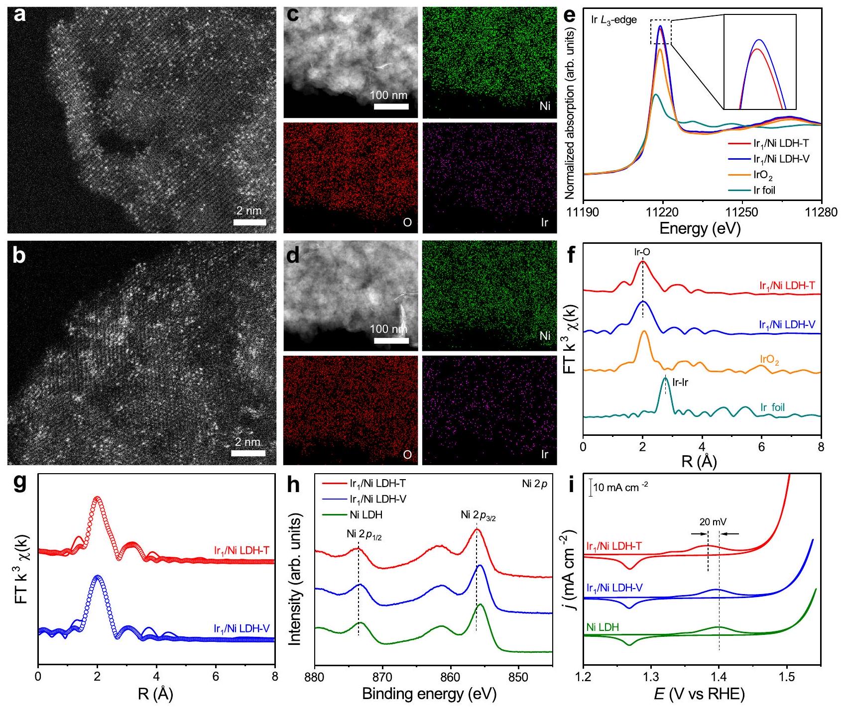
Synthesis and characterizations

The ultrathin Ni LDH was fabricated as the supports using a coprecipitation method . Transmission electron microscopy (TEM) images showed that Ni LDH samples consist of ultrathin nanosheets (Supplementary Fig. 1a). The X-ray diffraction (XRD) pattern of Ni LDH matched well with that of standard (JCPDS No. 14-0117) (Supplementary Fig. 1b). The thickness of as-prepared Ni LDH nanosheets was about measured by atomic force microscopy (AFM; Supplementary Fig. 2). We employed an electrochemical deposition method to selectively anchor Ir single atoms onto specific sites on Ni LDH surface. LDH-T and LDH-V were prepared through cathodic deposition and anodic deposition with as Ir precursor, respectively (Supplementary Fig. 3). The XRD patterns of both LDH-T and LDH-V were similar to those of the pristine Ni LDH (Supplementary Figs. 4 and 5). High-resolution transmission electron microscope (HR-TEM) indicated no noticeable metal or metal oxide particles in the two catalysts (Supplementary Fig. 6). The isolated bright spots in the aberration-corrected high-angle annular dark-field scanning TEM (HAADF-STEM) confirmed the atomically dispersed Ir in both LDH-T and LDH-V (Fig. 1a, b). In addition, energydispersive spectroscopic (EDS) elemental mapping results displayed the uniform distribution of Ir atoms across the entire Ni LDH matrix (Fig. 1c, d). The loading mass of Ir could be controlled via changing the concentration of (Supplementary Table 1). To avoid the aggregation of Ir single atoms into clusters at high concentrations of (Supplementary Fig. 7), we set the concentration of as . The contents of Ir in LDH-T and LDH-V were determined to be 2.18 and by inductively coupled plasma-atomic emission spectroscopy (ICP-AES), respectively.
The detailed electronic structure and coordination environment of LDH-T and LDH-V were further revealed by X-ray absorption near-edge spectroscopy (XANES) and extended X-ray absorption fine structure (EXAFS) studies. The normalized Ir -edge XANES and EXAFS spectra of LDH-T before and after electrooxidation activation confirmed that the bond was transformed to the bond after the OER activation process (Supplementary Fig. 8). The Ir -edge XANES spectra of LDH-T and LDH-V together
with Ir foil and as references were shown in Fig. 1e. The intensity of white line for LDH-T and LDH-V were stronger than that for , suggesting that the valence states of Ir in both the samples were higher than . Besides, the slightly weaker intensity of the white line for LDH-T relative to LDH-V could be caused by the electrons transfer from Ni LDH to Ir. The Ir -edge EXAFS spectra of Ir LDH-T and LDH-V both exhibited a major peak at A , which was assigned to the first-shell bonding as found in (Fig. 1f). The absence of Ir-Ir bonding at around excluded the formation of Ir-based clusters or nanoparticles, further confirming the singleatom dispersion of in both the samples. By fitting the EXAFS experimental spectra, the first-shell coordination of both LDH-T and LDH-V was determined to be with a coordination number of (Supplementary Table 2). Moreover, the EXAFS spectra of the two catalysts both discerned a minor peak at around , which was assigned to the Ir-Ni path from the second-shell coordination. Notably, the coordination number of the path for LDH-T was fitted to be about 2.8, which was larger than that for LDH-V ( ). This difference was attributed to the different coordination structures of Ir atoms in LDH-T and LDH-V.
To further elucidate the coordination structure of Ir atoms, we investigated the surface sites of Ni LDH in the process of electrochemical deposition. Under the driving force of electrode potentials, the negative or positive electric field will preferentially induce the deposition of Ir-based cations or Ir-based anions onto the electrode through electrostatic adsorption, respectively. Defective Ni LDH as the support provides different possible sites for anchoring single atoms, including the 3-fold hollow site of oxygen, 3-fold hexagonal close-packed (hcp) hollow site of oxygen, and oxygen vacancy site (Supplementary Fig. 9). The surface oxygen atoms of the 3-fold hollow sites provide lone-pair electrons and negative charge to bind Ir-based cations, whereas the deficiency of oxygen induce localized positive charge to combine Ir-based anions. For LDH-T, the deposited species in an alkaline environment should be cations originated from the precursor. The valence state of Ir in the deposited species was determined to be +4 by -edge XANES spectra, which showed the similar intensity of white line with for LDH-T before activation (Supplementary Fig. 8). Moreover, the fitting results for LDH-T before activation indicated the first-shell coordination of , since ions in alkaline environment were prone to attack in the solution or on . Besides, the existence of Cl in LDH-T was further confirmed by the XPS spectra (Supplementary Fig. 10). We further conducted DFT calculations to estimate the formation energy ( ) of , and on the surface sites of Ni LDH (Supplementary Fig. 11). Notably, the of on the 3-fold hollow site is -4.97 eV , which is lower than those on the 3-fold hcp hollow site ( -3.12 eV ) and the oxygen vacancy site ( 0 eV ). Similar results were obtained with and as the deposited species, exhibiting the lowest formation energy on the 3-fold hollow site. The higher formation energy of on the site may be caused by electrostatic repulsion between the inner-layer Ni atom and Ir atoms due to the closer distance, leading to lower stability. Therefore, cations were bonded with three oxygen atoms from 3-fold hollow site for LDH-T, followed by electrooxidation activation to form octahedra linked to Ni LDH. As for LDH-V, the negatively charged Ir-based anions were with full coordination of due to the alkaline environment . The of on the oxygen vacancy site ( -3.32 eV ) is much lower than those on the 3-fold and hollow sites, demonstrating the tendency of to anchor on the oxygen vacancy site (Supplementary Fig. 12). As such, Ir atoms in LDH-V connected with oxygen vacancy sites through one apex oxygen atom. The more covalent bonds between Ir sites and coordinated oxygen in LDHT would give rise to the stronger MSI relative to LDH-V.
The XPS measurements for LDH-T and LDH-V were performed to further clarify the MSI between Ir and Ni LDH. As displayed in Fig. 1h, compared with Ni LDH, the Ni peaks of LDH-T shifted to higher binding energy, indicating an increased valence state of Ni. With regard to LDH-V, the Ni peaks showed no obvious change after anchoring Ir single atoms. Besides, the Ir XPS spectra of LDH-T exhibited a slight negative shift compared with LDH-V (Supplementary Fig. 13), suggesting a slightly lower valence state of Ir single atoms in LDH-T, which was consistent with the results of XANES. Furthermore, cyclic voltammetry (CV) curves showed that the oxidation peak of Ni from +2 to +3 in LDH-T shifted lower by 20 mV compared with Ni LDH whereas the oxidation peak in LDH-V maintained the same potential with Ni LDH (Fig. 1i). As such, the stronger MSI between Ir atoms and Ni LDH in LDH-T enhanced the electron transfer from Ni to Ir , resulting in a higher oxidation state of Ni in LDH-T.

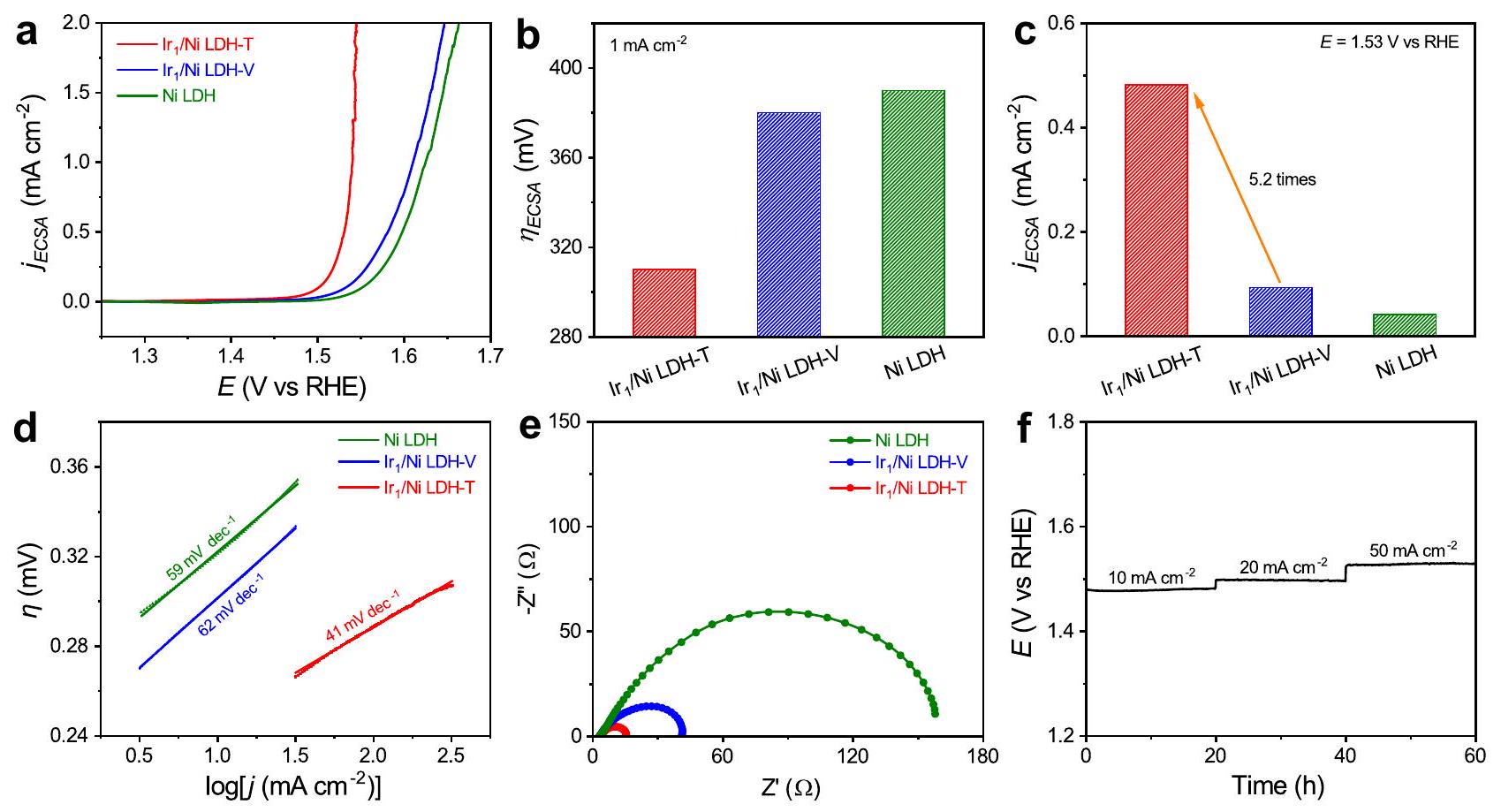
Electrocatalytic performance towards oxygen evolution reaction

To explore the influence of MSI on the catalytic performance of Irbased SACs, the as-prepared Ni LDH, LDH-V, and LDH-T
were applied as electrocatalysts towards OER, one of the most essential reactions for clean energy conversion and storage application . The linear sweep voltammetry (LSV) curves (Supplementary Figs. 14 and 15) measured in 1.0 M KOH solution show that LDH-T exhibited a remarkable activity with an overpotential of at , which significantly outperformed LDH-V and Ni LDH . The overpotentials of LDH-T at 50 and were 276 and 289 mV , respectively, which were also superior to those of LDH-V and Ni LDH. Moreover, LDH-T achieved a mass activity of at the overpotential of 300 mV , which was 19.5 times higher than LDH-V (Supplementary Fig. 16a, b). In addition, the normalized mass activity of LDH-T based on the content of Ir loading at the overpotential of 300 mV reached up to , which was 22.7 -fold higher than that of LDH-V (Supplementary Fig. 16c, d). Based on the number of overall metals, the calculated turnover frequency (TOF) (Supplementary Fig. 17a) of was about at the overpotential of 300 mV , which was much higher than those of LDH-V ( ) and Ni LDH ( ). Besides, the TOF based on the number of Ir sites of LDH-T was estimated to be , largely surpassing LDH-V by 19.0 times (Supplementary Fig. 17b). In
Fig. 1 | Structural characterizations of LDH-T and LDH-V.
HAADF-STEM images of LDH-T (a) and LDH-V (b). c, d EDS elemental mapping of LDH-T (c) and LDH-V (d). e, f Normalized XANES (e) and EXAFS (f) spectra at the Ir -edge for LDH-T and LDH-V. Ir foil and
were used as references. Experimental and fitting EXAFS results of LDH-T and LDH-V. The experimental and fitting results were shown with solid lines and circles, respectively. Ni XPS spectra of LDH-T, LDH-V, and Ni LDH. i CV curves of LDH-T, LDH-V, and Ni LDH.
Fig. 2 | Electrocatalytic performance towards OER. a ECSA-normalized polarization curves of Ni LDH, LDH-V, and LDH-T in 1.0 M KOH .
b Overpotentials of different catalysts at the current density of , respectively. c The ECSA-normalized current density at an overpotential of 300 mV for Ni LDH-T, LDH-V, and Ni LDH, respectively. d Tafel slopes of Ni LDH,
LDH-V, and LDH-T. e Electrochemical impedance spectra of Ni LDH, LDH-V, and LDH-T at 1.57 V vs. RHE. The values for Ni LDH, LDH-V, and LDH-T were , and , respectively. f Chronopotentiometric curves of LDH-T towards OER at 10,20, and for 20 h , respectively.
addition, the activity of Ni LDH after the cathodic and anodic treatment without Ir precursor remained almost unchanged, suggesting that the enhanced performance of LDH-T was attributed to the Ir center with site-specific MSI rather than Ni LDH (Supplementary Fig. 18).
To clarify the intrinsic activity of the samples, we tested electrochemical double-layer capacitance ( ) to assess electrochemically active surface area (ECSA) (Supplementary Fig. 19). The value of LDH-T was determined to be , which was higher than those of LDH-V ( ) and Ni LDH ( ), indicating an increment of ECSA. As shown in Fig. 2a, the ECSA-normalized OER activity still followed the order of LDH-T LDH-V LDH, suggesting that the OER activity of LDH-T was also intrinsically improved. Specially, the overpotential of LDH-T at was only 310 mV , which was lower than that of LDH-V and Ni LDH ( 390 mV ) (Fig. 2b). Besides, the ECSA-normalized current density for LDH-T was estimated to be at the overpotential of 300 mV , which was about 5.2 -fold and 11.4 -fold higher than those for LDH-V ( ) and Ni LDH ( ) (Fig. 2c). Figure 2d displays the Tafel plots for LDH-T, LDH-V, and Ni LDH, where their Tafel slopes were determined to be 41,62 , and , respectively. The lowest Tafel slope indicated the fastest reaction kinetics for LDH-T. Electrochemical impedance spectroscopy (EIS) measurements were further conducted to probe the interfacial charge-transfer resistances of the catalysts. The smallest semicircle diameter of LDH-T among the three catalysts suggested its fastest charge transfer at the interface (Fig. 2e). Durability tests of LDH-T with the superior activity were carried out via chronopotentiometry test at the current density of 10,20 , and , respectively. As plotted in Fig. Ni LDH-T kept stable for 20-h operation. The percentages of leached Ir and Ni after the durability test at 10,20 , and for 20 h were all detected by inductively coupled plasma-mass spectrometry
(ICP-MS) (Supplementary Fig. 20). After the durability test, the atomic dispersion of Ir atoms was maintained without any noticeable aggregation as shown in the HAADF-STEM image and EDS elemental mapping result (Supplementary Fig. 21a, b). Moreover, the XRD pattern and XPS spectra of LDH-T after the durability test also exhibited no observable change, further implying the structural robustness of LDH-T (Supplementary Fig. 21c, d).

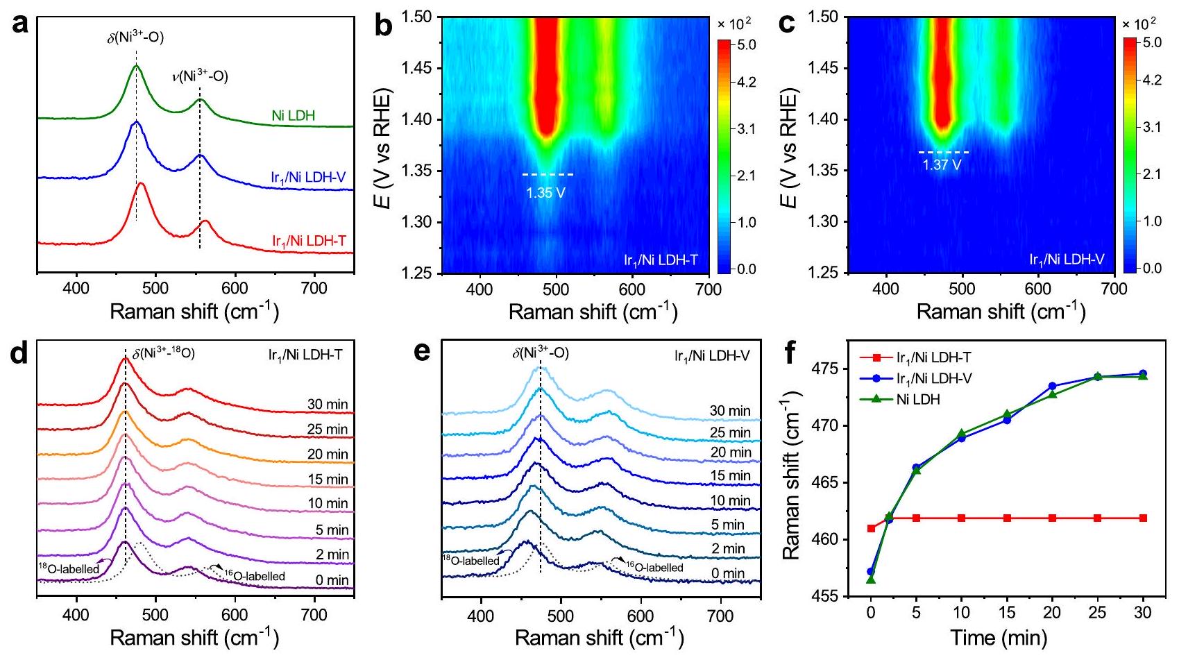
In situ spectroscopic characterizations

To gain an in-depth understanding of the reaction mechanism, we conducted a series of in situ Raman experiments using the homemade in situ Raman cell (Supplementary Fig. 22). The in situ Raman spectra of LDH-T, LDH-V, and Ni LDH were collected over the potentials ranging from 1.2 to 1.6 V vs. reversible hydrogen electrode (RHE), respectively (Supplementary Fig. 23). During the positive potential excursion, two bands for the three catalysts gradually emerged at around 481 and , which were assigned to the bending vibration ( ) and stretching vibration ( ) mode of NiOOH, respectively . However, compared with LDHV and Ni LDH, the peak of at 1.6 V vs. RHE for LDH-T shifted to higher wavenumbers from 475 to , implying that the MSI between single-atom Ir and Ni LDH was stronger in LDH-T (Fig. 3a). We further carried out in situ Raman measurements with a potential interval of 0.01 V to confirm such a change of electrochemical redox during OER process. The emergence of the Raman peaks for occurred at , and 1.37 V vs. RHE for LDHT, Ir LDH-V, and Ni LDH, respectively (Fig. 3b, c and Supplementary Fig. 24). The redox process of for LDH-T started at a more negative potential, which was attributed to the stronger MSI in LDH-T.
To confirm the real active centers during the OER process, we conducted oxygen-isotope-labeling in situ Raman spectra for LDH-T, LDH-V, and Ni LDH. The oxygen atoms of catalysts
Fig. 3 | In situ electrochemical Raman spectra. a In situ Raman spectra of Ni LDH, LDH-V , and LDH-T at 1.6 V vs. RHE, respectively. b, c In situ Raman spectra of LDH-T (b) and LDH-V (c) during the linear sweep voltammetry measurement, in which the dash lines mark the conversion potential from to
. d, e In situ Raman spectra of -labeled LDH-T (d) and LDH-V (e) acquired at in solution, respectively. f Shifts of Raman bands of with the increase of time during the oxygen isotope exchange experiments.
were initially labeled with
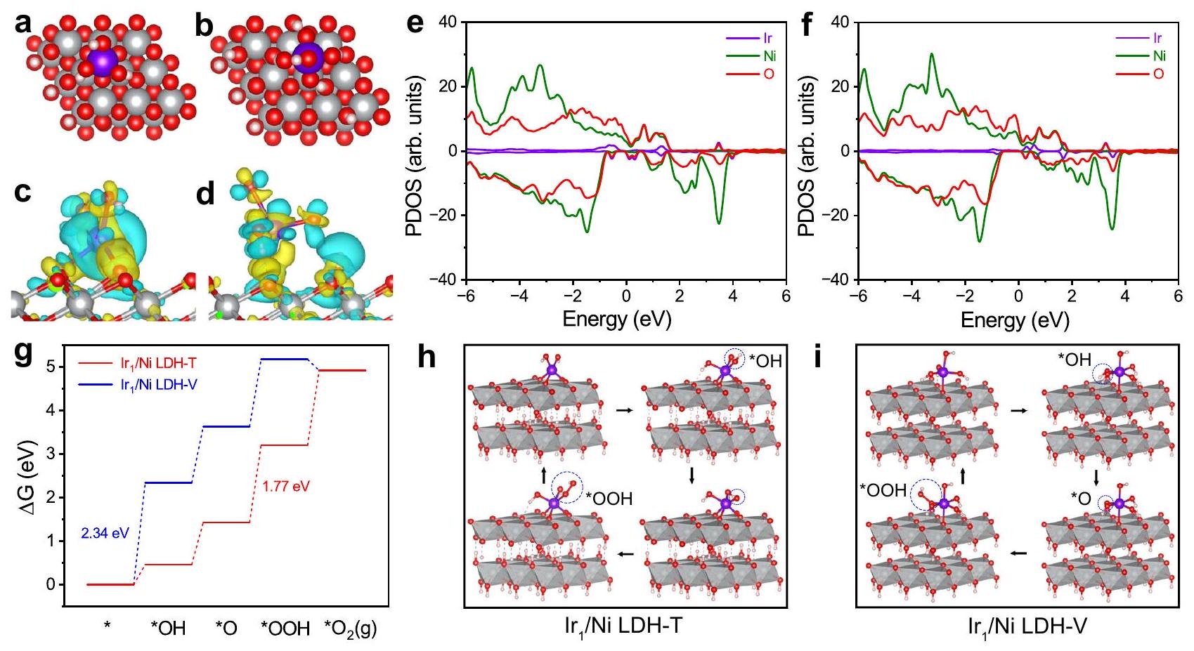
 by applying LSV measurement from 0.50 to 1.65 V vs. RHE for five cycles in solution with the addition of as an internal standard. As shown in Supplementary Fig. 25, compared with -labeled NiOOH , the peak of remained unchanged whereas the peak of for -labeled NiOOH shifted to lower wavenumbers by about , manifesting the occurrence of isotope exchange. After the quick replacement of the electrolyte with solution using a creep pump, the catalysts labeled with were subjected to OER at . As the reaction time increased, the peaks of and for LDH-V and Ni LDH gradually shifted back to the positions observed for the -labeled samples (Fig. 3e and Supplementary Fig. 26). The results suggest that the -labeled oxygen in LDH-V and Ni LDH was easily exchanged with the atom in during the OER process, indicating that Ni could be the active sites for LDH-V and Ni LDH . Nevertheless, in the case of LDH-T, the peaks of and exhibited no discernible shift, suggesting that the dominant active sites for LDH-T could be Ir single atoms rather than Ni species (Fig. 3f). Therefore, the integration of Ir on Ni LDH with strong MSI could induce the switch of the active sites from Ni sites to Ir sites.

DFT calculations

To get a theoretical insight into the reaction mechanism, we employed the DFT calculation to investigate the reaction process . The structure models of LDH-T and LDH-V were built on two-layer supercells based on the structure details discussed above (Fig. 4a, b). The results of charge density differences indicated that the electronic density of atoms in LDH-T was higher than that in Ni LDH-V (Fig. 4c, d). The Bader charge of LDH-T is 1.75, lower than that in LDH-V (2.02), indicating a lower oxidation state of Ir in LDH-T. These results verified that the MSI induced by LDH-T was stronger than that induced by LDH-V. Then we
calculated the projected density of states (PDOS) to elucidate the differences in electronic interactions between LDH-T and LDH-V (Fig. 4e, f and Supplementary Fig. 27). The positions of the band center for LDH-T, LDH-V, and Ni LDH were -1.59, -1.64 , and -1.52 eV , respectively. The moderate -band center of LDH-T is beneficial for balancing the adsorption and desorption abilities of intermediates (*OH, *O, and *OOH), thereby boosting the OER performance .
Furthermore, we calculated the free energy diagram for OER based on the conventional adsorbate evolution mechanism (AEM), which consists of four concurrent proton-electron transfer steps . As shown in Supplementary Figs. 28 and 29, the step from *OH to *O over NiOOH exhibited the highest free energy change ( ) of 2.20 eV among all the steps, which served as the rate-determining step (RDS). For LDH-V, the RDS on Ir sites was determined to be the formation of * OH with an of 2.34 eV , which was higher than that of RDS for NiOOH (Fig. ). In this case, the OER process was more easily proceeded on Ni sites rather than Ir sites for LDH-V. In contrast, the energy barrier of RDS (the step from *O to *OOH) on Ir sites for LDH-T was 1.77 eV , which was significantly lower than that for NiOOH , indicating that Ir atoms preferentially served as the true active sites for LDH-T (Fig. ). Accordingly, the strong MSI between Ir and Ni LDH optimized the adsorption energy for the oxygen-containing intermediates, thereby facilitating the switch of the active sites from Ni sites to Ir sites.

Discussion

In summary, we reported Ir SACs with site-specific MSI by a facile electrochemical deposition technique. Compared with LDH-V, the stronger MSI in LDH-T was reflected by more covalent bonds between Ir sites and coordinated oxygen from Ni LDH. The Ir LDHT exhibited superior OER activity, while LDH-V showed little improvement in activity compared with pristine Ni LDH. The strong MSI between Ir single atoms and Ni LDH in LDH-T not only
LDH-T and LDH-V with Ir as the active sites. The schematic OER pathway for LDH-T and LDH-V . The pink, red, gray, and purple spheres represent , and Ir atoms, respectively. The reaction intermediates are indicated by blue circles.
Fig. 4 | OER mechanism studies for /Ni LDH-T and LDH-V. a, b Schematic structure models of LDH-T (a) and LDH-V (b) from top views. c, d Charge density differences of Ir atoms on LDH-T (c) and LDH-V (d). The yellow and cyan areas indicate electron accumulation and depletion, respectively. e, PDOS in LDH-T (e) and LDH-V (f). g Free energy diagrams of
optimized the adsorption strength of oxygen-containing intermediates but also induced the switch of the catalytic active sites from Ni sites to Ir sites, thereby boosting the catalytic performance. This work provides a novel strategy to design highly active catalysts with site-specific MSI by electrochemically selective anchoring of single atoms and offers an in-depth understanding of the correlation between MSI and OER performance.

Methods

Chemicals

Nickel nitrate , sodium nitrate , sodium hydroxide , and formamide were purchased from Shanghai Chemical Reagent Company. Iridium (IV) chloride hydrate ( ) was purchased from Aladdin. Potassium hydroxide (99.99%) was purchased from Macklin. Nafion solution ( ) was purchased from Sigma-Aldrich. Water- ( 97 atom% O) was purchased from Beijing InnoChem Science & Technology Co., Ltd. All the other chemicals were of analytical grade and used as received without further purification. All aqueous solutions were prepared using deionized water with a resistivity of .

Synthesis of Ni LDH

Ni LDH nanosheets were fabricated according to a previous report . A solution of 10 mM NaNO ( 20 mL ) containing formamide was heated to under stirring. Then, 10 mL solution of 37.5 mM and 0.25 M NaOH were simultaneously added dropwise to maintain the solution pH at . The resulting solution was cooled to room temperature under nitrogen gas bubbling. Then the precipitate was washed with a mixture of ethanol and deionized water (1:1 vol%) at least three times. Finally, the precipitate was dispersed in a small amount of water and lyophilized.

Synthesis of LDH-T and LDH-V

LDH-T and LDH-V were prepared using our previously reported electrochemical deposition method . The deposition was performed in a three-electrode system (CHI 660E, Shanghai CH Instruments), with a Ni foam ( ), a carbon rod, and an electrode as the working, counter, and reference electrodes, respectively. 2.5 mg of Ni LDH were dispersed in 0.6 mL of ethanol, 0.6 mL of water, and of Nafion by sonication for 30 min to form a homogeneous mixture. Then of the mixture was loaded onto the working electrode. To synthesize LDH-T, 10 cycles of linear sweep voltammetry at the applied potentials ranging from 0.1 to -0.4 V vs. RHE were employed for the pretreatment of the working electrode. Afterward, as the Ir precursor was added into the electrolyte, followed by stirring for 10 min . Then cathodic deposition was performed by scanning the linear sweep voltammetry at the potentials from 0.1 to -0.4 V vs. RHE for 10 cycles, followed by a CV method under potentials ranging from 1.1 to 1.8 V vs. RHE. The scan rate was set at . In the case of LDH-V, 3 cycles of the CV method at the potentials ranging from 1.1 to 1.8 V vs. RHE were applied for the pretreatment of the working electrode. Afterward, as the Ir precursor was added to the electrolyte, followed by stirring for 10 min . The anodic deposition was then conducted by scanning the linear sweep voltammetry at the potentials from 1.1 to 1.8 V vs. RHE for 5 cycles. The scan rate was set at . The obtained samples were finally washed with water, followed by being dried for the electrochemical measurements. All potentials were measured against the AgCl electrode and converted to the RHE reference scale by (V vs. vs. .

Catalyst characterization

TEM images were obtained on a transmission electron microscope (Hitachi H-7650) operating at an acceleration voltage of 100 kV . AFM
was measured on the Veeco DI Nanoscope MultiMode V system. XRD patterns were collected using an X-ray diffractometer (Philips X’Pert Pro Super) with radiation ( ). HAADF-STEM images and EDS elemental mapping were obtained on a field-emission transmission electron microscope (JEOL ARM-200F) operating at an accelerating voltage of 200 kV using Cu-based TEM grids. XPS measurements were conducted on an X-ray photoelectron spectrometer (VG ESCALAB MK II) with as the exciting source. ICP-AES (Atomscan Advantage, Thermo Jarrell Ash) were applied for the measurement of concentrations for metal species. The prepared electrode was rinsed several times with ultrapure water and then subjected to ultrasonication in ethanol. The catalyst collected in ethanol was washed three times with ethanol and water by centrifugation. The resulting solid was freeze-dried in a vacuum freezedryer overnight. Before analysis using ICP-AES, a proper amount of powder sample was treated by hot aqua regia to get a clear solution. Line 221.6 and 224.2 were selected for Ni and Ir detection, respectively. ICP-MS (Thermo Fisher iCAP RQ) analysis was performed using 115In and 73Ge as internal standards with a concentration of 40 ppb .
XANES and EXAFS spectra at the Ir -edge were recorded at the BL14W1, Shanghai Synchrotron Radiation Facility, China. The energy was calibrated by Ir foil. Ir foil and were used as reference samples and measured in the transmission mode. The samples were measured in fluorescence mode. The Athena software package was used to analyze the data.

Electrochemical measurements

To assess the electrocatalytic activity of the samples, an electrochemical workstation (CHI 660E, Shanghai CH Instruments) was employed. All electrochemical tests were carried out in a standard three-electrode system under ambient conditions in 1.0 M KOH electrolyte. The content of Fe in 1.0 M KOH was quantified to be using ICP-MS. The working electrode was a foam electrode and the loading of catalysts was . A carbon rod and an electrode served as the counter and reference electrodes, respectively. The polarization curves of catalysts were measured by scanning linear sweep voltammetry under the potentials ranging from 1.1 to 1.8 V vs. RHE with a scan rate of . To correct for solution resistance, the potentials were adjusted using the equation vs. RHE , where and represented the current and the uncompensated ohmic electrolyte resistance, respectively. was extracted from the Nyquist plot through alternating current impedance in the region of high frequency . The value was measured to be . The ECSA of the catalyst was obtained by the following equation: ECSA , where and represented the roughness factor and the surface area of the glassy carbon electrode ( in this case), respectively. could be calculated according to based on the of an ideal flat oxide surface ( ). To obtain the of the samples, we conducted the CV measurements from 1.06 to 1.16 V vs. RHE at different scan rates ( 20 , , and ). was estimated by plotting the ( ) at 1.11 V vs. RHE against the scan rates. The and represented the anodic and cathodic current density, respectively. The linear slope was equivalent to twice of . To evaluate the specific activities and mass activities of the samples, the current densities were normalized based on ECSA and mass loading of catalysts, respectively. The TOFs were estimated using the following equation: TOF , where represented the current density, was the surface area of the electrode, represented the Faraday constant, and represented the mole number of metals on the electrode. EIS measurements were carried out in a standard three-electrode system at room temperature in 1.0 M KOH electrolyte at a potential of 1.57 V vs. RHE with a sinusoidal wave amplitude of 5 mV and a frequency scan range of . The working electrode was a Ni foam electrode and the loading of catalysts was . A
carbon rod and an electrode served as the counter and reference electrodes, respectively. To perform the durability test, the galvanostatic mode was used in 1.0 M KOH electrolyte at room temperature with current densities of 10,20 , and , respectively.

In situ Raman spectroscopy

The Raman spectra were carried out on a confocal microscope Raman system (Horiba LabRAM HR Evolution). The excitation wavelength was 532 nm , and a microscope objective with a numerical aperture of 0.55 was used in all Raman measurements. Before measurements, the Raman shift range was calibrated using the peak of silicon. A homemade Teflon Raman cell was used for the in situ electrochemical Raman measurements with a 3 mm Au electrode, a Pt wire, and an electrode as the working, counter, and reference electrodes, respectively. The working electrode was prepared by casting ink on a 3 mm Au electrode. LDH-T and LDH-V were synthesized by the same electrochemical deposition method. The potential was controlled by an electrochemical workstation (CHI 660E, Shanghai CH Instruments). To label the catalyst with oxygen-18, we conducted LSV measurements in a solution within the potential range of vs. RHE for five cycles.

DFT calculation method

All the spin-unrestricted DFT calculations with Hubbard corrections were performed using the Vienna ab initio simulation package (VASP). For the description of exchange-correlation effects, the Perdew-Burke-Ernzerhof function within the generalized gradient approximation was employed . A plane wave basis set was generated with a kinetic energy cutoff of 450 eV , and -points were sampled using a and Gamma mesh for geometry optimization and electronic structure calculations of all intermediates on NiOOH (001), LDH-T , and LDH-V, respectively. For a better description of the electrons, the Hubbard effective term for Ni was added to the PBE functional through the rotationally invariant approach proposed by Dudarev and co-workers . A single-cell NiOOH (001) surface with double atomic layers consisted of 36 oxygen atoms, 18 Ni atoms, and 18 H atoms. A vacuum layer of in the direction was set to avoid unwanted interactions between periodic images. The convergence of energy and force were set to be and , respectively. The DFT-D3 method was adopted to take van der Waals correction into consideration .

Theoretical evaluation of OER activity

The theoretical overpotentials for NiOOH, LDH-T, and LDHV interfaces were determined by following the conventional four-step electron-proton-coupled OER mechanism . The OER process generally involves the following steps:
where * represented the active site on the model of a specific surface, (l) and (g) represented the liquid and gas phases, respectively. Considering that the standard thermodynamic potential was 1.23 V for water oxidation, the Gibbs free energy for forming one water molecule was fixed to the experimental value . In this case, the calculation for bond energy of could be avoided, which was inaccurately interpreted by DFT . The free energy of the
molecule was calculated as
where and represented the calculated free energies of and , respectively. To calculate the Gibbs free energy for the reaction intermediates, we applied the computational hydrogen electrode (CHE) model . The difference in Gibbs free energies of these intermediates was calculated as , where , , and represented adsorption enthalpy, zero-point energy, temperature, and entropy, respectively. The change of Gibbs energy for intermediates (*O, *OH, and *OOH) and every OER step can be calculated as follows :
where was the potential against normal hydrogen electrode (NHE) at standard conditions ( ).

Data availability

The experiment data that support the findings of this study are available from the corresponding author upon reasonable request. The source data underlying Figs. 1-4 and Supplementary Figs. 1-29 are provided as a Source Data file. Source data are provided with this paper.

References

  1. Qiao, B. et al. Single-atom catalysis of CO oxidation using . Nat. Chem. 3, 634-641 (2011).
  2. Yang, H. et al. Intramolecular hydroxyl nucleophilic attack pathway by a polymeric water oxidation catalyst with single cobalt sites. Nat. Catal. 5, 414-429 (2022).
  3. Gu, J., Hsu, C. S., Bai, L., Chen, H. M. & Hu, X. Atomically dispersed sites catalyze efficient electroreduction to CO . Science 364, 1091-1094 (2019).
  4. Yang, J., Li, W., Wang, D. & Li, Y. Electronic metal-support interaction of single-atom catalysts and applications in electrocatalysis. Adv. Mater. 32, 2003300 (2020).
  5. Zhang, J., Liu, C. & Zhang, B. Insights into single-atom metal-support interactions in electrocatalytic water splitting. Small Methods 3, 1800481 (2019).
  6. Jin, Z. Y. et al. Understanding the inter-site distance effect in singleatom catalysts for oxygen electroreduction. Nat. Catal. 4, 615-622 (2021).
  7. Sun, H. et al. Atomic metal-support interaction enables reconstruction-free dual-site electrocatalyst. J. Am. Chem. Soc. 144, 1174-1186 (2022).
  8. Jiang, Z. et al. Modulating the electronic metal-support interactions in single-atom Pt-CuO catalyst for boosting acetone oxidation. Angew. Chem. Int. Ed. 61, e202200763 (2022).
  9. Li, W. H. et al. Creating high regioselectivity by electronic metal-support interaction of a single-atomic-site catalyst. J. Am. Chem. Soc. 143, 15453-15461 (2021).
  10. Han, B. et al. Strong metal-support interactions between Pt single atoms and . Angew. Chem. Int. Ed. 59, 11824-11829 (2020).
  11. Li, J. et al. Highly active and stable metal single-atom catalysts achieved by strong electronic metal-support interactions. J. Am. Chem. Soc. 141, 14515-14519 (2019).
  12. Yang, J. et al. Modulating the strong metal-support interaction of single-atom catalysts via vicinal structure decoration. Nat. Commun. 13, 4244 (2022).
  13. Ren, Y. et al. Unraveling the coordination structure-performance relationship in single-atom catalyst. Nat. Commun. 10, 4500 (2019).
  14. Jeong, H. et al. Controlling the oxidation state of Pt single atoms for maximizing catalytic activity. Angew. Chem. Int. Ed. 59, 20691-20696 (2020).
  15. Lee, S., Banjac, K., Lingenfelder, M. & Hu, X. Oxygen isotope labeling experiments reveal different reaction sites for the oxygen evolution reaction on nickel and nickel iron oxides. Angew. Chem. Int. Ed. 58, 10295-10299 (2019).
  16. Zheng, X. et al. Origin of enhanced water oxidation activity in an iridium single atom anchored on NiFe oxyhydroxide catalyst. Proc. Natl Acad. Sci. USA 118, e2101817118 (2021).
  17. Van Ooij, W. J. & Houtman, J. P. W. Ligand exchange in iridium hexahalocomplexes: Part 2. Influence of the pH on the reactions of the hexachloroiridite and hexachloroiridate ions in aqueous solutions. Radiochim. Acta 20, 47-50 (1973).
  18. Cui, C. et al. Atomically dispersed hybrid nickel-iridium sites for photoelectrocatalysis. Nat. Commun. 8, 1341 (2017).
  19. Zhang, Z. et al. Electrochemical deposition as a universal route for fabricating single-atom catalysts. Nat. Commun. 11, 1215 (2020).
  20. Yao, Y. C. et al. Engineering the electronic structure of single atom Ru sites via compressive strain boosts acidic water oxidation electrocatalysis. Nat. Catal. 2, 304-313 (2019).
  21. Lin, C. et al. In-situ reconstructed Ru atom array on with enhanced performance for acidic water oxidation. Nat. Catal. 4, 1012-1023 (2021).
  22. Shi, Z. et al. Confined Ir single sites with triggered lattice oxygen redox: toward boosted and sustained water oxidation catalysis. Joule 5, 2164-2176 (2021).
  23. Zhang, J. et al. Single-atom Au/NiFe layered double hydroxide electrocatalyst: probing the origin of activity for oxygen evolution reaction. J. Am. Chem. Soc. 140, 3876-3879 (2018).
  24. Li, P. et al. Boosting oxygen evolution of single-atomic ruthenium through electronic coupling with cobalt-iron layered double hydroxides. Nat. Commun. 10, 1711 (2019).
  25. Wang, Q. et al. Ultrahigh-loading of Ir single atoms on NiO matrix to dramatically enhance oxygen evolution reaction. J. Am. Chem. Soc. 142, 7425-7433 (2020).
  26. Cao, L. et al. Dynamic oxygen adsorption on single-atomic ruthenium catalyst with high performance for acidic oxygen evolution reaction. Nat. Commun. 10, 4849 (2019).
  27. Yin, J. et al. Iridium single atoms coupling with oxygen vacancies boosts oxygen evolution reaction in acid media. J. Am. Chem. Soc. 142, 18378-18386 (2020).
  28. Wang, Q. et al. Coordination engineering of iridium nanocluster bifunctional electrocatalyst for highly efficient and pH-universal overall water splitting. Nat. Commun. 11, 4246 (2020).
  29. Louie, M. W. & Bell, A. T. An investigation of thin-film Ni-Fe oxide catalysts for the electrochemical evolution of oxygen. J. Am. Chem. Soc. 135, 12329-12337 (2013).
  30. Bai, L., Lee, S. & Hu, X. Spectroscopic and electrokinetic evidence for a bifunctional mechanism of the oxygen evolution reaction. Angew. Chem. Int. Ed. 60, 3095-3103 (2021).
  31. Man, I. C. et al. Universality in oxygen evolution electrocatalysis on oxide surfaces. ChemCatChem 3, 1159-1165 (2011).
  32. Zheng, Y.-R. et al. Monitoring oxygen production on mass-selected iridium-tantalum oxide electrocatalysts. Nat. Energy 7, 55-64 (2021).
  33. Kang, J. X. et al. Valence oscillation and dynamic active sites in monolayer NiCo hydroxides for water oxidation. Nat. Catal. 4, 1050-1058 (2021).
  34. Wu, Z. Y. et al. Non-iridium-based electrocatalyst for durable acidic oxygen evolution reaction in proton exchange membrane water electrolysis. Nat. Mater. 22, 100-108 (2023).
  35. Lee, S., Bai, L. & Hu, X. Deciphering iron-dependent activity in oxygen evolution catalyzed by nickel-iron layered double hydroxide. Angew. Chem. Int. Ed. 59, 8072-8077 (2020).
  36. Stevens, M. B. et al. Measurement techniques for the study of thin film heterogeneous water oxidation electrocatalysts. Chem. Mater. 29, 120-140 (2016).
  37. Sun, J., Ruzsinszky, A. & Perdew, J. P. Strongly constrained and appropriately normed semilocal density functional. Phys. Rev. Lett. 115, 036402 (2015).
  38. Dudarev, S. L., Botton, G. A., Savrasov, S. Y., Humphreys, C. J. & Sutton, A. P. Electron-energy-loss spectra and the structural stability of nickel oxide: an LSDA+ U study. Phys. Rev. B 57, 1505 (1998).
  39. Grimme, S., Ehrlich, S. & Goerigk, L. Effect of the damping function in dispersion corrected density functional theory. J. Comput. Chem. 32, 1456-1465 (2011).
  40. Bajdich, M., García-Mota, M., Vojvodic, A., Nørskov, J. K. & Bell, A. T. Theoretical investigation of the activity of cobalt oxides for the electrochemical oxidation of water. J. Am. Chem. Soc. 135, 13521-13530 (2013).
  41. Friebel, D. et al. Identification of highly active Fe sites in (Ni, Fe) OOH for electrocatalytic water splitting. J. Am. Chem. Soc. 137, 1305-1313 (2015).
  42. Hu, M. et al. Tuning single-atom catalysts of nitrogen-coordinated transition metals for optimizing oxygen evolution and reduction reactions. J. Phys. Chem. C 124, 13168-13176 (2020).
  43. Bhat, A. et al. Prospects challenges and stability of 2D MXenes for clean energy conversion and storage applications. npj 2D Mater. Appl. 5, 61 (2021).
  44. Lv, Q. et al. Selectively nitrogen-doped carbon materials as superior metal-free catalysts for oxygen reduction. Nat. Commun. 9, 3376 (2018).

Acknowledgements

This work was supported by the National Key Research and Development Program of China (2021YFA1500500 and 2019YFA0405600), CAS Project for Young Scientists in Basic Research (YSBR-051), National Science Fund for Distinguished Young Scholars (21925204), NSFC
(U19A2015, 22221003, 22250007, and 22302184), Fundamental Research Funds for the Central Universities, K.C. Wong Education (GJTD-2020-15), Collaborative Innovation Program of Hefei Science Center, CAS (2022HSC-CIP004), the Joint Fund of the Yulin University and the Dalian National Laboratory for Clean Energy (YLU-DNL Fund 2022012), the DNL Cooperation Fund, CAS (DNL202003), International Partnership Program of Chinese Academy of Sciences ( ), the Anhui Natural Science Foundation for Young Scholars (2208085QB41), and the Fellowship of China Postdoctoral Science Foundation (2021M693058).

Author contributions

J.W., Z.Z., and J.Z. designed the study. J.W. and H.T. performed most of the experiments and analyzed the experimental data. L.S. carried out DFT calculations. R.W. provided help with the XAFS measurements. J.W., M.F., J.-L.W., and Y.W. conducted characterizations and analyzed the results. Z.Z. and S.Z. helped in the analysis of data. J.W. and J.Z. wrote the manuscript. All authors discussed the results and assisted during manuscript preparation.

Competing interests

The authors declare no competing interests.

Additional information

Supplementary information The online version contains
supplementary material available at
https://doi.org/10.1038/s41467-024-44815-0.
Correspondence and requests for materials should be addressed to Zhirong Zhang or Jie Zeng.
Peer review information Nature Communications thanks Huanyu Jin, and the other, anonymous, reviewer(s) for their contribution to the peer review of this work. A peer review file is available.
Reprints and permissions information is available at http://www.nature.com/reprints
Publisher’s note Springer Nature remains neutral with regard to jurisdictional claims in published maps and institutional affiliations.
Open Access This article is licensed under a Creative Commons Attribution 4.0 International License, which permits use, sharing, adaptation, distribution and reproduction in any medium or format, as long as you give appropriate credit to the original author(s) and the source, provide a link to the Creative Commons licence, and indicate if changes were made. The images or other third party material in this article are included in the article’s Creative Commons licence, unless indicated otherwise in a credit line to the material. If material is not included in the article’s Creative Commons licence and your intended use is not permitted by statutory regulation or exceeds the permitted use, you will need to obtain permission directly from the copyright holder. To view a copy of this licence, visit http://creativecommons.org/ licenses/by/4.0/.
© The Author(s) 2024

  1. Hefei National Research Center for Physical Sciences at the Microscale, University of Science and Technology of China, 230026 Hefei, Anhui, P. R. China.
    National Synchrotron Radiation Laboratory, University of Science and Technology of China, 230026 Hefei, Anhui, P. R. China. CAS Key Laboratory of Strongly-Coupled Quantum Matter Physics, University of Science and Technology of China, 230026 Hefei, Anhui, P. R. China. Key Laboratory of Surface and Interface Chemistry and Energy Catalysis of Anhui Higher Education Institutes, Department of Chemical Physics, University of Science and Technology of China, 230026 Hefei, Anhui, P. R. China. School of Chemistry & Chemical Engineering, Anhui University of Technology, 243002 Ma’anshan, Anhui, P. R. China. Institute of Advanced Technology, University of Science and Technology of China, 230031 Hefei, Anhui, P. R. China. These authors contributed equally: Jie Wei, Hua Tang, Li Sheng. e-mail: zzhirong@ustc.edu.cn; zengj@ustc.edu.cn