الخلايا التائية CAR المستهدفة لبروتين B7-H3 داخل البطينات الدماغية لعلاج الورم الدبقي الجسري المنتشر في الجسر: تجربة المرحلة الأولى Intracerebroventricular B7-H3-targeting CAR T cells for diffuse intrinsic pontine glioma: a phase 1 trial

المجلة: Nature Medicine، المجلد: 31، العدد: 3
DOI: https://doi.org/10.1038/s41591-024-03451-3
PMID: https://pubmed.ncbi.nlm.nih.gov/39775044
تاريخ النشر: 2025-01-07

الخلايا التائية CAR المستهدفة لبروتين B7-H3 داخل البطينات الدماغية لعلاج الورم الدبقي الجسري المنتشر في الجسر: تجربة المرحلة الأولى

تاريخ الاستلام: 11 أكتوبر 2024
تم القبول: 6 ديسمبر 2024
نُشر على الإنترنت: 7 يناير 2025
(أ) التحقق من التحديثات

تظهر قائمة بالمؤلفين وانتماءاتهم في نهاية الورقة.

ورم الدبقيات الجذعية المنتشر في الجسر (DIPG) هو ورم قاتل في الجهاز العصبي المركزي (CNS) يمنح متوسط بقاء قدره 11 شهرًا. نظرًا لأن B7-H3 يتم التعبير عنه في أورام CNS لدى الأطفال، قمنا بإجراء دراسة BrainChild-03، وهي تجربة سريرية من المرحلة الأولى ذات مركز واحد، تتضمن تصعيد الجرعة من العلاج بالخلايا التائية المستهدفة لبروتين B7-H3 (خلايا CAR T المستهدفة لبروتين B7-H3) للأطفال الذين يعانون من أورام CNS المتكررة أو المقاومة وDIPG. هنا نبلغ عن النتائج من الذراع C، المخصصة للمرضى الذين يعانون من DIPG. كانت الأهداف الرئيسية هي تقييم الجدوى والتحمل، وقد تم تحقيق كلاهما. شملت الأهداف الثانوية تقييم توزيع خلايا CAR T والبقاء. تم تسجيل 23 مريضًا مصابًا بـ DIPG، وتم علاج 21 منهم بجرعات متكررة من خلايا CAR T المستهدفة لبروتين B7-H3 عبر الحقن داخل البطين دون إجراء تخفيض لمناعة المريض مسبقًا. لم يُسمح بالعلاج الموجه للأورام المتزامن، بما في ذلك إعادة الإشعاع، أثناء العلاج وفقًا للبروتوكول. قدمنا إجمالي 253 جرعة داخل البطين وأسسنا أعلى نظام جرعات مخطط له، DR4، الذي تصاعد حتى خلايا لكل جرعة، كجرعة التحمل القصوى. كانت الأحداث السلبية الشائعة تشمل الصداع، والتعب، والحمى. كان هناك سمية محددة للجرعة واحدة (نزيف داخل الورم) خلال DR2. بالنسبة لجميع المرضى المعالجين ( )، كانت مدة البقاء المتوسطة من حقن خلايا CAR T الأولية 10.7 أشهر ومدة البقاء المتوسطة من التشخيص 19.8 أشهر مع بقاء 3 مرضى على قيد الحياة بعد 44 و45 و52 شهرًا من التشخيص. في النهاية، تُظهر هذه التجربة الأولى على البشر أن الجرعات المتكررة من خلايا CAR T المستهدفة لبروتين B7-H3 في المرضى الأطفال والشباب المصابين بـ DIPG قابلة للتحمل، بما في ذلك الجرعات المتكررة على مدى عدة سنوات، وقد تكون لها فعالية سريرية تستدعي مزيدًا من التحقيق في تجربة المرحلة الثانية متعددة المواقع.ClinicalTrials.govالتسجيل: NCT04185038.
ورم الجذع الدماغي المنتشر (DIPG) هو ورم قاتل في جذع الدماغ مسؤول عن سنوات الحياة المفقودة كل عام في الولايات المتحدة لقد أظهرت خلايا T الحاملة لمستقبلات المستضدات الهجينة (CAR) فعالية كبيرة ضد سرطان الدم لدى الأطفال. ومع ذلك، فإن التطبيق السريري لخلايا CAR T داخل الجمجمة للأطفال المصابين بـ DIPG هو مجال ناشئ في تطوير العلاجات. .
تجاربنا السريرية المرحلة الأولى التي تقدم خلايا CAR T داخل الجمجمة (BrainChild-01 التي تقدم مستقبل عامل نمو البشرة البشري 2)
خلايا CAR T المستهدفة لـ (HER2) (NCT03500991) ; BrainChild-02 توصيل خلايا CAR T الخاصة بـ EGFR806 (NCT03638167) استبعد المرضى الذين يعانون من DIPG. حيث يتم التعبير عن B7-H3 (CD276) في DIPG قمنا بتصميم خلايا CAR T المستهدفة لبروتين B7-H3 (خلايا CAR T لبروتين B7-H3) ووصفنا فعاليتها قبل السريرية. . بعد ذلك، قمنا بفتح الدراسة BrainChild-03 (NCT04185038)، حيث تم توصيل خلايا CAR T من نوع B7-H3 للأطفال الذين يعانون من أورام الجهاز العصبي المركزي المتكررة أو المقاومة والعلاج الإشعاعي للورم الجسدي، ونشرنا نتائج التحمل الأولية. .
هنا نبلغ عن الانتهاء من الذراع C من التجربة السريرية الأولى من نوعها في البشر المرحلة 1 BrainChild-03 المخصصة للأطفال المصابين بـ DIPG. تم تسجيل المرضى في أي وقت بعد العلاج الإشعاعي القياسي (بما في ذلك المرضى الذين يعانون من تقدم المرض و/أو النقائل). نعرض سلامة جرعات خلايا CAR T من نوع B7-H3 المعطاة بشكل متكرر داخل البطينات الدماغية حتى خلايا لكل جرعة للأطفال المصابين بـ DIPG، دليل على تنشيط المناعة في الجهاز العصبي المركزي والفائدة السريرية المحتملة.

النتائج

تصميم الدراسة وخصائص المرضى

كانت هذه دراسة المرحلة الأولى للعلاج الخلوي التبني المعطى بشكل متكرر عن طريق الحقن داخل البطين مع خلايا ذاتية. و تم نقل خلايا تم تعديلها فيروسياً لتعبير عن CAR محدد لـ B7-H3 إلى الأطفال والشباب الذين يعانون من DIPG. كانت الأهداف الرئيسية هي تقييم الجدوى والسلامة والتحمل لعلاج خلايا CAR T من نوع B7-H3 عن طريق الحقن داخل البطين، بينما شملت الأهداف الثانوية تقييم توزيع خلايا CAR T وبقائها. كانت معايير الأهلية تشمل ما يلي: العمر و سنوات، تشخيص DIPG (تم تشخيصه شعاعياً أو عبر تأكيد نسيجي لورم دبقي عالي الدرجة أو ورم دبقي منتشر في الخط الأوسط معدل H3K27M (DMG)) في أي نقطة زمنية بعد الانتهاء من الإشعاع القياسي، وجود قسطرة في الجهاز العصبي المركزي، أداء Lansky أو Karnofsky من جرعة ديكساميثازون غسل محدد بواسطة البروتوكول من العلاجات السابقة وأدلة مخبرية محددة بواسطة البروتوكول على وظيفة الأعضاء الكافية (بما في ذلك عدد اللمفاويات المطلق من خلايا ).
العمر الوسيط عند التسجيل هو 6 سنوات (النطاق: 2-22 سنة) وهو متسق مع العمر الوسيط التاريخي لهذه المرض. كان لدى المرضى درجات أداء لانسكي أو كارنوفسكي من ، و . تلقى جميع المرضى العلاج الإشعاعي القياسي عند التشخيص. عانى المريضان المسجلان اللذان لم يتلقيا خلايا CAR T من تقدم سريري خلال تصنيع خلايا CAR T ولم يستوفيا أبداً معايير الأهلية لحقن خلايا CAR T. من بين 21 مريضاً تم علاجهم، تلقى العلاج بعد تقدم ورم واحد على الأقل، بينما 9 مرضى تم علاجهم قبل أي تقدم في الورم. تم تحقيق الهدف الأساسي من إمكانية تصنيع خلايا CAR T من عملية فصل واحدة لجميع المشاركين. يتم تقديم الخصائص الأساسية للمرضى في الجدول 1. من بين 21 مريضًا تم علاجهم، استوفى 17 التشخيص الجزيئي لـ DMG إما من خلال نسيج الورم. أو عن طريق اختبار السائل الدماغي الشوكي أو البلازما (الشكل البياني الموسع 1). كان لدى مريض واحد (S008) ورم دبقي أنابلاستي في الجسر يحتوي على طفرة في جين IDH1.

العلاج

تم تسليم خلايا CAR T B7-H3 في الذراع C من BrainChild-03 ICV دون تخفيض اللمف السابق كل 14 يومًا على مدار 8 أسابيع خلال فترة مراقبة سمية الجرعة المحدودة (DLT) (الدورات 1 و 2). لم يُسمح بالعلاج الموجه للأورام المتزامن، بما في ذلك إعادة الإشعاع، أثناء العلاج البروتوكولي. نظرًا لأن العدد الأمثل من الجرعات غير معروف، كان المرضى الذين استوفوا معايير الحقن اللاحقة بعد الدورة 2 (والذين كانت لديهم جرعات إضافية من خلايا CAR T المجمدة متاحة) مؤهلين لمواصلة تلقي علاج خلايا CAR T بعد فترة DLT بجرعات كل 2-4 أسابيع. تم علاج المرضى على أربعة أنظمة جرعات متزايدة (DRs؛ الشكل 1). في DR1، تلقى ثلاثة مرضى قابلين للتقييم خلايا CAR T لكل جرعة. تم استخدام تصعيد الجرعة داخل المريض في DR2 و DR3 و DR4. في DR2، تلقى 6 مرضى قابلين للتقييم ما يصل إلى خلايا CAR T لكل جرعة؛ في DR3، تلقى 3 مرضى ما يصل إلى خلايا CAR T لكل جرعة، وفي DR4، تلقى 6 مرضى قابلين للتقييم ما يصل إلى عدد خلايا CAR T لكل جرعة. تم توسيع DR2 ليشمل ستة مرضى بسبب حدث ضار غير مقبول (نزيف داخل الورم)، وتم توسيع DR4 ليشمل ستة مرضى من أجل تأكيد إحصائي مخطط لنظام الجرعة القصوى المتسامحة (MTDR). لم يكن بالإمكان تقييم ثلاثة مرضى من حيث الأحداث الضارة غير المقبولة (لأنهم اختاروا متابعة علاج موجه للورم آخر بسبب القلق من تقدم المرض دون أن يتعرضوا لحدث ضار غير مقبول) وتم استبدالهم وفقًا للبروتوكول.
الجدول 1 | الخصائص السكانية والسريرية للمشاركين في الدراسة المسجلين في BrainChild-03 الذراع C )
خصائص قيمة
العمر الوسيط (بالسنوات (النطاق)) 6 (2-22)
جنس (%)
ذكر 9 (39)
أنثى 14 (61)
درجة أداء لانسكي أو كارنوفسكي (%)
90 12 (52)
٨٠ 7 (30)
70 2 (9)
60 2 (9)
متوسط الأشهر من التشخيص إلى التسجيل (النطاق) 6 (3-22)
تأكيد هيستوباثولوجي أو جزيئي لـ DMG (%) 18 (78)
تاريخ المرض عند التسجيل ( (%)
التقدم السابق 12 (52)
لا تقدم سابق 11 (48)
تاريخ المرض عند الجرعة الأولى من CAR B7-H3 (%); العدد الإجمالي المعالج: 21)
التقدم السابق 12 (57)
لا تقدم سابق 9 (43)
العلاج الإشعاعي السابق (%) ٢٣ (١٠٠)
للمرضى الذين بدأوا العلاج قبل أي تقدم ” )، كان الوقت الوسيط من التشخيص إلى الحقن الأولي لخلايا CAR T هو 6.6 أشهر (النطاق: 4.8-14.1؛ الشكل 2أ). بالنسبة للمرضى الذين بدأوا العلاج بعد التقدم في المرض ( )، كان الوقت الوسيط من التشخيص الأولي إلى حقن خلايا CAR T الأولية 10.3 أشهر (النطاق: الشهور). لجميع المرضى الذين يتلقون على الأقل حقنة واحدة من خلايا CAR T ( )، كان الوسيط الزمني بين التسجيل والجرعة الأولية من خلايا CAR T هو 1.4 شهر (النطاق: 1.0-4.4 شهر). تلقى 18 مريضًا يمكن تقييمهم لآثار الجرعة السامة 253 جرعة إجمالية (الوسيط: 9 لكل مريض؛ النطاق: 1-81) وبقوا على العلاج البروتوكولي لمدة وسطي قدره 5 أشهر (النطاق: أشهر).

السلامة

جميع المرضى الذين تلقوا على الأقل حقنة واحدة من CAR T مشمولون في ملخص الأحداث السلبية (AE) ; الجدول 2). كانت الآثار الجانبية الأكثر شيوعًا التي يمكن أن تُعزى إلى علاج CAR T بشكل محتمل أو محتمل أو مؤكد هي الصداع (17 مريضًا، الغثيان أو القيء (17 مريضًا، )، التعب ( 13 مريضًا، ) والحمى ( 12 مريضًا، كانت معظم الأحداث من الدرجة 1 أو 2 مع قلة من المرضى الذين عانوا من سمية من الدرجة 3. فقط مريض واحد تطور لديه استسقاء الرأس (الدرجة 3). تم اعتبار الحمى دليلاً على تنشيط المناعة المحلية بدلاً من متلازمة إطلاق السيتوكينات النظامية التقليدية (CRS). لم يتم ملاحظة متلازمة السمية العصبية المرتبطة بخلايا المناعة الفعالة. كانت الحالة الوحيدة للسمية المحددة للجرعة هي نزيف داخل الورم (الدرجة 4) الذي حدث في طفل يبلغ من العمر 3 سنوات مع تقدم مرضي بين التسجيل والحقن الأولي. تلقوا أول جرعة من خلايا CAR T (مستوى الجرعة (DL) 1 من DR2)، ثم كانت لديهم فحص عصبي مستقر ودرجة أداء غير متغيرة دون دليل على التهاب نظامي (بما في ذلك مستوى طبيعي لبروتين سي التفاعلي (CRP) ، الفيريتين، D-dimer، إنترلوكين (IL)-2، IL-6 والإنترفيرون- (IFN )) لمدة أسبوع واحد قبل الحدث الحاد. بعد النزيف، احتاجوا إلى دخول وحدة العناية المركزة للأطفال تلاها تحسن عصبي بطيء ومحدود، ولكن في النهاية كانت هناك تقدم مميت في ورمهم.

النتائج السريرية

تتطلب بعض التجارب السريرية لمرض DIPG التسجيل عند التشخيص، بينما تسمح أخرى للمرضى بالتسجيل في أي وقت بعد العلاج الإشعاعي القياسي. وهذا الأخير يعرض لخطر تحيز الخلود حيث يكون لبعض المرضى فترة طويلة قبل التسجيل تشكل جزءًا كبيرًا.
الشكل 1 | تصميم تجربة BrainChild-03 الذراع C. مخطط CONSORT لتجربة BrainChild-03 الذراع C.
الشكل 2 | البقاء على قيد الحياة بعد خلايا CAR T B7-H3 داخل الجمجمة. أ، مخطط السباحة يصف تاريخ المريض من وقت التشخيص حتى الوفاة أو أحدث متابعة. بالنسبة للمرضى الذين لديهم تقدمات متعددة أو تقدم مستمر طويل الأمد، قد يتم الإشارة فقط إلى التقدم الأولي. ب، بقاء كابلان-ماير
بعد حقن خلايا CAR T من نوع B7-H3 الأولي لجميع المرضى المعالجين. ج، بقاء كابلان-ماير مقسم حسب حالة التقدم السابقة في وقت الحقن الأولي لخلايا CAR T. المناطق المظللة في و يدل على حدود الثقة لتقديرات كابلان-ماير.
الجدول 2 | الأحداث الضائرة المسجلة خلال فترة مراقبة DLT
حدث الصف الأول-الثاني (%) الصف الثالث (%) الصف الرابع (%)
عصبي
صداع 15 (71) 2 (10) 0
ترنح 3 (14) 1 (5) 0
دوار 2 (10) 0 0
عُسر الكلام 1 (5) 0 0
عسر البلع 1 (5) 0 0
اضطراب العصب الوجهي 2 (10) 0 0
استسقاء الرأس 0 1 (5) 0
ضعف عضلي أحادي الجانب 2 (10) 2 (10) 0
تنميل 4 (19) 0 0
اعتلال الأعصاب الحسية الطرفية 1 (5) 0 0
اهتزاز 1 (5) 0 0
اضطراب العصب المائل 1 (5) 0 0
عيني
ازدواج الرؤية 1 (5) 0 0
رهاب الضوء 2 (10) 0 0
جهاز الهضم
غثيان 12 (57) 1 (5) 0
التقيؤ 15 (71) 1 (5) 0
فقدان الشهية 3 (14) 0 0
ألم بطني 1 (5) 0 0
عسر البلع 1 (5) 0 0
مرتبط بالورم
نزيف الورم 0 0 1 (5)
دستوري
حمى 11 (52) 1 (5) 0
إرهاق 13 (62) 0 0
malaise 1 (5) 0 0
ألم 2 (10) 0 0
تحريض 1 (5) 0 0
تنفسي
ضيق التنفس 1 (5) 0 0
زغطة 6 (29) 0 0
القلب والأوعية الدموية
ارتفاع ضغط الدم 1 (5) 0 0
انخفاض ضغط الدم 1 (5) 0 0
عضلي هيكلي
ألم الظهر 1 (5) 0 0
ألم الرقبة 3 (14) 1 (5) 0
بولية
سلس البول 1 (5) 0 0
جلدي
حكة 1 (5) 0 0
هيماتولوجي
نقص اللمفاويات 1 (5) 0 0
تصنيف CTCAE لجميع السميات الملحوظة خلال فترة مراقبة DLT.
جزء من بقائهم العام. لذلك، بينما قمنا بحساب البقاء الوسيط من التشخيص الأولي، قمنا أيضًا بتقييم البقاء من تسجيل الدراسة (لجميع المرضى المسجلين) ومن حقن خلايا CAR T الأولية (لجميع المرضى المعالجين). تم متابعة المرضى من وقت التسجيل، الذي حدث بين أغسطس 2020 وأبريل 2023، حتى العلاج حتى الوفاة أو التوقف عند آخر زيارة متابعة لهم قبل 13 نوفمبر 2024.
لجميع المرضى المسجلين ( )، كانت مدة البقاء المتوسطة من وقت تسجيل الدراسة 11.4 شهرًا (النطاق: الشهور) وكان متوسط البقاء على قيد الحياة من التشخيص حتى الوفاة (أو آخر اتصال للناجين) 19.5 شهرًا (النطاق: 6.5-52.5 شهرًا؛ الشكل 2أ). بالنسبة لجميع المرضى المعالجين ( )، كانت مدة البقاء المتوسطة من حقن خلايا CAR T الأولية 10.7 أشهر (النطاق: الشهور؛ الشكل 2ب) وكان الوسيط الزمني من التشخيص إلى الوفاة (أو آخر اتصال للناجين) 19.8 شهرًا مع 3 مرضى لا يزالون على قيد الحياة بعد 44.6 و 45.6 و 52.5 شهرًا من التشخيص (النطاق: 6.5-52.5 شهرًا؛ الشكل 2ب).
نظرًا لأنه غير معروف ما إذا كانت العلاج الخلوي داخل الجمجمة الذي يتم تقديمه في وقت قريب من الإشعاع الأولي قد يسبب سمية إضافية أو، على العكس، يكون أكثر فعالية عندما تكون عبء المرض في أدنى مستوياته، قمنا بإجراء تحليل لاحق للمقارنة بين المرضى الذين سجلوا قبل التقدم في المرض. ) مع الذين سجلوا بعد التقدم ( ). للمرضى الذين بدأوا علاج CAR T بعد التقدم ( )، كانت مدة البقاء الوسيطة بعد حقن خلايا CAR T الأولية 9.4 أشهر (النطاق: الشهور؛ الشكل 2ج) وكان متوسط البقاء على قيد الحياة من التشخيص 20.1 شهرًا (النطاق: الشهور). للمرضى الذين بدأوا العلاج قبل التقدم ( )، كانت مدة البقاء المتوسطة بعد حقن خلايا CAR T الأولية 13.6 شهرًا (النطاق: 2.8-45.8 شهرًا؛ الشكل 2c) وكانت المدة المتوسطة من التشخيص إلى الوفاة (أو آخر اتصال للناجين) 19.5 شهرًا (النطاق: أشهر). تم علاج جميع المرضى الثلاثة الناجين قبل التقدم. أحد المرضى الناجين في متابعة طويلة الأمد: S008 (ورم دبقي عالي الدرجة في الجسر مع طفرات في TP53 و IDH1 تم تشخيصه في سن 22 عامًا) ترك العلاج البروتوكولي بناءً على تفضيل المريض بعد عام من العلاج. لا يزال مريضان ناجيان تحت العلاج البروتوكولي: S021 (DMG في الجسر مع طفرة H3F3A K27M تم تشخيصه في سن عامين) قد تلقى 81 جرعة ( إجمالي خلايا CAR T) على مدى 37.5 شهرًا، وقد تلقت S027 (خزعة غير تشخيصية في سن 5 سنوات) 39 جرعة ( إجمالي خلايا CAR T) على مدى 31.5 شهرًا).
تلقى ما مجموعه 18 مريضًا حقنًا كافية من خلايا CAR T لتكون قابلة للتقييم من أجل تقييم التصوير العصبي. كانت أفضل استجابات التصوير العصبي الفردية على التصوير بالرنين المغناطيسي هي 1 استجابة جزئية (PR؛ 6%)، 15 مرض مستقر (SD؛ 83%) و2 تقدم مرضي (PD). من المرضى القابلين للتقييم لاستجابة المرض الذين تقدموا قبل العلاج، سجل 88.9% استجابة تصوير عصبي أفضل من SD أو PR بعد بدء العلاج وفقًا للبروتوكول. بينما لا يُصنف S021 كاستجابة شعاعية موضوعية، يكشف التصوير بالرنين المغناطيسي عن انخفاض مستمر ومتصاعد في شدة T2 في الجسر (الشكل 3أ). بينما شهد SO53 استجابة PR تتوافق مع تحسن في الأعراض السريرية، إلا أن هذا المريض شهد في النهاية تقدم الورم في مواقع النقائل بعد 60 يومًا (الشكل 3ب).

كشف خلايا CAR T وتحليل السيتوكينات

كان الهدف الثانوي هو تقييم توزيع خلايا CAR T من نوع B7-H3 في السائل الدماغي الشوكي. لإكمال ذلك، قمنا بجمع عينات متسلسلة من السائل الدماغي الشوكي من القسطرة العصبية قبل وبعد حقن خلايا CAR T، ثم قمنا بإجراء تحليل تدفق الخلايا للكشف عن علامة نقل مستقبل عامل نمو البشرة المتقطع (EGFRt). تم الكشف عن خلايا CAR T من نوع B7-H3 عبر علامة نقل EGFRt في 38.1% (40 من 105) من عينات السائل الدماغي الشوكي في الدورات 1 و 2 (البيانات الموسعة الأشكال 2 و 3 والجدول التكميلي) مع 13 من 18 ( ) المرضى القابلين للتقييم الذين لديهم خلايا CAR T قابلة للاكتشاف في نقطة زمنية واحدة على الأقل. كان متوسط ذروة اكتشاف خلايا CAR T عند الدورة (Cr) 2 الأسبوع (W) 3 بعد نقطة زمنية الحقن. بشكل عام، أظهرت نقاط زمنية Cr2 اكتشافًا أعلى في المتوسط مقارنة بنقاط زمنية Cr1 المتطابقة، باستثناء Cr2.W3.Pre. من بين 96 عينة دم محيطي تم جمعها، كان لدى اثنتين فقط اكتشاف للناقل (S021: Cr2.W4: 264.4 نسخ بواسطة qPCR؛ S039: Cr2.W1.Post 205.5 نسخة عن طريق qPCR). بشكل عام، هذا
الشكل 3 | تصوير الأعصاب بعد حقن خلايا CAR T من نوع B7-H3 في الموقع الإقليمي.
أ، صور الرنين المغناطيسي الطولي لـ SO21. صور الرنين المغناطيسي بتقنية T2 (الصف العلوي التاجي، الصف الأوسط المحوري) وصور الرنين المغناطيسي المحوري بعد التباين بتقنية T1 (الصف السفلي) تركز على الآفة الجسرية في نقاط زمنية مختلفة (انظر تسميات الأعمدة). عند التشخيص (DX)، كان الجسر متوسعًا مع زيادة شدة T2 غير المعززة، وتآكل الحفرة ما قبل الجسر (الأسهم المتقطعة) وتآكل جزئي في البطين الرابع (النجوم). بعد العلاج الإشعاعي (Post RT، 4 أشهر قبل العلاج المناعي (IT))، أصبحت الآفة أصغر، مع تقليل تآكل الحفرة ما قبل الجسر (الأسهم المتقطعة)، وزيادة شدة T2 العقدية المستمرة في الجسر الظهري الأيسر (الأسهم الصلبة) وتغيرات مرتبطة بالخزعة والعلاج في الجسر المركزي (رؤوس الأسهم). من بدء العلاج المناعي (Pre IT) إلى 30 شهرًا بعد العلاج المناعي، ظل حجم الجسر العام مستقرًا بينما
قد انخفضت شدة الإشارة. ب، تصوير بالرنين المغناطيسي الطولي للآفة الجسيمية في SO53. صور الرنين المغناطيسي المحوري بتقنية T2 (الصف العلوي) وصور الرنين المغناطيسي المحوري بعد التباين بتقنية T1 (الصف السفلي)، تركزت على الآفة الجسيمية في نقاط زمنية مختلفة (انظر تسميات الأعمدة). عند التشخيص، كان الجسر متضخمًا (الأسهم) مع شدة إشارة T2 منتشرة، وتعزيز غير متجانس وتخفيف جزئي للبطين الرابع (النجوم). بعد الإشعاع (بعد العلاج الإشعاعي، شهرين قبل العلاج الداخلي)، كان هناك مزيد من توسع الجسر وزيادة في شذوذ إشارة T2 (الأسهم)، مع استمرار التعزيز. من قبل العلاج الداخلي إلى شهرين بعد العلاج الداخلي، كانت الآفة الجسيمية أصغر (الأسهم). ومع ذلك، بحلول 4 أشهر بعد العلاج الداخلي، كانت الآفة أكبر، مع منطقة جديدة من التعزيز (رؤوس الأسهم). عند 6 و 8 أشهر بعد العلاج الداخلي، تم تقليل حجم الآفة، وشذوذ إشارة T2 والتعزيز.
يدعم هذا الاكتشاف أنه بينما يتم الكشف عن خلايا CAR T B7-H3 التي تم توصيلها عبر ICV بشكل مستمر في السائل الدماغي الشوكي بعد الحقن، فإن الدورة الدموية الجهازية كانت نادرة وعابرة وبمستويات منخفضة.
كان أحد الأهداف الاستكشافية لهذه الدراسة هو تقييم المؤشرات الحيوية الدالة على نشاط خلايا CAR T. لتحقيق ذلك، تم قياس 53 سيتوكين مرتبط بوظيفة خلايا T وتفاعلات البيئة المناعية باستخدام اختبار Meso Scale Discovery (MSD). تم تحليل ما مجموعه 105 عينات من السائل الدماغي الشوكي خلال الدورتين 1 و 2 من 18 مريضًا قابلين للتقييم باستخدام عينات حيوية متطابقة قبل وبعد الحقن من النقاط الزمنية التالية: Cr1.W1 ( ), Cr1.W3 ( ), Cr2.W1 ( ) و Cr2.W3 (الشكل 4 من البيانات الموسعة). بعد الحقن الأولي لخلايا CAR T (Cr1.W1)، لوحظت زيادات كبيرة في مستويات كيموكين CXCL10 (المعروف أيضًا باسم IFN -بروتين 10 المستحث (IP-10)، عامل تحفيز مستعمرات العدلات والماكروفاجات (GM-CSF)، IFN وعلامات الالتهاب CRP و
لم يزد مستوى بروتين الأميلويد A (SAA) بشكل ملحوظ بعد الحقنة الأولى، لكنه أظهر ارتفاعات ملحوظة بعد الحقنات اللاحقة (الشكل 4a، b). بينما أظهرت GM-CSF وTARC أكبر زيادات لها بعد الحقنة الأولى، أظهر CXCL10 وIFN كانت المستويات تزداد باستمرار بعد كل حقنة. بشكل عام، تدعم هذه النتائج تنشيط خلايا CAR T المحلية الإقليمية والنشاط السمي.

نقاش

نقدم نتائج من التجربة السريرية الأولى من نوعها في البشر المرحلة 1 BrainChild-03 الذراع C التي تقدم جرعات متكررة من خلايا CAR T B7-H3 عن طريق الحقن داخل البطين للأطفال والشباب المصابين بـ DIPG. بينما قامت مجموعات أخرى بإنتاج خلايا CAR T B7-H3. وقد قدم فريقنا لهم بشكل منهجي للمرضى الذين يعانون من أورام صلبة ، هنا نقدم، حسب علمنا، أول تجربة مكتملة لخلايا CAR T من نوع B7-H3 عبر الحقن داخل البطين. نعرض تحمل الجرعات المتكررة داخل البطين حتى الخلايا، مجتمعة تصل إلى خلايا في مريض واحد.
الشكل 4 | تركيزات الكيموكينات والسيتوكينات في السائل الدماغي الشوكي خلال فترة DLT.
مخطط بركان لجميع 53 سيتوكين تم اختبارها. تشير التسميات إلى السيتوكينات التي تظهر تغييرًا بمقدار الضعف على الأقل ومعدل اكتشاف خاطئ (FDR) معدل. عبر جميع تركيبات الأسابيع الأربعة للدورة. رسم بياني للغابات لستة سيتوكينات تظهر أنماطًا مميزة قبل وبعد الحقن مع الحقن اللاحقة: تلك التي تميل مع الحقن التراكمية (CRP، SAA وTARC) وتلك التي تم تنظيمها بشكل مستمر لأعلى أو لأسفل بعد كل حقنة (IFN ، CXCL10 (المعروف أيضًا باسم IP-10) و GM-CSF). يتم تقديم البيانات كتقدير النموذج للتغيير بعد-قبل على مقياس ± الانحراف المعياري. بالنسبة للتحليلات التفاضلية، يتم استخدام عينات فقط مع أزواج متطابقة قبل وبعد الحقن في كل مجموعة من تركيبات الكروم وال tungsten.

b


تم تضمين نفس المرضى. للتحليل المقدم في مخطط البركان، تم دمج بيانات من Cr1.W1 باستخدام النموذج الخطي المختلط. , كري1.و3 , Cr2.W1 و Cr2.W3 “، مما أسفر عن 44 قياسًا قبل الحقن و44 قياسًا بعد الحقن لكل سيتوكين تلبي معايير الجودة. تم تضمين المشاركين في الدراسة كعوامل عشوائية، وتم تضمين حالة ما قبل وما بعد الحقن كأثر ثابت. تم قياس المحللات بشكل مزدوج، وتم استبعاد العينات التي كانت لديها معاملات تباين إشارة أكبر من تم استبعادها واعتُبرت التركيزات التي تقل عن الحد الأدنى للكشف غير قابلة للاكتشاف ( ). تم جمع البيانات تم تحويلها للتحليل، وتُعرض التغيرات النسبية على مقياس.
فيما يتعلق بالتحمل، حدثت حالة واحدة من DLT في مريض يعاني من DIPG متقدم (غير مأخوذ منه خزعة) الذي تلقى أدنى جرعة من خلايا CAR T. الخلايا)، ثم بعد أسبوع واحد تطورت نزيف داخل الورم. بينما يعتبر النزيف التلقائي للورم خطرًا طبيعيًا معروفًا وكانت هذه حدثًا منفردًا، ويستحق النزيف الانتباه في التجارب المستقبلية حيث تم رؤيته أيضًا في مريض يتلقى خلايا CAR T من نوع GD2. من الجدير بالذكر أننا لم نجد أنه من الضروري مراقبة الضغط داخل الجمجمة لهذا المنتج المحدد. السمية العصبية المرتبطة بالورم والالتهاب تم وصفه بعد بدء التجربة، لذا لم يتم تسجيله بشكل استباقي، على الرغم من أنه تم التخطيط لتقييم رجعي للسمية العصبية المرتبطة بالورم والالتهاب عبر جميع تجارب خلايا CAR T في الجهاز العصبي المركزي لدينا.
فيما يتعلق بالفعالية، قد تحسن خلايا CAR T من نوع B7-H3 في السائل الدماغي الشوكي البقاء على قيد الحياة، لكن هذا التقييم محدود بطبيعته لأنه دراسة في المرحلة الأولى. استوفى مريض واحد (S053) المعايير الشعاعية للاستجابة الجزئية. لا يمكننا أن نستنتج بشكل قاطع أن الاستجابة كانت نتيجة فقط لنشاط خلايا CAR T، حيث من الممكن أن التغيير الشعاعي الملحوظ قد يكون تراجعًا عن تقدم زائف ناتج عن الإشعاع السابق وغير مرتبط بعلاج خلايا CAR T. المريض S021 – الذي يعاني من ورم طافح الطفرات الهيستونية والذي تلقى 81 جرعة وما زال على بروتوكول العلاج – قد شهد تقريبًا زوالًا لفرط الإشارة T2. ومع ذلك، فإن تحديد الاستجابة الكاملة يمثل تحديًا حيث أن الورم غالبًا ما يتوسع بشكل منتشر في الجسر، وقد لا تعكس عودة حجم الجسر إلى الطبيعي رياضيًا استجابة. بالنظر إلى الاتساق التاريخي للبقاء في المرضى الذين يعانون من DIPG، فإن البقاء بين المرضى الذين تم تشخيصهم حديثًا يمثل أفضل مقياس للنجاح السريري. إن متوسط البقاء البالغ 19.8 شهرًا لجميع المرضى المعالجين هو أفضل من متوسط البقاء التاريخي البالغ 11.2 شهرًا. وإن بقاء المرضى على قيد الحياة لمدة 9.4 أشهر بعد الحقن الأولي بخلايا CAR T للمرضى المسجلين بعد التقدم في المرض يتفوق على أقل من 3 أشهر من التقدم التاريخي إلى الوفاة. . ومع ذلك، قد تمثل القيم التاريخية بشكل ناقص البقاء الحديث حيث تحسنت تدابير الرعاية الداعمة وأصبح إعادة الإشعاع معيارًا. .
قد يستفيد المرضى أيضًا من العلاج الكيميائي السام، أو إعادة الإشعاع، أو الأدوية المستهدفة بعد التوقف عن العلاج البروتوكولي. ومن الجدير بالذكر أنه بينما لا يزال أطول الناجين الذين يتلقون العلاج البروتوكولي (SO21) مصابًا بـ DMG المؤكد جزيئيًا، فإن ناجيًا آخر على المدى الطويل (S008) لديه ورم دبقي عالي الدرجة مع طفرة IDH1 المرتبطة بتحسين البقاء، كما هو الحال مع الطفرات في كما هو موضح في S 045. حيث إن عقودًا من التجارب السريرية لـ DIPG قبل اكتشاف تغيير H3K27M لم تجد أي فائدة في البقاء، فإن نتائجنا تستدعي مزيدًا من التحقيق. وهذا صحيح بشكل خاص حيث إن التجارب السريرية للأطفال المصابين بـ DIPG غالبًا ما تستبعد المرضى بناءً على المرض النقيلي، أو الامتداد المحلي الإقليمي خارج الجسر أو حجم الورم الكلي، ولم تكن أي من هذه المعايير معايير استبعاد في هذه التجربة.
لم يكن من الضروري إجراء خزعة ورم للتسجيل لأن B7-H3 يتم التعبير عنه في معظم حالات DIPG. ولأننا لم نرغب في استبعاد المرضى القادمين من مناطق محرومة حيث، على الرغم من السلامة النسبية ، تم تثبيط الخزعة أو لم تكن متاحة. من غير المحتمل أن يحد هذا من تحليل السلامة لدينا ولكنه يقيد قدرتنا على ربط الفعالية بدرجة تعبير B7-H3. لتقييم مقاييس أخرى لنشاط خلايا CAR T، قمنا بتقييم مستويات السيتوكينات في السائل الدماغي الشوكي وأكدنا إشارات الالتهاب داخل الجمجمة بعد خلايا CAR T، على الرغم من أن أعداد المرضى الصغيرة تمنع الربط بالبقاء. من الواضح أن السيتوكينات المتعلقة بانتقال خلايا CAR T، مثل CXCL10، التي تم تحديدها أيضًا في منشوراتنا السابقة ، ترتفع بعد الحقن، على الرغم من أنه من المستحيل تحديد أن هذا يأتي مباشرة من تفاعل خلايا CAR T مع المستضد المستهدف وأن تدرج التركيز يصل إلى ذروته داخل الورم. من الجدير بالذكر أن دراسة حديثة وجدت انخفاضًا في تركيزات CXCL10 المرتبطة بتوقيت تقدم الورم . مجموعة من الدراسات التوافقية بما في ذلك قياس الطيف الكتلي للسائل الدماغي الشوكي وDNA الورم المتداول في السائل الدماغي الشوكي ستكتمل في البحث عن علامات الفشل والاستجابة والسمية. تحليل التصوير العصبي بعد العلاج المناعي يمثل تحديًا مع طبقة إضافية من
التعقيد بالنسبة لـ DIPG حيث يمكن أن ينخفض الجسر نفسه في الحجم إلى حد معين وقياسات فرط شدة T2 تخضع لعيوب التصوير وتحامل المراسل. لذلك، يتم إجراء تحقيقات متقدمة في التصوير الحجمي القائم على التعلم الآلي والتصوير المنتشر لمعالجة هذه القيود .
حاليًا، طورت عدة مراكز رائدة أخرى بما في ذلك مدينة الأمل، سانت جود، ستانفورد وأطفال تكساس برامج CAR T للخلايا العصبية للأطفال، على الرغم من أن هذه هي أول تقرير عن ذراع تجربة مكتملة تقدم خلايا CAR T B7-H3 داخل البطين. تقرير حديث من ستانفورد تناول تجربة تقديم خلايا CAR T GD2 للأطفال والشباب المصابين بـ DMG، مع اختلافات ملحوظة عن تجربتنا بما في ذلك تسجيلهم فقط لـ DMG المثبتة بالخزعة، واستبعادهم للمرضى الذين يعانون من مرض تالامي أو مخيخي كبير، ودمجهم لتقليل اللمف مع الجرعات الوريدية التي تم تعديلها لاحقًا لتشمل جرعات متكررة داخل البطين . وجدت دراستهم أن CRS كانت محدودة بالجرعة بعد جرعات خلايا CAR T GD2 الوريدية، لكنهم لم يجدوا أن CRS تعقد الحقن داخل البطين. استخدمت تجربتنا حصريًا الجرعات داخل البطين منذ البداية، وسمحت بتسجيل الأطفال الذين يتلقون الديكساميثازون ولم تدمج تقليل اللمف. من الجدير بالذكر أن متوسط أعمارهم كان 15 عامًا وشمل DMG الشوكي، بينما كان متوسط أعمارنا 6 سنوات وتم علاج المرضى الذين يعانون من DIPG فقط. كما سمحت تجربتهم بإعادة الإشعاع أثناء العلاج البروتوكولي، وهو ما لم يُسمح به هنا. بينما كانت كلا الدراستين لهما بقاء قابل للمقارنة للمرضى المعالجين بـ DIPG (17.6 شهرًا لتجربة GD2 في ستانفورد، 19.8 شهرًا في تجربتنا B7-H3) و، بشكل غير نمطي، كان هناك مرضى يعيشون لعدة سنوات بعد تسجيلهم عندما كان البقاء التاريخي لمدة عامين هو 5% (مرجع 31)، من المهم ملاحظة أن هذه تجارب سريرية من المرحلة 1 ليست مدعومة بالكامل لتحديد الفعالية. بشكل عام، وضعت هذه الأعمال أساسًا لدراسات المرحلة 2، بالإضافة إلى التجارب السريرية التي تتضمن استراتيجيات هندسية محسنة وأنظمة علاجية مركبة.
في النهاية، أظهرت تجربتنا السريرية أن خلايا CAR T B7-H3 داخل البطين التي تم إعطاؤها بشكل متكرر قابلة للتحمل وقابلة للتطبيق للأطفال والشباب المصابين بـ DIPG. ستسعى تجربة المرحلة 2 متعددة المواقع المخطط لها إلى تحديد الفعالية بالكامل، بينما تهدف الجهود ما قبل السريرية الجارية إلى تعزيز هجرة خلايا CAR T ووظيفة المؤثر، بالإضافة إلى تحديد أنظمة مركبة متآزرة، والتي نأمل أن تحسن حياة الأطفال والشباب المصابين بـ DIPG.

المحتوى عبر الإنترنت

أي طرق، مراجع إضافية، ملخصات تقارير Nature Portfolio، بيانات المصدر، بيانات موسعة، معلومات إضافية، شكر وتقدير، معلومات مراجعة الأقران؛ تفاصيل مساهمات المؤلفين والمصالح المتنافسة؛ وبيانات توفر البيانات والرموز متاحة علىhttps://doi.org/10.1038/s41591-024-03451-3.

References

  1. Cooney, T. et al. Contemporary survival endpoints: an International Diffuse Intrinsic Pontine Glioma Registry study. Neuro. Oncol. 19, 1279-1280 (2017).
  2. Vitanza, N. A. & Monje, M. Diffuse intrinsic pontine glioma: from diagnosis to next-generation clinical trials. Curr. Treat. Options Neurol. 21, 37 (2019).
  3. Gardner, R. A. et al. Intent-to-treat leukemia remission by CD19 CAR T cells of defined formulation and dose in children and young adults. Blood 129, 3322-3331 (2017).
  4. Maude, S. L. et al. Chimeric antigen receptor T cells for sustained remissions in leukemia. N. Engl. J. Med. 371, 1507-1517 (2014).
  5. Vitanza, N. A. et al. Locoregional CAR T cells for children with CNS tumors: clinical procedure and catheter safety. Neoplasia 36, 100870 (2023).
  6. Vitanza, N. A. et al. Intraventricular B7-H3 CAR T cells for diffuse intrinsic pontine glioma: preliminary first-in-human bioactivity and safety. Cancer Discov. 13, 114-131 (2023).
  7. Vitanza, N. A. et al. Locoregional CAR T cells for the treatment of CNS tumors in children: investigational drug service pharmacy activities. J. Hematol. Oncol. Pharm. 14, 148-154 (2024).
  8. Lin, F. Y. et al. Phase I trial of GD2.CART cells augmented with constitutive interleukin-7 receptor for treatment of high-grade pediatric CNS tumors. J. Clin. Oncol. 42, 2769-2779 (2024).
  9. Monje, M. et al. Intravenous and intracranial GD2-CAR T cells for H3K27M diffuse midline gliomas. Nature https://doi.org/10.1038/ s41586-024-08171-9 (2024).
  10. Wang, L. et al. Expansion of endogenous T cells in CSF of pediatric CNS tumor patients undergoing locoregional delivery of IL13Ra2-targeting CAR T cells: an interim analysis. Preprint at Research Square https://doi.org/10.21203/rs.3.rs-3454977/v1 (2023).
  11. Vitanza, N. A. et al. Locoregional infusion of HER2-specific CAR T cells in children and young adults with recurrent or refractory CNS tumors: an interim analysis. Nat. Med. 27, 1544-1552 (2021).
  12. Ravanpay, A. C. et al. EGFR806-CAR T cells selectively target a tumor-restricted EGFR epitope in glioblastoma. Oncotarget 10, 7080-7095 (2019).
  13. Haydar, D. et al. Cell-surface antigen profiling of pediatric brain tumors: B7-H3 is consistently expressed and can be targeted via local or systemic CAR T-cell delivery. Neuro. Oncol. 23, 999-1011 (2021).
  14. Majzner, R. G. et al. CAR T cells targeting B7-H3, a pan-cancer antigen, demonstrate potent preclinical activity against pediatric solid tumors and brain tumors. Clin. Cancer Res. 25, 2560-2574 (2019).
  15. Zhou, Z. et al. B7-H3, a potential therapeutic target, is expressed in diffuse intrinsic pontine glioma. J. Neurooncol. 111, 257-264 (2013).
  16. Maachani, U. B. et al. B7-H3 as a prognostic biomarker and therapeutic target in pediatric central nervous system tumors. Transl. Oncol. 13, 365-371 (2020).
  17. Du, H. et al. Antitumor responses in the absence of toxicity in solid tumors by targeting B7-H3 via chimeric antigen receptor T cells. Cancer Cell 35, 221-237.e8 (2019).
  18. Nehama, D. et al. B7-H3-redirected chimeric antigen receptor T cells target glioblastoma and neurospheres. EBioMedicine 47, 33-43 (2019).
  19. Talbot, L. J. et al. A novel orthotopic implantation technique for osteosarcoma produces spontaneous metastases and illustrates dose-dependent efficacy of B7-H3-CAR T cells. Front. Immunol. 12, 691741 (2021).
  20. Zhang, Z. et al. B7-H3-targeted CAR-T cells exhibit potent antitumor effects on hematologic and solid tumors. Mol. Ther. Oncolytics 17, 180-189 (2020).
  21. Tang, X. et al. Bioactivity and safety of B7-H3-targeted chimeric antigen receptor T cells against anaplastic meningioma. Clin. Transl. Immunol. 9, e1137 (2020).
  22. Tang, X. et al. Administration of B7-H3 targeted chimeric antigen receptor-T cells induce regression of glioblastoma. Signal Transduct. Target Ther. 6, 125 (2021).
  23. Pinto, N. et al. STRIvE-O2: A first-in-human phase I study of systemically administered B7-H3 chimeric antigen receptor T cells for patients with relapsed/refractory solid tumors. J. Clin. Oncol. 42, 4163-4172 (2024).
  24. Broniscer, A. et al. Intratumoral hemorrhage among children with newly diagnosed, diffuse brainstem glioma. Cancer 106, 1364-1371 (2006).
  25. Mahdi, J. et al. Tumor inflammation-associated neurotoxicity. Nat. Med. 29, 803-810 (2023).
  26. Morales La Madrid, A. et al. Second re-irradiation for DIPG progression, re-considering “old strategies” with new approaches. Childs Nerv. Syst. 33, 849-852 (2017).
  27. Gupta, N. et al. Prospective feasibility and safety assessment of surgical biopsy for patients with newly diagnosed diffuse intrinsic pontine glioma. Neuro. Oncol. 20, 1547-1555 (2018).
  28. Williams, J. R. et al. Progress in diffuse intrinsic pontine glioma: advocating for stereotactic biopsy in the standard of care. Neurosurg. Focus 48, E4 (2020).
  29. Aquino, D., Gioppo, A., Finocchiaro, G., Bruzzone, M. G. & Cuccarini, V. MRI in glioma immunotherapy: evidence, pitfalls, and perspectives. J. Immunol. Res. 2017, 5813951 (2017).
  30. Tam, L. T. et al. MRI-based radiomics for prognosis of pediatric diffuse intrinsic pontine glioma: an international study. Neurooncol. Adv. 3, vdab042 (2021).
  31. Mackay, A. et al. Integrated molecular meta-analysis of 1,000 pediatric high-grade and diffuse intrinsic pontine glioma. Cancer Cell 32, 520-537.e5 (2017).
Publisher’s note Springer Nature remains neutral with regard to jurisdictional claims in published maps and institutional affiliations.
Open Access This article is licensed under a Creative Commons Attribution 4.0 International License, which permits use, sharing, adaptation, distribution and reproduction in any medium or format, as long as you give appropriate credit to the original author(s) and the source, provide a link to the Creative Commons licence, and indicate if changes were made. The images or other third party material in this article are included in the article’s Creative Commons licence, unless indicated otherwise in a credit line to the material. If material is not included in the article’s Creative Commons licence and your intended use is not permitted by statutory regulation or exceeds the permitted use, you will need to obtain permission directly from the copyright holder. To view a copy of this licence, visit http://creativecommons. org/licenses/by/4.0/.
(c) The Author(s) 2025

Nicholas A. Vitanza (IP) , Rebecca Ronsley , Michelle Choe , Kristy Seidel , Wenjun Huang , Stephanie D. Rawlings-Rhea , Madison Beam , Leonel Steinmetzer , Ashley L. Wilson , Christopher Brown , Adam Beebe , Catherine Lindgren , Joshua A. Gustafson , Amy Wein , Susan Holtzclaw , Corrine Hoeppner , Hannah E. Goldstein , Samuel R. Browd , Jason S. Hauptman , Amy Lee , Jeffrey G. Ojemann , Erin E. Crotty , Sarah E. S. Leary , Francisco A. Perez , Jason N. Wright , Marta M. Alonso , Matthew D. Dun , Jessica B. Foster , Diana Hurst , Ada Kong , Alison Thomsen , Rimas J. Orentas , Catherine M. Albert , Navin Pinto , Colleen Annesley , Rebecca A. Gardner , On Ho , Sowmya Pattabhi , Juliane Gust ,

Ben Towne Center for Childhood Cancer and Blood Disorders Research, Seattle Children’s Research Institute, Seattle, WA, USA. Department of Pediatrics, Seattle Children’s Hospital, University of Washington, Seattle, WA, USA. Seattle Children’s Therapeutics, Seattle, WA, USA. Division of Neurosurgery, Seattle Children’s Hospital and Department of Neurological Surgery, University of Washington, Seattle, WA, USA. Center for Integrative Brain Research, Seattle Children’s Research Institute, Seattle, WA, USA. Department of Radiology, Seattle Children’s Hospital, Seattle, WA, USA. Department of Pediatrics, Program of Solid Tumors, University Clinic of Navarra, CIMA-Universidad de Navarra, Pamplona, Spain. Cancer Signalling Research Group, School of Biomedical Sciences and Pharmacy, College of Health, Medicine and Wellbeing, University of Newcastle, Newcastle, New South Wales, Australia. Precision Medicine Research Program, Hunter Medical Research Institute, Newcastle, New South Wales, Australia. Paediatric Stream, Mark Hughes Foundation Centre for Brain Cancer Research, College of Health, Medicine and Wellbeing, Newcastle, New South Wales, Australia. Division of Oncology, Children’s Hospital of Philadelphia, Philadelphia, PA, USA. Department of Pediatrics, University of Pennsylvania Perelman School of Medicine, Philadelphia, PA, USA. Department of Pharmacy, Seattle Children’s Hospital, Seattle, WA, USA. Division of Pediatric Neurology, Department of Neurology, University of Washington, Seattle, WA, USA. Department of Oncology, St. Jude Children’s Research Hospital, Memphis, TN, USA. These authors jointly supervised this work: Julie R. Park, Michael C. Jensen. e-mail: nicholas.vitanza@seattlechildrens.org

طرق

منتج خلايا CAR T

تم وصف خلايا CAR T الخاصة بنا B7-H3 (SCRI-CARB7H3(s)) وممارسات التصنيع الجيدة سابقًا . باختصار، يتم إلحاق الجيل الثاني، 4-1BB:zetaCAR بتسلسل تخطي الريبوسوم T2A يليه علامة سطح خلوية EGFRt . تم إلحاق ميوتيني DHFR البشري المقاوم للميثوتريكسات (huDHFR ; L22F، F31S) للسماح بالتخصيب باستخدام الميثوتريكسات خارج الجسم .

الأهداف

كانت الأهداف الرئيسية للدراسة هي تقييم الجدوى والسلامة وقابلية التحمل لتوصيل خلايا CAR T B7-H3 داخل البطين للأطفال والشباب المصابين بـ DIPG وتحديد نظام الجرعة القصوى القابلة للتحمل للمرحلة 2 (RP2DR). تم تقييم الجدوى بشكل أساسي من خلال القدرة على إنتاج منتج كافٍ لتلقي جميع الجرعات المخطط لها في الدورات 1 و 2 من عملية فصل واحدة. تم تقييم السلامة وقابلية التحمل بشكل أساسي من خلال التاريخ والفحوصات البدنية، والتقييمات المخبرية والإشعاعية، ومعايير المصطلحات الشائعة للأحداث الضائرة (CTCAE v5.0). تم تعريف DLT كحدث يمكن أن يكون مرتبطًا بخلايا CAR T ويحدث من الحقن الأول لخلايا CAR T حتى 28 يومًا بعد الحقن النهائي لخلايا CAR T. شملت DLT جميع سمية CTCAE v5.0 من الدرجة 3 باستثناء سمية الدرجة 3 المعروفة بأنها مرتبطة بخلايا CAR T بما في ذلك ما يلي: CRS من الدرجة 3 التي انخفضت إلى الدرجة 2 خلال الدرجة 3 انخفاض ضغط الدم، الحمى و/أو القشعريرة غير المنضبطة بالتدخل الطبي التي انخفضت إلى الدرجة 2 خلال الدرجة 3 PTT المنشط، الفيبروجين و/أو INR التي كانت بدون أعراض وحلت خلال الدرجة 3 انخفاض السكر في الدم و/أو اختلال الكهارل الذي كان بدون أعراض وحل خلال الدرجة 3 الغثيان و/أو القيء الذي انخفض إلى الدرجة 2 خلال 7 أيام؛ وأعراض عصبية من الدرجة 3 التي انخفضت إلى الدرجة 2 خلال 21 يومًا. شملت DLT أي سمية تستمر أيام تمنع المريض من تلقي الجرعات اللاحقة في الدورة 1 أو 2. اعتُبر المرضى قابلين للتقييم في تصعيد الجرعة إذا كانوا قابلين للتقييم من حيث السمية وتم احتسابهم في مجموعة تصعيد الجرعة. استخدمت معايير الاستجابة الإشعاعية مجموع أطول قطرين عموديين لتمييز SD، PD (>25% زيادة)، PR (>50% انخفاض) والاستجابة الكاملة.

المرضى

شملت معايير التسجيل لذراع BrainChild-03 C العمر و سنوات؛ DIPG (تم تشخيصه شعاعيًا أو عبر علم الأمراض الذي يؤكد الورم الدبقي عالي الدرجة أو DMG) في أي نقطة زمنية بعد الانتهاء من الإشعاع القياسي؛ القدرة على تحمل عملية الفصل؛ وجود قسطرة CNS؛ توقع الحياة أسابيع؛ أداء Lansky أو Karnofsky من ; فترة غسيل محددة من قبل الدراسة من العلاجات السابقة؛ وظيفة عضوية كافية بما في ذلك عدد اللمفاويات المطلقة خلايا ، عدد العدلات المطلقة خلايا ، الهيموجلوبين ، الصفائح الدموية ، الكرياتينين الحد الأعلى الطبيعي، البيليروبين الكلي الحد الأعلى الطبيعي أو البيليروبين المقترن ، تشبع الأكسجين ، وظيفة عصبية كافية تُعرف بأنها عجز مستقر لمدة أسبوع، العوامل المضادة للاختلاج المطلوبة وعدم وجود اعتلال دماغي؛ سلبية الفيروسات لفيروس HIV والتهاب الكبد B وC؛ واستخدام وسائل منع الحمل في المرضى من سن الإنجاب. شملت معايير الاستبعاد الديكساميثازون ; خلل قلبي شديد؛ نقص المناعة الأولي أو فشل نخاع العظام؛ هبوط CNS الوشيك؛ وجود > سمية بلع من الدرجة 3؛ ورم خبيث نشط آخر؛ عدوى نشطة شديدة؛ تلقي نشط لأي علاج مضاد للسرطان؛ أو الحمل و/أو الرضاعة. قدم المرضى و/أو الأوصياء عليهم موافقة مستنيرة كتابيًا وفقًا للمراجعة التنظيمية المحلية.

تصميم الدراسة والعلاج

بدأت BrainChild-03 التسجيل في 22 نوفمبر 2019. تم تضمين البيانات السريرية حتى 13 نوفمبر 2024. تم تسجيل أول مريض تم الإبلاغ عنه في أغسطس 2020، وتم تسجيل آخر مريض تم الإبلاغ عنه
في أبريل 2023. تم إجراء هذه الدراسة وفقًا لإرشادات إدارة الغذاء والدواء الأمريكية وإرشادات المؤتمر الدولي للتنظيم المتناغم لممارسات البحث السريري الجيدة، وإعلان هلسنكي ومتطلبات مجلس المراجعة المؤسسية المعمول بها، بما في ذلك الموافقة على بروتوكول الدراسة من قبل مجلس المراجعة المؤسسية لمستشفى الأطفال في سياتل. خضع مرضى BrainChild-03 الذراع C لعملية فصل الكريات البيضاء، وتصنيع خلايا CAR T، والتسريب من خلال قسطرة الجهاز العصبي المركزي الخاصة بهم. تم استخدام مستويات الجرعة داخل المريض (الشكل 1) في DRs بخلاف DR1 (الذي كانت فيه جميع الجرعات DL1). تم اتخاذ قرارات تصعيد الجرعة وتخفيضها باستخدام التصميم. تم تعريف MTDR على أنه أعلى مستوى من الجرعة مع وجود ما لا يقل عن 6 مشاركين في الدراسة يمكن تقييمهم من حيث DLT، وكان معدل DLT التراكمي لديهم خلال الدورات 1 و 2 أقل من تمت مراقبة الدراسة من قبل لجنة مراقبة البيانات. بروتوكول BrainChild-03 وقائمة التعديلات متاحة في المعلومات التكميلية.
متطلبات تلقي حقن خلايا CAR T تشمل قسطرة في الجهاز العصبي المركزي أيام من الجراحة، دليل على المرض، عدم الرضاعة الطبيعية و/أو الحمل، استيفاء فترات الغسيل المحددة من قبل الدراسة من العلاج الجسري، وظيفة الأعضاء المحددة من قبل الدراسة بشكل كافٍ، عدم وجود اعتلال دماغي أو نشاط نوبات غير مسيطر عليه، الامتثال للأدوية المضادة للاختلاج الموصوفة، عدم وجود دليل على عدوى شديدة نشطة وعدم وجود DLT سابق. بعد الدورة الثانية، كان المرضى مؤهلين لتلقي حقن إضافية عند الحد الأقصى المسموح به السابق، إذا تم استيفاء المعايير المذكورة وكان هناك عدد كافٍ من خلايا CAR T متاحة. تم تقييم الاستجابة بعد الدورة الثانية، والدورات الزوجية اللاحقة، عبر التصوير بالرنين المغناطيسي وعلم الخلايا السائل الدماغي الشوكي. بعد فترة الملاحظة الأولية لـ DLT، يمكن للمرضى المؤهلين الاستمرار في العلاج (على الأقل جرعة واحدة كل 28 يومًا لعدد غير محدود من الدورات). لم يتم جمع بيانات العلاج الموجه للأورام بعد توقف العلاج البروتوكولي حيث كانت هذه تجربة المرحلة الأولى. المتابعة مستمرة، مع مراقبة المرضى حتى الوفاة أو لمدة 15 عامًا في المتابعة طويلة الأمد.

التحليل الإحصائي

تم تحديد حجم العينة بناءً على المرحلة التصميم. تم تعريف MTDR على أنه أعلى مستوى من الجرعة مع وجود ما لا يقل عن ستة مشاركين في الدراسة يمكن تقييمهم من حيث DLT ومعدل DLT التراكمي خلال الدورات 1 و 2 أقل من تم تلخيص الخصائص الديموغرافية للمشاركين في الدراسة، والخصائص السريرية، وإمكانية تصنيع CAR T باستخدام إحصائيات وصفية تشمل التكرارات والنسب للبيانات الفئوية والوسيطات مع النطاقات للمتغيرات المستمرة. تم وصف فترات الفواصل بين التشخيص والتسجيل، وأول حقن CAR T، والوفاة أو نهاية المتابعة باستخدام إحصائيات الوسيط العادي والنطاق. تم استخدام تحليل كابلان-ماير لتقييم وقت البقاء من أول حقن CAR T. تم تعريف البقاء العام على أنه الوقت من أول حقن CAR T حتى الوفاة. تم حذف المشاركين في الدراسة الذين بقوا على قيد الحياة عند أحدث متابعة. تم أيضًا إجراء تحليل بقاء إضافي بنية العلاج بدءًا من وقت التسجيل وشمل المشاركين في الدراسة الذين لم يتلقوا أي منتج CAR T. تم استخدام برنامج SAS 9.4 (معهد SAS) لتحليل المتغيرات السريرية ووقت الحدث.

تحليل السائل الدماغي الشوكي

معالجة السائل الدماغي الشوكي. تم جمع عينات السائل الدماغي الشوكي من المرضى عن طريق البزل القطني أو القسطرة البطينية وتم الاحتفاظ بها في حتى المعالجة. خضعت العينات للطرد المركزي المتسلسل: أولاً عند لمدة 10 دقائق لإزالة الخلايا، تليها عملية الطرد المركزي النهائية عند لمدة 10 دقائق لإزالة أي بقايا متبقية. ثم تم تقسيم السائل الخالي من الخلايا وتجميده. تم قياس تعبير خلايا CAR T من خلال الكشف عن علامة نقل EGFRt باستخدام سيتوكسيماب المربوط بشكل مخصص بالألوفيكوسيانين (BD Biosciences). بالإضافة إلى ذلك، تم صبغ الخلايا باستخدام تراستوزوماب المربوط بالبيوتين بشكل مخصص تلاه ستربتافيدين (BD Bioscience) للكشف عن علامة HER2، التي لم تكن ذات صلة بهذه التجربة السريرية. تم تحديد خلايا CAR T كخلايا مفردة، وخلايا لمفاوية، وخلايا قابلة للحياة وتم تمييزها من خلال النمط الظاهري. HER2 (مع أو بدون CD36 ). تعبير CD4 و CD8 في كلا و تم تقييم السكان. تم استخدام BD LSRFortessa.
تم تقديم استراتيجيات تدفق تمثيلية في الشكل 5 من البيانات الموسعة. تم استبعاد العينات التي تحتوي على عدد من اللمفاويات أقل من حد الكمية المطلوب للاختبار من التقرير. تم تحديد حالة اكتشاف خلايا CAR T من خلال مجموعة من على الأقل EGFRt قابل للكشف. عدد الخلايا في العينة ومستوى EGFRt الخلايا كنسبة مئوية من اللمفاويات في العينة يجب أن تكون فوق الحد المحدد مسبقًا لحد الكشف (LOD) للاختبار.
اختبارات الفلورية الكهروكيميائية. تم إذابة عينات السائل الدماغي الشوكي للمرضى وتقييمها من حيث السيتوكينات والكيموكينات باستخدام مجموعة V-PLEX Plus Human Biomarker 54-Plex Kit (MesoScale Diagnostics، رقم الكتالوج K15248G)، وفقًا لتعليمات الشركة المصنعة. تم تخفيف جميع عينات السائل الدماغي الشوكي وفقًا لتوصيات الشركة المصنعة، باستثناء التحليلات CXCL10 و MCP-1، التي تم تخفيفها بمقدار 50 مرة للقياس. تم استخدام لوحة 96 بئر مسبقة الطلاء مع جسم مضاد للتقاط. تم حجب اللوحة باستخدام MSD Blocker A لمدة ساعة واحدة في درجة حرارة الغرفة. ثم، تم غسل اللوحة، وتم إضافة المعايير والعينات المخففة في نسخ مزدوجة إلى الآبار المعنية وتمت معالجة اللوحة طوال الليل في مع الاهتزاز. بعد الحضانة الليلية، تم إضافة جسم مضاد للكشف مرتبط بـ SULFO-TAG إلى الآبار وتم حضانة اللوحة لـ عند درجة حرارة الغرفة. تم غسل اللوحة مرة أخرى، وتطويرها باستخدام محلول القراءة وقياسها باستخدام جهاز MESO QuickPlex SQ120. تم إجراء معالجة البيانات باستخدام برنامج MSD Discovery Workbench الإصدار 4.0.13 بعد خطوة الغسل النهائية. تم إنشاء منحنيات قياسية باستخدام نموذج لوجستي ذو خمسة معلمات. تم استنتاج تركيز كل مادة تحليلية من المنحنى القياسي. القيم التي كانت أقل من أدنى حد للكشف (LLOD) اعتُبرت غير قابلة للكشف. تم إجراء التحليلات الإحصائية لبيانات السيتوكين التوافقية في R، باستخدام حزم Ime4 و emmeans و EnhancedVolcano. تم قياس مستويات السيتوكينات في نسختين، وتلك التي تجاوزت معامل التباين تم استبعادهم. تم تقييم التعبير التفاضلي بين نقاط الزمن قبل وبعد الحقن باستخدام نماذج التأثيرات المختلطة الخطية، مع تضمين المشاركين في الدراسة كمتغيرات عشوائية وقياسات التعبير عن السيتوكينات المحولة لوغاريتمياً كمتغير استجابة. تم تركيب نموذج واحد ليشمل جميع السيتوكينات ونقاط الزمن، وتم حساب التباينات الخطية لتقييم الفروقات بين ما قبل وما بعد الحقن في نقاط الزمن المحددة.

كشف خلايا CAR T في الدم المحيطي

تم تقييم الكشف عن خلايا CAR T في الدم المحيطي من خلال قياس الكمية باستخدام qPCR لبروتين تنشيط 5-ليبوكسجيناز البشري (FLAP-EF1؛ التفاصيل موضحة في المعلومات التكميلية). تم استخراج الحمض النووي الجيني من الخلايا أحادية النواة. تم تقييم بقاء خلايا CAR T في الجسم الحي من خلال تحليل مجمع. تم إنشاء منحنى قياسي لعدد نسخ النسخة عن طريق تضخيم تخفيفات متسلسلة من البلازميد epHIV7. ثم تم تحديد عدد نسخ الجين المنقول لكل نانوجرام من الحمض النووي الجيني المدخل. تم أخذ القياسات من عينات متميزة.

ملخص التقرير

معلومات إضافية حول تصميم البحث متاحة في ملخص تقارير مجموعة ناتشر المرتبط بهذه المقالة.

توفر البيانات

يجب تقديم جميع الطلبات للحصول على البيانات والمواد الخام والمحللة إلى J.P.W.jason.wendler@seattlechildrens.orgستتم مراجعة الطلبات بسرعة من قبل مكتب الملكية الفكرية في معهد أبحاث الأطفال في سياتل للتحقق مما إذا كان الطلب خاضعًا لأي التزامات تتعلق بالملكية الفكرية أو السرية. يتم تخزين البيانات الأولية ما قبل السريرية والسريرية في معهد أبحاث الأطفال في سياتل مع نسخ احتياطية مناسبة غير محددة. البيانات المتعلقة بالمرضى التي لم يتم تضمينها في الورقة تم توليدها كجزء من تجربة سريرية وقد تكون خاضعة لسرية المرضى. سيتم إصدار أي بيانات ومواد يمكن مشاركتها عبر اتفاقية نقل المواد.

References

  1. Ceppi, F. et al. Modified manufacturing process modulates CD19CAR T-cell engraftment fitness and leukemia-free survival in pediatric and young adult subjects. Cancer Immunol. Res. 10, 856-870 (2022).
  2. Jonnalagadda, M. et al. Efficient selection of genetically modified human T cells using methotrexate-resistant human dihydrofolate reductase. Gene Ther. 20, 853-860 (2013).
  3. Wickham, H. ggplot2: Elegant Graphics for Data Analysis (Springer, 2016).
  4. Blighe, K., Rana, S. & Lewis, M. EnhancedVolcano: Publicationready volcano plots with enhanced colouring and labeling. GitHub https://github.com/kevinblighe/EnhancedVolcano (2024).

شكر وتقدير

نشكر الأطفال والعائلات الذين يضعون ثقتهم في مستشفى سياتل للأطفال. نشكر فريق البحث السريري لدينا، بما في ذلك H. Ullom و S. Aravala و S. Bakotich و S. Bagchi و C. Brown و D. Chen و K. Fernandez و M. Fogg و V. Hanner و C. Krein و M. MacQuivey و Z. Maino و M. Mankowski و L. McCann و D. Palmer و R. Perona و E. Grace. نشكر برنامج أورام الدماغ لدينا، بما في ذلك J. Olson و D. Runco و A. Sato و A. Laurine و S. Morgan و W. Iwata و V. Klein و Z. Reinke و E. Estes و A. Breedt و C. Henson و J. Stevens. نشكر فريق الأفرز، وفريق خدمة الأدوية التجريبية، ونواة إنتاج الخلايا العلاجية، ومختبر العلوم التوافقية في العلاجات بمستشفى سياتل للأطفال. نحن ممتنون للدعم السخي من مؤسسة أفيري هوفمان لدراسة DIPG، و”عش كوحيد القرن”، و”عش بطريقة غراي”، و”حب لوسي”، ومؤسسة مكينا كلير، وصندوق أبحاث أورام الدماغ للأطفال في جمعية مستشفى سياتل للأطفال، ومؤسسة فريق كوزي، و”حل سرطان الأطفال”. يتم توفير التمويل أيضًا من قبل تحالف تمويل أبحاث DIPG/DMG (NAV)، ومؤسسة “نحن نحبك كوني” (NAV) ومنح أبحاث السرطان الترجمي لفريق “ستاند أب تو كانسر” (SU2C-AACR-DT-27-17؛ N.A.V.، R.R.، C.A.). “ستاند أب تو كانسر” هي قسم من مؤسسة صناعة الترفيه، وتدار منح الأبحاث من قبل الجمعية الأمريكية لأبحاث السرطان.

مساهمات المؤلفين

“كان ن.أ.ف. الباحث الرئيسي المؤسس، ور.ر. هو الباحث الرئيسي الحالي. ن.أ.ف.، ج.ر.ب. و م.س.ج. هم من تصوروا المشروع. ن.أ.ف.، ر.ر.، ك.أ.، ر.أ.ج.، ج.ر.ب. و م.س.ج. أشرفوا على الشؤون التنظيمية. ن.أ.ف.، ك.ب.، أ.ب.، ك.ل.، ج.أ.ج.، ر.أ.ج.، ج.ر.ب. و م.س.ج. خططوا وصمموا وكتبوا التجربة السريرية. ن.أ.ف.، ر.ر.، م.س.، أ.و.، س.هـ.، ك.هـ.، هـ.إ.ج.، س.ر.ب.، ج.س.هـ.، أ.ل.، ج.ج.أ.، إ.إ.س.، س.إ.س.ل.، د.هـ.، أ.ك.، أ.ت.، ك.م.أ.، ن.ب.، ج.ج. و ج.ر.ب. شاركوا في رعاية المرضى. ف.أ.ب. و ج.ن.و. قاموا بمراجعة وتحليل التصوير العصبي. ن.أ.ف.، ر.ر.، م.س.، ك.س.، و.هـ.، س.د.ر.-ر.، م.ب.، ل.س.، أ.ل.و.، م.م.أ.، م.د.د.، ج.ب.ف.، ر.ج.أ.، أ.هـ.، س.ب.، ج.ب.و.، ج.ر.ب. و م.س.ج. خططوا وأدوا و/أو حللوا الدراسات السريرية والمرتبطة. ن.أ.ف.، ر.ر.، م.س.، ك.س.، ج.ب.و. و م.س.ج. كتبوا الورقة. جميع المؤلفين راجعوا وحرروا الورقة. ج.

المصالح المتنافسة

N.A.V. يمتلك حصة في ويعمل كرئيس لمجلس الاستشارات العلمية لشركة BrainChild Bio, Inc. J.A.G. يمتلك حصة في شركة BrainChild Bio, Inc. R.J.O. يتلقى دعمًا بحثيًا من Lentigen Technology، وهي شركة تابعة لشركة Miltenyi Biotec، ويعمل كمستشار لشركة Umoja Biopharma. R.A.G. هو مخترع ويتلقى عائدات من براءات اختراع تتعلق بتقنيات خلايا CAR T التي تم ترخيصها لشركة Juno Therapeutics، وهي شركة تابعة لشركة Bristol Myers Squibb، ويعمل كمستشار لشركة Moonlight Bio. M.C.J. يمتلك حصة في ويشغل منصب كبير المسؤولين العلميين في شركة BrainChild Bio, Inc. M.C.J. يمتلك حصة في، ويعمل كمراقب مجلس، ويكون عضوًا في اللجنة التوجيهية المشتركة لشركة Umoja Biopharma, Inc.
ن.أ.ف، ج.أ.ج، ج.ب.ف، ج.ر.ب و م.س.ج هم مخترعون في براءات اختراع صادرة ومعلقة تتعلق بعلاجات خلايا CAR T. المؤلفون الآخرون يعلنون عدم وجود مصالح متنافسة.

معلومات إضافية

البيانات الموسعة متاحة لهذا البحث فيhttps://doi.org/10.1038/s41591-024-03451-3.
معلومات إضافية النسخة الإلكترونية تحتوي على مواد إضافية متاحة فيhttps://doi.org/10.1038/s41591-024-03451-3.
يجب توجيه المراسلات والطلبات للحصول على المواد إلى نيكولاس أ. فيتانزا.
معلومات مراجعة الأقران تشكر مجلة ناتشر ميديسن برايان تشوي والمراجعين الآخرين المجهولين على مساهمتهم في مراجعة هذا العمل. المحرر المسؤول الأول: سايلي ساداناند، بالتعاون مع فريق ناتشر ميديسن.
معلومات إعادة الطباعة والتصاريح متاحة علىwww.nature.com/reprints.
موضوع مصدر التشخيص المرضي تشوهات جزيئية
S005 نسيج الورم دي إم جي H3F3A، ATRX
S006 نسيج الورم دي إم جي H3F3A، ATM، FGFR1
سُو7 نسيج الورم دي إم جي H3F3A، TP53، PDGFRA مضاعف، FANCM
S008 نسيج الورم HGG TP53، IDH-1، MGMT غير ميثيلated
S012 غير متوفر غير متوفر غير متوفر
S014 بلازما دي إم جي H3F3A
S018 نسيج الورم دي إم جي HIST1H3B، ACVR1، NRAS
S021 نسيج الورم دي إم جي H3F3A
S023 نسيج الورم دي إم جي H3F3A، ACVR1
S024 غير متوفر غير متوفر غير متوفر
S027 نسيج الورم غير متوفر غير متوفر
S031 نسيج الورم دي إم جي فقدان H3K27me3، فرط تعبير EZHIP، PPM1D، FGFR1
S033 نسيج الورم دي إم جي H3F3A، TP53
S034 نسيج الورم دي إم جي H3K27M IHC، TP53
S036 نسيج الورم والسائل الدماغي الشوكي دي إم جي H3K27M، TP53
S039 نسيج الورم دي إم جي H3K27M IHC
S041 نسيج الورم دي إم جي فقدان H3K27me3 IHC
S044 نسيج الورم دي إم جي H3F3A، TP53
S045 نسيج الورم دي إم جي H3F3A، BRAF V600E، KMT2D، MAP2K3
S049 السائل الدماغي الشوكي دي إم جي H3F3A، TP53
S053 نسيج الورم دي إم جي H3K27M IHC
S054 غير متوفر غير متوفر غير متوفر
S057 غير متوفر غير متوفر غير متوفر
غير متوفر = لا يوجد. لم يتم تلقي خلايا CAR T. لم يتم الحصول على تشخيص نسيجي. تقرير علم الأمراض يذكر، “الخلايا غير النمطية التي تم رؤيتها أثناء العملية وفي الأقسام الدائمة تشير إلى وجود ورم دبقي متسلل. وقد لوحظ أن الهدف الستيريوتاكتيكي كان عمداً عند حافة الآفة.”
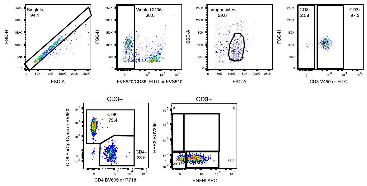
الشكل 1 من البيانات الموسعة | المعلومات التشخيصية المرضية لجميع المرضى.
الشكل البياني الموسع 2 | الكشف داخل الجمجمة عن خلايا CAR T في السائل الدماغي الشوكي. يتكون الكشف عن خلايا CAR T من نوع B7-H3 في السائل الدماغي الشوكي قبل وبعد الحقن خلال فترة DLT من 10 مجموعات مجدولة: Cr1.W1.Pre، Cr1.W1.Post، Cr1.W3.Pre، Cr1.W3.Post، Cr1.W4، Cr2.W1.Pre، Cr2.W1.Post، Cr2.W3.Pre، Cr2.W3.Post، Cr2.W4، والتي تُعرف على أنها (حيث Cr: دورة؛ W: أسبوع؛ Pre: قبل الحقن، وPost: بعد الحقن). -المحور يظهر تسلسل هذه المجموعات. تم استبعاد العينات التي تحتوي على عدد اللمفاويات أقل من حد الكمية القابلة للقياس (LOQ) من التحليل. مظلل
تمثل الدوائر عينات تم الكشف فيها عن خلايا CAR T B7-H3، والتي تم تعريفها بمستويات اللمفاويات/EGFRt+% التي تتجاوز حد الكشف (LOD) وعدد أدنى (>1) من خلايا EGFRt+ المكتشفة. حجم الدوائر يدل على نسبة اللمفاويات EGFRt+ (Lymph/EGFRt+%) المكتشفة في العينة. ومن الجدير بالذكر أن S014 لم يتم جمع أي عينة بسبب وضع التحويلة، وS018 تلقت جرعة واحدة فقط، وS045 فاتتها جميع عمليات الجمع بعد الحقن.
جميع المرضى الوسيط الحد الأقصى الحد الأدنى
Cr1.W1.Pre 0 0 0
كر1.و1.بريد 0.2395 18.908 0
Cr1.W3.Pre 0.517 1.609 0
Cr1.W3.Post 0.598 ٦٤.٤٣٥ 0.008
Cr1.W4 0.39 ٣.٧٨٧ 0
Cr2.W1.Pre 0.273 1.326 0
Cr2.W1.Post 0.337 21.901 0
Cr2.W3.Pre 0.3795 6.045 0
Cr2.W3.Post 1.416 ٣٤.٩٦٧ 0.079
Cr2.W4 0.718 25.401 0
الشكل البياني الممتد 3 | جدول اكتشاف خلايا CAR T في السائل الدماغي الشوكي. مستويات اكتشاف CAR بواسطة EGFRt+ عبر جميع المرضى وجميع أنظمة الجرعات (DR1 و DR2 و DR3 و DR4) خلال فترة DLT. استراتيجية التصفية: حية > مفردات > لمفاويات > CD3+ > EGFRt+.
المحلل متوسط تركيز Cr1 – Cr2 (بيكوجرام/مل) (النطاق) بعد-قبل FC (P.adj) Cr1-Cr2 Cr1.W1 ما بعد-ما قبل FC (P.adj) Cr1-W3 بعد-قبل FC (P.adj) Cr2-W1 بوست-بري FC (P.adj) Cr2-W3 بعد-قبل FC (P.adj)
CRP 1508.04 (0.00 – 962046.22) 1.2 (0.9) 3.6 (8.9e-3) 4.6 (8.0e )
س.ا.ع ١٣٦٣.٠٧ (٠.٠٠ – ٨٤٩٣٢٠.٧١) 2.3 (0.1) 15.1 (2.6e-11)
CXCL10 408.85 (1.09 108926.77) 12.4 (1.7e-42) 8.1 (1.5e-7) 9.6 (5.8e-8)
ICAM-1 ١٤٤٢٫٨١ (٠٫٨٦ – ٨٤٩٧٩٫٦٦) 1.6 (0.02) 0.9 (0.99) 1.4 (0.8) 1.9 (0.3) 3.3 (0.02)
MCP-1 199.63 (54.55 – 1409.79) 0.98 (0.99) 1.0 (0.99) 0.9 (0.99) 1.1 (0.9) 0.9 (0.9)
إنترفيرون غاما 4.55 (0.03 3388.64) 11.3 (5.2e-10) 19.8 (1.0e-14)
تارك 15.51 (0.33 787.98) 2.9 (3.8e-2) 3.2 ( )
GM-CSF 1.38 (0.01 70.64) 4.1 (7.6e ) 2.4 (0.09) 3.3 (0.01) 2.6 (0.07)
IL-16 6.89 (0.01 99.04) 2.0 (0.2) 2.0 (0.2) 2.5 (0.07) 2.6 (0.07)
MIP-1b 21.87 (5.08 412.17) 3.1 (0.01) 2.1 (0.2) 2.8 (0.03) 2.7 (0.07)
TNFa 0.66 (0.07 36.61) 2.4 (0.08) 1.7 (0.4) 2.1 (0.1) ٢.٣ (٠.١)
IL-8 30.89 (0.85 857.75) 2.1 ( ) 1.8 (0.03) 1.3 (0.8) 2.6 (0.05) 3.1 (0.02)
VEGF-C 31.80 (0.00 22287.80) 2.9 (0.02) 1.3 (0.8) 2.4 (0.08) 2.2 (0.2)
IL-12/IL23p40 17.61 (1.79 301.97) 2.7 (0.03) 2.2 (0.1) 2.2 (0.1) 1.8 (0.04)
الشكل البياني الممتد 4 | بيانات السيتوكينات في السائل الدماغي الشوكي. التركيزات المطلقة والتغيرات قبل الحقن وبعد الحقن للسيتوكينات التي تظهر تغييرًا لا يقل عن الضعف وقيمة p المعدلة بمعدل الاكتشاف الكاذب (FDR) في نموذج مختلط خطي. جمع النموذج البيانات من Cr1.W1( ), Cr1.W3 ( ), Cr2.W1 ( )، و Cr2.W3 ( )، مما يؤدي إلى التحضير المسبق قياسات ما بعد الحقن لكل سيتوكين استوفت معايير الجودة.
تم تضمين الموضوعات كمعترضات عشوائية، وتم تضمين حالة ما قبل/ما بعد الحقن كأثر ثابت. تم قياس المحللات بشكل مزدوج؛ تلك التي كانت لديها معاملات تباين (CV) أكبر من تم استبعادها. كانت التركيزات -تم تحويلها للتحليل، وتُعرض التغيرات النسبية على مقياس خطي. تُظهر الأعمدة اللاحقة التغيرات النسبية والقيم p المعدلة لنقاط زمن التسريب المنفصلة والمجمعة.
الشكل البياني الممتد 5 | استراتيجية تصنيف التدفق التمثيلية لاكتشاف خلايا CART. استراتيجية التصنيف المستخدمة لاكتشاف CAR القائم على التدفق في السائل الدماغي الشوكي. تم اختيار مجموعة اللمفاويات الفردية القابلة للحياة [CD36-] قبل تصنيف خلايا T وفحص خلايا EGFRt+CART وتعبير CD4/CD8.

محفظة الطبيعة

المؤلف(المؤلفون) المراسلون:
نيكولاس فيتانزا
آخر تحديث بواسطة المؤلفين: 12/32024

ملخص التقرير

تسعى Nature Portfolio إلى تحسين إمكانية تكرار العمل الذي ننشره. يوفر هذا النموذج هيكلًا للاتساق والشفافية في التقرير. لمزيد من المعلومات حول سياسات Nature Portfolio، يرجى الاطلاع على سياسات التحرير وقائمة مراجعة سياسة التحرير.

الإحصائيات

لجميع التحليلات الإحصائية، تأكد من أن العناصر التالية موجودة في أسطورة الشكل، أسطورة الجدول، النص الرئيسي، أو قسم الطرق.
غير متوفر
□ X

بيان حول ما إذا كانت القياسات قد أُخذت من عينات متميزة أو ما إذا كانت نفس العينة قد تم قياسها عدة مرات

اختبار(ات) الإحصاء المستخدمة وما إذا كانت أحادية الجانب أو ثنائية الجانب
يجب أن تُوصف الاختبارات الشائعة فقط بالاسم؛ واصفًا التقنيات الأكثر تعقيدًا في قسم الطرق.

وصف لجميع المتغيرات المشتركة التي تم اختبارها

وصف لأي افتراضات أو تصحيحات، مثل اختبارات الطبيعية والتعديل للمقارنات المتعددة

وصف كامل للمعلمات الإحصائية بما في ذلك الاتجاه المركزي (مثل المتوسطات) أو تقديرات أساسية أخرى (مثل معامل الانحدار) و التباين (مثل الانحراف المعياري) أو تقديرات مرتبطة بعدم اليقين (مثل فترات الثقة)

لاختبار الفرضية الصفرية، إحصائية الاختبار (على سبيل المثال، ) مع فترات الثقة، أحجام التأثير، درجات الحرية و قيمة ملحوظة أعطِ القيم كقيم دقيقة كلما كان ذلك مناسبًا.

□ لتحليل بايزي، معلومات حول اختيار القيم الأولية وإعدادات سلسلة ماركوف مونت كارلو

□ لتصميمات هرمية ومعقدة، تحديد المستوى المناسب للاختبارات والتقارير الكاملة عن النتائج

□ تقديرات أحجام التأثير (مثل حجم تأثير كوهين) بيرسون )، مما يشير إلى كيفية حسابها
تحتوي مجموعتنا على الويب حول الإحصائيات لعلماء الأحياء على مقالات تتناول العديد من النقاط المذكورة أعلاه.
تم التأكيد
حجم العينة بالضبط لكل مجموعة/شرط تجريبي، معطاة كرقم منفصل ووحدة قياس
بيان حول ما إذا كانت القياسات قد أُخذت من عينات متميزة أو ما إذا كانت نفس العينة قد تم قياسها عدة مرات
اختبار(ات) الإحصاء المستخدمة وما إذا كانت أحادية الجانب أو ثنائية الجانب
وصف جميع المتغيرات المرافقة التي تم اختبارها

ن

وصف لأي افتراضات أو تصحيحات، مثل اختبارات الطبيعية والتعديل للمقارنات المتعددة
وصف كامل للمعلمات الإحصائية بما في ذلك الاتجاه المركزي (مثل المتوسطات) أو تقديرات أساسية أخرى (مثل معامل الانحدار) والتباين (مثل الانحراف المعياري) أو التقديرات المرتبطة بعدم اليقين (مثل فترات الثقة)
في اختبار فرضية العدم، إحصائية الاختبار (على سبيل المثال، ) مع فترات الثقة، أحجام التأثير، درجات الحرية و قيمة ملحوظة القيم كقيم دقيقة كلما كان ذلك مناسبًا.

تحليل ساني، إعلام
أمة
اختيار القيم الأولية وإعدادات سلسلة ماركوف مونت كارلو
علماء الأحياء
” “” “” “

البرمجيات والشيفرة

معلومات السياسة حول توفر كود الكمبيوتر

جمع البيانات □ BD LSRFortessa، MSD MESO QuickPlex SQ 120MM، Li-COR Odyssey Clx، عداد الوميض TopCount
تحليل البيانات □ FlowJo v.10، R v4.4، emmeans (1.10.2)، ggplot2 (3.5.1)، و EnhancedVolcano (1.22.0).
بالنسبة للمخطوطات التي تستخدم خوارزميات أو برامج مخصصة تكون مركزية في البحث ولكن لم يتم وصفها بعد في الأدبيات المنشورة، يجب أن تكون البرمجيات متاحة للمحررين والمراجعين. نحن نشجع بشدة على إيداع الشيفرة في مستودع مجتمعي (مثل GitHub). راجع إرشادات مجموعة Nature لتقديم الشيفرة والبرمجيات لمزيد من المعلومات.

بيانات

معلومات السياسة حول توفر البيانات

يجب أن تتضمن جميع المخطوطات بيانًا حول توفر البيانات. يجب أن يوفر هذا البيان المعلومات التالية، حيثما ينطبق:
  • رموز الانضمام، معرفات فريدة، أو روابط ويب لمجموعات البيانات المتاحة للجمهور
  • وصف لأي قيود على توفر البيانات
  • بالنسبة لمجموعات البيانات السريرية أو بيانات الطرف الثالث، يرجى التأكد من أن البيان يتماشى مع سياستنا
يجب تقديم جميع الطلبات للحصول على البيانات والمواد الخام والمحللة إلى الدكتور جيسون ويندلر. ستتم مراجعة الطلبات بسرعة من قبل مكتب الملكية الفكرية في معهد أبحاث الأطفال في سياتل للتحقق مما إذا كان الطلب خاضعًا لأي التزامات تتعلق بالملكية الفكرية أو السرية. يتم تخزين البيانات الأولية السريرية والبيانات السريرية في معهد أبحاث الأطفال في سياتل مع نسخ احتياطية مناسبة غير محددة. البيانات المتعلقة بالمرضى التي لم يتم تضمينها في الورقة تم توليدها كجزء من التجارب السريرية وقد تكون خاضعة لسرية بين المرضى. سيتم إصدار أي بيانات ومواد يمكن مشاركتها عبر اتفاقية نقل المواد.

البحث الذي يتضمن مشاركين بشريين، بياناتهم، أو مواد بيولوجية

معلومات السياسة حول الدراسات التي تشمل مشاركين بشريين أو بيانات بشرية. انظر أيضًا معلومات السياسة حول الجنس، الهوية/التقديم الجنسي، والتوجه الجنسي والعرق، والعرقية والعنصرية.
التقارير عن الجنس والنوع الاجتماعي كان المرضى مؤهلين بغض النظر عن الجنس والنوع.
التقارير عن العرق أو الإثنية أو غيرها من المجموعات الاجتماعية ذات الصلة كان المرضى مؤهلين بغض النظر عن العرق والإثنية.
خصائص السكان الخصائص مدرجة في الجدول 1
التوظيف تم تجنيد المرضى من خلال إحالة الأطباء والإحالة الذاتية. تم تجنيد المرضى من كل من المراكز الصحية الأكاديمية ومراكز الصحة المجتمعية. لم يتلق المرضى أي تعويض مالي للتسجيل، على الرغم من أنه قد يوجد تحيز في حقيقة أن العائلات كانت بحاجة إلى وسائل للسفر إلى سياتل والإقامة محليًا خلال جزء من العلاج.
رقابة الأخلاقيات تم إجراء هذه الدراسة وفقًا لإرشادات إدارة الغذاء والدواء الأمريكية ومؤتمر التوافق الدولي لممارسات البحث السريري الجيدة، وإعلان هلسنكي، ومتطلبات مجلس المراجعة المؤسسية المعمول بها، بما في ذلك الموافقة على بروتوكول الدراسة من قبل مجلس المراجعة المؤسسية لمستشفى الأطفال في سياتل.
يرجى ملاحظة أنه يجب أيضًا تقديم معلومات كاملة حول الموافقة على بروتوكول الدراسة في المخطوطة.

التقارير الخاصة بالمجال

يرجى اختيار الخيار أدناه الذي يناسب بحثك بشكل أفضل. إذا لم تكن متأكدًا، اقرأ الأقسام المناسبة قبل اتخاذ قرارك.
علوم الحياة
العلوم السلوكية والاجتماعية □ العلوم البيئية والتطورية والبيئية
لنسخة مرجعية من الوثيقة مع جميع الأقسام، انظرnature.com/documents/nr-reporting-summary-flat.pdf

تصميم دراسة العلوم الحياتية

يجب على جميع الدراسات الإفصاح عن هذه النقاط حتى عندما يكون الإفصاح سلبياً.
حجم العينة تم تحديد حجم العينة بناءً على المرحلة التصميم. تم تعريف MTDR على أنه أعلى معدل للسمية مع وجود ما لا يقل عن ستة مواضيع قابلة للتقييم من حيث السمية ومعدل السمية التراكمي خلال الدورات 1 و 2 أقل من .
استثناءات البيانات لم يكن هناك بيانات مستبعدة.
التكرار تم إجراء اختبارات السيتوكين بشكل مزدوج
التوزيع العشوائي لم يتم إجراء أي عشوائية حيث كانت هذه تجربة سريرية من المرحلة الأولى غير عشوائية.
عمى لم يتم إجراء أي تعمية حيث كانت هذه تجربة سريرية من المرحلة الأولى غير معمية.

التقارير عن مواد وأنظمة وطرق محددة

نحتاج إلى معلومات من المؤلفين حول بعض أنواع المواد والأنظمة التجريبية والأساليب المستخدمة في العديد من الدراسات. هنا، يرجى الإشارة إلى ما إذا كانت كل مادة أو نظام أو طريقة مدرجة ذات صلة بدراستك. إذا لم تكن متأكدًا مما إذا كان عنصر القائمة ينطبق على بحثك، يرجى قراءة القسم المناسب قبل اختيار رد.
المواد والأنظمة التجريبية
مشارك في الدراسة
الأجسام المضادة
خطوط خلايا حقيقية النواة
علم الحفريات وعلم الآثار
الحيوانات وغيرها من الكائنات الحية
البيانات السريرية
استخدام بول للأبحاث المثيرة للقلق
طرق
محفظة الطبيعة | ملخص التقرير ~ أبريل
محفظة الطبيعة | ملخص التقرير أبريل 2023

الأجسام المضادة

الأجسام المضادة المستخدمة مادة حجب FcR البشرية (Miltenyi، رقم الكات#130-059-901، RRID:AB_2892112)؛ محلول صبغة برايلانت (BD، رقم الكات#563794، RRID:AB_2869750)؛ FVS520 (FITC، BD، رقم الكات#564407، RRID:AB_2869573)؛ FVS510 (FVS510، BD، رقم الكات#564406، RRID:AB_2869572)؛ CD36 (FITC، BD، رقم الكات#555454، RRID:AB_2291112)؛ CD3 (V450، BD، رقم الكات#652356، RRID:AB_2868395)؛ CD3 (FITC، BD، رقم الكات#555332، RRID:AB_395739)؛ CD4 (BV605، BD، رقم الكات#562658، RRID:AB_2744420)؛ CD4 (R718، BD، رقم الكات#567092، RRID:AB_2916435)؛ CD8 (PerCPCy5.5، BD، رقم الكات#560662، RRID:AB_1727513)؛ CD8 (BV605، BD، رقم الكات#564116، RRID:AB_2869551)؛ سيتوكسيماب (APC، BD، مخصص، RRID:AB_2459632).

التحقق

تم ربط الأجسام المضادة Cetuximab-APC بشكل مخصص بواسطة BD وتم التحقق منها داخليًا بواسطة قياس التدفق الخلوي. تم إجراء تجارب التخفيف باستخدام خطوط خلايا H9 التي تعبر عن EGFRt أو خلايا T التي تم نقلها بواسطة ناقلات من الدرجة البحثية تمثل تجارب سريرية أخرى تديرها مستشفى سياتل للأطفال. جميع الأجسام المضادة الأخرى تم التحقق منها بطريقة مماثلة من خلال التلوين الإيجابي على خطوط الخلايا بواسطة قياس التدفق الخلوي الذي يعكس تحقق الشركة المصنعة.

البيانات السريرية

معلومات السياسة حول الدراسات السريرية

يجب أن تتوافق جميع المخطوطات مع إرشادات ICMJE لنشر الأبحاث السريرية ويجب أن تتضمن جميع التقديمات قائمة مراجعة CONSORT مكتملة.
تسجيل التجارب السريرية
بروتوكول الدراسة
جمع البيانات
NCT04185038
تم توفير بروتوكول التجربة السريرية.
بدأت دراسة BrainChild-03 في 22 نوفمبر 2019. تشمل البيانات السريرية حتى 13 نوفمبر 2024. تم تسجيل أول مريض في أغسطس 2020 وآخر مريض تم تسجيله في أبريل 2023. تم إجراء أنشطة الدراسة في مستشفى الأطفال في سياتل.
كانت الأهداف الرئيسية هي: تقييم جدوى العلاج التبني الموضعي للجهاز العصبي المركزي باستخدام خلايا CD4+ و CD8+ T الذاتية المعدلة بواسطة الفيروسات المعوية للتعبير عن CAR محدد لـ B7-H3 و EGFRt، والتي يتم توصيلها بواسطة قسطرة دائمة إلى تجويف الورم أو النظام البطيني لدى الأطفال والشباب الذين يعانون من DIPG و DMG أو أورام الجهاز العصبي المركزي المتكررة/المقاومة؛ تقييم سلامة العلاج التبني الموضعي للجهاز العصبي المركزي باستخدام خلايا CD4+ و CD8+ T الذاتية المعدلة بواسطة الفيروسات المعوية للتعبير عن CAR محدد لـ B7-H3 و EGFRt، والتي يتم توصيلها بواسطة قسطرة دائمة إلى تجويف الورم أو النظام البطيني لدى الأطفال والشباب الذين يعانون من DIPG و DMG أو أورام الجهاز العصبي المركزي المتكررة/المقاومة؛ Establish tolerability of a fractionated CNS-delivered B7-H3 CAR T cell infusion schedule employing intra-subject dose escalation in children and young adults with DIPG, DMG, or recurrent/refractory CNS tumors; تحديد الجرعة القصوى المتسامحة (MTD) ونظام الجرعة الموصى به للمرحلة الثانية (RP2DR) من تسريب خلايا CAR T من نوع B7-H3 المجزأة الموصلة للجهاز العصبي المركزي. كانت الأهداف الثانوية هي: الأهداف الثانوية هي: تقييم توزيع خلايا CAR T من نوع B7-H3 داخل السائل الدماغي الشوكي (CSF) ومدى خروج خلايا CAR T من نوع B7-H3 إلى الدورة الدموية المحيطية؛ وتقييم استجابة المرض للعلاج الموضعي بخلايا CAR T من نوع B7-H3 لدى الأطفال والشباب الذين يعانون من DIPG و DMG أو أورام الجهاز العصبي المركزي المتكررة/المقاومة.

نباتات

مخزونات البذور
غير متوفر
أنماط جينية نباتية جديدة □
غير متوفر
المصادقة
غير متوفر

تدفق الخلايا

المؤامرات

أكد أن:

توضح تسميات المحاور العلامة والفلوركروم المستخدم (مثل CD4-FITC).
المقاييس على المحاور واضحة تمامًا. قم بتضمين الأرقام على المحاور فقط للرسم البياني في أسفل اليسار من المجموعة (المجموعة هي تحليل للعلامات المتطابقة).
جميع الرسوم البيانية هي رسوم بيانية متساوية الارتفاع مع نقاط شاذة أو رسوم بيانية بالألوان الزائفة.
تم توفير قيمة عددية لعدد الخلايا أو النسبة المئوية (مع الإحصائيات).

المنهجية

تحضير العينة تم جمع عينات السائل الدماغي الشوكي (CSF) من المرضى عن طريق البزل القطني أو القسطرة البطينية وتم الاحتفاظ بها في حتى المعالجة. خضعت العينات للطرد المركزي المتسلسل: أولاً عند لمدة 10 دقائق لإزالة الخلايا، تليها عملية الطرد المركزي النهائية عند لمدة 10 دقائق لإزالة أي بقايا متبقية. ثم تم تقسيم السائل الخالي من الخلايا وتجميده. تحديد النمط المناعي للسائل الدماغي الشوكي: تم إجراء تحديد النمط المناعي للعلامات السطحية على الخلايا المعزولة من عينات السائل الدماغي الشوكي باستخدام بروتوكولات صبغ قياسية تلتها تحليل بواسطة قياس التدفق الخلوي.
آلة حدد الأداة المستخدمة لجمع البيانات، مع تحديد العلامة التجارية ورقم الطراز.
برمجيات فلو جو
وفرة تجمع الخلايا تم استبعاد العينات التي تحتوي على عدد اللمفاويات أقل من حد الكمية المطلوب (LOQ) للاختبار من التقرير. يتم تحديد حالة اكتشاف خلايا CART من خلال مجموعة من عدد خلايا EGFRt+ القابلة للاكتشاف في العينة، بالإضافة إلى مستوى خلايا اللمفاويات/نسبة EGFRt+ في العينة ليكون فوق الحد المحدد مسبقًا للكشف (LOD) للاختبار.
استراتيجية البوابة استراتيجية التصفية المستخدمة للكشف عن CAR المعتمد على التدفق في السائل الدماغي الشوكي. تم اختيار مجموعة اللمفاويات الفردية القابلة للحياة [CD36-] قبل تصفية خلايا T وفحص خلايا CART الإيجابية لـ EGFRt وتعبير CD4/CD8.
قم بتحديد هذا المربع لتأكيد أنه تم تقديم رقم يوضح استراتيجية البوابة في المعلومات التكميلية.

التصوير بالرنين المغناطيسي

تصميم تجريبي

نوع التصميم الدراسات السريرية
مواصفات التصميم الدراسات السريرية
مقاييس الأداء السلوكي غير متوفر
نوع (أنواع) تصوير الاستحواذ الدراسات السريرية
شدة المجال 1.5T و 3T
معلمات التسلسل والتصوير تصوير بالرنين المغناطيسي الروتيني يتكون من الدماغ = تصوير T1 MPRAGE في الوضع السهمي بدقة 1 مم مع إعادة تشكيل متعددة المستويات بدون وبتباين؛ تصوير T2 في الوضع المحوري والجبهي؛ تصوير FLAIR في الوضع المحوري؛ تصوير DWI في الوضع المحوري مع خرائط ADC؛ تصوير SWI في الوضع المحوري. العمود الفقري = تصوير T1 في الوضع المحوري والسهمي بعد التباين.
مجال الاستحواذ الدماغ والحبل الشوكي
الرنين المغناطيسي الانتشاري غير مستخدم
معايير التصوير بالرنين المغناطيسي الانتشاري السريري الروتيني

التحضير المسبق

برمجيات المعالجة المسبقة غير متوفر
التطبيع غير متوفر
قالب التطبيع غير متوفر
إزالة الضوضاء والعيوب غير متوفر
تصفية الحجم غير متوفر

النمذجة الإحصائية والاستدلال

نوع النموذج والإعدادات غير متوفر
التأثيرات المختبرة
غير متوفر
حدد نوع التحليل: □ قائم على العائد على الاستثمار □ كلاهما
نوع الإحصاء للاستدلال
(انظر إكلوند وآخرون 2016)
تصحيح
النماذج والتحليل
مشارك في الدراسة
  • □ الاتصال الوظيفي و/أو الفعال
تحليل الرسم البياني X □
نموذج متعدد المتغيرات أو التحليل التنبؤي
محفظة الطبيعة | ملخص التقرير

  1. ®البريد الإلكتروني: nicholas.vitanza@seattlechildrens.org

Journal: Nature Medicine, Volume: 31, Issue: 3
DOI: https://doi.org/10.1038/s41591-024-03451-3
PMID: https://pubmed.ncbi.nlm.nih.gov/39775044
Publication Date: 2025-01-07

Intracerebroventricular B7-H3-targeting CART cells for diffuse intrinsic pontine glioma: a phase 1 trial

Received: 11 October 2024
Accepted: 6 December 2024
Published online: 7 January 2025
(A) Check for updates

A list of authors and their affiliations appears at the end of the paper

Diffuse intrinsic pontine glioma (DIPG) is a fatal central nervous system (CNS) tumor that confers a median survival of 11 months. As B7-H3 is expressed on pediatric CNS tumors, we conducted BrainChild-03, a single-center, dose-escalation phase 1 clinical trial of repetitive intracerebroventricular (ICV) dosing of B7-H3-targeting chimeric antigen receptor T cells (B7-H3 CAR T cells) for children with recurrent or refractory CNS tumors and DIPG. Here we report results from Arm C, restricted to patients with DIPG. The primary objectives were to assess feasibility and tolerability, which were both met. Secondary objectives included assessments of CAR T cell distribution and survival. A total of 23 patients with DIPG enrolled, and 21 were treated with repeated doses of ICV B7-H3 CAR T cells using intra-patient dose-escalation regimens without previous lymphodepletion. Concurrent tumor-directed therapy, including re-irradiation, was not allowed while on protocol therapy. We delivered a total of 253 ICV doses and established the highest planned dose regimen, DR4, which escalated up to cells per dose, as the maximally tolerated dose regimen. Common adverse events included headache, fatigue and fever. There was one dose-limiting toxicity (intratumoral hemorrhage) during DR2. For all treated patients ( ), the median survival from their initial CAR T cell infusion was 10.7 months and the median survival from diagnosis was 19.8 months with 3 patients still alive at 44,45 and 52 months from diagnosis. Ultimately, this completed first-in-human trial shows that repetitive ICV dosing of B7-H3 CAR T cells in pediatric and young adult patients with DIPG is tolerable, including multiyear repeated dosing, and may have clinical efficacy that warrants further investigation on a multisite phase 2 trial. ClinicalTrials.gov registration: NCT04185038.
Diffuse intrinsic pontine glioma (DIPG) is a fatal brainstem tumor responsible for years of life lost each year in the United States . Chimeric antigen receptor (CAR) T cells have had substantial efficacy against pediatric leukemia , yet the clinical application of intracranial CAR T cells for children with DIPG is a nascent area of therapeutic development .
Our initial phase 1 clinical trials delivering intracranial CAR T cells (BrainChild-01 delivering human epidermal growth factor receptor 2
(HER2)-specific CAR T cells (NCT03500991) ; BrainChild-02 delivering EGFR806-specific CAR T cells (NCT03638167)) excluded patients with DIPG. As B7-H3 (CD276) is expressed on DIPG , we designed B7-H3-targeting CAR T cells (B7-H3 CAR T cells) and described their preclinical efficacy . Next, we opened BrainChild-03 (NCT04185038), delivering B7-H3 CAR T cells to children with recurrent or refractory central nervous system (CNS) tumors and DIPG, and published on the preliminary tolerability .
Here we report on the completed Arm C of the first-in-human phase 1 clinical trial BrainChild-03 dedicated to children with DIPG. Patients were enrolled at any time following standard radiotherapy (including patients with disease progression and/or metastases). We show the safety of repetitive intracerebroventricularly (ICV) dosed B7-H3 CAR T cells up to cells per dose to children with DIPG, evidence of immune activation in the CNS and the potential clinical benefit.

Results

Study design and patient characteristics

This was a phase 1 study of repeatedly dosed ICV adoptive cell therapy with autologous and cells lentivirally transduced to express a B7-H3-specific CAR to children and young adults with DIPG. The primary objectives were to assess the feasibility, safety and tolerability of ICV B7-H3 CAR T cell therapy, while the secondary objectives included assessments of CAR T cell distribution and survival. Eligibility criteria included the following: age and years, a diagnosis of DIPG (diagnosed radiographically or via histopathologic confirmation of high-grade glioma or diffuse midline glioma H3K27M-altered (DMG)) at any timepoint following completion of standard radiation, presence of a CNS catheter, Lansky or Karnofsky performance of , dexamethasone dose , protocol-defined washout from previous therapies and protocol-defined laboratory evidence of adequate organ function (including an absolute lymphocyte count of cells ).
The median age at enrollment of 6 years (range: 2-22 years) is consistent with the historical median age for this disease . Patients had Lansky or Karnofsky performance scores of , and . All patients received standard radiation therapy at diagnosis. The two enrolled patients who did not receive CAR T cells each experienced clinical progression during CAR T cell manufacturing and never met eligibility for CAR T cell infusion. Of the 21 treated patients, received treatment after at least one tumor progression, while 9 patients were treated before any tumor progression. The primary objective of the feasibility to manufacture CAR T cells from a single apheresis was met for all enrollees. The baseline characteristics of the patients are provided in Table 1. Of the 21 treated patients, 17 met the molecular diagnosis of DMG either by tumor tissue or by CSF or plasma testing (Extended Data Fig.1). One patient(S008) had a pontine anaplastic astrocytoma harboring an IDH1 mutation.

Treatment

BrainChild-03 Arm C delivered ICV B7-H3 CAR T cells without previous lymphodepletion every 14 days over 8 weeks during the dose-limiting toxicity (DLT) observation period (courses 1 and 2). Concurrent tumor-directed therapy, including re-irradiation, was not allowed while on protocol therapy. As the optimal number of doses is unknown, patients-who met criteria for subsequent infusions beyond course 2 (and for whom additional cryopreserved CAR T cell doses were avail-able)-were eligible to continue receiving CAR T cell therapy beyond the DLT period with doses every 2-4 weeks. Patients were treated on four escalating dose regimens (DRs; Fig. 1). In DR1, three evaluable patients received CAR T cells per dose. DR2, DR3 and DR4 used intra-patient dose escalation. In DR2, 6 evaluable patients received up to CAR T cells per dose; in DR3,3 patients received up to CAR T cells per dose and, in DR4, 6 evaluable patients received up to CAR T cells per dose. DR2 expanded to six patients owing to a DLT (intratumoral hemorrhage), and DR4 expanded to six patients for a planned statistical confirmation of the maximally tolerated dose regimen (MTDR). Three patients were not DLT evaluable (as they elected to pursue other tumor-directed therapy out of concern for progressive disease (PD) without having experienced a DLT) and were replaced as per the protocol.
Table 1 | Demographics and clinical characteristics of study participants enrolled in BrainChild-03 Arm C ( )
Characteristics Value
Median age (years (range)) 6 (2-22)
Sex ( (%))
Male 9 (39)
Female 14 (61)
Lansky or Karnofsky performance status score ( (%))
90 12 (52)
80 7 (30)
70 2 (9)
60 2 (9)
Median months from diagnosis to enrollment (range) 6 (3-22)
Histopathologic or molecular confirmation of DMG ( (%)) 18 (78)
Disease history at enrollment ( (%))
Previous progression 12 (52)
No previous progression 11 (48)
Disease history at first dose of B7-H3 CAR ( (%); total number treated: 21)
Previous progression 12 (57)
No previous progression 9 (43)
Previous radiation therapy ( (%)) 23 (100)
For patients who began treatment before any progression( ), the median time from diagnosis to initial CAR T cell infusion was 6.6 months (range: 4.8-14.1; Fig. 2a). For patients who began treatment after progression ( ), the median time from initial diagnosis to initial CAR T cell infusion was 10.3 months (range: months). For all patients receiving at least one CAR T cell infusion ( ), the median interval between enrollment and initial CAR T cell dose was 1.4 months (range: 1.0-4.4 months). The 18 patients evaluable for DLT received 253 total doses (median: 9 per patient; range: 1-81) and remained on protocol therapy for a median of 5 months (range: months).

Safety

All patients who received at least one CAR T infusion are included in the adverse event (AE) summary ( ; Table 2). The most frequent AEs that were possibly, probably or definitely attributable to CAR T therapy were headache (17 patients, ), nause or vomiting (17 patients, ), fatigue ( 13 patients, ) and fever ( 12 patients, ). Most events were grade 1 or 2 with few patients experiencing grade 3 toxicities. Only one patient developed hydrocephalus (grade3). Fever was considered evidence of local immune activation rather than conventional systemic cytokine release syndrome (CRS). Immune-effector-cell-associated neurotoxicity syndrome was not observed. The sole DLT was an intratumoral hemorrhage (grade 4 ) occurring in a 3 year old with PD between enrollment and initial infusion. They received their first CAR T cell dose (dose level (DL)1 of DR2), then had a stable neurologic exam and unchanged performance score without evidence of systemic inflammation (including normal serum C-reactive protein (CRP), ferritin, D-dimer, interleukin (IL)-2, IL-6 and interferon- (IFN )) for 1 week before the acute event. Following the hemorrhage, they required admission to the pediatric intensive care unit followed by slow, limited neurologic improvement, but ultimately had fatal progression of their tumor.

Clinical outcomes

Some clinical trials for DIPG mandate enrollment at diagnosis, while others allow patients to enroll anytime following standard radiation. The latter risks an immortalization bias in which some patients have a prolonged period before enrollment that constitutes a substantial
Fig. 1 | BrainChild-03 Arm C trial design. CONSORT diagram of BrainChild-03 Arm C.
Fig. 2 | Survival following intracranial B7-H3 CAR T cells. a, Swimmer plot describing patient history from time of diagnosis through death or most recent follow-up. For patients with multiple progressions or long-standing ongoing progression, only initial progression may be noted.b, Kaplan-Meier survival
after the initial B7-H3 CAR T cell infusion for all treated patients. c, Kaplan-Meier survival stratified by previous progression status at the time of initial CAR T infusion. Shaded areas in and denote confidence limits for the Kaplan-Meier estimates.
Table 2 | AEs recorded during the DLT observation period
Event Grade 1-2, (%) Grade 3, (%) Grade 4, (%)
Neurologic
Headache 15 (71) 2 (10) 0
Ataxia 3 (14) 1 (5) 0
Dizziness 2 (10) 0 0
Dysarthria 1 (5) 0 0
Dysphagia 1 (5) 0 0
Facial nerve disorder 2 (10) 0 0
Hydrocephalus 0 1 (5) 0
Unilateral muscle weakness 2 (10) 2 (10) 0
Paresthesia 4 (19) 0 0
Peripheral sensory neuropathy 1 (5) 0 0
Tremor 1 (5) 0 0
Trochlear nerve disorder 1 (5) 0 0
Ophthalmic
Diplopia 1 (5) 0 0
Photophobia 2 (10) 0 0
Gastrointestinal
Nausea 12 (57) 1 (5) 0
Vomiting 15 (71) 1 (5) 0
Anorexia 3 (14) 0 0
Abdominal pain 1 (5) 0 0
Dysphagia 1 (5) 0 0
Tumor related
Tumor hemorrhage 0 0 1 (5)
Constitutional
Fever 11 (52) 1 (5) 0
Fatigue 13 (62) 0 0
Malaise 1 (5) 0 0
Pain 2 (10) 0 0
Agitation 1 (5) 0 0
Respiratory
Dyspnea 1 (5) 0 0
Hiccups 6 (29) 0 0
Cardiovascular
Hypertension 1 (5) 0 0
Hypotension 1 (5) 0 0
Musculoskeletal
Back pain 1 (5) 0 0
Neck pain 3 (14) 1 (5) 0
Urinary
Incontinence 1 (5) 0 0
Dermatologic
Pruritus 1 (5) 0 0
Hematologic
Lymphocytopenia 1 (5) 0 0
CTCAE grading of all observed toxicities during the DLT observation.
portion of their overall survival. Therefore, while we calculated the median survival from initial diagnosis, we also evaluated the survival from study enrollment (for all enrolled patients) and from initial CAR T cell infusion (for all treated patients). Patients were followed from the time of enrollment, which occurred between August 2020 and April 2023, through treatment until death or censoring at their last follow-up visit before 13 November 2024.
For all enrolled patients ( ), the median survival from the time of study enrollment was 11.4 months (range: months) and the median survival from diagnosis to death (or last contact for survivors) was 19.5 months (range: 6.5-52.5 months; Fig. 2a). For all treated patients ( ), the median survival from their initial CAR T cell infusion was 10.7 months (range: months; Fig. 2b) and the median time from diagnosis to death (or last contact for survivors) was 19.8 months with 3 patients still alive 44.6, 45.6 and 52.5 months from diagnosis (range: 6.5-52.5 months;Fig. 2b).
As it is unknown whether intracranial cellular therapy delivered temporally near initial radiation may cause added toxicity or, conversely, be more efficacious when disease burden may be lowest, we performed a post hoc analysis comparing patients who enrolled before progression ( ) with those who enrolled after progression ( ). For patients who began CAR T treatment after progression ( ), the median survival following their initial CAR T cell infusion was 9.4 months (range: months; Fig. 2c) and the median survival from diagnosis was 20.1 months (range: months). For patients who began treatment before progression ( ), the median survival following their initial CAR T cell infusion was 13.6 months (range: 2.8-45.8 months; Fig. 2c) and the median time from diagnosis to death (or last contact for survivors) was 19.5 months (range: months). All three surviving patients were treated before progression. One surviving patient is in long-term follow-up: S008 (pontine high-grade glioma with mutations in TP53 and IDH1 diagnosed at 22 years of age) left protocol therapy owing to patient preference after 1 year of treatment. Two surviving patients remain on protocol therapy: S021 (pontine DMG with a H3F3A K27M mutation diagnosed at 2 years of age) has received 81 doses ( total CAR T cells) over 37.5 months, and S027 (non-diagnostic biopsy at 5 years of age) has received 39 doses ( total CAR T cells) over 31.5 months.
A total of 18 patients received sufficient CAR T cell infusions to be evaluable for neuroimaging assessment. The best individual neuroimaging responses on MRI were 1 partial response (PR; 6%), 15 stable disease (SD; 83%) and 2 PD ( ). Of patients evaluable for disease response who had progressed prior to treatment, 88.9% recorded a best neuroimaging response of SD or PR after initiating protocol therapy. While S021 is not classified as an objective radiographic response, MRI reveals a sustained and ongoing decrease in pontine T2 hyperintensity (Fig. 3a). While SO53 experienced a PR correlating with improvement in clinical symptoms, this patient ultimately had tumor progression at metastatic sites of disease 60 days later (Fig. 3b).

CAR T cell detection and cytokine analysis

A secondary objective was to assess B7-H3 CAR T cell distribution in the CSF. To complete this, we collected serial CSF samples from the CNS catheter before and after CAR T cell infusions, then performed flow cytometry to detect the truncated epidermal growth factor receptor (EGFRt) transduction tag. B7-H3 CAR T cells were detected via their EGFRt transduction tag in 38.1% (40 of 105) of CSF biospecimens in courses 1 and 2 (Extended Data Figs. 2 and 3 and Supplementary Table) with 13 of 18 ( ) evaluable patients having detectable CAR T cells in at least one timepoint. The median peak CAR T cell detection was at the course (Cr) 2 week (W) 3 post-infusion timepoint. Overall, Cr2 timepoints showed higher median detection than matched Cr1 timepoints, except Cr2.W3.Pre. Of the 96 peripheral blood samples collected, only 2 had detectable vector (S021: Cr2.W4: 264.4 copies by qPCR; S039: Cr2.W1.Post 205.5 copies by qPCR). Overall, this
Fig. 3 | Neuroimaging after locoregional B7-H3 CAR T cell infusion.
a, Longitudinal MRIs of SO21. T2-weighted (coronal top row, axial middle row) and axial post-contrast T1-weighted (bottom row) MRI images focused on the pontine lesion at various timepoints (see column labels). At diagnosis (DX), the pons is expanded with non-enhancing T2 hyperintensity, prepontine cistern effacement (dashed arrows) and partial effacement of the fourth ventricle (asterisks). Following radiation (Post RT, 4 months before immunotherapy (IT)), the lesion is smaller, with reduced prepontine cistern effacement (dashed arrows), persistent nodular T2 hyperintensity in the left dorsal pons (solid arrows) and biopsy- and therapy-related changes in the central pons (arrowheads). From the initiation of IT (Pre IT) to 30 months Post IT, the overall pons size has remained stable while
hyperintensity has decreased. b, Longitudinal MRI of the pontine lesion of SO53. Axial T2-weighted (top row) and axial post-contrast T1-weighted (bottom row) MRI images, focused on the pontine lesion at various timepoints (see column labels). At DX, the pons is enlarged (arrows) with diffuse T2 hyperintensity, heterogeneous enhancement and partial effacement of the fourth ventricle (asterisks). Following radiation (Post RT, 2 months before IT), there is further expansion of the pons and increased T2 signal abnormality (arrows), with persistent enhancement. From Pre IT to 2 months Post IT, the pontine lesion is smaller (arrows). However, by 4 months Post IT, the lesion is larger, with a new region of enhancement (arrowheads). At 6 and 8 months Post IT, the lesion size, T2 signal abnormality and enhancement are reduced.
supports the finding that while ICV delivered B7-H3 CAR T cells are consistently detected in CSF after infusion, systemic circulation was rare, transient and at low levels.
An exploratory objective of this study was to assess biomarkers indicative of CAR T cell activity. To achieve this, 53 cytokines associated with T cell function and immune microenvironment interactions were measured using a Meso Scale Discovery (MSD) assay. A total of 105 CSF samples during courses 1 and 2 from 18 evaluable patients were analyzed using matched pre- and post-infusion biospecimens from the following timepoints: Cr1.W1 ( ), Cr1.W3 ( ), Cr2.W1 ( ) and Cr2.W3 (Extended Data Fig. 4). Following the initial CAR T infusion (Cr1.W1), significant elevations were observed in the levels of CXC motif chemokine ligand10 (CXCL10, also known as IFN -induced protein 10 (IP-10)), granulocyte-macrophage colony-stimulating factor (GM-CSF), IFN , and thymus and activation-regulated chemokine (TARC) (Fig. 4a,b). Interestingly, the inflammatory markers CRP and
serum amyloid A (SAA) did not significantly increase after the first infusion but showed notable elevations following subsequent infusions (Fig. 4a,b). While GM-CSF and TARC showed their most pronounced increases after the first infusion, CXCL10 and IFN levels consistently increased after each infusion. In aggregate, these findings support locoregional CAR T cell activation and cytotoxic activity.

Discussion

We present results from the completed first-in-human phase 1 trial BrainChild-03 Arm C delivering repetitive ICV doses of B7-H3 CAR T cells to children and young adults with DIPG. While other groups have generated B7-H3 CAR T cells and our team had delivered them systemically for patients with solid tumors , here we present, to our knowledge, the first completed ICV B7-H3 CAR T cell trial. We show tolerability of repetitive ICV dosing up to cells, cumulatively as high as cells in one patient.
Fig. 4 | Chemokine and cytokine concentrations in CSF during the DLT period.
a, A volcano plot of all 53 cytokines tested. The labels indicate cytokines showing at least a twofold change and a false discovery rate (FDR)-adjusted across all four course-week combinations.b, A forest plot for six cytokines that show distinctive pre- and post-infusion patterns with subsequent infusions: those trending with cumulative infusions (CRP, SAA and TARC) and those consistently upregulated or downregulated after each infusion (IFN , CXCL10 (also known as IP-10) and GM-CSF). Data are presented as the model estimate of the post-pre-change on a scale ± s.e.m. For differential analyses, only samples with matched pre- and post-infusion pairs at each Cr and W combination from the

b


same patient were included. For the analysis presented in the volcano plot, the linear mixed model combined data from Cr1.W1 , Cr1.W3 , Cr2.W1 and Cr2.W3 , resulting in 44 pre-infusion and 44 post-infusion measurements for each cytokine meeting quality filters. Study participants were included as random intercepts, and pre- and post-infusion status was included as a fixed effect. Analytes were measured in duplicate, samples with signal coefficients of variation greater than were excluded and concentrations below the LLOD were considered undetectable ( ). Data were transformed for analysis, and fold changes are presented on the scale.
Regarding tolerability, the single DLT occurred in a patient with progressive DIPG (unbiopsied) who received the lowest CAR T cell dose ( cells), then 1 week later developed intratumoral hemorrhage. While spontaneous tumor hemorrhage is a known natural risk and this was a solitary event, hemorrhage deserves attention on future trials as it also was seen in a patient receiving GD2 CAR T cells . Notably, we did not find it necessary to monitor intracranial pressure for this specific product. Tumor-inflammation-associated neurotoxicity was described after trial initiation, so it was not captured prospectively, although a retrospective assessment of tumor-inflammation-associated neurotoxicity across all of our CNS CAR T cell trials is planned.
Regarding efficacy, ICV B7-H3 CAR T cells may improve survival, but this evaluation is inherently limited as it is a phase 1 study. One patient (S053) met radiographic criteria for PR. We cannot definitively conclude that response was solely due to CAR T cell activity as it is possible that the observed radiographic change could be a decrement of previous radiation-induced pseudoprogression unassociated with CAR T cell therapy. S021-a patient with a histone-mutant tumor who has received 81 doses and remains on protocol therapy-has had near resolution of T2 hyperintensity. However, the designation for complete response is challenging as the tumor often diffusely expands the pons and resolution of pontine size to normal may not mathematically characterize a response. Considering the historical consistency of survival in patients with DIPG, survival among newly diagnosed patients represents the best metric of clinical success. The median survival of 19.8 months for all treated patients is superior to the historical median survival of 11.2 months and the 9.4 month survival after initial CAR T cell infusion for patients enrolled after progression is superior to the <3 month historical progression to death . However, historical values may underrepresent modern survival as supportive care measures have improved and re-irradiation has become standard .
Patients also may have benefitted from cytotoxic chemotherapy, re-irradiation or targeted medications after discontinuing protocol therapy. Notably, while our longest survivor still on protocol therapy (SO21) has molecularly confirmed DMG, another long-term survivor (S008) has a high-grade glioma with an IDH1 mutation that is associated with improved survival, as are mutations in as seen in S 045. As decades of DIPG clinical trials before the discovery of the H3K27M alteration found no survival benefit, our results warrant further investigation. This is especially true as clinical trials for children with DIPG often exclude patients based on metastatic disease, locoregional extension outside of the pons or overall tumor size, none of which were exclusion criteria on this trial.
Tumor biopsy was not required for enrollment because B7-H3 is expressed on most DIPGs and because we did not want to exclude patients coming from underserved areas where, despite the relative safety , biopsy was discouraged or not available. This is unlikely to limit our safety analysis but restricts our ability to correlate efficacy with the degree of B7-H3 expression. To assess other metrics of CAR T cell activity, we evaluated CSF cytokine levels and confirmed signals of intracranial inflammation following CAR T cells, although the small patient numbers preclude correlation to survival. It is clear that cytokines related to CAR T cell trafficking, such as CXCL10, which was also identified in our previous publications , are elevated following infusion, although it is impossible to specify that this is directly from CAR T cell engagement with target antigen and that the concentration gradient peaks within the tumor. Notably, a recent study found a decrease in CXCL10 concentrations associated with timing of tumor progression . A range of correlative studies including CSF mass spectrometry and CSF circulating tumor DNA will be completed in a search for biomarkers of failure, response and toxicity. Neuroimaging analysis following immunotherapy is challenging with an additional layer of
complexity for DIPG as the pons itself can only decrease in size to a certain extent and T2 hyperintensity measurements are subject to imaging artifact and reporter bias. Therefore, advanced machine-learning volumetric and diffuse based imaging investigations are ongoing to address these limitations .
Currently, several other leading centers including City of Hope, St. Jude, Stanford and Texas Children’s have developed clinical pediatric CNS CAR T cell programs, although this is the first report of a completed trial arm delivering ICV B7-H3 CAR T cells. A recent report by Stanford detailed the experience of delivering GD2 CAR T cells to children and young adults with DMG, with notable differences from our trial including their enrollment of only biopsy-proven DMG, their exclusion of patients with bulky thalamic or cerebellar disease, and their incorporation of lymphodepletion with intravenous dosing that was later amended to include repeated ICV dosing . Their study found CRS to be dose limiting following intravenous GD2 CAR T cell dosing, but they did not find CRS complicating ICV infusions. Our trial used exclusively ICV dosing from inception, allowed enrollment of children receiving dexamethasone and did not incorporate lymphodepletion. Notably, their median age was 15 and included spinal DMG, while the median age was 6 years on our study and only patients with DIPG were treated. Their trial also allowed re-irradiation while on protocol therapy, which was not allowed here. While both studies had comparable survival for treated patients with DIPG (17.6 months for Stanford’s GD2 trial, 19.8 months on our B7-H3 trial) and, atypically, have patients surviving multiple years beyond their enrollment when the historical 2 year survival is 5% (ref. 31), it is critical to note that these are phase 1 clinical trials not fully powered to delineate efficacy. In aggregate, this work has laid a foundation for phase 2 studies, as well as clinical trials incorporating enhanced engineering strategies and combinatorial therapeutic regimens.
Ultimately, our clinical trial has shown that repeatedly dosed ICV B7-H3 CAR T cells are tolerable and feasible for children and young adults with DIPG. A planned multisite phase 2 trial will aim to fully define efficacy, while ongoing preclinical efforts aim to enhance CAR T cell migration and effector function, as well as identify synergistic combinatorial regimens, which we hope will improve the lives of children and young adults with DIPG.

Online content

Any methods, additional references, Nature Portfolio reporting summaries, source data, extended data, supplementary information, acknowledgements, peer review information; details of author contributions and competing interests; and statements of data and code availability are available at https://doi.org/10.1038/s41591-024-03451-3.

References

  1. Cooney, T. et al. Contemporary survival endpoints: an International Diffuse Intrinsic Pontine Glioma Registry study. Neuro. Oncol. 19, 1279-1280 (2017).
  2. Vitanza, N. A. & Monje, M. Diffuse intrinsic pontine glioma: from diagnosis to next-generation clinical trials. Curr. Treat. Options Neurol. 21, 37 (2019).
  3. Gardner, R. A. et al. Intent-to-treat leukemia remission by CD19 CAR T cells of defined formulation and dose in children and young adults. Blood 129, 3322-3331 (2017).
  4. Maude, S. L. et al. Chimeric antigen receptor T cells for sustained remissions in leukemia. N. Engl. J. Med. 371, 1507-1517 (2014).
  5. Vitanza, N. A. et al. Locoregional CAR T cells for children with CNS tumors: clinical procedure and catheter safety. Neoplasia 36, 100870 (2023).
  6. Vitanza, N. A. et al. Intraventricular B7-H3 CAR T cells for diffuse intrinsic pontine glioma: preliminary first-in-human bioactivity and safety. Cancer Discov. 13, 114-131 (2023).
  7. Vitanza, N. A. et al. Locoregional CAR T cells for the treatment of CNS tumors in children: investigational drug service pharmacy activities. J. Hematol. Oncol. Pharm. 14, 148-154 (2024).
  8. Lin, F. Y. et al. Phase I trial of GD2.CART cells augmented with constitutive interleukin-7 receptor for treatment of high-grade pediatric CNS tumors. J. Clin. Oncol. 42, 2769-2779 (2024).
  9. Monje, M. et al. Intravenous and intracranial GD2-CAR T cells for H3K27M diffuse midline gliomas. Nature https://doi.org/10.1038/ s41586-024-08171-9 (2024).
  10. Wang, L. et al. Expansion of endogenous T cells in CSF of pediatric CNS tumor patients undergoing locoregional delivery of IL13Ra2-targeting CAR T cells: an interim analysis. Preprint at Research Square https://doi.org/10.21203/rs.3.rs-3454977/v1 (2023).
  11. Vitanza, N. A. et al. Locoregional infusion of HER2-specific CAR T cells in children and young adults with recurrent or refractory CNS tumors: an interim analysis. Nat. Med. 27, 1544-1552 (2021).
  12. Ravanpay, A. C. et al. EGFR806-CAR T cells selectively target a tumor-restricted EGFR epitope in glioblastoma. Oncotarget 10, 7080-7095 (2019).
  13. Haydar, D. et al. Cell-surface antigen profiling of pediatric brain tumors: B7-H3 is consistently expressed and can be targeted via local or systemic CAR T-cell delivery. Neuro. Oncol. 23, 999-1011 (2021).
  14. Majzner, R. G. et al. CAR T cells targeting B7-H3, a pan-cancer antigen, demonstrate potent preclinical activity against pediatric solid tumors and brain tumors. Clin. Cancer Res. 25, 2560-2574 (2019).
  15. Zhou, Z. et al. B7-H3, a potential therapeutic target, is expressed in diffuse intrinsic pontine glioma. J. Neurooncol. 111, 257-264 (2013).
  16. Maachani, U. B. et al. B7-H3 as a prognostic biomarker and therapeutic target in pediatric central nervous system tumors. Transl. Oncol. 13, 365-371 (2020).
  17. Du, H. et al. Antitumor responses in the absence of toxicity in solid tumors by targeting B7-H3 via chimeric antigen receptor T cells. Cancer Cell 35, 221-237.e8 (2019).
  18. Nehama, D. et al. B7-H3-redirected chimeric antigen receptor T cells target glioblastoma and neurospheres. EBioMedicine 47, 33-43 (2019).
  19. Talbot, L. J. et al. A novel orthotopic implantation technique for osteosarcoma produces spontaneous metastases and illustrates dose-dependent efficacy of B7-H3-CAR T cells. Front. Immunol. 12, 691741 (2021).
  20. Zhang, Z. et al. B7-H3-targeted CAR-T cells exhibit potent antitumor effects on hematologic and solid tumors. Mol. Ther. Oncolytics 17, 180-189 (2020).
  21. Tang, X. et al. Bioactivity and safety of B7-H3-targeted chimeric antigen receptor T cells against anaplastic meningioma. Clin. Transl. Immunol. 9, e1137 (2020).
  22. Tang, X. et al. Administration of B7-H3 targeted chimeric antigen receptor-T cells induce regression of glioblastoma. Signal Transduct. Target Ther. 6, 125 (2021).
  23. Pinto, N. et al. STRIvE-O2: A first-in-human phase I study of systemically administered B7-H3 chimeric antigen receptor T cells for patients with relapsed/refractory solid tumors. J. Clin. Oncol. 42, 4163-4172 (2024).
  24. Broniscer, A. et al. Intratumoral hemorrhage among children with newly diagnosed, diffuse brainstem glioma. Cancer 106, 1364-1371 (2006).
  25. Mahdi, J. et al. Tumor inflammation-associated neurotoxicity. Nat. Med. 29, 803-810 (2023).
  26. Morales La Madrid, A. et al. Second re-irradiation for DIPG progression, re-considering “old strategies” with new approaches. Childs Nerv. Syst. 33, 849-852 (2017).
  27. Gupta, N. et al. Prospective feasibility and safety assessment of surgical biopsy for patients with newly diagnosed diffuse intrinsic pontine glioma. Neuro. Oncol. 20, 1547-1555 (2018).
  28. Williams, J. R. et al. Progress in diffuse intrinsic pontine glioma: advocating for stereotactic biopsy in the standard of care. Neurosurg. Focus 48, E4 (2020).
  29. Aquino, D., Gioppo, A., Finocchiaro, G., Bruzzone, M. G. & Cuccarini, V. MRI in glioma immunotherapy: evidence, pitfalls, and perspectives. J. Immunol. Res. 2017, 5813951 (2017).
  30. Tam, L. T. et al. MRI-based radiomics for prognosis of pediatric diffuse intrinsic pontine glioma: an international study. Neurooncol. Adv. 3, vdab042 (2021).
  31. Mackay, A. et al. Integrated molecular meta-analysis of 1,000 pediatric high-grade and diffuse intrinsic pontine glioma. Cancer Cell 32, 520-537.e5 (2017).
Publisher’s note Springer Nature remains neutral with regard to jurisdictional claims in published maps and institutional affiliations.
Open Access This article is licensed under a Creative Commons Attribution 4.0 International License, which permits use, sharing, adaptation, distribution and reproduction in any medium or format, as long as you give appropriate credit to the original author(s) and the source, provide a link to the Creative Commons licence, and indicate if changes were made. The images or other third party material in this article are included in the article’s Creative Commons licence, unless indicated otherwise in a credit line to the material. If material is not included in the article’s Creative Commons licence and your intended use is not permitted by statutory regulation or exceeds the permitted use, you will need to obtain permission directly from the copyright holder. To view a copy of this licence, visit http://creativecommons. org/licenses/by/4.0/.
(c) The Author(s) 2025

Nicholas A. Vitanza (IP) , Rebecca Ronsley , Michelle Choe , Kristy Seidel , Wenjun Huang , Stephanie D. Rawlings-Rhea , Madison Beam , Leonel Steinmetzer , Ashley L. Wilson , Christopher Brown , Adam Beebe , Catherine Lindgren , Joshua A. Gustafson , Amy Wein , Susan Holtzclaw , Corrine Hoeppner , Hannah E. Goldstein , Samuel R. Browd , Jason S. Hauptman , Amy Lee , Jeffrey G. Ojemann , Erin E. Crotty , Sarah E. S. Leary , Francisco A. Perez , Jason N. Wright , Marta M. Alonso , Matthew D. Dun , Jessica B. Foster , Diana Hurst , Ada Kong , Alison Thomsen , Rimas J. Orentas , Catherine M. Albert , Navin Pinto , Colleen Annesley , Rebecca A. Gardner , On Ho , Sowmya Pattabhi , Juliane Gust ,

Ben Towne Center for Childhood Cancer and Blood Disorders Research, Seattle Children’s Research Institute, Seattle, WA, USA. Department of Pediatrics, Seattle Children’s Hospital, University of Washington, Seattle, WA, USA. Seattle Children’s Therapeutics, Seattle, WA, USA. Division of Neurosurgery, Seattle Children’s Hospital and Department of Neurological Surgery, University of Washington, Seattle, WA, USA. Center for Integrative Brain Research, Seattle Children’s Research Institute, Seattle, WA, USA. Department of Radiology, Seattle Children’s Hospital, Seattle, WA, USA. Department of Pediatrics, Program of Solid Tumors, University Clinic of Navarra, CIMA-Universidad de Navarra, Pamplona, Spain. Cancer Signalling Research Group, School of Biomedical Sciences and Pharmacy, College of Health, Medicine and Wellbeing, University of Newcastle, Newcastle, New South Wales, Australia. Precision Medicine Research Program, Hunter Medical Research Institute, Newcastle, New South Wales, Australia. Paediatric Stream, Mark Hughes Foundation Centre for Brain Cancer Research, College of Health, Medicine and Wellbeing, Newcastle, New South Wales, Australia. Division of Oncology, Children’s Hospital of Philadelphia, Philadelphia, PA, USA. Department of Pediatrics, University of Pennsylvania Perelman School of Medicine, Philadelphia, PA, USA. Department of Pharmacy, Seattle Children’s Hospital, Seattle, WA, USA. Division of Pediatric Neurology, Department of Neurology, University of Washington, Seattle, WA, USA. Department of Oncology, St. Jude Children’s Research Hospital, Memphis, TN, USA. These authors jointly supervised this work: Julie R. Park, Michael C. Jensen. e-mail: nicholas.vitanza@seattlechildrens.org

Methods

CAR T cell product

Our B7-H3-specific CAR T cells (SCRI-CARB7H3(s)) and Good Manufacturing Practices were previously described . Briefly, the second-generation, 4-1BB:zetaCAR is appended to a T2A ribosomal skip sequence followed by an EGFRt cell-surface tag . A methotrexate-resistant human DHFR mutein (huDHFR ; L22F, F31S) was appended to allow enrichment with methotrexate ex vivo .

Objectives

The study’s primary objectives were to assess the feasibility, safety and tolerability of ICV delivery of B7-H3 CAR T cells for children and young adults with DIPG and to define the maximally tolerated phase 2 dose regimen (RP2DR). Feasibility was primarily assessed by the ability to generate sufficient product to receive all planned doses in courses 1 and 2 from a single apheresis. Safety and tolerability were primarily assessed by history and physical exams, laboratory and radiographic evaluations, and Common Terminology Criteria for Adverse Events (CTCAE v5.0). A DLT was defined as an event that is possibly, probably or definitely attributable to CAR T cells and occurs from the initial CAR T cell infusion through 28 days following the final CAR T cell infusion. A DLT included all grade 3 CTCAE v5.0 toxicities except grade 3 toxicities known to be related to CAR T cells including the following:grade 3 CRS that decreased to grade 2 within grade 3 hypotension, fever and/or chills not controlled with medical intervention that decreased to grade 2 within grade 3 activated PTT, fibrinogen and/or INR that were asymptomatic and resolved within grade 3 hypoglycemia and/or electrolyte imbalance that was asymptomatic and resolved within grade 3 nausea and/or vomiting that decreased to grade 2 within 7 days; and grade 3 neurologic symptoms that decreased to grade 2 within 21 days. DLTs included any toxicity lasting days preventing the patient from receiving subsequent doses in course 1 or 2. Patients were considered DR escalation evaluable if evaluable for toxicity and counted in a dose-escalation cohort. Radiologic response criteria used the sum of the two longest two-dimensional perpendicular diameters to distinguish SD, PD (>25% increase), PR (>50% decrease) and complete response.

Patients

Enrollment criteria for BrainChild-03 Arm C included age and years; DIPG (diagnosed radiographically or via histopathology confirming high-grade glioma or DMG) at any timepoint following completion of standard radiation; ability to tolerate apheresis; presence of a CNS catheter; life expectancy weeks; Lansky or Karnofsky performance of ; study-determined washout from previous therapies; adequate organ function including an absolute lymphocyte count cells , absolute neutrophil count cells , hemoglobin , platelets , creatinine the upper limit of normal, total bilirubin the upper limit of normal or conjugated bilirubin , an oxygen saturation , adequate neurologic function defined as stable deficits for week, antiepileptic agents required and no encephalopathy; negative virology for HIV and hepatitis B and C; and use of contraception in patients of child-bearing age. Exclusion criteria included dexamethasone ; severe cardiac dysfunction; primary immunodeficiency or bone marrow failure; impending CNS herniation; presence of > grade 3 dysphagia; another active malignancy; severe, active infection; active receipt of any anticancer therapy; or pregnancy and/or breastfeeding. Patients and/or their guardians provided written informed consent in accordance with local regulatory review.

Study design and treatment

BrainChild-03 began accrual on 22 November 2019. Clinical data through 13 November 2024 are included. The first reported patient was enrolled in August 2020, and the last reported patient was enrolled
in April 2023. This study was conducted in accordance with FDA and International Conference on Harmonisation Guidelines for Good Clinical Practice, the Declaration of Helsinki and applicable institutional review board requirements, including study protocol approval by the Seattle Children’s Institutional Review Board. BrainChild-03 Arm C patients underwent leukopheresis, CAR T cell manufacture and infusions through their CNS catheter. DRs other than DR1 (in which all doses were DL1) used an intra-patient DL (Fig. 1). Dose escalation and de-escalation decisions were made using a modified design. The MTDR was defined as the highest DR with at least 6 DLT-evaluable study participants whose cumulative DLT rate during courses 1 and 2 was below . The study was monitored by a data monitoring committee. The BrainChild-03 protocol and list of amendments are available in Supplementary Information.
Requirements to receive CAR T cell infusions included a CNS catheter days from surgery, evidence of disease, not breastfeeding and/or pregnant, meeting study-defined washout periods from bridging therapy, adequate study-defined organ function, no encephalopathy or uncontrolled seizure activity, compliance with prescribed antiepileptic drug(s), no evidence of active severe infection and no previous DLT. Beyond course 2, patients were eligible to receive additional infusions at the previous maximum tolerated DL, if the above criteria were met and sufficient CAR T cells were available. Response was assessed following course 2 , and subsequent even-numbered courses, via MRI and CSF cytology. Following the initial DLT observation period, eligible patients could continue therapy (at least 1 dose every 28 days for an infinite number of courses). Tumor-directed therapy following discontinuation of protocol therapy was not collected as this was a phase 1 trial. Follow-up is ongoing, with patients monitored until death or for 15 years in long-term follow-up.

Statistical analysis

Sample size was based on the phase design. The MTDR was defined as the highest DR with at least six DLT-evaluable study participants and a cumulative DLT rate during courses 1 and 2 below . Study participant demographics, clinical characteristics and CAR T manufacturing feasibility were summarized with descriptive statistics including frequencies and percentages for categorical data and medians with ranges for continuous variables. The lengths of intervals between diagnosis and enrollment, first CAR T infusion, and death or end of follow-up were described using ordinary median and range statistics. Kaplan-Meier analysis was used for evaluating survival time from the first CAR T infusion. Overall survival was defined as time from first CAR T infusion to death. Surviving study participants were censored at most recent follow-up. A supplemental intent-to-treat survival analysis starting from the time of enrollment and including the study participants who did not receive any CAR T product was also done. SAS 9.4 (SAS Institute) software was used for analyses of clinical and time-to-event variables.

CSF analysis

CSF processing. Patient CSF samples were collected via lumbar puncture or ventricular catheter and kept at until processing. The samples underwent serial centrifugation: first at for 10 min to remove cells, followed by a final centrifugation at for 10 min to remove any remaining debris. The cell-free supernatant was then aliquoted and cryopreserved at . CAR T cell expression was quantified by detecting the EGFRt transduction tag using cetuximab custom conjugated to allophycocyanin (BD Biosciences). In addition, cells were stained with custom-biotinylated trastuzumab followed by streptavidin (BD Bioscience) for detection of a HER2 tag, which was not relevant to this trial. CAR T cells were identified as singlets, lymphocytes and viable cells and characterized by the phenotype HER2 (with or without CD36 ). The expression of CD4 and CD8 in both and populations was evaluated. BD LSRFortessa was used.
Representative flow gating strategies are provided in Extended Data Fig.5.Samples with lymphocyte counts under the limit of quantitation requirement for the assay were excluded from the report. The CAR T cell detection status was determined by a combination of at least one detectable EGFRt cell count in the sample and the level of EGFRt cells as a percentage of lymphocytes in the sample to be above the pre-defined limit of detection (LOD) for the assay.
Electrochemiluminescence assays. Patient CSF samples were thawed and assessed for cytokines and chemokines using the V-PLEX Plus Human Biomarker 54-Plex Kit (MesoScale Diagnostics, catalog number K15248G), following the manufacturer’s instructions. All CSF samples were diluted according to the manufacturer’s recommendations, except for the analytes CXCL10 and MCP-1, which were diluted 50-fold for measurement. A precoated 96-well plate with capture antibody was used. The plate was blocked with MSD Blocker A for 1 h at room temperature. Then, the plate was washed, diluted standards and samples were added in duplicates to the respective wells and the plate was incubated overnight at with shaking. Following the overnight incubation, a proprietary SULFO-TAG-conjugated detection antibody was added to the wells and the plate was incubated for at room temperature. The plate was washed again, developed using read buffer and measured using the MESO QuickPlex SQ120 instrument. Data processing was conducted using MSD Discovery Workbench version 4.0.13 software after a final wash step. Standard curves were generated using a five-parameter logistic model. The concentration of each analyte was extrapolated from the standard curve. Values below the lowest LOD (LLOD) were considered undetectable. Statistical analyses for correlative cytokine data were conducted in R, using the Ime4, emmeans and EnhancedVolcano packages . Cytokine levels were measured in duplicate, and those with a coefficient of variation exceeding were excluded. Differential expression between pre- and post-infusion timepoints was assessed using linear mixed-effects models, with study participants included as random intercepts and log2-transformed cytokine expression values as the response variable. A single model was fitted to include all cytokines and timepoints, and linear contrasts were calculated to evaluate pre- versus post-infusion differences at the specified timepoints.

Peripheral blood CAR T cell detection

CAR T cell peripheral blood detection was assessed by qPCR quantification of the human 5-lipoxygenase-activating protein elongation factor-1 (FLAP-EF1; details provided in Supplementary Information). Genomic DNA was extracted from mononuclear cells. The in vivo persistence of CAR T cells was evaluated through batched analysis. A standard curve for the transcript copy number was generated by amplifying serial dilutions of the plasmid epHIV7. The number of transgene copies per nanogram of genomic DNA input was then determined. Measurements were taken from distinct samples.

Reporting summary

Further information on research design is available in the Nature Portfolio Reporting Summary linked to this article.

Data availability

All requests for raw and analyzed data and materials should be made to J.P.W. (jason.wendler@seattlechildrens.org). Requests will be promptly reviewed by the intellectual property office of Seattle Children’s Research Institute to verify whether the request is subject to any intellectual property or confidentiality obligations. Raw preclinical and clinical data are stored at Seattle Children’s with indefinite appropriate backup. Patient-related data not included in the paper were generated as part of a clinical trial and may be subject to patient confidentiality. Any data and materials that can be shared will be released via a Material Transfer Agreement.

References

  1. Ceppi, F. et al. Modified manufacturing process modulates CD19CAR T-cell engraftment fitness and leukemia-free survival in pediatric and young adult subjects. Cancer Immunol. Res. 10, 856-870 (2022).
  2. Jonnalagadda, M. et al. Efficient selection of genetically modified human T cells using methotrexate-resistant human dihydrofolate reductase. Gene Ther. 20, 853-860 (2013).
  3. Wickham, H. ggplot2: Elegant Graphics for Data Analysis (Springer, 2016).
  4. Blighe, K., Rana, S. & Lewis, M. EnhancedVolcano: Publicationready volcano plots with enhanced colouring and labeling. GitHub https://github.com/kevinblighe/EnhancedVolcano (2024).

Acknowledgements

We thank the children and families who place their trust in Seattle Children’s. We thank our clinical research team, including H. Ullom, S. Aravala, S. Bakotich, S. Bagchi, C. Brown, D. Chen, K. Fernandez, M. Fogg, V. Hanner, C. Krein, M. MacQuivey, Z. Maino, M. Mankowski, L. McCann, D. Palmer, R. Perona and E. Grace. We thank our brain tumor program, including J. Olson, D. Runco, A. Sato, A. Laurine, S. Morgan, W. Iwata, V. Klein, Z. Reinke, E. Estes, A. Breedt, C. Henson and J. Stevens. We thank our apheresis team, the Investigational Drug Service team, the Therapeutic Cell Production Core and Seattle Children’s Therapeutics’ Correlative Sciences Lab. We are grateful for generous support from the Avery Huffman DIPG Foundation, Liv Like a Unicorn, Live Gray’s Way, Love for Lucy, McKenna Claire Foundation, the Pediatric Brain Tumor Research Fund Guild of Seattle Children’s, Team Cozzi Foundation and Unravel Pediatric Cancer. Funding is also provided by the DIPG/DMG Research Funding Alliance (NAV), the We Love You Connie Foundation (NAV) and St. Baldrick’s Stand Up to Cancer Dream Team Translational Cancer Research Grants (SU2C-AACR-DT-27-17; N.A.V., R.R., C.A.). Stand Up to Cancer is a division of the Entertainment Industry Foundation, and research grants are administered by the American Association for Cancer Research.

Author contributions

N.A.V. was the founding principal investigator, and R.R. is the current principal investigator. N.A.V., J.R.P. and M.C.J. conceived of the project. N.A.V., R.R., C.A., R.A.G., J.R.P. and M.C.J. oversaw regulatory affairs. N.A.V., C.B., A.B., C.L., J.A.G., R.A.G., J.R.P. and M.C.J. planned, designed and wrote the clinical trial. N.A.V., R.R., M.C., A.W., S.H., C.H., H.E.G., S.R.B., J.S.H., A.L., J.G.O., E.E.C., S.E.S.L., D.H., A.K., A.T., C.M.A., N.P., J.G. and J.R.P. participated in patient care. F.A.P. and J.N.W. performed neuroimaging review and analysis. N.A.V., R.R., M.C., K.S., W.H., S.D.R.-R., M.B., L.S., A.L.W., M.M.A., M.D.D., J.B.F., R.J.O., O.H., S.P., J.P.W., J.R.P. and M.C.J. planned, performed and/or analyzed the clinical and correlative studies. N.A.V., R.R., M.C., K.S., J.P.W. and M.C.J. wrote the paper. All authors reviewed and edited the paper. J.R.P. and M.C.J. oversaw all aspects of the work.

Competing interests

N.A.V. holds equity in and serves as the Scientific Advisory Board Chair for BrainChild Bio, Inc. J.A.G. holds equity in BrainChild Bio, Inc. R.J.O. receives research support from Lentigen Technology, a Miltenyi Biotec company, and is a consultant for Umoja Biopharma. R.A.G. is an inventor and receives royalties on patents related to CAR T cell technologies that are licensed to Juno Therapeutics, a Bristol Myers Squibb company, and serves as a consultant to Moonlight Bio. M.C.J. holds equity in and is the Chief Scientific Officer of BrainChild Bio, Inc. M.C.J. holds equity in, is a Board Observer for and serves as a member of the Joint Steering Committee of Umoja Biopharma, Inc.
N.A.V., J.A.G., J.B.F., J.R.P. and M.C.J. are inventors on issued and pending patents related to CAR T cell therapies. The other authors declare no competing interests.

Additional information

Extended data is available for this paper at https://doi.org/10.1038/s41591-024-03451-3.
Supplementary information The online version contains supplementary material available at https://doi.org/10.1038/s41591-024-03451-3.
Correspondence and requests for materials should be addressed to Nicholas A. Vitanza.
Peer review information Nature Medicine thanks Bryan Choi and the other, anonymous, reviewer(s) for their contribution to the peer review of this work. Primary Handling Editor: Saheli Sadanand, in collaboration with the Nature Medicine team.
Reprints and permissions information is available at www.nature.com/reprints.
Subject Source Pathologic Diagnosis Molecular Aberrations
S005 tumor tissue DMG H3F3A, ATRX
S006 tumor tissue DMG H3F3A, ATM, FGFR1
SOO7 tumor tissue DMG H3F3A, TP53, PDGFRA amp, FANCM
S008 tumor tissue HGG TP53, IDH-1, MGMT unmethylated
S012 N/A N/A N/A
S014 plasma DMG H3F3A
S018 tumor tissue DMG HIST1H3B, ACVR1, NRAS
S021 tumor tissue DMG H3F3A
S023 tumor tissue DMG H3F3A, ACVR1
S024 N/A N/A N/A
S027 tumor tissue N/A N/A
S031 tumor tissue DMG H3K27me3 loss, EZHIP over-expression, PPM1D, FGFR1
S033 tumor tissue DMG H3F3A, TP53
S034 tumor tissue DMG H3K27M IHC, TP53
S036 tumor tissue and CSF DMG H3K27M, TP53
S039 tumor tissue DMG H3K27M IHC
S041 tumor tissue DMG H3K27me3 loss IHC
S044 tumor tissue DMG H3F3A, TP53
S045 tumor tissue DMG H3F3A, BRAF V600E, KMT2D, MAP2K3
S049 CSF DMG H3F3A, TP53
S053 tumor tissue DMG H3K27M IHC
S054 N/A N/A N/A
S057 N/A N/A N/A
N/A = none available. Did not receive CAR T cells. Histologic diagnosis not obtained. Pathology report lists, “atypical cells seen both intraoperatively and in the permanent sections are suggestive of an infiltrating glioma. It was noted that the stereotactic target was intentionally at the edge of the lesion.”
Extended Data Fig. 1| Pathologic diagnostic information for all patients.
Extended Data Fig. 2 | Intracranial Detection of CAR T Cells in CSF. Detection of B7-H3 CAR T cells in CSF pre- and post- infusion during the DLT period consists of 10 scheduled collections: Cr1.W1.Pre, Cr1.W1.Post, Cr1.W3.Pre, Cr1.W3.Post, Cr1.W4, Cr2.W1.Pre, Cr2.W1.Post, Cr2.W3.Pre, Cr2.W3.Post, Cr2.W4, which are defined as (where Cr:course; W: week; Pre: pre-infusion, and Post: post-infusion). The -axis shows the sequence of these collections. Samples with lymphocytes count below the limit of quantitation (LOQ) were excluded from analysis. Shaded
circles represent samples where B7-H3 CAR T cells were detected, defined by lymphocyte/EGFRt+% levels above the limit of detection (LOD) and a minimal count (>1) of detected EGFRt+ cells. The size of the circles denotes the percentage of EGFRt+lymphocytes (Lymph/EGFRt+%) detected in the sample. Notably, S014 had no sample collected due to the placement of the shunt, S018 only received one dose, and S045 missed all post-infusion collection.
All Patients Median Upper Limit Lower Limit
Cr1.W1.Pre 0 0 0
Cr1.W1.Post 0.2395 18.908 0
Cr1.W3.Pre 0.517 1.609 0
Cr1.W3.Post 0.598 64.435 0.008
Cr1.W4 0.39 3.787 0
Cr2.W1.Pre 0.273 1.326 0
Cr2.W1.Post 0.337 21.901 0
Cr2.W3.Pre 0.3795 6.045 0
Cr2.W3.Post 1.416 34.967 0.079
Cr2.W4 0.718 25.401 0
Extended Data Fig. 3 | CSF CAR T cell detection Table. Levels of CAR detection by EGFRt+ across all patients and across all dose regimens (DR1, DR2, DR3 and DR4) during the DLT period. Gating Strategy:Live>Singlets>Lymphocyte>CD3+>EGFRt+.
Analyte Cr1 – Cr2 Median Conc (pg/mL) (Range) Cr1-Cr2 Post-Pre FC (P.adj) Cr1.W1 Post-Pre FC (P.adj) Cr1-W3 Post-Pre FC (P.adj) Cr2-W1 Post-Pre FC (P.adj) Cr2-W3 Post-Pre FC (P.adj)
CRP 1508.04 (0.00 – 962046.22) 1.2 (0.9) 3.6 (8.9e-3) 4.6 (8.0e )
SAA 1363.07 (0.00 – 849320.71) 2.3 (0.1) 15.1 (2.6e-11)
CXCL10 408.85 (1.09 108926.77) 12.4 (1.7e-42) 8.1 (1.5e-7 ) 9.6 (5.8e-8)
ICAM-1 1442.81 (0.86 – 84979.66) 1.6 (0.02) 0.9 (0.99) 1.4 (0.8) 1.9 (0.3) 3.3 (0.02)
MCP-1 199.63 (54.55 – 1409.79) 0.98 (0.99) 1.0 (0.99) 0.9 (0.99) 1.1 (0.9) 0.9 (0.9)
IFNg 4.55 (0.03 3388.64) 11.3 (5.2e-10) 19.8 (1.0e-14)
TARC 15.51 (0.33 787.98) 2.9 (3.8e-2) 3.2 ( )
GM-CSF 1.38 (0.01 70.64) 4.1 (7.6e ) 2.4 (0.09) 3.3 (0.01) 2.6 (0.07)
IL-16 6.89 (0.01 99.04) 2.0 (0.2) 2.0 (0.2) 2.5 (0.07) 2.6 (0.07)
MIP-1b 21.87 (5.08 412.17) 3.1 (0.01) 2.1 (0.2) 2.8 (0.03) 2.7 (0.07)
TNFa 0.66 (0.07 36.61) 2.4 (0.08) 1.7 (0.4) 2.1 (0.1) 2.3 (0.1)
IL-8 30.89 (0.85 857.75) 2.1 ( ) 1.8 (0.03) 1.3 (0.8) 2.6 (0.05) 3.1 (0.02)
VEGF-C 31.80 (0.00 22287.80) 2.9 (0.02) 1.3 (0.8) 2.4 (0.08) 2.2 (0.2)
IL-12/IL23p40 17.61 (1.79 301.97) 2.7 (0.03) 2.2 (0.1) 2.2 (0.1) 1.8 (0.04)
Extended Data Fig. 4 | CSF cytokine data. Absolute concentrations and pre-infusion versus post-infusion changes for cytokines exhibiting at least a two-fold change and a false discovery rate (FDR)-adjusted p -value in a linear mixed model. The model combined data from Cr1.W1( ), Cr1.W3 ( ), Cr2.W1 ( ), and Cr2.W3 ( ), resulting in pre-infusion and post-infusion measurements for each cytokine that met quality criteria.
Subjects were included as random intercepts, and pre/post-infusion status was included as a fixed effect. Analytes were measured in duplicate; those with coefficients of variation (CV) greater than were excluded. Concentrations were -transformed for analysis, and fold changes are presented on a linear scale. Subsequent columns show fold changes and adjusted p -values for the separate and combined infusion time points.
Extended Data Fig. 5 | Representative flow gating strategy for CART cell detection. Gating strategy used for flow-based CAR detection in CSF. Selection of the singlet, viable [CD36-] lymphocyte population was performed prior to T cell gating and examination of the EGFRt+CART cells and CD4/CD8 expression.

natureportfolio

Corresponding author(s):
Nicholas Vitanza
Last updated by author(s): 12/32024

Reporting Summary

Nature Portfolio wishes to improve the reproducibility of the work that we publish. This form provides structure for consistency and transparency in reporting. For further information on Nature Portfolio policies, see our Editorial Policies and the Editorial Policy Checklist.

Statistics

For all statistical analyses, confirm that the following items are present in the figure legend, table legend, main text, or Methods section.
n/a
□ X

A statement on whether measurements were taken from distinct samples or whether the same sample was measured repeatedly

The statistical test(s) used AND whether they are one- or two-sided
Only common tests should be described solely by name; describe more complex techniques in the Methods section.

A description of all covariates tested

A description of any assumptions or corrections, such as tests of normality and adjustment for multiple comparisons

A full description of the statistical parameters including central tendency (e.g. means) or other basic estimates (e.g. regression coefficient) AND variation (e.g. standard deviation) or associated estimates of uncertainty (e.g. confidence intervals)

For null hypothesis testing, the test statistic (e.g. ) with confidence intervals, effect sizes, degrees of freedom and value noted Give values as exact values whenever suitable.

□ For Bayesian analysis, information on the choice of priors and Markov chain Monte Carlo settings

□ For hierarchical and complex designs, identification of the appropriate level for tests and full reporting of outcomes

□ Estimates of effect sizes (e.g. Cohen’s , Pearson’s ), indicating how they were calculated
Our web collection on statistics for biologists contains articles on many of the points above.
Confirmed
The exact sample size for each experimental group/condition, given as a discrete number and unit of measurement
the exact sample size for each experimental group/condition, given as a discrete number and unit of measurement
statement on whether measurements were taken from distinct samples or whether the same sample was measured repeatedly
he statistical test(s) used AND whether they are one- or two-sided
description of all covariates tested

a

description of any assumptions or corrections, such as tests of normality and adjustment for multiple comparisons
full description of the statistical parameters including central tendency (e.g. means) or other basic estimates (e.g. regression coefficient) ND variation (e.g. standard deviation) or associated estimates of uncertainty (e.g. confidence intervals)
r null hypothesis testing, the test statistic (e.g. ) with confidence intervals, effect sizes, degrees of freedom and value noted ve values as exact values whenever suitable.

sian analysis, inform
ation
the choice of priors and Markov chain Monte Carlo settings
biologists
” ” ” ” “

Software and code

Policy information about availability of computer code

Data collection □ BD LSRFortessa, MSD MESO QuickPlex SQ 120MM, Li-COR Odyssey Clx, TopCount Scintillation Counter
Data analysis □ FlowJo v.10, R v4.4, emmeans (1.10.2), ggplot2 (3.5.1), and EnhancedVolcano (1.22.0).
For manuscripts utilizing custom algorithms or software that are central to the research but not yet described in published literature, software must be made available to editors and reviewers. We strongly encourage code deposition in a community repository (e.g. GitHub). See the Nature Portfolio guidelines for submitting code & software for further information.

Data

Policy information about availability of data

All manuscripts must include a data availability statement. This statement should provide the following information, where applicable:
  • Accession codes, unique identifiers, or web links for publicly available datasets
  • A description of any restrictions on data availability
  • For clinical datasets or third party data, please ensure that the statement adheres to our policy
All requests for raw and analyzed data and materials should be made to Dr. Jason Wendler. Requests will be promptly reviewed by the intellectual property office of Seattle Children’s Research Institute to verify if the request is subject to any intellectual property or confidentiality obligations. Raw preclinical and clinical data is stored at Seattle Children’s with indefinite appropriate backup. Patient-related data not included in the paper were generated as part of clinical trials and may be patient to patient confidentiality. Any data and materials that can be shared will be released via a Material Transfer Agreement.

Research involving human participants, their data, or biological material

Policy information about studies with human participants or human data. See also policy information about sex, gender (identity/presentation), and sexual orientation and race, ethnicity and racism.
Reporting on sex and gender Patients were eligible regardless of sex and gender.
Reporting on race, ethnicity, or other socially relevant groupings Patients were eligible regardless of race and ethnicity.
Population characteristics Characteristics are listed in Table 1
Recruitment Patients were recruited through physician and self-referral. Patients were recruited from both academic and community health centers. Patients received no financial compensation to enroll though bias may exist in the fact families required the means to travel to Seattle and stay locally during a portion of therapy.
Ethics oversight This study was conducted in accordance with FDA and International Conference on Harmonisation Guidelines for Good Clinical Practice, the Declaration of Helsinki, and applicable institutional review board requirements, including study protocol approval by the Seattle Children’s Institutional Review Board.
Note that full information on the approval of the study protocol must also be provided in the manuscript.

Field-specific reporting

Please select the one below that is the best fit for your research. If you are not sure, read the appropriate sections before making your selection.
Life sciences
Behavioural & social sciences □ Ecological, evolutionary & environmental sciences
For a reference copy of the document with all sections, see nature.com/documents/nr-reporting-summary-flat.pdf

Life sciences study design

All studies must disclose on these points even when the disclosure is negative.
Sample size Sample size was based on the phase design. The MTDR was defined as the highest DR with at least six DLT-evaluable subjects and a cumulative DLT rate during Courses 1 and 2 below .
Data exclusions There was no excluded data.
Replication Cytokine assays were performed in duplicate
Randomization No randomization was performed as this was a non-randomized phase 1 clinical trial.
Blinding No blinding was performed as this was a non-blinded phase 1 clinical trial.

Reporting for specific materials, systems and methods

We require information from authors about some types of materials, experimental systems and methods used in many studies. Here, indicate whether each material, system or method listed is relevant to your study. If you are not sure if a list item applies to your research, read the appropriate section before selecting a response.
Materials & experimental systems
Involved in the study
Antibodies
Eukaryotic cell lines
Palaeontology and archaeology
Animals and other organisms
Clinical data
Pual use research of concern
Methods
nature portfolio | reporting summary ~ April
nature portfolio | reporting summary April2023

Antibodies

Antibodies used FcR Blocking Reagent human (Miltenyi, cat#130-059-901, RRID:AB_2892112); Brilliant Stain Buffer (BD, cat#563794, RRID:AB_2869750); FVS520 (FITC, BD, cat#564407, RRID:AB_2869573); FVS510 (FVS510, BD, cat#564406, RRID:AB_2869572); CD36 (FITC, BD, cat#555454, RRID:AB_2291112); CD3 (V450, BD, cat#652356,RRID:AB_2868395); CD3 (FITC, BD, cat#555332, RRID:AB_395739); CD4 (BV605, BD, cat#562658, RRID:AB_2744420); CD4 (R718, BD, cat#567092, RRID:AB_2916435); CD8 (PerCPCy5.5, BD, cat#560662, RRID:AB_1727513); CD8 (BV605, BD, cat#564116, RRID:AB_2869551); Cetuximab (APC, BD, custom, RRID:AB_2459632).

Validation

Cetuximab-APC antibodies were custom-conjugated by BD and validated in-house by flow cytometry. Titration experiments were performed using EGFRt expressing H9 cell lines or T cells transduced with research-grade vectors representative of other clinical trials managed by Seattle Children’s. All other antibodies are validated in a similar way via positive staining on cell lines by flow cytometry mirroring manufacture validation.

Clinical data

Policy information about clinical studies

All manuscripts should comply with the ICMJE guidelines for publication of clinical research and a completed CONSORT checklist must be included with all submissions.
Clinical trial registration
Study protocol
Data collection
NCT04185038
The clinical trial protocol is provided.
BrainChild-03 began accrual on November 22 2019. Clinical data through November 132024 is included. The first reported patient was enrolled in August 2020 and the last reported patient was enrolled in April 2023. Study activities were conducted at Seattle Children’s.
The primary objectives were: To assess the feasibility of CNS locoregional adoptive therapy with autologous CD4+ and CD8+T cells lentivirally transduced to express a B7-H3-specific CAR and EGFRt, delivered by an indwelling catheter into the tumor cavity or ventricular system in children and young adults with DIPG, DMG, or recurrent/refractory CNS tumors; To assess the safety of CNS locoregional adoptive therapy with autologous CD4+ and CD8+ T cells lentivirally transduced to express a B7-H3-specific CAR and EGFRt, delivered by an indwelling catheter into the tumor cavity or ventricular system in children and young adults with DIPG, DMG, or recurrent/refractory CNS tumors; To establish the tolerability of a fractionated CNS-delivered B7-H3 CAR T cell infusion schedule employing intra-subject dose escalation in children and young adults with DIPG, DMG, or recurrent/refractory CNS tumors; To define the maximally tolerated dose (MTD) and recommended Phase 2 dose regimen (RP2DR) of CNS-delivered fractionated B7-H3 CAR T cell infusions. The secondary objectives were: The secondary objectives are: To assess B7-H3 CAR T cell distribution within the cerebrospinal fluid (CSF) and the extent to which B7-H3 CAR T cells egress into the peripheral circulation; and To assess disease response to B7-H3 CAR T cell locoregional therapy in children and young adults with DIPG, DMG, or recurrent/refractory CNS tumors.

Plants

Seed stocks
n/a
Novel plant genotypes □
n/a
Authentication
n/a

Flow Cytometry

Plots

Confirm that:

The axis labels state the marker and fluorochrome used (e.g. CD4-FITC).
The axis scales are clearly visible. Include numbers along axes only for bottom left plot of group (a ‘group’ is an analysis of identical markers).
All plots are contour plots with outliers or pseudocolor plots.
A numerical value for number of cells or percentage (with statistics) is provided.

Methodology

Sample preparation Patient cerebrospinal fluid (CSF) samples were collected via lumbar puncture or ventricular catheter and kept at until processing. The samples underwent serial centrifugation: first at for 10 minutes to remove cells, followed by a final centrifugation at for 10 minutes to remove any remaining debris. The cell-free supernatant was then aliquoted and cryopreserved at . CSF immunophenotyping: Immunophenotyping of surface markers on cells isolated from the CSF specimens was conducted using standard staining protocols followed by flow cytometry analysis.
Instrument Identify the instrument used for data collection, specifying make and model number.
Software FlowJo
Cell population abundance Samples with lymphocytes count under the limit of quantitation (LOQ) requirement for the assay were excluded from reporting. CART cell detection status is determined by a combination of at least one detectable EGFRt+ cell count in the sample, as well as the level of Lymphocytes/EGFRt+% cell in the sample to be above the pre-defined limit of detection (LOD) for the assay.
Gating strategy Gating strategy used for flow-based CAR detection in CSF. Selection of the singlet, viable [CD36-] lymphocyte population was performed prior to T cell gating and examination of the EGFRt+ CART cells and CD4/CD8 expression.
Tick this box to confirm that a figure exemplifying the gating strategy is provided in the Supplementary Information.

Magnetic resonance imaging

Experimental design

Design type Clinical studies
Design specifications Clinical studies
Behavioral performance measures n/a
Acquisition Imaging type(s) clinical studies
Field strength 1.5T and 3T
Sequence & imaging parameters Routine clinical MRI consisting of Brain = sagittal T1 MPRAGE at 1 mm with multiplanar reformats without and with contrast; axial and coronal T2; axial FLAIR; axial DWI with ADC maps; axial SWI. Spine = post-contrast axial and sagittal T1
Area of acquisition Brain and spinal cord
Diffusion MRI Not used
Parameters Routine clinical DWI

Preprocessing

Preprocessing software n/a
Normalization n/a
Normalization template n/a
Noise and artifact removal n/a
Volume censoring n/a

Statistical modeling & inference

Model type and settings n/a
Effect(s) tested
n/a
Specify type of analysis: □ ROI-based □ Both
Statistic type for inference
(See Eklund et al. 2016)
Correction
Models & analysis
Involved in the study
  • □ Functional and/or effective connectivity
X □ Graph analysis
X □ Multivariate modeling or predictive analysis
nature portfolio | reporting summary