الطبقة الخارجية المتصلبة ضرورية لتحمل الطماطم للجفاف A suberized exodermis is required for tomato drought tolerance

المجلة: Nature Plants، المجلد: 10، العدد: 1
DOI: https://doi.org/10.1038/s41477-023-01567-x
PMID: https://pubmed.ncbi.nlm.nih.gov/38168610
تاريخ النشر: 2024-01-02

الطبقة الخارجية المتصلبة ضرورية لتحمل الطماطم للجفاف

تاريخ الاستلام: 31 يناير 2023
تم القبول: 23 أكتوبر 2023
نُشر على الإنترنت: 2 يناير 2024
(د) التحقق من التحديثات

أليكس كانتو-باستور © كايسا كاجالا (1) ليدور شائر-موشي كونسيبسيون مانزانو براكاش تيميلسينا داميان دي بيليس © شارون غراي جوليا هولباين (1) هي يانغ © سنا محمد (1) نيبا نيرمال كيران سوريش © روبيرتاس أورساش جي. أليكس ميسون مونا غوران دونلي أ. ويست ألكسندر ت. بوروسكي © كينيث أ. شاكيل نيلما سينها © جوليا بيلي-سيريس © نيكو جيلدنر (ب سونغ لي روخوس بيني فرانك وسيوبان م. برادي

الملخص

تدمج جذور النباتات الإشارات البيئية مع التطور باستخدام تحكم مكاني زمني دقيق. يتضح ذلك في ترسيب السوبرين، وهو حاجز انتشار أبلاستيكي، ينظم تدفق الماء والمواد المذابة والغازات، وهو قابل للتكيف مع البيئة. يُعتبر السوبرين علامة مميزة لتمايز الأندوديرم، لكنه غائب في الأندوديرم للطماطم. بدلاً من ذلك، يوجد السوبرين في الإكسوديرم، وهو نوع من الخلايا غير موجود في الكائن النموذجي أرابيدوبسيس ثاليانا. هنا نوضح أن شبكة تنظيم السوبرين تحتوي على نفس الأجزاء التي تحفز إنتاج السوبرين في الإكسوديرم للطماطم والأندوديرم لأرابيدوبسيس. على الرغم من هذا الاستخدام المشترك لمكونات الشبكة، فقد خضعت الشبكة لإعادة توصيل لتحفيز تعبير مكاني مميز مع مساهمات مميزة من جينات معينة. توضح التحليلات الجينية الوظيفية لعامل النسخ MYB92 في الطماطم وإنزيم ASFT أهمية السوبرين الإكسوديرمي لاستجابة النبات لنقص الماء وأن الحاجز الإكسوديرمي يؤدي وظيفة مكافئة لتلك الخاصة بالأندوديرم ويمكن أن يعمل بدلاً منها.

لقد تطورت النباتات عمليات تنظيمية معقدة خاصة بنوع الخلايا للاستجابة والتكيف مع البيئات الديناميكية. في بعض أنواع الخلايا، تسمح هذه العمليات بتكوين حواجز انتشار أبلاستية دائمة وقابلة للتحفيز تنظم نقل المعادن والمواد الغذائية والمياه، ودخول مسببات الأمراض، ولديها القدرة على تخفيف ضغط نقص المياه. تحتوي الإندوديرم في جذر الأرابيدوبسيس ثاليانا على حواجز انتشار مكونة من اللجنين والسوبرين، حيث إن الأخيرة تستجيب بشكل كبير لنقص المغذيات. العديد من اللاعبين الجزيئيين المرتبطين
تم توضيح تخليق السوبرين والتنظيم النسخي لهذه العملية التخليقية باستخدام الأدمة الجذرية لنبات الأرابيدوبسيس كنموذج.
السوبرين هو بوليمر حيوي معقد غير قابل للذوبان في الماء، يتكون من مكونات عطرية مشتقة من الفينيل بروبانيد (بشكل أساسي حمض الفيروليك) وأخرى أليفاتية (بولي-أسيليغليسرول)، يتم ترسيبه بين الجدار الخلوي الأولي والغشاء البلازمي كهيكل صفائحي. بينما ترتيب التفاعلات الإنزيمية التي تنتج السوبرين ليس
فُهِمَ تمامًا تم التعرف على العديد من الإنزيمات المرتبطة بتخليق السوبرين التي تعمل في الإندوديرميس لجذر الأرابيدوبسيس. تشمل هذه الإنزيمات إطالة أحماض دهنية أكيل-CoA الثيوسترات إلى منتجات أحماض دهنية-CoA ذات سلاسل طويلة جدًا بواسطة مركب إطالة الأحماض الدهنية، حيث يُعتبر إنزيم كيتوأكيل-CoA سينثاز (KCS) الذي يُعرف باسم إنزيم تخليق حمض الدوكوسانيك (DAISY) هو الخطوة المحددة في المعدل. تتشكل الكحوليات الأولية من السوبيرين بواسطة إنزيمات اختزال الأحماض الدهنية (FAR)، التي تقلل الأحماض الدهنية C18:0-C22:0 إلى كحوليات دهنية أولية. سوبيرين -أحماض هيدروكسي أحماض -OH” و تنتج الأحماض ثنائية الكربوكسيل بواسطة إنزيمات السيتوكروم P450 أحادية الأكسجين من العائلات الفرعية CYP86A وCYP86B وCYP94B التي هي -هيدروكسيليت الأحماض الدهنية يتم تحفيز استرification الجلسرين لمشتقات الأحماض الدهنية من نوع acyl-CoA بواسطة إنزيمات نقل الأسيلا الجلسرين فوسفات (GPAT) بما في ذلك GPAT5 (المرجع 10). يتم استرification حمض الفيروليك إلى -أحماض الهيدروكسي والكحوليات الأولية بواسطة إنزيم نقل الفيروليل أليفاتيكي سوبيرين (ASFT) / -هيدروكسي حمض/كحول دهني هيدروكسي-سينامويل ترانسفيراز (FHT) ثم يتم نقل الأسترات من نوع الأسيلي-غليسيرول إلى الأبوستات وتستخدم كركائز للتبلمر بواسطة إنزيمات الأستراز/الليباز من نوع GDSL (GELP). .
تم تحديد العديد من إنزيمات التخليق الحيوي للسوبيرين التي تعمل في الجذر أو الغلاف أو البذور بناءً على تعبيرها المتزامن، مما أدى إلى فرضية أن وحدة نسخ بسيطة تنسق نسخها. على الرغم من أن الإفراط في التعبير عن عدة عوامل نسخ يمكن أن يحفز تخليق السوبيرين في أوراق أو جذور الأرابيدopsis يتم تحديد نسخ جينات تخليق السوبرين بشكل متكرر. فقط عندما يتم تحوير مجموعة من أربعة عوامل نسخ في الأرابيدوبسيس – MYB41 و MYB53 و MYB92 و MYB93 – يكون السوبرين غائبًا إلى حد كبير من الإندوديرميس في جذر الأرابيدوبسيس. على الرغم من عدم دراستها في الجذور، فإن عوامل النسخ MYB107 و MYB9 في الأرابيدوبسيس مطلوبة للتعبير عن جينات تخليق السوبرين وترسيب السوبرين في البذور. تظهر هذه البيانات أن عوامل النسخ المتعددة تنسق تعبير جينات تخليق السوبيرين في الأرابيدوبسيس، اعتمادًا على العضو. علاوة على ذلك، من المحتمل أن تكون مكونات هذه الوحدات التنظيمية النسخية محفوظة عبر الأنواع النباتية، حيث يتم التعبير المتزامن بقوة عن نظائر العديد من هذه عوامل النسخ وجيناتها المستهدفة عبر العديد من كاسيات البذور. .
بينما يتميز الإندوديرميس في جذر الأرابيدوبسيس بشكل جيد من الناحية التشريحية والجزيئية، فإن نوعًا إضافيًا من خلايا الجذر يودع حاجز انتشار أبلاستيكي خلال النمو الأولي في أنواع أخرى. . توجد هذه الطبقة الخلوية تحت البشرة، وهي الطبقة الخلوية القشرية الخارجية للجذر وقد أُشير إليها إما باسم الهيبوديرميس أو الإكسوديرميس. تم استخدام المصطلح الأخير نظرًا للملاحظات حول وجود شريط كاسبار (CS) محتمل. في الواقع، تم الإبلاغ عن أن 93% من النباتات الزهرية المدروسة تمتلك الطبقة الإكسوديرمية حاجزًا أبلاستيكًا مكونًا من السوبرين أو اللجنين. نظرًا لطبيعة هذه الميزات، يُفترض أن تعمل الإكسوديرميس بشكل مشابه للإندوديرميس، على الرغم من أن الحاجة إلى طبقتين محتملتين من الحواجز أقل وضوحًا. جذر سولانوم ليكوبيرسيكوم (البندورة) يحتوي على كل من الإكسوديرميس والإندويرميس. في المرحلة الأولى من التمايز، يتم ترسيب غلاف لجنيني على الوجه الخارجي (البشري) لجدران خلايا الإكسوديرمال وكذلك على جدرانها العمودية. خلال المرحلة الثانية من التمايز، يتم ترسيب السوبيرين حول السطح بالكامل لخلايا الإكسوديرمال. إن جفاف أو قابلية حامض الأبسيسيك (ABA) لتحفيز السوبرين في الإكسوديرم الخارجي للطماطم غير معروف، كما أن تأثير سوبرين الإكسوديرم الجذري على استجابات الإجهاد البيئي غير واضح. نظرًا لهذه التشابه في التوقيت والمظهر بين السوبرين في الإكسوديرم للطماطم والإندوديرم في الأرابيدوبسيس، هناك فرضيتان معقولتان بشأن تنظيمهما، وهما أنهما يستخدمان نفس الشبكات التنظيمية أو أنهما يستفيدان من برامج محددة لنوع الخلايا. في غياب إندوديرم مغطى بالسوبرين، قد تكون النباتات أكثر عرضة للجفاف، أو قد تكون الحواجز الإكسوديرمية كافية لتكون الحاجز الوظيفي الوحيد.
لمعالجة هذه الفرضيات، قمنا بتوصيف المشهد النسخي للغلاف الخارجي للطماطم بدقة خلوية وميزنا تراكم السوبرين استجابة لهرمون النبات ABA و في
استجابة لنقص المياه. حددنا وحدة تعبير مشترك للجينات المحتملة المتعلقة بالسوبرين، بما في ذلك المنظمات النسخية، وقمنا بالتحقق من صحة هؤلاء المرشحين من خلال إنشاء خطوط جذر شعر الطماطم متعددة الطفرات باستخدام CRISPR-Cas9 بواسطة Rhizobium rhizogenes. ونباتات الطماطم التي تم تحويلها بشكل مستقر باستخدام Agrobacterium tumefaciens، وقمنا بفحصها من أجل أنماط السوبرين باستخدام تقنيات الهيستوكيمياء. تشمل الجينات التي تم التحقق منها عامل النسخ MYB (SIMYB92، Solyc05g051550) الذي يحتوي طفرته على انخفاض في السوبرين الخارجي، وSIASFT (Solyc03g097500) الذي تحتوي طفرته على هيكل طبقي للسوبرين الخارجي مع انخفاض متزامن في مستويات السوبرين الجذري. لاختبار الفرضية القائلة بأن السوبرين مرتبط باستجابة الطماطم للجفاف، قمنا بتعريض خطوط الطفرات slmyb92 وslasft لظروف نقص المياه. أظهرت كلا الطفرتين استجابة مضطربة بما في ذلك اضطراب في جهد الماء في الساق وحالة الماء في الأوراق. تصف هذه الدراسة شبكة تنظيمية تحتوي على أجزاء محفوظة وإعادة توصيل لإنتاج توطين مكاني متميز، ومساهمات عوامل محددة لإنتاج هذه الحاجز الوظيفي المستجيب بيئيًا.

النتائج

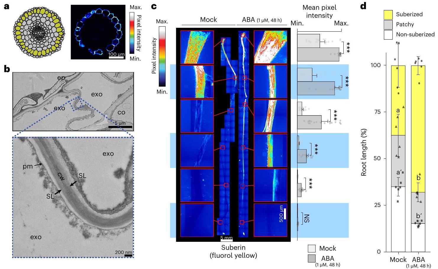
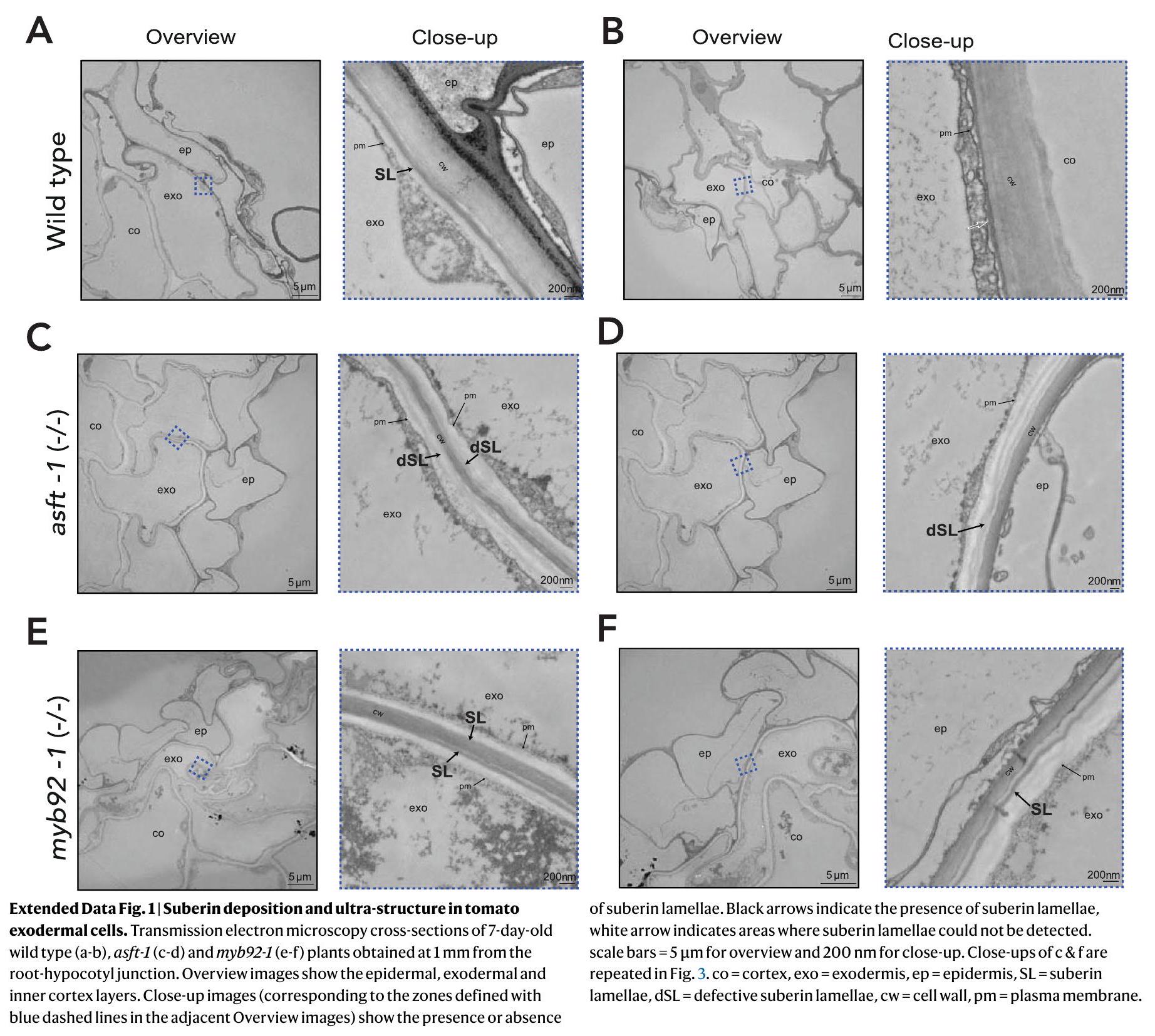
تطور وتركيب الإكسوديرميس المكسو بالسبيرين

قمنا سابقًا بتحديد كمية ترسيب السوبرين في الإكسوديرميس على المحور الطولي لجذر الطماطم (الصنف M82، LA3475) باستخدام الصبغة الهيستوكيميائية فلورول أصفر (FY). في جذور الأرابيدوبسيس، يكون السوبرين غائبًا عن خلايا الإندوديرم في منطقة مرستيم الجذر ومنطقة الإطالة، ويبدأ في الترسيب بشكل متقطع في منطقة التمايز المتأخرة بعد أن يتم تأسيس نظام الجذر، ثم يتبعه ترسيب كامل للسوبرين في منطقة التمايز البعيدة. أظهرت قياس السوبرين الخارجي في جذور الطماطم التي تبلغ من العمر 7 أيام نفس الفئات الثلاث للإيداع (لا شيء، بقع، وكامل) (الشكل 1أ، ج، د والشكل البياني الممتد 1). كما أظهرت المجهر الإلكتروني أن طبقات السوبرين تُودع بشكل أساسي على الوجوه البشرة والوجه بين الخارجي للخلايا الخارجية (الشكل 1ب والشكل البياني الممتد 1). كان السوبرين غائبًا باستمرار داخل الإندوديرم للجذر في جميع المناطق النمائية. (الشكل 1أ والشكل الإضافي 1).
تحليل المونومرات المرتبطة بجدار الخلية والمونومرات الأليفاتية المرتبطة بالبوليمر في جذور الطماطم التي تبلغ من العمر شهر واحد كشف عن هيمنة الأحماض ثنائية الكربوكسيل، مشابهة للبطاطس . بالمقارنة مع جذور الأرابيدopsis، التي تتميز في الغالب بـ أحماض -OH وطول سلسلة أقصى يبلغ 24 كربون (C24) ، C26 و C28 إضافية تمت ملاحظة الأحماض -OH والكحوليات الأولية في الطماطم (الشكل البياني الموسع 2a). لقد تم ملاحظة هذه الظاهرة من التباين بين الأنواع في تركيبة السوبيرين سابقًا. .

إنزيمات التخليق الحيوي للسوبيرين والمنظمات النسخية

لرسم خريطة مسار تخليق السوبرين في جذور الطماطم ومنظماته النسخية، استغللنا الملاحظات السابقة حول المحافظة النسبية للتنظيم المشترك النسخي لمسار السوبرين عبر كاسيات البذور. في جذر الأرابيدopsis، تزداد مستويات السوبرين عند المعالجة بـ هرمون يعتبر المستجيب الأول عند الإجهاد الناتج عن نقص الماء. يتم زيادة ترسيب السوبرين في الطبقة الخارجية في الطماطم بشكل مشابه عند معالجة ABA، سواء من حيث المنطقة التي تم سوبرينها بالكامل وكذلك من حيث شدة الإشارة (الشكل 1c، d)، مع استمرار غياب السوبرين في الطبقة الداخلية (البيانات الموسعة الشكل 3). يعتبر قريب S. lycopersicum البري، Solanum pennellii (LA0716)، مقاومًا للجفاف. ، وزيادة ترسيب السوبرين في الأرابيدوبسيس من خلال طفرة في ENHANCED SUBERIN1 (ESB1) تمنح تحمل الجفاف، على الرغم من أن esb1 يظهر أيضًا زيادة في اللجنين في الأندوديرم وتقطع في تشكيل CS. . لذلك، قمنا باختبار وتأكيد الفرضيات التي تفيد بأن S. pennellii لديه ترسيب أعلى من السوبرين مقارنة بـ M82 حتى في ظروف توفر الماء، ولا يظهر أي تغييرات في حجم أو موقع ترسيب السوبرين استجابةً لـ ABA في الشتلات (البيانات الموسعة الأشكال 2 و 3). وبالتالي، فإن مستويات السوبرين في S. pennellii هي ثابتة. لذلك، استخدمنا مجموعة بيانات تعبير الجينات
الشكل 1| يتم ترسيب السوبيرين في الإكسوديرميس في الطماطم ويتم تنظيمه بواسطة ABA. أ، تمثيل بياني (يسار) لعلم التشريح الجذري لـ S.lycopersicum (الصنف M82) (الإكسوديرميس مميز باللون الأصفر) وقطع عرضي تمثيلي (يمين) لجذر عمره 7 أيام ملون بـ FY. مقياس الرسم، . ب، مقاطع عرضية باستخدام مجهر الإلكترون الناقل لجذور عمرها 7 أيام تم الحصول عليها على بعد 1 مم من نقطة التقاء الجذر والهيبوكوتيل. الأعلى: طبقات البشرة (ep) والطبقة الخارجية (exo) والقشرة الداخلية (co). الأسفل: صورة مقربة للمنطقة المميزة (المنطقة المحددة بخطوط زرقاء منقطة)، تظهر وجود طبقات السوبيرين (SL). cw، جدار الخلية؛ pm، غشاء البلازما. ج، صبغة فلورول أصفر (FY) للسوبيرين في النوع البري.
نباتات عمرها 7 أيام تم معالجتها بعلاج وهمي أو لمدة 48 ساعة. صبغة كاملة لجذر رئيسي (يسار) ومتوسط إشارة الفلورول الأصفر على طول الجذر (يمين) ; أشرطة الخطأ، الانحراف المعياري. تشير النجوم إلى الأهمية مع تحليل التباين الأحادي (ANOVA) يليه اختبار توكي-كرامر بعد الاختبار (***P<0.005). NS، غير مهم. د، مراحل تطور ترسيب السوبيرين في النباتات من النوع البري المعالجة بالتحكم أو لمدة 48 ساعة. تم تصنيف المناطق إلى غير مغطاة باللحاء (أبيض)، مغطاة باللحاء بشكل متقطع (رمادي) ومغطاة باللحاء بشكل مستمر (أصفر)؛ تشير الحروف إلى مجموعات مختلفة إحصائيًا؛ وتشير الفواصل إلى مقارنات إحصائية مختلفة؛ ; أشرطة الخطأ، الانحراف المعياري.
توصيف النسخ في جذور M82 بالإضافة إلى الجذور من 76 خط إدخال طماطم مشتق من . ليكوبيرسيكوم cv . M82 و . بنيللي (LA0716) مع M82 كالأب المتكرر قمنا أيضًا بتحليل النسخ الجذرية لنباتات الطماطم التي تبلغ من العمر شهرًا تحت ظروف الري الجيد، والري المفرط، ونقص المياه. افترضنا أن الجينات المعنية مباشرة في تخليق وتراكم السوبرين ستكون مرتبطة بشكل كبير في كل من نقص المياه وسكان خط الإدخال.
من خلال دمج كلا من خطوط الإدخال، ومجموعات بيانات تشبع المياه ونقص المياه في تحليل شبكة ارتباط الجينات الموزونة (WGCNA) ، حددنا وحدات من الجينات المتعبّرة عن بعضها البعض (الشكل التوضيحي التكميلي 1). كانت وحدة (‘royalblue’) التي تحتوي على 180 جينًا غنية بشكل ملحوظ في الجينات المتعلقة بالسوبيرين (نسبة الاحتمالات ). وقد تم تأكيد ذلك من خلال التقاطع مع مجموعة بيانات عامة تصف تعبير الجينات في خطوط DCRi من الطماطم (الجدول التكميلي 1). تقوم خطوط DCRi بتنشيط الجينات المرتبطة بالسوبيرين في خلايا البشرة للفاكهة، مما يؤدي إلى تكوين السوبيرين على سطح الفاكهة. . تحتوي وحدة ‘royalblue’ على عدة نظائر جينية لعائلات الجينات المعروفة المسؤولة عن تخليق السوبرين مثل ناقلات الأسيلا للجليسرول-3-فوسفات (GPATs) ، ومركبات 3-كيتوأسيلا-CoA (KCSs) ، وناقلات الفيرول (ASFT/ FHTs) (الشكل 2a والجدول التكميلي 1). بالإضافة إلى ذلك، تم العثور على نظائر محتملة للطماطم من المنظمات النسخية المعروفة لتخليق السوبرين: AtMYB41 وAtMYB63 وAtMYB92 (SlMYB41: Solyc02g079280؛ SIMYB63: Solyc10gOO5550؛ SIMYB92: Solyc05g051550)، من بين آخرين، في هذه الوحدة. .

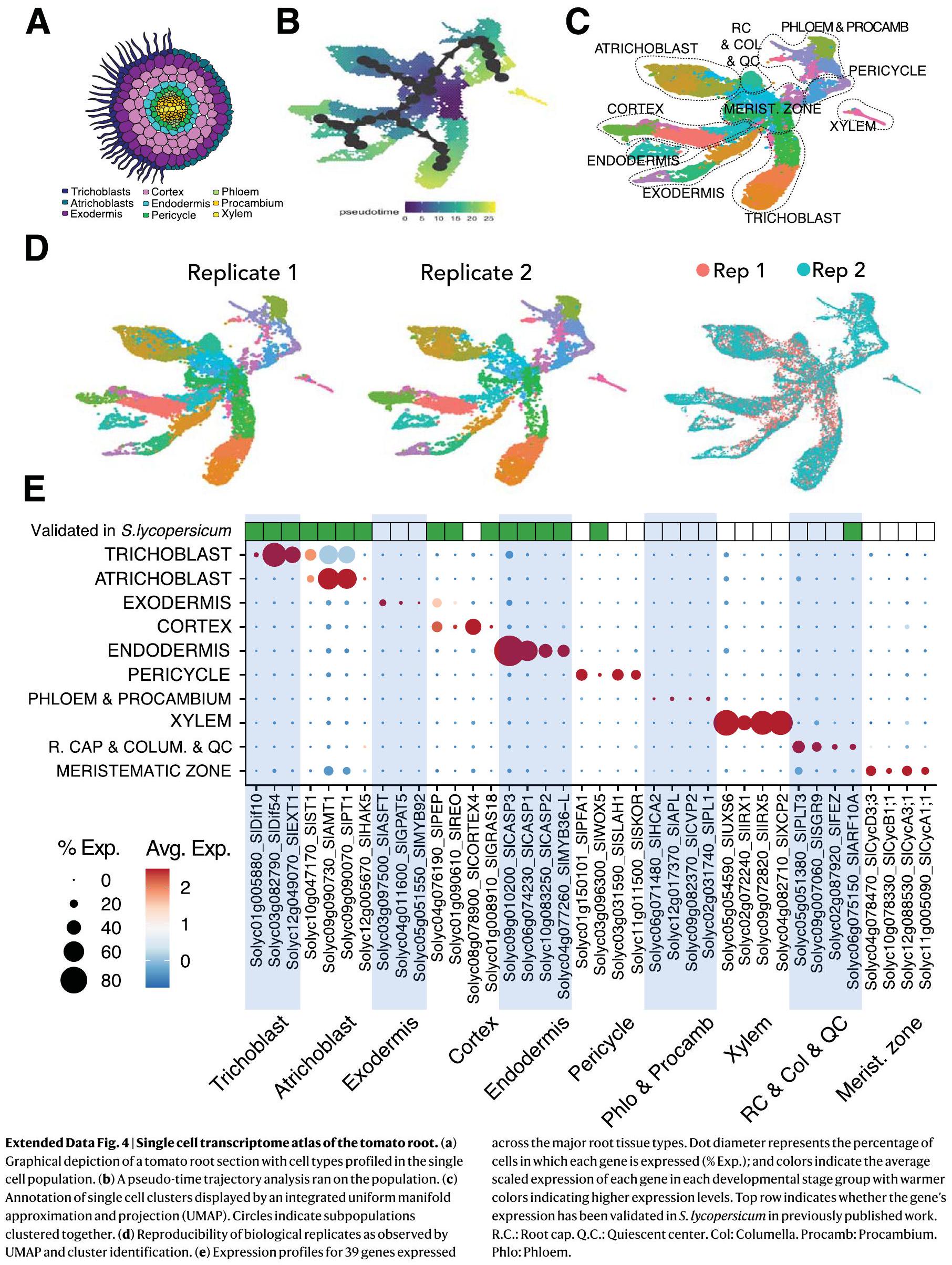
أطلس جذور الطماطم على مستوى الخلية الواحدة لرسم خريطة الإكسوديرميس

على الرغم من وجود ملفات الترجمة للطبقة الخارجية ، هذه البيانات لا توفر حلاً للتدرج التنموي الذي على طول
تم ترسيب السوبرين. لتحسين مجموعة الجينات المرتبطة بالسوبرين المرشحة، قمنا بإجراء تحليل التعبير الجيني على مستوى الخلية الواحدة لجذر الطماطم. استخدمنا منصة 10X Genomics scRNA-seq لتحليل أكثر من 20,000 خلية جذر. جمعنا الأنسجة من جذور الطماطم الأولية (M82) التي تبلغ من العمر 7 أيام حتى 3 سم من الطرف لتضمين المنطقة التي يتم فيها ملاحظة ترسيب السوبرين في البداية. تم إنشاء مصفوفات التعبير الجيني باستخدام cellranger وتم تحليلها في Seurat. بمجرد معالجة البيانات مسبقًا وتصفيتها من القطرات ذات الجودة المنخفضة، تم تحليل النسخ الجينية عالية الجودة لـ 22,207 خلية. بعد التطبيع، استخدمنا التجميع غير المراقب لتحديد تجمعات خلوية متميزة (الشكل الممتد 4). ثم تم تعيين هوية نوع الخلية لهذه التجمعات الخلوية باستخدام الأساليب التالية: قمنا أولاً بتقدير التداخل مع مجموعات النسخ الغنية بنوع الخلية الموجودة من جذر الطماطم. وجينات العلامة المستخرجة من كل من المجموعات. تم توضيح مجموعة فردية كنوع خلية محدد نظرًا لأكبر تداخل بين المجموعتين ووجود تعديل كبير. قيمة ( ثم، لرسم ديناميات تعبير الجينات عبر النضوج، قمنا بفحص تقدم حالة الخلية من خلال حساب مسارات الزمن الزائف باستخدام خوارزمية الشجرة الممتدة الدنيا. . كانت الشجرة متجذرة في منطقة الميرستيم الجذرية (المحددة في الخطوة السابقة)، وتم تجميع المجموعات في 10 أنواع من الخلايا لتعكس المعرفة البيولوجية الحالية حول تمايز جذر الطماطم (الشكل 2ب والشكل الإضافي 4). أخيرًا، تم تحديد الجينات ذات أنماط التعبير التي تم التحقق منها مسبقًا في الطماطم. مراسلين النسخ وعلامات نوع الخلايا المتوقعة بناءً على وظيفتها في الأرابيدopsis تمت إضافة البيانات على الكتل لتحسين التوصيف (الشكل 4c من البيانات الموسعة).
نظرًا للتعليق الناجح على هذه الأنواع من الخلايا، ركزنا على المسارات التنموية المرسومة المستمدة من افتراض
الشكل 2 | يتم التعبير عن إنزيمات تخليق السوبرين في الطماطم والمنظم النسخي في الإكسوديرميس الناضج. أ، مخطط مبسط لمسار تخليق السوبرين. تشير المربعات إلى عائلات الجينات المعنية في كل خطوة من خطوات المسار (تشير الألوان الزرقاء والصفراء إلى الإنزيمات التخليقية والمنظمات النسخية، على التوالي). الجينات المستهدفة في هذه الدراسة محددة باللون الأحمر. TFs، عوامل النسخ؛ VLCFAs، الأحماض الدهنية ذات السلسلة الطويلة جداً. ب، مجموعات خلايا مفردة موصوفة من 3 سم من طرف جذر الطماطم معروضة بواسطة UMAP مدمج. R.C.، غطاء الجذر. Q.C.، المركز الساكن؛ col، العمود؛ procamb، بروكامبيوم. ج، UMAP للخلايا المعلنة من القشرة/الإندوديرميس/الإكسوديرميس التي تم استخراجها من الإسقاط العام وإعادة تضمينها. أ
تم تضمين مجموعة صغيرة من الخلايا من منطقة الميرستيمات للمساعدة في تثبيت تقديرات الزمن الزائف. CEEI، البداية القشرية-الإكسوديرميس-الإندويرميس. د، تحليل مسار الزمن الزائف لكتل خلايا القشرة/الإندويرميس/الإكسوديرميس. هـ، ملفات التعبير الخاصة بنوع الخلايا أو الأنسجة لجينات مسار تخليق السوبرين. يمثل قطر النقطة نسبة الخلايا التي يتم فيها التعبير عن كل جين (% Exp.) وتدل الألوان على متوسط التعبير المقنن لكل جين في كل مجموعة من مراحل التطور، حيث تشير الألوان الأكثر دفئًا إلى مستويات تعبير أعلى. و، تعبير SIMYB92 و SIASFT في بيانات النسخ المفردة. -عدادات UMI المصححة المعيارية.
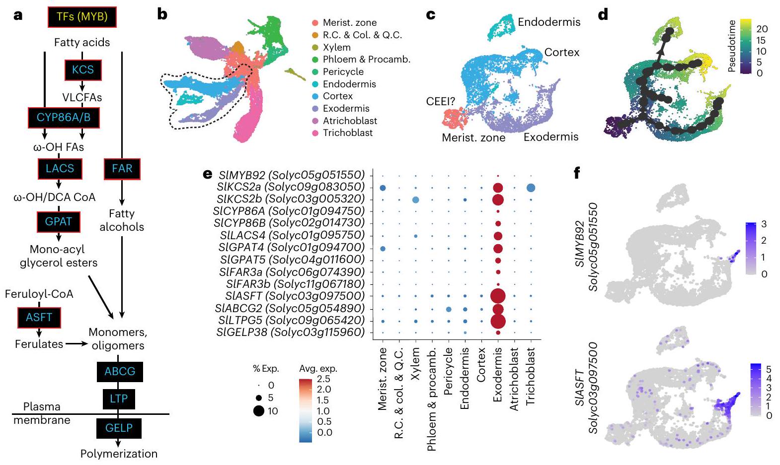
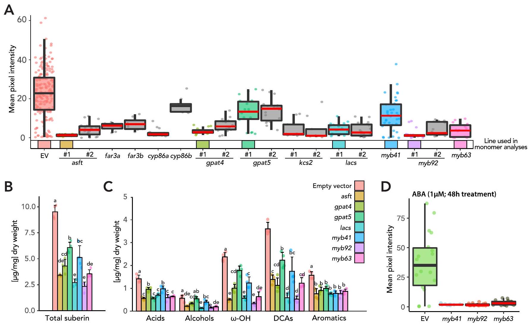
تم تحديد مجموعة الخلايا المرتبطة بمسارات التطور الثلاثة (CEEI) (الشكل 2c، d). ضمن هذه المسارات الثلاثة المرتبطة، قمنا بتحديد الخلايا التي كانت فيها إنزيمات تخليق السوبرين والجهات التنظيمية المحتملة معبرة بشكل عالٍ (الجدول التكميلي 3). من بين هذه، أظهرت النسخ الجينية لـ SIASFT (Solyc03g097500)، واثنين من FAR (SIFAR3A: Solyc06g074390؛ SIFAR3B: Solyc11g067190)، واثنين من CYP86 (SICYPB86A: Solyc01g094750؛ SICYP86B1: Solyc02g014730)، واثنين من KCS2 (SIKCS2a: Solyc09g083050 و SIKCS2b: Solyc03g005320)، واثنين من GPAT (SIGPAT4: Solyc01g094700؛ SlGPAT5: Solyc04g011600) وواحد من LACS (SILACS4: Solyc01g095750) تعبيرًا مقيدًا عند أقصى حافة من مسار التطور الإكسودرمي (الشكل 2e، f، والأشكال التكملية 2 و 3). قمنا بإنشاء تقرير نسخي يتكون من محفز SIASFT مرتبط ببروتين الفلورسنت الأخضر (GFP) المحلي في النواة وأكدنا أن تعبيره مقيد بالإكسودرم باستخدام جذور شعرية (الشكل البياني الممتد 5)، بما يتماشى مع تحليلنا على مستوى الخلية الواحدة. بالإضافة إلى ذلك، من بين ثلاثة عوامل نسخ تم الإشارة إليها سابقًا (SIMYB41، SIMYB63 و SIMYB92)، أظهر فقط SIMYB92 تعبيرًا محددًا ومقيدًا في الخلايا عند طرف مسار الإكسودرم (الشكل 2f والأشكال التكملية 3). بناءً على بيانات التعبير المشترك ومسار الخلايا، كانت هذه الجينات مرشحة محتملة لتنظيم النسخ الخاص بالسوبرين وإنزيمات تخليق السوبرين.

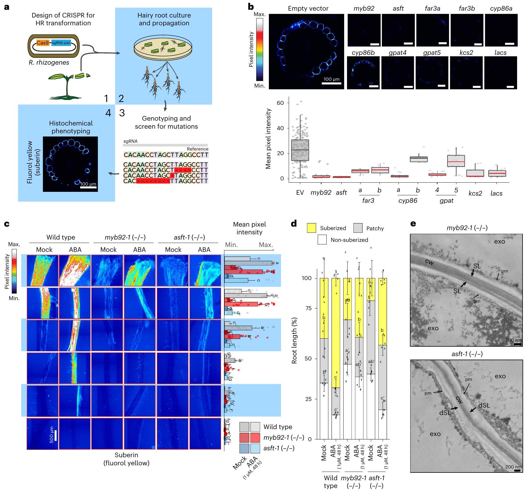
إسقاط الجينات المرشحة يعطل ترسيب السوبرين

تم إجراء التحقق الوظيفي لهذه الإنزيمات في البداية بواسطة تحرير الجينات باستخدام تقنية كريسبر-كاس9 باستخدام اثنين أو ثلاثة من أدلة RNA (الجدول التكميلي 4) وتم إدخالها إلى الطماطم عبر تحويل R. rhizogenes (الجذور الشعرية). (الشكل 3أ). تم تحديد الأنماط الظاهرية للأليلات الطافرة المؤكدة بالحذف لهذه الجينات لمستويات السوبيرين باستخدام صبغة الفلورول الأصفر (الشكل 3ب والبيانات الموسعة الشكل 6). بناءً على
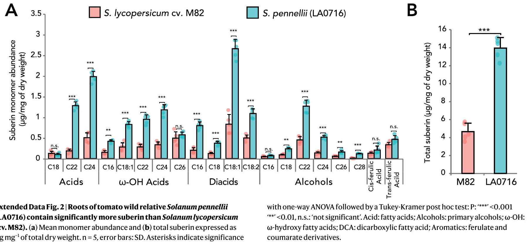
في تصنيف الأنسجة، أظهر جميع الطفرات باستثناء طفرة slcyp86b انخفاضًا في السوبرين (الشكل 3ب). تم الحصول على تأكيد إضافي لانخفاض مستويات السوبرين من خلال تحليل الأيض لمونومرات السوبرين في الطفرات slgpat5 و slgpat4 و slasft و sllacs و slmyb92 (الشكل التمديدي 6). وشملت هذه الانخفاض الجماعي لمكونات حمض الفيروليك وحمض السينا بيك؛ الأحماض الدهنية (C20 و C22 و C24)، -أحماض هيدروكسي (C18:2، C18:1، C20، C24، C26) و -الأحماض الثنائية (C18:2، C18:1، C18، C20، C22). نظرًا لتعبيرها في نهاية مسار التطور الخارجي (الشكل 2f)، تم إنتاج أليلات طافرة مستقرة من slasft و slmyb92 عن طريق التحويل باستخدام . تم استخدام نفس جزيئات RNA الدليل في تجارب التعديل الجيني على Agrobacterium tumefaciens. جميع الأليلات الطافرة التي تم استردادها والتي فقدت الوظيفة كانت تحتوي على طفرات تؤدي إلى أكواد توقف مبكرة (الجدول التكميلي 4). تم ملاحظة انخفاض مستويات السوبرين بالإضافة إلى تغييرات في تراكمه على طول مسار تطور الجذور في أليلات طافرة مستقلة لكل جين (الشكل 3c، d والشكل التكميلي 7). في حالة slasft، كان هناك تأخير كبير في ترسيب السوبرين وتغييرات في تركيبة المونومر في الإكسوديرميس، مما يختلف عن نظيره في الإندوديرميس لجذر الأرابيدوبسيس، حيث لا تظهر الطفرة atasft أي عيوب في الترسيب أو التوقيت أو التركيبة الرئيسية للمونومر. الأليلات الطافرة متشابهة من حيث انخفاض محتوى الفيرولات فيها. . لقد لاحظنا أيضًا عدم تنظيم الهيكل الطبقي في الطفرة slasft-1، ولكن ليس في الطفرة slmyb92-1 (الشكل 3e). بينما تم العثور على انخفاض في الأحماض الفيرولية في الطفرات الخاصة بنظائر ASFT في البطاطس. لم يتم الإبلاغ عن هذا الاضطراب الطبقي من قبل. وبالتالي، بينما تكون الإنزيمات المسؤولة عن تخليق السوبيرين متشابهة إلى حد كبير في الإكسوديرميس لجذر الطماطم والإندويرميس لجذر الأرابيدوبسيس، فإن هناك جديدًا في مساهماتها. أي أن SIASFT قد يكون مسؤولًا بشكل مباشر عن
الشكل 3 | الأليلات الطافرة لفقدان الوظيفة للجينات المرشحة تعطل السوبيرين الخارجي في الطماطم. أ، ملخص رسومي للجذر الشعري (HR؛ . ريزوجينيس) فحص الطفرات. ب، ملخص لظواهر الطفرات للجينات المرشحة في الجذور الشعرية. الأعلى: مقاطع عرضية تمثيلية لأجزاء ناضجة من الجذور ملونة بالفلورول الأصفر. الأسفل: الكمية الإجمالية لإشارة الفلورول الأصفر عبر مقاطع عرضية متعددة (النوع البري ; راحة تشير الخط الأحمر إلى الفروق ذات الدلالة الإحصائية في شدة بكسل الفلورول الأصفر في الطفرة مقارنة بالنمط البري كما تم تحديده باستخدام تحليل التباين أحادي الاتجاه يليه اختبار توكي-كرامر بعد الاختبار. ); EV، متجه فارغ. تمثل مراكز مخطط الصندوق الوسيط بينما تمثل حدود الصندوق السفلية والعلوية النسبة المئوية 25 و 75، على التوالي. تمثل الشعيرات القيم الدنيا والعليا. تمثل النقاط عينات فردية. ج، صبغة فلورول الأصفر للسبيرين في نباتات النوع البري التي تبلغ من العمر 7 أيام (مكررة من الشكل 1 للرجوع إليها)، slmyb92-1 و slasft-1 المعالجة بـ
سخرية أو ABA لمدة 48 ساعة. صبغ كامل للجزور الأولية عبر مقاطع مختلفة (يسار) ومتوسط شدة إشارة الفلورول الأصفر على طول الجذر (يمين) ” ). تشير الحروف إلى اختلافات كبيرة (تحليل التباين الأحادي متبوعًا باختبار توكي-كرامر بعد الاختبار ( ). أشرطة الخطأ، الانحراف المعياري د، مراحل تطور ترسيب السوبيرين في النباتات البرية والنباتات الطافرة التي تبلغ من العمر 7 أيام والمعالجة بالتحكم أو ABA لمدة 48 ساعة. تم تصنيف المناطق إلى غير مغطاة بالسبير (أبيض)، مغطاة بالسبير بشكل متقطع (رمادي) ومغطاة بالسبير بشكل مستمر (أصفر). ). تشير الحروف إلى مجموعات مختلفة إحصائيًا؛ وتشير الفواصل إلى مقارنات إحصائية مختلفة. e، مقاطع عرضية تمثيلية باستخدام مجهر الإلكترون الناقل لمتحولات slasft-1 و slmyb92-1 تم الحصول عليها على بعد 1 مم من نقطة التقاء الجذر والسويقة. يظهر المتحول slasft-1 عجزًا في الطبقة السوبرينية.structure.cwجدار الخلية؛ dSL، طبقات السوبيرين المعيبة؛ exo، الإكسوديرميس؛ pm، الغشاء البلازمي؛ SL، طبقات السوبيرين.
كلا من ارتباط جدار الخلية والتصاق بين الطبقات، و هو مساهم رئيسي في ترسيب السوبيرين.
في الأربيذوبس، يزيد تطبيق ABA من مستويات السوبرين، ويتطلب أربعة عوامل نسخ MYB بشكل متكرر لتحفيز
تراكم السوبيرين المدعوم بواسطة ABA في الأرابيدوبسيس (AtMYB41، AtMYB53، AtMYB92 وAtMYB93) نظرًا لقدرة ABA على تحفيز السوبرين في الأرابيدوبسيس والطماطم (الشكل 1c، d) افترضنا أن SIMYB92 أو SIASFT ضروريان لتخليق السوبرين المستحث بواسطة ABA. لذلك،
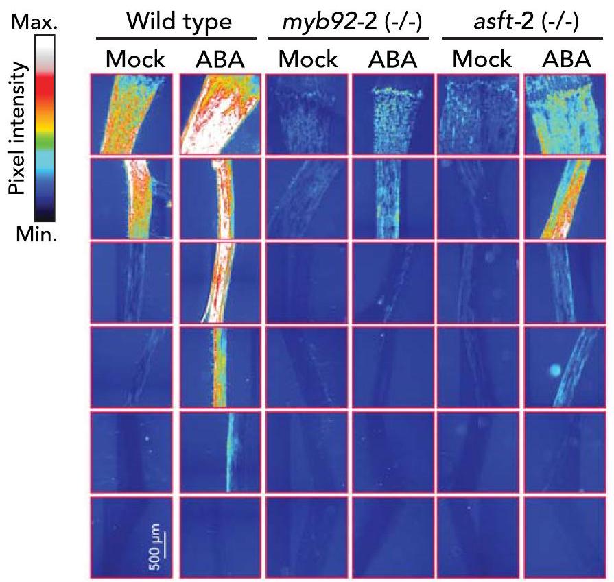
قمنا بمعالجة جذور slmyb92 و slasft بـ لمدة 48 ساعة، تركيز يكفي لزيادة المنطقة المكسوة بالكامل بالسوبرين في الطماطم دون التأثير على طول الجذور (الشكل 1أ والشكل الممتد 8). على الرغم من زيادة مستويات السوبرين عند معالجة ABA في هذه الخلفيات الطافرة، سواء من حيث حجم إشارة الفلورو الأصفر أو نسبة الجذر الذي تم كسوته بالكامل بالسوبرين، فإن درجة الزيادة كانت أقل مقارنة بالنمط البري (الشكل 3ج، د). هذه الانخفاضات في مستويات السوبرين في الطافرة الفردية slmyb92-1 في كل من ظروف التحكم والمعالجة بـ ABA هي نوعياً أكبر مما لوحظ في الطافرة الفردية في الأرابيدوبسيس، والتي يمكن إعادتها إلى مستويات النمط البري من السوبرين في الإندوديرم عند تعرضها لـ تشير هذه الفجوة إلى أن SIMYB92 قد يلعب دورًا أكثر بروزًا في ترسيب السوبيرين مقارنةً بنظيره في الأرابيدوبسيس. يتم تفسير عدم وجود هذه الظاهرة في الطفرة atmyb92-1 المستحثة بواسطة ABA من خلال تكرار عوامل النسخ AtMYB41 وAtMYB53 وAtMYB92 وAtMYB93. استكشفنا ما إذا كانت مثل هذه الازدواجية موجودة في الطماطم في الأليلات الطافرة لفقدان الوظيفة slmyb41 و slmyb63، وما إذا كانت كافية لتقليل السوبرين في ظروف التحكم ونقص المياه. لم يكن لعلاج ABA القدرة على تحفيز السوبرين إلى مستويات النوع البري في أي من الطافرات (الشكل 6d من البيانات الموسعة). نظرًا لأن الإكسوديرميس يتم تليينه أولاً ثم يتم تغطيته بالسوبرين. استكشفنا أيضًا ما إذا كانت SIMYB92 أو SIMYB41 أو SIMYB63 متورطة في ت lignification لجذر الطماطم. ومع ذلك، ظلت مستويات اللجنين في الإكسوديرميس والإندويرميس غير متأثرة في أي من الطفرات الجذرية الشعرية لهذه المنظمات النسخية (الشكل البياني الممتد 9)، مما يشير إلى أنها لا تلعب دورًا في الت lignification الأولي للإكسوديرميس أو الإندويرميس.

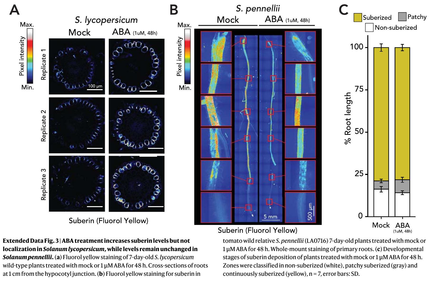
تغيير ترسيب السوبيرين يؤثر على استجابة نقص المياه

نظرًا للرابط المقترح بين السوبرين وتحمل الجفاف، بالإضافة إلى انخفاض مستويات السوبرين في كل من ظروف التحكم وABA في طفرات الطماطم لدينا، افترضنا أن خطوط slmyb92 و slasft ستكون أكثر حساسية لقيود المياه مقارنة بالنباتات من النوع البري. قمنا بتعريض نباتات عمرها 4 أسابيع ومروية جيدًا لظروف نقص المياه لمدة 10 أيام (الطرق). تم دراسة ترسيب السوبرين ومستويات المونومرات في نظام الجذر لخطوط slmyb92-1 و slasft-1 والنباتات من النوع البري في كل من ظروف المياه الكافية والمحدودة. تحت ظروف المياه الكافية، تم ملاحظة ترسيب السوبرين بشكل خفيف فقط في النوع البري، وبشكل حصري في الإكسوديرم، بينما كان غائبًا تمامًا في كلا الخطين الطافرين (الشكل 10 من البيانات الموسعة). متسقًا مع هذه الملاحظة، تم الكشف عن مستويات منخفضة جدًا من مونومرات السوبرين، دون ملاحظة اختلافات كبيرة في الأحماض الدهنية طويلة السلسلة جدًا، والكحوليات الأولية، -أحماض هيدروكسي الأحماض ثنائية الكربوكسيل والمكونات العطرية للسوبيرين (الشكل 4 أ والشكل التكميلي 4). ومع ذلك، تحت قيود المياه، زادت ترسيبات السوبيرين الخارجي، حيث كانت مستويات كلا السلالتين الطافرتين أقل من النوع البري (الشكل التكميلي 10). أظهر الطافرة المنظمة للتعبير الجيني slmyb92-1 انخفاضًا عامًا في معظم مجموعات المونومرات مقارنة بالنوع البري. بالمقابل، كانت الطافرة slasft-1 تعاني بشكل أساسي من نقص في حمض الفيروليك وركائزه الاستيرية، بالإضافة إلى الكحوليات الأولية الفردية. -أحماض الهيدروكسي (الشكل 4 أ والشكل التكميلي 4). علاوة على ذلك، تم تقليل جهد الماء في الساق، وموصلية الثغور، ومعدل النتح بشكل كبير استجابةً لظروف نقص المياه في كل من slmyb92-1 و slasft-1 مقارنة بالنمط البري، كما تم تقليل محتوى الماء النسبي في الأوراق أيضًا في slmyb92-1 (الشكل 4 ب-هـ). عند النظر في جميع الصفات الفسيولوجية بشكل جماعي باستخدام تحليل المكونات الرئيسية، أظهر slasft-1 استجابة أقل حدة لنقص المياه مقارنة بالنباتات من النمط البري، بينما كانت استجابة slmyb92-1 أكثر تطرفًا (الشكل التكميلي 5). توضح هذه البيانات أن انخفاض مستويات السوبيرين في الإكسوديرم الجذري للطماطم يؤثر بشكل مباشر على أداء النبات ككل تحت ظروف نقص المياه، ولكن ليس تحت ظروف كافية من المياه. علاوة على ذلك، قد تؤثر التغيرات في أحادي السوبيرين المحدد والبنية اللاميلية التي لوحظت بين الطافرين استجابةً لظروف نقص المياه بشكل مختلف على مدى الاستجابة الفسيولوجية.

نقاش

في الإندوديرميس الجذرية المعروفة جيدًا لنبات الأرابيدوبسيس، يتم ترسيب السوبيرين كطبقة كارهة للماء بين الغشاء البلازمي والجدار الخلوي الأساسي. من الناحية التطورية، يحدث تخليق وتراكم السوبرين كخطوة ثانية من تمايز الأندوديرم، حيث تكون الخطوة الأولى هي تخليق وتراكم الشريط الكاسباريني المتخشب. يعمل السوبرين كحاجز أبلاستيكي وحاجز عبر الخلايا، مما يساهم في تنظيم حركة الماء والمذابّات إلى الأسطوانة الوعائية. تظهر ملاحظاتنا الجماعية أنه، بالمقارنة مع الأرابيدوبسيس، (1) مكونات المسار محفوظة؛ (2) موقعها المكاني مميز؛ (3) ASFT و MYB92 هما منظمين حاسمين لتخليق السوبيرين نظرًا لظواهرهما كأليلات طافرة ذات فقدان وظيفي فردي، على عكس تكرارهما في الأرابيدوبسيس و (4) السوبيرين الخارجي له وظيفة مكافئة للسوبيرين الداخلي ويمكن أن يعمل في غيابه (الشكل 1). مكانيًا، في قشرة الجذر للطماطم، يتم ترسيب طبقات السوبيرين بين الجدار الخلوي الأولي الخارجي والغشاء البلازمي حول الخلية، مشابهًا لجذر الأرابيدوبسيس الداخلي وحواجز الأبولستيك الأخرى المكونة من السوبيرين مثل قشرة البطاطس. (الشكل 1ب). بطريقة زمنية مشابهة للاندوديرميس في الأرابيدوبسيس، هناك منطقة غير مغطاة بالسوبرين في طرف الجذر، ومنطقة مغطاة بالسوبرين بشكل متقطع في منتصف الجذر ومنطقة مغطاة بالسوبرين بشكل مستمر بالقرب من نقطة التقاء الجذر مع السويقة (الشكل 1ج، د).
لقد حصلنا على أدلة حول الجينات الأساسية التي تتحكم في تخليق السوبرين الخارجي على مدار الزمن التطوري من خلال التحليل المشترك، وتحليل النسخ الجيني على مستوى الخلية الواحدة، والتحليل الجيني. كان من الواضح من التحليل الجيني الوظيفي لطفرات SICYP86B وSIGPAT وSILACS وSIASFT أن هناك حفظًا للجينات ضمن مسار تخليق السوبرين بين الأرابيدوبسيس والطماطم. على الرغم من أن نفس الجينات تتحكم في تخليق السوبرين، إلا أن هناك تميزًا في الطماطم فيما يتعلق بتعبيرها المكاني (الخارجي) ومساهمة SIASFT الحاسمة في ارتباط جدار الخلية الأولي والتلاصق بين الطبقات لحاجز السوبرين. لم يتم ملاحظة هذه الظاهرة أبدًا في جذور طفرات الأرابيدوبسيس أو البطاطس. . بالإضافة إلى ذلك، تم اعتبار أعضاء تحت الشجرة GPAT4 معنيين بشكل حصري في تخليق الكوتين. ، وهنا، تم إظهار أن SIGPAT4 يشارك في تشكيل السوبرين الخارجي (الشكل 3). ركزنا على SIMYB92 كمرشح بسبب تعبيره في نهاية مسار الإكسوديرم. على الرغم من أن التوقيت الدقيق لهذه المسارات هو في الغالب تنبؤي بطبيعته، إلا أننا نلاحظ أن تعبير الإنزيمات الحيوية لا يتداخل تمامًا مع تعبير SIMYB92 ويشير إلى أن SIMYB92 ليس المنظم النسخي الوحيد لتعبير جينات السوبرين. قدرتنا على الحصول على خلايا إكسوديرمية متمايزة بشكل متزايد ربما تكون محدودة بقدرتنا على تحويل الخلايا تمامًا مع ترسيب الجدار الخلوي الثانوي. لذلك، فإن عدم وجود تعبير SIMYB41/53/93 في مسار الإكسوديرم لا يعني أن هذه الجينات غير معبر عنها في الإكسوديرم. في الأرابيدوبسيس، تظهر الطفرات الفردية لفقدان الوظيفة لـ MYB41 و MYB53 و MYB93 عدم وجود تغييرات في مستويات السوبرين، بينما تلك لـ يظهر تأخيرًا في التصلب تمت ملاحظة ظواهر السوبرين المتطرفة فقط عندما تم دمج طفرات جميع الجينات الأربعة. . بالمقابل، تم ملاحظة هذا النمط الظاهري المتطرف (انخفاض مستويات السوبيرين في الأليلات الطافرة slmyb92 كما تم قياسه بواسطة الفلورول الأصفر (الشكل 3ب-د) وتحليل التركيب في الجذور الشعرية والخطوط المستقرة (البيانات الموسعة الشكل 6 والشكل التكميلي 4)) في الطماطم عندما تم تحوير MYB92 فقط. يمكن أن تكون مستويات السوبيرين المتبقية الموجودة في الطافرات slmyb92 منظمة بواسطة عوامل النسخ MYB أخرى. في الواقع، أظهرت الطافرات في نظائر MYB41 و MYB63 في الطماطم أنماط ظاهرة سوبيرين خارجية (البيانات الموسعة الشكل 6)، مما يشير إلى أن هذه الجينات قد يتم التعبير عنها في مراحل التطور الخارجية اللاحقة.
تنظيم السوبرين في الإكسوديرم للطماطم بواسطة ABA يتماشى شكليًا مع ما يُلاحظ في الإندوديرم لجذر الأرابيدوبسيس، مع زيادة في كل من حجم ترسيب السوبرين ونسبة المنطقة المكسوة بالكامل بالسوبرين. على الرغم من التحديد المكاني المميز. على الأقل في حالة
الشكل 4 | يؤثر ضعف ترسيب السوبرين في slmyb92-1 و slasft-1 على أداء النبات بالكامل استجابةً لقيود المياه. أ، تركيبة السوبرين في جذور نباتات النوع البري الناضجة التي تبلغ من العمر شهرًا واحدًا، slmyb92-1 و slasft-1. تم تعريض النباتات لظروف كافية من المياه (WS) وقيود المياه (WL) لمدة 10 أيام. طرق). الأحماض، الأحماض الدهنية؛ الكحوليات، الكحوليات الأولية؛ -OH، -أحماض دهنية هيدروكسيلية؛ DCA، حمض دهني ثنائي الكربوكسيل؛ عطرية،
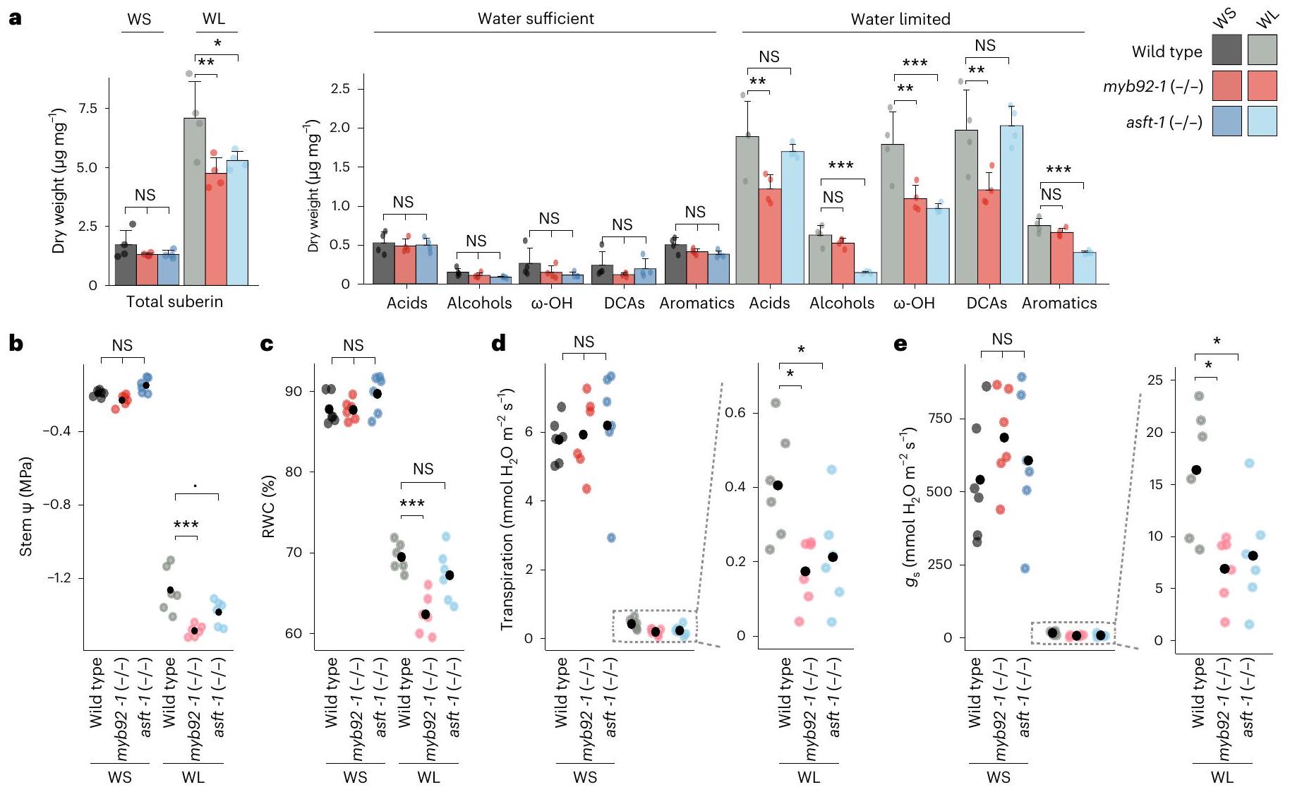
الإيزومرات الفيرولات والكومارات. تشير أشرطة الخطأ إلى الانحراف المعياري. ب-هـ، مخططات نقطية للقيم المسجلة لإمكانات الماء في الساق (الساق ) (ب)، محتوى الماء النسبي (ج)، النتح (د) وموصلية الثغور (هـ). الخط المنقط يشير إلى تكبير للحصول على دقة بصرية أفضل للقيم. النقاط السوداء تشير إلى القيم المتوسطة. تم إجراء تحليل التباين الأحادي (ANOVA) لكل علاج تلاه اختبار توكي-كرامر بعد الاختبار. .
الطفرات في الأليلات slmyb92 و slasft واختبارات ABA، يؤثر هذا العامل النسخي وإنزيم التخليق الحيوي على أنماط ترسيب السوبرين المرتبطة بالتطور ووساطة ABA (الشكل 3c، d). ستحدد التحليلات الإضافية للأليلات الطافرة لعوامل النسخ SIMYB41 و SIMYB62 في الطماطم ما إذا كانت التنظيم المتناسق للتطور والاستجابة للضغط في تخليق السوبرين هو القاعدة بالنسبة للسوبرين الخارجي. الدرجة التي يعتمد بها هذا التنظيم على إشارات ABA، كما هو الحال في الأرابيدوبسيس. يبقى أيضًا قيد التحقيق. ما يزال يتعين تحديده، مع ذلك، هو العوامل أو العناصر التنظيمية التي تحدد التنظيم المحدد للطبقة الخارجية لهذه الإنزيمات ومنظمات النسخ، بالإضافة إلى كيفية تنشيطها بواسطة ABA ولماذا تكون نشاطها مستقلة عن ABA في S. pennellii.
يمكن اعتبار التطبيق الخارجي لحمض الأبسيسيك بمثابة وكيل للاستجابة لكل من الجفاف وإجهاد الملح. لقد اختبرنا ضرورة الإكسوديرميس المسوبر للآداء الكلي للنبات تحت ظروف نقص المياه في نباتات الطماطم الناضجة (الشكل 4). وقد لوحظت الاستجابة المنخفضة بشكل كبير لنباتات slmyb92 و slasft تجاه ABA بشكل مشابه عند تعرضها للإجهاد الناتج عن الجفاف. في كلا التجربتين، فشلت نباتات slmyb92-1 و slasft-1 في الوصول إلى إشارات الفلورول الأصفر ومستويات السوبرين التي تعادل تلك الخاصة بالتحكم. تحت ظروف التحكم (المياه الكافية)، اكتشفنا مستويات منخفضة بشكل عام من السوبرين، والتي كانت قريبة من حد الكشف. وقللت من قدرتنا على تحديد الفروق المهمة بين الخطوط. وكان هذا متسقًا مع عدم وجود إشارة فلورول صفراء مميزة في أقسام الجذور الناضجة تحت ظروف كافية من الماء (الشكل 10 من البيانات الموسعة). قد يكون التأثير الملحوظ في تقدير الكيمائي للسوبيرين قد تم تخفيفه أيضًا بسبب احتواء العينة على أنظمة جذور كاملة مع جذور جانبية متفرعة بشكل كبير و
بما في ذلك المناطق الجذرية التي تحتوي على سوبيرين غير ناضج. يُعرف AtMYB92 أيضًا بتنظيم تطور الجذور الجانبية في الأرابيدوبسيس جنبًا إلى جنب مع نظيره القريب. ، والاختلافات في السوبيرين بين أنواع الجذور المختلفة هي احتمال. بغض النظر، كانت مستويات المونومرات من السوبيرين منخفضة بوضوح في الطفرات slmyb92-1 و slasft-1 بطريقة مميزة ومتداخلة استجابةً لظروف نقص المياه. متسقة مع وظيفتها، كانت الطفرة slasft-1 معيبة بشكل أساسي في تراكم الفيرولات، والكحوليات الأولية و -أحماض الهيدروكسي (الشكل 4 أ والشكل التكميلي 4)، بينما كان لدى slmyb92-1 عيوب في الأحماض الدهنية والأحماض غير المشبعة السائدة -أحماض الهيدروكسي وأحماض ثنائية الكربوكسيل (الشكل 4أ والشكل التكميلي 4). تشير الاضطرابات الأكثر حدة في الاستجابات الفسيولوجية استجابةً لحدود المياه في slmyb92-1 إلى أن السوبرين المكون من هذه المشتقات الدهنية يلعب دورًا في التحكم في امتصاص المياه عبر المسار الخلوي (الشكل 4ب، ج). كيف يعمل المسار الخلوي في نظام الجذر حيث يقع هذا الحاجز الأبو بلاستيكي على بعد أربع طبقات خلوية من الأسطوانة الوعائية لا يزال سؤالًا مهمًا ومفتوحًا.
تمت دراسة دور السوبرين الخارجي كحاجز أبلاستيكي لتدفق المياه في الذرة والأرز، حيث تم تحديده كحاجز لتدفق المياه، على الرغم من أن الذرة والأرز يحتويان أيضًا على إندوديرم سوبريني. . وبالتالي، لم يتم دراسة دور السوبرين الخارجي بمفرده فيما يتعلق بتأثيره على استجابة النباتات لقيود المياه. لقد تم دراسة الدور الدقيق للسوبرين الداخلي، مستقلًا عن الشريط كاسبار، في الأرابيدopsis، التي تفتقر إلى الإكسوديرميس. . في نباتات الأرابيدوبسيس المزروعة بالهيدروبونيك والتي تبلغ من العمر 21 يومًا، فإن الطفرات horst-1 وhorst-2 وhorst-1 ralph-1 وpCASP1:CDEF1 تحتوي على شريط كاسبار الوظيفي لكن مع تقليل السوبرين تمت مراقبتها
لأهمية السوبيرين في علاقات المياه. هذه الطفرات، باستثناء هورست-2، لديها مستويات أعلى (موصلية المياه الجذرية) ونشاط الأكوابورين الجذري بالنسبة لنوع البرية يمكن استنتاج أن الانخفاضات في إمكانات الماء في الساق، والتبخر، وموصلية الثغور مقارنة بالنمط البري في ظروف نقص المياه (الشكل 4ب، ج) هي نتيجة لانخفاض السوبرين (slmyb92) أو اضطرابات في تركيبة السوبرين (slasft). على افتراض أن طفراتنا المعيبة في السوبرين لديها موصلية هيدروليكية جذرية أعلى. فرضيتنا للتوفيق بين ملاحظاتنا مع Lpr الأعلى هي أن الطفرات لدينا تعاني من كفاءة استخدام المياه الم compromised تحت قيود المياه. قد يؤدي ذلك إلى تأخير بدء استجابة الجفاف بحيث تكون خسارة المياه كبيرة جدًا للتعافي بحلول الوقت الذي تُغلق فيه الثغور. يجب تحديد الآليات التي تحدث من خلالها ذلك وقد تستفيد من مزيد من الاستكشاف. لم تتغير مستويات اللجنين في الإكسوديرميس والإندوديرميس في الطفرات الخاصة بالمنظمات النسخية المحددة (الشكل 9 من البيانات الموسعة)، والتغيرات في اللجنين الإندوديرمي وحده ليس لها تأثير على الموصلية الهيدروليكية للجذور في الأرابيدوبسيس، وبالتالي، لا يلعب اللجنين أي دور في ملاحظاتنا. .
حسب علمنا، لم يتم التحقيق أبداً في استجابة النباتات التي لديها مستويات منخفضة من السوبرين في الطبقة الخارجية للجذور لقيود المياه. كما أن أهمية التشريح الشعاعي والخلايا للنباتات معروفة منذ فترة طويلة كأمر حاسم لفهم دور جذور النباتات في امتصاص المياه. في مواجهة نقص المياه. لذلك، توفر نتائجنا دليلاً مباشراً، من خلال الاضطراب الجيني، لدور السوبرين في نوع خلية محدد يساهم في استجابة الطماطم التكيفية لنقص المياه. علاوة على ذلك، تقدم نموذجاً يوضح كيف تساهم حواجز السوبرين الخارجية في علاقات المياه على مستوى النبات ككل في غياب الأندوديرم السوبريني. بينما تعتبر نتائجنا مفيدة حول أهمية السوبرين في الحفاظ على النتح وموصلية الثغور تحت نقص المياه في التربة، فإن استنتاجاتنا محدودة بمرحلة معينة من نمو النبات. لا يزال يتعين التحقيق في التغيرات استجابةً لقيود المياه في الحقل، لا سيما مع الأنماط الجينية التي تم تعديلها لتوفير أفضل صيانة لإمكانات المياه.
تم اقتراح السوبرين في جذور النباتات مؤخرًا كوسيلة لمكافحة تغير المناخ، بما في ذلك من خلال احتجاز الغازات الجوية. بالإضافة إلى منح تحمل الجفاف تقدم هذه الدراسة دليلًا على أن السوبرين الجذري ضروري لاستجابة الطماطم لظروف نقص المياه. قد يؤدي زيادة مستويات السوبرين داخل الإكسوديرميس الجذري و/أو الإندوديرميس إلى أن تكون هذه وسيلة فعالة. الإنتاج الدائم للسوبرين الإكسوديرمي في الطماطم المقاومة للجفاف ونظيرتها البرية، S. pennellii (الشكل 3 من البيانات الموسعة). بالطبع، يوفر هذا تلميحًا بأن الحفاظ على السوبرين في الظروف غير المجهدة والمجهدة قد يؤدي إلى مثل هذه الفائدة. ومع ذلك، يجب أيضًا أخذ التبادلات الناتجة عن مثل هذا الزيادة في الاعتبار. لقد ارتبطت مستويات السوبرين المرتفعة بتحمل الأمراض. ولكن يمكن أن تعمل أيضًا كحاجز للتفاعلات مع الميكروبات المتعايشة. وتقييد امتصاص العناصر الغذائية، ونمو النبات أو سكون البذور بغض النظر، فإن هذه العملية المعقدة تعتبر مثالاً أنيقًا على كيفية تطور النباتات مما أدى إلى شبكة تنظيم جيني بنفس الأجزاء ولكن بإعادة توصيل مكانية مميزة (الإكسوديرميس بدلاً من الإندوديرميس) ومساهمات من الجينات المختلفة. بشكل جماعي، تؤدي هذه إعادة التوصيل إلى التخليق الحيوي الدقيق والمتميز في الزمان والمكان لهذا البوليمر المتخصص لأداء الوظيفة المعادلة للسبيرين الإندوديرمي في استجابة النبات للبيئة.

طرق

المواد النباتية وظروف النمو

تم اشتقاق جميع خطوط الطماطم (S. lycopersicum) المستخدمة في هذه الدراسة من الصنف M82 (LA3475). كانت خط S. pennellii المستخدمة هي LA0716. تم تعقيم البذور على السطح في 70% (حجم/حجم) من الإيثانول لمدة 5 دقائق تليها 50% (حجم/حجم) من المبيض التجاري لمدة 20 دقيقة وثلاث غسلات بالماء المقطر المعقم. تم زرع البذور على الأطباق (بدون سكروز) أو في صناديق ماغينتا (مع السكروز) الذي يحتوي على وسط مرشجي وسكوج (MS) (Caisson, MSP09-50LT) MES (pH 5.8) و أجار (دييفكو، 214530)، وتم الحفاظ عليه في حاضنة
مع فترات ضوء/ظلام لمدة 7-10 أيام حتى تتوسع الفلقات بالكامل وتظهر الأوراق الحقيقية، ثم يتم حصادها أو نقلها إلى التربة.

تحويل الطماطم

تم إنتاج متحولين من الجذور الشعرية استنادًا إلى الأعمال المنشورة تم إنشاء خطوط ترانسجينية مستقرة بواسطة تحويل .tumefaciens في منشأة تحويل النباتات بجامعة كاليفورنيا في ديفيس.

تحليل التعبير الجيني لجذور M82 تحت ضغط الجفاف والفيضانات

شتلات SICO2p:TRAP و AtPEPp:TRAP من الصنف M82 (المرجع 23) تم زراعتها في أواني تحتوي على مادة الطين Turface Athletic Profile Field & Fairway (Turface Athletics) تم ترطيبها مسبقًا بمحلول مائي مغذي النيتروجين، 18% حمض الفوسفوريك و البوتاس القابل للذوبان). نمت النباتات في تصميم عشوائي بالكامل لمدة 31 يومًا في غرفة نمو في الرطوبة النسبية دورة الضوء/الظلام و شدة الضوء. بالنسبة لظروف ‘المروية جيدًا’، حافظنا على رطوبة الركيزة عند محتوى الماء في التربة. بالنسبة لعلاج نقص الماء، قمنا بحجب الماء عن النباتات لمدة 10 أيام قبل الحصاد، ولظروف التربة المشبعة بالماء، قمنا بغمر الأصيص حتى نقطة التقاء الجذر بالساق. قمنا بحصاد الجذور في أقرب وقت ممكن من الظهر النسبي. ) عن طريق غمر الوعاء في الماء البارد، وتدليك كرة الجذر لتحريرها، وشطفها ثلاث مرات متتالية بالماء، وتشريح أنسجة الجذر وتجميدها بسرعة باستخدام النيتروجين السائل. قمنا بحصاد الجذور الجانبية ( عمق) و1 سم من أطراف الجذور العرضية. تم إنشاء مكتبات تسلسل للجذور العرضية لكل سلالة في ظروف التحكم وظروف غمر المياه، ومن الجذور الجانبية في ظروف التحكم، وغمر المياه، ونقص المياه في أربعة مكررات بيولوجية لكل جينوتيب/علاج، باستثناء SICO2. تم عزل RNA الكلي من هذه الجذور كما هو موصوف سابقًا في ظروف التحكم (خمسة تكرارات بيولوجية). تم تنفيذ بناء مكتبة RNA-seq باستخدام بادئات عشوائية غير محددة السلسلة كما تم وصفه في الأصل تم تجميع مكتبات RNA-seq وتسلسلها باستخدام جهاز Illumina HiSeq4000 (50SR).

معالجة وتحليل بيانات تسلسل RNA للسكان تحت ظروف الجفاف ونقص المياه وخطوط الإدخال

تمت معالجة وتحليل بيانات تسلسل RNA للسكان المعرضين للجفاف، والفيضانات، وخطوط الإدخال كما تم وصفه سابقًا. تم تجميع التسلسلات، ثم تم تقليمها وتصفيتها باستخدام TrimGalore! (الإصدار 0.6.6) ، مع المعامل -a GATCGGAAGAGCACA. تم محاذاة القراءات المقطوعة بشكل زائف إلى مجموعة النسخ ITAG3.2 (DNA التكميلي) باستخدام Kallisto (الإصدار 0.43.1) ، مع المعلمات-b100-single-1200-s30، للحصول على تقديرات العد وقيم النسخ لكل مليون (TPM). تم تجميع العينات باستخدام cuttreestatic وتمت إزالة القيم الشاذة (GSM2323699) بحجم أدنى قدره 10.

توليد تراكيب كريسبر للطماطم

تم تصميم RNAs الموجهة (gRNAs) المستهدفة للإكسونات باستخدام أداة الويب CRISPR-PLANThttps://www.genome.arizona.edu/crispr/CRISPRsearch.html) (الجدول التكميلية 4). إذا لم يحدد هذا على الأقل ثلاثة دلائل بمحتوى GC بين 40 إلى تم تصميم الأدلة باستخدام CRISPR-P V2 (http://crispr.hzau.edu.cn/cgi-bin/CRISPR2/CRISPR) باستخدام محفز U6 snoRNA مع عدم التطابقات داخل تسلسل ترميز الجين المستهدف. تم استرجاع التسلسلات الجينومية (ITAG3.2) من Phytozomehttps://phytozome-next.jgi.doe.gov/تم التحقق من خصوصية البرايمر ضد إدخالات RNA من S. lycopersicum من قاعدة بيانات NCBI’s RefSeq RNA. في الحالات التي تم فيها اختيار اثنين فقط من gRNAs، تم ربط الزيتون الذي يحتوي على تسلسل gRNA في ناقلات pMR217/218 وتم إعادة تجميعه عبر تجميع Gateway في ناقل pMR290 الذي يحتوي على كاس 9 وشرائط التعبير لمقاومة الكاناميسين. في الحالات التي تم فيها اختيار ثلاثة gRNAs، تم فوسفة gRNAs وربطها في متجهات pYPQ131-3، ثم تم إعادة دمجها في pYPQ143 عبر تقنية جولدن.
تجميع البوابة. تم بعد ذلك إعادة دمج متجه pMR278 الذي يحتوي على جميع كاسيتات التعبير الثلاثة gRNA في pMR286/289.

توليد بناء تقرير النسخ SIASFT الذي يقود GFP موضعي في النواة

تم استنساخ منطقة بطول 2 كيلوباز upstream من كودون ATG (الأوليغو في الجدول التكميلية 5) في D-TOPO ثم تم إعادة دمجها في pMK7FNFm14GW. .

توليد وتحليل الطفرات باستخدام كريسبر-كاس9

تم تحديد الجينات لخطوط التحويل المستقلة في المنطقة الجينومية المستهدفة (الجدول التكميلي 4 و 5). في حالة الجذور الشعرية، تم الاحتفاظ بما لا يقل عن خطين يحتويان على حذف كبير في كلا الأليلين في الجين المعني لمزيد من التحليل. في حالة المحولات المستقرة، الجيل الأول ( تم تحديد النمط الجيني للكائنات المعدلة وراثيًا وتلقيحها ذاتيًا. تم اختيار النباتات ذات الأليلات الطافرة المتماثلة. و تم استخدامها في التجارب اللاحقة.

اختبار نقص المياه

تم نقل الشتلات (عمرها 7 أيام) إلى 0.51 مخروط يحتوي على تورفاس تم ترطيبه مسبقًا بمحلول مائي مغذي (يحتوي على نيتروجين حمض الفوسفوريك و البوتاسيوم القابل للذوبان). تم ضبط وزن جميع الأواني وتم تجفيف مجموعة صغيرة من الأواني بحيث يمكن حساب نسبة الماء في التربة. ثم تم زراعة النباتات في تصميم عشوائي بالكامل لمدة 3 أسابيع في غرفة نمو في ، الرطوبة النسبية دورة الضوء/الظلام و شدة الضوء، وتم ريها حتى تشبع التربة كل يومين. في نهاية الأسبوع الأول، تم إضافة الفيرميكوليت للحد من تبخر الماء من التربة. بعد 3 أسابيع، تم تخصيص نباتات كل خط عشوائيًا إلى مجموعتين من المعالجات (ستة نباتات لكل منهما) وتعرضت لمعالجات مختلفة لمدة 10 أيام. تم ري نباتات التحكم حتى تشبع التربة بمحلول مغذي كل يوم. تعرضت نباتات محدودة المياه لعجز مائي من خلال ضبط أوزان الأص pots يوميًا بمحلول مغذي (إلى أعلى وزن في المجموعة) حتى تم الحصول على محتوى مائي مستهدف في التربة يتراوح بين 40-50%. في يوم الحصاد، بين الساعة 09:00 و12:00، تم قياس الموصلية stomatal والتبخر على السطح السفلي للورقة النهائية من الورقة الثالثة أو أحدث ورقة ممتدة بالكامل باستخدام نظام LICOR-6400XT المحمول لقياس التركيب الضوئي. تم الحفاظ على شدة الضوء عند مع معدل تدفق هواء ثابت من ومرجع تركيز الهواء. تم جمع الورقة الأولية الثالثة (إما اليسرى أو اليمنى) لقياس محتوى الماء النسبي باستخدام نسخة معدلة من بروتوكول تم تأسيسه سابقًا. تم قطع الأوراق الطازجة باستخدام مشرط مما ترك -تم قياس طول السويقة والوزن الطازج الكلي (TFW). ثم تم وضع الأوراق في أكياس بلاستيكية مغلقة بسحاب تحتوي على 1 مل من الماء المقطر، مع التأكد من أن السويقة فقط هي التي تغمر في المحلول. تم حضن الأكياس في بعد 8 ساعات، تم إخراج الأوراق من الأكياس، ووضعها بين منشفتين ورقيتين لامتصاص الماء الزائد، ثم تم وزنها لتحديد الوزن المتورم (TW). بعد ذلك، تم وضع كل عينة في كيس ورقي وتجفيفها في فرن جاف لـ أيام. تم وزن العينات المجففة (DW)، وتم حساب محتوى الماء النسبي كالتالي: RWC . تم استخدام قسم من الورقة الرابعة، الذي يحتوي على الأوراق النهائية والأوراق الأولية، لقياس جهد الماء في الساق باستخدام غرفة ضغط مضخة (أداة PMS). تم حصاد أنظمة الجذور عن طريق غمر المخروط في الماء، وتدليك كرة الجذر لتحريرها، وشطفها وإزالة الماء الزائد باستخدام مناشف ورقية. تم قطع القسم الأوسط من نظام الجذر باستخدام مشرط. تم إضافة حوالي 300 ملغ من نسيج الجذر المقطوع إلى أكياس فلتر أنكون (مغلقة بدبوس). تم نقل الأكياس إلى كأس زجاجي، مع إضافة كمية زائدة من الكلوروفورم:الميثانول ( ) تم إضافته واستخراجه لمدة ساعتين. تم استخدام كلوروفورم:ميثانول جديد ( ) تم استبداله وتم تكرار الاستخراج طوال الليل تحت تحريك لطيف (مرتين). تم استخدام كلوروفورم:ميثانول جديد ( ) تم الإضافة وتم استخراج العينات لمدة ساعتين. تم تكرار الاستخراج طوال الليل مرتين مع
كلوروفورم:ميثانول الطازج ( ). أخيرًا، تم استخراج العينات باستخدام الميثانول لمدة ساعتين. تم إزالة الميثانول وتم تجفيف الأكياس في جهاز تجفيف فراغي لمدة 72 ساعة. تم إجراء تحليل أحادي السوبرين في هذه العينات كما هو موضح أدناه.

اختبار ABA

بعد 5 أيام من الإنبات، تم نقل الشتلات من طبق إلى أطباق MS جديدة بشكل عشوائي تحتوي على إما ABA أو وهمي. بعد 48 ساعة من العلاجات في حاضنة مع الضوء/الظلام، تم حصاد الجذور واستخدامها في التحليلات اللاحقة.

تحليل شبكة التعبير المشترك

تم إنشاء وحدات شبكة التعبير المشترك باستخدام WGCNA (الإصدار 1.70). تم تطبيع المكتبات باستخدام الكوانتيل وتم استخدام عتبة ناعمة بقيمة 8 لإنشاء شبكة خالية من المقياس. تم إنشاء شبكة موقعة باختيار قوة عتبة ناعمة تبلغ 8، وحجم الوحدة الأدنى 30، وحساسية اكتشاف الوحدة deepSplit بقيمة 2 و mergeCutHeight بقيمة 0.3. تم إزالة الجينات التي كانت لها ترابط مع الجين المشترك لوحدتها أقل من 0.2 من الوحدة (minKMEtoStay). تم ربط الوحدات بالجينات المرتفعة التعبير في خطوط DCRi الموصوفة سابقًا. (اختبار فيشر الدقيق).

عزل البروتوبلاست وتسلسل RNA أحادي الخلية

بعد سبعة أيام من الزراعة، تم قطع 50-100 جذر أولي لكل عينة بطول -3 سم من طرف الجذر ووضعها في -طبق بقطر يحتوي على منخل الخلايا و 4.5 مل من محلول الإنزيم سيلاز R10، 1.25% سيلاز RS، 0.3% ماكيروزيم R10، 0.12% بيكتوليز، 0.6 م مانيتول، 20 مللي مول MES (رقم هيدروجيني 5.7)، 0.1% ألبومين مصل البقر و 0.000194% (حجم/حجم) ميركابتوإيثانول). تم الحصول على السليلوز من أونوذوكا R10، والسليلوز من أونوذوكا RS وماكروزيم R10 من ياكولت للأدوية. وتم الحصول على بيكتوليز من سيغما-ألدريتش (P3026). بعد الهضم عند لمدة ساعتين عند 85 دورة في الدقيقة على هزاز مداري مع تحريك عرضي، تم تصفية محلول الخلايا مرتين من خلال مصافي الخلايا وتم الطرد المركزي لمدة 5 دقائق عند 500 جرام في جهاز الطرد المركزي ذو الدلو المتأرجح مع ضبط التسارع على الحد الأدنى. بعد ذلك، تم إعادة تعليق الرواسب باستخدام 1 مل من محلول الغسيل (0.6 م مانيتول، 20 م ميس (رقم الهيدروجيني 5.7)، ألبومين مصل البقر و الميركابتوإيثانول) وتم الطرد المركزي لمدة 3 دقائق عند 500 جرام. تم إعادة تعليق الرواسب في 1 مل من محلول الغسيل ونقلها إلى أنبوب ميكروسنترفيج سعة 1.7 مل. تم طرد العينات مركزيًا لمدة 3 دقائق عند وإعادة تعليقها إلى تركيز نهائي قدره خلايا لكل 1. تم تحميل تعليق البروتوبلاست بعد ذلك على شرائح ميكروفلويديك (10X Genomics) مع كيمياء v3 لالتقاط 10,000 خلية لكل عينة. تم ترميز الخلايا باستخدام جهاز Chromium Controller (10X Genomics). تم عكس نسخ RNA الرسول وتم بناء مكتبات إيلومينا للتسلسل باستخدام مواد من مجموعة Gene Expression v3 (10X Genomics) وفقًا لتعليمات الشركة المصنعة. تم إجراء التسلسل باستخدام NovaSeq 6000 (100-PE).

الجينات المستحثة بواسطة البروتوبلاست

بمجرد تنقية البروتوبلاستات، تم استخراج RNA الكلي باستخدام مجموعة Direct-zol RNA miniprep (ZYMO). تم إعداد مكتبات RNA-seq الكبيرة باستخدام مجموعة إعداد مكتبة QuantSeq 3′ mRNA-seq FWD (Lexogen). تم تجميع المكتبات المشفرة بالباركود، وتم تسلسل قراءات PE 150-bp على جهاز NovaSeq 6000 (Illumina) في مركز تقنيات الحمض النووي بجامعة UC Davis. تم تجميع التسلسلات، ثم تم تقليمها وتصفيتها باستخدام TrimGalore! (الإصدار 0.6.6). تم محاذاة قراءات R1 المقطوعة بشكل زائف إلى ترانسكريبتوم ITAG4.1 (cDNA) باستخدام Kallisto (الإصدار 0.46.2)، مع المعامل -b 100، للحصول على تقديرات العد وقيم TPM. الجينات المعبر عنها بشكل مختلف مع و تم اختيارها كجينات مستحثة من البروتوبلاست (edgeR v.3.34.1).

تحليل النسخ الجيني على مستوى الخلية الواحدة

تمت محاذاة ملفات FASTQ باستخدام cellranger (10X Genomics). تم محاذاة القراءات إلى جينوم الطماطم (SL4.0) مع توضيح الجينات ITAG4.1.
ملف مع تسلسلات عضيات (الميتوكوندريا والبلاستيدات) مضافة. تم تصفية الجينات المستحثة بواسطة البروتوبلاست (الجدول التكميلي 2)، والجينات التي تحتوي على عدّ في 3 خلايا أو أقل، والخلايا ذات الجودة المنخفضة التي تحتوي على أقل من 500 معرف جزيئي فريد (UMIs) والخلايا التي تحتوي على أكثر من 1% من عدّ UMIs التي تنتمي إلى جينات العضيات. ثم تم تطبيع البيانات باستخدام Seurat (الإصدار 4.0.5). ، تليها تحليل المكونات الرئيسية (PCA) وتقليل الأبعاد غير الخطية باستخدام تقريب وتوقع الفضاء المتجانس (UMAP). تم حساب خمسين مكونًا رئيسيًا وتم إنشاء تضمين UMAP باستخدام المكونات الرئيسية الـ 35 الأولى. تم حساب الجينات الغنية بالتجمعات باستخدام وظيفة ‘FindAllMarkers’ في Seurat مع استخدام only.pos = TRUE، min. pct عتبة logfc المعلمات.

تسمية تجمعات الخلايا المفردة

تمت تسمية المجموعات بناءً على تداخل جينات علامات المجموعات ومجموعة من جينات العلامات الغنية بنوع الخلايا من المرجع 23. بالنظر إلى مجموعة من العلامات الخاصة بالأنسجة لـ عدد أنواع الأنسجة، نحن نسمي هذه المجموعات ، مع تمثيل الجينات في قائمة العلامات. هذه العلامات متبادلة الحصرية بحيث لا تظهر أي جينات في مجموعتين مختلفتين. لأي ). لقد حددنا جينات العلامة من علامات التجمع التي تم إنشاؤها بواسطة Seurat ، ( )، حيث يساوي عدد المجموعات التي تم إنشاؤها بواسطة سيرات. قمنا بإنشاء جدول متداخل بين علامات و علامات، التي نمثلها في الجدول كـ لكل مجموعة من سيرات، افترضنا أن الخلايا التي تحتوي على أعلى عدد من العلامات المتداخلة هو نوع الخلية في هذا العنقود. تم استخدام اختبار كاي تربيع لتحديد دلالة التداخل مع تصحيح بونفيروني:
مع و
عدد العلامات المتداخلة الأعلى
عدد العلامات المتداخلة المتوقع مجموعها عدد أنواع الأنسجة
O2 = مجموع العلامات التي تتداخل مع جميع الكتل الأخرى مجموع ( ) إيماكس
عدد العلامات التي تتداخل مع جميع الكتل الأخرى
تم تكرار هذه العملية للعلامات المتداخلة الثانية والثالثة الأعلى حتى تم تصحيح كانت القيمة أعلى من 0.01. تم تعيين نوع توضيح الأنسجة لمجموعة فردية من الكتل التي كان لديها أكبر عدد من الجينات المتداخلة بين مجموعتي العلامات، بشرط أن كانت القيمة مهمة للتداخل.

تحليل المسار

تم إجراء تحليل المسار لخلايا الأنسجة الأرضية بعد اختيار وإعادة تجميع أنواع الخلايا الموصوفة على أنها الإكسوديرميس والمنطقة المرستيمية (العناقيد تم استيراد مصفوفات التعبير الجيني، وتقليل الأبعاد، والتجميع إلى غلاف dynverse من Seurat، وتم تحديد خلية بداية داخل مجموعة المنطقة المرستيمية. تم تشغيل استدلال المسار باستخدام خوارزمية الشجرة الممتدة الدنيا (MST). تم استخدام طريقة MST وإحداثيات UMAP من Seurat كمدخلات لـ mclust. تم تحديد الجينات التنبؤية أو الجينات التي تم التعبير عنها بشكل مختلف على طول المسار، والفروع المحددة والمعالم، وتم تصورها باستخدام خريطة حرارية باستخدام dynfeature ضمن حزمة R dynverse.

التحليل الهيستوكيميائي والتصويري

بالنسبة للأقسام، تم تقسيم الجذور في شرائح، مدمجة في أغاروز ومقطع إلى sections باستخدام جهاز الاهتزاز. ثم تم تحضين الأقسام في FY088 ( ، مذاب في حمض اللبنيك) لمدة ساعة واحدة في درجة حرارة الغرفة في الظلام، شطف ثلاث مرات بالماء
وتم صبغها بلون الأنيليين الأزرق ، مذاب في الماء) لمدة ساعة واحدة في الظلام. تم إجراء مجهرية المسح بالليزر الماسح الضوئي على مجهر زيس أوبزرفر Z1. عدسة موضوعية وفلتر GFP (تحفيز 488 نانومتر، الانبعاث). تم ملاحظة السوبرين في جذور S. lycopersicum البرية أو الطافرة التي تبلغ من العمر 7 أيام. تم نقع الجذور الكاملة في الميثانول لمدة 3 أيام، مع تغيير الميثانول يوميًا. بمجرد أن تصبح واضحة، تم نقع الجذور في الفلورول الأصفر 088 ( ، مذاب في الميثانول) لمدة ساعة واحدة في درجة حرارة الغرفة في الظلام، وغسل ثلاث مرات بالميثانول وتم صبغه بعنصر الأنيلين الأزرق ( ، مذاب في الميثانول) لمدة ساعة واحدة في درجة حرارة الغرفة في الظلام. تم تثبيت الجذور وملاحظتها باستخدام نظام تصوير الخلايا EVOS (ثيرمو فيشر) باستخدام فلتر GFP (488 نانومتر تحفيز، تم صبغ أقسام الجذر أيضًا بصبغة الفوشين الأساسية (Fisher Scientific، 632-99-5). تم تضمين المقاطع من طرف الجذر في أغاروز ومقسم إلى 150-200 باستخدام جهاز الاهتزاز (Leica VT1000 S). تم صبغ المقاطع في Clearsee مع الفوشين الأساسي لمدة 30 دقيقة ثم تم غسلها مرتين وتم تصويرها باستخدام مجهر ليزر مسح ضوئي (Zeiss LSM700) مع هدف؛ فوشين أساسي: 550-561 نانومتر تحفيز و الكشف. تم تصوير الجذور الشعرية لاندماجات النسخ SIASFT بنفس المجهر الضوئي التداخلي والعدسة، ولكن مع تحفيز عند 488 نانومتر وانبعاث عند لـ GFP، والتحفيز عند 555 نانومتر والانبعاث عند للتألق الذاتي للبروتين الفلوري الأحمر (RFP).

تحليل مونومر السوبيرين

تم غسل متوسط 80 ملغ من أنسجة الجذور الطازجة لكل تكرار بيولوجي (أربعة تكرارات بيولوجية) ووضعها على الفور في محلول بنسبة 2:1 من الكلوروفورم:الميثانول. بعد ذلك، تم استخراج عينات الجذور في جهاز سوكسهليت لمدة 8 ساعات، أولاً مع ، ثم باستخدام الميثانول لإزالة جميع الدهون القابلة للذوبان. تم تجفيف الأنسجة التي تم إزالة الدهون منها في جهاز تجفيف تحت فراغ فوق هلام السيليكا وتم وزنها. تم إطلاق وحدات السوبيرين باستخدام ثلاثي فلوريد البورون في الميثانول عند تم إضافة الدوتريكونتان إلى كل عينة ( ) كمعيار داخلي، مشبع تم استخدامه لإيقاف تفاعل التحويل الاسترائي، وتم استخراج المونومرات باستخدام . الـ تم غسل الكمية بالماء وتم إزالة الماء المتبقي باستخدام . الـ ثم تم تركيز الجزء إلى ومشتق بـ ، -بيس-تريميثيلسيليتر trifluoroacetamide (BSTFA) و بيريدين عند لمدة 40 دقيقة. تم فصل المركبات باستخدام الكروماتوغرافيا الغازية (GC) وتم الكشف عنها باستخدام كاشف التأين اللهبي (6890 N Network GC System، Agilent Technologies) كما تم وصفه سابقًا. تم تحديد المركبات باستخدام نظام كروماتوغرافيا الغاز المتطابق مع كاشف الطيف الكتلي الانتقائي (GC-MS؛ 5977A MSD، تقنيات أجيلنت). تم التعرف على المركبات من خلال نمط طيف التفتت المميز لها بالاستناد إلى مكتبة داخلية من المونومرات الشائعة للسوبيرين وقاعدة بيانات NIST.

ميكروسكوبية الإلكترون الناقل

تم تثبيت جذور الطماطم في محلول 2.5% من الجلutaraldehyde (EMS) في محلول فوسفات. ) لمدة ساعة واحدة في درجة حرارة الغرفة ثم تم تثبيتها في خليط جديد من رباعي أكسيد الأوزميوم (1%، EMS) مع 1.5% من البوتاسيوم فيروسيانيد (سيغما) في محلول PB لمدة ساعة واحدة. ثم تم غسل العينات مرتين في الماء المقطر وتجفيفها في محلول الأسيتون (سيغما) في تدرج تركيز ( لمدة 40 دقيقة، لـ لمدة 40 دقيقة و لمدة ساعة واحدة ثلاث مرات). تلا ذلك تسرب في راتنج LR White (EMS) في تدرج تركيزي ( LR وايت في الأسيتون لـ الأبيض في الأسيتون لمدة 6 ساعات و الأبيض لمدة 12 ساعة مرتين) وأخيرًا تم بلمرته لمدة 48 ساعة في في فرن في النيتروجين الجوي. مقاطع رقيقة جداً تم قطعها عرضياً عند 2 و 5 و 8 مم من طرف الجذر، منتصف الجذر و 1 مم أسفل نقطة التقاء الجذور مع السويقة باستخدام جهاز ليكا ألتراكوت UC7 (ليكا ميكروسستم)، وتم التقاطها على شريحة نحاسية.
شبكة (EMS) ومغطاة بطبقة من بولي ستيرين (Sigma). تم التقاط الميكروغراف والصور البانورامية باستخدام مجهر إلكتروني نافذ من FEI (FEI CM100) عند جهد تسريع قدره 80 كيلوفولت مع كاميرا رقمية TVIPS TemCamF416 (TVIPS) باستخدام برنامج EM-MENU (الإصدار 4.0) (TVIPS). تم محاذاة الصور البانورامية باستخدام برنامج IMOD (الإصدار 4.11) .

بناء شجرة النشوء والتطور

تم إنشاء الأشجار النشوية باستخدام الطرق الموضحة في المرجع 23.

الإحصائيات وإمكانية التكرار

تم إجراء جميع التحليلات الإحصائية في بيئة R (الإصدار 4.1.3)، وتم إعداد الرسوم البيانية باستخدام ggplot2 (الإصدار 3.3.6). بالنسبة للمقارنات المتعددة بين الأنماط الجينية، تم إجراء تحليل التباين الأحادي (ANOVA) مع اختبار توكي-كرامر بعد الاختبار. المجموعات التي أظهرت اختلافات أعطت القيمة الأقل من 0.05 اعتُبرت مختلفة بشكل ملحوظ. تمثل جميع الرسوم البيانية العمودية المتوسط، وتوضح أشرطة الخطأ الانحراف المعياري. في جميع الرسوم البيانية الصندوقية، يُظهر المركز الوسيط وتوضح حدود الصندوق السفلي والعلوي النسبة المئوية 25 و75، على التوالي. تمثل الشعيرات القيم الدنيا والقصوى. تمثل النقاط المغلقة عينات فردية. في جميع الحالات، يتم ذكر العينات البيولوجية الفردية كـ n. تم تكرار التجارب والصور التمثيلية بشكل مستقل على الأقل ثلاث مرات، ما لم يُذكر خلاف ذلك. فردي يمكن العثور على القيم لجميع التحليلات الإحصائية في الجدول التكميلي 6.

ملخص التقرير

معلومات إضافية حول تصميم البحث متاحة في ملخص تقارير مجموعة نيتشر المرتبط بهذه المقالة.

توفر البيانات

تم إيداع بيانات تسلسل RNA أحادي الخلية والكمية في GEO (GSE212405) وتم جعلها متاحة للجمهور. خطوط الطفرات الناتجة عن CRISPR متاحة عند الطلب. يعتمد التوقيت على الحصول على شهادات الصحة النباتية وفقًا للوائح استيراد البذور في دولة المقصد والتكاليف المرتبطة بها.

References

  1. Baxter, I. et al. Root suberin forms an extracellular barrier that affects water relations and mineral nutrition in Arabidopsis. PLoS Genet. 5, e1000492 (2009).
  2. Thomas, R. et al. Soybean root suberin: anatomical distribution, chemical composition, and relationship to partial resistance to Phytophthora sojae. Plant Physiol. 144, 299-311 (2007).
  3. Barberon, M. et al. Adaptation of root function by nutrient-induced plasticity of endodermal differentiation. Cell 164, 447-459 (2016).
  4. Molina, I., Li-Beisson, Y., Beisson, F., Ohlrogge, J. B. & Pollard, M. Identification of an Arabidopsis feruloyl-coenzyme A transferase required for suberin synthesis. Plant Physiol. 151, 1317-1328 (2009).
  5. Serra, O. & Geldner, N. The making of suberin. New Phytol. https://doi.org/10.1111/nph. 18202 (2022).
  6. Franke, R. et al. The DAISY gene from Arabidopsis encodes a fatty acid elongase condensing enzyme involved in the biosynthesis of aliphatic suberin in roots and the chalaza-micropyle region of seeds. Plant J. 57, 80-95 (2009).
  7. Domergue, F. et al. Three Arabidopsis fatty acyl-coenzyme A reductases, FAR1, FAR4, and FAR5, generate primary fatty alcohols associated with suberin deposition. Plant Physiol. 153, 1539-1554 (2010).
  8. Compagnon, V. et al. CYP86B1 is required for very long chain omega-hydroxyacid and alpha, omega -dicarboxylic acid synthesis in root and seed suberin polyester. Plant Physiol. 150, 1831-1843 (2009).
  9. Höfer, R. et al. The Arabidopsis cytochrome P450 CYP86A1 encodes a fatty acid -hydroxylase involved in suberin monomer biosynthesis. J. Exp. Bot. 59, 2347-2360 (2008).
  10. Beisson, F., Li, Y., Bonaventure, G., Pollard, M. & Ohlrogge, J. B. The acyltransferase GPAT5 is required for the synthesis of suberin in seed coat and root of Arabidopsis. Plant Cell 19, 351-368 (2007).
  11. Gou, J.-Y., Yu, X.-H. & Liu, C.-J. A hydroxycinnamoyltransferase responsible for synthesizing suberin aromatics in Arabidopsis. Proc. Natl Acad. Sci. USA 106, 18855-18860 (2009).
  12. Serra, O. et al. A feruloyl transferase involved in the biosynthesis of suberin and suberin-associated wax is required for maturation and sealing properties of potato periderm. Plant J. 62, 277-290 (2010).
  13. Ursache, R. et al. GDSL-domain proteins have key roles in suberin polymerization and degradation. Nat. Plants 7, 353-364 (2021).
  14. Shukla, V. et al. Suberin plasticity to developmental and exogenous cues is regulated by a set of MYB transcription factors. Proc. Natl Acad. Sci. USA 118, e2101730118 (2021).
  15. Kosma, D. K. et al. AtMYB41 activates ectopic suberin synthesis and assembly in multiple plant species and cell types. Plant J. 80, 216-229 (2014).
  16. Cohen, H., Fedyuk, V., Wang, C., Wu, S. & Aharoni, A. SUBERMAN regulates developmental suberization of the Arabidopsis root endodermis. Plant J. 102, 431-447 (2020).
  17. Lashbrooke, J. et al. MYB107 and MYB9 homologs regulate suberin deposition in angiosperms. Plant Cell 28, 2097-2116 (2016).
  18. Gou, M. et al. The MYB107 transcription factor positively regulates suberin biosynthesis. Plant Physiol. 173, 1045-1058 (2017).
  19. Enstone, D. E., Peterson, C. A. & Ma, F. Root endodermis and exodermis: structure, function, and responses to the environment. J. Plant Growth Regul. 21, 335-351 (2002).
  20. Perumalla, C. J., Peterson, C. A. & Enstone, D. E. A survey of angiosperm species to detect hypodermal Casparian bands. I. Roots with a uniseriate hypodermis and epidermis. Bot. J. Linn. Soc. 103, 93-112 (1990).
  21. Barberon, M. The endodermis as a checkpoint for nutrients. New Phytol. 213, 1604-1610 (2017).
  22. Geldner, N. The endodermis. Annu. Rev. Plant Biol. 64, 531-558 (2013).
  23. Kajala, K. et al. Innovation, conservation, and repurposing of gene function in root cell type development. Cell 184, 3333-3348.e19 (2021).
  24. Ron, M. et al. Hairy root transformation using Agrobacterium rhizogenes as a tool for exploring cell type-specific gene expression and function using tomato as a model. Plant Physiol. 166, 455-469 (2014).
  25. Naseer, S. et al. Casparian strip diffusion barrier in Arabidopsis is made of a lignin polymer without suberin. Proc. Natl Acad. Sci. USA 109, 10101-10106 (2012).
  26. Franke, R. et al. Apoplastic polyesters in Arabidopsis surface tissues-a typical suberin and a particular cutin. Phytochemistry 66, 2643-2658 (2005).
  27. Kolattukudy, P. E., Kronman, K. & Poulose, A. J. Determination of structure and composition of suberin from the roots of carrot, parsnip, rutabaga, turnip, red beet, and sweet potato by combined gas-liquid chromatography and mass spectrometry. Plant Physiol. 55, 567-573 (1975).
  28. Legay, S. et al. MdMyb93 is a regulator of suberin deposition in russeted apple fruit skins. New Phytol. 212, 977-991 (2016).
  29. Gong, P. et al. Transcriptional profiles of drought-responsive genes in modulating transcription signal transduction, and biochemical pathways in tomato. J. Exp. Bot. 61, 3563-3575 (2010).
  30. Eshed, Y., Abu-Abied, M., Saranga, Y. & Zamir, D. Lycopersicon esculentum lines containing small overlapping introgressions from L. pennellii. Theor. Appl. Genet. 83, 1027-1034 (1992).
  31. Gur, A. et al. Yield quantitative trait loci from wild tomato are predominately expressed by the shoot. Theor. Appl. Genet. 122, 405-420 (2011).
  32. Toal, T. W. et al. Regulation of root angle and gravitropism. G3 8, 3841-3855 (2018).
  33. Langfelder, P. & Horvath, S. WGCNA: an R package for weighted correlation network analysis. BMC Bioinformatics 9, 559 (2008).
  34. Du, H. et al. The evolutionary history of R2R3-MYB proteins across 50 eukaryotes: new insights into subfamily classification and expansion. Sci. Rep. 5, 11037 (2015).
  35. Saelens, W., Cannoodt, R., Todorov, H. & Saeys, Y. A comparison of single-cell trajectory inference methods. Nat. Biotechnol. 37, 547-554 (2019).
  36. Bucher, M., Schroeer, B., Willmitzer, L. & Riesmeier, J. W. Two genes encoding extensin-like proteins are predominantly expressed in tomato root hair cells. Plant Mol. Biol. 35, 497-508 (1997).
  37. Bucher, M. et al. The expression of an extensin-like protein correlates with cellular tip growth in tomato. Plant Physiol. 128, 911-923 (2002).
  38. Howarth, J. R., Parmar, S., Barraclough, P. B. & Hawkesford, M. J. A sulphur deficiency-induced gene, sdi1, involved in the utilization of stored sulphate pools under sulphur-limiting conditions has potential as a diagnostic indicator of sulphur nutritional status. Plant Biotechnol. J. 7, 200-209 (2009).
  39. von Wirén, N. et al. Differential regulation of three functional ammonium transporter genes by nitrogen in root hairs and by light in leaves of tomato. Plant J. 21, 167-175 (2000).
  40. Gómez-Ariza, J., Balestrini, R., Novero, M. & Bonfante, P. Cell-specific gene expression of phosphate transporters in mycorrhizal tomato roots. Biol. Fertil. Soils 45, 845-853 (2009).
  41. Nieves-Cordones, M., Alemán, F., Martínez, V. & Rubio, F. The Arabidopsis thaliana HAK5 K+ transporter is required for plant growth and acquisition from low solutions under saline conditions. Mol. Plant 3, 326-333 (2010).
  42. Jones, M. O. et al. The promoter from SIREO, a highly-expressed, root-specific Solanum lycopersicum gene, directs expression to cortex of mature roots. Funct. Plant Biol. 35, 1224-1233 (2008).
  43. Ho-Plágaro, T., Molinero-Rosales, N., Fariña Flores, D., Villena Díaz, M. & García-Garrido, J. M. Identification and expression analysis of GRAS transcription factor genes involved in the control of arbuscular mycorrhizal development in tomato. Front. Plant Sci. 10, 268 (2019).
  44. Li, P. et al. Spatial expression and functional analysis of Casparian strip regulatory genes in endodermis reveals the conserved mechanism in tomato. Front. Plant Sci. 9, 832 (2018).
  45. Bouzroud, S. et al. Auxin Response Factors (ARFs) are potential mediators of auxin action in tomato response to biotic and abiotic stress (Solanum lycopersicum). PLoS ONE 13, eO193517 (2018).
  46. Shahan, R. et al. A single-cell Arabidopsis root atlas reveals developmental trajectories in wild-type and cell identity mutants. Dev. Cell https://doi.org/10.1016/j.devcel.2022.01.008 (2022).
  47. Andersen, T. G. et al. Tissue-autonomous phenylpropanoid production is essential for establishment of root barriers. Curr. Biol. 31, 965-977.e5 (2021).
  48. Hosmani, P. S. et al. Dirigent domain-containing protein is part of the machinery required for formation of the lignin-based Casparian strip in the root. Proc. Natl Acad. Sci. USA 110, 14498-14503 (2013).
  49. Calvo-Polanco, M. et al. Physiological roles of Casparian strips and suberin in the transport of water and solutes. New Phytol. 232, 2295-2307 (2021).
  50. Beisson, F., Li-Beisson, Y. & Pollard, M. Solving the puzzles of cutin and suberin polymer biosynthesis. Curr. Opin. Plant Biol. 15, 329-337 (2012).
  51. Zhu, J.-K. Salt and drought stress signal transduction in plants. Annu. Rev. Plant Biol. 53, 247-273 (2002).
  52. Raghavendra, A. S., Gonugunta, V. K., Christmann, A. & Grill, E. ABA perception and signalling. Trends Plant Sci. 15, 395-401 (2010).
  53. Gibbs, D. J. et al. AtMYB93 is a novel negative regulator of lateral root development in Arabidopsis. New Phytol. 203, 1194-1207 (2014).
  54. Zimmermann, H. M., Hartmann, K., Schreiber, L. & Steudle, E. Chemical composition of apoplastic transport barriers in relation to radial hydraulic conductivity of corn roots (Zea mays L.). Planta 210, 302-311 (2000).
  55. Steudle, E. Water uptake by roots: effects of water deficit. J. Exp. Bot. 51, 1531-1542 (2000).
  56. Thompson, A. The new plants that could save us from climate change. Popular Mechanics https://www. popularmechanics.com/science/green-tech/a14000753/ the-plants-that-could-save-us-from-climate-change/ (2017).
  57. Pillay, I. & Beyl, C. Early responses of drought-resistant and -susceptible tomato plants subjected to water stress. J. Plant Growth Regul. 9, 213-219 (1990).
  58. Gur, A. & Zamir, D. Unused natural variation can lift yield barriers in plant breeding. PLoS Biol. 2, e245 (2004).
  59. Holbein, J. et al. Root endodermal barrier system contributes to defence against plant-parasitic cyst and root-knot nematodes. Plant J. 100, 221-236 (2019).
  60. Kashyap, A. et al. Induced ligno-suberin vascular coating and tyramine-derived hydroxycinnamic acid amides restrict Ralstonia solanacearum colonization in resistant tomato. New Phytol. 234, 1411-1429 (2022).
  61. Salas-González, I. et al. Coordination between microbiota and root endodermis supports plant mineral nutrient homeostasis. Science 371, eabd0695 (2021).
  62. To, A. et al. AtMYB92 enhances fatty acid synthesis and suberin deposition in leaves of Nicotiana benthamiana. Plant J. 103, 660-676 (2020).
  63. Reynoso, M. A. et al. Evolutionary flexibility in flooding response circuitry in angiosperms. Science 365, 1291-1295 (2019).
  64. Townsley, B. T., Covington, M. F., Ichihashi, Y., Zumstein, K. & Sinha, N. R. BrAD-seq: Breath Adapter Directional sequencing: a streamlined, ultra-simple and fast library preparation protocol for strand specific mRNA library construction. Front. Plant Sci. 6, 366 (2015).
  65. Krueger, F. Trim Galore (Babraham Bioinformatics, 2012).
  66. Bray, N. L., Pimentel, H., Melsted, P. & Pachter, L. Near-optimal probabilistic RNA-seq quantification. Nat. Biotechnol. 34, 525-527 (2016).
  67. Bari, V. K. et al. CRISPR/Cas9-mediated mutagenesis of CAROTENOID CLEAVAGE DIOXYGENASE 8 in tomato provides resistance against the parasitic weed Phelipanche aegyptiaca. Sci. Rep. 9, 11438 (2019).
  68. Karimi, M., Bleys, A., Vanderhaeghen, R. & Hilson, P. Building blocks for plant gene assembly. Plant Physiol. 145, 1183-1191 (2007).
  69. Sade, N., Galkin, E. & Moshelion, M. Measuring Arabidopsis, tomato and barley leaf relative water content (RWC). Bio-Protocol 5, e1451-e1451 (2015).
  70. Satija, R., Farrell, J. A., Gennert, D., Schier, A. F. & Regev, A. Spatial reconstruction of single-cell gene expression data. Nat. Biotechnol. 33, 495-502 (2015).
  71. Ron, M. et al. Identification of novel loci regulating interspecific variation in root morphology and cellular development in tomato. Plant Physiol. 162, 755-768 (2013).
  72. Scrucca, L., Fop, M., Murphy, T. B. & Raftery, A. E. mclust 5: clustering, classification and density estimation using Gaussian finite mixture models. R J. 8, 289-317 (2016).
  73. Kremer, J. R., Mastronarde, D. N. & McIntosh, J. R. Computer visualization of three-dimensional image data using IMOD. J. Struct. Biol. 116, 71-76 (1996).

شكر وتقدير

نشكر أ. م. باجمان و ك. زومشتاين على المساعدة التجريبية؛ و ج. بيان، ك. موري موتو و ت. تايلور على المناقشات؛ و د. كاوا و ج. ديميرير على نقد المخطوطة. كانت التمويلات كالتالي: أ. س.-ب.، س. م. ب.، ن. س.، ج. ب.-س. من NSF PGRP IOS-1856749 و IOS-211982O. س. م. ب. من HHMI 55108506. ك. ك. من زمالة ما بعد الدكتوراه SKR و زمالة MSCA RI 790057. ل. س.-م. من BARD FI-570-2018 و HFSP RGP0067. ج. م. من MSCA GF 655406. ب. ت. و س. ل. من DOE DE-SC0020358 و NSF MCB-2218234. د. أ. و. من زمالة إلسي ج. ستوكينغ PBD-UCD. س. ل. من برنامج USDA-HATCH. ر. أ. من EMBO ALTF 1046-2015. ر. أ. و ج. هـ. تم دعمهما مالياً من قبل وزارة الأغذية والزراعة الفيدرالية الألمانية (BMEL) من خلال المكتب الفيدرالي للزراعة والغذاء (BLE)، رقم المنحة 2821ERA16C. ج. أ. م. من NSF PRFB IOS1907008. أ. ت. ب. و ج. ب.-س. من NSF DGE-1922642. أ. ت. ب. من USDA NIFA 1026477.

مساهمات المؤلفين

A.C.-P.، K.K.، L.S.-M.، S.G.، C.M.، N.N.، D.A.W.، K.A.S.، N.S.، J.B.-S.، N.G.، S.L.، R.B.F. و S.M.B. وضعوا تصور المشروع. A.C.-P.، P.T.، S.L.، R.B.F. و S.M.B. طوروا المنهجية. A.C.-P.، K.K.، L.S.-M.، C.M.، D.D.B.، S.G.، J.H.، H.Y.، S.M.، K.S.، R.U.، D.A.W. و R.B.F. أجروا التحقيقات. A.C.-P. و P.T. نفذوا التحقيقات الحاسوبية. A.C.-P.، P.T.، S.L. و S.M.B. أجروا التحليل الرسمي. A.C.-P.، K.K.، L.S.-M.، C.M.، N.N.، G.A.M. و M.G. حصلوا على الموارد (البنى). A.C.-P.، K.K.، D.D.B.، N.G.، S.L.، R.B.F. و S.M.B. كتبوا المخطوطة. A.C.-P.، P.T.، D.D.B. و C.M. أنشأوا الأشكال والتصورات. A.C.-P.، K.K.، A.T.B.، K.A.S.، N.S.، N.G.، S.L.، R.B.F. و S.M.B. أشرفوا على المشروع. جميع المؤلفين ساهموا في تحرير المخطوطة.

المصالح المتنافسة

يعلن المؤلفون عدم وجود مصالح متنافسة.

معلومات إضافية

البيانات الموسعة متاحة لهذا البحث في
https://doi.org/10.1038/s41477-023-01567-x.
معلومات إضافية النسخة الإلكترونية
يتضمن مواد إضافية متاحة في
https://doi.org/10.1038/s41477-023-01567-x.
يجب توجيه المراسلات والطلبات للحصول على المواد إلى سيوبهان م. برادي.
تُعرب مجلة Nature Plants عن شكرها لمارك برناردز والمراجعين الآخرين المجهولين على مساهمتهم في مراجعة الأقران لهذا العمل.
معلومات إعادة الطباعة والتصاريح متاحة على
www.nature.com/reprints.
ملاحظة الناشر: تظل شركة سبرينغر ناتشر محايدة فيما يتعلق بالمطالبات القضائية في الخرائط المنشورة والانتماءات المؤسسية.
الوصول المفتوح هذه المقالة مرخصة بموجب رخصة المشاع الإبداعي النسب 4.0 الدولية، التي تسمح بالاستخدام والمشاركة والتكيف والتوزيع وإعادة الإنتاج بأي وسيلة أو صيغة، طالما أنك تعطي الائتمان المناسب للمؤلفين الأصليين والمصدر، وتوفر رابطًا لرخصة المشاع الإبداعي، وتوضح إذا ما تم إجراء تغييرات. الصور أو المواد الأخرى من طرف ثالث في هذه المقالة مشمولة في رخصة المشاع الإبداعي الخاصة بالمقالة، ما لم يُشار إلى خلاف ذلك في سطر الائتمان للمواد. إذا لم تكن المادة مشمولة في رخصة المشاع الإبداعي الخاصة بالمقالة وكان استخدامك المقصود غير مسموح به بموجب اللوائح القانونية أو يتجاوز الاستخدام المسموح به، فسيتعين عليك الحصول على إذن مباشرة من صاحب حقوق الطبع والنشر. لعرض نسخة من هذه الرخصة، قم بزيارةhttp://creativecommons. org/licenses/by/4.0/.
© المؤلف(ون) 2024


الشكل البياني الممتد 5| المحفز SIASFT يقود التعبير المحدد للطبقة الخارجية في الجذور. صور تمثيلية توضح تعبير المحفز SIASFT الذي يقود GFP موضعي النواة (SIASFTp:NLS-2xGFP) في خطين مستقلين من الجذور الشعرية المحولة. NLS-GFP والتألق الذاتي لللجنين (أخضر) وتألق RFP الذاتي للطبقة الخارجية (ماجنتا). (A و D) إسقاطات ز-ستاك عرضية لمناطق ناضجة من الجذور الشعرية المحولة. الخطوط الحمراء تشير إلى المستويات المعروضة في الأقسام الطولية التالية. (B و E) قسم طولي من المستوى العلوي لـ -المكدسات، تُظهر
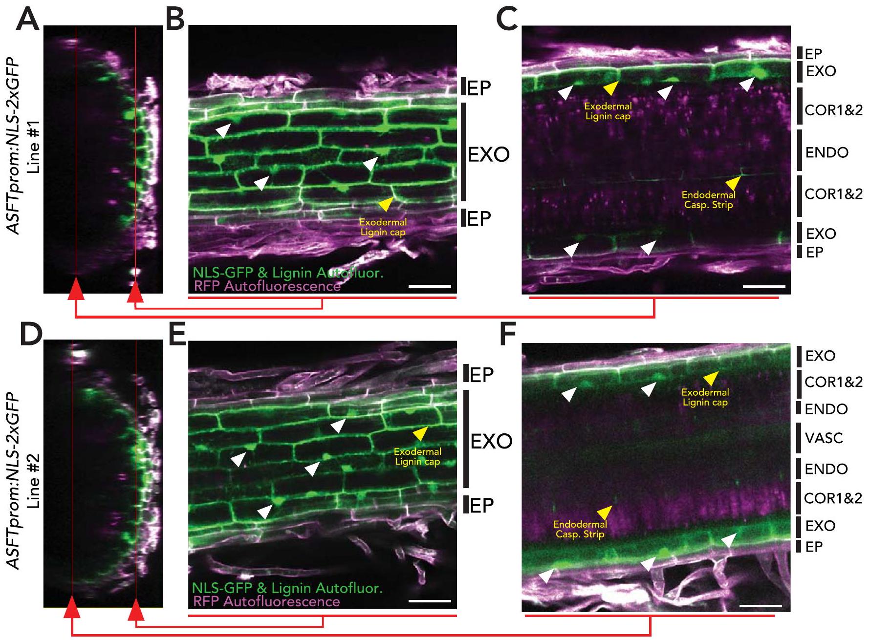
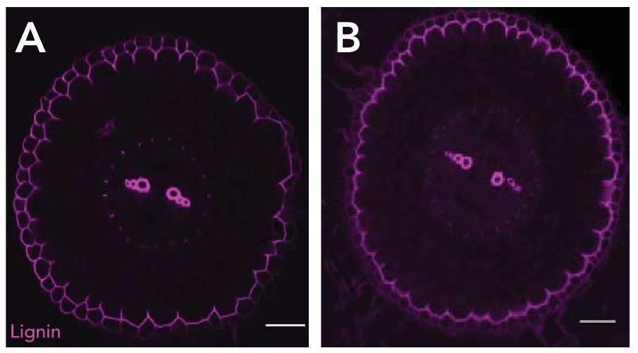
طبقات الخلايا البشرة والإكسوديرم. الأسهم البيضاء تشير إلى بعض النوى المعلمة بـ GFP في الإكسوديرم. الأسهم الصفراء تشير إلى الفلورية الذاتية لللجنين في القمة القطبية للإكسوديرم. (C و F) مقطع طولي من المستوى السفلي لكتل الز-، يظهر البشرة، الإكسوديرم، طبقات القشرة 1 و 2، والإندوديرم. الأسهم البيضاء تشير إلى النوى المعلمة بـ GFP في الإكسوديرم. الأسهم الصفراء تشير إلى الفلورية الذاتية لللجنين في القمة القطبية للإكسوديرم وشريط كاسبارين الإندوديرم. EP: البشرة؛ EXO: الإكسوديرم؛ COR1&2: طبقة القشرة 1 و 2؛ ENDO: الإندوديرم، VASC: الأوعية.
الشكل البياني الموسع الأليلات الطافرة المشتقة من R.rhizogenes التي تفقد الوظيفة للجينات المرشحة لديها ترسب غير كافٍ للسبيرين. (أ) تحليل موسع لظواهر الطفرات للجينات المرشحة في الجذور الشعرية (HR). متوسط إشارة الفلورول الأصفر عبر مقاطع عرضية متعددة (النوع البري ; استراحة ). الخط الأحمر يشير إلى فرق ذو دلالة إحصائية ( ) في شدة بكسل الفلورول الأصفر في الطفرة مقابل النوع البري كما تم تحديده باستخدام تحليل التباين الأحادي (ANOVA) متبوعًا باختبار توكي-كرامر بعد الاختبار. في معظم الحالات، تم تحليل خطين HR تم إنشاؤهما بشكل مستقل، كما هو موضح في الرسم البياني. (ب) متوسط إنزيمات تخليق السوبيرين وعوامل النسخ المنظمة لتخليق السوبيرين. الأحماض: الأحماض الدهنية؛ الكحوليات: الكحوليات الأولية؛ -OH: -أحماض دهنية هيدروكسيلية؛ DCA: حمض دهني ثنائي الكربوكسيل؛ العطرية: مشتقات الفيرولات والكومارات. أشرطة الخطأ: الانحراف المعياري. الحروف تشير إلى الفروق المعنوية (تحليل التباين الأحادي متبوعًا باختبار توكي-كرامر بعد الاختبار، علاج ABA لمدة 48 ساعة) لا تعيد السوبرين إلى مستويات النوع البري بواسطة صبغة الفلورو الأصفر في خطوط slmyb41 و slmyb92 و slmyb63. لا يمكن مقارنة متوسط كثافات البكسل بين المخططات A و D حيث تم التقاطها تحت إعدادات ليزر مختلفة. وفرة و (ج) تركيب المونومر للمتغيرات الناتجة عن R. rhizogenes من
الشكل 7 من البيانات الموسعة | ضعف ترسيب السوبيرين في الأليلات myb92-2 و asft-2 وتأثيرها على الاستجابة لـ ABA. صبغة فلورول أصفر (FY) للسوبيرين في النوع البري (مكرر من الشكل 1 للرجوع إليه)، نباتات myb92-2 و asft-2 المعالجة بالتحكم أو .
الشكل البياني الممتد 8 | طول الجذر لا يتأثر بشكل كبير بعلاج ABA. مخطط الصندوق لطول الجذر الكلي لنباتات النوع البري التي تبلغ من العمر 7 أيام والمعالجة بالعلاج الوهمي أو مابا لـ لم تجد تحليل ANOVA أحادي الاتجاه أي اختلافات ذات دلالة إحصائية.
الشكل البياني الموسع 9 | قمة اللجنين القطبية في الإكسوديرميس غير متأثرة في طفرات الجذور الشعرية myb41 و myb63 و myb92. أ. مقطع عرضي من الجذر الشعري الضابط ملون بالفوشين الأساسي. ب. مقطع عرضي من طافح الجذور الشعرية


ملطخة بالفوشين الأساسي. ج. مقطع عرضي لطفرات الجذر الشعري myb63 ملطخة بالفوشين الأساسي. د. مقطع عرضي لـ جذر شعري متحور ملون بالفوشين الأساسي. قضبان القياس .
الشكل البياني الممتد 10 | ترسيب السوبيرين في جذور ناضجة من النوع البري، myb92-1، و asft-1. مقاطع عرضية تمثيلية من الأجزاء الناضجة للجذور ملونة بالفلورول الأصفر (FY) لنباتات عمرها شهر واحد تعرضت لمدد من المياه الكافية (WS) وقيود المياه (WL) لمدة 10 أيام (الطرق).

محفظة الطبيعة

المؤلف (المؤلفون) المراسلون: شيفون برادي
آخر تحديث من المؤلف(ين): 16 أكتوبر 2023

ملخص التقرير

تتمنى Nature Portfolio تحسين قابلية إعادة إنتاج العمل الذي ننشره. يوفر هذا النموذج هيكلًا للاتساق والشفافية في التقرير. لمزيد من المعلومات حول سياسات Nature Portfolio، يرجى الاطلاع على سياسات التحرير وقائمة مراجعة سياسة التحرير.

الإحصائيات

لجميع التحليلات الإحصائية، تأكد من أن العناصر التالية موجودة في أسطورة الشكل، أسطورة الجدول، النص الرئيسي، أو قسم الطرق.
مؤكد
حجم العينة بالضبط لكل مجموعة/شرط تجريبي، معطاة كرقم منفصل ووحدة قياس
بيان حول ما إذا كانت القياسات قد أُخذت من عينات متميزة أو ما إذا كانت نفس العينة قد تم قياسها عدة مرات
اختبار(ات) الإحصاء المستخدمة وما إذا كانت أحادية الجانب أو ثنائية الجانب
يجب أن تُوصف الاختبارات الشائعة فقط بالاسم؛ واصفًا التقنيات الأكثر تعقيدًا في قسم الطرق.
وصف لجميع المتغيرات المشتركة التي تم اختبارها
وصف لأي افتراضات أو تصحيحات، مثل اختبارات الطبيعية والتعديل للمقارنات المتعددة
وصف كامل للمعلمات الإحصائية بما في ذلك الاتجاه المركزي (مثل المتوسطات) أو تقديرات أساسية أخرى (مثل معامل الانحدار) و التباين (مثل الانحراف المعياري) أو تقديرات مرتبطة بعدم اليقين (مثل فترات الثقة)
لاختبار الفرضية الصفرية، إحصائية الاختبار (على سبيل المثال ) مع فترات الثقة، أحجام التأثير، درجات الحرية و تمت ملاحظة القيمة. قدم قيم P كقيم دقيقة كلما كان ذلك مناسبًا.
لتحليل بايزي، معلومات حول اختيار القيم الأولية وإعدادات سلسلة ماركوف مونت كارلو
للتصاميم الهرمية والمعقدة، تحديد المستوى المناسب للاختبارات والتقارير الكاملة عن النتائج
تقديرات أحجام التأثير (مثل حجم تأثير كوهين) بيرسون )، مما يشير إلى كيفية حسابها
تحتوي مجموعتنا على الإنترنت حول الإحصائيات لعلماء الأحياء على مقالات تتناول العديد من النقاط المذكورة أعلاه.

البرمجيات والشيفرة

معلومات السياسة حول توفر كود الكمبيوتر
جمع البيانات
لم يتم استخدام أي برنامج لجمع البيانات.
تحليل البيانات
قص الشعر RNASEQ: TrimGalore!(v0.6.6); المحاذاة الزائفة: Kallisto v0.43.1 و v0.46.2; التجميع والتعبير المشترك: حزمة R WGCNA v1.70; معالجة البيانات: R v4.1.3; الرسم: ggplot2 v3.3.6. الخلية المفردة: التعيين: cellranger v1.1.0; المعالجة والتحليل: Seurat v4.0.5; صور TEM: EM-MENU v4.0 و IMOD v4.11;
بالنسبة للمخطوطات التي تستخدم خوارزميات أو برامج مخصصة تكون مركزية في البحث ولكن لم يتم وصفها بعد في الأدبيات المنشورة، يجب أن تكون البرمجيات متاحة للمحررين والمراجعين. نحن نشجع بشدة على إيداع الشيفرة في مستودع مجتمعي (مثل GitHub). راجع إرشادات مجموعة Nature لتقديم الشيفرة والبرمجيات لمزيد من المعلومات.

بيانات

معلومات السياسة حول توفر البيانات
يجب أن تتضمن جميع المخطوطات بيانًا حول توفر البيانات. يجب أن يوفر هذا البيان المعلومات التالية، حيثما ينطبق:
  • رموز الانضمام، معرفات فريدة، أو روابط ويب لمجموعات البيانات المتاحة للجمهور
  • وصف لأي قيود على توفر البيانات
  • بالنسبة لمجموعات البيانات السريرية أو بيانات الطرف الثالث، يرجى التأكد من أن البيان يتماشى مع سياستنا

مشاركون في الأبحاث البشرية

معلومات السياسة حول الدراسات التي تشمل المشاركين في الأبحاث البشرية والجنس والنوع في البحث.
التقارير عن الجنس والنوع الاجتماعي غير متوفر
خصائص السكان غير متوفر
التوظيف غير متوفر
رقابة الأخلاقيات غير متوفر
يرجى ملاحظة أنه يجب أيضًا تقديم معلومات كاملة حول الموافقة على بروتوكول الدراسة في المخطوطة.

التقارير المتخصصة في المجال

يرجى اختيار الخيار أدناه الذي يناسب بحثك بشكل أفضل. إذا لم تكن متأكدًا، اقرأ الأقسام المناسبة قبل اتخاذ قرارك.
علوم الحياة العلوم السلوكية والاجتماعية العلوم البيئية والتطورية والبيئية
لنسخة مرجعية من الوثيقة بجميع الأقسام، انظرnature.com/documents/nr-reporting-summary-flat.pdf

تصميم دراسة العلوم الحياتية

يجب على جميع الدراسات الإفصاح عن هذه النقاط حتى عندما يكون الإفصاح سلبياً.
حجم العينة استخدمنا ثلاث نسخ بيولوجية لتحليل RNA-Seq وأكثر من 20,000 خلية عبر جولتين في تقنيات النسخ الجيني أحادي الخلية 10X، وفقًا للممارسات الشائعة في هذا المجال. تم الإشارة إلى أحجام العينات للتجارب في الأساطير والنص. تم تحليل كل تحليل على الأقل مع ثلاث نسخ بيولوجية، وجولتين من التجارب. أسفرت النسخ البيولوجية عن نتائج مماثلة. لم يتم استخدام أي طريقة إحصائية لتحديد حجم العينة مسبقًا. عمومًا، تم اختيار حجم العينة تجريبيًا، بناءً على الخبرة السابقة حول مدى كبر حجم العينة الذي يجب أن يكون للحصول على نتيجة قابلة للتكرار وذات دلالة إحصائية على الأرجح.
استثناءات البيانات لم يتم استبعاد أي بيانات من التحليل.
التكرار تم إجراء ما لا يقل عن 3 تكرارات بيولوجية ودورتين مستقلتين من التجارب لكل تجربة لتقييم الجودة وقابلية تكرار الإجراء والأهمية الإحصائية. يتم تقديم عدد التكرارات في أساطير الأشكال حيثما كان ذلك مناسبًا.
التوزيع العشوائي تم تعيين النباتات عشوائيًا إلى أنظمة كافية من المياه أو محدودة المياه لتجارب الجفاف. تم توزيع الأواني عشوائيًا حول غرفة النمو وتم خلطها كل يومين أثناء الري طوال مدة التجارب. عند التعامل مع طفرات CRISPR، سواء في الجذور المستقرة أو الشعرية، تم أخذ عدة أليلات مستقلة لتقليل التحيز ومخاطر التأثيرات غير المستهدفة.
عمى تم إجراء تجارب النسخ الجيني دون معرفة مسبقة بنتيجة التجربة، وبالتالي لم يتم تطبيق التعمية. تم جمع وتحليل البيانات المتعلقة بالنمط الظاهري والتحليل المجهري من قبل عدة باحثين بشكل مستقل مع نتائج مشابهة. تم جمع بيانات تجربة الجفاف بشكل فعال وبطريقة عمياء من قبل باحثين مختلفين يقومون بأخذ عينات عشوائية دون معرفة مسبقة بإعداد العينة، وتم تسجيل القراءات في جهاز LICOR، وفقط بعد جمع البيانات تم تنزيلها وتحليلها.

التقارير عن مواد وأنظمة وطرق محددة

نحتاج إلى معلومات من المؤلفين حول بعض أنواع المواد والأنظمة التجريبية والأساليب المستخدمة في العديد من الدراسات. هنا، يرجى الإشارة إلى ما إذا كانت كل مادة أو نظام أو طريقة مدرجة ذات صلة بدراستك. إذا لم تكن متأكدًا مما إذا كان عنصر القائمة ينطبق على بحثك، يرجى قراءة القسم المناسب قبل اختيار رد.
المواد والأنظمة التجريبية طرق
غير متوفر مشارك في الدراسة غير متوفر مشارك في الدراسة
إكس
علم الحفريات وعلم الآثار
تصوير الأعصاب باستخدام الرنين المغناطيسي
شر البحث ذو الاستخدام المزدوج الذي يثير القلق

  1. قسم بيولوجيا النباتات ومركز الجينوم، جامعة كاليفورنيا، ديفيس، ديفيس، كاليفورنيا، الولايات المتحدة الأمريكية. إشارات النبات والبيئة، معهد البيولوجيا البيئية، جامعة أوترخت، أوترخت، هولندا. مدرسة علوم النبات والبيئة، جامعة فيرجينيا تك، بلاكسبرغ، فيرجينيا، الولايات المتحدة الأمريكية. قسم بيولوجيا النباتات الجزيئية، جامعة لوزان، لوزان، سويسرا. مرفق المجهر الإلكتروني، جامعة لوزان، لوزان، سويسرا. معهد علم النبات الخلوي والجزيئي، جامعة راينيش فريدريش فيلهلم، بون، ألمانيا. قسم بيولوجيا النباتات، جامعة كاليفورنيا، ديفيس، ديفيس، كاليفورنيا، الولايات المتحدة الأمريكية. مركز بيولوجيا خلايا النباتات، قسم علم النبات وعلوم النباتات، جامعة كاليفورنيا، ريفرسايد، ريفرسايد، كاليفورنيا، الولايات المتحدة الأمريكية. قسم علوم النبات، جامعة كاليفورنيا، ديفيس، ديفيس، كاليفورنيا، الولايات المتحدة الأمريكية. العنوان الحالي: قسم البيولوجيا التطورية والبيئية، كلية العلوم الطبيعية، معهد التطور، جامعة حيفا، حيفا، إسرائيل. الراحلة: شارون غراي. البريد الإلكتروني: sbrady@ucdavis.edu
  2. تم إيداع بيانات تسلسل RNA أحادي الخلية والكمية في GEO: GSE212405.

Journal: Nature Plants, Volume: 10, Issue: 1
DOI: https://doi.org/10.1038/s41477-023-01567-x
PMID: https://pubmed.ncbi.nlm.nih.gov/38168610
Publication Date: 2024-01-02

A suberized exodermis is required for tomato drought tolerance

Received: 31 January 2023
Accepted: 23 October 2023
Published online: 2 January 2024
(D) Check for updates

Alex Cantó-Pastor © , Kaisa Kajala (1) , Lidor Shaar-Moshe , Concepción Manzano , Prakash Timilsena , Damien De Bellis © , Sharon Gray , Julia Holbein (1) , He Yang © , Sana Mohammad (1) , Niba Nirmal , Kiran Suresh © , Robertas Ursache , G. Alex Mason , Mona Gouran , Donnelly A. West , Alexander T. Borowsky © , Kenneth A. Shackel , Neelima Sinha © , Julia Bailey-Serres © , Niko Geldner (B , Song Li , Rochus Benni Franke (1) & Siobhan M. Brady (

Abstract

Plant roots integrate environmental signals with development using exquisite spatiotemporal control. This is apparent in the deposition of suberin, an apoplastic diffusion barrier, which regulates flow of water, solutes and gases, and is environmentally plastic. Suberin is considered a hallmark of endodermal differentiation but is absent in the tomato endodermis. Instead, suberin is present in the exodermis, a cell type that is absent in the model organism Arabidopsis thaliana. Here we demonstrate that the suberin regulatory network has the same parts driving suberin production in the tomato exodermis and the Arabidopsis endodermis. Despite this co-option of network components, the network has undergone rewiring to drive distinct spatial expression and with distinct contributions of specific genes. Functional genetic analyses of the tomato MYB92 transcription factor and ASFT enzyme demonstrate the importance of exodermal suberin for a plant water-deficit response and that the exodermal barrier serves an equivalent function to that of the endodermis and can act in its place.

Plants have evolved complex cell type-specific regulatory processes to respond and adapt to dynamic environments. In certain cell types, such processes allow the formation of constitutive and inducible apoplastic diffusion barriers that regulate mineral, nutrient and water transport, pathogen entry, and have the capacity to alleviate water-deficit stress . The Arabidopsis thaliana root endodermis contains both lignified and suberized diffusion barriers, of which the latter is extremely responsive to nutrient deficiency . Many of the molecular players associated
with suberin biosynthesis and the transcriptional regulation of this biosynthetic process have been elucidated using the Arabidopsis root endodermis as a model.
Suberin is a complex hydrophobic biopolymer, composed of phenylpropanoid-derived aromatic (primarily ferulic acid) and aliphatic (poly-acylglycerol) constituents, which is deposited between the primary cell wall and the plasma membrane as a lamellar structure . While the order of the enzymatic reactions that produce suberin is not
entirely understood , many of the enzymes associated with suberin biosynthesis have been identified to function in the Arabidopsis root endodermis. These include the elongation of fatty acid acyl-CoA thioesters to very long chain fatty acid-CoA products by a fatty acid elongase complex for which the ketoacyl-CoA synthase (KCS) enzyme docosanoic acid synthase (DAISY) is the rate-limiting step . Suberin primary alcohols are formed by the fatty acyl reductase (FAR) enzymes, which reduce C18:0-C22:0 fatty acids to primary fatty alcohols . Suberin -hydroxyacids ( -OH acids) and -dicarboxylic acids are produced by cytochrome P450 monooxygenases from the subfamilies CYP86A, CYP86B and CYP94B which are -hydroxylate fatty acids . Glycerol esterification of fatty acid acyl-CoA derivatives is catalysed by the glycerol phosphate acyltransferases (GPAT) including GPAT5 (ref.10). Ferulic acid is esterified to -hydroxyacids and primary alcohols by the feruloyl transferase ALIPHATIC SUBERIN FERULOYL TRANSFERASE (ASFT)/ -HYDROXYACID/FATTY ALCOHOL HYDROXY-CINNAMOYL TRANSFERASE (FHT) . Acyl-glycerol-esters are then subjected to transport into the apoplast and used as substrates for polymerization by GDSL-type esterase/lipase (GELP) enzymes .
Many of the suberin biosynthetic enzymes acting in the root, periderm or seed were identified on the basis of their co-expression, leading to the hypothesis that a simple transcriptional module coordinates their transcription . Although the overexpression of several transcription factors can drive suberin biosynthesis in either Arabidopsis leaves or roots , the transcription of suberin biosynthetic genes is redundantly determined. It is only when a set of four Arabidopsis transcription factors-MYB41, MYB53, MYB92 and MYB93-are mutated that suberin is largely absent from the Arabidopsis root endodermis . Although not studied in roots, the Arabidopsis MYB107 and MYB9 transcription factors are required for suberin biosynthetic gene expression and suberin deposition in seeds . These data demonstrate that multiple transcription factors coordinate the expression of suberin biosynthesis genes in Arabidopsis, dependent on the organ. Furthermore, components of these transcriptional regulatory modules are probably conserved across plant species, as orthologues of many of these transcription factors and their target genes are strongly co-expressed across multiple angiosperms .
While the Arabidopsis root endodermis is well-characterized anatomically and molecularly, an additional root cell type deposits an apoplastic diffusion barrier during primary growth in other species . This cell layer is found below the epidermis, is the outermost cortical cell layer of the root and has been referred to as either the hypodermis or the exodermis. The latter term was used given observations of a potential Casparian Strip (CS). Indeed, in 93% of angiosperms studied, the exodermal layer was reported to possess an apoplastic barrier composed of suberin or lignin . Given the nature of these features, the exodermis is hypothesized to function similarly to the endodermis, although the need for two potential barrier layers is less clear . The Solanum lycopersicum (tomato) root contains both an exodermis and an endodermis. At its first stage of differentiation, a lignified cap is deposited on the outmost (epidermal) face of exodermal cell walls as well as on its anticlinal walls. During its second stage of differentiation, suberin is deposited around the entire surface of the exodermal cells . The drought or abscisic acid (ABA)-inducibility of tomato exodermal suberin is unknown as is the influence of root exodermal suberization on environmental stress responses. Given this similarity in timing and appearance of suberin between the tomato exodermis and Arabidopsis endodermis, two plausible hypotheses regarding their regulation are that they use the same regulatory networks or that they utilize distinct cell type-specific programmes. In the absence of a suberized endodermis, the plant may be more drought-susceptible, or the exodermal barrier may be sufficient to serve as the sole functional barrier.
To address these hypotheses, we profiled the transcriptional landscape of the tomato exodermis at cellular resolution and characterized suberin accumulation in response to the plant hormone ABA and in
response to water deficit. We identified a co-expression module of potential suberin-related genes, including transcriptional regulators, and validated these candidates by generating multiple CRISPR-Cas9 mutated tomato hairy root lines using Rhizobium rhizogenes and tomato plants stably transformed with Agrobacterium tumefaciens, and screened them for suberin phenotypes using histochemical techniques. The validated genes included a MYB transcription factor (SIMYB92, Solyc05g051550) whose mutant has a reduction in exodermal suberin, and the SIASFT (Solyc03g097500) whose mutant has a disrupted exodermis suberin lamellar structure with a concomitant reduction in root suberin levels. To test the hypothesis that suberin is associated with tomato’s drought response, we exposed slmyb92 and slasft mutant lines to water-deficit conditions. Both mutants displayed a disrupted response including perturbed stem water potential and leaf water status. This work describes a regulatory network with conserved parts and rewiring to yield distinct spatial localization, and contributions of specific factors to produce this environmentally responsive functional barrier.

Results

Development and composition of the suberized exodermis

We previously quantified exodermis suberin deposition along the longitudinal axis of the tomato root (cv. M82, LA3475) using the histochemical stain Fluorol Yellow (FY). In Arabidopsis roots, suberin is absent from the endodermal cells in the root meristem and elongation zones, begins to be deposited in a patchy manner in the late differentiation zone after the CS has become established, and is then followed by complete suberization in the distal differentiation zone . Quantification of exodermal suberin in 7-day-old tomato roots demonstrated the same three categories of deposition (none, patchy and complete) (Fig. 1a,c,d and Extended Data Fig. 1). Electron microscopy further demonstrated that within the completely suberized zone, suberin lamellae are deposited primarily on the epidermal and inter-exodermal faces of the exodermal cell (Fig. 1b and Extended Data Fig. 1). Suberin was consistently absent within the root endodermis throughout all developmental zones (Fig. 1a and Extended Data Fig. 1).
Monomer profiling of cell wall-associated and polymer-linked aliphatic suberin monomers in 1-month-old tomato roots revealed a predominance of -dicarboxylic acids, similar to potato . Compared with Arabidopsis roots, which mostly feature -OH acids and a maximum chain length of 24 carbons (C24) , additional C26 and C28 -OH acids and primary alcohols were observed in tomato (Extended Data Fig. 2a). This phenomenon of inter-specific variation in suberin composition has been previously observed .

Suberin biosynthetic enzymes and transcriptional regulators

To map the tomato root suberin biosynthetic pathway and its transcriptional regulators, we leveraged previous observations of relative conservation of transcriptional co-regulation of the suberin pathway across angiosperms . In the Arabidopsis root, suberin levels increase upon treatment with , a hormone which is a first responder upon water-deficit stress. Exodermal suberin deposition in tomato is similarly increased upon ABA treatment, both in terms of the region that is completely suberized as well as in the intensity of the signal (Fig. 1c,d), with the continued absence of endodermal suberin (Extended Data Fig.3).S.lycopersicum’s wild relative,Solanum pennellii (LA0716), is drought tolerant , and enhanced suberin deposition in Arabidopsis via mutation of ENHANCED SUBERIN1 (ESB1) confers drought tolerance, although esb1 also shows enhanced endodermal lignin and interrupted CS formation . Hence, we tested and confirmed the hypotheses that S. pennellii has higher suberin deposition than M82 even in water-sufficient conditions and shows no changes in the magnitude or location of suberin deposition in response to ABA in seedlings (Extended Data Figs. 2 and 3). S. pennellii suberin levels are thus constitutive. Therefore, we utilized a gene expression dataset
Fig. 1| Suberin is deposited in the tomato exodermis and is regulated by ABA. a, Graphical representation (left) of S.lycopersicum (cv. M82) root anatomy (the exodermis is highlighted in yellow) and representative cross-section (right) of a 7-day-old root stained with FY. Scale bar, . b, Transmission electron microscopy cross-sections of 7-day-old roots obtained at 1 mm from the root-hypocotyljunction. Top: the epidermal (ep), exodermal (exo) and inner cortex (co) layers. Bottom: a close-up of the featured region (zone defined with blue dotted lines), showing the presence of suberin lamellae (SL). cw, cell wall; pm, plasma membrane. c, Fluorol yellow (FY) staining for suberin in wild-type
7-day-old plants treated with mock or for 48 h . Whole-mount staining of primary root (left) and mean fluorol yellow signal along the root (right), ; error bars, s.d. Asterisks indicate significance with one-way analysis of variance (ANOVA) followed by a Tukey-Kramer post hoc test (***P<0.005). NS, not significant. d, Developmental stages of suberin deposition of wild-type plants treated with mock or for 48 h . Zones were classified as non-suberized (white), patchy suberized (grey) and continuously suberized (yellow); letters indicate statistically different groups; apostrophes indicate different statistical comparisons; ; error bars, s.d.
profiling transcription in M82 roots as well as across roots from 76 tomato introgression lines derived from . lycopersicum cv . M82 and . pennellii (LA0716) with M82 as the recurrent parent . We additionally profiled the root transcriptomes of 1-month-old tomato plants under well-watered, waterlogged and water-deficit conditions. We hypothesized that genes directly involved in the biosynthesis and deposition of suberin will be highly correlated in both water-deficit and the introgression line population.
By combining both introgression lines, waterlogging and water-deficit datasets in a weighted gene correlation network analysis (WGCNA) , we identified modules of co-expressed genes (Supplementary Fig. 1). A module (‘royalblue’) containing 180 genes was significantly enriched in suberin-related genes (odds ratio ). This was confirmed by intersection with a public dataset profiling gene expression in tomato DCRi lines (Supplementary Table 1). DCRi lines activate suberin-associated genes in the epidermal cells of fruit, which leads to suberization of the fruit surface . The ‘royalblue’ module contains several orthologues of well-known suberin biosynthetic gene families such as glycerol-3-phosphate acyltransferases (GPATs), 3-ketoacyl-CoA synthases (KCSs) and feruloyl transferases (ASFT/ FHTs) (Fig. 2a and Supplementary Table 1). In addition, putative tomato orthologues of known transcriptional regulators of suberin biosynthesis: AtMYB41, AtMYB63 and AtMYB92 (SlMYB41: Solyc02g079280; SIMYB63:Solyc10gOO5550;SIMYB92:Solyc05g051550), among others, were found in this module .

A single-cell tomato root atlas to map the exodermis

Although translatome profiles exist for the exodermis , these data do not provide resolution of the developmental gradient along which
suberin is deposited. To refine the candidate suberin-associated gene set, we conducted single-cell transcriptome profiling of the tomato root. We used the 10X Genomics scRNA-seq platform to profile over 20,000 root cells. We collected tissue from 7 -day-old primary roots of tomato (M82) seedlings up to 3 cm from the tip to include the region where suberin deposition is initially observed. Gene expression matrices were generated using cellranger and analysed in Seurat. Once the data were pre-processed and filtered for low-quality droplets, the remaining high-quality transcriptomes of 22,207 cells were analysed. After normalization, we used unsupervised clustering to identify distinct cell populations (Extended Data Fig. 4). These cell clusters were then assigned a cell type identity using the following approaches: We first quantified the overlap with existing cell type-enriched transcript sets from the tomato root and marker genes extracted from each of the clusters. An individual cluster was annotated as a specific cell type given the greatest overlap between the two sets and a significant adjusted value ( ). Then, to map gene expression dynamics across maturation, we examined cell-state progression by calculating pseudotime trajectories using a minimal spanning tree algorithm . The tree was rooted in the root meristematic zone (identified in the previous step), and clusters were grouped into 10 cell types to reflect existing biological knowledge on differentiation of the tomato root (Fig. 2b and Extended Data Fig. 4). Lastly, genes with previously validated expression patterns in tomato , transcriptional reporters and predicted cell type markers given their function in Arabidopsis , were overlaid on the clusters to refine annotation (Extended Data Fig. 4c).
Given the successful annotation of these cell types, we focused on the mapped developmental trajectories deriving from a presumed
Fig. 2 | The tomato suberin biosynthetic enzymes and transcriptional regulator are expressed in the mature exodermis. a, Simplified diagram of the suberin biosynthesis pathway. Boxes indicate gene families involved in each step of the pathway (blue and yellow indicate biosynthetic enzymes and transcriptional regulators, respectively). Genes targeted in this study are outlined in red. TFs, transcription factors; VLCFAs, very-long-chain fatty acids. b, Annotated single-cell clusters from 3 cm of the tomato root tip displayed by an integrated UMAP. R.C., root cap. Q.C., quiescent centre; col, columella; procamb, procambium.c, UMAP of cortex/endodermis/exodermis-annotated cells that were extracted from the general projection and re-embedded. A
small cluster of cells from the meristematic zone clusters were included to help anchor pseudotime estimations. CEEI, cortex-exodermis-endodermis initial. d, A pseudotime trajectory analysis for the cortex/endodermis/exodermis cell populations. e, Cell type or tissue-specific expression profiles for suberin biosynthetic pathway genes. Dot diameter represents the percentage of cells in which each gene is expressed (% Exp.) and colours indicate the average scaled expression of each gene in each developmental stage group with warmer colours indicating higher expression levels.f, Expression of SIMYB92 and SIASFT in the single-cell transcriptome data. The colour scale represents -normalized corrected UMI counts.
cortex-endodermal-exodermalinitial (CEEI) population (Fig.2c,d). Within these three associated trajectories, welocalized the cells in which the suberin biosynthetic enzymes and putative regulators were highly expressed (Supplementary Table 3). Of these, transcripts of SIASFT (Solyc03g097500), two FAR(SIFAR3A:Solyc06g074390;SIFAR3B:Solyc11g067190), two CYP86 (SICYPB86A: Solyc01g094750; SICYP86B1: Solyc02g014730), two KCS2 (SIKCS2a:Solyc09g083050 and SIKCS2b:Solyc03g005320), two GPAT(SIGPAT4:Solyc01g094700;SlGPAT5:Solyc04g011600) and one LACS(SILACS4: Solyc01g095750) showed restricted expression at the furthest edge of the exodermal developmental trajectory (Fig. 2e,f, and Supplementary Figs. 2 and 3). We generated a transcriptional reporter composed of the SIASFT promoter fused to nuclear-localized green fluorescent protein (GFP) and corroborated that its expression is restricted to the exodermis using hairy roots (Extended Data Fig. 5), in agreement with our single-cell analysis. In addition, of the three transcription factors previously noted (SIMYB41, SIMYB63and SIMYB92), only SIMYB92showed specific and restricted expression in cells at the tip of the exodermal trajectory (Fig.2 fand Supplementary Fig.3). On the basis of the co-expression and cellular trajectory data, these genes served as likely candidates for an exodermal suberintranscriptional regulator and suberin biosynthetic enzymes.

Knockout of candidate genes disrupt suberin deposition

Functional validation of these enzymes was initially performed by CRISPR-Cas9 gene editing using two or three guide RNAs (Supplementary Table 4) and were introduced into tomato via R. rhizogenes (hairy root) transformation (Fig. 3a). Deletion-confirmed mutant alleles of these genes were phenotyped for suberin levels using fluorol yellow staining (Fig. 3b and Extended Data Fig. 6). On the basis of
the histological phenotyping, all but the slcyp86b mutant showed a decrease in suberin (Fig.3b). Further confirmation of decreased suberin levels were obtained by suberin monomer metabolic profiling in the slgpat5, slgpat4, slasft, sllacs and slmyb92 mutants (Extended Data Fig. 6). These included collective reduction of ferulic acid and sinapic acid aromatic components; fatty acids (C20, C22, C24), -hydroxyacids (C18:2, C18:1, C20, C24, C26) and -diacids (C18:2, C18:1, C18, C20, C22). Given their expression in the terminus of the exodermal developmental trajectory (Fig. 2f), stable transgenic slasft and slmyb92 deletion mutant alleles were generated by transformation with . tumefaciens using the same guide RNAs. Recovered loss-of-function mutant alleles all had mutations leading to premature stop codons (Supplementary Table 4). Reduction of suberin levels as well as changes in its accumulation over the root developmental trajectory were observed in two independent mutant alleles of each gene (Fig. 3c,d and Extended Data Fig. 7). In the case of slasft, the significant delay in suberin deposition and changes in monomer composition in the exodermis differ from its orthologue in the Arabidopsis root endodermis, where the atasft mutant presents no defects in either deposition, timing or major monomer composition. The two mutants are similar in terms of their reduction in ferulate content . We also observed disorganization of the lamellar structure in the slasft-1 mutant, but not in the slmyb92-1 mutant (Fig. 3e). While reduction of ferulic acids has been found in mutants of ASFT orthologues in potato , this lamellar disorganization has not been reported before. Thus, while the enzymes responsible for suberin biosynthesis are largely the same for the tomato root exodermis and the Arabidopsis root endodermis, novelty is present in their contributions. That is, SIASFT is potentially directly responsible for
Fig. 3 | Loss-of-function mutant alleles of candidate genes disrupt exodermal suberization in tomato. a, Graphical summary of the hairy root (HR; . rhizogenes) mutant screen. b, Summary of mutant phenotypes of candidate genes in hairy roots. Top: representative cross-sections of mature portions of the roots stained with fluorol yellow. Bottom: overall quantification of the fluorol yellow signal across multiple cross-sections (wild-type ; rest ). Red line indicates statistically significant differences in fluorol yellow pixel intensity in the mutant versus wild type as determined with a one-way ANOVA followed by a Tukey-Kramer post hoc test ( ); EV, empty vector. Box plot centres depict the median while the bottom and top box limits depict the 25th and 75th percentile, respectively. Whiskers represent minima and maxima. Dots depict individual samples. c, Fluorol yellow staining for suberin in 7-day-old wild-type (repeated from Fig. 1 for reference), slmyb92-1 and slasft-1 plants treated with
mock or ABA for 48 h . Whole-mount staining of primary roots across different sections (left) and mean intensity of fluorol yellow signal along the root (right) ( ). Letters indicate significant differences (one-way ANOVA followed by a Tukey-Kramer post hoc test ( ). Error bars, s.d. d, Developmental stages of suberin deposition in the 7-day-old wild-type and mutant plants treated with mock or ABA for 48 h . Zones were classified as non-suberized (white), patchy suberized (grey) and continuously suberized (yellow) ( ). Letters indicate statistically different groups; apostrophes indicate different statistical comparisons e, Representative transmission electron microscopy cross-sections of slasft-1 and slmyb92-1 mutants obtained at 1 mm from the root-hypocotyl junction. The slasft-1 mutant presents a deficit in suberin lamellar structure.cw, cell wall; dSL, defective suberin lamellae; exo, exodermis; pm, plasma membrane; SL, suberin lamellae.
both cell wall attachment and inter-lamella adhesion, and is a primary contributor to suberin deposition.
In Arabidopsis, ABA application increases suberin levels, and four MYB transcription factors are redundantly required for induction of
ABA-mediated suberin accumulation in Arabidopsis (AtMYB41,AtMYB53, AtMYB92 and AtMYB93) . Given the ABA-inducibility of suberin in Arabidopsis and tomato (Fig. 1c,d) , we hypothesized that SIMYB92 or SIASFTare necessary for ABA-induced suberin biosynthesis. Therefore,
we treated slmyb92 and slasft roots with for 48 h , a concentration that is sufficient to increase the completely suberized zone in tomato without perturbing root length (Fig. 1a and Extended Data Fig. 8). Although suberin levels were increased upon ABA treatment in these mutant backgrounds, both in the magnitude of the fluorol yellow signal and the proportion of the root that is completely suberized, the degree to which they were increased is reduced compared with wild type (Fig. 3c,d). This decrease in suberin levels in the slmyb92-1 single mutant in both control and ABA -treated conditions is qualitatively greater than what was observed in the single mutant in Arabidopsis, which could be brought back to wild-type levels of endodermal suberin when exposed to . This difference suggests that SIMYB92 may play a more prominent role in suberin deposition than its Arabidopsis counterpart. The lack of this phenotype in the ABA-induced atmyb92-1 mutant is explained by redundancy of the AtMYB41,AtMYB53,AtMYB92 and AtMYB93 transcription factors . We explored whether such redundancy exists in tomato in the hairy root loss-of-function mutant alleles of slmyb41 and slmyb63, and whether they were sufficient to decrease exodermal suberin in control and water-deficit conditions. ABA treatment was not able to induce suberin to wild-type levels in any of the mutants (Extended Data Fig. 6d). Since the exodermis is first lignified and then suberized , we also explored whether SIMYB92, SIMYB41 or SIMYB63 were involved in lignification of the tomato root. However, the levels of lignin in the exodermis and endodermis remained unaffected in any of the hairy root mutants of these transcriptional regulators (Extended Data Fig. 9), suggesting that they do not have a role in the initial lignification of the exodermis or the endodermis.

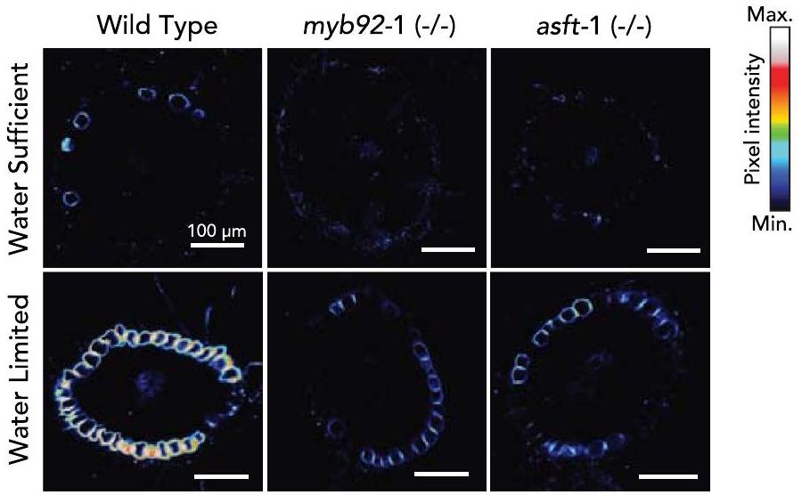
Impaired suberin deposition alters water limitation response

Given the suggested link between suberin and drought tolerance, as well as the decreased suberin levels in both control and ABA conditions in our tomato mutants, we hypothesized that the slmyb92 and slasft lines would be more sensitive to water limitation compared with wild-type plants. We subjected 4 -week-old well-watered plants to 10 days of water-deficit conditions (Methods). Suberin deposition and monomer levels were studied in the root system of slmyb92-1, slasft-1 and wild-type plants in both the water-sufficient and water-limited conditions. Under water-sufficient conditions, suberin deposition was only faintly observed in wild type, and exclusively in the exodermis, while being completely absent in both mutant lines (Extended Data Fig. 10). Consistent with this observation, very low levels of suberin monomers were detected, with no significant differences observed in the very long chain fatty acids, primary alcohols, -hydroxyacids, -dicarboxylic acids and aromatic components of suberin (Fig. 4a and Supplementary Fig. 4). Under water limitation, however, deposition of exodermal suberin was increased, with both mutant lines having lower levels than wild type (Extended Data Fig.10). The transcriptional regulator mutant slmyb92-1 showed a general reduction of most monomer groups compared with wild type. The slasft-1 mutant, in comparison, was primarily depleted in ferulic acid and its esterification substrates, as well as in individual primary alcohols and -hydroxyacids (Fig. 4a and Supplementary Fig. 4). Furthermore, stem water potential, stomatal conductance and transpiration rate were significantly decreased in response to water-limited conditions in both slmyb92-1 and slasft-1 relative to wild type, and leaf relative water content was also decreased in slmyb92-1 (Fig. 4b-e). When considering all physiological traits collectively using principal component analysis, slasft-1 showed a milder water-deficit response compared with wild-type plants, while slmyb92-1 was more extreme (Supplementary Fig. 5). These data demonstrate that decreased suberin levels in the tomato root exodermis directly perturb whole-plant performance under water-limited, but not under water-sufficient conditions. Furthermore, changes in specific suberin monomers and the lamellar structure that were observed between the two mutants in response to water-limited conditions may differently influence the extent of the physiological response.

Discussion

In the well-characterized Arabidopsis root endodermis, suberin is deposited as a hydrophobic layer between the plasma membrane and the primary cell wall . Developmentally, suberin biosynthesis and deposition occurs as a second step of endodermal differentiation, the first being the synthesis and deposition of the lignified Casparian strip . Suberin serves as an apoplastic barrier and a transcellular barrier, thus contributing to the regulation of the movement of water and solutes to the vascular cylinder . Our collective observations demonstrate that, relative to Arabidopsis, (1) the components of the pathway are conserved; (2) their spatial localization is distinct; (3) ASFT and MYB92 are critical regulators of suberin biosynthesis given their phenotypes as single loss-of-function mutant alleles, as opposed to their redundancy in Arabidopsis and (4) exodermal suberin has equivalent function to endodermal suberin and can function in its absence (Fig.1). Spatially, in the tomato root exodermis, suberin lamellae are deposited between the exodermal primary cell wall and the plasma membrane all around the cell, similar to the Arabidopsis root endodermis and other suberized apoplastic barriers such as the potato periderm (Fig. 1b). In a temporally similar fashion to the Arabidopsis endodermis, there is a non-suberized zone at the root tip, a patchy suberized zone in the middle of the root and a continuous suberized zone nearer to the roothypocotyl junction (Fig.1c,d).
We obtained clues to the underlying genes controlling exodermal suberin biosynthesis over developmental time by co-expression, single-cell transcriptome and genetic analyses. Conservation of the genes within the suberin biosynthetic pathway between Arabidopsis and tomato was evident from the functional genetic analysis of SICYP86B, SIGPAT, SILACS and SIASFT mutants. Despite the same genes controlling suberin biosynthesis, novelty in tomato is observed with respect to their tomato spatial (exodermal) expression and the critical contribution of SIASFT in primary cell wall attachment and inter-lamella adhesion of the suberin barrier. This phenotype has never been observed in Arabidopsis or potato asft mutant roots . In addition, members of the GPAT4 subclade have been regarded as exclusively involved in cutin biosynthesis , and here, SIGPAT4 was shown to participate in the formation of exodermal suberin (Fig.3). We focused on SIMYB92 as a candidate due to its expression at the end of the exodermal trajectory. Although the precise timing of these trajectories is largely predictive in nature, we note that the expression of the biosynthetic enzymes does not completely overlap that of SIMYB92 and suggests that SIMYB92 is not the sole transcriptional regulator of subering gene expression. Our ability to obtain increasingly differentiated exodermal cells is probably limited by our ability to completely protoplast cells with secondary cell wall deposition. Therefore, the lack of SIMYB41/53/93 expression in the exodermal trajectory does not mean that these genes are not expressed in the exodermis. In Arabidopsis, single loss-of-function mutants of MYB41, MYB53 and MYB93 show no changes in suberin levels, while that of shows a delay in suberization . Extreme suberin phenotypes were only observed when mutations of all four genes were combined . By contrast, this extreme phenotype (decrease of suberin levels in slmyb92 mutant alleles as measured by fluorol yellow (Fig. 3b-d) and compositional profiling in hairy roots and stable lines (Extended Data Fig. 6 and Supplementary Fig. 4)) was observed in tomato when only MYB92 was mutated. The residual suberin levels found in the slmyb92 mutants could be regulated by other MYB transcription factors. Indeed, mutants in tomato orthologues of Arabidopsis MYB41 and MYB63 showed exodermal suberin phenotypes (Extended Data Fig. 6), suggesting that these genes may be expressed in later exodermal developmental stages.
ABA-mediated regulation of tomato exodermal suberization is morphologically consistent with what is observed in the Arabidopsis root endodermis, with an increase in both the magnitude of suberin deposition and the proportion of the completely suberized zone , despite the distinct spatial localization. At least in the case of the
Fig. 4 | Impaired suberin deposition in slmyb92-1 and slasft-1 perturbs their whole-plant performances in response to water limitation. a, Suberin composition in roots of mature 1-month-old wild type, slmyb92-1 and slasft-1 plants. Plants were exposed to 10 days of water-sufficient (WS) and waterlimitation (WL) regimes ( , Methods). Acid, fatty acids; alcohols, primary alcohols; -OH, -hydroxy fatty acids; DCA, dicarboxylic fatty acid; aromatics,
ferulate and coumarate isomers. Error bars denote s.d. b-e, Dot plots of recorded values for stem water potential (stem ) (b), relative water content (c), transpiration (d) and stomatal conductance (e). Dotted line indicates zoom-in for better visual resolution of values. Black dots indicate mean values . One-way ANOVAs for each treatment were performed followed by a Tukey-Kramer post-hoc test. .
slmyb92 and slasft mutant alleles and the ABA assays, this transcription factor and biosynthetic enzyme influence both developmental and ABA-mediated suberin deposition patterns (Fig. 3c,d). Further analyses of mutant alleles of the tomato SIMYB41 and SIMYB62 transcription factors will determine whether a coordinated developmental and stress-inducible regulation of suberin biosynthesis is the norm for exodermal suberin. The degree to which this regulation is dependent on ABA signalling, as it is in Arabidopsis , also remains to be investigated. What remains to be identified, however, are the factors or regulatory elements that determine exodermal-specific regulation of these enzymes and transcriptional regulators, as well as how they are activated by ABA and why their activity is ABA-independent in S. pennellii.
External application of ABA can be considered a proxy for both drought and salt-stress response . We tested the necessity of suberized exodermis for whole-plant performance under water-limited conditions in mature tomato plants (Fig. 4). The strongly reduced response of slmyb92 and slasft plants to ABA was similarly observed upon drought stress. In both experiments, slmyb92-1 and slasft-1 failed to reach fluorol yellow signals and suberin levels equal to those of the control. Under control conditions (water-sufficient), we detected overall low suberin levels, which were near the detection limit of and reduced our ability to identify significant differences between the lines. This was consistent with the lack of distinct fluorol yellow signal in mature root sections under water-sufficient conditions (Extended Data Fig. 10). The effect observed in chemical suberin quantification may have also been attenuated by the sample comprising whole root systems with highly branched lateral roots and
including root areas with immature suberin. AtMYB92 is also known to regulate lateral root development in Arabidopsis together with its close orthologue , and differences in suberin within different root types are a possibility. Regardless, suberin monomeric levels were clearly decreased in the slmyb92-1 and slasft-1 mutants in a distinct and overlapping fashion in response to water-limited conditions. Consistent with its function, slasft-1 was primarily defective in accumulation of ferulate, primary alcohols and -hydroxyacids (Fig. 4a and Supplementary Fig. 4), while slmyb92-1 had defects in fatty acids and the predominant unsaturated -hydroxyacids and dicarboxylic acids (Fig. 4a and Supplementary Fig. 4). The more extreme perturbation of physiological responses in response to water limitation in slmyb92-1 suggests that suberin composed of these fatty acid derivatives plays a role in controlling transcellular-mediated uptake of water (Fig.4b,c). How the transcellular pathway operates in a root system where this apoplastic barrier is located four cell layers from the vascular cylinder remains an important and open question.
The role of exodermal suberin as an apoplastic barrier to water flow has been studied in maize and rice, where it was determined as a barrier to water flow, although maize and rice also present a suberized endodermis . Thus, the role of exodermal suberin alone has never been studied with respect to its influence on plant responses to water limitation. The precise role of endodermal suberin, independent of the Casparian strip, has been studied in Arabidopsis, which lacks an exodermis . In 21-day-old, hydroponically grown Arabidopsis plants, the horst-1, horst-2, horst-1 ralph-1 and pCASP1:CDEF1 mutants with a functional Casparian strip but with reduced suberin were monitored
for the importance of suberin in water relations. These mutants, except for horst-2, have higher (root hydraulic conductivity) and root aquaporin activity relative to wild type . One can extrapolate that the decreases in stem water potential, transpiration and stomatal conductance relative to wild type in water-limited conditions (Fig. 4b,c) are a consequence of decreased suberin (slmyb92) or perturbations in suberin composition (slasft). Assuming that our suberin-defective mutants have higher root hydraulic conductivity , our hypothesis to reconcile our observations with the higher Lpr would be that our mutants have compromised water-use efficiency under water limitation. This could lead to a delayed onset of the drought response such that the water loss is too great to recover by the time stomata are closed. The mechanisms by which this occurs need to be determined and could benefit from further exploration. The levels of lignin in the exodermis and endodermis were not altered in the mutants of the identified transcriptional regulators (Extended Data Fig. 9), and perturbations in endodermal lignin alone have no influence on root hydraulic conductivity in Arabidopsis, thus, lignin plays no role in our observations .
To the best of our knowledge, the response of plants with decreased root exodermal suberin levels to water limitation has never been investigated. The importance of plant radial and cellular anatomy has also long been known as critical to our understanding of the role of plant roots in water uptake in the face of water deficit. Therefore, our findings provide direct evidence, via genetic perturbation, for the role of suberin in a specific cell type mediating tomato’s adaptive response to water deficit. Further, they impart a model by which exodermal suberin barriers contribute to whole-plant water relations in the absence of a suberized endodermis. While our findings are informative about the importance of suberin in the maintenance of transpiration and stomatal conductance under soil water deficit, our conclusions are limited to a particular stage of plant growth. Changes in response to water limitation in the field, particularly with genotypes with modified suberin that impart better maintenance of water potential, remains to be investigated.
Suberin in plants roots has recently been proposed to be an avenue to combat climate change including via sequestration of atmospheric as well as conferring drought tolerance . This study provides evidence that root suberin is necessary for tomato’s response to water-deficit conditions. Increasing suberin levels within the root exodermis and/or the endodermis may indeed serve as such an avenue. The constitutive production of exodermal suberin in the drought-tolerant and wild relative of tomato, S. pennellii (Extended Data Fig. 3) , certainly provides a clue that maintenance of suberin in non-stressed and stressed conditions may result in such a benefit. However, trade-offs of such an increase must also be considered. Increased suberin levels have been associated with pathogen tolerance , but can also serve as a barrier to interactions with commensal microorganisms and constrain nutrient uptake, plant growth or seed dormancy . Regardless, this complex process serves as an elegant example of how plant evolution has resulted in a gene regulatory network with the same parts but distinct spatial rewiring (exodermis instead of endodermis) and contributions of the different genes. Collectively, this rewiring results in the distinct but precise spatiotemporal biosynthesis and deposition of this specialized polymer to perform the equivalent function of endodermal suberin in a plant’s response to the environment.

Methods

Plant material and growth conditions

All tomato (S. lycopersicum) lines used in this study were derived from cultivar M82 (LA3475). The S. pennellii line used was LA0716. Seeds were surface sterilized in 70% (v/v) ethanol for 5 min followed by 50% (v/v) commercial bleach for 20 min and three washes with sterile deionized water. Seeds were plated on plates (without sucrose) or in Magenta boxes (with sucrose) containing Murashige and Skoog (MS) medium (Caisson, MSP09-50LT), MES (pH 5.8) and agar (Difco, 214530), and maintained in a incubator
with light/dark periods for 7-10 days until cotyledons were fully expanded and the true leaves just emerged and either harvested or transferred to soil.

Tomato transformation

Hairy root transformants were generated on the basis of published work . Stable transgenic lines were generated by .tumefaciens transformation at the UC Davis Plant Transformation Facility.

Transcriptome profiling of M82 roots under drought and waterlogging stress

Seedlings of SICO2p:TRAP and AtPEPp:TRAP cv. M82 (ref. 23) were transplanted into pots with Turface Athletic Profile Field & Fairway clay substrate (Turface Athletics) pre-wetted with a nutrient water solution ( nitrogen, 18% phosphoric acid and soluble potash). Plants were grown in a completely randomized design for 31 days in a growth chamber at relative humidity, light/dark cycle and light intensity. For ‘well-watered’ conditions, we maintained substrate moisture at soil water content. For water-deficit treatment, we withheld water from the plants for 10 days before harvest, and for waterlogged conditions, we submerged the pot until the root-shoot junction. We harvested the roots as close to relative noon as feasible ( ) by immersing the pot into cool water, massaging the root ball free, rinsing three times sequentially with water, dissecting the root tissues and flash-freezing with liquid nitrogen. We harvested the lateral roots ( depth) and 1 cm root tips of adventitious roots. Sequencing libraries of adventitious roots were generated for each line in control and waterlogging conditions, and from lateral roots in control, waterlogging and water deficit conditions in four biological replicates per genotype/treatment, except for SICO2 :TRAP lateral roots in control conditions (five biological replicates). Total RNA was isolated from these roots as previously described , and non-strand specific random primer-primed RNA-seq library construction was performed as originally described . RNA-seq libraries were pooled and sequenced with the Illumina HiSeq4000 (50SR).

RNA-seq data processing and analysis for drought, water deficit and introgression line population

RNA-sequencing data processing and analyses for the drought, waterlogging and introgression line population were conducted as previously described . Sequences were pooled, and then trimmed and filtered using TrimGalore! (v.0.6.6) , with parameter -a GATCGGAAGAGCACA. Trimmed reads were pseudo-aligned to the ITAG3.2 transcriptome (complementary DNA) using Kallisto (v.0.43.1) , with the parameters-b100-single-1200-s30, to obtain count estimates and transcripts per million (TPM) values. Samples were clustered with cuttreestatic and outliers removed (GSM2323699) with a minSize of 10.

Generation of tomato CRISPR constructs

Guide RNAs (gRNAs) targeting exons were designed using the CRISPR-PLANT web tool (https://www.genome.arizona.edu/crispr/ CRISPRsearch.html) (Supplementary Table 4). If this did not specify at least three guides with GC content between 40 to , guides were designed with CRISPR-P V2 (http://crispr.hzau.edu.cn/cgi-bin/ CRISPR2/CRISPR) using the U6 snoRNA promoter with mismatches within the target gene coding sequence. Genomic sequences (ITAG3.2) were retrieved from Phytozome (https://phytozome-next.jgi.doe.gov/). Primer specificity was checked against S. lycopersicum RNA entries from NCBI’s RefSeq RNA. In cases where only two gRNAs were selected, olives containing gRNA sequence were ligated into pMR217/218 vectors and recombined via Gateway assembly into a pMR290 vector containing Cas9 and Kan resistance expression cassettes . In cases where three gRNAs were selected, gRNAs were phosphorylated and ligated into pYPQ131-3 vectors, and then recombined into pYPQ143 via Golden
Gate assembly. A pMR278 vector containing all threegRNA expression cassettes was then recombined into pMR286/289.

Generation of the SIASFT transcriptional reporter construct driving a nuclear-localized GFP

A 2-kb region upstream of the ATG codon (oligos in Supplementtary Table 5) was subcloned into D-TOPO and then recombined into pMK7FNFm14GW .

CRISPR-Cas9 mutant generation and analysis
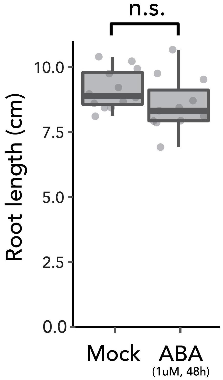
Independently transformed lines were genotyped at the targeted genomic region (Supplementary Tables 4 and 5). In the case of hairy roots, at least 2 lines containing large deletions in both alleles in the gene of interest were kept for further analysis. In the case of stable transformants, first-generation ( ) transgenics were genotyped and self-pollinated. plants with homozygous mutant alleles were selected. and were used in subsequent experiments.

Water-deficit assay

Seedlings (7-day-old) were transferred to 0.51 cones containing Turface pre-wetted with a nutrient water solution (containing nitrogen, phosphoric acid and soluble potash). All pots were weight adjusted and a small set of pots were dried so that the percentage of water in the soil could be calculated. Plants were then grown in a completely randomized design for 3 weeks in a growth chamber at , relative humidity, light/dark cycle and light intensity, and watered to soil saturation every other day. At the end of the first week, vermiculite was added to limit water evaporation from the soil. After 3 weeks, plants of each line were randomly assigned into two treatment groups (six plants each) and exposed to different treatments for 10 days. Control plants were watered to soil saturation with nutrient solution every day. Water-limited plants were exposed to water deficit by adjusting pot weights daily with nutrient solution (to the highest weight of the set) until a target soil water content of 40-50% was obtained. On the day of harvesting, between 09:00 to 12:00, stomatal conductance and transpiration were measured on the abaxial surface of the terminal leaflet of the third leaf or the youngest fully expanded leaf using a LICOR-6400XT portable photosynthesis system. Light intensity was kept at , with a constant air flow rate of and a reference concentration of air. The third (either left or right) primary leaflet was collected for measuring relative water content using a modified version of a previously established protocol . Fresh leaves were cut with a scalpel leaving a -long petiole and the total fresh weight (TFW) was measured. Leaves were then placed in individual zipper-locked plastic bags containing 1 ml of deionized water, making sure that only the leaf petiole is immersed in the solution. Bags were incubated at . After 8 h , leaves were taken out of the bags, placed between two paper towels to absorb excess water and then weighed to determine the turgid weight (TW). Each sample was then placed into a paper bag and dried in a dry oven for days. Dried samples were weighed (DW), and relative water content was calculated as: RWC . A section of the fourth leaf, containing the terminal and primary leaflets, was used to measure stem water potential using a pump-up pressure chamber (PMS Instrument). The root systems were harvested by immersing the cone into water, massaging the root ball free, rinsing and removing excess water with paper towels. The middle section of the root system was sectioned using a scalpel. Around 300 mg of the dissected root tissue were added to Ankon filter bags (sealed with a staple). Bags were transferred into a glass beaker, an excess of chloroform:methanol ( ) was added and extracted for 2 h . Fresh chloroform:methanol ( ) was replaced and the extraction was repeated overnight under gentle agitation (twice). Fresh chloroform:methanol ( ) was added and samples further extracted for 2 h . The extraction was repeated overnight twice with
fresh chloroform:methanol ( ). Finally, samples were extracted with methanol for 2 h . Methanol was removed and bags were dried in a vacuum desiccator for 72 h . Suberin monomer analysis was performed in these samples as described below.

ABA assay

At 5 days after germination, seedlings from a plate were randomly transferred to fresh MS plates containing either ABA or mock. After 48 h of treatments in a incubator with light/dark, roots were harvested and used in subsequent analyses.

Co-expression network analysis

Co-expression network modules were generated with the WGCNA (v.1.70). Libraries were quantile normalized and a soft threshold of 8 was used to create a scale-free network. A signed network was created choosing a soft thresholding power of 8 , minModuleSize of 30 , module detection sensitivity deepSplit of 2 and mergeCutHeight of 0.3. Genes with a consensus eigengene connectivity to their module eigengene of lower than 0.2 were removed from the module (minKMEtoStay). Modules were correlated with upregulated genes in DCRi lines described previously (Fisher’s exact test).

Protoplast isolation and scRNA-seq

Seven days after sowing, 50-100 primary roots per sample of length -3 cm from the root tip were cut and placed in a -diameter dish containing a cell strainer and 4.5 ml enzyme solution ( Cellulase R10, 1.25% Cellulase RS, 0.3% Macerozyme R10, 0.12% Pectolyase, 0.6 M mannitol, 20 mM MES ( pH 5.7 ), , 0.1% bovine serum albumin and 0.000194% (v/v) mercaptoethanol). Cellulase Onozuka R10, Cellulase Onozuka RS and Macerozyme R10 were obtained from Yakoult Pharmaceutical. Pectolyase was obtained from Sigma-Aldrich (P3026). After digestion at for 2 h at 85 r.p.m. on an orbital shaker with occasional stirring, the cell solution was filtered twice through cell strainers and centrifuged for 5 min at 500 g in a swinging bucket centrifuge with the acceleration set to minimal. Subsequently, the pellet was resuspended with 1 ml washing solution ( 0.6 M mannitol, 20 mM MES (pH 5.7), bovine serum albumin and mercaptoethanol) and centrifuged for 3 min at 500 g . The pellet was resuspended with 1 ml of washing solution and transferred to a 1.7 ml microcentrifuge tube. Samples were centrifuged for 3 min at and resuspended to a final concentration of cells per l. The protoplast suspension was then loaded onto microfluidic chips (10X Genomics) with v3 chemistry to capture 10,000 cells per sample. Cells were barcoded with a Chromium Controller (10X Genomics). Messenger RNA was reverse transcribed and Illumina libraries were constructed for sequencing with reagents from a3′ Gene Expression v3 kit (10X Genomics) according to manufacturer instructions. Sequencing was performed with a NovaSeq 6000 (100-PE).

Protoplasting-induced genes

Once protoplasts were purified, total RNA was extracted using the Direct-zol RNA miniprep kit (ZYMO). Bulk RNA-seq libraries were prepared using the QuantSeq 3′ mRNA-seq library prep kit FWD (Lexogen). Barcoded libraries were pooled, and PE 150-bp reads were sequenced on the NovaSeq 6000 instrument (Illumina) at the UC Davis DNA Technologies Core. Sequences were pooled, and then trimmed and filtered using TrimGalore! (v.0.6.6). R1 trimmed reads were pseudo-aligned to ITAG4.1 transcriptome (cDNA) using Kallisto (v.0.46.2), with the parameter -b 100, to obtain count estimates and TPM values. Differentially expressed genes with and were selected as protoplast-induced genes (edgeR v.3.34.1).

Single-cell transcriptome analysis

FASTQ files were mapped using cellranger (10X Genomics). Reads were aligned to the tomato genome (SL4.0) with the ITAG4.1 gene annotation
file with organellar (mitochondria and plastid) sequences appended. Protoplasting-induced (Supplementary Table 2) genes, genes with counts in 3 cells or less, low-quality cells that contained <500 unique molecular identifiers (UMIs) and cells with >1% UMI counts belonging to organelle genes were filtered out. Data were then normalized using Seurat (v.4.0.5) , followed by principal component analysis (PCA) and nonlinear dimensionality reduction using uniform manifold approximation and projection (UMAP). Fifty principal components were calculated and UMAP embedding was generated using the initial 35 principal components. Cluster-enriched genes were computed using the ‘FindAllMarkers’ function in Seurat using the only.pos = TRUE, min. pct , logfc.threshold parameters.

Single-cell cluster annotation

Clusters were annotated based on the overlap of cluster marker genes and a set of cell type-enriched marker genes from ref. 23. Given a set of tissue-specific markers for number of tissue types, we call these sets , with representing genes in the marker list. These markers are mutually exclusive such that no genes appear in two different sets ( for any ). We identified the marker genes from Seurat-generated cluster markers , ( ), where equals the number of Seurat-generated clusters. We generated an overlapping table between markers and markers, which we represent in the table as . For each Seurat cluster, we hypothesized that the cells with the highest number of overlapping markers is the cell type of this cluster. A chi-squared test was used to determine the significance of overlap with Bonferroni correction:
with and
number of highest overlapping markers
expected number of overlapping markers sum number of tissue types
O2 = sum of markers that overlap with all other clusters sum ( ) imax
number of markers that overlap with all other clusters
This process was repeated for the second and third highest overlapping markers until the corrected value was higher than 0.01. An individual cluster was assigned a tissue annotation type that had the most genes overlapping between the two marker sets, provided the value was significant for the overlap.

Trajectory analysis

A trajectory analysis was run for the ground tissue cells after selecting and re-clustering the cell types annotated as exodermis and meristematic zone (clusters ). Gene expression matrices, dimensionality reduction and clustering were imported into the dynverse wrapper from Seurat and a starting cell was decided within the meristematic zone cluster. Trajectory inference was run using the minimum spanning tree (MST) algorithm. The MST method and UMAP coordinates from Seurat were used as input for mclust . Predictive genes or genes that were differentially expressed along the trajectory, specific branches and milestones were identified and visualized with a heat map using dynfeature within the R package dynverse.

Histochemical and imaging analysis

For sections, roots were divided in segments, embedded in agarose and sliced in sections using a vibratome. Sections were then incubated in FY088 ( , dissolved in lactic acid) for 1 h at room temperature in darkness, rinsed three times with water
and counterstained with aniline blue ( , dissolved in water) for 1 h in darkness. Confocal laser scanning microscopy was performed on a Zeiss Observer Z1 confocal microscope with the objective and GFP filter ( 488 nm excitation, emission). For whole roots, suberin was observed in 7-day-old S. lycopersicum wild-type or mutant seedlings. Whole roots were incubated in methanol for 3 days, changing the methanol daily. Once cleared, roots were incubated in fluorol yellow 088 ( , dissolved in methanol) for 1 h at room temperature in the dark, rinsed three times with methanol and counterstained with aniline blue ( , dissolved in methanol) for 1 h at room temerature in the dark. Roots were mounted and observed with the EVOS cell imaging system (Thermo Fisher) using the GFP filter ( 488 nm excitation, emission). Root sections were also stained with basic fuchsin (Fisher Scientific, 632-99-5). ments from the root tip were embedded in agarose and sectioned at 150-200 using a vibratome (Leica VT1000 S). The sections were stained in Clearsee with basic fuchsin for 30 min and then washed two times and imaged with a Zeiss LSM700 confocal microscope with the objective; basic fuchsin: 550-561 nm excitation and detection. Hairy roots of SIASFT transcriptional fusions were imaged with the same confocal and objective, but with excitation at 488 nm and emission at for GFP, and excitation at 555 nm and emission at for red fluorescent protein (RFP) autofluorescence.

Suberin monomer analysis

An average of 80 mg fresh weight root tissue per biological replicate (four biological replicates) was washed and immediately placed in a 2:1 solution of chloroform:methanol. Subsequently, root samples were extracted in a Soxhlet extractor for 8 h , first with , afterwards with methanol to remove all soluble lipids. The delipidated tissues were dried in a desiccator over silica gel and weighed. Suberin monomers were released using boron trifluoride in methanol at overnight. Dotriacontane was added to each sample ( ) as an internal standard, saturated was used to stop the transesterification reaction, and monomers were extracted with . The fraction was washed with water and residual water removed using . The fraction was then concentrated down to and derivatized with , -bis-trimethylsilyltrifluoroacetamide (BSTFA) and pyridine at for 40 min . Compounds were separated using gas chromatography (GC) and detected using a flame ionization detector ( 6890 N Network GC System, Agilent Technologies) as previously described . Compound identification was accomplished using an identical gas chromatography system paired with a mass spectroscopy selective detector (GC-MS; 5977A MSD, Agilent Technologies). Compounds were identified by their characteristic fragmentation spectra pattern with reference to an internal library of common suberin monomers and the NIST database.

Transmission electron microscopy

Tomato roots were fixed in 2.5% glutaraldehyde solution (EMS) in phosphate buffer ( ) for 1 h at room temperature and subsequently fixed in a fresh mixture of osmium tetroxide (1%, EMS) with 1.5% potassium ferrocyanide (Sigma) in PB buffer for 1 h . The samples were then washed twice in distilled water and dehydrated in acetone solution (Sigma) in a concentration gradient ( for 40 min , for for 40 min and for 1 h three times). This was followed by infiltration in LR White resin (EMS) in a concentration gradient ( LR White in acetone for White in acetone for 6 h and White for 12 h two times) and finally polymerized for 48 h at in an oven in atmospheric nitrogen. Ultrathin sections were cut transversely at 2,5 and 8 mm from the root tip, the middle of the root and 1 mm below the hypocotyl-root junction using a Leica Ultracut UC7 (Leica Mikrosysteme), picked up on a copper slot
grid (EMS) and coated with a polystyrene film (Sigma). Micrographs and panoramic images were taken with an FEI transmission electron microscope (FEI CM100) at an acceleration voltage of 80 kV with a TVIPS TemCamF416 digital camera (TVIPS) using the software EM-MENU (v.4.0) (TVIPS). Panoramic images were aligned with the software IMOD (v.4.11) .

Phylogenetic tree construction

Phylogenetic trees were generated using the methods described in ref. 23.

Statistics and reproducibility

All statistical analyses were done in the R environment (v.4.1.3), and derived plots done in ggplot2 (v.3.3.6). For multiple comparisons between genotypes, a one-way ANOVA was performed with a TukeyKramer post hoc test. Groups in which differences gave a value lower than 0.05 were considered significantly different. All bar graphs represent mean, and error bars denote s.d. For all box plots, the centre depicts the median and the lower and upper box limits depict the 25th and 75th percentile, respectively. Whiskers represent minima and maxima. Closed dots depict individual samples. In all cases, individual biological samples are stated as n. Experiments and representative images were repeated independently at least three times, unless otherwise stated. Individual values for all statistical analyses can be found in Supplementary Table 6.

Reporting summary

Further information on research design is available in the Nature Portfolio Reporting Summary linked to this article.

Data availability

Single-cell and bulk RNA-seq data have been deposited at GEO (GSE212405) and have been made publicly available. CRISPR-generated mutant lines are available upon request. Timing is dependent upon obtaining phytosanitary certificates according to seed import regulations of the country of destination and associated costs.

References

  1. Baxter, I. et al. Root suberin forms an extracellular barrier that affects water relations and mineral nutrition in Arabidopsis. PLoS Genet. 5, e1000492 (2009).
  2. Thomas, R. et al. Soybean root suberin: anatomical distribution, chemical composition, and relationship to partial resistance to Phytophthora sojae. Plant Physiol. 144, 299-311 (2007).
  3. Barberon, M. et al. Adaptation of root function by nutrient-induced plasticity of endodermal differentiation. Cell 164, 447-459 (2016).
  4. Molina, I., Li-Beisson, Y., Beisson, F., Ohlrogge, J. B. & Pollard, M. Identification of an Arabidopsis feruloyl-coenzyme A transferase required for suberin synthesis. Plant Physiol. 151, 1317-1328 (2009).
  5. Serra, O. & Geldner, N. The making of suberin. New Phytol. https://doi.org/10.1111/nph. 18202 (2022).
  6. Franke, R. et al. The DAISY gene from Arabidopsis encodes a fatty acid elongase condensing enzyme involved in the biosynthesis of aliphatic suberin in roots and the chalaza-micropyle region of seeds. Plant J. 57, 80-95 (2009).
  7. Domergue, F. et al. Three Arabidopsis fatty acyl-coenzyme A reductases, FAR1, FAR4, and FAR5, generate primary fatty alcohols associated with suberin deposition. Plant Physiol. 153, 1539-1554 (2010).
  8. Compagnon, V. et al. CYP86B1 is required for very long chain omega-hydroxyacid and alpha, omega -dicarboxylic acid synthesis in root and seed suberin polyester. Plant Physiol. 150, 1831-1843 (2009).
  9. Höfer, R. et al. The Arabidopsis cytochrome P450 CYP86A1 encodes a fatty acid -hydroxylase involved in suberin monomer biosynthesis. J. Exp. Bot. 59, 2347-2360 (2008).
  10. Beisson, F., Li, Y., Bonaventure, G., Pollard, M. & Ohlrogge, J. B. The acyltransferase GPAT5 is required for the synthesis of suberin in seed coat and root of Arabidopsis. Plant Cell 19, 351-368 (2007).
  11. Gou, J.-Y., Yu, X.-H. & Liu, C.-J. A hydroxycinnamoyltransferase responsible for synthesizing suberin aromatics in Arabidopsis. Proc. Natl Acad. Sci. USA 106, 18855-18860 (2009).
  12. Serra, O. et al. A feruloyl transferase involved in the biosynthesis of suberin and suberin-associated wax is required for maturation and sealing properties of potato periderm. Plant J. 62, 277-290 (2010).
  13. Ursache, R. et al. GDSL-domain proteins have key roles in suberin polymerization and degradation. Nat. Plants 7, 353-364 (2021).
  14. Shukla, V. et al. Suberin plasticity to developmental and exogenous cues is regulated by a set of MYB transcription factors. Proc. Natl Acad. Sci. USA 118, e2101730118 (2021).
  15. Kosma, D. K. et al. AtMYB41 activates ectopic suberin synthesis and assembly in multiple plant species and cell types. Plant J. 80, 216-229 (2014).
  16. Cohen, H., Fedyuk, V., Wang, C., Wu, S. & Aharoni, A. SUBERMAN regulates developmental suberization of the Arabidopsis root endodermis. Plant J. 102, 431-447 (2020).
  17. Lashbrooke, J. et al. MYB107 and MYB9 homologs regulate suberin deposition in angiosperms. Plant Cell 28, 2097-2116 (2016).
  18. Gou, M. et al. The MYB107 transcription factor positively regulates suberin biosynthesis. Plant Physiol. 173, 1045-1058 (2017).
  19. Enstone, D. E., Peterson, C. A. & Ma, F. Root endodermis and exodermis: structure, function, and responses to the environment. J. Plant Growth Regul. 21, 335-351 (2002).
  20. Perumalla, C. J., Peterson, C. A. & Enstone, D. E. A survey of angiosperm species to detect hypodermal Casparian bands. I. Roots with a uniseriate hypodermis and epidermis. Bot. J. Linn. Soc. 103, 93-112 (1990).
  21. Barberon, M. The endodermis as a checkpoint for nutrients. New Phytol. 213, 1604-1610 (2017).
  22. Geldner, N. The endodermis. Annu. Rev. Plant Biol. 64, 531-558 (2013).
  23. Kajala, K. et al. Innovation, conservation, and repurposing of gene function in root cell type development. Cell 184, 3333-3348.e19 (2021).
  24. Ron, M. et al. Hairy root transformation using Agrobacterium rhizogenes as a tool for exploring cell type-specific gene expression and function using tomato as a model. Plant Physiol. 166, 455-469 (2014).
  25. Naseer, S. et al. Casparian strip diffusion barrier in Arabidopsis is made of a lignin polymer without suberin. Proc. Natl Acad. Sci. USA 109, 10101-10106 (2012).
  26. Franke, R. et al. Apoplastic polyesters in Arabidopsis surface tissues-a typical suberin and a particular cutin. Phytochemistry 66, 2643-2658 (2005).
  27. Kolattukudy, P. E., Kronman, K. & Poulose, A. J. Determination of structure and composition of suberin from the roots of carrot, parsnip, rutabaga, turnip, red beet, and sweet potato by combined gas-liquid chromatography and mass spectrometry. Plant Physiol. 55, 567-573 (1975).
  28. Legay, S. et al. MdMyb93 is a regulator of suberin deposition in russeted apple fruit skins. New Phytol. 212, 977-991 (2016).
  29. Gong, P. et al. Transcriptional profiles of drought-responsive genes in modulating transcription signal transduction, and biochemical pathways in tomato. J. Exp. Bot. 61, 3563-3575 (2010).
  30. Eshed, Y., Abu-Abied, M., Saranga, Y. & Zamir, D. Lycopersicon esculentum lines containing small overlapping introgressions from L. pennellii. Theor. Appl. Genet. 83, 1027-1034 (1992).
  31. Gur, A. et al. Yield quantitative trait loci from wild tomato are predominately expressed by the shoot. Theor. Appl. Genet. 122, 405-420 (2011).
  32. Toal, T. W. et al. Regulation of root angle and gravitropism. G3 8, 3841-3855 (2018).
  33. Langfelder, P. & Horvath, S. WGCNA: an R package for weighted correlation network analysis. BMC Bioinformatics 9, 559 (2008).
  34. Du, H. et al. The evolutionary history of R2R3-MYB proteins across 50 eukaryotes: new insights into subfamily classification and expansion. Sci. Rep. 5, 11037 (2015).
  35. Saelens, W., Cannoodt, R., Todorov, H. & Saeys, Y. A comparison of single-cell trajectory inference methods. Nat. Biotechnol. 37, 547-554 (2019).
  36. Bucher, M., Schroeer, B., Willmitzer, L. & Riesmeier, J. W. Two genes encoding extensin-like proteins are predominantly expressed in tomato root hair cells. Plant Mol. Biol. 35, 497-508 (1997).
  37. Bucher, M. et al. The expression of an extensin-like protein correlates with cellular tip growth in tomato. Plant Physiol. 128, 911-923 (2002).
  38. Howarth, J. R., Parmar, S., Barraclough, P. B. & Hawkesford, M. J. A sulphur deficiency-induced gene, sdi1, involved in the utilization of stored sulphate pools under sulphur-limiting conditions has potential as a diagnostic indicator of sulphur nutritional status. Plant Biotechnol. J. 7, 200-209 (2009).
  39. von Wirén, N. et al. Differential regulation of three functional ammonium transporter genes by nitrogen in root hairs and by light in leaves of tomato. Plant J. 21, 167-175 (2000).
  40. Gómez-Ariza, J., Balestrini, R., Novero, M. & Bonfante, P. Cell-specific gene expression of phosphate transporters in mycorrhizal tomato roots. Biol. Fertil. Soils 45, 845-853 (2009).
  41. Nieves-Cordones, M., Alemán, F., Martínez, V. & Rubio, F. The Arabidopsis thaliana HAK5 K+ transporter is required for plant growth and acquisition from low solutions under saline conditions. Mol. Plant 3, 326-333 (2010).
  42. Jones, M. O. et al. The promoter from SIREO, a highly-expressed, root-specific Solanum lycopersicum gene, directs expression to cortex of mature roots. Funct. Plant Biol. 35, 1224-1233 (2008).
  43. Ho-Plágaro, T., Molinero-Rosales, N., Fariña Flores, D., Villena Díaz, M. & García-Garrido, J. M. Identification and expression analysis of GRAS transcription factor genes involved in the control of arbuscular mycorrhizal development in tomato. Front. Plant Sci. 10, 268 (2019).
  44. Li, P. et al. Spatial expression and functional analysis of Casparian strip regulatory genes in endodermis reveals the conserved mechanism in tomato. Front. Plant Sci. 9, 832 (2018).
  45. Bouzroud, S. et al. Auxin Response Factors (ARFs) are potential mediators of auxin action in tomato response to biotic and abiotic stress (Solanum lycopersicum). PLoS ONE 13, eO193517 (2018).
  46. Shahan, R. et al. A single-cell Arabidopsis root atlas reveals developmental trajectories in wild-type and cell identity mutants. Dev. Cell https://doi.org/10.1016/j.devcel.2022.01.008 (2022).
  47. Andersen, T. G. et al. Tissue-autonomous phenylpropanoid production is essential for establishment of root barriers. Curr. Biol. 31, 965-977.e5 (2021).
  48. Hosmani, P. S. et al. Dirigent domain-containing protein is part of the machinery required for formation of the lignin-based Casparian strip in the root. Proc. Natl Acad. Sci. USA 110, 14498-14503 (2013).
  49. Calvo-Polanco, M. et al. Physiological roles of Casparian strips and suberin in the transport of water and solutes. New Phytol. 232, 2295-2307 (2021).
  50. Beisson, F., Li-Beisson, Y. & Pollard, M. Solving the puzzles of cutin and suberin polymer biosynthesis. Curr. Opin. Plant Biol. 15, 329-337 (2012).
  51. Zhu, J.-K. Salt and drought stress signal transduction in plants. Annu. Rev. Plant Biol. 53, 247-273 (2002).
  52. Raghavendra, A. S., Gonugunta, V. K., Christmann, A. & Grill, E. ABA perception and signalling. Trends Plant Sci. 15, 395-401 (2010).
  53. Gibbs, D. J. et al. AtMYB93 is a novel negative regulator of lateral root development in Arabidopsis. New Phytol. 203, 1194-1207 (2014).
  54. Zimmermann, H. M., Hartmann, K., Schreiber, L. & Steudle, E. Chemical composition of apoplastic transport barriers in relation to radial hydraulic conductivity of corn roots (Zea mays L.). Planta 210, 302-311 (2000).
  55. Steudle, E. Water uptake by roots: effects of water deficit. J. Exp. Bot. 51, 1531-1542 (2000).
  56. Thompson, A. The new plants that could save us from climate change. Popular Mechanics https://www. popularmechanics.com/science/green-tech/a14000753/ the-plants-that-could-save-us-from-climate-change/ (2017).
  57. Pillay, I. & Beyl, C. Early responses of drought-resistant and -susceptible tomato plants subjected to water stress. J. Plant Growth Regul. 9, 213-219 (1990).
  58. Gur, A. & Zamir, D. Unused natural variation can lift yield barriers in plant breeding. PLoS Biol. 2, e245 (2004).
  59. Holbein, J. et al. Root endodermal barrier system contributes to defence against plant-parasitic cyst and root-knot nematodes. Plant J. 100, 221-236 (2019).
  60. Kashyap, A. et al. Induced ligno-suberin vascular coating and tyramine-derived hydroxycinnamic acid amides restrict Ralstonia solanacearum colonization in resistant tomato. New Phytol. 234, 1411-1429 (2022).
  61. Salas-González, I. et al. Coordination between microbiota and root endodermis supports plant mineral nutrient homeostasis. Science 371, eabd0695 (2021).
  62. To, A. et al. AtMYB92 enhances fatty acid synthesis and suberin deposition in leaves of Nicotiana benthamiana. Plant J. 103, 660-676 (2020).
  63. Reynoso, M. A. et al. Evolutionary flexibility in flooding response circuitry in angiosperms. Science 365, 1291-1295 (2019).
  64. Townsley, B. T., Covington, M. F., Ichihashi, Y., Zumstein, K. & Sinha, N. R. BrAD-seq: Breath Adapter Directional sequencing: a streamlined, ultra-simple and fast library preparation protocol for strand specific mRNA library construction. Front. Plant Sci. 6, 366 (2015).
  65. Krueger, F. Trim Galore (Babraham Bioinformatics, 2012).
  66. Bray, N. L., Pimentel, H., Melsted, P. & Pachter, L. Near-optimal probabilistic RNA-seq quantification. Nat. Biotechnol. 34, 525-527 (2016).
  67. Bari, V. K. et al. CRISPR/Cas9-mediated mutagenesis of CAROTENOID CLEAVAGE DIOXYGENASE 8 in tomato provides resistance against the parasitic weed Phelipanche aegyptiaca. Sci. Rep. 9, 11438 (2019).
  68. Karimi, M., Bleys, A., Vanderhaeghen, R. & Hilson, P. Building blocks for plant gene assembly. Plant Physiol. 145, 1183-1191 (2007).
  69. Sade, N., Galkin, E. & Moshelion, M. Measuring Arabidopsis, tomato and barley leaf relative water content (RWC). Bio-Protocol 5, e1451-e1451 (2015).
  70. Satija, R., Farrell, J. A., Gennert, D., Schier, A. F. & Regev, A. Spatial reconstruction of single-cell gene expression data. Nat. Biotechnol. 33, 495-502 (2015).
  71. Ron, M. et al. Identification of novel loci regulating interspecific variation in root morphology and cellular development in tomato. Plant Physiol. 162, 755-768 (2013).
  72. Scrucca, L., Fop, M., Murphy, T. B. & Raftery, A. E. mclust 5: clustering, classification and density estimation using Gaussian finite mixture models. R J. 8, 289-317 (2016).
  73. Kremer, J. R., Mastronarde, D. N. & McIntosh, J. R. Computer visualization of three-dimensional image data using IMOD. J. Struct. Biol. 116, 71-76 (1996).

Acknowledgements

We thank A. M. Bagman and K. Zumstein for experimental assistance; C. Bian, K. Morimoto and T. Taylor for discussion; D. Kawa and G. Demirer for manuscript critiquing. Funding was as follows: A.C.-P., S.M.B., N.S., J.B.-S. by NSF PGRP IOS-1856749 and IOS-211982O. S.M.B. by HHMI 55108506. K.K. by SKR postdoctoral fellowship and MSCA RI fellowship 790057. L.S.-M. by BARD FI-570-2018 and HFSP RGP0067. C.M. by MSCA GF 655406. P.T. and S.L. by DOE DE-SC0020358 and NSF MCB-2218234. D.A.W. by Elsie J. Stocking fellowship PBD-UCD. S.L. by USDA-HATCH programme. R.U. by EMBO ALTF 1046-2015. R.U. and J.H. were financially supported by the German Federal Ministry of Food and Agriculture (BMEL) through the Federal Office for Agriculture and Food (BLE), grant number 2821ERA16C. G.A.M. by NSF PRFB IOS1907008. A.T.B. and J.B.-S. by NSF DGE-1922642. A.T.B. by USDA NIFA 1026477.

Author contributions

A.C.-P., K.K., L.S.-M., S.G., C.M., N.N., D.A.W., K.A.S., N.S., J.B.-S., N.G., S.L., R.B.F. and S.M.B. conceptualized the project. A.C.-P., P.T., S.L., R.B.F. and S.M.B. developed the methodology. A.C.-P., K.K., L.S.-M., C.M., D.D.B., S.G., J.H., H.Y., S.M., K.S., R.U., D.A.W. and R.B.F. conducted investigations. A.C.-P. and P.T. performed computational investigations. A.C.-P., P.T., S.L. and S.M.B. conducted formal analysis. A.C.-P., K.K., L.S.-M., C.M., N.N., G.A.M. and M.G. acquried resources (constructs). A.C.-P., K.K., D.D.B., N.G., S.L., R.B.F. and S.M.B. wrote the manuscript. A.C.-P., P.T., D.D.B. and C.M. created the figures and visualization. A.C.-P., K.K., A.T.B., K.A.S., N.S., N.G., S.L., R.B.F. and S.M.B. supervised the project. All authors contributed to editing of the manuscript.

Competing interests

The authors declare no competing interests.

Additional information

Extended data is available for this paper at
https://doi.org/10.1038/s41477-023-01567-x.
Supplementary information The online version
contains supplementary material available at
https://doi.org/10.1038/s41477-023-01567-x.
Correspondence and requests for materials should be addressed to Siobhan M. Brady.
Peer review information Nature Plants thanks Mark Bernards and the other, anonymous, reviewer(s) for their contribution to the peer review of this work.
Reprints and permissions information is available at
www.nature.com/reprints.
Publisher’s note Springer Nature remains neutral with regard to jurisdictional claims in published maps and institutional affiliations.
Open Access This article is licensed under a Creative Commons Attribution 4.0 International License, which permits use, sharing, adaptation, distribution and reproduction in any medium or format, as long as you give appropriate credit to the original author(s) and the source, provide a link to the Creative Commons license, and indicate if changes were made. The images or other third party material in this article are included in the article’s Creative Commons license, unless indicated otherwise in a credit line to the material. If material is not included in the article’s Creative Commons license and your intended use is not permitted by statutory regulation or exceeds the permitted use, you will need to obtain permission directly from the copyright holder. To view a copy of this license, visit http://creativecommons. org/licenses/by/4.0/.
© The Author(s) 2024


Extended Data Fig. 5| The SIASFT promoter drives exodermal-specific expression in roots. Representative images depicting expression of the SIASFT promoter driving nuclear localized GFP (SIASFTp:NLS-2xGFP) in two independently-transformed hairy root lines. NLS-GFP and lignin autofluorescence (Green) and epidermal RFP autofluorescence (Magenta). (A and D) Transversal z-stack projections of mature regions of transgenic hairy roots. Red lines indicate the planes shown in the subsequent longitudinal sections. (B and E) Longitudinal section of a top plane of the -stacks, showing
the epidermal and exodermal cell layers. White arrows indicate some of the GFP-tagged nuclei in the exodermis. Yellow arrows indicate the lignin autofluorescence of the exodermal polar cap. (C and F) Longitudinal section of a bottom plane of the z-stacks, showing the epidermis, exodermis, cortex layers 1 and 2, and endodermis. White arrows indicate the GFP-tagged nuclei in the exodermis. Yellow arrows indicate the lignin autofluorescence of the exodermal polar cap and the endodermal Casparian Strip. EP: Epidermis; EXO: Exodermis; COR1&2: Cortex layer 1 and 2; ENDO: Endodermis, VASC: Vasculature.
Extended Data Fig. R.rhizogenes-derived loss-of-function mutant alleles of candidate genes have impaired suberin deposition. (a) Extended analysis of mutant phenotypes of candidate genes in hairy roots (HR). Mean fluorol yellow signal across multiple cross sections (wild type ; rest ). Red line indicates statistically significant difference ( ) in fluorol yellow pixel intensity in the mutant vs wild type as determined with a one-way ANOVA followed by a Tukey-Kramer post hoc test. In most cases, two independently generated HR lines were analyzed, as indicated in the plot. (b) Mean suberin suberin biosynthetic enzymes and transcriptional regulators. Acid: fatty acids; Alcohols: primary alcohols; -OH: -hydroxy fatty acids; DCA: dicarboxylic fatty acid; Aromatics: ferulate and coumarate derivatives. Error bars: SD. Letters indicate significant differences (one-way ANOVA followed by a Tukey-Kramer post hoc test, ). (d) ABA treatment ( for 48 h ) does not restore suberin to wild type levels by fluorol yellow staining in slmyb41, slmyb92 and slmyb63 lines. Mean pixel intensities are not comparable between plots A and D as these were taken under different laser settings. abundance and (c) monomer composition of R. rhizogenes-generated mutants of
Extended Data Fig. 7| Impaired suberin deposition in the myb92-2 and asft-2 alleles and their impact on the response to ABA. Fluorol Yellow (FY) staining for suberin in wild type (repeated from Fig. 1 for reference), myb92-2 and asft-2 plants treated with mock or .
Extended Data Fig. 8 | Root length is not significantly affected by the ABA treatment. Boxplot of total root length of 7-day-old wild-type plants treated with mock or MABA for . A one-way ANOVA analysis did not find any statistically significant differences.
Extended Data Fig. 9 | Lignin polar cap in the exodermis is not affected in the myb41 myb63 and myb92 hairy root mutants. a. Cross section of control hairy root stained with basic fuchsin.b. Cross section of hairy root mutant


stained with basic fuchsin. c. Cross section of myb63 hairy root mutant stained with basic fuchsin. d. Cross section of hairy root mutant stained with basic fuchsin. Scale bars .
Extended Data Fig. 10 | Suberin deposition in mature roots of wild type, myb92-1, and asft-1. Representative cross sections of mature portions of the roots stained with fluorol yellow (FY) of one-month-old plants exposed to 10 days of water sufficient (WS) and water limitation (WL) regimes (Methods).

natureportfolio

Corresponding author(s): Siobhan Brady
Last updated by author(s): Oct 16, 2023

Reporting Summary

Nature Portfolio wishes to improve the reproducibility of the work that we publish. This form provides structure for consistency and transparency in reporting. For further information on Nature Portfolio policies, see our Editorial Policies and the Editorial Policy Checklist.

Statistics

For all statistical analyses, confirm that the following items are present in the figure legend, table legend, main text, or Methods section.
Confirmed
The exact sample size for each experimental group/condition, given as a discrete number and unit of measurement
A statement on whether measurements were taken from distinct samples or whether the same sample was measured repeatedly
The statistical test(s) used AND whether they are one- or two-sided
Only common tests should be described solely by name; describe more complex techniques in the Methods section.
A description of all covariates tested
A description of any assumptions or corrections, such as tests of normality and adjustment for multiple comparisons
A full description of the statistical parameters including central tendency (e.g. means) or other basic estimates (e.g. regression coefficient) AND variation (e.g. standard deviation) or associated estimates of uncertainty (e.g. confidence intervals)
For null hypothesis testing, the test statistic (e.g. ) with confidence intervals, effect sizes, degrees of freedom and value noted Give P values as exact values whenever suitable.
For Bayesian analysis, information on the choice of priors and Markov chain Monte Carlo settings
For hierarchical and complex designs, identification of the appropriate level for tests and full reporting of outcomes
Estimates of effect sizes (e.g. Cohen’s , Pearson’s ), indicating how they were calculated
Our web collection on statistics for biologists contains articles on many of the points above.

Software and code

Policy information about availability of computer code
Data collection
No software was used for data collection.
Data analysis
RNASEQ Trimming: TrimGalore!(v0.6.6); Pseado-aligmnment: Kallisto v0.43.1 and v0.46.2; Clusterin and co-expression: WGCNA R package v1.70; Data handling: R v4.1.3 ;Plotting: ggplot2 v3.3.6. SINGLE CELL: Mapping: cellranger v1.1.0; Processing and analysis: Seurat v4.0.5; TEM images: EM-MENU v4. 0 & IMOD v4.11;
For manuscripts utilizing custom algorithms or software that are central to the research but not yet described in published literature, software must be made available to editors and reviewers. We strongly encourage code deposition in a community repository (e.g. GitHub). See the Nature Portfolio guidelines for submitting code & software for further information.

Data

Policy information about availability of data
All manuscripts must include a data availability statement. This statement should provide the following information, where applicable:
  • Accession codes, unique identifiers, or web links for publicly available datasets
  • A description of any restrictions on data availability
  • For clinical datasets or third party data, please ensure that the statement adheres to our policy

Human research participants

Policy information about studies involving human research participants and Sex and Gender in Research.
Reporting on sex and gender NA
Population characteristics NA
Recruitment NA
Ethics oversight NA
Note that full information on the approval of the study protocol must also be provided in the manuscript.

Field-specific reporting

Please select the one below that is the best fit for your research. If you are not sure, read the appropriate sections before making your selection.
Life sciences Behavioural & social sciences Ecological, evolutionary & environmental sciences
For a reference copy of the document with all sections, see nature.com/documents/nr-reporting-summary-flat.pdf

Life sciences study design

All studies must disclose on these points even when the disclosure is negative.
Sample size We used three biological replicates for RNA-Seq and over 20,000 cells across two rounds on 10X single cell transcriptomics, following common practice in the field. Sample sizes for experiments are indicated in the legends and text. Each analysis was analyzed at least with three biological replicates, and two rounds of experimentation. Biological replicates yielded similar results. No statistical method was used to predetermine sample size. Generally sample size was chosen empirically, based on prior experience of how big a sample size must be to most probably obtain a reproducible, statistically significant result.
Data exclusions No data was excluded from the analysis.
Replication At least 3 biological replicates and two independent rounds of experimentation were performed for each experiment to assess quality, repeatability of a procedure, and statistical significance. Number of repeats is provided in the figure legends where appreciate
Randomization Plants were randomly assigned to water-sufficient or water-limited regimes for the drought experiments. Pots were distributed randomly around the growth chamber and shuffled every other day during watering for the duration of the experiments. When dealing with CRISPR mutants, both in stable and hairy roots, several independent alleles were taken to minimize bias and risk of off-target effects.
Blinding Transcriptomics experiments were carried out without prior knowledge of experimental outcome, thus, blinding was not applied. Phenotype and confocal analysis were collected and quantified by several researchers independently with similar outcomes. Drought experiment data collection was performed effectively blind by different researchers sampling randomly without prior knowledge of the sample set-up, readings were recorded in the LICOR, and only after collection data was downloaded, parsed out and analyzed.

Reporting for specific materials, systems and methods

We require information from authors about some types of materials, experimental systems and methods used in many studies. Here, indicate whether each material, system or method listed is relevant to your study. If you are not sure if a list item applies to your research, read the appropriate section before selecting a response.
Materials & experimental systems Methods
n/a Involved in the study n/a Involved in the study
X
Palaeontology and archaeology
MRI-based neuroimaging
Dual use research of concern

  1. Department of Plant Biology and Genome Center, University of California, Davis, Davis, CA, USA. Plant-Environment Signaling, Institute of Environmental Biology, Utrecht University, Utrecht, the Netherlands. School of Plant and Environmental Sciences, Virginia Tech, Blacksburg, VA, USA. Department of Plant Molecular Biology, University of Lausanne, Lausanne, Switzerland. Electron Microscopy Facility, University of Lausanne, Lausanne, Switzerland. Institute of Cellular and Molecular Botany, Rheinische Friedrich-Wilhelms-University of Bonn, Bonn, Germany. Department of Plant Biology, University of California, Davis, Davis, CA, USA. Center for Plant Cell Biology, Department of Botany and Plant Sciences, University of California, Riverside, Riverside, CA, USA. Department of Plant Sciences, University of California, Davis, Davis, CA, USA. Present address: Department of Evolutionary and Environmental Biology, Faculty of Natural Sciences, Institute of Evolution, University of Haifa, Haifa, Israel. Deceased: Sharon Gray. e-mail: sbrady@ucdavis.edu
  2. Single-cell and bulk RNA-seq data have been deposited at GEO: GSE212405.