الفهم الحالي للميكروبيوم المرتبط بمرض الزهايمر والاستراتيجيات العلاجية Current understanding of the Alzheimer’s disease-associated microbiome and therapeutic strategies

المجلة: Experimental & Molecular Medicine، المجلد: 56، العدد: 1
DOI: https://doi.org/10.1038/s12276-023-01146-2
PMID: https://pubmed.ncbi.nlm.nih.gov/38172602
تاريخ النشر: 2024-01-04

الفهم الحالي للميكروبيوم المرتبط بمرض الزهايمر والاستراتيجيات العلاجية

دونغ-أوه سيو (د) وديفيد م. هولتزمان

© المؤلف(ون) 2023

الملخص

مرض الزهايمر (AD) هو مرض تنكسي عصبي تقدمي قاتل. على الرغم من الجهود البحثية الهائلة لفهم هذا المرض المعقد، إلا أن الفيزيولوجيا المرضية الدقيقة للمرض ليست واضحة تمامًا. مؤخرًا، مضادات A لقد ثبت أن الأجسام المضادة تزيل الأميلويد من الدماغ وتبطئ التقدم السريري للخرف الخفيف من خلال ومع ذلك، فإن استكشاف استراتيجيات بديلة أمر بالغ الأهمية لفهم وتطوير تدخلات علاجية أكثر فعالية. في السنوات الأخيرة، حظي محور الميكروبيوتا-الأمعاء-الدماغ باهتمام كبير في مجال مرض الزهايمر. أشارت العديد من الدراسات إلى أن التغيرات في تركيبة ميكروبيوتا الأمعاء مرتبطة بتقدم مرض الزهايمر، وقد تم اقتراح عدة آليات أساسية. ومع ذلك، لا تزال الدراسات في هذا المجال في مراحلها الأولى، والعديد من جوانب هذا المجال بدأت للتو في الاستكشاف والفهم. إن الحصول على فهم أعمق للتفاعلات المعقدة ومسارات الإشارات المشاركة في تفاعل الميكروبيوتا مع مرض الزهايمر أمر حاسم لتحسين الاستراتيجيات العلاجية التي تستهدف ميكروبيوتا الأمعاء للتأثير إيجابياً على مرض الزهايمر. في هذا الاستعراض، نهدف إلى تلخيص الفهم الحالي لمحور الميكروبيوتا-الأمعاء-الدماغ في مرض الزهايمر. سنناقش الأدلة الموجودة بشأن دور ميكروبيوتا الأمعاء في مسببات مرض الزهايمر، والآليات المقترحة، والعوامل البيولوجية التي تؤثر على محور الميكروبيوم-الأمعاء-الدماغ في مرض الزهايمر، والأسئلة المتبقية في هذا المجال. وأخيراً، سنناقش النهج العلاجية المحتملة لإعادة تهيئة مجتمع ميكروبيوتا الأمعاء لتخفيف تقدم المرض. إن الاستكشاف المستمر لمحور الأمعاء-الدماغ وتطوير العلاجات القائمة على الميكروبيوتا يحملان إمكانية لتقدم إدارة مرض الزهايمر في المستقبل.

الطب التجريبي والجزيئي (2024) 56:86-94؛https://doi.org/10.1038/s12276-023-01146-2

مقدمة

مرض الزهايمر (AD) هو اضطراب تنكسي عصبي تقدمي يتميز بالترسيب المبكر خارج الخلايا للويحات المنتشرة والعصبية (المكونة من الأميلويد- الببتيدات) تليها التكوين داخل الخلايا للعقد اللييفية العصبية (المشكلة بواسطة بروتين تاو المفرط الفسفرة) في الدماغ . في حين أن هذه التراكمات تمثل العلامات المرضية الرئيسية لـ ، تشمل المرض تغيرات وعملية فسيولوجية مرضية أخرى متنوعة، مثل الالتهاب العصبي، خلل التشابك العصبي، واضطراب التمثيل الغذائي. على الرغم من الجهود البحثية الهائلة لفهم مسببات المرض، إلا أن علاقات السبب والنتيجة للعمليات البيولوجية المعقدة المشاركة في مرض الزهايمر غير مفهومة بشكل كامل. يطرح هذا الغموض تحديات كبيرة لتطوير علاجات فعالة، مما يبرز الحاجة إلى نهج شامل ومتعدد الأبعاد لمواجهة المرض.
يستوطن الجهاز الهضمي (GI) العديد من الكائنات الدقيقة، مثل البكتيريا والفطريات والفيروسات، والتي تُعرف مجتمعة باسم ميكروبيوتا الأمعاء (غالبًا ما يُستخدم المصطلح بالتبادل مع “الميكروبيوم”، الذي يشير إلى مجموعة الجينومات من جميع الكائنات الدقيقة). تدعم الأدلة المتزايدة مؤخرًا أن اختلال التوازن في مجتمع ميكروبيوتا الأمعاء، المعروف بالاختلال الميكروبي، مرتبط بمختلف أمراض الدماغ من خلال آليات متعددة تنظم الناقلات العصبية المحيطية والمنتجات الأيضية وجزيئات الإشارات المناعية. .
ظهرت الدراسات التي تُبلغ عن تغير في تركيبة ميكروبيوتا الأمعاء لدى مرضى الزهايمر ونماذج الحيوانات قبل أقل من عقد من الزمن، مما أثار بحثًا كبيرًا في هذا المجال. ومنذ ذلك الحين، أُجريت العديد من الدراسات لكشف العلاقة بين ميكروبيوم الأمعاء والزهايمر، واقتُرحت عدة فرضيات ميكانيكية لتفسير دور الميكروبيوتا في الزهايمر، بما في ذلك إنتاج مركبات نشطة عصبيًا، وتعديل الجهاز المناعي والتمثيل الغذائي، وتنظيم الحاجز الدموي الدماغي، والمشاركة في إنتاج وإزالة اللويحات. بينما يشير هذا التقدم إلى أن استهداف ميكروبيوتا الأمعاء قد يكون استراتيجية علاجية محتملة لمرض الزهايمر، فإن مجال “محور الميكروبيوتا-الأمعاء-دماغ الزهايمر” لا يزال جديدًا نسبيًا، ولا تزال هناك عدة فجوات في المعرفة تحتاج إلى معالجة لفهم التفاعل المعقد بين ميكروبيوتا الأمعاء ومرض الزهايمر بشكل كامل قبل تطبيق العلاجات التي تستهدف هذا التفاعل في الإعداد السريري.
تمت مناقشة النقاشات العامة حول العلاقة بين ميكروبيوتا الأمعاء ومرض الزهايمر بالفعل في مقال المراجعة السابق لدينا في هذا الاستعراض السردي، سنلخص بإيجاز ونحدث التقدمات الأخيرة في هذا المجال ونناقش الأسئلة الناشئة من الملاحظات الجديدة التي تحتاج إلى مزيد من التوضيح. وأخيرًا، سنناقش إمكانيات تعديل المرض من خلال الاستفادة من قدرات ميكروبيوتا الأمعاء.

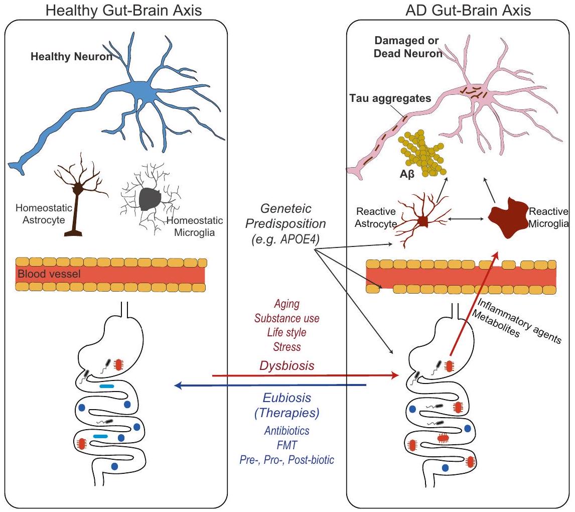
نظرة عامة على محور الميكروبيوتا-الأمعاء-الدماغ في مرض الزهايمر

على الرغم من أن فكرة وجود ارتباط بين الأمعاء والدماغ قد تم التعرف عليها منذ قرون في التاريخ الطبي، إلا أن فهم الدور المحدد للميكروبيوتا في محور الأمعاء-الدماغ قد حظي باهتمام كبير في العقود القليلة الماضية وأصبح موضوعًا ساخنًا في البحث العلمي والاهتمام العام. النمو الأسي في هذا المجال يعود إلى تطوير تقنيات التسلسل عالية الإنتاجية والمعلوماتية الحيوية، التي مكنت العلماء من توصيف تركيبة ووظيفة ميكروبيوتا الأمعاء بشكل دقيق. لقد فتح هذا التقدم التقني أيضًا آفاقًا جديدة لدراسة العلاقة بين ميكروبيوتا الأمعاء والأمراض العصبية، بما في ذلك مرض الزهايمر.
في عام 2017، قام كاتانيو وآخرون بقياس وفرة ستة أنواع بكتيرية محددة مسبقًا في براز مرضى الزهايمر (كبار السن الذين يعانون من ضعف إدراكي مع ترسبات الأميلويد) باستخدام الطريقة الكمية وجدوا أن مرضى الزهايمر يظهرون زيادة في وفرة البكتيريا المسببة للالتهابات مثل الإشريكية/الشجيلا وانخفاضًا في وفرة البكتيريا المضادة للالتهابات مثل يوباكتييريوم ركتالي مقارنة بالمجموعة الضابطة. في نفس العام، طبق فوجت وآخرون تقنية تسلسل جين 16S rRNA لتصنيف وتحديد التركيب التصنيفي البكتيري لعينات البراز من المشاركين المصابين وغير المصابين بتشخيص الخرف الناتج عن الزهايمر. وجدوا أن تنوع الميكروبيوم المعوي لمرضى الزهايمر انخفض بشكل ملحوظ مقارنة بالمجموعة الضابطة. بالإضافة إلى ذلك، على مستوى الشعبة، يظهر الميكروبيوم لدى المشاركين المصابين بالزهايمر انخفاضًا في الفيرميكيوت وزيادة في البكتيرويديتس. . عقب هذه الجهود البحثية الأولية، كانت هناك دراسات متابعة مماثلة تصف تركيبة المجتمع البكتيري في مرض الزهايمر. علاوة على ذلك، أظهرت الدراسات الحديثة أن الأفراد الذين يعانون من ضعف إدراكي خفيف (MCI، وهي حالة تتميز بتراجع في القدرات الإدراكية قد تسبق تطور الخرف) وحتى مرضى الزهايمر قبل السريري (قبل ظهور أعراض الزهايمر) يمكن أن يظهروا تركيبات مميزة للميكروبيوتا المعوية مقارنة بالمجموعات الضابطة . ومع ذلك، من المهم ملاحظة أن تفاصيل تراكيب ميكروبيوم الأمعاء الموضحة في مرض الزهايمر ليست دائمًا متسقة بين الدراسات. على سبيل المثال، في تحليل ميكروبيوم الأمعاء في دراسة فوغت وآخرين، زاد وفرة شعبة البكتيروديتس في مرضى الزهايمر (97% من القوقازيين) مقارنة بالمجموعة الضابطة. في المقابل، في دراسة أخرى أجريت مع مرضى صينيين، انخفضت وفرة البكتيروديتس في مرضى الزهايمر مقارنة بالمجموعة الضابطة. قد يكون التفاوت ناتجًا عن عدة عوامل، بما في ذلك الاختلافات في تصميم الدراسة، وتجميعات المرضى، وأنماط الحياة، والعادات الغذائية، وفي مجال تسلسل الحمض النووي الريبي.
دراسات الحيوانات دعمت أيضًا العلاقة بين تغير ميكروبيوتا الأمعاء وأمراض الزهايمر. . أظهرت هذه الدراسات اختلافات واضحة في تركيب الميكروبيوم بين نماذج الحيوانات لمرض الزهايمر ومجموعات التحكم، كما لوحظ من خلال تحليل 16S rRNA لعينات البراز أو الأعور الخاصة بها. ومع ذلك، وبالمثل للدراسات البشرية، فإن عدم الاتساق في تفاصيل تركيب ميكروبيوم الأمعاء بين الدراسات هو أيضًا سمة مميزة في الأدبيات الحيوانية، حتى في الدراسات التي تستخدم نفس نوع نموذج حيواني للأميلويدوز. على سبيل المثال، براندشيد وآخرون لوحظ انخفاض في البكتيرويديتس في عمر تسعة أسابيع في الفئران مقارنةً بالضوابط من النوع البري. في المقابل، أبلغ تشين وآخرون (2020) أن تراكيب الميكروبيوم ظلت متشابهة إلى حد كبير بين والفئران البرية عند عمر 12 أسبوعًا، وعندما كبرت الحيوانات، عند عمر 24 أسبوعًا، زادت بكتيريا البكتيرويديتس، والبروتيوبكتيريا، والديفيريبكتيريس في فئران 5xFAD مقارنةً بالضوابط البرية. التباينات في نتائج دراسات ميكروبيوتا الأمعاء ليست بالضرورة ناتجة فقط عن التحليل على مستوى الفيلوم العالي. من غير غير المألوف العثور على اختلافات في العائلات أو الأجناس المحددة المرتبطة بنماذج حيوانية لمرض الزهايمر عبر دراسات مختلفة. يمكن أن تساعد جهود البحث المستقبلية التي تركز على توحيد المنهجيات، واستخدام أحجام عينات أكبر، وتطبيق تقنيات تحليل متسقة في التغلب على هذه التباينات وتحديد تصنيفات واضحة.
توقيعات الميكروبيوتا المعوية المرتبطة بمرض الزهايمر. من ناحية أخرى، أدت التباينات والفشل في تحديد توقيعات تصنيفية واضحة مرتبطة بمرض الزهايمر إلى دفع المجال للتركيز على دراسة الأنشطة الوظيفية والتفاعلات للميكروبيوتا المعوية وعوامل أخرى تتجاوز التركيب التصنيفي (مثل علم الأيض).
أظهرت العديد من الدراسات تفاوتًا كبيرًا بين الأفراد في تركيب وتنوع الميكروبات المعوية. . التنوع بين الأفراد فيما يتعلق بميكروبيومات الأمعاء لديهم كبير مقارنة بالتنوع الجيني. بينما يشترك البشر في حوالي التشابه في جينوم المضيف لديهم، يمكن أن يختلف تركيب ووفرة أنواع الميكروبات داخل الميكروبيوم بشكل كبير من شخص لآخر، ويُقدّر أن فقط حوالي من الميكروبيوم (على مستوى الجنس أو النوع التقريبي) مشترك بين أفراد مختلفين ومع ذلك، يعترف المجال بأن أنواعًا ميكروبية مختلفة من سلالات تطورية متميزة يمكن أن تسهم في أنشطة وظيفية مماثلة داخل نظام الأمعاء البيئي. . هذا يعني أنه حتى إذا كانت التراكيب التصنيفية لميكروبيوتا الأمعاء لشخصين مختلفين مختلفة إلى حد كبير، فقد تظهر نشاطات وظيفية متشابهة. هذا الاعتراف بـ “التكرار الوظيفي” يبرز أهمية دراسة النشاط الوظيفي لميكروبيوتا الأمعاء والإمكانات الأيضية بدلاً من الاعتماد فقط على التركيب التصنيفي. تسلط هذه المناقشة الضوء أيضًا على أنه بدلاً من تصنيف أنواع البكتيريا المحددة بتصنيف بسيط مثل “جيد” أو “سيء”، من الأكثر فائدة التركيز على التركيب العام، والتنوع، واستقرار الشبكة الميكروبية وفهم تفاعلاتها الوظيفية.
مدفوعًا بالحاجة إلى تجاوز هذه الملاحظات الترابطية وكشف الأهمية الوظيفية لميكروبيوتا الأمعاء في أمراض الزهايمر، حدث تحول متزايد في المجال لتحديد اتجاه السببية والآليات المشاركة في محور ميكروبيوتا الأمعاء-الدماغ في الزهايمر. لاختبار السببية في مساهمة ميكروبيوتا الأمعاء في تقدم أمراض الزهايمر في الدراسات الحيوانية، تم تطبيق طرق مثل العلاج بالمضادات الحيوية، والنماذج الخالية من الجراثيم/النماذج الجنوبيوتية، وزرع ميكروبيوتا البراز (FMT). أظهرت عدة دراسات أن إعطاء مزيج من العلاجات بالمضادات الحيوية لنماذج الفئران المصابة بالأميلويدوز (فئران APP/PS1) أو تربية الفئران في ظروف خالية من الجراثيم يقلل من ترسيب لويحات الأميلويد الدماغية. علاوة على ذلك، فإن زراعة البراز باستخدام عينات براز مأخوذة من مجموعة الحيوانات المصابة بالأميلويدوز أو إدخال نوع محدد من الطحالب البكتيرية إلى حيوانات نموذج مرض الزهايمر يسهل ترسيب لويحات الأميلويد. .
بينما أظهرت الأدلة المتزايدة أن ميكروبيوتا الأمعاء تنظم ترسيب الأميلويد، كان هناك نقص في المعلومات حول مساهمة ميكروبيوتا الأمعاء في مرض تاو والتنكس العصبي، الذي يرتبط ارتباطًا قويًا بالتدهور المعرفي في مرض الزهايمر. في وقت مبكر من هذا العام، قامت مجموعتنا بتقييم الفرضية التي تفيد بأن ميكروبيوتا الأمعاء تنظم مرض تاو والتنكس العصبي بطريقة تعتمد على نوع الأيزوفورم ApoE. تم إخضاع فئران نموذج مرض تاو (فئران P301S الترانسجينية) التي تعبر عن الأيزوفورمات البشرية لـ APOE (APOE3 و APOE4) لتلاعب في ميكروبيوتا الأمعاء باستخدام طريقتين: (1) تربيتها في ظروف خالية من الجراثيم و (2) علاج قصير الأمد بالمضادات الحيوية لتعكير تركيبة المجتمعات البكتيرية. أدى التلاعب في ميكروبيوتا الأمعاء إلى تقليل ملحوظ في مرض تاو والتنكس العصبي بطريقة تعتمد على نوع الأيزوفورم ApoE. معًا، تدعم هذه النتائج أن ميكروبيوتا الأمعاء تنظم كل من ترسيب لويحات الأميلويد والتنكس العصبي الذي يتوسطه بروتين تاو (بشكل مستقل عن الأميلويدوز).

الآليات الأساسية والالتهاب العصبي

يتضح أن الميكروبيوتا يمكن أن تنظم أمراض الزهايمر في نماذج الفئران. ومع ذلك، كيف يمكن لميكروبيوتا الأمعاء أن تنظم أمراض الزهايمر في الدماغ التي تقع على مسافة من الجهاز الهضمي
لم يتضح المسار بعد. قد ينطوي تأثير الميكروبيوتا على مرض الزهايمر على مزيج من عدة مسارات وتفاعلات تساهم مجتمعة في أمراض الزهايمر بدلاً من مسار واحد فقط متورط في المحور. تطور الرأي حول دور الميكروبيوتا في المرض في اتجاهين مميزين في السنوات الأخيرة: (1) العدوى الميكروبية المباشرة في الجهاز العصبي المركزي و(2) المسارات غير المباشرة التي تشمل تعديل الجهاز المناعي المحيطي والجهاز الأيضي. تمثل هذه الاتجاهات وجهات نظر مختلفة وتؤكد على جوانب مميزة من علاقة ميكروبيوتا الأمعاء بالمرض.

العدوى الميكروبية المباشرة في الجهاز العصبي المركزي

الكائنات الدقيقة، بما في ذلك البكتيريا والفيروسات والفطريات والطفيليات، يمكن أن تسبب مباشرةً التهابات الجهاز العصبي المركزي (CNS). تؤثر هذه الالتهابات بشكل مباشر على نسيج الدماغ أو الهياكل المحيطة، مثل السحايا (الأغشية الواقية التي تغطي الدماغ والحبل الشوكي) أو السائل الدماغي الشوكي (CSF) المحيط بالدماغ والحبل الشوكي، وقد تكون بدورها قادرة على تحفيز أو تنظيم الأمراض . هذه ‘نظرية العدوى’ لـ تم اقتراح ذلك منذ حوالي 30 عامًا، وقد وردت العديد من التقارير التي تشير إلى تورط مسببات الأمراض البكتيرية والفيروسية المتنوعة في مرض الزهايمر، وأكثرها شيوعًا هي عائلة الهربسفيريديا (وخاصة HSV1، EBV، وHCMV) . لقد عادت هذه النظرة إلى دائرة الضوء مرة أخرى، حيث تظهر التقارير الوبائية الحديثة أن نوعين آخرين من فيروس الهربس، HHV6 وHHV7، بالإضافة إلى HSV1، مرتبطان بـ علاوة على ذلك، كشفت دراسة وبائية أخرى استنادًا إلى قاعدة بيانات التأمين الصحي الوطني في تايوان أن استخدام الأدوية المضادة للهربس لعلاج عدوى فيروس الهربس البسيط كان مرتبطًا بانخفاض خطر الإصابة بالخرف. . بالإضافة إلى ذلك، تم الكشف عن بعض أنواع البكتيريا الأخرى، مثل بورليا بورغدورفيري، الكلاميديا الرئوية، وبورفيروموناس جينجيفاليس، في الدماغ وارتبطت بتطور الأمراض . ومع ذلك، نظرًا لأن هذا المنظور لا يزال يعتمد بشكل رئيسي على بيانات ارتباطية، فلا يزال من غير الواضح ما إذا كانت العدوى الميكروبية في الدماغ تعني أن العدوى الميكروبية تنشط العملية المرضية لمرض الزهايمر أو تمثل علامة على تقدم المرض (مثل انهيار الحماية من الممرضات أو إعادة تنشيط الفيروس الكامن مع تقدم أمراض الزهايمر). حتى إذا أشارت البيانات الارتباطية إلى أن العدوى الميكروبية تسهم في ، التفسير غير مؤكد إذا كان العدوى هي السبب في (أي، أصل) ) أو يساهم في تقدم (على سبيل المثال، يسهل الالتهاب العصبي).
تم إعادة فحص الفرضية المعدية التي تقترح أن العامل الممرض هو السبب الجذري لمرض الزهايمر مؤخرًا، حيث تدعم الأدلة المتزايدة ذلك تُظهر المواد المجمعة نشاطًا مضادًا للميكروبات ضد بعض الكائنات الدقيقة، بما في ذلك البكتيريا والفطريات اختبر إيمر وآخرون ما إذا كان يمكن لعدوى فيروسية أن تزرع وتسرع ترسيب للحماية من مسببات الأمراض في الدماغ قاموا بحقن فيروس الهربس البسيط النوع 1 في أدمغة فئران نموذجية صغيرة تعبر بشكل مفرط عن الأميلويد ( ) والفئران ذات النوع البري، مما أدى إلى ترسيب متسارع لـ ، ومع جرعة قاتلة من فيروس الهربس البسيط النوع 1، عاشت الفئران المعدلة وراثيًا لفترة أطول من الفئران الضابطة. كما أظهرت الدراسة أن تثبط الأوليغومرات عدوى فيروس الهربس البسيط من النوع 1 (HSV1) في المختبر، مما يشير إلى أن العدوى تحفز تراكم لويحات بروتينية لزجة لحماية الدماغ من مسببات الأمراض الغازية. بشكل افتراضي، قد يعمل تقدم تراكم الأميلويد كآلية دفاعية، لكن الفشل في إزالة الأميلويد يمكن أن يؤدي إلى مستوى غير طبيعي من ترسيب الأميلويد، والالتهاب، وأمراض أخرى. على الرغم من التقدم البحثي في هذا الرأي، سيكون من الصعب جدًا إثبات ما إذا كان مرض الزهايمر ينشأ تمامًا من عدوى ميكروبية لأن ترسيب الأميلويد يبدأ قبل ظهور الأعراض بـ 15-20 سنة، ومن الصعب تتبع حالة العدوى الميكروبية للفرد مع تغيرات مؤشرات مرض الزهايمر الحيوية. بالإضافة إلى ذلك، لا تزال الدراسات قبل السريرية الحديثة تعتمد على بيانات باستخدام نماذج حيوانية معدلة وراثيًا لمرض الزهايمر تعبر بشكل مفرط عن جينات مسببة أو محفوفة بالمخاطر مرتبطة بالمرض. إذا كانت العدوى الميكروبية تسبب فعلاً مرض الزهايمر، فقد يحتاج الباحثون إلى إثبات أن العدوى الميكروبية في الحيوانات البرية أو البشر تدفع إلى أمراض مشابهة لمرض الزهايمر.
على أي حال، سيكون من المهم تحديد ما إذا كانت بعض الكائنات الدقيقة يمكن أن تعمل كمسرعات من خلال زيادة الالتهاب أو مرض الزهايمر لتؤثر على تقدم المرض.
يمكن أن تنشأ العدوى من عدة أعضاء طرفية وقد تنتشر إلى الدماغ عبر مسارات مختلفة: مجرى الدم، الامتداد المباشر من الهياكل المجاورة، أو على طول الأعصاب (مثل العصب المبهم). من المهم ملاحظة أن فيروس الهربس وأنواع البكتيريا التي نوقشت أعلاه توجد بشكل أساسي في الجهاز التنفسي (مثل C. pneumonia) أو تجويف الفم (مثل HSV1 و . gingivalis) بدلاً من ارتباطها بالجهاز الهضمي. ومع ذلك، أشارت بعض الدراسات الحديثة إلى أن التهابات الجهاز العصبي المركزي يمكن أن تنشأ أيضًا من الجهاز الهضمي، مما يساهم في أمراض الزهايمر. على سبيل المثال، أظهر وو وزملاؤه أن الحقن الوريدي لفطر الكانديدا البيضوية، وهو خميرة ممرضة انتهازية توجد عادة في الجهاز الهضمي البشري، يمكن أن يسبب التهاب دماغي عابر يتميز بالتليف البؤري المحيط بالخلايا الفطرية وترسيب كل من بروتين الأميلويد المسبق (APP) و الببتيدات هناك أيضًا احتمال أن البكتيريا المعوية المتعايشة قد تكون قادرة على الانتقال عبر العصب المبهم (تقرير مسودة غير منشور). .
فرضية أخرى هي أن بروتين الأميلويد المحيطي قد يزرع ويعزز تراكمه في الدماغ بسبب نقله الرجعي إلى الجهاز العصبي المركزي عبر العصب المبهم أو الدم. وقد أدى هذا الاكتشاف إلى فكرة أن بعض البكتيريا تنتج ألياف أميلويد خارج الخلية تسمى كورلي، والتي تتبنى أيضًا بنية بيتا شيت. من المثير للاهتمام، أظهرت الدراسات الحديثة أن الإدارة داخل الجهاز الهضمي لـ الأوليغومرات في الفئران ذات النوع البري تسببت في بيتا-أميلويد الدماغي . ومع ذلك، هناك حاجة إلى مزيد من الدراسات لتحديد ما إذا كانت الأميلويد التي تنتجها البكتيريا تساهم فعليًا في أمراض الزهايمر أو في الأميلويد المحيطي إيداع مع التقدم ثم يساهم في ترسب الأميلويد في الدماغ لاحقًا . بالإضافة إلى ذلك، الآلية الأساسية للأمعاء التأمبيلويد الناجم عن – في الدماغ لا يزال غير واضح.

المسار غير المباشر الذي ينظم الجهاز المناعي والأنظمة الأيضية الطرفية

حالياً، الفرضية الأكثر منطقية للتفسيرات الميكانيكية في هذا المجال هي أن الالتهاب العصبي يتوسط التفاعل بين ميكروبيوتا الأمعاء وتطور أمراض الزهايمر. التفاعل النجمي التفاعلي والتفاعل الدبقي الدقيق هما من السمات المرضية البارزة في دماغ الزهايمر. في المقام الأول، توفر الخلايا الدبقية الدعم والحماية للخلايا العصبية، وتزيل الخلايا الميتة والجسيمات الغريبة للحفاظ على التوازن الداخلي في الدماغ. ومع ذلك، فإن النشاط غير الطبيعي للخلايا الدبقية المرتبط بالمرض يعطل الشبكات الخلوية التوازنية في الدماغ ويسرع من تقدم مرض الزهايمر. أي أن الخلايا الدبقية المنشطة بشكل غير طبيعي قد تنشر السمية، تؤدي إلى تراكم أو إطلاق السيتوكينات المؤيدة للالتهابات وأنواع الأكسجين التفاعلية، التي تضر بالخلايا العصبية وتُسهّل مرض تاو، ويؤدي الالتهاب المفرط الإضافي إلى تلف الخلايا العصبية وتقدم المرض (لمراجعة أكثر تفصيلاً ). من اللافت أن الأدلة المتزايدة من الدراسات على الحيوانات أظهرت أن ميكروبيوتا الأمعاء تنظم نضج ووظيفة الخلايا الدبقية. أظهر إيرني وزملاؤه في عام 2015 أن استنفاد الميكروبيوتا من خلال تربية الفئران في ظروف خالية من الجراثيم أو إعطائها علاجًا بالمضادات الحيوية أدى إلى بقاء الخلايا الدبقية الدقيقة في حالات غير ناضجة مع استجابة منخفضة للعدوى الفيروسية. كما وجدوا أن المستقلبات التي تنتجها الميكروبات والتي تسمى الأحماض الدهنية قصيرة السلسلة (SCFAs) تشارك في هذا التفاعل. من المثير للاهتمام، أن هناك نمطًا محددًا حسب الجنس في تأثير ميكروبيوتا الأمعاء على نضوج الخلايا الدبقية الصغيرة خلال التطور. وبالمثل، أظهرت دراسة حديثة أن الخلايا النجمية المستزرعة أولياً يمكن أن تظهر استجابات مختلفة حسب الجنس عند معالجتها بالأحماض الدهنية قصيرة السلسلة. .
تأييدًا لهذه النتائج، أظهرت عدة دراسات تحقق في دور ميكروبيوتا الأمعاء في مرض الزهايمر أن التلاعب بميكروبيوتا الأمعاء في نماذج حيوانية لمرض الزهايمر أدى إلى تغييرات في الشكل والتعبير الجيني في الخلايا الدبقية.
مصحوبًا بانخفاض في أمراض الزهايمر (أي، الترسيب، مرض تاو، والتنكس العصبي . علاوة على ذلك، فإن التخفيض في تم منع ترسيب الأميلويد بيتا الناتج عن تعديل ميكروبيوتا الأمعاء بواسطة المضادات الحيوية من خلال استنفاد الخلايا الدبقية الصغيرة باستخدام مثبط مستقبل عامل تحفيز المستعمرات 1 (CSF1R) .
ومع ذلك، لا تزال الآليات الدقيقة التي تكمن وراء تنشيط المناعة الفطرية في الدماغ قيد التحقيق. تساعد ميكروبيوتا الأمعاء في الحفاظ على التوازن المناعي من خلال تنظيم التوازن بين الاستجابات الالتهابية والمضادة للالتهابات. . في المقام الأول، يُعد تنشيط الجهاز المناعي آلية دفاع أساسية للمضيف لحمايته من مسببات الأمراض. ومع ذلك، يؤدي الاستجابة الالتهابية المفرطة إلى زيادة في السيتوكينات المتداولة وتجنيد الكريات البيضاء، بما في ذلك الفاعلين في المناعة التكيفية الخلوية (أي الخلايا اللمفاوية B أو T)، التي تخترق الأنسجة أو تطلق سيتوكينات تنشط المناعة الفطرية في الدماغ. في النهاية، يمكن أن تسرع هذه العمليات من الالتهاب العصبي أظهرت الدراسات التي تستخدم نماذج حيوانية لمرض الزهايمر أن التلاعب بميكروبيوتا الأمعاء يقلل من عدة سيتوكينات محيطية مرتبطة بالبلعميات وخلايا المناعة التكيفية. أظهرت دراستنا الحديثة باستخدام نموذج حيواني لاعتلال التاو أيضًا أن السحايا الخلايا، خلايا التغصن البلازمية، وخلايا القاتل الطبيعي تعتمد على ميكروبيوتا الأمعاء. تقوم هذه الخلايا المناعية بإفراز السيتوكينات، مثل الإنترلوكين-17، واستجابة الإنترفيرون من النوع الأول، وغيرها، والتي قد تؤثر على الالتهاب العصبي في الدماغ، حتى بدون تسلل إلى الدماغ. أظهرت الدراسات البشرية أيضًا أن المرضى يظهرون تواقيع مناعية محيطية متغيرة . ومع ذلك، فقد حددت هذه الدراسات اختلافات في ملفات السيتوكينات المحيطية، ولا يزال من غير الواضح أي العوامل المناعية تشارك مباشرة في تنشيط المناعة الفطرية للدماغ في سياق هناك حاجة إلى مزيد من البحث لإثبات وجود صلة مباشرة بين التغيرات في السيتوكينات المحيطية وتنشيط المناعة الفطرية في الدماغ.
بينما يحتل مسار الالتهاب العصبي حالياً اهتماماً كبيراً كتفسير ميكانيكي في هذا المجال، قد تكون الآليات غير الدبقية متورطة أيضاً في التفاعل. أظهرت فئران نموذج الأميلويدوز (APP/PS1) التي تربت في ظروف خالية من الجراثيم زيادة في إنزيمات إزالة الأميلويد مثل إنزيم نيبريليسين المحلل (NPE) وإنزيم تحلل الإنسولين (IDE) مقارنة بالفئران التي تربت في ظروف تقليدية، مما قد يؤثر على إيداع قد تُعزز عوامل بيولوجية أخرى تأثير ميكروبيوتا الأمعاء. على سبيل المثال، قد تؤدي عوامل العمر والجينات إلى اضطراب نفاذية الأمعاء وسلامة الحاجز الدموي الدماغي (BBB)، مما يسرع دخول العوامل الالتهابية والجراثيم المتداولة إلى الدماغ، مما يحفز التنشيط المفرط للمناعة الفطرية في الدماغ. . بالإضافة إلى ذلك، وُجد أن المستقلبات الميكروبية مثل الأحماض الدهنية قصيرة السلسلة (SCFAs) تُعدّل النشاط التلقائي، وهو عملية خلوية تلعب دورًا حيويًا في الحفاظ على التوازن الخلوي من خلال تنظيف وإعادة تدوير الحطام الخلوي والبروتينات التالفة والعضيات تم الإبلاغ عن خلل في آلية الالتهام الذاتي وتغيرات في تنظيم الجينات المرتبطة بالالتهام الذاتي في أدمغة مرض الزهايمر يجدر استكشاف إمكانية أن الأحماض الدهنية قصيرة السلسلة المتداولة تنظم الأنشطة الذاتية الهضمية في أدمغة مرض الزهايمر. .

المستقلبات المشتقة من ميكروبيوتا الأمعاء: الأحماض الدهنية قصيرة السلسلة وغيرها

أحد العمليات الأساسية المشاركة في التفاعل بين ميكروبيوتا الأمعاء والمضيف هو إنتاج وتبادل المستقلبات. هذه الجزيئات الصغيرة، التي يمكن أن تكون مشتقة مباشرة من البكتيريا، أو من الأيض البكتيري للركائز الغذائية، أو من تعديل جزيئات المضيف، تؤثر على أنظمة المناعة والتمثيل الغذائي لدى المضيف. .
المستقلبات التي تم التحقيق فيها بشكل واسع في هذا المجال هي الأحماض الدهنية قصيرة السلسلة، وهي أحماض عضوية ذات طول سلسلة أقل من ست ذرات كربون، وتنتج بشكل رئيسي من خلال تخمير الألياف الغذائية بواسطة بكتيريا الأمعاء. كما نوقش أعلاه،
تتحكم الأحماض الدهنية قصيرة السلسلة في نضج وعمل المناعة الفطرية في الدماغ الخلايا الدبقية لا تعبر عن مستقبلات الأحماض الدهنية القصيرة السلسلة، لذا قد تنظم الأحماض الدهنية القصيرة السلسلة الخلايا الدبقية بشكل غير مباشر عبر خلايا الجهاز المناعي المحيطية التي تعبر عن مستقبلات الأحماض الدهنية القصيرة السلسلة. بالإضافة إلى ذلك، يمكن للأحماض الدهنية القصيرة السلسلة تنظيم إنتاج السيتوكينات بواسطة خلايا الجهاز المناعي المحيطية مثل العدلات، والبلعميات، والخلايا التغصنية (DCs) وكذلك تكاثرها وتمايزها. الخلايا و خلايا قد تحوّل هذه اللوائح الخلايا الدبقية. ومن الممكن أيضًا أن تقوم الأحماض الدهنية قصيرة السلسلة بتنظيم وظائف الخلايا الدبقية مباشرة. وجد إرني وزملاؤه أن الخلايا الدبقية الدقيقة تمتص الأسيتات (وهي واحدة من الأحماض الدهنية قصيرة السلسلة الرئيسية) التي دخلت الجهاز العصبي المركزي، وتقوم الأسيتات بتنظيم وظائف الخلايا الدبقية الدقيقة عبر آليات أيضية إبيجينية وميتوكندريا. .
التصور العام للأحماض الدهنية قصيرة السلسلة هو أنها تلعب دورًا مفيدًا في صحة الإنسان، مثل توفير الطاقة للخلايا ودعم سلامة الحاجز المعوي. . ومع ذلك، الصورة أكثر تعقيدًا فيما يتعلق بتأثير الأحماض الدهنية قصيرة السلسلة على الأمراض العصبية. ففي حين أظهرت الدراسات أن الأحماض الدهنية قصيرة السلسلة تحسن التعافي من عدة اضطرابات عصبية، مثل التصلب المتعدد، والسكتة الدماغية، وإصابات الدماغ الرضحية، تشير الدراسات الحديثة التي تستخدم نماذج حيوانية لمرض الزهايمر ومرض باركنسون إلى أن الأحماض الدهنية قصيرة السلسلة يمكن أن تسهل الالتهاب العصبي وتقدم المرض في غياب ميكروبات الأمعاء. . حتى داخل مجال مرض الزهايمر، فإن تأثيرات الأحماض الدهنية قصيرة السلسلة ليست دائمًا متسقة، حيث تشير بعض الأدبيات إلى أن مكملًا واحدًا من الأحماض الدهنية قصيرة السلسلة، وهو بترات الصوديوم، يخفف من إيداع ليس من الواضح ما الذي أدى إلى هذا التناقض، ولكن من الممكن أن يكون لكل حمض دهني قصير السلسلة، مثل الأسيتات، والبروبيونات، والبيوتيرات، تأثيرات وآليات عمل مميزة في سياقات مختلفة (تم ملاحظة التأثير الضار في نموذج مرض الزهايمر مع مكملات حمض دهني قصير السلسلة المختلطة) أو أن الاستجابة المناعية الطرفية المدفوعة بنفس حمض دهني قصير السلسلة قد تلعب دورًا مختلفًا في مرض كل حالة. . بالإضافة إلى ذلك، يمكن للأحماض الدهنية قصيرة السلسلة أن تؤثر بشكل مختلف على العمليات الفسيولوجية المختلفة بناءً على مستوياتها داخل الجسم. تم استخدام نفس الجرعات من الأحماض الدهنية قصيرة السلسلة المختلطة (بروبيونات الصوديوم، 25.9 مليمول؛ بيوتيرات الصوديوم، 40 مليمول؛ أسيتات الصوديوم، 67.5 مليمول) في الدراسات المذكورة أعلاه. تحتاج الدراسات المستقبلية إلى اختبار تأثير جرعات مختلفة من الأحماض الدهنية قصيرة السلسلة على تطور أمراض الزهايمر. وأخيرًا، يمكن أن يكون عمر المضيف عند تناول مكملات الأحماض الدهنية قصيرة السلسلة عاملاً مهمًا يجب أخذه في الاعتبار. عندما قمنا بإعطاء مكملات الأحماض الدهنية قصيرة السلسلة لنماذج حيوانية لمرض التاو، لاحظنا تعزيز التليف الدبقي في الحُصين لدى الفئران الكبيرة في السن (35 أسبوعًا) ولكن ليس لدى الفئران الصغيرة في السن (15 أسبوعًا). .
بالإضافة إلى الأحماض الدهنية قصيرة السلسلة (SCFAs)، تم الإشارة إلى مستقلبات أخرى في الدراسات السريرية لمرض الزهايمر، مثل الليبوبوليساكاريد (LPS)، أوكسيدات ثلاثي ميثيل الأمين (TMAO)، التربتوفان، وأحماض الصفراء. . على الرغم من ملاحظة اضطراب التمثيل الغذائي لهذه العوامل في مرضى الزهايمر، إلا أن الدور المحدد لهذه المستقلبات في لم يتم التحقيق فيه بشكل جيد بعد. هناك حاجة إلى مزيد من التحقيق لفهم دور المستقلبات بخلاف الأحماض الدهنية قصيرة السلسلة في مرض الزهايمر بشكل شامل.

الاختلافات الجينية للمضيف والفروق بين الجنسين

المدى الذي يشكل فيه التباين الجيني للمضيف تركيبة الميكروبيوم، مقابل العوامل البيئية، هو موضوع نقاش وبحث مستمر في علم الميكروبيوم. بينما تشير العديد من الدراسات إلى أن ميكروبيوم الأمعاء البشرية يتشكل نفسه بواسطة مجموعة واسعة من العوامل البيئية، بما في ذلك النظام الغذائي، ونمط الحياة، والجغرافيا، واستخدام الأدوية، والتعرض للعوامل الممرضة، أفاد آخرون أن التباين الجيني للمضيف هو منظم قوي لميكروبيوم المضيف. .
جينوتيب الأبوليبوبروتين E (APOE) هو عامل الخطر الوراثي الأكثر شيوعًا للأمراض العصبية ومرض الزهايمر. درست الدراسات الحديثة تركيب ميكروبيوم الأمعاء لدى البشر أو الحيوانات النموذجية التي تحمل أليلات APOE البشرية المختلفة. كشف تحليل ميكروبيوم الأمعاء لحاملي APOE2 وAPOE3 وAPOE4 أن العائلات البكتيرية المنتجة للأحماض الدهنية قصيرة السلسلة، مثل Ruminococcaceae وLachnospiraceae، كانت أكثر وفرة لدى حاملي APOE2 وأقل وفرة لدى حاملي APOE4 مقارنة بحاملي APOE3 بطريقة تدريجية (أي، APOE2… أبو إي 3 أبو إي 4 .
جميع التفاصيل في مقارنة تراكيب الميكروبيوم بين أشكال APOE ليست دائمًا متسقة. على سبيل المثال، أبلغ تران وآخرون أن عائلة لاكنوسبيراسيا كانت مرتفعة في فئران APOE4 مقارنة بفئران APOE3. قد يكون هذا التفاوت ناتجًا عن التعرضات البيئية المختلفة أو الأنظمة الغذائية بين الدراسات، لكنه يشير أيضًا إلى أن العوامل البيئية تتفاعل مع العوامل الوراثية في تشكيل الميكروبيوم. قمنا مؤخرًا بتقييم ما إذا كان ميكروبيوم الأمعاء ينظم التنكس العصبي الذي يسببه بروتين تاو من خلال التفاعل مع الأنماط المتعددة باستخدام اضطراب ميكروبيوتا الأمعاء الناجم عن المضادات الحيوية في نموذج حيواني لمرض التاو. كان التلاعب بميكروبيوتا الأمعاء وقائيًا عصبيًا ضد مرض التاو والتنكس العصبي. ومع ذلك، كانت الفعالية محدودة على الذكور، ومن اللافت أن التأثيرات كانت أكبر في وجود ApoE3 مقارنة بـ ApoE4. قد يكون هذا التأثير التفريقي للنمط المتغاير مع تعديل ميكروبيوم الأمعاء ناتجًا عن مزيج من الاستجابات المناعية الفطرية المحيطية والدماغية المختلفة مع APOE4، وفي النهاية، فشلت المضادات الحيوية في الحماية من التنكس العصبي الناجم عن تاو. ومع ذلك، تحتاج الدراسات المستقبلية إلى التحقيق بدقة في الأنسجة، ونوع الخلايا، والمسارات التي تتأثر بتفاعلات APOE والميكروبيوم هذه. بالإضافة إلى ذلك، لا تزال الآليات الدقيقة التي قد تعدل بها أليلات APOE ميكروبيوم الأمعاء بشكل مختلف بحاجة إلى فهم كامل. قد يكون لأليلات APOE المختلفة تأثيرات متباينة على وظيفة المناعة والالتهاب (مثل الاستجابة التفريقية للبلاعم والخلايا المناعية المحيطية الأخرى)، والتي يمكن أن تؤثر بدورها على تكوين ميكروبيوم الأمعاء. . بالإضافة إلى ذلك، نظرًا لأن الدور الأساسي لـ APOE يتعلق بتمثيل الدهون ونقل الدهون، فإن اختلافات في تمثيل الدهون أو تغيرات في وظيفة حاجز الأمعاء المرتبطة بأليلات APOE محددة قد تؤثر على بيئة الأمعاء والمجتمعات الميكروبية.
بالإضافة إلى التباين الجيني للمضيف، تم التعرف على الفروق الجنسية كعوامل مهمة في سياق ميكروبيوم الأمعاء واستجابته للمضادات الحيوية في نماذج حيوانية لمرض الزهايمر. في الدراسات التي نوقشت أعلاه، أدى اضطراب ميكروبيوتا الأمعاء الناجم عن المضادات الحيوية إلى تقليل أمراض الزهايمر في الذكور ولكن ليس في الإناث. لوحظ نفس نمط النتائج عبر عدة دراسات باستخدام نماذج حيوانية لكل من الأميلويدوز والتاووباثي. الآليات التي قد يؤثر من خلالها الجنس البيولوجي بشكل مختلف على النتائج المرضية المرتبطة بالمضادات الحيوية غير معروفة. هناك نمط نضج ميكروغليالي خاص بالجنس يتفاعل مع ميكروبيوتا الأمعاء أثناء التطور. بالإضافة إلى ذلك، من المعروف أن هناك اختلافات جنسية في تركيب ميكروبيوم الأمعاء والاستجابة المناعية للممرضات؛ حيث تظهر الإناث غالبًا استجابات مناعية فطرية أقوى وتكاثر خلايا تي/استجابات الأجسام المضادة مقارنة بالذكور. . ومع ذلك، من غير الواضح ما هي الفروق الدقيقة التي تفسر عدم الاستجابة للمضادات الحيوية لدى الإناث في هذه الدراسات. وأخيرًا، تلعب هرمونات الجنس مثل الإستروجين دورًا في تعديل الجهاز المناعي والاستجابات العصبية الالتهابية لدى الإناث قد تؤثر الفروقات الهرمونية بين الذكور والإناث على كيفية استجابة ميكروبيوتا الأمعاء للمضادات الحيوية وبالتالي تؤثر على أمراض الزهايمر. قد يكون للجمع بين استئصال المبايض والمضادات الحيوية تداعيات على الاستجابة المختلفة للمضادات الحيوية في أمراض الزهايمر، أو قد تؤثر العوامل الوراثية الخاصة بالجنس فقط على قابلية الدماغ للإصابة أو مقاومته للالتهاب العصبي، وتجمع أميلويد-بيتا، ومرض تاو.

ما وراء الأمعاء والبكتيريا

على الرغم من أننا ركزنا بشكل رئيسي على الميكروبيوتا الموجودة في الجهاز الهضمي في هذا الاستعراض، أشارت دراسات حديثة إلى أن الميكروبيوتا في مواقع أخرى غير الجهاز الهضمي، مثل ميكروبيوم الرئة والفم، قد تلعب دورًا في تطور أو تقدم مرض الزهايمر. على سبيل المثال، حدد مورير وآخرون عدة سلالات بكتيرية فموية، بما في ذلك P. gingivalis، بشكل أكثر تكرارًا في مرضى الزهايمر الذين يعانون من التهاب اللثة مقارنة بالمجموعة الضابطة. بالإضافة إلى ذلك، أظهرت دراسة أخرى أن العدوى الفموية بـ . تؤدي بكتيريا جينجيفاليس في نموذج حيواني للأميلويدوز إلى تدهور الوظائف الإدراكية و
يزيد من ترسيب اللويحات الشبيهة بمرض الزهايمر لقد حظي ميكروبيوم الرئة أيضًا باهتمام متزايد فيما يتعلق بمختلف الأمراض العصبية. وجدت دراسة حديثة أن الميكروبات المنتجة لـ LPS في الرئة أدت إلى تفاقم المرض باستخدام نموذج حيواني للتصلب المتعدد، وهو مرض مناعي ذاتي يسببه الخلايا التائية في الجهاز العصبي المركزي. الطرق الحالية للتلاعب بالميكروبيوتا، مثل الظروف الخالية من الجراثيم أو العلاجات بالمضادات الحيوية المحددة، يمكن أن تؤثر على الميكروبيوتا في أعضاء وأنسجة أخرى في الجسم بالإضافة إلى الجهاز الهضمي. تأثير هذه التلاعبات على الميكروبيوتا غير المعوية هو اعتبار مهم عند تفسير النتائج.
بالإضافة إلى ذلك، في مجال الميكروبيوم ومرض الزهايمر، تركز معظم الدراسات بشكل رئيسي على البكتيريا وأقل على الفيروسات والفطريات. قد يكون أحد الأسباب هو أن معظم الدراسات استخدمت تقنيات تسلسل جين 16S rRNA للبكتيريا لتحليل الميكروبيوم حتى الآن (لذلك، تشير مصطلحات “الميكروبيوتا” أو “الميكروبيوم” عمومًا إلى المجتمع البكتيري في هذا الاستعراض). بالإضافة إلى ذلك، فإن الوفرة النسبية للكائنات الدقيقة الأخرى، مثل الفطريات، الأوليات، العتائق، والفيروسات، أقل من تلك الخاصة بالبكتيريا، ولهذا السبب تلقت هذه الكائنات الدقيقة الأخرى اهتمامًا أقل. ومع ذلك، على الرغم من انخفاض وفرتها، قد تؤثر هذه الكائنات على مرض الزهايمر بشكل مباشر أو غير مباشر من خلال التفاعل مع المجتمعات البكتيرية – فجميع أنواع الكائنات الدقيقة تتفاعل مع بعضها البعض وتشكل شبكة ديناميكية مع المضيف. لذلك، يحتاج البحث في مجال الميكروبيوم ومرض الزهايمر إلى التوسع ليشمل التحقيق في المجتمعات الفطرية (الميكروبيوم الفطري)، المجتمعات الفيروسية (الفيروم)، والكائنات الدقيقة الأخرى. في المستقبل، ستوفر التحليلات الوظيفية، مثل التحليل الميتاجينومي، الميتاترنسكريبتومي، أو الميتابولومي، رؤى حول الأنشطة الوظيفية، التفاعلات، والمسارات داخل الميكروبيوتا التي تسهم في مرض الزهايمر.

الاستراتيجيات العلاجية

كما ناقشنا، ينظم ميكروبيوم الأمعاء جوانب متعددة من أمراض الزهايمر، بما في ذلك تراكم، مرض تاو، والتنكس العصبي، ربما من خلال تأثيرات على الالتهاب العصبي والتوازن الأيضي. كما تدعمها النتائج الحديثة، برز تعديل ميكروبيوتا الأمعاء كطريق واعد لإبطاء تقدم مرض الزهايمر. يتم استكشاف عدة طرق لتعديل ميكروبيوتا الأمعاء في مرض الزهايمر. وتشمل هذه المضادات الحيوية، وزرع البراز، والبريبايوتكس، والبروبيوتكس، والبوسطبيوتكس، وبعض التقنيات الحيوية الناشئة مثل المواد المغلفة والفيروسات البكتيرية. تهدف هذه التدخلات إلى استعادة توازن صحي لمجتمع الميكروبات المعوية وتخفيف أمراض وأعراض مرض الزهايمر (الشكل 1).

المضادات الحيوية

تم تصميم المضادات الحيوية بشكل أساسي لعلاج الالتهابات البكتيرية من خلال استهداف وقتل البكتيريا الضارة أو تثبيط نموها. في أبحاث الميكروبيوم، غالبًا ما تُستخدم المضادات الحيوية كأداة للتلاعب بالمجتمع الميكروبي بطريقة واسعة وغير محددة لأغراض تجريبية. الهدف عادة هو تقييم الوظيفة العامة للميكروبيوتا في مرض معين أو عملية بيولوجية بدلاً من استخدامها في تطبيق سريري مباشر. لأن الإزالة غير المحددة لكل من البكتيريا الممرضة والمفيدة يمكن أن تؤدي إلى اختلال التوازن الميكروبي وتأثيرات سلبية أخرى. ، قد لا تكون العلاجات بالمضادات الحيوية مثالية للتطبيقات السريرية في مرضى الزهايمر.
بالإضافة إلى ذلك، نظرًا لأن المضادات الحيوية في الدراسات قبل السريرية كانت غالبًا تُستخدم في مراحل مبكرة من حياة الحيوانات التجريبية أو خلال الفترة التي تسبق ظهور الأعراض، فلا يزال من غير المؤكد ما إذا كان استخدام المضادات الحيوية بعد فترة ظهور أعراض مرض الزهايمر يخفف بشكل فعال من مرض الزهايمر وأعراضه. في الدراسات المستقبلية، سيسمح تطبيق تصميم تجريبي يتوافق مع الأغراض العلاجية (أي اختبار تأثير المضادات الحيوية بعد بدء ظهور الأمراض) لنا بتقييم إمكانية استخدام المضادات الحيوية لمرض الزهايمر. ومن المثير للاهتمام، أن دراسة وبائية حديثة تدعم هذه الإمكانية. حيث قام راكوسا وآخرون بتحليل بيانات التأمين الصحي العام في
الشكل 1: مخطط تخطيطي لمساهمة ميكروبيوتا الأمعاء في أمراض الزهايمر والتدخلات القائمة على الميكروبيوتا لعلاج الزهايمر. الكائنات الدقيقة المتعايشة ضرورية لصحة الدماغ (يسار). ومع ذلك، يمكن لعوامل متعددة، بما في ذلك عمليات الشيخوخة، والتغيرات الغذائية، واستهلاك الأدوية/الكحول، أن تؤدي إلى اختلال توازن تركيبة مجتمعات الميكروبات المعوية، مما يؤثر على الأيض المحيطي والمناعة (يمين؛ الخط الأحمر). هذه العمليات البيولوجية تنظم في النهاية المناعة الفطرية للدماغ وتقدم أمراض الزهايمر. بالإضافة إلى ذلك، قد تسهم عوامل الخطر الوراثية (مثل وجود أليل ApoE4) في وجود ميكروبيوتا مرتبطة أكثر بالمرض، أو تؤثر على تفاعل المناعة الفطرية للدماغ، وسلامة الحاجز الدموي الدماغي (BBB) (الخط الأسود). يمثل الخط الأزرق في الشكل الإمكانات العلاجية للتدخلات المحتملة القائمة على الميكروبيوتا. إعادة تهيئة ميكروبيوتا الأمعاء غير المتوازنة باستخدام زرع ميكروبيوتا البراز (FMT)، أو البريبايوتكس من الألياف الغذائية، أو البروبيوتكس يحد من مساهمة اختلال التوازن الميكروبي في تقدم الزهايمر، مما يمنع التحول نحو الالتهاب المحيطي الضار، والأيض، والالتهاب العصبي.
ألمانيا تحقق في العلاقة بين المضادات الحيوية للاستخدام الجهازية والخرف. وجدوا انخفاضًا في احتمال الإصابة بالخرف مع استخدام المضادات الحيوية السابق. .

زرع الميكروبيوتا البرازية

FMT هو إجراء يتضمن نقل المادة البرازية من متبرع سليم إلى الجهاز الهضمي للمستلم. يهدف FMT إلى استعادة توازن الميكروبيوتا المعوية غير المتوازنة إلى تركيبة ميكروبيوتا معوية أكثر توازنًا وفائدة في المستلم، ويمكن أن يكون أداة علاجية محتملة لمرض الزهايمر.
أظهر كيم وآخرون أن زراعة الميكروبيوتا البرازية من الفئران البرية إلى فئران نموذجية معدلة وراثيًا تعبر عن الجينات المعدلة APP وPSEN1 وMAPT (ADLP) ) حسّن تشكيل ) اللويحات والتشابكات الليفية العصبية، تفاعل الخلايا الدبقية، والضعف الإدراكي . بالإضافة إلى ذلك، هناك تقرير حالة مثير للاهتمام لرجل يبلغ من العمر 82 عامًا يعاني من مرض الزهايمر (AD) وتعرض لعدوى كلوستريديوم ديفيسيل (CDI). خضع المريض لجلسة واحدة من زرع البراز (FMT) باستخدام براز زوجته البالغة من العمر 85 عامًا كمتبرعة، والتي كانت تتمتع بقدرات عقلية حادة. حتى بعد شهرين من الزرع، أظهر المريض تحسنًا سريعًا في أعراض مرض الزهايمر. وبالمثل، أظهرت دراسات كمية أخرى أيضًا أن زرع ميكروبيوم الأمعاء (FMT) حسّن بشكل كبير الأعراض السريرية لدى مرضى الزهايمر مقارنة بالمجموعة الضابطة، مما يدعم أن زرع ميكروبيوم الأمعاء قد يؤخر بشكل فعال التدهور المعرفي لدى مرضى الزهايمر. .

البريبايوتكس، البروبيوتكس، والبروبيوتكس اللاحقة

تُستخدم المضادات الحيوية لعلاج مجموعة واسعة من العدوى البكتيرية التي قد تكون مميتة بطريقة غير محددة. الفعل غير المحدد لـ
يمكن للمضادات الحيوية أن يكون لها عواقب غير مقصودة على ميكروبيوتا الأمعاء، بما في ذلك اضطراب الميكروفلورا الطبيعية، مما يؤدي إلى نقص محتمل في العناصر الغذائية وإمكانية سيطرة مسببات الأمراض الانتهازية. وبالنظر إلى ذلك، فإن النهج البديل لإعادة تهيئة ميكروبيوتا الأمعاء غير المتوازنة هو إدخال البكتيريا المفيدة إلى الجهاز الهضمي. يتضمن هذا النهج استخدام البروبيوتيك (الكائنات الحية الدقيقة الحية التي، عند إعطائها بكميات كافية، تمنح فائدة صحية للمضيف؛ مثل بيفيدوباكتيريوم ولاكتوباسيلوس)، والبريبايوتكس (الألياف الخاصة التي تعزز نمو البكتيريا المفيدة؛ مثل الإينولين)، والبروبيوتيك (المواد التي تنتجها البكتيريا المفيدة أثناء نموها، والتي يمكن أن تفيد صحتنا مباشرة؛ مثل الأحماض الدهنية قصيرة السلسلة).
الفوائد المحتملة للبروبيوتيك في مرض الزهايمر لا تزال في مراحلها الأولى من البحث بسبب فهمنا المحدود للعلاقة المعقدة بين الكائنات الدقيقة ومرض الزهايمر. ومع ذلك، فقد استكشفت عدة دراسات قبل سريرية وسريرية محدودة تأثيرات البروبيوتيك (مثل SLAB51، بيفيدوباكتيريوم بريف، وأكرمانسيا موكسينيفيلا) في نماذج حيوانية ومجموعات صغيرة من مرضى الزهايمر . أظهرت هذه الدراسات بعض النتائج الواعدة، مثل تحسن الوظيفة الإدراكية وتقليل لويحات الأميلويد بيتا، والتي قد تحدث من خلال التأثيرات المضادة للالتهابات للبروبيوتيك.
طريقة غير مباشرة لإعادة تهيئة ميكروبيوتا الأمعاء هي توفير العناصر الغذائية اللازمة لنمو البكتيريا المفيدة. الألياف الغذائية البريبايوتكس هي كربوهيدرات موجودة في الأطعمة النباتية لا تستطيع إنزيمات الإنسان هضمها في الأمعاء الدقيقة، ولكن بمجرد وصولها إلى الأمعاء الغليظة، تقوم ميكروبيوتا الأمعاء بشكل انتقائي
بتمثيلها الأيضي. تساهم هذه العملية الأيضية المرتبطة بالميكروبيوتا في حالات صحية مختلفة من خلال إنتاج المستقلبات، وتخليق الفيتامينات، وتنظيم الجهاز المناعي. بالإضافة إلى ذلك، تؤدي هذه العمليات الأيضية أيضًا إلى تغييرات في تركيبة ميكروبيوتا الأمعاء، مما يعزز نمو البكتيريا المفيدة، ويؤثر على التنوع الميكروبي، ويؤثر على صحة المضيف . ومع ذلك، لا يُعرف الكثير عن تأثير الألياف الغذائية على تقدم مرض الزهايمر.
على الرغم من الاعتقاد العام بأن استهلاك الألياف العالية مفيد للصحة، تشير الدراسات الحيوانية قبل السريرية الحديثة التي نوقشت أعلاه إلى أن الأحماض الدهنية قصيرة السلسلة، التي تنتج من خلال تحلل الألياف الغذائية بواسطة الميكروبات، تحفز الالتهاب العصبي ومرض الزهايمر . بينما أظهرت بعض الدراسات الحديثة التأثيرات المفيدة للأحماض الدهنية قصيرة السلسلة في الدراسات الحيوانية العصبية قبل السريرية، لا يزال من الممكن أن يؤدي استهلاك كمية عالية من الألياف الغذائية إلى إنتاج مفرط للأحماض الدهنية قصيرة السلسلة ويكون ضارًا لـ المرضى بدلاً من أن يكون مفيدًا . لذلك، سيكون الفهم الأعمق للآليات التي يتفاعل بها الألياف الغذائية مع تقدم مرض تاو والتنكس العصبي أمرًا حاسمًا لتحديد ما إذا كان التغيير الغذائي المستهدف في مرضى الزهايمر آمنًا وفعالًا في تحسين النتائج. وبالمثل، حظيت الأحماض الدهنية قصيرة السلسلة أيضًا بتقدير إيجابي من الجمهور بسبب فوائدها الصحية المحتملة. ومع ذلك، في سياق مرض الزهايمر، لا يزال من المبكر معرفة ما يجب التوصية به أو اعتبار البروبيوتيك لأن تأثيراتها المحددة على أمراض الزهايمر لا تزال قيد التحقيق، وهناك حاجة إلى مزيد من البحث لفهم آليات العمل والتطبيقات العلاجية المحتملة للبريبايوتكس، والبروبيوتيك، والبروبيوتيك في مرض الزهايمر. أظهرت دراسة حديثة أن أوليجومانان الصوديوم، وهو مزيج من الأوليجوسكاريدات الخطية الحمضية المستمدة من الطحالب البنية، يقلل من تسلل الخلايا المناعية المحيطية إلى الدماغ المدفوع بالمستقلبات، ويثبط الالتهاب العصبي، ويعكس ضعف الإدراك من خلال إعادة تهيئة ميكروبيوتا الأمعاء في نموذج حيواني للأميلويدوز. تدعم هذه الدراسة الفكرة الناشئة بأن تعديل ميكروبيوم الأمعاء باستخدام البريبايوتكس يمكن أن يكون استراتيجية جديدة لإبطاء تقدم مرض الزهايمر .

الخلاصة

في العقد الماضي، وبفضل التقدم في تقنيات تحليل الميكروبيوم والمعلوماتية الحيوية، حدث تقدم كبير في إثبات دور الميكروبيوتا (وخاصة ميكروبيوتا الأمعاء) في مرض الزهايمر. أظهرت الدراسات الرائدة أن ميكروبيوتا الأمعاء تنظم تطور أمراض الزهايمر. ومع ذلك، كما أكدت العديد من الدراسات، يمكن للميكروبيوتا في أجسامنا أن تؤثر على جميع الجوانب تقريبًا (أي الفسيولوجيا، والمناعة، والتمثيل الغذائي) للمضيف، وتتأثر مجتمعات الميكروبيوتا بالعديد من العوامل البيولوجية، مثل الجينات والجنس. لذلك، من الصعب تحديد مسار واحد فقط يشارك في محور “الميكروبيوتا-الأمعاء-دماغ الزهايمر”.
ومع ذلك، يدعم التقدم البحثي في مجال الميكروبيوم ومرض الزهايمر الفكرة القائلة بأن تعديل ميكروبيوم الأمعاء يمكن أن يكون استراتيجية واعدة لإبطاء تقدم مرض الزهايمر. يجب استكشاف العلاجات مثل المضادات الحيوية، والبريبايوتكس، والبروبيوتيك، والبروبيوتيك، وزرع ميكروبيوم الأمعاء (FMT)، واستراتيجيات أخرى لقدرتها على استعادة توازن ميكروبيوتا الأمعاء وربما التخفيف من أمراض الزهايمر. ومع ذلك، قبل أن يمكن استخدام هذه التطبيقات بفعالية، من الضروري فهم شامل للمسارات الآلية التي تربط ميكروبيوتا الأمعاء بأمراض الزهايمر لضمان سلامة وفعالية هذه التدخلات.
لا تزال التجارب السريرية التي تحقق في تعديل ميكروبيوتا الأمعاء في مرض الزهايمر في مراحلها الأولى، وهناك حاجة إلى مزيد من الأدلة لتحديد فعالية هذه التدخلات. تشمل التحديات في هذا المجال تعقيد ميكروبيوتا الأمعاء، والتباين بين الأفراد، والحاجة إلى بروتوكولات موحدة وتصاميم دراسات صارمة. هناك حاجة إلى دراسات طويلة الأمد لتقييم التأثيرات المستمرة لتعديل الميكروبيوتا وتأثيرها على تقدم مرض الزهايمر. بالإضافة إلى ذلك، سيكون تطوير تقنيات جديدة
لتعديل ميكروبيوتا الأمعاء بشكل أكثر انتقائية مفيدًا لتطوير الأساليب العلاجية: تشمل الأساليب التي تتضمن تغليف الميكروبات (حصر الخلايا الميكروبية داخل مصفوفة بوليمرية واقية)، والفيروسات البكتيرية (التي تستهدف وتزيل بكتيريا معينة بشكل انتقائي)، ومنظمات إنزيمات الميكروبات (التي تعدل نشاط إنزيمات ميكروبية محددة لإبطاء أو منع تفاعلات كيميائية حيوية معينة)، وميكروبات مهندسة حيويًا لإنتاج مستقلبات مفيدة، وهي في طور الظهور في هذا المجال. يمكن أن تؤدي هذه الأساليب إلى تدخلات ذات إطلاق محكم ومستهدف لاستعادة التوازن والوظيفة الميكروبية في الأمعاء بكفاءة.
في الختام، بينما يظهر تعديل ميكروبيوتا الأمعاء وعدًا كاستراتيجية محتملة لإبطاء تقدم مرض الزهايمر، هناك حاجة إلى مزيد من البحث لفهم الآليات الأساسية في التفاعل بين الميكروبيوتا ومرض الزهايمر ولتأسيس تدخلات فعالة.

REFERENCES

  1. Long, J. & Holtzman, D. M. Alzheimer disease: an update on pathobiology and treatment strategies. Cell 179, 312-339 (2019).
  2. Agirman, G., Yu, K. B. & Hsiao, E. Y. Signaling inflammation across the gut-brain axis. Science 374, 1087-1092 (2021).
  3. Seo, D. O. & Holtzman, D. M. Gut microbiota: from the forgotten organ to a potential key player in the pathology of Alzheimer’s disease. J. Gerontol. A Biol. Sci. Med. Sci. 75, 1232-1241 (2020).
  4. Moore, A. M., Mathias, M. & Valeur, J. Contextualising the microbiota-gut-brain axis in history and culture. Micro. Ecol. Health Dis. 30, 1546267 (2019).
  5. Bharti, R. & Grimm, D. G. Current challenges and best-practice protocols for microbiome analysis. Brief. Bioinform. 22, 178-193 (2021).
  6. Cattaneo, A. et al. Association of brain amyloidosis with pro-inflammatory gut bacterial taxa and peripheral inflammation markers in cognitively impaired elderly. Neurobiol. Aging 49, 60-68 (2017).
  7. Vogt, N. M. et al. Gut microbiome alterations in Alzheimer’s disease. Sci. Rep. 7, 13537 (2017).
  8. Jemimah, S., Chabib, C. M. M., Hadjileontiadis, L. & AlShehhi, A. Gut microbiome dysbiosis in Alzheimer’s disease and mild cognitive impairment: A systematic review and meta-analysis. PLOS ONE 18, e0285346 (2023).
  9. Ferreiro, A. L. et al. Gut microbiome composition may be an indicator of preclinical Alzheimer’s disease. Sci. Transl. Med. 15, eabo2984 (2023).
  10. Zhuang, Z. Q. et al. Gut microbiota is altered in patients with Alzheimer’s disease. J. Alzheimers Dis. 63, 1337-1346 (2018).
  11. Brandscheid, C. et al. Altered gut microbiome composition and tryptic activity of the 5xFAD Alzheimer’s mouse model. J. Alzheimers Dis. 56, 775-788 (2017).
  12. Chen, C. et al. Gut microbiota regulate Alzheimer’s disease pathologies and cognitive disorders via PUFA-associated neuroinflammation. Gut 71, 2233-2252 (2022).
  13. Sun, B. L. et al. Gut microbiota alteration and its time course in a tauopathy mouse model. J. Alzheimers Dis. 70, 399-412 (2019).
  14. Ursell, L. K. et al. The interpersonal and intrapersonal diversity of humanassociated microbiota in key body sites. J. Allergy Clin. Immunol. 129, 1204-1208 (2012).
  15. Human, M. P. C. Structure, function and diversity of the healthy human microbiome. Nature 486, 207-214 (2012).
  16. Turnbaugh, P. J. et al. A core gut microbiome in obese and lean twins. Nature 457, 480-484 (2009).
  17. Lozupone, C. A., Stombaugh, J. I., Gordon, J. I., Jansson, J. K. & Knight, R. Diversity, stability and resilience of the human gut microbiota. Nature 489, 220-230 (2012).
  18. Fetzer, I. et al. The extent of functional redundancy changes as species’ roles shift in different environments. Proc. Natl Acad. Sci. USA 112, 14888-14893 (2015).
  19. Harach, T. et al. Reduction of Abeta amyloid pathology in APPPS1 transgenic mice in the absence of gut microbiota. Sci. Rep. 7, 41802 (2017).
  20. Dodiya, H. B. et al. Sex-specific effects of microbiome perturbations on cerebral amyloidosis and microglia phenotypes. J. Exp. Med. 216, 1542-1560 (2019).
  21. Wang, X. L. et al. Helicobacter pylori filtrate impairs spatial learning and memory in rats and increases -amyloid by enhancing expression of presenilin-2. Front. Aging Neurosci. 6, 66 (2014).
  22. Kim, M. S. et al. Transfer of a healthy microbiota reduces amyloid and tau pathology in an Alzheimer’s disease animal model. Gut 69, 283-294 (2020).
  23. Seo, D. O. et al. ApoE isoform- and microbiota-dependent progression of neurodegeneration in a mouse model of tauopathy. Science 379, eadd1236 (2023).
  24. Koyuncu, O. O., Hogue, I. B. & Enquist, L. W. Virus infections in the nervous system. Cell Host Microbe 13, 379-393 (2013).
  25. Itzhaki, R. F. et al. Microbes and Alzheimer’s disease. J. Alzheimers Dis. 51, 979-984 (2016).
  26. Seaks, C. E. & Wilcock, D. M. Infectious hypothesis of Alzheimer disease. PLoS Pathog. 16, e1008596 (2020).
  27. Readhead, B. et al. Multiscale analysis of independent Alzheimer’s cohorts finds disruption of molecular, genetic, and clinical networks by human herpesvirus. Neuron 99, 64-82.e7 (2018).
  28. Tzeng, N. S. et al. Anti-herpetic medications and reduced risk of dementia in patients with herpes simplex virus infections-a nationwide, population-based cohort study in Taiwan. Neurotherapeutics 15, 417-429 (2018).
  29. Senejani, A. G. et al. Borrelia burgdorferi Co-Localizing with Amyloid Markers in Alzheimer’s Disease Brain Tissues. J. Alzheimers Dis. 85, 889-903 (2022).
  30. Chacko, A. et al. Chlamydia pneumoniae can infect the central nervous system via the olfactory and trigeminal nerves and contributes to Alzheimer’s disease risk. Sci. Rep. 12, 2759 (2022).
  31. Dominy, S. S. et al. Porphyromonas gingivalis in Alzheimer’s disease brains: Evidence for disease causation and treatment with small-molecule inhibitors. Sci. Adv. 5, eaau3333 (2019).
  32. Moir, R. D., Lathe, R. & Tanzi, R. E. The antimicrobial protection hypothesis of Alzheimer’s disease. Alzheimers Dement. 14, 1602-1614 (2018).
  33. Eimer, W. A. et al. Alzheimer’s disease-associated -amyloid is rapidly seeded by herpesviridae to protect against brain infection. Neuron 99, 56-63.e3 (2018).
  34. Wu, Y. et al. Microglia and amyloid precursor protein coordinate control of transient Candida cerebritis with memory deficits. Nat. Commun. 10, 685 (2019).
  35. Thapa, M. et al. Translocation of gut commensal bacteria to the brain. bioRxiv https://doi.org/10.1101/2023.08.30.555630 (2023).
  36. Friedland, R. P. & Chapman, M. R. The role of microbial amyloid in neurodegeneration. PLoS Pathog. 13, e1006654 (2017).
  37. Schäfer, K. H., Christmann, A. & Gries, M. Intra-gastrointestinal amyloid- oligomers perturb enteric function and induce Alzheimer’s disease pathology. J. Physiol. 598, 4141-4142 (2020).
  38. Jin, J. et al. Gut-derived -amyloid: Likely a centerpiece of the gut-brain axis contributing to Alzheimer’s pathogenesis. Gut Microbes 15, 2167172 (2023).
  39. Shi, Y. & Holtzman, D. M. Interplay between innate immunity and Alzheimer disease: APOE and TREM2 in the spotlight. Nat. Rev. Immunol. 18, 759-772 (2018).
  40. Brandebura, A. N., Paumier, A., Onur, T. S. & Allen, N. J. Astrocyte contribution to dysfunction, risk and progression in neurodegenerative disorders. Nat. Rev. Neurosci. 24, 23-39 (2023).
  41. Erny, D. et al. Host microbiota constantly control maturation and function of microglia in the CNS. Nat. Neurosci. 18, 965-977 (2015).
  42. Thion, M. S. et al. Microbiome influences prenatal and adult microglia in a sexspecific manner. Cell 172, 500-516.e16 (2018).
  43. Hanamsagar, R. et al. Generation of a microglial developmental index in mice and in humans reveals a sex difference in maturation and immune reactivity. Glia 66, 460 (2018).
  44. Matcovitch-Natan, O. et al. Microglia development follows a stepwise program to regulate brain homeostasis. Science 353, aad8670 (2016).
  45. Spichak, S. et al. Microbially-derived short-chain fatty acids impact astrocyte gene expression in a sex-specific manner. Brain Behav. Immun. Health 16, 100318 (2021).
  46. Erny, D. et al. Microbiota-derived acetate enables the metabolic fitness of the brain innate immune system during health and disease. Cell Metab. 33, 2260-2276.e7 (2021).
  47. Mezö, C. et al. Different effects of constitutive and induced microbiota modulation on microglia in a mouse model of Alzheimer’s disease. Acta Neuropathol. Commun. 8, 119 (2020).
  48. Dodiya, H. B. et al. Gut microbiota-driven brain amyloidosis in mice requires microglia. J. Exp. Med. 219, e20200895 (2022).
  49. Hou, K. et al. Microbiota in health and diseases. Signal. Transduct. Target Ther. 7, 135 (2022).
  50. Marizzoni, M. et al. A peripheral signature of Alzheimer’s disease featuring microbiota-gut-brain axis markers. Alzheimers Res. Ther. 15, 101 (2023).
  51. Bettcher, B. M., Tansey, M. G., Dorothée, G. & Heneka, M. T. Peripheral and central immune system crosstalk in Alzheimer disease-a research prospectus. Nat. Rev. Neurol. 17, 689-701 (2021).
  52. Minter, M. R. et al. Antibiotic-induced perturbations in gut microbial diversity influences neuro-inflammation and amyloidosis in a murine model of Alzheimer’s disease. Sci. Rep. 6, 30028 (2016).
  53. Köhler, C. A. et al. Peripheral cytokine and chemokine alterations in depression: a meta-analysis of 82 studies. Acta Psychiatr. Scand. 135, 373-387 (2017).
  54. Bell, R. D. et al. Apolipoprotein E controls cerebrovascular integrity via cyclophilin A. Nature 485, 512-516 (2012).
  55. Montagne, A. et al. APOE4 accelerates advanced-stage vascular and neurodegenerative disorder in old Alzheimer’s mice via cyclophilin A independently of amyloid- . Nat. Aging 1, 506-520 (2021).
  56. Tang, Y., Chen, Y., Jiang, H. & Nie, D. Short-chain fatty acids induced autophagy serves as an adaptive strategy for retarding mitochondria-mediated apoptotic cell death. Cell Death Differ. 18, 602-618 (2011).
  57. den Besten, G. et al. The role of short-chain fatty acids in the interplay between diet, gut microbiota, and host energy metabolism. J. Lipid Res. 54, 2325-2340 (2013).
  58. Boland, B. et al. Autophagy induction and autophagosome clearance in neurons: relationship to autophagic pathology in Alzheimer’s disease. J. Neurosci. 28, 6926-6937 (2008).
  59. Bonfili, L. et al. Microbiota modulation counteracts Alzheimer’s disease progression influencing neuronal proteolysis and gut hormones plasma levels. Sci. Rep. 7, 2426 (2017).
  60. Choi, H. & Mook-Jung, I. Functional effects of gut microbiota-derived metabolites in Alzheimer’s disease. Curr. Opin. Neurobiol. 81, 102730 (2023).
  61. Dalile, B., Van Oudenhove, L., Vervliet, B. & Verbeke, K. The role of short-chain fatty acids in microbiota-gut-brain communication. Nat. Rev. Gastroenterol. Hepatol. 16, 461-478 (2019).
  62. Smith, P. M. et al. The microbial metabolites, short-chain fatty acids, regulate colonic Treg cell homeostasis. Science 341, 569-573 (2013).
  63. Arpaia, N. et al. Metabolites produced by commensal bacteria promote peripheral regulatory T-cell generation. Nature 504, 451-455 (2013).
  64. Alves de Lima, K. et al. Meningeal T cells regulate anxiety-like behavior via IL17a signaling in neurons. Nat. Immunol. 21, 1421-1429 (2020).
  65. van der Hee, B. & Wells, J. M. Microbial regulation of host physiology by shortchain fatty acids. Trends Microbiol. 29, 700-712 (2021).
  66. Sadler, R. et al. Short-chain fatty acids improve poststroke recovery via immunological mechanisms. J. Neurosci. 40, 1162-1173 (2020).
  67. McMurran, C. E. et al. The microbiota regulates murine inflammatory responses to toxin-induced CNS demyelination but has minimal impact on remyelination. Proc. Natl Acad. Sci. USA 116, 25311-25321 (2019).
  68. Abdel-Haq, R. et al. A prebiotic diet modulates microglial states and motor deficits in a-synuclein overexpressing mice. Elife 11, e81453 (2022).
  69. Fernando, W. M. A. D. B. et al. Sodium butyrate reduces brain amyloid- levels and improves cognitive memory performance in an Alzheimer’s disease transgenic mouse model at an early disease stage. J. Alzheimers Dis. 74, 91-99 (2020).
  70. Jiang, Y., Li, K., Li, X., Xu, L. & Yang, Z. Sodium butyrate ameliorates the impairment of synaptic plasticity by inhibiting the neuroinflammation in 5XFAD mice. Chem. Biol. Interact. 341, 109452 (2021).
  71. Cuervo-Zanatta, D. et al. Dietary fiber modulates the release of gut bacterial products preventing cognitive decline in an Alzheimer’s mouse model. Cell Mol. Neurobiol. 43, 1595-1618 (2023).
  72. Killingsworth, J., Sawmiller, D. & Shytle, R. D. Propionate and Alzheimer’s disease. Front. Aging Neurosci. 12, 580001 (2020).
  73. Baloni, P. et al. Metabolic network analysis reveals altered bile acid synthesis and metabolism in Alzheimer’s disease. Cell Rep. Med. 1, 100138 (2020).
  74. Connell, E. et al. Microbial-derived metabolites as a risk factor of age-related cognitive decline and dementia. Mol. Neurodegener. 17, 43 (2022).
  75. Vogt, N. M. et al. The gut microbiota-derived metabolite trimethylamine N -oxide is elevated in Alzheimer’s disease. Alzheimers Res. Ther. 10, 124 (2018).
  76. Maier, L. et al. Extensive impact of non-antibiotic drugs on human gut bacteria. Nature 555, 623-628 (2018).
  77. Rothschild, D. et al. Environment dominates over host genetics in shaping human gut microbiota. Nature 555, 210-215 (2018).
  78. Parikh, I. J. et al. Murine gut microbiome association with APOE alleles. Front. Immunol. 11, 200 (2020).
  79. Maldonado Weng, J. et al. Synergistic effects of APOE and sex on the gut microbiome of young EFAD transgenic mice. Mol. Neurodegener. 14, 47 (2019).
  80. Tran, T. T. T. et al. APOE genotype influences the gut microbiome structure and function in humans and mice: relevance for Alzheimer’s disease pathophysiology. FASEB J. 33, 8221-8231 (2019).
  81. Vitek, M. P., Brown, C. M. & Colton, C. A. APOE genotype-specific differences in the innate immune response. Neurobiol. Aging 30, 1350-1360 (2009).
  82. Klein, S. L. & Flanagan, K. L. Sex differences in immune responses. Nat. Rev. Immunol. 16, 626-638 (2016).
  83. Colton, C. A., Brown, C. M. & Vitek, M. P. Sex steroids, APOE genotype and the innate immune system. Neurobiol. Aging 26, 363-372 (2005).
  84. Hosang, L. et al. The lung microbiome regulates brain autoimmunity. Nature 603, 138-144 (2022).
  85. Seo, D. O., Boros, B. D. & Holtzman, D. M. The microbiome: a target for Alzheimer disease. Cell Res. 29, 779-780 (2019).
  86. Francino, M. P. Antibiotics and the human gut microbiome: dysbioses and accumulation of resistances. Front. Microbiol. 6, 1543 (2015).
  87. Rakuša, E., Fink, A., Tamgüney, G., Heneka, M. T. & Doblhammer, G. Sporadic use of antibiotics in older adults and the risk of dementia: a nested case-control study based on German Health Claims Data. J. Alzheimers Dis. 93, 1329-1339 (2023).
  88. Hazan, S. Rapid improvement in Alzheimer’s disease symptoms following fecal microbiota transplantation: a case report. J. Int. Med. Res. 48, 300060520925930 (2020).
  89. Park, S. H. et al. Cognitive function improvement after fecal microbiota transplantation in Alzheimer’s dementia patient: a case report. Curr. Med. Res. Opin. 37, 1739-1744 (2021).
  90. Park, S. H. et al. Fecal microbiota transplantation can improve cognition in patients with cognitive decline and Clostridioides difficile infection. Aging (Albany NY) 14, 6449-6466 (2022).
  91. Akhgarjand, C., Vahabi, Z., Shab-Bidar, S., Etesam, F. & Djafarian, K. Effects of probiotic supplements on cognition, anxiety, and physical activity in subjects with mild and moderate Alzheimer’s disease: a randomized, double-blind, and placebo-controlled study. Front. Aging Neurosci. 14, 1032494 (2022).
  92. He, X. et al. The preventive effects of probiotic Akkermansia muciniphila on Dgalactose/AICI3 mediated Alzheimer’s disease-like rats. Exp. Gerontol. 170, 111959 (2022).
  93. Zhu, G., Zhao, J., Wang, G. & Chen, W. Bifidobacterium breve HNXY26M4 attenuates cognitive deficits and neuroinflammation by regulating the gut-brain axis in APP/PS1 mice. J. Agric. Food Chem. 71, 4646-4655 (2023).
  94. Bicknell, B. et al. Neurodegenerative and neurodevelopmental diseases and the gut-brain axis: the potential of therapeutic targeting of the microbiome. Int. J. Mol. Sci. 24, 9577 (2023).
  95. Lancaster, S. M. et al. Global, distinctive, and personal changes in molecular and microbial profiles by specific fibers in humans. Cell Host Microbe 30, 848-862.e7 (2022).
  96. Sonnenburg, J. L. & Bäckhed, F. Diet-microbiota interactions as moderators of human metabolism. Nature 535, 56-64 (2016).
  97. Wang, X. et al. Sodium oligomannate therapeutically remodels gut microbiota and suppresses gut bacterial amino acids-shaped neuroinflammation to inhibit Alzheimer’s disease progression. Cell Res. 29, 787-803 (2019).

الشكر والتقدير

بدعم من مؤسسة جود فينتشرز ومنحة المعاهد الوطنية للصحة RF1NS090934.

تضارب المصالح

دي. إم. إتش. هو مؤسس مشارك لشركة C2N Diagnostics, LLC، وهو عضو في المجلس الاستشاري العلمي و/أو مستشار لشركات جينينتيك، دينالي، C2N Diagnostics، كاجال Neurosciences، وأستيرويد. دي. إم. إتش. هو مخترع على براءة اختراع مرخصة من جامعة واشنطن لشركة NextCure حول الاستخدام العلاجي لأجسام مضادة مضادة لأبو إي. يتلقى مختبر هولتزمان منحًا بحثية من المعاهد الوطنية للصحة، وصندوق علاج الزهايمر، ومؤسسة رين ووتر، ومؤسسة JPB، ومؤسسة جود فينتشرز، ومؤسسة GHR، ونوفارتس، وإيلي ليلي، وNextCure. يعلن المؤلفون عدم وجود تضارب مصالح آخر.

معلومات إضافية

يجب توجيه المراسلات وطلبات المواد إلى دونغ-أوه سيو.
معلومات إعادة الطبع والإذن متاحة علىhttp://www.nature.com/ إعادة الطبع
ملاحظة الناشر: تظل Springer Nature محايدة فيما يتعلق بالمطالبات القضائية في الخرائط المنشورة والانتسابات المؤسسية.
الوصول المفتوح: هذه المقالة مرخصة بموجب رخصة المشاع الإبداعي النسبة 4.0 الدولية، التي تسمح بالاستخدام والمشاركة والتكييف والتوزيع وإعادة الإنتاج بأي وسط أو صيغة، طالما أنك تعطي الائتمان المناسب للمؤلف(ين) الأصلي(ين) والمصدر، وتوفر رابطًا إلى رخصة المشاع الإبداعي، وتشير إلى ما إذا تم إجراء تغييرات. الصور أو المواد الأخرى التابعة لأطراف ثالثة في هذه المقالة مشمولة في رخصة المشاع الإبداعي للمقالة، ما لم يُذكر خلاف ذلك في سطر الائتمان للمواد. إذا لم تكن المواد مشمولة في رخصة المشاع الإبداعي للمقالة وكان استخدامك المقصود غير مسموح به بموجب التنظيم القانوني أو يتجاوز الاستخدام المسموح به، فستحتاج إلى الحصول على إذن مباشرة من صاحب حقوق الطبع والنشر. لعرض نسخة من هذه الرخصة، قم بزيارة http://creativecommons.org/licenses/by/4.0/.
© المؤلف(ون) 2023

  1. قسم الأعصاب، مركز هوب للاضطرابات العصبية، مركز نايت لأبحاث مرض الزهايمر، كلية الطب بجامعة واشنطن، سانت لويس، ميزوري 63110، الولايات المتحدة الأمريكية. البريد الإلكتروني: seo.biome@gmail.com

Journal: Experimental & Molecular Medicine, Volume: 56, Issue: 1
DOI: https://doi.org/10.1038/s12276-023-01146-2
PMID: https://pubmed.ncbi.nlm.nih.gov/38172602
Publication Date: 2024-01-04

Current understanding of the Alzheimer’s disease-associated microbiome and therapeutic strategies

Dong-oh Seo (D) and David M. Holtzman

© The Author(s) 2023

Abstract

Alzheimer’s disease (AD) is a fatal progressive neurodegenerative disease. Despite tremendous research efforts to understand this complex disease, the exact pathophysiology of the disease is not completely clear. Recently, anti-A antibodies have been shown to remove amyloid from the brain and slow the clinical progression of mild dementia by . However, exploring alternative strategies is crucial to understanding and developing more effective therapeutic interventions. In recent years, the microbiota-gutbrain axis has received significant attention in the AD field. Numerous studies have suggested that alterations in the gut microbiota composition are associated with the progression of AD, and several underlying mechanisms have been proposed. However, studies in this area are still in their infancy, and many aspects of this field are just beginning to be explored and understood. Gaining a deeper understanding of the intricate interactions and signaling pathways involved in the microbiota-AD interaction is crucial for optimizing therapeutic strategies targeting gut microbiota to positively impact AD. In this review, we aim to summarize the current understanding of the microbiota-gut-brain axis in AD. We will discuss the existing evidence regarding the role of gut microbiota in AD pathogenesis, suggested underlying mechanisms, biological factors influencing the microbiome-gut-brain axis in AD, and remaining questions in the field. Last, we will discuss potential therapeutic approaches to recondition the community of gut microbiota to alleviate disease progression. An ongoing exploration of the gut-brain axis and the development of microbiota-based therapies hold the potential for advancing AD management in the future.

Experimental & Molecular Medicine (2024) 56:86-94; https://doi.org/10.1038/s12276-023-01146-2

INTRODUCTION

Alzheimer’s disease (AD) is a progressive neurodegenerative disorder characterized by the early extracellular deposition of diffuse and neuritic plaques (composed of amyloid- peptides) followed by the intracellular formation of neurofibrillary tangles (formed by hyperphosphorylated tau protein) in the brain . While these aggregates represent the main pathological hallmarks of , the disease involves various other pathophysiological changes and processes, such as neuroinflammation, synaptic dysfunction, and metabolic dysregulation. Despite tremendous research efforts to understand the pathogenesis of the disease, the cause-andeffect relationships of the complex biological processes involved in AD are not fully understood. The lack of clarity poses significant challenges for developing effective treatments, highlighting the need for a comprehensive and multidimensional approach to tackle the disease.
The gastrointestinal (GI) tract is inhabited by numerous microorganisms, such as bacteria, fungi, and viruses, collectively called the gut microbiota (often interchangeably used with “microbiome,” which refers to the collection of genomes from all microorganisms). Recent accumulating evidence supports that dysbiosis, an imbalanced community of gut microbiota, has been linked to various brain diseases through various mechanisms regulating peripheral neurotransmitters, metabolites, and immune signaling molecules .
Studies reporting altered gut microbiota composition in AD patients and animal models emerged less than a decade ago, sparking significant research in the field. Since then, numerous studies have been conducted to unravel the association between the gut microbiome and AD, and multiple mechanistic hypotheses have been suggested to explain the role of microbiota in AD, including the production of neuroactive compounds, modulation of the immune system and metabolism, regulation of the blood-brain barrier, and involvement in the production and clearance of plaques. While this progress suggests that targeting the gut microbiota could be a potential therapeutic strategy for AD, the “microbiota-gut-AD brain axis” field is still relatively new, and there are still several gaps in knowledge that need to be addressed to fully comprehend the complex interaction between the gut microbiota and AD before treatments targeting this interaction are applied in the clinical setting.
General discussions about the relationship between the gut microbiota and AD have already been reviewed in our previous review article . In this narrative review, we will briefly summarize and update the recent progress in the field and discuss emerging questions from the new observations that need further elucidation. Finally, we will discuss possibilities for disease modification by leveraging the capabilities of the gut microbiota.

OVERVIEW OF THE MICROBIOTA-GUT-AD BRAIN AXIS

Although the idea that there is a connection between the gut and the brain has been recognized for centuries in medical history, the understanding of the specific role of the microbiota in this gutbrain axis has gained significant attention in the last few decades and become a hot topic in scientific research and public interest . The exponential growth of the field is due to the development of high-throughput sequencing technologies and bioinformatics, which have enabled scientists to thoroughly characterize the composition and function of the gut microbiota . This technical advancement has also opened up new avenues for studying the relationship between the gut microbiota and neurological diseases, including AD.
In 2017, Cattaneo et al. measured the stool abundance of six preselected bacterial taxa in AD patients (cognitively impaired older adults with amyloidosis) using quantitative . They found that AD patients show an increased abundance of proinflammatory bacteria such as Escherichia/Shigella and a decreased abundance of anti-inflammatory bacteria such as Eubacterium rectale compared to controls. In the same year, Vogt et al. applied the 16 S rRNA gene sequencing technique to classify and identify the bacterial taxonomic composition of fecal samples from participants with and without a diagnosis of dementia due to AD . They found that the gut microbiota diversity of AD patients was significantly decreased compared to that of controls. Additionally, at the phylum level, the microbiome of AD participants shows decreased Firmicutes and increased Bacteroidetes . Subsequent to these initial research endeavors, there have been similar follow-up studies characterizing the composition of the bacterial community in AD. Furthermore, recent studies have shown that individuals with mild cognitive impairment (MCl, a condition characterized by a decline in cognitive abilities that may precede the development of dementia) and even preclinical AD patients (before the onset of symptomatic AD) can exhibit distinct gut microbiota compositions compared to controls . However, it is important to note that the details of the gut microbiome structures shown in AD are not always consistent among the studies. For example, in the gut microbiome analysis in the study by Vogt et al.,’, the abundance of the phylum Bacteroidetes was increased in AD patients (97% Caucasian) compared to controls. In contrast, in another study conducted with Chinese patients, the abundance of Bacteroidetes was decreased in AD patients compared to controls . The discrepancy might be due to several factors, including differences in study design, patient populations, lifestyles, dietary habits, and in the RNA sequencing area.
Animal studies have also supported the association between an altered gut microbiota and AD pathologies . These studies have shown distinct differences in the microbiome composition between animal models of AD and control groups, as observed through 16 S rRNA analysis of their fecal or cecum samples. However, similar to human studies, an inconsistency in the details of the gut microbiome structure among studies is also characteristic in the animal literature, even in the studies using the same type of amyloidosis animal model. For example, Brandscheid et al. observed a reduction in Bacteroidetes at nine weeks of age in mice compared to wild-type controls. In contrast, Chen et al. (2020) reported that the microbiome structures remained mostly similar between and WT mice at 12 weeks of age, and when animals became older, at 24 weeks of age, Bacteroidetes, Proteobacteria, and Deferribacteres were increased in 5xFAD mice compared to wild-type controls. The inconsistencies in gut microbiota study findings are not necessarily solely due to the high-level phylum analysis. It is not uncommon to find variations in identified families or genera associated with AD animal models across different studies. Future research efforts focusing on standardizing methodologies, employing larger sample sizes, and utilizing consistent analysis techniques can help overcome these inconsistencies and identify clear taxonomic
signatures of AD-associated gut microbiota. On the other hand, the inconsistency and failure to identify clear taxonomic signatures associated with AD have prompted the field to focus on investigating the functional activities and interactions of the gut microbiota and other factors beyond taxonomic composition (e.g., metabolomics).
Numerous studies have demonstrated significant interpersonal variability in gut microbial composition and diversity . The diversity among individuals regarding their gut microbiomes is substantial compared to genomic variation. While humans share approximately similarity in their host genome, the composition and abundance of microbial species within their microbiome can vary significantly from person to person, and it is estimated that only approximately of the microbiome (at the genera or rough species level) is shared between different individuals . However, the field recognizes that different microbial species from distinct phylogenetic lineages can contribute to similar functional activities within the gut ecosystem . This implies that even if the taxonomic compositions of two individuals’ gut microbiota are quite different, they may still exhibit similar functional activities. This recognition of “functional redundancy” underscores the importance of studying the functional activity of the gut microbiota and the metabolic potential rather than solely relying on taxonomic composition . This discussion also highlights that rather than labeling specific bacterial taxa with a simplistic dichotomy as “good” or “bad,” it is more meaningful to focus on the overall composition, diversity, and stability of the microbial network and understand its functional interactions.
Driven by the need to move beyond these correlational observations and uncover the functional significance of the gut microbiota in AD pathologies, there has been a growing shift in the field to determine the direction of causality and the mechanisms involved in the microbiota-gut-AD brain axis. To test causality in the contribution of gut microbiota to the progression of AD pathologies in animal studies, mainly antibiotic treatment, germ-free/gnotobiotic models, and fecal microbiota transplantation (FMT) methods have been applied. Several studies have shown that administering a cocktail of antibiotic treatments to amyloidosis model mice (APP/PS1 mice) or raising mice in germfree conditions reduces cerebral amyloid plaque deposition . Furthermore, FMT using fecal samples collected from the amyloidosis animal group or introducing a specific species of bacterial taxa to AD model animals facilitates amyloid plaque deposition .
While mounting evidence has shown that the gut microbiota regulates amyloid deposition, there was a lack of information on the contribution of the gut microbiota to tau pathology and neurodegeneration, which is strongly correlated with cognitive decline in AD. Early this year, our group assessed the hypothesis that the gut microbiota regulates tau pathology and neurodegeneration in an ApoE isoform-dependent manner. Tauopathy model mice (P301S tau transgenic mice) expressing human APOE isoforms (APOE3 and APOE4) were subjected to gut microbiota manipulation using two approaches: (1) being raised in germ-free conditions and (2) short-term antibiotic treatment to perturb the composition of bacterial communities. The manipulation of the gut microbiota resulted in a striking reduction in tau pathology and neurodegeneration in an ApoE isoform-dependent manner . Together, these results support that gut microbiota regulates both amyloid plaque deposition and tau-mediated neurodegeneration (independent of amyloidosis).

UNDERLYING MECHANISMS AND NEUROINFLAMMATION

It is becoming clear that microbiota can regulate AD pathologies in model mice. However, how the gut microbiota can regulate AD pathologies in the brain that are located at a distance from the GI
tract is not yet clear. The influence of the microbiota on AD might involve a combination of multiple pathways and interactions collectively contributing to AD pathologies rather than a single pathway involved in the axis. The view on the role of the microbiota in the disease has evolved in two distinct directions in recent years: (1) direct microbial infection in the central nervous system and (2) indirect pathways involving the modulation of the peripheral immune and metabolic systems. These directions represent different perspectives and emphasize distinct aspects of the gut microbiota-disease relationship.

Direct microbial infection in the central nervous system

Microorganisms, including bacteria, viruses, fungi, and parasites, can directly cause central nervous system (CNS) infections. These infections directly affect the brain tissue or the surrounding structures, such as the meninges (protective membranes covering the brain and spinal cord) or the cerebrospinal fluid (CSF) surrounding the brain and spinal cord, and, in turn, may be able to drive or regulate pathologies . This ‘infectious theory’ of was proposed approximately 30 years ago, and there have been many reports implicating diverse bacterial and viral pathogens in AD, the most frequent being Herpesviridae (particularly HSV1, EBV, and HCMV) . This view has again been in the spotlight, as recent epidemiological reports show that two other types of herpesvirus, HHV6 and HHV7, in addition to HSV1, are associated with . Furthermore, another epidemiological study based on Taiwan’s National Health Insurance Research Database revealed that using anti-herpetic medications to treat HSV infections was associated with a decreased risk of dementia . In addition, some other bacterial species, such as Borrelia burgdorferi, Chlamydia pneumonia, and Porphyromonas gingivalis, have also been detected in the brain and implicated in developing pathologies . However, because this perspective is yet mainly based on correlational data, it is still unclear whether microbial infection in the brain means that microbial infection activates the pathological process of AD or represents a mark of AD progression (e.g., a breakdown of pathogen protection or a reactivation of latent virus with the progression of AD pathologies). Even if the correlational data indicate that microbial infection contributes to , the interpretation is uncertain if the infection is the cause of (i.e., the origin of ) or contributes to the progression of (e.g., facilitates neuroinflammation).
The infectious hypothesis proposing that a pathogen is the root cause of AD has been recently re-examined, as growing evidence supports that aggregates exhibit antimicrobial activity against certain microorganisms, including bacteria and fungi . Eimer et al. tested whether a viral infection can seed and accelerate deposition to protect against pathogens in the brain . They injected HSV1 into the brains of young amyloid-overexpressing model mice ( ) and wild-type mice, which resulted in accelerated deposition of , and, with a lethal dose of HSV1, the transgenic mice lived longer than the controls. The study also demonstrated that oligomers inhibit HSV1 infection in vitro, suggesting that infection triggers the build-up of sticky protein plaques to protect the brain from invading pathogens. Speculatively, the progression of amyloid build-up could act as a defense mechanism, but failure to clear the amyloid can lead to an abnormal level of amyloid deposition, inflammation, and other pathologies. Despite the research progress in this view, it will be very hard to prove whether AD absolutely originates from microbial infection because amyloid deposition begins 15-20 years before symptoms appear, and it is difficult to track an individual’s microbial infection status with AD biomarker changes. Additionally, the recent preclinical studies are still based on data using an AD transgenic model animals overexpressing AD-related causative or risk genes. If microbial infection truly causes AD, researchers may need to demonstrate that a microbial infection in wild-type animals or humans drives AD-like pathologies.
Regardless, it will be important to determine whether certain microorganisms can act as accelerants by increasing inflammation or AD pathology to affect the progression of the disease.
Infections can originate from multiple peripheral organs and potentially spread to the brain through various routes: the bloodstream, direct extension from nearby structures, or along nerves (e.g., the vagus nerve). It is important to note that the herpesvirus and the bacterial species discussed above are primarily found in the respiratory tract (e.g., C. pneumonia) or oral cavity (e.g., HSV1 and . gingivalis) rather than being associated with the GI tract. However, some recent studies have indicated that CNS infections can also originate from the GI tract, contributing to AD pathologies. For example, Wu and colleagues demonstrated that the intravenous injection of Candida albicans, which is an opportunistic pathogenic yeast that is commonly found in the human GI tract, can establish a transient cerebritis that is marked by focal gliosis surrounding fungal cells and the deposition of both amyloid precursor protein (APP) and peptides . There is also a possibility that commensal gut bacteria may be able to translocate via the vagus nerve (unpublished preprint report) .
Another hypothesis is that peripheral amyloid protein might seed and promote its accumulation in the brain due to its retrograde transport to the CNS via the vagal nerve or blood. This discovery has led to the idea that some bacteria produce extracellular amyloid fibers called curli, which also adopt a betasheet structure . Interestingly, recent studies showed that intra-GI administration of oligomers in wild-type mice caused cerebral beta-amyloidosis . However, further studies are needed to determine whether bacterially produced amyloid actually contributes to AD pathologies or peripheral A deposition with progression and then contributes to brain amyloidosis later . Additionally, the underlying mechanism for enteric -induced amyloidosis in the brain is still unclear.

Indirect pathway modulating the peripheral immune and metabolic systems

Currently, the most plausible hypothesis for mechanistic explanations in the field is that neuroinflammation mediates the interaction between gut microbiota and the progression of AD pathologies. Reactive astrogliosis and microgliosis are prominent pathological features in the AD brain. Primarily, glial cells provide support and protection to neurons, clearing dead cells and foreign particles to maintain homeostasis in the brain. However, diseaseassociated abnormal activity of glial cells disrupts homeostatic cellular networks in the brain and accelerates AD progression. That is, abnormally activated glial cells may propagate toxicity, lead to accumulation, or release proinflammatory cytokines and reactive oxygen species, which are harmful to neurons and facilitate tau pathology, and further excessive inflammation leads to neuronal damage and disease progression (for a more detailed review ). Remarkably, mounting evidence from animal studies has demonstrated that the gut microbiota regulates the maturation and function of glial cells. Erny and colleagues, in 2015, showed that the depletion of microbiota by raising mice in germfree conditions or administering antibiotic treatment resulted in microglia being in immature states with a reduced response to viral infections. They also found that microbially produced metabolites called short-chain fatty acids (SCFAs) are involved in this interaction . Interestingly, there exists a sex-specific pattern in the influence of the gut microbiota on microglial maturation during development . Likewise, a recent study showed that primary cultured astrocytes could exhibit sex-specific differential responses to SCFA treatment .
Corroborating these findings, several studies investigating the role of gut microbiota in AD have demonstrated that gut microbiota manipulation in AD animal models resulted in morphological and gene expression changes in glial cells
accompanying the reduction in AD pathologies (i.e., deposition, tau pathology, and neurodegeneration) . Furthermore, the reduction in deposition by antibiotic-induced gut microbiota manipulation was prevented by depleting microglia using a colony-stimulating factor 1 receptor (CSF1R) inhibitor .
However, the precise mechanisms underlying the activation of brain innate immunity are still being investigated. The gut microbiota helps maintain immune homeostasis by regulating the balance between proinflammatory and anti-inflammatory responses . Primarily, immune activation is an essential defense mechanism of the host to protect against pathogens. However, an excessive inflammatory response leads to an increase in circulating cytokines and recruitment of leukocytes, including effectors of cellular adaptive immunity (i.e., B or T lymphocytes), which infiltrate the tissue or release cytokines that activate brain innate immunity. Eventually, these processes can accelerate neuroinflammation . Studies using AD animal models have also demonstrated that the manipulation of the gut microbiota reduces multiple peripheral cytokines related to macrophages and adaptive immune cells . Our recent study using an animal model of tauopathy also demonstrated that meningeal cells, plasmacytoid dendritic cells, and NK cells are gut microbiota dependent. These immune cells release cytokines, such as interleukin-17, interferon type-I response, and others, which may affect neuroinflammation in the brain, even without brain infiltration . Human studies have also shown that patients show altered peripheral immune signatures . However, these studies have identified differences in peripheral cytokine profiles, and it remains unclear which immune agents are directly involved in the activation of brain innate immunity in the context of . Further research is necessary to establish a direct link between peripheral cytokine changes and the activation of innate immunity in the brain.
While the neuroinflammation pathway currently occupies significant attention as a mechanistic explanation in this domain, nonglial mechanisms may also be involved in the interaction. Amyloidosis model mice (APP/PS1) raised in germ-free conditions showed an increase in amyloid clearing enzymes such as neprilysin degrading enzyme (NPE) and insulin-degrading enzyme (IDE) compared to conventionally raised mice, which may affect deposition . Other biological factors may amplify the influence of the gut microbiota. For example, age and genetic factors may disrupt gut permeability and blood-brain barrier (BBB) integrity, which accelerates the entry of circulating inflammatory agents and pathogens into the brain, driving excessive activation of brain innate immunity . In addition, microbial metabolites such as SCFAs have also been found to modulate autophagic activity, a cellular process that plays a crucial role in maintaining cellular homeostasis by clearing and recycling cellular debris, damaged proteins, and organelles . Dysfunction in autophagy machinery and alterations in the regulation of autophagy-related genes have been reported in AD brains . It is worth exploring the possibility that circulating SCFAs regulate autophagic activities in AD brains .

GUT MICROBIOTA-DERIVED METABOLITES: SCFAS AND OTHERS

One of the fundamental processes involved in the interaction between the gut microbiota and the host is the production and exchange of metabolites. These small molecules, which can be directly derived from bacteria, bacterial metabolism of dietary substrates, or the modification of host molecules, influence the host’s immune and metabolic systems .
The most extensively investigated metabolites in this field are SCFAs, which are organic acids with a chain length of fewer than six carbon atoms, and they are primarily produced through the fermentation of dietary fibers by gut bacteria. As discussed above,
SCFAs regulate the maturation and function of brain innate immunity . Glia does not express SCFA receptors, so SCFAs might regulate glia indirectly via peripheral immune cells that express SCFA receptors. In addition, SCFAs can regulate the production of cytokines by peripheral immune cells such as neutrophils, macrophages, and dendritic cells (DCs) and the proliferation and differentiation of cells and cells . These regulations may transform glial cells. It is also possible that SCFAs directly regulate glial functions. Erny and colleagues found that microglia take up acetate (one of the major SFCAs) that has entered the CNS, and the acetate modulates microglial functions via epigenetic and mitochondrial metabolic mechanisms .
The general perception of SCFAs is that they play a beneficial role in human health, such as providing energy to the cells and supporting the integrity of the intestinal barrier . However, the picture is more complex in regard to the impact of SCFAs on neurological diseases. While studies have shown that SCFAs improve recovery from several neurological disorders, such as multiple sclerosis, stroke, and traumatic brain injury, recent studies using animal models of AD and Parkinson’s disease suggest that SCFAs can facilitate neuroinflammation and disease progression in the absence of the gut microbiota . Even within the AD field, the effects of SCFAs are not always consistent, as some literature indicates that one SCFA supplement, sodium butyrate, alleviates deposition . It is unclear what led to this inconsistency, but it is possible that each SCFA, such as acetate, propionate, and butyrate, could have distinct effects and mechanisms of action in various contexts (the detrimental effect in the AD model was observed with mixed SCFA supplementation) or that the same SCFA-driven peripheral immune response may have a differential role in the pathology of each disease . In addition, SCFAs can exert different effects on various physiological processes based on their levels within the body. The same doses of mixed SCFAs (sodium propionate, 25.9 mM ; sodium butyrate, 40 mM ; sodium acetate, 67.5 mM ) were used across the above studies. Future studies need to test the effect of different doses of SCFAs on the development of AD pathologies. Last, host age at SCFA supplementation can be an important factor to consider. When we administered SCFA supplements to tauopathy model animals, we observed the promotion of hippocampal gliosis in old mice ( 35 weeks old) but not in young mice ( 15 weeks old) .
In addition to SCFAs, other metabolites have been implicated in AD clinical studies, such as LPS, trimethyl-amine N-oxides (TMAO), tryptophan, and bile acids . Although the metabolic dysregulation of these factors has been observed in AD patients, the specific role of these metabolites in has yet to be well investigated. Further investigation is necessary to comprehensively understand the role of metabolites beyond SCFAs in AD.

HOST GENETIC VARIANTS AND SEX DIFFERENCES

The extent to which host genetic variation shapes the composition of the microbiome, as opposed to environmental factors, is an ongoing debate and research in microbiome science. While many studies suggest that the human gut microbiome is itself shaped by a wide variety of environmental factors, including diet, lifestyle, geography, medication use, and exposure to pathogens, others have reported that host genetic variation is a strong regulator of the host microbiome .
The apolipoprotein E (APOE) genotype is the most prevalent genetic risk factor for neuropathology and AD. Recent studies have examined the gut microbiota composition of humans or model animals carrying different human APOE alleles. An analysis of the gut microbiome of APOE2, APOE3, or APOE4 carriers revealed that SCFA-producing bacterial families, such as Ruminococcaceae and Lachnospiraceae, were more abundant in APOE2 carriers and less abundant in APOE4 carriers than in APOE3 carriers in a stepwise fashion (i.e., APOE2 APOE3 APOE4) .
All details in comparing microbiome structures between APOE isoforms are not always consistent. For example, Tran et al. reported that Lachnospiraceae was increased in APOE4 mice compared to APOE3 mice . This discrepancy might be due to the different environmental exposures or diets among studies, but it also implies that environmental factors interact with genetic factors in microbiome shaping. We recently evaluated whether the gut microbiota regulates tau-mediated neurodegeneration by interacting with isoforms using antibiotic-induced gut microbiota perturbation in an animal model of tauopathy. Gut microbiota manipulation was neuroprotective against tau pathology and neurodegeneration. However, the effectiveness was limited to males, and remarkably, there were greater effects in the presence of ApoE3 than ApoE4 . This differential isoform effect with gut microbiota manipulation might be due to the combination of differential peripheral and brain innate immune responses with APOE4, and eventually, ABX failed to protect against taumediated neurodegeneration. However, future studies need to precisely investigate what tissue, cell type, and pathway are affected by these APOE and microbiome interactions. Additionally, the exact mechanisms by which APOE alleles may differentially modulate the gut microbiome still need to be fully understood. Different APOE alleles may have varying effects on immune function and inflammation (e.g., differential macrophage response and other peripheral immune cells), which can, in turn, influence the gut microbiome composition . Additionally, as the primary role of APOE is involved in lipid metabolism and the transport of lipids, differential lipid metabolism or alterations in gut barrier function associated with specific APOE alleles could affect the gut environment and microbial communities.
In addition to host genetic variation, sex differences have been recognized as important factors in the context of the gut microbiome and its response to antibiotics in AD model animals. In the studies discussed above, antibiotic-induced gut microbiota perturbation reduced AD pathologies in males but not females. This same pattern of results was observed across multiple studies using both amyloidosis and tauopathy animal models . The mechanisms whereby biological sex may differentially modulate antibiotic-associated pathological outcomes are not known. There is a sex-specific microglial maturation pattern that interacts with the gut microbiota during development . Additionally, it is known that there are sex differences in the gut microbiome structure and immune response to pathogens; females often exhibit stronger innate immune and T-cell proliferation/antibody responses than males . However, it is unclear what exact differences account for the nonresponse to antibiotics in females in these studies. Last, sex hormones such as estrogen play a role in modulating the immune system and neuroinflammatory responses in females . The hormonal differences between males and females might influence how the gut microbiota responds to antibiotics and subsequently impact AD pathologies. Combining ovariectomy and antibiotics may have implications for the differential response to antibiotics in AD pathologies, or only sex-specific genetic factors may influence the susceptibility or resilience of the brain to neuroinflammation, amyloid-beta aggregation, and tau pathology.

BEYOND THE GUT AND BACTERIA

Although we have mainly focused on the microbiota present in the GI tract in this review, recent studies have indicated that the microbiota in locations other than the GI tract, such as the lung and oral microbiome, may play a role in the development or progression of AD. For example, Maurer et al. identified several oral bacterial strains, including P. gingivalis, more frequently in AD patients with periodontitis than controls. Additionally, another study showed that oral infection with . gingivalis in an amyloidosis animal model impairs cognitive function and
increases the deposition of AD-like plaques . The lung microbiome has also gained increasing attention concerning various neurological diseases. A recent paper found that LPS-producing lung microbiota resulted in disease exacerbation using an animal model of multiple sclerosis, a T-cell-mediated autoimmune disease of the central nervous system . The current methods to manipulate the microbiota, such as germ-free conditions or specific antibiotic treatments, can affect the microbiota in other organs and tissues throughout the body in addition to the GI tract. The impact of these manipulations on non-GI microbiota is an important consideration when interpreting results.
Additionally, in the microbiome-AD field, most studies mainly focus on bacteria and less on viruses and fungi. One reason might be that most studies used bacterial 16 S rRNA gene sequencing techniques for microbiome analysis up to now (therefore, the terms “microbiota” or “microbiome” generally refer to the bacterial community in this review). In addition, the proportional abundance of other microorganisms, such as fungi, protozoa, archaea, and viruses, is lower than that of bacteria, and these other microorganisms have thus received less attention. However, even though their abundance is low, they may still affect AD pathology directly or indirectly by interacting with the bacterial communities -all types of microorganisms interact with each other and form a dynamic network with their host. Therefore, microbiome-AD research needs to expand into investigations of fungal communities (mycobiome), viral communities (virome), and other microorganisms. In the future, functional analyses, such as metagenomic, metatranscriptomic, or metabolomic analyses, will provide insights into the functional activities, interactions, and pathways within the microbiota that contribute to AD.

THERAPEUTIC STRATEGIES

As we discussed, the gut microbiota regulates multiple aspects of AD pathologies, including accumulation, tau pathology, and neurodegeneration, potentially through effects on neuroinflammation and metabolic homeostasis. As supported by recent findings, the modulation of the gut microbiota has emerged as a promising avenue to slow AD progression . Several approaches are being explored to modulate the gut microbiota in AD. These include antibiotics, FMT, prebiotics, probiotics, postbiotics, and some emerging biotechnologies, such as encapsulants and bacteriophages. These interventions aim to restore a healthy balance of the gut microbial community and alleviate AD pathologies and symptoms (Fig. 1).

Antibiotics

Antibiotics are primarily designed to treat bacterial infections by targeting and killing harmful bacteria or inhibiting their growth. In microbiome research, antibiotics are often utilized as a tool to manipulate the microbial community in a broad and nonspecific manner for experimental purposes. The objective is typically to assess the overall function of the microbiota in a particular disease or biological process rather than to be used in a direct clinical application. Because the nonspecific eradication of both pathogenic and beneficial bacteria can drive dysbiosis and other adverse effects , antibiotic treatments might not be ideal for clinical applications in AD patients.
Additionally, because the antibiotics in preclinical studies were often applied in early experimental animals’ lives or during the presymptomatic period, it is still being determined whether antibiotic use after the AD symptomatic period effectively alleviates AD pathology and symptoms. In future studies, applying an experimental design corresponding to therapeutic purposes (i.e., testing the effect of antibiotics after the onset of pathologies begins) will allow us to assess the possibility of using antibiotics for AD. Interestingly, a recent epidemiological study supports this possibility. Rakusa et al. analyzed public health insurance data in
Fig. 1 Schematic diagram of the contribution of gut microbiota to AD pathologies and microbiota-based AD interventions. Commensal microorganisms are essential for a healthy brain (left). However, multiple factors, including aging processes, dietary changes, and drug/alcohol consumption, can result in the unbalanced composition of gut microbial communities, influencing peripheral metabolism and immunity (right; red line). These biological processes ultimately regulate brain innate immunity and the progression of AD pathologies. Additionally, genetic risk factors (e.g., the presence of an ApoE4 allele) may contribute to or influence the presence of more disease-associated microbiota, the reactivity of brain innate immunity, and blood-brain barrier (BBB) integrity (black line). The blue line in the figure represents the therapeutic potential of putative microbiota-based interventions. Reconditioning the unbalanced gut microbiota using fecal microbiota transplantation (FMT), dietary fiber prebiotics, or probiotics limits the contribution of dysbiosis to AD progression, preventing the shift toward detrimental peripheral inflammation, metabolism, and neuroinflammation.
Germany to investigate the relationship between antibiotics for systemic use and dementia. They found a decreased likelihood of dementia with preceding antibiotic use .

Fecal microbiota transplantation

FMT is a procedure that involves transferring fecal material from a healthy donor into the GI tract of a recipient. FMT aims to restore the imbalanced gut microbiota to a more balanced and beneficial gut microbiota composition in the recipient and can be a potential therapeutic tool for AD.
Kim et al. demonstrated that the transplantation of the fecal microbiota from wild-type mice into transgenic model mice expressing APP, PSEN1, and MAPT transgenes (ADLP ) ameliorated the formation of plaques and neurofibrillary tangles, glial reactivity, and cognitive impairment . Additionally, there is an interesting case report with an 82-year-old male AD patient who suffered from Clostridioides difficile infection (CDI). The patient underwent a single FMT infusion using stool from the patient’s 85-year-old wife as a donor, who was intellectually acute. Even 2 months after FMT, the patient showed rapid improvement in AD symptoms . Likewise, other quantitative studies also showed that FMT significantly improved clinical symptoms in AD patients compared to controls, supporting that FMT might effectively delay cognitive decline in AD patients .

Prebiotics, probiotics, and postbiotics

Antibiotics are used to treat a broad spectrum of potentially fatal bacterial infections in a nonspecific way. The nonspecific action of
antibiotics can have unintended consequences on the gut microbiota, including a disruption of the normal microflora, leading to potential nutrient shortages and the possibility of opportunistic pathogens taking over. In consideration of this, an alternative approach to recondition the imbalanced gut microbiota is the introduction of beneficial bacteria to the GI tract. This approach involves the use of probiotics (live microorganisms that, when administered in adequate amounts, confer a health benefit on the host; e.g., Bifidobacterium and Lactobacillus), prebiotics (special fibers that promote the growth of beneficial bacteria; e.g., inulin), and postbiotics (substances produced by beneficial bacteria during their growth, which can directly benefit our health; e.g., SCFAs).
The potential benefits of probiotics in AD are still in the early stages of investigation due to our limited understanding of the complex relationship between microorganisms and AD. However, several preclinical and limited clinical studies have explored the effects of probiotics (e.g., SLAB51, Bifidobacterium breve, and Akkermansia muciniphila) in animal models and small groups of Alzheimer’s patients . These studies have shown some promising results, such as improvements in cognitive function and a reduction in amyloid-beta plaques, which potentially occur through the anti-inflammatory effects of probiotics.
An indirect way to recondition the gut microbiota is by providing the necessary nutrients for beneficial bacteria to thrive. Dietary fiber prebiotics are carbohydrates found in plant-based foods that human enzymes cannot digest in the small intestine, but once they reach the large intestine, gut microbiota selectively
metabolize them. This microbiota-associated metabolic process contributes to various health conditions by producing metabolites, synthesizing vitamins, and modulating the immune system. In addition, these metabolic processes also lead to changes in the gut microbiota composition, promoting the growth of beneficial bacteria, influencing microbial diversity, and affecting host health . However, little is known about the impact of dietary fiber on AD progression.
Although the general conception is that high-fiber consumption is beneficial for health, recent preclinical animal studies discussed above suggest that SCFAs, produced through the microbial breakdown of dietary fiber, drive neuroinflammation and AD pathologies . While some recent studies have shown the beneficial effects of SCFAs in neurological preclinical animal studies, it is still possible that a high level of dietary fiber consumption results in the overproduction of SCFAs and is detrimental to patients rather than beneficial . Therefore, a deeper mechanistic understanding of how dietary fiber interacts with the progression of tau pathology and neurodegeneration will be critical for establishing whether a targeted dietary change in AD patients is safe and effective in improving outcomes. Similarly, SCFAs have also gained positive public perception due to their potential health benefits. However, in the context of AD, it is too early to know what to recommend or to regard postbiotics because their specific effects on AD pathologies are still being investigated, and more research is needed to fully understand the mechanisms of action and potential therapeutic applications of dietary fiber prebiotics, probiotics, and postbiotics in AD. A recent study demonstrated that sodium oligomannate, a mixture of acidic linear oligosaccharides derived from brown algae, reduces metabolite-driven peripheral infiltration of immune cells into the brain, inhibits neuroinflammation, and reverses cognitive impairment by reconditioning the gut microbiota in an amyloidosis animal model. This study further supports the emerging idea that gut microbiome modulation using prebiotics can be a novel strategy to slow AD progression .

CONCLUSION

In the last decade, thanks to the advancements in microbiome analytic technology and bioinformatics, there has been significant progress in establishing the role of microbiota (particularly gut microbiota) in AD. Pioneering studies have demonstrated that gut microbiota regulates the development of AD pathologies. However, as many studies have confirmed, the microbiota in our body can affect almost all aspects (i.e., physiology, immunology, and metabolism) of the host, and microbiota communities are affected by many biological factors, such as genes and sex. Therefore, it is challenging to identify a single pathway involved in the “microbiota-gut-AD brain” axis.
Nevertheless, the research progress in the microbiome-AD field supports the notion that modulating the gut microbiome could be a promising strategy to slow AD progression. Treatments such as antibiotics, prebiotics, probiotics, postbiotics, FMT, and other strategies need to be explored for their ability to restore the balance of the gut microbiota and potentially mitigate AD pathology. However, before these applications can be effectively utilized, a comprehensive understanding of the mechanistic pathways connecting the gut microbiota to AD pathology is crucial to ensure the safety and efficacy of these interventions.
Clinical trials investigating the modulation of the gut microbiota in AD are still in the early stages, and more evidence is needed to determine the effectiveness of these interventions. Challenges in this field include the complexity of the gut microbiota, interindividual variability, and the requirement for standardized protocols and rigorous study designs. Long-term studies are necessary to assess the sustained effects of microbiota modulation and its impact on AD progression. In addition, developing new
techniques for modulating the gut microbiota more selectively would be beneficial for advancing therapeutic approaches: Approaches involving microbial encapsulation (enclosing microbial cells within a protective polymeric matrix), bacteriophages (selectively targeting and eliminating specific bacteria), microbial enzyme modulators (modulating the activity of specific microbial enzymes to slow down or prevent certain biochemical reactions), and other bioengineered microbes to produce beneficial metabolites are emerging in this field. These approaches could lead to controlled-release and targeted interventions to efficiently restore microbial balance and function in the gut.
In conclusion, while the modulation of the gut microbiota shows promise as a potential strategy to slow AD progression, further research is needed to understand the underlying mechanisms in the interaction between microbiota and AD and to establish effective interventions.

REFERENCES

  1. Long, J. & Holtzman, D. M. Alzheimer disease: an update on pathobiology and treatment strategies. Cell 179, 312-339 (2019).
  2. Agirman, G., Yu, K. B. & Hsiao, E. Y. Signaling inflammation across the gut-brain axis. Science 374, 1087-1092 (2021).
  3. Seo, D. O. & Holtzman, D. M. Gut microbiota: from the forgotten organ to a potential key player in the pathology of Alzheimer’s disease. J. Gerontol. A Biol. Sci. Med. Sci. 75, 1232-1241 (2020).
  4. Moore, A. M., Mathias, M. & Valeur, J. Contextualising the microbiota-gut-brain axis in history and culture. Micro. Ecol. Health Dis. 30, 1546267 (2019).
  5. Bharti, R. & Grimm, D. G. Current challenges and best-practice protocols for microbiome analysis. Brief. Bioinform. 22, 178-193 (2021).
  6. Cattaneo, A. et al. Association of brain amyloidosis with pro-inflammatory gut bacterial taxa and peripheral inflammation markers in cognitively impaired elderly. Neurobiol. Aging 49, 60-68 (2017).
  7. Vogt, N. M. et al. Gut microbiome alterations in Alzheimer’s disease. Sci. Rep. 7, 13537 (2017).
  8. Jemimah, S., Chabib, C. M. M., Hadjileontiadis, L. & AlShehhi, A. Gut microbiome dysbiosis in Alzheimer’s disease and mild cognitive impairment: A systematic review and meta-analysis. PLOS ONE 18, e0285346 (2023).
  9. Ferreiro, A. L. et al. Gut microbiome composition may be an indicator of preclinical Alzheimer’s disease. Sci. Transl. Med. 15, eabo2984 (2023).
  10. Zhuang, Z. Q. et al. Gut microbiota is altered in patients with Alzheimer’s disease. J. Alzheimers Dis. 63, 1337-1346 (2018).
  11. Brandscheid, C. et al. Altered gut microbiome composition and tryptic activity of the 5xFAD Alzheimer’s mouse model. J. Alzheimers Dis. 56, 775-788 (2017).
  12. Chen, C. et al. Gut microbiota regulate Alzheimer’s disease pathologies and cognitive disorders via PUFA-associated neuroinflammation. Gut 71, 2233-2252 (2022).
  13. Sun, B. L. et al. Gut microbiota alteration and its time course in a tauopathy mouse model. J. Alzheimers Dis. 70, 399-412 (2019).
  14. Ursell, L. K. et al. The interpersonal and intrapersonal diversity of humanassociated microbiota in key body sites. J. Allergy Clin. Immunol. 129, 1204-1208 (2012).
  15. Human, M. P. C. Structure, function and diversity of the healthy human microbiome. Nature 486, 207-214 (2012).
  16. Turnbaugh, P. J. et al. A core gut microbiome in obese and lean twins. Nature 457, 480-484 (2009).
  17. Lozupone, C. A., Stombaugh, J. I., Gordon, J. I., Jansson, J. K. & Knight, R. Diversity, stability and resilience of the human gut microbiota. Nature 489, 220-230 (2012).
  18. Fetzer, I. et al. The extent of functional redundancy changes as species’ roles shift in different environments. Proc. Natl Acad. Sci. USA 112, 14888-14893 (2015).
  19. Harach, T. et al. Reduction of Abeta amyloid pathology in APPPS1 transgenic mice in the absence of gut microbiota. Sci. Rep. 7, 41802 (2017).
  20. Dodiya, H. B. et al. Sex-specific effects of microbiome perturbations on cerebral amyloidosis and microglia phenotypes. J. Exp. Med. 216, 1542-1560 (2019).
  21. Wang, X. L. et al. Helicobacter pylori filtrate impairs spatial learning and memory in rats and increases -amyloid by enhancing expression of presenilin-2. Front. Aging Neurosci. 6, 66 (2014).
  22. Kim, M. S. et al. Transfer of a healthy microbiota reduces amyloid and tau pathology in an Alzheimer’s disease animal model. Gut 69, 283-294 (2020).
  23. Seo, D. O. et al. ApoE isoform- and microbiota-dependent progression of neurodegeneration in a mouse model of tauopathy. Science 379, eadd1236 (2023).
  24. Koyuncu, O. O., Hogue, I. B. & Enquist, L. W. Virus infections in the nervous system. Cell Host Microbe 13, 379-393 (2013).
  25. Itzhaki, R. F. et al. Microbes and Alzheimer’s disease. J. Alzheimers Dis. 51, 979-984 (2016).
  26. Seaks, C. E. & Wilcock, D. M. Infectious hypothesis of Alzheimer disease. PLoS Pathog. 16, e1008596 (2020).
  27. Readhead, B. et al. Multiscale analysis of independent Alzheimer’s cohorts finds disruption of molecular, genetic, and clinical networks by human herpesvirus. Neuron 99, 64-82.e7 (2018).
  28. Tzeng, N. S. et al. Anti-herpetic medications and reduced risk of dementia in patients with herpes simplex virus infections-a nationwide, population-based cohort study in Taiwan. Neurotherapeutics 15, 417-429 (2018).
  29. Senejani, A. G. et al. Borrelia burgdorferi Co-Localizing with Amyloid Markers in Alzheimer’s Disease Brain Tissues. J. Alzheimers Dis. 85, 889-903 (2022).
  30. Chacko, A. et al. Chlamydia pneumoniae can infect the central nervous system via the olfactory and trigeminal nerves and contributes to Alzheimer’s disease risk. Sci. Rep. 12, 2759 (2022).
  31. Dominy, S. S. et al. Porphyromonas gingivalis in Alzheimer’s disease brains: Evidence for disease causation and treatment with small-molecule inhibitors. Sci. Adv. 5, eaau3333 (2019).
  32. Moir, R. D., Lathe, R. & Tanzi, R. E. The antimicrobial protection hypothesis of Alzheimer’s disease. Alzheimers Dement. 14, 1602-1614 (2018).
  33. Eimer, W. A. et al. Alzheimer’s disease-associated -amyloid is rapidly seeded by herpesviridae to protect against brain infection. Neuron 99, 56-63.e3 (2018).
  34. Wu, Y. et al. Microglia and amyloid precursor protein coordinate control of transient Candida cerebritis with memory deficits. Nat. Commun. 10, 685 (2019).
  35. Thapa, M. et al. Translocation of gut commensal bacteria to the brain. bioRxiv https://doi.org/10.1101/2023.08.30.555630 (2023).
  36. Friedland, R. P. & Chapman, M. R. The role of microbial amyloid in neurodegeneration. PLoS Pathog. 13, e1006654 (2017).
  37. Schäfer, K. H., Christmann, A. & Gries, M. Intra-gastrointestinal amyloid- oligomers perturb enteric function and induce Alzheimer’s disease pathology. J. Physiol. 598, 4141-4142 (2020).
  38. Jin, J. et al. Gut-derived -amyloid: Likely a centerpiece of the gut-brain axis contributing to Alzheimer’s pathogenesis. Gut Microbes 15, 2167172 (2023).
  39. Shi, Y. & Holtzman, D. M. Interplay between innate immunity and Alzheimer disease: APOE and TREM2 in the spotlight. Nat. Rev. Immunol. 18, 759-772 (2018).
  40. Brandebura, A. N., Paumier, A., Onur, T. S. & Allen, N. J. Astrocyte contribution to dysfunction, risk and progression in neurodegenerative disorders. Nat. Rev. Neurosci. 24, 23-39 (2023).
  41. Erny, D. et al. Host microbiota constantly control maturation and function of microglia in the CNS. Nat. Neurosci. 18, 965-977 (2015).
  42. Thion, M. S. et al. Microbiome influences prenatal and adult microglia in a sexspecific manner. Cell 172, 500-516.e16 (2018).
  43. Hanamsagar, R. et al. Generation of a microglial developmental index in mice and in humans reveals a sex difference in maturation and immune reactivity. Glia 66, 460 (2018).
  44. Matcovitch-Natan, O. et al. Microglia development follows a stepwise program to regulate brain homeostasis. Science 353, aad8670 (2016).
  45. Spichak, S. et al. Microbially-derived short-chain fatty acids impact astrocyte gene expression in a sex-specific manner. Brain Behav. Immun. Health 16, 100318 (2021).
  46. Erny, D. et al. Microbiota-derived acetate enables the metabolic fitness of the brain innate immune system during health and disease. Cell Metab. 33, 2260-2276.e7 (2021).
  47. Mezö, C. et al. Different effects of constitutive and induced microbiota modulation on microglia in a mouse model of Alzheimer’s disease. Acta Neuropathol. Commun. 8, 119 (2020).
  48. Dodiya, H. B. et al. Gut microbiota-driven brain amyloidosis in mice requires microglia. J. Exp. Med. 219, e20200895 (2022).
  49. Hou, K. et al. Microbiota in health and diseases. Signal. Transduct. Target Ther. 7, 135 (2022).
  50. Marizzoni, M. et al. A peripheral signature of Alzheimer’s disease featuring microbiota-gut-brain axis markers. Alzheimers Res. Ther. 15, 101 (2023).
  51. Bettcher, B. M., Tansey, M. G., Dorothée, G. & Heneka, M. T. Peripheral and central immune system crosstalk in Alzheimer disease-a research prospectus. Nat. Rev. Neurol. 17, 689-701 (2021).
  52. Minter, M. R. et al. Antibiotic-induced perturbations in gut microbial diversity influences neuro-inflammation and amyloidosis in a murine model of Alzheimer’s disease. Sci. Rep. 6, 30028 (2016).
  53. Köhler, C. A. et al. Peripheral cytokine and chemokine alterations in depression: a meta-analysis of 82 studies. Acta Psychiatr. Scand. 135, 373-387 (2017).
  54. Bell, R. D. et al. Apolipoprotein E controls cerebrovascular integrity via cyclophilin A. Nature 485, 512-516 (2012).
  55. Montagne, A. et al. APOE4 accelerates advanced-stage vascular and neurodegenerative disorder in old Alzheimer’s mice via cyclophilin A independently of amyloid- . Nat. Aging 1, 506-520 (2021).
  56. Tang, Y., Chen, Y., Jiang, H. & Nie, D. Short-chain fatty acids induced autophagy serves as an adaptive strategy for retarding mitochondria-mediated apoptotic cell death. Cell Death Differ. 18, 602-618 (2011).
  57. den Besten, G. et al. The role of short-chain fatty acids in the interplay between diet, gut microbiota, and host energy metabolism. J. Lipid Res. 54, 2325-2340 (2013).
  58. Boland, B. et al. Autophagy induction and autophagosome clearance in neurons: relationship to autophagic pathology in Alzheimer’s disease. J. Neurosci. 28, 6926-6937 (2008).
  59. Bonfili, L. et al. Microbiota modulation counteracts Alzheimer’s disease progression influencing neuronal proteolysis and gut hormones plasma levels. Sci. Rep. 7, 2426 (2017).
  60. Choi, H. & Mook-Jung, I. Functional effects of gut microbiota-derived metabolites in Alzheimer’s disease. Curr. Opin. Neurobiol. 81, 102730 (2023).
  61. Dalile, B., Van Oudenhove, L., Vervliet, B. & Verbeke, K. The role of short-chain fatty acids in microbiota-gut-brain communication. Nat. Rev. Gastroenterol. Hepatol. 16, 461-478 (2019).
  62. Smith, P. M. et al. The microbial metabolites, short-chain fatty acids, regulate colonic Treg cell homeostasis. Science 341, 569-573 (2013).
  63. Arpaia, N. et al. Metabolites produced by commensal bacteria promote peripheral regulatory T-cell generation. Nature 504, 451-455 (2013).
  64. Alves de Lima, K. et al. Meningeal T cells regulate anxiety-like behavior via IL17a signaling in neurons. Nat. Immunol. 21, 1421-1429 (2020).
  65. van der Hee, B. & Wells, J. M. Microbial regulation of host physiology by shortchain fatty acids. Trends Microbiol. 29, 700-712 (2021).
  66. Sadler, R. et al. Short-chain fatty acids improve poststroke recovery via immunological mechanisms. J. Neurosci. 40, 1162-1173 (2020).
  67. McMurran, C. E. et al. The microbiota regulates murine inflammatory responses to toxin-induced CNS demyelination but has minimal impact on remyelination. Proc. Natl Acad. Sci. USA 116, 25311-25321 (2019).
  68. Abdel-Haq, R. et al. A prebiotic diet modulates microglial states and motor deficits in a-synuclein overexpressing mice. Elife 11, e81453 (2022).
  69. Fernando, W. M. A. D. B. et al. Sodium butyrate reduces brain amyloid- levels and improves cognitive memory performance in an Alzheimer’s disease transgenic mouse model at an early disease stage. J. Alzheimers Dis. 74, 91-99 (2020).
  70. Jiang, Y., Li, K., Li, X., Xu, L. & Yang, Z. Sodium butyrate ameliorates the impairment of synaptic plasticity by inhibiting the neuroinflammation in 5XFAD mice. Chem. Biol. Interact. 341, 109452 (2021).
  71. Cuervo-Zanatta, D. et al. Dietary fiber modulates the release of gut bacterial products preventing cognitive decline in an Alzheimer’s mouse model. Cell Mol. Neurobiol. 43, 1595-1618 (2023).
  72. Killingsworth, J., Sawmiller, D. & Shytle, R. D. Propionate and Alzheimer’s disease. Front. Aging Neurosci. 12, 580001 (2020).
  73. Baloni, P. et al. Metabolic network analysis reveals altered bile acid synthesis and metabolism in Alzheimer’s disease. Cell Rep. Med. 1, 100138 (2020).
  74. Connell, E. et al. Microbial-derived metabolites as a risk factor of age-related cognitive decline and dementia. Mol. Neurodegener. 17, 43 (2022).
  75. Vogt, N. M. et al. The gut microbiota-derived metabolite trimethylamine N -oxide is elevated in Alzheimer’s disease. Alzheimers Res. Ther. 10, 124 (2018).
  76. Maier, L. et al. Extensive impact of non-antibiotic drugs on human gut bacteria. Nature 555, 623-628 (2018).
  77. Rothschild, D. et al. Environment dominates over host genetics in shaping human gut microbiota. Nature 555, 210-215 (2018).
  78. Parikh, I. J. et al. Murine gut microbiome association with APOE alleles. Front. Immunol. 11, 200 (2020).
  79. Maldonado Weng, J. et al. Synergistic effects of APOE and sex on the gut microbiome of young EFAD transgenic mice. Mol. Neurodegener. 14, 47 (2019).
  80. Tran, T. T. T. et al. APOE genotype influences the gut microbiome structure and function in humans and mice: relevance for Alzheimer’s disease pathophysiology. FASEB J. 33, 8221-8231 (2019).
  81. Vitek, M. P., Brown, C. M. & Colton, C. A. APOE genotype-specific differences in the innate immune response. Neurobiol. Aging 30, 1350-1360 (2009).
  82. Klein, S. L. & Flanagan, K. L. Sex differences in immune responses. Nat. Rev. Immunol. 16, 626-638 (2016).
  83. Colton, C. A., Brown, C. M. & Vitek, M. P. Sex steroids, APOE genotype and the innate immune system. Neurobiol. Aging 26, 363-372 (2005).
  84. Hosang, L. et al. The lung microbiome regulates brain autoimmunity. Nature 603, 138-144 (2022).
  85. Seo, D. O., Boros, B. D. & Holtzman, D. M. The microbiome: a target for Alzheimer disease. Cell Res. 29, 779-780 (2019).
  86. Francino, M. P. Antibiotics and the human gut microbiome: dysbioses and accumulation of resistances. Front. Microbiol. 6, 1543 (2015).
  87. Rakuša, E., Fink, A., Tamgüney, G., Heneka, M. T. & Doblhammer, G. Sporadic use of antibiotics in older adults and the risk of dementia: a nested case-control study based on German Health Claims Data. J. Alzheimers Dis. 93, 1329-1339 (2023).
  88. Hazan, S. Rapid improvement in Alzheimer’s disease symptoms following fecal microbiota transplantation: a case report. J. Int. Med. Res. 48, 300060520925930 (2020).
  89. Park, S. H. et al. Cognitive function improvement after fecal microbiota transplantation in Alzheimer’s dementia patient: a case report. Curr. Med. Res. Opin. 37, 1739-1744 (2021).
  90. Park, S. H. et al. Fecal microbiota transplantation can improve cognition in patients with cognitive decline and Clostridioides difficile infection. Aging (Albany NY) 14, 6449-6466 (2022).
  91. Akhgarjand, C., Vahabi, Z., Shab-Bidar, S., Etesam, F. & Djafarian, K. Effects of probiotic supplements on cognition, anxiety, and physical activity in subjects with mild and moderate Alzheimer’s disease: a randomized, double-blind, and placebo-controlled study. Front. Aging Neurosci. 14, 1032494 (2022).
  92. He, X. et al. The preventive effects of probiotic Akkermansia muciniphila on Dgalactose/AICI3 mediated Alzheimer’s disease-like rats. Exp. Gerontol. 170, 111959 (2022).
  93. Zhu, G., Zhao, J., Wang, G. & Chen, W. Bifidobacterium breve HNXY26M4 attenuates cognitive deficits and neuroinflammation by regulating the gut-brain axis in APP/PS1 mice. J. Agric. Food Chem. 71, 4646-4655 (2023).
  94. Bicknell, B. et al. Neurodegenerative and neurodevelopmental diseases and the gut-brain axis: the potential of therapeutic targeting of the microbiome. Int. J. Mol. Sci. 24, 9577 (2023).
  95. Lancaster, S. M. et al. Global, distinctive, and personal changes in molecular and microbial profiles by specific fibers in humans. Cell Host Microbe 30, 848-862.e7 (2022).
  96. Sonnenburg, J. L. & Bäckhed, F. Diet-microbiota interactions as moderators of human metabolism. Nature 535, 56-64 (2016).
  97. Wang, X. et al. Sodium oligomannate therapeutically remodels gut microbiota and suppresses gut bacterial amino acids-shaped neuroinflammation to inhibit Alzheimer’s disease progression. Cell Res. 29, 787-803 (2019).

ACKNOWLEDGEMENTS

Supported by the Good Ventures Foundation and NIH grant RF1NS090934.

COMPETING INTERESTS

D.M.H. is a cofounder of C2N Diagnostics, LLC, and is on the scientific advisory board and/ or consults for Genentech, Denali, C2N Diagnostics, Cajal Neurosciences, and Asteroid. D.M.H. is an inventor on a patent licensed by Washington University to NextCure on the therapeutic use of anti-apoE antibodies. The Holtzman laboratory receives research grants from the National Institutes of Health, the Cure Alzheimer’s Fund, the Rainwater Foundation, the JPB Foundation, the Good Ventures Foundation, the GHR Foundation, Novartis, Eli Lilly, and NextCure. The authors declare no other competing interests.

ADDITIONAL INFORMATION

Correspondence and requests for materials should be addressed to Dong-oh Seo.
Reprints and permission information is available at http://www.nature.com/ reprints
Publisher’s note Springer Nature remains neutral with regard to jurisdictional claims in published maps and institutional affiliations.
Open Access This article is licensed under a Creative Commons Attribution 4.0 International License, which permits use, sharing, adaptation, distribution and reproduction in any medium or format, as long as you give appropriate credit to the original author(s) and the source, provide a link to the Creative Commons license, and indicate if changes were made. The images or other third party material in this article are included in the article’s Creative Commons license, unless indicated otherwise in a credit line to the material. If material is not included in the article’s Creative Commons license and your intended use is not permitted by statutory regulation or exceeds the permitted use, you will need to obtain permission directly from the copyright holder. To view a copy of this license, visit http:// creativecommons.org/licenses/by/4.0/.
© The Author(s) 2023

  1. Department of Neurology, Hope Center for Neurological Disorders, Knight Alzheimer’s Disease Research Center, Washington University School of Medicine, St. Louis, MO 63110, USA. email: seo.biome@gmail.com