الميكروبلاستيك في دم الإنسان: أنواع البوليمرات، التركيزات والتوصيف باستخدام μFTIR Microplastics in human blood: Polymer types, concentrations and characterisation using μFTIR

المجلة: Environment International، المجلد: 188
DOI: https://doi.org/10.1016/j.envint.2024.108751
PMID: https://pubmed.ncbi.nlm.nih.gov/38761430
تاريخ النشر: 2024-05-14

الميكروبلاستيك في دم الإنسان: أنواع البوليمرات، التركيزات والتوصيف باستخدام μFTIR

صوفي ف. ل. ليونارد كاترينا ر. ليدل شارلوت أ. أثيرال إيما تشابمان ماثيو واتكينز سيمون د. ج. كالمينوس جانيت م. روتشيل مركز الطب الحيوي، كلية الطب في هال يورك، جامعة هال، كينغستون أبون هال، HU6 7RX، المملكة المتحدة مدرسة العلوم الطبيعية، جامعة هال، كينغستون أبون هال، HU6 7RX، المملكة المتحدة كلية الصحة والعلوم، جامعة لينكولن، براتفورد بول، لينكولن، LN6 7TS، المملكة المتحدة

معلومات المقال

الكلمات المفتاحية:

الميكروبلاستيك
دم الإنسان
إنسان
FTIR
فثالات

الملخص

الميكروبلاستيك (MPs) هو جزء من الحياة اليومية، وأصبح الآن موجودًا في جميع أنحاء البيئة. والأهم من ذلك، لم يتم العثور على الميكروبلاستيك فقط في البيئة، ولكن أيضًا داخل أجسام البشر، بما في ذلك الدم. هدفنا هو تقديم معلومات جديدة حول مجموعة أنواع البوليمر للميكروبلاستيك الموجودة، بالإضافة إلى خصائص الحجم والشكل، في دم كامل من 20 متطوعًا صحيًا. تم تحديد أربعة وعشرون نوعًا من البوليمر من 18 من المتبرعين وتم قياسها في الدم، مع ملاحظة الغالبية للمرة الأولى. باستخدام نهج LOQ، توافقت خمسة أنواع من البوليمرات مع العتبة بمتوسط أقل انحراف معياري لـ تراوحت تركيزات البلاستيك التي تم تحليلها في عينات الدم بين البولي إيثيلين ) ، الإيثيلين بروبيلين دايين ( ) ، واثيلين-فينيل-أسيتات/كحول ( كانت الشظايا هي الأكثر وفرة. كانت جزيئات MP التي تم تحديدها داخل عينات الدم لها متوسط طول جزيء ( )، ومتوسط عرض الجسيمات . تم تصنيف النواب بشكل أساسي كقطع ( ) وكانت بيضاء/شفافة ( تم تحديد مجموعة متنوعة من المواد الكيميائية المضافة البلاستيكية بما في ذلك الفثالات المصنفة كم disruptors endocrine. كانت عينات الفراغ الإجرائية تتكون من 7 أنواع من البوليمرات، التي كانت متميزة عن تلك المحددة في الدم، principalmente الراتنج. ) ، بولي إيثيلين تيريفثاليت ( ) ، والبوليسترين ( ) بمتوسط انحراف معياري لـ تضيف هذه الدراسة إلى الأدلة المتزايدة على أن الجسيمات الدقيقة تُدخل إلى الجسم البشري وتُنقل عبر مجرى الدم. تثير أشكال وأحجام الجسيمات أسئلة مهمة فيما يتعلق بوجودها والمخاطر المرتبطة بها من حيث التأثيرات الضارة المحتملة مثل الالتهاب الوعائي، والتراكم داخل الأعضاء الرئيسية، والتغيرات في استجابة خلايا المناعة، أو التجلط والتخثر.

1. المقدمة

تُعرف الجزيئات البلاستيكية الدقيقة (MPs) بأنها جزيئات بلاستيكية صناعية تتراوح عادة بين و 5 مم في القطر (هارتمن وآخرون، 2019)، ويمكن تصنيفها على أنها أولية أو ثانوية (كول وآخرون، 2011). يتم تصنيع الجسيمات البلاستيكية الأولية عمداً للاستخدام التجاري بينما يتم إنتاج الجسيمات البلاستيكية الثانوية من خلال تآكل وتحلل البلاستيك الأكبر (كول وآخرون، 2011). تم تحديد الجسيمات البلاستيكية في بيئات متعددة بما في ذلك الهواء (أوبراين وآخرون، 2023)، والتربة (يانغ وآخرون، 2021)، وسلسلة الغذاء (مامون وآخرون، 2023)، ومياه الشرب (لي وآخرون، 2022). وهذا يعني أن احتمال تعرض البشر للجسيمات البلاستيكية كبير (تشو وآخرون، 2023). في الواقع، تم العثور على الجسيمات البلاستيكية بالفعل في مجموعة متزايدة من الأنسجة البشرية من البداية
الكشف في عينات البراز (شوابل وآخرون، 2019) وأنسجة رئة الجثث (أماتو-لورينكو وآخرون، 2021)، إلى عينات المرضى التي تم تحليلها مؤخرًا من الرئة (جينر وآخرون، 2022)، القولون (إبراهيم وآخرون، 2021)، الكبد (هورفاتيتس وآخرون، 2022)، المشيمة (راجوزا وآخرون، 2021)، حليب الثدي (راجوزا وآخرون، 2022)، الوريد (روتشيل وآخرون، 2023)، والخصية/الحيوانات المنوية (تشاو وآخرون، 2023). نظرًا لهذا المجال المتوسع من البحث، فإن أحد الطرق المحتملة لانتقال الجسيمات الدقيقة عبر الجسم وتراكمها في أنسجة وأعضاء الإنسان المختلفة هو عبر مجرى الدم (دونغ وآخرون، 2023). من المرجح أن تكون الطريقة الأكثر احتمالًا لدخول الدم هي عبر النظام الغذائي/الأمعاء، ولكن هناك بالتأكيد طريقتان إضافيتان للتعرض: الجسيمات الدقيقة المستنشقة التي تعبر الرئة إلى مجرى الدم (جينر وآخرون، 2022) أو أثناء الجراحة مباشرة إلى مجرى الدم كترسبات هوائية أو من تقشر المعدات الطبية (فيلد وآخرون، 2022).
من المهم أن مجموعة فرعية من البوليمرات الدقيقة قد تم التعرف عليها سابقًا في دم الإنسان (ليزلي وآخرون، 2022). استخدم ليزلي وآخرون (2022) نهجًا منهجيًا يعتمد على التحلل الحراري-الكروماتوغرافيا الغازية/مطيافية الكتلة (التحلل الحراري-GC/MS) لتحديد وجود خمسة أنواع من البوليمرات الدقيقة؛ بوليميثيل ميثاكريلات (PMMA)، بولي بروبيلين (PP)، بوليمر ستيرين (PS)، بولي إيثيلين (PE) وبولي إيثيلين تيريفثاليت (PET) في 17 من أصل 22. ) من عينات دم الإنسان. من المهم أنه على الرغم من تحديد وجود الجسيمات الدقيقة، لم تكن هناك معلومات عن حجم الجسيمات الدقيقة أو شكلها أو وجود أنواع بوليمر متنوعة أو إضافات بسبب قيود الطريقة التحليلية المستخدمة.
إن الآثار السريرية لوجود الجسيمات الدقيقة داخل جسم الإنسان غير معروفة. ومع ذلك، هناك أدلة تشير إلى أن شكل وحجم الجسيمات الدقيقة هما من الخصائص الحاسمة في تحديد سميتها المحتملة التي تقاس بالالتهاب، والإجهاد التأكسدي، وسلامة الحاجز (للمراجعة: دانوبولوس وآخرون، 2022). على سبيل المثال، تم ملاحظة أن الجسيمات بحجم النانو تحفز أو تقلل من تجميع الصفائح الدموية، وتكوين الجلطة، والتخثر اعتمادًا على تغليفها (نيمار وآخرون، 2002؛ بيهاري وآخرون، 2010؛ غريفين وآخرون، 2018)، ومع ذلك، فإن الجسيمات الدقيقة أقل تحديدًا. ربطت الدراسات المعتمدة على الخلايا والحيوانات حتى الآن تعرض الجسيمات الدقيقة بسمية الإنجاب في الفئران (ليو وآخرون، 2022)، والالتهاب في الفئران والبشر (لي وآخرون، 2020؛ يان وآخرون، 2022)، والاضطرابات التنموية والغدد الصماء في الفئران (تشاو تي وآخرون، 2023)، وعدم الاستقرار الجيني (تشوبان أوغلو وآخرون، 2021) وزيادة خطر الإصابة بأمراض القلب والأوعية الدموية (تاديك وآخرون، 2018؛ زو وآخرون، 2023). ومع ذلك، لم تكن هذه التحقيقات المختلفة تتمتع بفائدة معرفة الأنواع الدقيقة من البوليمرات الجسيمات الدقيقة وخصائص حجمها/شكلها لتطبيق نظام تعرض ذي صلة بيئيًا.
هدفت هذه الدراسة إلى تحديد أكبر مجموعة ممكنة من أنواع البوليمرات متعددة الجزيئات الموجودة في الدم. من خلال تقنية المجهر FTIR، هدفنا أيضًا إلى إضافة معلومات مهمة تتعلق بحجم وشكل جزيئات الميكروبلاستيك المحددة، بالإضافة إلى وجود أي إضافات كيميائية مرتبطة عادةً بتصنيع البلاستيك. هذا يسمح بفهم أعمق لخصائص الميكروبلاستيك الموجودة في الدم، وبالتالي يسهل فهم كيفية استجابة خلايانا لوجود هذه الميكروبلاستيك.

2. الطرق

2.1. الحصول على عينة دم

تم جمع عينات من الدم من متطوعين أصحاء خالين من المخدرات (تجاوزت أعمارهم 18 عامًا) الذين حضروا مركز الطب الحيوي بجامعة هال، ووفقًا للإرشادات الصحية والسلامة ذات الصلة. تم إجراء العمل بموجب إذن أخلاقي منحته لجنة الأخلاقيات NHS REC للدراسة ‘تحقيق في خلايا الدم لأغراض البحث في أمراض القلب والأوعية الدموية’ (21/SC0215). تم تخصيص رقم عينة للمتطوعين للحفاظ على السرية. تم جمع عينات الدم من المتبرعين الأصحاء خلال صيف 2023. تم جمع عينات الدم باستخدام أنابيب فاكوتينر تحتوي على 1.5 مل من محلول حمض الستريك دكستروز A (سترات الصوديوم، دكستروز، حمض الستريك ومواد مضادة للفطريات (كالسوربات)) (بيكتون ديكنسون، ميديسيف، المملكة المتحدة). تم جمع عينات الدم في نفس الإعداد الطبي ومعالجتها في نفس يوم التبرع. من اللحظة التي يتم فيها الحصول على عينة دم من المتبرع، هناك فرصة واضحة لتعرض العينة لبيئة الهواء الداخلي وأي ملوثات خلفية بما في ذلك الجزيئات المحمولة جواً. لمعالجة ذلك، تم بدء 10 فراغات إجرائية خلال تواريخ جمع الدم، مقلدة إنتاج عينة دم، وفتح أنبوب الفاكوتينر، ونقل العينة إلى زجاجة دورهام واضحة ومعدة مسبقاً لمدة زمنية مماثلة.

2.2. هضم عينة الدم وترشيحها

تم صب عينات الدم في زجاجة دورهام تحتوي على محلول تريز المصفى مسبقًا (50 مل عند pH 8)، إنزيم البنكرياس من الخنازير ( مل) (سيغما-ألدريتش، دورست، المملكة المتحدة) وليباز الخنازير ( ) (سيغما ألدريتش، دورست، المملكة المتحدة) وتم حضنه في لمدة 6 ساعات مع الاهتزاز كل
30 دقيقة. بعد الحضانة، تم تسخين العينات في حمام مائي عند لمدة 15 دقيقة وترك على الثلج حتى يبرد. عينات الدم ( ) و فراغات إجرائية ( ) تم صبها في قوارير زجاجية نظيفة مسبقًا تحتوي على بيروكسيد الهيدروجين ( 100 مل من ) ووُضعت في حاضنة مهتزة عند عند 65 دورة في الدقيقة لمدة 7 أيام. تم التكيف من جينر وآخرون (2022)، حيث تحافظ خطوة الهضم على سلامة البروتينات الميكروبية بينما تشجع على إزالة الجسيمات العضوية (مونّو وآخرون، 2018)، وخطوة التسخين تنكر الإنزيمات وتمنع التفاعلات الانفجارية. باستخدام نظام ترشيح زجاجي مُنظف مسبقًا، تم ترشيح العينات على مرشحات أكسيد الألمنيوم ( أنوديسك، واتفورد، المملكة المتحدة). تم تخزين الفلاتر في أطباق بتري قبل تحليل التركيب الكيميائي والشكل/الحجم.

2.3. التوصيف الكيميائي للجسيمات باستخدام تحليل FTIR

نيكوليه تم استخدام مجهر الأشعة تحت الحمراء iN10 (ThermoFisher، وولثام، ماساتشوستس، الولايات المتحدة الأمريكية) في وضع النقل المبرد بالنيتروجين السائل لإجراء طيفية FTIR. تم وضع جميع الفلاتر الأنودية على منصة مطيافية FTIR (ThermoScientific Nicolet iN10)، وقام المشغلون بالتنقل بشكل منهجي على سطح الأنوديسك باستخدام المرحلة الآلية والتغذية الحية من كاميرا الفيديو الرقمية CCD الملونة المدمجة، والتي مزودة بإضاءة مستقلة للانعكاس والنقل. تم إجراء تحليل الجسيمات من خلال استهداف الجسيمات يدويًا مما يسمح بجمع البيانات بشكل أسرع عند مستويات تحميل الجسيمات التي تم الحصول عليها مقارنةً بالوظائف التلقائية لمسح جميع المواقع على حجم شبكة محدد. كما أن المشغلين المدربين قادرون على توفير الوقت من خلال تمييز المواد العضوية غير المهضومة الأكبر حجمًا عن الجسيمات المحددة. يتم الحصول على الطيف في الوقت نفسه بينما يراقب المشغل الجسيمات الفردية باستخدام المجهر. تحليل الجسيمات التي يصل حجمها إلى تم تسهيله بواسطة كاشف الزئبق والكادميوم والتيلوريد (MCT). المجهر نيكوليت iN10 مزود بـ تم استخدام عدسة عالية الكفاءة وموحد مكثف ولديها تكبير موحد 123x مع إعداد الفتحة المستخدمة. تم تسجيل الطول (أكبر جانب) والعرض (ثاني أكبر حجم) لأي جزيء تم تحديده باستخدام أداة اختيار ارتفاع الفتحة وعرضها وزاويتها (برنامج ميكروسكوب ThermoScientific Omnic Picta Nicolet iN10). تم تصنيف الجزيئات حسب شكلها (ألياف، فيلم، شظية، رغوة أو كرة) (Free et al.، 2014)، وكان من المطلوب أن يكون للجزيئات الليفية نسبة طول إلى عرض أكبر من 3 (Vianello et al.، 2019). تم استخدام ‘غير منتظم’ عندما لم يكن من الممكن تحديد شكل الجزيء بوضوح كونه شظية أو فيلم. تم التقاط صور ‘لقطة شاشة’ للجزيئات من تغذية الكاميرا المباشرة حيث إن التقاط الصور ليس ميزة من ميزات البرنامج.
تم تحليل ربع فقط من كل فلتر (الفراغات الإجرائية وعينات الدم المهضومة). قبل التحليل، تم تسجيل طيف مرجعي للخلفية من منطقة الفلتر التي لا تحتوي على جزيئات أو حطام عضوي غير مهضوم. كانت معلمات FTIR؛ النطاق الطيفي من 4000-1250 سم دقة طيفية عالية ، رقم المسح 64. يتم قطع نطاق هذا المسح عند بسبب فلتر أنوديسك المكون من أكسيد الألمنيوم الذي يحجب النطاق أدنى هذه القيمة. لم يتم استخدام تحويل البيانات أو التنعيم أو تصحيحات الخط الأساسي. تم مقارنة طيف العينات الناتجة عبر مكتبات كيميائية متعددة للبوليمرات والإضافات البلاستيكية الشائعة (Omnic Picta، مكتبات Omnic Polymer)، الجسيمات التي تتطابق طيفيًا بالكامل مع تم تسجيلها. تم القيام بثلاث محاولات لجمع نجاح ( ) تطابق للجسيمات التي سقطت تحت مستوى المطابقة قبل الانتقال إلى الجسيم التالي. جميع الجسيمات (بوليمرات MP، المواد الكيميائية المضافة المرتبطة، وغيرها من البوليمرات غير البلاستيكية) التي حققت تم تسجيل المباريات وتم تضمينها في النتائج المعروضة (مجموعة البيانات متاحة على الرابط التاليhttps://doi.org/10.6084/m9.figshكان العدد الإجمالي للجسيمات المحددة 1713، منها 192 (11%) كانت جزيئات ميكرو بلاستيك أو جزيئات تحتوي على إضافات بلاستيكية مرتبطة. تم تقديم بيانات جزيئات الميكرو بلاستيك والمواد الكيميائية الإضافية المرتبطة وبيانات البوليمر البلاستيكي البديل فقط في النتائج. كانت بيانات المواد الكيميائية الإضافية المرتبطة متاحة باستخدام مكتبة طيف البوليمر OMNIC Picta كمعلومات إضافية حول تركيب الجسيمات.

2.4. تدابير ضمان الجودة والرقابة

فراغات إجرائية تم جمعها جنبًا إلى جنب مع عينات الدم لتحديد الكمية، وتوصيفها، وضبط أي تلوث خلفي (نوناان وآخرون، 2023). إن وجود فراغات إجرائية وتدابير أخرى لمراقبة الجودة يحقق حساسية أفضل ويقلل من احتمال النتائج الإيجابية الخاطئة. كانت الفراغات الإجرائية تعكس جميع خطوات معالجة العينات دون إضافة الدم. تقبل منهجية الفراغ الإجرائي أن كمية صغيرة من التلوث من الهواء أو المحاليل المستخدمة قد تحدث، وهذه تقيس مستويات وخصائص أي تلوث خلفي من هذا القبيل. احتوت الفراغات الإجرائية على ماء مقطر ثلاثي (مصفى مسبقًا) وهواء من الغرفة. تم تصفية جميع المواد الكيميائية مسبقًا وتحضيرها بكميات كبيرة. تمثل الجسيمات الدقيقة الموجودة في الفراغات الإجرائية تلوثًا من الجو الداخلي أثناء جمع الدم، وهضم الإنزيم، والتلوث من المواد الكيميائية المخبرية، والمعدات، أو الترسبات من الهواء أثناء نقل العينات بين الزجاجيات. محلول التريس و تم ترشيحها ثلاث مرات عبر فلاتر من ألياف الزجاج بقطر 47 مم من الدرجة 6 باستخدام مجموعة ترشيح فراغية مصنوعة بالكامل من الزجاج (GE Healthcare Life Sciences، مارلبورو، ماساتشوستس، الولايات المتحدة الأمريكية). تم تشغيل جميع الزجاجيات في غسالة الصحون باستخدام الماء المقطر قبل أن يتم شطفها خمس مرات باستخدام ماء ميلي كيو المصفى ثلاث مرات. تم عمل فتحات صغيرة في أغطية ورق الألمنيوم التي تغطي جميع المعدات والمواد الكيميائية عند الصب. لتجنب فقدان جزيئات العينة عند ترشيح العينات المهضومة، تم شطف الزجاجيات وجوانب مجموعة الترشيح ثلاث مرات باستخدام ماء ميلي كيو المصفى ثلاث مرات. بالإضافة إلى ذلك، لتجنب التلوث المتبادل، تم معالجة كل عينة بشكل فردي.
لا يوجد بروتوكول قياسي لحساب التلوث الخلفي في مجال أبحاث الجسيمات الدقيقة في الوقت الحالي. بدلاً من ذلك، تم تطبيق تعديلات متعددة للتلوث في هذه الدراسة للمقارنة. تم استخدام نهجين للتعديل: نهج حد الكشف (LOD) وحد الكمية (LOQ) (Horton et al.، 2021) ونهج الطرح (الأكثر استخدامًا في أبحاث الجسيمات الدقيقة). يتم تقريب LOQ عادةً بضرب LOD في 3.3 (الجدول S1 في المواد التكميلية). يتم تقديم النتائج باستخدام تقنية LOQ، بينما يتم تقديم البيانات الخام، وLOD، والقيم المعدلة بالطرح في الجدول S1 في المواد التكميلية للسماح بإجراء المقارنات. لتقدير معدلات استرداد الجسيمات الدقيقة من عينات الدم، تم إجراء تجربة تداخل موازية باستخدام مياه مُصفاة مسبقًا ومزودة تجاريًا. النواب عند تركيزين (المادة التكميلية SM1).

2.5. التحليل الإحصائي

تم إجراء اختبارات التجانس والدلالة الإحصائية على قيم MP غير المعدلة باستخدام برنامج GraphPad Prism 8.0.1 (GraphPad، سان دييغو، الولايات المتحدة الأمريكية). تم تحديد أن جميع البيانات غير موزعة بشكل طبيعي باستخدام اختبار شابيرو-ويلك، وتم إجراء اختبار مان-ويتني لتحديد الدلالة. لا توجد طريقة قياسية لحساب تركيزات MP، لذلك يتم استخدام ثلاث طرق: القيم غير المعدلة، القيم المعدلة مطروحًا منها متوسط قيم الفراغ الإجرائي (بغض النظر عن نوع البوليمر)، وطريقة LOQ التي تأخذ بيانات الفراغ الإجرائي في الاعتبار (هورتون وآخرون، 2021). تُستخدم القيم المستمدة من LOQ كأكثر القيم موثوقية، وتحتوي القيم غير المعدلة/المطروحة على جداول المواد التكميلية.
تم إجراء تقدير لكتلة كل بوليمر MP باستخدام عينة دم المتبرع، حيث تم الكشف عنها فوق حد الكشف الأدنى (LOQ)، باستخدام طريقة معدلة من Leusch وZiajahromi (2021) التي تحاكي حساب ‘الألياف’ للقطع. لا توجد حاليًا طريقة متاحة، ضمن أدبيات MPs، لتحويل الجسيمات على شكل قطع/لتر إلى قيم كتلة. تم تقدير الكتلة باستخدام الافتراض بأن الجسيمات هي أحجام صلبة بكثافة ثابتة، مع شكل وأبعاد تم تحديدها من قياسات المجهر. تم أخذ كثافة الجسيمات من تحديد المادة بواسطة طيف الأشعة تحت الحمراء الذي تم الكشف عنه في نفس الوقت. تم أخذ الصيغ الصريحة للتحويل من/إلى الحجم/التركيز من Leusch وZiajahromi (2021).

3. النتائج

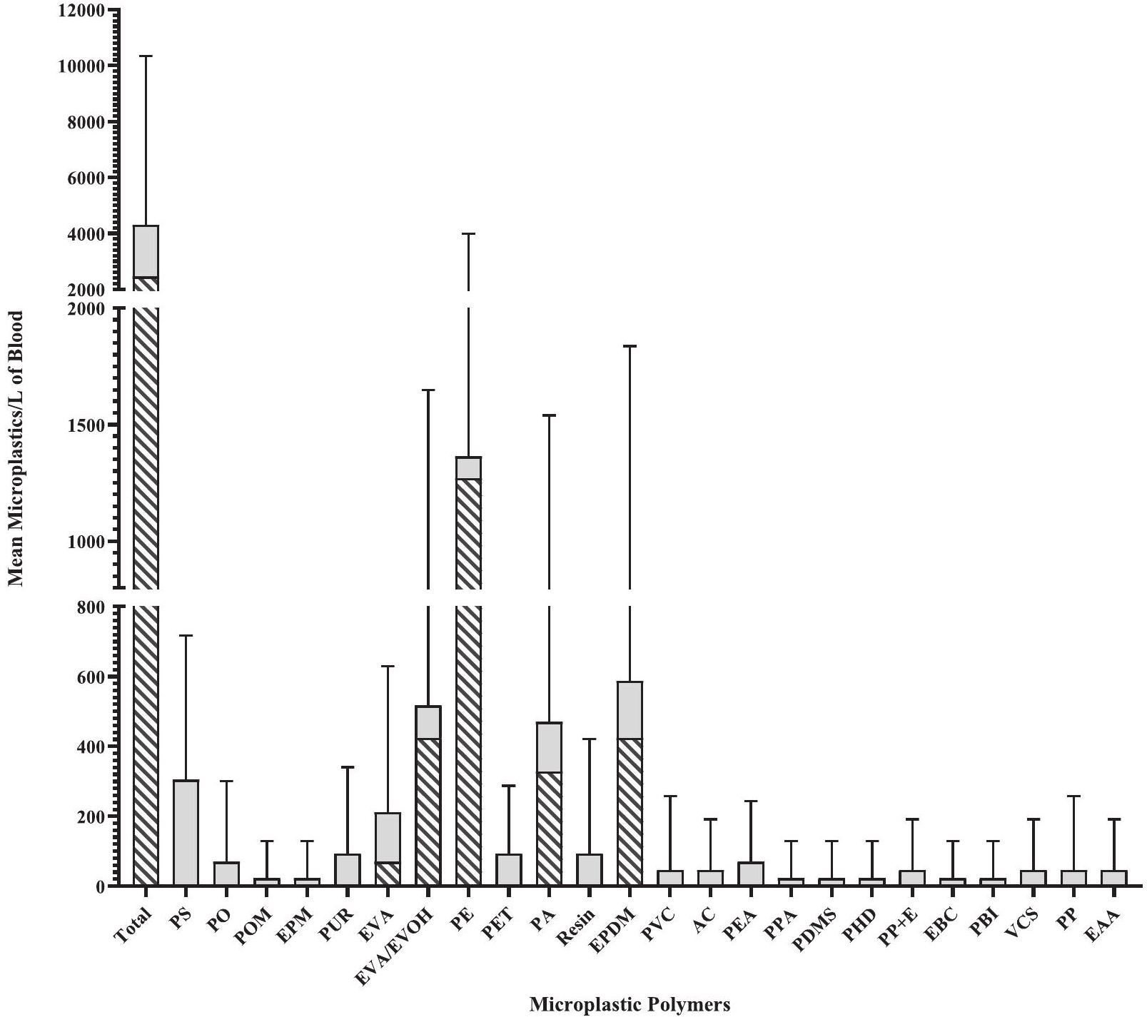
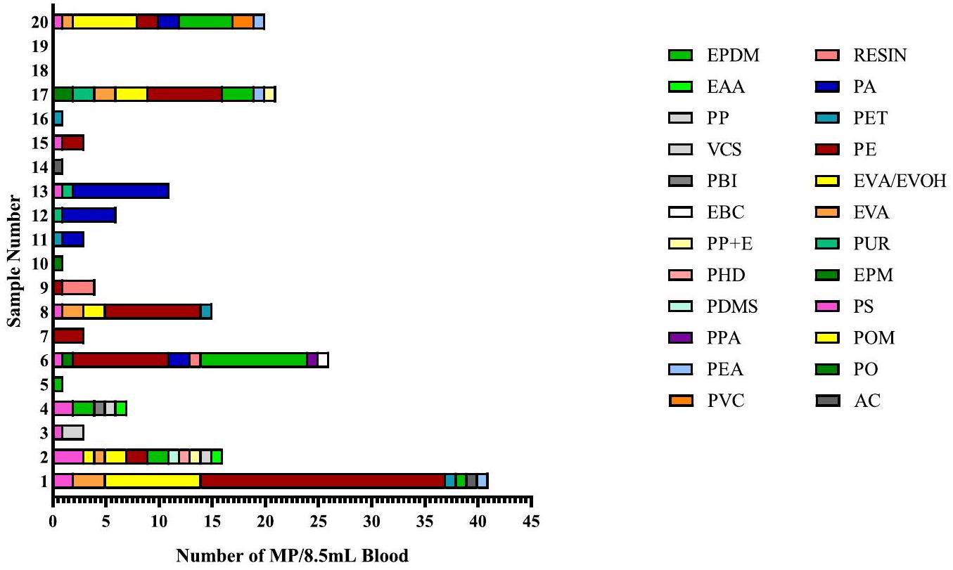
3.1. تركيزات الجسيمات الدقيقة المكتشفة في عينات دم الإنسان

في المجموع، تم تحديد 736 جزيء من الجسيمات الدقيقة من جميع عينات الدم البالغة 8.5 مل التي تم جمعها (الشكل 1). قبل تطبيق نهج عتبة LOQ، كانت هذه تتكون في البداية من 24 نوعًا من البوليمرات وتم الكشف عن الجسيمات الدقيقة في 18 من أصل 20. ) من عينات دم المتبرعين (الشكل 2). باستخدام فقط تلك الجزيئات التي استوفت معايير LOQ، تم الكشف عن الجزيئات في 8 من أصل 20 ( ) من عينات دم المتبرعين وكانت القيمة المتوسطة المكتشفة هي (الجدول 1). البيانات الخام باستخدام الحسابات غير المعدلة والطرح متاحة في الجدول S1. خمسة من 24 بوليمر MP المحددة، وهي: PE (عينة ، 17)، مونومر الإيثيلين بروبيلين داين (EPDM) (عينة 6، 20)، إيثيلين-فينيل أسيتات/إيثيلين فينيل كحول (EVA/EVOH) (عينة 1، 17، 20)، بولياميد (PA) (عينة، 12، 13)، إيثيلين-فينيل أسيتات (EVA) (عينة 1) (الشكل 1؛ الجدول 1)، كانت فوق حد الكشف (LOQ) (الجدول S2) لعينات الدم من كل متبرع حيثما تم الإشارة إلى ذلك.
تم توليد قيم تركيز الجسيمات الدقيقة المكتشفة في الدم للـ PE و EPDM و EVA/EVOH و PA و EVA مما يظهر أن المتبرعين (ن من أصل 20) حملت قيمة قابلة للقياس ( ) كتلة الجسيمات في دمهم (الشكل 1 والجدول 1). نوع البوليمر والتركيزات اختلفت حسب العينة، ولكن يمكن تحديد ما يصل إلى ثلاثة أنواع من البوليمر في عينة واحدة (الجدول S2)، مع كون PE الأكثر شيوعًا ( قيمة LOQ في من جميع المتبرعين الذين تم اختبارهم)، تليها EVA/EVOH (15٪)، EPDM (10٪) )، PA ( ) و EVA ( ). كانت التركيزات القصوى المقدرة في عينة الدم للرياضة البدنية، لـ EVA/EVOH، لـ EPDM، لـ PA، و لـ EVA. لإجراء تقدير محافظ للمجموع الكمي لتركيزات البوليمر في متبرعي الدم، يتم جمع جميع قيم البوليمر. تم استخدام LOQ لكل عينة. حيث لم يتم الكشف عن بوليمر لدى المتبرع عند مستوى LOQ، اعتُبرت هذه العينات كصفر. المتوسط التقديري ( كان تركيز المجموع لكل متبرع 0.66 الكتلة الإجمالية لجزيئات البلاستيك لكل عينة دم.
الفراغات المجمعة تحتوي على لكل عينات (النطاق ) مع 7 أنواع من الجسيمات الدقيقة ذات التركيب المختلف، مع بوليمرين، بولي تترافلورو إيثيلين (PTFE) وبولي أكريلاميد (PAM) تم التعرف عليهما فقط في عينات الفراغ الإجرائي. كان عدد جزيئات الجسيمات الدقيقة التي تم التعرف عليها في عينات الدم كبيرًا بشكل ملحوظ ( أكبر من الموجود في النماذج الإجرائية.

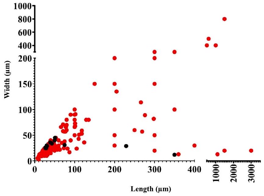
3.2. توصيف جزيئات MP من عينات الدم

بعد أن أثبتنا وجود البوليمرات في الدم، سعينا بعد ذلك لتحديد خصائصها. من بين 24 بوليمر تم اكتشافها في عينات الدم البشرية، شكلت الثلاثة بوليمرات الأكثر وفرة أكثر من من بين الذين تم تحديدهم، مع PE ( )، EPDM ( ) وخلائط الإيثيلين والفينيل أسيتات / الإيثيلين والفينيل الكحول (EVA/EVOH) (12 %) (الشكل 3؛ الشكل S1). كانت جزيئات MPs التي تم تحديدها ضمن عينات الدم لها متوسط طول جزيء ، ومتوسط عرض الجسيمات (الشكل 4). تم تصنيف النواب أيضًا بشكل أساسي كقطع ( ) وكان مظهرها أبيض/شفاف ( و على التوالي)، على الرغم من وجود عدد كبير من الشظايا بألوان مختلفة.
في المقارنة، تم تحديد 7 بوليمرات MP فقط في عينات الفراغ الإجرائية. في هذه الحالة، ومع ذلك، بينما كان PE ( ) كان لا يزال موجودًا، الراتنج ( ) ، PET ( ) و PS ( ) جنبًا إلى جنب مع PE تشكل من النواب الحاضرين (الشكل S2). ومن المثير للاهتمام أنه على الرغم من وجود عدد أقل بكثير من النواب الحاضرين في الفراغات الإجرائية، إلا أن متوسط طول الجسيمات كان 81.83 وعرض (الشكل 4) لم يكن مختلفًا بشكل ملحوظ ( و ) إلى الجزيئات الدقيقة الموجودة في الدم. علاوة على ذلك، كانت الجزيئات الدقيقة في الفراغات الإجرائية مرة أخرى، في الغالب، عبارة عن شظايا، مع زيادة صغيرة فقط في نسبة الألياف المحددة. ومع ذلك، كان هناك عدد أقل من الجزيئات الدقيقة الملونة، حيث كانت الجزيئات الدقيقة البيضاء والشفافة هي السائدة فقط ( و على التوالي) مع تحديد عدد قليل فقط من النواب السود في
الشكل 1. تركيزات جزيئات البلاستيك الدقيقة في عينات دم الإنسان لكل لتر من الدم (المتوسط SD). متوسط عدد جزيئات MP/L في عينات الدم. باستخدام القيم غير المعدلة، استنادًا إلى عدد الجزيئات المكتشفة في 8.5 مل من الدم مضروبًا في 118 (لتقريب 1 لتر) وعلى ربع فلتر مضروبًا في أربعة (الجدول S2، A)، تشير أشرطة الخطأ إلى الانحراف المعياري. متوسط عدد الجزيئات MP هو 4,306 والنطاق ( ). الاختصارات: AC، أكريليك؛ EAA، كوبوليمر حمض الإيثيلين الأكريليكي؛ EBC، كوبوليمر الإيثيلين-البوتان؛ EPDM، مونومر الإيثيلين بروبيلين ديني؛ EPM، إيثيلين بروبيلين؛ PBI، بولي بنزيميدازول؛ PDMS، بولي-دايميثيل سيليوكسان؛ PEA، ثنائي إيثيلين أدبيات؛ PET، بولي إيثيلين تيريفثاليت؛ PHD، بولي (1-هكساديكين)؛ PO، بولي أوليفين؛ POM، بولي أوكسي ميثيلين؛ PP، بولي بروبيلين؛ PP + E، بولي بروبيلين وإيثيلين؛ PPA، بولي فثالاميد؛ PS، بولي ستيرين؛ PUR، بولي إيثر يوريثان؛ PVC، بولي فينيل كلوريد؛ راتنج، راتنجات الألكيد؛ VCS، كوبوليمر كلوريد الفينيلدين-ستايرين.
فراغات إجرائية. تجربة التلاعب باستخدام جزيئات البلاستيك الدقيقة التجارية من بولي بروبيلين وبولي فينيل كلوريد حجم قدم معدل استرداد متوسط مؤشّر من عينات المياه المصفاة مسبقًا (المادة التكميلية SM1).

3.3. المواد المضافة MP الموجودة في عينات دم الإنسان

تم ملاحظة عدة مواد كيميائية مضافة من البوليمر أو بدائل البلاستيك في الجسيمات المستخرجة من عينات الدم. كانت المواد الكيميائية المضافة الأكثر شيوعًا المرتبطة بالجسيمات هي الفثالات التي تم الكشف عنها في عينات مع جزيئات/ لتر من الدم. كان ثلاثي (ن-أوكتيل، ن-ديسيل) تريميلات مرتبطًا بالجزيئات في عينات دم مع جسيمات بينما، كانت الألفا-أوليفين المتفاعلة، وثلاثي لوريل ثريثيوفوسفيت، وإستر الفوسفات بولي أوليفين و1،4-ثنائي فلوروبنزين-D4 موجودة فقط في عينة دم واحدة. وكان مضاف آخر، 1-ديكانول، مرتبطًا بالجزيئات في 60 من جميع عينات الدم بينما تم الكشف عن البوليمر (3-هيدروكسي بيوتيرات)، وهو بلاستيك حراري بكتيري بديل قابل للتحلل من نوع البوليستر. من عينات الدم.

4. المناقشة

في اكتشاف جديد، يكشف تحليل الدم من 20 متبرعًا صحيًا عن جزيئات MP بأنواع بوليمر مختلفة عما تم اكتشافه سابقًا. تمثل خصائص حجم وشكل جزيئات MP أول مجموعة بيانات من نوعها لجزيئات MP في دم الإنسان وتوفر تفاصيل هامة عن خصائص الشكل والحجم لتحديد الآثار البيولوجية المحتملة لوجودها في الدم. على وجه التحديد، لقد قدمت منهجية FTIR معلومات جديدة عن أنواع البوليمرات الدقيقة الموجودة بالإضافة إلى الأبعاد الشكلية والحجمية التي تتحدى النموذج المقبول حاليًا لخصائص البوليمرات الدقيقة داخل الأنسجة البشرية. من بين أربعة وعشرين نوعًا من البوليمرات البلاستيكية تم تحديدها من 18 من 20. ) المتبرعين، خمسة أنواع من البوليمر حققت عتبة LOQ بمتوسط نتيجة (نطاق ). PE ( EPDM ) ، و EVA-EVOH ( كانت القطع الأكثر وفرة.
التركيز فقط على تلك البوليمرات التي تجاوزت عتبة LOQ، أظهرت تركيزات الكتلة في الدم أن المتبرعين ( من أصل 20) كانت قابلة للقياس وهو أقل من من المتبرعين الذين تم الإبلاغ عنهم في
الشكل 2. التباين الفردي في تركيز وأنواع جزيئات البوليمر الدقيقة المكتشفة في المتبرع عينات الدم. قيم غير معدلة، ربع مساحة فلتر الأنوديسك التي تم تحليلها. الاختصارات: تشير إلى الشكل 1.
الجدول 1
عدد النواب المحددين ضمن عينات الدم بواسطة طيفية FTIR. يتم عرض أنواع البوليمرات التي استوفت معايير LOQ بوحدات MP/L من الدم. الاختصارات؛ – لم تستوف معايير LOQ. EPDM، مونومر الإيثيلين بروبيلين ديني؛ EVA، إيثيلين-أسيتات الفينيل؛ EVA/EVOH، إيثيلين-أسيتات الفينيل/إيثيلين كحول الفينيل؛ PA، بولياميد/نايلون؛ PE، بولي إيثيلين.
رقم عينة الدم التربية البدنية EVA/EVOH إيفا EPDM با
1 10,729 ٤٢٣٥ ١٤١٢
2
٣
٤
٥
٦ ٤١٤١ ٥١٢٩
٧
٨ ٤١٤١
9
10
11
12 ٢,٣٥٣
١٣ ٤٢٣٥
14
15 ٣٢٠٠
16
17 ٣٢٠٠ ١٤١٢
18
19
20 ٢,٨٢٤ ٢٣٠٦
المتوسط العام ٢٤٦٥.٨٥
SD
٤١٧٣.٥١
دراسة الدم الهولندية (ليزلي وآخرون، 2022). ومع ذلك، على غرار تلك الدراسة الأولى، اختلف نوع البوليمر والتركيزات أيضًا حسب العينة. في تلك الدراسة السابقة، التي ركزت على أنواع البوليمر المحددة فقط، كان البولي إيثيلين أيضًا موجودًا. كان البولي إيثيلين هو الأكثر شيوعًا (باستخدام قيم LOQ في من جميع المتبرعين الذين تم اختبارهم)، مع تقدير أدنى أقصى يبلغ 4.65 في هذه الدراسة بالنسبة إلى المبلغ عنه في الدراسة الهولندية. المتوسط المقدر ( كان تركيز المجموع لكل متبرع 0.66 إجمالي كتلة جزيئات البلاستيك لكل عينة دم في هذه الدراسة والتي هي أقل مقارنة بـ تم الإبلاغ عنه في الدراسة الهولندية (ليزلي وآخرون، 2022). من المهم أن نلاحظ أن الكتلة الأخيرة هي
الشكل 3. أنواع البوليمرات الدقيقة المحددة في عينات الدم. الاختصارات: انظر الشكل 1. باستخدام جميع الجزيئات (مجموعة البيانات غير المعدلة).
القيمة المستمدة من التحلل الحراري الفعلي بواسطة GCMS، بينما يوفر نهج FTIR أعداد/أحجام الجسيمات، وقد تم تقدير حساب الكتلة باستخدام معادلة مصممة للألياف بدلاً من الشظايا (Leusch & Ziajahromi، 2021)، مما قد يؤدي إلى تقدير منخفض للكتلة. قيد آخر من النهج المستخدم هنا في FTIR هو أنه تم تحليل ربع واحد فقط من الفلتر (يمثل ربع عينة الدم الفعلية). القيم المقدمة هي عدد الجسيمات المكتشفة مضروبًا في أربعة (لتمثيل فلتر كامل). لذلك، من الممكن أن تكون هناك جزيئات من بوليمرات أخرى موجودة ولكن لم يتم اكتشافها. أيضًا، قد تحتوي الحسابات على أخطاء تقريب حيث تم افتراض، ربما بشكل غير صحيح، أن جزيئات أنواع البوليمر المحددة موزعة بالتساوي على سطح الفلتر. تجربة التلاعب مع جزيئات PP و PVC ‘العذراء’ الموردة تجاريًا (من حجم) أشار إلى معدل استرداد متوسط قدره من عينات المياه المصفاة مسبقًا (المادة التكميلية SM1)، ضمن النطاق المبلغ عنه في دراسة حديثة -تحليل لـ 71 دراسة عن الميكروبلاستيك (Way et al., 2022) ويمثل اعتبارًا آخر لصالح القيم المستخلصة من تحليل عينات الدم كونها تقديرًا ناقصًا. بشكل عام، فإن قيمة تقدير الكتلة بالنسبة للدراسة السابقة للدم
الشكل 4. توزيع أبعاد حجم الجسيمات الدقيقة التي لوحظت في عينات الدم (بالأحمر) والعينات الفارغة (بالأسود). باستخدام جميع الجسيمات (مجموعة البيانات غير المعدلة). (للتفسير حول الإشارات إلى الألوان في أسطورة هذا الشكل، يُرجى الرجوع إلى النسخة الإلكترونية من هذه المقالة.)
ومعدل الاسترداد المتوسط لـ قد تشير إلى أن قيم MP المبلغ عنها هنا هي تقدير ناقص. تشمل القيود العامة الأخرى للنهج المنهجي المستخدم عدم اكتمال هضم المواد العضوية، واستخدام فلاتر Anodisc مما يتطلب نطاق مسح أقصر، و عتبة المطابقة المستخدمة مع مكتبة البوليمر للطيف. بينما تكون المطابقة لـ يمكن الحصول عليها، فقط التحليل الكيميائي الإضافي يمكن أن يوفر يقين تركيب الجسيمات.
تم تحديد البولي إيثيلين (PE) في 5 من أصل 20 عينة متبرع (باستخدام قيم LOQ) وقد تم عزله سابقًا من 5 من أصل 22 عينة دم بشري باستخدام طريقة التحلل الحراري، حيث كان أيضًا ثاني أكثر البوليمرات وفرة المكتشفة (ليزلي وآخرون، 2022). يُستخدم البولي إيثيلين في أفلام التعبئة والمنتجات، والأكياس، والعزل للأسلاك، والزجاجات، والعديد من العناصر المنزلية، بالإضافة إلى كونه مادة حيوية للزراعة الطبية (باكستون وآخرون، 2019). تم تحديد البولي إيثيلين سابقًا في أنسجة الرئة البشرية (جينر وآخرون، 2022) وتم التعرف عليه كأكثر أنواع الميكروبلاستيك وفرة في عينات حليب الثدي البشري (راجوزا وآخرون، 2022). فيما يتعلق بالتأثيرات البيولوجية، فإن التعرض للبولي إيثيلين (من كريات بحجم ) زاد من مستوى عدم الاستقرار الجينومي في دراسة خلايا الدم (كوبان أوغلو وآخرون، 2021).
على النقيض من ذلك، لم يتم الإبلاغ عن EPDM في أي نسيج بشري حتى الآن. يُستخدم EPDM في صناعة السيارات كطلاء عازل للطقس بسبب خصائصه الاستثنائية في مقاومة العوامل الجوية والحرارة (جاكوب وجوردان، 2011) وكذلك في العشب الصناعي مثل الملاعب الرياضية (أهرينز، 2018). قطع EPDM ( في الحجم) تم الإبلاغ مؤخرًا عن جميع عينات الهواء من بيئات العمل في صناعة المطاط حيث كانت الوفرة الأعلى في الهواء في ورشة ما بعد المعالجة في جزيئات (Sun وآخرون، 2023). تم التعرف على EVA-EVOH سابقًا في عينات البول البشرية (Rotchell وآخرون، اتصال شخصي). تُستخدم هذه كطبقة ربط بين البوليمرات الأخرى في المنتجات التي تتطلب حاجزًا للأكسجين، مثل تعبئة الطعام، والأفلام الزراعية، وفي صناعة السيارات (GaucherMiri وآخرون، 2002). يُستخدم EVA بمفرده عادةً في أنظمة توصيل الأدوية الطبية المُتحكم فيها (مثل وسائل منع الحمل حيث يُعتقد أنه يتمتع بخصائص التهاب ملائمة ويعتبر غير نشط نسبيًا) (Schneider وآخرون، 2017). كما يُستخدم في العناصر اليومية مثل الملابس/الحصائر حيث يتطلب الأمر حشوة (مثل أحذية الجري والحصائر الرياضية). يُستخدم EVOH بمفرده أيضًا كبوليمر متوافق حيويًا يشكل سدادة تشبه الإسفنج يمكن أن تسد التجاويف. وقد تم استخدامه بنجاح في حالات تشوهات الدماغ السريرية وفي النساء اللاتي يعانين من سلس البول الناتج عن الضغط (Elzayat & Corcos، 2008). يتكون فيلم EVA/ EVOH/EVA متعدد الطبقات من ثلاثة أنواع مختلفة من البوليمرات، مما يتيح توليد عدة أنواع من الجذور لكل منها.
نوع البوليمر، أي، PE، الذي يرتبط بأكثر بوليمر MP وفرة تم اكتشافه في هذه الدراسة (للمراجعة حول الهيكل: غاستون وآخرون، 2021).
وجود وتأثيرات المحتملة للجزيئات الدقيقة في الدم أمر حاسم لفهمه. يوفر الدم العديد من الوظائف الرئيسية، للمساعدة في حركة المواد والخلايا في جميع أنحاء الجسم. يمكن أن يؤدي اضطراب نظام الدم إلى تأثيرات كبيرة على الرفاهية العامة للجسم. لقد أثبتت الأعمال السابقة بالفعل أن جزيئات دقيقة مختارة (PET > PE > PS > PMMA) موجودة في دم الإنسان (ليزلي وآخرون، 2022) وداخل أنسجة الأوردة (راتنج الألكيد > بولي فينيل بروبيونات/أسيتات، (PVAc). نايلون-إيثيلين-فينيل أسيتات (نايلون-EVA)، طبقة الربط) في البشر (روتشيل وآخرون، 2023). هنا نعرض مزيجًا أكثر تنوعًا من أنواع البوليمرات الدقيقة مقارنةً بما تم تحديده سابقًا ونحدد متوسط طول الجسيمات بـ ، ومتوسط عرض الجسيمات من المهم أنه لم يتم العثور على كرات كروية داخل العينات. بالنظر إلى نطاق حجم الجزيئات الدقيقة، سواء من حيث الطول أو العرض، يجب ملاحظة أنه بينما يمكن أن تستوعب أجزاء من النظام الوعائي، مثل الشرايين والأوردة الرئيسية، جزيئات بهذا الحجم، فإن الشعيرات الدموية عادةً في القطر، مما يشكل حاجزًا نظريًا أمام أي جسيم يزيد عن في أكثر من بُعد واحد. لذلك، على الرغم من أن الجزيئات الصغيرة قد تتمكن من المرور عبر الشعيرات الدموية جزئيًا بسبب مرونتها وضغط نظام الدم، فمن المحتمل أن يكون لها مرور بطيء عبر نظام الشعيرات الدموية، وبالتالي يمكن أن تقدم فرصة واضحة للتفاعل مع كل من البطانة الوعائية وخلايا الدم.
بعد أن تم تأسيس وجود الجسيمات الدقيقة (MP) وتحديد أبعادها الفيزيائية داخل الدم، فإن السؤال المهم التالي يتعلق بأي آثار محتملة. بمجرد دخولها إلى الدم، يمكن للجسيمات الدقيقة التفاعل مع البروتينات الموجودة في البلازما، مثل الأجسام المضادة، الفيبرينوجين، أو بروتينات التخثر لتشكيل غلاف (Lundqvist et al., 2008). قد يساعد هذا الغلاف الجسيمات الدقيقة على عدم التعرف عليها من قبل الجهاز المناعي (Mirshafiee et al., 2016)، مما قد يطيل الوقت الذي تقضيه الجسيمات الدقيقة داخل الجسم. من الأدبيات المتاحة، تستخدم معظم الدراسات حول التأثيرات البيولوجية للتعرض الجسيمات النانوية (NP) أو أصغر الجسيمات الدقيقة حجماً. )، عادةً ما تكون على شكل كروي ومن أصل عذري. يمكن ابتلاع الجسيمات النانوية الأصغر ( الحجم، كرات PS) بواسطة البلعميات الفأرية والعدلات مما يؤدي إلى استجابة التهابية (فلورانس وآخرون، 2021). بينما تعزز خلايا البلعميات الفأرية المعرضة لكرات PS ( الحجم)، مع وجود غلاف مرتبط، من عملية الإدخال بواسطة البلعميات (رامسبرجر وآخرون، 2020). كما أظهرت الجسيمات النانوية المفعلة ( حجم كرات PS وكرات PS-أمين) تأثيرها على تجميع الصفائح الدموية ومنع تكوين الجلطة في نماذج القوارض (فلاسيل وآخرون، 2021؛ نيمار وآخرون، 2002)، مما يشير إلى أن هذه البلاستيكات الأصغر قد تساهم في إما نزيف عبر زيادة القابلية للنزيف أو الكدمات بسبب انخفاض التخثر، أو بيئة مؤيدة للتخثر تتضمن تجلط غير طبيعي. أظهرت الجسيمات النانوية من PE وPS ( الحجم) التي تم امتصاصها بواسطة خلايا المناعة في الأسماك (سالمو سالار) أنماط بلع مختلفة في الأنسجة (أبيهسيرا-غارسيا وآخرون، 2020)، مما يبرز مسارًا آخر للتأثير داخل الدم.
كانت الجسيمات المحددة في عينات الدم البشرية أكبر بمقدار ترتيب من حيث الحجم وأبعاد الشكل مقارنة بتلك المستخدمة في الدراسات التي تم مناقشتها حتى الآن. يثير هذا السؤال حول كيفية دخول الجسيمات الكبيرة نسبيًا إلى مجرى الدم. تم إثبات مسار مباشر، لجسيمات بحجم 4-148 ، من خلال الحقن (تشو وآخرون، 2024). قد تشمل المسارات غير المباشرة الاستنشاق، النظام الغذائي والاتصال الجلدي الذي يتضمن الحواجز. بينما تم إثبات أن الكرات بحجم النانو يمكن أن تنتقل عبر خلايا الحاجز المعوي البشري (دومينيش وآخرون، 2020)، فإن نطاق حجم البلاستيكات الدقيقة لم يتم التحقيق فيه بهذه الطريقة بعد. من حيث التأثيرات الناتجة عن وجود MP، من الأدبيات المتاحة في هذا النطاق الأكبر من حجم MP، فإن تلك التي بحجم ( كرات PS) أثارت تأثيرات السيتوكين والهستامين في خلايا الدم المحيطية البشرية (PBMCs)، بينما لم يكن للجسيمات الأكبر بحجم 100 (كرات PS) أي تأثيرات من هذا القبيل (هوانغ وآخرون، 2020). بالمقابل، أظهرت كرات PE بحجم ( ) وقطع ( ) تقليل حيوية الخلايا، وتحفيز إفراز السيتوكين IL-6 وTNF ألفا وزيادة انحلال الدم في PBMCs البشرية، وخلايا الصارية، وخلايا الدم الحمراء على الرغم من تركيز MP العالي (من ) التعرضات (تشوي وآخرون، 2021).
تم أيضًا إثبات أن إفراز السيتوكين IL-6 وTNF ألفا والهستامين يحدث بعد التعرض لكرات PP غير المنتظمة الشكل، حيث أثار حجم 20 استجابة أكبر مقارنة بحجم ، باستخدام خلايا الألياف الجلدية البشرية وخلايا البلعميات الفأرية (هوانغ وآخرون، 2019). باستخدام نماذج الأسماك، أثارت MP من PS وPC (الأحجام ) تفريغ العدلات في أسماك المينوز المسطحة (Pimephales promelas)، بينما أدى التعرض لـ MP من PVC ( كرات) إلى تقليل القدرة البلعومية لكريات الدم البيضاء في سمك الدنيس الذهبي (Sparus aurata) وسمك القاروص (Dicentrarchus labrax) (إسبينوزا وآخرون، 2018). لذلك، يؤثر حجم MP ونوعه وشكله وتركيزه على نتائج الاختبارات في المختبر باستخدام الأنواع الفقارية، ويمكن أن تساعد البيانات المقدمة هنا الآن في إبلاغ المستويات المطلوبة، وأنواع البوليمرات، والأشكال والأبعاد لفهم استجابة الجسم بشكل أكثر دقة لوجود هذه MP.
على الرغم من أنه يمكننا إعطاء أمثلة على تأثيرات محددة لـ MP على أنواع خلايا معينة، يجب أن نلاحظ أن التوزيع العام لـ MP وتأثيرات MP داخل البشر لا تزال غير مفهومة، وهي الخطوة الحاسمة التالية لهذا المجال. جزئيًا، هذا هو السبب في أن تحديد الشكل والحجم لـ MP المحددة داخل البشر أمر بالغ الأهمية، نظرًا لأن الشكل والحجم معروف أنهما يؤثران على الاستجابة الخلوية (للمراجعة: دانوبولوس وآخرون، 2021). علاوة على ذلك، في مجرى الدم، يعتبر الحجم والشكل القابل للتشكيل مهمين، حيث يمكن للشرايين والأوردة الرئيسية استيعاب MP الكبيرة غير القابلة للتشكيل، بينما تمثل الشعيرات الدموية حاجزًا نظريًا لـ MP نظرًا لأنها عادةً ما تكون في القطر. قد تتمكن MP من الضغط عبر الشعيرات الدموية، ولكن يجب أن نلاحظ أن الشعيرات ليست خطية فقط ويمكن أن تحتوي على انحناءات، وبالتالي قد لا تمتلك MP المرونة للتحرك حول الانحناءات بفعالية كما تفعل خلايا الدم الحمراء (وانغ وآخرون، 2022). بمجرد أن تعلق، من المحتمل أن تتأثر حركة خلايا الدم الحمراء، وبالتالي قد تحدث تغييرات محلية في تركيزات الأكسجين، مما يؤدي إلى تأثيرات على التمثيل الغذائي الخلوي ووظيفة الخلايا. قد تشير المستويات المتوسطة من MP المبلغ عنها هنا أيضًا إلى التراكم الحيوي. بينما لم يكن التراكم الحيوي ضمن نطاق هذه الدراسة، هناك دراسات تظهر تراكم MP عبر الأنواع البحرية (ميلر وآخرون، 2020)، على الرغم من أن هذا غير معروف بعد للبشر.
بالإضافة إلى أن نوع وشكل وحجم MP أمر بالغ الأهمية للتفاعل مع الجسم، يجب أيضًا ملاحظة أن MP تحتوي على آلاف الإضافات، العديد منها معروف بأنه سام (هالهداكيس وآخرون، 2018؛ زيمرمان وآخرون، 2021) وتمثل مجموعة من المواد الكيميائية السامة المحتملة. تم تحديد أربعة أنواع من الفثالات (ديوندسيل فثالات، ديكابريل فثالات، بوتيل بنزيل فثالات وأوكتيل بنزيل فثالات) داخل الجسيمات في عينات الدم. يجب أن نلاحظ أن البلاستيكات ليست المصدر الوحيد لهذه الأنواع من المواد الكيميائية. تم ربط الفثالات، المضافة كعوامل بلاستيكية، بنقاط سامة في البشر بما في ذلك الأنظمة الهرمونية، والعصبية، والقلبية الوعائية، والتنفسية بالإضافة إلى مسارات الاستجابة المناعية (تشانغ وآخرون، 2021؛ ماريانا وآخرون، 2023؛ وانغ وكيان، 2021). تم تحديد التريميلات، وهو عامل بلاستيكي آخر، المستخدم في تصنيع PVC في صناعة السيارات وكابلات العزل بالإضافة إلى مواد التعبئة والتغليف الغذائية، في من العينات على الرغم من أنه لا يعتبر ضارًا للبشر بعد (EFSA، 2019). قد تؤثر المواد الكيميائية السامة، مثل هذه، أيضًا على تكوين أي غلاف وتحدد مصير وتأثيرات MP الموجودة كما تم مناقشته سابقًا. الفئة النهائية للنقاش هي البلاستيكات الحيوية. تم تحديد بولي (3-هيدروكسي بيوتيرات)، وهو بلاستيك حراري مشتق من البكتيريا (بارهام وآخرون، 1984)، في من عينات الدم التي تم التحقيق فيها. يتم تسويق هذه البلاستيكات المشتقة بيولوجيًا كبديل لبلاستيكات نوع PES، وعلى الرغم من أنها قابلة للتحلل البيولوجي، يمكن اكتشاف شظاياها داخل جسم الإنسان.
لخلاصة القول، تم اكتشاف MP في عينات الدم البشرية، مما يظهر تركيبة بوليمرية مختلفة، ونطاق حجم أكبر، مما تم تقديره سابقًا. كما تم تصنيفها، للمرة الأولى، فيما يتعلق بتركيزاتها، وحجمها، وأبعاد شكلها، مع عدم اكتشاف أي كرات بشكل مفاجئ. بالإضافة إلى جزيئات MP، تم اكتشاف العديد من الإضافات الشائعة الاستخدام بما في ذلك أربعة أنواع مختلفة من الفثالات. تتيح هذه المعلومات الآن دراسات سمية أكثر تمثيلاً في المختبر وفي الجسم فيما يتعلق بـ
تداعيات وجود MP في الدم البشري وتبرز بشكل أكبر ضرورة إجراء مثل هذه التحليلات نظرًا للوفرة التي تم إثباتها في هذه التحقيق.

بيان مساهمة المؤلفين

صوفي ف. ل. ليونارد: الكتابة – مراجعة وتحرير، الكتابة – المسودة الأصلية، الإشراف، المنهجية، التحقيق، التحليل الرسمي. كاتريونا ر. ليدل: التحليل الرسمي. شارلوت أ. أثيرال: التحليل الرسمي. إيما تشابمان: الكتابة – مراجعة وتحرير، الكتابة – المسودة الأصلية، الإشراف، الموارد، المنهجية، التحليل الرسمي، تنسيق البيانات. ماثيو واتكينز: المنهجية، التحليل الرسمي. سيمون د. ج. كالمينوس: الكتابة – مراجعة وتحرير، الكتابة – المسودة الأصلية، الإشراف، الموارد، إدارة المشروع، التحقيق، الحصول على التمويل، التصور. جانيت م. روتشيل: الكتابة – مراجعة وتحرير، الكتابة – المسودة الأصلية، التحقق، الإشراف، الموارد، إدارة المشروع، التحليل الرسمي، تنسيق البيانات، التصور.

إعلان عن تضارب المصالح

يعلن المؤلفون أنهم ليس لديهم مصالح مالية متنافسة معروفة أو علاقات شخصية قد تكون قد أثرت على العمل المبلغ عنه في هذه الورقة.

توفر البيانات

ستكون البيانات متاحة عند الطلب.

شكر وتقدير

نود أن ن dedic هذا العمل إلى الراحلة الدكتورة مونيكا أرمان. كانت ستثير اهتمامها هذه الدراسة وبدون أفكارها وقدرتها، لم تكن S.V.L.L لتنال منحة الدكتوراه. تم تمويل هذا العمل من قبل مؤسسة القلب البريطانية، رقم منحة طلاب الدكتوراه: FS/PhD/22/29265 (إلى S.V.L.L.).
بيان الأخلاقيات: تم تنفيذ العمل بموجب إذن أخلاقي منحته دراسة NHS REC ‘تحقيق في خلايا الدم لأبحاث الأمراض القلبية الوعائية’ (21/SC0215).

الملحق أ. البيانات التكميلية

يمكن العثور على البيانات التكميلية لهذه المقالة عبر الإنترنت على https://doi. org/10.1016/j.envint.2024.108751.

References

Abihssira-Garcia, I., Park, Y., Kiron, V., Olsvik, P.A., 2020. Fluorescent microplastic uptake by imune cells of Atlantic salmon (Salmo salar L.). Front. Environ Sci 8, 560206. https://doi.org/10.3389/fenvs.2020.560206.
Ahrens, A., 2018. European Chemical Agency’s Perspective: An Evaluation of the Possible Health Risks of Recycle Rubber Granules used as Infill in Synthetic Turf Fields. In: ISEE Conference Abstracts, Vol. 2018, No. 1.
Amato-Lourenço, L.F., Carvalho-Oliveira, R., Júnior, G.R., dos Santos, G.L., Ando, R.A., Mauad, T., 2021. Presence of airborne microplastics in human lung tissue. J Hazard Mat. 416, 126124. https://doi.org/10.1016/j.jhazmat.2021.126124.
Barham, P.J., Keller, A., Otun, R.L., Holmes, P.A., 1984. Crystallisation and morphology of a bacterial thermoplastic: poly-3-hydroxybutyrate. J Mat Sci. 19, 2781-2794.
Bihari, P., Holzer, M., Praetner, M., Fent, J., Lerchenberger, M., Reichel, C., et al., 2010. Single-walled carbon nanotubes activate platelets and accelerate thrombus formation in the microcirculation. Toxicol. 269, 148-154. https://doi.org/10.1016/ j.tox.2009.08.011.
Chang, W.H., Herianto, S., Lee, C.C., Hung, H., Chen, H.L., 2021. The effects of phthalate ester exposure on human health: A review. Sci Total Environ. 786, 147371. https:// doi.org/10.1016/j.scitotenv.2021.147371.
Choi, D., Hwang, J., Bang, J., Han, S., Kim, T., Oh, Y., et al., 2021. In vitro toxicity from a physical perspective of polyethylene microplastics based on statistical curvature change analysis. Sci Total Environ. 752, 142242. https://doi.org/10.1016/j. scitotenv.2020.142242.
Çobanoğlu, H., Belivermiş, M., Sıkdokur, E., Kılıç, Ö., Çayır, A., 2021. Genotoxic and cytotoxic effects of polyethylene microplastics on human peripheral blood
lymphocytes. Chemosphere 272, 129805. https://doi.org/10.1016/J. Chemosphere.2021.129805.
Cole, M., Lindeque, P., Halsband, C., Galloway, T.S., 2011. Microplastics as contaminants in the marine environment: A review. Mar Pollut Bull. 62, 2588-2597. https://doi. org/10.1016/j.marpolbul.2011.09.025.
Danopoulos, E., Twiddy, M., West, R., Rotchell, J.M., 2021. A rapid review and metaregression analyses of the toxicological impacts of microplastic exposure in human cells. J Hazard Mat. 427, 127861. https://doi.org/10.1016/j.jhazmat.2021.127861.
Domenech, J., Hernández, A., Rubio, L., Marcos, R., Cortés, C., 2020. Interactions of polystyrene nanoplastics with in vitro models of the human intestinal barrier. Arch. Toxicol. 94, 2997-3012.
Dong, X., Liu, X., Hou, Q., Wang, Z., 2023. From natural environment to animal tissues: A review of microplastics (nanoplastics) translocation and hazards studies. Sci Tot Environ. 855, 158686. https://doi.org/10.1016/j.scitotenv.2022.158686.
EFSA, 2019. Panel on Food Contact Materials. Safety assessment of the substance trimellitic acid, tris (2-ethylhexyl) ester, for use in food contact materials. EFSA J. 17, e05864. https://doi.org/10.2903/j.efsa.2019.5864.
Elzayat, E.A., Corcos, J., 2008. Urethral injectables in the management of stress urinary incontinence. In: Urology, F. (Ed.), Female Urology, Third Edition. Elsevier, London, pp. 348-361.
Espinosa, C., Beltrán, J.M.G., Esteban, M.A., Cuesta, A., 2018. In vitro effects of virgin microplastics on fish head-kidney leucocyte activities. Environ Pollut. 235, 30-38. https://doi.org/10.1016/j.envpol.2017.12.054.
Florance, I., Ramasubbu, S., Mukherjee, A., Chandrasekaran, N., 2021. Polystyrene nanoplastics dysregulate lipid metabolism in murine macrophages in vitro. Toxicol. 458, 152850.
Free, C.M., Jensen, O.P., Mason, S.A., Eriksen, M., Williamson, N.J., Boldgiy, B., 2014. High-levels of microplastic pollution in a large, remote, mountain lake. Mar Pollut Bull. 85, 156-163. https://doi.org/10.1016/J.MARPOLBUL.2014.06.001.
Gaston, F., Dupuy, N., Girard-Perier, N., Marque, S.R.A., Dorey, S., 2021. Investigations at the product, macromolecular, and molecular level of the physical and chemical properties of a -irradiated multilayer EVA/EVOH/EVA film: comprehensive analysis and mechanistic insights. Polymers 13, 2671. https://doi.org/10.3390/ polym13162671.
Gaucher-Miri, V., Jones, G.K., Kaas, R., Hiltner, A., Baer, E., 2002. Plastic deformation of EVA, EVOH and their multilayers. J Mat Sci. 37, 2635-2644.
Griffin, M.T., Zhu, Y., Liu, Z., Aidun, C.K., Ku, D.N., 2018. Inhibition of high shear arterial thrombosis by charged nanoparticles. Biomicrofluidics 12, 042210. https:// doi.org/10.1063/1.5025349.
Hahladakis, J.H., Velis, C.A., Weber, R., Iacovidou, E., Purnell, P., 2018. An overview of chemical additives present in plastics: migration, release, fate and environmental impact during their use, disposal and recycling. J Hazard Mater. 344, 179-199. https://doi.org/10.1016/j.hazmat.2017.10014.
Hartmann, N.B., Hüffer, T., Thompson, R.C., Hassellöv, M., Verschoor, A., Daugaard, A. E., et al., 2019. Are we speaking the same language? Recommendations for a definition and categorization framework for plastic debris. Environ Sci Technol. 53, 1039-1047. https://doi.org/10.1021/acs.est.8b05297.
Horton, A.A., Cross, R.K., Read, D.S., Jürgens, M.D., Ball, H.L., Svendsen, C., et al., 2021. Semi-automated analysis of microplastics in complex wastewater samples. Environ Pollut. 268, 115841. https://doi.org/10.1016/j.envpol.2020.115841.
Horvatits, T., Tamminga, M., Liu, B., Sebode, M., Carambia, A., Fischer, L., et al., 2022. Microplastics detected in cirrhotic liver tissue. E Bio Medicine 82, 104147. https:// doi.org/10.1016/j.ebiom.2022.104147.
Hwang, J., Choi, D., Han, S., Choi, J., Hong, J., 2019. An assessment of the toxicity of polypropylene microplastics in human derived cells. Sci Tot Environ. 684, 657-669. https://doi.org/10.1016/j.scitotenv.2019.05.071.
Hwang, J., Choi, D., Han, S., Jung, S.Y., Choi, J., Hong, J., 2020. Potential toxicity of polystyrene microplastic particles. Sci Rep. 10, 7391. https://doi.org/10.1038/ s41598-020-64464-9.
Ibrahim, Y.S., Tuan Anuar, S., Azmi, A.A., Wan Mohd Khalik, W.M.A., Lehata, S., Hamzah, S.R., et al., 2021. Detection of microplastics in human colectomy specimens. JGH Open. 5, 116-121. https://doi.org/10.1002/jgh3.12457.
Jacob, S., Jourdain, E.P., 2011. Advancements in EPDM sponge compound processing using EPDM bimodal polymers. Rubber Chem Technol. 84, 527-542. https://doi. org/10.5254/1.3601888.
Jenner, L.C., Rotchell, J.M., Bennett, R.T., Cowen, M., Tentzeris, V., Sadofsky, L.R., 2022. Detection of microplastics in human lung tissue using FTIR spectroscopy. Sci Tot Environ. 831, 154907. https://doi.org/10.1016/j.scitotenv.2022.154907.
Leslie, H.A., van Velzen, M.J.M., Brandsma, S.H., Vethaak, A.D., Garcia-Vallejo, J.J., Lamoree, M.H., 2022. Discovery and quantification of plastic particle pollution in human blood. Environ Int. 163, 107199. https://doi.org/10.1016/j. envint.2022.107199.
Leusch, F.D.L., Ziajahromi, S., 2021. Converting mg/L to particles/L: reconciling the occurrence and toxicity literature in microplastics. Environ Sci Technol. 55, 11470-11472. https://doi.org/10.1021/acs.est.1c04093.
Li, B., Ding, Y., Cheng, X., Sheng, D., Xu, Z., Rong, Q., et al., 2020. Polyethylene microplastics affect the distribution of gut microbiota and inflammation development in mice. Chemosphere 244, 125492. https://doi.org/10.1016/j. chemosphere.2019.125492.
Li, Y., Peng, L., Fu, J., Dai, X., Wang, G., 2022. A microscopic survey on microplastics in beverages: the case of beer, mineral water and tea. Analyst 147, 1099-1105. https:// doi.org/10.1039/d2an00083k.
Liu, Z., Zhuan, Q., Zhang, L., Meng, L., Fu, X., Hou, Y., 2022. Polystyrene microplastics induced female reproductive toxicity in mice. J Hazard Mater. 424, 127629. https:// doi.org/10.1016/j.jhazmat.2021.127629.
Lundqvist, M., Stigler, J., Elia, G., Lynch, I., Cedervall, T., Dawson, K.A., 2008. Nanoparticle size and surface properties determine the protein corona with possible implications for biological impacts. PNAS USA 105, 14265-14270.
Mamun, A.A., Prasetya, T.A.E., Dewi, I.R., Ahmad, M., 2023. Microplastics in human food chains: Food becoming a threat to health safety. Sci Tot Environ. 858, 159834. https://doi.org/10.1016/j.scitotenv.2022.159834.
Mariana, M., Castelo-Branco, M., Soares, A.M., Cairrao, E., 2023. Phthalates’ exposure leads to an increasing concern on cardiovascular health. J Hazard Mater. 457, 131680. https://doi.org/10.1016/j.jhazmat.2023.131680.
Miller, M.E., Hamann, M., Kroon, F.J., 2020. Bioaccumulation and biomagnification of microplastics in marine organisms: a review and meta-analysis of current data. PloS ONE, 0240792. https://doi.org/10.1371/journal.pone.0240792.
Mirshafiee, V., Kim, R., Park, S., Mahmoudi, M., Kraft, M.L., 2016. Impact of protein precoating on the corona composition and nanoparticle cellular uptake. Biomaterials 75, 295-304. https://doi.org/10.1016/j.biomaterials.2015.10.019.
Munno, K., Helm, P.A., Jackson, D.A., Rochman, C., Sims, A., 2018. Impacts of temperature and selected chemical digestion methods on microplastic particles. Environ Toxicol Chem. 37, 91-98. https://doi.org/10.1002/etc.3935.
Nemmar, A., Hoylaerts, M.F., Hoet, P.H.M., Dinsdale, D., Smith, T., Xu, H., et al., 2002. Ultrafine particles affect experimental thrombosis in an in vivo hamster model. Am J Respir Crit Care Med 166, 998-1004. https://doi.org/10.1164/rccm.2001100260C.
Noonan, M.J., Grechi, N., Mills, C.L., de AMM Ferraz, M., 2023. Microplastics analytics: why we should not underestimate the importance of blank controls. Micropl & Nanopl. 3, 17. https://doi.org/10.1186/s43591-023-00065-3.
O’Brien, S., Rauert, C., Ribeiro, F., Okoffo, E.D., Burrows, S.D., O’Brien, J.W., et al., 2023. There’s something in the air: A review of sources, prevalence and behaviour of microplastics in the atmosphere. Sci Tot Environ. 874, 162193. https://doi.org/ 10.1016/j.scitotenv.2023.162193.
Paxton, N.C., Allenby, M.C., Lewis, P.M., Woodruff, M.A., 2019. Biomedical applications of polyethylene. Eur Polymer J. 118, 412-428. https://doi.org/10.1016/j. eurpolymj.2019.05.037.
Ragusa, A., Svelato, A., Santacroce, C., Catalano, P., Notarstefano, V., Carnevali, O., et al., 2021. Plasticenta: First evidence of microplastics in human placenta. Environ Int. 146, 106274. https://doi.org/10.1016/j.envint.2020.106274.
Ragusa, A., Notarstefano, V., Svelato, A., Belloni, A., Gioacchini, G., Blondeel, C., et al., 2022. Raman microspectroscopy detection and characterisation of microplastics in human breastmilk. Polymers 14, 2700. https://doi.org/10.3390/polym14132700.
Ramsperger, A.F.R.M., Narayana, V.K.B., Gross, W., Mohanraj, J., Thelakkat, M., Greiner, A., et al., 2020. Environmental exposure enhances the internalization of microplastic particles into cells. Sci Adv. 6, eabd1211 https://doi.org/10.1126/ sciadv.abd1211.
Rotchell, J.M., Jenner, L.C., Chapman, E., Bennett, R.T., Bolanle, I.O., Loubani, M., et al., 2023. Detection of microplastics in human saphenous vein tissue using FTIR: A pilot study. PLoS One 18, 0280594. https://doi.org/10.1371/journal.pone. 0280594.
Schneider, C., Langer, R., Loveday, D., Hair, D., 2017. Applications of ethylene vinyl acetate copolymers (EVA) in drug delivery systems. J Control Rel. 262, 284-295. https://doi.org/10.1016/j.jconrel.2017.08.004.
Schwabl, P., Koeppel, S., Koenigshofer, P., Bucsics, T., Trauner, M., Reiberger, T., et al., 2019. Detection of various microplastics in human stool: a prospective case series. Ann Intern Med. 171, 453-457. https://doi.org/10.7326/M19-0618.
Sun, X., Song, R., Liu, J., et al., 2023. Characterization of airborne microplastics at different workplaces of the poly(ethylene:propylene:diene) (EPDM) rubber industry. Environ Sci Pollut Res. 30, 78839-78848. https://doi.org/10.1007/s11356-023-27750-3.
Tadic, M., Cuspidi, C., Grassi, G., 2018. Heart rate as a predictor of cardiovascular risk. Eur J Clin Invest. 48, 12892. https://doi.org/10.1111/eci.12892.
Vianello, A., Jensen, R.L., Liu, L., Vollertsen, J., 2019. Simulating human exposure to indoor airborne microplastics using a Breathing Thermal Manikin. Sci Rep. 9, 8670. https://doi.org/10.1038/s41598-019-45054-w.
Vlacil, A.K., Bänfer, S., Jacob, R., Trippel, N., Kuzu, I., Schieffer, B., Grote, K., 2021. Polystyrene microplastic particles induce endothelial activation. PLoS One 16, e0260181. https://doi.org/10.1371/journal.pone. 0260181.
Wang, C., Li, J., Zhao, L., Qian, P., 2022. Shape transformations of red blood cells in the capillary and their possible connections to oxygen transport. J Biol Phys 48, 79-92. https://doi.org/10.1007/s10867-021-09594-5.
Wang, Y., Qian, H., 2021. Phthalates and their impacts on human health. Healthcare 9, 603. https://doi.org/10.3390/healthcare9050603.
Way, C., Hudson, M.D., Williams, I.D., Langley, G.J., 2022. Evidence of underestimation in microplastic research: a meta-analysis of recovery rates. Sci. Total Environ. 805, 150277. https://doi.org/10.1016/j.scitotenv.2021.150277.
Yan, Z., Liu, Y., Zhang, T., Zhang, F., Ren, H., Zhang, Y., 2022. Analysis of microplastics in human feces reveals a correlation between fecal microplastics and inflammatory bowel disease status. Environ Sci Technol. 56, 414-421. https://doi.org/10.1021/ acs.est.1c03924.
Yang, L., Zhang, Y., Kang, S., Wang, Z., Wu, C., 2021. Microplastics in soil: A review on methods, occurrence, sources, and potential risk. Sci Tot Environ. 780, 146546. https://doi.org/10.1016/j.scitotenv.2021.146546.
Zhao, Q., Zhu, L., Weng, J., Jin, Z., Cao, Y., Jiang, H., et al., 2023. Detection and characterization of microplastics in the human testis and semen. Sci Tot Environ. 877, 162713 https://doi.org/10.1016/j.scitotenv.2023.162713.
Zhao, T., Shen, L., Ye, X., Bai, G., Liao, C., Chen, Z., et al., 2023. Prenatal and postnatal exposure to polystyrene microplastics induces testis developmental disorder and affects male fertility in mice. J Hazard Mater. 445, 130544 https://doi.org/10.1016/ j.jhazmat.2022.130544.
Zhu, L., Ma, M., Sun, X., Wu, Z., Yu, Y., Kang, Y., Liu, Z., Xu, Q., An, L., 2024. Microplastics entry into the blood by infusion therapy: few but a direct pathway. Environ. Sci. Technol. 11, 2. https://doi.org/10.1021/acs.estlett.3c00905.
Zhu, X., Wang, C., Duan, X., Liang, B., Genbo, X.E., Huang, Z., 2023. Micro- and nanoplastics: A new cardiovascular risk factor? Environ Int. 171, 107662. https:// doi.org/10.1016/j.envint.2022.107662.
Zimmermann, L., Bartosova, Z., Braun, K., Oehlmann, J., Volker, C., Wagmer, M., 2021. Plastic products leach chemicals that induce in vitro toxicity under realistic use conditions. Environ Sci Technol. 55, 11814-11823.

    • Corresponding authors.
    E-mail addresses: Simon.Calaminus@hyms.ac.uk (S. D. J. Calaminus), JRotchell@lincoln.ac.uk (J.M. Rotchell).

Journal: Environment International, Volume: 188
DOI: https://doi.org/10.1016/j.envint.2024.108751
PMID: https://pubmed.ncbi.nlm.nih.gov/38761430
Publication Date: 2024-05-14

Microplastics in human blood: Polymer types, concentrations and characterisation using FTIR

Sophie V. L. Leonard , Catriona R. Liddle , Charlotte A. Atherall , Emma Chapman , Matthew Watkins , Simon D. J. Calaminus , Jeanette M. Rotchell Centre for Biomedicine, Hull York Medical School, University of Hull, Kingston-upon-Hull, HU6 7RX, United Kingdom School of Natural Sciences, University of Hull, Kingston-upon-Hull, HU6 7RX, United Kingdom College of Health and Science, University of Lincoln, Brayford Pool, Lincoln, LN6 7TS, United Kingdom

ARTICLE INFO

Keywords:

Microplastic
Human blood
Human
FTIR
Phthalate

Abstract

Microplastics (MPs) are an everyday part of life, and are now ubiquitous in the environment. Crucially, MPs have not just been found within the environment, but also within human bodies, including the blood. We aimed to provide novel information on the range of MP polymer types present, as well as their size and shape characteristics, in human whole blood from 20 healthy volunteers. Twenty-four polymer types were identified from 18 out of donors and quantified in blood, with the majority observed for the first time. Using an LOQ approach, five polymer types met the threshold with a lower mean SD of . The concentrations of plastics analysed in blood samples ranged from . Polyethylene ( ), ethylene propylene diene ( ), and ethylene-vinyl-acetate/alcohol ( ) fragments were the most abundant. MP particles that were identified within the blood samples had a mean particle length of ( ), and a mean particle width of . The MPs were predominantly categorised as fragments ( ) and were white/clear ( ). A variety of plastic additive chemicals were identified including endocrine disrupting-classed phthalates. The procedural blank samples comprised 7 polymer types, that were distinct from those identified in blood, mainly resin ( ), polyethylene terephthalate ( ), and polystyrene ( ) with a mean SD of . This study adds to the growing evidence that MPs are taken up into the human body and are transported via the bloodstream. The shape and sizes of the particles raise important questions with respect to their presence and associated hazards in terms of potential detrimental impacts such as vascular inflammation, build up within major organs, and changes to either immune cell response, or haemostasis and thrombosis.

1. Introduction

Microplastics (MPs) are defined as synthetic plastic particles that typically range between and 5 mm in diameter (Hartmann et al., 2019), and can be categorised as primary or secondary (Cole et al., 2011). Primary MPs are intentionally manufactured for commercial use while secondary MPs are generated via the weathering and breakdown of larger plastics (Cole et al., 2011). MPs have been identified across multiple environments including the air (O’Brien et al., 2023), soil (Yang et al., 2021), the food chain (Mamun et al., 2023), and drinking water (Li et al., 2022). This means that the potential for human exposure to MPs is significant (Zhu et al., 2023). Indeed, MPs have already been found in an ever-increasing variety of human tissues from the initial
detection in stool samples (Schwabl et al., 2019) and cadaver lung tissues (Amato-Lourenço et al., 2021), to more recently analysed patient samples from lung (Jenner et al., 2022), colon (Ibrahim et al., 2021), liver (Horvatits et al., 2022), placenta (Ragusa et al., 2021), breast milk (Ragusa et al., 2022), vein (Rotchell et al., 2023), and testis/sperm (Zhao Q et al., 2023). Given this expanding field of research one of the likely routes for MPs to travel through the body and accumulate in different human tissues and organs is via the bloodstream (Dong et al., 2023). The most likely route to enter the blood is via diet/gut but there are certainly two additional routes of exposure: inhaled MPs crossing the lung to the bloodstream (Jenner et al., 2022) or during surgery directly into the bloodstream as airborne fallout or from spallation of medical equipment (Field et al., 2022).
Importantly, a subset of MP polymers has been previously identified within human blood (Leslie et al., 2022). Leslie et al. (2022) used a pyrolysis-gas chromatography/mass spectrometry (pyrolysis-GC/MS) methodological approach to identify the presence of five MP polymer types; polymethyl methacrylate (PMMA), polypropylene (PP), polymerized styrene (PS), polyethylene (PE) and polyethylene terephthalate (PET) in 17 out of 22 ( ) of the human blood samples. Importantly, although the presence of MPs was identified there was no information on MP particle size, shape or the presence of diverse polymer types or additives due to the limitations of the analytical method used.
The clinical implications of the presence of MPs within the human body are not known. However, there is evidence to suggest that the shape and size of MPs are crucial characteristics in determining their potential toxicity measured as inflammation, oxidative stress, and barrier integrity (for review: Danopoulos et al., 2022). For example, nanosized particles have been noted to induce or reduce platelet aggregation, thrombus formation, and coagulation dependent on their coating (Nemmar et al., 2002; Bihari et al., 2010; Griffin et al., 2018), MPs however, are less well characterised. Cell-based and animal studies to date have linked MP exposure to reproduction toxicity in mice (Liu et al., 2022), inflammation in mice and humans (Li et al., 2020; Yan et al., 2022), developmental and endocrine disorders in mice (Zhao T et al., 2023), genomic instability (Çobanoğlu et al., 2021) and increased cardiovascular risk (Tadic et al., 2018; Zhu et al., 2023). However, these various investigations did not have the benefit of knowing precisely which types of MP polymers and their size/shape characteristics to employ an environmentally-relevant exposure regime.
This study aimed to identify as large a range of MPs polymer types present within the blood as possible. Using an FTIR microscopy technique, we also aimed to add important information regarding the size and shape of the MP particles identified, as well as the presence of any chemical additives commonly associated with the manufacture of plastics. This allows a deeper understanding of the characteristics of MPs present in the blood and therefore facilitates a better understanding of how our cells may respond to the presence of these MPs.

2. Methods

2.1. Blood sample acquisition

Blood samples were collected from healthy drug-free volunteers (aged over 18 years) attending the Centre of Biomedicine, University of Hull, and in accordance with relevant health and safety guidelines. Work was carried out under ethical permission granted NHS REC study ‘Investigation of blood cells for research into cardiovascular disease’ (21/SC0215). Volunteers were assigned a sample number to maintain anonymity. Blood samples were collected from healthy donors during summer 2023. Blood samples were collected using vacutainers containing 1.5 mL acid citrate dextrose solution A (sodium citrate, dextrose, citric acid and antimycotic (K sorbate) reagent) (Becton Dickinson, Medisave, U.K.). Blood samples were collected in the same medical setting and processed within the same day as donation. From the moment a blood sample is obtained from a donor, there is distinct opportunity for the sample to be exposed to the indoor air environment and any background contaminants including airborne MPs. To address this, 10 procedural blanks were initiated throughout the blood collection dates, mimicking the production of a blood sample, opening the vacutainer, and transferring the sample into a clear, precleaned Durham bottle for a similar length of time.

2.2. Blood sample digestion and filtration

Blood samples were decanted into a Durham bottle containing prefiltered tris buffer ( 50 mL at pH 8 ), pig pancreatic enzyme ( mL ) (Sigma-Aldrich, Dorset, UK) and porcine lipase ( ) (SigmaAldrich, Dorset, UK) and incubated at for 6 h with shaking every
30 min . After incubation, samples were heated in a water bath at for 15 min and left on ice till cold. Blood samples ( ) and procedural blanks ( ) were poured into pre-cleaned glass flasks containing hydrogen peroxide ( 100 mL of ) and placed into a shaking incubator at at 65 rpm for 7 days. Adapted from Jenner et al. (2022), the digest step maintains MP integrity while encourages the removal of organic particles (Munno et al., 2018), the heating step denatured enzymes and prevented explosive reactions. Using a precleaned glass filtration system, samples were filtered onto aluminium oxide filters ( Anodisc, Watford, U.K.). Filters were stored in petri dishes before chemical composition and shape/size analysis.

2.3. Chemical characterisation of particles using FTIR analysis

A Nicolet iN10 Infrared Microscope (ThermoFisher, Waltham MA, U.S.A) was used in liquid nitrogen cooled transmission mode to conduct FTIR spectroscopy. All Anodsic filters were placed onto the FTIR spectroscopy platform (ThermoScientific Nicolet iN10), and operators methodically navigated the anodisc surface using the motorised stage and live feed from the inbuilt colour CCD digital video camera, which is equipped with independent reflection and transmission illuminations. Particle analysis was performed by manually targeting particles allowing for a more rapid data collection at the levels of particle loading obtained compared to the automatic wizard functions to scan all locations over a specified grid size. Trained operators are also able to save time by distinguishing larger background undigested organic material from defined particles. Spectrum are simultaneously obtained while the operator observes individual particles using the microscope. Analysis of particles as small as was facilitated by the collect mercury cadmium telluride (MCT) detector. The Nicolet iN10 microscope is equipped with .A. high efficiency objective and condenser and has a standardised 123 x magnification with the aperture setting used. The length (largest side) and width (second largest size) of any particle identified was recorded using the aperture height, width and angle size selection tool (ThermoScientific Omnic Picta Nicolet iN10 microscopy software). Particles were classified by their shape (fibre, film, fragment, foam or sphere) (Free et al., 2014), fibrous particles were required to have a length to width ratio greater than 3 (Vianello et al., 2019). ‘Irregular’ was used when the particle shape could not clearly be defined as either fragment or film. ‘Screen capture’ images of particles were taken from the camera live feed as image capture is not a feature of the software.
Only a quarter of each filter (procedural blanks and digested blood samples) was analysed. Before analysis, a background reference spectrum was recorded from an area of the filter with no particles or background undigested organic debris present. FTIR parameters were; spectral range of 4000-1250 cm , high spectral resolution , scan number of 64 . This scan range is truncated at due to the aluminium oxide-composed anodisc filter masking the range below this value. Data transformation, smoothing and baseline corrections were not used. The resulting sample spectra was compared across multiple polymer and common plastic additive chemical libraries (Omnic Picta, Omnic Polymer Libraries), particles with a full spectral match of were recorded. Three attempts were made to collect a successful ( ) match for particles that fell below the match threshold before moving onto the next particle. All particles (MP polymers, associated additive chemicals and other non-plastic polymers) that achieved the match were recorded and included in the results shown (dataset made available at the following https://doi.org/10.6084/m9.figsh are. 24268474 ). The total number of particles identified was 1713, of which 192 (11 %) were MPs or particles containing associated plastic additives. Only the MPs, the associated additive chemicals, and alternative plastic polymer data are presented in the results. The associated additive chemicals data was available using the OMNIC Picta Polymer Library of spectra as additional information on the composition of particles.

2.4. Quality assurance and control measures

Procedural blanks were collected alongside the blood samples to quantify, characterise, and adjust for any background contamination (Noonan et al., 2023). Having procedural blanks and other quality control measures achieves better sensitivity and reduces the possibility of false positives. The procedural blank mirrored the entire sample processing steps without the addition of blood. A procedural blank approach accepts that a small amount of contamination from the air or solutions used may occur and these quantify the levels and characteristics of any such background contamination. The procedural blanks contained triple distilled water (pre-filtered) and air from the room. All reagents were pre-filtered and prepared in bulk. MPs found within the procedural blanks represent contamination from indoor atmosphere during blood collection, enzyme digest, contamination from laboratory reagents, equipment, or fallout from the air during transfer of samples between glassware. The tris buffer and was triple filtered across 47 mm glass fibre grade 6 filters using an all-glass vacuum filtration kit (GE Healthcare Life Sciences, Marlborough MA, U.S.A). All glassware was run through the dishwasher using distilled water before being rinsed five times using triple filtered MilliQ water. Small openings were made in the tinfoil lids that covered all equipment and reagents whenever pouring. To avoid sample particle loss when filtering digested samples, glassware and the sides of the filtration kit were rinse three times with triple filtered MilliQ water. Additionally, to avoid cross contamination, each sample was processed individually.
There is no standard protocol to account for background contamination within the MP research field at present. Instead, multiple contamination adjustments were applied in this study for comparison. Two adjustment approaches were used: a limit of detection (LOD) and limit of quantification (LOQ) approach (Horton et al., 2021) and a subtraction approach (most used in MP research). The LOQ is typically approximated by multiplying the LOD by 3.3 (Supplemental Material Table S1). The results using the LOQ technique is presented, while the raw data, LOD and subtraction adjusted values are presented in Supplemental Material Table S1 to allow comparisons to be made. To estimate recovery rates of MPs from the blood samples, a parallel spiking experiment was carried out using pre-filtered water and commercially supplied MPs at two concentrations (Supplemental Material SM1).

2.5. Statistical analysis

Tests for homogeneity and statistical significance were performed on unadjusted MP values using GraphPad Prism 8.0.1 Software (GraphPad, SanDiego, USA). All data was determined non normally distributed using a Shapiro-Wilk test and a Mann-Whitney test conducted to establish significance. No standard method exists for the calculation of MP concentrations, therefore three are used: the unadjusted values, the adjusted values minus the mean of the procedural blank values (regardless of polymer type) subtracted, and an LOQ method taking procedural blank data into account (Horton et al., 2021). The LOQ derived values are used as the most robust values, and the unadjusted/ subtracted values are contained in the Supplemental Material tables.
An estimation of the mass of each MP polymer with a donor’s blood sample, where detected above the LOQ, was conducted using an adapted method of Leusch and Ziajahromi (2021) surrogating the ‘fibre’ calculation for fragments. There is currently no method available, within the MPs literature, for conversion of fragment-shaped particles/L to mass values. The mass was estimated using the assumption that the particles are solid volumes with constant density of shape and dimensions determined from the microscope measurements. The density of the particles was taken from the identification of the material by IR spectra simultaneously detected. The explicit formulas to convert to / from volume / concentration were taken from Leusch and Ziajahromi (2021).

3. Results

3.1. MP concentrations detected in human blood samples

In total, 736 MP particles were characterised from all the 8.5 mL blood samples collected (Fig. 1). Before applying the LOQ threshold approach, these initially comprised 24 polymer types and MPs were detected in 18 out of the 20 ( ) of the donor blood samples (Fig. 2). Using only those MPs that met the LOQ criteria, MPs were detected in 8 out of the 20 ( ) of the donor blood samples and the mean value detected was (Table 1). The raw data using the unadjusted and subtraction calculations are available in Table S1. Five of the 24 MP polymers identified, namely: PE (sample , 17), ethylene propylene diene monomer (EPDM) (sample 6, 20), ethyl-ene-vinyl acetate/ethylene vinyl alcohol (EVA/EVOH) (sample 1, 17, 20 ), polyamide (PA) (sample, 12, 13), ethylene-vinyl acetate (EVA) (sample 1) (Fig. 1; Table 1), were above the LOQ (Table S2) for blood samples from each donor where indicated.
MP concentration values detected in blood were generated for PE, EPDM, EVA/EVOH, PA, and EVA demonstrating that of donors (n out of 20) carried a quantifiable ( ) mass of particles in their blood (Fig. 1 and Table 1). The polymer type and concentrations varied per sample, but up to three polymer types in a single sample (Table S2) could be identified, with PE the most widely encountered ( LOQ value in of all tested donors), followed by EVA/EVOH (15 %), EPDM (10 ), PA ( ) and EVA ( ). The estimated maximum concentration in a blood sample was for PE, for EVA/EVOH, for EPDM, for PA, and for EVA. To conduct a conservative estimate of the quantifiable sum total of the polymer concentrations in the blood donors, the sum of all polymer values LOQ per sample were used. Where a donor had no polymer detected at the level of the LOQ, these were considered as zero. The estimated mean ( SD) sum concentration for each donor was 0.66 total mass of plastic particles per blood sample.
The combined blanks contained per samples (range ) with 7 MP types of different composition, with 2 polymers, polytetrafluoroethylene (PTFE) and polyacrylamide (PAM) only identified in the procedural blank samples. The number of MP particles identified in the blood samples was significantly ( ) greater than found in the procedural blanks.

3.2. MP particle characterisation from blood samples

Having established the presence of MPs within the blood, we next sought to determine their characteristics. Of the 24 polymers detected in human blood samples, the three most abundant polymers made up over of those identified, with PE ( ), EPDM ( ) and ethyl-ene-vinyl acetate/ethylene vinyl alcohol (EVA/EVOH) (12 %) (Fig. 3; Fig S1). MPs particles that were identified within the blood samples had a mean particle length of , and a mean particle width of (Fig. 4). The MPs were also predominantly categorised as fragments ( ) and were white/clear in appearance ( and respectively), although there were a sizeable number of fragments of different colours.
In comparison in the procedural blank samples only 7 MP polymers were identified. In this case however while PE ( ) was still present, resin ( ), PET ( ) and PS ( ) alongside PE made up of the MPs present (Fig S2). Interestingly although there were far fewer MPs present in the procedural blanks, the mean particle length of 81.83 and width of (Fig. 4) was not significantly different ( and ) to the MPs found in blood. Furthermore, the MPs in the procedural blanks were again, mainly found to be fragments, with only a small increase in the percentage of fibres identified. There was however fewer coloured MPs present, with only predominately white and clear MPs ( and respectively) with only a small number of black MPs identified in the
Fig. 1. Concentrations of MP particles in human blood samples per litre of blood (mean SD). Mean number of MP particles/L in blood samples. Using unadjusted values, based on number of particles detected in 8.5 mL of blood multiplied by 118 (for 1L approximation) and on one quarter of a filter multiplied by four (Table S2, A), error bars denote standard deviation. Mean number of MPs 4,306 and range ( ). Abbreviations: AC, acrylic; EAA, ethylene acrylic acid copolymer; EBC, ethylene-butane copolymer; EPDM, ethylene propylene diene monomer; EPM, ethylene propylene; PBI, polybenzimidazole; PDMS, poly-dimethyl siloxane; PEA, polyethylene adipate diol; PET, polyethylene terephthalate; PHD, poly(1-hexadecene); PO, polyolefin; POM, polyoxymethylene; PP, polypropylene; PP + E, polypropylene and ethylene; PPA, polyphthalamide; PS, polystyrene; PUR, polyether urethane; PVC, polyvinyl chloride; resin, alkyd resins; VCS, vinylidene chloride-styrene copolymer.
procedural blanks. The spiking experiment using commercial PP and PVC MPs of size provided an indicative mean recovery rate of from pre-filtered water samples (Supplemental Material SM1).

3.3. MP additives found in human blood samples

Several MP polymer additive chemicals or plastic alternatives were observed in particles obtained from the blood samples. The most prevalent additive chemicals associated within particles were phthalates which were detected in samples with particles/ L of blood. Tri(n-octyl,n-decyl)trimellitate was associated with particles in blood samples with particles while, reacted alpha-olefin, trilauryl trithiophosphite, phosphate ester polyolefin and 1,4-difluorobenzene-D4 were only present in a single blood sample. Another additive, 1 -decanol was associated with particles in 60 of all blood samples whilst poly( 3 -hydroxybutyrate), a bacterial thermoplastic, biodegradable polyester-type plastic alternative, was detected in of the blood samples.

4. Discussion

In a novel finding, the analysis of blood from 20 healthy human donors reveals MP particles with differing polymer types than previously detected. The MP size and shape characteristics represent the first such dataset for MP particles in human blood and provides important shape and size property details for determining potential biological implications of their presence in the blood. Specifically, the FTIR methodological approach has provided novel information of the MP polymer types present as well as shape and size dimensions that challenge the current accepted paradigm for MP characteristics within human tissues. Of twenty-four plastic polymer types identified from 18 out of 20 ( ) donors, five polymer types met the LOQ threshold with an average result of (range ). PE ( ), EPDM ( ), and EVA-EVOH ( ) fragments were the most abundant.
Focussing only on those polymers that met the LOQ threshold, mass concentrations in blood demonstrated that of donors ( out of 20) were quantifiable which is less than the of donors reported in
Fig. 2. Individual variation in the concentration and types of MP polymer particles detected in donor’s blood samples. Unadjusted values, one quarter of the anodisc filter area analysed. Abbreviations: refer to Fig. 1.
Table 1
The number of MPs identified within the blood samples by FTIR spectroscopy. Polymer types that met the LOQ criteria are displayed in units of MP/L of blood. Abbreviations; – Did not meet LOQ criteria. EPDM, ethylene propylene diene monomer; EVA, ethylene-vinyl acetate; EVA/EVOH, ethyl-ene-vinyl acetate/ethylene vinyl alcohol; PA, polyamide/nylon; PE, polyethylene.
Blood sample number PE EVA/EVOH EVA EPDM PA
1 10,729 4,235 1,412
2
3
4
5
6 4,141 5,129
7
8 4,141
9
10
11
12 2,353
13 4,235
14
15 3,200
16
17 3,200 1,412
18
19
20 2,824 2,306
Overall Mean 2465.85
SD
4173.51
the Dutch blood study (Leslie et al., 2022). However, similarly to that first study, the polymer type and concentrations also varied per sample. In that previous study, which focussed on specific polymer types only, PE also featured. PE was the most widely encountered (using LOQ values in of all tested donors), with an estimated lower maximum of 4.65 in this study relative to reported in the Dutch study. The estimated mean ( SD) sum concentration for each donor was 0.66 total mass of plastic particles per blood sample in this study which is lower compared with reported in the Dutch study (Leslie et al., 2022). It is important to note that the latter mass is an
Fig. 3. Microplastic polymer types identified in blood samples. Abbreviations: see Fig. 1. Using all (unadjusted dataset) particles.
actual pyrolysis-GCMS-derived value, while the FTIR approach provides particle numbers/sizes, and the mass calculation has been estimated using an equation designed for fibres rather than fragments (Leusch & Ziajahromi, 2021), which may have resulted in an underestimation of mass. Another limitation of the FTIR approach adopted herein is that only one quarter of a filter (representing one quarter of the actual blood sample) has been analysed. The values presented are the particles detected multiplied by four (to represent a whole filter). It is therefore possible that particles of other polymers were present but not detected. Also, calculations may contain rounding errors where the assumption has been made, potentially incorrectly, that particles of specific polymer types are evenly distributed upon a filter surface. The spiking experiment with commercially supplied ‘virgin’ PP and PVC MPs (of size) indicated a mean recovery rate of from prefiltered water samples (Supplemental Material SM1), within the range reported in a recent -analysis of 71 microplastics studies (Way et al., 2022) and represents another consideration in favour of the values obtained from the blood sample analysis being an under-estimation. On balance, the mass estimation value relative to the previous blood study
Fig. 4. Microplastics size dimension distribution observed in blood (red) and procedural blank (black) samples. Using all (unadjusted dataset) particles. (For interpretation of the references to colour in this figure legend, the reader is referred to the web version of this article.)
and the mean recovery rate of would suggest that the MP values reported herein are an underestimation. Other, general, limitations of the methodological approach used include incomplete organic material digestion, the use of the Anodisc filters necessitating a shorter scan range employed, and the match threshold used with the polymer library of spectra. While a match of may be obtained, only further, additional chemical analysis can provide certainty of particle composition.
Taking each of the most abundant polymer types in turn, PE was identified in 5 out of 20 donor samples (using LOQ values) and has previously been isolated from 5 out of 22 human bloods using the pyrolysis approach, where it was also the second most abundant polymer detected (Leslie et al., 2022). PE is used in packaging film and products, bags, insulation for wires, bottles, many household items as well as a biomaterial for medical implants (Paxton et al., 2019). PE has previously been identified in human lung tissue (Jenner et al., 2022) and identified as the most abundant MP in human breastmilk samples (Ragusa et al., 2022). With respect to biological impacts, PE exposure (of beads sized ) increased the level of genomic instability in a blood cell study (Cobanoglu et al., 2021).
In contrast, EPDM has not been previously reported in any human tissue to date. EPDM is used in the automotive industry as a weatherseal coating due to its exceptional weathering and heat resistance properties (Jacob & Jourdain, 2011) as well as in artificial turfs such as sports fields (Ahrens, 2018). EPDM fragments ( in size) have recently been reported in all air samples from rubber industry occupational settings whereby the abundance was highest of airborne in the post-processing workshop at particles (Sun et al., 2023). EVA-EVOH has previously been identified in human urine samples (Rotchell et al., personal communication). These are used as a tie-layer between other polymers in products where an oxygen barrier is required, such as food packaging, agricultural film and in the automotive industry (GaucherMiri et al., 2002). EVA on its own is commonly used in medical controlled drug delivery systems (such as contraceptives since it is thought to have favourable inflammation characteristics and is regarded as relatively inert) (Schneider et al., 2017). It is also used in everyday items such as clothing/mats where padding is required (such as running shoes and sports mats). EVOH alone is also used as a biocompatible polymer that forms a spongelike plug that can seal cavities. It has been successfully used in brain malformation clinical cases and in women with stress urinary incontinence (Elzayat & Corcos, 2008). The EVA/ EVOH/EVA multi-layer film is composed of three different types of polymer, affording the generation of several types of radicals for each
type of polymer, i.e., PE, which links back to most abundant MP polymer detected in this study (for review of the structure: Gaston et al., 2021).
The presence and potential impacts of MPs in the blood is critical to understand. The blood provides many key functions, to aide in the movement of substances and cells around the body. Disruption of the blood system can have significant effects on the overall well-being on the body. Previous work has already established that selected MPs (PET > PE > PS > PMMA) are present in human blood (Leslie et al., 2022) and within vein tissue (alkyd resin > polyvinylpropionate/acetate, (PVAc) nylon-ethylene-vinyl acetate, (nylon-EVA), tie layer) in humans (Rotchell et al., 2023). Here we here show a much more varied mix of MP polymer types than previously identified and identify a mean particle length of , and a mean particle width of . Importantly no spherical beads, were found within the samples. Given the range of size of the MPs, both in length and width, it must be noted that whilst parts of the vascular system, such as major arteries and veins can accommodate particles of this size, capillaries are typically in diameter, presenting a theoretical barrier to any particle more than in more than one dimension. Therefore, although the MPs maybe able to squeeze through the capillary in part due to its flexibility and the pressure of the blood system, it is likely that it may have a slow transit through the capillary system, and therefore can then present a clear opportunity for interaction with both the vascular endothelium and blood cells.
Having established MP presence and characterised their physical dimensions within blood, the next important question relates to any potential implications. Once within the blood MPs can interact with proteins present within the plasma, for example immunoglobulins, fibrinogen, or coagulation proteins to form a corona (Lundqvist et al., 2008). Such corona may help the MP to not be recognised by the immune system (Mirshafiee et al., 2016), potentially elongating the time the MP spends within the body. Of the literature available, most biological impacts of exposure investigations use NP or smallest MP sized particles ( ), typically of spherical shape and virgin origin. Smaller sized NPs ( size, PS beads) can be ingested by murine macrophages and neutrophils leading to an inflammatory response (Florance et al., 2021). While murine macrophage cells exposed to PS beads ( size), with an associated corona, enhances internalisation by macrophages (Ramsperger et al., 2020). Functionalised NPs ( size PS and PS-amine beads) have also been shown to impact platelet aggregation and inhibit thrombus formation in rodent models (Vlacil et al., 2021; Nemmar et al., 2002), indicating that these smaller sized plastics could contribute to either a bleeding diathesis via an increased susceptibility to bleeding or bruising due to decreased clotting, or a prothrombotic environment involving abnormal coagulation. PE and PS NPs ( size) taken up by immune cells in fish (Salmo salar) demonstrated differential tissue phagocytosis profiles (Abihssira-Garcia et al., 2020), highlighting another pathway of impact within blood.
The particles identified in these human blood samples were an order of magnitude larger in size and of different shape dimensions relative to those used in studies discussed so far. This raises the question in terms of how relatively large particles can enter the bloodstream. One direct route, of particles sized 4-148 in size, has been evidenced via infusion (Zhu et al., 2024). Indirect routes could include inhalation, diet and dermal contact involving barriers. While nanoplastic-sized spheres have been demonstrated to translocate through human intestinal barrier cells (Domenech et al., 2020), the micro-size range of plastics have yet to investigated in this way. In terms of impacts resulting from MP presence, of the literature available in this larger MP size range, those of size (PS beads) induced cytokine and histamine impacts in human peripheral blood mononuclear cells (PBMCs), while the larger sized 100 (PS beads) had no such impacts (Hwang et al., 2020). In contrast, size PE beads and fragments have been shown to reduce cell viability, trigger cytokine IL-6 and TNF alpha release and increase hemolysis in human PBMCs, mast cells, red blood cells albeit at high MP concentration (of ) exposures (Choi et al., 2021).
Cytokine IL-6, TNF alpha and histamine release have also been shown to occur following exposure to irregular-shaped PP exposure, whereby 20 size elicited a larger response relative to size, using human dermal fibroblast and murine macrophage cells (Hwang et al., 2019). Using fish models, PS and polycarbonate (PC) MPs (sizes ) induced degranulation of neutrophils in flathead minnows (Pimephales promelas), while PVC MPs ( beads) exposure led to decreased phagocytic capacity of leucocytes in gilthead seabream (Sparus aurata) and sea bass (Dicentrarchus labrax) (Espinosa et al., 2018). Therefore, MP size, type, shape and concentration affect in vitro test results using vertebrate species, and the data presented herein can now better inform the required levels, polymer types, shapes and dimensions to be able to more accurately understand the body’s response to the presence of these MPs.
Although we can give examples of specific effects of MPs on certain cell types, it must be noted that the overall localisation of MPs and the effects of MPs within humans is not yet understood, and is the crucial next step for this field. In part this is why establishing the shape and size of the MPs identified within humans is so crucial, given that shape and size is known to affect cellular response (for review: Danopoulos et al., 2021). Furthermore, within the blood stream size and a malleable shape matter as whilst major arteries and veins can accommodate large unmalleable MPs, capillaries present a theoretical barrier to MPs given they are typically in diameter. MPs maybe able to squeeze through the capillary, but it must be noted that capillaries are not just linear and can contain bends, and as such MPs may not have the flexibility to move round bends effectively as completed by red blood cells (Wang et al., 2022). Once stuck it is likely that red blood cell movement will be compromised and therefore local changes in oxygen concentrations might occur, leading to impacts on cell metabolism and cell function. The mean levels of MPs reported herein may also indicate bioaccumulation. While bioaccumulation was not within the scope of this study, there are studies that do show MP bioaccumulation across marine species (Miller et al., 2020), though this is not yet known for humans.
Alongside the type, shape and size of the MP being crucial to interaction with the body, it must also be noted that MPs contain thousands of additives, many known to be toxic (Hahladakis et al., 2018; Zimmermann et al., 2021) and representing a pool of potentially toxic leachate chemicals. Four types of phthalates (diundecyl phthalate, dicapryl phthalate, butyl benzyl phthalate and octyl benzylphthalate) were identified within particles in the blood samples. It should be noted that plastics are not the only source of these types of chemicals. Phthalates, added as plasticising agents, have been associated with toxic endpoints in humans including endocrine, nervous, cardiovascular, and respiratory systems as well as immune response pathways (Chang et al., 2021; Mariana et al., 2023; Wang and Qian, 2021). Trimellitate, another plasticising agent, used in PVC manufacturing in the automotive industry and insulation cables as well as food packaging materials, was identified in of samples though is not yet considered harmful to humans (EFSA, 2019). Leachate chemicals, such as these, may also affect the make-up of any corona and determine fate and impacts of the MPs present as discussed previously. The final category for discussion is bioplastics. Poly(3-hydroxybutyrate), a bacterially-derived thermoplastic (Barham et al., 1984), was identified in of blood samples investigated. Such biologically-derived plastics are marketed as an alternative to PES type plastics, and yet although biodegradable, their fragments can be detected within a human body.
To conclude, MPs have been detected in human blood samples, displaying different polymer composition, and greater size range, than has previously been appreciated. They have also been characterised, for the first time, with respect to their concentrations, size, and shape dimensions, with surprisingly no beads detected. In addition to the MP particles, several commonly used MP additives including four different types of phthalates, have been detected. This information now allows more representative in vitro and in vivo toxicity studies regarding the
implications of MP presence in human blood and further highlights the urgency of conducting such analyses given the abundance evidenced in this investigation.

CRediT authorship contribution statement

Sophie V. L. Leonard: Writing – review & editing, Writing – original draft, Supervision, Methodology, Investigation, Formal analysis. Catriona R. Liddle: Formal analysis. Charlotte A. Atherall: Formal analysis. Emma Chapman: Writing – review & editing, Writing original draft, Supervision, Resources, Methodology, Formal analysis, Data curation. Matthew Watkins: Methodology, Formal analysis. Simon D. J. Calaminus: Writing – review & editing, Writing – original draft, Supervision, Resources, Project administration, Investigation, Funding acquisition, Conceptualization. Jeanette M. Rotchell: Writing – review & editing, Writing – original draft, Validation, Supervision, Resources, Project administration, Formal analysis, Data curation, Conceptualization.

Declaration of competing interest

The authors declare that they have no known competing financial interests or personal relationships that could have appeared to influence the work reported in this paper.

Data availability

Data will be made available on request.

Acknowledgements

We would like to dedicate this work to the late Dr Monica Arman. She would have been intrigued by this study and without her ideas and ability S.V.L.L would not have received her PhD studentship. This work was funded by the British Heart Foundation, PhD studentships Grant Number: FS/PhD/22/29265 (to S.V.L.L.).
Ethics statement: Work was carried out under ethical permission granted NHS REC study ‘Investigation of blood cells for research into cardiovascular disease’ (21/SC0215).

Appendix A. Supplementary data

Supplementary data to this article can be found online at https://doi. org/10.1016/j.envint.2024.108751.

References

Abihssira-Garcia, I., Park, Y., Kiron, V., Olsvik, P.A., 2020. Fluorescent microplastic uptake by imune cells of Atlantic salmon (Salmo salar L.). Front. Environ Sci 8, 560206. https://doi.org/10.3389/fenvs.2020.560206.
Ahrens, A., 2018. European Chemical Agency’s Perspective: An Evaluation of the Possible Health Risks of Recycle Rubber Granules used as Infill in Synthetic Turf Fields. In: ISEE Conference Abstracts, Vol. 2018, No. 1.
Amato-Lourenço, L.F., Carvalho-Oliveira, R., Júnior, G.R., dos Santos, G.L., Ando, R.A., Mauad, T., 2021. Presence of airborne microplastics in human lung tissue. J Hazard Mat. 416, 126124. https://doi.org/10.1016/j.jhazmat.2021.126124.
Barham, P.J., Keller, A., Otun, R.L., Holmes, P.A., 1984. Crystallisation and morphology of a bacterial thermoplastic: poly-3-hydroxybutyrate. J Mat Sci. 19, 2781-2794.
Bihari, P., Holzer, M., Praetner, M., Fent, J., Lerchenberger, M., Reichel, C., et al., 2010. Single-walled carbon nanotubes activate platelets and accelerate thrombus formation in the microcirculation. Toxicol. 269, 148-154. https://doi.org/10.1016/ j.tox.2009.08.011.
Chang, W.H., Herianto, S., Lee, C.C., Hung, H., Chen, H.L., 2021. The effects of phthalate ester exposure on human health: A review. Sci Total Environ. 786, 147371. https:// doi.org/10.1016/j.scitotenv.2021.147371.
Choi, D., Hwang, J., Bang, J., Han, S., Kim, T., Oh, Y., et al., 2021. In vitro toxicity from a physical perspective of polyethylene microplastics based on statistical curvature change analysis. Sci Total Environ. 752, 142242. https://doi.org/10.1016/j. scitotenv.2020.142242.
Çobanoğlu, H., Belivermiş, M., Sıkdokur, E., Kılıç, Ö., Çayır, A., 2021. Genotoxic and cytotoxic effects of polyethylene microplastics on human peripheral blood
lymphocytes. Chemosphere 272, 129805. https://doi.org/10.1016/J. Chemosphere.2021.129805.
Cole, M., Lindeque, P., Halsband, C., Galloway, T.S., 2011. Microplastics as contaminants in the marine environment: A review. Mar Pollut Bull. 62, 2588-2597. https://doi. org/10.1016/j.marpolbul.2011.09.025.
Danopoulos, E., Twiddy, M., West, R., Rotchell, J.M., 2021. A rapid review and metaregression analyses of the toxicological impacts of microplastic exposure in human cells. J Hazard Mat. 427, 127861. https://doi.org/10.1016/j.jhazmat.2021.127861.
Domenech, J., Hernández, A., Rubio, L., Marcos, R., Cortés, C., 2020. Interactions of polystyrene nanoplastics with in vitro models of the human intestinal barrier. Arch. Toxicol. 94, 2997-3012.
Dong, X., Liu, X., Hou, Q., Wang, Z., 2023. From natural environment to animal tissues: A review of microplastics (nanoplastics) translocation and hazards studies. Sci Tot Environ. 855, 158686. https://doi.org/10.1016/j.scitotenv.2022.158686.
EFSA, 2019. Panel on Food Contact Materials. Safety assessment of the substance trimellitic acid, tris (2-ethylhexyl) ester, for use in food contact materials. EFSA J. 17, e05864. https://doi.org/10.2903/j.efsa.2019.5864.
Elzayat, E.A., Corcos, J., 2008. Urethral injectables in the management of stress urinary incontinence. In: Urology, F. (Ed.), Female Urology, Third Edition. Elsevier, London, pp. 348-361.
Espinosa, C., Beltrán, J.M.G., Esteban, M.A., Cuesta, A., 2018. In vitro effects of virgin microplastics on fish head-kidney leucocyte activities. Environ Pollut. 235, 30-38. https://doi.org/10.1016/j.envpol.2017.12.054.
Florance, I., Ramasubbu, S., Mukherjee, A., Chandrasekaran, N., 2021. Polystyrene nanoplastics dysregulate lipid metabolism in murine macrophages in vitro. Toxicol. 458, 152850.
Free, C.M., Jensen, O.P., Mason, S.A., Eriksen, M., Williamson, N.J., Boldgiy, B., 2014. High-levels of microplastic pollution in a large, remote, mountain lake. Mar Pollut Bull. 85, 156-163. https://doi.org/10.1016/J.MARPOLBUL.2014.06.001.
Gaston, F., Dupuy, N., Girard-Perier, N., Marque, S.R.A., Dorey, S., 2021. Investigations at the product, macromolecular, and molecular level of the physical and chemical properties of a -irradiated multilayer EVA/EVOH/EVA film: comprehensive analysis and mechanistic insights. Polymers 13, 2671. https://doi.org/10.3390/ polym13162671.
Gaucher-Miri, V., Jones, G.K., Kaas, R., Hiltner, A., Baer, E., 2002. Plastic deformation of EVA, EVOH and their multilayers. J Mat Sci. 37, 2635-2644.
Griffin, M.T., Zhu, Y., Liu, Z., Aidun, C.K., Ku, D.N., 2018. Inhibition of high shear arterial thrombosis by charged nanoparticles. Biomicrofluidics 12, 042210. https:// doi.org/10.1063/1.5025349.
Hahladakis, J.H., Velis, C.A., Weber, R., Iacovidou, E., Purnell, P., 2018. An overview of chemical additives present in plastics: migration, release, fate and environmental impact during their use, disposal and recycling. J Hazard Mater. 344, 179-199. https://doi.org/10.1016/j.hazmat.2017.10014.
Hartmann, N.B., Hüffer, T., Thompson, R.C., Hassellöv, M., Verschoor, A., Daugaard, A. E., et al., 2019. Are we speaking the same language? Recommendations for a definition and categorization framework for plastic debris. Environ Sci Technol. 53, 1039-1047. https://doi.org/10.1021/acs.est.8b05297.
Horton, A.A., Cross, R.K., Read, D.S., Jürgens, M.D., Ball, H.L., Svendsen, C., et al., 2021. Semi-automated analysis of microplastics in complex wastewater samples. Environ Pollut. 268, 115841. https://doi.org/10.1016/j.envpol.2020.115841.
Horvatits, T., Tamminga, M., Liu, B., Sebode, M., Carambia, A., Fischer, L., et al., 2022. Microplastics detected in cirrhotic liver tissue. E Bio Medicine 82, 104147. https:// doi.org/10.1016/j.ebiom.2022.104147.
Hwang, J., Choi, D., Han, S., Choi, J., Hong, J., 2019. An assessment of the toxicity of polypropylene microplastics in human derived cells. Sci Tot Environ. 684, 657-669. https://doi.org/10.1016/j.scitotenv.2019.05.071.
Hwang, J., Choi, D., Han, S., Jung, S.Y., Choi, J., Hong, J., 2020. Potential toxicity of polystyrene microplastic particles. Sci Rep. 10, 7391. https://doi.org/10.1038/ s41598-020-64464-9.
Ibrahim, Y.S., Tuan Anuar, S., Azmi, A.A., Wan Mohd Khalik, W.M.A., Lehata, S., Hamzah, S.R., et al., 2021. Detection of microplastics in human colectomy specimens. JGH Open. 5, 116-121. https://doi.org/10.1002/jgh3.12457.
Jacob, S., Jourdain, E.P., 2011. Advancements in EPDM sponge compound processing using EPDM bimodal polymers. Rubber Chem Technol. 84, 527-542. https://doi. org/10.5254/1.3601888.
Jenner, L.C., Rotchell, J.M., Bennett, R.T., Cowen, M., Tentzeris, V., Sadofsky, L.R., 2022. Detection of microplastics in human lung tissue using FTIR spectroscopy. Sci Tot Environ. 831, 154907. https://doi.org/10.1016/j.scitotenv.2022.154907.
Leslie, H.A., van Velzen, M.J.M., Brandsma, S.H., Vethaak, A.D., Garcia-Vallejo, J.J., Lamoree, M.H., 2022. Discovery and quantification of plastic particle pollution in human blood. Environ Int. 163, 107199. https://doi.org/10.1016/j. envint.2022.107199.
Leusch, F.D.L., Ziajahromi, S., 2021. Converting mg/L to particles/L: reconciling the occurrence and toxicity literature in microplastics. Environ Sci Technol. 55, 11470-11472. https://doi.org/10.1021/acs.est.1c04093.
Li, B., Ding, Y., Cheng, X., Sheng, D., Xu, Z., Rong, Q., et al., 2020. Polyethylene microplastics affect the distribution of gut microbiota and inflammation development in mice. Chemosphere 244, 125492. https://doi.org/10.1016/j. chemosphere.2019.125492.
Li, Y., Peng, L., Fu, J., Dai, X., Wang, G., 2022. A microscopic survey on microplastics in beverages: the case of beer, mineral water and tea. Analyst 147, 1099-1105. https:// doi.org/10.1039/d2an00083k.
Liu, Z., Zhuan, Q., Zhang, L., Meng, L., Fu, X., Hou, Y., 2022. Polystyrene microplastics induced female reproductive toxicity in mice. J Hazard Mater. 424, 127629. https:// doi.org/10.1016/j.jhazmat.2021.127629.
Lundqvist, M., Stigler, J., Elia, G., Lynch, I., Cedervall, T., Dawson, K.A., 2008. Nanoparticle size and surface properties determine the protein corona with possible implications for biological impacts. PNAS USA 105, 14265-14270.
Mamun, A.A., Prasetya, T.A.E., Dewi, I.R., Ahmad, M., 2023. Microplastics in human food chains: Food becoming a threat to health safety. Sci Tot Environ. 858, 159834. https://doi.org/10.1016/j.scitotenv.2022.159834.
Mariana, M., Castelo-Branco, M., Soares, A.M., Cairrao, E., 2023. Phthalates’ exposure leads to an increasing concern on cardiovascular health. J Hazard Mater. 457, 131680. https://doi.org/10.1016/j.jhazmat.2023.131680.
Miller, M.E., Hamann, M., Kroon, F.J., 2020. Bioaccumulation and biomagnification of microplastics in marine organisms: a review and meta-analysis of current data. PloS ONE, 0240792. https://doi.org/10.1371/journal.pone.0240792.
Mirshafiee, V., Kim, R., Park, S., Mahmoudi, M., Kraft, M.L., 2016. Impact of protein precoating on the corona composition and nanoparticle cellular uptake. Biomaterials 75, 295-304. https://doi.org/10.1016/j.biomaterials.2015.10.019.
Munno, K., Helm, P.A., Jackson, D.A., Rochman, C., Sims, A., 2018. Impacts of temperature and selected chemical digestion methods on microplastic particles. Environ Toxicol Chem. 37, 91-98. https://doi.org/10.1002/etc.3935.
Nemmar, A., Hoylaerts, M.F., Hoet, P.H.M., Dinsdale, D., Smith, T., Xu, H., et al., 2002. Ultrafine particles affect experimental thrombosis in an in vivo hamster model. Am J Respir Crit Care Med 166, 998-1004. https://doi.org/10.1164/rccm.2001100260C.
Noonan, M.J., Grechi, N., Mills, C.L., de AMM Ferraz, M., 2023. Microplastics analytics: why we should not underestimate the importance of blank controls. Micropl & Nanopl. 3, 17. https://doi.org/10.1186/s43591-023-00065-3.
O’Brien, S., Rauert, C., Ribeiro, F., Okoffo, E.D., Burrows, S.D., O’Brien, J.W., et al., 2023. There’s something in the air: A review of sources, prevalence and behaviour of microplastics in the atmosphere. Sci Tot Environ. 874, 162193. https://doi.org/ 10.1016/j.scitotenv.2023.162193.
Paxton, N.C., Allenby, M.C., Lewis, P.M., Woodruff, M.A., 2019. Biomedical applications of polyethylene. Eur Polymer J. 118, 412-428. https://doi.org/10.1016/j. eurpolymj.2019.05.037.
Ragusa, A., Svelato, A., Santacroce, C., Catalano, P., Notarstefano, V., Carnevali, O., et al., 2021. Plasticenta: First evidence of microplastics in human placenta. Environ Int. 146, 106274. https://doi.org/10.1016/j.envint.2020.106274.
Ragusa, A., Notarstefano, V., Svelato, A., Belloni, A., Gioacchini, G., Blondeel, C., et al., 2022. Raman microspectroscopy detection and characterisation of microplastics in human breastmilk. Polymers 14, 2700. https://doi.org/10.3390/polym14132700.
Ramsperger, A.F.R.M., Narayana, V.K.B., Gross, W., Mohanraj, J., Thelakkat, M., Greiner, A., et al., 2020. Environmental exposure enhances the internalization of microplastic particles into cells. Sci Adv. 6, eabd1211 https://doi.org/10.1126/ sciadv.abd1211.
Rotchell, J.M., Jenner, L.C., Chapman, E., Bennett, R.T., Bolanle, I.O., Loubani, M., et al., 2023. Detection of microplastics in human saphenous vein tissue using FTIR: A pilot study. PLoS One 18, 0280594. https://doi.org/10.1371/journal.pone. 0280594.
Schneider, C., Langer, R., Loveday, D., Hair, D., 2017. Applications of ethylene vinyl acetate copolymers (EVA) in drug delivery systems. J Control Rel. 262, 284-295. https://doi.org/10.1016/j.jconrel.2017.08.004.
Schwabl, P., Koeppel, S., Koenigshofer, P., Bucsics, T., Trauner, M., Reiberger, T., et al., 2019. Detection of various microplastics in human stool: a prospective case series. Ann Intern Med. 171, 453-457. https://doi.org/10.7326/M19-0618.
Sun, X., Song, R., Liu, J., et al., 2023. Characterization of airborne microplastics at different workplaces of the poly(ethylene:propylene:diene) (EPDM) rubber industry. Environ Sci Pollut Res. 30, 78839-78848. https://doi.org/10.1007/s11356-023-27750-3.
Tadic, M., Cuspidi, C., Grassi, G., 2018. Heart rate as a predictor of cardiovascular risk. Eur J Clin Invest. 48, 12892. https://doi.org/10.1111/eci.12892.
Vianello, A., Jensen, R.L., Liu, L., Vollertsen, J., 2019. Simulating human exposure to indoor airborne microplastics using a Breathing Thermal Manikin. Sci Rep. 9, 8670. https://doi.org/10.1038/s41598-019-45054-w.
Vlacil, A.K., Bänfer, S., Jacob, R., Trippel, N., Kuzu, I., Schieffer, B., Grote, K., 2021. Polystyrene microplastic particles induce endothelial activation. PLoS One 16, e0260181. https://doi.org/10.1371/journal.pone. 0260181.
Wang, C., Li, J., Zhao, L., Qian, P., 2022. Shape transformations of red blood cells in the capillary and their possible connections to oxygen transport. J Biol Phys 48, 79-92. https://doi.org/10.1007/s10867-021-09594-5.
Wang, Y., Qian, H., 2021. Phthalates and their impacts on human health. Healthcare 9, 603. https://doi.org/10.3390/healthcare9050603.
Way, C., Hudson, M.D., Williams, I.D., Langley, G.J., 2022. Evidence of underestimation in microplastic research: a meta-analysis of recovery rates. Sci. Total Environ. 805, 150277. https://doi.org/10.1016/j.scitotenv.2021.150277.
Yan, Z., Liu, Y., Zhang, T., Zhang, F., Ren, H., Zhang, Y., 2022. Analysis of microplastics in human feces reveals a correlation between fecal microplastics and inflammatory bowel disease status. Environ Sci Technol. 56, 414-421. https://doi.org/10.1021/ acs.est.1c03924.
Yang, L., Zhang, Y., Kang, S., Wang, Z., Wu, C., 2021. Microplastics in soil: A review on methods, occurrence, sources, and potential risk. Sci Tot Environ. 780, 146546. https://doi.org/10.1016/j.scitotenv.2021.146546.
Zhao, Q., Zhu, L., Weng, J., Jin, Z., Cao, Y., Jiang, H., et al., 2023. Detection and characterization of microplastics in the human testis and semen. Sci Tot Environ. 877, 162713 https://doi.org/10.1016/j.scitotenv.2023.162713.
Zhao, T., Shen, L., Ye, X., Bai, G., Liao, C., Chen, Z., et al., 2023. Prenatal and postnatal exposure to polystyrene microplastics induces testis developmental disorder and affects male fertility in mice. J Hazard Mater. 445, 130544 https://doi.org/10.1016/ j.jhazmat.2022.130544.
Zhu, L., Ma, M., Sun, X., Wu, Z., Yu, Y., Kang, Y., Liu, Z., Xu, Q., An, L., 2024. Microplastics entry into the blood by infusion therapy: few but a direct pathway. Environ. Sci. Technol. 11, 2. https://doi.org/10.1021/acs.estlett.3c00905.
Zhu, X., Wang, C., Duan, X., Liang, B., Genbo, X.E., Huang, Z., 2023. Micro- and nanoplastics: A new cardiovascular risk factor? Environ Int. 171, 107662. https:// doi.org/10.1016/j.envint.2022.107662.
Zimmermann, L., Bartosova, Z., Braun, K., Oehlmann, J., Volker, C., Wagmer, M., 2021. Plastic products leach chemicals that induce in vitro toxicity under realistic use conditions. Environ Sci Technol. 55, 11814-11823.

    • Corresponding authors.
    E-mail addresses: Simon.Calaminus@hyms.ac.uk (S. D. J. Calaminus), JRotchell@lincoln.ac.uk (J.M. Rotchell).