بارافيلم® M وسترات-م® كمحاكيات للجلد في اختراق المختبر للرقع الدقيقة القابلة للذوبان المحملة بالبروتينات Parafilm® M and Strat-M® as skin simulants in in vitro permeation of dissolving microarray patches loaded with proteins

المجلة: International Journal of Pharmaceutics، المجلد: 655
DOI: https://doi.org/10.1016/j.ijpharm.2024.124071
PMID: https://pubmed.ncbi.nlm.nih.gov/38554738
تاريخ النشر: 2024-03-29

بارافيلم® M وسترات-م® كمحاكيات للجلد في اختراق المختبر للرقع الدقيقة القابلة للذوبان المحملة بالبروتينات

أنجاني، ك. ك.، نينغغولان، أ. د. س.، لي، هـ.، مياتموكو، أ.، لارانيتا، إ.، ودونلي، ر. ف. (2024). بارافيلم® م وسترات-م® كبدائل جلدية في اختراق المصفوفات الدقيقة القابلة للذوبان المحملة بالبروتينات في المختبر. المجلة الدولية للصيدلة، 655، المقال 124071.https://doi.org/10.1016/j.ijpharm.2024.124071

نُشر في:المجلة الدولية للصيدلة

نسخة الوثيقة:
نسخة الناشر بصيغة PDF، والمعروفة أيضًا باسم النسخة المسجلة
جامعة كوينز بلفاست – بوابة البحث:
رابط إلى سجل النشر في بوابة أبحاث جامعة كوينز بلفاست
حقوق الناشر
حقوق الطبع والنشر 2024 المؤلفون.
هذه مقالة مفتوحة الوصول نُشرت بموجب ترخيص المشاع الإبداعي للاستخدام مع الإشارة إلى المؤلفhttps://creativecommons.org/licenses/by/4.0/الذي يسمح بالاستخدام غير المقيد، والتوزيع، والاستنساخ في أي وسيلة، بشرط ذكر المؤلف والمصدر.
الحقوق العامة
حقوق النشر للمطبوعات المتاحة عبر بوابة أبحاث جامعة كوينز بلفاست محفوظة من قبل المؤلفين و/أو مالكي حقوق النشر الآخرين، ومن شروط الوصول إلى هذه المطبوعات أن يعترف المستخدمون ويلتزموا بالمتطلبات القانونية المرتبطة بهذه الحقوق.
سياسة الإزالة
بوابة البحث هي المستودع المؤسسي لجامعة كوينز الذي يوفر الوصول إلى مخرجات أبحاث جامعة كوينز. تم بذل كل جهد لضمان أن المحتوى في بوابة البحث لا ينتهك حقوق أي شخص، أو القوانين البريطانية المعمول بها. إذا اكتشفت محتوى في بوابة البحث تعتقد أنه ينتهك حقوق الطبع والنشر أو يخالف أي قانون، يرجى الاتصال بـopenaccess@qub.ac.uk.

بارافيلم M و Strat-M® كبدائل جلدية في اختراق المصفوفات الدقيقة القابلة للذوبان المحملة بالبروتينات في المختبر

قونيطا كورنيا أنجاني أفليا ديفينا كاليستا نينغولان هوانهوان لي أندانغ مياتموكو إينيكو لاراينيتا ريان ف. دونيلي كلية الصيدلة، جامعة كوينز بلفاست، مركز البيولوجيا الطبية، 97 طريق ليسبورن، بلفاست BT9 7BL، المملكة المتحدة كلية الصيدلة، جامعة ميغاريزكي، شارع أنتانغ رايا رقم 43، ماكاسار 90234، إندونيسيا كلية الصيدلة، جامعة إندونيسيا، ديبوك 16424، إندونيسيا كلية الصيدلة، جامعة إيرلانغا، مبنى نانيزار زامان جويونس، الحرم الجامعي C، موليورجو، سورابايا 60115، إندونيسيا مركز أبحاث وتطوير الخلايا الجذعية، جامعة إيرلانغا، مبنى معهد الأمراض الاستوائية، الحرم الجامعي C، موليورجو، سورابايا 60115، إندونيسيا

معلومات المقال

الكلمات المفتاحية:

بارافيلم م
سترات-م®
جلد خنزير مقطع بالدرماتوم
بروتين
دراسات الإدخال
دراسات النفاذية في المختبر

الملخص

تلعب دراسات النفاذية في المختبر دورًا حاسمًا في تحسين الصيغ في المراحل المبكرة قبل إجراء تحقيقات واسعة النطاق على نماذج حيوانية. تُستخدم الأغشية البيولوجية عادةً في هذه الدراسات لمحاكاة ظروف جلد الإنسان بدقة. ومع ذلك، عند التركيز على توصيل البروتينات والبيبتيدات عبر الجلد، يمكن أن يؤدي استخدام الأغشية البيولوجية إلى تعقيد عمليات التحليل والتquantification. تهدف هذه الدراسة إلى استكشاف بارافيلم. م و سترات-م كبدائل لجلد الخنازير المقطوع لتقييم توصيل البروتين من منصات رقعة الميكرواري الذائبة (MAP). في البداية، تم إعداد مجموعة متنوعة من MAPs محملة ببروتينات نموذجية مختلفة (ألبومين البيض، ألبومين مصل البقر ومنتجات الأيض لخلايا الجذع الميسنكية المشيمية). خضعت هذه الـ MAPs الذائبة لتقييم خصائص الإدخال وملفات النفاذية في المختبر عند دمجها مع أغشية مختلفة، جلد الخنازير المقطوع، بارافيلم. وسترات-م أظهرت ملفات الإدخال أن كل من بارافيلم م و سترات أظهرت أعماق إدخال مشابهة لجلد الخنازير المقطوع بالدرماتوم (في نطاق )، مما يشير إلى الوعد كبدائل للأغشية لدراسات الإدخال. في دراسات النفاذية في المختبر، أغشية صناعية مثل بارافيلم وسترات-م أظهرت القدرة على تجاوز التداخل الناتج عن البروتينات في الجلد، مما يوفر نتائج أكثر موثوقية مقارنةً بجلد الخنازير حديثة الولادة المقطوع. وبالتالي، تقدم هذه النتائج أدوات قيمة للاختبار الأولي عبر مختلف تركيبات MAP، خاصة في توصيل البروتينات والبيبتيدات عبر الجلد.

1. المقدمة

لقد اكتسبت العلاجات القائمة على البروتينات والببتيدات شهرة في علاج مختلف الأمراض المزمنة، وغالبًا ما تكون الخيار العلاجي الرئيسي (كيركبي وآخرون، 2020؛ برونو وآخرون، 2013). تكشف تقديرات السوق عن نمو كبير في سوق أدوية البروتينات والببتيدات، الذي يقترب حاليًا من 40 مليار دولار أمريكي سنويًا، متفوقًا على سوق الجزيئات الصغيرة (برونو وآخرون، 2013). يبرز هذا النمو المستمر التحقيقات والتطورات الجارية في طرق توصيل البروتينات والببتيدات، مع التركيز بشكل خاص على الأمراض التي كانت تُعتبر سابقًا غير قابلة للعلاج. بينما تضمن الحقن الوريدي التوافر الحيوي، تفضيله يتناقص للإدارة المتكررة في الأمراض المزمنة (كيركبي وآخرون، 2020؛ أنطوسوفا وآخرون، 2009؛ جيسكوت وآخرون، 2012). تحت الجلد
تم استخدام حقن الأجسام المضادة والببتيدات، مما يسمح بالإدارة الذاتية المحتملة من قبل المرضى في المنزل (Doughty et al., 2016; Bittner et al., 2018). ومع ذلك، يسعى المرضى إلى بدائل في شكل أشكال جرعات يمكن إدارتها ذاتيًا، خالية من الألم، وقليلة التوغل لتقليل زيارات المستشفى وتخفيف العبء على الرعاية الصحية المرتبطة بالعلاج (Anjani et al., 2023a). يظهر توصيل الأدوية عبر الجلد، وخاصة من خلال لاصقات الميكروأري (MAPs)، كحل محتمل. تهدف تقنية MAP، التي تم بحثها بشكل مكثف في الطب الحيوي، إلى تسهيل توصيل الأدوية واللقاحات بشكل فعال (Donnelly and Prausnitz, 2023; Vora et al., 2023). تعمل MAPs عن طريق اختراق الطبقة القرنية (SC) بدون ألم، مما يخلق قنوات دقيقة تساعد في انتشار الدواء إلى طبقات الجلد الأعمق (Anjani et al., 2022a). تتجاوز هذه الطريقة تحديات انتشار الدواء السلبي، مما يسمح بتوصيل مجموعة واسعة من المواد الكيميائية.
المواد بغض النظر عن حجم الجزيئات وخصائص المحبة للدهون (أنجاني وآخرون، 2021ب؛ الكيلاني وآخرون، 2015؛ أوبيرلي وآخرون، 2014). كما تقدم MAPs مزايا مثل الامتصاص غير السبلنخي، وتجنب التحلل المعوي، وتجاوز الأيض الكبدي الأولي (أنجاني وآخرون، 2023ب؛ لارانيتا وآخرون، 2016). وبالتالي، فإن استخدام MAPs يحمل وعدًا كبيرًا في تعزيز العلاج بالبروتينات والببتيدات.
في توصيل الأدوية عبر الجلد، تلعب دراسات النفاذ في المختبر دورًا محوريًا في تقييم ملاءمة النظام قبل إجراء تحقيقات واسعة النطاق على نماذج حيوانية. تعتبر جلد الإنسان أو الحيوان المستأصل المعيار الذهبي لمثل هذه التقييمات (Haq et al., 2018). ومع ذلك، فإن توفرها مقيد بسبب الاعتبارات الأخلاقية والمصادر المحدودة. بالإضافة إلى ذلك، تتطلب هذه الأغشية البيولوجية عمليات تحضير معقدة، ولها عمر قصير، وتظهر تباينًا عاليًا (Haq et al., 2018؛ Neupane et al., 2020). وبناءً عليه، لا يمكن استخدام جلد المستأصل كمواد موحدة للاختبارات الدوائية أو اختبارات مراقبة الجودة. علاوة على ذلك، عند التركيز على توصيل البروتينات والبيبتيدات عبر الجلد، فإن استخدام الأغشية البيولوجية في دراسات النفاذ يعقد عمليات التحليل والتquantification. خاصة في التquantification باستخدام مجموعات اختبار البروتين microBCA، فإن التمييز بين البروتين المستخرج من الجلد والبروتين الذي تم نفاذه من MAP يمثل تحديًا. ومن ثم، هناك حاجة ملحة لتحديد بدائل للأغشية البيولوجية المناسبة لهذا التصميم التجريبي.
تقترح هذه الدراسة، للمرة الأولى، استخدام الأغشية الاصطناعية، بارافيلم وسترات- “، كمواد محاكاة للجلد لدراسات الاختراق في المختبر. وقد تم الإبلاغ عن أن غشاء Strat-M® يعد بديلاً واعدًا للغشاء البيولوجي لاختراق المركبات الكيميائية المختلفة في الدراسات المختبرية لللاصقات الجلدية (Haq et al.، 2018؛ Pulsoni et al.، 2022؛ Uchida et al.، 2016). علاوة على ذلك، فإن Parafilm تمت دراسة M أيضًا واقتُرح أن يكون محاكيًا موثوقًا للجلد من أجل مراقبة الجودة السريعة لـ MAP من حيث تقييم ملف الإدخال (Larrañeta et al., 2014).
في الدراسة الحالية، أجرينا دراسة مقارنة، تقييم نفاذية ثلاثة بروتينات نموذجية (ألبومين البيض (OVA) وألبومين مصل البقر (BSA) ومنتجات الأيض لخلايا جذعية mesenchymal من السائل الأمنيوسي (AMSC-MP)) من خلال هذه الأغشية وجلد الخنازير حديثي الولادة المقطوع بالليزر. بالإضافة إلى ذلك، قمنا بتقييم ومقارنة ملف إدخال MAPs القابلة للذوبان التي تحتوي على ببتيدات أو بروتينات مع جلد الخنازير المقطوع بالليزر خارج الجسم. على حد علمنا، هناك دراسة واحدة منشورة فقط تركز على استخدام الأغشية الاصطناعية المصممة خصيصًا لتطبيقات MAP في الدراسات في المختبر (Garland et al.، 2012). استكشفت Garland et al. استخدام Silescol® كمحاكي للجلد في الدراسات في المختبر للجزيئات الصغيرة. وأشارت نتائجهم إلى نسبة نفاذية تراكمية أقل نسبيًا للمركبات النموذجية عبر غشاء Silescol® مقارنةً بجلد الخنازير حديثي الولادة، وهو ما لا يعد مثاليًا لمثل هذه التطبيقات. لذلك، ستفيد هذه الدراسة الباحثين الذين لديهم وصول محدود إلى أنسجة جلد الحيوانات ويفتقرون إلى مطياف الكتلة المتخصص لقياس نفاذية الأدوية القائمة على البروتينات والببتيدات من منصات MAP.

2. المواد والأساليب

2.1. المواد

الألبومين (بيضة الدجاج، الألبومين البيضاوي) وألبومين مصل البقر (BSA) (مسحوق مجفف بالتجميد)، بولي (فينيلي الكحول) (PVA) (وزن جزيئي 9-10 كيلودالتون، 80% مهدر) وبولي (فينيلي بيروليدون) (PVP) K90 (وزن جزيئي 360 كيلودالتون) تم شراؤها من سيغما-ألدريتش (دورست، المملكة المتحدة). تم الحصول على مسحوق الأيضات الخلوية الجذعية المكونة من السائل الأمنيوسي (AMSC-MP) المجفف بالتجميد من مركز أبحاث وتطوير الخلايا الجذعية، جامعة أيرلانغا، إندونيسيا. تم توفير بولي (فينيلي بيروليدون) (PVP) K29/32 (وزن جزيئي 58 كيلودالتون) من أشلاند (كيديرمنستر، المملكة المتحدة). تم شراء نموذج اختبار الانتشار عبر الجلد Strat-M® من ميرك (دارمشتات، ألمانيا). بارافيلم تم شراءه من أمكور (زيورخ، سويسرا). كانت جميع المواد الكيميائية والمواد الأخرى من الدرجة التحليلية وتم شراؤها من سيغما-ألدريتش (دورست، المملكة المتحدة) أو فيشر ساينتيفيك.
(لوغبره، المملكة المتحدة). تم الحصول على جلد الخنازير حديثي الولادة من خنازير ميتة أقل من 24 ساعة بعد الولادة، وتم شطفه في محلول فوسفات الملح (PBS pH 7.4)، وتم تقطيعه إلى سمك وتم الاحتفاظ به مجمداً عند حتى الاستخدام.

2.2. إعداد MAPs القابلة للذوبان

خليط مائي يحتوي على PVA و تم استخدام PVP K29/32 بنسبة 1:1، والذي يُطلق عليه PP2 (أنجاني وآخرون، 2023c)، لتحضير MAPs القابلة للذوبان، كما هو موضح في الجدول 1. تم صب 50 ملغ من كل تركيبة في قالب سيليكون ( منطقة الإبرة، 16 ثقب هرمية مكعبية، ارتفاع الإبرة و عرض القاعدة، موضح في الشكل 1) تم وضعه في غرفة الضغط لمدة 5 دقائق عند 5 بار. علاوة على ذلك، تم إزالة القوالب من غرفة الضغط وتم إزالة فائض التركيبة من سطح القالب، ثم تم الطرد المركزي بسرعة 5000 دورة في الدقيقة لمدة 10 دقائق مع التحكم في درجة الحرارة عند بعد ذلك، تم لصق حلقة سيليكون (قطر داخلي 18 مم وسمك 3 مم) على سطح القالب باستخدام محلول مائي من PVA بتركيز w/v وتم تجفيفه في درجة حرارة الغرفة لمدة 24 ساعة. تم إعداد الطبقة الثانية من MAP القابلة للذوبان عن طريق صبها. محلول مائي من بي في بي K90 الجلسرين، تم الطرد المركزي عند 5000 دورة في الدقيقة لمدة 10 دقائق في سمح لها بالجفاف في ظروف محيطة لمدة 24 ساعة. تم إزالة الجدران الجانبية لمواد MAP القابلة للذوبان باستخدام المقص ثم تم نقلها إلى الفرن. للتجفيف النهائي لمدة 24 ساعة.

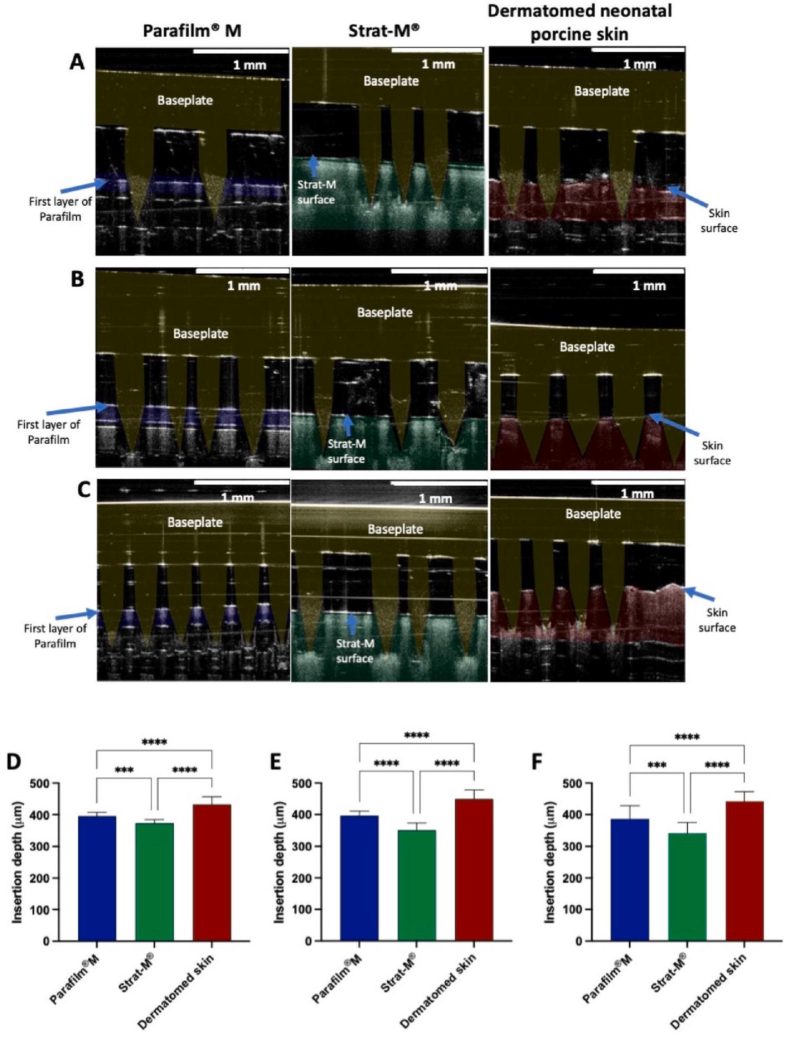
2.3. تصوير التداخل البصري وتصوير المسام

تم إدخال كل خريطة ذائبة في نماذج الأغشية (جلد مُستخرج، بارافيلم وStrat-M) باستخدام محلل القوام TA-TX2 (TA) (Stable Microsystems، هازلمير، المملكة المتحدة)، كما تم وصفه سابقًا (Bin Sabri et al.، 2021؛ Bin Sabri et al.، 2022؛ Anjani et al.، 2022b) ومبين في الشكل 2، حيث تم تطبيق قوة قدرها 32 نيوتن، وهو ما يعادل القوة الناتجة عن ضغط إصبع الإنسان (Larrañeta et al.، 2014). تم وضع قطعة من شمع الأسنان تحت الغشاء لحماية أطراف الإبر التي تخترق الغشاء (Bin Sabri et al.، 2021؛ Bin Sabri et al.، 2022؛ Anjani et al.، 2022b). تم ملاحظة الثقوب والإدخال ورؤيتها باستخدام مجهر تماسك بصري EX-101 (Michelson Diagnostics Ltd.، كنت، المملكة المتحدة)، ومجهر ضوئي رقمي (Leica EZ4 D، Leica Microsystems، ميلتون كينز، المملكة المتحدة) ومجهر إلكتروني مسح TM3030 (Hitachi، كريفيلد، ألمانيا). ثم تم معالجة وتحليل الصور الناتجة من تحليل OCT باستخدام Image المعاهد الوطنية للصحة، بيثيسدا، ماريلاند، الولايات المتحدة الأمريكية.

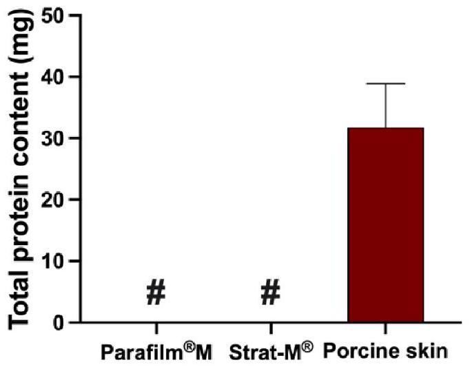
2.4. تحديد البروتين المفرز من الأغشية

لتوقع أي بروتينات قد تتداخل مع قياس البروتينات المستمدة من الأغشية (بما في ذلك جلد الخنازير حديثي الولادة المقطوع، Strat-M®، وParafilm) خلال الدراسات في المختبر باستخدام إعداد خلية فرانز، تم إجراء دراسة إطلاق كخطوة أولية قبل دراسات النفاذ في المختبر. تم قص كل غشاء إلى شكل دائري بقياس ، ما يعادل مساحة الجزء الداخلي من حجرة المتبرع في جهاز فرانز. ثم تم وضع هذه الأغشية في قوارير منفصلة تحتوي على 12 مل من PBS (رقم الهيدروجيني 7.4) وتم تحضينها في حاضنة مهتزة (Jeio Tech ISF7100، Medline Scientific، Chalgrove Oxon، المملكة المتحدة) عند لمدة 24 ساعة. بعد ذلك، تم تحليل العينات باستخدام مجموعة ميكرو-BCA، وإذا
الجدول 1
صياغة الطبقة الأولى من MAP القابلة للذوبان.
تركيب نوع البروتين AMSC-MP
ألبومين جمعية الكشافة الأمريكية
البروتين (%وزن/وزن) 10 10 10
خليط مائي PP2 (%وزن/وزن) 30 30 30
ماء منزوع الأيونات (%وزن/وزن) 60 60 60
الشكل 1. توضيح لحجم الإبرة والهندسة على مجموعة من MAP القابلة للذوبان.
الشكل 2. توضيح تخطيطي لإعداد جهاز تحليل القوام لتقييم خصائص إدخال MAP القابلة للذوبان في أغشية مختلفة (جلد خنزير مقطوع، سترات) وبارافيلم ).
إذا لزم الأمر، تم تخفيف العينات في PBS (رقم الهيدروجيني 7.4).

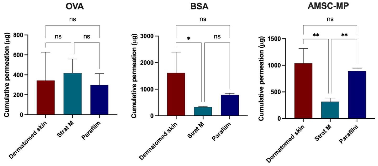
2.5. دراسات النفاذية في المختبر باستخدام خلايا فرانز

لدرس النفاذ في المختبر، تم استخدام جهاز فرانز (Permergear، هيلرتاون، بنسلفانيا، الولايات المتحدة الأمريكية) مع جلد خنزير حديث الولادة مقطوع، Strat-M® و Parafilm. كما هو موضح في الشكل 3. تم الحصول على جلد الخنازير من خنازير ميتة حديثة الولادة خلال 24 ساعة بعد الوفاة، وتم شطفه في محلول PBS، وتم تقطيعه بسمك ، ومجمد عند حتى الاستخدام. الغشاء (جلد خنزير حديث الولادة المقطوع، Strat-M® أو Parafilm تم تثبيته بعناية في حجرة المتبرع من
خلية فرانز باستخدام لاصق السيانوأكريلات. تم إضافة محلول PBS (pH 7.4) المسخن مسبقًا والمفكوك الغاز إلى حجرة الاستقبال، التي تم الحفاظ عليها عند وتم التحريك بسرعة 600 دورة في الدقيقة. تم الضغط يدويًا على MAPs القابلة للذوبان على الجلد لمدة 30 ثانية مع قطعة من شمع الأسنان تحتها كما هو موصوف سابقًا (Larrañeta et al.، 2016). تم وضع خزان يحتوي على الدواء فوق MAPs القابلة للذوبان، وتم توصيل حجرة المانح بحجرة المستلم. تم وضع وزن أسطواني من الفولاذ المقاوم للصدأ (15.0 جرام) فوق الخزان لمنع طرد MAP. تم إغلاق ذراع أخذ العينات في حجرة المستلم وحجرة المانح باستخدام بارافيلم. لمنع التبخر. في نقاط زمنية محددة سلفًا، وسائل الإصدار كانت
الشكل 3. توضيح تخطيطي لإعداد خلية انتشار فرانز لتقييم نفاذية البروتين من ذوبان MAP عبر أغشية مختلفة (جلد خنزير مقطوع، Strat-M® و Parafilm) ).
تم أخذها من حجرة الاستقبال وتحليلها باستخدام مجموعة ميكرو-BCA. إذا لزم الأمر، تم تخفيف العينات في PBS (درجة الحموضة 7.4).

2.6. التقدير باستخدام مجموعة اختبار بروتين الميكرو-BCA

تم قياس وتحليل العينات من الدراسات في المختبر باستخدام Micro BCA مجموعة اختبار البروتين (ثيرمو ساينتيفيك، روكفورد، إلينوي، الولايات المتحدة الأمريكية). تم إجراء التحليل وفقًا للإرشادات المقدمة من الشركة المصنعة. تم تحديد جميع العينات باستخدام قارئ الميكرو بلايت FluoStar Omega (BMG Labtech، ألمانيا) مع مبدأ الطيف الضوئي عند طول موجة الامتصاص 562 نانومتر.

2.7. التحليل الإحصائي

تم إجراء التحليل الإحصائي باستخدام برنامج جراف باد بريزم الإصدار 8.0 (برنامج GraphPad، سان دييغو، كاليفورنيا، الولايات المتحدة الأمريكية). تم تقديم النتائج التجريبية كمتوسطات الانحراف المعياري (SD)، ما لم يُذكر خلاف ذلك. غير متزاوج تم استخدام اختبار -test لمقارنة مجموعتين، بينما تم استخدام تحليل التباين الأحادي (ANOVA) لمقارنة مجموعات متعددة.

3. النتائج والمناقشة

3.1. إعداد MAPs القابلة للذوبان

استخدمت الدراسة ثلاثة بروتينات متميزة كمواد نموذجية (الألبومين المستخرج من بيض الدجاج (OVA، الوزن الجزيئي 45 كيلودالتون)، والألبومين البقري (BSA، الوزن الجزيئي 66.5 كيلودالتون) ومنتجات الأيض لخلايا الجذع الم mesenchymal من السائل الأمنيوسي (AMSC-MP، الوزن الجزيئي 76 كيلودالتون)). تم تصنيع MAPs القابلة للذوبان التي تحتوي على هذه البروتينات باستخدام طريقة الصب المزدوج، كما هو موضح في الأعمال السابقة (أنجاني وآخرون، 2022ب؛ أنجاني وآخرون، 2022ج؛ أنجاني وآخرون، 2022د). علاوة على ذلك، يمكن تحميل البروتينات في MAPs القابلة للذوبان باستخدام طرق أكثر تعقيدًا مثل تغليف الـ
تحتوي التركيبات على بروتينات (أنكاوينت وونغ وآخرون، 2020؛ كوديل وآخرون، 2018). في هذه الدراسة، تم تحميل كل بروتين في رؤوس الإبر بطريقة بسيطة مع ضمان الحد الأدنى من هدر البروتين. لهذا الغرض، تم دمج البروتين مع خليط من PVA و PVP. تعرض الشكل 4 صورًا مجهرية لمادة MAPs القابلة للذوبان الناتجة، مما يوضح تجانسها وبنيتها المحددة جيدًا، التي تتميز بإبر حادة. شكل الإبرة هو مزيج من الشكل المكعب والهرمي، نتيجة لقوالب بولي ديميثيل سيليوكسان المستخدمة في هذه التجربة. تم اختيار تصميم القالب هذا لقدرته على تسهيل الإدخال الفعال وتوصيل الدواء إلى الجلد، والذي أظهر تفوقًا مقارنة بأشكال أخرى (كورديرو وآخرون، 2020). أخيرًا، خضعت هذه MAPs القابلة للذوبان لاختبارات إدخال لاحقة وتقييم اختراق في المختبر.

3.2. التصوير المقطعي البصري وتصور المسام

لتقييم قدرة إدخال MAPs المحملة بالبروتين في أغشية مختلفة، تم استخدام جلد خنزير مقطوع بالشفرة. سترات-م ( 300 )، وبارافيلم ، التي تم توضيح هياكلها في الشكل 5، تم اختيار أنواع معينة من الأغشية للتقييم. كانت جلد الخنازير المستخدمة في هذه الدراسة مأخوذة من خنازير ميتة عند الولادة وتم تقطيعها إلى سمك ، والتي تتكون من البشرة (الطبقة القرنية، الطبقة الحبيبية، الطبقة الشوكية، والطبقة القاعدية) والجزء العلوي من طبقة الأدمة. تم اختيار هذه السماكة لأنها تقارب المسافة من سطح الجلد إلى الميكرو الدورة الدموية في الأدمة. مع ارتفاع الإبرة ستكون نسبة من الإبر الدقيقة في اتصال مباشر مع وسط الاستقبال عند استخدام تمثيلات البشرة السطحية أو العليا من حاجز الجلد (غارلاند وآخرون، 2012). تم تصميم Strat-M® لمحاكاة هيكل الجلد البشري، ويتميز بسطح مغطى بالدهون لمحاكاة طبقة الجلد القرني (حق وآخرون، 2018). تحت هذا الطلاء الدهني، يتكون Strat-M® من بولي إيثر سلفون، مما يخلق طبقة أكثر مسامية لمحاكاة الأدمة، تليها دعم غير منسوج من بولي أوليفين يشبه
الشكل 4. صور مجهرية لمواد MAPs الذائبة المحملة بـ (A) الألبومين المستخرج من بيض الدجاج (OVA)، (B) ألبومين مصل البقر (BSA)، و (C) نواتج الأيض لخلايا جذعية ميزانشيمية من السائل الأمنيوسي (AMSC-MP).
الطبقة تحت الجلد (Haq et al., 2018; Pulsoni et al., 2022). بارافيلم تم اختيارها كنموذج غشاء لهذه الدراسة نظرًا لتأكيدها كغشاء مقترح لدراسات إدخال MAP (Larrañeta et al.، 2014)، مما يجعلها اختبارًا سريعًا لمراقبة الجودة. تتكون من مزيج من شمع الهيدروكربون والبولي أوليفين، بارافيلم يكرر خصائص مشابهة للطبقة الثالثة من Strat-M®، ويهدف إلى محاكاة الطبقة تحت الجلد. على الرغم من أنه أقل تعقيدًا هيكليًا من الجلد الفعلي وStrat-M®، إلا أن Parafilm يوفر خصائص كارهة للماء كافية، تشبه الطبقة القرنية، وهي حاجز حاسم لتطبيق MAP.
توضح صور OCT في الشكل 6A-C إدخال MAPs القابلة للذوبان في أغشية مختلفة. من الواضح وجود فجوة بين قاعدة MAPs وسطح الأغشية، مما يشير إلى عدم اختراق MAPs القابلة للذوبان بشكل كامل في هذه الأغشية. كما هو موضح في الشكل 6D-F، اخترقت الإبر المجهرية لـ MAPs إلى أعماق تصل إلى حوالي 430. ، و لجلد الخنازير المقطوع، Strat-M®، وParafilm ، على التوالي. يتماشى عمق الإدخال في جلد الخنازير المقطوع مع النتائج المنشورة سابقًا (غارلاند وآخرون، 2012؛ دونيلي وآخرون، 2010؛ دونيلي وآخرون، 2011)، متجاوزًا بشكل كبير أعماق الاختراق التي لوحظت في محاكيات الجلد الأخرى ( يمكن أن يُعزى هذا التفاوت إلى اختلافات في مواد الغشاء ومرونته مقارنةً بجلد الخنازير المقطوع.
من الجدير بالذكر أنه قبل التجربة، تم توازن جلد الخنازير مع محلول فوسفات البفر pH (7.4) للحفاظ على سلامة الجلد. من المحتمل أن وجود الماء المالح عمل كزيت تشحيم (أنجاني وآخرون، 2023d)، مما سهل إدخال الإبرة بشكل أعمق مقارنةً بـ Strat-M® و Parafilm. . علاوة على ذلك، بارافيلم أظهرت اختراقًا أعمق مقارنة بـ
سترات-م®، ربما بسبب سمكها ذو الطبقة الواحدة ( ) مقابل الهيكل متعدد الطبقات لـ Strat-M® ( ). الشكل 7 يؤكد نجاح إنشاء الثقوب في جميع الأغشية، محاكيًا اختراق طبقة الجلد القرني. ومع ذلك، بينما يشبه Strat-M® سمك جلد الخنازير المقطوع، كان من الصعب اكتشاف الثقوب في طبقاته الأعمق بسبب خصائص القماش غير المنسوج من البوليمر، الذي يحاكي الطبقة تحت الجلد ويقدم مقاومة لاختراق الإبر الدقيقة. على الرغم من أن و اختلافات في عمق الإدخال بين بارافيلم وسترات-م مقارنةً بجلد الخنازير المقطوع، فإن كلا من محاكيات الجلد تظهر وعدًا كبدائل للأغشية لدراسات الإدخال.

3.3. تحديد البروتين المفرز من الأغشية

لتقييم إطلاق البروتين من أغشية مختلفة، تم إجراء دراسة أولية. توضح الشكل 8 أنه بعد حضانة جلد الخنازير حديثي الولادة المقطوع، في على مدار 24 ساعة، تم إطلاق حوالي 39 ملغ من البروتين. وعلى العكس، لم يتم الكشف عن أي بروتين في أي من بارافيلم م أو سترات-م المجموعات. تشير هذه النتيجة إلى أن استخدام الأغشية البيولوجية، مقترنًا بطريقة قياس الكمية الدقيقة للبروتين لتقييم توصيل البروتين، قد يؤدي إلى نتائج غير موثوقة بسبب تداخل البروتينات المشتقة من الجلد مع المادة الموصلة. علاوة على ذلك، وبالنظر إلى إطلاق البروتين من جلد الديرماتوم الفارغ، قمنا بتضمين جلد الخنازير المقطوع دون تطبيق ذوبان MAP في دراسات الاختراق في المختبر باستخدام إعداد خلية فرانز. ومع ذلك، بالنسبة لكل من بارافيلم وسترات-م لا تحكم أو فراغ
الشكل 5. توضيح تخطيطي يوضح الهيكل متعدد الطبقات لجلد الخنازير المقطوع بجانب Strat-M® وطبقة واحدة من Parafilm م.
تم تضمين المجموعات حيث لم يتم الكشف عن أي بروتين في هذه الدراسة الحالية. يضمن هذا الاستبعاد غياب تداخل البروتين أثناء عملية التقدير في هذه الحالات.

3.4. دراسات النفاذية في المختبر باستخدام خلايا فرانز

تم استخدام إعداد خلية فرانز في هذه الدراسة لتقييم نفاذية نماذج البروتين في بيئة في المختبر. لقد أثبت هذا الإعداد باستمرار أنه مثالي في جميع دراساتنا المتعلقة بـ MAP لتقييم انتشار الجزيئات من طبقة الطرف لـ MAPs إلى طبقة الجلد، لا سيما عبر حدود البشرة والأدمة (أنجاني وآخرون، 2022أ؛ أنجاني وآخرون، 2021ب؛ أنجاني وآخرون، 2023؛ بن صبري وآخرون، 2022؛ أنجاني وآخرون، 2022ب؛ أنجاني وآخرون، 2022ج؛ أنجاني وآخرون، 2022د؛ أنجاني وآخرون، 2021أ؛ أنجاني وآخرون، 2023هـ؛ أنجاني وآخرون، 2023و). على العكس من ذلك، فإن دراسات الإفراج في المختبر غير مناسبة لتقييم هذه العملية، حيث يتعرض اللصقة بالكامل لوسائط سائلة، مما يؤدي إلى تقدير مفرط لملف الإفراج عن الدواء الذي تم تحقيقه من نظام MAP القابل للذوبان. لذلك، نقترح استخدام كلا الغشائين، باراف. وسترات-م®، كبدائل للأغشية البيولوجية لتقييم توصيل البروتينات والبيبتيدات في دراسات النفاذ في المختبر باستخدام إعداد خلية فرانز.
الشكل 9 يعرض ملفات النفاذ للبروتينات النموذجية التي تم إطلاقها من أنظمة MAPs القابلة للذوبان عبر أغشية مختلفة، جلد خنزير مُستخرج، Parafilm®M، وStrat-M®. أظهر نفاذ OVA وBSA من أنظمة MAPs القابلة للذوبان ارتفاعًا أوليًا خلال ساعتين، تلاه استقرار بين 4 و24 ساعة لكل من Parafilm. وأغشية Strat-M®. في حالة AMSC-MP، لوحظت اختلافات ملحوظة بين Parafilm م و سترات-م عبر بارافيلم ، كانت نفاذية البروتين تزداد تدريجياً من الساعة الأولى حتى 6 ساعات، مع زيادة ملحوظة في . ومع ذلك، باستخدام Strat-M®،
تغلغل AMSC-MP على الفور خلال الساعة الأولى، مع الحفاظ على تغلغل مستمر طوال فترة التجربة. يمكن تفسير الفرق بين الغشائين بسبب تأثير عدة عوامل. تم تصميم غشاء Strat-M® لمحاكاة الجلد وكما يتضح في الشكل 7، فإن MAPs القابلة للذوبان لا تمر عبر الغشاء بالكامل. لذلك، تحتاج البروتينات إلى الانتشار عبر الغشاء لأنها ليست على اتصال مباشر مع سائل حجرة الاستقبال. يمكن أن تمر MAPs القابلة للذوبان عبر طبقة واحدة من Parafilm. م (الشكل 7) وعند وضعها في حجرة الاستقبال، ستكون الإبر الدقيقة على اتصال مباشر مع سائل حجرة الاستقبال. وهذا يفسر سبب النفاذ من بارافيلم. أسرع.
حدثت أكبر نفاذية للبروتينات الم facilitated بواسطة ذوبان MAPs عند استخدام جلد الخنازير حديثي الولادة المقطوع كغشاء نموذجي. بدأت عملية الإفراج المفاجئ خلال الساعة الأولى، تلتها زيادة ملحوظة في النفاذية التراكمية على مدار فترة التجربة التي استمرت 24 ساعة. هناك احتمال أن تكون MAPs الذائبة قد اخترقت جلد الخنازير المقطوع، مما قد يسرع من الإفراج السريع إلى حجرة الاستقبال. لتقييم إطلاق إجمالي محتوى البروتين من جلد الخنازير المقطوع دون تطبيق MAPs الذائبة، أجرينا تقييمًا إضافيًا. تشير الشكل 9 إلى كمية ملحوظة من إطلاق البروتين من الجلد، حتى أنها تجاوزت إجمالي البروتين المكتشف من كل من Parafilm. وسترات-م المجموعات في جميع الحالات. تبرز هذه الملاحظة أن استخدام أي غشاء بيولوجي يمكن أن يؤدي إلى نتائج إيجابية زائفة في اختراق البروتين، حيث تم الكشف عن البروتينات المشتقة من الجلد وتراكمها خلال عملية التقدير باستخدام طريقة BCA. وبالتالي، قد يساعد استخدام الأغشية الاصطناعية في التخفيف من هذه القيود المرتبطة بطريقة التقدير.
لقد استكشفت عدة دراسات توصيل BSA و OVA باستخدام منصة MAP. قام بادهي وآخرون بتطوير MAPs قائمة على الكيتوزان مغطاة بـ
الشكل 6. صور OCT لـ MAPs المحملة بـ (A) الألبومين المستخرج من بيض الدجاج (OVA)، (B) ألبومين مصل البقر (BSA)، و (C) منتجات الأيض لخلايا الجذع الم mesenchymal من السائل الأمنيوسي (AMSC-MP) المدخلة في Parafilm®M وStrat-M® و جلد الخنازير حديثي الولادة المقطوع بعد تطبيق ضغط قدره 32 نيوتن لمدة 30 ثانية. عمق الإدخال لـ MAPs القابلة للذوبان المحملة بـ (D) الألبومين المستخرج من بيض الدجاج (OVA)، (E) ألبومين مصل البقر (BSA)، و (F) منتجات الأيض لخلايا الجذع الم mesenchymal من السائل الأمنيوسي (AMSC-MP) في Parafilm®M وStrat-M® و جلد الخنازير حديثي الولادة المقطوع (المتوسط + الانحراف المعياري، n = 20). للسماح بالتفريق بين MAP القابلة للذوبان ونماذج الجلد، تم تطبيق ألوان زائفة في الإبر وطبقات نموذج الجلد.
الشكل 7. تم التقاط صور مجهرية للطبقة العليا وصور SEM للجانب الخلفي لكل غشاء بعد تطبيق MAPs القابلة للذوبان المحملة بـ (A) الألبومين المستمد من بيض الدجاج (OVA)، (B) ألبومين مصل البقر (BSA)، و (C) منتجات الأيض لخلايا الجذع الم mesenchymal من السائل الأمنيوسي (AMSC-MP) باستخدام محلل قوام بضغط 32 نيوتن لمدة 30 ثانية.
بروتين الألبومين البشري (BSA) وحمض البولي لاكتيك (Badhe et al., 2021). باستخدام طريقة لونية مع كاشف البيوريت ومطياف الأشعة فوق البنفسجية والمرئية مضبوط على طول موجي 540 نانومتر، قام الباحثون بتحديد كمية إطلاق BSA خلال دراسة اختراق في المختبر باستخدام خلايا فرانز. أظهرت الدراسة أن 98.5% من BSA قد اخترق عبر جلد الفئران المستأصلة خلال 50 ساعة (Badhe et al., 2021). ومع ذلك، كانت تفتقر إلى مجموعة تحكم بدون تطبيق MAP، مما أثار مخاوف بشأن إطلاق البروتين من الجلد خلال فترة الدراسة.
طور ديمير وآخرون MAPs قائمة على ألجينات الصوديوم محملة بـ BSA، مما يدل على نسبة أعلى من توصيل BSA. ) عبر جلد البطن البشري المقطوع بالمشرط مقارنةً بلاصقة عبر الجلد (ديمير وآخرون، 2013). قام الباحثون بتحديد كمية إطلاق BSA باستخدام طريقة BCA عند 562 نانومتر (ديمير وآخرون، 2013). ومع ذلك، كانت هناك نقص في تحليل مجموعة التحكم، مما يحد من الفهم بشأن مصدر البروتين خلال هذه الدراسات.
الشكل 8. إجمالي البروتين المفرز من كل غشاء بعد 24 ساعة من الحضانة عند في PBS (pH 7.4). يشير الرمز ‘#’ إلى أنه لم يتم الكشف عن أي بروتين في الوسط (يعني ).
فيما يتعلق بدراسات اختراق MAP المعتمدة على OVA، استخدم معادن وآخرون ووالهوف وآخرون OVA المعلم بالمواد المشعة (فان دير معادن وآخرون، 2014) وOVA المرتبطة بألكسّا 488 (والهوف وآخرون، 2018)، على التوالي. استخدم معادن وآخرون MAP حساس للرقم الهيدروجيني مغطى بـ OVA المعلم إشعاعيًا واستخدموا عداد غاما تلقائي للتحديد الكمي المحدد خلال دراسات الاختراق في المختبر عبر جلد الإنسان المستأصل (فان دير معادن وآخرون، 2014). استخدم والهوف وآخرون MAP بيولوجي السيراميك مغطى بـ OVA لتوصيل المستضد إلى الخلايا الشجرية في جلد الإنسان (والهوف وآخرون، 2018). استخدمت هذه الدراسات أساليب مختلفة لتحديد OVA بشكل انتقائي خلال دراسات الاختراق في المختبر.
فيما يتعلق بالارتباط بين التجارب المخبرية والتجارب الحية (IVIVC)، لم يتم حتى الآن إنشاء مثل هذه المناقشات لتوصيل الأدوية بواسطة MAP. يتطلب تطوير MAPs القابلة للذوبان، التي تذوب أو تتحلل عند ملامستها لسوائل الأنسجة الجلدية لإطلاق حمولتها الدوائية في الموقع، أخذ عوامل إضافية في الاعتبار عند تصميم تجارب اختراق في المختبر. من الناحية المثالية، يجب أن تحاكي غشاء النموذج المختار الذي يمثل حاجز الجلد في المختبر عمق اختراق الإبر الدقيقة، والتصاق قاعدة الإبرة الدقيقة بسطح الجلد، ومعدل ذوبان/تحلل الإبرة الدقيقة (Garland et al., 2012)، وإطلاق الدواء الذي يتم تحقيقه في سيناريو حي. علاوة على ذلك، نظرًا لأن طول الإبرة الدقيقة قد يتجاوز سمك حاجز الجلد المختار، قد تتصل جزء من الإبرة الدقيقة مباشرة بوسيط الاستقبال. لذلك، من الضروري التأكد من أن تركيبة وسيط الاستقبال المختار لا تبالغ بشكل كبير في تقدير معدل ذوبان الإبرة الدقيقة، وبالتالي، معدل إطلاق الدواء داخل الجلد (Garland et al., 2012).
عند ملاحظة البروتين التراكمي المقدم من إذابة MAPs عبر جلد الخنازير المقطوع بالمشرط مقارنة بالمجموعات الضابطة، تم إجراء التطبيع عن طريق طرح إجمالي البروتين من مجموعة MAPs المذابة. الشكل 10 يعرض البيانات المطبقة عند 24 ساعة لـ OVA وBSA وAMSC-MP الم released من MAPs المذابة عبر جلد الخنازير المقطوع بالمشرط، Parafilm. “، وStrat-M®. النسب التراكمية للاختراق لخرائط MAP المحملة بـ OVA على جلد الخنازير المقطوع، بارافيلم وسترات-م وُجدت أن ، و على التوالي. لم تُلاحظ فروق ذات دلالة إحصائية بين الأغشية بعد التطبيع ( ).
بالنسبة لـ BSA، فإن النفاذ التراكمي على مدى 24 ساعة من خلال جلد الخنازير المقطوع، بارافيلم م، واستراتيجيات م كان تقريبًا ، ، و على التوالي. ومن المثير للاهتمام أن النفاذ عبر جلد الخنازير المقطوع كان أعلى بشكل ملحوظ ( ) و
الشكل 9. (أ) تم استخدام محتوى البروتين الكلي لتحديد نفاذ الألبومين المستمد من بيض الدجاج (OVA) والألبومين البقري (BSA) ومنتجات الأيض لخلايا الجذع الم mesenchymal من السائل الأمنيوسي (AMSC-MP) من ذوبان MAPs عبر جلد الخنازير المقطوع. تمثل الخطوط المتقطعة البروتين المفرج عنه من جلد الخنازير المقطوعة حديثي الولادة دون تطبيق MAP الذائب. SD، تم استخدام إجمالي محتوى البروتين لت quantifying اختراق الألبومين المستمد من بيض الدجاج (OVA) ، والألبومين المصل البقري (BSA) ، ومنتجات الأيض لخلايا الجذع الم mesenchymal السليمة (AMSC-MP) من إذابة MAPs عبر Parafilm. M، Strat-M®، وجلد الخنازير المقطوع. تم الحصول على القيم المصححة للدواء الذي تم اختراقه عبر الجلد المقطوع عن طريق طرح قراءات الجلد المقطوع الفارغة (المتوسطات ).
الشكل 10. التراكم التراكمي لاختراق الألبومين المستمد من بيض الدجاج (OVA) ، والألبومين المصل البقري (BSA) ، ومنتجات الأيض لخلايا الجذع الم mesenchymal من السائل الأمنيوسي (AMSC-MP) عبر أغشية مختلفة (جلد خنزير مقطع، Parafilm®M وStrat-M®) على مدى 24 ساعة من دراسات الاختراق في المختبر (المتوسطات + الانحراف المعياري، ).
زاد بمقدار 4.88 مرة مقارنة بـ Strat-M®. قد يُعزى هذا التباين إلى وجود بروتينات الجلد، مما يؤدي إلى زيادة اكتشاف البروتين عبر نماذج بروتينية مختلفة. بالإضافة إلى ذلك، في توصيل AMSC-MP، كانت النسب التراكمية للاختراق هي ، و عبر جلد خنزير مقطع، بارافيلم وسترات-م ، على التوالي. ومن الجدير بالذكر أن نفاذ AMSC-MP عبر Parafilm®M زاد بشكل ملحوظ بمقدار 2.83 مرة مقارنة بـ Strat- بينما لم تكن هناك اختلافات كبيرة بين بارافيلم وجلد خنزير مقطع بالليزر ) في نسبة اختراق AMSC-MP.
أظهر تحليل بيانات النفاذية في المختبر أنماط ارتباط مميزة بين بارافيلم م و سترات-م بخصوص الاختراق التراكمي للبروتينات خلال فترة 24 ساعة. يبدو أن هذه الارتباطات تعتمد على خصائص نماذج البروتين المستخدمة في الدراسة. توضح الشكل 11 أنه، استنادًا إلى فئة معامل الارتباط (الارتباط والانحدار، بدون تاريخ)؛ بارافيلم أظهرت ارتباطات قوية ومتوسطة مع Strat-M® لـ OVA و BSA، على التوالي. ومن الجدير بالذكر أن النفاذ التراكمي لـ AMSC-MP عبر كل من Parafilm وسترات-م أظهر ارتباطًا قويًا جدًا، يتراوح بين 0.90 و 0.99.
ربط عينات جلد الخنازير المقطعة إلى كل من بارافيلم م و سترات-م ثبت أنه كان تحديًا بسبب تدخل بروتينات الجلد في نتائج النفاذ على مدار 24 ساعة. وبالتالي، تقترح هذه الدراسة أن كل من بارافيلم م و سترات-م تتفوق على جلد الخنازير حديثي الولادة المقطوع في موثوقية دراسات النفاذ. علاوة على ذلك، تشير إلى وجود تشابه في ملفات نفاذ البروتين عبر بارافيلم. وسترات-م مُبرزًا إمكانيات هذه المحاكيات الجلدية للاستخدام في MAPs المحملة بالبروتين
مخصص للتوصيل عبر الجلد. بالإضافة إلى ذلك، تشمل الخطط المستقبلية لهذه الدراسة إنشاء عامل تشابه. التركيز الحالي هو على الاستخدام المحتمل لبارافيلم. وسترات-م في الدراسات المخبرية كخطوة أولية لاختيار أفضل تركيبة للدراسات الحية باستخدام نماذج حيوانية مختلفة. وبالتالي، يجب أيضًا ملاحظة أن الكميات الكمية التي تم قياسها باستخدام الأغشية الاصطناعية قد لا تتوافق دائمًا مع دراسات النفاذ الفعلية باستخدام جلد الإنسان أو جلد الخنازير. ومع ذلك، تظهر نتائج هذه الدراسة إمكانيات بارافيلم. وسترات-م كبدائل لجلد الخنازير المقطوع في المرحلة المبكرة، مما يمكّن الباحثين من وضع بروتوكولات موحدة لتقييم اختراق المواد من أنظمة MAP.

4. الخاتمة

باختصار، تُظهر هذه الدراسة الاستخدام المحتمل لـ Paraf وسترات-م كبدائل لمحاكيات الجلد البيولوجية في دراسات الاختراق في المختبر، لا سيما للتحقيق في توصيل البروتين من منصات MAP. تشير نتائجنا إلى أن كل من Parafilm وتمتاز أغشية Parafilm®M وStrat-M® بفروق طفيفة فقط مقارنةً بجلد الخنازير المقطوع، مما يجعلها بدائل واعدة لدراسات الأغشية. يطلق جلد الخنازير المقطوع بروتينات خلال دراسات النفاذية في المختبر، مما يؤدي إلى تدخل محتمل في التقدير باستخدام تقنية BCA. استخدام الأغشية الاصطناعية مثل Parafilm®M وStrat يمكن أن تتجاوز مثل هذا التدخل، مما يوفر نتائج أكثر موثوقية مقارنةً بجلد الخنازير حديثي الولادة المقطوع بالجلد. من المهم أن نلاحظ أنه على الرغم من أن هذه الأغشية الاصطناعية تقدم رؤى حول الاتجاهات والارتباطات، إلا أنها قد لا تتطابق بدقة مع قيم النفاذية المطلقة للأغشية الجلدية البيولوجية لتوصيل البروتين. ومع ذلك، فإن هذه
الشكل 11. ارتباط النفاذ التراكمي بين بارافيلم وسترات-م للبومين المستمد من بيض الدجاج (OVA) ، وألبومين مصل البقر (BSA) ، ومنتجات الأيض لخلايا الجذع الم mesenchymal السليمة (AMSC-MP).
تعتبر النتائج مهمة لفهم إمكانيات هذه الأغشية في صياغة وتحسين أنظمة توصيل الأدوية. قد تكون هذه النتائج أدوات قيمة لجهود الفحص الأولي في مختلف تركيبات أنظمة توصيل الأدوية، وليس مقتصراً على البروتينات أو الببتيدات. توفر الأغشية الاصطناعية بدائل سريعة وفعالة من حيث التكلفة وسهلة الوصول لا تتطلب تحضيراً أو تخزيناً معقدين. تمثل هذه الدراسة مقارنة في مرحلة مبكرة بين الأغشية البيولوجية والاصطناعية لتحديد نموذج مناسب لمحاكاة الجلد في مجال صياغة أنظمة توصيل الأدوية.

بيان مساهمة مؤلفي CRediT

قونيطا كورنيا أنجاني: الكتابة – المراجعة والتحرير، الكتابة – المسودة الأصلية، التصور، التحقق، المنهجية، التحقيق، التحليل الرسمي، إدارة البيانات، التصور. أفليا ديفينا كاليستا نينغغولان: التحقيق، التحليل الرسمي، إدارة البيانات. هوانهوان لي: التحقيق، التحليل الرسمي. أندانغ مياتموكو: الكتابة – المراجعة والتحرير، الموارد، التحقيق. إينيكو لاراينيتا: الكتابة – المراجعة والتحرير، التصور، التحليل الرسمي. رايان ف. دونيلي: الإشراف، الموارد، الكتابة – المراجعة والتحرير.

إعلان عن تضارب المصالح

يعلن المؤلفون أنهم ليس لديهم أي مصالح مالية متنافسة معروفة أو علاقات شخصية قد تبدو أنها تؤثر على العمل المبلغ عنه في هذه الورقة.

توفر البيانات

ستكون البيانات متاحة عند الطلب.

References

Alkilani, A.Z., McCrudden, M.T.C., Donnelly, R.F., 2015. Transdermal drug delivery: innovative pharmaceutical developments based on disruption of the barrier properties of the stratum corneum. Pharmaceutics 7, 438-470. https://doi.org/ 10.3390/pharmaceutics7040438.
Angkawinitwong, U., Courtenay, A.J., Rodgers, A.M., Larrañeta, E., Mccarthy, H.O., Brocchini, S., Donnelly, R.F., Williams, G.R., 2020. A novel transdermal protein delivery strategy via electrohydrodynamic coating of PLGA microparticles onto microneedles. ACS Appl. Mater. Interf. 12, 12478-12488. https://doi.org/10.1021/ ACSAMI.9B22425/ASSET/IMAGES/LARGE/AM9B22425_0008.JPEG.
Anjani, Q.K., Bin Sabri, A.H., Donnelly, R., 2021a. Development and validation of simple and sensitive HPLC-UV method for ethambutol hydrochloride detection following transdermal application. Anal. Methods. https://doi.org/10.1039/D1AY01414E.
Anjani, Q.K., Permana, A.D., Cárcamo-Martínez, Á., Domínguez-Robles, J., Tekko, I.A., Larrañeta, E., Vora, L.K., Ramadon, D., Donnelly, R.F., 2021b. Versatility of hydrogel-forming microneedles in in vitro transdermal delivery of tuberculosis drugs. Eur. J. Pharm. Biopharm. 294-312, 294-312. https://doi.org/10.1016/j. ejpb.2020.12.003.
Anjani, Q.K., Hidayat, A., Sabri, B., Moreno-Castellanos, N., Utomo, E., CárcamoMartínez, Á., Domínguez-Robles, J., Ahmadi, L., Wardoyo, H., Donnelly, R.F., 2022a. Soluplus -based dissolving microarray patches loaded with colchicine: towards a minimally invasive treatment and management of gout. Biomater. Sci. https://doi. org/10.1039/D2BM01068B.
Anjani, Q.K., Bin Sabri, A.H., Utomo, E., Domínguez-Robles, J., Donnelly, R.F., 2022b. Elucidating the impact of surfactants on the performance of dissolving microneedle array patches. Mol. Pharm. 19, 1191-1208, 10.1021/ACS. MOLPHARMACEUT.1C00988/ASSET/IMAGES/LARGE/MP1C00988_0014.JPEG.
Anjani, Q.K., Bin Sabri, A.H., McGuckin, M.B., Li, H., Hamid, K.A., Donnelly, R.F., 2022c. In vitro permeation studies on carvedilol containing dissolving microarray patches quantified using a rapid and simple HPLC-UV analytical method. AAPS PharmSciTech 23, 1-13. https://doi.org/10.1208/S12249-022-02422-6/FIGURES/ 4.
Anjani, Q.K., Bin Sabri, A.H., Domínguez-Robles, J., Moreno-Castellanos, N., Utomo, E., Wardoyo, L.A.H., Larrañeta, E., Donnelly, R.F., 2022d. Metronidazole nanosuspension loaded dissolving microarray patches: An engineered composite pharmaceutical system for the treatment of skin and soft tissue infection. Biomaterials Adv. 140, 213073 https://doi.org/10.1016/J.BIOADV.2022.213073.
Anjani, Q.K., Bin Sabri, A.H., Hutton, A.J., Cárcamo-Martínez, Á., Wardoyo, L.A.H., Mansoor, A.Z., Donnelly, R.F., 2023a. Microarray patches for managing infections at a global scale. J. Control. Release 359, 97-115. https://doi.org/10.1016/j. jconrel.2023.05.038.
Anjani, Q.K., Bin Sabri, A.H., Hamid, K.A., Moreno-Castellanos, N., Li, H., Donnelly, R.F., 2023b. Tip loaded cyclodextrin-carvedilol complexes microarray patches. Carbohydr. Polym. 320, 121194 https://doi.org/10.1016/j.carbpol.2023.121194.
Anjani, Q.K., Pandya, A.K., Demartis, S., Domínguez-Robles, J., Moreno-Castellanos, N., Li, H., Gavini, E., Patravale, V.B., Donnelly, R.F., 2023c. Liposome-loaded polymeric microneedles for enhanced skin deposition of rifampicin. Int. J. Pharm. 646, 123446 https://doi.org/10.1016/J.IJPHARM.2023.123446.
Anjani, Q.K., Cárcamo-Martínez, Á., Wardoyo, L.A.H., Moreno-Castellanos, N., Bin Sabri, A.H., Larrañeta, E., Donnelly, R.F., 2023d. MAP-box: a novel, low-cost and easy-to-fabricate 3D-printed box for the storage and transportation of dissolving microneedle array patches. Drug Deliv. Transl. Res. https://doi.org/10.1007/ s13346-023-01393-w.
Anjani, Q.K., Bin Sabri, A.H., Hamid, K.A., Moreno-Castellanos, N., Li, H., Donnelly, R.F., 2023e. Tip loaded cyclodextrin-carvedilol complexes microarray patches. Carbohydr. Polym. 320, 121194.
Anjani, Q.K., Volpe-Zanutto, F., Hamid, K.A., Bin Sabri, A.H., Moreno-Castellano, N., Gaitán, X.A., Calit, J., Bargieri, D.Y., Donnelly, R.F., 2023f. Primaquine and chloroquine nano-sized solid dispersion-loaded dissolving microarray patches for the improved treatment of malaria caused by Plasmodium vivax. J. Controll. Release 361, 385-401. https://doi.org/10.1016/J.JCONREL.2023.08.009.
Antosova, Z., Mackova, M., Kral, V., Macek, T., 2009. Therapeutic application of peptides and proteins: parenteral forever? Trends Biotechnol. 27, 628-635. https://doi.org/ 10.1016/J.TIBTECH.2009.07.009.
Badhe, R.V., Adkine, D., Godse, A., 2021. Development of polylactic acid and bovine serum albumin-layered-coated chitosan microneedles using novel bees wax mould. Turk. J. Pharm. Sci. 18, 367. https://doi.org/10.4274/TJPS.GALENOS.2020.47897.
Bin Sabri, A.H., Anjani, Q.K., Donnelly, R.F., Hidayat Bin Sabri, A., Kurnia Anjani, Q., Donnelly, R.F., 2021. Synthesis and characterization of sorbitol laced hydrogelforming microneedles for therapeutic drug monitoring. Int. J. Pharm. 607, 121049.
Bin Sabri, A.H., Anjani, Q.K., Utomo, E., Ripolin, A., Donnelly, R.F., 2022. Development and characterization of a dry reservoir-hydrogel-forming microneedles composite for minimally invasive delivery of cefazolin. Int. J. Pharm. 617, 121593 https://doi.org/ 10.1016/J.IJPHARM.2022.121593.
Bittner, B., Richter, W., Schmidt, J., 2018. Subcutaneous administration of biotherapeutics: an overview of current challenges and opportunities. BioDrugs 32, 425. https://doi.org/10.1007/S40259-018-0295-0.
Bruno, B.J., Miller, G.D., Lim, C.S., 2013. Basics and recent advances in peptide and protein drug delivery. Ther. Deliv. 4, 1443. https://doi.org/10.4155/TDE.13.104.
Caudill, C.L., Perry, J.L., Tian, S., Luft, J.C., DeSimone, J.M., 2018. Spatially controlled coating of continuous liquid interface production microneedles for transdermal protein delivery. J. Control. Release 284, 122-132. https://doi.org/10.1016/J. JCONREL. 2018.05.042.
Cordeiro, A.S., Tekko, I.A., Jomaa, M.H., Vora, L., McAlister, E., Volpe-Zanutto, F., Nethery, M., Baine, P.T., Mitchell, N., McNeill, D.W., Donnelly, R.F., 2020. Twophoton polymerisation 3D printing of microneedle array templates with versatile designs: application in the development of polymeric drug delivery systems. Pharm. Res. 37, 1-15. https://doi.org/10.1007/S11095-020-02887-9/FIGURES/9.
11. Correlation and regression, (n.d.). https://www.bmj.com/about-bmj/resources-readers/publications/statistics-square-one/11-correlation-and-regression (Accessed December 2, 2023).
Demir, Y.K., Akan, Z., Kerimoglu, O., 2013. Sodium alginate microneedle arrays mediate the transdermal delivery of bovine serum albumin. PLoS One 8, e63819.
Donnelly, R.F., Prausnitz, M.R., 2023. The promise of microneedle technologies for drug delivery. Drug Deliv. Transl. Res. 1-8. https://doi.org/10.1007/S13346-023-014308/FIGURES/2.
Donnelly, R.F., Garland, M.J., Morrow, D.I.J., Migalska, K., Singh, T.R.R., Majithiya, R., Woolfson, A.D., 2010. Optical coherence tomography is a valuable tool in the study of the effects of microneedle geometry on skin penetration characteristics and in-skin dissolution. J. Control. Release 147, 333-341. https://doi.org/10.1016/J. JCONREL. 2010.08.008.
Donnelly, R.F., Majithiya, R., Singh, T.R.R., Morrow, D.I.J., Garland, M.J., Demir, Y.K., Migalska, K., Ryan, E., Gillen, D., Scott, C.J., Woolfson, A.D., 2011. Design, optimization and characterisation of polymeric microneedle arrays prepared by a novel laser-based micromoulding technique. Pharm. Res. 28, 41-57. https://doi.org/ 10.1007/S11095-010-0169-8/FIGURES/12.
Doughty, D.V., Clawson, C.Z., Lambert, W., Subramony, J.A., 2016. Understanding subcutaneous tissue pressure for engineering injection devices for large-volume protein delivery. J. Pharm. Sci. 105, 2105-2113. https://doi.org/10.1016/J. XPHS.2016.04.009.
Garland, M.J., Migalska, K., Tuan-Mahmood, T.M., Raghu Raj Singh, T., Majithija, R., Caffarel-Salvador, E., McCrudden, C.M., McCarthy, H.O., David Woolfson, A., Donnelly, R.F., 2012. Influence of skin model on in vitro performance of drug-loaded soluble microneedle arrays. Int. J. Pharm. 434, 80-89. https://doi.org/10.1016/J. IJPHARM.2012.05.069.
Haq, A., Goodyear, B., Ameen, D., Joshi, V., Michniak-Kohn, B., 2018. Strat-M® synthetic membrane: permeability comparison to human cadaver skin. Int. J. Pharm. 547, 432-437. https://doi.org/10.1016/J.IJPHARM.2018.06.012.
Jiskoot, W., Randolph, T.W., Volkin, D.B., Middaugh, C.R., Schöneich, C., Winter, G., Friess, W., Crommelin, D.J.A., Carpenter, J.F., 2012. Protein instability and immunogenicity: roadblocks to clinical application of injectable protein delivery systems for sustained release. J. Pharm. Sci. 101, 946-954. https://doi.org/10.1002/ JPS. 23018.
Kirkby, M., Hutton, A.R.J., Donnelly, R.F., 2020. Microneedle mediated transdermal delivery of protein, peptide and antibody based therapeutics: current status and future considerations. Pharm. Res. 37, 1-18. https://doi.org/10.1007/S11095-020-02844-6/TABLES/2.
E. Larrañeta, M.T.C. McCrudden, A.J. Courtenay, R.F. Donnelly, Microneedles: a new frontier in nanomedicine delivery, Pharmaceut. Res. 33 (2016) 1055-1073. doi: 10.1007/S11095-016-1885-5.
Larrañeta, E., Moore, J., Vicente-Pérez, E.M., González-Vázquez, P., Lutton, R., Woolfson, A.D., Donnelly, R.F., 2014. A proposed model membrane and test method for microneedle insertion studies. Int. J. Pharm. 472, 65-73. https://doi.org/ 10.1016/j.ijpharm.2014.05.042.
Larrañeta, E., Stewart, S., Fallows, S.J., Birkhäuer, L.L., McCrudden, M.T.C., Woolfson, A. D., Donnelly, R.F., 2016. A facile system to evaluate in vitro drug release from dissolving microneedle arrays. Int. J. Pharm. 497, 62-69. https://doi.org/10.1016/ J.IJPHARM.2015.11.038.
R. Neupane, S.H.S. Boddu, J. Renukuntla, R.J. Babu, A.K. Tiwari, Alternatives to biological skin in permeation studies: current trends and possibilities, Pharmaceutics 12 (2020) 152. doi: 10.3390/PHARMACEUTICS12020152.
Oberli, M.A., Schoellhammer, C.M., Langer, R., Blankschtein, D., 2014. Ultrasoundenhanced transdermal delivery: recent advances and future challenges. Ther. Deliv. 5, 843-857. https://doi.org/10.4155/tde.14.32.
Pulsoni, I., Lubda, M., Aiello, M., Fedi, A., Marzagalli, M., von Hagen, J., Scaglione, S., 2022. Comparison between franz diffusion cell and a novel micro-physiological system for in vitro penetration assay using different skin models. SLAS Technol. 27, 161-171. https://doi.org/10.1016/J.SLAST.2021.12.006.
Uchida, T., Nishioka, K., Motoki, A., Yakumaru, M., Sano, T., Todo, H., Sugibayashi, K., 2016. Effect of esters on the permeation of chemicals with different polarities through synthetic artificial membranes using a high-throughput diffusion cell array. Chem. Pharm. Bull. (Tokyo) 64, 1597-1606. https://doi.org/10.1248/CPB.C1600480.
Vallhov, H., Xia, W., Engqvist, H., Scheynius, A., 2018. Bioceramic microneedle arrays are able to deliver OVA to dendritic cells in human skin. J. Mater. Chem. B 6, 6808-6816. https://doi.org/10.1039/C8TB01476K.
Van Der Maaden, K., Varypataki, E.M., Romeijn, S., Ossendorp, F., Jiskoot, W., Bouwstra, J., 2014. Ovalbumin-coated pH -sensitive microneedle arrays effectively induce ovalbumin-specific antibody and T-cell responses in mice. Eur. J. Pharm. Biopharm. 88, 310-315. https://doi.org/10.1016/J.EJPB.2014.05.003.
Vora, L.K., Sabri, A.H., Naser, Y., Himawan, A., Hutton, A.R.J., Anjani, Q.K., VolpeZanutto, F., Mishra, D., Li, M., Rodgers, A.M., Paredes, A.J., Larrañeta, E., Thakur, R. R.S., Donnelly, R.F., 2023. Long-acting microneedle formulations. Adv. Drug Deliv. Rev. 201, 115055 https://doi.org/10.1016/J.ADDR.2023.115055.

  1. Open Access
    This research has been made openly available by Queen’s academics and its Open Research team. We would love to hear how access to this research benefits you. – Share your feedback with us: http://go.qub.ac.uk/oa-feedback
    • Corresponding author at: Chair in Pharmaceutical Technology, School of Pharmacy, Queen’s University Belfast, Medical Biology Centre, 97 Lisburn Road, Belfast BT9 7BL, Northern Ireland, UK.
    E-mail address: r.donnelly@qub.ac.uk (R.F. Donnelly).

Journal: International Journal of Pharmaceutics, Volume: 655
DOI: https://doi.org/10.1016/j.ijpharm.2024.124071
PMID: https://pubmed.ncbi.nlm.nih.gov/38554738
Publication Date: 2024-03-29

Parafilm M and Strat-M as skin simulants in in vitro permeation of dissolving microarray patches loaded with proteins

Anjani, Q. K., Nainggolan, A. D. C., Li, H., Miatmoko, A., Larrañeta, E., & Donnelly, R. F. (2024). Parafilm® M and Strat-M® as skin simulants in in vitro permeation of dissolving microarray patches loaded with proteins. International Journal of Pharmaceutics, 655, Article 124071. https://doi.org/10.1016/j.ijpharm.2024.124071

Published in:International Journal of Pharmaceutics

Document Version:
Publisher’s PDF, also known as Version of record
Queen’s University Belfast – Research Portal:
Link to publication record in Queen’s University Belfast Research Portal
Publisher rights
Copyright 2024 The Authors.
This is an open access article published under a Creative Commons Attribution License (https://creativecommons.org/licenses/by/4.0/), which permits unrestricted use, distribution and reproduction in any medium, provided the author and source are cited.
General rights
Copyright for the publications made accessible via the Queen’s University Belfast Research Portal is retained by the author(s) and / or other copyright owners and it is a condition of accessing these publications that users recognise and abide by the legal requirements associated with these rights.
Take down policy
The Research Portal is Queen’s institutional repository that provides access to Queen’s research output. Every effort has been made to ensure that content in the Research Portal does not infringe any person’s rights, or applicable UK laws. If you discover content in the Research Portal that you believe breaches copyright or violates any law, please contact openaccess@qub.ac.uk.

Parafilm M and Strat-M® as skin simulants in in vitro permeation of dissolving microarray patches loaded with proteins

Qonita Kurnia Anjani , Avelia Devina Calista Nainggolan , Huanhuan Li , Andang Miatmoko , Eneko Larrañeta , Ryan F. Donnelly School of Pharmacy, Queen’s University Belfast, Medical Biology Centre, 97 Lisburn Road, Belfast BT9 7BL, UK Fakultas Farmasi, Universitas Megarezky, Jl. Antang Raya No. 43, Makassar 90234, Indonesia Faculty of Pharmacy, University of Indonesia, Depok 16424, Indonesia Faculty of Pharmacy, Airlangga University, Nanizar Zaman Joenoes Building, Campus C, Mulyorejo, Surabaya 60115, Indonesia Stem Cell Research and Development Center, Airlangga University, Institute of Tropical Disease Building, Campus C, Mulyorejo, Surabaya 60115, Indonesia

ARTICLE INFO

Keywords:

Parafilm M
Strat-M®
Dermatomed porcine skin
Protein
Insertion studies
In vitro permeation studies

Abstract

In vitro permeation studies play a crucial role in early formulation optimisation before extensive animal model investigations. Biological membranes are typically used in these studies to mimic human skin conditions accurately. However, when focusing on protein and peptide transdermal delivery, utilising biological membranes can complicate analysis and quantification processes. This study aims to explore Parafilm M and Strat-M as alternatives to dermatomed porcine skin for evaluating protein delivery from dissolving microarray patch (MAP) platforms. Initially, various MAPs loaded with different model proteins (ovalbumin, bovine serum albumin and amniotic mesenchymal stem cell metabolite products) were prepared. These dissolving MAPs underwent evaluation for insertion properties and in vitro permeation profiles when combined with different membranes, dermatomed porcine skin, Parafilm , and Strat-M . Insertion profiles indicated that both Parafilm M and Strat showed comparable insertion depths to dermatomed porcine skin (in range of ), suggesting promise as membrane substitutes for insertion studies. In in vitro permeation studies, synthetic membranes such as Parafilm and Strat-M demonstrated the ability to bypass protein-derived skin interference, providing more reliable results compared to dermatomed neonatal porcine skin. Consequently, these findings present valuable tools for preliminary screening across various MAP formulations, especially in the transdermal delivery of proteins and peptides.

1. Introduction

Protein and peptide-based therapies have gained prominence in treating various chronic diseases, often being the primary therapeutic choice (Kirkby et al., 2020; Bruno et al., 2013). Market estimates reveal a significant growth in the protein and peptide drug market, currently nearing US$40 billion annually, outpacing the small molecule market (Bruno et al., 2013). This steady growth underscores the ongoing investigation and advancements in protein and peptide delivery methods, catering especially to diseases once deemed incurable. While intravenous injection ensures bioavailability, its preference diminishes for frequent administration in chronic diseases (Kirkby et al., 2020; Antosova et al., 2009; Jiskoot et al., 2012). Subcutaneous
injection of antibodies and peptides has been employed, allowing for potential self-administration by patients at home (Doughty et al., 2016; Bittner et al., 2018). However, patients seek alternatives in the form of self-administrable, painless, and minimally invasive dosage forms to reduce hospital visits and alleviate the healthcare burden associated with therapy (Anjani et al., 2023a). Transdermal drug delivery, notably via microarray patches (MAPs), emerges as a potential solution. MAP technology, intensively researched in biomedicine, aims to facilitate effective drug and vaccine delivery (Donnelly and Prausnitz, 2023; Vora et al., 2023). MAPs function by painlessly breaching the stratum corneum (SC), creating microchannels to aid drug diffusion into deeper dermal layers (Anjani et al., 2022a). This approach circumvents passive drug diffusion challenges, allowing delivery of a wide range of chemical
substances irrespective of molecular size and lipophilicity (Anjani et al., 2021b; Alkilani et al., 2015; Oberli et al., 2014). MAPs also offer advantages such as non-splanchnic absorption, avoidance of gastrointestinal degradation, and bypassing hepatic first-pass metabolism (Anjani et al., 2023b; Larrañeta et al., 2016). Consequently, the utilisation of MAPs holds significant promise in enhancing protein and peptide therapy.
In transdermal drug delivery, in vitro permeation studies play a pivotal role in assessing system suitability before extensive animal model investigations. Human or animal excised skin serves as the gold standard for such evaluations (Haq et al., 2018). However, their availability is constrained due to ethical considerations and limited sources. Additionally, these biological membranes entail complex preparation processes, have a short lifespan, and exhibit high variability (Haq et al., 2018; Neupane et al., 2020). Accordingly, excised skin cannot be used as a standardised material for pharmacopeial or quality control test. Furthermore, when focusing on protein and peptide transdermal delivery, using biological membranes in permeation studies complicates analysis and quantification processes. Particularly in quantification using microBCA protein assay kits, distinguishing between skin-sourced protein and that permeated from the MAP presents a challenge. Hence, there’s a crucial need to identify substitutes for biological membranes suitable for this experimental design.
This study proposes, for the first time, the use of artificial membranes, Parafilm and Strat- , as skin simulants for in vitro permeation studies. Strat-M® membrane has been reported to be a promising alternative to biological membrane for the permeation of different chemical compounds in in vitro studies of transdermal patches (Haq et al., 2018; Pulsoni et al., 2022; Uchida et al., 2016). Moreover, Parafilm M also has been studied and proposed to be a validated skin simulant for a rapid quality control of MAP in terms of evaluation of insertion profile (Larrañeta et al., 2014).
In the present study, we conducted a comparative study, assessing the permeation of three model proteins (ovalbumin (OVA) and bovine serum albumin (BSA) and amniotic mesenchymal stem cell metabolite products (AMSC-MP)) through these membranes and dermatomed neonatal porcine skin. Additionally, we evaluated and compared the insertion profile of dissolving MAPs containing peptides or proteins with ex vivo dermatomed porcine skin. To the best of our knowledge, there is only one published study that focuses on the use of artificial membranes tailored specifically for MAP applications in in vitro studies (Garland et al., 2012). Garland et al. explored the utilisation of Silescol® as a skin simulant in in vitro studies for small molecules. Their findings indicated a comparatively lower cumulative percentage permeation of model compounds across the Silescol® membrane when compared to neonatal porcine skin, which is not ideal for such applications. Therefore, this work would greatly benefit researchers to with limited access to animal skin tissue and lacking specialised mass spectrophotometers for quantifying protein and peptide-based drug permeation from MAP platforms.

2. Materials and methods

2.1. Materials

Albumin (chicken egg, ovalbumin) and bovine serum albumin (BSA) (lyophilised powder), poly(vinyl alcohol) (PVA) (MW 9-10 kDa, 80% hydrolysed) and poly(vinyl pyrrolidone) (PVP) K90 (MW 360 kDa) were purchased from Sigma-Aldrich (Dorset, UK). Amniotic mesenchymal stem cell metabolite products (AMSC-MP) lyophilised powder was obtained from the Stem Cell Research and Development Center, Airlangga University, Indonesia. Poly (vinyl pyrrolidone) (PVP) K29/32 (MW 58 kDa ) were provided by Ashland (Kidderminster, UK). Strat-M® transdermal diffusion test model was purchased from Merck (Darmstadt, Germany). Parafilm was purchased from Amcor (Zürich, Switzerland). All other chemicals and materials were of analytical grade and purchased from Sigma-Aldrich (Dorset, UK) or Fisher Scientific
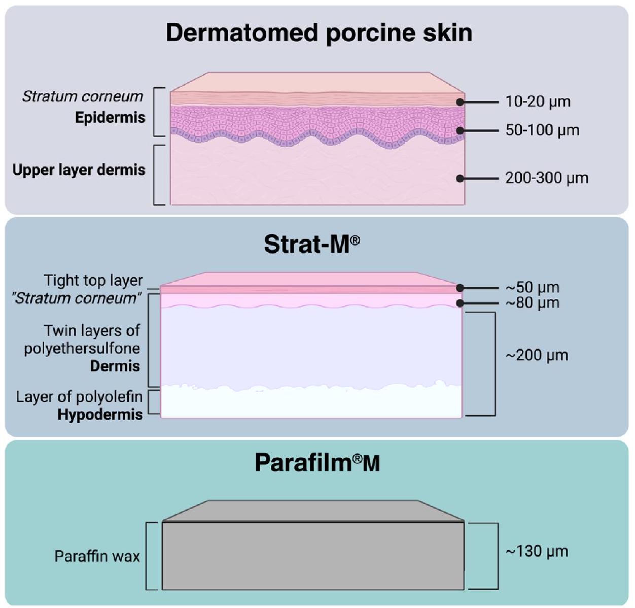
(Loughborough, UK). Neonatal porcine skin was obtained from stillborn piglets less than 24 h after birth, rinsed in phosphate buffer saline (PBS pH 7.4 ), dermatomed to a thickness of , and kept frozen at until use.

2.2. Preparation of dissolving MAPs

An aqueous blend containing PVA and PVP K29/ 32 with ratio 1:1, called PP2 (Anjani et al., 2023c), was used to prepare dissolving MAPs, as outlined in Table 1. A 50 mg aliquot of each formulation was poured into silicone mould ( needle area, 16 pyramidal-cuboidal holes, needle height and width at the base, illustrated in Fig. 1) placed into pressure chamber for 5 mins at 5 bar. Furthermore, the moulds were removed from pressure chamber and the excess of formulation was removed from the mould surface, and then centrifuged at 5000 rpm for 10 mins with the temperature controlled at . Following this, a silicone ring (internal diameter 18 mm and thickness 3 mm ) was glued on to the mould surface using w/v PVA aqueous solution and dried at ambient temperature for 24 h . The second layer of dissolving MAP was prepared by poured of aqueous solution of PVP K90 and glycerol, centrifuged at 5000 rpm for 10 mins at , allowed to dry under ambient conditions for 24 h . Sidewalls of dissolving MAPs were removed using scissors and then transferred into oven for final drying for 24 h .

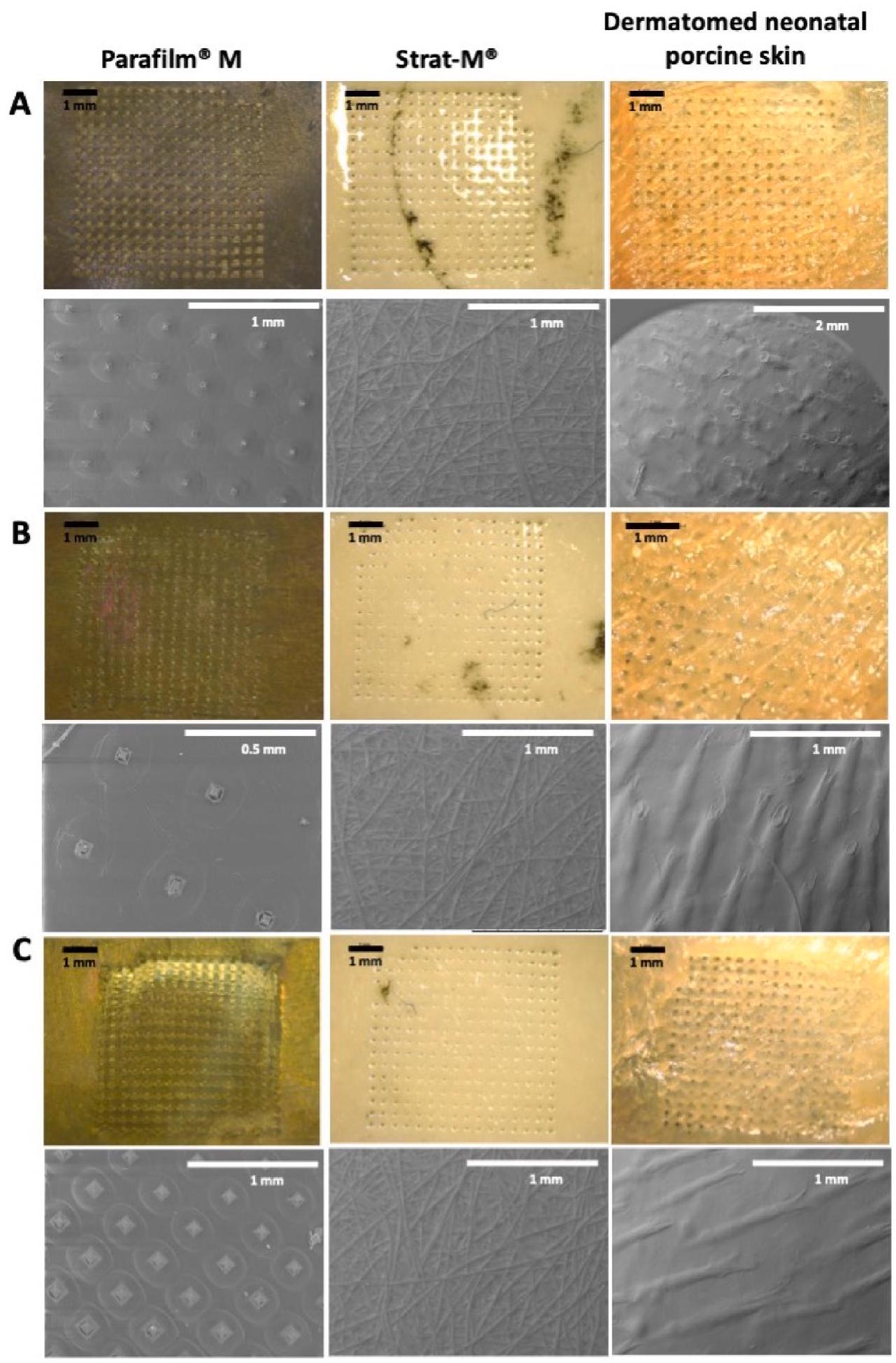
2.3. Optical coherence tomography and pore visualisation

Each dissolving MAP was inserted into a membrane models (dermatomed skin, Parafilm and Strat-M) using a TA-TX2 Texture Analyser (TA) (Stable Microsystems, Haslemere, UK), as previously described (Bin Sabri et al., 2021; Bin Sabri et al., 2022; Anjani et al., 2022b) and illustrated in Fig. 2, with a 32 N force was applied, which equal to force released by human thumb pressure (Larrañeta et al., 2014). A piece of dental wax was applied under the membrane to protect the tips of the needles piercing the membrane (Bin Sabri et al., 2021; Bin Sabri et al., 2022; Anjani et al., 2022b). The insertion and holes were observed and visualised using EX-101 optical coherence tomography (OCT) microscope (Michelson Diagnostics Ltd., Kent, UK), digital light microscope (Leica EZ4 D, Leica Microsystems, Milton Keynes, UK) and scanning electron microscope (SEM) TM3030 microscope (Hitachi, Krefeld, Germany). The images obtained from OCT analysis was then processed and analysed using Image National Institutes of Health, Bethesda MD, USA).

2.4. Determination of protein released from membranes

To anticipate any proteins that might interfere with the quantification of proteins sourced from membranes (including dermatomed neonatal porcine skin, Strat-M®, and Parafilm during the in vitro studies in the Franz cell setup, a release study was conducted as a preliminary step before the in vitro permeation studies. Each membrane was trimmed into a circular shape measuring , equivalent to the area of inner part of the donor compartment in the Franz cell apparatus. These membranes were then placed into separate vials containing 12 mL of PBS ( pH 7.4 ) and incubated in a shaker incubator (Jeio Tech ISF7100, Medline Scientific, Chalgrove Oxon, UK) at for 24 h . Subsequently, the samples were analysed using a micro-BCA kit, and if
Table 1
Formulation of first layer dissolving MAP.
Composition Protein Type AMSC-MP
Albumin BSA
Protein (%w/w) 10 10 10
Aqueous mixture PP2 (%w/w) 30 30 30
Deionised water (%w/w) 60 60 60
Fig. 1. Illustration of needle size and geometry on the array of dissolving MAP.
Fig. 2. Schematic illustration of texture analyser setup to evaluate dissolving MAP insertion properties into different membranes (dermatomed porcine skin, Strat and Parafilm ).
necessary, the samples were diluted in PBS ( pH 7.4 ).

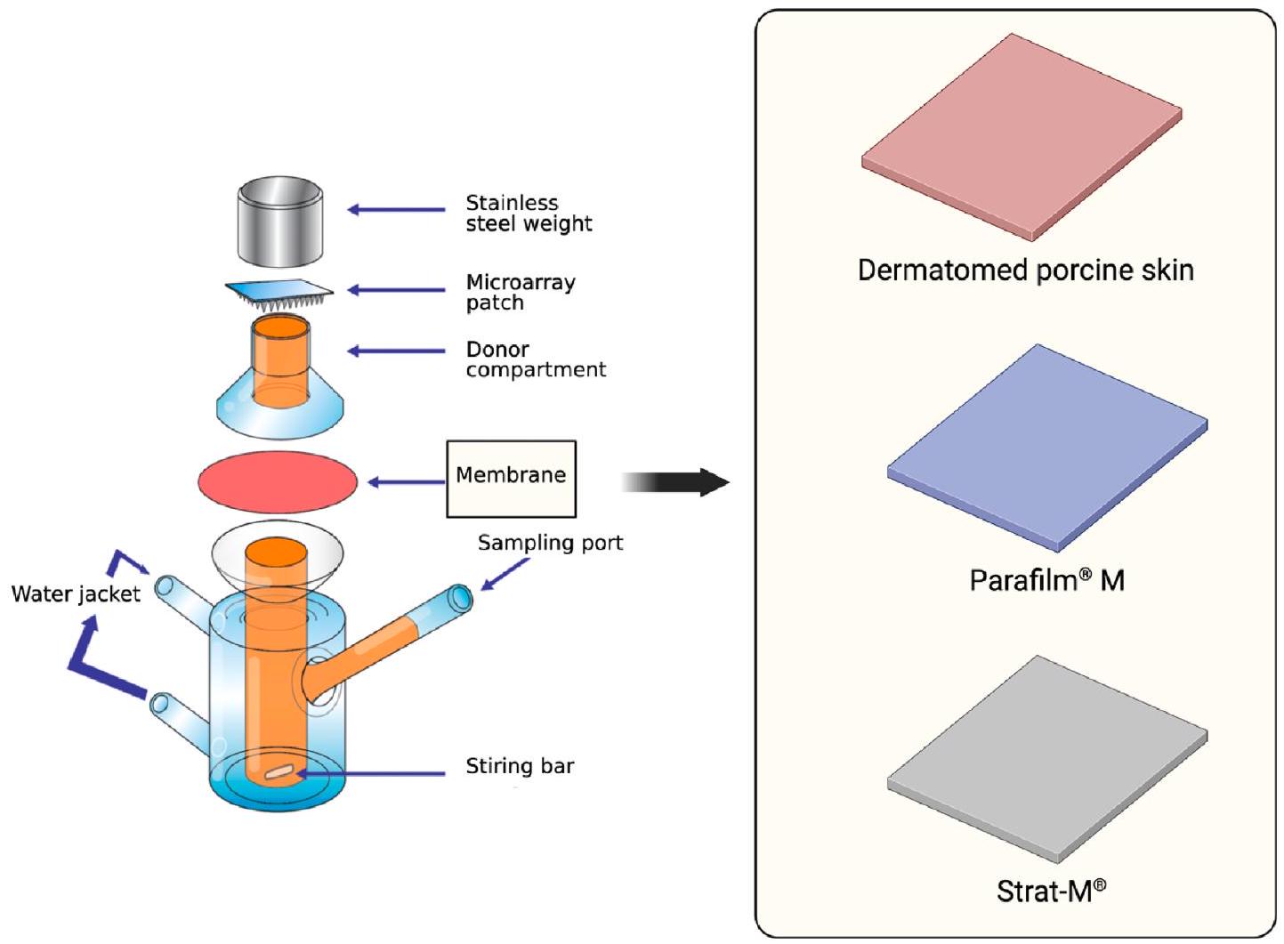
2.5. In vitro permeation studies using Franz cells

For the in vitro permeation study, a Franz cell (Permergear, Hellertown PA, USA) setup was used with dermatomed neonatal porcine skin, Strat-M® and Parafilm , as illustrated in Fig. 3. The porcine skin, obtained from stillborn piglets within 24 h post-mortem, was rinsed in PBS, dermatomed at the thickness of , and frozen at until use. The membrane (dermatomed neonatal porcine skin, Strat-M® or Parafilm was carefully attached to the donor compartment of the
Franz cell using cyanoacrylate glue. Preheated and degassed PBS ( pH 7.4) was added to the receiver compartment, which was maintained at and stirred at 600 rpm . The dissolving MAPs were manually pressed onto the skin for 30 s with a piece of dental wax underneath as described previously (Larrañeta et al., 2016). The drug-containing reservoir was placed on top of the dissolving MAPs, and the donor compartment was attached to the receiver compartment. A stainlesssteel cylindrical weight ( 15.0 g ) was placed on top of the reservoir to prevent MAP expulsion. The sampling arm of the receiver compartment and the donor compartment were sealed with Parafilm to prevent evaporation. At predetermined time points, of release media was
Fig. 3. Schematic illustration of Franz diffusion cell setup to assess permeation protein from dissolving MAP across different membranes (dermatomed porcine skin, Strat-M® and Parafilm ).
taken from the receiver compartment and analysed using micro-BCA kit. If necessary, samples were diluted in PBS (pH 7.4).

2.6. Quantification using micro-BCA protein assay kit

Samples from in vitro studies were quantified and analysing using Micro BCA Protein Assay Kit (Thermo Scientific, Rockford, IL, USA). Analysis was performed following the guideline provided by manufacturer. All samples were determined using a FluoStar Omega microplate reader (BMG Labtech, Germany) with spectroscopy principal with absorbance wavelength at 562 nm .

2.7. Statistical analysis

Statistical analysis was conducted using GraphPad Prism version 8.0 (GraphPad Software, San Diego, California, USA). The experimental results were presented as means standard deviation (SD), unless otherwise specified. An unpaired -test was used for comparing two cohorts, while one-way analysis of variance (ANOVA) was employed for comparing multiple cohorts.

3. Results and discussion

3.1. Preparation of dissolving MAPs

The study employed three distinct proteins as model substances (albumin derived from chicken eggs (OVA, MW 45 kDa ), and bovine serum albumin (BSA, MW 66.5 kDa ) and amniotic mesenchymal stem cell metabolite products (AMSC-MP, MW 76 kDa )). Dissolving MAPs containing these proteins were fabricated using the double casting method, as detailed in previous works (Anjani et al., 2022b; Anjani et al., 2022c; Anjani et al., 2022d). Moreover, proteins can be loaded into dissolving MAPs using more complex approaches such as coating the
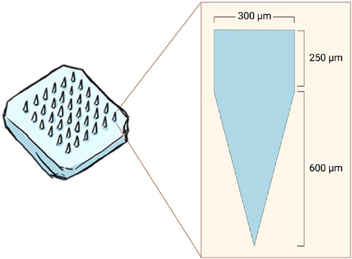
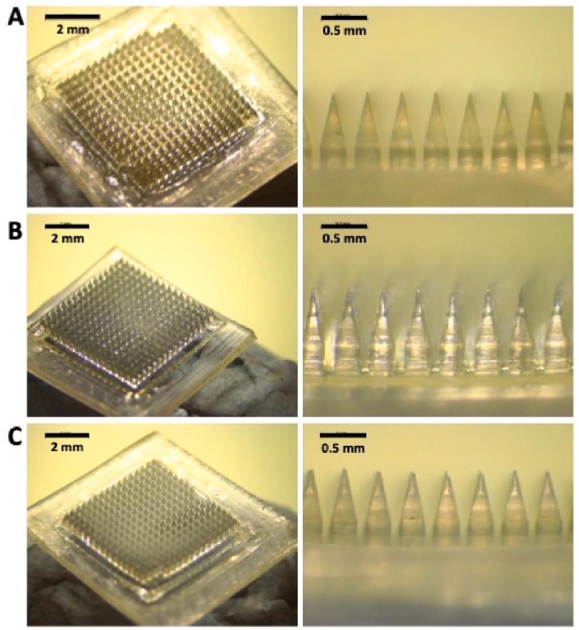
arrays with formulations containing proteins (Angkawinitwong et al., 2020; Caudill et al., 2018). In this study, each protein was loaded into the needle tips in a simple way and ensuring minimum waste of the protein cargo. For this purpose, the protein was combined with a mixture of PVA and PVP. Fig. 4 displays microscopic images of the resulting dissolving MAPs, demonstrating their homogeneity and welldefined structure, characterised by sharp needles. The needle shape is a combination of cuboidal and pyramidal, a result of the polydimethylsiloxane moulds used in this experiment. This mould design was chosen for its ability to facilitate effective insertion and drug delivery into the skin, which has shown superiority compared to other shapes (Cordeiro et al., 2020). Finally, these dissolving MAPs underwent subsequent insertion testing and in vitro permeation profiling.

3.2. Optical coherence tomography and pore visualisation

To assess the insertion capacity of protein-loaded MAPs into different membranes, dermatomed porcine skin ( ), Strat-M ( 300 ), and Parafilm , the structures of which are illustrated in Fig. 5, specific membrane types were chosen for evaluation. The porcine skin utilised in this study was sourced from stillborn piglets and dermatomed to a thickness of , comprising the epidermis (stratum corneum, stratum granulosum, stratum spinosum, and stratum basal) and the upper part of the dermis layer. This thickness was chosen because it approximates the distance from the skin surface to the dermal microcirculation. With a needle height of , a proportion of the microneedles will be in direct contact with the receiver medium when using epidermal or upper dermal representations of the skin barrier (Garland et al., 2012). Strat-M®, engineered to mimic human skin structure, features a lipid-coated surface to simulate the stratum corneum layer (Haq et al., 2018). Below this lipid coating, Strat-M® consists of polyether sulfone, creating a more porous layer to mimic the dermis, followed by a polyolefin non-fabric support resembling the
Fig. 4. Microscopic images of dissolving MAPs loaded with (A) albumin derived from chicken eggs (OVA), (B) bovine serum albumin (BSA), and (C) amniotic mesenchymal stem cell metabolite products (AMSC-MP).
subcutaneous layer (Haq et al., 2018; Pulsoni et al., 2022). Parafilm was selected as a model membrane for this study due to its validation as a proposed membrane for MAP insertion studies (Larrañeta et al., 2014), serving as a rapid quality control test. Composed of a blend of hydrocarbon wax and polyolefin, Parafilm replicates properties similar to the third layer of Strat-M®, aimed at simulating the subcutaneous layer. Although less structurally complex than actual skin and Strat-M®, Parafilm provides sufficient hydrophobic properties, resembling the stratum corneum, which is a crucial barrier for MAP application.
OCT images in Fig. 6A-C illustrate the insertion of dissolving MAPs into various membranes. A clear gap is evident between the MAP baseplate and the membrane surfaces, suggesting incomplete penetration of dissolving MAPs into these membranes. As depicted in Fig. 6D-F, the microneedles of MAPs penetrated to depths of approximately 430 , and for dermatomed porcine skin, Strat-M®, and Parafilm , respectively. The insertion depth into dermatomed porcine skin aligns with previously published findings (Garland et al., 2012; Donnelly et al., 2010; Donnelly et al., 2011), significantly surpassing penetration depths observed in other skin simulants ( ). This disparity may be attributed to differences in membrane materials and elasticity compared to dermatomed porcine skin.
Notably, prior to the experiment, the porcine skin was equilibrated with PBS pH (7.4) to maintain skin integrity. The presence of saline water likely acted as a lubricant (Anjani et al., 2023d), facilitating deeper needle insertion compared to Strat-M® and Parafilm . Furthermore, Parafilm exhibited a deeper penetration compared to
Strat-M®, possibly due to its single-layer thickness ( ) versus the multilayered structure of Strat-M® ( ). Fig. 7 confirms successful hole creation in all membranes, simulating piercing of the stratum corneum layer. However, while Strat-M® closely resembles the thickness of dermatomed porcine skin, detecting the holes in its deeper layers was challenging due to the polyolefin non-woven fabric’s characteristics, mimicking the hypodermal layer and offering resistance to microneedle penetration. Despite the and differences in insertion depth between Parafilm and Strat-M compared to dermatomed porcine skin, both skin simulants show promise as membrane substitutes for insertion studies.

3.3. Determination of protein released from membranes

To assess protein release from various membranes, a preliminary study was conducted. Fig. 8 demonstrates that after incubating dermatomed neonatal porcine skin at for 24 h , approximately 39 mg of protein was released. Conversely, no protein was detected in either the Parafilm M or Strat-M groups. This outcome suggests that using biological membranes, coupled with the micro-BCA quantification method for protein delivery assessment, might yield unreliable results due to skin-derived proteins interfering with the delivered substance. Moreover, accounting for protein release from the blank dermatomed skin, we included the dermatomed porcine skin without dissolving MAP application in further in vitro permeation studies using the Franz cell setup. However, for both Parafilm and Strat-M , no control or blank
Fig. 5. Schematic illustration depicting the multilayer structure of dermatomed porcine skin alongside Strat-M® and a single layer of Parafilm M.
groups were included as no protein was detected in this current study. This exclusion ensures the absence of protein interference during the quantification process in these cases.

3.4. In vitro permeation studies using Franz cells

The Franz cell setup was utilised in this study to evaluate the permeation of the protein models in an in vitro setting. This setup has consistently proven to be ideal in all our MAP-related studies for assessing the diffusion of molecules from the tip layer of MAPs to the skin layer, particularly across the epidermis-dermis boundary (Anjani et al., 2022a; Anjani et al., 2021b; Anjani et al., 2023; Bin Sabri et al., 2022; Anjani et al., 2022b; Anjani et al., 2022c; Anjani et al., 2022d; Anjani et al., 2021a; Anjani et al., 2023e; Anjani et al., 2023f). Conversely, in vitro release studies are not suitable for evaluating this process, as the entire patch is exposed to fluid media, leading to an overestimation of the drug release profile achieved from the dissolving MAP system. Therefore, we suggest using both membranes, Paraf , and Strat-M®, as alternatives to biological membranes to assess the delivery of proteins and peptides in in vitro permeation studies using the Franz cell setup.
Fig. 9 displays the permeation profiles of model proteins released from polymeric dissolving MAPs across different membranes, dermatomed porcine skin, Parafilm®M, and Strat-M®. Both OVA and BSA permeation from dissolving MAPs demonstrated an initial rise within 2 h , followed by a plateau between 4 and 24 h for both Parafilm and Strat-M® membranes. In the case of AMSC-MP, notable differences were observed between Parafilm M and Strat-M . Across Parafilm , the protein permeation gradually increased from the first hour up to 6 h , increasing significantly at . However, using Strat-M®,
AMSC-MP permeated immediately within the first hour, maintaining a sustained permeation throughout the experimental period. The difference between the two membranes can be explained due to the effect of several factors. Strat-M® membrane is designed to mimic the skin and as can be seen in Fig. 7 dissolving MAPs do not go through the entire membrane. Therefore, proteins need to diffuse through the membrane as they are not directly in contact with the fluid of the receiver compartment. Dissolving MAPs can go through one layer of Parafilm M (Fig. 7) and when placed in the receiver compartment, microneedles will be directly in contact with the fluid of the receiver compartment. That explains why permeation from Parafilm is faster.
The most substantial protein permeation facilitated by dissolving MAPs occurred when using dermatomed neonatal porcine skin as the model membrane. Burst release commenced within the first hour, followed by a marked sustained increase in cumulative permeation throughout the 24 -hour experimental period. There is a possibility that dissolving MAPs penetrated through the dermatomed porcine skin, potentially expediting the rapid release into the receiver compartment. To assess the release of total protein content from dermatomed porcine skin without dissolving MAP application, we conducted an additional evaluation. Fig. 9 indicates a noticeable amount of protein release from the skin, even surpassing the total protein detected from both Parafilm and Strat-M groups in all cases. This observation highlights that utilising any biological membrane can lead to false-positive results in protein permeation, as skin-derived proteins were detected and accumulated during the quantification process using the BCA method. Hence, employing synthetic membranes might alleviate this limitation associated with the quantification method.
Several studies have explored the delivery of BSA and OVA using the MAP platform. Badhe et al. developed chitosan-based MAPs coated with
Fig. 6. OCT images of MAPs loaded with (A) albumin derived from chicken eggs (OVA), (B) bovine serum albumin (BSA), and (C) amniotic mesenchymal stem cell metabolite products (AMSC-MP) inserted into Parafilm®M, Strat-M® and dermatomed neonatal porcine skin following the application of pressure of 32 N for 30 s. The insertion depth of dissolving MAPs loaded with (D) albumin derived from chicken eggs (OVA), (E) bovine serum albumin (BSA), and (F) amniotic mesenchymal stem cell metabolite products (AMSC-MP) into Parafilm®M, Strat-M® and dermatomed neonatal porcine skin (means + SD, n = 20). To allow differentiation between dissolving MAP and the skin models, false colours were applied in the needles and skin model layers.
Fig. 7. Microscopic images of the upper layer and SEM images of the back side of each membrane were taken after the application of dissolving MAPs loaded with (A) albumin derived from chicken eggs (OVA), (B) bovine serum albumin (BSA), and (C) amniotic mesenchymal stem cell metabolite products (AMSC-MP) using a texture analyser with a 32 N pressure for 30 s .
BSA and poly(lactic acid) (Badhe et al., 2021). Using a colorimetric method with biuret reagent and a UV-Vis spectrophotometer set at a wavelength of 540 nm , the researchers quantified BSA release during an in vitro permeation study using Franz cells. The study revealed 98.5% BSA permeation across excised rat skin within 50 h (Badhe et al., 2021). However, it lacked a control group without MAP application, raising concerns about protein release from the skin during the study period.
Demir et al. developed sodium alginate-based MAPs loaded with BSA, indicating a higher percentage of BSA delivery ( ) across dermatomed human abdominal skin compared to a transdermal patch (Demir et al., 2013). The researchers quantified BSA release using the BCA method at 562 nm (Demir et al., 2013). Nevertheless, a control group analysis was missing, limiting understanding regarding the source of protein during these studies.
Fig. 8. The total protein released from each membrane after 24 h of incubation at in PBS ( pH 7.4 ). The ‘#’ symbol indicates that no protein was detected in the media (means ).
Regarding OVA-mediated MAP permeation studies, Maaden et al. and Vallhov et al. utilised radiolabelled OVA (Van Der Maaden et al., 2014) and OVA conjugated with Alexa 488 (Vallhov et al., 2018), respectively. Maaden et al. employed a pH -sensitive MAP coated with radioactively labelled OVA and used an automatic gamma counter for specific quantification during in vitro permeation studies across excised human skin (Van Der Maaden et al., 2014). Vallhov et al. utilised a bioceramic MAP coated with OVA to deliver the antigen to dendritic cells in human skin (Vallhov et al., 2018). These studies employed different approaches to selectively quantify OVA during in vitro permeation studies.
Regarding in vitro-in vivo correlation (IVIVC), to date, such discussions have not yet been established for MAP-mediated delivery. The development of dissolving MAPs, which dissolve or degrade upon contact with skin interstitial fluid to release their drug cargo in situ, necessitates consideration of additional factors when designing in vitro permeation experiments. Ideally, the selected model membrane representing the skin barrier in vitro should mimic the depth of microneedle penetration, adhesion of the microneedle baseplate to the skin surface, microneedle dissolution/degradation rate (Garland et al., 2012), and subsequent drug release achieved in an in vivo scenario. Furthermore, given that microneedle length may exceed the thickness of the chosen skin membrane barrier, a portion of the microneedle may directly contact the receiver medium. Therefore, it is crucial to ensure that the composition of the receiver medium selected does not significantly overestimate the rate of microneedle dissolution and, consequently, the rate of drug release within the skin (Garland et al., 2012).
When observing the cumulative protein delivered from dissolving MAPs across dermatomed porcine skin in comparison to control groups, normalisation was performed by subtracting the total protein from the dissolving MAP group. Fig. 10 displays the normalised data at 24 h for OVA, BSA, and AMSC-MP released from dissolving MAPs across dermatomed porcine skin, Parafilm , and Strat-M®. The cumulative permeation percentages for OVA-loaded MAPs on dermatomed porcine skin, Parafilm , and Strat-M were found to be , and , respectively. No significant differences were observed between the membranes after normalisation ( ).
For BSA, the cumulative permeation over 24 h through dermatomed porcine skin, Parafilm M, and Strat-M was approximately , , and , respectively. Interestingly, the permeation across dermatomed porcine skin was significantly higher ( ) and
Fig. 9. (A) The total protein content was utilised to quantify the permeation of albumin derived from chicken eggs (OVA), bovine serum albumin (BSA), and amniotic mesenchymal stem cell metabolite products (AMSC-MP) from dissolving MAPs across dermatomed porcine skin. The dashed lines represent the protein released from dermatomed neonatal porcine skin without dissolving MAP application (means SD, ). (B) The total protein content was used to quantify the permeation of albumin derived from chicken eggs (OVA), bovine serum albumin (BSA), and amniotic mesenchymal stem cell metabolite products (AMSC-MP) from dissolving MAPs across Parafilm M, Strat-M®, and dermatomed porcine skin. Corrected values of drug permeated across dermatomed skin were obtained by subtracting the blank dermatomed skin readings (means ).
Fig. 10. Cumulative permeation of albumin derived from chicken eggs (OVA), bovine serum albumin (BSA), and amniotic mesenchymal stem cell metabolite products (AMSC-MP) across different membranes (dermatomed porcine skin, Parafilm®M and Strat-M®) over 24 h of in vitro permeation studies (means + SD, ).
increased 4.88-fold compared to Strat-M®. This discrepancy might be attributed to skin protein presence, leading to increased protein detection across different protein models. Additionally, in AMSC-MP delivery, the cumulative permeation percentages were , and across dermatomed porcine skin, Parafilm , and Strat-M , respectively. Notably, AMSC-MP permeation across Parafilm®M significantly increased by 2.83 -fold compared to Strat- , while there were no significant differences between Parafilm and dermatomed porcine skin ( ) in AMSC-MP permeation percentage.
The in vitro permeation data analysis revealed distinct correlation patterns between Parafilm M and Strat-M concerning the cumulative permeation of proteins within a 24 -hour timeframe. These correlations appear to depend on the properties of the protein models used in the study. Fig. 11 illustrates that, based on the correlation coefficient category (Correlation and regression, n.d.); Parafilm exhibited strong and moderate correlations with Strat-M® for OVA and BSA, respectively. Notably, the cumulative permeation of AMSC-MP across both Parafilm and Strat-M demonstrated a very strong correlation, ranging from 0.90 to 0.99 .
Correlating the dermatomed porcine skin samples to both Parafilm M and Strat-M proved challenging due to the interference of skin proteins in the permeation results over the 24 -hour period. Consequently, this study suggests that both Parafilm M and Strat-M outperform dermatomed neonatal porcine skin in terms of reliability in permeation studies. Furthermore, it indicates a similarity in the protein permeation profiles across Parafilm and Strat-M , highlighting the potential of these skin simulants for use in protein-loaded MAPs
intended for transdermal delivery. Additionally, future plans for this study include establishing a similarity factor. The current focus is on the potential use of Parafilm and Strat-M in in vitro studies as a preliminary step to select the best formulation for in vivo studies using different animal models. Thus, it should also be noted that the quantified amounts using synthetic membranes might not always correlate with actual permeation studies using human or porcine skin. However, the results of this study show the potential of Parafilm and Strat-M as alternatives to dermatomed porcine skin in the early-stage phase, enabling researchers to establish standardised protocols for assessing substance permeation from MAP systems.

4. Conclusion

In summary, this study demonstrates the potential use of Paraf and Strat-M as alternatives to biological skin simulants in in vitro permeation studies, particularly for investigating protein delivery from MAP platforms. Our findings indicate that both Parafilm and Strat-M® exhibit only slight differences compared to dermatomed porcine skin, making them promising substitutes for membrane studies. Dermatomed porcine skin releases proteins during in vitro permeation studies, leading to potential interference in quantification using the BCA technique. Utilising synthetic membranes like Parafilm®M and Strat can circumvent such interference, providing more reliable results compared to dermatomed neonatal porcine skin. It is important to note that while these synthetic membranes offer insights into trends and correlations, they may not precisely match absolute permeability values of biological skin membranes for protein delivery. However, these
Fig. 11. Correlation of cumulative permeation between Parafilm and Strat-M for albumin derived from chicken eggs (OVA), bovine serum albumin (BSA), and amniotic mesenchymal stem cell metabolite products (AMSC-MP).
results are important in understanding the potential of these membranes for formulating and optimising MAPs. These findings may serve as valuable tools for preliminary screening efforts in various MAP formulations, not limited to proteins or peptides. Synthetic membranes offer rapid, cost-effective, and easily accessible alternatives that do not require complex preparation or storage. This research marks an earlystage comparison between biological and synthetic membranes to identify a suitable skin simulant model in the field of MAP formulation.

CRediT authorship contribution statement

Qonita Kurnia Anjani: Writing – review & editing, Writing – original draft, Visualization, Validation, Methodology, Investigation, Formal analysis, Data curation, Conceptualization. Avelia Devina Calista Nainggolan: Investigation, Formal analysis, Data curation. Huanhuan Li: Investigation, Formal analysis. Andang Miatmoko: Writing – review & editing, Resources, Investigation. Eneko Larrañeta: Writing – review & editing, Visualization, Formal analysis. Ryan F. Donnelly: Supervision, Resources, Writing – review & editing.

Declaration of competing interest

The authors declare that they have no known competing financial interests or personal relationships that could have appeared to influence the work reported in this paper.

Data availability

Data will be made available on request.

References

Alkilani, A.Z., McCrudden, M.T.C., Donnelly, R.F., 2015. Transdermal drug delivery: innovative pharmaceutical developments based on disruption of the barrier properties of the stratum corneum. Pharmaceutics 7, 438-470. https://doi.org/ 10.3390/pharmaceutics7040438.
Angkawinitwong, U., Courtenay, A.J., Rodgers, A.M., Larrañeta, E., Mccarthy, H.O., Brocchini, S., Donnelly, R.F., Williams, G.R., 2020. A novel transdermal protein delivery strategy via electrohydrodynamic coating of PLGA microparticles onto microneedles. ACS Appl. Mater. Interf. 12, 12478-12488. https://doi.org/10.1021/ ACSAMI.9B22425/ASSET/IMAGES/LARGE/AM9B22425_0008.JPEG.
Anjani, Q.K., Bin Sabri, A.H., Donnelly, R., 2021a. Development and validation of simple and sensitive HPLC-UV method for ethambutol hydrochloride detection following transdermal application. Anal. Methods. https://doi.org/10.1039/D1AY01414E.
Anjani, Q.K., Permana, A.D., Cárcamo-Martínez, Á., Domínguez-Robles, J., Tekko, I.A., Larrañeta, E., Vora, L.K., Ramadon, D., Donnelly, R.F., 2021b. Versatility of hydrogel-forming microneedles in in vitro transdermal delivery of tuberculosis drugs. Eur. J. Pharm. Biopharm. 294-312, 294-312. https://doi.org/10.1016/j. ejpb.2020.12.003.
Anjani, Q.K., Hidayat, A., Sabri, B., Moreno-Castellanos, N., Utomo, E., CárcamoMartínez, Á., Domínguez-Robles, J., Ahmadi, L., Wardoyo, H., Donnelly, R.F., 2022a. Soluplus -based dissolving microarray patches loaded with colchicine: towards a minimally invasive treatment and management of gout. Biomater. Sci. https://doi. org/10.1039/D2BM01068B.
Anjani, Q.K., Bin Sabri, A.H., Utomo, E., Domínguez-Robles, J., Donnelly, R.F., 2022b. Elucidating the impact of surfactants on the performance of dissolving microneedle array patches. Mol. Pharm. 19, 1191-1208, 10.1021/ACS. MOLPHARMACEUT.1C00988/ASSET/IMAGES/LARGE/MP1C00988_0014.JPEG.
Anjani, Q.K., Bin Sabri, A.H., McGuckin, M.B., Li, H., Hamid, K.A., Donnelly, R.F., 2022c. In vitro permeation studies on carvedilol containing dissolving microarray patches quantified using a rapid and simple HPLC-UV analytical method. AAPS PharmSciTech 23, 1-13. https://doi.org/10.1208/S12249-022-02422-6/FIGURES/ 4.
Anjani, Q.K., Bin Sabri, A.H., Domínguez-Robles, J., Moreno-Castellanos, N., Utomo, E., Wardoyo, L.A.H., Larrañeta, E., Donnelly, R.F., 2022d. Metronidazole nanosuspension loaded dissolving microarray patches: An engineered composite pharmaceutical system for the treatment of skin and soft tissue infection. Biomaterials Adv. 140, 213073 https://doi.org/10.1016/J.BIOADV.2022.213073.
Anjani, Q.K., Bin Sabri, A.H., Hutton, A.J., Cárcamo-Martínez, Á., Wardoyo, L.A.H., Mansoor, A.Z., Donnelly, R.F., 2023a. Microarray patches for managing infections at a global scale. J. Control. Release 359, 97-115. https://doi.org/10.1016/j. jconrel.2023.05.038.
Anjani, Q.K., Bin Sabri, A.H., Hamid, K.A., Moreno-Castellanos, N., Li, H., Donnelly, R.F., 2023b. Tip loaded cyclodextrin-carvedilol complexes microarray patches. Carbohydr. Polym. 320, 121194 https://doi.org/10.1016/j.carbpol.2023.121194.
Anjani, Q.K., Pandya, A.K., Demartis, S., Domínguez-Robles, J., Moreno-Castellanos, N., Li, H., Gavini, E., Patravale, V.B., Donnelly, R.F., 2023c. Liposome-loaded polymeric microneedles for enhanced skin deposition of rifampicin. Int. J. Pharm. 646, 123446 https://doi.org/10.1016/J.IJPHARM.2023.123446.
Anjani, Q.K., Cárcamo-Martínez, Á., Wardoyo, L.A.H., Moreno-Castellanos, N., Bin Sabri, A.H., Larrañeta, E., Donnelly, R.F., 2023d. MAP-box: a novel, low-cost and easy-to-fabricate 3D-printed box for the storage and transportation of dissolving microneedle array patches. Drug Deliv. Transl. Res. https://doi.org/10.1007/ s13346-023-01393-w.
Anjani, Q.K., Bin Sabri, A.H., Hamid, K.A., Moreno-Castellanos, N., Li, H., Donnelly, R.F., 2023e. Tip loaded cyclodextrin-carvedilol complexes microarray patches. Carbohydr. Polym. 320, 121194.
Anjani, Q.K., Volpe-Zanutto, F., Hamid, K.A., Bin Sabri, A.H., Moreno-Castellano, N., Gaitán, X.A., Calit, J., Bargieri, D.Y., Donnelly, R.F., 2023f. Primaquine and chloroquine nano-sized solid dispersion-loaded dissolving microarray patches for the improved treatment of malaria caused by Plasmodium vivax. J. Controll. Release 361, 385-401. https://doi.org/10.1016/J.JCONREL.2023.08.009.
Antosova, Z., Mackova, M., Kral, V., Macek, T., 2009. Therapeutic application of peptides and proteins: parenteral forever? Trends Biotechnol. 27, 628-635. https://doi.org/ 10.1016/J.TIBTECH.2009.07.009.
Badhe, R.V., Adkine, D., Godse, A., 2021. Development of polylactic acid and bovine serum albumin-layered-coated chitosan microneedles using novel bees wax mould. Turk. J. Pharm. Sci. 18, 367. https://doi.org/10.4274/TJPS.GALENOS.2020.47897.
Bin Sabri, A.H., Anjani, Q.K., Donnelly, R.F., Hidayat Bin Sabri, A., Kurnia Anjani, Q., Donnelly, R.F., 2021. Synthesis and characterization of sorbitol laced hydrogelforming microneedles for therapeutic drug monitoring. Int. J. Pharm. 607, 121049.
Bin Sabri, A.H., Anjani, Q.K., Utomo, E., Ripolin, A., Donnelly, R.F., 2022. Development and characterization of a dry reservoir-hydrogel-forming microneedles composite for minimally invasive delivery of cefazolin. Int. J. Pharm. 617, 121593 https://doi.org/ 10.1016/J.IJPHARM.2022.121593.
Bittner, B., Richter, W., Schmidt, J., 2018. Subcutaneous administration of biotherapeutics: an overview of current challenges and opportunities. BioDrugs 32, 425. https://doi.org/10.1007/S40259-018-0295-0.
Bruno, B.J., Miller, G.D., Lim, C.S., 2013. Basics and recent advances in peptide and protein drug delivery. Ther. Deliv. 4, 1443. https://doi.org/10.4155/TDE.13.104.
Caudill, C.L., Perry, J.L., Tian, S., Luft, J.C., DeSimone, J.M., 2018. Spatially controlled coating of continuous liquid interface production microneedles for transdermal protein delivery. J. Control. Release 284, 122-132. https://doi.org/10.1016/J. JCONREL. 2018.05.042.
Cordeiro, A.S., Tekko, I.A., Jomaa, M.H., Vora, L., McAlister, E., Volpe-Zanutto, F., Nethery, M., Baine, P.T., Mitchell, N., McNeill, D.W., Donnelly, R.F., 2020. Twophoton polymerisation 3D printing of microneedle array templates with versatile designs: application in the development of polymeric drug delivery systems. Pharm. Res. 37, 1-15. https://doi.org/10.1007/S11095-020-02887-9/FIGURES/9.
11. Correlation and regression, (n.d.). https://www.bmj.com/about-bmj/resources-readers/publications/statistics-square-one/11-correlation-and-regression (Accessed December 2, 2023).
Demir, Y.K., Akan, Z., Kerimoglu, O., 2013. Sodium alginate microneedle arrays mediate the transdermal delivery of bovine serum albumin. PLoS One 8, e63819.
Donnelly, R.F., Prausnitz, M.R., 2023. The promise of microneedle technologies for drug delivery. Drug Deliv. Transl. Res. 1-8. https://doi.org/10.1007/S13346-023-014308/FIGURES/2.
Donnelly, R.F., Garland, M.J., Morrow, D.I.J., Migalska, K., Singh, T.R.R., Majithiya, R., Woolfson, A.D., 2010. Optical coherence tomography is a valuable tool in the study of the effects of microneedle geometry on skin penetration characteristics and in-skin dissolution. J. Control. Release 147, 333-341. https://doi.org/10.1016/J. JCONREL. 2010.08.008.
Donnelly, R.F., Majithiya, R., Singh, T.R.R., Morrow, D.I.J., Garland, M.J., Demir, Y.K., Migalska, K., Ryan, E., Gillen, D., Scott, C.J., Woolfson, A.D., 2011. Design, optimization and characterisation of polymeric microneedle arrays prepared by a novel laser-based micromoulding technique. Pharm. Res. 28, 41-57. https://doi.org/ 10.1007/S11095-010-0169-8/FIGURES/12.
Doughty, D.V., Clawson, C.Z., Lambert, W., Subramony, J.A., 2016. Understanding subcutaneous tissue pressure for engineering injection devices for large-volume protein delivery. J. Pharm. Sci. 105, 2105-2113. https://doi.org/10.1016/J. XPHS.2016.04.009.
Garland, M.J., Migalska, K., Tuan-Mahmood, T.M., Raghu Raj Singh, T., Majithija, R., Caffarel-Salvador, E., McCrudden, C.M., McCarthy, H.O., David Woolfson, A., Donnelly, R.F., 2012. Influence of skin model on in vitro performance of drug-loaded soluble microneedle arrays. Int. J. Pharm. 434, 80-89. https://doi.org/10.1016/J. IJPHARM.2012.05.069.
Haq, A., Goodyear, B., Ameen, D., Joshi, V., Michniak-Kohn, B., 2018. Strat-M® synthetic membrane: permeability comparison to human cadaver skin. Int. J. Pharm. 547, 432-437. https://doi.org/10.1016/J.IJPHARM.2018.06.012.
Jiskoot, W., Randolph, T.W., Volkin, D.B., Middaugh, C.R., Schöneich, C., Winter, G., Friess, W., Crommelin, D.J.A., Carpenter, J.F., 2012. Protein instability and immunogenicity: roadblocks to clinical application of injectable protein delivery systems for sustained release. J. Pharm. Sci. 101, 946-954. https://doi.org/10.1002/ JPS. 23018.
Kirkby, M., Hutton, A.R.J., Donnelly, R.F., 2020. Microneedle mediated transdermal delivery of protein, peptide and antibody based therapeutics: current status and future considerations. Pharm. Res. 37, 1-18. https://doi.org/10.1007/S11095-020-02844-6/TABLES/2.
E. Larrañeta, M.T.C. McCrudden, A.J. Courtenay, R.F. Donnelly, Microneedles: a new frontier in nanomedicine delivery, Pharmaceut. Res. 33 (2016) 1055-1073. doi: 10.1007/S11095-016-1885-5.
Larrañeta, E., Moore, J., Vicente-Pérez, E.M., González-Vázquez, P., Lutton, R., Woolfson, A.D., Donnelly, R.F., 2014. A proposed model membrane and test method for microneedle insertion studies. Int. J. Pharm. 472, 65-73. https://doi.org/ 10.1016/j.ijpharm.2014.05.042.
Larrañeta, E., Stewart, S., Fallows, S.J., Birkhäuer, L.L., McCrudden, M.T.C., Woolfson, A. D., Donnelly, R.F., 2016. A facile system to evaluate in vitro drug release from dissolving microneedle arrays. Int. J. Pharm. 497, 62-69. https://doi.org/10.1016/ J.IJPHARM.2015.11.038.
R. Neupane, S.H.S. Boddu, J. Renukuntla, R.J. Babu, A.K. Tiwari, Alternatives to biological skin in permeation studies: current trends and possibilities, Pharmaceutics 12 (2020) 152. doi: 10.3390/PHARMACEUTICS12020152.
Oberli, M.A., Schoellhammer, C.M., Langer, R., Blankschtein, D., 2014. Ultrasoundenhanced transdermal delivery: recent advances and future challenges. Ther. Deliv. 5, 843-857. https://doi.org/10.4155/tde.14.32.
Pulsoni, I., Lubda, M., Aiello, M., Fedi, A., Marzagalli, M., von Hagen, J., Scaglione, S., 2022. Comparison between franz diffusion cell and a novel micro-physiological system for in vitro penetration assay using different skin models. SLAS Technol. 27, 161-171. https://doi.org/10.1016/J.SLAST.2021.12.006.
Uchida, T., Nishioka, K., Motoki, A., Yakumaru, M., Sano, T., Todo, H., Sugibayashi, K., 2016. Effect of esters on the permeation of chemicals with different polarities through synthetic artificial membranes using a high-throughput diffusion cell array. Chem. Pharm. Bull. (Tokyo) 64, 1597-1606. https://doi.org/10.1248/CPB.C1600480.
Vallhov, H., Xia, W., Engqvist, H., Scheynius, A., 2018. Bioceramic microneedle arrays are able to deliver OVA to dendritic cells in human skin. J. Mater. Chem. B 6, 6808-6816. https://doi.org/10.1039/C8TB01476K.
Van Der Maaden, K., Varypataki, E.M., Romeijn, S., Ossendorp, F., Jiskoot, W., Bouwstra, J., 2014. Ovalbumin-coated pH -sensitive microneedle arrays effectively induce ovalbumin-specific antibody and T-cell responses in mice. Eur. J. Pharm. Biopharm. 88, 310-315. https://doi.org/10.1016/J.EJPB.2014.05.003.
Vora, L.K., Sabri, A.H., Naser, Y., Himawan, A., Hutton, A.R.J., Anjani, Q.K., VolpeZanutto, F., Mishra, D., Li, M., Rodgers, A.M., Paredes, A.J., Larrañeta, E., Thakur, R. R.S., Donnelly, R.F., 2023. Long-acting microneedle formulations. Adv. Drug Deliv. Rev. 201, 115055 https://doi.org/10.1016/J.ADDR.2023.115055.

  1. Open Access
    This research has been made openly available by Queen’s academics and its Open Research team. We would love to hear how access to this research benefits you. – Share your feedback with us: http://go.qub.ac.uk/oa-feedback
    • Corresponding author at: Chair in Pharmaceutical Technology, School of Pharmacy, Queen’s University Belfast, Medical Biology Centre, 97 Lisburn Road, Belfast BT9 7BL, Northern Ireland, UK.
    E-mail address: r.donnelly@qub.ac.uk (R.F. Donnelly).