تباين ومرونة خلايا T السامة: الآثار المترتبة على العلاج المناعي للسرطان CTLs heterogeneity and plasticity: implications for cancer immunotherapy

المجلة: Molecular Cancer، المجلد: 23، العدد: 1
DOI: https://doi.org/10.1186/s12943-024-01972-6
PMID: https://pubmed.ncbi.nlm.nih.gov/38515134
تاريخ النشر: 2024-03-21

تباين ومرونة خلايا T السامة: الآثار المترتبة على العلاج المناعي للسرطان

شينغكون بينغ أنقي أيمين جيانغ كانغ زانغ جيان زانغ كوان تشينغ بينغ لو ويفنغ باي

الملخص

تلعب الخلايا اللمفاوية التائية السامة للخلايا (CTLs) أدوارًا حاسمة في مكافحة الأورام، وتشمل مجموعات فرعية متنوعة بما في ذلك CD4+ وNK و الخلايا التائية تتجاوز الخلايا القاتلة التائية التقليدية CD8+ CTLs. ومع ذلك، لا تزال العلامات الحيوية للخلايا القاتلة التائية النهائية غير واضحة، حيث إن تعبير جزيئات السمية الخلوية لا يعني بالضرورة القدرة على السمية الخلوية. تتضمن تمايز الخلايا القاتلة التائية تنظيمًا نسخيًا بواسطة عوامل مثل T-bet وBlimp-1، على الرغم من أن التنظيم الوراثي للخلايا القاتلة التائية أقل وضوحًا. تعزز الخلايا القاتلة التائية قتل الأورام من خلال الحبيبات السامة الخلوية ومسارات مستقبلات الموت، ولكنها قد تحفز أيضًا تكوين الأورام في بعض السياقات. نظرًا لأن السمية الخلوية للخلايا القاتلة التائية تختلف عبر الأورام، فإن تعزيز هذه الوظيفة أمر حاسم. تلخص هذه المراجعة المعرفة الحالية حول مجموعات الخلايا القاتلة التائية، والعلامات الحيوية، وآليات التمايز، والوظائف المتعلقة بالسرطان، والاستراتيجيات لتحسين السمية الخلوية. تشمل الأسئلة الرئيسية المعلقة تحسين تعريف الخلايا القاتلة التائية، وتوصيف تنوع الأنواع الفرعية، وتوضيح مسارات التمايز والشيخوخة، وتحديد علاقات الخلايا القاتلة التائية مع الميكروبات، وتمكين تحليل متعدد الأوميات. سيسهل فهم أكثر شمولاً لبيولوجيا الخلايا القاتلة التائية تحسين تطبيقاتها في العلاج المناعي. بشكل عام، تلخص هذه المراجعة التباين، والتنظيم، والأدوار الوظيفية، واستراتيجيات التعزيز للخلايا القاتلة التائية في المناعة المضادة للأورام، مع تسليط الضوء على الفجوات في معرفتنا بتنوع الأنواع الفرعية، والعلامات الحيوية النهائية، والتحكم الوراثي، والتفاعلات الميكروبية، وتوصيف متعدد الأوميات. سيساعد معالجة هذه الأسئلة في تحسين فهمنا لعلم المناعة الخاص بالخلايا القاتلة التائية للاستفادة بشكل أفضل من الوظائف السامة ضد السرطان.

الكلمات الرئيسية: خلايا T السامة، السمية، البيئة الدقيقة المناعية للورم، المؤشرات الحيوية، الخلايا اللمفاوية التائية السامة

مقدمة

تلعب الخلايا اللمفاوية التائية السامة للخلايا (CTLs)، كنوع خاص من الخلايا اللمفاوية، دورًا حاسمًا في الوساطة للاستجابات المناعية المسؤولة عن قتل الأورام وإزالة العوامل الممرضة [1-3]. نظرًا لمزيد من استكشاف CTLs، فإن المعرفة الحالية حول CTLs لم تعد مقتصرة على CTLs الكلاسيكية (مثل CTLs CD8+)، ويعتقد الآن أيضًا أن CTLs تشمل CTLs CD4+. -CTLs، وCTLs القاتلة الطبيعية غير المتغيرة (iNK) [3]. أظهرت الدراسات أن أنواعًا مختلفة من CTLs تستخدم آليات مختلفة لتعظيم التعرف على الخلايا المستهدفة والقضاء عليها لقتل مسببات الأمراض وخلايا الورم [3]. تشترك CTLs في مجموعة واسعة من التوقيعات الجزيئية التي تسمح لهذه الخلايا بالتوسط مباشرة في قتل خلايا الورم بعد التعرف على الخلايا المستهدفة، مثل مسار الحبيبات السيتوسولية (مثل، البيرفورين/الجرانزيم) ومسار مستقبلات الموت [مثل، FAS/Fas وFas ligand (FasL)، وTNF-related apoptosis-inducing ligand (TRAIL)/مستقبل TRAIL (TRAIL-R)] [2، 4]. بالإضافة إلى ذلك، قد تلعب CTLs دورًا إضافيًا في القضاء على الورم من خلال تنشيط خلايا مناعية أخرى في جهاز المناعة [2].
في السنوات الأخيرة، حاول الباحثون تحديد مجموعة من العلامات الخلوية التي يمكن أن تميز الخلايا التائية السامة للخلايا (CTLs) عن غيرها من خلايا المناعة، لكن لم يتم التوصل إلى توافق كامل بشأن هذه العلامات الخلوية [5]. بالإضافة إلى عدم وجود مجموعة متناسقة تمامًا من العلامات الحيوية، توجد التحديات التالية فيما يتعلق بالعلامات الخلوية للخلايا التائية السامة للخلايا. على سبيل المثال، فإن اللمفاويات التي تعبر عن جزيئات مرتبطة بالسُمية الخلوية لا تظهر بالضرورة سُمية خلوية [5]. علاوة على ذلك، وجد جونسون وآخرون أنه على الرغم من أنهم يعبرون عن جزيئات مرتبطة بالسُمية الخلوية، خلايا لديها قدرة منخفضة على السمية الخلوية وغير قادرة على إحداث السمية الخلوية، ويرجع ذلك أساسًا إلى مستوياتها المنخفضة من إنزيم غرانزيم ب. والبروتين المثقب، وبالتالي فإن هذه الخلايا غير قادرة على توليد ثقوب كافية في الغشاء البلازمي للخلايا المستهدفة للتوسط في موت الخلايا المستهدفة [5، 6]. لذلك، لا يزال من الضروري استكشاف ومناقشة العلامات الخلوية للخلايا التائية السامة للخلايا في الدراسات المستقبلية.
ما إذا كانت خلايا CTLs تستخدم استراتيجيات أخرى لقتل خلايا الورم لا يزال غير واضح، وما إذا كانت هذه الوظائف القاتلة والجزيئات المعنية في عملياتها يمكن اعتبارها علامات خلوية لـ CTLs لم يتم توضيحه بعد. وجدت الدراسات الحديثة أن نظام البيرفورين/الجرانزيم ونظام مستقبلات الموت/الليغاند يمكن أن يحفز ليس فقط موت الخلايا المبرمج في الخلايا المستهدفة ولكن أيضًا أنواع أخرى من الموت الخلوي المنظم (RCD)، مثل النخر والاحتراق [3]. علاوة على ذلك، بالإضافة إلى تعزيز موت الخلايا المبرمج من خلال تنشيط الكاسبيز بواسطة الجرانزيم في نظام البيرفورين/الجرانزيم، فإن البيرفورين الذي يتم توصيله لاحقًا قد تلعب الزيادة المعتمدة على أنواع الأكسجين التفاعلية (ROS) والعمليات المسببة لتلف الحمض النووي دورًا مهمًا في تعزيز موت الخلايا [1].
تظل الأسئلة التالية غير مُعالجة بشكل جيد في مجال العلاج المناعي: 1) هل يمكن تعريف فئة من خلايا T كخلايا T سامة للخلايا؟ 2) ما هي العلامات الحيوية لخلايا CTLs؟ 3) ما هي الجزيئات التنظيمية الرئيسية المعنية في تمايز وتطور خلايا CTLs؟ 4) ما هي وظيفة خلايا CTLs في استجابة الجهاز المناعي للورم؟ 5) كيف يمكن تحسين أو تعزيز الوظيفة السامة للخلايا لخلايا CTLs في استجابة الجهاز المناعي للورم؟ لذلك، في هذه المراجعة، نقوم بتلخيص منهجي لأنواع الخلايا الرئيسية، والعلامات الحيوية، ومسارات تمايز وتطور الخلايا، والوظائف البيولوجية لخلايا CTLs. كما نصف بشكل منهجي المعلومات المتاحة حاليًا حول استهداف مسارات محددة لتحسين أو تعزيز الوظيفة السامة للخلايا لخلايا CTLs.

تصنيفات خلايا T السامة

في العقود الأخيرة، كانت المعرفة حول خلايا CTLs محدودة لأنها فئة من اللمفاويات التائية ذات الوظائف السامة للخلايا ضد خلايا الورم، وتعتبر CTLs خلايا تائية فعالة تتطور من خلايا CD8+ T غير الناضجة المنشطة لتؤدي وظائف قتل الورم. هيكل الاستقطاب بين CTLs وخلاياها المستهدفة يشكل الأساس الذي من خلاله تمارس CTLs وظيفتها السامة للخلايا، مما يؤدي في النهاية إلى موت الخلايا المستهدفة. في السنوات الأخيرة، مع تطور تسلسل الجينوم عالي الإنتاجية، وخاصة تسلسل النسخ الجيني على مستوى الخلية الواحدة، وتحليل طيف الكتلة، وتسلسل مستوى الخلية الواحدة لمستقبلات الخلايا التائية (TCR)، لم تقتصر المعرفة حول CTLs على خلايا CD8+ T ذات الوظيفة السامة للخلايا فحسب، بل شملت أيضًا خلايا CD4+ T ذات السمية، وخلايا NKT و خلايا T.

خلايا CTLs CD8+

تعتبر خلايا CTLs CD8+ الخلوية السامة خلايا فعالة تتمايز بعد التنشيط الأولي لخلايا T CD8+، وهي ضرورية لتخلص الجسم من خلايا الورم، وتلعب دورًا مهمًا في قتل مسببات الأمراض (مثل الفيروسات والبكتيريا) [8، 9]. عند تعرضها لمستضدات تقدمها خلايا تقديم المستضد (APCs)، يتم تنشيط خلايا T CD8+ الساذجة المحددة للمستضد وتدخل في عملية تكاثر كلوني لتصبح خلايا CTLs CD8+ قادرة على إفراز السيتوكينات الالتهابية والجزيئات السامة للخلايا [10، 11]. واحدة من الخصائص الرئيسية لخلايا CTLs CD8+ هي أنها شديدة التفاعل مع الخلايا المستهدفة، بما في ذلك الخلايا المصابة بالفيروسات وخلايا الورم. واحدة من الميزات الرئيسية لخلايا CTLs CD8+ هي قدرتها القوية على القتل ضد الخلايا المستهدفة، بما في ذلك الخلايا المصابة بالفيروسات وخلايا الورم. يمكن لخلايا CTLs CD8+ استخدام مجموعة من الجزيئات الفعالة، بما في ذلك الجرانزيم، والبرفورين، وطريق FAS/FASL، لتنفيذ تأثيراتها القاتلة على الخلايا المستهدفة. تم وصف أهمية خلايا CTLs CD8+ كنوع من CTL بالتفصيل في دراسات سابقة [12].

CTLs CD4+

تقليديًا، تلعب خلايا CD4+ T وظائف مهمة بشكل رئيسي في إنتاج الأجسام المضادة، وتنشيط خلايا CD8+ T المساعدة المحددة لمستضد، وتعديل المناعة. تم التعرف على خلايا CD4+ T السامة للخلايا لأول مرة في رفض المناعة المتغايرة، ولكن يُعتبر هذا الظاهرة الآن منذ فترة طويلة كفن محدث بواسطة الثقافة في المختبر. في السنوات الأخيرة، كشفت عدة دراسات أن خلايا CD4+ CTLs موجودة على نطاق واسع في البشر والفئران. يمكن لخلايا CD4+ CTLs قتل الخلايا المستهدفة من خلال التعرف المعتمد على جزيئات معقد التوافق النسيجي الرئيسي (MHC)-II، أو إفراز المواد السامة للخلايا (مثل الجرانزيم والبرفورين)، أو عبر مسار مستقبلات موت الخلايا. بعد ذلك، تم إظهار أن خلايا CD4+ CTLs تلعب أدوارًا مهمة في العدوى الفيروسية، والأورام، والأمراض المناعية الذاتية، والتطعيمات، من بين عمليات أخرى. تمثل خلايا CD4+ CTLs مجموعة فرعية مستقلة من خلايا CD4+ T المساعدة (Th) ذات وظائف سامة للخلايا محددة لمستضد. حاليًا، جميع المجموعات الفرعية المعروفة من خلايا CD4+ Th، بما في ذلك خلايا T التنظيمية (Tregs)، وخلايا التنظيم من النوع 1 (Tr1s)، وTh1، وTh2، وTh17، والمجموعات الفرعية غير الكلاسيكية، تظهر إمكانيات سامة للخلايا.

-CTLs

توجد الخلايا بشكل رئيسي في الأنسجة الحاجزة مثل الجلد والأغشية المخاطية وتشكل فقط جزءًا صغيرًا من خلايا CD3+ T في الدورة الدموية المحيطية والأنسجة. تدخل خلايا T في حالة التنشيط خلال دقائق بعد التحفيز المستضدي [20]. نظرًا لقدرتها على إنتاج مجموعة متنوعة من السيتوكينات بسرعة بعد التنشيط، تشارك الخلايا في إنشاء الخط الأول من الدفاع ضد العدوى والأورام [21،22]. بالإضافة إلى خصائصها المناعية الذاتية، تتمتع الخلايا أيضًا بوظائف مناعية تكيفية [22]. وفقًا للتعبير عن مستقبلات الخلايا التائية (TCR) سلسلة، إنسان يمكن تقسيم خلايا T إلى ثلاث مجموعات فرعية من الخلايا، ، و V83T: V توجد خلايا T بشكل رئيسي في الأنسجة؛ على النقيض من ذلك، V توجد الخلايا بشكل رئيسي في الدم المحيطي وتطلق سلسلة من العوامل الالتهابية [مثل، إنترفيرون غاما (IFN- ) وعامل نخر الورم (TNF )] [23-25]، وتعتبر خلايا T V83 أصغر نسبة من الخلايا وتوزع بشكل رئيسي في الكبد.
جميع الأنواع الفرعية من يمكن أن تمارس الخلايا تأثيرات سامة للخلايا، وتساعد في تحلل خلايا الورم، وتفرز عوامل التهابية للمساعدة في تنشيط خلايا المناعة الأخرى لتحقيق تأثيرات مضادة للورم [26-29]. وقد حددت عدة دراسات قائمة على تسلسل RNA أحادي الخلية (scRNA-seq) وجود خلايا سامة. خلايا T -CTLs) في أنسجة الورم [26، 30]. على سبيل المثال، كشف بيزولاتو وآخرون عن الخصائص السامة المشتركة والفريدة لـ V81 T و خلايا T بواسطة تسلسل RNA أحادي الخلية [30]. باستخدام تسلسل RNA أحادي الخلية،
وجد هارمون وآخرون أن مجموعة فرعية من V الخلايا ذات السمية الخلوية (ارتفاع تعبير GZMB، GZMK، IFN- و (TNF) موجود في كل من سرطان بطانة الرحم (EC) وسرطان القولون والمستقيم (CRC) [26]. تعتبر خلايا T من نوع V81 هي السائدة مجموعة خلايا T في الأنسجة البشرية وتوجد في الأنسجة المخاطية مثل الأدمة والظهارة المعوية. V تقوم الخلايا بتحفيز موت الخلايا المبرمج (الابوبتوز) لخلايا الورم من خلال وسائط سامة للخلايا مثل البيرفورين والجرانزيم ومن خلال إفراز IFN- و TNF- [26]. الوظيفة السامة للخلايا لـ V تم استخدام الخلايا في مجموعة متنوعة من العلاجات السرطانية [بما في ذلك اللوكيميا اللمفاوية الحادة (ALL)، واللوكيميا النقوية الحادة (AML)، واللوكيميا اللمفاوية المزمنة من نوع B (B-CLL)، والورم العصبي الدبقي] [31-33]. تشكل خلايا V82 T النسبة الأكبر من خلايا الورم في الجسم و إلى الخلايا اللمفاوية CD3+ المتداولة وهي السلالة الفرعية السائدة من خلايا T في الدم المحيطي [33]. مستقبلات الخلايا التائية التي تعبر عنها V الخلايا تتزاوج بشكل تفضيلي مع و السلاسل وتستهدف خلايا الورم مباشرة عبر البيرفورين والجرانزيم أو تستهدف هذه الخلايا بشكل غير مباشر من خلال إفراز IFN- و TNF- تتميز خلايا T وظيفياً بخصائص كل من وخلايا NK، وتشمل هذه الخصائص الديناميكية إعادة تركيب المستقبلات، والذاكرة الخلوية، وعرض المستضدات، وآلية السمية الخلوية المعتمدة على الأجسام المضادة غير المقيدة بمركب التوافق النسيجي الرئيسي (ADCC) لتمكين قتل الورم [35].

iNK-CTLs

تُعتبر خلايا T القاتلة الطبيعية (NKTs) مجموعة فرعية متخصصة من اللمفاويات T التي تعبر عن كل من NKs (CD56 و CD161) وم receptors المرتبطة بـ TCR، وتشارك بعض من صفاتها ووظائفها مع خلايا NK وتعتبر مكونات من الجهاز المناعي الداخلي [2]. تشارك هذه الخلايا في الاستجابة المناعية الداخلية ولكنها أيضًا تشارك في تنظيم الاستجابة المناعية التكيفية. يمكن تصنيف خلايا NKT إلى نوعين وفقًا لما إذا كانت تستجيب لـ -غالاكتوسيل سيراميد ( -مجمع GalCer)/CD1d: تستجيب خلايا NKT من النوع الأول (iNKT) ولا تستجيب خلايا NKT من النوع الثاني لهذا المجمع، على التوالي [2].
تعبّر خلايا iNKT عن مستقبلات TCR ثابتة تتكون من V ج سلاسل (TCR ) و V سلاسل (TCR ) [36]. CD1d، وهو بروتين من فئة MHC I، قادر على تقديم مجموعة متنوعة من المستضدات الدهنية إلى خلايا T [37]. تتمايز خلايا iNKT بشكل أساسي في الغدة الصعترية إلى خلايا NKT1 وNKT2 وNKT17 وNKT10 [36، 38]. وقد وجدت الدراسات أن خلايا NKT1 تميل إلى إظهار مستوى أعلى من التعبير الخلوي مقارنةً بباقي تحت المجموعات من خلايا iNKT [38]. تتعرف خلايا iNKT على الخلايا الشاذة، مثل الخلايا المصابة أو التالفة أو المتقدمة في السن أو خلايا الورم التي تعبر عن مجموعة من جزيئات lipid-CD1d [37]. عند التحفيز بواسطة -GalCer/CD1d، خلايا iNKT لا تظهر فقط
نشاط القتل المباشر ضد خلايا الورم [39، 40] ولكنه أيضًا يعدل خلايا المناعة الأخرى لتظهر نشاطًا مضادًا للورم بشكل غير مباشر [41]. على سبيل المثال، CD1d على سرطان الرئة غير صغير الخلايا يحفز السمية الخلوية المعتمدة على خلايا iNKT [37]. وجد كونشي وآخرون أن – خلايا NKT المنشطة بواسطة GalCer/CD1d تمارس تأثير قتل مباشر على خطوط خلايا سرطان الرئة البشرية (RERF-LCOK و PC-3) [42]. بعد التنشيط، تظهر خلايا iNKT CD4-CD8- و CD4-CD8+ وظيفة سامة للخلايا وزيادة في IFN- الإنتاج [38]. وجدت دراسة أخرى أن خلايا iNKT تعتمد على مسار البيرفورين/الجرانزيم لتفعيل تأثيرها القاتل على سرطان القولون والمستقيم [43].

المؤشرات الحيوية الخلوية للخلايا التائية السامة

حالياً، هناك اتفاق متغير وغير مكتمل بشأن مجموعة المؤشرات الحيوية لتحديد الخلايا التائية السامة. بالإضافة إلى ذلك، تم تحديد بعض المشكلات المتعلقة بالمؤشرات الحيوية للخلايا التائية السامة، ومن هذه المشكلات أن اللمفاويات التي تعبر عن جزيئات مرتبطة بالوظائف السامة ليست بالضرورة سامة [5]. لذلك، لا يزال من الضروري استكشاف ومناقشة المؤشرات الحيوية للخلايا التائية السامة بشكل أكبر في الدراسات المستقبلية. هنا، نقوم بتلخيص المؤشرات الحيوية للخلايا التائية السامة التي تم تحديدها في الدراسات السابقة وتصنيفها إلى جزيئات مرتبطة بسطح الخلايا، وجزيئات مرتبطة داخل الخلايا، وجزيئات مرتبطة خارج الخلايا [9، 44، 45].

الجزيئات المرتبطة بسطح الخلية

بروتينات الليزوزوم LAMP-1 (CD107a) و LAMP-2 (CD107b)

يمكن أن تعبر التعبيرات السطحية لبروتينات الليزوزوم، بروتين الغشاء المرتبط بالليزوزوم (LAMP)-1 (CD107a) وLAMP-2 (CD107b)، عن أحد العلامات الخلوية للخلايا التائية السامة للخلايا (CTLs) [5]. تقوم الخلايا التائية السامة للخلايا بإطلاق جزيئات سامة ضد الخلايا المستهدفة التي يتم إفرازها من الليزوزومات التي تنتقل وتندمج مع الغشاء البلازمي [46]. بعد إفراز هذه الجزيئات، يتم التعبير بكثرة عن البروتينات الموجودة في الليزوزومات، مثل LAMP وLAMP-1 (CD107a) وLAMP-2 (CD107b)، على سطح الخلية. هذه الجزيئات هي علامات تفريغ يمكن استخدامها للتعرف على الخلايا التائية السامة للخلايا المنشطة عند التحفيز في المختبر [5].

جزيئات السطح المرتبطة بخلايا NK

تم العثور مؤخرًا على أن الجزيئات السطحية المرتبطة بخلايا NK، التي كانت في الأصل مستقبلات رئيسية معبر عنها في خلايا NK، قد تُعبر أيضًا على سطح خلايا CTLs من نوع CD4+ وأنها قد تكون علامات مرشحة لسميتها الخلوية.
NKG7 [47، 48] لقد حظي احتمال استخدام NKG7 كعلامة للسمية الخلوية في خلايا T السامة CD4+ باهتمام متزايد في السنوات الأخيرة. في تطوير فئران ترانسجينية NKG7-Cre، يمكن استخدام NKG7 للتعرف على خلايا T السامة CD4+ عند التهجين مع Rosa26-LoxP-STOP-LoxP.
فئران التقارير الفلورية [47]. يمكن تحديد خلايا CTLs CD4+ التي تعبر عن جينات مرتبطة بخلايا NK (مثل Nkg7 و Klrb1) من خلال تسلسل RNA أحادي الخلية (scRNA-seq) لخلايا الدم البيضاء أحادية النواة في الدم المحيطي (PBMCs) [53]. وجدت دراسة أخرى تعتمد على scRNA-seq وجود خلايا CTLs CD4+ التي تعبر عن Gzmb و Nkg7 في سرطانات المثانة والكبد [54].
NKG2D [14, 49-52] NKG2D يعمل كمستقبل رئيسي للتفعيل يتم التعبير عنه في خلايا NK، ويعتقد أن خلايا CD4+ T التي تعبر عن NKG2D لها وظيفة سامة محتملة مستقلة عن مسار TCR-MHC [48]. قام الباحثون في البداية بتحديد خلايا CD4+ CTLs التي تعبر عن NKG2D في B-CLLs [44، 55]. كما يُعتقد أن خلايا CD4+NKG2D+ T هي خلايا سامة وقد أظهرت أنها تشارك في التهاب المفاصل الروماتويدي (RA) وجرانولوماتوز ويجنر (WG) والتصلب المتعدد (MS)، من بين أمراض المناعة الذاتية البشرية الأخرى [49-52، 56].
NKG2A و NKG2C/E و SLAMF7 [14] يمكن التعرف على CD4+ CTLs بواسطة NKG2A، وهو عضو في عائلة مستقبلات الليكتين من النوع C، ويشكل ثنائيًا غير متجانس مع CD94 [14]. تم العثور على تعبير NKG2C/E في CD4+ CTLs المقيمة في الأنسجة من الفئران المصابة بفيروس الإنفلونزا A (IAV)، وقد كشفت الدراسات الإضافية أن تعبير NKG2C/E في CD4+ CTLs مرتبط بتعبير Blimp-1 وليس بتعبير Eomesodermin (Eomes) [57]. SLAMF7 غني بشكل ملحوظ في CD4+ CTLs، وقد وجدت الدراسات في المختبر أن تعبير SLAMF7 يزيد من قتل خلايا الهدف المعتمد على MHC من الفئة II [48].

آخرون

في السنوات الأخيرة، تم تصنيف العديد من الجزيئات الأخرى كعلامات حيوية للخلايا التائية السامة للخلايا (CTLs)، ومن بينها CRTAM [14، 58]، CD27، CD28 [14، 59]، CD38 [60]، CD26 [61] وCD56 [62-65]. قد تتلقى CRTAM اهتمامًا متزايدًا كعلامة جديدة للخلايا التائية السامة للخلايا CD4+. وقد ارتبط تعبير CRTAM بزيادة السمية الخلوية (على سبيل المثال، Eomes، IFN- ، GzmB، والبيرفورين) [48]. وقد وجدت الدراسات أن خلايا CD4+ T المنشطة جزئيًا تعبر عن CRTAM، وأن خلايا CRTAM+CD4+ T فقط لديها الفرصة للتطور إلى خلايا CTLs CD4+ [58]. يمكن أن تكتسب خلايا CRTAM+CD4+ T السمية الخلوية استجابةً لتحفيز الإنترلوكين (IL)-2 وتُعرف باسم خلايا CTLs Th0 [14، 58]. بالإضافة إلى ذلك، يمكن أن تتمايز خلايا CRTAM+ T إلى خلايا شبيهة بـ Th1 أو Th2 ولا تزال تحتفظ بسميتها الخلوية [14]. يتم التعبير عن مستقبلات التكامل المساعد CD27 وCD28 بمستويات منخفضة على خلايا CTLs CD4+ وتحدد نمط خلايا T متمايز للغاية [14، 59]. CD38، وهو بروتين سكري له وظيفة إنزيمية خارج الخلوية، له وظيفة سامة محتملة في الأفراد المصابين بالملاريا، كما يتضح من الاكتشاف
إن توسيع خلايا T من نوع CD38+CD4+ مرتبط بشكل كبير بتقليل الطفيليات في الدم [60]. تم اقتراح CD26، وهو بروتين سكري معبر عنه على نطاق واسع ويملك نشاط ديوبيبتيديل ببتيداز IV (DPPIV)، كعلامة جديدة لخلايا CTLs من نوع CD4+ [61]. قد يكون تعبير CD56 مرتبطًا بحالة تنشيط اللمفاويات [62-64]. CD56+ تظهر خلايا T زيادة في السمية المضادة للأورام ولديها IFN- قوي طاقة الإنتاج [65، 66].
استنادًا إلى تسلسل RNA أحادي الخلية، وجد الباحثون أن KLRB1 و KLRG1 و KLRF1 و GPR56 قد تكون قادرة على العمل كعلامات لخلايا CTLs CD4+ [67]. بالإضافة إلى ذلك، وجدت دراسة أخرى أن مجموعة فرعية من خلايا الذاكرة T التي تعبر عن KLRG1 بمستويات منخفضة و CD127 (IL-7R) بمستويات عالية قد تعمل كسلائف لخلايا CTLs CD4+ [68]. كما أن التعبير العالي عن عوامل النسخ RUNX3 و Eomes يُستخدم عادةً لتحديد خلايا CTLs CD4+ [48، 59].

الجزيئات المرتبطة بالخارج الخلوي

يمكن أن تعمل الجزيئات المرتبطة بالسُمية الخلوية التي تفرزها الخلايا التائية السامة (CTLs) كعلامات خلوية لهذه الخلايا، وتشمل هذه الجزيئات الجرانزيم A (GzmA)، الجرانزيم B (GzmB)، الجرانزيم K (GzmK) [45، 69]، والبرفورين (Prf1) [3، 5، 45].

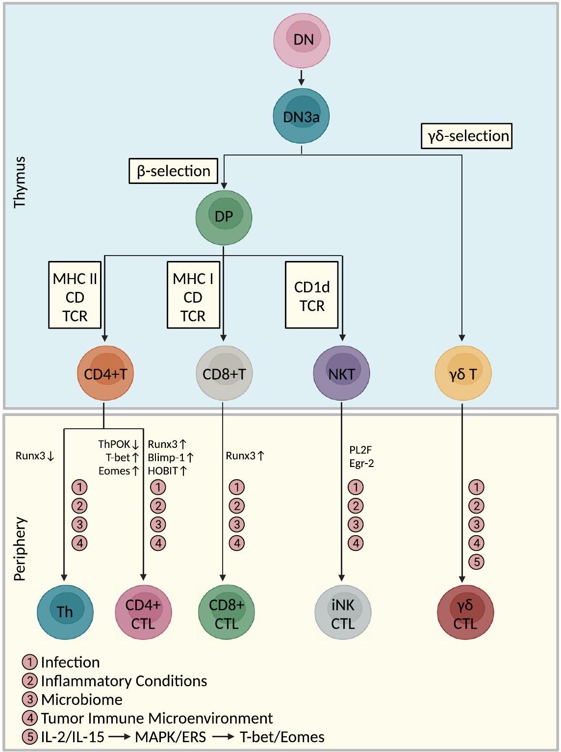
أصل ومسار تمايز خلايا CTLs

تتكاثر خلايا السلف التيموسية في مرحلة CD4(-) CD8(-) السلبية المزدوجة (DN). أولاً، تدخل هذه الخلايا خط الخلايا التائية في مرحلة DN2، وتلي هذه الخطوة إكمال إعادة ترتيب الجينات في مستقبلات الخلايا التائية (TCR). تي سي آر و TCR المواقع في مرحلة DN3 [70]. بعد و الاختيار في مرحلة DN3a، تدخل هذه الخلايا التائية ت و سلالات T، على التوالي. بعد ذلك، ثم تقوم خلايا سلالة T بتقليل تعبير CD25 وزيادة تعبير CD4 وCD8 لتصبح خلايا مزدوجة الإيجابية (DP). تمر خلايا DP بعملية TCR إعادة ترتيب الجينات ومرحلتَي اختيار MHC واختيار CD1d [70، 71]، مما يؤدي إلى ظهور خلايا T من نوع CD4+، وخلايا T من نوع CD8+، وخلايا NKT. معظم تظل خلايا سلالة T خلايا DN ولكنها تقلل من تعبير CD24 عند النضوج. أظهرت الدراسات مزيدًا من تنشيط تمايز خلايا T إلى خلايا CTL تحت تأثير العدوى، والظروف الالتهابية، والميكروبيوم، وبيئة الورم المناعية (TIME)، أو تحفيز بعض مسارات الإشارة المحددة.

مسارات تمايز خلايا CTLs CD4+

يمكن أن تتطور خلايا CTLs CD4+ بشكل أكبر من تحت المجموعات Th0 و Th1 و Th2 و Th17 و Treg [14]. في الوقت الحالي، لم يتم توحيد عوامل النسخ المعنية في التمايز الداخلي لخلايا CTLs CD4+ بشكل كامل و
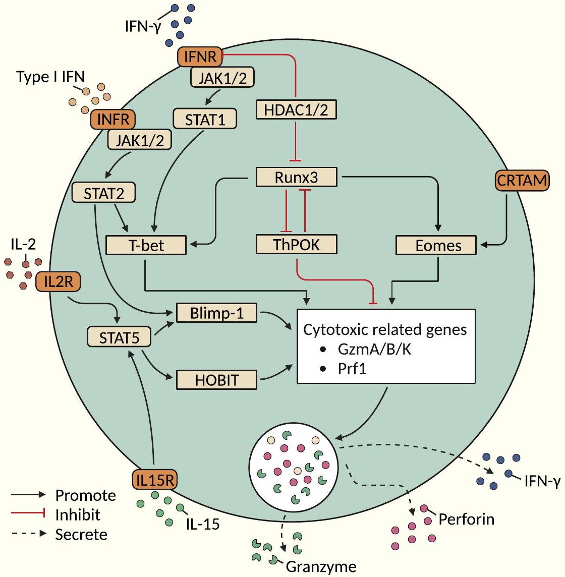
تم توضيحه. قد تكون عوامل النسخ التي تحفز التأثيرات السامة للخلايا في CD8+ CTLs [مثل T-bet، بروتين نضوج الخلايا اللمفاوية B المستحث (Blimp-1)، Eomes، عامل النسخ من عائلة RUNX 3 (Runx3)، عامل تحفيز الخلايا المساعدة POZKruppel (ThPOK)، ونظير Blimp-1 في الخلايا التائية (HOBIT)] متورطة في تمايز CD4+ CTLs، ولكن لا يزال هناك حاجة لمزيد من التحقق. أظهرت الدراسات أن خلايا CTLs CD4+ من خلايا Th1 تمثل الغالبية العظمى من خلايا CTLs CD4+ (التي تفرز بشكل أساسي IFN- TNF- و IL-2) [14]. وفقًا للدراسات الحالية، قد تتضمن عملية تمايز خلايا CTLs CD4+ ثلاث مسارات (الشكل 2): (I) الاعتماد على إشارات TCR كمسار الحدث الابتدائي، (II) الإشارات من خلال مستقبل CRTAM كمسار الحدث الابتدائي، و (III) مسار التعديل التنظيمي الجيني. .

الاعتماد على إشارات TCR كحدث ابتدائي

الغدة الصعترية هي موقع رئيسي لتطور خلايا T، وتهاجر خلايا سلف خلايا T التي تنشأ من نخاع العظام إلى الغدة الصعترية وتتفارق إلى مجموعة متنوعة من تحت مجموعات خلايا T. يمكن أن تتمايز الخلايا التائية إلى خلايا CTLs CD8+، وخلايا Th CD4+، وخلايا NKT. تتحكم نشاط عوامل النسخ الرئيسية في توليد مجموعة متنوعة من أنماط Th استجابة لمجموعة واسعة من الإشارات البيئية. TCR يمكن تمييز الخلايا التائية الشابة إلى خلايا CTLs CD8+، وخلايا Th CD4+، وخلايا NKT. أظهرت الدراسات أن RUNX3/ThPOK وT-bet وEomes وBlimp-1 وHOBIT تلعب أدوارًا مهمة في تنظيم تمايز خلايا CTLs CD4+.
محور عامل النسخ RUNX3/ThPOK تحت التحفيز المناسب (بما في ذلك تحفيز TCR و IL-2/IL-15/نوع I IFN/IFN- يمكن لخلايا CD4+ Th تقليل تنظيم ThPOK لمزيد من التمايز إلى CD4+ CTLs. يتم تنظيم تقليل تنظيم THPOK بواسطة محور كتم الصوت RUNX3-THPOK، مما يؤدي إلى زيادة تنظيم الجينات المرتبطة بالسمية (مثل Eomes وIfng وGzmB وPrf1). وبالتالي، قد يصبح التوازن بين تعبير ThPOK وRUNX3 شرطًا مسبقًا لتحديد ما إذا كانت خلايا CD4+ Th تتمايز إلى سلالة CD4+ CTL. بالإضافة إلى ذلك، IFN- في النهاية، يتوسط تعبير الجزيئات المرتبطة بالسُمية الخلوية من خلال فسفرة ناقل الإشارة ومُنشط النسخ (STAT)-1 (STAT1) وزيادة تعبير ThPOK [59].
عوامل النسخ الأخرى (T-bet، Eomes، Blimp-1، وHOBIT) بالإضافة إلى محور عامل النسخ RUNX3/ThPOK، يمكن أن تشارك عوامل النسخ الأخرى (مثل T-bet، Eomes، Blimp-1، وHOBIT) أيضًا في تنظيم تمايز خلايا CTLs CD4+ [55]. على سبيل المثال، يرتبط IL-2 بـ IL-2R مما يعزز بشكل أكبر
الشكل 1 مسارات تمايز خلايا CTLs ذات الأصول التطويرية التيموسية وفي الدم المحيطي. تم إنشاء هذا الشكل استنادًا إلى الأدوات المقدمة من Biorender.com (تم الوصول إليها في 13/10/2023)
تنظيم التعبير عن Blimp-1/T-bet الناتج عن فسفرة STAT2، والتي في النهاية تتوسط التعبير عن الجزيئات المرتبطة بالسمية الخلوية (مثل IFN- ، غرانزيم ب وبيرفورين) [5، 55، 59، 73، 75، 77، 78]. علاوة على ذلك، في غياب STAT2، يتم تقليل تعبير T-bet وغرانزيم ب [59]. يرتبط IL-2R على سطح Tregs بـ IL-2 ليمنع بشكل تنافسي تمايز CD4+ CTLs [73]. مشابهًا لـ T-bet، يلعب Eomes دورًا رئيسيًا في تحفيز نسخ الجينات المتعلقة بالسُمية في CD4+ CTLs [5، 55]. بالإضافة إلى ذلك، يرتبط IL-15 بـ IL-15R على سطح خلايا CD4+ T لتنشيط نسخ HOBIT التي تحفزها STAT5.
الفوسفوريلation، التي في النهاية تتوسط تعبير الجرانزيم B والبرفورين في خلايا CD28-CD4+ T [79-81].

الإشارة من خلال CRTAM كمسار حدث ابتدائي

بالإضافة إلى المسار المعتمد على RUNX3، الذي يعتمد على إشارات TCR كحدث ابتدائي، يلعب CRTAM دورًا مهمًا في تحفيز تمايز CTLs من نوع CD4+ [14]. ينظم CRTAM مباشرة تعبير Eomes بطريقة مستقلة عن RUNX3 [14]. الأدوار التي تم تحديدها سابقًا لـ T-bet وBlimp-1 وEomes،
الشكل 2 مسارات تمايز خلايا CTLs CD4+. تشمل هذه المسارات بشكل رئيسي I) الاعتماد على إشارات TCR كمسار الحدث الابتدائي، (II) الإشارات من خلال المستقبل CRTAM كمسار الحدث الابتدائي و (III) مسار التعديل التنظيمي الجيني. تم إنشاء هذا الشكل استنادًا إلى الأدوات المقدمة من Biorender.com (تم الوصول إليها في 13/10/2023)
إن تنظيم التعبير عن الجرانزيمات وغيرها من الجزيئات الليزوزومية بواسطة Runx3 وThPOK وHOBIT سيكون متوافقًا مع كل من نماذج تطوير خلايا CD4+ CTL المعتمدة على CRTAM وغير المعتمدة على RUNX3.

مسارات التعديل الوراثي فوق الجيني

بالإضافة إلى شبكات تنظيم عوامل النسخ، تلعب شبكات التعديل الجيني (مثل ميثلة الحمض النووي وتعديل الهيستون) أدوارًا مهمة في تنظيم التعبير الجيني. ومع ذلك، فإن المعرفة الحالية بالتعديلات الجينية في تنظيم خلايا CD4+ CTLs لا تزال محدودة جدًا. يتم التحكم في تعديلات الأسيتيل للبروتين بواسطة أسيتيل ترانسفيراز الهيستون (HAT) وديستيلز الهيستون (HDAC) وتعمل كمنشطات مشتركة ومثبطات [59]. يمكن استقطاب HDAC إلى مواقع الجينات النشطة، وبالتعاون مع HAT، يعمل هذا الديستيلز.
تعمل كمنظم لنسخ الجينات [59]. تنشيط TCR مع IFN- يمكن أن تحفز التحفيز JAK1/2 لمزيد من تنشيط STAT1 [74]. تظهر خلايا CD4+ T التي تفتقر إلى HDAC1 مستويات مرتفعة من STAT1 الفسفوري (p-STAT1) [82]. وبالتالي، يمكن أن تعمل HDAC1 كمنظم سلبي رئيسي لتنشيط STAT1 في خلايا CD4+ T [82]. في وقت لاحق خلال تطوير خلايا T، يؤدي نقص HDAC1 وHDAC2 إلى تمايز خلايا CD4+ Th إلى خلايا CD4+ CTLs من خلال زيادة تعبير RUNX3، ويتجلى هذا التمايز بشكل رئيسي من خلال زيادة تعبير ملف جين CD8 (مثل Cd8a وCd8b1) [83]. بالإضافة إلى ذلك، تمر HDAC1 وHDAC2 وتؤدي في النهاية إلى توليد خلايا CD4+ CTLs [74]. نظرًا لأن STAT1 وSTAT2 قد يتشكلان كهيترديمر [84]، فإن المستويات المرتفعة من STAT1 الفسفوري التي لوحظت في خلايا CD4+ T التي تفتقر إلى HDAC1 وHDAC2 قد تشير إلى تداخل بين مسارات الإشارة المعتمدة على STAT1 وSTAT2 [59].

مسار تمايز خلايا CTLs CD8+

تبدأ عملية توليد خلايا CTLs CD8+ من خلايا الجذع الدموية (HSCs) في نخاع العظام. تنضج HSCs وتتطور إلى سلفيات لمفاوية شائعة (CLPs)، وبعد ذلك، تهاجر CLPs إلى المنطقة تحت البريتوانية في الغدة الصعترية. تؤدي عمليات الاختيار الإيجابي والسلبي في الغدة الصعترية إلى تطوير خلايا T CD4(-)CD8(+) من خلال ارتباط TCR والألفة لجزيئات مشابهة لـ MHCI. أظهرت الدراسات أن مسار إشارة IL-7 يؤثر على تعبير RUNX3 وهو أمر حاسم لتوليد خلايا CTLs CD8+. بالإضافة إلى ذلك، تلعب عوامل النسخ T-bet وEomes أدوارًا مهمة في تمايز ووظيفة خلايا CTLs CD8+. تعمل T-bet وEomes معًا لتحفيز تعبير IFN- ، GzmB، البيرفورين، CXCR3، وCXCR4 في خلايا CD8+ T، التي تؤدي في النهاية إلى توليد السمية في خلايا CD8+ T [55، 89]. يتم استقطاب خلايا CD8+ T الساذجة إلى العقد اللمفاوية المتصرفة (dLN) من خلال إعادة الدوران عبر CCR7 أو كيموكينات مثل CCR4/5. تقوم APCs بمعالجة وتقديم مستضدات الورم، وتهاجر إلى dLN وتقدم المستضدات على سطحها لخلايا CD8+ T الساذجة. تؤدي التفاعلات بين APCs وخلايا CD8+ T إلى تكاثر وتنشيط خلايا CD8+ T إلى CTLs، التي تقلل من CCR7 وتزيد من مستقبلات الكيموكينات مثل BLT1، CXCR3، CCR5، وCX3CR1. ثم تهاجر هذه الخلايا إلى TIME وفي النهاية تؤدي إلى قتل خلايا الورم [90].

مسار التمايز -CTLs

بعد الاختيار في الغدة الصعترية، السلالة متميزة. معظم تظل خلايا سلالة T خلايا DN ولكنها تقلل من تعبير CD24 عند النضوج [70]. لأن تظل الخلايا غير ناضجة في الغدة الصعترية، تعمل الخلايا وفقًا لعوامل الإشارة البيئية في المحيط [27]. عند تحفيز المستضد، تتحول اللمفاويات من خلايا غير ناضجة (CD27+، CD62L+CCR7+، CD45RA+) إلى خلايا ذاكرة مركزية ذات وظيفة تكاثرية ومنخفضة الفعالية (CD27+، CD62L+CCR7+، CD45RA-). عند تحفيزها بمزيد من المستضد (Ag)، تنضج هذه الخلايا أكثر لتصبح خلايا ذاكرة فعالة (CD27-، CD45RA-) وتنتج IFN- أو الجرانيزيم/البيرفورين، مما يؤدي في النهاية إلى تعبير اللمفاويات عن التعبير النهائي المتمايز لـ CD45RA (TEMRA) [30]. تم تمييز مسار النضج هذا من الخلايا الساذجة إلى خلايا TEMRA في TCRV خلايا T، حيث يسبق تنشيط TCR ويقود تدريجياً التعبير عن مستقبلات سامة للخلايا مشتركة مع خلايا NK. أظهرت الدراسات أن IL-2 و IL-15 تحفز التعبير عن CD107a (علامة التفريغ). تساعد خلايا T هذه الخلايا على قتل خلايا الورم، كما أن IL-2 و IL-15 الخارجيين يعززان أيضًا
وظائف المؤثر (خاصة التفريغ/القدرة السامة للخلايا) لـ خلايا T [91]. وقد كشفت التحقيقات الميكانيكية الإضافية أن IL-2/IL-15، من خلال كيناز البروتين المنشط بالميتوجين (MAPK)/كيناز الإشارة خارج الخلوية المنظم مسار (ERK) ، يحفز تعبير T-bet و Eomes ، مما يعزز في النهاية التأثيرات السامة للخلايا الخلايا [91].

مسارات تمايز خلايا iNK-CTLs

على غرار مجموعات خلايا T الكلاسيكية، تتطور خلايا NKT في الغدة الصعترية، ولكن خلايا NKT تت diverge عندما تدخل مرحلة DP. يعتمد تمايز خلايا iNKT على الارتباط بين TCR و CD1d لبدء برنامج تطوير خلايا NKT، والذي يتم تمايزه بشكل أكبر تحت اختيار الاستجابة المبكرة للنمو 2 (Egr-2) وزنك الإصبع لسرطان الدم النخاعي (PLZF). يمكن تقسيم خلايا iNKT إلى مجموعات فرعية مشابهة لخلايا CD4+ Th. على سبيل المثال، تعبر خلايا NKT1 عن T-bet وتفرز بشكل أساسي IFN- بينما تعبر خلايا NKT2 عن بروتين ربط GATA-3 (GATA3) وPLZF وتفرز السيتوكينات من نوع Th2 (مثل IL-4 وIL-13). كما تتميز خلايا iNKT بتعبيرها عن مستقبلات اليتيم المرتبطة بـ RAR- ROR t) وإفراز IL-17 [36]. توجد أيضًا مجموعات فرعية أخرى من خلايا NKT، وتشمل خلايا NKT المنتجة لـ IL-9، وخلايا NKTFH التي تعبر عن B-cell lymphoma 6 (BCL6)، وخلايا NKT10 المنتجة لـ IL-10 [36]. يعتبر T-bet ضروريًا لمرحلة النضج النهائية لخلايا iNKT وينظم مباشرة تنشيط الجينات المرتبطة بالسمية في خلايا iNKT (مثل، البيرفورين، CD178، و IFN- ) [92، 93]. على النقيض من ذلك، فإن خلايا iNKT التي تفتقر إلى T-bet غير قادرة على إنتاج IFN- استجابةً لتحفيز TCR وغير قادرة على إظهار وظائف سامة للخلايا [92، 94].

وظائف الخلايا التائية السامة للخلايا في مناعة الأورام

حالياً، التركيز الرئيسي على وظيفة خلايا CTLs هو تأثيرها القاتل على خلايا الورم (الشكل 3). من ناحية، تقوم خلايا CTLs بتحفيز الموت المبرمج في خلايا الورم بشكل رئيسي من خلال العمل السيتوليتيكي للجرانزيمات/البيرفورين أو المسارات المعتمدة على مستقبلات الموت/الليغاند. وجدت الدراسات الحديثة أن أنظمة البيرفورين/الجرانزيم ومُستقبلات الموت/الليغاند يمكن أن تحفز ليس فقط الموت المبرمج في الخلايا المستهدفة ولكن أيضًا أنواع أخرى من الموت الخلوي المنظم، مثل النخر المبرمج والموت الخلوي الالتهابي [3]. علاوة على ذلك، بالإضافة إلى تعزيز الموت المبرمج من خلال تنشيط الكاسبيز بواسطة الجرانزيمات في نظام البيرفورين/الجرانزيم، فإن البيرفورين الذي يتم وساطته لاحقًا… قد تلعب الزيادة المعتمدة على ROS وعمليات تلف الحمض النووي دورًا مهمًا في تعزيز موت الخلايا [1]. من ناحية أخرى، قد تمارس خلايا CTLs تأثيراتها القاتلة للأورام من خلال التفاعلات مع خلايا المناعة الأخرى في بيئة الورم. في السنوات الأخيرة، اقترحت بعض الدراسات أيضًا أن خلايا CTLs قد تعمل كـ “سيف ذي حدين” في قتل الأورام.
الشكل 3 الوظائف الرئيسية والمسارات للاستجابة المناعية المضادة للأورام التي تتوسطها خلايا CTLs في خلايا الورم تشمل بشكل رئيسي القتل السيتولي المباشر والتفعيل المساعد لخلايا المناعة الأخرى لمزيد من التوسط في قتل الورم. تم إنشاء هذا الشكل استنادًا إلى الأدوات المقدمة من Biorender.com (تم الوصول إليها في 13/10/2023)
العملية وأن هذه الخلايا قد تلعب أيضًا دورًا رئيسيًا في تعزيز تكوين الأورام وتقدمها.

المناعة المضادة للأورام

تعتبر خلايا CTLs CD8+، كأكثر أنواع CTLs تقليدية وكلاسيكية، خلايا T فعالة تتطور من خلايا T CD8+ غير الناضجة المنشطة. بعد التحفيز المستضدي، تبدأ مستقبلات التكامل CD27 وCD28 في إرسال إشارات في خلايا T CD8+. بالإضافة إلى ذلك، مع مساعدة خلايا T CD4+ (تفاعل CD40-CD40L)، تمارس خلايا CTLs CD8+ تأثيراتها على قتل خلايا الورم. حاليًا، تم إثبات أن النشاط المضاد للورم الخاص بخلايا CTLs CD8+ فعال ضد مجموعة متنوعة من أنواع الأورام، مثل الميلانوما، وسرطان الثدي، وسرطان الرئة، وسرطان الكبد (HCC)، والورم الدبقي، وسرطان الدم الحاد والمزمن، واللمفوما. تمارس خلايا CTLs CD8+ تأثيرات قتل الخلايا السيتوليتية بشكل مباشر وتفعيل تكميلي لخلايا المناعة الأخرى لممارسة تأثيرات قتل الورم بشكل أكبر. تلعب الوساطة في قتل خلايا الورم دورًا حاسمًا وقد ارتبطت بتحسين التوقعات في مرضى الأورام. على سبيل المثال، يرتبط التحسن الكبير في التوقعات السريرية للمرضى الذين يعانون من سرطان القولون والمستقيم مع إصلاح غير متطابق (pMMR) بمدى تسلل خلايا T CD4+GzmB+ في مركز
ورم [95]. بالإضافة إلى ذلك، تعتبر خلايا CD4+ CTLs مؤشراً على نتيجة المرضى الذين يعانون من الأورام المعالجة بمثبطات المناعة [54، 96-99]. كما أظهرت الدراسات أن خلايا CD4+ CTLs مهمة في السيطرة على انتشار سرطان الرئة [76]. تقتل خلايا CD4+ T خلايا الميلانوما بطريقة مقيدة بمركب التوافق النسيجي الرئيسي من النوع الثاني (MHCII) بعد العلاج الواضح بخلايا CD4+ T محددة المستضد [16]. خلايا Th9/Th17 التي تم نقلها إلى المضيف بطريقة متسلسلة تحفز قتل الورم عن طريق إطلاق إنزيم غرانزيم B [100، 101]. خلايا CD4+ T التي تعبر عن كيموكين (CXCL)-13 (CXCL13) وجينات سامة للخلايا مرتبطة بزيادة ملحوظة في مدة البقاء الإجمالية (OS) لدى مرضى الميلانوما [102]. يمكن أن تتمايز خلايا CD4+ T الساذجة بشكل أكبر إلى خلايا CD4+ CTLs وتساعد في قتل خلايا الميلانوما في أجسام المضيفين الذين يعانون من نقص اللمفاويات [16]. حالياً، تركز الدراسات على وظيفة -تركزت خلايا CTL على المناعة المضادة للأورام، principalmente من خلال قتلها الخلوي المباشر وتنشيطها المساعد لخلايا المناعة الأخرى لمزيد من قتل الأورام [27]. على سبيل المثال، V الخلايا سامة للغاية ضد الورم العصبي [103]. V الخلايا المعزولة من اللمفاويات المتسللة إلى الورم (TILs) في أورام القولون سامة للخلايا لكل من الخلايا الظهارية الورمية الذاتية والخارجية [103]. بالإضافة إلى ذلك، يرتبط تسلل خلايا CTLs V82 (CD107a+) بنتائج إيجابية.
النتائج السريرية لدى مرضى سرطان المثانة وتسبب زيادة إفراز IFN- و TNF- . تعتبر خلايا iNK-CTLs، كلاعبين مهمين في الوظائف السامة للخلايا، ذات دور لا غنى عنه في تحفيز قتل خلايا الورم وتحسين تشخيص الورم. أظهرت الدراسات وجود ارتباط كبير بين زيادة عدد IFN- -إنتاج iNK-CTLs وزيادة البقاء لفترة طويلة في مرضى سرطان الرئة غير صغير الخلايا (NSCLC) [41]. إن التسلل العالي لـ iNK-CTLs في بيئة الورم (TME) يحسن بشكل كبير من معدل البقاء بدون تكرار لمدة 5 سنوات (5y-RFS) لمرضى سرطان القولون والمستقيم في المرحلة الثالثة [104].

آثار القتل من نوع الليسوجيني المباشر

مسار الجرانيزيم/البيرفورين ومسار مستقبلات الموت المعتمد لهما أهمية كبيرة في تحقيق تأثيرات القتل الخلوية المباشرة [105-107]. بعد الإشارة المحددة من مستقبلات الخلايا التائية (TCR)، تفرز الخلايا التائية السامة للخلايا (CTLs) البيرفورين لتمزيق أغشية الخلايا المستهدفة، وتدخل الجرانيزيم إلى السيتوسول بمساعدة البيرفورين وتنتقل إلى الخلايا المستهدفة لتحفيز الموت المبرمج (الابوبتوز) [108]. تشمل المسارات المعتمدة على مستقبلات الموت في الخلايا التائية السامة للخلايا بشكل رئيسي Fas/FasL وTRAIL/ TRAIL-R [105، 106]. يرتبط FASL المعبر عنه على سطح الخلايا الفعالة بـ FAS على سطح الخلايا المستهدفة وينشط مجال الموت المرتبط بـ Fas (FADD)/كاسبيز 8/بروتين مثبط شبيه بإنزيم تحويل IL-1b الشبيه بـ FADD.
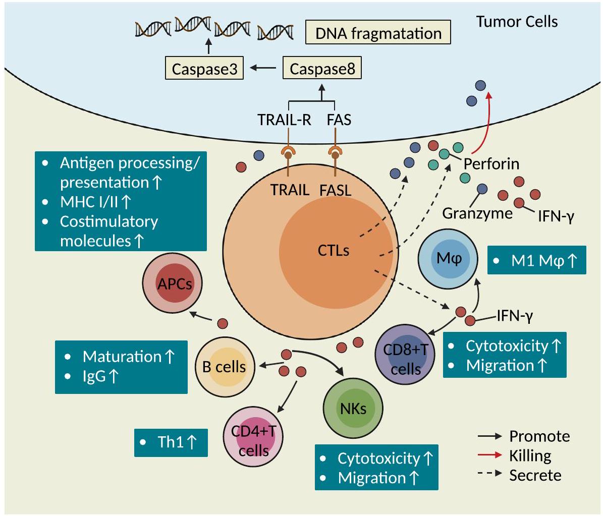
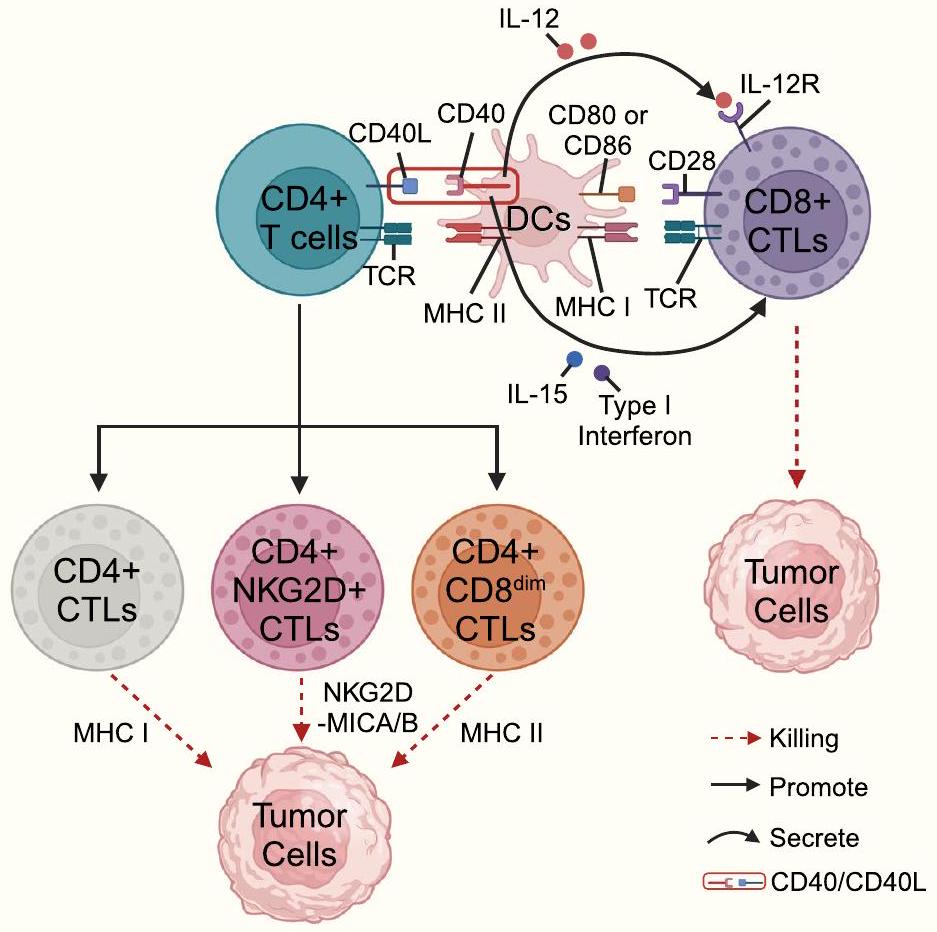
معقد الإشارة المميتة الناتج عن (FLIP) وفي النهاية موت الخلايا المبرمج بواسطة الكاسبيز 3 في الخلايا المستهدفة [18]. يرتبط TRAIL المعبر عنه على سطح خلايا CTLs بـ TRAIL-R على سطح الخلايا المستهدفة ويمكن أن يمارس تأثيرًا قاتلًا على خلايا الورم المقاومة لمسار Fas/FasL [106]. وجدت الدراسات الحديثة أن اختيار مسار الجرانزيم/البيرفورين ومسار مستقبلات الموت يعتمد على شدة الإشارة التحفيزية الخارجية والبيئة الدقيقة المحلية؛ على سبيل المثال، في ظل ظروف تتكون من تركيز عالٍ من مستضد معين وغياب IL-2، تفضل خلايا CTLs CD4+ اعتماد مسار Fas/FasL لقتل الخلايا المستهدفة، وفي ظل ظروف تتكون من تركيز منخفض من المستضد في وجود IL-2، تفضل خلايا CTLs CD4+ استخدام مسار القتل المعتمد على البيرفورين/الجرانزيم [109]. يمكن أن تقتل خلايا CTLs CD4+ خلايا الورم من خلال ثلاثة آليات محتملة (الشكل 4): أولاً، يمكن أن تتعرف خلايا CTLs CD4+ على المستضدات المتماثلة المعروضة بواسطة APCs وتفرز حبيبات لقتل الخلايا المستهدفة بطريقة تعتمد على MHC الفئة II؛ ثانيًا، يمكن أن تقوم خلايا CTLs CD4+ بزيادة تعبير NKG2D لقتل خلايا الورم بطريقة تعتمد على مسار NKG2D-MICA/B؛ ثالثًا، يمكن أن تقتل خلايا CTLs CD4+CD8dim التي تعبر عن مستويات منخفضة من CD8 (CD8dim) خلايا الورم بطريقة تعتمد على MHC الفئة I [55]. يمكن أن تبدأ خلايا T CD8+ تأثيرات سامة لاحقة عند التحفيز بالمستضد.
الشكل 4 التفاعلات الرئيسية بين مستقبلات الخلايا السطحية والعوامل المرتبطة بها بين خلايا CD4+CTLs، خلايا CD4+T، خلايا DCs، خلايا CD8+CTLs، وخلايا الورم. تم إنشاء هذا الشكل استنادًا إلى الأدوات المقدمة من Biorender.com (تم الوصول إليها في 21/02/2024)
بالإضافة إلى ذلك، يمكن لخلايا CD4+ Th تنشيط برامج التعبير الجيني في خلايا CD8+ CTLs وتعزيز وظيفتها. تقدم خلايا DC المستضدات إلى خلايا CD4+ Th في سياق MHC من الفئة II، مما يؤدي إلى زيادة تعبير CD40L على سطح خلايا CD4+ Th. الربط اللاحق لـ CD40L على خلايا CD4+ Th وCD40 على cDC1 يعزز بشكل أكبر قدرة تقديم المستضدات لـ cDC1 (مثل جزيئات MHC من الفئة I) بالإضافة إلى تعبير الجزيئات المساعدة (مثل CD80 و/أو CD86 وCD70) والسيتوكينات (مثل الإنترفيرونات من النوع I، IL-12، وIL-15). ثم تقوم cDC1 بتوسط السمية الخلوية لخلايا CD8+ CTLs ضد الخلايا المستهدفة بطريقة تعتمد على MHC من الفئة I (الشكل 4) [110]. مؤخرًا، تم العثور على خلايا DC من نوع CD5+ لتحفيز استجابات مضادة لـ CD4+ Th وCD8+ CTL ضد الأورام وتعزيز الاستجابات للعلاج المناعي [111]. V و V تقوم خلايا T التي تم استقطابها إلى البيئة المجهرية للورم بممارسة تأثيرات قتل الورم من خلال البيرفورين/الجرانزيم، وIFN- TNF- -, مستقبل الموت ligand-، وسمية الوسائط التي تتوسطها مسار ADCC [27،34]. يمكن استهداف خلايا T للأورام عبر ADCC، حيث يتم التعبير عن CD16 (مستقبل Fcy III) على تتفاعل الخلايا مع خلايا الهدف من خلال جزء Fc من الأجسام المضادة لتسهيل قتل الورم [112، 113]. يمكن أن تمارس iNK-CTLs السمية الخلوية من خلال مسار Fas/FasL، وTNF- مسار، ومسارات الجرانيزيم والبيرفورين في خلايا الورم الحاملة لجزيئات CD1d [37، 114]. يؤدي مسار مضاد الورم Fas/FasL في خلايا iNK-CTLs إلى الاستماتة من خلال عمل الخلايا الفعالة لـ FasL على سطح خلايا iNK-CTLs مع الخلايا التي تعبر عن جزيئات Fas، مما يدمر خلايا الورم. تأثير القتل لـ TNF- يتطلب الاتصال المطول بين خلايا المؤثر وخلايا الهدف لتعزيز نشاط بروتين البيرفورين، مما يؤدي إلى قتل فعال لمعظم خلايا الورم. أظهرت الدراسات أن مساهمة البيرفورين/الجرانزيم أكثر أهمية من مساهمة فاس/فاس إل في القضاء على خلايا الورم [115]. بالمثل، تقوم خلايا iNK-CTLs المنشطة بواسطة GalCer بممارسة نشاط سمي عبر البيرفورين/الجرانزيم [37].

تنشيط مساعد للخلايا المناعية الأخرى

تقوم خلايا CTL أيضًا بتحفيز استجابة مناعية مضادة للأورام من خلال إفراز عوامل التهابية (مثل IFN ) وتعزيز وظيفة خلايا المناعة مثل خلايا CTLs CD8+، وخلايا B، والبلعميات، وخلايا DCs، وخلايا NKs، وخلايا APCs [116].
البلاعم والخلايا المقدمة للمستضدات تميل البلاعم أكثر إلى التحول إلى بلاعم من النوع M1 التي تحفزها IFN- تفرزها خلايا CTLs وتشارك في استجابات محددة لمستضدات Th1 [117، 118]. تعتبر البلعميات من النوع M1 محدثة للالتهابات وتمارس وظائف تقديم المستضدات، مما يعزز السمية الخلوية في خلايا CTLs CD8+. IFN- يمكن أن تزيد المواد التي تنتجها خلايا CTLs من تعبير TAP-1 و TAP-2، مما يمكن أن يعزز المزيد من
زيادة تنظيم وظائف معالجة المستضدات وعرضها في خلايا APCs. بالمقابل، IFN- يمكن أن يزيد أيضًا من تعبير بروتينات MHC من الفئة الأولى و MHC من الفئة الثانية [116]. بالإضافة إلى ذلك، فإن نقل IFN- تساهم مسارات الإشارة في زيادة تعبير الجزيئات المساعدة على السطح الخلوي لخلايا APCs، مما يعزز مشاركتها في المناعة الخلوية [116]. على سبيل المثال، IFN- الذي تفرزه خلايا iNK-CTLs يعزز تنظيم الجزيئات المساعدة للجهاز المناعي وجزيئات MHC من الفئة الثانية بواسطة خلايا DCs، ومن ثم، IFN- يحفز إنتاج IL-12 بطريقة تعتمد على CD40/CD40L [116-118]. يؤدي إفراز IL-12 المستمر بواسطة DCs الناضجة إلى تحفيز خلايا iNK-CTLs لزيادة تعبير IL-12R وبالتالي يعزز حلقة تغذية راجعة إيجابية بين iNK-CTLs وDCs [2]. الخلايا المنشطة يمكن لخلايا T تعزيز نضوج خلايا DC عن طريق إفراز السيتوكينات (مثل IFN- و TNF- ) [34]. بالإضافة إلى ذلك، IFN- و TNF- يفرز بواسطة V V يمكن أن تعزز خلايا T خلايا DCs لزيادة تنظيم CCR7 وتسهيل هجرة خلايا DCs إلى الأنسجة اللمفاوية لتنشيطها. خلايا T وبالتالي تبدأ الاستجابة المناعية [34، 119]. IFN- يمكن أن يساعد ذلك خلايا DC في زيادة تعبير جزيئات MHC وجزيئات التحفيز المساعد، مما قد يعزز وظائفها في معالجة المستضدات وعرضها. جميع الآليات المذكورة أعلاه تسهل الإزالة المناعية لخلايا الورم.
خلايا IFN- الذي تنتجه خلايا CTLs مهم لتعزيز تكاثر خلايا B وتنظيم تغيير فئة الأجسام المضادة [12، 34، 116، 117، 120، 121]. IFN- يرتبط بمستقبلات خلايا B وإشارات تنشيط CD40 لتحفيز تعبير BCL-6. IFN- و IL-12 يعززان بشكل تآزري تحويل فئة الأجسام المضادة من IgM إلى IgG2a (وهي جسم مضاد محدد ذو affinity أعلى) وبالتالي يسهلان معالجة وتقديم المستضد بواسطة خلايا المناعة الأخرى [117]. على سبيل المثال، تظهر خلايا T CXCR5+CD8+ [122] ذات الوظائف السامة للخلايا وظائف مساعدة لخلايا B، والتي تشمل بشكل رئيسي تحفيز تكاثر خلايا B، وتحويل فئة الأجسام المضادة/مستقبلات خلايا B (BCR) وإنتاج الأجسام المضادة [12،120،121]، وتعزيز تفاعلات CD4+ Th-B-cell [12]. بالإضافة إلى ذلك، وجدت دراسة أخرى أن خلايا T CXCR5+ICOS+CD8+ تظهر تسللًا كبيرًا في العقد اللمفاوية الورمية (LNs) للمرضى المصابين بسرطان هودجكين (HL) ويمكن أن تزيد من تعبير IL-2 وIL-4 وIL-21 [123]، مما يعزز تكاثر خلايا B وإنتاج الأجسام المضادة. بالإضافة إلى ذلك، IFN- يفرز بواسطة تلعب الخلايا دورًا مهمًا في تنظيم نضوج خلايا B وإنتاج الأجسام المضادة المناعية [34، 117]. يمكن أن تشكل خلايا iNK-CTLs تفاعلًا ثنائي الاتجاه مع خلايا B: من جهة، يمكن لخلايا B تقديم المستضدات الدهنية إلى خلايا NKT من النوع الأول عبر CD1d [124]، ومن جهة أخرى، فإن خلايا iNK-CTLs
يمكن أن ترخص خلايا B لبدء وتنشيط الاستجابة المضادة للورم بفعالية [2].
تحفيز خلايا CTLs CD8+ بواسطة IFN- الإشارات تزيد من تعبير IL-2R وT-bet وgranzyme، والتي تعمل كوسائط مهمة لقتل الورم بواسطة خلايا CTLs CD8+ [116]. بالإضافة إلى ذلك، IFN- يتوسط هجرة خلايا CTLs CD8+ وNKs إلى البيئة المجهرية للورم من خلال تعزيز تعبير الكيموكينات (مثل CXCL9 وCXCL10 وCXCR3) [125]، مما يعزز في النهاية التأثير السمي لخلايا CTLs CD8+ ويعيق تكوين الأورام وتقدمها [117]. IFN- يعمل على الخلايا القاتلة الطبيعية ويعزز قتل خلايا الورم بواسطة الخلايا القاتلة الطبيعية من خلال TRAIL، في حين يمكن تعزيز تعبير TRAIL بواسطة IFN- -المستحث IRF1 [37، 93، 117]. بالإضافة إلى ذلك، استجابةً لإعادة تعرض الجسم للمستضد وتنشيط إفراز السيتوكينات، تطلق الذاكرة النوعية للمستضدات من خلايا CTLs مجموعة متنوعة من السيتوكينات والكيموكينات، مثل IFN- ، عامل نخر الورم ألفا (CCL3)، وبروتين جذب المونوسيت-1 (MCP1)، الذي يساهم في استقطاب المونوسيت والخلايا القاتلة الطبيعية (NK) ويزيد من إفراز CXCL9 وCXCL10 لاستقطاب المزيد من الخلايا القاتلة الطبيعية لتنشيط خلايا B والخلايا التغصنية (DCs) [34، 117]. بالإضافة إلى ذلك، IFN- الذي تفرزه خلايا CTLs يلعب دورًا مهمًا في توجيه تمايز خلايا Th1، مما يعزز التأثيرات المضادة للورم في البيئة المجهرية للورم.

تعزيز تكوين الأورام والتقدم

لقد أظهرت مجموعات خلايا CD4 T المثبطة للمناعة (مثل Tr1) أنها تمتلك وظائف سامة للخلايا ولكن قد تلعب دورًا في تعزيز تكوين الأورام وتقدمها. على سبيل المثال، يمكن لخلايا Tr1 السامة أن تتصدى للاستجابات المناعية للأورام من خلال تأثيرها القاتل على خلايا APC. ترتبط خلايا Tr1 السامة GZMK+Eomes+ بتقدم الأورام في سرطان القولون والمستقيم، وسرطان الرئة غير صغير الخلايا، والأورام التي تتطور فيها نقائل كبدية. بالإضافة إلى ذلك، قد يعزز IL10 المفرز من خلايا Tr1 تحول البلعميات إلى بلعميات من النوع M2 ويعيق نضوج خلايا DCs للعب دور إضافي في تعزيز نمو الأورام.

تحسين وظيفة السمية الخلوية للخلايا التائية السامة

تقوم الخلايا التائية السامة للخلايا (CTLs) بتحفيز مجموعة من أنواع الأضرار المختلفة في الأورام، بما في ذلك النخر، والبرم، والنخر البرمي، والاحتراق الخلوي [132]. ومع ذلك، تبدأ خلايا الورم سلسلة من المسارات لتقليل السمية الخلوية للخلايا التائية السامة، مما يساعد خلايا الورم على التهرب من التعرف عليها وقتلها بواسطة الجهاز المناعي [132]. لذلك، فإن فهم كيفية تنظيم خلايا الورم لسمية الخلايا التائية السامة يوفر أساسًا نظريًا لـ
تحسين السمية الخلوية للخلايا التائية السامة (CTLs) وتعزيز دورها في المناعة المضادة للأورام. أظهرت الدراسات أن السمية الخلوية للخلايا التائية السامة يمكن تحسينها أو تعزيزها من خلال تعديل مستوى تعبير السيتوكينات، وتقليل نسبة تسرب بعض الخلايا المناعية المحددة، وتعديل مستوى تعبير بعض الجزيئات في بيئة الورم، أو تغيير بعض المسارات الأيضية في الخلايا التائية السامة.

CTLs CD4+

وجد Xu وآخرون أن anti-PD-1-IL-15m يحسن وظيفة خلايا T المتسللة إلى الورم والمناعة المضادة للورم، وأن anti-PD-1-IL-15m يعزز القدرة التكاثرية والسمية الخلوية لخلايا CD8+ TILs وCD4+ TILs، لكن الآلية الجزيئية الأساسية لم تتضح بعد [133]. IFN- يزيد من تعبير MHC II ويعزز التأثير السمي للخلايا التائية CD4+ CTLs على خلايا الورم [134]. إن حجب HLA-G/CD85j يزيد من النشاط السيتوليتي للخلايا التائية CD4+ CTLs لتحسين الاستجابات المناعية المضادة للورم [135، 136]. تستخدم الخلايا التائية التنظيمية IL-2 للحد من المناعة الخلوية المعتمدة على الخلايا التائية، بينما يدفع IL-2 الداخلي زيادة تعبير عامل النسخ Blimp-1 داخل خلايا CD4+ Th لتعزيز تعبير الجرنازيم B، وقد تتنافس الخلايا التائية التنظيمية مع IL-2 للسيطرة سلبًا على هذه العملية [73]. يحفز تحفيز CD137 زيادة تعبير علامات برنامج السمية (Eomes/Granzyme B) في الخلايا التائية التنظيمية مع الحفاظ على خصائص Foxp3، وتعيد معالجة CD137 agonist برمجة الخلايا التائية التنظيمية إلى CD4+ CTLs [137].

خلايا CTLs CD8+

في نماذج الفئران، يعزز الجمع بين تحفيز OX40 والمسار المثبط PD-1 التعبير المشترك عن عدة مستقبلات خلايا NK (مثل NKG2A وNKG2D وKLRG1) ومستقبلات الكيموكينات بواسطة خلايا CD4+ T وخلايا CD8+ T، التي لديها قدرة عالية على التكاثر وإظهار السمية الخلوية. قد تساهم خلايا CD4+ T المفعلة بواسطة OX40 أيضًا في توسيع وتمايز خلايا CD8+ T. في نموذج الفأر، يعزز CCL21+ICAM1 السمية الخلوية لخلايا CD8+ T، كما يتضح بشكل رئيسي من زيادة كبيرة في مستويات الجرanzيم B. بالإضافة إلى ذلك، يعزز فيروس الهربس البسيط العلاجي 1 (oHSV) الذي يحمل CD40L نضوج خلايا DCs في البيئة المجهرية للورم، ويعزز تمايز Th1، ويزيد من السمية الخلوية لخلايا CD8+ CTLs. في B-CLL، تزيد خلايا CD4+ Th المفعلة من تعبير miR-181b في B-CLL عبر إشارة CD40-CD40L، وتؤدي إلى تقليل تعبير IL-10، وتعزز المزيد من السمية الخلوية لخلايا CD8+ CTLs. في نموذج ميلانوما الفأر، تؤدي المعالجة باستخدام مزيج من حجب CD47 وCTLA4 مع العلاج الإشعاعي (RT) إلى زيادة كبيرة في خلايا CD8+ CTLs في أورام الفئران. حجب البلعميات المرتبطة بالورم (TAM)
تقلل مستقبلات النفايات MARCO و IL37R من عدد خلايا Tregs وتستعيد السمية الخلوية والقدرة المضادة للورم لخلايا NK و CD8+ CTLs. يزيد Clec9A على cDC1 من التأثير السمي لخلايا CD8+ CTLs. يمكن أن يعزز تحسين استقلاب الأسيتات في خلايا CD8+ CTLs فعالية هذه الخلايا. أظهرت الدراسات أن LSD1 يشكل مجمعات نووية مع Eomes من خلايا CD8+ CTLs من مرضى الميلانوما وسرطان الثدي المقاوم للعلاج المناعي، مما يؤدي في النهاية إلى تعطيل خلايا CD8+ CTLs، واستهداف فسفرة مسار LSD1 يمكن أن يزيد من السمية الخلوية لخلايا CD8+ CTLs. تظهر خلايا CD8+ CTLs المنشطة زيادة في مسار التحلل السكري وتتطلب إشارات CD28 المساعدة لإطالة مدة زيادة التحلل السكري؛ في نموذج فأر بدين لسرطان الثدي، يزيد تقليل STAT3 في خلايا CD8+ CTLs أو العلاج بمثبطات أكسدة الأحماض الدهنية من كل من التحلل السكري والوظيفة السامة لخلايا CD8+ CTLs (بما في ذلك IFN- ، غرانزيم ب و CD107a) وبالتالي يمنع تطور الأورام الثديية [148].

-CTLs و iNKT-CTLs

V تتكيف الخلايا مع وقت نقص الأكسجين، وقد أظهرت الدراسات في المختبر أن زراعة خلايا V81 T تحت ظروف نقص الأكسجين تعزز من سميتها الخلوية. IL-2/IL-21 تعزز بشكل كبير من تكاثر ووظيفة السمية الخلوية لـ تمنع خلايا T [150،151]. إن حجب TIM-3 يزيد من تأثير القتل لـ الخلايا على خلايا سرطان القولون من خلال تنشيط مسار ERK1/2 وزيادة تعبير البيرفورين والجرانزيم B [152]. بالإضافة إلى ذلك، فإن تنشيط BTN3A/CD277 يعزز التنشيط ويزيد من السمية الخلوية لـ تقلص عدد خلايا T [153]. يتم تقليل عدد خلايا iNKT-CTLs ووظيفتها في العديد من أنواع السرطان، وقد تتضمن الأسباب الكامنة وراء ذلك فقدان/خفض تعبير CD1d، وفقدان -الميكروغلوبيولين، أو نقص المستضدات المنشطة [104]. ومع ذلك، تظل الدراسات حول زيادة السمية الخلوية لخلايا iNKT-CTLs محدودة جداً.

أسئلة مفتوحة

يجب إعادة التفكير في تعريف خلايا CTL.

لقد أنشأت دراسات مختلفة معايير مختلفة لتعريف خلايا CTLs. في الماضي، كانت خلايا CTLs تُعتبر مجموعة من خلايا التأثير التي تمايزت من خلايا CD8+ T الأولية بعد التنشيط ومارست تأثيرات قتل مباشرة على الخلايا المستهدفة. في السنوات الأخيرة، مع تطور تسلسل الجينوم عالي الإنتاجية، تم تحديد المزيد من العلامات الجزيئية السامة بناءً على علامات سطح الخلية. حاليًا، لا يقتصر تعريف خلايا CTLs على خلايا CD8+ T ذات السمية فقط، بل يشمل أيضًا خلايا CD4+ T، وخلايا NKT و خلايا T التي يمكن أن تظهر وظائف سامة للخلايا. بالإضافة إلى ذلك، لا
تم إنشاء مؤشرات حيوية موحدة ومعيارية لتحديد الخلايا التائية السامة. من ناحية، بعض الخلايا التائية التي تعبر عن جزيئات مرتبطة بالوظيفة السامة لا تظهر بالضرورة وظيفة سامة. على سبيل المثال، خلايا CD8+ التائية التي تعبر عن GzmK وGzmB تظهر فقط قدرة سامة منخفضة جداً ولا تمارس سمية كافية لقتل الخلايا المستهدفة. من ناحية أخرى، لم يتم التعرف بعد بشكل متسق على علامات السمية مثل جزيئات التفريغ السام، والجينات المشفرة للجرانزيم والبرفورين، وعوامل النسخ المرتبطة بتمايز السمية، والعلامات المرتبطة بالإشارات الخلوية، وجزيئات مستقبلات سطح الخلايا القاتلة الطبيعية، وCRTAM، وعوامل النسخ (Eomes وRUNX3) من خلال دراسات مختلفة. لذلك، هناك حاجة إلى العديد من الدراسات المستقبلية لإثبات ما إذا كانت العلامات المحددة يمكن أن تصبح المعيار الذهبي لتحديد الخلايا التائية السامة.

يجب استكشاف الأنماط الفرعية الوظيفية لخلايا CTLs في أنواع خلايا T المختلفة بشكل أكبر

مع الاستخدام الواسع لتسلسل الخلايا المفردة، تم تحديد عدد متزايد من الأنماط الفرعية للخلايا التائية السامة. على سبيل المثال، حددت عدة دراسات تعتمد على تسلسل RNA للخلايا المفردة وجود الخلايا السامة -CTLs في أنسجة الورم [26، 30]، ومن خلال تسلسل RNA أحادي الخلية، حدد الباحثون وجود V السامة للخلايا تحتوي مجموعات فرعية من خلايا T في كل من سرطان المريء وسرطان القولون والمستقيم [26]. وجدت دراسة أخرى تعتمد على تسلسل RNA أحادي الخلية خلايا CTLs CD4+ التي تعبر عن Gzmb وNkg7 في سرطانات المثانة والكبد [54]. تم تحديد خلايا CTLs CD4+ التي تعبر عن جينات مرتبطة بخلايا NK في PBMCs من خلال تسلسل RNA أحادي الخلية [53]. ومع ذلك، لم يتم توضيح ما إذا كانت الخلايا السامة للخلايا موجودة أيضًا في خلايا T الأخرى. بالإضافة إلى ذلك، لا تزال القضايا التالية غير معالجة في مجال خلايا CTLs: 1) ما إذا كان يمكن تصنيف خلايا CTLs المختلفة إلى أنواع فرعية مختلفة و 2) تحتاج العلامات الحيوية، والتحديد الوظيفي، والتحقق من صحة الأنواع الفرعية المختلفة من خلايا CTLs إلى تحسينات إضافية من خلال العديد من الدراسات.

الجزيئات الرئيسية المعنية في تمايز أنواع مختلفة من خلايا CTL مثيرة للجدل

حالياً، لم يتم توحيد الأهداف الرئيسية المتعلقة بمسارات التمايز ونقاط التمايز لخلايا CTLs بشكل كامل. من ناحية، لم يتم توضيح مسارات التمايز المتعلقة بخلايا CTLs من نوع CD4+ بشكل كامل، وهي مقسمة حالياً إلى (I) إشارة TCR كمسار الحدث الابتدائي، (II) الإشارة من خلال مستقبل CRTAM كمسار الحدث الابتدائي، و(III) مسار التعديل التنظيمي الجيني. ومع ذلك، لا يزال وجود مسارات أخرى قد تتوسط تمايز خلايا CD4+ CTLs غير واضح. بالإضافة إلى ذلك، فإن فهمنا لـ
دور التعديلات التنظيمية الجينية غير الوراثية في وساطة تمايز خلايا CTLs من نوع CD4+ لا يزال محدودًا جدًا. من ناحية أخرى، فإن مسارات تمايز خلايا NK-CTL و V -CTL في CTLs CD8+ تظل المسارات التقليدية الأكثر شيوعًا للتمايز، بما في ذلك تنظيم عوامل النسخ (مثل T-bet وEomes وEgr-2 وPLZF) والتحفيز المستضدي المحيطي. إن تحديد الجزيئات الرئيسية التي تنظم تمايز CTL مفيد لتنظيم وتدخل تمايز CTL، وما إذا كانت هناك أدوية مستهدفة لهذه الجزيئات الرئيسية لا يزال غير واضح. هناك حاجة إلى مزيد من الدراسات ما قبل السريرية لاستكشاف هذه القضية بشكل أعمق في المستقبل.

لا يزال التوصيف المتعدد الأوميات لأنماط مختلفة من خلايا T السامة للخلايا غير واضح

قد تحتوي الأنواع الفرعية المختلفة من خلايا CTLs على ميزات متعددة الأوميات مختلفة (مثل البروتيوميات، والميتابولوميات، والترانسكريبتوميات، والجينوميات، والإبيجينوميات). استنادًا إلى الميتابولوميات، يمكن أن يؤدي تنشيط مسار الأيض الخاص بالأسيتات إلى تعزيز الوظيفة السامة للخلايا الأقوى لخلايا CD8+ CTLs [145]. يسهل تنشيط المسار الجليكولي تنشيط خلايا CD8+ CTLs لوظائف قتل الخلايا اللاحقة [148]. يمكن أن يعزز البيئة الهايبوكسية الخلايا لتعزيز وظيفتها السامة للخلايا بشكل أكبر [149]. استنادًا إلى علم التعبير الجيني، يمكن أن يعزز تعبير بعض السيتوكينات (مثل IL-2/IL-21) بشكل كبير من تكاثر ووظيفة السمية للخلايا الخلايا [150،151]. IL-2 الداخلي يدفع زيادة التعبير عن عامل النسخ Blimp-1 داخل خلايا CD4+ Th، مما يعزز بدوره التعبير عن GzmB وبالتالي يدفع تمايز خلايا CD4+ Th إلى خلايا CD4+ CTLs [73]. CD137 يحفز زيادة في التعبير عن الجزيئات السامة في Tregs مع الحفاظ على خصائص Foxp3 [137]. ومع ذلك، لا يزال هناك حاجة إلى مزيد من البحث لاكتشاف ما إذا كانت الأنماط الفرعية الأخرى من CTLs قد تمتلك ملفات متعددة الأوميات مختلفة.
تنظيم وإدارة الوظائف السامة للخلايا التائية السامة المستندة إلى تحليل متعدد الأوميات يعظم من تأثيرات قتل الورم للخلايا التائية السامة. تعزز علاج المحفز CD137 تحويل الخلايا التائية التنظيمية إلى خلايا تائية سامة CD4+ [137]. يمكن أن يؤدي زيادة تنظيم مسار الأيض الأسيتاتي إلى تعزيز فعالية خلايا CD8+ التائية السامة [145]. استهداف مسار LSD1 الفسفوري يزيد من السمية للخلايا التائية السامة CD8+ [147]، ويمكن أن يؤدي تثبيط أكسدة الأحماض الدهنية إلى زيادة التحلل السكري لتعزيز الوظيفة السامة لخلايا CD8+ التائية السامة [148]. لذلك، تظل الاستراتيجيات لإدارة وتنظيم الوظيفة السامة للخلايا التائية السامة استنادًا إلى هذه الميزات متعددة الأوميات اتجاهًا مهمًا للبحث المستقبلي.

العلاقة بين الميكروبات (داخل الورم أو المعوية) وخلايا T السامة للخلايا غير واضحة

لقد حددت الدراسات الكائنات الدقيقة التي قد تؤثر بشكل مهم على وظيفة خلايا CTLs. فيما يتعلق بالكائنات الدقيقة داخل الورم، يمكن أن يقوم Talimogene laherparepvec (T-VEC) [وهو نوع معدل وراثيًا من فيروس الهربس البسيط من النوع الأول] بتسهيل تجنيد خلايا CD8+ CTLs إلى البيئة المجهرية للورم من خلال تعديل إفراز الإنترفيرونات من النوع الأول والكيماويات الجاذبة (مثل CXCL9 وCXCL10) وبالتالي تحفيز تأثيرات قتل الورم السامة [154]. بالإضافة إلى ذلك، ترتبط بعض الكائنات الدقيقة الفيروسية داخل الورم بزيادة تسلل خلايا NKT وتحسين كبير في تشخيص مرضى الأورام [155]. في الميلانوما، ترتبط أنواع Lactobacillus بشكل إيجابي بوفرة خلايا CD8+ CTLs، ويمكن أن يزداد تسلل خلايا CD8+ CTLs بشكل تدريجي من خلال زيادة وفرة Chlamydia trachomatis داخل الورم [154]. يمكن أن تؤثر أنواع Clostridium داخل الورم والمواد الأيضية المرتبطة بها على تأثيرات قتل الورم من خلال زيادة مستوى الكاسبيز 3 وتنشيط خلايا CD8+ CTLs [156]. لقد حاولت عدة دراسات توضيح تأثيرات الكائنات الدقيقة داخل الورم على خلايا CTLs، لكن هذه الدراسات المحدودة لا يمكنها توضيح تأثيرات الكائنات الدقيقة داخل الورم على خلايا CTLs بشكل كامل. لذلك، لا تزال هناك حاجة إلى العديد من الدراسات لاستكشاف دور الكائنات الدقيقة داخل الورم على خلايا CTLs في المستقبل.
ومع ذلك، فإن فهم تأثير الميكروبات المعوية على إنتاج خلايا CTL محدود للغاية. قد تؤثر الميكروبات المعوية على نسبة خلايا CTL CD8+ التي تتسلل إلى الميلانوما الجلدية، وLachnoclostridium مرتبط بشكل إيجابي بتعبير خلايا CTL CD8+ المتسللة والكيموكينات CXCL9 وCXCL10 وCCL5 في أنسجة الميلانوما الجلدية. بالإضافة إلى ذلك، أظهرت الدراسات أن الميكروبات داخل الورم والميكروبات المعوية تلعب أدوارًا مترابطة وتفاعلية، وبالتالي، يجب أن تُعطى المزيد من الاهتمام في المستقبل لتأثيرات الميكروبات المعوية على خلايا CTL.
في الوقت الحاضر، لا يزال عدد الدراسات حول تأثير الكائنات الدقيقة على آلية مكافحة الأورام للخلايا التائية السامة للخلايا (CTLs) محدودًا جدًا. استنادًا إلى الدراسات المذكورة أعلاه، وجدنا أن آلية قتل الأورام بواسطة الكائنات الدقيقة على CTLs تتضمن بشكل رئيسي تنظيم إفراز السيتوكينات وتنظيم الكيمياء الخلوية. ومع ذلك، فإن هذه الآليات ليست كافية لتوضيح تأثير الميكروبات على آلية مكافحة الأورام لـ CTLs بشكل كامل. لذلك، لا يزال البحث في هذا الاتجاه بحاجة إلى تعزيز وتوطيد الاستنتاجات. بالإضافة إلى تأثيرات الكائنات الدقيقة على CTLs، لا تزال هناك خلايا مناعية أخرى وخلايا دعامية موجودة في بيئة الورم. لم يتم دراسة التفاعل بين الخلايا المناعية والخلايا الدعامية، التي تعد مكونات مهمة في بيئة الورم، والميكروبات بشكل منهجي.
لذلك، لا يزال يتعين معالجة هذه القضية وأن تكون محور الدراسات المستقبلية.

لم يتم بعد توضيح أنماط شيخوخة أنواع مختلفة من خلايا CTL واستراتيجيات عكس عملية شيخوخة خلايا CTL.

تقلل الشيخوخة المناعية من قدرة الجسم على المراقبة المناعية وإزالة المناعة، مما يؤدي إلى استجابة مناعية محدودة وتكوين الأورام. تشير الشيخوخة الخلوية إلى حالة توقف دائم لدورة الخلية حيث تفقد الخلايا قدرتها على الانقسام، وغالبًا ما تكون هذه الظاهرة مصحوبة بزيادة تعبير السيتوكينات وزيادة إفراز الخلايا. يمكن أن تحدث الشيخوخة الخلوية في جميع مراحل النمو والتطور وهي آلية مهمة للحفاظ على توازن الأنسجة ومنع انتشار الخلايا التالفة. يمكن أن تؤثر الشيخوخة الخلوية على قابلية تكيف الخلايا المناعية وفي النهاية تؤثر على نتائج علاج السرطان. تفتقر خلايا T الشيخوخة، كخلايا ذاكرة/فعالة متمايزة في مرحلة متأخرة، إلى تعبير CD28 ولكنها تعبر عن CD57 ومستقبلات تنظيمية. بالإضافة إلى ذلك، تعبر خلايا T الشيخوخة عن CD45RA ولكن ليس CD45RO وتكون في حالة توقف دورة الخلية. أظهرت الدراسات التي أجريت في السنوات الأخيرة أن نمط الشيخوخة موجود أيضًا في خلايا CTL الموجودة في بيئة الورم. كشف شوساكو وزملاؤه أن خلايا CTL التي تقدم معززات محسنة وراثيًا ومثبطات تعكس نمط الشيخوخة لخلايا CTL. ومع ذلك، فإن القليل من الدراسات حاولت استكشاف الآليات التي تتوسط حالات الشيخوخة وأنماط الشيخوخة لخلايا CTL. لذلك، يبقى نمط الشيخوخة القائم على خلايا CTL اتجاهًا مهمًا لمناعة مضادة للأورام في المستقبل. بالإضافة إلى ذلك، لا تزال هناك المزيد من الأسئلة غير المجابة بشأن الشيخوخة لخلايا CTL. أولاً، لا يزال التمييز بين الأنماط الفرعية المختلفة فيما يتعلق بشيخوخة خلايا CTL غير واضح. ثانيًا، يمكن اكتشاف أدوية تتدخل في أهداف الشيخوخة لخلايا CTL في المستقبل لتنظيم وإدارة أنماط الشيخوخة لخلايا CTL أو أنماطها الفرعية المحددة بدقة.

تنظيم الخلايا التائية السامة للخلايا (CTLs) على المستوى الإبيجينومي غير واضح

فهمنا الحالي لتنظيم الإبيجينيتك لتنوع خلايا T السامة للخلايا (CTLs) ومرونتها ووظيفتها غير الطبيعية لا يزال بدائيًا. بينما يتم رسم العوامل النسخية الرئيسية التي تقود تمايز CTLs، فإن كيفية تشكيل التعديلات الإبيجينيتكية لمجموعات فرعية متنوعة من CTLs والتحكم في مصيرها في بيئة الورم المعقدة غير محددة بشكل جيد. يجب أن تجرى الدراسات المستقبلية تحليلًا متكاملًا متعدد الأبعاد لتوصيف الميثيلين في الحمض النووي، وتعديلات الهيستون، والوصول إلى الكروماتين، والتعبير الجيني في CTLs داخل الورم مقارنةً بـ CTLs الصحية لكشف الأنماط الإبيجينيتكية غير المنظمة المرتبطة بالإرهاق. الجينات
يمكن أن تساعد الاضطرابات الدوائية للمنظمات الجينية في نماذج الفئران في تقييم التأثير بشكل سببي على تراكم خلايا T السامة للخلايا، وتكوين الأنواع، والوظائف السامة داخل الأورام. من خلال رسم الخرائط للمناظر الجينية المرتبطة بتنوع خلايا T السامة للخلايا وضعفها، يمكننا تحديد أهداف دوائية جديدة لعكس البرمجة الجينية غير التكيفية وإعادة تنشيط المناعة المضادة للأورام. ستوفر أساليب متعددة الأوميات على مستوى الخلية الواحدة، التي تجمع بين ATAC-seq وChIP-seq وRNAseq، مزيدًا من الدقة في الدائرة الجينية التي تنظم تباين خلايا T السامة للخلايا ووظيفتها. أخيرًا، فإن توضيح التفاعلات بين التغيرات الجينية والشبكات النسخية والمسارات الأيضية يقدم رؤى على مستوى الأنظمة حول كيفية تشكيل الإشارات الخارجية لهوية خلايا T السامة للخلايا واللياقة التكيفية في بيئة الورم الدقيقة. سيساعد توضيح الأسس الجينية لخصائص خلايا T السامة للخلايا وقرارات المصير بشكل شامل في اكتشاف استراتيجيات جديدة لمكافحة ضعف خلايا T السامة للخلايا وتحسين العلاجات المناعية.

الاختصارات

CTLs الخلايا اللمفاوية التائية السامة للخلايا
قرص مضغوط مجموعة التمايز
إن كيه قاتل طبيعي
خلايا T خلايا تي غاما دلتا
تي سي آر مستقبلات الخلايا التائية
APCs خلايا تقديم المستضدات
خلايا جذعية Hematopoietic خلايا جذعية دموية
CLPs السلائف اللمفاوية المشتركة
iNKT خلايا القاتل الطبيعي invariant
تسلسل RNA أحادي الخلية تسلسل RNA على مستوى الخلية الواحدة
تي إم إي البيئة المجهرية للورم
إنترفيرون- إنترفيرون غاما
STAT موصل الإشارة ومفعل النسخ
روس أنواع الأكسجين التفاعلية
RCD الموت الخلوي المنظم
مكافحة غسل الأموال سرطان الدم النخاعي الحاد
كُلّ سرطان الدم اللمفاوي الحاد
CRC سرطان القولون والمستقيم
سرطان الرئة غير صغير الخلايا سرطان الرئة غير صغير الخلايا
B-CLL سرطان الدم اللمفاوي المزمن من نوع B
ADCC السُميّة المعتمدة على الأجسام المضادة بواسطة الخلايا
RORyt مستقبل اليتيم المرتبط بـ RAR-غاما
BCL6 لمفوما الخلايا B 6
HDAC هيستون ديأسيتيلز
LSD1 دي ميثيلاز 1 المحدد لليزين
تي-فك تاليموجين لاهيرباربيفيك
إل إنترلوكين
CXCR مستقبل كيموكين (نمط C-X-C)
DCs الخلايا الشجرية
تام البلاعم المرتبطة بالورم
الوقت البيئة المناعية للورم
TNFa عامل نخر الورم ألفا
MHC مجمع التوافق النسيجي الرئيسي
ث خلايا المساعدة T
RUNX3 عامل النسخ من عائلة RUNX 3
إيومز إيموسوديرمين
مصباح بروتين سكري مرتبط بالغشاء الليزوزومي
بليمب-1 بروتين النضوج المستحث بواسطة الخلايا اللمفاوية B-1
رانكس 3 عامل النسخ من عائلة RUNX 3
ثبوك عامل كروبل الشبيه بـ POZ المحفز للخلايا المساعدة
هوبيت نظير بلينب-1 في خلايا T
CXCL ليغاند كيموكين (C-X-C motif)

شكر وتقدير

غير قابل للتطبيق.

مساهمات المؤلفين

كتابة – المسودة الأصلية، س.ك.ب، أ.ق.ل، أ.م.ج، ي.ف.ب، ج.غ.ز، ق.س؛ التصور، ق.س، ب.ل. وي.ف.ب؛ التحقيق، س.ك.ب، أ.ق.ل، ي.ف.ب؛ كتابة – المراجعة والتحرير، س.ك.ب، أ.ق.ل، ي.ف.ب، أ.م.ج، ج.غ.ز، ق.س، ب.ل. وج.ز؛ التصور، س.ك.ب، أ.م.ج، أ.ق.ل. جميع المؤلفين قرأوا ووافقوا على النسخة المنشورة من المخطوطة.

تمويل

تم دعم هذا العمل من قبل مشاريع البحث والتطوير الرئيسية لعلوم وتكنولوجيا سيتشوان (2022YFS0221، 2022YFS0074، 2022YFS0156، 2022YFS0378 و2023YFS0305) وصندوق المواهب الشابة لمستشفى الشعب بمقاطعة سيتشوان (2021QN08).

توفر البيانات والمواد

لم يتم إنشاء أو تحليل أي مجموعات بيانات خلال الدراسة الحالية.

الإعلانات

غير قابل للتطبيق.
غير قابل للتطبيق.

المصالح المتنافسة

يعلن المؤلفون عدم وجود مصالح متنافسة.
تاريخ الاستلام: 25 ديسمبر 2023 تاريخ القبول: 26 فبراير 2024
نُشر على الإنترنت: 21 مارس 2024

References

  1. Weigelin B, Friedl P.T cell-mediated additive cytotoxicity – death by multiple bullets. Trends Cancer. 2022;8:980-7.
  2. Nair S & Dhodapkar MV. Natural killer T cells in cancer immunotherapy. Front Immunol. 2017; 8. https://doi.org/10.3389/fimmu.2017. 01178.
  3. de Miguel D, et al. Inflammatory cell death induced by cytotoxic lymphocytes: a dangerous but necessary liaison. FEBS J. 2022;289:4398-415.
  4. Martínez-Lostao L, Anel A, Pardo J. How do cytotoxic lymphocytes kill cancer cells? Clinical Cancer Res. 2015;21:5047-56.
  5. Venkatesh H, Tracy SI & Farrar MA. Cytotoxic CD4 T cells in the mucosa and in cancer. Front Immunol. 2023; 14. https://doi.org/10.3389/fimmu. 2023.1233261.
  6. Jonsson AH, et al. Granzyme K+ CD8 T cells form a core population in inflamed human tissue. Sci Transl Med. 2022;14(649):eabo0686.
  7. Capitani N, Patrussi L & Baldari CT. Nature vs. Nurture: The two opposing behaviors of cytotoxic t lymphocytes in the tumor microenvironment. Int J Mol Sci. 2021; 22. https://doi.org/10.3390/ijms222011221.
  8. Torres RM, Turner JA, D’Antonio M, Pelanda R, Kremer KN. Regulation of CD8 T-cell signaling, metabolism, and cytotoxic activity by extracellular Iysophosphatidic acid. Immunol Rev. 2023;317:203-22.
  9. Lisci M, Griffiths GM. Arming a killer: mitochondrial regulation of CD8+ T cell cytotoxicity. Trends Cell Biol. 2023;33:138-47.
  10. Harty JT, Badovinac VP. Shaping and reshaping CD8+ T-cell memory. Nat Rev Immunol. 2008;8:107-19.
  11. Konduri V. et al. CD8+CD161+T-Cells: Cytotoxic Memory Cells With High Therapeutic Potential. Front Immunol. 2021; 11. https://doi.org/10. 3389/fimmu.2020.613204.
  12. Al Moussawy M & Abdelsamed HA. Non-cytotoxic functions of CD8 T cells: “repentance of a serial killer”. Front Immunol. 2022; 13. https://doi. org/10.3389/fimmu.2022.1001129.
  13. Wagner H, Götze D, Ptschelinzew L, Röllinghoff M. Induction of cytotoxic T lymphocytes against I-region-coded determinants: in vitro evidence for a third histocompatibility locus in the mouse. J Exp Med. 1975;142:1477-87.
  14. Takeuchi A & Saito T. CD4 CTL, a cytotoxic subset of CD4 + T cells, their differentiation and function. Front Immunol. 2017; 8. https://doi.org/10. 3389/fimmu.2017.00194.
  15. Xie Y, et al. Naive tumor-specific CD4 + T cells differentiated in vivo eradicate established melanoma. J ExpMed. 2010;207:651-67.
  16. Quezada SA, et al. Tumor-reactive CD4+ T cells develop cytotoxic activity and eradicate large established melanoma after transfer into lymphopenic hosts. J Exp Med. 2010;207:637-50.
  17. van de Berg PJ, van Leeuwen EM, ten Berge IJ, van Lier R. Cytotoxic human CD4+ T cells. Curr Opin Immunol. 2008;20:339-43.
  18. Juno JA. et al. Cytotoxic CD4 T cells-friend or foe during viral infection? Front Immunol. 2017; 8. https://doi.org/10.3389/fimmu.2017.00019.
  19. Freuchet A , et al. Identification of human exTreg cells as CD16+CD56+ cytotoxic CD4+ T cells. Nat Immunol. 2023;24:1748-61.
  20. Fisch P, et al. Gamma/delta T cell clones and natural killer cell clones mediate distinct patterns of non-major histocompatibility complexrestricted cytolysis. J Exp Med. 1990;171:1567-79.
  21. Silva-Santos B, Serre K, Norell H. T cells in cancer. Nat Rev Immunol. 2015;15:683-91.
  22. Chitadze G, Oberg H-H, Wesch D, Kabelitz D. The Ambiguous role of T lymphocytes in antitumor immunity. Trends Immunol. 2017;38:668-78.
  23. Doherty DG, Dunne MR, Mangan BA, Madrigal-Estebas L. Preferential Th1 cytokine profile of phosphoantigen-stimulated human cells. Mediators Inflamm. 2010;2010:704941.
  24. Mao , et al. A new effect of IL-4 on human cells: promoting regulatory V T cells via IL-10 production and inhibiting function of V cells. Cell Mol Immunol. 2016;13:217-28.
  25. Caccamo N, et al. Differentiation, phenotype, and function of interleu-kin-17-producing human cells. Blood. 2011;118:129-38.
  26. Harmon C, et al. cell dichotomy with opposing cytotoxic and wound healing functions in human solid tumors. Nat Cancer. 2023;4:1122-37.
  27. Raverdeau M, Cunningham SP, Harmon C, Lynch L. T cells in cancer: a small population of lymphocytes with big implications. Clin Transl Immunol. 2019;8:e01080.
  28. Holderness J, Hedges JF, Ramstead A, Jutila MA. Comparative biology of T cell function in humans, mice, and domestic animals. Annu Rev Anim Biosci. 2013;1:99-124.
  29. Niu C, et al. In vitro analysis of the proliferative capacity and cytotoxic effects of ex vivo induced natural killer cells, cytokine-induced killer cells, and gamma-delta T cells. BMC Immunol. 2015;16:61.
  30. Pizzolato G, et al. Single-cell RNA sequencing unveils the shared and the distinct cytotoxic hallmarks of human TCRV and TCRV lymphocytes. Proc Natl Acad Sci U S A. 2019;116:11906-15.
  31. Almeida AR, et al. Delta one T cells for immunotherapy of chronic lymphocytic leukemia: clinical-grade expansion/differentiation and preclinical proof of concept. Clin Cancer Res. 2016;22:5795-804.
  32. Halim L, Parente-Pereira AC, Maher J. Prospects for immunotherapy of acute myeloid leukemia using T cells. Immunotherapy. 2017;9:111-4.
  33. Fisher JPH, et al. Neuroblastoma killing properties of V and negative T cells following expansion by artificial antigen-presenting cells. Clin Cancer Res. 2014;20:5720-32.
  34. Chan KF, Duarte JDG, Ostrouska S & Behren A. Cells in the Tumor Microenvironment-Interactions With Other Immune Cells. Front Immunol. 2022; 13. https://doi.org/10.3389/fimmu.2022.894315.
  35. Pont , et al. The gene expression profile of phosphoantigen-specific human lymphocytes is a blend of a T-cell and NK-cell signatures. Eur J Immunol. 2012;42:228-40.
  36. Kratzmeier C, Singh S, Asiedu EB, Webb TJ. Current Developments in the Preclinical and Clinical use of Natural Killer T cells. Bio Drugs. 2023;37:57-71. https://doi.org/10.1007/s40259-022-00572-4.
  37. Nakashima, H. & Kinoshita, M. Antitumor Immunity Exerted by Natural Killer and Natural Killer T Cells in the Liver. J Clin Med. 2023; 12. https:// doi.org/10.3390/jcm12030866.
  38. Crosby CM, Kronenberg M. Tissue-specific functions of invariant natural killer T cells. Nat Rev Immunol. 2018;18:559-74. https://doi.org/10.1038/ s41577-018-0034-2.
  39. Bassiri H, et al. iNKT cell cytotoxic responses control T-lymphoma growth in vitro and in vivo. Cancer Immunol Res. 2014;2:59-69.
  40. Perna SK, et al. Interleukin-7 Mediates Selective Expansion of Tumorredirected Cytotoxic T Lymphocytes (CTLs) without Enhancement of Regulatory T-cell Inhibition. Clin Cancer Res. 2014;20:131-9.
  41. Ihara F, et al. Regulatory T cells induce CD4- NKT cell anergy and suppress NKT cell cytotoxic function. Cancer Immunol Immunother. 2019;68:1935-47.
  42. Konishi J, et al. The characteristics of human NKT cells in lung cancerCD1d independent cytotoxicity against lung cancer cells by NKT cells and decreased human NKT cell response in lung cancer patients. Hum Immunol. 2004;65:1377-88.
  43. Díaz-Basabe A, et al. Human intestinal and circulating invariant natural killer T cells are cytotoxic against colorectal cancer cells via the perforingranzyme pathway. Mol Oncol. 2021;15:3385-403.
  44. Cachot A, et al. Tumor-specific cytolytic CD4 T cells mediate immunity against human cancer. Sci Adv. 2021;7(9):eabe3348.
  45. Hoeks C, Duran G, Hellings N & Broux B. When Helpers Go Above and Beyond: Development and Characterization of Cytotoxic CD4+ T Cells. Front Immunol. 2022; 13. https://doi.org/10.3389/fimmu.2022.951900.
  46. Peters PJ, et al. Cytotoxic T lymphocyte granules are secretory lysosomes, containing both perforin and granzymes. J Exp Med. 1991;173(5):1099-109.
  47. Ng SS, et al. The NK cell granule protein NKG7 regulates cytotoxic granule exocytosis and inflammation. Nat Immunol. 2020;21:1205-18.
  48. Cenerenti, M., Saillard, M., Romero, P. & Jandus, C. The Era of Cytotoxic CD4 T Cells. Front Immunol. 2022; 13. https://doi.org/10.3389/fimmu. 2022.867189.
  49. Fasth AER, Björkström NK, Anthoni M, Malmberg K-J, Malmström V. Activating NK-cell receptors co-stimulate CD4+CD28-T cells in patients with rheumatoid arthritis. Eur J Immunol. 2010;40:378-87.
  50. Groh V, Brühl A, El-Gabalawy H, Nelson JL, Spies T. Stimulation of T cell autoreactivity by anomalous expression of NKG2D and its MIC ligands in rheumatoid arthritis. Proc Natl Acad Sci. 2003;100:9452-7.
  51. de Menthon M, et al. Excessive interleukin-15 transpresentation endows NKG2D + CD4 + T cells with innate-like capacity to lyse vascular endothelium in granulomatosis with polyangiitis (Wegener’s). Arthritis Rheum. 2011;63:2116-26.
  52. Broux B, et al. IL-15 Amplifies the Pathogenic Properties of CD4+CD28T Cells in Multiple Sclerosis. J Immunol. 2015;194:2099-109.
  53. Hashimoto K, et al. Single-cell transcriptomics reveals expansion of cytotoxic CD4 T cells in supercentenarians. Proc Natl Acad Sci. 2019;116:24242-51.
  54. Oh DY, et al. Intratumoral CD4+ T cells mediate anti-tumor cytotoxicity in human bladder cancer. Cell. 2020;181:1612-1625.e13.
  55. Wang B, Hu S, Fu X, Li L. CD4+ Cytotoxic T Lymphocytes in Cancer Immunity and Immunotherapy. Adv Biol. 2023;7:2200169.
  56. Lin Y-C, et al. Murine cytotoxic CD4+ T cells in the tumor microenvironment are at a hyper-maturation stage of Th1 CD4 + T cells sustained by IL-12. Int Immunol. 2023;35:387-400.
  57. Marshall NB, et al. NKG2C/E Marks the Unique Cytotoxic CD4 T Cell Subset, ThCTL, Generated by Influenza Infection. J Immunol. 2017;198:1142-55.
  58. Takeuchi A, et al. CRT AM determines the CD4+ cytotoxic T lymphocyte lineage. J Exp Med. 2016;213:123-38.
  59. Preglej T, Ellmeier W. CD4+ Cytotoxic T cells – Phenotype, Function and Transcriptional Networks Controlling Their Differentiation Pathways. Immunol Lett. 2022;247:27-42.
  60. Burel JG, et al. Reduced Plasmodium Parasite Burden Associates with CD38 + CD4 + T Cells Displaying Cytolytic Potential and Impaired IFN Production. PLoS Pathog. 2016;12:e1005839.
  61. Nelson MH , et al. Identification of human cell populations with distinct antitumor activity. Sci Adv. 2023;6:eaba7443.
  62. Van Acker HH, et al. Interleukin-15 enhances the proliferation, stimulatory phenotype, and antitumor effector functions of human gamma delta T cells. J Hematol Oncol. 2016;9:101.
  63. Liu Y, et al. Growth and activation of natural killer cells Ex Vivo from children with neuroblastoma for adoptive cell therapy. Clin Cancer Res. 2013;19:2132-43.
  64. Nörenberg J, Jaksó P, Barakonyi A. Gamma/Delta T Cells in the Course of Healthy Human Pregnancy: Cytotoxic Potential and the Tendency of CD8 Expression Make CD56+ T Cells a Unique Lymphocyte Subset. Front Immunol. 2021;11:596489.
  65. Gruenbacher G, et al. Stress-related and homeostatic cytokines regulate Vү9V82 T-cell surveillance of mevalonate metabolism. Oncoimmunology. 2014;3:e953410.
  66. Alexander AAZ, et al. Isopentenyl pyrophosphate-activated CD56+ T lymphocytes display potent antitumor activity toward human squamous cell carcinoma. Clin Cancer Res. 2008;14:4232-40.
  67. Truong KL, et al. Killer-like receptors and GPR56 progressive expression defines cytokine production of human CD4+ memory T cells. Nat Commun. 2019;10(1):2263.
  68. Patil VS, et al. Precursors of human CD4 + cytotoxic T lymphocytes identified by single-cell transcriptome analysis. Sci Immunol. 2018;3(19):eaan8664.
  69. Tanemoto S , et al. Single-cell transcriptomics of human gut T cells identifies cytotoxic CD4+CD8A+T cells related to mouse CD4 cytotoxic T cells. Front Immunol. 2022;13:977117.
  70. Ciofani M, Zúñiga-Pflücker JC. Determining versus cell development. Nat Rev Immunol. 2010;10:657-63.
  71. Pellicci DG, Koay H-F, Berzins SP. Thymic development of unconventional T cells: how NKT cells, MAIT cells and T cells emerge. Nat Rev Immunol. 2020;20:756-70.
  72. Soghoian DZ & Streeck H. Cytolytic CD4 + T cells in viral immunity. Exp Rev Vacc. 2010; 9: 1453-1463. https://doi.org/10.1586/erv.10.132.
  73. Śledzińska A, et al. Regulatory T cells restrain interleukin-2- and Blimp-1-dependent acquisition of cytotoxic function by CD4 + T cells. Immunity. 2020;52:151-166.e6.
  74. Preglej T, et al. Histone deacetylases 1 and 2 restrain CD4+ cytotoxic T lymphocyte differentiation. JCI Insight. 2020;5(4):e133393.
  75. Hua L, et al. Cytokine-Dependent Induction of CD4 + T cells with Cytotoxic Potential during Influenza Virus Infection. J Virol. 2013;87:11884-93.
  76. Liu Q, et al. Tumor-Specific CD4+ T Cells Restrain Established Metastatic Melanoma by Developing Into Cytotoxic CD4-T Cells. Front Immunol. 2022;13:875718.
  77. Workman AM, Jacobs AK, Vogel AJ, Condon S, Brown DM. Inflammation Enhances IL-2 Driven Differentiation of Cytolytic CD4 T Cells. PLoS One. 2014;9:e89010.
  78. Brown DM, Kamperschroer C, Dilzer AM, Roberts DM, Swain SL. IL-2 and antigen dose differentially regulate perforin- and FasL-mediated cytolytic activity in antigen specific CD4+ T cells. Cell Immunol. 2009;257:69-79.
  79. Oja AE, et al. The transcription factor hobit identifies human cytotoxic CD4+T cells. Front Immunol. 2017;8:325.
  80. Mackay LK, et al. Hobit and Blimp1 instruct a universal transcriptional program of tissue residency in lymphocytes. Science. 2016;1979(352):459-63.
  81. Alonso-Arias R, et al. IL-15 preferentially enhances functional properties and antigen-specific responses of CD4+CD28null compared to CD4+CD28+T cells. Aging Cell. 2011;10:844-52.
  82. Göschl L, et al. A T cell-specific deletion of HDAC1 protects against experimental autoimmune encephalomyelitis. J Autoimmun. 2018;86:51-61.
  83. Boucheron N , et al. cell lineage integrity is controlled by the histone deacetylases HDAC1 and HDAC2. Nat Immunol. 2014;15:439-48.
  84. Li X, Leung S, Qureshi S, Darnell JE, Stark GR. Formation of STAT1-STAT2 Heterodimers and Their Role in the Activation of IRF-1 Gene Transcription by Interferon-a(*). J Biol Chem. 1996;271:5790-4.
  85. Del Vecchio F. et al. Professional killers: The role of extracellular vesicles in the reciprocal interactions between natural killer, CD8+ cytotoxic T-cells and tumour cells. J Extracell Vesicles. 2021; 10. https://doi.org/10. 1002/jev2.12075.
  86. Taniuchi I. CD4 Helper and CD8 Cytotoxic T Cell Differentiation. 2018. https://doi.org/10.1146/annurev-immunol.
  87. Park J-H, et al. Signaling by intrathymic cytokines, not T cell antigen receptors, specifies CD8 lineage choice and promotes the differentiation of cytotoxic-lineage T cells. Nat Immunol. 2010;11:257-64.
  88. Hernández-Hoyos G, Anderson MK, Wang C, Rothenberg EV, Alberolalla J. GATA-3 expression is controlled by TCR signals and regulates CD4/ CD8 differentiation. Immunity. 2003;19:83-94.
  89. Joshi NS, et al. Inflammation directs memory precursor and short-lived effector CD8+T cell fates via the graded expression of T-bet transcription factor. Immunity. 2007;27:281-95.
  90. Sharma RK, Chheda ZS, Jala VR, Haribabu B. Regulation of cytotoxic T-Lymphocyte trafficking to tumors by chemoattractants: implications for immunotherapy. Exp Rev Vacc. 2015;14:537-49.
  91. Ribot JC, Ribeiro ST, Correia DV, Sousa AE, Silva-Santos B. Human thymocytes are functionally immature and differentiate into cytotoxic type 1 effector T cells upon IL-2/IL-15 signaling. J Immunol. 2014;192:2237-43.
  92. Matsuda JL, George TC, Hagman J, Gapin L. Temporal dissection of T-bet functions. J Immunol. 2007;178:3457-65.
  93. Altman JB, Benavides AD, Das R & Bassiri H. Antitumor Responses of Invariant Natural Killer T Cells. J Immunol Res. 2015. 2015. https://doi. org/10.1155/2015/652875.
  94. Townsend MJ, et al. T-bet regulates the terminal maturation and homeostasis of NK and Valpha14i NKT cells. Immunity. 2004;20:477-94.
  95. Qi J, et al. Analysis of Immune Landscape Reveals Prognostic Significance of Cytotoxic CD4 + T Cells in the Central Region of pMMR CRC. Front Oncol. 2021;11:724232.
  96. Bonnal RJP, et al. Clonally expanded EOMES + Tr 1-like cells in primary and metastatic tumors are associated with disease progression. Nat Immunol. 2021;22:735-45.
  97. Germano G, et al. Cd4 t cell-dependent rejection of beta-2 microglobulin null mismatch repair-deficient tumors. Cancer Discov. 2021;11:1844-59.
  98. Nagasaki J, et al. The critical role of CD4+ T cells in PD-1 blockade against MHC-II-expressing tumors such as classic Hodgkin lymphoma. Blood Adv. 2020;4:4069-82.
  99. Oh DY, Fong L. Cytotoxic CD4+ T cells in cancer: expanding the immune effector toolbox. Immunity. 2021;54:2701-11.
  100. Tilg H, Adolph TE, Gerner RR, Moschen AR. The intestinal microbiota in colorectal cancer. Cancer Cell. 2018;33:954-64.
  101. Marshall EA, et al. Emerging roles of Thelper 17 and regulatory T cells in lung cancer progression and metastasis. Mol Cancer. 2016;15:67.
  102. Veatch JR, et al. Neoantigen-specific CD4+ T cells in human melanoma have diverse differentiation states and correlate with CD8+T cell, macrophage, and B cell function. Cancer Cell. 2022;40:393-409.e9.
  103. Siegers GM & Lamb LS. Cytotoxic and regulatory properties of circulating cells: A new player on the cell therapy field? Mol Ther. 2014; 22 1416-1422. https://doi.org/10.1038/mt.2014.104.
  104. Ingram, Z. et al. Targeting natural killer t cells in solid malignancies. Cells. 2021; 10. https://doi.org/10.3390/cells10061329.
  105. Kotov DI, Kotov JA, Goldberg MF, Jenkins MK. Many Th cell subsets have fas ligand-dependent cytotoxic potential. J Immunol. 2018;200:2004-12.
  106. Thomas WD, Hersey P. TNF-Related Apoptosis-Inducing Ligand (TRAIL) induces apoptosis in fas ligand-resistant melanoma cells and mediates CD4 T cell killing of target cells. The J Immunol. 1998;161:2195-200.
  107. Zheng CF, et al. Cytotoxic CD4 + T cells use granulysin to kill Cryptococcus neoformans, and activation of this pathway is defective in HIV patients. Blood. 2006;109:2049-57.
  108. Voskoboinik I, Whisstock JC, Trapani JA. Perforin and granzymes: function, dysfunction and human pathology. Nat Rev Immunol. 2015;15:388-400.
  109. Brown DM. Cytolytic CD4 cells: direct mediators in infectious disease and malignancy. Cell Immunol. 2010;262:89-95.
  110. Borst J, Ahrends T, Bąbała N, Melief CJM, Kastenmüller W. CD4 + T cell help in cancer immunology and immunotherapy. Nat Rev Immunol. 2018;18:635-47.
  111. He M , et al. CD5 expression by dendritic cells directs T cell immunity and sustains immunotherapy responses. Science (1979). 2023;379(6633):eabg2752.
  112. Lee D. et al. Human T Cell subsets and their clinical applications for cancer immunotherapy. Cancers. 2022; 14. https://doi.org/10.3390/ cancers14123005.
  113. Caccamo N, Dieli F, Meraviglia S, Guggino G, Salerno A. Gammadelta T cell modulation in anticancer treatment. Curr Cancer Drug Targets. 2010;10:27-36.
  114. Shissler SC, Lee MS & Webb TJ. Mixed signals: Co-stimulation in invariant natural killer T cell-mediated cancer immunotherapy. Frontiers in Immunol. 2017; 8. https://doi.org/10.3389/fimmu.2017.01447.
  115. Kägi D, Ledermann B, Bürki K, Zinkernagel RM, Hengartner H. Molecular mechanisms of lymphocyte-mediated cytotoxicity and their role in immunological protection and pathogenesis in vivo. Annu Rev Immunol. 1996;14:207-32.
  116. Alspach E, Lussier DM, Schreiber RD. Interferon and its important roles in promoting and inhibiting spontaneous and therapeutic cancer immunity. Cold Spring Harb Perspect Biol. 2019;11(3):a028480.
  117. Gocher AM, Workman CJ & Vignali DAA. Interferon- : teammate or opponent in the tumour microenvironment? Nat Rev Immunol. 2022; 22 158-172. https://doi.org/10.1038/s41577-021-00566-3.
  118. Gálvez NMS. et al. Type I natural killer T cells as key regulators of the immune response to infectious diseases. Clin Microbiol Rev. 2021; 34: 1-37. https://doi.org/10.1128/CMR.00232-20.
  119. Shrestha N. et al. Regulation of Acquired Immunity by T-Cell/Den-dritic-Cell Interactions. Ann N Y Acad Sci. 2005; 1062: 79-94.
  120. Ye , et al. cells infiltrating hepatocellular carcinomas are activated and predictive of a better prognosis. Aging. 2019;11(20):8879-91.
  121. Shen J, et al. A subset of CXCR5+CD8+T cells in the germinal centers from human tonsils and lymph nodes help B cells produce immunoglobulins. Front Immunol. 2018;9:2287.
  122. Gibbs BF, Sumbayev VV, Hoyer KK. CXCR5+CD8 T cells: Potential immunotherapy targets or drivers of immune-mediated adverse events? Front Med (Lausanne). 2022;9:1034764.
  123. Le K-S, et al. CXCR5 and ICOS expression identifies a CD8 T-cell subset with TFH features in Hodgkin lymphomas. Blood Adv. 2018;2:1889-900.
  124. Chaudhry MS, Karadimitris A. Role and Regulation of CD1d in Normal and Pathological B Cells. J Immunol. 2014;193:4761-8.
  125. Colvin RA, Campanella GSV, Sun J & Luster AD. Intracellular Domains of CXCR3 That Mediate CXCL9, CXCL10, and CXCL11 Function*. J Biol Chem. 2004; 279: 30219-30227.
  126. Bolivar-Wagers S, Larson JH, Jin S & Blazar BR. Cytolytic CD4+ and CD8+ Regulatory T-Cells and Implications for Developing Immunotherapies to Combat Graft-Versus-Host Disease. Front Immunol. 2022; 13. https:// doi.org/10.3389/fimmu.2022.864748.
  127. Chen PP, et al. Alloantigen-specific type 1 regulatory T cells suppress through CTLA-4 and PD-1 pathways and persist long-term in patients. Sci Transl Med. 2021;13(617):eabf5264.
  128. Magnani CF, et al. Killing of myeloid APCs via HLA class I, CD2 and CD226 defines a novel mechanism of suppression by human Tr1 cells. Eur J Immunol. 2011;41:1652-62.
  129. Roessner PM, et al. EOMES and IL-10 regulate antitumor activity of T regulatory type 1 CD4+T cells in chronic lymphocytic leukemia. Leukemia. 2021;35:2311-24.
  130. Chuang Y, Hung ME, Cangelose BK, Leonard JN. Regulation of the IL-10-driven macrophage phenotype under incoherent stimuli. Innate Immun. 2016;22:647-57.
  131. Mittal SK, Cho K-J, Ishido S, Roche PA. Interleukin 10 (IL-10)-mediated Immunosuppression: march-I induction regulates antigen presentation by macrophages but not dendritic cells*. J Biol Chem. 2015;290:27158-67.
  132. Tuomela K, Ambrose AR. & Davis DM. Escaping Death: How Cancer Cells and Infected Cells Resist Cell-Mediated Cytotoxicity. Front Immunol. 2022; 13. https://doi.org/10.3389/fimmu.2022.867098.
  133. Xu Y , et al. An engineered IL15 cytokine mutein fused to an anti-PD1 improves intratumoral T-cell function and antitumor immunity. Cancer Immunol Res. 2021;9:1141-57.
  134. Meng F, Zhen S, Song B. HBV-specific CD4+ cytotoxic T cells in hepatocellular carcinoma are less cytolytic toward tumor cells and suppress CD8+ T cell-mediated antitumor immunity. APMIS. 2017;125:743-51.
  135. Jacquier A, et al. Tumor infiltrating and peripheral CD4+ILT2+T cells are a cytotoxic subset selectively inhibited by HLA-G in clear cell renal cell carcinoma patients. Cancer Lett. 2021;519:105-16.
  136. Dumont C , et al. cells are an intratumoral cytotoxic population selectively inhibited by the immune-checkpoint HLA-G. Cancer Immunol Res. 2019;7:1619-32.
  137. Akhmetzyanova I, et al. CD137 agonist therapy can reprogram regulatory T cells into cytotoxic CD4+ T cells with antitumor activity. J Immunol. 2016;196:484-92.
  138. van der Sluis TC, et al. OX40 agonism enhances PD-L1 checkpoint blockade by shifting the cytotoxic T cell differentiation spectrum. Cell Rep Med. 2023;4(3):100939.
  139. Yunger S, Geiger B, Friedman N, Besser MJ, Adutler-Lieber S. Modulating the proliferative and cytotoxic properties of patient-derived TIL by a synthetic immune niche of immobilized CCL21 and ICAM1. Front Oncol. 2023;13:1116328.
  140. Wang R, et al. CD40L-armed oncolytic herpes simplex virus suppresses pancreatic ductal adenocarcinoma by facilitating the tumor microenvironment favorable to cytotoxic T cell response in the syngeneic mouse model. J Immunother Cancer. 2022;10(1):e003809.
  141. Di Marco M, et al. Enhanced Expression of miR-181b in B Cells of CLL Improves the Anti-Tumor Cytotoxic T Cell Response. Cancers (Basel). 2021;13:257.
  142. Schwartz AL, et al. Antisense targeting of CD47 enhances human cytotoxic T-cell activity and increases survival of mice bearing B16 melanoma when combined with anti-CTLA4 and tumor irradiation. Cancer Immunol Immunother. 2019;68:1805-17.
  143. La Fleur L, et al. Targeting MARCO and IL37R on immunosuppressive macrophages in lung cancer blocks regulatory t cells and supports cytotoxic lymphocyte function. Cancer Res. 2021;81:956-67.
  144. Yan L, et al. Increased expression of Clec9A on cDC1s associated with cytotoxic CD8+ T cell response in COPD. Clin Immunol. 2022;242:109082.
  145. Leone RD, et al. Glutamine blockade induces divergent metabolic programs to overcome tumor immune evasion. Science. 2019;1979(366):1013-21.
  146. Li C , et al. The transcription factor Bhlhe40 programs mitochondrial regulation of resident CD8+ T cell fitness and functionality. Immunity. 2019;51:491-507.e7.
  147. Van Acker HH, Ma S, Scolaro T, Kaech SM, Mazzone M. How metabolism bridles cytotoxic CD8+ T cells through epigenetic modifications. Trends Immunol. 2021;42:401-17. https://doi.org/10.1016/j.it.2021.03.006.
  148. Zhang C , et al. STAT3 activation-induced fatty acid oxidation in CD8+ T effector cells is critical for obesity-promoted breast tumor growth. Cell Metab. 2020;31:148-161.e5.
  149. Siegers GM, Dutta I, Lai R, Postovit LM. Functional plasticity of Gamma delta T cells and breast tumor targets in hypoxia. Front Immunol. 2018;9:1367.
  150. Jiang H, et al. cells in hepatocellular carcinoma patients present cytotoxic activity but are reduced in potency due to IL-2 and IL-21 pathways. Int Immunopharmacol. 2019;70:167-73.
  151. Assy L, Khalil SM, Attia M, Salem ML. IL-12 conditioning of peripheral blood mononuclear cells from breast cancer patients promotes the zoledronate-induced expansion of cells in vitro and enhances their cytotoxic activity and cytokine production. Int Immunopharmacol. 2023;114:109402.
  152. Gao Z, et al. Gamma delta T-cell-based immune checkpoint therapy: attractive candidate for antitumor treatment. Mol Cancer. 2023;22:31.
  153. Rigau M, Uldrich AP, Behren A. Targeting butyrophilins for cancer immunotherapy. Trends Immunol. 2021;42:670-80.
  154. Zhu G , et al. Intratumour microbiome associated with the infiltration of cytotoxic CD8+ T cells and patient survival in cutaneous melanoma. Eur J Cancer. 2021;151:25-34.
  155. Perry LM, et al. Human soft tissue sarcomas harbor an intratumoral viral microbiome which is linked with natural killer cell infiltrate and prognosis. J Immunother Cancer. 2023;11(1):e004285.
  156. Wang H , et al. The microbial metabolite trimethylamine N -oxide promotes antitumor immunity in triple-negative breast cancer. Cell Metab. 2022;34:581-594.e8.
  157. Zeng B, et al. The oral cancer microbiome contains tumor space-specific and clinicopathology-specific bacteria. Front Cell Infect Microbiol. 2022;12:942328.
  158. Varela-Eirín M, Demaria M. Cellular senescence. Curr Biol. 2022;32:R448-52.
  159. Hernandez-Segura A, Nehme J, Demaria M. Hallmarks of cellular senescence. Trends Cell Biol. 2018;28:436-53.
  160. Choi YW, et al. Senescent tumor cells build a cytokine shield in colorectal cancer. Advanced Sci. 2021;8(4):2002497.
  161. Dock JN, Effros RB. Aging and Disease Role of CD8 T Cell replicative senescence in human aging and in HIV-mediated. Immunosenescence. 2011;2(5):382-97.
  162. Zelle-Rieser C, et al. T cells in multiple myeloma display features of exhaustion and senescence at the tumor site. J Hematol Oncol. 2016;9:116.
  163. Shosaku J. Genome-wide DNA methylation analysis of senescence in repetitively infected memory cytotoxic T lymphocytes. Immunology. 2018;153:253-67.

ملاحظة الناشر

تظل شركة سبرينجر ناتشر محايدة فيما يتعلق بالمطالبات القضائية في الخرائط المنشورة والانتماءات المؤسسية.

  1. شينغكون بينغ، أنكي لين، آيمين جيانغ وكانغ زانغ مؤلفون مشتركون. لقد ساهم هؤلاء المؤلفون بالتساوي في هذا العمل ويتشاركون في تأليف العمل الأول.
    *المراسلة:
    كوان تشينغ
    chengquan@csu.edu.cn
    بينغ لو
    luopeng@smu.edu.cn
    يفينغ باي
    baiyifeng@med.uestc.edu.cn
    قسم الأشعة، مستشفى الشعب بمقاطعة سيتشوان، كلية الطب، جامعة علوم وتكنولوجيا الإلكترونيات في الصين، تشنغدو، الصين
    قسم الأورام، مستشفى تشوجيانغ، جامعة الطب الجنوبية، قوانغتشو 510282، غوانغدونغ، الصين
    قسم المسالك البولية، مستشفى تشانغهاي، جامعة الطب العسكري البحرية (الجامعة الطبية العسكرية الثانية)، شنغهاي، الصين
    قسم الميكروبيولوجيا المرضية والمناعة، كلية العلوم الطبية الأساسية، جامعة شيان جياوتونغ، شيان 710061، شنشي، الصين
    قسم جراحة الأعصاب، مستشفى شيانغيا، جامعة جنوب الوسط، تشانغشا 410008، هونان، الصين
    المركز الوطني للبحوث السريرية لاضطرابات الشيخوخة، مستشفى شيانغيا، جامعة وسط جنوب الصين، هونان، الصين
    قسم الأورام، مستشفى الشعب بمقاطعة سيتشوان، كلية الطب، جامعة العلوم والتكنولوجيا الإلكترونية في الصين، تشنغدو، الصين

Journal: Molecular Cancer, Volume: 23, Issue: 1
DOI: https://doi.org/10.1186/s12943-024-01972-6
PMID: https://pubmed.ncbi.nlm.nih.gov/38515134
Publication Date: 2024-03-21

CTLs heterogeneity and plasticity: implications for cancer immunotherapy

Shengkun Peng , Anqi , Aimin Jiang , Cangang Zhang , Jian Zhang , Quan Cheng , Peng Luo and Yifeng Bai

Abstract

Cytotoxic T lymphocytes (CTLs) play critical antitumor roles, encompassing diverse subsets including CD4+, NK, and T cells beyond conventional CD8+ CTLs. However, definitive CTLs biomarkers remain elusive, as cytotoxicitymolecule expression does not necessarily confer cytotoxic capacity. CTLs differentiation involves transcriptional regulation by factors such as T-bet and Blimp-1, although epigenetic regulation of CTLs is less clear. CTLs promote tumor killing through cytotoxic granules and death receptor pathways, but may also stimulate tumorigenesis in some contexts. Given that CTLs cytotoxicity varies across tumors, enhancing this function is critical. This review summarizes current knowledge on CTLs subsets, biomarkers, differentiation mechanisms, cancer-related functions, and strategies for improving cytotoxicity. Key outstanding questions include refining the CTLs definition, characterizing subtype diversity, elucidating differentiation and senescence pathways, delineating CTL-microbe relationships, and enabling multi-omics profiling. A more comprehensive understanding of CTLs biology will facilitate optimization of their immunotherapy applications. Overall, this review synthesizes the heterogeneity, regulation, functional roles, and enhancement strategies of CTLs in antitumor immunity, highlighting gaps in our knowledge of subtype diversity, definitive biomarkers, epigenetic control, microbial interactions, and multi-omics characterization. Addressing these questions will refine our understanding of CTLs immunology to better leverage cytotoxic functions against cancer.

Keywords CTLs, Cytotoxic, Tumor immune microenvironment, Biomarkers, Cytotoxic T lymphocytes

Introduction

Cytotoxic T lymphocytes (CTLs), as a special type of lymphocyte, play a crucial role in mediating the immune responses responsible for tumor killing and pathogen clearance [1-3]. Due to further exploration of CTLs, the current knowledge of CTLs is not only limited to classical CTLs (e.g., CD8+ CTLs), and CTLs are now also thought to include CD4+ CTLs, -CTLs, and invariant natural killer T (iNK)-CTLs [3]. Studies have shown that different types of CTLs utilize different mechanisms to maximize the recognition and elimination of target cells for the killing of pathogens and tumor cells [3]. CTLs share a wide range of molecular signatures that allow these cells to directly mediate the killing of tumor cells after the recognition of target cells, such as the granule cytosolic pathway (e.g., perforin/granzyme) and the death receptor pathway [e.g., FAS/Fas and Fas ligand (FasL), TNF-related apoptosis-inducing ligand (TRAIL)/TRAIL receptor (TRAIL-R)] [2, 4]. In addition, CTLs may play a further role in tumor elimination by activating other immune cells in the immune system [2].
In recent years, researchers have attempted to identify a set of cellular markers that can directly distinguish CTLs from other immune cells, but a full consensus on these cellular markers has not been reached [5]. In addition to the lack of a fully harmonized set of biomarkers, the following challenges exist with respect to cellular markers for CTLs. For example, lymphocytes that express molecules associated with cytotoxicity do not necessarily exhibit cytotoxicity [5]. Moreover, Jonsson et al. found that even though they express cytotoxicity-associated molecules, cells have a low cytotoxicity potential and are unable to exert cytotoxicity, mainly due to their low levels of granzyme B and perforin, and these cells are thus unable to generate sufficient pores in the plasma membrane of target cells to mediate target cell death [5, 6]. Therefore, cellular markers for CTLs still need to be further explored and discussed in future studies.
Whether CTLs utilize other strategies to kill tumor cells remains unclear, and whether these killing functions and the molecules involved in their processes can be judged as cellular markers for CTLs has not yet been elaborated. Recent studies found that the perforin/granzyme system and the death receptor/ligand system can induce not only apoptosis in target cells but also other types of regulated cell death (RCD), such as necroptosis and pyroptosis [3]. Furthermore, in addition to promoting apoptosis through granzyme-activated caspase activation in the perforin/granzyme system, the subsequent perforin-mediated -dependent elevation of reactive oxygen species (ROS) and DNA-damaging processes may play an important role in promoting cell death [1].
The following questions remain poorly addressed in the field of immunotherapy: 1) Can a class of T cells be defined as cytotoxic T cells? 2) What are the biomarkers of CTLs? 3) What are the key regulatory molecules involved in the differentiation and development of CTLs? 4) What is the function of CTLs in the tumor immune response? 5) How can the cytotoxic function of CTLs in the tumor immune response be further improved or enhanced? Therefore, in this review, we systematically summarize the major cell types, biomarkers, cell differentiation and development pathways, and biological functions of CTLs. We also systematically describe the currently available information on the targeting of specific pathways to improve or enhance the cytotoxic function of CTLs.

Classifications of CTLs

In recent decades, the knowledge of CTLs has been limited because these are a class of T lymphocytes with cytotoxic functions against tumor cells, and CTLs are effector T cells that develop from activated naïve CD8+ T cells to exert tumor-killing functions [7]. The structure of the polarization between CTLs and their target cells serves as the basis through which CTLs exert their cytotoxic function, which ultimately leads to the death of the target cells [7]. In recent years, with the development of high-throughput sequencing, especially singlecell transcriptome sequencing, mass spectrometry flow, and single-cell level sequencing analysis of T-cell receptor (TCR), the understanding of CTLs has not only been limited to CD8+ T cells with cytotoxic function but also CD4+ T cells with cytotoxicity, NKT cells and T cells.

CD8+ CTLs

CD8+ CTLs are cytotoxic effector cells that differentiate after the initial activation of CD8+ T cells, are essential for tumor cell clearance, and play an important role in the killing of pathogens (such as viruses and bacteria) [8, 9]. Upon exposure to antigens presented by antigen-presenting cells (APCs), antigen-specific naïve CD8+ T cells are activated and enter a process of clonal proliferation to become CD8+ CTLs with the ability to secrete inflammatory cytokines and cytotoxic molecules [10,11]. One of the main characteristics of CD8+ CTLs is that they are highly reactive to target cells, including virally infected cells and tumor cells. One of the key features of CD8+ CTLs is their potent killing ability against target cells, including virally infected cells and tumor cells. CD8+ CTLs can directly use a suite of effector molecules, including granzymes, perforin, and the FAS/FASL pathway, to execute their killing effects on target cells. The importance of CD8+ CTLs as a type of CTL has been described in detail in previous studies [12].

CD4+ CTLs

Traditionally, CD4+ T cells play important functions mainly in antibody production, helper antigen-specific CD8+ T-cell activation, and immunomodulation. Cytotoxic CD4+ T cells were first identified in allogeneic immune rejection, but this phenomenon has now long been regarded as an artifact produced by in vitro culture [13]. In recent years, several studies have revealed that CD4+ CTLs are widely present in humans and mice [14]. CD4+ CTLs can kill target cells through the major histocompatibility complex (MHC)-II-like molecule-dependent recognition of target cells, the secretion of cytotoxic substances (e.g., granzymes and perforins), or the deathligand receptor pathway [14]. Subsequently CD4+ CTLs were further shown to play important roles in viral infections [14], tumors [14-16], autoimmune diseases [14, 17], and vaccinations [5,18], among other processes. CD4+ CTLs represent an independent subset of CD4+ T helper (Th) cells with antigen-specific cytotoxic functions [14]. Currently, all known CD4+ Th subpopulations, including regulatory T cells (Tregs), type 1 regulatory cells (Tr1s), Th1, Th2, Th17, and nonclassical subpopulations, exhibit cytotoxic potential [5, 19].

-CTLs

cells are mainly found in barrier tissues such as skin and mucous membranes and account for only a small fraction of CD3+ T cells in peripheral circulation and tissues. T cells enter their activation state within minutes after antigenic stimulation [20]. Due to their ability to rapidly produce a variety of cytokines after activation, cells are involved in creating the first line of defense against infections and tumors [21,22]. In addition to their intrinsic immune characteristics, cells also have adaptive immune functions [22]. According to the expression of the TCR chain, human T cells can be divided into three cell subpopulations, , and V83T: V T cells are mainly found in tissues; in contrast, V cells are mainly found in the peripheral blood and release a series of inflammatory factors [e.g., interferon gamma (IFN- ) and tumour necrosis factor (TNF )] [23-25], and V83 T cells constitute the smallest fraction of cells and are mainly distributed in the liver.
All subtypes of cells can exert cytotoxic effects, mediate tumor cell lysis, and secrete inflammatory factors to aid the activation of other immune cells for further antitumor effects [26-29]. Several single-cell RNA sequencing (scRNA-seq)-based studies have identified the presence of cytotoxic T cells ( -CTLs) in tumor tissues [26, 30]. For example, Pizzolato et al. revealed the shared and unique cytotoxic characteristics of V81 T and T cells by scRNA-seq [30]. Using scRNA-seq,
Harmon et al. found that a subpopulation of V cells with cytotoxicity (high expression of GZMB, GZMK, IFN- , and TNF) is present in both endometrial carcinoma (EC) and colorectal cancer (CRC) [26]. V81 T cells are the predominant T-cell subset in human tissues and are found in mucosal tissues such as the dermis and intestinal epithelium. V cells induce apoptosis of tumor cells through cytotoxic mediators such as perforin and granzyme and by releasing IFN- and TNF- [26]. The cytotoxic function of V cells has been used in a variety of cancer therapeutics [including acute lymphocyte leukemia (ALL), acute myeloid leukemia (AML), B-cell chronic lymphocytic leukemia (B-CLL), and neuroblastoma] [31-33]. V82 T cells account for the largest proportion of tumor cells in the body and to of circulating CD3+ lymphocytes and are the predominant subpopulation of T cells in the peripheral blood [33]. TCRs expressed by V cells preferentially couple with the and chains and directly target tumor cells via perforin and granzyme or indirectly target these cells through the release of IFN- and TNF- T cells functionally share the characteristics of both and NK cells, and these dynamic properties include receptor recombination, cellular memory, antigen presentation, and a non-MHC-restricted antibody-dependent cell-mediated cytotoxicity (ADCC) mechanism to mediate tumor killing [35].

iNK-CTLs

Natural killer T cells (NKTs) are a specialized subpopulation of T lymphocytes that express both NKs (CD56 and CD161) and TCR-associated receptors, share some of their phenotypes and functions with NK cells and are components of the intrinsic immune system [2]. These cells are involved in the intrinsic immune response but also participate in and regulate the adaptive immune response. NKT cells can be classified into two types according to whether they respond to the -galactosylceramide ( -GalCer)/CD1d complex: type I NKT cells (iNKT) and type II NKT cells respond and do not respond to this complex, respectively [2].
iNKT expresses a constant TCR composed of V J chains (TCR ) and V chains (TCR ) [36]. CD1d, an MHC class I protein, is capable of presenting a variety of lipid antigens to T cells [37]. iNKT cells differentiate predominantly in the thymus into NKT1, NKT2, NKT17, and NKT10 cells [36, 38]. Studies have found that NKT1 cells tend to exhibit a higher level of cellular expression than other subpopulations of iNKT cells [38]. iNKT cells recognize aberrant cells, such as infected, damaged, senescent, and tumor cells that express a combination of lipid-CD1d molecules [37]. Upon stimulation by -GalCer/CD1d, iNKT cells not only exhibit
direct killing activity against tumor cells [39, 40] but also modulate other immune cells to exhibit indirect antitumor activity [41]. For example, CD1d on NSCLC induces iNKT cell-mediated cytotoxicity [37]. Konishi et al. found that -GalCer/CD1d-stimulated NKT cells exert a direct killing effect on human lung cancer cell lines (RERF-LCOK and PC-3) [42]. After activation, CD4-CD8-iNKT and CD4-CD8+ iNKT cells show cytotoxic function and increased IFN- production [38]. Another study found that iNKT cells are dependent on the perforin/granzyme pathway to mediate their killing effect on CRC [43].

Cellular biomarkers of CTLs

Currently, there is a highly variable and incomplete agreement on the combination of biomarkers for identifying CTLs. In addition, some problems have been identified regarding biomarkers for CTLs, and these include the fact that lymphocytes that express molecules associated with cytotoxic functions are not necessarily cytotoxic [5]. Therefore, biomarkers for CTLs still need to be further explored and discussed in future studies. Here, we summarize the biomarkers for CTLs identified in previous studies and classify these into cell surface-related molecules, intracellular-related molecules and extracellu-lar-related molecules [9, 44, 45].

Cell surface-associated molecules

Lysosomal proteins LAMP-1 (CD107a) and LAMP-2 (CD107b)

The surface expression of the lysosomal proteins lys-osome-associated membrane glycoprotein (LAMP)-1 (CD107a) and LAMP-2 (CD107b) may serve as one of the cellular markers of CTLs [5]. CTLs release cytotoxic particles against target cells that are secreted from secreted lysosomes that translocate and fuse to the plasma membrane [46]. After secretion of these particles, proteins located in the lysosomes, such as LAMP, LAMP-1 (CD107a), and LAMP-2 (CD107b), are abundantly expressed on the cell surface. These molecules are degranulation markers that can be used to recognize activated CTLs upon in vitro stimulation [5].

NK-associated surface molecules

NK-associated surface molecules, which were originally key receptors expressed in NK cells, have also recently been found to be potentially expressed on the surface of CD4+ CTLs and to be candidate markers of their cytotoxicity [14, 45, 47-52].
NKG7 [47, 48] The potential of NKG7 as a marker of cytotoxicity in CD4+ CTLs has gained increasing attention in recent years. In the development of NKG7-Cre transgenic mice, NKG7 can be used to recognize CD4+ CTLs when crossing with Rosa26-LoxP-STOP-LoxP
fluorescent reporter mice [47]. CD4+ CTLs expressing NK-related genes (e.g., Nkg7 and Klrb1) can be identified by scRNA-seq of peripheral blood mononuclear cells (PBMCs) [53]. Another scRNA-seq-based study found the presence of CD4+ CTLs coexpressing Gzmb and Nkg7 in bladder and liver cancers [54].
NKG2D [14, 49-52] NKG2D serves as a key activating receptor expressed in NK cells, and it is thought that CD4+ T cells expressing NKG2D have a putative cytotoxic function independent of the TCRMHC pathway [48]. Researchers initially identified NKG2D-expressing CD4+ CTLs in B-CLLs [44, 55]. CD4+NKG2D+ T cells are also thought to be cytotoxic cells and have been shown to be involved in rheumatoid arthritis (RA), Wegener’s granulomatosis (WG) and multiple sclerosis (MS), among other human autoimmune diseases [49-52, 56].
NKG2A, NKG2C/E, and SLAMF7 [14] CD4+ CTLs can be recognized by NKG2A, a member of the C-type lectin receptor family, and form a heterodimer with CD94 [14]. NKG2C/E expression has been found on tissue-resident CD4+ CTLs from influenza A virus (IAV)-infected mice, and further studies have revealed that NKG2C/E expression in CD4+ CTLs is correlated with Blimp-1 expression and not with Eomesodermin (Eomes) expression [57]. SLAMF7 is significantly enriched in CD4+ CTLs, and in vitro studies have found that SLAMF7 expression increases MHC class II-dependent target cell killing [48].

Others

In recent years, numerous other molecules have been classified as biomarkers for CTLs, and these include CRTAM [14, 58], CD27, CD28 [14, 59], CD38 [60], CD26 [61] and CD56 [62-65]. CRTAM may receive increasing attention as a novel marker for CD4+ CTLs. CRTAM expression has been associated with enhanced cytolysis (e.g., Eomes, IFN- , GzmB, and perforin) [48]. Studies have found that partially activated CD4+ T cells express CRTAM, and only CRTAM+CD4+ T cells have the opportunity to develop into CD4+ CTLs [58]. CRTAM+CD4+ T cells can acquire cytotoxicity in response to interleukins (IL)-2 induction and are referred to as Th0 CTLs [14, 58]. In addition, CRTAM+ T cells can differentiate into Th1- or Th2-like cells and still retain their cytotoxicity [14]. The costimulatory receptors CD27 and CD28 are expressed at low levels on CD4+ CTLs and identify a highly differentiated T-cell phenotype [14, 59]. CD38, a glycoprotein with extracellular enzyme function, has a potentially cytotoxic function in malaria-infected individuals, as evidenced by the finding
that CD38+CD4+ T-cell expansion is significantly correlated with a reduction in blood parasites [60]. CD26, a widely expressed glycoprotein with dipeptidyl peptidase IV (DPPIV) activity, has recently been proposed as a new marker for CD4+ CTLs [61]. CD56 expression may be correlated with the activation status of lymphocytes [62-64]. CD56+ T cells exhibit enhanced antitumor cytotoxicity and have a strong IFN- production capacity [65, 66].
Based on scRNA-seq, researchers have found that KLRB1, KLRG1, KLRF1 and GPR56 may be able to serve as markers for CD4+ CTLs [67]. In addition, another study found that a subset of memory T cells with low expression of KLRG1 and high expression of CD127 (IL-7R) may serve as precursors of CD4+ CTLs [68]. High expression of the transcription factors RUNX3 and Eomes is also commonly used for the identification of CD4+ CTLs [48, 59].

Extracellular-associated molecules

Cytotoxicity-associated molecules secreted by CTLs may also serve as cellular markers for CTLs, and these include granzyme A (GzmA), granzyme B (GzmB), granzyme K (GzmK) [45, 69], and perforin (Prf1) [3, 5, 45].

Origin and differentiation trajectory of CTLs

Thymic progenitor cells proliferate at the CD4(-) CD8(-) double negative (DN) stage. First, these cells enter the T-cell lineage at the DN2 stage, and this step is followed by completion of gene rearrangements at the TCR , TCR and TCR loci at the DN3 stage [70]. After and selection at the DN3a stage, these T cells enter the T and T lineages, respectively. Subsequently, the T lineage cells then downregulate CD25 and upregulate CD4 and CD8 to become double-positive (DP) cells. DP cells undergo TCR gene rearrangement and the MHC-selection and CD1d-selection phases [70, 71], which gives rise to CD4+ T cells, CD8+ T cells and NKT cells. Most T lineage cells remain DN cells but downregulate CD24 expression upon maturation [70]. Studies have shown further activation of T-cell differentiation into CTLs under the influence of infection, inflammatory conditions, the microbiome, the tumor immune microenvironment (TIME), or stimulation of certain specific signaling pathways (Fig. 1).

Differentiation pathways of CD4+ CTLs

CD4+ CTLs can further develop from the Th0, Th1, Th2, Th17 and Treg subpopulations [14]. At present, the transcription factors involved in the internal differentiation of CD4+ CTLs have not been fully unified and
clarified. Transcription factors that induce cytotoxic effects in CD8+ CTLs [e.g., T-bet, B lymphocyte-induced maturation protein-1 (Blimp-1), Eomes, RUNX Family Transcription Factor 3 (Runx3), T-helper inducing POZKruppel like factor (ThPOK), and Homolog of Blimp-1 in T cells (HOBIT)] may be involved in the differentiation of CD4+ CTLs, but further validation is still needed . Studies have shown that CD4+ CTLs from Th1 cells represent the majority of CD4+ CTLs (predominantly secreting IFN- , TNF- and IL-2) [14]. According to the current studies, the differentiation process of CD4+ CTLs may involve three pathways (Fig. 2): (I) dependence on TCR signaling as the initiating event pathway, (II) signaling through the receptor CRTAM as the initiating event pathway, and (III) epigenetic regulatory modification pathway .

Dependence on TCR signaling as an initiating event

The thymus is a major site of T-cell development, and T-cell precursor cells originating from the bone marrow migrate to the thymus and differentiate into a variety of T-cell subpopulations [7]. TCR thymocytes can differentiate into CD8+ CTLs, CD4+ Th cells, and NKT cells. The activity of key transcription factors controls the generation of a variety of Th profiles in response to a wide range of environmental signals. TCR thymocytes can be differentiated into CD8+ CTLs, CD4+ Th cells, and NKT cells. Studies have shown that RUNX3/ThPOK, T-bet, Eomes, Blimp-1, and HOBIT play important roles in regulating the differentiation of CD4+ CTLs.
RUNX3/ThPOK transcription factor axis Under appropriate stimulation (including TCR stimulation and IL-2/ IL-15/type I IFN/IFN- ), CD4+ Th cells can downregulate ThPOK for further differentiation into CD4+ CTLs [52, 55, 56, 73, 75]. The downregulation of THPOK is regulated by the RUNX3-THPOK silencing axis [59], which results in the upregulation of cytotoxicity-associated genes (e.g., Eomes, Ifng, GzmB and Prf1) [76]. Thus, the balance between ThPOK and RUNX3 expression may become a prerequisite for determining whether CD4+ Th cells differentiate into the CD4+ CTL lineage. In addition, IFN- ultimately mediates the expression of cytotoxicity-related molecules by phosphorylating signal transducer and activator of transcription (STAT)-1 (STAT1) and upregulating ThPOK [59].
Other transcription factors (T-bet, Eomes, Blimp-1, and HOBIT) In addition to the RUNX3/ThPOK transcription factor axis, other transcription factors (e.g., T-bet, Eomes, Blimp-1, and HOBIT) can also be involved in regulating the differentiation of CD4+ CTLs [55]. For example, the binding of IL-2 to IL-2R further promotes
Fig. 1 Differentiation trajectories of CTLs of thymic developmental origins and in the peripheral blood. This figure was created based on the tools provided by Biorender.com (accessed on 13/10/2023)
the upregulation of Blimp-1/T-bet expression induced by STAT2 phosphorylation, which ultimately mediates the expression of cytotoxicity-associated molecules (e.g., IFN- , granzyme B and perforin) [5, 55, 59, 73, 75, 77, 78]. Furthermore, in the absence of STAT2, the expression of T-bet and granzyme B is reduced [59]. IL-2R on the surface of Tregs binds to IL-2 to competitively inhibit the differentiation of CD4+ CTLs [73]. Similar to T-bet, Eomes plays a key role in inducing the transcription of cytotoxicity-related genes in CD4+ CTLs [5, 55]. In addition, IL-15 binds to IL-15R on the surface of CD4+ T cells to activate HOBIT transcription induced by STAT5
phosphorylation, which ultimately mediates the expression of granzyme B and perforin in CD28-CD4+ T cells [79-81].

Signaling through CRTAM as an initiating event pathway

In addition to the RUNX3-dependent pathway, which relies on TCR signaling as an initiating event, CRTAM plays an important role in inducing the differentiation of CD4+ CTLs [14]. CRTAM directly regulates Eomes expression in an RUNX3-independent manner [14]. The previously identified roles of T-bet, Blimp-1, Eomes,
Fig. 2 Differentiation trajectories of CD4+ CTLs. These mainly include I) dependence on TCR signaling as the initiating event pathway, (II) signaling through the receptor CRTAM as the initiating event pathway and (III) the epigenetic regulatory modification pathway. This figure was created based on the tools provided by Biorender.com (accessed on 13/10/2023)
Runx3, ThPOK, and HOBIT in regulating the expression of granzymes and other lysogenic molecules would be compatible with both the CRTAM-dependent and RUNX3-independent models of CD4+ CTL development [5].

Epigenetic modification pathways

In addition to transcription factor regulatory networks, epigenetic modification networks (e.g., DNA methylation and histone modification) play important roles in regulating gene expression. However, the current knowledge of epigenetic modifications in the regulation of CD4+ CTLs remains very limited. Protein acetylation modifications are controlled by histone acetyltransferase (HAT) and histone deacetylase (HDAC) and act as transcriptional coactivators and corepressors [59]. HDAC can be recruited to active gene loci, and in conjunction with HAT, this deacetylase
further acts as a gene transcription regulator [59]. TCR activation with IFN- stimulation can induce JAK1/2 to further activate STAT1 [74]. HDAC1-deficient CD4+ T cells show increased levels of phosphorylated STAT1 (p-STAT1) [82]. Thus, HDAC1 can act as a key negative regulator of STAT1 activation in CD4+ T cells [82]. Later during T-cell development, HDAC1 and HDAC2 deficiency induces CD4+ Th cells to differentiate into CD4+ CTLs by upregulating RUNX3, and this differentiation is mainly manifested by upregulation of the expression of the CD8 gene profile (e.g., Cd8a and Cd8b1) [83]. In addition, HDAC1 and HDAC2 pass through and ultimately induce the generation of CD4+ CTLs [74]. Because STAT1 and STAT2 may be heterodimerized [84], the increased levels of phosphorylated STAT1 observed in HDAC1-HDAC2-deficient CD4+ T cells may indicate crosstalk between STAT1- and STAT2-dependent signaling pathways [59].

Differentiation pathway of CD8+ CTLs

The process of generating CD8+ CTLs begins with hematopoietic stem cells (HSCs) in the bone marrow. HSCs mature and develop into common lymphoid progenitors (CLPs), and subsequently, CLPs migrate to the subperitoneal region of the thymus. Positive and negative selection processes in the thymus culminate in the development of CD4(-)CD8(+) T cells through the TCR binding and affinity for MHCI-like molecules [85]. Studies have shown that the IL-7 signaling pathway affects RUNX3 expression [86] and is critical for the generation of CD8+ CTLs [86-88]. In addition, the T-bet and Eomes transcription factors play important roles in the differentiation and function of CD8+ CTLs. T-bet and Eomes function together to induce the expression of IFN- , GzmB, perforin, CXCR3, and CXCR4 in CD8+ T cells, which ultimately mediate the generation of cytotoxicity in CD8+ T cells [55, 89]. Naïve CD8+ T cells are recruited to the draining lymph node (dLN) through CCR7 recirculation or chemokines such as CCR4/5. APCs process and present tumor antigens, migrate to the dLN and present antigens on its surface to naïve CD8+ T cells. The interaction between APCs and CD8+ T cells leads to the proliferation and activation of CD8+ T cells into CTLs, which downregulate CCR7 and upregulate chemokine receptors such as BLT1, CXCR3, CCR5, and CX3CR1. These cells then migrate to the TIME and ultimately mediate tumor cell killing [90].

Differentiation pathway of -CTLs

After selection in the thymus, the lineage is differentiated. Most T lineage cells remain DN cells but downregulate CD24 expression upon maturation [70]. Because cells remain immature in the thymus, cells function according to environmental signaling effectors in the periphery [27]. Upon antigen stimulation, lymphocytes shift from naïve (CD27+, CD62L+CCR7+, CD45RA+) cells to central memory cells with proliferative and low effector function (CD27+, CD62L+CCR7+, CD45RA-). Upon further antigen (Ag) stimulation, these cells further mature into effector memory cells (CD27-, CD45RA-) and produce IFN- or granzyme/perforin, which ultimately leads to lymphocytes expressing terminally differentiated expression of CD45RA (TEMRA) [30]. This maturation pathway from naïve to TEMRA cells was characterized in TCRV T cells, where TCR activation precedes and progressively drives the expression of cytotoxic receptors shared with NK cells [30]. Studies have shown that IL-2 and IL-15 induce the expression of CD107a (degranulation marker) on T cells and give these cells their tumor cell-killing ability and that exogenous IL-2 and IL-15 also enhance the
effector functions (especially degranulation/cytotoxic potential) of T cells [91]. Further mechanistic investigations have revealed that IL-2/IL-15, through the mitogen-activated protein kinase (MAPK)/extracellular signal-regulated kinase (ERK) pathway, induces the expression of T-bet and Eomes, which ultimately enhances the cytotoxic effects of cells [91].

Differentiation pathways of iNK-CTLs

Similar to classical T-cell subsets, NKT cells develop in the thymus, but NKTs diverge as they enter the DP phase [71]. iNKT differentiation depends on the binding between the TCR and CD1d to initiate the NKT cell developmental program, which is further differentiated under early growth response 2 (Egr-2) and promyelocytic leukemia zinc finger (PLZF) selection [36]. iNKT cells can be divided into subpopulations similar to CD4+ Th cells. For example, NKT1 cells express T-bet and predominantly secrete IFN- , whereas NKT2 cells express GATA-binding protein 3 (GATA3) and PLZF and secrete Th2-type cytokines (e.g., IL-4 and IL-13). iNKT cells are also characterized by the expression of RAR-related orphan receptor- ROR t) and the secretion of IL-17 [36]. Other subpopulations of NKT cells also exist, and these include IL-9-producing NKT cells, B-cell lymphoma 6 (BCL6)-expressing NKTFH, and IL-10-producing NKT10 cells [36]. T-bet is essential for the final maturation stage of iNKT cells and directly regulates the activation of genes associated with cytotoxicity in iNKT cells (e.g., perforin, CD178, and IFN- ) [92, 93]. In contrast, T-bet-deficient iNKT cells are unable to produce IFN- in response to TCR stimulation and are unable to exhibit cytotoxic functions [92, 94].

Cellular functions of CTLs in tumor immunity

Currently, the main focus on the function of CTLs is their killing effect on tumor cells (Fig. 3). On the one hand, CTLs mediate apoptosis in tumor cells mainly through the cytolytic action of granzymes/perforins or death receptor/ligand-dependent pathways. Recent studies found that the perforin/granzyme and death receptor/ ligand systems could induce not only apoptosis in target cells but also other types of RCD, such as necroptosis and pyroptosis [3]. Furthermore, in addition to promoting apoptosis through granzyme-activated caspase activation in the perforin/granzyme system, the subsequent perforin-mediated -dependent elevation of ROS and DNA damage processes may play an important role in promoting cell death [1]. On the other hand, CTLs may further exert their tumor-killing effects through interactions with other immune cells in the TME. In recent years, some studies have also suggested that CTLs may act as a “double-edged sword” in the tumor killing
Fig. 3 The main functions and pathways of the antitumor immune response mediated by CTLs in tumor cells mainly involve direct cytolytic killing and auxiliary activation of other immune cells to further mediate tumor killing. This figure was created based on the tools provided by Biorender. com (accessed on 13/10/2023)
process and that these cells may also play a key role in promoting tumorigenesis and progression.

Antitumor immunity

CD8+ CTLs, as the most classical and traditional CTLs, are mainly effector T cells that developed from activated naïve CD8+ T cells. After antigenic stimulation, CD27 and CD28 costimulatory receptors initiate signaling in CD8+ T cells. In addition, with the assistance of CD4+ T cells (CD40-CD40L interaction), CD8+ CTLs further exert their effects on tumor cell killing. Currently, the specific antitumor activity of CD8+ CTLs has been demonstrated to be effective against a variety of tumor types, such as melanoma, breast cancer, lung cancer, hepatocellular carcinoma (HCC), glioblastoma, acute and chronic leukemia, and lymphoma [7]. CD8+ CTLs directly exert cytolytic cell-killing effects and complementary activation of other immune cells to further exert tumor-killing effects. Mediating tumor cell killing plays a crucial role and has been associated with improved prognosis in tumor patients. For example, the significantly improved clinical prognosis of patients with proficient mismatch repair (pMMR) CRC is correlated with the extent of CD4+GzmB+ T-cell infiltration in the center of the
tumor [95]. In addition, CD4+ CTLs are predictive of the outcome of patients with tumors treated with ICIs [54, 96-99]. CD4+ CTLs have also been shown to be important for controlling lung cancer metastasis [76]. CD4+ T cells kill melanoma cells in an MHCII-restricted manner after overt treatment with antigen-specific CD4+ T cells [16]. Th9/Th17 cells that were transferred to the host in a relayed manner induce tumor killing by releasing granzyme B [100, 101]. CD4+ T cells coexpressing chemokine (C-X-C motif) ligand (CXCL)-13 (CXCL13) and cytotoxic genes are associated with a significantly prolonged overall survival (OS) time in melanoma patients [102]. Naïve CD4+ T cells can further differentiate into CD4+ CTLs and mediate the killing of melanoma cells in lymphocytopenic host bodies [16]. Currently, studies on the function of -CTLs have focused on antitumor immunity, mainly through their direct cytolytic killing and adjunctive activation of other immune cells for further tumor killing [27]. For example, V cells are highly cytotoxic against neuroblastoma [103]. V cells isolated from tumor infiltrating lymphocytes (TILs) in colon tumors are cytotoxic to both autologous and allogeneic epithelial tumor cells [103]. In addition, the infiltration of V82 CTLs (CD107a+) is associated with favorable
clinical outcomes in bladder cancer patients and induces enhanced secretion of IFN- and TNF- . iNK-CTLs, as important players in cytotoxic functions, play an indispensable role in mediating tumor cell killing and improving tumor prognosis. Studies have demonstrated a significant correlation between an increased number of IFN- -producing iNK-CTLs and prolonged survival in NSCLC patients [41]. High infiltration of iNK-CTLs in the TME significantly improves the 5 -year recurrencefree survival (5y-RFS) of stage III CRC patients [104].

Direct lysogenic-type killing effects

The granzyme/perforin pathway and death receptordependent pathway are of great importance in exerting direct cytolytic-type killing effects [105-107]. After specific TCR signaling, CTLs secrete perforin to lyse target cell membranes, and granzymes undergo cytosolization with the help of perforin and translocate to target cells to induce apoptosis [108]. The death receptor-dependent pathways in CTLs mainly include Fas/FasL and TRAIL/ TRAIL-R [105, 106]. FASL expressed on the surface of effector cells binds to FAS on the surface of target cells and activates the intracellular Fas-associated death domain (FADD)/caspase8/FADD-like IL-1b-converting enzyme-like apoptotic protein-inhibitory protein
(FLIP)-induced death signaling complex and eventually caspase 3 -mediated apoptosis in target cells [18]. TRAIL expressed on the surface of CTLs binds to TRAIL-R on the surface of target cells and can exert a killing effect on tumor cells that are resistant to the Fas/FasL pathway [106]. Recent studies found that the granzyme/perforin pathway and death receptor-dependent pathway selection are affected by exogenous stimulus signal intensity and the local microenvironment; for example, under conditions consisting of a high concentration of a specific antigen and the absence of IL-2, CD4+ CTLs prefer to adopt the Fas/FasL pathway for the killing of target cells, and under conditions consisting of a low antigen concentration in the presence of IL-2, CD4+ CTLs prefer to utilize perforin/granzyme pathway-mediated killing [109]. CD4+ CTLs may kill tumor cells through three potential mechanisms (Fig. 4): First, CD4+ CTLs can recognize homologous antigens presented by APCs and secrete granules to kill target cells in the MHC class II-dependent manner; Second, CD4+ CTLs can upregulate NKG2D to kill tumor cells in the NKG2D-MICA/B pathway-dependent manner; Third, CD4+CD8dim CTLs expressing low levels of CD8 (CD8dim) can kill tumor cells in a MHC class I-dependent manner [55]. CD8+ T cells can initiate subsequent cytotoxic effects upon antigenic stimulation.
Fig. 4 Key cell surface receptor-ligand interactions between the CD4+CTLs, CD4+T cells, DCs, CD8+CTLs, and tumor cells. This figure was created based on the tools provided by Biorender.com (accessed on 21/02/2024)
Additionally, CD4+ Th can activate gene expression programs in CD8+ CTLs and enhance their function. DCs present antigens to CD4+ Th in the context of MHC class II, leading to increased expression of CD40L on the surface of CD4+ Th. Subsequent binding of CD40L on CD4+ Th and CD40 on cDC1 further enhances antigen presentation capacity of cDC1 (e.g. MHC class I molecules) as well as expression of costimulatory ligands (e.g. CD80 and/or CD86 and CD70) and cytokines (e.g. type I interferons, IL-12, and IL-15). cDC1s then mediate the cytotoxicity of CD8+ CTLs against target cells in a MHC class I-dependent manner (Fig. 4) [110]. Recently, CD5+ DCs have been found to induce anti-CD4+ The and anti-CD8+ CTL responses against tumors and enhance responses to immunotherapy [111]. V and V T cells recruited to the TME further exert tumor killing effects through perforin/granzyme-, IFN- TNF- -, death receptor ligand-, and ADCC pathway-mediated cytotoxicity [27,34]. T cells can be targeted to tumors via ADCC, where CD16 (Fcy receptor III) expressed on cells binds to the target cells with the antibody’s Fc fragment to mediate tumor killing [112, 113]. iNK-CTLs can exert cytotoxicity through the Fas/FasL pathway, the TNF- pathway, and the granzyme and perforin pathways of tumor cells carrying CD1d molecules [37, 114]. The Fas/FasL antitumor pathway in iNK-CTLs cells causes apoptosis through the effector cell’s action of FasL on the surface of iNK-CTLs cells with cells expressing Fas molecules, which destroys tumor cells. The killing effect of TNF- requires prolonged contact between effector cells and target cells to further promote perforin protein activity, resulting in effective killing of most tumor cells. Studies have shown that for tumor cell elimination, the contribution of perforin/granzyme is more significant than that of Fas/FasL [115]. Similarly, -GalCer-activated iNK-CTLs exert cytotoxic activity via perforin/granzyme [37].

Adjuvant activation of other immune cells

CTLs further exert an adjuvant antitumor immune response by secreting inflammatory factors (e.g., IFN ) and enhancing the function of immune cells such as CD8+ CTLs, B cells, macrophages, DCs, NKs, and APCs [116].
Macrophages and APCs Macrophages are more inclined to transform into M1-type macrophages stimulated by IFN- secreted by CTLs and participate in Th1 antigen-specific responses [117, 118]. M1-type macrophages are proinflammatory and exert antigen-presenting functions, which further induces cytotoxicity in CD8+ CTLs. IFN- produced by CTLs can upregulate the expression of TAP-1 and TAP-2, which can further
upregulate the antigen processing and presentation functions of APCs. In contrast, IFN- can also increase the expression of MHC class I and MHC class II proteins [116]. In addition, transduction of the IFN- signaling pathway contributes to upregulation of the expression of costimulatory molecules on the cell surface of APCs, which enhances their involvement in cellular immunity [116]. For example, IFN- secreted by iNK-CTLs promotes the upregulation of costimulatory molecules and MHC class II molecules by DCs, and subsequently, IFN- induces IL-12 production in a CD40/CD40L-dependent manner [116-118]. Sustained IL-12 secretion by mature DCs triggers iNK-CTLs cells to increase IL-12R expression and thereby promotes a positive feedback loop between iNK-CTLs and DCs [2]. Activated T cells can promote the maturation of DCs by secreting cytokines (e.g., IFN- and TNF- ) [34]. In addition, IFN- and TNF- secreted by V V T cells could promote DCs to upregulate CCR7 and facilitate DCs to migrate to lymphoid tissues to activate T cells and thus initiate the immune response [34, 119]. IFN- could help DCs further upregulate the expression of MHC molecules and costimulatory molecules, which could further enhance their functions in antigen processing and presentation [116]. All of the abovementioned mechanisms facilitate the immune elimination of tumor cells [116].
cells IFN- produced by CTLs is important for promoting B-cell proliferation and regulating antibody class switching [12, 34, 116, 117, 120, 121]. IFN- binds to B-cell receptors and CD40 activation signals to induce BCL-6 expression. IFN- and IL-12 synergistically promote antibody class switching from IgM to IgG2a (a higher-affinity specific antibody) and thereby facilitate the processing and presentation of Ag by other immune cells [117]. For example, CXCR5+CD8+ T cells [122] with cytotoxic functions exhibit B-cell helper functions, which mainly include stimulation of B-cell proliferation, antibody/B-cell receptor (BCR) class switching and antibody production [12,120,121], and enhancement of CD4+ Th-B-cell interactions [12]. In addition, another study found that CXCR5+ICOS+CD8+ T cells show significant infiltration in tumor lymph nodes (LNs) of patients with Hodgkin’s lymphoma (HL) and could upregulate the expression of IL-2, IL-4, and IL-21 [123], which promotes B-cell proliferation and antibody production. In addition, IFN- secreted by cells plays an important role in regulating B-cell maturation and immune-antibody production [34, 117]. iNK-CTLs can form a bidirectional interaction with B cells: on the one hand, B cells can present lipid antigens to type I NKT cells via CD1d [124], and on the other hand, iNK-CTLs
can license B cells to effectively initiate and activate the antitumor response [2].
CD8+ CTLs, NKs and CD4+ Th cells The stimulation of CD8+ CTLs by IFN- signaling upregulates the expression of IL-2R, T-bet and granzyme, which serve as important mediators for tumor killing by CD8+ CTLs [116]. In addition, IFN- mediates the migration of CD8+ CTLs and NKs to the TME by promoting the expression of chemokines (e.g., CXCL9, CXCL10, and CXCR3) [125], which ultimately enhances the cytotoxic effect of CD8+ CTLs and inhibits tumorigenesis and progression [117]. IFN- acts on NKs and promotes the killing of tumor cells by NKs through TRAIL, whereas TRAIL expression can be enhanced by IFN- -induced IRF1 [37, 93, 117]. In addition, in response to antigen re-exposure and activation of cytokine release, the antigen-specific memory of CTLs releases a variety of cytokines and chemokines, such as IFN- , CC motif chemokine ligand 3 (CCL3), and monocyte chemoattractant protein-1 (MCP1), which contribute to the recruitment of monocytes and NK cells and upregulate the secretion of CXCL9 and CXCL10 to further recruit NKs to further activate B cells and DCs [34, 117]. In addition, IFN- secreted by CTLs plays an important role in mediating the differentiation of Th1 cells, which enhances the antitumor effects in the TME [99, 117, 118].

Promotion of tumorigenesis and progression

Immunosuppressive CD4 T-cell subsets (e.g., Tr1) have been shown to have cytotoxic functions [126,127] but may play a role in promoting tumorigenesis and progression. For example, cytotoxic Tr1 cells can counteract tumor immune responses through their killing effect on APCs [128]. Cytotoxic GZMK+Eomes+Tr1 is associated with tumor progression in CRC, non-small cell lung cancer (NSCLC), and tumors that develop liver metastases [96, 129]. In addition, IL10 secreted by Tr1 cells may promote the transformation of macrophages to M2-type macrophages and inhibit the maturation of DCs to further play a role in promoting tumor growth [96, 130, 131].

Improving the cytotoxic function of CTLs

Cytotoxic CTLs induce a range of different types of damage in tumors, including necrosis, apoptosis, necrotic apoptosis, and cellular pyroptosis [132]. However, tumor cells initiate a series of pathways to downregulate the cytotoxicity of CTLs, which will help tumor cells evade recognition and killing by the immune system [132]. Therefore, understanding how tumor cells regulate the cytotoxicity of CTLs provides a theoretical basis for
improving the cytotoxicity of CTLs and for enhancing the role of CTLs in antitumor immunity. Studies have shown that the cytotoxicity of CTLs could be improved or enhanced by modulating the expression level of cytokines, reducing the infiltration ratio of certain specific immune cells, modulating the expression level of certain molecules in the TIME, or altering certain metabolic pathways in CTLs.

CD4+ CTLs

Xu et al. found that anti-PD-1-IL-15m improves tumorinfiltrating T-cell function and antitumor immunity, and anti-PD-1-IL-15m enhances the proliferative capacity and cytotoxicity of CD8+ TILs and CD4+ TILs, but the underlying molecular mechanism has not been clarified [133]. IFN- upregulates the expression of MHC II and increases the cytotoxic effect of CD4+ CTLs on tumor cells [134]. The blockade of HLA-G/CD85j increases the cytolytic activity of CD4+ CTLs to improve the antitumor immune responses [135, 136]. Tregs utilize IL-2 deprivation to inhibit T-cell-mediated cellular immunity, whereas endogenous IL-2 drives the upregulation of the transcription factor Blimp-1 within CD4+ Th cells to further promote granzyme B expression, and Tregs may compete with IL-2 to negatively control this process [73]. CD137 stimulation induces increased expression of cytotoxicity program markers (Eomes/Granzyme B) in Tregs while maintaining Foxp3 properties, and CD137 agonist therapy reprograms Tregs to CD4+ CTLs [137].

CD8+ CTLs

In mouse models, the combination of OX40 costimulation and the PD-1 inhibitory pathway promotes the coexpression of multiple NK cell receptors (e.g., NKG2A, NKG2D, and KLRG1) and chemokine receptors by CD4+ T cells and CD8+ T cells, which have high potential to proliferate and exhibit cytotoxicity [138]. OX40-activated CD4+ T cells may also contribute to CD8+ T-cell expansion and differentiation [138]. In a mouse model, CCL21+ICAM1 enhances the cytotoxicity of CD8+ T cells, as evidenced mainly by a significant increase in the granzyme B levels [139]. In addition, oncolytic herpes simplex virus 1 (oHSV) carrying CD40L enhances the maturation of DCs in the TME, promotes Th1 differentiation, and enhances the cytotoxicity of CD8+ CTLs [140]. In B-CLL, activated CD4+ Th cells increase miR-181b expression in B-CLL via CD40-CD40L signaling, mediate a decrease in IL-10 expression, and further enhance the cytotoxicity of CD8+ CTLs [141]. In a mouse melanoma model, treatment using a combination of CD47 and CTLA4 blockade with radiotherapy (RT) results in a significant increase in CD8+ CTLs in mouse tumors [142]. Blockade of the tumor-associated macrophage (TAM)
scavenger receptors MARCO and IL37R reduces the number of Tregs and restores the cytotoxicity and antitumor capacity of NKs and CD8+ CTLs [143]. Clec9A on cDC1 increases the cytotoxic effect of CD8+ CTLs [144]. The enhancement of acetate metabolism in CD8+ CTLs could enhance the efficacy of CD8+ CTLs [145]. Studies have shown that LSD1 forms nuclear complexes with Eomes of CD8+ CTLs from immunotherapy-resistant melanoma and breast cancer patients, ultimately mediating dysfunction of CD8+ CTLs [146, 147], and targeting the phosphorylation of the LSD1 pathway can increase the cytotoxicity of CD8+ CTLs [147]. Activated CD8+ CTLs exhibit upregulation of the glycolytic pathway and require CD28 costimulatory signaling to prolong the duration of glycolytic upregulation; in an obese mouse model of breast cancer, the knockdown of STAT3 in CD8+ CTLs or treatment with inhibitors of fatty acid oxidation increases both glycolysis and the toxic function of CD8+ CTLs (including IFN- , granzyme B and CD107a) and thereby inhibits mammary tumor development [148].

-CTLs and iNKT-CTLs

V cells are adapted to the TIME of hypoxia, and in vitro studies have shown that culturing V81 T cells under hypoxic conditions enhances their cytotoxicity [149]. IL-2/IL-21 significantly promotes the proliferation and cytotoxic function of T cells [150,151]. Blocking TIM-3 increases the killing effect of cells on colon cancer cells by activating the ERK1/2 pathway and upregulating perforin and granzyme B expression [152]. In addition, the activation of BTN3A/CD277 promotes the activation and enhances the cytotoxicity of T cells [153]. iNKT-CTLs are reduced in number and functionally impaired in many cancer types, and the underlying causes may involve loss/downregulation of CD1d expression, loss of -microglobulin, or lack of activating antigens [104]. However, studies on increasing the cytotoxicity of iNKT-CTLs remain very limited.

Open questions

The definition of CTLs needs to be rethought

Different studies have established different criteria for defining CTLs. In the past, CTLs were considered a group of effector cells that differentiated from initial CD8+ T cells after activation and exerted direct killing effects on target cells. In recent years, with the development of high-throughput sequencing, more molecular signatures of cytotoxicity have been identified based on cell surface biomarkers. Currently, the definition of CTLs is not only limited to CD8+ T cells with cytotoxicity but also includes CD4+ T cells, NKT cells and T cells that can exhibit cytotoxic functions. In addition, no
uniform and standardized biomarkers have been established for determining CTLs. On the one hand, some T cells expressing molecules related to cytotoxic function do not necessarily exhibit cytotoxic function. For example, CD8+ T cells expressing GzmK and GzmB only exhibit very low cytotoxicity potential and do not exert sufficient cytotoxicity to kill target cells. On the other hand, cytotoxicity markers such as cytotoxic degranulation molecules, granzyme- and perforin-encoding genes, cytotoxicity differentiation-associated transcription factors, markers associated with cellular signaling, NK cell surface receptor molecules, CRTAM, and transcription factors (Eomes and RUNX3) have not yet been consistently identified by different studies. Therefore, many future studies are needed to demonstrate whether specific markers can become the gold standard for determining CTLs.

The functional subtypes of CTLs in different T-cell subtypes need to be further explored

With the widespread use of single-cell sequencing, an increasing number of CTL subtypes have been identified. For example, several scRNA-seq-based studies have identified the presence of cytotoxic -CTLs in tumor tissues [26, 30], and through scRNA-seq, researchers have identified the presence of cytotoxic V T-cell subpopulations in both EC and CRC [26]. Another scRNA-seq-based study found CD4+ CTLs coexpressing Gzmb and Nkg7 in bladder and liver cancers [54]. CD4+ CTLs expressing NK-associated genes were identified in PBMCs by scRNA-seq [53]. However, whether cytotoxic cells are also present in other T cells has not been clarified. In addition, the following issues remain unaddressed in the field of CTLs: 1) whether different CTLs can be categorized into different subtypes and 2) the biomarkers, functional identification and validation of different subtypes of CTLs need to be further improved by many studies.

The key molecules involved in the differentiation of different CTLs are controversial

Currently, the key targets regarding the differentiation trajectories and differentiation nodes of CTLs have not been fully unified. On the one hand, the differentiation trajectories regarding CD4+ CTLs have not been fully clarified and are currently divided into (I) TCR signaling as the initiating event pathway, (II) signaling through the receptor CRTAM as the initiating event pathway, and (III) epigenetic regulatory modification pathway. However, the existence of other pathways that could mediate the differentiation of CD4+ CTLs remains unclear. In addition, our understanding of the
role of epigenetic regulatory modifications mediating the differentiation of CD4+ CTLs remains very limited. On the other hand, the differentiation pathways of NK-CTL and V -CTL in CD8+ CTLs remain the more traditional differentiation pathways, including transcription factor regulation (e.g., T-bet, Eomes, Egr-2, and PLZF) and peripheral antigenic stimulation. Identifying the key molecules that regulate CTL differentiation is beneficial for the regulation and intervention of CTL differentiation, and whether targeted drugs exist for these key molecules remains unclear. Further preclinical studies are needed to further explore this issue in the future.

The multiomics characterization of different subtypes of CTLs remains unclear

Different subtypes of CTLs may have different multiomics features (e.g., proteomics, metabolomics, transcriptomics, genomics and epigenomics). Based on metabolomics, activation of the acetate metabolic pathway could further mediate the stronger cytotoxic function of CD8+ CTLs [145]. Activation of the glycolytic pathway facilitates the activation of CD8+ CTLs for further subsequent cell killing functions [148]. The hypoxic environment could promote cells to further enhance their cytotoxic function [149]. Based on transcriptomics, the expression of some cytokines (e.g., IL-2/IL-21) can significantly promote the proliferation and cytotoxic function of cells [150,151]. Endogenous IL-2 drives the upregulation of the transcription factor Blimp-1 within CD4+ Th cells, which further promotes the expression of GzmB and thereby drives the differentiation of CD4+ Th cells to CD4+ CTLs [73]. CD137 induces an increase in the expression of cytotoxic molecules in Tregs while preserving Foxp3 properties [137]. However, much research is still needed to further discover whether other subtypes of CTLs may have different multiomics profiles.
The regulation and management of cytotoxic functions of CTLs based on multiomics profiling maximizes the tumor-killing effects of CTLs. CD137 agonist therapy promotes the conversion of Tregs into CD4+ CTLs [137]. Upregulation of the acetate metabolic pathway could enhance the efficacy of CD8+ CTLs [145]. Targeting the phosphorylated LSD1 pathway increases the cytotoxicity of CD8+ CTLs [147], and inhibition of fatty acid oxidation can increase glycolysis to further enhance the toxic function of CD8+ CTLs [148]. Therefore, strategies for managing and regulating the cytotoxic function of CTLs based on these multiomics features remains an important direction for future research.

The relationship between microorganisms (intratumoral or intestinal) and CTLs is unclear

Studies have identified microorganisms that may have an important influence on the function of CTLs. In terms of intratumoral microorganisms, Talimogene laherparepvec (T-VEC) [a genetically modified type-I herpes simplex virus] can mediate the recruitment of CD8+ CTLs to the TME by modulating the secretion of typeI IFNs and chemokines (e.g., CXCL9 and CXCL10) and thereby triggering cytotoxic tumor-killing effects [154]. In addition, some of the intratumoral viral microbes are associated with increased NKT cell infiltration and significantly improved prognosis of tumor patients [155]. In melanoma, Lactobacillus spp. are positively correlated with the abundance of CD8+ CTLs, and the infiltration of CD8+ CTLs could progressively increase by increasing the abundance of Chlamydia trachomatis within the tumor [154]. Intratumoral Clostridium spp. and their associated metabolites can further exert tumor-killing effects by upregulating caspase3 and activating CD8+ CTLs [156]. Several studies have attempted to elucidate the effects of intratumoral microorganisms on CTLs, but these limited studies cannot fully elucidate the effects of intratumoral microorganisms on CTLs. Therefore, many studies are still needed to further explore the role of intratumoral microorganisms on CTLs in the future.
However, the understanding of the impact of intestinal microbes on the production of CTLs is much more limited. Intestinal microbes may influence the proportion of CD8+ CTLs that infiltrated cutaneous melanomas, and Lachnoclostridium is positively correlated with the expression of infiltrated CD8+ CTLs and the chemokines CXCL9, CXCL10, and CCL5 in cutaneous melanoma tissues [154]. In addition, studies have shown that intratumor microbes and intestinal microbes play interrelated and interacting roles [157], and therefore, future attention needs to be paid to the effects of intestinal microbes on CTLs.
At present, the number of studies on the effect of microorganisms on the antitumor mechanism of CTLs remains very limited. Based on the abovementioned studies, we found that the tumor killing mechanism of microorganisms on CTLs mainly involves the regulation of cytokine secretion and the regulation of cellular chemotaxis. However, these mechanisms are not sufficient to fully elucidate the effect of microbes on the antitumor mechanism of CTLs. Therefore, future research in this direction is still needs to further strengthen and consolidate the conclusions. In addition to the effects of microorganisms on CTLs, other immune cells and stromal cells still exist in the TIME. The crosstalk between immune and stromal cells, which are important components of the TIME, and microbes has not been systematically
explored. Therefore, this issue still needs to be addressed in and be the focus of future studies.

The aging patterns of different types of CTLs and strategies for reversing the aging process of CTLs have not yet been elucidated

Immunosenescence decreases the body’s immune surveillance and immune clearance abilities, resulting in a restricted immune response and tumorigenesis. Cellular senescence refers to a permanent cell cycle arrest state in which cells lose their ability to divide, and this effect is often accompanied by upregulation of cytokine expression and enhanced cellular secretion [158]. Cellular senescence can occur at all stages of growth and development and is an important mechanism for maintaining tissue homeostasis and preventing the expansion of damaged cells [159]. Cellular senescence can alter the adaptability of immune cells and ultimately affect the outcome of cancer therapy [160]. Senescent T cells, as late differentiated memory/effector T cells, lack CD28 expression but express CD57 and regulatory receptors [161, 162]. In addition, senescent T cells express CD45RA but not CD45RO and are in cell cycle arrest [161, 162]. Studies conducted in recent years found that a senescence pattern also exists in CTLs found in the TME [162, 163]. Shosaku et al. revealed that CTLs presenting epigenetically enhanced enhancers and repressed promoters imply a senescence pattern of CTLs [163]. However, few studies have attempted to explore the mechanisms mediating the senescence states and senescence patterns of CTLs. Therefore, the senescence pattern based on CTLs remains an important direction for future antitumor immunity. In addition, there remain more unanswered questions regarding the senescence of CTLs. First, the distinction between different subtypes regarding the senescence of CTLs remains unclear. Second, drugs that intervene with the senescence targets of CTLs can be discovered in the future to precisely regulate and manage the senescence patterns of CTLs or their specific subtypes.

The epigenetic regulation of CTLs is unclear

Our current understanding of the epigenetic regulation of CTLs heterogeneity, plasticity, and dysfunction remains rudimentary. While key transcription factors driving CTLs differentiation are being mapped out, how epigenetic modifications shape diverse CTLs subpopulations and control their fate in the complex tumor microenvironment is poorly defined. Future studies should conduct integrated multi-omics profiling of DNA methylation, histone modifications, chromatin accessibility, and gene expression in intratumoral CTLs compared to healthy CTLs to reveal dysregulated epigenetic patterns associated with exhaustion. Genetic
and pharmacological perturbation of epigenetic regulators in mouse models can help causally evaluate the impact on CTLs accumulation, subtype composition, and cytotoxic functions within tumors. By mapping epigenetic landscapes linked to CTLs heterogeneity and impairment, we can identify novel drug targets to reverse maladaptive epigenetic programming and reinvigorate anti-tumor immunity. Single-cell multi-omics approaches combining ATAC-seq, ChIP-seq and RNAseq will provide further resolution of the epigenetic circuitry orchestrating CTLs divergence and dysfunction. Finally, elucidating interactions between epigenetic alterations, transcriptional networks, and metabolic pathways offers systems-level insight into how extrinsic signals shape CTLs identity and adaptive fitness in the tumor microenvironment. Comprehensively elucidating the epigenetic underpinnings of CTLs properties and fate decisions will uncover new strategies to combat CTLs dysfunction and improve immunotherapies.

Abbreviatons

CTLs Cytotoxic T Lymphocytes
CD Cluster of Differentiation
NK Natural Killer
T cells Gamma Delta T cells
TCR T-cell Receptor
APCs Antigen-Presenting Cells
HSCs Hematopoietic Stem Cells
CLPs Common Lymphoid Progenitors
iNKT Invariant Natural Killer T
scRNA-seq Single-cell RNA Sequencing
TME Tumor Microenvironment
IFN- Interferon-gamma
STAT Signal Transducer and Activator of Transcription
ROS Reactive Oxygen Species
RCD Regulated Cell Death
AML Acute Myeloid Leukemia
ALL Acute Lymphocytic Leukemia
CRC Colorectal Cancer
NSCLC Non-Small Cell Lung Cancer
B-CLL B-Cell Chronic Lymphocytic Leukemia
ADCC Antibody-Dependent Cell-Mediated Cytotoxicity
RORyt RAR-related Orphan Receptor-gamma
BCL6 B-Cell Lymphoma 6
HDAC Histone Deacetylase
LSD1 Lysine-Specific Demethylase 1
T-VEC Talimogene Laherparepvec
IL Interleukin
CXCR Chemokine (C-X-C motif) Receptor
DCs Dendritic Cells
TAM Tumor-Associated Macrophage
TIME Tumor Immune Microenvironment
TNFa tumour necrosis factor a
MHC major histocompatibility complex
Th T helper cells
RUNX3 RUNX Family Transcription Factor 3
Eomes Eomesodermin
LAMP lysosome-associated membrane glycoprotein
Blimp-1 B lymphocyte-induced maturation protein-1
Runx3 RUNX Family Transcription Factor 3
ThPOK T-helper inducing POZ-Kruppel like factor
HOBIT Homolog of Blimp-1 in T cells
CXCL Chemokine (C-X-C motif) ligand

Acknowledgements

Not applicable.

Authors’ contributions

Writing-original draft, S.K.P, A.Q.L, A.M.J, Y.F.B, C.G.Z, Q.C.; Conceptualization, Q.C., P.L. and Y.F.B.; Investigation, S.K.P, A.Q.L, Y.F.B; Writing-review and editing, S.K.P, A.Q.L, Y.F.B, A.M.J, C.G.Z, Q.C., P.L. and J.Z.; Visualization, S.K.P, A.M.J, A.Q.L. All authors have read and agreed to the published version of the manuscript.

Funding

This work was supported by Key Research and Development Projects of Sichuan Science and Technology (2022YFS0221, 2022YFS0074, 2022YFS0156, 2022YFS0378 and 2023YFS0305) and Sichuan Provincial People’s Hospital Hospital Young Talent Fund (2021QN08).

Availability of data and materials

No datasets were generated or analysed during the current study.

Declarations

Not applicable.
Not applicable.

Competing interests

The authors declare no competing interests.
Received: 25 December 2023 Accepted: 26 February 2024
Published online: 21 March 2024

References

  1. Weigelin B, Friedl P.T cell-mediated additive cytotoxicity – death by multiple bullets. Trends Cancer. 2022;8:980-7.
  2. Nair S & Dhodapkar MV. Natural killer T cells in cancer immunotherapy. Front Immunol. 2017; 8. https://doi.org/10.3389/fimmu.2017. 01178.
  3. de Miguel D, et al. Inflammatory cell death induced by cytotoxic lymphocytes: a dangerous but necessary liaison. FEBS J. 2022;289:4398-415.
  4. Martínez-Lostao L, Anel A, Pardo J. How do cytotoxic lymphocytes kill cancer cells? Clinical Cancer Res. 2015;21:5047-56.
  5. Venkatesh H, Tracy SI & Farrar MA. Cytotoxic CD4 T cells in the mucosa and in cancer. Front Immunol. 2023; 14. https://doi.org/10.3389/fimmu. 2023.1233261.
  6. Jonsson AH, et al. Granzyme K+ CD8 T cells form a core population in inflamed human tissue. Sci Transl Med. 2022;14(649):eabo0686.
  7. Capitani N, Patrussi L & Baldari CT. Nature vs. Nurture: The two opposing behaviors of cytotoxic t lymphocytes in the tumor microenvironment. Int J Mol Sci. 2021; 22. https://doi.org/10.3390/ijms222011221.
  8. Torres RM, Turner JA, D’Antonio M, Pelanda R, Kremer KN. Regulation of CD8 T-cell signaling, metabolism, and cytotoxic activity by extracellular Iysophosphatidic acid. Immunol Rev. 2023;317:203-22.
  9. Lisci M, Griffiths GM. Arming a killer: mitochondrial regulation of CD8+ T cell cytotoxicity. Trends Cell Biol. 2023;33:138-47.
  10. Harty JT, Badovinac VP. Shaping and reshaping CD8+ T-cell memory. Nat Rev Immunol. 2008;8:107-19.
  11. Konduri V. et al. CD8+CD161+T-Cells: Cytotoxic Memory Cells With High Therapeutic Potential. Front Immunol. 2021; 11. https://doi.org/10. 3389/fimmu.2020.613204.
  12. Al Moussawy M & Abdelsamed HA. Non-cytotoxic functions of CD8 T cells: “repentance of a serial killer”. Front Immunol. 2022; 13. https://doi. org/10.3389/fimmu.2022.1001129.
  13. Wagner H, Götze D, Ptschelinzew L, Röllinghoff M. Induction of cytotoxic T lymphocytes against I-region-coded determinants: in vitro evidence for a third histocompatibility locus in the mouse. J Exp Med. 1975;142:1477-87.
  14. Takeuchi A & Saito T. CD4 CTL, a cytotoxic subset of CD4 + T cells, their differentiation and function. Front Immunol. 2017; 8. https://doi.org/10. 3389/fimmu.2017.00194.
  15. Xie Y, et al. Naive tumor-specific CD4 + T cells differentiated in vivo eradicate established melanoma. J ExpMed. 2010;207:651-67.
  16. Quezada SA, et al. Tumor-reactive CD4+ T cells develop cytotoxic activity and eradicate large established melanoma after transfer into lymphopenic hosts. J Exp Med. 2010;207:637-50.
  17. van de Berg PJ, van Leeuwen EM, ten Berge IJ, van Lier R. Cytotoxic human CD4+ T cells. Curr Opin Immunol. 2008;20:339-43.
  18. Juno JA. et al. Cytotoxic CD4 T cells-friend or foe during viral infection? Front Immunol. 2017; 8. https://doi.org/10.3389/fimmu.2017.00019.
  19. Freuchet A , et al. Identification of human exTreg cells as CD16+CD56+ cytotoxic CD4+ T cells. Nat Immunol. 2023;24:1748-61.
  20. Fisch P, et al. Gamma/delta T cell clones and natural killer cell clones mediate distinct patterns of non-major histocompatibility complexrestricted cytolysis. J Exp Med. 1990;171:1567-79.
  21. Silva-Santos B, Serre K, Norell H. T cells in cancer. Nat Rev Immunol. 2015;15:683-91.
  22. Chitadze G, Oberg H-H, Wesch D, Kabelitz D. The Ambiguous role of T lymphocytes in antitumor immunity. Trends Immunol. 2017;38:668-78.
  23. Doherty DG, Dunne MR, Mangan BA, Madrigal-Estebas L. Preferential Th1 cytokine profile of phosphoantigen-stimulated human cells. Mediators Inflamm. 2010;2010:704941.
  24. Mao , et al. A new effect of IL-4 on human cells: promoting regulatory V T cells via IL-10 production and inhibiting function of V cells. Cell Mol Immunol. 2016;13:217-28.
  25. Caccamo N, et al. Differentiation, phenotype, and function of interleu-kin-17-producing human cells. Blood. 2011;118:129-38.
  26. Harmon C, et al. cell dichotomy with opposing cytotoxic and wound healing functions in human solid tumors. Nat Cancer. 2023;4:1122-37.
  27. Raverdeau M, Cunningham SP, Harmon C, Lynch L. T cells in cancer: a small population of lymphocytes with big implications. Clin Transl Immunol. 2019;8:e01080.
  28. Holderness J, Hedges JF, Ramstead A, Jutila MA. Comparative biology of T cell function in humans, mice, and domestic animals. Annu Rev Anim Biosci. 2013;1:99-124.
  29. Niu C, et al. In vitro analysis of the proliferative capacity and cytotoxic effects of ex vivo induced natural killer cells, cytokine-induced killer cells, and gamma-delta T cells. BMC Immunol. 2015;16:61.
  30. Pizzolato G, et al. Single-cell RNA sequencing unveils the shared and the distinct cytotoxic hallmarks of human TCRV and TCRV lymphocytes. Proc Natl Acad Sci U S A. 2019;116:11906-15.
  31. Almeida AR, et al. Delta one T cells for immunotherapy of chronic lymphocytic leukemia: clinical-grade expansion/differentiation and preclinical proof of concept. Clin Cancer Res. 2016;22:5795-804.
  32. Halim L, Parente-Pereira AC, Maher J. Prospects for immunotherapy of acute myeloid leukemia using T cells. Immunotherapy. 2017;9:111-4.
  33. Fisher JPH, et al. Neuroblastoma killing properties of V and negative T cells following expansion by artificial antigen-presenting cells. Clin Cancer Res. 2014;20:5720-32.
  34. Chan KF, Duarte JDG, Ostrouska S & Behren A. Cells in the Tumor Microenvironment-Interactions With Other Immune Cells. Front Immunol. 2022; 13. https://doi.org/10.3389/fimmu.2022.894315.
  35. Pont , et al. The gene expression profile of phosphoantigen-specific human lymphocytes is a blend of a T-cell and NK-cell signatures. Eur J Immunol. 2012;42:228-40.
  36. Kratzmeier C, Singh S, Asiedu EB, Webb TJ. Current Developments in the Preclinical and Clinical use of Natural Killer T cells. Bio Drugs. 2023;37:57-71. https://doi.org/10.1007/s40259-022-00572-4.
  37. Nakashima, H. & Kinoshita, M. Antitumor Immunity Exerted by Natural Killer and Natural Killer T Cells in the Liver. J Clin Med. 2023; 12. https:// doi.org/10.3390/jcm12030866.
  38. Crosby CM, Kronenberg M. Tissue-specific functions of invariant natural killer T cells. Nat Rev Immunol. 2018;18:559-74. https://doi.org/10.1038/ s41577-018-0034-2.
  39. Bassiri H, et al. iNKT cell cytotoxic responses control T-lymphoma growth in vitro and in vivo. Cancer Immunol Res. 2014;2:59-69.
  40. Perna SK, et al. Interleukin-7 Mediates Selective Expansion of Tumorredirected Cytotoxic T Lymphocytes (CTLs) without Enhancement of Regulatory T-cell Inhibition. Clin Cancer Res. 2014;20:131-9.
  41. Ihara F, et al. Regulatory T cells induce CD4- NKT cell anergy and suppress NKT cell cytotoxic function. Cancer Immunol Immunother. 2019;68:1935-47.
  42. Konishi J, et al. The characteristics of human NKT cells in lung cancerCD1d independent cytotoxicity against lung cancer cells by NKT cells and decreased human NKT cell response in lung cancer patients. Hum Immunol. 2004;65:1377-88.
  43. Díaz-Basabe A, et al. Human intestinal and circulating invariant natural killer T cells are cytotoxic against colorectal cancer cells via the perforingranzyme pathway. Mol Oncol. 2021;15:3385-403.
  44. Cachot A, et al. Tumor-specific cytolytic CD4 T cells mediate immunity against human cancer. Sci Adv. 2021;7(9):eabe3348.
  45. Hoeks C, Duran G, Hellings N & Broux B. When Helpers Go Above and Beyond: Development and Characterization of Cytotoxic CD4+ T Cells. Front Immunol. 2022; 13. https://doi.org/10.3389/fimmu.2022.951900.
  46. Peters PJ, et al. Cytotoxic T lymphocyte granules are secretory lysosomes, containing both perforin and granzymes. J Exp Med. 1991;173(5):1099-109.
  47. Ng SS, et al. The NK cell granule protein NKG7 regulates cytotoxic granule exocytosis and inflammation. Nat Immunol. 2020;21:1205-18.
  48. Cenerenti, M., Saillard, M., Romero, P. & Jandus, C. The Era of Cytotoxic CD4 T Cells. Front Immunol. 2022; 13. https://doi.org/10.3389/fimmu. 2022.867189.
  49. Fasth AER, Björkström NK, Anthoni M, Malmberg K-J, Malmström V. Activating NK-cell receptors co-stimulate CD4+CD28-T cells in patients with rheumatoid arthritis. Eur J Immunol. 2010;40:378-87.
  50. Groh V, Brühl A, El-Gabalawy H, Nelson JL, Spies T. Stimulation of T cell autoreactivity by anomalous expression of NKG2D and its MIC ligands in rheumatoid arthritis. Proc Natl Acad Sci. 2003;100:9452-7.
  51. de Menthon M, et al. Excessive interleukin-15 transpresentation endows NKG2D + CD4 + T cells with innate-like capacity to lyse vascular endothelium in granulomatosis with polyangiitis (Wegener’s). Arthritis Rheum. 2011;63:2116-26.
  52. Broux B, et al. IL-15 Amplifies the Pathogenic Properties of CD4+CD28T Cells in Multiple Sclerosis. J Immunol. 2015;194:2099-109.
  53. Hashimoto K, et al. Single-cell transcriptomics reveals expansion of cytotoxic CD4 T cells in supercentenarians. Proc Natl Acad Sci. 2019;116:24242-51.
  54. Oh DY, et al. Intratumoral CD4+ T cells mediate anti-tumor cytotoxicity in human bladder cancer. Cell. 2020;181:1612-1625.e13.
  55. Wang B, Hu S, Fu X, Li L. CD4+ Cytotoxic T Lymphocytes in Cancer Immunity and Immunotherapy. Adv Biol. 2023;7:2200169.
  56. Lin Y-C, et al. Murine cytotoxic CD4+ T cells in the tumor microenvironment are at a hyper-maturation stage of Th1 CD4 + T cells sustained by IL-12. Int Immunol. 2023;35:387-400.
  57. Marshall NB, et al. NKG2C/E Marks the Unique Cytotoxic CD4 T Cell Subset, ThCTL, Generated by Influenza Infection. J Immunol. 2017;198:1142-55.
  58. Takeuchi A, et al. CRT AM determines the CD4+ cytotoxic T lymphocyte lineage. J Exp Med. 2016;213:123-38.
  59. Preglej T, Ellmeier W. CD4+ Cytotoxic T cells – Phenotype, Function and Transcriptional Networks Controlling Their Differentiation Pathways. Immunol Lett. 2022;247:27-42.
  60. Burel JG, et al. Reduced Plasmodium Parasite Burden Associates with CD38 + CD4 + T Cells Displaying Cytolytic Potential and Impaired IFN Production. PLoS Pathog. 2016;12:e1005839.
  61. Nelson MH , et al. Identification of human cell populations with distinct antitumor activity. Sci Adv. 2023;6:eaba7443.
  62. Van Acker HH, et al. Interleukin-15 enhances the proliferation, stimulatory phenotype, and antitumor effector functions of human gamma delta T cells. J Hematol Oncol. 2016;9:101.
  63. Liu Y, et al. Growth and activation of natural killer cells Ex Vivo from children with neuroblastoma for adoptive cell therapy. Clin Cancer Res. 2013;19:2132-43.
  64. Nörenberg J, Jaksó P, Barakonyi A. Gamma/Delta T Cells in the Course of Healthy Human Pregnancy: Cytotoxic Potential and the Tendency of CD8 Expression Make CD56+ T Cells a Unique Lymphocyte Subset. Front Immunol. 2021;11:596489.
  65. Gruenbacher G, et al. Stress-related and homeostatic cytokines regulate Vү9V82 T-cell surveillance of mevalonate metabolism. Oncoimmunology. 2014;3:e953410.
  66. Alexander AAZ, et al. Isopentenyl pyrophosphate-activated CD56+ T lymphocytes display potent antitumor activity toward human squamous cell carcinoma. Clin Cancer Res. 2008;14:4232-40.
  67. Truong KL, et al. Killer-like receptors and GPR56 progressive expression defines cytokine production of human CD4+ memory T cells. Nat Commun. 2019;10(1):2263.
  68. Patil VS, et al. Precursors of human CD4 + cytotoxic T lymphocytes identified by single-cell transcriptome analysis. Sci Immunol. 2018;3(19):eaan8664.
  69. Tanemoto S , et al. Single-cell transcriptomics of human gut T cells identifies cytotoxic CD4+CD8A+T cells related to mouse CD4 cytotoxic T cells. Front Immunol. 2022;13:977117.
  70. Ciofani M, Zúñiga-Pflücker JC. Determining versus cell development. Nat Rev Immunol. 2010;10:657-63.
  71. Pellicci DG, Koay H-F, Berzins SP. Thymic development of unconventional T cells: how NKT cells, MAIT cells and T cells emerge. Nat Rev Immunol. 2020;20:756-70.
  72. Soghoian DZ & Streeck H. Cytolytic CD4 + T cells in viral immunity. Exp Rev Vacc. 2010; 9: 1453-1463. https://doi.org/10.1586/erv.10.132.
  73. Śledzińska A, et al. Regulatory T cells restrain interleukin-2- and Blimp-1-dependent acquisition of cytotoxic function by CD4 + T cells. Immunity. 2020;52:151-166.e6.
  74. Preglej T, et al. Histone deacetylases 1 and 2 restrain CD4+ cytotoxic T lymphocyte differentiation. JCI Insight. 2020;5(4):e133393.
  75. Hua L, et al. Cytokine-Dependent Induction of CD4 + T cells with Cytotoxic Potential during Influenza Virus Infection. J Virol. 2013;87:11884-93.
  76. Liu Q, et al. Tumor-Specific CD4+ T Cells Restrain Established Metastatic Melanoma by Developing Into Cytotoxic CD4-T Cells. Front Immunol. 2022;13:875718.
  77. Workman AM, Jacobs AK, Vogel AJ, Condon S, Brown DM. Inflammation Enhances IL-2 Driven Differentiation of Cytolytic CD4 T Cells. PLoS One. 2014;9:e89010.
  78. Brown DM, Kamperschroer C, Dilzer AM, Roberts DM, Swain SL. IL-2 and antigen dose differentially regulate perforin- and FasL-mediated cytolytic activity in antigen specific CD4+ T cells. Cell Immunol. 2009;257:69-79.
  79. Oja AE, et al. The transcription factor hobit identifies human cytotoxic CD4+T cells. Front Immunol. 2017;8:325.
  80. Mackay LK, et al. Hobit and Blimp1 instruct a universal transcriptional program of tissue residency in lymphocytes. Science. 2016;1979(352):459-63.
  81. Alonso-Arias R, et al. IL-15 preferentially enhances functional properties and antigen-specific responses of CD4+CD28null compared to CD4+CD28+T cells. Aging Cell. 2011;10:844-52.
  82. Göschl L, et al. A T cell-specific deletion of HDAC1 protects against experimental autoimmune encephalomyelitis. J Autoimmun. 2018;86:51-61.
  83. Boucheron N , et al. cell lineage integrity is controlled by the histone deacetylases HDAC1 and HDAC2. Nat Immunol. 2014;15:439-48.
  84. Li X, Leung S, Qureshi S, Darnell JE, Stark GR. Formation of STAT1-STAT2 Heterodimers and Their Role in the Activation of IRF-1 Gene Transcription by Interferon-a(*). J Biol Chem. 1996;271:5790-4.
  85. Del Vecchio F. et al. Professional killers: The role of extracellular vesicles in the reciprocal interactions between natural killer, CD8+ cytotoxic T-cells and tumour cells. J Extracell Vesicles. 2021; 10. https://doi.org/10. 1002/jev2.12075.
  86. Taniuchi I. CD4 Helper and CD8 Cytotoxic T Cell Differentiation. 2018. https://doi.org/10.1146/annurev-immunol.
  87. Park J-H, et al. Signaling by intrathymic cytokines, not T cell antigen receptors, specifies CD8 lineage choice and promotes the differentiation of cytotoxic-lineage T cells. Nat Immunol. 2010;11:257-64.
  88. Hernández-Hoyos G, Anderson MK, Wang C, Rothenberg EV, Alberolalla J. GATA-3 expression is controlled by TCR signals and regulates CD4/ CD8 differentiation. Immunity. 2003;19:83-94.
  89. Joshi NS, et al. Inflammation directs memory precursor and short-lived effector CD8+T cell fates via the graded expression of T-bet transcription factor. Immunity. 2007;27:281-95.
  90. Sharma RK, Chheda ZS, Jala VR, Haribabu B. Regulation of cytotoxic T-Lymphocyte trafficking to tumors by chemoattractants: implications for immunotherapy. Exp Rev Vacc. 2015;14:537-49.
  91. Ribot JC, Ribeiro ST, Correia DV, Sousa AE, Silva-Santos B. Human thymocytes are functionally immature and differentiate into cytotoxic type 1 effector T cells upon IL-2/IL-15 signaling. J Immunol. 2014;192:2237-43.
  92. Matsuda JL, George TC, Hagman J, Gapin L. Temporal dissection of T-bet functions. J Immunol. 2007;178:3457-65.
  93. Altman JB, Benavides AD, Das R & Bassiri H. Antitumor Responses of Invariant Natural Killer T Cells. J Immunol Res. 2015. 2015. https://doi. org/10.1155/2015/652875.
  94. Townsend MJ, et al. T-bet regulates the terminal maturation and homeostasis of NK and Valpha14i NKT cells. Immunity. 2004;20:477-94.
  95. Qi J, et al. Analysis of Immune Landscape Reveals Prognostic Significance of Cytotoxic CD4 + T Cells in the Central Region of pMMR CRC. Front Oncol. 2021;11:724232.
  96. Bonnal RJP, et al. Clonally expanded EOMES + Tr 1-like cells in primary and metastatic tumors are associated with disease progression. Nat Immunol. 2021;22:735-45.
  97. Germano G, et al. Cd4 t cell-dependent rejection of beta-2 microglobulin null mismatch repair-deficient tumors. Cancer Discov. 2021;11:1844-59.
  98. Nagasaki J, et al. The critical role of CD4+ T cells in PD-1 blockade against MHC-II-expressing tumors such as classic Hodgkin lymphoma. Blood Adv. 2020;4:4069-82.
  99. Oh DY, Fong L. Cytotoxic CD4+ T cells in cancer: expanding the immune effector toolbox. Immunity. 2021;54:2701-11.
  100. Tilg H, Adolph TE, Gerner RR, Moschen AR. The intestinal microbiota in colorectal cancer. Cancer Cell. 2018;33:954-64.
  101. Marshall EA, et al. Emerging roles of Thelper 17 and regulatory T cells in lung cancer progression and metastasis. Mol Cancer. 2016;15:67.
  102. Veatch JR, et al. Neoantigen-specific CD4+ T cells in human melanoma have diverse differentiation states and correlate with CD8+T cell, macrophage, and B cell function. Cancer Cell. 2022;40:393-409.e9.
  103. Siegers GM & Lamb LS. Cytotoxic and regulatory properties of circulating cells: A new player on the cell therapy field? Mol Ther. 2014; 22 1416-1422. https://doi.org/10.1038/mt.2014.104.
  104. Ingram, Z. et al. Targeting natural killer t cells in solid malignancies. Cells. 2021; 10. https://doi.org/10.3390/cells10061329.
  105. Kotov DI, Kotov JA, Goldberg MF, Jenkins MK. Many Th cell subsets have fas ligand-dependent cytotoxic potential. J Immunol. 2018;200:2004-12.
  106. Thomas WD, Hersey P. TNF-Related Apoptosis-Inducing Ligand (TRAIL) induces apoptosis in fas ligand-resistant melanoma cells and mediates CD4 T cell killing of target cells. The J Immunol. 1998;161:2195-200.
  107. Zheng CF, et al. Cytotoxic CD4 + T cells use granulysin to kill Cryptococcus neoformans, and activation of this pathway is defective in HIV patients. Blood. 2006;109:2049-57.
  108. Voskoboinik I, Whisstock JC, Trapani JA. Perforin and granzymes: function, dysfunction and human pathology. Nat Rev Immunol. 2015;15:388-400.
  109. Brown DM. Cytolytic CD4 cells: direct mediators in infectious disease and malignancy. Cell Immunol. 2010;262:89-95.
  110. Borst J, Ahrends T, Bąbała N, Melief CJM, Kastenmüller W. CD4 + T cell help in cancer immunology and immunotherapy. Nat Rev Immunol. 2018;18:635-47.
  111. He M , et al. CD5 expression by dendritic cells directs T cell immunity and sustains immunotherapy responses. Science (1979). 2023;379(6633):eabg2752.
  112. Lee D. et al. Human T Cell subsets and their clinical applications for cancer immunotherapy. Cancers. 2022; 14. https://doi.org/10.3390/ cancers14123005.
  113. Caccamo N, Dieli F, Meraviglia S, Guggino G, Salerno A. Gammadelta T cell modulation in anticancer treatment. Curr Cancer Drug Targets. 2010;10:27-36.
  114. Shissler SC, Lee MS & Webb TJ. Mixed signals: Co-stimulation in invariant natural killer T cell-mediated cancer immunotherapy. Frontiers in Immunol. 2017; 8. https://doi.org/10.3389/fimmu.2017.01447.
  115. Kägi D, Ledermann B, Bürki K, Zinkernagel RM, Hengartner H. Molecular mechanisms of lymphocyte-mediated cytotoxicity and their role in immunological protection and pathogenesis in vivo. Annu Rev Immunol. 1996;14:207-32.
  116. Alspach E, Lussier DM, Schreiber RD. Interferon and its important roles in promoting and inhibiting spontaneous and therapeutic cancer immunity. Cold Spring Harb Perspect Biol. 2019;11(3):a028480.
  117. Gocher AM, Workman CJ & Vignali DAA. Interferon- : teammate or opponent in the tumour microenvironment? Nat Rev Immunol. 2022; 22 158-172. https://doi.org/10.1038/s41577-021-00566-3.
  118. Gálvez NMS. et al. Type I natural killer T cells as key regulators of the immune response to infectious diseases. Clin Microbiol Rev. 2021; 34: 1-37. https://doi.org/10.1128/CMR.00232-20.
  119. Shrestha N. et al. Regulation of Acquired Immunity by T-Cell/Den-dritic-Cell Interactions. Ann N Y Acad Sci. 2005; 1062: 79-94.
  120. Ye , et al. cells infiltrating hepatocellular carcinomas are activated and predictive of a better prognosis. Aging. 2019;11(20):8879-91.
  121. Shen J, et al. A subset of CXCR5+CD8+T cells in the germinal centers from human tonsils and lymph nodes help B cells produce immunoglobulins. Front Immunol. 2018;9:2287.
  122. Gibbs BF, Sumbayev VV, Hoyer KK. CXCR5+CD8 T cells: Potential immunotherapy targets or drivers of immune-mediated adverse events? Front Med (Lausanne). 2022;9:1034764.
  123. Le K-S, et al. CXCR5 and ICOS expression identifies a CD8 T-cell subset with TFH features in Hodgkin lymphomas. Blood Adv. 2018;2:1889-900.
  124. Chaudhry MS, Karadimitris A. Role and Regulation of CD1d in Normal and Pathological B Cells. J Immunol. 2014;193:4761-8.
  125. Colvin RA, Campanella GSV, Sun J & Luster AD. Intracellular Domains of CXCR3 That Mediate CXCL9, CXCL10, and CXCL11 Function*. J Biol Chem. 2004; 279: 30219-30227.
  126. Bolivar-Wagers S, Larson JH, Jin S & Blazar BR. Cytolytic CD4+ and CD8+ Regulatory T-Cells and Implications for Developing Immunotherapies to Combat Graft-Versus-Host Disease. Front Immunol. 2022; 13. https:// doi.org/10.3389/fimmu.2022.864748.
  127. Chen PP, et al. Alloantigen-specific type 1 regulatory T cells suppress through CTLA-4 and PD-1 pathways and persist long-term in patients. Sci Transl Med. 2021;13(617):eabf5264.
  128. Magnani CF, et al. Killing of myeloid APCs via HLA class I, CD2 and CD226 defines a novel mechanism of suppression by human Tr1 cells. Eur J Immunol. 2011;41:1652-62.
  129. Roessner PM, et al. EOMES and IL-10 regulate antitumor activity of T regulatory type 1 CD4+T cells in chronic lymphocytic leukemia. Leukemia. 2021;35:2311-24.
  130. Chuang Y, Hung ME, Cangelose BK, Leonard JN. Regulation of the IL-10-driven macrophage phenotype under incoherent stimuli. Innate Immun. 2016;22:647-57.
  131. Mittal SK, Cho K-J, Ishido S, Roche PA. Interleukin 10 (IL-10)-mediated Immunosuppression: march-I induction regulates antigen presentation by macrophages but not dendritic cells*. J Biol Chem. 2015;290:27158-67.
  132. Tuomela K, Ambrose AR. & Davis DM. Escaping Death: How Cancer Cells and Infected Cells Resist Cell-Mediated Cytotoxicity. Front Immunol. 2022; 13. https://doi.org/10.3389/fimmu.2022.867098.
  133. Xu Y , et al. An engineered IL15 cytokine mutein fused to an anti-PD1 improves intratumoral T-cell function and antitumor immunity. Cancer Immunol Res. 2021;9:1141-57.
  134. Meng F, Zhen S, Song B. HBV-specific CD4+ cytotoxic T cells in hepatocellular carcinoma are less cytolytic toward tumor cells and suppress CD8+ T cell-mediated antitumor immunity. APMIS. 2017;125:743-51.
  135. Jacquier A, et al. Tumor infiltrating and peripheral CD4+ILT2+T cells are a cytotoxic subset selectively inhibited by HLA-G in clear cell renal cell carcinoma patients. Cancer Lett. 2021;519:105-16.
  136. Dumont C , et al. cells are an intratumoral cytotoxic population selectively inhibited by the immune-checkpoint HLA-G. Cancer Immunol Res. 2019;7:1619-32.
  137. Akhmetzyanova I, et al. CD137 agonist therapy can reprogram regulatory T cells into cytotoxic CD4+ T cells with antitumor activity. J Immunol. 2016;196:484-92.
  138. van der Sluis TC, et al. OX40 agonism enhances PD-L1 checkpoint blockade by shifting the cytotoxic T cell differentiation spectrum. Cell Rep Med. 2023;4(3):100939.
  139. Yunger S, Geiger B, Friedman N, Besser MJ, Adutler-Lieber S. Modulating the proliferative and cytotoxic properties of patient-derived TIL by a synthetic immune niche of immobilized CCL21 and ICAM1. Front Oncol. 2023;13:1116328.
  140. Wang R, et al. CD40L-armed oncolytic herpes simplex virus suppresses pancreatic ductal adenocarcinoma by facilitating the tumor microenvironment favorable to cytotoxic T cell response in the syngeneic mouse model. J Immunother Cancer. 2022;10(1):e003809.
  141. Di Marco M, et al. Enhanced Expression of miR-181b in B Cells of CLL Improves the Anti-Tumor Cytotoxic T Cell Response. Cancers (Basel). 2021;13:257.
  142. Schwartz AL, et al. Antisense targeting of CD47 enhances human cytotoxic T-cell activity and increases survival of mice bearing B16 melanoma when combined with anti-CTLA4 and tumor irradiation. Cancer Immunol Immunother. 2019;68:1805-17.
  143. La Fleur L, et al. Targeting MARCO and IL37R on immunosuppressive macrophages in lung cancer blocks regulatory t cells and supports cytotoxic lymphocyte function. Cancer Res. 2021;81:956-67.
  144. Yan L, et al. Increased expression of Clec9A on cDC1s associated with cytotoxic CD8+ T cell response in COPD. Clin Immunol. 2022;242:109082.
  145. Leone RD, et al. Glutamine blockade induces divergent metabolic programs to overcome tumor immune evasion. Science. 2019;1979(366):1013-21.
  146. Li C , et al. The transcription factor Bhlhe40 programs mitochondrial regulation of resident CD8+ T cell fitness and functionality. Immunity. 2019;51:491-507.e7.
  147. Van Acker HH, Ma S, Scolaro T, Kaech SM, Mazzone M. How metabolism bridles cytotoxic CD8+ T cells through epigenetic modifications. Trends Immunol. 2021;42:401-17. https://doi.org/10.1016/j.it.2021.03.006.
  148. Zhang C , et al. STAT3 activation-induced fatty acid oxidation in CD8+ T effector cells is critical for obesity-promoted breast tumor growth. Cell Metab. 2020;31:148-161.e5.
  149. Siegers GM, Dutta I, Lai R, Postovit LM. Functional plasticity of Gamma delta T cells and breast tumor targets in hypoxia. Front Immunol. 2018;9:1367.
  150. Jiang H, et al. cells in hepatocellular carcinoma patients present cytotoxic activity but are reduced in potency due to IL-2 and IL-21 pathways. Int Immunopharmacol. 2019;70:167-73.
  151. Assy L, Khalil SM, Attia M, Salem ML. IL-12 conditioning of peripheral blood mononuclear cells from breast cancer patients promotes the zoledronate-induced expansion of cells in vitro and enhances their cytotoxic activity and cytokine production. Int Immunopharmacol. 2023;114:109402.
  152. Gao Z, et al. Gamma delta T-cell-based immune checkpoint therapy: attractive candidate for antitumor treatment. Mol Cancer. 2023;22:31.
  153. Rigau M, Uldrich AP, Behren A. Targeting butyrophilins for cancer immunotherapy. Trends Immunol. 2021;42:670-80.
  154. Zhu G , et al. Intratumour microbiome associated with the infiltration of cytotoxic CD8+ T cells and patient survival in cutaneous melanoma. Eur J Cancer. 2021;151:25-34.
  155. Perry LM, et al. Human soft tissue sarcomas harbor an intratumoral viral microbiome which is linked with natural killer cell infiltrate and prognosis. J Immunother Cancer. 2023;11(1):e004285.
  156. Wang H , et al. The microbial metabolite trimethylamine N -oxide promotes antitumor immunity in triple-negative breast cancer. Cell Metab. 2022;34:581-594.e8.
  157. Zeng B, et al. The oral cancer microbiome contains tumor space-specific and clinicopathology-specific bacteria. Front Cell Infect Microbiol. 2022;12:942328.
  158. Varela-Eirín M, Demaria M. Cellular senescence. Curr Biol. 2022;32:R448-52.
  159. Hernandez-Segura A, Nehme J, Demaria M. Hallmarks of cellular senescence. Trends Cell Biol. 2018;28:436-53.
  160. Choi YW, et al. Senescent tumor cells build a cytokine shield in colorectal cancer. Advanced Sci. 2021;8(4):2002497.
  161. Dock JN, Effros RB. Aging and Disease Role of CD8 T Cell replicative senescence in human aging and in HIV-mediated. Immunosenescence. 2011;2(5):382-97.
  162. Zelle-Rieser C, et al. T cells in multiple myeloma display features of exhaustion and senescence at the tumor site. J Hematol Oncol. 2016;9:116.
  163. Shosaku J. Genome-wide DNA methylation analysis of senescence in repetitively infected memory cytotoxic T lymphocytes. Immunology. 2018;153:253-67.

Publisher’s Note

Springer Nature remains neutral with regard to jurisdictional claims in published maps and institutional affiliations.

  1. Shengkun Peng, Anqi Lin, Aimin Jiang and Cangang Zhang joint authors. These authors have contributed equally to this work and share first authorship.
    *Correspondence:
    Quan Cheng
    chengquan@csu.edu.cn
    Peng Luo
    luopeng@smu.edu.cn
    Yifeng Bai
    baiyifeng@med.uestc.edu.cn
    Department of Radiology, Sichuan Provincial People’s Hospital, School of Medicine, University of Electronic Science and Technology of China, Chengdu, China
    Department of Oncology, Zhujiang Hospital, Southern Medical University, Guangzhou 510282, Guangdong, China
    Department of Urology, Changhai hospital, Naval Medical University (Second Military Medical University), Shanghai, China
    Department of Pathogenic Microbiology and ImmunologySchool of Basic Medical Sciences, Xi’an Jiaotong University, Xi’an 710061, Shaanxi, China
    Department of Neurosurgery, Xiangya Hospital, Central South University, Changsha 410008, Hunan, China
    National Clinical Research Center for Geriatric Disorders, Xiangya HospitalCentral South University, Hunan, China
    Department of Oncology, Sichuan Provincial People’s Hospital, School of Medicine, University of Electronic Science and Technology of China, Chengdu, China