تحفيز اختزال ثاني أكسيد الكربون بواسطة الضوء المرئي على مواقع الإنديوم المتوترة ذريًا في الهواء المحيط Visible-light-driven CO2 photoreduction over atomically strained indium sites in ambient air

المجلة: Nature Communications، المجلد: 16، العدد: 1
DOI: https://doi.org/10.1038/s41467-025-57140-x
PMID: https://pubmed.ncbi.nlm.nih.gov/40025011
تاريخ النشر: 2025-03-01

تحفيز اختزال ثاني أكسيد الكربون بواسطة الضوء المرئي على مواقع الإنديوم المتوترة ذريًا في الهواء المحيط

تاريخ الاستلام: 21 يوليو 2024
تم القبول: 8 فبراير 2025
نُشر على الإنترنت: 01 مارس 2025
(أ) التحقق من التحديثات

كاي وانغ ⊕ ، يانجون هو (1) شيوفان ليو جون لي (1) و بين ليو (1) ®

الملخص

يقدم هندسة الإجهاد استراتيجية جذابة لتحسين الأداء التحفيزي الجوهري لمحفز غير متجانس. هنا، ننجح في إدخال إجهاد في كبريتيد الإنديوم الطبقي. ) على مقياس ذري من خلال إدخال تنسيق الأكسجين ووجود فراغ الكبريت باستخدام طريقة الكيمياء الرطبة. يعرض تحسينات كبيرة أداء الاختزال الضوئي، محققاً معدل تحويل CO إلى تحت إضاءة الضوء المرئي في الهواء المحيط. تشير القياسات الطيفية في الموقع جنبًا إلى جنب مع الحسابات النظرية إلى أن المادة المتوترة ذريًا تتميز بعيوب غير مرتبة في الشبكة على السطح، مما يوفر مواقع تحفيزية غير منسقة غنية ويؤدي إلى تشوه هيكلي، مما ينتج عنه تعديل في هيكل النطاق الذي يعزز الامتزاز/التفعيل ويعزز فصل حوامل الشحنة الناتجة عن الضوء أثناء اختزال ثاني أكسيد الكربون. يوفر هذا العمل نهجًا جديدًا للتصميم العقلاني لمحفزات ضوئية مشدودة ذريًا من أجل تقليل في الهواء المحيط.

مدفوع بالطاقة الشمسية يعتبر تقليل العائد إلى وقود أو مواد كيميائية ذات قيمة مضافة عالية نهجًا جذابًا وواعدًا لمعالجة قضايا الاحتباس الحراري المرتبطة بالاستدامة. انبعاثات . ومع ذلك، فعال تظل عملية الاختزال الضوئي ذات الانتقائية العالية والاستقرار قضية حاسمة بسبب الطاقة العالية للتفكك لـ رابطة ( ) وعملية تفاعل نقل الإلكترونات المتعددة البطيئة حركيًا . الـ تركيز الغازات العادمة المنبعثة من الصناعات، مثل محطات الطاقة التي تعمل بالفحم، منخفض نسبيًا، مما يقيّد التنفيذ العملي لـ الاختزال الضوئي تصميم استراتيجية فعالة لتعزيز التركيز المنخفض تنشيط الجزيئات وتحسين لقد أصبحت كفاءة التحويل من التحديات الرئيسية في الاختزال الضوئي
تتميز كبريتيدات المعادن بهياكل إلكترونية سطحية قابلة للتعديل بفضل حالات الفلزات القابلة للتعديل، والتي تم تطبيقها على نطاق واسع في التحفيز الضوئي. تفاعل اختزال تظهر الكبريتيدات المعدنية ثنائية الأبعاد (2D) مساحات سطحية محددة عالية، والتي يجب أن
تستفيد التفاعلات الضوئية الحفازة من خلال تقديم عدد كبير من المواقع النشطة حفازياً. علاوة على ذلك، يمكن أن يؤدي الهيكل النانوي الرقيق لهذه المواد ثنائية الأبعاد إلى تقصير مسارات هجرة حوامل الشحنة الناتجة عن الضوء بشكل كبير، مما يقلل بشكل كبير من إعادة اتحاد الإلكترونات والثقوب ويحسن كفاءة فصل حوامل الشحنة. لتحسين أداء محفزات الكبريتيد المعدنية في التحفيز الضوئي، يوفر ضبط الهيكل الإلكتروني لمواقع المعادن من خلال هندسة الضغط استراتيجية فعالة. يمكن تحقيق هندسة الإجهاد في المواد النانوية بشكل رئيسي من خلال خلق عدم تطابق أو تشوه في الواجهات. يمكن أن تؤدي هذه الطرق إلى تحفيز إجهاد شبكي طويل المدى، لكنها غير قابلة للتطبيق في التلاعب ببيئة الإجهاد حول مواقع ذرية محددة على المدى القصير. من ناحية أخرى، قد يوفر تعديل البيئة الكيميائية حول الكاتيونات المعدنية استراتيجية مبتكرة لضبط الضغط على المستوى الذري (الشكل 1أ).
في هذا العمل، قمنا بتطوير طريقة كيميائية رطبة لإدخال تنسيق الأكسجين وعيوب الكبريت في ، الذي أدى إلى
الشكل 1 | التوصيفات الهيكلية. أ رسم توضيحي تخطيطي لمحفز موقع معدني مشدود ذريًا. ب صورة TEM. ج صورة HAADF-STEM وصور الخرائط العنصرية المقابلة. د صورة HRTEM و المقابل
صورة رسم خرائط الإجهاد على طول الاتجاه حسب المعدل التراكمي. زانيس و طيف FT-EXAFS المسجل في In -edge. h WT-EXAFS مخططات مُتَوتِّر وأكسد تم توفير بيانات المصدر للشكل 1f و g كملف بيانات مصدر.
إجهاد صغير النطاق على المستوى الذري (مجهد الشكل 1أ). تم تحضير المادة المتوترة ذريًا عرضت نشاطًا ضوئيًا محفزًا أعلى بحوالي 9 مرات من الحالة النقية نحو تحويل إلى CO في الهواء المحيط تحت إشعاع الضوء المرئي. استنادًا إلى
مجموعة من القياسات الطيفية في الموقع وحسابات نظرية الكثافة (DFT)، التوتر الذري تم العثور عليه قادرًا على الترويج الامتزاز/التفعيل وفي نفس الوقت تعزيز فصل حامل الشحنة الناتج عن الضوء، كنتيجة لذلك،
تعزيز أداء اختزال ثاني أكسيد الكربون بشكل كبير تحت إضاءة الطيف الكامل/الضوء المرئي في كل من النقاء وبيئة الهواء المحيط.

النتائج والمناقشة

الخصائص الهيكلية للطبقات مع مواقع الإنديوم المتوترة ذريًا

تم إدخال تنسيق الأكسجين وخلل الكبريت في عبر تفاعل حلقي سهل من خطوة واحدة، حيث يتم تحفيز درجة الشد في يمكن التحكم فيه بشكل جيد من خلال النسبة الحجمية للماء والإيثيلين جلايكول المستخدمة في التفاعل الحلقي الحراري (الشكل التكميلي 1). أنماط حيود الأشعة السينية (XRD) (الشكل التكميلي 2) للمواد النقية التي تم تحضيرها حديثًا ومجهد تنتمي إلى المرحلة المكعبة (PDF #65-0459) . قمة الانكسار (311) للضغط الميكانيكي أظهر تحولًا نحو الانخفاض زوايا مقارنةً بالحالة الأصلية ، مما يشير إلى وجود محتمل لاضطراب هيكلي محلي وإجهاد في الحالة المتوترة أظهرت صور المجهر الإلكتروني الماسح (SEM) والمجهر الإلكتروني الناقل (TEM) كما هو موضح في الشكل التكميلي 3، الشكل 1b والشكل التكميلي 4 أن كلا من العينة النقية ومجهد عرضت مورفولوجيا تشبه الزهور تم تجميعها من صفائح نانوية رقيقة. أشارت صور رسم الخرائط العنصرية إلى توزيع متجانس لعناصر الإنديوم والكبريت والأكسجين (الشكل 1c) في حالة الشد. كما هو موضح في صورة TEM عالية الدقة (HR-TEM) (الشكل 1d والشكل التوضيحي 5)، فإن الضغط تمتلك بلورية معتدلة مع تباعد شبكي دقيق يبلغ 0.325 نانومتر، يمكن نسبه إلى المستوى (311) من الشكل المكعب. شكل 1e والشكل التكميلية 6 أظهرا خرائط الإجهاد التي تم الحصول عليها من خلال تحليل الطور الهندسي (GPA). عرض الشكل 1e توزيع الإجهاد حول عيوب اضطراب الشبكة (الدوائر البيضاء في الشكل 1d)، والتي أظهرت إجهاد ضغط واسع النطاق حوالي 7%، مع وصول بعض المناطق إلى . الأسهم المميزة تشير إلى التشوه على المستوى الذري في الشبكة، مما يؤكد وجود مواقع السطحية المشدودة ذريًا من الإنديوم أظهرت الشكل التوضيحي الإضافي 7 توزيع الإجهاد حول مناطق اضطراب الشبكة المؤكسدة استبدلت ذرات الأكسجين جزئيًا روابط In-S في الطبقات ، مما يؤدي بدوره إلى خلق إجهاد شد كبير. للمقارنة، الحالة النقية و تم تحليلها أيضًا بواسطة GPA، وكلاهما من النقاء و عرضت بلورية جيدة مع أشكال مورفولوجية رقيقة للغاية على شكل صفائح كما هو موضح في الشكل التكميلي 8-9. أشعة إكس الطيفية (XPS) (الشكل التكميلي 10) أشارت إلى انخفاض ذروة XPS وزيادة ذروة XPS في الضغط مقارنةً بالحالة الأصلية “، مما يشير إلى أن إنشاء الإجهاد كان مرتبطًا بتكوين روابط In-O. يمكن استخلاص نفس الاستنتاج من التحول الملحوظ لـ و قمم إلى طاقات ربط أعلى في الحالة المتوترة يلاحظ أن شدة O ذروة XPS انخفضت تدريجياً مع زيادة وقت التقطيع (الشكل التكميلي 11)، مما يؤكد أن تم إدخال الأيونات في سطح المشدود عن طريق استبدال الأيونات في روابط In-S .
الهياكل الذرية والإلكترونية المختلفة تم فحص العينات بشكل إضافي بواسطة مطيافية امتصاص الأشعة السينية (XAS). أظهرت جميع العينات قمم ما قبل الحافة مشابهة عند حوالي 27935 إلكترون فولت في طيف هيكل الحافة القريبة لامتصاص الأشعة السينية (XANEs) لحافة الإنديوم، مما يشير إلى ترتيبات ذرية مشابهة للإنديوم (الشكل 1f). كما هو موضح في تحويل فورييه (FT) للإنديوم طيف الامتصاص الدقيق للأشعة السينية الممتدة عند حافة (EXAFS) (الشكل 1g والجدول التكميلي 1)، المتوتر أظهر العينة انخفاضًا طول الرابطة Å مقارنةً بذلك من النقاء عينة ( Å ). علاوة على ذلك، تظهر مخططات تحويل الموجات (WT) لـ EXAFS في كل من فضاء K وفضاء R (الشكل 1h والشكل التكميلي 12-17) تنسيقات مختلفة لـ In-O وIn-S في حالة الشد مقارنةً بعينات المرجع. إشارة رنين مغناطيسي إلكتروني (EPR) أقوى بشكل واضح عند تمت ملاحظته تحت الضغط من النقاء (الشكل التوضيحي التكميلي 18)، مما يشير إلى محتوى أعلى من فراغات الكبريت في الحالة المتوترة تم الإبلاغ عن إشارة EPR مماثلة في كبريتيد المعادن المعيب . جميع نتائج التوصيف المذكورة أعلاه تشير إلى وجود روابط InO مع وجود فراغات كبريت في الحالة المتوترة .
خصائص امتصاص الضوء للمنتج المحضر حديثًا تمت دراسة المحفزات بواسطة مطيافية الامتصاص بالأشعة فوق البنفسجية والمرئية. كما هو موضح في الشكل 2a، مقارنةً بالعينة النقية المتوتر أظهر قدرة محسّنة على امتصاص الضوء. فجوة الطاقة للمواد المتوترة تم حسابه ليكون حوالي 1.76 إلكترون فولت (الشكل التوضيحي 19)، والذي كان أقل قليلاً من ذلك الخاص بالمواد النقية بالإضافة إلى قياسات طيف الإلكترون الضوئي فوق البنفسجي (UPS) (الشكل التكميلي 20)، تم الاستنتاج أن الحد الأدنى من نطاق التوصيل (CB) للمواد النقية ومجهد كان يقع عند -0.33 و -0.41 إلكترون فولت، على التوالي، كما هو موضح في الشكل 2b. من المهم أن نلاحظ أن الانزياح للأسفل موضع الحافة للضغط لا يزال بإمكانه تلبية الطلب الديناميكي الحراري لـ تقليل . كاعتبار مهم للتصوير الضوئي، تم التحقيق بشكل أكبر في فصل ونقل حوامل الشحنة الناتجة عن الضوء. كما هو موضح في الشكل 2c، فإن الضغط عرضت أقل كثافة قمة فوتولومينسنس (PL) بالنسبة للعينة النقية مؤكسد و ، مما يشير إلى أدنى معدل لإعادة تركيب حاملات الشحنة المحفزة بالضوء. يمكن استنتاج نفس النتيجة من قياسات جهد السطح الضوئي (SPV) والتيار الضوئي العابر وقياسات طيف الامتصاص الكهربائي (الشكل 2d والشكل التكميلي 21). تم إجراء طيف الامتصاص العابر بالفيمتوثانية (fs-TA) لدراسة ديناميات حاملات الشحنة. الشكل 2e-f يقارن طيف TA ثنائي الأبعاد (2D) بالألوان الزائفة للعينة النقية ومجهد التي عرضت نطاقين مميزين من الامتصاص في و ، على التوالي. تُلاحظ قمم التبييض الضوئي النموذجية في الرسوم البيانية بالألوان الزائفة لـ ومجهد . تم تخصيص هذه الإشارات لتبييض الحالة الأرضية (GSB)، مما يعكس استرخاء الحالة المثارة. أظهرت الشكل 2 ج ملفات الحركة الزمنية fs-TA عند 475 نانومتر لـ ومجهد مع زيادة وقت الاستقصاء، تم الكشف عن انخفاض واضح في شدة TA، مما يشير إلى انخفاض في الشحنات الضوئية النشطة المتولدة (الشكل التوضيحي 22). طيف fs-TA لـ ومجهد يمكن أن تتناسب بشكل جيد مع دالة ثلاثية الأسية والمعلمات الملائمة هي لـ ، و للمجهد قصير العمر توافق مع احتجاز الإلكترون بواسطة حالة احتجاز الإلكترون (e-TS)، بينما كانت الحياة الطويلة و كانت مرتبطة بانتقال الإلكترون وإعادة التركيب، على التوالي. أعطى التناسب الحركي زمن تلاشي إشارة الحالة الأساسية (GSB) عند 42.85 و 25.05 بيكوثانية لـ ومجهد ، على التوالي، استنادًا إلى دالة انحلال ثلاثية الأس. زمن انحلال GSB الأقصر في الحالة المتوترة نتج عن نقل حاملات الشحنة إلى المواقع المتوترة على المستوى الذري. توفر النتائج أعلاه أدلة متضافرة على أن مواقع الإنديوم المتوترة يمكن أن تعزز النقل السريع للإلكترونات الناتجة عن الضوء.

تحفيز ضوئي أداء التخفيض

ال أداء الفوتوخفض للمواد المتوترة تم تقييمه تحت الضوء الكامل الطيف أو الضوء المرئي التشعيع في النقاء دون وجود أي مواد تفاعلية تضحية. تم تحسين كمية المحفز الضوئي المستخدمة أولاً في التفاعل الضوئي. تفاعل الاختزال (الشكل التوضيحي 23). بالإضافة إلى ذلك، قمنا بإجراء تجارب تحكم إضافية باستخدام الأرجون النقي كبديل كغاز تغذية. لم يتم تحديد أي منتجات تقليل CO2 (الشكل التوضيحي 24)، مما يلغي تأثير تلوث الكربون أثناء التفاعل الضوئي. الشكل 3a والشكل التوضيحي 25 يوضحان أن CO و يؤدي إلى إجهاد مفرط يمكن أن تصل إلى 397.2 و ( 204.9 و ) بعد 4 ساعات من التفاعل الضوئي تحت إضاءة الطيف الكامل (المرئي). عائد CO هو حوالي 8.5 أعلى من ذلك فوق . في هذه الأثناء، تم أكسدة الماء لإنتاج الأكسجين الجزيئي (الشكل التوضيحي 26). كما هو موضح في الشكل 3ب، فإن الضغط يمكن أن تحافظ على مستوى ملحوظ من أول أكسيد الكربون و يؤدي تحت إشعاع الطيف الكامل والضوء المرئي (الشكل التوضيحي 27) حتى لو تم تغيير بيئة التفاعل من النقاء الكامل إلى جوي أفضل بكثير من النقي (الشكل التوضيحي 28-29). الـ
الشكل 2 | رؤى حول ديناميات نقل الشحنة. أ طيف طيف الانعكاس المنتشر للأشعة فوق البنفسجية والمرئية (DRS) و هيكل النطاقات للمواد النقية مُتَوتِّر و . طيف PL في حالة الاستقرار و إشارات SPV فوق النقاء مُتَوتِّر
مؤكسد و رسوم بيانية زائفة الألوان لامتصاص العابر ثنائي الأبعاد لـ بكر و مُجهَد . ج إشارات التحلل للنقاء ومجهد تم توفير بيانات المصدر للشكل 2أ، ج، د، ز كملف بيانات مصدر.
أول أكسيد الكربون و تم إنتاج المنتجات بالفعل عبر الاختزال الضوئي، الذي تم التحقق منه بواسطة تجارب الوسم (الشكل التكميلي 30)، ووجود الجزيئات كان له تأثير ضئيل على تفاعل الاختزال الضوئي (الشكل التكميلي 31). الكفاءة الكمية الظاهرة (AQE) للمواد المتوترة تم تقديرها وظهر الناتج في الشكل التوضيحي 32، الذي يشبه بشكل جيد طيف الامتصاص UV-vis، حيث وصل إلى أقصى كفاءة امتصاص كمي تبلغ حوالي عند 400 نانومتر. درجة حرارة سطح المادة المتوترة تحت إشعاع الضوء المرئي كان حوالي ، تم قياسها باستخدام تقنية التصوير الحراري (الشكل التكميلي 33). كشفت التجارب الضابطة أن تم إنهاء تفاعل الاختزال تمامًا في الظلام عند أو حتى عند مستوى عالٍ يصل إلى (الشكل التوضيحي 34). علاوة على ذلك، عندما تمت إزالة الحرارة الناتجة عن الضوء بواسطة الماء المثلج، تم توتر يمكن أن تحافظ على أداء تحفيزي ضوئي مشابه تقريبًا (الشكل التكميلي 34). هذه النتائج أكدت التأثير الضوئي تفاعل الاختزال فوق الضغط بالإضافة إلى النشاط الضوئي المحفز العالي، فإن المجهود كما أظهرت استقرارًا ضوئيًا جيدًا: لم يُلاحظ أي تدهور كبير في النشاط الضوئي بعد عشرة دورات تفاعل ضوئي متتالية (الشكل 3c والشكل التكميلي 35).
هيكل المتوتر يمكن أن يتم الحفاظ عليه بشكل جيد بعد عشر دورات من التفاعل الضوئي استنادًا إلى مجموعة من التوصيفات بما في ذلك XRD وTEM وEPR وXPS (الشكل التكميلي 36)، مما يشير إلى استقراره الممتاز في التفاعل الضوئي. مقارنةً بالعوامل المحفزة الضوئية المعتمدة على الإنديوم المبلغ عنها في الأدبيات، فإن الضغط أظهر تحفيز ضوئي أفضل بشكل واضح أداء التخفيض في كل من النقي والمخفف البيئة (الجدول التكميلي 2-3).
لفهم الـ آلية تفاعل الاختزال الضوئي على المواد المتوترة تم إجراء قياسات مطيافية الأشعة تحت الحمراء باستخدام تحويل فورييه (FTIR) في الموقع لاستكشاف وسائط التفاعل. وُجدت أنواع الكربونات موجودة على سطح المواد المتوترة. في الرطوبة الجو تحت ظروف مظلمة (الشكل 3د). ذروة الامتصاص عند 1636 و 1431 و يمكن أن يُعزى إلى و ب- ، على التوالي. إن ملاحظة أنواع الكربونات و اقترح أن يمكن أن يتم امتصاصه وتفعيله على المواد المتوترة تحت ظروف مظلمة. خفض الضغط الجزئي قلل من شدة القمم المميزة للأشعة تحت الحمراء (الشكل 3e). بالمقارنة مع الحالة المظلمة، ظهرت عدة قمم امتصاص جديدة حول و
الشكل 3 | التحفيز الضوئي أداء التخفيض. عوائد المنتج في تفاعل الاختزال الضوئي على مختلف المحفزات الضوئية التي تم توصيلها في نقاء و ب الهواء المحيط. تمثل أشرطة الخطأ الانحراف المعياري لثلاث قياسات مستقلة. ج التحفيز الضوئي الدوري تفاعل الاختزال الذي تم إجراؤه في الهواء المحيط لمدة 40 ساعة. تم تسجيل طيف DRIFTS في الموقع على المواد المتوترة د تحت ظروف مظلمة في نقاء تحت ظروف مظلمة في الهواء المحيط، تحت
شرط الإشعاع الطيف الكامل في النقاء تحت ظروف إشعاع الضوء المرئي في النقاء ، و تحت ظروف إشعاع الضوء المرئي في الهواء المحيط. طيف FTIR في الموقع في نطاق الأرقام الموجية من 2020 إلى سجلت فوق المجهود تحت ظروف الإشعاع الطيفي الكامل في النقاء (يسار) وتحت ظروف إشعاع الضوء المرئي في محلول مخفف (يمين). تم توفير بيانات المصدر للشكل 3a-i كملف بيانات مصدر.
ظهرت تحت إشعاع الطيف الكامل (الشكل 3f) أو إشعاع الضوء المرئي (الشكل 3g) وزادت كثافتها مع زيادة وقت الإشعاع الضوئي، بسبب زيادة تغطية * COOH (وهو وسيط حاسم لـ الاختزال إلى CO أو ) ، و (الوسطاء المهمون لـ تقليل إلى )، على التوالي. بالإضافة إلى ذلك، (1415 و ) و b-CO3 قمم الامتصاص أيضًا تقوى تدريجيًا مع زيادة مدة الإضاءة الضوئية. تغيير بيئة التفاعل من النقاء إلى الهواء المحيط قلل التغطية (الشكل 3 هـ) وغير قوة الامتزاز لـ وسيطات التخفيض. قمة امتصاص *CO عند حوالي تم اكتشافه بوضوح فوق المجهود في النقاء الجو تحت كل من الإشعاع الضوئي المرئي والطيف الكامل (الشكل 3i يسار)، مما يشير إلى امتصاص *CO بشكل نسبي قوي على المواد المتوترة السطح، الذي يجب أن يكون مفيدًا للخطوات اللاحقة من البروتنة نحو تشكيل. في بيئة الهواء المحيط، لا يوجد شريط واضح بالقرب من تم الكشف عن الضغط الزائد ، الذي شرح الانتقاء الملحوظ الاختزال الضوئي إلى CO.

رؤية النشاط الضوئي المحفز المتزايد

للحصول على رؤى نظرية إضافية، تم إجراء حسابات نظرية الكثافة (DFT) لاستكشاف الامتزاز والتنشيط على المواد المتوترة المتوتر نموذج الحساب موضح في الشكل 4أ، الشكل التكميلي 37 والبيانات التكميلية 1، 2 لمزيد من التفاصيل حول النماذج، التي تم بناؤها استنادًا إلى مجموعة من نتائج التوصيف. طاقة الامتزاز على موقع In في الحالة النقية تم حسابه ليكون -1.25 إلكترون فولت (الشكل 4ب). كانت طاقة الامتزاز على موقع In سلبية بشكل ملحوظ أكثر من تلك الموجودة على مواقع S و O في الحالة المتوترة. ، مما يشير إلى أن تشكيل مواقع الإنديوم غير المشبعة منسقًا هو أمر مفضل لـ امتصاص يمكن ملاحظته في الشكل 4c أن الإجهاد أظهر أداءً أفضل بكثير قدرة الامتزاز أفضل من الحالة الأصلية . على وجه الخصوص، منحنى الامتزاز للضغط ارتفعت بشكل ملحوظ أسرع في نطاق الضغط المتوسط. عند ، الـ سعات الامتزاز للمواد النقية مُتَوتِّر مؤكسد و كانوا ، و ، على التوالي. علاوة على ذلك، فإن زيادة في تمت ملاحظة مركز النطاق من -0.72 إلى -0.60 إلكترون فولت أيضًا في الحالة المتوترة (الشكل 4d والشكل التكميلية 38). إنه
الشكل 4 | دراسة DFT. أ رسم تخطيطي يوضح النماذج الهيكلية لـ ومجهد . ب محسوب طاقة الامتزاز على السطح النقي ومجهد . ج إيزوثرمات الامتزاز. د الشحنة المحسوبة وفقًا لبادر و -مركز النطاق. e منحنيات TPD. منحنيات CO TPD. مخططات الطاقة الحرة المحسوبة لـ
تقليل. تم حساب كثافة الشحنة التفاضلية لمتوسط * COOH على الحالة النقية ومجهد سطح. الإضافات ، وتظهر الهياكل المحسّنة بواسطة تحويل فورييه السريع (In: بنفسجي، S: أصفر، O: أحمر، C: بني، وH: وردي). بيانات المصدر لـ تُقدم كملف بيانات المصدر.
من المعروف جيدًا أن الزيادة في التردد -يمكن لمركز الشريط أن يدفع المزيد من حالات عدم الترابط فوق مستوى فيرمي، مما يؤدي إلى تقليل الشغل وزيادة قوة ارتباط المادة الماصة. لقد تحرك موقع الإنديوم المتوتر -مركز النطاق أقرب إلى مستوى فيرمي وأدى إلى روابط سطحية أقوى بين المواقع النشطة و جزيئات .
التحلل الحراري المبرمج بالحرارة (TPD) لـ تم تنفيذ ذلك للتحقق من التحسين الامتزاز على الضغط (الشكل 4e). الـ -منطقة الذروة TPD للضغط كان واضحًا أنه أكبر من تلك الخاصة بالآخرين عينات ذات مستوى أعلى درجة حرارة الإزالة، مما يشير إلى أن الضغط يمكن أن يعزز بالفعل الامتزاز. علاوة على ذلك، أشارت قياسات TPD لثاني أكسيد الكربون إلى وجود امتزاز أقوى لثاني أكسيد الكربون على المناطق المتوترة. ، الذي كان مفيدًا لعملية هدرجة CO لتكوين في تفاعل الاختزال الضوئي (الشكل 4f). تم محاكاة الطاقة الحرة للتفاعل وتكوين الهيكل لكل وسيط لتوضيح التحفيز الضوئي آلية تفاعل الاختزال (الشكل التكميلي 39 والجدول التكميلي 4). من الجدير بالذكر أن طاقة تشكيل *COOH على المواد المتوترة كان أكثر سلبية من ذلك على “، مما يشير إلى أن مواقع الإنديوم المتوترة ذريًا يمكن أن تخفض الطاقة المطلوبة للتحويل الرئيسي لـ * إلى * COOH (الشكل 4g). بعد ذلك،
كشفت ملفات الطاقة الحرة المنحدرة أن تكوين *CO من *COOH كان أكثر تلقائية على المواد المتوترة. . في هذه الأثناء، *بروتون CO إلى *CHO على المجهود كان لديه حاجز طاقة أعلى من ذلك على الحالة الأصلية ، مما يمكن أن يضمن انتقائية أعلى لتوليد CO على المواد المتوترة كما تم التأكيد من خلال كثافات الشحن التفاضلية على نماذج الهياكل الماصة لـ * COOH، تم نقل تركيزات أعلى من الإلكترونات إلى المنطقة القريبة من السطح لمواقع In المتوترة. (الشكل 4h). بالإضافة إلى ذلك، كشفت قياسات زاوية تماس الماء (الشكل التكميلي 40) وحسابات DFT (الشكل التكميلي 41) أن الضغط يمكن أيضًا تخصيص خطوة تحديد المعدل لتقليل الماء لتعزيز توليد البروتونات (الجدول التكميلي 5).

نقاش

باختصار، متوتر ذري تم تصميم المحفز الضوئي بشكل منطقي وتم تحضيره بنجاح بواسطة طريقة الكيمياء الرطبة، والتي تظهر أداءً عاليًا أداء الاختزال الضوئي في كل من النقي وبيئة الهواء المحيط تحت إضاءة الطيف الكامل/الضوء المرئي. تم إثبات أن الضغط يمكن أن يعزز بشكل كبير فصل حاملات الشحنة الناتجة عن الضوء ويحسن الامتصاص/التفعيل كما يتميز بمجموعة من
القياسات وحسابات DFT، مما أدى إلى تعزيز كبير في أداء اختزال CO2 الضوئي.

طرق

المواد

(AR، ثيوأسيتاميد (TAA، AR، ) ، (AR، حبيبي تم شراءها من شركة علاء الدين للمواد الكيميائية المحدودة. تم شراء الإيثيلين جلايكول من شركة شنغهاي للمواد الكيميائية المحدودة. تم استخدام جميع المواد الكيميائية دون مزيد من التنقية ومياه فائقة النقاء. تم استخدامه طوال التجربة بأكملها.

تركيب

إجمالي 1.6 مليمول من تم إذابته في 50 مل من الماء المنزوع الأيونات وتم تحريكه لمدة 60 دقيقة. ثم أضيف 2.4 ملليمول من TAA وتم تحريكه لمدة 15 دقيقة أخرى. تم نقل المزيج الناتج إلى وعاء أوتوكلاف من الفولاذ المقاوم للصدأ مبطن بتفلون سعة 100 مل، والذي تم إغلاقه والاحتفاظ به في لمدة 90 دقيقة. بعد التفاعل، تم تبريد الأوتوكلاف بشكل طبيعي، وتم غسل المنتجات بالماء المنزوع الأيونات والإيثانول عدة مرات وتجفيفها في بيئة محيطة.

تركيب المواد المتوترة

إجمالي 1.6 مليمول من تم إذابته في مذيب مختلط يحتوي على 35 مل من الماء المنزوع الأيونات و15 مل من الإيثيلين جلايكول وتم تحريكه لمدة 60 دقيقة. ثم، أضيف 2.4 مليمول من TAA وتم التحريك لمدة 15 دقيقة أخرى. تم نقل المزيج الناتج إلى وعاء أوتوكلاف من الفولاذ المقاوم للصدأ مبطن بتفلون سعة 100 مل، والذي تم إغلاقه والاحتفاظ به في لمدة 90 دقيقة. بعد التفاعل، تم تبريد الأوتوكلاف بشكل طبيعي، وتم غسل المنتجات بالماء المنزوع الأيونات والإيثانول عدة مرات وتجفيفها في بيئة محيطة.

تركيب المؤكسد

إجمالي 1.6 مليمول من تم إذابته في 50 مل من الإيثيلين غليكول وتم تحريكه لمدة 60 دقيقة. ثم أضيف 2.4 مليمول من TAA وتم تحريكه لمدة 15 دقيقة أخرى. تم نقل المزيج الناتج إلى وعاء أوتوكلاف من الفولاذ المقاوم للصدأ مبطن بتفلون سعة 100 مل، والذي تم إغلاقه والاحتفاظ به في لمدة 90 دقيقة. بعد التفاعل، تم تبريد الأوتوكلاف بشكل طبيعي، وتم غسل المنتجات بالماء المنزوع الأيونات و1 م NaOH عدة مرات وتجفيفها في بيئة محيطة.

تركيب

المُستَحَصل عليه تم تسخينه بسرعة عند لمدة دقيقتين ثم تم تبريده إلى درجة حرارة الغرفة، وتم غسل المنتجات بالماء المنزوع الأيونات والإيثانول عدة مرات وتجفيفها في فرن مفرغ من الهواء طوال الليل.

توصيف

تم تحليل المعلومات الهيكلية للعينات المحضرة بواسطة حيود الأشعة السينية البودرة (XRD) على جهاز D/MAX-RB باستخدام النحاس. الإشعاع. تم فحص أشكال العينات بواسطة مجهر الإلكترون الناقل (AC-TEM، JEM-ARM200F) عند 200 كيلوفولت. تم تسجيل أطياف مطياف الأشعة السينية للألكترونات (XPS) على مطياف Thermo VG Multilab 2000 مع مصدر Al Kα. تم إجراء قياسات مطياف الأشعة فوق البنفسجية للألكترونات (UPS) على جهاز Thermo Fisher ESCALAB 250Xi باستخدام إشعاع الهيليوم I. تم الحصول على قياسات EPR باستخدام مطياف Bruker طراز EMXPLUS 10/12. تم دراسة الخصائص البصرية للعينات على مطياف UV-vis-NIR (UV-3600). تم إجراء قياسات طيف الفوتوفولتية السطحية (SPV) على مطياف فوتوفولتية سطحية مستقر (PL-SPV/IPCE1000، بكين بيرفكتلايت). يتكون الجهاز من مصباح Xe (CHF-XM-500 W)، ومونكروماتور (ZLolix SBP500) ومضخم إشارة متزامن (SB830-DSP) مع قاطع ضوء (SR540). تمت دراسة خصائص الامتزاز من خلال إجراء -تجارب TPD و CO-TPD على جهاز Micromeritics AutoChem 2950HP. امتصاص-إزالة CO2
تم قياس الإيزوثيرمات على جهاز Micromeritics ASAP2460. تم تسجيل استجابات التيار الضوئي العابر وطيف التحليل الكهربائي (EIS) للعينات على جهاز عمل كهربائي (CHI660C Instruments، الصين) بتكوين ثلاثي الأقطاب في (aq.)، حيث أن (إلكترود KCl المشبع) ، وطبق من البلاتين وزجاج أكسيد القصدير الإنديوم (ITO) المغطى بالعينة عملت كإلكترود مرجعي وإلكترود مضاد وإلكترود عمل على التوالي. تم الحصول على طيف امتصاص الأشعة السينية بالقرب من حافة الهيكل (XANES) عند الإنديوم تم جمع البيانات عند محطة TLS16A1 في المركز الوطني لأبحاث إشعاع السنكروترون (NSRRC). تم استخدام الرقاقة كمرجع. تم إجراء تقليل البيانات، وتحليل البيانات، وتناسب بنية الامتصاص الدقيقة للأشعة السينية الممتدة (EXAFS) باستخدام حزم البرمجيات Athena وArtemis. تم إجراء قياس مطيافية الأشعة تحت الحمراء بتحويل فورييه (FTIR) في جهاز مطياف FTIR Vertex 70 (Bruker). تم معالجة العينة المراد قياسها أولاً داخل الحجرة في أرجون عند لمدة 30 دقيقة لتنظيف سطح العينة. بعد ذلك، تم جمع طيف الخلفية بعد خفض درجة حرارة الغرفة إلى درجة حرارة الغرفة. بعد ذلك، الغاز ( أو هواء تم إدخال ( ) إلى المفاعل. بعد الوصول إلى توازن الامتصاص في 30 دقيقة، تم جمع الطيف الخلفي مرة أخرى. بعد ذلك، تم تشغيل مصدر الضوء (مصباح زينون مع ألياف بصرية، PLS-SXE300)، وتم تسجيل الطيف كل 5 دقائق تحت إشعاع الطيف الكامل أو الضوء المرئي.

تحفيز ضوئي تقليل

عادةً، تم توزيع 20 ملغ من المحفز الضوئي في 5 مل من الماء المقطر. تحت تأثير الموجات فوق الصوتية. ثم تم إسقاط التعليق أعلاه على قرص من الكوارتز بقطر 5 سم وتجفيفه في للحصول على قرص الكوارتز المغلف بالضوء المحفز. بعد ذلك، تم وضع قرص الكوارتز في وعاء تفاعل مصمم خصيصًا. مملوء بـ 5 مل من الماء المنزوع الأيونات في الأسفل. ثم، عالية النقاء أو تم استخدام الهواء المضغوط كـ المصدر. تم توفير إشعاع الضوء بواسطة مصباح PLS-SXE300D Xe (بيجينغ بيرفكتلايت) مع فلتر AM 1.5 G القياسي الذي يحتوي على كثافة ضوء خارجة من ، وتم الحصول على الضوء المرئي عن طريق إضافة فلتر لاحظ أن المسافة من المصباح إلى العينة كانت . بعد توازن الامتصاص، تم استخدام نظام التحفيز الضوئي المغلق للغاز (Labsolar-6A، بكين بيرفكتلايت) وجهاز الكروماتوغرافيا الغازية (GC-2014C، شركة شيمادزو، اليابان) لقياس المنتجات الغازية. تم التحكم في درجة حرارة مفاعل الغاز-الصلب عند بواسطة نظام مياه تبريد متداول أثناء إشعاع الضوء. شدة الضوء لـ تم قياس تفاعل الاختزال الضوئي بواسطة مقياس القدرة الضوئية (PL-MW2000، بكين بيرفكتلايت).

التفاصيل الحاسوبية

تم إجراء جميع حسابات نظرية الكثافة (DFT) في حزمة المحاكاة الأولية فيينا (VASP) ضمن تقريب التدرج العام (GGA) باستخدام صيغة بيردو-بورك-إرنزرهوف (PBE). تم اختيار إمكانيات الموجة المعززة المتوقعة (PAW) لوصف النوى الأيونية وتم أخذ الإلكترونات التكافؤية في الاعتبار باستخدام مجموعة أساس الموجة المسطحة مع حد طاقة حركية يبلغ 450 إلكترون فولت. من أجل تحسين كل من الهندسة وحجم الشبكة، تم إجراء تكامل منطقة بريل. مركزي -نقطة أخذ العينات. استخدمت الحسابات الذاتية الاتساق عتبة طاقة تقارب تم تحسين هندسات التوازن وثوابت الشبكة مع أقصى ضغط على كل ذرة داخل Å. في هذا العمل، أ ( سوبرسيل (111) وتم تعديله تم اعتماد (111) ألواح سطحية، حيث تم تثبيت نصف الطبقات الذرية السفلية لجميع الحسابات. لتجنب التفاعلات بين الهياكل الدورية، Åتم إدخال فراغ فراغ. تم وصف التفاعل الضعيف بواسطة طريقة DFT + D3 باستخدام تصحيح تجريبي في مخطط غريمي. تم اعتماد طريقة استقطاب الدوران لوصف النظام المغناطيسي. تم استخدام إجهاد ثلاثي المحاور. من
تم تطبيقه بالتساوي على جميع الاتجاهات في المستوى (محاور A و B، إجهاد الضغط)، بينما تم استخدام الإجهاد في الاتجاه العمودي (محور C، إجهاد الشد). تم حساب طاقة الامتزاز على النحو التالي: ماص ماص ، حيث ماص و E (المادة الماصة) تمثل الطاقة الكلية لـ * المادة الماصة، * وجزيء المادة الماصة، على التوالي. تم حساب الطاقة الحرة لجيبس لكل خطوة أساسية على النحو التالي: في الذي هو الطاقة الإلكترونية عند 0 كلفن المحسوبة بواسطة DFT، هو مصطلح طاقة النقطة الصفرية، وT هو درجة الحرارة المطلقة (هنا 298.15 كلفن)، وS هو الإنتروبيا.

توفر البيانات

تم توفير بيانات المصدر مع هذه الورقة.

References

  1. Xu, Y. et al. Engineering built-in electric field microenvironment of CQDs/g- heterojunction for efficient photocatalytic reduction. Adv. Sci. e2403607 (2024).
  2. Wang, L., Wang, D. & Li, Y. Single-atom catalysis for carbon neutrality. Carbon Energy 4, 1021-1079 (2022).
  3. Ou, H. et al. Carbon nitride photocatalysts with integrated oxidation and reduction atomic active centers for improved conversion. Angew. Chem. Int. Ed. 61, e202206579 (2022).
  4. Vu, N. N., Kaliaguine, S. & Do, T. O. Critical aspects and recent advances in structural engineering of photocatalysts for sunlightdriven photocatalytic reduction of into fuels. Adv. Funct. Mater. 29, 1901825 (2019).
  5. Ou, M. et al. Amino-assisted anchoring of perovskite quantum dots on porous for enhanced photocatalytic reduction. Angew. Chem. Int. Ed. 57, 13570-13574 (2018).
  6. Liu, L. et al. Tunable interfacial charge transfer in 2D-2D composite for efficient visible-light-driven conversion. Adv. Mater. 35, 2300643 (2023).
  7. Ouyang, T. et al. Dinuclear metal synergistic catalysis boosts photochemical -to-CO conversion. Angew. Chem. Int. Ed. 57, 16480-16485 (2018).
  8. Zhou, Y. et al. Engineering 2D photocatalysts toward carbon dioxide reduction. Adv. Energy Mater. 11, 2003159 (2021).
  9. Zhao, Z. et al. Interfacial chemical bond and oxygen vacancyenhanced CdSe-DETA S-scheme heterojunction for photocatalytic conversion. Adv. Funct. Mater. 33, 2214470 (2023).
  10. Zhang, X. et al. Photocatalytic conversion of diluted into light hydrocarbons using periodically modulated multiwalled nanotube arrays. Angew. Chem. Int. Ed. 51, 12732-12735 (2012).
  11. Wang, K. et al. Unlocking the charge-migration mechanism in SScheme junction for photoreduction of diluted with high selectivity. Adv. Funct. Mater. 34, 2309603 (2023).
  12. Yin, S. et al. Boosting water decomposition by sulfur vacancies for efficient photoreduction. Energy Environ. Sci. 15, 1556-1562 (2022).
  13. Wang, S., Guan, B. Y., Lu, Y. & Lou, X. W. D. Formation of hierarchical heterostructured nanotubes for efficient and stable visible light reduction. J. Am. Chem. Soc. 139, 17305-17308 (2017).
  14. Xing, Z. et al. From one to two: in situ construction of an ultrathin 2D-2D closely bonded heterojunction from a single-phase monolayer nanosheet. J. Am. Chem. Soc. 141, 19715-19727 (2019).
  15. Wang, K. et al. Unveiling S-Scheme charge transfer pathways in hybrid nanofiber photocatalysts for low-concentration hydrogenation. Solar RRL 7, 2200963 (2022).
  16. Yu, F., Jing, X., Wang, Y., Sun, M. & Duan, C. Hierarchically porous metal-organic framework interface for selective photocatalytic conversion of with into . Angew. Chem. Int. Ed. 60, 24849-24853 (2021).
  17. Wang, K. et al. In situ-illuminated X-ray photoelectron spectroscopy investigation of S-Scheme core-shell hybrid nanofibers for highly efficient solar-driven overall splitting. Solar RRL 6, 2200736 (2022).
  18. Bie, C., Zhu, B., Xu, F., Zhang, L. & Yu, J. In situ grown monolayer N -doped graphene on CdS hollow spheres with seamless contact for photocatalytic reduction. Adv. Mater. 31, e1902868 (2019).
  19. Li, J. et al. Interfacial engineering of nanowires promotes metallic photocatalytic reduction activity under near-infrared light irradiation. J. Am. Chem. Soc. 143, 6551-6559 (2021).
  20. Liang, L. et al. Infrared light-driven overall splitting at room temperature. Joule 2, 1004-1016 (2018).
  21. Jiao, X. et al. Defect-mediated electron-hole separation in one-unitcell layers for boosted solar-driven reduction. J. Am. Chem. Soc. 139, 7586-7594 (2017).
  22. Li, Y. et al. Plasmonic hot electrons from oxygen vacancies for infrared light-driven catalytic reduction on . Angew. Chem. Int. Ed. 60, 910-916 (2021).
  23. Zhu, S. et al. Selective photoreduction into product enabled by charge-polarized metal pair sites. Nano Lett 21, 2324-2331 (2021).
  24. . et al. Selective visible-light-driven photocatalytic reduction to mediated by atomically thin layers. Nat. Energy 4, 690-699 (2019).
  25. Wang, K. et al. Atomic-level insight of sulfidation-engineered Aur-ivillius-related nanosheets enabling visible light lowconcentration conversion. Carbon Energy 5, e264 (2022).
  26. Maiti, S. et al. Engineering electrocatalyst nanosurfaces to enrich the activity by inducing lattice strain. Energy Environ. Sci. 14, 3717-3756 (2021).
  27. He, H. et al. Interface chemical bond enhanced ions intercalated carbon nitride/CdSe-diethylenetriamine S-scheme heterojunction for photocatalytic synthesis in pure water. Adv. Funct. Mater. 34, 2315426 (2024).
  28. Liu, L. et al. Synergistic polarization engineering on bulk and surface for boosting photoreduction. Angew. Chem. Int. Ed. 60, 18303-18308 (2021).
  29. Li, A. et al. Three-phase photocatalysis for the enhanced selectivity and activity of reduction on a hydrophobic surface. Angew. Chem. Int. Ed. 58, 14549-14555 (2019).
  30. Li, X. et al. Ultrathin conductor enabling efficient IR light reduction. J. Am. Chem. Soc. 141, 423-430 (2019).
  31. Liu, W. et al. Vacancy-cluster-mediated surface activation for boosting chemical fixation. Chem. Sci. 14, 1397-1402 (2023).
  32. Bellotti, P. & Glorius, F. Strain-release photocatalysis. J. Am. Chem. Soc. 145, 20716-20732 (2023).
  33. Yue, X., Cheng, L., Li, F., Fan, J. & Xiang, Q. Highly strained Bi-MOF on bismuth oxyhalide support with tailored intermediate adsorption/desorption capability for robust photoreduction. Angew. Chem. Int. Ed. 61, e202208414 (2022).
  34. Cao, X. et al. Engineering lattice disorder on a photocatalyst: photochromic BiOBr nanosheets enhance activation of aromatic C-H bonds via water oxidation. J. Am. Chem. Soc. 144, 3386-3397 (2022).
  35. Wang, Z. & Zhou, G. Lattice-strain control of flexible Janus indium chalcogenide monolayers for photocatalytic water splitting. J. Phys. Chem. C 124, 167-174 (2019).
  36. Miao, Y., Zhao, Y., Zhang, S., Shi, R. & Zhang, T. Strain engineering: a boosting strategy for photocatalysis. Adv. Mater. 34, e2200868 (2022).
  37. Feng, H. et al. Modulation of photocatalytic properties by strain in 2D BiOBr nanosheets. ACS Appl. Mater. Interfaces 7, 27592-27596 (2015).
  38. Huo, W., Xu, W., Guo, Z., Zhang, Y. & Dong, F. Motivated surface reaction thermodynamics on the bismuth oxyhalides with lattice strain for enhanced photocatalytic NO oxidation. Appl. Catal. B 284, 119694 (2021).
  39. Dai, D. et al. Strain adjustment realizes the photocatalytic overall water splitting on tetragonal zircon . Adv. Sci. 9, e2105299 (2022).
  40. Zhong, Q. et al. Strain-modulated seeded growth of highly branched black Au superparticles for efficient photothermal conversion. J. Am. Chem. Soc. 143, 20513-20523 (2021).
  41. Cai, X. et al. Synergism of surface strain and interfacial polarization on Pd@Au core-shell cocatalysts for highly efficient photocatalytic reduction over . J. Mater. Chem. A 8, 7350-7359 (2020).
  42. Yan, Y. et al. Tensile strain-mediated spinel ferrites enable superior oxygen evolution activity. J. Am. Chem. Soc. 145, 24218-24229 (2023).
  43. Hou, Z. et al. Lattice-strain engineering for heterogenous electrocatalytic oxygen evolution reaction. Adv. Mater. 35, e2209876 (2023).
  44. Wei, K. et al. Strained zero-valent iron for highly efficient heavy metal removal. Adv. Funct. Mater. 32, 2200498 (2022).
  45. Sabbah, A. et al. Boosting photocatalytic reduction in a heterostructure through strain-induced direct Z-scheme and a mechanistic study of molecular interaction thereon. Nano Energy 93, 106809 (2022).
  46. Huang, H. et al. Noble-metal-free ultrathin MXene coupled with nanoflakes for ultrafast photocatalytic reduction of hexavalent chromium. Appl. Catal. B 284, 119754 (2021).
  47. Liu, Y. et al. Rapid room-temperature mechanosynthesis tensilestrained for robust photomineralization. Catal. Commun. 177, 106638 (2023).
  48. Hao, L. et al. Surface-halogenation-induced atomic-site activation and local charge separation for superb photoreduction. Adv. Mater. 31, e1900546 (2019).
  49. Li, X. et al. Atomically strained metal sites for highly efficient and selective photooxidation. Nano Lett. 23, 2905-2914 (2023).
  50. Xiao, Y., Yao, C., Su, C. & Liu, B. Nanoclusters for photoelectrochemical water splitting: bridging the photosensitizer and carrier transporter. EcoEnergy 1, 60-84 (2023).
  51. Di, J. et al. Isolated single atom cobalt in atomic layers to trigger efficient photoreduction. Nat. Commun. 10, 2840 (2019).
  52. Wu, X . et al. Identification of the active sites on metallic nano-sea-urchin for atmospheric photoreduction under UV, visible, and near-infrared light illumination. Angew. Chem. Int. Ed. 62, e202213124 (2023).
  53. Zhang, H. et al. Isolated cobalt centers on nanowires perform as a reaction switch for efficient photoreduction. J. Am. Chem. Soc. 143, 2173-2177 (2021).

شكر وتقدير

تم دعم هذا العمل ماليًا من قبل المؤسسة الوطنية للعلوم الطبيعية في الصين (22378104، 52104254، 22173029، و22308336)، وصندوق بدء التشغيل لجامعة مدينة هونغ كونغ (9020003)، وITF – رئاسة STEM العالمية (9446006)، ومختبر JC STEM لإعادة تدوير CO2 المتقدمة (9228005) وبرنامج الفرق الابتكارية المتميزة.
الباحثون الشباب ومتوسطي العمر في مؤسسات التعليم العالي في مقاطعة هوبى (T2023021).

مساهمات المؤلفين

التخطيط المفهومي: ك.و. و ب.ل. التحقيق: ك.و. ي.هـ.، إكس.ل.، ج.ل.، و ب.ل. جمع البيانات التجريبية ومعالجتها: ك.و. المحاكاة الحاسوبية: ك.و. و إكس.ل. كتابة المخطوطة المشتركة: ك.و. و ب.ل. مراجعة المخطوطة: ك.و. ي.هـ.، إكس.ل.، ج.ل.، و ب.ل. الحصول على التمويل: ك.و. ي.هـ.، ج.ل.، و ب.ل. الإشراف: ك.و. و ب.ل.

المصالح المتنافسة

يعلن المؤلفون عدم وجود مصالح متنافسة.

معلومات إضافية

معلومات إضافية النسخة الإلكترونية تحتوي على مواد إضافية متاحة فيhttps://doi.org/10.1038/s41467-025-57140-x.
يجب توجيه المراسلات والطلبات للحصول على المواد إلى كاي وانغ، جون لي أو بين ليو.
معلومات مراجعة الأقران تشكر مجلة Nature Communications يانزهاو تشانغ، ليوتشينغ يانغ، والمراجعين الآخرين المجهولين على مساهمتهم في مراجعة هذا العمل. يتوفر ملف مراجعة الأقران.
معلومات إعادة الطباعة والتصاريح متاحة على
http://www.nature.com/reprints
ملاحظة الناشر: تظل شركة سبرينجر ناتشر محايدة فيما يتعلق بالمطالبات القضائية في الخرائط المنشورة والانتماءات المؤسسية.
الوصول المفتوح هذه المقالة مرخصة بموجب رخصة المشاع الإبداعي النسبية-غير التجارية-بدون مشتقات 4.0 الدولية، والتي تسمح بأي استخدام غير تجاري، ومشاركة، وتوزيع، وإعادة إنتاج في أي وسيلة أو صيغة، طالما أنك تعطي الائتمان المناسب للمؤلفين الأصليين والمصدر، وتوفر رابطًا لرخصة المشاع الإبداعي، وتوضح إذا قمت بتعديل المادة المرخصة. ليس لديك إذن بموجب هذه الرخصة لمشاركة المواد المعدلة المشتقة من هذه المقالة أو أجزاء منها. الصور أو المواد الأخرى من طرف ثالث في هذه المقالة مشمولة في رخصة المشاع الإبداعي الخاصة بالمقالة، ما لم يُشار إلى خلاف ذلك في سطر الائتمان للمادة. إذا لم تكن المادة مشمولة في رخصة المشاع الإبداعي الخاصة بالمقالة وكان استخدامك المقصود غير مسموح به بموجب اللوائح القانونية أو يتجاوز الاستخدام المسموح به، فستحتاج إلى الحصول على إذن مباشرة من صاحب حقوق الطبع والنشر. لعرض نسخة من هذه الرخصة، قم بزيارة http:// creativecommons.org/licenses/by-nc-nd/4.0/.
(ج) المؤلف(ون) 2025

  1. ¹كلية العلوم الحضرية والبيئية، مختبر هوبى الرئيسي لتحليل الملوثات وتقنية إعادة الاستخدام، جامعة هوبى العادية، هوانغشي، جمهورية الصين الشعبية. معهد هنان للتكنولوجيا المتقدمة، جامعة تشنغتشو، تشنغتشو، جمهورية الصين الشعبية. قسم علوم المواد والهندسة، جامعة مدينة هونغ كونغ، منطقة هونغ كونغ الإدارية الخاصة، جمهورية الصين الشعبية. قسم الكيمياء، معهد هونغ كونغ للطاقة النظيفة (HKICE) ومركز الألماس الفائق والأفلام المتقدمة (COSDAF)، جامعة مدينة هونغ كونغ، منطقة هونغ كونغ الإدارية الخاصة، جمهورية الصين الشعبية. البريد الإلكتروني: وانغكاي@hbnu.edu.cn; junli2019@zzu.edu.cn; bliu48@cityu.edu.hk

Journal: Nature Communications, Volume: 16, Issue: 1
DOI: https://doi.org/10.1038/s41467-025-57140-x
PMID: https://pubmed.ncbi.nlm.nih.gov/40025011
Publication Date: 2025-03-01

Visible-light-driven photoreduction over atomically strained indium sites in ambient air

Received: 21 July 2024
Accepted: 8 February 2025
Published online: 01 March 2025
(A) Check for updates

Kai Wang ⊕ , Yanjun Hu (1) , Xiufan Liu , Jun Li (1) & Bin Liu (1) ®

Abstract

Strain engineering offers an attractive strategy for improving intrinsic catalytic performance of a heterogeneous catalyst. Herein, we successfully create strain into layered indium sulfide ( ) at atomic scale via introducing oxygen coordination and sulfur vacancy using a wet-chemistry method. The atomically strained exhibits greatly enhanced photoreduction performance, achieving a to CO conversion rate of under visible light illumination in ambient air. In-situ spectroscopic measurements together with theoretical calculations indicate that the atomically strained features lattice disordered defects on surface, which provides rich uncoordinated catalytic sites and induces structural distortion, resulting in modified band structure that promotes adsorption/activation and boosts photogenerated charge carriers’ separation during CO2 photoreduction. This work provides a new approach for the rational design of atomically strained photocatalysts for reduction in ambient air.

Solar-driven reduction to yield high value-added fuels or chemicals is considered as a highly attractive and promising approach to ameliorate global warming issues associated with enduring emissions . However, efficient photoreduction with high selectivity and stability remains a critical issue because of the high dissociation energy of the bond ( ) and the kinetically sluggish multiple-electron-transfer reaction process . The concentration in the exhaust gases discharged from industries, such as coal-fired power stations is relatively low, restricting practical implementation of photoreduction . Designing an effective strategy to boost the low-concentration molecular activation and improve the conversion efficiency has become the key challenges in photoreduction.
Metal sulfides have tunable surficial electronic structures owing to their adjustable metal valence states, which have been extensively applied in photocatalytic reduction reaction . Two-dimensional (2D) metal sulfides exhibit high specific surface areas, which shall
benefit photocatalytic reactions by offering a large number of catalytically active sites. Moreover, the ultrathin architecture of these 2D materials can also significantly shorten the migration pathways of photogenerated charge carriers, thereby substantially reducing electron-hole recombination and improving efficiency of charge carrier separation. To further improve the performance of metal sulfide photocatalysts in photocatalysis, tuning electronic structure of metal sites by strain engineering provides an effective strategy . Strain engineering of nanomaterials can be mainly achieved by engendering interfacial mismatch or deformation. These methods can induce longrange lattice strain but are not applicable for manipulating the strain environment around specific atomic sites in a short range . On the other hand, modulating chemical environment around metal cations may provide an innovative strategy to adjust strain on the atomic scale (Fig. 1a).
In this work, we developed a wet-chemistry method to introduce oxygen coordination and sulfur vacancy into , which induced a
Fig. 1 | Structural characterizations. a Schematic illustration of atomically strained metal-site catalyst. b TEM image. c HAADF-STEM image and the corresponding elemental mapping images. d HRTEM image and the corresponding
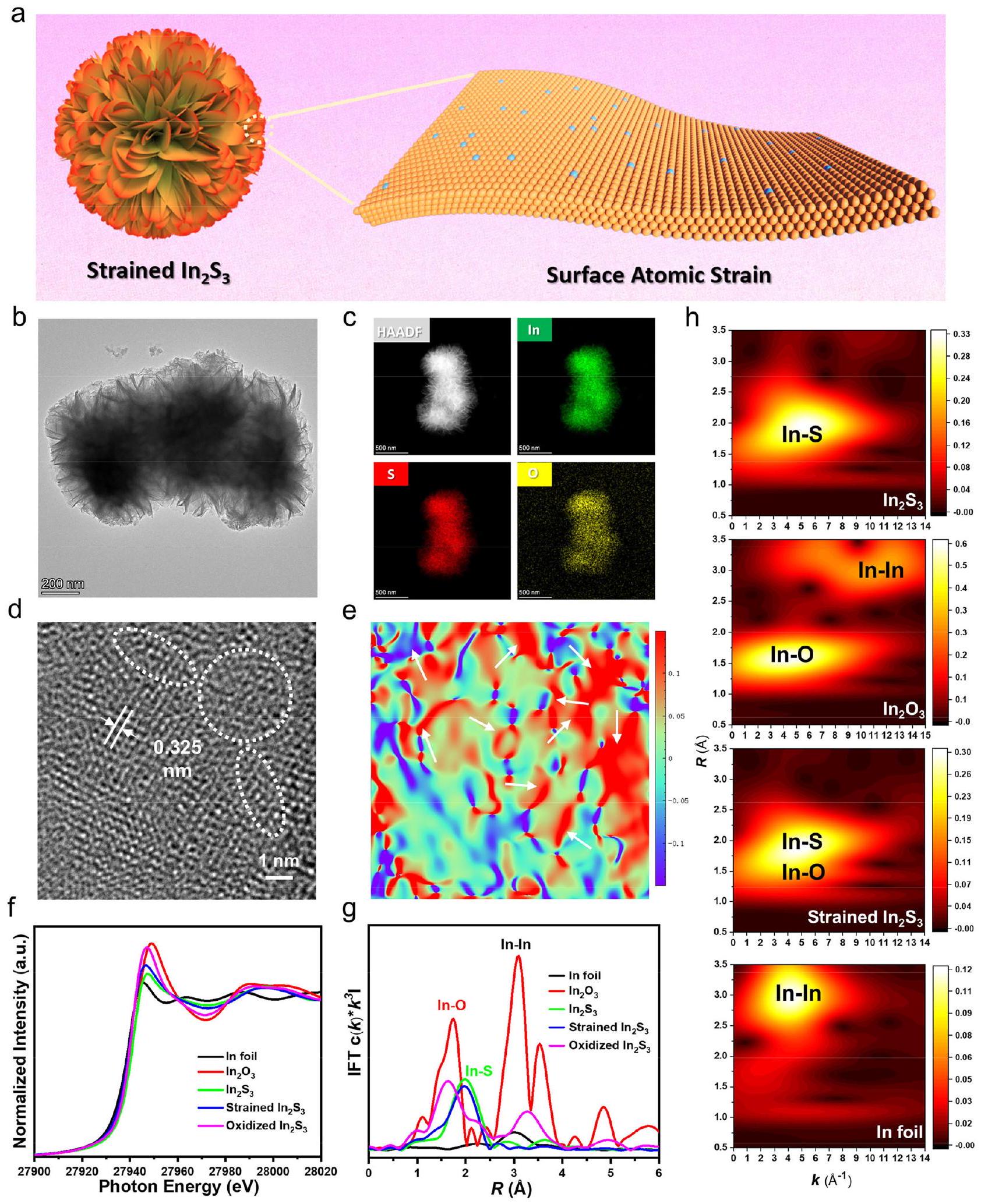
strain mapping image along the direction by GPA. XANES and FT-EXAFS spectra recorded in the In -edge. h WT-EXAFS plots of , strained and oxidized . Source data for Fig. 1f,g are provided as a Source Data file.
small-domain strain at the atomic scale (strained , Fig. 1a). The asprepared atomically strained exhibited approximately 9 times higher photocatalytic activity than pristine , towards converting to CO in ambient air under visible light irradiation. Based on a
collection of in-situ spectroscopic measurements and density functional theory (DFT) calculations, the atomically strained was found capable to promote adsorption/activation and at the same time boost photogenerated charge carrier separation, as a result,
greatly enhancing CO2 photoreduction performance under full spectrum/visible light illumination in both pure and ambient air environment.

Results and discussion

Structural characterizations of layered
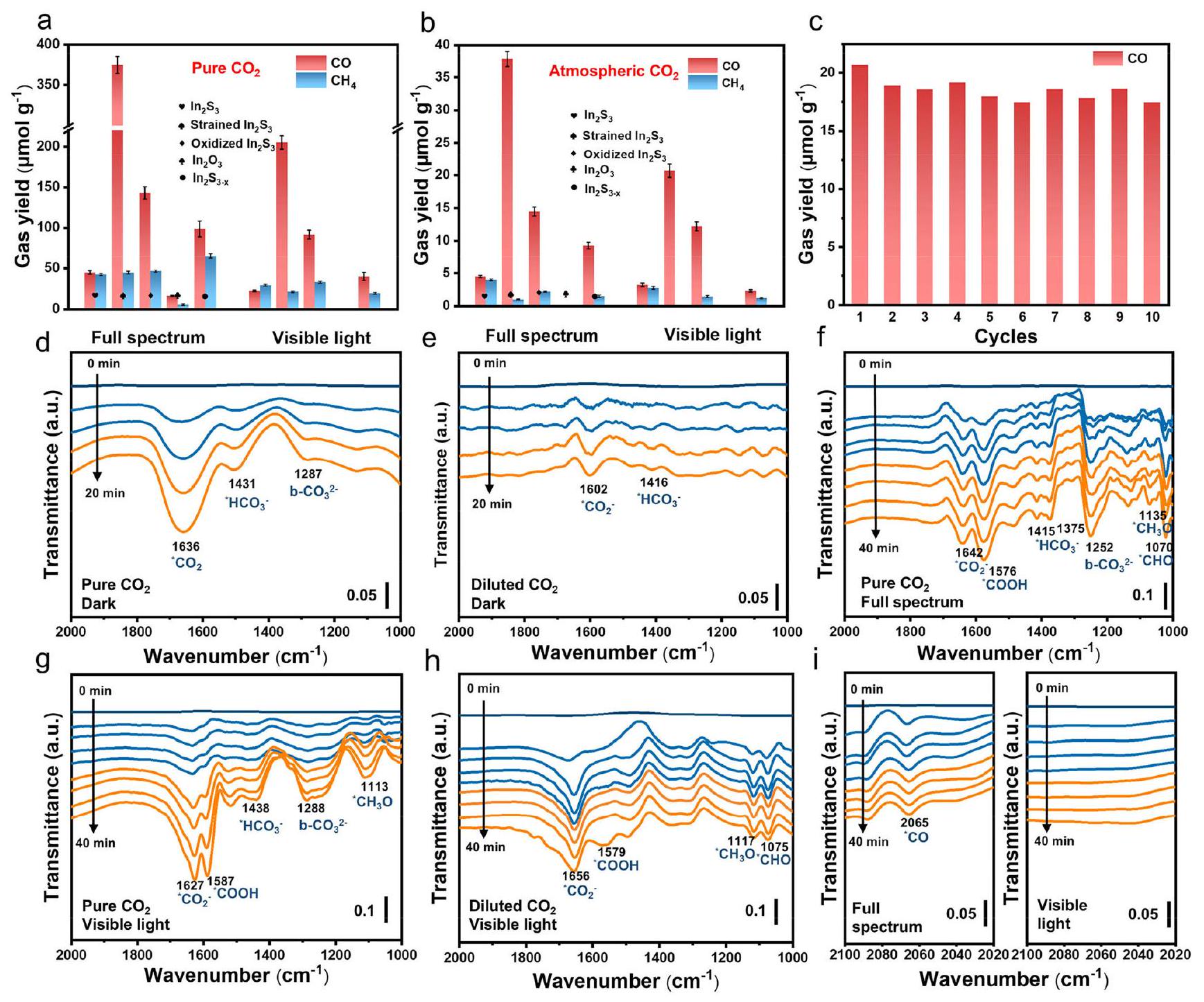
 with atomically strained Indium sites

Oxygen coordination and sulfur vacancy were introduced into via a facile one-step solvothermal reaction, in which the induced strain degree in could be well controlled by the volumetric ratio of water and ethylene glycol used in the solvothermal reaction (Supplementary Fig. 1). X-ray diffraction (XRD) patterns (Supplementary Fig. 2) of the as-prepared pristine and strained belong to cubicphase (PDF #65-0459) . The (311) diffraction peak of strained displayed a shift to lower angles as compared to pristine , indicating possible presence of local structural disorder and strain in the strained . Scanning electron microscopy (SEM) and transmission electron microscopy (TEM) images as displayed in Supplementary Fig. 3, Fig. 1b and Supplementary Fig. 4 showed that both of the pristine and strained exhibited flower-like morphology assembled from thin nanosheets. Elemental mapping images indicated homogeneous distribution of In, S and O elements (Fig. 1c) in strained . As illustrated in high-resolution TEM (HR-TEM) image (Fig. 1d and Supplementary Fig. 5), the strained possessed moderate crystallinity with a fine lattice spacing of 0.325 nm , assignable to the (311) plane of cubic . Figure 1e and Supplementary Fig. 6 showed the strain maps obtained by geometric phase analysis (GPA). Figure 1e displayed the strain distribution around the lattice disorder defects (white circles in Fig. 1d), which showed widespread compression strain around 7%, with some regions reaching . The marked arrows indicated the atomic-level distortion at the lattice, confirming the atomically strained surficial In sites . Supplementary Fig. 7 showed the strain distribution around the lattice disorder areas of oxidized . The oxygen atoms partially replaced the In-S bonds in layered , which in turn creates significant tensile stress. For comparison, pristine and were also analyzed by GPA, and both of pristine and displayed good crystallinity with ultrathin sheet-like morphologies as presented in Supplementary Fig. 8-9. X-ray photoelectron spectroscopy (XPS) spectra (Supplementary Fig. 10) indicated decreased XPS peak and enhanced XPS peak in strained as compared to pristine , suggesting that strain creation was associated with In-O bond formation. The same conclusion can be drawn from the observed shift of and peaks to higher binding energies in strained . It is observed that the intensity of O XPS peak gradually reduced with increasing sputtering time (Supplementary Fig. 11), corroborating that the ions were introduced into the surface of strained by replacing ions in In-S bonds .
The atomic and electronic structures of different samples were further examined by X-ray absorption spectroscopy (XAS). All samples showed similar pre-edge peaks at about 27935 eV in the In K-edge X-ray absorption near-edge structure (XANEs) spectra, suggesting similar In atomic arrangements (Fig. 1f). As displayed in the Fourier-transformed (FT) In -edge extended X-ray absorption fine structure (EXAFS) spectra (Fig. 1g and Supplementary Table 1), the strained sample showed a reduced bond length as compared to that of pristine sample ( ). Moreover, wavelet transform (WT) EXAFS plots in both K space and R space (Fig. 1h and Supplementary Fig. 12-17) show different In-O and In-S coordination in strained as compared to reference samples. An obviously stronger electron paramagnetic resonance (EPR) signal at was observed over strained than pristine (Supplementary Fig. 18), indicating a higher S-vacancy content in strained . A similar EPR signal was reported in defective metal sulfide . All of the above characterization results indicate presence of InO bonds together with sulfur vacancies in strained .
The light absorption properties of the as-prepared catalysts were studied by UV-vis absorption spectroscopy. As shown in Fig. 2a, compared with pristine , the strained displayed enhanced light absorption ability. The bandgap of the strained was calculated to be about 1.76 eV (Supplementary Fig. 19), which was slightly smaller than that of pristine . Combined with ultraviolet photoelectron spectroscopy (UPS) measurements (Supplementary Fig. 20), it was inferred that the conduction band (CB) minimum of pristine and strained was located at -0.33 and -0.41 eV , respectively, as illustrated in Fig. 2b. It is important to note that the downshifted edge position for strained can still meet the thermodynamic demand for reduction . As an important consideration for photocatalysis, the separation and transfer of photogenerated charge carriers were further investigated. As revealed in Fig. 2c, the strained exhibited the lowest photoluminescence (PL) peak intensity relative to pristine , oxidized and , indicating its lowest photoinduced charge carrier recombination rate. The same conclusion could be deduced from the surface photovoltage (SPV), transient photocurrent and electrochemical impedance spectroscopy measurements (Fig. 2d and Supplementary Fig. 21). Femtosecond transient absorption (fs-TA) spectroscopy was further performed to study the charge carrier dynamics. Figure 2e-f compared two-dimensional (2D) pseudocolor TA spectra for pristine and strained , which displayed two distinctive absorption bands at and , respectively. Typical photobleaching peaks are observed in the pseudocolor plots of and strained . These signals were assigned to the groundstate bleaching (GSB), reflecting the excited state relaxation. Figure 2 g showed the fs-TA kinetic profiles at 475 nm for and strained . With increasing probing time, an obvious decrease in TA intensity was detected, indicating a decrease of active photogenerated charges (Supplementary Fig. 22). The fs-TA spectra of and strained could be well fitted by a tri-exponential function and the fitting parameters are for , and for strained . The short-lived corresponded to electron trapping by electron trapping state (e-TS), while the long-lived and were related to electron transition and recombination, respectively. Kinetic fitting gave the decay time of the ground-state bleaching (GSB) signal at 42.85 and 25.05 ps for and strained , respectively, based on a triexponential decay function. The shorter GSB decay time in strained resulted from charge carrier transfer to the atomically strained sites. The above results provide concerted evidences that the strained In sites can promote rapid transfer of photogenerated electrons.

Photocatalytic reduction performance

The photoreduction performance of strained was evaluated under full spectrum or visible light ( ) irradiation in pure without presence of any sacrificial reagents. The amount of photocatalyst used was first optimized in the photocatalytic reduction reaction (Supplementary Fig. 23). Besides, we have conducted additional control experiments using pure Ar to replace as the feeding gas. No CO2 reduction products could be identified (Supplementary Fig. 24), eliminating the influence of carbon contamination during photoreaction. Figure 3a and Supplementary Fig. 25 show that the CO and yields over strained can reach 397.2 and ( 204.9 and ) after 4 h of photoreaction under full spectrum (visible) light illumination. The yield of CO is ca. 8.5 higher than that over pristine . In the meanwhile, water was oxidized to evolve molecular oxygen (Supplementary Fig. 26). As displayed in Fig. 3b, the strained could still maintain appreciable CO and yields under full spectrum and visible light irradiation (Supplementary Fig. 27) even if the reaction environment was switched from pure to atmospheric far superior to pristine (Supplementary Fig. 28-29). The
Fig. 2 | Insights into charge transfer dynamics. a UV-vis diffuse reflectance spectroscopy (DRS) spectra and band structure of pristine , strained and . c Steady-state PL spectra and SPV signals over pristine , strained
, oxidized and . 2D transient absorption pseudocolor plots for pristine and strained . g The decay signals of pristine and strained . Source data for Fig. 2a,c,d,g are provided as a Source Data file.
CO and products were indeed produced via photoreduction, verified by the labeling experiments (Supplementary Fig. 30), and the presence of molecular had little influence on the photoreduction reaction (Supplementary Fig. 31). The apparent quantum efficiency (AQE) of strained was estimated and the result was shown in Supplementary Fig. 32, which well resembled the UV-vis absorption spectrum, reaching a maximum AQE of around at 400 nm . The surface temperature of the strained under visible light irradiation was at around , measured using a thermographic technique (Supplementary Fig. 33). The control experiments disclosed that the reduction reaction was completely terminated in dark at or even at as high as (Supplementary Fig. 34). Furthermore, when the photogenerated heat was removed by ice water, the strained can still maintain nearly the same photocatalytic performance (Supplementary Fig. 34). These results reaffirmed the photoinduced reduction reaction over strained . Besides high photocatalytic activity, the strained also displayed decent photocatalytic stability: no significant photocatalytic activity decay was noticed after ten consecutive photoreaction cycles (Fig. 3c and Supplementary Fig. 35).
The structure of the strained could be well maintained after ten cycles of photoreaction based on a collection of characterizations including XRD, TEM, EPR and XPS (Supplementary Fig. 36), indicating its excellent stability in photoreaction. Compared to the reported In-based photocatalysts in literature, the strained exhibited an obviously better photocatalytic reduction performance in both pure and dilute environment (Supplementary Table 2-3).
To understand the photoreduction reaction mechanism over strained , in-situ Fourier transform infrared spectroscopy (FTIR) measurements were performed to probe the reaction intermediates. Carbonate species were found existing on the surface of strained in humid atmosphere under dark condition (Fig. 3d). The absorption peak at 1636,1431 and could be attributed to and b- , respectively. The observation of carbonate species and suggested that could be adsorbed and activated over strained under dark condition. Lowering partial pressure reduced the intensities of infrared characteristic peaks (Fig. 3e). Compared with dark condition, several new absorption peaks at around and
Fig. 3 | Photocatalytic reduction performance. Product yields in photoreduction reaction over various photocatalysts conduced in a pure and b ambient air. Error bars represent the standard deviation of three independent measurements. c Cyclic photocatalytic reduction reaction conducted in ambient air for 40 hours. In-situ DRIFTS spectra recorded over strained d under dark condition in pure , e under dark condition in ambient air, under
full spectrum irradiation condition in pure under visible light irradiation condition in pure , and under visible light irradiation condition in ambient air. i The in-situ FTIR spectra in the wavenumber range from 2020 to recorded over strained under full spectrum irradiation condition in pure (left) and under visible light irradiation condition in diluted (right). Source data for Fig. 3a-i are provided as a Source Data file.
appeared under full spectrum (Fig. 3f) or visible-light (Fig. 3g) irradiation and their intensities increased with increasing light irradiation time, due to increased coverage of * COOH (a crucial intermediate for reduction to CO or ), and (the important intermediates for reduction to ), respectively. Besides, the (1415 and ) and b-CO3 absorption peaks also gradually strengthened with elongating light illumination time. Switching reaction environment from pure to ambient air reduced the coverage (Fig. 3 h ) and changed the adsorption strength of reduction intermediates. The *CO absorption peak at around was clearly detected over strained in pure atmosphere under both visible and full spectrum light irradiation (Fig. 3i left), suggesting relatively strong *CO adsorption over strained surface, which should be beneficial for the subsequent steps of protonation towards formation. In ambient air environment, no obvious band near was detected over strained , which explained the observed selective photoreduction to CO .

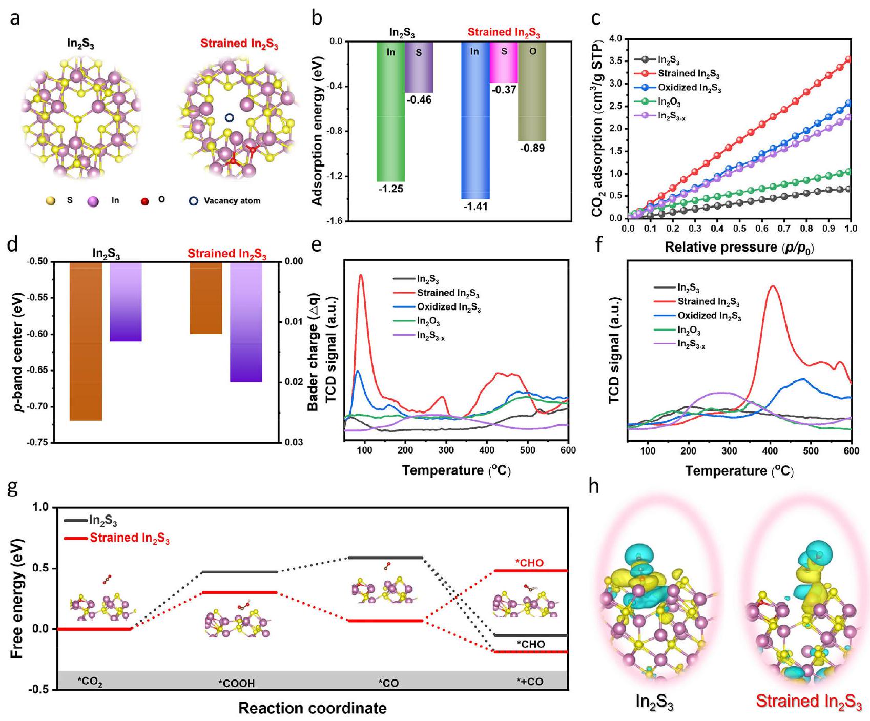
Insight of the increased photocatalytic activity

To gain further theoretical insights, density functional theory (DFT) calculations were conducted to explore adsorption and activation over strained . The strained calculation model is shown in Fig. 4a, Supplementary Fig. 37 and Supplementary Data 1, 2 for details on the models, which was built based on a collection of characterization results. The adsorption energy on the In site of pristine was calculated to be -1.25 eV (Fig. 4b). The adsorption energy over the In site was significantly more negative than that over the S and O site in strained , suggesting that formation of coordinatively unsaturated In sites is favorable for adsorption . It can be observed in Fig. 4c that strained showed a much better adsorption ability than pristine . In particular, the adsorption curve of strained rose significantly faster in the middle pressure range. At , the adsorption capacities of pristine , strained , oxidized and were , and , respectively. Moreover, an upshift of the -band center from -0.72 to -0.60 eV was also observed in strained (Fig. 4d and Supplementary Fig. 38). It is
Fig. 4 | DFT study. a Schematic illustration showing the structural models for and strained . b Calculated adsorption energy on pristine and strained . c adsorption isotherms. d Calculated Bader charge and -bandcenter. e TPD curves. CO TPD curves. Calculated free energy diagrams for
reduction. h Calculated differential charge density of * COOH intermediate over pristine and strained surface. The insets , and h show the DFToptimized structures (In: purple, S: yellow, O: red, C: brown, and H: pink). Source data for are provided as a Source Data file.
well established that the upshifted -band center can push more antibonding states above the Fermi level, thus resulting in decreased occupation and stronger adsorbate bonding. The strained In site shifted the -band center closer to the Fermi level and resulted in stronger surface bonding between the active sites and molecules .
Temperature-programmed desorption (TPD) of was performed to verify the enhanced adsorption over strained (Fig. 4e). The -TPD peak area for strained was obviously larger than those for other samples with a higher desorption temperature, indicating that the strained indeed could enhance adsorption. Moreover, the CO TPD measurements indicated a concurrent stronger CO adsorption over strained , which was beneficial for * CO hydrogenation to form in photoreduction reaction (Fig. 4f). The reaction free energy and the structure configuration of each intermediate were simulated to illustrate the photocatalytic reduction reaction mechanism (Supplementary Fig. 39 and Supplementary Table 4). Of note, the *COOH formation energy on strained was more negative than that on , indicating that the atomically strained In sites can lower the energy required for the key conversion of * to * COOH (Fig. 4g). Afterward,
the downhill free energy profiles revealed that the formation of *CO from *COOH was more spontaneous over strained . Meanwhile, *CO protonation to *CHO on strained had an energy barrier higher than that on pristine , which can ensure a higher selectivity of CO generation on strained . As confirmed by differential charge densities over the * COOH -adsorbed structure models, higher concentrations of electrons were transferred to the near-surface region of strained In sites on strained (Fig. 4h). Besides, water contact angle measurements (Supplementary Fig. 40) and DFT calculations (Supplementary Fig. 41) uncovered that the strained could also tailor the rate-determining step for water reduction to boost proton generation (Supplementary Table 5).

Discussion

In summary, an atomically strained photocatalyst has been rationally designed and successfully prepared by a wet-chemistry method, which exhibits high photoreduction performance in both pure and ambient air environment under full spectrum/visible light illumination. It is demonstrated that the strained can significantly enhance photogenerated charge carrier separation and improve adsorption/activation as characterized by a collection of
measurements and DFT calculations, as a result, greatly boosting CO2 photoreduction performance.

Methods

Materials

(AR, ), thioacetamide (TAA, AR, ), (AR, ) granular were purchased from Aladdin Reagent Co. Ltd. Ethylene glycol was purchased from Shanghai Chemical Reagent Co., Ltd. All chemical reagents were used without further purification and ultrapure water was used throughout the whole experiment.

Synthesis of

A total of 1.6 mmol of was dissolved in 50 mL deionized water and stirred for 60 min . Then 2.4 mmol TAA was added and stirred for another 15 min . The obtained mixture was transferred into a 100 mL Teflon-lined stainless-steel autoclave, which was sealed and kept at for 90 min . After the reaction, the autoclave was cooled down naturally, and the products were washed with deionized water and ethanol for several times and dried in ambient environment.

Synthesis of strained

A total of 1.6 mmol of was dissolved in a mixed solvent containing 35 mL of deionized water and 15 mL of ethylene glycol and stirred for 60 min . Then, 2.4 mmol of TAA was added and stirred for another 15 min . The obtained mixture was transferred into a 100 mL Teflon-lined stainless-steel autoclave, which was sealed and kept at for 90 min . After the reaction, the autoclave was cooled down naturally, and the products were washed with deionized water and ethanol for several times and dried in ambient environment.

Synthesis of oxidized

A total of 1.6 mmol of was dissolved in 50 mL of ethylene glycol and stirred for 60 min . Then 2.4 mmol TAA was added and stirred for another 15 min . The obtained mixture was transferred into a 100 mL Teflon-lined stainless-steel autoclave, which was sealed and kept at for 90 min . After the reaction, the autoclave was cooled down naturally, and the products were washed with deionized water and 1 M NaOH for several times and dried in ambient environment.

Synthesis of

The as-obtained was rapidly heated at for 2 min in Ar with and then cooled to room temperature, and the products were washed with deionized water and ethanol for several times and dried in vacuum oven overnight

Characterization

The structural information of the as-prepared samples was analyzed by powder X-ray diffraction (XRD) on a D/MAX-RB diffractometer with Cu radiation. The morphologies of the samples were examined by transmission electron microscopy (AC-TEM, JEM-ARM200F) at 200 kV . X-ray photoelectron spectroscopy (XPS) spectra were recorded on a Thermo VG Multilab 2000 spectrometer with Al Kα source. Ultraviolet photoelectron spectroscopy (UPS) measurements were performed on a Thermo Fisher ESCALAB 250Xi instrument with the He I radiation ( ) source. EPR measurements were obtained using a Bruker model EMXPLUS 10/12 spectrometer. The optical properties of the samples were studied on a UV-vis-NIR spectrophotometer (UV-3600). The surface photovoltage (SPV) spectroscopy was performed on stable surface photovoltage spectrometer (PL-SPV/IPCE1000, Beijing Perfectlight). The equipment consists of a Xe lamp (CHF-XM-500 W), a monochromator (ZLolix SBP500) and a lock-in amplifier (SB830-DSP) with light chopper (SR540). The adsorption properties were studied by performing -TPD and CO-TPD experiments on a Micromeritics AutoChem 2950HP. The CO2 adsorption-desorption
isotherms were measured on a Micromeritics ASAP2460. The transient photocurrent responses and the electrochemical impedance spectroscopy (EIS) spectra of the samples were recorded on an electrochemical workstation (CHI660C Instruments, China) with a threeelectrode configuration in (aq.), where an (saturating KCl ) electrode, a Pt plate and an indium tin oxide (ITO) glass coated with the sample worked as the reference, counter and working electrode, respectively. X-ray absorption near edge structure (XANES) spectra at the In -edge were collected at the TLS16A1 station in the National Synchrotron Radiation Research Center (NSRRC). In foil was used as the reference. Data reduction, data analysis and extended X-ray absorption fine structure (EXAFS) fitting were performed using the Athena and Artemis software packages. In-situ Fourier transform infrared spectroscopy (FTIR) measurement was performed on a Vertex 70 FTIR spectrometer (Bruker). The sample to be measured was first pretreated inside the chamber in Ar at for 30 min to clean the sample’s surface. Afterwards, the background spectrum was then collected after the chamber’s temperature was lowered to room temperature. Next, the gas ( or air ) was introduced into the reactor. After reaching the adsorption equilibrium in 30 min , the background spectrum was collected again. Subsequently, the light source (Xenon lamp with fiber optics, PLS-SXE300) was turned on, and the spectrum was recorded every 5 min under full spectrum or visible light irradiation.

Photocatalytic reduction

Typically, 20 mg of photocatalyst was dispersed in 5 mL of deionized under ultrasonication. The above suspension was then dropped onto a quartz disc with a diameter of 5 cm and dried at to obtain the photocatalyst-coated quartz disc. Afterwards, the quartz disc was placed in a custom-made reaction vessel filled with 5 mL of deionized on the bottom. Then, of high purity or compressed air was used as the source. Light irradiation was provided by a PLS-SXE300D Xe lamp (Beijing Perfectlight) with a standard AM 1.5 G filter having an output light density of , and the visible light was obtained by adding a filter . Note that the distance from the lamp to the sample was . After adsorption equilibrium, the online gas-closed photocatalytic system (Labsolar-6A, Beijing Perfectlight) and gas chromatograph (GC-2014C, Shimadzu Corp., Japan) were used to quantify the gaseous products. The temperature of the gas-solid reactor was controlled at by a recirculating cooling water system during light irradiation. The light intensity for photoreduction reaction was measured by an optical power meter (PL-MW2000, Beijing Perfectlight).

Computational details

All density functional theory (DFT) calculations were performed in Vienna Ab initio Simulation Package (VASP) within the generalized gradient approximation (GGA) using the Perdew-Burke-Ernzerhof (PBE) formulation. The projected augmented wave (PAW) potentials were chosen to describe the ionic cores and valence electrons were taken into account using a plane wave basis set with a kinetic energy cutoff at 450 eV . For the optimization of both geometry and lattice size, the Brillouin zone integration was performed with centered -point sampling. The self-consistent calculations applied a convergence energy threshold of . The equilibrium geometries and lattice constants were optimized with maximum stress on each atom within . In this work, a ( ) supercell of (111) and modified (111) surface slabs were adopted, where the half bottom atomic layers were fixed for all calculations. To avoid interactions between periodic structures, vacuum space was inserted. The weak interaction was described by DFT + D3 method using empirical correction in Grimme’s scheme. Spin polarization method was adopted to describe the magnetic system. The triaxial strain of
was applied equally for all in-plane directions (A and B axes, compress strain), while a strain was used in the perpendicular direction ( C axis, tensile strain). The adsorption energy was calculated as: adsorbent adsorbent , where adsorbent and E (adsorbent) represent the total energy of * adsorbent, * and adsorbent molecule, respectively. The Gibbs free energy for each elementary step was calculated as: , in which is the electronic energy at 0 K calculated by DFT, is the zero-point energy term, and T is the absolute temperature (here 298.15 K ), S is the entropy.

Data availability

Source data are provided with this paper.

References

  1. Xu, Y. et al. Engineering built-in electric field microenvironment of CQDs/g- heterojunction for efficient photocatalytic reduction. Adv. Sci. e2403607 (2024).
  2. Wang, L., Wang, D. & Li, Y. Single-atom catalysis for carbon neutrality. Carbon Energy 4, 1021-1079 (2022).
  3. Ou, H. et al. Carbon nitride photocatalysts with integrated oxidation and reduction atomic active centers for improved conversion. Angew. Chem. Int. Ed. 61, e202206579 (2022).
  4. Vu, N. N., Kaliaguine, S. & Do, T. O. Critical aspects and recent advances in structural engineering of photocatalysts for sunlightdriven photocatalytic reduction of into fuels. Adv. Funct. Mater. 29, 1901825 (2019).
  5. Ou, M. et al. Amino-assisted anchoring of perovskite quantum dots on porous for enhanced photocatalytic reduction. Angew. Chem. Int. Ed. 57, 13570-13574 (2018).
  6. Liu, L. et al. Tunable interfacial charge transfer in 2D-2D composite for efficient visible-light-driven conversion. Adv. Mater. 35, 2300643 (2023).
  7. Ouyang, T. et al. Dinuclear metal synergistic catalysis boosts photochemical -to-CO conversion. Angew. Chem. Int. Ed. 57, 16480-16485 (2018).
  8. Zhou, Y. et al. Engineering 2D photocatalysts toward carbon dioxide reduction. Adv. Energy Mater. 11, 2003159 (2021).
  9. Zhao, Z. et al. Interfacial chemical bond and oxygen vacancyenhanced CdSe-DETA S-scheme heterojunction for photocatalytic conversion. Adv. Funct. Mater. 33, 2214470 (2023).
  10. Zhang, X. et al. Photocatalytic conversion of diluted into light hydrocarbons using periodically modulated multiwalled nanotube arrays. Angew. Chem. Int. Ed. 51, 12732-12735 (2012).
  11. Wang, K. et al. Unlocking the charge-migration mechanism in SScheme junction for photoreduction of diluted with high selectivity. Adv. Funct. Mater. 34, 2309603 (2023).
  12. Yin, S. et al. Boosting water decomposition by sulfur vacancies for efficient photoreduction. Energy Environ. Sci. 15, 1556-1562 (2022).
  13. Wang, S., Guan, B. Y., Lu, Y. & Lou, X. W. D. Formation of hierarchical heterostructured nanotubes for efficient and stable visible light reduction. J. Am. Chem. Soc. 139, 17305-17308 (2017).
  14. Xing, Z. et al. From one to two: in situ construction of an ultrathin 2D-2D closely bonded heterojunction from a single-phase monolayer nanosheet. J. Am. Chem. Soc. 141, 19715-19727 (2019).
  15. Wang, K. et al. Unveiling S-Scheme charge transfer pathways in hybrid nanofiber photocatalysts for low-concentration hydrogenation. Solar RRL 7, 2200963 (2022).
  16. Yu, F., Jing, X., Wang, Y., Sun, M. & Duan, C. Hierarchically porous metal-organic framework interface for selective photocatalytic conversion of with into . Angew. Chem. Int. Ed. 60, 24849-24853 (2021).
  17. Wang, K. et al. In situ-illuminated X-ray photoelectron spectroscopy investigation of S-Scheme core-shell hybrid nanofibers for highly efficient solar-driven overall splitting. Solar RRL 6, 2200736 (2022).
  18. Bie, C., Zhu, B., Xu, F., Zhang, L. & Yu, J. In situ grown monolayer N -doped graphene on CdS hollow spheres with seamless contact for photocatalytic reduction. Adv. Mater. 31, e1902868 (2019).
  19. Li, J. et al. Interfacial engineering of nanowires promotes metallic photocatalytic reduction activity under near-infrared light irradiation. J. Am. Chem. Soc. 143, 6551-6559 (2021).
  20. Liang, L. et al. Infrared light-driven overall splitting at room temperature. Joule 2, 1004-1016 (2018).
  21. Jiao, X. et al. Defect-mediated electron-hole separation in one-unitcell layers for boosted solar-driven reduction. J. Am. Chem. Soc. 139, 7586-7594 (2017).
  22. Li, Y. et al. Plasmonic hot electrons from oxygen vacancies for infrared light-driven catalytic reduction on . Angew. Chem. Int. Ed. 60, 910-916 (2021).
  23. Zhu, S. et al. Selective photoreduction into product enabled by charge-polarized metal pair sites. Nano Lett 21, 2324-2331 (2021).
  24. . et al. Selective visible-light-driven photocatalytic reduction to mediated by atomically thin layers. Nat. Energy 4, 690-699 (2019).
  25. Wang, K. et al. Atomic-level insight of sulfidation-engineered Aur-ivillius-related nanosheets enabling visible light lowconcentration conversion. Carbon Energy 5, e264 (2022).
  26. Maiti, S. et al. Engineering electrocatalyst nanosurfaces to enrich the activity by inducing lattice strain. Energy Environ. Sci. 14, 3717-3756 (2021).
  27. He, H. et al. Interface chemical bond enhanced ions intercalated carbon nitride/CdSe-diethylenetriamine S-scheme heterojunction for photocatalytic synthesis in pure water. Adv. Funct. Mater. 34, 2315426 (2024).
  28. Liu, L. et al. Synergistic polarization engineering on bulk and surface for boosting photoreduction. Angew. Chem. Int. Ed. 60, 18303-18308 (2021).
  29. Li, A. et al. Three-phase photocatalysis for the enhanced selectivity and activity of reduction on a hydrophobic surface. Angew. Chem. Int. Ed. 58, 14549-14555 (2019).
  30. Li, X. et al. Ultrathin conductor enabling efficient IR light reduction. J. Am. Chem. Soc. 141, 423-430 (2019).
  31. Liu, W. et al. Vacancy-cluster-mediated surface activation for boosting chemical fixation. Chem. Sci. 14, 1397-1402 (2023).
  32. Bellotti, P. & Glorius, F. Strain-release photocatalysis. J. Am. Chem. Soc. 145, 20716-20732 (2023).
  33. Yue, X., Cheng, L., Li, F., Fan, J. & Xiang, Q. Highly strained Bi-MOF on bismuth oxyhalide support with tailored intermediate adsorption/desorption capability for robust photoreduction. Angew. Chem. Int. Ed. 61, e202208414 (2022).
  34. Cao, X. et al. Engineering lattice disorder on a photocatalyst: photochromic BiOBr nanosheets enhance activation of aromatic C-H bonds via water oxidation. J. Am. Chem. Soc. 144, 3386-3397 (2022).
  35. Wang, Z. & Zhou, G. Lattice-strain control of flexible Janus indium chalcogenide monolayers for photocatalytic water splitting. J. Phys. Chem. C 124, 167-174 (2019).
  36. Miao, Y., Zhao, Y., Zhang, S., Shi, R. & Zhang, T. Strain engineering: a boosting strategy for photocatalysis. Adv. Mater. 34, e2200868 (2022).
  37. Feng, H. et al. Modulation of photocatalytic properties by strain in 2D BiOBr nanosheets. ACS Appl. Mater. Interfaces 7, 27592-27596 (2015).
  38. Huo, W., Xu, W., Guo, Z., Zhang, Y. & Dong, F. Motivated surface reaction thermodynamics on the bismuth oxyhalides with lattice strain for enhanced photocatalytic NO oxidation. Appl. Catal. B 284, 119694 (2021).
  39. Dai, D. et al. Strain adjustment realizes the photocatalytic overall water splitting on tetragonal zircon . Adv. Sci. 9, e2105299 (2022).
  40. Zhong, Q. et al. Strain-modulated seeded growth of highly branched black Au superparticles for efficient photothermal conversion. J. Am. Chem. Soc. 143, 20513-20523 (2021).
  41. Cai, X. et al. Synergism of surface strain and interfacial polarization on Pd@Au core-shell cocatalysts for highly efficient photocatalytic reduction over . J. Mater. Chem. A 8, 7350-7359 (2020).
  42. Yan, Y. et al. Tensile strain-mediated spinel ferrites enable superior oxygen evolution activity. J. Am. Chem. Soc. 145, 24218-24229 (2023).
  43. Hou, Z. et al. Lattice-strain engineering for heterogenous electrocatalytic oxygen evolution reaction. Adv. Mater. 35, e2209876 (2023).
  44. Wei, K. et al. Strained zero-valent iron for highly efficient heavy metal removal. Adv. Funct. Mater. 32, 2200498 (2022).
  45. Sabbah, A. et al. Boosting photocatalytic reduction in a heterostructure through strain-induced direct Z-scheme and a mechanistic study of molecular interaction thereon. Nano Energy 93, 106809 (2022).
  46. Huang, H. et al. Noble-metal-free ultrathin MXene coupled with nanoflakes for ultrafast photocatalytic reduction of hexavalent chromium. Appl. Catal. B 284, 119754 (2021).
  47. Liu, Y. et al. Rapid room-temperature mechanosynthesis tensilestrained for robust photomineralization. Catal. Commun. 177, 106638 (2023).
  48. Hao, L. et al. Surface-halogenation-induced atomic-site activation and local charge separation for superb photoreduction. Adv. Mater. 31, e1900546 (2019).
  49. Li, X. et al. Atomically strained metal sites for highly efficient and selective photooxidation. Nano Lett. 23, 2905-2914 (2023).
  50. Xiao, Y., Yao, C., Su, C. & Liu, B. Nanoclusters for photoelectrochemical water splitting: bridging the photosensitizer and carrier transporter. EcoEnergy 1, 60-84 (2023).
  51. Di, J. et al. Isolated single atom cobalt in atomic layers to trigger efficient photoreduction. Nat. Commun. 10, 2840 (2019).
  52. Wu, X . et al. Identification of the active sites on metallic nano-sea-urchin for atmospheric photoreduction under UV, visible, and near-infrared light illumination. Angew. Chem. Int. Ed. 62, e202213124 (2023).
  53. Zhang, H. et al. Isolated cobalt centers on nanowires perform as a reaction switch for efficient photoreduction. J. Am. Chem. Soc. 143, 2173-2177 (2021).

Acknowledgements

This work was financially supported by the National Nature Science Foundation of China (22378104, 52104254, 22173029, and 22308336), the City University of Kong Hong startup fund (9020003), ITF – RTHGlobal STEM Professorship (9446006), JC STEM lab of Advanced CO2 Upcycling (9228005) and Program for Innovative Teams of Outstanding
Young and Middle-aged Researchers in the Higher Education Institutions of Hubei Province (T2023021).

Author contributions

Conceptional layout: K.W. and B.L. Investigation: K.W. Y.H., X.L., J.L., and B.L. Experimental data collection and processing: K.W. Computational simulation: K.W. and X.L. Manuscript co-writing: K.W. and B.L. Manuscript revising: K.W. Y.H., X.L., J.L., and B.L. Funding acquisition: K.W. Y.H., J.L., and B.L. Supervision: K.W. and B.L.

Competing interests

The authors declare no competing interests.

Additional information

Supplementary information The online version contains supplementary material available at https://doi.org/10.1038/s41467-025-57140-x.
Correspondence and requests for materials should be addressed to Kai Wang, Jun Li or Bin Liu.
Peer review information Nature Communications thanks Yanzhao Zhang, Liuqing Yang, and the other, anonymous, reviewer(s) for their contribution to the peer review of this work. A peer review file is available.
Reprints and permissions information is available at
http://www.nature.com/reprints
Publisher’s note Springer Nature remains neutral with regard to jurisdictional claims in published maps and institutional affiliations.
Open Access This article is licensed under a Creative Commons Attribution-NonCommercial-NoDerivatives 4.0 International License, which permits any non-commercial use, sharing, distribution and reproduction in any medium or format, as long as you give appropriate credit to the original author(s) and the source, provide a link to the Creative Commons licence, and indicate if you modified the licensed material. You do not have permission under this licence to share adapted material derived from this article or parts of it. The images or other third party material in this article are included in the article’s Creative Commons licence, unless indicated otherwise in a credit line to the material. If material is not included in the article’s Creative Commons licence and your intended use is not permitted by statutory regulation or exceeds the permitted use, you will need to obtain permission directly from the copyright holder. To view a copy of this licence, visit http:// creativecommons.org/licenses/by-nc-nd/4.0/.
(c) The Author(s) 2025

  1. ¹College of Urban and Environmental Sciences, Hubei Key Laboratory of Pollutant Analysis and Reuse Technology, Hubei Normal University, Huangshi, PR China. Henan Institute of Advanced Technology, Zhengzhou University, Zhengzhou, PR China. Department of Materials Science and Engineering, City University of Hong Kong, Hong Kong SAR, PR China. Department of Chemistry, Hong Kong Institute of Clean Energy (HKICE) & Center of Super-Diamond and Advanced Films (COSDAF), City University of Hong Kong, Hong Kong SAR, PR China. e-mail: wangkai@hbnu.edu.cn; junli2019@zzu.edu.cn; bliu48@cityu.edu.hk