المجلة: Scientific Reports، المجلد: 14، العدد: 1
DOI: https://doi.org/10.1038/s41598-024-52688-y
PMID: https://pubmed.ncbi.nlm.nih.gov/38280908
تاريخ النشر: 2024-01-27
DOI: https://doi.org/10.1038/s41598-024-52688-y
PMID: https://pubmed.ncbi.nlm.nih.gov/38280908
تاريخ النشر: 2024-01-27
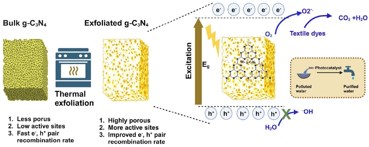
تحلل ضوئي فعال للملوثات الصبغية النسيجية باستخدام نيتريد الكربون الجرافيتي المعرض للحرارة (TE–g–C3N4)
نيتريد الكربون الجرافيتي
في السنوات الأخيرة، ساهمت العولمة، وتقدم التكنولوجيا، إلى جانب تطور تفضيلات الموضة لدى البشر، بشكل كبير في زيادة تصنيع المنسوجات والملابس.
تشير التقارير إلى أن الأصباغ السامة تسبب التهابات الجهاز التنفسي، وتهيج الجلد، وتهيج العينين.
نيتريد الكربون الجرافيتي
عملية التقشير الحراري لـ
قسم المواد التجريبية
ميلامين
الشكل 1. مخطط يوضح إعداد
أوكسالات
تركيب
تحضير الكمية الكبيرة
الجزء الأكبر
الجزء الأكبر
إعداد
تي-إي-جي-سي3
تي-إي-جي-سي3
توصيف
تم استخدام اختبارات حيود الأشعة السينية (XRD) للتحقق من إنتاج ونقاء المواد المُركبة باستخدام جهاز باناليتيكال X’Pert.
تجارب التحفيز الضوئي
النشاط الضوئي المحفز لـ TE-g-C
تم تقييم نسبة التحلل لكل صبغة وثابت معدل تفاعل المحفز الضوئي باستخدام المعادلات التالية.
أين،
القياسات الكهروكيميائية
تم إجراء تحليل موت-شوتكي لتأكيد قيمة نطاق التوصيل. باستخدام
النتائج والمناقشة
نتائج حيود الأشعة السينية البودرة
شكل 2أ أظهر نمط حيود الأشعة السينية لجميع العينات المحضرة
شكل 2أ أظهر نمط حيود الأشعة السينية لجميع العينات المحضرة
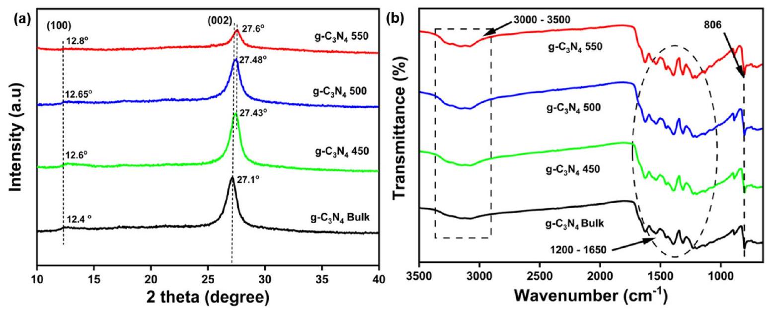
الشكل 2. (أ) نتائج حيود الأشعة السينية للمواد المسحوقة
ترتيب حلقات التري-س-ترازاين والتكرار الدوري لوحدات الهيبتازين داخل المستوى، على التوالي. أظهرت العينات التي تم تقشيرها حرارياً قممًا أوسع مع انخفاض في الشدة مقارنةً بنظيراتها الكتلية، مما قد يعكس تقليل المسافة بين الطبقات في العينات المقشرة.
تحليل FTIR
تمت دراسة المجموعات الوظيفية والترابط الكيميائي للمواد المحضرة بواسطة مطيافية الأشعة تحت الحمراء بتحويل فورييه (FTIR)، كما هو موضح في الشكل 2b. هنا، تمتلك جميع العينات المحضرة طيفًا مميزًا مشابهًا.
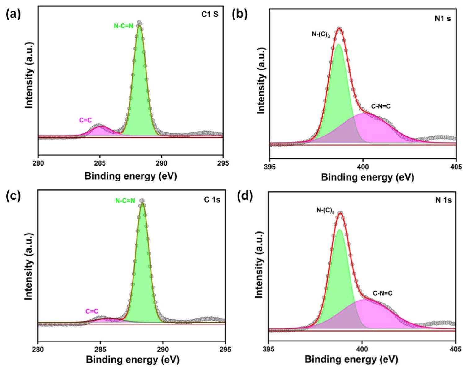
تحليل XPS
تم إجراء تحليل XPS لتحليل الحالة الكيميائية التفصيلية للمحفز المُعد. كما لوحظ من الشكل 3a، فإن طيف C 1 s للكتلة
| عينة | مساحة السطح
|
حجم المسام
|
حجم البلورات (نانومتر) | فجوة الطاقة (eV) | التركيب العنصري | |
| نسبة الكربون (وزن%) | N (نسبة الوزن%) | |||||
| ج-
|
٥.٠٣٥ | 0.028 | 9.0 | 2.54 | ٣٤.٠٦ | 65.94 |
|
|
9.189 | 0.08 | ٥.٧ | ٢.٥٩ | ٣٦.٠٠ | ٦٤.٠٠ |
|
|
١٣.٤٥ | 0.092 | 5.3 | 2.61 | ٣٧.٠٠ | ٦٣.٠٠ |
|
|
٤٨.٢٠٣ | 0.32 | ٤.٢ | 2.7 | ٣٩.٣ | 60.7 |
الجدول 1. المساحة السطحية، حجم المسام، حجم البلورات، فجوة الطاقة، والتركيب العنصري للمواد الكتلية والم exfoliated
الشكل 3. طيف XPS للكتلة
تم العثور عليها عند 288.34 إلكترون فولت و 285.341.
تحليل الشكل
توضح الشكل 4 الشكل البنيوي للعينات المحضرة التي أكدت تأثير درجة الحرارة على التقشر الحراري للمواد المحضرة. في حالة الكتلة
تحليل المساحة السطحية
تظهر الشكل 5
دراسة بصرية
تم فحص الخصائص البصرية للمواد المحفزة الضوئية المنتجة باستخدام مطيافية الانعكاس المنتشر للأشعة فوق البنفسجية (UV-DRS). تُظهر طيف الامتصاص البصري للعينات المحضرة في الشكل 6a. جميع الـ
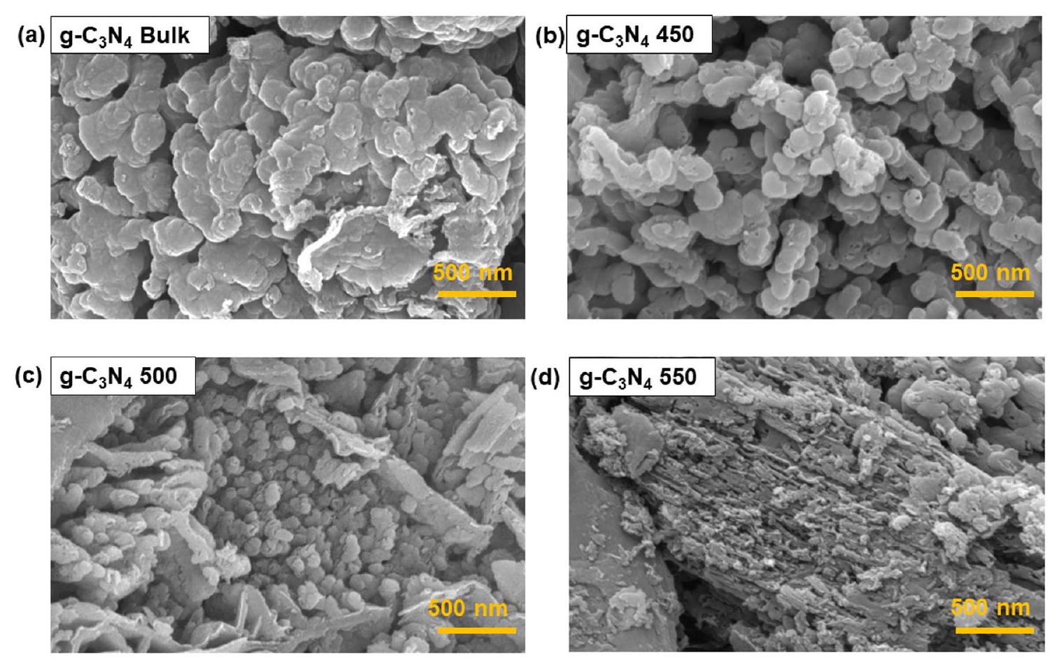
الشكل 4. صور FESEM للعينات المُركبة: (أ)
الشكل 5. (أ)
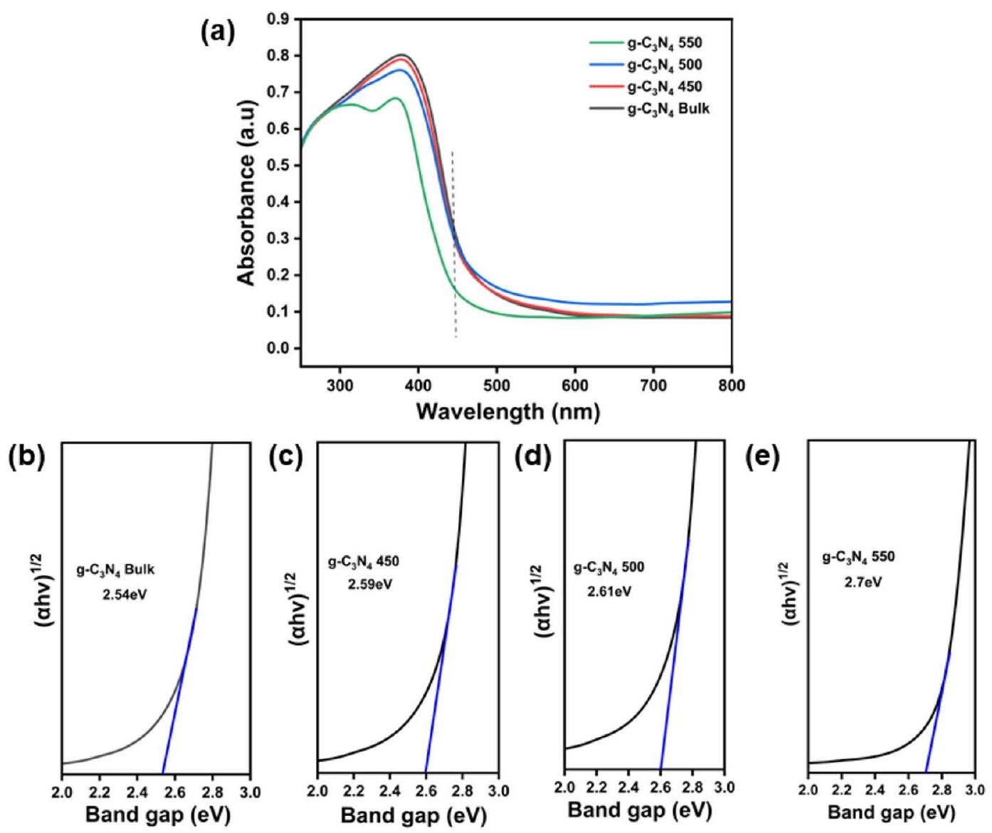
الشكل 6. (أ) طيف الامتصاص للأشعة فوق البنفسجية والمرئية
أظهرت العينات المحضرة حافة امتصاص عند 450 نانومتر مما يدل على الخصائص الجوهرية (
علاوة على ذلك، تم جدولتها في الجدول 1 فجوات النطاق لجميع المحفزات الضوئية. تم حساب قيم فجوة النطاق باستخدام المعادلة التالية:
أين،
النشاط الضوئي التحفيزي
الأداء الضوئي المحفز لـ
أثر المحفز
تم تحليل الخصائص التحفيزية للمحفزات المحضرة من خلال إجراء التحلل الضوئي لجميع الأصباغ الثلاثة بشكل فردي تحت إشعاع الضوء فوق البنفسجي. في البداية، تم وضع جميع الأصباغ في غرفة التفاعل الضوئي وتم إشعاعها لمدة 60 دقيقة دون وجود محفز وتم تسجيل طيف الامتصاص المقابل كما هو موضح في الشكل 7a-f. أظهر الاختبار الفارغ (الخط الأسود في الشكل 7) عدم وجود تحلل للأصباغ. ومع ذلك، بعد إضافة المحفز الضوئي إلى محاليل الأصباغ، كان هناك انخفاض ملحوظ في تركيزات جميع محاليل الأصباغ.
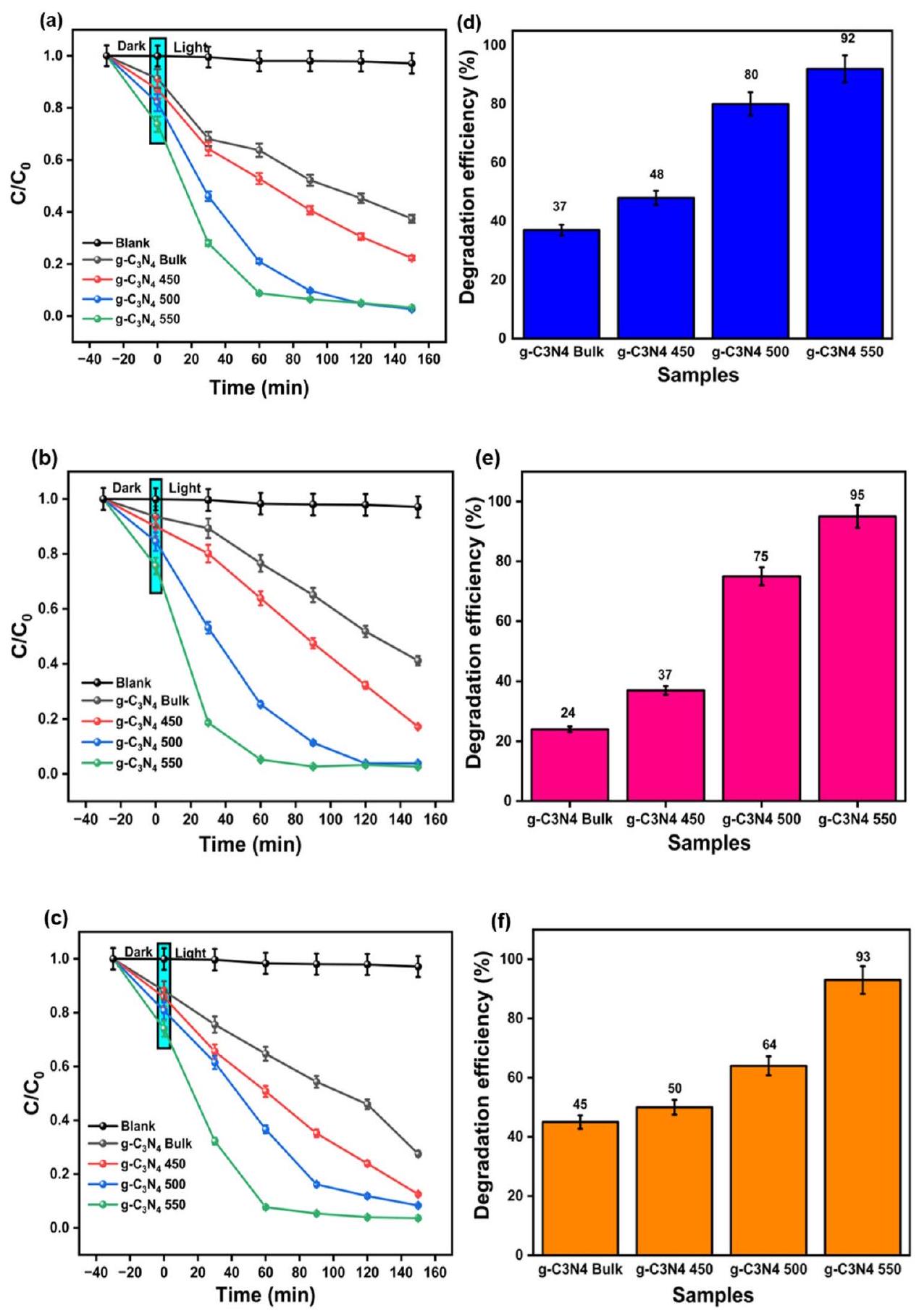
الشكل 7. (أ-ج) حركيات التحلل المعتمدة على الزمن المقاسة كتركيز الصبغة عند الزمن t بالنسبة لتركيزها الابتدائي
علاوة على ذلك، فإن ثابت المعدل لـ
| محفز ضوئي | صبغة MB | صبغة RhB | صبغة MO |
| جملة
|
|
|
|
|
|
|
|
|
|
|
|
|
|
|
|
|
|
|
الجدول 2. قيم ثابت المعدل للكتلة والمستخرجة
تحقيق في آليات التفاعل الضوئي المحفز تحليل الطيف الضوئي (PL) وتحليل impedanc
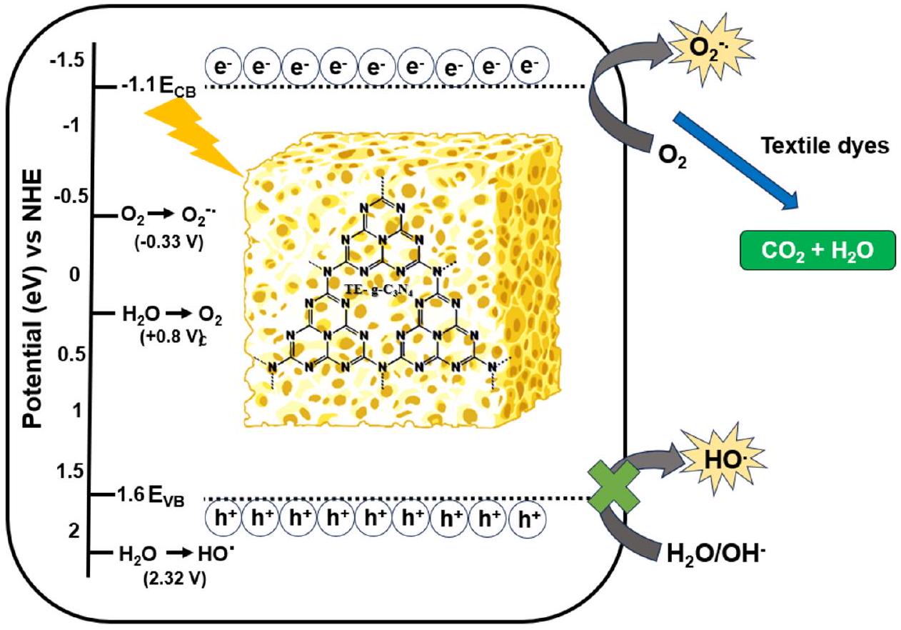
من المتوقع أن يتمتع المحفز الضوئي الجيد بمعدل نقل شحنة مرتفع ومعدل إعادة تركيب منخفض للثقوب والإلكترونات المثارة ضوئيًا.
اختبارات امتصاص الجذور الحرة
في عملية التحلل الضوئي النموذجية، يتم تحفيز الفجوة الناتجة عن الضوء (
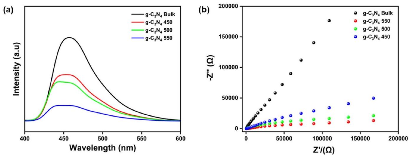
الشكل 8. (أ) طيف الفوتولومينسنس (PL) للمواد المحفزة الضوئية المُعدة، (ب) تحليل EIS للمواد المحفزة الضوئية المُعدة.
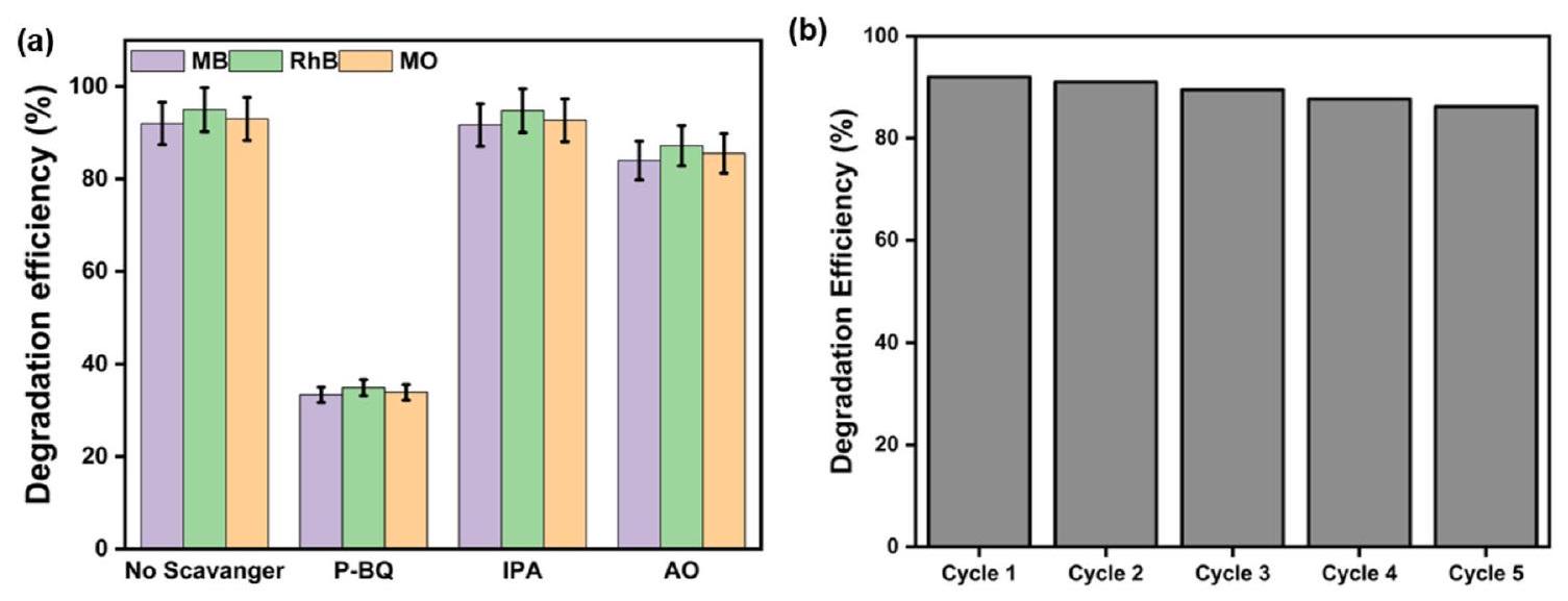
الشكل 9. (أ) دراسة امتصاص الجذور الحرة لتحديد آليات التفاعل لـ
| محفز ضوئي | فجوة الطاقة (eV) | جهد نطاق التوصيل (eV) [
|
جهد نطاق التكافؤ ( eV ) [
|
| جملة
|
2.54 | -1.05 | 1.49 |
|
|
٢.٥٩ | -1.075 | 1.51 |
|
|
2.61 | -1.085 | 1.52 |
|
|
2.7 | -1.13 | 1.57 |
الجدول 3. فجوة الطاقة (
أين،
بالإضافة إلى ذلك، تم إجراء تحليل موت-شوتكي أيضًا لأفضل محفز.
تقييم قابلية إعادة استخدام المحفزات الضوئية
الأفضل أداءً
الخاتمة
TE-
الشكل 10. آلية التفاعل الضوئي المقترحة
بعد 5 جولات متتالية. وبالتالي، TE-g-C-
توفر البيانات
جميع البيانات التي تدعم نتائج الدراسة متاحة من المؤلف المراسل عند الطلب المعقول.
تاريخ الاستلام: 15 نوفمبر 2023؛ تاريخ القبول: 22 يناير 2024
نُشر على الإنترنت: 27 يناير 2024
تاريخ الاستلام: 15 نوفمبر 2023؛ تاريخ القبول: 22 يناير 2024
نُشر على الإنترنت: 27 يناير 2024
References
- Khairul Akter, M. M., Haq, U. N., Islam, M. M. & Uddin, M. A. Textile-apparel manufacturing and material waste management in the circular economy: A conceptual model to achieve sustainable development goal (SDG) 12 for Bangladesh. Clean. Environ. Syst. 4, 100070. https://doi.org/10.1016/j.cesys.2022.100070 (2022).
- Akhtar, W. H., Watanabe, C., Tou, Y. & Neittaanmäki, P. A new perspective on the textile and apparel industry in the digital transformation era. Textiles 2, 633-656 (2022).
- Luján-Ornelas, C., Güereca, L. P., Franco-García, M.-L. & Heldeweg, M. A life cycle thinking approach to analyse sustainability in the textile industry: A literature review. Sustainability 12, 10193 (2020).
- Berradi, M. et al. Textile finishing dyes and their impact on aquatic environs. Heliyon 5, e02711. https://doi.org/10.1016/j.heliyon. 2019.e02711 (2019).
- Tamburini, D., Shimada, C. M. & McCarthy, B. T. The molecular characterization of early synthetic dyes in E. Knecht’s textile sample book “A Manual of Dyeing”, (1893) by high performance liquid chromatography-diode array detector-mass spectrometry (HPLC-DAD-MS). Dyes Pigments 190, 109286. https://doi.org/10.1016/j.dyepig.2021.109286 (2021).
- Slama, H. B. et al. Diversity of synthetic dyes from textile industries, discharge impacts and treatment methods. Appl. Sci. 11, 6255 (2021).
- Niinimäki, K. et al. The environmental price of fast fashion. Nat. Rev. Earth Environ. 1, 189-200. https://doi.org/10.1038/s43017-020-0039-9 (2020).
- Paul, D. Research on heavy metal pollution of river Ganga: A review. Ann. Agrar. Sci. 15, 278-286. https://doi.org/10.1016/j.aasci. 2017.04.001 (2017).
- Dwivedi, S., Mishra, S. & Tripathi, R. D. Ganga water pollution: A potential health threat to inhabitants of Ganga basin. Environ. Int. 117, 327-338. https://doi.org/10.1016/j.envint.2018.05.015 (2018).
- Low, J., Cao, S., Yu, J. & Wageh, S. Two-dimensional layered composite photocatalysts. Chem. Commun. 50, 10768-10777. https:// doi.org/10.1039/C4CC02553A (2014).
- Khan, I. et al. Review on methylene blue: Its properties, uses toxicity and photodegradation. Water 14, 242 (2022).
- Hassan, M. M. & Carr, C. M. A critical review on recent advancements of the removal of reactive dyes from dyehouse effluent by ion-exchange adsorbents. Chemosphere 209, 201-219. https://doi.org/10.1016/j.chemosphere.2018.06.043 (2018).
- Sharma, J., Sharma, S. & Soni, V. Classification and impact of synthetic textile dyes on aquatic flora: A review. Reg. Stud. Mar. Sci. 45, 101802. https://doi.org/10.1016/j.rsma.2021.101802 (2021).
- Al-Tohamy, R. et al. A critical review on the treatment of dye-containing wastewater: Ecotoxicological and health concerns of textile dyes and possible remediation approaches for environmental safety. Ecotoxicol. Environ. Saf. 231, 113160. https://doi.org/ 10.1016/j.ecoenv.2021.113160 (2022).
- Lellis, B., Fávaro-Polonio, C. Z., Pamphile, J. A. & Polonio, J. C. Effects of textile dyes on health and the environment and bioremediation potential of living organisms. Biotechnol. Res. Innov. 3, 275-290. https://doi.org/10.1016/j.biori.2019.09.001 (2019).
- Nilsson, R., Nordlinder, R., Wass, U., Meding, B. & Belin, L. Asthma, rhinitis, and dermatitis in workers exposed to reactive dyes. Br. J. Ind. Med. 50, 65-70. https://doi.org/10.1136/oem.50.1.65 (1993).
- Kooter, I. et al. Molecular signature of asthma-enhanced sensitivity to CuO nanoparticle aerosols from 3D cell model. ACS Nano 13, 6932-6946. https://doi.org/10.1021/acsnano.9b01823 (2019).
- Adane, T., Adugna, A. T. & Alemayehu, E. Textile industry effluent treatment techniques. J. Chem. 2021, 5314404. https://doi.org/ 10.1155/2021/5314404 (2021).
- Ismail, W. et al. Water treatment and artificial intelligence techniques: A systematic literature review research. Environ. Sci. Pollut. Res. 30, 71794-71812. https://doi.org/10.1007/s11356-021-16471-0 (2023).
- Sikosana, M. L., Sikhwivhilu, K., Moutloali, R. & Madyira, D. M. Municipal wastewater treatment technologies: A review. Proc. Manuf. 35, 1018-1024. https://doi.org/10.1016/j.promfg.2019.06.051 (2019).
- Rani, B., Nayak, A. K. & Sahu, N. K. Nanostructured Materials for Visible Light Photocatalysis 1-22 (Elsevier, 2022).
- Ren, G. et al. Recent advances of photocatalytic application in water treatment: A review. Nanomaterials 11, 1804 (2021).
- Chong, M. N., Jin, B., Chow, C. W. K. & Saint, C. Recent developments in photocatalytic water treatment technology: A review. Water Res. 44, 2997-3027. https://doi.org/10.1016/j.watres.2010.02.039 (2010).
- Schneider, J. et al. Understanding
photocatalysis: Mechanisms and materials. Chem. Rev. 114, 9919-9986. https://doi.org/ 10.1021/cr5001892 (2014). - Rajbongshi, B. M. Handbook of Smart Photocatalytic Materials 127-149 (Elsevier, 2020).
- Yang, X. & Wang, D. Photocatalysis: From fundamental principles to materials and applications. ACS Appl. Energy Mater. 1, 6657-6693. https://doi.org/10.1021/acsaem.8b01345 (2018).
- Porley, V. & Robertson, N. Nanostructured Photocatalysts 129-171 (Elsevier, 2020).
- Ong, W.-J., Tan, L.-L., Ng, Y. H., Yong, S.-T. & Chai, S.-P. Graphitic carbon nitride (g-C3N4)-based photocatalysts for artificial photosynthesis and environmental remediation: Are we a step closer to achieving sustainability?. Chem. Rev. 116, 7159-7329. https://doi.org/10.1021/acs.chemrev.6b00075 (2016).
- Li, H., Liu, Y., Gao, X., Fu, C. & Wang, X. Facile synthesis and enhanced visible-light photocatalysis of graphitic carbon nitride composite semiconductors. ChemSusChem 8, 1189-1196. https://doi.org/10.1002/cssc. 201500024 (2015).
- Chen, Y. et al. Surface modification of g-C3N4 by hydrazine: Simple way for noble-metal free hydrogen evolution catalysts. Chem. Eng. J. 286, 339-346. https://doi.org/10.1016/j.cej.2015.10.080 (2016).
- Jürgens, B. et al. Melem (2,5,8-Triamino-tri-s-triazine), an important intermediate during condensation of melamine rings to graphitic carbon nitride: Synthesis, structure determination by X-ray powder diffractometry, solid-state NMR, and theoretical studies. J. Am. Chem. Soc. 125, 10288-10300. https://doi.org/10.1021/ja0357689 (2003).
- Komatsu, T. & Nakamura, T. Polycondensation/pyrolysis of tris-s-triazine derivatives leading to graphite-like carbon nitrides. J. Mater. Chem. 11, 474-478. https://doi.org/10.1039/B005982J (2001).
- Xia, P., Zhu, B., Cheng, B., Yu, J. & Xu, J. 2D/2D g-C3N4/MnO2 nanocomposite as a direct Z-scheme photocatalyst for enhanced photocatalytic activity. ACS Sustain. Chem. Eng. 6, 965-973. https://doi.org/10.1021/acssuschemeng.7b03289 (2018).
- Chen, Y. & Bai, X. A review on quantum dots modified g-C3N4-Based photocatalysts with improved photocatalytic activity. Catalysts 10, 142 (2020).
- Alaghmandfard, A. & Ghandi, K. A comprehensive review of graphitic carbon nitride (g-C3N4)-metal oxide-based nanocomposites: Potential for photocatalysis and sensing. Nanomaterials 12, 294 (2022).
- Alam, U., Pandey, K. & Verma, N. Photocatalytic oxidation of glyphosate and reduction of Cr(VI) in water over ACF-supported CoNiWO4-gCN composite under batch and flow conditions. Chemosphere 297, 134119. https://doi.org/10.1016/j.chemosphere. 2022.134119 (2022).
- Alam, U., Pandey, A. & Verma, N. An anthraquinone-integrated S-scheme-based
composite with enhanced hydrogen production activity. Int. J. Hydrog. Energy 48, 2532-2541. https://doi.org/10.1016/j.ijhydene.2022.10.151 (2023). - Beyhaqi, A., Azimi, S. M. T., Chen, Z., Hu, C. & Zeng, Q. Exfoliated and plicated g-
nanosheets for efficient photocatalytic organic degradation and hydrogen evolution. Int. J. Hydrog. Energy 46, 20547-20559. https://doi.org/10.1016/j.ijhydene.2021.03. 174 (2021). - Feng, J. et al. Comparing the photocatalytic properties of
treated by thermal decomposition, solvothermal and protonation. Results Phys. 11, 331-334. https://doi.org/10.1016/j.rinp.2018.09.014 (2018). - Huang, T. et al. General synthesis strategy for hollow porous prismatic graphitic carbon nitride: A high-performance photocatalyst for
production and degradation of RhB. J. Mater. Sci. 55, 6037-6050. https://doi.org/10.1007/s10853-020-04439-3 (2020). - Rani, B., Nayak, A. K. & Sahu, N. K. Degradation of mixed cationic dye pollutant by metal free melem derivatives and graphitic carbon nitride. Chemosphere 298, 134249. https://doi.org/10.1016/j.chemosphere.2022.134249 (2022).
- Wang, H. et al. Self-assembled sulphur doped carbon nitride for photocatalytic water reforming of methanol. Chem. Eng. J. 445, 136790. https://doi.org/10.1016/j.cej.2022.136790 (2022).
- Papailias, I. et al. Effect of processing temperature on structure and photocatalytic properties of g-C3N4. Appl. Surf. Sci. 358, 278-286. https://doi.org/10.1016/j.apsusc.2015.08.097 (2015).
- Papailias, I. et al. Chemical vs thermal exfoliation of
for NOx removal under visible light irradiation. Appl. Catal. B Environ. 239, 16-26. https://doi.org/10.1016/j.apcatb.2018.07.078 (2018). - Hwa, K.-Y., Santhan, A., Ganguly, A. & Kanna Sharma, T. S. Two dimensional architectures of graphitic carbon nitride with the substitution of heteroatoms for bifunctional electrochemical detection of nilutamide. Chemosphere 320, 138068. https://doi.org/ 10.1016/j.chemosphere.2023.138068 (2023).
- Chellammal Gayathri, R., Elakkiya, V. & Sumathi, S. Synthesis of cerium and bismuth doped nickel aluminate for the photodegradation of methylene blue, methyl orange and rhodamine B dyes. Chemosphere 303, 135056. https://doi.org/10.1016/j.chemo sphere.2022.135056 (2022).
- Niu, P., Zhang, L., Liu, G. & Cheng, H.-M. Graphene-like carbon nitride nanosheets for improved photocatalytic activities. Adv. Funct. Mater. 22, 4763-4770. https://doi.org/10.1002/adfm. 201200922 (2012).
- Zhang, J., Zhang, M., Yang, C. & Wang, X. Nanospherical carbon nitride frameworks with sharp edges accelerating charge collection and separation at a soft photocatalytic interface. Adv. Mater. 26, 4121-4126. https://doi.org/10.1002/adma.201400573 (2014).
- Yang, S. et al. Exfoliated graphitic carbon nitride nanosheets as efficient catalysts for hydrogen evolution under visible light. Adv. Mater. 25, 2452-2456. https://doi.org/10.1002/adma. 201204453 (2013).
- Yang, L. et al. A surface modification resultant thermally oxidized porous
with enhanced photocatalytic hydrogen production. Appl. Catal. B Environ. 204, 335-345. https://doi.org/10.1016/j.apcatb.2016.11.047 (2017). - Xu, H.-Y., Zheng, Z. & Mao, G.-J. Enhanced photocatalytic discoloration of acid fuchsine wastewater by
schorl composite catalyst. J. Hazard. Mater. 175, 658-665. https://doi.org/10.1016/j.jhazmat.2009.10.059 (2010). - Song, X., Yang, Q., Jiang, X., Yin, M. & Zhou, L. Porous graphitic carbon nitride nanosheets prepared under self-producing atmosphere for highly improved photocatalytic activity. Appl. Catal. B Environ. 217, 322-330. https://doi.org/10.1016/j.apcatb.2017.05. 084 (2017).
- Gupta, R., Alam, U. & Verma, N. Efficient photofixation of nitrogen to ammonia over an S-scheme-based
heterojunction. Chem. Eng. J. 479, 147644. https://doi.org/10.1016/j.cej.2023.147644 (2024). - Qu, Y. & Duan, X. Progress, challenge and perspective of heterogeneous photocatalysts. Chem. Soc. Rev. 42, 2568-2580. https:// doi.org/10.1039/C2CS35355E (2013).
- Samanta, S., Martha, S. & Parida, K. Facile synthesis of Au/g-C3N4 nanocomposites: An inorganic/organic hybrid plasmonic photocatalyst with enhanced hydrogen gas evolution under visible-light irradiation. ChemCatChem 6, 1453-1462. https://doi. org/10.1002/cctc. 201300949 (2014).
- Kang, Y. et al. An amorphous carbon nitride photocatalyst with greatly extended visible-light-responsive range for photocatalytic hydrogen generation. Adv. Mater. 27, 4572-4577. https://doi.org/10.1002/adma. 201501939 (2015).
- Li, Q., He, Y. & Peng, R. Graphitic carbon nitride (g-C3N4) as a metal-free catalyst for thermal decomposition of ammonium perchlorate. RSC Adv. 5, 24507-24512. https://doi.org/10.1039/C5RA01157D (2015).
- Xue, J., Ma, S., Zhou, Y., Zhang, Z. & He, M. Facile photochemical synthesis of
with plasmon-enhanced photocatalytic activity for antibiotic degradation. ACS Appl. Mater. Interfaces 7, 9630-9637. https://doi.org/10.1021/acsami.5b01212 (2015). - Kong, L. et al. Porous size dependent g-C3N4 for efficient photocatalysts: Regulation synthesizes and physical mechanism. Mater. Today Energy 13, 11-21. https://doi.org/10.1016/j.mtener.2019.04.011 (2019).
- Phoon, B. L., Lai, C. W., Pan, G.-T., Yang, T.C.-K. & Juan, J. C. Highly mesoporous
with uniform pore size distribution via the template-free method to enhanced solar-driven tetracycline degradation. Nanomaterials 11, 2041 (2021). - Monga, D. & Basu, S. Enhanced photocatalytic degradation of industrial dye by
nanocomposite: Role of shape of . Adv. Powder Technol. 30, 1089-1098. https://doi.org/10.1016/j.apt.2019.03.004 (2019). - Wang, K. et al. Sulfur-doped
with enhanced photocatalytic -reduction performance. Appl. Catal. B Environ. 176-177, 44-52. https://doi.org/10.1016/j.apcatb.2015.03.045 (2015). - Saravanakumar, K. & Park, C. M. Rational design of a novel
double Z-scheme structure: Photocatalytic performance for antibiotic degradation and mechanistic insight. Chem. Eng. J. 423, 130076. https://doi.org/10.1016/j.cej.2021. 130076 (2021). - Liang, L. et al.
nano-fragments as highly efficient hydrogen evolution photocatalysts: Boosting effect of nitrogen vacancy. Appl. Catal. A Gen. 599, 117618. https://doi.org/10.1016/j.apcata.2020.117618 (2020). - Devi, L. G. & Kavitha, R. A review on non metal ion doped titania for the photocatalytic degradation of organic pollutants under UV/solar light: Role of photogenerated charge carrier dynamics in enhancing the activity. Appl. Catal. B Environ. 140-141, 559-587. https://doi.org/10.1016/j.apcatb.2013.04.035 (2013).
- Yuan, X. et al. Facile synthesis of 3D porous thermally exfoliated
nanosheet with enhanced photocatalytic degradation of organic dye. J. Coll. Interface Sci. 468, 211-219. https://doi.org/10.1016/j.jcis.2016.01.048 (2016). - Liu, N. et al. From Triazine to Heptazine: Origin of graphitic carbon nitride as a photocatalyst. ACS Omega 5, 12557-12567. https:// doi.org/10.1021/acsomega.0c01607 (2020).
- Jia, T. et al. Rational construction of direct Z -scheme
hybrid photocatalyst for significant enhancement of visible-light photocatalytic activity. Appl. Surf. Sci. 499, 143941. https://doi.org/10.1016/j.apsusc.2019.143941 (2020). - Bahadoran, A. et al. Novel S-scheme
heterojunction with enhanced photocatalytic degradation of sulfamerazine under visible light irradiation. Appl. Surf. Sci. 568, 150957. https://doi.org/10.1016/j.apsusc.2021.150957 (2021). - Xu, J., Wang, Z. & Zhu, Y. Enhanced Visible-light-driven photocatalytic disinfection performance and organic pollutant degradation activity of porous g-C 3 N4 nanosheets. ACS Appl. Mater. Interfaces 9, 27727-27735. https://doi.org/10.1021/acsami.7b07657 (2017).
شكر وتقدير
يود المؤلفون أن يشكروا إدارة VIT على التسهيلات المطلوبة لتنفيذ هذا العمل. تم إنشاء الملخص الرسومي باستخدامبيوريندر.كوم (رقم الترخيص: TH268W5VVJ).
مساهمات المؤلفين
س.ج: التركيب، المنهجية، التصور، تنظيم البيانات، التحليل الرسمي، التحقيق، كتابة المسودة الأصلية. ت.ك: التحليل الرسمي، التصور. س.س: التصور، الموارد، التحليل الرسمي. أ.ب: الإشراف، الموارد، تنظيم البيانات، الحصول على التمويل، التصور، التحقق، كتابة المراجعة والتحرير.
المصالح المتنافسة
يعلن المؤلفون عدم وجود مصالح متنافسة.
معلومات إضافية
معلومات إضافية النسخة الإلكترونية تحتوي على مواد إضافية متاحة علىhttps://doi.org/10.1038/s41598-024-52688-y.
يجب توجيه المراسلات والطلبات للحصول على المواد إلى A.P.
معلومات إعادة الطباعة والتصاريح متاحة علىwww.nature.com/reprints.
ملاحظة الناشر: تظل شركة سبرينغر ناتشر محايدة فيما يتعلق بالمطالبات القضائية في الخرائط المنشورة والانتماءات المؤسسية.
معلومات إعادة الطباعة والتصاريح متاحة علىwww.nature.com/reprints.
ملاحظة الناشر: تظل شركة سبرينغر ناتشر محايدة فيما يتعلق بالمطالبات القضائية في الخرائط المنشورة والانتماءات المؤسسية.
الوصول المفتوح هذه المقالة مرخصة بموجب رخصة المشاع الإبداعي النسب 4.0 الدولية، التي تسمح بالاستخدام والمشاركة والتكيف والتوزيع وإعادة الإنتاج بأي وسيلة أو صيغة، طالما أنك تعطي الائتمان المناسب للمؤلفين الأصليين والمصدر، وتوفر رابطًا لرخصة المشاع الإبداعي، وتوضح ما إذا تم إجراء تغييرات. الصور أو المواد الأخرى من طرف ثالث في هذه المقالة مشمولة في رخصة المشاع الإبداعي الخاصة بالمقالة، ما لم يُشار إلى خلاف ذلك في سطر الائتمان للمواد. إذا لم تكن المادة مشمولة في رخصة المشاع الإبداعي الخاصة بالمقالة وكان استخدامك المقصود غير مسموح به بموجب اللوائح القانونية أو يتجاوز الاستخدام المسموح به، فستحتاج إلى الحصول على إذن مباشرة من صاحب حقوق الطبع والنشر. لعرض نسخة من هذه الرخصة، قم بزيارةhttp://creativecommons.org/licenses/by/4.0/.
© المؤلف(ون) 2024
© المؤلف(ون) 2024
قسم الكيمياء، مدرسة العلوم المتقدمة، معهد فيلور للتكنولوجيا، فيلور، تاميل نادو 632014، الهند. هندسة تصنيع الأعضاء البشرية (HOME)، مختبر، مركز المواد الحيوية، العلاج الخلوي والجزيئي (CBCMT)، معهد فيلور للتكنولوجيا، فيلور، تاميل نادو 632014، الهند. قسم الهندسة الكهروضوئية، جامعة تايبيه الوطنية للتكنولوجيا، تايبيه 106، تايوان. البريد الإلكتروني:arunkumar.p@vit.ac.in
Journal: Scientific Reports, Volume: 14, Issue: 1
DOI: https://doi.org/10.1038/s41598-024-52688-y
PMID: https://pubmed.ncbi.nlm.nih.gov/38280908
Publication Date: 2024-01-27
DOI: https://doi.org/10.1038/s41598-024-52688-y
PMID: https://pubmed.ncbi.nlm.nih.gov/38280908
Publication Date: 2024-01-27
Efficient photocatalytic degradation of textile dye pollutants using thermally exfoliated graphitic carbon nitride (
Graphitic carbon nitride (
In recent years, globalization, the advancement of technology, coupled with the evolving fashion preferences of humans, has significantly contributed to the augmentation of textile and garment manufacturing
toxic dyes is reported to trigger respiratory infections, skin, and eye irritations
Graphitic carbon nitride
The thermal exfoliation process of
Experimental section Materials
Melamine
Figure 1. Schematic showing the preparation of
oxalate
Synthesis
Preparation of bulk
The bulk
The bulk
Preparation of
TE-g-C3
TE-g-C3
Characterization
X-ray diffraction (XRD) tests were employed to verify the production and purity of the synthesized materials using Panalytical X’Pert
Photocatalytic experiments
The photocatalytic activity of the TE-g-C
The degradation percentage of each dye and the rate constant of the photocatalyst reaction were evaluated using the following equations.
where,
Electrochemical measurements
Mott-Schottky analysis was performed to confirm the conduction band value. Using
Results and Discussion
Powder XRD results
Figure 2a showed the XRD pattern of all the prepared
Figure 2a showed the XRD pattern of all the prepared
Figure 2. (a) Powder XRD results of
arrangement of tri-s-triazine heterocycles and the periodic repetition of heptazine units inside the plane, respectively. The thermally exfoliated samples showed broader peaks with reduced intensities when compared to their bulk counterparts, which probably depicted the reduced layer spacing in the exfoliated ones
FTIR analysis
Functional groups and chemical bonding of prepared materials were studied by Fourier transform infrared (FTIR) spectroscopy, as shown in Fig. 2b. Here all the prepared samples possess similar characteristics spectra of
XPS analysis
The XPS analysis was performed to analyze the detailed chemical status of the catalyst prepared. As observed from Fig. 3a, the C 1 s spectrum of bulk
| Sample | Surface area
|
Pore volume (
|
Crystallite size (nm) | Band gap (eV) | Elemental composition | |
| C (wt%) | N (wt%) | |||||
| g-
|
5.035 | 0.028 | 9.0 | 2.54 | 34.06 | 65.94 |
|
|
9.189 | 0.08 | 5.7 | 2.59 | 36.00 | 64.00 |
|
|
13.45 | 0.092 | 5.3 | 2.61 | 37.00 | 63.00 |
|
|
48.203 | 0.32 | 4.2 | 2.7 | 39.3 | 60.7 |
Table 1. Surface area, pore volume, crystallite size, band gap and, elemental composition of bulk and exfoliated
Figure 3. XPS spectrum of bulk
were found at 288.34 eV and 285.341.
Morphology analysis
Figure 4 illustrated the structural morphology of prepared samples which confirmed the effect of temperature on the thermal exfoliation of the prepared materials. In case of bulk
Surface area analysis
Figure 5 displays the
Optical study
The optical characteristics of the produced photocatalysts were examined using ultra-violet diffuse reflectance spectroscopy (UV-DRS). The optical absorption spectra of the prepared samples are shown in Fig. 6a. All the
Figure 4. FESEM images of the synthesized samples: (a)
Figure 5. (a)
Figure 6. (a) UV-Visible absorbance spectra of
prepared samples exhibited absorption edge at 450 nm depicting intrinsic (
Furthermore, the band gaps of all photocatalysts are tabulated in Table 1. The band gap values were calculated using the following equation:
where,
Photocatalytic activity
The photocatalytic performance of
Effect of catalyst
The catalytic properties of the prepared catalysts were analyzed by performing photodegradation of all three dyes individually under irradiation of UV light. Initially, all the dyes were kept in the photocatalytic reaction chamber and irradiated for 60 min without a catalyst and their corresponding absorbance spectrum was recorded as shown in Fig. 7a-f. The blank test (black line in Fig. 7) showed no degradation of dyes. However, after addition of photocatalyst to the dye solutions, there was a remarkable reduction in the concentrations of all dye solutions. Figure
Figure 7. (a-c) Time dependent degradation kinetics measured as the concentration of dye at time t with respect to initial concentration
Moreover, the rate constant for
| Photocatalyst | MB dye | RhB dye | MO dye |
| bulk
|
|
|
|
|
|
|
|
|
|
|
|
|
|
|
|
|
|
|
Table 2. Rate constant values for the bulk and exfoliated
Investigation of the photocatalytic reaction mechanisms Photoluminescence (PL) Spectroscopy and EIS analysis
A good photocatalyst are expected to have high charge transfer rate and low recombination rate of photoexcited holes and electrons
Radical scavenging tests
In a typical photocatalytic degradation process, the photo-induced hole (
Figure 8. (a) Photoluminescence (PL) spectra of prepared photocatalysts, (b) EIS analysis of the prepared photocatalysts.
Figure 9. (a) Radical scavenging study to determine the reaction mechanisms for
| Photocatalyst | Band gap (eV) | Conduction band potential (eV) [
|
Valance band potential ( eV ) [
|
| bulk
|
2.54 | -1.05 | 1.49 |
|
|
2.59 | -1.075 | 1.51 |
|
|
2.61 | -1.085 | 1.52 |
|
|
2.7 | -1.13 | 1.57 |
Table 3. Band gap (
where,
Additionally, Mott-Schottky analysis was also performed for the best catalyst
Evaluation of the reusability of the photocatalysts
The best-performing
Conclusion
The TE-
Figure 10. The proposed photocatalytic reaction mechanism of
after 5 consecutive runs. Thus, TE-g-C-
Data availability
All data that supports the findings of the study are available from the corresponding author on reasonable request.
Received: 15 November 2023; Accepted: 22 January 2024
Published online: 27 January 2024
Received: 15 November 2023; Accepted: 22 January 2024
Published online: 27 January 2024
References
- Khairul Akter, M. M., Haq, U. N., Islam, M. M. & Uddin, M. A. Textile-apparel manufacturing and material waste management in the circular economy: A conceptual model to achieve sustainable development goal (SDG) 12 for Bangladesh. Clean. Environ. Syst. 4, 100070. https://doi.org/10.1016/j.cesys.2022.100070 (2022).
- Akhtar, W. H., Watanabe, C., Tou, Y. & Neittaanmäki, P. A new perspective on the textile and apparel industry in the digital transformation era. Textiles 2, 633-656 (2022).
- Luján-Ornelas, C., Güereca, L. P., Franco-García, M.-L. & Heldeweg, M. A life cycle thinking approach to analyse sustainability in the textile industry: A literature review. Sustainability 12, 10193 (2020).
- Berradi, M. et al. Textile finishing dyes and their impact on aquatic environs. Heliyon 5, e02711. https://doi.org/10.1016/j.heliyon. 2019.e02711 (2019).
- Tamburini, D., Shimada, C. M. & McCarthy, B. T. The molecular characterization of early synthetic dyes in E. Knecht’s textile sample book “A Manual of Dyeing”, (1893) by high performance liquid chromatography-diode array detector-mass spectrometry (HPLC-DAD-MS). Dyes Pigments 190, 109286. https://doi.org/10.1016/j.dyepig.2021.109286 (2021).
- Slama, H. B. et al. Diversity of synthetic dyes from textile industries, discharge impacts and treatment methods. Appl. Sci. 11, 6255 (2021).
- Niinimäki, K. et al. The environmental price of fast fashion. Nat. Rev. Earth Environ. 1, 189-200. https://doi.org/10.1038/s43017-020-0039-9 (2020).
- Paul, D. Research on heavy metal pollution of river Ganga: A review. Ann. Agrar. Sci. 15, 278-286. https://doi.org/10.1016/j.aasci. 2017.04.001 (2017).
- Dwivedi, S., Mishra, S. & Tripathi, R. D. Ganga water pollution: A potential health threat to inhabitants of Ganga basin. Environ. Int. 117, 327-338. https://doi.org/10.1016/j.envint.2018.05.015 (2018).
- Low, J., Cao, S., Yu, J. & Wageh, S. Two-dimensional layered composite photocatalysts. Chem. Commun. 50, 10768-10777. https:// doi.org/10.1039/C4CC02553A (2014).
- Khan, I. et al. Review on methylene blue: Its properties, uses toxicity and photodegradation. Water 14, 242 (2022).
- Hassan, M. M. & Carr, C. M. A critical review on recent advancements of the removal of reactive dyes from dyehouse effluent by ion-exchange adsorbents. Chemosphere 209, 201-219. https://doi.org/10.1016/j.chemosphere.2018.06.043 (2018).
- Sharma, J., Sharma, S. & Soni, V. Classification and impact of synthetic textile dyes on aquatic flora: A review. Reg. Stud. Mar. Sci. 45, 101802. https://doi.org/10.1016/j.rsma.2021.101802 (2021).
- Al-Tohamy, R. et al. A critical review on the treatment of dye-containing wastewater: Ecotoxicological and health concerns of textile dyes and possible remediation approaches for environmental safety. Ecotoxicol. Environ. Saf. 231, 113160. https://doi.org/ 10.1016/j.ecoenv.2021.113160 (2022).
- Lellis, B., Fávaro-Polonio, C. Z., Pamphile, J. A. & Polonio, J. C. Effects of textile dyes on health and the environment and bioremediation potential of living organisms. Biotechnol. Res. Innov. 3, 275-290. https://doi.org/10.1016/j.biori.2019.09.001 (2019).
- Nilsson, R., Nordlinder, R., Wass, U., Meding, B. & Belin, L. Asthma, rhinitis, and dermatitis in workers exposed to reactive dyes. Br. J. Ind. Med. 50, 65-70. https://doi.org/10.1136/oem.50.1.65 (1993).
- Kooter, I. et al. Molecular signature of asthma-enhanced sensitivity to CuO nanoparticle aerosols from 3D cell model. ACS Nano 13, 6932-6946. https://doi.org/10.1021/acsnano.9b01823 (2019).
- Adane, T., Adugna, A. T. & Alemayehu, E. Textile industry effluent treatment techniques. J. Chem. 2021, 5314404. https://doi.org/ 10.1155/2021/5314404 (2021).
- Ismail, W. et al. Water treatment and artificial intelligence techniques: A systematic literature review research. Environ. Sci. Pollut. Res. 30, 71794-71812. https://doi.org/10.1007/s11356-021-16471-0 (2023).
- Sikosana, M. L., Sikhwivhilu, K., Moutloali, R. & Madyira, D. M. Municipal wastewater treatment technologies: A review. Proc. Manuf. 35, 1018-1024. https://doi.org/10.1016/j.promfg.2019.06.051 (2019).
- Rani, B., Nayak, A. K. & Sahu, N. K. Nanostructured Materials for Visible Light Photocatalysis 1-22 (Elsevier, 2022).
- Ren, G. et al. Recent advances of photocatalytic application in water treatment: A review. Nanomaterials 11, 1804 (2021).
- Chong, M. N., Jin, B., Chow, C. W. K. & Saint, C. Recent developments in photocatalytic water treatment technology: A review. Water Res. 44, 2997-3027. https://doi.org/10.1016/j.watres.2010.02.039 (2010).
- Schneider, J. et al. Understanding
photocatalysis: Mechanisms and materials. Chem. Rev. 114, 9919-9986. https://doi.org/ 10.1021/cr5001892 (2014). - Rajbongshi, B. M. Handbook of Smart Photocatalytic Materials 127-149 (Elsevier, 2020).
- Yang, X. & Wang, D. Photocatalysis: From fundamental principles to materials and applications. ACS Appl. Energy Mater. 1, 6657-6693. https://doi.org/10.1021/acsaem.8b01345 (2018).
- Porley, V. & Robertson, N. Nanostructured Photocatalysts 129-171 (Elsevier, 2020).
- Ong, W.-J., Tan, L.-L., Ng, Y. H., Yong, S.-T. & Chai, S.-P. Graphitic carbon nitride (g-C3N4)-based photocatalysts for artificial photosynthesis and environmental remediation: Are we a step closer to achieving sustainability?. Chem. Rev. 116, 7159-7329. https://doi.org/10.1021/acs.chemrev.6b00075 (2016).
- Li, H., Liu, Y., Gao, X., Fu, C. & Wang, X. Facile synthesis and enhanced visible-light photocatalysis of graphitic carbon nitride composite semiconductors. ChemSusChem 8, 1189-1196. https://doi.org/10.1002/cssc. 201500024 (2015).
- Chen, Y. et al. Surface modification of g-C3N4 by hydrazine: Simple way for noble-metal free hydrogen evolution catalysts. Chem. Eng. J. 286, 339-346. https://doi.org/10.1016/j.cej.2015.10.080 (2016).
- Jürgens, B. et al. Melem (2,5,8-Triamino-tri-s-triazine), an important intermediate during condensation of melamine rings to graphitic carbon nitride: Synthesis, structure determination by X-ray powder diffractometry, solid-state NMR, and theoretical studies. J. Am. Chem. Soc. 125, 10288-10300. https://doi.org/10.1021/ja0357689 (2003).
- Komatsu, T. & Nakamura, T. Polycondensation/pyrolysis of tris-s-triazine derivatives leading to graphite-like carbon nitrides. J. Mater. Chem. 11, 474-478. https://doi.org/10.1039/B005982J (2001).
- Xia, P., Zhu, B., Cheng, B., Yu, J. & Xu, J. 2D/2D g-C3N4/MnO2 nanocomposite as a direct Z-scheme photocatalyst for enhanced photocatalytic activity. ACS Sustain. Chem. Eng. 6, 965-973. https://doi.org/10.1021/acssuschemeng.7b03289 (2018).
- Chen, Y. & Bai, X. A review on quantum dots modified g-C3N4-Based photocatalysts with improved photocatalytic activity. Catalysts 10, 142 (2020).
- Alaghmandfard, A. & Ghandi, K. A comprehensive review of graphitic carbon nitride (g-C3N4)-metal oxide-based nanocomposites: Potential for photocatalysis and sensing. Nanomaterials 12, 294 (2022).
- Alam, U., Pandey, K. & Verma, N. Photocatalytic oxidation of glyphosate and reduction of Cr(VI) in water over ACF-supported CoNiWO4-gCN composite under batch and flow conditions. Chemosphere 297, 134119. https://doi.org/10.1016/j.chemosphere. 2022.134119 (2022).
- Alam, U., Pandey, A. & Verma, N. An anthraquinone-integrated S-scheme-based
composite with enhanced hydrogen production activity. Int. J. Hydrog. Energy 48, 2532-2541. https://doi.org/10.1016/j.ijhydene.2022.10.151 (2023). - Beyhaqi, A., Azimi, S. M. T., Chen, Z., Hu, C. & Zeng, Q. Exfoliated and plicated g-
nanosheets for efficient photocatalytic organic degradation and hydrogen evolution. Int. J. Hydrog. Energy 46, 20547-20559. https://doi.org/10.1016/j.ijhydene.2021.03. 174 (2021). - Feng, J. et al. Comparing the photocatalytic properties of
treated by thermal decomposition, solvothermal and protonation. Results Phys. 11, 331-334. https://doi.org/10.1016/j.rinp.2018.09.014 (2018). - Huang, T. et al. General synthesis strategy for hollow porous prismatic graphitic carbon nitride: A high-performance photocatalyst for
production and degradation of RhB. J. Mater. Sci. 55, 6037-6050. https://doi.org/10.1007/s10853-020-04439-3 (2020). - Rani, B., Nayak, A. K. & Sahu, N. K. Degradation of mixed cationic dye pollutant by metal free melem derivatives and graphitic carbon nitride. Chemosphere 298, 134249. https://doi.org/10.1016/j.chemosphere.2022.134249 (2022).
- Wang, H. et al. Self-assembled sulphur doped carbon nitride for photocatalytic water reforming of methanol. Chem. Eng. J. 445, 136790. https://doi.org/10.1016/j.cej.2022.136790 (2022).
- Papailias, I. et al. Effect of processing temperature on structure and photocatalytic properties of g-C3N4. Appl. Surf. Sci. 358, 278-286. https://doi.org/10.1016/j.apsusc.2015.08.097 (2015).
- Papailias, I. et al. Chemical vs thermal exfoliation of
for NOx removal under visible light irradiation. Appl. Catal. B Environ. 239, 16-26. https://doi.org/10.1016/j.apcatb.2018.07.078 (2018). - Hwa, K.-Y., Santhan, A., Ganguly, A. & Kanna Sharma, T. S. Two dimensional architectures of graphitic carbon nitride with the substitution of heteroatoms for bifunctional electrochemical detection of nilutamide. Chemosphere 320, 138068. https://doi.org/ 10.1016/j.chemosphere.2023.138068 (2023).
- Chellammal Gayathri, R., Elakkiya, V. & Sumathi, S. Synthesis of cerium and bismuth doped nickel aluminate for the photodegradation of methylene blue, methyl orange and rhodamine B dyes. Chemosphere 303, 135056. https://doi.org/10.1016/j.chemo sphere.2022.135056 (2022).
- Niu, P., Zhang, L., Liu, G. & Cheng, H.-M. Graphene-like carbon nitride nanosheets for improved photocatalytic activities. Adv. Funct. Mater. 22, 4763-4770. https://doi.org/10.1002/adfm. 201200922 (2012).
- Zhang, J., Zhang, M., Yang, C. & Wang, X. Nanospherical carbon nitride frameworks with sharp edges accelerating charge collection and separation at a soft photocatalytic interface. Adv. Mater. 26, 4121-4126. https://doi.org/10.1002/adma.201400573 (2014).
- Yang, S. et al. Exfoliated graphitic carbon nitride nanosheets as efficient catalysts for hydrogen evolution under visible light. Adv. Mater. 25, 2452-2456. https://doi.org/10.1002/adma. 201204453 (2013).
- Yang, L. et al. A surface modification resultant thermally oxidized porous
with enhanced photocatalytic hydrogen production. Appl. Catal. B Environ. 204, 335-345. https://doi.org/10.1016/j.apcatb.2016.11.047 (2017). - Xu, H.-Y., Zheng, Z. & Mao, G.-J. Enhanced photocatalytic discoloration of acid fuchsine wastewater by
schorl composite catalyst. J. Hazard. Mater. 175, 658-665. https://doi.org/10.1016/j.jhazmat.2009.10.059 (2010). - Song, X., Yang, Q., Jiang, X., Yin, M. & Zhou, L. Porous graphitic carbon nitride nanosheets prepared under self-producing atmosphere for highly improved photocatalytic activity. Appl. Catal. B Environ. 217, 322-330. https://doi.org/10.1016/j.apcatb.2017.05. 084 (2017).
- Gupta, R., Alam, U. & Verma, N. Efficient photofixation of nitrogen to ammonia over an S-scheme-based
heterojunction. Chem. Eng. J. 479, 147644. https://doi.org/10.1016/j.cej.2023.147644 (2024). - Qu, Y. & Duan, X. Progress, challenge and perspective of heterogeneous photocatalysts. Chem. Soc. Rev. 42, 2568-2580. https:// doi.org/10.1039/C2CS35355E (2013).
- Samanta, S., Martha, S. & Parida, K. Facile synthesis of Au/g-C3N4 nanocomposites: An inorganic/organic hybrid plasmonic photocatalyst with enhanced hydrogen gas evolution under visible-light irradiation. ChemCatChem 6, 1453-1462. https://doi. org/10.1002/cctc. 201300949 (2014).
- Kang, Y. et al. An amorphous carbon nitride photocatalyst with greatly extended visible-light-responsive range for photocatalytic hydrogen generation. Adv. Mater. 27, 4572-4577. https://doi.org/10.1002/adma. 201501939 (2015).
- Li, Q., He, Y. & Peng, R. Graphitic carbon nitride (g-C3N4) as a metal-free catalyst for thermal decomposition of ammonium perchlorate. RSC Adv. 5, 24507-24512. https://doi.org/10.1039/C5RA01157D (2015).
- Xue, J., Ma, S., Zhou, Y., Zhang, Z. & He, M. Facile photochemical synthesis of
with plasmon-enhanced photocatalytic activity for antibiotic degradation. ACS Appl. Mater. Interfaces 7, 9630-9637. https://doi.org/10.1021/acsami.5b01212 (2015). - Kong, L. et al. Porous size dependent g-C3N4 for efficient photocatalysts: Regulation synthesizes and physical mechanism. Mater. Today Energy 13, 11-21. https://doi.org/10.1016/j.mtener.2019.04.011 (2019).
- Phoon, B. L., Lai, C. W., Pan, G.-T., Yang, T.C.-K. & Juan, J. C. Highly mesoporous
with uniform pore size distribution via the template-free method to enhanced solar-driven tetracycline degradation. Nanomaterials 11, 2041 (2021). - Monga, D. & Basu, S. Enhanced photocatalytic degradation of industrial dye by
nanocomposite: Role of shape of . Adv. Powder Technol. 30, 1089-1098. https://doi.org/10.1016/j.apt.2019.03.004 (2019). - Wang, K. et al. Sulfur-doped
with enhanced photocatalytic -reduction performance. Appl. Catal. B Environ. 176-177, 44-52. https://doi.org/10.1016/j.apcatb.2015.03.045 (2015). - Saravanakumar, K. & Park, C. M. Rational design of a novel
double Z-scheme structure: Photocatalytic performance for antibiotic degradation and mechanistic insight. Chem. Eng. J. 423, 130076. https://doi.org/10.1016/j.cej.2021. 130076 (2021). - Liang, L. et al.
nano-fragments as highly efficient hydrogen evolution photocatalysts: Boosting effect of nitrogen vacancy. Appl. Catal. A Gen. 599, 117618. https://doi.org/10.1016/j.apcata.2020.117618 (2020). - Devi, L. G. & Kavitha, R. A review on non metal ion doped titania for the photocatalytic degradation of organic pollutants under UV/solar light: Role of photogenerated charge carrier dynamics in enhancing the activity. Appl. Catal. B Environ. 140-141, 559-587. https://doi.org/10.1016/j.apcatb.2013.04.035 (2013).
- Yuan, X. et al. Facile synthesis of 3D porous thermally exfoliated
nanosheet with enhanced photocatalytic degradation of organic dye. J. Coll. Interface Sci. 468, 211-219. https://doi.org/10.1016/j.jcis.2016.01.048 (2016). - Liu, N. et al. From Triazine to Heptazine: Origin of graphitic carbon nitride as a photocatalyst. ACS Omega 5, 12557-12567. https:// doi.org/10.1021/acsomega.0c01607 (2020).
- Jia, T. et al. Rational construction of direct Z -scheme
hybrid photocatalyst for significant enhancement of visible-light photocatalytic activity. Appl. Surf. Sci. 499, 143941. https://doi.org/10.1016/j.apsusc.2019.143941 (2020). - Bahadoran, A. et al. Novel S-scheme
heterojunction with enhanced photocatalytic degradation of sulfamerazine under visible light irradiation. Appl. Surf. Sci. 568, 150957. https://doi.org/10.1016/j.apsusc.2021.150957 (2021). - Xu, J., Wang, Z. & Zhu, Y. Enhanced Visible-light-driven photocatalytic disinfection performance and organic pollutant degradation activity of porous g-C 3 N4 nanosheets. ACS Appl. Mater. Interfaces 9, 27727-27735. https://doi.org/10.1021/acsami.7b07657 (2017).
Acknowledgements
Authors would like to thank VIT management for required facilities to carry out this work. Graphical abstract was created with Biorender.com (License Number: TH268W5VVJ).
Author contributions
S.G: Synthesis, Methodology, Conceptualization, Data curation, Formal analysis, Investigation, Writing-original draft. T.K: Formal analysis, Visualization. S.S: Conceptualization, Resource, Formal analysis. A.P: Supervision, Resources, Data curation, Funding acquisition, Conceptualization, Validation, Writing-review & editing.
Competing interests
The authors declare no competing interests.
Additional information
Supplementary Information The online version contains supplementary material available at https://doi.org/ 10.1038/s41598-024-52688-y.
Correspondence and requests for materials should be addressed to A.P.
Reprints and permissions information is available at www.nature.com/reprints.
Publisher’s note Springer Nature remains neutral with regard to jurisdictional claims in published maps and institutional affiliations.
Reprints and permissions information is available at www.nature.com/reprints.
Publisher’s note Springer Nature remains neutral with regard to jurisdictional claims in published maps and institutional affiliations.
Open Access This article is licensed under a Creative Commons Attribution 4.0 International License, which permits use, sharing, adaptation, distribution and reproduction in any medium or format, as long as you give appropriate credit to the original author(s) and the source, provide a link to the Creative Commons licence, and indicate if changes were made. The images or other third party material in this article are included in the article’s Creative Commons licence, unless indicated otherwise in a credit line to the material. If material is not included in the article’s Creative Commons licence and your intended use is not permitted by statutory regulation or exceeds the permitted use, you will need to obtain permission directly from the copyright holder. To view a copy of this licence, visit http://creativecommons.org/licenses/by/4.0/.
© The Author(s) 2024
© The Author(s) 2024
Department of Chemistry, School of Advanced Sciences, Vellore Institute of Technology, Vellore, Tamil Nadu 632014, India. Human Organ Manufacturing Engineering (HOME), Lab, Centre for Biomaterials, Cellular and Molecular Theranostics (CBCMT), Vellore Institute of Technology, Vellore, Tamil Nadu 632014, India. Department of Electro-Optical Engineering, National Taipei University of Technology, Taipei 106, Taiwan. email: arunkumar.p@vit.ac.in