تدخين السجائر ومرض القلب والأوعية الدموية الناتج عن تصلب الشرايين Cigarette Smoking and Atherosclerotic Cardiovascular Disease

المجلة: Journal of Atherosclerosis and Thrombosis، المجلد: 31، العدد: 3
DOI: https://doi.org/10.5551/jat.rv22015
PMID: https://pubmed.ncbi.nlm.nih.gov/38220184
تاريخ النشر: 2024-01-13

تدخين السجائر ومرض القلب والأوعية الدموية الناتج عن تصلب الشرايين

ماري إيشيدا , تشيمي ساكاي , يوسوكي كوباياشي و تاكافومي إيشيدا قسم فسيولوجيا القلب والأوعية الدموية والطب، كلية الدراسات العليا للعلوم الطبية والصحية، جامعة هيروشيما، هيروشيما، اليابان. قسم طب القلب والأوعية الدموية، كلية الدراسات العليا للعلوم الطبية والصحية، جامعة هيروشيما، هيروشيما، اليابان. قسم طب القلب والأوعية الدموية، جامعة فوكوشيما الطبية، فوكوشيما، اليابان.

الملخص

تأثيرات تدخين السجائر الضارة على صحة القلب والأوعية الدموية، وخاصة تصلب الشرايين والتخثر، مثبتة جيدًا، وتستمر الآليات الأكثر تفصيلًا في الظهور. تعتبر خلل وظيفة البطانة، الالتهاب، والتخثر كأسباب أساسية للتأثيرات الضارة للتدخين. يؤدي دخان السجائر إلى خلل في وظيفة البطانة، مما يؤدي إلى ضعف توسيع الأوعية وتنظيم التجلط. تشمل العوامل المساهمة في خلل وظيفة البطانة انخفاض التوافر الحيوي لأكسيد النيتريك، وزيادة مستويات الأنيون الفائق، وإطلاق الإندوتيلين. الالتهاب المزمن لجدار الأوعية هو آلية مركزية لتصلب الشرايين الناتج عن التدخين. يرفع التدخين بشكل نظامي علامات الالتهاب ويحفز التعبير عن جزيئات الالتصاق والسيتوكينات في أنسجة مختلفة. تلعب مستقبلات التعرف على الأنماط وجزيئات الأنماط المرتبطة بالضرر أدوارًا حاسمة في الآلية الكامنة وراء الالتهاب الناتج عن التدخين. يُظهر تلف الحمض النووي الناتج عن التدخين وتنشيط المناعة الفطرية، مثل نازعة البيرين 3 (NLRP3)، وسانتاز GMP-AMP الحلقي (cGAS)-محفز جينات الإنترفيرون (STING)، ومستقبلات Toll-like 9، أنها تعزز التعبير عن السيتوكينات الالتهابية. يؤثر الإجهاد التأكسدي والالتهاب الناتج عن دخان السجائر على التصاق الصفائح الدموية وتجمعها وتجلطها عبر زيادة جزيئات الالتصاق. علاوة على ذلك، يؤثر على سلسلة التجلط وتوازن التحلل الفيبريني، مما يسبب تكوين الجلطة. تساهم ميتالوبروتيناز المصفوفة في ضعف اللويحات والأحداث الأثروجلطية. يؤثر التدخين على الخلايا الالتهابية وجزيئات الالتصاق مما يزيد من خطر الأثروجلطة. بشكل جماعي، يؤثر التعرض لدخان السجائر بشكل عميق على وظيفة البطانة، والالتهاب، والتخثر، مما يساهم في تطور وتقدم تصلب الشرايين وأمراض القلب والأوعية الدموية الأثروجلطية. فهم هذه الآليات المعقدة يبرز الحاجة الملحة للإقلاع عن التدخين لحماية صحة القلب والأوعية الدموية. تستعرض هذه المراجعة الشاملة الآليات المتعددة الأوجه التي يساهم بها التدخين في هذه الحالات المهددة للحياة.

الكلمات الرئيسية: التدخين، الالتهاب، DAMPs، خلل وظيفة البطانة، الأثروجلطة

1. المقدمة

تعتبر أمراض القلب والأوعية الدموية وأمراض الدماغ الوعائية السبب الثاني والرابع للوفاة في اليابان، بعد الأورام الخبيثة، وتساهم معًا في (340,000 شخص سنويًا) من جميع الوفيات في جميع أنحاء العالم . في المجتمعات الغربية، لا يزال تصلب الشرايين وأمراض القلب والأوعية الدموية المرتبطة بهما من أكثر أسباب الوفاة والمراضة شيوعًا، ويعتقد أن أكثر من من وفيات القلب والأوعية الدموية تُعزى إلى
التدخين بالسجائر . تم إجراء العديد من الدراسات الوبائية حول العلاقة بين التدخين وتصلب الشرايين، ومن الواضح أن التدخين يزيد من حدوث ووفاة الأمراض، بما في ذلك مرض القلب الإقفاري ومرض الدماغ الوعائي القائم على تصلب الشرايين . تم دراسة الآليات الكامنة وراء تصلب الشرايين، والتخثر الشرياني، وتجلياتها السريرية كأمراض قلبية وعائية ناتجة عن تدخين السجائر بشكل مكثف ولكن
لا تزال مفهومة جزئيًا فقط. دخان السجائر هو رذاذ معقد للغاية يحتوي على أكثر من 4000 مركب، بما في ذلك النيكوتين، وأول أكسيد الكربون، وأنواع الأكسجين التفاعلية (ROS)، وأنواع النيتروجين التفاعلية، وأكسيد النيتريك (NO)، والهيدروكربونات العطرية متعددة الحلقات، والأكرولين، وغيرها من المعادن والأكسيدات . اقترحت العديد من الدراسات أن النيكوتين، وأول أكسيد الكربون، وROS مسؤولة عن مسببات الأمراض وتقدم تصلب الشرايين الناتج عن التدخين . المركبات المؤكسدة في دخان السجائر تسبب إجهادًا تأكسديًا على البطانة، مما يؤدي إلى خلل في وظيفة البطانة وإصابة، مما يؤدي إلى بدء عملية تصلب الشرايين. ومع ذلك، من المهم أن نلاحظ أن مكونًا واحدًا أو مجموعة من المركبات في دخان السجائر من غير المحتمل أن تكون مرتبطة بخلل وظيفة البطانة أو تصلب الشرايين، ولكن مزيجًا معقدًا ومتغيرًا من المكونات يشارك في بدء وتقدم تصلب الشرايين وكذلك النتائج القلبية الوعائية .
أظهرت العديد من الأدبيات بشكل قوي أن دخان السجائر يؤثر سلبًا على جميع مراحل تصلب الشرايين، وليس فقط الأثروجينيسيس. الآلية الرئيسية لتصلب الشرايين الناتج عن دخان السجائر هي الالتهاب المزمن لجدار الأوعية. اقترحت الدراسات الحديثة أن نمط الجزيئات المرتبطة بالضرر (DAMPs) يشارك في الالتهاب الذي يؤدي إلى تصلب الشرايين . DAMPs هي هياكل جزيئية تُطلق من الخلايا والأنسجة التالفة وكذلك الخلايا النخرية وتشارك في تحفيز المناعة الفطرية والالتهاب . يتم أيضًا تحفيز الفيروبتوز بواسطة دخان السجائر وقد يساهم في الأثروجينيسيس . يعزز دخان السجائر تنشيط الصفائح الدموية ويسهل تجمع الصفائح الدموية والتصاقها بموقع إصابة البطانة . علاوة على ذلك، فإنه ينشط ميتالوبروتيناز المصفوفة (MMP)، مما يعزز تكوين اللويحات الضعيفة وانفجارها . تسلط هذه العمليات المتعددة الأوجه الضوء على التأثير الضار لتدخين السجائر على وظيفة البطانة، والالتهاب، والتخثر، مما يظهر أهمية الإقلاع عن التدخين لصحة القلب والأوعية الدموية.
تسلط هذه المراجعة الضوء على العملية الرئيسية المشاركة في تشكيل وتقدم تصلب الشرايين الناتج عن التدخين، بما في ذلك خلل وظيفة البطانة والإصابة، وتحفيز الالتهاب، والتحول نحو حالة مفرطة التجلط في المناطق الأثروجلطية والدورة الدموية، بينما تقدم الأبحاث الحديثة في هذا المجال.

2. خلل وظيفة البطانة

تنتج البطانة الوعائية مواد موسعة مهمة
مع تأثيرات مضادة لتصلب الشرايين ومضادة للتجمع، مثل NO والبروستاسيكلين، وبالتالي فهي عضو ديناميكي للغاية تمتلك خصائص مضادة للالتهاب، ومضادة للتخثر، وموسعة للأوعية. يؤدي التعرض لدخان السجائر إلى خلل في وظيفة البطانة، والذي يعتبر أول مظاهر تصلب الشرايين، مما يؤدي إلى ضعف التنظيم والصيانة لتوسيع الأوعية وتنظيم التجلط . غالبًا ما يتم قياس وظيفة البطانة في البشر عبر التمدد المعتمد على التدفق (FMD) في الساعد. نشر سيليرماجر وزملاؤه دراسة تُظهر أن التدخين المستمر يقلل بشكل متناسب من FMD في البالغين الشباب الأصحاء . هذا الانخفاض الناتج عن التدخين في التمدد المعتمد على البطانة قابل للعكس، مع تحسن كبير في FMD تم الإبلاغ عنه بعد عام واحد من الإقلاع عن التدخين . تم الإبلاغ عن ارتباط قوي مماثل بين التدخين السلبي وFMD، والذي تم إثبات أنه قابل للعكس أيضًا بعد عام واحد من توقف التعرض .
أظهرت العديد من الدراسات السريرية وفي المختبر أن خلل وظيفة البطانة الناتج عن دخان السجائر يتم بوساطة نقص توافر NO الحيوي، وإنتاج الأنيونات الفائقة، وزيادة إنتاج وإطلاق الإندوتيلين (الشكل 1). تم الإبلاغ عن أن تكوين ROS وخلل في نازعة أكسيد النيتريك الباطني (eNOS) يساهمان في مسببات تصلب الشرايين الناتج عن التدخين . أفاد هايتزر وزملاؤه أن التدخين المزمن يؤدي إلى خلل في وظيفة البطانة وأن فيتامين مضاد للأكسدة يستعيد خلل وظيفة البطانة، مما يشير إلى أن ROS تشارك في انخفاض توافر NO الوعائي في خلل وظيفة البطانة. بالإضافة إلى الكميات الكبيرة من الجذور الحرة والمركبات المؤكسدة مثل , بيروكسينيتريت، فينول، ونيتروزامينات في مرحلة بخار دخان السجائر، تحتوي مرحلة الجسيمات على تركيزات عالية من الكينونات، التي تمر بدورات أكسدة واختزال في الأنظمة البيولوجية لإنتاج و وأنواع أخرى من الجذور الحرة. في دخان السجائر يتم نقله عبر مجرى الدم إلى البطانة الوعائية، حيث يتفاعل مع NO لتحفيز تكوين الأنيون السام للغاية بيروكسينيتريت ( ). معًا، تم اقتراح أن هذه العوامل تقلل من مستويات NO الفعالة للأوعية وتقلل من الاستجابة لموسعات الأوعية المعتمدة على البطانة. علاوة على ذلك، تم ربط التتراهيدروببترين (BH4)، وهو عامل مساعد لـ eNOS، بتحسين توسع الأوعية تجاه الأسيتيل كولين في المدخنين المزمنين، ولكن ليس التتراهيدرو نيوبترين (NH4)، مما يشير إلى أن انخفاض النشاط الحيوي لـ NO في المدخنين المزمنين ناتج جزئيًا عن انخفاض توفر BH4 وأن إنتاج السوبر أوكسيد بواسطة NOS هو مصدر مهم للسوبر أوكسيد في المدخنين المزمنين . باروا وآخرون
الشكل 1. خلل البطانة الناتج عن دخان السجائر
الجذور الحرة وأنيون بيروكسينيتريت (ONOO-) من مرحلة بخار دخان السجائر أو الناتجة عبر دورات الأكسدة والاختزال تنشط ECs عن طريق تحفيز السليكتينات الباطنية (P- و E-selectin)، ICAM-1، VCAM-1، والصفائح الدموية. يتم تحفيز التصاق وحيدة النواة والهجرة عبر البطانة من خلال زيادة تعبير جزيئات الالتصاق. يتميز تنشيط البطانة بانخفاض توفر NO وينتج عنه فقدان وظيفة خلايا العضلات الملساء الوعائية. يرتبط خلل البطانة الناتج عن التدخين بزيادة إفراز ET-1. يعيق مستخلص الدخان وظيفة الميتوكوندريا ويمكن أن يحفز الالتهاب عبر أنماط جزيئية مرتبطة بالضرر. تؤدي السيتوكينات الالتهابية والمسببة للتصلب العصيدي التي يتم إفرازها من EC استجابةً للتعرض للدخان إلى مزيد من خلل البطانة (يرجى الرجوع إلى قسم الالتهاب). EC، خلايا البطانة؛ VSMC، خلايا العضلات الملساء الوعائية؛ ICAM-1، جزيء الالتصاق بين الخلايا-1؛ VCAM-1، جزيء الالتصاق الخلوي الوعائي-1؛ NO، أكسيد النيتريك؛ ET-1، إندوثيلين 1. تم إنشاؤه باستخدام Biorender.com.
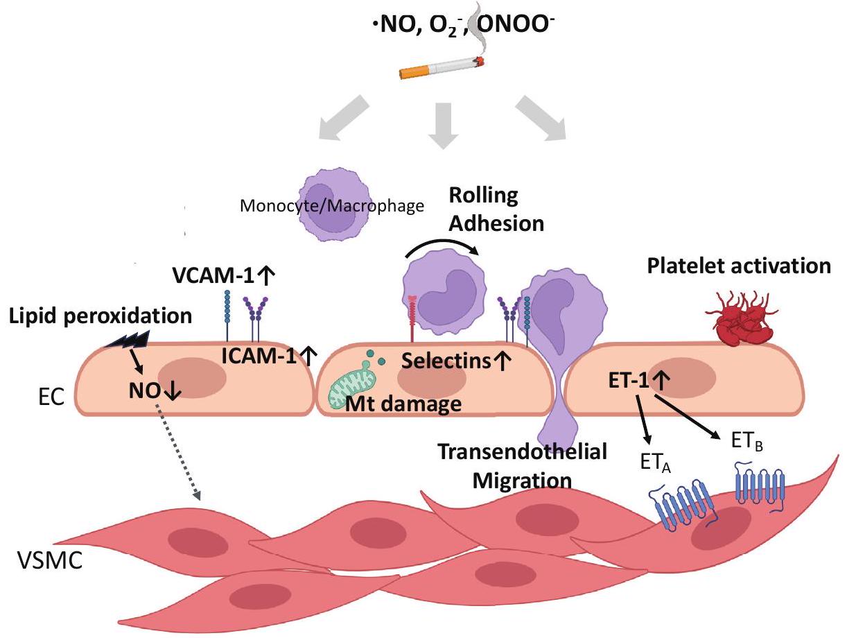
أفاد آخرون أن خلايا البطانة الوعائية المزروعة في وسط زراعة مدعوم بمصل المدخنين أظهرت انخفاضًا في إنتاج NO، وانخفاضًا في نشاط eNOS، وزيادة في إنتاج ROS الداخلي مقارنةً بغير المدخنين، والتي تم استعادتها بواسطة أدوية مضادة للأكسدة . تم الإبلاغ عن تنشيط أكسيداز الزانثين , نيكوتيناميد أدينين ثنائي النوكليوتيد فوسفات الهيدروجين (NADPH) , وسلسلة التنفس الميتوكوندري أيضًا كمساهمين في زيادة ROS في خلايا البطانة المعرضة لدخان السجائر. بالإضافة إلى ذلك، وُجد أن مستخلص دخان السجائر (CSE)، وهو مكون قابل للذوبان في الماء من دخان التبغ، يحفز مستوى معتدل من نفاذية الغشاء الخارجي للميتوكوندريا (MOMP) التي لا تحفز موت الخلايا المبرمج، أي MOMP الأقلية، بينما تقلل أيضًا من جهد الغشاء الميتوكوندري في خلايا البطانة. يؤدي هذا التحفيز إلى تسرب محتويات الميتوكوندريا، بما في ذلك الإلكترونات، الحمض النووي الميتوكوندري، وسيتوكروم C، إلى السيتوسول . يؤدي انخفاض جهد الغشاء الميتوكوندري والخلل الناتج في الميتوكوندريا
إلى زيادة في ). تساهم هذه الظاهرة أيضًا في تطور الالتهاب من خلال DAMPs، والتي سيتم مناقشتها في القسم التالي.

3. الالتهاب

كأهم مسببات تصلب الشرايين، يلعب الالتهاب المزمن لجدار الأوعية دورًا كبيرًا في تطوره وتقدمه . على الرغم من أن التدخين معروف على نطاق واسع بأنه يحفز الالتهاب، إلا أن الآليات التفصيلية التي من خلالها تؤدي عادات التدخين إلى الالتهاب في جدار الأوعية لا تزال قيد التحقيق. لقد تم إثبات أن التدخين يؤثر على النظام المناعي بشكل نظامي ومحلي. بين المدخنين، تم الإبلاغ عن زيادة في العدلات، اللمفاويات، والوحيدات مقارنةً بغير المدخنين . كما يظهر المدخنون مستويات مرتفعة بشكل ملحوظ من السيتوكينات الالتهابية في المصل مثل عامل نخر الورم- و إنترلوكين (IL)- . كما
تم ذكره لاحقًا، أظهرت التجارب التي أجريت في المختبر أن CSE يزيد من تعبير IL-6 و IL-8 . ومع ذلك، على حد علمنا، لم يتم إجراء تقارير متسقة تشير إلى أن التدخين يزيد من تركيزات IL-6 و IL-8 في دم الإنسان. ومع ذلك، هناك تقارير تشير إلى أن التدخين قد لا يغير تركيزات IL-6 و IL-8 في الدم وأخرى تشير إلى زيادة في تركيزات IL-6 و IL-8 في لعاب المدخنين . كما أظهر التدخين والتدخين السلبي أيضًا زيادة في مستويات المصل من علامة الالتهاب بروتين C-reactive (CRP) .
أبلغت التجارب في المختبر عن جوانب مختلفة من الالتهاب الناتج عن دخان السجائر. لقد تم إثبات أن دخان السجائر يحفز تعبير جزيئات الالتصاق على أسطح خلايا البطانة وإفراز سيتوكينات تعزز تصلب الشرايين مثل IL-6 و IL-8 . لقد أظهرنا أن التنشيط يشارك في التنظيم العلوي لـ IL-6 في خلايا البطانة . بالإضافة إلى ذلك، في خلايا الظهارة الرئوية البشرية والأرومات الليفية، ينشط دخان التبغ كيناز ميتوجين ومُنشط للإجهاد، مما يؤدي إلى تنشيط جينات التهابية تعتمد على -، بما في ذلك IL-6، IL-8، و COX-2، مصحوبة بتغيرات في بنية الكروماتين .
الالتهاب الوعائي هو متعدد العوامل. على الرغم من أن النظام المناعي الفطري مسؤول أساسًا عن الدفاع عن المضيف ضد الكائنات الأجنبية، تشير الأدلة المتزايدة إلى مشاركة النظام المناعي الفطري في الالتهاب الوعائي . كما اقترحت الرؤى الحديثة أن عدم تنظيم المناعة الفطرية يحفز الالتهاب المزمن عبر أعضاء متنوعة، مما يؤدي إلى حالات مثل تصلب الشرايين، الأمراض المناعية الذاتية، الأورام، والأمراض المرتبطة بالعمر . تلعب مستقبلات التعرف على الأنماط (PRRs) دورًا حاسمًا في الوظيفة السليمة للنظام المناعي الفطري , حيث تتعرف مباشرة على هياكل جزيئية محددة، مثل أنماط الجزيئات المرتبطة بالميكروبات (PAMPs)، من مسببات الأمراض الميكروبية وDAMPs التي يتم إفرازها من خلايا المضيف عند حدوث ضرر أو موت. تعبر خلايا متنوعة موجودة في آفات تصلب الشرايين، بما في ذلك خلايا البطانة، البلعميات، والخلايا الشجرية، عن مستقبلات شبيهة بالتول (TLRs)، وهي نوع من PRRs . الأكثر بحثًا في سياق تصلب الشرايين هو TLR9، الذي يتعرف على الحمض النووي غير الميثيل (CpG) (ديكسي نوكليوتيد) المستمد من البكتيريا والفيروسات ، بالإضافة إلى الحمض النووي المستمد من الذات . يؤدي هذا التعرف إلى تنشيط TLR9 وبدء استجابات مناعية فطرية متنوعة. في تجارب الفئران، تم إثبات أن TLR9 يلعب دورًا محوريًا في تطوير
تصلب الشرايين. على سبيل المثال، يؤدي تثبيط تنشيط TLR9 باستخدام أوليغوديوكسي نيوكليوتيدات تنظيمية مناعية إلى تحويل مرونة البلعميات إلى بلعميات مضادة للالتهابات ويثبط تشكيل لويحات تصلب الشرايين . على العكس، فإن إدارة المحفز TLR9 من خلال الحقن الوريدي تعزز تشكيل اللويحات في الفئران التي تفتقر إلى بروتين ApoE . فيما يتعلق بتأثير التدخين، تم الإبلاغ عن أن التعرض لبخار السجائر الإلكترونية يزيد من تعبير TLR9 في الوحيدات لدى الفئران التي تفتقر إلى ApoE، مما يؤدي إلى تسريع آفات تصلب الشرايين (الشكل 2). لم تؤدي إدارة مضاد TLR9 قبل التعرض لبخار السجائر الإلكترونية إلى تقليل تنظيم TLR9 في لويحات تصلب الشرايين فحسب، بل أيضًا إلى تثبيط الزيادة في مستويات البلازما من السيتوكينات الالتهابية وتراكم الدهون والبلعميات في اللويحات. علاوة على ذلك، تم إثبات أن الحمض النووي الميتوكوندري الخالي من الخلايا المعزول من البلعميات المعالجة بمستخلص بخار السجائر الإلكترونية ينشط TLR9 في خلايا التقرير، وأن تثبيط TLR9 يثبط تحفيز السيتوكينات الالتهابية في البلعميات .
تم الإبلاغ عن مشاركة الانفلامازوم في تقدم تصلب الشرايين . NLRP3 هو مستشعر داخل الخلايا يكشف عن مجموعة واسعة من PAMPs بالإضافة إلى DAMPs المستمدة من الذات والأجنبية، مثل ATP، بلورات الكوليسترول، الحمض النووي الميتوكوندري المؤكسد، GMP-AMP الحلقي، والجسيمات النانوية، مما يؤدي إلى تشكيل وتنشيط الانفلامازوم NLRP3 AIM2 هو مكون من مكونات الإنفلامازوم يختلف عن NLRP3 ويعمل كبروتين حساس للحمض النووي يتعرف على الحمض النووي البكتيري والفيروسي، بالإضافة إلى الحمض النووي مزدوج الشريط الداخلي. تؤدي تجميع الإنفلامازوم إلى إطلاق السيتوكينات المؤيدة للالتهابات IL-1 المعتمدة على الكاسبيز 1. و IL-18 بالإضافة إلى موت الخلايا النخرية الناتج عن غازدرمين D (GSDMD)، مما يسهل الإفراج الخارجي عن IL-1 و IL-18 زراعة خلايا نخاع العظم التي تفتقر إلى NLRP3 و ASC و IL-1 إلى الفئران التي تعاني من نقص مستقبلات البروتين الدهني منخفض الكثافة (LDLR) المعرضة لتصلب الشرايين، قللت من حجم الآفة التصلبية. بشكل متسق، أظهرت الفئران التي تفتقر إلى ApoE وتفتقر إلى caspase-1/11 والفئران التي تفتقر إلى LDLR وتفتقر إلى caspase-1/11 تقدمًا أقل في تصلب الشرايين. توفر هذه النتائج دليلاً مباشراً على مساهمة إنزيم NLRP3 في تقدم تصلب الشرايين. علاوة على ذلك، تم ملاحظة زيادة في تعبير AIM2 وتراكم الحمض النووي مزدوج الشريط خارج الخلية في الفئران التي تفتقر إلى ApoE والتي تعاني من تصلب شرايين متقدم. أدى الإفراط في التعبير عن AIM2 في الفئران التي تفتقر إلى ApoE والمُغذّاة بنظام غذائي عالي الدهون إلى تكوين لويحات تحتوي على المزيد من الأوعية الدموية المسببة للاحتراق.
الشكل 2. مخطط توضيحي لآليات الالتهاب الناتج عن التدخين
تُعزى آلية تطور تصلب الشرايين بشكل رئيسي إلى الالتهاب المزمن في جدار الأوعية الدموية، مع تدخين السجائر كعامل مُحفز للالتهاب. يلعب الجهاز المناعي الفطري، الذي يتضمن مستقبلات التعرف على الأنماط، دورًا محوريًا، وخاصة TLR9، وNLRP3/AIM2، وcGAS-STING، في آفات تصلب الشرايين. يتم تنشيط cGAS الناتج عن دخان السجائر بشكل رئيسي بواسطة mtDNA الحر في السيتوسول الذي يتم إطلاقه من خلال MOMP الأقلية. تؤدي كل من مسارات TLR9 وcGAS إلى زيادة تعبير السيتوكينات الالتهابية، مثل IL-6 وIL-8. كما ينشط mtDNA الحر في السيتوسول أيضًا AIM2 inflammasome الذي يستشعر الحمض النووي، بينما ينشط ROS الناتج عن دخان السجائر NLRP3 inflammasome؛ وكلاهما يؤديان إلى تحفيز الموت الخلوي المبرمج GSDMD وإطلاق السيتوكينات الالتهابية مثل IL-1. و IL-18 إلى الفضاء خارج الخلوي. بالإضافة إلى ذلك، يؤدي دخان السجائر إلى نوع آخر من موت الخلايا، وهو الفيروبتوز، مما يؤدي إلى تنشيط عامل النسخ الحساس للأكسدة، وتعبير MMP، والالتهاب في خلايا العضلات الملساء الوعائية. تطلق الأنسجة التالفة DAMPs، مما يساهم في المناعة الفطرية والالتهاب. تولد آليات أخرى مثل NETs ROS وDAMPs وت activates NLRP3 inflammasome في البلعميات؛ كما تساهم في تكوين تصلب الشرايين. يتم تقديم الآليات الخاصة بالخلايا للالتهاب الناتج عن التدخين في هذه الصورة. EC، خلية بطانية؛ VSMC، خلية عضلية ملساء وعائية؛ TLR، مستقبل شبيه بالتول؛ cGAS، سينثاز GMP-AMP الحلقي؛ STING، منبه جينات الإنترفيرون؛ mtDNA، الحمض النووي الميتوكوندري؛ MOMP، نفاذية الغشاء الخارجي للميتوكوندريا؛ IL، إنترلوكين؛ ROS، أنواع الأكسجين التفاعلية؛ GSDMD، غازدرمين D؛ MMP، ميتالوبروتياز المصفوفة؛ DAMPs، أنماط جزيئية مرتبطة بالضرر؛ NETs: مصائد خارج الخلوية للعدلات. تم إنشاؤه باستخدام بيوريندر.كوم.
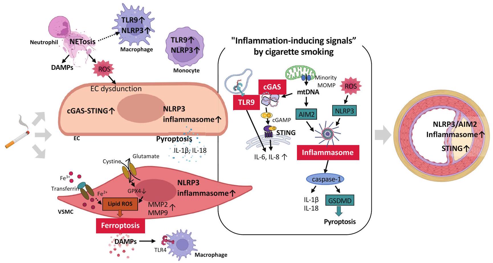
خلايا العضلات الملساء وزيادة تجنيد البلعميات داخل اللويحات تسلط هذه الدراسات الضوء بشكل جماعي على الدور المحوري للإنفلامازوم في تقدم تصلب الشرايين. في سياق تطور تصلب الشرايين الناتج عن دخان السجائر، تم الإبلاغ عن أن تنشيط إنفلامازوم NLRP3 يعد أمرًا حاسمًا (الشكل 2). لقد أظهرت الدراسات أن التعرض لدخان السجائر ينشط إنفلامازوم NLRP3 في مراحل خلوية مختلفة، بما في ذلك وحيدات النوى، البلعميات، وخلايا الرغوة، مما يساهم في بدء وتقدم وتطور تصلب الشرايين. . من المثير للاهتمام أن مكثف دخان السجائر أدى إلى تمايز أحادية THP-1 إلى بلاعم، مما نتج عنه تنشيط محدد المراحل لجهاز الالتهاب NLRP3 وزيادة في IL-1 وإفراز IL-18 . علاوة على ذلك، فإن علاج النيكوتين في خلايا العضلات الملساء الوعائية للفئران
وأدى نسيج الشريان الأورطي للفئران إلى زيادة إنتاج الجذور الحرة للأكسجين، وتنشيط إنزيم NLRP3، وارتفاع مستوى بروتين CRP. في الفئران التي تفتقر إلى بروتين ApoE والمغذاة على نظام غذائي عالي الدهون، زاد النيكوتين من آفات تصلب الشرايين وزاد من السيتوكينات الالتهابية، ربما من خلال تحفيز موت الخلايا البيروبتوزية في الخلايا البطانية عبر ROS-NLRP3. ، حيث قامت النيكوتين بتنشيط NLRP3 من خلال ROS، مما أدى إلى الموت الخلوي النخرى بالإضافة إلى IL-1 وإنتاج IL-18 في خلايا بطانة الشريان الأورطي البشري أفادت الأبحاث السريرية بزيادة التعبير النسخي والترجمة لعوامل علامة NLRP3 inflammasome (كاسبيز-1، برو-IL-1 IL-1 ، البروتين IL-18، و IL-18) في أحادية النواة لدى المدخنين الذين يعانون من مرض الشريان التاجي (CAD) مقارنة بغير المدخنين الذين يعانون من CAD . بالإضافة إلى ذلك، علامات الإجهاد التأكسدي (8-إيزوبروستان و8-أوكسي-2′-ديوكسيغوانوزين)، كاسبيز-1، IL-1 وتم العثور على IL-18 في المصل ليكون
يكون مرتفعًا لدى المدخنين الذين يعانون من مرض الشريان التاجي .
إنزيم سينثاز GMP-AMP الحلقي (cGAS)، المعروف بأنه مستشعر للحمض النووي السيتوزولي، يتم تفعيله ليس فقط بواسطة الحمض النووي المشتق من مسببات الأمراض ولكن أيضًا بواسطة الحمض النووي الداخلي. يتم التعبير عنه في خلايا متنوعة، بما في ذلك الخلايا الدبقية الصغيرة، الخلايا العصبية، الخلايا الكبدية، وحيدات النوى المحيطية، والخلايا الشجرية، البلعميات، والخلايا القلبية الوعائية، مثل الخلايا البطانية، خلايا العضلات الملساء الوعائية، خلايا القلب، والخلايا الليفية. يحفز cGAS إنتاج الرسول الثاني 2’3′-cGAMP، الذي ينشط المحفز المرتبط بالشبكة الإندوبلازمية لجينات الإنترفيرون (STING)، مما يؤدي في النهاية إلى تحفيز الإنترفيرونات من النوع الأول والجينات الالتهابية. في نماذج حيوانية من تصلب الشرايين، تم إثبات أن مسار cGAS-STING يلعب دورًا محوريًا في تحفيز الالتهاب في تطور تصلب الشرايين. في الفئران التي تفتقر إلى بروتين ApoE والمُغذية بنظام غذائي عالي الدهون، كانت علامات تلف الحمض النووي وتعبير بروتين STING مرتفعة في البلعميات الأبهريّة، وقد حسّن تثبيط STING بالعقاقير أو الحذف الجيني لـ STING من تصلب الشرايين في هذه الفئران. تشير هذه النتائج إلى تورط STING في تقدم تصلب الشرايين الناتج عن تناول نظام غذائي عالي الدهون. لقد أظهرنا مؤخرًا أن CSE القابل للذوبان في الماء يسبب تلفًا في الحمض النووي النووي والميتوكوندري في خلايا البطانة الوعائية، مما يؤدي إلى زيادة في الحمض النووي السيتوزولي، وهو DAMP. وهذا بدوره ينشط مسار cGAS-STING، مما يؤدي إلى زيادة التعبير عن السيتوكينات الالتهابية المعتمدة على cGAS-STING. (الشكل 2). بينما زادت كل من الحمض النووي النووي والحمض النووي الميتوكوندري بواسطة CSE، تم تحديد الحمض النووي الميتوكوندري كأحد المساهمين الرئيسيين في تحفيز IL-6 بين السيتوكينات الالتهابية. كما يظهر المدخنون مستويات مرتفعة من الحمض النووي الخالي من الخلايا المتداول المشتق من كل من المصادر النووية والميتوكوندرية، مما يشير إلى إمكانية تفاقم الالتهاب الجهازي. تسلط هذه النتائج مجتمعة الضوء على الدور الحاسم للإشارات التي تتوسطها مستقبلات PRR وحساسات الحمض النووي من خلال التعرف على DAMP في الالتهاب الناتج عن دخان السجائر.
كما ذُكر سابقًا، فإن DAMPs هي هياكل جزيئية تُطلق من الأنسجة التالفة أو الخلايا النخرية، مما يساهم في تحفيز المناعة الفطرية والالتهاب. وقد تم اقتراح أن أشكالًا مختلفة من موت الخلايا يمكن أن تُ triggered بواسطة دخان السجائر، مما يؤدي إلى السبب الأساسي لتصلب الشرايين. تم الإبلاغ عن أن الفيروبتوزيس، وهو نوع حديث من موت الخلايا، يتم تحفيزه بواسطة CSE في خلايا العضلات الملساء الوعائية، وهو مختلف عن الموت الخلوي المبرمج أو النخر. (الشكل 2). السمة المركزية للفيروبتوز هي وجود الحديد النشط من الناحية الحمراء ووظيفة غير طبيعية لـ
أكسدة الدهون تشير الأدلة المتزايدة إلى أن الفيروبتوزيس يؤثر بشكل كبير على تنظيم الإجهاد التأكسدي والاستجابات الالتهابية ويشارك في أمراض القلب والأوعية الدموية الناتجة عن تصلب الشرايين. أبلغ سامبيلفانجيل وآخرون أن الزيادات الناتجة عن CSE في التعبير عن السيتوكينات الالتهابية، مثل IL-1 و IL-6، بالإضافة إلى MMP-2 و MMP-9 في خلايا العضلات الملساء الوعائية، يتم تثبيطها بواسطة مثبط الفيروبتوز ferrostatin-1 علاوة على ذلك، كشفت التجارب خارج الجسم أن CSE يسبب فقدان الطبقة الوسطى في نسيج الشريان الأورطي، والذي يتم قمعه بواسطة فيروستاتين-1. تشير هذه النتائج إلى أن الموت الخلوي الناتج عن CSE يحرر DAMPs مثل الأحماض النووية وصندوق المجموعة عالية الحركة 1، المعترف بها بواسطة مستقبلات المناعة الفطرية مثل TLR4 على البلعميات. ، مما يعزز الالتهاب في النهاية (الشكل 2). إن حقيقة أن آلية الفيرواستاتين-1 تتضمن التقاط الجذور الحرة للدهون الغشائية تدعم إمكانية أن تؤثر أكسدة الدهون على عوامل النسخ الحساسة للأكسدة مثل وAP-1، اللذان يلعبان أدوارًا محورية في التعبير عن السيتوكينات وMMPs. تشير هذه الرؤى إلى تورط فقدان خلايا العضلات الملساء الوعائية، والالتهاب، وتدهور المصفوفة الناتج عن CSE في تطور تمدد الأوعية الدموية الأبهري والانفصالات، والتي يعتبر التدخين عامل خطر رئيسي لها.
آليات جزيئية أخرى مثل الفخاخ خارج الخلوية للعدلات (NETs) والالتهام الذاتي تنتج DAMPs وتعدل المناعة الفطرية. يتم حالياً التحقيق في دورها في مسببات تصلب الشرايين. الفخاخ خارج الخلوية هي هياكل تتكون من الكروماتين الذي يحتوي على الحمض النووي المحرر والذي يتشكل أثناء تنشيط العدلات. هذه الهياكل تتوزع مع بروتينات متنوعة وتلعب دورًا حاسمًا في التقاط وتعقيم الكائنات الدقيقة في البيئة خارج الخلوية. تشير الأدلة الحديثة إلى أن الفخاخ خارج الخلوية متورطة في ظروف مرضية فسيولوجية متنوعة، بما في ذلك تصلب الشرايين، والتخثر، والأمراض المناعية الذاتية، والسرطان، والسكري، ومرض الزهايمر. لقد تم ملاحظة الفخاخ خارج الخلوية في آفات تصلب الشرايين في كل من النماذج البشرية والحيوانية، وقد تم الإبلاغ عن تورطها في آليات متنوعة تؤدي إلى تكوين تصلب الشرايين. . على وجه الخصوص، تحفز NETs الإجهاد التأكسدي، وتعزز خلل وظيفة الخلايا البطانية والموت المبرمج، وتساهم في تطور الالتهاب من خلال تحفيز توليد DAMPs، كما ذُكر سابقًا. بالإضافة إلى ذلك، تشكل مصفوفة شبيهة بالفبرين لالتصاق الصفائح الدموية وتنشيطها وتجمعها. كما أنها تعزز تراكم الجزيئات التي تعزز التخثر مثل عامل فون ويلبراند والفبرينوجين، مما يساهم بشكل كبير في تشكيل الجلطة وانتشارها. وقد تم الإبلاغ عن أن النيكوتين يحفز NETs في
الشكل 3. التجلط الشرياني الناتج عن دخان السجائر
دخان السجائر ينشط الخلايا البطانية، مما يعزز تعبير السليكتينات البطانية (P – و E – سليكتين)، ICAM-1، و VCAM1، مما يعزز التصاق وحيدات النواة/الخلايا اللمفاوية والصفائح الدموية بالبطانة الالتهابية. تعبير vWF الموجود في الأنسجة تحت البطانية المعرضة من الخلايا البطانية المعطلة المصابة يعزز التصاق الصفائح الدموية وتفاعلات التجمع، مما يؤدي إلى تشكيل جلطات صفائحية. علاوة على ذلك، يساهم التدخين في تشكيل لويحات ضعيفة من خلال زيادة الالتهاب وزيادة نشاط MMPs. التعرض لدخان السجائر يحفز تعبير MMP-9 وMMP-12 في البلعميات، وMMP-1 وMMP-8 وMMP-9 في الخلايا البطانية، وMMP-2 وMMP-9 في خلايا العضلات الملساء الوعائية. ICAM، جزيء الالتصاق بين الخلايا؛ VCAM، جزيء الالتصاق الخلوي الوعائي؛ vWF، عامل فون ويلبراند؛ MMP، ميتالوبروتياز المصفوفة؛ VSMC، خلايا العضلات الملساء الوعائية.بيوريندر.كوم.
خلايا شبيهة بالعدلات مشتقة من خلايا HL-60 (الشكل 2). تؤدي NETs المستحثة بواسطة النيكوتين إلى تنظيم سلبى للاوتوفاجي في البلعميات، مما يؤدي إلى تنشيط inflammasome. إن تثبيط تكوين NET عن طريق استنفاد إنزيم الببتيديل أرجينين دييميناز 4 في كريات الدم البيضاء العدلية يؤدي إلى تقليل لويحات تصلب الشرايين في الفئران المعتمدة على ApoE التي تم علاجها بالنيكوتين. وهذا يشير إلى أن NETs تعزز تكوين تصلب الشرايين من خلال تنشيط inflammasome عن طريق تثبيط البلعمة الذاتية للماكروفاج. .

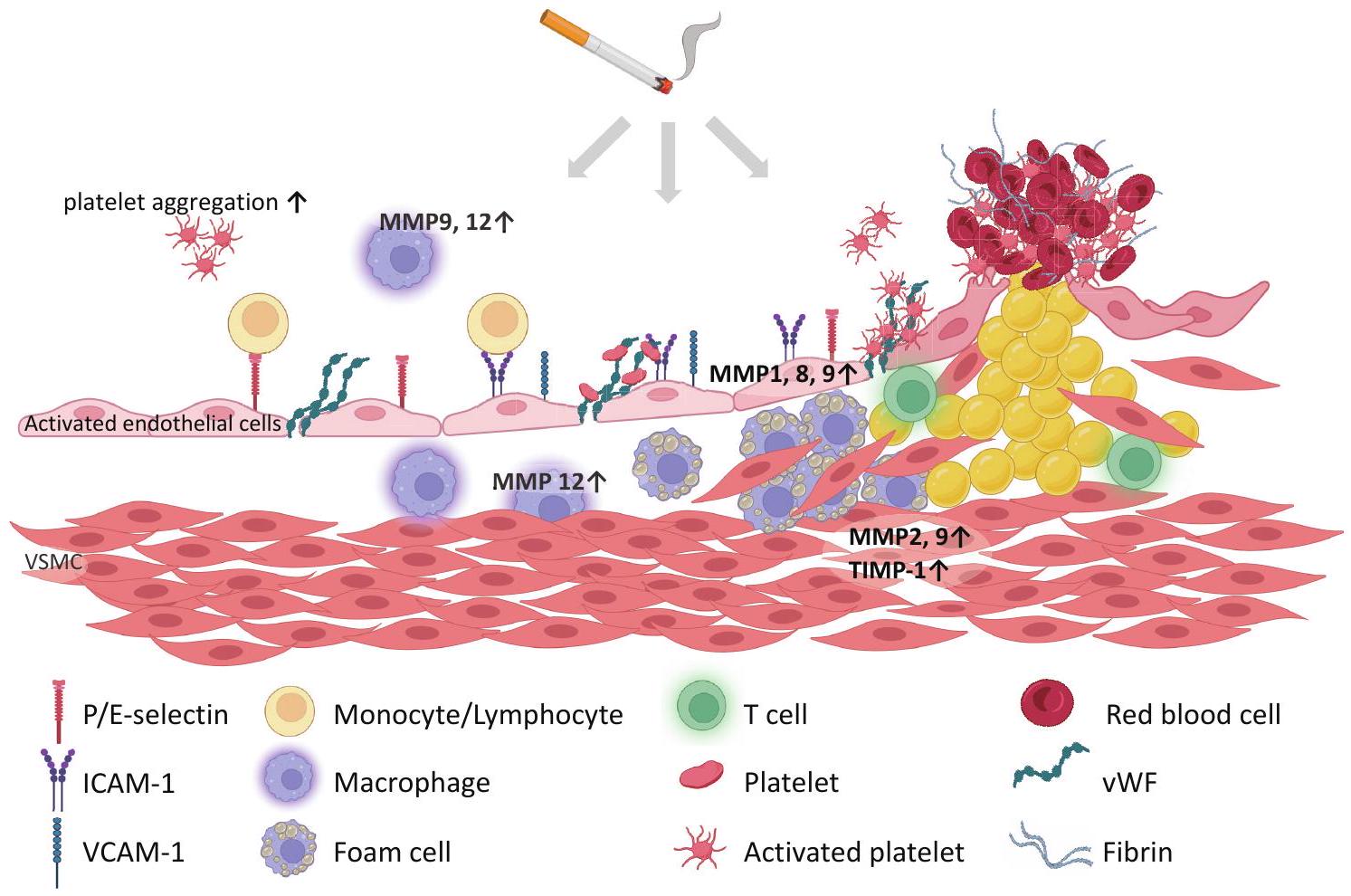
4. التأثيرات التخثرية

بين المرضى الذين يعانون من متلازمة الشريان التاجي الحادة، فإن المدخنين لديهم معدل أعلى من تجلط الدم داخل الدعامة مقارنة بغير المدخنين. وقد تم الإبلاغ عن أن من حالات الوفاة القلبية المفاجئة بسبب التخثر الشرياني الحاد كانوا مدخنين . لفترة طويلة، زيادة الميل لتجمع الصفائح الدموية وتجلط الدم لدى الأفراد المعرضين
تم الإبلاغ عن دخان السجائر (الشكل 3). تم إثبات وجود ارتباط إيجابي بين درجة التدخين ومدى تنشيط الصفائح الدموية سواء في الجسم الحي وفي المختبر تم اقتراح آليات متعددة لزيادة تجمع الصفائح الدموية لدى المدخنين. زيادة عدد الصفائح الدموية وقد وُجدت مستويات مرتفعة من عامل تنشيط الصفائح الدموية (PAF) والدهون الشبيهة بـ PAF في المدخنين. تشير بعض الدراسات إلى أن الإجهاد التأكسدي يلعب دورًا محوريًا في تنشيط الصفائح الدموية الناتج عن التدخين. وقد أظهرت ن-أسيتيل سيستين، وهو مضاد أكسدة، أنه يحمي من تأثير دخان السجائر على تدهور تحلل ADP الذي تسببه الصفائح الدموية. أظهرت دراسة سابقة أن التدخين على المدى الطويل يعيق إفراز الصفائح الدموية . هذا يشير إلى عدم توازن في تنظيم الصفائح الدموية مما يؤدي إلى زيادة تجمع الصفائح الدموية حيث أن أكسيد النيتريك هو مثبط قوي للصفائح الدموية. أظهرت التحليلات البروتينية مع الصفائح الدموية من المدخنين زيادة في تعبير الجزيئات المؤيدة للالتهابات، بما في ذلك الصفائح الدموية
مستقبل الفبرينوجين الجليكوبروتين IIb/IIIa، مقارنة بغير المدخنين التعرض لدخان السجائر يعزز تنشيط الصفائح الدموية بسبب زيادة في الثromboxane. تشكيل كما أنه يعزز توليد الثرومبين المعتمد على الصفائح الدموية، وهو منشط قوي للغاية للصفائح الدموية، مما قد يؤدي إلى حلقة تغذية راجعة إيجابية لتنشيط الصفائح الدموية والتخثر. .
كما ذُكر سابقًا، تتعرض خلايا البطانة للأذى والتفعيل بسبب دخان السجائر، مما يجعلها مواقع للالتهاب والميول التخثرية. في خلايا البطانة المفعلة بالتدخين، تم الإبلاغ عن زيادة في إنتاج عامل فون ويلبراند وثrombomodulin. (الشكل 3). تعرض المصفوفة تحت البطانية للدم المتدفق يعزز تنشيط الصفائح الدموية وتجمعها من خلال التفاعلات بين مستقبلات الجليكوبروتين السطحية للصفائح الدموية والكولاجين تحت البطاني وعامل فون ويلبراند المثبت. تعتبر هذه العملية المرحلة الأولية لتكوين الجلطة المرضية. بالإضافة إلى زيادة تجميع الصفائح الدموية والتخثر، تم الإبلاغ عن أن التعرض لدخان السجائر مرتبط بحالة من ضعف الليفانوليز. .
تساهم زيادة الالتهاب وزيادة نشاط MMP في تشكيل اللويحات القابلة للخطر. التعرض لدخان السجائر يحفز تعبير أنواع مختلفة من MMPs في أنواع خلايا مختلفة (الشكل 3). لقد تم إثبات أن دخان السجائر أو مستخلص دخان السجائر يحفز التعبير عن MMP-12، وعامل نخر الورم، وعامل الأنسجة في البلعميات الهوائية الرئوية وأنسجة الرئة. . علاوة على ذلك، أظهرت اللويحات المأخوذة من المدخنين الذين خضعوا لجراحة استئصال الشريان السباتي زيادة في تعبير MMP-12 المشتق من البلعميات مقارنة بغير المدخنين. في خلايا بطانة الشريان الأورطي والشرايين التاجية البشرية المعرضة لدخان السجائر أو CSE، زاد تعبير MMP-1 وMMP-8 وMMP-9 وقد أظهرت الدراسات أن النيكوتين يعزز تعبير MMP في خلايا العضلات الملساء تم الإبلاغ عن عدة آليات تكمن وراء تعبير MMP. أدى CSE إلى تحفيز تعبير MMP-2 وMMP-9 ومثبط الأنسجة للميتالوبروتيناز-1 (TIMP-1) في خلايا العضلات الملساء الوعائية، والتي تم الإبلاغ عن أنها توقفت بواسطة مثبطات محددة للفيروبتوز. تشمل مكونات دخان السجائر المسؤولة عن تحفيز الفيروبتوز في خلايا العضلات الملساء الوعائية الأكرولين وكيتون الفينيل الميثيلي. كما تم إثبات أن الأكرولين ودخان السجائر يزيدان من تعبير MMP-1 ويقللان من تعبير TIMP-3 في خلايا بطانة الشريان الأورطي للأرانب، وهو ما يُعزى إلى تثبيط مسار الهدف الثديي من رابيمايسين-p70S6K. في الفئران التي تفتقر إلى ApoE، قام الأكرولين بتنشيط MMP-9 في
البلاعم البشرية ونشاط MMP المحفز بطريقة تعتمد على الإجهاد التأكسدي في آفات تصلب الشرايين المتقدمة العلاقة بين احتشاء عضلة القلب وانتشار أمراض القلب والأوعية الدموية والتعرض لتلوث الهواء الغني بالأكرولين موثقة جيدًا. مما يشير إلى أن تنشيط MMP قد يلعب دورًا حاسمًا كعامل عدم استقرار في اللويحات الحادة.
بشكل عام، يرتبط الالتهاب الجهازي بانفجار اللويحات وحدوث متلازمة الشريان التاجي الحادة. , مما يبرز أهمية الالتهاب في قابلية اللويحات للتعرض للخطر. علاوة على ذلك، قد يحفز دخان السجائر تراكم وحيدات النواة المتداولة داخل الغلاف الليفي للويحات المستقرة من خلال تعزيز التعبير عن جزيئات الالتصاق وبروتين جذب وحيدات النواة-1 . عند تمزق اللويحة، تتأثر جميع عمليات التصاق الصفائح الدموية والتجمع، وتفعيل سلسلة التخثر، وتوازن التحلل الفيبريني من خلال تفاعلات معقدة. كما ذُكر سابقًا، يحفز دخان السجائر الالتهاب في الخلايا، مما يؤدي إلى زيادة التعبير عن السيتوكينات الالتهابية وجزيئات الالتصاق . تعزز خلايا البطانة الالتهابية التعبير عن E – و P -selectin، وجزيء الالتصاق بين الخلايا-1، وجزيء الالتصاق الخلوي الوعائي-1 (الأشكال 1 و 3)، مما يعزز الالتصاق الثابت للصفائح الدموية بالبطانة الالتهابية من خلال التفاعلات مع مستقبلات الصفائح الدموية، مما يؤدي في النهاية إلى تشكيل جلطة وعائية انسدادية . علاوة على ذلك، يوجد حلقة تغذية راجعة إيجابية حيث تحفز الجزيئات الحيوية المشتقة من الصفائح الدموية مثل IL-1 و CD40 ligand -زيادة التعبير عن جزيئات الالتصاق الباطنية، مما يسهل المزيد من التصاق الصفائح الدموية وخلايا الدم البيضاء بجدار الشرايين .

5. الاستنتاجات وآفاق المستقبل

لقد ثبت أن تدخين السجائر له آثار ضارة على صحة القلب والأوعية الدموية، مع تركيز خاص على تصلب الشرايين والتخثر الشرياني. لقد أبرزت هذه المراجعة الشاملة الآليات المعقدة التي يساهم بها التدخين في مثل هذه الحالات المهددة للحياة، بما في ذلك خلل البطانة، الالتهاب، وآثار التخثر الشرياني. باختصار، يُعتبر خلل البطانة، الذي يتميز بانخفاض توافر NO وزيادة إنتاج الأنيون الفائق، أحد أوائل مظاهر تصلب الشرايين الناتج عن التدخين. يُعتبر الالتهاب الناتج عن التدخين عاملًا مسببًا مركزيًا في تصلب الشرايين الناتج عن التدخين، مع بعض المشاركة في الاستجابة الالتهابية التي تتوسطها
PRRs و DAMPs. هذه الآليات تحفز الالتهاب المزمن في جدار الأوعية الدموية وتساهم في تقدم تصلب الشرايين. علاوة على ذلك، يؤثر دخان السجائر بشكل مباشر على التخثر الشرياني من خلال تعزيز تنشيط الصفائح الدموية، والتصاقها، وتجمعها، بالإضافة إلى إرباك سلسلة التخثر وتوازن التحلل الفيبريني. تساهم MMPs أيضًا في قابلية اللويحات للأذى والأحداث التخثرية. يرتبط خلل البطانة، الالتهاب، والتخثر الشرياني ببعضها البعض ويزيد من تفاقم كل منها للآخر. نظرًا للعبء الكبير على الصحة العامة الذي تسببه أمراض القلب والأوعية الدموية، خاصة في المجتمعات المدخنة، من الضروري تسليط الضوء على الحاجة الملحة للإقلاع عن التدخين. إن فهم هذه الآليات متعددة الأوجه عند تقاطع وظيفة البطانة، الالتهاب، والتخثر يبرز أهمية المبادرات والعلاجات المضادة للتدخين للحفاظ على صحة القلب والأوعية الدموية. يجب أن تستمر الأبحاث المستقبلية في استكشاف المسارات الجزيئية المحددة والتدخلات العلاجية المحتملة التي تستهدف هذه الآليات، مما يوفر في النهاية استراتيجيات أكثر فعالية للتخفيف من المخاطر القلبية الوعائية المرتبطة بتدخين السجائر.

الشكر والتقدير

تم دعم هذه الدراسة جزئيًا من خلال منح البحث العلمي (KAKENHI #21K080290 إلى M.I.، KAKENHI #22K160760 إلى C.S.، و KAKENHI#23K07557 إلى T.I.); منحة بحثية من مؤسسة تسوتشيا الطبية التذكارية إلى M.I.; منحة من مؤسسة أبحاث التدخين (M.I. و C.S.).

المصالح المتنافسة

T.I. هو رئيس دورة موقوفة من قبل BIOTRONIK Japan, Inc. و Abbot Medical Japan LLC و Japan Lifeline Co., Ltd. لم يعلن المؤلفون الآخرون عن أي تضارب محتمل في المصالح فيما يتعلق بالبحث، التأليف، و/أو نشر هذه المقالة.

References

  1. Outline of Health, Labour and Welfare Statistics 2022. 2022
  2. Ezzati M, Henley SJ, Thun MJ, Lopez AD: Role of smoking in global and regional cardiovascular mortality. Circulation, 2005; 112: 489-497
  3. Anderson KM, Wilson PW, Odell PM, Kannel WB: An updated coronary risk profile. A statement for health professionals. Circulation, 1991; 83: 356-362
  4. Iso H, Date C, Yamamoto A, Toyoshima H, Watanabe Y, Kikuchi S, Koizumi A, Wada Y, Kondo T, Inaba Y,
Tamakoshi A: Smoking cessation and mortality from cardiovascular disease among Japanese men and women: the JACC Study. Am J Epidemiol, 2005; 161: 170-179
5) Smith CJ, Fischer TH: Particulate and vapor phase constituents of cigarette mainstream smoke and risk of myocardial infarction. Atherosclerosis, 2001; 158: 257267
6) Csordas A, Bernhard D: The biology behind the atherothrombotic effects of cigarette smoke. Nat Rev Cardiol, 2013; 10: 219-230
7) Barua RS, Ambrose JA: Mechanisms of coronary thrombosis in cigarette smoke exposure. Arterioscler Thromb Vasc Biol, 2013; 33: 1460-1467
8) Kobayashi Y, Sakai C, Ishida T, Nagata M, Nakano Y, Ishida M: Mitochondrial DNA is a key driver in cigarette smoke extract-induced IL-6 expression. Hypertens Res, 2023; doi: 10.1038/s41440-023-01463-z. ePub ahead of print
9) Messner B, Bernhard D: Smoking and cardiovascular disease: mechanisms of endothelial dysfunction and early atherogenesis. Arterioscler Thromb Vasc Biol, 2014; 34: 509-515
10) Jaén RI, Val-Blasco A, Prieto P, Gil-Fernández M, Smani T, López-Sendón JL, Delgado C, Boscá L, FernándezVelasco M: Innate Immune Receptors, Key Actors in Cardiovascular Diseases. JACC Basic Transl Sci, 2020; 5: 735-749
11) Zindel J, Kubes P: DAMPs, PAMPs, and LAMPs in Immunity and Sterile Inflammation. Annu Rev Pathol, 2020; 15: 493-518
12) Sampilvanjil A, Karasawa T, Yamada N, Komada T, Higashi T, Baatarjav C, Watanabe S, Kamata R, Ohno N, Takahashi M: Cigarette smoke extract induces ferroptosis in vascular smooth muscle cells. Am J Physiol Heart Circ Physiol, 2020; 318: H508-h518
13) Wang Y, Zhao Y, Ye T, Yang L, Shen Y, Li H: Ferroptosis Signaling and Regulators in Atherosclerosis. Front Cell Dev Biol, 2021; 9: 809457
14) Fuster V, Moreno PR, Fayad ZA, Corti R, Badimon JJ: Atherothrombosis and high-risk plaque: part I: evolving concepts. J Am Coll Cardiol, 2005; 46: 937-954
15) Celermajer DS, Sorensen KE, Gooch VM, Spiegelhalter DJ, Miller OI, Sullivan ID, Lloyd JK, Deanfield JE: Noninvasive detection of endothelial dysfunction in children and adults at risk of atherosclerosis. Lancet, 1992; 340: 1111-1115
16) Johnson HM, Gossett LK, Piper ME, Aeschlimann SE, Korcarz CE, Baker TB, Fiore MC, Stein JH: Effects of smoking and smoking cessation on endothelial function: 1 -year outcomes from a randomized clinical trial. J Am Coll Cardiol, 2010; 55: 1988-1995
17) Otsuka R, Watanabe H, Hirata K, Tokai K, Muro T, Yoshiyama M, Takeuchi K, Yoshikawa J: Acute effects of passive smoking on the coronary circulation in healthy young adults. Jama, 2001; 286: 436-441
18) Rahman MM, Laher I: Structural and functional alteration of blood vessels caused by cigarette smoking: an overview of molecular mechanisms. Curr Vasc Pharmacol, 2007; 5: 276-292
19) Heitzer T, Just H, Münzel T: Antioxidant vitamin C improves endothelial dysfunction in chronic smokers.
Circulation, 1996; 94: 6-9
20) Heitzer T, Brockhoff C, Mayer B, Warnholtz A, Mollnau H, Henne S, Meinertz T, Münzel T: Tetrahydrobiopterin improves endothelium-dependent vasodilation in chronic smokers: evidence for a dysfunctional nitric oxide synthase. Circ Res, 2000; 86: E36-41
21) Barua RS, Ambrose JA, Srivastava S, DeVoe MC, EalesReynolds LJ: Reactive oxygen species are involved in smoking-induced dysfunction of nitric oxide biosynthesis and upregulation of endothelial nitric oxide synthase: an in vitro demonstration in human coronary artery endothelial cells. Circulation, 2003; 107: 2342-2347
22) Kayyali US, Budhiraja R, Pennella CM, Cooray S, Lanzillo JJ, Chalkley R, Hassoun PM: Upregulation of xanthine oxidase by tobacco smoke condensate in pulmonary endothelial cells. Toxicol Appl Pharmacol, 2003; 188: 59-68
23) Jaimes EA, DeMaster EG, Tian RX, Raij L: Stable compounds of cigarette smoke induce endothelial superoxide anion production via NADPH oxidase activation. Arterioscler Thromb Vasc Biol, 2004; 24: 1031-1036
24) Miró O, Alonso JR, Jarreta D, Casademont J, UrbanoMárquez A, Cardellach F: Smoking disturbs mitochondrial respiratory chain function and enhances lipid peroxidation on human circulating lymphocytes. Carcinogenesis, 1999; 20: 1331-1336
25) Ueda K, Sakai C, Ishida T, Morita K, Kobayashi Y, Horikoshi Y, Baba A, Okazaki Y, Yoshizumi M, Tashiro S, Ishida M: Cigarette smoke induces mitochondrial DNA damage and activates cGAS-STING pathway: application to a biomarker for atherosclerosis. Clin Sci (Lond), 2023; 137: 163-180
26) Suski JM, Lebiedzinska M, Bonora M, Pinton P, Duszynski J, Wieckowski MR: Relation Between Mitochondrial Membrane Potential and ROS Formation. In: Palmeira CM, Moreno AJ, eds. Mitochondrial Bioenergetics: Methods and Protocols. Totowa, NJ: Humana Press; 2012: 183-205
27) Lavi S, Prasad A, Yang EH, Mathew V, Simari RD, Rihal CS, Lerman LO, Lerman A: Smoking is associated with epicardial coronary endothelial dysfunction and elevated white blood cell count in patients with chest pain and early coronary artery disease. Circulation, 2007; 115: 2621-2627
28) Barbieri SS, Zacchi E, Amadio P, Gianellini S, Mussoni L, Weksler BB, Tremoli E: Cytokines present in smokers’ serum interact with smoke components to enhance endothelial dysfunction. Cardiovasc Res, 2011; 90: 475483
29) Wang H, Chen H, Fu Y, Liu M, Zhang J, Han S, Tian Y, Hou H, Hu Q: Effects of Smoking on InflammatoryRelated Cytokine Levels in Human Serum. Molecules, 2022; 27
30) Hussein BJ, Atallah HN, AL-Dahhan NAA: Salivary levels of Interleukin-6, Interleukin-8 and Tumor Necrosis Factor-alpha in Smokers Aged 35-46 Years with Dental Caries Disease. Medico Legal Update, 2020; 20: 14641470
31) Jefferis BJ, Lowe GD, Welsh P, Rumley A, Lawlor DA, Ebrahim S, Carson C, Doig M, Feyerabend C, McMeekin
L, Wannamethee SG, Cook DG, Whincup PH: Secondhand smoke (SHS) exposure is associated with circulating markers of inflammation and endothelial function in adult men and women. Atherosclerosis, 2010; 208: 550-556
32) Bernhard D, Csordas A, Henderson B, Rossmann A, Kind M, Wick G: Cigarette smoke metal-catalyzed protein oxidation leads to vascular endothelial cell contraction by depolymerization of microtubules. Faseb j, 2005; 19: 1096-1107
33) Sundar IK, Chung S, Hwang JW, Lapek JD, Jr., Bulger M, Friedman AE, Yao H, Davie JR, Rahman I: Mitogenand stress-activated kinase 1 (MSK1) regulates cigarette smoke-induced histone modifications on NF- B-dependent genes. PLoS One, 2012; 7: e31378
34) Hansson GK, Libby P, Schönbeck U, Yan ZQ: Innate and adaptive immunity in the pathogenesis of atherosclerosis. Circ Res, 2002; 91: 281-291
35) Ishida M, Sakai C, Ishida T: Role of DNA damage in the pathogenesis of atherosclerosis. J Cardiol, 2023; 81: 331336
36) Shaw AC, Goldstein DR, Montgomery RR: Agedependent dysregulation of innate immunity. Nat Rev Immunol, 2013; 13: 875-887
37) Takeuchi O, Akira S: Pattern recognition receptors and inflammation. Cell, 2010; 140: 805-820
38) Roshan MH, Tambo A, Pace NP: The Role of TLR2, TLR4, and TLR9 in the Pathogenesis of Atherosclerosis. Int J Inflam, 2016; 2016: 1532832
39) Miggin SM, O’Neill LA: New insights into the regulation of TLR signaling. J Leukoc Biol, 2006; 80: 220-226
40) Banchereau J, Pascual V: Type I interferon in systemic lupus erythematosus and other autoimmune diseases. Immunity, 2006; 25: 383-392
41) Ma C, Ouyang Q, Huang Z, Chen X, Lin Y, Hu W, Lin L: Toll-Like Receptor 9 Inactivation Alleviated Atherosclerotic Progression and Inhibited Macrophage Polarized to M1 Phenotype in ApoE-/- Mice. Dis Markers, 2015; 2015: 909572
42) Krogmann AO, Lüsebrink E, Steinmetz M, Asdonk T, Lahrmann C, Lütjohann D, Nickenig G, Zimmer S: Proinflammatory Stimulation of Toll-Like Receptor 9 with High Dose CpG ODN 1826 Impairs Endothelial Regeneration and Promotes Atherosclerosis in Mice. PLoS One, 2016; 11: e0146326
43) Li J, Huynh L, Cornwell WD, Tang MS, Simborio H, Huang J, Kosmider B, Rogers TJ, Zhao H, Steinberg MB, Thu Thi Le L, Zhang L, Pham K, Liu C, Wang H: Electronic Cigarettes Induce Mitochondrial DNA Damage and Trigger TLR9 (Toll-Like Receptor 9)-Mediated Atherosclerosis. Arterioscler Thromb Vasc Biol, 2021; 41: 839-853
44) Takahashi M: NLRP3 inflammasome as a key driver of vascular disease. Cardiovasc Res, 2022; 118: 372-385
45) Swanson KV, Deng M, Ting JP: The NLRP3 inflammasome: molecular activation and regulation to therapeutics. Nat Rev Immunol, 2019; 19: 477-489
46) Kumari P, Russo AJ, Shivcharan S, Rathinam VA: AIM2 in health and disease: Inflammasome and beyond. Immunol Rev, 2020; 297: 83-95
47) Duewell P, Kono H, Rayner KJ, Sirois CM, Vladimer G,
Bauernfeind FG, Abela GS, Franchi L, Nuñez G, Schnurr M, Espevik T, Lien E, Fitzgerald KA, Rock KL, Moore KJ, Wright SD, Hornung V, Latz E: NLRP3 inflammasomes are required for atherogenesis and activated by cholesterol crystals. Nature, 2010; 464: 1357-1361
48) Usui F, Shirasuna K, Kimura H, Tatsumi K, Kawashima A, Karasawa T, Hida S, Sagara J, Taniguchi S, Takahashi M : Critical role of caspase-1 in vascular inflammation and development of atherosclerosis in Western diet-fed apolipoprotein E-deficient mice. Biochem Biophys Res Commun, 2012; 425: 162-168
49) Hendrikx T, Jeurissen ML, van Gorp PJ, Gijbels MJ, Walenbergh SM, Houben T, van Gorp R, Pöttgens CC, Stienstra R, Netea MG, Hofker MH, Donners MM, Shiri-Sverdlov R: Bone marrow-specific caspase-1/11 deficiency inhibits atherosclerosis development in Ldlr(-/-) mice. Febs j, 2015; 282: 2327-2338
50) Paulin N, Viola JR, Maas SL, de Jong R, FernandesAlnemri T, Weber C, Drechsler M, Döring Y, Soehnlein O: Double-Strand DNA Sensing Aim2 Inflammasome Regulates Atherosclerotic Plaque Vulnerability. Circulation, 2018; 138: 321-323
51) Pan J, Han L, Guo J, Wang X, Liu D, Tian J, Zhang M, An F: AIM2 accelerates the atherosclerotic plaque progressions in ApoE-/- mice. Biochem Biophys Res Commun, 2018; 498: 487-494
52) Mehta S, Dhawan V: Exposure of cigarette smoke condensate activates NLRP3 inflammasome in THP-1 cells in a stage-specific manner: An underlying role of innate immunity in atherosclerosis. Cell Signal, 2020; 72: 109645
53) Yao Y, Mao J, Xu S, Zhao L, Long L, Chen L, Li D, Lu S: Rosmarinic acid inhibits nicotine-induced C-reactive protein generation by inhibiting NLRP3 inflammasome activation in smooth muscle cells. J Cell Physiol, 2019; 234: 1758-1767
54) Wu X, Zhang H, Qi W, Zhang Y, Li J, Li Z, Lin Y, Bai X, Liu X, Chen X, Yang H, Xu C, Zhang Y, Yang B: Nicotine promotes atherosclerosis via ROS-NLRP3mediated endothelial cell pyroptosis. Cell Death Dis, 2018; 9: 171
55) Mehta S, Vijayvergiya R, Dhawan V: Activation of NLRP3 inflammasome assembly is associated with smoking status of patients with coronary artery disease. Int Immunopharmacol, 2020; 87: 106820
56) Hong , Ma T, Liu X, Wang C: cGAS-STING pathway: post-translational modifications and functions in sterile inflammatory diseases. Febs j, 2022; 289: 6187-6208
57) Pham PT, Fukuda D, Nishimoto S, Kim-Kaneyama JR, Lei XF, Takahashi Y, Sato T, Tanaka K, Suto K, Kawabata Y, Yamaguchi K, Yagi S, Kusunose K, Yamada H, Soeki T, Wakatsuki T, Shimada K, Kanematsu Y, Takagi Y, Shimabukuro M, Setou M, Barber GN, Sata M: STING, a cytosolic DNA sensor, plays a critical role in atherogenesis: a link between innate immunity and chronic inflammation caused by lifestyle-related diseases. Eur Heart J, 2021; 42: 4336-4348
58) Li M, Wang ZW, Fang LJ, Cheng SQ, Wang X, Liu NF: Programmed cell death in atherosclerosis and vascular calcification. Cell Death Dis, 2022; 13: 467
59) Yu Y, Yan Y, Niu F, Wang Y, Chen X, Su G, Liu Y, Zhao X, Qian L, Liu P, Xiong Y: Ferroptosis: a cell death connecting oxidative stress, inflammation and cardiovascular diseases. Cell Death Discov, 2021; 7: 193
60) Wen Q, Liu J, Kang R, Zhou B, Tang D: The release and activity of HMGB1 in ferroptosis. Biochem Biophys Res Commun, 2019; 510: 278-283
61) Moschonas IC, Tselepis AD: The pathway of neutrophil extracellular traps towards atherosclerosis and thrombosis. Atherosclerosis, 2019; 288: 9-16
62) Hidalgo A, Libby P, Soehnlein O, Aramburu IV, Papayannopoulos V, Silvestre-Roig C: Neutrophil extracellular traps: from physiology to pathology. Cardiovasc Res, 2022; 118: 2737-2753
63) Sano M, Maejima Y, Nakagama S, Shiheido-Watanabe Y, Tamura N, Hirao K, Isobe M, Sasano T: Neutrophil extracellular traps-mediated Beclin-1 suppression aggravates atherosclerosis by inhibiting macrophage autophagy. Front Cell Dev Biol, 2022; 10: 876147
64) Cornel JH, Becker RC, Goodman SG, Husted S, Katus H, Santoso A, Steg G, Storey RF, Vintila M, Sun JL, Horrow J, Wallentin L, Harrington R, James S: Prior smoking status, clinical outcomes, and the comparison of ticagrelor with clopidogrel in acute coronary syndromesinsights from the PLATelet inhibition and patient Outcomes (PLATO) trial. Am Heart J, 2012; 164: 334
65) Burke AP, Farb A, Malcom GT, Liang YH, Smialek J, Virmani R: Coronary risk factors and plaque morphology in men with coronary disease who died suddenly. N Engl J Med, 1997; 336: 1276-1282
66) Levine PH: An acute effect of cigarette smoking on platelet function. A possible link between smoking and arterial thrombosis. Circulation, 1973; 48: 619-623
67) Hawkins RI: Smoking, platelets and thrombosis. Nature, 1972; 236: 450-452
68) Caponnetto P, Russo C, Di Maria A, Morjaria JB, Barton S, Guarino F, Basile E, Proiti M, Bertino G, Cacciola RR, Polosa R: Circulating endothelial-coagulative activation markers after smoking cessation: a 12 -month observational study. Eur J Clin Invest, 2011; 41: 616-626
69) Blache D: Involvement of hydrogen and lipid peroxides in acute tobacco smoking-induced platelet hyperactivity. Am J Physiol, 1995; 268: H679-685
70) Tell GS, Grimm RH, Jr., Vellar OD, Theodorsen L: The relationship of white cell count, platelet count, and hematocrit to cigarette smoking in adolescents: the Oslo Youth Study. Circulation, 1985; 72: 971-974
71) Imaizumi T, Satoh K, Yoshida H, Kawamura Y, Hiramoto M, Takamatsu S: Effect of cigarette smoking on the levels of platelet-activating factor-like lipid(s) in plasma lipoproteins. Atherosclerosis, 1991; 87: 47-55
72) Togna AR, Latina V, Orlando R, Togna GI: Cigarette smoke inhibits adenine nucleotide hydrolysis by human platelets. Platelets, 2008; 19: 537-542
73) Ichiki K, Ikeda H, Haramaki N, Ueno T, Imaizumi T: Long-term smoking impairs platelet-derived nitric oxide release. Circulation, 1996; 94: 3109-3114
74) Della Corte A, Tamburrelli C, Crescente M, Giordano L, D’Imperio M, Di Michele M, Donati MB, De Gaetano G, Rotilio D, Cerletti C: Platelet proteome in healthy
volunteers who smoke. Platelets, 2012; 23: 91-105
75) FitzGerald GA, Oates JA, Nowak J: Cigarette smoking and hemostatic function. Am Heart J, 1988; 115: 267271
76) Hioki H, Aoki N, Kawano K, Homori M, Hasumura Y, Yasumura T, Maki A, Yoshino H, Yanagisawa A, Ishikawa K : Acute effects of cigarette smoking on plateletdependent thrombin generation. Eur Heart J, 2001; 22: 56-61
77) Markuljak I, Ivankova J, Kubisz P: Thrombomodulin and von Willebrand factor in smokers and during smoking. Nouv Rev Fr Hematol (1978), 1995; 37: 137-139
78) Lippi G, Franchini M, Targher G: Arterial thrombus formation in cardiovascular disease. Nat Rev Cardiol, 2011; 8: 502-512
79) Hansson GK, Libby P, Tabas I: Inflammation and plaque vulnerability. J Intern Med, 2015; 278: 483-493
80) Newby AC: Metalloproteinases and vulnerable atherosclerotic plaques. Trends Cardiovasc Med, 2007; 17: 253-258
81) Perlstein TS, Lee RT: Smoking, metalloproteinases, and vascular disease. Arterioscler Thromb Vasc Biol, 2006; 26: 250-256
82) Churg A, Wang X, Wang RD, Meixner SC, Pryzdial EL, Wright JL: Alpha1-antitrypsin suppresses TNF-alpha and MMP-12 production by cigarette smoke-stimulated macrophages. Am J Respir Cell Mol Biol, 2007; 37: 144151
83) Takimoto-Sato M, Suzuki M, Kimura H, Ge H, Matsumoto M, Makita H, Arai S, Miyazaki T, Nishimura M, Konno S: Apoptosis inhibitor of macrophage (AIM)/ CD5L is involved in the pathogenesis of COPD. Respir Res, 2023; 24: 201
84) Nordskog BK, Blixt AD, Morgan WT, Fields WR, Hellmann GM: Matrix-degrading and pro-inflammatory changes in human vascular endothelial cells exposed to cigarette smoke condensate. Cardiovasc Toxicol, 2003; 3: 101-117
85) Carty CS, Soloway PD, Kayastha S, Bauer J, Marsan B, Ricotta JJ, Dryjski M: Nicotine and cotinine stimulate secretion of basic fibroblast growth factor and affect expression of matrix metalloproteinases in cultured
human smooth muscle cells. J Vasc Surg, 1996; 24: 927934; discussion 934-925
86) O’Toole TE, Zheng YT, Hellmann J, Conklin DJ, Barski O, Bhatnagar A: Acrolein activates matrix metalloproteinases by increasing reactive oxygen species in macrophages. Toxicol Appl Pharmacol, 2009; 236: 194201
87) Henning RJ: Particulate matter air pollution is a significant risk factor for cardiovascular disease. Curr Probl Cardiol, 2023: 102094
88) Bouki KP, Katsafados MG, Chatzopoulos DN, Psychari SN, Toutouzas KP, Charalampopoulos AF, Sakkali EN, Koudouri AA, Liakos GK, Apostolou TS: Inflammatory markers and plaque morphology: an optical coherence tomography study. Int J Cardiol, 2012; 154: 287-292
89) Palmer RM, Stapleton JA, Sutherland G, Coward PY, Wilson RF, Scott DA: Effect of nicotine replacement and quitting smoking on circulating adhesion molecule profiles (sICAM-1, sCD44v5, sCD44v6). Eur J Clin Invest, 2002; 32: 852-857
90) Komiyama M, Takanabe R, Ono K, Shimada S, Wada H, Yamakage H, Satoh-Asahara N, Morimoto T, Shimatsu A, Takahashi Y, Hasegawa K: Association between monocyte chemoattractant protein-1 and blood pressure in smokers. J Int Med Res, 2018; 46: 965-974
91) Ambrose JA, Barua RS: The pathophysiology of cigarette smoking and cardiovascular disease: an update. J Am Coll Cardiol, 2004; 43: 1731-1737
92) Bielinski SJ, Berardi C, Decker PA, Kirsch PS, Larson NB, Pankow JS, Sale M, de Andrade M, Sicotte H, Tang W, Hanson NQ, Wassel CL, Polak JF, Tsai MY: P-selectin and subclinical and clinical atherosclerosis: the MultiEthnic Study of Atherosclerosis (MESA). Atherosclerosis, 2015; 240: 3-9
93) Cacciola RR, Guarino F, Polosa R: Relevance of endothelial-haemostatic dysfunction in cigarette smoking. Curr Med Chem, 2007; 14: 1887-1892
94) Jennings LK: Mechanisms of platelet activation: need for new strategies to protect against platelet-mediated atherothrombosis. Thromb Haemost, 2009; 102: 248257

  1. Address for correspondence: Mari Ishida, Department of Cardiovascular Physiology and Medicine, Graduate School of Biomedical and Health Sciences, Hiroshima University, 1-2-3 Kasumi, Minami-ku, Hiroshima 734-8551, Japan E-mail: mari@hiroshima-u.ac.jp
    Received: October 31, 2023 Accepted for publication: December 1, 2023
    Copyright@2024 Japan Atherosclerosis Society
    This article is distributed under the terms of the latest version of CC BY-NC-SA defined by the Creative Commons Attribution License.

Journal: Journal of Atherosclerosis and Thrombosis, Volume: 31, Issue: 3
DOI: https://doi.org/10.5551/jat.rv22015
PMID: https://pubmed.ncbi.nlm.nih.gov/38220184
Publication Date: 2024-01-13

Cigarette Smoking and Atherosclerotic Cardiovascular Disease

Mari Ishida , Chiemi Sakai , Yusuke Kobayashi and Takafumi Ishida Department of Cardiovascular Physiology and Medicine, Graduate School of Biomedical and Health Sciences, Hiroshima University, Hiroshima, Japan. Department of Cardiovascular Medicine, Graduate School of Biomedical and Health Sciences, Hiroshima University, Hiroshima, Japan. Department of Cardiovascular Medicine, Fukushima Medical University, Fukushima, Japan.

Abstract

The detrimental effects of cigarette smoking on cardiovascular health, particularly atherosclerosis and thrombosis, are well established, and more detailed mechanisms continue to emerge. As the fundamental pathophysiology of the adverse effects of smoking, endothelial dysfunction, inflammation, and thrombosis are considered to be particularly important. Cigarette smoke induces endothelial dysfunction, leading to impaired vascular dilation and hemostasis regulation. Factors contributing to endothelial dysfunction include reduced bioavailability of nitric oxide, increased levels of superoxide anion, and endothelin release. Chronic inflammation of the vascular wall is a central pathogenesis of smoking-induced atherosclerosis. Smoking systemically elevates inflammatory markers and induces the expression of adhesion molecules and cytokines in various tissues. Pattern recognition receptors and damage-associated molecular patterns play crucial roles in the mechanism underlying smoking-induced inflammation. Smoking-induced DNA damage and activation of innate immunity, such as the NLRP3 inflammasome, cyclic GMP-AMP synthase (cGAS)-stimulator of interferon genes (STING) pathway, and Toll-like receptor 9, are shown to amplify inflammatory cytokine expression. Cigarette smoke-induced oxidative stress and inflammation influence platelet adhesion, aggregation, and coagulation via adhesion molecule upregulation. Furthermore, it affects the coagulation cascade and fibrinolysis balance, causing thrombus formation. Matrix metalloproteinases contribute to plaque vulnerability and atherothrombotic events. The impact of smoking on inflammatory cells and adhesion molecules further intensifies the risk of atherothrombosis. Collectively, exposure to cigarette smoke exerts profound effects on endothelial function, inflammation, and thrombosis, contributing to the development and progression of atherosclerosis and atherothrombotic cardiovascular diseases. Understanding these intricate mechanisms highlights the urgent need for smoking cessation to protect cardiovascular health. This comprehensive review investigates the multifaceted mechanisms through which smoking contributes to these life-threatening conditions.

Key words: Smoking, Inflammation, DAMPs, Endothelial dysfunction, Atherothrombosis

1. Introduction

Cardiac and cerebrovascular diseases are the second and fourth leading causes of death in Japan, following malignant neoplasms, and together account for ( 340,000 people a year) of all deaths worldwide . In Western societies, atherosclerosis and associated cardiovascular disease remain as the most frequent causes of death and morbidity, and more than of cardiovascular deaths are thought to be
attributed to cigarette smoking . Many epidemiological studies have been conducted on the association between smoking and atherosclerosis, and it is clear that smoking increases the incidence and mortality of diseases, including ischemic heart disease and cerebrovascular disease that are based on atherosclerosis . The mechanisms underlying atherosclerosis, arterial thrombosis, and their clinical manifestations as cardiovascular diseases induced by cigarette smoking have been extensively studied but
are still only partially understood. Cigarette smoke is an extremely complex aerosol that contains over 4,000 compounds, including nicotine, carbon monoxide, reactive oxygen species (ROS), reactive nitrogen species, nitric oxide (NO), polycyclic hydrocarbons, acrolein, and other metals and oxidants . Many studies have suggested that nicotine, carbon monoxide, and ROS are responsible for the pathogenesis and progression of smoking-induced atherosclerosis . The oxidant compounds in cigarette smoke induce oxidative stress on the endothelium, which results in endothelial dysfunction and injury, leading to the initiation of the atherosclerotic process. Importantly, however, a single constituent or a group of compounds in cigarette smoke is not likely to be the one associated with endothelial dysfunction or atherosclerosis, but a very complex and variable mixture of constituents are involved in atherosclerosis initiation and progression as well as cardiovascular outcomes .
Many literatures have strongly suggested that cigarette smoke exerts an adverse effect on all stages of atherosclerosis, not just atherogenesis. The major pathogenesis of atherosclerosis induced by cigarette smoke is chronic inflammation of the vascular wall. Recent studies have suggested that damage-associated molecular pattern (DAMPs) is involved in the inflammation leading to atherosclerosis . DAMPs are molecular structures released from damaged cells and tissues as well as necrotic cells and are involved in the induction of innate immunity and inflammation . Ferroptosis is also induced by cigarette smoke and may contribute to atherogenesis . Cigarette smoke promotes platelet activation and facilitates platelet aggregation and adhesion to the site of endothelial injury . Furthermore, it activates matrix metalloproteinases (MMP), thereby promoting vulnerable plaque formation and rupture . These multifaceted processes highlight the detrimental impact of cigarette smoking on endothelial function, inflammation, and thrombosis, showing the importance of smoking cessation for cardiovascular health.
This review highlights the key process involved in the formation and progression of atherosclerosis caused by smoking, including endothelial dysfunction and damage, induction of inflammation, and shift toward a procoagulant state in the atherosclerotic regions and circulation, while introducing recent research in this field.

2. Endothelial Dysfunction

The vascular endothelium produces important
vasodilators with anti-atherosclerotic and antiaggregatory effects, such as NO and prostacyclin, and thus is a highly dynamic organ possessing antiinflammatory, antithrombotic, and vasodilatory properties. Exposure to cigarette smoke leads to endothelial dysfunction, which is considered as the earliest manifestation of atherosclerosis, resulting in impaired regulation and maintenance of vascular dilation and hemostasis . Endothelial function in humans is often measured via flow-mediated dilatation (FMD) in the forearm. Celermajer et al. published a study demonstrating that continuous smoking dosedependently lowers FMD in healthy young adults . This smoking-induced reduction in endotheliumdependent vasodilatation is reversible, with significant improvements in FMD reported after 1 year of smoking cessation . A similarly strong association between passive smoking and FMD was reported, which was also demonstrated to be reversible 1 year after exposure cessation .
Numerous clinical and in vitro studies have demonstrated that endothelial dysfunction induced by cigarette smoke is mediated by a lack of NO bioavailability, production of superoxide anions, and increased production and release of endothelin (Fig. 1). ROS formation and dysfunctional endothelial NO synthase (eNOS) have been reported to contribute to the etiology of smoking-induced atherosclerosis . Heitzer et al. reported that chronic smoking induces endothelial dysfunction and that antioxidant vitamin restores endothelial dysfunction, suggesting that ROS are involved in the reduced vascular NO availability in endothelial dysfunction. In addition to the large amounts of free radicals and pro-oxidants such as , peroxynitrite, phenol, and nitrosamines in the vapor phase of cigarette smoke, the particle phase contains high concentrations of quinones, which in biological systems undergo redox cycles to further produce and and other oxidant species. in cigarette smoke is transported through the bloodstream to the vascular endothelium, where it reacts with NO to induce the formation of the highly cytotoxic peroxynitrite anion ( ). Together, these have been suggested to reduce vasoactive levels of NO and decrease response to endothelium-dependent vasodilators. Furthermore, tetrahydrobiopterin (BH4), a cofactor for eNOS, was associated with improved vasodilation to acetylcholine in chronic smokers, but not tetrahydroneopterin (NH4), suggesting that reduced NO bioactivity in chronic smokers is partially caused by reduced BH4 availability and that NOSmediated superoxide production is an important source of superoxide in chronic smokers . Barua et
Fig. 1. Cigarette smoke-induced endothelial dysfunction
Free radicals and peroxynitrite anion (ONOO-) from the vapor phase of cigarette smoke or produced via redox cycles activate ECs by inducing endothelial selectins (P- and E-selectin), ICAM-1, VCAM-1, and platelets. Monocyte adhesion and transendothelial migration are induced by increased adhesion molecule expression. Endothelial activation is characterized by reduced NO availability and results in the loss of function of vascular smooth muscle cells. Smoking-induced endothelial dysfunction is associated with increased ET-1 release. Smoke extract disrupts mitochondrial function and can trigger inflammation via damage-associated molecular patterns. Inflammatory and proatherogenic cytokines released from EC in response to smoke exposure further lead to endothelial dysfunction (refer to the section on inflammation). EC, endothelial cells; VSMC, vascular smooth muscle cells; ICAM-1, intercellular adhesion molecule-1; VCAM-1, vascular cell adhesion molecule-1; NO, nitric oxide; ET-1, endothelin 1. Created using Biorender.com.
al. reported that vascular endothelial cells cultured in a culture medium supplemented with smoker’s serum exhibited decreased NO production, decreased eNOS activity, and increased endogenous ROS production compared with nonsmokers, which were restored by antioxidant drugs . Activation of xanthine oxidase , nicotinamide adenine dinucleotide phosphate hydrogen (NADPH) , and mitochondrial respiratory chain have also been reported to contribute to ROS increase in endothelial cells exposed to cigarette smoke. In addition, it was found that cigarette smoke extract (CSE), a water-soluble tobacco smoke component, induces a moderate level of mitochondrial outer membrane permeabilization (MOMP) that does not trigger apoptosis, namely, minority MOMP, while also diminishing the mitochondrial membrane potential in endothelial cells. This induction results in the leakage of mitochondrial contents, including electrons, mitochondrial DNA, and cytochrome C, into the cytosol . A decrease in mitochondrial membrane potential and resultant mitochondrial
dysfunction leads to an increase in ). This phenomenon also contributes to the development of inflammation through DAMPs, which will be discussed in the next section.

3. Inflammation

As the major pathogenesis of atherosclerosis, chronic inflammation of the vascular wall plays a significant role in its development and progression . Although cigarette smoking is widely known to induce inflammation, the detailed mechanisms by which smoking habits induce inflammation in the vascular wall are still under investigation. Smoking has been clinically demonstrated to systemically and locally impact and activate the immune system. Among smokers, an increase in neutrophils, lymphocytes, and monocytes compared with nonsmokers have been reported . Smokers also exhibit significantly elevated serum levels of inflammatory cytokines such as tumor necrosis factor- and interleukin (IL)- . As
mentioned later, experiments conducted in vitro have demonstrated that CSE increases the expression of IL-6 and IL-8 . However, to the best of our knowledge, no consistent reports indicating that smoking elevates IL-6 and IL-8 concentrations in human blood have been conducted. However, there are reports suggesting that smoking may not alter the blood concentrations of IL-6 and IL-8 and others indicating an increase in IL-6 and IL-8 concentrations in the saliva of smokers . Smoking and secondhand smoking have also been shown to increase serum levels of the inflammatory marker C-reactive protein (CRP) .
In vitro experiments have reported various aspects of cigarette smoke-induced inflammation. Cigarette smoke has been demonstrated to induce the expression of adhesion molecules on endothelial cell surfaces and the release of atherosclerosis-promoting cytokines such as IL-6 and IL-8 . We demonstrated that activation is involved in the upstream regulation of IL-6 in the endothelial cells . Additionally, in human lung epithelial cells and fibroblasts, tobacco smoke activates mitogen- and stress-activated kinase, leading to the activation of -dependent inflammatory genes, including IL-6, IL-8, and COX-2, accompanied by changes in chromatin structure .
Vascular inflammation is multifactorial. Although the innate immune system is essentially responsible for host defense against foreign organisms, accumulating evidence indicates the involvement of the innate immune system in vascular inflammation . Recent insights also proposed that dysregulation of innate immunity triggers chronic inflammation across diverse organs, culminating under conditions such as atherosclerosis, autoimmune diseases, malignancies, and age-related deseases . Pattern recognition receptors (PRRs) play a critical role in the proper functioning of the innate immune system , directly recognizing specific molecular structures, such as pathogen-associated molecular patterns (PAMPs), from microbial pathogens and DAMPs released from host cells upon damage or death. Various cells present in atherosclerotic lesions, including endothelial cells, macrophages, and dendritic cells, express Toll-like receptors (TLRs), a type of PRRs . The most extensively researched in the context of atherosclerosis is TLR9, which recognizes unmethylated CpG (cytosine-phosphate-guanine dideoxynucleotide) DNA, derived from bacteria and viruses , as well as self-derived DNA . This recognition activates TLR9 and initiates various innate immune responses. In mouse experiments, TLR9 has been demonstrated to play a pivotal role in the development of
atherosclerosis. For example, inhibition of TLR9 activation using immune-regulatory oligodeoxynucleotides transforms macrophage plasticity into anti-inflammatory macrophages and suppresses atherosclerotic plaque formation . Conversely, TLR9 agonist administration through intravenous injection promotes plaque formation in apolipoprotein E (ApoE)-deficient mice . As regards the impact of smoking, exposure to e-cigarette vapor has been reported in ApoE-deficient mice to increase TLR9 expression in monocytes, leading to accelerated atherosclerotic lesions (Fig.2). TLR9 antagonist administration before e-cigarette vapor exposure not only mitigated TLR9 upregulation in atherosclerotic plaques but also suppressed the increase in plasma levels of inflammatory cytokines and accumulation of lipids and macrophages in the plaques. Furthermore, cell-free mitochondrial DNA isolated from macrophages treated with e-cigarette vapor extract was demonstrated to activate TLR9 in reporter cells, and TLR9 inhibition suppressed the induction of inflammatory cytokines in macrophages .
The involvement of inflammasome in atherosclerosis progression has been reported . NLRP3 is an intracellular sensor that detects a broad range of PAMPs as well as self- and foreign-derived DAMPs, such as ATP, cholesterol crystals, oxidized mitochondrial DNA, cyclic GMP-AMP, and nanoparticle, resulting in the formation and activation of the NLRP3 inflammasome . AIM2 is an inflammasome component distinct from NLRP3 and functions as a DNA sensor protein that recognizes bacterial and viral DNA, as well as endogenous double-strand DNA . Inflammasome assembly leads to caspase 1 -dependent release of proinflammatory cytokines IL-1 and IL-18 as well as to gasdermin D (GSDMD)-mediated pyroptotic cell death, facilitating the extracellular release of IL-1 and IL-18 . Transplantation of bone marrow cells lacking NLRP3, ASC, and IL-1 into atherosclerosis-prone lowdensity lipoprotein receptor (LDLR)-deficient mice reduced the size of atherosclerotic lesion . Consistently, ApoE-deficient mice lacking caspase-1/11 and LDLR-deficient mice lacking caspase-1/11 exhibited reduced atherosclerosis progression . These findings provide direct evidence of the contribution of the NLRP3 inflammasome to the progression of atherosclerosis. Furthermore, increased AIM2 expression and accumulation of extracellular double-strand DNA were observed in ApoE-deficient mice with advanced atherosclerosis . Overexpression of AIM2 in ApoEdeficient mice fed a high-fat diet resulted in the formation of plaques with more pyroptotic vascular
Fig. 2. Schematic diagram of the mechanisms of inflammation induced by smoking
The pathogenesis of atherosclerosis is mainly driven by chronic inflammation in the vascular wall, with cigarette smoking as an inflammatory trigger. The innate immune system, involving pattern recognition receptors, plays a pivotal role, particularly TLR9, NLRP3/AIM2 inflammasome, and cGAS-STING, in atherosclerotic lesions. Cigarette smoke-induced cGAS is mainly activated by cytosolic-free mtDNA released through minority MOMP. Both the TLR9 and cGAS pathways lead to an increased expression of inflammatory cytokines, such as IL-6 and IL-8. Cytosolic-free mtDNA also activates DNA-sensing AIM2 inflammasome, and cigarette smoke-induced ROS activates NLRP3 inflammasome; both induce GSDMD-mediated pyroptosis and release of proinflammatory cytokines such as IL-1 and IL-18 into the extracellular space. In addition, cigarette smoke triggers another form of cell death, ferroptosis, leading to the activation of redox-sensitive transcription factor, MMP expression, and inflammation in VSMC. Damaged tissues release DAMPs, contributing to innate immunity and inflammation. Other mechanisms such as NETs generate ROS and DAMPs and activates NLRP3 inflammasome in macrophages; they also contribute to atherogenesis. The cell-specific mechanisms of inflammation induced by smoking is presented in this figure. EC, endothelial cell; VSMC, vascular smooth muscle cell; TLR, Toll-like receptor; cGAS, cyclic GMP-AMP synthase; STING, stimulator of interferon genes; mtDNA, mitochondrial DNA; MOMP, mitochondrial outer membrane permeability; IL, interleukin; ROS, reactive oxygen species; GSDMD, gasdermin D; MMP, matrix metalloprotease; DAMPs, damage-associated molecular patterns; NETs: neutrophil extracellular traps. Created using Biorender.com.
smooth muscle cells and increased macrophage recruitment within the plaques . These studies collectively highlight the pivotal role of inflammasome in atherosclerosis progression. In the context of atherosclerosis development caused by cigarette smoke, NLRP3 inflammasome activation has been reported to be crucial (Fig. 2). Exposure to cigarette smoke has been shown to activate NLRP3 inflammasome in various cell stages, including monocytes, macrophages, and foam cells, contributing to the initiation, progression, and advancement of atherosclerosis . Interestingly, cigarette smoke condensate induced differentiation of THP-1 monocytes into macrophages, resulting in stagespecific activation of the NLRP3 inflammasome and increased IL-1 and IL-18 secretion . Furthermore, nicotine treatment in rat vascular smooth muscle cells
and rat aortic tissues resulted in increased ROS production, NLRP3 inflammasome activation, and CRP elevation . In ApoE-deficient mice fed a highfat diet, nicotine amplified atherosclerotic lesions and increased inflammatory cytokines, possibly through ROS-NLRP3-mediated endothelial cell pyroptosis , as nicotine activated NLRP3 through ROS, leading to pyroptosis as well as IL-1 and IL-18 production in human aortic endothelial cells . Clinical research has reported increased transcriptional and translational expressions of NLRP3 inflammasome markers (caspase-1, pro-IL-1 , IL-1 , pro-IL-18, and IL-18) in the monocytes of smokers with coronary artery disease (CAD) compared with nonsmokers with CAD . In addition, oxidative stress markers (8-isoprostane and 8-oxo-2′-deoxyguanosine), caspase-1, IL-1 , and IL-18 in serum were found to
be elevated in smokers with CAD .
Cyclic GMP-AMP synthase (cGAS), known as a cytosolic DNA sensor, is activated not only by pathogen-derived DNA but also by endogenous DNA. It is expressed in various cells, including microglia, neurons, hepatocytes, peripheral monocytes, dendritic cells, macrophages, and cardiovascular cells, such as endothelial cells, vascular smooth muscle cells, cardiomyocytes, and fibroblasts . cGAS catalyzes the production of the second messenger 2’3′-cGAMP, which activates the endoplasmic reticulum-associated stimulator of interferon genes (STING), ultimately leading to the induction of type I interferons and inflammatory genes . In animal models of atherosclerosis, the cGAS-STING pathway has been demonstrated to play a pivotal role in the induction of inflammation in atherosclerosis development. In ApoE-deficient mice fed a high-fat diet, DNA damage markers and STING protein expression were elevated in aortic macrophages, and pharmacological inhibition or genetic deletion of STING improved atherosclerosis in these mice . These results suggest the involvement of STING in the progression of atherosclerosis induced by administration of a high-fat diet. We recently demonstrated that water-soluble CSE induces nuclear and mitochondrial DNA damage in vascular endothelial cells, leading to an increase in cytosolic DNA, a DAMP. This, in turn, activates the cGASSTING pathway, leading to the upregulation of cGAS-STING-dependent inflammatory cytokine expression (Fig.2). While both nuclear and mitochondrial DNA were increased by CSE, mitochondrial DNA has been identified as a main contributor to IL-6 induction among inflammatory cytokines . Smokers also exhibit increased levels of circulating cell-free DNA derived from both nuclear and mitochondrial sources, indicating the potential to further exacerbate systemic inflammation . Collectively, these findings highlight the pivotal role of PRR- and DNA sensor-mediated signaling through DAMP recognition in cigarette smoke-induced inflammation.
As aforementioned, DAMPs are molecular structures released from damaged tissues or necrotic cells, contributing to the induction of innate immunity and inflammation. Various forms of cell death have been suggested to be triggered by cigarette smoke, leading to the underlying cause of atherosclerosis . Ferroptosis, a recently identified cell death, has been reported to be induced by CSE in vascular smooth muscle cells, distinct from apoptosis or necrosis (Fig. 2). The central feature of ferroptosis is the presence of redox-active iron and dysfunction of
lipid peroxidation . Accumulating evidence suggests that ferroptosis significantly influences the regulation of oxidative stress and inflammatory responses and is implicated in atherosclerotic cardiovascular diseases . Sampilvanjil et al. reported that CSE-induced increases in the expression of inflammatory cytokines, such as IL-1 and IL-6, as well as MMP-2 and MMP-9 in vascular smooth muscle cells, are inhibited by the ferroptosis inhibitor ferrostatin-1 . Furthermore, ex vivo experiments revealed that CSE induces medial loss in aortic tissue, which is suppressed by ferrostatin-1. These findings indicate that CSE-induced ferroptosis releases DAMPs such as nucleic acids and high mobility group box 1 , recognized by innate immune receptors such as TLR4 on macrophages , ultimately enhancing inflammation (Fig. 2). The fact that the mechanism of ferrostatin-1 involves radical scavenging of membrane lipids supports the possibility that lipid peroxidation regulates redox-sensitive transcription factors such as and AP-1, which play pivotal roles in the expression of cytokines and MMPs. These insights imply the involvement of vascular smooth muscle cell loss, inflammation, and matrix degradation induced by CSE in the development of aortic aneurysms and dissections, for which smoking is a major risk factor.
Other molecular mechanisms such as neutrophil extracellular traps (NETs) and autophagy produce DAMPs and modulate innate immunity. Their role in the pathogenesis of atherosclerosis is also currently investigated. NETs are structures composed of chromatin containing released DNA that are formed during neutrophil activation. These structures are dispersed with various proteins and play a crucial role in the capture and sterilization of microorganisms in the extracellular environment. Recent evidence suggests that NETs are implicated under various pathophysiological conditions, including atherosclerosis, thrombosis, autoimmune diseases, cancer, diabetes, and Alzheimer’s disease. NETs have been observed in atherosclerotic lesions of both human and animal models, and their involvement in diverse mechanisms leading to atherogenesis has been reported . In particular, NETs induce oxidative stress, promote endothelial cell dysfunction and apoptosis, and contribute to the development of inflammation by triggering the generation of DAMPs, as aforementioned. In addition, they form a fibrin-like matrix for platelet adhesion, activation, and aggregation. They also promote the accumulation of thrombosis-promoting molecules such as von Willebrand factor and fibrinogen, significantly contributing to thrombus formation and propagation. Nicotine has been reported to induce NETs in
Fig. 3. Cigarette smoke-induced atherothrombosis
Cigarette smoke activates endothelial cells, which enhances the expression of endothelial selectins ( P – and E -selectin), ICAM-1, and VCAM1, promoting the adhesion of monocytes/lymphocytes and platelets to inflammatory endothelium. Expression of vWF expressed in the exposed subendothelial tissue from injured dysfunctional endothelial cells promotes platelet adhesion and aggregation reactions, leading to platelet thrombus formation. Moreover, smoking contributes to the formation of vulnerable plaques through increased inflammation and upregulated activity of MMPs. Exposure to cigarette smoke induces the expression of MMP-9 and MMP-12 in macrophage, MMP-1, MMP8, and MMP-9 in the endothelial cells, and MMP-2 and MMP-9 in VSMC. ICAM, intercellular adhesion molecule; VCAM, vascular cell adhesion molecule; vWF, von Willebrand factor; MMP, matrix metalloprotease; VSMC, vascular smooth muscle cells. Created using Biorender.com.
neutrophil-like cells derived from HL-60 cells (Fig. 2). NETs induced by nicotine negatively regulate autophagy in macrophages, resulting in the activation of inflammasome . Inhibition of NET formation by depleting neutrophil peptidylarginine deiminase 4 in neutrophil leukocytes results in reduced atherosclerotic plaques in ApoE-deficient mice treated with nicotine. This suggests that NETs promote atherogenesis via inflammasome activation by inhibiting macrophage autophagy .

4. Thrombotic Effects

Among patients with acute coronary syndrome, smokers have a higher incidence of in-stent thrombosis than nonsmokers , and it has been reported that of cases of sudden cardiac death due to acute atherothrombosis were smokers . For quite some time, increased tendency for platelet aggregation and blood clotting in individuals exposed
to cigarette smoke has been reported (Fig. 3). A positive association was demonstrated between the degree of smoking and the extent of platelet activation both in vivo and in vitro . Multiple mechanisms for the increased platelet aggregation in smokers have been suggested. An increased platelet count and increased circulating levels of platelet-activating factor (PAF) and PAF-like lipids have been found in smokers . Some studies have suggested that oxidative stress plays a pivotal role in smoking-induced platelet activation. N-acetylcysteine, an antioxidant, is shown to protect against cigarette smoke-induced impairment of platelet-mediated ADP hydrolysis . A previous study showed that long-term smoking impairs the release of platelet-derived . This suggests an imbalance in platelet regulation resulting in increased platelet aggregation as NO is a potent platelet inhibitor. Proteomic analysis with the platelets from smokers revealed increased expression of proinflammatory molecules, including the platelet
fibrinogen receptor glycoprotein IIb/IIIa, compared with nonsmokers . Exposure to cigarette smoke enhances platelet activation due to an increase in thromboxane formation ; it also promotes platelet-dependent generation of thrombin, a highly potent platelet activator, potentially leading to a positive-feedback loop for platelet activation and coagulation .
As aforementioned, endothelial cells are damaged and activated by cigarette smoke, becoming the sites for inflammation and thrombotic tendencies. In endothelial cells activated by smoking, increased generation of von Willebrand factor and thrombomodulin has been reported (Fig.3). Exposing the subendothelial matrix to flowing blood promotes platelet activation and aggregation through interactions between platelet surface glycoprotein receptors, subendothelial collagen, and immobilized von Willebrand factor. This process is considered the initial stage of pathological thrombus formation . In addition to increased platelet aggregation and coagulation, exposure to cigarette smoke has been reported to be correlated with a state of impaired fibrinolysis .
Increased inflammation and upregulated MMP activity contribute to the formation of vulnerable plaques . Exposure to cigarette smoke induces the expression of various MMPs in different cell types (Fig.3). Cigarette smoke or CSE has been demonstrated to induce the expression of MMP-12, tumor necrosis factor, and tissue factor in lung alveolar macrophages and lung tissue . Furthermore, plaques obtained from smokers undergoing carotid endarterectomy exhibited increased expression of macrophage-derived MMP-12 compared with nonsmokers. In human aortic and coronary artery endothelial cells exposed to cigarette smoke or CSE, the expression of MMP-1, MMP-8, and MMP-9 increased , and nicotine has been shown to promote MMP expression in smooth muscle cells . Several mechanisms underlying MMP expression have been reported. CSE induced the expression of MMP-2, MMP-9, and tissue inhibitor of metalloproteinases-1 (TIMP-1) in vascular smooth muscle cells, which was reportedly terminated by ferroptosis-specific inhibitors . Cigarette smoke constituents responsible for ferroptosis induction in vascular smooth muscle cells include acrolein and methyl vinyl ketone. Acrolein and cigarette smoke were also demonstrated to upregulate MMP-1 expression and downregulate TIMP-3 expression in rabbit aortic endothelial cells, which was attributed to the inhibition of the mammalian target of rapamycin-p70S6K pathway. In ApoE-deficient mice, acrolein activated MMP-9 in
human macrophages and stimulated MMP activity in an oxidative stress-dependent manner in advanced atherosclerotic lesions . The association between myocardial infarction and cardiovascular disease prevalence and exposure to acrolein-rich air pollution is well documented , indicating that MMP activation may play a critical role as an instability factor in acute plaques.
In general, systemic inflammation is associated with plaque rupture and the occurrence of acute coronary syndrome , highlighting the importance of inflammation in plaque vulnerability. Furthermore, cigarette smoke may induce the accumulation of circulating monocytes within the fibrous cap of stable plaques by promoting the expression of adhesion molecules and monocyte chemoattractant protein-1 . Upon plaque rupture, platelet adhesion and aggregation, coagulation cascade activation, and fibrinolysis balance are all affected via complex interactions. As aforementioned, cigarette smoke triggers inflammation in cells, leading to increased expression of inflammatory cytokines and adhesion molecules . Inflammatory endothelial cells enhance the expression of endothelial E – and P -selectin, intercellular adhesion molecule-1, and vascular cell adhesion molecule-1 (Figs. 1 and 3), promoting the firm adhesion of platelets to inflammatory endothelium via interactions with platelet receptors, ultimately leading to the formation of occlusive intravascular thrombus . Furthermore, there exists a positive-feedback loop in which platelet-derived bioactive molecules such as IL-1 and CD40 ligand stimulate -dependent upregulation of endothelial adhesion molecules, further facilitating adhesion of platelets and white blood cells to the arterial wall .

5. Conclusions and Future Perspective

Cigarette smoking has well-established detrimental effects on cardiovascular health, with a particular focus on atherosclerosis and atherothrombosis. This comprehensive review has highlighted the intricate mechanisms by which smoking contributes to such life-threatening conditions, including endothelial dysfunction, inflammation, and atherothrombotic effects. In summary, endothelial dysfunction, characterized by reduced NO bioavailability and increased superoxide anion production, is one of the earliest manifestations of smoking-induced atherosclerosis. Smoking-induced inflammation is a central pathogenic factor in smoking-induced atherosclerosis, with some involvement in inflammatory response mediated by
PRRs and DAMPs. These mechanisms induce chronic vascular wall inflammation and contribute to atherosclerotic progression. Furthermore, cigarette smoke exerts a direct influence on atherothrombosis by enhancing platelet activation, adhesion, and aggregation as well as perturbing the coagulation cascade and fibrinolysis balance. MMPs further contribute to plaque vulnerability and atherothrombotic events. Endothelial dysfunction, inflammation, and atherothrombosis are correlated and mutually exacerbate each other. Considering the significant public health burden posed by cardiovascular diseases, particularly in smoking populations, it is necessary to highlight the urgent need for smoking cessation. Understanding these multifaceted mechanisms at the intersection of endothelial function, inflammation, and thrombosis emphasizes the importance of anti-smoking initiatives and therapies for the preservation of cardiovascular health. Future research should continue to explore the specific molecular pathways and potential therapeutic interventions targeting these mechanisms, ultimately providing more effective strategies for mitigating cardiovascular risks associated with cigarette smoking.

Acknowledgements

This study was supported in part by Grants-inAid for Scientific Research (KAKENHI #21K080290 to M.I., KAKENHI #22K160760 to C.S., and KAKENHI#23K07557 to T.I.); a research grant from the Tsuchiya Memorial Medical Foundation to M.I.; a grant from the Smoking Research Foundation (M.I. and C.S.).

Competing Interests

T.I. is a chair of an endowed course by BIOTRONIK Japan, Inc., Abbot Medical Japan LLC, and Japan Lifeline Co., Ltd. Other authors declared no potential conflicts of interest with respect to the research, authorship, and/or publication of this article.

References

  1. Outline of Health, Labour and Welfare Statistics 2022. 2022
  2. Ezzati M, Henley SJ, Thun MJ, Lopez AD: Role of smoking in global and regional cardiovascular mortality. Circulation, 2005; 112: 489-497
  3. Anderson KM, Wilson PW, Odell PM, Kannel WB: An updated coronary risk profile. A statement for health professionals. Circulation, 1991; 83: 356-362
  4. Iso H, Date C, Yamamoto A, Toyoshima H, Watanabe Y, Kikuchi S, Koizumi A, Wada Y, Kondo T, Inaba Y,
Tamakoshi A: Smoking cessation and mortality from cardiovascular disease among Japanese men and women: the JACC Study. Am J Epidemiol, 2005; 161: 170-179
5) Smith CJ, Fischer TH: Particulate and vapor phase constituents of cigarette mainstream smoke and risk of myocardial infarction. Atherosclerosis, 2001; 158: 257267
6) Csordas A, Bernhard D: The biology behind the atherothrombotic effects of cigarette smoke. Nat Rev Cardiol, 2013; 10: 219-230
7) Barua RS, Ambrose JA: Mechanisms of coronary thrombosis in cigarette smoke exposure. Arterioscler Thromb Vasc Biol, 2013; 33: 1460-1467
8) Kobayashi Y, Sakai C, Ishida T, Nagata M, Nakano Y, Ishida M: Mitochondrial DNA is a key driver in cigarette smoke extract-induced IL-6 expression. Hypertens Res, 2023; doi: 10.1038/s41440-023-01463-z. ePub ahead of print
9) Messner B, Bernhard D: Smoking and cardiovascular disease: mechanisms of endothelial dysfunction and early atherogenesis. Arterioscler Thromb Vasc Biol, 2014; 34: 509-515
10) Jaén RI, Val-Blasco A, Prieto P, Gil-Fernández M, Smani T, López-Sendón JL, Delgado C, Boscá L, FernándezVelasco M: Innate Immune Receptors, Key Actors in Cardiovascular Diseases. JACC Basic Transl Sci, 2020; 5: 735-749
11) Zindel J, Kubes P: DAMPs, PAMPs, and LAMPs in Immunity and Sterile Inflammation. Annu Rev Pathol, 2020; 15: 493-518
12) Sampilvanjil A, Karasawa T, Yamada N, Komada T, Higashi T, Baatarjav C, Watanabe S, Kamata R, Ohno N, Takahashi M: Cigarette smoke extract induces ferroptosis in vascular smooth muscle cells. Am J Physiol Heart Circ Physiol, 2020; 318: H508-h518
13) Wang Y, Zhao Y, Ye T, Yang L, Shen Y, Li H: Ferroptosis Signaling and Regulators in Atherosclerosis. Front Cell Dev Biol, 2021; 9: 809457
14) Fuster V, Moreno PR, Fayad ZA, Corti R, Badimon JJ: Atherothrombosis and high-risk plaque: part I: evolving concepts. J Am Coll Cardiol, 2005; 46: 937-954
15) Celermajer DS, Sorensen KE, Gooch VM, Spiegelhalter DJ, Miller OI, Sullivan ID, Lloyd JK, Deanfield JE: Noninvasive detection of endothelial dysfunction in children and adults at risk of atherosclerosis. Lancet, 1992; 340: 1111-1115
16) Johnson HM, Gossett LK, Piper ME, Aeschlimann SE, Korcarz CE, Baker TB, Fiore MC, Stein JH: Effects of smoking and smoking cessation on endothelial function: 1 -year outcomes from a randomized clinical trial. J Am Coll Cardiol, 2010; 55: 1988-1995
17) Otsuka R, Watanabe H, Hirata K, Tokai K, Muro T, Yoshiyama M, Takeuchi K, Yoshikawa J: Acute effects of passive smoking on the coronary circulation in healthy young adults. Jama, 2001; 286: 436-441
18) Rahman MM, Laher I: Structural and functional alteration of blood vessels caused by cigarette smoking: an overview of molecular mechanisms. Curr Vasc Pharmacol, 2007; 5: 276-292
19) Heitzer T, Just H, Münzel T: Antioxidant vitamin C improves endothelial dysfunction in chronic smokers.
Circulation, 1996; 94: 6-9
20) Heitzer T, Brockhoff C, Mayer B, Warnholtz A, Mollnau H, Henne S, Meinertz T, Münzel T: Tetrahydrobiopterin improves endothelium-dependent vasodilation in chronic smokers: evidence for a dysfunctional nitric oxide synthase. Circ Res, 2000; 86: E36-41
21) Barua RS, Ambrose JA, Srivastava S, DeVoe MC, EalesReynolds LJ: Reactive oxygen species are involved in smoking-induced dysfunction of nitric oxide biosynthesis and upregulation of endothelial nitric oxide synthase: an in vitro demonstration in human coronary artery endothelial cells. Circulation, 2003; 107: 2342-2347
22) Kayyali US, Budhiraja R, Pennella CM, Cooray S, Lanzillo JJ, Chalkley R, Hassoun PM: Upregulation of xanthine oxidase by tobacco smoke condensate in pulmonary endothelial cells. Toxicol Appl Pharmacol, 2003; 188: 59-68
23) Jaimes EA, DeMaster EG, Tian RX, Raij L: Stable compounds of cigarette smoke induce endothelial superoxide anion production via NADPH oxidase activation. Arterioscler Thromb Vasc Biol, 2004; 24: 1031-1036
24) Miró O, Alonso JR, Jarreta D, Casademont J, UrbanoMárquez A, Cardellach F: Smoking disturbs mitochondrial respiratory chain function and enhances lipid peroxidation on human circulating lymphocytes. Carcinogenesis, 1999; 20: 1331-1336
25) Ueda K, Sakai C, Ishida T, Morita K, Kobayashi Y, Horikoshi Y, Baba A, Okazaki Y, Yoshizumi M, Tashiro S, Ishida M: Cigarette smoke induces mitochondrial DNA damage and activates cGAS-STING pathway: application to a biomarker for atherosclerosis. Clin Sci (Lond), 2023; 137: 163-180
26) Suski JM, Lebiedzinska M, Bonora M, Pinton P, Duszynski J, Wieckowski MR: Relation Between Mitochondrial Membrane Potential and ROS Formation. In: Palmeira CM, Moreno AJ, eds. Mitochondrial Bioenergetics: Methods and Protocols. Totowa, NJ: Humana Press; 2012: 183-205
27) Lavi S, Prasad A, Yang EH, Mathew V, Simari RD, Rihal CS, Lerman LO, Lerman A: Smoking is associated with epicardial coronary endothelial dysfunction and elevated white blood cell count in patients with chest pain and early coronary artery disease. Circulation, 2007; 115: 2621-2627
28) Barbieri SS, Zacchi E, Amadio P, Gianellini S, Mussoni L, Weksler BB, Tremoli E: Cytokines present in smokers’ serum interact with smoke components to enhance endothelial dysfunction. Cardiovasc Res, 2011; 90: 475483
29) Wang H, Chen H, Fu Y, Liu M, Zhang J, Han S, Tian Y, Hou H, Hu Q: Effects of Smoking on InflammatoryRelated Cytokine Levels in Human Serum. Molecules, 2022; 27
30) Hussein BJ, Atallah HN, AL-Dahhan NAA: Salivary levels of Interleukin-6, Interleukin-8 and Tumor Necrosis Factor-alpha in Smokers Aged 35-46 Years with Dental Caries Disease. Medico Legal Update, 2020; 20: 14641470
31) Jefferis BJ, Lowe GD, Welsh P, Rumley A, Lawlor DA, Ebrahim S, Carson C, Doig M, Feyerabend C, McMeekin
L, Wannamethee SG, Cook DG, Whincup PH: Secondhand smoke (SHS) exposure is associated with circulating markers of inflammation and endothelial function in adult men and women. Atherosclerosis, 2010; 208: 550-556
32) Bernhard D, Csordas A, Henderson B, Rossmann A, Kind M, Wick G: Cigarette smoke metal-catalyzed protein oxidation leads to vascular endothelial cell contraction by depolymerization of microtubules. Faseb j, 2005; 19: 1096-1107
33) Sundar IK, Chung S, Hwang JW, Lapek JD, Jr., Bulger M, Friedman AE, Yao H, Davie JR, Rahman I: Mitogenand stress-activated kinase 1 (MSK1) regulates cigarette smoke-induced histone modifications on NF- B-dependent genes. PLoS One, 2012; 7: e31378
34) Hansson GK, Libby P, Schönbeck U, Yan ZQ: Innate and adaptive immunity in the pathogenesis of atherosclerosis. Circ Res, 2002; 91: 281-291
35) Ishida M, Sakai C, Ishida T: Role of DNA damage in the pathogenesis of atherosclerosis. J Cardiol, 2023; 81: 331336
36) Shaw AC, Goldstein DR, Montgomery RR: Agedependent dysregulation of innate immunity. Nat Rev Immunol, 2013; 13: 875-887
37) Takeuchi O, Akira S: Pattern recognition receptors and inflammation. Cell, 2010; 140: 805-820
38) Roshan MH, Tambo A, Pace NP: The Role of TLR2, TLR4, and TLR9 in the Pathogenesis of Atherosclerosis. Int J Inflam, 2016; 2016: 1532832
39) Miggin SM, O’Neill LA: New insights into the regulation of TLR signaling. J Leukoc Biol, 2006; 80: 220-226
40) Banchereau J, Pascual V: Type I interferon in systemic lupus erythematosus and other autoimmune diseases. Immunity, 2006; 25: 383-392
41) Ma C, Ouyang Q, Huang Z, Chen X, Lin Y, Hu W, Lin L: Toll-Like Receptor 9 Inactivation Alleviated Atherosclerotic Progression and Inhibited Macrophage Polarized to M1 Phenotype in ApoE-/- Mice. Dis Markers, 2015; 2015: 909572
42) Krogmann AO, Lüsebrink E, Steinmetz M, Asdonk T, Lahrmann C, Lütjohann D, Nickenig G, Zimmer S: Proinflammatory Stimulation of Toll-Like Receptor 9 with High Dose CpG ODN 1826 Impairs Endothelial Regeneration and Promotes Atherosclerosis in Mice. PLoS One, 2016; 11: e0146326
43) Li J, Huynh L, Cornwell WD, Tang MS, Simborio H, Huang J, Kosmider B, Rogers TJ, Zhao H, Steinberg MB, Thu Thi Le L, Zhang L, Pham K, Liu C, Wang H: Electronic Cigarettes Induce Mitochondrial DNA Damage and Trigger TLR9 (Toll-Like Receptor 9)-Mediated Atherosclerosis. Arterioscler Thromb Vasc Biol, 2021; 41: 839-853
44) Takahashi M: NLRP3 inflammasome as a key driver of vascular disease. Cardiovasc Res, 2022; 118: 372-385
45) Swanson KV, Deng M, Ting JP: The NLRP3 inflammasome: molecular activation and regulation to therapeutics. Nat Rev Immunol, 2019; 19: 477-489
46) Kumari P, Russo AJ, Shivcharan S, Rathinam VA: AIM2 in health and disease: Inflammasome and beyond. Immunol Rev, 2020; 297: 83-95
47) Duewell P, Kono H, Rayner KJ, Sirois CM, Vladimer G,
Bauernfeind FG, Abela GS, Franchi L, Nuñez G, Schnurr M, Espevik T, Lien E, Fitzgerald KA, Rock KL, Moore KJ, Wright SD, Hornung V, Latz E: NLRP3 inflammasomes are required for atherogenesis and activated by cholesterol crystals. Nature, 2010; 464: 1357-1361
48) Usui F, Shirasuna K, Kimura H, Tatsumi K, Kawashima A, Karasawa T, Hida S, Sagara J, Taniguchi S, Takahashi M : Critical role of caspase-1 in vascular inflammation and development of atherosclerosis in Western diet-fed apolipoprotein E-deficient mice. Biochem Biophys Res Commun, 2012; 425: 162-168
49) Hendrikx T, Jeurissen ML, van Gorp PJ, Gijbels MJ, Walenbergh SM, Houben T, van Gorp R, Pöttgens CC, Stienstra R, Netea MG, Hofker MH, Donners MM, Shiri-Sverdlov R: Bone marrow-specific caspase-1/11 deficiency inhibits atherosclerosis development in Ldlr(-/-) mice. Febs j, 2015; 282: 2327-2338
50) Paulin N, Viola JR, Maas SL, de Jong R, FernandesAlnemri T, Weber C, Drechsler M, Döring Y, Soehnlein O: Double-Strand DNA Sensing Aim2 Inflammasome Regulates Atherosclerotic Plaque Vulnerability. Circulation, 2018; 138: 321-323
51) Pan J, Han L, Guo J, Wang X, Liu D, Tian J, Zhang M, An F: AIM2 accelerates the atherosclerotic plaque progressions in ApoE-/- mice. Biochem Biophys Res Commun, 2018; 498: 487-494
52) Mehta S, Dhawan V: Exposure of cigarette smoke condensate activates NLRP3 inflammasome in THP-1 cells in a stage-specific manner: An underlying role of innate immunity in atherosclerosis. Cell Signal, 2020; 72: 109645
53) Yao Y, Mao J, Xu S, Zhao L, Long L, Chen L, Li D, Lu S: Rosmarinic acid inhibits nicotine-induced C-reactive protein generation by inhibiting NLRP3 inflammasome activation in smooth muscle cells. J Cell Physiol, 2019; 234: 1758-1767
54) Wu X, Zhang H, Qi W, Zhang Y, Li J, Li Z, Lin Y, Bai X, Liu X, Chen X, Yang H, Xu C, Zhang Y, Yang B: Nicotine promotes atherosclerosis via ROS-NLRP3mediated endothelial cell pyroptosis. Cell Death Dis, 2018; 9: 171
55) Mehta S, Vijayvergiya R, Dhawan V: Activation of NLRP3 inflammasome assembly is associated with smoking status of patients with coronary artery disease. Int Immunopharmacol, 2020; 87: 106820
56) Hong , Ma T, Liu X, Wang C: cGAS-STING pathway: post-translational modifications and functions in sterile inflammatory diseases. Febs j, 2022; 289: 6187-6208
57) Pham PT, Fukuda D, Nishimoto S, Kim-Kaneyama JR, Lei XF, Takahashi Y, Sato T, Tanaka K, Suto K, Kawabata Y, Yamaguchi K, Yagi S, Kusunose K, Yamada H, Soeki T, Wakatsuki T, Shimada K, Kanematsu Y, Takagi Y, Shimabukuro M, Setou M, Barber GN, Sata M: STING, a cytosolic DNA sensor, plays a critical role in atherogenesis: a link between innate immunity and chronic inflammation caused by lifestyle-related diseases. Eur Heart J, 2021; 42: 4336-4348
58) Li M, Wang ZW, Fang LJ, Cheng SQ, Wang X, Liu NF: Programmed cell death in atherosclerosis and vascular calcification. Cell Death Dis, 2022; 13: 467
59) Yu Y, Yan Y, Niu F, Wang Y, Chen X, Su G, Liu Y, Zhao X, Qian L, Liu P, Xiong Y: Ferroptosis: a cell death connecting oxidative stress, inflammation and cardiovascular diseases. Cell Death Discov, 2021; 7: 193
60) Wen Q, Liu J, Kang R, Zhou B, Tang D: The release and activity of HMGB1 in ferroptosis. Biochem Biophys Res Commun, 2019; 510: 278-283
61) Moschonas IC, Tselepis AD: The pathway of neutrophil extracellular traps towards atherosclerosis and thrombosis. Atherosclerosis, 2019; 288: 9-16
62) Hidalgo A, Libby P, Soehnlein O, Aramburu IV, Papayannopoulos V, Silvestre-Roig C: Neutrophil extracellular traps: from physiology to pathology. Cardiovasc Res, 2022; 118: 2737-2753
63) Sano M, Maejima Y, Nakagama S, Shiheido-Watanabe Y, Tamura N, Hirao K, Isobe M, Sasano T: Neutrophil extracellular traps-mediated Beclin-1 suppression aggravates atherosclerosis by inhibiting macrophage autophagy. Front Cell Dev Biol, 2022; 10: 876147
64) Cornel JH, Becker RC, Goodman SG, Husted S, Katus H, Santoso A, Steg G, Storey RF, Vintila M, Sun JL, Horrow J, Wallentin L, Harrington R, James S: Prior smoking status, clinical outcomes, and the comparison of ticagrelor with clopidogrel in acute coronary syndromesinsights from the PLATelet inhibition and patient Outcomes (PLATO) trial. Am Heart J, 2012; 164: 334
65) Burke AP, Farb A, Malcom GT, Liang YH, Smialek J, Virmani R: Coronary risk factors and plaque morphology in men with coronary disease who died suddenly. N Engl J Med, 1997; 336: 1276-1282
66) Levine PH: An acute effect of cigarette smoking on platelet function. A possible link between smoking and arterial thrombosis. Circulation, 1973; 48: 619-623
67) Hawkins RI: Smoking, platelets and thrombosis. Nature, 1972; 236: 450-452
68) Caponnetto P, Russo C, Di Maria A, Morjaria JB, Barton S, Guarino F, Basile E, Proiti M, Bertino G, Cacciola RR, Polosa R: Circulating endothelial-coagulative activation markers after smoking cessation: a 12 -month observational study. Eur J Clin Invest, 2011; 41: 616-626
69) Blache D: Involvement of hydrogen and lipid peroxides in acute tobacco smoking-induced platelet hyperactivity. Am J Physiol, 1995; 268: H679-685
70) Tell GS, Grimm RH, Jr., Vellar OD, Theodorsen L: The relationship of white cell count, platelet count, and hematocrit to cigarette smoking in adolescents: the Oslo Youth Study. Circulation, 1985; 72: 971-974
71) Imaizumi T, Satoh K, Yoshida H, Kawamura Y, Hiramoto M, Takamatsu S: Effect of cigarette smoking on the levels of platelet-activating factor-like lipid(s) in plasma lipoproteins. Atherosclerosis, 1991; 87: 47-55
72) Togna AR, Latina V, Orlando R, Togna GI: Cigarette smoke inhibits adenine nucleotide hydrolysis by human platelets. Platelets, 2008; 19: 537-542
73) Ichiki K, Ikeda H, Haramaki N, Ueno T, Imaizumi T: Long-term smoking impairs platelet-derived nitric oxide release. Circulation, 1996; 94: 3109-3114
74) Della Corte A, Tamburrelli C, Crescente M, Giordano L, D’Imperio M, Di Michele M, Donati MB, De Gaetano G, Rotilio D, Cerletti C: Platelet proteome in healthy
volunteers who smoke. Platelets, 2012; 23: 91-105
75) FitzGerald GA, Oates JA, Nowak J: Cigarette smoking and hemostatic function. Am Heart J, 1988; 115: 267271
76) Hioki H, Aoki N, Kawano K, Homori M, Hasumura Y, Yasumura T, Maki A, Yoshino H, Yanagisawa A, Ishikawa K : Acute effects of cigarette smoking on plateletdependent thrombin generation. Eur Heart J, 2001; 22: 56-61
77) Markuljak I, Ivankova J, Kubisz P: Thrombomodulin and von Willebrand factor in smokers and during smoking. Nouv Rev Fr Hematol (1978), 1995; 37: 137-139
78) Lippi G, Franchini M, Targher G: Arterial thrombus formation in cardiovascular disease. Nat Rev Cardiol, 2011; 8: 502-512
79) Hansson GK, Libby P, Tabas I: Inflammation and plaque vulnerability. J Intern Med, 2015; 278: 483-493
80) Newby AC: Metalloproteinases and vulnerable atherosclerotic plaques. Trends Cardiovasc Med, 2007; 17: 253-258
81) Perlstein TS, Lee RT: Smoking, metalloproteinases, and vascular disease. Arterioscler Thromb Vasc Biol, 2006; 26: 250-256
82) Churg A, Wang X, Wang RD, Meixner SC, Pryzdial EL, Wright JL: Alpha1-antitrypsin suppresses TNF-alpha and MMP-12 production by cigarette smoke-stimulated macrophages. Am J Respir Cell Mol Biol, 2007; 37: 144151
83) Takimoto-Sato M, Suzuki M, Kimura H, Ge H, Matsumoto M, Makita H, Arai S, Miyazaki T, Nishimura M, Konno S: Apoptosis inhibitor of macrophage (AIM)/ CD5L is involved in the pathogenesis of COPD. Respir Res, 2023; 24: 201
84) Nordskog BK, Blixt AD, Morgan WT, Fields WR, Hellmann GM: Matrix-degrading and pro-inflammatory changes in human vascular endothelial cells exposed to cigarette smoke condensate. Cardiovasc Toxicol, 2003; 3: 101-117
85) Carty CS, Soloway PD, Kayastha S, Bauer J, Marsan B, Ricotta JJ, Dryjski M: Nicotine and cotinine stimulate secretion of basic fibroblast growth factor and affect expression of matrix metalloproteinases in cultured
human smooth muscle cells. J Vasc Surg, 1996; 24: 927934; discussion 934-925
86) O’Toole TE, Zheng YT, Hellmann J, Conklin DJ, Barski O, Bhatnagar A: Acrolein activates matrix metalloproteinases by increasing reactive oxygen species in macrophages. Toxicol Appl Pharmacol, 2009; 236: 194201
87) Henning RJ: Particulate matter air pollution is a significant risk factor for cardiovascular disease. Curr Probl Cardiol, 2023: 102094
88) Bouki KP, Katsafados MG, Chatzopoulos DN, Psychari SN, Toutouzas KP, Charalampopoulos AF, Sakkali EN, Koudouri AA, Liakos GK, Apostolou TS: Inflammatory markers and plaque morphology: an optical coherence tomography study. Int J Cardiol, 2012; 154: 287-292
89) Palmer RM, Stapleton JA, Sutherland G, Coward PY, Wilson RF, Scott DA: Effect of nicotine replacement and quitting smoking on circulating adhesion molecule profiles (sICAM-1, sCD44v5, sCD44v6). Eur J Clin Invest, 2002; 32: 852-857
90) Komiyama M, Takanabe R, Ono K, Shimada S, Wada H, Yamakage H, Satoh-Asahara N, Morimoto T, Shimatsu A, Takahashi Y, Hasegawa K: Association between monocyte chemoattractant protein-1 and blood pressure in smokers. J Int Med Res, 2018; 46: 965-974
91) Ambrose JA, Barua RS: The pathophysiology of cigarette smoking and cardiovascular disease: an update. J Am Coll Cardiol, 2004; 43: 1731-1737
92) Bielinski SJ, Berardi C, Decker PA, Kirsch PS, Larson NB, Pankow JS, Sale M, de Andrade M, Sicotte H, Tang W, Hanson NQ, Wassel CL, Polak JF, Tsai MY: P-selectin and subclinical and clinical atherosclerosis: the MultiEthnic Study of Atherosclerosis (MESA). Atherosclerosis, 2015; 240: 3-9
93) Cacciola RR, Guarino F, Polosa R: Relevance of endothelial-haemostatic dysfunction in cigarette smoking. Curr Med Chem, 2007; 14: 1887-1892
94) Jennings LK: Mechanisms of platelet activation: need for new strategies to protect against platelet-mediated atherothrombosis. Thromb Haemost, 2009; 102: 248257

  1. Address for correspondence: Mari Ishida, Department of Cardiovascular Physiology and Medicine, Graduate School of Biomedical and Health Sciences, Hiroshima University, 1-2-3 Kasumi, Minami-ku, Hiroshima 734-8551, Japan E-mail: mari@hiroshima-u.ac.jp
    Received: October 31, 2023 Accepted for publication: December 1, 2023
    Copyright@2024 Japan Atherosclerosis Society
    This article is distributed under the terms of the latest version of CC BY-NC-SA defined by the Creative Commons Attribution License.