تسليط الضوء على الكفاءة العلاجية للمعادن التي تم تصنيعها بطريقة خضراء وجزيئات أكسيدها النانوية في التهاب اللثة Spotlight on therapeutic efficiency of green synthesis metals and their oxide nanoparticles in periodontitis

المجلة: Journal of Nanobiotechnology، المجلد: 22، العدد: 1
DOI: https://doi.org/10.1186/s12951-023-02284-5
PMID: https://pubmed.ncbi.nlm.nih.gov/38183090
تاريخ النشر: 2024-01-05

تسليط الضوء على الكفاءة العلاجية للمعادن التي تم تصنيعها بطريقة خضراء وجزيئات أكسيدها النانوية في التهاب اللثة

محمد كيارشي , برهام محمد , نادر غوتبي , آزاده تاديونفارد , كاميار نصيري , بارسا كازمي , أشكان بادكوبه , سامان ياسامينة و علي جوداكي

الملخص

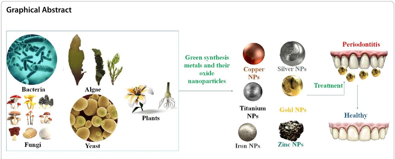
التهاب اللثة، أحد أكثر الأمراض السنية انتشارًا، يسبب فقدان العظام والأنسجة اللثوية التي تثبت الأسنان في مكانها. قد تؤدي عدة بكتيريا، موجودة عادة في تجويف الفم الصحي سريريًا، إلى تحفيز واستمرار التهاب اللثة عندما ترتفع تركيزاتها في الشق اللثوي. لقد أظهرت عدة جزيئات معدنية مؤقتة وأكسيد المعادن تأثيرًا مضادًا للبكتيريا ضد أنواع مختلفة من البكتيريا سالبة وموجبة الجرام، بما في ذلك البكتيريا المسببة للأمراض والمقاومة للأدوية. لذلك، يمكن استخدام الجزيئات النانوية في الطب الحيوي لعلاج مشاكل اللثة وفي تكنولوجيا النانو لمنع تطور الكائنات الدقيقة. بدلاً من استخدام المواد الكيميائية الضارة أو الآلات التي تستهلك الطاقة، تم اقتراح تصنيع جزيئات أكسيد المعادن والمعادن النانوية (NPs) بطريقة حيوية. لإنتاج جزيئات أكسيد المعادن والمعادن النانوية، فإن التقنية المثالية هي “التصنيع الأخضر” بسبب سمّيتها المنخفضة وسلامتها على صحة الإنسان والبيئة. يبدو أن جزيئات الذهب النانوية (AuNPs) أقل سمية للخلايا الثديية مقارنةً بالمعادن النانوية الأخرى لأن نشاطها المضاد للبكتيريا لا يعتمد على أنواع الأكسجين التفاعلية (ROS). كما أن جزيئات الفضة النانوية (AgNPs) تتمتع أيضًا بالاستقرار الكيميائي، والنشاط التحفيزي، والتوصيل الكهربائي والحراري الممتاز، على سبيل المثال من بين خصائصها الأخرى المفيدة. لوحظ أن جزيئات أكسيد الزنك (ZnO) وجزيئات النحاس (Cu) أظهرت تأثيرات مثبطة ملحوظة ضد سلالات البكتيريا موجبة وسالبة الجرام، على التوالي. أظهرت جزيئات ZnO نشاطًا قاتلًا ضد الكائنات الدقيقة المسؤولة عن التهاب اللثة. الأدوية التي تحتوي على جزيئات نانوية مغناطيسية فعالة للغاية ضد العدوى البكتيرية والفطرية المقاومة للأدوية المتعددة. جزيئات أكسيد التيتانيوم ( ) متورطة في زيادة نشاط البيروكسيداز اللعابي لدى الأفراد الذين تم تشخيصهم بالتهاب اللثة المزمن. علاوة على ذلك، فإن بعض الجزيئات النانوية المعدنية لديها القدرة على تعزيز الفعالية المضادة للميكروبات لعلاجات التهاب اللثة عند دمجها. لذلك، فإن هذه الجزيئات النانوية، بالإضافة إلى جزيئات أكسيدها، هي فقط بعض المعادن وأكاسيد المعادن التي تم تصنيعها بطرق صديقة للبيئة وأظهرت فوائد علاجية ضد التهاب اللثة.

الكلمات الرئيسية: التصنيع الأخضر، التهاب اللثة، جزيئات أكسيد المعادن النانوية، جزيئات المعادن النانوية، مضاد للبكتيريا

المقدمة

هناك حاجة ملحة لعلاجات أكثر فعالية حيث أن مقاومة البكتيريا للمضادات الحيوية الحالية أصبحت الآن معروفة كقضية صحية كبيرة. لمنع تكوين اللويحات وتطورها، فإن الوقاية الأولية من خلال تطوير أدوية مضادة للميكروبات مبتكرة وموجهة وفعالة أمر ضروري. التأثيرات المضادة للميكروبات على البكتيريا المسببة لالتهاب اللثة وتأثيرات التآزر لجزيئات المعادن النانوية مع المضادات الحيوية هي مجالات بحث أساسية[1]. في العديد من الدول، يحتل التهاب اللثة مرتبة عالية بين أهم القضايا الصحية العامة [2]. مزمن، التهابي، ومعدٍ، يظهر كضرر للأسنان والأنسجة الداعمة لها، مع حالات شديدة تؤدي إلى نزيف غزير والتهاب اللثة، وأسنان متخلخلة، وفقدان نهائي. تشير الأبحاث إلى أن بكتيريا الزائفة الهوائية (P. aeruginosa)، الإشريكية القولونية (E. coli)، S. pyogenes، وB. cereus قد تصبح نشطة إذا تم إزعاج الميكروبيوم. بين المرضى المزمنين، تميل B. cereus، S. pyogenes، P. aeruginosa، وE. coli إلى الوجود بأعداد غير متناسبة في موقع التهاب اللثة. كما أشارت الدراسات الحديثة إلى أن التهاب اللثة هو أكثر العدوى الميكروبية انتشارًا في الفم [3]. يعتبر تنظيف الأسنان وتخطيط الجذور علاجات غير جراحية لالتهاب اللثة تشمل تنظيف الأسنان واللثة بدقة للتخلص من الجير والجراثيم. في بعض الحالات، قد يحتاج التهاب اللثة إلى تدخل جراحي للعلاج. قد تتضمن هذه التدخلات زراعة العظام والأنسجة لتعويض العظام والأنسجة المفقودة، بالإضافة إلى جراحة الرفرف، التي ترفع اللثة للتخلص من الجراثيم والجير. يمكن استخدام المضادات الحيوية لتقليل كمية الجراثيم المرتبطة بالتهاب اللثة أو لمنع تدمير ارتباط السن بالعظام [4]. تسبب البكتيريا مرض اللثة، والتي يمكن علاجها بالمضادات الحيوية. تُستخدم المضادات الحيوية، بما في ذلك التتراسيكلين، الميترونيدازول، والأموكسيسيلين، عادةً
لعلاج التهاب اللثة. يمكن علاج الألم والتورم الناتج عن التهاب اللثة بالأدوية المضادة للالتهابات. عادةً ما تأخذ هذه الأدوية شكل أدوية مضادة للالتهابات غير الستيرويدية (NSAIDs). تقلل NSAIDs الالتهاب والألم من خلال منع الجسم من إنتاج البروستاجلاندين. الإيبوبروفين والأسبرين هما مثالان شائعان على NSAIDs. علاوة على ذلك، فإن استخدام توصيل الأدوية المزدوجة له إمكانيات لعلاج حالات التهاب اللثة. تشمل الأمثلة التوضيحية استخدام هلام يتشكل في الموقع (ISFG) يحتوي على دوكسيسيكلين هايكليت وإيبوبروفين، بالإضافة إلى تطبيق مصفوفة تتشكل في الموقع (ISFM) تشمل فانومايسين هيدروكلوريد (VH) وبورنول. منهجية بديلة هي استخدام زراعة الأدوية التي تطلق الأدوية، والتي يتم إدخالها مباشرة في الجيب اللثوي [5، 6] (الشكل 1).
تحديد سبب وتوقيت التهاب اللثة هو العقبة الأولى في علاجه. إن التشخيص المبكر للمرض والعلاج أمران حاسمان في تجنب المضاعفات اللاحقة. نظرًا لعدم وجود ألم، نادرًا ما يسعى المرضى للحصول على الرعاية الطبية. تتكون العقبة الثانية من تشخيص وإدارة كل عامل يساهم في هذا المرض بدقة. تعتبر البكتيريا هي العوامل المسببة الرئيسية لأمراض اللثة، مما يلحق ضررًا مباشرًا وغير مباشر على الأنسجة الداعمة للمضيف. يمكن التحكم في جميع حالات التهاب اللثة المزمن بشكل فعال من خلال تنظيف الأسنان وتخطيط الجذور، والتي تشمل الإزالة الميكانيكية وتقليل الكلس والكتلة البكتيرية في البيئة تحت اللثة. إن العناية باللثة على مر الزمن هي الجزء الثالث الأكثر صعوبة في علاج التهاب اللثة المزمن. يُطلق على هذا الجزء من العلاج اسم صيانة اللثة. خلال هذا الجزء من العلاج، قد يكون من الصعب الحفاظ على حماس المريض واهتمامه، ومراقبة جميع عوامل الخطر الخاصة بهم، ثم اتخاذ القرارات الصحيحة بشأن إعادة العلاج عندما
الشكل 1 تمثيل تخطيطي لالتهاب اللثة. يتميز التهاب اللثة بوجود لثة ملتهبة، حمراء، ونازفة تحيط بالأسنان. على الرغم من أن مرض اللثة يظهر أعراضًا مشابهة، إلا أنه يظهر أيضًا كفقدان للعظام. مادة لزجة تُسمى اللويحة، التي تتكون في تجويف الفم من الطعام، اللعاب، والبكتيريا، تهيج أنسجة اللثة من خلال تغليف السن فوق وتحت خط اللثة. إذا لم يتم التخلص من اللويحة، فإنها تتصلب إلى كلس، وهي مادة تصبح صعبة الإزالة للغاية. تمتلك الكائنات الدقيقة في اللويحة والكلس القدرة على تدمير العظام والأنسجة اللثوية التي تحيط بالأسنان في النهاية. وهذا يؤدي إلى تكوين شقوق عميقة، وضمور العظام، وفقدان الأسنان المحتمل [12]
مطلوب [7]. تشمل التدخلات العلاجية تنفيذ استراتيجيات تعديل السلوك، بما في ذلك، على سبيل المثال لا الحصر، ما يلي: توجيهات شخصية لنظافة الفم، برنامج للإقلاع عن التدخين، تعديلات غذائية، أدوات تحت اللثة لإزالة الجير واللويحات، العلاج الدوائي المحلي والنظامي، ومجموعة من الإجراءات الجراحية. تعتبر العلاج الكيميائي المضاد للميكروبات مفيدًا تقريبًا لجميع العلاجات الميكانيكية لالتهاب اللثة، ولم يظهر أي خيار علاجي واحد تفوقه. يتطلب علاج التهاب اللثة التزامًا مستمرًا بممارسات نظافة الفم الدقيقة بسبب الطبيعة المزمنة للمرض. عند تنفيذها بشكل مناسب، تقلل هذه التقنيات من احتمال بدء المرض وتقدمه [8]. يعد تنظيف العدوى تحت اللثة والتخلص من منطقة التهاب اللثة أيضًا جزءًا أساسيًا من علاج التهاب اللثة. على الرغم من أن العلاج لمدة يوم إلى ثلاثة أيام يبدو كافيًا لتخفيف أعراض مرض التهاب اللثة، إلا أنه لا يبدو أنه يمنع الحالة من العودة. قد تعمل بعض العلاجات بشكل أفضل عند إعطائها من خلال مزيج من طرق مختلفة. قد تكون الطريقة السريعة المفعول مفيدة للعلاج الأول لأنها يمكن أن توفر جرعة معقمة من
العامل المضاد للبكتيريا في جيب اللثة. قد يمنع النقل طويل الأمد للعوامل المضادة للبكتيريا إلى المنطقة المحيطة بفتحة الجيب إعادة استعمار الجيب من الفم عن طريق إيقاف اللويحات الهامشية. إن تطوير الأغشية الحيوية بواسطة مسببات الأمراض الفموية يمثل عقبة كبيرة في تطوير مقاومة المضادات الحيوية. لمنع أو القضاء على الأغشية الحيوية الميكروبية، يمكن استخدام مركبات تعيق عوامل الالتصاق وت extinguish الكميات الحيوية النشطة. هناك حاجة إلى مزيد من البحث لتقييم فعالية استراتيجيات قمع الكمية في مكافحة تشكيل الأغشية الحيوية لمسببات الأمراض اللثوية.
لقد أظهرت الأدوية المضادة للبكتيريا والمضادة للالتهابات فعاليتها في تحسين النتائج السريرية وإعاقة تقدم المرض. وقد أظهرت المضادات الحيوية النظامية الشائعة، مثل الأموكسيسيلين، والميترونيدازول، والدوكسي سيكلين، فعالية ملحوظة. ومع ذلك، من الضروري توخي الحذر عند إعطاء المضادات الحيوية، حيث إن استخدامها العشوائي يمكن أن يسهم في ظهور سلالات بكتيرية مقاومة للمضادات الحيوية وعواقب غير مرغوب فيها أخرى. تستغرق مراحل البحث والاختبار وإنتاج دواء جديد في المتوسط 13-15 عامًا وتكلف من 2-3 مليار دولار في الولايات المتحدة وحدها. العديد من الأدوية المحتملة
لا تتجاوز معظم الأدوية المراحل الأولى من الاختبار في عملية تطوير الأدوية، ومعدل النجاح منخفض بشكل عام. ومع ذلك، بسبب زيادة مقاومة المضادات الحيوية بين مسببات الأمراض اللثوية، تحول التركيز في علاج التهاب اللثة من قتل البكتيريا إلى إعادة تأسيس توازن صحي بين الميكروبات الفموية وأنسجة اللثة المضيفة. لذا، ما هو مطلوب هو تصميم دوائي منهجي يكون حديثًا وفعالًا لعلاج التهاب اللثة. يمكن وصف عوامل صيدلانية ذات آليات عمل متنوعة للمرضى الذين يحملون ميكروبات مقاومة للمضادات الحيوية، من بين أمور أخرى، بسبب حساسيتهم لمثل هذه المسببات. علاوة على ذلك، يمكن وصف هذه العوامل بشكل فردي أو مجتمعة لتعزيز فعاليتها. بعد العمليات الجراحية اللثوية الخطرة، غالبًا ما يتم إعطاء المرضى مضادات حيوية كإجراء وقائي. ولكن، تماشيًا مع الاتجاه الحالي لزيادة مقاومة البكتيريا البشرية للمضادات الحيوية، زادت مقاومة الأدوية أيضًا لدى الأشخاص الذين يعانون من أمراض اللثة خلال السنوات القليلة الماضية. لا تعمل المضادات الحيوية بشكل جيد على هذه الجراثيم بسبب البيئة الخاصة في منطقة اللثة وطريقة تكوين الأغشية الحيوية. نحتاج إلى طرق جديدة لعلاج أمراض اللثة.
تقدم تقنية النانو نهجًا متقدمًا للتحديات الملحة اليوم. توفر تطبيقات تقنية النانو خيارًا مثاليًا وصحيحًا في طب الأسنان وتبدو أنها تقدم إجابات للمشكلات التي تظهر مع الممارسات السنية التقليدية. يمكن أن تتطابق هذه الجسيمات النانوية الجديدة عن كثب مع خصائص السطح والاتصال لأنسجة الأسنان. مع تطوير وتنفيذ الجسيمات النانوية/ الحوامل النانوية، وجدت تقنية النانو استخدامًا واسع النطاق عبر مختلف التخصصات. نظرًا لصغر حجمها، يمكن للجسيمات النانوية اختراق دفاعات الكائنات الحية بسهولة. كما تم دراسة المعرفة النانوية الحيوية لتطبيقاتها المحتملة في مجالات مثل التصوير وتشخيص الأمراض في مراحلها المبكرة، بالإضافة إلى التوصيل الفعال والمستهدف للأدوية، والحمض النووي، والمركبات العلاجية إلى أعضاء أو خلايا معينة. يمكن تصنيف الجسيمات النانوية إلى نوعين متميزين، وهما العضوية وغير العضوية، اعتمادًا على تركيبة هيكلها. تعتبر الجسيمات النانوية غير العضوية مثالية لنقل المستضدات كلقاح بسبب حجمها الصغير، وثباتها العالي، وقابلية تعديلها المتحكم بها، وارتفاع قدرتها على الاختراق، وتحميلها العالي من الأدوية، وملف الإفراز النشط. تُعرف هذه الإنتاجات، المعروفة باسم الجسيمات النانوية الهجينة غير العضوية، عادةً بأنها تحتوي على نواة غير عضوية وعضوية.
الشكل 2 التدخلات لعلاج التهاب اللثة. الخط الأول للدفاع في علاج أمراض اللثة هو إجراء تنظيف شامل يتكون من تخطيط الجذور وإزالة الأنسجة الميتة. يتم القضاء على الجير والجراثيم من أسطح الأسنان وتحت خط اللثة من خلال التنظيف. يمكن تحقيق ذلك باستخدام الأدوات أو الليزر أو جهاز الموجات فوق الصوتية. المضادات الحيوية هي وسيلة شائعة أخرى غير جراحية لعلاج أمراض اللثة. تم تصميم المضادات الحيوية الخاصة باللثة بشكل احترافي لتوضع في الجيوب العميقة داخل اللثة بواسطة أطباء الأسنان، لمعالجة العدوى البكتيرية التي انتشرت إلى الجذر وخط اللثة. في بعض الأحيان، تكون المضادات الحيوية الفموية مطلوبة للقضاء على الكائنات الدقيقة المسببة للعدوى [17، 18]
القشرة [25-27]. لقد أثبت استخدام الجسيمات النانوية، التي تتراوح عادةً بين 0.2 و 100 نانومتر في الحجم، فعاليته كاستراتيجية جديدة لمكافحة البكتيريا. الذهب تيتانيوم فضة نحاس زنك المغنيسيوم ، والبزموت (Bi) هي المعادن الأكثر شيوعًا المستخدمة في التطبيقات البيولوجية اليوم [28،29]. نظرًا لأنها قد تكون صغيرة جدًا، يمكن للجزيئات النانوية المعدنية عبور طبقة البيبتيدوجليكان بسهولة وإيذاء البكتيريا داخلها. بالإضافة إلى ذلك، قد تمتص الأسطح الخلوية البكتيرية السالبة الشحنة ، ، و الأيونات الناتجة عن الجسيمات النانوية المعدنية بسبب الخصائص الأنيونية للأحماض التكسوائية والليبوبوليسكاريد [30]. قد تكون مثبطات نمو البكتيريا، مثل الجسيمات النانوية المعدنية، فعالة في علاج التهاب اللثة. لعلاج التهاب اللثة، طور العلماء منصات جديدة تستخدم الجسيمات النانوية المعدنية كعوامل مضادة للالتهابات ومضادة للبكتيريا فعالة [31]. تم تحديد الجسيمات النانوية ذات الخصائص المضادة للميكروبات. تشمل هذه الجسيمات النانوية الذهب، ثاني أكسيد التيتانيوم ( )، السيليكون (Si)، أكسيد النحاس (CuO)، وأكسيد الزنك (ZnO) [32]. كما أن الجسيمات النانوية (NPs) تعزز فعالية الجزيئات الحيوية النشطة في الجسم، مما يسهل اختراقها، ويحسن معدلات إطلاق الأدوية، ويسمح بتوصيل أكثر تحكمًا، وكل ذلك ضروري لنمو الأنسجة اللثوية بنجاح [33-35]. بالتزامن مع الجسيمات النانوية، جعلت التطورات الأخيرة في تكنولوجيا النانو من الممكن توصيل الأدوية والجزيئات الحيوية المضادة للالتهابات إلى مواقع محددة داخل الفم، بما في ذلك مسببات الأمراض اللثوية، والخلايا الالتهابية، والأنسجة اللثوية. يُوصى بشدة بإجراء أبحاث إضافية لتقييم فعالية الأدوية المحلية المعتمدة على الجسيمات النانوية في علاج الأمراض اللثوية السريرية [36]. يمكن العثور على الأمراض اللثوية باستخدام أجهزة استشعار حيوية على النانو. مع هذه الأجهزة، يمكنك العثور على المواد الكيميائية في سوائل الجسم مثل اللعاب، والدم، وسوائل الشقوق اللثوية [37]. مثال على ذلك هو أهمية الكشف المتعدد والسريع عن العلامات الحيوية في سوائل الشقوق اللثوية (GCF) للتشخيص السريع والدقيق لتقدم وشدة التهاب اللثة. في سعيهم لإنشاء نظام اختبار نقطة الرعاية (POCT) للتشخيص السريري لالتهاب اللثة، ابتكر العلماء شريط اختبار مناعي بتدفق جانبي على شكل قرص (LFIS) باستخدام الجسيمات النانوية الخضراء ذات النواة والقشرة (G-UCNPs) ككاشف للضوء. يتيح هذا الشريط الكشف المتزامن عن ثلاثة علامات حيوية مرتبطة بالتهاب اللثة، وهي، إنترلوكين-1 بيتا (IL-1 العامل النخر الورمي ألفا (TNF- )، إنترلوكين-8 (MPP-8)، وإنترلوكين-1 بيتا (GCF). تم الكشف عن الثلاثة مؤشرات حيوية بأقصى حساسية وخصوصية في GCF المعزز بواسطة منصة G-UCNPs-LFIS، التي أظهرت حدود حساسية تبلغ 5.455، 0.054، و ، على التوالي، في المحاليل القياسية. كما لوحظت معدلات استرداد مستقرة وعالية للعلامات الحيوية الثلاثة في اللعاب الصناعي الذي تم تغييره. بالإضافة إلى ذلك، تم الحصول على
أظهرت النتائج مؤشر ارتباط مرتفع نسبيًا (0.995 لاكتشاف MMP-8، 0.976 لـ IL-1 الكشف، و0.977 لـ TNF- الكشف) مقارنةً بالمؤشرات التي تم الحصول عليها من خلال طرق الكشف السريرية. هذا يثبت دقة التشخيص لجهاز G-UCNPs-LFIS الذي تم تطويره. علاوة على ذلك، تم إكمال عملية الكشف بالكامل في 30 دقيقة فقط، مما يسمح للمستخدمين بمراقبة حالات التهاب اللثة لديهم بسرعة [38]. لتسهيل الكشف المبكر عن التهاب اللثة، لا يزال هناك طلب كبير على مواد الاستشعار القادرة على التمييز بدقة وبشكل انتقائي بين ميثيل مركابتان (CH3SH) بمستويات ppb والمركبات الكبريتية المتطايرة (VSCs)، نظرًا لأن تركيز CH3SH في النفس الزفير يزداد بشكل طفيف مع تقدم مرض اللثة. الهدف الرئيسي من هذه الدراسة هو تطوير حساسات مرض اللثة الذاتية الإدراك من خلال تكديس نانومتري استراتيجي لجزيئات الذهب النانوية (AuNPs) بسمك 3 نانومتر على أفلام الزنك أكسيد النانوية (ZnO) بسمك 30 نانومتر. سيتم تحقيق ذلك من خلال إجراء من خطوتين يتضمن ترسيب الطبقات الذرية والتبخر الحراري. تم تحسين أداء الكشف عن الغاز بشكل كبير عندما تم طلاء ZnO بجزيئات AuNPs التي تم التحكم في حجمها وكثافتها. كانوا قادرين على الاستجابة إلى للغاز بمقدار 50 جزء في البليون من CH3SH وكان لديها حد كشف يبلغ 50 جزء في البليون. كان الكشف الانتقائي العالي لمستويات ppb من CH3SH في جو H2S ممكنًا باستخدام حساسات هجين من ZnO مدمجة مع جزيئات الذهب النانوية (Au NP) التي كانت تتمتع بأداء موثوق وقابل للتكرار في استشعار الغاز.
يمكن استخدام العمليات الفيزيائية والكيميائية والبيولوجية جميعها لتصنيع الجسيمات النانوية. تعتبر العمليات البيولوجية أكثر إنتاجية ونظافة وغير سامة وصديقة للبيئة مقارنةً بالتركيب الفيزيائي والكيميائي، الذي غالبًا ما يتضمن استهلاكًا كبيرًا للطاقة ومكونات ضارة. تم اقتراح مستخلصات النباتات والإنزيمات و/أو عمليات التخليق الميكروبي كبدائل أكثر صداقة للبيئة للتخليق الكيميائي التقليدي. استجابةً لهذه الحركة الدولية، طور العلماء طريقة تُسمى التخليق الأخضر (GS) التي تستخدم مستخلصات النباتات لإنشاء جسيمات نانوية بيئية وخضراء. تقدم النباتات عدة فوائد، بما في ذلك كونها رخيصة الإنتاج وسهلة الوصول وصديقة للمستخدم. لقد زادت إنتاجية الجسيمات النانوية التي تتراوح في الحجم والشكل والثبات بطرق مختلفة مؤخرًا. مقارنةً بالتقنيات التقليدية، فإن عائد الجسيمات النانوية المعدنية الناتجة باستخدام الأساليب الخضراء أكبر، وهي غير سامة. وذلك لأنه لا يتم إنتاج أي نواتج ثانوية ضارة خلال عملية التخليق. في النانوبيوتكنولوجيا، يُعتبر التخليق الأخضر للجسيمات النانوية باستخدام خلايا حية تطورًا جديدًا مثيرًا. لا تُستخدم المركبات السامة والضارة، ولا يتم تضمين عوامل الاختزال أو التثبيت أو التغطية الخارجية، كما أنه لا حاجة لضغط أو درجة حرارة عالية في التخليق الأخضر للجسيمات النانوية.
[42]. تم تضمين الكيانات البيولوجية، بما في ذلك مستخلصات النباتات، والبكتيريا، والطحالب، في التخليق الحيوي لجزيئات النانو المعدنية وأكاسيد المعادن. بالمقارنة مع جزيئات النانو المنتجة من الطحالب والفطريات والبكتيريا، فإن التحضير القائم على النباتات هو إجراء سريع وسهل وبسيط لتخليق جزيئات النانو على نطاق واسع. يعد تخليق الأجهزة النانوية الوظيفية، وإنتاج أدوية جديدة، وتجسيد عمليات توصيل الأدوية بعضًا من العديد من الاستخدامات الصيدلانية للمواد النانوية الخضراء التي تم إنشاؤها (NMs) [43]. تم استخدام مبادئ “الكيمياء الخضراء” الصديقة للبيئة في الإنتاج البيولوجي (باستخدام كائنات مثل النباتات، والبكتيريا، والفطريات، والطحالب، والأكتينوميسيتات) لجزيئات النانو المعدنية أو أكاسيد المعادن [44]. بديل أكثر صداقة للبيئة لإنشاء جزيئات النانو ذات الخصائص المناسبة هو تخليقها بواسطة الكائنات الحية. يمكن أن تستجيب الكائنات الحية أحادية الخلية ومتعددة الخلايا للتخليق البيولوجي [45]. تتمتع جزيئات النانو التي يتم إنتاجها بواسطة النباتات بفائدة تتمثل في وجود حركيات أعلى بشكل ملحوظ من تلك الخاصة بالعمليات البيولوجية الأخرى. نظرًا للمواد الكيميائية النباتية الاستثنائية، تم استخدام عدة أجزاء من النبات، بما في ذلك الورقة، والساق، والبذور، والفاكهة، والجذور، على نطاق واسع لإنتاج جزيئات النانو. لإنشاء جزيئات النانو، يتم أولاً تنظيف بعض أجزاء النبات بالماء العادي أو المقطر، ثم يتم إضافة المحاليل الملحية المقابلة التي تحتوي على جزيئات النانو المرغوبة، ثم يتم تصفيتها وعصرها. يتغير لون المحلول، مما يدل على تخليق جزيئات النانو، والتي يمكننا فصلها بسهولة [46، 47]. في العقود الأخيرة، برز التخليق الحيوي لجزيئات النانو المعدنية وأكاسيد المعادن كمنطقة دراسة واعدة. لقد زاد الاهتمام بدراسة الكيمياء الخضراء واستخدام الأساليب الخضراء لتخليق جزيئات النانو من أجل الحصول على إجراء أقل ضررًا للبيئة [47]. بشكل محدد، تقدم هذه المقالة ملخصًا لجزيئات النانو الخضراء، بما في ذلك الحديد والسيلينيوم والنحاس، بالإضافة إلى تخليقها وخصائصها واستخداماتها المحتملة استنادًا إلى الأساليب المستندة إلى النباتات. تشمل الفوائد البيولوجية الناشئة لجزيئات النانو المعدنية وأكسيد المعادن التي تم تخليقها بطريقة خضراء التشخيص، شفاء الجروح، علاج الأنسجة، العلاج المناعي، الطب التجديدي، طب الأسنان (التهاب اللثة)، ومنصات الاستشعار الحيوي. يمكن علاج العديد من الأمراض الفموية/الأسنان بشكل فعال باستخدام تخليق المعادن النانوية بطريقة خضراء مثل وأن جزيئات الذهب النانوية، التي تساعدها مستخلصات نباتية طبية مختلفة. كما تستخدم معجون الأسنان وغسول الفم، وهما من الأساسيات في النظافة اليومية. هناك حاجة إلى مزيد من الدراسة، على الرغم من وجود نقص حالي في المعلومات حول سلامة المواد النانوية. مقاومة الأدوية هي مجرد مشكلة واحدة يمكن حلها من خلال دمج مجموعة متنوعة من المستخلصات النباتية في الجسيمات النانوية. تستعرض هذه المقالة تأثير الجسيمات النانوية المعدنية وأكاسيدها المقابلة وطريقة GS على تطور وتقدم التهاب اللثة.

المكونات البيولوجية للتخليق “الأخضر”

لقد شهدت علوم المواد اهتمامًا كبيرًا بمفهوم “التخليق الأخضر”، الذي يُعتبر نهجًا موثوقًا ومستدامًا وصديقًا للبيئة لإنتاج مواد مختلفة ونانو مواد. يشمل ذلك الجسيمات النانوية المعدنية وأكاسيد المعادن، والمواد الهجينة، والمواد المستوحاة من الطبيعة. لذلك، يُعتبر التخليق الأخضر نهجًا مهمًا للتخفيف من الآثار الضارة المرتبطة بتخليق الجسيمات النانوية التقليدية المستخدمة غالبًا في المختبرات والبيئات الصناعية. تم استخدام التخليق الأخضر للجسيمات النانوية المعدنية للعمل مع مواد بيولوجية متنوعة (مثل البكتيريا والفطريات والطحالب ومستخلصات النباتات). مقارنةً بالتخليق الذي يتم بوساطة البكتيريا و/أو الفطريات، فإن استخدام مستخلصات النباتات هو تقنية بسيطة جدًا لإنتاج الجسيمات النانوية على نطاق واسع، مما يجعلها واحدة من أكثر الطرق الخضراء جاذبية لتخليق الجسيمات النانوية المعدنية/أكاسيد المعادن. تُعرف هذه المواد بالجسيمات النانوية البيوجينية. إن استخدام الميكروبات في الإنتاج البيوجيني للجسيمات النانوية له العديد من الفوائد مقارنةً بالطرق الأخرى ويجري دراسته بشكل متزايد. يسمح دمج المستخلصات البيولوجية مع محاليل أملاح المعادن بحدوث التخليق البيوجيني. يتم دراسة متغيرات مثل الرقم الهيدروجيني، ودرجة الحرارة، والوقت، والإشعاع، وكمية أملاح المعادن والمستخلصات. الجسيمات النانوية البيوجينية آمنة، ولا تضر بالبيئة، وتساعدنا في اتخاذ نهج أفضل. يمكن أيضًا استخدام الجسيمات النانوية البيوجينية في العديد من مجالات العلوم والتكنولوجيا الأخرى. الجسيمات النانوية المعدنية البيوجينية هي مواد نانوية قد حظيت باهتمام كبير نظرًا لأبعادها الدقيقة ونسبة المساحة السطحية إلى الحجم المرتفعة، والتي يتم تحقيقها من خلال العمليات البيولوجية. الآلية التي تقتل بها الجسيمات النانوية المعدنية أو أكاسيد المعادن البيوجينية البكتيريا غالبًا ما تتضمن إطلاق أيونات المعادن، التي تتفاعل مع أغشية الخلايا، وتسبب ضررًا لأغشية الخلايا، وفي النهاية تؤدي إلى انفصال أغشية الخلايا إلى حفر أو فجوات. تعتمد تقنيات التخليق الأخضر المعتمدة على البيولوجيا على مجموعة متنوعة من معلمات التفاعل، بما في ذلك الرقم الهيدروجيني (محايد، قاعدي، أو حمضي)، ودرجة الحرارة، والضغط، والمذيب. تم أخذ تنوع النباتات بعين الاعتبار على نطاق واسع لإنتاج الجسيمات النانوية المعدنية/أكاسيد المعادن لأن مجموعة متنوعة من مستخلصات النباتات، وخاصة تلك المستخلصة من الأوراق، تحتوي على مواد كيميائية نباتية قوية مثل التربينوات، والأميدات، والأحماض الكربوكسيلية، والفلافونات، والكيتونات، والأحماض الأسكوربية. يمكن أن تحول هذه العناصر أملاح المعادن إلى جسيمات نانوية معدنية. قد تكون أسهل الطرق وأكثرها صداقة للبيئة موجودة في تقنيات استخراج النباتات، مما يمنحها ميزة عند التوسع للإنتاج الصناعي. قد تلعب المواد الكيميائية النباتية (المواد الكيميائية النباتية) أدوارًا متنوعة، بما في ذلك عوامل التغطية، والتقليل، والتثبيت، في عمليات التخليق التي تؤدي إلى إنشاء الجسيمات النانوية. يُعتبر هذا التخليق الأخضر طريقة معالجة سهلة ورخيصة وسريعة لأنه لا يتضمن استخدام أي مواد كيميائية قد تكون خطرة أو مكلفة.
تتمثل مزايا استخدام طريقة خضراء لتوليد الجسيمات النانوية في أنها رخيصة وسهلة التنفيذ. ومع ذلك، فإن الحجم والشكل الصغيرين المحققين والخصائص الفريدة للركائز البيولوجية المستخدمة تعني أن إنتاج الجسيمات النانوية الخضراء قد يعزز خصائص هذه المواد النانوية. باستخدام مجموعة متنوعة من التقنيات الطيفية، تم إنتاج الجسيمات النانوية الفضية من خلال تخليق مستخلصات جذور مائية من نبات Rheum palmatum. تم اكتشاف الجسيمات النانوية السداسية والكرية. المكورات العنقودية الذهبية (S. aureus) و تم تثبيط P. aeruginosa بشكل كبير أيضًا بواسطة AgNPs المنتجة، مع قيم IC90 لـ قيم المل و IC50 البكتيريا المعرضة لجزيئات الفضة النانوية (AgNPs) أظهرت كمية كبيرة من تسرب البروتين وتغيرات شكلية. عرض الباحثون إنتاج جزيئات الفضة النانوية باستخدام السوبرناتانت لثقافة Massilia Spp. MAHUQ-52 والاستخدام المضاد للبكتيريا لهذه الجزيئات ضد كليبسيلا الرئوية (K. pneumoniae) وسالمونيلا إنتيريديس، التي تعتبر مقاومة لمجموعة من الأدوية. تم تحديد النشاط المضاد للبكتيريا لجزيئات الفضة النانوية المنتجة بواسطة Massilia Spp. MAHUQ-52 باستخدام تقنية انتشار الأقراص ضد K. pneumoniae وS. Enteritidis. أظهرت جزيئات الفضة النانوية الحيوية النشاط المضاد للبكتيريا القوي ضد كل من K. pneumoniae وS. Enteritidis. تم تحديد التركيز المثبط الأدنى (MICs) لجزيئات الفضة النانوية المنتجة ضد . الالتهاب الرئوي و . كانت نسبة الإنتيريتيديس 12.5 و على التوالي. كانت التركيزات المثبطة الدنيا للبكتيريا (MBC) لجزيئات الفضة النانوية (AgNPs) التي تم تخليقها حيوياً ضد كلا البكتيريا . من خلال التحقيق باستخدام FE-SEM، لوحظ أن الخلايا المعالجة بـ AgNPs أظهرت تغييرات هيكلية مع جدران خلوية غير متساوية ومتضررة أدت إلى موت الخلايا [60]. تتضمن الدراسات السابقة إنتاج ZnO-NPs من خلال طريقة سريعة وفعالة من حيث التكلفة وصديقة للبيئة باستخدام أربعة منتجات نباتية مختلفة. كانت النباتات المستخدمة في هذه الدراسة هي Beta vulgaris وCinnamomum tamala وCinnamomum verum وBrassica oleracea var. Italica. أظهرت جميع أمثلة ZnO-NPs نشاطًا مضادًا للبكتيريا ضد كل من البكتيريا سالبة وموجبة الجرام، بينما وُجد أن ZnO-NPs المصنوعة من Beta vulgaris غير نشطة تجاه S. aureus. كما تم إثبات النشاط المضاد للفطريات لـ ZnO-NPs باستخدام بقع فطرية من Candida albicans (C. albicans) وAspergillus niger (A. niger). أظهرت ZnO-NPs المصنوعة من Cinnamomum tamala نشاطًا ضد C. albicans، بينما وُجد أن تلك المصنوعة من Beta vulgaris نشطة ضد A. niger. بالإضافة إلى ذلك، أظهرت ZnO-NPs المصنوعة من مستخلص Brassica oleracea var. italica فعالية ضد كلا الفطرين. بشكل عام، تم إثبات أنه يمكن تخليق ZnO-NPs بسرعة وبتكلفة منخفضة وبشكل مستدام. قد تُستخدم هذه الجسيمات كعامل مضاد للبكتيريا محتمل ضد أنواع ميكروبية متنوعة [61]. وصف الباحثون طريقة بسيطة لصنع MgO NPs باستخدام مستخلصات أوراق مائية من Aloe vera وPisidium guavajava. لقد كان النشاط المضاد للبكتيريا لـ MgO NPs المنتجة
تم التحقيق فيه بشكل شامل ضد البكتيريا سالبة الجرام ( بكتيريا الإشريكية القولونية (E. coli) والبكتيريا إيجابية الغرام (S. aureus)، التي يتم تنظيمها بشكل جيد بواسطة جزيئات أكسيد المغنيسيوم النانوية (MgO NPs). يمكن للباحثين استخدام كلا المادتين السابقتين كعامل تغليف وعامل مختزل (RA). يظهر امتصاص الأشعة فوق البنفسجية عند 221 نانومتر. تم تقليلها كـ MgO. تظهر نتائج FTIR أن المواد الأولية النباتية تعمل كعوامل اختزال وتغطية. تظهر حيود الأشعة السينية أن المنتجات النهائية نقية وفي شكل بلوري مكعب مركزي الوجوه (FCC). أظهرت FESEM مع EDAX أن جزيئات MgO النانوية المنتجة لها شكل مكعب ومصنوعة بالكامل من MgO. تظهر أبحاث XPS أن جزيئات MgO النانوية المحضرة كانت مصنوعة من MgO والمواد الأولية النباتية. كانت الفعالية المضادة للبكتيريا لجزيئات MgO النانوية المحضرة تستجيب لكل من E. coli و S. aureus [62].
بينما تقدم طرق GS فوائد لا حصر لها، فإنها تقدم أيضًا بعض العقبات، مثل ندرة المواد الخام والحاجة إلى حصادها ونضوجها في أوقات محددة، بالإضافة إلى صعوبة الحصول على المواد الخام المطلوبة. يمكن استخدام العديد من المواد النباتية في التخليق البيئي للجزيئات النانوية؛ في الواقع، قام العديد من الباحثين بدراسة النباتات المتاحة محليًا والوفيرة. على الرغم من أن هذه الدراسات تقدم إمكانية تحسين استخدام النباتات المحلية، إلا أن تحقيق إنتاج عالمي على نطاق واسع من المعادن النانوية بتقنية GS لا يزال يمثل تحديًا كبيرًا. تتطلب عمليات GS المحددة درجات حرارة مرتفعة للغاية وأوقات تخليق طويلة، مما يستهلك كميات كبيرة من الطاقة وقد يكون له عواقب سلبية على البيئة. على الرغم من استخدام مواد خام صديقة للبيئة، قد لا تتوافق عملية التصنيع تمامًا مع مبادئ GS. الخصائص المحددة للجزيئات النانوية التي تم تخليقها بواسطة مستخلصات مختلفة غير كافية بسبب التباين الكبير في الحجم والشكل. القيد الرئيسي لهذه الطريقة هو أنها تنتج جزيئات نانوية تتميز بهياكل سطحية معيبة. تشير التقارير الحالية إلى تباينات كبيرة في حجم الجسيمات، مما يجعل التكنولوجيا الخضراء غير مناسبة للإنتاج على نطاق واسع ويشكل تحديًا كبيرًا في التحكم في حجم الجسيمات أثناء الإنتاج.

بكتيري

تطبيقات التكنولوجيا الحيوية التجارية، مثل التنظيف البيولوجي، والهندسة الوراثية، والاستخراج الحيوي، استخدمت على نطاق واسع الأنواع البكتيرية. قد تقلل البكتيريا من أيونات المعادن، مما يجعلها مرشحة واعدة لتحضير الجسيمات النانوية. يتم استخدام أنواع مختلفة من البكتيريا لإنتاج الجسيمات النانوية المعدنية وغيرها من الجسيمات النانوية الجديدة. تم تصنيع الجسيمات النانوية المعدنية/أكسيد المعدن على نطاق واسع باستخدام الكائنات الدقيقة بدائية النواة والميكروبات الفطرية. نظرًا لأن البكتيريا يمكن التلاعب بها بسهولة، فقد أصبح إنتاجها للجسيمات النانوية طريقة شائعة [66، 67]. تم استخدام سلالات بكتيرية مثل Bacillus cereus (B. cereus)، Bacillus amyloliquefaciens (B. amyl)، Bacillus indicus (B. indicus)، وBacillus
cecembensis (B. cecembensis) بشكل واسع في تخليق الجسيمات النانوية الفضية المخفضة حيويًا بأحجام وأشكال متنوعة. Shewanella oneidensis، Corynebacterium Spp. SH09، Aeromonas Spp. SH10، Phaeocystis antarctica، Pseudomonas proteolytica، Enterobacter cloacae، Geobacter Spp..، وArthrobacter gangotriensis. على غرار كيفية استخدام العديد من سلالات البكتيريا المختلفة في تخليق الجسيمات النانوية الذهبية (بما في ذلك Bacillus megaterium D01، Desulfovibrio desulfuricans، E. coli DH5a، Bacillus subtilis (B. subtilis) 168، Shewanella alga، Rhodopseudomonas capsulate، وPlectonema boryanum UTEX 485) [50].

ملخص

يمكن أيضًا إنتاج الجسيمات النانوية أحادية التشتت ذات الأشكال المحددة بشكل فعال من خلال التخليق الحيوي للمعادن/أكسيد المعادن بواسطة الفطريات. لأنها تحتوي على العديد من الإنزيمات المختلفة داخل خلاياها، فهي عوامل بيولوجية متفوقة لصنع الجسيمات النانوية من المعادن وأكسيد المعادن. يمكن تخليق المزيد من الجسيمات النانوية بواسطة الفطريات الكفؤة أكثر من البكتيريا. وفرة الإنزيمات والبروتينات والمكونات المخفضة على أسطح خلايا الفطريات هي سبب آخر لتفوقها على الأنواع الأخرى. يُعتقد أن الجسيمات النانوية المعدنية تنشأ من خلال الاختزال الإنزيمي (الاختزال) في جدار الخلية الفطرية أو السيتوبلازم. , وZnO هي بعض المعادن والأكسيدات التي يمكن تخليقها باستخدام الفطريات [50]. باستخدام الفطريات، تمكن العلماء من جميع أنحاء العالم من تخليق الجسيمات النانوية خارج الخلايا وداخلها، على التوالي. تشمل الأمثلة الشائعة المذكورة Penicillium Spp.، Fusarium Spp.، Fusarium oxysporum، Fusarium semitectum، Fusarium acuminatum، Fusarium solani، Cladosporium cladosporioides، Trichoderma viride، وAspergillus Spp. باستخدام الفطريات، يمكننا مقارنة وتباين الإنتاج الداخلي والخارجي للجسيمات النانوية مثل AgNPs [68].

ملخص

الطحالب الطحالب الدقيقة، المعروفة غالبًا باسم الطحالب، هي كائنات دقيقة في البيئات المائية تقوم بعملية التمثيل الضوئي. لقد ثبت أن الطحالب، مثل الكائنات الدقيقة الأخرى، تلعب دورًا حيويًا في الإنتاج البيولوجي للمواد النانوية وتراكم المعادن الثقيلة الأخرى. في بعض الأحيان، تُستخدم الطحالب في إنتاج الجسيمات النانوية ZnO والإنتاج الصناعي للجسيمات النانوية AU وAg. الطحالب الدقيقة معروفة بقدرتها على تحويل المعادن السامة المحتملة إلى نظائرها غير السامة. Phaeodactylum tricornutum هو نوع من الطحالب الدقيقة، وقد تم استخدام مصلها مؤخرًا في التخليق الحيوي للجسيمات النانوية Ti بمتوسط قطر جزيئي يبلغ 49.7 نانومتر. بسبب أنشطتها السامة للخلايا، ومضادة للبكتيريا، ومضادة للكهرباء، والبيوجينية، كانت الجسيمات النانوية الناتجة من الطحالب الدقيقة لها القدرة على الاستخدام في تطبيقات طبية متنوعة. وشملت هذه طرق التصوير

، والهايبرثيرميا، وأجهزة الاستشعار الحيوية، وأنظمة توصيل الأدوية، وعلاجات السرطان، ودراسات نظام المناعة [69]. بالإضافة إلى ذلك، تم استخدام الطحالب الدقيقة من Sargassum muticum وSargassum myriocystum لإنتاج الجسيمات النانوية ZnO بحجم 36 نانومتر. تم إنشاء الجسيمات النانوية المتغيرة في الشكل والحجم التي تحتوي على مجموعات كربونيل وهيدروكسيل بواسطة S. myriocystum. بالإضافة إلى ذلك، تم اكتشاف أن الجسيمات النانوية التي تم إنتاجها بواسطة S. muticum كانت سداسية، وكانت بوليسكاريداتها تحتوي على مجموعات هيدروكسيل وسلفات. تم استخدام طريقتين، بما في ذلك سلالات السيانوبكتيريا والطحالب الدقيقة، لتقييم إنتاج الجسيمات النانوية Ag، كما هو موضح في الأدبيات [70، 71]. تضمنت إحدى التقنيات إضافة نترات الفضة إلى وسائط خالية من الخلايا، بينما تضمنت الأخرى غسل وتعليق الكتلة الحيوية الحية من مجموعتي البكتيريا في محلول . باستخدام أي من هاتين التقنيتين، تمكنت أربعة عشر من بين ستة عشر سلالة تم اختبارها من إنتاج الجسيمات النانوية Ag بنجاح بأقطار تتراوح من 13.0-31.0 نانومتر، مما يدل على أن المكونات الخارجية كانت متورطة في إنشاء الجسيمات النانوية Ag. كما تم إظهار النشاط المضاد للبكتيريا للجسيمات النانوية Ag، باستثناء أكبر الجسيمات النانوية التي تم إنتاجها بواسطة سلالة السيانوبكتيريا (Limnothrix Spp. 37-2-1). علاوة على ذلك، تم تخليق الجسيمات النانوية Ag وAuNPs باستخدام Chlorella vulgaris، وهي طحالب دقيقة تعمل كمعمل نانوي. تم إظهار النشاط المضاد للبكتيريا لكل من الجسيمات النانوية ضد . aureus، Streptococcus Spp.، وE. coli. بالمثل، تم تخليق الجسيمات النانوية أكسيد القصدير بخصائص ضوئية حيوية باستخدام Chlorella vulgaris. تم إظهار النشاط المضاد للبكتيريا ضد أربعة بكتيريا مرضية، وخصائص مضادة للأكسدة، وسمية خلوية ضد سرطان الرئة، والانهيار الضوئي لصبغة الميثيل البرتقالي تحت ضوء الأشعة فوق البنفسجية من قبل الجسيمات النانوية المنتجة. تم استخدام الطحالب البحرية Padina Spp. لإثبات أنه يمكن استخدام الطحالب البحرية لتخليق الجسيمات النانوية Ag. وقد تم اكتشاف أنه عند تضمين هذا النوع المحدد من الطحالب، زاد تخليق الجسيمات النانوية Ag. تم قتل كل من S. aureus وP. aeruginosa بواسطة الجسيمات النانوية مع مناطق تثبيط تبلغ 15 و13 مم، على التوالي [68، 72، 73].

الخميرة

أظهرت العديد من الدراسات جدوى استخدام الخمائر لتخليق المواد النانوية؛ الخمائر هي كائنات حقيقية النواة وحيدة الخلية تطورت من كائنات متعددة الخلايا. يتم استخدام العديد من الأنواع البالغ عددها حوالي 1500 من الخمائر على نطاق واسع في تصنيع المواد النانوية المعدنية. تتمتع الخمائر بأسطح كبيرة، مما يجعلها عرضة بشكل خاص لتراكم المعادن السامة [74]. أظهرت العديد من المختبرات أن الخميرة يمكن أن تخليق الجسيمات النانوية/ المواد النانوية بنجاح. يمكن استخدام سلالة خميرة مقاومة للفضة ومغذيات Saccharomyces cerevisiae (S. cerevisiae) لتخليق الجسيمات النانوية Ag وAu، كما تم وصفه مؤخرًا. يتم إنتاج العديد من الجسيمات النانوية المعدنية باستخدام أنواع مختلفة [50، 75].

ملخص

خلايا النبات يمكن للنباتات تخزين مستويات متفاوتة من المعادن الثقيلة في أنسجتها المختلفة. لذلك، تلقت استراتيجيات التخليق الحيوي باستخدام مستخلصات النباتات مزيدًا من الاهتمام كبديل بسيط وفعال ومنخفض التكلفة وقابل للتطبيق عمليًا لعمليات التحضير القياسية لتوليد الجسيمات النانوية [76]. من بين الجزيئات الحيوية الموجودة في النباتات تلك التي تتمتع بقدرة استثنائية على تحفيز اختزال ملح المعدن إلى الجسيمات النانوية. كان التخليق بمساعدة مستخلصات النباتات هو أول مسار تخليقي حيوي يتم استكشافه للجسيمات النانوية الذهبية والفضية. تم استخدام نباتات مختلفة، بما في ذلك الألوة فيرا (Aloe barbadensis Miller)، الشوفان (Avena sativa)، البرسيم (Medicago sativa)، تولسي (Osimum sanctum)، الليمون (Citrus limon)، النيم (Azadirachta indica)، الكزبرة (Coriandrum sativum)، الخردل (Brassica juncea) وعشب الليمون (Cymbopogon flexuosus)، لتخليق الجسيمات النانوية Ag وAu. أظهرت نباتات أخرى، بما في ذلك الخردل (Brassica juncea)، والبرسيم (Medicago sativa)، وعباد الشمس (Helianthus annuus)، أنها تنتج الجسيمات النانوية في الجسم الحي أيضًا. الكزبرة (Coriandrum sativum)، وزهرة التاج (Calotropis gigantean)، وورقة النحاس (Acalypha indica)، ووردة الصين (Hibiscus rosa-sinensis)، والشاي الأخضر (Camellia sinensis)، ومستخلص ورقة الألوة (Aloe barbadensis Miller) هي بعض من العديد من مستخلصات أوراق النباتات التي تم استخدامها لتحضير الجسيمات النانوية ZnO [46، 77-80].

آلية التخليق “الأخضر” للمعادن وجسيماتها النانوية أكسيدها

تم استخدام التخليق الأخضر من قبل عدة مجموعات لإنشاء الجسيمات النانوية المعدنية/أكسيد المعدن. آليات الإنتاج “الأخضر” للمعادن وجسيماتها النانوية أكسيدها، مثل تلك المستندة إلى الكائنات الدقيقة ومستخلصات أوراق النباتات، قيد التحقيق الآن. علاوة على ذلك، من المحتمل أن يكون الاختزال الإنزيمي (الاختزال) في جدار الخلية أو داخل الخلية الفطرية هو العملية لإنشاء الجسيمات النانوية المعدنية. يتم تخليق الجسيمات النانوية المعدنية/أكسيد المعدن، بما في ذلك Ag، , وZnO، باستخدام أنواع فطرية مختلفة [50].

آلية قائمة على الكائنات الدقيقة

يمكن أن تستخدم أنواع مختلفة من البكتيريا طرقًا متنوعة لإنشاء الجسيمات النانوية. أولاً، يتم احتجاز أيونات المعادن على أو داخل الخلايا الميكروبية، ثم، بفضل عمل الإنزيمات، يتم تحويلها إلى جسيمات نانوية معدنية. استنادًا إلى الفرضية التالية، أوضح سنيها وآخرون [81] العملية التي تساعد بها الكائنات الدقيقة في تشكيل جسيمات نانوية من الفضة والذهب من خلال فطر فيرتيسيليوم أو الكتلة الحيوية للطحالب. كانت التفاعلات الكهروستاتيكية بين أيونات الفضة أو الذهب وإنزيمات جدار الخلية السالبة الشحنة هي الخطوة الأولى في التقاط الأيونات على سطح خلايا الفطر. بعد ذلك، تم تقليل نوى الفضة أو الذهب من الأيونات لإنتاج النمو. كل من نيكوتيناميد أدينين ثنائي النوكليوتيد (NADH) والاختزال النيتريتي، الذي يعتمد على NADH، ضروريان لإنتاج الجسيمات النانوية. كان الاختزال النيتريتي
تم إثبات مسؤولية B. licheniformis عن تقليل AgNPs بواسطة كاليسوارالال وآخرون [82]. على الرغم من أن الكائنات الدقيقة تنتج أيونات أملاح المعادن ونانو جزيئات معدنية، إلا أن آليات الاختزال الحيوي المعنية في هذه العمليات ليست مفهومة جيدًا [81، 82] (الشكل 3).

آلية مستندة إلى مستخلص أوراق النباتات

يتم استخدام مستخلص أوراق النباتات كعامل مساعد في إنتاج الجسيمات النانوية من خلال دمج محاليل سابقة المعادن تحت ظروف تفاعل متغيرة. يمكن التحكم في سرعة إنشاء الجسيمات النانوية، بالإضافة إلى عائدها واستقرارها، بواسطة العوامل التي تحكم ظروف مستخلص أوراق النباتات (مثل أنواع المركبات النباتية، تركيز المركبات النباتية، تركيز أملاح المعادن، الرقم الهيدروجيني، ودرجة الحرارة). تحتوي مستخلصات أوراق النباتات على مركبات نباتية تتمتع بالقدرة الملحوظة على تقليل أيونات المعادن في وقت أقل بكثير من الفطريات والبكتيريا، التي تحتاج إلى فترة حضانة أطول. لذلك، تعتبر مستخلصات أوراق النباتات خيارًا رائعًا وآمنًا لصنع الجسيمات النانوية من المعادن وأكاسيد المعادن. بالإضافة إلى ذلك، يسهل مستخلص أوراق النباتات تخليق الجسيمات النانوية من خلال العمل كعامل اختزال وعامل مثبت خلال عملية التخليق. بالإضافة إلى النبات المستخدم، يلعب تركيز المركبات النباتية في مستخلص أوراق النباتات دورًا كبيرًا في إنتاج الجسيمات النانوية. تشمل المركبات النباتية الرئيسية المسؤولة عن الاختزال الحيوي للجسيمات النانوية الفلافونويدات، التربينويدات، السكريات، الكيتونات، الألدهيدات، الأحماض الكربوكسيلية، والأميدات. بشكل عام، هناك ثلاث مراحل رئيسية لآلية تخليق الجسيمات النانوية المعدنية في النباتات ومستخلصات النباتات: 1) مرحلة التنشيط، وهي عندما يحدث اختزال أيونات المعادن وتكوين نوى الذرات المعدنية المختزلة؛ 2) مرحلة النمو، وهي عندما تندمج الجسيمات النانوية الصغيرة المجاورة بشكل عفوي في جزيئات أكبر (التكوين المباشر للجسيمات النانوية باستخدام النوى المتغايرة والنمو، واختزال أيونات المعادن الإضافية؛ وهي عملية تعرف باسم نضوج أوستوالد)، والتي ترتبط بزيادة في الاستقرار الديناميكي الحر للجسيمات النانوية؛ و3) مرحلة إنهاء العملية، التي تحدد الشكل النهائي للجسيمات النانوية. تمتلك مستخلصات النباتات القدرة على العمل كعوامل مثبتة وعوامل اختزال خلال عملية تخليق الجسيمات النانوية. من المعروف أن خصائص الجسيمات النانوية يمكن أن تتأثر بمصدر مستخلص النبات. وذلك لأن تركيزات وخلائط العوامل العضوية الاختزالية تختلف بين المستخلصات. يتضمن الاختزال الحيوي الذي يتم بوساطة مستخلص نباتي عمومًا دمج محلول مائي يحتوي على ملح المعدن المعني مع المستخلص المائي. عادةً ما تصل التفاعل إلى نهايته في غضون بضع دقائق عند درجة حرارة الغرفة. نظرًا لتعدد المركبات الكيميائية المستخدمة، فإن عملية الاختزال الحيوي معقدة نسبيًا.
الشكل 3 مخطط يوضح العمليات المعنية في إنتاج الجسيمات النانوية المعدنية خارج الخلية وداخلها. يتم إنتاج الجسيمات النانوية المعدنية خارج الخلية عن طريق احتجاز أيونات المعادن على جدار الخلية وتقليلها في وجود المستقلبات أو الإنزيمات المفرزة. بعد مرور أيونات المعادن إلى السيتوبلازم داخل الخلية، يتم تقليل أيونات المعادن خلال إنتاج الجسيمات النانوية المعدنية داخل الخلية بسبب التفاعلات الأيضية مع إنزيمات مثل نترات ريدوكتاز [65].
تُنتج الجسيمات النانوية غير السامة من خلال التخليق الحيوي المستند إلى النباتات، مما يجعلها بديلاً آمناً ومستداماً بيئياً للعمليات الكيميائية، خاصة في التطبيقات الطبية الحيوية. بالإضافة إلى ذلك، تقلل مستخلصات النباتات من الوقت اللازم لتقليل أيونات المعادن. وذلك لأن التخليق النانوي النباتي لا يتطلب تكوين ثقافات خلوية، أو فترات حضانة طويلة، أو درجات حرارة مرتفعة. يمكن أن يُعزى التخفيض السريع لأيونات المعادن إلى قدرة المكونات النباتية على التبرع بالإلكترونات (المجموعات الوظيفية تجاه معقدات أيونات المعادن). تُعتبر مستخلصات النباتات الطبيعية بشكل أساسي لتخليق الجسيمات النانوية بسبب فوائدها البيئية المحتملة المرتبطة بالتخليق الحيوي. الاستخدام الرئيسي للتخليق الحيوي هو أنه يسمح باختيار المذيبات، والمواد غير الضارة للتثبيت، والمواد المساعدة الصديقة للبيئة. تحتوي مستخلصات النباتات على مجموعة متنوعة من المركبات، بما في ذلك الأمينات، والأميدات، والقلويدات، والفلافونويدات، والفينولات، والتربينويدات.
البروتينات والكاروتينات. تساهم المكونات الكيميائية النباتية المذكورة أعلاه في تقليل وتثبيت أيونات المعادن خلال عملية تخليق الجسيمات النانوية الصديقة للبيئة.

جزيئات المعادن النانوية في التهاب اللثة

ردًا على البايوفيلم تحت اللثة، يقوم الجهاز المناعي البشري بتحفيز التهاب مستمر يعرف باسم التهاب اللثة. يعتبر تنظيف الأسنان وتخطيط الجذور أمثلة على أساليب العلاج التقليدية التي تهدف إلى تقليل البايوفيلم تحت اللثة، وغالبًا ما يتم دعمها بعلاجات مساعدة مضادة للبكتيريا مثل المضادات الحيوية والمطهرات والبروبيوتيك. لقد تم استخدام المضادات الحيوية والمطهرات والبروبيوتيك بشكل متكرر بالتزامن مع الإدارة التقليدية لالتهاب اللثة بما في ذلك إزالة الأنسجة الميكانيكية وممارسات النظافة الفموية المنتظمة في العقدين الماضيين. ومع ذلك، فإن إيصال هذه المواد الطبية إلى الأماكن التي تحتاج إليها
الشكل 4 تمثيل تخطيطي لآليات التخليق الأخضر (GS) لجزيئات النانو المعدنية وأكاسيد المعادن بواسطة أجزاء مختلفة من النباتات
لا يزال الأمر صعبًا. نادرًا ما يُوصى بالمضادات الحيوية الجهازية لعلاج أمراض اللثة بسبب المخاطر المرتبطة باستخدامها وعدم القدرة على تحقيق تركيز عالٍ بما يكفي في موقع المرض. بالإضافة إلى ذلك، تم وصف انخفاض سريع في تركيز البلازما، وعدم تحمل الجهاز الهضمي، والمقاومة الميكروبيولوجية، والحساسية المفرطة، مما يؤدي إلى جرعات دون العلاج بالمضادات الحيوية. بعض العوامل المحلية التي يمكن أن تجعل علاج اللثة أقل إقناعًا هي شدة المرض، وعدد الفروع المعنية، وقدرة المريض على التحكم في اللويحات، وعوامل احتباس اللويحات (مثل الحشوات الخاطئة والتجاويف)، وإصابات الوجه. من خلال القضاء على مسببات الأمراض تحت اللثة التي تستمر بعد العلاج الميكانيكي التقليدي لأمراض اللثة، تسعى المضادات الحيوية الجهازية لعلاج أمراض اللثة إلى دعم نظام الدفاع لدى المضيف في التغلب على العدوى وتعزيز العلاج الميكانيكي لأمراض اللثة. قد تتحدد فعالية المضادات الحيوية الجهازية في علاج أمراض اللثة بناءً على قابلية البكتيريا للمضادات الحيوية. إن تثبيط إنزيم الكولاجيناز يمكّن عددًا محدودًا من العوامل الكيميائية العلاجية من تقليل تدهور الكولاجين والعظام. عادةً ما يستجيب مرضى التهاب اللثة المستقر أو التهاب اللثة بشكل إيجابي للعلاج الميكانيكي لأمراض اللثة؛ حيث يوفر العلاج بالمضادات الحيوية فائدة إضافية ضئيلة أو معدومة. ليس من الواضح كيف يمكن استخدام المضادات الحيوية الجهازية بشكل أفضل لعلاج كل من أمراض اللثة قصيرة الأمد وطويلة الأمد. ومع ذلك، يبدو أن الطريقة جذابة لأن أمراض اللثة تسببها الميكروبات. من ناحية أخرى، هناك
إثبات مختلط أن هذه العوامل تساعد في معظم أمراض اللثة، وأن المضادات الحيوية يمكن أن تكون ضارة أيضًا. بعض المشاكل المتعلقة بالمضادات الحيوية الجهازية تشمل الاستجابات التحسسية، خطر الإصابة بعدوى أخرى، السمية، تركيبات الأدوية، تعاون المريض، والأهم من ذلك، مقاومة الجراثيم للدواء. بالنسبة لمعظم أمراض اللثة، يجب أن تكون طرق التنظيف الميكانيكية، مثل تصريف القيح من خراجات اللثة الحادة، هي الخيار الأول. يجب استخدام المضادات الحيوية الجهازية فقط مع طرق التنظيف الميكانيكية. يجب ألا يستخدمها الأشخاص الذين يعانون من أمراض مزمنة لأنها قد تجعل الخراجات أكثر احتمالاً للتشكل. عندما تكون هناك حالة حادة يصعب تنظيفها أو تصريف القيح منها، أو عندما يكون هناك انتشار محلي أو اضطراب نظامي، يمكن التفكير في استخدام مضادات حيوية جهازية إضافية. عندما يعاني شخص ما من مرض لثة مزمن يبدأ مبكرًا أو يتفاقم بسرعة، أو عندما تفشل العلاجات الميكانيكية، ولا تكون الجراحة خيارًا، يجب عليهم التفكير في إضافة المضادات الحيوية إلى خطة علاجهم. إذا كنت لا تنظف أسنانك أو تدخن التبغ، فلا ينبغي عليك استخدام المضادات الحيوية [93].
ظهرت جيل جديد من أنظمة توصيل الأدوية المحلية الفعالة نتيجة البحث في المشكلة، ويمكن أن تزيد هذه الأنظمة من تركيز جزيئات الدواء في موقع العمل مع تقليل أو القضاء على آثارها الضارة النظامية [94، 95]. البكتيريا المسببة للحمض مثل S. mutans و S. sobrinus و Lactobacillus Spp. تشارك في تطور المرض [96]. تعتبر الاضطرابات اللثوية، التي قد تؤثر على الأنسجة الصلبة واللينة، هي الأكثر
الأمراض الالتهابية التنكسية شيوعًا التي تؤثر على الناس. أمراض اللثة والهياكل الداعمة للأسنان، الناتجة عن جزيئات اللويحات التي تتشكل على سطح الجذر الصلب بجوار الأنسجة اللينة للدعامة اللثوية، يمكن أن تكون سطحية (التهاب اللثة) أو جهازية (التهاب اللثة)، مما يدمر العظم السنخي الذي يثبت الأسنان في مكانها والرباط اللثوي. قد تصبح الأسنان رخوة أو تسقط إذا تعرضت لفقدان مثل هذا في الارتباط وخلق جيوب لثوية لاحقًا. تشمل العدوى اللثوية الأولية Porphyromonas gingivalis و Prevotella intermedia و Aggregatibacter actinomycetemcomitans [96، 97].
يؤدي تأثير حجم الكم إلى أن الجسيمات النانوية المعدنية، التي يتم إنتاجها عن طريق طحن المعدن إلى جزيئات صغيرة ( بقطر)، لها مجموعة مختلفة من الخصائص مقارنة بالمعادن الكتلية [98]. حجمها الصغير جدًا يمنحها خصائص فريدة، مثل الاتصال الأفضل بالخلايا، لأنها تتمتع بنسبة سطح إلى كتلة أكبر والقدرة على استخدامها بطرق متنوعة يمكن التحكم فيها [99]. في معظم الأحيان، تكون الجسيمات النانوية الأصغر أكثر فعالية في قتل البكتيريا لأنها تستطيع الدخول إلى الخلايا وإيقاف نمو البكتيريا. أيضًا، تتمتع الجسيمات النانوية الأصغر بنسبة سطح إلى حجم أعلى من الجسيمات النانوية الأكبر، مما يساعد على توليد ROS [100]. لقد حققت العديد من التخصصات نتائج واعدة في السنوات الأخيرة في دراسة الجسيمات النانوية المعدنية. تشمل هذه التخصصات الكيمياء، البيولوجيا، علوم المواد، والطب، وهي ليست معلومات جديدة. لقد جذبت AgNPs، على وجه الخصوص، الانتباه لخصائصها المضادة للميكروبات القوية عبر تخصصات متنوعة. نظرًا لأن AuNPs غير نشطة ومستقرة جدًا، غالبًا ما يتم التحقيق فيها لاستخدامها المحتمل في تطبيقات استشعار حيوي أو توصيل الأدوية. إحدى الطرق لجعل AuNPs مضادة للبكتيريا هي تغليفها بمركبات عضوية تظهر خصائص مضادة للبكتيريا [101].
تمت دراسة الجسيمات النانوية بشكل شامل في تطبيقات بيولوجية متنوعة. أظهرت الجسيمات النانوية المعدنية فعالية ضد مسببات الأمراض ذات الأولوية، بالإضافة إلى حجمها الصغير وانتقائيتها للبكتيريا، كما ذكرت منظمة الصحة العالمية (WHO) [102]. ليس من الصعب على البكتيريا اكتساب المقاومة للجسيمات النانوية المعدنية لأنها لا ترتبط بمستقبل معين في الخلية البكتيرية، ولكن نطاق عملها المضاد للبكتيريا يتوسع أيضًا. لذلك، أظهرت معظم الأبحاث حول فعالية الجسيمات النانوية المعدنية نتائج مشجعة في كل من البكتيريا إيجابية الجرام وسلبية الجرام. سيجد الباحثون أنه على الرغم من أن الجسيمات النانوية الأخرى التي غالبًا ما تستخدم في العلاج بالمضادات الحيوية (مثل ، و CuO NPs) يتم دراستها أيضًا، إلا أن AgNPs تحظى بالنصيب الأكبر من الانتباه [103]. قد تكون مثبطات نمو البكتيريا، مثل الجسيمات النانوية المعدنية، فعالة في علاج التهاب اللثة. لعلاج التهاب اللثة، طور العلماء منصات جديدة تستخدم الجسيمات النانوية المعدنية
كعوامل مضادة للالتهابات ومضادة للبكتيريا فعالة [104].
أظهرت الجسيمات النانوية المعدنية فعالية ضد . coli، . aureus، B. subtilis، و Salmonella، مما يجعلها بديلاً محتملاً للمضادات الحيوية النانوية القياسية. ومع ذلك، يُقبل على نطاق واسع أن الآثار الضارة المضادة للبكتيريا للجسيمات النانوية المعدنية قد تُعزى إلى ثلاثة آليات رئيسية: إنتاج الأنواع التفاعلية المؤكسدة (ROS)، تحرير الأيونات، والتفاعل بين الجسيمات النانوية والغشاء الخلوي. تتمتع الجسيمات النانوية المعدنية، مقارنة بأملاحها المقابلة، بفعالية متزايدة في معالجة العدوى البكتيرية. بشكل أساسي، يؤثر حجم أبعاد الجسيمات النانوية بشكل كبير على الآلية المضادة للبكتيريا [105]. لقد أظهرت الجسيمات النانوية المعدنية، بما في ذلك ، أكسيد الكالسيوم ، وأكسيد المغنيسيوم ، فعالية مضادة للميكروبات [106]. تنتج هذه الجسيمات النانوية ROS، التي تشكل خطرًا على الكائنات الدقيقة لأنها قد تكسر مكونات الخلايا، بما في ذلك الحمض النووي، RNA، والبروتينات. مقارنةً بالمعادن النانوية الأخرى، تعتبر AuNPs أقل خطورة على خلايا الثدييات لأن تأثيرها المضاد للبكتيريا لا يعتمد على ROS. نتيجة لإمكاناتها العالية في التوظيف، تعتبر هذه الجسيمات النانوية أيضًا مرشحة واعدة للاستخدام كمواد نانوية مضادة للبكتيريا مستهدفة. كما أن ZnO NPs تتمتع بنشاط ضوئي محفز كبير، مما يعزز كفاءتها المضادة للبكتيريا. كما أن ZnO NPs قادرة على إنتاج ROS عند تعرضها للأشعة فوق البنفسجية [107]. بالإضافة إلى ذلك، فإن الأيونات المعدنية التي تطلقها هذه الجسيمات النانوية قاتلة للحمض النووي والبروتينات البكتيرية. عندما تتلامس الجسيمات النانوية المعدنية مع البكتيريا، تجذب أغشيتها السالبة الشحنة الأيونات المعدنية الموجبة الشحنة. أغشية البكتيريا مسامية، مما يسمح بدخول الأيونات المعدنية إلى الخلايا. من خلال القيام بذلك، تتفاعل مع مجموعة السلفهيدريل (-SH) على البروتينات الميكروبية، مما يجعلها غير قادرة على إنتاج البروتينات أو الأحماض النووية. تعتمد قدرة الجسيمات النانوية على قتل الجراثيم على عوامل، بما في ذلك حجم جزيئاتها وشحنتها السطحية. بسبب حجم جزيئاتها الأصغر ونسبة السطح إلى الحجم الأفضل، تكون فعالة جدًا ضد الجراثيم دون التأثير على الخصائص الميكانيكية للمادة. بالإضافة إلى الاتصال المباشر مع الجسيمات النانوية، هناك طريقة أخرى لقتل البكتيريا وهي عن طريق إطلاق المادة الكيميائية المضادة للبكتيريا المحملة [31، 108] (الجدول 1) (الشكل 5). لتحديد ما إذا كان يمكن استخدامها لعلاج التهاب اللثة، قام الباحثون بإنشاء وإنتاج وتوصيف AgNPs المرتبطة إما بالميترونيدازول (AgNPs-PEG-MET) أو الكلورهيكسيدين (AgNPs-CHL). بينما كانت AgNPs-CHL أكثر سمية للخلايا من AgNPs-PEG-MET، اتضح أنها كانت عاملًا مضادًا للبكتيريا أكثر فعالية؛ ومع ذلك، أظهرت كلاهما خصائص إيجابية عند جرعات غير سامة. قامت AgNPs-CHL و AgNPs-PEG-MET بتثبيط إنتاج TNF ، IL- ، IL-6، وغيرها من السيتوكينات المؤيدة للالتهابات. كما كانت ميتالوبروتينازات MMP3 و MMP8 كذلك
الجدول 1 آلية عمل الجسيمات النانوية المعدنية وأكسيد المعادن المضادة للبكتيريا وطريقة الإنتاج الشائعة
الجسيمات النانوية المعدنية آلية العمل المضادة للبكتيريا طريقة الإنتاج الشائعة المراجع
AuNPs من خلال التسبب في تكوين ثقوب في جدار الخلية البكتيرية، تمارس AuNPs تأثيرها المضاد للبكتيريا، مما يظهر في موت الخلايا وفقدان المحتويات الخلوية لاحقًا تعتبر واحدة من أكثر الطرق المعروفة لتخليق AuNPs، تم تطوير طريقة توركيفيتش بواسطة توركيفيتش في عام 1951 وتعتمد على اختزال HAuCl 4 بواسطة سترات في الماء [114، 115]
AgNPs يمكن لجزيئات الفضة النانوية (AgNPs) اختراق جدران الخلايا البكتيرية، مما يؤدي إلى تغيير تكوين أغشية الخلايا وقد يتسبب في موت الخلايا. من خلال إطلاق أيونات الفضة، يمكنها زيادة نفاذية أغشية الخلايا، وتوليد أنواع الأكسجين التفاعلية (ROS)، والتداخل مع تكرار الحمض النووي الريبي منقوص الأكسجين. يمكن تصنيف طرق التخليق الحالية إلى نوعين متميزين: من الأسفل إلى الأعلى ومن الأعلى إلى الأسفل. تتضمن الطريقة من الأعلى إلى الأسفل استخدام قوى فيزيائية متنوعة – بما في ذلك التأثيرات الميكانيكية (مثل السحق والطحن والطحن الدقيق)؛ والقوى الكهربائية (مثل تفريغ القوس الكهربائي أو التبخر بالليزر)؛ والقوى الحرارية (مثل تكثف البخار) – لتوليد جزيئات نانوية معدنية من المواد الضخمة. تتضمن الطريقة من الأسفل إلى الأعلى نواة وتكاثر المكونات الجزيئية لتشكيل تجمعات معقدة. تعتبر التخليق الكيميائي والبيولوجي من الطرق الشائعة من الأسفل إلى الأعلى لإنتاج الجزيئات النانوية من أملاح السلف. [١١٦، ١١٧]
MNPs يُعتقد أن الآليات المضادة للبكتيريا التي تظهرها الجسيمات النانوية المغناطيسية (MNPs) ناتجة عن عاملين: تنشيط أنواع الأكسجين التفاعلية (ROS) على أسطح الجسيمات النانوية يؤدي إلى إجهاد أكسدي داخل الخلية البكتيرية، مما يؤدي إلى موت الخلية. تمت عملية إنتاج جزيئات نانوية من Fe2O3 عبر وسائل هيدروحرارية. تضمنت الإجراء إذابة 0.85 ملغ من FeCl 3.6 H 2 O في 100 مل من الماء المقطر مرتين داخل دورق دائري سعة 250 مل. تلا ذلك 45 دقيقة من التحريك المغناطيسي عند عند 800 دورة في الدقيقة [١١٨، ١١٩]
جزيئات النحاس النانوية تتميز جزيئات النحاس النانوية بكونها تفاعلية بشكل استثنائي بسبب نسبة المساحة السطحية إلى الحجم العالية، مما يمكنها من التفاعل بشكل كبير مع غشاء الخلية، مما يؤدي إلى موت الخلايا من خلال إتلاف المواد الوراثية الخلوية. التحقيق الحالي يتعلق بالإنتاج الصديق للبيئة لجزيئات النحاس النانوية (CuNPs) باستخدام تقنيتين متميزتين: (I) نهج قائم على الزمن و(II) المعالجة الحرارية لمحلول مائي. المستخلص النباتي المعني هو المورينغا (Moringa oleifera Lam). [120, 121]
TiONPs العمل المضاد للميكروبات غالبًا ما يُنسب إلى أنواع الأكسجين التفاعلية (ROS) التي تولد شحنة مثيرة في وجود O2 بسبب إشعاع فجوة الطاقة. تقتل ROS خلايا البكتيريا من خلال مجموعة متنوعة من الآليات التي تتفاعل بها. أنتجت تقنية الرش بالمصفوفة المنصهرة (MMS) راتينجًا شفافًا يتكون من جزيئات أكسيد التيتانيوم النانوية. للحصول على تشتتات متجانسة من الجزيئات النانوية، يسمح الضغط البخاري المنخفض لركيزة الإيثوكسيليت البنتايريثريتول (PEEL) بتطبيق هذه التقنية الفراغية مباشرة على PEEL السائل أثناء التحريك. [١٢٢، ١٢٣]
جزيئات نانوية من أكسيد الزنك تعتبر الخصائص المضادة للبكتيريا لجزيئات ZnO النانوية جذابة، والتي يمكن أن تُعزى إلى زيادة المساحة السطحية المحددة لها وتحسين تفاعل سطح الجسيمات الناتج عن تقليل حجم الجسيمات. تم تخليق جزيئات ZnO النانوية بواسطة عملية هيدروحرارية باستخدام هيدروكسيد الصوديوم ونيترات الزنك سداسية الماء. [١٢٤، ١٢٥]
الشكل 5: جزيئات النانو المعدنية وأكاسيد المعادن مثل جزيئات نانو الفضة وجزيئات نانو الذهب لها آلية تخليق خضراء (A) وتأثيرات مضادة للبكتيريا (B)
تم تقليلها بواسطة كلا العلاجين، مما قد يعني أنها ستمنع تدهور الأنسجة [109]. وجدت هذه الدراسة أن جزيئات الذهب النانوية بحجم 45 نانومتر يمكن أن تقلل بشكل كبير من الالتهاب وتعزز البيئة الميكروبية الالتهابية للرباط السني عن طريق التحكم في إنتاج السيتوكينات الالتهابية والتجديدية وتعديل استقطاب البلعميات، مما يؤثر بدوره على تمايز خلايا الرباط السني البشرية (hPDLCs). أدى العلاج بجزيئات الذهب النانوية بحجم 45 نانومتر إلى زيادة كبيرة في الرباط السني الجديد المتكون، والعظام، والسمنتوم في العيوب اللثوية، وتقليل تدمير الأنسجة مع تقدم التهاب اللثة. أظهرت هذه الدراسة أن يمكن لجزيئات الذهب النانوية (AuNPs) أن تغير خلايا الأنسجة الضامة السنية البشرية (hPDLCs) بشكل مباشر، ولكن يمكنها أيضًا تغيير الاستجابة الالتهابية المبكرة للأنسجة اللثوية من خلال التحكم في أنماط الخلايا البلعمية. وقد أدى ذلك إلى خلق بيئة ميكروية بمستويات منخفضة من السيتوكينات الالتهابية ومستويات عالية من السيتوكينات الترميمية مثل بروتين العظام المولد -2 (BMP-2)، مما أدى إلى تمايز خلايا الأنسجة الضامة السنية وتجديد الأنسجة اللثوية وإيقاف تقدم التهاب اللثة. عند مقارنة جزيئات أكسيد الزنك النانوية (ZnO NPs) بجزيئات نانوية أخرى، بما في ذلك الفضة، فإنها أكثر توافقًا حيويًا. ومع ذلك، بسبب السمية الجوهرية للمعدن وقابلية ذوبان الجزيئات النانوية، التي تتأثر بالتكوين الكيميائي للمعدن، والامتصاص، والقدرة على التسبب في الإجهاد التأكسدي، قد تكون قاتلة عند تركيزات كبيرة. على العكس من ذلك، فإن إعطاء جرعات أقل يقلل من تأثيرها المضاد للبكتيريا. تولد جزيئات أكسيد الزنك النانوية (ZnO NPs) أنواع الأكسجين التفاعلية (ROS) استجابةً للضوء. ) أو الاهتزازات فوق الصوتية، التي تزيد من نشاطها المضاد للبكتيريا عند تركيزات منخفضة وتسبب الإجهاد التأكسدي. نتيجة لذلك، نظرت هذه الدراسة في مدى فعالية استراتيجيات مضادة للميكروبات تعتمد على ROS ضد الأغشية الحيوية متعددة الميكروبات (P. gingivitis و P. intermedia و A. actinomycetemcomitans) التي تشكلت على البراغي الصغيرة المطلية بجزيئات ZnO النانوية. كما نظرت في كيفية تأثير هذه الأغشية الحيوية على مستويات العوامل الالتهابية (TNF- ، إيل- ، و IL-6) التي تطلقها خلايا الألياف اللثوية البشرية (HGF) حول البراغي الصغيرة. وجدت الدراسة أن استخدام جزيئات ZnO النانوية في العلاج الضوئي الديناميكي لقتل الميكروبات يمكن أن يقلل بشكل كبير من كمية البيوفيلم المسبب للأمراض اللثوية والسيتوكينات الالتهابية في الجسم [111]. في دراسة أخرى، قام العلماء بتقييم ما إذا كانت تقوم NPs بتنشيط COX-2 وتبحث في المسارات الجزيئية التي تكمن وراء التأثيرات المؤيدة للالتهابات للجزيئات النانوية على خلايا الرباط السني البشري. أدى علاج NP لخلايا PDL إلى زيادة مستويات mRNA وبروتين COX-2. من خلال التسبب في البروتين المثبط للحصول على الفسفرة ثم التحلل، أدت NPs إلى كل من الانتقال النووي وارتباط الحمض النووي لعامل النسخ كابا B (NF-кB) في خلايا PDL. عند المعالجة بـ NPs، كيناز الإشارة خارج الخلية (ERK) وكان أكت سريعًا
تم تنشيطها. قد تجلس هذه البروتينات أمام NF-кB. إعطاء خلايا PDL مثبط MEK1/2 U0126 ومثبط PI3K LY294002 قلل بشكل كبير من نشاط NF-кB وإنتاج COX-2 عندما كانت الخلايا PDL موجودة. عندما تم معالجة خلايا PDL بـ تم جمع المزيد من الجذور الحرة داخل الخلايا. إضافة مادة ن-أسيتيل سيستين (NAC) كمنظف للجذور الحرة إلى الخلايا قبل الإضافة أوقفت الجسيمات النانوية (NPs) زيادة تعبير p65 و p50 و COX-2 [112]. لإزالة البايوفيلم الفموي، تعتبر الجسيمات النانوية المفعلة مغناطيسيًا ذات اهتمام. على وجه التحديد، يمكن تفعيل الجسيمات النانوية من أكسيد الحديد (IONPs) كجسيمات مضادة للميكروبات ويتم التحكم فيها عن بُعد بواسطة المجالات المغناطيسية. من أجل تحديد نطاقات الحجم الأكثر أمانًا وفعالية للجسيمات النانوية من أكسيد الحديد وتركيزات العلاج للجسيمات النانوية المغناطيسية النشطة لإزالة البايوفيلم السني، يقدم R. Fritz وزملاؤه بيانات في ثقافات بكتيرية متعددة الأنواع، بايوفيلم مثبت، كيراتينوسيت اللثة البشرية، وخلايا ليفية لثة بشرية بمفردها وفي وجود ثقافات مشتركة من بايوفيلم متعدد الأنواع. يجد الباحثون أن الجسيمات النانوية من أكسيد الحديد المغلفة بالألجينات (ATA) أكثر فعالية من تلك المغلفة بالدكستران وأن الأقطار الأصغر ( على عكس يبدو أن لها نشاطًا مضادًا للبكتيريا أفضل. في نموذج التهاب اللثة في المختبر، أظهرت خلايا الكيراتينوسيت اللثوية البشرية (TIGK) المزروعة مع أغشية حيوية متعددة الأنواع المعالجة وغير المعالجة أيضًا ميلًا لانخفاض علامات الالتهاب في الآبار التي تحتوي على أغشية حيوية معالجة بـ IONP.

التخليق الأخضر للمعادن وجسيمات أكسيدها النانوية في التهاب اللثة

مقارنةً بالأساليب التقليدية، فإن الإنتاج الضخم لجزيئات النانو بواسطة GS باستخدام خلايا حية من خلال العمليات البيولوجية أكبر. العديد من المواد الكيميائية الحيوية والمكونات المستخدمة لتثبيت وتقليل جزيئات النانو الخضراء تأتي من النباتات. يمكن تقسيم GS لجزيئات النانو إلى ثلاث فئات متميزة: خارج الخلية، داخل الخلية، والمواد الكيميائية النباتية. يمكن تصنيع جزيئات النانو المعدنية باستخدام مكونات نباتية متنوعة؛ هذه العملية غير مكلفة، وآمنة للبيئة، وفعالة. مقارنةً بالبدائل الفيزيائية والكيميائية، فإن جزيئات النانو التي تم تصنيعها بطريقة خضراء أكثر فعالية في إزالة الألوان والمضادات الحيوية وأيونات المعادن. عندما يتعلق الأمر بإنشاء جزيئات النانو، فإن طريقة GS متفوقة لأنها تساعد في تقليل السمية، وتحسين الاستقرار، وتكون صديقة للبيئة، وتوفر المال. من المقبول على نطاق واسع أن المستقلبات الثانوية (البوليفينولات) التي تنتجها النباتات تلعب دورًا حاسمًا في تطوير GS لجزيئات النانو. طريقة GS أكثر دقة وأمانًا واقتصادية وقابلة للتكرار ومستقرة وقابلة للتكيف مقارنةً بالطرق المنافسة. المكورات العنقودية هي البكتيريا الأكثر شيوعًا المسؤولة عن العدوى الميكروبية المرتبطة بالأغشية الحيوية. ومع ذلك، تم ربط بكتيريا أخرى، بما في ذلك المكورات العقدية الميتانية، وبورفيروموناس gingivalis، وAggregatibacter actinomycetemcomitans أيضًا بـ
تطوير التهاب اللثة [129]. الهدف الأساسي من العلاج اللثوي هو القضاء على الفيلم الحيوي البكتيري وتقليل الالتهاب بحيث يمكن السيطرة على المرض بشكل فعال. في الماضي، تم استخدام عدة أدوية مضادة للميكروبات لعلاج أمراض اللثة. لذلك، من الضروري استكشاف مجموعة واسعة من العوامل ذات الخصائص الفيزيائية الكيميائية المتطورة، مع التركيز بشكل خاص على العوامل المضادة للبكتيريا التي تتمتع بخصائص جديدة ومميزة يمكن استخدامها كبديل للعلاجات اللثوية [130، 131]. لقد ظهرت المواد النانوية كعلاج مؤخرًا كنهج مبتكر لوقف تقدم العديد من الأمراض المدمرة. وقد استخدم الباحثون مراقبة ردود الفعل الشديدة للتمييز بين الاختزال البيوكيميائي للجسيمات النانوية وطرق التصنيع القياسية. كانت السمية الخلوية للجسيمات النانوية الخضراء أقل بكثير من تلك الخاصة بالجسيمات النانوية الكيميائية، مما يشير إلى أنها آمنة للاستخدام في مجموعة متنوعة من البيئات الطبية الحيوية [132] (الجدول 2) (الشكل 5).

الملخص

جزيئات الذهب النانوية (AuNPs) تم استكشاف العديد من التقنيات الفيزيائية والكيميائية المختلفة واستخدامها في إنتاج جزيئات الذهب النانوية. وقد ركزت العديد من المقالات الحديثة على تخليق وتوصيف جزيئات الذهب النانوية بسبب خصائصها الفيزيائية والكيميائية المميزة ونطاق تطبيقاتها الواسع. يتم إنتاج جزيئات الذهب النانوية من خلال وسائل فيزيائية (مثل إزالة الليزر) ولها توزيع ضيق في حجم الجسيمات، ولكن العائد ضعيف والتكلفة مرتفعة. توجد طرق كيميائية (مثل استخدام بوروهيدريد الصوديوم) لإنتاج جزيئات الذهب النانوية. تم تطوير طرق خضراء بديلة بسبب الآثار الضارة للمذيبات العضوية والمواد المخفضة المستخدمة في التصنيع الكيميائي لجزيئات الذهب النانوية. يمكن تصنيع المواد النانوية المعروفة باسم جزيئات الذهب النانوية بسهولة من خلال إجراء كيميائي أخضر من خطوة واحدة. وهي معروفة بأنها غير سامة ومتوافقة حيوياً. نظراً لخصائصها، تعتبر جزيئات الذهب النانوية مرشحاً واعداً للاستخدام في العلوم البيولوجية. النباتات المعروفة باسم “المجمعات الفائقة” يمكن أن تستخرج المعادن من البيئة. قد تحتوي جزء من الكتلة الحيوية في أوراق وسيقان نبات البرسيم على ذرات من الذهب. من أجل إنتاج خارجي فعال وسريع لـ ، وقد تم استخدام العديد من مستخلصات مرق النباتات والإبلاغ عنها في السنوات الأخيرة، بما في ذلك النيم، وألوة فيرا، وأرينا سافيتا، والبرسيم، والقمح، والجيرانيوم، وهابiscus سابداريفا، وعشب الليمون. لها خصائص مميزة على النانو مقاس الذهب، والعديد من وظائفها تجعلها مناسبة للاستخدام العلاجي والاستخدام الواسع في النانوبيوتكنولوجيا. نظرًا لهياكلها النانوية، وحجم السطح الضخم، والتوافق الحيوي، تم استخدام جزيئات الذهب النانوية لعلاج مشاكل اللثة، وتسوس الأسنان، وهندسة الأنسجة، وزراعة الأسنان. وبفضل خصائصها المضادة للميكروبات والفطريات، تُستخدم جزيئات الذهب النانوية لزيادة فعالية مواد حيوية مختلفة. إنها تأتي

بأحجام وتركيزات مختلفة لإظهار فوائدها الطبية. نظرًا لخصائصها المرغوبة، فإن جزيئات الذهب النانوية لديها القدرة على استخدامها كمواد مالئة في المواد الحيوية. يعد تشخيص مرض اللثة أمرًا أساسيًا لوقف تقدمه وبدء العلاج الفعال. نظرًا لخصائصها البصرية المهمة، تلعب جزيئات الذهب النانوية دورًا حاسمًا في الكشف عن مرض اللثة. تشير النتائج إلى أن حجم وتركيز جزيئات الذهب النانوية يؤثران على نمو هذه الخلايا بشكل مفيد. لذلك، قد يستخدم مهندسو الأنسجة جزيئات الذهب النانوية كمصدر للمساعدة في إصلاح الأنسجة التالفة أو المريضة. الحاضر. يمكن أن تساعد معظم أجزاء النبات، مثل الأوراق والزهور والجذور والبذور، النباتات على النمو مرة أخرى. تعتبر التربينويدات والفيتامينات والبوليسكاريد والبروتينات والأحماض الأمينية والقلويدات والمركبات الفينولية (البوليمرية) والأمينات العطرية والتانينات والصابونينات والكيتونات والألدهيدات والفلافونويدات والأحماض العضوية والإنزيمات مجرد بعض من العديد من الجزيئات الحيوية والمواد الأيضية الموجودة في مستخلصات الأوراق. تعمل هذه المواد كعوامل مختزلة ومثبتات للمعلقات النانوية خلال عملية التخليق النباتي. يُعتقد أن المواد الكيميائية الفينولية الرئيسية الموجودة في النباتات، مثل الفلافونويدات مثل البروانثوسيانيدين والكيمبفيرول والكويرسيتين والجنستين، هي المسؤولة عن إنتاج جزيئات الذهب النانوية. تفوقت عملية إنتاج الجسيمات النانوية الصديقة للبيئة باستخدام الجزيئات البيولوجية المستخرجة من النباتات على العمليات الكيميائية التقليدية. عمليات تجميع هذه الجسيمات النانوية المستندة إلى النباتات مُتحكم فيها بشكل كبير، مما يجعلها مرشحة ممتازة لتصنيع الجسيمات النانوية المعدنية. منذ ألفي عام، تم التعرف على الموريندا سيتريفوليا لخصائصها الطبية. تُزرع أساسًا من أجل جذورها وأوراقها وثمارها، ويبدو أن هذه النبتة، التي تنشأ في آسيا الاستوائية، هي عينة طبية ذات قيمة عالية مع استخدام واسع في الطب الشعبي. توثق الدراسة الحالية كيفية تخليق جزيئات الذهب النانوية باستخدام مستخلص مائي من جذور الموريندا سيتريفوليا. في طيف الأشعة فوق البنفسجية-المرئية، تميزت جزيئات الذهب النانوية المُصنعة بقمة عند 540 نانومتر. قد يكون المستخلص المحتوي على البروتين مسؤولًا عن تشكيل الجسيمات النانوية وقد لعب دورًا كبيرًا في تثبيت الجسيمات النانوية المُشكلة، وفقًا لنتيجة FTIR. للمرة الأولى، تم استخدام مستخلص Scrophularia striata (SS) في الطريقة السريعة والاقتصادية والصديقة للبيئة في هذه الدراسة لإنشاء جزيئات الذهب النانوية SS-AuNPs من خلال الاختزال والتغطية والتثبيت للمادة. أظهرت جزيئات الذهب النانوية SS-AuNPs المُعدة نشاطًا مضادًا للبكتيريا قويًا ضد الكائنات الدقيقة التي تم فحصها. كانت التركيزات المثبطة الدنيا لسلالات S. aureus وEnterococcus faecalis وP. aeruginosa وAcinetobacter baumannii. تم العثور على E. coli و K. pneumonia و Proteus mirabilis ، و [153]. لتقييم نجاح علاج الزرع، العظام
الجدول 2 الكفاءة العلاجية للمعادن التي تم تصنيعها بطريقة خضراء وجسيماتها النانوية من أكاسيدها في التهاب اللثة
جزيئات النانو المعدنية وأكسيد المعدن آثار طريقة التخليق الأخضر الحكام
NPs الخصائص المضادة للبكتيريا ومضادة للفيلم الحيوي لـ تمت دراسة S. mutans و Citrobacter freundii و Candida albicans. تظهر هذه الدراسة أن تتميز الجسيمات النانوية المصنعة بشكل مستدام بخصائص استثنائية مضادة للبكتيريا ومضادة للأغشية الحيوية. تم تصنيع الجسيمات النانوية بطريقة خضراء باستخدام مستخلصات من أغصان الأزدارخت الهندي، والتين البنغالي، والقرنفل، والنعناع البري، والليمون، والإشيناسيا الأرجوانية، وأكانثوفيليوم لاكسياسكولوم. [١٣٣]
جزيئات النحاس النانوية تعتبر جزيئات النحاس النانوية خيارًا واعدًا للاستخدام كعامل مضاد للتثبيت في زراعة الأسنان نظرًا لتأثيرها القاتل للبكتيريا ضد بكتيريا Aggregatibacter actinomycetemcomitans (وهي واحدة من العوامل الممرضة الرئيسية المسؤولة عن توليد التهاب اللثة العدواني الموضعي) وتوافقها مع الخلايا. أحد أكثر الأساليب شيوعًا لإنتاج نانو جزيئات النحاس ونانو أكسيد النحاس يتضمن دمج تركيز معروف من مستخلص النبات مع تركيز مسبق متاح، وتسخين المزيج إلى درجة حرارة محددة، والتحريك المستمر للمزيج لمدة محددة مسبقًا. على سبيل المثال، تم الحصول على هذه المستخلصات من نباتات تشمل Celastrus paniculatus وCardiospermum halicacabum وZingiber officinale [134-136]
IONPs أظهرت النتائج أن نانو جزيئات BEP-IONPs المصنعة تظهر تأثيرًا قويًا مضادًا للبكتيريا، مع قدرة عالية على امتصاص صبغة MB تصل إلى في 210 دقيقة ومنطقة تثبيط تبلغ 23.5 مم للبكتيريا سالبة الجرام P. aeruginosa استخدم الباحثون في دراستهم مستخلص البروبوليس المصري البني (BEP) في تخليق IONPs بسبب خصائصه المخفضة والمستقرة [137]
AgNPs تجد الدراسة الحالية أن نانو جزيئات الفضة البيوجينية المصنعة باستخدام EFLAE لديها قدرة عالية على التثبيط ضد مسببات الأمراض الميكروبية التي تسبب التهاب اللثة، بما في ذلك E. Coli وB. cereus وS. pyogenes وP. aeruginosa قام المحققون في الدراسة بصنع نانو جزيئات الفضة البيوسنتيكية باستخدام مستخلص مائي من أوراق Erythrina fusca (EFLAE) [138]
AuNPs أظهرت نانو جزيئات BCL-AuNPs عند تركيز أقل من MIC فعالية ملحوظة ضد تشكيل الأغشية الحيوية ضد P. aeruginosa PAO1. لوحظ انخفاض في تشكيل الأغشية الحيوية قدره و تمت ملاحظته باستخدام اختبار لوحة الميكروتيتر وطريقة الأنبوب، على التوالي، استجابةً للعلاج باستخدام BCL-AuNPs عند تركيز قدره تم تصنيع نانو جزيئات AuNPs الكروية باستخدام المركب النباتي بايكالين كعامل تغليف وتقليل، كما أظهر الباحثون [139]
ZnO NPs كانت النشاط المضاد للبكتيريا لمركب الراتنج الذي يحتوي على نانو جزيئات ZnO أكبر بكثير من ذلك الذي يحتوي على نانو جزيئات الفضة يمكن تصنيع نانو جزيئات ZnO من الفينولات والفلافونات، وهما مستخلصان نباتيان [136, 140-142]
NPs أظهرت المطهرات الفموية تأثيرات مماثلة لهذه النانو جزيئات في التجارب التي أجريت. أدت إضافة نانو جزيئات BiNPs صفرية التكافؤ إلى إيقاف إنتاج الأغشية الحيوية لـ . mutans تمامًا تم استخراج النانو جزيئات المستمدة من مستخلصات النباتات من أجزاء مختلفة من الأشجار، بما في ذلك اللحاء والجذور والأوراق والزهور ومستخلصات الفاكهة والقشور [143, 144]
التجديد (الاندماج العظمي) هو مبدأ أساسي. كان هدف تحقيق وونغ وآخرون هو تقييم الخصائص المسكنة والمحفزة للعظام لنانو جزيئات AuNPs المصنعة باستخدام المواد الكيميائية النباتية المستمدة من Anogeissus latifolia (A. latifolia). أظهرت نانو جزيئات AuNPs المصنعة بواسطة Green استقرارًا ملحوظًا في مكونات الدم المختلفة، بما في ذلك ألبومين مصل البقر , ألبومين مصل الإنسان ( )، سيستين (0.2 م)، وهيستيدين (0.2 م). عندما تم فحص نانو جزيئات AuNPs المصنعة حيويًا باستخدام كريات الدم الحمراء والفيبروبلاست اللثوي، على التوالي، من حيث توافق الدم وتوافق الخلايا، أظهر أيضًا أن المواد لم تكن خطرة. عند تعرضها لخطوط خلايا MG-63، أظهرت نانو جزيئات AuNPs نسبة أعلى من بقاء الخلايا ( ) مقارنةً بمجموعة التحكم ( )، مما يشير إلى إمكاناتها في تحفيز العظام. بالإضافة إلى ذلك، أظهرت تجارب النشاط المسكن أن نانو جزيئات AuNPs الناتجة ومستخلص أوراق
. latifolia أظهرت تأثيرًا كبيرًا مضادًا للألم. أظهر الباحثون أن نانو جزيئات AuNPs المستقرة والمتوافقة حيويًا والصديقة للبيئة تم استخدامها كعامل محفز للعظام أثناء علاجات زراعة الأنسجة السنية وكخاصية مسكنة فعالة لإدارة الألم في رعاية التمريض [154]. نتيجة لخصائصه المضادة للالتهابات، والمضادة للحمى، والمضادة لارتفاع السكر في الدم، يُعتبر Pterocarpus santa، المعروف أيضًا باسم الصندل الأحمر، أو yerra chandanam، أو lal chandan، من أغلى النباتات في العالم. كان هدف هذا البحث هو إنشاء نسخة صناعية من نانو جزيئات الصندل الأحمر AuNPs وتعريف وتقييم أنشطتها المضادة للأكسدة والمضادة للالتهابات؛ تم استخدام تغيير اللون وطيف الأشعة فوق البنفسجية المرئية لتتبع تشكيل نانو جزيئات AuNPs، وأكد فحص المجهر الإلكتروني الناقل (TEM) أن نانو جزيئات AuNPs التي تم تصنيعها بواسطة Pterocarpus santa لها شكل كروي وتقيس بين 2 و35 نانومتر. عند تركيز قدره مل، أوقفت نانو جزيئات الصندل الأحمر البيوسنتيكية نشاط الجذور الحرة DPPH بنسبة . كانت أعلى نسبة تثبيط وحماية (80.5%) عند لنانو جزيئات الصندل الأحمر AuNP. لوحظت تأثيرات جيدة مضادة للأكسدة ومضادة للالتهابات في نانو جزيئات AuNPs المصنعة من خلال خشب الصندل الأحمر GS، مما يشير إلى إمكاناتها للاستخدام في علاج اللثة التجديدي [155]. تم تسليط الضوء على إنتاج نانو جزيئات وAuNPs باستخدام مستخلص نباتي من Oroxylum indicum (Oi) بمساعدة الميكروويف الصديقة للبيئة في البحث. تعمل الفلافونويدات الموجودة في مستخلصات النباتات كعوامل تقليل واستقرار أثناء إنتاج Oi-AuNPs وOi-AgNPs. من خلال استخدام مطيافية الكتلة وNMR، تم التحقق من وجود الفلافونويدات في مستخلصات النباتات من Oi. وفقًا لفحص HRTEM، كانت أحجام الجسيمات (AuNPs) و
(AgNPs). تم إنتاج Oi-Ag وOi-AuNPs كنتيجة لها FCC. تم تحديد متوسط حجم الجسيمات باستخدام معادلة ديباي-شيرر كـ 21 نانومتر و8 نانومتر، على التوالي. تم اكتشاف أن النانو جزيئات و AuNPs المنتجة سامة جدًا للبكتيريا S. aureus وE. coli. بالإضافة إلى ذلك، تم استخدام تقليل 4-نيتروفينول لفحص الكفاءة التحفيزية للنانو جزيئات المنتجة و . وفقًا لقيم kobs التي تم الحصول عليها، والتي كانت لنانو جزيئات AgNPs و لنانو جزيئات AuNPs، فإن نانو جزيئات AgNPs أقل نشاطًا بمقدار الضعف مقارنةً بنانو جزيئات AuNPs. تظهر النتائج أن إنتاج نانو جزيئات Ag وAuNPs بمساعدة الميكروويف آمن وفعال ضد البكتيريا. يمكن استخدامها لغاية مزدوجة، واحدة منها هي تقليل . لدراسة تقليل تم أيضًا تحليل كينتيك التفاعل وكفاءة إعادة تدوير المحفز. أخيرًا، تظهر نانو جزيئات Ag وAuNPs الناتجة عن التخليق الحيوي بمساعدة الميكروويف وعدًا كبيرًا كعوامل تحفيز لعمليات الهدرجة وكمضادات بكتيرية فعالة للغاية [156]. يعد استخدام نانو جزيئات AuNPs عبر GS نهجًا مستدامًا بيئيًا يحمل وعدًا للتطبيقات الميكروبية الساكنة والقاتلة. تركز التحقيقات الحالية على تخليق نانو جزيئات AuNPs في درجة حرارة الغرفة عبر مستخلص أوراق Justicia glauca ومعالجة أيونات الكلور الذهبية، التي تظهر تأثيرًا مضادًا ضد البكتيريا والفطريات المسببة للأمراض الفموية (Micrococcus luteus (M. luteus) وB. subtilis وS. aureus وS. mutans وLactobac). يتم معالجة الأيونات باستخدام المضادات الحيوية أزيثروميسين (AZM) وكلاريثروميسين (CLR). فيما يتعلق بالأبعاد، كانت نانو جزيئات AuNPs البيوسنتيكية كروية وسداسية ومستقرة حتى . ضد مسببات الأمراض الفموية، أظهرت نانو جزيئات AuNPs ونانو جزيئات AuNPs المرتبطة بالأدوية نشاطًا مضادًا للبكتيريا والفطريات. أظهرت نانو جزيئات AuNPs البيوجينية قيم MIC تتراوح من 6.25 إلى عند اختبارها ضد مجموعة محددة من مسببات الأمراض الفموية. باختصار، نستنتج أن نظام توصيل الأدوية البيوجيني الذي يستخدم AZM وCLR يحمل وعدًا كعلاج مضاد للميكروبات، مشروطًا بتقييم شامل لسمومه في المختبر وفي الجسم الحي [157]. تم تصنيع نانو جزيئات AuNPs الكروية باستخدام المركب النباتي بايكالين كعامل تغليف وRA، كما أظهر الباحثون. كانوا يقومون بتوصيف وتقييم فعالية مضادة للأغشية الحيوية لنانو جزيئات AuNPs المغلفة بالبايكالين (BCL-AuNPs) ضد P. aeruginosa PAO1. تم العثور على متوسط حجم نانو جزيئات BCL-AuNPs البيوسنتيكية ليكون 26.5 نانومتر، وأظهرت شكلًا كرويًا. أظهرت نانو جزيئات BCL-AuNPs عند تركيز أقل من MIC فعالية ملحوظة ضد P. aeruginosa PAO1. لوحظ انخفاض في تشكيل الأغشية الحيوية قدره و تمت ملاحظته باستخدام اختبار لوحة الميكروتيتر وطريقة الأنبوب، على التوالي، استجابةً للعلاج باستخدام BCL-AuNPs عند تركيز قدره . لوحظ انخفاض ملحوظ في إنتاج البوليمرات الخارجية (EPS) بنسبة في المئة. بالإضافة إلى ذلك، السباحة و
تم إعاقة سلوكيات التجمع بشكل كبير عند وجود BCL-AuNPs. تم التحقيق في تأثير BCL-AuNPs على الطبوغرافيا السطحية وهندسة حيوانات P. aeruginosa الحيوية باستخدام المجهر الضوئي وCLSM. لذلك، تثير نتائج التحقيق إمكانية استخدام BCL-AuNPs في إنشاء علاجات جديدة لتجنب وعلاج الأمراض المزمنة الناتجة عن الأغشية الحيوية [139].

جزيئات الفضة النانوية

جزيئات الفضة النانوية (AgNPs) هي مجال جديد ومتطور ضد البكتيريا الخطيرة. يتم التحقيق في جزيئات الفضة النانوية على نطاق هيكلي واسع من بالإضافة إلى ذلك، فإن النانو-فضة (nano-Ag) لديها مساحة سطحية كبيرة متاحة تسمح بارتباط أي ربيطات. يتم استخدامه عادة في شكل نشاط مضاد للميكروبات [158]. تم اقتراح العديد من آليات العمل لشرح الأنشطة المضادة للبكتيريا لجزيئات الفضة النانوية. تشمل هذه القدرة على الالتصاق بجدران الخلايا البكتيرية وإحداث تغييرات هيكلية في غشاء الخلية، والقدرة على تعطيل وتمرير غشاء الخلية من خلال توليد جزيئات راديكالية من الفضة النانوية، وإطلاق أيونات الفضة داخل البيئة الخلوية، مما يؤدي إلى إضعاف وظائف خلوية متنوعة. في الماضي، كانت عملية تخليق جزيئات الفضة النانوية تشمل بشكل رئيسي عملية كيميائية حيث تم تقليلها باستخدام RA كيميائي. تستخدم موارد بيئية متنوعة، بما في ذلك البكتيريا والنباتات والطحالب والفطريات، عمليات عضوية. إن تخليق AgNPs باستخدام الميكروبات لديه إمكانات عالية للتوسع ويقدم فوائد بيئية متأصلة. ومع ذلك، من الجدير بالذكر أن عملية الإنتاج باستخدام الكائنات الحية الدقيقة تعتبر أكثر تكلفة مقارنة بمستخلصات النباتات. تُستخدم مستخلصات النباتات لتخليق AgNPs بسبب محتواها الغني من الفلافونويدات والبوليسكاريدات والسابوجينينات والستيرويدات. تعمل هذه المركبات كـ RA ومواد تغليف، مما يمنع بشكل فعال تجمع NPs ويتيح تحكمًا أفضل في حجمها. عادةً ما تعتبر عملية الحصول على AgNPs من مستخلصات النباتات تقنية بسيطة نسبيًا. تتبع جمع قطع النبات عملية تنقية المياه المعقمة، ثم التجفيف في بيئة مظللة، وأخيرًا الطحن. يتم غلي المسحوق المجفف في الماء منزوع الأيونات لإنتاج المستخلص النباتي. يخضع المستخلص الناتج للتصفية لإزالة أي مكونات غير قابلة للذوبان. المحلول، الذي له تركيز 1 مليمول ، ثم يتم تعزيزها بحجم محدد من مستخلص النبات. قد يكون تغيير لون الوسط، الذي غالبًا ما يؤدي إلى لون بني داكن، مع ملاحظة طيف الأشعة فوق البنفسجية-visible (UV-Vis)، وسيلة للتحقق من عملية تخليق AgNPs. يمكن جمع AgNPs بكفاءة من خلال إجراء عدة
عمليات الطرد المركزي بسرعة لمدة 15 دقيقة [160].
هدف هذه الدراسة هو إجراء تخليق حيوي لجزيئات الفضة النانوية (AgNPs) واختبارها ضد الميكروبات الضارة التي تسبب مرض اللثة. تشمل الأسباب وراء ذلك الدليل على أن البكتيريا تلعب دورًا كبيرًا في مرض اللثة، وقدرة إريثرينا فوسكا على تغيير الميكروبيوم الممرض البشري، وقدرة جزيئات الفضة النانوية الصديقة للبيئة على تعزيز الاستجابة المضادة للميكروبات الطبيعية في الجسم. قام الباحثون في الدراسة بتخليق جزيئات الفضة النانوية باستخدام مستخلص مائي من أوراق إريثرينا فوسكا (EFLAE). ثم تم تحسينها، وتوصيفها، واختبار استقرارها. تم تقييم إمكاناتها المضادة للميكروبات ضد الميكروبات البشرية الممرضة التي تسبب التهاب اللثة (P. aeruginosa، E. coli، S. pyogenes، وS. aureus). كان اللون البني وإشارة الرنين السطحي البلازمي عند 433 نانومتر من العوامل الرئيسية التي ساهمت في نجاح تخليق جزيئات الفضة النانوية. كانت المعايير لتخليق جزيئات الفضة النانوية هي تركيز، 1:9 EFLAE و نسبة الحجم درجة الحرارة، و2 ساعة، باستخدام تحسين مدفوع بالطيف فوق البنفسجي-visible. وفقًا لدراسات الاستقرار، يظهر إشارة تدعم استقرار AgNP بين 415 و424 نانومتر. تم الاعتراف بالتغيرات والتوسعات في دراسات الشخصية من خلال إشارات حيود الأشعة السينية (XRD) عند 38.95، 44.97، 64.92، و78.97 تمثل 111، 200، 220، و311 من مستويات الهيكل المكعب لـ AgNPs؛ العنصري الكربون والأكسجين في مطيافية الأشعة السينية المشتتة للطاقة؛ وحجم جزيئات الفضة النانوية أقل من 32 نانومتر في صورة المجهر الإلكتروني الماسح بالانبعاث الميداني. عند اختبارها باستخدام طريقة الانتشار المناسبة ضد الميكروبات المسببة للأمراض التي تسبب مرض اللثة، أظهرت جزيئات الفضة النانوية البيولوجية المحسّنة والمميزة أكبر منطقة تثبيط (مقاسة بالمليمترات) ضد بكتيريا B. cereus (13 و18)، تليها P. aeruginosa (11 و19)، E. coli (10 و18)، وS. pyogenes (9 و15) عند الجرعات المعطاة 50 و . العمل الحالي يجد أن AgNPs البيوجينية المصنعة باستخدام EFLAE لديها قدرة عالية على التثبيط ضد مسببات الأمراض الميكروبية التي تسبب التهاب اللثة، بما في ذلك E. Coli وB. cereus وS. pyogenes، و . الأيروجينوزا. كما يقترح EFLAE كمصدر محتمل لجزيئات الفضة النانوية GS [138]. أظهرت دراسة أخرى أن Streptomyces rochei MS-37 قد تكون الأولى من نوعها كنوع جديد من الأكتينوبكتيريا البحرية القادرة على التخليق الحيوي الأخضر لجزيئات الفضة النانوية مع خصائص واعدة مضادة للميكروبات، ومضادة للالتهابات، ومضادة للفيلم الحيوي، ومضادة للأكسدة كمرشحات لمنع العدوى المرتبطة بالغشاء السني. في هذه الدراسة، استخدمنا Streptomyces rochei MS-37، وهو سلالة جديدة من الأكتينوبكتيريا البحرية، لتخليق جزيئات الفضة النانوية لأغراض طبية محتملة عديدة. كانت جزيئات الفضة النانوية لها ذروة عند 429 نانومتر في مطيافية الرنين البلازمي السطحي. كانت جزيئات الفضة النانوية كروية، ومستقرة جداً (26 مللي فولت)، وصغيرة جداً (متوسط
23.2 نانومتر بواسطة TEM، 59.4 نانومتر بواسطة DLS)، ومحمية بواسطة عوامل التغطية. تراوحت قيم الحد الأدنى من التركيز المثبط (MICs) لجزيئات الفضة النانوية (AgNPs) التي تظهر نشاطًا مضادًا للبكتيريا من 8 إلى تم تقييم الالتصاق البكتيري واختراق البكتيريا عبر أغشية تجديد الأنسجة الموجهة نوعيًا باستخدام مسببات الأمراض اللثوية. أشارت النتائج إلى أن AgNPs يمكن أن تساعد في منع العدوى المرتبطة بالأغشية. كانت AgNPs أيضًا فعالة جدًا في القضاء على الجذور الحرة لأكسيد النيتريك. كان لها تأثير كبير مضاد للتخثر عندما تم تقييم وظيفتها المضادة للالتهابات من خلال تثبيط تخثر البروتين. مقارنةً بخلايا الدم البيضاء المحيطية البشرية (PBMCs)، أظهرت CAL27 حساسية أكبر تجاه السمية الخلوية لـ AgNPs، مع قيم IC50 من للسابق و لذلك. لقد أظهرت الخصائص المضادة للالتهابات لجزيئات الفضة النانوية (AgNPs) قدرتها على تثبيط ت denaturation البروتينات والتخلص من الجذور الحرة بمساعدة أكسيد النيتريك. تمهد نتائج هذا العمل الطريق لاستخدام البكتيريا الأكتينوبكتيرية البحرية في التخليق الحيوي لجزيئات الفضة النانوية، والتي لها تطبيقات بيولوجية واعدة، بما في ذلك علاج أمراض اللثة.
اكتشفت أبحاث مختلفة أن استخدام مستخلص أوراق Justicia glauca يعزز من إنتاج جزيئات الفضة النانوية (AgNPs). المواد العضوية القابلة للذوبان في الماء في مستخلص الأوراق هي المسؤولة بشكل أساسي عن التحويل. محلول من AgNPs. كانت أحجام AgNPs تتراوح بين 10 و 20 نانومتر، وفقًا لمسحات TEM. تم اختبار خصائصها المضادة للميكروبات ضد AgNPs التي تم تصنيعها بطريقة خضراء و AgNPs الممزوجة بالأدوية، وكانت البكتيريا المستخدمة هي S. mutans و S. aureus و Lactobacillus acidophilus (L. acidophilus) و M. luteus و B. subtilis و E. coli و P. aeruginosa و C. albicans. جميع هذه البكتيريا مرتبطة بتسوس الأسنان وأمراض اللثة. النشاط المضاد للبكتيريا والفطريات لـ AgNPs و AgNPs الممزوجة بالأدوية ملحوظ. ضد بعض البكتيريا المرتبطة بتسوس الأسنان وأمراض اللثة، أظهرت AgNPs MIC قدره تشير هذه الدراسة إلى أن مستخلص أوراق J. glauca قد يقوم بتخليق جزيئات الفضة النانوية (AgNPs) خارج الخلايا. تم تقييم الأنشطة المضادة للميكروبات لجزيئات الفضة النانوية الخضراء المُصنّعة وجزيئات الفضة النانوية المخلوطة بالأدوية ضد S. mutans وS. aureus وL. acidophilus وM. luteus وB. subtilis وE. coli وP. aeruginosa وC. albicans، وهي العوامل المسببة لتسوس الأسنان وأمراض اللثة.
كان هدف هذا البحث هو وصف بعض المواد الكيميائية الموجودة في المستخلصات الهيدروكحولية المجففة بالتجميد لبذور وزهور S. cumini، بالإضافة إلى AgNPs-HEScSeed وAgNPs-HEScFlower، وتقييم أنشطتها المضادة للبكتيريا. تم العثور على سبعة وسبعة عشر مادة فيتوكيميائية في HEScSeed وHEScFlower على التوالي. لوحظ أن AgNPs المستخرجة من النباتات كانت مستقرة ولها أحجام وأشكال متنوعة. أظهرت المستخلصات وAgNPs-المستخلصات النباتية نشاطًا مضادًا للبكتيريا. و على التوالي) ضد جميع الأنواع المختبرة
(A. naeslundii، C. albicans، F. nucleatum، S. aureus، S. epidermidis، S. mutans، S. oralis، و V. dispar). تظهر العدوى الطبية وطب الأسنان MICs محددة الأنواع وتركيزات مثبطة دنيا (MMCs) لاستخراج بذور وزهور S. cumini. قد تتحقق التأثيرات البكتيريا والفطرية لـ AgNPs-HEScSeed و AgNPs-HEScFlower عند تركيزات أقل بكثير من تلك الخاصة باستخراجات النباتات بسبب أشكالها الفريدة، وأحجامها، ومكوناتها الكيميائية، واستقرارها، وسالبية الشحنة (التغطية). نمو S. aureus (قطر منطقة التثبيط: ) ، إي كولاي المبيضات البيضاء ، و تيفي موريوم تم تثبيطها عندما تم اختبار هذه الجسيمات النانوية الفضية (AgNPs) من أجل العمل المضاد للميكروبات (طريقة انتشار الأقراص) ضد بعض السلالات المسببة للأمراض البشرية.
منتج توصيل الأدوية الذي تصنعه GS من الجسيمات النانوية باستخدام الأوكيموم سانكتوم سريع ورخيص وصديق للبيئة. مستخلص الأوكيموم سانكتوم النقي (OSE)، تم العثور على أن جزيئات الفضة النانوية (AgNP) التي تم تصنيعها باستخدام مستخلص أوراق الزيتون (OSE) لها نشاط مضاد للميكروبات ضد أربعة مسببات الأمراض اللثوية (Fusobacterium nucleatum وPorphyromonas gingivalis وAggregatibacter actinomycetemcomitans وPrevotella intermedia) باستخدام اختبارات التركيز المثبط الأدنى (MIC) والتركيز القاتل الأدنى (MBC). يبدو أن جميع محاليل الاختبار تعتمد على الجرعة في حساسيتها للميكروبات المختبرة، كما يتضح من النتائج. وبالتالي، قد يتم تعزيز التأثير المضاد للبكتيريا من خلال زيادة التركيز. بناءً على هذه النتائج، يبدو أن جزيئات الفضة النانوية (AgNP) لها تأثير مضاد للبكتيريا أقوى مما كان يُعتقد سابقًا وأن . يعتبر Actinomycetemcomitens أكثر عرضة لهذا العامل. قد يظهر نموذج جديد لعلاج التهاب اللثة المزمن من تطوير واستخدام سريري لجزيئات الفضة النانوية التي تم تصنيعها باستخدام Ocimum sanctum كنظام توصيل دوائي محلي منظم يُستخدم بالتزامن مع أفضل الممارسات الحالية. هذا لديه القدرة على مساعدتنا في التغلب على النتائج السلبية لاستخدام الأدوية الاصطناعية ومشكلة مقاومة الأدوية التي توجد اليوم [164].
قد يكون S. rochei MS-37 أول بكتيريا أكتينوبكتيرية بحرية يتم دراسة أدائها فيما يتعلق بجزيئات الفضة النانوية (AgNPs) وإمكاناتها كخيارات مضادة للبكتيريا، ومضادة للالتهابات، ومضادة للفيلم الحيوي، ومضادة للأكسدة لتقليل الأمراض السنية المرتبطة بالغشاء. في هذا البحث، تم استخدام السلالة البحرية الجديدة من البكتيريا الأكتيوبكتيرية S. rochei MS-37 لتخليق AgNPs ذات استخدامات طبية محتملة. كانت جزيئات الفضة النانوية تظهر ذروة عند 429 نانومتر في مطيافية الرنين السطحي البلازمي. كانت جزيئات الفضة النانوية كروية، ومستقرة جداً (26 مللي فولت)، وصغيرة جداً (متوسط 23.2 نانومتر بواسطة المجهر الإلكتروني النافذ، 59.4 نانومتر بواسطة تشتت الضوء الديناميكي)، ومحمية بواسطة عوامل تغليف. كانت جزيئات الفضة النانوية ذات نشاط مضاد للبكتيريا واعدة، حيث كانت تركيزات الحد الأدنى المثبطة (MICs) تتراوح بين 8 و تقييمات نوعية للميكروبات
تمت دراسة الالتصاق واختراق البكتيريا عبر أغشية تجديد الأنسجة الموجهة باستخدام مسببات الأمراض اللثوية. أظهرت النتائج أن جزيئات الفضة النانوية (AgNPs) يمكن أن تساعد في منع العدوى المرتبطة بالأغشية. بالإضافة إلى ذلك، تم إظهار أن جزيئات الفضة النانوية أظهرت تأثيرًا كبيرًا مضادًا للتخثر وقدرة عالية على القضاء على الجذور الحرة لأكسيد النيتريك عند تقييم وظيفتها المضادة للالتهابات باستخدام هذه الطرق. كانت قيم IC50 لجزيئات الفضة النانوية هي في خلايا PBMC البشرية و في CAL27، على التوالي. تمهد نتائج هذا البحث الطريق لاستخدام البكتيريا البحرية من نوع الأكتينوبكتيريا في تخليق جزيئات الفضة النانوية، والتي لها تطبيقات بيولوجية واعدة، بما في ذلك علاج أمراض اللثة [132].

جزيئات الحديد النانوية

جزيئات الحديد النانوية (INPs) مثيرة للاهتمام بسبب خصائصها الفيزيائية والكيميائية الفريدة، بما في ذلك مغناطيسيتها القوية، وقدراتها على امتصاص الميكروويف، وانخفاض سمّيتها، وارتفاع نشاطها التحفيزي. هناك ثلاثة أنواع رئيسية من جزيئات الحديد النانوية (INPs): أكسيد الحديد جزيئات النانو (NPs)، أكسيد الحديد الهيدروكسيدي (FeOOH) جزيئات النانو، والحديد الصفري (ZVI) جزيئات النانو [165]. إن النهج العلاجي الصديق للبيئة والفعال من حيث التكلفة لإنتاج جزيئات النانو الخضراء (INP) يكتسب شعبية. بسبب تأثيرها البيئي المنخفض وفعاليتها العالية من حيث التكلفة، حلت العمليات الخضراء محل الطرق التقليدية لتخليق جزيئات النانو من مستخلص نبات البرتقال (Citrus sinensis). تم دراسة التأثيرات المضادة للبكتيريا لجزيئات النانو الحديدية (IONPs) المنتجة بيولوجيًا باستخدام بكتيريا سالبة وموجبة الجرام. أظهرت هذه النتائج أن جزيئات النانو الحديدية كانت لها قدرة مضادة للبكتيريا كبيرة من خلال تثبيط العديد من الأنواع البكتيرية [42]. لقد جذبت جزيئات النانو الحديدية مثل المغنتيت والمغيميت الكثير من الانتباه مؤخرًا. أبلغ غاو وآخرون عن نهج فريد لإدارة الأغشية الحيوية (اللوحات): جزيئات النانو الحفازة (CAT) المصنوعة من مواد متوافقة حيويًا. مع النشاط الشبيه بالبيروكسيداز الذي يعزز تكسير المصفوفة خارج الخلوية وموت البكتيريا داخل البيئات الحمضية للأغشية الحيوية المسببة للتسوس. لقد أظهرت الدراسات أن تآزر INP يعزز الفعالية المضادة للبكتيريا لكل من الأدوية الطبيعية والصناعية. تم التحقيق في كيفية تأثير المواد الطبيعية الشائعة، مثل براعم القرنفل، وأوراق النيم، وأوراق الشاي الأخضر، على S. mutans. استخدم الباحثون ثلاثة مستخلصات نباتية مختلفة: واحد يحتوي على INP، وواحد يحتوي على INP والأموكسيسيلين، وواحد يحتوي فقط على المستخلص النباتي لدراسة تأثيرات تركيبات العلاج المختلفة على S. mutans. إن الجمع بين INP ووكيل مضاد للميكروبات يزيد من فعالية الوكيل المضاد للميكروبات. لقد أظهرت الدراسات أن تآزر INP يعزز الفعالية المضادة للبكتيريا لكل من الأدوية الطبيعية والصناعية. تم التحقيق في كيفية تأثير المواد الطبيعية الشائعة، مثل براعم القرنفل، وأوراق النيم، وأوراق الشاي الأخضر، على S. mutans. استخدم الباحثون ثلاثة مستخلصات نباتية مختلفة: واحد يحتوي على INP،
واحد مع INP والأموكسيسيلين، وواحد مع مستخلص النبات فقط لدراسة تأثيرات تركيبات العلاج المختلفة على S. mutans. إن دمج INP مع عامل مضاد للميكروبات يزيد من فعالية عامل المضاد للميكروبات. كان كل من مستخلص البذور الباردة (S-MNP) ومستخلص اللب (P-MNP) من MNPs دائري الشكل بعد تصنيعهما. كان لدى S-MNP نطاق حجم أضيق من بينما تراوح حجم P-MNP بين 12 و 45 نانومتر. كانت كلا الجزيئات فعالة بنفس القدر في قتل البكتيريا التي اختبرها الباحثون. تم تقليل نمو البكتيريا بواسطة و بواسطة S-MNP و P-MNP، على التوالي، عند تركيزات من للبكتيريا إيجابية وسلبية الغرام. مع تثبيط أقصى يتراوح بين 63-88% بعد العلاج مع S-MNP و P-MNP ( )، على التوالي، كانت C. albicans أقل الميكروبات تأثراً. يمكن استخدام المستخلص المائي لـ CTC لصنع جزيئات نانوية مغناطيسية ذات خصائص مضادة للبكتيريا. الطرق المقدمة بسيطة ويمكن استخدامها في التحليل الشامل لجزيئات نانوية مغناطيسية [166]. استخدم الباحثون، للمرة الأولى، مستخلص فاكهة الكونشينا في إنتاج جزيئات المغنتيت النانوية NPs)، والتي توصف بأنها صديقة للبيئة، بسيطة وفعالة. مستخلص ثمرة . يتم استخدام الكونسينّا كمادة حافظة ومثبتة. تُستخدم طيف الامتصاص للأشعة فوق البنفسجية والمرئية (UV-VisNIR) للتحقيق فيما إذا كان قد يمنع مستخلص فاكهة الكونشينا التخفيف من الأيونات. يدعم تحليل حيود الأشعة السينية (XRD) الهيكل المكعب للسبينيل تم تحديد حجم البلورات المتوسطة للجزيئات النانوية المنتجة ليكون 28 نانومتر. صور المجهر الإلكتروني الماسح (SEM) لـ تظهر الجسيمات المستخدمة في التحقيقات الشكلية أن الجسيمات لها شكل شبه كروي. الاستخدامات الطبية الحيوية لـ تم تأكيد وجود الجسيمات النانوية (NPs) من خلال نشاطها المضاد للبكتيريا الكبير ضد الكائنات الدقيقة سالبة الجرام مثل الإشريكية القولونية (E. coli) والزائفة الزنجارية (P. aeruginosa) [167]. وقد وجدت الجسيمات النانوية لأكاسيد المعادن عدة تطبيقات، مثل العوامل المضادة للبكتيريا، والأدوية المضادة للسرطان، وعوامل معالجة مياه الصرف، والمحللات للأصباغ العضوية الضارة. في دراستهم، تم استخدام مستخلص البروبوليس المصري البني (BEP) في تخليق الجسيمات النانوية الحديدية (IONPs) بسبب خصائصه في التخفيف والاستقرار. بعد تصنيعها، تم استخدام BEP-IONPs للقضاء على الجراثيم والتخلص من صبغة الميثيلين الأزرق الكاتيونية (MB) في الماء. كانت أحجام الجسيمات المتوسطة للجسيمات النانوية الكروية BEP-IONPs التي تم تخليقها مع نسبة (BEP: FeCl 3) وتلك التي تم إنشاؤها بنسبة 1:2 (BEP: FeCl3) كانت حوالي 87 و194 نانومتر، على التوالي. عدة سلالات من البكتيريا موجبة الجرام وسالبة الجرام، بما في ذلك B. subtilis وP. aeruginosa وS. aureus، و تم استخدام بكتيريا الإشريكية القولونية (E. coli) لتقييم النشاط المضاد للبكتيريا. أظهرت النتائج أن الجسيمات النانوية BEPION التي تم تصنيعها تظهر نشاطًا مضادًا للبكتيريا قويًا، مع قدرة عالية على امتصاص صبغة الميثيلين الأزرق تصل إلى عند 210 دقيقة و
منطقة تثبيط بمقدار 23.5 مم للبكتيريا سالبة الجرام P. aeruginosa. مع معدل ثابت من أظهر التحلل الضوئي التحفيزي لصبغة MB بواسطة BEP-IONPs أنه يتبع حركية من الدرجة الأولى الزائفة. في النهاية، فإن BEP-IONPs المنتجة لديها القدرة على العمل كتحفيز ضوئي لإزالة الأصباغ العضوية الضارة وكعوامل مضادة للبكتيريا فعالة ضد الأمراض البشرية [137]. تم اختبار نظام AgNP قائم على Ulvan من أجل الفعالية المضادة للبكتيريا في دراسة منفصلة. تم استخدام Ulvan، وهو بوليسكاريد مكلور معزول من Ulva lactuca، لإجراء GS لـ AgNP البيولوجي. تم تقييم غسول الفم التجريبي الذي يحتوي على AgNPs من حيث سلامته وفعاليته. تم تأكيد وجود AgNPs باستخدام قياس الطيف الضوئي (مقياس الطيف الضوئي UV-A المرئي)، وتم تأكيد توصيفها باستخدام مطيافية الأشعة تحت الحمراء بتحويل فورييه (FTIR)، وXRD، وTEM. AgNPs عند مُثبَط لنشاط مضادات الأكسدة في اختبار DDPH. ضد S. mutans و S. aureus و Lactobacillus و C. albicans، أظهر غسول الفم المحتوي على AgNPs تأثيرًا مضادًا للبكتيريا بتركيز قدره تشير هذه الدراسة إلى أن غسول الفم المصنوع باستخدام نظام أولفان-AgNP قد يكون عاملًا فعالًا وآمنًا ومضادًا للبكتيريا في الفم [168].

جزيئات النحاس النانوية

في العقدين الماضيين، حظيت جسيمات النحاس النانوية (CuNPs) باهتمام كبير بسبب سهولة تصنيعها نسبيًا وكونها تظهر مجموعة واسعة من الخصائص الفيزيائية المفيدة المحتملة التي تختلف باختلاف حجمها وشكلها وتركيبها. يتم إنتاج مياه نظيفة وقاتلة للبكتيريا بواسطة Vital Water in Containers من خلال تدمير أنواع وسلالات بكتيرية مختلفة. بالإضافة إلى ذلك، يُعتبر النحاس بديلاً أرخص من عوامل مضادة للبكتيريا الأخرى مثل الذهب والفضة. كما أن له نشاطًا مضادًا للأكسدة أكبر وعمر افتراضي أطول من العوامل العضوية المضادة للبكتيريا المماثلة. بسبب شكلها البلوري غير العادي ونسبة المساحة السطحية إلى الحجم العالية، تظهر خصائص في المجالات الفيزيائية والكيميائية والبيولوجية يصعب العثور عليها في أي مكان آخر. تم استخدام عمليات فيزيائية وكيميائية في تخليق CuNPs. على الرغم من كونها مكلفة وتتطلب تركيزًا كبيرًا من المواد السطحية، تظل طريقة المستحلب الدقيق هي الاستراتيجية الكيميائية الأكثر استخدامًا. تشمل العمليات الفيزيائية التي يمكن أن تنتج الجسيمات النانوية (NPs) الإزالة بالليزر، وعمليات الهباء الجوي، والتحلل الإشعاعي. ومع ذلك، فإن التكلفة العالية للمعدات والاستهلاك الكبير للطاقة تجعل هذه الأساليب أقل شيوعًا. يسمح الإشعاع الميكروويف بتكوين CuNPs حتى في غياب عامل مثبت. تتشكل جسيمات CuO النانوية عند إضافة حمض الأسكوربيك خلال عملية الإنتاج. تم استخدام النباتات في تصنيع الجسيمات النانوية المعدنية بسبب توفرها، وتكلفتها المنخفضة، وتأثيرها البيئي المنخفض، وغياب السمية.
المخلفات [170]. واحدة من أكثر الطرق شيوعًا لإنتاج نانو جزيئات النحاس (Cu) وأكسيد النحاس (CuO) تتضمن دمج تركيز معروف من مستخلص النبات مع تركيز مسبق متاح، وتسخين المزيج إلى درجة حرارة محددة، وتحريك الخليط باستمرار لمدة محددة مسبقًا. نظرًا لخصائصها الفيزيائية الاستثنائية، تُستخدم نانو جزيئات النحاس في المضادات الحيوية. وبفضل خصائصها المطهرة وثباتها في المصفوفة، تغطي المعدات الطبية بما في ذلك أنظمة نقل الحرارة، والمواد المضادة للميكروبات، والمواد فائقة القوة، وأجهزة الاستشعار، والمح catalysts [171، 172]. تم استخدام مستخلصات النباتات في تخليق نانو جزيئات النحاس. وقد جاءت هذه المستخلصات من نباتات تشمل Celastrus paniculatus وCardiospermum halicacabum وZingiber officinale وEryngium caucasicum وPlectranthus amboinicus وAzadirachta indica وPunica granatum وEclipta prostrata وCitrus medica Linn. وMadhuca longifolia. إطلاق الأيونات ليس العامل الأكثر أهمية في العمل البكتيري لنانو جزيئات النحاس، على عكس الفضة. حالة الأكسدة، الحجم، والبنية البلورية للنانو جزيئات كلها عوامل أساسية. نانو جزيئات النحاس هي خيار واعد للاستخدام كعامل مضاد للزراعة حول الزرعات السنية بسبب تأثيرها البكتيري ضد Aggregatibacter actinomycetemcomitans (واحد من العوامل الممرضة الرئيسية المسؤولة عن توليد التهاب اللثة العدواني الموضعي) وتوافقها الخلوي. أظهرت نانو جزيئات أكسيد النحاس المستخلصة من الألوة فيرا تأثيرات قوية مضادة للتسوس وقد وجدت استخدامًا واسع النطاق في مجال طب الأسنان [134-136]. تم استخدام مستخلصات الإيثانول والماء من ثمار الزعرور لصنع نانو جزيئات الفضة والنحاس. تم التحقيق في توزيع حجم الجسيمات كدالة لدرجة حموضة المحلول، ومدة الاتصال، ودرجة الحرارة، ومعدن الأيون المسبق، ونوع مستخلص الزعرور. تم تخليق نانو جزيئات الفضة والنحاس بحجم متجانس وتوزيع مستقر بمتوسط حجم 60 نانومتر و200 نانومتر، على التوالي. تم تحليل المستخلصات لمحتواها الكلي من الفينولات ومحتوى الأنثوسيانين. في هذه الدراسة، تم استخدام مستخلص الزعرور كعامل مثبت ومختزل لإنشاء عملية خضراء لتحضير نانو جزيئات الفضة والنحاس. وقد تم الإبلاغ عن تقليل أيونات الفضة والنحاس باستخدام مستخلص الإيثانول من ثمار الزعرور المجففة والماء. عند تركيزات منخفضة نسبيًا (أقل من )، أظهرت الجسيمات النانوية المعدنية التي تم تصنيعها باستخدام طرق صديقة للبيئة فعالية مضادة للبكتيريا مماثلة ضد مسببات الأمراض الضارة المحددة (A. niger، E. coli، و . سيريبيسيا). كانت الجسيمات النانوية المعدنية، التي تم تصنيعها من أملاح غير عضوية، جميعها ذات حجم وشكل موحد، بمتوسط قطر يبلغ 60 نانومتر لجسيمات الفضة النانوية و200 نانومتر لجسيمات النحاس النانوية. تعتبر طريقة إنتاج الجسيمات النانوية الخضراء المبلغ عنها بديلاً لتقنيات التخليق الفيزيائية والكيميائية الأكثر شيوعًا، وتظهر وعدًا [173].
في دراسة مختلفة، نظر العلماء في النشاط المضاد للبكتيريا المحتمل لجزيئات النحاس النانوية المصنوعة من مستخلص شجرة السرو الكبيرة (CME) ضد البكتيريا التي تسبب
التهاب اللثة. ثم، باستخدام التحليل المورفولوجي/الكيميائي الحيوي وطرق 16S-rRNA، تم تقييم الأنشطة المضادة للبكتيريا لجزيئات CME-CuNPs ضد الكائنات الدقيقة الفموية (M. luteus وB. subtilis وP. aerioginosa) التي تسبب أمراض اللثة. بعد وصف CME-CuNPs، تم الإشارة إلى تطوير CME-CuNPs المستقرة من خلال الذروة المكتشفة عند 577 نانومتر باستخدام مطياف الأشعة فوق البنفسجية والمرئية. بالإضافة إلى ذلك، تم توضيح تأثير CME-CuNPs الأحادية التشتت البيضاوية والكرية بأقطار تتراوح من 11.3 إلى 22.4 نانومتر من خلال البيانات. وفقًا لدراسة FTIR، قد تحتوي CME على Ras، الذي ساهم في تقليل النحاس وتخليق CME-CuNP. بالإضافة إلى ذلك، أظهرت CME-CuNPs فعالية مضادة للبكتيريا قوية ضد عدة عزلات، متجاوزة القيم الموثقة في الأدبيات. تم اختبار البكتيريا الفموية لمدى حساسيتها لـ CME-CuNPs ومحلول التآزر من الكليندامايسين مع CME-CuNPs. كانت قدرة المحلول على تثبيط نمو البكتيريا ملحوظة. تم العثور على الحد الأدنى من التركيز المثبط (MIC) والحد الأدنى من التركيز القاتل (MBC) وتركيز التثبيط الجزئي (FIC) لـ CME-CuNPs مع الكليندامايسين ضد الكائنات الدقيقة المختارة المسببة لأمراض اللثة تتراوح بين 2.6 و ، و0.312 و0.5، على التوالي. في الختام، قد تكون الفعالية المضادة للبكتيريا التآزرية لـ CME-CuNPs مع الكليندامايسين ضد السلالات المختبرة ذات صلة بتطوير أدوية أكثر فعالية لإدارة الأمراض السنية في المستقبل. هدف البحث
كان الهدف هو إنشاء نانو نحاس مضاد للبكتيريا باستخدام تركيبة عشبية من CME كعامل مضاد للميكروبات التي تسبب الأمراض المعدية في منطقة اللثة، بما في ذلك الجراثيم سالبة الجرام. . الزائفة الزنجارية والجرام إيجابية . سوبتيليس. كما حدد الباحثون الوقاية السنية كهدف لهم. قام المحققون أيضًا بتقييم الفعالية المضادة للبكتيريا لخلط الجسيمات النانوية النحاسية الحيوية مع المضادات الحيوية لمكافحة مقاومة المضادات الحيوية [174] (الشكل 6). تم استخدام مستخلص قشر الرمان كعامل تغليف ورافع أثناء التخليق البيولوجي للجسيمات النانوية النحاسية. تم إنشاء جسيمات نانوية نحاسية مستقرة عند إضافة مستخلص قشر الرمان إلى المحاليل المائية لـ تم تحديد توزيع حجم جزيئات النحاس النانوية (CuNPs) من خلال التحقيق باستخدام المجهر الإلكتروني ليكون بين 15 و 20 نانومتر. عند اختبارها في المختبر ضد مسببات الأمراض الانتهازية مثل أظهرت جزيئات النحاس النانوية المنتجة بيولوجيًا نشاطًا مضادًا للبكتيريا ضد .Luteus MTCC 1809 و P. aeruginosa MTCC 424 و Salmonella enterica MTCC 1253 و Enterobacter aerogenes MTCC 2823 [175].

NPs

باستخدام إيزوبروبوكسايد التيتانيوم (IV) كمادة أولية، تم استخدام عملية ترسيب بسيطة لإنشاء درجات الحرارة بين 400 و تم استخدامها لتكلس مسحوق السلف في الجو [176، 177]. بسبب قدراتها القوية على الأكسدة، ومؤشر الانكسار العالي، والاستقرار الكيميائي المقبول،
الشكل 6 تأثيرات CME-CuNPs المضادة للبكتيريا على سلالات بكتيرية مختلفة في علاج التهاب اللثة
تكلفة منخفضة، وخصائص أكسدة هائلة، تُعتبر الجسيمات النانوية أكاسيد معروفة وقابلة للتكيف مع زيادة الإنتاج [178، 179]. علاوة على ذلك، تم استخدام الجسيمات النانوية، جنبًا إلى جنب مع الجسيمات النانوية الأخرى من أكاسيد المعادن، بشكل أساسي لخصائصها المضادة للميكروبات، والتصوير الضوئي، ومكافحة الطفيليات [176، 180، 181]. ومن الأمثلة البارزة على ذلك ، الذي يظهر توافقًا حيويًا جيدًا ويعمل كنموذج تمثيلي للاستجابة للإشعاع فوق الصوتي لتوليد أنواع الأكسجين التفاعلية (ROS) للعلاج المضاد للبكتيريا. العائد الكمي لـ ROS في النقي – يتم تقييد الجسيمات النانوية (NPs) بسبب الفجوة الكبيرة في النطاق وإعادة التركيب السريعة للإلكترونات (e-) والفجوات. ) تحت إشعاع فوق الصوتي [182].
الطريقة الأكثر شيوعًا للإنتاج – تتضمن NPs تشكيل محلول لمادة سابقة من التيتانيوم مع المذيب اللازم. غالبًا ما يستخدم الناس الإيثانول أو الماء المقطر كمذيبات. يمكن إنتاج NPs (حمض الميتا تيتانيك أو هيدروكسيد التيتانيوم) بطريقة صديقة للبيئة من مجموعة متنوعة من سوائل التيتانيوم، بما في ذلك TTIP (تيترا إيزوبروبوكسايد التيتانيوم)، (تيتراكلوريد)، و من فوائد النانو تكنولوجيا الخضراء هي أن (تي أوكسي سلفات) و تُستخدم الجسيمات الكبيرة في عمليات الإنتاج. كما أن السلفيات القابلة للذوبان في الماء قابلة للتطبيق بنفس القدر [183]. يتم تنظيم عمليات الاختزال والاستقرار التي تحكم إنتاج الجسيمات النانوية بواسطة مكونات نباتية مثل الأحماض الفينولية، والقلويدات، والبروتينات، والإنزيمات، والكربوهيدرات. تم استخدام العديد من الأنواع النباتية المختلفة لاستخراج الجسيمات النانوية بأشكال مختلفة. يحدث تسخين سريع لمزيج التفاعل عندما يكون هناك سلف الملح ملوث بمستخلص النبات. ثم يتم تسخين المحلول إلى مستوى مريح ويتم تحريكه باستمرار. يتم استخدام تغيير في اللون كعلامة أولى على نجاح التخليق، ويمكن استخدام الطرق الطيفية لتأكيد ذلك. تم ربط تخليق الجسيمات النانوية بألوان مختلفة، من الأخضر الفاتح إلى الأخضر الداكن جداً [184]. تشمل الخطوات التالية الترشيح، والغسل بالماء المقطر، والتجفيف، والتكلس للجسيمات النانوية. للتخلص من المجموعات العضوية، يتم عادةً إجراء التكلس عند درجات حرارة تتراوح بين 400 و أحمد وآخرون درسوا الخصائص المضادة للبكتيريا والفطريات للمنتجات الخضراء NPs باستخدام مستخلص أوراق النعناع البري كعامل اختزال وثاني أكسيد التيتانيوم كمواد أولية. أخضر لقد أظهرت الدراسات أن الجسيمات النانوية تشجع على العمل المضاد للبكتيريا والفطريات ضد مجموعة متنوعة من الميكروبات. تم تصنيع الجسيمات النانوية بطريقة خضراء باستخدام مستخلصات من أغصان الأزدارخت الهندي، والتين البنغالي، والقرنفل، والنعناع البري، والليمون، والإشيناسيا الأرجوانية، والأكانثوفيليوم لاكسياسكولوم [136]. تم تحضير NPs باستخدام مستخلصات من فicus benghalensis وSyzygium aromaticum وأغصان Azadirachta indica. الخصائص المضادة للبكتيريا ومضادة للفيلم الحيوي لـ تمت دراسة NPs حول S. mutans و Citrobacter freundii،
و C. albicans. تظهر هذه الدراسة أن تمتلك الجسيمات النانوية المصنعة بشكل مستدام خصائص استثنائية مضادة للبكتيريا ومضادة للأغشية الحيوية [133]. تُستخدم مستخلصات النباتات من فلفل كابسيكوم أنوم L. والبصل (أليوم سيبا) في GS من -NPs في عمل آخر. بسبب معامل الانكسار العالي وقدرته على امتصاص الضوء فوق البنفسجي، يعتبر مهماً كونه صبغة بيضاء وكمحفز صديق للبيئة. إنه غير نشط، غير سام، ورخيص. يقوم الباحثون بتقييم فعالية NPs المضادة للبكتيريا واستكشاف إمكانية استخدام مستخلص النيم والتولسي لتعزيز هذه التأثيرات في العلاجات المركبة. تحدث التأثيرات التآزرية عندما يتم دمج NPs مع زيت النبات. ستتم قريبًا اختبار الجسيمات النانوية التي تحتوي على مستخلص النيم والتولسي لفعاليتها المضادة للبكتيريا ضد مجموعة متنوعة من العدوى الفموية، بما في ذلك S. mutans وL. acidophilus وS. aureus وC. albicans. تم خلط الجسيمات النانوية مع مستخلصات النيم والتولسي بتركيزات مختلفة لاختبار تأثيراتها المضادة للميكروبات. كانت منطقة التثبيط للجسيمات النانوية ضد S. mutans هي 20 مم (عند التركيز 150 ل)، في حين كانت منطقة التثبيط للجسيمات النانوية ضد C. albicans هي 18 مم (عند التركيز 100 ل). نظرًا لاحتواء مستخلصات النيم والتولسي على تعتبر الجسيمات النانوية فعالة ضد مسببات الأمراض الفموية، بما في ذلك اللاكتوباسيلس و . موتانس، تؤكد نتائج الباحثين فعاليتها كعامل مضاد للبكتيريا [185]. تم استخدام بكتيريا الفم وخلايا الألياف لاختبار التأثيرات المضادة للبكتيريا والسامة للخلايا للمنتج الجديد الأخضر NPs التي تستخدم مستخلصات من البروبوليس الإيراني. تم جمع البروبوليس لهذه الدراسة، وتم إعداد مستخلصات كحولية من العينات. تخليق حيوي لـ الجسيمات النانوية مع مستخلصات البروبوليس. أظهرت دراسة FTIR أن الفلافونويدات والمواد الكيميائية الفينولية، بالإضافة إلى كانت الجسيمات النانوية موجودة في مستخلص البروبوليس. تم توليد نطاقات مكثفة أيضًا للجسيمات النانوية، كما يتضح من طيف الأشعة فوق البنفسجية – المرئية. وُجد المكون المثبت في أشكال شبه كروية ممتازة بحجم يقارب 21 نانومتر، كما هو موضح في طيف الأشعة السينية المشتتة للطاقة (EDX) وصور المجهر الإلكتروني الماسح (SEM). تم استخدام طيف EDX لتأكيد وجود التيتانيوم و كانوا حاضرين. لم تُلاحظ أي تأثيرات سامة خلوية ملحوظة. كشفت البيانات أن كانت عينة الخلخال أكثر فعالية ضد البكتيريا من برو 2 (عينة جيلان) و NPs. يقدم هذا البحث طريقة جديدة لإنتاج NPs من مستخلصات البروبوليس، مما يؤدي إلى منتج أكثر أمانًا ويستخدم مكونات خام أكثر سهولة وصديقة للبيئة وفعالة من حيث التكلفة. مقارنةً بـ Pro2 NPs و NPs، أداء Pro1 كانت NPs متفوقة. كان لنماذج NP أقل تأثير. علاوة على ذلك، لم يكن هناك أي تأثير لـ Pro1 ولا برو1 لقد منعت الجسيمات النانوية إنتاج الأغشية الحيوية لـ C. albicans بشكل ملحوظ. علاوة على ذلك، برو1 ، ولم تظهر الجسيمات النانوية من عينة جيلان أي تأثير ملحوظ على تحلل الأغشية الحيوية الميكروبية لـ C. albicans. تأثيرات تقليل الأغشية الحيوية لـ لم تكن NPs ذات دلالة إحصائية؛ ومع ذلك، كلاهما و كانت NPs فعالة
ضد الكائنات الدقيقة المذكورة أعلاه [186]. في هذا العمل، قام الباحثون بدراسة النشاط المضاد للبكتيريا لـ تم إنتاج الجسيمات النانوية باستخدام مستخلص أوراق اللوفيا الحادة. تم استخدام EDX لتحديد التركيب العنصري للجسيمات المنتجة. تم إظهار أن الأنواع البكتيرية B. subtilis و E. coli و E. faecalis و K. pneumonia و S. aureus و P. aeruginosa كانت عرضة بشكل خاص لسمية الـ قدرات الجسيمات النانوية المضادة للبكتيريا. تم تحديد الحد الأدنى من التركيز المثبط باستخدام تجربة تخفيف الميكرو بروث، وتم حساب منطقة التثبيط باستخدام اختبار انتشار الأقراص. لذلك، من المعقول أن نفترض أن يمكن إنتاج الجسيمات النانوية، كفئة فريدة من المواد المضادة للميكروبات، لعلاج الأمراض الميكروبية [187].

جزيئات نانوية من أكسيد الزنك

الزنك هو معدن نادر موزع على نطاق واسع يساعد في النشاط التحفيزي للعديد من الإنزيمات. نظرًا لتوافقه الحيوي، يُستخدم أكسيد الزنك في مختلف المنتجات السنية، بما في ذلك أكسيد الزنك مع اليوجينول، والملغم، والسيراميك، والأسمنت السني. يُعتبر واحدًا من أكثر المواد أمانًا المستخدمة في القطاع الصيدلاني، وفقًا لإدارة الغذاء والدواء (FDA) [188، 189]. يمكن تخليق نانو جزيئات أكسيد الزنك من الفينولات والفلافونويدات، وهما مستخلصان نباتيان. تعتبر عملية التخليق الحيوي بمساعدة المستخلصات النباتية إجراءً بسيطًا يتكون من ثلاث خطوات. الخطوة الأولى هي عادةً استخراج المادة النباتية. ثم، يتم إدخال أملاح الزنك كمقدمة للمستخلصات النباتية. في هذه المرحلة، يتم تحويل أيونات المعادن إلى نانو جزيئات قبل أن يتم تثبيتها من خلال إضافات أخرى من المواد الكيميائية المقللة والمغلفة. يتم تصنيع نانو جزيئات أكسيد الزنك في المرحلة الأخيرة من عملية التخليق متعددة الخطوات التي تشمل التلدين عند درجات حرارة عالية. تم إجراء دراسات على تطوير نانو جزيئات بأحجام وأشكال كروية، شبه كروية، سداسية، على شكل قضبان، وكتل في نباتات عائلة الشفوية، بما في ذلك أنيسوشيلوس كارنوست، وبلكترا نثوس أمبوانيكوس، وفيتيكس نيجوندو. وُجد أن زيادة كمية المستخلص النباتي تقلل من متوسط حجم النانو جزيئات. قد تكون القوى الكهروستاتيكية الناتجة عن الضوء مسؤولة أيضًا عن تفاعلات النانو جزيئات مع البكتيريا [140-142]. النشاط المضاد للبكتيريا لمركب الراتنج الذي يحتوي على نانو جزيئات أكسيد الزنك على . كانت نسبة . mutans أكبر بكثير من تلك الموجودة في راتنج المركب المحتوي على AgNPs. بالإضافة إلى ذلك، هناك عدة طرق لإنتاج ZnO NPs بسرعة وسهولة. يمكن تعزيز النشاط الحيوي لـ ZnO NPs، وبالتالي تطبيقاتها البيولوجية المحتملة، على سبيل المثال، من خلال استخدام طرق الإنتاج الخضراء. تعتبر الزرعات المعدلة بـ ZnO NP فعالة ضد الكائنات الدقيقة. تم طلاء أنابيب النانو من التيتانيوم والأوراق النانوية بـ ZnO NPs بواسطة إليزابيث وآخرين. مقارنةً بالمواد النانوية غير المعدلة، تم تعزيز الفعالية المضادة للبكتيريا للعينات المعدلة بشكل كبير [136]. تم عزل P. aeruginosa من الطين في منطقة جذور المنغروف، وتم تصفية الكتلة الحيوية الخاصة به
تم استخدامه لتخليق ZnO -NPs في البحث. في هذا البحث، تم إنتاج ZnO-NPs كروية الشكل ذات بنية بلورية وأقصى SPR (رنين البلازمون السطحي) يبلغ 380 نانومتر بشكل فعال. أظهر فحص البيانات أن التركيز أثر على هذه الأنشطة الحيوية. التي تم تصنيعها بطريقة صديقة للبيئة، أظهرت فعالية كبيرة ضد البكتيريا الموجبة لصبغة جرام (S. aureus و B. subtilis) والبكتيريا السالبة لصبغة جرام (E. coli و . أيروجينوزا)، والفطريات وحيدة الخلية (C. albicans). عند تركيز 200 جزء في المليون، تم قياس مناطق التثبيط عند ( ) ، ( و ) و ( )، على التوالي. قيم MIC لـ . تم تحديد تركيزات S. aureus و P. aeruginosa و E. coli و B. subtilis و C. albicans لتكون 200 جزء في المليون و 50 جزء في المليون، على التوالي، مع مناطق تثبيط تتراوح من إلى . علاوة على ذلك، بعد 24 ساعة، أظهرت الجسيمات النانوية من ZnO التي تم تصنيعها حيوياً معدل وفيات أعلى بشكل ملحوظ لبعوضة Culex pipiens، مع نسب من عند 200 جزء في المليون، بالمقارنة مع أسيتات الزنك ( بنفس التركيز والوقت) [190]. قام الباحثون بدراسة الفعالية المضادة للبكتيريا لجزيئات ZnNPs الخضراء، التي تم تصنيعها باستخدام طريقة الميكروويف بالتزامن مع Lavandula vera DC، ضد Streptococcus mutans و Actinomyces viscosus، مقارنة بالكلورهيكسيدين. كانت أطوال الموجات القصوى لامتصاص ZnNPs تتراوح بين 230 و 330 نانومتر. تراوحت أحجام ZnNPs بين 30 و 80 نانومتر، في حين كانت معظم الجزيئات النانوية تتراوح بين 50 و 60 نانومتر. تم الإبلاغ عن الحد الأدنى من التركيز المثبط (MIC) لـ S. mutans و A. viscosus بالنسبة لـ ZnNPs + الكلورهيكسيدين ليكون 1.66 و على التوالي. بالإضافة إلى ذلك، تم تحديد الحد الأدنى من التركيز المثبط المرتبط حصريًا بـ ZnNPs ليكون 13.33 و لـ S. mutans و A. viscosus، على التوالي. أظهرت تركيبة ZnNPs والكلورهيكسيدين تأثيرًا مضادًا للبكتيريا ذو دلالة إحصائية ( ضد كلا الميكروبات مقارنة بالكلورهيكسيدين وحده. كانت الفعالية المضادة للبكتيريا لجزيئات ZnNPs عند دمجها مع الكلورهيكسيدين أكثر وعدًا بشكل ملحوظ من تلك الخاصة بالكلورهيكسيدين وحده. ومع ذلك، هناك حاجة إلى مزيد من البحث لتوضيح الآليات الدقيقة وسمية ZnNPs.

NPs الأخرى

، وقد كانت جزيئات نانو التيتانيوم موضوعًا للبحث المكثف بسبب خصائصها المضادة للبكتيريا المزعومة. بالمقابل، يتم تصنيف البزموت كعنصر “صديق للبيئة” نظرًا لعدم وجوده للسرطنة وميوله القليل للتراكم الحيوي والسمية الخلوية [192-196]. تتطلب الإجراءات الفيزيائية والكيميائية المذكورة أعلاه استخدام مركبات خطرة وأدوات دقيقة؛ لذلك، يُفضل استخدام طريقة التخليق الأخضر على الطرق الأخرى. توفر الطرق المعتمدة على البيولوجيا، المعروفة بالتخليق الحيوي أو التخليق الأخضر، حلاً مستدامًا بيئيًا وفعالًا من حيث التكلفة وعملية.
نهج تصنيع الجسيمات النانوية المعدنية. تستخدم هذه الطرق كيانات بيولوجية مثل البكتيريا والفطريات والفيروسات والخمائر والأكتينوميسيتات ومستخلصات النباتات والمزيد. تم تصنيعها حيوياً تكون الجسيمات النانوية أقل تكلفة، وأكثر صداقة للبيئة، وأسهل في التصنيع، وغير سامة مقارنة بتلك التي تنتجها الكائنات الدقيقة. علاوة على ذلك، على عكس الجسيمات النانوية المستمدة من الكائنات الدقيقة، المُصنَّعة حيوياً تظهر المستخلصات النباتية ملف خطر منخفض بسبب الاستخدام العام للإيثانول والماء المقطر كمواد مذابة في إنتاج المستخلصات النباتية. تم استخراج الجسيمات النانوية الحيوية المشتقة من مستخلصات النباتات من أجزاء مختلفة من الأشجار، بما في ذلك اللحاء، والجذور، والأوراق، والزهور، ومستخلصات الفاكهة، والقشور [144]. سمح فصل سلالة البكتيريا المنتجة للجسيمات النانوية الحيوية (المعروفة باسم Delftia Spp. SFG) من المستنقعات المالحة بمزيد من التنقية، والتوصيف، وتوضيح الخصائص السامة للخلايا ومضادات الأكسدة للجسيمات النانوية الحيوية [197]. شمل استفسار إضافي استخدام مستخلص قشر فاكهة Nephelium lappaceum L. لإنتاج رقائق نانوية [198]. حاليًا، يتم استخدام طريقة تقليل من خطوة واحدة من قبل الباحثين لإنتاج جزيئات نانوية من البزموت (BiNPs) بوساطة جزيئات حيوية. تم تخليق BiNPs باستخدام مجموعة متنوعة من الجزيئات الحيوية، بما في ذلك ألبومين مصل البقر، ألبومين مصل الإنسان، والجيلاتين [199]. في طب الأسنان والطب، تم استخدام المواد النانوية كعوامل مضادة للبكتيريا جديدة وبديلة في السنوات الأخيرة. تم استخدام بيساليسيلات البزموت (BSS) كعامل مضاد للبكتيريا؛ ومع ذلك، لم يتم التحقيق بشكل شامل في فعاليتها المحتملة ضد البكتيريا المسؤولة عن مرض اللثة في شكل جزيئات نانوية (BSS-nano). كان هدف دراسة الباحث هو تقييم ملف الأمان لـ BSS-nano من خلال فحص سميته الخلوية في خلايا الألياف اللثوية البشرية (HGF-1) وفعاليته المضادة للبكتيريا ضد البكتيريا الهوائية السالبة. وقد تم إثبات أن BSS-nano له بُعد رئيسي من وشكل متعدد الأضلاع. يمكن دمج العوامل المضادة للبكتيريا في المواد السنية والمحاليل المطهرة عبر BSS-nano [200]. الدراسات حول التأثيرات المضادة للبكتيريا لجزيئات BiNPs صفرية التكافؤ لا تزال في مراحلها الأولى لكنها واعدة. مشابهة للكلورهيكسيدين (CHX)، كانت فعالة في منع انتشار . موتانس. يجب أخذ في الاعتبار أن الجسيمات النانوية BiNPs ذات القيمة الصفرية تثبط نمو البكتيريا عند تركيز مثبط أدنى يبلغ 0.5 مللي مول قبل دمجها في غسول الفم. وقد أظهرت CHX، المعايير الصناعية لمطهرات الفم، تأثيرات مماثلة لهذه الجسيمات النانوية في التجارب التي أجريت. أدت إضافة الجسيمات النانوية BiNPs ذات القيمة الصفرية إلى توقف إنتاج الأغشية الحيوية لـ .mutans بالكامل. كانت هذه النتيجة غير متوقعة، حيث كان من المتوقع أن تمنع الجسيمات النانوية ذات القيمة الصفرية تكاثر الخلايا ولكن ليس إيقافه بالكامل. وقد افترض الباحثون أن تعطيل 69% من الخلايا بواسطة الجسيمات النانوية سيؤدي إلى عدد غير كافٍ من الخلايا المتبقية لتوليد غشاء حيوي.
استنادًا إلى غلبة الأدلة التجريبية، يبدو أن هذه الجسيمات النانوية تمثل خيارًا علاجيًا قابلاً للتطبيق لعلاج العدوى البكتيرية المرتبطة بالأغشية الحيوية. لقد لعب استخدام GS في هذه الجسيمات النانوية دورًا حاسمًا في تعزيز التطبيقات السريرية لإدارة المرضى الذين يعانون من التهاب اللثة.
أكسيد الكوبالت نانو جزيئات ( -Nps) لها تطبيقات عديدة، وتُفضل حاليًا طرق التركيب البيئي على غيرها من الأساليب بسبب الفوائد المرتبطة بإنتاجها. تتعلق التحقيقات الحالية بتركيب عبر مستخلص ورق السفايدة من بوبولوس سيلياتا وكبريتات الكوبالت سداسية الماء، التي عملت كمصدر للكوبالت. الخصائص المضادة للبكتيريا للمنتج المُركب تم تقييم Nps بشأن البكتيريا سالبة الجرام وإيجابية الجرام. أشارت النتائج إلى أن المركبات أظهرت نشاطًا ضد E. coli وB. subtilis وB. licheniformis وK. pneumoniae. تم استخدام تحليل التباين الأحادي (ANOVA) لتحليل نتائج النشاط إحصائيًا بالتزامن مع “اختبار المقارنة المتعددة لدونيت”. وُجد أن مستويات النشاط المتوسطة كانت الأعلى لـ . سوبتيليس وأدنى بالنسبة لـ E. coli [201]. تم تحقيق مركب تنسيق منخفض الأبعاد من كوبالت (II) تم تصنيعه حديثًا من خلال دمج حمض رباعي الكربوكسيليك غير المتناظر القائم على الإيثر شبه الصلب مع ligand ثنائي البيريديل المساعد. -ثنائي البيريدين (bipy) باستخدام ظروف تفاعل الحل الحراري. وفي الوقت نفسه، تم تقييم فعاليته في علاج التهاب اللثة المزمن، وتم التحقيق في الآلية الأساسية. وقد أكدت النتائج من تحليل RT-PCR في الوقت الحقيقي أن المركب أظهر قدرة تعتمد على الجرعة على كبح مستويات التعبير النسبي لجينات البقاء في بكتيريا Porphyromonas gingivalis بشكل كبير. يظهر المركب وعدًا كمرشح قابل للتطبيق لعلاج التهاب اللثة المزمن من خلال قدرته على تثبيط التعبير عن جينات البقاء المرتبطة بـ Porphyromonas gingivalis [202].

آفاق المستقبل

تاريخياً، تم معالجة مشاكل صحة الفم بالعلاجات العشبية. وقد تم إثبات أن GS من ، وأن الجسيمات النانوية المحسّنة بمركبات نباتية تتفوق على المواد التقليدية في علاج العديد من اضطرابات الأسنان. يُلاحظ استخدام الهياكل النانوية في أمراض التهاب اللثة. تُستخدم الجسيمات النانوية في زراعة الغرسات السنية، والأطراف الصناعية، وأدوية الوقاية من الأمراض الفموية. يمكن أيضًا توصيل الأدوية والسوائل الفموية عبر المواد النانوية، مما يسهل علاج الأورام الفموية وتعزيز الرعاية الصحية الفموية. بالإضافة إلى معجون الأسنان وغسول الفم، تتواجد هذه المواد في منتجات العناية بالأسنان الأخرى. ومع ذلك، فإن المعرفة بشأن سلامة المواد النانوية محدودة، مما يستدعي مزيدًا من البحث. يمكن أن تُحل العديد من القضايا، بما في ذلك مقاومة الأدوية، من خلال
تم تصنيع الجسيمات النانوية بواسطة GS [136]. لقد حظيت “التركيب الأخضر” باهتمام كبير في مجال علوم المواد بسبب سمعتها كطريقة موثوقة وصديقة للبيئة ومستدامة لإنتاج مجموعة متنوعة من المواد والجسيمات النانوية، مثل المواد المستوحاة من الطبيعة، والجسيمات النانوية المعدنية/الأكسيدية، والمواد الهجينة. نتيجة لذلك، تعتبر GS أداة حاسمة للتخفيف من الآثار الضارة المرتبطة بأساليب التركيب التقليدية للجسيمات النانوية التي تُستخدم على نطاق واسع في الصناعة والمختبرات [203]. مؤخرًا، تم التعرف على الجسيمات النانوية المعدنية الأكسيدية لفائدتها التجارية الكبيرة. ومع ذلك، كانت السمية المحتملة لهذه الجسيمات النانوية مجالًا آخر ذا اهتمام بحثي كبير. وبالتالي، فإن التركيب الأخضر لهذه الجسيمات هو حل حاسم لضمان مستويات سمية منخفضة وتسهيل دمجها بلا عوائق في السلع الاستهلاكية المخصصة للاستخدام البشري. على الرغم من أنه قد يبدو بسيطًا، فإن استخدام الكائنات الدقيقة ومستخلصات النباتات في التركيب البيولوجي للجسيمات النانوية المعدنية الأكسيدية يقدم إمكانيات هائلة لتطوير جزيئات اقتصادية وصديقة للبيئة قد تجد استخدامًا في صناعة الرعاية الصحية. علاوة على ذلك، من الضروري إجراء تقييمات للسمية على الجسيمات النانوية وتأسيس بروتوكولات قوية لإدارة المخاطر المتعلقة بتركيبها، والتعامل مع المواد، والتخزين، والتخلص [204]. لقد تم استخدام الطب العشبي لعلاج الأمراض الفموية والأسنان وتقديم الرعاية اليومية منذ العصور القديمة. نظرًا لأن الجسيمات النانوية المعدنية البيوجينية التي تتوسطها النباتات يمكن أن تتجاوز عيوب العلاجات العشبية، فإن دمج الأدوية العشبية في تركيبات الجسيمات النانوية في طب الأسنان يمثل تقدمًا مبتكرًا. لقد تم إثبات أن التركيب البيئي للجسيمات النانوية المعدنية (Ag، Cu، وAuNPs) باستخدام مستخلصات النباتات الطبية أكثر فعالية من المواد التقليدية في علاج مختلف الأمراض الفموية والأسنان [205].
يمكن إنتاج الجسيمات النانوية كيميائيًا أو عبر الطرق الخضراء؛ ومع ذلك، فإن التخليق الكيميائي للجسيمات النانوية يتطلب جهدًا كبيرًا، ويكون مكلفًا، ويستهلك طاقة كبيرة. نظرًا لقدرتها السهلة على التعديل، فإن الجسيمات النانوية الذهبية لديها القدرة على العمل كحاملات لعوامل مضادة للميكروبات. ومع ذلك، فإن تنفيذ العمليات الفيزيائية أو الكيميائية لإنتاج الجسيمات النانوية يعد مكلفًا للغاية، ويتطلب جهدًا كبيرًا، ويكون ضارًا بالنظم البيئية. من ناحية أخرى، فإن إنتاج الجسيمات النانوية عبر الطرق الخضراء أكثر اقتصادية ويستهلك طاقة أقل. الآثار الجانبية الضارة للمذيبات العضوية وسُمّية المواد المخفضة المستخدمة في الإنتاج الكيميائي للجسيمات النانوية الذهبية دفعت إلى البحث عن بدائل صديقة للبيئة. الجسيمات النانوية الذهبية، وهي نوع من المواد النانوية، يمكن إنتاجها بسهولة من خلال إجراء كيمياء خضراء من خطوة واحدة، وهو ما يعد مفيدًا للبيئة. إن عدم سُمّيتها وتوافقها الحيوي معروف عالميًا. تعتبر الجسيمات النانوية الذهبية مرشحة واعدة للتطبيقات البيولوجية بسبب خصائصها الفريدة. الفائدة التشخيصية للجسيمات النانوية الذهبية في أمراض اللثة.
تعتبر الأمراض كبيرة نظرًا لخصائصها البصرية المميزة والحاسمة. تم استخدام الكائنات الدقيقة المسببة للتسوس السني وأمراض اللثة، بما في ذلك S. mutans و S. aureus و L. acidophilus و M. luteus و B. subtilis و E. coli و P. aeruginosa و C. albicans، لتقييم الخصائص المضادة للميكروبات لجزيئات الفضة النانوية التي تم تصنيعها بطريقة خضراء وجزيئات الفضة النانوية الممزوجة بالأدوية. تظهر جزيئات الفضة النانوية، بالاشتراك مع جزيئات الفضة النانوية الممزوجة بالأدوية، خصائص ملحوظة مضادة للبكتيريا والفطريات. يمكن ملاحظة قيم التركيز المثبط الأدنى لجزيئات الفضة النانوية، كما تم تحديدها ضد كائنات دقيقة معينة تسبب التسوس السني وأمراض اللثة، ضمن نطاق التركيز. [163]. أصبح استخدام الجسيمات النانوية الحديدية GS شائعًا بشكل متزايد كنهج علاجي فعال من حيث التكلفة ومستدام بيئيًا. تم تقييم التأثيرات المضادة للميكروبات للجسيمات النانوية الحديدية التي تم إنتاجها بيولوجيًا باستخدام ميكروبات سالبة وموجبة الجرام. تم إثبات النشاط المضاد للبكتيريا للجسيمات النانوية الحديدية من خلال حقيقة أنها منعت سلالات بكتيرية ضمن منطقة كبيرة. من خلال تآزر الجسيمات النانوية الحديدية، يتم تعزيز النشاط المضاد للبكتيريا لكل من الأدوية الطبيعية والصناعية. تم التحقيق في فعالية المواد الطبيعية الشائعة، مثل أوراق الشاي الأخضر، براعم النيم، وبراعم القرنفل، ضد S. mutans. لتقييم تأثير تركيبات العلاج المختلفة على S. mutans، استخدم الباحثون ثلاثة مستخلصات نباتية أخرى: واحد بمفرده، واحد بالاشتراك مع الجسيمات النانوية الحديدية، وواحد مع كل من الجسيمات النانوية الحديدية والأموكسيسيلين. تزداد فعالية العامل المضاد للميكروبات عند دمجه مع الجسيمات النانوية الحديدية [207، 208]. لقد حظيت الجسيمات النانوية ZnO باهتمام كبير في أبحاث المضادات الحيوية [209]. تم اقتراح ZnO كمادة محتملة لحشوات الأسنان بسبب خصائصه المضادة للميكروبات. وبالتالي، تم صياغة مواد تشحيم قابلة للتطبيق تحتوي على ZnO لعلاج التهاب اللثة [210]. الشاي الأخضر أظهرت الجسيمات النانوية خصائص مضادة للميكروبات والفطريات مشجعة ضد مجموعة متنوعة من الكائنات الدقيقة. تم إنتاجها بيئيًا تظهر الجسيمات النانوية خصائص مضادة للبكتيريا (S. mutans) ومضادة للفيلم الحيوي ملحوظة [183، 211]. لقد حظيت استخدامات الجسيمات النانوية القائمة على النحاس والنحاس، المكونة من معدن النحاس الرخيص والوفير، باهتمام كبير في الآونة الأخيرة [212]. يُعزى استخدام العديد من مكونات النباتات أو النباتات الكاملة في تخليق الجسيمات النانوية النحاسية إلى وفرة المركبات النشطة بيولوجيًا الموجودة في النباتات. تم اشتقاق الجسيمات النانوية النحاسية المُصنعة من تحضيرات نباتات تم اكتشافها في مناطق مختلفة من العالم. تم استخدام صيغ متعددة من الجسيمات النانوية النحاسية من قبل العلماء في علاج أمراض اللثة [213].
من الضروري فهم أن ظهور الجسيمات النانوية مصحوب بمشاكل تتعلق بالسلامة والبيئة والأخلاقيات، على الرغم من وعدها الكبير. لضمان نشرها المناسب والمستدام، هناك حاجة إلى أبحاث شاملة ورقابة تنظيمية في ضوء المخاوف بشأن تأثيراتها على المدى الطويل.
آثارها على صحة الإنسان والأنظمة البيئية. أي منتج نانو طبي أو أسنان أكمل مرحلة البحث والتطوير يخضع لبروتوكول اختبار قبل سريري صارم في المختبر لفحص خصائصه الميكانيكية والمناعية والسمية. تم وضع إرشادات لمراجعة مخاطر المواد النانوية من قبل عدة منظمات، بما في ذلك المعهد الوطني للسلامة والصحة المهنية ووكالة حماية البيئة الأمريكية. التحديات التشريعية مستمرة عند إنشاء إطار تنظيمي متعدد التخصصات لتقييم وإدارة تكنولوجيا النانو ومعالجة القضايا الأخلاقية التي تقع في أربعة مجالات: الميتافيزيقا، والمساواة، والخصوصية، والأمن. نتيجة لذلك، من الضروري أن يفهم المشاركون درجة الخطر المرتبطة بتعرضهم لمواد ومعلومات جديدة، وأن يتم إنشاء مجالس لمراقبة السلامة في كل تجربة سريرية مكلفة بمراقبة وتوثيق أي آثار سلبية مبكرة، وتحديد الشذوذ في إدارة البيانات، وحماية رفاهية وأمن المشاركين في التجارب. إن عدم القدرة على التنبؤ بطبيعة المواد النانوية يضع أطباء الأسنان في معضلة أخلاقية عند مواجهتهم لمجموعة واسعة من المواد، بعضها له فائدة سريرية مثبتة من خلال دراسات سريرية قصيرة الأجل (مثل الراتنجات المركبة المملوءة بالنانو). في المقابل، فإن مواد أخرى، بما في ذلك الراتنجات المركبة الهجينة أو المملوءة بالميكرو، جذابة من الناحية المفاهيمية ولكن تفتقر إلى مثل هذا الدعم. إن الطريقة التقليدية لاتخاذ القرارات الأخلاقية، التي تعتمد أساسًا على النفعية، ليست متماشية مع التقدم السريع في تكنولوجيا النانو ومستقبلها غير المتوقع. بسبب ذلك، هناك حاجة لفهم أعمق للعلم، جنبًا إلى جنب مع تقييمات المخاطر والفوائد والمخاوف الأخلاقية في كل مرحلة من مراحل التطوير. وقد أدى ذلك إلى مفهوم الأخلاقيات الاستباقية والحكم، الذي تم إنشاؤه لاستخدام نماذج التحليل الأخلاقي لتحديد ومعالجة الآثار الأخلاقية والاجتماعية عندما تكون التكنولوجيا لا تزال في مراحلها المبكرة. وهذا يسمح بتعديل وتوجيه التكنولوجيا نحو نتيجة مقبولة أخلاقياً بسهولة. وبالتالي، فإن القرار باستخدام المواد النانوية يعتمد على الحالة السريرية المحددة والسن الذي يحتاج إلى ترميم، مع مراعاة دقيقة للمتطلبات الجمالية، والتحميل، والمخاطر المحتملة مثل العادات غير الوظيفية. تهدف الأبحاث المستمرة إلى تحسين أداء المواد النانوية الحالية. تشمل التطورات المستقبلية المحتملة أجهزة استشعار حيوية نانوية أكثر كفاءة من حيث التكلفة والفعالية، قادرة على تشخيص سرطان الفم بدقة. بالإضافة إلى ذلك، يتم تطوير أنظمة جديدة لتوصيل الأدوية عن طريق الفم لمنع تكوين الأغشية الحيوية وتقليل انتشار أمراض اللثة وتسوس الأسنان. بينما المبادئ العلمية الكامنة وراء تكنولوجيا النانو هي
رغم أنها جذابة، إلا أن تطبيقها السريري الواسع محدود بسبب غياب الأدلة السريرية طويلة الأمد حول فعاليتها [215].
لدى الجسيمات النانوية النباتية القدرة على أن تُستخدم بشكل أوسع في الوقاية من الأمراض الفموية، وعلاج سرطان الفم، والأطراف الصناعية والغرسات، ورعاية صحة الفم. على الرغم من وجود فوائد عديدة مثبتة للجسيمات النانوية، إلا أنها تمتلك أيضًا عيوبًا محددة، بما في ذلك التكاليف الباهظة، والاحتمالية للإصابة بأمراض تنفسية من خلال الاستنشاق البسيط، والتغيرات في التوازن الداخلي. يُعتبر علم السموم النانوية، وهو فرع جديد من علم السموم، مجالًا يدرس الآثار السلبية للجسيمات النانوية، والتي قد يكون لها تداعيات سمية. الجسيمات النانوية شديدة التفاعل وتسبب العديد من الآثار الجزيئية السلبية بسبب حجمها الدقيق. على الرغم من أن معظم الجسيمات النانوية المستخرجة من النباتات غير معالجة، إلا أنها ليست الجزيئات الوظيفية المفضلة لاستخراج النباتات. لإنتاج الجسيمات النانوية بتكلفة منخفضة، يتم فحص كل مجموعة نشطة من مستخلصات النباتات. تحديد أي جزيء يعمل كعامل تقوية (RA) أو كعامل مثبت (SA) وتحديد الجسيمات النانوية البيولوجية المسؤولة عن التطبيقات العلاجية هي عملية معقدة للغاية. يجب أن يتم السعي أولاً لتقييم السمية العامة للنظام في الجسم الحي. سيكون الحصول على الجرعة الموصى بها من الجرعات التعويضية للجسيمات النانوية المنتجة بيئيًا مهمة صعبة. هناك حاجة إلى أبحاث مستقبلية لتحديد الآثار طويلة المدى للجسيمات النانوية الخضراء على العديد من الأطباء. من الجدير بالذكر أن تخليق الجسيمات النانوية بواسطة GS هو طريقة فعالة واقتصادية وسريعة مكنت علماء النانو من تصنيع الجسيمات النانوية المرغوبة عبر عمليات الطاقة المتجددة. هذه التقنية اقتصادية بسبب عدم الحاجة إلى عامل مثبت خارجي. للتخليق البيولوجي مزايا ملحوظة عدة مقارنة بالطرق الفيزيائية والكيميائية. أولاً، إنها عملية نظيفة ومفيدة بيئيًا، تستخدم مركبات غير ضارة. ثانيًا، تستفيد من المصادر المتجددة. أخيرًا، تساهم المكونات البيولوجية النشطة مثل الإنزيمات والمواد الكيميائية النباتية في عملية التخفيض.
ومع ذلك، فإن تطبيقها في العيادة مقيد تمامًا. لضمان تطبيقها بشكل آمن، يجب أن تخضع هذه الجسيمات النانوية المعدنية لاختبارات شاملة لتأثيراتها السلبية. بالإضافة إلى ذلك، عند تنفيذ مفهوم الاقتصاد الدائري، من الضروري مراعاة توفر المواد الأولية وتكاليف معالجتها، فضلاً عن قابليتها لإعادة التدوير واستدامتها بعد الاستخدام. أما بالنسبة لعلاج التهاب اللثة، فإن الآلية المضادة للبكتيريا الأساسية التي تؤثر بها الجسيمات النانوية المعدنية المختلفة غير معروفة. البحث الذي يتم إجراؤه في الجسم الحي ضروري لتقييم الإمكانات الطبية للجسيمات النانوية المعدنية بدقة وتحديد كيفية تفاعل الميكروبات مع هذه العوامل. لفهم دورها بالكامل في الأنظمة البيولوجية، تعتبر الدراسات في الجسم الحي ضرورية. علاوة على ذلك، فإن البيانات من
تشير المنشورات التي تمت مراجعتها هنا إلى أن التحقيق في استخدام الجسيمات النانوية المعدنية لعلاج التهاب اللثة لا يزال في مراحله المبكرة. جزء كبير من الدراسات التي تم فحصها في هذه المقالة فني وغالبًا ما لا تتضمن تحليلًا رئيسيًا للتكاليف/الفوائد أو أوصافًا مفصلة للإجراءات الأساسية المتبعة في كل تحقيق. تتطلب الآليات الكامنة وراء امتصاص الخلايا للجسيمات النانوية المعدنية وخصائصها المضادة للبكتيريا في علاج التهاب اللثة مزيدًا من البحث. من المهم ملاحظة أن معظم آليات النقل المحددة للموقع تؤدي بشكل جيد في المختبر ولكن بشكل غير كافٍ في الجسم الحي. وبالتالي، قد تستفيد الأبحاث لعلاج التهاب اللثة في الجسم الحي من هذه التحقيقات. على الرغم من أن العيش بشكل مستدام هو هدف مرغوب، إلا أن هناك العديد من العيوب المحتملة للتكنولوجيا والعمليات الخضراء، بما في ذلك التكاليف العالية للتنفيذ، والجهل، وغياب المواد الكيميائية أو المواد الخام البديلة. على الرغم من وجود العديد من الفوائد لتخليق المواد النانوية الخضراء، إلا أن هناك عيوبًا. تشمل هذه المشكلات اختيار المواد الخام، وظروف التفاعل، ومراقبة جودة المنتج، والتطبيق. تتحدى هذه العناصر إنشاء مواد نانوية صديقة للبيئة للإنتاج والاستخدام الواسع النطاق. وجد الباحثون أن العديد من النباتات المتاحة بسهولة في المنطقة يمكن استخدامها لصنع الجسيمات النانوية الخضراء. تشير هذه الدراسات إلى أنه على الرغم من أن الاستخدام الكامل للنباتات المحلية ممكن، إلا أن الإنتاج العالمي الكبير للمواد النانوية لا يزال يمثل تحديًا. قد يكون من الصعب استخدام المكونات الخام في التصنيع الفعلي بسبب قيود الوقت. خلال مرحلة الإزهار، يجب أن تجمع ورقة القطن المكونات اللازمة لصنع الجسيمات النانوية الفضية. علاوة على ذلك، فإن درجة الحرارة المثالية للعديد من العمليات الاصطناعية الصديقة للبيئة مرتفعة، وعملية التخليق تستغرق وقتًا طويلاً، مما يتطلب كمية كبيرة من الطاقة التي قد تكون ضارة بالبيئة. حتى مع المواد الأولية الصديقة للبيئة، لا تتبع العملية دائمًا إرشادات التخليق المستدام. تظهر الجسيمات النانوية الناتجة عن مستخلصات مختلفة اختلافات كبيرة في الشكل والحجم، مع كون الجودة المقاسة غير كافية. تشير المصادر الحالية إلى أنه بسبب الاختلافات الواسعة في قطر الجسيمات، فإن التكنولوجيا الخضراء غير مناسبة للإنتاج على نطاق واسع، ويصبح التحكم في حجم الجسيمات تحديًا كبيرًا خلال عملية التصنيع. فقط دراسة حديثة كانت قادرة على إظهار كيف أثرت مستخلصات النباتات على التخليق، وحتى في تلك الحالة، تظل العمليات الجزيئية الدقيقة المعنية غامضة. تختلف الجسيمات النانوية الناتجة عن اقتباسات مختلفة بشكل كبير في الحجم والشكل، والخصائص الموجودة غير كافية. تشير المصادر الحالية إلى وجود اختلافات كبيرة في حجم الجسيمات، مما يجعل التكنولوجيا الخضراء غير مناسبة للإنتاج على نطاق واسع ويقدم تحديات كبيرة للتحكم في حجم الجسيمات خلال التصنيع. بالإضافة إلى ذلك، هناك العديد من التحديات، مثل العائد المنخفض، وعدم الانتظام.
يجب التغلب على حجم الجسيمات، وإجراءات الفصل المعقدة، والدورية، وإمكانية الوصول إلى المواد الخام المحلية، والعديد من الأمور الأخرى قبل أن يمكن تحقيق تخليق المواد النانوية المستدامة واستخداماتها. هناك الآن العديد من تقنيات وعمليات تخليق المواد النانوية الخضراء المختلفة المتاحة، وسيتم تطوير المزيد في المستقبل [223].

مقارنة الجسيمات النانوية مع بعضها البعض

تتمتع جزيئات الذهب النانوية (AuNPs) بخصائص فريدة تجعلها مفيدة في طب الأسنان. يمكن استخدامها بشكل مبتكر لعلاج تسوس الأسنان، وتجديد العظام، وطب الأسنان اللثوي، وزراعة الأسنان، وهندسة الأنسجة، وتشخيص السرطان. يمكن استخدامها كإضافة لمواد الأسنان المختلفة بسبب خصائصها المضادة للميكروبات والفطريات. قوة القتل لـ تم زيادة بلازما درجة الحرارة المنخفضة لـ .mutans بواسطة AuNPs. من خلال إضافة AuNPs، قد تزداد النشاط المضاد للبكتيريا للمادة، ويمكن تقليل احتمالية حدوث تسوس ثانوي. وفقًا للدراسات المخبرية، تلعب AuNPs دورًا في تجديد الأنسجة الداعمة للأسنان لأنها تحفز نمو خلايا الرباط اللثوي بجزيئات صغيرة تصل إلى 60 نانومتر. كما أظهرت أيضًا القدرة على حجب إنتاج الأغشية الحيوية، مما يجعلها أداة قيمة للعلاج الوقائي في علم اللثة.
يمكن أن يكون من الممكن إنتاج AgNPs بطريقة ميسورة التكلفة ومستدامة وصديقة للبيئة باستخدام تقنيات تقضي على المواد النباتية. توفر النباتات خيارًا أفضل وأكثر أمانًا وبتكلفة أقل للإجراءات الفيزيائية والكيميائية بسبب مكوناتها الفيتوكيميائية. نظرًا لأن سمية AgNP وفعاليتها تعتمد على كل من الحجم والشكل، فقد حظيت تقنيات و بروتوكولات التخليق باهتمام كبير في المجتمع العلمي مؤخرًا. يمكن إنتاج AgNPs كيميائيًا أو بيولوجيًا أو فيزيائيًا. بينما تعتبر العملية الكيميائية محفوفة بالمخاطر ومكلفة، تتطلب الإجراءات الفيزيائية الكثير من الطاقة للحفاظ على الضغط ودرجة الحرارة العالية المطلوبة للتفاعل. بسبب عيوب المواد النانوية، مثل سمّيتها لخلايا العظام، وتوافقها الحيوي المتغير بناءً على الحجم والسطح والتركيب، وتكلفتها العالية، تم تطوير تقنيات حيوية أو تخليق حيوي، مثل استخدام مستخلصات بيومولكولية مستندة إلى النباتات. قبل استخدام المواد النانوية في المجال الطبي الحيوي، من الضروري استخدام مواد متوافقة حيويًا. أظهرت هذه الدراسة لأول مرة النشاط المضاد للبكتيريا لـ AgNPs المستقرة مع ثلاثي الببتيد المتوافق حيويًا الجلوتاثيون ضد الكائنات الدقيقة الشائعة في التهاب اللثة. أظهرت المواد النانوية خصائص مضادة للبكتيريا قوية ضد S. mutans. تركيزات منخفضة من المواد النانوية ( لم تؤثر بشكل كبير على بقاء الخلايا خلال اختبارات السمية الخلوية لـ GSH-AgNP في خلايا HGF-1، حيث ظل بقاء الخلايا فوق . عند جرعات من ومع ذلك، فقدت الجدوى بنسبة تزيد عن تظل كمية الفضة في خلايا HGF-1، و
امتصاص الخلايا للفضة (Ag ) أظهرت علاقة عكسية مع الجرعة الأولية من GSH-AgNP. وكان ذلك متماشياً مع سمية الجسيمات النانوية المعتمدة على التركيز. عندما كانت GSH-AgNPs موجودة، أنتجت الخلايا الليفية عشرة أضعاف IL-8 (أو TNF- ) من IL-6 أو علامات المناعة الأخرى. وُجدت علاقة غير مواتية بين إنتاج السيتوكينات وحيوية الخلايا عند تركيزات GSH-AgNPs ( التركيز) الذي يؤثر على حيوية الخلايا. تضيف نتائج التحقيق إلى المعرفة بتأثير AgNPs في تجويف الفم. كما تثير مخاوف إضافية بشأن استخدام هذه المواد النانوية في تطبيقات أمراض اللثة وغيرها من العلاجات السنية المضادة للبكتيريا. على وجه التحديد، يجب تحسين تركيز AgNPs بما يتناسب مع تأثيراتها السمية الخلوية والالتهابية والبكتيرية [226]. أظهرت AgNPs نشاطًا مضادًا للبكتيريا أكبر (80-100%) ضد كل من البكتيريا إيجابية الجرام وسلبية الجرام وكذلك ضد الخمائر مقارنة بـ AuNPs ( )، مما يشير إلى أنه قد يتم استخدامها كعوامل مضادة للميكروبات [227].
تُستخدم الجسيمات النانوية بشكل واسع في قطاع الغذاء وغالبًا ما توجد كمكونات في مجموعة من المنتجات الطبية والتجميلية، بما في ذلك معاجين الأسنان وواقيات الشمس. حتى مع الاستخدام الواسع لـ بالنسبة للجسيمات النانوية، فإن القليل معروف عن تأثيراتها البيولوجية والعمليات الأساسية للاستجابة الخلوية. لذلك، من الضروري أن يكون هناك فهم شامل للخصائص السمية لهذه المادة. يمكن أن تكون الجسيمات النانوية خطرة، ويبدو أن السبب الرئيسي لذلك هو إنتاج أنواع الأكسجين التفاعلية. قد تؤدي هذه الأنواع إلى إجهاد تأكسدي، والتهاب، وسُمية جينية، وتغيرات استقلابية، وحتى السرطان. بعض الخصائص الكيميائية والفيزيائية لـ تشمل العوامل التي تؤثر بشكل كبير على نوع ودرجة تلف الخلايا في الجسيمات النانوية حجمها، وبنيتها البلورية، وتنشيطها بالضوء. لاستخدامها في علاج التهاب اللثة، هناك حاجة إلى مزيد من الأبحاث السريرية والتحقيقات.
وفقًا لأبحاث حديثة، قد يزيد استخدام الجسيمات النانوية المعدنية الهجينة معًا من فعاليتها البكتيرية [229]. قد يكون تزاوج الجسيمات النانوية المضادة للبكتيريا طريقة تكتيكية جديدة لمكافحة هذه الكائنات الدقيقة المسببة للأمراض. قد تصبح المعادن النبيلة، مثل الجسيمات النانوية الفضية، أكثر نشاطًا بيولوجيًا بسبب التأين الأسرع الناتج عن التزاوج مع النانو- (أو النانو- ). ومع ذلك، فإن تزاوج أكاسيد أشباه الموصلات (ZnO أو ) مع المعادن النبيلة (Ag أو Au) زاد أيضًا من إنتاج ROS [230]. على سبيل المثال، كان الهدف من هذه الدراسة هو تسليط الضوء على الفائدة السريرية المحتملة للجسيمات النانوية المدعمة بالفضة لتوفير خصائص مضادة للميكروبات ضد التصاق البكتيريا المرتبطة بالتهاب المحيط بالزرع . actinomycetemcomitans وTannerella forsythia وCampylobacter rectus لمكونات الزرع السنية عبر الغشاء. زاد محتوى الفضة في الجسيمات النانوية بشكل كبير خلال عملية التلدين. عند الاختبار
ضد الجسيمات النانوية المدعمة بالفضة التي تم تلدينها ضد A. actinomycetemcomitans وT. forsythia وC. rectus، أظهرت نشاطًا مضادًا للبكتيريا [231]. تتمتع الجسيمات النانوية ZnO الأكثر استخدامًا ببعض المزايا مقارنةً بالجسيمات النانوية الفضية، بما في ذلك كونها أقل تكلفة وظهورها أكثر بياضًا. ترتبط البروتينات ببعضها البعض بواسطة المعادن الثقيلة السامة. ترتبط مجموعات الثيول للإنزيمات الحيوية ارتباطًا وثيقًا بالمعادن الأثقل، مما يجعلها غير نشطة بشكل فعال. يُعتقد أنه بمجرد ارتباط الجسيمات النانوية المعدنية مثل الفضة بمجموعات الوظيفة للبروتين، يصبح البروتين غير نشط ومشوه. وقد أظهر أنه في الأغشية الحيوية المتكاملة، لم يؤثر التعرض للأشعة فوق البنفسجية والجسيمات النانوية المفعلة سطحيًا على نشاط الفوسفاتاز القلوي (ALP). ومع ذلك، فإن إنزيم ALP المفرز من E. Coli مثبط بشدة عند تركيزات من الجسيمات النانوية ZnO أقل بكثير من تلك التي تُرى في الأغشية الحيوية الكاملة [232، 233]. تم الإبلاغ عن نظام مضاد للبكتيريا محلي يعتمد على الجسيمات النانوية متعددة الوظائف بحجم الميكرو، المحصورة في كريات ATA. اكتشف الباحثون أن ATA أظهرت نشاطًا ضوئيًا معتدلًا ونشاطات قوية لمحاكاة الإنزيمات المزدوجة المعززة حراريًا (مثل البيروكسيداز وكتالاز) في تركيزات منخفضة . نتيجة لذلك، أظهر مزيج ATA/H2O2/NIR نشاطًا مضادًا للبكتيريا قويًا ضد . gingivalis و . gordonii في كل من أشكالها العائمة والأغشية الحيوية. قد يتم إطلاقها بواسطة ATA بمساعدة ROS بكميات كافية لجعلها فعالة ضد البكتيريا اللثوية. علاوة على ذلك، كان من المتوقع أن يعزز الأكسجين المنتج محليًا البيئة ناقصة الأكسجين ويقلل من رد الفعل الالتهابي لخلايا جذع اللثة تجاه الليببوليسكاريد. في نموذج الفئران الحية لالتهاب اللثة، قلل مزيج ATA/ /NIR من الحمل البكتيري، وقلل الالتهاب، وشجع على شفاء الأنسجة. لقد طوروا طريقة فريدة لتزويد التهاب اللثة بالأكسجين وتوفير علاج مضاد للبكتيريا مستهدف وطويل الأمد. لا تستخدم هذه الطريقة المضادات الحيوية أو الجسيمات النانوية، وقد أظهرت إمكانات كبيرة في تطبيقات بيولوجية متنوعة لعلاج التهاب اللثة التكميلي [234، 235].
لقد أظهر أن CuONPs مضادة للبكتيريا وتثبط نمو الأغشية الحيوية. تعزز نسبة مساحة السطح/الحجم الكبيرة للجسيمات النانوية النحاسية من عملها المضاد للبكتيريا. لا يزال الآلية الدقيقة التي تدمر بها CuNPs الميكروبات غير معروفة بعد الكثير من البحث. تتمتع CuNPs بنشاط مضاد للبكتيريا أفضل من AgNPs، وهو نوع مختلف من الجسيمات النانوية المستخدمة بشكل متكرر في الأبحاث الطبية الحيوية ضد . Coli وB. subtilis وS. aureus. تتمتع CuNPs بخصائص مضادة للميكروبات ومعدنية تجعلها جذابة للتطبيقات السنية. يمكن صنع هذه المركبات الجسيمية بسهولة باستخدام مستلزمات طب الأسنان الشائعة ويقال إنها مستقرة فيزيائيًا وكيميائيًا. ومع ذلك، فإن استخدامها في العيادة محدود. كانت CuNPs محور معظم
أبحاث الأسنان منذ أن كانت عاملًا مضادًا للبكتيريا ومعدلًا للملغم. يقال إن CuNPs، محور الدراسة الحالية، مفيدة عند إضافتها إلى الأسمنت السني، والمواد الترميمية، واللاصقات، والراتنجات، ومحاليل الري، والتعبئة، والأسلاك التقويمية والدعائم، وطلاءات سطح الزرع، وعملية تجديد العظام. مقارنةً بـ AgNPs، فإن CuONPs أقل تكلفة، وآمنة بيئيًا، ومستقرة كيميائيًا. CuONPs أقل تكلفة، وأكثر صداقة للبيئة، ولها بنية كيميائية أكثر استقرارًا من AgNPs [31].
وفقًا لبعض الأبحاث، قد تحتوي البكتيريا سالبة الجرام على تركيز عالٍ من الجسيمات بين أغشيتها الداخلية والخارجية أثناء عملها كإنزيمات نانوية. يمكن أن يؤدي ذلك إلى تأثير مضاد للبكتيريا أقوى من البكتيريا موجبة الجرام. تظهر الجسيمات وعدًا عند استخدامها في علاج التهاب اللثة نظرًا لأن البكتيريا سالبة الجرام هي البكتيريا السائدة في الحالة. نظرًا لأن الجسيمات النانوية لا تشكل خطرًا كبيرًا على الحيوانات، فإنها تُستخدم على نطاق واسع للتلامس مع الأنسجة البشرية [236،237]. هنا، تم استخدام نهج طبقة تلو الأخرى لإنشاء نوع خاص من الجسيمات النانوية المغناطيسية المضادة للبكتيريا (AMPs)، والتي ثبت أنها بديل مفيد للعلاج المستهدف لالتهاب اللثة. أظهرت AMPs أداءً مثاليًا في التأثير المضاد للبكتيريا، واستعادة قابلة للتكرار، وتوافق خلوي، كما يتضح من الأداء المضاد للبكتيريا الدوري، والسمية الخلوية، ومعدل التأثير المضاد للبكتيريا. تشير أبحاث العمليات المضادة للبكتيريا إلى أن AMPs تسبب تحلل البكتيريا وموتها عن طريق تمزق أغشية خلايا البكتيريا بالإضافة إلى تثبيط عمل إنزيمات سلسلة التنفس البكتيرية. علاوة على ذلك، قد يتم القضاء على حوالي من الأغشية الحيوية البكتيرية التي تطورت. والأهم من ذلك، أظهرت دراسة حية أن استخدام AMPs لعلاج التهاب اللثة بعد تكوين العظام السنخية قد يقلل بشكل كبير من الحالة أو ربما يعالجها. باختصار، يضع الباحثون أساسًا لإنشاء AMPs كحاملات أدوية لجيوب اللثة لإدارة أو تجنب التهاب اللثة [238].
هناك العديد من التطبيقات الطبية المحتملة لشبكات المعادن-البوليفينول (MPNs)، وهي منصات نانوية للجسيمات النانوية الهجينة متعددة الوظائف التي تم إنشاؤها عن طريق تنسيق أيونات المعادن مع البوليفينولات. نتيجة لذلك، تم تطبيق MPNs على سطح AuAg@PC-Fe، أو الجسيمات النانوية المتفرعة AuAg. خففت شبكة بروسيانيدين (PC)-Fe من الإجهاد التأكسدي والالتهاب المفرط وحسنت الخصائص الضوئية الحرارية للجسيمات النانوية AuAg لإنتاج نشاط مضاد للبكتيريا ضوئيًا حراريًا فعال ضد العدوى اللثوية. من خلال تحفيز مسار إشارة كيناز الفوسفوإنوزيتيد 3/كيناز البروتين B وزيادة تنظيم العامل النووي المرتبط بالكروموسوم 2، يساعد AuAg@PC-Fe في استقطاب البلعميات المنشطة بديلًا. يتم تحقيق ذلك من خلال القضاء على ROS و
تثبيط مسار إشارة العامل النووي كابا-B، الذي ينظم المناعة. في vivo، تم تعزيز قدرة إصلاح الأنسجة الالتهابية اللثوية. يقدم هذا التصميم طرقًا جديدة لاستخدام MPNs في العلاج المناعي والعلاج الضوئي الحراري. يوفر نهجًا جديدًا لعلاج الاضطرابات المعدية، بما في ذلك التهاب اللثة [239].

الخاتمة

تعتبر الجسيمات النانوية المعدنية واعدة للغاية بسبب نشاطها المضاد للبكتيريا القوي. نظرًا لأن الجسيمات النانوية المعدنية تثبط تكاثر العديد من الكائنات الدقيقة، فإنها ذات صلة بعلاج التهاب اللثة. ومن ثم، قدمت الجسيمات النانوية المعدنية المبتكرة منظورًا متميزًا في تعزيز الأطر المضادة للبكتيريا والمضادة للالتهابات لإدارة التهاب اللثة. ومع ذلك، قد تكون هناك منهجيات مختلفة ضرورية للتخليق في المختبر على نطاق محدود وإنتاج على نطاق صناعي. يعد تخليق الهياكل المعقدة، وتخليق الجسيمات النانوية المشتتة، والتحكم في الحجم والشكل، وإمكانية التكرار من بين أبرز العقبات في تخليق المواد النانوية. بالإضافة إلى النباتات، تم تخليق الجسيمات النانوية المعدنية باستخدام مجموعة متنوعة من الكائنات الدقيقة، بما في ذلك البكتيريا والفطريات والخمائر. في العقود الأخيرة، ظهرت تقنية التخليق الأخضر للمواد النانوية كخيار صديق للبيئة ومستدام لإزالة الأصباغ. على وجه الخصوص، تم اعتبار مستخلصات أوراق النباتات مواد وظيفية وفعالة من حيث التكلفة لتخليق الجسيمات النانوية. استخدم العديد من الباحثين طريقة التخليق الأخضر لتحضير الجسيمات النانوية المعدنية/أكسيد المعدن من مستخلصات أوراق النباتات لاستكشاف تطبيقاتها المتنوعة. تمتلك الجزيئات الحيوية الموجودة في النباتات، مثل الكربوهيدرات والبروتينات والإنزيمات المساعدة، إمكانات ملحوظة لتقليل أملاح المعادن إلى جسيمات نانوية. يعتبر التخليق الأخضر متفوقًا على التخليق الكيميائي التقليدي من حيث فعالية التكلفة، وتقليل التلوث، وتعزيز سلامة البيئة وصحة الإنسان. علاوة على ذلك، فإن القضاء على المركبات الضارة المعروفة بأنها تسبب السمية يعزز التوافق الحيوي للمنتج النهائي مع الأنسجة الصحية، مما يحسن ملاءمته للتطبيقات الحية. ومن ثم، بالنظر إلى التقدمات الأخيرة والجهود المستمرة لتعزيز التخليق الأخضر للجسيمات النانوية المعدنية واستكشاف استخداماتها الطبية المحتملة، من المتفائل أن تنفيذ منهجيتنا على نطاق واسع، بالإضافة إلى تنفيذها التجاري في علاج التهاب اللثة، سيثبت أنه مفيد للغاية في السنوات القادمة.

شكر وتقدير

يشكر المؤلفون جميع الباحثين الذين ساهموا في تقدم العلم.

مساهمات المؤلفين

SY و NG و AT و LM و AJ كتابة – المسودة الأصلية. MK و PM و PK و AS، تمت المراجعة والتحرير. قرأ جميع المؤلفين ووافقوا على المخطوطة النهائية.

تمويل

لا يوجد تمويل.

توفر البيانات والمواد

غير قابل للتطبيق.

إعلانات

غير قابل للتطبيق.
غير قابل للتطبيق.

المصالح المتنافسة

يعلن المؤلفون أنهم ليس لديهم مصالح متنافسة.

تفاصيل المؤلف

كلية طب الأسنان، جامعة لورستان للعلوم الطبية، خرم آباد، إيران. لجنة البحث الطلابية، جامعة البرز للعلوم الطبية، كرج، إيران. طبيب أسنان عام، جامعة أزاد الإسلامية في أصفهان، كلية طب الأسنان، أصفهان، إيران. زميل في تقويم الوجه والفكين، قسم الدراسات العليا في التعويضات السنية، كلية الأسنان، جامعة طهران للعلوم الطبية، طهران، إيران. قسم طب الأسنان، جامعة أزاد الإسلامية للعلوم الطبية، طهران، إيران. كلية طب الأسنان، جامعة إيلام للعلوم الطبية، إيلام، إيران. قسم جراحة الفم والفكين، كلية طب الأسنان، جامعة قم للعلوم الطبية، قم، إيران. باحثون أزاد، فيرو-بيوتيك، طهران، إيران. نادي الباحثين الشباب والنخبة، فرع تبريز، جامعة أزاد الإسلامية، تبريز، إيران. قسم جراحة الفم والفكين، جامعة لورستان للعلوم الطبية، خرم آباد، لورستان، إيران.

تم الاستلام: 29 أكتوبر 2023 تم القبول: 19 ديسمبر 2023

تم النشر عبر الإنترنت: 05 يناير 2024

References

  1. Holla G, Yeluri R, Munshi AK. Evaluation of minimum inhibitory and minimum bactericidal concentration of nano-silver base inorganic antimicrobial agent (Novaron ) against Streptococcus mutans. Contemp Clin Dent. 2012;3(3):288.
  2. Chen , et al. Advanced biomaterials and their potential applications in the treatment of periodontal disease. Crit Rev Biotechnol. 2016;36(4):760-75.
  3. Sudiono J, et al. Bactericidal and cytotoxic effects of Erythrina fusca leaves aquadest extract. Dent J Majal Kedokt Gigi. 2013;46:9-13.
  4. Lu H, et al. Effect of adjunctive systemic antibiotics on microbial populations compared with scaling and root planing alone for the treatment of periodontitis: a pilot randomized clinical trial. J Periodontol. 2022;93(4):570-83.
  5. Lertsuphotvanit N , et al. Borneol-based antisolvent-induced in situ forming matrix for crevicular pocket delivery of vancomycin hydrochloride. Int J Pharm. 2022;617: 121603.
  6. Lee F-Y, et al. In vitro and in vivo investigation of drug-eluting implants for the treatment of periodontal disease. AAPS PharmSciTech. 2011;12:1110-5.
  7. Shaddox LM, Walker CB. Treating chronic periodontitis: current status, challenges, and future directions. Clin Cosmet Investig Dent. 2010;2:79-91.
  8. Graziani F, et al. Nonsurgical and surgical treatment of periodontitis: how many options for one disease? Periodontol 2000. 2017;75(1):152-88.
  9. Jain N, et al. Recent approaches for the treatment of periodontitis. Drug Discovery Today. 2008;13(21-22):932-43.
  10. Sanz I, et al. Nonsurgical treatment of periodontitis. J Evi Based Dental Pract. 2012;12(3):76-86.
  11. Haque MM , et al. Advances in novel therapeutic approaches for periodontal diseases. BMC Oral Health. 2022;22(1):1-23.
  12. Tonetti MS , Greenwell H , Kornman KS . Staging and grading of periodontitis: framework and proposal of a new classification and case definition. J Periodontol. 2018;89:S159-72.
  13. Wang C-T, Chen B-S. Drug discovery for periodontitis treatment based on big data mining, systems biology, and deep learning methods. SynBio. 2023;1(1):116-43.
  14. Gasner, N.S. and R.S. Schure, Periodontal disease, in StatPearls. 2022, StatPearls Publishing.
  15. Belibasakis GN , et al. Periodontal microbiology and microbial etiology of periodontal diseases: historical concepts and contemporary perspectives. Periodontol 2000. 2023. https://doi.org/10.1111/prd.12473.
  16. Muñoz Navarro C, et al. Analysis of the oral microbiome in a patient with cardiofaciocutaneous syndrome and severe periodontal disease: impact of systemic antibiotic therapy. Antibiotics. 2022;11(12):1754.
  17. Curtis DA, et al. Treatment planning considerations in the older adult with periodontal disease. Periodontol 2000. 2021;87(1):157-65.
  18. Shaddox LM, Walker CB. Treating chronic periodontitis: current status, challenges, and future directions. Clin Cosmetic Investi Dent. 2010. https://doi.org/10.2147/CCIDE.S7712.
  19. Jain P, et al. Nanotechnology interventions as a putative tool for the treatment of dental afflictions. Nanotechnol Rev. 2022;11(1):1935-46.
  20. Kang J, et al. Applications of nanotechnology in virus detection, tracking, and infection mechanisms. Wiley Interdiscipl Rev Nanomed Nanobiotechnol. 2021. https://doi.org/10.1002/wnan.1700.
  21. Sahu AK, et al. SARS-CoV-2: phylogenetic origins, pathogenesis, modes of transmission, and the potential role of nanotechnology. VirusDisease. 2021. https://doi.org/10.1007/s13337-021-00653-y.
  22. Assefi M, et al. A state-of-the-art review on solid lipid nanoparticles as a nanovaccines delivery system. J Drug Deliv Sci Technology. 2023. https://doi.org/10.1016/j.jddst.2023.104623.
  23. Jafari-Gharabaghlou D, et al. Combination of metformin and phenformin synergistically inhibits proliferation and hTERT expression in human breast cancer cells. Iran J Basic Med Sci. 2018;21(11):1167.
  24. Javan N, et al. Synergistic antiproliferative effects of co-nanoencapsulated curcumin and chrysin on mda-mb-231 breast cancer cells through upregulating mir-132 and mir-502c. Nutr Cancer. 2019;71(7):1201-13.
  25. Yasamineh S, et al. An overview on nanoparticle-based strategies to fight viral infections with a focus on COVID-19. J Nanobiotechnol. 2022;20(1):440.
  26. Yasamineh S , et al. A state-of-the-art review on the recent advances of niosomes as a targeted drug delivery system. Int J Pharmac. 2022. https://doi.org/10.1016/j.ijpharm.2022.121878.
  27. Gholizadeh O , et al. Therapeutic and diagnostic applications of nanoparticles in the management of COVID-19: a comprehensive overview. Virol J. 2022;19(1):1-22.
  28. Dizaj SM, et al. Antimicrobial activity of carbon-based nanoparticles. Adv Pharmac Bull. 2015;5(1):19.
  29. Rudramurthy GR, et al. Nanoparticles: alternatives against drug-resistant pathogenic microbes. Molecules. 2016;21(7):836.
  30. Safarov T, et al. An overview of nanotechnology-based treatment approaches against Helicobacter Pylori. Expert Rev Anti Infect Ther. 2019;17(10):829-40.
  31. Nasiri K, et al. Recent advances in metal nanoparticles to treat periodontitis. J Nanobiotechnol. 2023;21(1):283 .
  32. Priyadarsini S, Mukherjee S, Mishra M. Nanoparticles used in dentistry: a review. J Oral Biol Craniofacial Res. 2018;8(1):58-67.
  33. Chen H, et al. Nano-Based drug delivery systems for periodontal tissue regeneration. Pharmaceutics. 2022;14(10):2250.
  34. Oveili E, et al. The potential use of mesenchymal stem cells-derived exosomes as microRNAs delivery systems in different diseases. Cell Commun Signaling. 2023;21(1):1-26.
  35. Yasamineh S , et al. A state-of-the-art review on the recent advances of niosomes as a targeted drug delivery system. Int J Pharm. 2022;624: 121878.
  36. Basudan AM. Nanoparticle based periodontal drug delivery-a review on current trends and future perspectives. Saudi Dent J. 2022;34(8):669-80.
  37. Kelotte D, Melath A. Nanotechnology and periodontics. J Period Implant Sci. 2023;53(4):245-7.
  38. He W, et al. Upconversion nanoparticles-based lateral flow immunoassay for point-of-care diagnosis of periodontitis. Sens Actuators B Chem. 2021;334: 129673.
  39. Bae G , et al. Nanometric lamination of zinc oxide nanofilms with gold nanoparticles for self-perceived periodontal disease sensors. Compos B Eng. 2022;230: 109490.
  40. de Carvalho Bernardo WL, et al. Antimicrobial effects of silver nanoparticles and extracts of Syzygium cumini flowers and seeds: Periodontal, cariogenic and opportunistic pathogens. Arch Oral Biol. 2021;125: 105101.
  41. Kurhade P, Kodape S, Choudhury R. Overview on green synthesis of metallic nanoparticles. Chem Pap. 2021;75(10):5187-222.
  42. Salem SS, Fouda A. Green synthesis of metallic nanoparticles and their prospective biotechnological applications: an overview. Biol Trace Elem Res. 2021;199:344-70.
  43. Shafey AME. Green synthesis of metal and metal oxide nanoparticles from plant leaf extracts and their applications: a review. Green Proc Synt. 2020;9(1):304-39.
  44. Pal G, Rai P, Pandey A. Green synthesis of nanoparticles: A greener approach for a cleaner future. In: Green synthesis, characterization and applications of nanoparticles. Elsevier; 2019. p. 1-26.
  45. Mohanpuria P, Rana NK, Yadav SK. Biosynthesis of nanoparticles: technological concepts and future applications. J Nanopart Res. 2008;10:507-17.
  46. Iravani S. Green synthesis of metal nanoparticles using plants. Green Chem. 2011;13(10):2638-50.
  47. Bukhari A, et al. Green synthesis of metal and metal oxide nanoparticles using different plants’ parts for antimicrobial activity and anticancer activity: a review article. Coatings. 2021;11(11):1374.
  48. Vijayaram S, et al. Applications of green synthesized metal nanoparticles—a review. Biol Trace Element Res. 2023. https://doi.org/10.1007/ s12011-023-03645-9.
  49. Subbiah U, Elango S, Jayesh R. Herbals and green synthesized nanoparticles in dentistry. In: Nanobiomaterials in Clinical Dentistry. Elsevier; 2019. p. 617-46.
  50. Singh J, et al. ‘Green’synthesis of metals and their oxide nanoparticles: applications for environmental remediation. J Nanobiotechnol. 2018;16(1):1-24.
  51. Mughal B, et al. Biogenic nanoparticles: synthesis, characterisation and applications. Appl Sci. 2021;11(6):2598.
  52. Fanoro OT, Oluwafemi OS. Bactericidal antibacterial mechanism of plant synthesized silver, gold and bimetallic nanoparticles. Pharmaceutics. 2020;12(11):1044.
  53. Rai ade , Yadav A. Biogenic nanoparticles: an introduction to what they are, how they are synthesized and their applications. In: Metal nanoparticles in microbiology. Springer; 2011. p. 1-14.
  54. Kumar A, et al. Biogenic metallic nanoparticles: Biomedical, analytical, food preservation, and applications in other consumable products. Front Nanotechnol. 2023;5:1175149.
  55. Patil S, Chandrasekaran R. Biogenic nanoparticles: a comprehensive perspective in synthesis, characterization, application and its challenges. J Genetic Eng Biotechnol. 2020;18:1-23.
  56. Doble, M., K. Rollins, and A. Kumar, Green chemistry and engineering. 2010: Academic Press.
  57. Devi D, et al. Eco-friendly green synthesis approach and evaluation of environmental and biological applications of Iron oxide nanoparticles. Inorganic Chem Commun. 2023. https://doi.org/10.1016/j. inoche.2023.110700.
  58. Alprol AE, et al. Algal extracts for green synthesis of zinc oxide nanoparticles: promising approach for algae bioremediation. Materials. 2023;16(7):2819.
  59. Arokiyaraj S, et al. Green synthesis of silver nanoparticles using Rheum palmatum root extract and their antibacterial activity against Staphylococcus aureus and Pseudomonas aeruginosa. Artificial cells Nanomed Biotechnol. 2017;45(2):372-9.
  60. Huq MA, Akter S. Biosynthesis, characterization and antibacterial application of novel silver nanoparticles against drug resistant pathogenic Klebsiella pneumoniae and Salmonella enteritidis. Molecules. 2021;26(19):5996.
  61. Pillai AM, et al. Green synthesis and characterization of zinc oxide nanoparticles with antibacterial and antifungal activity. J Mol Struct. 2020;1211: 128107.
  62. Umaralikhan L, Jamal Mohamed Jaffar M. Green synthesis of MgO nanoparticles and it antibacterial activity. Iranian J Sci Technol Trans A Sci. 2018;42:477-85.
  63. Bhattarai B, Zaker Y, Bigioni TP. Green synthesis of gold and silver nanoparticles: challenges and opportunities. Current Opinion Green Sustain Chem. 2018;12:91-100.
  64. Ying S, et al. Green synthesis of nanoparticles: Current developments and limitations. Environ Technol Innov. 2022;26: 102336.
  65. Bahrulolum , et al. Green synthesis of metal nanoparticles using microorganisms and their application in the agrifood sector. J Nanobiotechnol. 2021;19(1):1-26.
  66. Iravani S. Bacteria in nanoparticle synthesis: current status and future prospects. Int Schol Res Notices. 2014. https://doi.org/10.1155/2014/ 359316.
  67. Thakkar KN, Mhatre SS, Parikh RY. Biological synthesis of metallic nanoparticles. Nanomed Nanotechnol Biol Med. 2010;6(2):257-62.
  68. Alsaiari NS, et al. Plant and microbial approaches as green methods for the synthesis of nanomaterials: synthesis, applications, and future perspectives. Molecules. 2023;28(1):463.
  69. Pavithra KG, et al. Microalgae for biofuel production and removal of heavy metals: a review. Environ Chem Lett. 2020;18:1905-23.
  70. Agarwal H, Kumar SV, Rajeshkumar S. A review on green synthesis of zinc oxide nanoparticles-an eco-friendly approach. Resource-Efficient Technol. 2017;3(4):406-13.
  71. Aldayel MF, AI Kuwayti MA, El Semary NA. Investigating the production of antimicrobial nanoparticles by Chlorella vulgaris and the link to its loss of viability. Microorganisms. 2022;10(1):145.
  72. AI-Enazi NM, Alwakeel S, Alhomaidi E. Photocatalytic and biological activities of green synthesized SnO 2 nanoparticles using Chlorella vulgaris. J Appl Microbiol. 2022;133(6):3265-75.
  73. Bhuyar P, et al. Synthesis of silver nanoparticles using marine macroalgae Padina sp and its antibacterial activity towards pathogenic bacteria. Beni-Suef Univ J Basic Appl Sci. 2020;9:1-15.
  74. Bhattacharya D, Gupta RK. Nanotechnology and potential of microorganisms. Crit Rev Biotechnol. 2005;25(4):199-204.
  75. Yurkov AM, Kemler M, Begerow D. Species accumulation curves and incidence-based species richness estimators to appraise the diversity of cultivable yeasts from beech forest soils. PLoS ONE. 2011;6(8): e23671.
  76. Hudlikar M , et al. Green synthesis of TiO 2 nanoparticles by using aqueous extract of Jatropha curcas L. latex. Mater Lett. 2012;75:196-9.
  77. Vidya C, et al. Green synthesis of ZnO nanoparticles by Calotropis gigantea. Int J Curr Eng Technol. 2013;1(1):118-20.
  78. Gnanasangeetha D, Thambavani DS. Biogenic production of zinc oxide nanoparticles using Acalypha indica. J Chem Biol and Phy Sci (JCBPS). 2013;4(1):238.
  79. Devi HS, Singh TD. Synthesis of copper oxide nanoparticles by a novel method and its application in the degradation of methyl orange. Adv Electron Electr Eng. 2014;4(1):83-8.
  80. Gunalan S, Sivaraj R, Rajendran V. Green synthesized ZnO nanoparticles against bacterial and fungal pathogens. Prog Nat Sci Mat Int. 2012;22(6):693-700.
  81. Sneha K, et al. Corynebacterium glutamicum-mediated crystallization of silver ions through sorption and reduction processes. Chem Eng J. 2010;162(3):989-96.
  82. Kalishwaralal K, et al. Extracellular biosynthesis of silver nanoparticles by the culture supernatant of Bacillus licheniformis. Mater Lett. 2008;62(29):4411-3.
  83. Mittal AK, Chisti Y, Banerjee UC. Synthesis of metallic nanoparticles using plant extracts. Biotechnol Adv. 2013;31(2):346-56.
  84. Malik P, et al. Green chemistry based benign routes for nanoparticle synthesis. J Nanopart. 2014. https://doi.org/10.1155/2014/302429.
  85. Mukunthan K, Balaji S. Cashew apple juice (Anacardium occidentale L.) speeds up the synthesis of silver nanoparticles. Int J Green Nanotechnol. 2012;4(2):71-9.
  86. Makarov VV, et al. “Green” nanotechnologies: synthesis of metal nanoparticles using plants. Acta Naturae. 2014;6(1):35-44.
  87. Singh , et al. Revisiting the green synthesis of nanoparticles: uncovering influences of plant extracts as reducing agents for enhanced
    synthesis efficiency and its biomedical applications. Int J Nanomed. 2023. https://doi.org/10.2147/IJN.S419369.
  88. Saravanan M, et al. Synthesis of silver nanoparticles from Bacillus brevis (NCIM 2533) and their antibacterial activity against pathogenic bacteria. Microb Pathog. 2018;116:221-6.
  89. Arya A, et al. Biogenic synthesis of copper and silver nanoparticles using green alga Botryococcus braunii and its antimicrobial activity. Bioinorg Chem Appl. 2018. https://doi.org/10.1155/2018/7879403.
  90. Soni V, et al. Sustainable and green trends in using plant extracts for the synthesis of biogenic metal nanoparticles toward environmental and pharmaceutical advances: a review. Environ Res. 2021;202: 111622.
  91. Thatyana M , et al. Advances in phytonanotechnology: a plant-mediated green synthesis of metal nanoparticles using phyllanthus plant extracts and their antimicrobial and anticancer applications. Nanomaterials. 2023;13(19):2616.
  92. Kapoor A, et al. Systemic antibiotic therapy in periodontics. Dent Res J (Isfahan). 2012;9(5):505-15.
  93. Addy M, Martin M. Systemic antimicrobials in the treatment of chronic periodontal diseases: a dilemma. Oral Dis. 2003;9:38-44.
  94. John MT, et al. Network meta-analysis of studies included in the Clinical Practice Guideline on the nonsurgical treatment of chronic periodontitis. J Clin Periodontol. 2017;44(6):603-11.
  95. Ranjbar-Mohammadi M, et al. Electrospinning of PLGA/gum tragacanth nanofibers containing tetracycline hydrochloride for periodontal regeneration. Mater Sci Eng C. 2016;58:521-31.
  96. Vargas-Reus MA, et al. Antimicrobial activity of nanoparticulate metal oxides against peri-implantitis pathogens. Int J Antimicrob Agents. 2012;40(2):135-9.
  97. Allaker RP, Memarzadeh K. Nanoparticles and the control of oral infections. Int J Antimicrob Agents. 2014;43(2):95-104.
  98. Roduner E. Size matters: why nanomaterials are different. Chem Soc Rev. 2006;35(7):583-92.
  99. Wang L, Hu C, Shao L. The antimicrobial activity of nanoparticles: present situation and prospects for the future. Int J Nanomedicine. 2017;12:1227-49.
  100. Franco D, et al. Metal-based nanoparticles: antibacterial mechanisms and biomedical application. Microorganisms. 2022;10(9):1778.
  101. Yang X, et al. Pharmaceutical intermediate-modified gold nanoparticles: against multidrug-resistant bacteria and wound-healing application via an electrospun scaffold. ACS Nano. 2017;11(6):5737-45.
  102. Organization, W.H., Antibacterial agents in clinical development: an analysis of the antibacterial clinical development pipeline, including tuberculosis. 2017, World Health Organization.
  103. Sánchez-López E, et al. Metal-based nanoparticles as antimicrobial agents: an overview. Nanomaterials. 2020;10(2):292.
  104. Shiva Manjunath RG. Anju Rana, Nanotechnology in periodontal management. J Adv Oral Res. 2015;6(1):1-8.
  105. Kırmusaoğlu, S., Bacterial pathogenesis and antibacterial control. 2018: BoD-Books on Demand.
  106. Priyadarsini S, Mukherjee S, Mishra M. Nanoparticles used in dentistry: a review. J Oral Biol Craniofac Res. 2018;8(1):58-67.
  107. Dizaj SM, et al. Antimicrobial activity of the metals and metal oxide nanoparticles. Mater Sci Eng C. 2014;44:278-84.
  108. Zhang S, et al. Antimicrobial properties of metal nanoparticles and their oxide materials and their applications in oral biology. J Nanomat. 2022. https://doi.org/10.1155/2022/2063265.
  109. Steckiewicz KP, et al. Silver nanoparticles as chlorhexidine and metronidazole drug delivery platforms: their potential use in treating periodontitis. Int J Nanomed. 2022. https://doi.org/10.2147/IJN.S339046.
  110. Ni C, et al. Gold nanoparticles modulate the crosstalk between macrophages and periodontal ligament cells for periodontitis treatment. Biomaterials. 2019;206:115-32.
  111. Bahrami R, et al. Anti-biofilm and bystander effects of antimicrobial photo-sonodynamic therapy against polymicrobial periopathogenic biofilms formed on coated orthodontic mini-screws with zinc oxide nanoparticles. Photodiag Photodyn Ther. 2023. https://doi.org/10. 1016/j.pdpdt.2023.103288.
  112. Kim D-H, et al. Titanium dioxide nanoparticles induce COX-2 expression through ROS generation in human periodontal ligament cells. J Toxicol Sci. 2019;44(5):335-45.
  113. Fritz, S.R., et al. Superparamagnetic nanoparticles for the treatment of periodontal disease. in Colloidal Nanoparticles for Biomedical Applications XVIII. 2023. SPIE.
  114. Sathiyaraj S, et al. Biosynthesis, characterization, and antibacterial activity of gold nanoparticles. J Infect Public Health. 2021;14(12):1842-7.
  115. Herizchi R, et al. Current methods for synthesis of gold nanoparticles. Artif Cells Nanomed Biotechnol. 2016;44(2):596-602.
  116. Yin IX, et al. The antibacterial mechanism of silver nanoparticles and its application in dentistry. International J Nanomed. 2020. https:// doi.org/10.2147/IJN.S246764.
  117. Nguyen NPU, et al. Synthesis of silver nanoparticles: from conventional to ‘modern’methods—a review. Processes. 2023;11(9):2617.
  118. Alangari A, et al. Iron oxide nanoparticles: preparation, characterization, and assessment of antimicrobial and anticancer activity. Adsorpt Sci Technol. 2022;2022:1-9.
  119. Abdulsada FM, et al. Evaluation of the antibacterial properties of iron oxide, polyethylene glycol, and gentamicin conjugated nanoparticles against some multidrug-resistant bacteria. J Funct Biomat. 2022;13(3):138.
  120. Rajeshkumar S, et al. Antibacterial and antioxidant potential of biosynthesized copper nanoparticles mediated through Cissus arnotiana plant extract. J Photochem Photobiol, B. 2019;197: 111531.
  121. Saran M, et al. Green synthesis and characterisation of CuNPs: insights into their potential bioactivity. IET Nanobiotechnol. 2018;12(3):357-64.
  122. Porta M , et al. Titanium oxide nanoparticle dispersions in a liquid monomer and solid polymer resins prepared by sputtering. New J Chem. 2016;40(11):9337-43.
  123. de Dicastillo, C.L., et al., Antimicrobial effect of titanium dioxide nanoparticles. Antimicrobial Resistance-A One Health Perspective, 2020.
  124. Sirelkhatim A, et al. Review on zinc oxide nanoparticles: antibacterial activity and toxicity mechanism. Nano-micro letters. 2015;7:219-42.
  125. Bulcha B, et al. Synthesis of zinc oxide nanoparticles by hydrothermal methods and spectroscopic investigation of ultraviolet radiation protective properties. J Nanomater. 2021;2021:1-10.
  126. Mustapha T, et al. A review on plants and microorganisms mediated synthesis of silver nanoparticles, role of plants metabolites and applications. Int J Environ Res Public Health. 2022;19(2):674.
  127. Matussin S , et al. Plant-extract-mediated SnO 2 nanoparticles: synthesis and applications. ACS Sustain Chem Eng. 2020;8(8):3040-54.
  128. Brar KK, et al. Green route for recycling of low-cost waste resources for the biosynthesis of nanoparticles (NPs) and nanomaterials (NMs)-a review. Environ Res. 2022;207: 112202.
  129. Khalil M, et al. Antibacterial activity of Salvadora persica against oral pathogenic bacterial isolates. Niger J Clin Pract. 2019;22(10):1378-87.
  130. Moghadam ET, et al. Current herbal medicine as an alternative treatment in dentistry: in vitro, in vivo and clinical studies. Eur J Pharmacol. 2020;889: 173665.
  131. Wei Y, et al. Pyroptosis-induced inflammation and tissue damage. J Mol Biol. 2022;434(4): 167301.
  132. Elsilk SE, et al. Streptomyces rochei MS-37 as a novel marine actinobacterium for green biosynthesis of silver nanoparticles and their biomedical applications. Molecules. 2022;27(21):7296.
  133. Achudhan D , et al. The antibacterial, antibiofilm, antifogging and mosquitocidal activities of titanium dioxide (TiO2) nanoparticles green-synthesized using multiple plants extracts. J Environ Chem Eng. 2020;8(6): 104521.
  134. Fernández-Arias M , et al. Copper nanoparticles obtained by laser ablation in liquids as bactericidal agent for dental applications. Appl Surf Sci. 2020;507: 145032.
  135. Rajeshkumar S, et al. Anticariogenic activity of fresh aloe vera gel mediated copper oxide nanoparticles. Indian J Public Health Res Devel. 2019. https://doi.org/10.5958/0976-5506.2019.04158.5.
  136. Yazdanian M, et al. The potential application of green-synthesized metal nanoparticles in dentistry: a comprehensive review. Bioinorg Chem Appl. 2022. https://doi.org/10.1155/2022/2311910.
  137. Matar GH, Andac M. Green synthesis of iron oxide nanoparticles using brown Egyptian propolis extract for evaluation of their antibacterial activity and degradation of dyes. Inorg Chem Commun. 2023. https:// doi.org/10.1016/j.inoche.2023.110889.
  138. Fuloria NK, et al. Response of green synthesized drug blended silver nanoparticles against periodontal disease triggering pathogenic microbiota. J Appl Biol Biotechnol. 2019;7(4):46-56.
  139. Rajkumari J, et al. Facile green synthesis of baicalein fabricated gold nanoparticles and their antibiofilm activity against Pseudomonas aeruginosa PAO1. Microb Pathog. 2017;107:261-9.
  140. Anbuvannan , et al. Anisochilus carnosus leaf extract mediated synthesis of zinc oxide nanoparticles for antibacterial and photocatalytic activities. Mater Sci Semicond Proc. 2015;39:621-8.
  141. Ambika S, Sundrarajan M. Antibacterial behaviour of Vitex negundo extract assisted ZnO nanoparticles against pathogenic bacteria. J Photochem Photobiol, B. 2015;146:52-7.
  142. Baranwal A, et al. Prospects of nanostructure materials and their composites as antimicrobial agents. Front Microbiol. 2018;9:422.
  143. Hernandez-Delgadillo R, et al. Zerovalent bismuth nanoparticles inhibit Streptococcus mutans growth and formation of biofilm. Int J Nanomed. 2012. https://doi.org/10.2147/IJN.S29854.
  144. Prakash , et al. Green synthesis of bismuth based nanoparticles and its applications-a review. Sust Chem Pharmacy. 2022;25: 100547.
  145. Pourmortazavi SM, et al. Reducing power of Eucalyptus oleosa leaf extracts and green synthesis of gold nanoparticles using the extract. Int J Food Prop. 2017;20(5):1097-103.
  146. Yadi M , et al. Current developments in green synthesis of metallic nanoparticles using plant extracts: a review. Artif Cells Nanomed Biotechnol. 2018;46(sup3):336-43.
  147. Teimuri-Mofrad R , et al. Green synthesis of gold nanoparticles using plant extract: Mini-review. Nanochem Res. 2017;2(1):8-19.
  148. Bapat RA, et al. Recent advances of gold nanoparticles as biomaterial in dentistry. Int J Pharm. 2020;586: 119596.
  149. Bharadwaj KK, et al. Green synthesis of gold nanoparticles using plant extracts as beneficial prospect for cancer theranostics. Molecules. 2021;26(21):6389.
  150. Stozhko NY, et al. The effect of the antioxidant activity of plant extracts on the properties of gold nanoparticles. Nanomaterials. 2019;9(12):1655.
  151. Teimouri M, et al. Gold nanoparticles fabrication by plant extracts: synthesis, characterization, degradation of 4-nitrophenol from industrial wastewater, and insecticidal activity-a review. J Clean Prod. 2018;184:740-53.
  152. Suman T, et al. The Green synthesis of gold nanoparticles using an aqueous root extract of Morinda citrifolia L. Spectrochim Acta Part A Mol Biomol Spectrosc. 2014;118:11-6.
  153. Alizadeh SR, et al. Scrophularia striata extract mediated synthesis of gold nanoparticles; their antibacterial, antileishmanial, antioxidant, and photocatalytic activities. Inorg Chem Commun. 2023;156: 111138.
  154. Wang M, Wang L. Plant polyphenols mediated synthesis of gold nanoparticles for pain management in nursing care for dental tissue implantation applications. J Drug Delivery Sci Technol. 2020;58: 101753.
  155. Navya PD, et al. Assessment of antioxidant and anti-inflammatory properties of gold nanoparticles synthesized using pterocarpus santa-an in vitro study. J Popul Ther Clin Pharmacol. 2023;30(16):361-7.
  156. Worakitjaroenphon S, et al. Green synthesis of silver and gold nanoparticles using Oroxylum indicum plant extract for catalytic and antimicrobial activity. Biomass Conv Biorefinery. 2023. https://doi.org/10.1007/ s13399-023-04734-4.
  157. Emmanuel R, et al. Antimicrobial efficacy of drug blended biosynthesized colloidal gold nanoparticles from Justicia glauca against oral pathogens: a nanoantibiotic approach. Microb Pathog. 2017;113:295-302.
  158. Wankar S, et al. The era of green nanomaterials for sensing. In: Kulkarni S, editor., et al., Innovations in green nanoscience and nanotechnology. Boca Raton: CRC Press; 2022. p. 209-25.
  159. Suwan T, Khongkhunthian S, Okonogi S. Green synthesis and inhibitory effects against oral pathogens of silver nanoparticles mediated by rice extracts. Drug Discov Therap. 2018;12(4):189-96.
  160. Ahmed S , et al. A review on plants extract mediated synthesis of silver nanoparticles for antimicrobial applications: a green expertise. J Adv Res. 2016;7(1):17-28.
  161. Makvandi P, et al. Biofabricated nanostructures and their composites in regenerative medicine. ACS Appl Nano Materials. 2020;3(7):6210-38.
  162. Pandit , et al. Biological agents for synthesis of nanoparticles and their applications. J King Saud University-Sci. 2022;34(3): 101869.
  163. Emmanuel R, et al. Antimicrobial efficacy of green synthesized drug blended silver nanoparticles against dental caries and periodontal disease causing microorganisms. Mater Sci Eng, C. 2015;56:374-9.
  164. Sirisha P, et al. Antimicrobial effect of silver nanoparticles synthesised with Ocimum sanctum leaf extract on periodontal pathogens. J Oral Health Dent Sci. 2017;1:1-7.
  165. Sharma, K. and C. Chauhan, Role of magnetic nanoparticle (MNPs) in cancer treatment: a review. Materials Today: Proceedings, 2021.
  166. Farouk F, et al. Synthesis of magnetic iron oxide nanoparticles using pulp and seed aqueous extract of Citrullus colocynth and evaluation of their antimicrobial activity. Biotech Lett. 2020;42:231-40.
  167. Mane-Gavade, S., et al. Green synthesis of magnetite nanoparticles (Fe3O4 NPs) using Acacia concinna fruit extract and their antibacterial activity. in Macromolecular Symposia. 2021. Wiley Online Library.
  168. Mohandoss S, et al. Ulvan as a reducing agent for the green synthesis of silver nanoparticles: a novel mouthwash. Inorganics. 2023;11(1):5.
  169. Devi RS, et al. Free radical scavenging activity of copper nanoparticles synthesized from dried ginger. J Pharm Res Int. 2020;32(19):1-7.
  170. Din MI, Rehan R. Synthesis, characterization, and applications of copper nanoparticles. Anal Lett. 2017;50(1):50-62.
  171. Letchumanan D, et al. Plant-based biosynthesis of copper/copper oxide nanoparticles: an update on their applications in biomedicine, mechanisms, and toxicity. Biomolecules. 2021;11(4):564.
  172. Rafique M , et al. A review on synthesis, characterization and applications of copper nanoparticles using green method. NANO. 2017;12(04):1750043.
  173. Abbas AH, Fairouz NY. Characterization, biosynthesis of copper nanoparticles using ginger roots extract and investigation of its antibacterial activity. Mater Today Proc. 2022;61:908-13.
  174. El-Rab, S.M.G., et al., Green Synthesis of Copper Nano-Drug and Its Dental Application upon Periodontal Disease-Causing Microorganisms. 2021.
  175. Kaur P, Thakur R, Chaudhury A. Biogenesis of copper nanoparticles using peel extract of Punica granatum and their antimicrobial activity against opportunistic pathogens. Green Chem Lett Rev. 2016;9(1):33-8.
  176. Nadeem M, et al. The current trends in the green syntheses of titanium oxide nanoparticles and their applications. Green Chem Lett Rev. 2018;11(4):492-502.
  177. Buraso W, et al. Synthesis of TiO2 nanoparticles via a simple precipitation method and photocatalytic performance. Mater Res Express. 2018;5(11): 115003.
  178. Ziental D, et al. Titanium dioxide nanoparticles: prospects and applications in medicine. Nanomaterials. 2020;10(2):387.
  179. Manimaran K, et al. Enhanced photocatalytic degradation, antimicrobial and anticancer efficiency of mycosynthesized TiO2 nanoparticles using Pleurotus ostreatus mushroom extract: an eco-friendly approach. J Environ Chem Eng. 2023. https://doi.org/10.1016/j.jece. 2023.111512.
  180. Sun J, et al. Improving performance of dental resins by adding titanium dioxide nanoparticles. Dent Mater. 2011;27(10):972-82.
  181. Rehman K, et al. A Coronopus didymus based eco-benign synthesis of Titanium dioxide nanoparticles (TiO2 NPs) with enhanced photocatalytic and biomedical applications. Inorganic Chem Commun. 2022;137:109179.
  182. Xin Y, et al. A robust ROS generation nanoplatform combating periodontitis via sonodynamic/chemodynamic combination therapy. Chem Eng J. 2023;451: 138782.
  183. Nabi G, et al. A review on novel eco-friendly green approach to synthesis TiO 2 nanoparticles using different extracts. J Inorg Organomet Polym Mater. 2018;28:1552-64.
  184. Dobrucka R. Synthesis of titanium dioxide nanoparticles using Echinacea purpurea herba. Iranian J Pharm Res IJPR. 2017;16(2):756.
  185. Hinaz N. Green synthesis and antimicrobial activity of titanium dioxide nanoparticles with neem and tulsi extract against oral pathogens. Int J Periodontal Rehabil. 2022;3(1):17-29.
  186. Tahmasebi E, et al. Antimicrobial properties of green synthesized novel TiO2 nanoparticles using Iranian propolis extracts. J Basic Microbiol. 2023. https://doi.org/10.1002/jobm. 202300221.
  187. Anbumani , et al. Green synthesis and antimicrobial efficacy of titanium dioxide nanoparticles using Luffa acutangula leaf extract. J King Saud University-Sci. 2022;34(3): 101896.
  188. Król A, et al. Zinc oxide nanoparticles: synthesis, antiseptic activity and toxicity mechanism. Adv Coll Interface Sci. 2017;249:37-52.
  189. Kaliamurthi S , et al. The relationship between Chlorella sp. and zinc oxide nanoparticles: changes in biochemical, oxygen evolution, and lipid production ability. Proc Biochem. 2019;85:43-50.
  190. Abdo AM, et al. Green synthesis of Zinc Oxide Nanoparticles (ZnO-NPs) by Pseudomonas aeruginosa and their activity against pathogenic microbes and common house mosquito, Culex pipiens. Materials. 2021;14(22):6983.
  191. Delfani , et al. Potent effects of green synthesized zinc nanoparticles against some cariogenic bacteria (Streptococcus mutans and Actinomyces viscosus). Global J Med Pharm Biomed Update. 2022. https://doi.org/ 10.25259/GJMPBU_17_2022.
  192. Khan ST, Musarrat J, Al-Khedhairy AA. Countering drug resistance, infectious diseases, and sepsis using metal and metal oxides nanoparticles: current status. Colloids Surf, B. 2016;146:70-83.
  193. Norouzi M, et al. Recent advances on nanomaterials-based fluorimetric approaches for microRNAs detection. Mater Sci Eng, C. 2019;104: 110007.
  194. Yasamineh S, et al. Future Prospects of Natural Polymer-Based Drug Delivery Systems in Combating Lung Diseases. In: Natural Polymeric Materials based Drug Delivery Systems in Lung Diseases. Springer; 2023. p. 465-82.
  195. Norman, N.C., Chemistry of arsenic, antimony and bismuth. 1997: Springer Science & Business Media.
  196. Badireddy AR, Chellam S. Antibacterial and antifouling properties of lipophilic bismuth compounds. Adv Chem Res Nova Sci Publ. 2014;21:1-28.
  197. Shakibaie M, et al. Cytotoxic and antioxidant activity of the biogenic bismuth nanoparticles produced by Delftia sp. SFG Materials Res Bulletin. 2018;104:155-63.
  198. Karnan T, Samuel S. A novel bio-mimetic approach for the fabrication of Bi2O3 nanoflakes from rambutan (Nephelium lappaceum L.) peel extract and their photocatalytic activity. Ceramics Int. 2016;42(4):4779-87.
  199. Liu C, et al. Biomolecules-assisted synthesis of degradable bismuth nanoparticles for dual-modal imaging-guided chemo-photothermal therapy. Chem Eng J. 2020;382: 122720.
  200. Vega-Jiménez A, et al. Bismuth subsalicylate nanoparticles with anaerobic antibacterial activity for dental applications. Nanotechnology. 2017;28(43): 435101.
  201. Hafeez M , et al. Green synthesis of cobalt oxide nanoparticles for potential biological applications. Mater Res Expr. 2020;7(2): 025019.
  202. Hao W-J, et al. A new Co (II)-coordination complex: application values against chronic periodontitis through regulating Porphyromonas gingivalis survival genes expression. Inorganic Nano-Metal Chem. 2021;51(11):1507-11.
  203. Abbaszadeh H, et al. Chronic obstructive pulmonary disease and asthma: mesenchymal stem cells and their extracellular vesicles as potential therapeutic tools. Stem Cell Res Ther. 2022;13(1):1-15.
  204. Samuel MS, et al. A review on green synthesis of nanoparticles and their diverse biomedical and environmental applications. Catalysts. 2022;12(5):459.
  205. Abdelhameed RM, El-Shahat M, Emam HE. Employable metal (Ag & Pd)@ MIL-125-NH2@ cellulose acetate film for visible-light driven photocatalysis for reduction of nitro-aromatics. Carbohyd Polym. 2020;247: 116695.
  206. Chowdhury MA, et al. Green synthesis and characterization of zirconium nanoparticlefor dental implant applications. Heliyon. 2023. https://doi.org/10.1016/j.heliyon.2022.e12711.
  207. Kasthuri G, Reddy AN, Roopa PM. Application of green synthesized iron nanoparticles for enhanced antimicrobial activity of selected traditional and commonly exploited drug amoxicillin against Streptococcus mutans. Biosci, Biotechnol Res Asia. 2017;14(3):1135-41.
  208. Ahmadi M. Iron oxide nanoparticles for delivery purposes. In: Nanoengineered Biomaterials for Advanced Drug Delivery. Elsevier; 2020. p. 373-93.
  209. Porter GC, et al. AgNP/Alginate nanocomposite hydrogel for antimicrobial and antibiofilm applications. Carbohyd Polym. 2021;251: 117017.
  210. Mahadlek J, Charoenteeraboon J, Phaechamud T. Zinc oxide gels for periodontitis treatment. J Metal Mater Mineral. 2010;20(3):159-63.
  211. Ahmad W, Jaiswal KK, Soni S. Green synthesis of titanium dioxide (TiO2) nanoparticles by using Mentha arvensis leaves extract and its antimicrobial properties. Inorganic Nano-Metal Chem. 2020;50(10):1032-8.
  212. Anandarup G, et al. Cu and Cu-Based nanoparticles: synthesis and applications in catalysis. Chem Rev. 2016. https://doi.org/10.1021/acs. chemrev.5b00482.
  213. Murthy H, et al. Synthesis of green copper nanoparticles using medicinal plant hagenia abyssinica (Brace) JF. Gmel. leaf extract: antimicrobial properties. J Nanomat. 2020. https://doi.org/10.1155/2020/3924081.
  214. Ahana G , et al. Biogenesis and green synthesis of metal nanoparticles and their pharmacological applications. Plant Sci Today. 2023. https:// doi.org/10.14719/pst.2417.
  215. AlKahtani RN. The implications and applications of nanotechnology in dentistry: a review. Saudi dental J. 2018;30(2):107-16.
  216. Hester K, et al. Anticipatory ethics and governance (AEG): towards a future care orientation around nanotechnology. NanoEthics. 2015;9:123-36.
  217. Uzair B, et al. Green and cost-effective synthesis of metallic nanoparticles by algae: safe methods for translational medicine. Bioengineering. 2020;7(4):129.
  218. Wongpreecha J, et al. One-pot, large-scale green synthesis of silver nanoparticles-chitosan with enhanced antibacterial activity and low cytotoxicity. Carbohyd Polym. 2018;199:641-8.
  219. Turunc E, et al. Green synthesis of silver and palladium nanoparticles using Lithodora hispidula (Sm.) Griseb. (Boraginaceae) and application to the electrocatalytic reduction of hydrogen peroxide. Mater Chem Phys. 2017;202:310-9.
  220. Sana SS, Dogiparthi LK. Green synthesis of silver nanoparticles using Givotia moluccana leaf extract and evaluation of their antimicrobial activity. Mater Lett. 2018;226:47-51.
  221. Turakhia B, Turakhia P, Shah S. Green synthesis of zero valent iron nanoparticles from Spinacia oleracea (spinach) and its application in waste water treatment. J Adv Res Appl Sci. 2018;5(1):46-51.
  222. Kora AJ, Rastogi L. Catalytic degradation of anthropogenic dye pollutants using palladium nanoparticles synthesized by gum olibanum, a glucuronoarabinogalactan biopolymer. Ind Crops Prod. 2016;81:1-10.
  223. Pal K, Chakroborty S, Nath N. Limitations of nanomaterials insights in green chemistry sustainable route: review on novel applications. Green Proc Synt. 2022;11(1):951-64.
  224. Li C, et al. Gold nanoparticles promote proliferation of human periodontal ligament stem cells and have limited effects on cells differentiation. J Nanomaterials. 2016. https://doi.org/10.1155/2016/1431836.
  225. Hossain N, Islam MA, Chowdhury MA. Synthesis and characterization of plant extracted silver nanoparticles and advances in dental implant applications. Heliyon. 2022;8:e12313.
  226. Zorraquín-Peña I, et al. Glutathione-stabilized silver nanoparticles: antibacterial activity against periodontal bacteria, and cytotoxicity and inflammatory response in oral cells. Biomedicines. 2020;8(10):375.
  227. Franzolin MR, et al. Antimicrobial activity of silver and gold nanoparticles prepared by photoreduction process with leaves and fruit extracts of Plinia cauliflora and Punica granatum. Molecules. 2022. https://doi. org/10.3390/molecules27206860.
  228. Grande F, Tucci P. Titanium dioxide nanoparticles: a risk for human health? Mini Rev Med Chem. 2016;16(9):762-9.
  229. Shabatina TI, Vernaya Ol. Hybrid nanosystems of antibiotics with metal nanoparticles-novel antibacterial agents. Molecules. 2023. https://doi. org/10.3390/molecules28041603.
  230. Tri PN, et al. Antibacterial behavior of hybrid nanoparticles. In: Noble Metal-Metal Oxide Hybrid Nanoparticles. Elsevier; 2019. p. 141-55.
  231. Yeniyol S, et al. Antibacterial activity of As-annealed TiO2 nanotubes doped with Ag nanoparticles against periodontal pathogens. Bioinorg Chem Appl. 2014. https://doi.org/10.1155/2014/829496.
  232. Ibrahim L, et al. Estimation of ZnO nanoparticles effect on salivary alp activity in chronic periodontitis patients: in vitro study. Int J Adv Res Biol Sci. 2016;3(4):152-9.
  233. Schug H, et al. Effect of TiO2 nanoparticles and UV radiation on extracellular enzyme activity of intact heterotrophic biofilms. Environ Sci Technol. 2014;48(19):11620-8.
  234. Tan X , et al. Near-infrared-enhanced dual enzyme-mimicking TiO2-x@alginate microspheres with antibactericidal and oxygeneration abilities to treat periodontitis. ACS Appl Mater Interfaces. 2022;15(1):391-406.
  235. Nasiri K, et al. MicroRNAs function in dental stem cells as a promising biomarker and therapeutic target for dental diseases. Mol Diag Ther. 2023. https://doi.org/10.1007/s40291-023-00675-w.
  236. Wei F, et al. Recoverable peroxidase-like Fe3O4@ MoS2-Ag nanozyme with enhanced antibacterial ability. Chem Eng J. 2021;408: 127240.
  237. Zhang X, et al. Highly biosafe biomimetic stem cell membranedisguised nanovehicles for cartilage regeneration. Journal of Materials Chemistry B. 2020;8(38):8884-93.
  238. Guo C, et al. Engineering magnetically induced antibacterial organic/ inorganic hybrid nanoparticles for the treatment of periodontitis. Giant. 2023;13:100145.
  239. Wang H, et al. Branched AuAg nanoparticles coated by metal-phenolic networks for treating bacteria-induced periodontitis via photothermal antibacterial and immunotherapy. Mater Des. 2022;224: 111401.

ملاحظة الناشر

تظل Springer Nature محايدة فيما يتعلق بالمطالبات القضائية في الخرائط المنشورة والانتماءات المؤسسية.

هل أنت مستعد لتقديم بحثك؟ اختر BMC واستفد من:

  • تقديم سريع ومريح عبر الإنترنت
  • مراجعة دقيقة من قبل باحثين ذوي خبرة في مجالك
  • نشر سريع عند القبول
  • دعم لبيانات البحث، بما في ذلك أنواع البيانات الكبيرة والمعقدة
  • الوصول المفتوح الذهبي الذي يعزز التعاون الأوسع وزيادة الاقتباسات
  • أقصى رؤية لبحثك: أكثر من 100 مليون مشاهدة للموقع سنويًا
في BMC، البحث دائمًا في تقدم.
تعرف على المزيدbiomedcentral.com/submissions
BMC

  1. *المراسلة:
    سامن ياسامينة
    Yassaman124@gmail.com
    علي جوداكي
    Joudakic@gmail.com
    القائمة الكاملة لمعلومات المؤلف متاحة في نهاية المقال

Journal: Journal of Nanobiotechnology, Volume: 22, Issue: 1
DOI: https://doi.org/10.1186/s12951-023-02284-5
PMID: https://pubmed.ncbi.nlm.nih.gov/38183090
Publication Date: 2024-01-05

Spotlight on therapeutic efficiency of green synthesis metals and their oxide nanoparticles in periodontitis

Mohammad Kiarashi , Parham Mahamed , Nader Ghotbi , Azadeh Tadayonfard , Kamyar Nasiri , Parisa Kazemi , Ashkan Badkoobeh , Saman Yasamineh and Ali Joudaki

Abstract

Periodontitis, one of the most prevalent dental diseases, causes the loss of bone and gum tissue that hold teeth in place. Several bacteria, commonly present in clinically healthy oral cavities, may induce and perpetuate periodontitis when their concentration rises in the gingival sulcus. Antibacterial effect against various Gram-negative and Gram-positive bacteria, including pathogenic and drug-resistant ones, has been shown for several distinct transient metal and metal oxide NPs. Therefore, NPs may be used in biomedicine to treat periodontal problems and in nanotechnology to inhibit the development of microorganisms. Instead of using harmful chemicals or energyintensive machinery, biosynthesis of metal and metal oxide nanoparticles (NPs) has been suggested. To produce metal and metal oxide NPs, the ideal technique is “Green” synthesis because of its low toxicity and safety for human health and the environment. Gold NPs (AuNPs) appear to be less toxic to mammalian cells than other nanometals because their antibacterial activity is not dependent on reactive oxygen species (ROS). AgNPs also possess chemical stability, catalytic activity, and superior electrical and thermal conductivity, to name a few of their other advantageous characteristics. It was observed that zinc oxide (ZnO) NPs and copper (Cu) NPs exhibited discernible inhibitory effects against gram-positive and gram-negative bacterial strains, respectively. ZnO NPs demonstrated bactericidal activity against the microorganisms responsible for periodontitis. Medications containing magnetic NPs are highly effective against multidrug-resistant bacterial and fungal infections. The titanium dioxide ( ) NPs are implicated in elevating salivary peroxidase activity in individuals diagnosed with chronic periodontitis. Furthermore, specific metallic NPs have the potential to enhance the antimicrobial efficacy of periodontitis treatments when combined. Therefore, these NPs, as well as their oxide NPs, are only some of the metals and metal oxides that have been synthesized in environmentally friendly ways and shown to have therapeutic benefits against periodontitis.

Keywords Green synthesize, Periodontitis, Metal oxide Nanoparticles, Metal nanoparticles, Antibacterial

Introduction

There is a pressing need for more potent treatments since bacteria resistance to current antibiotics is now recognized as a significant health concern. To stop plaque from forming and maturing, primary prevention via the development of innovative, targeted, and effective antimicrobial medications is essential. Antimicrobial effects on periodontal disease-causing bacteria and the synergistic effects of metal NPs with antibiotics are essential research areas[1]. In many nations, periodontal disease ranks high among top public health concerns [2]. Chronic, inflammatory, and infectious, it manifests as damage to teeth and their supporting tissues, with severe instances resulting in profuse bleeding and inflammation of the gums, loose teeth, and eventual loss. Research suggests that Pseudomonas aeruginosa (P. aeruginosa), Escherichia coli (E. coli), S. pyogenes, and B. cereus may all become active if the microbiota is disturbed. Among chronic patients, B. cereus, S. pyogenes, P. aeruginosa, and E. coli tend to be found in disproportionately large numbers at the periodontal site. Recent studies have also pointed to periodontal disease as the most prevalent microbial infection of the mouth [3]. Scaling and root planning are non-surgical therapies for periodontitis that include thoroughly cleaning the teeth and gums to get rid of tartar and germs. In some instances, periodontitis may need surgical intervention for treatment. These might consist of bone and tissue transplants to replace lost bone and tissue, as well as flap surgery, which lifts the gums to get rid of germs and tartar. Antibiotics may be used to lower the amount of germs linked to periodontitis or to stop the tooth’s connection to the bone from being destroyed [4]. Gum disease is caused by bacteria, which may be treated with antibiotics. Antibiotics, including tetracyclines, metronidazole, and amoxicillin, are often
used for periodontitis. Pain and swelling from periodontitis may be treated with anti-inflammatory medications. These medications usually take the form of nonsteroidal anti-inflammatory medicines (NSAIDs). NSAIDs reduce inflammation and discomfort by preventing the body from making prostaglandins. Ibuprofen and aspirin are two common NSAIDs. Moreover, the use of dual medication delivery has potential for the treatment of periodontal conditions. Illustrative instances include the use of in situ forming gel (ISFG) containing doxycycline hyclate and ibuprofen, as well as the application of in situ forming matrix (ISFM) including vancomycin hydrochloride (VH) and borneol. An alternative methodology is the use of drug-eluting implants, which are inserted directly into the periodontal pocket [5, 6] (Fig. 1).
Determining the cause and timing of periodontitis is the initial obstacle in its treatment. Early disease diagnosis and treatment are crucial in averting subsequent complications. Owing to the absence of discomfort, patients seldom seek medical attention. The second obstacle consists of accurately diagnosing and managing every factor contributing to this illness. Bacteria are the principal causative agents of periodontal diseases, inflicting both direct and indirect harm upon the supporting tissues of the host. Practically all instances of chronic periodontitis can be effectively controlled through scaling and root planing, which involve the mechanical removal and reduction of calculus and bacterial bulk in the subgingival environment. Taking care of the periodontium over time is the third most difficult part of treating chronic periodontitis. Periodontal maintenance, another name for this part of therapy, is what it’s called. During this part of therapy, it can be hard to keep the patient motivated and on track, keep an eye on all their risk factors, and then make the right choices about retreatment when
Fig. 1 A diagrammatic depiction of periodontitis. Gingivitis is distinguished by the presence of inflamed, red, and oozing gums that encircle the teeth. Although periodontal disease exhibits similar symptoms, it additionally manifests as bone loss. A viscous substance called plaque, which is produced in the oral cavity by food, saliva, and bacteria, irritates the gum tissue by coating the tooth both above and below the gumline. Plaque, if not eliminated, solidifies into calculus, a substance that becomes exceedingly challenging to remove. Plaque and calculus microorganisms have the potential to eventually obliterate the bone and gingival tissue that surround the teeth. This results in the formation of deep fissures, bone atrophy, and potential tooth loss [12]
needed [7]. Therapeutic intervention encompasses implementing behavior modification strategies, including but not limited to the following: personalized oral hygiene directives, a smoking cessation program, dietary modifications, subgingival instrumentation for calculus and plaque removal, local and systemic pharmacotherapy, and a range of surgical procedures. Supplementary antimicrobial chemotherapy is advantageous for virtually all mechanical periodontal treatments, and no single treatment option has demonstrated superiority. Periodontal treatment necessitates a continuous dedication to meticulous oral hygiene practices due to the chronic nature of the disease. When implemented appropriately, these techniques reduce the likelihood of disease initiation and progression [8]. Cleaning the subgingival infection and getting rid of the periodontal area are also essential parts of treating periodontitis. Although treatment for one to three days seems to be enough to ease the symptoms of periodontal disease, it does not seem to stop the condition from coming back. Some treatments might work best when given through a mix of different methods. A short-acting disintegrating method may be helpful for the first treatment because it can provide a sterilizing dose of
the antibacterial agent in the periodontal pocket. Longterm transport of antibacterial agents to the area around the pocket’s opening may then stop pocket recolonization from the mouth by stopping marginal plaque [9, 10]. The development of biofilm by oral pathogens presents a significant obstacle in developing antibiotic resistance. To prevent or eliminate microbial biofilms, compounds that inhibit adherence factors and extinguish bioactive quorums can be utilized. Further research is necessary to evaluate the efficacy of quorum suppression strategies in combating periodontal pathogen biofilm formation [11].
Antibiotic and anti-inflammatory medications have demonstrated efficacy in enhancing clinical outcomes and impeding the advancement of the disease. Commonly employed systemic antibiotics, such as amoxicillin, metronidazole, and doxycycline, have shown notable effectiveness. However, it is crucial to exercise caution in administering antibiotics, as their indiscriminate use can contribute to the emergence of antibiotic-resistant bacterial strains and other undesirable consequences. The research, testing, and production phases of a new medicine take an average of 13-15 years and cost $2-3 billion in the United States alone. Many prospective medications
never make it beyond the first stages of testing in the drug development process, and the success rate is low overall. However, due to rising antibiotic resistance among periodontal pathogens, the focus of periodontitis treatment has switched from killing off bacteria to reestablishing a healthy balance between the oral microbiota and the host periodontal tissue. So, what is required is a systematic medication design that is both modern and effective for the treatment of periodontitis [13] (Fig. 2). Pharmaceutical agents with diverse mechanisms of action may be prescribed to patients harboring antibacterial-resistant microorganisms, among others, due to their susceptibility to such pathogens. Furthermore, these agents may be prescribed singly or in combination to further their utility [14, 15]. After risky periodontal surgeries, patients are often given antibiotics as a preventative measure. But, in line with the current trend of human bacteria becoming more antibiotic-resistant, drug resistance has also grown in people with gum disease over the past few years. Antibiotics don’t work as well on these germs because of the particular environment in the gum area and the way biofilm forms. For gum diseases, we need new ways to treat them [11, 16].
Nanotechnology offers a cutting-edge approach to today’s pressing challenges. Nanotechnology applications offer a perfect and correct option in dentistry and seem to have answers to the issues that come up with regular dental practices. These brand-new NPs can closely match the surface and contact features of tooth tissues [19]. With the development and implementation of NPs/ nanocarriers, nanotechnology has found widespread use across various disciplines. Because of their small size, NPs may easily penetrate the defenses of living organisms. Nanobiomedical knowledge has also been studied for its potential applications in areas including imaging and early-stage illness diagnostics, as well as the efficient and targeted delivery of drugs, genes, and therapeutic compounds to specific organs or cells [20-22]. NPs may be categorized into two distinct kinds, namely organic and inorganic, depending on the composition of their structure [23, 24]. Inorganic NPs are ideal for antigen transport as a vaccination because of their tiny size, greater constancy, controlled adjustability, higher penetrance, superior drug loadings, and activated discharge profile. These productions, known as hybrid inorganic NPs, often have an inorganic core and an organic
Fig. 2 Interventions for periodontitis. The initial line of defense in treating periodontal disease is a thorough cleansing procedure consisting of root planning and debridement. Tartar and pathogens are eliminated from tooth surfaces and below the gingival line through scaling. Utilizing instruments, a laser, or an ultrasonic device, can be achieved. Antibiotics are an additional prevalent non-surgical way to treat periodontal disease. Proficient periodontal antibiotics are specifically formulated for deep pocket placement within the gums by dental practitioners, to address bacterial infections that have spread to the root and gumline. Occasionally, oral antibiotics are required to eliminate infection-causing microorganisms [17, 18]
shell [25-27]. The use of NPs, generally between 0.2 and 100 nm in size, has proven effective as a new antibacterial strategy. Gold , titanium , silver , copper , zinc , magnesium , and bismuth (Bi) are the most common metals utilized in biological applications today [28,29]. Because they may be so tiny, metallic NPs can readily cross the peptidoglycan Accepted Manuscript layer and harm the bacteria inside. Additionally, negatively charged bacterial cell surfaces may absorb , , and ions produced by metallic NPs because of the anionic characteristics of teicoic acids and lipopolysaccharides [30]. Inhibitors of bacterial growth, such as metal NPs, might be effective in treating periodontitis. To treat periodontitis, scientists have developed novel platforms that use metal NPs as effective anti-inflammatory and antibacterial agents [31]. NPs with antimicrobial properties have been identified. These NPs include Au, , titanium dioxide ( ), silicon ( Si ), Cu oxide ( CuO ), and zinc oxide ( ZnO ) [32]. NPs also improve the in vivo effectiveness of bioactive molecules, making them easier to penetrate, improving drug release rates, and allowing for more controlled delivery, all of which are needed for successful periodontal tissue regrowth [33-35]. In conjunction with NPs, recent advances in nanotechnology have made it possible to deliver medications and anti-inflammatory biomolecules to specific sites within the mouth, including periodontal pathogens, inflammatory cells, and periodontal tissues. Highly recommended is additional research evaluating the efficacy of NP-based local drug delivery pharmaceuticals in the treatment of clinical periodontal disease [36]. Periodontal diseases can be found with nanoscale biosensors. With these devices, you can find chemicals in body fluids like spit, blood, and periodontal crevicular fluid [37]. An instance of this is the criticality of multiplex and rapid detection of biomarkers in gingival crevicular fluid (GCF) for the expeditious and accurate diagnosis of the progression and severity of periodontitis. In pursuit of establishing a chairside point-of-care testing (POCT) system for the clinical diagnosis of periodontitis, scientists devised a disk-like lateral flow immunoassay strip (LFIS) employing green core-shell upconversion NPs (G-UCNPs) as the luminescence probe. This strip enables simultaneous detection of three biomarkers associated with periodon-titis-namely, interleukin-1 beta (IL-1 ), tumor necrosis factor-alpha (TNF- ), interleukin-8 (MPP-8), and inter-leukin-1 beta (GCF). The three biomarkers were detected with utmost sensitivity and specificity in spiked GCF by the G-UCNPs-LFIS platform, which demonstrated sensitivity limits of 5.455, 0.054 , and , respectively, in standard solutions. Stable and high recovery rates of the three biomarkers were also observed in artificial saliva that had been altered. In addition, the obtained
results demonstrated a comparatively high correlation index ( 0.995 for MMP-8 detection, 0.976 for IL-1 detection, and 0.977 for TNF- detection) compared to the indices obtained through clinical detection methods. This validates the diagnostic accuracy of the G-UCNPs-LFIS that was developed. Furthermore, the entire detection process was completed in a mere 30 min , allowing users to monitor their periodontitis conditions promptly [38]. To facilitate the early detection of periodontitis, there remains significant demand for sensing materials capable of selectively and accurately differentiating ppb-level methyl mercaptan (CH3SH) from volatile sulfur compounds (VSCs), given that the concentration of CH3SH in exhaled breath marginally increases with the progression of periodontal disease. The principal objective of this study is to develop self-perceived periodontal disease sensors via strategic nanometric lamination of 3 nm thick AuNPs onto 30 nm -thick ZnO nanofilms. This will be achieved through a two-step procedure involving atomic layer deposition and thermal evaporation. The gas detecting performance was greatly improved when ZnO was coated with AuNPs controlled in size and density. They could respond to of the gas for 50 ppb of CH 3 SH and have a detection limit of 50 ppb . The highly selective detection of ppb-level CH3SH in an H2S atmosphere was possible with Au NP-incorporated ZnO hybrid sensors that had reliable and repeatable gas sensing performance [39].
Physical, chemical, and biological processes may all be used to manufacture NPs. Biological processes are more productive, clean, nontoxic, and ecologically friendly than physical and chemical synthesis, which often includes significant energy consumption and harmful ingredients. Plant extracts, enzymes, and/or microbial synthesis processes have been proposed as more environmentally sound alternatives to conventional chemical synthesis. In response to this international movement, scientists have developed a method called Green Synthesis (GS) that uses plant extracts to create ecological and green NPs. Plants offer several benefits, including being inexpensive to produce, easily accessible, and user-friendly [40]. The productivity of NPs ranging in size, shape, and stability has been increased in different ways recently. Compared to traditional techniques, the yield of metal NPs generated using green approaches is greater, and they are non-toxic. This is because no hazardous by-products are created during the synthesis process [41]. In bionanotechnology, the GS of NPs utilizing live cells is an exciting new development. Toxic and hazardous compounds, as well as the inclusion of external reducing, stabilizing, or capping agents, are not used in the GS of NPs, nor is high pressure or temperature necessary
[42]. Biological entities, including plant extracts, bacteria, and algae, have been included in the biosynthesis of metal and metal oxide NPs. Compared to algae-, fungi-, and bacteria-based produced NPs, plant-based preparation is a fast, quick, and simple procedure for synthesizing NPs at a large scale. Synthesis of functional nanodevices, production of new medications, and personification of drug delivery processes are only some of the many pharmacological uses for the created green nanomaterials (NMs) [43]. Environmentally friendly “green chemistry” principles have been used in biological production (using organisms like plants, bacteria, fungus, algae, and actinomycetes) of metal or metal oxide NPs [44]. One greener alternative to creating NPs with the appropriate qualities is to synthesize them by living organisms. For biological synthesis, both unicellular and multicellular organisms may respond [45]. Plant-mediated NPs have the benefit of having kinetics that are noticeably greater than those of other biological processes. Due to exceptional phytochemicals, several plant parts, including the leaf, stem, seed, fruit, and roots, have been employed extensively for the production of NPs. To create NPs, certain plant sections are first cleaned with tap or distilled water, then the corresponding salt solutions containing the desired NPs are added, filtered, and squeezed out. The solution’s color shifts, indicating the synthesis of NPs, which we can readily separate [46, 47]. In recent decades, the biosynthesis of metal and metal oxide NPs has emerged as a promising study area. To get a procedure that is less harmful to the environment, there has been an increase in the study of green chemistry and the use of green approaches for the synthesis of NPs [47]. Specifically, this article provides a synopsis of green NPs, including , iron, selenium, and Cu , as well as their synthesis, characterization, and potential uses based on plant-based approaches. Emerging biological benefits for green synthesized metal and metal oxide NPs include diagnostics, wound healing, tissue treatment, immunotherapy, regenerative medicine, dentistry (periodontitis), and biosensing platforms [48]. Many oral/dental illnesses may be effectively treated using GS of metal NPs like , and AuNPs, which are helped by different medicinal plant extracts. Toothpaste and mouthwash, two staples of everyday hygiene, also use these. More study is needed, although there is currently a lack of information on the safety of NMs. Drug resistance is only one problem that might be solved by incorporating a variety of plant extracts into NPs [49]. This article examines the impact of metal NPs and their corresponding oxides and the GS method on the development and progression of periodontitis.

Biological components for “green” synthesis

Materials science has seen significant interest in the concept of “green” synthesis, which is regarded as a dependable, sustainable, and environmentally friendly approach for producing various materials and NMs. This includes metal and metal oxide NPs, hybrid materials, and bioinspired materials. Therefore, GS is considered a significant approach to mitigate the detrimental impacts associated with conventional synthesizing NPs frequently used in laboratory and industrial settings. To work with various biological materials (such as bacteria, fungi, algae, and plant extracts), GS of metallic NPs has been employed. Compared to synthesis mediated by bacteria and/or fungus, using plant extracts is a very straightforward technique for producing NPs at a large scale, making it one of the more attractive green ways of synthesis for metal/ metal oxide NPs. These substances are referred to as biogenic NPs [50]. The use of microbes in the biogenic production of NPs has many benefits over other methods and is being studied more and more [51]. Combining biological extracts with solutions of metal salts allows biogenic synthesis to take place. Variables such as pH , temperature, time, radiation, and metal salt and extract amount are investigated [52]. Biogenic NPs are safe, don’t harm the environment, and help us take a better approach. Biogenic NPs can also be used in many other science and technology areas [53]. Biogenic metallic NPs (BMNPs) are nanostructure materials that have garnered considerable interest owing to their minute dimensions and elevated surface area-to-volume ratio, which are achieved via biological processes [54]. The mechanism by which biogenic metallic or metal oxide NPs kill bacteria often involves the release of metal ions, which interact with cell membranes, cause damage to the cell membranes, and eventually cause the cell membranes to separate into pits or gaps [55]. Biobased GS techniques rely on various reaction parameters, including pH (neutral, basic, or acidic), temperature, pressure, and solvent. Plant biodiversity has been widely taken into consideration for the production of metal/metal oxide NPs because a variety of plant extracts, particularly those from leaves, include potent phytochemicals such as terpenoids, amides, carboxylic acids, flavones, ketones, and ascorbic acids. These elements can turn metal salts into metal NPs [56]. The easiest way and most ecologically friendly procedures may be found in plant extraction techniques, giving them an edge when scaling up for industrial production. Plant chemicals (phytochemicals) may serve a variety of roles, including capping, reducing, and stabilizing agents, in the synthesis processes that lead to the creation of NPs. This GS is an easy, cheap, and fast processing method since it does not involve the use of any potentially dangerous or costly chemicals [57]. The main
advantages of using a green method to generate NPs are that it is cheap and simple to implement. However, the tiny size and shape achieved and the unique characteristics of the biological substrates employed mean that green NP production may enhance the characteristics of these NMs [58]. Utilizing a variety of spectroscopic techniques, AgNPs were produced by the synthesis of aqueous root extracts of Rheum palmatum. Hexagonal and spherical NPs were discovered. Staphylococcus aureus (S. aureus) and . aeruginosa were also significantly inhibited by the produced AgNPs, with IC90 values of ml and IC50 values of , respectively. Bacteria exposed to AgNPs exhibited a significant amount of protein leakage and morphological alterations [59]. Researchers showcase the production of AgNPs utilizing Massilia Spp. MAHUQ-52 culture supernatant and the antibacterial use of these NPs against pathogenic Klebsiella pneumoniae (K. pneumoniae) and Salmonella Enteritidis, which are multi-drug resistant. The antibacterial activity of Massilia Spp. MAHUQ-52 mediated produced AgNPs were determined using the disk diffusion technique against K. pneumoniae and S. Enteritidis. Biosynthesized AgNPs demonstrated robust antibiotic activity against both K. pneumoniae and S. Enteritidis. The MICs of produced AgNPs against . pneumoniae and . Enteritidis were 12.5 and , respectively. The MBC of biosynthesized AgNPs against both bacteria was . From FE-SEM investigation, it was observed that the AgNPs-treated cells displayed structural alterations with uneven and damaged cell walls that terminated cell death [60]. The past studies involve the production of ZnO-NPs through a fast, cost-effective, and eco-friendly method using four different plant products. The plants used in this study were Beta vulgaris, Cinnamomum tamala, Cinnamomum verum, and Brassica oleracea var. Italica. All examples of ZnO-NPs showed antibiotic activity against both gram-negative and positive bacteria, while ZnO-NPs made using Beta vulgaris was found to be inactive towards S. aureus. The antifungal activity of ZnO-NPs was also proven using Candida albicans (C. albicans) and Aspergillus niger (A. niger) fungal spots. ZnO-NPs made from Cinnamomum tamala were shown to be active against C. albicans, whereas those made from Beta vulgaris were found to be active against A. niger. Additionally, ZnO-NPs made from Brassica oleracea var. italica extract have shown efficacy against both fungi stains. Overall, it was shown that ZnO -NPs could be synthesized quickly, cheaply, and sustainably. These particles might be used as a possible antibacterial agent against various microbial species [61]. Researchers described a simple method for making MgO NPs using aqueous leaf extracts from Aloe vera and Pisidium guavajava. The antibacterial activity of produced MgO NPs has
been thoroughly investigated against Gram-negative ( . coli) and Gram-positive (S. aureus) bacteria, which are well-regulated by MgO NPs. Investigators may utilize both precursor materials as a capping and reducing agent (RA). UV absorption shows on 221 nm proves reduced as MgO . FTIR results show that plant precursors act as reducing and capping agents. X-ray diffraction shows that the end goods are pure and in face-centered cubic structures (FCC) crystal form. FESEM with EDAX represented the produced MgO NPs having a cubic shape and fully made of MgO. XPS research shows that the asprepared MgO NPs were made of MgO and plant precursor. Antibacterial action of as-prepared MgO NPs replied to both E. coli and S. aureus [62].
While GS methods offer innumerable benefits, they also present certain obstacles, such as a scarcity of raw materials and the need to harvest and mature them at specific times, as well as the difficulty of obtaining the desired raw materials [63]. Numerous plant materials can be utilized in the ecological synthesis of NPs; in fact, several researchers have investigated locally obtainable and abundant plants. Although these studies offer the potential for optimizing the utilization of indigenous plants, attaining large-scale worldwide production of GS nanoscale metals remains a formidable challenge. Specific GS processes necessitate exceedingly high temperatures and lengthy synthesis times, which consume substantial amounts of energy and may have negative environmental consequences. Despite utilizing eco-friendly raw materials, the manufacturing process may not entirely adhere to the principles of GS. The properties determined for NPs synthesized by various extracts are inadequate due to the significant variability in size and shape. The primary limitation of this approach is that it generates NPs characterized by defective surface structures. Present reports indicate substantial variations in particle size, rendering green technology unsuitable for large-scale production and posing a formidable challenge in particle size control during production [64, 65].

Bacterial

Commercial biotechnological applications, such as bioremediation, genetic engineering, and bioleaching, have extensively used bacterial species. Bacteria may decrease metal ions, making them promising candidates for preparing NPs. Many different kinds of bacteria are used to produce metallic and other new NPs. Metal/metal oxide NPs have been widely synthesized using prokaryotic microorganisms and actinomycetes. Since bacteria may be easily manipulated, their production of NPs has become a popular method [66, 67]. Bacterial strains such as Bacillus cereus (B. cereus), Bacillus amyloliquefaciens (B. amyl), Bacillus indicus (B. indicus), and Bacillus
cecembensis (B. cecembensis) have been used extensively in the synthesis of bioreduced AgNPs with varied size/ shape morphologies. Shewanella oneidensis, Corynebacterium Spp. SH09, Aeromonas Spp. SH10, Phaeocystis antarctica, Pseudomonas proteolytica, Enterobacter cloacae, Geobacter Spp.., and Arthrobacter gangotriensis. Similar to how many different bacteria strains have been employed for the synthesis of AuNPs (including Bacillus megaterium D01, Desulfovibrio desulfuricans, E. coli DH5a, Bacillus subtilis (B. subtilis) 168, Shewanella alga, Rhodopseudomonas capsulate, and Plectonema boryanum UTEX 485) [50].

Abstract

Fungi Monodispersed NPs with well-defined morphologies may also be efficiently produced by the fungal-mediated biosynthesis of metal/metal oxide NPs. Because they contain many different enzymes inside their cells, they are superior biological agents for making NPs out of metals and metal oxides. More NPs can be synthesized by competent fungi than by bacteria. The abundance of enzymes, proteins, and reducing components on the cell surfaces of fungi is another reason they excel over other species. Metal NPs are thought to arise by enzymatic reduction (reductase) in the fungal cell wall or cytoplasm. , and ZnO are some of the metals and oxides that may be synthesized using fungi [50]. Using fungi, scientists from around the globe have been able to biosynthesize NPs extracellularly and intracellularly, respectively. Commonly cited examples include Penicillium Spp., Fusarium Spp., Fusarium oxysporum, Fusarium semitectum, Fusarium acuminatum, Fusarium solani, Cladosporium cladosporioides, Trichoderma viride, and Aspergillus Spp. Utilizing fungus, we can compare and contrast the internal and extracellular production of NPs like AgNPs [68].

Abstract

Algae Microalgae, often known as algae, are microorganisms in aquatic environments that perform photosynthesis. It has been shown that algae, like other microorganisms, play a crucial role in the biological production of NMs and the buildup of other heavy metals. On occasion, algae are used in producing ZnO NPs and the industrial production of AU and AgNPs. Microalgae are well-known for their ability to transform potentially toxic metals into their nontoxic analogs. Phaeodactylum tricornutum is a kind of microalgae, and its supernatant has recently been employed in the biosynthesis of Ti NPs with a mean particle diameter of 49.7 nm . Because of their cytotoxic, antibacterial, antistatic, and biogenic activities, the NPs generated from microalgae had the potential for use in various biomedical applications. These included imaging

methods, hyperthermia, biosensors, drug delivery systems, cancer therapies, and studies of the immune system [69]. In addition, microalgae of the Sargassum muticum and Sargassum myriocystum were employed to produce 36 nm -sized ZnO NPs. Shape- and size-variable NPs containing carbonyl and hydroxyl groups were created by S. myriocystum. In addition, the S. muticum-produced NPs were discovered to be hexagonal, and their polysaccharides included both hydroxyl and sulfate groups. Two approaches, including cyanobacterial and microalgal strains were used to evaluate the production of AgNPs, as described in the literature [70, 71]. One technique included adding Ag nitrate to cell-free media, while another involved washing and suspending live biomass from the two groups of bacteria in an solution. Using any of these two techniques, fourteen of the sixteen tested strains were able to successfully produce AgNPs with diameters ranging from 13.0-31.0 nm, demonstrating that extracellular components were involved in the creation of AgNPs. Antibacterial activity has also been shown for the AgNPs, except for the largest NPs generated by the cyanobacterium strain (Limnothrix Spp. 37-2-1). Furthermore, Ag and AuNPs were synthesized using Chlorella vulgaris, a nanofactory microalgae. Antibacterial activity was shown for both NPs against . aureus, Streptococcus Spp., and E. coli. Similarly, tin oxide NPs with photocatalytic and biological properties were synthesized greenly using Chlorella vulgaris. Antibacterial activity against four pathogenic bacteria, antioxidant properties, cytotoxicity against lung cancer, and photocatalytic breakdown of the methyl orange dye UV light were all shown by the produced NPs. Padina Spp. marine macroalgae were utilized to prove that marine macroalgae may be used to synthesize AgNPs. It was discovered that when this particular type of algae was included, the synthesis of AgNPs increased. S. aureus and P. aeruginosa were both killed by the NPs with inhibition zones of 15 and 13 mm , respectively [68, 72, 73].

Yeast

Multiple studies have shown the viability of using yeasts to synthesize NMs; yeasts are single-celled eukaryotes that developed from multicellular ones. Many of the roughly 1500 varieties of yeast are widely used to fabricate metallic NMs. Yeasts have large surface areas, making them particularly susceptible to the accumulation of toxic metals [74]. Many laboratories have shown that yeast can successfully synthesize NPs/ NMs. One may use an Ag-tolerant yeast strain and Saccharomyces cerevisiae (S. cerevisiae) broth to biosynthesize Ag and AuNPs, as was recently described. Numerous metallic NPs are produced using various species [50, 75].

Abstract

Plant cell Plants may store varying levels of heavy metals in their many tissues. Therefore, biosynthetic strategies using plant extracts have received more attention as a straightforward, productive, reasonably priced, and practically doable alternative to standard preparation processes for NP generation [76]. Among the biomolecules found in plants are those with exceptional ability to catalyze the reduction of metal salt into NPs. Plant extract-assisted synthesis was the first biosynthetic route to explore for Au and AgNPs. Various plants, including aloe vera (Aloe barbadensis Miller), Oat (Avena sativa), alfalfa (Medicago sativa), Tulsi (Osimum sanctum), Lemon (Citrus limon), Neem (Azadirachta indica), Coriander (Coriandrum sativum), Mustard (Brassica juncea) and lemon grass (Cymbopogon flexuosus), have been utilized to synthesize AgNPs and AuNPs. Other plants, including mustard (Brassica juncea), alfalfa (Medicago sativa), and sunflower (Helianthus annuus), were shown to produce NPs in vivo as well. Coriander (Coriandrum sativum), crown flower (Calotropis gigantean), Cu leaf (Acalypha indica), China rose (Hibiscus rosa-sinensis), Green Tea (Camellia sinensis), and aloe leaf broth extract (Aloe barbadensis Miller) are just some of the many plant leaf extracts that have been used to prepare ZnO NPs [46, 77-80].

Mechanism of “green” synthesis for metals and their oxide nanoparticles

GS has been used by several groups to create metal/metal oxide NPs. Mechanisms for “green” production of metals and their oxide NPs, such as those based on microorganisms and plant leaf extracts, are now under investigation. Furthermore, enzymatic reduction (reductase) in the cell wall or inside the fungal cell is likely the process for creating metallic NPs. Metal/metal oxide NPs, including Ag, , and ZnO , are synthesized using various fungal species [50].

Microorganism-based mechanism

Different bacteria may use various ways to create NPs. First, metal ions are detained on or inside the microbial cells, and then, thanks to the work of enzymes, they are transformed into metal NPs. Based on the following hypothesis, Sneha et al. [81] outlined the process by which microorganisms aid in forming Ag and AuNPs through Verticillium Spp. or algal biomass. Electrostatic interactions between Ag or Au ions and negatively charged cell wall enzymes were the first step in capturing the ions on the surface of fungal cells. Afterward, the Ag or Au nuclei were reduced from ions to produce growth. Both nicotinamide adenine dinucleotide (NADH) and nitrate reductase, which is reliant on NADH, are essential for the production of NPs. The nitrate reductase was
shown to be responsible for B. licheniformis’ bioreduction of AgNPs by Kalishwaralal et al. [82]. Although microorganisms produce metal salt ions and metallic NPs, the bioreduction mechanisms involved in these processes are not well understood [81, 82] (Fig. 3).

Plant leaf extract-based mechanism

Plant leaf extract is used as a catalyst in NP production by combining metal precursor solutions under varying reaction conditions. The pace of NP creation, as well as their yield and stability, are allowed to be controlled by the factors governing the circumstances of the plant leaf extract (such as the kinds of phytochemicals, phytochemical concentration, metal salt concentration, pH , and temperature). Plant leaf extracts include phytochemicals with the remarkable capacity to decrease metal ions in much less time than fungus and bacteria, which need a more extended incubation period. Therefore, plant leaf extracts are a great, safe option for making NPs out of metals and metal oxides. In addition, plant leaf extract facilitates the synthesis of NPs by serving as both an RA and a stabilizing agent throughout the synthesis process. In addition to the plant used, the concentration of phytochemicals in the plant leaf extract plays a significant role in NP production. Plants’ primary phytochemicals responsible for the bioreduction of NPs include flavonoids, terpenoids, sugars, ketones, aldehydes, carboxylic acids, and amides [50, 83-85]. In general, there are three primary phases to the mechanism of metal NP synthesis in plants and plant extracts: 1) the activation phase, which is when the reduction of metal ions and nucleation of the reduced metal atoms take place; 2) the growth phase, which is when the small adjacent NPs spontaneously merge into larger particles (direct formation of NPs using heterogeneous nucleation and growth, and further metal ion reduction; a process known as Ostwald ripening), which is coupled with an increase in the thermodynamic stability of NPs; and 3) the process termination phase, which determines the final shape of the NPs [86] (Fig. 4). Plant extracts have the potential to function as both stabilizing and RAs during the NP synthesis process. It is recognized that the properties of the NPs can be influenced by the origin of the plant extract. This is because the concentrations and mixtures of organic RAs vary between extracts. A plant extract-mediated bioreduction generally entails the combination of an aqueous solution containing the metal salt in question with the aqueous extract. Within a few minutes, the reaction typically reaches its conclusion at ambient temperature. Due to the multitude of chemical compounds utilized, the bioreduction process is comparatively intricate [83]. Biocompatible,
Fig. 3 Diagram illustrating the processes involved in the extracellular and intracellular production of metal NPs. metal NPs are produced extracellularly by ensnaring metal ions on the cell wall and reducing them in the presence of metabolites or released enzymes. Following the passage of metal ions into the cytoplasm of the cell, the metal ions are reduced during the intracellular production of metal NPs due to metabolic interactions with enzymes such as nitrate reductase [65]
non-toxic NPs are produced via plant-based GS, rendering them a secure and environmentally sustainable substitute for chemical processes, especially in biomedical implementations [87]. Additionally, plant extracts decrease the time necessary to reduce metal ions. This is because phyto nanofabrication does not necessitate the formation of cell cultures, prolonged incubation periods, or elevated temperatures. The expeditious reduction of metal ions can be attributed to the elec-tron-donating capability of plant constituents (functional groups towards metal ion complexes). Natural plant extracts are primarily considered for NPs synthesis due to their potential ecological benefits associated with GS. The primary use of GS is that it permits the selection of solvents, benign materials for stabilization, and environmentally friendly RAs. Plant extracts contain a diverse array of compounds, including amines, amides, alkaloids, flavonoids, phenols, terpenoids,
proteins, and carotenoids. The phytochemical components mentioned above contribute to the reduction and stabilization of metal ions throughout the environmentally friendly NP synthesis process [88-91].

Metal nanoparticles in periodontitis

In reaction to subgingival biofilm, the human immune system triggers persistent inflammation known as periodontitis. Dental scaling and root planning are examples of traditional therapy modalities aimed at reducing subgingival biofilm, and they are often supported with antibacterial coadjuvant treatments such as antibiotics, antiseptics, and probiotics [41]. Antibiotics, antiseptics, and probiotics have frequently been used in conjunction with traditional periodontal management including mechanical debridement and regular dental hygiene practices in the previous two decades. However, getting these medicinal substances where they need to go
Fig. 4 Schematic representation of the mechanisms of green synthesis (GS) of metal and metal oxide NPs by different parts of plants
remains difficult. Systemic antibiotics are seldom recommended for the treatment of periodontal diseases because of the risks associated with their use and the inability to achieve a high enough concentration at the disease site. In addition, Rapid drop in plasma concentration, gastrointestinal intolerance discomfort, microbiological resistance, and hypersensitivity have all been described, leading to sub-therapeutic doses of antibiotics [31]. Some of the local factors that can make periodontal treatment less compelling are the severity of the disease, the number of furcation involved, the patient’s ability to control plaque, plaque-retentive factors (such as faulty fillings and cavities), and facial injuries [7]. By eliminating subgingival pathogens that persist following conventional mechanical periodontal therapy, systemic periodontal antibiotic therapy seeks to support the host’s defense system in overcoming the infection and to reinforce mechanical periodontal treatment. The effectiveness of systemic antibiotics in treating periodontal diseases may be determined by the susceptibility of bacteria to antibiotics. Inhibiting the enzyme collagenase enables a limited number of chemotherapeutic agents to decrease collagen and bone degradation. Stable adult periodontitis or gingivitis patients typically respond favorably to mechanical periodontal therapy; antibiotic therapy provides minimal or no additional benefit [92]. It’s not clear how to best use systemic antimicrobials to treat both short-term and long-term gum illnesses. That being said, the method seems appealing because periodontal diseases are caused by microbes. On the other hand, there is
mixed proof that these agents help with most periodontal diseases, and antimicrobials can also hurt. Some problems with systemic antimicrobials are allergic responses, the risk of getting another infection, toxicity, drug combinations, patient cooperation, and, most importantly, germs becoming resistant to the drug. For most periodontal diseases, mechanical cleaning methods, such as draining pus from acute gum abscesses, should be the first choice. Systemic antimicrobials should only be used with mechanical cleaning methods. They should never be used by people with chronic diseases because they can make abscesses more likely to form. When there is an acute disease that is hard to debride or drain pus from, or when there is local spread or systemic upset, additional systemic antimicrobials may be thought of. When someone has a chronic periodontal disease that starts early or gets worse quickly, or when mechanical treatments have failed, and surgery is not a choice, they should think about adding antimicrobials to their treatment plan. If you don’t brush your teeth or smoke tobacco, you shouldn’t use antimicrobials [93].
A new generation of efficient local drug delivery systems has emerged due to research into the problem, and these systems can increase the concentration of drug molecules at the site of action while reducing or eliminating their systemic harmful effects [94, 95]. Acidogenic plaque bacteria such as S. mutans, S. sobrinus, and Lactobacillus Spp. are involved in the development of the illness [96]. Periodontal disorders, which may affect both the hard and soft tissues, are the most
frequent inflammatory degenerative ailments affecting people. Diseases of the gums and supporting structures of the teeth, caused by plaque particles that form on the hard root surface next to the soft tissues of the supporting periodontium, can be superficial (gingivitis) or systemic (periodontitis), destroying the alveolar bone that anchors the teeth in place and the periodontal ligament. Teeth may become loose or fall out if they experience such attachment loss and the subsequent creation of periodontal pockets. Primary periodontal infections include Porphyromonas gingivalis, Prevotella intermedia, and Aggregatibacter actinomycetemcomitans [96, 97].
The quantum size effect causes metal NPs, which are generated by grinding metal into tiny particles ( in diameter), to have a different set of characteristics than bulk metals [98]. Their very small size gives them unique qualities, like better contact with cells, because they have a bigger surface area-to-mass ratio and the ability to be used in various ways that can be controlled [99]. Most of the time, smaller NPs are more effective at killing bacteria because they can get inside cells and stop bacteria from growing. Also, smaller NPs have a higher surface area-to-volume ratio than bigger NPs, which helps ROS generation [100]. Numerous disciplines have investigated metal NPs, promising results emerging in recent years. These disciplines, includeing chemistry, biology, materials science, and medicine, are not new information. AgNPs, in particular, have garnered attention for their potent antimicrobial properties across various disciplines. Since AuNPs are both inert and very stable, they are often investigated for potential use in biosensing or drug delivery applications. Coating AuNPs with organic compounds that exhibit antibacterial characteristics is one method for making antibacterial AuNPs [101].
NPs have been subject to a comprehensive investigation of various biological applications. Metal-based NPs have shown efficacy against priority pathogens, in addition to their decreased size and selectivity for bacteria, as stated by the World Health Organization (WHO) [102]. Not only is it more challenging for bacteria to acquire resistance to metal-based NPs because they do not bind to a particular receptor in the bacterial cell, but the range of their antibacterial action is also expanded. Therefore, most research on the effectiveness of metal-based NPs has shown encouraging results in both Gram-positive and Gram-negative bacteria. Researchers will find that although other NPs often utilized in antibiotherapy (such as , and CuO NPs) are also studied, AgNPs get the lion’s share of the attention [103]. Inhibitors of bacterial growth, such as metal NPs, might be effective in treating periodontitis. To treat periodontitis, scientists have developed novel platforms that use metal NPs
as effective anti-inflammatory and antibacterial agents [104].
Metallic NPs have shown efficacy against . coli, . aureus, B. subtilis, and Salmonella, serving as a potential substitute for standard antibacterial nanobiotics. Nevertheless, it is widely accepted that the detrimental antibacterial effects of metallic NPs may be attributed to three main mechanisms: the production of reactive oxidative species (ROS), the liberation of ions, and the interaction between NPs and the cellular membrane. Metallic NPs, compared to their corresponding salts, have heightened efficacy in addressing bacterial infections. Primarily, the magnitude of NP dimensions has a significant impact on the antibacterial mechanism [105]. Metal NPs, including , calcium oxide , and magnesium oxide , have been shown to have antimicrobial action [106]. These NPs produce ROS, which are hazardous to microorganisms because they may break down cellular components, including DNA, RNA, and proteins. Compared to the other nanometals, AuNPs are less hazardous to mammalian cells because their antibacterial effect does not depend on ROS. As a result of their high functionalization potential, these NPs are also promising candidates for use as targeted antibacterial NMs. ZnO NPs also have great photocatalytic activity, which boosts their antibacterial efficiency. ZnO NPs are also capable of producing ROS when exposed to UV light [107]. In addition, the metal ions given off by these NPs are lethal to the DNA and protein of bacteria. When metal NPs contact bacteria, their negatively charged membranes attract the positively charged metal ions. Bacterial membranes are porous, allowing metal ions within the cells. In doing so, they react with the sulfhydryl group (-SH) on microbial proteins, rendering them incapable of producing proteins or nucleic acids. The ability of NPs to kill germs depends on factors, including their particle size and surface charge. Because of their smaller particle size and better surface-to-volume ratio, they are very effective against germs without compromising the material’s mechanical properties. In addition to direct contact with NPs, another method for killing bacteria is by the release of the loaded antibacterial chemical [31, 108] (Table 1) (Fig. 5). To determine whether they may be used to treat periodontitis, researchers created, produced, and characterized AgNPs conjugated with either metronidazole (AgNPs-PEG-MET) or chlorhexidine (AgNPs-CHL). While AgNPs-CHL was more cytotoxic than AgNPs-PEG-MET, it turned out to be a more effective antibacterial agent; both, however, showed positive characteristics at nontoxic doses. AgNPs-CHL and AgNPs-PEG-MET inhibited the production of TNF , IL- , IL-6, and other proinflammatory cytokines. The metalloproteinases MMP3 and MMP8 were likewise
Table 1 Metal and metal oxide nanoparticles antibacterial mechanism and common production method
Metal NPs Antibacterial mechanism Common production method Refs
AuNPs By causing the formation of perforations in the bacterial cell wall, AuNPs exert their antibacterial effect, manifesting in cell demise and the subsequent loss of cellular contents One of the most well-known methods for the synthesis of AuNPs, the Turkevich method, was developed by Turkevich in 1951 and relies on the reduction of HAuCl 4 by citrate in water [114, 115]
AgNPs AgNPs are capable of penetrating bacterial cell walls, thereby altering the configuration of cell membranes and potentially inducing cellular demise. By releasing silver ions, they are capable of increasing the permeability of cell membranes, generating reactive oxygen species (ROS), and interfering with the replication of deoxyribonucleic acid The categorization of current synthesis methods can be delineated into two distinct types: bottom-up and top-down. The top-down approach involves the utilization of diverse physical forces-including mechanical influences (e.g., crushing, grinding, and milling); electrical forces (e.g., electrical arc discharge or laser ablation); and thermal forces (e.g., vapor condensation-to generate metal NPs from bulk materials. The bottom-up approach involves the nucleation and proliferation of molecular components to form complex aggregates. Chemical and biological synthesis are prevalent bottom-up approaches to produce NPs from precursor salts [116, 117]
MNPs The antibacterial mechanisms exhibited by magnetic nanoparticles (MNPs) are believed to result from two factors: Activation of ROS on the surfaces of the NPs induces oxidative stress within the bacterial cell, leading to cellular demise The process of producing Fe2O3-NPs was carried out via hydrothermal means. The procedure entailed the dissolution of 0.85 mg of FeCl 3.6 H 2 O in 100 mL of doubledistilled water within a 250 mL round-bottom flask. This was followed by 45 min of magnetic agitation at at 800 rpm [118, 119]
CuNPs CuNPs are exceptionally reactive by their high surface area to volume ratio, which enables them to interact profusely with the cell membrane, thereby causing cell mortality by damaging cellular genetic materials The present inquiry pertains to the environmentally friendly production of CuNPs using two distinct techniques: (I) a time-based approach and (II) thermal treatment of an aqueous solution. The plant extract in question is Moringa oleifera Lam [120, 121]
TiONPs The antimicrobial action of is frequently attributed to ROS that generate a chargeinducing charge in the presence of O 2 due to band-gap irradiation. ROS kills bacterial cells via a variety of mechanisms by which they react Molten matrix sputtering (MMS) produced a transparent resin comprising titanium oxide NPs. To acquire homogenous dispersions of NPs, the low vapor pressure of the liquid pentaerythritol ethoxylate (PEEL) substrate enables the direct application of this vacuum technique to liquid PEEL while agitating [122, 123]
ZnO NPs The antibacterial properties of ZnO NPs are appealing, which can be attributed to their increased specific surface area and improved particle surface reactivity resulting from the reduced particle size ZnO NPs were synthesized by a hydrothermal process employing sodium hydroxide and Zn nitrate hex-hydrate [124, 125]
Fig. 5 metal and metal oxide NPs such as AgNPs and AuNPs have green synthesis mechanism (A) and antibacterial effects (B)
reduced by both treatments, which may mean they will prevent tissue deterioration [109]. This study found that 45 nm AuNPs could significantly reduce inflammation and enhance the inflammatory microenvironment of the periodontal ligament by controlling the production of inflammatory and regenerative cytokines and modulating macrophage polarization, which in turn affects the differentiation of hPDLCs (human periodontal ligament cells). Treatment with 45 AuNPs resulted in a significant increase in newly-formed periodontal attachment, bone, and cementum in periodontal defects and reduced tissue destruction as periodontitis progressed. This study showed that AuNPs could not only change hPDLCs directly, but they could also change the early inflammatory response of periodontal tissues by controlling the phenotypes of macrophages. This created a microenvironment with low levels of inflammatory cytokines and high levels of reparative cytokines like bone morphogenetic protein-2 (BMP-2), which led to PDLC differentiation, periodontal tissue regeneration, and stopping the progression of periodontitis [110]. Comparing ZnO NPs to other NPs, including Ag, they are more biocompatible. However, because of the metal’s intrinsic toxicity and the solubility of the NPs, which is influenced by the metal’s chemical makeup, absorption, and capacity to cause oxidative stress, it may be lethal at large concentrations. Conversely, the administration of lesser doses lessens its antibacterial effects. ZnO NPs generate ROS in response to light ( ) or ultrasonic vibrations, which at low concentrations increases their antibacterial activity and causes oxidative stress. As a result, this study looked at how well ROS-based antimicrobial strategies worked against the polymicrobial periopathogenic biofilms (P. gingivitis, P. intermedia, and A. actinomycetemcomitans) that formed on mini-screws coated with ZnO NPs. It also looked at how these biofilms affected the levels of inflammatory factors (TNF- , IL- , and IL-6) released by human gingival fibroblast (HGF) cells around the mini-screws. The study found that using ZnO NPs in photo-sonodynamic treatment to kill microbes could greatly lower the amount of periopathogen biofilm and inflammatory cytokines in the body [111]. In another study, scientists evaluated whether NPs activate COX-2 and looked into the molecular pathways that underlie NPs’ pro-inflammatory effects on human periodontal ligament (PDL) cells. NP treatment of PDL cells resulted in the upregulation of COX-2 mRNA and protein levels. By causing the inhibitory protein to get phosphorylated and then degrade, NPs induced both the nuclear translocation and DNA binding of nuclear factor-kappaB (NF-кB) in PDL cells. When treated with NPs, extracellular signal-regulated kinase (ERK) and Akt was quickly
activated. These proteins may sit before NF-кB. Giving PDL cells the MEK1/2 inhibitor U0126 and the PI3K inhibitor LY294002 greatly reduced the activity of NF-кВ and the production of COX-2 when NPs were present. When PDL cells were treated with NPs, more ROS gathered inside the cells. Adding the ROS scavenger N -acetyl cysteine (NAC) to cells before adding NPs stopped the particles from increasing the expression of p65, p50, and COX-2 [112]. To eliminate oral biofilm, magnetically activated NPs are of interest. Specifically, iron oxide NPs (IONPs) may be functionalized as antimicrobial particles and are remotely controlled by magnetic fields. In order to ascertain the safest, most effective IONPs size ranges and treatment concentrations of active magnetic NPs for the removal of dental biofilms, R. Fritz et al. present data in multi-species bacterial cultures, established biofilms, human gingival keratinocytes, and human gingival fibroblast cells alone and in the presence of multispecies biofilm co-cultures. Researchers find that IONPs coated with alginate (ATA) are more effective than those coated with dextran and that smaller diameters ( as opposed to ) seem to have better antibacterial activity. In an in-vitro periodontitis model, human gingival keratinocyte (TIGK) cells co-cultured with treated and untreated multispecies biofilms likewise showed a tendency of decreased inflammatory markers in wells with IONP-treated biofilms [113].

Green synthesis of metals and their oxide nanoparticles in periodontitis

Compared to conventional approaches, the mass-produced by GS of NPs using live cells through biological processes is greater. Many biochemicals and components used to stabilize and reduce green NPs originate in plants [126]. The NPs’ GS may be broken down into three distinct classes: extracellular, intracellular, and phytochemical. Metal NPs may be synthesized using various plant components; this process is inexpensive, safe for the environment, and efficient. Compared to physical and chemical alternatives, green-synthesized NPs are more effective in removing colors, antibiotics, and metal ions [127]. When it comes to creating NP particles, the GS approach is superior since it helps lower toxicity, improve stability, is environmentally benign, and saves money. It is widely accepted that the secondary metabolites (polyphenols) produced by plants play a crucial role in developing the GS of NPs. The GS method is more refined, secure, economical, replicable, stable, and adaptable than competing methods [48, 128]. Staphylococcus is the most prevalent bacterium responsible for microbial infections associated with biofilms. However, other bacteria, including Streptococcus mutans, Porphyromonas gingivalis, and Aggregatibacter actinomycetemcomitans have also been linked to
the development of periodontitis [129]. The fundamental goal of periodontal therapy is to eliminate the bacterial biofilm and reduce inflammation so that the illness may be controlled effectively. In the past, several different antimicrobial drugs were used to treat periodontal disease. Therefore, it is essential to look into a wide range of agents with cutting-edge physicochemical properties, with a particular focus on antibacterial agents that have novel and distinctive qualities that might be used as a replacement for periodontal therapies [130, 131]. NMs as therapeutics have recently emerged as an innovative approach to halting the progression of many devastating illnesses. Extreme reaction monitoring has been used by researchers to differentiate between biochemical reduction of NPs and standard manufacturing methods. The cytotoxicity of green NPs was much lower than that of chemical NPs, indicating that they are safe for usage in a variety of biomedical settings [132] (Table 2) (Fig. 5).

Abstract

AuNPs Many different physical and chemical techniques have been explored and/or used in producing AuNPs. Many recent articles have focused on the synthesis and characterization of AuNPs because of their distinctive physicochemical properties and extensive range of applications. AuNPs produced through physical means (such as laser ablation) have a narrow particle size distribution, but the yield is poor, and the cost is high. Chemical methods (such as using sodium borohydride) exist for producing AuNPs. Alternative green methods were developed because of the harmful effects of organic solvents and reducing reagents used in the chemical manufacturing of AuNPs. NMs known as AuNPs may be easily manufactured by a single-step green chemical procedure. They are well-known for being non-toxic and biocompatible. Due to their properties, AuNPs are a promising candidate for usage in the biological sciences [136, 145, 146]. Plants known as “hyperaccumulators” can scavenge metals from the environment. A portion of the biomass in an alfalfa plant’s leaves and stems may contain atoms of Au. For efficient and rapid extracellular production of , and CuNPs, many plant broth extracts have been used and reported on in recent years. These include neem, Aloe vera, Arena sativa, alfalfa, wheat, geranium, Hibiscus sabdariffa, and lemongrass. It has distinct nanoscale Au characteristics, and its many functions make it wellsuited for therapeutic use and widespread use in nanobiotechnology. Due to their nanostructures, enormous surface volume, and biocompatibility, AuNPs have been employed to treat gum problems, dental cavities, tissue engineering, and dental implantology. Due to their antimicrobial and antifungal properties, AuNPs are used to increase the efficacy of various biomaterials. They come

in various sizes and concentrations to demonstrate their medicinal benefits. Due to their desirable characteristics, AuNPs have the potential to be used as fillers in biomaterials. Diagnosis of periodontal disease is essential for halting its progression and beginning effective therapy. Due to their important optical features, AuNPs play a crucial role in detecting periodontal disease. The results suggest that the size and concentration of AuNPs influence the growth of these cells in a beneficial way. Therefore, tissue engineers may employ AuNPs as a resource to aid in the repair of damaged or sick tissues [136, 147, 148]. When NPs are being made by living things, all of these biological chemicals can lower the amount of present. Most plant parts, like leaves, flowers, roots, and seeds, can help plants grow again [149]. Terpenoids, vitamins, polysaccharides, proteins, amino acids, alkaloids, (poly) phenolic compounds, aromatic amines, tannins, saponins, ketones, aldehydes, flavonoids, organic acids, and enzymes are just a few of the many biomolecules and metabolites found in leaf extracts. These substances function as RAs and stabilizers of nanosuspensions during the phytosynthesis process. The primary phenolic chemicals found in plants, namely flavonoids like proanthocyanidin, kaempferol, quercetin, and genistein, are thought to be in charge of producing AuNPs [150]. Environmentally friendly NP production utilizing biological molecules extracted from plants outperformed conventional chemical processes. The assembly processes of these plant-based NPs are highly controlled, which makes them excellent candidates for the fabrication of metal NPs [151]. Since two thousand years ago, Morinda citrifolia has been recognized for its medicinal properties. Primarily cultivated for its roots, foliage, and fruits, this plant, which originates in Tropical Asia, appears to be a highly esteemed medicinal specimen with extensive folk medicine use. The current study documents how AuNPs were synthesized utilizing an aqueous extract of Morinda citrifolia roots. In the UV-vis spectrum, the synthesized AuNPs were distinguished by a peak at 540 nm . Proteincontaining extract may have been responsible for the formation of the NPs and may have played a significant role in the stabilization of the formed NPs, according to the FTIR result [152]. For the first time, Scrophularia striata (SS) extract was used in this study’s quick, affordable, and environmentally friendly method to create SS-AuNPs by reducing, capping, and stabilizing the material. Against the examined microorganisms, the prepared SS-AuNPs demonstrated strong antibacterial activity. The MIC for the strains of S. aureus, Enterococcus faecalis, P. aeruginosa, Acinetobacter baumannii, . coli, K. pneumonia, and Proteus mirabilis were found to be , and [153]. To evaluate the success of implant therapy, bone
Table 2 Therapeutic efficiency of green synthesis metals and their oxide nanoparticles in periodontitis
Metal and metal oxide nanoparticles Effects Green synthesis method Refs
NPs The antibacterial and antibiofilm qualities of were studied about S . mutans, Citrobacter freundii, and Candida albicans. This work shows that NPs manufactured sustainably have exceptional antibacterial and antibiofilm properties NPs were green-synthesized using extracts from Azadirachta indica twigs, Ficus benghalensis, Syzygium aromaticum, Mentha arvensis, Citrus aurantifolia, Echinacea purpurea, and Acanthophyllum laxiusculum [133]
CuNPs CuNPs are a promising option for usage as an anti-peri-implantation agent in dental implants due to their bactericidal effect against Aggregatibacter actinomycetemcomitans (one of the primary pathogens responsible for generating localized aggressive periodontitis) and their cytocompatibility One of the most common approaches for producing Cu and CuO NPs involves combining a known concentration of the plant extract with an available precursor concentration, heating the combination to a specified temperature, and continuously stirring the mixture at a predetermined duration. For example, these extracts have come from plants including Celastrus paniculatus, Cardiospermum halicacabum, and Zingiber officinale [134-136]
IONPs The results showed that the synthesized BEP-IONPs exhibit potent antibacterial action, with a high MB dye adsorption capacity of up to at 210 min and a zone of inhibition of 23.5 mm for gram-negative bacteria P. aeruginosa Researchers in their study, Brown Egyptian Propolis (BEP) extract was used in the synthesis of IONPs because of its reducing and stabilizing properties [137]
AgNPs The current work finds that biogenic AgNPs manufactured using EFLAE have a high potential for inhibition against microbiota pathogens that generate periodontitis, including E. Coli, B. cereus, S. pyogenes, and P. aeruginosa Investigators in the study made biosynthetic AgNPs using an aqueous extract from Erythrina fusca leaves (EFLAE) [138]
AuNPs BCL-AuNPs at a sub-MIC concentration demonstrated noteworthy antibiofilm efficacy against P. aeruginosa PAO1. A decrease in biofilm formation of and was observed using the microtiter plate assay and tube method, respectively, in response to treatment with BCL-AuNPs at a concentration of The spherical AuNPs were synthesized using the phytocompound baicalein as a capping and reducing agent, as demonstrated by the researchers [139]
ZnO NPs The antibacterial activity of ZnO NPs-containing composite resin on S. mutans was much greater than that of AgNPs-containing composite resin ZnO NPs may be synthesized from phenols and flavones, two plant extracts [136, 140-142]
NPs Oral antiseptics have been shown to exhibit comparable effects to these NPs in the conducted experiments. The introduction of zerovalent BiNPs halted . mutans biofilm production entirely NPs derived from plant extracts are extracted from various tree parts, including the bark, roots, leaves, flowers, fruit extracts, and shells [143, 144]
regeneration (osteointegration) is a fundamental principle. The objective of wong et al. investigation was to evaluate the analgesic and osteoinductive properties of AuNPs synthesized with phytochemicals derived from Anogeissus latifolia (A. latifolia). The AuNPs synthesized by Green exhibited remarkable stability in various blood components, including bovine serum albumin , human serum albumin ( ), cysteine ( 0.2 M ), and histidine ( 0.2 M ). When the biofabricated AuNPs were examined using erythrocytes and periodontal fibroblasts, respectively, for blood compatibility and cytocompatibility, it was also shown that the substances were not dangerous. When exposed to MG-63 cell lines, AuNPs exhibited a higher percentage of cell viability ( ) than the control group ( ), indicating their potential for osteoinduction. Additionally, analgesic activity experiments showed that the generated AuNPs and the aqueous leaf extract of . latifolia exhibited a substantial antinociperceptive effect. Researchers showed that the stable, biocompatible, and environmentally friendly AuNPs were used as a bone-inducing agent during dental tissue implantation treatments and as an effective analgesic property for pain management in nursing care [154].
As a result of its anti-inflammatory, antipyretic, and anti-hyperglycemic characteristics, Pterocarpus santa, also known as red sandal, yerra chandanam, or lal chandan, is one of the world’s most costly plants. The purpose of this research was to create a synthetic version of red sandal AuNPs and to define and evaluate their antioxidant and anti-inflammatory activities; color change and UV-visible spectroscopy were used to track AuNPs’ formation, and transmission electron microscopy (TEM) examination confirmed that the Pterocarpus santamediated AuNPs are of spherical shape and measure between 2 and 35 nm in size. At a concentration of ml, biosynthesized red sandal AuNP inhibited DPPH radical activity by . Inhibition and protection were highest (80.5%) at for red sandal AuNP. Good antioxidant and anti-inflammatory effects were observed in AuNPs synthesized through red sandalwood GS, suggesting their potential for use in regenerative periodontal treatment [155]. An environmentally friendly microwaveassisted production of colloidal and AuNPs utilizing plant extract from Oroxylum indicum (Oi) is highlighted in the research. The flavonoids found in plant extracts serve as reducing and stabilizing agents during the production of Oi-AuNPs and Oi-AgNPs. Through the use of mass spectrometry and NMR, the presence of flavonoids in plant extracts of Oi was verified. According to HRTEM examination, the particles’ sizes were (AuNPs) and (AgNPs). Oi-Ag and OiAuNPs that are produced as a consequence have FCC. The average particle size was determined using
Debye-Scherrer’s equation as 21 nm and 8 nm , respectively. It is discovered that the produced and AuNPs are very poisonous to the bacterium S. aureus and E. coli. Additionally, the reduction of 4-nitrophenol was used to examine the catalytic efficiency of the produced and . According to the obtained kobs values, which were for AgNPs and for AuNPs, AgNPs are twofold less active than AuNPs. The findings show that the microwave-assisted GS of Ag and AuNPs created is safe and effective against bacteria. It may be used for two purposes, one of which is reducing . For the reduction study, the reaction kinetics and catalyst recycling efficiency were also analyzed. Finally, the Ag and AuNPs produced by micro-wave-assisted biosynthesis show great promise as catalysts for hydrogenation processes and highly effective antibacterial agents [156]. Utilizing AuNPs via the GS is an ecologically sustainable approach that holds promise for microbistatic and microbicidal applications. The present investigation centers on the synthesis of AuNPs at room temperature via the aqueous leaf extract of Justicia glauca and the treatment of chloroaurate ions, which demonstrate an antagonistic effect against oral pathogenic bacteria and fungi (Micrococcus luteus (M. luteus), B. subtilis, S. aureus, S. mutans, Lactobac). The ions are treated with azithromycin (AZM) and clarithromycin (CLR) antibiotics. Regarding dimension, the biosynthesized AuNPs were spherical, hexagonal, and stable to within . Against oral pathogens, the AuNPs and drug-conjugated AuNPs exhibited prospective antibacterial and antifungal activity. Biogenic AuNPs showed MIC values ranging from 6.25 to when tested against a specific set of oral pathogens. In summary, we deduce that the biogenic drug delivery system utilizing AZM and CLR holds promise as a prospective antimicrobial treatment, contingent upon a comprehensive evaluation of its cytotoxicity in vitro and in vivo [157]. The spherical AuNPs were synthesized using the phytocompound baicalein as both a capping and RA, as demonstrated by the researchers. They were characterizing and assessing the anti-biofilm effectiveness of baicalein-coated AuNPs (BCL-AuNPs) against P. aeruginosa PAO1. The average dimension of the biosynthesized BCL-AuNPs was found to be 26.5 nm , and they exhibited a spherical morphology. BCL-AuNPs at a sub-MIC concentration demonstrated noteworthy anti-biofilm efficacy against . aeruginosa PAO1. A decrease in biofilm formation of and was observed using the microtiter plate assay and tube method, respectively, in response to treatment with BCL-AuNPs at a concentration of . An observed decrease in exopolysaccharide (EPS) production of percent was significant. Additionally, the swimming and
swarming behaviors were significantly impeded when BCL-AuNPs were present. The impact of BCL-AuNPs on the surface topography and architecture of P. aeruginosa biofilm was further investigated using light microscopy and CLSM. Therefore, the results of the investigation raise the possibility of using BCL-AuNPs in the creation of new treatments for the avoidance and treatment of chronic illnesses caused by biofilms [139].

AgNPs

AgNPs are a novel and developing field against dangerous bacteria. AgNPs are widely investigated in the structural range of . Additionally, nano-Ag has a significant accessible surface area that permits the binding of any ligands. is typically employed in the form of antimicrobial activity [158]. Numerous mechanisms of action have been proposed to explain the antibacterial activities of AgNPs. These include the capacity of AgNPs to adhere to bacterial cell walls and induce structural modifications in the cell membrane, the ability to disrupt and permeabilize the cell membrane through the generation of reactive AgNP radicals, and the release of Ag ions within the intracellular environment, leading to the impairment of various cellular functions. In the past, the synthesis of AgNPs mainlyincluded a chemical process whereby was reduced using a chemical RA. Various environmental resources, including bacteria, plants, algae, and fungi, use organic processes. The synthesis of AgNPs using microbes has a high potential for scalability and offers inherent environmental benefits. However, it is worth noting that the production process using microorganisms is comparatively more costly than plant extracts [159]. Plant extracts are used to synthesize AgNPs because of their rich content of flavonoids, polysaccharides, sapogenins, and steroids. These compounds serve as RA and capping agents, effectively preventing the aggregation of NPs and enabling enhanced control over their size. Typically, the process of acquiring AgNPs from plant extracts is considered to be a relatively simple technique. The collection of plant fragments is followed by the purification of sterile water, further drying in a shaded environment, and, ultimately, pulverization. The desiccated powder is boiled in deionized water to produce the botanical extract. The resultant infusion undergoes filtration to eliminate any insoluble constituents. The solution, which has a concentration of 1 mM , is then augmented with a specific volume of the plant extract. The change in color of the medium, often resulting in a dark brown hue, together with the observation of ultraviolet-visible (UV-Vis) spectra, may serve as a means to validate the synthesis process of AgNPs. AgNPs may be efficiently collected by performing several
centrifugation processes at a speed of for 15 min [160].
The goal of this study is to conduct GS of AgNPs and to test them against the harmful microbes that bring on periodontal disease. Reasons for this include proof that bacteria play a significant role in periodontal disease, Erythrina fusca’s ability to alter the human pathogen microbiota, and the ability of eco-friendly AgNPs to enhance the body’s natural antimicrobial response. Investigators in the study made biosynthetic AgNPs using an aqueous extract from Erythrina fusca leaves (EFLAE). They were then optimized, characterized, and tested for stability. Their antimicrobial potential against pathogenic human microflora that cause periodontitis (P. aeruginosa, E. coli, S. pyogenes, and S. aureus) was assessed. The brown coloring and surface plasmon resonance signal at 433 nm were the main factors contributing to AgNP GS’s success. The parameters for AgNP biosynthesis were concentration, 1:9 EFLAE and volumetric ratio, temperature, and 2 h , using UV-visible spectrometry-driven optimization. According to stability studies, a signal that supports AgNP stability appears between 415 and 424 nm . Changes and expansions in character studies were acknowledged X-ray diffraction (XRD) signals at 38.95, 44.97, 64.92, and 78.97 representing 111, 200, 220, and 311 AgNPs cubic structure planes; elemental , carbon , and oxygen in energy-dispersive X-ray spectroscopy; and AgNPs size below 32 nm in field emission scanning electron microscopy micrograph. When tested using the suitable diffusion method against pathogenic microflora that cause periodontal disease, optimized and characterized biogenic AgNPs showed the largest inhibitory zone (measured in millimeters) against B. cereus (13 and 18), followed by P. aeruginosa (11 and 19), E. coli (10 and 18), and S. pyogenes (9 and 15) at administered doses of 50 and . The current work finds that biogenic AgNPs manufactured using EFLAE have a high potential for inhibition against microbiota pathogens that generate periodontitis, including E. Coli, B. cereus, S. pyogenes, and . aeruginosa. It also suggests EFLAE as a possible source for AgNPs GS [138]. Another study demonstrated that Streptomyces rochei MS-37 may be the first of its kind as a novel marine actinobacterium capable of green biosynthesis of AgNPs with promising antimicrobial, anti-inflammatory, anti-biofilm, and antioxidant candidate properties for preventing dental membraneassociated infections. In this investigation, we employed Streptomyces rochei MS-37, a novel marine actinobacterial strain, to biosynthesize Ag NPs for many potential medical uses. The AgNPs had a peak at 429 nm in surface plasmon resonance spectroscopy. The AgNPs were spherical, very stable ( 26 mV ), extremely small (average
23.2 nm by TEM, 59.4 nm by DLS), and protected by capping agents. The MICs of the AgNPs exhibiting antibacterial activity varied from 8 to . Bacterial adherence and bacterial penetration across guided tissue regeneration membranes were qualitatively evaluated using periodontal pathogens. The results indicated that AgNPs could help prevent membrane-associated infection. AgNPs were also very effective in scavenging nitric oxide free radicals. They had a significant anti-denaturation impact when their anti-inflammatory function was evaluated by inhibiting protein denaturation. Compared to human peripheral blood mononuclear cells (PBMCs), CAL27 was shown to be more sensitive to the cytotoxicity of the AgNPs, with IC50 values of for the former and for the latter. Anti-inflammatory properties of AgNPs have been shown by their ability to inhibit protein denaturation and scavenge free radicals with the help of nitric oxide. The results of this work pave the way for the use of marine actinobacteria in the biosynthesis of Ag NPs, which has promising biological applications, including periodontal therapy [132, 161, 162].
Different research discovered that using the leaf extract of Justicia glauca enhanced the GS of AgNPs. The watersoluble organics in the leaf extract are primarily responsible for converting solution to AgNPs . The sizes of the AgNPs were between 10 and 20 nm , according to TEM scans. Tested for their antimicrobial properties against green synthesized AgNPs and drug-blended AgNPs were S. mutans, S. aureus, Lactobacillus acidophilus (L. acidophilus), M. luteus, B. subtilis, E. coli, P. aeruginosa, and C. albicans. These bacteria have all been related to dental caries and periodontal disease. The antibacterial and antifungal activity of the AgNPs and drug-blended AgNPs is striking. Against some bacteria linked to dental cavities and periodontal disease, AgNPs were shown to have a MIC of . This research indicates that J. glauca leaf extract may perform GS of AgNPs outside of cells. Antimicrobial activities of green synthesized AgNPs and drug-blended AgNPs against S. mutans, S. aureus, L. acidophilus, M. luteus, B. subtilis, E. coli, P. aeruginosa, and C. albicans, the causative agents of dental caries and periodontal disease, have been assessed [163].
The purpose of this research was to describe certain chemicals present in lyophilized hydroalcoholic extracts of S. cumini seed and flower as well as AgNPs-HEScSeed and AgNPs-HEScFlower and evaluate their antibacterial activities. Seven and seventeen phytochemical substances were found in HEScSeed and HEScFlower, respectively. Stable, plant-extract-AgNPs were observed to have varying sizes and forms. Extracts and AgNPs-plant extracts showed antibacterial activity ( and , respectively) against all species tested
(A. naeslundii, C. albicans, F. nucleatum, S. aureus, S. epidermidis, S. mutans, S. oralis, and V. dispar). Medically and dentally relevant infections show species-specific MICs and minimum inhibitory concentrations (MMCs) for the extracts of S. cumini seed and flower. The bacteriostatic and fungistatic effects of AgNPs-HEScSeed and AgNPs-HEScFlower may be achieved at much lower concentrations than those of plant extracts due to their unique forms, sizes, chemical components, stability, and electronegativity (capping). The growth of S. aureus (diameter of the zone of inhibition: ), . coli , C. albicans , and . Typhimurium ( ) was inhibited when these AgNPs were screened for antimicrobial action (disc diffusion method) against some human pathogenic strains. [40].
The medication delivery product made by GS of NPs utilizing Ocimum Sanctum is quick, cheap, and environmentally friendly. Pure Ocimum sanctum extract (OSE), , and AgNPs synthesized with OSE (AgNP) were found to have antimicrobial activity against four periodontal pathogens (Fusobacterium nucleatum, Porphyromonas gingivalis, Aggregatibacter actinomycetemcomitans, and Prevotella intermedia) using MIC and minimum bactericidal concentration (MBC) tests. All test solutions seem to be dose-dependent in their sensitivity to the test microorganism, as seen by the findings. The antibacterial effect may, therefore, be enhanced by increasing the concentration. Based on these results, it seems that AgNP has more potent antibacterial action than previously thought and that . actinomycetemcomitens is more vulnerable to this agent. A new paradigm for treating chronic periodontitis may emerge from the development and clinical use of AgNPs synthesized with Ocimum sanctum as a regulated local drug delivery system used in conjunction with current best practices. This has the potential to help us beat the adverse outcomes of using synthetic drugs and the problem of drug resistance that exists today [164].
S. rochei MS-37 may be the first marine actinobacterium to have its performance studied about the GS of AgNPs and its potential as antibacterial, anti-inflammatory, anti-biofilm, and antioxidant options for decreasing membrane-associated dental illnesses. In this research, the novel marine actinobacterial strain S. rochei MS-37 was employed to biosynthesize AgNPs with potential medicinal uses. The AgNPs had a peak at 429 nm in surface plasmon resonance spectroscopy. The AgNPs were spherical, very stable ( 26 mV ), extremely small (average 23.2 nm by TEM, 59.4 nm by Dynamic Light Scattering (DLS)), and protected by capping agents. AgNPs with promising antibacterial activity have MICs between 8 and . Qualitative assessments of microbial
adherence and bacterial penetration across guided tissue regeneration membranes were performed using periodontal pathogens. The results indicated that AgNPs could help prevent membrane-associated infection. Additionally, it was shown that the AgNPs exhibited a substantial anti-denaturation effect and a high capacity for scavenging nitric oxide free radicals when their anti-inflammatory function was evaluated using these methods. The IC50 values for the AgNPs were in human PBMCs and in CAL27, respectively. The results of this research pave the way for the use of marine actinobacteria in the biosynthesis of AgNPs, which has promising biological applications, including periodontal therapy [132].

IONPs

Iron NPs (INPs) are fascinating because of their unique physicochemical properties, including their strong magnetism, microwave absorption capabilities, low toxicity, and high catalytic activity. There are three main types of iron NPs (INPs): iron oxide NPs, iron oxidehydroxide (FeOOH) NPs, and zero-valent iron (ZVI) NPs [165]. The ecologically friendly and cost-effective therapeutic approach of INP green production is gaining favor. Because of their low environmental impact and high costeffectiveness, green processes have replaced traditional methods for synthesizing INP from Citrus sinensis plantmediated extract. The antibacterial effects of biologically produced IONPs were studied using Gram-negative and Gram-positive bacteria. These results showed that IONPs had considerable antibacterial potential by inhibiting many bacterial species [42]. IONPs such as magnetite and maghemite have attracted a lot of attention lately. Gao et al. reported a unique approach to managing biofilms (plaques): catalytic (CAT) NPs constructed of biocompatible with the peroxidase-like activity that promotes extracellular matrix breakdown and bacterial mortality within acidic niches of caries-causing biofilms. It has been shown that the synergy of INP enhances the antibacterial action of both natural and synthetic drugs. It investigated how common natural materials, such as clove buds, neem leaves, and green tea leaves, affected S. mutans. Researchers employed three different plant extracts: one with INP, one with INP and amoxicillin, and one with only the plant extract to examine the effects of different treatment combinations on S. mutans. The combination of INP with an antimicrobial agent increases the antimicrobial agent’s effectiveness [136]. It has been shown that the synergy of INP enhances the antibacterial action of both natural and synthetic drugs. It investigated how common natural materials, such as clove buds, neem leaves, and green tea leaves, affected S. mutans. Researchers employed three different plant extracts: one with INP,
one with INP and amoxicillin, and one with only the plant extract to examine the effects of different treatment combinations on S. mutans. The combination of INP with an antimicrobial agent increases the antimicrobial agent’s effectiveness. Both the cold seed extract (S-MNP) and the pulp extract (P-MNP) MNPs were round after they were manufactured. The S-MNP had a narrower size range of , whereas the P-MNP ranged from 12 to 45 nm . Both particles were just as effective as one another in killing off the bacteria investigators put them through their paces with. Bacterial growth was decreased by and by S-MNP and P-MNP, respectively, at concentrations of for Gram-positive and negative bacteria. With a maximal inhibition of 63-88% following treatment with S-MNP and P-MNP ( ), respectively, C. albicans was the least impacted microbe. CTC’s aqueous extract may be utilized to make MNPs have antibacterial properties. The presented methods are straightforward and may be used for widespread GS of MNPs [166]. Researchers, for the first time, use . concinna fruit extract in producing magnetite NPs ( NPs), which is described as being environmentally friendly, straightforward, and effective. The extract of the fruit of . concinna is used as a preservative and stabilizer. UV-visible absorption spectra (UV-VisNIR) are used to investigate whether or not . concinna fruit extract may inhibit the reduction of ions. XRD supports the cubic spinel structure of NPs, and the average crystallite size of the produced NPs is determined to be 28 nm . Scanning electron microscopy (SEM) images of NPs, used for morphological investigations, show that the particles have a quasi-spherical shape. Biomedical uses for the green synthesized NPs have been confirmed by their significant antibacterial activity against gram-negative microorganisms such as E. coli and P. aeruginosa [167]. Metal oxide NPs have found several applications, such as antibacterial agents, anticancer medications, wastewater treatment agents, and degradants for hazardous organic dyes. Researchers in their study, Brown Egyptian Propolis (BEP) extract was used in the synthesis of IONPs because of its reducing and stabilizing properties. After being manufactured, BEP-IONPs were put to use to eliminate germs and get rid of cationic methylene blue ( MB ) dye in water. The average particle sizes of the spherical BEP-IONPs synthesized with a (BEP: FeCl 3 ) ratio and those created with a 1:2 (BEP: FeCl3) ratio were around 87 and 194 nm , respectively. Several gram-positive and gram-negative bacterial strains, including B. subtilis, P. aeruginosa, S. aureus, and . coli, were used to assess the antibacterial activity. The results showed that the synthesized BEPIONPs exhibit potent antibacterial action, with a high MB dye adsorption capacity of up to at 210 min and a
zone of inhibition of 23.5 mm for gram-negative bacteria P. aeruginosa. With a constant rate of , the photocatalytic degradation of MB dye by BEP-IONPs was shown to follow pseudo-first-order kinetics. Ultimately, the produced BEP-IONPs have the potential to function as both photocatalysts for the elimination of hazardous organic dyes and as efficient antibacterial agents against human diseases [137]. An AgNP system based on Ulvan was tested for antibacterial efficacy in a separate investigation. Ulvan, a sulphated polysaccharide isolated from Ulva lactuca, was used to carry out the GS of biogenic AgNP. An experimental mouthwash containing AgNPs was evaluated for its safety and effectiveness. The existence of AgNPs was confirmed using spectrophotometric measurement (UV-A visible spectrophotometer), and their characterization was confirmed using Fourier transform infrared spectroscopy (FTIR), XRD, and TEM. AgNPs at inhibited of the antioxidant activity in a DDPH assay. Against S. mutans, S. aureus, Lactobacillus, and C. albicans, the mouth rinse containing AgNPs showed antibacterial action at a concentration of . This research suggests that mouthwash made using the Ulvan-AgNP system may be an effective, safe, and antibacterial agent in the mouth [168].

CuNPs

In the last two decades, CuNPs have received a lot of interest due to their relatively easy construction and the fact that they exhibit a wide variety of potentially useful physical characteristics that vary with their size, shape, and composition. Clean, bactericidal water is produced by Vital Water in Containers by destroying various bacterial species and strains. In addition, Cuis is a cheaper alternative to other antibacterial agents as Au and Ag . It has more antioxidant activity and a longer shelf life than comparable organic antibacterial agents. Because of their unusual crystal form and high surface area-to-volume ratio, they exhibit properties in the physical, chemical, and biological realms that are hard to find anywhere else [169]. Physical and chemical processes were employed in the CuNPs’ synthesis. Despite being expensive and demanding a substantial surfactant concentration, the microemulsion method remains the most frequently used chemical strategy. Physical processes that can generate NPs include laser ablation, aerosol processes, and radiolysis. However, the expensive cost of equipment and the significant energy usage make these approaches less common. Microwave irradiation allows for the formation of CuNPs even in the absence of a stabilizing agent. CuO NPs are formed when ascorbic acid is added during the production process. Plants have been used to manufacture metallic NPs due to their accessibility, low cost, low environmental impact, and absence of toxic
byproducts [170]. One of the most common approaches for producing Cu and CuO NPs involves combining a known concentration of the plant extract with an available precursor concentration, heating the combination to a specified temperature, and continuously stirring the mixture at a predetermined duration. Due to their exceptional physical properties, CuNPs are used in antibiotics. Due to their disinfecting properties and matrix stability, they cover medical equipment including heat transfer systems, antimicrobial materials, superstrong materials, sensors, and catalysts [171, 172]. Plant extracts have been used in the synthesis of CuNP. These extracts have come from plants including Celastrus paniculatus, Cardiospermum halicacabum, Zingiber officinale, Eryngium caucasicum, Plectranthus amboinicus, Azadirachta indica, Punica granatum, Eclipta prostrata, Citrus medica Linn., and Madhuca longifolia. The release of ions is not the most critical factor in the bactericidal action of CuNPs, unlike Ag. The oxidation state, size, and crystalline structure of the NPs are all essential factors. CuNPs are a promising option for usage as an anti-peri-implantation agent in dental implants due to their bactericidal effect against Aggregatibacter actinomycetemcomitans (one of the primary pathogens responsible for generating localized aggressive periodontitis) and their cytocompatibility. Aloe vera-derived CuO NPs have been shown to have potent anti-cariogenic effects and have found widespread usage in the dentistry field [134-136]. Hawthorn fruit ethanol and water extracts were used to make Ag and CuNPs. Particle size distribution was investigated as a function of solution pH , contact duration, temperature, metal ion precursor, and hawthorn extract type. AgNPs and CuNPs were synthesized with a monodisperse size distribution and a stable average size of 60 nm and 200 nm , respectively. Extracts were analyzed for their total phenolic content and anthocyanin content. In this study, hawthorn extract was used as a stabilizer and reductant to create a green process for preparing silver and CuNPs. There have been reports of reducing Ag and Cu ions using ethanol extract from dried hawthorn fruit and water. At relatively low concentrations (below ), metal NPs synthesized using green methods exhibited comparable antibacterial efficacy against the designated harmful pathogens (A. niger, E. coli, and . cerevisiae). The metal NPs, synthesized from inorganic salts, all had a uniform size and shape, with an average diameter of 60 nm for AgNPs and 200 nm for CuNPs. An alternative to the more common physical and chemical synthesis techniques, the reported green NP production method shows promise [173].
In a different study, scientists looked at the potential antibacterial activity of CuNPs made with Cupressus macrocarpa extract (CME) against bacteria that cause
periodontitis. Then, utilizing morphological/biochemical analysis and 16S-rRNA approaches, the antibacterial activities of CME-CuNPs were evaluated against oral microorganisms (M. luteus, B. subtilis, and P. aerioginosa) that cause periodontal disease. After the CME-CuNPs were described, the development of stable CME-CuNPs was indicated by the peak discovered at 577 nm using a UV-Vis spectrometer. Additionally, the effect of elliptical and spherical monodispersed CME-CuNPs with diameters ranging from 11.3 to 22.4 nm was demonstrated by the data. According to the FTIR study, the CME may include Ras, which contributed to Cu reduction and the synthesis of CME-CuNP. Additionally, the CME-CuNPs demonstrated strong antibacterial effectiveness against several isolates, surpassing the documented values in the literature. Oral bacteria were tested for their susceptibility to CME-CuNPs and the synergistic solution of clindamycin with CME-CuNPs. The solution’s ability to inhibit bacterial growth was remarkable. The MIC, MBC, and fractional inhibitory concentration (FIC) of CME-CuNPs with clindamycin against the chosen periodontal diseasecausing microbes were found to be between 2.6 and , and 0.312 and 0.5 , respectively. In conclusion, CME-CuNPs’ synergistic antibacterial activity with clindamycin against the tested strains may be relevant for the future development of more potent medicines to manage dental illnesses. The goal of the research
was to create antibacterial nano Cu using an herbal formulation of CME as an RA against microorganisms that cause infectious diseases of the periodontal region, including gram-negative . aeruginosa and gram-positive . subtilis. The researchers also set dental prophylaxis as their target. Investigators further assessed the antibacterial efficacy of mixing biosynthesized CuNPs with antibiotics to combat antibiotic resistance [174] (Fig. 6). Punica granatum peel extract was used as a capping and RA during the biological synthesis of CuNPs. Stable CuNPs were created when P. granatum peel extract was added to aqueous solutions of . The size distribution of CuNPs was determined by electron microscopy investigation to be between 15 and 20 nm . When tested in vitro against opportunistic pathogens such as . luteus MTCC 1809, P. aeruginosa MTCC 424, Salmonella enterica MTCC 1253, and Enterobacter aerogenes MTCC 2823, the biologically produced CuNPs showed antibacterial solid activity [175].

NPs

Using Ti (IV) isopropoxide as a starting material, a straightforward precipitation process was used to create NPs. Temperatures between 400 and were used to calcine the precursor powder in the atmosphere [176, 177]. Because of their strong oxidation capabilities, high refractive index, acceptable chemical stability,
Fig. 6 CME-CuNPs antibacterial effects on different bacteria strains in periodontitis treatment
reduced cost, and formidable oxidation properties, NPs are well-known and adaptable oxides with increasing output [178, 179]. Furthermore, NPs, along with other metal oxide NPs, have been predominantly utilized for their antimicrobial, photocatalytic, and antiparasitic properties [176, 180, 181]. One notable example is , which exhibits favorable biocompatibility and serves as a representative model for responding to ultrasonic irradiation to generate reactive oxygen species (ROS) for antibacterial therapy. The quantum yield of ROS in pure – NPs is restricted due to the large band gap and rapid recombination of electrons (e-) and holes ( ) under ultrasonic irradiation [182].
The most typical method for producing – NPs involves the formation of a solution of a Ti precursor with the necessary solvent. Most often, people will use ethanol or distilled water as solvents. NPs (metatitanic acid or titanyl hydroxide) may be produced in an environmentally friendly manner from a variety of Ti precursors, including TTIP (Ti tetra isopropoxide), ( Ti tetrachloride), and . One of the benefits of green nanotechnology is that ( Ti oxysulfate) and bulk particles are used in the production processes. Equally applicable are water-soluble precursors [183]. The reduction and stabilization processes governing NP production are regulated by plant components such as phenolic acids, alkaloids, proteins, enzymes, and carbohydrates. Many different plant species have been used to extract NPs in various forms. Rapid heating of the reaction mixture occurs when a precursor salt is contaminated with the plant extract. The solution is then heated to a comfortable level and stirred continuously. A change in hue is used as a first sign of successful synthesis, and spectroscopic methods may be used to confirm this. NP synthesis has been linked to various colors, from light green to very dark green [184]. The next steps include filtering, washing with distilled water, drying, and calcining the NPs. To get rid of organic groups, calcination is often done at temperatures between 400 and . Ahmad et al. examined the antibacterial and antifungal properties of greenproduced NPs using Mentha arvensis leaf extract as a RA and Ti tetra isopropoxide as a precursor. Green NPs have been shown to encourage antibacterial and antifungal action against various microbes. NPs were green-synthesized using extracts from Azadirachta indica twigs, Ficus benghalensis, Syzygium aromaticum, Mentha arvensis, Citrus aurantifolia, Echinacea purpurea, and Acanthophyllum laxiusculum [136]. NPs were prepared using extracts from Ficus benghalensis, Syzygium aromaticum, and twigs of Azadirachta indica. The antibacterial and antibiofilm qualities of NPs were studied about S. mutans, Citrobacter freundii,
and C. albicans. This work shows that NPs manufactured sustainably have exceptional antibacterial and antibiofilm properties [133]. Plant extracts from Capsicum annum L. and Allium cepa (onion) are used in GS of -NPs in another work. Because of its high refractive index and its ability to absorb UV light, is of interest as both a white pigment and an eco-friendly catalyst. It is inert, non-toxic, and cheap. Researchers assess NPs’ antibacterial efficacy and explore the possibility of using neem and tulsi extract to boost these effects in combination treatments. Synergistic effects occur when NPs are combined with plant oil. NPs containing neem and tulsi extract will soon be tested for their antibacterial efficacy against a variety of oral infections, including S. mutans, L. acidophilus, S. aureus, and C. albicans. NPs were mixed with neem and tulsi extracts of varying concentrations to test their antimicrobial effects. The zone of inhibition for NPs against S. mutans was 20 mm (at conc 150 l ), whereas the zone of inhibition for NPs against C. albicans was 18 mm (at conc 100 l ). Since neem and tulsi extract-containing NPs are effective against oral pathogens, including lactobacillus and . mutans, Researchers’ results confirm their efficacy as an antibacterial agent [185]. Oral bacteria and fibroblast cells were used to test the antibacterial and cytotoxic effects of green-produced new NPs utilizing extracts of Iranian propolis. Propolis was collected for this investigation, and alcoholic extracts were made from the samples. Biosynthesizing the NPs with propolis extracts. The FTIR study revealed that flavonoids and phenolic chemicals, in addition to NPs, were present in the propolis extract. Intense bands also generated NPs, as seen by UV-Vis spectroscopy. The stabilizing ingredient was found in excellent quasi-spherical forms at roughly 21 nm , as shown by Energy Dispersive X-ray (EDX) spectra and SEM pictures. The EDX spectrum was utilized to confirm that Ti and were present. No appreciable cytotoxic effects were seen. The data revealed that (Khalkhal sample) was more effective against bacteria than Pro2 (Gilan sample) and NPs. This research introduces a novel method for producing NPs from propolis extracts, which results in a safer product and uses more accessible, environmentally friendly, and cost-effective raw ingredients. Compared to Pro2 NPs and NPs, the performance of Pro1 NPs was superior. NPs had the least impact. Furthermore, neither Pro1 nor Pro1 NPs inhibited C. albicans biofilm production appreciably. Furthermore, , Pro1 , and NPs from the Gilan sample showed no significant impact on the microbial biofilm breakdown of C. albicans. The biofilm-reduction effects of NPs were not statistically significant; however, both and NPs were effective
against the microorganisms mentioned above [186]. In this work, investigators investigated the antibacterial activity of NPs produced using Luffa acutangula leaf extract. EDX was used to determine the elemental makeup of the produced NPs. B. subtilis, E. coli, E. faecalis, K. pneumonia, S. aureus, and P. aeruginosa were shown to be particularly vulnerable to the toxicity of the NPs’ antibacterial capabilities. The MIC was determined using a micro broth dilution experiment, and the zone of inhibition was calculated using a disc diffusion assay. Therefore, it is reasonable to assume that NPs, as a unique class of antimicrobial materials, may be produced to treat microbial diseases [187].

ZnO NPs

Zn is a widely distributed trace mineral that aids in the catalytic activity of several enzymes. Because of its biocompatibility, ZnO is used in various dental goods, including ZnO eugenol, amalgam, ceramics, and dental cement. One of the safest materials used in the pharmaceutical sector, according to the Food and Drug Administration (FDA) [188, 189]. ZnO NPs may be synthesized from phenols and flavones, two plant extracts. Biosynthesis with the help of plant extracts is a three-step procedure that is quite simple to carry out. The first step is often extracting the plant material. Then, Zn salts are introduced as a precursor to the plant extracts. At this point, metal ions are transformed into NPs before being stabilized by further additions of reducing and capping chemicals. ZnO NPs are made in the last stage of a multi-step synthesis process that includes high-temperature annealing. Studies on the development of NPs of spherical, quasi-spherical, hexagonal, rod-shaped, and agglomerate sizes and forms in Lamiaceae plants, including Anisochilus carnosus, Plectranthus amboinicus, and Vitex negundo, have been conducted at length. It was discovered that increasing the amount of plant extract decreased the average NP size. Light-induced electrostatic forces might also be responsible for the NPs’ interactions with bacteria [140-142]. The antibacterial activity of ZnO NPs-containing composite resin on . mutans was much greater than that of AgNPs-containing composite resin. In addition, there are several methods for quickly and easily producing ZnO NPs. The bioactivity of ZnO NPs, and hence their potential biological applications, may be enhanced, for example, by using green production methods. ZnO NP-modified implants are effective against microorganisms. Titania nanotubes and nanoleaves were coated with ZnO NPs by Elizabeth et al. Compared to unaltered nano-patterned materials, the antibacterial efficacy of the changed samples was significantly boosted [136]. P. aeruginosa was isolated from silt in the mangrove rhizosphere, and its biomass filtrate
was utilized to biosynthesize ZnO -NPs in research. In this research, spherical ZnO-NPs with a crystalline structure and a maximum SPR (surface plasmon resonance) of 380 nm were effectively generated. The examination of the data revealed that the concentration affected these bioactivities. The , which were synthesized in a green manner, demonstrated significant effectiveness against pathogenic Gram-positive bacteria (S. aureus and B. subtilis), Gram-negative bacteria (E. coli and . aeruginosa), and unicellular fungi (C. albicans). At a concentration of 200 ppm , the inhibition zones were measured at ( ), ( and ), and ( ), respectively. The MIC values for . aureus and P. aeruginosa E. coli, B. subtilis, and C. albicans were determined to be 200 ppm and 50 ppm , respectively, with zones of inhibition ranging from to . Furthermore, after 24 h , the biosynthesized ZnO-NPs exhibited a significantly higher mortality rate for Culex pipiens, with percentages of at 200 ppm , in comparison to Zn acetate ( at the same concentration and time) [190]. Investigators examined the antibacterial efficacy of ZnNPs green, which were synthesized using the microwave method in combination with Lavandula vera DC, against Streptococcus mutans and Actinomyces viscosus, compared to chlorhexidine. The maximal absorption wavelength of ZnNPs was between 230 and 330 nm . ZnNPs ranged in size from 30 to 80 nm , whereas most NPs were 50 to 60 nm . The optimal MICs for S. mutans and A. viscosus about ZnNPs + chlorhexidine were reported to be 1.66 and , respectively. Additionally, the MICs associated exclusively with ZnNPs were determined to be 13.33 and for S. mutans and A. viscosus, respectively. The combination of ZnNPs and chlorhexidine exhibited a statistically significant antibacterial effect ( ) against both microorganisms compared to chlorhexidine alone. The antibacterial efficacy of ZnNPs in combination with chlorhexidine was significantly more promising than that of chlorhexidine alone. However, additional research is required to elucidate the precise mechanisms and toxicity of ZnNPs [191].

Other NPs

, and Ti NPs have all been the subject of extensive research due to their alleged antibacterial properties. In contrast, Bi is classified as a “green” element due to its lack of carcinogenicity and minimal propensity for bioaccumulation and cytotoxicity [192-196]. The physical mentioned above and chemical procedures necessitate using hazardous compounds and precise instruments; therefore, GS is preferable to other methods. Bio-assisted methods, known as biosynthesis or GS, provide an environmentally sustainable, cost-effective, and functional
approach to manufacturing metal NPs. These methods employ biological entities such as bacteria, fungi, viruses, yeast, actinomycetes, plant extracts, and more. Biosynthesized NPs are less expensive, more environmentally benign, more straightforward to manufacture, and non-toxic than those produced by microorganisms. Moreover, in contrast to NPs derived from microorganisms, biosynthesized NPs exhibit a reduced hazard profile due to the general use of ethanol and distilled water as solvents in producing plant extracts. NPs derived from plant extracts are extracted from various tree parts, including the bark, roots, leaves, flowers, fruit extracts, and shells [144]. Separating the BiNPs-producing bacterial strain (named Delftia Spp. SFG) from salt marsh allowed for further purification, characterization, and elucidation of the biogenic BiNPs’ cytotoxic and antioxidant properties [197]. An additional inquiry involved the utilization of a fruit peel extract of Nephelium lappaceum L. to produce nanoflakes [198]. Currently, a one-step reduction method is being employed by researchers to produce biomolecule-mediated BiNPs. BiNPs were synthesized using an assortment of biomolecules, including bovine serum albumin, human serum albumin, and gelatin [199]. In dentistry and medicine, NMs have been utilized as novel, alternative antibacterial agents in recent years. Bi subsalicylate (BSS) has been employed as an antibacterial agent; however, its potential effectiveness against the bacteria responsible for periodontal disease in the form of NPs (BSS-nano) has not been exhaustively investigated. The objective of the researcher” ‘s study was to assess the safety profile of BSS-nano through an examination of its cytotoxicity in human gingival fibroblast (HGF-1) cells and its antibacterial effectiveness against oral anaerobic bacteria. It was demonstrated that BSS-nano has a principal dimension of and a polygonal shape. Antibacterial agents may be incorporated into dental materials and antiseptic solutions via BSS-nano [200]. Studies on the antibacterial effects of zerovalent BiNPs are in their infancy but promising. Similar to Chlorhexidin (CHX), they were effective in preventing the spread of . mutans. Consideration must be given to the fact that zero-valent BiNPs inhibit bacterial growth at a MIC of 0.5 mM before their incorporation into mouthwash. CHX, the industry standard for oral antiseptics, has been shown to exhibit comparable effects to these NPs in the conducted experiments. The introduction of zerovalent BiNPs halted . mutans biofilm production entirely. This result was unexpected, as it was expected that zero-valent BiNPs would inhibit cell proliferation but not wholly halt it. It was postulated by the researchers that the inactivation of 69% of the cells by NPs would result in an insufficient number of cells remaining to generate a biofilm.
Based on the preponderance of experimental evidence, these NPs appear to be a viable therapeutic option for biofilm-associated bacterial infections [143]. The utilization of GS in this NP has played a crucial role in advancing clinical applications for the management of patients with periodontitis.
Cobalt oxide NPs ( -Nps) have numerous applications, and ecological synthesis methods are currently favored over other approaches due to the benefits associated with their production. The present investigation involved the synthesis of via the Safaida leaf extract of Populus ciliata and Co nitrate hexahydrate, which served as the Co source. The antibacterial properties of the synthesized – Nps were assessed about gram-negative and gram-positive bacteria. The results indicated that the compounds exhibited activity against E. coli, B. subtillus, B. lichenifermia, and K. pneumonia. The one-way analysis of variance (ANOVA) was employed to statistically analyze the activity results in conjunction with “Dunnett’s Multiple Comparison Test.” The mean activity levels were found to be highest for . subtilis and lowest for E. coli [201]. A newly synthesized low-dimensional Co(II) coordination complex was achieved by combining a semi-rigid ether-based unsymmetrical tetracarboxylic acid with the ancillary dipyridyl ligand -bipyridine (bipy) using solvothermal reaction conditions. Concurrently assessing its efficacy in treating chronic periodontitis, the underlying mechanism was investigated. The findings from the realtime RT-PCR analysis further validated that the compound exhibited a dose-dependent ability to substantially suppress the relative expression levels of survival genes in Porphyromonas gingivalis. The compound shows promise as a viable candidate for treating chronic periodontitis through its ability to inhibit the expression of survival genes associated with Porphyromonas gingivalis [202].

Future prospective

Historically, oral health issues have been treated with herbal remedies. It has been demonstrated that GS of , and Fe NPs enhanced with plant compounds is superior to conventional materials in treating numerous dental disorders. The use of nanostructures in periodontitis diseases is observed. NPs are utilized in the placement of dental implants, prostheses, and oral disease prevention medications. Pharmaceuticals and oral fluids can also be delivered via NMs, thereby facilitating treating oral malignancies and enhancing oral healthcare. In addition to toothpaste and mouthwash, these are present in additional dental care products. However, knowledge regarding the safety of NMs is limited, which calls for further research. Many issues, including drug resistance, could potentially be resolved with
NPs manufactured by GS [136]. “Green” synthesis has garnered significant interest in the field of materials science due to its reputation as a dependable, environmentally favorable, and sustainable method for producing a diverse array of materials and NMs, such as bioinspired materials, metal/oxide NMs, and hybrid materials. As a result, GS is considered a crucial instrument for mitigating the harmful effects associated with conventional synthesis methods for NPs that are widely employed in industry and laboratories [203]. Recently, metal oxide NPs have come to be recognized for their significant commercial utility. Nonetheless, the potential toxicity of these NMs has been an additional considerable area of research interest. Consequently, the GS of these particles is a crucial solution for guaranteeing reduced toxicity levels and facilitating their unimpeded incorporation into consumer goods intended for human use. While it may seem simplistic, the utilization of microorganisms and plant extracts in the biological synthesis of metal oxide NPs presents tremendous potential for developing economical and environmentally friendly particles that may find utility in the healthcare industry. Furthermore, it is imperative to conduct toxicity assessments on NPs and establish robust risk management protocols for their synthesis, materials handling, storage, and disposal [204]. Herbal medicine has been used to treat oral and dental diseases and provide daily care since antiquity. Because biogenic metal NPs mediated by plants can circumvent the drawbacks of herbal remedies, incorporating herbal medication into NP formulations in dentistry represents an innovative breakthrough. It has been demonstrated that the ecological synthesis of metal NPs (Ag, Cu, and AuNPs) using medicinal plant extracts is more effective than conventional materials at treating various oral and dental diseases [205].
NPs can be produced chemically or via GS; however, the chemical synthesis of NPs is labor-intensive, costly, and energy-intensive. Due to their facile functionalization, AuNPs have the potential to serve as antimicrobial agent carriers. Nevertheless, implementing physical or chemical processes to generate NPs is prohibitively expensive, labor-intensive, and detrimental to ecosystems. Conversely, the GS of NPs is more economical and consumes less energy [206]. The harmful side effects of organic solvents and the toxicity of reducing reagents used in the chemical production of AuNPs prompted the search for environmentally friendly alternatives. AuNPs, a type of NMs, are readily produced via a onestep green chemistry procedure that is favorable to the environment. Their non-toxicity and biocompatibility are renowned worldwide. AuNPs are up-and-coming candidates for biological applications due to their unique properties. The diagnostic utility of AuNPs in periodontal
disease is substantial owing to their distinctive and crucial optical characteristics [145, 146]. Dental caries and periodontal disease-causing microorganisms, including S. mutans, S. aureus, L. acidophilus, M. luteus, B. subtilis, E. coli, P. aeruginosa, C. albicans, were used to assess the antimicrobial properties of green synthesized AgNPs and drug-blended AgNPs. The AgNPs, in combination with drug-blended AgNPs, exhibit noteworthy antibacterial and antifungal properties. The MIC values of AgNPs, as determined against specific microorganisms that cause dental caries and periodontal disease, are observable within the concentration range of [163]. The GS of iron NPs is becoming increasingly prevalent as a cost-effective and environmentally sustainable therapeutic approach. The antimicrobial effects of biologically generated IONPs were evaluated using Gram-negative and Gram-positive microorganisms. The antibacterial activity of IONPs was demonstrated by the fact that they inhibited bacterial strains within a significant zone. Through the synergy of IONP, the antibacterial activity of both natural and synthetic medications is enhanced. The efficacy of prevalent natural substances, such as green tea leaves, neem buds, and clove buds, against S. mutans was investigated. To assess the impact of different treatment combinations on S. mutans, investigators utilized three others plant extracts: one alone, one in combination with IONP, and one with both IONP and amoxicillin. The efficacy of an antimicrobial agent is augmented when it is combined with IONP [207, 208]. ZnO NPs have garnered significant attention in antibacterial research [209]. ZnO has been proposed as a potential material for dental fillings due to its antimicrobial properties. Consequently, practicable administration lubricants containing ZnO were formulated to treat periodontitis [210]. Green NPs exhibited encouraging antimicrobial and antifungal properties against various microorganisms. Environmentally produced NPs exhibit remarkable antibacterial (S. mutans) and antibiofilm properties [183, 211]. The utilization of Cu and Cu -based NPs, composed of inexpensive and abundant Cu metal, has garnered significant attention in recent times [212]. The utilization of numerous plant components or entire plants for the GS of CuNPs is attributable to the abundance of bioactive compounds found in plants. Synthesized CuNPs have been derived from preparations of plants discovered in various parts of the world. Multiple CuNP formulations have been utilized by scientists in periodontal therapy [213].
It is crucial to understand that the emergence of NPs is accompanied by safety, environmental and ethical problems, despite their enormous promise. To guarantee their appropriate and sustainable deployment, comprehensive research and regulatory monitoring are required in light of concerns about their long-term
effects on human health and ecosystems [214]. Any dental or medical nanoproduct that has completed its research and development phase is subjected to a rigorous preclinical in vitro testing protocol to examine its mechanical, immunological, and toxicological qualities. Guidelines for reviewing the dangers of NMs have been established by several organizations, including the National Institute of Occupational Safety and Health and the US Environmental Protection Agency. Legislative challenges are persistent when creating a multidisciplinary regulatory framework to evaluate and manage nanotechnology and address ethical issues that fall into the four areas of metaphysics, equality, privacy, and security. As a result, it is imperative that participants comprehend the degree of danger linked to their exposure to novel substances and information and that each clinical trial establish safety monitoring boards tasked with diligently monitoring and documenting any earlyonset adverse effects, identifying irregularities in data management, and safeguarding the welfare and security of test subjects. The inherent unpredictability of NMs presents dentists with an ethical quandary when confronted with an extensive selection of materials, some of which have established clinical utility through short-term clinical studies (e.g., nano filled composite resins). In contrast, others, including hybrid or micro filled composite resins, are conceptually appealing but lack such support. The conventional method of making ethical decisions, which primarily relies on utilitarianism, is not up to speed with the rapid advancements in nanotechnology and its unpredictable future. Because of this, a deeper comprehension of the science is needed, along with risk/benefit assessments and ethical concerns at every stage of development. This resulted in the concept of anticipatory ethics and governance, created to use ethical analysis models to identify and address ethical and societal implications when the technology is still in its early stages. This allows for easy modification and guidance of the technology towards an ethically acceptable outcome [215, 216]. Consequently, the decision to utilize NMs is contingent upon the specific clinical situation and the tooth that requires restoration, with careful consideration given to aesthetic requirements, loading, and potential hazards like parafunctional habits. Ongoing research aims to enhance the performance of current NMs. Possible future developments include nano-biosensing devices that are more cost-effective and efficient, capable of accurately diagnosing oral cancer. Additionally, novel oral drug delivery systems are being developed to impede biofilm formation and decrease the prevalence of periodontal disease and dental caries. While the scientific principles underlying nanotechnology are
captivating, their extensive clinical application is limited by the absence of long-term clinical evidence about their efficacy [215].
Plant NPs have the potential to be utilized more extensively in the prevention of oral diseases, treatment of oral cancer and prostheses and implants, and oral health care. While there have been numerous demonstrated benefits of NPs, they also possess specific disadvantages, including exorbitant expenses, the potential for respiratory disease through simple inhalation, and alterations in homeostasis. A novel subfield of toxicology, nanotoxicity investigates the adverse effects of NPs, which may have toxicological ramifications. The NPs are incredibly reactive and induce numerous adverse molecular effects due to their minute size. Although most plant extract NPs are unprocessed, they are not the preferred functional molecules for plant extracts. To produce NPs at a minimal cost, every active group of plant extracts is examined. Determining which molecule functions as an RA or stabilizer agent (SA) and identifying the biological NPs responsible for therapeutic applications are highly complex. Primarily, the in vivo evaluation of the system’s overall toxicity should be pursued. Acquiring the recommended dosage of compensatory doses for NMs produced environmentally will prove to be a formidable task. Future research is necessary to determine the long-term effects of green NPs on numerous clinicians [136]. Notably, GS-mediated NP synthesis is an efficient, economical, and rapid method that has enabled nanotechnologists to fabricate desirable NMs via renewable energy processes. This technique is economical due to the absence of a requirement for an external stabilizing agent. Biological synthesis has several notable advantages over physical and chemical methods. Firstly, it is a clean and environmentally beneficial process, utilizing benign compounds. Secondly, it makes use of renewable sources. Lastly, active biological components such as enzymes and phytochemicals contribute to the reduction process [217, 218].
Their application in the clinic, however, is entirely restricted. To ensure their safe application, these NPs must undergo exhaustive testing for adverse effects. In addition, when implementing the circular economy concept, it is necessary to consider the availability of primary materials and their processing costs, as well as their recyclability and sustainability post-use. As for treating periodontitis, the primary antibacterial mechanism by which different metal NPs exert their effects is unknown. Research conducted in vivo is essential for precisely assessing the medicinal potential of metal NPs and determining how microbes react to these factors. To fully understand their role in biological systems, in-vivo investigations are essential. Furthermore, data from the
publications reviewed here indicates that the investigation into using metal NPs to treat periodontitis is still in its early phases. A significant portion of the studies examined in this article are technical and frequently do not include a principal expense/benefit analysis or detailed descriptions of the fundamental procedures involved in each investigation. The mechanisms underlying the cellular uptake of metal NPs and their antibacterial properties in treating periodontitis require additional research. It is important to note that most site-specific transfer mechanisms perform admirably in vitro but inadequately in vivo. Thus, research for periodontitis therapy in vivo could benefit this investigation [31]. Although living sustainably is a desirable goal, there are several potential drawbacks to green technology and processes, including high implementation costs, ignorance, and the absence of substitute chemicals or raw materials. Although there are many benefits to green NMs synthesis, there are drawbacks. These include problems with raw material selection, reaction conditions, product quality control, and application. These elements challenge the creation of environmentally friendly NMs for production and widespread use. Researchers found that various easily found plants in the area may be used to make green NPs. These studies indicate that although full use of native plants is feasible, significant global NMs production remains challenging. Utilizing raw components in actual manufacturing may be difficult due to time constraints. Throughout the blossoming stage, the cotton leaf should gather the ingredients required to make Ag NPs [64, 219, 220]. Furthermore, the ideal temperature for several environmentally friendly synthetic processes is high, and the synthesis process takes a long time, requiring a significant amount of energy that might be harmful to the environment. Even with ecologically friendly starting materials, the procedure doesn’t always follow sustainable synthesis guidelines. The NPs generated by distinct extracts exhibit significant variation in form and size, with the measured quality being inadequate. Current sources state that because of the wide variations in particle diameter, green technology is inappropriate for large-scale production, and managing particle size becomes a significant difficulty throughout the manufacturing process. Only a recent study was able to show how plant extracts affected synthesis, and even then, the precise molecular processes involved remain mysterious. The NPs produced by different quotes differ significantly in size and shape, and the properties found are insufficient. Current sources indicate significant differences in particle size, which renders green technology inappropriate for large-scale production and presents substantial challenges for controlling particle size throughout manufacture [221-223]. In addition, several challenges, such as poor yield, irregular
particle size, complex separation procedures, periodic, local raw material accessibility, and much more, need to be overcome before sustainable NMs synthesis and its uses can be achieved. There are now many different green NM synthesis techniques and processes accessible, and more will be developed in the future [223].

Comparison of nanoparticles with each other

AuNPs have unique qualities that make them useful in dentistry. They may be used innovatively for dental caries, bone regeneration, periodontology, implantology, tissue engineering, and cancer diagnostics. It may be used as an addition to different dental materials because of its antimicrobial and antifungal properties. The killing power of . mutans’ lower temperature plasma is increased by AuNPs. By adding AuNPs, the antibacterial activity of the material may be increased, and the likelihood of secondary caries can be decreased. According to in-vitro studies, AuNPs have a role in periodontal regeneration because they stimulate the growth of periodontal ligament cells with particles as small as 60 nm . They have also shown the ability to block the production of biofilms, making them a valuable tool for periodontology preventative therapy [148, 224].
It could be possible to produce AgNPs in an affordable, sustainable, and environmentally friendly manner using techniques that eliminate the plant material. Plants provide a better, safer, and more affordable option for physical and chemical procedures because of their phytochemical components. Given that AgNP’s toxicity and efficacy are dependent on both size and shape, synthesis techniques and protocols have garnered a great deal of attention in the scientific community recently. AgNPs may be produced chemically, biologically, or physically. While the chemical process is risky and expensive, physical procedures need a lot of energy to sustain the high pressure and temperature required for the reaction. Because of the drawbacks of NMs, such as their toxicity to bone cells, variable biocompatibility based on size, surface, and composition, and high cost, biochemical techniques or biosynthesis have been developed, such as the use of plant-based biomolecular extracts [225]. Before using NPs in the biomedical area, it is crucial to use biocompatible materials. This work showed for the first time the antibacterial activity of AgNPs stabilized with the biocompatible tripeptide glutathione against typical periodontal microorganisms. Against S. mutans, the NPs demonstrated potent antibacterial properties. Low NP concentrations ( ) did not significantly affect cell viability during the GSH-AgNP cytotoxicity assays in HGF-1 cells, as cell viability remained over . At doses of , however, viability was lost by more than . Ag quantity stays in the HGF-1 cells, and
the cellular absorption of Ag ( ) exhibited an inverse relationship with the initial GSH-AgNP dosage. This was in line with the NPs’ concentration-dependent toxicity. When GSH-AgNPs were present, fibroblasts produced ten times more IL-8 (or TNF- ) than IL-6 or other immune markers. An unfavorable association was found between the generation of cytokines and cell viability at GSH-AgNPs concentrations ( concentration) that impair cell viability. The investigation results add to the knowledge of the impact of AgNPs in the oral cavity. It also raises additional concerns about using this NMs in periodontal and other dental applications for antibacterial therapies. Specifically, the concentration of AgNPs must be optimized in proportion to their cytotoxic, inflammatory, and bacterial effects [226]. AgNPs showed more antibacterial activity (80-100%) against both Gram-positive and Gram-negative bacteria as well as yeart than AuNPs ( ), suggesting that they might be used as antimicrobial agents [227].
NPs are extensively used in the food sector and are often found as ingredients in a range of medicinal and cosmetic items, including toothpaste and sunscreens. Even with the extensive use of NPs, little is known about their biological impacts and the underlying processes of cellular response. As such, it is critical to have a comprehensive understanding of the toxicological properties of this material. NPs can potentially be hazardous, and the main reason for this appears to be the production of ROS. These ROS may induce oxidative stress, inflammation, genotoxicity, metabolic changes, and even cancer. Some chemical and physical characteristics of NPs that significantly affect the kind and degree of cell damage include their size, crystal structure, and photo-activation. To ultimately use it in treating periodontitis, further clinical research and investigations are needed [228].
According to recent research, using hybrid metal NPs in combination may increase their bactericidal efficacy [229]. Antibiotic NP hybridization might thus be a novel tactical method for combating these pathogenic microorganisms. Noble metals, like Ag NPs, may become more biologically active due to faster ionization resulting from hybridization with nano- (or nano- ). However, the hybridization of semiconductor oxides ( ZnO or ) with noble metals ( Ag or Au ) also increased the production of ROS [230]. For instance, the purpose of this study was to highlight the potential clinical benefit of Agdoped nanotubes for providing antimicrobial properties against the adhesion of peri-implantitis-associated bacteria . actinomycetemcomitans, Tannerella forsythia, and Campylobacter rectus for transmucosal components of dental implants. Ag content in nanotubes increased dramatically during annealing. When tested
against Ag-doped nanotubes as-annealed against A. actinomycetemcomitans, T. forsythia, and C. rectus, they demonstrated antibacterial solid activity [231]. The most widely used ZnO NPs have a few advantages over AgNPs, including being less expensive and having a whiter appearance. Proteins are bonded to one another by toxic heavy metals. The thiol groups of vital enzymes are strongly related to heavier metals, effectively rendering them inactive. It is believed that once metal NPs like Ag bind to a protein’s functional groups, the protein becomes denatured and inactive. It was shown that in intact heterotrophic biofilms, exposure to UV light and surface-functionalized NPs did not affect alkaline phosphatase (ALP) activity. However, E. Coli’s secreted ALP enzyme is severely inhibited at concentrations of ZnO NPs much lower than those seen in full biofilms [232, 233]. A local antibacterial system based on micro sized multifunctional encapsulated in ATA microspheres has been reported. Researchers discovered that ATA showed moderate photocatalytic activity and strong photothermally enhanced dual enzyme-mimicking (peroxidase-like and catalase-like) activities in low concentrations. As a result, the ATA/H2O2/ NIR combination demonstrated vigorous antibacterial activity against . gingivalis and . gordonii in both their planktonic and biofilm forms. may be released by ATA with the help of ROS in sufficient amounts to make it effective against periodontal bacteria. Furthermore, it was expected that the oxygen produced locally would enhance the hypoxic environment and reduce the inflammatory reaction of periodontal stem cells to lipopolysaccharide. In an in vivo rat model of periodontitis, the ATA/ /NIR combination reduced the bacterial load, reduced inflammation, and encouraged tissue healing. They developed a unique method for oxygenating periodontitis and providing targeted, long-lasting antibacterial treatment. This approach does not employ antibiotics or NMs, and it has shown great potential in various biological applications for supplementary periodontitis treatment [234, 235].
It has been shown that CuONPs are antibacterial and inhibit the growth of biofilms. The large surface area/volume ratio of Cu NPs enhances their antibacterial action. The exact mechanism by which CuNPs destroy microbes is still unknown after much investigation. CuNPs have better bactericidal activity than AgNPs, a different type of NP frequently used in biomedical research against . Coli, B. subtilis, and S. aureus. CuNPs have antimicrobial and metallic properties that make them attractive for dental applications. These NPs composites are easily made using common dentistry supplies and are said to be physiochemically stable. Their use in the clinic is, however, minimal. CuNPs have been the focus of most
dental research since they are an antibacterial agent and an amalgam modifier. CuNPs, the focus of the current study, are said to be beneficial when added to dental cement, restorative materials, adhesives, resins, irrigating solutions, obturations, orthodontic archwires and brackets, implant surface coatings, and the bone regeneration process. Compared to AgNPs, CuONPs are less costly, environmentally safe, and chemically stable. CuONPs are less expensive, more ecologically friendly, and have a more stable chemical structure than AgNPs [31].
According to some research, gram-negative bacteria may have an enrichment of particles between their inner and outer membranes while acting as nanozymes. This can result in a more potent antibacterial impact than gram-positive bacteria. particles show promise when used in the treatment of periodontitis since gramnegative bacteria are the predominant bacteria in the condition. Since NPs do not pose a substantial risk to animals, they are widely used to touch human tissues [236,237]. Here, a layer-by-layer approach was used to create a particular kind of antibacterial magnetic nanoparticles (AMPs), which were shown to be a helpful substitute for the focused treatment of periodontitis. AMPs demonstrated exemplary performance in bactericidal effect, reproducible recovery, and cytocompatibility, as shown by the cyclic antibacterial performance, cytotoxicity, and bactericidal rate. The research of antibacterial processes indicates that AMPs cause bacterial lysis and death by rupturing bacterial cell membranes in addition to inhibiting the action of bacterial respiratory chain dehydrogenase. Furthermore, about of the bacterial biofilms that have developed may be eliminated. More crucially, an in vivo investigation showed that using AMPs to treat periodontitis following the formation of alveolar bone might dramatically reduce the condition or perhaps cure it. In summary, researchers establish a basis for the creation of AMPs as periodontal pocket medication carriers for the management or avoidance of periodontitis [238].
There are several potential medical applications for metal-phenolic networks (MPNs), which are nanoplatforms of multifunctional hybrid NMs created by coordinating metal ions with polyphenols. As a result, MPNs were applied to the surface of AuAg@PC-Fe, or branching AuAg NPs. Procyanidin (PC)-Fe network relieved oxidative stress and excessive inflammation and improved the photothermal characteristics of AuAg NPs to produce efficient photothermal antibacterial activity against periodontal infections. By stimulating the phosphoinositide 3-kinase/protein kinase B signaling pathway and upregulating nuclear factor erythroid 2-related factor 2, AuAg@PC-Fe helps polarize alternatively activated macrophages. This is achieved by scavenging ROS and
suppressing the nuclear factor kappa-B signaling pathway, which regulates immunity. In vivo, repair capacity of periodontal inflammatory tissue was enhanced. This design offers novel approaches to using MPNs in immunotherapy and photothermal treatment. It provides a fresh approach to treating infectious disorders, including periodontitis [239].

Conclusion

Metal NPs are highly promising due to their potent antibacterial activity. Because metallic NPs inhibit the proliferation of numerous microorganisms, they are pertinent to treating periodontitis. Hence, the innovative metal NPs offered a distinct perspective on advancing productive antibacterial and anti-inflammatory frameworks for the management of periodontitis. Nevertheless, different methodologies may be necessary for laboratory synthesis on a limited scale and industrial-scale production. Synthesis of complex structures, synthesis of nanodispersed particles, size and shape control, and reproducibility are some of the most significant obstacles in synthesizing NMs. In addition to plants, metal NPs have been synthesized utilizing a variety of microorganisms, including bacteria, fungi, and yeast. In recent decades, GS of NMs has emerged as an environmentally benign and sustainable technology for removing pigments. Particularly, leaf extracts from plants have been regarded as functional and cost-effective substances for the synthesis of NPs. Numerous researchers have utilized the GS method to prepare metal/metal oxide NPs from plant leaf extracts to investigate their diverse applications. Biomolecules found in plants, such as carbohydrates, proteins, and coenzymes, have remarkable potential for reducing metal salts into NPs. GS is superior to conventional chemical synthesis in terms of cost-effectiveness, pollution reduction, and enhancement of environmental and human health safety. Furthermore, the elimination of hazardous compounds that are known to cause toxicity enhances the biocompatibility of the final product with healthy tissues, thereby improving its suitability for in vivo applications. Hence, considering the recent advancements and continuous endeavors to enhance the GS of metal NPs and investigate their potential biomedical uses, it is optimistic that the implementation of our methodology on a significant scale, as well as their commercial implementation in treating periodontitis, will prove to be exceedingly beneficial in the coming years.

Acknowledgements

The authors thank all researchers who contributed to the advancement of science.

Author contributions

SY, NG, AT, LM, and AJ Writing – original draft. MK, PM, PK, AS, Reviewed and edited. All authors read and approved the final manuscript.

Funding

There is no Funding.

Availability of data and materials

Not applicable.

Declarations

Not applicable.
Not applicable.

Competing interests

The authors declare that they have no competing interests.

Author details

College of Dentistry, Lorestan University of Medical Sciences, Khorramabad, Iran. Student Research Committee, Alborz University of Medical Sciences, Karaj, Iran. General Dentist, Isfahan Azad University, School of Dentistry, Isfahan, Iran. Maxillofacial prosthetics fellow, Postgraduate department of prosthodontics, Dental Faculty,Tehran University of Medical Sciences, Tehran, Iran. Department of Dentistry, Islamic Azad University of Medical Sciences, Tehran, Iran. Faculty of Dentistry, Ilam University of Medical Sciences, Ilam, Iran. Department of Oral and Maxillofacial Surgery, School of Dentistry, Qom University of Medical Sciences, Qom, Iran. Azad Researchers, Viro-Biotech, Tehran, Iran. Young Researchers and Elite Club, Tabriz Branch, Islamic Azad University, Tabriz, Iran. Department of Oral and Maxillofacial Surgery, Lorestan University of Medical Sciences, Khorram Abad, Lorestan, Iran.

Received: 29 October 2023 Accepted: 19 December 2023

Published online: 05 January 2024

References

  1. Holla G, Yeluri R, Munshi AK. Evaluation of minimum inhibitory and minimum bactericidal concentration of nano-silver base inorganic antimicrobial agent (Novaron ) against Streptococcus mutans. Contemp Clin Dent. 2012;3(3):288.
  2. Chen , et al. Advanced biomaterials and their potential applications in the treatment of periodontal disease. Crit Rev Biotechnol. 2016;36(4):760-75.
  3. Sudiono J, et al. Bactericidal and cytotoxic effects of Erythrina fusca leaves aquadest extract. Dent J Majal Kedokt Gigi. 2013;46:9-13.
  4. Lu H, et al. Effect of adjunctive systemic antibiotics on microbial populations compared with scaling and root planing alone for the treatment of periodontitis: a pilot randomized clinical trial. J Periodontol. 2022;93(4):570-83.
  5. Lertsuphotvanit N , et al. Borneol-based antisolvent-induced in situ forming matrix for crevicular pocket delivery of vancomycin hydrochloride. Int J Pharm. 2022;617: 121603.
  6. Lee F-Y, et al. In vitro and in vivo investigation of drug-eluting implants for the treatment of periodontal disease. AAPS PharmSciTech. 2011;12:1110-5.
  7. Shaddox LM, Walker CB. Treating chronic periodontitis: current status, challenges, and future directions. Clin Cosmet Investig Dent. 2010;2:79-91.
  8. Graziani F, et al. Nonsurgical and surgical treatment of periodontitis: how many options for one disease? Periodontol 2000. 2017;75(1):152-88.
  9. Jain N, et al. Recent approaches for the treatment of periodontitis. Drug Discovery Today. 2008;13(21-22):932-43.
  10. Sanz I, et al. Nonsurgical treatment of periodontitis. J Evi Based Dental Pract. 2012;12(3):76-86.
  11. Haque MM , et al. Advances in novel therapeutic approaches for periodontal diseases. BMC Oral Health. 2022;22(1):1-23.
  12. Tonetti MS , Greenwell H , Kornman KS . Staging and grading of periodontitis: framework and proposal of a new classification and case definition. J Periodontol. 2018;89:S159-72.
  13. Wang C-T, Chen B-S. Drug discovery for periodontitis treatment based on big data mining, systems biology, and deep learning methods. SynBio. 2023;1(1):116-43.
  14. Gasner, N.S. and R.S. Schure, Periodontal disease, in StatPearls. 2022, StatPearls Publishing.
  15. Belibasakis GN , et al. Periodontal microbiology and microbial etiology of periodontal diseases: historical concepts and contemporary perspectives. Periodontol 2000. 2023. https://doi.org/10.1111/prd.12473.
  16. Muñoz Navarro C, et al. Analysis of the oral microbiome in a patient with cardiofaciocutaneous syndrome and severe periodontal disease: impact of systemic antibiotic therapy. Antibiotics. 2022;11(12):1754.
  17. Curtis DA, et al. Treatment planning considerations in the older adult with periodontal disease. Periodontol 2000. 2021;87(1):157-65.
  18. Shaddox LM, Walker CB. Treating chronic periodontitis: current status, challenges, and future directions. Clin Cosmetic Investi Dent. 2010. https://doi.org/10.2147/CCIDE.S7712.
  19. Jain P, et al. Nanotechnology interventions as a putative tool for the treatment of dental afflictions. Nanotechnol Rev. 2022;11(1):1935-46.
  20. Kang J, et al. Applications of nanotechnology in virus detection, tracking, and infection mechanisms. Wiley Interdiscipl Rev Nanomed Nanobiotechnol. 2021. https://doi.org/10.1002/wnan.1700.
  21. Sahu AK, et al. SARS-CoV-2: phylogenetic origins, pathogenesis, modes of transmission, and the potential role of nanotechnology. VirusDisease. 2021. https://doi.org/10.1007/s13337-021-00653-y.
  22. Assefi M, et al. A state-of-the-art review on solid lipid nanoparticles as a nanovaccines delivery system. J Drug Deliv Sci Technology. 2023. https://doi.org/10.1016/j.jddst.2023.104623.
  23. Jafari-Gharabaghlou D, et al. Combination of metformin and phenformin synergistically inhibits proliferation and hTERT expression in human breast cancer cells. Iran J Basic Med Sci. 2018;21(11):1167.
  24. Javan N, et al. Synergistic antiproliferative effects of co-nanoencapsulated curcumin and chrysin on mda-mb-231 breast cancer cells through upregulating mir-132 and mir-502c. Nutr Cancer. 2019;71(7):1201-13.
  25. Yasamineh S, et al. An overview on nanoparticle-based strategies to fight viral infections with a focus on COVID-19. J Nanobiotechnol. 2022;20(1):440.
  26. Yasamineh S , et al. A state-of-the-art review on the recent advances of niosomes as a targeted drug delivery system. Int J Pharmac. 2022. https://doi.org/10.1016/j.ijpharm.2022.121878.
  27. Gholizadeh O , et al. Therapeutic and diagnostic applications of nanoparticles in the management of COVID-19: a comprehensive overview. Virol J. 2022;19(1):1-22.
  28. Dizaj SM, et al. Antimicrobial activity of carbon-based nanoparticles. Adv Pharmac Bull. 2015;5(1):19.
  29. Rudramurthy GR, et al. Nanoparticles: alternatives against drug-resistant pathogenic microbes. Molecules. 2016;21(7):836.
  30. Safarov T, et al. An overview of nanotechnology-based treatment approaches against Helicobacter Pylori. Expert Rev Anti Infect Ther. 2019;17(10):829-40.
  31. Nasiri K, et al. Recent advances in metal nanoparticles to treat periodontitis. J Nanobiotechnol. 2023;21(1):283 .
  32. Priyadarsini S, Mukherjee S, Mishra M. Nanoparticles used in dentistry: a review. J Oral Biol Craniofacial Res. 2018;8(1):58-67.
  33. Chen H, et al. Nano-Based drug delivery systems for periodontal tissue regeneration. Pharmaceutics. 2022;14(10):2250.
  34. Oveili E, et al. The potential use of mesenchymal stem cells-derived exosomes as microRNAs delivery systems in different diseases. Cell Commun Signaling. 2023;21(1):1-26.
  35. Yasamineh S , et al. A state-of-the-art review on the recent advances of niosomes as a targeted drug delivery system. Int J Pharm. 2022;624: 121878.
  36. Basudan AM. Nanoparticle based periodontal drug delivery-a review on current trends and future perspectives. Saudi Dent J. 2022;34(8):669-80.
  37. Kelotte D, Melath A. Nanotechnology and periodontics. J Period Implant Sci. 2023;53(4):245-7.
  38. He W, et al. Upconversion nanoparticles-based lateral flow immunoassay for point-of-care diagnosis of periodontitis. Sens Actuators B Chem. 2021;334: 129673.
  39. Bae G , et al. Nanometric lamination of zinc oxide nanofilms with gold nanoparticles for self-perceived periodontal disease sensors. Compos B Eng. 2022;230: 109490.
  40. de Carvalho Bernardo WL, et al. Antimicrobial effects of silver nanoparticles and extracts of Syzygium cumini flowers and seeds: Periodontal, cariogenic and opportunistic pathogens. Arch Oral Biol. 2021;125: 105101.
  41. Kurhade P, Kodape S, Choudhury R. Overview on green synthesis of metallic nanoparticles. Chem Pap. 2021;75(10):5187-222.
  42. Salem SS, Fouda A. Green synthesis of metallic nanoparticles and their prospective biotechnological applications: an overview. Biol Trace Elem Res. 2021;199:344-70.
  43. Shafey AME. Green synthesis of metal and metal oxide nanoparticles from plant leaf extracts and their applications: a review. Green Proc Synt. 2020;9(1):304-39.
  44. Pal G, Rai P, Pandey A. Green synthesis of nanoparticles: A greener approach for a cleaner future. In: Green synthesis, characterization and applications of nanoparticles. Elsevier; 2019. p. 1-26.
  45. Mohanpuria P, Rana NK, Yadav SK. Biosynthesis of nanoparticles: technological concepts and future applications. J Nanopart Res. 2008;10:507-17.
  46. Iravani S. Green synthesis of metal nanoparticles using plants. Green Chem. 2011;13(10):2638-50.
  47. Bukhari A, et al. Green synthesis of metal and metal oxide nanoparticles using different plants’ parts for antimicrobial activity and anticancer activity: a review article. Coatings. 2021;11(11):1374.
  48. Vijayaram S, et al. Applications of green synthesized metal nanoparticles—a review. Biol Trace Element Res. 2023. https://doi.org/10.1007/ s12011-023-03645-9.
  49. Subbiah U, Elango S, Jayesh R. Herbals and green synthesized nanoparticles in dentistry. In: Nanobiomaterials in Clinical Dentistry. Elsevier; 2019. p. 617-46.
  50. Singh J, et al. ‘Green’synthesis of metals and their oxide nanoparticles: applications for environmental remediation. J Nanobiotechnol. 2018;16(1):1-24.
  51. Mughal B, et al. Biogenic nanoparticles: synthesis, characterisation and applications. Appl Sci. 2021;11(6):2598.
  52. Fanoro OT, Oluwafemi OS. Bactericidal antibacterial mechanism of plant synthesized silver, gold and bimetallic nanoparticles. Pharmaceutics. 2020;12(11):1044.
  53. Rai ade , Yadav A. Biogenic nanoparticles: an introduction to what they are, how they are synthesized and their applications. In: Metal nanoparticles in microbiology. Springer; 2011. p. 1-14.
  54. Kumar A, et al. Biogenic metallic nanoparticles: Biomedical, analytical, food preservation, and applications in other consumable products. Front Nanotechnol. 2023;5:1175149.
  55. Patil S, Chandrasekaran R. Biogenic nanoparticles: a comprehensive perspective in synthesis, characterization, application and its challenges. J Genetic Eng Biotechnol. 2020;18:1-23.
  56. Doble, M., K. Rollins, and A. Kumar, Green chemistry and engineering. 2010: Academic Press.
  57. Devi D, et al. Eco-friendly green synthesis approach and evaluation of environmental and biological applications of Iron oxide nanoparticles. Inorganic Chem Commun. 2023. https://doi.org/10.1016/j. inoche.2023.110700.
  58. Alprol AE, et al. Algal extracts for green synthesis of zinc oxide nanoparticles: promising approach for algae bioremediation. Materials. 2023;16(7):2819.
  59. Arokiyaraj S, et al. Green synthesis of silver nanoparticles using Rheum palmatum root extract and their antibacterial activity against Staphylococcus aureus and Pseudomonas aeruginosa. Artificial cells Nanomed Biotechnol. 2017;45(2):372-9.
  60. Huq MA, Akter S. Biosynthesis, characterization and antibacterial application of novel silver nanoparticles against drug resistant pathogenic Klebsiella pneumoniae and Salmonella enteritidis. Molecules. 2021;26(19):5996.
  61. Pillai AM, et al. Green synthesis and characterization of zinc oxide nanoparticles with antibacterial and antifungal activity. J Mol Struct. 2020;1211: 128107.
  62. Umaralikhan L, Jamal Mohamed Jaffar M. Green synthesis of MgO nanoparticles and it antibacterial activity. Iranian J Sci Technol Trans A Sci. 2018;42:477-85.
  63. Bhattarai B, Zaker Y, Bigioni TP. Green synthesis of gold and silver nanoparticles: challenges and opportunities. Current Opinion Green Sustain Chem. 2018;12:91-100.
  64. Ying S, et al. Green synthesis of nanoparticles: Current developments and limitations. Environ Technol Innov. 2022;26: 102336.
  65. Bahrulolum , et al. Green synthesis of metal nanoparticles using microorganisms and their application in the agrifood sector. J Nanobiotechnol. 2021;19(1):1-26.
  66. Iravani S. Bacteria in nanoparticle synthesis: current status and future prospects. Int Schol Res Notices. 2014. https://doi.org/10.1155/2014/ 359316.
  67. Thakkar KN, Mhatre SS, Parikh RY. Biological synthesis of metallic nanoparticles. Nanomed Nanotechnol Biol Med. 2010;6(2):257-62.
  68. Alsaiari NS, et al. Plant and microbial approaches as green methods for the synthesis of nanomaterials: synthesis, applications, and future perspectives. Molecules. 2023;28(1):463.
  69. Pavithra KG, et al. Microalgae for biofuel production and removal of heavy metals: a review. Environ Chem Lett. 2020;18:1905-23.
  70. Agarwal H, Kumar SV, Rajeshkumar S. A review on green synthesis of zinc oxide nanoparticles-an eco-friendly approach. Resource-Efficient Technol. 2017;3(4):406-13.
  71. Aldayel MF, AI Kuwayti MA, El Semary NA. Investigating the production of antimicrobial nanoparticles by Chlorella vulgaris and the link to its loss of viability. Microorganisms. 2022;10(1):145.
  72. AI-Enazi NM, Alwakeel S, Alhomaidi E. Photocatalytic and biological activities of green synthesized SnO 2 nanoparticles using Chlorella vulgaris. J Appl Microbiol. 2022;133(6):3265-75.
  73. Bhuyar P, et al. Synthesis of silver nanoparticles using marine macroalgae Padina sp and its antibacterial activity towards pathogenic bacteria. Beni-Suef Univ J Basic Appl Sci. 2020;9:1-15.
  74. Bhattacharya D, Gupta RK. Nanotechnology and potential of microorganisms. Crit Rev Biotechnol. 2005;25(4):199-204.
  75. Yurkov AM, Kemler M, Begerow D. Species accumulation curves and incidence-based species richness estimators to appraise the diversity of cultivable yeasts from beech forest soils. PLoS ONE. 2011;6(8): e23671.
  76. Hudlikar M , et al. Green synthesis of TiO 2 nanoparticles by using aqueous extract of Jatropha curcas L. latex. Mater Lett. 2012;75:196-9.
  77. Vidya C, et al. Green synthesis of ZnO nanoparticles by Calotropis gigantea. Int J Curr Eng Technol. 2013;1(1):118-20.
  78. Gnanasangeetha D, Thambavani DS. Biogenic production of zinc oxide nanoparticles using Acalypha indica. J Chem Biol and Phy Sci (JCBPS). 2013;4(1):238.
  79. Devi HS, Singh TD. Synthesis of copper oxide nanoparticles by a novel method and its application in the degradation of methyl orange. Adv Electron Electr Eng. 2014;4(1):83-8.
  80. Gunalan S, Sivaraj R, Rajendran V. Green synthesized ZnO nanoparticles against bacterial and fungal pathogens. Prog Nat Sci Mat Int. 2012;22(6):693-700.
  81. Sneha K, et al. Corynebacterium glutamicum-mediated crystallization of silver ions through sorption and reduction processes. Chem Eng J. 2010;162(3):989-96.
  82. Kalishwaralal K, et al. Extracellular biosynthesis of silver nanoparticles by the culture supernatant of Bacillus licheniformis. Mater Lett. 2008;62(29):4411-3.
  83. Mittal AK, Chisti Y, Banerjee UC. Synthesis of metallic nanoparticles using plant extracts. Biotechnol Adv. 2013;31(2):346-56.
  84. Malik P, et al. Green chemistry based benign routes for nanoparticle synthesis. J Nanopart. 2014. https://doi.org/10.1155/2014/302429.
  85. Mukunthan K, Balaji S. Cashew apple juice (Anacardium occidentale L.) speeds up the synthesis of silver nanoparticles. Int J Green Nanotechnol. 2012;4(2):71-9.
  86. Makarov VV, et al. “Green” nanotechnologies: synthesis of metal nanoparticles using plants. Acta Naturae. 2014;6(1):35-44.
  87. Singh , et al. Revisiting the green synthesis of nanoparticles: uncovering influences of plant extracts as reducing agents for enhanced
    synthesis efficiency and its biomedical applications. Int J Nanomed. 2023. https://doi.org/10.2147/IJN.S419369.
  88. Saravanan M, et al. Synthesis of silver nanoparticles from Bacillus brevis (NCIM 2533) and their antibacterial activity against pathogenic bacteria. Microb Pathog. 2018;116:221-6.
  89. Arya A, et al. Biogenic synthesis of copper and silver nanoparticles using green alga Botryococcus braunii and its antimicrobial activity. Bioinorg Chem Appl. 2018. https://doi.org/10.1155/2018/7879403.
  90. Soni V, et al. Sustainable and green trends in using plant extracts for the synthesis of biogenic metal nanoparticles toward environmental and pharmaceutical advances: a review. Environ Res. 2021;202: 111622.
  91. Thatyana M , et al. Advances in phytonanotechnology: a plant-mediated green synthesis of metal nanoparticles using phyllanthus plant extracts and their antimicrobial and anticancer applications. Nanomaterials. 2023;13(19):2616.
  92. Kapoor A, et al. Systemic antibiotic therapy in periodontics. Dent Res J (Isfahan). 2012;9(5):505-15.
  93. Addy M, Martin M. Systemic antimicrobials in the treatment of chronic periodontal diseases: a dilemma. Oral Dis. 2003;9:38-44.
  94. John MT, et al. Network meta-analysis of studies included in the Clinical Practice Guideline on the nonsurgical treatment of chronic periodontitis. J Clin Periodontol. 2017;44(6):603-11.
  95. Ranjbar-Mohammadi M, et al. Electrospinning of PLGA/gum tragacanth nanofibers containing tetracycline hydrochloride for periodontal regeneration. Mater Sci Eng C. 2016;58:521-31.
  96. Vargas-Reus MA, et al. Antimicrobial activity of nanoparticulate metal oxides against peri-implantitis pathogens. Int J Antimicrob Agents. 2012;40(2):135-9.
  97. Allaker RP, Memarzadeh K. Nanoparticles and the control of oral infections. Int J Antimicrob Agents. 2014;43(2):95-104.
  98. Roduner E. Size matters: why nanomaterials are different. Chem Soc Rev. 2006;35(7):583-92.
  99. Wang L, Hu C, Shao L. The antimicrobial activity of nanoparticles: present situation and prospects for the future. Int J Nanomedicine. 2017;12:1227-49.
  100. Franco D, et al. Metal-based nanoparticles: antibacterial mechanisms and biomedical application. Microorganisms. 2022;10(9):1778.
  101. Yang X, et al. Pharmaceutical intermediate-modified gold nanoparticles: against multidrug-resistant bacteria and wound-healing application via an electrospun scaffold. ACS Nano. 2017;11(6):5737-45.
  102. Organization, W.H., Antibacterial agents in clinical development: an analysis of the antibacterial clinical development pipeline, including tuberculosis. 2017, World Health Organization.
  103. Sánchez-López E, et al. Metal-based nanoparticles as antimicrobial agents: an overview. Nanomaterials. 2020;10(2):292.
  104. Shiva Manjunath RG. Anju Rana, Nanotechnology in periodontal management. J Adv Oral Res. 2015;6(1):1-8.
  105. Kırmusaoğlu, S., Bacterial pathogenesis and antibacterial control. 2018: BoD-Books on Demand.
  106. Priyadarsini S, Mukherjee S, Mishra M. Nanoparticles used in dentistry: a review. J Oral Biol Craniofac Res. 2018;8(1):58-67.
  107. Dizaj SM, et al. Antimicrobial activity of the metals and metal oxide nanoparticles. Mater Sci Eng C. 2014;44:278-84.
  108. Zhang S, et al. Antimicrobial properties of metal nanoparticles and their oxide materials and their applications in oral biology. J Nanomat. 2022. https://doi.org/10.1155/2022/2063265.
  109. Steckiewicz KP, et al. Silver nanoparticles as chlorhexidine and metronidazole drug delivery platforms: their potential use in treating periodontitis. Int J Nanomed. 2022. https://doi.org/10.2147/IJN.S339046.
  110. Ni C, et al. Gold nanoparticles modulate the crosstalk between macrophages and periodontal ligament cells for periodontitis treatment. Biomaterials. 2019;206:115-32.
  111. Bahrami R, et al. Anti-biofilm and bystander effects of antimicrobial photo-sonodynamic therapy against polymicrobial periopathogenic biofilms formed on coated orthodontic mini-screws with zinc oxide nanoparticles. Photodiag Photodyn Ther. 2023. https://doi.org/10. 1016/j.pdpdt.2023.103288.
  112. Kim D-H, et al. Titanium dioxide nanoparticles induce COX-2 expression through ROS generation in human periodontal ligament cells. J Toxicol Sci. 2019;44(5):335-45.
  113. Fritz, S.R., et al. Superparamagnetic nanoparticles for the treatment of periodontal disease. in Colloidal Nanoparticles for Biomedical Applications XVIII. 2023. SPIE.
  114. Sathiyaraj S, et al. Biosynthesis, characterization, and antibacterial activity of gold nanoparticles. J Infect Public Health. 2021;14(12):1842-7.
  115. Herizchi R, et al. Current methods for synthesis of gold nanoparticles. Artif Cells Nanomed Biotechnol. 2016;44(2):596-602.
  116. Yin IX, et al. The antibacterial mechanism of silver nanoparticles and its application in dentistry. International J Nanomed. 2020. https:// doi.org/10.2147/IJN.S246764.
  117. Nguyen NPU, et al. Synthesis of silver nanoparticles: from conventional to ‘modern’methods—a review. Processes. 2023;11(9):2617.
  118. Alangari A, et al. Iron oxide nanoparticles: preparation, characterization, and assessment of antimicrobial and anticancer activity. Adsorpt Sci Technol. 2022;2022:1-9.
  119. Abdulsada FM, et al. Evaluation of the antibacterial properties of iron oxide, polyethylene glycol, and gentamicin conjugated nanoparticles against some multidrug-resistant bacteria. J Funct Biomat. 2022;13(3):138.
  120. Rajeshkumar S, et al. Antibacterial and antioxidant potential of biosynthesized copper nanoparticles mediated through Cissus arnotiana plant extract. J Photochem Photobiol, B. 2019;197: 111531.
  121. Saran M, et al. Green synthesis and characterisation of CuNPs: insights into their potential bioactivity. IET Nanobiotechnol. 2018;12(3):357-64.
  122. Porta M , et al. Titanium oxide nanoparticle dispersions in a liquid monomer and solid polymer resins prepared by sputtering. New J Chem. 2016;40(11):9337-43.
  123. de Dicastillo, C.L., et al., Antimicrobial effect of titanium dioxide nanoparticles. Antimicrobial Resistance-A One Health Perspective, 2020.
  124. Sirelkhatim A, et al. Review on zinc oxide nanoparticles: antibacterial activity and toxicity mechanism. Nano-micro letters. 2015;7:219-42.
  125. Bulcha B, et al. Synthesis of zinc oxide nanoparticles by hydrothermal methods and spectroscopic investigation of ultraviolet radiation protective properties. J Nanomater. 2021;2021:1-10.
  126. Mustapha T, et al. A review on plants and microorganisms mediated synthesis of silver nanoparticles, role of plants metabolites and applications. Int J Environ Res Public Health. 2022;19(2):674.
  127. Matussin S , et al. Plant-extract-mediated SnO 2 nanoparticles: synthesis and applications. ACS Sustain Chem Eng. 2020;8(8):3040-54.
  128. Brar KK, et al. Green route for recycling of low-cost waste resources for the biosynthesis of nanoparticles (NPs) and nanomaterials (NMs)-a review. Environ Res. 2022;207: 112202.
  129. Khalil M, et al. Antibacterial activity of Salvadora persica against oral pathogenic bacterial isolates. Niger J Clin Pract. 2019;22(10):1378-87.
  130. Moghadam ET, et al. Current herbal medicine as an alternative treatment in dentistry: in vitro, in vivo and clinical studies. Eur J Pharmacol. 2020;889: 173665.
  131. Wei Y, et al. Pyroptosis-induced inflammation and tissue damage. J Mol Biol. 2022;434(4): 167301.
  132. Elsilk SE, et al. Streptomyces rochei MS-37 as a novel marine actinobacterium for green biosynthesis of silver nanoparticles and their biomedical applications. Molecules. 2022;27(21):7296.
  133. Achudhan D , et al. The antibacterial, antibiofilm, antifogging and mosquitocidal activities of titanium dioxide (TiO2) nanoparticles green-synthesized using multiple plants extracts. J Environ Chem Eng. 2020;8(6): 104521.
  134. Fernández-Arias M , et al. Copper nanoparticles obtained by laser ablation in liquids as bactericidal agent for dental applications. Appl Surf Sci. 2020;507: 145032.
  135. Rajeshkumar S, et al. Anticariogenic activity of fresh aloe vera gel mediated copper oxide nanoparticles. Indian J Public Health Res Devel. 2019. https://doi.org/10.5958/0976-5506.2019.04158.5.
  136. Yazdanian M, et al. The potential application of green-synthesized metal nanoparticles in dentistry: a comprehensive review. Bioinorg Chem Appl. 2022. https://doi.org/10.1155/2022/2311910.
  137. Matar GH, Andac M. Green synthesis of iron oxide nanoparticles using brown Egyptian propolis extract for evaluation of their antibacterial activity and degradation of dyes. Inorg Chem Commun. 2023. https:// doi.org/10.1016/j.inoche.2023.110889.
  138. Fuloria NK, et al. Response of green synthesized drug blended silver nanoparticles against periodontal disease triggering pathogenic microbiota. J Appl Biol Biotechnol. 2019;7(4):46-56.
  139. Rajkumari J, et al. Facile green synthesis of baicalein fabricated gold nanoparticles and their antibiofilm activity against Pseudomonas aeruginosa PAO1. Microb Pathog. 2017;107:261-9.
  140. Anbuvannan , et al. Anisochilus carnosus leaf extract mediated synthesis of zinc oxide nanoparticles for antibacterial and photocatalytic activities. Mater Sci Semicond Proc. 2015;39:621-8.
  141. Ambika S, Sundrarajan M. Antibacterial behaviour of Vitex negundo extract assisted ZnO nanoparticles against pathogenic bacteria. J Photochem Photobiol, B. 2015;146:52-7.
  142. Baranwal A, et al. Prospects of nanostructure materials and their composites as antimicrobial agents. Front Microbiol. 2018;9:422.
  143. Hernandez-Delgadillo R, et al. Zerovalent bismuth nanoparticles inhibit Streptococcus mutans growth and formation of biofilm. Int J Nanomed. 2012. https://doi.org/10.2147/IJN.S29854.
  144. Prakash , et al. Green synthesis of bismuth based nanoparticles and its applications-a review. Sust Chem Pharmacy. 2022;25: 100547.
  145. Pourmortazavi SM, et al. Reducing power of Eucalyptus oleosa leaf extracts and green synthesis of gold nanoparticles using the extract. Int J Food Prop. 2017;20(5):1097-103.
  146. Yadi M , et al. Current developments in green synthesis of metallic nanoparticles using plant extracts: a review. Artif Cells Nanomed Biotechnol. 2018;46(sup3):336-43.
  147. Teimuri-Mofrad R , et al. Green synthesis of gold nanoparticles using plant extract: Mini-review. Nanochem Res. 2017;2(1):8-19.
  148. Bapat RA, et al. Recent advances of gold nanoparticles as biomaterial in dentistry. Int J Pharm. 2020;586: 119596.
  149. Bharadwaj KK, et al. Green synthesis of gold nanoparticles using plant extracts as beneficial prospect for cancer theranostics. Molecules. 2021;26(21):6389.
  150. Stozhko NY, et al. The effect of the antioxidant activity of plant extracts on the properties of gold nanoparticles. Nanomaterials. 2019;9(12):1655.
  151. Teimouri M, et al. Gold nanoparticles fabrication by plant extracts: synthesis, characterization, degradation of 4-nitrophenol from industrial wastewater, and insecticidal activity-a review. J Clean Prod. 2018;184:740-53.
  152. Suman T, et al. The Green synthesis of gold nanoparticles using an aqueous root extract of Morinda citrifolia L. Spectrochim Acta Part A Mol Biomol Spectrosc. 2014;118:11-6.
  153. Alizadeh SR, et al. Scrophularia striata extract mediated synthesis of gold nanoparticles; their antibacterial, antileishmanial, antioxidant, and photocatalytic activities. Inorg Chem Commun. 2023;156: 111138.
  154. Wang M, Wang L. Plant polyphenols mediated synthesis of gold nanoparticles for pain management in nursing care for dental tissue implantation applications. J Drug Delivery Sci Technol. 2020;58: 101753.
  155. Navya PD, et al. Assessment of antioxidant and anti-inflammatory properties of gold nanoparticles synthesized using pterocarpus santa-an in vitro study. J Popul Ther Clin Pharmacol. 2023;30(16):361-7.
  156. Worakitjaroenphon S, et al. Green synthesis of silver and gold nanoparticles using Oroxylum indicum plant extract for catalytic and antimicrobial activity. Biomass Conv Biorefinery. 2023. https://doi.org/10.1007/ s13399-023-04734-4.
  157. Emmanuel R, et al. Antimicrobial efficacy of drug blended biosynthesized colloidal gold nanoparticles from Justicia glauca against oral pathogens: a nanoantibiotic approach. Microb Pathog. 2017;113:295-302.
  158. Wankar S, et al. The era of green nanomaterials for sensing. In: Kulkarni S, editor., et al., Innovations in green nanoscience and nanotechnology. Boca Raton: CRC Press; 2022. p. 209-25.
  159. Suwan T, Khongkhunthian S, Okonogi S. Green synthesis and inhibitory effects against oral pathogens of silver nanoparticles mediated by rice extracts. Drug Discov Therap. 2018;12(4):189-96.
  160. Ahmed S , et al. A review on plants extract mediated synthesis of silver nanoparticles for antimicrobial applications: a green expertise. J Adv Res. 2016;7(1):17-28.
  161. Makvandi P, et al. Biofabricated nanostructures and their composites in regenerative medicine. ACS Appl Nano Materials. 2020;3(7):6210-38.
  162. Pandit , et al. Biological agents for synthesis of nanoparticles and their applications. J King Saud University-Sci. 2022;34(3): 101869.
  163. Emmanuel R, et al. Antimicrobial efficacy of green synthesized drug blended silver nanoparticles against dental caries and periodontal disease causing microorganisms. Mater Sci Eng, C. 2015;56:374-9.
  164. Sirisha P, et al. Antimicrobial effect of silver nanoparticles synthesised with Ocimum sanctum leaf extract on periodontal pathogens. J Oral Health Dent Sci. 2017;1:1-7.
  165. Sharma, K. and C. Chauhan, Role of magnetic nanoparticle (MNPs) in cancer treatment: a review. Materials Today: Proceedings, 2021.
  166. Farouk F, et al. Synthesis of magnetic iron oxide nanoparticles using pulp and seed aqueous extract of Citrullus colocynth and evaluation of their antimicrobial activity. Biotech Lett. 2020;42:231-40.
  167. Mane-Gavade, S., et al. Green synthesis of magnetite nanoparticles (Fe3O4 NPs) using Acacia concinna fruit extract and their antibacterial activity. in Macromolecular Symposia. 2021. Wiley Online Library.
  168. Mohandoss S, et al. Ulvan as a reducing agent for the green synthesis of silver nanoparticles: a novel mouthwash. Inorganics. 2023;11(1):5.
  169. Devi RS, et al. Free radical scavenging activity of copper nanoparticles synthesized from dried ginger. J Pharm Res Int. 2020;32(19):1-7.
  170. Din MI, Rehan R. Synthesis, characterization, and applications of copper nanoparticles. Anal Lett. 2017;50(1):50-62.
  171. Letchumanan D, et al. Plant-based biosynthesis of copper/copper oxide nanoparticles: an update on their applications in biomedicine, mechanisms, and toxicity. Biomolecules. 2021;11(4):564.
  172. Rafique M , et al. A review on synthesis, characterization and applications of copper nanoparticles using green method. NANO. 2017;12(04):1750043.
  173. Abbas AH, Fairouz NY. Characterization, biosynthesis of copper nanoparticles using ginger roots extract and investigation of its antibacterial activity. Mater Today Proc. 2022;61:908-13.
  174. El-Rab, S.M.G., et al., Green Synthesis of Copper Nano-Drug and Its Dental Application upon Periodontal Disease-Causing Microorganisms. 2021.
  175. Kaur P, Thakur R, Chaudhury A. Biogenesis of copper nanoparticles using peel extract of Punica granatum and their antimicrobial activity against opportunistic pathogens. Green Chem Lett Rev. 2016;9(1):33-8.
  176. Nadeem M, et al. The current trends in the green syntheses of titanium oxide nanoparticles and their applications. Green Chem Lett Rev. 2018;11(4):492-502.
  177. Buraso W, et al. Synthesis of TiO2 nanoparticles via a simple precipitation method and photocatalytic performance. Mater Res Express. 2018;5(11): 115003.
  178. Ziental D, et al. Titanium dioxide nanoparticles: prospects and applications in medicine. Nanomaterials. 2020;10(2):387.
  179. Manimaran K, et al. Enhanced photocatalytic degradation, antimicrobial and anticancer efficiency of mycosynthesized TiO2 nanoparticles using Pleurotus ostreatus mushroom extract: an eco-friendly approach. J Environ Chem Eng. 2023. https://doi.org/10.1016/j.jece. 2023.111512.
  180. Sun J, et al. Improving performance of dental resins by adding titanium dioxide nanoparticles. Dent Mater. 2011;27(10):972-82.
  181. Rehman K, et al. A Coronopus didymus based eco-benign synthesis of Titanium dioxide nanoparticles (TiO2 NPs) with enhanced photocatalytic and biomedical applications. Inorganic Chem Commun. 2022;137:109179.
  182. Xin Y, et al. A robust ROS generation nanoplatform combating periodontitis via sonodynamic/chemodynamic combination therapy. Chem Eng J. 2023;451: 138782.
  183. Nabi G, et al. A review on novel eco-friendly green approach to synthesis TiO 2 nanoparticles using different extracts. J Inorg Organomet Polym Mater. 2018;28:1552-64.
  184. Dobrucka R. Synthesis of titanium dioxide nanoparticles using Echinacea purpurea herba. Iranian J Pharm Res IJPR. 2017;16(2):756.
  185. Hinaz N. Green synthesis and antimicrobial activity of titanium dioxide nanoparticles with neem and tulsi extract against oral pathogens. Int J Periodontal Rehabil. 2022;3(1):17-29.
  186. Tahmasebi E, et al. Antimicrobial properties of green synthesized novel TiO2 nanoparticles using Iranian propolis extracts. J Basic Microbiol. 2023. https://doi.org/10.1002/jobm. 202300221.
  187. Anbumani , et al. Green synthesis and antimicrobial efficacy of titanium dioxide nanoparticles using Luffa acutangula leaf extract. J King Saud University-Sci. 2022;34(3): 101896.
  188. Król A, et al. Zinc oxide nanoparticles: synthesis, antiseptic activity and toxicity mechanism. Adv Coll Interface Sci. 2017;249:37-52.
  189. Kaliamurthi S , et al. The relationship between Chlorella sp. and zinc oxide nanoparticles: changes in biochemical, oxygen evolution, and lipid production ability. Proc Biochem. 2019;85:43-50.
  190. Abdo AM, et al. Green synthesis of Zinc Oxide Nanoparticles (ZnO-NPs) by Pseudomonas aeruginosa and their activity against pathogenic microbes and common house mosquito, Culex pipiens. Materials. 2021;14(22):6983.
  191. Delfani , et al. Potent effects of green synthesized zinc nanoparticles against some cariogenic bacteria (Streptococcus mutans and Actinomyces viscosus). Global J Med Pharm Biomed Update. 2022. https://doi.org/ 10.25259/GJMPBU_17_2022.
  192. Khan ST, Musarrat J, Al-Khedhairy AA. Countering drug resistance, infectious diseases, and sepsis using metal and metal oxides nanoparticles: current status. Colloids Surf, B. 2016;146:70-83.
  193. Norouzi M, et al. Recent advances on nanomaterials-based fluorimetric approaches for microRNAs detection. Mater Sci Eng, C. 2019;104: 110007.
  194. Yasamineh S, et al. Future Prospects of Natural Polymer-Based Drug Delivery Systems in Combating Lung Diseases. In: Natural Polymeric Materials based Drug Delivery Systems in Lung Diseases. Springer; 2023. p. 465-82.
  195. Norman, N.C., Chemistry of arsenic, antimony and bismuth. 1997: Springer Science & Business Media.
  196. Badireddy AR, Chellam S. Antibacterial and antifouling properties of lipophilic bismuth compounds. Adv Chem Res Nova Sci Publ. 2014;21:1-28.
  197. Shakibaie M, et al. Cytotoxic and antioxidant activity of the biogenic bismuth nanoparticles produced by Delftia sp. SFG Materials Res Bulletin. 2018;104:155-63.
  198. Karnan T, Samuel S. A novel bio-mimetic approach for the fabrication of Bi2O3 nanoflakes from rambutan (Nephelium lappaceum L.) peel extract and their photocatalytic activity. Ceramics Int. 2016;42(4):4779-87.
  199. Liu C, et al. Biomolecules-assisted synthesis of degradable bismuth nanoparticles for dual-modal imaging-guided chemo-photothermal therapy. Chem Eng J. 2020;382: 122720.
  200. Vega-Jiménez A, et al. Bismuth subsalicylate nanoparticles with anaerobic antibacterial activity for dental applications. Nanotechnology. 2017;28(43): 435101.
  201. Hafeez M , et al. Green synthesis of cobalt oxide nanoparticles for potential biological applications. Mater Res Expr. 2020;7(2): 025019.
  202. Hao W-J, et al. A new Co (II)-coordination complex: application values against chronic periodontitis through regulating Porphyromonas gingivalis survival genes expression. Inorganic Nano-Metal Chem. 2021;51(11):1507-11.
  203. Abbaszadeh H, et al. Chronic obstructive pulmonary disease and asthma: mesenchymal stem cells and their extracellular vesicles as potential therapeutic tools. Stem Cell Res Ther. 2022;13(1):1-15.
  204. Samuel MS, et al. A review on green synthesis of nanoparticles and their diverse biomedical and environmental applications. Catalysts. 2022;12(5):459.
  205. Abdelhameed RM, El-Shahat M, Emam HE. Employable metal (Ag & Pd)@ MIL-125-NH2@ cellulose acetate film for visible-light driven photocatalysis for reduction of nitro-aromatics. Carbohyd Polym. 2020;247: 116695.
  206. Chowdhury MA, et al. Green synthesis and characterization of zirconium nanoparticlefor dental implant applications. Heliyon. 2023. https://doi.org/10.1016/j.heliyon.2022.e12711.
  207. Kasthuri G, Reddy AN, Roopa PM. Application of green synthesized iron nanoparticles for enhanced antimicrobial activity of selected traditional and commonly exploited drug amoxicillin against Streptococcus mutans. Biosci, Biotechnol Res Asia. 2017;14(3):1135-41.
  208. Ahmadi M. Iron oxide nanoparticles for delivery purposes. In: Nanoengineered Biomaterials for Advanced Drug Delivery. Elsevier; 2020. p. 373-93.
  209. Porter GC, et al. AgNP/Alginate nanocomposite hydrogel for antimicrobial and antibiofilm applications. Carbohyd Polym. 2021;251: 117017.
  210. Mahadlek J, Charoenteeraboon J, Phaechamud T. Zinc oxide gels for periodontitis treatment. J Metal Mater Mineral. 2010;20(3):159-63.
  211. Ahmad W, Jaiswal KK, Soni S. Green synthesis of titanium dioxide (TiO2) nanoparticles by using Mentha arvensis leaves extract and its antimicrobial properties. Inorganic Nano-Metal Chem. 2020;50(10):1032-8.
  212. Anandarup G, et al. Cu and Cu-Based nanoparticles: synthesis and applications in catalysis. Chem Rev. 2016. https://doi.org/10.1021/acs. chemrev.5b00482.
  213. Murthy H, et al. Synthesis of green copper nanoparticles using medicinal plant hagenia abyssinica (Brace) JF. Gmel. leaf extract: antimicrobial properties. J Nanomat. 2020. https://doi.org/10.1155/2020/3924081.
  214. Ahana G , et al. Biogenesis and green synthesis of metal nanoparticles and their pharmacological applications. Plant Sci Today. 2023. https:// doi.org/10.14719/pst.2417.
  215. AlKahtani RN. The implications and applications of nanotechnology in dentistry: a review. Saudi dental J. 2018;30(2):107-16.
  216. Hester K, et al. Anticipatory ethics and governance (AEG): towards a future care orientation around nanotechnology. NanoEthics. 2015;9:123-36.
  217. Uzair B, et al. Green and cost-effective synthesis of metallic nanoparticles by algae: safe methods for translational medicine. Bioengineering. 2020;7(4):129.
  218. Wongpreecha J, et al. One-pot, large-scale green synthesis of silver nanoparticles-chitosan with enhanced antibacterial activity and low cytotoxicity. Carbohyd Polym. 2018;199:641-8.
  219. Turunc E, et al. Green synthesis of silver and palladium nanoparticles using Lithodora hispidula (Sm.) Griseb. (Boraginaceae) and application to the electrocatalytic reduction of hydrogen peroxide. Mater Chem Phys. 2017;202:310-9.
  220. Sana SS, Dogiparthi LK. Green synthesis of silver nanoparticles using Givotia moluccana leaf extract and evaluation of their antimicrobial activity. Mater Lett. 2018;226:47-51.
  221. Turakhia B, Turakhia P, Shah S. Green synthesis of zero valent iron nanoparticles from Spinacia oleracea (spinach) and its application in waste water treatment. J Adv Res Appl Sci. 2018;5(1):46-51.
  222. Kora AJ, Rastogi L. Catalytic degradation of anthropogenic dye pollutants using palladium nanoparticles synthesized by gum olibanum, a glucuronoarabinogalactan biopolymer. Ind Crops Prod. 2016;81:1-10.
  223. Pal K, Chakroborty S, Nath N. Limitations of nanomaterials insights in green chemistry sustainable route: review on novel applications. Green Proc Synt. 2022;11(1):951-64.
  224. Li C, et al. Gold nanoparticles promote proliferation of human periodontal ligament stem cells and have limited effects on cells differentiation. J Nanomaterials. 2016. https://doi.org/10.1155/2016/1431836.
  225. Hossain N, Islam MA, Chowdhury MA. Synthesis and characterization of plant extracted silver nanoparticles and advances in dental implant applications. Heliyon. 2022;8:e12313.
  226. Zorraquín-Peña I, et al. Glutathione-stabilized silver nanoparticles: antibacterial activity against periodontal bacteria, and cytotoxicity and inflammatory response in oral cells. Biomedicines. 2020;8(10):375.
  227. Franzolin MR, et al. Antimicrobial activity of silver and gold nanoparticles prepared by photoreduction process with leaves and fruit extracts of Plinia cauliflora and Punica granatum. Molecules. 2022. https://doi. org/10.3390/molecules27206860.
  228. Grande F, Tucci P. Titanium dioxide nanoparticles: a risk for human health? Mini Rev Med Chem. 2016;16(9):762-9.
  229. Shabatina TI, Vernaya Ol. Hybrid nanosystems of antibiotics with metal nanoparticles-novel antibacterial agents. Molecules. 2023. https://doi. org/10.3390/molecules28041603.
  230. Tri PN, et al. Antibacterial behavior of hybrid nanoparticles. In: Noble Metal-Metal Oxide Hybrid Nanoparticles. Elsevier; 2019. p. 141-55.
  231. Yeniyol S, et al. Antibacterial activity of As-annealed TiO2 nanotubes doped with Ag nanoparticles against periodontal pathogens. Bioinorg Chem Appl. 2014. https://doi.org/10.1155/2014/829496.
  232. Ibrahim L, et al. Estimation of ZnO nanoparticles effect on salivary alp activity in chronic periodontitis patients: in vitro study. Int J Adv Res Biol Sci. 2016;3(4):152-9.
  233. Schug H, et al. Effect of TiO2 nanoparticles and UV radiation on extracellular enzyme activity of intact heterotrophic biofilms. Environ Sci Technol. 2014;48(19):11620-8.
  234. Tan X , et al. Near-infrared-enhanced dual enzyme-mimicking TiO2-x@alginate microspheres with antibactericidal and oxygeneration abilities to treat periodontitis. ACS Appl Mater Interfaces. 2022;15(1):391-406.
  235. Nasiri K, et al. MicroRNAs function in dental stem cells as a promising biomarker and therapeutic target for dental diseases. Mol Diag Ther. 2023. https://doi.org/10.1007/s40291-023-00675-w.
  236. Wei F, et al. Recoverable peroxidase-like Fe3O4@ MoS2-Ag nanozyme with enhanced antibacterial ability. Chem Eng J. 2021;408: 127240.
  237. Zhang X, et al. Highly biosafe biomimetic stem cell membranedisguised nanovehicles for cartilage regeneration. Journal of Materials Chemistry B. 2020;8(38):8884-93.
  238. Guo C, et al. Engineering magnetically induced antibacterial organic/ inorganic hybrid nanoparticles for the treatment of periodontitis. Giant. 2023;13:100145.
  239. Wang H, et al. Branched AuAg nanoparticles coated by metal-phenolic networks for treating bacteria-induced periodontitis via photothermal antibacterial and immunotherapy. Mater Des. 2022;224: 111401.

Publisher’s Note

Springer Nature remains neutral with regard to jurisdictional claims in published maps and institutional affiliations.

Ready to submit your research? Choose BMC and benefit from:

  • fast, convenient online submission
  • thorough peer review by experienced researchers in your field
  • rapid publication on acceptance
  • support for research data, including large and complex data types
  • gold Open Access which fosters wider collaboration and increased citations
  • maximum visibility for your research: over 100 M website views per year
At BMC, research is always in progress.
Learn more biomedcentral.com/submissions
BMC

  1. *Correspondence:
    Saman Yasamineh
    Yassaman124@gmail.com
    Ali Joudaki
    Joudakic@gmail.com
    Full list of author information is available at the end of the article