تعزيز خصائص الالتصاق المخاطي للجيلاتين من خلال التعديل الكيميائي مع الأنهدريدات غير المشبعة Enhancing Mucoadhesive Properties of Gelatin through Chemical Modification with Unsaturated Anhydrides

المجلة: Biomacromolecules، المجلد: 25، العدد: 3
DOI: https://doi.org/10.1021/acs.biomac.3c01183
PMID: https://pubmed.ncbi.nlm.nih.gov/38319691
تاريخ النشر: 2024-02-06

تعزيز خصائص الالتصاق المخاطي للجيلاتين من خلال التعديل الكيميائي مع الأنهدريدات غير المشبعة

مقالةالنسخة المنشورة

المشاع الإبداعي: النسبة 4.0 (CC-BY)
الوصول المفتوح
شاتاباييفا، إ. ORCID:https://orcid.org/0000-0001-91535198كالديبيكوف، د. ب. ORCID:https://orcid.org/0000-0002-7191-5465أولمانوفا، ل.، زهايسانباييفا، ب. أ.، مون، إ. أ.، كينيسوفا، ز. أ. ORCID:https://orcid.org/0000-0003-2768824Xكودايبرغينوف، س. إ. وخوتوريانسكي، ف. ف. ORCID:https://orcid.org/0000-0002-7221-2630 (2024) تعزيز الخصائص اللاصقة للمخاط للجيلاتين من خلال التعديل الكيميائي مع الأنهدريدات غير المشبعة. بيومكرومولكيولز. ISSN 1526-4602 doi: 10.1021/acs.biomac.3c01183 متاح على https://centaur.reading.ac.uk/115238/
من المستحسن الرجوع إلى نسخة الناشر إذا كنت تنوي الاقتباس من العمل. انظر الإرشادات حول الاقتباس.
لربط هذه المقالة برقم DOI:http://dx.doi.org/10.1021/acs.biomac.3c01183
الناشر: الجمعية الكيميائية الأمريكية
اتفاقية المستخدم النهائي.

www.reading.ac.uk/centaur

سنتر

الأرشيف المركزي في جامعة ريدينغ
مخرجات أبحاث ريدينغ على الإنترنت

تعزيز الخصائص اللزجة للجيلاتين من خلال التعديل الكيميائي مع الأنهدريدات غير المشبعة

إلفيرا أ. شتاباييفا، داولة ب. كالديبيكوف،* ليلى أولمانوفا، بالنور أ. زهايسانباييفا، إيلينا أ. مون، زارينا أ. كينيسوفا، ساركيت إ. كودايبرغينوف، وفيتالي ف. خوتوريانسكي*

استشهد بهذا:https://doi.org/10.1021/acs.biomac.3c01183
اقرأ على الإنترنت
تم التنزيل عبر جامعة ريدينغ في 23 فبراير 2024 الساعة 14:53:45 (UTC).
انظرhttps://pubs.acs.org/sharingguidelinesلخيارات حول كيفية مشاركة المقالات المنشورة بشكل قانوني.

الملخص

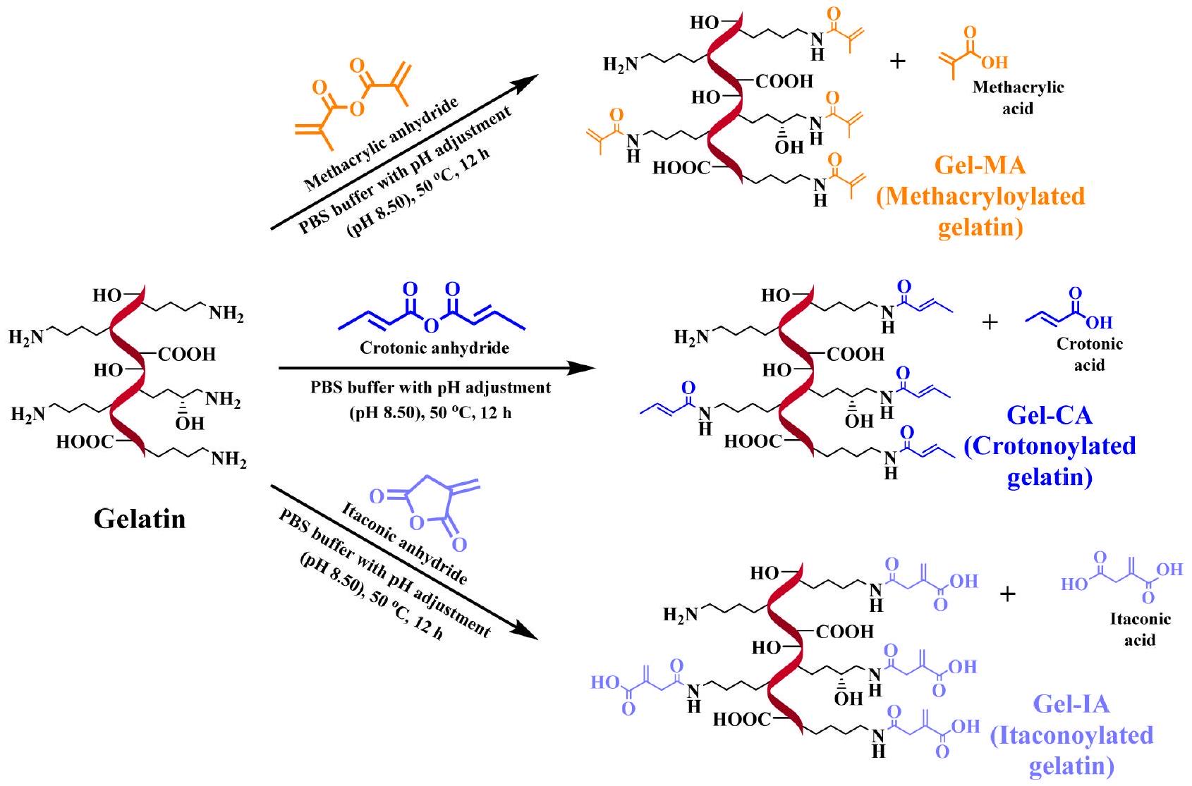
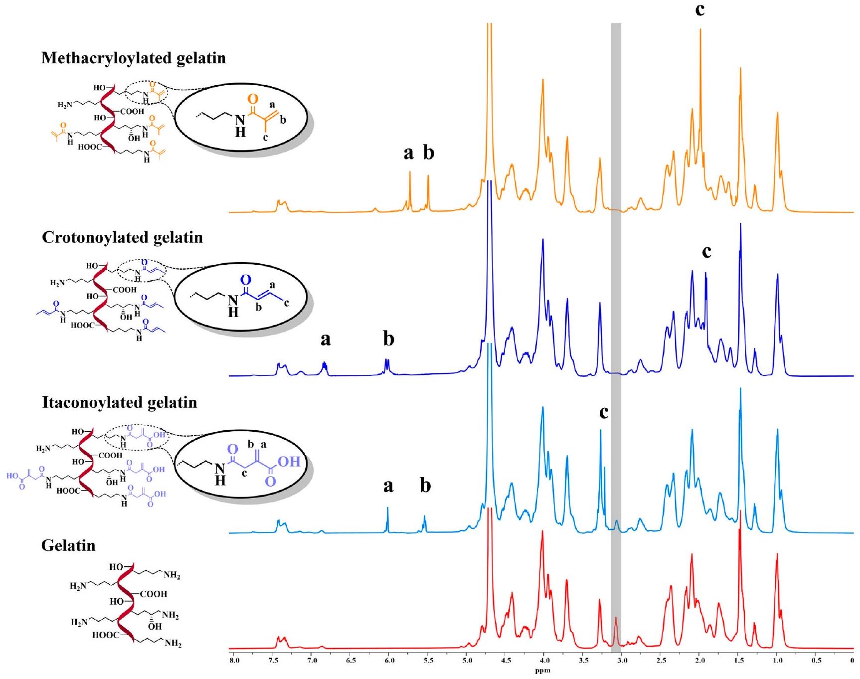
الجيلاتين هو بوليمر طبيعي قابل للذوبان في الماء وله خصائص لاصقة مخاطية ضعيفة. لقد تم استخدامه تقليديًا كمكون رئيسي في العديد من الأدوية، بما في ذلك الكبسولات اللينة والصلبة، والتحاميل، وهندسة الأنسجة، والطب التجديدي. يمكن تحسين الخصائص اللاصقة المخاطية للجيلاتين عن طريق تعديله من خلال الاقتران مع مجموعات لاصقة غير مشبعة محددة. في هذه الدراسة، تم تعديل الجيلاتين من خلال التفاعل مع أنهدريدات الكروتونيك، والإيتاكونيك، والميثاكريليك بنسب مولية متفاوتة لإنتاج جيلاتين كروتونيلات، وإيتاكونيلات، وميثاكريولات (المختصرة كـ Gel-CA، Gel-IA، وGel-MA، على التوالي). تم تأكيد التخليق الناجح باستخدام تم دراسة تأثير التعديل الكيميائي على النقطة المعزولة من خلال قياسات اللزوجة والحركة الكهربائية. تطور التخزين ( ) والخسارة ( ) تم استخدام المودولي لتحديد نقاط تجلط الحرارة القابلة للعكس للهلام المعدل و
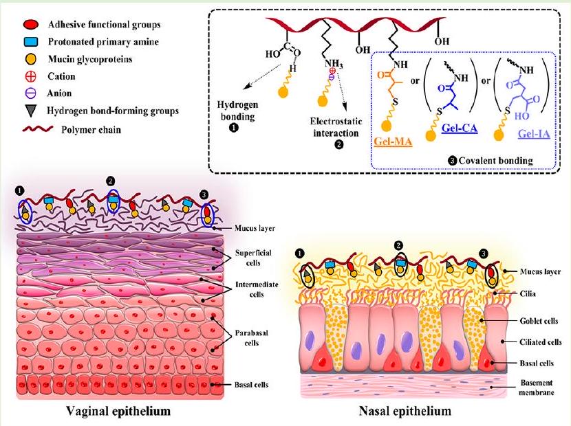
الجيلاتين غير المعدل. تم تقييم سلامة مشتقات الجيلاتين المعدل من خلال اختبار تهيج الغشاء المخاطي للرخويات في الجسم الحي (SMIT) واختبار MTT في المختبر باستخدام خط خلايا الألياف الرئوية البشرية. تم إعداد شكلين مختلفين من الجرعات، مثل الجل الفيزيائي والميكروكريات المجففة بالرش، وتم تقييم خصائصها اللاصقة للمخاط باستخدام تقنية التدفق مع الكشف الفلوري واختبار الشد باستخدام أنسجة المهبل الخنزيرية و الغشاء المخاطي الأنفي للأغنام. أظهرت الجيلاتينات المعدلة بمجموعات غير مشبعة خصائص لاصقة للمخاط متفوقة مقارنة بالجيلاتين الأصلي. إن القدرة المعززة للجيلاتين المعدل بهذه المجموعات الوظيفية غير المشبعة ترجع إلى تكوين روابط تساهمية مع المجالات الفرعية الغنية بالسيستين الموجودة في الميوسين من خلال تفاعلات الإضافة من نوع مايكل بتفاعل ثيول-إين التي تحدث في ظل ظروف ذات صلة فسيولوجيًا.

1. المقدمة

الجيلاتين هو بوليمر حيوي طبيعي مشتق من الكولاجين من خلال التحلل المائي الجزئي والت denaturation الحرارية. يتم استخراج الكولاجين نفسه من العظام والغضاريف والأنسجة الضامة وجلود الحيوانات المذبوحة وقشور الأسماك. تعتبر الجيلاتين واحدة من أهم وأكثر البوليمرات المتعددة الاستخدامات في الصناعة بسبب خصائصها الفيزيائية والكيميائية الفريدة. تعتمد خصائص الجيلاتين على طريقة إنتاجها، والتي يمكن أن تشمل المعالجة الحمضية أو القلوية. الجيلاتين من النوع A المتاحة تجارياً، والتي يتم إنتاجها عبر العملية الحمضية، لها نقطة تعادل كهربائي (IEP) عند بينما يظهر الجيلاتين من النوع B، الذي يتم تصنيعه من خلال العملية القلوية، نقطة التركيز الكهربائي المعكوس عند pH 4.5-5.5. كمواد حيوية قائمة على البروتين، يتمتع الجيلاتين بتوافق حيوي ممتاز، وقابلية للتحلل البيولوجي، وعدم السمية، وعدم المناعية. وقد تم اعتماده كمواد معترف بها بشكل عام على أنها آمنة (GRAS) من قبل إدارة الغذاء والدواء الأمريكية (FDA). يذوب الجيلاتين بسهولة في الماء الدافئ عند ولديه القدرة على تشكيل تقاطعات جسدية-
الهيدروجيلات القابلة للعكس الحراري المرتبطة عند التبريد تحت ج. بالإضافة إلى ذلك، فإنه يظهر نقطة انصهار قريبة من درجة حرارة الجسم. هذه الخصائص المميزة هي العوامل الرئيسية التي تدفع إلى استخدامه الواسع في الصناعات الصيدلانية والغذائية والتجميلية. يستخدم الجيلاتين عادة كمكون رئيسي في العديد من التركيبات، بما في ذلك الكبسولات الصلبة واللينة، تحاميل مهبلية ومستقيمية كشبكات في هندسة الأنسجة والطب التجديدي، كحامل في توصيل الأدوية (مثل الكريات الدقيقة)، وفي العديد من التطبيقات المتعلقة بالصحة الأخرى.
تُعرّف الالتصاق المخاطي بأنه قدرة المواد على الالتصاق بالأغشية المخاطية في الجسم لفترة طويلة من الزمن. تشمل الطرق المعتمدة لإدارة الأدوية عبر الأغشية المخاطية العين (القرنية والملتحمة)، الأنف، الفم (الخد واللسان)، الجهاز الهضمي، المستقيم، المهبل، والداخل المثاني. تقدم توصيل الأدوية عبر الأغشية المخاطية عدة مزايا مهمة، مثل سهولة إدارة شكل الجرعة وطبيعته غير الغازية، وزيادة مدة الإقامة على السطح المخاطي، وتحسين توافر الدواء الحيوي، وتجنب الأيض الكبدي الأول، وتقليل تكرار الجرعات، وإمكانية إنهاء العلاج بسرعة عند الحاجة.
تُستخدم العديد من البوليمرات اللاصقة المخاطية من الجيل الأول (التقليدية) تقليديًا في أشكال الجرعات المختلفة لتوصيل الأدوية عبر الأغشية المخاطية. تشمل هذه الفئة من المواد اللاصقة البوليمرات القابلة للذوبان في الماء من أصول طبيعية وصناعية، مثل الكيتوزان، وجيلان الصمغ، والألجينات، وبوليمرات الكربوبول، ومشتقات السليلوز (مثل السليلوز ميثيل الكربوكسي، وهيدروكسي بروبيل ميثيل السليلوز، وغيرها). تحدث الالتصاق المخاطي في هذه أنظمة توصيل الأدوية من خلال تفاعلات فيزيائية متنوعة (رابطة غير محددة) بين الجزيئات الكبيرة وبروتينات الميوسين الموجودة على الأسطح المخاطية. تشمل هذه التفاعلات بشكل أساسي قوى غير تساهمية، مثل الروابط الهيدروجينية، والجذب الكهروستاتيكي، وقوى فان دير فالس، والتفاعلات الكارهة للماء، وتشابك السلاسل/ الانتشار. عمومًا، تُظهر البوليمرات متعددة الشحنات خصائص لاصقة مخاطية أفضل مقارنة بالبوليمرات غير الأيونية. علاوة على ذلك، قد تختلف آليات التصاق هذه المواد بالأسطح المخاطية اعتمادًا على طبيعة شكل الجرعة. على سبيل المثال، تتأثر خصائص الالتصاق لأشكال الجرعات الصلبة (مثل الأقراص) بعملية الترطيب، بينما تتأثر احتفاظ التركيبات السائلة على الأسطح المخاطية بشكل أكبر بخصائصها الريولوجية.
في العقود القليلة الماضية، تم استكشاف طرق كيميائية مختلفة لتصميم البوليمرات وتركيباتها بخصائص لاصقة محسّنة. يمكن تحقيق هذا التحسين من خلال وظيفة البوليمرات المحبة للماء مع مجموعات لاصقة محددة قادرة على تشكيل روابط تساهمية مع الأنسجة المخاطية تحت الظروف الفسيولوجية. تمثل البوليمرات المحتوية على ثيول، المعروفة أيضًا باسم “ثيومرات”، واحدة من التقدمات البارزة في المواد اللاصقة المخاطية من الجيل الثاني، التي رائدها برنكوبيك شنورش وزملاؤه. تم تعديل هذه البوليمرات من خلال إدخال مجموعات وظيفية ثيول (سلفهيدريل) على سلاسلها الجانبية. تشكل البوليمرات الثيولية جسور ثنائي الكبريتيد من خلال تفاعلات تساهمية (عبر تفاعلات الأكسدة) مع المجالات الفرعية الحاملة للسيستين من بروتينات الغليكوبروتين المخاطية الموجودة على الأسطح المخاطية. يؤدي ذلك إلى تعزيز قدراتها على الالتصاق بالمخاط مما ينتج عنه زيادة مدة بقاء الدواء في موقع التطبيق. ومع ذلك، من الجدير بالذكر أن البوليمرات المحتوية على ثيول عرضة للأكسدة، مما يمكن أن يؤدي إلى تداخل غير مرغوب فيه بين البوليمرات.
تم اقتراح استراتيجيات متنوعة مؤخرًا لتعزيز الخصائص اللاصقة للمركبات البوليمرية المحبة للماء من خلال إدخال مجموعات لاصقة مثل الأكرول. ميثاكريلويل ماليميد كاتيكول بورونات و -هيدروكسي (سلفو) سوكسينيميد إستر. البوليمرات المعالجة بالأكريلويل، التي اقترحها لأول مرة فريق البحث بقيادة بيانكو-بيليد، تم تسليط الضوء على أنها فئة جديدة من المساعدات الصيدلانية مع خصائص لاصقة محسّنة بشكل كبير مقارنةً بنظيراتها غير المعدلة. كما كانت مجموعة خوتوريانسكي رائدة في استخدام المواد المشتقة من الميثاكريلويل والماليميد لتصميم أشكال الجرعات مع تحسينات.
خصائص الالتصاق المخاطي. وتشمل تطوير النانو هلام، الحويصلات الدهنية جزيئات نانوية تركيبات سائلة، الهلام في الموقع وأقراص مطلية بالرش. تستطيع هذه المجموعات الوظيفية غير المشبعة تشكيل روابط تساهمية مع المجالات الفرعية الغنية بالسيستين من بروتينات الغليكوزيل عبر تفاعلات الإضافة من نوع مايكل بتقنية ثيول-إين لتحقيق روابط لاصقة قوية. علاوة على ذلك، فإن الجزيئات الكبيرة المفعلة بـ (ميث) أكريلويل وماليميد لديها استقرار أفضل ضد الأكسدة دون ميل للتشابك بين الجزيئات أو داخلها على عكس البوليمرات المحتوية على ثيول. مؤخرًا، أظهرت الناقلات النانوية المفعلة بالألدهيد أيضًا القدرة على الالتصاق بقوة ببطانة المثانة البولية للخنازير وبالغشاء المخاطي الأنفي للأغنام من خلال تشكيل روابط إيمين مع مجموعات الأمين الأولية الموجودة على سطح الأنسجة المخاطية عبر كيمياء قاعدة شيف.
تقليديًا، تم اعتبار الجيلاتين بوليمرًا ذو خصائص لاصقة مخاطية ضعيفة. تُعزى قدرات الالتصاق للجيلاتين بشكل أساسي إلى طبيعته الأمفوتيرية، مما يؤدي إلى تفاعلات كهربائية ضعيفة مع الأسطح المخاطية مقارنةً بالتفاعلات التساهمية القوية. كانت النتائج المتعلقة بالخصائص الحيوية/المخاطية للجيلاتين موضوع نقاش. وقد أفادت عدة دراسات أن الجيلاتين يظهر لاصقية مخاطية ملائمة، أو أن إضافة الجيلاتين تحسن الخصائص اللاصقة للأنظمة المدروسة إلى حد ما. ومع ذلك، تشير معظم الدراسات إلى أن الجيلاتين النقي يظهر خصائص لاصقة ضعيفة، و/أو أن وجوده لا يساهم بشكل كبير في لزوجة التركيبات. ومع ذلك، يحتوي الجيلاتين على مواقع تفاعلية داخل هيكله الجزيئي، مثل مجموعات الأمين، والمجموعات الكربوكسيلية، ومجموعات الهيدروكسيل، مما يوفر فرصًا للتزاوج مع المجموعات الوظيفية التي يمكن أن تؤدي إلى تغييرات في خصائصه الفيزيائية والكيميائية. مراجعة حديثة من أحمدي وأبو سمه تمت مناقشة استراتيجيات مختلفة لتعزيز الخصائص اللاصقة للمخاط للجيلاتين. إحدى الطرق المقترحة هي إدخال مجموعات الميثاكريلويل في الجيلاتين؛ ومع ذلك، حسب علمنا، لا توجد أدلة قاطعة تدعم تأثيرها الإيجابي على الخصائص اللاصقة للمخاط.
في الدراسة الحالية، نبلغ عن تعديل الجيلاتين من خلال التفاعلات مع ثلاثة أنهدريدات غير مشبعة (الكراتون، الإيتاكون، والميثاكريليك) من أجل تعزيز خصائصه اللاصقة للمخاط. تم توصيف مشتقات الجيلاتين الناتجة بشكل كامل باستخدام تمت دراسة تأثير التعديل الكيميائي على النقطة المعزولة باستخدام قياسات اللزوجة والحركة الكهربائية. تم استخدام مطيافية الرنين المغناطيسي النووي (NMR) ومطيافية الأشعة تحت الحمراء (FTIR) واختبار TNBSA اللوني. تطور التخزين ( ) والخسارة ( تم استخدام ) المودولي ( لتحديد نقاط الجلطة الحرارية القابلة للعكس لكل من الجيلاتين المعدل وغير المعدل. كما تم تقييم الخصائص السمية لهذه المشتقات باستخدام اختبار تهيج الغشاء المخاطي للرخويات (SMIT) واختبار MTT في خلايا خط fibroblasts الرئوية البشرية. تم إعداد شكلين مختلفين من الجرعات مثل الجل الفيزيائي والميكروكريات المجففة بالرش وتم تقييم احتفاظها على الأسطح المخاطية باستخدام الغشاء المخاطي المهبلي الخنزيري والغشاء المخاطي الأنفي للأغنام.

2. القسم التجريبي

2.1. المواد. أنهدريد الكروتون (CA)، أنهدريد الإيتاكون (IA)، أنهدريد الميثاكريليك (MA)، الجيلاتين من النوع A من جلد الخنازير (قوة الجل) بلوم)، كلوريد البنزالكونيوم (BAC)، أكسيد الديوتيريوم، 3-(4،5-ثنائي ميثيل الثيازول-2-يل)-2،5-ثنائي الفينيل تيترازوليوم بروميد (مادة MTT)، ملح الفلورسئين الصوديوم (NaFl)، محلول الجلutaraldehyde ( في درجة II)، جلايسين، حمض الهيدروكلوريك 1 م
محلول، بيكربونات الصوديوم، دوديكانول سلفات الصوديوم (SDS)، حمض 2،4،6-ثلاثي نيتروبنزين سلفونيك (TNBSA، في ) و صبغة التريبان الأزرق ( تم شراء المحلول من سيغما-ألدريتش (غيلينغهام، المملكة المتحدة). ثنائي ميثيل سلفوكسيد (DMSO)، وسط دلبوكو المعدل (DMEM)، مصل جنين البقر (FBS)، بنسلين-ستربتوميسين ( أقراص محلول ملحي معزز بالفوسفات (PBS) (التي تم استخدامها لتحضير 100 مل من محلول PBS في ماء منزوع الأيونات، pH 7.40، هيدروكسيد الصوديوم، وتربسين-EDTA تم شراء المحلول من شركة فيشر ساينتيفيك (لوغبره، المملكة المتحدة). كانت جميع المواد الكيميائية الأخرى من الدرجة التحليلية واستخدمت دون مزيد من التنقية. تم شراء أنابيب الغسيل مع حد قطع الوزن الجزيئي 1214 كيلودالتون من شركة ميديسيل ميمبرينز المحدودة (لندن، المملكة المتحدة). تم استخدام الماء المقطر طوال التجارب التي تتضمن المحاليل المائية.
2.2. تخليق مشتقات الجيلاتين. تم تعديل الجيلاتين كيميائيًا باستخدام أنهدريدات غير مشبعة مختلفة وفقًا للإجراءات الموصوفة سابقًا مع بعض التعديلات. باختصار، الجيلاتين تم إذابته في 100 مل من محلول PBS (رقم الهيدروجيني 7.40) في حتى تشكلت محلول شفاف متجانس أثناء التحريك. بعد ذلك، أضيفت كميات مرغوبة من أنهدريد الكروتون، أو أنهدريد الإيتاكون، أو أنهدريد الميثاكريليك بالتنقيط إلى محاليل الجيلاتين وتفاعلت لمدة 12 ساعة عند تحت التحريك المستمر لإنتاج مشتقات الجيلاتين المقرونة بالكروتون، أو الإيتاكون، أو الميثاكريلات (المختصرة بـ Gel-CA و Gel-IA و Gel-MA، على التوالي). تم الحفاظ على الرقم الهيدروجيني عند 8.50 طوال التفاعل عن طريق إضافة محلول NaOH بتركيز 4 م. بعد التخفيف بإضافة 50 مل من محلول PBS (الرقم الهيدروجيني 7.40) لإيقاف التفاعل، تم تنقية كل منتج ناتج عن طريق الدياليسيس ضد الماء منزوع الأيونات (5 لترات؛ تم تغيير الماء 8 مرات) لمدة 48 ساعة في الظلام لإزالة الأملاح، والأنهدريدات غير المتفاعلة، والأحماض الناتجة. أخيرًا، تم تجفيف المحلول بالتجميد، مما شكل منتجًا أبيض يشبه الإسفنج، وتم ختمه وتخزينه في الفريزر حتى الاستخدام لاحقًا. تم تلخيص البيانات حول الكميات المتنوعة من الأنهدريدات الموجودة في خليط التفاعل الأولي في الجدول S1 في المعلومات الداعمة.
2.3. تحضير الجسيمات الدقيقة المجففة بالرش. تم إذابة عينات الجيلاتين المعدلة كيميائيًا وغير المعدلة (0.5 جرام) في البداية في 100 مل من المحاليل المائية التي تحتوي على ملح فلوريسئين الصوديوم ( ) في لمدة 60 دقيقة مع التحريك بسرعة 400 دورة في الدقيقة. في تحضير منفصل، من تم إضافة محاليل الجلوتارالدهيد المائية إلى المحاليل القائمة على الجيلاتين وتم تحريكها لمدة 60 دقيقة أخرى بسرعة 400 دورة في الدقيقة. بعد ذلك، تم تجفيف المحاليل الناتجة باستخدام جهاز تجفيف الرذاذ الصغير Büchi Mini Spray Dryer B-290 (Büchi Labortechnik AG، فلافيل، سويسرا) المزود بجهاز إزالة الرطوبة S-396 لإنتاج جزيئات ميكروية قائمة على الجيلاتين محملة بـ NaFl، سواء كانت متشابكة أو غير متشابكة. تم توصيل المحاليل إلى الفوهة بمعدل تغذية باستخدام مضخة بيرستالتية ثم تم تجفيفها بالرش عند درجة حرارة المدخل ودرجة حرارة المخرج . كمعيار، تم ضبط معدل الشفط على لزيادة معدل الفصل في السايكلون؛ تم استخدام النيتروجين المضغوط لتفريق السائل إلى قطرات دقيقة. تم جمع المنتجات الناتجة من التجفيف بالرش، وإغلاقها لحمايتها من إعادة الترطيب، وتغليفها بورق الألمنيوم، وتخزينها في الفريزر حتى إجراء اختبارات إضافية.
2.4. التوصيف. 2.4.1. قياس درجة التFunctionalization. تم تأكيد تعديل الجيلاتين باستخدام طيف الرنين المغناطيسي النووي. عشرون من الجيلاتين ومشتقاته (Gel-CA و Gel-IA و Gel-MA) تم إذابتها في حرارة دافئة تم تسجيل طيف NMR للعينات باستخدام جهاز مطياف NMR Bruker Avance III بتردد 500 ميجاهرتز (Bruker UK Ltd.، كوفنتري، المملكة المتحدة) في مع 128 مسح لكل طيف. قبل التفسير، تم تصحيح الطور لكل طيف ناتج. تم تطبيق تصحيح القاعدة قبل دمج الإشارات ذات الاهتمام.
تم استخدام تحليل الرنين المغناطيسي النووي (NMR) لتحديد درجة التFunctionalization (DoF) لمشتقات الجيلاتين بطريقة مشابهة لما تم وصفه سابقًا. تم تقدير نسبة دمج المجموعات غير المشبعة من خلال مقارنة شدة القمم الناتجة عن الروابط المزدوجة بتلك الخاصة بالبقايا العطرية الموجودة في سلاسل الجيلاتين الجانبية.
تم استخدام اختبار اللونيمتري لحمض 2،4،6-ثلاثي نيترو بنزين سلفونيك (TNBSA)، الذي تم تطويره في الأصل بواسطة حبيب، لتحديد
المجموعات الأمينية الحرة المتبقية بعد تحويل الجيلاتين مع تغييرات طفيفة. باختصار، الجيلاتين ومشتقاته تم إذابتها بشكل منفصل في محلول بيكربونات الصوديوم 0.1 م (رقم الهيدروجيني 8.50). ثم، من محلول TNBSA (محضر بتركيز 0.1 م) تم إضافة (المخزن المؤقت) إلى من كل عينة اختبار وحلها وتم حضنها في لمدة ساعتين مع التحريك برفق. بعد ذلك، من سلفات دوديسيل الصوديوم (SDS) و تم إضافة 1 م HCl إلى كل عينة اختبار لإيقاف التفاعل. ثم تم قياس امتصاص كل محلول عند 335 نانومتر وتم تحديد تركيز الأمينات الأولية الحرة باستخدام منحنى قياسي للجليسين (انظر الشكل S1 في المعلومات الداعمة). تم تحديد كمية المجموعات الأمينية الحرة لتكون 0.434 مللي مول لكل 1 جرام من الجيلاتين. تم حساب درجة الحرية عن طريق طرح كمية الأمينات الحرة المتبقية. المجموعات في كل جيلاتين معدل من كمية الحرة مجموعات في الجيلاتين الأصلي. تم استخدام مطياف UV-vis من شيمادزو UV-1900i (كيوتو، اليابان) لتسجيل طيف الامتصاص UV-vis للعناصر. تم إجراء جميع التجارب مع تقدير درجة الحرية في ثلاث نسخ.
2.4.2. مطيافية تحويل فورييه بالأشعة تحت الحمراء (FTIR). تم تسجيل طيف FTIR للجيلاتين غير المعدل والمعدل باستخدام مطياف FTIR Nicolet iS10 (Thermo Scientific، المملكة المتحدة) مع ملحق iTX للانعكاس الكلي المخفف (ATR) المزود بكريستال ماسي. تم جمع الأطياف من متوسط 32 مسحًا بين 4000 و ، باستخدام وضع النفاذية مع دقة طيفية قدرها .
2.4.3. المجهر الإلكتروني الماسح (SEM). تم فحص شكل وحجم الجسيمات الدقيقة المجففة بالرش المستندة إلى الجيلاتين ومشتقاته المعدلة (Gel-CA و Gel-IA و Gel-MA) باستخدام مجهر إلكتروني ماسح من نوع Zeiss Crossbeam 540 (Carl Zeiss Microscopy GmbH، يينا، ألمانيا) عند جهد تسريع قدره 5 كيلوفولت. تم طلاء العينات بالذهب قبل التصوير. ثم تم تحليل الصور الملتقطة باستخدام برنامج ImageJ (NIH، الولايات المتحدة الأمريكية) لتحديد متوسط قطر الجسيمات الدقيقة.
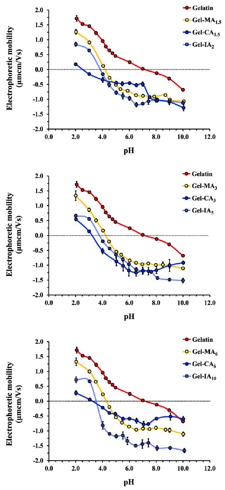
2.4.4. تحديد النقطة المعزولة كهربائياً (IEP). تم تحديد النقاط المعزولة كهربائياً للجيلاتين المعدل وغير المعدل باستخدام تقنية اللزوجة التقليدية. وحركة الكهربية باستخدام جهاز Zetasizer Nano-ZS90 (مالفرن إنسترومنتس، ورسيسترشاير، المملكة المتحدة)، على التوالي، مع تغيير درجة حموضة المحلول. تم إجراء جميع القياسات في ثلاث نسخ. . باختصار، تم تحضير محلول (w/v) من الجيلاتين أو مشتقاته في ماء منزوع الأيونات عند وتم التحريك حتى الذوبان الكامل. تم تحديد اللزوجة النوعية باستخدام مقياس لزوجة من نوع أوستوالد (بقطر شعري يبلغ 0.86 مم) ويتم التعبير عنها كنسبة لزمن التدفق ( ) إلى الماء ( ):
نقطة التساوي الكهربائي (IEP) تم تحديد ( ) لكل عينة بناءً على الرقم الهيدروجيني الذي أظهرت فيه محلول البوليمر أدنى قيمة لزوجة، مما يشير إلى أن الشحنة الإجمالية للجزيئات الكبيرة قريبة من الصفر عند هذا الرقم الهيدروجيني المحدد.
في تجارب الحركة الكهربائية، تم استخدام معامل انكسار الضوء النموذجي للبروتين 1.45 وامتصاص 0.001 لجميع القياسات في وضع الجهد الزتاوي. تم استخدام اللزوجة (0.8872 سنتي بواز) ومعامل الانكسار (1.33) للماء كمعلمات للتشتت. تم تحليل كل عينة ثلاث مرات. تم معالجة النتائج باستخدام نموذج سمولوشوفسكي. )، ومتوسط حركة الكهربية المتوسطة تم حساب قيم الانحراف المعياري. تم رسم منحنى الحركة الكهربائية مقابل الرقم الهيدروجيني لكل عينة اختبار والنقطة التي يتقاطع فيها المنحنى مع صفر حركة اعتُبرت كـ .
2.4.5. الدراسات الريولوجية. جهاز قياس اللزوجة TA DHR-1 (TA Instruments، نيو كاسل، ديلاوير، الولايات المتحدة الأمريكية) مزود بلوحة بيلتير ذات درجة حرارة متغيرة وجيومتري مخروط-قرص من الفولاذ المقاوم للصدأ. زاوية المخروط ) تم استخدامها لإجراء التجارب الريولوجية. كانت التجارب تهدف إلى تحديد درجات حرارة الجلطة والانصهار لعينات الجيلاتين. في البداية، كانت العينات ( تم تحقيق التوازن في المحاليل المائية عند لمدة 5 دقائق. بعد ذلك، تم تبريد كل عينة من 40 إلى ، تبع
من خلال اختبارات سحب درجة الحرارة من 0 إلى (مسخن) ثم تم تقليله مرة أخرى إلى (مبرد) بمعدل مسح . خلال هذه الاختبارات، التغيرات في معامل التخزين ( ) ومعامل الفقدان ( تم تسجيل ( ) كدالة لدرجة الحرارة عند إجهاد مطبق قدره وتردد (ما يعادل 1.6 هرتز). تم استخدام غطاء فخ المذيب لضمان درجة حرارة موحدة ومنع تبخر الخليط. تم حساب النتائج التي تم الحصول عليها كمتوسط قيم 3 قياسات.
2.5. تقييم السمية. 2.5.1. اختبار حيوية الخلايا في المختبر. تم زراعة خط خلايا الألياف الرئوية البشرية (HPF) في وسط ديلبيكو المعدل (DMEM) المدعوم بـ مصل الجنين البقري (FBS) و ستربتوميسين/بنسلين عند في حاضنة مرطبة مع . عند الوصول إلى التجمع في قوارير زراعة الخلايا T-25، تم جمع الخلايا باستخدام محلول التربسين-EDTA، مزروع في أطباق 96 بئر بكثافة خلايا لكل بئر، وتم الاحتفاظ بها طوال الليل تحت ظروف زراعة قياسية (37 و في الحاضنة المرطبة) لتعزيز ارتباط الخلايا. ثم تم معالجة الخلايا بمحلولات الجيلاتين المعدلة وغير المعدلة (Gel-CA و Gel-IA و Gel-MA) بتركيزات تبلغ 1.3 و في وسط نمو الخلايا. ثم تم حضن الخلايا لمدة 24 ساعة عند في بيئة رطبة الحاضنة. خلايا غير معالجة و تم استخدام محلول DMSO كتحكم سلبي وإيجابي على التوالي. تم استخدام التحكم الإيجابي للتحقق من نشاط الاختبار. بعد التعرض لمحلولات الجيلاتين ومشتقاته، تلقى كل بئر محلول MTT في الظلام، وتمت حضانة الخلايا لمدة 4 ساعات أخرى عند في بيئة رطبة الحاضنة. تم إذابة بلورات الفورمازان التي تم إنتاجها خلال فترة الحضانة عن طريق إضافة ( ) كبريتات الدوديسيل الصوديوم (SDS). تم قياس الامتصاص عند 570 نانومتر باستخدام قارئ الميكرو بلايت Varioskan (ثيرمو ساينتيفيك، الولايات المتحدة الأمريكية). تم حساب نسبة البقاء باستخدام المعادلة التالية:
2.5.2. اختبار تهيج الغشاء المخاطي للرخويات. تم إجراء اختبار تهيج الغشاء المخاطي للرخويات (SMIT) في الجسم الحي باستخدام المنهجية التي تم وصفها سابقًا من قبل مجموعة بحثنا. تم جمع حلزونات Arion lusitanicus في حديقة هاريس (ريدينغ، المملكة المتحدة) وتم الاحتفاظ بها في حاويات بلاستيكية مهواة في درجة حرارة الغرفة. تم إطعامها بالخس والملفوف والخيار لمدة 48 ساعة. بعد ذلك، تم فحص بطانة جسم كل حلزون بعناية، وتم استخدام الحلزونات التي لا تحتوي على إصابات ماكروسكوبية والتي تتميز بوجود نتوءات واضحة وسطح قدم. كانت الحلزونات التي تزن ثم تم اختيارها واحتفظ بها بشكل فردي في قوارير زجاجية سعة 2 لتر مبطنة بمنشفة ورقية مبللة بـ 20 مل من محلول PBS (pH 7.40) وتركها في درجة حرارة الغرفة لمدة 48 ساعة قبل بدء التجارب. كانت القوارير مغطاة بغلاف بلاستيكي مثقوب لضمان تبادل الهواء. ثم تم وزن كل حلزون بشكل فردي ووضعه في طبق بتري بلاستيكي بحجم 90 مم مبطن بورق فلتر ووتمان مبلل بـ 2 مل من محلول العينة. تضمنت عينات الاختبار محلول PBS (التحكم السلبي)، كلوريد البنزالكونيوم (التحكم الإيجابي) المحضر في PBS، بالإضافة إلى محاليل الجيلاتين المعدلة وغير المعدلة التي تم إعدادها في PBS. بعد 60 دقيقة من الاتصال مع عينات الاختبار، تم إزالة الحلزونات من أطباق بتري، وغسلها بـ 10 مل من PBS، وتم مسحها برفق بمنشفة ورقية، ثم تم إعادة وزنها. تم تقدير إنتاج المخاط (MP) بناءً على فقدان وزن جسم الحلزون وتم حسابه باستخدام المعادلة التالية:
أين و هي أوزان الحلزونة قبل وبعد التعرض للمواد المختبرة، على التوالي. تم التعبير عن نتائج التجارب كمتوسط قيم الانحراف المعياري ( ) وتم تقييمه إحصائيًا.
2.6. دراسات الاحتفاظ خارج الجسم على الغشاء المخاطي المهبلي للخنازير والغشاء المخاطي الأنفي للأغنام. 2.6.1. تصميم شكل الجرعة النموذجي. 0.1 تم تحضير المحلول في ماء منزوع الأيونات واستخدم كمركب دوائي نموذجي. ثم، الهلام وطرحه الكروتونوي (Gel-CA) والإيتاكونوي (Gel-IA) والميثاكريلو-
تم إذابة مشتقات الجيلاتين المعدلة (Gel-MA) بشكل منفصل في 10 مل من المحاليل المائية لـ NaFl. تم تحريك الخلطات لمدة 12 ساعة في درجة حرارة الغرفة حتى تشكلت محاليل متجانسة، وتم تغطيتها بورق الألمنيوم، وتخزينها في الثلاجة للاستخدام لاحقًا. تم استخدام هذه التركيبات المعتمدة على الجيلاتين المعدلة وغير المعدلة المحملة بـ NaFl في دراسات الالتصاق المخاطي في المهبل الخنزيري.
يتم وصف تحضير الجسيمات الدقيقة المجففة بالرش المحملة بـ NaFl المستندة إلى الجيلاتين ومشتقاته في القسم 2.3. تم استخدام هذه الجسيمات الدقيقة في دراسات الالتصاق المخاطي في أنف الأغنام. يتم وصف التركيبات لتحضير محاكاة السائل المهبلي (VFS؛ pH 4.0) والسائل الأنفي الاصطناعي (ANF؛ pH 5.80) المستخدمة لغسل الأسطح المخاطية في الجداول S2 و S3 في المعلومات الداعمة، على التوالي. تم الحفاظ على محاليل VFS و ANF عند خلال تجارب الالتصاق المخاطي باستخدام حمام مائي.
2.6.2. تحضير الأنسجة. تم استلام أنسجة المهبل من الخنازير ورؤوس الأغنام من مسالخ P.C. Turner (فارنبورو، المملكة المتحدة) مباشرة بعد ذبح الحيوانات، وتم تعبئتها ونقلها إلى المختبر في حاويات بلاستيكية باردة، واستخدامها خلال 24 ساعة من جمعها. تم تشريح أنسجة المهبل بعناية (مع تجنب الاتصال بالغشاء المخاطي الداخلي) باستخدام شفرات حادة يمكن التخلص منها للحصول على الأقسام.
تم استخراج الأنسجة المخاطية للحاجز الأنفي بعناية من رؤوس الأغنام باستخدام مقصات قطع العظام، ثم تم قطعها إلى قطع مربعة سم مع شفرات حادة قابلة للتخلص منها.
2.6.3. تقنية التدفق المستمر. تم إجراء تجارب لتقييم احتفاظ الغشاء المخاطي بالتركيبات المعتمدة على الجيلاتين المعدل وغير المعدل على أنسجة المهبل الخنازير والأنف الأغنام خارج الجسم باستخدام طريقة التدفق المستمر المعروفة مع تعديلات طفيفة تتعلق بالكشف الفلوري. في البداية، تم تثبيت نسيج مهبلي أو أنفي مقطوع حديثًا على شريحة زجاجية للميكروسكوب مع الجانب المخاطي موجهًا لأعلى، ثم وُضِع على ركيزة مثبتة بزاوية وتم شطفها مسبقًا بـ 1 مل من محلول VFS أو ANF، على التوالي، قبل بدء كل اختبار لالتصاق المخاط الخارجي.
تم التقاط صور الفلورية لسطح الغشاء المخاطي لأنسجة المهبل باستخدام مجهر ستيريو Leica MZ10F (Leica Microsystems، المملكة المتحدة) مزود بكاميرا رقمية Leica DFC3000G مزودة بفلتر بروتين الفلورسنت الأخضر (GFP) (أزرق، ) في تكبير، مع زمن تعرض قدره 10 مللي ثانية و الكسب. في البداية، تم الحصول على صور لأنسجة المهبل الفارغة لتحديد شدة الفلورسنت الخلفية لكل عينة قبل إعطاء المادة الاختبارية. بعد ذلك، تم أخذ عينات مسبقة التسخين ( ) من أي مشتقات الجيلاتين، Gel-CA، Gel-IA، أو Gel-MA المحضرة في ماء منزوع الأيونات يحتوي على أو تحكم في تم إيداع المحلول على السطح المخاطي. ثم تم ري العينات بشكل متكرر بمحلول VFS (رقم الهيدروجين 4.0) بمعدل تدفق باستخدام مضخة حقن. تم الحصول على صور المجهر الفلوري لسطح الغشاء المخاطي لكل عينة مهبلية في نقاط زمنية محددة مسبقًا ثم تم تحليلها باستخدام برنامج ImageJ (NIH، الولايات المتحدة الأمريكية) من خلال قياس كثافة البكسل بعد كل غسلة مع VFS. تم طرح كثافة البكسل للعينات العارية (الغشاء المخاطي المهبلي بدون مادة اختبار فلورية) من كل قياس وتم تحويل البيانات إلى قيم كثافة طبيعية باستخدام المعادلة التالية:
أين هو شدة الفلورسنت الخلفية لعينة نسيج معينة (نسيج فارغ)؛ يشير إلى شدة الفلورية الأولية لتلك العينة (عينة الأنسجة التي تم تطبيق مادة فلورية لاصقة عليها قبل بدء أول غسيل؛ وقد اعتُبر هذا كنقطة زمنية صفرية مع شدة الفلورية)؛ و يمثل شدة الفلورة لعينة الأنسجة تلك مع المادة الفلورية اللاصقة بعد كل دورة غسيل. ثم تم تحويل هذه الشدات الفلورية إلى قيم % احتفاظ مخاطي.
بالإضافة إلى ذلك، تم تقييم أداء اللصق المخاطي للجزيئات الدقيقة القائمة على الجيلاتين المعدلة وغير المعدلة المجففة بالرش باستخدام
المخطط 1. توضيح تخطيطي لتفاعل تعديل الجيلاتين مع أنهدريدات غير مشبعة مختلفة
يرجى ملاحظة أن الهيكل التخطيطي يعرض فقط احتمالين لتفاعل الأنهيدريد مع الأمين الأولي. مجموعات من الليسين/الهيدروكسيليسين (وهي الجوانب التفاعلية) في هيكل الجيلاتين. في الواقع، يمكن أن يتفاعل أيضًا مع أي مجموعات هيدروكسيل (-OH) موجودة في الجيلاتين بسبب زيادة وقت التفاعل وقد يؤدي ذلك إلى زيادة درجة الحرية.
نفس تقنية التدفق من خلال الزجاج كما هو موصوف أعلاه مع بعض التعديلات. تم استخدام عينات من الجسيمات الدقيقة غير المتشابكة فقط في هذه التجربة. حوالي 100 ملغ من الجسيمات الدقيقة القائمة على الجيلاتين (تتضمن الجيلاتين، Gel-CA، Gel-IA، أو Gel-MA) تحتوي على 1 تم تطبيقها على الغشاء المخاطي الأنفي للأغنام خارج الجسم، والذي كان مثبتًا بالفعل على شريحة زجاجية، موضوعة على أنبوب طرد مركزي من نوع فالكون مقطوع إلى نصفين مائل بزاوية تم بعد ذلك تقطير محلول ANF (pH 5.80) على الغشاء المخاطي الأنفي بمعدل تدفق باستخدام مضخة حقن (كان إجمالي وقت الغسيل 30 دقيقة). تم تعيين معدل التدفق المذكور عمدًا أعلى من معدل إنتاج السوائل الأنفية الفسيولوجي لأسباب عملية. تم إجراء هذا التعديل لتسريع التجارب وضمان إمكانية إجرائها ضمن إطار زمني معقول. بعد ذلك، تم جمع محلول ANF المتدفق عبر الغشاء المخاطي الأنفي في فترات زمنية محددة مسبقًا. تم أخذ عينات من سلسلة من محاليل ANF المجمعة بعد كل دورة غسيل للتحليل لتحديد كمية الجسيمات الدقيقة المحملة بـ NaFl التي تم غسلها من سطح الغشاء المخاطي. تم إجراء التحليل باستخدام مقياس الطيف الفلوري Varian Cary Eclipse (سانتا كلارا، كاليفورنيا، الولايات المتحدة الأمريكية) في و تمت عملية التقدير بناءً على منحنى قياسي لـ NaFl (انظر الشكل S2 في المعلومات الداعمة) من خلال الحسابات، التي وفرت كمية التركيبات المحتفظ بها على الغشاء المخاطي الأنفي للأغنام.
2.6.4. طريقة الشد (الانفصال). تم استخدام جهاز تحليل القوام TA.XT Plus (Stable Micro Systems Ltd.، ساري، المملكة المتحدة) الذي يعمل في وضع اختبار اللصق لتقييم أداء اللصق المخاطي للجزيئات الدقيقة المجففة بالرش المستندة إلى الجيلاتين ومشتقاته. تم استخدام عينات من الجزيئات الدقيقة المتشابكة وغير المتشابكة في هذه التجربة لتقييم مساهمة انتشار الجزيئات الكبيرة والقدرة على التكوين.
طبقة متداخلة مع المخاط في الالتصاق المخاطي. تم استخدام أنسجة أنف الأغنام المعزولة حديثًا خلال 24 ساعة من الاسترجاع لهذا التجربة. كما تم الإبلاغ عنه سابقًا وتم تعديله ببعض التغييرات، تم تأمين قسم من نسيج أنف الأغنام بحيث يكون الجانب المخاطي متجهًا لأسفل على سطح مسبار أسطواني مصمم خصيصًا. تم بعد ذلك ربط هذا المسبار بذراع متنقل لجهاز تحليل القوام. تم تثبيت قطعة أخرى من نسيج الأنف بحيث يكون الجانب المخاطي متجهًا لأعلى بشكل آمن على جهاز الالتصاق المخاطي لجهاز تحليل القوام. قبل كل قياس، تم شطف أنسجة الأنف مسبقًا بـ 1 مل من محلول ANF (رقم الهيدروجين 5.80). بعد ذلك، تم إعطاء ملغ من كل تركيبة ميكروبارتكل قائمة على الجيلاتين المجفف بالرش للغشاء المخاطي الأنفي المثبت على الجهاز. ثم تم خفض المجس الأسطواني المتحرك الذي يحمل الأنسجة الأنفية الفارغة لإقامة اتصال مع السطح المخاطي المقابل. تم استخدام البيانات المستمدة من تجارب الانفصال لتقييم قوة الالتصاق المخاطي وإجمالي عمل الالتصاق. تم تطبيق إعدادات المعدات التالية: اختبار سرعة الضغط. ; اختبار السرعة ثانية؛ سرعة ما بعد الاختبار القوة المطبقة ; وقت الاتصال 120 ثانية; نوع الزناد كان “تلقائي”; قوة الزناد ; وارجع المسافة 10 مم.
تم إجراء جميع التجارب لتقييم احتفاظ التركيبات على الغشاء المخاطي المهبلي الخنزيري خارج الجسم والغشاء المخاطي الأنفي للأغنام في و الرطوبة النسبية داخل الحاضنة لمحاكاة الظروف الفسيولوجية. تم إجراء جميع القياسات ثلاث مرات، والمتوسط تم حساب قيم الانحراف المعياري ثم تم تقييمها إحصائيًا.
2.7. التحليل الإحصائي. تم إجراء جميع القياسات في الدراسة الحالية ثلاث مرات على الأقل وتم التعبير عن البيانات كمتوسط قيم الانحراف المعياري. تم إجراء التحليلات الإحصائية باستخدام برنامج GraphPad Prism (الإصدار 8.0؛ سان
الشكل 1. طيف NMR للجيلاتين المعدل وغير المعدل المسجل في في .
دييغو، كاليفورنيا، الولايات المتحدة الأمريكية). تم مقارنة البيانات وتقييمها من حيث الأهمية باستخدام اختبار الطالب ذو الذيلين. -اختبار وتحليل التباين أحادي الاتجاه (ANOVA) مع اختبار بونفيروني بعد الاختبار (لنتائج اختبار SMIT وتجارب الالتصاق المخاطي). بالنسبة لاختبار MTT، تم استخدام تحليل التباين أحادي الاتجاه مع مقارنات متعددة لدونيت لمقارنة المجموعات الضابطة والمعالجة، بينما داخل المجموعات المعالجة، تم استخدام اختبار الطالب ذو الطرفين. تم تطبيق اختبار لمقارنة الجيلاتين ومشتقاته. اعتُبرت الفروق الإحصائية ذات دلالة عند مستوى .

3. النتائج والمناقشة

3.1. التخليق والتوصيف. توفر وجود مواقع تفاعلية في جزيئات الجيلاتين، مثل الأمينات والمجموعات الكربوكسيلية والهيدروكسيلية، فرصة للتعديل الكيميائي مما يؤدي إلى تغييرات في خصائصه الفيزيائية والكيميائية. في هذا العمل، تم تخليق جيلاتين مضاف إليه الكروتون، والإيتاكون، والميثاكريلات عن طريق تفاعل هذا البوليمر الحيوي مع أنهدريدات الكروتون، والإيتاكون، والميثاكريليك، على التوالي، بنسب تغذية مختلفة بالنسبة للأمينات الحرة على هيكل الجيلاتين (المخطط 1). بعد التنقية عن طريق الدياليسيس والتجفيف بالتجميد، تم توصيف مشتقات الجيلاتين باستخدام طيف الرنين المغناطيسي النووي (الشكل 1). عرضت الطيف الأربعة الإشارات المميزة التي تت correspond إلى بروتونات الأحماض الأمينية العطرية في الجيلاتين عند إلى 7.50 جزء في المليون. درجة التFunctionalization
تم تحديد (DoF) من خلال مقارنة تكاملات قمم الهيدروجين المزدوجة المميزة لكل بديل جيلاتين معدل وتكامل المنطقة المقابلة للقمم المجمعة لبروتونات الأروماتية للفينيل ألانين والتيروزين، حيث كانت إشاراتهم بمثابة مرجع. بناءً على في تحليل NMR لطيف الجيلاتين المعدل، يمكن ملاحظة إشارات جديدة: بروتونات من ميثيليدين مميز. ظهرت مجموعة ) عند 5.50 و 5.72 جزء في المليون وقمة تتوافق مع مجموعة الميثيل ( )، لوحظ عند 1.98 جزء في المليون، من وظائف الميثاكريلويل على الجيلاتين المعدل (Gel-MA)؛ قمم مميزة عند 6.00 و 6.83 جزء في المليون تُنسب إلى ميثينين ( بروتونات وقمة عند 1.91 جزء في المليون تُنسب إلى الميثيل ) مجموعة من مجموعات الكروتونويل (جل-CA)؛ قمم بروتون جديدة تنتمي إلى الميثيليدين ( مجموعة تم تحديدها عند 5.54 و 6.01 جزء في المليون وقمة عند 3.21 جزء في المليون مخصصة للمثيلين مجموعة من مجموعات الإيتاكونويل (Gel-IA). تؤكد جميع طيف العينات المعدلة من الجيلاتين نجاح التعديل مع كل أنهدريد عند جميع النسب المولية. تُظهر طيفيات الرنين المغناطيسي النووي للجيلاتين الأصلي ومشتقاته بنسب مولية مختلفة بالإضافة إلى الأنهدريدات في الأشكال S3-S7 في المعلومات الداعمة.
الجدول 1. تعديل الجيلاتين باستخدام أنهدريدات مختلفة ونتيجة درجة الحرية المحددة باستخدام طيفية الرنين المغناطيسي النووي واختبار TNBSA، بالإضافة إلى خصائص فيزيائية كيميائية أخرى
عينة زيادة مولارية بمقدار -fold من أنهدريد عمق المجال بواسطة NMR (%) DoF بواسطة اختبار TNBSA ) العائد (%) برنامج التعليم الفردي
جيلاتين ٧.٠
جيل-ما 1.5 0.373 ٤.٢ ٤.٢
جيل-ما ٣ 0.380 ٤.٢ ٤.١
جيل-ما ٦ 0.384 ٤.٣ ٤.٤
جل-CA 1.5 0.355 2.5 ND
جل- ٣ 0.358 3.2 ND
جل-CA ٦ 0.360 3.2 ND
جيل-IA 2 0.274 3.8 ٤.٠
جيل-IA ٥ 0.298 3.8 ٤.٠
جيل-IA 10 0.298 ٣.٥ ND
جل-ما، جل-كا، وجل-يا هي جيلاتينات ميثاكريلويل، كروتونويل، وإيتاكونويل على التوالي؛ DoF، درجة التFunctionalization؛ اختبار TNBSA، اختبار حمض 2،4،6-ثلاثي نيتروبنزين سلفونيك؛ IEP، النقطة المعزولة كهربائياً؛ ND، غير قابل للكشف. تشير اللواحق إلى الفائض المولي من الأنهيدريدات المضافة بالنسبة لعدد مجموعات الأمين الحرة في الجيلاتين. كمية المجموعات غير المشبعة المدمجة. يتم تقديم النتائج كمتوسط قيم الانحراف المعياري تم تحديد إجمالي كمية مجموعات الأمينو الحرة في الجيلاتين الأصلي على أنها .
في حالة Gel-IA، أكد انخفاض إشارة ميثيلين الليسين عند 3.06 جزء في المليون مع زيادة كمية IA في التفاعل تعديل بقايا الليسين على الهيكل العظمي للجيلاتين، بينما في التفاعل مع CA و MA عند جميع النسب المولية، اختفت إشارات الليسين، مما يشير إلى التحويل الكامل للمجموعات الأمينية. يمكن أن يُعزى ذلك على الأرجح إلى زيادة تفاعل CA و MA مقارنة بـ IA. نظرًا لأن التفاعل يحدث في بيئة مائية، ستتفاعل الأنهدريدات، ومع ذلك، ستتفاعل نظائر الكروتونيك والميثاكريليك بشكل أسرع، مما يؤدي إلى استهلاك أعلى للمواد المتفاعلة. علاوة على ذلك، أدت التفاعلات مع MA إلى مشتقات، تعرض طيفها قمم صغيرة إضافية عند 6.17 جزء في المليون، مما يشير إلى أنه بالإضافة إلى التفاعل مع بقايا الليسين، يتم إدخال بعض مجموعات الميثاكريلويل من خلال التفاعلات مع مجموعات جانبية أخرى في الجيلاتين، مثل مجموعات الهيدروكسيل، بسبب زيادة وقت التفاعل. تلخص الجدول 1 البيانات حول درجة التFunctionalization (DoF) للجيلاتين بناءً على النسب المولية المحددة من خلال كليهما الكمي. تحليل الرنين المغناطيسي النووي واختبار TNBSA بالإضافة إلى عوائد المنتج.
تم تحديد قيم درجات الحرية أيضًا باستخدام منحنى قياسي للجليسين من خلال اختبار TNBSA (انظر الشكل S1 في المعلومات الداعمة). وُجد أن إجمالي كمية مجموعات الأمين الحرة في الجيلاتين النقي هو ، وهو ما يتوافق بشكل جيد مع البيانات المنشورة سابقًا. تم طرح كمية الأمينات الحرة المتبقية بعد تحويل الجيلاتين من كمية الأمينات الحرة مجموعات في جيلاتين أصلي لحساب كمية المجموعات غير المشبعة المدخلة (الجدول 1).
تم تأكيد تحويل الجيلاتين بشكل إضافي باستخدام مطيافية FTIR (انظر الشكل S8 في المعلومات الداعمة). أظهر تحليل FTIR للطيف بوضوح قمم الامتصاص الرئيسية: يظهر طيف الجيلاتين الأصلي وجود النطاق العريض ( ) مع ذروة قصوى عند وكتف في تمثيل غير المتماثل والمتماثل اهتزازات الشد (أميد A)، على التوالي، والتي تتداخل مع اهتزاز -OH في نفس المنطقة. القمم عند 2935 و 2872 تتوافق مع اهتزازات التمدد غير المتماثلة والمتماثلة في المجموعات. نطاقات الامتصاص المميزة عند 1630 و 1523 و مكلفون بـ تمدد (أميد I) انحناء زائد تمدد (أميد II)، و تمديد مرتبط بـ اهتزازات الانحناء (أميد III) على التوالي، وهي متوافقة بشكل جيد
مع بيانات FTIR عن الجيلاتين المبلغ عنها في الأدبيات. القمم عند 1439 و ترجع إلى انحناء CH والذروة عند يمثل تمدد. أدى إدخال مجموعات الكروتونويل والإيتاكونويل والميثاكريلويل إلى حدوث تحولات صغيرة في نطاقات الأميد للجيلاتين نحو ترددات أعلى. توجد نطاق امتصاص نموذجي لمجموعات الفينيل ( يجب أن يكون اهتزاز تمدد C مرئيًا عند ومع ذلك، في حالتنا، تداخل هذا مع إشارة الأميد I (انظر الشكل S8 في المعلومات الداعمة). ومع ذلك، زادت شدة قمم الأميد I و II و III، وهو ما يمكن أن يُعزى إلى الروابط الأميد المدمجة مع اهتزاز التمدد. بشكل عام، تم الحصول على طيف الأشعة تحت الحمراء مشابه لمجموعات مختلفة من الجيلاتين المعدل، مما يشير إلى قيود طيفية FTIR في تأكيد فعالية تحويل الجيلاتين عند تحضير مجموعات متنوعة.
الطبيعة الأمفوتيرية للجلاتين تعود إلى وجود مجموعات الأمين والمجموعات الكربوكسيلية الموجودة في الأحماض الأمينية في السلاسل الجزيئية الكبيرة. عند قيم pH أقل من نقطة التعادل الكهربائي (IEP)، تحمل الجزيئات الكبيرة للجلاتين شحنة موجبة؛ بينما عند IEP هم مشحونون بشحنة سالبة. عند جيلاتين IEP له شحنة صافية تساوي صفر. تمثل نقطة IEP أيضًا النقطة التي تتبنى فيها سلاسل البوليمر المتعدد الشكل أكثر أشكالها كثافة مما يؤدي إلى الحد الأدنى من اللزوجة في المحاليل.
في هذه الدراسة، تم تحديد نقاط IEP لكل من الجيلاتين المشتق وغير المشتق باستخدام قياسات الحركة الكهربائية ومتابعة التغيرات في لزوجة المحلول كدالة لـ pH. المحاليل المائية ( ) من الجيلاتين ومشتقاته المعدلة تم معايرتها عن طريق إضافة كميات متغيرة من الحمض (0.03 M HCl) أو القاعدة (0.02 M NaOH)، وتم متابعة التغيرات الناتجة في pH باستخدام مقياس pH. تم تقدير نقاط IEP للعينات من خلال تحديد قيمة pH التي تقاطع عندها منحنى الحركة الكهربائية الصفر (الشكل 2) أو عندما أظهر محلول البوليمر المتعدد الشكل الحد الأدنى من اللزوجة النوعية (انظر الشكل S9 في المعلومات الداعمة). وُجد أن الجيلاتين الأصلي (النوع A) المستخرج من جلد الخنزير له من pH 7.0، وهو ضمن النطاق المحدد من قبل الشركة المصنعة. كان من المتوقع أن يؤدي إدخال مجموعات الأنهدريد غير المشبعة في جزء الليسين من الجيلاتين إلى خفض النقطة الكهربائية بشكل كبير. في الواقع، أدى إدخال مجموعات CA و IA و MA، وفقدان الأمينات الأولية لليسين، إلى تقليل نقطة IEP لمشتقات الجيلاتين المعدلة إلى أقل من
الشكل 2. الحركة الكهربائية كدالة لمنحنيات pH المحلول، تم تسجيلها لتحديد من الجيلاتين الأصلي ومشتقاته المعدلة كيميائيًا (Gel-MA و Gel-CA و Gel-IA هي جيلاتين ميثاكريلويل، كروتونويليت، وإيتاكونويليت، على التوالي).
التي تشير إلى التعديل الكيميائي الناجح لهذا البوليمر الحيوي (الجدول 1). التغيرات الملحوظة في IEP خلال اشتقاق الجيلاتين تتأثر بوضوح بعاملين: درجة تفعيل الجيلاتين وطبيعة المجموعات الوظيفية المدخلة. يؤدي إدخال مجموعات الإيتاكونويل إلى تقليل أكثر وضوحًا في IEP. يُعزى هذا التأثير إلى وجود مجموعة حمض كربوكسيلي إضافية داخل جزء الإيتاكونويل.
من المعروف أن محاليل الجيلاتين في الماء تظهر انتقالات من الحالة السائلة إلى الحالة الهلامية عند انخفاض درجة الحرارة. تم دراسة هذا السلوك للجيلاتين الأصلي ومشتقاته الجديدة باستخدام
قياسات الريولوجيا. التغيرات الريولوجية التي تحدث مع المحاليل المائية من الجيلاتين الأصلي ومشتقاته المعدلة كيميائيًا (Gel-CA و Gel-IA و GelMA) عند تغيرات في درجة الحرارة معروضة في الأشكال S10 و S11 في المعلومات الداعمة. بالنظر إلى اتجاه البيانات التي تم الحصول عليها، يمكن تحديد كل من نقاط الانصهار ( ) ونقاط التجلط ( ) من خلال أخذ درجة حرارة تقاطع بين معامل التخزين ( ) ومعامل الفقدان تُعزى درجة حرارة التقاطع إلى درجة حرارة الانتقال من “الهلام إلى السائل” أو “السائل إلى الهلام”، مما يشير إلى الانتقال من تشكيل شبكة مرنة إلى محلول عند التسخين أو تشكيل هلام مرتبط جسديًا، على التوالي. وُجد أن الجيلاتين الأصلي (النوع A، من جلد الخنزير) أظهر عند و عند خلال دورات التسخين والتبريد، على التوالي. البيانات التي تم الحصول عليها حول درجات حرارة الانصهار والتجلط للجيلاتين الأصلي تتفق جيدًا مع البيانات المبلغ عنها في الأدبيات. تمت ملاحظة درجات حرارة التجلط والانصهار لمشتقات الجيلاتين أن لها ارتباطًا جيدًا مع درجة تفعيلها (DoF). تم الكشف عن نقاط و الأدنى لمشتقات الجيلاتين ذات DoF الأكبر، مما يشير إلى أن التعديل الكيميائي يؤثر على خصائص التجلط للجيلاتين. تم تقديم درجات حرارة الانصهار والتجلط لهذه البوليمرات الحيوية في الجدول 2.
الجدول 2. درجات حرارة التجلط ( ) والانصهار ( ) لمشتقات الجيلاتين المشتقة وغير المشتقة التي تم تحديدها باستخدام قياسات ريولوجية ديناميكية، حيث تحدث درجة حرارة تقاطع معامل التخزين ( ) ومعامل الفقدان ( ) عند عمليات التبريد والتسخين، على التوالي
عينة
جيلاتين
Gel-MA
Gel-MA
Gel-MA
Gel-CA
Gel-
Gel-CA
Gel-IA
Gel-IA
Gel-IA
Gel-MA و Gel-CA و Gel-IA هي جيلاتين ميثاكريلويل وكروتونويليت وإيتاكونويليت، على التوالي.
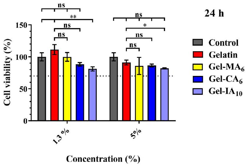
3.2. علم السموم. 3.2.1. حيوية الخلايا. تم دراسة السمية الخلوية في المختبر للجيلاتين ومشتقاته المعدلة (Gel-CA و Gel-IA و Gel-MA) باستخدام اختبار MTT مع خلايا الألياف الرئوية البشرية (HPF). يعتمد الاختبار على قدرة الميتوكوندريا في الخلايا الحية على تقليل 3-(4،5-ثنائي ميثيل ثيازول-2-يل)-2،5-ثنائي فينيل تيترازوليوم بروميد (MTT)، وهي مادة صفراء، إلى بلورات فورمازان غير قابلة للذوبان (لون بنفسجي). تتيح هذه التقنية حساب عدد الخلايا الحية بعد المعالجة بالمادة الاختبارية. تم معالجة خلايا HPF بالجيلاتين، Gel-CA و Gel-MA محاليل بتركيزات 1.3 و في وسائط نمو الخلايا لمدة 24 ساعة. كانت مجموعة التحكم السلبية تتكون من خلايا غير معالجة اعتبرت كـ من الخلايا الحية. يعرض الشكل 3 البيانات حول حيوية الخلايا في وجود الجيلاتين المعدل وغير المعدل. أظهرت نتائج MTT أن حيوية الخلايا متقاربة للجيلاتين ومشتقاته المعدلة كيميائيًا
الشكل 3. تأثير تركيبات الجيلاتين المعدلة وغير المعدلة عند تركيزات 1.3 و على نسبة الخلايا الحية HPF بعد 24 ساعة من التعرض التي تم تقييمها باستخدام اختبار MTT. البيانات ممثلة كمتوسط خطأ معياري للمتوسط من تجربتين مستقلتين في أربعة مكررات. Gel-MA و Gel-IA هي جيلاتين ميثاكريلويل وكروتونويليت وإيتاكونويليت، على التوالي. تظهر الفروق ذات الدلالة الإحصائية كـ ; **p ; “ns” تشير إلى عدم الدلالة.
المشتقات المعدلة حيث لم تكن هناك فروق ذات دلالة إحصائية في الحيوية بين هذه البوليمرات الحيوية ( ) مقابل التحكم بعد 24 ساعة من المعالجة في التركيزات المدروسة. من بين المشتقات المدروسة، أظهر فقط Gel-IA انخفاضًا طفيفًا في حيوية الخلايا عند تركيز إلى 81.1 . ومع ذلك، وفقًا لمعايير ISO التي وضعتها أهداف التنمية المستدامة للأمم المتحدة (SDGs؛ ISO 10993-5:2009) يعتبر المادة للاستخدام الطبي آمنة طالما أن حيوية الخلايا في المختبر هي وأعلى. تظهر نتائجنا أن مستوى حيوية الخلايا ظل مرتفعًا ( ) بعد المعالجة مع جميع البوليمرات الحيوية عند التركيزات المعطاة (الشكل 3).
نظرًا لأن الجيلاتين يعتبر مادة آمنة، في الخطوة التالية، قارننا حيوية الخلايا المعالجة بالجيلاتين مع المجموعة المعالجة بمشتقاته المعدلة. لم تظهر Gel-MA و GelCA وكذلك الجيلاتين النقي فرقًا كبيرًا في الحيوية ( ). علاوة على ذلك، يمكن رؤية أن حيوية الجيلاتين كانت أعلى من تلك الخاصة بمجموعة التحكم. يمكن الافتراض أن الجيلاتين النقي عند تركيز أقل يظهر بعض القدرة على تسهيل تكاثر الخلايا. تتفق نتائجنا جيدًا مع التقارير السابقة، ويمكن اعتبار الجيلاتين ومشتقاته المعدلة آمنة في هذا الصدد.
في معظم الحالات، لم يكن الفرق بين الجيلاتين ومشتقاته المعدلة ذا دلالة إحصائية ( ، مما يشير إلى أن التعديل الكيميائي للجيلاتين بمجموعات غير مشبعة لا يسبب زيادة في سمية البوليمر. يمكن الاستنتاج أن GelCA و Gel-IA و Gel-MA التي تم تصنيعها غير سامة ومناسبة لمزيد من التطوير في التطبيقات الصيدلانية.
3.2.2. تهيج الغشاء المخاطي. تم تطوير اختبار تهيج الغشاء المخاطي للرخويات (SMIT) في الأصل بواسطة أدريانس وزملائه وقد تم التحقق منه كطريقة بديلة وموثوقة لتقييم قدرة التهيج المخاطي لمختلف المواد الكيميائية، والمساعدات، ومستحضرات التجميل، والتركيبات، والمكونات النشطة. تم تطبيق هذه التقنية في العديد من الدراسات، بما في ذلك تقييم التهيج الأنفي والمهبلي. يستخدم SMIT الرخويات الأرضية، التي لا تحميها التشريعات التي تتحكم في تجارب الحيوانات وتعتبر
لديها وعي محدود. إفراز المخاط ضروري للرخويات لمساعدتها في الحركة ومنع الجفاف. كما أنها تطلق المخاط وتفقد وزن الجسم عند ملامستها للمهيجات. عندما يتضرر غشاءها المخاطي، تنتج الرخويات بروتينات وإنزيمات إضافية. يوفر الاختبار نقاط نهاية قابلة للقياس لتصنيف المواد الاختبارية إلى غير مهيجة ( )، خفيفة ( 10)، معتدل (نسبة MP% 10-17.5)، أو مهيج بشدة (نسبة MP% 17.5) بناءً على مستويات إنتاج المخاط. بشكل عام، تسبب المهيجات الخفيفة زيادة في إنتاج المخاط؛ ومع ذلك، فإن المهيجات القوية تؤدي إلى تآكل الأنسجة بالإضافة إلى زيادة إنتاج المخاط.
يتم استخدام النسخة المعدلة من هذا الاختبار بشكل روتيني من قبل مجموعة البحث لدينا لتقييم التوافق الحيوي لمختلف المواد المضافة البوليمرية. تقدم الشكل 4 البيانات حول
الشكل 4. إنتاج المخاط (MP%) من قواقع أريون لوسيتانيكوس استجابةً للتلامس لمدة 60 دقيقة مع الجيلاتين ومشتقاته الميثاكريلوية (Gel-MA 6) والمشتق الكروتونوي (Gel-CA 6) والمشتق الإيتاكونوي (Gel-IA المشتقات، بالإضافة إلى الضوابط الإيجابية (كلوريد البنزالكونيوم؛ BAC) والسلبية (محلول ملحي مخفف بالفوسفات؛ PBS). يتم التعبير عن البيانات كمتوسط قيم الانحراف المعياري ( . يتم تقديم الفروق ذات الدلالة الإحصائية على أنها ; “ns” يدل على عدم الأهمية.
نسبة إنتاج المخاط (MP%) بواسطة قواقع أريون لوسيتانيكوس بعد 60 دقيقة من التعرض لورق الترشيح المنقوع في محاليل الجيلاتين ومشتقاته المعدلة بالإضافة إلى الضوابط الإيجابية والسلبية. جميع المواد الاختبارية المستخدمة في هذا الاختبار تم إذابتها في محلول ملحي مخفف بالفوسفات (PBS؛ pH 7.40). تم إعداد عينات قائمة على الجيلاتين عند ، حيث أنه عند هذه التركيزات كانت هذه العينات قادرة على تشكيل طبقة هلامية رقيقة على سطح ورق الترشيح. الحلزونات الموضوعة في أظهر محلول BAC (التحكم الإيجابي؛ pH 7.36) انزعاجًا شديدًا، مما أدى إلى إنتاج كميات أكبر بكثير من المخاط الملون بالأصفر حتى البرتقالي. ) من أولئك المعرضين لمحلول PBS (التحكم السلبي؛ pH 7.40 ) بمستوى منخفض جداً من MP% ( ). هذه النتائج تتوافق بشكل جيد مع الدراسات السابقة. قيم إنتاج المخاط المسجلة للرخويات المعرضة للجيلاتين، Gel-MA جل-CA ، و Gel-IA كانوا 1 ، و ، على التوالي. كانت إفرازات المخاط عديمة اللون، مما يعد مؤشراً جيداً أولياً للتوافق الحيوي. لا توجد فروق ذات دلالة إحصائية ) في النسبة المئوية MP% لوحظت بين القيم المسجلة للسيطرة السلبية ومواد الاختبار القائمة على الجيلاتين، مما يوضح الطبيعة غير المهيجة لكل من الجيلاتين و
الشكل 5. صور الفلورية المختارة التي تظهر احتباس الغشاء المخاطي لـ جيلاتين طبيعي، جيلاتين ميثاكريلويل (Gel-MA6)، جيلاتين إيتاكونويل (Gel-IA) )، وجيلاتين مكلور الكروتون (Gel-CA تركيبات تحتوي على فلوريسئين الصوديوم (NaFl)، بالإضافة إلى الحرة NaFl (يستخدم كتحكم غير لاصق للمخاط) على أنسجة المهبل الخنزيرية المقطعة حديثًا بعد الغسل بأحجام مختلفة من محلول VFS (درجة الحموضة 4.0؛ معدل التدفق معلمات المجهر الفلوري: التكبير ; وقت التعرض -10 مللي ثانية ; الكسب تتوافق قضبان القياس مع 2 مم.
الجيلاتين المعدل. توفر النتائج رؤى قيمة حول التوافق الحيوي لمشتقات الجيلاتين وتقترح أنه يمكن استخدامها في تطبيقات متنوعة دون التسبب في تهيج الغشاء المخاطي. تقدم الأشكال S12 و S13 في المعلومات الداعمة توضيحًا تخطيطيًا مفصلًا لإجراء اختبار SMIT وصورًا لزواحف Arion lusitanicus المعرضة للمواد المختبرة، على التوالي.

3.3. دراسات الالتصاق المخاطي. 3.3.1. الاحتفاظ في ظروف خارج الجسم

أنسجة المهبل الخنزيري. يُفضل غالبًا استخدام الطريق المهبلي لإعطاء الأدوية لعلاج محلي لمختلف الاضطرابات والعدوى النسائية. يتمتع الإعطاء المحلي بإمكانية تحسين امتصاص الأدوية وتوصيلها إلى الأنسجة المستهدفة مع تقليل الآثار الجانبية. ومع ذلك، يتطلب إعطاء الأدوية عبر المهبل التغلب على عدة عقبات لتحقيق امتصاص فعال للأدوية واحتفاظ بها. يحد الظهارة المهبلية، التي تتكون من عدة طبقات من الخلايا المعبأة بإحكام، من اختراق الأدوية. علاوة على ذلك، فإن وجود طبقة سميكة من المخاط داخل تجويف المهبل يعيق أيضًا انتشار الأدوية إلى الأنسجة الأساسية. تعمل طبقة المخاط، المكونة من الجليكوبروتينات والميوسينات، كحاجز واقي لزج ومرن، حيث تقوم بتنظيف وتزييت ظهارة الجهاز التناسلي للمساعدة في القضاء على مسببات الأمراض والمواد الغريبة. علاوة على ذلك، قد تؤثر الخصائص الفيزيائية والكيميائية للسائل المهبلي، بما في ذلك حجمه ولزوجته ودرجة حموضته، أيضًا بشكل غير ملائم على امتصاص الأدوية واحتفاظها. بالإضافة إلى ذلك، يمكن أن تؤثر عوامل مثل الفسيولوجيا المهبلية، والعمر، ودورة الطمث، واضطرابات الجهاز التناسلي، ومعايير التركيب أيضًا على معدل ومدى امتصاص الأدوية في تجويف المهبل. تشكل هذه العوامل التشريحية مجتمعة تحديات في تحقيق توصيل فعال للأدوية عند إعطائها عن طريق المهبل.
في هذه الدراسة، تم دراسة الاستخدام المحتمل للجيلاتين ومشتقاته المعالجة بالكروتونويلايت، والإيتاكونويلايت، والميثاكريلويل في توصيل الأدوية عبر المهبل باستخدام اختبار في المختبر يعتمد على التدفق مع الكشف الفلوري. الـ
تم إعداد التركيبات باستخدام فلوريسئين الصوديوم (NaFl)، وهو علامة فلورية تسهل الكشف والقياس السهل لمستويات الاحتفاظ بالغشاء المخاطي. لقد تم استخدام هذه الطريقة على نطاق واسع لدراسة احتفاظ التركيبات المختلفة على أسطح مخاطية مختلفة، بما في ذلك الأنسجة المهبلية. تم التحقق من فعالية هذه التقنية مقارنةً بأساليب أخرى معتمدة تُستخدم لتقييم الخصائص اللاصقة للمخاط.
باختصار، الجيلاتين المحتوي على NaFl ومشتقاته المعدلة كيميائيًا (Gel-CA ، و Gel-MA )، بالإضافة إلى محلول NaFl الحر، تم تطبيقه على سطح الغشاء المخاطي المهبلي الخنزيري خارج الجسم وترك لي equilibrate في ثم تم ري السطح المخاطي بأحجام متفاوتة من محاكاة السائل المهبلي (VFS؛ pH 4.0؛ معدل التدفق )، وتم تحديد وجود التركيبة على السطح المخاطي باستخدام مجهر الفلورية. كان الحجم الإجمالي لـ VFS المستخدم في تجربة الغسل هذه يهدف إلى محاكاة كمية الإفرازات المهبلية الطبيعية لدى النساء الأصحاء يوميًا). تُظهر الصور المجهرية الفلورية النموذجية لاحتفاظ هذه التركيبات المأخوذة بعد كل غسلة مع VFS في الشكل 5. يمكن قياس احتفاظ الغشاء المخاطي من خلال قياس التغير في كثافة بكسل الصورة مع مرور الوقت لإعطاء نسبة الفلورية بالنسبة للوقت الأولي (الشكل 6). ساعد تحليل الصور في الكشف عن أن دمج مجموعات الكروتونويل، والإيتاكونويل، والميثاكريلويل في هيكل الجيلاتين عزز بشكل كبير احتفاظ التركيبة على الغشاء المخاطي المهبلي الخنزيري المقطوع حديثًا. من بين الجيلاتين المعدل، Gel-MA أظهرت أداءً متفوقًا في الالتصاق المخاطي مقارنة بالجيلاتين الأصلي ) ومحلول الفلورسئين الصوديوم خلال تجربة الغسيل. كما هو متوقع، أظهرت المحاليل الخالية من البوليمر من NaFl (التي عملت كتحكم غير لاصق للمخاط) احتفاظًا أقل بكثير وتم غسلها بسرعة من السطح المخاطي، مع فقط تمت ملاحظة الفلورية عند الانتهاء من دورة الغسيل الكاملة. تقليديًا، يُعتبر الجيلاتين الطبيعي بوليمرًا ذو خصائص ضعيفة.
الشكل 6. نسبة الاحتفاظ بـ جيلاتين طبيعي، جيلاتين ميثاكريلويل (Gel-MA) جلاتين إيتاكونويليت (Gel )، وجيلاتين مكلور الكروتون (Gel-CA تركيبات تحتوي على فلوريسئين الصوديوم (NaFl) بالإضافة إلى الحر NaFl (يستخدم كتحكم غير لاصق للمخاط) على الغشاء المخاطي المهبلي الخنزيري المقطوع حديثًا بعد الري بأحجام مختلفة من محلول VFS (درجة الحموضة 4.0؛ معدل التدفق ). يتم التعبير عن البيانات كمتوسط قيم الانحراف المعياري ( تمثل الفروق ذات الدلالة الإحصائية كـ 0.0001؛ “ns” تشير إلى عدم الأهمية.
قدرات الاحتفاظ والنتائج التي تم الحصول عليها خلال تجارب الالتصاق المخاطي خارج الجسم دعمت هذا التقييم.
وبالتالي، أظهرت الجيلاتين النقي التصاقًا أضعف بكثير مع الغشاء المخاطي المهبلي مقارنةً بنظائرها المعدلة: جيل-ما. ; جل-IA ; وجيل ومع ذلك، فإن الاحتفاظ أكبر مقارنةً بمحلول NaFl ). لوحظ أن Gel-IA أظهرت احتفاظًا صغيرًا ولكنه أكبر بشكل ملحوظ ) على الغشاء المخاطي المهبلي بالمقارنة مع خلال تجربة الالتصاق المخاطي. ومع ذلك، لم يكن هناك فرق ذو دلالة إحصائية تم العثور عليه بين هذه التركيبات في نهاية اختبار الغسل (الشكل 6). من الجدير بالذكر أن Gel-MA أظهرت أداءً أفضل في الالتصاق المخاطي ( ) مقارنةً بالجيلاتين المعدل الآخر (Gel-CA و Gel . لقد أبلغنا سابقًا أن البوليمرات المعدلة بمجموعات الميثاكريلويل أظهرت تحسنًا كبيرًا في الالتصاق على مختلف الأسطح المخاطية وأظهرت أداءً مشابهًا أو حتى أفضل في الالتصاق المخاطي عند درجات أعلى من التعديل مقارنةً بالكيتوزان، الذي يُعتبر عادةً “المعيار الذهبي” للبوليمرات اللاصقة/التحكم الإيجابي في هذا المجال.
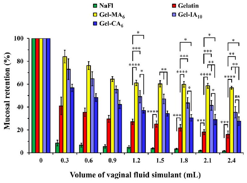
كما هو متوقع، ينخفض احتفاظ جميع التركيبات على مدار عملية الغسيل، ومع ذلك، يتم ملاحظة الاتجاه التالي: Gel- الجيلاتين NaFl. وفقًا لهذه النتائج، من المعقول أن نفترض أن الأداء الممتاز في الالتصاق المخاطي لهذه التركيبات يعود إلى تفاعلها مع الأسطح المخاطية عبر ثلاث آليات، كما هو موضح في الشكل 7: (i) قدرة المجموعات الوظيفية غير المشبعة (الميثاكريلويل، الكروتونويل، الإيتاكونويل) من الجيلاتين المعدل على تشكيل روابط تساهمية مع مجموعات الثيول الموجودة في طبقة المخاط من خلال إضافة من نوع مايكل
الشكل 7. توضيح تخطيطي لاحتفاظ الجيلاتين الميثاكريلويل (Gel-MA)، الجيلاتين الكروتونويلي (Gel-CA)، والجيلاتين الإيتاكونويلي (Gel-IA) على الأسطح المخاطية المهبلية والأنفية.
رد الفعل؛ (ii) التفاعل الكهروستاتيكي بين مجموعات الأمين الأولية البروتونية المتبقية داخل الجيلاتين المعدل والميوسينات المشحونة سلبًا الموجودة على السطح المخاطي؛ و (iii) الروابط الهيدروجينية بين مجموعات الهيدروكسيل، الكربوكسيل من الجيلاتين وموحدات بروتين الميوسين.
تساهم التفاعلات الكهروستاتيكية للجزيئات الكبيرة الأمفوتيرية في الالتصاق المخاطي بشكل كبير في اعتمادها على درجة حموضة المحلول وموقعها النسبي مقابل نقطة التكافؤ للبوليمر الأمفولي. هذا، بدوره، يعتمد على درجة تعديل الجيلاتين وطبيعة المجموعات الوظيفية المدخلة. بالنظر إلى أن تجارب الاحتفاظ أجريت في VFS عند درجة حموضة 4.0، فإن الجيلاتين غير المعدل فقط سيحمل شحنة إيجابية قوية تحت هذه الظروف، نظرًا لنقطة التكافؤ الخاصة به عند 7.0. تظهر مشتقات الجيلاتين التي تحتوي على مجموعات الميثاكريلويل قيم نقطة التكافؤ بين 4.2-4.3، مما يؤدي إلى شحنات إيجابية ضعيفة فقط. تمتلك المشتقات التي تحتوي على مجموعات الكروتونويل والإيتاكونويل قيم نقطة التكافؤ تتراوح بين 2.5-3.2 و3.5-3.8، على التوالي، مما يجعل هذه البوليمرات مشحونة سلبًا خلال تجارب الاحتفاظ. وبالتالي، من المعقول أن نستنتج أن الربط الكهروستاتيكي لمشتقات الجيلاتين مع السطح المهبلي ليس العامل الرئيسي الذي يساهم في الالتصاق المخاطي.
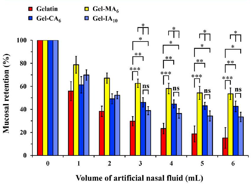
3.3.2. الاحتفاظ على أنسجة أنف الأغنام الحية. يوفر الإعطاء داخل الأنف وسيلة غير جراحية لتوصيل الأدوية. تعمل العوامل العلاجية التي يتم توصيلها إلى تجويف الأنف محليًا وتوفر هدفًا مباشرًا للجهاز العصبي المركزي. يتراوح الإنتاج اليومي المتوقع للمخاط الأنفي بين 0.1 و (أو من 20 إلى 40 مل) في الظروف العادية. ومع ذلك، يمكن أن يتأثر مقدار إفراز المخاط الدقيق بعوامل مختلفة مثل الظروف البيئية، والحساسية، والعدوى، والاختلافات الفردية، بالإضافة إلى الموقع المحدد والمنطقة من الغشاء المخاطي الأنفي البشري. يعتبر تطهير المخاط عملية فسيولوجية معقدة وديناميكية تساعد في الحفاظ على نظافة الممرات الهوائية العليا والسفلى والدفاع ضد الجسيمات والجراثيم المحمولة جواً. يتم ذلك من خلال تفاعل المخاط الأنفي وضربات الأهداب. تتحرك الموجات المنسقة من الأهداب الصغيرة الشبيهة بالشعر تدريجيًا بالمخاط الكثيف من مقدمة الأنف إلى البلعوم الأنفي، حيث يمكن ابتلاعه أو طرده. بالإضافة إلى ذلك، يكون المخاط الأنفي حمضيًا قليلاً ( ) لمنع العدوى التنفسية. تعمل آلية الحماية للجهاز التنفسي بكفاءة ويمكن أن تحد بشكل كبير من وقت بقاء المواد العلاجية عند إعطائها عبر الأنف. تم تصميم أشكال الجرعات اللاصقة المخاطية لمواجهة آلية التطهير من خلال الالتصاق بالسطح المخاطي، وبالتالي إطالة وقت الاحتفاظ وتحسين امتصاص الدواء الفعال.
تستخدم التركيبات الدقيقة عادةً في توصيل الأدوية عبر الأنف. لذلك، في هذه الدراسة، تم صياغة الجيلاتين ومشتقاته كجزيئات دقيقة مع NaFl كدواء نموذجي. تم تصميم نوعين من الجزيئات الدقيقة مع وبدون استخدام عامل ربط لتقييم دور ربط الجيلاتين على خصائص احتفاظها في الأنف. تم إنتاج جزيئات دقيقة محملة بـ NaFl مرتبطة وغير مرتبطة بناءً على الجيلاتين ومشتقاته المعدلة (Gel-CA؛ Gel-IA وGel-MA) بنجاح باستخدام تقنية التجفيف بالرش. كان الاختلاف بين الجزيئات المرتبطة وغير المرتبطة هو إضافة الجلوتارالدهيد، الذي يسهل ربط جزيئات الجيلاتين الكبيرة. تم تحديد مورفولوجيا سطح هذه الجزيئات الدقيقة باستخدام المجهر الإلكتروني الماسح (SEM). كانت الجزيئات الدقيقة التي تم جمعها بعد التجفيف بالرش كروية وقدمت
ملمس سطح مجعد، خالية من البلورات، المسام، والشقوق (انظر الأشكال S14 وS15 في المعلومات الداعمة). أدت جميع التركيبات إلى جزيئات دقيقة ذات مورفولوجيات مشابهة وكان متوسط أقطار الجزيئات ، والتي كانت ضمن النطاق المتوقع للجزيئات الدقيقة الناتجة عن تقنيات التجفيف بالرش الشائعة.
في هذه التجربة، قمنا بتقييم إمكانيات الجزيئات الدقيقة المعتمدة على الجيلاتين المعدل وغير المعدل كتركيبات لاصقة مخاطية لتطبيقها في توصيل الأدوية عبر الأنف. تمت دراسة خصائص الاحتفاظ لهذه التركيبات باستخدام نفس طريقة التدفق في المختبر كما هو موضح أعلاه مع بعض التعديلات. تم استخدام عينات الجزيئات الدقيقة غير المرتبطة فقط في هذه التجربة. الغرض من استخدام الجزيئات الدقيقة غير المرتبطة هو تسهيل التصاقها من خلال السماح لها بالانتفاخ ببطء والذوبان عند ملامستها للسطح المخاطي الرطب عند تطبيقها/استنشاقها. باختصار، من عينات الجزيئات الدقيقة المعتمدة على الجيلاتين المعدل وغير المعدل التي تحتوي على NaFl تم إيداعها على الغشاء المخاطي الأنفي للأغنام الحية. تم إجراء التجربة عند في حاضنة. خلال هذه التجربة، تم جمع محلول ANF المتدفق على الغشاء المخاطي الأنفي خلال كل دورة غسيل في نقاط زمنية محددة مسبقًا. بعد ذلك، تم تحليل هذه العينات باستخدام مقياس الطيف الفلوري. باستخدام البيانات المكتسبة وإنشاء منحنى قياسي للجزيئات الدقيقة مع NaFl، يمكن حساب كمية شكل الجرعة التي تم غسلها من الغشاء المخاطي الأنفي. وبالتالي، من خلال الحسابات العكسية، يمكن تقدير كمية التركيبات المحتفظ بها على الغشاء المخاطي الأنفي للأغنام (الشكل 8). جميع العينات
الشكل 8. نسبة الاحتفاظ بـ من الجيلاتين النقي، الجيلاتين الميثاكريلويل (Gel-MA 6)، الجيلاتين الكروتونويلي (Gel )، والجيلاتين الإيتاكونويلي (Gel-IA ) التركيبات الجزيئية الدقيقة غير المرتبطة، كل منها يحتوي على فلوريسئين الصوديوم (NaFl)، على الغشاء المخاطي الأنفي للأغنام المقطعة حديثًا. تم غسل الغشاء المخاطي بأحجام متغيرة من محلول ANF (درجة حموضة 5.80؛ معدل التدفق . يتم التعبير عن البيانات كمتوسط قيم الانحراف المعياري ( ). يتم إعطاء الفروق ذات الدلالة الإحصائية كـ *p < 0.05؛ **p ; ; “ns” تشير إلى عدم الأهمية.
أظهرت انخفاضًا في الفلورية المحددة خلال عملية الغسيل، مما يشير إلى إزالة التركيبات الجزيئية الدقيقة المعتمدة على الجيلاتين من السطح المخاطي. ومع ذلك، أظهرت الجزيئات الدقيقة من الجيلاتين الميثاكريلويل (Gel-MA ) التصاقًا أقوى ( ) مع الغشاء المخاطي الأنفي مقارنةً بمشتقات الجيلاتين المعدلة الأخرى (Gel-CA و Gel-IA ). مرة أخرى، أظهر الجيلاتين النقي أداءً أقل
الشكل 9. خصائص الالتصاق المخاطي لـ من البوليمرات الحيوية المختلفة: (A) أقصى قوة للفصل و (B) إجمالي ملفات عمل الالتصاق للجزيئات الدقيقة المعتمدة على الجيلاتين المرتبط وغير المرتبط ومشتقاتها المعدلة كيميائيًا التي تم تحديدها بواسطة تحليل القوام. يتم التعبير عن البيانات كمتوسط قيم الانحراف المعياري . يتم عرض الفروق ذات الدلالة الإحصائية كـ ; ; ***p < 0.001؛ “ns” تمثل عدم الأهمية.
أداء الالتصاق المخاطي في مقابل نظيراتها المعدلة: Gel-MA ; Gel-CA ; و . من المثير للاهتمام، لم يتم ملاحظة أي فرق ذو دلالة إحصائية ( ) بين الجل و Gel-IA التركيبات طوال العملية التجريبية، حيث أظهرت ملف احتفاظ مشابه. بشكل عام، تتماشى هذه الملاحظات مع النتائج السابقة وتشير إلى أن المشتقات الثلاثة من البوليمر الحيوي التي تم فحصها لها احتفاظ أكبر على الغشاء المخاطي الأنفي مقارنة بالجيلاتين الأصلي بسبب خصائصها المعززة من حيث الالتصاق بالمخاط.
لدعم النتائج التي تم الحصول عليها من طريقة التدفق في المختبر، تم استخدام اختبار الشد لتقييم التصاق الجسيمات الدقيقة المستندة إلى الجيلاتين المتقاطع وغير المتقاطع ومشتقاتها المعدلة كيميائيًا على الغشاء المخاطي الأنفي للأغنام المقطوعة حديثًا. تعتبر طريقة الشد واحدة من التقنيات المستخدمة على نطاق واسع والمثبتة لتقييم خصائص الالتصاق بالمخاط لأشكال الجرعات المختلفة. تتضمن هذه الطريقة تقريب المواد اللاصقة البوليمرية من الأنسجة المخاطية مع سحبها لاحقًا، تليها تسجيل وتحليل ملفات الانفصال الناتجة. يتم تحديد أداء الالتصاق بالمخاط لأشكال الجرعات من خلال مجموعة من معلمين مهمين: قياس أقصى قوة للانفصال المطلوبة لفصل شكل الجرعة عن الغشاء المخاطي؛ والعمل الكلي للالتصاق، الذي يُعرف بأنه المساحة تحت منحنى قوة الانفصال مقابل المسافة المقابلة (انظر الأشكال S16 و S17 في
المعلومات الداعمة لملفات الانفصال النموذجية للمواد الاختبارية).
الشكل 9 يوضح قيم أقصى قوة للانفصال ( ) والعمل الكلي للالتصاق ( )، كما تم حسابه من دراسات اختبار الشد لكل من الجسيمات الدقيقة المستندة إلى الجيلاتين المتقاطع وغير المتقاطع. أظهرت التركيبات المعدلة كيميائيًا من الجيلاتين (Gel-MA و Gel-IA ) أداءً متفوقًا في الالتصاق بالمخاط ( مع تعبير عن قيم أعلى من و عند مقارنتها بالجيلاتين الأصلي ضمن الجسيمات الدقيقة غير المتقاطعة. وقد تبين أن العينات المستندة إلى الجسيمات الدقيقة Gel-CA 6 غير المتقاطعة أظهرت أقل أداء في الاحتفاظ مقارنةً بنظيراتها Gel-MA و Gel-IA التي أظهرت قيمة أقل من . لم يتم ملاحظة أي فرق ذو دلالة إحصائية في بين Gel-MA و Gel-IA ، ومع ذلك، أظهرت Gel-IA قيمة أعلى من مقارنةً بـ Gel-MA و Gel-CA .
من المثير للاهتمام، لم يتم ملاحظة أي اختلافات ذات دلالة إحصائية بين الجسيمات الدقيقة من الجيلاتين المعدل المتقاطع ( و Gel-IA )، حيث أظهرت ملفات و مشابهة جدًا، مما يشير إلى أداء لاصق بالمخاط قابل للمقارنة بين هذه الأنواع من أشكال الجرعات. بشكل عام، أظهر الجيلاتين الأصلي أسوأ خصائص الالتصاق بالمخاط مقارنةً بنظائره المعدلة ضمن كل من الجسيمات الدقيقة المتقاطعة وغير المتقاطعة.
أدت إضافة مجموعات وظيفية غير مشبعة من الكروتونويل والإيتاكونويل والميثاكريلويل إلى هيكل الجيلاتين إلى تعزيز كبير في أداء الالتصاق بالمخاط لجميع التركيبات المستندة إلى الجسيمات الدقيقة Gel-IA و Gel-CA و Gel-MA. ترتبط قدرة عينات الجيلاتين المعدلة كيميائيًا على الالتصاق جيدًا بالأنسجة المخاطية بتفاعل المجموعات الوظيفية غير المشبعة مع مجموعات الثيول في المجالات الفرعية الغنية بالسيستين الموجودة في الميوسين عبر تفاعل إضافة ثيول-إين ميخايل لتشكيل روابط تساهمية، والتي تحدث في ظل ظروف ذات صلة فسيولوجيًا (الشكل 7). هذه عملية سريعة تسمح بإقامة التصاق محسّن في فترة معقولة بعد إعطاء شكل الجرعة على السطح المخاطي. بالإضافة إلى ذلك، يمكن أن ترتبط مجموعات الأمين الأولية البروتونية المتبقية داخل هيكل الجيلاتين المعدل أيضًا بالميوسين من خلال تفاعلات كهرستاتيكية حيث أن درجة حموضة محلول ANF المستخدم حمضية ضعيفة (درجة حموضة 5.80).
أظهرت الجسيمات الدقيقة المكونة من الجيلاتين المتقاطع والمشتقات خصائص لاصقة بالمخاط أقل مقارنةً بالجسيمات المعدة باستخدام البوليمرات غير المتقاطعة. وهذا يشير إلى أن قابلية انتشار الجزيئات الكبيرة تلعب دورًا كبيرًا في الالتصاق بالمخاط. لا يمكن للجزيئات الكبيرة المتقاطعة الانتشار بحرية وتشكيل طبقة متداخلة مع المخاط، مما يقلل من قدرة الجسيمات الدقيقة على الالتصاق بالسطح المخاطي. تتماشى هذه النتيجة بشكل جيد مع نظرية انتشار الالتصاق بالمخاط.

4. الاستنتاجات

في هذه الدراسة، نبلغ عن تخليق وتوصيف الجيلاتين المعدل بالكروتونويل والإيتاكونويل والميثاكريلويل (Gel-CA و Gel-IA و Gel-MA، على التوالي) مع خصائص لاصقة بالمخاط محسّنة. تم تأكيد التعديل باستخدام تقنيات طيف NMR و FTIR واختبار TNBSA وتم حساب درجة التوظيف. تم أيضًا دراسة تأثير التعديل على نقطة التكافؤ، اللزوجة وخصائص الجل القابلة للعكس حراريًا للجيلاتين. أظهرت جميع الجيلاتينات المشتقة خصائص لاصقة بالمخاط متفوقة مقارنةً بالجيلاتين الأصلي. إن إدخال الأنهدريدات غير المشبعة في الجيلاتين ليس ضارًا لخصائصها السمية كما تم تقييمه باستخدام اختبار SMIT في الجسم الحي واختبار MTT في خلايا HPF. يمكن اعتبار الجيلاتينات المعدلة مع مجموعات وظيفية غير مشبعة كمواد مساعدة جديدة ذات خصائص لاصقة بالمخاط محسّنة لتشكيل أشكال جرعات لاصقة بالمخاط لتوصيل الأدوية المهبلية والأنفية. يمكن أن تجد Gel-CA و Gel-IA و Gel-MA أيضًا تطبيقات في مجالات أخرى من توصيل الأدوية عبر الغشاء المخاطي، على سبيل المثال، عند تشكيلها كأفلام أو جلات أو جزيئات دقيقة أو نانوية.

– المحتوى المرتبط

(س) المعلومات الداعمة

المعلومات الداعمة متاحة مجانًا على https://pubs.acs.org/doi/10.1021/acs.biomac.3c01183.
نسب التغذية لتخليق الجيلاتين المعدل بالكروتونويل أو الإيتاكونويل أو الميثاكريلويل؛ تركيبة محاكاة السائل المهبلي (VFS) والسائل الأنفي الاصطناعي (ANF)؛ منحنى قياسي من الجلايسين يستخدم لتحديد كمية مجموعات الأمين الحرة في الجيلاتين المعدل وغير المعدل؛ منحنى قياسي من الفلورسئين؛ طيف NMR للأنهدريدات غير المشبعة؛ طيف NMR و FTIR للجيلاتين الأصلي ومشتقاته المعدلة؛ صور مجهرية إلكترونية مسح
(SEM) للجسيمات الدقيقة المجففة بالرش المستندة إلى عينات الجيلاتين المعدلة وغير المعدلة؛ منحنيات اللزوجة المحددة مقابل منحنيات درجة الحموضة لتحديد نقاط التكافؤ؛ منحنيات تخزين وفقدان المودول المستخدمة لتحديد نقاط الجل القابلة للعكس حراريًا؛ توضيح تخطيطي لاختبار تهيج الغشاء المخاطي (SMIT) باستخدام نوع Arion lusitanicus؛ صور نموذجية لإنتاج المخاط بواسطة رخويات Arion lusitanicus عند الاتصال بالمواد الاختبارية؛ ملفات انفصال نموذجية للمواد الاختبارية (PDF)

– معلومات المؤلف

المؤلفون المتجاوبون

فيتالي ف. خوتوريانسكي – مدرسة ريدينغ للصيدلة، جامعة ريدينغ، وايتكنيتس RG6 6DX ريدينغ، المملكة المتحدة؛ © orcid.org/0000-0002-7221-2630; البريد الإلكتروني: v.khutoryanskiy@reading.ac.uk
داولت ب. كالديبيكوف – مدرسة ريدينغ للصيدلة، جامعة ريدينغ، وايتكنيتس RG6 6DX ريدينغ، المملكة المتحدة؛ قسم الكيمياء والتكنولوجيا الكيميائية، جامعة الفارابي كازاخستان الوطنية، 050040 ألماتي، كازاخستان؛ معهد مواد البوليمر والتكنولوجيا، 050019 ألماتي، كازاخستان؛ © orcid.org/ 0000-0002-7191-5465; البريد الإلكتروني: dauletchem@gmail.com, d.kaldybekov@reading.ac.uk

المؤلفون

إلفيرا أ. شتاباييفا – مدرسة ريدينغ للصيدلة، جامعة ريدينغ، وايتكنيتس RG6 6DX ريدينغ، المملكة المتحدة؛ قسم الكيمياء والتكنولوجيا الكيميائية، جامعة الفارابي كازاخستان الوطنية، 050040 ألماتي، كازاخستان؛ © orcid.org/0000-0001-9153-5198
ليلى أولمانوفا – مدرسة العلوم والإنسانية، جامعة نازارباييف، 010000 أستانا، كازاخستان
بالنور أ. زهايسنباييفا – مدرسة الهندسة والعلوم الرقمية، جامعة نازارباييف، 010000 أستانا، كازاخستان
إلينا أ. مون – مدرسة العلوم والإنسانية، جامعة نازارباييف، 010000 أستانا، كازاخستان
زارينا أ. كينيسوفا – قسم الكيمياء والتكنولوجيا الكيميائية، جامعة الفارابي كازاخستان الوطنية، 050040 ألماتي، كازاخستان؛ © orcid.org/0000-0003-2768-824X
سركيت إ. كودايبرغينوف – معهد مواد البوليمر والتكنولوجيا، 050019 ألماتي، كازاخستان
معلومات الاتصال الكاملة متاحة على:
https://pubs.acs.org/10.1021/acs.biomac.3c01183

مساهمات المؤلفين

تم كتابة المخطوطة من خلال مساهمات جميع المؤلفين. جميع المؤلفين قد وافقوا على النسخة النهائية من المخطوطة.

ملاحظات

يعلن المؤلفون عدم وجود مصلحة مالية متنافسة.

– الشكر والتقدير

يُعرب المؤلفون عن شكرهم للجنة العلوم بوزارة العلوم والتعليم العالي في جمهورية كازاخستان (رقم المنحة AP14869167) على الدعم المالي لهذا البحث. كما يود المؤلفون أن يشكروا الدكتور سام أسبينال على مساعدته وتدريبه على آلة التجفيف بالرش. كما يُشكر P.C. Turner Abattoirs (فارنبورو، المملكة المتحدة) على
توفير أنسجة المهبل الخنزير وأنسجة أنف الأغنام لتجارب الالتصاق المخاطي. V.V.K. يشكر الجمعية الملكية على زمالته الصناعية (IF ).

– الاختصارات

ANF، سائل أنفي صناعي؛ BAC، كلوريد البنزالكونيوم؛ CA، أنهدريد الكروتون؛ , أكسيد الديوتيريوم؛ DoF، درجة التوظيف؛ DMEM، وسط دلبوكو المعدل؛ DMSO، ثنائي ميثيل سلفوكسيد؛ FBS، مصل جنين البقر؛ NaFI، ملح الصوديوم الفلورسيني؛ جل، جيلاتين؛ Gel-CA، جيلاتين كروتونويليتد؛ Gel-IA، جيلاتين إيتاكونويليتد؛ Gel-MA، جيلاتين ميثاكريلويلتد؛ IA، أنهدريد الإيتاكون؛ MA، أنهدريد الميثاكريليك؛ MP، إنتاج المخاط؛ مفاعل MTT، 3-(4،5-ثنائي ميثيل ثيازول-2-يل)-2،5-ثنائي الفينيل تيترازوليوم بروميد؛ PBS، محلول ملحي معزول بالفوسفات؛ SMIT، اختبار تهيج الغشاء المخاطي للرخويات؛ اختبار TNBSA، اختبار حمض 2،4،6-ثلاثي نيتروبنزين سلفونيك؛ VFS، محاكاة سائل المهبل

– REFERENCES

(1) Karim, A. A.; Bhat, R. Fish Gelatin: Properties, Challenges, and Prospects as an Alternative to Mammalian Gelatins. Food Hydrocolloids 2009, 23 (3), 563-576.
(2) Schrieber, R.; Gareis, H. Gelatine Handbook: Theory and Industrial Practice; Wiley-VCH Verlag GmbH & Co. KGaA: Weinheim, 2007.
(3) Alipal, J.; Mohd Pu’ad, N. A. S.; Lee, T. C.; Nayan, N. H. M.; Sahari, N.; Basri, H.; Idris, M. I.; Abdullah, H. Z. A Review of Gelatin: Properties, Sources, Process, Applications, and Commercialisation. Mater. Today: Proc. 2021, 42, 240-250.
(4) Gullapalli, R. P.; Mazzitelli, C. L. Gelatin and Non-Gelatin Capsule Dosage Forms. J. Pharm. Sci. 2017, 106 (6), 1453-1465.
(5) Osmałek, T.; Froelich, A.; Jadach, B.; Tatarek, A.; Gadzinski, P.; Falana, A.; Gralinska, K.; Ekert, M.; Puri, V.; Wrotynska-Barczynska, J.; Michniak-Kohn, B. Recent Advances in Polymer-Based Vaginal Drug Delivery Systems. Pharmaceutics 2021, 13 (6), 884.
(6) Bhagat, V.; Becker, M. L. Degradable Adhesives for Surgery and Tissue Engineering. Biomacromolecules 2017, 18 (10), 3009-3039.
(7) De Clercq, K.; Schelfhout, C.; Bracke, M.; De Wever, O.; Van Bockstal, M.; Ceelen, W.; Remon, J. P.; Vervaet, C. GenipinCrosslinked Gelatin Microspheres as a Strategy to Prevent Postsurgical Peritoneal Adhesions: In Vitro and in Vivo Characterization. Biomaterials 2016, 96, 33-46.
(8) Yuk, H.; Varela, C. E.; Nabzdyk, C. S.; Mao, X.; Padera, R. F.; Roche, E. T.; Zhao, X. Dry Double-Sided Tape for Adhesion of Wet Tissues and Devices. Nature 2019, 575, 169-174.
(9) Li, C.; Marton, I.; Harari, D.; Shemesh, M.; Kalchenko, V.; Pardo, M.; Schreiber, G.; Rudich, Y. Gelatin Stabilizes Nebulized Proteins in Pulmonary Drug Delivery against COVID-19. ACS Biomater. Sci. Eng. 2022, 8 (6), 2553-2563.
(10) Khutoryanskiy, V. V. Advances in Mucoadhesion and Mucoadhesive Polymers. Macromol. Biosci. 2011, 11 (6), 748-764.
(11) Serra, L.; Doménech, J.; Peppas, N. A. Engineering Design and Molecular Dynamics of Mucoadhesive Drug Delivery Systems as Targeting Agents. Eur. J. Pharm. Biopharm. 2009, 71 (3), 519-528.
(12) Bernkop-Schnürch, A. Thiomers: A New Generation of Mucoadhesive Polymers. Adv. Drug Delivery Rev. 2005, 57 (11), 1569-1582.
(13) Laffleur, F.; Bernkop-Schnürch, A. Thiomers: Promising Platform for Macromolecular Drug Delivery. Future Med. Chem. 2012, 4, 2205-2216.
(14) Laffleur, F.; Hörmann, N.; Gust, R.; Ganner, A. Synthesis, Characterization and Evaluation of Hyaluronic Acid-Based Polymers for Nasal Delivery. Int. J. Pharm. 2023, 631, No. 122496.
(15) Davidovich-Pinhas, M.; Bianco-Peled, H. Novel Mucoadhesive System Based on Sulfhydryl-Acrylate Interactions. J. Mater. Sci.: Mater. Med. 2010, 21 (7), 2027-2034.
(16) Kolawole, O. M.; Lau, W. M.; Khutoryanskiy, V. V. Methacrylated Chitosan as a Polymer with Enhanced Mucoadhesive Properties for Transmucosal Drug Delivery. Int. J. Pharm. 2018, 550 (1-2), 123-129.
(17) Tonglairoum, P.; Brannigan, R. P.; Opanasopit, P.; Khutoryanskiy, V. V. Maleimide-Bearing Nanogels as Novel Mucoadhesive Materials for Drug Delivery. J. Mater. Chem. B 2016, 4 (40), 6581-6587.
(18) Kim, K.; Kim, K.; Ryu, J. H.; Lee, H. Chitosan-Catechol: A Polymer with Long-Lasting Mucoadhesive Properties. Biomaterials 2015, 52 (1), 161-170.
(19) Prosperi-Porta, G.; Kedzior, S.; Muirhead, B.; Sheardown, H. Phenylboronic-Acid-Based Polymeric Micelles for Mucoadhesive Anterior Segment Ocular Drug Delivery. Biomacromolecules 2016, 17 (4), 1449-1457.
(20) Menzel, C.; Hauser, M.; Frey, A.; Jelkmann, M.; Laffleur, F.; Götzfried, S. K.; Gust, R.; Bernkop-Schnürch, A. Covalently Binding Mucoadhesive Polymers: N-Hydroxysuccinimide Grafted Polyacrylates. Eur. J. Pharm. Biopharm. 2019, 139, 161-167.
(21) Eshel-Green, T.; Bianco-Peled, H. Mucoadhesive Acrylated Block Copolymers Micelles for the Delivery of Hydrophobic Drugs. Colloids Surf., B 2016, 139, 42-51.
(22) Kaldybekov, D. B.; Tonglairoum, P.; Opanasopit, P.; Khutoryanskiy, V. V. Mucoadhesive Maleimide-Functionalised Liposomes for Drug Delivery to Urinary Bladder. Eur. J. Pharm. Sci. 2018, 111, 83-90.
(23) Kaldybekov, D. B.; Filippov, S. K.; Radulescu, A.; Khutoryanskiy, V. V. Maleimide-Functionalised PLGA-PEG Nanoparticles as Mucoadhesive Carriers for Intravesical Drug Delivery. Eur. J. Pharm. Biopharm. 2019, 143, 24-34.
(24) Shan, X.; Aspinall, S.; Kaldybekov, D. B.; Buang, F.; Williams, A. C.; Khutoryanskiy, V. V. Synthesis and Evaluation of Methacrylated Poly(2-Ethyl-2-Oxazoline) as a Mucoadhesive Polymer for Nasal Drug Delivery. ACS Appl. Polym. Mater. 2021, 3 (11), 5882-5892.
(25) Agibayeva, L. E.; Kaldybekov, D. B.; Porfiryeva, N. N.; Garipova, V. R.; Mangazbayeva, R. A.; Moustafine, R. I.; Semina, I. I.; Mun, G. A.; Kudaibergenov, S. E.; Khutoryanskiy, V. V. Gellan Gum and Its Methacrylated Derivatives as in Situ Gelling Mucoadhesive Formulations of Pilocarpine: In Vitro and in Vivo Studies. Int. J. Pharm. 2020, 577, No. 119093.
(26) Buang, F.; Fu, M.; Chatzifragkou, A.; Amin, M. C. I. M.; Khutoryanskiy, V. V. Hydroxyethyl Cellulose Functionalised with Maleimide Groups as a New Excipient with Enhanced Mucoadhesive Properties. Int. J. Pharm. 2023, 642, No. 123113.
(27) Brotherton, E. E.; Neal, T. J.; Kaldybekov, D. B.; Smallridge, M. J.; Khutoryanskiy, V. V.; Armes, S. P. Aldehyde-Functional Thermoresponsive Diblock Copolymer Worm Gels Exhibit Strong Mucoadhesion. Chem. Sci. 2022, 13, 6888-6898.
(28) Hunter, S. J.; Abu Elella, M. H.; Johnson, E. C.; Taramova, L.; Brotherton, E. E.; Armes, S. P.; Khutoryanskiy, V. V.; Smallridge, M. J. Mucoadhesive Pickering Nanoemulsions via Dynamic Covalent Chemistry. J. Colloid Interface Sci. 2023, 651, 334-345.
(29) Bonferoni, M. C.; Chetoni, P.; Giunchedi, P.; Rossi, S.; Ferrari, F.; Burgalassi, S.; Caramella, C. Carrageenan-Gelatin Mucoadhesive Systems for Ion-Exchange Based Ophthalmic Delivery: In Vitro and Preliminary in Vivo Studies. Eur. J. Pharm. Biopharm. 2004, 57 (3), 465-472.
(30) Padhi, J. R.; Nayak, D.; Nanda, A.; Rauta, P. R.; Ashe, S.; Nayak, B. Development of Highly Biocompatible Gelatin & ICarrageenan Based Composite Hydrogels: In Depth Physiochemical Analysis for Biomedical Applications. Carbohydr. Polym. 2016, 153, 292-301.
(31) Liu, Y.; Cheong NG, S.; Yu, J.; Tsai, W. B. Modification and Crosslinking of Gelatin-Based Biomaterials as Tissue Adhesives. Colloids Surf., B 2019, 174, 316-323.
(32) Jovanović, M.; Tomić, N.; Cvijić, S.; Stojanović, D.; Ibrić, S.; Uskoković, P. Mucoadhesive Gelatin Buccal Films with Propranolol Hydrochloride: Evaluation of Mechanical, Mucoadhesive, and Biopharmaceutical Properties. Pharmaceutics 2021, 13 (2), 273.
(33) Abruzzo, A.; Bigucci, F.; Cerchiara, T.; Cruciani, F.; Vitali, B.; Luppi, B. Mucoadhesive Chitosan/Gelatin Films for Buccal Delivery of Propranolol Hydrochloride. Carbohydr. Polym. 2012, 87 (1), 581588.
(34) Abruzzo, A.; Cerchiara, T.; Bigucci, F.; Gallucci, M. C.; Luppi, B. Mucoadhesive Buccal Tablets Based on Chitosan/Gelatin Microparticles for Delivery of Propranolol Hydrochloride. J. Pharm. Sci. 2015, 104 (12), 4365-4372.
(35) Trastullo, R.; Abruzzo, A.; Saladini, B.; Gallucci, M. C.; Cerchiara, T.; Luppi, B.; Bigucci, F. Design and Evaluation of Buccal Films as Paediatric Dosage Form for Transmucosal Delivery of Ondansetron. Eur. J. Pharm. Biopharm. 2016, 105, 115-121.
(36) Mahaling, B.; Katti, D. S. Understanding the Influence of Surface Properties of Nanoparticles and Penetration Enhancers for Improving Bioavailability in Eye Tissues In Vivo. Int. J. Pharm. 2016, 501, 1-9.
(37) Giordani, B.; Abruzzo, A.; Prata, C.; Nicoletta, F. P.; Dalena, F.; Cerchiara, T.; Luppi, B.; Bigucci, F. Ondansetron Buccal Administration for Paediatric Use: A Comparison between Films and Wafers. Int. J. Pharm. 2020, 580, No. 119228.
(38) Kudaibergenov, S. E. Synthetic and Natural Polyampholytes: Structural and Behavioral Similarity. Polym. Adv. Technol. 2021, 32 (3), 906-918.
(39) Ahmady, A.; Abu Samah, N. H. A Review: Gelatine as a Bioadhesive Material for Medical and Pharmaceutical Applications. Int. J. Pharm. 2021, 608, No. 121037.
(40) Van Den Bulcke, A. I.; Bogdanov, B.; De Rooze, N.; Schacht, E. H.; Cornelissen, M.; Berghmans, H. Structural and Rheological Properties of Methacrylamide Modified Gelatin Hydrogels. Biomacromolecules 2000, 1(1), 31-38.
(41) Claaßen, C.; Claaßen, M. H.; Truffault, V.; Sewald, L.; Tovar, G. E. M.; Borchers, K.; Southan, A. Quantification of Substitution of Gelatin Methacryloyl: Best Practice and Current Pitfalls. Biomacromolecules 2018, 19 (1), 42-52.
(42) Brinkman, W. T.; Nagapudi, K.; Thomas, B. S.; Chaikof, E. L. Photo-Cross-Linking of Type I Collagen Gels in the Presence of Smooth Muscle Cells: Mechanical Properties, Cell Viability, and Function. Biomacromolecules 2003, 4 (4), 890-895.
(43) Nichol, J. W.; Koshy, S. T.; Bae, H.; Hwang, C. M.; Yamanlar, S.; Khademhosseini, A. Cell-Laden Microengineered Gelatin Methacrylate Hydrogels. Biomaterials 2010, 31 (21), 5536-5544.
(44) Norris, S. C. P.; Delgado, S. M.; Kasko, A. M. Mechanically Robust Photodegradable Gelatin Hydrogels for 3D Cell Culture and in Situ Mechanical Modification. Polym. Chem. 2019, 10 (23), 31803193.
(45) Habeeb, A. F. S. A. Determination of Free Amino Groups in Proteins by Trinitrobenzenesulfonic Acid. Anal. Biochem. 1966, 14 (3), 328-336.
(46) Lee, B. H.; Shirahama, H.; Cho, N. J.; Tan, L. P. Efficient and Controllable Synthesis of Highly Substituted Gelatin Methacrylamide for Mechanically Stiff Hydrogels. RSC Adv. 2015, 5 (128), 106094106097.
(47) Zhu, M.; Wang, Y.; Ferracci, G.; Zheng, J.; Cho, N. J.; Lee, B. H. Gelatin Methacryloyl and Its Hydrogels with an Exceptional Degree of Controllability and Batch-to-Batch Consistency. Sci. Rep. 2019, 9, 6863.
(48) Benson, J. E. Viscometric Determination of the Isoelectric Point of a Protein. J. Chem. Educ. 1963, 40 (9), 468-469.
(49) Khutoryanskaya, O. V.; Morrison, P. W. J.; Seilkhanov, S. K.; Mussin, M. N.; Ozhmukhametova, E. K.; Rakhypbekov, T. K.; Khutoryanskiy, V. V. Hydrogen-Bonded Complexes and Blends of Poly(Acrylic Acid) and Methylcellulose: Nanoparticles and Mucoadhesive Films for Ocular Delivery of Riboflavin. Macromol. Biosci. 2014, 14 (2), 225-234.
(50) Irmukhametova, G. S.; Mun, G. A.; Khutoryanskiy, V. V. Thiolated Mucoadhesive and PEGylated Nonmucoadhesive Organosilica Nanoparticles from 3-Mercaptopropyltrimethoxysilane. Langmuir 2011, 27 (15), 9551-9556.
(51) Mun, E. A.; Williams, A. C.; Khutoryanskiy, V. V. Adhesion of Thiolated Silica Nanoparticles to Urinary Bladder Mucosa: Effects of PEGylation, Thiol Content and Particle Size. Int. J. Pharm. 2016, 512 (1), 32-38.
(52) Porfiryeva, N. N.; Nasibullin, S. F.; Abdullina, S. G.; Tukhbatullina, I. K.; Moustafine, R. I.; Khutoryanskiy, V. V. Acrylated Eudragit® E PO as a Novel Polymeric Excipient with Enhanced Mucoadhesive Properties for Application in Nasal Drug Delivery. Int. J. Pharm. 2019, 562, 241-248.
(53) Haddow, P. J.; da Silva, M. A.; Kaldybekov, D. B.; Dreiss, C. A.; Hoffman, E.; Hutter, V.; Khutoryanskiy, V. V.; Kirton, S. B.; Mahmoudi, N.; McAuley, W. J.; Cook, M. T. Polymer Architecture Effects on Poly( -diethyl acrylamide)- -Poly(ethylene glycol)- Poly( -diethyl acrylamide) Thermoreversible Gels and Their Evaluation as a Healthcare Material. Macromol. Biosci. 2022, 22 (3), No. 2100432.
(54) Kolawole, O. M.; Lau, W. M.; Khutoryanskiy, V. V. Synthesis and Evaluation of Boronated Chitosan as a Mucoadhesive Polymer for Intravesical Drug Delivery. J. Pharm. Sci. 2019, 108 (9), 3046-3053.
(55) Hoch, E.; Schuh, C.; Hirth, T.; Tovar, G. E. M.; Borchers, K. Stiff Gelatin Hydrogels Can Be Photo-Chemically Synthesized from Low Viscous Gelatin Solutions Using Molecularly Functionalized Gelatin with a High Degree of Methacrylation. J. Mater. Sci. Mater. Med. 2012, 23 (11), 2607-2617.
(56) Kim, S.; Nimni, M. E.; Yang, Z.; Han, B. Chitosan/GelatinBased Films Crosslinked by Proanthocyanidin. J. Biomed. Mater. Res. 2005, 75B (2), 442-450.
(57) Barth, A. Infrared Spectroscopy of Proteins. Biochim. Biophys. Acta – Bioenerg. 2007, 1767 (9), 1073-1101.
(58) Hashim, D. M.; Man, Y. B. C.; Norakasha, R.; Shuhaimi, M.; Salmah, Y.; Syahariza, Z. A. Potential Use of Fourier Transform Infrared Spectroscopy for Differentiation of Bovine and Porcine Gelatins. Food Chem. 2010, 118 (3), 856-860.
(59) Cebi, N.; Durak, M. Z.; Toker, O. S.; Sagdic, O.; Arici, M. An Evaluation of Fourier Transforms Infrared Spectroscopy Method for the Classification and Discrimination of Bovine, Porcine and Fish Gelatins. Food Chem. 2016, 190, 1109-1115.
(60) Rahali, K.; Ben Messaoud, G.; Kahn, C. J. F.; SanchezGonzalez, L.; Kaci, M.; Cleymand, F.; Fleutot, S.; Linder, M.; Desobry, S.; Arab-Tehrany, E. Synthesis and Characterization of Nanofunctionalized Gelatin Methacrylate Hydrogels. Int. J. Mol. Sci. 2017, 18 (12), 2675.
(61) Sheppard, S. E.; Houck, R. C. The Structure of Gelatin Sols and Gels. III. J. Phys. Chem. 1930, 34 (10), 2187-2201.
(62) Johlin, J. M. The Isoelectric Point of Gelatin and Its Relation to the Minimum Physical Properties of Gelatin. J. Biol. Chem. 1930, 86 (1), 231-243.
(63) Lowe, A. B.; McCormick, C. L. Synthesis and Solution Properties of Zwitterionic Polymers. Chem. Rev. 2002, 102 (11), 4177-4190.
(64) Gudmundsson, M. Rheological Properties of Fish Gelatins. J. Food Sci. 2002, 67 (6), 2172-2176.
(65) Goudoulas, T. B.; Germann, N. Phase Transition Kinetics and Rheology of Gelatin-Alginate Mixtures. Food Hydrocolloids 2017, 66, 49-60.
(66) Boran, G.; Mulvaney, S. J.; Regenstein, J. M. Rheological Properties of Gelatin from Silver Carp Skin Compared to Commercially Available Gelatins from Different Sources. J. Food Sci. 2010, 75 (8), E565-E571.
(67) ISO 10993-5:2009(en), Biological evaluation of medical devices – Part 5: Tests for in vitro cytotoxicity. https://www.iso.org/ obp/ui/#iso:std:iso:10993:-5:ed-3:v1:en (accessed 2022-09-03).
(68) Elzoghby, A. O.; Samy, W. M.; Elgindy, N. A. Protein-Based Nanocarriers as Promising Drug and Gene Delivery Systems. J. Controlled Release 2012, 161 (1), 38-49.
(69) Suresh, D.; Suresh, A.; Kannan, R. Engineering Biomolecular Systems: Controlling the Self-Assembly of Gelatin to Form UltraSmall Bioactive Nanomaterials. Bioact. Mater. 2022, 18, 321-336.
(70) Ulubayram, K.; Aksu, E.; Gurhan, S. I. D.; Serbetci, K.; Hasirci, N. Cytotoxicity Evaluation of Gelatin Sponges Prepared with Different Cross-Linking Agents. J. Biomater. Sci. Polym. Ed. 2002, 13 (11), 1203-1219.
(71) Won, Y.-W.; Kim, Y.-H. Preparation and Cytotoxicity Comparison of Type a Gelatin Nanoparticles with Recombinant Human Gelatin Nanoparticles. Macromol. Res. 2009, 17 (7), 464468.
(72) Shin, H.; Olsen, B. D.; Khademhosseini, A. The Mechanical Properties and Cytotoxicity of Cell-Laden Double-Network Hydrogels Based on Photocrosslinkable Gelatin and Gellan Gum Biomacromolecules. Biomaterials 2012, 33 (11), 3143-3152.
(73) Adriaens, E.; Remon, J. P. Gastropods as an Evaluation Tool for Screening the Irritating Potency of Absorption Enhancers and Drugs. Pharm. Res. 1999, 16 (8), 1240-1244.
(74) Adriaens, E.; Dierckens, K.; Bauters, T. G. M.; Nelis, H. J.; Van Goethem, F.; Vanparys, P.; Remon, J. P. The Mucosal Toxicity of Different Benzalkonium Chloride Analogues Evaluated with an Alternative Test Using Slugs. Pharm. Res. 2001, 18 (7), 937-942.
(75) Callens, C.; Adriaens, E.; Dierckens, K.; Remon, J. P. Toxicological Evaluation of a Bioadhesive Nasal Powder Containing a Starch and Carbopol® 974 P on Rabbit Nasal Mucosa and Slug Mucosa. J. Controlled Release 2001, 76 (1-2), 81-91.
(76) Ceulemans, J.; Vermeire, A.; Adriaens, E.; Remon, J. P.; Ludwig, A. Evaluation of a Mucoadhesive Tablet for Ocular Use. J. Controlled Release 2001, 77 (3), 333-344.
(77) Adriaens, E.; Ameye, D.; Dhondt, M. M. M.; Foreman, P.; Remon, J. P. Evaluation of the Mucosal Irritation Potency of Co-Spray Dried Amioca®/Poly(Acrylic Acid) and Amioca®/Carbopol® 974P Mixtures. J. Controlled Release 2003, 88 (3), 393-399.
(78) Dhondt, M.; Adriaens, E.; Roey, J.; Remon, J. The Evaluation of the Local Tolerance of Vaginal Formulations Containing Dapivirine Using the Slug Mucosal Irritation Test and the Rabbit Vaginal Irritation Test. Eur. J. Pharm. Biopharm. 2005, 60 (3), 419425.
(79) Adriaens, E.; Remon, J. P. Mucosal Irritation Potential of Personal Lubricants Relates to Product Osmolality as Detected by the Slug Mucosal Irritation Assay. Sex. Transm. Dis. 2008, 35 (5), 512516.
(80) Adriaens, E.; Bytheway, H.; De Wever, B.; Eschrich, D.; Guest, R.; Hansen, E.; Vanparys, P.; Schoeters, G.; Warren, N.; Weltens, R.; Whittingham, A.; Remon, J. P. Successful Prevalidation of the Slug Mucosal Irritation Test to Assess the Eye Irritation Potency of Chemicals. Toxicol. In Vitro 2008, 22 (5), 1285-1296.
(81) Lenoir, J.; Adriaens, E.; Remon, J. P. New Aspects of the Slug Mucosal Irritation Assay: Predicting Nasal Stinging, Itching and Burning Sensations. J. Appl. Toxicol. 2011, 31 (7), 640-648.
(82) Lenoir, J.; Bachert, C.; Remon, J. P.; Adriaens, E. The Slug Mucosal Irritation (SMI) Assay: A Tool for the Evaluation of Nasal Discomfort. Toxicol. In Vitro 2013, 27 (6), 1954-1961.
(83) Petit, J. Y.; Doré, V.; Marignac, G.; Perrot, S. Assessment of Ocular Discomfort Caused by 5 Shampoos Using the Slug Mucosal Irritation Test. Toxicol. In Vitro 2017, 40, 243-247.
(84) Balls, M.; Goldberg, A. M.; Fentem, J. H.; Broadhead, C. L.; Burch, R. L.; Festing, M. F. W.; Frazier, J. M.; Hendriksen, C. F. M.; Jennings, M.; van der Kamp, M. D. O.; Morton, D. B.; Rowan, A. N.; Russell, C.; Russell, W. M. S.; Spielmann, H.; Stephens, M. L.; Stokes, W. S.; Straughan, D. W.; Yager, J. D.; Zurlo, J.; van Zutphen, B. F. M. The Three Rs: The Way Forward: The Report and Recommendations of ECVAM Workshop 11. Altern. to Lab. Anim. 1995, 23 (6), 838866.
(85) Moiseev, R. V.; Steele, F.; Khutoryanskiy, V. V. Polyaphron Formulations Stabilised with Different Water-Soluble Polymers for Ocular Drug Delivery. Pharmaceutics 2022, 14 (5), 926.
(86) Hussain, A.; Ahsan, F. The Vagina as a Route for Systemic Drug Delivery. J. Controlled Release 2005, 103 (2), 301-313.
(87) Krogstad, E. A.; Rathbone, M. J.; Woodrow, K. A. Vaginal Drug Delivery. In Focal Controlled Drug Delivery. Advances in Delivery Science
and Technology; Domb, A. J., Khan, W., Eds.; Springer: Boston, MA, 2014; pp 607-651.
(88) Shapiro, R. L.; DeLong, K.; Zulfiqar, F.; Carter, D.; Better, M.; Ensign, L. M. In Vitro and Ex Vivo Models for Evaluating Vaginal Drug Delivery Systems. Adv. Drug Delivery Rev. 2022, 191, No. 114543.
(89) Davidovich-Pinhas, M.; Bianco-Peled, H. Methods to Study Mucoadhesive Dosage Forms. In Mucoadhesive Materials and Drug Delivery Systems; Khutoryanskiy, V. V., Ed.; John Wiley & Sons, Ltd: Chichester, U.K., 2014; pp 175-196.
(90) Eschenbach, D. A.; Thwin, S. S.; Patton, D. L.; Hooton, T. M.; Stapleton, A. E.; Agnew, K.; Winter, C.; Meier, A.; Stamm, W. E. Influence of the Normal Menstrual Cycle on Vaginal Tissue, Discharge, and Microflora. Clin. Infect. Dis. 2000, 30 (6), 901-907.
(91) Sogias, I. A.; Williams, A. C.; Khutoryanskiy, V. V. Why Is Chitosan Mucoadhesive? Biomacromolecules 2008, 9 (7), 1837-1842.
(92) Fu, M.; Filippov, S. K.; Williams, A. C.; Khutoryanskiy, V. V. On the Mucoadhesive Properties of Synthetic and Natural Polyampholytes. J. Colloid Interface Sci. 2024, 659, 849-858.
(93) Lale, A. M.; Mason, J. D. T.; Jones, N. S. Mucociliary Transport and Its Assessment: A Review. Clin. Otolaryngol. Allied Sci. 1998, 23 (5), 388-396.
(94) Sahin-Yilmaz, A.; Naclerio, R. M. Anatomy and Physiology of the Upper Airway. Proc. Am. Thorac. Soc. 2011, 8 (1), 31-39.
(95) Beule, A. G. Physiology and Pathophysiology of Respiratory Mucosa of the Nose and the Paranasal Sinuses. GMS Curr. Top. Otorhinolaryngol. Head Neck Surg. 2010, 9, Doc07.
(96) Chavda, V. P.; Jogi, G.; Shah, N.; Athalye, M. N.; Bamaniya, N.; K Vora, L.; Cláudia Paiva-Santos, A. Advanced Particulate CarrierMediated Technologies for Nasal Drug Delivery. J. Drug Delivery Sci. Technol. 2022, 74, No. 103569.
(97) Favaro-Trindade, C. S.; Santana, A. S.; Monterrey-Quintero, E. S.; Trindade, M. A.; Netto, F. M. The Use of Spray Drying Technology to Reduce Bitter Taste of Casein Hydrolysate. Food Hydrocolloids 2010, 24 (4), 336-340.
(98) Osman, R.; Kan, P. L.; Awad, G.; Mortada, N.; El-Shamy, A. E.; Alpar, O. Spray Dried Inhalable Ciprofloxacin Powder with Improved Aerosolisation and Antimicrobial Activity. Int. J. Pharm. 2013, 449 (12), 44-58.

  1. All outputs in CentAUR are protected by Intellectual Property Rights law, including copyright law. Copyright and IPR is retained by the creators or other copyright holders. Terms and conditions for use of this material are defined in
  2. Received: October 31, 2023
    Revised: January 13, 2024
    Accepted: January 16, 2024

Journal: Biomacromolecules, Volume: 25, Issue: 3
DOI: https://doi.org/10.1021/acs.biomac.3c01183
PMID: https://pubmed.ncbi.nlm.nih.gov/38319691
Publication Date: 2024-02-06

Enhancing mucoadhesive properties of gelatin through chemical modification with unsaturated anhydrides

ArticlePublished Version

Creative Commons: Attribution 4.0 (CC-BY)
Open Access
Shatabayeva, E. ORCID: https://orcid.org/0000-0001-91535198, Kaldybekov, D. B. ORCID: https://orcid.org/0000-0002-7191-5465, Ulmanova, L., Zhaisanbayeva, B. A., Mun, E. A., Kenessova, Z. A. ORCID: https://orcid.org/0000-0003-2768824X, Kudaibergenov, S. E. and Khutoryanskiy, V. V. ORCID: https://orcid.org/0000-0002-7221-2630 (2024) Enhancing mucoadhesive properties of gelatin through chemical modification with unsaturated anhydrides. Biomacromolecules. ISSN 1526-4602 doi: 10.1021/acs.biomac.3c01183 Available at https://centaur.reading.ac.uk/115238/
It is advisable to refer to the publisher’s version if you intend to cite from the work. See Guidance on citing.
To link to this article DOI: http://dx.doi.org/10.1021/acs.biomac.3c01183
Publisher: American Chemical Society
the End User Agreement.

www.reading.ac.uk/centaur

CentAUR

Central Archive at the University of Reading
Reading’s research outputs online

Enhancing Mucoadhesive Properties of Gelatin through Chemical Modification with Unsaturated Anhydrides

Elvira O. Shatabayeva, Daulet B. Kaldybekov,* Leila Ulmanova, Balnur A. Zhaisanbayeva, Ellina A. Mun, Zarina A. Kenessova, Sarkyt E. Kudaibergenov, and Vitaliy V. Khutoryanskiy*

Cite This: https://doi.org/10.1021/acs.biomac.3c01183
Read Online
Downloaded via UNIV OF READING on February 23, 2024 at 14:53:45 (UTC).
See https://pubs.acs.org/sharingguidelines for options on how to legitimately share published articles.

Abstract

Gelatin is a water-soluble natural polyampholyte with poor mucoadhesive properties. It has traditionally been used as a major ingredient in many pharmaceuticals, including soft and hard capsules, suppositories, tissue engineering, and regenerative medicine. The mucoadhesive properties of gelatin can be improved by modifying it through conjugation with specific adhesive unsaturated groups. In this study, gelatin was modified by reacting with crotonic, itaconic, and methacrylic anhydrides in varying molar ratios to yield crotonoylated-, itaconoylated-, and methacryloylated gelatins (abbreviated as Gel-CA, Gel-IA, and Gel-MA, respectively). The successful synthesis was confirmed using NMR, FTIR spectroscopies, and colorimetric TNBSA assay. The effect of chemical modification on the isoelectric point was studied through viscosity and electrophoretic mobility measurements. The evolution of the storage ( ) and loss ( ) moduli was employed to determine thermoreversible gelation points of modified and unmodified gelatins. The safety of modified gelatin derivatives was assessed with an in vivo slug mucosal irritation test (SMIT) and an in vitro MTT assay utilizing human pulmonary fibroblasts cell line. Two different model dosage forms, such as physical gels and spray-dried microparticles, were prepared and their mucoadhesive properties were evaluated using a flow-through technique with fluorescent detection and a tensile test with ex vivo porcine vaginal tissues and sheep nasal mucosa. Gelatins modified with unsaturated groups exhibited superior mucoadhesive properties compared to native gelatin. The enhanced ability of gelatin modified with these unsaturated functional groups is due to the formation of covalent bonds with cysteine-rich subdomains present in the mucin via thiol-ene click Michael-type addition reactions occurring under physiologically relevant conditions.

1. INTRODUCTION

Gelatin is a natural biopolymer derived from collagen through its partial hydrolysis and heat denaturation. Collagen itself is extracted from the bones, cartilage, connective tissues, and skins of slaughtered animals, and fish scales. It is industrially one of the most important and widely used polyampholytes owing to its unique physicochemical properties. The properties of gelatin depend on the method of its production, which can involve either acid or alkaline treatment. Commercially available type A gelatin, produced via the acid process, has an isoelectric point (IEP) at , whereas type B gelatin, manufactured through the alkaline process, exhibits an IEP at pH 4.5-5.5. As a protein-based biomaterial, gelatin has excellent biocompatibility, biodegradability, nontoxicity, and nonimmunogenicity. It has been approved as a GRAS (generally recognized as safe) material by the U.S. Food and Drug Administration (FDA). Gelatin readily dissolves in warm water at and has the ability to form physically cross-
linked thermoreversible hydrogels upon cooling below C. Additionally, it exhibits a melting point close to body temperature. These distinctive properties are the key factors driving its extensive applications in pharmaceutical, food, and cosmetic industries. Gelatin is commonly used as a major ingredient in many formulations, including hard and soft capsules, vaginal and rectal suppositories, as matrices in tissue engineering and regenerative medicine, as a carrier in drug delivery (e.g., microspheres), and in many other healthrelated applications.
Mucoadhesion is defined as the ability of materials to adhere to mucosal membranes in the body for an extended period of time. The established routes for transmucosal drug administration include ocular (corneal and conjunctival), nasal, oromucosal (buccal and sublingual), gastrointestinal, rectal, vaginal, and intravesical. Transmucosal drug delivery offers several important advantages, such as the ease of dosage form administration and its noninvasive nature, prolonged residence time on the mucosal surface, improved drug bioavailability, the avoidance of hepatic first-pass metabolism, reduced dosing frequency, and the possibility for quick termination of therapy when needed.
Numerous first-generation (conventional) mucoadhesive polymers are traditionally used in various dosage forms for transmucosal drug delivery. This class of mucoadhesives includes water-soluble polymers of both natural and synthetic origins, such as chitosan, gellan gum, alginate, polycarbopols, and cellulose derivatives (e.g., carboxymethylcellulose, hydroxypropyl methylcellulose, and others). The mucoadhesion in these drug delivery systems occurs through various physical interactions (nonspecific binding) between the macromolecules and mucin glycoproteins present on mucosal surfaces. These interactions primarily involve noncovalent forces, such as hydrogen bonding, electrostatic attraction, van der Waals forces, hydrophobic interactions, and chain entanglements/ diffusion. Generally, polyelectrolytes exhibit better mucoadhesive properties compared to nonionic polymers. Furthermore, the mechanisms of adhesion of these materials to mucosal surfaces may vary depending on the nature of the dosage form. For instance, adhesive properties of solid dosage forms (such as tablets) are influenced by the hydration process, while the retention of liquid formulations on mucosal surfaces is more affected by their rheological properties.
In the past few decades, different chemical approaches have been explored to design polymers and their formulations with enhanced mucoadhesive properties. This enhancement can be accomplished through functionalization of hydrophilic polymers with specific adhesive groups capable of forming covalent bonds with mucosal tissues under physiological conditions. Thiolated polymers, also known as “thiomers”, represent one of the prominent advances in the secondgeneration mucoadhesive materials, pioneered by BernkopSchnürch and co-workers. These polymers have been modified by introducing thiol (sulfhydryl) functional groups onto their side chains. Thiolated polymers form interdisulfide bridges through covalent interactions (via oxidation reactions) with cysteine-bearing subdomains of mucus glycoproteins present on mucosal surfaces. This leads to their enhanced mucoadhesive capabilities resulting in a prolonged drug residence time at the side of application. However, it is worth noting that thiolated polymers are prone to oxidation, which can lead to unwanted cross-linking of polymers.
Various strategies have recently been proposed to enhance the mucoadhesive properties of hydrophilic polymers by introducing adhesive moieties such as acryloyl, methacryloyl, maleimide, catechol, boronate, and -hydroxy(sulfo)succinimide ester groups. Acryloylated polymers, first proposed by the Bianco-Peled research group, were highlighted as a novel class of pharmaceutical excipients with substantially improved mucoadhesive properties compared to their unmodified counterparts. The Khutoryanskiy group also pioneered the use of methacryloyl- and maleimide-functionalized materials to design dosage forms with enhanced
mucoadhesive properties. These include the development of nanogels, liposomes, nanoparticles, liquid formulations, in situ gels, and spray-coated tablets. These unsaturated functional groups are able to form covalent bonds with cysteine-rich subdomains of mucin glycoproteins through thiol-ene click Michael-type addition reactions to achieve strong mucoadhesive bonds. Moreover, (meth)acryloyl- and maleimide-functionalized macromolecules have potentially better stability against oxidation with no tendency for interand intramolecular cross-linking unlike thiolated polymers. Recently, aldehyde-functionalized nanocarriers have also been shown the ability to adhere to the porcine urinary bladder and sheep nasal mucosae strongly by forming imine bonds with primary amine groups present on the surface of mucosal tissue via Schiff base chemistry.
Traditionally, gelatin has been regarded as a polymer with poor mucoadhesive properties. Gelatin’s adhesion capabilities are primarily attributed to its amphoteric nature, which results in weak electrostatic interactions with mucosal surfaces when compared to strong covalent interactions. The findings regarding the bio/mucoadhesive properties of gelatin have been a subject of debate. Several studies have reported that gelatin exhibits favorable mucoadhesiveness, or addition of gelatin improves the adhesive properties of the studied systems to some extent. However, most studies report that pristine gelatin demonstrates weak mucoadhesive properties, and/or its presence does not significantly contribute to the adhesiveness of formulations. Nevertheless, gelatin contains reactive sites within its molecular structure, such as amine, carboxylic, and hydroxyl groups, providing opportunities for conjugation with functional groups that can lead to changes in its physical and chemical properties. A recent review by Ahmady and Abu Samah discussed various strategies to enhance the mucoadhesive properties of gelatin. One suggested approach is the introduction of methacryloyl moieties into gelatin; however, as far as we are aware, there is no conclusive evidence supporting its positive impact on mucoadhesive properties.
In the present study, we report the modification of gelatin by the reactions with three unsaturated anhydrides (crotonic, itaconic, and methacrylic anhydrides) in order to enhance its mucoadhesive properties. The resulting gelatin derivatives were fully characterized using NMR and FTIR spectroscopies and colorimetric TNBSA assay. The effect of chemical modification on the isoelectric point was studied using viscosity and electrophoretic mobility measurements. The evolution of the storage ( ) and loss ( ) moduli was employed to determine the thermo-reversible gelation points of both modified and unmodified gelatins. The toxicological properties of these derivatives were also assessed using in vivo slug mucosal irritation test (SMIT) and in vitro MTT assay in human pulmonary fibroblasts cell line. Two different model dosage forms such as physical gels and spray-dried microparticles were prepared and their retention on mucosal surfaces were evaluated using ex vivo porcine vaginal and sheep nasal mucosae.

2. EXPERIMENTAL SECTION

2.1. Materials. Crotonic anhydride (CA), itaconic anhydride (IA), methacrylic anhydride (MA), type A gelatin from porcine skin (gel strength Bloom), benzalkonium chloride (BAC), deuterium oxide, 3-(4,5-dimethylthiazol-2-yl)-2,5-diphenyltetrazolium bromide (MTT reagent), fluorescein sodium salt (NaFl), glutaraldehyde solution ( in , grade II), glycine, 1 M hydrochloric acid
solution, sodium bicarbonate, sodium dodecyl sulfate (SDS), 2,4,6trinitrobenzenesulfonic acid (TNBSA, in ), and trypan blue ( solution) were purchased from Sigma-Aldrich (Gillingham, U.K.). Dimethyl sulfoxide (DMSO), Dulbecco’s modified eagle medium (DMEM), fetal bovine serum (FBS), penicillin-streptomycin ( ), phosphate-buffered saline (PBS) tablets (which were used to make 100 mL of PBS solution in deionized water, pH 7.40), sodium hydroxide, and trypsin-EDTA ( solution) were purchased from Fisher Scientific (Loughborough, U.K.). All other chemicals were of analytical grade and used without further purification. Dialysis tubing with a molecular weight cutoff of 1214 kDa was purchased from Medicell Membranes Ltd. (London, U.K.). Deionized water was used throughout the experiments involving aqueous solutions.
2.2. Synthesis of Gelatin Derivatives. Gelatin was chemically functionalized with different unsaturated anhydrides using previously described procedures with some modifications. Briefly, gelatin was dissolved in 100 mL of PBS solution ( pH 7.40 ) at until a transparent homogeneous solution formed while stirred. Subsequently, desired amounts of crotonic anhydride, itaconic anhydride, or methacrylic anhydride were added dropwise to the gelatin solutions and reacted for 12 h at under constant stirring to produce crotonoylated, itaconoylated or methacryloylated gelatin derivatives (abbreviated as Gel-CA, Gel-IA, and Gel-MA, respectively). The pH was maintained at 8.50 throughout the reaction by adding 4 M NaOH solution. Following the dilution with an additional 50 mL of PBS solution ( pH 7.40 ) to quench the reaction, each resulting product was then purified by dialysis against deionized water ( 5 L ; water changed 8 times) for 48 h in the dark to remove salts, unreacted anhydrides, and byproduct acids. Finally, the solution was lyophilized, forming a white sponge-like product, sealed and stored in a freezer until further use. The data on the varied amounts of anhydrides present in the initial reaction mixture are summarized in Table S1 in the Supporting Information.
2.3. Preparation of Spray-Dried Microparticles. Both chemically modified and unmodified gelatin samples ( 0.5 g ) were initially dissolved in 100 mL of aqueous solutions containing fluorescein sodium salt ( ) at for 60 min while stirring at 400 rpm . In separate preparation, of glutaraldehyde aqueous solutions were added into gelatin-based solutions and stirred for another 60 min at 400 rpm . Subsequently, the resulting solutions were spray-dried using a Büchi Mini Spray Dryer B-290 (Büchi Labortechnik AG, Flawil, Switzerland) equipped with a Dehumidifier S-396 to generate free-flowing cross-linked and non-cross-linked NaFl-loaded gelatin-based microparticles. The solutions were delivered to the nozzle at a feed rate of using a peristaltic pump and then spray-dried at inlet temperature and outlet temperature of . As a standard, the aspirator rate was set to to maximize the separation rate of the cyclone; compressed nitrogen was used to disperse the liquid into fine droplets. The resultant spray-dried products were collected, sealed to protect from rehydration, wrapped with aluminum foil, and stored in a freezer until further tests.
2.4. Characterization. 2.4.1. Quantification of the Degree of Functionalization. The modification of gelatin was confirmed using NMR spectroscopy. Twenty of gelatin and its derivatives (Gel-CA, Gel-IA, and Gel-MA) were dissolved in warm NMR spectra of samples were recorded using a 500 MHz Bruker Avance III NMR spectrometer (Bruker UK Ltd., Coventry, U.K.) at with 128 scans per spectrum. Prior to interpretation, each resulting spectrum was phase corrected. Baseline correction was applied before integrating the signals of interest.
NMR analysis was employed to quantify the degree of functionalization (DoF) of gelatin derivatives similar to that described previously. The percentage of unsaturated groups incorporation was estimated by comparing the integrated intensities of the double bond peaks to those of the aromatic residues present in gelatin side chains.
A colorimetric 2,4,6-trinitrobenzenesulfonic acid (TNBSA) assay, originally developed by Habeeb, was also used to determine the
remaining free amino groups after gelatin derivatization with minor changes. Briefly, gelatin and its derivatives ( ) were separately dissolved in 0.1 M sodium bicarbonate solution ( pH 8.50 ). Then, of TNBSA solution (prepared in 0.1 M buffer) was added to of each test sample solution and incubated at for 2 h with gentle stirring. Afterward, of sodium dodecyl sulfate (SDS) and of 1 M HCl were added to each test sample to stop the reaction. The absorbance of each solution was then measured at 335 nm and the concentration of free primary amines was quantified using a glycine standard curve (see Figure S1 in the Supporting Information). The amount of free amino groups was determined to be 0.434 mmol per 1 g of gelatin. The DoF was calculated by subtracting the amount of remaining free groups in each modified gelatin from the amount of free groups in native gelatin. A UV-1900i Shimadzu UV-vis spectrophotometer (Kyoto, Japan) was employed to record the UV-vis absorption spectra of the samples. All experiments with DoF quantification were conducted in triplicate.
2.4.2. Fourier Transform Infrared (FTIR) Spectroscopy. FTIR spectra of unmodified and modified gelatins were recorded using a Nicolet iS10 FTIR spectrophotometer (Thermo Scientific, U.K.) with an iTX attenuated total reflectance (ATR) accessory equipped with a diamond crystal. The spectra were collected from an average of 32 scans between 4000 and , employing the transmittance mode with a spectral resolution of .
2.4.3. Scanning Electron Microscopy (SEM). The morphology and size of spray-dried microparticles based on gelatin and its modified (Gel-CA, Gel-IA, and Gel-MA) derivatives were examined using a Zeiss Crossbeam 540 scanning electron microscope (Carl Zeiss Microscopy GmbH, Jena, Germany) at an accelerating voltage of 5 kV . The samples were sputter-coated with gold prior to imaging. The acquired images were then analyzed with ImageJ software (NIH, U.S.A.) to determine the average mean diameter of the microparticles.
2.4.4. Determination of the Isoelectric Point (IEP). The isoelectric points of modified and unmodified gelatin were determined using a conventional viscometric technique and electrophoretic mobility using a Zetasizer Nano-ZS90 instrument (Malvern Instruments, Worcestershire, U.K.), respectively, while varying the solution pH . All measurements were carried out in triplicates at . Briefly, a (w/v) solution of either gelatin or its derivatives was prepared in deionized water at and stirred until complete dissolution. The specific viscosity was determined using an Ostwald-type capillary viscometer (with capillary diameter of 0.86 mm ) and is expressed as the ratio of the time of flow ( ) to that of water ( ):
The isoelectric point ( IEP ) of each sample was determined based on the pH at which the polymer solution exhibited a minimum viscosity value, indicating that the overall charge of the macromolecules is close to zero at that specific pH .
In electrophoretic mobility experiments, a typical protein refractive index of 1.45 and absorbance of 0.001 were used for all measurements in zeta-potential mode. Viscosity ( 0.8872 cP ) and refractive index (1.33) of water were used as dispersant parameters. Each sample was analyzed three times. The results were processed using the Smoluchowski model ( ), and the average electrophoretic mobility mean standard deviation values were calculated. The electrophoretic mobility versus pH curve was plotted for each test sample and the point at which the curve intersects zero mobility was considered as an .
2.4.5. Rheological Studies. A TA DHR-1 rheometer (TA Instruments, New Castle, DE, U.S.A.) equipped with a variable temperature Peltier plate and a stainless steel cone-plate geometry ( ; cone angle ) was used to conduct the rheological experiments. The experiments aimed to determine the gelation and melting temperatures of gelatin samples. Initially, the samples ( v aqueous solutions) were equilibrated at for 5 min . Subsequently, each sample was cooled from 40 to , followed
by temperature sweep tests from 0 to (heated) and then reduced back to (cooled) at a scanning rate of . During these tests, the changes in the storage modulus ( ) and loss modulus ( ) were recorded as a function of temperature at an applied strain of and a frequency of (equivalent to 1.6 Hz ). A solvent trap cover was used to ensure uniform temperature and prevent evaporation of the mixture. The results obtained were calculated as the mean values of 3 measurements.
2.5. Toxicity Assessment. 2.5.1. In Vitro Cell Viability Assay. Human pulmonary fibroblasts (HPF) cell line was cultured in Dulbecco’s modified eagle medium (DMEM) fortified with fetal bovine serum (FBS) and streptomycin/penicillin at in a humidified incubator with . Upon reaching confluency in T-25 cell culture flasks, cells were harvested using trypsinEDTA solution, seeded in 96-well plates at a density of cells per well, and kept overnight under standard cultivation conditions ( 37 and in the humidified incubator) to promote cell attachment. The cells were then treated with modified and unmodified gelatin solutions (Gel-CA, Gel-IA, and Gel-MA,) at concentrations of 1.3 and in cell growth medium. The cells were then incubated for 24 h at in a humidified incubator. Nontreated cells and DMSO solution were used as a negative and a positive control, respectively. Positive control was used to check the assay activity. Following the exposure to the solutions of gelatin and its derivatives, each well received of MTT solution in the dark, and the cells were further incubated for 4 h at in a humidified incubator. Formazan crystals produced during the incubation period were dissolved by adding ( ) sodium dodecyl sulfate (SDS). The absorbance was measured at 570 nm with a Varioskan microplate reader (Thermo Scientific, U.S.A.). The viability percentage was calculated using the following equation:
2.5.2. Slug Mucosal Irritation Test. Slug mucosal irritation test (SMIT) was conducted in vivo using the methodology previously described by our research group. Arion lusitanicus slugs were collected in Harris Garden (Reading, U.K.) and housed in ventilated plastic containers at room temperature. They were fed with lettuce, cabbage, and cucumber for 48 h . Then, the body lining of each slug was carefully inspected, and only slugs without macroscopic injuries with clear tubercles and a foot surface were used for testing. Slugs weighing were then selected and kept individually in 2 L glass beakers lined with a paper towel moistened with 20 mL of PBS solution ( pH 7.40) and left at room temperature for 48 h before commencing the experiments. The beakers were covered with punctured cling film to ensure air exchange. Then, each slug was individually weighed and placed in a 90 mm plastic Petri dish lined with Whatman filter paper soaked with 2 mL of sample solutions. The test samples included PBS solution (negative control), benzalkonium chloride (positive control) prepared in PBS, as well as of modified and unmodified gelatin solutions prepared in PBS. Following 60 min contact with test samples, slugs were removed from the Petri dishes, rinsed with 10 mL of PBS, gently wiped with the paper towel, and then reweighed. The mucus production (MP) was estimated based on a slug body weight loss and calculated using the following equation:
where and are the weights of a slug before and after exposure to the tested materials, respectively. The results of the experiments were expressed as mean standard deviation values ( ) and evaluated statistically.
2.6. Ex Vivo Retention Studies on Porcine Vaginal and Sheep Nasal Mucosae. 2.6.1. Model Dosage Form Design. A 0.1 solution was prepared in deionized water and used as a model drug compound. Then, of gelatin and its crotonoylated (Gel-CA), itaconoylated (Gel-IA), and methacryloy-
lated (Gel-MA) derivatives were separately dissolved in 10 mL of aqueous solutions of NaFl . The mixtures were stirred for 12 h at room temperature until homogeneous solutions formed, covered with aluminum foil, and stored in a fridge for further use. These NaFlloaded modified and unmodified gelatin-based formulations were employed in porcine vaginal mucoadhesion studies.
The preparation of NaFl -loaded spray-dried microparticles based on gelatin and its derivatives is described in section 2.3. These microparticles were used in sheep nasal mucoadhesion studies. The formulations to prepare vaginal fluid simulant (VFS; pH 4.0 ) and artificial nasal fluid (ANF; pH 5.80 ) used for washing the mucosal surfaces are described in Tables S2 and S3 in the Supporting Information, respectively. VFS and ANF solutions were maintained at throughout mucoadhesion experiments using a water bath.
2.6.2. Tissue Preparation. Porcine vaginal tissues and sheep heads were received from P.C. Turner Abattoirs (Farnborough, U.K.) immediately after animal slaughter, packed, transported to the laboratory in cold plastic containers, and used within 24 h of collection. The vaginal tissues were carefully dissected (avoiding contact with the internal mucosa) using disposable sharp blades to yield sections.
The nasal septum mucosal tissues were carefully extracted from sheep heads with bone-cutting shear scissors and then cut into a cm square pieces with disposable sharp blades.
2.6.3. Flow-through Technique. Experiments to evaluate the mucosal retention of modified and unmodified gelatin-based formulations on ex vivo porcine vaginal and sheep nasal tissues were conducted using a well-established flow-through method involving fluorescent detection with minor modifications. Initially, freshly excised vaginal or nasal tissue was mounted on a microscope glass slide with the mucosal side facing upward, then placed on a substrate fixed at an angle of and prerinsed with 1 mL of either VFS or ANF solution, respectively, before commencing each ex vivo mucoadhesion test.
Fluorescence images were captured for the mucosal surface of the vaginal tissues using a Leica MZ10F stereomicroscope (Leica Microsystems, U.K.) equipped with a Leica DFC3000G digital camera fitted with a green fluorescence protein (GFP) filter (blue, ) at magnification, with an exposure time of 10 ms and a gain. Initially, images of blank vaginal tissues were acquired to determine the background fluorescence intensity for each sample prior to administration of the test material. Subsequently, prewarmed aliquots ( ) from either gelatin, Gel-CA, Gel-IA, or Gel-MA derivatives prepared in deionized water containing or a control of solution were deposited onto the mucosal surface. The samples were then repeatedly irrigated with VFS solution ( pH 4.0 ) at a flow rate of using a syringe pump. The fluorescence microscopy images of the mucosal surface of each vaginal sample were acquired at predetermined time points and then analyzed with ImageJ software (NIH, U.S.A.) by measuring the pixel intensity after each wash with VFS. The pixel intensity of the bare samples (vaginal mucosa without fluorescent test material) was subtracted from each measurement and data were converted into normalized intensity values using the following equation:
where is the background fluorescence intensity of a given tissue sample (a blank tissue); denotes the initial fluorescence intensity of that sample (the tissue sample with a mucoadhesive fluorescent material applied on it before the start of first washing; this was considered as zero time point with fluorescence intensity); and represents the fluorescence intensity of that tissue sample with the mucoadhesive fluorescent material after each washing cycle. These fluorescence intensities were then converted into % mucosal retention values.
In addition, the mucoadhesive performance of spray-dried modified and unmodified gelatin-based microparticles was assessed using the
Scheme 1. Schematic Illustration of the Modification Reaction of Gelatin with Different Unsaturated Anhydrides
Please note that the schematic structure displays only two possibilities of an anhydride reaction with primary amine ( ) groups of lysine/ hydroxylysine (which are the reactive sides) in the gelatin backbone. In reality, it could also react with any hydroxyl ( -OH ) groups present in gelatin due to increased reaction time and could result in a greater DoF.
same in vitro flow-through technique as described above with some modifications. Only non-cross-linked microparticle samples were used in this experiment. Approximately 100 mg of gelatin-based microparticles (included gelatin, Gel-CA, Gel-IA, or Gel-MA) containing 1 were applied onto ex vivo sheep nasal mucosa, which was already mounted on a glass slide, placed on half-cut Falcon centrifuge tube inclined at an angle of . ANF solution ( pH 5.80 ) was then dripped onto the nasal mucosa at a flow rate of using a syringe pump (total washing time was 30 min ). The flow rate mentioned was intentionally set higher than the physiological production rate of nasal fluid for practical reasons. This adjustment was made to expedite the experiments and ensure they could be conducted within a reasonable time frame. Subsequently, ANF solution flowing through the nasal mucosa was collected at predetermined time intervals. Aliquots from a series of collected ANF solutions after each washing cycle were taken for analysis to determine the amount of NaFl -loaded microparticles that washed off the mucosal surface. The analysis was performed using a Varian Cary Eclipse fluorescence spectrophotometer (Santa Clara, CA, U.S.A.) at and . The quantification was based on a NaFl standard curve (see Figure S2 in the Supporting Information) through the calculations, which provided the amount of retained formulations on sheep nasal mucosa.
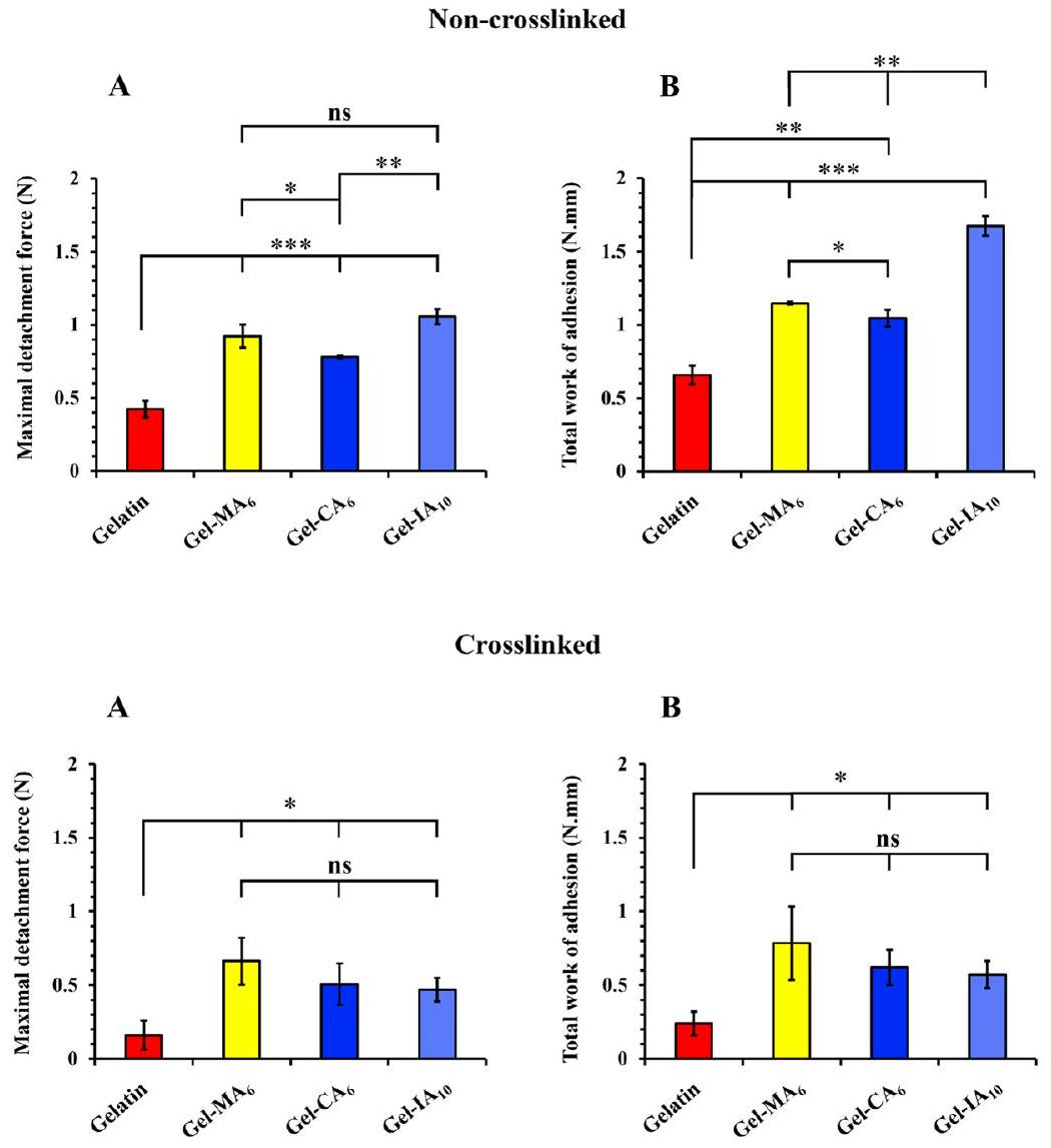
2.6.4. Tensile (Detachment) Method. A TA.XT Plus Texture Analyzer (Stable Micro Systems Ltd., Surrey, U.K.) operated in its adhesive test mode was used to evaluate the mucoadhesive performance of spray-dried microparticles based on gelatin and its derivatives. Both cross-linked and non-cross-linked microparticle samples were employed in this experiment to evaluate the contribution of macromolecules diffusion and ability to form
interpenetrating layer with mucus in mucoadhesion. Freshly isolated sheep nasal tissues were used within 24 h of retrieval for this experiment. As previously reported and adapted with some changes, a section of sheep nasal tissue with the mucosal side facing downward was secured at the surface of a bespoke cylindrical probe. This probe was subsequently attached to the mobile arm of the texture analyzer. Another piece of nasal tissue with the mucosal side facing upward was securely mounted on the mucoadhesion rig of the texture analyzer. Prior to each measurement, nasal tissues were prerinsed with 1 mL of ANF solution ( pH 5.80 ). Subsequently, mg of each spray-dried gelatin-based microparticle formulation was dosed to the nasal mucosa mounted on the rig. The mobile cylindrical probe bearing the blank nasal tissue was then lowered to establish a contact with the opposing mucosal surface. Data acquired from the detachment experiments were used to assess the mucoadhesive strength and the total work of adhesion. The following equipment settings were applied: prespeed test ; test speed sec; post-test speed ; applied force ; contact time 120 s ; trigger type was “auto”; trigger force ; and return distance 10 mm .
All the experiments to assess the retention of formulations on ex vivo porcine vaginal mucosa and sheep nasal mucosa were conducted at and relative humidity within an incubator to mimic physiological conditions. The measurements were all performed in triplicate, and the mean standard deviation values were calculated and then evaluated statistically.
2.7. Statistical Analysis. All measurements in the present study were conducted at a minimum of three times and data were expressed as mean standard deviation values. Statistical analyses were performed using a GraphPad Prism software (version 8.0; San
Figure 1. NMR spectra of modified and unmodified gelatins recorded in at .
Diego, CA, U.S.A.). Data were compared and assessed for significance using two-tailed Student’s -test and a one-way analysis of variance (ANOVA) with Bonferroni post hoc test (for results of SMIT assay and mucoadhesion experiments). For the MTT assay, a one-way ANOVA with Dunnett’s multiple comparisons was employed to compare control and treated groups, while within the treated groups, two-tailed Student’s -test was applied to compare gelatin and its derivatives. Statistical differences were considered significant at a level of .

3. RESULTS AND DISCUSSION

3.1. Synthesis and Characterization. The presence of reactive sites in gelatin macromolecules, such as amines and carboxylic and hydroxyl groups, provide an opportunity for chemical modification leading to changes in its physical and chemical properties. In this work, crotonoylated, itaconoylated, and methacryloylated gelatins were synthesized by reacting this biopolymer with crotonic, itaconic, and methacrylic anhydrides, respectively, at various feed ratios with respect to free amines on the gelatin backbone (Scheme 1). Following purification by dialysis and lyophilization, gelatin derivatives were then characterized using NMR spectroscopy (Figure 1). All four spectra displayed the characteristic signals corresponding to the protons of aromatic amino acids in gelatin at to 7.50 ppm . The degree of functionalization
(DoF) was determined by comparing the integrals of the characteristic double bond hydrogen peaks of each modified gelatin substituent and the integration of the area corresponding to the combined peaks of the aromatic protons of phenylalanine and tyrosine, where their signals were served as a reference. Based on NMR analysis of the spectra of modified gelatins new signals can be observed: protons from distinctive methylidene ( ) group appeared at 5.50 and 5.72 ppm and a peak corresponding to the methyl group ( ), observed at 1.98 ppm , of the methacryloyl functionalities on the modified gelatin (Gel-MA); characteristic peaks at 6.00 and 6.83 ppm assigned to two methine ( ) protons and a peak at 1.91 ppm attributed to the methyl ) group of the crotonoyl groups (Gel-CA); new proton peaks belonging to the methylidene ( group identified at 5.54 and 6.01 ppm and a peak at 3.21 ppm assigned to the methylene group of the itaconoyl groups (Gel-IA). All spectra of modified gelatin samples confirm successful modification with each anhydride at all molar ratios. NMR spectra of native gelatin and its derivatives with different molar ratios as well as the anhydrides are illustrated in Figures S3-S7 in the Supporting Information.
Table 1. Modification of Gelatin with Different Anhydrides and Resulting DoF Determined Using NMR Spectroscopy and a TNBSA Assay, as Well as Other Physicochemical Characteristics
sample -fold molar excess of an anhydride DoF by NMR (%) DoF by TNBSA assay ( ) yield (%) IEP
gelatin 7.0
Gel-MA 1.5 0.373 4.2 4.2
Gel-MA 3 0.380 4.2 4.1
Gel-MA 6 0.384 4.3 4.4
Gel-CA 1.5 0.355 2.5 ND
Gel- 3 0.358 3.2 ND
Gel-CA 6 0.360 3.2 ND
Gel-IA 2 0.274 3.8 4.0
Gel-IA 5 0.298 3.8 4.0
Gel-IA 10 0.298 3.5 ND
Gel-MA, Gel-CA, and Gel-IA are methacryloylated, crotonoylated, and itaconoylated gelatins, respectively; DoF, degree of functionalization; TNBSA assay, 2,4,6-trinitrobenzenesulfonic acid assay; IEP, isoelectric point; ND, not detectable. The suffixes denote the molar excess of anhydrides added referring to the amount of free amino groups in gelatin. The amount of incorporated unsaturated groups. Results are given as the mean standard deviation values The total amount of free amino groups in a native gelatin was determined to be .
In the case of Gel-IA, a decrease of the lysine methylene signal at 3.06 ppm with an increasing amount of IA in the reaction confirmed the modification of lysine residues on the gelatin backbone, whereas in the reaction with CA and MA at all molar ratios, the lysine signals disappeared, indicating the complete conversion of the amino groups. This can most likely be attributed to the increased reactivity of CA and MA compared to IA. Since the reaction takes place in an aqueous environment, the anhydrides will react, however, the crotonic and methacrylic counterparts will do so more quickly, leading to a higher reagent consumption. Moreover, reactions with MA resulted in derivatives, whose spectra display additional small peaks at 6.17 ppm , suggesting that in addition to reacting with lysine residues, some methacryloyl moieties are introduced through the reactions with other side groups in gelatin, e.g., hydroxyl groups, due to the increased reaction time. Table 1 summarizes the data on the degree of functionalization (DoF) of gelatin based on the molar ratios determined via both quantitative NMR analysis and TNBSA assay as well as the product yields.
The determination of DoF values was also done using a glycine standard curve through TNBSA assay (see Figure S1 in the Supporting Information). The total amount of free amino groups in pristine gelatin was found to be , which is in good agreement with the data published earlier. The amount of the remaining free amines after gelatin derivatization was then subtracted from the amount of free groups in a native gelatin to calculate the amount of introduced unsaturated groups (Table 1).
The derivatization of gelatin was further confirmed using FTIR spectroscopy (see Figure S8 in the Supporting Information). FTIR analysis of spectra clearly showed the key absorbance peaks: the spectrum of the parent gelatin shows the presence of the broad band ( ) with a maximum peak at and a shoulder at representing the asymmetric and symmetric stretching vibrations (amide A), respectively, which overlaps with – OH stretching in the same region. The peaks at 2935 and 2872 correspond to the asymmetric and symmetric stretching vibrations in groups. The characteristic absorption bands at 1630, 1523, and are assigned to the stretching (amide I bending plus stretching (amide II), and stretching coupled to bending (amide III) vibrations, respectively, and are in good agreement
with the FTIR data on gelatin reported in the literature. The peaks at 1439 and are due to CH bending and the peak at represents stretching. The introduction of crotonoyl, itaconoyl, and methacryloyl moieties caused small shifts in the amide bands of gelatin to higher frequencies. An absorption band typical for vinyl groups ( C stretching vibration) should be visible at , however, in our case, this was overlapped with the amide I signal (see Figure S8 in the Supporting Information). Nevertheless, the intensities of the amide I, II, and III peaks increased, which can be attributed to the incorporated amide bonds coupled with a stretching vibration. Overall, similar infrared spectra were acquired for different batches of modified gelatins, suggesting the limitations of FTIR spectroscopy in confirming the effectiveness of gelatin derivatization when preparing various batches.
The amphoteric nature of gelatin is due to the presence of amino and carboxylic groups present in amino acids in the macromolecular chains. At pHs lower than isoelectric point (IEP), the macromolecules of gelatin carry a positive charge; whereas at IEP they are negatively charged. At IEP gelatin has a net charge of zero. The IEP also represents the point at which the polyampholyte chains adopt their most compact conformation resulting in minimal viscosity in solutions.
In this study the IEPs for both derivatized and underivatized gelatin macromolecules were determined using the measurements of the electrophoretic mobility and following the changes in solution viscosity as a function of pH . Aqueous solutions ( ) of gelatin and its modified derivatives were titrated by adding varying amounts of acid ( 0.03 M HCl ) or base ( 0.02 M NaOH ), and resulting pH changes were followed using a pH meter. The IEPs of the samples were estimated by determining the pH value at which the electrophoretic mobility curve crossed zero (Figure 2) or when the polyampholyte solution exhibited a minimum specific viscosity (see Figure S9 in the Supporting Information). It was found that native gelatin (type A) produced from porcine skin has an of pH 7.0 , which is within the range given by the manufacturer. It was expected that the incorporation of unsaturated anhydride groups into lysine moiety of gelatin would lower the isoelectric point significantly. Indeed, the introduction of CA, IA and MA groups, and the loss of the lysine primary amines, resulted in a reduction of the IEP of the modified gelatin derivatives below
Figure 2. Electrophoretic mobility as a function of solution pH curves, recorded to determine the of native gelatin and its chemically modified derivatives (Gel-MA, Gel-CA, and Gel-IA are methacryloylated, crotonoylated, and itaconoylated gelatins, respectively).
that of native gelatin indicating the successful chemical modification of this biopolymer (Table 1). The observed changes in IEP during gelatin derivatization are evidently influenced by two factors: the degree of gelatin functionalization and the nature of the introduced functional groups. The incorporation of itaconoyl groups into gelatin leads to a more pronounced reduction in IEP. This effect is attributed to the presence of an additional carboxylic acid group within the itaconoyl moiety.
Gelatin solutions in water are known to exhibit sol-to-gel transitions upon decrease in temperature. This behavior for native gelatin and its new derivatives was studied using
rheological measurements. The rheological changes happening with aqueous solutions of native gelatin and its chemically modified derivatives (Gel-CA, Gel-IA, and GelMA) upon changes in temperature are displayed in Figures S10 and S11 in the Supporting Information. Considering the trend of the data obtained, both the melting ( ) and gelation ( ) points can be determined by taking a crossover temperature between the storage modulus ( ) and loss modulus The crossover temperature is attributed to the “gel-to-sol” or “sol-to-gel” transition temperature, which indicates the transition from an elastic network formation to a solution upon heating or a physically cross-linked gel formation, respectively. It was found that the native gelatin (type A, from porcine skin) exhibited a at and a at during heating and cooling cycles, respectively. The obtained data on melting and gelation temperatures of native gelatin are in good agreement with the data reported in the literature. Gelation and melting temperatures of gelatin derivatives were observed to have a good correlation with their degree of functionalization (DoF). The lower and points were detected for gelatin derivatives with the greater DoF, indicating that chemical modification affects the gelling properties of gelatin. The melting and gelation temperatures of these biopolymers are presented in Table 2.
Table 2. Gelation ( ) and Melting ( ) Temperatures of Derivatized and Underivatized Gelatins Determined Using Dynamic Rheological Measurements, where a Crossover Temperature of Storage Modulus ( ) and Loss Modulus ( ) Occurs upon Cooling and Heating Scans, Respectively
sample
gelatin
Gel-MA
Gel-MA
Gel-MA
Gel-CA
Gel-
Gel-CA
Gel-IA
Gel-IA
Gel-IA
Gel-MA, Gel-CA, and Gel-IA are methacryloylated, crotonoylated, and itaconoylated gelatins, respectively.
3.2. Toxicology. 3.2.1. Cell Viability. In vitro cytotoxicity of gelatin and its modified derivatives (Gel-CA, Gel-IA, and Gel-MA) was studied using MTT assay with human pulmonary fibroblasts (HPF) cells. The assay is based on the ability of mitochondria of live cells to reduce 3-(4,5-dimethylthiazol-2-yl)-2,5-diphenyltetrazolium bromide (MTT reagent), a yellow substance, to insoluble formazan crystals (violet color). This technique allows to calculate the number of viable cells after treatment with the test material. HPF cells were treated with gelatin, Gel-CA , and Gel-MA solutions at concentrations of 1.3 and in cell growth media for 24 h . The negative control group consisted of untreated cells was considered as of viable cells. Figure 3 displays the data on cell viability in the presence of modified and unmodified gelatins. MTT results showed that cell viabilities are comparable for gelatin and its chemically
Figure 3. Effect of modified and unmodified gelatin formulations at 1.3 and concentrations on the percentage of viable HPF cells after 24 h exposure assessed using MTT assay. Data represented as the mean standard error of the mean of two independent experiments in quadruplicate. Gel-MA , and Gel-IA are methacryloylated, crotonoylated, and itaconoylated gelatins, respectively. Statistically significant differences are shown as ; **p ; “ns” denotes no significance.
modified derivatives as there were no statistically significant differences in viability between these biopolymers ( ) against the control after 24 h of treatment in the studied concentrations. Among the considered, only Gel-IA exhibited a slight decrease in cell viability at concentration to 81.1 . However, according to the ISO standards developed by the United Nations Sustainable Development Goals (SDGs; ISO 10993-5:2009) the material for biomedical application is considered safe as long as the in vitro viability is and above. Our results show that the level of cell viability remained high ( ) after the treatment with all biopolymers at given concentrations (Figure 3).
Since gelatin is considered as a safe material, in the next step, we compared the viability of gelatin-treated cells with the group treated with its modified derivatives. Gel-MA and GelCA as well as pure gelatin did not show a significant difference in viability ( ). Moreover, it can be seen that the viability of gelatin was higher than that of the control group. It can be assumed that pure gelatin at a lower concentration shows some ability to facilitate cell proliferation. Our results are in good agreement with previous reports, and gelatin and its modified derivatives can be considered safe in this regard.
In the majority of cases, the difference between gelatin and its modified derivatives was not statistically significant ( , which indicates that chemical modification of gelatin with unsaturated groups does not cause an increase in the polymer toxicity. It can be concluded that the synthesized GelCA, Gel-IA, and Gel-MA are nontoxic and suitable for further development in pharmaceutical applications.
3.2.2. Mucosal Irritancy. The in vivo slug mucosal irritation test (SMIT) was originally developed by Adriaens and coworkers and has been validated as an alternative and reliable method for evaluating the mucosal irritation potency of various chemicals, excipients, cosmetics, formulations, and active ingredients. This technique has been applied in many studies, including the evaluation of nasal and vaginal irritation. SMIT uses terrestrial slugs, which are not protected by legislation controlling animal experiments and are
considered to have limited sentience. Mucus secretion is essential for slugs to aid their locomotion and prevent dehydration. They also release mucus and lose body weight when in contact with irritants. When their mucosal membrane is damaged the slugs produce additional proteins and enzymes. The test provides quantifiable end points for classifying test materials into nonirritating ( ), mild ( 10), moderate (MP% 10-17.5), or severely irritating (MP% 17.5) based on the levels of mucus production. In general, mild irritants cause an increase in mucus production; however, strong irritants induce tissue erosion in addition to increased mucus production.
The modified version of this assay is routinely used by our research group for assessing the biocompatibility of different polymeric excipients. Figure 4 presents the data on the
Figure 4. Mucus production (MP%) by Arion lusitanicus slugs in response to 60 min contact with gelatin and its methacryloylated (Gel-MA 6 ), crotonoylated (Gel-CA 6 ), and itaconoylated (Gel-IA ) derivatives, as well as positive (benzalkonium chloride; BAC) and negative (phosphate buffered saline; PBS) controls. Data are expressed as mean standard deviation values ( ). Statistically significant differences are given as ; “ns” denotes no significance.
percentage of mucus production (MP%) by Arion lusitanicus slugs after 60 min of exposure to a filter paper soaked with solutions of gelatin and its modified derivatives as well as positive and negative controls. All test materials used in this assay were dissolved in phosphate-buffered saline (PBS; pH 7.40). Gelatin-based samples were prepared at , as at this concentration these samples were able to form a thin gel layer on top of the filter paper. Slugs placed in BAC solution (positive control; pH 7.36 ) exhibited an extreme discomfort, producing significantly larger amounts of yellow up to orange-colored mucus ( ) than those exposed to PBS solution (negative control; pH 7.40 ) with a very low level of MP% ( ). These results are in good agreement with the previous studies. Mucus production values recorded for the slugs exposed to gelatin, Gel-MA , Gel-CA , and Gel-IA were 1 , and , respectively. The mucus secretions were colorless, which serves as a good initial indicator of biocompatibility. No statistically significant differences ( ) in MP% were observed between the values recorded for the negative control and gelatin-based test materials, demonstrating the nonirritating nature of both gelatin and
Figure 5. Selected fluorescence images showing mucosal retention of native gelatin, methacryloylated gelatin (Gel-MA6), itaconoylated gelatin (Gel-IA ), and crotonoylated gelatin (Gel-CA ) formulations containing fluorescein sodium ( NaFl ), as well as free NaFl (used as a nonmucoadhesive control), on freshly dissected porcine vaginal tissue after washing with varying volumes of VFS solution ( pH 4.0 ; flow rate ). Fluorescence microscope parameters: magnification ; exposure time -10 ms ; gain . Scale bars correspond to 2 mm .
modified gelatins. The results provide valuable insights into the biocompatibility of gelatin derivatives and suggest that they could be potentially used in various applications without causing mucosal irritation. Figures S12 and S13 in the Supporting Information provide a detailed schematic illustration of the SMIT assay procedure and photographs with Arion lusitanicus slugs exposed to the test materials, respectively.

3.3. Mucoadhesion Studies. 3.3.1. Retention on Ex Vivo

Porcine Vaginal Tissues. The vaginal route of drug administration is often preferred for the local treatment of various gynecological dysfunctions and infections. Local administration has the potential to improve drug absorption and delivery to target tissues while reducing adverse effects. However, vaginal drug administration requires overcoming several obstacles to achieve effective drug absorption and retention. The vaginal epithelium that consists of multiple layers of cells tightly packed together limits the penetration of drugs. Moreover, the presence of a thick layer of mucus within the vaginal cavity further hinders drug diffusion into the underlying tissues. The mucus layer, composed of glycoproteins and mucins, serves as a viscoelastic protective barrier, clearing and lubricating the reproductive tract epithelia to help eliminating pathogens and foreign substances. Furthermore, the physicochemical properties of vaginal fluid, including its volume, viscosity, and acidic pH of the vaginal environment may also have an unfavorable impact on drug absorption and retention. Additionally, such factors as vaginal physiology, age, menstrual cycle, reproductive system disorders, and formulation parameters can also affect the rate and extent of drug absorption in the vaginal cavity. Collectively, these anatomical factors pose challenges in achieving effective drug delivery when administered intravaginally.
In this study, the potential use of gelatin and its crotonoylated, itaconoylated, and methacryloylated derivatives in vaginal drug delivery was studied using an in vitro assay based on flow-through with fluorescent detection. The
formulations were prepared with fluorescein sodium ( NaFl ), which is a fluorescent marker that facilitates easy detection and measurement of mucosal retention levels. This method has been widely employed to study the retention of various formulations on different mucosal surfaces, including vaginal tissues. The effectiveness of this technique was validated against other established methodologies used to assess mucoadhesive properties.
Briefly, NaFl-containing gelatin and its chemically modified derivatives (Gel-CA , and Gel-MA ), as well as free NaFl solution, were administered on the surface of ex vivo porcine vaginal mucosa and allowed to equilibrate at . The mucosal surface was then irrigated with varying volumes of vaginal fluid simulant (VFS; pH 4.0; flow rate ), and the presence of the formulation on the mucosal surface was determined using a fluorescence microscope. The total volume of VFS used in this wash-off experiment was aimed to mimic the amount of normal vaginal discharge in healthy women daily). The exemplar fluorescent microphotographs of the retention of these formulations taken after each wash with VFS are illustrated in Figure 5. The mucosal retention can be quantified by measuring the change in image pixel intensity over time to give a percentage of the fluorescence relative to the initial time (Figure 6). Image analysis helped to reveal that the incorporation of crotonoyl, itaconoyl, and methacryloyl functional groups into gelatin structure significantly enhanced formulation retention on freshly excised porcine vaginal mucosa. Among modified gelatins, Gel-MA demonstrated superior mucoadhesive performance compared to native gelatin ( ) and fluorescein sodium solution throughout the washoff experiment. As anticipated, the polymer-free solutions of NaFl (served as a nonmucoadhesive control) exhibited significantly poorer retention and was rapidly washed out from the mucosal surface, with only fluorescence observed upon completion of the full washing cycle. Traditionally, native gelatin is considered as a polymer with poor
Figure 6. Percentage retention of native gelatin, methacryloylated gelatin (Gel-MA ), itaconoylated gelatin (Gel ), and crotonoylated gelatin (Gel-CA ) formulations containing fluorescein sodium ( NaFl ) as well as free NaFl (used as a nonmucoadhesive control) on freshly excised porcine vaginal mucosa after irrigating with different volumes of VFS solution ( pH 4.0 ; flow rate ). Data are expressed as mean standard deviation values ( ). Statistically significant differences are represented as 0.0001; “ns” denotes no significance.
retention capabilities and the results generated during the ex vivo mucoadhesion experiments corroborated this assessment.
Thus, pure gelatin displayed considerably weaker adhesion to the vaginal mucosa in comparison with its modified counterparts: Gel-MA ; Gel-IA ; and Gel , however, a greater retention compared to NaFl solution ( ). It was observed that Gel-IA exhibited a small but significantly greater retention ( ) on vaginal mucosa in contrast to during the mucoadhesion experiment. However, no statistically significant difference ( was found between these formulations at the end of the wash-off test (Figure 6). It is worth noting that Gel-MA displayed better mucoadhesive performance ( ) compared to the other modified gelatins (Gel-CA and Gel . Previously, we have reported that polymers modified with methacryloyl groups exhibited substantially improved adhesion to various mucosal surfaces and demonstrated comparable or even better mucoadhesive performance at higher degrees of functionalization relative to chitosan, which is commonly regarded as a “gold standard” mucoadhesive polymer/positive control in this field.
As expected, retention of all formulations declines over the course of the washing, yet the following trend is observed: Gel- gelatin NaFl. According to these findings, it is reasonable to assume that the excellent mucoadhesive performance of these formulations is due to their interaction with mucosal surfaces via three mechanisms, as illustrated in Figure 7: (i) the ability of unsaturated functional groups (methacryloyl, crotonoyl, itaconoyl) of modified gelatins to form covalent bonds with thiol groups present in the mucus layer through Michael-type addition
Figure 7. Schematic illustration of the retention of methacryloylated gelatin (Gel-MA), crotonoylated gelatin (Gel-CA), and itaconoylated gelatin (Gel-IA) formulations on vaginal and nasal mucosal surfaces.
reaction; (ii) electrostatic interaction between residual protonated primary amino groups within the modified gelatins and negatively charged mucins present on mucosal surface; and (iii) hydrogen bonding between hydroxyl, carboxyl groups of gelatin and mucin glycoprotein moieties.
The electrostatic interaction contribution of amphoteric macromolecules to mucoadhesion is highly dependent on the solution pH and its relative position against the IEP of polyampholyte. This, in turn, relies on the degree of gelatin functionalization and the nature of the introduced functional groups. Considering that the retention experiments were conducted in VFS at pH 4.0 , only unmodified gelatin will carry a strong positive charge under these conditions, given its IEP at 7.0. Gelatin derivatives with methacryloyl groups exhibit IEP values between 4.2-4.3, resulting in only weak positive charges. Derivatives with crotonoyl and itaconoyl groups possess IEP values ranging from 2.5-3.2 and 3.5-3.8, respectively, making these polymers negatively charged during the retention experiments. Consequently, it is reasonable to infer that electrostatic binding of gelatin derivatives to the vaginal surface is not the primary factor contributing to mucoadhesion.
3.3.2. Retention on Ex Vivo Sheep Nasal Tissues. Intranasal administration offers a noninvasive route of drug delivery. Therapeutic agents delivered to the nasal cavity act locally and provide a direct target to the central nervous system. Estimated daily production of nasal mucus varies between 0.1 and (or from 20 to 40 mL ) under normal conditions. However, the exact amount of mucus secretion can be influenced by various factors such as environmental conditions, allergies, infections, individual variations, as well as specific location and region of the human nasal mucosa. Mucociliary clearance is a complex and dynamic physiological process that helps maintaining the upper and lower airways clean and defend against airborne particles and pathogens. This is done through the interaction of nasal mucus and ciliary beating. The coordinated waves of tiny hair-like cilia gradually move the thick mucus from the front of the nose to the nasopharynx, where it can either be swallowed or expectorated. Additionally, the nasal mucus is slightly acidic ( ) in order to prevent respiratory infections. The protective mechanism of the respiratory system functions efficiently and can greatly limit the residence time of therapeutic substances when administered via the nasal route. Mucoadhesive dosage forms are designed to counteract the clearance mechanism by adhering to the mucosal surface, therefore prolonging the retention time and improving effective drug absorption.
Microparticulate formulations are commonly used in nasal drug delivery. Therefore, in this study, gelatin and its derivatives were formulated as microparticles with NaFl as a model drug. Two types of microparticles were designed with and without the use of a cross-linking agent to evaluate the role of gelatin cross-linking on their nasal retention properties. NaFl-loaded cross-linked and non-cross-linked microparticles based on gelatin and its modified derivatives (Gel-CA; Gel-IA and Gel-MA) have been successfully produced using a spray drying technique. The difference between cross-linked and non-cross-linked particles was the addition of glutaraldehyde, which facilitates cross-linking of gelatin macromolecules. The surface morphology of these microparticles was characterized using scanning electron microscopy (SEM). Microparticles collected after spray drying were spherical and presented a
wrinkled surface texture, free of crystals, pores, and cracks (see Figures S14 and S15 in the Supporting Information). All the formulations led to microparticles with similar morphologies and the mean diameters of the particles were , which were within the range expected for microparticles generated by common spray drying techniques.
In this experiment, we have evaluated the potential of modified and unmodified gelatin-based spray-dried microparticles as mucoadhesive formulations for their application in nasal drug delivery. The retention properties of these formulations were studied involving the same in vitro flowthrough method as described above with some modifications. Only non-cross-linked microparticle samples were used in this experiment. The purpose of using non-cross-linked microparticles is to facilitate their adhesion by allowing them to slowly swell and dissolve upon contact with wet mucosal surface when applied/inhaled. Briefly, of modified and unmodified gelatin-based microparticle samples containing NaFl were deposited on ex vivo sheep nasal mucosa. The experiment was conducted at in an incubator. In the course of this experiment, ANF solution flowing down the nasal mucosa during each washing cycle was collected at predetermined time points. Subsequently, these samples were analyzed using a fluorescence spectrophotometer. Using the data acquired and generating a standard curve for microparticles with NaFl , the amount of dosage form washed off the nasal mucosa could be calculated. Consequently, through reverse calculations, the amount of retained formulations on sheep nasal mucosa could be estimated (Figure 8). All samples
Figure 8. Percentage retention of of pristine gelatin, methacryloylated gelatin (Gel-MA 6 ), crotonoylated gelatin (Gel ), and itaconoylated gelatin (Gel-IA ) non-cross-linked microparticle formulations, each containing fluorescein sodium (NaFl), on freshly dissected sheep nasal mucosa. The mucosa was washed with varying volumes of ANF solution ( pH 5.80 ; flow rate . Data are expressed as mean standard deviation values ( ). Statistically significant differences are given as *p < 0.05; **p ; ; “ns” denotes no significance.
exhibited a reduction in determined fluorescence upon washing process, indicating the removal of gelatin-based microparticle formulations from the mucosal surface. However, methacryloylated gelatin (Gel-MA ) microparticles demonstrated stronger adhesion ( ) to nasal mucosa compared to other modified gelatin derivatives (Gel-CA and Gel-IA ). Once again, pristine gelatin showed poorer
Figure 9. Mucoadhesive characteristics of of different biopolymers: (A) Maximal force of detachment and (B) total work of adhesion profiles for microparticles based on cross-linked and non-cross-linked gelatins and their chemically modified derivatives determined by texture analysis. Data are expressed as mean standard deviation values . Statistically significant differences are displayed as ; ; ***p < 0.001; “ns” represents no significance.
mucoadhesive performance in contrast to its modified counterparts: Gel-MA ; Gel-CA ; and . Interestingly, no statistically significant difference ( ) was observed between Gel and Gel-IA formulations throughout the experimental process, as they displayed a similar retention profile. Overall, these observations are consistent with the previous results and indicate that the three biopolymer derivatives examined have a greater retention on nasal mucosa than native gelatin due to their enhanced mucoadhesive properties.
To further corroborate the findings obtained from the in vitro flow-through method, a tensile test was employed in order to assess the adhesion of microparticles based on cross-linked and non-cross-linked gelatin and its chemically modified derivatives to freshly excised sheep nasal mucosa. The tensile method is one of the widely used and established techniques to evaluate the mucoadhesive properties of various dosage forms. This method involves the bringing polymeric mucoadhesives in contact with mucosal tissue with its subsequent withdrawal, followed by recording and analysis of the resulting detachment profiles. The mucoadhesive performance of dosage forms is then determined through a combination of two important parameters: the measurement of maximal force of detachment required to separate the dosage form from mucosa; and the total work of adhesion, defined as the area under the corresponding detachment force versus distance curve (see Figures S16 and S17 in the
Supporting Information for the exemplar detachment profiles of test materials).
Figure 9 illustrates the values of maximal force of detachment ( ) and total work of adhesion ( ), as calculated from tensile test studies for both cross-linked and non-cross-linked gelatin-based microparticles. Chemically modified gelatin (Gel-MA , and Gel-IA ) formulations exhibited superior mucoadhesive performance ( expressing higher and profiles when compared to parent gelatin within non-cross-linked microparticles. It was revealed that samples based on non-cross-linked Gel-CA 6 microparticles displayed the least retention performance compared to Gel-MA and Gel-IA counterparts showing a lower value. No statistically significant difference was observed in profiles between Gel-MA and Gel-IA , however, Gel-IA exhibited a higher value in contrast to Gel-MA and Gel-CA .
Interestingly, there were no statistically significant differences observed between the cross-linked modified gelatin ( , and Gel-IA ) microparticles, as they exhibited very similar and profiles, indicating comparable mucoadhesive performance among these type of dosage forms. Overall, pristine gelatin displayed the poorest mucoadhesive properties in comparison to its modified counterparts within both cross-linked and non-cross-linked microparticles.
The introduction of specific unsaturated crotonoyl, itaconoyl and methacryloyl functional groups into gelatin structure substantially enhanced the mucoadhesive performance of all formulations based on Gel-IA, Gel-CA, and Gel-MA microparticles. The ability of chemically modified gelatin samples to adhere well to the mucosal tissue is related to the reaction of unsaturated functional groups with thiol groups in cysteinerich subdomains present in mucin via thiol-ene click Michaeltype addiction reaction to form covalent bonds, which occurs under physiologically relevant conditions (Figure 7). This is a quick process that allows establishing improved adhesion within a reasonable period following dosage form administration on the mucosal surface. Additionally, the residual protonated primary amine groups within the modified gelatin structure could also bind with mucins through electrostatic interactions as pH of ANF solution used is weakly acidic ( pH 5.80).
The microparticles composed of cross-linked gelatin and derivatives exhibited poorer mucoadhesive properties compared to the particles prepared with non-cross-linked polymers. This indicates that diffusivity of macromolecules plays a substantial role in mucoadhesion. Cross-linked macromolecules cannot diffuse freely and form an interpenetrating layer with the mucus, which reduces the ability of microparticles to adhere to mucosal surface. This finding is in good agreement with the diffusion theory of mucoadhesion.

4. CONCLUSIONS

In this study, we report the synthesis and characterization of crotonoylated, itaconoylated, and methacryloylated gelatin (Gel-CA, Gel-IA, and Gel-MA, respectively) derivatives with enhanced mucoadhesive properties. The modification was confirmed using NMR, FTIR spectroscopic techniques and TNBSA assay and the degree of functionalization was calculated. The effect of modification on isoelectric point, viscosity and thermo-reversible gelation characteristics of gelatins were also studied. All derivatized gelatins exhibited superior mucoadhesive properties compared to native gelatin. The incorporation of unsaturated anhydrides into gelatins is not detrimental for their toxicological characteristics as evaluated using in vivo SMIT assay and in vitro MTT assay in HPF cells line. Modified gelatins with unsaturated functional groups could be considered as novel excipients with enhanced mucoadhesive properties for potential formulation of mucoadhesive dosage forms for vaginal and nasal drug delivery. GelCA, Gel-IA, and Gel-MA could also find applications in other areas of transmucosal drug delivery, for instance, when formulated as films, gels, micro-, or nanoparticles.

– ASSOCIATED CONTENT

(s) Supporting Information

The Supporting Information is available free of charge at https://pubs.acs.org/doi/10.1021/acs.biomac.3c01183.
Feed ratios for the synthesis of crotonoylated, itaconoylated, or methacryloylated gelatin derivatives; Composition of vaginal fluid simulant (VFS) and artificial nasal fluid (ANF); A glycine standard curve used to determine the amount of free amino groups in modified and unmodified gelatins; A fluorescein standard curve; NMR spectra of unsaturated anhydrides; NMR and FTIR spectra of native gelatin and its modified derivatives; Scanning electron microscopy
(SEM) images of spray-dried microparticles based on modified and unmodified gelatin samples; Specific viscosity versus pH curves to determine isoelectric points; Storage and loss moduli curves used to determine thermo-reversible gelation points; Schematic illustration of slug mucosal irritation test (SMIT) procedure using Arion lusitanicus species; Exemplar photographs of mucus production by Arion lusitanicus slugs in contact with test materials; Exemplar detachment profiles of test materials (PDF)

– AUTHOR INFORMATION

Corresponding Authors

Vitaliy V. Khutoryanskiy – Reading School of Pharmacy, University of Reading, Whiteknights RG6 6DX Reading, United Kingdom; © orcid.org/0000-0002-7221-2630; Email: v.khutoryanskiy@reading.ac.uk
Daulet B. Kaldybekov – Reading School of Pharmacy, University of Reading, Whiteknights RG6 6DX Reading, United Kingdom; Department of Chemistry and Chemical Technology, Al-Farabi Kazakh National University, 050040 Almaty, Kazakhstan; Institute of Polymer Materials and Technology, 050019 Almaty, Kazakhstan; © orcid.org/ 0000-0002-7191-5465; Email: dauletchem@gmail.com, d.kaldybekov@reading.ac.uk

Authors

Elvira O. Shatabayeva – Reading School of Pharmacy, University of Reading, Whiteknights RG6 6DX Reading, United Kingdom; Department of Chemistry and Chemical Technology, Al-Farabi Kazakh National University, 050040 Almaty, Kazakhstan; © orcid.org/0000-0001-9153-5198
Leila Ulmanova – School of Sciences and Humanities, Nazarbayev University, 010000 Astana, Kazakhstan
Balnur A. Zhaisanbayeva – School of Engineering and Digital Sciences, Nazarbayev University, 010000 Astana, Kazakhstan
Ellina A. Mun – School of Sciences and Humanities, Nazarbayev University, 010000 Astana, Kazakhstan
Zarina A. Kenessova – Department of Chemistry and Chemical Technology, Al-Farabi Kazakh National University, 050040 Almaty, Kazakhstan; © orcid.org/0000-0003-2768-824X
Sarkyt E. Kudaibergenov – Institute of Polymer Materials and Technology, 050019 Almaty, Kazakhstan
Complete contact information is available at:
https://pubs.acs.org/10.1021/acs.biomac.3c01183

Author Contributions

The manuscript was written through contributions of all authors. All authors have given approval to the final version of the manuscript.

Notes

The authors declare no competing financial interest.

– ACKNOWLEDGMENTS

The authors acknowledge the Committee of Science of the Ministry of Science and Higher Education of the Republic of Kazakhstan (Grant No. AP14869167) for the financial support of this research. The authors would like to thank Dr. Sam Aspinall for his help and training on the spray drying machine. P.C. Turner Abattoirs (Farnborough, U.K.) is also thanked for
supplying porcine vaginal and sheep nasal tissues for mucoadhesion experiments. V.V.K. acknowledges the Royal Society for his industry fellowship (IF ).

– ABBREVIATIONS

ANF, artificial nasal fluid; BAC, benzalkonium chloride; CA, crotonic anhydride; , deuterium oxide; DoF, degree of functionalization; DMEM, Dulbecco’s modified eagle medium; DMSO, dimethyl sulfoxide; FBS, fetal bovine serum; NaFI, fluorescein sodium salt; Gel, gelatin; Gel-CA, crotonoylated gelatin; Gel-IA, itaconoylated gelatin; Gel-MA, methacryloylated gelatin; IA, itaconic anhydride; MA, methacrylic anhydride; MP, mucus production; MTT reagent, 3-(4,5-dimethylthiazol-2-yl)-2,5-diphenyltetrazolium bromide; PBS, phosphate-buffered saline; SMIT, slug mucosal irritation test; TNBSA assay, 2,4,6-trinitrobenzenesulfonic acid assay; VFS, vaginal fluid simulant

– REFERENCES

(1) Karim, A. A.; Bhat, R. Fish Gelatin: Properties, Challenges, and Prospects as an Alternative to Mammalian Gelatins. Food Hydrocolloids 2009, 23 (3), 563-576.
(2) Schrieber, R.; Gareis, H. Gelatine Handbook: Theory and Industrial Practice; Wiley-VCH Verlag GmbH & Co. KGaA: Weinheim, 2007.
(3) Alipal, J.; Mohd Pu’ad, N. A. S.; Lee, T. C.; Nayan, N. H. M.; Sahari, N.; Basri, H.; Idris, M. I.; Abdullah, H. Z. A Review of Gelatin: Properties, Sources, Process, Applications, and Commercialisation. Mater. Today: Proc. 2021, 42, 240-250.
(4) Gullapalli, R. P.; Mazzitelli, C. L. Gelatin and Non-Gelatin Capsule Dosage Forms. J. Pharm. Sci. 2017, 106 (6), 1453-1465.
(5) Osmałek, T.; Froelich, A.; Jadach, B.; Tatarek, A.; Gadzinski, P.; Falana, A.; Gralinska, K.; Ekert, M.; Puri, V.; Wrotynska-Barczynska, J.; Michniak-Kohn, B. Recent Advances in Polymer-Based Vaginal Drug Delivery Systems. Pharmaceutics 2021, 13 (6), 884.
(6) Bhagat, V.; Becker, M. L. Degradable Adhesives for Surgery and Tissue Engineering. Biomacromolecules 2017, 18 (10), 3009-3039.
(7) De Clercq, K.; Schelfhout, C.; Bracke, M.; De Wever, O.; Van Bockstal, M.; Ceelen, W.; Remon, J. P.; Vervaet, C. GenipinCrosslinked Gelatin Microspheres as a Strategy to Prevent Postsurgical Peritoneal Adhesions: In Vitro and in Vivo Characterization. Biomaterials 2016, 96, 33-46.
(8) Yuk, H.; Varela, C. E.; Nabzdyk, C. S.; Mao, X.; Padera, R. F.; Roche, E. T.; Zhao, X. Dry Double-Sided Tape for Adhesion of Wet Tissues and Devices. Nature 2019, 575, 169-174.
(9) Li, C.; Marton, I.; Harari, D.; Shemesh, M.; Kalchenko, V.; Pardo, M.; Schreiber, G.; Rudich, Y. Gelatin Stabilizes Nebulized Proteins in Pulmonary Drug Delivery against COVID-19. ACS Biomater. Sci. Eng. 2022, 8 (6), 2553-2563.
(10) Khutoryanskiy, V. V. Advances in Mucoadhesion and Mucoadhesive Polymers. Macromol. Biosci. 2011, 11 (6), 748-764.
(11) Serra, L.; Doménech, J.; Peppas, N. A. Engineering Design and Molecular Dynamics of Mucoadhesive Drug Delivery Systems as Targeting Agents. Eur. J. Pharm. Biopharm. 2009, 71 (3), 519-528.
(12) Bernkop-Schnürch, A. Thiomers: A New Generation of Mucoadhesive Polymers. Adv. Drug Delivery Rev. 2005, 57 (11), 1569-1582.
(13) Laffleur, F.; Bernkop-Schnürch, A. Thiomers: Promising Platform for Macromolecular Drug Delivery. Future Med. Chem. 2012, 4, 2205-2216.
(14) Laffleur, F.; Hörmann, N.; Gust, R.; Ganner, A. Synthesis, Characterization and Evaluation of Hyaluronic Acid-Based Polymers for Nasal Delivery. Int. J. Pharm. 2023, 631, No. 122496.
(15) Davidovich-Pinhas, M.; Bianco-Peled, H. Novel Mucoadhesive System Based on Sulfhydryl-Acrylate Interactions. J. Mater. Sci.: Mater. Med. 2010, 21 (7), 2027-2034.
(16) Kolawole, O. M.; Lau, W. M.; Khutoryanskiy, V. V. Methacrylated Chitosan as a Polymer with Enhanced Mucoadhesive Properties for Transmucosal Drug Delivery. Int. J. Pharm. 2018, 550 (1-2), 123-129.
(17) Tonglairoum, P.; Brannigan, R. P.; Opanasopit, P.; Khutoryanskiy, V. V. Maleimide-Bearing Nanogels as Novel Mucoadhesive Materials for Drug Delivery. J. Mater. Chem. B 2016, 4 (40), 6581-6587.
(18) Kim, K.; Kim, K.; Ryu, J. H.; Lee, H. Chitosan-Catechol: A Polymer with Long-Lasting Mucoadhesive Properties. Biomaterials 2015, 52 (1), 161-170.
(19) Prosperi-Porta, G.; Kedzior, S.; Muirhead, B.; Sheardown, H. Phenylboronic-Acid-Based Polymeric Micelles for Mucoadhesive Anterior Segment Ocular Drug Delivery. Biomacromolecules 2016, 17 (4), 1449-1457.
(20) Menzel, C.; Hauser, M.; Frey, A.; Jelkmann, M.; Laffleur, F.; Götzfried, S. K.; Gust, R.; Bernkop-Schnürch, A. Covalently Binding Mucoadhesive Polymers: N-Hydroxysuccinimide Grafted Polyacrylates. Eur. J. Pharm. Biopharm. 2019, 139, 161-167.
(21) Eshel-Green, T.; Bianco-Peled, H. Mucoadhesive Acrylated Block Copolymers Micelles for the Delivery of Hydrophobic Drugs. Colloids Surf., B 2016, 139, 42-51.
(22) Kaldybekov, D. B.; Tonglairoum, P.; Opanasopit, P.; Khutoryanskiy, V. V. Mucoadhesive Maleimide-Functionalised Liposomes for Drug Delivery to Urinary Bladder. Eur. J. Pharm. Sci. 2018, 111, 83-90.
(23) Kaldybekov, D. B.; Filippov, S. K.; Radulescu, A.; Khutoryanskiy, V. V. Maleimide-Functionalised PLGA-PEG Nanoparticles as Mucoadhesive Carriers for Intravesical Drug Delivery. Eur. J. Pharm. Biopharm. 2019, 143, 24-34.
(24) Shan, X.; Aspinall, S.; Kaldybekov, D. B.; Buang, F.; Williams, A. C.; Khutoryanskiy, V. V. Synthesis and Evaluation of Methacrylated Poly(2-Ethyl-2-Oxazoline) as a Mucoadhesive Polymer for Nasal Drug Delivery. ACS Appl. Polym. Mater. 2021, 3 (11), 5882-5892.
(25) Agibayeva, L. E.; Kaldybekov, D. B.; Porfiryeva, N. N.; Garipova, V. R.; Mangazbayeva, R. A.; Moustafine, R. I.; Semina, I. I.; Mun, G. A.; Kudaibergenov, S. E.; Khutoryanskiy, V. V. Gellan Gum and Its Methacrylated Derivatives as in Situ Gelling Mucoadhesive Formulations of Pilocarpine: In Vitro and in Vivo Studies. Int. J. Pharm. 2020, 577, No. 119093.
(26) Buang, F.; Fu, M.; Chatzifragkou, A.; Amin, M. C. I. M.; Khutoryanskiy, V. V. Hydroxyethyl Cellulose Functionalised with Maleimide Groups as a New Excipient with Enhanced Mucoadhesive Properties. Int. J. Pharm. 2023, 642, No. 123113.
(27) Brotherton, E. E.; Neal, T. J.; Kaldybekov, D. B.; Smallridge, M. J.; Khutoryanskiy, V. V.; Armes, S. P. Aldehyde-Functional Thermoresponsive Diblock Copolymer Worm Gels Exhibit Strong Mucoadhesion. Chem. Sci. 2022, 13, 6888-6898.
(28) Hunter, S. J.; Abu Elella, M. H.; Johnson, E. C.; Taramova, L.; Brotherton, E. E.; Armes, S. P.; Khutoryanskiy, V. V.; Smallridge, M. J. Mucoadhesive Pickering Nanoemulsions via Dynamic Covalent Chemistry. J. Colloid Interface Sci. 2023, 651, 334-345.
(29) Bonferoni, M. C.; Chetoni, P.; Giunchedi, P.; Rossi, S.; Ferrari, F.; Burgalassi, S.; Caramella, C. Carrageenan-Gelatin Mucoadhesive Systems for Ion-Exchange Based Ophthalmic Delivery: In Vitro and Preliminary in Vivo Studies. Eur. J. Pharm. Biopharm. 2004, 57 (3), 465-472.
(30) Padhi, J. R.; Nayak, D.; Nanda, A.; Rauta, P. R.; Ashe, S.; Nayak, B. Development of Highly Biocompatible Gelatin & ICarrageenan Based Composite Hydrogels: In Depth Physiochemical Analysis for Biomedical Applications. Carbohydr. Polym. 2016, 153, 292-301.
(31) Liu, Y.; Cheong NG, S.; Yu, J.; Tsai, W. B. Modification and Crosslinking of Gelatin-Based Biomaterials as Tissue Adhesives. Colloids Surf., B 2019, 174, 316-323.
(32) Jovanović, M.; Tomić, N.; Cvijić, S.; Stojanović, D.; Ibrić, S.; Uskoković, P. Mucoadhesive Gelatin Buccal Films with Propranolol Hydrochloride: Evaluation of Mechanical, Mucoadhesive, and Biopharmaceutical Properties. Pharmaceutics 2021, 13 (2), 273.
(33) Abruzzo, A.; Bigucci, F.; Cerchiara, T.; Cruciani, F.; Vitali, B.; Luppi, B. Mucoadhesive Chitosan/Gelatin Films for Buccal Delivery of Propranolol Hydrochloride. Carbohydr. Polym. 2012, 87 (1), 581588.
(34) Abruzzo, A.; Cerchiara, T.; Bigucci, F.; Gallucci, M. C.; Luppi, B. Mucoadhesive Buccal Tablets Based on Chitosan/Gelatin Microparticles for Delivery of Propranolol Hydrochloride. J. Pharm. Sci. 2015, 104 (12), 4365-4372.
(35) Trastullo, R.; Abruzzo, A.; Saladini, B.; Gallucci, M. C.; Cerchiara, T.; Luppi, B.; Bigucci, F. Design and Evaluation of Buccal Films as Paediatric Dosage Form for Transmucosal Delivery of Ondansetron. Eur. J. Pharm. Biopharm. 2016, 105, 115-121.
(36) Mahaling, B.; Katti, D. S. Understanding the Influence of Surface Properties of Nanoparticles and Penetration Enhancers for Improving Bioavailability in Eye Tissues In Vivo. Int. J. Pharm. 2016, 501, 1-9.
(37) Giordani, B.; Abruzzo, A.; Prata, C.; Nicoletta, F. P.; Dalena, F.; Cerchiara, T.; Luppi, B.; Bigucci, F. Ondansetron Buccal Administration for Paediatric Use: A Comparison between Films and Wafers. Int. J. Pharm. 2020, 580, No. 119228.
(38) Kudaibergenov, S. E. Synthetic and Natural Polyampholytes: Structural and Behavioral Similarity. Polym. Adv. Technol. 2021, 32 (3), 906-918.
(39) Ahmady, A.; Abu Samah, N. H. A Review: Gelatine as a Bioadhesive Material for Medical and Pharmaceutical Applications. Int. J. Pharm. 2021, 608, No. 121037.
(40) Van Den Bulcke, A. I.; Bogdanov, B.; De Rooze, N.; Schacht, E. H.; Cornelissen, M.; Berghmans, H. Structural and Rheological Properties of Methacrylamide Modified Gelatin Hydrogels. Biomacromolecules 2000, 1(1), 31-38.
(41) Claaßen, C.; Claaßen, M. H.; Truffault, V.; Sewald, L.; Tovar, G. E. M.; Borchers, K.; Southan, A. Quantification of Substitution of Gelatin Methacryloyl: Best Practice and Current Pitfalls. Biomacromolecules 2018, 19 (1), 42-52.
(42) Brinkman, W. T.; Nagapudi, K.; Thomas, B. S.; Chaikof, E. L. Photo-Cross-Linking of Type I Collagen Gels in the Presence of Smooth Muscle Cells: Mechanical Properties, Cell Viability, and Function. Biomacromolecules 2003, 4 (4), 890-895.
(43) Nichol, J. W.; Koshy, S. T.; Bae, H.; Hwang, C. M.; Yamanlar, S.; Khademhosseini, A. Cell-Laden Microengineered Gelatin Methacrylate Hydrogels. Biomaterials 2010, 31 (21), 5536-5544.
(44) Norris, S. C. P.; Delgado, S. M.; Kasko, A. M. Mechanically Robust Photodegradable Gelatin Hydrogels for 3D Cell Culture and in Situ Mechanical Modification. Polym. Chem. 2019, 10 (23), 31803193.
(45) Habeeb, A. F. S. A. Determination of Free Amino Groups in Proteins by Trinitrobenzenesulfonic Acid. Anal. Biochem. 1966, 14 (3), 328-336.
(46) Lee, B. H.; Shirahama, H.; Cho, N. J.; Tan, L. P. Efficient and Controllable Synthesis of Highly Substituted Gelatin Methacrylamide for Mechanically Stiff Hydrogels. RSC Adv. 2015, 5 (128), 106094106097.
(47) Zhu, M.; Wang, Y.; Ferracci, G.; Zheng, J.; Cho, N. J.; Lee, B. H. Gelatin Methacryloyl and Its Hydrogels with an Exceptional Degree of Controllability and Batch-to-Batch Consistency. Sci. Rep. 2019, 9, 6863.
(48) Benson, J. E. Viscometric Determination of the Isoelectric Point of a Protein. J. Chem. Educ. 1963, 40 (9), 468-469.
(49) Khutoryanskaya, O. V.; Morrison, P. W. J.; Seilkhanov, S. K.; Mussin, M. N.; Ozhmukhametova, E. K.; Rakhypbekov, T. K.; Khutoryanskiy, V. V. Hydrogen-Bonded Complexes and Blends of Poly(Acrylic Acid) and Methylcellulose: Nanoparticles and Mucoadhesive Films for Ocular Delivery of Riboflavin. Macromol. Biosci. 2014, 14 (2), 225-234.
(50) Irmukhametova, G. S.; Mun, G. A.; Khutoryanskiy, V. V. Thiolated Mucoadhesive and PEGylated Nonmucoadhesive Organosilica Nanoparticles from 3-Mercaptopropyltrimethoxysilane. Langmuir 2011, 27 (15), 9551-9556.
(51) Mun, E. A.; Williams, A. C.; Khutoryanskiy, V. V. Adhesion of Thiolated Silica Nanoparticles to Urinary Bladder Mucosa: Effects of PEGylation, Thiol Content and Particle Size. Int. J. Pharm. 2016, 512 (1), 32-38.
(52) Porfiryeva, N. N.; Nasibullin, S. F.; Abdullina, S. G.; Tukhbatullina, I. K.; Moustafine, R. I.; Khutoryanskiy, V. V. Acrylated Eudragit® E PO as a Novel Polymeric Excipient with Enhanced Mucoadhesive Properties for Application in Nasal Drug Delivery. Int. J. Pharm. 2019, 562, 241-248.
(53) Haddow, P. J.; da Silva, M. A.; Kaldybekov, D. B.; Dreiss, C. A.; Hoffman, E.; Hutter, V.; Khutoryanskiy, V. V.; Kirton, S. B.; Mahmoudi, N.; McAuley, W. J.; Cook, M. T. Polymer Architecture Effects on Poly( -diethyl acrylamide)- -Poly(ethylene glycol)- Poly( -diethyl acrylamide) Thermoreversible Gels and Their Evaluation as a Healthcare Material. Macromol. Biosci. 2022, 22 (3), No. 2100432.
(54) Kolawole, O. M.; Lau, W. M.; Khutoryanskiy, V. V. Synthesis and Evaluation of Boronated Chitosan as a Mucoadhesive Polymer for Intravesical Drug Delivery. J. Pharm. Sci. 2019, 108 (9), 3046-3053.
(55) Hoch, E.; Schuh, C.; Hirth, T.; Tovar, G. E. M.; Borchers, K. Stiff Gelatin Hydrogels Can Be Photo-Chemically Synthesized from Low Viscous Gelatin Solutions Using Molecularly Functionalized Gelatin with a High Degree of Methacrylation. J. Mater. Sci. Mater. Med. 2012, 23 (11), 2607-2617.
(56) Kim, S.; Nimni, M. E.; Yang, Z.; Han, B. Chitosan/GelatinBased Films Crosslinked by Proanthocyanidin. J. Biomed. Mater. Res. 2005, 75B (2), 442-450.
(57) Barth, A. Infrared Spectroscopy of Proteins. Biochim. Biophys. Acta – Bioenerg. 2007, 1767 (9), 1073-1101.
(58) Hashim, D. M.; Man, Y. B. C.; Norakasha, R.; Shuhaimi, M.; Salmah, Y.; Syahariza, Z. A. Potential Use of Fourier Transform Infrared Spectroscopy for Differentiation of Bovine and Porcine Gelatins. Food Chem. 2010, 118 (3), 856-860.
(59) Cebi, N.; Durak, M. Z.; Toker, O. S.; Sagdic, O.; Arici, M. An Evaluation of Fourier Transforms Infrared Spectroscopy Method for the Classification and Discrimination of Bovine, Porcine and Fish Gelatins. Food Chem. 2016, 190, 1109-1115.
(60) Rahali, K.; Ben Messaoud, G.; Kahn, C. J. F.; SanchezGonzalez, L.; Kaci, M.; Cleymand, F.; Fleutot, S.; Linder, M.; Desobry, S.; Arab-Tehrany, E. Synthesis and Characterization of Nanofunctionalized Gelatin Methacrylate Hydrogels. Int. J. Mol. Sci. 2017, 18 (12), 2675.
(61) Sheppard, S. E.; Houck, R. C. The Structure of Gelatin Sols and Gels. III. J. Phys. Chem. 1930, 34 (10), 2187-2201.
(62) Johlin, J. M. The Isoelectric Point of Gelatin and Its Relation to the Minimum Physical Properties of Gelatin. J. Biol. Chem. 1930, 86 (1), 231-243.
(63) Lowe, A. B.; McCormick, C. L. Synthesis and Solution Properties of Zwitterionic Polymers. Chem. Rev. 2002, 102 (11), 4177-4190.
(64) Gudmundsson, M. Rheological Properties of Fish Gelatins. J. Food Sci. 2002, 67 (6), 2172-2176.
(65) Goudoulas, T. B.; Germann, N. Phase Transition Kinetics and Rheology of Gelatin-Alginate Mixtures. Food Hydrocolloids 2017, 66, 49-60.
(66) Boran, G.; Mulvaney, S. J.; Regenstein, J. M. Rheological Properties of Gelatin from Silver Carp Skin Compared to Commercially Available Gelatins from Different Sources. J. Food Sci. 2010, 75 (8), E565-E571.
(67) ISO 10993-5:2009(en), Biological evaluation of medical devices – Part 5: Tests for in vitro cytotoxicity. https://www.iso.org/ obp/ui/#iso:std:iso:10993:-5:ed-3:v1:en (accessed 2022-09-03).
(68) Elzoghby, A. O.; Samy, W. M.; Elgindy, N. A. Protein-Based Nanocarriers as Promising Drug and Gene Delivery Systems. J. Controlled Release 2012, 161 (1), 38-49.
(69) Suresh, D.; Suresh, A.; Kannan, R. Engineering Biomolecular Systems: Controlling the Self-Assembly of Gelatin to Form UltraSmall Bioactive Nanomaterials. Bioact. Mater. 2022, 18, 321-336.
(70) Ulubayram, K.; Aksu, E.; Gurhan, S. I. D.; Serbetci, K.; Hasirci, N. Cytotoxicity Evaluation of Gelatin Sponges Prepared with Different Cross-Linking Agents. J. Biomater. Sci. Polym. Ed. 2002, 13 (11), 1203-1219.
(71) Won, Y.-W.; Kim, Y.-H. Preparation and Cytotoxicity Comparison of Type a Gelatin Nanoparticles with Recombinant Human Gelatin Nanoparticles. Macromol. Res. 2009, 17 (7), 464468.
(72) Shin, H.; Olsen, B. D.; Khademhosseini, A. The Mechanical Properties and Cytotoxicity of Cell-Laden Double-Network Hydrogels Based on Photocrosslinkable Gelatin and Gellan Gum Biomacromolecules. Biomaterials 2012, 33 (11), 3143-3152.
(73) Adriaens, E.; Remon, J. P. Gastropods as an Evaluation Tool for Screening the Irritating Potency of Absorption Enhancers and Drugs. Pharm. Res. 1999, 16 (8), 1240-1244.
(74) Adriaens, E.; Dierckens, K.; Bauters, T. G. M.; Nelis, H. J.; Van Goethem, F.; Vanparys, P.; Remon, J. P. The Mucosal Toxicity of Different Benzalkonium Chloride Analogues Evaluated with an Alternative Test Using Slugs. Pharm. Res. 2001, 18 (7), 937-942.
(75) Callens, C.; Adriaens, E.; Dierckens, K.; Remon, J. P. Toxicological Evaluation of a Bioadhesive Nasal Powder Containing a Starch and Carbopol® 974 P on Rabbit Nasal Mucosa and Slug Mucosa. J. Controlled Release 2001, 76 (1-2), 81-91.
(76) Ceulemans, J.; Vermeire, A.; Adriaens, E.; Remon, J. P.; Ludwig, A. Evaluation of a Mucoadhesive Tablet for Ocular Use. J. Controlled Release 2001, 77 (3), 333-344.
(77) Adriaens, E.; Ameye, D.; Dhondt, M. M. M.; Foreman, P.; Remon, J. P. Evaluation of the Mucosal Irritation Potency of Co-Spray Dried Amioca®/Poly(Acrylic Acid) and Amioca®/Carbopol® 974P Mixtures. J. Controlled Release 2003, 88 (3), 393-399.
(78) Dhondt, M.; Adriaens, E.; Roey, J.; Remon, J. The Evaluation of the Local Tolerance of Vaginal Formulations Containing Dapivirine Using the Slug Mucosal Irritation Test and the Rabbit Vaginal Irritation Test. Eur. J. Pharm. Biopharm. 2005, 60 (3), 419425.
(79) Adriaens, E.; Remon, J. P. Mucosal Irritation Potential of Personal Lubricants Relates to Product Osmolality as Detected by the Slug Mucosal Irritation Assay. Sex. Transm. Dis. 2008, 35 (5), 512516.
(80) Adriaens, E.; Bytheway, H.; De Wever, B.; Eschrich, D.; Guest, R.; Hansen, E.; Vanparys, P.; Schoeters, G.; Warren, N.; Weltens, R.; Whittingham, A.; Remon, J. P. Successful Prevalidation of the Slug Mucosal Irritation Test to Assess the Eye Irritation Potency of Chemicals. Toxicol. In Vitro 2008, 22 (5), 1285-1296.
(81) Lenoir, J.; Adriaens, E.; Remon, J. P. New Aspects of the Slug Mucosal Irritation Assay: Predicting Nasal Stinging, Itching and Burning Sensations. J. Appl. Toxicol. 2011, 31 (7), 640-648.
(82) Lenoir, J.; Bachert, C.; Remon, J. P.; Adriaens, E. The Slug Mucosal Irritation (SMI) Assay: A Tool for the Evaluation of Nasal Discomfort. Toxicol. In Vitro 2013, 27 (6), 1954-1961.
(83) Petit, J. Y.; Doré, V.; Marignac, G.; Perrot, S. Assessment of Ocular Discomfort Caused by 5 Shampoos Using the Slug Mucosal Irritation Test. Toxicol. In Vitro 2017, 40, 243-247.
(84) Balls, M.; Goldberg, A. M.; Fentem, J. H.; Broadhead, C. L.; Burch, R. L.; Festing, M. F. W.; Frazier, J. M.; Hendriksen, C. F. M.; Jennings, M.; van der Kamp, M. D. O.; Morton, D. B.; Rowan, A. N.; Russell, C.; Russell, W. M. S.; Spielmann, H.; Stephens, M. L.; Stokes, W. S.; Straughan, D. W.; Yager, J. D.; Zurlo, J.; van Zutphen, B. F. M. The Three Rs: The Way Forward: The Report and Recommendations of ECVAM Workshop 11. Altern. to Lab. Anim. 1995, 23 (6), 838866.
(85) Moiseev, R. V.; Steele, F.; Khutoryanskiy, V. V. Polyaphron Formulations Stabilised with Different Water-Soluble Polymers for Ocular Drug Delivery. Pharmaceutics 2022, 14 (5), 926.
(86) Hussain, A.; Ahsan, F. The Vagina as a Route for Systemic Drug Delivery. J. Controlled Release 2005, 103 (2), 301-313.
(87) Krogstad, E. A.; Rathbone, M. J.; Woodrow, K. A. Vaginal Drug Delivery. In Focal Controlled Drug Delivery. Advances in Delivery Science
and Technology; Domb, A. J., Khan, W., Eds.; Springer: Boston, MA, 2014; pp 607-651.
(88) Shapiro, R. L.; DeLong, K.; Zulfiqar, F.; Carter, D.; Better, M.; Ensign, L. M. In Vitro and Ex Vivo Models for Evaluating Vaginal Drug Delivery Systems. Adv. Drug Delivery Rev. 2022, 191, No. 114543.
(89) Davidovich-Pinhas, M.; Bianco-Peled, H. Methods to Study Mucoadhesive Dosage Forms. In Mucoadhesive Materials and Drug Delivery Systems; Khutoryanskiy, V. V., Ed.; John Wiley & Sons, Ltd: Chichester, U.K., 2014; pp 175-196.
(90) Eschenbach, D. A.; Thwin, S. S.; Patton, D. L.; Hooton, T. M.; Stapleton, A. E.; Agnew, K.; Winter, C.; Meier, A.; Stamm, W. E. Influence of the Normal Menstrual Cycle on Vaginal Tissue, Discharge, and Microflora. Clin. Infect. Dis. 2000, 30 (6), 901-907.
(91) Sogias, I. A.; Williams, A. C.; Khutoryanskiy, V. V. Why Is Chitosan Mucoadhesive? Biomacromolecules 2008, 9 (7), 1837-1842.
(92) Fu, M.; Filippov, S. K.; Williams, A. C.; Khutoryanskiy, V. V. On the Mucoadhesive Properties of Synthetic and Natural Polyampholytes. J. Colloid Interface Sci. 2024, 659, 849-858.
(93) Lale, A. M.; Mason, J. D. T.; Jones, N. S. Mucociliary Transport and Its Assessment: A Review. Clin. Otolaryngol. Allied Sci. 1998, 23 (5), 388-396.
(94) Sahin-Yilmaz, A.; Naclerio, R. M. Anatomy and Physiology of the Upper Airway. Proc. Am. Thorac. Soc. 2011, 8 (1), 31-39.
(95) Beule, A. G. Physiology and Pathophysiology of Respiratory Mucosa of the Nose and the Paranasal Sinuses. GMS Curr. Top. Otorhinolaryngol. Head Neck Surg. 2010, 9, Doc07.
(96) Chavda, V. P.; Jogi, G.; Shah, N.; Athalye, M. N.; Bamaniya, N.; K Vora, L.; Cláudia Paiva-Santos, A. Advanced Particulate CarrierMediated Technologies for Nasal Drug Delivery. J. Drug Delivery Sci. Technol. 2022, 74, No. 103569.
(97) Favaro-Trindade, C. S.; Santana, A. S.; Monterrey-Quintero, E. S.; Trindade, M. A.; Netto, F. M. The Use of Spray Drying Technology to Reduce Bitter Taste of Casein Hydrolysate. Food Hydrocolloids 2010, 24 (4), 336-340.
(98) Osman, R.; Kan, P. L.; Awad, G.; Mortada, N.; El-Shamy, A. E.; Alpar, O. Spray Dried Inhalable Ciprofloxacin Powder with Improved Aerosolisation and Antimicrobial Activity. Int. J. Pharm. 2013, 449 (12), 44-58.

  1. All outputs in CentAUR are protected by Intellectual Property Rights law, including copyright law. Copyright and IPR is retained by the creators or other copyright holders. Terms and conditions for use of this material are defined in
  2. Received: October 31, 2023
    Revised: January 13, 2024
    Accepted: January 16, 2024