تفعيل واستقرار الفروق الكبيرة في الكهربية السالبة بين المواقع الذرية المجاورة ZnIn2S4 لتحقيق التحلل الضوئي المائي الشامل بكفاءة Large electronegativity differences between adjacent atomic sites activate and stabilize ZnIn2S4 for efficient photocatalytic overall water splitting
التحلل الضوئي الشامل للماء إلى هيدروجين وأكسجين مرغوب فيه لإنتاج وقود متجدد ومستدام ونظيف على الأرض على المدى الطويل. تعتبر كبريتيدات المعادن محفزات مثالية لتوليد الهيدروجين، لكن تجانس مكوناتها وعدم استقرار الكبريت النموذجي يسبب إنتاج أكسجين غير نشط، مما يبقى عقبة كبيرة أمام التحلل الشامل للماء. هنا، يتم تحفيز تشوه موقع الكاتيون لتطعيم الأكسجين. (D-O-ZIS) يخلق اختلافات كبيرة في الكهربية السالبة بين المواقع الذرية المجاورة، مع المواقع الغنية بالإلكترونات والمواقع التي تعاني من نقص الإلكترونات في الهيكل المحلي لـالمواقع. إن خاصية إعادة توزيع الشحنات القوية تنشط تفاعلات الأكسجين المستقرة فيالمواقع وتتجنب المشكلة الشائعة لعدم استقرار الكبريت في التحفيز الضوئي لمركبات الكبريتيد المعدنية، بينماتفضل المواقع امتصاص/إطلاق الهيدروجين. وبالتالي، تم تحقيق تفاعل انقسام الماء بشكل عام في D-O-ZIS بكفاءة ملحوظة في تحويل الطاقة الشمسية إلى هيدروجين.، مصاحبة لـمعدل الاحتفاظ بعد اختبار التحفيز الضوئي لمدة 120 ساعة. في هذا العمل، نستمد تصميمًا عالميًا من منظور اختلافات الكهربية السالبة لتنشيط وتثبيت محفزات كبريتيد المعادن لتحفيز فعال لتفكيك الماء بشكل كامل.
استخدام ضوء الشمس والماء، وهما من أكثر الموارد الطبيعية وفرة على الأرض، لإنتاج الهيدروجين ) والأكسجين ( ) بنسبة ستوكيومترية من يمتلك إمكانيات كبيرة لتحقيق الحياد الكربونيبالمقارنة مع تقنيات إنتاج الهيدروجين الشمسي المعينة، مثل تحليل الماء الكهروضوئي، فإن التحليل الضوئي الكلي للماء يلغي الحاجة إلى جهد خارجي أو دوائر كهربائية، مما يقلل من تكاليف النظام ويخفف من مخاوف تآكل المحفز الضوئي، والاستقرار، والسلامة.التحلل الكلي للماء القائم على أشباه الموصلات هو طريق مثالي لتحويل الطاقة الشمسية إلى طاقة كيميائيةبناء يمكن أن يعزز المحفز الضوئي الهجين جمع الضوء ويسهل فصل الشحنات. ومع ذلك، فإن المسارات التفاعلية الطويلة والتوزيع العشوائي للمواقع النشطة في الأنظمة الهجينة قد قيدت نشاطها الضوئي التحفيزي مؤخراً، تم استخدام محفزات ضوئية فردية، مثل، أطر عضوية تساهمية TpBpy، و ، إلخ. تم تطويرها لتحقيق الانقسام الكلي للمياه مع تجنب مشاكل بناء الأنظمة الهجينة. ومع ذلك، فإن تطوير المحفزات الضوئية ذات الكفاءات العالية في تحويل الطاقة الشمسية إلى هيدروجين (STH) في أنظمة الهيدروجين الشمسية لا يزال يمثل تحديًا أساسيًا .
تعتبر كبريتيدات المعادن محفزات ضوئية واعدة بسبب نطاقاتها الطاقية المناسبة، وهياكلها القابلة للتصميم، وخصائصها الكهروضوئية الممتازة.المركبات الكبريتية المعدنية الممثلة مثل، و لقد تم استخدامها على نطاق واسع في مجال تحليل المياه الضوئي.من بينهم، (ZIS) هو شبه موصل معدني ثلاثي الطبقات نموذجي، يمتلك فجوة نطاق مناسبة تبلغ حوالي 2.44 إلكترون فولت وإمكانات نطاق التوصيل تبلغ 0.43 إلكترون فولت مقابل القطب الهيدروجيني العادي (NHE)، مما يمنحه قدرة امتصاص الضوء المرئي وقوة اختزال قوية لـتوليد من تحليل الماءلقد حققت العديد من الاستراتيجيات تحسينًاأداء الإنتاج حول محفز ZIS الضوئي، مثل بناء هيكل غير متجانس بنمط Z من فراغات الكبريت ZIS مع أشباه الموصلات الأخرىتصنيع فراغات S الناتجة عن تشويب النحاس الذري في رقائق ZIS النانويةدمج تشويب الأكسجين في مواقع الأنيونات في مواقع ذرات الكبريت في ZISومعدل مساعد للتفاعل من ذرات البلاتين الفردية البارزةلكنهم لم يتمكنوا من تحقيق تفاعلات تفكيك الماء الفعالة والمستقرة بشكل عام. واحدة من التحديات الرئيسية التي تواجه تحسين الكفاءة لتفكيك الماء هي التركيبة المتجانسة لمواقع النشاط والبنية الإلكترونية المتسقة في محفز ضوئي واحد. بالإضافة إلى ذلك، فإن ذرات الكبريت الموجودة في محفز ZIS عرضة بشدة للأكسدة بواسطة الثقوب الناتجة عن الضوء، مما يؤدي إلى عدم استقراره. تؤدي هذه العقبات إلى قاعدة خاملة تعيق إنتاج الأكسجين وفي النهاية تؤدي إلى أداء ضعيف في تفكيك الماء بشكل عام..
هنا، تم تصميم تشويش مستحث لموقع الكاتيون من خلال إضافة الأكسجين إلى ZIS (D-O-ZIS) لكسر تجانس مكوناته بين المواقع الذرية المجاورة وتحقيق أداء عالٍ في عملية التحليل الكهربائي للماء. عادةً، تميل ذرات الأكسجين إلى احتلال موقع الأنيون في ZIS، حيث لا يتم إنتاج الأكسجين (الشكل التكميلي 1). إن إضافة الأكسجين في موقع الكاتيون تمثل تحديًا بسبب الطاقة غير المواتية المعنية. على عكس إضافة الأكسجين في موقع الأنيون النموذجي في ZIS، يتغلب D-O-ZIS على الحاجز العالي لإضافة الأكسجين في موقع الكاتيون من خلال بناء هيكل عالي الطاقة مشوه كوسيط، مما يسهل إضافة ذرات الأكسجين إلى مواقع الكاتيون (أي، إضافة الأكسجين المستحثة في موقع الكاتيون). تتضمن الاستراتيجية تحفيز الهجرة الذرية حراريًا لتوليد هيكل حافة مشوه (D-ZIS)، والذي يتم معالجته بعد ذلك بـالبلازما لاستحضار تشويب الأكسجين في مواقع الكاتيونات وخلق D-O-ZIS (انظر الشكل 1a وقسم الطرق لعملية التخليق بالتفصيل). أدت حالات التشوه وتشويب الأكسجين في مواقع الكاتيونات إلى إعادة توزيع الشحنات وغيرت توازن الكهربية السالبة لمواقع الذرات المنسقة في مناطق التشوه المشوبة بالأكسجين. على وجه التحديد، كانت الإلكترونات الغنيةالمواقع وافتقار الإلكتروناتالمواقع المحليةتكوين D-O-ZIS قد أظهر سلوك امتصاص/إطلاق مثالي لـ أو خلال التفاعل. رابطة S-O في D-O-ZIS، كونها حالة إلكترونية مهجنة لـ S، يعزز تطور الأكسجين المستقر لتفاعلات تحليل الماء بشكل عام ويتجنب المشكلة الشائعة لعدم استقرار الكبريت في تحفيز الضوء باستخدام كبريتيدات المعادن. وبالتالي، أظهر D-O-ZIS كعامل تحفيز ضوئي منفرد أداءً استثنائيًا في تحليل الماء بشكل عام مع STH، مصحوبًا بزيادة في استقرار تحليل الماء معدل الاحتفاظ بعد 120 ساعة. في هذا العمل، يمكن أن تنشط هذه الاستراتيجية أيضًا الأكسجين الخامل لمحفزات الفوتوكاتاليست الأخرى من كبريتيدات المعادن، مثل و مظهراً عالمية تصميم كبريتيدات المعادن من منظور اختلافات الكهربية السالبة لعملية التحليل الكهربائي للماء بشكل عام.
النتائج
توصيف المحفز الضوئي
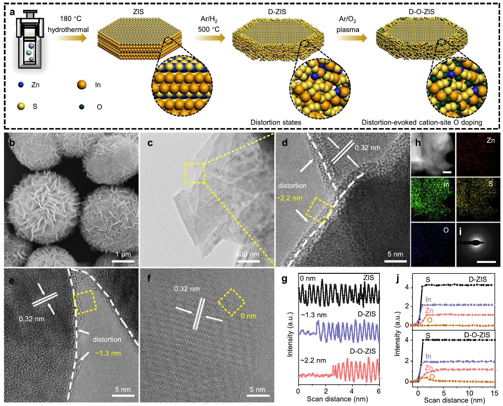
تم تحليل المحفزات الضوئية باستخدام المجهر الإلكتروني الماسح (SEM) والمجهر الإلكتروني الناقل (TEM)، مما كشف عن هياكل تشبه الزهور النانوية تتكون من طبقات نانوية (الشكل 1ب، ج، الشكل التكميلي 2). أكدت صور المجهر الإلكتروني الناقل عالي الدقة (HRTEM) أن حواف الشبكة في العينة كانت لها مسافة بين الطبقات تبلغ 0.32 نانومتر، مما يتوافق مع مستوى الشبكة (102) لمرحلة ZIS السداسية (الشكل 1د-و).أظهرت D-ZIS و D-O-ZIS قشور حواف مشوهة (الشكل 1د، هـ، المربعات الصفراء) مقارنة بـ ZIS (الشكل 1و). أشارت ملفات الخطوط إلى أن سماكات D-ZIS و D-O-ZIS المشوهة كانت و ، على التوالي (الشكل 1g). كشفت تقنية التحليل الطيفي بالأشعة السينية المشتتة للطاقة (EDX) أن Zn و In و S و O كانت موزعة بشكل موحد في D-O-ZIS (الشكل 1h)، بينما كان من الصعب اكتشاف ذرة O في ZIS و D-ZIS (الشكل التكميلي 2e، g). أشارت أنماط حيود الإلكترونات في المنطقة المختارة (SAED) إلى أن هياكل D-O-ZIS و D-ZIS كانت مشوهة عند الحواف مقارنةً بهيكل بلورة ZIS (الشكل 1i، الشكل التكميلي 2f، h). أظهرت مسح الخطوط بتقنية TEM الماسح في مجال الظلام العالي الزاوية (HAADF-STEM) لعنصر Zn أن فراغات Zn كانت محصورة في حواف D-ZIS و D-O-ZIS، بينما أظهر مسح الخط لعنصر O أن ذرات O المضافة في D-O-ZIS كانت موضعها بشكل أساسي عند الحافة الخارجية (الشكل 1j، الشكل التكميلي 2i)، وهيكل بلورة D-O-ZIS متوافق مع هيكل ZIS (الشكل التكميلي 2j).
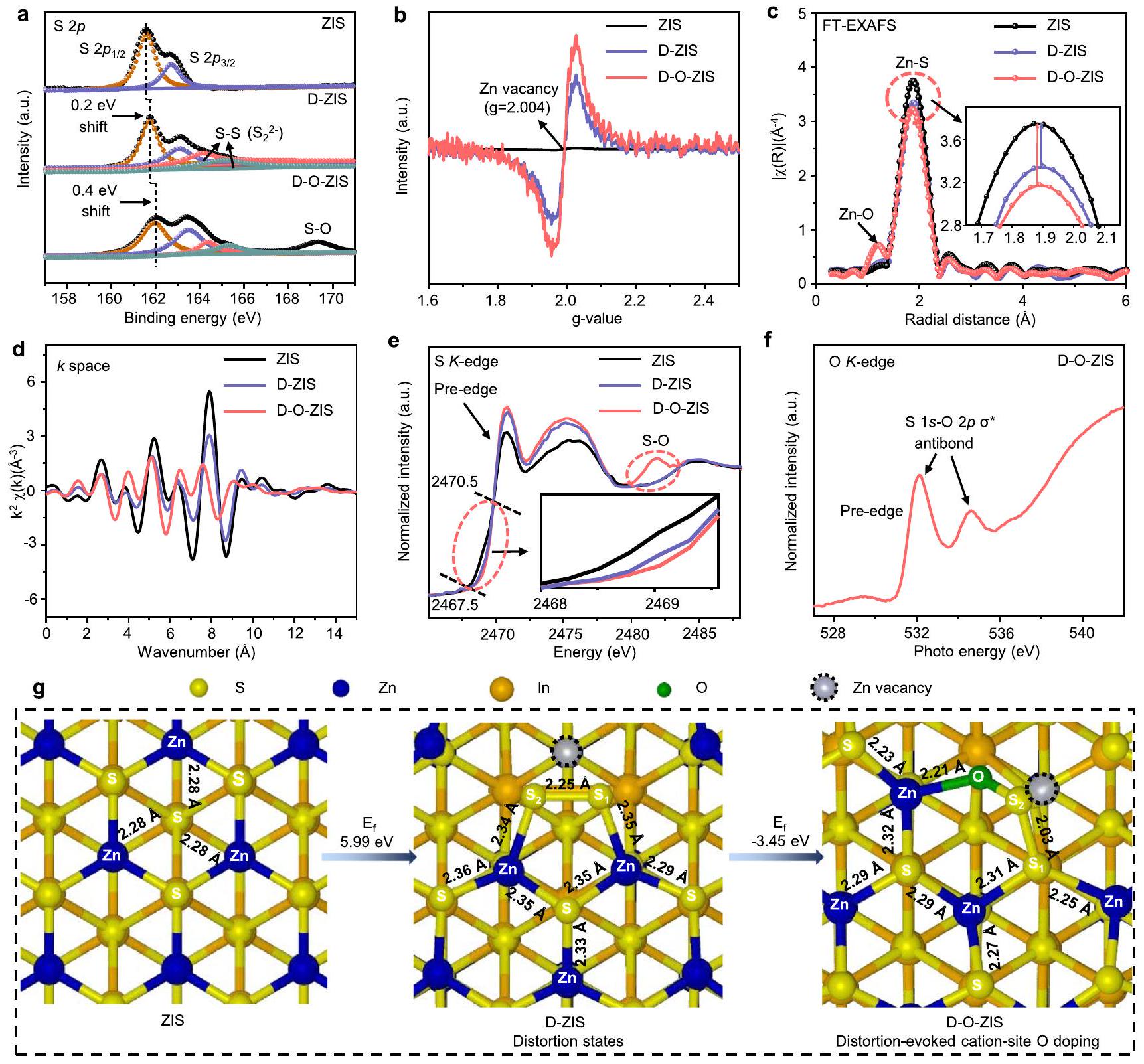
قمنا بإجراء تحليل أكثر تفصيلاً لهيكل قشرة الحافة وخصائص تشبع الأكسجين للمواد المحفزة للضوء. كان هيكل قشرة الحافة المشوهة لـ D-ZIS، الذي يختلف عن ZIS بدون تشوه، ناتجًا بشكل أساسي عن روابط S-S وإدخال فراغات Zn.أظهرت مطيافية الإلكترونات الضوئية بالأشعة السينية (XPS) تحولات طاقة أعلى في D-ZIS، مع قمم إضافية تم تعيينها لرابطة S-S.عند 164.4 و 165.3 إلكترون فولت (الشكل 2أ)أظهر طيف الرنين المغناطيسي للإلكترون (ESR) لـ D-ZIS شدة ذروة عندنُسبت إلى الإلكترونات الحرة غير المتزاوجة المحصورة في فراغ الزنك، مما يؤكد وجود فراغ الزنك (الشكل 2ب)، بتركيزمحدد بواسطة طيف الأشعة السينية للزنك (الشكل التكميلي 3، الجدول التكميلي 1)، وهو ما يتماشى مع نتائج مطياف الانبعاث البلازمي المقترن بالحث (ICP) (الجدول التكميلي 2). تحويل فورييه لـمنحنيات لـأظهرت طيف الامتصاص الدقيق للأشعة السينية الممتدة عند حافة (FT-EXAFS) قمة Zn-S عندفي D-ZIS، ولكن مع شدة مخفضة مقارنة بـ ZIS (الشكل 2c)، مما يشير إلى وجود حالات تشوه ناتجة عن فراغات الزنك.. تم تأكيد هذه النتيجة بشكل أكبر بواسطة -طيف EXAFS عند حافة الفضاء، الذي أظهر تذبذبًا مخففًا لـ D-ZIS مقارنةً بـ ZIS (الشكل 2d)، وخرائط الكنتور الموجية القوية (الشكل التكميلي 4)أكدت معلمات الشبكة المشوهة لنتائج تركيب EXAFS عند حافة Zn K (الشكل التكميلي 5، الجدول التكميلي 3) وجود حالات تشوه ناتجة عن روابط S-S وأطوال الروابط المتغيرة..
أدى إدخال ذرات الأكسجين إلى تكوين المزيد من فراغات الزنك، مما نتج عنه زيادة في حالات التشوه في D-O-ZIS. أكدت تقنيات XPS وRaman وجود تشبع بالأكسجين في D-O-ZIS، مع تركيز لذرات الأكسجين قدرهكما تأكدت من طيف O 1s (الشكل 2أ، الشكل التكميلي 3). ESR وتشير بيانات FT-EXAFS عند الحافة إلى زيادة في فراغات الزنك في D-O-ZIS (الشكل 2b، c)، مع تركيز من (الشكل التكميلي 3c). قمة ضعيفة عند حوالي المكلف بالتنسيق Zn -O في D-O-ZIS. الـ-edge EXAFS لـ D-O-ZIS فيأظهر الفضاء تذبذبًا طفيفًا، مما يشير إلى زيادة في حالات التشوه في الهيكل (الشكل 2د). هذا يتماشى مع تشكيل فراغات الزنك الناتجة عن إضافة الأكسجين في الحسابات النظرية (الشكل التكميلي 6). قمنا أيضًا بالتحقق من إضافة ذرات الأكسجين في مواقع الكاتيونات في مواقع ذرات الزنك في D-O-ZIS باستخدام طيف امتصاص الأشعة السينية بالقرب من حافة S K (XANES)، والذي كشف عن رابطة منسقة S-O عند 2481.8 eV (الشكل 2e) أظهر الحافة السابقة للكبريت S ارتفاعًا في الطاقة من 2467.5 لـ ZIS النقي إلى 2468.9 إلكترون فولت لـ D-ZIS بسبب تكوين روابط مضادة بين الكبريت. أدى التشويب بالأكسجين، الذي يمتلك كهرسلبية أقوى، إلى انتقال إضافي للحافة السابقة للكبريت إلى 2470.5 إلكترون فولت لـ D-OZIS، مما أدى إلى توليد حالة تكافؤ أعلى لذرة الكبريت المنسقة.، ومن ثم، فإن فرق الكهربية السالبة داخل رابطة. الطيف XANES الحدي أكد وجودرابطة كـمضاد للرابطة (الشكل 2f)أظهرت محاكاة حسابات نظرية الكثافة الوظيفية (DFT) لحالات التشوه وهياكل تشبع الأكسجين (الشكل 2g) أن معلمات الشبكة تتطابق مع نتائج التوافق XANES (الشكل التكميلي 5، الجدول التكميلي 3). إن تشبع الأكسجين في مواقع الكاتيون الناتج عن التشوه في ZIS له طاقة تشكيل ( ) من 5.99 إلكترون فولت لـ
الشكل 1 | تخليق المحفز الضوئي وخصائص الشكل. أ رسم تخطيطي لعملية التخليق لـ ZIS وD-ZIS وD-O-ZIS. تمثل الكرات الصفراء والزرقاء والبرتقالية والخضراء ، في، وذرات الأكسجين، على التوالي. تُظهر الصورة المكبرة هيكل الحافة للعينات؛صورة SEM لـ D-O-ZIS؛صورة TEM لـ D-O-ZIS؛صورة HRTEM لـ D-O-ZIS (تكبير خطوط الشبكة في الشكل 1c). السهم يشير إلى التشوه في الحافة. المربع المتقطع الأصفر يشير إلى سمك القشرة.لـ D-O-ZIS؛ صورة HRTEM لـ D-ZIS. السهم يدل على التشوه في الحافة. المربع المتقطع الأصفر يدل على سمك القشرة لـلـ
دي-زيز؛صورة HRTEM لـ ZIS. المربع المتقطع الأصفر يدل على سمك القشرةلـ ZIS؛الملفات الخطية المعنية على حافة ZIS وD-ZIS وD-O-ZIS من الحافة الخارجية إلى القلب في الشكل 1d-f؛صور خرائط EDX المقابلة لـ D-O-ZIS. مقياس الرسم هو 500 نانومتر؛ نمط SAED لحافة D-O-ZIS. مقياس الرسم هوتوزيع العناصر ومسح الخطوط بتقنية HAADF-STEM لـعناصر S و D و O من الحافة الخارجية إلى النواة لـ D-ZIS و D-O-ZIS. يتم توفير بيانات المصدر كملف بيانات مصدر. تكوينات مشوهة تحتوي على هياكل عالية الطاقة تحتوي على روابط S-S وعيوب Zn. إن تشويب الأكسجين في مواقع الكاتيون له طاقة سالبة تبلغ -3.45 eV. وهذا يشير إلى أن الهياكل المشوهة تخفض الطاقة المطلوبة لتشويب الأكسجين في مواقع ذرات Zn، مما يجعلها مفضلة من الناحية الطاقية.
أداء التحلل المائي الشامل بالتحفيز الضوئي
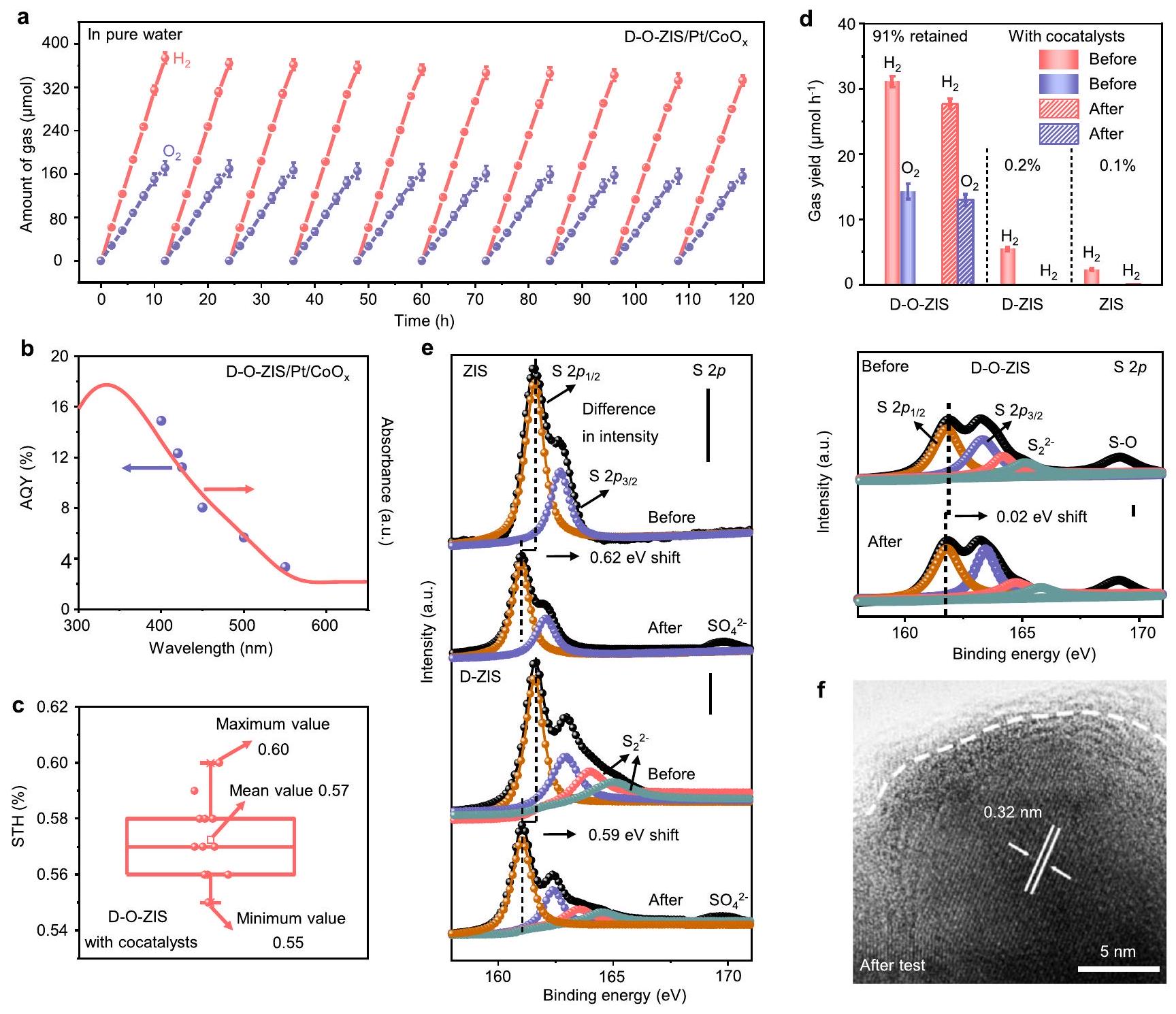
تم تقييم أداء العينات في التحفيز الضوئي لتفاعل انقسام الماء الكلي من الماء النقي باستخدام البلاتين وتستخدم كعوامل مساعدة. يمكن أن يعزز تحميل العوامل المساعدة بشكل كبير النشاط الضوئي التحفيزي لـ D-O-ZIS لتفكيك الماء من خلال بناء نطاق طاقة متطابق بين D-O-ZIS والعوامل المساعدة وتقليل الطاقة الحرة لامتصاص الهيدروجين والأكسجين (الأشكال التكميلية 7-9). عند التعرض للإشعاع الضوئي عند AM1.5مع Pt المحسن وجار التحميل، و تم إنتاجها بشكل مستمر على مدى D-O-ZIS، محققة كميات تطور تصل إلى 373.2 وخلال تفاعل 12 ساعة، على التوالي (الأشكال التكميلية 9، 10، الشكل 3أ). في الوقت نفسه، خلال عملية التحفيز الضوئي، و أقل عرضة للامتزاز على، مصحوبًا بـالانخفاض الملحوظ بعدتفاعل الظلام (الأشكال التكميلية 11-13)لا يمكن لـ ZIS و D-ZIS تقسيم الماء إلى و يحدث تحميل المساعد المساعد وتخفيف النشاط في كل دورة (الشكل التكميلي 14). تم التحقيق في العائد الكمي الظاهر (AQY) لـ D-O-ZIS لعملية التحليل الكلي للماء (الشكل التكميلي 15، الجدول التكميلي 4)، وتم حسابه ليكون عند 400 نانومتر (الشكل 3ب، الجدول التكميلي 5)، أعلى من ZIS (1.40%) أو D-ZIS (3.11%) (الشكل التكميلي 16، الجداول التكملية 6، 7). تم قياس كفاءة تحويل الطاقة الشمسية إلى هيدروجين (STH) عند AM1.5 G (إشعاع ضوء الشمس المحاكى بقيمة متوسطة تبلغ 0.57% (الشكل 3c، الجدول التكميلي 8)، والذي يتفوق على معظم المحفزات الضوئية الفردية التي تم الإبلاغ عنها مؤخرًا (الشكل التكميلي 17، الجدول التكميلي 9) ومحفزات ضوئية مركبة قائمة على ZIS (الشكل التكميلي 18، الجدول التكميلي 10).
قمنا بمزيد من التحقيق في أداء التحفيز الضوئي لـ D-O-ZIS المفرد دون إضافة البلاتين والمحفزات المساعدة (الشكل التوضيحي التكميلي 19، الجدول التكميلي 11). لا يزال النظام
الشكل 2 | الهياكل الهندسية والإلكترونية المحلية للمحفزات الضوئية. أ طيف XPS للكبريتفي ZIS وD-ZIS وD-O-ZIS؛طيف ESR لـ ZIS و D-ZIS و D-O-ZIS؛ ج منحنيات تحويل فورييه لـ-مرجح-طيف EXAFS لزنك إنديوم الكبريتيد (ZIS) وزنك إنديوم الكبريتيد المزدوج (DZIS) وزنك إنديوم الكبريتيد المشتق (D-O-ZIS)؛ d Zn-طيف EXAFS لزيس، D-ZIS، وD-O-ZIS في الفضاء؛ e العادي S -طيف XANES لـ ZIS و D-ZIS و D-O-ZIS. مدمج هو تكبير-حافة ما قبل الحافة؛مُعَايَر-طيف XANES الحافة لـ D-OZIS؛العملية التخطيطية لتحويل الهيكل المحلي من D-O-ZIS لتشكيل التشوه وتطعيم موقع الكاتيون بالأكسجين؛ في الوقت نفسه، تم تصوير أطوال الروابط المقابلة على الهياكل. تظهر الهياكل من منظور علوي. تم توفير بيانات المصدر كملف بيانات مصدر.
تم إنتاج و التطور، بقيم 76.8 و ، على التوالي. كانت كفاءة STH الناتجة 0.12% (الجدول التكميلي 12) وهي الأعلى من بين المحفزات الضوئية التي تم التحقيق فيها دون تحميل أي محفزات مساعدة (الشكل التكميلي 17). بالمقابل، أنتجت ZIS و D-ZIS فقط وأظهرت انخفاضًا في النشاط التحفيزي في كل دورة (الشكل التكميلي 20). تم تأكيد النشاط العام لتفكيك الماء لـ D-O-ZIS من خلال إجراء أو تفاعلات نصفية. أظهر D-OZIS أو تطور خلال التفاعلات النصفية، بينما لم يتم الكشف عن أي على ZIS و D-ZIS (الشكل التكميلي 21). قياس نظائر الأكسجين لـ D-O-ZIS أكد أن الناتج كان بسبب تفكيك الماء (الشكل التكميلي 22).
قمنا أيضًا بتقييم اختبار استقرار التحفيز الضوئي لـ ZIS و D-ZIS و D-O-ZIS بعد تفاعل لمدة 120 ساعة. أظهرت النتائج أن D-OZIS احتفظت بـ من معدل تطور الغاز التحفيزي الضوئي الأصلي،
مما يدل على استقرار الأداء العام لتفكيك الماء (الشكل 3d)، بينما تدهورت ZIS و D-ZIS تقريبًا إلى الصفر. تم تقييم درجة تآكل التحفيز الضوئي للمحفزات الناتجة عن تسرب الكبريت بعد التفاعل التحفيزي الضوئي بواسطة S XPS (الشكل 3e). أظهرت S طيف D-O-ZIS أقل تحول في طاقة الربط بمقدار 0.02 eV مع الحفاظ على شدة الاختبار السابقة مقارنة بـ ZIS (تحول 0.62 eV) و D-ZIS (تحول 0.59 eV) مع انخفاض الشدات ومنتج الأكسدة لـ ، مما يدل على أن تسرب الكبريت في D-O-ZIS تم قمعه بشكل كبير . صورة HRTEM والتحليل الهيكلي لـ D-O-ZIS بعد الاختبار كشفت عن ميزات تشوه مستقرة (الشكل 3f، الشكل التكميلي 23)، مع الحفاظ على سمك القشرة بحوالي ، بينما أظهرت ZIS و D-ZIS خصائص تسرب الكبريت (الشكل التكميلي 24). بالإضافة إلى ذلك، وجدنا أن -edge و -edge XANES لـ D-O-ZIS أظهرت تغييرات ضئيلة في القمم المميزة بعد الاختبار (الشكل التكميلي 25)، مما يدل على استقراره الهيكلي لـ
الشكل 3 | الأداء العام لتفكيك الماء التحفيزي الضوئي. أ وقت اعتماد الأداء التحفيزي الضوئي العام لتفكيك الماء على D-O-ZIS في الماء النقي تحت إضاءة AM1.5 القياسية ( )، تم استخدام Pt و كمحفزات مساعدة، نسبة Pt إلى 1:4، كانت كتلة المحفز 35 ملغ وتم تقييم النشاط التحفيزي الضوئي عبر إجمالي إنتاج الهيدروجين والأكسجين لدورة، مدة كل دورة 12 ساعة. تمثل أشرطة الخطأ الانحرافات المعيارية من النتائج الإحصائية لثلاث مجموعات من التجارب؛ الاعتماد على الطول الموجي لـ AQY خلال تفكيك الماء التحفيزي الضوئي العام بناءً على D-O-ZIS. AQY تشير إلى العائد الكمي الظاهر الذي تم حسابه باستخدام المعادلات (2) و (3) في المعلومات التكميلية والتفاصيل الموضحة في الجدول التكميلي 5، تم استخدام Pt و كمحفزات مساعدة، نسبة Pt إلى من ; كفاءة STH لـ D-O-ZIS مع تحميل المحفزات المساعدة ( ) لتفكيك الماء التحفيزي الضوئي العام. تم تقييم قيمة STH 12
مرة مع عينات منفصلة كما هو موضح في الجدول التكميلي 8 وتم حسابها باستخدام المعادلة (1) في النص الرئيسي. تمثل الخط المركزي الوسيط، وتمثل الصندوق العلوي والسفلي الربع العلوي والسفلي، على التوالي، يمثل المستطيل الصغير القيمة المتوسطة والقيم القصوى/الدنيا موضحة بواسطة الأشرطة العلوية/السفلية؛ إنتاج الغاز التحفيزي الضوئي لـ ZIS و D-ZIS و D-O-ZIS قبل وبعد اختبار تفكيك الماء التحفيزي الضوئي العام في الماء النقي. تمثل أشرطة الخطأ الانحرافات المعيارية من النتائج الإحصائية لثلاث مجموعات من التجارب؛ e طيف S XPS لـ ZIS و D-ZIS و D-O-ZIS قبل وبعد اختبار التحفيز الضوئي لمدة 120 ساعة. تشير الأشرطة الرأسية إلى الفرق في الشدة قبل وبعد الاختبار؛ صورة HRTEM لـ D-O-ZIS بعد اختبار التحفيز الضوئي لمدة 120 ساعة. تم توفير بيانات المصدر كملف بيانات مصدر.
تفاعلات التحفيز الضوئي (مناقشة إضافية لآلية الاستقرار في مبدأ العمل التحفيزي الضوئي لقسم التفعيل والاستقرار). يمكن أن تنشط هذه الاستراتيجية التصميمية خصائص الأكسجين الخاملة لمحفزات الكبريتيد المعدنية الأخرى، بما في ذلك و . هذا يوضح القابلية العالمية لمركبات الكبريتيد المعدنية في تفكيك الماء العام من منظور اختلاف الكهربية (الشكل التكميلي 26).
ديناميات نقل الشحنة والفصل
تم التحقيق في الخصائص البصرية والكهربائية للعينات لتحديد آلية النشاط التحفيزي الضوئي المحسن. أظهر طيف امتصاص UV-vis لـ D-O-ZIS امتصاصًا بصريًا مكثفًا
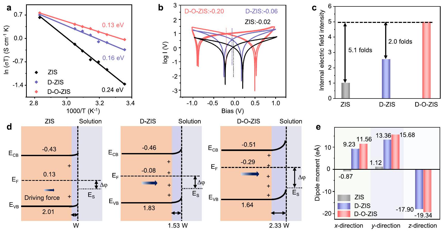
في منطقة الطيف المرئي وتحولًا أحمر مقارنة بـ ZIS (الشكل التكميلي 27). من خلال إدخال التشوه وتطعيم الأكسجين في ZIS، أظهر D-O-ZIS أقل طاقة تنشيط لنقل الحاملات بمقدار 0.13 eV (الشكل 4a)، وهو ما يعد ملائمًا لنقل الشحنة وتم حسابه باستخدام معادلة أرهينيوس (الشكل التكميلي 28) . لتقييم ديناميات فصل الحاملات، تم تحليل شدة المجال الكهربائي الداخلي للمحفزات الضوئية من خلال التحول المحتمل من 0 فولت إلى جهد تقاطع الانحياز ( ) بناءً على اختبار الموصلية . أظهر D-O-ZIS أقوى مجال كهربائي داخلي مع أكبر تحول محتمل عند -0.20 فولت (الشكل 4b، الشكل التكميلي 29). تم تقدير شدة المجال الكهربائي الداخلي بشكل أكبر باستخدام قياسات ضوئية عابرة وكانت شدة D-O-ZIS 5.1 و
الشكل 4 | ديناميات نقل الشحنة والفصل. أ طاقة تنشيط نقل الحاملات لـ ZIS و D-ZIS و D-O-ZIS (مشتقة من مخططات طيف الامتصاص الكهربائي في الشكل التكميلي 28)؛ ب الموصلية الإلكترونية المقاسة عبر الفولتمتر الدوري بمعدل مسح . تمثل الخط المنقط الأسود لـ ZIS. تمثل الخط المنقط الأزرق لـ D-ZIS، وتمثل الخط المنقط الأحمر لـ D-O-ZIS. تم اشتقاق المخططات من الشكل التكميلي 29؛ ج شدة المجال الكهربائي الداخلي لـ ZIS و D-ZIS و D-O-ZIS (وفقًا للشكل التكميلي 30، مع افتراض أن شدة ZIS هي ” 1 “)؛ مخطط الهياكل النطاقية التفصيلية، انحناء النطاق، ومنطقة الشحنة السطحية لـ ZIS و D-ZIS و D-O-ZIS. طاقة نطاق التوصيل: ( )، نطاق التكافؤ ( ) ومستوى فيرمي ( )
موضحة في الرسم البياني، الجهد مقابل القطب الهيدروجيني العادي (NHE). يوجد حد أدنى من درجة انحناء النطاق وقوة الدفع على سطح ZIS بسبب الروابط المعلقة مع ذرات الكبريت غير المشبعة على سطحه. يمكن اكتشاف انحناء خفيف في D-ZIS. يتمتع D-O-ZIS بانحناء نطاق قوي بشكل ملحوظ. الفرق بين المحفز الضوئي والمحلول: ، مستوى فيرمي لمحلول الماء ( ). تم الحصول على حساب نطاق الطاقة التفصيلية للمحفز الضوئي كما هو موضح في الشكل التكميلي 7. تم الحصول على حساب عرض منطقة الشحنة السطحية من الشكل التكميلي 7b والتفاصيل موضحة في المعلومات التكميلية. e تم حساب لحظات ثنائي القطب لـ ZIS و D-ZIS و D-O-ZIS على طول ثلاثة اتجاهات هيكلية مختلفة. تم توفير بيانات المصدر كملف بيانات مصدر.
أقوى بمقدار 2.0 مرة من ZIS و D-ZIS، على التوالي (الشكل 4c، الشكل التكميلي 30) . بالإضافة إلى ذلك، أظهر D-O-ZIS أكبر قدرة على فصل الشحنة مع متوسط عمر حامل مطول قدره 42.71 نانوثانية وكفاءة فصل الشحنة بـ ، المتأثرة بالمجال الكهربائي الداخلي (الشكل التكميلي 31) .
قمنا بحساب نطاق الطاقة خلال عملية التحفيز الضوئي لتوضيح شدة المجال الكهربائي الداخلي المحسن وديناميات نقل الشحنة والفصل. تم تحديد هياكل نطاق الطاقة من خلال طيف الانعكاس المنتشر UV-vis وطيف الإلكترون الضوئي فوق البنفسجي (UPS) (الشكل 4d) . تم العثور على مستوى فيرمي ( ) للمحفز الضوئي يرتفع مع زيادة حالات التشوه وتطعيم الأكسجين. خلال عملية التحفيز الضوئي، يكون المحفز الضوئي والمحلول مع إمكانيات كهربائية مختلفة في اتصال ( : المحفز الضوئي؛ لمحلول الماء)، مما يخلق منطقة شحنة سطحية عند الواجهات قمنا بتقدير عرض منطقة الشحنة الساكنة ( ) باستخدام مخطط موت-شوتكي من الشكل التوضيحي الإضافي 7b ووجدنا أن D-ZIS و D-O-ZIS أظهرا عرضًا أوسع يبلغ 1.53 وعندما تم تعيين عرض منطقة الاستنفاد لـ ZIS كـيُنسب اتساع منطقة الشحنة الفضائية المعززة إلى الزيادةفرق )، وهو ما يتماشى مع اتجاهات المجال الكهربائي الداخلي. إن المناطق الأوسع لشحن الفضاء بين واجهات المحفز الضوئي والمحلول قللت من مسافة انزلاق الثقوب على D-O-ZIS، مما وفر قوة دافعة قوية لفصل الشحنات .
تم التحقق من التغيرات في الجهود المتوسطة وشدة المجال الكهربائي الداخلي بشكل إضافي بواسطة DFT.ارتفعت في D-O-ZIS بسبب زيادة حالات التشويه وتطعيم الأكسجين (الشكل التكميلي 32)، وهو ما يتماشى مع النتائج التجريبية. عكس فرق الجهد الكهروستاتيكي شدة المجال الكهربائي الداخلي، الذي زاد بشكل متناسب معالفرق بين واجهات المحفز الضوئي والمحلول (الشكل التكميلي 33). أظهر التحليل الإضافي لعزم ثنائي القطب أن عزم ثنائي القطب تغير بشكل ملحوظ على طول، و التوجيهات في D-O-ZIS، مما يؤدي إلى إنشاء ثنائي قطب هيكلي وإضافة عدم تماثل إلى الهيكل المحلي، وبالتالي زيادة المجال الكهربائي الداخلي (الشكل 4e). أكدت هذه النتائج أن حالات التشويه المتزايدة وأدى الت doping في D-O-ZIS إلى تعزيز المجال الكهربائي الداخلي، مما وفر قوة دافعة قوية لفصل الشحنات.
مبدأ العمل الضوئي التحفيزي للتفعيل والاستقرار
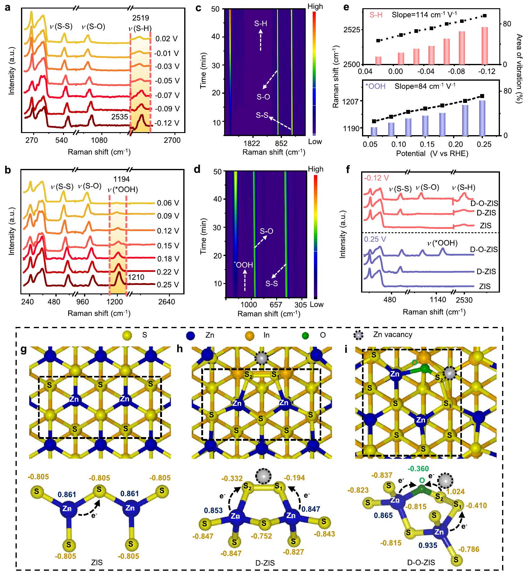
تم استخدام مطيافية رامان في الموقع لمراقبة التغيرات التي تحدث على سطح D-O-ZIS. كانت إشارة رامان لامتصاص S-H عندتمت ملاحظته على النطاق المحتمل من 0.02 إلى -0.12 فولت مقابل القطب الهيدروجيني القابل للعكس (RHE) لتتبع عملية تطور الهيدروجين (الشكل 5أ)ظهر ذروة S-H عند 0.02 فولت وأصبحت أقوى مع زيادة الجهد. الذروة عنديتوافق مع امتصاص *OOH على D-O-ZIS خلال تطور الأكسجين كما هو موضح في الشكل.، الذي تزايد تدريجياً على مدى نطاق الجهد الأنودي من 0.06 إلى 0.25 فولت مقابل RHE. التحولات الحمراء لـ (من ) و OOH (من نُسِبَت إلى ظاهرة الضبط الحاد الملحوظة وامتصاص الأنواع السطحية، بما في ذلك، و نوعأظهرت الرسوم البيانية ثنائية الأبعاد أيضًا أن و تزايدت الاهتزازات على D-O-ZIS مع مرور الوقت (الشكل 5c، d). تم تحديد معدلات التحول لـ S-H و الاهتزازات في D-O-ZIS هي و ، على التوالي (الشكل 5e). إشارات رامان ( و تم الكشف عن ( ) عند إمكانات -0.12 فولت و0.25 فولت، على التوالي، لـ ZIS وD-ZIS وD-O-ZIS (الشكل 5f). كانت إشارة S-H أقوى لـ D-O-ZIS مقارنة بـ ZIS أو D-ZIS، وتم الكشف عن إشارة *OOH فقط لـ D-O-ZIS.
الشكل 5 | عملية تطور الهيدروجين والأكسجين الضوئي المحفز التي تم التحقيق فيها بواسطة طيف رامان في الموقع. أ طيف رامان في الموقع لعملية تطور الهيدروجين الضوئي المحفز على D-O-ZIS. سلسلة من أطياف رامان عند إمكانيات مختلفة ( مقابل RHE) تظهر التغير الديناميكي لعملية تطور الهيدروجين؛ طيف رامان في الموقع لعملية تطور الأكسجين الضوئي الحفاز على D -O-ZIS. سلسلة من أطياف رامان عند إمكانيات مختلفة ( مقابل RHE) تظهر التغير الديناميكي لعملية تطور الأكسجين؛ ج خرائط الكنتور ثنائية الأبعاد للاهتزازات الرامان لعملية تطور الهيدروجين؛ خرائط الكنتور ثنائية الأبعاد للاهتزازات الرامان لعملية تطور الأكسجين؛ تحولات رامان ونسب المساحات لـ S-H السندات و الاهتزازات مقابل الإمكانيات على سطح D-O-ZIS؛ فرق شدة إشارات رامان لاهتزازات S-H واهتزازات OOH مقابل الجهود المتطابقة -0.12 فولت و0.25 فولت، على التوالي، للعينات خلال عمليات تطور الهيدروجين وتطور الأكسجين. الشحنة على الذرات من حساب شحنة بادير لـهيكل ZIS،هيكل D-ZIS، وهيكل D-O-ZIS. القيمة السلبية تشير إلى الحصول على الإلكترونات، بينما القيمة الإيجابية تعني فقدان الإلكترونات. الهياكل موضحة من منظور علوي. الجزء السفلي هو عرض جزئي للهيكل المقابل. تم توفير بيانات المصدر كملف بيانات مصدر.
في هذه الأثناء، O 1طيف XPS بعد اختبارات التحفيز الضوئي أظهر إشارة منفي D-O-ZIS (الشكل التوضيحي التكميلي 34)، مما يؤكد المزيد من نشاط تفاعلات انقسام الماء بشكل عام في D-O-ZIS.
تم إجراء حسابات DFT للحصول على فهم لتأثيرات حالات التشوه وتطعيم الأكسجين في مواقع الكاتيون. كشفت الشحنة المحسوبة باستخدام طريقة بادر عن إعادة توزيع قوية للشحنة في مناطق حالات التشوه وتطعيم الأكسجين. في هيكل ZIS، حدث انتقال للشحنة من ذرة الزنك ( ) إلى ذرة الكبريت ( ) (الشكل 5g). في هيكل D-ZIS، الشحنات على و تم إعادة توزيع الذرات بشكل كبيرلـ و لـ ) في الرابطة ثنائية القطب لـ المركز. تم نقل الإلكترونات من ذرات الزنك القريبة ( ) إلى الـ و المواقع في D-ZIS، وكانت فراغات Zn تعمل كفخ للإلكترونات (الشكل 5h)تسبب تشبع موقع الكاتيون بالـ O في D-O-ZIS في زيادة كبيرة في الكهربية السالبة، مما أدى إلى تغيير الشحنة على المركب المنسق.الموقع ليصبح أكثر إيجابية (1.024|e|)، حتى أعلى من الشحنة على ذرة الزنك ( ). الشحنة على الـ الموقع كان“، مما يشير إلى وجود فرق كبير في الكهربية السالبة بين المواقع المجاورة (الشكل 5i). تتماشى هذه النتائج مع نتائج XANES لذرة الأكسجين المنسقة كتركيب S-O (الشكل 2e). النقل الإضافي للشحنة (منتم تحسين حركة الشحنات من خلال تحويل الذرات إلى ذرات الأكسجين والإلكترونات المستخرجة عند فراغ الزنك، مما أدى إلى توليد مركز شحن أكثر إيجابية.الموقع، الذي قد يبدل المواقع النشطة في الهيكل المحلي.
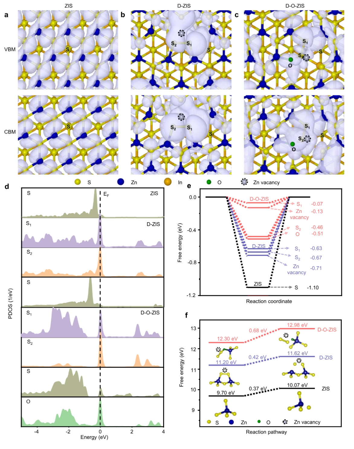
لفهم دور المراكز النشطة في العمليات التحفيزية، قمنا بدراسة توزيع كثافة الشحنة في الحد الأقصى لحزمة التكافؤ (VBM) والحد الأدنى لحزمة التوصيل (CBM).أظهرت ZIS كثافات شحن موزعة بشكل موحد في كل من VBM و CBM (الشكل 6a). ومع ذلك، كانت كثافات الشحن في VBM و CBM لـ D-ZIS محلية عندالمواقع، فراغ الزنك، وذرات الكبريت غير المشبعة (الشكل 6ب). في الوقت نفسه، كانت كثافة شحنة مستوى الطاقة الحدي التوصيلي لـ D-O-ZIS محلية عندالمواقع وخلل الزنك، بينما انخفضت كثافة الشحنة عند مستوى الفيرمي بشكل ملحوظ فيالمواقع. وهذا يشير إلى أن الإلكترونات يمكن أن تُثار بسهولة بواسطة الضوء إلى نطاق التوصيل، مما يؤدي إلى نقص الإلكترونات فيالموقع (الشكل 6ج). لذلك، فإن تراكم الثقوب الناتجة عن الضوء في المواقع والإلكترونات المحاصرة فيالمواقع والشحنات الناتجة عن فراغات الزنك التي كانت مفصولة مكانيًا داخل المواقع الذرية، مما يوفر قوة دافعة قوية لفصل الإلكترونات والثقوب بكفاءة على D-O-ZIS.
تم إجراء حساب كثافة الحالات الجزئية (PDOS) للتحقيق في هيكل حالات التشوه وتطعيم الأكسجين في D-O-ZIS. أظهرت PDOS لـ D-O-ZIS S-Oرنينات الترابط بالقرب من مستوى فيرمي )، مع هيمنة الحالات الإلكترونية الهجينة على الميزة (الشكل التكميلي 35) الحالات الإلكترونية لـو Oزاد عندوكان ذرة الكبريت المحيطة أيضًا مفعلة مقارنةً بـ D-ZIS و ZIS فيتكوين D-O-ZIS (الشكل 6d). أظهر D-O-ZIS درجة عالية من التداخل بالنسبة لل intermediates، و * OOH ) الممتصة على -فرقة، تشير إلى تفاعلات قوية خلال عملية إنتاج الأكسجين (الشكل التكميلي 36)الطاقة الحرة لامتصاص الهيدروجين المثلىكان -0.07 إلكترون فولت عندالموقع و -0.13 إلكترون فولت عند فراغ الزنك، مما يشير إلى أنالموقع أكثر ملاءمة لامتصاص/إطلاق الهيدروجين من فراغ الزنك في D-O-ZIS، بينما أظهر D-ZIS مستوى مثالي.من -0.63 إلكترون فولت عندالموقع و ZIS بقيمة -1.10 eV في موقع S (الشكل 6e)، ونماذج الامتصاص الخاصة بهم موضحة في الشكل التكميلية 37-39. حاجز الطاقة الحرة لـالامتزاز عندكان الموقع في D -O-ZIS منخفضًا (0.31 eV) مقارنةً بحواجز الطاقة في ZIS (1.27 eV) وD-ZIS (0.97 eV)، مما يعزز تفاعل تطور الأكسجين (الشكل التكميلي 40). علاوة على ذلك،تم التحقيق في نشاط تطور الذرات المعدنية (أي مواقع الزنك) في D-O-ZIS، مع استبعاد دور مواقع المعادن كمواقع لإنتاج الأكسجين (حاجز الطاقة 0.85 eV) في تصميم هذا المحفز الضوئي (الشكل التكميلي 41). من خلال دراسة الهياكل بمستويات مختلفة من فراغات الزنك وتطعيم الأكسجين (الشكل التكميلي 42)، تم تحسين D-O-ZIS بشكل كبير من امتصاص الوسائط الرئيسية لتطور و (الشكل التكميلي 43). وبالتالي، أظهر D-O-ZIS الأنواع الممتصة من الهيدروجين والأكسجين
في و على التوالي، مما يعزز تفاعل الانقسام الكلي للماء.
قمنا أيضًا بإجراء قياس الجهد الكهربائي الحر وتحرير الطاقة لأكسدة أيونات الكبريت بواسطة الثقوب الناتجة عن الضوء لتوضيح آلية الاستقرار. كانت أيونات الكبريت في ZIS أو D-ZIS سهلة الأكسدة والتفريغ بواسطة الثقوب الناتجة عن الضوء بسبب الجهد الكهربائي بينما كانت أيونات الكبريت في لها جهد أكسدة أقل من لـ D-O-ZIS مع قوة أكسدة ضعيفة (الشكل التكميلي 44). كان حاجز الطاقة لأكسدة أيونات الكبريت في التكوين 0.68 eV لـ D-O-ZIS، أعلى من ذلك في D-ZIS (0.42 eV) و ZIS (0.37 eV)، مما يشير إلى أن أيونات الكبريت في التكوين كانت صعبة الأكسدة بواسطة الثقوب الناتجة عن الضوء (الشكل 6f). بالإضافة إلى ذلك، كان حاجز الطاقة للأكسدة لأيونات الكبريت لـ D-O-ZIS (0.68 eV) أعلى من الطاقة الحرة لإنتاج الأكسجين البالغة 0.31 eV (الشكل التكميلي 40)، مما يشير إلى أن D-O-ZIS تفضل إنتاج الأكسجين خلال تفاعلات الانقسام المائي بدلاً من أن تتأكسد بواسطة الثقوب الناتجة عن الضوء.
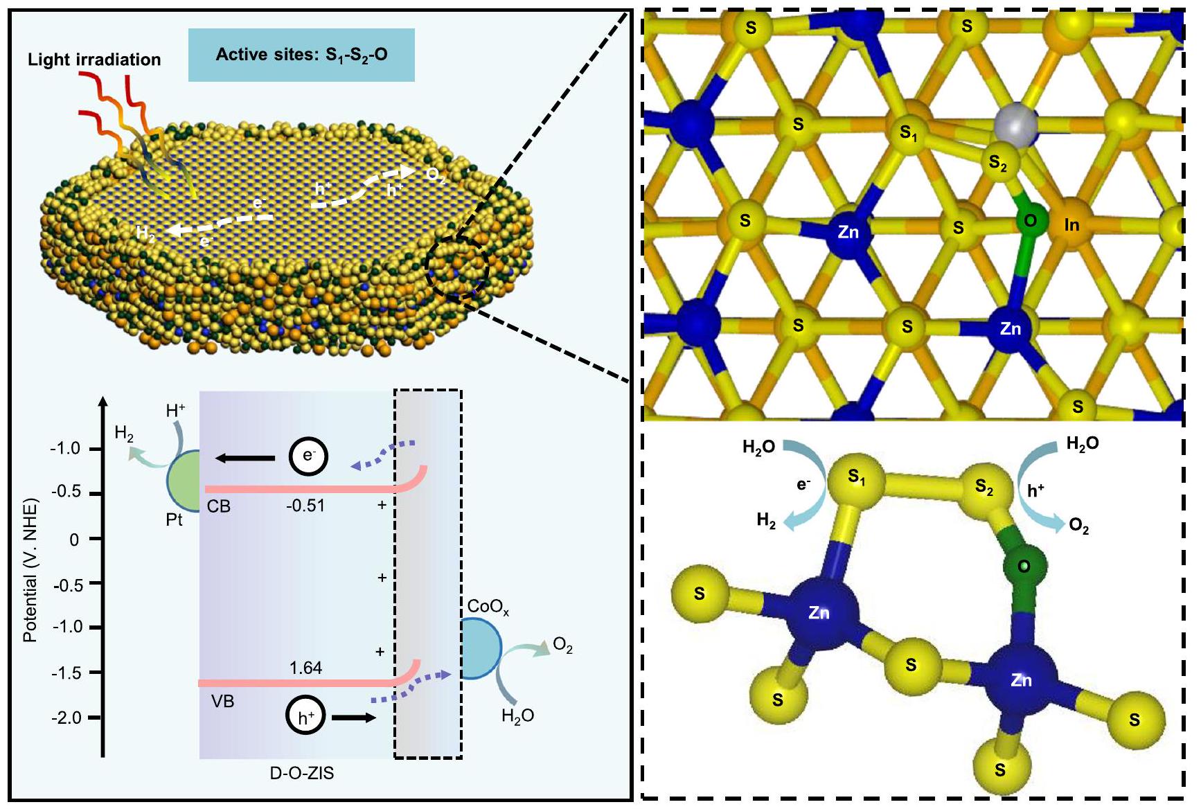
اقترحنا مبدأ العمل للانقسام الكلي للماء على محفز D-OZIS الضوئي (الشكل 7)، وتم توضيح الآليات الحفازة لـ ZIS و D-ZIS في الشكل التكميلي 45. أولاً، يمتص D-O-ZIS الفوتونات الساقطة لإنتاج حوامل الشحن الناتجة عن الضوء. يتم فصل أزواج الإلكترون والثقب الناتجة عن الضوء بكفاءة مدفوعة بواسطة المجال الكهربائي الداخلي بسبب ثنائي القطب القوي لهيكل تطعيم الأكسجين في مواقع الكاتيون الناتجة عن التشوه، ثم يتم نقلها إلى مواقع النشطة للخضوع لتفاعل أكسدة-اختزال. بسبب تحسين نطاقات الطاقة، فإن CB لـ D-O-ZIS سالب بما فيه الكفاية (-0.51 eV) لإنتاج بينما VB إيجابي بما فيه الكفاية (1.64 eV) لإنتاج . إن خاصية إعادة توزيع الشحن القوية والفروق الكبيرة في الكهربية بين مواقع الذرات و تنشط تفاعلات الأكسجين المستقرة في مواقع وتتجنب المشكلة الشائعة لعدم استقرار الكبريت في تحفيز الكبريتيد المعدني، بينما تفضل مواقع امتصاص/إطلاق الهيدروجين. تعزز المحفزات المساعدة من Pt و النشاط الضوئي لـ D-O-ZIS من خلال تعزيز فصل الشحن وتقليل الطاقة الحرة لامتصاص الهيدروجين والأكسجين. وبالتالي، يحقق D-O-ZIS كمحفز ضوئي فردي انقسامًا كليًا فعالًا للماء مع استقرار عالٍ.
نقاش
تقترح هذه الدراسة استراتيجية فرق الكهربية لتنشيط واستقرار ZIS من أجل الانقسام الكلي للماء الضوئي، محققة كفاءة تحويل ملحوظة من إلى الهيدروجين مع استقرار عالٍ. ينتج تطعيم الأكسجين في مواقع الكاتيون الناتجة عن التشوه في مواقع ذرات الزنك في D-O-ZIS فروقًا كبيرة في الكهربية بين مواقع الذرات المجاورة، حيث تكون مواقع غنية بالإلكترونات ومواقع ناقصة الإلكترونات في الهيكل المحلي . تنشط خاصية إعادة توزيع الشحن القوية تفاعلات الأكسجين المستقرة في مواقع وامتصاص/إطلاق الهيدروجين في مواقع . تعرض دراستنا القابلية العالمية لتنشيط واستقرار محفزات الكبريتيد المعدني، مثل ZIS، و ، من أجل الانقسام الكلي الفعال للماء الضوئي من خلال استراتيجية تطعيم الأكسجين في مواقع الكاتيون الناتجة عن التشوه من منظور فروق الكهربية.
طرق
تخليق المحفزات الضوئية
تخليق (ZIS). يتضمن تخليق ZIS عادةً الإجراء التالي: من من و 4 مليمول من ثيوأسيتاميد تم إذابته في 35 مل من الماء المنزوع الأيونات وتم تحريكه بقوة لمدة 30 دقيقة. ثم تم نقل المحلول المختلط إلى وعاء أوتوكلاف مبطن بتفلون سعة 50 مل وتم تسخينه عند لمدة 12 ساعة. بعد التبريد إلى درجة حرارة الغرفة، تم جمع التعليق الأصفر الناتج وغسله بالإيثانول والماء المنزوع الأيونات أربع مرات على التوالي. أخيرًا، تم تجفيف المنتج عند طوال الليل قبل الاستخدام الإضافي.
الشكل 6 | مبدأ العمل للانقسام الكلي للماء الضوئي المحدد بواسطة DFT. توزيع كثافة الشحن الجزئي بالقرب من حافة نطاق التوصيل ونطاق التكافؤ لـ ZIS، D-ZIS، و D-O-ZIS. قيمة السطح المعزول هي . تظهر الهياكل من منظور علوي. د PDOS لذرات مختلفة من ، ، المحيطة بـ S العادية و O في D-O-ZIS، و و في D-ZIS، و
D-ZIS
في
D-O-ZIS
ف
ذرة S في ZIS؛ مستوى فيرمي (); هـ القيم المحسوبة لـ في مواقع مختلفة في ZIS و D-ZIS و D-O-ZIS؛ ف الطاقة الحرة لأكسدة أيونات الكبريت في ZIS و D-ZIS و D-O-ZIS. التكوينات المدرجة هي ذرة S التي تتسرب بواسطة أكسدة الثقوب الناتجة عن الضوء من الهيكل النقي. تم توفير بيانات المصدر كملف بيانات مصدر.
الشكل 7 | مبدأ العمل للانقسام الكلي للماء الضوئي. الهيكل المحلي موضح من منظور علوي، والأسفل هو عرض جزئي للهيكل المقابل. نطاق التوصيل (CB)، نطاق التكافؤ (VB). تم توفير بيانات المصدر كملف بيانات مصدر.
تخليق ZIS المشوه (D-ZIS). تم تخليق D-ZIS بدءًا من ZIS، باستخدام استراتيجية هجرة حرارية. تضمنت عملية التخليق تسخين المادة السابقة لـ ZIS عند لمدة 30 دقيقة تحت جو من (1 بار). بمجرد اكتمال التفاعل، تم السماح للمنتج المسحوق الناتج بالتبريد بشكل طبيعي إلى درجة حرارة الغرفة، بعد ذلك تم جمعه وغسله عدة مرات باستخدام الإيثانول والماء المنزوع الأيونات. ثم تم تجفيف المنتج عند تحت الفراغ طوال الليل لضمان إزالة كاملة لأي مذيب متبقي قبل التحليل الإضافي.
تخليق تطعيم الأكسجين في مواقع الكاتيون الناتجة عن التشوه لـ ZIS (D-O-ZIS). لتخليق D-O-ZIS، تم معالجة D-ZIS الناتج بتدفق من عند لمدة 10 دقائق. ثم تم جمع المسحوق الناتج وغسله ثلاث مرات بالإيثانول والماء المنزوع الأيونات، على التوالي، قبل أن يتم تجفيفه تحت الفراغ عند .
اختبار تفاعل الانقسام الكلي للماء الضوئي
لإجراء التفاعلات الضوئية، استخدمنا وعاء تفاعل مع نظام دوران مغلق للغاز وإخلاء. قبل كل تفاعل، قمنا بتوزيع 35 ملغ من المحفزات الضوئية في 50 مل من الماء النقي وأخلينا الهواء من الوعاء، واستبدلناه بغاز الأرجون. ثم أجرينا تطور و في مفاعل كوارتز باستخدام مصباح Xe بقوة 300 واط للإشعاع. تم ضخ الغازات المتطورة واكتشافها بواسطة جهاز كروماتوغرافيا الغاز Shimadzu GC-2014c مع كاشف موصلية حرارية. قمنا بقياس كفاءة STH تحت ضوء الشمس المحاكي عند إضاءة AM1.5 G (). تم تحديد كفاءة STH وفقًا للمعادلة التالية (1):
هنا، و تشير إلى معدل تطور ، طاقة جيبس التفاعل خلال تفاعل الانقسام المائي، تدفق الطاقة الضوئية تحت إشعاع AM1.5 G، ومساحة العينة المعرضة للإشعاع، على التوالي. القيمة المستخدمة في الحسابات هي للماء السائل في نظام التفاعل. قيمة هي . قيمة هي .
توفر البيانات
جميع البيانات التي تم توليدها في هذه الدراسة متاحة في المقالة والمعلومات التكميلية، والبيانات الخام التي تم توليدها في هذه الدراسة متاحة في ملف بيانات المصدر. تم توفير بيانات المصدر مع هذه الورقة.
References
Zhou, P. et al. Solar-to-hydrogen efficiency of more than in photocatalytic water splitting. Nature 613, 66-70 (2023).
Zhang, Y. et al. Internal quantum efficiency higher than achieved by combining doping and quantum effects for photocatalytic overall water splitting. Nat. Energy https://doi.org/10. 1038/s41560-023-01242-7.
Zhao, D. et al. Boron-doped nitrogen-deficient carbon nitride-based Z-scheme heterostructures for photocatalytic overall water splitting. Nat. Energy 6, 388-397 (2021).
Teng, Z. et al. Atomically dispersed antimony on carbon nitride for the artificial photosynthesis of hydrogen peroxide. Nat. Catal. 4, 374-384 (2021).
Cheng, H. et al. Rational design of covalent heptazine frameworks with spatially separated redox centers for high-efficiency photocatalytic hydrogen peroxide production. Adv. Mater. 34, 2107480-2107489 (2022).
Shi, . et al. Protruding Pt single-sites on hexagonal to accelerate photocatalytic hydrogen evolution. Nat. Commun. 13, 1287-1297 (2022).
Li, C. et al. Constructing direct Z-scheme heterostructure by enwrapping on CdS hollow cube for efficient photocatalytic generation. Adv. Sci. 7, 2201773-2201783 (2022).
Wang, X. et al. Interfacial chemical bond and internal electric field modulated Z-scheme – MoSe photocatalyst for efficient hydrogen evolution. Nat. Commun. 12, 4112-4123 (2021).
Qiu, B., Cai, L., Zhang, N., Tao, X. & Chai, Y. A ternary dumbbell structure with spatially separated catalytic sites for photocatalytic overall water splitting. Adv. Sci. 7, 1903568-1903575 (2020).
Wang, Q. et al. Scalable water splitting on particulate photocatalyst sheets with a solar-to-hydrogen energy conversion efficiency exceeding 1%. Nat. Mater. 15, 611-617 (2016).
Liu, J. et al. Metal-free efficient photocatalyst for stable water splitting via a two-electron pathway. Science 347, 6225-6230 (2015).
Wang, Z. et al. Overall water splitting by an nanorod single crystals grown on the edges of particles. Nat. Catal. 1, 756-763 (2018).
Wang, Q. et al. Oxysulfide photocatalyst for visible-light-driven overall water splitting. Nat. Mater. 18, 827-832 (2019).
Song, X. et al. Overall photocatalytic water splitting by an organolead iodide crystalline material. Nat. Catal. 3, 1027-1033 (2020).
Fu, J. et al. Interface engineering of thin film photoanode for highly efficient photoelectrochemical water splitting. Nat. Commun. 13, 729-738 (2022).
Wu, J. et al. Breaking through water-splitting bottlenecks over carbon nitride with fluorination. Nat. Commun. 13, 6999-7007 (2022).
Yang, Y. et al. Engineering -ketoamine covalent organic frameworks for photocatalytic overall water splitting. Nat. Commun. 14, 593-603 (2023).
Chen, K. et al. Overall water splitting by a -based photocatalyst decorated with an Ir-promoted Ru-based cocatalyst. J. Am. Chem. Soc. 145, 3839-3843 (2023).
Wang, M. et al. A hydrogen-deficient nickel-cobalt double hydroxide for photocatalytic overall water splitting. Angew. Chem. Int. Ed. 59, 11510-11515 (2020).
Lang, R. et al. Hydroformylation of olefins by a rhodium single atom catalyst with activity comparable to . Angew. Chem. Int. Ed. 55, 16054-16058 (2016).
Pan, R. et al. Two-dimensional all-in-one sulfide monolayers driving photocatalytic overall water splitting. Nano Lett. 21, 6228-6236 (2021).
Zhang, S. et al. Gradient hydrogen migration modulated with self adapting vacancy in copper doped nanosheet for photocatalytic hydrogen evolution. ACS Nano 15, 15238-15248 (2021).
Yang, W. et al. Enhanced photoexcited carrier separation in oxygendoped nanosheets for hydrogen evolution. Angew. Chem. Int. Ed. 55, 6716-6720 (2016).
Tian, Z. et al. Efficient photocatalytic hydrogen peroxide generation coupled with selective benzylamine oxidation over defective nanobelts. Nat. Commun. 12, 2039-2049 (2021).
Xu, W., Gao, W., Meng, L., Tian, W. & Li, L. Incorporation of sulfate anions and sulfur vacancies in photoanode for enhanced photoelectrochemical water splitting. Adv. Energy Mater. 11, 2101181-2101189 (2021).
He, Y. et al. 3D hierarchical nanosheets with rich Zn vacancies boosting photocatalytic reduction. Adv. Funct. Mater. 29, 1905153-1905163 (2019).
Meng, L. et al. Simultaneous manipulation of O-doping and metal vacancy in atomically thin nanosheet arrays toward improved photoelectrochemical performance. Angew. Chem. Int. Ed. 57, 16882-16887 (2018).
Chandrasekaran, S. et al. Recent advances in metal sulfides: from controlled fabrication to electrocatalytic, photocatalytic and photoelectrochemical water splitting and beyond. Chem. Soc. Rev. 48, 4178-4280 (2019).
Zheng, X. et al. Theory-driven design of high-valence metal sites for water oxidation confirmed using in situ soft X-ray absorption. Nat. Chem. 10, 149-154 (2018).
Dai, J. et al. Single-phase perovskite oxide with super-exchange induced atomic-scale synergistic active centers enables ultrafast hydrogen evolution. Nat. Commun. 11, 5657-5667 (2020).
Zhang, Q. et al. Electronically modified atomic sites within a multicomponent composite for efficient oxygen electroreduction. Adv. Energy Mater. 11, 2100303-2100314 (2021).
Guo, Q., Lau, K. C. & Pandey, R. A XANES study of lithium polysulfide solids: a first principles study. Mater. Adv. 2, 6403-6410 (2021).
Ochmann, M. et al. UV-photochemistry of the disulfide bond: evolution of early photoproducts from picosecond X-ray absorption spectroscopy at the sulfur K-edge. J. Am. Chem. Soc. 140, 6554-6561 (2018).
Meng, L. et al. Atomic layer deposition triggered Fe-In-S cluster and gradient energy band in ZnInS photoanode for improved oxygen evolution reaction. Nat. Commun. 12, 5247-5256 (2021).
Oishi, M. et al. Direct observation of reversible oxygen anion redox reaction in Li -rich manganese oxide, , studied by soft X-ray absorption spectroscopy. J. Mater. Chem. A 4, 9293-9302 (2016).
Lin, S. C. et al. Operando time-resolved X-ray absorption spectroscopy reveals the chemical nature enabling highly selective reduction. Nat. Commun. 11, 3525-3537 (2020).
Li, Z. et al. Blocking the reverse reactions of overall water splitting on a photocatalyst modified with . Nat. Catal. 6, 80-88 (2023).
Chen, S . et al. Surface modifications of to promote photocatalytic Z-Scheme overall water splitting. J. Am. Chem. Soc. 143, 10633-10641 (2021).
Hu, Y. et al. Lattice distortion induced internal electric field in photoelectrode for efficient charge separation and transfer. Nat. Commun. 11, 2129-2139 (2020).
Suyatin, D. B. et al. Strong Schottky barrier reduction at Au-catalyst/ GaAs nanowire interfaces by electric dipole formation and Fermilevel unpinning. Nat. Commun. 5, 3221-3229 (2020).
Han, T. et al. Anion-exchange-mediated internal electric field for boosting photogenerated carrier separation and utilization. Nat. Commun. 12, 4952-4963 (2021).
Xiao, X. et al. A promoted charge separation/transfer system from Cu single atoms and layers for efficient photocatalysis. Adv. Mater. 32, 2003082-2003090 (2020).
Jiang, W. et al. Photocatalyst for high-performance production: Ga-doped polymeric carbon nitride. Angew. Chem. Int. Ed. 60, 6124-6129 (2021).
Xin, X., Zhang, Y., Wang, R., Guo, P. & Li, X. Hydrovoltaic effectenhanced photocatalysis by polyacrylic acid/cobaltous oxidenitrogen doped carbon system for efficient photocatalytic water splitting. Nat. Commun. 14, 1759-1768 (2023).
Zhang, Z., Nagashima, H. & Tachikawa, T. Ultra-narrow depletion layers in a hematite mesocrystal based photoanode for boosting multihole water oxidation. Angew. Chem. Int. Ed. 59, 9047-9054 (2020).
Boettcher, S. W. et al. Potentially confusing: potentials in electrochemistry. ACS Energy Lett. 6, 261-266 (2021).
Dong, J. C. et al. In situ Raman spectroscopic evidence for oxygen reduction reaction intermediates at platinum single crystal surfaces. Nat. Energy 4, 60-67 (2019).
Lin, Y. et al. In situ identification and time resolved observation of the interfacial state and reactive intermediates on a cobalt oxide nanocatalysts for the oxygen evolution reaction. ACS Catal. 12, 5345-5355 (2022).
Wright, D. et al. Vibrational stark effects: ionic influence on local fields. J. Phys. Chem. Lett. 13, 4905-4911 (2022).
Zhou, Y. et al. Electronegativity-induced charge balancing to boost stability and activity of amorphous electrocatalysts. Adv. Mater. 34, 2100537-2100546 (2022).
الشكر والتقدير
تدعم هذه البحث مؤسسة العلوم الطبيعية الوطنية في الصين (22261142666، 52172237)، وصندوق العلوم في شينشيانغ للعلماء الشباب المتميزين (2022JC-21)، وصندوق البحث لمختبر الدولة الرئيسي لمعالجة التصلب (NPU)، الصين (رقم المنحة 2021-QZ-02)، وصندوق البحث الأساسي للجامعات المركزية (3102019JC005، D5000220033). تم منح جميع التمويلات لـ X.L.
مساهمات المؤلفين
X.X. و X.L. ابتكرا الفكرة. X.L. أشرف على المشروع. X.X. قام بعمليات التخليق، والتوصيف، والتصوير الضوئي. Y.L. و Y.Z. و Y. Wang قاموا بإجراء التجارب الكهروكيميائية الضوئية. X.C. و Y. Wei و C.D. و J.S. و R.W. و P.G. و J.Y. و J.Z. و A.J.S. و M.-M.T. قاموا بتحليل البيانات وعلقوا على المخطوطة.
المصالح المتنافسة
يعلن المؤلفون عدم وجود مصالح متنافسة.
معلومات إضافية
المعلومات التكميلية النسخة الإلكترونية تحتوي على
المواد التكميلية المتاحة على https://doi.org/10.1038/s41467-024-44725-1.
يجب توجيه المراسلات وطلبات المواد إلى شوانهوا لي.
معلومات مراجعة الأقران تشكر Nature Communications يورونغ يانغ والمراجعين الآخرين المجهولين على مساهمتهم في مراجعة الأقران لهذا العمل. يتوفر ملف مراجعة الأقران.
معلومات إعادة الطبع والتصاريح متاحة على http://www.nature.com/reprints
ملاحظة الناشر تظل Springer Nature محايدة فيما يتعلق بالمطالبات القضائية في الخرائط المنشورة والانتماءات المؤسسية.
Photocatalytic overall water splitting into hydrogen and oxygen is desirable for long-term renewable, sustainable and clean fuel production on earth. Metal sulfides are considered as ideal hydrogen-evolved photocatalysts, but their component homogeneity and typical sulfur instability cause an inert oxygen production, which remains a huge obstacle to overall water-splitting. Here, a distortion-evoked cation-site oxygen doping of (D-O-ZIS) creates significant electronegativity differences between adjacent atomic sites, with sites being electron-rich and sites being electron-deficient in the local structure of sites. The strong charge redistribution character activates stable oxygen reactions at sites and avoids the common issue of sulfur instability in metal sulfide photocatalysis, while sites favor the adsorption/ desorption of hydrogen. Consequently, an overall water-splitting reaction has been realized in D-O-ZIS with a remarkable solar-to-hydrogen conversion efficiency of , accompanying a retention rate after 120 h photocatalytic test. In this work, we inspire an universal design from electronegativity differences perspective to activate and stabilize metal sulfide photocatalysts for efficient overall water-splitting.
The utilization of sunlight and water, two of the most abundant natural resources on earth, for the production of hydrogen ( ) and oxygen ( ) at a stoichiometric ratio of , holds great potential for achieving carbon neutrality . Compared with the certain solar hydrogen production techniques, such as photo-electrochemical water splitting, photocatalytic overall water-splitting eliminates the need for external bias or circuitry, thereby reducing system costs and mitigating photocatalyst corrosion, stability, and safety concerns . Semiconductor-based photocatalytic overall water splitting is an ideal solar-to-chemical energy conversion route . Constructing a
hybrid photocatalyst can enhance light harvesting and facilitate charge separation . However, the long reaction paths and random distribution of active sites in hybrid systems limited its photocatalytic activity . Recently, single photocatalysts, such as , , TpBpy covalent organic frameworks, , and , etc. have been developed to achieve overall water splitting while avoiding the problems of constructing hybrid systems . However, the development of photocatalysts with high solar-to hydrogen (STH) efficiencies in solar hydrogen systems remains a fundamental challenge .
Metal sulfides are considered as promising photocatalysts due to their appropriate energy bands, designable structures, and excellent photoelectric properties . The representative metal sulfides such as , and have been widely used in the field of photocatalytic water splitting . Among them, (ZIS) is a typical ternary layered metal chalcogenide semiconductor, possessing suitable band gap of about 2.44 eV and conduction band potential of 0.43 eV versus Normal Hydrogen Electrode (NHE), which holds the visible-light absorption and strong reduction capacity for generation from water splitting . Many strategies have achieved enhanced production performance around ZIS photocatalyst, such as constructing a Z-scheme heterostructure of sulfur vacancies ZIS with other semiconductor , fabricating S vacancy induced with atomic Cu doping in ZIS nanosheets , incorporating anion-site oxygen doping into the sulfur atom sites of ZIS , and modulating cocatalyst of protruding single Pt atoms , but they have not been able to attain efficient and stable overall water-splitting reactions. One of the critical challenges faced in enhancing the efficiency for water splitting is the homogeneous composition of active sites and consistent electronic structure in single photocatalyst. Additionally, the sulfur atoms present in a ZIS photocatalyst are highly prone to oxidation by photogenerated holes, leading to its instability. Those bottlenecks result in an inert base that hinders oxygen production and ultimately lead to poor overall water-splitting performance .
Here, a distortion-evoked cation-site O doping of ZIS (D-O-ZIS) was designed to break the homogeneity of its component between adjacent atomic sites and realize high performance overall water splitting. Normally, O atoms tend to occupy the anion-site position of ZIS, where no oxygen is produced (Supplementary Fig. 1). O doping in the cation-site position is challenging to achieve due to the unfavorable energetics involved. Different from the typical anion-site O doping in ZIS, D-O-ZIS overcomes the high barrier of cation-site O doping by constructing an intermediate, distorted high-energy structure, which facilitates the doping of O atoms into the cation sites (i.e., distortion-evoked cation-site O doping). The strategy involves thermally inducing atomic migration to generate a distorted edge structure (D-ZIS), which is subsequently treated with plasma to evoke cation-site O doping and create D-O-ZIS (see Fig. 1a and the methods section for a detailed synthesis process). The distortion states and cation-site O doping induced charge redistribution and altered the electronegativity balance of coordinated atomic sites in the O-doped distortion regions. Specifically, the electron-rich sites and the electron-deficient sites in the local configuration of D-O-ZIS have manifested optimal adsorption/desorption behavior of or during the reaction. The S-O bond of D-O-ZIS, being a hybridized electronic state of S , promotes stable oxygen evolution for overall water-splitting reactions and avoids the common issue of sulfur instability in metal sulfide photocatalysis. Consequently, D-O-ZIS as a single photocatalyst exhibited outstanding photocatalytic overall water-splitting performance with a STH, accompanied by an enhanced water-splitting stability of retention rate after 120 h . In this work, this strategy could also effectively activate the oxygen inert of other metal sulfide photocatalysts, such as and , demonstrating the design universality of the metal sulfides from electronegativity differences perspective for overall water splitting.
Results
Photocatalyst characterization
The photocatalysts were analyzed using scanning electron microscopy (SEM) and transmission electron microscopy (TEM), which revealed nanoflower-like structures consisting of nanosheets (Fig. 1b, c, Supplementary Fig. 2). High-resolution TEM (HRTEM) images confirmed that the lattice fringes of the sample had an interplanar distance of 0.32 nm , corresponding to the (102) lattice plane of hexagonal phase ZIS (Fig. 1d-f) . D-ZIS and D-O-ZIS exhibited distorted edge shells
(Fig. 1d, e, yellow squares) compared to ZIS (Fig. 1f). Line profiles indicated that the thicknesses of D-ZIS and D-O-ZIS distorted edge shells were and , respectively (Fig. 1g). Energydispersive X-ray spectroscopy (EDX) revealed that Zn, In, S, and O were uniformly dispersed spatially in D-O-ZIS (Fig. 1h), while O atom was hardly detected in ZIS and D-ZIS (Supplementary Fig. 2e, g). Selected area electron diffraction (SAED) patterns further indicated that the structures of D-O-ZIS and D-ZIS were distorted at the edges compared to the ZIS crystal structure (Fig. 1i, Supplementary Fig. 2f, h). Highangle annular dark field scanning TEM (HAADF-STEM) line scans for the Zn element showed that Zn vacancies were confined to the edges of D-ZIS and D-O-ZIS, whereas the line scan for O showed that the O atoms doped in D-O-ZIS were primarily localized at the outer edge (Fig. 1j, Supplementary Fig. 2i), and the crystal structure of the D-O-ZIS is consistent with that of ZIS (Supplementary Fig. 2j).
We conducted a more detailed analysis of the edge shell structure and O doping characteristics of the photocatalysts. The distorted edge shell structure of D-ZIS, which differed from the ZIS without distortion, was primarily caused by S-S bonds and the introduction of Zn vacancies. S X-ray photoelectron spectroscopy (XPS) exhibited higher energy shifts in D-ZIS, with additional peaks assigned to the S-S bond at 164.4 and 165.3 eV (Fig. 2a) . Electron spin resonance (ESR) spectroscopy of D-ZIS displayed a peak intensity at attributed to the unpaired free electrons trapped in Zn vacancy, confirming the existence of Zn vacancy (Fig. 2b), with a concentration of determined by Zn XPS spectra (Supplementary Fig. 3, Supplementary Table 1) , which is consistent with the inductively coupled plasma (ICP) emission spectrometer results (Supplementary Table 2). The Fourier-transform of curves for -edge extended X-ray absorption fine structure (FT-EXAFS) spectra showed a Zn-S peak at in D-ZIS, but with reduced intensity compared to ZIS (Fig. 2c), indicating the presence of Zn vacancy induced distortion states . This finding was further confirmed by the -edge EXAFS spectra in space, which exhibited a damped oscillation for D-ZIS compared to ZIS (Fig. 2d), and the strong wavelet contour plots (Supplementary Fig. 4) . The distorted lattice parameters of the Zn K-edge EXAFS fitted results (Supplementary Fig. 5, Supplementary Table 3) confirmed the presence of distortion states induced by the S-S bonds and the altered bond lengths .
The incorporation of O atoms induced the formation of more Zn vacancies, resulting in increased distortion states in D-O-ZIS. XPS and Raman spectroscopy confirmed O doping in D-O-ZIS, with an O atom concentration of as confirmed by O 1s spectra (Fig. 2a, Supplementary Fig. 3). ESR and -edge FT-EXAFS data suggested an increase in Zn vacancies in D-O-ZIS (Fig. 2b, c), with a concentration of (Supplementary Fig. 3c). A weak peak at about assigned to the Zn -O coordination in D-O-ZIS . The -edge EXAFS of D-O-ZIS in space showed minimal oscillation, indicating an increase in distortion states in the structure (Fig. 2d) . This is consistent with the formation of Zn vacancies induced by O doping in theoretical calculations (Supplementary Fig. 6). We further verified cation-site O atom doping in Zn atom sites of D-O-ZIS using S K-edge X-ray absorption near-edge structure (XANES) spectra, which revealed an S-O coordinated bond at 2481.8 eV (Fig. 2e) . The rising S pre-edge showed a higher energy shift from 2467.5 for pristine ZIS to 2468.9 eV for D-ZIS due to S-S antibond formation. Doping with O , which possesses stronger electronegativity, led to a further shift of the S pre-edge to 2470.5 eV for D-OZIS, generating a higher valence state of coordinated S atom , and hence, the electronegativity difference within the bond. The edge XANES spectra verified the existence of the bond as an anti-bond (Fig. 2f) . Density Functional Theory (DFT) calculation simulations of distortion states and O doping structures (Fig. 2g) revealed that the lattice parameters matched the XANES fitted results (Supplementary Fig. 5, Supplementary Table 3). Distortion-evoked cation-site O doping in ZIS has a formation energy ( ) of 5.99 eV for
Fig. 1 | Photocatalyst synthesis and morphology characterization. a Schematic of the synthetic process for ZIS, D-ZIS, and D-O-ZIS. The yellow, blue, orange, and green spheres represent the , In, and O atoms, respectively. The enlarged image shows the edge structure of samples; SEM image of D-O-ZIS; TEM image of D-O-ZIS; HRTEM image of D-O-ZIS (The lattice fringe enlargement of Fig. 1c). The arrow denotes the distortion in edge. The yellow dashed square denotes shell thickness of for D-O-ZIS; e HRTEM image of D-ZIS. The arrow denotes the distortion in edge. The yellow dashed square denotes shell thickness of for
D-ZIS; HRTEM image of ZIS. The yellow dashed square denotes shell thickness of for ZIS; The respective line profiles on the edge of ZIS, D-ZIS, and D-O-ZIS from the outer edge to the core in Fig. 1d-f; The corresponding EDX mapping images of D-O-ZIS. The scale bar is 500 nm ; i SAED pattern of D-O-ZIS edge. The scale bar is The element distribution and HAADF-STEM line scans of , S, and O elements from the outer edge to the core for D-ZIS and D-O-ZIS. Source data are provided as a Source Data file.
distorted configurations with high-energy structures containing S-S bonds and Zn vacancies. Cation-site O doping has a negative energy of -3.45 eV . This suggests that distortion structures lower the energy required for cation-site O doping in Zn atom sites, making it energetically favorable.
The photocatalytic performance of samples was evaluated for an overall water-splitting reaction from pure water with Pt and used as co-catalysts. Loading of co-catalysts can greatly enhance the photocatalytic activity of D-O-ZIS for water splitting by constructing matched energy band between D-O-ZIS and cocatalysts and reducing the free energy of hydrogen and oxygen adsorption (Supplementary Figs. 7-9). When exposed to light irradiation at AM1.5 with optimized Pt and loading, and were steadily produced over D-O-ZIS, achieving evolution amounts up to 373.2 and within 12 h reaction, respectively (Supplementary Figs. 9, 10, Fig. 3a). Meanwhile, during the photocatalytic process, and are less
prone to adsorb onto , accompanied by a reduction observed after a dark reaction (Supplementary Figs. 11-13) . ZIS and D-ZIS cannot split water into and with co-catalyst loading and activity attenuation occurs in each cycle (Supplementary Fig. 14). The apparent quantum yield (AQY) of D-O-ZIS for overall water splitting was investigated (Supplementary Fig. 15, Supplementary Table 4), and calculated to be at 400 nm (Fig. 3b, Supplementary Table 5), higher than that of ZIS (1.40%) or D-ZIS (3.11%) (Supplementary Fig. 16, Supplementary Tables 6, 7). The solar-to hydrogen (STH) efficiency was measured at AM1.5 G ( ) simulated sunlight irradiation with a mean value of 0.57% (Fig. 3c, Supplementary Table 8), which outperforms most of the recently reported single photocatalysts (Supplementary Fig. 17, Supplementary Table 9) and ZIS based composite photocatalysts (Supplementary Fig. 18, Supplementary Table 10).
We further investigated the photocatalytic performance of single D-O-ZIS without adding Pt and cocatalysts (Supplementary Fig. 19, Supplementary Table 11). The system still
Fig. 2 | Geometric and local electronic structures of photocatalysts. a XPS spectra of S in ZIS, D-ZIS, and D-O-ZIS; ESR spectra of ZIS, D-ZIS, and D-O-ZIS; c The Fourier-transform curves of -weighted -edge EXAFS spectra of ZIS, DZIS, and D-O-ZIS; d Zn -edge EXAFS spectra of ZIS, D-ZIS, and D-O-ZIS in space; e Normalized S -edge XANES spectra of ZIS, D-ZIS, and D-O-ZIS. Embedded is an
enlargement of -edge pre-edge; Normalized -edge XANES spectra of D-OZIS; The schematic process of the local structure transformation of D-O-ZIS to form distortion and cation-site O doping; Meanwhile, the corresponding bond lengths were depicted on the structures. The structures are shown in top view. Source data are provided as a Source Data file.
produced and evolution, with values of 76.8 and , respectively. The STH efficiency yielded value of 0.12% (Supplementary Table 12) and is the highest of the investigated photocatalysts without loading any cocatalysts (Supplementary Fig. 17). In contrast, ZIS and D-ZIS only produced and showed a decrease in catalytic activity in each cycle (Supplementary Fig. 20). The overall water-splitting activity of D-O-ZIS was further confirmed by performing or evolution half-reactions. D-OZIS exhibited or evolution during the half-reactions, while no was detected on ZIS and D-ZIS (Supplementary Fig. 21). The O isotopic measurement for D-O-ZIS confirmed that the generated was due to water splitting (Supplementary Fig. 22).
We also evaluated the photocatalytic stability test for the ZIS, D-ZIS, and D-O-ZIS after a 120 h reaction. The results showed that D-OZIS retained of its original photocatalytic gas evolution rate,
demonstrating stability of overall water-splitting performance (Fig. 3d), while ZIS and D-ZIS decay almost to zero. The photocorrosion degree of photocatalysts induced by S leaching after the photocatalytic reaction was evaluated by S XPS (Fig. 3e). The S spectra of D-O-ZIS showed the smallest binding energy shift of 0.02 eV while preserving the pretest intensity compared to ZIS ( 0.62 eV shift) and D-ZIS ( 0.59 eV shift) with reduced intensities and oxidation product of , indicating that S leaching in D-O-ZIS was significantly suppressed . The HRTEM image and structural analysis of D-O-ZIS after testing revealed stable distortion features (Fig. 3f, Supplementary Fig. 23), maintaining the shell thickness of about , while ZIS and D-ZIS exhibited S leaching characteristics (Supplementary Fig. 24). Additionally, we found that -edge and -edge XANES for D-O-ZIS showed negligible changes in characteristic peaks after testing (Supplementary Fig. 25), further indicating its structural stability for
Fig. 3 | Photocatalytic overall water-splitting performance. a Time-dependent photocatalytic overall water splitting over D-O-ZIS in pure water under standard AM1.5 illumination ( ), Pt and used as cocatalysts, Pt to ratio of 1:4, the photocatalyst mass was 35 mg and the photocatalytic activity was evaluated via the total hydrogen and oxygen yield of a cycle, the time of each cycle is 12 h . Error bars represent the standard deviations from the statistic results of three sets of experiments; Wavelength-dependent of AQY during photocatalytic overall water splitting based on D-O-ZIS. AQY denotes the apparent quantum yield that was calculated using equations (2) and (3) in Supplementary Information and details shown in Supplementary Table 5, Pt and used as cocatalysts, Pt to ratio of ; The STH efficiency of D-O-ZIS with cocatalysts ( ) loading for photocatalytic overall water splitting. The STH value was evaluated 12
times with separate samples as shown in Supplementary Table 8 and calculated using Eq. (1) in main text. The center line represents the median, the top and bottom box represent the upper and lower quartile, respectively, the small rectangle represents the mean value and the maximum/minimum values are indicated by the top/bottom bars; Photocatalytic gas yield of ZIS, D-ZIS, and D-O-ZIS before and after photocatalytic overall water splitting test in pure water. Error bars represent the standard deviations from the statistic results of three sets of experiments; e The S XPS spectra of ZIS, D-ZIS, and D-O-ZIS before and after 120 h photocatalytic test. The vertical bars indicate the difference in intensity before and after test; HRTEM image of D-O-ZIS after 120 h photocatalytic test. Source data are provided as a Source Data file.
photocatalytic reactions (further discussion of stability mechanism in photocatalytic working principle of activation and stability section). This design strategy can activate the oxygen-inert properties of other metal sulfide photocatalysts, including and . This demonstrates the universal applicability of metal sulfides in overall water splitting from an electronegativity difference perspective (Supplementary Fig. 26).
Kinetics of charge transport and separation
The optical and electrical characteristics of the samples were investigated to determine the mechanism of the improved photocatalytic activity. The UV-vis absorption spectrum of D-O-ZIS showed intense
optical absorption of the visible region and a redshift compared to ZIS (Supplementary Fig. 27). By introducing distortion and O doping into ZIS, D-O-ZIS exhibited the smallest carrier transport activation energy of 0.13 eV (Fig. 4a), which is favorable for charge transport and was calculated using the Arrhenius equation (Supplementary Fig. 28) . To evaluate the carrier separation dynamics, the internal electric field intensity of photocatalysts was analyzed via the potential shift from 0 V to the bias intersection voltage ( ) based on the conductivity test . D-O-ZIS showed the strongest internal electric field with the largest potential shift at -0.20 V (Fig. 4b, Supplementary Fig. 29). The internal electric field intensity was further estimated using transient photoelectric measurements and the intensity of D-O-ZIS was 5.1 and
Fig. 4 | Charge transport and separation kinetics. a Carrier transport activation energy of ZIS, D-ZIS, and D-O-ZIS (derived from in-situ Electrochemical impedance spectroscopy plots in Supplementary Fig. 28); b Electronic conductivity measured via cyclic voltammetry at scanning rate . The black dotted line represents the of ZIS. The blue dotted line represents the of D-ZIS, and the red dotted line represents the of D-O-ZIS. The plots are derived from Supplementary Fig. 29; c Internal electric field intensity of ZIS, D-ZIS, and D-O-ZIS (according to the Supplementary Fig. 30, assuming the intensity of ZIS to be ” 1 “); Schematic of the detailed band structures, band bending, and space charge region for ZIS, D-ZIS, and D-O-ZIS. Energy of conduction band: ( ), valence band ( ) and Fermi level ( )
are depicted in diagram, potential versus Normal Hydrogen Electrode (NHE). A minimal degree of band bending and driving force exist on the surface of ZIS due to the dangling bond with unsaturated sulfur atoms on its surface. A mild band bending can be detected in D-ZIS. The D-O-ZIS has significantly strong band bending. difference between the photocatalyst and solution: , Fermi level of water solution ( ). The detailed energy band calculation of photocatalyst seen in Supplementary Fig. 7. The width of space charge region calculation is obtained from Supplementary Fig. 7b and details seen in Supplementary Information. e The calculated dipole moments of ZIS, D-ZIS, and D-O-ZIS along three different structural directions. Source data are provided as a Source Data file.
2.0 times stronger than that of ZIS and D-ZIS, respectively (Fig. 4c, Supplementary Fig. 30) . Additionally, D-O-ZIS exhibited the greatest charge separation ability with a prolonged average carrier lifetime of 42.71 ns and charge separation efficiency of , influenced by the internal electric field (Supplementary Fig. 31) .
We calculated the energy band during the photocatalytic process to elucidate the enhanced internal electric field intensity and kinetics of charge transport and separation. The energy band structures were determined through UV-vis diffuse reflectance spectra and ultraviolet photoelectron spectroscopy (UPS) (Fig. 4d) . The Fermi level ( ) of the photocatalyst was found to upshift with increasing distortion states and O doping. During the photocatalytic process, the photocatalyst and solution with different electric potentials are in contact ( : photocatalyst; for water solution), which creates a space-charge region at the interfaces . We estimated the space charge region width ( ) using a Mott-Schottky plot from Supplementary Fig. 7b and found that D-ZIS and D-O-ZIS showed wider widths of 1.53 and when the depletion region width of ZIS was set as . The enhanced space charge region width is ascribed to the increased difference ( ), which is consistent with the trends of the internal electric field. The wider space-charge regions between the photocatalyst and solution interfaces reduced hole drift distance on D-O-ZIS, which provided a strong driving force for charge separation .
The changes in average potentials and internal electric field intensity were further verified by DFT. The raised in D-O-ZIS due to the increased distortion states and O doping (Supplementary Fig. 32), which is consistent with experimental findings. The electrostatic potential difference reflected the internal electric field intensity, which increased proportionally to the difference between the
photocatalyst and solution interfaces (Supplementary Fig. 33). Further analysis of the dipole moment revealed that the dipole moment changed significantly along the , and directions in D-O-ZIS, inducing a dipole of structure and adding asymmetry to the local structure, thus increasing the internal electric field (Fig. 4e) . These findings confirmed that the increasing distortion states and doping in D-O-ZIS enhanced the internal electric field, providing a strong driving force for charge separation.
Photocatalytic working principle of activation and stability
In-situ Raman spectroscopy was used to monitor the changes occurring on the surface of D-O-ZIS. The Raman signal for S-H adsorption at was observed over the potential range from 0.02 to -0.12 V versus Reversible Hydrogen Electrode (RHE) to track hydrogen evolution process (Fig. 5a) . The S-H peak appeared at 0.02 V and became stronger as the potential increased. The peak at corresponding to *OOH adsorption on D-O-ZIS during oxygen evolution was observed in Fig. , which intensified gradually over the anodic potential range from 0.06 to 0.25 V versus RHE. The redshifts of (from ) and OOH (from ) were attributed to the significant stark tuning phenomenon and adsorption of interfacial species, including , and species . Twodimensional contour plots also showed that the and vibrations on D-O-ZIS intensified with time (Fig. 5c, d). The determined shift rates for the S-H and vibrations in D-O-ZIS are and , respectively (Fig. 5e). Raman signals ( and ) were detected at -0.12 V and 0.25 V potentials, respectively, for ZIS, D-ZIS and D-O-ZIS (Fig. 5f). The S-H signal was stronger for D-O-ZIS than that of ZIS or D-ZIS, and the *OOH signal was only detected for D-O-ZIS.
Fig. 5 | Photocatalytic hydrogen and oxygen evolution process investigated by in-situ Raman spectra. a In-situ Raman spectra of photocatalytic hydrogen evolution process on D-O-ZIS. A series of Raman spectra at different potential ( versus RHE) exhibit the dynamic variation of hydrogen evolution process; In-situ Raman spectra of photocatalytic oxygen evolution process on D -O-ZIS. A series of Raman spectra at different potentials ( versus RHE) exhibit the dynamic variation of oxygen evolution process; c 2D contour maps of Raman vibrations of hydrogen evolution process; 2D contour maps of Raman vibrations of oxygen evolution process; e Raman shifts and area ratios of the S-H
bonds and vibrations versus potentials at the D-O-ZIS surface; Intensity difference of the Raman signals of S-H vibrations and “OOH vibrations versus identical potentials of -0.12 V and 0.25 V , respectively, for the samples during hydrogen evolution and oxygen evolution processes. The charge on atoms from Bader charge calculation for ZIS structure, D-ZIS structure, and i D-O-ZIS structure. The negative value is referred to obtain electrons, while the positive value means losing electrons. The structures are shown in top view. The bottom is a partial display of the corresponding structure. Source data are provided as a Source Data file.
Meanwhile, the O 1 XPS spectrum after photocatalytic tests showed a signal of in D-O-ZIS (Supplementary Fig. 34), further confirming the activity of overall water-splitting reactions in D-O-ZIS.
DFT calculations were performed to gain insight into the effects of distortion states and cation-site O doping. The calculated Bader charge over the structures revealed a strong charge redistribution in the regions of distortion states and O doping. In the ZIS structure, a charge transfer occurred from the Zn atom ( ) to the S atom ( ) (Fig. 5g). In the D-ZIS structure, the charges on the and atoms were significantly redistributed ( for and for ) in the dipolar bond of the center. Electrons were transferred from nearby Zn atoms ( ) to the and sites in D-ZIS, and the Zn vacancy acted as an electron trap (Fig. 5h) . Cation-site O doping in D-O-ZIS resulted in a large electronegativity, causing the charge on the coordinated site to become more positive (1.024|e|), even higher than the charge on the Zn atom ( ). The charge on the site was , indicating a significant difference in electronegativity between the adjacent sites (Fig. 5i). These findings are consistent with the XANES results of the O atom coordinated as S-O configuration (Fig. 2e). The additional charge transfer (from atoms to O atoms and electrons extracted at the Zn vacancy) improved charge mobility and generated a more positive charge center of the site, which may switch the active sites in the local structure.
To understand the role of active centers in catalytic processes, we investigated the distribution of charge density in the valence band maximum (VBM) and conduction band minimum (CBM) . ZIS exhibited uniformly distributed charge densities in both the VBM and CBM (Fig. 6a). However, the VBM and CBM charge densities of D-ZIS were localized at the sites, Zn vacancy, and unsaturated S atoms (Fig. 6b). Meanwhile, the CBM charge density of D-O-ZIS was localized at the sites and Zn vacancy, while the VBM charge density was significantly decreased at the sites. This indicates that electrons could be easily photoexcited to the conduction band, resulting in electron depletion at the site (Fig. 6c) . Therefore, the photogenerated holes accumulation at the sites and electrons trap at the sites and Zn vacancy induced charges that were spatially separated within atomic sites, providing a strong driving force for efficient electron-hole separation on D-O-ZIS.
The partial density of states (PDOS) calculation was conducted to investigate the structure of distortion states and O doping in D-O-ZIS. The PDOS of D-O-ZIS showed S -O bonding resonances near the Fermi level ( ), with hybridized electronic states dominating the feature (Supplementary Fig. 35) . The electronic states of the and O increased at , and the surrounding S atom was also activated compared to D-ZIS and ZIS in the configuration of D-O-ZIS (Fig. 6d). D-O-ZIS showed a high degree of overlap for the intermediates ( , and * OOH ) adsorbed on the -band, indicating strong interactions during the oxygen production process (Supplementary Fig. 36) . The optimum hydrogen adsorption-free energy ( ) was -0.07 eV at the site and -0.13 eV at the Zn vacancy, indicating that the site is more conducive to hydrogen adsorption/ desorption than the Zn vacancy in D-O-ZIS, while D-ZIS showed an optimum of -0.63 eV at the site and ZIS of -1.10 eV at S site (Fig. 6e), and their adsorption models are depicted in Supplementary Fig. 37-39. The free energy barrier of adsorption at the site in D -O-ZIS was low ( 0.31 eV ) compared to the energy barriers in ZIS ( 1.27 eV ) and D-ZIS ( 0.97 eV ), which enhances the oxygen evolution reaction (Supplementary Fig. 40). Furthermore, the evolution activity on metal atoms (i.e., Zn sites) was investigated in D-O-ZIS, excluding the role of metal sites as oxygen production sites (energy barrier 0.85 eV ) in this photocatalyst design (Supplementary Fig. 41). By investigating structures with varying Zn vacancy levels and O doping (Supplementtary Fig. 42), the D-O-ZIS significantly optimized the adsorption of key intermediates for and evolution (Supplementary Fig. 43). Consequently, D-O-ZIS exhibited hydrogen and oxygen species adsorbed
at the and site, respectively, which would promote an overall water-splitting reaction.
We further performed the redox potential and free energy of sulfur ions oxidation by the photogenerated holes to elucidate the stability mechanism. The sulfur ions of ZIS or D-ZIS were easily oxidized and devitalized by photoinduced holes due to the redox potential of , while the sulfur ions in had a lower redox potential of for D-O-ZIS with weak oxidation driving force (Supplementary Fig. 44). The energy barrier for sulfur ions oxidation in configuration was 0.68 eV for D-O-ZIS, higher than that in D-ZIS ( 0.42 eV ), and ZIS ( 0.37 eV ), indicating that the sulfur ions in configuration were difficult to oxidize by photogenerated holes (Fig. 6f). Additionally, the oxidation energy barrier of sulfur ions for D-O-ZIS ( 0.68 eV ) was higher than the oxygen production free energy of 0.31 eV (Supplementary Fig. 40), which suggests that D-O-ZIS preferred to generate oxygen during watersplitting reactions instead of being oxidized by photogenerated holes.
We proposed the work principle of overall water-splitting on D-OZIS photocatalyst (Fig. 7), and the catalytic mechanisms of ZIS and D-ZIS were illustrated in Supplementary Fig. 45. Firstly, D-O-ZIS absorbs incident photon to produce photogenerated charge carriers. The photogenerated electron-hole pairs are efficiently separated driven by the internal electric field due to the strong dipole of the distortion-evoked cation-site O doping structure, and then transferred to the active sites to undergo a redox reaction. Due to the optimized energy bands, the CB of D-O-ZIS is negative enough ( -0.51 eV ) to produce , while the VB is positive enough ( 1.64 eV ) to produce . The strong charge redistribution character and large electronegativity differences between and atomic sites activate stable oxygen reactions at sites and avoids the common issue of sulfur instability in metal sulfide photocatalysis, while sites favor the adsorption/desorption of hydrogen. The co-catalysts of Pt and loading further enhance the photocatalytic activity of D-O-ZIS by promoting charges separation and reducing the free energy of hydrogen and oxygen adsorption. Consequently, D-O-ZIS as a single photocatalyst realizes efficient overall water splitting with high stability.
Discussion
This work proposes an electronegativity difference strategy to activate and stabilize ZIS for photocatalytic overall water splitting, achieving a remarkable solar-to-hydrogen conversion efficiency along with high stability. A distortion-evoked cation-site O doping in Zn atom sites of D-O-ZIS generates significant electronegativity differences between adjacent atomic sites, with sites being electron-rich and sites being electron-deficient in the local structure. The strong charge redistribution character activates stable oxygen reactions at sites and hydrogen adsorption/desorption at sites. Our study showcases the universal applicability of activating and stabilizing metal sulfides photocatalysts, such as ZIS, and , for efficient photocatalytic overall water splitting through distortion-evoked cation-site O doping strategy from the perspective of electronegativity differences.
Methods
Synthesis of photocatalysts
Synthesis of (ZIS). The synthesis of ZIS typically involves the following procedure: of of , and 4 mmol of thioacetamide were dissolved in 35 mL of deionized water and stirred vigorously for 30 min . The mixed solution was then transferred to a 50 mL Teflon-lined autoclave and heated at for 12 h . After cooling to room temperature, the resulting yellow suspension was collected and washed with ethanol and deionized water four times, respectively. Finally, the product was dried at overnight before further use.
Fig. 6 | Photocatalytic overall water-splitting working principle determined by DFT. Distribution of partial charge density near the edge of conduction band and valence band of ZIS, D-ZIS, and D-O-ZIS. The iso-surface value is . The structures are shown in top view. d PDOS of different atoms of , , surrounding normal S , and O atoms in D-O-ZIS, and and atoms in D-ZIS, and
D-ZIS
In
D-O-ZIS
f
S atom in ZIS; Fermi level ( ); e The computed values of at different sites in ZIS, D-ZIS, and D-O-ZIS; f The free energy of sulfur ions oxidation in ZIS, D-ZIS, and D-O-ZIS. The included configurations are the S atom leaching by photogenerated holes oxidation from pristine structure. Source data are provided as a Source Data file.
Fig. 7 | Photocatalytic overall water-splitting working principle. The local structure is shown in top view, and the bottom is a partial display of the corresponding structure. Conduction band (CB), Valence band (VB). Source data are provided as a Source Data file.
Synthesis of distorted ZIS (D-ZIS). The D-ZIS was synthesized starting from ZIS, using a thermal migration strategy. The synthesis process involved heating the precursor material of ZIS at for 30 min under an atmosphere of (1 bar). Once the reaction was completed, the resulting powder product was allowed to cool naturally to room temperature, after which it was collected and washed several times using ethanol and deionized water. The product was then dried at under vacuum overnight to ensure complete removal of any remaining solvent before further analysis.
Synthesis of distortion-evoked cation-site oxygen doping of ZIS (D-O-ZIS). For the synthesis of D-O-ZIS, the resulting D-ZIS was treated with flow at for 10 min . The resulting powder was then collected and washed three times with ethanol and deionized water, respectively, before being dried under vacuum at .
Photocatalytic overall water-splitting reaction test
To perform photocatalytic reactions, we used a reaction vessel with a gas-closed circulation and evacuation system. Before each reaction, we dispersed 35 mg of photocatalysts in 50 mL of pure water and evacuated the air from the vessel, replacing it with Ar gas. We then conducted photocatalytic and evolution in a quartz reactor using a 300 W Xe lamp for irradiation. The evolved gases were pumped and detected by a Shimadzu GC-2014c gas chromatography with a thermal conductivity detector. We measured the STH efficiency under simulated sunlight at AM1.5 G illumination ( ). The STH efficiency was determined according to the following Eq. (1):
Here, , and denote the evolution rate, the reaction Gibbs energy during the water-splitting reaction, the light energy flux under the AM1.5 G irradiation, and the irradiated sample area, respectively. The value used for the calculations is for the liquid water in the reaction system. The value of is . The value is .
Data availability
All data generated in this study are provided in the article and Supplementary Information, and the raw data generated in this study are provided in the Source Data file. Source data are provided with this paper.
References
Zhou, P. et al. Solar-to-hydrogen efficiency of more than in photocatalytic water splitting. Nature 613, 66-70 (2023).
Zhang, Y. et al. Internal quantum efficiency higher than achieved by combining doping and quantum effects for photocatalytic overall water splitting. Nat. Energy https://doi.org/10. 1038/s41560-023-01242-7.
Zhao, D. et al. Boron-doped nitrogen-deficient carbon nitride-based Z-scheme heterostructures for photocatalytic overall water splitting. Nat. Energy 6, 388-397 (2021).
Teng, Z. et al. Atomically dispersed antimony on carbon nitride for the artificial photosynthesis of hydrogen peroxide. Nat. Catal. 4, 374-384 (2021).
Cheng, H. et al. Rational design of covalent heptazine frameworks with spatially separated redox centers for high-efficiency photocatalytic hydrogen peroxide production. Adv. Mater. 34, 2107480-2107489 (2022).
Shi, . et al. Protruding Pt single-sites on hexagonal to accelerate photocatalytic hydrogen evolution. Nat. Commun. 13, 1287-1297 (2022).
Li, C. et al. Constructing direct Z-scheme heterostructure by enwrapping on CdS hollow cube for efficient photocatalytic generation. Adv. Sci. 7, 2201773-2201783 (2022).
Wang, X. et al. Interfacial chemical bond and internal electric field modulated Z-scheme – MoSe photocatalyst for efficient hydrogen evolution. Nat. Commun. 12, 4112-4123 (2021).
Qiu, B., Cai, L., Zhang, N., Tao, X. & Chai, Y. A ternary dumbbell structure with spatially separated catalytic sites for photocatalytic overall water splitting. Adv. Sci. 7, 1903568-1903575 (2020).
Wang, Q. et al. Scalable water splitting on particulate photocatalyst sheets with a solar-to-hydrogen energy conversion efficiency exceeding 1%. Nat. Mater. 15, 611-617 (2016).
Liu, J. et al. Metal-free efficient photocatalyst for stable water splitting via a two-electron pathway. Science 347, 6225-6230 (2015).
Wang, Z. et al. Overall water splitting by an nanorod single crystals grown on the edges of particles. Nat. Catal. 1, 756-763 (2018).
Wang, Q. et al. Oxysulfide photocatalyst for visible-light-driven overall water splitting. Nat. Mater. 18, 827-832 (2019).
Song, X. et al. Overall photocatalytic water splitting by an organolead iodide crystalline material. Nat. Catal. 3, 1027-1033 (2020).
Fu, J. et al. Interface engineering of thin film photoanode for highly efficient photoelectrochemical water splitting. Nat. Commun. 13, 729-738 (2022).
Wu, J. et al. Breaking through water-splitting bottlenecks over carbon nitride with fluorination. Nat. Commun. 13, 6999-7007 (2022).
Yang, Y. et al. Engineering -ketoamine covalent organic frameworks for photocatalytic overall water splitting. Nat. Commun. 14, 593-603 (2023).
Chen, K. et al. Overall water splitting by a -based photocatalyst decorated with an Ir-promoted Ru-based cocatalyst. J. Am. Chem. Soc. 145, 3839-3843 (2023).
Wang, M. et al. A hydrogen-deficient nickel-cobalt double hydroxide for photocatalytic overall water splitting. Angew. Chem. Int. Ed. 59, 11510-11515 (2020).
Lang, R. et al. Hydroformylation of olefins by a rhodium single atom catalyst with activity comparable to . Angew. Chem. Int. Ed. 55, 16054-16058 (2016).
Pan, R. et al. Two-dimensional all-in-one sulfide monolayers driving photocatalytic overall water splitting. Nano Lett. 21, 6228-6236 (2021).
Zhang, S. et al. Gradient hydrogen migration modulated with self adapting vacancy in copper doped nanosheet for photocatalytic hydrogen evolution. ACS Nano 15, 15238-15248 (2021).
Yang, W. et al. Enhanced photoexcited carrier separation in oxygendoped nanosheets for hydrogen evolution. Angew. Chem. Int. Ed. 55, 6716-6720 (2016).
Tian, Z. et al. Efficient photocatalytic hydrogen peroxide generation coupled with selective benzylamine oxidation over defective nanobelts. Nat. Commun. 12, 2039-2049 (2021).
Xu, W., Gao, W., Meng, L., Tian, W. & Li, L. Incorporation of sulfate anions and sulfur vacancies in photoanode for enhanced photoelectrochemical water splitting. Adv. Energy Mater. 11, 2101181-2101189 (2021).
He, Y. et al. 3D hierarchical nanosheets with rich Zn vacancies boosting photocatalytic reduction. Adv. Funct. Mater. 29, 1905153-1905163 (2019).
Meng, L. et al. Simultaneous manipulation of O-doping and metal vacancy in atomically thin nanosheet arrays toward improved photoelectrochemical performance. Angew. Chem. Int. Ed. 57, 16882-16887 (2018).
Chandrasekaran, S. et al. Recent advances in metal sulfides: from controlled fabrication to electrocatalytic, photocatalytic and photoelectrochemical water splitting and beyond. Chem. Soc. Rev. 48, 4178-4280 (2019).
Zheng, X. et al. Theory-driven design of high-valence metal sites for water oxidation confirmed using in situ soft X-ray absorption. Nat. Chem. 10, 149-154 (2018).
Dai, J. et al. Single-phase perovskite oxide with super-exchange induced atomic-scale synergistic active centers enables ultrafast hydrogen evolution. Nat. Commun. 11, 5657-5667 (2020).
Zhang, Q. et al. Electronically modified atomic sites within a multicomponent composite for efficient oxygen electroreduction. Adv. Energy Mater. 11, 2100303-2100314 (2021).
Guo, Q., Lau, K. C. & Pandey, R. A XANES study of lithium polysulfide solids: a first principles study. Mater. Adv. 2, 6403-6410 (2021).
Ochmann, M. et al. UV-photochemistry of the disulfide bond: evolution of early photoproducts from picosecond X-ray absorption spectroscopy at the sulfur K-edge. J. Am. Chem. Soc. 140, 6554-6561 (2018).
Meng, L. et al. Atomic layer deposition triggered Fe-In-S cluster and gradient energy band in ZnInS photoanode for improved oxygen evolution reaction. Nat. Commun. 12, 5247-5256 (2021).
Oishi, M. et al. Direct observation of reversible oxygen anion redox reaction in Li -rich manganese oxide, , studied by soft X-ray absorption spectroscopy. J. Mater. Chem. A 4, 9293-9302 (2016).
Lin, S. C. et al. Operando time-resolved X-ray absorption spectroscopy reveals the chemical nature enabling highly selective reduction. Nat. Commun. 11, 3525-3537 (2020).
Li, Z. et al. Blocking the reverse reactions of overall water splitting on a photocatalyst modified with . Nat. Catal. 6, 80-88 (2023).
Chen, S . et al. Surface modifications of to promote photocatalytic Z-Scheme overall water splitting. J. Am. Chem. Soc. 143, 10633-10641 (2021).
Hu, Y. et al. Lattice distortion induced internal electric field in photoelectrode for efficient charge separation and transfer. Nat. Commun. 11, 2129-2139 (2020).
Suyatin, D. B. et al. Strong Schottky barrier reduction at Au-catalyst/ GaAs nanowire interfaces by electric dipole formation and Fermilevel unpinning. Nat. Commun. 5, 3221-3229 (2020).
Han, T. et al. Anion-exchange-mediated internal electric field for boosting photogenerated carrier separation and utilization. Nat. Commun. 12, 4952-4963 (2021).
Xiao, X. et al. A promoted charge separation/transfer system from Cu single atoms and layers for efficient photocatalysis. Adv. Mater. 32, 2003082-2003090 (2020).
Jiang, W. et al. Photocatalyst for high-performance production: Ga-doped polymeric carbon nitride. Angew. Chem. Int. Ed. 60, 6124-6129 (2021).
Xin, X., Zhang, Y., Wang, R., Guo, P. & Li, X. Hydrovoltaic effectenhanced photocatalysis by polyacrylic acid/cobaltous oxidenitrogen doped carbon system for efficient photocatalytic water splitting. Nat. Commun. 14, 1759-1768 (2023).
Zhang, Z., Nagashima, H. & Tachikawa, T. Ultra-narrow depletion layers in a hematite mesocrystal based photoanode for boosting multihole water oxidation. Angew. Chem. Int. Ed. 59, 9047-9054 (2020).
Boettcher, S. W. et al. Potentially confusing: potentials in electrochemistry. ACS Energy Lett. 6, 261-266 (2021).
Dong, J. C. et al. In situ Raman spectroscopic evidence for oxygen reduction reaction intermediates at platinum single crystal surfaces. Nat. Energy 4, 60-67 (2019).
Lin, Y. et al. In situ identification and time resolved observation of the interfacial state and reactive intermediates on a cobalt oxide nanocatalysts for the oxygen evolution reaction. ACS Catal. 12, 5345-5355 (2022).
Wright, D. et al. Vibrational stark effects: ionic influence on local fields. J. Phys. Chem. Lett. 13, 4905-4911 (2022).
Zhou, Y. et al. Electronegativity-induced charge balancing to boost stability and activity of amorphous electrocatalysts. Adv. Mater. 34, 2100537-2100546 (2022).
Acknowledgements
This research is supported by the National Natural Science Foundation of China (22261142666, 52172237), the Shaanxi Science Fund for Distinguished Young Scholars (2022JC-21), the Research Fund of the State Key Laboratory of Solidification Processing (NPU), China (Grant No. 2021-QZ-02), and the Fundamental Research Funds for the Central Universities (3102019JC005, D5000220033). All fundings are awarded to X.L.
Author contributions
X.X. and X.L. conceived the idea. X.L. supervised the project. X.X. performed the synthesis, characterization, and photocatalysis. Y.L., Y.Z., and Y . Wang performed the photo-electrochemical experiments. X.C., Y. Wei, C.D., J.S., R.W., P.G., J.Y., J.Z., A.J.S., and M.-M.T. analyzed the data and commented on the manuscript.
Competing interests
The authors declare no competing interests.
Additional information
Supplementary information The online version contains
supplementary material available at https://doi.org/10.1038/s41467-024-44725-1.
Correspondence and requests for materials should be addressed to Xuanhua Li.
Peer review information Nature Communications thanks Yurong Yang and the other, anonymous, reviewers for their contribution to the peer review of this work. A peer review file is available.
Reprints and permissions information is available at http://www.nature.com/reprints
Publisher’s note Springer Nature remains neutral with regard to jurisdictional claims in published maps and institutional affiliations.