تفعيل IL-33 للخلايا اللمفاوية التائية المساعدة من النوع 2 يحفز تشكيل الهياكل اللمفاوية الثلاثية في سرطان البنكرياس IL-33-activated ILC2s induce tertiary lymphoid structures in pancreatic cancer

المجلة: Nature، المجلد: 638، العدد: 8052
DOI: https://doi.org/10.1038/s41586-024-08426-5
PMID: https://pubmed.ncbi.nlm.nih.gov/39814891
تاريخ النشر: 2025-01-15

تفعيل IL-33 للخلايا اللمفاوية التائية المساعدة من النوع 2 يحفز تشكيل الهياكل اللمفاوية الثلاثية في سرطان البنكرياس

https://doi.org/10.1038/s41586-024-08426-5
تاريخ الاستلام: 25 أكتوبر 2021
تم القبول: 19 نوفمبر 2024
نُشر على الإنترنت: 15 يناير 2025
الوصول المفتوح
تحقق من التحديثات

ماسataka أميساكي عبد الرزاق زبّودج هيروشي يانو سيكي لينسي زانغ جورج باين أدريين كايا تشاندرا ريبيكا يو بابلو غواسپ زاكاري م. سيثنا أكيهيرو أوموتو لويس أ. روجاس شارلوت تشينغ تيريزا ووترز ألكسندر سولوفيوف ستيفن مارتيس أشلي س. دوان شارلوت رايش إيمانويل م. برونو مارتينا ميليغتي كيفن سواريز زاكا أودجيريل جون أليك مورا جوليا ن. زهاو ميثات غونن روي غاردنر أليكسي ف. تومانوڤ عبد الجبار خان أوليفيا فيرغنول إليزابيث ك. نياكاتورا إيفو سي. لورينز مانويل باكا إيرين باترسون بنجامين غرينباوم ديفيد أرتيس طه مرغوب فينود ب. بالاشاندران

الملخص

الهياكل اللمفاوية الثانوية (TLSs) هي تجمعات لمفاوية خارجية تتشكل من جديد وتنظم المناعة في الأنسجة الملتهبة بشكل مزمن، بما في ذلك الأورام. على الرغم من أن TLSs تتشكل نتيجة لتنشيط مسار مستقبلات اللمفوتوكسين (LT)-LTβ بسبب الالتهاب. تبقى الإشارات الخلوية والالتهابية التي تحفز TLSs غير محددة بشكل كامل. هنا نوضح أن الإنترلوكين-33 (IL-33)، وهو الألارمين الذي يتم إفرازه من الأنسجة الملتهبة ، يحفز TLSs. في الفئران، نقص Il33 يخفف بشكل كبير الالتهاب و LT تحفيز R-الذي يسبب TLSs في نماذج التهاب القولون وسرطان القناة البنكرياسية (PDAC). في PDAC، ينشط مجال الألارمين من IL-33 خلايا اللمفويات الفطرية من المجموعة 2 (ILC2s) التي تعبر عن LT والتي تشارك في ما يُحتمل أن يكون خلايا المنظم النخاعية لبدء تكوين الأنسجة اللمفاوية الثانوية. من الجدير بالذكر أن خلايا ILC2 اللمفاوية تهاجر إلى الأورام الغدية البنكرياسية من الأمعاء، ويمكن تحريكها إلى الأورام الغدية البنكرياسية في أنسجة مختلفة وتكون متأثرة بالميكروبات المعوية. علاوة على ذلك، نكتشف خلايا ILC2 اللمفاوية المحتملة وخلايا تعبر عن IL-33 داخل TLSs في الأورام الغدية البنكرياسية البشرية التي ترتبط بتحسن التشخيص. لاستغلال هذا المسار اللمفاوي للعلاج المناعي، نقوم بتصميم بروتين IL-33 بشري مؤتلف يزيد من خلايا ILC2 اللمفاوية وTLSs داخل الورم ويظهر نشاطًا مضادًا للورم معززًا في الفئران المصابة بالأورام الغدية البنكرياسية. باختصار، نحدد الجزيئات والخلايا لمسار يمكن استهدافه بالعقاقير الذي يحفز TLSs الناتجة عن الالتهاب. بشكل أوسع، نكشف عن وظيفة لمفاوية للمنبهات وخلايا ILC2.

الأعضاء اللمفاوية الثانوية (SLOs) هي هياكل رئيسية في الثدييات تنسق الاستجابات المناعية للإصابات النسيجية الحادة. تقوم SLOs بتصريف مستضدات الأنسجة الإقليمية وتجمع خلايا المناعة التي تأخذ عينات من المستضدات مع خلايا المناعة الفعالة، وبالتالي تنسق استراتيجية هيكلية وخلوية لمراقبة الأنسجة وإطلاق استجابات مناعية تكيفية بسرعة. ومع ذلك، في حالات الالتهاب المزمن، يجب على المضيفين تطوير الأعضاء اللمفاوية بشكل غير طبيعي داخل الأنسجة الملتهبة لتعزيز شدة وقرب الاستجابة المناعية. مثل هذه
الأعضاء اللمفاوية خارج الموقع، المعروفة باسم TLSs، هي هياكل شائعة تنظم المناعة في الأنسجة الملتهبة بشكل مزمن. بما في ذلك العدوى، الالتهاب والسرطان.
في السرطان، يبدو أن المضيفين يتطورون في أي نسيج يمكن أن يعزز من إنتاجه الداخلي وعلاجي مناعة مضادة للأورام. لذلك، فإن الاستراتيجيات الجديدة لتحفيز مواقع التفاعل اللمفاوي الثانوية جذابة، حيث أن معظم الأورام البشرية تحتوي على عدد أقل من خلايا المناعة المتسللة (باردة مناعياً)، مما يجعلها مقاومة للعلاجات المناعية الحالية. ومع ذلك، على عكس المواقع اللمفاوية الثانوية.
الشكل 1 | تعبير IL33 يرتبط بتوقيعات النسخ الخاصة بـ TLS في الأورام البشرية. أ، ب، ارتباط غير متحيز لتعبير جينات mRNA في الأورام الكثيفة مع توقيعات النسخ الخاصة بـ TLS (توقيعات TLS 1 (المرجع 4)، 2 (المرجع 7) و 3 (المرجع 8)) في سرطان البنكرياس القنوي البشري (أ، الأعلى). ارتباط الأورام الكثيفة تعبير عن توقيعات النسخ TLS و في سرطان البنكرياس البشري (أ، أسفل) والأورام البشرية (توقيع TLS 1؛ ب). ج، د، الكيمياء المناعية (ج، يسار) والتألق المناعي (ج، يمين)، وتكرار التألق المناعي الكمي لجرعة IL- الخلايا (ج) و IL- الخلايا (د)، وارتباطها بـ
البقاء و د) في سرطان البنكرياس القنوي البشري. الخط الأحمر المنقط في ج يظهر TLS المحتمل. الأنسجة البنكرياسية المجاورة؛ HR، نسبة المخاطر. يمثل عدد الأورام (أ-ج) أو المرضى (البقاء؛ ج ود). تمثل القيم العالية والمنخفضة (في مخططات البقاء في ج ود) (مرتفع) أو < (منخفض) التردد الوسيط للفوج. تشير الأشرطة الأفقية في مخططات النقاط إلى الوسيط (ج، د). تم حساب القيم باستخدام معامل الارتباط بيرسون ذو الجانبين ( و مان-ويتني ذو ذيلين -اختبار (c (يسار) و d (يسار و وسط)) واختبار لوغ-رانك ذو الجانبين (c (يمين) و d (يمين)). بالنسبة لـ c، قضبان القياس، (يسار) و (صحيح).
التي يتم تشكيلها، تتجمع TLSs من جديد في الأنسجة الملتهبة بشكل مزمن. وبالتالي، فإن المحركات والخلايا المستحثة بواسطة الالتهاب التي تحفز TLSs لا تزال غير محددة بشكل كامل.

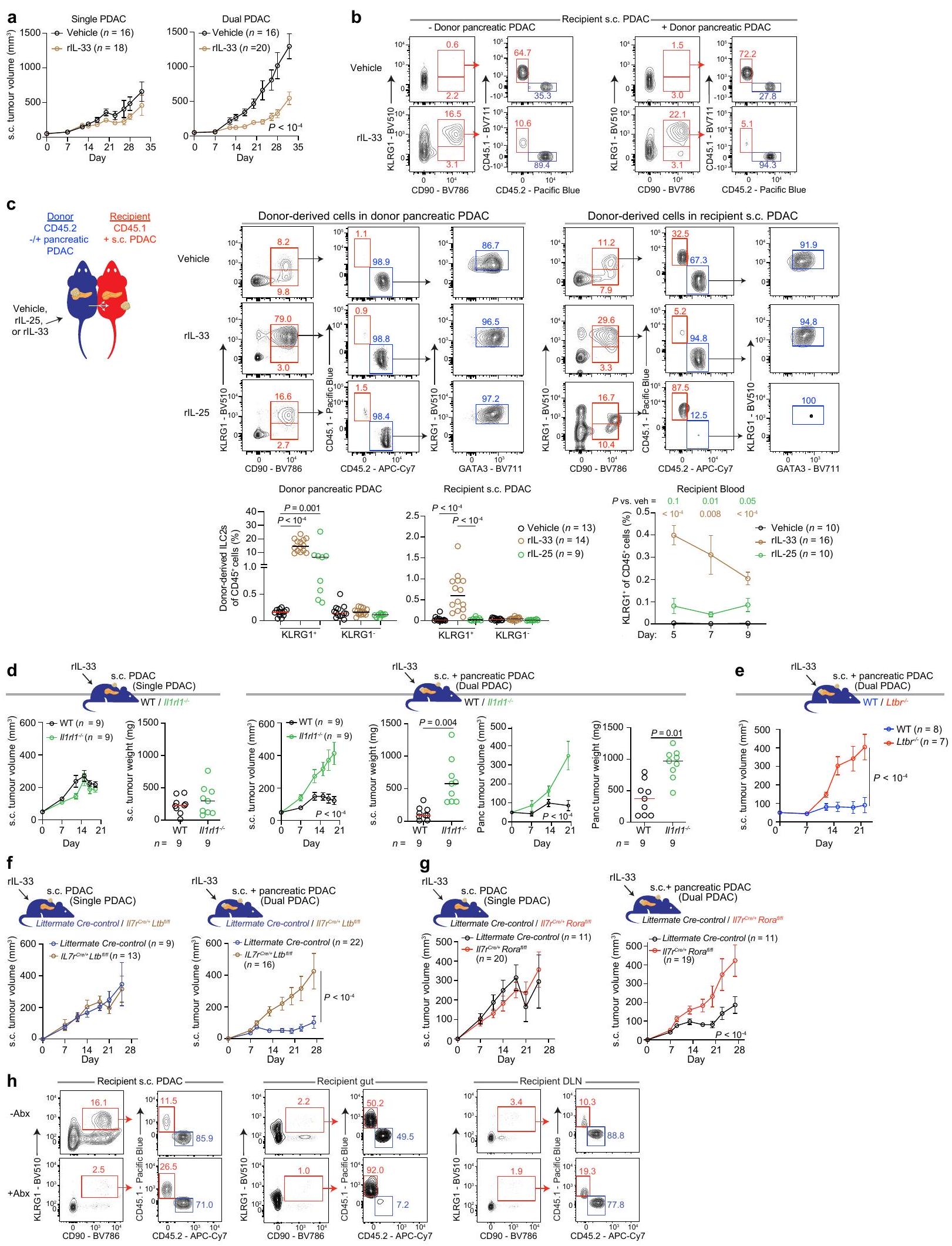
IL-33 يتوسط تكوين الأوعية اللمفاوية

لتحديد الإشارات الالتهابية المرشحة التي تحفز TLSs في الأورام، بحثنا عن جينات تعبر عن تعبير يرتبط إيجابيًا مع التوقيعات النسخية المعروفة لـ TLS. في نموذج PDAC – ورم بارد قاتل مع عدد قليل من TLSs ولكن، عند وجودها، فإن كثافة TLS داخل الورم الأعلى تعزز المناعة وترتبط ببقاء أطول. تُحدد مواقع الأنسجة اللمفاوية الثانوية (TLSs) من خلال تحفيز الكيموكينات والخلايا المقيمة (الخلايا التائية المنشطة، خلايا B، الخلايا الشجرية (DCs) والخلايا النخاعية). اخترنا ثلاث توقيعات نسخية غير متداخلة بشكل أساسي تحدد TLSs داخل الورم بناءً على الكيموكينات الخلايا أو عوامل أخرى تعزز العلاج المناعي وجدنا أن تعبير IL33 – الذي يشفر الألارمين الذي يتم إفرازه بشكل تقليدي في الأنسجة الملتهبة -مرتبط بجميع توقيعات TLS الثلاثة (الشكل 1أ) و مع تعبير ( يشفر السيتوكين المحفز لـ TLS ) في ثلاث مجموعات مختلفة من PDAC ( PDACs؛ الشكل 1أ، الشكل البياني الممتد 1أ والجدول التكميلي 1)، بالإضافة إلى أنواع أخرى من الأورام الباردة والحارة ( الأنسجة، 3,887 ورم؛ الشكل 1ب والبيانات الموسعة الشكل 1ب). داخل TLSs في PDAC، اكتشفنا IL- الخلايا، بما في ذلك IL- خلايا المناعة
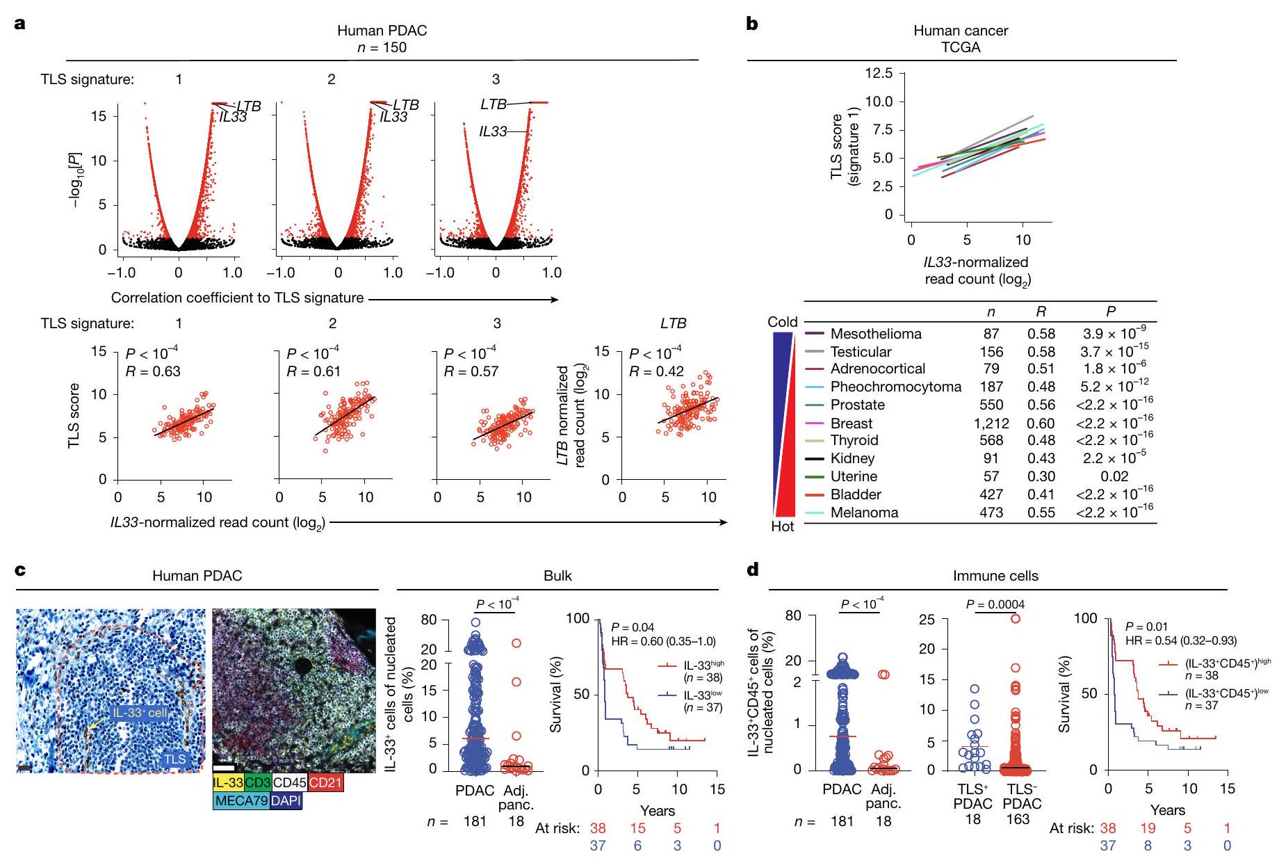
(الشكل 1c (يسار)، الشكل البياني الممتد 1c، d والجدول التكميلي 2) التي كانت أكثر وفرة في الأورام مقارنة بالأنسجة البنكرياسية المجاورة (الشكل 1c (وسط) و1d (يسار))، وكانت غنية في الأورام التي تحتوي على TLSs مقارنة بتلك التي لا تحتوي عليها (الشكل 1d (وسط)). علاوة على ذلك، كانت مستويات IL- داخل الورم أعلى… الكتلة و IL- كانت كثافات خلايا المناعة مرتبطة بتحسين بقاء مرضى سرطان البنكرياس (الشكل 1c (يمين) و1d (يمين)). معًا، حددت هذه النتائج وجود علاقة بين IL-33 وTLSs في السرطان.
للبحث في رابط سببي محتمل بين IL-33 وTLSs، استخدمنا نموذج فأر PDAC المزروع بشكل موضعي المدفوع بواسطة Kras وTrp53 (فئران PDAC) مع عدد قليل من TLSs الأساسية لفحص ما إذا كان Il33 الداخلي مطلوبًا لتكوين الأنسجة اللمفاوية من جديد. وجدنا أن تنشيط مستقبل LTβ (LTβR)… -إشارة معيارية لتكوين TLS -تم توليد TLSs داخل الورم وتم التحكم في الأورام في النوع البري (WT) ولكن ليس في Il33 فئران PDAC (الشكل 2a (يسار) والشكل البياني الممتد 2a). كان IL33 مطلوبًا أيضًا لتكوين الأوعية اللمفاوية الناتج عن الالتهاب، حيث أن سلفات الدكستران الصوديوم (DSS) عن طريق الفم، الذي يثير التهاب القولون الكيميائي (فئران DSS-colitis) ونماذج مرض الأمعاء الالتهابي البشري، تحفز تجمعات اللمفاويات القولونية في WT ولكن ليس في Il33 الفئران (الشكل 2أ (يمين))، مما يزيد من فقدان الوزن ويقلل من البقاء (الشكل البياني الممتد 2ب). وبالتالي، فإن IL33 الداخلي مطلوب لتحفيز TLSs في نماذج السرطان والالتهاب.
الشكل 2 | منطقة الألارمين من IL-33 تحفز TLSs في PDAC والتهاب القولون الكيميائي. أ، صبغة الهيماتوكسيلين والإيوزين (H&E) (الأعلى) وقياس TLSs داخل الورم (الأسفل اليسار) وتجمعات اللمفاوية داخل القولون (LA؛ الأسفل اليمين) في PDAC المعالج بـ anti-LTβR (اليسار) والتهاب القولون DSS (اليمين) WT و الفئران. ب، صبغة H&E (الأعلى) وصبغة المناعة الفلورية (IF؛ في المنتصف) وكثافة TLS (مقدرة بـ H&E؛ الأسفل) في الفئران المصابة بسرطان البنكرياس (PDAC) المعالجة بالسيارة (veh.) و rll-33. PDAC1-6، فئران PDAC موضعية تم إنشاؤها باستخدام ستة خطوط خلوية من PDAC. ج، د، حالات نضوج TLS داخل الورم (مقدرة بالمناعة الفلورية) (ج)، وتكرار خلايا B والتنوع والطفرة الجسدية (د) في الفئران المعالجة بالسيارة والفئران المعالجة بـ rll-33. هـ، صبغة H&E وتقدير LA القولوني.
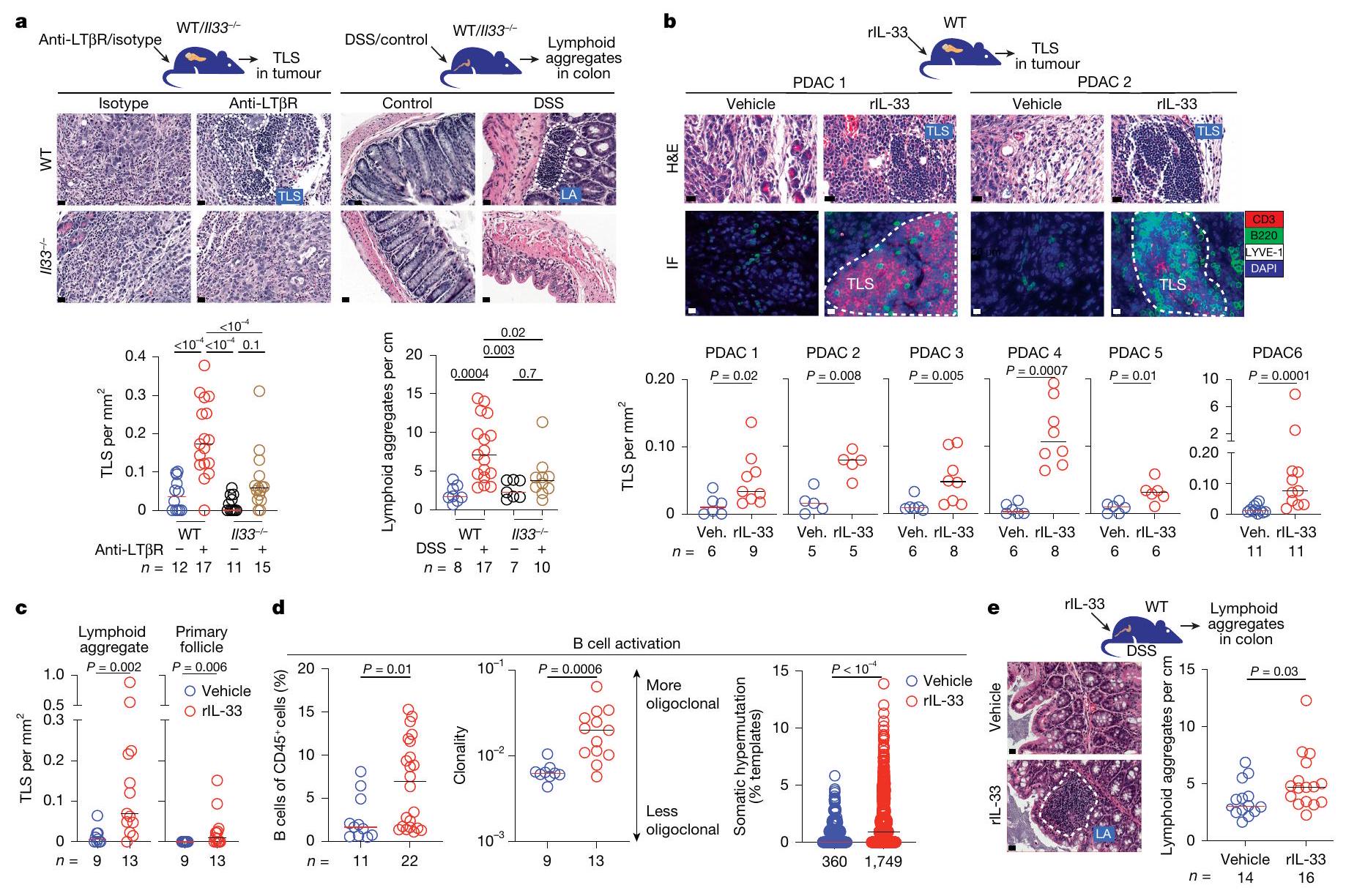
في الفئران المصابة بالتهاب القولون الناتج عن DSS والمعالجة بالسيارة والمعالجة بـ rIL-33. تم جمع البيانات بعد أسبوعين ( ، اليسار)، 3 أسابيع (ب، PDAC 3 و 4؛ و د) و 3 – 5 أسابيع (ب، PDAC 1، 2، 5 و 6) بعد زراعة الورم، و 2 أسابيع بعد بدء DSS (أ (يمين) و )، مجمعة من تجارب مستقلة مع فئران لكل مجموعة مع نتائج متسقة. يمثل عدد الأورام الفردية أو الأعضاء من الفئران الفردية التي تم تحليلها بشكل منفصل، أو نسخ خلايا B (د، اليمين). الخطوط المنقطة في و نعرض TLS/LA المحتملة؛ الأشرطة الأفقية في -e يظهر الوسيط. تم حساب القيم باستخدام تحليل التباين ثنائي الاتجاه (ANOVA) مع اختبار المقارنات المتعددة لتوكاي (a) واختبار مان-ويتني ذو الطرفين -اختبار (ب-هـ). قضبان القياس، قمة و و أسفل ).
بروتين IL-33 الكامل الطول يحتوي على مجالين – مجال نووي في الطرف N ومجال ألارمين في الطرف C الذي، بعد إصابة الأنسجة، يتم تقطيعه خارج الخلية إلى IL-33. ويرتبط بمستقبل ST2 للكشف عما إذا كان IL-33 يعمل كعامل محفز خارجي لتكوين TLS نتيجة الالتهاب، قمنا بإعطاء مجال الألارمين من IL-33 (IL-33 تم إعطاء IL-33 المؤتلف (rIL-33) لفئران نموذج سرطان البنكرياس (PDAC) بجرعات (الشكل البياني الإضافي 1e) التي تقارب تقريبًا مستويات IL-33 الثابتة في مصل البشر من المرضى الذين يعانون من PDAC (الشكل البياني الإضافي 1f والجدول التكميلي 3). أدى rIL-33 إلى تحفيز TLSs داخل الورم من جديد عبر عدة نماذج فئران PDAC المستمدة من خلايا فقيرة في TLS، والتي تغطي مجموعة من تسربات خلايا T داخل الورم (الشكل 2b والشكل البياني الإضافي 2c,d). كما أدى rIL-33 إلى تحفيز حالات نضوج مختلفة لـ TLS، مما زاد من كثافة التجمعات اللمفاوية داخل الورم. (TLS المبكر) والجريبات الأولية (TLS المتوسطة) في أورام PDAC (الشكل 2c والشكل الإضافي 2e). علاوة على ذلك، أظهرت الأورام في الفئران المعالجة بـ rIL-33 TLS مع BCL6 خلايا B (الشكل التوضيحي الممتد 2f)، وزيادة ترددات خلايا B داخل الورم، وتوسع خلايا B المستنسخة، والطفرات الجسدية. (الشكل 2د)، مما يشير إلى تفاعلات المركز الجرثومي وتوليد الجريبات الثانوية (TLS المتأخرة) في فئران التهاب القولون الناتج عن DSS، وسع الرIL-33 تجمعات اللمفاويات القولونية بدرجة أقل (الشكل 2e)، لكنه خفف
فقدان الوزن وزيادة بقاء الفئران (الشكل 2g من البيانات الموسعة). وبالتالي، فإن الألارمين IL-33 يحفز تكوين الأوعية اللمفاوية في السرطان والالتهاب.

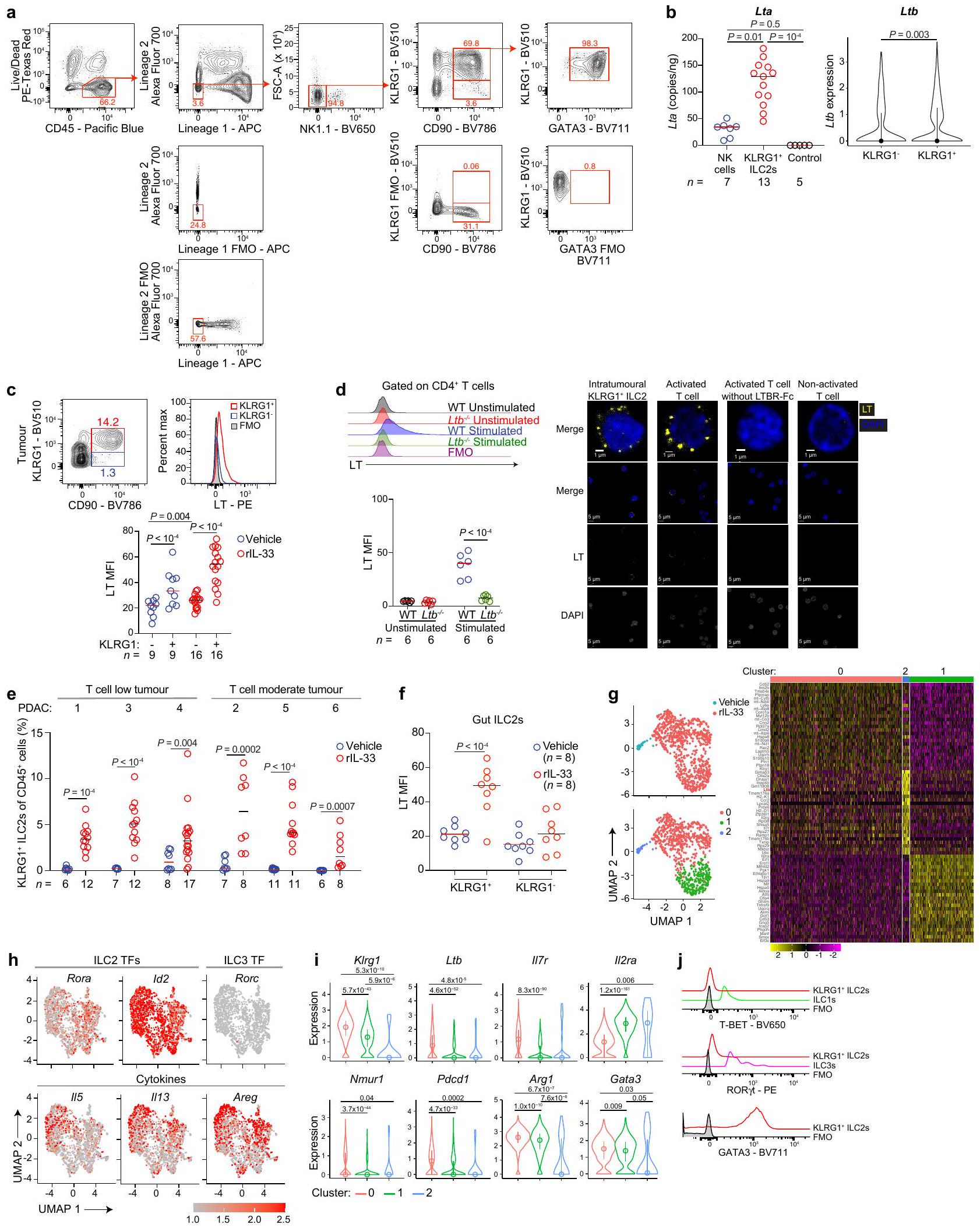
IL-33 يوسع ILC2s اللمفاوية

كما اقترحت هذه النتائج وجود مسار ليمفونيغيني مدفوع بـ IL-33، سعينا بعد ذلك لتحديد الخلايا التي تتوسط TLSs المستحثة بواسطة rIL-33. كما أبلغنا سابقًا أن rIL-33 يوسع بشكل مهيمن داخل الورم. ILC2s في فئران PDAC قمنا بدراسة النسخ الجينية على مستوى الخلية الواحدة لـ ST2 ILC2s في الأورام والعقد اللمفاوية التي تصرف الأورام (DLNs) في فئران PDAC المعالجة بـ rIL-33 لجزيئات اللمفوجين (الشكل 3أ). لقد اكتشفنا مجموعة داخل الورم من خلايا ILC2s التي تعبر عن Klrg1 و (الشكل 3أ والشكل البياني الموسع 3أ،ب)، (الشكل البياني الممتد 3ب) و LT، الـ LT السيتوكين الهتيروترميري الضروري لتكوين TLS (الشكل 3ب والشكل الإضافي 3ج، د). بشكل متسق، في نماذج متعددة من PDAC وفئران التهاب القولون الناتج عن DSS، وسعت rlL-33 KLRG1 داخل الورم (الشكل 3ج والشكل الإضافي 3هـ) وفي الأمعاء (الشكل 3د) ILC2s، ولكن ليس KLRG1 ILC2s، التي تعبر عن LT (الشكل الإضافي 3c، f). هذه KLRG1 داخل الورم المستحثة بواسطة rIL-33 كما أن خلايا ILC2s تعبر عن عوامل النسخ الكلاسيكية لخلايا ILC2 (Gata3 و Rora (الشكل 3 أ والممتد
الشكل 3 | انظر الصفحة التالية للتعليق.
شكل البيانات ) و Id2 (الشكل 3h من البيانات الموسعة)، السيتوكينات (Il5 و Il13 و Areg؛ الشكل 3h من البيانات الموسعة) والمستقبلات (Il7r و Nmur1 ; الشكل 3أ والشكل البياني الموسع 3ي)، ولكن ليس ILC1 (Tbx21 (الذي يشفر T-BET)؛ الشكل البياني الموسع 3j) أو ILC3 (Rorc (الذي يشفر RORyt)؛ الشكل البياني الموسع
الشكل 3هـ، وj) عوامل النسخ. وبالتالي، rlL-33 يوسع KLRG1 ILC2s التي تعبر عن LT والتي قد تتوسط في تكوين الأوعية اللمفاوية.
أظهرت التقارير الأخيرة أن خلايا ILC2 والسابقة تعبر بشكل انتقائي عن مستقبل النيوروميدين (NMUR1) الذي يمكن استخدامه لـ
الشكل 3 | IL-33 ينشط ILC2s اللمفاوية. أ، تحليل تسلسل RNA أحادي الخلية (scRNA-seq) لـ ILC2s المنقاة من الورم و DLN من الفئران المصابة بسرطان البنكرياس PDAC المعالجة بـ rlL-33. تمثل تمثيلات تقريب الفضاء الموحد والإسقاط (UMAP) الخلايا حسب المجموعات غير المراقبة (أعلى اليسار)، والأعضاء (أسفل اليسار) وبعض الجينات المختارة (اليمين). تُظهر خريطة الحرارة (الوسط) أعلى 25 جينًا حسب المجموعة. تمثل شريط مقياس خريطة الحرارة النتيجة. يمثل شريط مقياس UMAP تعبير الجينات. ب، التصفية (يسار) وتعبير LT بواسطة قياس التدفق (يمين) والتصوير المجهري التوافقي (أسفل) في KLRG1 داخل الورم. ILC2s من WT المعالج بـ rIL-33 و فئران PDAC. FMO، الفلورية ناقص واحد؛ MFI، متوسط شدة الفلورية. ج، د، تحديد الخلايا، وتكرار وعدد ILC2s داخل الورم (ج) والأمعاء (د) في الفئران المعالجة بالدواء الوهمي والمعالجة بـ rIL-33 (ج) و DSS-colitis (د). هـ، تحديد الخلايا (يسار)، تكرار ILC2 (وسط) وكثافة TLS داخل الورم (يمين) في Nmur1 المعالجة بـ rlL-33 و DT. روزا26 والمجموعة الضابطة من نفس السلالة روزا26 فئران PDAC. ف، كثافة TLS داخل الورم (يسار)، التصفية، التردد وعدد ILC2s،
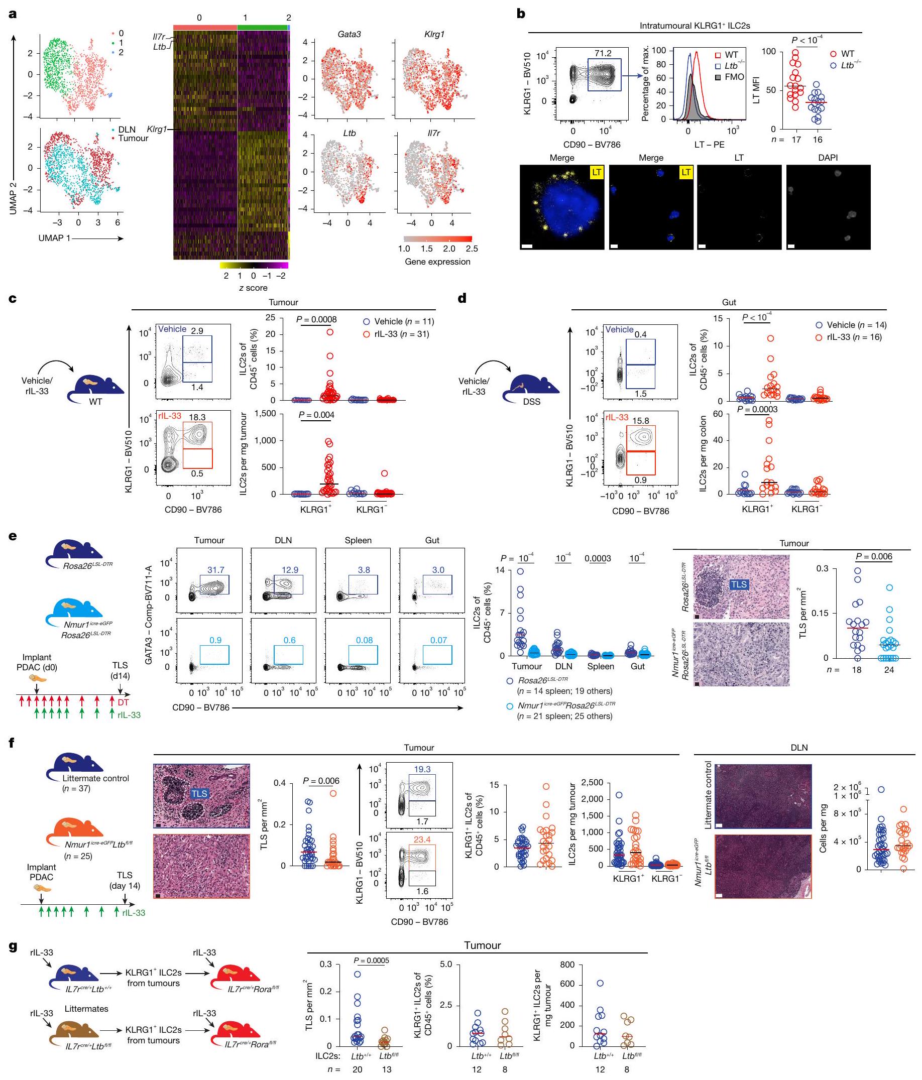
وعدد خلايا المناعة DLN (يمين) في Nmur1 المعالجة بـ rlL-33 وأشقاء الفئران الضابطة لسرطان البنكرياس. ج، ورم KLRG1 تمت تنقية ILC2s من المعالجة بـ rlL-33 أو فئران PDAC من نفس القمامة وتم نقلها إلى رور مستقبلات الفئران PDAC. كثافة TLS، تردد وعدد خلايا ILC2 في PDACs المستقبلة. تم جمع البيانات بعد 10 أيام (أ)، و2 أسبوع (ب و د-و) و5 أسابيع (ج) بعد زراعة الورم، و2 أسبوع (ز) بعد النقل، مجمعة من تجارب مستقلة مع فئران لكل مجموعة مع نتائج متسقة. يمثل عدد الأورام الفردية أو الأعضاء من الفئران الفردية التي تم تحليلها بشكل منفصل. تظهر القضبان الأفقية الوسيط. تشمل ضوابط الأشقاء من نفس القمامة ونمور1 مجمعة. تم حساب القيم باستخدام اختبار مان-ويتني ذي الطرفين -اختبار (ب، هـ (يمين)، فاند ج) وتحليل التباين ثنائي الاتجاه مع اختبار هولم (ج، د و هـ (يسار)). شريط القياس، (ب(يسار)) (ب(جميع الآخرين)) (e و (يسار)) و (f(يمين)).
استهداف جيني محدد لـ ILC2 للتحقيق فيما إذا كانت خلايا ILC2 تساهم في تكوين اللمفونات المعتمد على IL-33، حيث أن خلايا ILC2 (ولكن ليس خلايا ILCs أو الخلايا المناعية الأخرى) في نموذجنا تعبر أيضًا بشكل انتقائي عن NMUR1 (الشكل البياني الممتد 4a)، استخدمنا Nmur1 روزا26 الفئران المعدلة وراثياً التي تعبر بشكل دائم عن مستقبل سم الدفتيريا البشري (DTR) على NMUR1 الخلايا لاستنفاد جميع ILC2s بشكل حاد وانتقائي. في Nmur1 روزا26 فئران PDAC، تم بدء سم الدفتيريا (DT) قبل يومين من زراعة الورم وعلاج rIL-33 أدى إلى استنفاد ILC2s بحوالي داخل الأورام وعبر الأعضاء (الشكل 3e (يسار)) مع الحفاظ على ILC3s (الشكل البياني الممتد 4b) وخلايا المناعة الأخرى (الشكل البياني الممتد 4c). ومن الجدير بالذكر أن تقليل ILC2 بشكل حاد خلال علاج rIL-33 قلل من TLSs داخل الورم بحوالي 65% (الشكل 3e (يمين)). وبالتالي، تساهم ILC2s في تكوين الأنسجة اللمفاوية المعتمد على IL-33.
بعد ذلك، لفحص ما إذا كانت خلايا ILC2s تستخدم LT الداخلي للخلايا لتحفيز TLSs داخل الورم، قمنا بإنشاء فئران ترانسجينية تفتقر إلى على ILC2s (Nmur1 لتب فلفل ). في Nmur1 المعالج بـ rlL-33 لطب أدت إزالة Ltb المستهدفة في ILC2 في الفئران المصابة بسرطان البنكرياس، مقارنةً مع الفئران الشقيقة، إلى تقليل كثافة TLS داخل الورم بحوالي دون تغيير ترددات KLRG1 ILC2s (الشكل 3f)، ILC1s، ILC3s وخلايا المناعة الأخرى في الأورام (البيانات الموسعة الشكل 4d (الأعلى))، خلايا المناعة في العقد اللمفاوية العميقة (الشكل 3f (اليمين)) أو مجموعات خلايا المناعة في الأعضاء (البيانات الموسعة الشكل 4d (الأسفل)). بشكل متسق، استراتيجية جينية أوسع لحذف على جميع IL-7R الخلايا اللمفاوية (II77 كما قلل أيضًا من TLSs داخل الورم، ولكنه قلل بشكل ملحوظ أيضًا من KLRG1 داخل الورم ILC2s (الشكل 4e من البيانات الموسعة)، ليقترح أن إشارات LT من اللمفاويات غير ILC2 قد توسع KLRG1 إلى الحد الأقصى. ILC2s في الأورام. معالجة rIL-33 كان لدى فئران PDAC أيضًا ترددات داخل الورم قابلة للمقارنة من خلايا المناعة الأخرى ولكن عدد أقل من الحمضات (الشكل 4e الموسع (يمينًا))، مما يتماشى مع التأثيرات اللاحقة لاستنفاد ILC2. لتأكيد أن خلايا ILC2 استخدمت LT للتأثير على تشكيل TLS باستخدام نهج متعامد، قمنا بتنقية ونقل KLRG1 داخل الورم. ILC2s من المعالجة بـ rIL-33 أو فئران PDAC من نفس القمامة (أي، ILC2s كفء أو ناقص في tb) إلى Il7r رورا مستقبلو PDAC الذين يظهرون انخفاضًا في ILC2s المحيطية في الأنسجة (الشكل 3g). مقارنةً مع ILC2s المنقولة التي تحتوي على intact ، ومتسق مع النتائج في Nmur1 لطب نقص Ltb الذاتي في الفئران المصابة بـ PDAC (الشكل 3f) قلل من تشكيل TLS دون التأثير على KLRG1 داخل الورم الذي يتم تحفيزه بواسطة rIL-33. تراكم ILC2 في الأورام (الشكل 3g). بشكل جماعي، تشير هذه التجارب الجزيئية والخلوية لفقدان واكتساب الوظيفة إلى أن rIL-33-activated KLRG1 تقوم خلايا ILC2 بتحفيز TLSs في الأورام الغدية البنكرياسية باستخدام مسار يتضمن LT.
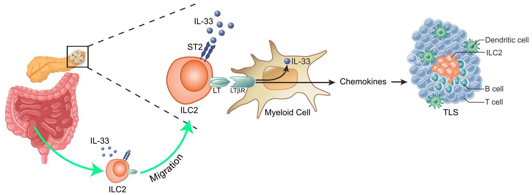
في SLOs و TLSs، تعبر خلايا المحفز عن LT الذي يرتبط بـ LTβR على خلايا منظم TLS لتحفيز التعبير عن الكيموكينات وجزيئات الالتصاق، وتنسيق تكوين الأنسجة اللمفاوية. لتحديد خلايا المنظم التي تتعاون مع المحفز rlL-33 ILC2s لتحفيز TLSs في PDAC، وجدنا أن CD11b داخل الورم الخلايا النخاعية تعبر عن LT بأعلى مستوى ر (الشكل 5 أ من البيانات الموسعة). مقارنة مع الخلايا النخاعية، داخل الورم تم التعبير عن خلايا النخاع
نسخة متميزة من النسخ الجينية (الشكل التوضيحي الممتد 5ب-هـ والجدول التكميلية 4)، مع كيموكينات تحفز TLS أعلى، بما في ذلك Cxcl13 و (الشكل التوضيحي الممتد 5d)، جزيء التصاق خلايا المنظم الكنسي Vcam1 (الشكل التوضيحي الممتد 5e) وعوامل الكيمياء الالتهابية التي تجذب مكونات خلايا TLS (الشكل 5e من البيانات الموسعة). LTβR داخل الورم كما أن الخلايا النخاعية أعربت عن mRNA وبروتين II33 (الشكل 5f من البيانات الموسعة). وبشكل متسق، LT تفعيل R زاد من KLRG1 داخل الورم ILC2s في WT ولكن ليس فئران PDAC (الشكل 5g من البيانات الموسعة) للإشارة إلى LT قد توسع تحفيز R ILC2s من خلال دائرة تعتمد على IL-33 ذات المنشأ الذاتي. لاختبار ما إذا كان IL-33 الذاتي المنشأ المستمد من خلايا النخاع العظمي يعدل KLRG1 ILC2s في الأورام، قمنا بتنقية IL-33 الكفء (WT) أو الناقص (II33 ) الخلايا النخاعية من الأورام الغدية البنكرياسية وزرعها مع خلايا الأورام الغدية البنكرياسية في البنكرياس لـ الفئران (الشكل البياني الممتد 5h). LT IL-33 المستمد من خلايا النخاع الشوكي حفز KLRG1 في PDACs للتعبير عن LT وتحفيز TLSs (الشكل 5h من البيانات الموسعة). معًا، تشير هذه البيانات إلى أن تتعاون خلايا ILC2s وخلايا المايلويد داخل الورم كخلايا محفزة ومنظمة لتكوين TLS، على التوالي، لتعزيز إشارات IL-33 وتحفيز TLSs في الأورام.

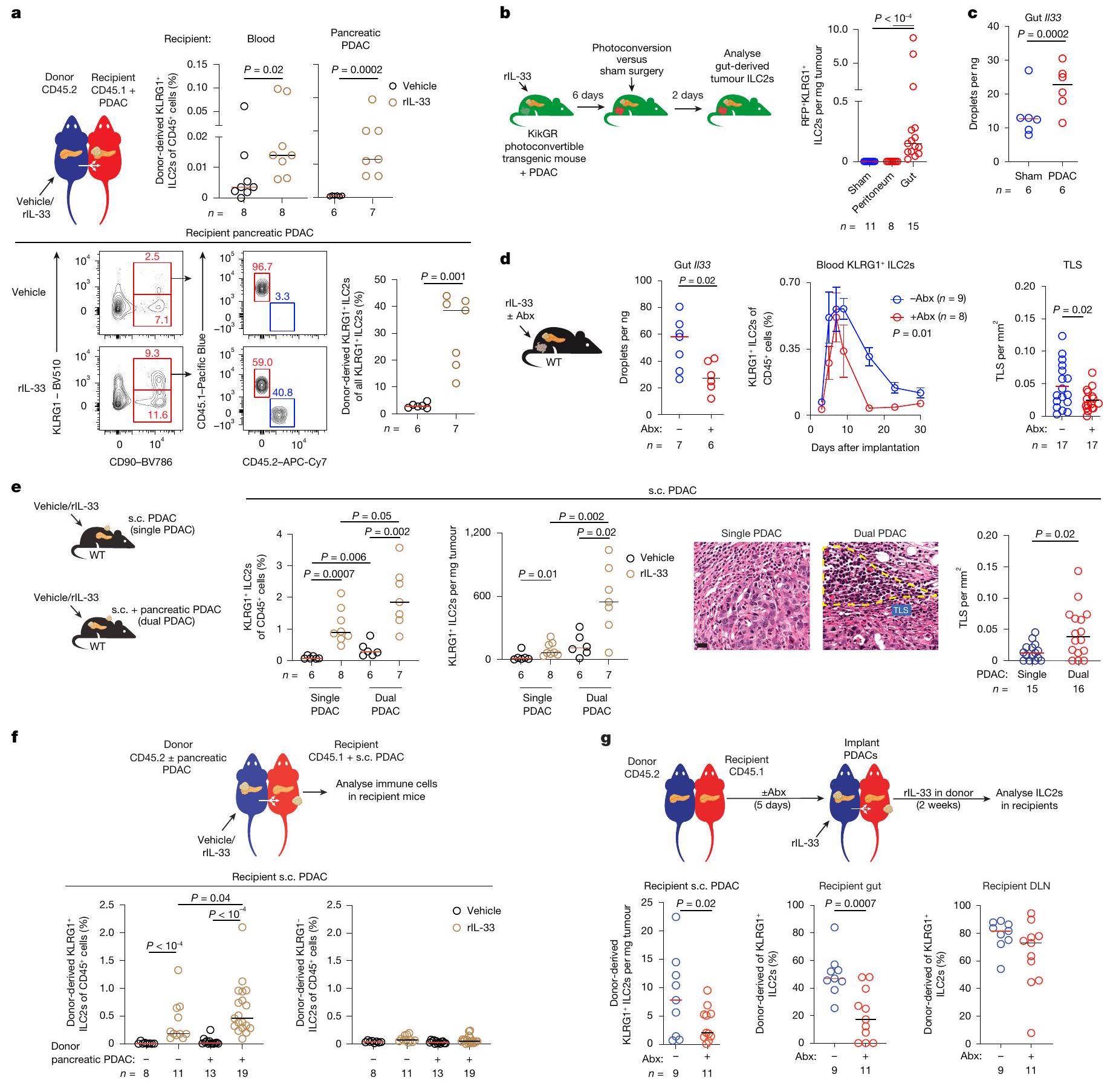
يمكن لخلايا ILC2s اللمفاوية أن تهاجر من الأمعاء

كما أشارت نتائجنا إلى وظيفة ليمفونية لـ ILC2s، قمنا بالتحقيق فيما إذا كانت الليمفونية تتوسع خلايا ILC2 محليًا أو تهاجر إلى الأورام . في فئران PDAC، حيث توسع rll-33 KLRG1 ILC2s في الدم (الشكل التوضيحي الممتد 6a)، استخدمنا فئران بارابيوتيك متجانسة مع أورام البنكرياس القنوية (PDACs) في البنكرياس المتلقي لاختبار ما إذا كان KLRG1 انتقلت خلايا ILC2 عن طريق الدم إلى الأورام (الشكل البياني الممتد 6b). أدى إعطاء rlL-33 إلى المتبرعين المتصلين إلى تحفيز المتبرع (الشكل 4أ) ولكن ليس KLRG1- ILC2s أو غير ILCs (الشكل البياني الموسع 6ج، د) للهجرة إلى دم المتلقي المتزاوج والأورام. وبالتالي، تنتقل خلايا ILC2 عبر الدم إلى الأورام.
حيث يمكن لخلايا ILC2s الهجرة إلى الأنسجة من الأمعاء ، افترضنا أن الأمعاء قد تكون واحدة من مصادر ILC2s التي تهاجر إلى أورام البنكرياس. لتتبع أصل داخل الورم استخدمنا فئران KikGR المتحولة وراثيًا حيث تعبر جميع الخلايا عن بروتين قابل للتحويل من الأخضر إلى الأحمر. عالجنا فئران PDAC من نوع KikGR بـ rlL-33، وقمنا بتحويل أنسجة الأمعاء بالضوء وبحثنا عن اللون الأحمر المشتق من الأمعاء. ILC2s في PDACs (الشكل 4ب). في الفئران المصابة بـ PDAC المعالجة بـ rll-33، اكتشفنا عددًا نادرًا من اللون الأحمر ILC2s في أورام الأمعاء – تم تحويلها بالضوء ولكن ليس في الفئران الضابطة (المعالجة الوهمية؛ تم تحويلها بالضوء في الصفاق) (الشكل 4ب والشكل التمديدي 6هـ)، مما يشير إلى أن يمكن لخلايا ILC2 الانتقال إلى الأورام الغدية البنكرياسية من خلال دائرة الأمعاء-الدم.
لفحص ما إذا كانت الأورام الغدية البنكرياسية قد أعدت مثل هذه الدائرة المهاجرة من ILC2 المشتقة من الأمعاء، بحثنا عن إشارات مشتقة من الأمعاء قد تحفز ILC2s على الهجرة. كما تشير النتائج الأخيرة إلى أن ميكروبات الأمعاء يمكن أن تحفز ILC2s على الهجرة في حالات الالتهاب الحاد. قمنا بفحص ما إذا كانت أورام البنكرياس الغدية (PDACs) قد غيرت ميكروبيوم الأمعاء وبالتالي حفزت خلايا ILC2 في الأمعاء.
الشكل 4 | تهاجر خلايا ILC2 اللمفاوية إلى الأورام من خلال دائرة الأمعاء-الدم المعدلة بواسطة الميكروبيوتا و PDAC. أ، فئران بارابيوتيك مع PDACs في البنكرياس المتلقي. تم علاج المتبرعين بمركب أو rll-33. الكمية (أعلى) والتصنيف (أسفل) لخلايا ILC2 المشتقة من المتبرع في دم المتلقي و PDAC. ب، تم علاج فئران KikGR PDAC يوميًا بـ rlL-33 لمدة 6 أيام. في اليوم السادس، تم تحويل الأمعاء أو الصفاق ضوئيًا. المخطط التجريبي (يسار) وكثافة الخلايا المشتقة من الأمعاء (RFP ) تم تحليل mRNA Il33 في الأمعاء بواسطة PCR القطرات الرقمية في الفئران المعالجة بالشام و PDAC. د، mRNA Il33 في الأمعاء (يسار)، KLRG1 في الدم ILC2s (في المنتصف) وكثافة TLS داخل الورم (على اليمين) في فئران PDAC المعالجة بـ rlL-33 التي تم علاجها مع أو بدون مضادات حيوية (Abx). e، KLRG1 داخل الورم تردد ILC2، العدد وكثافة TLS في سرطان البنكرياس (PDACs) تحت الجلد (s.c.) في الفئران المعالجة بالعلاج الوهمي والفئران المعالجة بـ rIL-33 مع PDAC واحد تحت الجلد (PDAC واحد) أو PDACs تحت الجلد والبنكرياس (PDAC مزدوج). f، فئران بارابيوتيك مع PDACs تحت الجلد في المستلمين مع أو بدون PDACs البنكرياسية في المتبرعين. تم علاج المتبرعين بالعلاج الوهمي أو rIL-33.
تظهر نسبة ILC2 المشتقة من المتبرع في أورام البنكرياس القنوي (PDACs) في المتلقين (أسفل). فئران بارابيوتيك مع أورام البنكرياس الغدية تحت الجلد في المتلقين وأورام البنكرياس الغدية في المتبرعين. تم علاج المتبرعين بـ rIL-33؛ وتم علاج المتبرعين والمتلقين بالمضادات الحيوية. يتم عرض عدد وتكرار ILC2 المشتقة من المتبرع في أورام البنكرياس الغدية تحت الجلد في المتلقين والأمعاء (أسفل). تم جمع البيانات بعد أسبوعين (أ، هـ (يسار)، و )، كما هو موضح في المخطط التجريبي (ب)، 4 أسابيع (ج و و5 أسابيع (e(right)) بعد زراعة الورم، مجمعة من تجارب مستقلة مع فئران لكل مجموعة مع نتائج متسقة. تمثل القيم عدد الأورام الفردية أو الأعضاء من الفئران الفردية التي تم تحليلها بشكل منفصل. تُظهر الأشرطة الأفقية الوسيط. تم حساب القيم باستخدام اختبار مان-ويتني ذي الطرفين -اختبار (أ، ج، د، هـ (يمين) و غ)، تحليل التباين الأحادي مع اختبار المقارنات المتعددة كروسكال-واليس (ب)، تحليل التباين الثنائي مع اختبار كروسكال-واليس (د، الوسط) وتحليل التباين الثنائي مع اختبار المقارنات المتعددة سيداك (هـ (يسار) و ف). شريط القياس، .
الشكل 5 | علاج IL-33 البشري المهندَس يحفز TLSs ونشاط مضاد للورم في PDAC. أ، تنشيط ST2 في خلايا HEK بلو البشرية المبلغة عن IL-33 مع H-rIL-33 وH-e-rIL-33 وH-e-rIL-33-Fc. EC تركيز الفعالية نصف الأقصى. ب-د، KLRG1 داخل الورم تردد ILC2 (ب)، كثافة TLS (ج) وحجم الورم (د) في فئران PDAC المعالجة بالسيارة، أو rIL-33 الفأري، أو H-rIL-33 أو H-e-rIL-33-Fc المتزايد. تم ملاءمة المنحنيات في (أ) بواسطة غير خطي.
خزانات من الجدير بالذكر أن الأورام الغدية البنكرياسية (PDACs) لم تزد فقط من KLRG1 داخل البنكرياس تكرارات ILC2 (الشكل 7a من البيانات الموسعة)، ولكن أيضًا زادت من تنوع وتغيرت تركيبة ميكروبات الأمعاء (الشكل 7b من البيانات الموسعة) وزادت من تعبير mRNA لـ Il33 في الأمعاء (الشكل 4c)، مما يشير إلى أن التغيرات الميكروبية في الأمعاء قد تزيد من مستويات IL-33 في الأمعاء وتحفز KLRG1 في الأمعاء. الهجرة إلى ILC2s بشكل متسق، فإن إزالة الميكروبات بواسطة المضادات الحيوية انخفاض تعبير mRNA لـ Il33 في الأمعاء، وانخفاض تكرار الدم ILC2s وتقليل TLSs داخل الورم (الشكل 4d). علاوة على ذلك، أدى إزالة الميكروبات إلى تقليل طفيف في مستوى IL-33 في المصل، وزيادة متبادلة في تكرار KLRG1 في الأمعاء. ILC2s (الشكل 7c من البيانات الموسعة) التي تم تقليلها بشكل متسق بعد استعادة الميكروبيوتا في الفئران الخالية من الجراثيم (الشكل 7d من البيانات الموسعة)، مما يشير إلى أن تركيبة الميكروبيوتا قد تؤثر على قدرة الأمعاء ILC2s للدخول إلى الدورة الدموية. بشكل جماعي، تشير هذه النتائج إلى أن يمكن لخلايا ILC2 الانتقال إلى الأورام من الأمعاء وتكون متأثرة بميكروبات الأمعاء.
كما تشير هذه النتائج، فإن الأورام الغدية البنكرياسية تحفز المضيف لطرد خلايا ILC2 إلى المواقع الملتهبة، قمنا بالتحقيق فيما إذا كانت الأورام الغدية البنكرياسية يمكن أن تعزز استجابة ILC2 للأورام في الأنسجة البعيدة. لاختبار ذلك، قمنا بإعطاء rIL-33 للفئران التي تعاني من أورام غدية بنكرياسية فردية (جلد فقط، أورام غدية بنكرياسية تحت الجلد) أو مزدوجة (تحت الجلد وبنكرياسية). على عكس الفئران ذات الأورام الغدية البنكرياسية الفردية، حيث وسعت rIL-33 خلايا KLRG1 داخل الورم. لم تتحكم خلايا ILC2 بعد في نمو سرطان البنكرياس الثانوي، وفي الفئران ذات سرطان البنكرياس الثنائي، زاد rll-33 من KLRG1 بشكل أكبر. ترددات ILC2، وكثافات TLS المعززة (الشكل 4e) ونمو الورم المتحكم فيه في PDACs تحت الجلد (الشكل التمديدي 8a). لتأكيد أن PDACs المزدوجة عززت KLRG1 ILC2s التي انتقلت عبر الدم إلى الأورام تحت الجلد، قمنا بنمذجة ثنائية PDACs في الفئران المترافقة مع وجود PDACs البنكرياسية في المتبرعين المترافقين وPDACs تحت الجلد في المستلمين (الشكل 4f). بما يتماشى مع النتائج في فئران PDAC الثنائية، زادت PDACs البنكرياسية في المتبرعين المترافقين من KLRG1 المستحث بواسطة rll-33. لكن ليس KLRG1 هجرة ILC2 إلى PDACs في المتلقين المترافقين (الشكل 4f والشكل الإضافي 8b). هذا PDAC المعزز في البنكرياس KLRG1 المستمد من الدم تطلب نقل ILC2 إلى PDACs تحت الجلد سيتوكينات مميزة مقارنةً
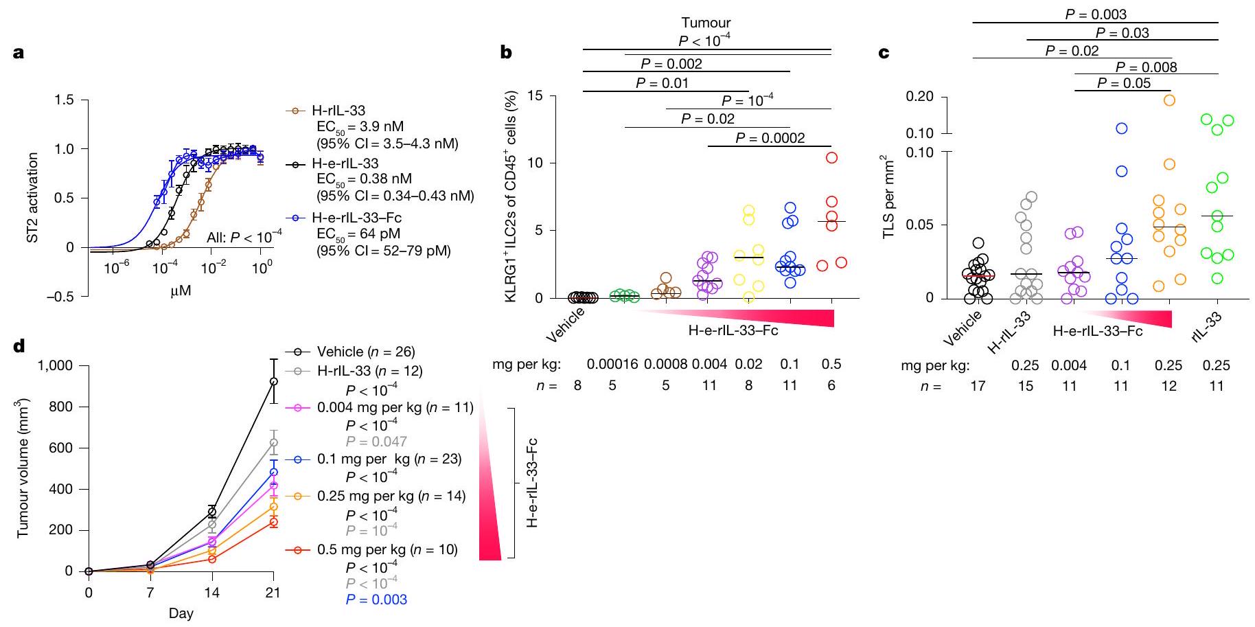
الانحدار. تم جمع البيانات في أسابيع ( و ) بعد زراعة الورم، مجمعة من تجارب مستقلة مع فئران لكل مجموعة مع نتائج متسقة. تمثل القيم عدد الأورام الفردية من الفئران الفردية. تُظهر الأشرطة الأفقية الوسيط. تم حساب القيم باستخدام مجموع المربعات الإضافي اختبار (أ) وتحليل التباين ثنائي الاتجاه مع اختبار تصحيح المقارنات المتعددة لتوكاي (ب-د).
للالتهابات الحادة حيث، على الرغم من أن كل من rIL-33 و rIL-25 قد وسعت ILC2s في PDACs المتبرعة (الشكل 8c من البيانات الموسعة)، فقط rlL-33 وليس rll-25 KLRG1 المنتشر ILC2s من بارابيونت المتبرعين إلى دم بارابيونت المتلقي و s.c.PDACs (الشكل البياني الموسع 8c). علاوة على ذلك، كانت الزيادة في الهجرة إلى s.c. PDACs في الفئران ذات PDAC المزدوج مصحوبة بنمو محدود لـ s.c. PDAC في وجود ST2-، LTβR-، – وبطريقة تعتمد على Rora (الشكل 8d-g من البيانات الموسعة). أخيرًا، أدى إزالة الميكروبات في الفئران المزدوجة PDAC إلى تقليل ILC2s المشتقة من المتبرع في PDACs تحت الجلد للمستقبل والأمعاء، ولكن ليس في العقد اللمفاوية العميقة للمستقبل (الشكل 4g والشكل 8h من البيانات الموسعة)، مما يشير إلى أن الميكروبات تعزز ILC2s في الأورام مرتبطة بدائرة الأمعاء-الدم. باختصار، يقوم مضيف البنكرياس PDAC بتعزيز هجرة ILC2 المعتمدة على الميكروبيوتا إلى PDACs في الأنسجة البعيدة.

IL-33 المهندسة تعزز تكوين الأوعية اللمفاوية

لفحص مدى صلة نتائجنا بالبشر، قمنا بعد ذلك بالتحقيق فيما إذا كانت KLRG1 التي تعبر عن LT توجد خلايا ILC2 في السرطان البشري. في سرطان البنكرياس القنوي البشري، اكتشفنا KLRG1. ILC2s التي عبرت عن LT (الشكل الإضافي 9a-c والجدول التكميلية 5)، مما يشير إلى أن قد تمارس خلايا ILC2 وظيفة مشابهة في تكوين اللمف في الأورام البشرية. للتحقيق في العلاقة بين خلايا ILC2 المنشطة بواسطة IL-33 وTLSs داخل الورم، قمنا بتطوير توقيع نسخي بشري لخلايا ILC2 المنشطة بواسطة rIL-33 (الطرق) وفحصنا ارتباطه بتوقيعات TLS. في الأورام التي تغطي طيفًا من النشاط المناعي الهستولوجيات، 4,037 ورم). ارتبطت توقيعات ILC2 المنشطة بجميع توقيعات النسخ الثلاثة لـ TLS (الشكل البياني الممتد 1g)، مما يشير إلى أن KLRG1 قد تساهم خلايا ILC2 في تشكيل TLS في الأورام البشرية.
مع وجود أظهرت خلايا ILC2 في الأورام البشرية أن مسار IL-33-ILC2-TLS يمكن أن يتم التلاعب به للعلاج المناعي، قمنا بتصميم IL-33 البشري (H-rIL-33) لزيادة نشاطه ونصف عمره في الجسم. أولاً، للقضاء على أكسدة السيستين التي تحفز تكوين الروابط الثنائية.
تغييرات الترابط والتشكيل التي تعطل موقع ارتباط ST2 لـ IL-33، قمنا باستبدال جميع بقايا السيستين الأربعة في IL-33 البشري إلى بقايا السيرين (المشار إليه فيما بعد بـ H-enhanced-rll-33 (H-e-rll-33)). وجدنا أن H-e-rlL-33 عزز تنشيط ST2 مقارنة بـ H-rlL33، وهو ما يتماشى مع الملاحظات السابقة. (الشكل 5أ). بعد ذلك، من أجل إطالة عمر النصف لـ H-e-rll-33 في الجسم الحي، استخدمنا نهجًا مثبتًا وقمنا بدمج H-e-rlL-33 مع جزء Fc من IgG1 البشري (H-e-rll-33-Fc) مع التعديلات الموصوفة L234A/L235A التي تحد من ارتباط مستقبلات Fc غاما من خلال وصلة قصيرة تحتوي على جلايسين/سيرين. ثم أكدنا أن H-e-rlL-33-Fc عزز تنشيط ST2 في المختبر مقارنةً بـ H-rIL-33 و H-e-rIL-33 (الشكل 5a). كما أظهرت العديد من الدراسات أن IL-33 البشري نشط بيولوجيًا في الفئران. قمنا باختبار ما إذا كان H-e-rIL-33-Fc قد وسع KLRG1 ILC2s و TLSs المستحثة في الفئران. باستمرار، تم توسيع KLRG1 داخل الورم بواسطة H-e-rll-33-Fc ILC2s و TLSs بطريقة تعتمد على الجرعة (الشكل 5b، c) للتحكم في أورام PDAC (الشكل 5d). وبالتالي، فإن محور IL-33-ILC2-TLS هو مسار لم يتم وصفه سابقًا يمكن هندسته لعلاج سرطان البنكرياس (بيانات موسعة الشكل 10).

نقاش

نجد أن IL-33، وهو مادة تنبهية يتم إفرازها بشكل تقليدي بواسطة الأنسجة التالفة، ينشط استجابة خلوية هجرة ليمفونية. قد يكون مثل هذا المسار متوقعًا عند النظر إلى الوراء – حيث تتقلص خلايا المحفزات النسيجية اللمفاوية، وهي الخلايا التي تبدأ تكوين الأعضاء اللمفاوية قبل الولادة، في مرحلة البلوغ بعد الولادة. على الرغم من أن خلايا ILC3 قد تشمل بعض وظائف المحفزات اللمفاوية في البالغين ILC3s لا تستجيب بشكل تقليدي لإشارات الضرر. وبالتالي، يمكن للمرء أن يفترض أن هناك مسارًا خلويًا غير متكرر يجب أن يبدأ تكوين الأنسجة اللمفاوية استجابةً لإصابة الأنسجة. في الواقع، تسلط نتائجنا الضوء على IL-33 كمرشح لبدء TLS الالتهابي، وILC2s كمرشح محفز لمثل هذا المسار.
ملاحظاتنا لا تعني أن خلايا ILC2 هي المحفزات الوحيدة لتكوين الأنسجة اللمفاوية المدفوعة بـ IL-33. على الرغم من أن دراسات زيادة وفقدان الوظيفة تظهر أن خلايا ILC2 تحفز بالفعل الأنسجة اللمفاوية في طريقة تعتمد على LT، إلا أن نتائجنا لا تستبعد أن الأنسجة اللمفاوية المدفوعة بـ IL-33 قد تستفيد من مساهمات من ST2 أخرى. الخلايا، بما في ذلك خلايا T المنشطة وخلايا B التي تستجيب لـ IL-33 . علاوة على ذلك، قد يقوم IL-33 أيضًا بتنشيط جهاز المناعة الآخر وغير المناعي الخلايا تمارس تأثيرات مضادة، وربما مؤيدة للورم، في سياقات مختلفة من خلال آليات غير TLS. حيث يتم التعبير عن Nmur1 في الجهاز العصبي. استراتيجيتنا الجينية لاستنزاف خلايا ILC2s بشكل انتقائي من خلال استهداف Nmur1 باستخدام DT قد تعقد المحاولات لدراسة تشكيل TLS في الأنسجة العصبية. ومع ذلك، نظرًا لأن خلايا ILC2s تستجيب مبكرًا لضرر الأنسجة وتستقطب خلايا لمفاوية أخرى إلى المواقع المصابة، فإن خلايا ILC2s تعتبر خلايا قمة مرشحة رئيسية لبدء TLS المدفوعة بـ IL-33 وتعزيز استجابة TLS. . هذا يوسع نطاق الوظيفة المعترف بها للـ IL-33 كمنبه، ودور خلايا ILC2s كحراس للإصابة، ليشمل تكوين اللمف الناتج عن الالتهاب.
تدعم موثوقية مثل هذا المسار اللمفاوي المتنقل الخلايا النخاعية المصاحبة داخل الورم التي تبدو أنها تولد TLSs بشكل تعاوني. في الواقع، تعبر الخلايا النخاعية وغيرها من خلايا المناعة بشكل قابل للتحفيز عن IL- و LT ، وإطلاق IL- السيتوسولي (لم يتم إثباته في دراستنا) من خلال مسار النقل للبروتينات التي لا تحتوي على زعامة التي تفتقر إلى ببتيد إشارة إفراز. ومع ذلك، لا يزال من غير الواضح لماذا تمتلك الخلايا النخاعية الآلية الجزيئية للتعبير عن IL-33. نحن نفترض أن الخلايا النخاعية قد تستخدم IL-33 للتغذية الراجعة والحفاظ على تعبير LT لضمان مستويات محلية مستمرة من IL-33. وبالتالي، قد يتم التعبير عن IL-33 في الخلايا النخاعية لتوسيع خلايا المحفز في الأنسجة المتضررة لبدء TLSs وتمكين خلايا المنظم من تعزيز هذه الاستجابة بشكل أكبر. على الرغم من أننا نجد أن الخلايا النخاعية تستخدم IL-33 لتحفيز LT على ILC2s، قد يمتد هذا المسار إلى خلايا أخرى تعبر عن IL-33 وLT. ومن الجدير بالذكر أنه حيث أننا لا نثبت أن الخلايا النخاعية تفرز IL-33، لا يمكننا استبعاد أن المجال N- الطرفي قد يساهم أيضًا في تكوين اللمف. ومع ذلك، فإن هذه النتائج – التعبير الذاتي المستدام عن ligand المحفز من خلال حلقة تغذية راجعة خلوية وجزيئية – تحمل
علامة مميزة لمسار TLS في الرواية. من الناحية الفسيولوجية، من المنطقي أن يبرمج هذا الهيكل التنظيمي SLOs في مواقع تشريحية محددة ويزود الخلايا المهاجرة بالآلات الجزيئية للتنقل إلى الأنسجة لتحفيز TLSs.
إن اكتشافنا أن سرطان البنكرياس (PDAC) يهيئ استجابة اللمفانية المضيفة من الأمعاء ويتأثر بالميكروبات المعوية هو أمر ملحوظ، حيث لا يزال الفهم الدقيق للخلايا التي تربط الميكروبات المعوية بكل من TLSs ومناعة الورم في تطور. ومع ذلك، لم نقم بالتحقيق، وبالتالي لا يمكننا استبعاد، المساهمات الوظيفية لـ ILC2s التي تهاجر إلى الأورام من مصادر أخرى، أو لـ ILC2s غير المهاجرة. وبالتالي، حيث يمكن أن تأتي ILC2s الدائرة من خزانات أنسجة مختلفة. قد تهاجر خلايا ILC2s بشكل معقول إلى الأورام ليس فقط من الأمعاء ولكن أيضًا من خزانات بديلة لتوليد استجابة منسقة من المضيف تجاه الورم. ومع ذلك، تقدم نتائجنا خلايا ILC2s كخلايا محتملة يمكن من خلالها أن تعدل الميكروبات مناعة الورم.
قدرة هندسة مسار IL-33-ST2 لتعزيز TLSs والتحكم في PDAC قد يكون لها آثار على العلاج المناعي للسرطان. حيث إن PDAC يشبه تقريبًا الأورام المناعية الباردة التي تبدو غير كافية المناعية وبالتالي غير حساسة للعلاجات المناعية من الجيل الأول التي تعزز المناعة الذاتية، يمكن أن تفتح استراتيجيات تعزيز TLSs في PDAC طرقًا لتفعيل المناعة في أنواع أخرى من السرطان، سواء كعلاج أحادي أو بالاشتراك مع استراتيجيات تفعيل أخرى. ومع ذلك، يبدو أن دور IL-33 في السرطان متعدد الأوجه (على سبيل المثال، IL-33 الكامل الطول المعبر عنه في خلايا السرطان قد يسرع من نمو الورم. بالمقارنة مع التسليم الانتقائي لمجال ارتباط ST2 من IL-33 كما هو موضح هنا) وفي التطور، سيتطلب النقل السريري تطبيقًا عقلانيًا. على الرغم من أننا نشغل هذا المسار، إلا أنه يمكن أيضًا تصور حظر هذا المسار لعلاج الأمراض الالتهابية المزمنة التي تتوسطها زيادة نشاط TLS. بغض النظر، تشير نتائجنا بشكل أوسع إلى أن تحفيز اللومفوجينيسيس الناتج عن الألارمين يمكن استغلاله للعلاج المناعي.

المحتوى عبر الإنترنت

أي طرق، مراجع إضافية، ملخصات تقارير Nature Portfolio، بيانات المصدر، بيانات موسعة، معلومات إضافية، شكر وتقدير، معلومات مراجعة الأقران؛ تفاصيل مساهمات المؤلفين والمصالح المتنافسة؛ وبيانات توفر البيانات والرموز متاحة علىhttps://doi.org/10.1038/s41586-024-08426-5.

مقالة

  1. مؤلفون: مورا، ج. أ. وآخرون. تقوم خلايا ILC2s بتعزيز حجب PD-1 من خلال تنشيط المناعة ضد السرطان المحددة للأنسجة. ناتشر 579، 790-796 (2020).
  2. كلاوز، سي. إس. إن. وآخرون. الببتيد العصبي نيويروميدين U يحفز خلايا اللمفوية الفطرية والتهاب النوع 2. ناتشر 549، 282-286 (2017).
  3. والراب، أ. وآخرون. الببتيد العصبي NMU يعزز الالتهاب الرئوي التحسسي المدفوع بواسطة ILC2. ناتشر 549، 351-356 (2017).
  4. كاردوسو، ف. وآخرون. التنظيم العصبي لخلايا اللمفويات الفطرية من النوع 2 عبر نيويروميدين U. ناتشر 549، 277-281 (2017).
  5. تسوا، أ. م. وآخرون. تنظيم الببتيدات العصبية لاستجابات ILC2 غير المتكررة على أسطح الحواجز. ناتشر 611، 787-793 (2022).
  6. جارك، ك. ج. وآخرون. الوظائف غير المتكررة لخلايا اللمفويات الفطرية من المجموعة 2. ناتشر 611، 794-800 (2022).
  7. نوسباوم، ج. س. وآخرون. خلايا اللمفويات الفطرية من النوع 2 تتحكم في توازن الإيوزينوفيل. ناتشر 502، 245-248 (2013).
  8. غوغوي، م. وآخرون. LIF المستمد من ILC2 يرخص التقدم من المناعة النسيجية إلى المناعة النظامية. ناتشر 632، 885-892 (2024).
  9. أوليغانت، سي. جي. وآخرون. الحوار الذي يتم بوساطة MHCII بين خلايا اللمفويات الفطرية من المجموعة 2 و تعمل الخلايا على تعزيز المناعة من النوع 2 وتعزز طرد الديدان الطفيلية. المناعة 41، 283-295 (2014).
  10. أنسل، ك. م. وآخرون. حلقة تغذية راجعة إيجابية مدفوعة بالكيماوكينات تنظم الجريبات اللمفاوية. ناتشر 406، 309-314 (2000).
  11. لوثر، س. أ. وآخرون. أنشطة مختلفة للكيماويات المنزلية CCL19 وCCL21 وCXCL12 في تجنيد الخلايا اللمفاوية والخلايا الشجرية وتكوين الأنسجة اللمفاوية. مجلة المناعة 169، 424-433 (2002).
  12. ماير، د. وآخرون. يحدث تطور الأعضاء اللمفاوية خارج الموقع من خلال تعزيز بقاء خلايا المحفز للأنسجة اللمفاوية بواسطة الإنترلوكين 7. المناعة 26، 643-654 (2007).
  13. سرافي، م. ن.، غارسيا-زيبيدا، إ. أ.، ماكلين، ج. أ.، تشارو، إ. ف. ولستر، أ. د. بروتين جذب المونوسيتات في الفئران (MCP)-5: كيموكين CC جديد هو نظير هيكلي ووظيفي للبروتين MCP-1 البشري. ج. تجريبي. ميد. 185، 99-110 (1997).
  14. زاو، إكس. وآخرون. يتم إفراز CCL9 بواسطة الظهارة المرتبطة بالجريب وتستقطب CD11b من منطقة القبة في لويحات باير. الخلايا الشجرية. J. Immunol. 171، 2797-2803 (2003).
  15. Zhang، م. وآخرون. CCL7 يجذب cDC1 لتعزيز المناعة المضادة للورم وتسهيل العلاج المناعي عن طريق نقاط التفتيش لسرطان الرئة غير صغير الخلايا. نات. كوميونيك. 11، 6119 (2020).
  16. هوانغ، ي. وآخرون. خلايا KLRG1(hi) السلبية السلالة المستجيبة لـ IL-25 هي خلايا لمفاوية فطرية من النوع 2 “التهابية” متعددة الإمكانيات. نات. إيمونول. 16، 161-169 (2015).
  17. هوانغ، ي. وآخرون. النقل بين الأعضاء المعتمد على S1P لخلايا اللمفويات الفطرية من المجموعة 2 يدعم دفاع المضيف. ساينس 359، 114-119 (2018).
  18. Pu، Q. وآخرون. ميكروبيوتا الأمعاء تنظم استجابات الالتهاب لمحور الأمعاء والرئة من خلال التوسط في هجرة قسم ILC2. ج. المناعة. 207، 257-267 (2021).
  19. ريكاردو-غونزاليس، ر. ر. وآخرون. مسارات محددة للأنسجة تطرد ILC2s المنشطة لنشر المناعة من النوع 2. ج. إكسب. ميد. 217، 171 (2020).
  20. فلمار، أ.-ل. وآخرون. إنترلوكين-33 يحفز إنزيم تريبتوفان هيدروكسيلاز 1 لتعزيز المناعة التي mediated بواسطة خلايا اللمفويات الفطرية المجموعة 2. المناعةhttps://doi.org/10.1016/j.immuni.2020.02.009 (2020).
  21. بوشالكار، س. وآخرون. ميكروبيوم سرطان البنكرياس يعزز الأورام من خلال تحفيز كبت المناعة الفطرية والتكيفية. اكتشاف السرطان. 8، 403-416 (2018).
  22. كوهن، إ. س. وآخرون. أكسدة الألارمين IL-33 تنظم الالتهاب المعتمد على ST2. نات. كوميون. 6، 1-10 (2015).
  23. الت، سي. وآخرون. IL-33 طويل المفعول يحفز خلايا جذعية ومؤشرية دموية عالية الجودة بشكل أكثر كفاءة من عامل تحفيز مستعمرات العدلات أو AMD3100. بيولوجيا. زراعة نخاع العظم 25، 1475-1485 (2019).
  24. شميتز، ج. وآخرون. IL-33، سيتوكين شبيه بالإنترلوكين-1 الذي يتواصل عبر بروتين مستقبل IL-1 المرتبط ST2 ويحفز السيتوكينات المرتبطة بالنوع 2 من خلايا T المساعدة. المناعة 23، 479-490 (2005).
  25. سوا، س. وآخرون. تحليل علاقة النسب لجين RORyt خلايا لمفاوية فطرية. العلوم 330، 665-669 (2010).
  26. إيكيدا، أ. وآخرون. إنسان NKp44 تترافق خلايا اللمفويات الفطرية من المجموعة 3 مع الهياكل اللمفاوية الثلاثية المرتبطة بالأورام في سرطان القولون والمستقيم. أبحاث المناعة السرطانية. 8، 724-731 (2020).
  27. بيترز، أ. وآخرون. خلايا Th17 تحفز تكوين جريبات لمفاوية غير طبيعية في التهاب أنسجة الجهاز العصبي المركزي. المناعة 35، 986-996 (2011).
  28. جاكلو، ن. وآخرون. حجب الجزيء المثبط المشترك PD-1 يحرر المناعة المضادة للورم المعتمدة على ILC2 في الميلانوما. نات. إيمونول.https://doi.org/10.1038/s41590-021-00943-z (2021).
  29. ألونسو-كوربيلو، د. وآخرون. برنامج وراثي بيئي محفز يبدأ تكوين الأورام. ناتشر 590، 642-648 (2021).
  30. هوارد، أ. د. وآخرون. تحديد مستقبلات النيوروميدين ودورها في التغذية. الطبيعة 406، 70-74 (2000).
  31. Qi، ف. وآخرون. تنتج البلعميات IL-33 من خلال تنشيط مسار إشارة MAPK أثناء عدوى RSV. مول. مناعة. 87، 284-292 (2017).
  32. كباشيما، ك. وآخرون. الحاجة إلى مستقبلات اللمفوتوكسين-بيتا الداخلية من أجل توازن خلايا الدندريت في الأنسجة اللمفاوية. المناعة 22، 439-450 (2005).
  33. هونغ، ل.-ي. وآخرون. السياق الخلوي للتعبير عن IL-33 يحدد التأثير على المناعة المضادة للديدان. ساي. مناعة. 5، eabc6259 (2020).
  34. تشانغ، م. وآخرون. مسار النقل لإفراز البروتين غير التقليدي المعتمد على الحويصلات. خلية 181، 637-652.e15 (2020).
  35. عالم، أ. وآخرون. الميكروبيوم الفطري يحفز إفراز IL-33 والمناعة من النوع 2 في سرطان البنكرياس. خلية السرطان 40، 153-167.e11 (2022).
ملاحظة الناشر: تظل شركة سبرينجر ناتشر محايدة فيما يتعلق بالمطالبات القضائية في الخرائط المنشورة والانتماءات المؤسسية.
الوصول المفتوح هذه المقالة مرخصة بموجب رخصة المشاع الإبداعي النسبية-غير التجارية-بدون اشتقاقات 4.0 الدولية، التي تسمح بأي استخدام غير تجاري، ومشاركة، وتوزيع، وإعادة إنتاج في أي وسيلة أو صيغة، طالما أنك تعطي الائتمان المناسب للمؤلفين الأصليين والمصدر، وتوفر رابطًا لرخصة المشاع الإبداعي، وتوضح إذا قمت بتعديل المادة المرخصة. ليس لديك إذن بموجب هذه الرخصة لمشاركة المواد المعدلة المشتقة من هذه المقالة أو أجزاء منها. الصور أو المواد الأخرى من طرف ثالث في هذه المقالة مشمولة في رخصة المشاع الإبداعي الخاصة بالمقالة، ما لم يُشار إلى خلاف ذلك في سطر الائتمان للمادة. إذا لم تكن المادة مشمولة في رخصة المشاع الإبداعي الخاصة بالمقالة وكان استخدامك المقصود غير مسموح به بموجب اللوائح القانونية أو يتجاوز الاستخدام المسموح به، فستحتاج إلى الحصول على إذن مباشرة من صاحب حقوق الطبع والنشر. لعرض نسخة من هذه الرخصة، قم بزيارة http:// creativecommons.org/licenses/by-nc-nd/4.0/.
(ج) المؤلفون 2025

طرق

فئران

تم شراء فئران C57BL/6 (WT، CD45.2) و C57BL/6 CD45.1 من مختبر جاكسون. Il1rl1 (نقص ST2) و Il33 كانت الفئران هدية من م. ج. روزن. فئران و تم وصف الفئران سابقًا . كانت الفئران هدية من A. N.J. McKenzie. نمور روز تم وصف الفئران سابقًا . لtb تم تهجين الفئران مع Nmur1 و فئران للحصول على و فئران. CAG-KikGR كانت الفئران هدية من جي. إي. ديل. تم توفير فئران خالية من الجراثيم من قبل منشأة الجنوبيوتيك في مركز وايل كورنيل الطبي. في جميع التجارب، تم مطابقة الفئران من حيث العمر والجنس، وتم تعيينها عشوائيًا إلى مجموعات العلاج المحددة، مع إجراء ما لا يقل عن تجربتين مستقلتين طوال الوقت. تم تحديد أحجام العينات للتجارب دون حسابات قوة رسمية. تم تربية الحيوانات والحفاظ عليها في منشأة حيوانات خالية من مسببات الأمراض المحددة. تم إجراء جميع التجارب وفقًا لبروتوكول معتمد من لجنة رعاية واستخدام الحيوانات المؤسسية (IACUC) في مركز ميموريال سلوان كيترينغ للسرطان (MSK) وامتثالًا لجميع اللوائح الأخلاقية ذات الصلة بـ IACUC.

خطوط الخلايا وإجراءات الحيوانات

خطوط الخلايا. جميع خطوط خلايا الورم (هدايا من R. H. Vonderheide و B. J Stanger) تم اشتقاقها من KPC (Pdx1-cre; LSL-Kras أو KPCY (Pdx1-cre; LSL-Kras ; LSL-Trp53 الموس. تم التحقق من جميع خطوط الخلايا كخطوط خلايا PDAC حقيقية بناءً على التحقق النسيجي من قبل أخصائي أمراض السرطان البنكرياسي المخصص. تم زراعة خط خلايا HEK-Blue-IL-33 (Invivogen) في DMEM (Gibco)، FBS (جيبكو)، بنسلين ( ستربتوميسين ) و نورموكين (إنفيفوجين) في في تم زراعة جميع خطوط الخلايا الأخرى في DMEM مع 10% FBS والجلوتامين (2 مليمول) في تم اختبار جميع خطوط الخلايا بانتظام باستخدام مجموعة كشف الميكوبلازما MycoAlert (لونزا).
أورام PDAC. تم زراعة الأورام بشكل موضعي (البنكرياس، فئران PDAC) أو تحت الجلد كما هو موصوف سابقًا. باختصار، بالنسبة للزراعة الموضعية، تم تخدير الفئران باستخدام الإيزوفلوران، وتم عمل شق صغير تم إجراء شق في الجانب الأيسر من البطن. خلايا الورم ( خلايا لـ KPC-4662 (PDAC 6)؛ الخلايا لجميع الآخرين) تم تعليقها في ماتريجيل (بيكتون ديكنسون)، مخففة بنسبة 1:1 مع PBS بارد، وتم حقنها كـ حجم في ذيل البنكرياس باستخدام إبرة قياس 31. تم التحقق من نجاح الحقن من خلال ظهور فقاعة سائلة دون تسرب داخل البطن. تم إغلاق جدار البطن بخيوط قابلة للامتصاص من نوع PDS-II (إيثيكون)، وتم إغلاق الجلد بمشابك الجروح (روبوز). لزراعة تحت الجلد، خلايا الورم ( خلايا لـ KPC-4662 (PDAC 6)؛ تم إعادة تعليق الخلايا (لجميع الآخرين) في PBS معقم وزرعها تحت الجلد. تم إنشاء جميع الأورام (PDAC وتحت الجلد) باستخدام KPC-4662 (PDAC 6) ما لم يُذكر خلاف ذلك. بالنسبة للجراحة الوهمية، تم معالجة الفئران بحقن ماتريجيل الموضعية فقط.
في فئران PDAC، تم قياس أحجام الأورام باستخدام الموجات فوق الصوتية المتسلسلة (Vevo 2100 ميكروألتراساوند، فيجوال سونيكس) وتحليلها (Vivo Lab v.3.1.1، فوجي فيلم فيجوال سونيكس) كما هو موصوف سابقًا. تم جمع الأورام في النقاط الزمنية المحددة. لتقييم تسرب خلايا T في الأورام السرطانية للبنكرياس المستمدة من خطوط خلوية مختلفة (الشكل البياني الموسع 2c)، تم جمع الأورام في نقاط زمنية بحجوم قابلة للمقارنة. بالنسبة للأورام تحت الجلد، تم قياس طول وعرض الورم كل أيام مع الدعامات، وتم حساب أحجام الأورام كحجم طول عرض تم euthanizing الفئران في الأوقات المحددة ومعالجتها للتاريخ النسيجي أو تحليل تدفق الخلايا. لم يتم إجراء أي إخفاء في التدخلات التجريبية على الفئران، حيث كانت المعرفة بمجموعات العلاج مطلوبة. بالنسبة لتحليلات البقاء، تم تحديد البقاء من خلال حجم الورم أو صحة الفئران التي تتطلب القتل الرحيم كما هو محدد في إرشادات لجنة الرعاية المؤسسية للحيوانات. لم تتجاوز أي أورام فئران أحجام الأورام القصوى المحددة من قبل اللجنة.
من في الشكل 2ب والأشكال البيانية الموسعة 2ج، د و3هـ، فإن PDAC 1 هو خط الخلايا 6419، وPDAC 2 هو خط الخلايا 50، وPDAC 3 هو خط الخلايا 6694، وPDAC 4 هو خط الخلايا 52، وPDAC 5 هو خط الخلايا 6499 وPDAC 6 هو خط الخلايا 4662.
مضاد LT العلاج. تم علاج الفئران بـ مضاد LT الأجسام المضادة المحفزة (3C8، إنفيتروجن) أو التحكم في النوع (إنفيتروجن) عن طريق الحقن داخل الصفاق كل 3 أيام كما هو موصوف .
التهاب القولون الناتج عن DSS. تم إذابة 3% من DSS (وزن/حجم) (MP Biomedicals) في مياه الشرب. تم تعريض الفئران بشكل متكرر لـ DSS لمدة 7 أيام، تلتها 7 أيام من التعافي. تم euthanized للتحليلات في النقاط الزمنية التجريبية المحددة. تم بدء rll-33 في اليوم 0 من علاج DSS.
نقص حاد في خلايا ILC2 باستخدام سم الدفتيريا. روز فئران أو مجموعة تحكم من نفس القمامة روزا تم علاج الفئران بحقن داخل الصفاق مرتين يوميًا بجرعة 100 نانوغرام من DT (EMD Millipore) لمدة 7 أيام بدءًا من يومين قبل زراعة الورم، وكل يومين بعد ذلك، كما هو موصوف سابقًا. .
بارابيوز. تم ربط إناث الفئران المتجانسة CD45.1 و CD45.2 (بعمر 6 أسابيع) جراحيًا كما تم وصفه سابقًا. باختصار، تم وضع الفئران ذات الوزن الجسماني المماثل معًا لمدة أسبوعين قبل الجراحة وتم وضعها على مضادات حيوية وقائية مستمرة (حمية سلفاترِيم، WF فيشر وابنه) بدءًا من اليوم الذي يسبق الجراحة. تم إجراء شقوق جلدية جانبية من الكوع إلى الركبة على كل فأر، وتم خياطة الأطراف الأمامية والخلفية معًا. بعد الجراحة، تم الحفاظ على الفئران على سلفاميثوكسازول وقائي (حمية سلفاترِيم) لمدة أسبوعين، تلتها حمية طبيعية بعد ذلك. بعد تأكيد وجود تشيمر الدم بعد 4 أسابيع من جراحة التزاوج، تم زراعة أورام البنكرياس و/أو أورام PDAC تحت الجلد كما هو موضح أعلاه. لإزالة الميكروبات في الفئران المتزاوجة، بعد تأكيد وجود تشيمر الدم، تم علاج الفئران المانحة بمضادات حيوية يومية (كما هو موضح أدناه) عن طريق التغذية الفموية لمدة 5 أيام قبل زراعة PDAC، مع الحفاظ على العلاج بالمضادات الحيوية في مياه الشرب طوال مدة التجربة. تم euthanizing الفئران المتزاوجة وجمع الأعضاء بعد 14 يومًا من زراعة الورم.
تحويل ضوئي. تم تتبع هجرة ILC2 باستخدام CAG-KikGR فئران تعبر عن بروتين الفلورسنت القابل للتحويل كيكومي الأخضر-الأحمر (KikGR). باختصار، تم إنشاء أورام البنكرياس الغدية الأولية، وتم علاج فئران PDAC يوميًا بـ rIL-33، وبعد 6 أيام من زراعة الورم، تم تحويل الأنسجة ضوئيًا. لتحقيق ذلك، تم إجراء شق بطول 1 سم في جدار البطن، وتم تحويل السليلوم والأمعاء الدقيقة (الأمعاء) أو الصفاق باستخدام ليزر بنفسجي بزاوية 405 نانومتر لمدة 10 دقائق. تم جمع PDACs بعد يومين لتقييم ILC2s المحولة ضوئيًا المشتقة من الأمعاء باستخدام تقنية تحليل تدفق الخلايا.
إزالة الميكروبيوم. تم إزالة الميكروبيوم باستخدام مزيج من المضادات الحيوية من الفانكومايسين. ؛ سيغما-ألدريتش)، نيومايسين ( ; سيغما-ألدريتش)، ميتronيدازول ( ; سانتا كروز للبيوتكنولوجيا) وأمفوتيريسين ( ; تم إعطاؤه يوميًا عن طريق التغذية القسرية عن طريق الفم لمدة 5 أيام ثم تم زراعة أورام PDAC في الموقع الصحيح، وتم الحفاظ على إزالة الميكروبيوم باستخدام الأمبيسيلين. ; سيغما-ألدريتش)، فانومايسين ( ؛ سيغما-ألدريتش)، نيومايسين ( ; سيغما-ألدريتش)، ميتronيدازول ( ; سانتا كروز للبيوتكنولوجيا) وأمفوتيريسين ( ; MP Biomedicals) في مياه الشرب طوال مدة التجربة.
نقل الميكروبات البرازية. لإعادة تكوين الميكروبات في الفئران الخالية من الجراثيم، تم جمع عينات برازية طازجة من فئران WT C57BL/6، وإعادة تعليقها في محلول PBS معقم (6 كريات برازية لكل 1 مل من PBS)، و تم إعطاء التعليق عن طريق التغذية الفموية كل يومين لمدة أسبوعين ثم تم زراعة PDACs وتم الحفاظ على إعادة تكوين الميكروبيوم من خلال نقل ميكروبات البراز الأسبوعي اللاحق.
جمع الدم. تم جمع الدم المحيطي من الوريد تحت الفك السفلي للفئران باستخدام lancet حيواني من نوع القضيب الذهبي (Medipoint) في أنابيب فصل البلازما مع الهيبارين الليثيوم (BD Biosciences) وMicrovette CB300Z (Sarstedt).

تحليل الميكروبيوم

استخراج الحمض النووي وتوليد مكتبة 16S. تم جمع عينات البراز، وتم استخراج الحمض النووي من كريات براز فردية وإيداعها في أنبوب زجاجي 0.1 مم من Qiagen PowerBead (Qiagen) باستخدام مجموعة Promega Maxwell RSC PureFood GMO وAuthentication Kit (Promega) وفقًا لتعليمات الشركة المصنعة. تم قياس كمية الحمض النووي باستخدام مجموعة Quant-iT dsDNA High Sensitivity Assay Kit مع قارئ الألواح Promega GloMax على لوحة ميكرو (Fisher Scientific). ثم تم توليد مكتبات 16S كما هو موصوف سابقًا (مشروع ميكروبيوم الأرض؛ https://earthmicrobiome.org/protocols-and-standards/16s/تم غسل مكتبات الأمبليكون باستخدام كرات مغناطيسية AMPure XP من بيكمان كولتر، وتم التحقق من جودة وحجم المكتبة باستخدام جهاز PerkinElmer LabChip GXII مع مجموعة مواد كيميائية DNA1K (Perkin Elmer) وفقًا لتعليمات الشركة المصنعة. تم تطبيع المكتبات إلى 2 نانومتر باستخدام محطة العمل PerkinElmer Zephyr G3 NGS (Perkin Elmer) وتم تجميعها باستخدام أحجام متطابقة عبر المكتبات المطابقة في أنبوب DNA سعة 1.5 مل من إيبندورف.
تسلسل ومعالجة البيانات. تم تسلسل المكتبات المجمعة (Illumina MiSeq) بتركيز تحميل قدره 7 بيكو مول. ، تم استخدام مجموعة أدوات MiSeq Reagent Kit v2، 250 دورة. تم معالجة القراءات الخام المفككة باستخدام Nextflow nf-core أمبليسيك خط أنابيب (v.2.4.0) مع المعلمات التالية: -ملف تعريف سنغولاريتي –بداية_الأمام GTGYCAGCMGCCGCGGTAA –بداية_الخلف CCGYCAATT YMTTTRAGTTT –داتا_مرجع_تصنيف silva –تجاهل_ملفات_الإدخال_الفارغة –تجاهل_التقليم_الفاشل –تكرار_حد_أدنى 10 –الاحتفاظ_بالغير_مقصوص –طول_قص_الأمام 240 –طول_قص_الخلف 160. على وجه التحديد، تم تقليم القراءات باستخدام cutadapt. تم تنفيذ PhiX، وتصنيف الجودة، ودمج أزواج القراءة، وحل متغيرات تسلسل الأمبليكون باستخدام DADA2 تمت أيضًا عملية التعيين التصنيفي اللاحقة باستخدام DADA2، مع الاستعانة بقاعدة بيانات المرجع سيلفا. تم تحليل جداول الوفرة باستخدام حزمة برامج QIIME2. تم تقليل الجداول إلى 8000 تسلسل لتحليل تنوع شانون وتحليل المكونات الرئيسية.

الـ H-rlL-33 المعاد تركيبه و H-e-rlL-33-Fc

تم إنتاج بروتينات H-rll-33 و H-e-rlL-33-Fc بواسطة شركة GenScript Biotech. باختصار، تم تحسين تسلسلات الحمض النووي المستهدفة من حيث الكودونات، وتم تصنيعها وإعادة استنساخها في متجه تعبير مدفوع بواسطة محفز فيروس السيتوميجالو، متبعًا تسلسل ببتيد الإشارة IL-2 البشري. تم التعبير عن البروتينات من خلال نقل مؤقت في خلايا مبيض الهامستر الصيني HD وتم تنقيتها بواسطة كروماتوغرافيا الارتباط، تلاها كروماتوغرافيا الفصل حسب الحجم للحصول على النقاء المطلوب. تم تحليل البروتين المنقى بواسطة SDS-PAGE، وطرق النقل الغربي، وتحليل الكروماتوغرافيا السائلة عالية الأداء لتحديد الكتلة الجزيئية والنقاء.

علاج IL-33 المؤتلف، IL-25، H-rIL-33 و H-e-rIL-33-Fc

بعد زراعة الورم، تم علاج الفئران بحقن تحت الجلد بجرعة 500 نانوغرام من IL-33 المورثي الخالي من الحامل. IL-25 (R&D Systems) أو H-rlL-33 (Proteos) يوميًا لمدة 7 أيام، ثم كل يومين بعد ذلك. بالنسبة لـ H-e-rlL-33-Fc، بعد زراعة الورم، تم علاج الفئران كل يومين لمدة 7 أيام، ثم كل 4 أيام بعد ذلك بالجرعات المحددة.

عينات بشرية

تم جمع جميع الأنسجة في مركز ميموريال سلوان كيترينج وفقًا لبروتوكولات الدراسة المعتمدة من قبل مجلس المراجعة المؤسسي (IRB) في المركز. تم الحصول على موافقة مستنيرة من جميع المرضى. تم إجراء الدراسة وفقًا صارم لجميع اللوائح الأخلاقية المؤسسية. كانت جميع عينات الأورام مستأصلة جراحيًا من سرطان البنكرياس القنوي الأولي (لتحليل النسخ الجيني للأورام وقياس IL-33 في المصل؛ الجداول التكميلية 1 و 3)، أو سرطان البنكرياس القنوي البشري المستأصل جراحيًا (التلوين المناعي/التألق المناعي،
تدفق الخلايا؛ الجداول التكميلية 2 و 5). مصفوفات الأنسجة البشرية لسرطان البنكرياس الغدي تسلسل RNA لـ PDAC من ICGC وتسلسل الأورام البشرية من TCGA تم وصفها سابقًا.

تحليل التعبير الجيني للأورام

بالنسبة لمجموعة MSK، تم اختيار أورام البنكرياس الأولية من مرضى تم استئصالهم جراحيًا بشكل عشوائي لإجراء تحليل التعبير الجيني كما تم وصفه سابقًا. باختصار، تم استخراج RNA الكلي من الأورام المجمّدة الطازجة المدمجة في OCT باستخدام مواد عزل RNA من TRIzol (تقنيات الحياة)، وتم تأهيله على جهاز Agilent BioAnalyzer، وتم قياسه بواسطة الفلورية (Ribogreen) وتم إعداده لتحليل التعبير عن كامل النسخ باستخدام مجموعة مواد WT Pico (Affymetrix). ثم تم تضخيم RNA باستخدام PCR منخفض الدورة تلاه تضخيم خطي باستخدام تكنولوجيا النسخ في المختبر T7. ثم تم تحويل cRNA إلى أهداف هجين من DNA ذات سلسلة حساسة مبيوتنة، وتم هجينه إلى GeneChip Human Transcriptome Array 2.0 (Affymetrix)، وتم مسحه باستخدام جهاز GeneChip Scanner 3000 وتم تحليله باستخدام R (الإصدار 4.0.3).
بالنسبة لمجموعة TCGA، تم الحصول على مجموعات بيانات RNA-seq من https:// gdc.cancer.gov/تحت معرفات TCGA-PAAD (مجموعة TCGA PDAC)، TCGA-BRCA TCGA-SKCM TCGA-ACC، TCGA-BLCA، TCGA-KICH، TCGA-MESO، TCGA-PCPG، TCGA-PRAD، TCGA-TGCT أو TCGA-UCS. بالنسبة لمجموعة ICGC، تم الحصول على بيانات RNA-seq من بيانات منشورة سابقًا. . بالنسبة لسرطان البنكرياس القنوي (PDAC) في جميع المجموعات، تم تضمين المرضى الذين تم تشخيصهم بالسرطان نسيجياً. كانت جميع البيانات -تحول.

عزل الخلايا

تم فصل أورام البنكرياس في الفئران والبشر والأمعاء الفئران ميكانيكياً وتم تحضينها في الكولاجيناز (كولاجيناز II لأورام الفئران، كولاجيناز IV لأورام البشر، كلاهما ؛ وورثينغتون للبيوكيمياء، ثيرمو فيشر ساينتيفيك)، DNase I ( ; روشي دياغنوستكس) ومحلول ملح هانك المتوازن (جيبكو، ثيرمو فيشر ساينتيفيك) لمدة 30 دقيقة في تم إيقاف عملية الهضم باستخدام FBS (تقنيات الحياة). تم تفكيك الأورام والعقد اللمفاوية المتنقلة ميكانيكياً وتصفيتها من خلال مصافي خلايا نايلون بحجم 100 ميكرومتر و40 ميكرومتر (فالكون، ثيرمو فيشر العلمية) باستخدام PBS مع 5% FBS (تقنيات الحياة) و4 مللي مول من EDTA (pH 8.0، إنفيتروجين). تم تفكيك الطحال ميكانيكياً وتصفيته من خلال مصافي خلايا نايلون بحجم 70 ميكرومتر و40 ميكرومتر (فالكون، ثيرمو فيشر العلمية) باستخدام PBS مع 5% FBS و2 مللي مول من EDTA، تلاها تحلل كريات الدم الحمراء (محلول تحلل كريات الدم الحمراء، إنفيتروجين العلمية). تم معالجة الدم المحيطي مع تحلل كريات الدم الحمراء وتصفيته من خلال مصافي خلايا نايلون بحجم 40 ميكرومتر. تم حجب مستقبلات Fc للفئران باستخدام جسم مضاد محدد لـ FccRIII/II ( لكل الخلايا؛ 2.4G2، Bio XCell).

نقل الخلايا المايلويدية المعتمدة على ILC2

تم علاج فئران المتبرعين بسرطان البنكرياس المتقدم (PDAC) بمقدار 500 نانوغرام من rlL-33 الفأري الخالي من الحامل (R&D Systems) في محلول PBS معقم يوميًا لمدة 10 أيام. من أجل نقل ILC2، تم استخدام CD45 الحي. لين تم تنقية خلايا ILC2s من الأورام بنسبة نقاء 98% في اليوم العاشر بعد الزرع باستخدام جهاز فرز الخلايا Aria (BD Biosciences). ثم، تم نقل خلايا ILC2s على الفور إلى أورام البنكرياس PDAC في الموقع. رور الفئران من خلال حقن داخل الصفاق بعد 3 أيام من زراعة الورم. لنقل الخلايا النخاعية، CD45 الحي إن كيه 1.1 CD11b LTβR تم تنقية الخلايا بطريقة الفرز إلى النقاء باستخدام جهاز فرز الخلايا أريا. من أجل النقل التبني، تم خلط الخلايا مع تعليقات خلايا الورم وتم حقنها في ذيل البنكرياس باستخدام إبرة قياس 31. تم إعطاء علاج rll-33 (500 نانوغرام لكل فأر كما هو موضح أعلاه) في الفئران المستقبلة في يوم نقل خلايا ILC2 أو الخلايا النخاعية حتى يوم جمع الأنسجة. تم جمع الأنسجة في النقاط الزمنية المحددة.

تدفق الخلايا

تم تحليل جميع العينات على نظام FACS LSR Fortessa (BD Biosciences). تم تعريف خلايا ILC2s من الفئران على أنها حية (CD3، CD5، NK1.1، CD11b، CD11c، CD19، FceR1)CD90 . الفأر KLRG1 ILC2s
تم تعريفها على أنها حية (CD3، CD5، NK1.1، CD11b، CD11c، CD19، FceR1)CD90 . الكل تم تأكيد أن خلايا ILC2 كما تم تعريفها هي في نموذجنا (الشكل 3 أ من البيانات الموسعة). تم تعريف خلايا ILC2 البشرية على أنها CD45 حية لين (CD3، CD5، CD56، CD11b، CD11c، CD14، CD16، CD19، TCR FceR1)CD127 CRTH2 البشر KLRG1 تم تعريف ILC2s على أنها CD45 حية لين (CD3، CD5، CD56، CD11b، CD11c، CD14، CD16، CD19، TCR FceR1)CD127 . تم استخدام التعريفات التالية لخلايا المناعة الأخرى: ILC1، حية لين ; ILC3، حي ; NK، CD45 الحي ; خلايا B، تعيش ; خلايا T، حية ; خلايا T، حية ; الحمضات، CD45 الحي إن كيه 1.1 CD3 CD19 CD11b سيجليفس ; البلعميات، CD45 الحي NK1.1 ; خلايا مثبطة مشتقة من النخاع العظمي (MDSCs)، CD45 حية إن كيه 1.1 سيغليك إف ; DCs، CD45 الحي .
تم صبغ خلايا الفأر بالأجسام المضادة التالية وفقًا لتعليمات الشركة المصنعة: من BioLegend، CD11c (N418، BV510)، CD19 (6D5، PE و BV785)، CD25 (PC61، PerCP-Cy5.5)، CD3 (145-2C11، BV711)، CD4 (RM4-5، BV711 و BV786)، CD45 (30-F11، Pacific Blue)، CD45.1 (A20، BV711 و APC-Cy7)، CD45.2 (104، Pacific Blue و APC-Cy7)، CD8 (53-6.7، BV510)، KLRG1 (2F1/KLRG1، BV510)، LTBR (5G11، PE-Cy7)، NK1.1 (PK136، BV605)، SIGLECF (S17007L، APC)، CD127 (A7R34، PE-Cy7) وصبغة Zombie Red Fixable Viability (423110)؛ من BD Biosciences، CD11b (M1/70، Alexa Fluor 700، APC، و APC-Cy7)، CD5 (537.3، APC)، CD11c (HL3، APC)، CD90.2 (53-2.1، BV786)، GATA3 (L50-823، BV711 و PE)، Gr-1 (RB6-8C5، BV605)، NK1.1 (PK136، BV650 و APC)، DRAQ7 (51-9011172)، T-BET (O4-46، BV650)، RORγt (Q31378، PE) و 4’،6-diamidino-2-phenylindole (DAPI)؛ من Invitrogen Scientific، CD11c (N418، FITC)، CD127 (A7R34، FITC)، CD19 (eBio1D3، Alexa Fluor 700)، CD3(17A2، Alexa Fluor 700)، CD8 (53-6.7، Alexa Fluor 700)، F4/80 (BM8، PE-Cy5)، FceR1 (MAR-1، APC)، FOXP3 (FJK-16s، FITC)، IL-33 (396118، PE)، MHC class II (M5/ (MR-9-4، أليكسا فلور 700).
لكشف عن LT الفأر، تم تحضين تعليقات الخلايا المفردة مع بروتين شيميري LTßR-Fc المأشوب. ، أنظمة البحث والتطوير) في PBS مع FBS و 4 مللي مولار EDTA لمدة 30 دقيقة في الظلام عند ، تليها حضانة مع جسم مضاد ثانوي (جسم مضاد من الماعز ضد IgG2a من الفأر مرتبط، إنفيتروجن) لمدة 30 دقيقة في الظلام عند كما تم وصفه سابقًا كضوابط إيجابية، خلايا T الطحالية بكميات كبيرة (معزولة بواسطة الاختيار السلبي باستخدام مجموعة عزل خلايا T الشاملة للفئران II، ميلتيني بيوتيك) من WT (التحكم الإيجابي) أو تم تحفيز الفئران (التحكم السلبي) في المختبر باستخدام مضاد CD3/مضاد CD28. ، BD Biosciences) في أطباق ستة آبار المطلية مع البنسلين و ستربتوميسين لمدة 4 ساعات بناءً على بروتوكول موصوف سابقًا وتم فحص تعبير LT السطحي باستخدام طرق الصبغ المماثلة كما هو موصوف أعلاه. لتحليل تعبير بروتين NMUR1 في الفئران، تم أولاً حجب تعليق الخلايا المفردة بـ مصل الفأر العادي (جاكسون إيمونوريسيرش) في محلول FACS (1% BSA، 2.5 مليمول EDTA، 25 مليمول HEPES، 0.05% صوديوم أزيد في PBS) تلاه صبغ حيوية باستخدام صبغة LIVE/DEAD Fixable Aqua Dead Cell Stain (ثيرمو فيشر ساينتيفيك) في PBS. ثم تم حضن الخلايا لمدة 30 دقيقة مع جسم مضاد مضاد للفأر NMUR1-بيوتينيل (12-A03-A؛ كما تم وصفه سابقًا) في المحتوى في خليط الأجسام المضادة لتلوين السطح. ثم، بعد الغسل بمحلول FACS، تم تلوين الخلايا باستخدام الفلوروكروم المترافق مع ستربتافيدين-BV650 بتركيز 1/250 (BioLegend) لمدة 15 دقيقة، تلاها التلوين الداخلي والكشف باستخدام الطرق الموضحة أعلاه.
تم صبغ خلايا بشرية بالأجسام المضادة التالية وفقًا لتعليمات الشركة المصنعة: من BD Biosciences، GATA3 (L50-823، PE)؛ من BioLegend، CD11b (ICRF44، APC)، CD45 (HI30، Pacific Blue)، CD56 (HCD56، BV605)، CRTH2 (BM16، PerCP/Cy5.5 و PE)، FccR1 (AER37، APC)، KLRG1 (2F1/KLRG1، BV510)، TBET (4B10، BV711) و TCR. (IP26، APC)؛ من Invitrogen Scientific، CD14 (61D3، APC)، CD16 (CB16، APC)، CD11c (3.9، APC)، CD127 (RDR5، FITC)، CD3 (OKT3، أليكس فلور 700)، CD5 (L17F12، APC) و CD19 (HIB19، AF700).
لكشف عن LT البشري، تم تحضين تعليقات الخلايا المفردة مع بروتين شيميري LTβR-Fc البشري المعاد تركيبه ، أنظمة البحث والتطوير، 629-LR) في PBS مع 1% FBS و 0.5 مللي مولار EDTA (pH 8.0، إنفيتروجن) لمدة 30 دقيقة في الظلام عند تبع ذلك حضانة مع جسم مضاد ثانوي (أجسام مضادة بشرية IgG من الفأر، إنفيتروجين) لمدة 30 دقيقة في الظلام عند جميع العينات المستخدمة في تحليل تدفق الخلايا كانت من مرضى غير مختارين تم جمعهم بشكل استباقي (الجدول التكميلي 5).

صبغة هيماتوكسيلين وإيوزين

تم قطع أورام البنكرياس إلى شرائح بسمك 2 مم وثُبتت في محلول بارافورمالدهيد 4% (علوم المجهر الإلكتروني)، وتم تضمينها في البارافين، وصبغها بصبغة H&E، وتم مسحها باستخدام الماسح الضوئي البانورامي (3DHistech) مع هدف NA. تم تحديد عدد TLSs في ثلاثة أقسام على الأقل باستخدام QuPath (الإصدار 0.2.3؛ https:// qupath.github.io/). تجمع مضغوط من اللمفاويات اعتُبر كـ TLS في فئران الكوليت الناتج عن DSS، تم عد التجمعات اللمفاوية في مقطع من القولون وتم تطبيعها حسب طول كل مقطع قولوني.

كشف IL-33 في المصل

إنسان. تم جمع المصل وتخزينه ( حتى الاستخدام. تم قياس مستويات IL-33 بواسطة مصفوفة السيتوكين المتعددة (اختبار اكتشاف 71-بلكس للسيتوكينات/الكيموكينات البشرية؛ إيف تكنولوجيز). تظهر الخصائص السريرية والمرضية للمرضى في الجدول التكميلي 3.
فأر. تم جمع المصل بعد 48 ساعة من آخر جرعة من rll-33 وتم تخزينه ( حتى الاستخدام. تم قياس مستويات IL-33 بواسطة مصفوفة السيتوكين المتعددة (اختبار اكتشاف Mouse Cytokine Th1712-Plex، Eve Technologies).

علم المناعة النسيجية

تم إجراء الكيمياء المناعية على مصفوفات الأنسجة الدقيقة البشرية الموصوفة سابقًا لسرطان البنكرياس الغدي. باختصار، تم إزالة البارافين من مقاطع الأنسجة المدمجة بالبارافين باستخدام محلول EZPrep (نظام فينتانا الطبي). تم إجراء استرجاع المستضد باستخدام محلول CC1 (نظام فينتانا الطبي)، تلاه محلول Background Buster (إنوفيكس). ثم تم استخدام محلول حجب الأفيدين-البيوتين (نظام فينتانا الطبي) لحجب مقاطع الأنسجة لمدة 30 دقيقة. تم تحضين المقاطع مع جسم مضاد مضاد للبشر IL-33 (AF3625، نظام R&D) لمدة 4 ساعات، تلاه 60 دقيقة مع جسم مضاد للأرانب البيوتينيل ضد IgG من الماعز (مختبرات فيكتور) بتخفيف 1:200. تم الكشف عن إيجابية IL-33 باستخدام مجموعة الكشف DAB (نظام فينتانا الطبي). تم اعتبار أي خلية تظهر إيجابية IL-33 في السيتوبلازم أو النواة على أنها ملونة إيجابياً. تم تحديد وعدّ الخلايا النواة في TLS باستخدام وظيفة تحليل الجسيمات في ImageJ (الإصدار 2.3.0، NIH). IL-33 تم عد الخلايا في TLS يدويًا.

المناعية الفلورية

ثلاثة ألوان. تم قطع الأنسجة المدمجة في البارافين إلى تم إجراء التألق المناعي المتعدد باستخدام معالج Discovery XT (أنظمة فينتانا الطبية) كما هو موصوف سابقًا لتلوين الشرائح لبروتين B220، تم تحضين الأقسام مع مضاد B220 (RA3-6B2، BD Biosciences) لمدة 6 ساعات، تلاها تحضين لمدة 60 دقيقة مع مضاد IgG الماعز البيوتيني (Vector Laboratories) بتخفيف 1:200. تم الكشف باستخدام ستربتافيدين-HRP D (Ventana Medical Systems)، تلاها تحضين مع تيراميد ألكسا فلور 594 (Invitrogen) تم تحضيره وفقًا لتعليمات الشركة المصنعة مع تخفيفات محددة مسبقًا. ثم تم تحضين الأقسام مع مضاد CD3 (Dako) لمدة 6 ساعات، تلاها تحضين لمدة 60 دقيقة مع مضاد IgG الأرنب البيوتيني (Vector Laboratories) بتخفيف 1:200. تم الكشف باستخدام ستربتافيدين-HRP D (Ventana Medical Systems)، تلاها تحضين مع تيراميد ألكسا 488 (Invitrogen) تم تحضيره وفقًا لتعليمات الشركة المصنعة مع تخفيفات محددة مسبقًا. أخيرًا، تم تحضين الأقسام مع مضاد LYVE-1 (R&D systems) لمدة 6 ساعات، تلاها تحضين لمدة 60 دقيقة مع مضاد IgG الأرنب البيوتيني (Vector Laboratories) بتخفيف 1:200. تم الكشف باستخدام ستربتافيدين-HRP D (Ventana Medical Systems).
تبع ذلك حضانة مع Tyramide Alexa 647 (Invitrogen) تم تحضيرها وفقًا لتعليمات الشركة المصنعة مع تخفيفات محددة مسبقًا. بعد التلوين، تم تلوين الشرائح بعكس مع DAPI (Sigma-Aldrich) لمدة 10 دقائق وتم تغطيتها بـ Mowiol.
خمسة ألوان في الفئران. تم قطع الأنسجة المدمجة في البارافين إلى تم إجراء التألق المناعي المتعدد باستخدام معالج Discovery XT (أنظمة فينتانا الطبية) كما هو موضح تم إجراء تلوين الأجسام المضادة المتسلسل على النحو التالي: 6 ساعات من الحضانة مع الأجسام المضادة المضادة لـ MECA-79 (BD Pharmingen) تليها 60 دقيقة من الحضانة مع الجسم المضاد الثانوي (Al488)؛ 6 ساعات من الحضانة مع الأجسام المضادة المضادة لـ BCL6 (Abcam) تليها 60 دقيقة من الحضانة مع الجسم المضاد الثانوي (CF594)؛ 6 ساعات من الحضانة مع الأجسام المضادة المضادة لـ CD11c (Cell Signaling) تليها الجسم المضاد الثانوي (Al647)؛ 6 ساعات من الحضانة مع الأجسام المضادة المضادة لـ CD3 (Dako) تليها الجسم المضاد الثانوي (CF543)؛ و 6 ساعات من الحضانة مع الأجسام المضادة المضادة لـ B220 (BD BioScience) تليها الجسم المضاد الثانوي (CF430). لتحديد عدد TLS، تم تحديد عدد TLSs في ثلاث مقاطع على الأقل باستخدام CaseViewer (3DHISTECH). تم تحديد TLSs في الفئران ككتلة مضغوطة من الخلايا اللمفاوية T و B والأوردة اللمفية الظهارية العالية. حالات نضوج TLS تم تعريفها على النحو التالي: تجمعات لمفاوية، ؛ الجريبات الأولية، .
خمسة ألوان في البشر. تم إجراء المناعة الفلورية المتعددة الآلية باستخدام نظام صبغ لايكا بوند BX. تم قطع الأنسجة المضمنة في البارافين بسمك وخبز في لمدة ساعة واحدة. تم تحميل الشرائح في جهاز لايكا بوند وتم إجراء التلوين كما يلي. تم معالجة العينات مسبقًا بمحلول استرجاع الأنتيجين القائم على EDTA (لايكا، AR9640) لمدة 20 دقيقة عند تم إجراء صبغ الأجسام المضادة الرباعية والكشف عنها بشكل متسلسل. تم استخدام الأجسام المضادة الأولية ضد IL-33 ، أنظمة البحث والتطوير)، MECA-79 ( فارمينجن)، CD21 ليكا بيوسيستمز)، CD45 ( تم استخدام Abcam) و CD3 (1:10، Roche Diagnostics).
تم استخدام بوليمر ليكا بوند المضاد للأرانب HRP للأجسام المضادة للأرانب؛ وللأجسام المضادة للماعز والفئران والفئران، تم استخدام الأجسام المضادة الثانوية المضادة للماعز (جاكسون إيمونوريسيرش)، والأجسام المضادة الثانوية المضادة للفئران (مختبرات فيكتور) والأجسام المضادة الثانوية المضادة للفئران (أبكام) قبل تطبيق بوليمر ليكا بوند المضاد للأرانب HRP. بعد ذلك، تم استخدام مواد تعزيز إشارة أليكسا فلور تيراميد (لايف تكنولوجيز) أو مركبات صبغة CF تيراميد (بيوتيوم) للكشف. بعد كل جولة من صبغ المناعة الفلورية، تم إجراء استرجاع الإبيتوبي لتفكيك الأجسام المضادة الأولية والثانوية قبل تطبيق جسم مضاد أولي آخر. ثم تم غسل الشرائح في PBS وتمت حضانتها في دي بي آي (سيغما-ألدريتش) في PBS لمدة 5 دقائق، تم شطفها بـ PBS وتم تثبيتها في موفيول 4-88 (كالبيوكيم). تم الاحتفاظ بالشريحة طوال الليل في قبل التصوير. تم تحديد إيجابية CD45 و IL-33 بواسطة CaseViewer (3DHISTECH). IL- و IL- تم قياس الخلايا باستخدام برنامج ImageJ (المعهد الوطني للصحة)، وتم تقدير عدد الخلايا لكل وحدة مساحة من خلال حساب المساحة السطحية بناءً على وجود الخلايا. تم تحديد TLSs البشرية كتركيبات مدمجة من خلايا T ( خلايا ب والأوردة اللمفاوية الظهارية العالية (MECA-79) .

تسلسل مستقبلات خلايا B

تم إجراء تسلسل مستقبلات خلايا B الفأر بواسطة Adaptive Biotechnology. باختصار، عشرة تم حفظ تجاعيد كتل FFPE لكل عينة في تم استخراج الحمض النووي، وتم إجراء تسلسل المناعي لمناطق CDR3 لسلاسل مستقبلات خلايا B في الفئران على الحمض النووي الجينومي من عينات مثبتة بالـ FFPE باستخدام اختبار immunoSEQ (تقنيات البيولوجيا التكيفية). تم تضخيم جميع العينات في تفاعل البوليميراز المتعدد (PCR) تحت السيطرة على التحيز، تلا ذلك تسلسل عالي الإنتاجية، وتحديد وقياس الكميات المطلقة لمناطق CDR3 الفريدة. تم معالجة بيانات التسلسل الناتجة وتحليلها على قاعدة البيانات العلائقية المستندة إلى الويب immunoSEQ Analyser.

التحور المفرط الجسدي

تم تصدير جميع الترتيبات إلى ملفات FASTA باستخدام أداة تحويل FASTA في منصة Immunoseq Analyzer المستندة إلى الويب وتم إدخالها في خط أنابيب nf-core/airrflow للتحليل باستخدام مجموعة أدوات Immcantation. . تم توضيح كل إعادة ترتيب باستخدام أليل جين V(D)J الجرثومي الخاص به باستخدام IgBlast مع مرجع الجرثوم Mus musculus IMGT الإصدار 2022.10.04 (http://www.imgt.org/IMGT_GENE-DBتم الاحتفاظ بإعادة الترتيب مع تسلسلات السلاسل الثقيلة المنتجة لتحليل علاقات النسيلة لبروتينات B، وتم تعريف النسائل من خلال التجميع الطيفي المدرك لـ VDJ باستخدام حزمة R SCOPer. تم حساب ترددات الطفرات باستخدام دالة الطفرات الملحوظة في حزمة R شازام .

تفاعل البوليميراز المتسلسل القائم على القطرات الرقمية

للحصول على خلايا نقية، تم تحلل الخلايا المجمدة في 1 مل من مادة TRIzol (ثيرمو فيشر ساينتيفيك)، وتم تحفيز فصل الطور باستخدام من الكلوروفورم. تم استخراج RNA من الطور المائي باستخدام مجموعة miRNeasy Micro (Qiagen) على جهاز QIAcube Connect (Qiagen) وفقًا لبروتوكول الشركة المصنعة. تم استرجاع العينات في من ماء خالٍ من RNase.
لأجل الأمعاء الكاملة، تم تجانس الأنسجة المجمدة في TRIzol (ثيرمو فيشر ساينتيفيك)، وتم تحفيز فصل الطور باستخدام من الكلوروفورم. تم استخراج RNA من السائل المائي باستخدام مجموعة عزل RNA الكلي MagMAX mirVana (ثيرمو فيشر ساينتيفيك) على معالج الجسيمات المغناطيسية KingFisher Flex (ثيرمو فيشر ساينتيفيك) وفقًا لبروتوكول الشركة المصنعة مع المدخل. تم غسل العينات في من محلول الإيلاشن.
تم تصميم مجسات التعبير PCR (بايو راد) للجينات التالية: Il33 الفأر (dMmuCPE5096722)، Ltbr (dMmuCPE5113608)، Ccl19 (dMmuCPE5092188)، Cxcl9 (dMmuCPE5122450) وCxcl13 (dMmuCPE5110356). تم إجراء توليد القطرات على نظام QX200 ddPCR (بايو راد) باستخدام cDNA الناتج من RNA الكلي مع مجموعة One-Step RT-ddPCR المتقدمة للبروتينات (بايو راد) وفقًا لبروتوكول الشركة المصنعة مع النسخ العكسي عند وتم التلدين/التمديد عند تم تقييم كل عينة في نسختين فنيتين. تم قراءة الألواح وتحليلها (QuantaSoft) لتقييم عدد القطرات الإيجابية للجينات المعنية. تم تطبيع عدد القطرات مع كمية RNA الكلية المدخلة.

تسلسل RNA أحادي الخلية

تم وصف إعداد المكتبة، والتسلسل، والمعالجة اللاحقة لتوصيف المناعة على مستوى الخلية الواحدة سابقًا باختصار، ST2 تم تنقية خلايا ILC2 من الأورام والعقد اللمفاوية العميقة من فئران PDAC المعالجة بـ rll-33 لمدة 10 أيام، وCD45 و CD45 تم تنقية الخلايا من أورام الفئران المصابة بسرطان البنكرياس المعالج بـ rll-33 لمدة 14 يومًا. تم إعداد مكتبات scRNA-seq وفقًا لتوصيات الشركة المصنعة (دليل مستخدم Chromium Single Cell V(D)J PN-1000006، 10x Genomics). تم تعليق الخلايا ( قابلة للحياة) بتركيز يتراوح بين 90-200 خلية لكل تم تحميلها على منصة 10x Genomics Chromium لتوليد كريات هلامية في المستحلب (GEMs)، مستهدفة حوالي 2000 خلية مفردة لكل عينة. بعد توليد GEM، تم حضانة العينات في لمدة 45 دقيقة في جهاز التدوير الحراري C 1000 Touch مع وحدة تفاعل 96-Deep Well (BioRad) لإنتاج cDNA poly(A) مع باركود. ينتهي بإضافة قالب تبديل أوليغو مرتبط بشريط خلوي ومعرفات جزيئية فريدة (UMIs). بعد كسر GEMs، تم تنظيف cDNA أحادي السلسلة باستخدام كرات DynaBeads MyOne Silane (Thermo Fisher Scientific). ثم تم تضخيم cDNA ( لمدة 45 ثانية؛ ثم 16 دورة من لـ لـ لمدة ساعة واحدة)، بعد ذلك تم تقييم جودة cDNA باستخدام جهاز Agilent Bioanalyzer 2100، حيث تم الحصول على منتج بحجم حوالي 1,200 قاعدة. تم تكسير cDNA (50 نانوغرام) إنزيمياً، وإصلاح نهاياته، وإضافة ذيل A، وخضع لاختيار حجم مزدوج الجوانب باستخدام كرات SPRI select (Beckman Coulter) وتم ربطه بالمهايئات المقدمة في المجموعة. داخل كل مكتبة، تم إدخال مجموعة عينات فريدة من خلال 14 دورة من تضخيم PCR باستخدام
الفهارس المقدمة في المجموعة ( لمدة 45 ثانية؛ 14 دورة من لمدة 20 ثانية، لـ لـ لمدة دقيقة واحدة؛ ثم تم الاحتفاظ بـ تم إجراء اختيار مزدوج الجوانب للمكتبات المفهرسة، وبعد ذلك تم تقدير كمية المكتبات باستخدام قياس الفلورية Qubit (Thermo Fisher Scientific). تم استخدام جهاز Agilent Bioanalyzer 2100 لتقييم الجودة (متوسط حجم المكتبة 450 قاعدة)، وبعد ذلك تم تضخيم cDNA عبر 18 دورة، وتم إضافة مؤشر عينة فريد لكل مكتبة في 16 دورة. ثم تم تجميع المكتبات المخففة باستخدام نظام NovaSeq 600 على خلية تدفق قراءة مزدوجة النهاية، وتم تسلسلها لمدة 28 دورة على R1 (رمز شريطي 10x وUMIs)، تلاها 8 دورات من 17 مؤشر (مؤشر العينة)، و89 قاعدة على R2 من النسخة، مما أدى إلى الحصول على حوالي 100 مليون مجموعة لكل عينة. تم إجراء المعالجة الأولية لصور التسلسل باستخدام برنامج التحليل في الوقت الحقيقي من Illumina (RTA). مجموعة برامج 10x Genomics Cell Ranger Single Cell v.3.0.2 (https://support.10xgenomics.com/single-cell-gene-expression/software/pipe-lines/latest/what-is-cellranger) تم استخدامه لفك تشفير العينات، ومحاذاتها إلى المرجع الجينومي للفأر mm 10، وتصفيتها، وعدّ UMIs، الخلايا المفردة تم إنهاء الجينات والتحكم في الجودة وفقًا لمعايير الشركة المصنعة. تم تحليل البيانات المعالجة لاحقًا في R (الإصدار 4.0.3).
لتقدير توقيع كيموكين TLS في الخلايا النخاعية، تم حساب درجة مميزة لـ 12 كيموكين باستخدام طريقة GSVA في R (الإصدار 4.0.3). باختصار، لتحقيق مراقبة الجودة، تم تصفية الخلايا ذات الجودة المنخفضة بناءً على الانحراف المطلق الوسيط لعدد الميزات الفريدة وعدد الميتوكوندريا. ، تليها عملية التطبيع باستخدام طريقة القياس العالمية. ثم تم استخراج بيانات الفحص وتطبيقها كمجموعة تعبير، مع توفير 12 كيموكين (CCL2، CCL3، CCL4، CCL5، CCL8، CCL18، CCL19، CCL21A، CCL21B، CXCL9، CXCL10، CXCL11 وCXCL13) كمجموعة جينات مرجعية باستخدام وظيفة GSVA. تم إسقاط التعبير المتفق عليه على مجموعات خلايا الهدف باستخدام تصورات UMAP ومخططات الكمان.

توقيعات النسخ لـ TLS و ILC2

تم استخراج توقيعات جينات TLS المعروفة من كل مجموعة بيانات ترنسكريبتوميك (انظر قسم “تحليل ترنسكريبتوميك الورم”). باختصار، تضمنت التوقيعات الجينات CD79B و EIF1AY و PTGDS و CCR6 و SKAP1 و CETP و CD1D في المرجع 4؛ و CCL2 و CCL3 و CCL4 و CCL5 و CCL8 و CCL18 و CCL19 و CCL21 و CXCL9 و CXCL10 و CXCL11 و CXCL13 في المرجع 7؛ و CXCL13 و CD200 و FBLN7 و ICOS و SGPP2 و SH2D1A و TIGIT و PDCD1 في المرجع 8. تم حساب درجة التوقيع كمتوسط تعبير الجينات. تم إجراء اختبارات الارتباط لبيرسون باستخدام دالة rcorr من حزمة Hmisc (الإصدار 4.5) في R (الإصدار 4.0.3).
لتأسيس توقيع نسخي لخلايا ILC2 داخل الورم البشري (درجة ILC2)، قمنا أولاً بتحديد الجينات المرشحة المعبر عنها بشكل مختلف في KLRG1 المنشط بواسطة rll-33. ILC2s في الأورام والعقد اللمفاوية العميقة لجرذان PDAC، استنادًا إلى النسخ الجينية المفردة المنقاة (من scRNA-seq أعلاه) باستخدام حزمة Seurat على R. بعد التجميع غير المراقب، قمنا بحساب الجينات المعبر عنها بشكل مختلف لكل مجموعة في مجموعة البيانات الموضحة في الشكل 3a باستخدام اختبار ويلكوكسون للرتب مع تعديل للمقارنات المتعددة. ونتيجة لذلك، حددنا 341 جينًا تم تنظيمه بشكل كبير ومحدد في KLRG1. ILC2s (العنقود 0).
بعد ذلك، حددنا النظائر البشرية لـ 341 جينًا تم تنشيطها في الفأر KLRG1. ILC2s باستخدام قاعدة بيانات Ensembl Biomart ، وحددنا الجينات ذات الأصول البشرية الفريدة. بعد ذلك، قمنا بتنزيل عدد القراءات الخام لسرطانات البنكرياس الأولية (PAAD) من مجموعة بيانات RNA-seq TCGA باستخدام حزمة R TCGAbiolinks قمنا بإزالة الجينات غير المشفرة من مصفوفة عدد القراءة، بالإضافة إلى الجينات التي تحتوي على اقرأ لكل مليون على الأقل من العينات في مجموعة البيانات. ثم قمنا بتقييد قائمة جينات ILC2 إلى الجينات المعبر عنها (كما هو محدد أعلاه) في مجموعة بيانات TCGA PAAD، ورتبناها حسب الأهمية الإحصائية في تحليل التعبير التفاضلي للفئران واخترنا أعلى 20 جينًا لتكون بمثابة درجة ILC2 البشرية (LAPTM5، IL7R، TSPAN13، LY6E، S100A10، S100A6، HSPA8، SELPLG، ITM2B، B2M، CORO1A، PFN1، MYL12B، CNN2، RAC2، TSPO، TMSB4Y، PTPN18، CD52 و RPL37A).
لتقييم العلاقة بين نتيجة ILC2 هذه وتوقيعات TLS قمنا بحساب الجين المعاير المحول لوغاريتميًا
التعبير في مجموعة بيانات TCGA-PAAD الموضحة أعلاه باستخدام طريقة TMM من حزمة edgeR. تم حساب التعبير التوافقي للتوقيعات الثلاثة الموجودة لـ TLS ودرجة ILC2 الجديدة باستخدام طريقة GSVA. نحن جرينا -اختبارات لارتباطات بيرسون بين التعبير التوافقي لدرجة ILC2 وكل من التوقيعات الثلاثة الموجودة لـ TLS وتم تطبيق تصحيح بونفيروني للاختبارات المتعددة.

الميكروسكوب الضوئي المترابط

لتصور تعبير LT على تم علاج فئران PDAC بـ rlL-33 لمدة أسبوعين، KLRG1 داخل الورم تم تنقية ILC2s بشكل جزئي وتم الكشف عن تعبير LT السطحي باستخدام LT الفأري المعاد تركيبه. بروتين ر-فك الشيميري أنظمة البحث والتطوير) كما هو موضح أعلاه (انظر قسم ‘تدفق الخلايا’). KLRG1 المنقاة بالفرز تم تحضين خلايا T المنشطة بواسطة CD3/28 والمطهرة بواسطة MACS مع بروتين شيميري LTβR-Fc المأخوذ من الفأر. “أنظمة البحث والتطوير” في في لمدة 30 دقيقة. ثم تم صبغ الخلايا باستخدام جسم مضاد مضاد للماوس IgG2a مرتبط بـ PE (إنفيتروجين). لمدة 30 دقيقة. ثم تم استقرار الخلايا على شرائح مغطاة بالبوليلايسين لمدة 30 دقيقة في درجة حرارة الغرفة، مثبتة على الشرائح بـ تمت معالجة PFA واختراقها بـ تم معالجة Triton X-100 لمدة 20 دقيقة في درجة حرارة الغرفة. تم تثبيت الشرائح مع الخلايا المثبتة باستخدام مادة تثبيت مضادة للتلاشي SlowFade Diamond من Molecular Probes مع DAPI (ثيرمو فيشر ساينتيفيك).
لتصوير LT-PE وDAPI النووي، تم التقاط الصور على نظام مسح ليزري نقطي متزامن (SP5، لايكا) مع تغطيس الزيت باستخدام العدسة 2 التكبير البصري). تم تصور الصور ذات الإشارات الثنائية باستخدام ImageJ.

دراسات في المختبر

اختبار تقرير ST2. إجمالي تم زراعة خلايا HEK-Blue IL-33 (Invivogen) في أطباق 96 بئر باستخدام DMEM، FBS، البنسلين ( ) وستربتوميسين ( تم حضانة الخلايا لمدة 24 ساعة في مع H-rIL-33 (Proteos) أو H-e-rIL-33 أو H-e-rIL-33-Fc عند التركيزات المحددة. بعد الحضانة، تم إضافة السائل العلوي إلى من محلول QUANTI-Blue (Invivogen) لكل بئر في لوحة 96 بئر ذات قاع مسطح. تم حضن اللوحة لمدة ساعتين عند في 5% ثاني أكسيد الكربون متبوعًا بالكشف عن الامتصاص عند طول موجي 630 نانومتر على جهاز قراءة Cytation 3 (BioTek).

الإحصائيات

تم إجراء المقارنات بين مجموعتين باستخدام اختبار مان ويتني U غير المتزاوج مع نهج بنجاميني-كريجر-يكوتيلي لاكتشاف الأخطاء الكاذبة للمقارنات عبر نقاط زمنية متعددة (ذو طرفين). تم إجراء المقارنات بين مجموعات متعددة باستخدام تحليل التباين الأحادي (ANOVA) تلاه اختبار كروسكال-واليس للمقارنات المتعددة. تم إجراء المقارنات بين مجموعات متعددة عبر نقاط زمنية متعددة باستخدام تحليل التباين الثنائي (ANOVA) تلاه اختبار شيداك للمقارنات المتعددة. تمت مقارنة المنحنيات باستخدام مجموع إضافي من المربعات اختبار. تم حساب الارتباطات بين متغيرين باستخدام الانحدار الخطي. كانت جميع مستويات ألفا 0.05؛ اعتُبر فرقًا كبيرًا. تم إجراء التحليلات الإحصائية باستخدام R (الإصدار 4.0.3، scRNA-seq) وPrism الإصدار 9.2.0 (برنامج GraphPad، وكل ما تبقى).

توفر المواد

جميع المواد متاحة من المؤلف المقابل عند الطلب المعقول.

ملخص التقرير

معلومات إضافية حول تصميم البحث متاحة في ملخص تقارير مجموعة نيتشر المرتبط بهذه المقالة.

توفر البيانات

تم توفير بيانات المصدر لجميع التجارب. بيانات RNA-seq من TCGA و ICGC متاحة للجمهور. جميع تعبيرات RNA الكمية الأخرى
البيانات متاحة تحت رقم الوصول إلى الأرشيف الجيني (GEO) GSE184585. تم الإبلاغ عن بيانات تسلسل RNA أحادي الخلية (scRNA-seq) لـ ILC2s سابقًا. وهي متاحة تحت رقم الوصول GEO GSE136720. بيانات تسلسل RNA أحادي الخلية للخلايا النخاعية متاحة تحت رقم الوصول GEO GSE225990. بيانات تسلسل جين 16S rRNA متاحة تحت رقم معرف BioProject PRJNA944673. تم توفير بيانات المصدر مع هذه الورقة.
51. تومانو، أ. وآخرون. الدور المتميز للسموم اللمفاوية السطحية التي تعبر عنها خلايا B في تنظيم الأنسجة اللمفاوية الثانوية. المناعة 17، 239-250 (2002).
52. فوترر، أ.، مينك، ك.، لوز، أ.، كوسكو-فيلبوا، م. هـ. وبيفر، ك. مستقبل اللمفوتوكسين بيتا يتحكم في تكوين الأعضاء ونضوج الألفة في الأنسجة اللمفاوية الطرفية. المناعة 9، 59-70 (1998).
53. نوفوتشين، س. & هادجانتاكيس، أ.-ك. استخدام بروتين كيك جي آر القابل للتحويل من الأخضر إلى الأحمر للتأشير الخلوي وتحليل السلالة في خلايا ES والأجنة الفأرية. BMC Dev. Biol. 9، 49 (2009).
54. هايبك، ج. وآخرون. مسار مدفوع باللمفوتوكسين إلى سرطان الكبد الخلوي. خلية السرطان 16، 295-308 (2009).
55. ويرتز، س. وآخرون. نماذج الفئران المستحثة كيميائيًا للالتهاب المعوي الحاد والمزمن. نات. بروتوك. 12، 1295-1309 (2017).
56. عارفوزمان، م. وآخرون. الألياف الغذائية هي عامل حاسم في استجابات ILC2 المرضية والالتهاب المعوي. ج. تجريبي. ميد. 221، e20232148 (2024).
57. زغارا-رويز، د. ف. وآخرون. تطور الخلايا التائية المحددة لميكروبات الأمعاء في الغدة الصعترية. ناتشر 594، 413-417 (2021).
58. تومماسو، ب. د. وآخرون. نكست فلو يمكّن من إنشاء سير عمل حسابية قابلة للتكرار. نات. بيوتكنولوجي. 35، 316-319 (2017).
59. إيويلز، ب. أ. وآخرون. إطار عمل nf-core لخطوط أنابيب المعلوماتية الحيوية التي تم تنسيقها من قبل المجتمع. نات. بيولوجيا تقنية. 38، 276-278 (2020).
60. ستراوب، د. وآخرون. تفسيرات دراسات المجتمع الميكروبي البيئي متحيزة بسبب خط أنابيب تسلسل الأمبليكون لجين 16S rRNA المختار. فرونت. ميكروبيول. 11، 550420 (2020).
61. مارتن، م. كوتادابت يزيل تسلسلات الموصلات من قراءات التسلسل عالي الإنتاجية. مجلة EMBnet.https://doi.org/10.14806/ej.17.1.200 (2011).
62. كالاهاان، ب. ج. وآخرون. DADA2: استنتاج عينة عالية الدقة من بيانات أمبليكون إيلومينا. نات. ميثودز 13، 581-583 (2016).
63. كواست، سي. وآخرون. مشروع قاعدة بيانات جينات الرنا الريبوسومي SILVA: تحسين معالجة البيانات والأدوات المستندة إلى الويب. أبحاث الأحماض النووية. 41، D590-D596 (2013).
64. بيلي، ب. وآخرون. التحليلات الجينومية تحدد الأنماط الجزيئية لسرطان البنكرياس. ناتشر 531، 47-52 (2016).
65. ليو، ج. وآخرون. مورد بيانات سريرية متكامل من TCGA للسرطان الشامل لدفع تحليلات نتائج البقاء عالية الجودة. خلية 173، 400-416 (2018).
66. رافائيل، ب. ج. وآخرون. التوصيف الجينومي المتكامل للأدينوكارسينوما القنوية البنكرياسية. خلية السرطان 32، 185-203.e13 (2017).
67. بيرجر، أ. س. وآخرون. دراسة جزيئية شاملة عن جميع أنواع السرطان للأورام النسائية وسرطان الثدي. خلية السرطان 33، 690-705 (2018).
68. شبكة، تي. سي. جي. أ. وآخرون. التصنيف الجينومي للورم الميلاني الجلدي. خلية 161، 1681-1696 (2015).
69. باندو، ج. ك.، ليانغ، هـ.-إي. ولوكلي، ر. م. تحديد وتوزيع خلايا اللمفوية الفطرية النامية في أمعاء الفأر الجنيني. نات. إيمونول. 16، 153-160 (2014).
70. تشيانغ، إ. واي. وآخرون. الاستنزاف المستهدف للخلايا TH1 وTH17 التي تعبر عن اللمفوتوكسين-أ يمنع الأمراض المناعية الذاتية. نات. ميد. 15، 766-773 (2009).
71. رودريغيز، أ. ب. وآخرون. الآليات المناعية تنظم الهياكل اللمفاوية الثلاثية في الأورام عبر الألياف السرطانية المرتبطة بالسرطان. تقرير الخلية. 36، 109422 (2021).
72. سيلينا، ك. وآخرون. تحدد المراكز الجرثومية الأهمية التنبؤية للهياكل اللمفاوية الثلاثية وتتعرض للتأثير السلبي من الكورتيكوستيرويدات في سرطان الخلايا الحرشفية الرئوي. أبحاث السرطان. 78، 1308-1320 (2017).
73. بوش، ف. وآخرون. نضوج الهياكل اللمفاوية الثانوية وعودة سرطان القولون والمستقيم من المرحلة الثانية والثالثة. أونكوإيمونولوجي 7، e1378844 (2017).
74. كالديتارو، ج. وآخرون. الهياكل اللمفاوية الثلاثية داخل الورم مرتبطة بانخفاض خطر تكرار الإصابة المبكر بسرطان الكبد hepatocellular carcinoma. مجلة الكبد 70، 58-65 (2019).
75. فانهيرسيك، ل. وآخرون. الهياكل اللمفاوية الثلاثية الناضجة تتنبأ بفعالية مثبطات نقاط التفتيش المناعية في الأورام الصلبة بشكل مستقل عن تعبير PD-L1. نات. كانسر 2، 794-802 (2021).
76. كارلسون، سي. إس. وآخرون. استخدام القوالب الاصطناعية لتصميم اختبار PCR متعدد غير متحيز. نات. كوميونيك. 4، 2680 (2013).
77. غابيرنيت، ج. وآخرون. nf-core/airrflow: سير عمل لتحليل مجموعة مستقبلات المناعة التكيفية باستخدام إطار عمل Immcantation. PLoS Comput. Biol. 20، e1012265 (2024).
78. نوري، ن. وكلاينشتاين، س. هـ. تحليل الطفرات الجسدية لتحسين تحديد عائلات الخلايا البائية من بيانات تسلسل الجيل التالي. PLoS Comput. Biol. 16، e1007977 (2020).
79. غوبتا، ن. ت. وآخرون. تشينج-أو: مجموعة أدوات لتحليل بيانات تسلسل مجموعة الأجسام المضادة لبروتينات B على نطاق واسع. المعلوماتية الحيوية 31، 3356-3358 (2015).
80. Hänzelmann, S.، Castelo, R. & Guinney, J. GSVA: تحليل تباين مجموعة الجينات لبيانات الميكروأري وRNA-seq. BMC Bioinform. 14، 7 (2013).
81. مكارثي، د. ج.، كامبل، ك. ر.، لون، أ. ت. ل. و ويلز، ك. ف. سكاتر: المعالجة المسبقة، مراقبة الجودة، التطبيع والتصور لبيانات تسلسل RNA أحادي الخلية في R. المعلوماتية الحيوية 33، btw777 (2017).
82. كانينغهام، ف. وآخرون. إنسيبل 2022. أبحاث الأحماض النووية. 50، D988-D995 (2021).
83. كولابريكو، أ. وآخرون. TCGAbiolinks: حزمة R/Bioconductor للتحليل التكاملي لبيانات TCGA. أبحاث الأحماض النووية 44، e71 (2016).
84. سيلفا، ت. س. وآخرون. سير عمل TCGA: تحليل بيانات الجينوميات والابيجينوميات السرطانية باستخدام حزم Bioconductor. F100ORes. 5، 1542 (2016).
85. منير، م. وآخرون. وظائف جديدة في حزمة TCGAbiolinks لدراسة ودمج بيانات السرطان من GDC وGTEx. PLoS Comput. Biol. 15، e1006701 (2019).
86. روبنسون، م. د. وأوشلاك، أ. طريقة تطبيع القياس لتحليل التعبير التفاضلي لبيانات RNA-seq. جينوم بيو. 11، R25 (2010).
الشكر والتقدير تم دعم هذا العمل من قبل NIH RO1 CA262516 (V.P.B.)، NIH P50 CA257881 (V.P.B.)، جائزة استمرار الباحث السريري من دامون رونيون (V.P.B.)، جائزة بيرشينغ سكوير سون للباحثين الشباب (V.P.B.)، جائزة بن وروز كول PRIA (V.P.B.)، جائزة سارة مين وماثيو بينكوس لعلاج سرطان البنكرياس المناعي (V.P.B.)، معهد لودفيغ لأبحاث السرطان (T.M.)، Swim Across America (T.M.) ومعهد باركر (T.M.). يتلقى D.A. الدعم من CURE لمرض التهاب الأمعاء، معهد جيل روبرتس للبحث في مرض التهاب الأمعاء، مؤسسة كينيث راينين، مؤسسة عائلة ساندرز، مؤسسة روزان إتش. سيلبرمان، ليندا وغلين غرينبرغ، برنامج مركز ألين للاكتشاف، برنامج مستشار لمجموعة بول ج. ألين فرونتيرز من مؤسسة عائلة بول ج. ألين والمعاهد الوطنية للصحة (DK126871، Al151599، AI095466، AI095608، AR070116، Al172027، DK132244). H.Y. مدعوم من جائزة زمالة بحث مؤسسة كرون ومرض القولون (937437) والمعاهد الوطنية للصحة K99AI180354. تم تمويل خدمات مركز الجينوم المتكامل من قبل منحة دعم مركز سرطان NCI (P30 CA08748)، Cycle for Survival، ومركز ماري-جوزيه وهنري ر. كرافيس للسرطان الجزيئي. تم إجراء خدمات تسلسل 16S rRNA في مختبر ميكروبيوم في وييل كورنيل ميديسن. نحن نقدر دعم موظفي معهد تري-إنستيتيوشنال لاكتشاف العلاجات (TDI)، وهي منظمة 501(c)(3). يتلقى TDI دعمًا ماليًا من شركة تاكيدا للأدوية، والمعاهد الأم لـ TDI (مركز ميموريال سلوان كيتيرينغ للسرطان، جامعة روكفلر ووييل كورنيل ميديسن) ومساهمة من L. ساندرز ومصادر خيرية أخرى.
مساهمات المؤلفين: قام V.P.B. بتصور الدراسة. صمم M.A. و A.Z. و V.P.B. جميع التجارب. قام M.A. و A.Z. بتنفيذ وتفسير جميع التجارب بمساعدة من L.A.R. و P.G. و A.K.C. و G.P. و R.Y. و T.W. و H.Y. و D.A. و Z.O. صمم M.A. و A.Z. و A.G.K. و I.C.L. و O.V. و M.B. و T.M. و V.P.B. H-e-rlL-33-Fc. قام O.V. و E.K.N. و I.C.L. و M.B. و A.G.K. بتنسيق وتصنيع H-e-rIL-33-Fc. استخلص A.S. و B.G. توقيع النسخ الخاص بـ ILC2. قام A.Z. و S.M. و B.G. بإجراء تحليلات الميكروبيوم. قدم M.G. إشرافًا إحصائيًا، وأشرف R.G. على تحليل بيانات تدفق السيتومتر. قدم A.V.T. الفئران والإرشادات الفنية. قدم E.P. إشرافًا تحريرياً. قام M.A. و A.Z. و H.Y. و S.L.Z. و G.P. و A.K.C. و R.Y. و P.G. و Z.M.S. و A.O. و L.A.R. و C.C. و T.W. و A.S. و S.M. و A.S.D. و C.R. و E.M.B. و M.M. و K.S. و Z.O. و J.A.M. و J.N.Z. و M.G. و R.G. و A.V.T. و A.G.K. و O.V. و E.K.N. و I.C.L. و M.B. و E.P. و B.G. و D.A. و T.M. و V.P.B. بتحليل وتفسير البيانات. كتب M.A. و A.Z. و V.P.B. المخطوطة بمشاركة من جميع المؤلفين.
المصالح المتنافسة: قد يستفيد O.V. وE.K.N. وI.C.L. وM.B. وA.G.K. (موظفون في معهد الاكتشاف العلاجي الثلاثي) من التطوير الإضافي وترخيص الجزيئات الموصوفة هنا. L.A.R. مدرج كاختراع لبراءة اختراع تتعلق بالعلاج الفيروسي الانتحاري. V.P.B. يذكر أنه تلقى أتعاب من Genentech وAbbvie، ودعم بحثي من Bristol Myers Squibb وGenentech، وقد استشار Merck ومدرج كاختراع في طلب براءة اختراع يتعلق باستخدام IL-33 وIL-33 المعزز لعلاج السرطان المناعي. T.M. يذكر أنه مستشار لشركات Daiichi Sankyo وLeap Therapeutics وImmunos Therapeutics وPfizer، ومؤسس مشارك لشركة Imvaq Therapeutics. يمتلك T.M. أسهماً في Imvaq Therapeutics. حصل T.M. على تمويل بحثي من Surface Oncology وKyn Therapeutics وInfinity Pharmaceuticals وPeregrine Pharmaceuticals وAdaptive Biotechnologies وLeap Therapeutics وAprea Therapeutics، ويتلقى حالياً تمويل بحثي من Bristol-Myers Squibb وEnterome SA وRealta Life Sciences. T.M. مدرج كاختراع في طلبات براءة اختراع تتعلق بالعمل على العلاج الفيروسي الانتحاري، واللقاحات المعتمدة على الفيروسات ألفا، ونمذجة النيو-أنتيجين، وCD40 وGITR وOX40 وPD-1 وCTLA-4. ساهم D.A. في مجالس استشارية علمية في Pfizer وTakeda وFARE وKRF. يعلن المؤلفون الآخرون عدم وجود مصالح متنافسة.

معلومات إضافية

معلومات إضافية النسخة الإلكترونية تحتوي على مواد إضافية متاحة فيhttps://doi.org/10.1038/s41586-024-08426-5.
يجب توجيه المراسلات والطلبات للحصول على المواد إلى فينود ب. بالاتشاندرا. تشكر مجلة نيتشر شيغيو كوياسو والمراجعين الآخرين المجهولين على مساهمتهم في مراجعة الأقران لهذا العمل.
معلومات إعادة الطبع والتصاريح متاحة علىhttp://www.nature.com/reprints.
الشكل البياني الموسع 1 | تعبير IL33 داخل الورم في الأورام البشرية. أ، ب، ارتباط التعبير داخل الورم تعبير عن توقيعات النسخ TLS (توقيع TLS: )، و تعبير mRNA في مجموعات PDAC البشرية (الأعلى، مجموعة مركز ميموريال سلوان كيتيرينغ للسرطان (MSK)؛ الأسفل، مجموعة المعهد الدولي لجينوم السرطان (ICGC)) والأورام البشرية من أطلس جينوم السرطان (ب). ج، تلوين مناعي IL-33 في PDACs البشرية مع TLSs. الخطوط الحمراء المنقطة = TLS المحتمل. د، التألق المناعي
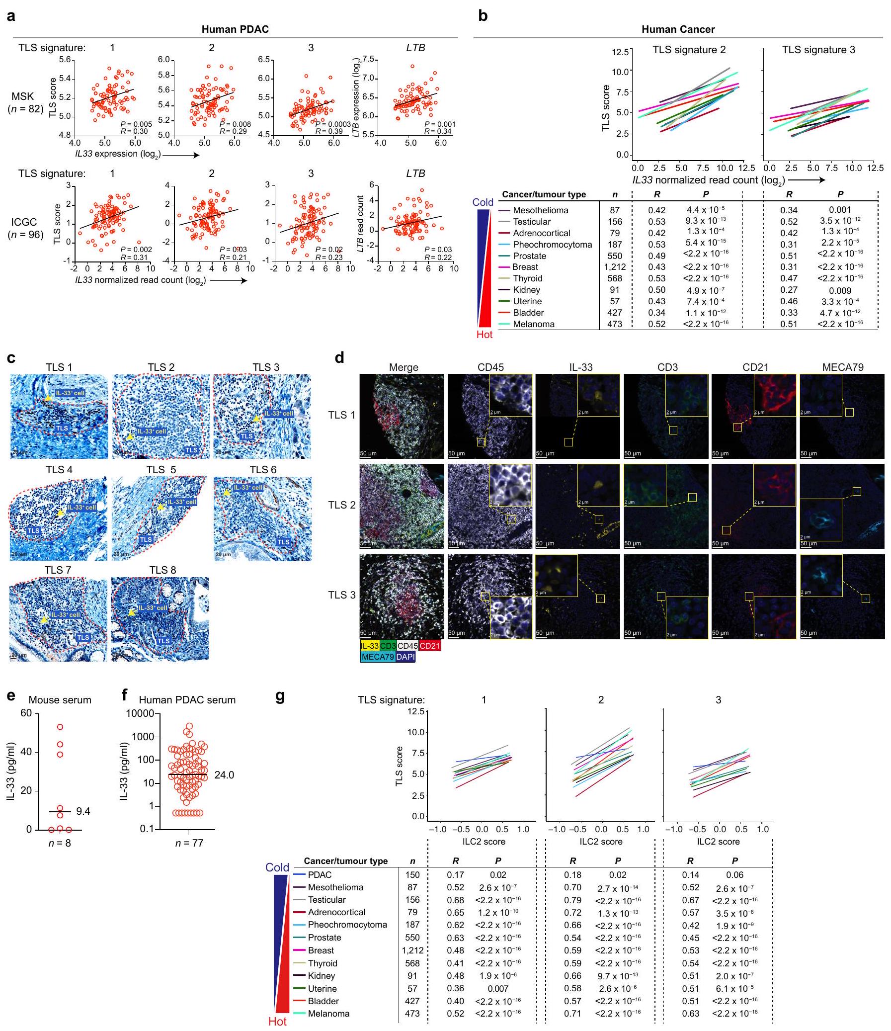
صور لـ TLSs في سرطان البنكرياس البشري. هـ، و، مستوى IL-33 في مصل الفئران المصابة بسرطان البنكرياس المعالجة بـ rlL-33 (هـ) وفي مرضى سرطان البنكرياس البشري (و). ز، ارتباط توقيع النسخ الخاص بـ ILC2 البشري المنشط بواسطة IL-33 بتوقيعات TLS (توقيعات TLS: ) في الأورام البشرية من أطلس جينوم السرطان. عدد الأورام فئران أو المرضى (إناث). البيانات التي تم جمعها بعد 4 أسابيع (هـ) من زراعة الورم، مجمعة من تجربتين مستقلتين مع فئران لكل مجموعة مع نتائج متسقة. القيم بواسطة معامل الارتباط بيرسون ثنائي الاتجاه ( ).

CD8 خلايا T
٨ PDAC 1 ( )
٦
٤ أو
2
منخفض معتدل


الشكل البياني الموسع 2 | المرض والنشاط المناعي مع تعديل IL-33 في سرطان البنكرياس القنوي والتهاب القولون الناتج عن DSS. أ، وزن الورم في الفئران البرية المعالجة بمضاد LTβR و
فئران PDAC.b، فقدان الوزن والبقاء في WT و فئران التهاب القولون الناتج عن DSS. ج، تكرار و خلايا T في نماذج الفئران لسرطان البنكرياس مع تسلل منخفض ومتوسط لخلايا T. د، H&E لـ TLSs في الأورام ذات تسلل منخفض ومتوسط لخلايا T. الخطوط البيضاء المنقطة = TLS المحتمل. هـ، و، صور المناعة الفلورية للتجمعات اللمفاوية داخل الورم والجريبات الأولية (هـ) وTLSs مع BCL6. خلايا B (ف) في فئران PDAC المعالجة بـ rlL-33. ج، فقدان الوزن والبقاء في
فئران الكوليتيس الناتج عن DSS المعالجة بالمركبة والمعالجة بـ rIL-33. البيانات المجمعة بعد 2 (أ)، 3 (ج، د PDAC 3، 4) و3-5 (ج، د PDAC 1، 2، 5، 6، هـ، و) أسابيع بعد زراعة الورم، مجمعة من تجارب مستقلة مع فئران لكل مجموعة مع نتائج متسقة. أورام فردية من فئران فردية أو فئران فردية تم تحليلها بشكل منفصل. القضبان الأفقية الوسيط. القيم بواسطة تحليل التباين الأحادي مع اختبار المقارنات المتعددة لتوكاي (أ)، اختبار مان-ويتني ذو الاتجاهين -اختبار (ج)، تحليل التباين ثنائي الاتجاه مع اختبار المقارنات المتعددة لسيداك (ب، ج، فقدان الوزن)، واختبار الرتبة اللوغاريتمية (ب، ج، البقاء).
الشكل البياني الممتد 3 | انظر الصفحة التالية للتعليق.

مقالة

الشكل البياني الموسع 3 | IL-33 يوسع تعبير LT ILC2s في فئران PDAC. أ، KLRG1 استراتيجية تصنيف ILC2. ب، و تعبير mRNA في KLRG1 المنقى ILC2s (PCR القطرات الرقمية – اليسار؛ تسلسل RNA أحادي الخلية – اليمين). ج، د، ف، تعبير LT على KLRG1 ILC2s بواسطة تحليل تدفق الخلايا (c، فئران PDAC المعالجة بـ rIL-33؛ f، فئران DSS-colitis المعالجة بـ rlL-33)، وتصوير مجسم لكشف KLRG1 المنقى داخل الورم ILC2s (د). في د، تعبير LT بواسطة تحليل تدفق الخلايا (يسار) والتصوير المجهري الفلوري (يمين) على WT المحفز بواسطة αCD3 و αCD28 و الخلايا (رسوم بيانية لتدفق السيتومتر) تظهر كضوابط إيجابية وسلبية. e، تكرار KLRG1 داخل الورم ILC2s في فئران PDAC المعالجة بـ rlL-33. PDAC 1-6 = فئران PDAC موضعية تم إنشاؤها باستخدام خطوط خلايا PDAC 1-6. تحليل تسلسل RNA أحادي الخلية لـ 794 خلية ILC2 نقية من الورم (g) و 1,668 خلية ILC2 نقية من الورم والعقد اللمفاوية المت draining (DLN) من فئران PDAC (h, i). خريطة الحرارة = أعلى 25
الجينات لكل مجموعة. UMAP، مخططات الكمان = عوامل النسخ ILC (h)، والعلامات (i) في المجموعات غير المراقبة في الشكل 3a، وسيتوكينات ILC2 (h). لم يكن Tbx21 قابلاً للاكتشاف. j، عوامل النسخ ILC على KLRG1 داخل الورم. ILC2s من فئران PDAC المعالجة بـ rIL-33 بواسطة تحليل تدفق الخلايا. البيانات التي تم جمعها بعد 10 أيام و أسبوعين (ب-ف) بعد زراعة الورم، مجمعة من تجارب مستقلة مع فئران لكل مجموعة مع نتائج متسقة. تم تحليل الأورام الفردية من الفئران الفردية بشكل منفصل. القضبان الأفقية = الوسيط؛ تُظهر الرسوم البيانية على شكل كمان التوزيع مع القيم الدنيا والقصوى والوسيط (الخطوط). القيم بواسطة تحليل التباين ثنائي الاتجاه مع اختبار المقارنات المتعددة لتوكاي (ب يسار، هـ، و)، اختبار رانك الموقع ويليكسون (ب، رسومات الكمان)، تحليل التباين ثنائي الاتجاه مع اختبار هولم بعد الاختبار (ج)، وتحليل التباين ثنائي الاتجاه مع اختبار المقارنات المتعددة لسيداك (د).
الشكل البياني الممتد 4 | لا يؤثر الحذف الحاد والشرطي لـ ILC2 على ILC1s وILC3s أو اللمفاويات غير ILC داخل الورم. أ، تعبير NMUR1 على خلايا المناعة داخل الورم في الخلايا المعالجة بـ rlL-33 في Nmur1 فئران PDAC. ب، ج، تصنيف وتكرار جميع خلايا ILCs (ب) وخلايا المناعة (ج) في أعضاء rll-33 والمعالجة بسم الدفتيريا (DT) Nmur1 روزا26 والمجموعة الضابطة من نفس القمامة ROSA26 فئران PDAC. د، قياس ILCs والخلايا المناعية في أعضاء الفئران المعالجة بـ rIL-33 Nmur1 وفئران PDAC من نفس السلالة. هـ، KLRG1 داخل الورم تحديد ILC2، التردد والعدد (يسار)، كثافة TLS
تردد الخلايا المناعية (يمين) في المعالجة بـ rll-33 و التحكم في الأشقاء فئران PDAC. الخط المنقط = TLS المفترض. تم جمع البيانات بعد 2 (ب-هـ) و 4 (أ) أسابيع من زراعة الورم، مجمعة من تجارب مستقلة مع فئران لكل مجموعة مع نتائج متسقة. الأورام الفردية أو الأعضاء من الفئران الفردية تم تحليلها بشكل منفصل. القضبان الأفقية الوسيط. في د، التحكم في الأشقاء ونمور1 مجمعة. القيم بواسطة تحليل التباين ثنائي الاتجاه مع اختبار المقارنات المتعددة لسيداك (ب، هـ اليمين) واختبار مان ويتني ذو الطرفين (هـ اليسار، الوسط).
الشكل البياني الموسع داخل الورم تعمل خلايا النخاع العظمي كمنظمي TLS. أ، LT تعبير R في الخلايا المناعية داخل الورم في فئران PDAC WT المعالجة بـ rIL-33. ب، تحديد وتصنيف تعبير mRNA بواسطة PCR القطرات الرقمية في LT داخل الورم المنقى و LT الخلايا النخاعية. ج-هـ، تسلسل RNA أحادي الخلية لـ 8,409 و خلايا المايلويد داخل الورم المنقاة من فئران PDAC المعالجة بـ rll-33. في (ج)، UMAP = خلايا حسب التجمعات غير المراقبة. خريطة الحرارة = أعلى 50 جينًا معبرًا عنه بشكل مختلف في و الخلايا. د، داخل الورم و LT أنماط خلايا النخاع العظمي بواسطة تسلسل RNA أحادي الخلية (يسار) وتعبير كيموكينات تحفيز TLS بواسطة PCR القطرات الرقمية (يمين). UMAP = خلايا بواسطة LT تعبير R وجينات توقيع TLS رسوم بيانية على شكل كمان للكيماوكينات المعبر عنها بشكل مختلف (يسار) وعالمي (يمين) بواسطة LT تعبير mRNA لـ Il33 (يسار) بواسطة PCR القطرات الرقمية وبروتين IL-33 (يمين) بواسطة تحليل التدفق في خلايا النخاع المنقي داخل الورم.
كثافة ILC2 في WT المعالج بمضاد LTβR فئران PDAC. ح، مخطط تجريبي (أعلى) وLT MFI لـ KLRG1 داخل الورم كثافة خلايا ILC2 وTLS داخل الورم (أسفل) في فئران PDAC المعالجة بـ rll-33 والمزروعة مع الخلايا المنقاة من WT أو II33 فئران PDAC. البيانات المجمعة 4 (أ) و أسابيع بعد زراعة الورم، مجمعة من تجارب مستقلة مع فئران لكل مجموعة مع نتائج متسقة. تم تحليل الأورام الفردية من الفئران الفردية بشكل منفصل. القضبان الأفقية = الوسيط؛ تُظهر الرسوم البيانية على شكل كمان التوزيع مع الحد الأدنى، والحد الأقصى، والوسيط (الخطوط). MFI = متوسط شدة الفلورسنت. القيم بواسطة اختبار ANOVA أحادي الاتجاه (أ)، اختبار مان ويتني ذو الطرفين (ب، د اليمين، ف، ح)، اختبار ويلكوكسون للرتب الموقعة (د، هـ مخططات الكمان)، واختبار ANOVA ثنائي الاتجاه مع اختبار المقارنات المتعددة لسيداك (ج، ح LT MFI).

الشكل 6 من البيانات الموسعة | استراتيجية تصنيف ILC2 وتكرارها في بارابيوتيك

فئران PDAC. أ، تكرار KLRG1 ILC2s في دم الفئران المصابة بسرطان البنكرياس PDAC المعالجة بـ rIL-33. ب-د، مخطط تجريبي واستراتيجية تصنيف (ب) للمتبرع والمستقبل KLRG1 ILC2s في دم المتلقي. تحديد (ج) وتكرار الخلايا غير ILCs المشتقة من المتبرع (أسفل)، ILC2s KLRG1 (د أعلى) و CD45 الخلايا (د أسفل) في دم المتلقين والأورام. هـ، تصنيف في فئران PDAC من نوع KikGR التي تم تحويلها بالضوء كما في
الشكل 4ب؛ مقيد على KLRG1 ILC2s. البيانات المجمعة في اليوم السابع (أ-د، دم) واليوم الرابع عشر (ج، د، ورم) بعد زراعة الورم، مجمعة من تجارب مستقلة مع فئران لكل مجموعة مع نتائج متسقة. تم تحليل الأورام الفردية أو الأعضاء من الفئران الفردية بشكل منفصل. القضبان الأفقية = الوسيط. القيم بواسطة اختبار مان-ويتني ثنائي الذيل (أ، ج، د).
الشكل 7 من البيانات الموسعة | تؤثر PDACs على تنوع وتركيب ميكروبات الأمعاء. أ، KLRG1 البنكرياسي تردد ILC2 في الفئران المعالجة بالشام والفئران المصابة بسرطان البنكرياس. ب، مخطط تجريبي وتحليلات الميكروبيوم البرازي في الفئران المصابة بسرطان البنكرياس من خلال تنوع ألفا (مؤشر شانون، يسار) وتحليل المكونات الرئيسية (يمين). ج، مخطط تجريبي، IL-33 في المصل، وKLRG1 في الأمعاء. ILC2s في فئران PDAC المعالجة بـ rIL-33 مع وبدون علاج بالمضادات الحيوية (Abx). د، KLRG1 في الأمعاء ILC2s
في فئران PDAC الخالية من الجراثيم مع وبدون زراعة ميكروبية برازية (FMT) من فئران WT. البيانات المجمعة و 4 (ج، د) أسابيع بعد زراعة الورم، مجمعة من تجارب مستقلة مع فئران لكل مجموعة مع نتائج متسقة. قياسات فردية من فئران فردية تم تحليلها بشكل منفصل. القضبان الأفقية الوسيط. القيم بواسطة اختبار مان-ويتني ثنائي الاتجاه (أ، ج، د) واختبار ويلش t (ب).
الشكل البياني الممتد 8|انظر الصفحة التالية للتعليق.

مقالة

الشكل التوضيحي الممتد 8 | المحددات الجزيئية للتحكم المزدوج في PDAC بواسطة rIL-33. أ، حجم الورم في PDACs تحت الجلد (s.c.) في الفئران المعالجة بالدواء الوهمي والفئران المعالجة بـ rIL-33 مع PDAC تحت الجلد فقط (PDAC فردي) أو PDACs تحت الجلد والبنكرياس (PDAC مزدوج). ب، تصنيف ILC2s من الفئران المتزاوجة مع PDACs تحت الجلد في المتلقين مع/بدون PDAC البنكرياس في المتبرعين كما هو موضح في الشكل 4f. ج، مخطط تجريبي (يسار)، تصنيف (أعلى) وقياس ILC2s المشتقة من المتبرع في البنكرياس المتبرع (أسفل اليسار)، PDAC تحت الجلد في المتلقي (أسفل الوسط) والدم (أسفل اليمين) في فئران PDAC المتزاوجة المعالجة بالدواء الوهمي، rIL-33 أو rIL-25. د، هـ، حجم الورم في الفئران WT المعالجة بـ rIL-33، Il1rl1 و سرطان البنكرياس تحت الجلد (مفرد) وسرطان البنكرياس تحت الجلد + سرطان البنكرياس (ثنائي) في الفئران. حجم الورم في سرطان البنكرياس تحت الجلد في الفئران المعالجة بـ rlL-33
تحكم في فضلات الأشقاء و فئران PDAC الفردية والثنائية. تصنيف ILC2s من الفئران المتزاوجة مع PDACs تحت الجلد في المستلمين وPDACs البنكرياسية في المتبرعين المعالجين بالمضادات الحيوية أو بدونها كما هو موضح في الشكل 4g. تم جمع البيانات بعد 5-9 أيام (ج، الدم)، 2 (ب، ج، الورم) و3-5 (د) أسابيع بعد الزرع، مجمعة من تجارب مستقلة مع فئران لكل مجموعة مع نتائج متسقة. الأورام الفردية أو الأعضاء من الفئران الفردية تم تحليلها بشكل منفصل. القضبان الأفقية = الوسيط. رفقاء التحكم من نوع Cre = و أورا القيم بواسطة تحليل التباين الثنائي الاتجاه مع اختبار المقارنات المتعددة لتوكاي (أ)، اختبار مان-ويتني ذو الاتجاهين (وزن الورم د) وتحليل التباين الثنائي الاتجاه مع اختبار المقارنات المتعددة لسيداك (بقية الأمور).

b


C


الشكل 9 من البيانات الموسعة | KLRG1 داخل الورم البشري تحديد ILC2 والنمط الظاهري. أ-ج، استراتيجية التحديد (أ) وتعبير العلامات (ب، ج) على KLRG1 داخل الورم ILC2s في سرطان البنكرياس الغدي البشري. المرضى الأفراد. MFI شدة الفلورة المتوسطة. القيم بواسطة اختبار مان ويتني ذو الذيلين المزدوج .

مقالة

الشكل البياني الممتد 10| مسار IL-33-ILC2-TLS. الرسم التخطيطي المقترح لمسار IL-33-ILC2-TLS. TLS: الهيكل اللمفاوي الثلاثي. © 2024 مركز ميموريال سلوان-كيترينغ للسرطان، مستشفى ميموريال للسرطان والعلوم المرتبطة
الأمراض، ومعهد سلوان-كيترينغ لأبحاث السرطان، كل منهما في نيويورك، نيويورك. جميع الحقوق محفوظة. أعيد نشره بإذن.

بحث الطبيعة

المؤلف (المؤلفون) المراسلون:
آخر تحديث بواسطة المؤلف(ين): 27/8/2024

ملخص التقرير

تسعى Nature Research إلى تحسين قابلية تكرار الأعمال التي ننشرها. يوفر هذا النموذج هيكلًا للاتساق والشفافية في التقرير. لمزيد من المعلومات حول سياسات Nature Research، يرجى الاطلاع على سياسات التحرير وقائمة مراجعة سياسة التحرير.

الإحصائيات

لجميع التحليلات الإحصائية، تأكد من أن العناصر التالية موجودة في أسطورة الشكل، أسطورة الجدول، النص الرئيسي، أو قسم الطرق.

غير متوفر


تم التأكيد
حجم العينة بالضبط لكل مجموعة/شرط تجريبي، معطاة كرقم منفصل ووحدة قياس
بيان حول ما إذا كانت القياسات قد أُخذت من عينات متميزة أو ما إذا كانت نفس العينة قد تم قياسها عدة مرات
اختبار(ات) الإحصاء المستخدمة وما إذا كانت أحادية الجانب أو ثنائية الجانب
يجب أن تُوصف الاختبارات الشائعة فقط بالاسم؛ واصفًا التقنيات الأكثر تعقيدًا في قسم الطرق.

وصف لجميع المتغيرات المشتركة التي تم اختبارها

وصف لأي افتراضات أو تصحيحات، مثل اختبارات الطبيعية والتعديل للمقارنات المتعددة

وصف كامل للمعلمات الإحصائية بما في ذلك الاتجاه المركزي (مثل المتوسطات) أو تقديرات أساسية أخرى (مثل معامل الانحدار) و التباين (مثل الانحراف المعياري) أو تقديرات مرتبطة بعدم اليقين (مثل فترات الثقة)

لاختبار الفرضية الصفرية، إحصائية الاختبار (على سبيل المثال، ) مع فترات الثقة، أحجام التأثير، درجات الحرية و قيمة ملحوظة أعطِ القيم كقيم دقيقة كلما كان ذلك مناسبًا.

□ لتحليل بايزي، معلومات حول اختيار الأوليات وإعدادات سلسلة ماركوف مونت كارلو

□ لتصميمات هرمية ومعقدة، تحديد المستوى المناسب للاختبارات والتقارير الكاملة عن النتائج
□ س
تقديرات أحجام التأثير (مثل حجم تأثير كوهين) بيرسون )، مما يشير إلى كيفية حسابها
تحتوي مجموعتنا على الويب حول الإحصائيات لعلماء الأحياء على مقالات تتناول العديد من النقاط المذكورة أعلاه.

البرمجيات والشيفرة

معلومات السياسة حول توفر كود الكمبيوتر

جمع البيانات
لتحليل تدفق الخلايا، تم تحليل جميع العينات على جهاز FACS LSR Fortessa (BD Biosciences) باستخدام FlowJo (الإصدار 10.7.1، Tree Star).
تم رقمنة الشرائح المرضية باستخدام جهاز Panoramic Flash 250 (3Dhistech، بودابست، المجر) باستخدام عدسة Zeiss 20x/0.8NA ومرشحات مخصصة لـ A488 و A546 و A594 و A647.
H&E: تم مسح الشرائح باستخدام الماسح الضوئي البانورامي (3DHistech، بودابست، المجر)
تم تحديد عدد الخلايا النواة في TLS باستخدام وظيفة تحليل الجسيمات في ImageJ (الإصدار 2.3.0، NHI، الولايات المتحدة الأمريكية). تم عد خلايا IL33+ في TLS يدويًا.
تم تحديد إيجابية CD45 و IL33 بواسطة CaseViewer (3DHISTECH Ltd.).
مجهري: لتصوير LT-PE وDAPI النووي، تم التقاط الصور على نظام مجهر ليزري مسح نقطي (SP5، لايكا، ويتزلار، ألمانيا) باستخدام عدسة تكبير 63x مع زيت.
تحليل الميكروبيوم: تم تسلسل المكتبات المجمعة باستخدام جهاز إيلومينا ميسيك.
تم إجراء اختبار تقرير ST2: تم الكشف عن امتصاص الطول الموجي 630 نانومتر باستخدام جهاز قراءة Cytation 3 (BioTek).
تسلسل مستقبلات خلايا B: تم إجراء تسلسل المناعي لمناطق CDR3 لسلاسل مستقبلات خلايا B في الفئران على الحمض النووي الجيني من عينات مثبتة بالصيغة الفورمالينية باستخدام اختبار immunoSEQ (تقنيات البيولوجيا التكيفية).
تم إجراء توليد القطرات في نظام QX200 ddPCR (بايو راد) باستخدام cDNA الناتج من 0.8-2 نانوغرام من RNA الكلي مع مجموعة OneStep RT-ddPCR المتقدمة للبروبي (بايو راد) وفقًا لبروتوكول الشركة المصنعة مع النسخ العكسي في والمعالجة الحرارية/
امتداد في تم تقييم كل عينة في نسختين فنيتين. تم قراءة الألواح وتحليلها باستخدام برنامج (QuantaSoft) لتقييم عدد القطرات الإيجابية للجينات المعنية.

تحليل البيانات

تم إجراء التحليلات الإحصائية باستخدام (الإصدار 4.0.3، تسلسل RNA أحادي الخلية) و Prism 9.2.0 (برنامج GraphPad، كل شيء آخر).
تحليل التعبير الجيني للأورام: تم تحليل البيانات باستخدام (الإصدار 4.0.3).
تدفق الخلايا: تم تحليل البيانات باستخدام FlowJo (الإصدار 10.7.1، Tree Star).
تم تحديد عدد TLSs في ثلاث مقاطع على الأقل باستخدام QuPath الإصدار 0.2.3.
تم تحديد عدد الخلايا النواة في TLS باستخدام وظيفة تحليل الجسيمات في ImageJ الإصدار 2.3.0.
تم قياس خلايا IL33+ و IL33+CD45+ باستخدام برنامج ImageJ الإصدار 2.3.0، وتم تقدير عدد الخلايا لكل وحدة مساحة من خلال حساب المساحة السطحية بناءً على وجود خلايا DAPI+.
المجهر الضوئي المتداخل: تم تصور الصور ذات الإشارات الثنائية باستخدام ImageJ الإصدار 2.3.0.
تسلسل RNA على مستوى الخلية الواحدة: تم إجراء المعالجة الأولية لصور التسلسل باستخدام برنامج التحليل في الوقت الحقيقي من إيلومينا (RTA). مجموعة برامج 10X Genomics Cell Ranger للخلية الواحدة الإصدار 3.0.2https://support.10xgenomics.com/تم استخدام (single-cell-gene-expression/software/pipelines/latest/what-is-cellranger) لفصل العينات، ومحاذاتها إلى مرجع الجينوم الفأري mm10، وتصفيتها، وعدّ وحدات التعبير الجيني (UMIs)، وجينات نهاية الخلية المفردة 5’، ومراقبة الجودة وفقًا لمعايير الشركة المصنعة. تم تحليل البيانات المعالجة بعد ذلك في (الإصدار 4.0.3).
تحليل الميكروبيوم: تم معالجة القراءات الخام المفككة باستخدام Nextflow، خط أنابيب nf-core ampliseq (الإصدار 2.4.0)، مع المعلمات التالية: -profile singularity –FW_primer GTGYCAGCMGCCGCGGTAA –RV_primer CCGYCAATTYMTTTRAGTTT –dada_ref_taxonomy silva -ignore_empty_input_files –ignore_failed_trimming –min_frequency 10 –retain_untrimmed –trunclenf 240 –trunclenr 160. على وجه التحديد، تم تقليم القراءات باستخدام cutadapt (https://doi.org/10.14806/ej.17.1.200تم إجراء معالجة البيانات باستخدام PhiX، وتصنيف الجودة، ودمج أزواج القراءة، وحل متغيرات تسلسل الأمبليكون باستخدام DADA2. كما تم إجراء التعيين التصنيفي اللاحق باستخدام DADA2، مع الاستعانة بقاعدة بيانات المرجع Silva (الإصدار 138). تم تحليل جداول الوفرة باستخدام حزمة البرمجيات QIIME2.
تسلسل مستقبلات خلايا B: تم معالجة بيانات التسلسل وتحليلها على قاعدة البيانات العلائقية المستندة إلى الويب immunoSEQ Analyser.
التحور الجسدي: تم الاحتفاظ بإعادة ترتيب تسلسلات السلاسل الثقيلة المنتجة لتحليل العلاقات التكتلية لخلايا B، وتم تعريف التكتلات من خلال التجميع الطيفي المدرك لـ VDJ باستخدام حزمة R SCOPer. تم حساب ترددات الطفرات باستخدام دالة الطفرات المرصودة في حزمة R Shazam.
بالنسبة للمخطوطات التي تستخدم خوارزميات أو برامج مخصصة تكون مركزية في البحث ولكن لم يتم وصفها بعد في الأدبيات المنشورة، يجب أن تكون البرمجيات متاحة للمحررين والمراجعين. نحن نشجع بشدة على إيداع الشيفرة في مستودع مجتمعي (مثل GitHub). راجع إرشادات Nature Research لتقديم الشيفرة والبرمجيات لمزيد من المعلومات.

بيانات

معلومات السياسة حول توفر البيانات

يجب أن تتضمن جميع المخطوطات بيانًا عن توفر البيانات. يجب أن يوفر هذا البيان المعلومات التالية، حيثما ينطبق:
  • رموز الانضمام، معرفات فريدة، أو روابط ويب لمجموعات البيانات المتاحة للجمهور
  • قائمة بالأشكال التي تحتوي على بيانات خام مرتبطة بها
  • وصف لأي قيود على توفر البيانات
تم توفير بيانات المصدر لجميع التجارب. بيانات تسلسل RNA من TCGA و ICGC متاحة للجمهور. جميع بيانات التعبير الجيني الأخرى متاحة تحت رقم الوصول إلى Gene Expression Omnibus (GEO) GSE184585. تم الإبلاغ عن بيانات تسلسل RNA أحادي الخلية (scRNA-seq) لـ ILC2s سابقًا ومتاحة تحت رقم الوصول GEO GSE136720. بيانات تسلسل RNA أحادي الخلية للخلايا النخاعية تحت رقم الوصول GEO GSE225990. تسلسل جين 16s rRNA متاح تحت رقم معرف BioProject PRJNA944673.
جميع البيانات الأخرى متاحة من المؤلفين عند الطلب المعقول.

التقارير الخاصة بالمجال

يرجى اختيار الخيار أدناه الذي يناسب بحثك بشكل أفضل. إذا لم تكن متأكدًا، اقرأ الأقسام المناسبة قبل اتخاذ قرارك.
علوم الحياة □ العلوم السلوكية والاجتماعية □ العلوم البيئية والتطورية والبيئية
لنسخة مرجعية من الوثيقة بجميع الأقسام، انظرnature.com/documents/nr-reporting-summary-flat.pdf

تصميم دراسة العلوم الحياتية

يجب على جميع الدراسات الإفصاح عن هذه النقاط حتى عندما يكون الإفصاح سلبياً.
حجم العينة
تم تحديد أحجام العينات بناءً على تجربتنا وتجارب باحثين آخرين مع خطوط الخلايا المعنية. لم تُستخدم أي طرق إحصائية حيث لاحظنا العديد من التأثيرات ذات الدلالة الإحصائية في البيانات باستخدام الطرق المذكورة أعلاه لاختيار حجم العينة دون حسابات مسبقة لحجم العينة.
استثناءات البيانات
لم يتم استبعاد أي بيانات من التحليلات.

التقارير عن مواد وأنظمة وطرق محددة

نحتاج إلى معلومات من المؤلفين حول بعض أنواع المواد والأنظمة التجريبية والأساليب المستخدمة في العديد من الدراسات. هنا، يرجى الإشارة إلى ما إذا كانت كل مادة أو نظام أو طريقة مدرجة ذات صلة بدراستك. إذا لم تكن متأكدًا مما إذا كان عنصر القائمة ينطبق على بحثك، يرجى قراءة القسم المناسب قبل اختيار رد.
المواد والأنظمة التجريبية
غير متوفر مشارك في الدراسة
خطوط خلايا حقيقية النواة
□ علم الحفريات وعلم الآثار
– الحيوانات وغيرها من الكائنات الحية
المشاركون في الأبحاث البشرية
V
طرق
مشارك في الدراسة
تسلسل شريحة الكروماتين
تدفق الخلايا
تصوير الأعصاب القائم على الرنين المغناطيسي

الأجسام المضادة

الأجسام المضادة المستخدمة
أجسام مضادة لتدفق الفأر
اسم الأجسام المضادة لقناة الهدف صانع رقم الكتالوج النسخة الكمية ( ما لم يُذكر خلاف ذلك
CD11b APC APC رات مضاد للفأر CD11b BD Bioscience 553312 M1/70 0.5
CD11b APC-Cy7 مضاد CD11b من الفأر BD Bioscience 557657 M1/70 2
CD11c APC APC هامستر مضاد للفأر CD11c BD Bioscience 550261 HL3 0.5
CD11c BV510 Brilliant Violet 510 مضاد للفأر CD11c BioLegend 117338 N418 0.5
CD11c FITC مضاد مو CD11c، FITC إيفيتروجين 11-0114-85 N418 0.5
CD127 FITC مضاد مو CD127، FITC إيفيتروجين 11-1271-85 A7R34 1
CD127 PE-Cy7 PE/سيانين 7 مضاد للفأر CD127 BioLegend 135014 A7R34 1
CD19 PE PE مضاد للفأر CD19 BioLegend 115508 6D5 1
CD19 BV785 Brilliant Violet 875 مضاد للفأر CD19 BioLegend 115543 6D5 1
CD19 AF700 مضاد مو CD19، أليكسا فلوور 700 إنفيتروجين 65-0193-82 eBio1D3 1.25
CD25 PerCP-Cy5.5 PerCP-Cy5.5 مضاد للفأر CD25 BioLegend 102030 PC61 1.5
CD3 BV711 فيوليت لامع 711 مضاد لـ CD3e الفأري BioLegend 100349 145-2C11 0.5
CD3 AF700 مضاد مو CD3e، أليكسا فلور 700 إنفيتروجين 56-0032-82 17A2 1.25
CD4 BV711 BV711 مضاد للفأر CD4 BioLegend 100550 RM4-5 0.5
CD4 BV786 فيوليت لامع 76 مضاد للفأر CD4 BioLegend 100552 RM4-5 0.5
CD45 باك بلو مضاد للفأر CD45 بايو ليجند 103126 30-F11 0.5
CD45.1 باك بلو مضاد للفأر CD45.1 من بايو ليجند 110722 A20 0.5
CD45.1 BV711 BV711 مضاد للفأر CD45.1 BioLegend 110739 A20 0.5
CD45.2 APC-Cy7 APC-Cy7 مضاد للفأر CD45.2 BioLegend 1098241040.5
CD5 APC APC رات مضاد للفأر CD5 BD بيوساينس 550035 53-7.3 0.5
CD8 BV510 فيوليت برايت (BV) 510 مضاد للفأر CD8a بايو ليجند 100751 53-6.7 2
CD90.2 BV786 BV786 فأر مضاد للفأر CD90.2 BD Bioscience 564365 53-2.1 0.5
محلول DAPI PacBlue DAPI BD Bioscience
DRAQ7 APC-Cy7 DRAQ7 BD Phermigen 51-9011172 1
F4/80 PE-Cy5 مضاد مو F4/70، PE-Cy5 إيفيتروجين 15-4801-82 BM8 0.5
Fc-block غير متوفر InVivoMab مضاد للفأر CD16/32 BioXCell BE0307 2.4G2 10μl/عينة
FcER1 APC مضاد مو FceRi ألفا، APC إنفيتروجن 17-5898-82 “MAR-1” 0.5
جهاز مضاد GATA3 BV711 BV711 فئران مضاد لـ GATA3 BD Horizon 565449 L50-823 2
GR-1 BV605 BV605 فأر مضاد لمستضد Ly-6G وLy-6C BD Bioscience 563299 RB6-8C5 0.3
IL-33 PE IL-33 جسم مضاد وحيد النسيلة (396118) إنفيتروجين MA5-23640 39611810
IL17Rb PE PE مضاد للفأر IL17RB BioLegend 146306 9B10 1.25
نموذج الأيزوتيب لـ alL-33 PE Rat IgG2a kappa Isotype Control، PE Invitrogen 12-4321-80 eBR2a 1
KLRG1 BV510 Brilliant Violet 510 مضاد للفأر/البشر KLRG1 BioLegend 138421 2F1/KLRG1 1.25
LTBR PE-Cy7 PE/سيانين7 جسم مضاد لمستقبل اللمفوتوكسين بيتا للفأر BioLegend 134410 5G11 0.5
مضاد MHC الفئة 2 AF700 لمولد المضاد MHC الفئة II، أليكسا فلور 700 إنفيتروجين 56-5321-82 M5/114.15.2 0.5
NK1.1 BV650 BV650 مضاد للفأر NK1.1 BD Bioscience 564143 PK136 0.5
NK1.1 APC APC فأر مضاد لفأر NK1.1 BD Phermigen 550627 PK136 0.5
NK1.1 BV605 فيوليت برايت 605 مضاد للفأر NK1.1 بايو ليجند 108740 PK136 0.5
الأجسام المضادة البيوتينية NMUR1-النسخة 12-A03-A
أجسام مضادة لتدفق الإنسان
اسم الجسم المضاد لقناة الهدف الشركة المصنعة رقم الكتالوج النسخة الكمية
جهاز مضاد GATA3 PE PE الفأر من BD Biosciences 560074 L50-823 4
PD-1 BV786 BV786 فأر مضاد للبشر CD279 (PD-1) BD Biosciences 563789 EH12.1 2
RORgt BV650 BV650 مضاد للفأر البشري RORgt BD Biosciences 563424 Q21-559 2
الأجسام المضادة الثانوية لـ hLTBR-Fc BV650 BV650 فأر مضاد للبشر IgG BD Biosciences 740596 G18-145 2
الأجسام المضادة الثانوية لـ hLTBR-Fc PE PE فأر مضاد للبشر IgG BD Biosciences 555787 G18-145 2
CD117 PE-Cy5 PE/Cy5 مضاد للبشر CD117 (c-kit) BioLegend 323412 A3C6E2 2
CD11b APC APC مضاد للبشر CD11b BioLegend 301310 ICRF44 2
CD161 BV785 فيوليت برايت 785 مضاد للبشر CD161 BioLegend 339930 HP-3G10 2
CD25 APC-Cy7 APC/Cy7 مضاد للبشر CD25 BioLegend 302614 BC96 2
CD45 PacBlue الأزرق الباسيفيكي مضاد للبشر CD45 BioLegend 304029 HI30 2
CD56 BV605 فيوليت برايت 605 مضاد للبشر CD56 (NCAM) بايو ليجند 318334 HCD56 2
CRTH2 PerCP-Cy5.5 PerCP/Cy5.5 مضاد للبشر CD294 (CRTH2) BioLegend 350116 BM16 4
CRTH2 PE PE مضاد الإنسان CD294 (CRTH2) BioLegend 350106 BM16 4
FceRIa APC APC مضاد لـ FceRIa البشري BioLegend 334612 AER-37 2
ICOS BV711 بنفسجي لامع 711 مضاد للبشر/الفئران/الجرذان CD278 (ICOS) BioLegend 313548 C398.4A 2
NKp44 PE-Cy7 PE/Cy7 مضاد للبشر CD336 (NKp44) BioLegend 325116 P44-8 2
T-BET BV711 بنفسجي لامع 711 مضاد-Tbet BioLegend 6448204 B10 2
تفاعل TCR a/b AF700 أليكسا فلور 700 مضاد لـ TCR البشري a/b بايو ليجند 306730 IP26 2
KLRG1 BV510 Brilliant Violet 510 مضاد للفأر/البشر KLRG1 BioLegend 138421 2F1/KLRG1 2
CD11c APC مضاد Hu CD11c، APC Invitrogen 17-0116-42 3.92
CD127 FITC مضاد Hu CD127، FITC Invitrogen 11-1278-42 eBioRDR5 2
CD14 APC مضاد Hu CD14، APC إيفيتروجين 17-0149-42 61D3 2
CD16 APC مضاد Hu CD16 Invitrogen 17-0168-42 eBioCB16 2
CD19 AF700 مضاد Hu CD19 إيفيتروجين 56-0199-42 HIB19 2
CD3 AF700 مضاد Hu CD3، أليكسا فلور 700 إنفيتروجين 56-0037-42 OKT3 2
CD5 APC مضاد Hu CD5، APC إنفيتروجن 17-0058-42 L17F12 2
ST2 PE Anti-Hu IL-33R (ST2)، PE Invitrogen 12-9338-42 hIL33Rcap 4
بروتين مؤتلف
اسم المنتج رقم كتالوج الصانع التطبيق التركيز/الجرعة
rhLTBR/Fc كيميرا أنظمة البحث والتطوير 629-LR تدفق
نظام البحث والتطوير Fc Chimera R&D 1008-LR 4C
بروتين IL-33 المؤتلف من الفأر أنظمة R&D 3626-ML-010/CF العلاج (عن طريق الحقن في الصفاق)
بروتين IL-25 المؤتلف من الفأر أنظمة R&D 1399-ML-010/CF العلاج (عن طريق الحقن في الصفاق)
سم الدفتيريا (DT) EMD Millipore 322326100 نانوجرام
مادة في الجسم/ في المختبر
اسم المنتج رقم كتالوج الصانع التطبيق تفاصيل الجرعة
أجسام مضادة وحيدة النسيلة لمستقبل اللمفوتوكسين بيتا من إيفيتروجن 16-5671-82 الثقافة غير متاحة النسخة 3C8
أجسام مضادة وحيدة النسيلة لمستقبل اللمفوتوكسين بيتا من إيفيتروجن 16-5671-38 فيفو حقن Clone 3C8
تحكم إيزوتيب IgG1 كابا من رات إنفيتروجين فيفو / حقن نوع الأيزوتيب لم agonist aLTBR
التحقق
تم التحقق من جميع الأجسام المضادة من قبل الشركة المصنعة واستخدامها وفقًا لتعليماتها. في تجاربنا، تم تضمين عينات التحكم من نوع الأيزوتيب و/أو FMO. يمكن العثور على معلومات إضافية حول التحقق على مواقع الشركات المصنعة.

خطوط خلايا حقيقية النواة

معلومات السياسة حول خطوط الخلايا
مصدر(s) خط الخلايا
المصادقة
تلوث الميكوبلازما
خطوط يتم التعرف عليها بشكل خاطئ بشكل شائع
(انظر سجل ICLAC)
تم اشتقاق جميع خطوط خلايا الورم من فئران KPC (Pdx1-Cre; LSL-KrasG12D/+; LSL-Trp53R172H/+) أو KPCY (Pdx1-Cre; LSL-KrasG12D/+; LSL-Trp53R172H/+; Rosa26YFP/YFP) وكانت هدايا من R.H. Vonderheide و B.J Stanger. تم الحصول على خط خلايا HEK-Blue-IL33 من Invivogen.
تم التحقق من جميع خطوط الخلايا على أنها خطوط خلايا سرطانية حقيقية من البنكرياس. وكان ذلك بناءً على التحقق النسيجي من قبل أخصائي أمراض البنكرياس المخصص، حيث أن هذه الخطوط الخلوية تولد أورامًا عند زراعتها داخل البنكرياس تعيد بشكل دقيق ميزات كل من سرطانات البنكرياس البشرية وسرطانات البنكرياس التي تتطور في الفئران المعدلة وراثيًا بشكل عفوي.
تم اختبار خطوط الخلايا بانتظام باستخدام مجموعة كشف الميكوبلازما MycoAlert (لونزا). لم تختبر أي من خطوط الخلايا المستخدمة في هذه الدراسة إيجابياً للميكوبلازما.
لم يتم استخدام أي خطوط يتم التعرف عليها بشكل خاطئ بشكل شائع في هذه الدراسة.

الحيوانات والكيانات الأخرى

معلومات السياسة حول الدراسات التي تشمل الحيوانات؛ توجيهات ARRIVE الموصى بها للإبلاغ عن أبحاث الحيوانات
الحيوانات المخبرية تم شراء فئران C57BL/6 (النوع البري [WT]، CD45.2) وفئران C57BL/6 CD45.1 من مختبر جاكسون. كانت فئران ||1r|1-/- (المعوزة لـ ST2) و II33-/- هدية من M.J. Rosen. كانت فئران Ltbfl/fl هدية من A.V. Tumanov. تم وصف فئران Nmur1iCre-eGFP ROSALSL-DTR سابقًا. تم تهجين Ltbfl/fl مع Nmur1iCre-eGFP و II7rCre/+ للحصول على فئران Nmur1iCre-iGFP Ltbfl/fl و II7rCre/+Ltbfl/fl. كانت فئران II7rCre/+Rorafl/fl هدية من A.N.J. McKenzie. كانت فئران Ltbr-/- هدية من T.T. Lu. كانت فئران CAG-KikGR33 هدية من G.E. Diehl. تم توفير الفئران الخالية من الجراثيم من منشأة الجنوطوبيوتيك في مركز وييل كورنيل الطبي (MSK). في جميع التجارب، كانت الفئران تتراوح أعمارها بين 6-14 أسبوعًا، وتمت مطابقتها من حيث العمر والجنس، وتم تعيينها عشوائيًا إلى مجموعات العلاج المحددة، مع إجراء ما لا يقل عن تجربتين مستقلتين طوال الوقت. تم استخدام كل من الحيوانات الذكرية والأنثوية. تم تربية الحيوانات والحفاظ عليها في منشأة حيوانات خالية من مسببات الأمراض المحددة في مركز ميموريال سلوان كيتيرينغ للسرطان.
الحيوانات البرية لم يتم استخدام حيوانات برية.
عينات تم جمعها من الميدان لم يتم استخدام عينات تم جمعها من الميدان في هذه الدراسة.
رقابة الأخلاقيات تم تربية الحيوانات والحفاظ عليها في منشأة حيوانات خالية من مسببات الأمراض، وتم إجراء جميع التجارب وفقًا لبروتوكول معتمد من لجنة رعاية واستخدام الحيوانات المؤسسية (IACUC) في مركز ميموريال سلون كيترينغ للسرطان (MSK) وامتثالًا لجميع اللوائح الأخلاقية ذات الصلة.
يرجى ملاحظة أنه يجب أيضًا تقديم معلومات كاملة حول الموافقة على بروتوكول الدراسة في المخطوطة.

مشاركون في البحث البشري

معلومات السياسة حول الدراسات التي تشمل المشاركين في الأبحاث البشرية
خصائص السكان
تُفصّل الخصائص السريرية للمرضى في مصفوفة الأنسجة الدقيقة، وتدفق السيتومتر، وزراعة ILC2، ومجموعات النسخ في الجداول التكميلية 1 و2 و3 و5، وتُقدم أدناه.
الخصائص العيادية المرضية لمرضى سرطان البنكرياس القنوي في مجموعة ترانسكريبتوم MSK )
الخصائص ن (%)
الجنس
ذكر 40 (49)
أنثى 42 (51)
العمر (سنة)
الوسيط (المدى interquartile) 68 (62-75)
موقع الورم
رأس 53 (65)
الجسم/الذيل 29 (35)
الإجراء
استئصال البنكرياس والاثني عشر 53 (65)
استئصال البنكرياس البعيد 29 (35)
pT
12 (2)
21 (1)
378 (95)
41 (1)
pN
033 (40)
148 (59)
x1 (1)
pM
081 (99)
11 (1)
المرحلة المرضية
IA 2 (2)
IB 1 (1)
IIA 30 (37)
IIB 47 (57)
III 1 (1)
الرابع 1 (1)
هامش جراحي
إيجابي 9 (11)
سالب 71 (87)
غير متاح 2 (2)
العلاج المساعد
نعم 61 (74)
لا 21 (26)
الخصائص السريرية المرضية لمرضى سرطان البنكرياس القنوي في مجموعة IHC و IF )
الخصائص ن (%)
الجنس
Male 4 (36)
Female 7 (64)
Age (y)
Median (IQR) 70 (62-79)
Tumor location
Head 9 (82)
Body/tail 2 (18)
Procedure
Pancreaticoduodenectomy 9 (82)
Distal pancreatectomy 2 (18)
pT
10(0)
20(0)
3 11 (100)
4 (0)
pN
O (45)
16 (55)
pM
0 11 (100)
10(0)
Pathological stage
IIA 5 (45)
IIB 6 (55)
Surgical margin
Positive 4 (36)
Negative 7 (64)
Adjuvant treatment
Yes 11 (100)
No O (0)
Clinicopathological characteristics of PDAC patients in flow cytometry cohort (n = 9)
Characteristic n (%)
Sex
Male 4 (44)
Female 5 (55)
Age (y)
Median (IQR) 67 (63-80)
Procedure
Pancreaticoduodenectomy 6 (66)
Distal pancreatectomy 2 (22)
Total pancreatectomy 1 (11)
pT
14 (44)
23(33)
3 1 (11)
4 0 (0)
x 1 (11)
pN
O 4 (44)
11 (11)
22(22)
x2(22)
pM
O 4 (54)
11 (11)
23(33)
x 1 (11)
Pathological stage
l (33)
II 2 (22)
III 3 (33)
Local recurrence 1 (11)
Surgical margin
Positive 0 (0)
Negative 9(100)
Adjuvant treatment
Yes 7 (77)
No 2 (22)
Clinicopathological characteristics of PDAC patients with serum IL33 measurements (n=77)
Characteristic n (%)
Sex
Male 43 (56)
Female 34 (44)
ملخص التقرير | أبحاث الطبيعة أبريل 2020
التوظيف
تم تجنيد جميع المرضى المؤهلين لإجراء استئصال جراحي في مركز ميموريال سلوان كيترينغ للسرطان للمشاركة في بروتوكول معتمد من مجلس المراجعة المؤسسية. تم جمع عينات من جميع المرضى الذين قدموا موافقة مستنيرة؛ وتمت جميع إجراءات الدراسة وفقًا صارم لجميع القوانين الأخلاقية والمؤسسية. نظرًا لأن المرضى تم تجنيدهم فقط في مركز ميموريال سلوان كيترينغ للسرطان، هناك احتمال لوجود انحياز في الاختيار خاص بالمؤسسة. لا نعتقد أن هذا الانحياز المحتمل سيؤثر على نتائج هذه الدراسة.
تم استئصال جميع عينات الأورام جراحيًا من الأورام الأولية لسرطان البنكرياس (لأغراض تحليل النسخ الجيني للأورام أو قياس IL33 في المصل) أو استئصال جراحي لسرطان البنكرياس البشري (تدفق الخلايا). تم وصف مصفوفات الأنسجة لسرطان البنكرياس البشري، وتسلسل RNA لسرطان البنكرياس من ICGC، وتسلسل الأورام البشرية من TCGA سابقًا.
رقابة الأخلاقيات
تم جمع جميع الأنسجة في مركز ميموريال سلوان كيترينغ للسرطان بموجب بروتوكول الدراسة رقم 15-149 الذي تمت الموافقة عليه من قبل لجنة المراجعة المؤسسية لمركز ميموريال سلوان كيترينغ للسرطان. تم الحصول على موافقة مستنيرة من جميع المرضى. كانت الدراسة متوافقة تمامًا مع جميع اللوائح الأخلاقية المؤسسية.
يرجى ملاحظة أنه يجب أيضًا تقديم معلومات كاملة حول الموافقة على بروتوكول الدراسة في المخطوطة.

تدفق الخلايا

المؤامرات

أكد أن:
توضح تسميات المحاور العلامة والفلوركروم المستخدم (مثل CD4-FITC).
المقاييس على المحاور مرئية بوضوح. قم بتضمين الأرقام على المحاور فقط للرسم البياني في أسفل اليسار من المجموعة (المجموعة هي تحليل للعلامات المتطابقة).
جميع الرسوم البيانية هي رسوم بيانية متساوية الارتفاع مع نقاط شاذة أو رسوم بيانية بالألوان الزائفة.
تم توفير قيمة عددية لعدد الخلايا أو النسبة المئوية (مع الإحصائيات).

المنهجية

تحضير العينة

تم فصل أورام البنكرياس في الفئران والبشر والأمعاء في الفئران ميكانيكياً وتم تحضينها في الكولاجيناز (كولاجيناز II لأورام الفئران، كولاجيناز IV لأورام البشر، كلاهما شركة وورثينغتون للبيوكيمياء، فيشر العلمية)، دي إن أيز 1 ( ; روشي للتشخيصات)، ومحلول ملح متوازن من هانك (جيبكو، فيشر ساينتيفيك) لمدة 30 دقيقة في ثم تم إيقاف عملية الهضم باستخدام مصل الجنين البقري (FBS، تقنيات الحياة). بعد ذلك، تم فصل الأورام والعقد اللمفاوية المتنقلة ميكانيكياً وتصفيتها من خلال و مصافي خلايا نايلون (فالكون، فيشر ساينتيفيك) باستخدام PBS مع 5% FBS (لايف تكنولوجيز) و2 مللي مول من EDTA (pH 8.0، إنفيتروجن). تم تفكيك الطحال ميكانيكياً وتصفيته من خلال مصافي خلايا نايلون بحجم 70 و40 ميكرومتر (فالكون، فيشر ساينتيفيك) باستخدام PBS مع FBS و 2 مللي مولار EDTA، تليها تحلل كريات الدم الحمراء (محلول تحلل كريات الدم الحمراء، إنفيتروجين ساينتيفيك). تم معالجة الدم المحيطي باستخدام تحلل كريات الدم الحمراء وتم تصفيته من خلال مصافي خلوية من النايلون. تم حجب مستقبلات Fc للفئران باستخدام جسم مضاد محدد لـ FceRIII/II (1 ملغ لكل) الخلايا؛ استنساخ 2.4G2، بايو إكسل.
آلة
برمجيات
وفرة تجمع الخلايا
استراتيجية البوابة
تم جمع بيانات تحليل تدفق الخلايا على جهاز BD LSR Fortessa (BD Biosciences). تم إجراء فرز تحليل تدفق الخلايا على جهاز BD FACS Aria (BD Biosciences).
تم تحليل البيانات باستخدام برنامج FlowJo (الإصدار 10.7.1، Tree Star).
لتحويل ILC2، تم تنقية ILC2s الحية، CD45+، lineage-، CD90+، KLRG1+ من الأورام إلى نقاء 98% في اليوم العاشر بعد الزرع باستخدام جهاز فرز الخلايا Aria (BD Biosciences).
لإجراء نقل خلايا النخاع، تم تنقية خلايا حية، CD45+، NK1.1-، CD11b+، LTBR+ إلى نقاء 90% باستخدام جهاز فرز الخلايا أريا.
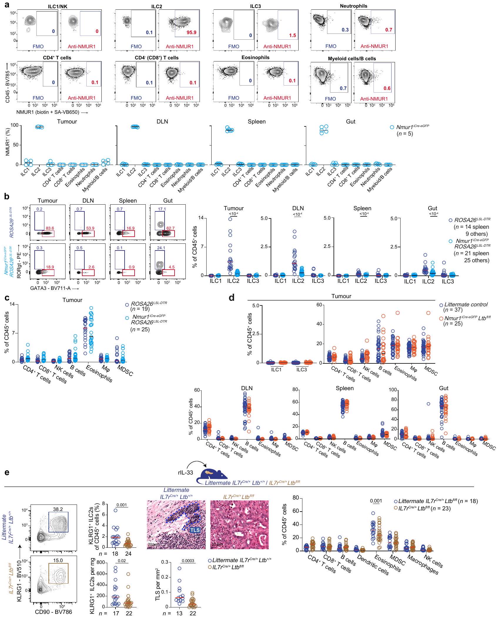
تم تعريف خلايا ILC2 في الفئران على أنها حية، CD45+، سلالة- (CD3، CD5، NK1.1، CD11b، CD11c، CD19، FceR1)، CD90+. كانت جميع الخلايا الحية، CD45+، سلالة-، CD90+ إيجابية لـ GATA3 (الشكل البياني الموسع 3a).
تم تعريف الفئران KLRG1+ على أنها حية، CD45+، سلالة- (CD3، CD5، NK1.1، CD11b، CD11c، CD19، FceR1)، CD90+، KLRG1+.
تم استخدام التعريفات التالية لخلايا المناعة الأخرى:
ILC1 - live, CD45+, lineage-, Tbet+
ILC3 - live, CD45+, lineage-,NK1.1-, Rorgt+
NK - live, CD45+, CD3-, NK1.1+
B cell - live, CD45+, CD3-, NK1.1-, CD19+
CD4+ T cell - live, CD45+, NK1.1-, CD3+, CD8-, CD4+
ملخص التقرير | أبحاث الطبيعة أبريل 2020
خلايا T CD8+ – حية، CD45+، NK1.1-، CD3+، CD4-، CD8+
الحمضات – حية، CD45+، NK1.1-، CD3-، CD19-، CD11b+ SiglecF+
البلاعم – حية، CD45+، NK1.1-، CD3-، CD19-، CD11b+، F4/80+
خلية مثبطة مشتقة من النخاع العظمي (MDSC) – حية، CD45+، NK1.1-، CD3-، CD19-، CD11b+، SiglecF-، Gr-1+
الخلايا الشجرية (DCs) – حية، CD45+، NK1.1-، CD3-، CD19-، Gr-1-، F4/80-، CD11c+، MHC-II+
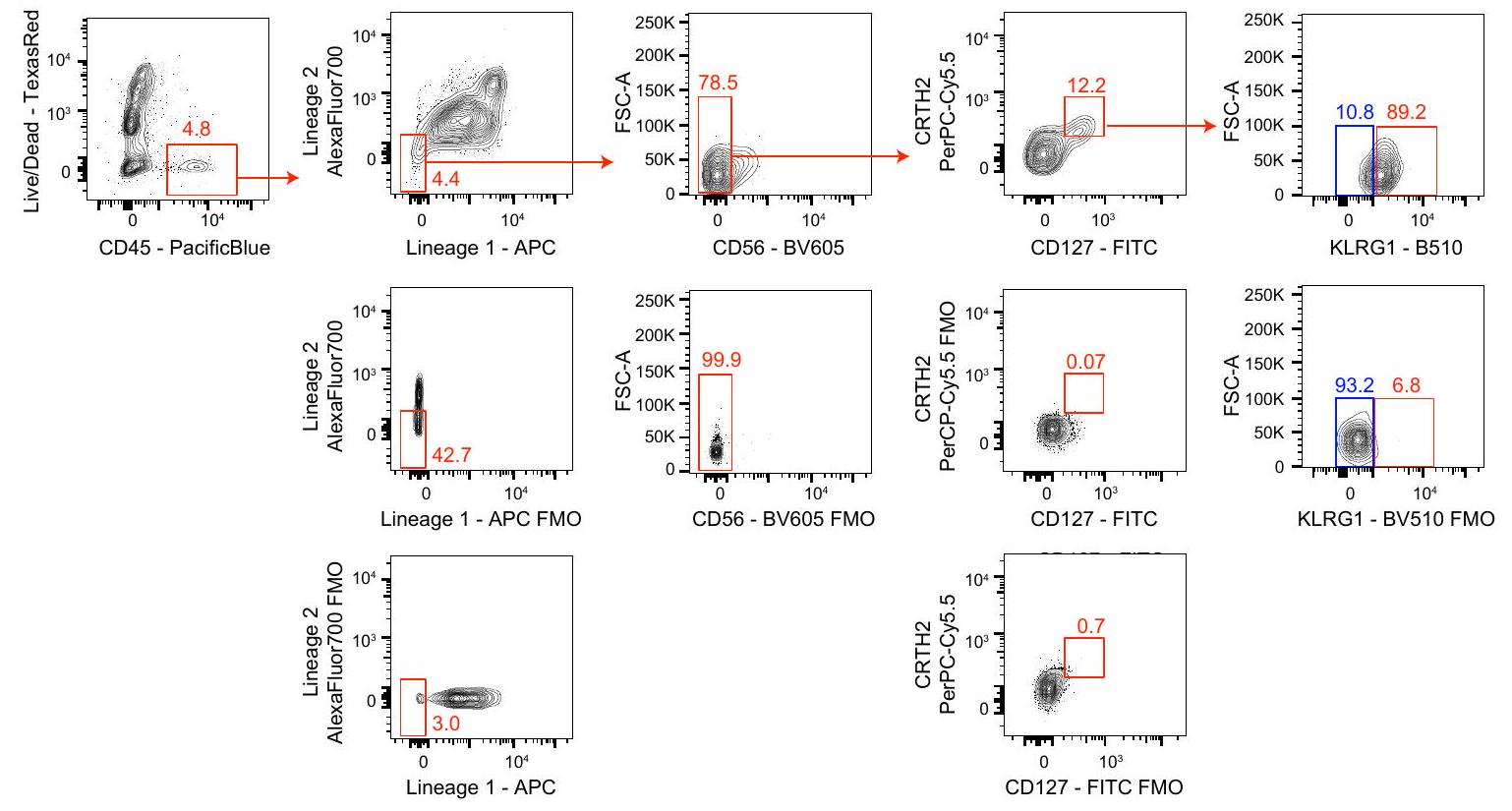
تم تعريف خلايا ILC2 البشرية على أنها حية، CD45+، سلالة- (CD3، CD5، CD56، CD11b، CD11c، CD14، CD16، CD19، TCRa/b، FceR1)، CD127+، CRTH2+.
تم تعريف خلايا ILC2s البشرية KLRG1+ على أنها حية، CD45+، سلالة- (CD3، CD5، CD56، CD11b، CD11c، CD14، CD16، CD19، TCRa/b، FceR1)، CD127+، CRTH2+، KLRG1+.
قم بتحديد هذا المربع لتأكيد أنه تم تقديم رقم يوضح استراتيجية البوابة في المعلومات التكميلية.

  1. خدمة المناعة وعلم الأورام، برنامج الأورام البشرية وعلم الأمراض، مركز ميموريال سلوان كيتيرينغ للسرطان، نيويورك، نيويورك، الولايات المتحدة الأمريكية. خدمة الكبد والبنكرياس والقنوات الصفراوية، قسم الجراحة، مركز ميموريال سلوان كيترينغ للسرطان، نيويورك، نيويورك، الولايات المتحدة الأمريكية. معهد جيل روبرتس للبحوث في مرض الأمعاء الالتهابي، وايل كورنيل للطب، نيويورك، نيويورك، الولايات المتحدة الأمريكية. مركز فريدمان للتغذية والالتهابات، وييل كورنيل ميديسن، جامعة كورنيل، نيويورك، نيويورك، الولايات المتحدة الأمريكية. مركز ألين لاكتشاف التفاعلات العصبية المناعية، نيويورك، نيويورك، الولايات المتحدة الأمريكية. قسم الطب في جامعة جوان وسانفورد آي. ويل، قسم أمراض الجهاز الهضمي والكبد، وييل كورنيل ميديسن، جامعة كورنيل، نيويورك، نيويورك، الولايات المتحدة الأمريكية. قسم الميكروبيولوجيا وعلم المناعة، وييل كورنيل ميديسن، جامعة كورنيل، نيويورك، نيويورك، الولايات المتحدة الأمريكية. خدمة علم الأورام الحاسوبية، قسم الوبائيات وعلم الإحصاء الحيوي، مركز ميموريال سلوان كيتيرينغ للسرطان، نيويورك، نيويورك، الولايات المتحدة الأمريكية. مركز ديفيد م. روبنشتاين لأبحاث سرطان البنكرياس، مركز ميموريال سلوان كيترينغ للسرطان، نيويورك، نيويورك، الولايات المتحدة الأمريكية. قسم الإحصاء الحيوي وعلم الأوبئة، مركز ميموريال سلوان كيتيرينغ للسرطان، نيويورك، نيويورك، الولايات المتحدة الأمريكية. مرفق تدفق الخلايا، مركز ميموريال سلوان كيتيرينغ للسرطان، نيويورك، نيويورك، الولايات المتحدة الأمريكية. قسم الميكروبيولوجيا، المناعة وعلم الوراثة الجزيئية، مركز جامعة تكساس للعلوم الصحية في سان أنطونيو، سان أنطونيو، تكساس، الولايات المتحدة الأمريكية. معهد اكتشاف العلاجات الثلاثي المؤسسي، نيويورك، نيويورك، الولايات المتحدة الأمريكية. مركز العليان لقاحات السرطان، مركز ميموريال سلوان كيتيرينغ للسرطان، نيويورك، نيويورك، الولايات المتحدة الأمريكية. علم وظائف الأعضاء، الفيزياء الحيوية وعلم الأحياء النظامية، وييل كورنيل للطب، كلية وييل كورنيل للطب، نيويورك، نيويورك، الولايات المتحدة الأمريكية. مركز سرطان ساندرا وإدوارد ماير، وييل كورنيل ميديسن، كلية وييل كورنيل للطب، نيويورك، نيويورك، الولايات المتحدة الأمريكية. معهد باركر لعلاج السرطان المناعي، وايل كورنيل للطب، نيويورك، نيويورك، الولايات المتحدة الأمريكية. سباحة عبر أمريكا ومختبر لودفيغ التعاوني، قسم علم الأدوية، وييل كورنيل للطب، نيويورك، نيويورك، الولايات المتحدة الأمريكية. ساهم هؤلاء المؤلفون بالتساوي: ماساتاكا أميساكي، عبد الرزاق زببود. البريد الإلكتروني: balachav@mskcc.org
    1. كراتز، أ.، كامبوس-نيتو، أ.، هانسون، م. س. ورادل، ن. هـ. الالتهاب المزمن الناتج عن اللمفوتوكسين هو تكوين أنسجة لمفاوية جديدة. ج. إكسب. ميد. 183، 1461-1472 (1996).
    2. سكوت، آي. سي. وآخرون. يتم تنشيط الإنترلوكين-33 بواسطة أنشطة البروتياز المرتبطة بالحساسية والنخر لتنظيم نشاطه كمنبه خلال تلف الظهارة. ساي. ريب. 8، 3363 (2018).
    3. ساوتيس-فريدمان، سي.، بيتيبرز، ف.، كالديرارو، ج. وفريدمان، و. هـ. الهياكل اللمفاوية الثانوية في عصر علاج السرطان المناعي. نات. ريف. كانسر 19، 307-325 (2019).
    4. كابريتا، ر. وآخرون. الهياكل اللمفاوية الثانوية تحسن العلاج المناعي والبقاء في الميلانوما. ناتشر 577، 561-565 (2020).
    5. بيتيبر، ف. وآخرون. ترتبط خلايا B بالبقاء واستجابة العلاج المناعي في الساركوما. ناتشر 577، 556-560 (2020).
    6. هيلمينك، ب. أ. وآخرون. خلايا ب والهياكل اللمفاوية الثلاثية تعزز استجابة العلاج المناعي. ناتشر 577، 549-555 (2020).
    7. كوبولا، د. وآخرون. هياكل فريدة تشبه العقد اللمفاوية الهاجرة موجودة في سرطان القولون والمستقيم الأولي البشري تم تحديدها من خلال تحليل جينات المناعة. المجلة الأمريكية لعلم الأمراض 179، 37-45 (2011).
    8. Gu-Trantien، C. وآخرون. CD4 تسلل خلايا T المساعدة الجريبية يتنبأ ببقاء مرضى سرطان الثدي. J. Clin. Invest. 123، 2873-2892 (2013).
    9. هيروكا، ن. وآخرون. العضو اللمفاوي الثالث داخل الورم هو مؤشر إيجابي في مرضى سرطان البنكرياس. المجلة البريطانية للسرطان 112، 1782-1790 (2015).
    10. ليو، ف. ي.، جيرارد، ج.-ب. وتورنكويست، هـ. ر. الإنترلوكين-33 في الصحة والمرض. نات. ريف. إيمونول. 16، 676-689 (2016).
    11. درايتون، د. ل.، يينغ، إكس.، لي، ج.، ليسلاور، و. ورادل، ن. هـ. يوجه LTaß خارج الموقع تكوين الأعضاء اللمفاوية مع التعبير المتزامن عن عنوان العقدة الطرفية وسلفوترانسفيراز مقيد بالأوعية الدموية اللمفاوية. ج. إكسب. ميد. 197، 1153-1163 (2003).
    12. لوكاشيف، م. وآخرون. استهداف اللمفوتوكسين- مستقبل مع أجسام مضادة منشطة كعلاج محتمل للسرطان. أبحاث السرطان. 66، 9617-9624 (2006).
    13. رينرت، ب. د.، جيمس، د.، ماكاي، ف.، براوننج، ج. ل. و هوشمان، ب. س. إن تكوين العقد اللمفاوية يتم تحفيزه من خلال الإشارات عبر اللمفوتوكسين المستقبل. المناعة 9، 71-79 (1998).
    14. لوخنر، م. وآخرون. الأنسجة اللمفاوية الثلاثية المستحثة بواسطة الميكروبيوم تزيد من حدة الأمراض الالتهابية في غياب خلايا RORyt و LTi. ج. تجريبي. طب. 208، 125-134 (2010).
    15. شومخر، ت. ن. وثومين، د. س. الهياكل اللمفاوية الثلاثية في السرطان. العلوم 375، eabf9419 (2022).

Journal: Nature, Volume: 638, Issue: 8052
DOI: https://doi.org/10.1038/s41586-024-08426-5
PMID: https://pubmed.ncbi.nlm.nih.gov/39814891
Publication Date: 2025-01-15

IL-33-activated ILC2s induce tertiary lymphoid structures in pancreatic cancer

https://doi.org/10.1038/s41586-024-08426-5
Received: 25 October 2021
Accepted: 19 November 2024
Published online: 15 January 2025
Open access
Check for updates

Masataka Amisaki , Abderezak Zebboudj , Hiroshi Yano , Siqi Linsey Zhang , George Payne , Adrienne Kaya Chandra , Rebecca Yu , Pablo Guasp , Zachary M. Sethna , Akihiro Ohmoto , Luis A. Rojas , Charlotte Cheng , Theresa Waters , Alexander Solovyov , Stephen Martis , Ashley S. Doane , Charlotte Reiche , Emmanuel M. Bruno , Martina Milighetti , Kevin Soares , Zagaa Odgerel , John Alec Moral , Julia N. Zhao , Mithat Gönen , Rui Gardner , Alexei V. Tumanov , Abdul G. Khan , Olivia Vergnolle , Elisabeth K. Nyakatura , Ivo C. Lorenz , Manuel Baca , Erin Patterson , Benjamin Greenbaum , David Artis , Taha Merghoub & Vinod P. Balachandran

Abstract

Tertiary lymphoid structures (TLSs) are de novo ectopic lymphoid aggregates that regulate immunity in chronically inflamed tissues, including tumours. Although TLSs form due to inflammation-triggered activation of the lymphotoxin (LT)-LTβ receptor (LT/ßR) pathway , the inflammatory signals and cells that induce TLSs remain incompletely identified. Here we show that interleukin-33 (IL-33), the alarmin released by inflamed tissues , induces TLSs. In mice, Il33 deficiency severely attenuates inflammation- and LT R-activation-induced TLSs in models of colitis and pancreatic ductal adenocarcinoma (PDAC). In PDAC, the alarmin domain of IL-33 activates group 2 innate lymphoid cells (ILC2s) expressing LT that engage putative myeloid organizer cells to initiate tertiary lymphoneogenesis. Notably, lymphoneogenic ILC2s migrate to PDACs from the gut, can be mobilized to PDACs in different tissues and are modulated by gut microbiota. Furthermore, we detect putative lymphoneogenic ILC2s and IL-33-expressing cells within TLSs in human PDAC that correlate with improved prognosis. To harness this lymphoneogenic pathway for immunotherapy, we engineer a recombinant human IL-33 protein that expands intratumoural lymphoneogenic ILC2s and TLSs and demonstrates enhanced anti-tumour activity in PDAC mice. In summary, we identify the molecules and cells of a druggable pathway that induces inflammationtriggered TLSs. More broadly, we reveal a lymphoneogenic function for alarmins and ILC2s.

Secondary lymphoid organs (SLOs) are key mammalian structures that coordinate immune responses to acute tissue injury. SLOs drain regional tissue antigens and colocalize immune cells that sample antigens with effector immune cells to thereby coordinate a structural and cellular strategy to patrol tissues and rapidly mount adaptive immune responses. However, in chronic inflammatory states, hosts must develop lymphoid organs ectopically within inflamed tissues to boost the intensity and proximity of the immune response. Such
ectopic lymphoid organs, termed TLSs, are ubiquitous structures that regulate immunity in chronically inflamed tissues , including infection, inflammation and cancer.
In cancer, hosts ostensibly develop in any tissue that can boost endogenous and therapeutic anti-tumour immunity. Therefore, new strategies to induce TLSs are attractive, as most human tumours have fewer infiltrating immune cells (immunologically cold), rendering them resistant to current immunotherapies. However, in contrast to SLOs
Fig. 1 |IL33 expression correlates with TLS transcriptional signatures in human tumours. a,b, Unbiased correlation of bulk tumour mRNA gene expression to TLS transcriptional signatures (TLS signatures 1 (ref.4), 2 (ref.7) and 3 (ref. 8)) in human PDAC (a, top). Correlation of bulk tumour expression to TLS transcriptional signatures and in human PDAC (a, bottom) and human tumours (TLS signature 1; b). c,d, Immunohistochemistry (c, left) and immunofluorescence (c, right), and the immunofluorescence-quantified frequency of bulk IL- cells (c) and IL- cells (d), and association with
survival ( and d) in human PDAC. The dotted red line in c shows the putative TLS. Adj. panc., adjacent pancreatic tissue; HR, hazard ratio. represents the number of tumours (a-c) or patients (survival;c and d). High and low (in the survival plots in c and d) represent (high) or < (low) the median frequency of the cohort. Horizontal bars in dot plots indicate the median (c,d). values were calculated using two-sided Pearson correlation ( and ), two-tailed Mann-Whitney -test (c (left) and d (left and middle)) and two-sided log-rank test (c (right) and d (right)). For c, scale bars, (left) and (right).
that are preformed, TLSs assemble de novo in chronically inflamed tissues. As such, inflammation-induced drivers and cells that induce TLSs remain incompletely identified.

IL-33 mediates lymphoneogenesis

To identify candidate inflammatory signals that induce TLSs in tumours, we searched for genes with expression that positively correlated with known TLStranscriptional signatures in PDAC-a lethal model cold tumour with few TLSs but, when present, higher intratumoural TLS density boosts immunity and is correlated with longer survival .As TLSs are identified by inducing chemokines and resident cells (activated T cells, B cells, dendritic cells (DCs) and myeloid cells) , we selected three principally non-overlapping transcriptional signatures that identify intratumoural TLSs based on chemokines , cells or other immunotherapy-promoting factors . We found that expression of IL33-which encodes the alarmin canonically released in inflamed tissues -correlated with all three TLS signatures (Fig. 1a) and with expression ( encodes the TLS-inducing cytokine ) in three different PDAC cohorts ( PDACs; Fig. 1a, Extended Data Fig. 1a and Supplementary Table 1), as well as in other cold and hot tumours ( histologies, 3,887 tumours; Fig. 1b and Extended Data Fig. 1b). Within TLSs in PDAC, we detected IL- cells, including IL- immune cells
(Fig.1c (left), Extended Data Fig. 1c,d and Supplementary Table 2) that were more abundant in tumours compared with in adjacent pancreatic tissues (Fig. 1c (middle) and 1d (left)), and enriched in tumours with TLSs compared with those without (Fig. 1d (middle)). Furthermore, higher intratumoural IL- bulk and IL- immune cell densities were correlated with improved PDAC survival (Fig. 1c (right) and 1d (right)). Together, these results identified a correlation between IL-33 and TLSs in cancer.
To probe a potential causative link between IL-33 and TLSs, we used a Kras- and Trp53-driven orthotopically implanted PDAC mouse model (PDAC mice) with few baseline TLSs to first examine whether endogenous Il33 is required for de novo lymphoneogenesis. We found that activating the LTβ receptor (LTβR) -a canonical signal for TLS neogenesis -generated intratumoural TLSs and controlled tumours in wild-type (WT) but not in Il33 PDAC mice (Fig. 2a (left) and Extended Data Fig. 2a). Il33 was also required for inflammation-induced lymphoneogenesis, as oral dextran sulfate sodium (DSS), which provokes chemical colitis (DSS-colitis mice) and models human inflammatory bowel disease, stimulated colonic lymphoid aggregates in WT but not in Il33 mice (Fig. 2a (right)), exacerbating weight loss and diminishing survival (Extended Data Fig. 2b). Endogenous IL33 is therefore required to induce TLSs in models of cancer and inflammation.
Fig. 2 | The alarmin domain of IL-33 induces TLSs in PDAC and chemical colitis. a, Haematoxylin and eosin staining (H&E) (top) and quantification of intratumoural TLSs (bottom left) and intracolonic lymphoid aggregates (LA;bottom right) in anti-LTβR-treated PDAC (left) and DSS-colitis (right) WT and mice. b, H&E (top) and immunofluorescence (IF; middle) staining, and the TLS density (H&E quantified;bottom) in vehicle (veh.) and rll-33-treated PDAC mice. PDAC1-6, orthotopic PDAC mice established with six PDAC cell lines. c,d, Intratumoural TLS maturation states (immunofluorescence quantified) (c), and the B cell frequency and clonality and somatic hypermutation (d) in vehicletreated and rll-33-treated PDAC mice.e, H&E and colonic LA quantification
in vehicle-treated and rIL-33-treated DSS-colitis mice. Data were collected 2 weeks ( , left), 3 weeks (b, PDAC 3 and 4 ; and d) and 3 – 5 weeks (b, PDAC 1, 2, 5 and 6) after tumour implantation, and 2 weeks after DSS initiation (a (right) and ), pooled from independent experiments with mice per group with consistent results. represents the number of individual tumours or organs from individual mice analysed separately, or B cell clones (d, right). The dotted lines in and e show putative TLS/LA; the horizont al bars in -e show the median. values were calculated using two-way analysis of variance (ANOVA) with Tukey’s multiple-comparison test (a) and two-tailed Mann-Whitney -test (b-e). Scale bars, top and and bottom ).
Full-length IL-33 protein contains two domains-an N-terminal nuclear-localization domain and a C-terminal alarmin domain that, after tissue injury, is extracellularly cleaved to IL-33 and binds to the receptor ST2 . To probe whether IL-33 served as an inflammation-induced extracellular TLS trigger, we administered the alarmin domain of IL-33 (IL-33 , hereafter, recombinant IL-33 (rll-33)) to PDAC mice at doses (Extended Data Fig. 1e) that roughly approximated IL-33 steady-state levels in human sera of patients with PDAC (Extended Data Fig. 1f and Supplementary Table 3). rIL-33 induced de novo intratumoural TLSs across several TLS-poor, distinct cell-line-derived PDAC mouse models that spanned a range of intratumoural T cell infiltrates (Fig. 2b and Extended Data Fig. 2c,d). rlL-33 also induced different TLS maturation states, increasing the density of intratumoural lymphoid aggregates (early TLS) and primary follicles (intermediate TLS) in PDACs (Fig. 2c and Extended Data Fig. 2e). Moreover, tumours in rIL-33-treated PDAC mice evidenced TLSs with BCL6 B cells (Extended Data Fig. 2f), and increased intratumoural B cell frequencies, B cell clonal expansion and somatic hypermutation (Fig. 2d), suggesting germinal centre reactions and secondary follicle generation (late TLS) . In DSS-colitis mice, rIL-33 expanded colonic lymphoid aggregates to a lesser degree (Fig. 2e), but attenuated
weight loss and enhanced mouse survival (Extended Data Fig. 2g). Thus, the alarmin IL-33 triggers lymphoneogenesis in cancer and inflammation.

IL-33 expands lymphoneogenic ILC2s

As these results suggested an IL-33-driven lymphoneogenic pathway, we next sought to identify the cells that mediate rll-33-induced TLSs. As we previously reported that rIL-33 dominantly expands intratumoural ILC2s in PDAC mice , we examined the single-cell transcriptomes of ST2 ILC2s in tumours and tumour draining lymph nodes (DLNs) in rIL-33-treated PDAC mice for lymphoneogenic molecules (Fig. 3a). We detected an intratumoural population of ILC2s that co-expressed Klrg1 and (Fig. 3a and Extended Data Fig. 3a,b), (Extended Data Fig. 3b) and LT, the LT heterortrimeric cytokine essential for TLS neogenesis (Fig. 3b and Extended Data Fig. 3c,d). Consistently, in multiple PDAC models and DSS-colitis mice, rlL-33 expanded intratumoural (Fig. 3c and Extended Data Fig. 3e) and gut (Fig. 3d) KLRG1 ILC2s, but not KLRG1 ILC2s, that expressed LT (Extended Data Fig. 3c,f). These rIL-33-induced intratumoural KLRG1 ILC2s also expressed canonical ILC2 transcription factors (TFs) (Gata3 and Rora (Fig. 3a and Extended
Fig.3|See next page for caption.
Data Fig. ) and Id2 (Extended Data Fig. 3h)), cytokines (Il5,Il13 and Areg; Extended Data Fig. 3h) and receptors (Il7r and Nmur1 ; Fig. 3a and Extended Data Fig. 3i), but not ILC1 (Tbx21 (encoding T-BET); Extended Data Fig. 3j) or ILC3 (Rorc (encoding RORyt); Extended Data
Fig. 3h,j) TFs. Thus, rlL-33 expands KLRG1 ILC2s expressing LT that may mediate lymphoneogenesis.
Recent reports have revealed that ILC2s and precursors selectively express the neuromedin receptor (NMUR1) that can be used for
Fig. 3 | IL-33 activates lymphoneogenic ILC2s. a, Single-cell RNA-sequencing (scRNA-seq) analysis of purified tumour and DLNILC2s from rlL-33-treated PDAC mice. The uniform manifold approximation and projection (UMAP) representations show cells by unsupervised clusters (top left), organs (bottom left) and select genes (right). The heat map (middle) shows the top 25 genes by cluster. The heat map scale bar represents the score. The UMAP scale bar represents gene expression.b, Gating (left) and LT expression by flow cytometry (right) and confocal imaging (bottom) in intratumoural KLRG1 ILC2s from rIL-33-treated WT and PDAC mice. FMO, fluorescence minus one; MFI, mean fluorescence intensity. c,d, Gating, and the frequency and number of intratumoural (c) and gut (d) ILC2s in vehicle-treated and rIL-33treated PDAC (c) and DSS-colitis (d) mice. e, Gating (left), ILC2 frequency (middle) and intratumoural TLS density (right) in rlL-33 and DT-treated Nmur1 Rosa26 and littermate control Rosa26 PDAC mice. f, Intratumoural TLS density (left), gating, frequency and the number of ILC2s,
and the number of DLN immune cells (right) in rlL-33-treated Nmur1 and littermate control PDAC mice.g, Tumour KLRG1 ILC2s were sortpurified from rlL-33-treated or littermate PDAC mice and transferred to Ror PDAC mouse recipients. The TLS density, ILC2 frequency and number in recipient PDACs. Data were collected at 10 days (a), 2 weeks (b and d-f) and 5 weeks (c) after tumour implantation, and 2 weeks (g) after transfer, pooled from independent experiments with mice per group with consistent results. represents the number of individual tumours or organs from individual mice analysed separately. The horizontal bars show the median. In , littermate controls include and Nmur1 combined. values were calculated using two-tailed Mann-Whitney -test (b, e (right), fand g) and two-way ANOVA with Holm’s test (c, d and e (left)). Scale bars, (b(left)), (b(all others)), (e and (left)) and (f(right)).
ILC2-specific genetic targeting . To investigate whether ILC2s contribute to IL-33-mediated lymphoneogenesis, as ILC2s (but not other ILCs or immune cells) in our model also selectively expressed NMUR1 (Extended Data Fig. 4a), we used Nmur1 Rosa26 transgenic mice that constitutively express the human diphtheria toxin receptor (DTR) on NMUR1 cells to acutely and selectively deplete all ILC2s. In Nmur1 Rosa26 PDAC mice, diphtheria toxin (DT) initiated 2 days before tumour implantation and rIL-33 treatment depleted ILC2s by approximately within tumours and across organs (Fig. 3e (left)) while sparing ILC3s (Extended Data Fig. 4b) and other immune cells (Extended Data Fig. 4c). Notably, acute ILC2 depletion during rIL-33 treatment reduced intratumoural TLSs by approximately 65% (Fig.3e (right)). Thus, ILC2s contribute to IL-33-mediated lymphoneogenesis.
Next, to examine whether ILC2s used cell-intrinsic LT to induce intratumoural TLSs, we generated transgenic mice that lack on ILC2s (Nmur1 Ltb flfl ). In rlL-33-treated Nmur1 Ltb PDAC mice, compared with the littermate controls, ILC2-targeted Ltb deletion reduced intratumoural TLS density by approximately without altering the frequencies of KLRG1 ILC2s (Fig. 3f), ILC1s, ILC3s and other immune cells in tumours (Extended Data Fig. 4d (top)), immune cells in DLNs (Fig. 3f (right)) or immune cell subsets in organs (Extended Data Fig. 4d (bottom)). Consistently, a broader genetic strategy to delete on allIL-7R lymphocytes (II77 ) also reduced intratumoural TLSs, but notably also reduced intratumoural KLRG1 ILC2s (Extended Data Fig. 4e), to suggest that non-ILC2 lymphocyte LT signalling may maximally expand KLRG1 ILC2s in tumours. rIL-33-treated PDAC mice also had comparable intratumoural frequencies of other immune cells but fewer eosinophils (Extended Data Fig. 4e (right)), consistent with the downstream effects of ILC2 depletion . To confirm that ILC2s used LT to impact TLS formation using an orthogonal approach, we purified and transferred intratumoural KLRG1 ILC2s from rIL-33-treated or littermate PDAC mice (that is, ILC2s proficient or deficient in tb) into Il7r Rora PDAC recipients that exhibit reduced peripheral ILC2s in tissues (Fig. 3g). Compared with transferred ILC2s with intact , and consistent with the results in Nmur1 Ltb PDAC mice (Fig. 3f), ILC2-intrinsic Ltb deficiency reduced TLS formation without impairing rIL-33-mediated intratumoural KLRG1 ILC2 accumulation in tumours (Fig. 3g). Collectively, these molecular and cellular loss- and gain-of-function experiments suggest that rIL-33-activated KLRG1 ILC2s induce TLSs in PDACs using a pathway involving LT.
In SLOs and TLSs, inducer cells express LT that binds to LTβR on TLS organizer cells to stimulate the expression of chemokines and adhesion molecules, and coordinate lymphoneogenesis . To identify the organizer cells that cooperate with rlL-33-induced ILC2s to induce TLSs in PDAC, we found that intratumoural CD11b myeloid cells most highly expressed LT R (Extended Data Fig. 5a). Compared with myeloid cells, intratumoural myeloid cells expressed
a distinct transcriptome (Extended Data Fig. 5b-e and Supplementary Table 4), with higher TLS-inducing chemokines, including Cxcl13 and (Extended Data Fig. 5d), the canonical organizer cell adhesion molecule Vcam1 (Extended Data Fig. 5e) and inflammatory chemokines that recruit TLS cellular components (Extended Data Fig. 5e). Intratumoural LTβR myeloid cells also expressed II33 mRNA and protein (Extended Data Fig. 5f). Consistently, LT R activation expanded intratumoural KLRG1 ILC2s in WT but not PDAC mice (Extended Data Fig. 5g) to indicate LT R stimulation may expand ILC2s through an endogenous IL-33-dependent circuit. To test whether myeloid-cell-derived endogenous IL-33 modulated KLRG1 ILC2s in tumours, we purified IL-33 proficient (WT) or deficient (II33 ) myeloid cells from PDACs and co-implanted them with PDAC cells into pancreata of mice (Extended Data Fig. 5h). LT myeloid-cell-derived IL-33 induced KLRG1 in PDACs to express LT and induce TLSs (Extended Data Fig. 5h). Together, these data suggest that ILC2s and intratumoural myeloid cells cooperate as TLS inducer and organizer cells, respectively, to augment IL-33 signalling and induce TLSs in tumours.

Lymphoneogenic ILC2s can migrate from the gut

As our findings suggested a lymphoneogenic function for ILC2s, we investigated whether lymphoneogenic ILC2s expand locally or migrated to tumours . In PDAC mice, as rll-33 expanded KLRG1 ILC2s in the blood (Extended Data Fig. 6a), we used congenic parabiotic mice with PDACs in recipient pancreata to test whether KLRG1 ILC2s migrated haematogenously to tumours (Extended Data Fig. 6b). rlL-33 administered to donor parabionts induced donor (Fig. 4a) but not KLRG1- ILC2s or non-ILCs (Extended Data Fig. 6c,d) to migrate to recipient parabiont blood and tumours. Thus, ILC2s migrate haematogenously to tumours.
As ILC2s can migrate to tissues from the gut , we hypothesized that the gut might be one source of ILC2s that migrate to pancreatic tumours. To trace the origin of intratumoural ILC2s, we used KikGR transgenic mice in which all cells express a green-to-red photoconvertible protein. We treated KikGR PDAC mice with rlL-33, photoconverted gut tissue and searched for gut-derived red ILC2s in PDACs (Fig. 4b). In rll-33-treated PDAC mice, we detected rare red ILC2s in tumours of gut-photoconverted but not control (sham treated; peritoneum-photoconverted) mice (Fig. 4b and Extended Data Fig. 6e), indicating that ILC2s can migrate to PDACs through a gut-blood circuit.
To examine whether PDACs primed such a gut-derived migratory ILC2 circuit, we searched for gut-derived signals that may stimulate ILC2s to migrate. As recent findings indicate gut microbiota can stimulate ILC2s to migrate in acute inflammatory states , we examined whether PDACs altered gut microbiota and, consequently, stimulated gut ILC2
Fig. 4 | Lymphoneogenic ILC2s migrate to tumours through a microbiotaand PDAC-modulated gut-blood circuit. a, Parabiotic mice with PDACs in recipient pancreata. Donors were treated with vehicle or rll-33. Quantification (top) and gating (bottom) of donor-derived ILC2s in recipient blood and PDAC.b, KikGR PDAC mice were treated daily with rlL- 33 for 6 days. On day 6, the gut or peritoneum was photoconverted. Experimental schematic (left) and density of gut-derived (RFP ) ILC2s in pancreatic PDACs (right). c, Gut Il33 mRNA was analysed by digital droplet PCR in sham-treated and PDAC mice. d, Gut Il33 mRNA (left), blood KLRG1 ILC2s (middle) and the intratumoural TLS density (right) in rlL-33-treated PDAC mice treated with or without antibiotics (Abx). e, The intratumoural KLRG1 ILC2 frequency, number and TLS density in skin (s.c.) PDACs in vehicle-treated and rIL-33treated mice with s.c. alone (single PDAC) or s.c. and pancreatic PDACs (dual PDAC). f, Parabiotic mice with s.c. PDACs in recipients with or without pancreatic PDACs in donors. Donors were treated with vehicle or rll-33.
The donor-derived ILC2 frequency in recipient s.c. PDACs (bottom) is shown. , Parabiotic mice with s.c. PDACs in recipients and pancreatic PDACs in donors. Donors were treated with rIL-33; donor and recipients were treated with antibiotics. The donor-derived ILC2 number and frequency in recipient s.c. PDACs and gut (bottom) are shown. Data were collected at 2 weeks (a, e (left), and ), as shown in the experimental schema (b), 4 weeks (c and and 5 weeks (e(right)) after tumour implantation, pooled from independent experiments with mice per group with consistent results. values represent the number of individual tumours or organs from individual mice analysed separately. The horizontal bars show the median. values were calculated using two-tailed Mann-Whitney -test (a, c, d, e (right) and g), one-way ANOVA with Kruskal-Wallis multiple-comparison test (b), two-way ANOVA with Kruskal-Wallis test (d, middle) and two-way ANOVA with Sidak’s multiplecomparison test (e(left) and f). Scale bar, .
Fig. 5 | An engineered human IL-33 therapeutic stimulates TLSs and antitumour activity in PDAC. a, ST2 activation in HEK blue human IL-33 reporter cells with H-rIL-33, H-e-rIL-33 and H-e-rIL-33-Fc. EC , half-maximal effective concentration.b-d, The intratumoural KLRG1 ILC2 frequency (b), TLS density (c) and tumour volume (d) in PDAC mice treated with vehicle, mouse rIL-33, H-rIL-33 or escalating H-e-rIL-33-Fc. Curves in a were fit by nonlinear
reservoirs . Notably, PDACs not only increased intrapancreatic KLRG1 ILC2 frequencies (Extended Data Fig. 7a), but also increased the diversity and altered the composition of gut microbiota (Extended Data Fig. 7b) and increased gut Il33 mRNA expression (Fig. 4c), suggesting that gut microbial changes may increase gut IL-33 levels and stimulate gut KLRG1 ILC2s to migrate . Consistently, antibiotic-mediated microbiota ablation decreased gut Il33 mRNA expression, decreased the frequency of blood ILC2s and reduced intratumoural TLSs (Fig. 4d). Furthermore, microbiota ablation slightly reduced serum IL-33, and reciprocally increased the frequency of gut KLRG1 ILC2s (Extended Data Fig. 7c) that, consistently, was reduced after microbiota restoration in germ-free mice (Extended Data Fig. 7d), suggesting that microbiota composition may modulate the ability of gut ILC2s to enter the circulation. Collectively, these results suggest that ILC2s can migrate to tumours from the gut and are modulated by gut microbiota.
As these results suggest PDACs prime the host to extrude ILC2s to inflamed sites, we investigated whether PDACs could amplify the ILC2 response to tumours in distant tissues. To test this, we administered rIL-33 to mice with either single (skin alone, subcutaneous (s.c.) PDAC) or dual (s.c. and pancreatic) PDACs. In contrast to single-PDAC mice, in which rIL-33 expanded intratumoural KLRG1 ILC2s yet did not control s.c. PDAC growth, in dual-PDAC mice, rll-33 further amplified KLRG1 ILC2 frequencies, boosted TLS densities (Fig. 4e) and controlled tumour growth in s.c. PDACs (Extended Data Fig. 8a). To confirm that dual-PDACs amplified KLRG1 ILC2s that trafficked haematogenously to s.c. tumours, we modelled dual PDACs in parabiotic mice with pancreatic PDACs in donor parabionts and s.c. PDACs in recipients (Fig.4f). Consistent with the findings in dual-PDAC mice, pancreatic PDACs in donor parabionts increased rll-33-induced KLRG1 but not KLRG1 ILC2 migration to s.c. PDACs in recipient parabionts (Fig. 4f and Extended Data Fig. 8b). This pancreatic PDAC-amplified haematogenous KLRG1 ILC2 trafficking to s.c. PDACs required distinct cytokines compared
regression. Data were collected at weeks ( and ) after tumour implantation, pooled from independent experiments with mice per group with consistent results. values represent the number of individual tumours from individual mice. The horizontal bars show the median. values were calculated using extra-sum-of-squares test (a) and two-way ANOVA with Tukey’s multiplecomparison correction test (b-d).
to acute infections as, although both rIL-33 and rIL-25 expanded ILC2s in donor PDACs (Extended Data Fig. 8c), only rlL-33 and not rll-25 disseminated KLRG1 ILC2s from donor parabionts into recipient parabiont blood and s.c.PDACs (Extended Data Fig. 8c). Furthermore, increased migration to s.c. PDACs in dual-PDAC mice was accompanied by restricted s.c. PDAC growth in an ST2-, LTβR-, – and Rora-dependent manner (Extended Data Fig. 8d-g). Finally, microbiota ablation in parabiotic PDAC mice decreased donor-derived ILC2s in recipient s.c. PDACs and gut, but not recipient DLNs (Fig. 4g and Extended Data Fig. 8h), indicating that microbiota amplify ILC2s in tumours connected to a gut-blood circuit. In summary, a host pancreatic PDAC amplifies microbiota-mediated haematogenous ILC2 migration to PDACs in distant tissues.

Engineered IL-33 amplifies lymphoneogenesis

To examine the relevance of our findings in humans, we next investigated whether LT-expressing KLRG1 ILC2s are present in human cancer. In human PDACs, we detected KLRG1 ILC2s that expressed LT (Extended Data Fig. 9a-c and Supplementary Table 5), suggesting that ILC2s may exert similar lymphoneogenic function in human tumours. To investigate the association between IL-33-activated ILC2s and intratumoural TLSs, we developed a human transcriptional signature of rIL-33-activated ILC2s (Methods) and examined its association with TLS signatures in tumours spanning a spectrum of immune activity ( histologies, 4,037 tumours). Activated ILC2 signatures correlated with all three TLS transcriptional signatures (Extended Data Fig. 1g), suggesting that KLRG1 ILC2s may contribute to TLS formation in human tumours.
As the presence of ILC2s in human tumours suggested that the IL-33-ILC2-TLS pathway could be manipulated for immunotherapy, we engineered human rIL-33 (H-rIL-33) to increase its activity and half-life in vivo. First, to eliminate cysteine oxidation that triggers disulfide
bonding and conformational changes that disrupt the ST2-binding site of IL-33, we substituted all four cysteine residues in human IL-33 to serine residues (hereafter, H-enhanced-rll-33 (H-e-rll-33)). We found that H-e-rlL-33 enhanced ST2 activation compared with H-rlL33, consistent with previous observations (Fig. 5a). Next, to prolong the H-e-rll-33 half-life in vivo, we used an established approach and fused H-e-rlL-33 to the human IgG1 Fc fragment (H-e-rll-33-Fc) with described L234A/L235A substitutions that limit Fc gamma receptor binding through a short Gly/Ser-containing linker. We then confirmed that H-e-rlL-33-Fc enhanced in vitro ST2 activation compared with H-rIL-33 and H-e-rIL-33 (Fig. 5a). As numerous studies have shown that human IL-33 is biologically active in mice , we tested whether H-e-rIL-33-Fc expanded KLRG1 ILC2s and induced TLSs in mice. Consistently, H-e-rll-33-Fc expanded intratumoural KLRG1 ILC2s and TLSs in a dose-dependent manner (Fig. 5b,c) to control PDAC tumours (Fig. 5d). Thus, the IL-33-ILC2-TLS axis is a previously undescribed pathway that can be engineered to treat pancreatic cancer (Extended Data Fig. 10).

Discussion

We find that IL-33, an alarmin that is canonically released by damaged tissues, activates a migratory cellular lymphoneogenic response. Such a pathway may have been predictable in hindsight-lymphoid tissue inducer cells, the cells that initiate prenatal lymphoid organogenesis, contract in the postnatal adult . Although ILC3s may subsume some lymphoneogenic lymphoid tissue inducer function in adults , ILC3s do not canonically respond to damage signals. Thus, one may hypothesize that a non-redundant cellular pathway must initiate lymphoneogenesis in response to tissue injury. Indeed, our findings highlight IL-33 as a candidate inflammatory TLS initiator, and ILC2s as a candidate inducer, of such a pathway.
Our observations do not imply ILC2s are the sole inducers of IL-33-driven lymphoneogenesis. Although gain- and loss-of-function studies demonstrate ILC2s indeed induce TLSs in an LT-dependent manner, our results do not exclude that IL-33-driven TLSs may harness contributions from other ST2 cells, including activated T cells and B cells that respond to IL-33 . Moreover, IL-33 may also activate other immune and non-immune cells to exert both anti- and, possibly, pro-neoplastic effects in different contexts through non-TLS mechanisms. As Nmur1 is expressed in the nervous system , our genetic strategy to selectively deplete ILC2s by targeting Nmur1 with DT may confound attempts to study TLS formation in neuronal tissues. Yet, as ILC2s respond early to tissue damage and recruit other lymphocytes to injured sites, ILC2s are prime candidate apex cells to initiate IL-33-driven TLSs and amplify the TLS response . This broadens the scope of the recognized alarmin function of IL-33, and the role of ILC2s as sentinels of injury, to include inflammation-triggered lymphoneogenesis.
The fidelity of such a mobile lymphogenic pathway is further supported by companion intratumoural myeloid cells that appear to cooperatively generate TLSs. Indeed, myeloid cells and other immune cells inducibly express IL- and LT , and release cytosolic IL- (not demonstrated in our study) through a translocation pathway for leaderless proteins that lack a secretory signal peptide. Yet, why myeloid cells possess the molecular machinery to express IL-33 has remained unclear. We posit that myeloid cells may use IL-33 to feedback and sustain LT expression to ensure continuous local levels of IL-33. Thus, IL-33 may be expressed in myeloid cells to both expand inducer cells in injured tissues to initiate TLSs and enable organizer cells to further amplify this response. Although we find that myeloid cells use IL-33 to induce LT on ILC2s, this pathway may extend to other IL-33- and LT-expressing cells. Notably, as we do not demonstrate that myeloid cells secrete IL-33, we cannot rule out that the N -terminal domain may also contribute to lymphoneogenesis. Yet, these findings-self-sustained expression of the inducing ligand through a cellular and molecular feedback loop-bear
the hallmark of a novel TLS pathway. Physiologically, this organizational structure would logically preprogram SLOs at defined anatomical locations and equip migratory cells with molecular machinery to traffic to tissues to induce TLSs.
Our finding that PDAC primes this host lymphoneogenic response from the gut and is modulated by microbiota is notable, as a precise understanding of cells that link gut microbiota to both TLSs and tumour immunity remains in evolution. However, we did not investigate, and therefore cannot rule out, the functional contributions of ILC2s migrating to tumours from other sources, or of non-migratory ILC2s. Thus, as circulating ILC2s can derive from different tissue reservoirs ,ILC2s may plausibly migrate to tumours not only from the gut but also from alternative reservoirs to generate a coordinated host response to a tumour. Notwithstanding, our findings introduce ILC2s as potential cells through which microbiota may modulate tumour immunity.
The ability to engineer the IL-33-ST2 pathway to boost TLSs and potentially control PDAC could have implications for cancer immunotherapy. As PDAC phenocopies the approximately of immunologically cold tumours that appear to be insufficiently immunogenic and therefore insensitive to first-generation immunotherapies that amplify endogenous immunity, strategies to boost TLSs in PDAC could unlock pathways to activate immunity in other cancers, either as monotherapy or in combination with other activating strategies. Yet, as the role of IL-33 in cancer appears multifaceted (for example, full-length IL-33 expressed in cancer cells may accelerate tumour growth in contrast to selective delivery of the ST2-binding domain of IL-33 as shown here) and in evolution, clinical translation will require rational application. Although we activate this pathway, one also envisions blocking this pathway to treat chronic inflammatory diseases mediated by excess TLS activity. Regardless, our findings more broadly suggest that alarmin-triggered lymphoneogenesis can be harnessed for immunotherapy.

Online content

Any methods, additional references, Nature Portfolio reporting summaries, source data, extended data, supplementary information, acknowledgements, peer review information; details of author contributions and competing interests; and statements of data and code availability are available at https://doi.org/10.1038/s41586-024-08426-5.

Article

  1. Moral, J. A. et al. ILC2s amplify PD-1 blockade by activating tissue-specific cancer immunity. Nature 579, 790-796 (2020).
  2. Klose, C. S. N. et al. The neuropeptide neuromedin U stimulates innate lymphoid cells and type 2 inflammation. Nature 549, 282-286 (2017).
  3. Wallrapp, A. et al. The neuropeptide NMU amplifies ILC2-driven allergic lung inflammation. Nature 549, 351-356 (2017).
  4. Cardoso, V. et al. Neuronal regulation of type 2 innate lymphoid cells via neuromedin U. Nature 549, 277-281 (2017).
  5. Tsou, A. M. et al. Neuropeptide regulation of non-redundant ILC2 responses at barrier surfaces. Nature 611, 787-793 (2022).
  6. Jarick, K. J. et al. Non-redundant functions of group 2 innate lymphoid cells. Nature 611, 794-800 (2022).
  7. Nussbaum, J. C. et al. Type 2 innate lymphoid cells control eosinophil homeostasis. Nature 502, 245-248 (2013).
  8. Gogoi, M. et al. ILC2-derived LIF licences progress from tissue to systemic immunity. Nature 632, 885-892 (2024).
  9. Oliphant, C. J. et al. MHCII-mediated dialog between group 2 innate lymphoid cells and cells potentiates type 2 immunity and promotes parasitic helminth expulsion. Immunity 41, 283-295 (2014).
  10. Ansel, K. M. et al. A chemokine-driven positive feedback loop organizes lymphoid follicles. Nature 406, 309-314 (2000).
  11. Luther, S. A. et al. Differing activities of homeostatic chemokines CCL19, CCL21, and CXCL12 in lymphocyte and dendritic cell recruitment and lymphoid neogenesis. J. Immunol. 169, 424-433 (2002).
  12. Meier, D. et al. Ectopic lymphoid-organ development occurs through interleukin 7-mediated enhanced survival of lymphoid-tissue-inducer cells. Immunity 26, 643-654 (2007).
  13. Sarafi, M. N., Garcia-Zepeda, E. A., MacLean, J. A., Charo, I. F. & Luster, A. D. Murine monocyte chemoattractant protein (MCP)-5: a novel CC chemokine that is a structural and functional homologue of human MCP-1. J. Exp. Med. 185, 99-110 (1997).
  14. Zhao, X. et al. CCL9 is secreted by the follicle-associated epithelium and recruits dome region Peyer’s patch CD11b dendritic cells. J. Immunol. 171, 2797-2803 (2003).
  15. Zhang, M. et al. CCL7 recruits cDC1 to promote antitumor immunity and facilitate checkpoint immunotherapy to non-small cell lung cancer. Nat. Commun. 11, 6119 (2020).
  16. Huang, Y. et al. IL-25-responsive, lineage-negative KLRG1(hi) cells are multipotential “inflammatory” type 2 innate lymphoid cells. Nat. Immunol. 16, 161-169 (2015).
  17. Huang, Y. et al. S1P-dependent interorgan trafficking of group 2 innate lymphoid cells supports host defense. Science 359, 114-119 (2018).
  18. Pu, Q. et al. Gut microbiota regulate gut-lung axis inflammatory responses by mediating ILC2 compartmental migration. J. Immunol. 207, 257-267 (2021).
  19. Ricardo-Gonzalez, R. R. et al. Tissue-specific pathways extrude activated ILC2s to disseminate type 2 immunity. J. Exp. Med. 217, 171 (2020).
  20. Flamar, A.-L. et al. Interleukin- 33 induces the enzyme tryptophan hydroxylase 1 to promote inflammatory group 2 innate lymphoid cell-mediated immunity. Immunity https://doi.org/10.1016/j.immuni.2020.02.009 (2020).
  21. Pushalkar, S. et al. The pancreatic cancer microbiome promotes oncogenesis by induction of innate and adaptive immune suppression. Cancer Discov. 8, 403-416 (2018).
  22. Cohen, E. S. et al. Oxidation of the alarmin IL-33 regulates ST2-dependent inflammation. Nat. Commun. 6, 1-10 (2015).
  23. Alt, C. et al. Long-acting IL-33 mobilizes high-quality hematopoietic stem and progenitor cells more efficiently than granulocyte colony-stimulating factor or AMD3100. Biol. Blood Marrow Transplant 25, 1475-1485 (2019).
  24. Schmitz, J. et al. IL-33, an interleukin-1-like cytokine that signals via the IL-1 receptor-related protein ST2 and induces T helper type 2-associated cytokines. Immunity 23, 479-490 (2005).
  25. Sawa, S. et al. Lineage relationship analysis of RORyt innate lymphoid cells. Science 330, 665-669 (2010).
  26. Ikeda, A. et al. Human NKp44 group 3 innate lymphoid cells associate with tumorassociated tertiary lymphoid structures in colorectal cancer. Cancer Immunol. Res. 8, 724-731 (2020).
  27. Peters, A. et al. Th17 cells induce ectopic lymphoid follicles in central nervous system tissue inflammation. Immunity 35, 986-996 (2011).
  28. Jacquelot, N. et al. Blockade of the co-inhibitory molecule PD-1 unleashes ILC2-dependent antitumor immunity in melanoma. Nat. Immunol. https://doi.org/10.1038/s41590-021-00943-z (2021).
  29. Alonso-Curbelo, D. et al. A gene-environment-induced epigenetic program initiates tumorigenesis. Nature 590, 642-648 (2021).
  30. Howard, A. D. et al. Identification of receptors for neuromedin and its role in feeding. Nature 406, 70-74 (2000).
  31. Qi, F. et al. Macrophages produce IL-33 by activating MAPK signaling pathway during RSV infection. Mol. Immunol. 87, 284-292 (2017).
  32. Kabashima, K. et al. Intrinsic lymphotoxin-beta receptor requirement for homeostasis of lymphoid tissue dendritic cells. Immunity 22, 439-450 (2005).
  33. Hung, L.-Y. et al. Cellular context of IL-33 expression dictates impact on anti-helminth immunity. Sci. Immunol. 5, eabc6259 (2020).
  34. Zhang, M. et al. A translocation pathway for vesicle-mediated unconventional protein secretion. Cell 181, 637-652.e15 (2020).
  35. Alam, A. et al. Fungal mycobiome drives IL-33 secretion and type 2 immunity in pancreatic cancer. Cancer Cell 40, 153-167.e11 (2022).
Publisher’s note Springer Nature remains neutral with regard to jurisdictional claims in published maps and institutional affiliations.
Open Access This article is licensed under a Creative Commons Attribution-NonCommercial-NoDerivatives 4.0 International License, which permits any non-commercial use, sharing, distribution and reproduction in any medium or format, as long as you give appropriate credit to the original author(s) and the source, provide a link to the Creative Commons licence, and indicate if you modified the licensed material. You do not have permission under this licence to share adapted material derived from this article or parts of it. The images or other third party material in this article are included in the article’s Creative Commons licence, unless indicated otherwise in a credit line to the material. If material is not included in the article’s Creative Commons licence and your intended use is not permitted by statutory regulation or exceeds the permitted use, you will need to obtain permission directly from the copyright holder. To view a copy of this licence, visit http:// creativecommons.org/licenses/by-nc-nd/4.0/.
(c) The Author(s) 2025

Methods

Mice

C57BL/6 (WT, CD45.2) and C57BL/6 CD45.1 mice were purchased from Jackson Laboratory. Il1rl1 (ST2 deficient) and Il33 mice were a gift from M.J. Rosen. mice and mice were previously described . mice were a gift from A. N.J. McKenzie. Nmur Rosa mice were previously described . Ltb mice were crossed to Nmur1 and mice to obtain and mice. CAG-KikGR mice were a gift from G. E. Diehl. Germ-free mice were provided by the gnotobiotic facility at Weill Cornell Medical Center. For all experiments, 6-14-week-old mice were age and sex matched and randomly assigned to specified treatment groups, with at least two independent experiments performed throughout. Sample sizes for experiments were determined without formal power calculations. Animals were bred and maintained in a specific-pathogen-free animal facility. All experiments were conducted in accordance with an Institutional Animal Care and Use Committee (IACUC) approved protocol at Memorial Sloan Kettering Cancer Center (MSK) and in compliance with all IACUC-relevant ethical regulations.

Cell lines and animal procedures

Cell lines. All tumour cell lines (gifts from R. H. Vonderheide and B. J Stanger) were derived from KPC (Pdx1-cre;LSL-Kras or KPCY (Pdx1-cre;LSL-Kras ; LSL-Trp53 ) mice. All cell lines were authenticated as bona fide PDAC cell lines based on histopathological verification by a dedicated pancreatic cancer pathologist. The HEK-Blue-IL-33 cell line (Invivogen) was cultured in DMEM (Gibco), FBS (Gibco), penicillin ( ), streptomycin ( ) and normocin (Invivogen) at in . All of the other cell lines were cultured in DMEM with 10% FBS and glutamine ( 2 mM ) at in . All of the cell lines were regularly tested using the MycoAlert Mycoplasma Detection Kit (Lonza).
PDAC tumours. Tumours were implanted orthotopically (pancreatic, PDAC mice) or s.c. as previously described . In brief, for orthotopic implantation, mice were anaesthetized using isoflurane, and a small left abdominal side incision was made. Tumour cells ( cells for KPC-4662 (PDAC 6); cells for all others) were suspended in Matrigel (Becton Dickinson), diluted 1:1 with cold PBS, and injected as a volume into the tail of the pancreas with a 31-gauge needle. Successful injection was verified by the appearance of a fluid bubble without intraperitoneal (i.p.) leakage. The abdominal wall was closed with absorbable PDS-II sutures (Ethicon), and the skin was closed with wound clips (Roboz). For s.c. implantation, tumour cells ( cells for KPC-4662(PDAC 6); cells for all others) were resuspended in sterile PBS and implanted s.c. All tumours (PDAC and s.c.) were established with KPC-4662 (PDAC 6) unless otherwise specified. For sham surgery, mice were treated with orthotopic Matrigel injections only.
In PDAC mice, tumour volumes were measured using serial ultrasound (Vevo 2100 microultrasound, Visual Sonics) and analysed (Vivo Lab v.3.1.1, Fuji Film Visual Sonics) as previously described . Tumours were collected at the indicated timepoints. To assess T cell infiltrates in PDACs derived from different cell lines (Extended Data Fig. 2c), tumours were collected at timepoints with comparable volumes. For s.c. tumours, tumour length and width were measured every days with callipers, and tumour volumes were calculated as volume length width . Mice were euthanized at the indicated timepoints and processed for histology or flow cytometry. No blinding was performed in experimental mouse interventions, as knowledge of the treatment groups was required. For survival analyses, survival was determined by a tumour volume of or mouse health requiring euthanasia as defined by institutional IACUC guidelines. No mouse tumours exceeded IACUC-defined maximal tumour volumes
of . In Fig. 2b and Extended Data Figs. 2c,d and 3e, PDAC 1 is cell line 6419, PDAC 2 is cell line 50, PDAC 3 is cell line 6694, PDAC 4 is cell line 52, PDAC 5 is cell line 6499 and PDAC 6 is cell line 4662.
Anti-LT treatment. Mice were treated with of anti-LT R agonistic antibody (3C8, Invitrogen) or isotype control (Invitrogen) i.p. every 3 days as described .
DSS-colitis. 3% DSS (m/v) (MP Biomedicals) was dissolved in drinking water. Mice were iteratively exposed to DSS for 7 days, followed by 7 days of recovery , and euthanized for analyses at the indicated experimental timepoints. rll-33 was initiated on day 0 of DSS treatment.
Acute ILC2 depletion with diphtheria toxin. Rosa mice or littermate control Rosa mice were treated with twice daily i.p. injections of 100 ng DT (EMD Millipore) for 7 days beginning 2 days before tumour implantation, and every 2 days thereafter, as previously described .
Parabiosis. Female congenic CD45.1 and CD45.2 mice (aged 6 weeks) were surgically connected as previously described . In brief, mice of similar body weight were co-housed 2 weeks before surgery and placed onto continuous prophylactic antibiotics (Sulfatrim diet, WF Fisher and Son) beginning the day before surgery. Lateral skin incisions from the elbow to knee were made on each mouse, and forelimbs and hindlimbs were sutured together. After surgery, mice were maintained on prophylactic sulfamethoxazole (Sulfatrim diet) for 2 weeks, followed by a normal diet thereafter. After confirming blood chimerism 4 weeks following parabiotic surgery, pancreatic and/or s.c. PDACs were implanted as described above. To ablate microbiota in parabiotic mice, after confirming blood chimerism, donor mice were treated with daily antibiotics (as described below) by oral gavage for 5 days before PDAC implantation, with subsequent antibiotic treatment maintained in the drinking water throughout the experimental duration. Parabionts were euthanized and organs were collected 14 days after tumour implantation.
Photoconversion. ILC2 migration was tracked using CAG-KikGR mice that express Kikume Green-Red (KikGR) photoconvertible fluorescent protein. In brief, orthotopic PDACs were established, PDAC mice were treated daily with rIL-33 and, 6 days after tumour implantation, tissue was photoconverted . To do so, a 1 cm incision was made into the abdominal wall, and the caecum and small intestine (gut) or peritoneum were photoconverted using a 405 nm violet laser for 10 min . PDACs were collected 2 days later to assess gut-derived photoconverted ILC2s by flow cytometry.
Microbiome ablation. Microbiome was ablated using an antibiotic cocktail of vancomycin ( ; Sigma-Aldrich), neomycin ( ; Sigma-Aldrich), metronidazole ( ; Santa Cruz Biotech) and amphotericin ( ; MP Biomedicals) administered daily by oral gavage for 5 days . Orthotopic PDAC tumours were then implanted, and microbiome ablation was maintained with ampicillin ( ; Sigma-Aldrich), vancomycin ( ; Sigma-Aldrich), neomycin ( ; Sigma-Aldrich), metronidazole ( ; Santa Cruz Biotech) and amphotericin ( ; MP Biomedicals) in the drinking water for the duration of the experiment.
Faecal microbiota transfer. To reconstitute microbiota in germ-free mice, fresh faecal samples were collected from WT C57BL/6 mice, resuspended in sterile PBS ( 6 faecal pellets per 1 ml of PBS), and of the suspension was administered by oral gavage every other day for 2 weeks . PDACs were then implanted and microbiome reconstitution was maintained with subsequent weekly faecal microbiota transfer.
Blood collection. Peripheral blood was collected from the submandibular vein of mice using a golden rod animal lancet (Medipoint) in plasma separation tubes with lithium heparin (BD Biosciences) and Microvette CB300Z (Sarstedt).

Microbiome analysis

DNA extraction and 16S library generation. Faecal samples were collected, DNA was extracted from single faecal pellets and deposited into a Qiagen PowerBead glass 0.1 mm tube (Qiagen) using the Promega Maxwell RSC PureFood GMO and Authentication Kit (Promega) according to the manufacturer’s instructions. DNA was quantified using the Quant-iT dsDNA High Sensitivity Assay Kit with the Promega GloMax plate reader on a microplate (Fisher Scientific). 16S libraries were then generated as described previously (Earth Microbiome Project;https:// earthmicrobiome.org/protocols-and-standards/16s/). Amplicon libraries were washed using Beckman Coulter AMPure XP magnetic beads, and the library quality and size were verified using the PerkinElmer LabChip GXII with the DNA1K Reagent Kit (Perkin Elmer) according to the manufacturer’s instructions. Libraries were normalized to 2 nM using the PerkinElmer Zephyr G3 NGS Workstation (Perkin Elmer) and pooled using identical volumes across normalized libraries into a 1.5 ml Eppendorf DNA tube.
Sequencing and data processing. Pooled libraries were sequenced (Illumina MiSeq) at a loading concentration of 7 pM with , paired-end 250 (MiSeq Reagent Kit v2,500-cycles). Demultiplexed raw reads were processed using the Nextflow , nf-core ampliseq pipeline (v.2.4.0), with the following parameters: -profile singularity –FW_primer GTGYCAGCMGCCGCGGTAA –RV_primer CCGYCAATT YMTTTRAGTTT–dada_ref_taxonomy silva –ignore_empty_input_files –ignore_failed_trimming –min_frequency 10 –retain_untrimmed –trunclenf 240 –trunclenr 160. Specifically, reads were trimmed with cutadapt , PhiX, and quality filtering, read pair merging and amplicon sequence variant resolution was performed with DADA2 . Subsequent taxonomic assignment was also performed with DADA2, using the Silva reference database (v.138). Abundance tables were analysed using the QIIME2 software package. Tables were rarefied to 8,000 sequences for the Shannon diversity and principal component analyses.

Recombinant H-rlL-33 and H-e-rlL-33-Fc

H-rll-33 and H-e-rlL-33-Fc proteins were produced by GenScript Biotech. In brief, target DNA sequences were codon-optimized, synthesized and subcloned into a cytomegalovirus promoter-driven expression vector following the human IL-2 signal peptide sequence. The proteins were expressed by transient transfection in HD Chinese hamster ovary cells and purified by affinity chromatography, followed by size-exclusion chromatography to obtain the desired purity. The purified protein was analysed by SDS-PAGE, western blotting and high-performance liquid chromatography analysis to determine the molecular mass and purity.

Recombinant IL-33, IL-25, H-rIL-33 and H-e-rIL-33-Fc treatment

After tumour implantation, mice were treated with i.p. injections of 500 ng carrier-free recombinant murine IL-33 , IL-25 (R&D Systems) or H-rlL-33 (Proteos) daily for 7 days, and then every 2 days thereafter. For H-e-rlL-33-Fc, after tumour implantation, mice were treated every 2 days for 7 days, and then every 4 days thereafter at the indicated doses.

Human samples

All tissues were collected at MSK according to MSK Institutional Review Board (IRB) approved study protocols. Informed consent was obtained for all of the patients. The study was performed in strict compliance with all institutional ethical regulations. All tumour samples were surgically resected primary PDACs (for tumour transcriptomic profiling and serum IL-33 measurement; Supplementary Tables 1 and 3), or surgically resected human PDAC (immunohistochemistry/immunofluorescence,
flow cytometry; Supplementary Tables 2 and 5). The human PDAC tissue microarrays , PDAC RNA-seq from ICGC and human tumour sequencing from TCGA have been previously described.

Tumour transcriptomic profiling

For the MSK cohort, primary PDACs from surgically resected patients with PDAC were randomly selected to undergo transcriptomic profiling as previously described . In brief, total RNA from fresh-frozen OCT-embedded tumours was extracted using TRIzol RNA Isolation Reagents (Life Technologies), qualified on an Agilent BioAnalyzer, quantified by fluorometry (Ribogreen) and prepared for whole-transcriptome expression analysis using the WT Pico Reagent Kit (Affymetrix). RNA was then amplified using low-cycle PCR followed by linear amplification using T7 in vitro transcription technology. The cRNA was then converted to biotinylated sense-strand DNA hybridization targets, hybridized to GeneChip Human Transcriptome Array 2.0 (Affymetrix), scanned using the GeneChip Scanner 3000 and analysed using R (v.4.0.3).
For the TCGA cohort, RNA-seq datasets were obtained from https:// gdc.cancer.gov/ under the identifiers of TCGA-PAAD (TCGA PDAC cohort), TCGA-BRCA , TCGA-SKCM , TCGA-ACC, TCGA-BLCA, TCGA-KICH, TCGA-MESO, TCGA-PCPG, TCGA-PRAD, TCGA-TGCT or TCGA-UCS. For the ICGC cohort, RNA-seq data were obtained from previously published data . For PDACs in all cohorts, patients with histologically diagnosed PDAC were included. All data were -transformed.

Cell isolation

Mouse and human PDACs and mouse intestines were mechanically dissociated and incubated in collagenase (collagenase II for mouse tumours, collagenase IV for human tumours, both ; Worthington Biochemical, Thermo Fisher Scientific), DNase I ( ; Roche Diagnostics) and Hank’s balanced salt solution (Gibco, Thermo Fisher Scientific) for 30 min at . Digestion was then quenched with FBS (Life Technologies). Digested tumours and DLNs were then mechanically disassociated and filtered through 100 mm and 40 mm nylon cell strainers (Falcon, Thermo Fisher Scientific) using PBS with 5% FBS (Life Technologies) and 4 mM EDTA (pH 8.0, Invitrogen). Spleens were mechanically dissociated and filtered through 70 mm and 40 mm nylon cell strainers (Falcon, Thermo Fisher Scientific) using PBS with 5% FBS and 2 mM EDTA, followed by red blood cell (RBC) lysis (RBC lysis buffer, Invitrogen Scientific). Peripheral blood was processed with RBC lysis and filtered through 40 mm nylon cell strainers. Mouse Fc receptors were blocked with FccRIII/II-specific antibody ( per cells; 2.4G2, Bio XCell).

ILC2 and myeloid cell adoptive transfer

Donor orthotopic PDAC donor mice were treated with 500 ng carrier-free murine rlL-33 (R&D Systems) in sterile PBS daily for 10 days. For ILC2 transfer, live CD45 lin ILC2s from tumours were sort-purified to 98% purity at day 10 after implantation using the Aria Cell Sorter (BD Biosciences). Then, ILC2s were immediately transferred to orthotopic PDAC-tumour-bearing Ror mice through i.p.injection3 days after tumour implantation. For myeloid cell transfer, live CD45 NK1.1 CD11b LTβR cells were sort-purified to purity using the Aria Cell Sorter. For adoptive transfer, cells were mixed with tumour cell suspensions and injected into the tail of the pancreas using a 31-gauge needle. rll-33 treatment ( 500 ng per mouse as described above) was administered in recipient mice on the day of ILC2 or myeloid cell transfer until the day of tissue collection. Tissues were collected at the indicated timepoints.

Flow cytometry

All of the samples were analysed on the FACS LSR Fortessa (BD Biosciences) system. Mouse ILC2s were defined as live (CD3, CD5, NK1.1, CD11b, CD11c, CD19, FceR1)CD90 . Mouse KLRG1 ILC2s
were defined as live (CD3, CD5, NK1.1, CD11b, CD11c, CD19, FceR1)CD90 . All ILC2s as defined were confirmed to be in our model (Extended Data Fig. 3a). Human ILC2s were defined as live CD45 lin (CD3, CD5, CD56, CD11b, CD11c, CD14, CD16, CD19, TCR , FceR1)CD127 CRTH2 . Human KLRG1 ILC2s were defined as live CD45 lin (CD3, CD5, CD56, CD11b, CD11c, CD14, CD16, CD19, TCR , FceR1)CD127 . The following definitions were used for other immune cells: ILC1, live lin ; ILC3, live ; NK, live CD45 ; B cells, live ; T cells, live ; T cells, live ; Eosinophils, live CD45 NK1.1 CD3 CD19 CD11b SIGLECF ; macrophages, live CD45 NK1.1 ; myeloid-derived suppressor cells (MDSCs), live CD45 NK1.1 SiglecF ; DCs, live CD45 .
Mouse cells were stained with the following antibodies according to the manufacturer’s instructions: from BioLegend, CD11c (N418, BV510), CD19 (6D5, PE and BV785), CD25 (PC61, PerCP-Cy5.5), CD3 (145-2C11, BV711), CD4 (RM4-5, BV711 and BV786), CD45 (30-F11, Pacific Blue), CD45.1 (A20, BV711 and APC-Cy7), CD45.2 (104, Pacific Blue and APC-Cy7), CD8 (53-6.7, BV510), KLRG1 (2F1/KLRG1, BV510),LTBR (5G11, PE-Cy7), NK1.1 (PK136, BV605), SIGLECF (S17007L, APC), CD127 (A7R34, PE-Cy7) and Zombie Red Fixable Viability dye (423110); from BD Biosciences, CD11b (M1/70, Alexa Fluor 700, APC, and APC-Cy7), CD5 (537.3, APC), CD11c (HL3, APC), CD90.2 (53-2.1, BV786), GATA3 (L50-823, BV711 and PE), Gr-1 (RB6-8C5, BV605), NK1.1 (PK136, BV650 and APC), DRAQ7 (51-9011172), T-BET (O4-46, BV650), RORγt (Q31378, PE) and 4′,6-diamidino-2-phenylindole (DAPI); from Invitrogen Scientific, CD11c (N418, FITC), CD127 (A7R34, FITC), CD19 (eBio1D3, Alexa Fluor 700), CD3(17A2, Alexa Fluor 700), CD8 (53-6.7, Alexa Fluor 700), F4/80 (BM8, PE-Cy5), FceR1 (MAR-1, APC), FOXP3 (FJK-16s, FITC), IL-33 (396118, PE), MHC class II (M5/114.15.2, Alexa Fluor 700), ST2 (RMST2-2, PE-Cy7) and TCRV (MR-9-4, Alexa Fluor 700).
To detect mouse LT, single-cell suspensions were incubated with recombinant mouse LTßR-Fc chimeric protein ( , R&D systems) in PBS with FBS and 4 mM EDTA for 30 min in the dark at , followed by incubation with a secondary antibody (goat anti-mouse IgG2a conjugated, Invitrogen) for 30 min in the dark at , as previously described . As positive controls, bulk splenic T cells (isolated by negative selection using the mouse Pan T Cell Isolation Kit II, Miltenyi Biotech) from WT (positive control) or (negative control) mice were stimulated in vitro with anti-CD3/anti-CD28 , BD Biosciences) in coated six-well plates with penicillin and streptomycin for 4 h based on a previously described protocol and examined for surface LT expression using identical staining methods as described above. To analyse mouse NMUR1 protein expression, single-cell suspension was first blocked with normal mouse serum (Jackson ImmunoResearch) in FACS buffer (1% BSA, 2.5 mM EDTA, 25 mM HEPES, 0.05% sodium azide in PBS) followed by viability staining using LIVE/DEAD Fixable Aqua Dead Cell Stain dye (Thermo Fisher Scientific) in PBS. Cells were then incubated 30 min with anti-mouse NMUR1-biotinylated antibody (12-A03-A; previously described) at contained in the surface staining antibody mix. Then, after washing with FACS buffer, cells were counterstained with Streptavidin-BV650 conjugated fluorochrome at 1/250 dilution (BioLegend) for 15 min , followed by the intracellular staining and detection using the methods described above.
Human cells were stained with the following antibodies according to the manufacturer’s instructions: from BD Biosciences, GATA3 (L50-823, PE); from BioLegend, CD11b (ICRF44, APC), CD45 (HI30, Pacific Blue), CD56 (HCD56, BV605), CRTH2 (BM16, PerCP/Cy5.5 and PE), FccR1 (AER37, APC), KLRG1 (2F1/KLRG1, BV510), TBET (4B10, BV711) and TCR (IP26, APC); from Invitrogen Scientific, CD14 (61D3, APC), CD16 (CB16, APC), CD11c (3.9, APC), CD127 (RDR5, FITC), CD3 (OKT3, Alexa Fluor 700), CD5 (L17F12, APC) and CD19 (HIB19, AF700).
To detect human LT, single-cell suspensions were incubated with recombinant human LTβR-Fc chimeric protein ( , R&D systems, 629-LR) in PBS with 1% FBS and 0.5 mM EDTA (pH 8.0, Invitrogen) for 30 min in the dark at followed by incubation with a secondary antibody (mouse anti-human IgG, Invitrogen) for 30 min in the dark at . All of the samples for flow cytometry were from prospectively collected unselected patients (Supplementary Table 5).

H&E staining

Pancreatic tumours were cut into 2-mm-thick slices and fixed in 4% paraformaldehyde solution (Electron Microscopy Sciences), embedded in paraffin, stained with H&E, and scanned on the Panoramic Scanner (3DHistech) with the NA objective. The number of TLSs were determined in at least three sections using QuPath (v.0.2.3; https:// qupath.github.io/). A compact aggregate of lymphocytes was considered as a TLS . In DSS-colitis mice, lymphoid aggregates were counted in a section of a colon and normalized by the length of each colonic section.

Serum IL-33 detection

Human. Serum was collected and stored ( ) until use. IL-33 levels were measured by multiplexed cytokine array (Human Cytokine/ Chemokine 71-Plex Discovery Assay Array; Eve Technologies). Patient clinicopathological characteristics are shown in Supplementary Table3.
Mouse. Serum was collected 48 h after the last rll- 33 dose and stored ( ) until use. IL-33 levels were measured by multiplexed cytokine array (Mouse Cytokine Th1712-Plex Discovery Assay, Eve Technologies).

Immunohistochemistry

Immunohistochemistry was performed on previously described human PDAC tissue microarrays . In brief, paraffin-embedded tissue sections were deparaffinized with EZPrep buffer (Ventana Medical Systems). Antigen retrieval was performed with CC1 buffer (Ventana Medical Systems), followed by Background Buster solution (Innovex). Avidin-biotin blocking solution (Ventana Medical Systems) was then used to block tissue sections for 30 min . The sections were incubated with anti-human IL-33 antibody (AF3625, R&D System) for 4 h , followed by 60 min with biotinylated rabbit anti-goat IgG (Vector labs) at 1:200 dilution. IL-33 positivity was detected using the DAB detection kit (Ventana Medical Systems). Any cell demonstrating cytoplasmic or nuclear IL-33 positivity was designated to have positive staining. Nucleated cells in a TLS were determined and counted using the Analyse Particles function in ImageJ (v.2.3.0, NIH). IL-33 cells in a TLS were counted manually.

Immunofluorescence

Three-colour. Paraffin-embedded tissues were sliced into sections. Multiplex immunofluorescence was performed using a Discovery XT processor (Ventana Medical Systems) as described previously . To stain slides for B220, the sections were incubated with anti-B220 (RA3-6B2, BD Biosciences) for 6 h , followed by 60 min incubation with biotinylated horse anti-goat IgG (Vector Laboratories) at 1:200 dilution. Detection was performed with Streptavidin-HRP D (Ventana Medical Systems), followed by incubation with Tyramide Alexa Fluor 594 (Invitrogen) prepared according to the manufacturer’s instructions with predetermined dilutions. The sections were then incubated with anti-CD3 (Dako) for 6 h , followed by 60 min incubation with biotinylated goat anti-rabbit IgG (Vector Laboratories) at 1:200 dilution. Detection was performed with Streptavidin-HRP D (Ventana Medical Systems), followed by incubation with Tyramide Alexa 488 (Invitrogen) prepared according to the manufacturer’s instructions with predetermined dilutions. Finally, the sections were incubated with anti-LYVE-1 (R&D systems) for 6 h , followed by 60 min incubation with biotinylated goat anti-rabbit IgG (Vector Laboratories) at 1:200 dilution. Detection was performed with Streptavidin-HRP D (Ventana Medical Systems),
followed by incubation with Tyramide Alexa 647 (Invitrogen) prepared according to the manufacturer’s instructions with predetermined dilutions. After staining, the slides were counterstained with DAPI (Sigma-Aldrich) for 10 min and cover-slipped with Mowiol.
Five-colour in mice. Paraffin-embedded tissues were sliced into sections. Multiplex immunofluorescence was performed using a Discovery XT processor (Ventana Medical Systems) as described . The sequential antibody staining was conducted as follows: 6 h incubation with anti-MECA-79 (BD Pharmingen) followed by 60 min incubation with secondary antibody (Al488); 6 h incubation with anti-BCL6 (Abcam) followed by 60 min incubation with secondary antibody (CF594); 6 h incubation with anti-CD11c (Cell Signaling) followed by secondary antibody (Al647); 6 h incubation with anti-CD3 (Dako) followed by secondary antibody (CF543); and 6 h incubation with anti-B220 (BD BioScience) followed by secondary antibody (CF430). For TLS quantification, the number of TLSs was determined in at least three sections using CaseViewer (3DHISTECH). Mouse TLSs were determined as a compact aggregate of T and B lymphocytes and high endothelial venules. TLS maturation states were defined as follows: lymphoid aggregates, ; primary follicles, .
Five-colour in humans. Automated multiplex immunofluorescence was conducted using the Leica Bond BX staining system. Paraffin-embedded tissues were sectioned at a thickness of and baked at for 1 h . The slides were loaded in Leica Bond and staining was performed as follows. The samples were pretreated with EDTA-based epitope retrieval ER2 solution (Leica, AR9640) for 20 min at . The 4-plex antibody staining and detection were conducted sequentially. The primary antibodies against IL-33 , R&D Systems), MECA-79 ( Pharmingen), CD21 , Leica Biosystems), CD45 ( , Abcam) and CD3 (1:10, Roche Diagnostics) were used.
For rabbit antibodies, Leica Bond Polymer anti-rabbit HRP was used; for goat, rat and mouse antibodies, rabbit anti-goat (Jackson ImmunoResearch), rabbit anti-rat (Vector Laboratories) and rabbit anti-mouse (Abcam) secondary antibodies were used before applying the Leica Bond Polymer anti-rabbit HRP. Next, Alexa Fluor tyramide signal amplification reagents (Life Technologies) or CF dye tyramide conjugates (Biotium) were used for detection. After each round of immunofluorescence staining, epitope retrieval was performed to denature primary and secondary antibodies before applying another primary antibody. The slides were then washed in PBS and incubated in DAPI (Sigma-Aldrich) in PBS for 5 min , rinsed with PBS and mounted in Mowiol 4-88 (Calbiochem). The slides were kept overnight at before imaging. CD45 and IL-33 positivity was determined by CaseViewer (3DHISTECH). IL- and IL- cells were quantified on ImageJ (National Institute of Health), and cells per area estimated by calculating surface area based on presence of cells. Human TLSs were determined as compact aggregate structures of T cells ( , B cells ( and high endothelial venules (MECA-79) .

B cell receptor sequencing

Mouse B cell receptor sequencing was done by Adaptive Biotechnology. In brief, ten curls of FFPE blocks for each sample were preserved at . DNA was extracted, and immunosequencing of the CDR3 regions of mouse B cell receptor chains was performed on genomic DNA from FFPE-fixed samples using the immunoSEQ Assay (Adaptive Biotechnologies ). All of the samples were amplified in a bias-controlled multiplex PCR, followed by high-throughput sequencing, identification and quantification of absolute abundances of unique CDR3 regions. The resulting sequencing data were processed and analysed on the immunoSEQ Analyser web-based relational database.

Somatic hypermutation

All rearrangements were exported to FASTA files using the Fasta Conversion tool in the Immunoseq Analyzer web-based platform and input to the nf -core/airrflow pipeline for analysis using the Immcantation toolset . Each rearrangement was annotated with its germline V(D) J gene allele using IgBlast with the Mus musculus IMGT germline reference v.2022.10.04 (http://www.imgt.org/IMGT_GENE-DB). Rearrangements with productive heavy-chain sequences were retained for analysis of B cell clonal relationships, and clones were defined by VDJ-aware spectral clustering using the R package SCOPer . Mutation frequencies were computed using the observed mutations function in the R package Shazam .

Digital droplet PCR

For purified cells, frozen cells were lysed in 1 ml of TRIzol Reagent (Thermo Fisher Scientific), and phase separation was induced with of chloroform. RNA was extracted from of the aqueous phase using the miRNeasy Micro Kit (Qiagen) on the QIAcube Connect (Qiagen) according to the manufacturer’s protocol. The samples were eluted in of RNase-free water.
For whole intestine, frozen tissues were homogenized in in TRIzol (Thermo Fisher Scientific), and phase separation was induced with of chloroform. RNA was extracted from the aqueous using the MagMAX mirVana Total RNA Isolation Kit (Thermo Fisher Scientific) on the KingFisher Flex Magnetic Particle Processor (Thermo Fisher Scientific) according to the manufacturer’s protocol with input. Samples were eluted in of elution buffer.
PCR expression probes were designed (Bio-Rad) for the following genes: mouse Il33 (dMmuCPE5096722), Ltbr (dMmuCPE5113608), Ccl19 (dMmuCPE5092188), Cxcl9 (dMmuCPE5122450) and Cxcl13 (dMmuCPE5110356). Droplet generation was performed on the QX200 ddPCR system (Bio-Rad) using cDNA generated from total RNA with the One-Step RT-ddPCR Advanced Kit for Probes (Bio-Rad) according to the manufacturer’s protocol with reverse transcription at and annealing/extension at . Each sample was evaluated in technical duplicates. Plates were read and analysed (QuantaSoft) to assess the number of droplets positive for genes of interest. The number of droplets was normalized to the input amount of total RNA.

scRNA-seq

Library preparation, sequencing and post-processing for single-cell immune profiling have been previously described .In brief,ST2 ILC2s were purified from tumours and DLNs from PDAC mice treated with rll-33 for 10 days, and CD45 and CD45 cells were purified from tumours of PDAC mice treated with rll- 33 for 14 days. scRNA-seq libraries were prepared according to the manufacturer’s recommendations (Chromium Single Cell V(D)JUser Guide PN-1000006, 10x Genomics). Cell suspensions ( viable) at a concentration of 90-200 cells per l were loaded onto the 10x Genomics Chromium platform to generate Gel Beads in Emulsion (GEMs), targeting about 2,000 single cells per sample. After GEM generation, the samples were incubated at for 45 min in the C 1000 Touch Thermal cycler with a 96-Deep Well Reaction Module (BioRad) to generate poly(A) cDNA barcoded at the end by the addition of a template switch oligo linked to a cell barcode and unique molecular identifiers (UMIs). After breaking the GEMs, the single-stranded cDNA was cleansed with DynaBeads MyOne Silane Beads (Thermo Fisher Scientific). The cDNA was then amplified ( for 45 s ; then 16 cycles of for for for 1 h ), after which the cDNA quality was assessed using the Agilent Bioanalyzer 2100 , obtaining a product of about 1,200 bp. cDNA( 50 ng ) was enzymatically fragmented, end repaired, A-tailed, subjected to a double-sided size selection with SPRI select beads (Beckman Coulter) and ligated to adaptors provided in the kit. Within each library, a unique sample kit was then introduced through 14 cycles of PCR amplification using the
indexes provided in the kit ( for 45 s ; 14 cycles of for 20 s , for for for 1 min ; then held at ). A second double-sided selection was then performed on the indexed libraries, after which the libraries were quantified using Qubit fluorometric quantification (Thermo Fisher Scientific). The Agilent Bioanalyzer 2100 was used to assess the quality (average library size 450 bp), after which cDNA was amplified with 18 cycles, and a unique sample index was added to each library in 16 cycles. Diluted libraries were then clustered using the NovaSeq 600 system on a paired-end read flow cell, sequenced for 28 cycles on R1 (10x barcode and the UMIs), followed by 8 cycles of 17 index (sample index), and 89 bases on R2 transcript, obtaining approximately 100 million clusters per samples. Primary processing of sequencing images was done using Illumina’s Real Time Analysis software (RTA). 10x Genomics Cell Ranger Single Cell Software suite v.3.0.2 (https:// support.10xgenomics.com/single-cell-gene-expression/software/pipe-lines/latest/what-is-cellranger) was used to demultiplex samples, align to the mouse genomic reference mm 10 , filter, count UMIs, single-cell end genes and control quality according to the manufacturer’s parameters. Processed data were subsequently analysed in R (v.4.0.3).
To estimate a TLS chemokine signature in myeloid cells, a signature score was computed for the 12 chemokines using the GSVA method in R (v.4.0.3). In brief, to achieve quality control, low-quality cells were filtered out based on the median absolute deviation of unique feature counts and mitochondrial counts , followed by normalization using a global-scaling method. Assay data were then extracted and applied as the expression set, with the 12 chemokines (CCL2, CCL3, CCL4, CCL5, CCL8, CCL18, CCL19, CCL21A, CCL21B, CXCL9, CXCL10, CXCL11 and CXCL13) provided as a reference gene set using the GSVA function. The ensuing consensus expression was projected onto target cell populations using UMAP and violin plot visualizations.

TLS and ILC2 transcriptional signatures

Known TLS gene signatures were extracted from each transcriptomic dataset (see the ‘Tumour transcriptomic profiling’ section). In brief, the signatures included the genes CD79B, EIF1AY, PTGDS, CCR6, SKAP1, CETP and CD1D in ref. 4; CCL2, CCL3, CCL4, CCL5, CCL8, CCL18, CCL19, CCL21, CXCL9, CXCL10, CXCL11 and CXCL13 in ref.7; and CXCL13, CD200, FBLN7, ICOS, SGPP2, SH2D1A, TIGIT and PDCD1 in ref. 8. The signature score was calculated as the mean gene expression. Pearson’s correlation tests were performed using the rcorr function of the Hmisc package (v.4.5) in R (v.4.0.3).
To establish a human intratumoural ILC2 transcriptional signature (ILC2 score), we first identified candidate differentially expressed genes in rll-33-activated KLRG1 ILC2s in tumours and DLNs of PDAC mice, based on purified single-cell transcriptomes (from scRNA-seq above) using the Seurat package on R. After unsupervised clustering, we computed differentially expressed genes for each cluster in the dataset shown in Fig. 3a using Wilcoxon rank-sum test with adjustment for multiple comparisons. As a result, we identified 341 genes significantly and specifically upregulated in KLRG1 ILC2s (cluster 0).
We next identified human orthologues of the 341 genes upregulated in mouse KLRG1 ILC2s using the Ensembl Biomart database , and identified genes with unique human orthologues. We next downloaded the raw read counts for the primary pancreatic cancers (PAAD) from the RNA-seq TCGA dataset using TCGAbiolinks R package . We removed non-coding genes from the read count matrix, as well as genes with read per million in at least of the samples in the dataset. We then restricted the ILC2 gene list to the genes expressed (as defined above) in the TCGA PAAD dataset, ordered them by the statistical significance in the mouse differential expression analysis and selected the top 20 genes to serve as the human ILC2 score (LAPTM5, IL7R, TSPAN13, LY6E, S100A10,S100A6, HSPA8, SELPLG,ITM2B, B2M, CORO1A, PFN1, MYL12B, CNN2, RAC2, TSPO, TMSB4Y, PTPN18, CD52 and RPL37A).
To evaluate the relationship between this ILC2 score and TLS signatures , we computed the log-transformed normalized gene
expression in the TCGA-PAAD dataset described above using the TMM method from edgeR package. Consensus expression of the three existing TLS signatures and the new ILC2 score was computed using the GSVA method . We ran -tests for Pearson correlations between the consensus expression of the ILC2 score and each of the three existing TLS signatures and applied the Bonferroni correction for multiple testing.

Confocal microscopy

To visualize LT expression on ILC2s, PDAC mice were treated with rlL-33 for 2 weeks, intratumoural KLRG1 ILC2s were sort purified and surface LT expression was detected with recombinant mouse LT R-Fc chimeric protein ( , R&D systems) as described above (see the ‘Flow cytometry’ section). Sort-purified KLRG1 ILC2s and MACS-purified CD3/28-stimulated T cells were incubated with recombinant mouse LTβR-Fc chimeric protein ( , R&D systems) in at for 30 min . Cells were then stained with PE-conjugated anti-mouse IgG2a antibody (Invitrogen) at for 30 min . Cells were then settled on polylysine-coated coverslips for 30 min at room temperature, fixed on coverslips with PFA and permeabilized with Triton X-100 for 20 min at room temperature. The coverslips with fixed cells were mounted with Molecular Probes SlowFade Diamond Antifade Mountant with DAPI (Thermo Fisher Scientific).
To visualize LT-PE and nuclear DAPI, the images were taken on a point laser-scanning confocal system (SP5, Leica) with a oil-immersion using objective 2 ( visual magnification). Images with binomial signals were visualized using ImageJ.

In vitro studies

ST2 reporter assay. A total of HEK-Blue IL-33 cells (Invivogen) was seeded onto 96 -well plates with DMEM, FBS, penicillin ( ) and streptomycin ( ). Cells were incubated for 24 hat in with H-rIL-33 (Proteos), H-e-rIL-33 or H-e-rIL-33-Fc at the designated concentrations. After incubation, of supernatant was added to of QUANTI-Blue solution (Invivogen) per well in a flat-bottom 96-well plate. The plate was incubated for 2 h at in 5% CO2 followed by 630-nm-wavelength absorbance detection on the Cytation 3 reader (BioTek).

Statistics

Comparisons betweentwogroupswereperformed using unpairedMannWhitney U-test with the Benjamini-Krieger-Yekutieli false-discovery approach for multiple-timepoint comparisons (two-tailed). Comparisons among multiple groups were performed using one-way ANOVA followed by Kruskal-Wallis multiple-comparison post-test. Comparisons among multiple groups across multiple timepoints were performed using two-way ANOVA followed by Šidák’s multiple-comparison post-test. curves were compared using an extra sum of squares test. Correlations between two variables were calculated using linear regression. All alpha levels were 0.05 ; was considered to be a significant difference. Statistical analyses were performed using R (v.4.0.3, scRNA-seq) and Prism v.9.2.0 (GraphPad Software, all else).

Material availability

All material is available from the corresponding author on reasonable request.

Reporting summary

Further information on research design is available in the Nature Portfolio Reporting Summary linked to this article.

Data availability

Source data are provided for all experiments. RNA-seq data from the TCGA and ICGC are publicly available . All other bulk RNA expression
data are available under Gene Expression Omnibus (GEO) accession number GSE184585. scRNA-seq data of ILC2s have been previously reported and are available under GEO accession number GSE136720. scRNA-seq data of myeloid cells are available under GEO accession number GSE225990.16S rRNA gene sequencing data are available under BioProject ID number PRJNA944673. Source data are provided with this paper.
51. Tumanov, A. et al. Distinct role of surface lymphotoxin expressed by B cells in the organization of secondary lymphoid tissues. Immunity 17, 239-250 (2002).
52. Fütterer, A., Mink, K., Luz, A., Kosco-Vilbois, M. H. & Pfeffer, K. The lymphotoxin beta receptor controls organogenesis and affinity maturation in peripheral lymphoid tissues. Immunity 9, 59-70 (1998).
53. Nowotschin, S. & Hadjantonakis, A.-K. Use of KikGR a photoconvertible green-to-red fluorescent protein for cell labeling and lineage analysis in ES cells and mouse embryos. BMC Dev. Biol. 9, 49 (2009).
54. Haybaeck, J. et al. A lymphotoxin-driven pathway to hepatocellular carcinoma. Cancer Cell 16, 295-308 (2009).
55. Wirtz, S. et al. Chemically induced mouse models of acute and chronic intestinal inflammation. Nat. Protoc. 12, 1295-1309 (2017).
56. Arifuzzaman, M. et al. Dietary fiber is a critical determinant of pathologic ILC2 responses and intestinal inflammation. J. Exp. Med. 221, e20232148 (2024).
57. Zegarra-Ruiz, D. F. et al. Thymic development of gut-microbiota-specific T cells. Nature 594, 413-417 (2021).
58. Tommaso, P. D. et al. Nextflow enables reproducible computational workflows. Nat. Biotechnol. 35, 316-319 (2017).
59. Ewels, P. A. et al. The nf-core framework for community-curated bioinformatics pipelines. Nat. Biotechnol. 38, 276-278 (2020).
60. Straub, D. et al. Interpretations of environmental microbial community studies are biased by the selected 16 S rRNA (gene) amplicon sequencing pipeline. Front. Microbiol. 11, 550420 (2020).
61. Martin, M. Cutadapt removes adapter sequences from high-throughput sequencing reads. EMBnet J. https://doi.org/10.14806/ej.17.1.200 (2011).
62. Callahan, B. J. et al. DADA2: high-resolution sample inference from Illumina amplicon data. Nat. Methods 13, 581-583 (2016).
63. Quast, C. et al. The SILVA ribosomal RNA gene database project: improved data processing and web-based tools. Nucleic Acids Res. 41, D590-D596 (2013).
64. Bailey, P. et al. Genomic analyses identify molecular subtypes of pancreatic cancer. Nature 531, 47-52 (2016).
65. Liu, J. et al. An integrated TCGA pan-cancer clinical data resource to drive high-quality survival outcome analytics. Cell 173, 400-416 (2018).
66. Raphael, B. J. et al. Integrated genomic characterization of pancreatic ductal adenocarcinoma. Cancer Cell 32, 185-203.e13 (2017).
67. Berger, A. C. et al. A comprehensive pan-cancer molecular study of gynecologic and breast cancers. Cancer Cell 33, 690-705 (2018).
68. Network, T. C. G. A. et al. Genomic classification of cutaneous melanoma. Cell 161, 1681-1696 (2015).
69. Bando, J. K., Liang, H.-E. & Locksley, R. M. Identification and distribution of developing innate lymphoid cells in the fetal mouse intestine. Nat. Immunol. 16, 153-160 (2014).
70. Chiang, E. Y. et al. Targeted depletion of lymphotoxin-a-expressing TH1 and TH17 cells inhibits autoimmune disease. Nat. Med. 15, 766-773 (2009).
71. Rodriguez, A. B. et al. Immune mechanisms orchestrate tertiary lymphoid structures in tumors via cancer-associated fibroblasts. Cell Rep. 36, 109422 (2021).
72. Silina, K. et al. Germinal centers determine the prognostic relevance of tertiary lymphoid structures and are impaired by corticosteroids in lung squamous cell carcinoma. Cancer Res. 78, 1308-1320 (2017).
73. Posch, F. et al. Maturation of tertiary lymphoid structures and recurrence of stage II and III colorectal cancer. Oncoimmunology 7, e1378844 (2017).
74. Calderaro, J. et al. Intra-tumoral tertiary lymphoid structures are associated with a low risk of early recurrence of hepatocellular carcinoma. J. Hepatol. 70, 58-65 (2019).
75. Vanhersecke, L. et al. Mature tertiary lymphoid structures predict immune checkpoint inhibitor efficacy in solid tumors independently of PD-L1 expression. Nat. Cancer 2, 794-802 (2021).
76. Carlson, C. S. et al. Using synthetic templates to design an unbiased multiplex PCR assay. Nat. Commun. 4, 2680 (2013).
77. Gabernet, G. et al. nf-core/airrflow: an adaptive immune receptor repertoire analysis workflow employing the Immcantation framework. PLoS Comput. Biol. 20, e1012265 (2024).
78. Nouri, N. & Kleinstein, S. H. Somatic hypermutation analysis for improved identification of B cell clonal families from next-generation sequencing data. PLoS Comput. Biol. 16, e1007977 (2020).
79. Gupta, N. T. et al. Change-O: a toolkit for analyzing large-scale B cell immunoglobulin repertoire sequencing data. Bioinformatics 31, 3356-3358 (2015).
80. Hänzelmann, S., Castelo, R. & Guinney, J. GSVA: gene set variation analysis for microarray and RNA-seq data. BMC Bioinform. 14, 7 (2013).
81. McCarthy, D. J., Campbell, K. R., Lun, A. T. L. & Wills, Q. F. Scater: pre-processing, quality control, normalization and visualization of single-cell RNA-seq data in R. Bioinformatics 33, btw777 (2017).
82. Cunningham, F. et al. Ensembl 2022. Nucleic Acids Res. 50, D988-D995 (2021).
83. Colaprico, A. et al. TCGAbiolinks: an R/Bioconductor package for integrative analysis of TCGA data. Nucleic Acids Res. 44, e71 (2016).
84. Silva, T. C. et al. TCGA Workflow: analyze cancer genomics and epigenomics data using Bioconductor packages. F100ORes. 5, 1542 (2016).
85. Mounir, M. et al. New functionalities in the TCGAbiolinks package for the study and integration of cancer data from GDC and GTEx. PLoS Comput. Biol. 15, e1006701 (2019).
86. Robinson, M. D. & Oshlack, A. A scaling normalization method for differential expression analysis of RNA-seq data. Genome Biol. 11, R25 (2010).
Acknowledgements This work was supported by NIH RO1 CA262516 (V.P.B.), NIH P50 CA257881 (V.P.B.), a Damon Runyon Clinical Investigator Continuation Award (V.P.B.), a Pershing Square Sohn Prize for Young Investigators (V.P.B.), the Ben and Rose Cole PRIA Foundation Scholar Award (V.P.B.), the Sarah Min and Matthew Pincus Pancreatic Cancer Immunotherapy Award (V.P.B.), the Ludwig Institute for Cancer Research (T.M.), Swim Across America (T.M.) and the Parker Institute (T.M.). D.A. receives support from CURE for IBD, the Jill Roberts Institute for Research in IBD, Kenneth Rainin Foundation, the Sanders Family Foundation, Rosanne H. Silbermann Foundation, Linda and Glenn Greenberg, the Allen Discovery Center Program, a Paul G. Allen Frontiers Group advised program of the Paul G. Allen Family Foundation and the National Institutes of Health (DK126871, Al151599, AI095466, AI095608, AR070116, Al172027, DK132244). H.Y. is supported by a Crohn’s and Colitis Foundation Research Fellowship Award (937437) and National Institutes of Health K99AI180354. Services of the Integrated Genomics Core were funded by the NCI Cancer Center Support Grant (P30 CA08748), Cycle for Survival, and the Marie-Josée and Henry R. Kravis Center for Molecular Oncology. Services for the 16S rRNA-seq were performed at the Microbiome Core Laboratory at Weill Cornell Medicine. We acknowledge the support of the staff at the Tri-Institutional Therapeutics Discovery Institute (TDI), a 501(c)(3) organization. The TDI receives financial support from Takeda Pharmaceutical Company, TDI’s parent institutes (Memorial Sloan Kettering Cancer Center, The Rockefeller University and Weill Cornell Medicine) and a contribution from L. Sanders and other philanthropic sources.
Author contributions V.P.B. conceived the study. M.A., A.Z. and V.P.B. designed all of the experiments. M.A. and A.Z. performed and interpreted all of the experiments with assistance from L.A.R., P.G., A.K.C., G.P., R.Y., T.W., H.Y., D.A. and Z.O. M.A., A.Z., A.G.K., I.C.L., O.V., M.B., T.M. and V.P.B. designed H-e-rlL-33-Fc. O.V., E.K.N., I.C.L., M.B. and A.G.K. coordinated and synthesized H-e-rIL-33-Fc. A.S. and B.G. derived the ILC2 transcriptional signature. A.Z., S.M. and B.G. performed the microbiome analyses. M.G. provided statistical oversight, and R.G. oversaw flow cytometry data analysis. A.V.T. provided mice and technical guidance. E.P. provided editorial oversight. M.A., A.Z., H.Y., S.L.Z., G.P., A.K.C., R.Y., P.G., Z.M.S., A.O., L.A.R., C.C., T.W., A.S., S.M., A.S.D., C.R., E.M.B., M.M., K.S., Z.O., J.A.M., J.N.Z., M.G., R.G., A.V.T., A.G.K., O.V., E.K.N., I.C.L., M.B., E.P., B.G., D.A., T.M. and V.P.B. analysed and interpreted the data. M.A., A.Z. and V.P.B. wrote the manuscript with input from all of the authors.
Competing interests O.V., E.K.N., I.C.L., M.B. and A.G.K. (employees of the Tri-Institutional Therapeutics Discovery Institute) may benefit from the further development and licensure of molecules described here. L.A.R. is listed as an inventor of a patent related to oncolytic viral therapy. V.P.B. reports honoraria from Genentech, and Abbvie, research support from Bristol Myers Squibb and Genentech, has consulted for Merck and is listed as an inventor on a patent application related to the use of IL-33 and enhanced IL-33 for cancer immunotherapy. T.M. reports that he is a consultant for Daiichi Sankyo, Leap Therapeutics, Immunos Therapeutics and Pfizer, and co-founder of Imvaq Therapeutics. T.M. has equity in Imvaq therapeutics. T.M. has received research funding from Surface Oncology, Kyn Therapeutics, Infinity Pharmaceuticals, Peregrine Pharmaceuticals, Adaptive Biotechnologies, Leap Therapeutics and Aprea Therapeutics, and currently receives research funding from Bristol-Myers Squibb, Enterome SA and Realta Life Sciences. T.M. is listed as an inventor on patent applications related to work on oncolytic viral therapy, alphavirus-based vaccines, neo-antigen modelling, CD40, GITR, OX40, PD-1 and CTLA-4. D.A. has contributed to scientific advisory boards at Pfizer, Takeda, FARE and the KRF. The other authors declare no competing interests.

Additional information

Supplementary information The online version contains supplementary material available at https://doi.org/10.1038/s41586-024-08426-5.
Correspondence and requests for materials should be addressed to Vinod P. Balachandran. Peer review information Nature thanks Shigeo Koyasu and the other, anonymous, reviewer(s) for their contribution to the peer review of this work.
Reprints and permissions information is available at http://www.nature.com/reprints.
Extended Data Fig. 1 | Intratumoural IL33 expression in human tumours. a, b, Correlation of intratumoural expression to TLS transcriptional signatures (TLS signature: ), and mRNA expression in human PDAC cohorts (top, Memorial Sloan Kettering Cancer Center (MSK) cohort; bottom, International Cancer Genome Consortium (ICGC) cohort) and human tumours from the Cancer Genome Atlas (b). c, IL-33 immunohistochemistry in human PDACs with TLSs. Dotted red lines = putative TLS. d, Immunofluorescence
images of TLSs in human PDACs. e, f, Serum IL-33 in rlL-33-treated PDAC mice (e) and in human PDAC patients (f). g, Correlation of IL-33-activated human ILC2 transcriptional signature to TLS signatures (TLS signatures: ) in human tumours from the Cancer Genome Atlas. number of tumours , mice or patients (f). Data collected at 4 weeks (e) after tumour implantation, pooled from 2 independent experiments with mice per group with consistent results. values by two-sided Pearson’s correlation ( ).

CD8 T cells
8 PDAC 1 ( )
6
4 O
2
Low Moderate


Extended Data Fig. 2 | Disease and immune activity with IL-33 modulation in PDAC and DSS-colitis. a, Tumour weight in anti-LTβR-treated WT and
PDAC mice.b, Weight loss and survival in WT and DSS-colitis mice. c, Frequency of and T cells in PDAC mouse models with low and moderate T cell infiltration. d, H&E of TLSs in T cell low and T cell moderate tumours. Dotted white lines = putative TLS. e, f, Immunofluorescence images of intratumoural lymphoid aggregates and primary follicles (e) and TLSs with BCL6 B cells (f) in rlL-33-treated PDAC mice. g, Weight loss and survival in
vehicle-treated and rIL-33-treated DSS-colitis mice. Data collected 2 (a), 3 (c, d PDAC 3, 4) and 3-5 (c, d PDAC 1, 2, 5, 6, e, f) weeks after tumour implantation, pooled from independent experiments with mice per group with consistent results. individual tumours from individual mice or individual mice analysed separately. Horizontal bars median. values by one-way ANOVA with Tukey’s multiple comparison test (a), two-tailed Mann-Whitney -test (c), two-way ANOVA with Sidak’s multiple comparison test (b,g, weight loss), and log rank test (b,g, survival).
Extended Data Fig. 3 | See next page for caption.

Article

Extended Data Fig. 3 | IL-33 expands LT-expressing ILC2s in PDAC mice. a, KLRG1 ILC2 gating strategy. b, and mRNA expression in purified KLRG1 ILC2s (digital droplet PCR – left; scRNA-seq – right). c, d, f, LT expression on KLRG1 ILC2s by flow cytometry (c, rIL-33-treated PDAC mice; f, rlL-33-treated DSS-colitis mice), and confocal imaging of purified intratumoural KLRG1 ILC2s (d). In d, LT expression by flow cytometry (left) and confocal imaging (right) on αCD3 and αCD28-stimulated WT and cells (flow cytometry histograms) shown as positive and negative controls. e, Frequency of intratumoural KLRG1 ILC2s in rlL-33-treated PDAC mice. PDAC 1-6 = orthotopic PDAC mice established with PDAC cell lines 1-6. , scRNA-seq of 794 purified tumour ILC2s (g) and 1,668 purified tumour and draining lymph node (DLN) ILC2s (h, i) from PDAC mice. Heat map = top 25
genes for each cluster. UMAP, violin plots = ILC transcription factors (h), and markers (i) in unsupervised clusters in Fig. 3a, and ILC2 cytokines (h). Tbx21 was undetectable.j, ILC transcription factors on intratumoural KLRG1 ILC2s from rIL-33-treated PDAC mice by flow cytometry. Data collected 10 days and 2 weeks (b-f) after tumour implantation, pooled from independent experiments with mice per group with consistent results. individual tumours from individual mice analysed separately. Horizontal bars = median; violin plots show the distribution with minima, maxima, and median (lines). values by two-way ANOVA with Tukey’s multiple comparisons test (b left, e,f), Wilcoxon signed rank test (b, i violin plots), two-way ANOVA with Holm’s post-test (c), and two-way ANOVA with Sidak’s multiple comparisons test (d).
Extended Data Fig. 4 | Acute and conditional ILC2 deletion does not impact intratumoural ILC1s, ILC3s or non-ILC lymphocytes. a, NMUR1 expression on intratumoural immune cells in rlL-33-treated in Nmur1 PDAC mice. b, c, Gating and frequency of all ILCs (b) and immune cells (c) in organs of rll-33 and diphtheria toxin (DT)-treated Nmur1 ROSA26 and littermate control ROSA26 PDAC mice. d, Quantification of ILCs and immune cells in organs of rIL-33-treated Nmur1 and littermate control PDAC mice. e, Intratumoural KLRG1 ILC2 gating, frequency and number (left), TLS density
(middle) and immune cell frequency (right) in rll-33-treated and littermate control PDAC mice. Dotted line = putative TLS. Data collected 2 (b-e) and 4 (a) weeks after tumour implantation, pooled from independent experiments with mice per group with consistent results. individual tumours or organs from individual mice analysed separately. Horizontal bars median. In d, littermate control and Nmur1 combined. values by two-way ANOVA with Sidak’s multiple comparison test (b, e right) and two-tailed Mann Whitney test (e left, middle).
Extended Data Fig. | Intratumoural myeloid cells function as TLS organizers. a, LT R expression in intratumoural immune cells in rIL-33-treated WT PDAC mice.b, Gating and mRNA expression by digital droplet PCR in purified intratumoural LT and LT myeloid cells. c-e, scRNA-seq of 8,409 and intratumoural myeloid cells purified from rll-33-treated PDAC mice. In c, UMAP = cells by unsupervised clusters. Heat map = top 50 differentially expressed genes in and cells. d, Intratumoural and LT myeloid cell phenotypes by scRNA-seq (left) and TLS-inducing chemokine expression by digital droplet PCR (right). UMAP = cells by LT R expression and TLS signature genes . e, Violin plots of chemokines differentially (left) and globally (right) expressed by LT cells.f, Il33 mRNA expression (left) by digital droplet PCR and IL-33 protein (right) by flow cytometry in purified intratumoural myeloid cells.g, Intratumoural
ILC2 density in agonist anti-LTβR-treated WT and PDAC mice. h, Experimental schema (top) and LT MFI of intratumoural KLRG1 ILC2s and intratumoural TLS density (bottom) in rll-33-treated PDAC mice co-implanted with cells purified from WT or II33 PDAC mice. Data collected 4 (a) and weeks after tumour implantation, pooled from independent experiments with mice per group with consistent results. individual tumours from individual mice analysed separately. Horizontal bars = median; violin plots show the distribution with minima, maxima, and median (lines). MFI = mean fluorescence intensity. values by one-way ANOVA test (a), two-tailed Mann-Whitney test (b, d right, f, h), Wilcoxon signed rank test (d, e violin plots), and two-way ANOVA with Sidak’s multiple comparison test (g, h LT MFI).

Extended Data Fig. 6| ILC2 gating strategy and frequency in parabiotic

PDAC mice. a, Frequency of KLRG1 ILC2s in blood of rIL-33-treated PDAC mice. b-d, Experimental schema and gating strategy (b) of donor and recipient KLRG1 ILC2s in recipient blood. Gating (c) and frequency of donor-derived non-ILCs (cbottom), KLRG1 ILC2s (d top) and CD45 cells (d bottom) in recipient blood and tumours. e, Gating in sham and photoconverted KikGR PDAC mice as in
Fig. 4b; gated on KLRG1 ILC2s. Data collected 7 (a-d, blood) and 14 (c, d, tumour) days after tumour implantation, pooled from independent experiments with mice per group with consistent results. individual tumours or organs from individual mice analysed separately. Horizontal bars= median. values by two-tailed Mann-Whitney test (a, c, d).
Extended Data Fig. 7| PDACs alter diversity and composition of gut microbiota.a, Pancreatic KLRG1 ILC2 frequency in sham-treated and PDAC mice.b, Experimental schema and faecal microbiome analyses in PDAC mice by alpha diversity (Shannon index, left) and principal coordinate analysis (right). c, Experimental schema, serum IL-33, and gut KLRG1 ILC2s in rIL-33-treated PDAC mice with and without antibiotic (Abx) treatment. d, Gut KLRG1 ILC2s
in germ-free PDAC mice with and without faecal microbial transplantation (FMT) from WT mice. Data collected and 4 (c,d) weeks after tumour implantation, pooled from independent experiments with mice per group with consistent results. individual measurements from individual mice analysed separately. Horizontal bars median. values by two-tailed Mann-Whitney test (a, c, d) and Welch’s t-test (b).
Extended Data Fig. 8|See next page for caption.

Article

Extended Data Fig. 8 | Molecular determinants of rIL-33-mediated dual PDAC control. a, Tumour volume in skin (subcutaneous (s.c.)) PDACs in vehicle-treated and rll-33-treated mice with s.c. PDAC alone (single PDAC) or s.c. and pancreatic PDACs (dual PDAC).b, Gating of ILC2s from parabiotic mice with s.c. PDACs in recipients with/without pancreatic PDACs in donors as in Fig. 4f. c, Experimental schema (left), gating (top) and quantification of donor-derived ILC2s in donor pancreatic (bottom left), recipient s.c. PDAC (bottom middle) and blood (bottom right) in parabiotic PDAC mice treated with vehicle, rlL-33 or rlL-25. d, e, Tumour size in rlL-33-treated WT, Il1rl1 and s.c. PDAC (single) and s.c. + pancreatic (dual) PDAC mice.f, g, Tumour volume in s.c. PDACs in rlL-33-treated
littermate Cre-control, and single and dual PDAC mice.h, Gating of ILC2s from parabiotic mice with s.c. PDACs in recipients and pancreatic PDACs in donors treated with or without antibiotics (Abx) as in Fig. 4g. Data collected at 5-9 days (c, blood), 2(b, c, tumour) and 3-5(d) weeks post-implantation, pooled from independent experiments with mice per group with consistent results. individual tumours or organs from individual mice analysed separately. Horizontal bars= median. Cre-control littermates= and ora values by two-way ANOVA with Tukey’s multiple comparison test (a), two-tailed Mann-Whitney test (d tumour weight) and two-way ANOVA with Sidak’s multiple comparison test (all else).

b


C


Extended Data Fig. 9 | Human intratumoural KLRG1 ILC2 gating and phenotype. a-c, Gating strategy (a) and marker expression (b,c) on intratumoural KLRG1 ILC2s in human PDACs. individual patients. MFI mean fluorescence intensity. values by paired two-tailed Mann Whitney test .

Article

Extended Data Fig. 10| The IL-33-ILC2-TLS pathway. Proposed schematic of IL-33-ILC2-TLS pathway. TLS: tertiary lymphoid structure. © 2024 Memorial Sloan-Kettering Cancer Center, Memorial Hospital for Cancer and Allied
Diseases, and Sloan-Kettering Institute for Cancer Research, each in New York, NY. All rights reserved. Republished with permission.

nature research

Corresponding author(s):
Last updated by author(s): 8/27/2024

Reporting Summary

Nature Research wishes to improve the reproducibility of the work that we publish. This form provides structure for consistency and transparency in reporting. For further information on Nature Research policies, see our Editorial Policies and the Editorial Policy Checklist.

Statistics

For all statistical analyses, confirm that the following items are present in the figure legend, table legend, main text, or Methods section.

n/a


Confirmed
The exact sample size for each experimental group/condition, given as a discrete number and unit of measurement
A statement on whether measurements were taken from distinct samples or whether the same sample was measured repeatedly
The statistical test(s) used AND whether they are one- or two-sided
Only common tests should be described solely by name; describe more complex techniques in the Methods section.

A description of all covariates tested

A description of any assumptions or corrections, such as tests of normality and adjustment for multiple comparisons

A full description of the statistical parameters including central tendency (e.g. means) or other basic estimates (e.g. regression coefficient) AND variation (e.g. standard deviation) or associated estimates of uncertainty (e.g. confidence intervals)

For null hypothesis testing, the test statistic (e.g. ) with confidence intervals, effect sizes, degrees of freedom and value noted Give values as exact values whenever suitable.

□ For Bayesian analysis, information on the choice of priors and Markov chain Monte Carlo settings

□ For hierarchical and complex designs, identification of the appropriate level for tests and full reporting of outcomes
□ X
Estimates of effect sizes (e.g. Cohen’s , Pearson’s ), indicating how they were calculated
Our web collection on statistics for biologists contains articles on many of the points above.

Software and code

Policy information about availability of computer code

Data collection
For flow cytometry, all samples were analyzed on a FACS LSR Fortessa (BD Biosciences) using FlowJo (version 10.7.1, Tree Star).
Pathologic slides were digitized using Panoramic Flash 250 (3Dhistech, Budapest Hungary) using Zeiss 20x/0.8NA objective and custom filters for A488, A546, A594 and A647.
H&E: Slides were scanned on the Panoramic Scanner (3DHistech, Budapest, Hungary)
IHC: Nucleated cells in a TLS were determined and counted using the Analyze Particles function in ImageJ (ver. 2.3.0, NHI, USA). IL33+ cells in a TLS were counted manually.
IF: CD45 and IL33 positivity was determined by CaseViewer (3DHISTECH Ltd.).
Confocal: To visualize LT-PE and nuclear-DAPI, the images were taken on a point laser scanning confocal system (SP5, Leica, Wetzlar, Germany) with a 63x oil immersion objective 2.
Microbiome analysis: Pooled libraries were sequenced using Illumina MiSeq.
ST2 reporter assay: 630-nm wavelength absorbance detection was performed on a Cytation 3 reader (BioTek).
B cell receptor sequencing: Immunosequencing of the CDR3 regions of mouse B cell receptor chains was performed on genomic DNA from FFPE-fixed samples using the immunoSEQ Assay (Adaptive Biotechnologies)
ddPCR: Droplet generation was performed on a QX200 ddPCR system (Bio-Rad) using cDNA generated from 0.8-2 ng total RNA with the OneStep RT-ddPCR Advanced Kit for Probes (Bio-Rad) according to the manufacturer’s protocol with reverse transcription at and annealing/
extension at . Each sample was evaluated in technical duplicates. Plates were read and analyzed (QuantaSoft) software to assess the number of droplets positive for genes of interest.

Data analysis

Statistical analyses were performed using (version 4.0.3, single cell RNA sequencing) and Prism 9.2.0 (GraphPad Software, all else).
Tumor transcriptomic profiling: Data were analyzed using (version 4.0.3).
Flow cytometry: Data were analyzed using FlowJo (version 10.7.1, Tree Star).
H&E: The number of TLSs were determined in at least 3 sections using QuPath ver.0.2.3.
IHC: Nucleated cells in a TLS were determined and counted using the Analyze Particles function in ImageJ ver. 2.3.0.
IF: IL33+ and IL33+CD45+ cells were quantified on ImageJ ver. 2.3.0, and cells per area estimated by calculating surface area based on presence of DAPI+ cells.
Confocal: The images with binomial signals were visualized using ImageJ ver. 2.3.0.
Single-cell RNA sequencing: Primary processing of sequencing images was done using Illumina’s Real Time Analysis software (RTA). 10X Genomics Cell Ranger Single Cell Software suite v3.0.2 (https://support.10xgenomics.com/ single-cell-gene-expression/software/pipelines/ latest/what-is-cellranger) was used to demultiplex samples, align to mouse genomic reference mm10, filter, count UMIs, single-cell 5′ end genes, and control quality per the manufacturer’s parameters. Processed data were subsequently analyzed in (version 4.0.3).
Microbiome analysis: Demultiplexed raw reads were processed using Nextflow, nf-core ampliseq pipeline (version 2.4.0), with the following parameters: -profile singularity –FW_primer GTGYCAGCMGCCGCGGTAA –RV_primer CCGYCAATTYMTTTRAGTTT –dada_ref_taxonomy silva -ignore_empty_input_files –ignore_failed_trimming –min_frequency 10 –retain_untrimmed –trunclenf 240 –trunclenr 160 . Specifically, reads were trimmed with cutadapt (https://doi.org/10.14806/ej.17.1.200), PhiX, and quality filtering, read pair merging, and amplicon sequence variant resolution was performed with DADA2. Subsequent taxonomic assignment was also performed with DADA2, using the Silva reference database, (version 138). Abundance tables were analyzed using the QIIME2 software package.
B cell receptor sequencing: Sequence data were processed and analyzed on the immunoSEQ Analyser web-based relational database.
Somatic hypermutation: Rearrangements with productive heavy chain sequences were retained for analysis of B cell clonal relationships, and clones were defined by VDJ-aware spectral clustering using the R package SCOPer. Mutation frequencies were computed using the observed mutations function in the R package Shazam
For manuscripts utilizing custom algorithms or software that are central to the research but not yet described in published literature, software must be made available to editors and reviewers. We strongly encourage code deposition in a community repository (e.g. GitHub). See the Nature Research guidelines for submitting code & software for further information.

Data

Policy information about availability of data

All manuscripts must include a data availability statement. This statement should provide the following information, where applicable:
  • Accession codes, unique identifiers, or web links for publicly available datasets
  • A list of figures that have associated raw data
  • A description of any restrictions on data availability
Source data are provided for all experiments. RNA sequencing data from the TCGA and ICGC are publicly available. All other bulk RNA expression data are available under Gene Expression Omnibus (GEO) accession number GSE184585. scRNA-seq data of ILC2s have been previously reported and are available under GEO accession number GSE136720. scRNA-seq of myeloid cells are under GEO accession number GSE225990. 16s rRNA gene sequencing is available under BioProject ID number PRJNA944673.
All other data are available from the authors on reasonable request.

Field-specific reporting

Please select the one below that is the best fit for your research. If you are not sure, read the appropriate sections before making your selection.
Life sciences □ Behavioural & social sciences □ Ecological, evolutionary & environmental sciences
For a reference copy of the document with all sections, see nature.com/documents/nr-reporting-summary-flat.pdf

Life sciences study design

All studies must disclose on these points even when the disclosure is negative.
Sample size
Sample sizes were determined based on our and other investigators experience with the respective cell lines used. No statistical methods were used as we observed many statistically significant effects in the data with the above methods of sample size selection without a priori sample size calculations.
Data exclusions
No data were excluded from the analyses.

Reporting for specific materials, systems and methods

We require information from authors about some types of materials, experimental systems and methods used in many studies. Here, indicate whether each material, system or method listed is relevant to your study. If you are not sure if a list item applies to your research, read the appropriate section before selecting a response.
Materials & experimental systems
n/a Involved in the study
【 Eukaryotic cell lines
□ Palaeontology and archaeology
– Animals and other organisms
【 Human research participants
V
Methods
Involved in the study
ChIP-seq
Elow cytometry
MRI-based neuroimaging

Antibodies

Antibodies used
Mouse Flow Antibody
Target Channel Antibody name Maker Catalog # Clone Amount ( unless noted)
CD11b APC APC Rat Anti-Mouse CD11b BD Bioscience 553312 M1/70 0.5
CD11b APC-Cy7 APC-Cy7 Rat Anti-CD11b BD Bioscience 557657 M1/70 2
CD11c APC APC Hamster Anti-Mouse CD11c BD Bioscience 550261 HL3 0.5
CD11c BV510 Brilliant Violet 510 anti-mouse CD11c BioLegend 117338 N418 0.5
CD11c FITC Anti-Mo CD11c, FITC Invitrogen 11-0114-85 N418 0.5
CD127 FITC Anti-Mo CD127, FITC Invitrogen 11-1271-85 A7R34 1
CD127 PE-Cy7 PE/Cyanine7 anti-mouse CD127 BioLegend 135014 A7R34 1
CD19 PE PE anti-mouse CD19 BioLegend 115508 6D5 1
CD19 BV785 Brilliant Violet 875 anti-mouse CD19 BioLegend 115543 6D5 1
CD19 AF700 Anti-Mo CD19, Alexa Fuor 700 Invitrogen 65-0193-82 eBio1D3 1.25
CD25 PerCP-Cy5.5 PerCP-Cy5.5 anti-mouse CD25 BioLegend 102030 PC61 1.5
CD3 BV711 Brilliant Violet 711 anti-mouse CD3e BioLegend 100349 145-2C11 0.5
CD3 AF700 Anti-Mo CD3e, Alexa Fluor 700 Invitrogen 56-0032-82 17A2 1.25
CD4 BV711 BV711 anti-mouse CD4 BioLegend 100550 RM4-5 0.5
CD4 BV786 Brilliant Violet 76 anti-mouse CD4 BioLegend 100552 RM4-5 0.5
CD45 PacBlue Pacific Blue anti-mouse CD45 BioLegend 103126 30-F11 0.5
CD45.1 PacBlue Pacific Blue anti-mouse CD45.1 BioLegend 110722 A20 0.5
CD45.1 BV711 BV711 anti-mouse CD45.1 BioLegend 110739 A20 0.5
CD45.2 APC-Cy7 APC-Cy7 annti-mouse CD45.2 BioLegend 1098241040.5
CD5 APC APC Rat Anti-Mouse CD5 BD Bioscience 550035 53-7.3 0.5
CD8 BV510 Brilliant Violet (BV) 510 anti-mouse CD8a BioLegend 100751 53-6.7 2
CD90.2 BV786 BV786 Rat Anti-Mouse CD90.2 BD Bioscience 564365 53-2.1 0.5
DAPI PacBlue DAPI solution BD Bioscience
DRAQ7 APC-Cy7 DRAQ7 BD Phermigen 51-9011172 1
F4/80 PE-Cy5 Anti-Mo F4/70, PE-Cy5 Invitrogen 15-4801-82 BM8 0.5
Fc-block n/a InVivoMab anti-mosue CD16/32 BioXCell BE0307 2.4G2 10μl/sample
FcER1 APC Anti-Mo FceRi alpha, APC Invitrogen 17-5898-82 “MAR-1” 0.5
GATA3 BV711 BV711 Mouse anti-GATA3 BD Horizon 565449 L50-823 2
GR-1 BV605 BV605 Rat Anti-Mouse Ly-6G and Ly-6C BD Bioscience 563299 RB6-8C5 0.3
IL-33 PE IL-33 Monoclonal antibody (396118) invitrogen MA5-23640 39611810
IL17Rb PE PE anti-mouse IL17RB BioLegend 146306 9B10 1.25
Isotype of alL-33 PE Rat IgG2a kappa Isotype Control, PE invitrogen 12-4321-80 eBR2a 1
KLRG1 BV510 Brilliant Violet 510 anti-mouse/human KLRG1 BioLegend 138421 2F1/KLRG1 1.25
LTBR PE-Cy7 PE/Cyanine7 anti-mouse lymphotoxin beta receptor antibody BioLegend 134410 5G11 0.5
MHC class2 AF700 Anti-Mo MHC Class II, Alexa Fluor 700 Invitrogen 56-5321-82 M5/114.15.2 0.5
NK1.1 BV650 BV650 anti-mouse NK1.1 BD Bioscience 564143 PK136 0.5
NK1.1 APC APC Mouse Anti-Mouse NK1.1 BD Phermigen 550627 PK136 0.5
NK1.1 BV605 Brilliant Violet 605 anti-mouse NK1.1 BioLegend 108740 PK136 0.5
NMUR1-biotinylated antibody clone 12-A03-A
Human Flow Antibody
Target Channel Antibody name Maker Catalog # Clone Amount
GATA3 PE PE Mouse anti-GATA3 BD Biosciences 560074 L50-823 4
PD-1 BV786 BV786 Mouse Anti-Human CD279 (PD-1) BD Biosciences 563789 EH12.1 2
RORgt BV650 BV650 Mouse anti-Human RORgt BD Biosciences 563424 Q21-559 2
Secondary Ab for hLTBR-Fc BV650 BV650 Mouse Anti-Human IgG BD Biosciences 740596 G18-145 2
Secondary Ab for hLTBR-Fc PE PE Mouse Anti-Human IgG BD Biosciences 555787 G18-145 2
CD117 PE-Cy5 PE/Cy5 anti-human CD117 (c-kit) BioLegend 323412 A3C6E2 2
CD11b APC APC anti-human CD11b BioLegend 301310 ICRF44 2
CD161 BV785 Brilliant Violet 785 anti-human CD161 BioLegend 339930 HP-3G10 2
CD25 APC-Cy7 APC/Cy7 anti-human CD25 BioLegend 302614 BC96 2
CD45 PacBlue Pacific Blue anti-human CD45 BioLegend 304029 HI30 2
CD56 BV605 Brilliant Violet 605 anti-human CD56 (NCAM) BioLegend 318334 HCD56 2
CRTH2 PerCP-Cy5.5 PerCP/Cy5.5 anti-human CD294 (CRTH2) BioLegend 350116 BM16 4
CRTH2 PE PE anti-human CD294 (CRTH2) BioLegend 350106 BM16 4
FceRIa APC APC anti-Human FceRIa BioLegend 334612 AER-37 2
ICOS BV711 Brilliant Violet 711 anti-human/mouse/rat CD278 (ICOS) BioLegend 313548 C398.4A 2
NKp44 PE-Cy7 PE/Cy7 anti-human CD336 (NKp44) BioLegend 325116 P44-8 2
T-BET BV711 Brilliant Violet 711 anti-Tbet BioLegend 6448204 B10 2
TCR a/b AF700 Alexa Fluor 700 anti-human TCR a/b BioLegend 306730 IP26 2
KLRG1 BV510 Brilliant Violet 510 anti-mouse/human KLRG1 BioLegend 138421 2F1/KLRG1 2
CD11c APC Anti-Hu CD11c, APC Invitrogen 17-0116-42 3.92
CD127 FITC Anti-Hu CD127, FITC Invitrogen 11-1278-42 eBioRDR5 2
CD14 APC Anti-Hu CD14, APC Invitrogen 17-0149-42 61D3 2
CD16 APC Anti-Hu CD16 Invitrogen 17-0168-42 eBioCB16 2
CD19 AF700 Anti-Hu CD19 Invitrogen 56-0199-42 HIB19 2
CD3 AF700 Anti-Hu CD3, Alexa Fluor 700 Invitrogen 56-0037-42 OKT3 2
CD5 APC Ant-Hu CD5, APC Invitrogen 17-0058-42 L17F12 2
ST2 PE Anti-Hu IL-33R (ST2), PE Invitrogen 12-9338-42 hIL33Rcap 4
Recombinant Protein
Product name Maker Catalog # Application Concentration/Dose
rhLTBR/Fc Chimera R&D systems 629-LR Flow
rmLTBR/Fc Chimera R&D systems 1008-LR Flow , 4C
Recombinant Mouse IL-33 Protein R&D systems 3626-ML-010/CF Treatment (i.p.)
Recombinant Mouse IL-25 Protein R&D systems 1399-ML-010/CF Treatment (i.p.)
Diphtheria toxin (DT) EMD Millipore 322326100 ng
in vivo/ vitro material
Product name Maker Catalog # Application Dose Details
Lymphotoxin beta Receptor Monoclonal Antibody Invitrogen 16-5671-82 Culture N/A Clone 3C8
Lymphotoxin beta Receptor Monoclonal Antibody Invitrogen 16-5671-38 Vivo injection Clone 3C8
Rat IgG1 kappa Isotype control Invitrogen vivo / injection Isotype for aLTBR agonist
Validation
All antibodies were validated by the manufacturer and used per their instructions. In our experiments, isotype and/or FMO control samples were included. Additional information on validation can be found on the manufacturers’ websites.

Eukaryotic cell lines

Policy information about cell lines
Cell line source(s)
Authentication
Mycoplasma contamination
Commonly misidentified lines
(See ICLAC register)
All tumor cell lines were derived from KPC (Pdx1-Cre;LSL-KrasG12D/+;LSL-Trp53R172H/+) or KPCY (Pdx1-Cre;LSL-KrasG12D/ +;LSL-Trp53R172H/+; Rosa26YFP/YFP) mice and were gifts from R.H. Vonderheide and B.J Stanger. The HEK-Blue-IL33 cell line was obtained from Invivogen.
All cell lines were authenticated as bonafide pancreatic cancer cell lines. This was based on histopathologic verification by a dedicated pancreatic cancer pathologist that these cell lines generate tumors on intra-pancreatic implantation that faithfully recapitulate features of both human pancreatic cancers and pancreatic cancers that develop in spontaneous genetically engineered mice.
Cell lines were regularly tested using MycoAlert Mycoplasma Detection Kit (Lonza). None of the cell lines used in this study tested positive for Mycoplasma.
No commonly misidentified lines were used in this study.

Animals and other organisms

Policy information about studies involving animals; ARRIVE guidelines recommended for reporting animal research
Laboratory animals C57BL/6 (wild-type [WT], CD45.2) and C57BL/6 CD45.1 mice were purchased from Jackson Laboratory. ||1r|1-/- (ST2 deficient) and II33-/- mice were a gift from M.J. Rosen. Ltbfl/fl mice were a gift from A.V. Tumanov. Nmur1iCre-eGFP ROSALSL-DTR mice are previously described24. Ltbfl/fl was crossed to Nmur1iCre-eGFP and II7rCre/+ to obtain Nmur1iCre-iGFP Ltbfl/fl and II7rCre/+Ltbfl/fl mice. II7rCre/+Rorafl/fl mice were a gift from A.N.J. McKenzie. Ltbr-/- mice were a gift from T.T. Lu. CAG-KikGR33 mice were a gift from G.E. Diehl. Germ-free mice were provided by the gnotobiotic facility at Weill Cornell Medical Center (MSK). For all experiments, 6-14-week old mice were age, and sex-matched and randomly assigned to specified treatment groups, with at least two independent experiments performed throughout. Both male and female animals were utilized. Animals were bred and maintained in a specific pathogen-free animal facility at Memorial Sloan Kettering Cancer Center.
Wild animals No wild animals were used.
Field-collected samples No field-collected samples were used in this study.
Ethics oversight Animals were bred and maintained in a specific pathogen-free animal facility, and all experiments were conducted in accordance with an Institutional Animal Care and Use Committee (IACUC) approved protocol at Memorial Sloan Kettering Cancer Center (MSK) and in compliance with all relevant ethical regulations.
Note that full information on the approval of the study protocol must also be provided in the manuscript.

Human research participants

Policy information about studies involving human research participants
Population characteristics
Clinical characteristics of patients in the tissue microarray, flow cytometry, ILC2 culturing, and transcriptomic cohorts are outlined in Supplementary Tables 1, 2, 3, and 5 and are provided below.
Clinicopathological characteristics of PDAC patients in MSK transcriptome cohort ( )
Characteristic n (%)
Sex
Male 40 (49)
Female 42 (51)
Age (y)
Median (IQR) 68 (62-75)
Tumor location
Head 53 (65)
Body/tail 29 (35)
Procedure
Pancreaticoduodenectomy 53 (65)
Distal pancreatectomy 29 (35)
pT
12 (2)
21 (1)
378 (95)
41 (1)
pN
033 (40)
148 (59)
x1 (1)
pM
081 (99)
11 (1)
Pathological Stage
IA 2 (2)
IB 1 (1)
IIA 30 (37)
IIB 47 (57)
III 1 (1)
IV 1 (1)
Surgical margin
Positive 9 (11)
Negative 71 (87)
NA 2 (2)
Adjuvant Treatment
Yes 61 (74)
No 21 (26)
Clinicopathological characteristics of PDAC patients in IHC and IF cohort ( )
Characteristic n (%)
Sex
Male 4 (36)
Female 7 (64)
Age (y)
Median (IQR) 70 (62-79)
Tumor location
Head 9 (82)
Body/tail 2 (18)
Procedure
Pancreaticoduodenectomy 9 (82)
Distal pancreatectomy 2 (18)
pT
10(0)
20(0)
3 11 (100)
4 (0)
pN
O (45)
16 (55)
pM
0 11 (100)
10(0)
Pathological stage
IIA 5 (45)
IIB 6 (55)
Surgical margin
Positive 4 (36)
Negative 7 (64)
Adjuvant treatment
Yes 11 (100)
No O (0)
Clinicopathological characteristics of PDAC patients in flow cytometry cohort (n = 9)
Characteristic n (%)
Sex
Male 4 (44)
Female 5 (55)
Age (y)
Median (IQR) 67 (63-80)
Procedure
Pancreaticoduodenectomy 6 (66)
Distal pancreatectomy 2 (22)
Total pancreatectomy 1 (11)
pT
14 (44)
23(33)
3 1 (11)
4 0 (0)
x 1 (11)
pN
O 4 (44)
11 (11)
22(22)
x2(22)
pM
O 4 (54)
11 (11)
23(33)
x 1 (11)
Pathological stage
l (33)
II 2 (22)
III 3 (33)
Local recurrence 1 (11)
Surgical margin
Positive 0 (0)
Negative 9(100)
Adjuvant treatment
Yes 7 (77)
No 2 (22)
Clinicopathological characteristics of PDAC patients with serum IL33 measurements (n=77)
Characteristic n (%)
Sex
Male 43 (56)
Female 34 (44)
nature research | reporting summary April2020
Recruitment
All patients eligible for surgical resection at Memorial Sloan Kettering Cancer Center were recruited to participate in an Institutional Review Board-approved protocol. All patients who provided informed consent had samples collected; all study procedures were conducted in strict compliance with all ethical and institutional regulations. As patients were only recruited at Memorial Sloan Kettering Cancer Center, there is the potential for institution-specific selection bias. We do not believe that this potential bias would impact the results of this study.
All tumor samples were surgically resected primary PDACs (for tumor transcriptomic profiling or serum IL33 measurement) or surgically resected human PDAC (flow cytometry,). The human PDAC tissue microarrays, PDAC RNA sequencing from the ICGC, and human tumor sequencing from the TCGA have been previously described.
Ethics oversight
All tissues were collected at Memorial Sloan Kettering Cancer Center under study protocol #15-149 that was approved by the Memorial Sloan Kettering Cancer Center Institutional Review Board. Informed consent was obtained for all patients. The study was in strict compliance with all institutional ethical regulations.
Note that full information on the approval of the study protocol must also be provided in the manuscript.

Flow Cytometry

Plots

Confirm that:
The axis labels state the marker and fluorochrome used (e.g. CD4-FITC).
The axis scales are clearly visible. Include numbers along axes only for bottom left plot of group (a ‘group’ is an analysis of identical markers).
All plots are contour plots with outliers or pseudocolor plots.
A numerical value for number of cells or percentage (with statistics) is provided.

Methodology

Sample preparation

Mouse and human PDACs and mouse intestines were mechanically dissociated and incubated in collagenase (collagenase II for murine tumors, collagenase IV for human tumors, both ; Worthington Biochemical Corp., Fisher Scientific), DNAse 1 ( ; Roche Diagnostics), and Hank’s balanced salt solution (Gibco, Fisher Scientific) for 30 minutes at . Digestion was then quenched with fetal bovine serum (FBS, Life Technologies). Digested tumors and DLNs were then mechanically disassociated and filtered through and nylon cell strainers (Falcon, Fisher Scientific) using PBS with 5% FBS (Life Technologies) and 2 mM EDTA (pH8.0, Invitrogen). Spleens were mechanically dissociated and filtered through 70- and 40mm nylon cell strainers (Falcon, Fisher Scientific) using PBS with FBS and 2 mM EDTA, followed by RBC lysis (RBC lysis buffer, Invitrogen Scientific). Peripheral blood was processed with RBC lysis and filtered through nylon cell strainers. Mouse Fc receptors were blocked with FceRIII/II-specific antibody ( 1 mg per cells; clone 2.4G2, Bio XCell).
Instrument
Software
Cell population abundance
Gating strategy
Flow cytometry data were collected on a BD LSR Fortessa (BD Biosciences). Flow cytometry sorting was performed on a BD FACS Aria (BD Biosciences).
Data were analyzed using FlowJo Software (version 10.7.1, Tree Star).
For ILC2 transfer, live, CD45+, lineage-, CD90+, KLRG1+ ILC2s from tumors were sort-purified to 98% purity at day 10 postimplantation using an Aria Cell Sorter (BD Biosciences)
For myeloid cell transfer, live, CD45+, NK1.1-, CD11b+, LTBR+ cells were sort-purified to 90% purity using an Aria Cell Sorter.
Mouse ILC2s were defined as live, CD45+, lineage- (CD3, CD5, NK1.1, CD11b, CD11c, CD19, FceR1), CD90+. All live, CD45+, lineage-, CD90+ cells were GATA3+ (Extended Data Figure 3a).
Mouse KLRG1+ were defined as live, CD45+, lineage- (CD3, CD5, NK1.1, CD11b, CD11c, CD19, FceR1), CD90+, KLRG1+.
The following definitions were used for other immune cells:
ILC1 - live, CD45+, lineage-, Tbet+
ILC3 - live, CD45+, lineage-,NK1.1-, Rorgt+
NK - live, CD45+, CD3-, NK1.1+
B cell - live, CD45+, CD3-, NK1.1-, CD19+
CD4+ T cell - live, CD45+, NK1.1-, CD3+, CD8-, CD4+
nature research | reporting summary April2020
CD8+ T cell – live, CD45+, NK1.1-, CD3+, CD4-, CD8+
Eosinophils – live, CD45+, NK1.1-, CD3-, CD19-, CD11b+ SiglecF+
Macrophage – live, CD45+, NK1.1-, CD3-, CD19-, CD11b+ , F4/80+
Myeloid derived suppressor cell (MDSC) – live, CD45+, NK1.1-, CD3-, CD19-, CD11b+ SiglecF- Gr-1+
Dendritic cells (DCs) – live, CD45+, NK1.1-, CD3-, CD19-, Gr-1-, F4/80-, CD11c+, MHC-II+
Human ILC2s were defined as live, CD45+, lineage- (CD3, CD5, CD56, CD11b, CD11c, CD14, CD16, CD19, TCRa/b, FceR1), CD127+, CRTH2+.
Human KLRG1+ ILC2s were defined as live, CD45+, lineage- (CD3, CD5, CD56, CD11b, CD11c, CD14, CD16, CD19, TCRa/b, FceR1), CD127+, CRTH2+, KLRG1+.
Tick this box to confirm that a figure exemplifying the gating strategy is provided in the Supplementary Information.

  1. Immuno-Oncology Service, Human Oncology and Pathogenesis Program, Memorial Sloan Kettering Cancer Center, New York, NY, USA. Hepatopancreatobiliary Service, Department of Surgery, Memorial Sloan Kettering Cancer Center, New York, NY, USA. Jill Roberts Institute for Research in Inflammatory Bowel Disease, Weill Cornell Medicine, New York, NY, USA. Friedman Center for Nutrition and Inflammation, Weill Cornell Medicine, Cornell University, New York, NY, USA. Allen Discovery Center for Neuroimmune Interactions, New York, NY, USA. Joan and Sanford I. Weill Department of Medicine, Division of Gastroenterology and Hepatology, Weill Cornell Medicine, Cornell University, New York, NY, USA. 7Department of Microbiology and Immunology, Weill Cornell Medicine, Cornell University, New York, NY, USA. Computational Oncology Service, Department of Epidemiology & Biostatistics, Memorial Sloan Kettering Cancer Center, New York, NY, USA. David M. Rubenstein Center for Pancreatic Cancer Research, Memorial Sloan Kettering Cancer Center, New York, NY, USA. Department of Biostatistics & Epidemiology, Memorial Sloan Kettering Cancer Center, New York, NY, USA. Flow Cytometry Core Facility, Memorial Sloan Kettering Cancer Center, New York, NY, USA. Department of Microbiology, Immunology & Molecular Genetics, University of Texas Health Science Center at San Antonio, San Antonio, TX, USA. Tri-Institutional Therapeutics Discovery Institute, New York, NY, USA. The Olayan Center for Cancer Vaccines, Memorial Sloan Kettering Cancer Center, New York, NY, USA. Physiology, Biophysics & Systems Biology, Weill Cornell Medicine, Weill Cornell Medical College, New York, NY, USA. Sandra and Edward Meyer Cancer Center, Weill Cornell Medicine, Weill Cornell Medical College, New York, NY, USA. Parker Institute for Cancer Immunotherapy, Weill Cornell Medicine, New York, NY, USA. Swim Across America and Ludwig Collaborative Laboratory, Department of Pharmacology, Weill Cornell Medicine, New York, NY, USA. These authors contributed equally: Masataka Amisaki, Abderezak Zebboudj. e-mail: balachav@mskcc.org
    1. Kratz, A., Campos-Neto, A., Hanson, M. S. & Ruddle, N. H. Chronic inflammation caused by lymphotoxin is lymphoid neogenesis. J. Exp. Med. 183, 1461-1472 (1996).
    2. Scott, I. C. et al. Interleukin- 33 is activated by allergen- and necrosis-associated proteolytic activities to regulate its alarmin activity during epithelial damage. Sci. Rep. 8, 3363 (2018).
    3. Sautès-Fridman, C., Petitprez, F., Calderaro, J. & Fridman, W. H. Tertiary lymphoid structures in the era of cancer immunotherapy. Nat. Rev. Cancer 19, 307-325 (2019).
    4. Cabrita, R. et al. Tertiary lymphoid structures improve immunotherapy and survival in melanoma. Nature 577, 561-565 (2020).
    5. Petitprez, F. et al. B cells are associated with survival and immunotherapy response in sarcoma. Nature 577, 556-560 (2020).
    6. Helmink, B. A. et al. B cells and tertiary lymphoid structures promote immunotherapy response. Nature 577, 549-555 (2020).
    7. Coppola, D. et al. Unique ectopic lymph node-like structures present in human primary colorectal carcinoma are identified by immune gene array profiling. Am. J. Pathol. 179, 37-45 (2011).
    8. Gu-Trantien, C. et al. CD4 follicular helper T cell infiltration predicts breast cancer survival. J. Clin. Invest. 123, 2873-2892 (2013).
    9. Hiraoka, N. et al. Intratumoral tertiary lymphoid organ is a favourable prognosticator in patients with pancreatic cancer. Br. J. Cancer 112, 1782-1790 (2015).
    10. Liew, F. Y., Girard, J.-P. & Turnquist, H. R. Interleukin-33 in health and disease. Nat. Rev. Immunol. 16, 676-689 (2016).
    11. Drayton, D. L., Ying, X., Lee, J., Lesslauer, W. & Ruddle, N. H. Ectopic LTaß directs lymphoid organ neogenesis with concomitant expression of peripheral node addressin and a HEVrestricted sulfotransferase. J. Exp. Med. 197, 1153-1163 (2003).
    12. Lukashev, M. et al. Targeting the lymphotoxin- receptor with agonist antibodies as a potential cancer therapy. Cancer Res. 66, 9617-9624 (2006).
    13. Rennert, P. D., James, D., Mackay, F., Browning, J. L. & Hochman, P. S. Lymph node genesis is induced by signaling through the lymphotoxin receptor. Immunity 9, 71-79 (1998).
    14. Lochner, M. et al. Microbiota-induced tertiary lymphoid tissues aggravate inflammatory disease in the absence of RORyt and LTi cells. J. Exp. Med. 208, 125-134 (2010).
    15. Schumacher, T. N. & Thommen, D. S. Tertiary lymphoid structures in cancer. Science 375, eabf9419 (2022).