جزيئات فيروسية مصممة للتوصيل المؤقت لمجمعات ريبونوكليوبروتين المحرر الرئيسي في الجسم الحي Engineered virus-like particles for transient delivery of prime editor ribonucleoprotein complexes in vivo

المجلة: Nature Biotechnology، المجلد: 42، العدد: 10
DOI: https://doi.org/10.1038/s41587-023-02078-y
PMID: https://pubmed.ncbi.nlm.nih.gov/38191664
تاريخ النشر: 2024-01-08

جزيئات فيروسية مصممة للتوصيل المؤقت لمجمعات ريبونوكليوبروتين المحرر الرئيسي في الجسم الحي

تاريخ الاستلام: 18 يونيو 2023
تاريخ القبول: 30 نوفمبر 2023
تاريخ النشر على الإنترنت: 8 يناير 2024

تحقق من التحديثات

ميروي آن(1) , أديتيا راغورام , صموئيل و. دو(1) , ساجيا بانسكوطة , جيسي ر. ديفيس , غريغوري أ. نيوبي , بول ز. تشين (1) , كريستوف بالتشوفسكي © & ديفيد ر. ليو (ب

الملخص

يمكن التحرير الرئيسي من تثبيت دقيق للتبديلات الجينومية والإدخالات والحذف في الأنظمة الحية. ومع ذلك، لا يزال توصيل مكونات التحرير الرئيسي بكفاءة في المختبر وفي الجسم الحي يمثل تحديًا. هنا نبلغ عن جزيئات فيروسية مصممة (PE-eVLPs) تقوم بتوصيل بروتينات المحرر الرئيسي، وأدلة RNA للتحرير الرئيسي وRNA أحادي الدليل المقطوع كمجمعات ريبونوكليوبروتين مؤقتة. لقد قمنا بتصميم PE-eVLPs من النوع v3 وv3b بكفاءة تحرير أعلى من 65 إلى 170 مرة في خلايا الإنسان مقارنةً ببناء PE-eVLP استنادًا إلى بنية eVLP للمحرر الأساسي التي أبلغنا عنها سابقًا. في نموذجين من الفئران للعمى الجيني، أدت الحقن الفردية لـ v3 PE-eVLPs إلى مستويات ذات صلة علاجية من التحرير الرئيسي في الشبكية، واستعادة التعبير البروتيني وإنقاذ جزئي لوظيفة الرؤية. تدعم PE-eVLPs المحسنة التوصيل المؤقت في الجسم الحي للريبونوكليوبروتينات المحرر الرئيسي، مما يعزز السلامة المحتملة للتحرير الرئيسي من خلال تقليل التحرير غير المستهدف وإلغاء إمكانية دمج الجينات المسرطنة.

من بين أنظمة تحرير الجينوم الحالية التي تعمل في خلايا الثدييات المنقسمة وغير المنقسمة في المختبر وفي الجسم الحي، يوفر التحرير الرئيسي مرونة غير عادية من خلال تمكين استبدال تسلسل DNA المستهدف بأي تسلسل محدد آخر يحتوي على ما يصل إلى عدة مئات من الأزواج القاعدية المدخلة أو المحذوفة أو المستبدلة . تجعل هذه المرونة أنظمة PE واعدة بشكل خاص لعلاج مجموعة واسعة من الأمراض الجينية في البشر. المحرر الرئيسي (PE) هو بروتين مصمم يتكون من مجال نيكاز قابل للبرمجة معطل تحفيزي (مثل نيكاز Cas9) مرتبط بمجال النسخ العكسي المصمم (RT). تحدد RNA دليل التحرير الرئيسي (pegRNA) تسلسل البروتوسبايسر المستهدف وتشفير التعديلات المرغوبة في قالب النسخ العكسي في امتداد pegRNA. يتطلب آلية التحرير الرئيسي
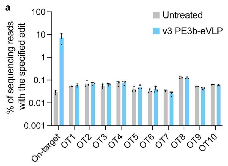
ثلاث أحداث تزاوج مستقلة للأحماض النووية قبل أن يمكن أن يحدث التحرير ولا يعتمد على كسر DNA مزدوج الشريط أو قوالب DNA المتبرع. نتيجةً لهذه الآلية، فإن التحرير الرئيسي مقاوم بطبيعته للتحرير غير المستهدف أو التحرير الجانبي، ويمكن أن يستمر مع عدد قليل من المنتجات الجانبية أو العواقب غير المرغوب فيها لكسر DNA مزدوج الشريط .
يتطلب تحقيق الإمكانات الكاملة للتحرير الرئيسي لأغراض البحث أو التطبيقات العلاجية في الثدييات طرقًا آمنة وفعالة قادرة على توصيل PEs إلى الأنسجة في الجسم الحي. حتى الآن، أبلغت عدة مجموعات عن توصيل PE في الجسم الحي عبر طرق توصيل فيروسية، بما في ذلك الفيروسات الغدية والفيروسات المرتبطة بالأدينوفيروس (AAV) . ومع ذلك، تتطلب طرق التوصيل الفيروسي أن يتم ترميز الجين المتبرع مباشرة في كاسيت التعبير الجيني الفيروسي، مما يحد من حجم الجين المتبرع. يحتوي جينوم AAV
على حد لحجم الجين الحمولات (لا تشمل التكرارات الطرفية المعكوسة) ، مما يتطلب تقسيم حمولات كبيرة مثل PEs (6.4 كيلوبايت في حجم الجين لمحرر رئيسي من الجيل الأول) إلى عدة AAVs ، مما يحد من كفاءة التحرير خاصة عند جرعات متوسطة أو منخفضة من الناقل . كما أن طرق التوصيل الفيروسي تشكل مخاطر أمان محتملة بما في ذلك زيادة التحرير غير المستهدف من التعبير المستمر للجين المتبرع وإمكانية دمج الحمض النووي غير المرغوب فيه في جينومات خلايا المضيف . تتجنب طرق التوصيل غير الفيروسية، مثل الجسيمات النانوية الدهنية، بعض هذه المشكلات من خلال تعبئة المحررين كـ RNAs الرسولية (mRNAs) تعبر بشكل مؤقت. ومع ذلك، لا يزال استهداف الأنسجة في الجسم الحي خارج الكبد من أجل تحرير الجينات العلاجية بكفاءة يمثل تحديًا ، على الرغم من التقدم الأخير في استهداف خلايا الجذعية الدموية .
تعتبر الجزيئات الشبيهة بالفيروسات (VLPs) وسائل توصيل واعدة من حيث المبدأ تقدم فوائد رئيسية لكل من طرق التوصيل الفيروسي وغير الفيروسي . تتشكل VLPs من التجميع التلقائي والانفصال لبروتينات بولي الفيروسات التي تحبس جزيئات الحمولة من خلايا المنتج. تفتقر VLPs إلى جينوم محمول ولكنها تحتفظ بالقدرة على نقل خلايا الثدييات وإطلاق الحمولة . استكشفت الدراسات السابقة VLPs لتوصيل نيكاز Cas9 . أبلغنا مؤخرًا عن توصيل فعال في الجسم الحي لمحرر قاعدة الأدينين (ABE): ريبونوكليوبروتينات RNA أحادية الدليل (sgRNA) مع جزيئات فيروسية مصممة (eVLPs) التي تغلبت على عنق الزجاجة الجزيئي المحدد في تعبئة الحمولة والإفراج والتوطين.
تقدم VLPs المصممة عدة مزايا على طرق التوصيل الأخرى كمرشح لتوصيل PE في الجسم الحي. أولاً، لا تخضع eVLPs لقيود صارمة على حجم الحمولة، مما يلغي الحاجة إلى تقسيم PEs إلى ناقلات منفصلة متعددة. بالإضافة إلى ذلك، يمكن لـ eVLPs تعبئة RNPs، الشكل الأكثر مؤقتًا من عوامل تحرير الجينات، مما يقلل من تكرار التحرير غير المستهدف من خلال تقليل مدة تعرض الجينوم لعوامل التحرير . نظرًا لأن eVLPs تفتقر إلى , فإنها تتجنب الدمج غير المرغوب فيه للمواد الوراثية الفيروسية في جينومات الخلايا المنقولة. أخيرًا، يمكن أن تكون eVLPs مزيفة بأنواع مختلفة من البروتينات السكرية، مما يمكّن الاستهداف المحدد لأنواع الخلايا ذات الأهمية مع جهود هندسة بروتين الغلاف.
في هذه المقالة، نبلغ عن تطوير نظام PE-eVLP الذي يقوم بتوصيل أنظمة PE الكاملة بما في ذلك pegRNAs وsgRNAs المقطوعة (ngRNAs) كـ RNPs. أدى الاستبدال البسيط لمحرري القاعدة (BEs) بـ PEs في نظام BE-eVLP المحسن إلى تحقيق توصيل وظيفي منخفض جدًا لأنظمة التحرير الرئيسي (<1% كفاءة التحرير في خلايا الثدييات المزروعة). من خلال تحديد عنق الزجاجة في توصيل PE-eVLP بشكل منهجي وهندسة الحلول المقابلة، طورنا الجيل الثالث من v3 PE-eVLPs التي تقدم تحسينًا بمقدار 79 مرة في كفاءة التحرير الرئيسي مقارنةً بـ v1 PE-eVLPs في خلايا الفأر Neuro-2A (N2A) وتحسينًا بمقدار 170 مرة في خلايا HEK293T البشرية. أظهر حقن تحت الشبكية واحد من v3 PE-eVLPs تحريرًا رئيسيًا فعالًا في الجسم الحي في نماذج الفئران، مصححًا حذفًا بمقدار 4 أزواج قاعدية في Mfrp في نموذج الفأر rd6 للانحلال الشبكي (15% كفاءة متوسطة) وتصحيح استبدال Rpe65 لإنقاذ جزئي لوظيفة الرؤية في نموذج rd12 (7.2% كفاءة متوسطة). تؤسس دراستنا PE-eVLPs كطريقة خالية من الفيروسات لتوصيل أنظمة التحرير الرئيسي في شكل RNP.

النتائج

توصيل PE: pegRNA RNPs عبر eVLPs في خلايا مزروعة

في السابق، قمنا بتصميم بنية v4 BE-eVLP التي تعبئ وتوصل الأدينين . لاختبار ما إذا كانت بنية v4 BE-eVLP يمكن أن تعبئ وتوصل PEs (الشكل 1أ)، قمنا بإنتاج PE-eVLPs عن طريق نقل خلايا Gesicle 293T باستخدام بلازميدات ترمز لبروتين الغلاف VSV-G، وبروتين بولي MMLV Gag-Pol البري، وبروتين الاندماج MMLV Gag-PE المصمم وpegRNA. ثم عالجنا الخلايا المستهدفة بجرعات متزايدة من eVLPs. نتجت مستويات منخفضة جدًا من التحرير الرئيسي من نظام v1.1PE-eVLP عندما تم برمجته لتثبيت تعديل +1 T-to-A في موقع HEK293T 3 (المسمى لاحقًا HEK3) في خلايا HEK293T (0.17% كفاءة التحرير) وتعديل +2 G-to-C في Dnmt1 في خلايا N2A (0.74% كفاءة التحرير) عند أعلى جرعة من eVLPs تم اختبارها ( ، حوالي جزيئات لطبق يحتوي على 30,000-35,000 خلية) (الشكل 1ب). ثم قمنا بإدخال تعديلين أبلغنا عنهما مؤخرًا لتعزيز التحرير الرئيسي. أولاً، طورنا v1.2 PE-eVLPs التي تستخدم pegRNAs مصممة (epegRNAs) ، التي تحتوي على نمط عقدة زائفة في نهايتها التي تحمي pegRNA من التحلل. ثانيًا، استبدلنا بروتين PE بـ PEmax , بنية بروتين PE المحسنة التي تشمل طفرات SpCas9، رابط محسن بين كاس9 نيكاز ونطاق RT، تحسين الكودونات، وتحسين إشارة التوطين النووي (NLS). أظهرت PE-eVLPs الإصدار 1.3 تحسينًا بمقدار 19 مرة و1.7 مرة في كفاءة التحرير الأساسي عند HEK3 (3.2%) وDnmt1 (1.3%)، على التوالي، مقارنةً بـ PE-eVLPs الإصدار 1.1 (الشكل 1b).

الهندسة المنهجية لبنية PE وeVLP

لزيادة كفاءة PE-eVLPs، سعينا لتحديد الاختناقات الميكانيكية في PE-eVLPs الإصدار 1. تم تشكيل eVLPs اعتمادًا على المعالجة البروتينية لبروتين Gag-Pol المتعدد إلى مجالات وظيفية متعددة بواسطة بروتياز MMLV. بالمصادفة، فإن نطاق RT في PEs التقليدية أيضًا ينشأ من MMLV، وبالتالي يحتوي على موقع قطع بروتياز TSTLLIENS داخلي في الطرف C-terminus (الشكل 1c). افترضنا أن القطع في هذا الموقع قد يلغي NLS الحاسم في الطرف C الذي يوجه التوطين النووي لـ PEs. لذلك، قمنا بحذف ستة أحماض أمينية في الطرف C من MMLVRT لإزالة موقع التعرف على البروتياز، مما أدى إلى إنتاج PE-eVLPs الإصدار 2.1. قدم هذا التعديل تحسينًا ثابتًا في كفاءة تحرير PE-eVLP مقارنةً بـ PE-eVLPs الإصدار 1.3 (بمتوسط 1.8 مرة و1.2 مرة، على التوالي) عند كل من موضع HEK3 وموضع Dnmt1 (الشكل 1d). اختبرنا تقصيرًا إضافيًا لـ MMLV RT لإزالة نطاق RNaseH بالكامل، وهي استراتيجية تقصير شائعة تستخدمها أنظمة AAV لتقليل حجم PE وتسهيل التعبئة المزدوجة لـ AAV . في نظام PE-eVLP، ومع ذلك، أدى القضاء على نطاق RNaseH إلى كفاءات تحرير أقل باستمرار في كل من خلايا HEK293T وN2A مقارنةً بـ PE-eVLP الإصدار 2.1 (الشكل 1d).
تشجع إشارة التصدير النووي (NES) التوطين السيتوبلازمي لبروتين الاندماج Gag-cargo. بعد قطع البروتياز، يجب فصل عوامل تحرير الجينات عن NES لتوطين نووي فعال. إن وجود أربعة مواقع قطع بروتياز MMLV إضافية داخل بروتين Gag المتعدد يرفع من احتمال القطع غير المكتمل، مما يؤدي إلى احتفاظ جزء من شحنة PE ببعض أجزاء Gag مع NESs (الشكل 1e). في الواقع، لاحظنا سابقًا شحنة من
الشكل 1|هندسة بنى PE وeVLP. أ، مخطط لـ PE-eVLPs الإصدار 1. ب، كفاءات التحرير الأساسي لـ PE-eVLPs الإصدار 1 لـ HEK3+1 تعديل T-to-A في خلايا HEK293T وتعديل Dnmt1 +2 G-to-C في خلايا N2A. أدى اعتماد epegRNAs (الإصدار 1.2) وبنية PEmax (الإصدار 1.3) إلى تحسين كفاءات التحرير الأساسي مقارنةً بـ PE-eVLPs الإصدار 1.1. ج، مخطط لهندسة PE لإزالة موقع قطع البروتياز الداخلي (TSTLLIENS) في MMLV RT. د، تحسينات تمثيلية في كفاءات تحرير PE-eVLP نتيجة لهندسة نطاق RT. PEmax-FL تشير إلى PE-eVLPs الإصدار 1 مع MMLV RT بطول كامل كنطاق مؤثر PE؛ PEmax-RNaseH del تشير إلى PE-eVLPs الإصدار 1 مع RT مقطوع نطاق RNaseH المستخدمة كنطاق مؤثر PE؛ PEmax-6aa del تشير إلى PE-eVLPs الإصدار 2.1 مع RTs محذوفة من ستة أحماض أمينية المستخدمة كنطاق مؤثر PE. هـ، مخطط للآلية المقترحة لنضوج eVLP وتسليم الشحنة، وتصميم PE-eVLPs الإصدار 2.2 و2.3. و، كفاءات التحرير الأساسي لـ PE-eVLPs مع NES الموضوعة في مواقع مختلفة
(موقع NES 1-5) من نطاق Gag. أظهر موقع NES 5 (الإصدار 2.2) تحسينًا في التحرير مقارنةً بـ PE-eVLPs الإصدار 2.1. ز، مقارنة كفاءات التحرير الأساسي مع PE-eVLPs الإصدار 1، 2.1، 2.2 و2.3 عند موضع HEK3 في خلايا HEK293T وموضع Dnmt1 في خلايا N2A. القيم المعروضة في جميع الرسوم البيانية تمثل متوسط كفاءات التحرير الأساسي s.e.m. من ثلاث نسخ بيولوجية. تم ملاءمة البيانات لمنحنيات لوجستية بأربعة معلمات باستخدام الانحدار غير الخطي. تم إجراء مقارنات بين إصدارات مختلفة من PE-eVLPs مع eVLPs المنتجة والمتحولة بالتوازي في تجربة واحدة كبيرة لتقليل التباين بين التحضيرات. تم توفير بيانات جميع PE-eVLPs المنتجة والمختبرة بالتوازي في الشكل التكميلي 1. الرسوم البيانية في و تظهر مجموعة فرعية من البيانات من الشكل التكميلي 1. بالنسبة لجميع الظروف، تم معالجة 30,000-35,000 خلية مع eVLPs تحتوي على .
أحجام متغيرة في BE-eVLPs بواسطة البقعة الغربية , مما يدعم الفرضية بأن المنتجات المقطوعة بشكل غير صحيح يمكن أن يتم تعبئتها في eVLPs. للتخفيف من هذه المشكلة، قمنا بنقل NESs داخل بروتين Gag المتعدد (الشكل 1f). قمنا بإدخال NES قبل موقع قطع البروتياز لكل مجال فرعي من Gag واثنين من المواقع الإضافية التي تم إظهارها سابقًا لتحمل إدخالات كبيرة في Gag MMLV- . من بين خمسة
المواقع المختبرة، دعم إدخال NES بين مجالات p12 وCA من Gag (موقع NES 5) أعلى كفاءة تحرير أساسي في خلايا HEK293T، مما أدى إلى إنتاج PE-eVLP الإصدار 2.2 (الشكل 1f).
أخيرًا، لتسهيل إطلاق الشحنة، قمنا بإدخال روابط GGS المحيطة بموقع قطع البروتياز المهندسة لزيادة إمكانية الوصول إلى الموقع لمعالجة البروتياز. أظهرت بنية PE-eVLP الإصدار 2.3 الناتجة

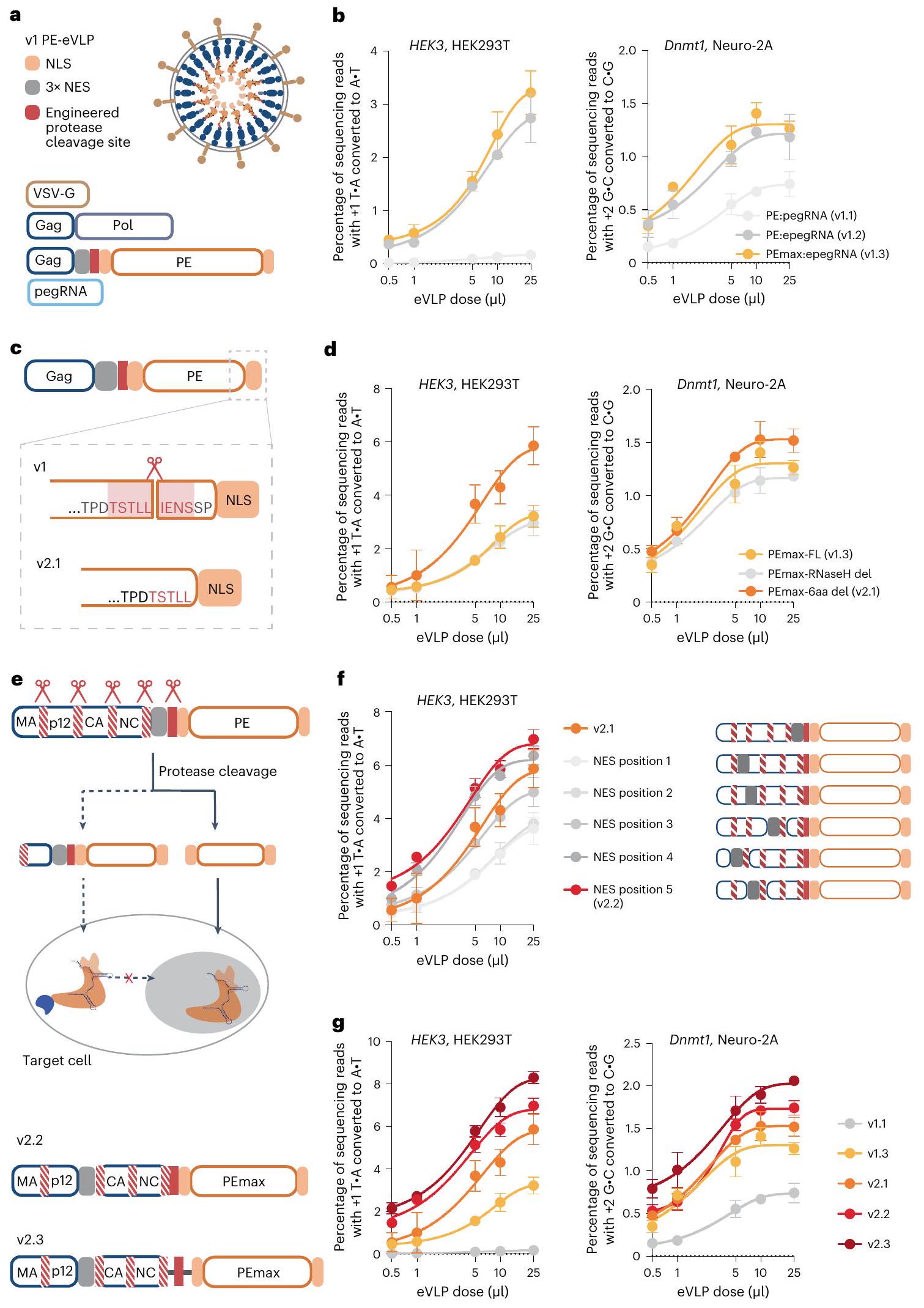
(الشكل 1e) التي تحتوي على تقصير MMLV RT، وتحسين NES وتحسين الروابط متوسط تحسين بمقدار 2.6 مرة و1.6 مرة في كفاءة التحرير الأساسي مقارنةً ببنية PE-eVLP الإصدار 1.3 عند أعلى جرعة تم اختبارها عند موضع HEK3 في خلايا HEK293T وموضع Dnmt1 في خلايا N2A، على التوالي (الشكل 1g).

اعتماد أداء PE-eVLP على نوع التحرير

لقد أظهرنا سابقًا أن كفاءات التحرير الأساسي يمكن أن تتأثر بواسطة مسارات إصلاح عدم التطابق الخلوية (MMR)، وأن التهرب أو تثبيط MMR يحسن كفاءات التحرير الأساسي . نظرًا للطبيعة المؤقتة لـ PE:pegRNA RNPs المعبأة في eVLPs، افترضنا أن عكس التحرير المثبت بواسطة MMR قد يكون ضارًا بشكل خاص لكفاءة التحرير.
لقد اكتشفنا سابقًا أن تثبيت طفرات إضافية صامتة أو غير ضارة بالقرب من تعديل الهدف يتسبب في هروب الوسيط التحريري الأساسي بشكل طبيعي من MMR، مما يعزز كفاءات التحرير ونقاء المنتجات . لاختبار هذه الإمكانية لأنظمة التحرير الأساسي التي يتم تسليمها بواسطة eVLP، قمنا بتثبيت استبدالات إضافية قريبة عند موضع HEK3 وموضع Dnmt1 باستخدام نظام PE-eVLP الإصدار 2.3. في كلا الحالتين، تم تحسين كفاءات التحرير الأساسي بشكل كبير، حيث حققنا 28% متوسط تحرير لتعديل استبدال 5-bp عند و6.6% تحرير لتعديل استبدال 4-bp عند Dnmt1 عند أعلى جرعة تم اختبارها (الشكل التكميلي 1). بسبب تكرار الكودونات، يمكن تحقيق العديد من التعديلات ذات الصلة العلاجية مع التثبيت المتزامن لطفرات صامتة إضافية لتفادي . الفائدة الكبيرة للتهرب من MMR على نتائج تحرير PE-eVLP تُعلم تصميم epegRNAs المثلى عند استخدام PE-eVLPs.

تقييد تعبئة epegRNA يحد من كفاءة PE-eVLP

على عكس آلية تعبئة بروتين PE، التي تعتمد على الاندماج مع بروتين Gag المتعدد، تعتمد تعبئة epegRNA بالكامل على الارتباط غير التساهمي مع نطاق Cas9 من PEs. لاختبار ما إذا كانت تسليم epegRNA هي اختناق في أداء PE-eVLP، قمنا بتعزيز epegRNAs عبر نقل بلازميدات تشفر epegRNAs قبل 24 ساعة من تحويل PE-eVLP. في الواقع، حسنت إضافة epegRNA بشكل كبير كفاءة تحرير PE-eVLP بأكثر من ثمانية أضعاف في المتوسط، بينما قدمت إضافة sgRNA تحسينًا طفيفًا لكفاءة تحرير BE-eVLP في تجربة مماثلة (الشكل 2a والشكل التكميلي 2a). تكشف هذه النتائج أن تحميل epegRNA، وتسليمه أو عمره يحد من تحرير PE-eVLP إلى حد أكبر بكثير من تأثير تحميل sgRNA أو تسليمه على تحرير القاعدة باستخدام BE-eVLPs. بالمقابل، لم تحسن إضافة بروتين PE عبر نقل بلازميدات تعبر عن PEs كفاءة تحرير PE-eVLP (الشكل 2a)، مما يتماشى مع عدم كفاية epegRNAs ولكن ليس بروتينات PE في PE-eVLPs.
لقد لاحظنا سابقًا أن الألفة الظاهرة لـ Cas9 مع sgRNAs وpegRNAs وepegRNAs تتناقص بهذا الترتيب (ملاحظة تكملية 1). لزيادة ألفة Cas9-epegRNA، اعتمدنا هيكل RNA الدليل F + E ، الذي يحسن ارتباط Cas9-دليل RNA من خلال تمديد الساق في هيكل السقالة الذي يوفر مقبضًا ممتدًا لارتباط Cas9. لقد حسنت سقالة دليل RNA F + E بشكل معتدل كفاءات تحرير PE-eVLP (الشكل البياني للبيانات الموسعة 2b) وتم استخدامها في جميع epegRNAs اللاحقة.
لمعالجة عدم كفاءة تعبئة epegRNA في v2.3 PE-eVLPs بشكل أكبر، استخدمنا نظام MCP-MS2. كآلية إضافية لتعزيز تعبئة epegRNA. يمكن أن يؤدي ارتباط بروتين غلاف MS2 (MCP) مع حلقة الساق RNA الخاصة بـ MS2 إلى تسهيل تعبئة sgRNA لنقل إنزيم Cas9 و BE. يمكنه تجنيد RT غير المرتبط إلى Cas9 nickase في أنظمة PE المنفصلة ، ويمكنه استقطاب مجالات بروتينية إضافية إلى لتطبيق هذا النظام على تغليف PE:epegRNA RNP في eVLPs، قمنا بتصميم بلازميد اندماج Gag-MCP-Pol عن طريق إدخال نسخة واحدة من MCP بين الطرف C من مجال نواة Gag والطرف N من مجال Pol (الشكل 2b). وقد افترضنا أن إدخال MCP في هذا الموضع سيؤثر بشكل طفيف على معالجة بروتين Gag المتعدد من أجل نضوج جزيئات eVLP (بيانات موسعة الشكل 2c و
ملاحظة إضافية 2) وأن MCP المدمج سيتم تقديمه في داخل الجسيم، مما يسهل تجنيد epegRNAs إلى eVLPs.
بعد ذلك، اختبرنا سلسلة من epegRNAs مع إدخالات حلقة MS2 في حلقات السقالة أو في الطرف 3′. لاحظنا أن إدخال نسخة واحدة من حلقة MS2 في حلقة ST2 من epegRNAs (MS2-epegRNAs) أدى إلى أعلى مستوى من التحرير الأولي (الشكل 2c). زيادة عدد النسخ من كل من MCP و MS2 aptamers أضعف كفاءة التحرير، ربما لأن الاندماجات الأكبر تزعزع استقرار Gag-Pol و epegRNAs (الشكل التمديدي 2d، e). أخيرًا، قمنا بإعادة تحسين النسبة المولية لبلازميدات الاندماج Gag عن طريق تغيير نسبة بلازميد Gag-Pol البري، بلازميد Gag-PE وبلازميد Gag-MCP-Pol (الشكل 2d). أدت النسبة المحسنة 5:2:1 لبلازميد Gag-Pol: بلازميد Gag-MCP-Pol: بلازميد Gag-PE إلى v3PE-eVLPs التي حسنت متوسط كفاءة التحرير الأولي بمقدار 4.0 مرة مقارنة بـ v2.3 PE-eVLPs عند موضع Dnmt1 في خلايا N2A عند أعلى جرعة تم اختبارها (الشكل 2e).

PE3 يحسن بشكل أكبر التحرير الأساسي باستخدام PE-eVLPs

نظام PE-eVLP الموصوف حتى الآن يجمع بين PE واحد و epegRNA واحد (استراتيجية ‘PE2’). تضيف استراتيجية ‘PE3’ ngRNA إضافي يوجه PE لعمل قطع في الشريط غير المعدل، مما يحفز استبداله باستخدام الشريط المعدل كقالب، مما يزيد من كفاءة التحرير على حساب زيادة ترددات indel. لتمكين PE3-eVLPs، اختبرنا إمكانية تعبئة ngRNA في نفس الجسيم أو في جسيم منفصل عن epegRNA. لاختيار نظام الجسيمات الشاملة المثالي v3 PE3-eVLP، قمنا بنقل خلايا المنتج بنسب متغيرة من بلازميدات MS2-epegRNA وMS2-ngRNA في نفس الوقت، جنبًا إلى جنب مع مكونات PE-eVLP الأخرى. أظهرت النتائج أن نقل بلازميدات MS2-epegRNA وMS2-ngRNA بنسب محسّنة تبلغ 4:1 أدى إلى v3 PE3-eVLPs التي تدعم كفاءة تحرير أعلى بمعدل 1.3 مرة مقارنة بنظام v3 PE2-eVLP عند موضع Dnmt1 في خلايا N2A مع من eVLPs (الشكل 2f). بدلاً من ذلك، فإن إنتاج دفعات منفصلة من eVLPs التي تحتوي على PE:MS2-epegRNA RNP أو PE:MS2-ngRNA RNP ونقل الخلايا المستهدفة بكلا النوعين من eVLPs أسفر عن مستويات تحرير مماثلة (الشكل 2f). نظرًا لمستويات التحرير المماثلة مع الاستراتيجيتين، تقدمنا باستخدام نظام الجسيمات الشامل v3 PE3-eVLP لسهولة الإنتاج، والقدرة على التحكم بدقة في نسبة epegRNA وngRNA، وكفاءة توصيل محتملة أعلى في الجسم الحي من نظام الجسيمات الواحدة الذي لا يتطلب النقل المشترك بواسطة عدة eVLPs.

إزالة امتداد 5′-G تعزز التحرير باستخدام بعض epegRNAs

تُنسخ الأدلة RNA من محفز U6 في نظام PE-eVLP، مع تفضيل البدء من الجوانين. في المركز +1 . epegRNAs التي لا تبدأ بـ غالبًا ما يتم إضافة حرف G إضافي في نهاية. مثل هذه -G قد تعيق بشكل حجمي ارتباط pegRNA بنطاق Cas9 ومع ذلك، قد يقدم ذلك تحديًا إضافيًا لتكوين معقد PE:epegRNA. علاوة على ذلك، قد تعيق القاعدة غير المقترنة 5′ G تشكيل حلقة R وقطع الحمض النووي بواسطة إنزيم كاس9 بطريقة تعتمد على تسلسل الهدف. نظرًا لأن جزيئات PE:epegRNA RNPs التي يتم توصيلها بواسطة PE-eVLPs قصيرة العمر عن قصد، فقد افترضنا أن أي تدهور في معدل البحث عن الهدف وقطع الحمض النووي قد يكون ضارًا بكفاءات التحرير الأولي التي تتم بواسطة PE-eVLP.
لتجاوز هذه العقبة المحتملة، قمنا باختبار أنظمة v3 PE-eVLP التي تستخدم epegRNAs تحتوي على إما فاصل بطول 20 قاعدة يبدأ بالسايتوزين (C) أو اليوراسيل (U) أو الأدينين (A)، أو غير متطابق. تبعها فاصل بطول 19 قاعدة (الشكل التمديدي 3أ). تشير البيانات الناتجة إلى أن التحسين الناتج عن حذف الـ -G يمكن أن يكون الامتداد كبيرًا، على الرغم من أن التأثير يختلف اعتمادًا على تسلسل الهدف. في موضع Col12a1 في خلايا N2A، يبدأ فاصل بطول 20 قاعدة بـ تفوق على النسخة الأصلية -bp الفاصل بمقدار 2.8 -طية (الشكل البياني الممتد 3a). عند موضع Ctnnb1، يبدأ كل من الفاصل 20 -bp بـ و الـ -bp الفاصل حقق نتائج مماثلة، حيث قدم كلاهما تحسينًا بمقدار 4.7 مرة مقارنة بالأصل -bp الفاصل.
الشكل 2 | هندسة pegRNA و ngRNA. أ، تجربة نقل مزدوجة تحدد epegRNAs كعنصر محدد في v2.3 PE-eVLPs. ب، مخطط v3 PE-eVLPs باستخدام استراتيجية MCP-MS2 لتجنيد epegRNAs. تتيح دمج استراتيجية MCP-MS2 ثلاثة أوضاع لتحميل RNA الدليل إلى eVLPs: (i) عبر الارتباط بـ PE، (ii) عبر ارتباط حلقة الساق MS2 بـ MCP و (iii) عبر تفاعل ثلاثي المكونات MCP:MS2-gRNAs:PE. ج، كفاءات التحرير لـ v2.3 PE-eVLPs عند موضع Dnmt1 في خلايا N2A مع إدخال حلقة الساق MS2 في مواقع مختلفة في epegRNAs. يشير الطرف 3′ إلى v2.3 PE-eVLPs مع إدخال حلقة الساق MS2 بعد نمط tevoPreQ1 المنظم لـ epegRNA؛ يشير الطرف 3′ * إلى v2.3 PE-eVLPs مع إدخال حلقة الساق MS2 مباشرة بعد تمديد الـ pegRNA، وبالتالي استخدام حلقة الساق MS2 لمحاكاة نمط هيكلي في نهاية epegRNAs؛ TL تشير إلى v2.3 PE-eVLPs مع إدخال حلقة الساق MS2 داخل الحلقة الرباعية من هيكل pegRNA؛ و ST2 تشير إلى v2.3 PE-eVLPs مع إدخال حلقة الساق MS2 داخل حلقة ST2 من هيكل pegRNA. د، خريطة حرارية لكفاءات التحرير من
تحسين الاستوكيومترية لبلازميدات Gag-Pol وGag-MCP-Pol وGag-PE لإنتاج v3 PE-eVLPs. هـ، مقارنة كفاءات التحرير مع v2.3 وv3 PE-eVLPs عند موقع Dnmt1 في خلايا N2A. كفاءات التحرير في أنظمة PE3-eVLP ذات الجزيئات المنفصلة وذات الكل في واحد v3 مع نسب متغيرة من MS2-epegRNA إلى MS2-ngRNA. ج، قياس عدد جزيئات epegRNA المعبأة لكل eVLP في الأجيال المتعاقبة من PE-eVLPs. ح، قياس عدد جزيئات بروتين PE المعبأة لكل eVLP في الأجيال المتعاقبة من PE-eVLPs. تمثل القيم متوسط كفاءات التحرير الأساسية. الخطأ المعياري للمتوسط لثلاث نسخ بيولوجية (أ و ج-و) أو ثلاث نسخ تقنية (ز و ). تم ملاءمة البيانات لمنحنيات لوجستية بأربعة معلمات باستخدام الانحدار غير الخطي. تم تقديم بيانات جميع PE-eVLPs التي تم إنتاجها واختبارها بالتوازي في الشكل التوضيحي 2. الرسوم البيانية في ونعرض مجموعة فرعية من البيانات من الشكل التوضيحي 2.
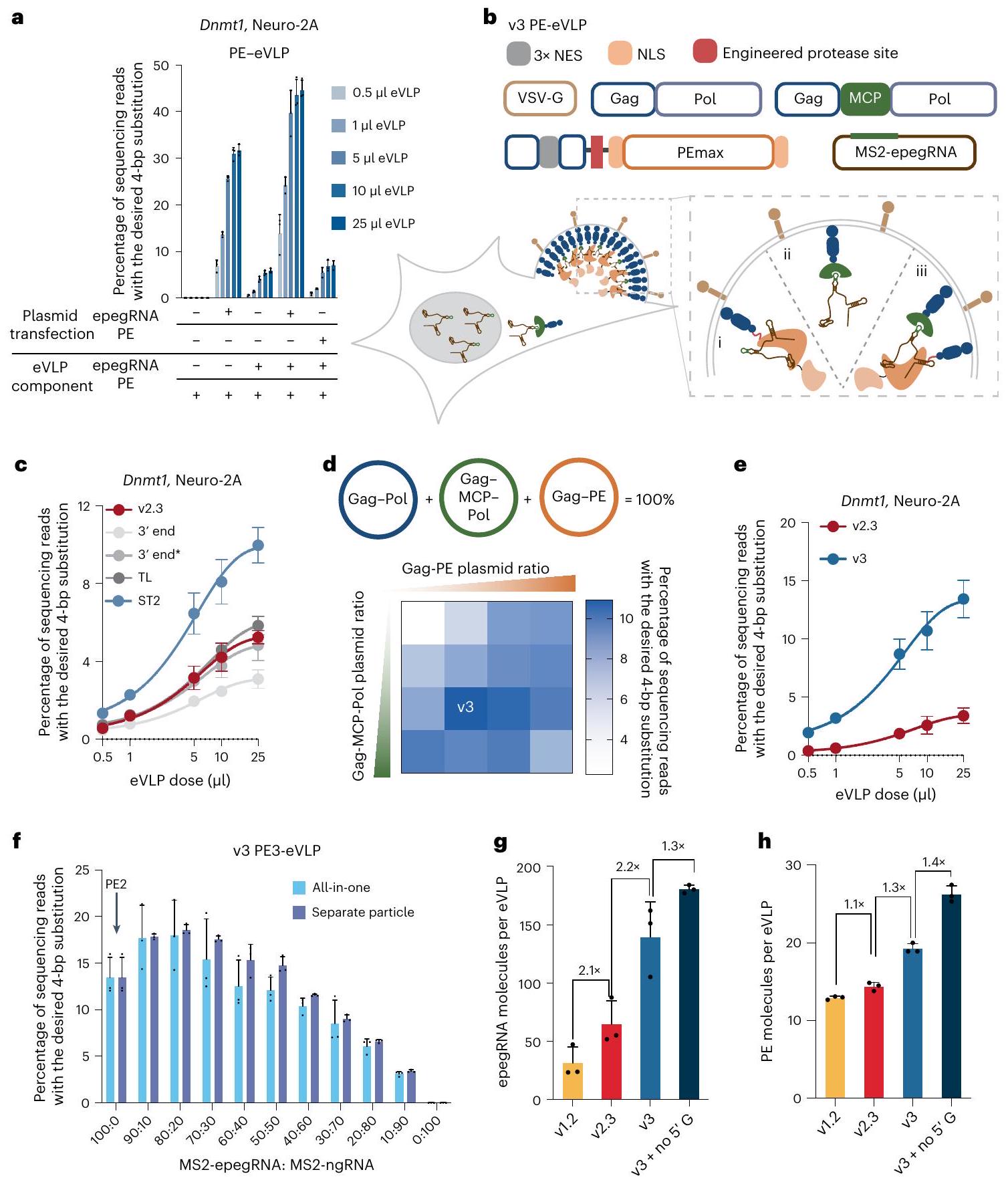
لجميع الظروف، تم معالجة 30,000-35,000 خلية باستخدام eVLPs التي تحتوي على .

توصيف تحميل شحنة PE-eVLP

للتحقق من الآليات المستخدمة لتعزيز تعبئة الشحن، قمنا بتحديد كمية epegRNAs وبروتين PE المعبأ في كل نسخة من PE-eVLP. تم تحديد كمية epegRNAs السليمة بواسطة تفاعل البوليميراز المتسلسل الكمي (qPCR) للحمض النووي الريبي الكلي المستخرج من eVLPs (الشكل 2g). تم استخدام عدد جزيئات eVLP لكل وحدة حجم تم قياسها باستخدام اختبار الامتصاص المناعي المرتبط بالإنزيم (ELISA) المضاد لـ MLV p30 (الغلاف) (الشكل 3b البيانات الموسعة) لحساب عدد epegRNAs المعبأة لكل eVLP. على الرغم من أن هندسة بنية PE و eVLP في v2.3 PE-eVLP لم تكن مصممة لتعزيز تجنيد epegRNA، إلا أن v2.3 PE-eVLPs لا تزال حسنت تعبئة epegRNA مقارنة بـ v1.2 PE-eVLPs بمعدل 2.1 مرة، على الأرجح لأن تعبئة epegRNA تحدث كمعقد مع بروتين PE وبالتالي ترتبط بتعبئة بروتين PE. استراتيجيات MCP-MS2 في نظام v3 PE-eVLP حسنت تحميل epegRNA بمعدل 2.2 مرة مقارنة بـ v2.3 PE-eVLPs، مما يتماشى مع تحسين كفاءة التحرير. أخيرًا، تم حذف -G الإضافي على epegRNAs قدم تحسينًا إضافيًا بمقدار 1.3 مرة في تحميل epegRNA، مما يدعم الفرضية بأن التحسين في كفاءة التحرير ناتج عن التمديد. -قد تنشأ حالة حذف G من تحسين ارتباط epegRNAs بـ PEs. كشفت التوصيفات الإضافية لنسبة تكوين MS2-epegRNA و MS2-ngRNA في v3PE-eVLPs عن نسبة تقريبية 1:1 بين النوعين (الشكل 3c من البيانات الموسعة). نظرًا لأن تكوين MS2-ngRNAs زاد في eVLPs مقارنةً بـ نستخدم الكيمياء الحركية لإنتاج eVLP، ونفترض أن ngRNAs لديها قدرة ارتباط أعلى من epegRNAs لبروتين PE، مما يؤدي إلى تحميل أكثر كفاءة في eVLPs.
كشفت تقنية ELISA باستخدام الأجسام المضادة المضادة لـ Cas9 عن زيادة في بروتين PE لكل جزيء في النسخ المتعاقبة من PE-eVLPs (الشكل 2h). حسنت استراتيجية MS2-MCP في النسخة الثالثة من PE-eVLP كل من تجنيد epegRNA ومستويات بروتين PE لكل جزيء بمقدار 1.3 مرة، ربما بسبب التفاعل الثلاثي المكون بين MCP:MS2-gRNAs:PE الذي يعزز بشكل تآزري دمج جميع مكونات التحرير الأساسي (الشكل 2b). وبالمثل، فإن حذف -G الإضافة حسنت تغليف بروتين PE بمعدل إضافي قدره 1.4 مرة ليصل إلى متوسط 26 جزيئًا من بروتين PE لكل eVLP (الشكل 2h). بشكل عام، تكشف هذه النتائج أن الزيادات في كفاءة تغليف كل من epegRNA وبروتين PE في PE-eVLPs تتوافق مع تحسينات في كفاءات التحرير الأساسي عبر الأجيال المتعاقبة من PE-eVLPs، مما يشير إلى أن PE-eVLPs من النسخة 3 تعزز نتائج التحرير الأساسي من خلال معالجة الاختناقات في تغليف البروتين والحمض النووي الريبي.

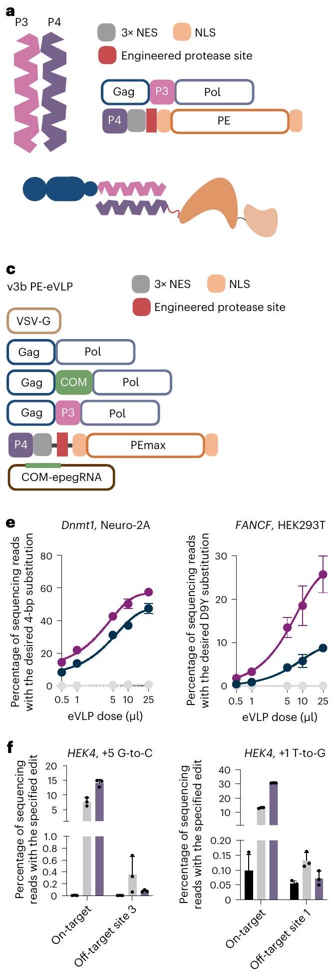
توظيف PE من خلال ارتباط الببتيد الحلزوني

على الرغم من أن تغليف PE في PE-eVLPs اعتمد نفس الاستراتيجية مثل تغليف BE في v4 BE-eVLP، وأدت الجولات المتعاقبة من تحسينات محددة لـ PE إلى تحسين متوسط يبلغ 2.0 مرة في تغليف PE في eVLPs، إلا أن العدد المطلق لجزيئات PE المعبأة لكل جزيء (26 لكل eVLP) ظل حوالي نصف القيمة لجزيئات BE في BE-eVLPs (55 لكل eVLP). افترضنا أن الحجم الكبير لبروتين الاندماج Gag-PE (309 كيلودالتون) قد يعيق إنتاجه في خلايا المنتج. للتغلب على هذه المشكلة، صممنا نظام تعبئة PE بديل لا يتطلب اندماج Gag مع PE (الشكل 3أ). بدلاً من ذلك، استخدمنا ببتيدات ملتفة. -الببتيدات الحلزونية التي يمكن أن ترتبط بقوة وبشكل محدد مع الآخرين -الببتيدات الحلزونية لتشكيل بنية سوبر هليكس . اخترنا زوج الببتيد P3-P4 المتشابك الذي تم استخدامه سابقًا في خلايا الثدييات لاستقطاب مجالات البروتين إلى Cas9 (المرجع 61). صممنا بناء الاندماج Gag-P3-Pol عن طريق دمج P3 في الطرف C من مجال نواة Gag، ودمجنا ببتيد الشريك P4، متبوعًا بـ NES وموقع قطع البروتياز المهندسة، إلى الطرف N من PE (المشار إليه فيما بعد بـ P4-PE). قمنا بإنتاج PE-eVLPs عن طريق نقل بلازميدات تشفر VSV-G و MMLV Gag-Pol من النوع البري و Gag-P3-Pol و P4-PE و epegRNA إلى خلايا الإنتاج. أظهر هذا النظام البديل كفاءة تحرير أولية أعلى بمعدل 1.3 مرة مقارنة بنظام v2.3 Gag-PE-eVLP المماثل في موضع Dnmt1 في خلايا N2A عند أعلى جرعة تم اختبارها (الشكل 3b). أظهر eVLP الضابط الذي يفتقر إلى ببتيد P3 كفاءة تحرير أقل بمعدل 28 مرة، مما يدعم دور ارتباط P3-P4 في تسهيل تعبئة PE.
بالإضافة إلى ذلك، أكدنا من خلال تحليل الغربي لمستخلصات خلايا المنتج مستويات أعلى بكثير من P4-PE مقارنة بـ Gag-PE (الشكل 4a من البيانات الموسعة والملاحظة التكميلية 3)، مما يتماشى مع فرضيتنا بأن بروتين الاندماج الأصغر P4-PE قد حسن مستويات إنتاج البروتين.
بعد ذلك، استكشفنا التآزر بين هذا النظام البديل واستراتيجية تجنيد pegRNA باستخدام بروتينات ربط الأبتامر، مما أدى إلى تحسينات كبيرة في نظام v3 PE-eVLP. أثبتت المحاولات الأولية باستخدام بناء اندماج واحد Gag-MCP-P3-Pol أنها غير فعالة (الشكل 4b من البيانات الموسعة). لذلك، اختبرنا إنتاج PE-eVLP باستخدام كل من بروتينات ربط الأبتامر Gag-aptamer-binding-Pol وبناء Gag-P3Pol. بدلاً من زوج MCP-MS2، استخدمنا زوج بروتين-RNA الأبتامر COM-Com. (الشكل 4c من البيانات الموسعة)، معتبراً أن الحجم الأصغر لكل من بروتين COM و aptamer Com قد يكون أقل تأثيراً على نضوج الجسيمات أو وظيفة PE. قمنا بتحسين النسبة المولية لبلازميدات Gag-COM-Pol و Gag-P3-Pol و P4-PE لإنتاج نظام v3bPE-eVLP، وهو بديل لنظام v3 PE-eVLP (الشكل 3c والشكل 3d). قمنا بمقارنة نظام v3 PE3-eVLP ونظام v3b PE3-eVLP في مواقع متعددة ولاحظنا اختلافات تعتمد على الموقع في كفاءة التحرير (الشكل 3e). في مواقع Dnmt1 و FANCF، تفوق v3b على v3 في كفاءة التحرير الأولي بمعدل 1.2 و 2.9 مرة، على التوالي، عند أعلى جرعة تم اختبارها. في مواقع HEK3 و Col12a1، أدت الأنظمة إلى أداء متقارب، متماشياً مع مستويات متقاربة من بروتين PE المعبأ في v3 و v3b PE-eVLPs التي تم تحليلها عبر تقنية الويسترن بلوت (الشكل 4d من البيانات الموسعة والملاحظة التكميلية 3).
أدت التحسينات التراكمية في PE3-eVLPs من النسخة v3 وv3b إلى زيادات كبيرة في كفاءات التحرير الأساسي مقارنةً بـ PE2-eVLPs من النسخة v1.1 (الشكل 3e). تفوقت PE3-eVLPs من النسخة v3 وv3b على PE2-eVLPs من النسخة v1.1 بمعدل 79 و65 مرة في موقع Dnmt1 في خلايا N2A، و170 و150 مرة في موقع HEK3 في خلايا HEK293T، على التوالي. في موقع Col12a1 في خلايا N2A وموقع FANCF في خلايا HEK293T، لم نلاحظ أي تحرير فوق مستوى الخلفية (<0.1%) باستخدام PE2-eVLPs من النسخة v1.1، بينما حققت هياكل PE-eVLP من الجيل الثالث في المتوسط 7.7% و26% من التحرير في المواقع المعنية عند أعلى جرعة تم اختبارها. كشفت دراسة تكرار المنتجات الجانبية للإدخال والحذف عن عدم وجود فرق واضح بين PEs التي تم توصيلها بواسطة PE-eVLPs مقابل تلك التي تم توصيلها عن طريق نقل البلازميد (البيانات الموسعة الشكل 5a). علاوة على ذلك، لاحظنا انخفاضًا في تباين الدفعات كما يتضح من كفاءات التحرير الأساسي المتسقة باستخدام أربع دفعات مستقلة من PE-eVLPs تم إعدادها في أيام مختلفة (البيانات الموسعة الشكل 5b). تبرز هذه البيانات مجتمعة الفوائد الكبيرة التي نتجت عن الهندسة المنهجية لهياكل PE-eVLP من الجيل الثالث (البيانات الموسعة الشكل 6).

تستفيد BE-eVLPs من هندسة هيكل PE-eVLPs

نظرًا للتحسينات الكبيرة في كفاءات تحرير الجينات الأساسية التي تقدمها v3 و v3b PE-eVLPs، سعينا لتقييم ما إذا كانت هذه التعديلات تعزز أيضًا توصيل BE بواسطة eVLP. قمنا بتغليف اثنين من ABEs، ABE8e و ABE7.10-NG ، بالإضافة إلى السيتوزين BE، TadCBEd في بنية v4 BE-eVLP التي تم الإبلاغ عنها سابقًا، وفي بنى v3 أو v3b PE-eVLP (مع استبدال PE بـ BE)، مستهدفةً موقع BCL11A في خلايا HEK293T (الشكل التمديدي 7a-c). بينما قدمت بنية v3 و v3b PE-eVLP تحسينات طفيفة لـ ABE8e النشط للغاية، إلا أنها عززت بشكل كبير كفاءات التحرير لبروتينات BE الأقل نشاطًا مثل ABE7.10-NG. بالنسبة لـ ABE7.10-NG، حققت بنى v3 و v3b PE-eVLP كفاءات تحرير أساسية أعلى بمقدار 2.6 مرة و 3.2 مرة، على التوالي، عند أعلى جرعة تم اختبارها (الشكل التمديدي 7b). تشير هذه النتائج إلى أن بعض الاستراتيجيات التي حسنت من تغليف بروتين PE و pegRNA يمكن أن تستفيد أيضًا من تغليف وكفاءة التحرير بعد النقل لبضائع تحرير الجينات الأخرى.

التوصيل العابر لـ PE بواسطة eVLPs يقلل من التحرير غير المستهدف

أظهرت الدراسات السابقة أن التسليم المؤقت لوكلاء تحرير الجينوم يمكن أن يقلل من التحرير غير المستهدف. . يجب أن تؤدي التسليم المباشر لمجمعات RNP في النظرية إلى أقصر مدة تعرض لعامل التحرير على الجينوم مقارنة بتسليم mRNA أو DNA. لقد أظهرت تقنية التحرير الأساسي أنها تتمتع بخصوصية عالية لـ DNA، ربما بسبب
الشكل 3 | تطوير v3b PE-eVLPs، التي تستخدم آلية بديلة لتجنيد PE. أ، مخطط لتجنيد PE المعتمد على الببتيد الملفوف. NES، إشارة التصدير النووي. NLS، إشارة التوطين النووي. ب، مقارنة كفاءة التحرير الأساسي مع v2.3 PE-eVLPs مقابل النظام البديل الذي يستخدم الببتيد الملفوف لتجنيد PE. ج، مخطط لنظام v3b PE-eVLP. NES، إشارة التصدير النووي. NLS، إشارة التوطين النووي. د، خريطة حرارية لكفاءات التحرير من تحسين النسبة المولية لبلازميدات Gag-Pol وGag-COM-Pol وGag-P3-Pol لإنتاج v3bPE-eVLPs. هـ، كفاءات التحرير الأساسي مقارنة بين v1.1PE2-eVLPs وv3 PE3-eVLPs وv3b PE3-eVLPs في Dnmt1 وFANCF وCol12a1 و
ب

موقع HEK3 في خلايا N2A و HEK293T. ف، مقارنة كفاءات التحرير لأربعة تعديلات رئيسية مختلفة تستهدف موقع HEK4 في خلايا HEK293T بعد نقل البلازميد أو العلاج مع v3b PE-eVLPs في الموقع المستهدف HEK4 والمواقع المعروفة خارج الهدف لـ pegRNA المقابل. . القيم المعروضة في جميع الرسوم البيانية وخرائط الحرارة تمثل متوسط كفاءة التحرير الأساسي لثلاث نسخ بيولوجية، وتُمثل أشرطة الخطأ الانحراف المعياري. تم ملاءمة البيانات لمنحنيات لوجستية بأربعة معلمات باستخدام الانحدار غير الخطي. بالنسبة لجميع الظروف بخلاف تم معالجة الخلايا بـ eVLPs التي تحتوي على .
الشكل 4 | تحرير الجهاز العصبي المركزي باستخدام PE-eVLPs عبر حقن PO ICV. أ، مخطط سير العمل لحقن ICV في حديثي الولادة والتحليل اللاحق. FACS، فرز الخلايا المعتمد على الفلورسنت. ب، كفاءة التحرير الأساسي في مجموعة كبيرة أو مجموعة إيجابية GFP من قشرة الدماغ التي تم جمعها بعد 3 أسابيع من حقن PO ICV.
استهداف موقع Dnmt1 باستخدام v3 PE3-eVLPs و v3b PE3-eVLPs. تمثل الأعمدة متوسط كفاءة التحرير الأساسي لثلاثة فئران وتمثل أشرطة الخطأ الانحراف المعياري، حيث يمثل كل نقطة فأرًا فرديًا. تلقى كل فأر حوالي إجمالاً.
إلى متطلبات ثلاث خطوات متميزة من تفاعل الحمض النووي الهجين من أجل تحرير منتج. نظرًا لصعوبة العثور على المواقع الجينومية التي تدعم مستويات تحرير غير مستهدفة يمكن قياسها بسهولة، قمنا بقياس التحرير غير المستهدف من PE-eVLPs مقارنة بنقل البلازميد في أكثر موقعين تم تحريرهما بشكل كبير من المواقع غير المستهدفة التي تم اكتشافها في تجربة سابقة لتوصيف التحرير غير المستهدف. على الرغم من مستويات التحرير المستهدف المماثلة أو الأعلى، تم تقليل التحرير غير المستهدف في هذين الموقعين غير المستهدفين القابلين بشكل غير عادي بواسطة PE-eVLPs مقارنة بنقل البلازميد في خلايا HEK293T بعد 7 أيام من العلاج (الشكل 3f). على سبيل المثال، بالنسبة لتحرير إدخال +2 TAA، تحسن نسبة التحرير المستهدف إلى التحرير غير المستهدف بمقدار 3.0 أضعاف لـ PE-eVLPs مقارنة بنقل البلازميد (الشكل 3f). تؤكد هذه النتائج أن تكرار التحرير غير المستهدف يمكن أن يتم تقليله بشكل أكبر من خلال التسليم المؤقت لـ PE RNPs باستخدام PE-eVLPs.

تقوم PE-eVLPs بوساطة تحرير رئيسي قوي في الدماغ في الجسم الحي

بعد ذلك، اختبرنا قدرة v3 و v3b PE-eVLPs على إجراء تعديل أولي في الجسم الحي في الجهاز العصبي المركزي (CNS) للفئران. قمنا بإنتاج v3 و v3b PE3-eVLPs المبرمجة لتنفيذ استبدال 4-bp في موقع Dnmt1. قمنا بحقن PE3-eVLPs في فئران C57BL/6 عبر الحقن داخل البطين (ICV) في اليوم 0 بعد الولادة (P0)، وهي مرحلة يشهد فيها الدماغ تطورًا سريعًا. قمنا بحقن فيروس لنتي فيروس مزيف من نوع VSV-G يحتوي على EGFP مرتبط بنطاق كلاشيت/ANC-1/Syne-1 الموجه إلى الغشاء النووي لتكثيف الخلايا التي أتيحت لها الفرصة لمواجهة eVLPs.
قمنا بجمع نصفي الدماغ بعد 3 أسابيع من الحقن، وعزلنا كل من الكتلة والبروتين الفلوري الأخضر (GFP). النوى (الشكل 8أ من البيانات الموسعة)، تم استخراج الحمض النووي الجينومي وأجرينا تسلسل عالي الإنتاجية (HTS) (الشكل 4أ). حققنا في المتوسط تحرير القشرة بشكل جماعي و36% تحرير بين نوى GFP+ باستخدام v3PE3-eVLPs (الشكل 4ب). أظهرت v3b PE3-eVLPs التي أظهرت تحريرًا أكثر كفاءة في زراعة الخلايا أداءً أفضل من بناء v3 PE3-eVLP في الجسم الحي، حيث حققت 3.2% تحرير في القشرة الجماعية و47% متوسط تحرير بين نوى GFP+ (الشكل 4ب). يشير التحرير الفعال الأولي بين نوى GFP+ في الدماغ إلى أن PE RNPs التي تم توصيلها بواسطة PE-eVLPs يمكن أن تحفز تحريرًا أوليًا فعالًا في الخلايا التي تم نقلها. كما أبلغنا سابقًا ومع ذلك، تحدث أحداث النقل بشكل أساسي بالقرب من موقع الحقن، ربما بسبب معدل الانتشار البطيء لـ eVLPs. بقطر كما تم قياسه بواسطة تشتت الضوء الديناميكي (DLS) داخل الدماغ. صبغ النوى المعزولة باستخدام جسم مضاد خاص بالعصبونات مضاد NeuN كشف أن معظم كانت النوى سلبية بالنسبة لـ NeuN، مما يشير إلى أن فيروس اللينت الفيروسي المقلد GFP:KASH و PE-eVLPs قد قام بشكل أساسي بنقل الخلايا غير العصبية (الشكل 8b من البيانات الموسعة). نظرًا لقدرة eVLPs على تغيير التوجه باستخدام بروتينات غلاف بديلة استخدام بروتين غلاف محدد للخلايا العصبية مثل FuG-B2 (المرجع 70) قد يمكّن من استهداف الخلايا العصبية بشكل أكثر كفاءة.

PE-eVLP يصحح الطفرات المسببة لأمراض الشبكية في الجسم الحي

بعد ذلك، سعينا لاختبار الفائدة العلاجية لتوصيل PE داخل الجسم عبر PE-eVLPs من خلال تصحيح طفرة تسبب مرض الشبكية في فئران rd6، وهو نموذج للتنكس الشبكي المتنحي الجسمي الناتج عن حذف 4 قواعد في مانح الربط لجين البروتين المرتبط بنوع الغشاء (Mfrp). (الشكل 5أ). تؤدي الطفرة إلى ظهور مبكر، وتقدم بطيء في تنكس الشبكية في الفئران، مشابه للعيوب الوراثية في مرتبط بالميكروفتالموس والتهاب الشبكية الصباغي في البشر نظرًا للقدرة الفريدة لتحرير الجينات الأساسية على تصحيح حذف مكون من 4 قواعد بشكل مباشر ودقيق، وإمكانية استخدام تحرير الجينات الأساسية لعلاج العمى الخلقي طورنا استراتيجية PE3b حيث يتم تصميم ngRNA ليقوم بعمل قطع فقط بعد تثبيت التعديل الرئيسي المطلوب في الشريط المعدل. -لتصحيح هذا الحذف المكون من 4 قواعد في frp (الشكل 5أ). بعد نقل البلازميد في الخلايا التي احتوت على الحذف المكون من 4 قواعد في الموقع الداخلي، حققت أفضل استراتيجية PE3b في المتوسط تصحيحًا بنسبة 18% للطفرات مع التحولات (الجدول التكميلي 4). مشجعين من هذه النتيجة، قمنا بإنتاج v3 PE3b-eVLPs مزودة بنمط VSV-G لنقل فعال لخلايا الظهارة الصبغية الشبكية (RPE). حيث يتم التعبير عن MFRP بكثرة .
قمنا بإجراء حقن تحت الشبكية في فئران rd6 التي تبلغ من العمر 5 أسابيع لإدارة v3 PE3b-eVLPs التي تصحح الطفرة (الشكل 5b). بعد 3 أسابيع من الحقن، قمنا بإجراء HTS على الحمض النووي الجينومي المستخرج من نسيج RPE. أظهر تحليل التسلسل في المتوسط 15% من تحرير الشبكية الكلي باستخدام v3 PE3b-eVLPs (الشكل 5c). أظهر التحليل الغربي أن تعبير MFRP قد تم استعادته في العيون المعالجة بـ v3 PE3b-eVLPs (الشكل 5d). قمنا بإجراء الكيمياء المناعية على مسطحات RPE المسطحة وأكدنا استعادة قوية لتعبير MFRP في RPE (الشكل 5e). قمنا بتقييم تحرير القمة غير المستهدف من علاج v3 PE3b-eVLP من خلال التسلسل العميق لأعلى عشرة مواقع غير مستهدفة مرتبطة بـ frp epegRNA و ngRNA كما تم ترشيحهما من خلال التدوير للإبلاغ عن تأثيرات القطع في المختبر بواسطة التسلسل (CIRCLE-seq) لم نكتشف أي تعديل غير مستهدف في العينات المعالجة بـ PE-eVLP (>0.1%، حد الكشف لطريقة HTS المستهدفة المستخدمة) (الشكل 5f، g). هذه البيانات تتماشى مع الخصوصية العالية بشكل فطري للحمض النووي في التحرير الرئيسي. ، وتم تعزيز ذلك بشكل أكبر من خلال التعرض المؤقت للخلايا لـ PEs من PE-eVLPs. بالإضافة إلى ذلك، لم تُلاحظ أي علامات على السمية أو تغييرات في الشكل في مقاطع العين المجمدة المعالجة (الشكل 9 من البيانات الموسعة).
أخيرًا، لاختبار قدرة PE-eVLPs على التوسط في تحرير الجينات العلاجي في الجسم الحي، قمنا بتطبيق PE-eVLPs لعلاج نموذج الفأر rd12. ، الذي يظهر تدهورًا أكثر حدة في الشبكية مما يسمح بتقييم إنقاذ نمط المرض. يحتوي نموذج rd12 على طفرة غير مفيدة في الإكسون 3 من جين صبغة الشبكية 65 (Rpe65) (c. 130 C > T; p.R44X) (الشكل 5h)، مما يؤدي إلى تقليل استجابات تخطيط كهربائية الشبكية (ERG) بدءًا من 3 أسابيع من العمر. . الـ

الطفرة المقابلة في البشر في الشكل المتماثل تسبب عمى ليبر الخلقي . لقد أظهرنا سابقًا إنقاذًا جزئيًا لهذه الظاهرة المرضية باستخدام BE-eVLPs . ومع ذلك، فإن تحرير القاعدة في هذا الموقع ينتج تعديلات غير مستهدفة في الجوار أزواج القواعد التي قد تعطل إنزيم RPE65 وتسبب آثارًا سلبية بينما آلية تحرير الجينات الرئيسية تتجنب بطبيعتها التحرير الجانبي .
قمنا بتطوير استراتيجية PE3b لتصحيح طفرة Rpe65R44X (الشكل 5h) من خلال فحص مجموعة من epegRNAs بأطوال مواقع ربط البرايمر المتنوعة. ) وأطوال قوالب النسخ العكسي
( ) ، وسبعة مرشحين من ngRNAs عبر نقل البلازميد في خط خلايا NIH3T3 المهندسة التي تحتوي على الطفرة المقابلة. كانت استراتيجية PE3b الأكثر وعدًا قد أسفرت عن تصحيح دقيق مع إدخال (الجدول التكميلي 5). قمنا بإجراء حقن تحت الشبكية لعمر 5 أسابيع الفئران التي تحتوي على v3 PE3b-eVLPs لتصحيح الطفرات. أسفرت الفحوصات عالية الإنتاجية على الحمض النووي الجيني المستخرج من أنسجة RPE للفئران المعالجة عن تصحيح متوسط قدره 7.2% للطفرات في أنسجة RPE الكثيفة (الشكل 5i). هذه الكفاءة في التحرير أعلى من القيمة المبلغ عنها من الثلاثي وتوصيل PE بواسطة AAV ثنائي الناقل بجرعات منخفضة و
الشكل 5 | تصحيح مرض الشبكية في الجسم الحي باستخدام PE-eVLPs. أ، استراتيجية PE3b المحسّنة لتصحيح حذف 4 قواعد في Mfrp في فئران rd6. الطفرة (بالأحمر)، وسباكر epegRNA (بالأزرق الفاتح) وتسلسل سباكر ngRNA (بالأزرق الداكن) مميزان. ب، مخطط لحقن تحت الشبكية لفئران rd6. ج، كفاءة التحرير الأولي وتكرار indel المرتبط في الحمض النووي الجيني المأخوذ من فئران rd6. تم تمثيل البيانات كقيم متوسطة. س.م. كل نقطة تمثل فأرًا فرديًا لـ (غير معالج) أو (معالجة v3 PE3b-eVLP). د، هـ، بلوت غربي لمستخلصات بروتين أنسجة RPE (د) وبلوت المناعية على مسطحات RPE (هـ) من فئران C57BL/6J البرية، غير المعالجة الفئران المعالجة بـ V3 PE3-eVLP الفئران. تم صبغ الألواح المسطحة بمضاد الجسم المضاد لمستقبلات MFRP (الأخضر) ومضاد الجسم المضاد لـ ZO-1 (الأحمر). تحليل تحرير المعتمد على PE (f) ومنتجات indel الثانوية. في موقع الهدف والمواقع العشر الأولى المرشحة من CIRCLE-seq المرتبطة بتسلسل rd6 epegRNA (f) و ngRNA (g). من أجل و تم تمثيل البيانات كقيم متوسطة س.م. كل نقطة تمثل فأرًا فرديًا لـ (غير معالج) أو (تمت معالجته بواسطة v3PE3b-eVLP). ، ال
طفرة R44X في الإكسون 3 من Rpe65 في نموذج الفأر rd12 واستراتيجية PE3b المحسّنة المستخدمة لتصحيح هذه الطفرة. الطفرة (باللون الأحمر)، الفاصل الزمني epegRNA (باللون الأزرق الفاتح) وتسلسل الفاصل الزمني ngRNA (باللون الأزرق الداكن) مميز. i، كفاءة تحرير الجينات لتصحيح طفرة Rpe65 R44X وتكرار indel المرتبط في الحمض النووي الجينومي المأخوذ من الفئران rd12. j، كفاءة تحرير الجينات لتصحيح طفرة Rpe65 R44X في RNA من Rpe65 المأخوذ من الفئران rd12. k، تحليل الغربي لمستخلصات بروتين أنسجة RPE من فئران C57BL/6J البرية، غير المعالجة. الفئران المعالجة بـ V3 PE3b-eVLP الفئران. قمت بقياس سعة الموجات A و B في حالة العتمة باستخدام تخطيط كهربائية الشبكية بعد التكيف مع الظلام طوال الليل. بالنسبة لـ i و j و I، تم تمثيل البيانات كقيم متوسطة. س.م. كل نقطة تمثل فأراً فردياً لـ (غير معالج) أو (تمت معالجته بـ v3 PE3b-eVLP). م، موجات ERG التمثيلية من فئران C57BL/6J البرية، وفئران rd12 غير المعالجة، وفئران المعالجة بـ v3 PE3-eVLP الفئران. تلقى كل فأر حوالي لكل عين. WT، النوع البري.
يمكن مقارنتها بتلك التي تم تحقيقها من خلال توصيل PE بواسطة AAV مزدوج الناقل بجرعات عالية ولكن باستخدام طريقة توصيل خالية من الفيروسات تعتمد على جزيء واحد.
قمنا بتحليل نسخ Rpe65 في RNA المستخرج من خلال تسلسل الحمض النووي التكميلي. تم تعزيز التحرير على مستوى النسخ، حيث تم تحقيق تصحيح متوسط قدره 14% من الطفرة المستهدفة (الشكل 5j)، على الأرجح بسبب تسريع الانحلال الناتج عن النقص في النسخ غير المصححة وزيادة تركيز خلايا RPE التي تعبر عن RPE65 والتي يتم نقلها بشكل تفضيلي بواسطة PE-eVLPs بين أنواع الخلايا الأخرى التي تم جمعها من تشريح الأنسجة. أكدنا التعبير القوي عن RPE65 بطول كامل في عيون المعالجة بـ v3 PE3b-eVLP من خلال تقنية الويسترن بلوت (الشكل 5k). أظهر تخطيط كهربائية الشبكية (ERG) للحيوانات المعالجة بـ v3 PE3b-eVLP بالفعل إنقاذًا كبيرًا لوظيفة الرؤية استجابةً للمؤثرات مقارنةً بالعيون غير المعالجة (الشكل 5l,m). لم يتم الكشف عن أي تحرير رئيسي غير مستهدف فوق حد الكشف في أعلى عشرة مواقع مستهدفة غير مباشرة مرشحة بواسطة CIRCLE-seq المرتبطة بـ rd12 epegRNA و ngRNA (الشكل 10a,b من البيانات الموسعة). معًا، تُظهر هذه النتائج أن التطبيق في الجسم الحي لـ PE-eVLPs المحسّنة يمكن أن يصحح طفرة مرضية وينقذ جزئيًا ظاهرة المرض في الثدييات.

نقاش

من خلال الهندسة المكثفة لكل مكون رئيسي، قمنا بتطوير جزيئات فيروسية شبيهة بجزيئات كاملة توصل PE RNPs إلى خلايا الثدييات في الثقافة وفي الجسم الحي. التحسينات الأخيرة على أنظمة التحرير الأساسي، بما في ذلك epegRNAs بنية PEmax وتجنب MMR ساهمت هذه التحسينات في تحقيق نتائج أفضل مع PE-eVLPs. أدى تحديد الاختناقات في تعبئة الحمولة إلى إنتاج متغيرات PE تعزز التسليم بواسطة PE-eVLPs، بالإضافة إلى تحسين هياكل eVLP التي تسهل إطلاق الحمولة وتحديد موقعها. عالج تقديم آلية إضافية لتجنيد RNA الدليل قيود تعبئة RNA الدليل، ونظام v3bPE-eVLP البديل ألغى الحاجة إلى الاندماج التساهمي مع بروتين Gag المتعدد. معًا، أدت هذه التحسينات إلى تحقيق كفاءة تحرير أولية أعلى بمقدار 170 مرة مقارنةً بـ v1.1 PE-eVLPs في اختبار HEK3 القياسي في خلايا HEK293T.
أثبتت أنظمة PE-eVLPs المحسّنة v3 و v3b فعاليتها في الجسم الحي. تم تحقيق تحرير رئيسي قوي في الجهاز العصبي المركزي للفئران عبر حقن داخل البطين في مرحلة حديثي الولادة، مما يمثل أول عرض لتحرير الجهاز العصبي المركزي مع توصيل مؤقت لمركب PE RNP. في شبكية العين للفئران، صححت حقنة واحدة من v3 PE3-eVLPs بدقة حذفًا مسببًا للأمراض يتكون من 4 قواعد في نموذج rd6 من التنكس الشبكي، مما أعاد إنتاج بروتين MFRP كامل الطول. في نموذج الفئران rd12 من العمى الوراثي، حققت v3 PE3-eVLPs مستويات تحرير رئيسي مماثلة لنظام AAV-PE ثلاثي المتجهات الذي تم الإبلاغ عنه مؤخرًا. ولكن باستخدام وسيلة توصيل جزيئية غير فيروسية، مما أدى إلى إنقاذ جزئي لوظيفة الرؤية. تظهر هذه النتائج أن v 3 و v 3 b PE-eVLPs يمكن أن تحقق كفاءة تحرير رئيسية مقارنة بتلك التي تم تحقيقها باستخدام نظام توصيل AAV-PE، مع تجنب عيوب أنظمة التوصيل الفيروسية مثل التعبير المطول عن المحرر الذي يزيد من التأثيرات غير المستهدفة.
تحرير الترددات وخطر دمج الحمض النووي المسبب للسرطان حسب علمنا، تمثل هذه النتائج أيضًا الاستخدام الأول لجزيئات PE RNPs لتحقيق إنقاذ ظاهري لنموذج حيواني لمرض وراثي.
بينما أظهرت أنظمة v3 و v3bPE-eVLP مستويات تحرير ذات صلة علاجياً، فإن أنظمة PE-eVLP ستستفيد من الجهود الهندسية المستمرة لتطوير PEs من الجيل التالي وأنظمة eVLP المحسنة. علاوة على ذلك، يمكن أن يؤدي هندسة بروتين الغلاف المحدد للأنسجة إلى توسيع نطاق تطبيقات PE-eVLP لتشمل أنسجة متنوعة. هناك احتمال أن التوصيل المؤقت بجرعة واحدة لـ PE RNPs بواسطة PE-eVLPs قد يقلل من المناعية ذات الصلة سريرياً. يتطلب مزيدًا من التحقيق. أخيرًا، سيكون من الضروري تحسين إنتاج eVLP على نطاق واسع في المستقبل لتحقيق الإمكانات العلاجية الكاملة لـ eVLPs. ومع ذلك، فإن نظام PE-eVLP المبلغ عنه هنا يقدم مزايا فريدة في توصيل الجزيئات الفردية غير الفيروسية لـ PEs في شكلها الأكثر عابرة كـ RNPs، مما يوفر مزايا من حيث السلامة والخصوصية المستهدفة مقارنة بطرق توصيل الحمض النووي أو الحمض النووي الريبي المرسال.

المحتوى عبر الإنترنت

أي طرق، مراجع إضافية، ملخصات تقارير Nature Portfolio، بيانات المصدر، بيانات موسعة، معلومات تكميلية، شكر وتقدير، معلومات مراجعة الأقران؛ تفاصيل مساهمات المؤلفين والمصالح المتنافسة؛ وبيانات توفر البيانات والرموز متاحة علىhttps://doi.org/10.1038/s41587-023-02078-y.

References

  1. Anzalone, A. V. et al. Search-and-replace genome editing without double-strand breaks or donor DNA. Nature 576, 149-157 (2019).
  2. Chen, P. J. et al. Enhanced prime editing systems by manipulating cellular determinants of editing outcomes. Cell 184, 5635-5652 e5629 (2021).
  3. Newby, G. A. & Liu, D. R. In vivo somatic cell base editing and prime editing. Mol. Ther. 29, 3107-3124 (2021).
  4. Nelson, J. W. et al. Engineered pegRNAs improve prime editing efficiency. Nat. Biotechnol. 40, 402-410 (2022).
  5. Anzalone, A. V. et al. Programmable deletion, replacement, integration and inversion of large DNA sequences with twin prime editing. Nat. Biotechnol. 40, 731-740 (2022).
  6. Choi, J. et al. Precise genomic deletions using paired prime editing. Nat. Biotechnol. 40, 218-226 (2022).
  7. Doman, J. L., Sousa, A. A., Randolph, P. B., Chen, P. J. & Liu, D. R. Designing and executing prime editing experiments in mammalian cells. Nat. Protoc. 17, 2431-2468 (2022).
  8. Bock, D. et al. In vivo prime editing of a metabolic liver disease in mice. Sci. Transl. Med. 14, eabl9238 (2022).
  9. Zheng, C. et al. A flexible split prime editor using truncated reverse transcriptase improves dual-AAV delivery in mouse liver. Mol. Ther. 30, 1343-1351 (2022).
  10. Zhi, S. et al. Dual-AAV delivering split prime editor system for in vivo genome editing. Mol. Ther. 30, 283-294 (2022).
  11. Qin, H. et al. Vision rescue via unconstrained in vivo prime editing in degenerating neural retinas. J. Exp. Med. https://doi.org/ 10.1084/jem. 20220776 (2023).
  12. Gao, R. et al. Genomic and transcriptomic analyses of prime editing guide RNA-independent off-target effects by prime editors. CRISPR J. 5, 276-293 (2022).
  13. Kim, D. Y., Moon, S. B., Ko, J. H., Kim, Y. S. & Kim, D. Unbiased investigation of specificities of prime editing systems in human cells. Nucleic Acids Res. 48, 10576-10589 (2020).
  14. Liu, Y. et al. Efficient generation of mouse models with the prime editing system. Cell Discov. 6, 27 (2020).
  15. Schene, I. F. et al. Prime editing for functional repair in patient-derived disease models. Nat. Commun. 11, 5352 (2020).
  16. Geurts, M. H. et al. Evaluating CRISPR-based prime editing for cancer modeling and CFTR repair in organoids. Life Sci. Alliance https://doi.org/10.26508/lsa. 202000940 (2021).
  17. Park, S. J. et al. Targeted mutagenesis in mouse cells and embryos using an enhanced prime editor. Genome Biol. 22, 170 (2021).
  18. Gao, P. et al. Prime editing in mice reveals the essentiality of a single base in driving tissue-specific gene expression. Genome Biol. 22, 83 (2021).
  19. Lin, J. et al. Modeling a cataract disorder in mice with prime editing. Mol. Ther. Nucleic Acids 25, 494-501 (2021).
  20. Jin, S. et al. Genome-wide specificity of prime editors in plants. Nat. Biotechnol. 39, 1292-1299 (2021).
  21. Habib, O., Habib, G., Hwang, G. H. & Bae, S. Comprehensive analysis of prime editing outcomes in human embryonic stem cells. Nucleic Acids Res. 50, 1187-1197 (2022).
  22. She, K. et al. Dual-AAV split prime editor corrects the mutation and phenotype in mice with inherited retinal degeneration. Signal Transduct. Target Ther. 8, 57 (2023).
  23. Jang, H. et al. Application of prime editing to the correction of mutations and phenotypes in adult mice with liver and eye diseases. Nat. Biomed. Eng. 6, 181-194 (2022).
  24. Liu, B. et al. A split prime editor with untethered reverse transcriptase and circular RNA template. Nat. Biotechnol. 40, 1388-1393 (2022).
  25. Davis, J. R. et al. Efficient prime editing in mouse brain, liver and heart with dual AAVs. Nat. Biotechnol. https://doi.org/10.1038/ s41587-023-01758-z (2023).
  26. Wu, Z., Yang, H. & Colosi, P. Effect of genome size on AAV vector packaging. Mol. Ther. 18, 80-86 (2010).
  27. Davis, J. R. et al. Efficient in vivo base editing via single adeno-associated viruses with size-optimized genomes encoding compact adenine base editors. Nat. Biomed. Eng. 6, 1272-1283 (2022).
  28. Taha, E. A., Lee, J. & Hotta, A. Delivery of CRISPR-Cas tools for in vivo genome editing therapy: trends and challenges. J. Control. Release 342, 345-361 (2022).
  29. Chandler, R. J., Sands, M. S. & Venditti, C. P. Recombinant adeno-associated viral integration and genotoxicity: insights from animal models. Hum. Gene Ther. 28, 314-322 (2017).
  30. Kazemian, P. et al. Lipid-nanoparticle-based delivery of CRISPR/ Cas9 genome-editing components. Mol. Pharm. 19, 1669-1686 (2022).
  31. Loughrey, D. & Dahlman, J. E. Non-liver mRNA delivery. Acc. Chem. Res. 55, 13-23 (2022).
  32. Breda, L. et al. In vivo hematopoietic stem cell modification by mRNA delivery. Science 381, 436-443 (2023).
  33. Raguram, A., Banskota, S. & Liu, D. R. Therapeutic in vivo delivery of gene editing agents. Cell 185, 2806-2827 (2022).
  34. Lyu, P., Wang, L. & Lu, B. Virus-like particle mediated CRISPR/Cas9 delivery for efficient and safe genome editing. Life https://doi.org/ 10.3390/life10120366 (2020).
  35. Garnier, L., Wills, J. W., Verderame, M. F. & Sudol, M. WW domains and retrovirus budding. Nature 381, 744-745 (1996).
  36. Choi, J. G. et al. Lentivirus pre-packed with Cas9 protein for safer gene editing. Gene Ther. 23, 627-633 (2016).
  37. Campbell, L. A. et al. Gesicle-mediated delivery of CRISPR/Cas9 ribonucleoprotein complex for inactivating the HIV provirus. Mol. Ther. 27, 151-163 (2019).
  38. Lyu, P., Javidi-Parsijani, P., Atala, A. & Lu, B. Delivering Cas9/ sgRNA ribonucleoprotein (RNP) by lentiviral capsid-based bionanoparticles for efficient ‘hit-and-run’ genome editing. Nucleic Acids Res. 47, e99 (2019).
  39. Mangeot, P. E. et al. Genome editing in primary cells and in vivo using viral-derived Nanoblades loaded with Cas9-sgRNA ribonucleoproteins. Nat. Commun. 10, 45 (2019).
  40. Gee, P. et al. Extracellular nanovesicles for packaging of CRISPRCas9 protein and sgRNA to induce therapeutic exon skipping. Nat. Commun. 11, 1334 (2020).
  41. Indikova, I. & Indik, S. Highly efficient ‘hit-and-run’ genome editing with unconcentrated lentivectors carrying Vpr.Prot.Cas9 protein produced from RRE-containing transcripts. Nucleic Acids Res. 48, 8178-8187 (2020).
  42. Hamilton, J. R. et al. Targeted delivery of CRISPR-Cas9 and transgenes enables complex immune cell engineering. Cell Rep. 35, 109207 (2021).
  43. Yao, X. et al. Engineered extracellular vesicles as versatile ribonucleoprotein delivery vehicles for efficient and safe CRISPR genome editing. J. Extracell. Vesicles 10, e12076 (2021).
  44. Banskota, S. et al. Engineered virus-like particles for efficient in vivo delivery of therapeutic proteins. Cell 185, 250-265 e216 (2022).
  45. Zuris, J. A. et al. Cationic lipid-mediated delivery of proteins enables efficient protein-based genome editing in vitro and in vivo. Nat. Biotechnol. 33, 73-80 (2015).
  46. Rees, H. A. et al. Improving the DNA specificity and applicability of base editing through protein engineering and protein delivery. Nat. Commun. 8, 15790 (2017).
  47. Tozser, J. Comparative studies on retroviral proteases: substrate specificity. Viruses 2, 147-165 (2010).
  48. Lim, K. I., Klimczak, R., Yu, J. H. & Schaffer, D. V. Specific insertions of zinc finger domains into Gag-Pol yield engineered retroviral vectors with selective integration properties. Proc. Natl Acad. Sci. USA 107, 12475-12480 (2010).
  49. Dhindsa, R. S. et al. A minimal role for synonymous variation in human disease. Am. J. Hum. Genet. 109, 2105-2109 (2022).
  50. Kruglyak, L. et al. Insufficient evidence for non-neutrality of synonymous mutations. Nature 616, E8-E9 (2023).
  51. Chen, B. et al. Dynamic imaging of genomic loci in living human cells by an optimized CRISPR/Cas system. Cell 155, 1479-1491 (2013).
  52. Bernardi, A. & Spahr, P. F. Nucleotide sequence at the binding site for coat protein on RNA of bacteriophage R17. Proc. Natl Acad. Sci. USA 69, 3033-3037 (1972).
  53. Lu, Z. et al. Lentiviral capsid-mediated Streptococcus pyogenes Cas9 ribonucleoprotein delivery for efficient and safe multiplex genome editing. CRISPR J. 4, 914-928 (2021).
  54. Lyu, P. et al. Adenine base editor ribonucleoproteins delivered by lentivirus-like particles show high on-target base editing and undetectable RNA off-target activities. CRISPR J. 4, 69-81 (2021).
  55. Feng, Y. et al. Enhancing prime editing efficiency and flexibility with tethered and split pegRNAs. Protein Cell 14, 304-308 (2023).
  56. Chen, R. et al. Enhancement of a prime editing system via optimal recruitment of the pioneer transcription factor P65. Nat. Commun. 14, 257 (2023).
  57. Ma, H. et al. Pol III promoters to express small RNAs: delineation of transcription initiation. Mol. Ther. Nucleic Acids 3, e161 (2014).
  58. Kulcsar, P. I. et al. Blackjack mutations improve the on-target activities of increased fidelity variants of SpCas9 with 5’G-extended sgRNAs. Nat. Commun. 11, 1223 (2020).
  59. Mullally, G. et al. 5′ modifications to CRISPR-Cas9 gRNA can change the dynamics and size of R-loops and inhibit DNA cleavage. Nucleic Acids Res. 48, 6811-6823 (2020).
  60. Mason, J. M. & Arndt, K. M. Coiled coil domains: stability, specificity, and biological implications. ChemBioChem 5, 170-176 (2004).
  61. Lebar, T., Lainscek, D., Merljak, E., Aupic, J. & Jerala, R. A tunable orthogonal coiled-coil interaction toolbox for engineering mammalian cells. Nat. Chem. Biol. 16, 513-519 (2020).
  62. Richter, M. F. et al. Phage-assisted evolution of an adenine base editor with improved Cas domain compatibility and activity. Nat. Biotechnol. 38, 883-891 (2020).
  63. Gaudelli, N. M. et al. Programmable base editing of to in genomic DNA without DNA cleavage. Nature 551, 464-471 (2017).
  64. Neugebauer, M. E. et al. Evolution of an adenine base editor into a small, efficient cytosine base editor with low off-target activity. Nat. Biotechnol. https://doi.org/10.1038/s41587-022-01533-6 (2022).
  65. Cameron, P. et al. Mapping the genomic landscape of CRISPRCas9 cleavage. Nat. Methods 14, 600-606 (2017).
  66. Liang, S. Q. et al. Genome-wide profiling of prime editor off-target sites in vitro and in vivo using PE-tag. Nat. Methods https://doi.org/10.1038/s41592-023-01859-2 (2023).
  67. Farhy-Tselnicker, I. & Allen, N. J. Astrocytes, neurons, synapses: a tripartite view on cortical circuit development. Neural Dev. 13, 7 (2018).
  68. Semple, B. D., Blomgren, K., Gimlin, K., Ferriero, D. M. & Noble-Haeusslein, L. J. Brain development in rodents and humans: Identifying benchmarks of maturation and vulnerability to injury across species. Prog. Neurobiol. 106-107, 1-16 (2013).
  69. Gusel’nikova, V. V. & Korzhevskiy, D. E. NeuN as a neuronal nuclear antigen and neuron differentiation marker. Acta Naturae 7, 42-47 (2015).
  70. Kato, S. et al. Selective neural pathway targeting reveals key roles of thalamostriatal projection in the control of visual discrimination. J. Neurosci. 31, 17169-17179 (2011).
  71. Kameya, S. et al. Mfrp, a gene encoding a frizzled related protein, is mutated in the mouse retinal degeneration 6. Hum. Mol. Genet. 11, 1879-1886 (2002).
  72. Ayala-Ramirez, R. et al. A new autosomal recessive syndrome consisting of posterior microphthalmos, retinitis pigmentosa,
    foveoschisis, and optic disc drusen is caused by a MFRP gene mutation. Mol. Vis. 12, 1483-1489 (2006).
  73. Yan, A. L., Du, S. W. & Palczewski, K. Genome editing, a superior therapy for inherited retinal diseases. Vision Res. 206, 108192 (2023).
  74. Puppo, A. et al. Retinal transduction profiles by high-capacity viral vectors. Gene Ther. 21, 855-865 (2014).
  75. Suh, S. et al. Restoration of visual function in adult mice with an inherited retinal disease via adenine base editing. Nat. Biomed. Eng. 5, 169-178 (2021).
  76. Tsai, S. Q. et al. CIRCLE-seq: a highly sensitive in vitro screen for genome-wide CRISPR-Cas9 nuclease off-targets. Nat. Methods 14, 607-614 (2017).
  77. Pang, J. J. et al. Retinal degeneration 12 (rd12): a new, spontaneously arising mouse model for human Leber congenital amaurosis (LCA). Mol. Vis. 11, 152-162 (2005).
  78. Zhong, Z. et al. Seven novel variants expand the spectrum of RPE65-related Leber congenital amaurosis in the Chinese population. Mol. Vis. 25, 204-214 (2019).
  79. Dong, W. & Kantor, B. Lentiviral vectors for delivery of gene-editing systems based on CRISPR/Cas: current state and perspectives. Viruses https://doi.org/10.3390/v13071288 (2021).
  80. Schellekens, H. Bioequivalence and the immunogenicity of biopharmaceuticals. Nat. Rev. Drug Discov. 1, 457-462 (2002).
Publisher’s note Springer Nature remains neutral with regard to jurisdictional claims in published maps and institutional affiliations.
Open Access This article is licensed under a Creative Commons Attribution 4.0 International License, which permits use, sharing, adaptation, distribution and reproduction in any medium or format, as long as you give appropriate credit to the original author(s) and the source, provide a link to the Creative Commons license, and indicate if changes were made. The images or other third party material in this article are included in the article’s Creative Commons license, unless indicated otherwise in a credit line to the material. If material is not included in the article’s Creative Commons license and your intended use is not permitted by statutory regulation or exceeds the permitted use, you will need to obtain permission directly from the copyright holder. To view a copy of this license, visit http://creativecommons. org/licenses/by/4.0/.
© The Author(s) 2024

طرق

الاستنساخ الجزيئي

تم استنساخ جميع البلازميدات باستخدام إما USER أو Gibson أو تجميع Golden Gate. تم تضخيم الحمض النووي بواسطة تفاعل البوليميراز المتسلسل (PCR) باستخدام PhusionU Green Multiplex PCR Master Mix (Thermo Fisher Scientific, F564S). تم تحويل البلازميدات إلى سلالة Mach1 (Thermo Fisher Scientific, C862003) من الإشريكية القولونية القابلة للتفاعل كيميائيًا وتم تحضيرها باستخدام مجموعات Plasmid Plus Midiprep (Qiagen, 12945).

زراعة الخلايا

تم زراعة خلايا HEK293T (ATCC، CRL-3216) وخلايا Neuro-2a (ATCC، CCL-131) وخلايا Gesicle Producer 293T (Takara، 632617) في وسط ديلبيكو المعدل من إيجل (DMEM) بالإضافة إلى GlutaMax (Life Technologies؛ 10569044) المضاف إليه مصل الجنين البقري (FBS). تم الحفاظ على الخلايا في مع تم التأكد من أن خطوط الخلايا سلبية لعدوى الميكوبلازما خلال هذه الدراسة.

إنتاج PE-eVLP

تم إنتاج eVLPs كما تم وصفه سابقًا باختصار، تم زراعة خلايا Gesicle Producer 293T بكثافة خلايا لكل قنينة في 10 مل من DMEM وسائط FBS في قنينة T75 (كورنينج، 353136). بعد 18-24 ساعة من الزراعة، تم نقل مزيج من البلازميدات إلى خلايا الإنتاج باستخدام مادة نقل jetPRIME (Polyplus، 101000001) وفقًا لبروتوكول الشركة المصنعة. لإنتاج v3 PE-eVLPs، تم استخدام بلازميدات تعبر عن VSV-G (400 نانوغرام)، و Gag-Pol من نوع MMLV البري ( ), جاج-إم سي بي-بول ( )، Gag-PE ( 563 نانوغرام ) و MS2-guide RNA ( تم نقل MS2-epegRNA لـ v3 PE2-eVLP، 3520 نانوغرام من MS2-epegRNA و 880 نانوغرام من MS2-ngRNA لـ v3 PE3-eVLP) إلى كل قنينة T75. لإنتاج v3b PE-eVLPs، تم استخدام بلازميدات تعبر عن VSV-G (400 نانوغرام)، و Gag-Pol من نوع MMLV البري ( ), جاج-كوم-بول وCOM-gRNAs ( COM-epegRNA لـ v3b PE2-eVLP، تم نقل COM-epegRNA و 880 نانوجرام من COM-ngRNA لـ v3b PE3-eVLP) بشكل مشترك إلى كل قنينة T75. المجموع هو بعد التحويل الجيني، تم جمع السائل الفائق، وتم الطرد المركزي عند 500 جرام لمدة 5 دقائق، ثم تم تصفية السائل الفائق من خلال فلتر بولي فينيليدين ثنائي الفلورايد (PVDF). بالنسبة لـ PE-eVLPs المستخدمة مع الخلايا المزروعة، تم إضافة محلول ترسيب فيروس PEG-it (System Biosciences، LV825A-1) لاحقًا إلى السائل الطافي لترسيب eVLPs طوال الليل في . في اليوم التالي، تم ترسيب eVLPs بواسطة الطرد المركزي عند لمدة 30 دقيقة عند وتم تركيزها بمقدار 100 مرة عن طريق إعادة التعليق في lof Opti-MEM (Life Technologies; 31985070). تم تركيز جميع eVLPs التي تم اختبارها لتجارب التحسين في زراعة الخلايا بشكل موحد باستخدام الطريقة المذكورة أعلاه لتسهيل المقارنة المباشرة لقوة PE-eVLP بنفس حجم eVLPs التي تم نقلها. تحتوي PE-eVLPs التي تم تركيزها بهذه الطريقة على حوالي . بالنسبة لـ PE-eVLPs المستخدمة في الجسم الحي، تم تركيز eVLPs باستخدام السكروز في محلول عازل فوسفات الملح (PBS) عبر الطرد المركزي الفائق عند لـ rAV بمقدار 118.2 مم لمدة ساعتين عند استخدام دوار SW28 في جهاز الطرد المركزي الفائق Optima XPN (بيكمان كولتر). تم إعادة تعليق حبيبات eVLP في محلول PBS بارد بعد الطرد المركزي الفائق. تم طرد محلول eVLP مرة أخرى عند لمدة 5 دقائق في جهاز الطرد المركزي الثابت الزاوية لإزالة الحطام. تم إعادة تعليق eVLPs المنقاة بواسطة الطرد المركزي الفائق في حجم أدنى من محلول PBS لزيادة جرعة PE-eVLPs ضمن حجم الحقن المسموح به. للتخزين على المدى القصير، تم تخزين eVLPs في لمدة تصل إلى أسبوع واحد. للتخزين على المدى الطويل، تم تخزين eVLPs في وتم إذابته على الثلج مباشرة قبل الاستخدام. تم تجنب التجميد والذوبان المتكرر.

نقل PE-eVLP في الخلايا المزروعة وجمع الحمض النووي الجيني

تم زراعة الخلايا المستهدفة بكثافة تتراوح بين 30,000-35,000 خلية لكل بئر في أطباق 48 بئر (كورنينج، 354509). إجمالي بعد الزراعة، تم إضافة PE-eVLPs إلى وسط الخلايا المستهدفة. ما لم يُذكر خلاف ذلك،
تم جمع الحمض النووي الجيني الخلوي بعد 72 ساعة من النقل كما هو موصوف سابقًا باختصار، تم إزالة الوسط من كل بئر وتم غسل الخلايا بـ PBS. ثم محلول تحليل lof (10 مللي مولار تريس-هيدروكلوريك pH 8.0، SDS و تم إضافة البروتياز K إلى كل بئر. بعد الحضانة في لمدة ساعة واحدة، تم تسخين المستخلص إلى لمدة 30 دقيقة وتم استخدامه مباشرة كمدخل لتحضير HTS في المراحل التالية.

HTS لعينات الحمض النووي الجينومي

تم إجراء HTS كما هو موصوف سابقًا تم إدراج البرايمرات المستخدمة لتكبير المواقع الجينومية والأمبليكونات المقابلة في الجدول التكميلي 2. باختصار، تم استخدام lysate الخلايا الذي يحتوي على الحمض النووي الجينومي الموصوف أعلاه مباشرة لتكبير الموقع المستهدف في الجولة الأولى من PCR (PCR1). بالنسبة لتعديلات استبدال القواعد، تم تكبير الموقع المستهدف باستخدام Phusion U Green Multiplex PCR Master Mix (Thermo Fisher Scientific, F564S) تحت الظروف التالية: دورات من و ( 40 ث ); و لعمليات الإدراج والحذف التي تكون أكثر عرضة لتحيز تفاعل البوليميراز المتسلسل (PCR)، تم مراقبة PCR1 باستخدام فلوريسنس SYBR Green مع qPCR وتم إيقاف التفاعل في المرحلة الأسية لتجنب التضخيم المفرط للموقع المستهدف. بعد ذلك، تم استخدام منتج PCR1 كقالب للجولة الثانية من PCR (PCR2) لإضافة رموز باركود فريدة من إلومينا. تم إجراء PCR2 باستخدام خليط ماستر مالتبلكس PCR Phusion U Green تحت الظروف التالية: دورات من و ( 30 ث ); و تم تجميع منتجات PCR2 وتنقيتها على هلام الأجاروز بنسبة 1.5% من خلال استخراج الجل باستخدام مجموعة استخراج الجل QIAquick (Qiagen; 28704). تم قياس كمية المكتبة باستخدام مجموعة Qubit dsDNA HS Assay Kit (Thermo Scientific, Q32852) وتم تسلسلها باستخدام مجموعة Illumina MiSeq 300 v2 (Illumina) على جهاز Illumina MiSeq.

تحليل بيانات HTS

تم فصل قراءات HTS باستخدام برنامج MiSeq Reporter الإصدار 2.6 (إيلومينا). تم إجراء تحليل البيانات باستخدام CRISPResso2 كما تم وصفه سابقًا. . باختصار، تم تصفية القراءات حسب الحد الأدنى لمتوسط درجة الجودة ( ) قبل التحليل. تم إجراء تحليل CRISPResso2 مع تفعيل ‘discard_indel_reads’، وتم تعيين نافذة التكميل لتشمل على الأقل عشرة نيوكليوتيدات في الاتجاهين العلوي والسفلي لموقع قطع pegRNA و/أو ngRNA. تم حساب كفاءة التحرير الأساسي كنسبة القراءات التي تحتوي على التحرير المطلوب بدون إدخالات أو حذف مقسومة على العدد الإجمالي للقراءات المتوافقة مع المرجع. تم حساب تكرار الإدخالات أو الحذف كعدد القراءات الم discarded مقسومًا على العدد الإجمالي للقراءات المتوافقة مع المرجع. يُفترض أن الحد الأدنى من الكشف هو ، المحددة بواسطة معدل الخطأ لطريقة HTS المستخدمة.

نقل البلازميد

تم إجراء نقل البلازميد لأغراض غير إنتاج eVLP باستخدام Lipofectamine 2000 (Invitrogen، 11668500) وفقًا لبروتوكول الشركة المصنعة كما هو موصوف سابقًا. باختصار، تم زراعة الخلايا في إما أطباق 96 بئر (كورنينج، 353075) بكثافة 15,000-20,000 خلية لكل بئر أو أطباق 48 بئر (كورنينج، 354509) بكثافة 30,000-35,000 خلية لكل بئر. بعد 16-24 ساعة من الزراعة، تم خلط البلازميدات الاختبارية في Opti-MEM (لايف تكنولوجيز، 31985070). بالنسبة للتعديل في أطباق 96 بئر، تم خلط البلازميدات المحررة (250 نانوغرام) وبلازميدات RNA الموجهة (40 نانوغرام epegRNA لـ PE2؛ 30 نانوغرام pegRNA و10 نانوغرام ngRNA لـ PE3) مع من Lipofectamine 2000. للتعديل في 48 بئر، تم خلط بلازميدات المحرر (750 نانوغرام) وبلازميدات RNA الموجه (250 نانوغرام epegRNA لـ PE2؛ 188 نانوغرام epegRNA و62.5 نانوغرام ngRNA لـ PE3) مع من Lipofectamine 2000. بعد الحضانة في درجة حرارة الغرفة لمدة 10 دقائق، تمت إضافة خليط التعديل مباشرة إلى وسط الخلايا المستهدفة. تم جمع الحمض النووي الجينومي بعد 72 ساعة من التعديل وفقًا للبروتوكول الموصوف أعلاه.

تحديد محتوى بروتين PE-eVLP بواسطة ELISA

تم تحديد محتوى بروتين PE-eVLPs كما هو موصوف سابقًا . باختصار، تم استخدام PE-eVLPs لتحديد محتوى البروتين
تم تركيزها عبر الطرد المركزي الفائق كما هو موصوف أعلاه للكشف الأمثل عن البروتين. تم خلط إجمالي من PE-eVLPs التي تم طردها مركزيًا مع محلول عينة Laemmli خالي من الصبغة (100 مليمول تريس pH 7.5، SDS و الجليسرول) وتم حضنها عند لمدة 15 دقيقة. تم استخدام PE-eVLPs الممزقة كمدخل لتحديد بروتين PE وبروتين MLV p30 بواسطة ELISA. تم تحديد محتوى PE في PE-eVLPs باستخدام مجموعة ELISA FastScan Cas9 (Streptococcus pyogenes) (Cell Signaling Technology، 29666C) وفقًا لبروتوكول الشركة المصنعة. تم إنشاء منحنى قياسي باستخدام بروتين نوكلياز Cas9 المعاد تركيبه (S. pyogenes) (New England Biolabs، M0386). تم قياس عدد eVLPs لكل حجم عن طريق تحديد محتوى MLV p30 باستخدام مجموعة ELISA لمستضد MuLV Core (Cell Biolabs، VPK-156) وفقًا لبروتوكول الشركة المصنعة وتم حسابه على افتراض أن 20% من p30 المقاس في المحلول مرتبط بـ VLPs وأن كل جزيء VLP يحتوي على 1,800 جزيء من p30 (مرجع 81).

تحديد محتوى pegRNA في PE-eVLP بواسطة RT-qPCR

تم تركيز PE-eVLPs المستخدمة لتحديد محتوى pegRNA عبر الطرد المركزي الفائق كما هو موصوف أعلاه. تم معالجة إجمالي من PE-eVLPs التي تم طردها مركزيًا مع DNase I (Qiagen، 79254) لإزالة أي بقايا من الحمض النووي البلازميدي. تم استخراج RNA من PE-eVLPs باستخدام مجموعة QIAamp Viral RNA Mini Kit (Qiagen، 52906) وفقًا لبروتوكول الشركة المصنعة.
لإنشاء منحنى قياسي، تم نسخ RNA الموجه (epegRNAs أو ngRNAs) في المختبر باستخدام مجموعة HiScribe T7 High Yield RNA Synthesis Kit (New England Biolabs، E2040S) وفقًا لبروتوكول الشركة المصنعة. تم تنقية RNA باستخدام مجموعة Monarch RNA Cleanup Kit (New England Biolabs؛ T2030S). تم إخضاع epegRNAs المنسوخة في المختبر لنفس معالجة DNase وإجراء استخراج RNA كما هو موصوف أعلاه.
تم تخفيف gRNAs القياسية والاختبار المستخرجة من PE-eVLPs بشكل متسلسل وتم نسخها عكسيًا لإنشاء cDNA باستخدام SuperScript IV Reverse Transcriptase (Invitrogen، 18090010) وفقًا لبروتوكول الشركة المصنعة. باختصار، تم ربط بادئ عكسي محدد تسلسليًا يرتبط بـ نهاية gRNAs مع RNA القالب عند الحضانة عند لمدة 5 دقائق. ثم تمت إضافة مزيج RT إلى RNA المربوط، وتم حضن التفاعل عند لمدة 20 دقيقة، تليها لمدة 10 دقائق. تم استخدام cDNA الناتج كمدخل لـ qPCR. تم إجراء qPCR باستخدام Power SYBR Green Master Mix (Applied Biosystems، 4368577) تحت الظروف التالية: , و40 دورة من و . نظرًا لأن جميع RNAs بما في ذلك gRNAs قد تكون عرضة للتدهور في الخلايا، لتحديد epegRNAs الوظيفية التي تحتفظ بمساحتها، هيكلها وامتداد 3’، تم تصميم بادئات qPCR للارتباط بجزء من المساحة والهيكل عند نهاية، وجزء من PBS والنمط الهيكلي عند نهاية. تم إدراج بادئات RT-qPCR في الجدول التكميلي 3.

DLS

تم إجراء DLS باستخدام Zetasizer Nano ZS (Malvern Panalytical). تم تخفيف إجمالي من PE-eVLPs المنقاة بواسطة الطرد المركزي الفائق في من PBS، وتم نقل العينات إلى قوارير لقياسها. تم أخذ قياسات الارتداد ( ) ( لكل عينة) باستخدام عشرة جولات من 8 ثوانٍ ووقت توازن قدره 10 ثوانٍ. تم حساب توزيع حجم العدد باستخدام معامل انكسار مقدر قدره 1.45 وامتصاص قدره 0.001 بناءً على القيم المحددة مسبقًا للبروتينات والفوسفوليبيدات، ويمثل متوسط القطر الحجم المتوسط لثلاث نسخ فنية.

تحليل Western blot لمحتوى بروتين lysate الخلايا المنتجة

تم زرع خلايا Gesicle Producer 293T بكثافة 300,000-320,000 خلية لكل بئر في صفائح بسترة من ستة آبار (Corning؛ 3506). بعد إجمالي لاحقًا، تم خلط بلازميدات Gag-Pol البرية ( ) وبلازميدات الاندماج المحرر ( ) مع من Lipofectamine 2000. بعد 48 ساعة، تم غسل الخلايا بمحلول PBS وتم تمزيقها في من محلول RIPA (المقدم من المتجر الداخلي لمعهد Broad؛ 2.5 مليمول صوديوم
حمض الديوكسيكوليك، 1 مليمول EDTA، 1% Triton X-100، تريس- HCl pH 8 و SDS في ماء RODI) مضافًا إليه 1 مليمول من الفينيل ميثيل سلفونيل فلوريد (Sigma-Aldrich، 93482) وcOmplete مثبط البروتين (Sigma-Aldrich، 4693159001) عن طريق الحضانة عند لمدة 30 دقيقة. تم طرد lysate عند لمدة 20 دقيقة وتم جمع الطور العلوي. تم قياس مستوى البروتين الكلي بواسطة اختبار حمض البيكينشونيك (Thermo Scientific، #23252) وفقًا لبروتوكول الشركة المصنعة وتم تطبيع العينات بناءً على تركيز البروتين المقاس.
تم إجراء Western blots كما هو موصوف سابقًا . باختصار، تم فصل lysates على هلام NuPAGE 3-8% Tris-acetate (Thermo Fisher Scientific؛ EA0376) في محلول تشغيل NuPAGE Tris-acetate SDS (Thermo Fisher Scientific، LA0041) لمدة 45 دقيقة عند 150 فولت. تم نقل الهلام إلى غشاء PVDF (Life Technologies، IB24002) باستخدام جهاز نقل هلام iBlot2 (Thermo Fisher Scientific، IB21001) عند 20 فولت لمدة 7 دقائق. تم حجب الغشاء باستخدام محلول حجب Intercept Blocking Buffer (LI-COR، 927-70050) لمدة ساعة واحدة في درجة حرارة الغرفة مع هز لطيف. تم غسل الغشاء ثلاث مرات بمحلول 1x TBS-Tween مع الهز في درجة حرارة الغرفة لمدة 5 دقائق لكل غسلة. ثم تم حضن الغشاء مع الأجسام المضادة الأولية (أجسام مضادة الفأر Cas9: Thermo Fisher Scientific، #MA5-23519؛ أجسام مضادة الأرنب GAPDH: Cell Signaling Technology، #2118) بتخفيف 1:1,000 في محلول ربط Superblock (1% ألبومين مصل البقر (BSA) في TBS-Tween). في اليوم التالي، تم غسل الغشاء ثلاث مرات مع TBS-Tween كما هو موصوف أعلاه. ثم تم حضن الغشاء مع الأجسام المضادة الثانوية (أجسام مضادة الماعز المضادة للفأر: LI-COR IRDye 680RD و926-68070، وأجسام مضادة الماعز المضادة للأرنب: LI-COR IRDye 800RD و926-32211) بتخفيف 1:10,000 في محلول ربط Superblock لمدة ساعة واحدة في درجة حرارة الغرفة مع هز لطيف. تم غسل الغشاء ثلاث مرات قبل التصوير باستخدام نظام تصوير ChemiDoc MP (Bio-Rad، 12003154).

تحليل Western blot لمحتوى بروتين PE-eVLP

تم تركيز PE-eVLPs عبر الطرد المركزي الفائق وتم تمزيقها في محلول Laemmli خالي من الصبغة كما هو موصوف سابقًا. تم إجراء Western blots كما هو موصوف أعلاه، باستخدام أجسام مضادة الفأر Cas9 (Thermo Fisher Scientific، MA5-23519) كأجسام مضادة أولية وأجسام مضادة الماعز المضادة للفأر (LI-COR IRDye 680RD و926-68070) كأجسام مضادة ثانوية.

تحليل الأهداف الجانبية في الخلايا المزروعة

لمقارنة التحرير غير المستهدف بين تعديل البلازميد وPE-eVLPs، تم زرع الخلايا بكثافة 30,000-35,000 خلية لكل بئر في صفائح 48 بئر كما هو موصوف سابقًا. بعد يوم واحد، تم إجراء تعديل البلازميد كما هو موصوف سابقًا وتم إجراء نقل PE-eVLP عن طريق إضافة من PE-eVLPs المركزة بالطرد المركزي إلى وسط يحتوي على خلايا مستهدفة. بعد 3 أيام من العلاج، تم تقسيم الخلايا إلى صفائح 48 بئر جديدة لمنع الخلايا من التكدس الزائد. بعد 7 أيام من العلاج، تم استخراج الحمض النووي الجينومي من الخلايا كما هو موصوف سابقًا. تم استخدام الحمض النووي الجينومي لتكبير موقع HEK4 المستهدف، وموقع الهدف الجانبي 1 وموقع الهدف الجانبي 3.
تم تحليل التحرير غير المستهدف كما هو موصوف سابقًا . باختصار، تم محاذاة القراءات مع الأمبليكون المرجعي غير المستهدف باستخدام CRISPResso2 مع المعلمات ‘-q 30’، ‘discard_indel_reads TRUE’ و’-w25′. تم استدعاء القراءات غير المستهدفة بأكبر قدر ممكن من التساهل لالتقاط جميع المنتجات المحتملة للنسخ العكسي في موقع Cas9. لتقييم التحرير غير المستهدف المحتمل الذي يتم بوساطة pegRNA، تم تصميم تسلسل النوكليوتيد تمت مقارنة موقع النيك الخاص بـ Cas9 بتسلسل شريحة الحمض النووي 3′ المشفر بواسطة قالب النسخ العكسي لـ epegRNA. تم تحديد الحد الأدنى من تسلسل شريحة الحمض النووي 3′ الذي يختلف عن موقع النيك الخاص بـ Cas9 كعلامة تسلسل غير مستهدفة. تم اعتبار جميع القراءات المتوافقة مع المرجع التي تحتوي على هذه العلامة التسلسلية غير المستهدفة كقراءات غير مستهدفة، وتم حساب كفاءة التحرير غير المستهدف بواسطة pegRNA كنسبة (القراءات التي تحتوي على العلامة التسلسلية غير المستهدفة) / (إجمالي عدد القراءات المتوافقة مع المرجع). تكرار الإدخالات
أو تم قياس الحذف في مواقع النيك غير المستهدفة لـ Cas9 كنسبة مئوية من (القراءات المهملة)/(إجمالي القراءات المتوافقة مع المرجع). يتم حساب إجمالي التحرير غير المستهدف كـ (تكرار التحرير غير المستهدف المدعوم من pegRNA) + (تكرار الإنديل في موقع نيك Cas9).

إنتاج الفيروسات القهقرية

تم إنتاج الفيروسات القهقرية المستخدمة في هذه الدراسة كما هو موصوف سابقًا باختصار، تم زراعة خلايا HEK293T/17 (ATCC CRL-11268) في قوارير T75 (كورنينج؛ 353136) بكثافة خلايا لكل قنينة في 10 مل من DMEM وسط FBS. إجمالي بعد الزرع، لإنتاج فيروس لنتي يعبر عن GFP:KASH، البلازميدات التي تعبر عن VSV-G ( ), psPAX2 ( ) و lenti-GFP:KASH ( تم خلطها في 1.5 مل من Opti-MEM وتم تحضينها مع مادة نقل الجينات FuGENE HD (Promega; E2312) وفقًا لبروتوكول الشركة المصنعة. تم إضافة خليط نقل البلازميد مباشرة إلى وسط الخلايا. إجمالي بعد التحويل الجيني، تم جمع السائل الفائق وتدويره بسرعة 500 جرام لمدة 5 دقائق لإزالة بقايا الخلايا. ثم تم تصفية السائل الفائق من خلال فلتر PVDF. تم تركيز الفيروس اللينتيفيروسي بعد ذلك إلى السكروز في محلول وسادة PBS عبر الطرد المركزي الفائق كما هو موصوف أعلاه لإنتاج eVLP.

الحيوانات

تم شراء فئران C57BL/6J الحوامل في الوقت المحدد لدراسات P0 من مختبرات تشارلز ريفر (027). تم شراء نماذج الفئران المصابة بالضمور الشبكي rd6 (003684) و rd12 (005379) من مختبر جاكسون. تم الموافقة على جميع التجارب التي تشمل الحيوانات الحية من قبل لجنة رعاية واستخدام الحيوانات في معهد برود (D16-00903؛ 0048-04-15-2) ولجنة رعاية واستخدام الحيوانات في جامعة كاليفورنيا، إيرفين (D16-00259؛ AUP-21-096). تم الحفاظ على مرافق إيواء الفئران في مع الرطوبة، في دورة ضوء لمدة 12 ساعة/ وظلام لمدة 12 ساعة مع الوصول الحر إلى نظام غذائي قياسي للجرذان والماء. تم توزيع الحيوانات عشوائيًا على مجموعات تجريبية مختلفة.

حقن POICV وجمع الأنسجة

تم إجراء حقن P0ICV كما تم وصفه سابقًا. باختصار، تم إنتاج الحقن الدقيقة عن طريق سحب ميكروبيبات PCR (شركة دروموند العلمية، 5-000-1001-X10) على جهاز سوتير P1000 لسحب الميكروبيبات. تم إعداد محلول الحقن مباشرة قبل الحقن عن طريق الخلط من PE-eVLPs، فيروس لنتي VSV-G المزيف المسمى GFP:KASH و من فاست جرين. إجمالي محلول الحقن (يحتوي على حوالي تم تحميل ( ) على ماصات دروموند PCR. تم تخدير الفئران حديثة الولادة بالتبريد على الثلج حتى أصبحت غير مستجيبة للقرص الثنائي على أصابع القدم. ثم تم حقن محلول الحقن في كل بطين. تم التحقق من الحقن من خلال انتشار اللون الأخضر السريع عبر الإضاءة الشفافة للرأس. بعد 3 أسابيع من الحقن، تم قتل الفئران بواسطة الاختناق. تم جمع أنسجة الدماغ عن طريق تقسيم نصفي الكرة المخية على طول المستوى السهمي.

العزل النووي والفرز

تم عزل النوى كما هو موصوف سابقًا باختصار، تم نقل نصفي الدماغ المجمعة إلى جهاز الهوموجينيزر من نوع دوانس (سيغما-ألدريتش، D8938) مع 2 مل من محلول EZ-PREP (سيغما-ألدريتش، NUC-101). تم هرس الأنسجة بـ 20 ضربة باستخدام المدقة. و 20 ضربة بالمِدق تم دمج المستخلصات مع 2 مل من محلول EZ-PREP الطازج وتم الطرد المركزي عند 500 جرام لمدة 5 دقائق. تم تصريف السائل العلوي وتم غسل راسب النوى عن طريق إعادة التعليق في 4 مل من محلول تعليق النوى البارد. BSA و صبغة فيبرانت داي سايكل روبي (ثيرمو فيشر، V10309) في PBS). تم طرد الخليط مرة أخرى في جهاز الطرد المركزي بسرعة 500 جرام لمدة 5 دقائق. بعد جولتين من الغسيل، تم إعادة تعليق الرواسب في 3 مل من محلول إعادة تعليق النواة وتم تصفيته من خلال منخل الخلايا. تم فرز النوى المعزولة باستخدام جهاز فرز الخلايا Sony MA900 (سوني)
تمت معالجة الخلايا باستخدام برنامج MA900 Cell Sorter v3.1 في مركز تحليل التدفق الخلوي بمعهد برود. انظر الشكل 7 في البيانات الموسعة للحصول على مثال تمثيلي لتصنيف الخلايا المعتمد على الفلورية. تم تصنيف النوى في محلول تحلل DNAdvance (Beckman Coulter، A48705) المضاف إليه 25 مليمول من ثيوثريتال و Proteinase K (Thermo Fisher). تم تنقية الحمض النووي الجيني لاحقًا وفقًا لبروتوكول الشركة المصنعة باستخدام مجموعة DNAdvance (Beckman Coulter، A48705). بالنسبة لتصنيف الخلايا العصبية المحددة، تم إجراء عزل النوى كما هو موضح أعلاه. بعد خطوة الطرد المركزي الأولى، تم غسل النوى بـ 4 مل من PBS + BSA. ). بعد الطرد المركزي وصب السائل العلوي، تم إعادة تعليق النوى في 1 مل من PBS + BSA ( ) و تم إضافة جسم مضاد مضاد لـ NeuN (Abcam، ab190565). بعد الحضانة في لمدة 45 دقيقة في الظلام مع التحريك، تم الطرد المركزي للمزيج بسرعة 500 جرام لمدة 5 دقائق. تم تصريف السائل العلوي وغسل الرواسب مرتين باستخدام 1 مل من PBS المضاف إليه BSA و (ثيرمو فيشر، D1306). ثم تم فرز النوى الملونة باستخدام تقنية التدفق ومعالجتها كما هو موضح أعلاه.

حقن تحت الشبكية

تم تحضير خليط الحقن للحقن تحت الشبكية مباشرة قبل الحقن عن طريق خلط 15-20 من PE-eVLP مع AAV-GFP (Addgene, 105530-AAV1). تم تخدير الفئران عن طريق الحقن داخل البطن بمزيج يتكون من الكيتامين و زيلزين في PBS بجرعة 0.1 مل لكل 20 جرام من وزن الجسم، وتم توسيع بؤبؤ العين باستخدام تطبيق موضعي لمحلول تروبيكاميد العيني بتركيز 1% (أكورون، 17478-102-12) و10% فينيل إفرين (فاليانت، 42702-0103-05). تم إجراء الحقن تحت الشبكية تحت ميكروسكوب جراحي عيني (زايس). تم ترطيب القرنيات باستخدام تطبيق جل ترطيب العين جين تيل الشديد (0.3% هيبرو ميلوز، ألكون). تم إجراء شق عبر القرنية بجوار الحافة في الجانب الأنفي باستخدام إبرة قياس 27. تم إدخال إبرة ذات طرف غير حاد قياس 34 (أدوات وورلد بريسيجن، NF34BL-2) متصلة بمجموعة RPE-KIT (أدوات وورلد بريسيجن، RPE-KIT) بواسطة أنبوب سيلي فليكس (أدوات وورلد بريسيجن، SILFLEX-2) من خلال الشق القرني مع تجنب العدسة والتقدم عبر الشبكية. تم حقن كل فأر بـ لوف PE-eVLP (يحتوي على حوالي مزيج من (eVLPs) + AAV1-GFP (استخدم لتأكيد كفاءة الحقن) لكل عين. بعد الحقن، تم إعادة تطبيق الجل، وتم عكس التخدير باستخدام أتيباميزول عن طريق الحقن داخل البطن. ; تم السماح لفئران MWI Animal Health، 032800 بالتعافي على وسادة حرارية. بعد أسبوعين من الحقن، تم تقييم إشارة GFP عن طريق تصوير قاع العين بالليزر كعلامة على كفاءة الحقن والشبكيات التي أظهرت تم جمعها للتحليل اللاحق.

تخطيط كهربائية الشبكية

قبل التسجيل، تم تكييف الفئران مع الظلام لمدة 24 ساعة طوال الليل. تحت ضوء الأمان، تم تخدير الفئران عن طريق الحقن داخل الصفاق بمزيج يتكون من الكيتامين و زيلزين في محلول PBS بجرعة 0.1 مل لكل 20 جرام من وزن الجسم، وتم توسيع بؤبؤ العين بواسطة تطبيق موضعي لمحلول تروبيكاميد العيني بتركيز 1% (أكورون، 17478-102-12) و10% فينيل إفرين (فاليانت، 42702-0103-05). تم ترطيب القرنيات بواسطة تطبيق جل العين المرطب GenTeal Severe (0.3% هيبرومايلوز، ألكون). تم وضع الفأر على جهاز ERG للثدييات الساخن Diagnosys Celeris (Diagnosys LCC). تم وضع الأقطاب الكهربائية العينية على القرنيات، وتم وضع القطب المرجعي تحت الجلد بين الأذنين، وتم وضع القطب الأرضي في الساق الخلفية. تم تحفيز العينين بضوء أخضر (ذروة الانبعاث 544 نانومتر، عرض النطاق) محفز لـ (كانديلا ثانية لكل متر مربع) تم تجميع الاستجابات لعشرة منبهات بفاصل زمني بين المنبهات قدره 10 ثوانٍ، و – وتم الحصول على سعات الموجة ب من شكل موجة ERG المتوسط. تم تسجيل ERGs باستخدام نظام علم وظائف الأعضاء للجرذان Celeris (Diagnosys LLC) وتحليلها باستخدام برنامج Espion V6 (Diagnosys LLC).

فصل RPE وتحضير الحمض النووي الجيني والحمض النووي الريبي

تحت المجهر الضوئي، تم تشريح عيون الفئران لفصل الكوب الخلفي للعين (الذي يحتوي على الصباغ الشبكي، المشيمية والصلبة) عن الشبكية والأجزاء الأمامية. تم غمر كل كوب خلفي للعين على الفور في محلول PBS. تم فصل خلايا الصباغ الشبكي، المشيمية والصلبة في محلول PBS من الكوب الخلفي للعين عن طريق التقطير بلطف، تلا ذلك إزالة الكوب الخلفي المتبقي. خلايا من تمت معالجة الفئران بعد ذلك لاستخراج الحمض النووي الجينومي باستخدام مجموعة DNeasy Blood & Tissue Kit (Qiagen، 69504) وتمت معالجة خلايا فئران rd12 باستخدام مجموعة AllPrep DNA/RNA Micro Kit (Qiagen، 80284).

تحليل البقعة الغربية لاستخراج أنسجة RPE من الفئران

لإعداد مستخلص البروتين من نسيج RPE للفأر، تم نقل كوب العين المقطوع للفأر، الذي يتكون من RPE، المشيمية والصلبة، إلى أنبوب ميكروسنترفيوج. من محلول RIPA مع مثبطات البروتياز وتمت عملية التمزيق باستخدام مطحنة كهربائية (Fisher Scientific، K749540-0000)، وتمت الحضانة على الثلج لمدة 20 دقيقة ثم تم الطرد المركزي لمدة 20 دقيقة عند في . تم إزالة الشوائب المناعية من الدم قبل تحميل الجل باستخدام سوائل دينابيد بروتين جي (ثيرمو فيشر، 10003D). إجمالي من مستخلصات RPE الممزوجة مسبقًا مع محلول عينة NuPAGE LDS (ثيرمو فيشر، NP0007) ووكيل تقليل عينة NuPAGE (ثيرمو فيشر، NP0004)، وتم نزع الطي عند لمدة 10 دقائق، تم تحميلها في كل بئر من هلام NuPAGE 4-12% Bis-Tris (ثيرمو فيشر، NP0321BOX)، وتم فصلها لمدة ساعة واحدة عند 130 فولت ونقلها إلى غشاء PVDF (ميليبور، IPVH00010). بعد ساعة واحدة من الحجب في حليب خالي من الدسم في PBS يحتوي على 0.1% (حجم/حجم) Tween-20 (PBS-T)، تم تحضين الغشاء مع الأجسام المضادة الأولية، جسم مضاد وحيد النسيلة من الماعز ضد الفأر MFRP (1:1,000؛ R&D Systems، AF3445) أو جسم مضاد من الفأر ضد الفأر RPE65 (1:1,000؛ إنتاج داخلي) مخفف في 1% (وزن/حجم) حليب خالي من الدسم في PBS-T طوال الليل في . بعد الحضانة الليلية، تم غسل الأغشية ثلاث مرات بمحلول PBS-T لمدة 5 دقائق لكل مرة، ثم تم حضانتها مع جسم مضاد من نوع IgG-البروكسيداز (HRP) المضاد للماعز (1:10,000؛ أبكام، ab97110) أو جسم مضاد من نوع IgG-البروكسيداز (HRP) المضاد للفأر (1:5,000؛ تكنولوجيا الإشارة الخلوية، 7076S) لمدة ساعة واحدة في درجة حرارة الغرفة. بعد غسل الغشاء ثلاث مرات بمحلول PBS-T لمدة 5 دقائق لكل مرة، تم تصور حزم البروتين بعد التعرض لمادة SuperSignal West Pico Plus Chemiluminescent (ثيرمو فيشر؛ 34577). تم إزالة الأغشية (ثيرمو فيشر، 21059)، وإعادة حجبها وإعادة اختبارها من أجل تعبير -أكتين باستخدام الأجسام المضادة للأرانب أجسام مضادة متعددة النسائل ضد الأكتين (1:1,000؛ تكنولوجيا الإشارة الخلوية، 4970S)، وفقًا لنفس البروتوكول. كانت الأجسام المضادة الثانوية المقابلة هي جسم مضاد من الماعز ضد الأرانب IgG-HRP (1:5,000؛ تكنولوجيا الإشارة الخلوية، 7074S).

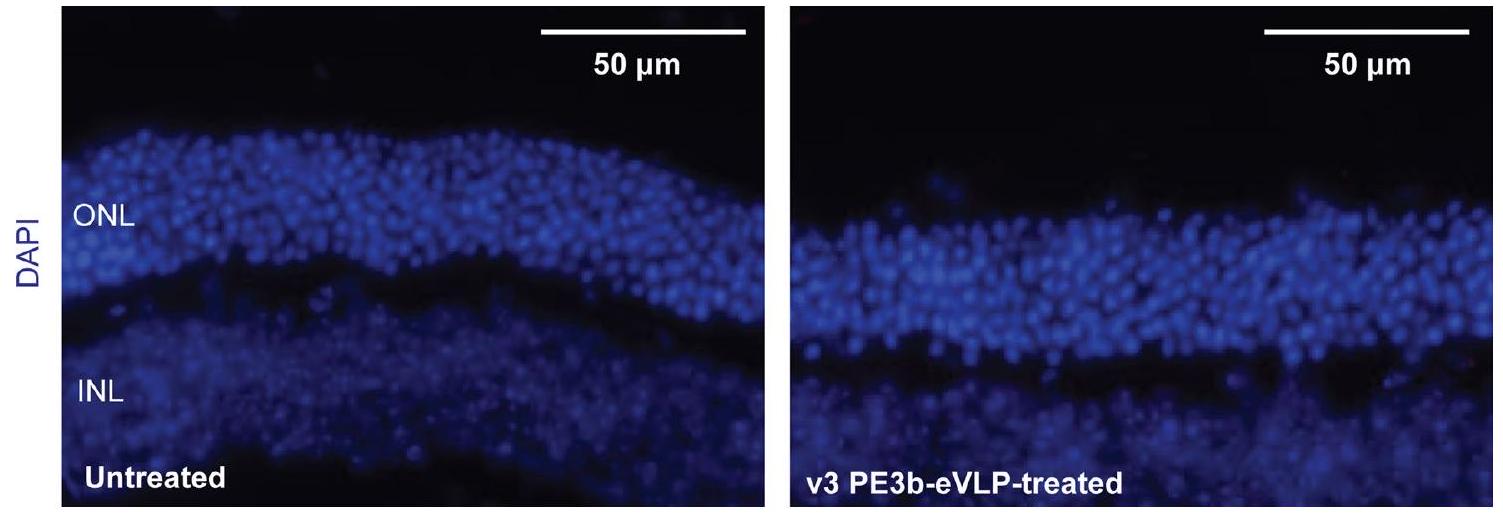
التلوين المناعي النسجي لشرائح RPE المسطحة والشرائح المجمدة

تم استئصال عيون الفئران وتثبيتها باستخدام 4% بارافورمالدهيد في PBS لمدة 20 دقيقة في درجة حرارة الغرفة، ثم تم غسلها ثلاث مرات في PBS لمدة 5 دقائق لكل مرة. لصنع شريحة مسطحة من RPE، تمت إزالة الجزء الأمامي والشبكية من الكوب الخلفي للعين تحت مجهر تشريحي، وتم إجراء أربع قطع شعاعية نحو العصب البصري لتسطيح الكوب الخلفي إلى شريحة مسطحة من RPE. تم نفاذية العينات وحجبها في 0.1% تريتون X-100 (سيغما-ألدريتش، T8532) مع 3% مصل حيوانات دونكي العادي (NDS) في PBS لمدة 30 دقيقة وتم تحضينها مع الأجسام المضادة الأولية المناسبة في PBS، 0.1% تريتون X-100 و3% NDS، بما في ذلك جسم مضاد ضد MFRP من الماعز (1:100؛ R&D Systems، AF3445) وجسم مضاد متعدد النسائل ضد ZO-1 من الأرانب (1:100؛ إنفيتروجين، 61-7300) طوال الليل في في اليوم التالي، تم غسل العينات ثلاث مرات في PBS لمدة 5 دقائق لكل منها، ثم تم تحضينها مع الأجسام المضادة الثانوية المناسبة في PBS + 0.1% Triton X-100 و3% NDS، بما في ذلك جسم مضاد للأرانب موصول بألكسا فلور 594 (1:200؛ ثيرمو فيشر، A21207) وجسم مضاد للماعز موصول بألكسا فلور 647 (1:200؛ ثيرمو فيشر، A32849) لمدة ساعتين في درجة حرارة الغرفة في الظلام. تم تحضين عينات الشرائح المجمدة في 1 مل من DAPI (ثيرمو فيشر، 62248) في PBS لمدة 10 دقائق. تم غسل العينات ثلاث مرات في PBS لمدة 5 دقائق لكل منها. ثم تم تثبيت العينات باستخدام VECTASHIELD HardSet Antifade.
وسط التركيب (Vector Labs H-1400-10) وتم تصويره على ميكروسكوب الفلورية الشامل Keyence BZ-X800.

ترشيح مواقع غير مستهدفة بواسطة CIRCLE-seq لـ و نماذج

تم إجراء تحليل تعديل الأهداف غير المستهدفة باستخدام CIRCLE-seq كما تم وصفه سابقًا. . تم عزل الحمض النووي الجينومي من كبد الفأر rd6 باستخدام مجموعة Gentra Puregene (Qiagen، 158845) وفقًا لتعليمات الشركة المصنعة. تم تقطيع الحمض النووي الجينومي المنقى باستخدام جهاز Covaris S2 إلى طول متوسط يبلغ 300 قاعدة. تم إصلاح الأطراف للحمض النووي المجزأ، وإضافة ذيل A، وربطه بمحول يحتوي على اليوراسيل، باستخدام مجموعة تحضير المكتبة KAPA HTP، خالية من PCR (KAPA Biosystems، KK8235). تم معالجة الحمض النووي المرتبط بالمحول باستخدام إنزيم Lambda Exonuclease (New England Biolabs، M0262) وإنزيم E. coli Exonuclease I (New England Biolabs، M0293) ثم باستخدام إنزيم USER (New England Biolabs، M5505) وكيناز T4 للنوكليوتيدات المتعددة (New England Biolabs، M0201). تم إجراء الدائرة الداخلية للحمض النووي باستخدام إنزيم T4 DNA ligase (New England Biolabs، M0202) وتم تكسير الحمض النووي الخطي المتبقي بواسطة DNase المعتمد على ATP Plasmid-Safe (Lucigen، E3110). تم طلب RNAs الدليل الاصطناعية من IDT مع تعديل 2′-O-methyl القياسي في أول ثلاث قواعد وآخر ثلاث قواعد. تم إعادة تعليق RNAs الدليل الاصطناعية إلى في ماء خالٍ من النوكلياز، تم نزع الطي عند لمدة 5 دقائق وتم تسخينها ببطء عند إلى تم إجراء تفاعلات القطع في المختبر باستخدام 125 نانوجرام من الحمض النووي الدائري المعالج بواسطة Plasmid-Safe، و90 نانومتر من بروتين نوكلياز Cas9 (New England Biolabs، M0386) و270 نانومتر من RNA الدليل الاصطناعي في حجم لمدة ساعة واحدة. تم معالجة المنتجات المقسمة ببروتيناز ك كما هو موصوف تم ربطها بمحول على شكل دبوس شعر (New England Biolabs, E7600S)، ومعالجتها بإنزيم USER (New England Biolabs, M5505) وتم تضخيمها بواسطة PCR باستخدام بادئات عالمية مشفرة (New England Biolabs, E7600S) باستخدام بوليميراز Kapa HiFi (KAPA Biosystems, KK4824). تم تسلسل المكتبات باستخدام قراءات مزدوجة الطرف بطول 150 قاعدة مع جهاز Illumina MiSeq. تم إجراء تحليلات بيانات CIRCLE-seq باستخدام برنامج تحليل CIRCLE-seq مفتوح المصدر ومع المعلمات الموصى بها افتراضيًا. تم تحليل أفضل عشرة مواقع مستهدفة غير مباشرة تم ترشيحها لـ epegRNA المستخدمة في نماذج rd6 و rd12 بواسطة HTS من نسيج RPE للفئران غير المعالجة أو المعالجة بـ v3 PE3b-eVLP. تم تحليل التحرير غير المستهدف لمواقع epegRNA المرتبطة، كما هو موضح أعلاه، كالتالي: (تكرار التحرير غير المستهدف بواسطة pegRNA) + (تكرار indel في موقع كاس9). تم تحليل الإدخالات أو الحذف في مواقع التحرير غير المستهدفة المرتبطة بـ ngRNA كنسبة مئوية من القراءات المهملة مقسومة على إجمالي القراءات المتوافقة مع المرجع. تم إدراج أفضل عشرة مواقع مستهدفة غير مباشرة مرشحة بواسطة CIRCLE-seq في الجدول التكميلية 6 (نموذج rd6) والجدول التكميلية 7 (نموذج rd12).

الإحصائيات وإمكانية التكرار

تم تقديم البيانات كمتوسط وخطأ معياري للمتوسط (s.e.m.). تم إجراء مقارنات بين إصدارات مختلفة من PE-eVLPs مع eVLPs التي تم إنتاجها ونقلها بالتوازي في تجربة كبيرة واحدة. تم الحصول على النسخ البيولوجية من خلال معالجة ثلاثة انقسامات مستقلة من خطوط الخلايا (عينات) لدراسات زراعة الخلايا، أو ثلاثة حيوانات أو أكثر لدراسات in vivo، باستخدام دفعة واحدة من PE-eVLPs. يتم عرض انخفاض التباين بين الدفعات المختلفة من PE-eVLPs في الشكل التمديدي 5b. تم وصف حجم العينة والاختبارات الإحصائية المستخدمة لكل تجربة في أساطير الأشكال. لم يتم استخدام طرق إحصائية لتحديد حجم العينة مسبقًا. تم إجراء التحليل الإحصائي باستخدام برنامج GraphPad Prism.

ملخص التقرير

معلومات إضافية حول تصميم البحث متاحة في ملخص تقارير مجموعة نيتشر المرتبط بهذه المقالة.

توفر البيانات

تم إيداع ملفات بيانات HTS في قاعدة بيانات أرشيف تسلسل القراءة NCBI تحت رموز الوصول PRJNA980181 (المرجع 86). تسلسلات الحمض النووي
تم توفير تفاصيل بنية PE-eVLP في المعلومات التكميلية. تم إيداع البلازميدات الرئيسية التالية من هذا العمل في Addgene للتوزيع: Gag-MCP-Pol (Addgene #211370)، Gag-PE (Addgene #211371)، MS2-epegRNA-Dnmt1 (Addgene #211372)، Gag-COM-Pol (Addgene #211373)، Gag-PE3-Pol (#211374)، P4-PE (#211375)، COM-epegRNA-Dnmt1 (#211376). البلازميدات الأخرى والبيانات الخام متاحة من المؤلف المراسل عند الطلب. تم توفير صورة غير معدلة للبلات الغربية المعروضة في الشكل 5d، k كبيانات مصدر. تم توفير بيانات المصدر مع هذه الورقة.

توفر الشيفرة

الكود المستخدم لتحليل بيانات HTS متاح علىhttps://github. com/pinellolab/CRISPResso2. الشيفرة المستخدمة لتحليل بيانات CIRCLEseq متاحة على https://github.com/tsailabSJ/circleseq.

References

  1. Renner, T. M., Tang, V. A., Burger, D. & Langlois, M. A. Intact viral particle counts measured by flow virometry provide insight into the infectivity and genome packaging efficiency of Moloney Murine Leukemia virus. J. Virol. https://doi.org/10.1128/JVI.01600-19 (2020).
  2. Levy, J. M. et al. Cytosine and adenine base editing of the brain, liver, retina, heart and skeletal muscle of mice via adeno-associated viruses. Nat. Biomed. Eng. 4, 97-110 (2020).
  3. Golczak, M., Kiser, P. D., Lodowski, D. T., Maeda, A. & Palczewski, K. Importance of membrane structural integrity for RPE65 retinoid isomerization activity. J. Biol. Chem. 285, 9667-9682 (2010).
  4. Lazzarotto, C. R. et al. CHANGE-seq reveals genetic and epigenetic effects on CRISPR-Cas9 genome-wide activity. Nat. Biotechnol. 38, 1317-1327 (2020).
  5. circleseq. GitHub https://github.com/tsailabSJ/circleseq (2018)
  6. An, M. et al. Engineered virus-like particles for transient delivery of prime editor ribonucleoprotein complexes in vivo. NCBI https://www.ncbi.nlm.nih.gov/bioproject/PRJNA980181 (2023).

شكر وتقدير

تم دعم هذا العمل من قبل NIH UG3AI150551 و U01AI142756 و R35GM118062 و RM1HG009490؛ مؤسسة بيل وميليندا غيتس؛ و HHMI. تم دعم هذا البحث جزئيًا من خلال منح بحثية من NIH EY034501 (NEI) ومنحة من مؤسسة مكافحة العمى (برنامج TRAP) لك.ب. ومنح بحثية من NEI F3OEY033642 و T32GM008620 لـ س.و.د. يقر المؤلفون بالدعم من منحة أساسية من NIH P30 EY034070 ومنحة مكتب عميد كلية الطب بجامعة UCI ومنحة غير مقيدة من بحث لمنع العمى لقسم طب العيون في UCI. تم دعم أ.ر. من خلال زمالة أبحاث الدراسات العليا من NSF. نشكر مايكل هوارد، كينيا غوزمان وموظفي برود فيفاريم.
نصائح ومساعدة في رعاية الفئران. نشكر J. Doman و S. DeCarlo و P. Randolph و A. Yan و A. Tworak على المناقشات المفيدة. هذه المقالة تخضع لسياسة الوصول المفتوح إلى المنشورات الخاصة بـ HHMI. لقد منح رؤساء مختبرات HHMI سابقًا ترخيصًا غير حصري CC BY 4.0 للجمهور وترخيصًا قابلًا للتراخيص الفرعية لـ HHMI في مقالاتهم البحثية. وفقًا لتلك التراخيص، يمكن أن يتوفر المخطوط المعتمد من المؤلف لهذه المقالة مجانًا بموجب ترخيص CC BY 4.0 فور نشرها.

مساهمات المؤلفين

قام م.أ. بتصميم وتنفيذ التجارب في المختبر وفي الكائنات الحية الموضحة في هذا العمل، وقام بتحليل البيانات. قدم أ.ر. و س.ب. المشورة وصمما التجارب الأولية. قام س.و.د. بإجراء حقن تحت الشبكية لفئران rd6 و rd12 وأجرى تقييمًا ظاهريًا. قام م.أ. و ج.ر.د. بإجراء حقن PO ICV ومعالجة الأنسجة. قام ج.أ.ن. و م.أ. بإجراء CIRCLE-seq. ساعد ب.ز.ج. في قياسات DLS. قام م.أ. و د.ر.ل. بتصميم البحث وصياغة هذه المخطوطة بمشاركة جميع المؤلفين.

المصالح المتنافسة

يعلن المؤلفون عن وجود مصالح مالية متنافسة: قدم كل من م.أ.، أ.ر.، س.ب. و د.ر.ل. طلبات براءات اختراع على هذا العمل من خلال معهد برود. س.ب. هو حالياً مستشار لشركة نفلوب ثيرابيوتيكس. ج.ر.د. هو حالياً موظف في برايم ميديسن. ك.ب. هو مستشار لشركات بولجنيكس، ألنيلام وأبفي، إنك. د.ر.ل. هو مستشار و/أو مالك أسهم في برايم ميديسن، بيم ثيرابيوتيكس، بيراوايز بلانتس، كروم ميديسن ونفلوب ثيرابيوتيكس، الشركات التي تستخدم أو تقدم عوامل تحرير الجينوم أو هندسة الإيبيجينوم. المؤلفون المتبقون يعلنون عدم وجود مصالح متنافسة. المراسلات:drliu@fas.harvard.edu.

معلومات إضافية

البيانات الموسعة متاحة لهذا البحث في
https://doi.org/10.1038/s41587-023-02078-y.
معلومات إضافية النسخة الإلكترونية تحتوي على مواد إضافية متاحة فيhttps://doi.org/10.1038/s41587-023-02078-y.
يجب توجيه المراسلات والطلبات للحصول على المواد إلى ديفيد ر. ليو.
تُعرب مجلة نيتشر للتكنولوجيا الحيوية عن شكرها لدان بير والمراجعين الآخرين المجهولين على مساهمتهم في مراجعة هذا العمل.
معلومات إعادة الطباعة والتصاريح متاحة علىwww.nature.com/reprints.
الشكل البياني الممتد 1 | التعديلات التي تتجنب MMR تدعم تحرير البرايم بشكل أكثر كفاءة. تثبيت الطفرات القريبة يحسن كفاءات تحرير البرايم لـ v2.3 PE-eVLPs في موضع HEK3 وموضع Dnmt1 في خلايا HEK293T وN2A على التوالي. القيم المعروضة في جميع الرسوم البيانية تمثل متوسط تحرير البرايم
كفاءة التحرير لثلاث نسخ بيولوجية وتمثل أشرطة الخطأ الانحراف المعياري. تم ملاءمة البيانات لمنحنيات لوجستية بأربعة معلمات باستخدام الانحدار غير الخطي.
الشكل البياني الممتد 3 | تحسين تحميل وتوصيل شحنة eVLP. أ، التغير النسبي في كفاءة تحرير PE-eVLP مقارنةً بالـ 5′ G غير المتطابقة الأصلية + 20-bp epegRNA protospacer. ب، قياس عدد جزيئات eVLP لكل وحدة حجم في تحضيرات الأجيال المتعاقبة من PE-eVLPs بواسطة ELISA مضاد MLV p30. تم استخدام هذه البيانات الكمية في
مثال على فواصل epegRNA لموقع Col12a1 . . . GCTTGACTTCCATGGTTCCACAATGG
  • 5′ G+20-bp GTGACTTCCATGGTTCCACAA
  • 20-زوج قاعدي TGACTTCCATGGTTCCACAA
  • 5 ‘ G+19-bp GGACTTCCATGGTTCCACAA

    تجارب لتحديد عدد جزيئات بروتين محرر الأحماض النووية epegRNA و eVLP كما هو موضح في الشكل 2g، h. ج، نسبة تكوين epegRNA و ngRNA في v3 PE2-eVLPs و v3 PE3-eVLPs. تمثل البيانات القيمة المتوسطة لثلاث تكرارات فنية وتمثل أشرطة الخطأ الانحراف المعياري.
الشكل 4 من البيانات الموسعة | تحسين وتوصيف v3b PE-eVLP.
a، تمثيل غربي يقارن تعبير بروتين الاندماج gag-PE من v3 PE-eVLPs مقابل بروتين الاندماج P4-PE من v3b PE-eVLPs في خلايا المنتج التي تم نقلها بالبروتينات الاندماجية المقابلة. ب، كفاءات التحرير الأولي لـ v3b PE-eVLPs مع Gag-P3-Pol أو Gag-MCP-P3-Pol. بناء الاندماج Gag-MCP-P3-Pol غير متوافق مع الإنتاج الفعال لـ PE-eVLPs. ج، كفاءات التحرير لـ v3b PE-eVLPs عند موضع Dnmt1 في خلايا N2A مع إدخال الأبتامر Com في مواقع مختلفة في epegRNAs. موضع إدخال الأبتامر Com هو كما يلي: يشير إلى v3b PE-eVLPs مع إدخال الأبتامر Com بعد نمط tevoPreQ1 المنظم لـ
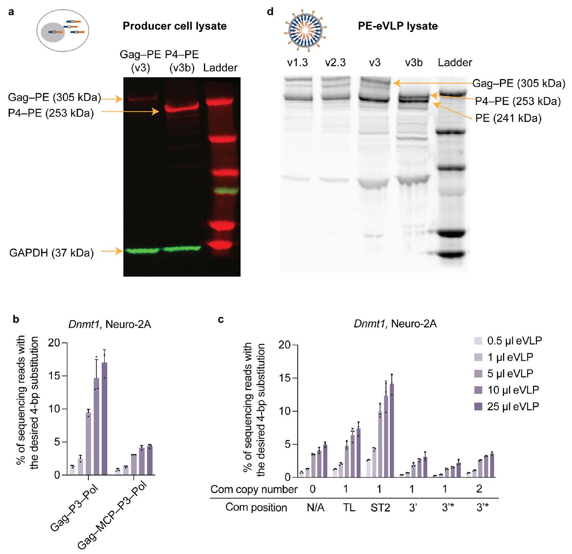
epegRNA؛ 3 * تشير إلى v3b PE-eVLPs مع إدخال الأبتامر Com مباشرة بعد الامتداد 3′ من pegRNA، مما يستخدم الأبتامر Com لمحاكاة نمط هيكلي في الطرف 3′ من epegRNAs؛ TL تشير إلى v3b PE-eVLPs مع إدخال الأبتامر Com داخل الحلقة الرباعية من هيكل pegRNA؛ ST2 تشير إلى v3b PE-eVLPs مع إدخال الأبتامر Com داخل حلقة ST2 من هيكل pegRNA. د، تمثيل غربي يقيّم كمية الحمولة PE المعبأة في v1.3 و v2.3 و v3 و v3b PE-eVLPs. الأشكال المعروضة في (أ) و (د) هي صور تمثيلية من تجربتين مكررتين بشكل مستقل. القيم المعروضة في (ب) و (ج) تمثل متوسط كفاءة التحرير الأساسي لثلاث نسخ بيولوجية والأشرطة الخطأ تمثل الانحراف المعياري.
الشكل البياني الممتد 5|توصيف PE-eVLPs. أ، مقارنة كفاءة التحرير الأساسي (% التحرير) وتوليد المنتجات الجانبية للإدخال والحذف (% indel) لنظام PE3 المقدم عن طريق نقل البلازميد مقابل v3 PE3-eVLP. يستهدف نظام PE3 موضع Dnmt1 في خلايا N2A. تمثل البيانات متوسط كفاءة التحرير الأساسي لثلاث تكرارات بيولوجية وتمثل أشرطة الخطأ الانحراف المعياري. ب، كفاءات التحرير الأساسي عند موضع Dnmt1 في خلايا N2A و
b
موقع HEK3 في خلايا HEK293T من المعالجة بأربعة دفعات مستقلة من v3 PE3-eVLPs المنتجة في أيام مختلفة، حيث تشير كل نقطة إلى كفاءة التحرير الأساسي لكل من دفعات v3 PE3-eVLP الأربعة. البيانات المعروضة تمثل متوسط كفاءة التحرير الأساسي من أربع دفعات مختلفة من v3 PE3-eVLP والأشرطة الخطأ تمثل الانحراف المعياري.
بروتينات ملحقة بروتينات الشحن ARNs الموجهة تحسينات من النسخة السابقة
الإصدار 1.1 غير متوفر
pegRNA
الإصدار 1.2 غير متوفر
epegRNA epegRNA
الإصدار 1.3 غير متوفر
epegRNA PEmax
الإصدار 2.1 غير متوفر
epegRNA قصّ MMLV RT
الإصدار 2.2 غير متوفر
تحسين NES
الإصدار 2.3 غير متوفر
epegRNA تحسين الرابط
v3
MS2-epegRNA
تجنيد gRNA عبر تفاعل MCP:MS2
v3b
توظيف الشحن عبر تفاعل P3-P4؛ توظيف gRNA عبر تفاعل COM:Com
المفتاح: موقع إنزيم البروتياز 3xNES NLS المهندَس – الوصلة
*المخططات الموضحة في الجدول تمثل PE2-eVLPs. بالنسبة لـ PE3-eVLPs، يتم تعبئة ngRNAs إضافية.
الشكل التوضيحي الممتد 6 | ملخص تخطيطي لتصاميم PE-eVLP. مخطط للبروتينات المساعدة، بروتينات الحمولة، تصاميم RNA الموجه، ووصف التحسينات من النسخة السابقة للأجيال المتعاقبة من PE-eVLPs. بروتين الغلاف VSV-G، وبروتين القفيصة MMLV Gag-Pol الذي هو شائع في
تم استبعاد جميع نسخ PE-eVLPs من الجدول. المخططات المعروضة في الجدول تمثل PE2-eVLPs. بالنسبة لـ PE3-eVLPs، يتم تعبئة ngRNAs إضافية بنسبة 4:1 للـ pegRNAs و ngRNAs مع تعديل السقالة المناسب وإدخال الأبتامر.

الشكل 8 من البيانات الموسعة | مثال على تصنيف FACS لفرز نواة واحدة.
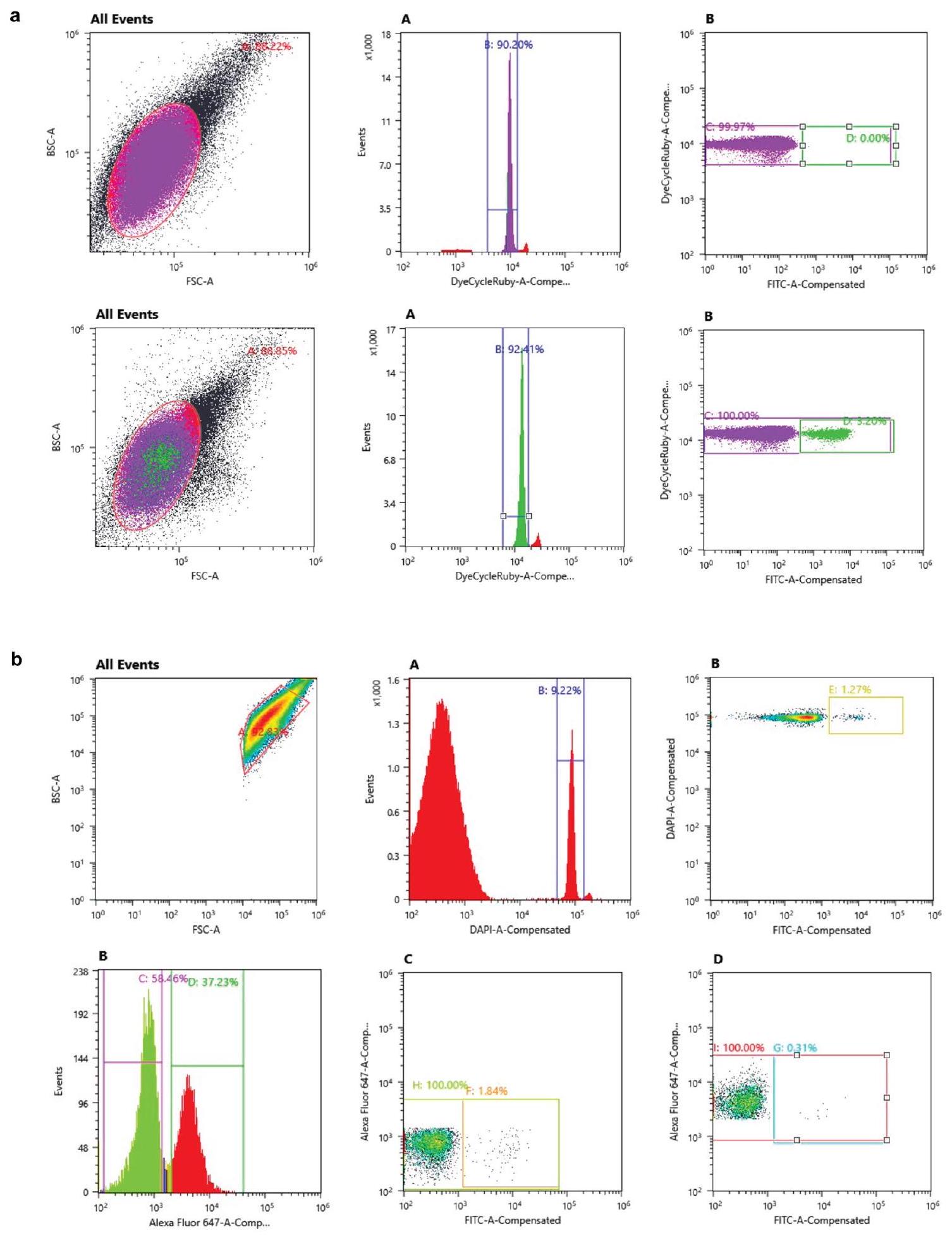
تم تحديد نواة واحدة بناءً على نسب التشتت الأمامي (FSC-A) والتشتت الخلفي (BCS-A) وإشارة DyeCycle Ruby. تم تحديد النوى الإيجابية لـ GFP بناءً على إشارة FITC. يعرض الصف الأول بيانات FACS التمثيلية للعينات غير المعالجة، بينما يعرض الصف الثاني بيانات FACS التمثيلية لعينات القشرة المجمعة من الفئران حديثة الولادة التي تم حقنها معاً بـ PE-eVLPs و فيروس لنتيفيروس GFP:KASH المجهز بنمط VSV-G عبر حقن ICV. النوى الكثيفة تتوافق مع الأحداث التي اجتازت البوابة C والنوى الإيجابية لـ GFP تتوافق مع الأحداث التي اجتازت البوابة D. ب، مثال على تصنيف FACS للنوع العصبي المحدد.
فرز. تم تحديد نواة واحدة بناءً على نسب التشتت الأمامي (FSC-A) والتشتت الخلفي (BCS-A) وإشارة DAPI. تميز الإشارة من جسم مضاد NeuN المرتبط بـ Alexa 647 بين السكان الإيجابيين لـ NeuN والسكان السلبيين لـ NeuN. تم تحديد النوى الإيجابية لـ GFP بناءً على إشارة FITC في كل من السكان الإيجابيين والسكان السلبيين لـ NeuN. تمثل الأبواب المعروضة بيانات FACS لعينات الدماغ الأوسط التي تم جمعها من فئران حديثي الولادة التي تم حقنها معاً بـ PE-eVLPs و فيروس لنتي VSV-G مزيف النوع GFP:KASH عبر حقن ICV. يمثل البوابة F نوى إيجابية GFP من المجموعة السلبية لـ NeuN. تمثل البوابة G نوى إيجابية GFP من المجموعة الإيجابية لـ NeuN.
الشكل 9 من البيانات الموسعة | تلطيخ المناعة النسيجية على مقاطع العين المجمدة من فئران rd6. مقاطع الشبكية المجمدة من الفئران غير المعالجة تم صبغ الفئران rd6 المعالجة بـ v3 PE3b-eVLP بـ DAPI (الأزرق). الصورة المعروضة هي صورة تمثيلية من تجربتين تم تكرارهما بشكل مستقل.
الشكل 10 من البيانات الموسعة | تحليل الأهداف غير المستهدفة في فئران rd12. أ، تحليل التحرير المعتمد على PE في الموقع المستهدف وفي أعلى 10 مواقع غير مستهدفة تم ترشيحها بواسطة CIRCLE-seq المرتبطة بتسلسل epegRNA الخاص بـ rd12. ب، تحليل الإضافات والحذف في الموقع المستهدف وأعلى 10 مواقع غير مستهدفة تم ترشيحها بواسطة CIRCLE-seq.
b

مرتبطة بتسلسل rd12ngRNA. تمثل الأعمدة القيم المتوسطة لـ (غير معالج) أو (معالجة v3PE3b-eVLP)، حيث يمثل كل نقطة فأرًا فرديًا وأشرطة الخطأ تمثل الانحراف المعياري.

محفظة الطبيعة

المؤلف (المؤلفون) المراسلون: ديفيد ر. ليو
آخر تحديث من المؤلف(ين): 16 نوفمبر 2023

ملخص التقرير

تسعى Nature Portfolio إلى تحسين إمكانية تكرار العمل الذي ننشره. يوفر هذا النموذج هيكلًا للاتساق والشفافية في الإبلاغ. لمزيد من المعلومات حول سياسات Nature Portfolio، يرجى الاطلاع على سياسات التحرير وقائمة مراجعة سياسة التحرير.

الإحصائيات

لجميع التحليلات الإحصائية، تأكد من أن العناصر التالية موجودة في أسطورة الشكل، أسطورة الجدول، النص الرئيسي، أو قسم الطرق.
مؤكد
حجم العينة بالضبط ( ) لكل مجموعة/شرط تجريبي، معطاة كرقم منفصل ووحدة قياس
بيان حول ما إذا كانت القياسات قد أُخذت من عينات متميزة أو ما إذا كانت نفس العينة قد تم قياسها عدة مرات
اختبار(ات) الإحصاء المستخدمة وما إذا كانت أحادية الجانب أو ثنائية الجانب
يجب أن تُوصف الاختبارات الشائعة فقط بالاسم؛ واصفًا التقنيات الأكثر تعقيدًا في قسم الطرق.
وصف لجميع المتغيرات المشتركة التي تم اختبارها
وصف لأي افتراضات أو تصحيحات، مثل اختبارات الطبيعية والتعديل للمقارنات المتعددة
“凶” translates to “شر” in Arabic.
وصف كامل للمعلمات الإحصائية بما في ذلك الاتجاه المركزي (مثل المتوسطات) أو تقديرات أساسية أخرى (مثل معامل الانحدار) والتباين (مثل الانحراف المعياري) أو تقديرات عدم اليقين المرتبطة (مثل فترات الثقة)
X لاختبار الفرضية الصفرية، إحصائية الاختبار (على سبيل المثال ) مع فترات الثقة، أحجام التأثير، درجات الحرية و قيمة ملحوظة أعطِ القيم كقيم دقيقة كلما كان ذلك مناسبًا.
لتحليل بايزي، معلومات حول اختيار القيم الأولية وإعدادات سلسلة ماركوف مونت كارلو
للتصاميم الهرمية والمعقدة، تحديد المستوى المناسب للاختبارات والتقارير الكاملة عن النتائج
تقديرات أحجام التأثير (مثل حجم تأثير كوهين) بيرسون )، مما يشير إلى كيفية حسابها
تحتوي مجموعتنا على الإنترنت حول الإحصائيات لعلماء الأحياء على مقالات تتناول العديد من النقاط المذكورة أعلاه.

البرمجيات والشيفرة

معلومات السياسة حول توفر كود الكمبيوتر

جمع البيانات

تم استخدام برنامج Miseq reporter (الإصدار 2.6) على جهاز Illumina Miseq لفك تشفير بيانات HTS. تم استخدام جهاز فرز Sony MA900 لفرز الخلايا مع برنامج MA900 Cell Sorter الإصدار 3.1. تم تسجيل ERGs باستخدام نظام Celeris لقياس كهربائية الخلايا في القوارض.

تحليل البيانات

تم حساب التكرار والمتوسط والانحرافات المعيارية باستخدام GraphPad Prism 9. تم استخدام CRISPResso2 لتحليل بيانات HTS لقياس كفاءة التحرير في المواقع الجينومية.https://github.com/pinellolab/CRISPResso2تم تحليل ERGs باستخدام برنامج Espion V6. تم إجراء تحليل CIRCLE-seq باستخدام برنامج تحليل CIRCLE-seq مفتوح المصدر ومع المعلمات الموصى بها افتراضيًا (https://github.com/tsailabSJ/circleseq).

بيانات

معلومات السياسة حول توفر البيانات

يجب أن تتضمن جميع المخطوطات بيانًا عن توفر البيانات. يجب أن يتضمن هذا البيان المعلومات التالية، حيثما ينطبق:
  • رموز الانضمام، معرفات فريدة، أو روابط ويب لمجموعات البيانات المتاحة للجمهور
  • وصف لأي قيود على توفر البيانات
  • بالنسبة لمجموعات البيانات السريرية أو بيانات الطرف الثالث، يرجى التأكد من أن البيان يتماشى مع سياستنا
ملفات بيانات التسلسل عالي الإنتاجية متاحة من NCBI SRA تحت رمز الوصول PRJNA980181. تسلسلات الحمض النووي لهياكل PE-eVLP متوفرة في المعلومات التكميلية. البلازميدات الرئيسية من هذا العمل متاحة من Addgene والبلازميدات الأخرى متاحة من المؤلف المراسل عند الطلب.

البحث الذي يتضمن مشاركين بشريين، بياناتهم، أو مواد بيولوجية

معلومات السياسة حول الدراسات التي تشمل مشاركين بشريين أو بيانات بشرية. انظر أيضًا معلومات السياسة حول الجنس، الهوية/التقديم الجنسي، والتوجه الجنسي والعرق، والعرقية والعنصرية.
التقارير عن الجنس والنوع الاجتماعي. هذه الدراسة لا تشمل المشاركين في أبحاث بشرية.
التقارير عن العرق أو الإثنية أو غيرها من المجموعات الاجتماعية ذات الصلة
خصائص السكان
التوظيف
رقابة الأخلاقيات
غير متوفر

غير متوفر
غير متوفر
غير متوفر
يرجى ملاحظة أنه يجب أيضًا تقديم معلومات كاملة حول الموافقة على بروتوكول الدراسة في المخطوطة.

التقارير المتخصصة في المجال

يرجى اختيار الخيار أدناه الذي يناسب بحثك بشكل أفضل. إذا لم تكن متأكدًا، اقرأ الأقسام المناسبة قبل اتخاذ قرارك.
علوم الحياة العلوم السلوكية والاجتماعية العلوم البيئية والتطورية والبيئية
لنسخة مرجعية من الوثيقة بجميع الأقسام، انظرnature.com/documents/nr-reporting-summary-flat.pdf

تصميم دراسة العلوم الحياتية

يجب على جميع الدراسات الإفصاح عن هذه النقاط حتى عندما يكون الإفصاح سلبياً.
حجم العينة أحجام العينات كانت نسخ بيولوجية مستقلة، وفقًا للأدبيات السابقة والمعايير في مجال تقنيات تحرير الجينوم (أنزالون 2019، ليفي 2020، بانسوكوتا وراجورام 2022)
استبعاد البيانات للتصحيح في نموذج rd6 و rd12، تم حقن الفئران مع PE-eVLPs و AAV1-GFP. تم فحص إشارة GFP كعلامة على نجاح الحقن. تم أخذ عينات من العيون التي أظهرت أقل من تم استبعاد إشارة GFP من التحليل بسبب حقن غير فعال. لم يتم استبعاد أي بيانات أخرى من الدراسة.
التكرار تم تكرار جميع التجارب أكثر من مرة. كانت جميع محاولات التكرار ناجحة.
التوزيع العشوائي تم تعيين جميع ظروف التحكم والاختبار عشوائيًا. على سبيل المثال، بالنسبة لجميع تجارب الزراعة الخلوية/الأنسجة، تم تعيين الظروف عشوائيًا إلى آبار عبر ألواح تحتوي على 96 أو 48 بئرًا، ولا يُتوقع أن تؤثر موضع الألواح على نتيجة التجربة. بالنسبة لتجارب in vivo، تم تعيين الفئران عشوائيًا إلى مجموعات.
مُعَمي تم تحليل جميع بيانات HTS باستخدام برنامج نصي آلي من CRISPResso2 لا يسمح بتدخل الباحث، لذلك لم يكن الباحث معصوب العينين. تم تحليل بيانات تحرير الحقن تحت الشبكية بواسطة باحث معصوب العينين. لم يكن التعمية ضرورية للتجارب الأخرى لأن ظروف التحكم والاختبار تمت معالجتها بشكل متطابق.

التقارير عن مواد وأنظمة وطرق محددة

نحتاج إلى معلومات من المؤلفين حول بعض أنواع المواد والأنظمة التجريبية والأساليب المستخدمة في العديد من الدراسات. هنا، يرجى الإشارة إلى ما إذا كانت كل مادة أو نظام أو طريقة مدرجة ذات صلة بدراستك. إذا لم تكن متأكدًا مما إذا كان عنصر القائمة ينطبق على بحثك، يرجى قراءة القسم المناسب قبل اختيار رد.
المواد والأنظمة التجريبية طرق
غير متوفر مشارك في الدراسة غير متوفر مشارك في الدراسة
علم الحفريات وعلم الآثار
إكس

الأجسام المضادة

الأجسام المضادة المستخدمة أجسام مضادة لـ Mouse Cas9 (Thermo Fisher Scientific، #MA5-23519)؛ أجسام مضادة لـ Rabbit GAPDH (Cell signaling Technology، #2118)؛ أجسام مضادة لـ Goat anti-mouse MFRP (R&D Systems #AF3445)؛ أجسام مضادة لـ Mouse anti-mouse RPE65 (إنتاج داخلي؛ تم الحصول عليها من مختبر الدكتور كريستوف بالتشوفسكي، جامعة كاليفورنيا إيرفين؛ غولتشاك وآخرون، 2010)؛ أجسام مضادة متعددة النسائل لـ Rabbit anti-beta-actin (Cell Signaling Technology؛ 4970S)؛ أجسام مضادة لـ Anti-NeuN (Abcam، ab190565)؛ أجسام مضادة ثانوية لـ Goat anti-mouse (LI-COR IRDye 680RD، 926-68070)؛ أجسام مضادة لـ Goat anti-rabbit (LI-COR IRDye 800RD، 926-32211)؛ أجسام مضادة لـ Donkey anti-goat IgG-HRP (Abcam، ab97110)؛ أجسام مضادة لـ Goat anti-mouse IgG-HRP (Cell Signaling Technology، 7076S)؛ أجسام مضادة لـ Goat anti-rabbit IgG-HRP (Cell signaling Technology، 7074S)؛ أجسام مضادة متعددة النسائل لـ Rabbit anti-ZO-1 (Invitrogen، #61-7300)؛ أليكسا فلور 594 المرتبطة بـ donkey anti-rabbit IgG (Thermo Fisher؛ A21207)؛ أليكسا فلور 647 المرتبطة بـ donkey anti-goat IgG (Thermo Fisher، A32849)؛
التحقق تم التحقق من جميع الأجسام المضادة التجارية من قبل الشركات المصنعة لاختبار الويسترن بلوت. بالنسبة للأجسام المضادة المنتجة داخليًا، قمنا بتأكيد وجود خطوط واضحة عند الوزن الجزيئي المتوقع مع وجود التحكم الإيجابي في التجارب.

خطوط خلايا حقيقية النواة

معلومات السياسة حول خطوط الخلايا والجنس والنوع في البحث

مصدر(s) خط الخلايا
المصادقة
تلوث الميكوبلازما
الخطوط التي يتم التعرف عليها بشكل خاطئ بشكل شائع
(انظر سجل ICLAC)
تم الحصول على خلايا HEK293T وNeuro-2A وHEK293T/17 من ATCC. تم الحصول على خلايا Gesicle 293T من Takara Bio. كانت خلايا NIH 3T3 التي تحتوي على جين Rpe65 المدمج بواسطة الفيروسات الراجعة والذي يحتوي على طفرة هدية من مختبر بالزيفسكي.
تم التحقق من صحة خطوط الخلايا التجارية من قبل المورد باستخدام تحليل STR. لم يتم التحقق من صحة خط الخلايا المهدى.
تم اختبار جميع خطوط الخلايا سلبية لعدوى الميكوبلازما.
لا شيء مستخدم.

الحيوانات وغيرها من الكائنات البحثية

معلومات السياسة حول الدراسات التي تشمل الحيوانات؛ توجيهات ARRIVE الموصى بها للإبلاغ عن أبحاث الحيوانات، والجنس والنوع في البحث
الحيوانات المخبرية تم شراء إناث الفئران الحوامل من سلالة C57BL/6J (027) من مختبرات تشارلز ريفر لدراسات P0. لم يتم تحديد عمر الفئران الحوامل. تم استخدام الصغار للحقن في يوم الولادة. تم شراء نماذج الفئران المصابة بالتنكس الشبكي rd6 (003684) و rd12 (005379) من مختبر جاكسون. تم إجراء الحقن تحت الشبكية على فئران rd6 و rd12 التي تبلغ من العمر 5 أسابيع. تم الحفاظ على مرافق سكن الفئران على نظام إضاءة لمدة 12 ساعة. دورة مظلمة، مع درجة حرارة مستقرة (70 درجة فهرنهايت) درجة فهرنهايت) والرطوبة ( )، مع الوصول الحر إلى نظام غذائي قياسي للجرذان والماء.
الحيوانات البرية هذه الدراسة لا تشمل الحيوانات البرية.
التقارير عن الجنس تم استخدام كل من ذكور وإناث الفئران لكل حالة.
عينات تم جمعها من الميدان هذه الدراسة لا تتضمن عينات تم جمعها من الميدان.
رقابة الأخلاقيات قدمت لجنة IACUC في معهد برود (D16-00903؛ 0048-04-15-2) ولجنة IACUC في جامعة كاليفورنيا، إيرفين (D16-00259؛ AUP-21-096) الأخلاقيات على مدار الليل لجميع التجارب التي تشمل الحيوانات الحية.
يرجى ملاحظة أنه يجب أيضًا تقديم معلومات كاملة حول الموافقة على بروتوكول الدراسة في المخطوطة.
مخزونات البذور غير متوفر
أنماط جينية نباتية جديدة
غير متوفر
المصادقة
غير متوفر

تدفق الخلايا

المؤامرات

أكد أن:
توضح تسميات المحاور العلامة والفلوركروم المستخدم (مثل CD4-FITC).
المقاييس على المحاور مرئية بوضوح. قم بتضمين الأرقام على المحاور فقط للرسم البياني في أسفل اليسار من المجموعة (المجموعة هي تحليل للعلامات المتطابقة).
جميع الرسوم البيانية هي رسوم بيانية متساوية الارتفاع مع نقاط شاذة أو رسوم بيانية بالألوان الزائفة.
تم توفير قيمة عددية لعدد الخلايا أو النسبة المئوية (مع الإحصائيات).

المنهجية

تحضير العينة
آلة
برمجيات
وفرة تجمع الخلايا
استراتيجية البوابة
تم وصف بروتوكول استخراج النوى لتحليل الخلايا باستخدام فلو سيتوميتر في قسم الطرق من هذه المخطوطة.
فرز الخلايا سوني MA900
برنامج فرز الخلايا MA900 الإصدار 3.1
في المتوسط من النوى المرتبة كانت إيجابية لـ GFP.
تم تحديد النوى الفردية بناءً على نسب التشتت الأمامي (FSC-A) والتشتت الخلفي (BSC-A) وإشارة DyeCycle Ruby أو DAPI. تم تحديد النوى الإيجابية لـ GFP بناءً على إشارة FITC. تم تقديم استراتيجية التحديد التمثيلية في الشكل 8 من البيانات الموسعة.
قم بتحديد هذا المربع لتأكيد أنه تم تقديم رقم يوضح استراتيجية البوابة في المعلومات التكميلية.

  1. معهد ميركين للتقنيات التحويلية في الرعاية الصحية، معهد برود التابع لمعهد ماساتشوستس للتكنولوجيا وجامعة هارفارد، كامبريدج، ماساتشوستس، الولايات المتحدة الأمريكية. قسم الكيمياء وعلم الأحياء الكيميائي، جامعة هارفارد، كامبريدج، ماساتشوستس، الولايات المتحدة الأمريكية. معهد هوارد هيوز الطبي، جامعة هارفارد، كامبريدج، ماساتشوستس، الولايات المتحدة الأمريكية. معهد غافين هيربرت لطب العيون، مركز أبحاث الرؤية الانتقالية، قسم طب العيون، جامعة كاليفورنيا، إيرفين، كاليفورنيا، الولايات المتحدة الأمريكية. قسم الفسيولوجيا والبيوفيزيكس، جامعة كاليفورنيا، إيرفين، كاليفورنيا، الولايات المتحدة الأمريكية. قسم الكيمياء، جامعة كاليفورنيا، إيرفين، كاليفورنيا، الولايات المتحدة الأمريكية. قسم البيولوجيا الجزيئية والكيمياء الحيوية، جامعة كاليفورنيا، إيرفين، كاليفورنيا، الولايات المتحدة الأمريكية. البريد الإلكتروني: drliu@fas.harvard.edu
  2. بالنسبة للمخطوطات التي تستخدم خوارزميات أو برامج مخصصة تكون مركزية في البحث ولكن لم يتم وصفها بعد في الأدبيات المنشورة، يجب أن تكون البرمجيات متاحة للمحررين والمراجعين. نحن نشجع بشدة على إيداع الشيفرة في مستودع مجتمعي (مثل GitHub). راجع إرشادات مجموعة Nature لتقديم الشيفرة والبرمجيات لمزيد من المعلومات.

Journal: Nature Biotechnology, Volume: 42, Issue: 10
DOI: https://doi.org/10.1038/s41587-023-02078-y
PMID: https://pubmed.ncbi.nlm.nih.gov/38191664
Publication Date: 2024-01-08

Engineered virus-like particles for transient delivery of prime editor ribonucleoprotein complexes in vivo

Received: 18 June 2023
Accepted: 30 November 2023
Published online: 8 January 2024

Check for updates

Meirui An(1) , Aditya Raguram , Samuel W. Du(1) , Samagya Banskota , Jessie R. Davis , Gregory A. Newby , Paul Z. Chen (1) , Krzysztof Palczewski © & David R. Liu (B

Abstract

Prime editing enables precise installation of genomic substitutions, insertions and deletions in living systems. Efficient in vitro and in vivo delivery of prime editing components, however, remains a challenge. Here we report prime editor engineered virus-like particles (PE-eVLPs) that deliver prime editor proteins, prime editing guide RNAs and nicking single guide RNAs as transient ribonucleoprotein complexes. We systematically engineered v3 and v3b PE-eVLPs with 65- to 170-fold higher editing efficiency in human cells compared to a PE-eVLP construct based on our previously reported base editor eVLP architecture. In two mouse models of genetic blindness, single injections of v3 PE-eVLPs resulted in therapeutically relevant levels of prime editing in the retina, protein expression restoration and partial visual function rescue. Optimized PE-eVLPs support transient in vivo delivery of prime editor ribonucleoproteins, enhancing the potential safety of prime editing by reducing off-target editing and obviating the possibility of oncogenic transgene integration.

Among current genome editing systems that function in both dividing and nondividing mammalian cells in vitro and in vivo, prime editing offers unusual versatility by enabling the replacement of a target DNA sequence with virtually any other specified sequence containing up to several hundred inserted, deleted or substituted base pairs . This versatility makes PE systems particularly promising for the treatment of a broad range of genetic diseases in humans. A prime editor (PE) is an engineered protein consisting of a catalytically impaired programmable nickase domain (such as a Cas9 nickase) fused to an engineered reverse transcriptase (RT) domain. The prime editing guide RNA (pegRNA) specifies the target protospacer sequence and simultaneously encodes the desired edits in the reverse transcription template in the extension of the pegRNA. The mechanism of prime editing
requires three independent nucleic acid hybridization events before editing can take place and does not rely on double-strand DNA breaks or donor DNA templates. As a result of this mechanism, prime editing is inherently resistant to off-target editing or bystander editing, and can proceed with few indel byproducts or other undesired consequences of double-strand DNA breaks .
Fully realizing the potential of prime editing for research or therapeutic applications in mammals requires safe and efficient methods capable of delivering PEs into tissues in vivo. So far, several groups have reported the in vivo delivery of PE via viral delivery methods, including adenoviruses and adeno-associated viruses (AAV) . Viral delivery methods, however, require that the transgene be encoded directly in the viral gene expression cassette, limiting transgene size. The AAV genome
has a cargo gene size limitation of (not including inverted terminal repeats) , requiring large cargoes such as PEs ( 6.4 kb in gene size for a first-generation PE) to be split into multiple AAVs , limiting editing efficiency especially at moderate or low vector doses . Viral delivery methods also pose potential safety risks including increased off-target editing from sustained transgene expression and the possibility of unwanted cargo DNA integration into host cell genomes . Nonviral delivery methods, such as lipid nanoparticles, avoid some of these issues by packaging editors as transiently expressing messenger RNAs (mRNAs). In vivo nonviral targeting of tissues beyond the liver for efficient therapeutic gene editing remains a challenge , however, despite recent advances targeting hematopoietic stem cells .
Virus-like particles (VLPs) are potentially promising delivery vehicles that in principle offer key benefits of both viral and nonviral delivery methods . VLPs are formed by spontaneous assembly and budding of retroviral polyproteins that encapsulate cargo molecules from producer cells. VLPs lack a packaged genome but retain the ability to transduce mammalian cells and release cargo . Previous studies explored VLPs for delivering Cas9 nuclease . We recently reported efficient in vivo delivery of adenine base editor (ABE):single guide RNA (sgRNA) ribonucleoproteins (RNPs) with iteratively engineered virus-like particles (eVLPs) that overcame specific molecular bottlenecks in cargo packaging, release and localization.
Engineered VLPs offer several advantages over other delivery methods as a candidate for in vivo PE delivery. First, eVLPs are not subject to stringent cargo size limitations, obviating the requirement of splitting PEs into multiple separate vectors. In addition, eVLPs can package RNPs, the most transient form of gene editing agents, thereby reducing frequency of off-target editing by minimizing the exposure duration of the genome to editing agents . Since eVLPs lack , they avoid unwanted integration of viral genetic material into the genomes of transduced cells. Finally, eVLPs can be pseudotyped with different glycoproteins, enabling specific targeting of cell types of interest with envelope protein engineering efforts.
In this Article, we report the development of a PE-eVLP system that delivers complete PE systems including pegRNAs and nicking sgRNAs (ngRNAs) as RNPs. Simple replacement of base editors (BEs) with PEs in the optimized BE-eVLP system yielded very low functional delivery of prime editing systems (<1% editing efficiency in cultured mammalian cells). Through systematic identification of PE-eVLP delivery bottlenecks and engineering corresponding solutions, we developed third-generation v3 PE-eVLPs that offer a 79 -fold improvement in prime editing efficiency compared to v1 PE-eVLPs in mouse Neuro-2A (N2A) cells and a 170 -fold improvement in human HEK293T cells. A single subretinal injection of v3 PE-eVLPs demonstrated efficient in vivo prime editing in mouse models, correcting a 4-bp deletion in Mfrp in the rd6 mouse model of retinal degeneration (15% average efficiency) and correcting an Rpe65 substitution to partially rescue visual function in the rd12 model (7.2% average efficiency). Our study establishes PE-eVLPs as a virus-free method for the in vivo delivery of prime editing systems in RNP form.

Results

Delivery of PE:pegRNA RNPs via eVLPs in cultured cells

Previously, we engineered a v4 BE-eVLP architecture that package and deliver adenine . To test if the v4 BE-eVLP architecture can package and deliver PEs (Fig. 1a), we produced PE-eVLPs by transfecting Gesicle 293T cells with plasmids encoding the envelope protein VSV-G, wild-type MMLV Gag-Pol polyprotein, engineered MMLV Gag-PE fusion protein and pegRNA. We then treated target cells with increasing doses of eVLPs. Very low levels of prime editing resulted from this v1.1PE-eVLP system when programmed to install a +1 T-to-A edit at HEK293T site 3 (hereafter named HEK3) in HEK293T cells (0.17% editing efficiency) and a +2 G-to-C edit at Dnmt1 in N2A cells (0.74% editing efficiency) at the highest dose of eVLPs tested ( , approximately particles for a well containing 30,000-35,000 cells) (Fig.1b). We then incorporated two modifications that we recently reported to enhance prime editing. First, we developed v1.2 PE-eVLPs that use engineered pegRNAs (epegRNAs) , which have a pseudoknot motif at their end that protects the pegRNA from degradation. Second, we replaced the PE protein with PEmax , an improved PE protein architecture that include SpCas9 mutations, an optimized linker between the Cas9 nickase and RT domain, codon optimization, and nuclear localization signal (NLS) optimization. The resulting v1.3 PE-eVLPs showed 19 -fold and 1.7-fold improvement in prime editing efficiency at HEK3 (3.2%) and Dnmt1 (1.3%), respectively, compared to v1.1 PE-eVLPs (Fig. 1b).

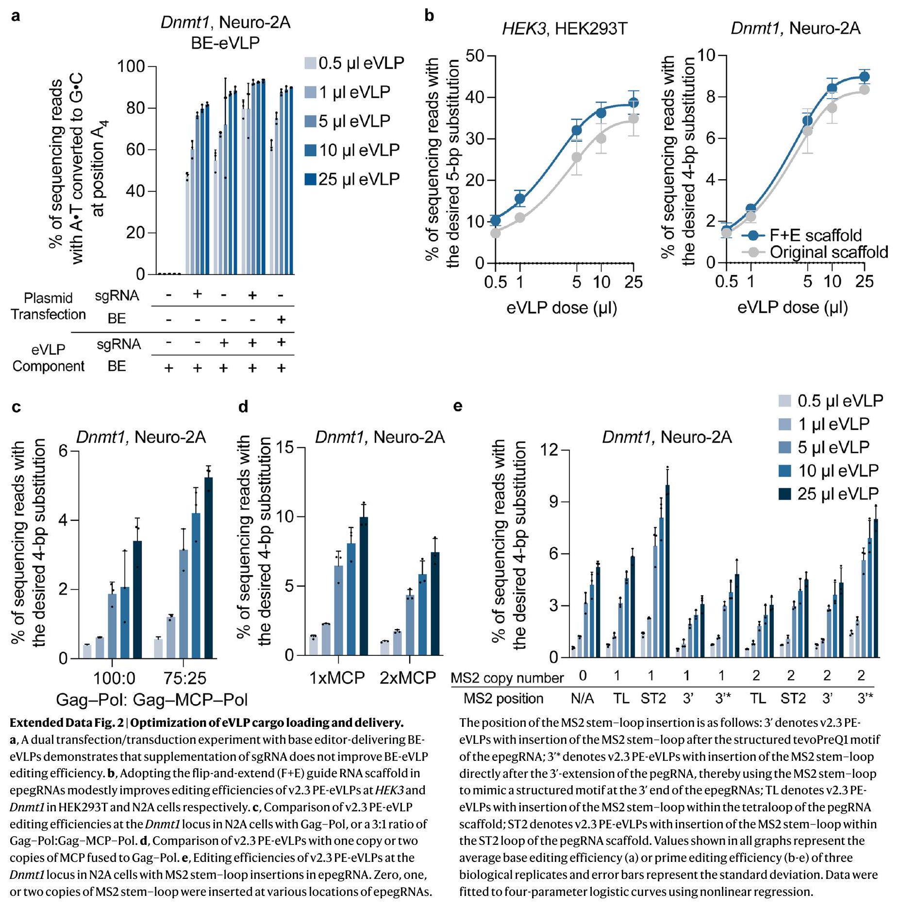
Systematic engineering of PE and eVLP architecture

To further enhance the efficiency of PE-eVLPs, we sought to identify mechanistic bottlenecks in v1 PE-eVLPs. The formation of eVLPs relies on the proteolytic processing of the Gag-Pol polyprotein into multiple functional domains by MMLV protease. Coincidentally, the RT domain of canonical PEs also originates from MMLV, and therefore contains an endogenous TSTLLIENS protease cleavage site at the C-terminus (Fig. 1c). We hypothesized that cleavage at this site may eliminate the crucial C-terminal NLS that direct nuclear localization of the PEs. Therefore, we deleted six amino acids at the C-terminus of MMLVRT to remove the protease recognition site, yielding v2.1PE-eVLPs. This modification offered consistent improvement in PE-eVLP editing efficiency over v1.3 PE-eVLPs (averaging 1.8 -fold and 1.2 -fold, respectively) at both the HEK3 locus and Dnmt1 locus (Fig.1d). We tested further truncation of the MMLV RT to remove the entire RNaseH domain, a common truncation strategy used by AAV systems to reduce PE size and facilitate dual-AAV packaging . In the PE-eVLP system, however, eliminating the RNaseH domain resulted in consistently lower editing efficiencies in both HEK293T and N2A cells than v2.1 PE-eVLP (Fig. 1d).
The nuclear export signal (NES) promotes cytoplasmic localization of Gag-cargo fusion protein. After protease cleavage, however, gene editing agents must be separated from the NES for efficient nuclear localization. The presence of four additional MMLV protease cleavage sites within the Gag polyprotein raises the possibility of incomplete cleavage, resulting in a fraction of PE cargo retaining some portion of Gag along with NESs (Fig. 1e). Indeed, we previously observed cargo of
Fig. 1|Engineering of PE and eVLP architectures. a, Schematic of v1PE-eVLPs. b, Prime editing efficiencies of v1PE-eVLPs for the HEK3+1 T-to-A edit in HEK293T cells and Dnmt1 +2 G-to-C edit in N2A cells. Adoption of epegRNAs (v1.2) and the PEmax architecture (v1.3) improved prime editing efficiencies compared to v1.1 PE-eVLPs. c, Schematic of PE engineering to eliminate the endogenous protease cleavage site (TSTLLIENS) in MMLV RT. d, Representative improvements in PEeVLP editing efficiencies as a result of RT domain engineering. PEmax-FL denotes v1 PE-eVLPs with full-length MMLV RT as PE effector domain; PEmax-RNaseH del denotes v1PE-eVLPs with RNaseH domain-truncated RT used as the PE effector domain; PEmax-6aa del denotes v2.1 PE-eVLPs with six-amino-acid-deleted RTs used as the PE effector domain. e, Schematic of the proposed mechanism of eVLP maturation and cargo delivery, and the design of v2.2 and v2.3 PE-eVLPs.f, Prime editing efficiencies of PE-eVLPs with the NES placed at various locations
(NES position 1-5) of the Gag domain. NES position 5 (v2.2) showed improved editing compared to that of v2.1PE-eVLPs. g, Comparison of prime editing efficiencies with v1, v2.1, v2.2 and v2.3 PE-eVLPs at the HEK3 locus in HEK293T cells and Dnmt1 locus in N2A cells. Values shown in all graphs represent the mean prime editing efficiencies s.e.m. of three biological replicates. Data were fitted to four-parameter logistic curves using nonlinear regression. Comparisons of different versions of PE-eVLPs are made with eVLPs produced and transduced in parallel in one large experiment to minimize variability between preparations. Data from all PE-eVLPs produced and tested in parallel are provided in Supplementary Fig 1. The graphs in and show a subset of data from Supplementary Fig 1. For all conditions, 30,000-35,000 cells were treated with eVLPs containing .
varying sizes in BE-eVLPs by western blot , supporting the hypothesis that improperly cleaved products can be packaged in eVLPs. To mitigate this problem, we relocated the NESs within the Gag polyprotein (Fig. 1f). We inserted NES before the protease cleavage site of each Gag subdomain and two additional sites that had been previously shown to tolerate large insertions in the MMLV Gag- . Among five
tested sites, inserting NES between the p12 and CA domains of Gag (NES position 5) supported the highest prime editing efficiency in HEK293T cells, yielding v2.2 PE-eVLP (Fig. 1f).
Lastly, to facilitate cargo release, we inserted GGS linkers flanking the engineered protease cleavage site to increase the accessibility of the site for protease processing. The resulting v2.3 PE-eVLP architecture

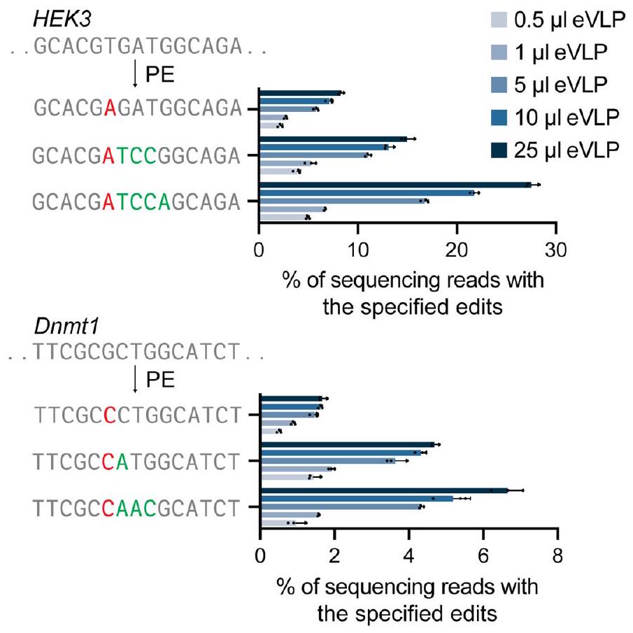
(Fig. 1e) containing MMLV RT truncation, NES optimization and linker optimization showed an average 2.6 -fold and 1.6 -fold improvement in prime editing efficiency over the v1.3PE-eVLP architecture at the highest dose tested at the HEK3 locus in HEK293T cells and Dnmt1 locus in N2A cells, respectively (Fig. 1g).

Dependence of PE-eVLP performance on edit type

We previously demonstrated that prime editing efficiencies can be impaired by cellular mismatch repair (MMR) pathways, and that evasion or inhibition of MMR improves prime editing efficiencies . Given the transient nature of PE:pegRNA RNPs packaged in eVLPs, we hypothesized that the reversion of the installed edit by MMR might be especially detrimental to editing efficiency.
We previously discovered that the installation of additional silent or benign mutations near the target edit causes the prime editing intermediate to natively evade MMR, enhancing editing efficiencies and product purities . To test this possibility for eVLP-delivered prime editing systems, we installed additional nearby substitutions at the HEK3 locus and Dnmt1 locus with the v2.3 PE-eVLP system. In both cases, prime editing efficiencies were substantially improved, achieving 28% average editing for a 5-bp substitution edit at and 6.6% editing for a4-bp substitution edit at Dnmt1 at the highest dose tested (Extended Data Fig.1). Due to codon redundancy, many therapeutically relevant edits can be achieved with the concurrent installation of additional silent mutations to evade . The substantial benefit of MMR evasion to PE-eVLP editing outcomes informs the design of optimal epegRNAs when using PE-eVLPs.

Insufficient epegRNA packaging limits PE-eVLP efficiency

In contrast to the mechanism of PE protein packaging, which depends on fusion with the Gag polyprotein, epegRNA packaging relies entirely on noncovalent association with the Cas9 domain of PEs. To test if epegRNA delivery is a bottleneck in PE-eVLP performance, we supplemented epegRNAs via transfection of plasmids encoding epegRNAs 24 h before PE-eVLP transduction. Indeed, epegRNA supplementation greatly improved PE-eVLP editing efficiency by greater than eightfold on average, while sgRNA supplementation offered minor improvement to BE-eVLP editing efficiency in an analogous experiment (Fig. 2a and Extended Data Fig. 2a). These results reveal that epegRNA loading, delivery or lifetime limits PE-eVLP editing to a much greater extent than sgRNA loading or delivery impacts base editing with BE-eVLPs. In contrast, PE protein supplementation via transfection of plasmids expressing PEs did not improve PE-eVLP editing efficiency (Fig. 2a), consistent with the insufficiency of epegRNAs but not PE proteins in PE-eVLPs.
We previously observed that the apparent affinity of Cas9 for sgRNAs, pegRNAs and epegRNAs decreases in that order (Supplementary Note 1). To increase Cas9-epegRNA affinity, we adopted the F + E guide RNA scaffold , which improves Cas9-guide RNA binding through a stem extension in the scaffold structure that provides an extended handle for Cas9 binding. The F + E guide RNA scaffold moderately improved PE-eVLP editing efficiencies (Extended Data Fig. 2b) and was used in all subsequent epegRNAs.
To further address the inefficiency of epegRNA packaging in v2.3 PE-eVLPs, we used the MCP-MS2 system as an additional mechanism to promote epegRNA packaging. Binding of the MS2 coat protein (MCP) to the MS2 RNA stem-loop can mediate sgRNA packaging for Cas9 nuclease and BE delivery , can recruit an untethered RT to Cas9 nickase in split PE systems , and can recruit additional protein domains to . To apply this system to PE:epegRNA RNP packaging in eVLPs, we designed a Gag-MCP-Pol fusion plasmid by inserting one copy of MCP between the C-terminus of the Gag nucleocapsid domain and the N-terminus of the Pol domain (Fig. 2b). We reasoned that MCP insertion at this position would minimally disturb Gag polyprotein processing for eVLP particle maturation (Extended Data Fig. 2c and
Supplementary Note2) and that fused MCP would be presented at the interior of the particle, facilitating recruitment of epegRNAs into eVLPs.
Next, we tested a series of epegRNAs with MS2 stem-loop insertions in scaffold loops or at the 3′ end. We observed that inserting one copy of the MS2 stem-loop in the ST2 loop of epegRNAs (MS2-epegRNAs) led to the highest level of prime editing (Fig. 2c). Increasing the copy number of both MCP and MS2 aptamers impaired editing efficiency, probably because the larger fusions destabilize Gag-Pol and epegRNAs (Extended Data Fig. 2d,e). Lastly, we reoptimized the stoichiometry of Gag-fusion plasmids by varying the ratio of wild-type Gag-Pol plasmid, Gag-PE plasmid and Gag-MCP-Pol plasmid (Fig. 2d). The optimized ratio of 5:2:1 Gag-Pol plasmid:Gag-MCP-Pol plasmid:Gag-PE plasmid resulted in v3PE-eVLPs that improved average prime editing efficiency by 4.0 -fold over v2.3 PE-eVLPs at Dnmt1 locus in N2A cells at the highest dose tested (Fig. 2e).

PE3 further improves prime editing with PE-eVLPs

The PE-eVLP system described thus far packages one PE and one epegRNA (the ‘PE2 strategy’). The ‘PE3 strategy’ adds an additional ngRNA that directs the PE to nick the unedited strand, stimulating its replacement using the edited strand as a template, increasing editing efficiency at the cost of increased indel frequencies . To enable PE3-eVLPs, we tested the possibility of packaging the ngRNA in the same or in a separate particle as the epegRNA. To screen for an optimal all-in-one particle v3 PE3-eVLP system, we transfected the producer cells with varying ratios of MS2-epegRNA and MS2-ngRNA plasmids simultaneously, along with other PE-eVLP components. The results demonstrated that transfecting MS2-epegRNA and MS2-ngRNA plasmids in an optimized ratio of 4:1 led to v3 PE3-eVLPs that support 1.3-fold higher average editing efficiency over the v3 PE2-eVLP system at Dnmt1 locus in N2A cells with of eVLPs (Fig.2f). Alternatively, producing separate eVLP batches packaging PE:MS2-epegRNA RNP or PE:MS2-ngRNA RNP and transducing target cells with both eVLPs yielded comparable editing levels (Fig. 2f). Given the comparable levels of editing with two strategies, we moved forward with the all-in-one particle v3 PE3-eVLP system for the ease of production, ability to precisely control epegRNA and ngRNA stoichiometry, and potentially higher in vivo delivery efficiency from a single-particle system that does not require co-transduction by multiple eVLPs.

5′-G extension removal enhances editing with certain epegRNAs

Guide RNAs are transcribed from the U6 promoter in the PE-eVLP system, with a preference for initiating from guanine at the +1 position . epegRNAs that do not start with a are often appended with an extra G at their end. Such a -G extension may sterically hinder pegRNA binding to the Cas9 domain , however, and thus may introduce an additional challenge for PE:epegRNA complex formation. Moreover, the unpaired 5′ G may impede R-loop formation and DNA cleavage by Cas9 nuclease in a target sequence-dependent manner . Because PE:epegRNA RNPs delivered by PE-eVLPs are short lived by design, we reasoned that any impairment of the rate of target search and DNA nicking may be detrimental to PE-eVLP-mediated prime editing efficiencies.
To overcome this potential hindrance, we tested v3 PE-eVLP systems that use epegRNAs containing either a 20-bp spacer starting with cytosine (C), uracil (U) or adenine (A), or a mismatched followed by a 19 -bp spacer (Extended Data Fig. 3a). The resulting data indicates that the improvement from omitting the -G extension can be substantial, although the effect varies depending on the target sequence. At the Col12a1 locus in N2A cells, a 20 -bp spacer beginning with a outperformed the original -bp spacer by 2.8 -fold (Extended Data Fig. 3a). At the Ctnnb1 locus, both the 20 -bp spacer beginning with a and the -bp spacer yielded comparable results, both offering 4.7 -fold improvement over the original -bp spacer.
Fig. 2 | Engineering of pegRNA and ngRNA. a, A dual transfection/transduction experiment identifies epegRNAs as the limiting component in the v2.3 PE-eVLPs. b, Schematic of v3 PE-eVLPs utilizing the MCP-MS2 strategy for the recruitment of epegRNAs. The incorporation of MCP-MS2 strategy enables three modes of guide RNA loading into eVLPs: (i) via binding to PE, (ii) via MS2 stem-loop binding to MCP and (iii) via MCP:MS2-gRNAs:PE three-component interaction. c, Editing efficiencies of v2.3 PE-eVLPs at the Dnmt1 locus in N2A cells with MS2 stem-loop insertion at various locations in epegRNAs. The 3′ end denotes v2.3 PE-eVLPs with insertion of the MS2 stem-loop after the structured tevoPreQ1 motif of the epegRNA; 3 ‘ end* denotes v2.3 PE-eVLPs with insertion of the MS2 stem-loop directly after the extension of the pegRNA, thereby using the MS2 stem-loop to mimic a structured motif at the end of epegRNAs; TL denotes v2.3 PE-eVLPs with insertion of the MS2 stem-loop within the tetraloop of the pegRNA scaffold; and ST2 denotes v2.3 PE-eVLPs with insertion of the MS2 stem-loop within the ST2 loop of the pegRNA scaffold. d, Heatmap of editing efficiencies from
stoichiometry optimization of Gag-Pol, Gag-MCP-Pol and Gag-PE plasmids for production of v3 PE-eVLPs. e, Comparison of editing efficiencies with v2.3 and v3 PE-eVLPs at the Dnmt1 locus in N2A cells. , Editing efficiencies of all-inone and separate-particle v3 PE3-eVLP systems with varying MS2-epegRNA to MS2-ngRNA ratios. g, Quantification of the number of epegRNAs molecules packaged per eVLP in successive generations of PE-eVLPs. h, Quantification of the number of PE protein molecules packaged per eVLP in successive generations of PE-eVLPs. Values represent the mean prime editing efficiencies s.e.m. of three biological replicates (a and c-f) or three technical replicates (g and ). Data were fitted to four-parameter logistic curves using nonlinear regression. Data from all PE-eVLPs produced and tested in parallel are provided in Supplementary Fig 2. The graphs in and e show a subset of data from Supplementary Fig 2.
For all conditions, 30,000-35,000 cells were treated with eVLPs containing .

Characterization of PE-eVLP cargo loading

To validate the mechanisms used to enhance cargo packaging, we quantified the epegRNAs and PE protein packaged in each PE-eVLP version. Intact epegRNAs were quantified by RT-quantitative polymerase chain reaction (qPCR) of total RNA extracted from eVLPs (Fig. 2g). The number of eVLP particles per unit volume measured using anti-MLV p30 (capsid) enzyme-linked immunosorbent assay (ELISA; Extended Data Fig. 3b) was used to calculate the number of epegRNAs packaged per eVLP. Although the PE and eVLP architecture engineering in v2.3 PE-eVLP was not designed to enhance epegRNA recruitment, v2.3 PE-eVLPs still improved epegRNA packaging over v1.2 PE-eVLPs by 2.1-fold, presumably because epegRNA packaging occurs as a complex with PE protein and therefore is correlated with PE protein packaging. The MCP-MS2 strategy in the v3 PE-eVLP system improved epegRNA loading by 2.2 -fold compared to v2.3 PE-eVLPs, consistent with improved editing efficiency. Lastly, omitting the -G extension on the epegRNAs offered an additional 1.3 -fold improvement in epegRNA loading, supporting the hypothesis that the improvement in editing efficiency from extended -G omission may arise from improved binding of epegRNAs to PEs. Additional characterization of the percent composition of MS2-epegRNA and MS2-ngRNA in v3PE-eVLPs revealed an approximate 1:1 ratio of the two species (Extended Data Fig. 3c). Since the composition of MS2-ngRNAs increased in the eVLPs compared to the stoichiometry used for eVLP production, we hypothesize that ngRNAs have higher binding affinity than epegRNAs for PE protein, resulting in more efficient loading into eVLPs.
ELISA with anti-Cas9 antibodies revealed an increase in PE protein per particle in successive versions of PE-eVLPs (Fig. 2h). The MS2-MCP strategy in v3 PE-eVLP improved both epegRNA recruitment and PE protein levels per particle by 1.3-fold, probably due to the three-component interaction between MCP:MS2-gRNAs:PE that synergistically enhances incorporation of all prime editing components (Fig. 2b). Likewise, omitting the -G extension further improved the PE packaging by an additional 1.4 -fold to an average of 26 molecules of PE protein per eVLP (Fig. 2h). Overall, these findings reveal that increases in both epegRNA and PE protein packaging efficiency in PE-eVLPs correlate with improvements in prime editing efficiencies over successive generations of PE-eVLPs, suggesting that v3 PE-eVLPs enhance prime editing outcomes by addressing bottlenecks in protein and RNA packaging.

PE recruitment via coiled-coil peptide association

Although PE packaging in PE-eVLPs adopted the same strategy as BE packaging in v4 BE-eVLP and successive rounds of PE-specific optimizations led to an overall 2.0 -fold average improvement in PE packaging in eVLPs, the absolute number of PE molecules packaged per particle ( 26 per eVLP) remained about half the value for BEs in BE-eVLPs (55 per eVLP) . We hypothesized that the large size of the Gag-PE fusion protein ( 309 kDa ) might hinder its production in the producer cells. To overcome this problem, we designed an alternative PE packaging system that does not require Gag fusion to PE (Fig. 3a). Instead, we used coiled-coil peptides, -helical peptides that can strongly and specifically bind other -helical peptides to form a superhelix structure . We chose the P3-P4 coiled-coil peptide pair that has been previously used in mammalian cells to recruit protein domains to Cas9 (ref. 61). We designed Gag-P3-Pol fusion construct by fusing P3 to the C-terminus of the Gag nucleocapsid domain, and fused the partner P4 peptide, followed by NES and the engineered protease cleavage site, to the N-terminus of PE (hereafter referred to as P4-PE). We produced PE-eVLPs by transfecting plasmids encoding VSV-G, wild-type MMLV Gag-Pol, Gag-P3-Pol, P4-PE and epegRNA into producer cells. This alternative system exhibited a 1.3 -fold higher average prime editing efficiency compared to the analogous v2.3 Gag-PE-eVLP system at Dnmt1 locus in N2A cells at the highest dose tested (Fig. 3b). The control eVLP lacking the P3 peptide showed 28 -fold lower editing efficiency, supporting the role of P3-P4 association in facilitating PE packaging.
Additionally, we confirmed via western blot of producer cell lysates substantially higher levels of P4-PE compared to Gag-PE (Extended Data Fig. 4a and Supplementary Note 3), consistent with our hypothesis that the smaller P4-PE fusion protein improved protein production levels.
Next, we explored the synergy of this alternative system with the pegRNA recruitment strategy using aptamer-binding proteins, which yielded substantial improvements in the v3 PE-eVLP system. Initial attempts using a single Gag-MCP-P3-Pol fusion construct proved inefficient (Extended Data Fig. 4b). Therefore, we tested PE-eVLP production using both Gag-aptamer-binding proteins-Pol and Gag-P3Pol constructs. Instead of the MCP-MS2 pair, we used the COM-Com protein-RNA aptamer pair (Extended Data Fig. 4c), reasoning that the smaller size of both the COM protein and the Com aptamer may be less perturbative to particle maturation or PE function. We optimized the stoichiometry of Gag-COM-Pol, Gag-P3-Pol and P4-PE plasmids to yield the v3bPE-eVLP system, an alternative to v3 PE-eVLP system (Fig. 3c and Fig. 3d). We compared the v3 PE3-eVLP and v3b PE3-eVLP system at multiple loci and observed locus-dependent differences in editing efficiency (Fig. 3e). At the Dnmt1 and FANCF loci, v3b outperformed v3 in prime editing efficiency by 1.2- and 2.9-fold, respectively, at the highest dose tested. At HEK3 and Col12a1 loci, the two systems performed comparably, consistent with comparable levels of PE protein packaged in v3 and v3b PE-eVLPs analyzed via western blot (Extended Data Fig. 4d and Supplementary Note3).
The cumulative improvements in v3 and v3b PE3-eVLPs yielded large increases in prime editing efficiencies compared to the v1.1 PE2-eVLPs (Fig. 3e). v3 and v3b PE3-eVLPs outperformed v1.1 PE2-eVLPs by 79 – and 65 -fold at the Dnmt1 locus in N2A cells, and 170 – and 150 -fold at the HEK3 locus in HEK293T cells, respectively. At the Col12a1 locus in N2A cells and FANCF locus in HEK293T cells, we did not observe any editing above background level (<0.1%) with v1.1PE2-eVLPs, while the third-generation PE-eVLP architectures achieved on average 7.7% and 26% editing at the respective loci at the highest dose tested. Examination of the frequency of insertion-deletion byproducts revealed no apparent difference between PEs delivered by PE-eVLPs versus by plasmid transfection (Extended Data Fig. 5a). Furthermore, we observed low batch-to-batch variability as evidenced by consistent prime editing efficiencies using four independent batches of PE-eVLPs prepared on different days (Extended Data Fig. 5b). These data collectively highlight substantial benefits that resulted from the systematic engineering of third-generation PE-eVLP architectures (Extended Data Fig. 6).

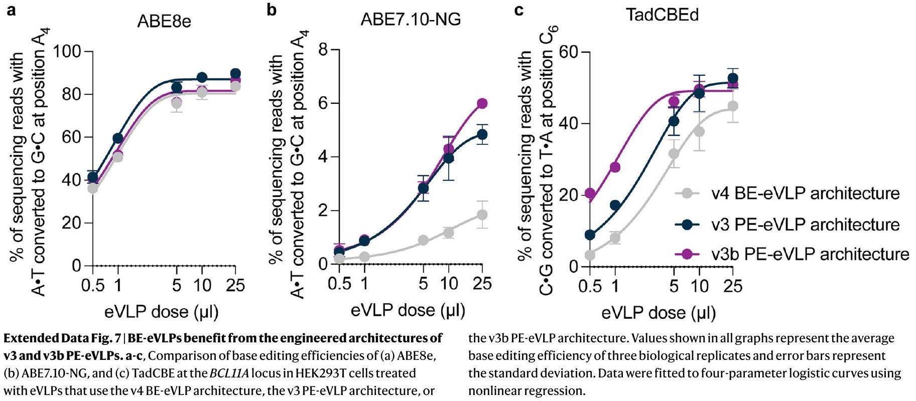
BE-eVLPs benefit from the engineered PE-eVLPs architecture

Given the substantial improvements in prime editing efficiencies offered by v3 and v3b PE-eVLPs, we sought to further evaluate whether these alterations also enhance eVLP-mediated BE delivery. We packaged two ABEs, ABE8e and ABE7.10-NG , as well as cytosine BE, TadCBEd , in the previously reported v4 BE-eVLP architecture, and in the v3 or v3b PE-eVLP architectures (replacing the PE with either BE), targeting the BCL11A locusin HEK293T cells (Extended Data Fig.7a-c). While the v3and v3bPE-eVLP architecture offered minor improvements for highly active ABE8e, they substantially enhanced editing efficiencies for less active BEs such as ABE7.10-NG. For ABE7.10-NG, v3 and v3b PE-eVLP architectures yielded 2.6-fold and 3.2-fold higher base editing efficiencies, respectively, at the highest dose tested (Extended Data Fig. 7b). These results suggest that some of the strategies that improved PE protein and pegRNA packaging can also benefit the packaging and post-transduction editing efficiency of other gene editing cargoes.

Transient delivery of PE by eVLPs reduces off-target editing

Previous studies have shown that transient delivery of genome editing agents can reduce off-target editing . Direct delivery of RNP complexes in theory should result in the shortest duration of editing agent exposure to the genome compared to mRNA or DNA delivery. Prime editing has been shown to exhibit high DNA specificity, probably due
Fig. 3 | Development of v3b PE-eVLPs, which use an alternative PE recruitment mechanism. a, Schematic of coiled-coil peptide-dependent recruitment of PE. NES, nuclear export signal. NLS, nuclear localization signal. b, Comparison of prime editing efficiency with v2.3 PE-eVLPs versus the alternative system employing coiled-coil peptide for the recruitment of PE. c, Schematic of v3b PE-eVLP system. NES, nuclear export signal. NLS, nuclear localization signal. d, Heatmap of editing efficiencies from stoichiometry optimization of Gag-Pol, Gag-COM-Pol and Gag-P3-Pol plasmids for production of v3bPE-eVLPs. e, Prime editing efficiencies comparing v1.1PE2-eVLPs, v3 PE3-eVLPs and v3b PE3-eVLPs at Dnmt1, FANCF, Col12a1 and
b

HEK3 locus in N2A and HEK293T cells.f, Comparison of editing efficiencies for four different prime edits targeting HEK4 locus in HEK293T cells following plasmid transfection or treatment with v3b PE-eVLPs at the on-target HEK4 site and known off-target sites for the corresponding pegRNA . Values shown in all graphs and heatmaps represent the average prime editing efficiency of three biological replicates, and error bars represent the standard deviation. Data were fitted to four-parameter logistic curves using nonlinear regression. For all conditions other than cells were treated with eVLPs containing .
Fig. 4 | CNS editing with PE-eVLPs via PO ICV injection. a, Schematic of workflow for neonatal ICV injection and subsequent analysis. FACS, fluorescenceactivated cell sorting. b, Prime editing efficiency in bulk or GFP-positive population from the brain cortex collected 3 weeks following PO ICV injection
targeting the Dnmt1 locus with v3 PE3-eVLPs and v3b PE3-eVLPs. Bars represent the average prime editing efficiency of three mice and error bars represent the standard deviation, with each dot representing an individual mouse. Each mouse received approximately in total.
to the requirement of three distinct DNA hybridization steps for productive editing . Given the difficulty of finding genomic loci that support readily measured levels of off-target prime editing, we measured off-target editing from PE-eVLPs compared to plasmid transfection at the two most highly edited off-target sites discovered in a previous off-target prime editing profiling experiment . Despite comparable or higher levels of on-target editing, off-target editing at these two unusually prone off-target sites by PE-eVLPs was reduced compared to plasmid transfection in HEK293T cells 7 days after treatment (Fig.3f). For example, for a +2 TAA insertion edit, the ratio of on-target to off-target editing improved by 3.0 -fold for PE-eVLPs compared to plasmid transfection (Fig. 3f). These findings confirm that the frequency of off-target prime editing can be further reduced by transient delivery of PE RNPs using PE-eVLPs.

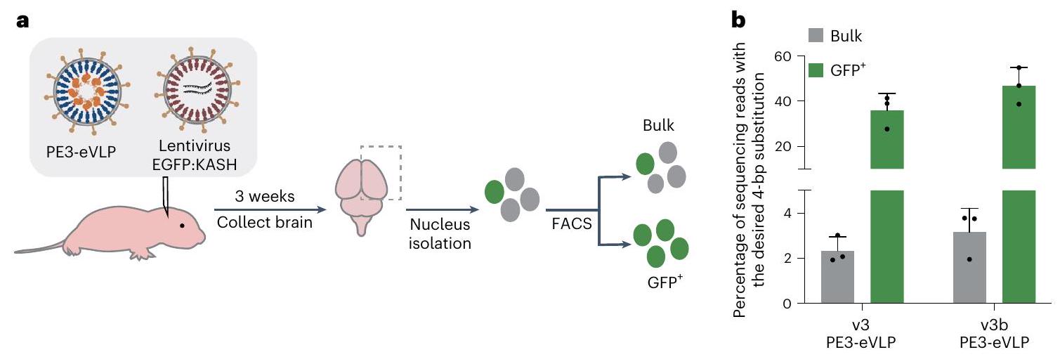
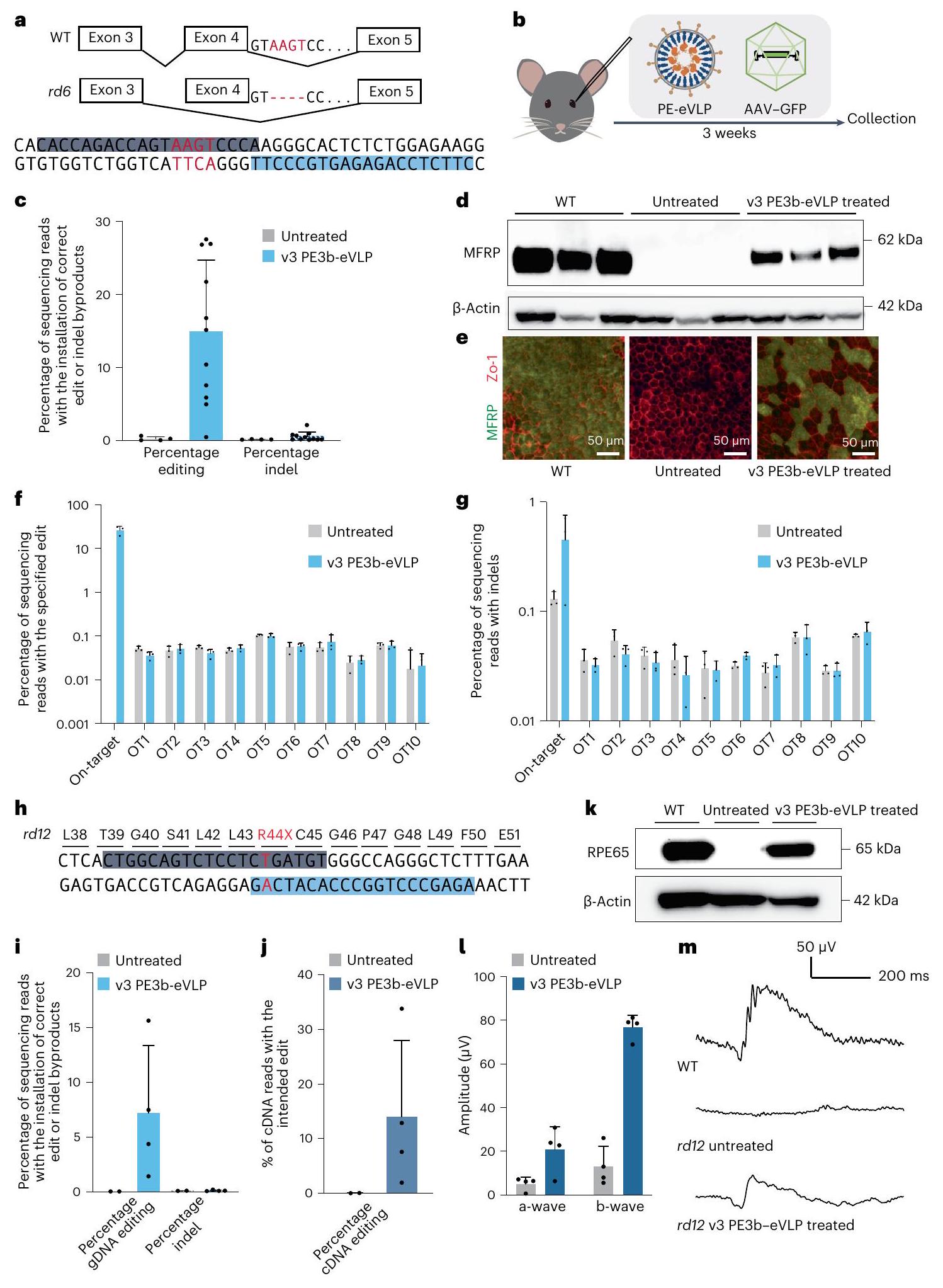
PE-eVLPs mediate potent in vivo prime editing in the brain

Next, we tested the ability of v3 and v3b PE-eVLPs to mediate in vivo prime editing in the mouse central nervous system (CNS). We produced v3 and v3b PE3-eVLPs programmed to perform a 4-bp substitution at the Dnmt1 locus. We injected PE3-eVLPs into C57BL/6 mice via intracerebroventricular (ICV) injection on postnatal day 0 (P0), a stage at which the brain undergoes rapid development . We co-injected VSV-G-pseudotyped lentivirus encoding EGFP fused to a nuclear membrane-localized Klarsicht/ANC-1/Syne-1 homology (KASH) domain to enrich cells that had the opportunity to encounter eVLPs.
We collected the brain hemispheres 3 weeks after injection, isolated both bulk and green fluorescent protein (GFP) nuclei (Extended Data Fig. 8a), extracted genomic DNA and performed high-throughput sequencing (HTS) (Fig. 4a). We achieved on average bulk cortex editing and 36% editing among GFP+ nuclei with v3PE3-eVLPs (Fig. 4b). The v3b PE3-eVLPs that showed more efficient editing in cell culture outperformed the v3 PE3-eVLP construct in vivo, achieving 3.2% editing in bulk cortex and 47% average editing among GFP+ nuclei (Fig. 4b). Efficient prime editing among GFP+ nuclei in the brain indicates that PE RNPs delivered by PE-eVLPs can induce efficient prime editing in cells that are transduced. As we previously reported , however, transduction events occur primarily near the site of injection, probably due to the slow rate of diffusion of eVLPs ( in diameter as measured by dynamic light scattering (DLS)) within the brain. Staining isolated nuclei with neuron-specific anti-NeuN antibody revealed that most nuclei were NeuN negative, suggesting VSV-G pseudotyped GFP:KASH lentivirus and PE-eVLPs primarily transduced nonneuronal cells (Extended Data Fig. 8b). Given the ability of eVLPs to alter tropism by using alternative envelope proteins , using a neuron-specific envelope protein such as FuG-B2 (ref. 70) may enable more efficient targeting of neurons.

PE-eVLP corrects mutations causing retinal diseases in vivo

Next, we sought to test the therapeutic utility of in vivo PE delivery via PE-eVLPs by correcting a mutation causing retinal disease in rd6 mice, a model of autosomal recessive retinal degeneration caused by a 4-bp deletion in the splice donor of the membrane-type frizzled-related protein (Mfrp) gene (Fig. 5a). The mutation leads to early onset, slowly progressive retinal degeneration in mice, akin to genetic defects in associated with microphthalmos and retinitis pigmentosa in humans . Given the unique ability of prime editing to directly and precisely correct a 4-bp deletion, and the potential of prime editing to treat congenital blindness , we developed a PE3b strategy-in which the ngRNA is designed to nick only after the desired prime edit is installed into the edited strand -to correct this 4-bp deletion in frp (Fig. 5a). Following plasmid transfection in cells that contained the 4-bp deletion at the endogenous locus, the best PE3b strategy yielded on average 18% correction of the mutation with indels (Supplementary Table 4). Encouraged by this result, we produced v3 PE3b-eVLPs pseudotyped with VSV-G to efficiently transduce the retinal pigment epithelium (RPE) cells , where MFRP is most abundantly expressed .
We performed subretinal injections into 5-week-old rd6 mice to administer the mutation-correcting v3 PE3b-eVLPs (Fig. 5b). A total of 3 weeks post-injection, we performed HTS on the genomic DNA extracted from the RPE tissue. Sequencing analysis revealed on average 15% bulk retinal editing with v3 PE3b-eVLPs (Fig. 5c). Western blot revealed that MFRP expression was restored in v3 PE3b-eVLP-treated eyes (Fig. 5d). We performed immunohistochemistry on the RPE flatmounts and confirmed robust restoration of MFRP expression in the RPE (Fig. 5e). We assessed off-target prime editing from v3 PE3b-eVLP treatment by deep sequencing the top ten off-target sites associated with the frp epegRNA and ngRNA as nominated by circularization for in vitro reporting of cleavage effects by sequencing (CIRCLE-seq) . We did not detect any off-target editing in PE-eVLP-treated samples (>0.1%, the limit of detection of the targeted HTS method used) (Fig. 5f,g). These data are consistent with the inherently high DNA specificity of prime editing , further enhanced by transient exposure of cells to PEs from PE-eVLPs. Additionally, no signs of toxicity or morphology changes were observed in treated eye cryosections (Extended Data Fig. 9).
Finally, to further test the ability of PE-eVLPs to mediate therapeutic in vivo prime editing, we applied PE-eVLPs to treat the rd12 mouse model , which displays more severe retinal degeneration that allows evaluation of disease phenotype rescue. The rd12 model contains a nonsense mutation in exon 3 of the retinal pigment epithelium 65 (Rpe65) gene (c. 130 C > T; p.R44X) (Fig. 5h), leading to diminishment of electroretinogram (ERG) responses from 3 weeks of age . The

corresponding mutation in humans in homozygous form causes Leber congenital amaurosis . We previously demonstrated partial rescue of this disease phenotype using BE-eVLPs . However, base editing at this site generates bystander edits at nearby base pairs that may inactivate the RPE65 enzyme and cause adverse effects , whereas the mechanism of prime editing inherently avoids bystander editing .
We developed a PE3b strategy to correct the Rpe65R44X mutation (Fig. 5h) by screening a panel of epegRNAs with varying primer binding site lengths ( ) and reverse transcription template lengths
( ), and seven candidate ngRNAs via plasmid transfection in an engineered NIH3T3 cell line containing the corresponding mutation. The most promising PE3b strategy yielded precise correction with indels (Supplementary Table 5). We performed subretinal injection of 5-week-old mice with mutation-correcting v3 PE3b-eVLPs. HTS on the genomic DNA extracted from RPE tissues of the treated mice resulted in on average 7.2% correction of the mutation in bulk RPE tissue (Fig. 5i). This editing efficiency is higher than the reported value from triple and low-dose dual-vector AAV-mediated PE delivery and
Fig. 5 | Retinal disease correction in vivo with PE-eVLPs. a, An optimized PE3b strategy to correct a 4-bp deletion in Mfrp in the rd6 mice. The mutation (red), the epegRNA spacer (light blue) and the ngRNA spacer sequence (dark blue) are highlighted. b, Schematic of subretinal injection of rd6 mice. c, Prime editing efficiency and the associated indel frequency in the genomic DNA collected from the rd6 mice. Data are represented as mean values s.e.m. Each dot represents an individual mouse for (untreated) or (v3 PE3b-eVLP treated). d,e, Western blot of RPE tissue protein extracts (d) and immunohistochemistry blot on RPE flatmounts (e) from wild-type C57BL/6J mice, untreated mice and v3 PE3-eVLP-treated mice. Flatmounts were stained with anti-MFRP antibody (green) and anti-ZO-1 antibody (red).f,g, Analysis of PE-dependent editing (f) and indel byproducts at the on-target site and top ten CIRCLE-seq nominated off-target sites associated with the rd6 epegRNA (f) and ngRNA (g) sequence. For and , data are represented as mean values s.e.m. Each dot represents an individual mouse for (untreated) or (v3PE3b-eVLP treated). , The
R44X mutation in Rpe65 exon 3 in the rd12 mouse model and the optimized PE3b strategy used for correction of this mutation. The mutation (red), the epegRNA spacer (light blue) and the ngRNA spacer sequence (dark blue) are highlighted. i, Prime editing efficiency of Rpe65 R44X mutation correction and the associated indel frequency in genomic DNA collected from rd12 mice.j, Prime editing efficiency of Rpe65 R44X mutation correction in Rpe65 RNA collected from rd12 mice. k, Western blot of RPE tissue protein extracts from wild-type C57BL/6J mice, untreated mice and v3 PE3b-eVLP-treated mice. I, Scotopic A – and B -wave amplitudes measured by ERG following overnight dark adaptation. For i, j and I, data are represented as mean values s.e.m. Each dot represents an individual mouse for (untreated) or (v3 PE3b-eVLP treated). m, Representative ERG waveforms from wild-type C57BL/6J mice, untreated rd12 mice and v3 PE3-eVLP-treated mice. Each mouse received approximately per eye. WT, wild type.
comparable to that achieved by high-dose dual-vector AAV-mediated PE delivery , but using a virus-free, single-particle delivery method.
We further analyzed Rpe65 transcripts in the extracted RNA by sequencing the complementary DNA. Editing was enriched at the transcript level, achieving on average 14% correction of the target mutation (Fig. 5j), presumably due to nonsense-mediated decay accelerating the degradation of uncorrected transcripts and enrichment of the major RPE65-expressing RPE cells that are preferentially transduced by PE-eVLPs among other cell types co-collected from tissue dissection. We confirmed robust expression of full-length RPE65 in v3 PE3b-eVLP-treated eyes by western blot (Fig. 5k). ERG of the v3 PE3b-eVLP-treated animals indeed showed substantial rescue of visual function in response to the stimuli compared to the untreated eyes (Fig. 5l,m). No off-target prime editing was detected above the limit of detection at top ten CIRCLE-seq-nominated off-target sites associated with the rd12 epegRNA and ngRNA (Extended Data Fig.10a,b). Together, these results demonstrate that in vivo application of optimized PE-eVLPs can correct a pathogenic mutation and partially rescue disease phenotype in mammals.

Discussion

Through extensive engineering of each major component, we developed an all-in-one virus-like particle that delivers PE RNPs into mammalian cells in culture and in vivo. Recent improvements to prime editing systems, including epegRNAs , the PEmax architecture and MMR evasion , contributed to improved outcomes with PE-eVLPs. Identification of bottlenecks in cargo packaging yielded PE variants that promote delivery by PE-eVLPs, as well as optimized eVLP architectures that facilitate cargo release and cargo localization. Introducing an additional mechanism for guide RNA recruitment addressed guide RNA packaging limitations, and an alternative v3bPE-eVLP system eliminated the need for covalent fusion to the Gag polyprotein. Together, these improvements yielded 170-fold higher average prime editing efficiency compared to v1.1 PE-eVLPs at a benchmark HEK3 test edit in HEK293T cells.
The optimized v3 and v3b PE-eVLPs systems proved efficacious in vivo. Potent prime editing was achieved in the mouse CNS via neonatal ICV injection, marking the first demonstration of CNS editing with transient delivery of a PE RNP. In the mouse retina, a single injection of v3 PE3-eVLPs precisely corrected a pathogenic 4-bp deletion in the rd6 model of retinal degeneration, restoring production of full-length MFRP protein. In the rd12 mouse model of genetic blindness, v3 PE3-eVLPs achieved comparable prime editing levels to a recently reported triple-vector AAV-PE system , but using a nonviral, single-particle delivery vehicle, resulting in partial rescue of visual function. These findings demonstrate that v 3 and v 3 b PE-eVLPs can achieve prime editing efficiency comparable to that attained using an AAV-PE delivery system, while avoiding drawbacks of viral delivery systems such as prolonged editor expression that increases off-target
editing frequencies and the risk of oncogenic DNA integration . To our knowledge, these findings also represent the first use of PE RNPs to achieve phenotypic rescue of an animal model of genetic disease.
While v3 and v3bPE-eVLPs demonstrated therapeutically relevant editing levels, PE-eVLP systems would benefit from the continued engineering effort for the next-generation PEs and improved eVLP systems. Furthermore, tissue-specific envelope protein engineering could expand the scope of PE-eVLP applications to diverse tissues. The possibility that single-dose, transient delivery of PE RNPs by PE-eVLPs may mitigate clinically relevant immunogenicity warrants further investigation. Lastly, future optimization in large-scale eVLP production will be necessary to fully realize the therapeutic potential of eVLPs. Nonetheless, the PE-eVLP system reported here offers unique advantages of nonviral, single-particle delivery of PEs in their most transient form as RNPs, presenting safety and target specificity advantages over DNA or mRNA delivery methods.

Online content

Any methods, additional references, Nature Portfolio reporting summaries, source data, extended data, supplementary information, acknowledgements, peer review information; details of author contributions and competing interests; and statements of data and code availability are available at https://doi.org/10.1038/s41587-023-02078-y.

References

  1. Anzalone, A. V. et al. Search-and-replace genome editing without double-strand breaks or donor DNA. Nature 576, 149-157 (2019).
  2. Chen, P. J. et al. Enhanced prime editing systems by manipulating cellular determinants of editing outcomes. Cell 184, 5635-5652 e5629 (2021).
  3. Newby, G. A. & Liu, D. R. In vivo somatic cell base editing and prime editing. Mol. Ther. 29, 3107-3124 (2021).
  4. Nelson, J. W. et al. Engineered pegRNAs improve prime editing efficiency. Nat. Biotechnol. 40, 402-410 (2022).
  5. Anzalone, A. V. et al. Programmable deletion, replacement, integration and inversion of large DNA sequences with twin prime editing. Nat. Biotechnol. 40, 731-740 (2022).
  6. Choi, J. et al. Precise genomic deletions using paired prime editing. Nat. Biotechnol. 40, 218-226 (2022).
  7. Doman, J. L., Sousa, A. A., Randolph, P. B., Chen, P. J. & Liu, D. R. Designing and executing prime editing experiments in mammalian cells. Nat. Protoc. 17, 2431-2468 (2022).
  8. Bock, D. et al. In vivo prime editing of a metabolic liver disease in mice. Sci. Transl. Med. 14, eabl9238 (2022).
  9. Zheng, C. et al. A flexible split prime editor using truncated reverse transcriptase improves dual-AAV delivery in mouse liver. Mol. Ther. 30, 1343-1351 (2022).
  10. Zhi, S. et al. Dual-AAV delivering split prime editor system for in vivo genome editing. Mol. Ther. 30, 283-294 (2022).
  11. Qin, H. et al. Vision rescue via unconstrained in vivo prime editing in degenerating neural retinas. J. Exp. Med. https://doi.org/ 10.1084/jem. 20220776 (2023).
  12. Gao, R. et al. Genomic and transcriptomic analyses of prime editing guide RNA-independent off-target effects by prime editors. CRISPR J. 5, 276-293 (2022).
  13. Kim, D. Y., Moon, S. B., Ko, J. H., Kim, Y. S. & Kim, D. Unbiased investigation of specificities of prime editing systems in human cells. Nucleic Acids Res. 48, 10576-10589 (2020).
  14. Liu, Y. et al. Efficient generation of mouse models with the prime editing system. Cell Discov. 6, 27 (2020).
  15. Schene, I. F. et al. Prime editing for functional repair in patient-derived disease models. Nat. Commun. 11, 5352 (2020).
  16. Geurts, M. H. et al. Evaluating CRISPR-based prime editing for cancer modeling and CFTR repair in organoids. Life Sci. Alliance https://doi.org/10.26508/lsa. 202000940 (2021).
  17. Park, S. J. et al. Targeted mutagenesis in mouse cells and embryos using an enhanced prime editor. Genome Biol. 22, 170 (2021).
  18. Gao, P. et al. Prime editing in mice reveals the essentiality of a single base in driving tissue-specific gene expression. Genome Biol. 22, 83 (2021).
  19. Lin, J. et al. Modeling a cataract disorder in mice with prime editing. Mol. Ther. Nucleic Acids 25, 494-501 (2021).
  20. Jin, S. et al. Genome-wide specificity of prime editors in plants. Nat. Biotechnol. 39, 1292-1299 (2021).
  21. Habib, O., Habib, G., Hwang, G. H. & Bae, S. Comprehensive analysis of prime editing outcomes in human embryonic stem cells. Nucleic Acids Res. 50, 1187-1197 (2022).
  22. She, K. et al. Dual-AAV split prime editor corrects the mutation and phenotype in mice with inherited retinal degeneration. Signal Transduct. Target Ther. 8, 57 (2023).
  23. Jang, H. et al. Application of prime editing to the correction of mutations and phenotypes in adult mice with liver and eye diseases. Nat. Biomed. Eng. 6, 181-194 (2022).
  24. Liu, B. et al. A split prime editor with untethered reverse transcriptase and circular RNA template. Nat. Biotechnol. 40, 1388-1393 (2022).
  25. Davis, J. R. et al. Efficient prime editing in mouse brain, liver and heart with dual AAVs. Nat. Biotechnol. https://doi.org/10.1038/ s41587-023-01758-z (2023).
  26. Wu, Z., Yang, H. & Colosi, P. Effect of genome size on AAV vector packaging. Mol. Ther. 18, 80-86 (2010).
  27. Davis, J. R. et al. Efficient in vivo base editing via single adeno-associated viruses with size-optimized genomes encoding compact adenine base editors. Nat. Biomed. Eng. 6, 1272-1283 (2022).
  28. Taha, E. A., Lee, J. & Hotta, A. Delivery of CRISPR-Cas tools for in vivo genome editing therapy: trends and challenges. J. Control. Release 342, 345-361 (2022).
  29. Chandler, R. J., Sands, M. S. & Venditti, C. P. Recombinant adeno-associated viral integration and genotoxicity: insights from animal models. Hum. Gene Ther. 28, 314-322 (2017).
  30. Kazemian, P. et al. Lipid-nanoparticle-based delivery of CRISPR/ Cas9 genome-editing components. Mol. Pharm. 19, 1669-1686 (2022).
  31. Loughrey, D. & Dahlman, J. E. Non-liver mRNA delivery. Acc. Chem. Res. 55, 13-23 (2022).
  32. Breda, L. et al. In vivo hematopoietic stem cell modification by mRNA delivery. Science 381, 436-443 (2023).
  33. Raguram, A., Banskota, S. & Liu, D. R. Therapeutic in vivo delivery of gene editing agents. Cell 185, 2806-2827 (2022).
  34. Lyu, P., Wang, L. & Lu, B. Virus-like particle mediated CRISPR/Cas9 delivery for efficient and safe genome editing. Life https://doi.org/ 10.3390/life10120366 (2020).
  35. Garnier, L., Wills, J. W., Verderame, M. F. & Sudol, M. WW domains and retrovirus budding. Nature 381, 744-745 (1996).
  36. Choi, J. G. et al. Lentivirus pre-packed with Cas9 protein for safer gene editing. Gene Ther. 23, 627-633 (2016).
  37. Campbell, L. A. et al. Gesicle-mediated delivery of CRISPR/Cas9 ribonucleoprotein complex for inactivating the HIV provirus. Mol. Ther. 27, 151-163 (2019).
  38. Lyu, P., Javidi-Parsijani, P., Atala, A. & Lu, B. Delivering Cas9/ sgRNA ribonucleoprotein (RNP) by lentiviral capsid-based bionanoparticles for efficient ‘hit-and-run’ genome editing. Nucleic Acids Res. 47, e99 (2019).
  39. Mangeot, P. E. et al. Genome editing in primary cells and in vivo using viral-derived Nanoblades loaded with Cas9-sgRNA ribonucleoproteins. Nat. Commun. 10, 45 (2019).
  40. Gee, P. et al. Extracellular nanovesicles for packaging of CRISPRCas9 protein and sgRNA to induce therapeutic exon skipping. Nat. Commun. 11, 1334 (2020).
  41. Indikova, I. & Indik, S. Highly efficient ‘hit-and-run’ genome editing with unconcentrated lentivectors carrying Vpr.Prot.Cas9 protein produced from RRE-containing transcripts. Nucleic Acids Res. 48, 8178-8187 (2020).
  42. Hamilton, J. R. et al. Targeted delivery of CRISPR-Cas9 and transgenes enables complex immune cell engineering. Cell Rep. 35, 109207 (2021).
  43. Yao, X. et al. Engineered extracellular vesicles as versatile ribonucleoprotein delivery vehicles for efficient and safe CRISPR genome editing. J. Extracell. Vesicles 10, e12076 (2021).
  44. Banskota, S. et al. Engineered virus-like particles for efficient in vivo delivery of therapeutic proteins. Cell 185, 250-265 e216 (2022).
  45. Zuris, J. A. et al. Cationic lipid-mediated delivery of proteins enables efficient protein-based genome editing in vitro and in vivo. Nat. Biotechnol. 33, 73-80 (2015).
  46. Rees, H. A. et al. Improving the DNA specificity and applicability of base editing through protein engineering and protein delivery. Nat. Commun. 8, 15790 (2017).
  47. Tozser, J. Comparative studies on retroviral proteases: substrate specificity. Viruses 2, 147-165 (2010).
  48. Lim, K. I., Klimczak, R., Yu, J. H. & Schaffer, D. V. Specific insertions of zinc finger domains into Gag-Pol yield engineered retroviral vectors with selective integration properties. Proc. Natl Acad. Sci. USA 107, 12475-12480 (2010).
  49. Dhindsa, R. S. et al. A minimal role for synonymous variation in human disease. Am. J. Hum. Genet. 109, 2105-2109 (2022).
  50. Kruglyak, L. et al. Insufficient evidence for non-neutrality of synonymous mutations. Nature 616, E8-E9 (2023).
  51. Chen, B. et al. Dynamic imaging of genomic loci in living human cells by an optimized CRISPR/Cas system. Cell 155, 1479-1491 (2013).
  52. Bernardi, A. & Spahr, P. F. Nucleotide sequence at the binding site for coat protein on RNA of bacteriophage R17. Proc. Natl Acad. Sci. USA 69, 3033-3037 (1972).
  53. Lu, Z. et al. Lentiviral capsid-mediated Streptococcus pyogenes Cas9 ribonucleoprotein delivery for efficient and safe multiplex genome editing. CRISPR J. 4, 914-928 (2021).
  54. Lyu, P. et al. Adenine base editor ribonucleoproteins delivered by lentivirus-like particles show high on-target base editing and undetectable RNA off-target activities. CRISPR J. 4, 69-81 (2021).
  55. Feng, Y. et al. Enhancing prime editing efficiency and flexibility with tethered and split pegRNAs. Protein Cell 14, 304-308 (2023).
  56. Chen, R. et al. Enhancement of a prime editing system via optimal recruitment of the pioneer transcription factor P65. Nat. Commun. 14, 257 (2023).
  57. Ma, H. et al. Pol III promoters to express small RNAs: delineation of transcription initiation. Mol. Ther. Nucleic Acids 3, e161 (2014).
  58. Kulcsar, P. I. et al. Blackjack mutations improve the on-target activities of increased fidelity variants of SpCas9 with 5’G-extended sgRNAs. Nat. Commun. 11, 1223 (2020).
  59. Mullally, G. et al. 5′ modifications to CRISPR-Cas9 gRNA can change the dynamics and size of R-loops and inhibit DNA cleavage. Nucleic Acids Res. 48, 6811-6823 (2020).
  60. Mason, J. M. & Arndt, K. M. Coiled coil domains: stability, specificity, and biological implications. ChemBioChem 5, 170-176 (2004).
  61. Lebar, T., Lainscek, D., Merljak, E., Aupic, J. & Jerala, R. A tunable orthogonal coiled-coil interaction toolbox for engineering mammalian cells. Nat. Chem. Biol. 16, 513-519 (2020).
  62. Richter, M. F. et al. Phage-assisted evolution of an adenine base editor with improved Cas domain compatibility and activity. Nat. Biotechnol. 38, 883-891 (2020).
  63. Gaudelli, N. M. et al. Programmable base editing of to in genomic DNA without DNA cleavage. Nature 551, 464-471 (2017).
  64. Neugebauer, M. E. et al. Evolution of an adenine base editor into a small, efficient cytosine base editor with low off-target activity. Nat. Biotechnol. https://doi.org/10.1038/s41587-022-01533-6 (2022).
  65. Cameron, P. et al. Mapping the genomic landscape of CRISPRCas9 cleavage. Nat. Methods 14, 600-606 (2017).
  66. Liang, S. Q. et al. Genome-wide profiling of prime editor off-target sites in vitro and in vivo using PE-tag. Nat. Methods https://doi.org/10.1038/s41592-023-01859-2 (2023).
  67. Farhy-Tselnicker, I. & Allen, N. J. Astrocytes, neurons, synapses: a tripartite view on cortical circuit development. Neural Dev. 13, 7 (2018).
  68. Semple, B. D., Blomgren, K., Gimlin, K., Ferriero, D. M. & Noble-Haeusslein, L. J. Brain development in rodents and humans: Identifying benchmarks of maturation and vulnerability to injury across species. Prog. Neurobiol. 106-107, 1-16 (2013).
  69. Gusel’nikova, V. V. & Korzhevskiy, D. E. NeuN as a neuronal nuclear antigen and neuron differentiation marker. Acta Naturae 7, 42-47 (2015).
  70. Kato, S. et al. Selective neural pathway targeting reveals key roles of thalamostriatal projection in the control of visual discrimination. J. Neurosci. 31, 17169-17179 (2011).
  71. Kameya, S. et al. Mfrp, a gene encoding a frizzled related protein, is mutated in the mouse retinal degeneration 6. Hum. Mol. Genet. 11, 1879-1886 (2002).
  72. Ayala-Ramirez, R. et al. A new autosomal recessive syndrome consisting of posterior microphthalmos, retinitis pigmentosa,
    foveoschisis, and optic disc drusen is caused by a MFRP gene mutation. Mol. Vis. 12, 1483-1489 (2006).
  73. Yan, A. L., Du, S. W. & Palczewski, K. Genome editing, a superior therapy for inherited retinal diseases. Vision Res. 206, 108192 (2023).
  74. Puppo, A. et al. Retinal transduction profiles by high-capacity viral vectors. Gene Ther. 21, 855-865 (2014).
  75. Suh, S. et al. Restoration of visual function in adult mice with an inherited retinal disease via adenine base editing. Nat. Biomed. Eng. 5, 169-178 (2021).
  76. Tsai, S. Q. et al. CIRCLE-seq: a highly sensitive in vitro screen for genome-wide CRISPR-Cas9 nuclease off-targets. Nat. Methods 14, 607-614 (2017).
  77. Pang, J. J. et al. Retinal degeneration 12 (rd12): a new, spontaneously arising mouse model for human Leber congenital amaurosis (LCA). Mol. Vis. 11, 152-162 (2005).
  78. Zhong, Z. et al. Seven novel variants expand the spectrum of RPE65-related Leber congenital amaurosis in the Chinese population. Mol. Vis. 25, 204-214 (2019).
  79. Dong, W. & Kantor, B. Lentiviral vectors for delivery of gene-editing systems based on CRISPR/Cas: current state and perspectives. Viruses https://doi.org/10.3390/v13071288 (2021).
  80. Schellekens, H. Bioequivalence and the immunogenicity of biopharmaceuticals. Nat. Rev. Drug Discov. 1, 457-462 (2002).
Publisher’s note Springer Nature remains neutral with regard to jurisdictional claims in published maps and institutional affiliations.
Open Access This article is licensed under a Creative Commons Attribution 4.0 International License, which permits use, sharing, adaptation, distribution and reproduction in any medium or format, as long as you give appropriate credit to the original author(s) and the source, provide a link to the Creative Commons license, and indicate if changes were made. The images or other third party material in this article are included in the article’s Creative Commons license, unless indicated otherwise in a credit line to the material. If material is not included in the article’s Creative Commons license and your intended use is not permitted by statutory regulation or exceeds the permitted use, you will need to obtain permission directly from the copyright holder. To view a copy of this license, visit http://creativecommons. org/licenses/by/4.0/.
© The Author(s) 2024

Methods

Molecular cloning

All plasmids were cloned using either USER, Gibson or Golden Gate assembly. DNA was PCR amplified with PhusionU Green Multiplex PCR Master Mix (Thermo Fisher Scientific, F564S). Plasmids were transformed into Mach1 (Thermo Fisher Scientific, C862003) chemically competent Escherichia coli and were prepared using Plasmid Plus Midiprep kits (Qiagen, 12945).

Cell culture

HEK293T cells (ATCC, CRL-3216), Neuro-2a cells (ATCC, CCL-131) and Gesicle Producer 293T cells (Takara, 632617) were cultured in Dulbecco’s modified Eagle medium (DMEM) plus GlutaMax (Life Technologies; 10569044) supplemented with fetal bovine serum (FBS). Cells were maintained at with . Cell lines were confirmed to be negative for mycoplasma during this study.

PE-eVLP production

eVLPs were produced as previously described . Briefly, Gesicle Producer 293T cells were plated at a density of cells per flask in 10 ml of DMEM FBS media in T75 flask (Corning, 353136). A total of 18-24 h after seeding, a mixture of plasmids was transfected to producer cells with jetPRIME transfection reagent (Polyplus, 101000001) following the manufacturer’s protocol. For production of v3 PE-eVLPs, plasmids expressing VSV-G ( 400 ng ), wild-type MMLV Gag-Pol ( ), Gag-MCP-Pol ( ), Gag-PE ( 563 ng ) and MS2-guide RNA ( MS2-epegRNA for v3 PE2-eVLP, 3520 ng MS2-epegRNA and 880 ng MS2-ngRNA for v3 PE3-eVLP) were co-transfected to each T75 flask. For production of v3b PE-eVLPs, plasmids expressing VSV-G ( 400 ng ), wild-type MMLV Gag-Pol ( ), Gag-COM-Pol and COM-gRNAs ( COM-epegRNA for v3b PE2-eVLP, COM-epegRNA and 880 ng COM-ngRNA for v3b PE3-eVLP) were co-transfected to each T75 flask. A total of after transfection, supernatants were collected, centrifuged at 500 g for 5 min , then the supernatant was filtered through polyvinylidene difluoride (PVDF) filter. For PE-eVLPs used with cultured cells, PEG-it Virus Precipitation Solution (System Biosciences, LV825A-1) was subsequently added to the supernatant to precipitate eVLPs overnight at . The next day, the eVLPs were pelleted by centrifugation at for 30 min at and were concentrated 100 -fold by resuspending in lof Opti-MEM (Life Technologies; 31985070). All eVLPs tested for optimization experiments in cell culture were concentrated uniformly using the above mentioned method to facilitate direct comparison of PE-eVLP potency at the same volume of eVLPs transduced. PE-eVLPs concentrated by this method contain approximately . For PE-eVLPs used in vivo, eVLPs were concentrated using a sucrose in phosphate-buffered saline (PBS) cushion solution via ultracentrifugation at for an rAV of 118.2 mm for 2 h at using an SW28 rotor in an Optima XPN Ultracentrifuge (Beckman Coulter). The eVLP pellets were resuspended in cold PBS solution following ultracentrifugation. The eVLP solution was further centrifuged at for 5 min on a fixed-angle tabletop centrifuge to remove debris. eVLPs purified by ultracentrifugation and used for in vivo applications were resuspended in a minimum volume of PBS solution to maximize the dose of PE-eVLPs within the permitted volume of injection. For short-term storage, eVLPs were stored at for up to 1 week. For long-term storage, eVLPs were stored at and thawed on ice immediately before use. Repeated freeze-thaw was avoided.

PE-eVLP transduction in cultured cells and genomic DNA collection

Target cells were plated at a density of 30,000-35,000 cells per well in 48 -well plates (Corning, 354509). A total of after seeding, PE-eVLPs were added to the media of target cells. Unless otherwise noted,
cellular genomic DNA was collected 72 h after transduction as previously described . Briefly, medium was removed from each well and cells were washed with PBS. Then lof lysis buffer ( 10 mM Tris-HClpH8.0, SDS and proteinase K ) was added to each well. Following incubation at for 1 h , the lysate was heated to for 30 min and was used directly as an input for downstream HTS preparation.

HTS of genomic DNA samples

HTS was performed as described previously . Primers used for the amplification of genomic loci and corresponding amplicons are listed in Supplementary Table 2. Briefly, of cell lysate containing genomic DNA described above was used directly for the amplification of the target locus in the first round of PCR (PCR1). For base substitution edits, the target locus was amplified using Phusion U Green Multiplex PCR Master Mix (Thermo Fisher Scientific, F564S) under the following conditions: cycles of and ( 40 s ); and . For insertion and deletion edits that are more susceptible to PCR bias, PCR1 was monitored using SYBR Green fluorescence with qPCR and the reaction was stopped at the exponential phase to avoid over-amplification of the target locus. Subsequently, of PCR1 product was used as a template for the second round of PCR (PCR2) to append unique Illumina barcodes. PCR2 was conducted using Phusion U Green Multiplex PCR Master Mix under the following condition: cycles of and ( 30 s ); and . PCR2 products were pooled and purified on 1.5 % agarose gel by gel extraction using QIAquick Gel Extraction Kit (Qiagen; 28704). The library was quantified by Qubit dsDNA HS Assay Kit (Thermo Scientific, Q32852) and was sequenced using Illumina MiSeq 300 v2 Kit (Illumina) on Illumina MiSeq instrument.

HTS data analysis

HTS reads were demultiplexed using the MiSeq Reporter software v2.6 (Illumina). Data analysis was conducted using CRISPResso2 as previously described . Briefly, reads were filtered by minimum average quality score ( ) before analysis. CRISPResso2 analysis was performed with ‘discard_indel_reads’ on, and the quantification window was set to encompass at least ten nucleotides upstream and downstream of the pegRNA and/or ngRNA nick site. Prime editing efficiency was calculated as the percentage of reads with the desired editing without indels divided by the total number of reference-aligned reads. Indel frequency was calculated as the number of discarded reads divided by the total number of reference-aligned reads. The lower limit of detection is assumed to be , defined by the error rate of the HTS method used.

Plasmid transfection

Plasmid transfection for purposes other than eVLP production was performed using Lipofectamine 2000 (Invitrogen, 11668500) following the manufacturer’s protocol as described previously . Briefly, cells were seeded in either 96 -well plates (Corning, 353075) at a density of 15,000-20,000 cells per well or 48 -well plates (Corning, 354509) at a density of 30,000-35,000 cells per well. A total of 16-24 hafter seeding, test plasmids were mixed in Opti-MEM (Life Technologies, 31985070). For 96-well transfection, editor plasmids ( 250 ng ) and guide RNA plasmids (40 ng epegRNA for PE2; 30 ng pegRNA and 10 ng ngRNA for PE3) were mixed with of Lipofectamine 2000 . For 48 -well transfection, editor plasmids ( 750 ng ) and guide RNA plasmids ( 250 ng epegRNA for PE2; 188 ng epegRNA and 62.5 ng ngRNA for PE3) were mixed with of Lipofectamine 2000. Following incubation at room temperature for 10 min , the transfection mixture was added directly to the media of the target cells. Genomic DNA was collected 72 h after transfection following the protocol described above.

PE-eVLP protein content quantification by ELISA

The protein content of PE-eVLPs was quantified as described previously . Briefly, PE-eVLPs used for protein content quantification were
concentrated via ultracentrifugation as described above for optimal detection of protein. A total of of ultracentrifuged PE-eVLPs was mixed with dye-free Laemmli sample buffer ( 100 mM Tris pH 7.5, SDS and glycerol) and was incubated at for 15 min . The lysed PE-eVLPs were used as input for quantification of PE protein and MLV p30 protein by ELISA. PE content in PE-eVLPs was quantified using the FastScan Cas9 (Streptococcuspyogenes) ELISA kit (Cell Signaling Technology, 29666C) following the manufacturer’s protocol. A standard curve was generated using recombinant Cas9 (S. pyogenes) nuclease protein (New England Biolabs, M0386). The number of eVLPs per volume was measured by quantifying MLV p30 content with the MuLV Core Antigen ELISA kit (Cell Biolabs, VPK-156) following the manufacturer’s protocol and calculated by assuming that 20% of the measured p30 in solution is associated with VLPs and that each VLP molecule contains 1,800 molecules of p30 (ref. 81).

PE-eVLP pegRNA content quantification by RT-qPCR

PE-eVLPs used for pegRNA content quantification were concentrated via ultracentrifugation as described above. A total of of ultracentrifuged PE-eVLPs was treated with DNase I (Qiagen, 79254) to remove any residual plasmid DNA carry-over. RNA was extracted from PE-eVLPs using QIAamp Viral RNA Mini Kit (Qiagen, 52906) following the manufacturer’s protocol.
For standard curve generation, guide RNAs (epegRNAs or ngRNAs) were transcribed in vitro using the HiScribe T7 High Yield RNA Synthesis Kit (New England Biolabs, E2040S) following the manufacturer’s protocol. RNA was purified using Monarch RNA Cleanup Kit (New England Biolabs; T2030S). In vitro-transcribed epegRNAs were subjected to the same DNase treatment and RNA extraction procedure as PE-eVLPs as described above.
Standard and test gRNAs extracted from PE-eVLPs were serially diluted and reverse-transcribed to generate cDNA using SuperScript IV Reverse Transcriptase (Invitrogen, 18090010) following manufacturer’s protocol. Briefly, a sequence-specific reverse primer that binds the end of gRNAs was annealed to the template RNA upon incubation at for 5 min . RT mix was then added to the annealed RNA, and the reaction was incubated at for 20 min , followed by for 10 min . The cDNA generated was used as an input for qPCR . qPCR was performed using Power SYBR Green Master Mix (Applied Biosystems, 4368577) under the following condition: , and 40 cycles of and . Because all RNAs including gRNAs are potentially susceptible to degradation in cells, to exclusively quantify functional epegRNAs that retain their spacer, scaffold and 3 ‘ extension, qPCR primers were designed to anneal to part of the spacer and scaffold at the end, and to part of the PBS and structured motif at the end. RT-qPCR primers are listed in Supplementary Table 3.

DLS

DLS was performed with a Zetasizer Nano ZS (Malvern Panalytical). A total of of PE-eVLPs purified by ultracentrifugation were diluted in of PBS, and the samples were transferred to cuvettes for measurement. Backscatter ( ) measurements ( per sample) were taken each using ten runs of 8 s and an equilibration time of 10 s . The number size distribution was calculated using an estimated refractive index of 1.45 and absorption of 0.001 based on the preset values for proteins and phospholipids, and mean diameter reported represents the average size of three technical replicates.

Western blot analysis of producer cell lysate protein content

Gesicle Producer 293T cells were plated at a density of 300,000320,000 cells per well in six-well plates (Corning; 3506). A total of later, wild-type Gag-Pol plasmids ( ) and editor fusion plasmids ( ) were mixed with of Lipofectamine 2000. A total of 48 h later, cells were washed with PBS and lysed in of RIPA buffer (supplied by Broad Institute Internal Store; 2.5 mM sodium
deoxycholic acid, 1 mM EDTA, 1% Triton X-100, Tris- HCl pH 8 and SDS in RODI water) supplemented with 1 mM phenylmethylsulfonyl fluoride (Sigma-Aldrich, 93482) and cOmplete Protease Inhibitor (Sigma-Aldrich, 4693159001 ) by incubating at for 30 min . The lysate was centrifuged at for 20 min and the supernatant was collected. Total protein level was measured by bicinchoninic acid assay (Thermo Scientific, #23252) following the manufacturer’s protocol and samples were normalized on the basis of the protein concentration measured.
Western blots were performed as described previously . Briefly, lysates were separated on a NuPAGE 3-8% Tris-acetate gel (Thermo Fisher Scientific; EA0376) in NuPAGE Tris-acetate SDS running buffer (Thermo Fisher Scientific, LA0041) for 45 min at 150 V . The gel was transferred to a PVDF membrane (Life Technologies, IB24002) using an iBlot2 Gel Transfer Device (Thermo Fisher Scientific, IB21001) at 20 V for 7 min . The membrane was blocked using Intercept Blocking Buffer (LI-COR, 927-70050) for 1 h at room temperature with gentle rocking. The membrane was washed three times with 1x TBS-Tween by rocking at room temperature for 5 min per wash. Then the membrane was incubated with primary antibodies (mouse Cas9 antibody: Thermo Fisher Scientific, #MA5-23519; rabbit GAPDH antibody: Cell Signaling Technology, #2118) at 1:1,000 dilution in Superblock Binding Buffer (1% bovine serum albumin (BSA) in TBS-Tween). The next day, the membrane was washed three times with TBS-Tween as described above. Then the membrane was incubated with secondary antibodies (goat anti-mouse antibody: LI-COR IRDye 680RD and 926-68070, and goat anti-rabbit antibody:LI-COR IRDye 800RD and 926-32211) at 1:10,000 dilution in Superblock Binding Buffer for 1 h at room temperature with gentle rocking. The membrane was washed three times before imaging using a ChemiDoc MP Imaging System (Bio-Rad, 12003154).

Western blot analysis of PE-eVLP protein content

PE-eVLPs were concentrated via ultracentrifugation and lysed in dye-free Laemmli buffer as described before. Western blots were performed as described above, using mouse Cas9 antibody (Thermo Fisher Scientific, MA5-23519) as the primary antibody and goat anti-mouse antibody (LI-COR IRDye 680RD and 926-68070) as the secondary antibody.

Off-target analysis in cultured cells

For comparison of off-target editing between plasmid transfection and PE-eVLPs, cells were seeded at a density of 30,000-35,000 cells per well in 48-well plates as described previously. After 1 day, plasmid transfection was performed as described previously and PE-eVLP transduction was performed by adding of ultracentrifuge-concentrated PE-eVLPs to media containing target cells. A total of 3 days after treatment, cells were split into new 48 -well plates to prevent cells from being overconfluent. A total of 7 days after treatment, genomic DNA was extracted from cells as described previously. Genomic DNAs were used for the amplification of the on-target HEK4 locus, off-target site 1 and off-target site 3.
The off-target editing was analyzed as described previously . Briefly, reads were aligned to reference off-target amplicons using CRISPResso2 with parameters ‘-q 30’, ‘discard_indel_reads TRUE’ and ‘-w25’. Off-target reads were called as leniently as possible to capture all potential reverse transcription products at the Cas9 nick site. To assess potential pegRNA-mediated off-target editing, nucleotide sequence of the Cas9 nick site was compared to the 3′ DNA flap sequence encoded by the epegRNA reverse transcription template. The minimum sequence of the 3′ DNA flap that deviates from Cas9 nick site was designated as an off-target marker sequence. All reference-aligned reads that contain this off-target marker sequence were called as off-target reads and pegRNA-mediated off-target editing efficiency was calculated as the percentage of (reads containing the off-target marker sequence)/ (the total number of reference-aligned reads). Frequency of insertions
or deletions at the off-target Cas9 nick sites were quantified as a percentage of (discarded reads)/(the total reference-aligned reads). Total off-target editing is calculated as (pegRNA-mediated off-target editing frequency) + (indel frequency at the Cas9 nick site).

Lentivirus production

Lentivirus used in this study was produced as described previously . Briefly, HEK293T/17 (ATCC CRL-11268) cells were plated in T75 flasks (Corning; 353136) at a density of cells per flask in 10 ml of DMEM FBS medium. A total of after seeding, for production of lentivirus expressing GFP:KASH, plasmids expressing VSV-G ( ), psPAX2 ( ) and lenti-GFP:KASH ( ) were mixed in 1.5 ml of Opti-MEM and were incubated with FuGENE HD Transfection Reagent (Promega; E2312) following the manufacture’s protocol. The plasmid transfection mixture was added directly to the media of the cells. A total of after transfection, supernatants were collected and centrifuged at 500 g for 5 min to remove the cell debris. Then the supernatant was filtered through PVDF filter. Lentivirus was subsequently concentrated into sucrose in PBS cushion solution via ultracentrifugation as described above for eVLP production.

Animals

Timed pregnant C57BL/6J mice for P0 studies were purchased from Charles River Laboratories (027). Retinal degeneration mouse models rd6 (003684) and rd12 (005379) were purchased from the Jackson Laboratory. All experiments involving live animals were approved by the Broad Institute Institutional Animal Care and Use Committee (D16-00903; 0048-04-15-2) and the University of California, Irvine Institutional Animal Care and Use Committee (D16-00259; AUP-21-096). Mouse housing facilities were maintained at with humidity, on a 12 h light/ 12 h dark cycle with ad libitum access to standard rodent diet and water. Animals were randomly assigned to various experimental groups.

POICV injections and tissue collection

P0ICV injections were performed as previously described . Briefly, syringes for microinjection were generated by pulling PCR Micropipettes (Drummond Scientific Company, 5-000-1001-X10) on the Sutter P1000 micropipette puller. Injection solution was made immediately before injection by mixing of PE-eVLPs, of VSV-G pseudotyped GFP:KASH lentivirus and of Fast Green. A total of l injection solution (containing approximately ) was front-loaded to Drummond PCR pipettes. Neonatal mice were cryo-anesthetized on ice until they were unresponsive to bilateral toe pinch. Then of injection solution was injected into each ventricle. Injection was verified by the spread of Fast Green via transillumination of the head. A total of 3 weeks after injection, mice were killed by asphyxiation. Brain tissues were collected by splitting the hemispheres along the sagittal plane.

Nuclear isolation and sorting

Nuclei isolation was performed as previously described . Briefly, collected brain hemispheres were transferred to the Dounce homogenizer (Sigma-Aldrich, D8938) along with 2 ml of EZ-PREP buffer (Sigma-Aldrich, NUC-101). Tissues were homogenized with 20 strokes with pestle and 20 strokes with pestle . The homogenates were combined with 2 ml of fresh EZ-PREP buffer and were centrifuged at 500 g for 5 min . Supernatant was decanted and the nuclei pellet was washed by resuspending in 4 ml of ice-cold Nuclei Suspension Buffer ( BSA and Vybrant DyeCycle Ruby (Thermo Fisher, V10309) in PBS). The mixture was centrifuged again at 500 g for 5 min . Following two rounds of wash total, the pellet was resuspended in 3 ml of nuclear resus pension buffer and was filtered through cell strainer. The iso lated nuclei were flow-sorted using the Sony MA900 Cell Sorter (Sony
Biotechnology) at the Broad Institute flow cytometry core using MA900 Cell Sorter software v3.1. See Extended Data Fig. 7 for a representative example of fluorescence-activated cell sorting gating. Nuclei were sorted into DNAdvance lysis buffer (Beckman Coulter, A48705) supplemented with 25 mM dithiothreitol and Proteinase K (Thermo Fisher). The genomic DNA was subsequently purified following the manufacturer’s protocol using DNAdvance kit (Beckman Coulter, A48705). For neuron-specific sorting, nuclei isolation was performed as described above. After the first centrifugation step, nuclei were washed with 4 ml of PBS + BSA ( ). Following centrifugation and decanting supernatant, nuclei were resuspended with 1 ml of PBS + BSA ( ) and of anti-NeuN antibody (Abcam, ab190565) was added. Following incubation at for 45 min in the dark with rocking, the mixture was centrifuged at 500 g for 5 min . The supernatant was decanted and the pellet was washed twice with 1 ml of PBS supplemented with BSA and (Thermo Fisher, D1306). The stained nuclei were then flow-sorted and processed as described above.

Subretinal injection

The injection mix for subretinal injection was prepared immediately before injection by mixing 15-20 of PE-eVLP with AAV-GFP (Addgene, 105530-AAV1). Mice were anesthetized by intraperitoneal injection of a cocktail consisting of ketamine and xylazine in PBS at a dose of 0.1 ml per 20 g body weight, and their pupils were dilated with topical administration of 1% tropicamide ophthalmic solution (Akorn, 17478-102-12) and 10% phenylephrine (Valeant, 42702-0103-05). Subretinal injections were performed under an ophthalmic surgical microscope (Zeiss). The corneas were hydrated with the application of GenTeal Severe Lubricant Eye Gel (0.3% hypromellose, Alcon). An incision was made through the cornea adjacent to the limbus at the nasal side using a 27-gauge needle. A 34-gauge blunt-end needle (World Precision Instruments, NF34BL-2) connected to an RPE-KIT (World Precision Instruments, RPE-KIT) by SilFlex tubing (World Precision Instruments, SILFLEX-2) was inserted through the corneal incision while avoiding the lens and advanced through the retina. Each mouse was injected with lof PE-eVLP (containing approximately eVLPs) + AAV1-GFP (used to confirm injection efficiency) mixture per eye. After injections, the gel was reapplied, anesthesia was reversed with intraperitoneal atipamezole ; MWI Animal Health, 032800) and mice were allowed to recover on a heat pad. Two weeks after injection, GFP signal was assessed by scanning laser ophthalmoscopy as a marker for injection efficiency and retinas that showed were collected for downstream analysis.

Electroretinography

Before recording, mice were dark adapted for 24 h overnight. Under a safety light, mice were anesthetized by intraperitoneal injection of a cocktail consisting of ketamine and xylazine in PBS at a dose of 0.1 ml per 20 g body weight, and their pupils were dilated with topical administration of 1% tropicamide ophthalmic solution (Akorn, 17478-102-12) and 10% phenylephrine (Valeant, 42702-0103-05). The corneas were hydrated with the application of GenTeal Severe Lubricant Eye Gel (0.3% hypromellose, Alcon). The mouse was placed on a heated Diagnosys Celeris rodent ERG device (Diagnosys LCC). Ocular electrodes were placed on the corneas, the reference electrode was positioned subdermally between the ears, and the ground electrode was placed in the rear leg. The eyes were stimulated with a green light (peak emission 544 nm , bandwidth ) stimulus of (candela second per meter squared( )). The responses for ten stimuli with an inter-stimulus interval of 10 s were averaged together, and the – and b-wave amplitudes were acquired from the averaged ERG waveform. The ERGs were recorded with the Celeris rodent electrophysiology system (Diagnosys LLC) and analyzed with Espion V6 software (Diagnosys LLC).

RPE dissociation and genomic DNA and RNA preparation

Under a light microscope, mouse eyes were dissected to separate the posterior eyecup (containing RPE, choroid and sclera) from the retina and anterior segments. Each posterior eyecup was immediately immersed in PBS. RPE, choroid and scleral cells were detached in PBS from the posterior eyecup by gentle pipetting, followed by a removal of the remaining posterior eyecup. Cells from mice were then processed for genomic DNA using the DNeasy Blood & Tissue Kit (Qiagen, 69504) and cells from rd12 mice were processed with the AllPrep DNA/ RNA Micro Kit (Qiagen, 80284).

Western blot analysis of mouse RPE tissue extracts

To prepare the protein lysate from the mouse RPE tissue, the dissected mouse eyecup, consisting of RPE, choroid and sclera, was transferred to a microcentrifuge tube containing of RIPA buffer with protease inhibitors and homogenized with a motorized grinder (Fisher Scientific, K749540-0000), incubated on ice for 20 min and then centrifuged for 20 min at at . The resulting supernatant was precleared with Dynabeads Protein G (Thermo Fisher, 10003D) to remove immunoglobulin contaminants from the blood before gelloading. A total of of RPE lysates premixed with NuPAGE LDS Sample Buffer (Thermo Fisher, NP0007) and NuPAGE Sample Reducing Agent (Thermo Fisher, NP0004), and denatured at for 10 min , was loaded into each well of a NuPAGE 4-12% Bis-Tris gel (Thermo Fisher, NP0321BOX), separated for 1 h at 130 V and transferred onto a PVDF membrane(Millipore,IPVH00010).After1 hblocking in nonfat milk in PBS containing 0.1% (v/v) Tween-20 (PBS-T), the membrane was incubated with primary antibody, goat anti-mouse MFRP monoclonal antibody (1:1,000; R&D Systems, AF3445) or mouse anti-mouse RPE65 (1:1,000; in-house production ) diluted in 1% (w/v) nonfat milk in PBS-T overnight at . After overnight incubation, membranes were washed three times with PBS-T for 5 min each and then incubated with donkey anti-goat IgG-horseradish peroxidase (HRP) antibody (1:10,000; Abcam, ab97110) or goat anti-mouse IgG-HRP antibody (1:5,000; Cell Signaling Technology, 7076S) for 1 h at room temperature. After washing the membrane three times with PBS-T for 5 min each, protein bands were visualized after exposure to SuperSignal West Pico Plus Chemiluminescent substrate (Thermo Fisher; 34577). Membranes were stripped (Thermo Fisher, 21059), reblocked and reprobed for -actin expression using rabbit anti- -actin polyclonal antibody (1:1,000; Cell Signaling Technology, 4970S), following the same protocol. The corresponding secondary antibody was goat anti-rabbit IgG-HRP antibody (1:5,000; Cell Signaling Technology, 7074S).

Immunohistochemistry of RPE flatmounts and cryosections

Mouse eyes were enucleated and fixed with 4% paraformaldehyde in PBS for 20 min at room temperature and washed three times in PBS for 5 min each. To make RPE flatmounts, the anterior segment and retina were removed from the posterior eyecup under a dissecting microscope, and four radial cuts were made toward the optic nerve to flatten the eyecup into an RPE flatmount. Samples were permeabilized and blocked in 0.1% Triton X-100 (Sigma-Aldrich, T8532) with3% normal donkey serum (NDS) in PBS for 30 min and incubated with the appropriate primary antibody in PBS, 0.1% Triton X-100 and 3% NDS, including goat anti-MFRP antibody (1:100; R&D Systems, AF3445) and rabbit anti-ZO-1 polyclonal antibody (1:100; Invitrogen, 61-7300) overnight at . The next day, samples were washed three times in PBS for 5 min each and then incubated with the appropriate secondary antibody in PBS + 0.1% Triton X-100 and 3% NDS, including Alexa Fluor 594-conjugated donkey anti-rabbit IgG (1:200; Thermo Fisher, A21207) and Alexa Fluor 647-conjugated donkey anti-goat IgG (1:200; Thermo Fisher, A32849) for 2 h at room temperature in the dark. Cryosection samples were incubated in 1 ml DAPI (Thermo Fisher, 62248) in PBS for 10 min . Samples were washed three times in PBS for 5 min each. The samples were then mounted with VECTASHIELD HardSet Antifade
Mounting Medium (Vector Labs H-1400-10) and imaged on a Keyence BZ-X800 All-in-One fluorescence microscope.

CIRCLE-seq nomination of off-target sites for the and models

CIRCLE-seq off-target editing analysis was performed as previously described . Genomic DNA from rd6 mouse liver was isolated using Gentra Puregene Kit (Qiagen, 158845) following the manufacturer’s instructions. Purified genomic DNA was sheared with a Covaris S2 instrument to an average length of 300 bp . The fragmented DNA was end repaired, A-tailed and ligated to a uracil-containing stem-loop adaptor, using the KAPA HTP Library Preparation Kit, PCR Free (KAPA Biosystems, KK8235). Adaptor-ligated DNA was treated with Lambda Exonuclease (New England Biolabs, M0262) and E. coli Exonuclease I (New England Biolabs, M0293) and then with USER enzyme (New England Biolabs, M5505) and T4 poly-nucleotide kinase (New England Biolabs, M0201). Intramolecular circularization of the DNA was performed with T4 DNA ligase (New England Biolabs, M0202) and residual linear DNA was degraded by Plasmid-Safe ATP-dependent DNase (Lucigen, E3110). Synthetic guide RNAs were ordered from IDT with standard 2′-O-methyl modification at first three and last three bases. The synthetic guide RNAs were resuspended to in nuclease-free water, denatured at for 5 min and slowly annealed at to . In vitro cleavage reactions were performed with 125 ng Plasmid-Safe-treated circularized DNA, 90 nM Cas9 nuclease protein (New England Biolabs, M0386) and 270 nM synthetic guide RNA in a volume for 1 h . Cleaved products were treated with proteinase K as described , A-tailed, ligated with a hairpin adaptor (New England Biolabs, E7600S), treated with USER enzyme (New England Biolabs, M5505) and amplified by PCR with barcoded universal primers (New England Biolabs, E7600S) using Kapa HiFi Polymerase (KAPA Biosystems, KK4824). Libraries were sequenced with 150-bp/150-bp paired-end reads with an Illumina MiSeq instrument. CIRCLE-seq data analyses were performed using open-source CIRCLE-seq analysis software and default recommended parameters . The top ten nominated off-target sites for epegRNA used for the rd6 and rd12 models were analyzed by HTS from the RPE tissue of untreated or v3 PE3b-eVLP-treated mice. Off-target editing for epegRNA-associated off-target sites was analyzed, as described above, as (pegRNA-mediated off-target editing frequency) + (indel frequency at the Cas9 nick site). Insertions or deletions at ngRNA-associated off-target sites were analyzed as a percentage of discarded reads divided by the total reference-aligned reads. Top ten CIRCLE-seq nominated off-target sites are listed in Supplementary Table 6 (rd6 model) and Supplementary Table 7 (rd12 model).

Statistics and reproducibility

Data are presented as mean and standard error of the mean (s.e.m.). Comparisons of different versions of PE-eVLPs were made with eVLPs produced and transduced in parallel in one large experiment. Biological replicates were obtained by treating three independently maintained cell line splits (aliquots) for cell culture studies, or three or more animals for in vivo studies, with a single batch of PE-eVLPs. Low batch-to-batch variability for different PE-eVLP batches is shown in Extended Data Fig. 5b. The sample size and the statistical tests used for each experiment are described in the figure legends. No statistical methods were used to predetermine sample size. Statistical analysis was performed using GraphPad Prism software.

Reporting summary

Further information on research design is available in the Nature Portfolio Reporting Summary linked to this article.

Data availability

HTS data files were deposited to the NCBI Sequence Read Archive database under accession codes PRJNA980181 (ref. 86). DNA sequences
of the PE-eVLP architecture are provided in Supplementary Information. The following key plasmids from this work are deposited to Addgene for distribution: Gag-MCP-Pol (Addgene #211370), Gag-PE (Addgene #211371), MS2-epegRNA-Dnmt1 (Addgene #211372), Gag-COM-Pol (Addgene #211373), Gag-PE3-Pol (#211374), P4-PE (#211375), COM-epegRNA-Dnmt1 (#211376). Other plasmids and raw data are available from the corresponding author on request. Unmodified image of the western blots shown in Fig. 5d,k are provided as Source data. Source data are provided with this paper.

Code availability

The code used for analysis of HTS data is available at https://github. com/pinellolab/CRISPResso2. The code used for analysis for CIRCLEseq data is available at https://github.com/tsailabSJ/circleseq.

References

  1. Renner, T. M., Tang, V. A., Burger, D. & Langlois, M. A. Intact viral particle counts measured by flow virometry provide insight into the infectivity and genome packaging efficiency of Moloney Murine Leukemia virus. J. Virol. https://doi.org/10.1128/JVI.01600-19 (2020).
  2. Levy, J. M. et al. Cytosine and adenine base editing of the brain, liver, retina, heart and skeletal muscle of mice via adeno-associated viruses. Nat. Biomed. Eng. 4, 97-110 (2020).
  3. Golczak, M., Kiser, P. D., Lodowski, D. T., Maeda, A. & Palczewski, K. Importance of membrane structural integrity for RPE65 retinoid isomerization activity. J. Biol. Chem. 285, 9667-9682 (2010).
  4. Lazzarotto, C. R. et al. CHANGE-seq reveals genetic and epigenetic effects on CRISPR-Cas9 genome-wide activity. Nat. Biotechnol. 38, 1317-1327 (2020).
  5. circleseq. GitHub https://github.com/tsailabSJ/circleseq (2018)
  6. An, M. et al. Engineered virus-like particles for transient delivery of prime editor ribonucleoprotein complexes in vivo. NCBI https://www.ncbi.nlm.nih.gov/bioproject/PRJNA980181 (2023).

Acknowledgements

This work was supported by NIH UG3AI150551, U01AI142756, R35GM118062 and RM1HG009490; the Bill and Melinda Gates Foundation; and HHMI. This research was supported in part by NIH research grants EY034501 (NEI) and a grant from Foundation Fighting Blindness (TRAP program) to K.P. and NEI research grants to F3OEY033642 and T32GM008620 to S.W.D. The authors acknowledge support from a NIH core grant P30 EY034070, UCI School of Medicine Dean’s office grant and from a Research to Prevent Blindness unrestricted grant to the Department of Ophthalmology at UCI. A.R. was supported by an NSF Graduate Research Fellowship. We thank Michael Howard, Kenia Guzman and the Broad Vivarium staff for
advice and assistance with mouse husbandry. We thank J. Doman, S. DeCarlo, P. Randolph, A. Yan and A. Tworak for helpful discussions. This article is subject to HHMI’s Open Access to Publications policy. HHMI lab heads have previously granted a nonexclusive CC BY 4.0 license to the public and a sublicensable license to HHMI in their research articles. Pursuant to those licenses, the author-accepted manuscript of this article can be made freely available under a CC BY 4.0 license immediately upon publication.

Author contributions

M.A. designed and executed in vitro and in vivo experiments described in this work, and analyzed data. A.R. and S.B. advised and designed initial experiments. S.W.D. performed subretinal injection of rd6 and rd12 mice and performed phenotypic assessment. M.A. and J.R.D. performed PO ICV injection and tissue processing. G.A.N. and M.A. performed CIRCLE-seq. P.Z.C. assisted with DLS measurements. M.A. and D.R.L. designed the research and drafted this manuscript with input from all authors.

Competing interests

The authors declare competing financial interests: M.A., A.R., S.B. and D.R.L. have filed patent applications on this work through the Broad Institute. S.B. is currently a consultant for Nvelop Therapeutics. J.R.D. is currently an employee of Prime Medicine. K.P. is a consultant for Polgenix, Alnylam and AbbVie, Inc. D.R.L. is a consultant and/ or equity owner for Prime Medicine, Beam Therapeutics, Pairwise Plants, Chroma Medicine and Nvelop Therapeutics, companies that use or deliver genome editing or epigenome engineering agents. The remaining authors declare no competing interests. Correspondence: drliu@fas.harvard.edu.

Additional information

Extended data is available for this paper at
https://doi.org/10.1038/s41587-023-02078-y.
Supplementary information The online version contains supplementary material available at https://doi.org/10.1038/s41587-023-02078-y.
Correspondence and requests for materials should be addressed to David R. Liu.
Peer review information Nature Biotechnology thanks Dan Peer and the other, anonymous, reviewer(s) for their contribution to the peer review of this work.
Reprints and permissions information is available at www.nature.com/reprints.
Extended Data Fig. 1 | MMR-evading edits support more efficient prime editing. Installation of nearby mutations improves prime editing efficiencies of v2.3 PE-eVLPs at the HEK3 locus and Dnmt1 locus in HEK293T and N2A cells respectively. Values shown in all graphs represent the average prime
editing efficiency of three biological replicates and error bars represent the standard deviation. Data were fitted to four-parameter logistic curves using nonlinear regression.
Extended Data Fig. 3 | Optimization of eVLP cargo loading and delivery. a, Fold change in PE-eVLP editing efficiency compared to the original mismatched 5′ G + 20-bp epegRNA protospacer. b, Quantification of the number of eVLP particles per unit volume in preparations of successive generations of PE-eVLPs by anti-MLV p30 ELISA. These quantification data were used in
Example Col12a1 locus epegRNA spacers . . . GCTTGACTTCCATGGTTCCACAATGG
  • 5′ G+20-bp GTGACTTCCATGGTTCCACAA
  • 20-bp TGACTTCCATGGTTCCACAA
  • 5 ‘ G+19-bp GGACTTCCATGGTTCCACAA

    experiments to determine the number of prime editor protein and epegRNA molecules per eVLP shown in Fig. 2g,h. c, Percentage of epegRNA and ngRNA composition in v3 PE2-eVLPs and v3 PE3-eVLPs. Data represent the average value of three technical replicates and error bars represent the standard deviation.
Extended Data Fig. 4 | v3b PE-eVLP optimization and characterization.
a, Representative western blot comparing expression of the gag-PE fusion protein from v3 PE-eVLPs versus the P4-PE fusion protein from v3b PE-eVLPs in producer cells transfected with the corresponding fusion proteins. b, Prime editing efficiencies of v3b PE-eVLPs with Gag-P3-Pol or Gag-MCP-P3-Pol. The Gag-MCP-P3-Pol fusion construct is not compatible with the efficient production of PE-eVLPs. c, Editing efficiencies of v3b PE-eVLPs at the Dnmt1 locus in N2A cells with the Com aptamer inserted at various locations in the epegRNAs. The position of the Com aptamer insertion is as follows: denotes v3b PE-eVLPs with insertion of the Com aptamer after the structured tevoPreQ1 motif of the
epegRNA; 3 * denotes v3b PE-eVLPs with insertion of the Com aptamer directly after the 3′-extension of the pegRNA, thereby using the Com aptamer to mimic a structured motif at the 3 ‘ end of epegRNAs; TL denotes v3b PE-eVLPs with insertion of the Com aptamer within the tetraloop of the pegRNA scaffold; ST2 denotes v3b PE-eVLPs with insertion of the Com aptamer within the ST2 loop of the pegRNA scaffold. d, Representative western blot evaluating the amount of PE cargo packaged in v1.3, v2.3, v3 and v3b PE-eVLPs. Figures shown in (a) and (d) are representative images from two independently repeated experiments. Values shown in (b) and (c) represent the average prime editing efficiency of three biological replicates and error bars represent the standard deviation.
Extended Data Fig. 5|Characterization of PE-eVLPs. a, Comparison of prime editing efficiency (% editing) and insertion-deletion byproduct generation (% indel) of PE3 system delivered by plasmid transfection versus v3 PE3-eVLP. PE3 system targets Dnmt1 locus in N2A cells. Data represent the average prime editing efficiency of three biological replicates and error bars represent the standard deviation. b, Prime editing efficiencies at Dnmt1 locus in N2A cells and
b
HEK3 locus in HEK293T cells from treatment with four independent batches of v3 PE3-eVLPs produced on different days, with each dot indicating the prime editing efficiency of each of the four v3 PE3-eVLP batches. Data shown represent the mean prime editing efficiency from four different v3PE3-eVLP batches and error bars represent the standard deviation.
Accessory proteins Cargo proteins Guide RNAs Improvements from the previous version
v1.1 N/A
pegRNA
v1.2 N/A
epegRNA epegRNA
v1.3 N/A
epegRNA PEmax
v2.1 N/A
epegRNA MMLV RT truncation
v2.2 N/A
NES optimization
v2.3 N/A
epegRNA Linker optimization
v3
MS2-epegRNA
gRNA recruitment via MCP:MS2 interaction
v3b
Cargo recruitment via P3-P4 interaction; gRNA recruitment via COM:Com interaction
Key: 3xNES NLS Engineered protease site – Linker
*Schematics shown in the table represent PE2-eVLPs. For PE3-eVLPs, additional ngRNAs are pacakged.
Extended Data Fig. 6 | Schematic summary of PE-eVLP designs. Schematic of accessory proteins, cargo proteins, guide RNA designs, and description of improvements from the previous version for successive generations of PE-eVLPs. Envelope protein VSV-G, and capsid protein MMLV Gag-Pol that are common in
all versions of PE-eVLPs are omitted from the table. Schematics shown in the table represent PE2-eVLPs. For PE3-eVLPs, additional ngRNAs are packaged at a ratio of 4:1 for pegRNAs and ngRNAs with corresponding scaffold modification and aptamer insertion.

Extended Data Fig. 8 | Example FACS gating for single nucleus sorting.
a, Single nucleus was gated based on forward scatter (FSC-A) and back scatter (BCS-A) ratios and DyeCycle Ruby signal. GFP-positive nuclei were gated based on the FITC signal. The first row displays representative FACS data for untreated samples and the second row displays representative FACS data for cortex samples harvested from neonatal mice co-injected with PE-eVLPs and VSV-G pseudotyped GFP:KASH lentivirus via ICV injection. Bulk nuclei correspond to events that passed gate C and GFP-positive nuclei correspond to events that passed gate D. b, Example FACS gating for neuron-specific
sorting. Single nucleus was gated based on forward scatter (FSC-A) and back scatter (BCS-A) ratios and DAPI signal. The signal from Alexa 647-conjugated NeuN antibody distinguishes NeuN-positive and NeuN-negative populations. GFP-positive nuclei were gated based on FITC signal in both NeuN-positive and NeuN-negative populations. Gates displayed represent FACS data for midbrain samples harvested from neonatal mice co-injected with PE-eVLPs and VSV-G pseudotyped GFP:KASH lentivirus via ICV injection. Gate F represents GFP-positive nuclei from the NeuN-negative population. Gate G represents GFPpositive nuclei from the NeuN-positive population.
Extended Data Fig. 9 | Immunohistochemistry blot on eye cryosections of rd6 mice. Retina cryosections from untreated mice and v3 PE3b-eVLP-treated rd6 mice were stained with DAPI (blue). Figure shown is a representative image from two independently repeated experiments.
Extended Data Fig. 10 | Off-target analysis in rd12 mice. a, Analysis of PEdependent editing at the on-target site and at the top 10 CIRCLE-seq nominated off-target sites associated with the rd12 epegRNA sequence. b, Analysis of indels at the on-target site and the top 10 CIRCLE-seq nominated off-target sites
b

associated with the rd12ngRNA sequence. Bars represent average values for (untreated) or (v3PE3b-eVLP-treated), with each dot representing an individual mouse and error bars representing standard deviation.

natureportfolio

Corresponding author(s): David R. Liu
Last updated by author(s): Nov 16, 2023

Reporting Summary

Nature Portfolio wishes to improve the reproducibility of the work that we publish. This form provides structure for consistency and transparency in reporting. For further information on Nature Portfolio policies, see our Editorial Policies and the Editorial Policy Checklist.

Statistics

For all statistical analyses, confirm that the following items are present in the figure legend, table legend, main text, or Methods section.
Confirmed
X The exact sample size ( ) for each experimental group/condition, given as a discrete number and unit of measurement
A statement on whether measurements were taken from distinct samples or whether the same sample was measured repeatedly
X The statistical test(s) used AND whether they are one- or two-sided
Only common tests should be described solely by name; describe more complex techniques in the Methods section.
X A description of all covariates tested
A description of any assumptions or corrections, such as tests of normality and adjustment for multiple comparisons

A full description of the statistical parameters including central tendency (e.g. means) or other basic estimates (e.g. regression coefficient) AND variation (e.g. standard deviation) or associated estimates of uncertainty (e.g. confidence intervals)
X For null hypothesis testing, the test statistic (e.g. ) with confidence intervals, effect sizes, degrees of freedom and value noted Give values as exact values whenever suitable.
For Bayesian analysis, information on the choice of priors and Markov chain Monte Carlo settings
For hierarchical and complex designs, identification of the appropriate level for tests and full reporting of outcomes
Estimates of effect sizes (e.g. Cohen’s , Pearson’s ), indicating how they were calculated
Our web collection on statistics for biologists contains articles on many of the points above.

Software and code

Policy information about availability of computer code

Data collection

Miseq reporter software (v2.6) was used on the Illumina Miseq to demultiplex HTS data. Sony MA900 sorter was used for FACS with MA900 Cell Sorter software v3.1. ERGs were recorded with the Celeris rodent electrophysiology system.

Data analysis

Frequency, mean, and standard deviations were calculated using GraphPad Prism 9. CRISPResso2 was used to analyze HTS data for quantifying editing efficiency at the genomic sites (https://github.com/pinellolab/CRISPResso2). ERGs were analyzed with Espion V6 software. CIRCLE-seq analysis was performed using open-source CIRCLE-seq analysis software and default recommended parameters (https:// github.com/tsailabSJ/circleseq).

Data

Policy information about availability of data

All manuscripts must include a data availability statement. This statement should provide the following information, where applicable:
  • Accession codes, unique identifiers, or web links for publicly available datasets
  • A description of any restrictions on data availability
  • For clinical datasets or third party data, please ensure that the statement adheres to our policy
High-throughput sequencing data files are available from NCBI SRA under accession code PRJNA980181. DNA sequences of the PE-eVLP architectures are provided in the Supplementary Information. Key plasmids from this work are available from Addgene and other plasmids are available from the corresponding author on request.

Research involving human participants, their data, or biological material

Policy information about studies with human participants or human data. See also policy information about sex, gender (identity/presentation), and sexual orientation and race, ethnicity and racism.
Reporting on sex and gender This study does not involve human research participants.
Reporting on race, ethnicity, or other socially relevant groupings
Population characteristics
Recruitment
Ethics oversight
N/A

N/A
N/A
N/A
Note that full information on the approval of the study protocol must also be provided in the manuscript.

Field-specific reporting

Please select the one below that is the best fit for your research. If you are not sure, read the appropriate sections before making your selection.
Life sciences Behavioural & social sciences Ecological, evolutionary & environmental sciences
For a reference copy of the document with all sections, see nature.com/documents/nr-reporting-summary-flat.pdf

Life sciences study design

All studies must disclose on these points even when the disclosure is negative.
Sample size Sample sizes were independent biological replicates, in accordance with previous literature and standards in the field of genome editing technologies (Anzalone 2019, Levy 2020, Banskota and Raguram 2022)
Data exclusions For the rd6 and rd12 model correction, mice were co-injected with PE-eVLPs and AAV1-GFP. GFP signal was examined as a marker for successful injection. Samples from the eyes that showed less than GFP signal was excluded from the analysis due to inefficient injection. No other data were excluded from the study.
Replication All experiments were repeated more than once. All attempts at replication were successful.
Randomization All control and test conditions were assigned randomly. For example, for all in vitro/tissue culture experiments, conditions were assigned randomly to wells across 96 – or 48 -well plates and plate positioning is not expected to affect experimental outcome. For in vivo experiments, mice were assigned randomly into groups.
Blinding All HTS data were analyzed using an automated CRISPResso2 script that does not allow experimenter intervention, therefore experimenter was not blinded. Subretinal injection editing data were analyzed by blinded investigator. Blinding was not necessary for other experiments because control and test conditions were processed identically.

Reporting for specific materials, systems and methods

We require information from authors about some types of materials, experimental systems and methods used in many studies. Here, indicate whether each material, system or method listed is relevant to your study. If you are not sure if a list item applies to your research, read the appropriate section before selecting a response.
Materials & experimental systems Methods
n/a Involved in the study n/a Involved in the study
Palaeontology and archaeology
X

Antibodies

Antibodies used Mouse Cas9 antibody (Thermo Fisher Scientific, #MA5-23519); Rabbit GAPDH antibody (Cell signaling Technology, #2118); Goat anti-mouse MFRP antibody (R&D Systems #AF3445); Mouse anti-mouse RPE65 antibody (in house production; obtained from the laboratory of Dr. Krzysztof Palczewski, UC Irvine ; Golczak et al, 2010); Rabbit anti-beta-actin polyclonal antibody (Cell Signaling Technology; 4970S); Anti-NeuN antibody (Abcam, ab190565); Goat anti-mouse secondary antibody (LI-COR IRDye 680RD, 926-68070); Goat anti-rabbit antibody (LI-COR IRDye 800RD, 926-32211); Donkey anti-goat IgG-HRP antibody (Abcam, ab97110); Goat anti-mouse IgG-HRP antibody (Cell Signaling Technology, 7076S); Goat anti-rabbit IgG-HRP antibody (Cell signaling Technology, 7074S); Rabbit anti-ZO-1 polyclonal antibody (Invitrogen, #61-7300); Alexa Fluor 594-conjugated donkey anti-rabbit IgG (Thermo Fisher; A21207); Alexa Fluor 647-conjugated donkey anti-goat IgG (Thermo Fisher, A32849);
Validation All commercial antibodies were validated by the manufacturers for western blot. For in-house produced antibody, we confirmed clear bands at the expected molecular weight with positive control included in the experiments.

Eukaryotic cell lines

Policy information about cell lines and Sex and Gender in Research

Cell line source(s)
Authentication
Mycoplasma contamination
Commonly misidentified lines
(See ICLAC register)
HEK293T, Neuro-2A and HEK293T/17 cells were obtained from ATCC. Gesicle 293T cells were obtained from Takara Bio. NIH 3T3 cell containing lentivirally integrated Rpe65 gene containing mutation was a gift from Palczewski Lab.
Commercial cell lines were authenticated by the supplier using STR analysis. The gifted cell line was not authenticated.
All cell lines were tested negative for mycoplasma.
None used.

Animals and other research organisms

Policy information about studies involving animals; ARRIVE guidelines recommended for reporting animal research, and Sex and Gender in Research
Laboratory animals Time pregnant C57BL/6J mice (027) for P0 studies were purchased from Charles River Laboratories. Age of the pregnant mice was not specified. Litters were used for injection on the day of birth. Reitnal degenration mice models rd6 (003684) and rd12 (005379) were purchased from the Jackson Laboratory. Subretinal injections were performed on 5 -week-old rd6 and rd12 mice. Mouse housing facilities were maintained on a 12 h light dark cycle, with stable temperature ( 70 degree Fahrenheit degree Fahrenheit) and humidity ( ), with ad libitum access to standard rodent diet and water.
Wild animals This study does not involve wild animals.
Reporting on sex Both male and female mice were used for each condition.
Field-collected samples This study does not involve samples collected from the field.
Ethics oversight Broad Institute IACUC committee (D16-00903; 0048-04-15-2) and University of California, Irvine IACUC committee (D16-00259; AUP-21-096) provided ethics overnight for all experiments involving live animals.
Note that full information on the approval of the study protocol must also be provided in the manuscript.
Seed stocks N/A
Novel plant genotypes
N/A
Authentication
N/A

Flow Cytometry

Plots

Confirm that:
The axis labels state the marker and fluorochrome used (e.g. CD4-FITC).
The axis scales are clearly visible. Include numbers along axes only for bottom left plot of group (a ‘group’ is an analysis of identical markers).
All plots are contour plots with outliers or pseudocolor plots.
A numerical value for number of cells or percentage (with statistics) is provided.

Methodology

Sample preparation
Instrument
Software
Cell population abundance
Gating strategy
Nuclei extraction protocol for FACS is described in the methods section of this manuscript.
Sony MA900 Cell Sorter
MA900 Cell Sorter software v3.1
On average of sorted nuclei were GFP-positive.
Single nuclei were gated based on forward scatter (FSC-A) and back scatter (BSC-A) ratios, and DyeCycle Ruby or DAPI signal. GFP-positive nuclei were gated based on FITC signal. Representative gating strategy is presented in Extended Data Fig. 8.
Tick this box to confirm that a figure exemplifying the gating strategy is provided in the Supplementary Information.

  1. Merkin Institute of Transformative Technologies in Healthcare, Broad Institute of MIT and Harvard, Cambridge, MA, USA. Department of Chemistry and Chemical Biology, Harvard University, Cambridge, MA, USA. Howard Hughes Medical Institute, Harvard University, Cambridge, MA, USA. Gavin Herbert Eye Institute, Center for Translational Vision Research, Department of Ophthalmology, University of California, Irvine, CA, USA. Department of Physiology and Biophysics, University of California, Irvine, CA, USA. Department of Chemistry, University of California, Irvine, CA, USA. Department of Molecular Biology and Biochemistry, University of California, Irvine, CA, USA. e-mail: drliu@fas.harvard.edu
  2. For manuscripts utilizing custom algorithms or software that are central to the research but not yet described in published literature, software must be made available to editors and reviewers. We strongly encourage code deposition in a community repository (e.g. GitHub). See the Nature Portfolio guidelines for submitting code & software for further information.