حاملات النانو من الدهون الصلبة لتوصيل الأدوية: ابتكارات التصميم واستراتيجيات التوصيف – مراجعة شاملة Solid lipid nanocarriers for drug delivery: design innovations and characterization strategies—a comprehensive review

المجلة: Deleted Journal، المجلد: 6، العدد: 6
DOI: https://doi.org/10.1007/s42452-024-05897-z
تاريخ النشر: 2024-05-16

مراجعة

حاملات النانو من الدهون الصلبة لتوصيل الأدوية: ابتكارات التصميم واستراتيجيات التوصيف – مراجعة شاملة

ساراد باوار نايك بوك (د) . تشاندراكالا فينكاتيش . سوومياشري بانديناهالي راجان . تينباتينام شانموغام ساراسواتي برافين كومار كوسوما نارايانا غورونتلا نيتين بالاسورامانيام شيلبا مونيشاميريدي

تاريخ الاستلام: 19 فبراير 2024 / تاريخ القبول: 16 أبريل 2024
تم النشر على الإنترنت: 16 مايو 2024
© المؤلفون 2024 مفتوح

الملخص

توفر الحاملات الكولودية المعتمدة على الدهون، وخاصة جزيئات الدهون الصلبة (SLNs)، منصة متعددة الاستخدامات لصياغة الأدوية غير القابلة للذوبان في الماء، مما يقدم آثارًا صيدلانية كبيرة عبر مجالات متنوعة. تستكشف هذه الورقة المنهجيات المستخدمة في إنتاج SLN، بدءًا من التجانس عالي الضغط إلى تقنيات المستحلبات الدقيقة، حيث يؤثر كل أسلوب على خصائص وفعالية الجزيئات النانوية الناتجة. توجد طرق إدارة متنوعة لـ SLNs، تستفيد من خصائص المصفوفة الدهنية الواقية لحماية الأدوية المحصورة، مما يقلل من التحلل ويعزز الفعالية العلاجية. علاوة على ذلك، تظهر SLNs خصائص إطلاق مستدام، مما يسهل توصيل الأدوية لفترات طويلة ويقلل من الحاجة إلى الجرعات المتكررة. تساهم أحجامها الصغيرة ومساحتها السطحية الكبيرة في تحسين ذوبان الأدوية، وزيادة التوافر البيولوجي، ومدة الاحتفاظ داخل الجسم. تؤكد وجود براءات اختراع متعددة على الأبحاث الكبيرة التي أجريت في مجال SLNs، مع توفر العديد من التركيبات التجارية على مستوى العالم. في الختام، تسلط هذه العمل الضوء على الطبيعة المعقدة لـ SLNs ودورها المحوري في تقدم تقنيات توصيل الأدوية. تُوجه الجهود المستمرة نحو التغلب على التحديات واستكشاف طرق علاجية جديدة، مما يبرز المشهد الديناميكي والمتطور لأبحاث وتطبيقات SLN.

أبرز المقالات

  • تتراوح جزيئات الدهون الصلبة، التي تتراوح عادةً من 50 إلى 1000 نانومتر، من دهون متوافقة حيويًا وقابلة للتحلل، مما يوفر فوائد عديدة مقارنةً بأنظمة توصيل الأدوية التقليدية.
  • لقد عززت التقدمات الأخيرة في تقنيات التحضير من قدرات وتطبيقات SLNs، مما يجعلها منصة واعدة لتوصيل مجموعة متنوعة من العوامل العلاجية.
  • في المقالة الحالية، نناقش تقنيات التقييم المختلفة المصممة لتقييم جوانب مختلفة من SLNs.
الكلمات الرئيسية: كولود • متاح حيويًا • حامل • إطلاق مستدام • دواء غير قابل للذوبان في الماء
الاختصارات
SLNs جزيئات الدهون الصلبة
NLSs حاملات الدهون النانوية
API المكون الصيدلاني النشط
SEEDS أنظمة توصيل الأدوية ذاتية الاستحلاب
PVP بولي فينيل بيروليدون
PVA بولي فينيل كحول
DPPC فوسفاتيديل كولين ثنائي بالميتوي
DMPG فوسفاتيديل جليسرول ثنائي الميرستوي
SCMC صوديوم كربوكسي ميثيل السليلوز
HPMC هيدروكسي بروبيل ميثيل السليلوز
HPH التجانس عالي الضغط
PCS طيف تداخل الفوتون
LD تشتت الليزر
SLS تشتت الضوء الثابت
SEM المجهر الإلكتروني الماسح
TEM المجهر الإلكتروني الناقل
XRD تحليل تشتت الأشعة السينية
AFM المجهر الذري

1 المقدمة

تشمل الدهون مجموعة متنوعة من المركبات العضوية، مثل الدهون، الهرمونات، المواد الخافضة للتوتر السطحي، المذيبات المساعدة، الشموع، الستيرويدات، والفوسفوليبيدات. تتكون من مجموعة متنوعة للغاية من الجزيئات [1]. تظهر الدهون قابلية الذوبان في المذيبات العضوية بينما تكون غير قابلة للذوبان في الماء. تُستخدم الدهون بشكل أساسي لتسهيل الأدوية التي لا تذوب جيدًا في الماء وتخترق الجسم بالكامل. تحتوي غشاء الخلية على عنصر حيوي يتكون من هيكل جليسرول واثنين من الأحماض الدهنية، أحدهما محب للماء والآخر كاره للماء [2،3]. تتجاوز تقنية النانو الجسيمات الدقيقة والماكرو [4]. لقد وجدت تطبيقات في مجالات متنوعة من علوم الحياة، مثل توصيل الأدوية، التشخيصات، المواد الغذائية، إنتاج المواد الحيوية، الطب الحيوي، أجهزة الاستشعار الحيوية، الإلكترونيات النانوية، إنتاج الطاقة، سلامة الغذاء، والمنتجات الاستهلاكية، من بين أمور أخرى [5]. أحد التطبيقات المهمة هو القدرة على تخصيص المواد الصغيرة لتحقيق خصائص محددة. تقدم الجسيمات النانوية فوائد عديدة في سياق توصيل الأدوية بشكل خاص إلى الأورام. بالإضافة إلى ذلك، يمكن استخدامها لتطعيم الإنزيمات، مما يحسن من استقرار وفعالية المحفزات الحيوية، مما يجعل الجزيئات أكثر قوة، وزيادة قدرة تحميل الأدوية، من بين أمور أخرى [6]. تعتبر جزيئات الدهون الصلبة أنواعًا من التركيبات المعتمدة على الدهون ولها العديد من الفوائد في توصيل الأدوية، وتطوير الأدوية، والبحث [7]. تعتبر جزيئات الدهون الصلبة (SLNs) وحاملات الدهون النانوية (NLCs) حاملات كولودية تتراوح أحجامها بين 50 و 1000 نانومتر مع نواة مكونة من الدهون عند درجات حرارة الجسم والغرفة [8]. SLN هي طريقة معقدة لتوصيل الأدوية. في هذا التركيب، يتم خلط المكون الصيدلاني النشط (API) مع حاملات الدهون، مثل الشموع، والدهون الثلاثية، والأحماض الدهنية، والستيرويدات، والجليسريدات الجزئية. باستخدام المواد الخافضة للتوتر السطحي، يمكننا استقرار جزيئات الدهون الصلبة [9].
SLNs هي أكثر الحاملات الكولودية المعتمدة على الدهون فعالية، مقسمة إلى ما يلي:
  1. نظام المستحلب: يتكون من المستحلبات الدقيقة، وأنظمة توصيل الأدوية ذاتية الاستحلاب، والمستحلبات النانوية، والمستحلبات بيكرينغ.
  2. النظام الحويصلي: يشمل الليبوزومات، النيوسومات، الفارماكوسومات، الفيتوسومات، الترانسفورزومات، الإيثوسومات، الأرتوسومات، الفيسوسومات، الكولوديسومات، والهيربوسومات.
  3. نظام الجسيمات الدهنية: يشمل الليبوسفيرات، والميكرو جزيئات الدهون الصلبة، وجزيئات الدهون الصلبة، والجسيمات النانوية، وحاملات الدهون، ومركبات الأدوية الدهنية [10].

2 صياغة جزيئات الدهون الصلبة

تتكون SLNs عادةً من الدهون كمواد مصفوفة، إلى جانب المستحلبات، والمستحلبات المساعدة، والماء كمكونات إضافية في صياغتها. يتم استخدام معدلات الشحن، وهي عوامل تعزز كل من مدة الدورة والقدرة على استهداف مناطق معينة، لتلبية معايير الاستقرار والاستهداف. فيما يلي قائمة بمختلف المواد المضافة المستخدمة في صياغة جزيئات الدهون الصلبة [11].

2.1 مصفوفة الدهون

تعتبر الدهون المكونات الأساسية للصياغة وتلعب دورًا حيويًا في تحديد الاستقرار، والإطلاق، والتغليف لمكون API. إحدى القضايا المهمة المتعلقة بـ SLNs هي قدرتها المحدودة على استيعاب الأدوية المحبة للماء، ويرجع ذلك أساسًا إلى تأثيرات التقسيم التي تحدث أثناء عملية الإنتاج. يمكن دمج الأدوية ذات الفعالية العالية والجرعات المنخفضة التي تكون محبة للماء بشكل مناسب في المصفوفة الدهنية الصلبة. في دواء الدهون، بينما يستقر منطقة السطح الخافضة للتوتر السطحي نواة الدواء الدهني، فإن المركبات لها شكل كروي. تشمل الدهون الأساسية الأحماض الدهنية، والجليسرولات، والشمع، ومجموعاتها. تشمل المثبتات السطحية أملاح الصفراء، والكوليسترول، والفوسفوليبيدات، والسفينغوميلينات. تعزز الروابط استهداف الأنسجة. تسمح مركبات الدهون والأدوية بإدراج كل من الأدوية المحبة للماء، مثل دوكسوروبيسين وتوبراميسين، وكذلك الأدوية المحبة للدهون، مثل بروجستيرون وسيكلوسبورين أ. تشمل الدهون المستخدمة في صياغة جزيئات الدهون الصلبة ما يلي [12]: شمع النحل، وحمض البيهين، وثلاثي الجليسريدات (ميغليول 812)، وكيتيل بالميتات، وكوليسترول، وجليسرول ثلاثي اللوريات (ديناسان 112)، وجليسرول ثلاثي الميرستات (ديناسان 114)، وجليسرول أحادي الستيرات، وجليسرول ثلاثي الستيرات (ديناسان 118)، وجليسرول بيهينات (كومبريترول)، وجليسرول أحادي الستيرات (إيمويتور 900)، وجليسرول ثلاثي بالميتات (ديناسان 116)، والدهون الصلبة (وايتبسول E 85)، وأحادي الستيرات أحادي السيتريت، والجليسرول (أسيادان N12)، وبروبلين غليكول بالميتات، وبريسيترول ATO 5 (أحادي، ثنائي، وثلاثي الجليسريدات من الأحماض الدهنية C16-C18)، وسوفتيسان 142/كيتيل الكحول (75:25)، وسوفتيسان 142، والبارافين الصلب، وحمض الستاريك، والسوبر بوليستات، وسينكروكس HRSC (خليط من الجليسرول، تريبيهنات، وكالسيوم بيهينات)، ووايتبسول E 85/كيتيل الكحول (75:25)، ووايتبسول H5، ووايتبسول W3.

2.2 المواد الخافضة للتوتر السطحي

تُستخدم المواد السطحية لتعزيز الاستقرار الغروي للجزيئات خلال عملية تصنيع الجسيمات الدهنية الصلبة التقليدية. كما أن للجسيمات الدهنية الصلبة خصائص فيزيائية وكيميائية مختلفة تعتمد على تركيبة وتركيز المادة السطحية. تمتلك المواد السطحية وظيفتين مهمتين: فهي تشتت المزيج الدهني في المرحلة المائية وتثبت الجسيمات الدهنية النانوية في المشتتات بعد التبريد. الاعتبارات الأساسية عند استخدام المواد السطحية في صياغة الجسيمات الدهنية الصلبة هي سلامتها وتوافقها مع المواد المساعدة الأخرى. يمكن أن تعزز المواد السطحية نفاذية الخلايا الظهارية وتتغلب على أي قيود في امتصاص الأدوية.

2.3 المواد المساعدة السطحية

تُستخدم تقنية المسح الحراري التفاضلي وتشتت الضوء الثابت لدراسة كيفية تأثير المواد المساعدة السطحية على أنماط التبلور والمتانة الفيزيائية للجسيمات الدهنية الصلبة. تشير الأبحاث إلى أن أنسب المواد المساعدة السطحية هي الأمفيبيلية، مما يعني أنها تمتلك خصائص كارهة للماء ومحبة للماء. يجب أن تحتوي هذه المواد المساعدة السطحية على مناطق كارهة للماء كبيرة وأن تكون قابلة للذوبان بشكل كبير في الماء. هذا يمكن أن يسمح لها بتوفير إمدادات متاحة من الجزيئات لتثبيت الواجهات.

2.4 المستحلبات

يؤثر اختيار المستحلب بشكل كبير على جودة الجسيمات الدهنية الصلبة. إن زيادة تركيز المستحلب تسهل تقليل التوتر السطحي وتقسيم الجسيمات أثناء التجانس. يؤدي تقليل حجم الجسيمات إلى زيادة مساحة السطح المعرضة. يجب أن تمتلك المستحلبات الخصائص التالية: عدم السمية، التوافق مع المواد المساعدة الأخرى، القدرة على إنتاج الحجم المطلوب بأقل كمية، والقدرة على ضمان استقرار كافٍ للجسيمات الدهنية الصلبة من خلال تغليف سطحها. إنهم
يعززون دوران الجسيمات الدهنية الصلبة عن طريق تثبيط النظام الشبكي البيني وتعزيز توصيل الأدوية إلى الدماغ.
تشمل المستحلبات الفوسفatidylcholine 95% (Epikuron 200)، الليسيثين من فول الصويا (Lipoid S 75، Lipoid S 100)، الليسيثين من البيض (Lipoid E 80)، بولوكزمر 188 (Pluronic F 68)، بولوكزمر 407، بولوكزامين 908، بولي سوربات 80، كريموفور EL، وسولوتول HS.

2.5 المواد المساعدة للمستحلبات

تظهر جزيئات الفوسفوليبيد المرتبطة بالحويصلات حركة محدودة. وبالتالي، فإنها تفتقر إلى القدرة على تغليف الواجهات التي تم تشكيلها حديثًا بسرعة، بينما تخضع الدهون الصلبة لإعادة التبلور. تسبب الحركة المحدودة لجزيئات الفوسفوليبيد تجمع الجسيمات وزيادة حجم الجسيمات الدهنية الصلبة عندما يكون هناك غياب مفاجئ لمستحلب على سطح الجسيم. لمنع ذلك، تُستخدم المواد المساعدة للمستحلبات، مثل الجليكوكولات (مادة أيونية) والتيلوكسا بول (بوليمر غير أيوني). تشمل هذه المواد التيلوكسا بول، ملح صوديوم التوروكولات، ملح صوديوم حمض التوروديوكسيكوليك، كبريتات الصوديوم دوديسيل، جليكوكولات الصوديوم، أوليات الصوديوم، هيميسوكسينات الكوليسترول، والبيوتانول.

2.6 المواد الحافظة للتجميد

تُعتبر المواد الحافظة للتجميد ضرورية عادةً في عملية التجفيف بالتجميد لتقليل أو القضاء على تجمع المواد المذابة أو المواد المعلقة. فيما يلي أمثلة على هذه المواد: تريهالوز، جلوكوز، مانوز، مالطوز، لاكتوز، سوربيتول، مانيتول، جلايسين، بولي فينيل بيروليدون (PVP)، بولي فينيل كحول (PVA)، وجيلاتين.

2.7 معدلات الشحن

يمكن استخدام معدلات السطح، مثل البوليمرات المحبة للماء، لتقليل امتصاص الجسيمات الدهنية النانوية بواسطة النظام الشبكي البيني. تشمل هذه المواد: ستيريل أمين، فوسفات ديسيتيل، ديفالميتويلي فوسفاتيديل كولين (DPPC)، ديميرستويلي فوسفاتيديل غليسيرول (DMPG)، بولي إيثيلين غليكول، وبولوكزمر.

2.8 عوامل تحسين وقت الدوران

من خلال استخدام بوليمرات متوافقة حيوياً مناسبة، مثل بولي إيثيلين غليكول، يمكن إطالة مدة الدوران وتعزيز عملية الامتصاص. مثال على ذلك هو الثيميرسال.

2.8.1 مزايا الجسيمات الدهنية الصلبة

فيما يلي مزايا الجسيمات الدهنية الصلبة:
  1. تنظم وتوجه إطلاق الأدوية.
  2. تمتلك توافقاً استثنائياً مع الكائنات الحية.
  3. تحسن استقرار الأدوية.
  4. تشمل تركيزاً مرتفعاً ومكثفاً للأدوية.
  5. سهلة التعقيم وتمتلك حجمًا أكبر.
  6. تعزز تنظيم معدل إطلاق المواد المحصورة.
  7. تحسن قدرة الجسم على امتصاص واستخدام المركبات الحيوية المحصورة.
  8. تحمي المركبات القابلة للتفكك من خلال الحماية الكيميائية.
  9. أسهل بكثير في الإنتاج مقارنةً بالجسيمات النانوية البوليمرية.
  10. لا حاجة لمذيب محدد.
  11. يمكن استخدام الطرق التقليدية لتصنيع المستحلبات.
  12. المواد الخام المطلوبة هي نفسها المستخدمة في المستحلبات.
  13. مستوى الاستقرار على المدى الطويل كبير.
  14. هناك درجة أكبر من المرونة.
  15. يمكن استخدام عملية التعقيم التجارية.
  16. يمكن تحقيق إطلاق دوائي مستدام واستهداف الأدوية.
  17. تحسن استقرار الأدوية.
  18. يتم تحقيق حمولة دوائية عالية.
  19. لا يظهر الحامل سمية حيوية.
  20. يتم القضاء على الحاجة للمذيبات العضوية.
  21. يمكن دمج الأدوية المحبة للدهون والمحبة للماء، ويتم تحسين التوافر الحيوي للمركبات الحيوية المحصورة.
  22. لا توجد مشاكل تتعلق بتصنيع وتعقيم هذا المنتج على نطاق واسع.
  23. إنه مضاف جديد يستخدم في اللقاحات.
  24. تُستخدم الجسيمات الدهنية الصلبة على نطاق واسع في علاج السرطان.

2.8.2 عيوب الجسيمات الدهنية الصلبة

عيوب الجسيمات الدهنية الصلبة هي كما يلي:
  1. من الممكن حدوث تجمع للجسيمات.
  2. ميل غير متوقع للتجلي.
  3. ديناميات غير متوقعة لتغيرات الطور البوليمري.
  4. إطلاق مفاجئ.
الجسيمات الدهنية الصلبة هي حامل يُستخدم لاستهداف موقع معين بشكل خاص. يمكن استخدام الجسيمات الدهنية الصلبة لتوصيل الأدوية العصبية. يمكن صياغة الجسيمات الدهنية الصلبة بأشكال مختلفة، مثل النانو معلقات، النانو بلورات، النانو هلام، الليبوسومات، النيوسومات، الجسيمات النانوية، وحوامل الدهون النانوية. يمكن تحقيق إدارة الجسيمات الدهنية الصلبة من خلال عدة طرق، بما في ذلك الطرق الوريدية، الفموية، الشرجية، الأنفية، التنفسية، العينية، الموضعية، الفموية، والعينية. على الرغم من أن الطريق الموضعية تُعتبر الأكثر تقدمًا للجسيمات الدهنية الصلبة، من المهم ملاحظة أن الجلد يعمل كحاجز واقٍ بين الجسم والبيئة الخارجية، مما يحمي من العوامل الخارجية الضارة. ومع ذلك، تُستخدم هذه الطريقة على نطاق واسع للتوصيل الجهازي من خلال الإدارة الموضعية وتحتفظ بوعود في العلاج بسبب مساحتها السطحية الكبيرة. لتجنب التقلبات في تركيز البلازما، من المهم تجنب الأيض الأولي، وتقليل حدوث آثار جانبية شديدة في مجرى الدم، وزيادة التوافر الحيوي. الجل هو تحضيرات شبه صلبة موحدة تحتوي على دواء واحد أو أكثر في قاعدة محبة للماء أو كارهة للماء. عادةً ما تكون الجل مراحل سائلة تم تكثيفها باستخدام مواد مناسبة، مثل عوامل التجلط مثل كاربوبول، صوديوم CMC، وHPMC، بالإضافة إلى مضادات الأكسدة، المثبتات، والمواد الحافظة المضادة للميكروبات. تقدم العديد من الفوائد في علاج التهاب المفاصل المزمن والتهاب المفاصل العظمي. تحافظ على مستويات مثالية من تركيز البلازما. هناك العديد من الشركات التي تسوق الأدوية كجسيمات دهنية صلبة وهناك أيضًا بعض براءات الاختراع المتعلقة بالجسيمات الدهنية الصلبة، والتي تم إدراجها في الجداول 1 و2.
الهدف الرئيسي من الدراسة الحالية هو تسليط الضوء على جميع الطرق الممكنة لتحضير الجسيمات الدهنية الصلبة والميكرو جزيئات الدهنية الصلبة وتقنيات تقييمها.
تقدم SLNs (نانو جزيئات الدهون الصلبة) طريقًا واعدًا لتوصيل الأدوية نظرًا لقدرتها على تعزيز توافر الدواء الحيوي، والاستقرار، والاستهداف. ومع ذلك، تواجه عدة تحديات وقيود تتطلب الانتباه لاستخدامها الفعال. يكمن أحد القيود الكبيرة في سعتها المحدودة لتحميل الأدوية غير القطبية، الناتجة عن قيود مصفوفة الدهون. علاوة على ذلك، فإن مشكلات مثل عدم الاستقرار الفيزيائي أثناء التخزين، وتسرب الدواء، وتحديات التوسع، تشكل عقبات أمام تطبيقها على نطاق واسع. بالإضافة إلى ذلك، قد لا تكون SLNs مثالية لتوصيل الجزيئات الحيوية الكبيرة بسبب قيود الحجم ومخاطر التحلل المحتملة. علاوة على ذلك، يمكن أن تكون قدراتها على اختراق الأنسجة محدودة، مما يؤثر على التطبيقات مثل علاج السرطان. تحقيق حركيات إطلاق الدواء المرغوبة، وضمان التوافق الحيوي، ومعالجة المخاوف المتعلقة بالمناعة هي أيضًا اعتبارات حاسمة. علاوة على ذلك، يبقى تعزيز كفاءة الاستهداف تحديًا، مما يتطلب أساليب مبتكرة، مثل تعديل السطح واستراتيجيات الدمج. من الضروري معالجة هذه التحديات من خلال تحسين التركيب، وتقنيات التوصيف المتقدمة، والمنهجيات المبتكرة لتعظيم إمكانيات SLNs في توصيل الأدوية. يتم توضيح طرق التحضير المختلفة في الشكل 1.

3 تحضير جزيئات الدهون

3.1 التجانس عالي الضغط (HPH)

تُعتبر هذه التقنية موثوقة وقوية، وتُستخدم بشكل خاص لإنتاج SLNs. تقوم أجهزة الخلط عالية الضغط بدفع سائل تحت ضغوط مرتفعة (100-2000 بار) من خلال فتحة ضيقة، عادةً ما تكون قياسها فقط
الجدول 1 المنتجات المتاحة تجارياً التي تُسوَّق كـ SLNs [1،32]
رقم التسلسل اسم الدواء شركة/اسم منتج مُسوَّق
1 زيت بذور الكشمش الأسود وزيت المانوكا مختبر كيميائي د. كورت ريشتر، CLR-برلين، ألمانيا استعادة نانو ليبيد CLR
2 مساعد الإنزيم Q10، بولي ببتيد، مستخلص الكركديه، مستخلص الزنجبيل، كيتوسكر د. ريمبلر GmbH، ويديمارك، ألمانيا كريم كوتانوفا نانو ريبير كيو10
٣ الأنزيم المساعد Q10، بولي ببتيد، مستخلص المافان د. ريمبلر GmbH، ويديمارك، ألمانيا سيروم مكثف نانو ريبير Q10
٤ مساعد الإنزيم Q10 بولي ببتيد، حمض أورسوليك، حمض أولينوليك، مستخلص بذور عباد الشمس د. ريمبلر، جيم إتش، ويدمارك، ألمانيا كريم كوتانوفا نانوفيتال كيو 10
٥ أوليجوساكاريدات نشطة للغاية بياتي يوهين GmbH، أشهايم، ألمانيا كريم إصلاح العمق NLC
٦ مساعد الإنزيم Q10، والأحماض الدهنية غير المشبعة -3 و -6 شركة أمورباكفيك. سيول، كوريا الجنوبية منعم إضافي مرطب
٧ زيت بذور م. تيرنيفوليا، الأفوكادو، اليوريا، زيت بذور الكشمش الأسود شول، مانهايم، ألمانيا كريم التجديد المكثف
٨ زيت جوز الكوكوي، مونوئي تيكي تاهيتي، ببتيد زائف، بروتين القمح المهدرج لانكراي إنترناشيونال S.A. باريس، فرنسا كريم سورمر
9 الأنزيم المساعد Q10، أوليغوسكاريد نشط للغاية مختبر ACM للأمراض الجلدية سيروم العين العميق NLC
10 زيت بذور م. تيرنيفوليا، الأفوكادو، اليوريا، زيت بذور الكشمش الأسود د. هاوشكا وولا هيلميتيل GmbH كريم التجديد المكثف
11 زيت جوز الهند، مستخلص وايلدر، ببتيد زائف، مستخلص الحليب من جوز الهند ابنة الصيف سيروم إكسير الجمال نانو فيتاليسانت
12 Q10، بولي ببتيد، مستخلص الكركديه، حمض الأورسوليك، حمض الأولينوليك، مستخلص بذور عباد الشمس الدكتور ريمبلر، جيم إتش كريم كوتانوفا نانوفيتال وإصلاح Q10
١٣ زيت جوز الكوكوي، مونوئي تيكي تاهيتي، ببتيد زائف، بروتين القمح المهدرج ابنة الصيف سيروم كريم كونتور العينين نانو ريموديلانت
الجدول 2 بعض براءات الاختراع المتعلقة بجزيئات الدهون الصلبة النانوية (SLNs)
رقم البراءة عنوان رقم المرجع
O2010112749 تركيب جلدي لتوصيل المينوكسيديل [33]
SLN المحملة بالتيربينافين المدمجة في جل مع تحسين ترسيب الجلد وزيادة النشاط المضاد للفطريات [٣٤]
N611/MUM/2011 تم دمج ميبيروسين SLN في جل قائم على الكاربوبول لتعزيز الفعالية في علاج التهابات الجلد [35]
N3658/MUM/2014 تم استخدام SLN كحامل في تطوير لاصقات عبر الجلد لاختراق الديلتيازيم [36]
تحضير الأقراص من تعليقات SLN [37]
N201711046022 علاج السل والأمراض التي تسببها هيليكوباكتر بيلوري من خلال الإدارة الفموية لشرائح نانوية محملة بالريفابوتين والريفامبيسين [٣٨]
S2011082214 جزيئات الدهون الصلبة من الفيناسترايد وطريقة التحضير [39]
طريقة تحضير جزيئات الدهون الصلبة من الكاتالاز [40]
A200900215 حمل جزيئات الدهون الصلبة من الكركمين والبيبرين وطريقة التحضير [41]
CN101559038B طريقة تحضير نانوسفير الدهون الصلبة المستهدفة بحمض الفوليك والسليمارين [42]
N101972229B كرات نانوية دهنية للإعطاء عن طريق الفم [43]
استخدام جزيئات الدهون الصلبة التي تتكون من بروبيونات الكوليسترول و/أو بيوتيرات الكوليسترول [٤٤]
N103784421B تكوين ماصات الأشعة فوق البنفسجية من خلال الإدماج في جزيئات الدهون الصلبة [٤٥]
N105708803A جزيئات الدهون الصلبة [46]
P0167825 جزيئات نانوية من الدهون الصلبة كحامل للجينات أو الأدوية، التركيبة وطريقة تحضيرها [47]
O06128888 جزيئات الدهون الصلبة من السيكلوسبورين ذات استقرار فيزيائي جيد أثناء التخزين وطريقة تحضيرها [٤٨]
S7147841 جزيئات الدهون الصلبة الكلية للفلافونويد من أمبيلوبسيس غروسيدينتاتا وطريقة التحضير [٤٩]
جزيئات الدهون الصلبة من ريسفيراترول وطريقة تحضيرها
الشكل 1 تقنيات متنوعة يمكن استخدامها لإنتاج جزيئات الدهون الصلبة [50-54]
بضعة ميكرونات في العرض. يتعرض السائل لتسارع سريع، منتقلاً من مسافة قصيرة إلى سرعة تتجاوز تسبب قوى القص الشديدة والتجويف في تفكك الجسيمات إلى أحجام أصغر من ميكرون. عادةً، يكون محتوى الدهون من يتم استخدامه، على الرغم من أن الأبحاث قد استكشفت أيضًا محتويات الدهون التي تصل إلى هناك طريقتان رئيسيتان مستخدمتان في HPH، وهما التوحيد الساخن والتوحيد البارد، وكلاهما يتضمن عملية خلط الدواء مع كمية كبيرة من الدهون المنصهرة.

3.1.1 التجانس الساخن

يتم إجراء التوحيد الساخن عند درجات حرارة تتجاوز نقطة انصهار الدهون، مما يجعله مكافئًا لتوحيد المستحلب. يتم إعداد مستحلب مسبق عن طريق خلط ذوبان الدهون المحملة بالدواء مع مرحلة المستحلب القائمة على الماء في خلاط عالي القص عند نفس درجة الحرارة. يتم إجراء التوحيد عالي الضغط (HPH) للمستحلب المسبق عند درجات حرارة تتجاوز نقطة انصهار الدهون. الخطوات المتبعة في التحضير موضحة في الشكل 2. عادةً ما تؤدي درجات الحرارة المرتفعة إلى تقليل أحجام الجسيمات نتيجة لانخفاض لزوجة المرحلة الداخلية. ومع ذلك، فإن درجات الحرارة المرتفعة تعزز من معدل تدهور الدواء والناقل. غالبًا ما تؤدي زيادة ضغط التوحيد أو زيادة عدد الدورات إلى أحجام جسيمات أكبر نتيجة للطاقة الحركية المرتفعة للجسيمات.

3.1.2 التجانس البارد

تم اقتراح التوحيد البارد كوسيلة لحل بعض المشكلات المرتبطة بالتوحيد الساخن، مثل حقيقة أن درجات الحرارة العالية تؤدي إلى تحلل الأدوية، وخلط الأدوية مع الماء أثناء عملية التوحيد، وعملية التبلور المعقدة للنانوعجينة، التي غالبًا ما تؤدي إلى تغييرات متعددة و/أو انصهارات فائقة التبريد. تتضمن هذه العملية تبريد دواء يحتوي على دهون، ثم طحن الدهون الصلبة إلى جزيئات صغيرة. ثم يتم توزيع هذه الجزيئات الدقيقة في محلول سطحي بارد، مما يؤدي إلى الحصول على تعليق مسبق. بعد ذلك، يتم خلط التعليق المسبق بشكل متساوٍ في درجة حرارة الغرفة.
الشكل 2 خطوات في تحضير جزيئات الدهون الصلبة بواسطة عملية الخلط الساخن
أو أقل، حيث تكون الجاذبية قوية بما يكفي لتفكيك الجسيمات الدقيقة الدهنية مباشرة إلى جزيئات دهنية صلبة نانوية [57]. تفاصيل الخطوات المتضمنة في هذه العملية موضحة في الشكل 3.

3.1.3 الموجات فوق الصوتية/التجانس عالي السرعة

يمكن أيضًا تحضير SLNs باستخدام تقنية الموجات فوق الصوتية أو تقنية الخلط عالي السرعة. من أجل تحقيق حجم جزيئات أصغر، من الضروري الجمع بين الموجات فوق الصوتية والخلط عالي السرعة [58].

3.1.4 الموجات فوق الصوتية في الحمام

جهاز السونكشن في الحمام هو الخيار المفضل لمعالجة كميات كبيرة من تشتت الدهون المخففة. يتضمن السونكشن في الحمام غمر حاوية العينة في حمام مائي مزود بمحول فوق صوتي. يقوم المحول بتوليد موجات صوتية عالية التردد، مما يؤدي إلى تكوين فقاعات تجويف في السائل. يؤدي انفجار هذه الفقاعات إلى توليد طاقة مركزة شديدة، مما يعزز الاستحلاب والتشتت. الهدف الرئيسي من السونكشن في الحمام في إعداد SLN هو تقليل حجم جزيئات تشتت الدهون. تساهم أحجام الجزيئات الأصغر في زيادة
الشكل 3 خطوات في إعداد جزيئات الدهون الصلبة بواسطة عملية التجانس البارد
مساحة السطح، وتحسين تحميل الدواء، وزيادة التوافر البيولوجي. خلال السونكشن، تساعد الطاقة المتولدة في تكسير قطرات الدهون الكبيرة إلى قطرات أصغر، مما يسهل تكوين مستحلب مستقر. يعمل هذا المستحلب كمواد سابقة لتصلب الدهون إلى جزيئات نانوية.

3.1.5 السونكشن باستخدام المجس

جهاز السونكشن بالمجس مثالي لتفريق المواد التي تتطلب كمية كبيرة من الطاقة ضمن حجم محصور. تستخدم أجهزة السونكشن بالمجس الطاقة لمعالجة تشتت الدهون، ولكن الحرارة الزائدة يمكن أن تسبب تدهور الدهون. قبل الاستخدام، يجب أن تزيل الطرد المركزي أي جزيئات معدنية قد تكون تسربت من رؤوس السونكشن إلى التشتت.

3.1.6 تشتت الفيلم بالموجات فوق الصوتية

في هذه الطريقة، يتم إذابة الدهون ومركبات الأدوية في المذيبات العضوية، ثم يتم إزالة المذيبات لإنشاء فيلم دهني. ثم يتم إضافة محلول مائي يحتوي على مستحلبات. يتم تشكيل SLNs صغيرة وموحدة باستخدام الموجات فوق الصوتية ومجس ناشر [58].

3.2 الطريقة المعتمدة على المذيب

تعتبر التقنية المعتمدة على المذيب واحدة من الطرق المستخدمة في إنتاج SLNs. تشمل إذابة كل من الدهون، وإذا رغبت، الدواء في مذيب، ثم تشكيل SLNs عن طريق إزالة المذيب.

3.2.1 طريقة تبخر المذيب

يمكن أيضًا إعداد SLNs باستخدام طريقة تبخر المذيب. يتم إذابة المادة غير القطبية في مذيب عضوي غير قابل للذوبان في الماء، مثل السيكلوهكسان، ثم يتم خلطها مع محلول مائي لتشكيل مستحلب. عندما يتبخر المذيب، تترسب الدهون في الوسط المائي، مما يؤدي إلى تكوين تشتت جزيئات نانوية بحجم متوسط يبلغ 25 نانومتر [59]. يتم تشتت المحلول في مرحلة قائمة على الماء من خلال عملية التجانس عالي الضغط. ثم يتم تعريض المستحلب للتبخر تحت ضغط منخفض ( ) لإزالة المذيب العضوي [59].

3.2.2 طريقة حقن المذيب

تتضمن هذه الطريقة إذابة الدهون، وخيارياً، الدواء في مذيب عضوي، ثم حقن هذا المحلول في مرحلة مائية. تؤدي الانتشار السريع للمذيب في المرحلة المائية إلى ترسيب الدهون، مما يشكل جزيئات الدهون الصلبة. تتيح هذه الطريقة تشكيل SLNs دون الحاجة إلى قوى قص عالية ودرجات حرارة مرتفعة، مما يجعلها مناسبة للمركبات الحساسة للحرارة. قد تكون هناك حاجة لمعدل حقن لتحقيق الخصائص المرغوبة لـ SLN لتطبيق معين.

3.2.3 طريقة استحلاب المذيب-الانتشار

تسمح هذه التقنية بتكوين جزيئات بمتوسط أقطار يتراوح بين 30 إلى 100 نانومتر. الفائدة الرئيسية من هذه التقنية هي تشتت الحرارة أثناء عملية التحضير [60]. يتم تقديم تمثيل تخطيطي للخطوات المعنية في طريقة استحلاب المذيب-الانتشار في الشكل 4.

3.3 الطريقة المعتمدة على الميكروإيمولشن

تعتمد هذه الطريقة على عملية تخفيف الميكروإيمولشن. تعتبر الميكروإيمولشن أنظمة ثنائية الطور تتكون من طور داخلي وآخر خارجي، مثل الميكروإيمولشن من نوع o/w. تتضمن إنتاجها تحريك مزيج شفاف عند نطاق درجة حرارة من . يتكون هذا المزيج عادةً من حمض دهني ذو نقطة انصهار منخفضة (مثل حمض الستاريك)، ومادة مستحلبة (مثل بولي سوربات 20)، ومساعدات استحلاب (مثل البيوتانول)، والماء. يتم توزيع الميكروإيمولشن المسخن بالتساوي في الماء المبرد ( ) أثناء التحريك (الشكل 5). يمكن استخدام انتشار SLN كسائل تكوير للتحول إلى منتج صلب، مثل الأقراص أو الكريات، لعملية التكوير. ومع ذلك، عندما تكون تركيز الجزيئات
الشكل 4 تمثيل منهجي لطريقة استحلاب الانتشار
الشكل 5 خطوات في طريقة الميكروإيمولشن لإعداد جزيئات الدهون الصلبة. البيانات قابلة للتنزيل على https://doi.org/10. 23893/1307-2080.APS. 05616 ومتاحة على موقع Semantic Scholar على https://www.semanticscholar.org/paper/Solid-lipid-nanopartic les%3A-a-promising-technology-Bhatt-Sharma/59b330fe3c8eb42506839c745e6fac039dde74e5
منخفضة، يجب إزالة كمية كبيرة من الماء. تعزز الفروق الكبيرة في درجة الحرارة التكوين السريع لبلورات الدهون وتمنع التكتل. عندما تتم إضافة خطوة تخفيف إلى تركيبة، فإن كمية الدهون التي يمكن الوصول إليها تكون أقل بكثير مما هو عليه عند استخدام التجانس عالي الضغط [61، 62].

3.4 التكتل

تم الإبلاغ مؤخرًا عن طريقة التكتل في الأدبيات كطريقة خالية من المذيبات لإعداد SLNs. تتضمن هذه التقنية عملية مواجهة الأملاح القلوية للأحماض الدهنية باستخدام محلول حمضي مع وجود عوامل مثبتة. يؤدي ذلك إلى تكوين ميكيلات الدهون الصلبة المكونة من الأحماض الدهنية. توفر تقنية التكتل طريقة عملية وسهلة الحصول للتغلب على القيود في عملية إعداد SLNs. تمتلك هذه الطريقة القدرة على إحداث ثورة في إنتاج SLNs، مما يوفر وسيلة أكثر كفاءة وفعالية من حيث التكلفة لإعداد الجزيئات النانوية لتوصيل الأدوية. لذا، فإن طريقة التكتل هي تطور مثير في مجال تكنولوجيا الجزيئات النانوية ولها القدرة على لعب دور كبير في تحسين طرق توصيل الأدوية [63].

3.5 التكنولوجيا الفائقة

تستخدم تقنية السوائل الفائقة ثاني أكسيد الكربون الفائق لاستخراج المستحلبات من نوع الزيت في الماء (o/w). وقد تم اقتراح هذه الطريقة لإنشاء جزيئات دقيقة/نانوية بمستوى عالٍ من فعالية التغطية لمختلف أنواع
الأدوية، بما في ذلك الكيتوبروفين، والإندوميثاسين، والكامبتوثيسين. المادة الناتجة هي مسحوق مجفف مع نطاق محدود من أحجام الجزيئات، وقدرة محسنة على التدفق، وكمية مخفضة من المذيب العضوي المتبقي. يعزز ذلك إنشاء تركيبات دوائية سائلة أو صلبة محسنة. الطريقة مستدامة بيئيًا ولديها القدرة على التوسع [64]. الخطوات المعنية في التحضير بواسطة التكنولوجيا الفائقة موضحة في الشكل 6.

3.6 المساعدة بالموجات الدقيقة

يعتبر الإشعاع بالموجات الدقيقة طريقة فعالة للغاية لإنتاج الميكروإيمولشن. يتيح إنشاء جزيئات مترابطة بأبعاد أقل من 50 نانومتر مع تباين جيد في خطوة واحدة، دون الحاجة إلى مواد خافضة للتوتر السطحي. علاوة على ذلك، يمكن أن يؤدي التخليق بمساعدة الموجات الدقيقة إلى إنتاج جزيئات نانوية أصغر مع تباين أقل مقارنة بطرق التسخين التقليدية. يُعزى ذلك إلى التسخين المحلي الدقيق والتغيرات الحرارية الدنيا التي تحققها تسخين الموجات الدقيقة. بالإضافة إلى ذلك، عادةً ما يؤدي التخليق بالموجات الدقيقة إلى تحسين معدلات التفاعل وعوائد المنتجات بسبب التسخين السريع والمتجانس. بالمقابل، يؤدي التسخين التقليدي في البداية إلى تسخين جدران الوعاء، مما يتسبب في بطء التفاعلات وعوائد أقل حيث تنتشر الحرارة تدريجياً إلى خليط التفاعل [65].

3.7 الطرد المركزي غير المتماثل المزدوج

الطرد المركزي المزدوج هو طريقة جديدة تُستخدم لإنتاج الليبوزومات وغيرها من الجسيمات النانوية الدهنية. على مدى أكثر من عشر سنوات، تم استخدام هذه التقنية وتحسينها باستمرار لإنتاج مجموعة من الجسيمات النانوية الدهنية، مثل المستحلبات، والجسيمات النانوية الصلبة، والبلمرات، والبلورات النانوية. في السنوات الأخيرة، كانت هناك تقدمات كبيرة في تكنولوجيا المعدات، مما أدى إلى زيادة اعتماد الطرد المركزي المزدوج كطريقة مفضلة لإنتاج الجسيمات النانوية الدهنية والبلورات النانوية في أحجام دفعات صغيرة ومتوسطة. توفر هذه الطريقة البساطة والسرعة والنظافة في عملية الإنتاج. بالإضافة إلى ذلك، توفر الفرصة لإجراء فحص فعال وسريع للصيغ أو التحضير في الموقع للجسيمات النانوية العلاجية. الطرد المركزي المزدوج هو إجراء مميز يتضمن وضع عينة في أنبوب في جهاز طرد مركزي عالي السرعة (الدوران الأساسي) ثم تدويرها حول محور ثانٍ (الدوران الثانوي)، الشكل 7. يتعرض التسارع المركزي، الذي يتميز بمدى عالٍ، لتغيرات مستمرة في الاتجاه بالنسبة لأنبوب العينة. ونتيجة لذلك، تتعرض المادة العينة لحركات متكررة وعنيفة داخل الأنبوب. غالبًا ما يتم تضمين كرات الزركونيا ذات الأوزان العالية لتسهيل العملية. يمكن استخدام الحركات العنيفة للعينة لأغراض الخلط، والتحريك، والطحن، أو التوحيد.

3.8 مقاول الغشاء

تُستخدم طريقة ملامس الغشاء أيضًا لتحضير SLNs. تتضمن العملية ضغط الدهون من خلال مسام الغشاء عند درجة حرارة تتجاوز نقطة انصهار الدهون. ثم يتم تدوير الماء خارج المسام، مما يؤدي إلى تكوين قطرات من الدهون المنصهرة التي تُبرد في درجة حرارة الغرفة (الشكل 8). تقدم هذه الطريقة عدة فوائد، بما في ذلك سهولة الاستخدام، والقدرة على التحكم في حجم SLNs من خلال اختيار معلمات العملية المناسبة، وقابلية التوسع. بالإضافة إلى ذلك، تُستخدم تقنية ملامس الغشاء لتحضير الجسيمات النانوية البوليمرية باستخدام طريقة البلمرة الواجهة أو طريقة الترسيب النانوي، التي تتضمن تشتت البوليمرات المُشكلة مسبقًا [67].
الشكل 6 الخطوات المتبعة في تحضير جزيئات الدهون الصلبة باستخدام تقنية الحالة فوق الحرجة
الشكل 7 طريقة الطرد المركزي المزدوج غير المتناظر لتحضير جزيئات الدهون الصلبة. يمكن تنزيل البيانات من https://doi.org/10.5772/intechopen.68523
الشكل 8 إعداد جزيئات الدهون الصلبة بواسطة طريقة متعاقد الغشاء. البيانات قابلة للتنزيل على https://doi.org/10.22270/jddt.v9i2.2461

3.9 درجة حرارة انقلاب الطور

طريقة درجة حرارة الانقلاب (PIT) هي نهج عالي الكفاءة ومنخفض الطاقة يُستخدم للتحكم في ذوبانية السطحيّات غير الأيونية المثيلة بالإيثوكسيل عن طريق تغيير درجة الحرارة. عند تعرضها لدرجات حرارة مرتفعة، تخضع السطحيّة لتغيير في خصائصها، مما يجعلها كارهة للماء. يؤدي ذلك إلى تكوين ميكيلات عكسية ذات انحناءات سلبية وممتلئة بالماء. درجة حرارة TPIT (درجة الحرارة المحددة) هي الدرجة التي تظهر فيها السطحيّة جذبًا متساويًا نحو كل من طور الزيت والماء، مما يؤدي إلى غياب كامل للانحناء التلقائي وانخفاض كبير في التوتر السطحي. تحت درجة حرارة TPIT، تخلق السطحيّات غير الأيونية التي امتصت الماء قطرات صغيرة ذات مستوى عالٍ من الذوبانية في الماء. من خلال استخدام طريقة PIT، تمكنت عدة دراسات من إنتاج SLNs بفعالية باستخدام حمض الستاريك كمكون دهن صلب. عند التبريد، يخضع حمض الستاريك للتصلب، مما يؤدي إلى تكوين نواة صلبة تحيط بها قشرة سطحيّة صلبة، تتكون من توين 60 وسبان 60، على التوالي. من الجدير بالذكر أن طريقة PIT قد تم استخدامها أيضًا على نطاق واسع لتحضير النانوإيمولشن التي تحتوي على زيوت متطايرة. أظهرت الأبحاث السابقة أن الجمع بين الزيوت المتطايرة والثابتة بنسب محسّنة يمكن أن يؤدي إلى تكوين قطرات صغيرة ونانوإيمولشن عالية الجودة ومستقرة. التركيب والتركيز للسطحيّة هما من بين عدة عوامل تؤثر على إنشاء وخصائص النانوإيمولشن.
الشكل 9 إعداد جزيئات الدهون الصلبة بواسطة طريقة عكس الطور الحراري. البيانات قابلة للتنزيل على https://doi.org/10. 1080/10837450.2022.2084554

4 طرق تحضير الجسيمات الدقيقة الدهنية

4.1 تشتت الانصهار

طريقة التشتت بالانصهار هي نهج فعال جداً عندما يتعلق الأمر بإنتاج الجسيمات النانوية الدهنية الصلبة (SLNs). الخطوة الأولى هي إذابة الدواء والدهون الصلبة في مذيب عضوي، والذي يعمل كمرحلة زيتية. من أجل تحقيق نفس درجة الحرارة، يتم تسخين كل من المرحلة الزيتية والمرحلة المائية. تتضمن الخطوة الثانية إضافة المرحلة الزيتية إلى جزء من المرحلة المائية، ثم يتم تحريك المستحلب الناتج بقوة عند عدد أعلى من الدورات في الدقيقة لعدة ساعات. أخيراً، لتشكيل الجسيمات النانوية الدهنية الصلبة، يُسمح للمزيج بالبرودة إلى درجة حرارة الغرفة. على عكس طريقة استحلاب المذيب والتبخر، فإن طريقة التشتت بالانصهار لا تتطلب تبخر أي مذيب عضوي. لا تزال أكثر موثوقية من طريقة الموجات فوق الصوتية، على الرغم من أن قابلية تكرار هذه الطريقة أقل من طريقة استحلاب المذيب-التبخر.

4.2 طريقة المستحلب المزدوج

في هذه الطريقة، يتم احتواء الدواء داخل مثبت يحافظ عليه منفصلاً عن المرحلة المائية في الخارج، بينما يتبخر المذيب في المرحلة المائية خارج المستحلب المزدوج w/o/w [68]. يتم توضيح طريقة التحضير بواسطة المستحلب المزدوج في الشكل 10.

4.3 طريقة التجميد المجهري

تعتبر عمليات الرش الكريوجيني طرقًا فعالة للغاية لتقليل حجم الجسيمات، مما يمكن أن يحسن بشكل كبير من معدل ذوبان الأدوية التي لا تذوب بسهولة. تنتج هذه العمليات بنجاح جزيئات دوائية نانوية غير متبلورة بمستوى عالٍ من المسامية عند درجات حرارة منخفضة للغاية. بعد العمليات الكريوجينية، يمكن استخدام مجموعة من طرق التجفيف، مثل التجفيف بالتجميد بالرش، والتجفيف بالتجميد في الجو، والتجفيف بالتجميد في الفراغ، والتجفيف بالتبريد، لإنتاج مساحيق جافة. يمكن استخدام تقنيات رش كريوجينية متنوعة، بما في ذلك الرش بالتجميد على السوائل الكريوجينية، والرش بالتجميد في السوائل الكريوجينية، والرش بالتجميد في بخار فوق السائل، والتجميد السريع للغاية لإنتاج جزيئات دوائية أصغر مع قابلية رطوبة محسنة. يمكن استخدام الهالوكربونات، والكلوروفلوروكربونات، والنيتروجين السائل كوسائط كريوجينية في العملية التقليدية للرذاذ بالتجميد في البخار. يقوم فوهة تقع فوق المبرد الذي يغلي بتحويل محلول التغذية إلى قطرات صغيرة. ثم تنزل القطرات المجزأة إلى المبرد وتتصلب بسرعة عند ملامستها للكريوجين. يتم جمع المادة المتصلبة وتخضع للتجفيف بالتبريد من أجل إزالة المذيب. ومع ذلك، فإن استخدام الكلوروفلوروكربونات
الشكل 10 تحضير جزيئات الدهون الصلبة الدقيقة بطريقة الاستحلاب المزدوج. يمكن تنزيل البيانات على https://doi.org/10.1016/j.jsps.2012.05.009
كمادة تبريد في هذه العملية مقيدة بشدة بسبب تأثيرها الضار على طبقة الأوزون. حتى بعض البدائل لمركبات الكلوروفلوروكربون، مثل الهيدروفلوروكربونات، لديها القدرة على إذابة المادة الفعالة وتقليل فعالية التركيبة المسحوقة. تتضمن عملية تجميد الرش إلى بخار تحويل الغاز السائل إلى بخار النيتروجين. يمكن أن يتسبب ذلك في التصاق القطرات ببعضها البعض ببطء وتصلبها، مما يمكن أن يخلق مساحيق جافة غير مجهرية ولها نطاق واسع من أحجام الجسيمات.

4.4 طريقة التجفيف بالرش

إنه بديل لعملية التجفيف بالتجميد. يتضمن ذلك استخدام الدهون التي لديها نقاط انصهار تتجاوز تتحقق أفضل النتائج باستخدام تركيز SLN في محلول من التريهالوز في الماء أو محلول الترهالوز في خليط من الإيثانول والماء [70].

4.5 طريقة الرش الكهربائي

تعتبر تقنية الرش الكهربائي طريقة فعالة تستخدم مجالًا كهربائيًا لتحويل السوائل إلى جزيئات دقيقة. يمكن تحسين التحكم في حجم الجزيئات وشكلها عن طريق ضبط المتغيرات، مثل معدل تدفق السائل والجهد المطبق. الرش الكهربائي هو طريقة بسيطة من خطوة واحدة تسمح بإدارة عدة معلمات معالجة (الشكل 11). علاوة على ذلك، يتفوق الرش الكهربائي على الطرق الأخرى من خلال منع أي آثار سلبية على الجزيئات الحيوية [71]

4.6 طريقة التصلب بالرش

تعتبر طرق التصلب بالرش والتغليف بالذوبان في سرير سائل تقنيات شائعة لمعالجة فعالة من حيث التكلفة وخالية من المذيبات عندما تكون المواد المساعدة الدهنية هي المكونات الرئيسية للصيغة [7، 8]. تتضمن طريقة التصلب بالرش، المعروفة أيضًا بالتبريد أو التبريد بالرش، تحويل خليط منصهر يحتوي عادةً على مادة فعالة دوائيًا في حامل منصهر إلى رذاذ. يتم ذلك في غرفة حيث يتم الحفاظ على درجة الحرارة أقل من نقطة انصهار الحامل [1،9]. تتصلب القطرات على الفور عند ملامستها للهواء البارد، مما يؤدي إلى تشكيل سريع لجزيئات الدهون الصلبة الدقيقة [1]. هذه
الشكل 11 طريقة الرش الكهربائي لتحضير جزيئات الدهون الصلبة الدقيقة. المصدر: يمكن تنزيل الوثيقة من https://doi.org/10. 1016/B978-0-12-817923-9. 00005-5 من ScienceDirect
الشكل 12 طريقة التصلب بالرش لتحضير جزيئات الدهون الصلبة الدقيقة. يمكن تنزيل هذه الوثيقة من https://doi.org/10. 3390/molecules24193471
تعتبر هذه التقنيات فعالة للغاية ومثبتة في الصناعة (الشكل 12)، مما يجعلها خيارًا موثوقًا لمصنعي الأدوية [72].

4.7 طريقة قائمة على المستحلبات الدقيقة

تعتبر المستحلبات مقدمة فعالة للغاية لتشكيل جزيئات الدهون الصلبة. يمكن تسخين الدهون التي تكون صلبة عند درجة حرارة عادية إلى فوق نقطة انصهارها لتحويلها بسهولة إلى حالة سائلة. بعد ذلك، يمكن دمج هذه الدهون السائلة مع الماء عند نفس درجة الحرارة، مما يؤدي إلى إنشاء مستحلب مسخن. يؤدي خفض درجة حرارة هذا المستحلب إلى درجة حرارة الغرفة العادية إلى تصلب القطرات، مما يؤدي إلى تشكيل جزيئات الدهون الصلبة. إذا كان الهدف هو إنتاج جزيئات الدهون الصلبة من المستحلبات المسخنة بحجم قطرات أكبر من , فإن تقنية تشتت الانصهار، التي تُعرف أحيانًا باسم الانصهار بالليزر الانتقائي، هي خيار ممتاز. يمكن استخدام المواد السابقة، مثل الزيت في الماء (o/w) أو الماء في الزيت في الماء (w/o/w) [73].
تذوب الجليسريدات في مذيب عضوي، مثل الكلوروفورم. يصبح المحلول مستحلبًا في الماء بعد تبخر المذيب العضوي، مما يتسبب في ترسيب الدهون وتشكيل جزيئات نانوية [74، 75].

5 معلمات التوصيف

5.1 التوصيف الفيزيائي الكيميائي لجزيئات الدهون الصلبة النانوية

تم إدراج المعلمات الفيزيائية الكيميائية لجزيئات الدهون الصلبة النانوية وتقنيات التوصيف الخاصة بها أدناه.
(1) الحجم المرئي والتوزيع
i. مطيافية تداخل الفوتون
ii. تشتت الليزر
iii. تشتت الضوء الديناميكي
iv. مطيافية الليزر الثابتة
(2) الجهد السطحي الكهربائي
i. جهد زتا
ii. مسبار حساس لدرجة الحموضة [76]
(3) الشكل والهيكل السطحي
i. مجهر إلكتروني نافذ
ii. مجهر إلكتروني ماسح
iii. مجهر القوة الذرية
iv. المجهر الضوئي [77]
(4) البلورية
i. حيود الأشعة السينية
(5) الكارهية السطحية
i. تقسيم ذو مرحلتين
ii. قياس زاوية الاتصال
(6) الكثافة
i. بيكنومتر الغاز
(7) اللزوجة
i. مقياس اللزوجة [78]
(8) الوزن الجزيئي
i. كروماتوغرافيا نفاذ الهلام
ii. إطلاق الدواء في المختبر
  1. أنابيب الغسيل
  2. غسيل عكسي
  3. خلية الانتشار العابر
    (9) كفاءة الاحتجاز [79]

6 توصيف جزيئات الدهون الصلبة النانوية

6.1 توزيع الحجم المرئي

6.1.1 مطيافية تداخل الفوتون (PCS)

تقوم PCS بت quantifying تقلبات تشتت الضوء من خلال تتبع حركة الجزيئات. تشمل القياسات نطاق حجم يمتد من بضع نانومترات إلى 3 ميكرونات. تؤثر عوامل مختلفة على العلاقة بين زاوية الحيود ونصف قطر الجزيء [73].

6.1.2 تشتت الليزر (LD)

تتمتع LD بميزة تغطية طيف واسع، يمتد من النانومترات إلى المليمترات الأصغر. يتم تعزيز حساسية LD للجزيئات الصغيرة من خلال زيادة تباين تشتت الاستقطاب [80].

6.1.3 تشتت الضوء الديناميكي

يقيس التغير في قوة الضوء المشتت خلال فترة زمنية من الميكروثانية. تسبب ظاهرة الحركة البراونية تشتت الضوء، والذي يمكن قياسه من خلال حساب دالة الارتباط الذاتي. يتم استخدام هذا المعامل (فان دي فين وآخرون، [82]). [81] لقياس بعض العوامل، مثل الحجم، التركيز المنخفض، واللزوجة.

6.1.4 تشتت الضوء الثابت (SLS)

يُعرف حيود فراونهوفر باسم بديل لهذه التقنية. تتضمن هذه الطريقة جمع نمط الضوء المشتت من محلول الجزيئات وإدراجه في معادلة كهرومغناطيسية. الطريقة صعبة وفعالة في نفس الوقت [82].

6.2 الجهد السطحي الكهربائي ودرجة الحموضة

6.2.1 جهد زتا

يمكن إجراء القياس باستخدام محلل جهد زتا أو مقياس زتا. يوفر جهد زتا نظرة ثاقبة على قوة التنافر أو الجذب الكهروستاتيكي بين الجزيئات. تحتوي التعليق المائي لجزيئات الدهون الصلبة النانوية على جزيئات [83].

6.2.2 المجسات الحساسة لدرجة الحموضة

مسبار درجة الحموضة هو أداة علمية تستخدم لقياس تركيز أيونات الهيدروجين في المحاليل المائية. يوفر معلومات حول حموضة وقلوية المواد بناءً على درجة حموضتها. تُستخدم معادلة نيرنست (كارمن وآخرون، [85]) [84] لتقدير مقدار الجهد الكهروكيميائي.

6.3 الشكل والهيكل السطحي

6.3.1 مجهر إلكتروني نافذ

يتم توجيه شعاع إلكتروني مكثف نحو العينة، التي تتكون من جزيئات دقيقة جدًا، مما يؤدي إلى تفاعلات إلكترون-إلكترون. يمكن استخدام الذرات لملاحظة الخصائص، مثل الهيكل البلوري بالإضافة إلى ميزات محددة داخل الهيكل، بما في ذلك الانزياحات وحدود الحبيبات [85]. الشكل 13
الشكل 13 مجهر إلكتروني نافذ (TEM) لجزيئات الدهون الصلبة النانوية المعاد تكوينها. يمكن تنزيل هذه الوثيقة والوصول إليها على الموقع الإلكتروني—https://doi.org/ 10.3390/molecules28041545
الشكل 14 صورة مجهر إلكتروني ماسح لجزيئات الدهون الصلبة النانوية الكركم. يمكن تنزيل هذه الوثيقة والوصول إليها على الموقع الإلكتروني https://doi.org/10. 15171/apb. 2016.04
الشكل 15 صور لمجهر القوة الذرية (AFM) لحمض الدوكوسانيك SLNs. يمكن تنزيل هذه الوثيقة والوصول إليها على https://doi.org/10.1038/ srep41104

6.3.2 مجهر إلكتروني ماسح

تشمل العملية توليد صور العينة من خلال استخدام شعاع إلكتروني مركّز يتم توجيهه نحو السطح. تتفاعل الإلكترونات مع العينات وتولد إشارات متنوعة تشمل تفاصيل حول الطبوغرافيا السطحية وتركيب العينة [86]. تظهر صورة SEM أدناه في الشكل 14.

6.3.3 مجهر القوة الذرية

يتعلق هذا بوجود جزيئات غروانية في مواجهة تدفق التيار داخل العينة. تتبع الخريطة الطبوغرافية الحركة بين الطرف والسطح بناءً على القوى المطبقة، الشكل 15. تتيح هذه المنهجية تحقيق مستوى عالٍ جدًا من التفاصيل في العينة [87، 89].

6.3.4 المجهر الضوئي

المجهر الضوئي، المعروف أيضًا باسم المجهر الضوئي، هو تقنية تستخدم الضوء المرئي ونظام من العدسات المكبرة لمراقبة الأجسام الصغيرة. يستخدم هذا المجهر كاميرات حساسة للضوء التقليدي لالتقاط الصورة وإنتاج صورة مجهرية [88].

6.4 البلورية

6.4.1 حيود الأشعة السينية

العملية المستخدمة هنا هي لتحديد مدى البلورية، والتي يتم تحديدها من خلال تحليل تشتت الإشعاع من المستوى البلوري داخل مادة صلبة. يمكن أيضًا استخدام تقنية حيود الأشعة السينية لتحديد مستوى البلورية الذي تظهره الجزيئات النانوية. تعتبر هذه التقنية قيمة لتقييم المادة الكتلية الشكل 16 [89، 90].

6.5 الكارهية السطحية

6.5.1 تقسيم ذو مرحلتين

تقسم هذه التقنية المركبات إلى سائلين متميزين عادةً ما يكونان قادرين على الاختلاط، حيث يكون أحدهما قطبي والآخر غير قطبي، اعتمادًا على ذوبانيتهما بالنسبة لبعضهما البعض. يسهل الجهد الكيميائي عملية الفصل
الشكل 16 XRD للبيربرين، PVA، SA و بيربرين-SLN. يمكن تنزيل هذا المستند والوصول إليه على الموقع https://doi.org/10.36468/ pharmaceutical-sciences. 766
العملية. هناك فئتان رئيسيتان: أنظمة البوليمر/بوليمر و البوليمر/ملح. يؤثر الرقم الهيدروجيني، تركيز الجزيئات الحيوية، تركيز البوليمر، وخصائص سطح الجزيء الحيوي جميعها على فصل الطور. تشكل هذه الظروف حساسة لا تضر الجزيئات الحيوية الحساسة [90].

6.5.2 قياس زاوية الاتصال

تشير زاوية الاتصال إلى الزاوية المتكونة عند واجهة بخارين أو سائلين، مما يمثل التفاعل بين الصلب والسائل. تعتمد على معادلة يونغ. الأداة المستخدمة لقياس زاوية الاتصال هي الجونيو متر. يظهر الظاهرة الديناميكية لزوايا الاتصال الهستيرية مجموعة من زوايا الاتصال، تتراوح من الحد الأقصى إلى الحد الأدنى. تلعب زاوية الاتصال دورًا حاسمًا في تحديد قدرة ترطيب سطح غشاء البوليمر. تشمل بعض العوامل التي تؤثر على زاوية الاتصال التباين، خشونة السطح، وحجم وشكل الجسيم. تشمل الأدوات البديلة المجسات المشعة وأشعة إكس من الإشعاع المتزامن [91، 92].

6.6 الكثافة

6.6.1 بيكنومتر الغاز

البيكنومتر الغازي هو جهاز يستخدم لتحديد الكثافة الدقيقة لمادة صلبة من خلال تطبيق مبدأ أرخميدس وقانون بويل. بدلاً من استخدام سائل، يتم استخدام غاز خامل، عادةً الهيليوم. هناك العديد من الفوائد لهذه الطريقة حيث توفر نتائج سريعة ودقيقة [93].

6.7 اللزوجة

6.7.1 جهاز قياس اللزوجة

جهاز قياس اللزوجة هو أداة تستخدم لقياس لزوجة سائل. يتم استخدام جهاز قياس اللزوجة للسوائل التي تظهر تغييرات في ظروف التدفق. يمكن لجهاز قياس اللزوجة تحديد اللزوجة فقط تحت شرط تدفق محدد. عادةً، إما أن يتحرك الجسم عبر السائل أو يبقى السائل ثابتًا، أو يحدث العكس. لكي يحدث التدفق الطبقي، يجب أن يكون رقم رينولدز له قيمة صغيرة. تشمل أنواع أجهزة قياس اللزوجة جهاز قياس اللزوجة على شكل U، جهاز قياس اللزوجة الكروية الساقطة، جهاز قياس اللزوجة كريبس، وجهاز قياس اللزوجة الكوارتز [94، 95].

6.8 الوزن الجزيئي

6.8.1 كروماتوغرافيا نفاذية الجل

تسمح كروماتوغرافيا الفصل حسب الحجم، والمعروفة أيضًا بكروماتوغرافيا ترشيح الجل، بفصل المحللات بناءً على حجمها [96].

6.8.2 الإفراج في المختبر

تستخدم طريقة انتشار الغشاء على نطاق واسع للتحقيق في ذوبان الأدوية في المختبر. يتم استخدام تقنيات مختلفة، مثل طريقة كيس الغسيل، طريقة كيس الغسيل العكسي، وطريقة الغسيل جنبًا إلى جنب، في العديد من الأجهزة [97]. يتم استخدام تقنيات الفصل لعزل الجسيمات النانوية المشتتة من الطور المستمر. يتم أيضًا تقييم كمية الدواء. نظرًا للأبعاد الصغيرة للأنظمة الجسيمية النانوية، فإنه من الصعب جدًا فصلها عن الوسط المحيط [98]. تم تطوير طرق التدفق المستمر في البداية لتعديل الإفراج عن الجرعات الفموية. يوفر استخدام طريقة جهاز الصيدلة الأمريكية 4 العديد من الفوائد في استعادة البيئة في المختبر [99، 100].
6.8.2.1 أنبوب الغسيل يمكن تحقيق الإفراج عن الدواء في بيئة مختبرية (في المختبر) من خلال استخدام أنبوب الغسيل. يتم احتواء تشتت الجسيمات النانوية الدهنية الصلبة داخل أنبوب غسيل تم شطفه مسبقًا، والذي يمكن إغلاقه بإحكام. بعد ذلك، يتم إخضاع كيس الغسيل للغسيل ضد وسط ذوبان مناسب عند درجة حرارة الغرفة. في فترات مناسبة، يتم استخراج عينات من وسط الذوبان، وتخضع للطرد المركزي، وتحلل لمحتوى الدواء باستخدام تقنية تحليل مناسبة [101].
6.8.2.2 الغسيل العكسي تتضمن هذه التقنية وضع عدة أكياس غسيل صغيرة، كل منها يحتوي على 1 مل من وسط الذوبان، في تشتت SLN. بعد ذلك، يتم نقل SLNs إلى المحلول المحيط [102].
6.8.2.3 خلية انتشار فرانز من أجل تحديد مدى قدرة الأدوية على اختراق بيئة صناعية، تعتبر خلايا فرانز طريقة موثوقة للغاية. واحدة من فوائد هذه الاختبارات هي أنها تتطلب فقط كمية صغيرة من الدواء للتحليل، وتلاعب محدود بالأنسجة، وعدم الحاجة لجمع عينات مستمرة. بالإضافة إلى ذلك، لا يوجد متطلبات لجمع عينات مستمرة. أحد مكونات نظام FDC هو حجرة استقبال تحتوي على خمسة ملليلترات من PBS. بعد مرور المركب عبر بديل الجلد، يتم إطلاقه في هذه الحجرة. من الممكن إجراء تحليل شامل لديناميات الاختراق بمرور الوقت من خلال تطبيق شرط التجربة ذو الجرعة اللانهائية مباشرة على هذا البديل أثناء وجوده في حجرة المانح. يمكن العثور على محرك مغناطيسي وحمام مائي يتم التحكم فيه بواسطة ترموستات في نظام خلية انتشار فرانز. يمكن لهذا الحمام المائي الحفاظ على درجة حرارة بدقة [103-105].

6.9 كفاءة الاحتجاز

يتم تحديد نسبة تحميل الدواء وكفاءة الاحتجاز باستخدام المعادلة التالية [58]:
تحميل الدواء كمية الدواء الموجودة في الجسيمات النانوية المجففة بالتجميد/كمية الجسيمات النانوية المجففة بالتجميد
تساهم إعداد وتوصيف الجسيمات النانوية الدهنية الصلبة في فعاليتها ومرونتها وملاءمتها لمجموعة واسعة من التطبيقات في توصيل الأدوية، مستحضرات التجميل، التصوير، وغيرها من المجالات. تمكن هذه المساهمات من تطوير تركيبات متقدمة مع أداء محسّن وفوائد علاجية.

7 الخاتمة

يعتبر توصيف وتصميم SLNs لتوصيل الأدوية طرقًا واعدة ومميزة في علوم الصيدلة. لجعل SLNs تتكيف مع الاحتياجات المحددة لتوصيل الأدوية، من المهم أن يكون هناك فهم عميق لخصائصها الفيزيائية والكيميائية، وعوامل التركيب، وطرق التحسين. أظهر العلماء أن التقدم في تكنولوجيا النانو يجعل من الممكن تغيير الحجم، والشحنة على السطح، وقدرة حمل الدواء لـ SLNs بدقة. تعتبر SLNs منصة متعددة الاستخدامات لإدارة مجموعة متنوعة من العوامل العلاجية. يجعل استخدام الدهون الصلبة في الناقلات النانوية أكثر توافقًا حيويًا، مما يقلل من خطر السمية المرتبطة بطرق التوصيل الأخرى. أيضًا، تساعد خصائص الإفراج المنضبط والتوصيل المستمر لـ SLNs في تحسين فعالية العلاجات وتقليل الآثار الجانبية، مما يجعل المرضى أكثر احتمالًا للامتثال لخطط علاجهم. تجعل القدرة المحسنة لـ SLNs من الممكن لها
استهداف أنسجة أو خلايا معينة مباشرة لتوصيل الأدوية، وهو ما لا يمكن تحقيقه مع أنظمة توصيل الأدوية الأخرى. لنقل SLNs بفعالية من المختبر إلى الاستخدام السريري، من الضروري معالجة القضايا المتعلقة بالتحجيم، والاستقرار، وإمكانية التكرار، على الرغم من وجود تقدم ملحوظ في هذا المجال. تعتبر الأبحاث والتطوير المستمرة ضرورية للتغلب على هذه التحديات واستغلال الإمكانات الكاملة لـ SLNs في توصيل الأدوية.

8 آفاق المستقبل

تكمن آفاق المستقبل والتطورات المحتملة في توصيل الأدوية في توصيف وتصميم SLNs. يمكن توقع العديد من التطورات المستقبلية المحتملة في هذا المجال الذي يتقدم بسرعة. تمتلك الناقلات النانوية الدهنية الصلبة الكثير من الإمكانات لتوصيل الأدوية. وذلك لأن الأبحاث لا تزال جارية، مما ينبغي أن يؤدي إلى تحسينات جديدة في وظيفتها، وخصوصيتها، واستخداماتها العلاجية. يتطلب التقدم المستمر في هذه الناقلات تعاون الباحثين، والأطباء، وشركاء الصناعة لسد الفجوة بين الاكتشافات العلمية وتطبيقات الرعاية الصحية العملية.
الشكر والتقدير المؤلفون ممتنون لجامعة كمبالا الدولية لتوفير المرافق اللازمة لإجراء العمل البحثي. أصرح بموجب هذا أن هذه المساهمة هي بالكامل عملي الخاص، بكلماتي الخاصة، وأن جميع المصادر المستخدمة في البحث عنها معترف بها بالكامل وجميع الاقتباسات محددة بشكل صحيح.
مساهمات المؤلفين: التصور: س. ب. ن. ب، ف. ج، س. ب. ر؛ التحليل الرسمي والتحقيق: س. ب. ن. ب، ف. ج، ت. س. س، ن. ب، س. م؛ الكتابة – إعداد المسودة الأصلية: س. ب. ن. ب، ت. س. س، ك. ب. ك، ن. ج، ن. ب؛ الكتابة – المراجعة والتحرير: س. ب. ن. ب، ف. ج، س. ب. ر، ت. س. س، ك. ب. ك، ن. ج، ن. ب، س. م. تم مراجعتها بشكل نقدي: ك. ب. ك، ن. ج، ن. ب؛ الحصول على التمويل: لا شيء؛ الموارد: س. ب. ن. ب. الإشراف: ن. ج.
التمويل لم يتلق المؤلف (المؤلفون) أي تمويل محدد لهذا العمل.
توفر البيانات مجموعات البيانات / المعلومات المستخدمة في هذه الدراسة متاحة عند الطلب المعقول.

الإعلانات

موافقة الأخلاقيات والموافقة على المشاركة غير قابلة للتطبيق.
الموافقة على النشر قرأ جميع المؤلفين ووافقوا على النسخة النهائية من النتائج كما هو موجود في المخطوطة.
المصالح المتنافسة جميع المؤلفين يذكرون أنه لم يكن هناك أي مصلحة متنافسة في هذا العمل.
الوصول المفتوح هذه المقالة مرخصة بموجب رخصة المشاع الإبداعي للاستخدام والمشاركة والتكيف والتوزيع وإعادة الإنتاج في أي وسيلة أو تنسيق، طالما أنك تعطي الائتمان المناسب للمؤلف (المؤلفين) الأصليين والمصدر، وتوفر رابطًا لرخصة المشاع الإبداعي، وتوضح ما إذا كانت هناك تغييرات قد تم إجراؤها. الصور أو المواد الأخرى من طرف ثالث في هذه المقالة مشمولة في رخصة المشاع الإبداعي للمقالة، ما لم يُشار إلى خلاف ذلك في سطر ائتمان للمادة. إذا لم تكن المادة مشمولة في رخصة المشاع الإبداعي للمقالة واستخدامك المقصود غير مسموح به بموجب اللوائح القانونية أو يتجاوز الاستخدام المسموح به، ستحتاج إلى الحصول على إذن مباشرة من صاحب حقوق الطبع والنشر. لعرض نسخة من هذه الرخصة، قم بزيارة http://creativecommons.org/licenses/by/4.0/.

References

  1. Ahmad J. Lipid nanoparticles based cosmetics with potential application in alleviating skin disorders. Cosmetics. 2021;8:84. https://doi. org/10.3390/cosmetics8030084.
  2. Shrestha H, Bala R, Arora S. Lipid-based drug delivery systems. J Pharm (Cairo). 2014;2014:801820. https://doi.org/10.1155/2014/801820.
  3. Williams HD, Trevaskis NL, Yeap YY, Anby MU, Pouton CW, Porter CJH. Lipid-based formulations and drug supersaturation: harnessing the unique benefits of the lipid digestion/absorption pathway. Pharm Res. 2013;30:2976-92. https://doi.org/10.1007/s11095-013-1126-0.
  4. Ravichandran R. Nanotechnology-based drug delivery systems. NanoBiotechnology. 2009;5:17-33. https://doi.org/10.1007/ s12030-009-9028-2.
  5. Jawahar N, Meyyanathan SN, Reddy G, Sood S. ChemInform abstract: solid lipid nanoparticles for oral delivery of poorly soluble drugs. ChemInform. 2013. https://doi.org/10.1002/chin.201327225.
  6. Park K. Facing the truth about nanotechnology in drug delivery. ACS Nano. 2013;7:7442-7. https://doi.org/10.1021/nn404501g.
  7. Sultana K, et al. Review of solid lipid nano particle. Int J Res Trend Innov. 2020;5(5):1 (ISSN: 2456-3315).
  8. Prabhakaran E, et al. Solid lipid nanoparticles. Sci Revs Chem Commun. 2012;2(1):80-102 (ISSN 2277-2669).
  9. Ramadon D. Solid lipid nanoparticles (SLN): formulation and fabrication. Pharm Sci Res. 2023. https://doi.org/10.7454/psr.v10i2.1313.
  10. Surender V, Deepika M. Solid lipid nanoparticles: a comprehensive review. J Chem Pharm Res. 2016;8(8):102-14.
  11. Manjunath K, Reddy JS, Venkateswarlu V. Solid lipid nanoparticles as drug delivery systems. Methods Find Exp Clin Pharmacol. 2005;27:127-44. https://doi.org/10.1358/mf.2005.27.2.876286.
  12. Yadav N, Khatak S, Sara U. Solid lipid nanoparticles-a review. Int J Appl Pharm. 2013;5:8-18.
  13. Luo WC, Lu X. Solid lipid nanoparticles for drug delivery. Methods Mol Biol. 2023;2622:139-46. https://doi.org/10.1007/978-1-0716-2954-3_12.
  14. Shirure PD, Pathan MA, Surwase PR, Kareppa MS. Review on solid lipid nanoparticles: as a promising approach for targeted drug delivery system. World J Pharm. 2019;8(3):433-50. https://doi.org/10.20959/wjpps20193-13273.
  15. Nazarova A, Yakimova L, Filimonova D, Stoikov I. Surfactant effect on the physicochemical characteristics of solid lipid nanoparticles based on Pillar[5]arenes. Int J Mol Sci. 2022;23:779. https://doi.org/10.3390/ijms23020779.
  16. Khatak S, Dureja H. Recent techniques and patents on solid lipid nanoparticles as novel carrier for drug delivery. Recent Pat Nanotechnol. 2015;9:150-77. https://doi.org/10.2174/1872210510999151126105754.
  17. Sarangi MK, Padhi S. Solid lipid nanoparticles—a review. J Crit Rev. 2016. https://doi.org/10.31838/jcr.03.01.02.
  18. Das S, Chaudhury A. Recent advances in lipid nanoparticle formulations with solid matrix for oral drug delivery. AAPS PharmSciTech. 2011;12:62-76. https://doi.org/10.1208/s12249-010-9563-0.
  19. Alsaad A, Hussien A, Gareeb M. Solid lipid nanoparticles (SLN) as a novel drug delivery system: a theoretical review. Syst Rev Pharm. 2020;11(5):259-73.https://doi.org/10.31838/srp.2020.5.39.
  20. Qushawy M, Nasr A. Solid lipid nanoparticles (SLNs) as nano drug delivery carriers: preparation, characterization and application. Int J App Pharm. 2019. https://doi.org/10.22159/ijap.2020v12i1.35312.
  21. Ghasemiyeh P, Mohammadi-Samani S. Solid lipid nanoparticles and nanostructured lipid carriers as novel drug delivery systems: applications, advantages and disadvantages. Res Pharm Sci. 2018;13:288-303. https://doi.org/10.4103/1735-5362.235156.
  22. El-Housiny S, Shams Eldeen MA, El-Attar YA, Salem HA, Attia D, Bendas ER, El-Nabarawi MA. Fluconazole-loaded solid lipid nanoparticles topical gel for treatment of pityriasis versicolor: formulation and clinical study. Drug Deliv. 2018;25:78-90. https://doi.org/10.1080/10717 544.2017.1413444.
  23. Mukherjee S, Ray S, Thakur RS. Solid lipid nanoparticles: a modern formulation approach in drug delivery system. Indian J Pharm Sci. 2009;71:349-58. https://doi.org/10.4103/0250-474X.57282.
  24. Singh VK, et al. Formulation and evaluation of topical gel of acelofenac containing piparine. Indo Am J Pharm Res. 2013. https://doi.org/ 10.1044/1980-iajpr.00404.
  25. Sanna V, Gavini E, Cossu M, Rassu G, Giunchedi P. Solid lipid nanoparticles (SLN) as carriers for the topical delivery of econazole nitrate: in-vitro characterization, ex-vivo and in-vivo studies. J Pharm Pharmacol. 2007;59:1057-64. https://doi.org/10.1211/jpp.59.8.0002.
  26. Khan AS, Shah KU, Mohaini MA, Alsalman AJ, Hawaj MAA, Alhashem YN, Ghazanfar S, Khan KA, Niazi ZR, Farid A. Tacrolimus-loaded solid lipid nanoparticle gel: formulation development and in vitro assessment for topical applications. Gels. 2022;8(2):129. https://doi.org/ 10.3390/gels8020129.
  27. Chandrakala V, Mamatha HS, Usha A, Priya B. Formulation and evaluation of solid lipid nanoparticles-based gel containing miconazole nitrate (an antifungal agent). Adv Concepts Pharm Res. 2024;5:108-19. https://doi.org/10.9734/bpi/acpr/v5/6727C.
  28. Arumugarajan AK, et al. Preparation and in vitro evaluation of etodolac extended release tablets prepared by wet granulation method employing kollidon SR. Indo Am J Pharm Sci. 2015;2(7):1133.
  29. Rostamkalaei SS, Akbari J, Saeedi M, Morteza-Semnani K, Nokhodchi A. Topical gel of Metformin solid lipid nanoparticles: a hopeful promise as a dermal delivery system. Colloids Surf B Biointerfaces. 2019;175:150-7. https://doi.org/10.1016/j.colsurfb.2018.11.072.
  30. Garse H, Jagtap P, Kadam V. Solid lipid nanoparticles based gel for topicals delivary of antifungal agents. Int J Pharm Sci Res. 2015;6(8):3571. https://doi.org/10.13040/IJPSR.0975-8232.6(8).3571-79.
  31. Dolatabadi JE, Valizadeh H, Hamishehkar H. Solid lipid nanoparticles as efficient drug and gene delivery systems: recent breakthroughs. Adv Pharm Bull. 2015;5:151-9. https://doi.org/10.15171/apb.2015.022.
  32. Purohit DK. Nano-lipid carriers for topical application: current scenario. Asian J Pharm (AJP). 2016. https://doi.org/10.22377/ajp.v10i1. 544.
  33. Padois K, Pirot F, Falson F. Solid lipid nanoparticles encapsulating minoxidil and aqueous suspension containing same. Patent WO2010112749. 2010.
  34. Vavia PR, Wavikar PR. Solid lipid nanoparticles based formulation of antifungal agent and preparation method thereof. Patent IN611/ MUM/2011. 2011.
  35. Mandal PA. Topical gel containing solid lipid nanoparticles. Patent IN3658/MUM/2014. 2014.
  36. Gupta GD. Development of transdermal matrix type patch of diltiazem hydrochloride using solid lipid nanoparticles for arrhythmia. Patent IN201711046022. 2017.
  37. Faure A, Voorspoels JF, Mertens RJ, Kiekens FR, inventors; Gruenenthal GmbH, assignee. Process for the preparation of a solid dosage form, in particular a tablet, for pharmaceutical use and process for the preparation of a precursor for a solid dosage form, in particular a tablet. Patent US2011082214, 2011.
  38. Babii VVE, Ignatiev AV, Gelperina SE, Maksi menko OO, Vachugova LV, Shipulo EV. Pharmaceu- tical composition for treatment tuberculosis and diseases caused by Helicobacter pylori based on solid lipid nano- particles and method for tuberculosis treatment. Patent EA200900215. 2009.
  39. Jun T, Dangguo W. Haitao G, et al. Solid lipid nanoparticles of finasteride and preparation method thereof. CN101559038B. 2011.
  40. Qi C, Chen Y, Jing Q-Z, Wang X-G. Preparation and characterization of catalase-loaded solid lipid nanoparticles protecting enzyme against proteolysis. Int J Mol Sci. 2011;12:4282-93. https://doi.org/10.3390/ijms12074282.
  41. Jingling T, Jinmei R, Linhua W, Hongyu J, Mengting L, Chao C. Curcumin and piperine carried solid lipid nanoparticles and preparation method thereof. CN103784421A. 2014.
  42. Liantian Y, Zhao L, Yang Y, Jin CS, Weitong S, Tong DYZ. Folic acid targeting silymarin solid lipid nanosphere preparation method. CN105708803A. 2014.
  43. Speiser P. Lipid nano pellets as drug carriers for oral administration. EP0167825A3. 1985.
  44. Gasco MR. Use of solid lipid nanoparticles comprising cholesteryl propionate and/or cholesteryl butyrate. WO2006128888A1. 2006.
  45. Herzog B. Formulation of UV absorbers by incorporation in solid lipid nanoparticles. EP1378231A1. 2003.
  46. Weiss J, Schweiggert C, Leuenberger B, Novotny M, Tedeschi C, Kessler A. Solid lipid nanoparticles. US9616001B2. 2014.
  47. V Shastri, E Sussman, A Jayagopal. Functionalized solid lipid nanoparticles and methods of making and using same. US20060083781A1. 2005.
  48. Penkler LJ, Müller RH, Runge SA, Ravelli V. Pharmaceutical cyclosporin formulation with improved biopharmaceutical properties, improved physical quality and greater stability, and method for producing said formulation. EP1073426B1. 1999.
  49. Zhicheng W, Kexin Z, Bing W, Feng C, Jianlin R, Tong Z, Qi Z, Shiyu Z. Resveratrol solid lipid nano-particles and preparation method thereof. CN104688715A. 2015.
  50. Schwarz J, Weisspapir M. Colloidal solid lipid vehicle for pharmaceutical use. US20060222716A1. 2006.
  51. Lin C-H, Chen C-H, Lin Z-C, Fang J-Y. Recent advances in oral delivery of drugs and bioactive natural products using solid lipid nanoparticles as the carriers. J Food Drug Anal. 2017;25:219-34. https://doi.org/10.1016/j.jfda.2017.02.001.
  52. Yadav N, Khatak S, Sara US. Solid Lipid Nanoparticles- A Review. International Journal of Applied Pharmaceutics. 2013;5(2):8-18.
  53. Battaglia L, Ugazio E. Lipid nano- and microparticles: an overview of patent-related research. J Nanomater. 2019. https://doi.org/ 10.1155/2019/2834941.
  54. Mirchandani Y, Patravale VB, Brijesh S. Solid lipid nanoparticles for hydrophilic drugs. J Controll Release. 2021;335:457-64. https:// doi.org/10.1016/j.jconrel.2021.05.032.
  55. Duan Y, Dhar A, Patel C, Khimani M, Neogi S, Sharma P, Siva Kumar N, Vekariya RL. A brief review on solid lipid nanoparticles: part and parcel of contemporary drug delivery systems. RSC Adv. 2020;10:26777-91. https://doi.org/10.1039/d0ra03491f.
  56. Shirodkar RK, Kumar L, Mutalik S, Lewis S. Solid lipid nanoparticles and nanostructured lipid carriers: emerging lipid based drug delivery systems. Pharm Chem J. 2019;53:440-53. https://doi.org/10.1007/s11094-019-02017-9.
  57. Uner M. Preparation, characterization and physico-chemical properties of solid lipid nanoparticles (SLN) and nanostructured lipid carriers (NLC): their benefits as colloidal drug carrier systems. Pharmazie. 2006;61:375-86.
  58. Sun S-B, Liu P, Shao F-M, Miao Q-L. Formulation and evaluation of PLGA nanoparticles loaded capecitabine for prostate cancer. Int J Clin Exp Med. 2015;8:19670-81.
  59. Duong V-A, Nguyen T-T-L, Maeng H-J. Preparation of solid lipid nanoparticles and nanostructured lipid carriers for drug delivery and the effects of preparation parameters of solvent injection method. Molecules. 2020;25:4781. https://doi.org/10.3390/molecules2 5204781.
  60. Amoabediny G, Haghiralsadat F, Naderinezhad S, Helder MN, Akhoundi Kharanaghi E, Mohammadnejad Arough J, Zandieh-Doulabi B. Overview of preparation methods of polymeric and lipid-based (niosome, solid lipid, liposome) nanoparticles: a comprehensive review. Int J Polym Mater Polym Biomater. 2018;67:383-400. https://doi.org/10.1080/00914037.2017.1332623.
  61. Harde H, Das M, Jain S. Solid lipid nanoparticles: an oral bioavailability enhancer vehicle. Expert Opin Drug Deliv. 2011;8:1407-24. https:// doi.org/10.1517/17425247.2011.604311.
  62. Geszke-Moritz M, Moritz M. Solid lipid nanoparticles as attractive drug vehicles: composition, properties and therapeutic strategies. Mater Sci Eng C Mater Biol Appl. 2016;68:982-94. https://doi.org/10.1016/j.msec.2016.05.119.
  63. Hao J, Wang F, Wang X, Zhang D, Bi Y, Gao Y, Zhao X, Zhang Q. Development and optimization of baicalin-loaded solid lipid nanoparticles prepared by coacervation method using central composite design. Eur J Pharm Sci. 2012;47:497-505. https://doi.org/10.1016/j.ejps. 2012.07.006.
  64. Andrade LN, Oliveira DML, Chaud MV, Alves TFR, Nery M, da Silva CF, Gonsalves JKC, Nunes RS, Corrêa CB, Amaral RG, Sanchez-Lopez E, Souto EB, Severino P. Praziquantel-solid lipid nanoparticles produced by supercritical carbon dioxide extraction: physicochemical characterization, release profile, and cytotoxicity. Molecules. 2019;24:3881. https://doi.org/10.3390/molecules24213881.
  65. Dunn SS, Beckford Vera DR, Benhabbour SR, Parrott MC. Rapid microwave-assisted synthesis of sub- 30 nm lipid nanoparticles. J Colloid Interface Sci. 2017;488:240-5. https://doi.org/10.1016/j.jcis.2016.10.093.
  66. Koehler JK, Schmager S, Bender V, Steiner D, Massing U. Preparation of nanosized pharmaceutical formulations by dual centrifugation. Pharmaceuticals. 2023;16:1519. https://doi.org/10.3390/ph16111519.
  67. Bagul US, Pisal VV, Solanki NV, Karnavat A. Current status of solid lipid nanoparticles: a review. Mod Appl Bioequivalence Bioavailab. 2018;3(4):1-10.
  68. Ram D, Debnath S, Babu M, Nath T, Thejeswi B. A review on solid lipid nanoparticles. Res J Pharm Technol. 2012;5:1359-68.
  69. Khadka P, Ro J, Kim H, Kim I, Kim JT, Kim H, Cho JM, Yun G, Lee J. Pharmaceutical particle technologies: an approach to improve drug solubility, dissolution and bioavailability. Asian J Pharm Sci. 2014;9:304-16. https://doi.org/10.1016/j.ajps.2014.05.005.
  70. Sawant KK, Dodiya SS. Recent advances and patents on solid lipid nanoparticles. Recent Pat Drug Deliv Formul. 2008;2:120-35. https:// doi.org/10.2174/187221108784534081.
  71. Shanaghi E, Aghajani M, Esmaeli F, Faramarzi MA, Jahandar H, Amani A. Application of electrospray in preparing solid lipid nanoparticles and optimization of nanoparticles using artificial neural networks. Avicenna J Med Biotechnol. 2020;12:251-4.
  72. Candiani A, Milanesi A, Foglio Bonda A, Diana G, Bari E, Segale L, Torre ML, Giovannelli L. Solid lipid microparticles by spray congealing of water/oil emulsion: an effective/versatile loading strategy for a highly soluble drug. Pharmaceutics. 2022;14:2805. https://doi.org/ 10.3390/pharmaceutics14122805.
  73. Khairnar SV, Pagare P, Thakre A, Nambiar AR, Junnuthula V, Abraham MC, Kolimi P, Nyavanandi D, Dyawanapelly S. Review on the scaleup methods for the preparation of solid lipid nanoparticles. Pharmaceutics. 2022;14:1886. https://doi.org/10.3390/pharmaceutics14 091886.
  74. Bondì ML, Craparo EF. Solid lipid nanoparticles for applications in gene therapy: a review of the state of the art. Expert Opin Drug Deliv. 2010;7:7-18. https://doi.org/10.1517/17425240903362410.
  75. Shahgaldian P, Da Silva E, Coleman AW, Rather B, Zaworotko MJ. Para-acyl-calix-arene based solid lipid nanoparticles (SLNs): a detailed study of preparation and stability parameters. Int J Pharm. 2003;253:23-38. https://doi.org/10.1016/s0378-5173(02)00639-7.
  76. Schubert MA, Müller-Goymann CC. Solvent injection as a new approach for manufacturing lipid nanoparticles-evaluation of the method and process parameters. Eur J Pharm Biopharm. 2003;55:125-31. https://doi.org/10.1016/s0939-6411(02)00130-3.
  77. Bose S, Michniak-Kohn B. Preparation and characterization of lipid based nanosystems for topical delivery of quercetin. Eur J Pharm Sci. 2013;48:442-52. https://doi.org/10.1016/j.ejps.2012.12.005.
  78. Sastri T, Gadela R, Pidikiti S, Vajjhala P. Solid lipid nanoparticles: Preparation techniques, their characterization, and an update on recent studies. J Appl Pharm Sci. 2020;10:126-41. https://doi.org/10.7324/JAPS.2020.10617.
  79. Balka SR, Sundari PT. Formulation and evaluation of solid lipid nanoparticles of etoricoxib by employing glyceryl monostearate and gelucire. 48/16. 2019) https://doi.org/10.5281/ZENODO.2583501.
  80. Montasser I, Shahgaldian P, Perret F, Coleman AW. Solid lipid nanoparticle-based calix[n]arenes and calix-resorcinarenes as building blocks: synthesis, formulation and characterization. Int J Mol Sci. 2013;14:21899-942. https://doi.org/10.3390/ijms141121899.
  81. Kumar R, Singh A, Sharma K, Dhasmana D, Garg N, Siril PF. Preparation, characterization and in vitro cytotoxicity of fenofibrate and nabumetone loaded solid lipid nanoparticles. Mater Sci Eng C Mater Biol Appl. 2020;106:110184. https://doi.org/10.1016/j.msec.2019. 110184.
  82. Van de Ven H, Vermeersch M, Shunmugaperumal T, Vandervoort J, Maes L, Ludwig A. Solid lipid nanoparticle (SLN) formulations as a potential tool for the reduction of cytotoxicity of saponins. Pharmazie. 2009;64:172-6.
  83. Garud A, Singh D, Garud N. Solid lipid nanoparticles (SLN): method, characterization and applications. Int Curr Pharm J. 2012;1:384-93. https://doi.org/10.3329/icpj.v1i11.12065.
  84. Mishra DK, Dhote V, Bhatnagar P, Mishra PK. Engineering solid lipid nanoparticles for improved drug delivery: promises and challenges of translational research. Drug Deliv Transl Res. 2012;2:238-53. https://doi.org/10.1007/s13346-012-0088-9.
  85. Plajnšek KT, Pajk S, Govedarica B, Pečar S, Srčič S, Kristl J. A novel fluorescent probe for more effective monitoring of nanosized drug delivery systems within the cells. Int J Pharm. 2011;416:384-93. https://doi.org/10.1016/j.ijpharm.2011.06.046.
  86. Jores K, Mehnert W, Drechsler M, Bunjes H, Johann C, Mäder K. Investigations on the structure of solid lipid nanoparticles (SLN) and oil-loaded solid lipid nanoparticles by photon correlation spectroscopy, field-flow fractionation and transmission electron microscopy. J Control Release. 2004;95:217-27. https://doi.org/10.1016/j.jconrel.2003.11.012.
  87. Daghighpoor Z. Back pain and herniated disc linked to depression. Am J Ethnomed. 2017. https://doi.org/10.21767/2348-9502-C1-003.
  88. Carrillo C, Sánchez-Hernández N, García-Montoya E, Pérez-Lozano P, Suñé-Negre JM, Ticó JR, Suñé C, Miñarro M. DNA delivery via cationic solid lipid nanoparticles (SLNs). Eur J Pharm Sci. 2013;49:157-65. https://doi.org/10.1016/j.ejps.2013.02.011.
  89. Sharifi M, Attar F, Saboury AA, Akhtari K, Hooshmand N, Hasan A, El-Sayed MA, Falahati M. Plasmonic gold nanoparticles: optical manipulation, imaging, drug delivery and therapy. J Control Release. 2019;311-312:170-89. https://doi.org/10.1016/j.jconrel.2019.08.032.
  90. Noack A, Hause G, Mäder K. Physicochemical characterization of curcuminoid-loaded solid lipid nanoparticles. Int J Pharm. 2012;423:44051. https://doi.org/10.1016/j.ijpharm.2011.12.011.
  91. Paumelle R, Blanquart C, Briand O, Barbier O, Duhem C, Woerly G, Percevault F, Fruchart JC, Dombrowicz D, Glineur C, Staels B. Acute antiinflammatory properties of statins involve peroxisome proliferator-activated receptor-alpha via inhibition of the protein kinase C signaling pathway. Circ Res. 2006;98(3):361-9. https://doi.org/10.1161/01.RES.0000202706.70992.95.
  92. Milsmann J, Oehlke K, Greiner R, Steffen-Heins A. Fate of edible solid lipid nanoparticles (SLN) in surfactant stabilized o/w emulsions. Part 2: Release and partitioning behavior of lipophilic probes from SLN into different phases of o/w emulsions. Colloids Surf A Physicochem Eng Asp. 2018;558:623-31. https://doi.org/10.1016/j.colsurfa.2017.05.050.
  93. Tang K, Lv X, Wu S, Xuan S, Huang X, Bai C. Measurement for contact angle of iron ore particles and water. ISIJ Int. 2018;58:379-400. https://doi.org/10.2355/isijinternational.ISIJINT-2017-424.
  94. Poumellec M-A, Dejode M, Figl A, Darcourt J, Haudebourg J, Sabah Y, Voury A, Martaens A, Barranger E. Sentinel node detection using optonuclear probe (gamma and fluorescence) after green indocyanine and radio-isotope injections. Gynecol Obstet Fertil. 2016;44:20710. https://doi.org/10.1016/j.gyobfe.2016.02.012.
  95. Patravale VB, Mirani AG. Preparation and characterization of solid lipid nanoparticles-based gel for topical delivery. Methods Mol Biol. 2019;2000:293-302. https://doi.org/10.1007/978-1-4939-9516-5_20.
  96. Abdelbary G, Fahmy RH. Diazepam-loaded solid lipid nanoparticles: design and characterization. AAPS PharmSciTech. 2009;10:211-9. https://doi.org/10.1208/s12249-009-9197-2.
  97. Ramteke KH, Joshi SA, Dhole SN. Solid lipid nanoparticle: a review. IOSR J Pharm. 2012;2:34-44. https://doi.org/10.9790/3013-26103444.
  98. Narala A, Veerabrahma K. Preparation, characterization and evaluation of quetiapine fumarate solid lipid nanoparticles to improve the oral bioavailability. J Pharm (Cairo). 2013;2013:265741. https://doi.org/10.1155/2013/265741.
  99. Zeng J, Pang X, Zhang L, Medina A, Rozelle S. Gender inequality in education in China: a meta-regression analysis. Contemp Econ Policy. 2014;32(2):474-91. https://doi.org/10.1111/coep.12006.
  100. Mehnert W, Mäder K. Solid lipid nanoparticles: production, characterization and applications. Adv Drug Deliv Rev. 2001;47:165-96. https://doi.org/10.1016/s0169-409x(01)00105-3.
  101. Aljaeid B, Hosny KM. Miconazole-loaded solid lipid nanoparticles: formulation and evaluation of a novel formula with high bioavailability and antifungal activity. IJN. 2016. https://doi.org/10.2147/IJN.S100625.
  102. Venishetty VK, Chede R, Komuravelli R, Adepu L, Sistla R, Diwan PV. Design and evaluation of polymer coated carvedilol loaded solid lipid nanoparticles to improve the oral bioavailability: a novel strategy to avoid intraduodenal administration. Colloids Surf B Biointerfaces. 2012;95:1-9. https://doi.org/10.1016/j.colsurfb.2012.01.001.
  103. Wissing SA, Müller RH, Manthei L, Mayer C. Structural characterization of Q10-loaded solid lipid nanoparticles by NMR spectroscopy. Pharm Res. 2004;21:400-5. https://doi.org/10.1023/B:PHAM.0000019291.36636.c1.
  104. Güney G, Kutlu HM, Genç L. Preparation and characterization of ascorbic acid loaded solid lipid nanoparticles and investigation of their apoptotic effects. Colloids Surf B Biointerfaces. 2014;121:270-80. https://doi.org/10.1016/j.colsurfb.2014.05.008.
  105. Pulsoni I, Lubda M, Aiello M, Fedi A, Marzagalli M, Von Hagen J, Scaglione S. Comparison between franz diffusion cell and a novel microphysiological system for in vitro penetration assay using different skin models. SLAS Technol. 2022;27:161-71. https://doi.org/10.1016/j. slast.2021.12.006.
Publisher’s Note Springer Nature remains neutral with regard to jurisdictional claims in published maps and institutional affiliations.

  1. Sarad Pawar Naik Bukke, drsaradpawar@kiu.ac.ug; Chandrakala Venkatesh, chandrakala.epcp@eastpoint.ac.in Department of Pharmaceutics and Pharmaceutical Technology, Kampala International University, Western Campus, P.O. Box 71, Ishaka-Bushenyi, Uganda. Department of Pharmaceutics, East Paint College of Pharmacy, Bidarahalli, Bangalore 560049, Karnataka, India. Department of Pharmaceutics, SRM College of Pharmacy, SRM Institute of Science and Technology, Kattankulathur, Tamilnadu 603203, India. Department of Pharmaceutical Chemistry, School of Pharmaceutical Sciences, Delhi Pharmaceutical Sciences and Research University, New Delhi, India. Department of Clinical Pharmacy and Pharmacy Practice, Kampala International University, Western Campus, P.O. Box 71, Ishaka-Bushenyi, Uganda.

Journal: Deleted Journal, Volume: 6, Issue: 6
DOI: https://doi.org/10.1007/s42452-024-05897-z
Publication Date: 2024-05-16

Review

Solid lipid nanocarriers for drug delivery: design innovations and characterization strategies-a comprehensive review

Sarad Pawar Naik Bukke (D) . Chandrakala Venkatesh . Sowmyashree Bandenahalli Rajanna . Tenpattinam Shanmugam Saraswathi Praveen Kumar Kusuma Narayana Goruntla Nitin Balasuramanyam Shilpa Munishamireddy

Received: 19 February 2024 / Accepted: 16 April 2024
Published online: 16 May 2024
© The Author(s) 2024 OPEN

Abstract

Lipid-based colloidal carriers, particularly Solid Lipid Nanoparticles (SLNs), offer a versatile platform for formulating hydrophobic drugs, presenting significant pharmaceutical implications across diverse fields. This paper explores methodologies utilized in SLN production, ranging from high-pressure homogenization to microemulsion techniques, with each method influencing the characteristics and efficacy of the resultant nanoparticles. Various administration routes for SLNs exist, leveraging the lipid matrix’s protective properties to shield encapsulated drugs, thus minimizing degradation and enhancing therapeutic efficacy. Furthermore, SLNs exhibit sustained release properties, facilitating prolonged drug delivery and reducing the need for frequent dosing. Their small size and high surface area contribute to improved drug dissolution, enhanced bioavailability, and extended retention within the body. The existence of multiple patents underscores the substantial research conducted in the domain of SLNs, with numerous commercial formulations available globally. In conclusion, this work highlights the intricate nature of SLNs and their pivotal role in advancing drug delivery techniques. Ongoing efforts are directed toward overcoming challenges and exploring novel therapeutic avenues, highlighting the dynamic and evolving landscape of SLN research and application.

Article Highlights

  • Solid Lipid particles, typically ranging from 50 to 1000 nm , are composed of biocompatible and biodegradable lipids, offering numerous benefits over conventional drug delivery systems.
  • Recent advancements in preparation techniques have further enhanced the capabilities and applications of SLNs, making them a promising platform for delivering various therapeutic agents.
  • In the present article, we discuss various evaluation techniques tailored to assess different aspects of SLNs.
Keywords Colloid • Bioavailable • Carrier • Sustained release • Hydrophobic drug
Abbreviations
SLNs Solid lipid nanoparticles
NLSs Nanostructured lipid carriers
API Active pharmaceutical ingredient
SEEDS Self-emulsifying drug delivery systems
PVP Polyvinyl pyrrolidone
PVA Polyvinyl alcohol
DPPC Dipalmitoyl phosphatidyl choline
DMPG Dimyristoyl phophatidyl glycerol
SCMC Sodium carboxy methyl cellulose
HPMC Hydroxyl propyl methyl cellulose
HPH High pressure homogenization
PCS Photon correlation spectroscopy
LD Laser diffraction
SLS Static light scattering
SEM Scanning electron microscopy
TEM Transmission Electron Microscopy
XRD X-Ray diffraction analysis
AFM Atomic force microscopy

1 Introduction

Lipids encompass a variety of organic compounds, such as fats, hormones, surfactants, co-solvents, waxes, steroids, and phospholipids. They consist of a highly diverse assortment of molecules [1]. Lipids exhibit solubility in organic solvents while being insoluble in water. Lipids are mostly used to facilitate drugs that do not dissolve well in water and penetrate into the body completely. The cell membrane contains a crucial element composed of a glycerol backbone and two fatty acids, one hydrophilic and one hydrophobic [2,3]. Nanotechnology surpasses micro- and macroparticles [4]. It has found applications in diverse areas of life sciences, such as drug delivery, diagnostics, nutraceuticals, biomaterial production, biomedicine, biosensors, nanoelectronics, energy production, food safety, and consumer products, among others [5]. One significant application is the ability to customize minuscule materials to achieve specific characteristics. Nanoparticles offer numerous benefits in the context of delivering drugs specifically to tumors. In addition, they can be used to immunize enzymes, improving the stability and effectiveness of biocatalysts, making molecules more potent, and increasing the drug loading capacity, among other things [6]. Solid lipid nanoparticles are types of lipid-based formulations with several benefits in drug delivery, drug development, and research [7]. Solid Lipid Nanoparticles (SLNs), and Nanostructured Lipid Carriers (NLCs) are colloidal carriers having a size range between 50 and 1000 nm with a lipid-forming core at both body and room temperatures [8]. SLN is a complex way to deliver drugs. In this composition, the Active Pharmaceutical Ingredient (API) is mixed with lipid carriers, like waxes, triglycerides, fatty acids, steroids, and partial glycerides. By using surfactants, we can stabilize the solid lipid nanoparticles [9].
SLNs are the most effective lipid-based colloidal carriers, divided into the following:
  1. The emulsion system: This comprises microemulsions, Self-nanoemulsifying Drug Delivery Systems nanoemulsions, and pickering emulsions.
  2. The vesicular system: It includes liposomes, niosomes, pharmacosomes, phytosomes, transferosomes, ethosomes, arthosomes, vesosomes, colloidosomes, and herbosomes.
  3. The lipid particulate system: It includes lipospheres, solid lipid microparticles, solid lipid nanoparticles, nanoparticles, lipid carriers, and lipid drug conjugates [10].

2 Formulation of solid lipid nanoparticles

SLNs commonly consist of lipids as matrix materials, along with emulsifiers, co-emulsifiers, and water as additional ingredients in their formulation. Charge modifiers, which are agents that enhance both the duration of circulation and the ability to target specific areas, are employed to fulfill the criteria of stability and targeting. The following is a list of different excipients utilized in the formulation of solid lipid nanoparticles [11].

2.1 Lipid matrix

Lipids are the primary constituents of the formulation and play a crucial role in determining the stability, release, and encapsulation of an API. An important issue with SLNs is their limited ability to accommodate hydrophilic drugs, primarily due to partitioning effects that occur during the production process. Only drugs with high potency and low dosages that are hydrophilic can be appropriately integrated into the solid lipid matrix. In lipid medication, while a surfactant interfacial region stabilizes the lipid drug core, the conjugates have a spherical morphology. Core lipids encompass fatty acids, acylglycerols, waxes, and combinations thereof. The surface stabilizers encompass bile salts, cholesterol, phospholipids, and sphingomyelins. Ligands enhance tissue targeting. Lipid-drug Conjugates allow for the inclusion of both water-loving (hydrophilic) drugs, such as doxorubicin and tobramycin, as well as fat-loving (lipophilic) drugs, such as progesterone and cyclosporine A. The lipids utilized in the formulation of solid lipid nanoparticles include the following [12]: beeswax, behenic acid, caprylic/capric triglyceride (Miglyol 812), cetyl palmitate, cholesterol, glyceryl trilaurate (Dynasan 112), glyceryl trimyristate (Dynasan 114), glyceryl monostearate, glyceryl tristearate (Dynasan 118), glyceryl behenate (Compritol), glyceryl monostearate (Imwitor 900), glyceryl tripalmitate (Dynasan 116), hardened fat (Witepsol E 85), monostearate monocitrate, and glycerol (Acidan N12), propylene glycol palmitic stearate, Precirol ATO 5 (mono, di, and triglycerides of C16-C18 fatty acids), Softisan 142/cetyl alcohol (75:25), Softisan 142, solid paraffin, stearic acid, superpolystate, Syncrowax HRSC (mixture of glycerol, tribehenate, and calcium behenate), Witepsol E 85/cetyl alcohol (75:25), Witepsol H5, and Witepsol W3.

2.2 Surfactants

Surfactants are employed to enhance the colloidal stability of particles during the manufacturing process of traditional SLNs. SLNs also have different physical and chemical properties depending on the surfactant’s composition and concentration. Surfactants have two important functions: they disperse the lipid melt in the aqueous phase and stabilize the lipid nanoparticles in dispersions after cooling. The primary considerations when employing surfactants in the formulation of solid lipid nanoparticles are their safety and compatibility with other excipients. Surfactants can enhance the permeability of epithelial cells and overcome any constraints in the absorption of drugs [13].

2.3 Co-surfactants

Differential scanning calorimetry and static light scattering are used to look at how co-surfactants affect the crystallization patterns and physical durability of SLNs. Research indicates that the most suitable co-surfactants are amphiphilic, meaning they have both hydrophobic and hydrophilic properties. These co-surfactants should have significant hydrophobic regions and also be highly soluble in water. This can allow them to have a readily available supply of molecules for stabilizing interfaces [14].

2.4 Emulsifiers

The selection of an emulsifier significantly influences the quality of solid lipid nanoparticles. Enhancing the emulsifier concentration facilitates the reduction of surface tension and particle partitioning during homogenization. Decreasing the size of particles results in an augmentation of the exposed surface area. Emulsifiers must possess the following characteristics: non-toxicity, compatibility with other excipients, the ability to generate the desired size with minimal quantity, and the ability to ensure sufficient stability for the SLNs by coating their surface. They
enhance the circulation of SLNs by inhibiting the Reticuloendothelial System and enhancing the delivery of drugs to the brain [15].
The emulsifiers include phosphatidylcholine 95% (Epikuron 200), soy lecithin (Lipoid S 75, Lipoid S 100), egg lecithin (Lipoid E 80), poloxamer 188 (Pluronic F 68), poloxamer 407, poloxamine 908, polysorbate 80, Cremophor EL, and Solutol HS.

2.5 Co-emulsifiers

Vesicle-bound phospholipid molecules display restricted mobility. Consequently, they lack the capacity to promptly envelop the recently formed interfaces, while solid lipids undergo recrystallization. The limited mobility of phospholipid molecules causes particle aggregation and an increase in the size of SLNs when there is a sudden absence of an emulsifier on the particle’s surface. To prevent this, co-emulsifiers, such as glycocholate (an ionic substance) and tyloxapol (a nonionic polymer), are used. These include tyloxapol, taurocholate sodium salt, taurodeoxycholic acid sodium salt, sodium dodecyl sulfate, sodium glycocholate, sodium oleate, cholesteryl hemisuccinate, and butanol [16].

2.6 Cryoprotectants

Cryoprotectants are typically required in the process of lyophilization to reduce or eliminate the aggregation of solutes or suspended substances. The following are examples of these substances: trehalose, glucose, mannose, maltose, lactose, sorbitol, mannitol, glycine, Polyvinylpyrrolidone (PVP), Polyvinyl Alcohol (PVA), and gelatin [17].

2.7 Charge modifiers

Surface modifiers, such as hydrophilic polymers, can be used to decrease the uptake of lipid nanoparticles by the reticuloendothelial system. These include the following: stearylamine, dicetyl phosphate, Dipalmitoylphosphatidylcholine (DPPC), Dimyristoylphophatidylglycerol (DMPG), polyethylene glycol, and poloxamer [18].

2.8 Agents for improving circulation time

By employing suitable biocompatible polymers, such as polyethylene glycol, the duration of circulation could be prolonged and the absorption process could be enhanced. An example is thiomersal [19].

2.8.1 Advantages of SLNs [20]

The following are listed the advantages of SLNs:
  1. Regulate and direct the release of drugs.
  2. Have exceptional compatibility with living organisms.
  3. Improve the stability of pharmaceuticals.
  4. Involve elevated and intensified drug concentration.
  5. Simple to sterilize and possess increased size.
  6. Enhance the regulation of the rate at which enclosed substances are released.
  7. Improve the body’s ability to absorb and use trapped bioactive compounds.
  8. Safeguard labile-incorporated compounds through chemical protection.
  9. Significantly more straightforward to produce compared to biopolymeric nanoparticles.
  10. No requirement for a specific solvent.
  11. Traditional methods for manufacturing emulsions can be used.
  12. The raw materials needed are identical to those used in emulsions.
  13. The level of long-term stability is significant.
  14. There is a greater degree of versatility.
  15. The process of commercial sterilization can be utilized.
  16. Controlled drug release and drug targeting are possible.
  17. Drug stability is enhanced.
  18. A high drug payload is achieved.
  19. The carrier does not exhibit bio-toxicity.
  20. The need for organic solvents is eliminated.
  21. Both lipophilic and hydrophilic drugs can be incorporated, and the bioavailability of entrapped bioactive compounds is enhanced [22].
  22. There are no issues related to the manufacturing and sterilization of this product on a large scale.
  23. It is a novel additive used in vaccines.
  24. SLNs are extensively employed in cancer treatment.

2.8.2 Disadvantages of SLNs [21]

The disadvantages of SLNs are the following:
  1. Particle agglomeration is possible.
  2. Unforeseeable propensity for gelation.
  3. Unforeseen kinetics of polymeric phase changes.
  4. Burst release
SLN is a carrier that is used to specifically target a particular site. SLNs can be used to deliver neuropharmaceuticals. Solid lipid nanoparticles can be formulated in various forms, such as nanosuspensions, nanocrystals, nanogels, liposomes, niosomes, nanoparticles, and nanostructured lipid carriers [23]. The administration of SLNs can be achieved through multiple routes, including parenteral, oral, rectal, nasal, respiratory, ocular, topical, perioral, and ophthalmic routes [24]. Although the topical route is considered the most advanced for solid lipid nanoparticles, it is important to note that the skin serves as a protective barrier between the body and the external environment, shielding against harmful external factors. Nevertheless, this method is extensively employed for systemic delivery through topical administration and holds promise in therapy due to its substantial surface area [25]. To prevent fluctuations in plasma concentration, it is important to avoid first-pass metabolism, minimize the occurrence of severe side effects in the bloodstream, and increase bioavailability [26]. Gels are uniform, semi-solid preparations containing one or more drugs in a hydrophilic or hydrophobic base. Gels are typically liquid phases that have been thickened using appropriate substances, such as gelling agents like Carbopol, sodium CMC, and HPMC, as well as antioxidants, stabilizers, and antimicrobial preservatives [27]. They offer numerous benefits in the treatment of chronic arthritis and osteoarthritis. They sustain optimal levels of plasma concentration [28-31]. There are several companies marketing drugs as SLNs and there are also a few patents on SLNs, which are listed in Tables 1 and 2.
The key objective of the present study is to highlight all possible methods for the preparation of solid lipid nanoparticles and solid lipid microparticles and their evaluation techniques.
SLNs present a promising avenue for drug delivery due to their potential to enhance drug bioavailability, stability, and targeting. However, they encounter several challenges and limitations that require attention for their effective utilization. One significant constraint lies in their limited loading capacity for hydrophobic drugs, stemming from the constraints of the lipid matrix. Moreover, issues, such as physical instability during storage, drug leakage, and scale-up challenges, pose hurdles in their widespread application. Additionally, SLNs may not be optimal for delivering large biomolecules due to size constraints and potential degradation risks. Furthermore, their tissue penetration capabilities can be limited, impacting applications, like cancer therapy. Achieving desired drug release kinetics, ensuring biocompatibility, and addressing concerns regarding immunogenicity are also critical considerations. Moreover, enhancing targeting efficiency remains a challenge, necessitating innovative approaches, such as surface modification and combination strategies. Addressing these challenges through formulation optimization, advanced characterization techniques, and innovative methodologies is imperative for maximizing the potential of SLNs in drug delivery. Various preparation methods are illustrated in Fig. 1

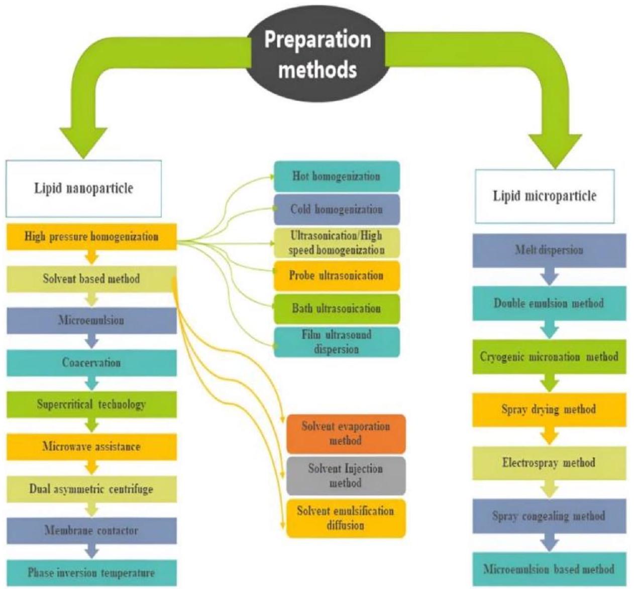
3 Lipid nanoparticles preparation

3.1 High-pressure homogenization (HPH)

This technique is both reliable and powerful, and it is specifically employed for the production of SLNs. High-pressure homogenizers propel a liquid at elevated pressures (100-2000 bar) through a narrow aperture, typically measuring only a
Table 1 Commercially available products marketed as SLNs [1,32]
S. No Drug name Company/Name Marketed product
1 Black currant seed oil and manuka oil Chemisches Laboratium Dr. Kurt Richter, CLR-Berlin, Germany NanoLipid Restore CLR
2 Coenzyme Q10, polypeptide, hibiscus extract, ginger extract, ketosugar Dr. Rimpler GmbH, Wedemark, Germany Cutanova Cream Nanorepair Q10
3 Coenzyme Q10, polypeptide, mafane extract Dr. Rimpler GmbH, Wedemark, Germany Intensive Serum Nanorepair Q10
4 Coenzyme Q10, , polypeptide, ursolic acid, oleanolic acid, sunflower seed extract Dr. Rimpler, GimH, Wedemark, Germany Cutanova Cream Nanovital Q10
5 , highly active oligosaccharides Beate Johnen GmbH, Aschheim, Germany NLC Deep Effect Repair Cream
6 Coenzyme Q10, -3 und -6 unsaturated fatty acids Amorepacific Corp. Seoul, South Korea Extra Moist Softener
7 M. Ternifolia seed oil, avocado, urea, black currant seed oil Scholl, Mannheium, Germany Regeneration crème intensive
8 Kukui nut oil, Monoi Tiki Tahiti, pseudopeptide, hydrolyzed wheat protein Lancray International S.A. Paris, France SURMER Crème
9 Coenzyme Q10, highly active oligosaccharides ACM Laboratoire Dermatologique NLC Deep Effect Eye Serum
10 M. Ternifolia seed oil, avocado, urea, black currant seed oil Dr. Hauschka WALA Heilmittei GmbH Regenerations crème intensive
11 Coconut oil, wilder extract, pseudo peptide, milk extract from coconut Summer’s Daughter Surmer Elixir de Beatue Nano vitalisant
12 Q10, polypetide, hibiscus extract, , ursolic acid, oleanolic acid, sunflower seed extract Dr. Rimpler, GimH Cutanova Cream Nanovital and Repair Q10
13 Kukui nut oil, Monoi Tiki Tahiti, pseudo peptide, hydolysed wheat protein Summer’s Daughter Surmer Crème Contour Des Yeux Nano Remodelante
Table 2 Some of the patents related to Solid Lipid Nanoparticles (SLNs)
Patent number Title Ref. no
O2010112749 Dermal composition for minoxidil delivery [33]
Terbinafine-loaded SLN incorporated into a gel with enhanced skin deposition and improved antifungal activity [34]
N611/MUM/2011 Mupirocin SLN incorporated into a Carbopol-based gel to enhance efficacy in skin infection treatment [35]
N3658/MUM/2014 SLN used as a carrier in the development of transdermal patches for diltiazem permeation [36]
Preparation of tablets from SLN suspensions [37]
N201711046022 Treatment of tuberculosis and diseases mediated by Helicobacter pylori via the peroral administration of SLN loaded with rifabutin and rifampicin [38]
S2011082214 Solid lipid nanoparticles of finasteride and preparation method [39]
Preparation method of catalase solid lipid nanoparticles preparation [40]
A200900215 Carrying solid lipid nanoparticles of curcumin and piperine and the preparation method [41]
CN101559038B Folic acid targeting silymarin solid lipid nanosphere preparation method [42]
N101972229B Lipid nano pellets for oral administration [43]
Use of solid lipid nanoparticles comprising cholesteryl propionate and/or cholesteryl butyrate [44]
N103784421B Formulation of UV absorbers by incorporation in solid lipid nanoparticles [45]
N105708803A Solid lipid nanoparticles [46]
P0167825 Solid lipid nanoparticle as a gene or drug carrier, formulation and method for preparing the same [47]
O06128888 Cyclosporin solid lipid nanoparticles with good storage physical stability and preparation method thereof [48]
S7147841 Ampelopsis grossedentata total flavonoid solid lipid nanoparticles and preparation method [49]
Resveratrol solid lipid nanoparticles and preparation method thereof
Fig. 1 Various techniques that can be employed to produce solid lipid nanoparticles [50-54]
few microns in width. The fluid undergoes rapid acceleration, transitioning from a short distance to a velocity exceeding . Intense shear stress and cavitation forces cause the particles to break down into sizes smaller than a micron. Typically, a lipid content of is utilized, although research has also explored lipid contents as high as . There are two main methods employed for HPH, namely hot homogenization and cold homogenization, both of which involve the process of mixing the drug with a large quantity of melted lipid [55].

3.1.1 Hot homogenization

Hot homogenization is performed at temperatures that exceed the lipid’s melting point, making it equivalent to emulsion homogenization. A pre-emulsion is made by mixing the drug-loaded lipid melt with the water-based emulsifier phase in a high-shear mixer at the same temperature. High-pressure Homogenization (HPH) of the pre-emulsion is conducted at temperatures that exceed the lipid’s melting point. The steps involved in the preparation are illustrated in Fig. 2. Typically, elevated temperatures lead to reduced particle sizes as a result of the decreased viscosity of the inner phase. Nevertheless, elevated temperatures amplify the rate at which the drug and the carrier deteriorate. Higher homogenization pressure or an increased number of cycles frequently lead to larger particle sizes as a result of the particles’ elevated kinetic energy [56].

3.1.2 Cold homogenization

Cold homogenization was proposed as a way to fix some problems with hot homogenization, such as the fact that high temperatures break down drugs, drugs mix with water during homogenization, and the nanoemulsion’s complicated crystallization process, which often leads to multiple changes and/or supercooled melts. This procedure involves cooling a drug that contains lipids, and then grinding the solid lipid into tiny particles. These microparticles are then dispersed in a cold surfactant solution, resulting in a pre-suspension. After that, the pre-suspension is mixed evenly at room temperature
Fig. 2 Steps in Solid lipid nanoparticles preparation by hot homogenization process
or lower, where gravity is strong enough to break up the lipid microparticles directly into solid lipid nanoparticles [57]. The details of the steps involved in this process are illustrated in Fig. 3.

3.1.3 Ultrasonication/high-speed homogenization

SLNs can also be prepared using ultrasonication or high-speed homogenization technique. In order to achieve a smaller particle size, a combination of ultrasonication and high-speed homogenization is necessary [58].

3.1.4 Bath ultrasonication

A bath sonicator is the preferred choice for processing large quantities of diluted lipid dispersions. Bath sonication involves immersing the sample container in a water bath equipped with an ultrasonic transducer. The transducer generates high-frequency sound waves, leading to the formation of cavitation bubbles in the liquid. The implosion of these bubbles generates intense localized energy, promoting emulsification and dispersion. The primary goal of bath sonication in SLN preparation is to reduce the particle size of the lipid dispersion. Smaller particle sizes contribute to increased
Fig. 3 Steps in Solid lipid nanoparticles preparation by cold homogenization process
surface area, improved drug loading, and enhanced bioavailability. During sonication, the energy generated helps break down larger lipid droplets into smaller ones, facilitating the formation of a stable emulsion. This emulsion serves as a precursor for the subsequent solidification of lipids into nanoparticles.

3.1.5 Probe ultrasonication

The probe sonicator is ideal for dispersing substances that necessitate a substantial amount of energy within a confined volume. Probe tip sonicators utilize energy to treat lipid dispersions, but excessive heat can cause lipid degradation. Before use, centrifugation should remove any metal particles that may have leaked from the sonication tips into the dispersion.

3.1.6 Film ultrasound dispersion

In this method, lipids and drug compounds are dissolved in organic solvents, and then the solvents are removed to create a lipid film. An aqueous solution with emulsions is then added. Small and uniform SLNs are formed using ultrasound and a diffuser probe [58].

3.2 Solvent-based method

The solvent-based technique is one of the methods employed in the production of SLNs. It includes dissolving both the lipid and, if desired, the drug in a solvent, and then forming SLNs by eliminating the solvent.

3.2.1 Solvent evaporation method

SLNs can also be prepared using the solvent evaporation method. The hydrophobic substance is dissolved in an organic solvent that is not soluble in water, such as cyclohexane, and then mixed with an aqueous solution to form an emulsion. When the solvent evaporates, the lipid precipitates in the aqueous medium, resulting in the formation of a nanoparticle dispersion with an average size of 25 nm [59]. The solution is dispersed in a water-based phase through the process of high-pressure homogenization. The emulsion is then subjected to evaporation under reduced pressure ( ) in order to remove the organic solvent [59].

3.2.2 Solvent injection method

This method involves dissolving the lipid and, optionally, the drug in an organic solvent, and then injecting this solution into an aqueous phase. The rapid diffusion of the solvent into the aqueous phase results in the precipitation of the lipid, forming solid lipid nanoparticles. This method allows for the formation of SLNs without the need for high shear forces and high temperatures, making it suitable for heat-sensitive compounds. An injection rate may be necessary to achieve the desired SLN characteristics for a particular application.

3.2.3 Solvent emulsification-diffusion method

This technique allows for particles with average diameters ranging from 30 to 100 nm . The primary benefit of this technique is the dissipation of heat during the preparation process [60]. A schematic representation of the steps involved in the solvent emulsification-diffusion method is provided in Fig. 4.

3.3 Microemulsion-based method

This approach relies on the process of diluting microemulsions. Micro-emulsions are biphasic systems consisting of an inner and outer phase, such as o/w microemulsions. Their production involves agitating a transparent blend at a temperature range of . This blend usually consists of a fatty acid with a low melting point (such as stearic acid), an emulsifier (such as polysorbate 20), co-emulsifiers (such as butanol), and water. The heated microemulsion is evenly distributed in chilled water ( ) while being agitated (Fig. 5). The spread of SLN can be used as a granulation fluid to turn into a solid product, like tablets or pellets, for the granulation process. However, when the particle concentration is
Fig. 4 Systematic representation of emulsification diffusion method
Fig. 5 Steps in Micro-emulsion method for the preparation of solid lipid nanoparticles. The data are downloadable at https://doi.org/10. 23893/1307-2080.APS. 05616 and accessible at Semantic Scholar website at https://www.semanticscholar.org/paper/Solid-lipid-nanopartic les%3A-a-promising-technology-Bhatt-Sharma/59b330fe3c8eb42506839c745e6fac039dde74e5
low, a significant amount of water must be eliminated. Significant temperature differences promote the quick formation of lipid crystals and prevent clumping. When a dilution step is added to a formulation, the amount of lipids that can be reached is much lower than when high-pressure homogenization is used [61, 62].

3.4 Coacervation

The coacervation method has been recently reported in the literature as a solvent-free technique for preparing SLNs. This technique entails the process of counteracting alkaline salts of fatty acids by using an acidic solution while stabilizing agents are present. This results in the formation of solid lipid micelles composed of fatty acids. The coacervation technique provides a practical and easily attainable method to overcome the limitations in the preparation process of SLNs. This method has the potential to revolutionize the production of SLNs, offering a more efficient and cost-effective way to prepare nanoparticles for drug delivery. So, the coacervation method is an exciting development in the field of nanoparticle technology and has the potential to play a significant role in enhancing drug delivery methods [63].

3.5 Supercritical technology

The supercritical fluid technique employs supercritical carbon dioxide to extract oil-in-water (o/w) emulsions. This method has been suggested for creating micro/nanoparticles with a high level of encapsulation effectiveness for various types
of medications, including ketoprofen, indomethacin, and camptothecin. The resulting substance is a desiccated powder with a limited range of particle sizes, improved ability to flow, and a reduced amount of remaining organic solvent. This enhances the creation of enhanced liquid or solid drug formulations. The method is ecologically sustainable and has the capacity to be expanded [64]. Steps involved in the preparation by supercritical technology in shown in Fig. 6.

3.6 Microwave assistance

Microwave irradiation is a highly efficient method for producing nanoemulsions. It enables the creation of sub- 50 nm cross-linked particles with good polydispersity in a single step, without requiring surfactants. Furthermore, microwaveassisted synthesis can generate smaller nanoparticles with reduced polydispersity compared to traditional heating methods. This is attributed to the precise localized heating and minimal temperature variances achieved by microwave heating. Additionally, microwave synthesis typically results in enhanced reaction rates and product yields due to rapid, uniform dielectric heating. In contrast, conventional heating initially warms the vessel walls, causing slower reactions and lower yields as the heat gradually spreads to the reaction mixture [65].

3.7 Dual asymmetric centrifuge

Dual centrifugation is a novel method employed for the production of liposomes and other lipid nanoparticles. For over ten years, this technology has been utilized and consistently improved to produce a range of lipid nanoparticles, such as emulsions, SLNs, polymersomes, and nanocrystals. In recent years, there have been significant advancements in equipment technology, leading to the increasing adoption of dual centrifugation as the preferred method for producing lipid nanoparticles and nanocrystals in small and medium batch sizes. This approach offers simplicity, speed, and sterility in the production process. Additionally, it provides the opportunity for efficient and rapid formulation screening or on-site preparation of therapeutic nanoparticles. Dual centrifugation is a distinctive procedure that entails placing a sample vial in a high-speed centrifuge (primary rotation) and then rotating it around a second axis (secondary rotation), Fig. 7. The centrifugal acceleration, characterized by its high magnitude, undergoes constant changes in direction with respect to the sample vial. As a consequence, the sample material experiences frequent and vigorous movements within the vial. Zirconia beads with high weights are commonly included to facilitate the process. The vigorous sample motions can be employed for the purposes of blending, agitating, grinding, or standardizing [66].

3.8 Membrane contractor

The membrane contactor method is also utilized to prepare SLNs. The process involves pressing the lipid through the membrane pores at a temperature exceeding the lipid’s melting point. Water is then circulated beyond the pores, resulting in the formation of droplets of melted lipid that are cooled at room temperature (Fig. 8). This method offers several benefits, including ease of use, the ability to control the size of the SLNs by selecting the appropriate process parameters, and scalability. Additionally, the membrane contractor technique is utilized to prepare polymeric nanoparticles using the interfacial polymerization method or the nanoprecipitation method, which involves the dispersion of preformed polymers [67].
Fig. 6 Steps involved in the preparation of solid lipid nanoparticles by supercritical technology
Fig. 7 Dual asymmetric centrifuge method for the preparation of solid lipid nanoparticles. The data are downloadable at https://doi. org/10.5772/intechopen. 68523
Fig. 8 Preparation of solid lipid nanoparticles by membrane contractor method. The data are downloadable at https://doi.org/10.22270/jddt. v9i2.2461

3.9 Phase inversion temperature

The PIT (Phase-inversion Temperature) method is a highly efficient, low-energy approach employed to control the solubility of polyethoxylated nonionic surfactants by altering the temperature. When exposed to elevated temperatures, the surfactant undergoes a change in its properties, becoming hydrophobic. This leads to the formation of reverse micelles that have negative curvatures and are swollen with water. The TPIT (specific temperature) is the temperature at which the surfactant exhibits equal attraction towards both oil and water phases, leading to a complete absence of spontaneous curvature and significantly reduced interfacial tension. Under the TPIT, nonionic surfactants that have absorbed water create small droplets that have a high level of solubility in water. By employing the PIT method, several studies have effectively generated SLNs utilizing stearic acid as the solid lipid component. Upon cooling, stearic acid undergoes solidification, resulting in the formation of a solid core that encases the solid surfactant shell, which consists of tween 60 and span 60, respectively. Notably, the PIT method has also been extensively employed for the preparation of nanoemulsions containing volatile oils. Prior research has indicated that the combination of volatile and fixed oils in an optimized proportion can result in the formation of small droplets and high-quality, stable nanoemulsions Fig. 9. The composition and concentration of surfactant are among several factors that influence the creation and characteristics of nanoemulsions.
Fig. 9 Preparation of solid lipid nanoparticles by phase inversion temperature method. The data are downloadable at https://doi.org/10. 1080/10837450.2022.2084554

4 Lipid microparticles preparation methods

4.1 Melt dispersion

The melting dispersion method is an approach that is very effective when it comes to the production of SLNs. The first step is to melt the drug and the solid lipid in an organic solvent, which serves as the oil phase. In order to achieve the same temperature, the oil phase and the water phase are both heated. The second step involves adding the oil phase to a portion of the water phase, and then vigorously stirring the resulting emulsion at a higher revolution per minute for a number of hours. Finally, in order to form the SLNs, the mixture is allowed to cool down to room temperature. In contrast to the method of solvent emulsification and evaporation, the melting dispersion method does not require the evaporation of any organic solvent. It is still more reliable than the ultrasonication method, despite the fact that the reproducibility of this method is lower than that of the solvent emulsification-evaporation method.

4.2 Double emulsion method

In this method, the drug is contained within a stabilizer that keeps it separate from the water phase on the outside, while the solvent evaporates in the water phase on the outside of the w/o/w double emulsion [68]. The method of preparation by double emulsion is illustrated in Fig. 10

4.3 Cryogenic micronization method

Cryogenic spray processes are highly effective methods for reducing the size of particles, which can greatly improve the rate at which drugs that are not easily soluble dissolve. They successfully produce nanostructured, amorphous drug particles with a high level of porosity at extremely low temperatures. Following cryogenic processes, a range of drying methods, such as spray freeze drying, atmospheric freeze drying, vacuum freeze drying, and lyophilization, can be employed to generate dry powders. Various cryogenic spray techniques can be utilized, including spray freezing onto cryogenic fluids, Spray Freezing into cryogenic Liquids, spray freezing into vapor over liquid, and ultra-rapid freezing for the production of smaller drug particles with enhanced wettability. Halocarbons, chlorofluorocarbons, and liquid nitrogen can be used as cryogenic media in the traditional process of spray freezing into vapor. A nozzle located above the refrigerant that is boiling transforms the feed solution into tiny droplets. The fragmented droplets subsequently descend into the refrigerant and promptly solidify upon coming into contact with the cryogen. The solidified substance is gathered and subjected to lyophilization in order to eliminate the solvent. Nevertheless, the utilization of chlorofluorocarbons
Fig. 10 Preparation of solid lipid microparticles by double emulsion method. The data are downloadable at https://doi.org/10.1016/j.jsps. 2012.05.009
as a cryogenic medium in this procedure is severely restricted due to their detrimental impact on the ozone layer. Even certain substitutes for chlorofluorocarbons, like hydrofluoroalkanes, have the ability to dissolve the API and reduce the effectiveness of the powdered formulation. The spray freezing into vapor process involves the atomization of liquid gas into nitrogen vapor. This can cause droplets to slowly stick together and harden, which can create dry powders that are not micronized and have a wide range of particle sizes [69].

4.4 Spray drying method

It is a substitute for the lyophilization process. This involves the utilization of lipids that have melting points exceeding . The most optimal outcomes are achieved using a concentration of SLN in a solution of trehalose in water or a trehalose solution in a mixture of ethanol and water [70].

4.5 Electrospray method

Electrospray is an effective technique that uses an electric field to convert liquids into fine particles. The control of particle size and shape can be optimized by adjusting variables, such as liquid flow rate and applied voltage. Electrospray is a simple, single-step method that allows for the management of several processing parameters (Fig. 11). Furthermore, electrospray outperforms other methods by preventing any adverse effects on biomolecules [71]

4.6 Spray congealing method

Spray congealing and fluid bed hot melt coating are popular techniques for cost-effective and solvent-free processing when lipid excipients are the principal components of the formulation [7, 8]. Spray congealing, also known as spray chilling or cooling, involves atomizing a molten slurry that typically contains an API in a melted carrier. This is done in a chamber where the temperature is maintained below the melting point of the carrier [1,9]. The droplets immediately solidify when they come in contact with cold air, resulting in the rapid formation of Solid Lipid Microparticles [1]. These
Fig. 11 Electrospray method for the preparation of solid lipid microparticles. Source: The document is downloadable with https://doi.org/10. 1016/B978-0-12-817923-9. 00005-5 from ScienceDirect
Fig. 12 Spray congealing method for the preparation of solid lipid microparticles. This document is downloadable at https://doi.org/10. 3390/molecules24193471
techniques are highly efficient and well-established in the industry (Fig. 12), making them a reliable choice for pharmaceutical manufacturers [72].

4.7 Microemulsion-based method

Emulsions are a highly effective precursor for the formation of solid lipid particles. Heating lipids that are solid at a normal temperature up to above their melting point can readily convert them into a liquid condition. Subsequently, this fluid lipid can be combined with water at an identical temperature, leading to the creation of a heated emulsion. Lowering the temperature of this emulsion to the ambient room temperature results in the solidification of the droplets, leading to the formation of solid lipid particles. If the interest is to generate solid lipid particles from heated emulsions with droplet sizes larger than , the melt dispersion technique, sometimes referred to as Selective Laser Melting, is an excellent choice. Preceding substances, such as oil-in-water (o/w) or multiple water-in-oil-in-water (w/o/w), can be utilized [73].
The glycerides are dissolved in an organic solvent, such as chloroform. The solution becomes emulsified in water after the organic solvent evaporates, which causes the lipid to precipitate and nanoparticles to form [74, 75].

5 Characterization parameters

5.1 Physicochemical characterization of solid lipid nanoparticles

The physicochemical parameters of SLNs and their characterization techniques are listed below.
(1) Visual size and distribution
i. Photon correlation spectroscopy
ii. Laser diffraction
iii. Dynamic light scattering
iv. Static laser spectroscopy
(2) Electrical surface potential
i. Zeta potential
ii. pH -sensitive probe [76]
(3) Shape and surface morphology
i. Transmission electron microscopy
ii. Scanning electron microscopy
iii. Atomic force microscopy
iv. Optical microscopy [77]
(4) Crystallinity
i. X-ray diffraction
(5) Surface hydrophobicity
i. Two-phase partition
ii. Contact angle measurement
(6) Density
i. Gas pycnometer
(7) Viscosity
i. Viscometer [78]
(8) Molecular weight
i. Gel permeation chromatography
ii. In vitro drug release
  1. Dialysis tubing
  2. Reverse dialysis
  3. Trans diffusion cell
    (9) Entrapment efficiency [79]

6 Characterization of solid lipid nanoparticles

6.1 Visual size distribution

6.1.1 Photon correlation spectroscopy (PCS)

PCS quantifies the fluctuations in light scattering by tracking the motion of particles. The measurement encompasses a size range spanning from a few nanometers to 3 microns. Various factors influence the relationship between the diffraction angle and the particle radius [73].

6.1.2 Laser diffraction (LD)

LD has the advantage of covering a broad spectrum, ranging from nanometers to smaller millimeters. The sensitivity of LD to small particles is enhanced by the increase in polarization intensity differential scattering [80].

6.1.3 Dynamic light scattering

It measures the variation in the strength of the dispersed light within a time interval of microseconds. The phenomenon of Brownian motion causes the scattering of light, which can be measured by calculating the autocorrelation function. This coefficient (Van de Ven et al., [82]). [81] is used to measure certain factors, such as the size, low concentration, and viscosity.

6.1.4 Static light scattering (SLS)

Fraunhofer diffraction is an alternative name for this technique. This method involves collecting the scattered light pattern from a solution of particles and incorporating it into an electromagnetic equation. The method is both challenging and efficient [82].

6.2 Electrical surface potential and pH

6.2.1 Zeta potential

The measurement can be performed using a zeta potential analyzer or zettameter. Zeta potential provides an insight into the strength of the electrostatic repulsion or attraction between particles. The SLN aqueous suspension contains particles [83].

6.2.2 pH-sensitive probes

A pH probe is a scientific instrument used to measure the concentration of hydrogen ions in aqueous solutions. It provides information about the acidity and alkalinity of substances based on their pH. The Nernst equation (Karmen et al., [85]) [84] is used to estimate the magnitude of electrochemical potential.

6.3 Shape and surface morphology

6.3.1 Transmission electron microscopy

An intense electron beam is directed towards the analyte, which consists of very fine particles, resulting in electron-electron interactions. The atoms can be utilized to observe characteristics, such as the crystal structure as well as specific features within the structure, including dislocations and grain boundaries [85]. Figure 13
Fig. 13 Transmission electron microscopy (TEM) of reconstituted SLN. This document is downloadable and accessible at website—https://doi.org/ 10.3390/molecules28041545
Fig. 14 Scanning electron microscopy picture of curcumin solidlipid nanoparticles. This document is downloadable and accessible at website https://doi.org/10. 15171/apb. 2016.04
Fig. 15 Photographs of atomic force microscopy (AFM) of docosanoic acid SLNs. This document is downloadable and accessible at https://doi.org/10.1038/ srep41104

6.3.2 Scanning electron microscopy

The process involves generating sample images through the use of a focused electron beam that is directed onto the surface. Electrons engage with samples and generate diverse signals that encompass details regarding the surface topography and composition of the sample [86]. SEM picture is shown below in Fig. 14.

6.3.3 Atomic force microscopy

This pertains to the presence of colloidal particles in opposition to current flow within the sample. The topological map tracks the movement between the tip and surface based on the exerted forces, Fig. 15. This methodology enables achieving an extremely high level of detail in the sample [87, 89].

6.3.4 Optical microscopy

Optical microscopy, also known as light microscopy, is a technique that uses visible light and a system of magnifying lenses to observe small objects. This particular microscope utilizes conventional, light-sensitive cameras to capture the image and produce a micrograph [88].

6.4 Crystallinity

6.4.1 X-ray diffraction

The process employed here is to quantify the extent of crystallinity, which is ascertained by analyzing the scattering of radiation from the crystal plane within a solid material. The X-ray diffraction technique can also be used to determine the level of crystallinity that a nanoparticle exhibits. This technique is valuable for evaluating the mass material Fig. 16 [89, 90].

6.5 Surface hydrophobicity

6.5.1 Two-phase partition

This technique divides the compounds into two distinct fluids that are typically capable of mixing, with one being polar and the other nonpolar, depending on their solubilities in relation to each other. The chemical potential facilitates the separation
Fig. 16 XRD of Berberine, PVA, SA and Berberine-SLN. This document is downloadable and accessible at website https://doi.org/10.36468/ pharmaceutical-sciences. 766
process. There are two primary categories: polymer/polymer and polymer/salt systems. pH, biomolecule concentration, polymer concentration, and the characteristics of the biomolecule’s surface all have an impact on the phase separation. They constitute delicate circumstances that do not harm susceptible biomolecules [90].

6.5.2 Contact angle measurement

The contact angle refers to the angle formed at the interface of two vapors or two liquids, representing the interaction between the solid and the liquid. It relies on Young’s equation. A goniometer is the instrument utilized for measuring the contact angle. The dynamic phenomenon of contact angle hysteresis exhibits a range of contact angles, varying from the maximum to the minimum. The contact angle plays a crucial role in determining the wetting capacity of the polymer membrane surface. Heterogeneity, surface roughness, and the size and shape of the particle are a few factors that affect the contact angle. Alternative instruments include radiolabel probes and synchrotron radiation X-rays [91, 92].

6.6 Density

6.6.1 Gas pycnometer

The gas pycnometer is a device utilized to determine the precise density of a solid substance through the application of Archimedes’ principle and Boyle’s law. Instead of using a fluid, an inert gas, typically helium, is employed. There are several benefits of this approach as it provides prompt and precise outcomes [93].

6.7 Viscosity

6.7.1 Viscometer

A viscometer is an instrument employed to measure the viscosity of a fluid. A rheometer is employed for fluids exhibiting variations in flow conditions. A viscometer is capable of determining viscosity only under a specific flow condition. Typically, either the object traverses the fluid or the liquid remains stationary, or the opposite occurs. In order for laminar flow to occur, the Reynolds number must have a small value. Various types of viscometers include the U-tube viscometer, falling sphere viscometer, Krebs viscometer, and quartz viscometer [94, 95].

6.8 Molecular weight

6.8.1 Gel permeation chromatography

Size-exclusive chromatography, also referred to as gel filtration chromatography, allows for the separation of analytes based on their size [96].

6.8.2 In vitro release

The membrane diffusion method is widely employed for the investigation of in vitro drug dissolution. Various techniques, such as the dialysis sac method, reverse dialysis sac method, and side-by-side dialysis method, are employed in numerous devices [97]. Separation techniques are employed to isolate the dispersed nanoparticles from the continuous phase. The quantity of the drug is also assessed. Due to the small dimensions of nanoparticulate systems, it is extremely challenging to separate them from the surrounding medium [98]. Continuous flow methods were initially developed to modify the release of oral dosages. The utilization of the United States Pharmacopeia apparatus 4 method offers numerous benefits in the restoration of the in vitro environment [99, 100].
6.8.2.1 Dialysis tubing Drug release in a laboratory setting (in vitro) can be accomplished by employing dialysis tubing. The solid lipid nanoparticle dispersion is enclosed within pre-rinsed dialysis tubing, which can be tightly sealed. Subsequently, the dialysis sac is subjected to dialysis against an appropriate dissolution medium at ambient temperature. At appropriate intervals, samples are extracted from the dissolution medium, subjected to centrifugation, and analyzed for drug content using a suitable analytical technique [101].
6.8.2.2 Reverse dialysis This technique involves the placement of multiple small dialysis sacs, each containing 1 ml of dissolution medium, into an SLN dispersion. Subsequently, the SLNs are transferred into the surrounding solution [102].
6.8.2.3 Franz diffusion cell For the purpose of determining the extent to which drugs are able to penetrate an artificial environment, Franz cells are an extremely reliable method. One of the benefits of these tests is that they require only a small amount of drug for analysis, limited tissue manipulation, and no ongoing sample collection. Additionally, there is no requirement for continuous sample collection. One of the components of the FDC system is a receiver compartment that holds five milliliters of PBS. Following the passage of the compound through the skin surrogate, it is then released into this compartment. It is possible to conduct a comprehensive analysis of the penetration kinetics over time by applying the infinite-dose experimental condition directly onto this surrogate while it is located in the donor compartment. A magnetic stirrer and a water bath that is controlled by a thermostat can be found in the Franz diffusion cell system. This water bath is capable of maintaining a temperature of precisely [103-105].

6.9 Entrapment efficiency

The drug loading percentage and entrapment efficiency are determined using the following formula [58]:
Drug loading Amount of drug found in the lyophilized NPs/Amount of lyophilized NPs
Preparation and characterization of solid lipid nanoparticles contribute to their efficacy, versatility, and suitability for a wide range of applications in drug delivery, cosmetics, imaging, and other fields. These contributions enable the development of advanced formulations with enhanced performance and therapeutic benefits.

7 Conclusion

The characterization and design of SLNs for drug delivery are highly promising and distinctive approaches in the pharmaceutical sciences. To make SLNs adapt to specific needs for drug delivery, it is important to have a deep understanding of their physicochemical properties, formulation factors, and optimization methods. Scientists have shown that advances in nanotechnology make it possible to precisely change the size, charge on the surface, and drug-carrying ability of SLNs. SLNs are a versatile platform for administering various therapeutic agents. Using solid lipids in nanocarriers makes them more biocompatible, which lowers the risk of toxicity that comes with other delivery methods. Also, the controlled release and uninterrupted delivery properties of SLNs help make therapies work better and reduce side effects, which makes patients more likely to follow their treatment plans. The improved adaptability of SLNs makes it possible for them to
directly target specific tissues or cells for drug delivery, which is not possible with other drug delivery systems. To effectively move SLNs from the laboratory to clinical use, it is essential to address issues concerning scalability, stability, and reproducibility, even though there have been notable advancements in this area. Continual research and development are essential for overcoming these challenges and fully harnessing the potential of SLNs for drug delivery.

8 Future prospective

The future prospects and potential advancements in drug delivery lie in the characterization and design of SLNs. Various potential future developments can be anticipated in this rapidly progressing field. Solid lipid nanocarriers have a lot of potential for drug delivery. This is because research is still going on, which should lead to new improvements in their functionality, specificity, and therapeutic uses. The ongoing advancement in these carriers requires the cooperation of researchers, physicians, and industry partners to bridge the gap between scientific breakthroughs and practical healthcare applications.
Acknowledgements The authors are thankful to the Kampala International University for providing necessary facilities to carry out the research work. I hereby declare that this submission is entirely my own work, in my own words, and that all sources used in researching it are fully acknowledged and all quotations properly identified.
Author contributions Conceptualization: S. P. N. B, V. C, S. B. R; Formal analysis and investigation: S. P. N. B, V. C, T. S. S, N. B, S. M; writing—original draft preparation: S. P. N. B, T. S. S, K. P. K, N. G, N. B; writing—review and editing: S. P. N. B, V. C, S. B. R, T. S. S, K. P. K, N. G, N. B, S. M. Critically revised: K. P. K, N. G, N. B; funding acquisition: none; resources: S. P. N. B. supervision: N. G.
Funding The author(s) received no specific funding for this work.
Data availability The datasets/information used for this study is available on reasonable request.

Declarations

Ethics approval and consent to participate Not applicable.
Consent for publication All the authors have read and agreed to the final copy of the finding as contained in the manuscript.
Competing interests All authors report that there was no competing interest in this work.
Open Access This article is licensed under a Creative Commons Attribution 4.0 International License, which permits use, sharing, adaptation, distribution and reproduction in any medium or format, as long as you give appropriate credit to the original author(s) and the source, provide a link to the Creative Commons licence, and indicate if changes were made. The images or other third party material in this article are included in the article’s Creative Commons licence, unless indicated otherwise in a credit line to the material. If material is not included in the article’s Creative Commons licence and your intended use is not permitted by statutory regulation or exceeds the permitted use, you will need to obtain permission directly from the copyright holder. To view a copy of this licence, visit http://creativecommons.org/licenses/by/4.0/.

References

  1. Ahmad J. Lipid nanoparticles based cosmetics with potential application in alleviating skin disorders. Cosmetics. 2021;8:84. https://doi. org/10.3390/cosmetics8030084.
  2. Shrestha H, Bala R, Arora S. Lipid-based drug delivery systems. J Pharm (Cairo). 2014;2014:801820. https://doi.org/10.1155/2014/801820.
  3. Williams HD, Trevaskis NL, Yeap YY, Anby MU, Pouton CW, Porter CJH. Lipid-based formulations and drug supersaturation: harnessing the unique benefits of the lipid digestion/absorption pathway. Pharm Res. 2013;30:2976-92. https://doi.org/10.1007/s11095-013-1126-0.
  4. Ravichandran R. Nanotechnology-based drug delivery systems. NanoBiotechnology. 2009;5:17-33. https://doi.org/10.1007/ s12030-009-9028-2.
  5. Jawahar N, Meyyanathan SN, Reddy G, Sood S. ChemInform abstract: solid lipid nanoparticles for oral delivery of poorly soluble drugs. ChemInform. 2013. https://doi.org/10.1002/chin.201327225.
  6. Park K. Facing the truth about nanotechnology in drug delivery. ACS Nano. 2013;7:7442-7. https://doi.org/10.1021/nn404501g.
  7. Sultana K, et al. Review of solid lipid nano particle. Int J Res Trend Innov. 2020;5(5):1 (ISSN: 2456-3315).
  8. Prabhakaran E, et al. Solid lipid nanoparticles. Sci Revs Chem Commun. 2012;2(1):80-102 (ISSN 2277-2669).
  9. Ramadon D. Solid lipid nanoparticles (SLN): formulation and fabrication. Pharm Sci Res. 2023. https://doi.org/10.7454/psr.v10i2.1313.
  10. Surender V, Deepika M. Solid lipid nanoparticles: a comprehensive review. J Chem Pharm Res. 2016;8(8):102-14.
  11. Manjunath K, Reddy JS, Venkateswarlu V. Solid lipid nanoparticles as drug delivery systems. Methods Find Exp Clin Pharmacol. 2005;27:127-44. https://doi.org/10.1358/mf.2005.27.2.876286.
  12. Yadav N, Khatak S, Sara U. Solid lipid nanoparticles-a review. Int J Appl Pharm. 2013;5:8-18.
  13. Luo WC, Lu X. Solid lipid nanoparticles for drug delivery. Methods Mol Biol. 2023;2622:139-46. https://doi.org/10.1007/978-1-0716-2954-3_12.
  14. Shirure PD, Pathan MA, Surwase PR, Kareppa MS. Review on solid lipid nanoparticles: as a promising approach for targeted drug delivery system. World J Pharm. 2019;8(3):433-50. https://doi.org/10.20959/wjpps20193-13273.
  15. Nazarova A, Yakimova L, Filimonova D, Stoikov I. Surfactant effect on the physicochemical characteristics of solid lipid nanoparticles based on Pillar[5]arenes. Int J Mol Sci. 2022;23:779. https://doi.org/10.3390/ijms23020779.
  16. Khatak S, Dureja H. Recent techniques and patents on solid lipid nanoparticles as novel carrier for drug delivery. Recent Pat Nanotechnol. 2015;9:150-77. https://doi.org/10.2174/1872210510999151126105754.
  17. Sarangi MK, Padhi S. Solid lipid nanoparticles—a review. J Crit Rev. 2016. https://doi.org/10.31838/jcr.03.01.02.
  18. Das S, Chaudhury A. Recent advances in lipid nanoparticle formulations with solid matrix for oral drug delivery. AAPS PharmSciTech. 2011;12:62-76. https://doi.org/10.1208/s12249-010-9563-0.
  19. Alsaad A, Hussien A, Gareeb M. Solid lipid nanoparticles (SLN) as a novel drug delivery system: a theoretical review. Syst Rev Pharm. 2020;11(5):259-73.https://doi.org/10.31838/srp.2020.5.39.
  20. Qushawy M, Nasr A. Solid lipid nanoparticles (SLNs) as nano drug delivery carriers: preparation, characterization and application. Int J App Pharm. 2019. https://doi.org/10.22159/ijap.2020v12i1.35312.
  21. Ghasemiyeh P, Mohammadi-Samani S. Solid lipid nanoparticles and nanostructured lipid carriers as novel drug delivery systems: applications, advantages and disadvantages. Res Pharm Sci. 2018;13:288-303. https://doi.org/10.4103/1735-5362.235156.
  22. El-Housiny S, Shams Eldeen MA, El-Attar YA, Salem HA, Attia D, Bendas ER, El-Nabarawi MA. Fluconazole-loaded solid lipid nanoparticles topical gel for treatment of pityriasis versicolor: formulation and clinical study. Drug Deliv. 2018;25:78-90. https://doi.org/10.1080/10717 544.2017.1413444.
  23. Mukherjee S, Ray S, Thakur RS. Solid lipid nanoparticles: a modern formulation approach in drug delivery system. Indian J Pharm Sci. 2009;71:349-58. https://doi.org/10.4103/0250-474X.57282.
  24. Singh VK, et al. Formulation and evaluation of topical gel of acelofenac containing piparine. Indo Am J Pharm Res. 2013. https://doi.org/ 10.1044/1980-iajpr.00404.
  25. Sanna V, Gavini E, Cossu M, Rassu G, Giunchedi P. Solid lipid nanoparticles (SLN) as carriers for the topical delivery of econazole nitrate: in-vitro characterization, ex-vivo and in-vivo studies. J Pharm Pharmacol. 2007;59:1057-64. https://doi.org/10.1211/jpp.59.8.0002.
  26. Khan AS, Shah KU, Mohaini MA, Alsalman AJ, Hawaj MAA, Alhashem YN, Ghazanfar S, Khan KA, Niazi ZR, Farid A. Tacrolimus-loaded solid lipid nanoparticle gel: formulation development and in vitro assessment for topical applications. Gels. 2022;8(2):129. https://doi.org/ 10.3390/gels8020129.
  27. Chandrakala V, Mamatha HS, Usha A, Priya B. Formulation and evaluation of solid lipid nanoparticles-based gel containing miconazole nitrate (an antifungal agent). Adv Concepts Pharm Res. 2024;5:108-19. https://doi.org/10.9734/bpi/acpr/v5/6727C.
  28. Arumugarajan AK, et al. Preparation and in vitro evaluation of etodolac extended release tablets prepared by wet granulation method employing kollidon SR. Indo Am J Pharm Sci. 2015;2(7):1133.
  29. Rostamkalaei SS, Akbari J, Saeedi M, Morteza-Semnani K, Nokhodchi A. Topical gel of Metformin solid lipid nanoparticles: a hopeful promise as a dermal delivery system. Colloids Surf B Biointerfaces. 2019;175:150-7. https://doi.org/10.1016/j.colsurfb.2018.11.072.
  30. Garse H, Jagtap P, Kadam V. Solid lipid nanoparticles based gel for topicals delivary of antifungal agents. Int J Pharm Sci Res. 2015;6(8):3571. https://doi.org/10.13040/IJPSR.0975-8232.6(8).3571-79.
  31. Dolatabadi JE, Valizadeh H, Hamishehkar H. Solid lipid nanoparticles as efficient drug and gene delivery systems: recent breakthroughs. Adv Pharm Bull. 2015;5:151-9. https://doi.org/10.15171/apb.2015.022.
  32. Purohit DK. Nano-lipid carriers for topical application: current scenario. Asian J Pharm (AJP). 2016. https://doi.org/10.22377/ajp.v10i1. 544.
  33. Padois K, Pirot F, Falson F. Solid lipid nanoparticles encapsulating minoxidil and aqueous suspension containing same. Patent WO2010112749. 2010.
  34. Vavia PR, Wavikar PR. Solid lipid nanoparticles based formulation of antifungal agent and preparation method thereof. Patent IN611/ MUM/2011. 2011.
  35. Mandal PA. Topical gel containing solid lipid nanoparticles. Patent IN3658/MUM/2014. 2014.
  36. Gupta GD. Development of transdermal matrix type patch of diltiazem hydrochloride using solid lipid nanoparticles for arrhythmia. Patent IN201711046022. 2017.
  37. Faure A, Voorspoels JF, Mertens RJ, Kiekens FR, inventors; Gruenenthal GmbH, assignee. Process for the preparation of a solid dosage form, in particular a tablet, for pharmaceutical use and process for the preparation of a precursor for a solid dosage form, in particular a tablet. Patent US2011082214, 2011.
  38. Babii VVE, Ignatiev AV, Gelperina SE, Maksi menko OO, Vachugova LV, Shipulo EV. Pharmaceu- tical composition for treatment tuberculosis and diseases caused by Helicobacter pylori based on solid lipid nano- particles and method for tuberculosis treatment. Patent EA200900215. 2009.
  39. Jun T, Dangguo W. Haitao G, et al. Solid lipid nanoparticles of finasteride and preparation method thereof. CN101559038B. 2011.
  40. Qi C, Chen Y, Jing Q-Z, Wang X-G. Preparation and characterization of catalase-loaded solid lipid nanoparticles protecting enzyme against proteolysis. Int J Mol Sci. 2011;12:4282-93. https://doi.org/10.3390/ijms12074282.
  41. Jingling T, Jinmei R, Linhua W, Hongyu J, Mengting L, Chao C. Curcumin and piperine carried solid lipid nanoparticles and preparation method thereof. CN103784421A. 2014.
  42. Liantian Y, Zhao L, Yang Y, Jin CS, Weitong S, Tong DYZ. Folic acid targeting silymarin solid lipid nanosphere preparation method. CN105708803A. 2014.
  43. Speiser P. Lipid nano pellets as drug carriers for oral administration. EP0167825A3. 1985.
  44. Gasco MR. Use of solid lipid nanoparticles comprising cholesteryl propionate and/or cholesteryl butyrate. WO2006128888A1. 2006.
  45. Herzog B. Formulation of UV absorbers by incorporation in solid lipid nanoparticles. EP1378231A1. 2003.
  46. Weiss J, Schweiggert C, Leuenberger B, Novotny M, Tedeschi C, Kessler A. Solid lipid nanoparticles. US9616001B2. 2014.
  47. V Shastri, E Sussman, A Jayagopal. Functionalized solid lipid nanoparticles and methods of making and using same. US20060083781A1. 2005.
  48. Penkler LJ, Müller RH, Runge SA, Ravelli V. Pharmaceutical cyclosporin formulation with improved biopharmaceutical properties, improved physical quality and greater stability, and method for producing said formulation. EP1073426B1. 1999.
  49. Zhicheng W, Kexin Z, Bing W, Feng C, Jianlin R, Tong Z, Qi Z, Shiyu Z. Resveratrol solid lipid nano-particles and preparation method thereof. CN104688715A. 2015.
  50. Schwarz J, Weisspapir M. Colloidal solid lipid vehicle for pharmaceutical use. US20060222716A1. 2006.
  51. Lin C-H, Chen C-H, Lin Z-C, Fang J-Y. Recent advances in oral delivery of drugs and bioactive natural products using solid lipid nanoparticles as the carriers. J Food Drug Anal. 2017;25:219-34. https://doi.org/10.1016/j.jfda.2017.02.001.
  52. Yadav N, Khatak S, Sara US. Solid Lipid Nanoparticles- A Review. International Journal of Applied Pharmaceutics. 2013;5(2):8-18.
  53. Battaglia L, Ugazio E. Lipid nano- and microparticles: an overview of patent-related research. J Nanomater. 2019. https://doi.org/ 10.1155/2019/2834941.
  54. Mirchandani Y, Patravale VB, Brijesh S. Solid lipid nanoparticles for hydrophilic drugs. J Controll Release. 2021;335:457-64. https:// doi.org/10.1016/j.jconrel.2021.05.032.
  55. Duan Y, Dhar A, Patel C, Khimani M, Neogi S, Sharma P, Siva Kumar N, Vekariya RL. A brief review on solid lipid nanoparticles: part and parcel of contemporary drug delivery systems. RSC Adv. 2020;10:26777-91. https://doi.org/10.1039/d0ra03491f.
  56. Shirodkar RK, Kumar L, Mutalik S, Lewis S. Solid lipid nanoparticles and nanostructured lipid carriers: emerging lipid based drug delivery systems. Pharm Chem J. 2019;53:440-53. https://doi.org/10.1007/s11094-019-02017-9.
  57. Uner M. Preparation, characterization and physico-chemical properties of solid lipid nanoparticles (SLN) and nanostructured lipid carriers (NLC): their benefits as colloidal drug carrier systems. Pharmazie. 2006;61:375-86.
  58. Sun S-B, Liu P, Shao F-M, Miao Q-L. Formulation and evaluation of PLGA nanoparticles loaded capecitabine for prostate cancer. Int J Clin Exp Med. 2015;8:19670-81.
  59. Duong V-A, Nguyen T-T-L, Maeng H-J. Preparation of solid lipid nanoparticles and nanostructured lipid carriers for drug delivery and the effects of preparation parameters of solvent injection method. Molecules. 2020;25:4781. https://doi.org/10.3390/molecules2 5204781.
  60. Amoabediny G, Haghiralsadat F, Naderinezhad S, Helder MN, Akhoundi Kharanaghi E, Mohammadnejad Arough J, Zandieh-Doulabi B. Overview of preparation methods of polymeric and lipid-based (niosome, solid lipid, liposome) nanoparticles: a comprehensive review. Int J Polym Mater Polym Biomater. 2018;67:383-400. https://doi.org/10.1080/00914037.2017.1332623.
  61. Harde H, Das M, Jain S. Solid lipid nanoparticles: an oral bioavailability enhancer vehicle. Expert Opin Drug Deliv. 2011;8:1407-24. https:// doi.org/10.1517/17425247.2011.604311.
  62. Geszke-Moritz M, Moritz M. Solid lipid nanoparticles as attractive drug vehicles: composition, properties and therapeutic strategies. Mater Sci Eng C Mater Biol Appl. 2016;68:982-94. https://doi.org/10.1016/j.msec.2016.05.119.
  63. Hao J, Wang F, Wang X, Zhang D, Bi Y, Gao Y, Zhao X, Zhang Q. Development and optimization of baicalin-loaded solid lipid nanoparticles prepared by coacervation method using central composite design. Eur J Pharm Sci. 2012;47:497-505. https://doi.org/10.1016/j.ejps. 2012.07.006.
  64. Andrade LN, Oliveira DML, Chaud MV, Alves TFR, Nery M, da Silva CF, Gonsalves JKC, Nunes RS, Corrêa CB, Amaral RG, Sanchez-Lopez E, Souto EB, Severino P. Praziquantel-solid lipid nanoparticles produced by supercritical carbon dioxide extraction: physicochemical characterization, release profile, and cytotoxicity. Molecules. 2019;24:3881. https://doi.org/10.3390/molecules24213881.
  65. Dunn SS, Beckford Vera DR, Benhabbour SR, Parrott MC. Rapid microwave-assisted synthesis of sub- 30 nm lipid nanoparticles. J Colloid Interface Sci. 2017;488:240-5. https://doi.org/10.1016/j.jcis.2016.10.093.
  66. Koehler JK, Schmager S, Bender V, Steiner D, Massing U. Preparation of nanosized pharmaceutical formulations by dual centrifugation. Pharmaceuticals. 2023;16:1519. https://doi.org/10.3390/ph16111519.
  67. Bagul US, Pisal VV, Solanki NV, Karnavat A. Current status of solid lipid nanoparticles: a review. Mod Appl Bioequivalence Bioavailab. 2018;3(4):1-10.
  68. Ram D, Debnath S, Babu M, Nath T, Thejeswi B. A review on solid lipid nanoparticles. Res J Pharm Technol. 2012;5:1359-68.
  69. Khadka P, Ro J, Kim H, Kim I, Kim JT, Kim H, Cho JM, Yun G, Lee J. Pharmaceutical particle technologies: an approach to improve drug solubility, dissolution and bioavailability. Asian J Pharm Sci. 2014;9:304-16. https://doi.org/10.1016/j.ajps.2014.05.005.
  70. Sawant KK, Dodiya SS. Recent advances and patents on solid lipid nanoparticles. Recent Pat Drug Deliv Formul. 2008;2:120-35. https:// doi.org/10.2174/187221108784534081.
  71. Shanaghi E, Aghajani M, Esmaeli F, Faramarzi MA, Jahandar H, Amani A. Application of electrospray in preparing solid lipid nanoparticles and optimization of nanoparticles using artificial neural networks. Avicenna J Med Biotechnol. 2020;12:251-4.
  72. Candiani A, Milanesi A, Foglio Bonda A, Diana G, Bari E, Segale L, Torre ML, Giovannelli L. Solid lipid microparticles by spray congealing of water/oil emulsion: an effective/versatile loading strategy for a highly soluble drug. Pharmaceutics. 2022;14:2805. https://doi.org/ 10.3390/pharmaceutics14122805.
  73. Khairnar SV, Pagare P, Thakre A, Nambiar AR, Junnuthula V, Abraham MC, Kolimi P, Nyavanandi D, Dyawanapelly S. Review on the scaleup methods for the preparation of solid lipid nanoparticles. Pharmaceutics. 2022;14:1886. https://doi.org/10.3390/pharmaceutics14 091886.
  74. Bondì ML, Craparo EF. Solid lipid nanoparticles for applications in gene therapy: a review of the state of the art. Expert Opin Drug Deliv. 2010;7:7-18. https://doi.org/10.1517/17425240903362410.
  75. Shahgaldian P, Da Silva E, Coleman AW, Rather B, Zaworotko MJ. Para-acyl-calix-arene based solid lipid nanoparticles (SLNs): a detailed study of preparation and stability parameters. Int J Pharm. 2003;253:23-38. https://doi.org/10.1016/s0378-5173(02)00639-7.
  76. Schubert MA, Müller-Goymann CC. Solvent injection as a new approach for manufacturing lipid nanoparticles-evaluation of the method and process parameters. Eur J Pharm Biopharm. 2003;55:125-31. https://doi.org/10.1016/s0939-6411(02)00130-3.
  77. Bose S, Michniak-Kohn B. Preparation and characterization of lipid based nanosystems for topical delivery of quercetin. Eur J Pharm Sci. 2013;48:442-52. https://doi.org/10.1016/j.ejps.2012.12.005.
  78. Sastri T, Gadela R, Pidikiti S, Vajjhala P. Solid lipid nanoparticles: Preparation techniques, their characterization, and an update on recent studies. J Appl Pharm Sci. 2020;10:126-41. https://doi.org/10.7324/JAPS.2020.10617.
  79. Balka SR, Sundari PT. Formulation and evaluation of solid lipid nanoparticles of etoricoxib by employing glyceryl monostearate and gelucire. 48/16. 2019) https://doi.org/10.5281/ZENODO.2583501.
  80. Montasser I, Shahgaldian P, Perret F, Coleman AW. Solid lipid nanoparticle-based calix[n]arenes and calix-resorcinarenes as building blocks: synthesis, formulation and characterization. Int J Mol Sci. 2013;14:21899-942. https://doi.org/10.3390/ijms141121899.
  81. Kumar R, Singh A, Sharma K, Dhasmana D, Garg N, Siril PF. Preparation, characterization and in vitro cytotoxicity of fenofibrate and nabumetone loaded solid lipid nanoparticles. Mater Sci Eng C Mater Biol Appl. 2020;106:110184. https://doi.org/10.1016/j.msec.2019. 110184.
  82. Van de Ven H, Vermeersch M, Shunmugaperumal T, Vandervoort J, Maes L, Ludwig A. Solid lipid nanoparticle (SLN) formulations as a potential tool for the reduction of cytotoxicity of saponins. Pharmazie. 2009;64:172-6.
  83. Garud A, Singh D, Garud N. Solid lipid nanoparticles (SLN): method, characterization and applications. Int Curr Pharm J. 2012;1:384-93. https://doi.org/10.3329/icpj.v1i11.12065.
  84. Mishra DK, Dhote V, Bhatnagar P, Mishra PK. Engineering solid lipid nanoparticles for improved drug delivery: promises and challenges of translational research. Drug Deliv Transl Res. 2012;2:238-53. https://doi.org/10.1007/s13346-012-0088-9.
  85. Plajnšek KT, Pajk S, Govedarica B, Pečar S, Srčič S, Kristl J. A novel fluorescent probe for more effective monitoring of nanosized drug delivery systems within the cells. Int J Pharm. 2011;416:384-93. https://doi.org/10.1016/j.ijpharm.2011.06.046.
  86. Jores K, Mehnert W, Drechsler M, Bunjes H, Johann C, Mäder K. Investigations on the structure of solid lipid nanoparticles (SLN) and oil-loaded solid lipid nanoparticles by photon correlation spectroscopy, field-flow fractionation and transmission electron microscopy. J Control Release. 2004;95:217-27. https://doi.org/10.1016/j.jconrel.2003.11.012.
  87. Daghighpoor Z. Back pain and herniated disc linked to depression. Am J Ethnomed. 2017. https://doi.org/10.21767/2348-9502-C1-003.
  88. Carrillo C, Sánchez-Hernández N, García-Montoya E, Pérez-Lozano P, Suñé-Negre JM, Ticó JR, Suñé C, Miñarro M. DNA delivery via cationic solid lipid nanoparticles (SLNs). Eur J Pharm Sci. 2013;49:157-65. https://doi.org/10.1016/j.ejps.2013.02.011.
  89. Sharifi M, Attar F, Saboury AA, Akhtari K, Hooshmand N, Hasan A, El-Sayed MA, Falahati M. Plasmonic gold nanoparticles: optical manipulation, imaging, drug delivery and therapy. J Control Release. 2019;311-312:170-89. https://doi.org/10.1016/j.jconrel.2019.08.032.
  90. Noack A, Hause G, Mäder K. Physicochemical characterization of curcuminoid-loaded solid lipid nanoparticles. Int J Pharm. 2012;423:44051. https://doi.org/10.1016/j.ijpharm.2011.12.011.
  91. Paumelle R, Blanquart C, Briand O, Barbier O, Duhem C, Woerly G, Percevault F, Fruchart JC, Dombrowicz D, Glineur C, Staels B. Acute antiinflammatory properties of statins involve peroxisome proliferator-activated receptor-alpha via inhibition of the protein kinase C signaling pathway. Circ Res. 2006;98(3):361-9. https://doi.org/10.1161/01.RES.0000202706.70992.95.
  92. Milsmann J, Oehlke K, Greiner R, Steffen-Heins A. Fate of edible solid lipid nanoparticles (SLN) in surfactant stabilized o/w emulsions. Part 2: Release and partitioning behavior of lipophilic probes from SLN into different phases of o/w emulsions. Colloids Surf A Physicochem Eng Asp. 2018;558:623-31. https://doi.org/10.1016/j.colsurfa.2017.05.050.
  93. Tang K, Lv X, Wu S, Xuan S, Huang X, Bai C. Measurement for contact angle of iron ore particles and water. ISIJ Int. 2018;58:379-400. https://doi.org/10.2355/isijinternational.ISIJINT-2017-424.
  94. Poumellec M-A, Dejode M, Figl A, Darcourt J, Haudebourg J, Sabah Y, Voury A, Martaens A, Barranger E. Sentinel node detection using optonuclear probe (gamma and fluorescence) after green indocyanine and radio-isotope injections. Gynecol Obstet Fertil. 2016;44:20710. https://doi.org/10.1016/j.gyobfe.2016.02.012.
  95. Patravale VB, Mirani AG. Preparation and characterization of solid lipid nanoparticles-based gel for topical delivery. Methods Mol Biol. 2019;2000:293-302. https://doi.org/10.1007/978-1-4939-9516-5_20.
  96. Abdelbary G, Fahmy RH. Diazepam-loaded solid lipid nanoparticles: design and characterization. AAPS PharmSciTech. 2009;10:211-9. https://doi.org/10.1208/s12249-009-9197-2.
  97. Ramteke KH, Joshi SA, Dhole SN. Solid lipid nanoparticle: a review. IOSR J Pharm. 2012;2:34-44. https://doi.org/10.9790/3013-26103444.
  98. Narala A, Veerabrahma K. Preparation, characterization and evaluation of quetiapine fumarate solid lipid nanoparticles to improve the oral bioavailability. J Pharm (Cairo). 2013;2013:265741. https://doi.org/10.1155/2013/265741.
  99. Zeng J, Pang X, Zhang L, Medina A, Rozelle S. Gender inequality in education in China: a meta-regression analysis. Contemp Econ Policy. 2014;32(2):474-91. https://doi.org/10.1111/coep.12006.
  100. Mehnert W, Mäder K. Solid lipid nanoparticles: production, characterization and applications. Adv Drug Deliv Rev. 2001;47:165-96. https://doi.org/10.1016/s0169-409x(01)00105-3.
  101. Aljaeid B, Hosny KM. Miconazole-loaded solid lipid nanoparticles: formulation and evaluation of a novel formula with high bioavailability and antifungal activity. IJN. 2016. https://doi.org/10.2147/IJN.S100625.
  102. Venishetty VK, Chede R, Komuravelli R, Adepu L, Sistla R, Diwan PV. Design and evaluation of polymer coated carvedilol loaded solid lipid nanoparticles to improve the oral bioavailability: a novel strategy to avoid intraduodenal administration. Colloids Surf B Biointerfaces. 2012;95:1-9. https://doi.org/10.1016/j.colsurfb.2012.01.001.
  103. Wissing SA, Müller RH, Manthei L, Mayer C. Structural characterization of Q10-loaded solid lipid nanoparticles by NMR spectroscopy. Pharm Res. 2004;21:400-5. https://doi.org/10.1023/B:PHAM.0000019291.36636.c1.
  104. Güney G, Kutlu HM, Genç L. Preparation and characterization of ascorbic acid loaded solid lipid nanoparticles and investigation of their apoptotic effects. Colloids Surf B Biointerfaces. 2014;121:270-80. https://doi.org/10.1016/j.colsurfb.2014.05.008.
  105. Pulsoni I, Lubda M, Aiello M, Fedi A, Marzagalli M, Von Hagen J, Scaglione S. Comparison between franz diffusion cell and a novel microphysiological system for in vitro penetration assay using different skin models. SLAS Technol. 2022;27:161-71. https://doi.org/10.1016/j. slast.2021.12.006.
Publisher’s Note Springer Nature remains neutral with regard to jurisdictional claims in published maps and institutional affiliations.

  1. Sarad Pawar Naik Bukke, drsaradpawar@kiu.ac.ug; Chandrakala Venkatesh, chandrakala.epcp@eastpoint.ac.in Department of Pharmaceutics and Pharmaceutical Technology, Kampala International University, Western Campus, P.O. Box 71, Ishaka-Bushenyi, Uganda. Department of Pharmaceutics, East Paint College of Pharmacy, Bidarahalli, Bangalore 560049, Karnataka, India. Department of Pharmaceutics, SRM College of Pharmacy, SRM Institute of Science and Technology, Kattankulathur, Tamilnadu 603203, India. Department of Pharmaceutical Chemistry, School of Pharmaceutical Sciences, Delhi Pharmaceutical Sciences and Research University, New Delhi, India. Department of Clinical Pharmacy and Pharmacy Practice, Kampala International University, Western Campus, P.O. Box 71, Ishaka-Bushenyi, Uganda.