سجل المريض الطبي والعلاجات القائمة على mRNA باستخدام الإبر الدقيقة تحت الجلد On-patient medical record and mRNA therapeutics using intradermal microneedles
تتطلب التدخلات الطبية غالبًا جرعات مؤقتة، مما يستلزم دقة في تسجيل السجلات الطبية. في العديد من البيئات العالمية، تكون هذه السجلات غير موثوقة أو غير متاحة في نقطة الرعاية، مما يؤدي إلى علاجات أقل فعالية أو وقاية من الأمراض. هنا نقدم تقنية تسجيل السجلات الطبية على المريض غير المرئية للعين المجردة التي تخزن بدقة المعلومات الطبية في جلد المريض كجزء من الإبر الدقيقة المستخدمة للعلاجات تحت الجلد. نحن نقوم بتحسين تصميم الإبر الدقيقة لضمان توصيل موثوق للعلاجات القائمة على RNA الرسول (mRNA) والميكروكريات الفلورية القريبة من الأشعة تحت الحمراء التي تشفر تسجيل السجلات الطبية على المريض. تمكّن معالجة الصور المعتمدة على التعلم العميق من تشفير وفك تشفير المعلومات بدقة زمنية ومكانية ممتازة. تظهر الدراسات طويلة الأمد في نموذج الخنازير سلامة وفعالية وموثوقية هذا النهج لتوصيل السجلات الطبية على المريض ولقاح mRNA الذي يشفر فيروس كورونا المسبب لمتلازمة التنفس الحادة الوخيمة 2 (SARS-CoV-2). يمكن أن تساعد هذه التقنية العاملين في مجال الرعاية الصحية في اتخاذ قرارات مستنيرة في الظروف التي تكون فيها سجلات موثوقة غير متاحة، مما يساهم في تحقيق العدالة في الرعاية الصحية العالمية.
تتطلب التدخلات الطبية غالبًا جرعات مؤقتة محددة، مما يستلزم دقة في تسجيل السجلات الطبية. على وجه الخصوص، أثبتت أنظمة توصيل القائمة على mRNA أنها منصة متعددة الاستخدامات لتطوير اللقاحات والعلاجات ضد الأمراض المستعصية , وغالبًا ما تتطلب جرعات متعددة. على سبيل المثال، أثبتت التجارب السريرية لـ RNAactive CV7201 لداء الكلب (CureVac) , RNActive CV9103 لسرطان البروستاتا (CureVac) و mRNA-4157-P201 للورم الميلانيني (موديرنا) تطلبت 3 و 5 و 9 إدارات جرعات، على التوالي. العديد من العلاجات الأخرى
ومعظم التطعيمات الموصى بها للأطفال تتطلب جرعات متعددة (على سبيل المثال، لقاحات التهاب الكبد B وشلل الأطفال) أيضًا . إن الفشل في إكمال هذه الجرعات المؤقتة يعرض الحماية للخطر. حاليًا، يفشل ما يصل إلى من المرضى في الالتزام بالعلاجات الطبية حول العالم، ويكون ضعف الالتزام مسؤولاً عن 125,000 وفاة سنوية في الولايات المتحدة وحدها . في أفريقيا جنوب الصحراء الكبرى، من الأطفال بين 12 و 23 شهرًا يفشلون في إكمال التطعيمات الموصى بها للأطفال . تساهم عدة عوامل في هذه الفجوات،
بما في ذلك عدم الوصول أو عدم القدرة على تحمل تكاليف العلاجات الطبية واللقاحات، ولكن أحد المساهمين الرئيسيين هو أنظمة تسجيل السجلات الطبية غير الكافية . تقدم الطرق التقليدية مثل بطاقات الورق وقواعد البيانات عبر الإنترنت مخاطر خطيرة لفقدان الوصول إلى السجلات الطبية، وبالتالي خطر فقدان أو تأخير الجرعات المتابعة. لهذا السبب، ظهرت مجموعة من وسائل تسجيل السجلات الطبية بناءً على مسح بصمات الأصابع , تطبيقات الهواتف المحمولة , الشرائح الدقيقة وأكثر. ومع ذلك، أثارت هذه الأساليب مخاوف تتعلق بالخصوصية المتعلقة بتخزين البيانات الطبية القابلة للتحديد في قاعدة بيانات مركزية، مما يشكل خطرًا على خرق البيانات أو سوء الاستخدام أو تداعيات الجودة .
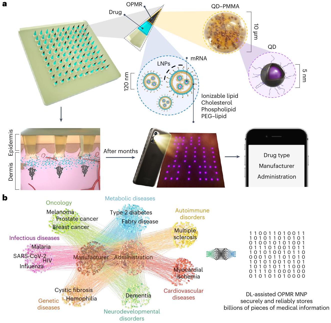
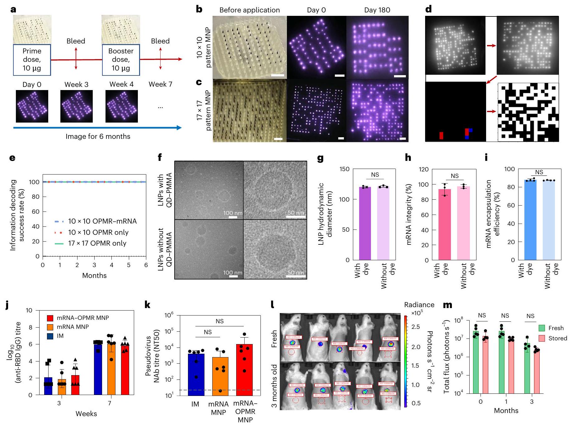
طورنا تقنية قوية لتسجيل السجلات الطبية على المريض (OPMR) باستخدام لاصقة إبر دقيقة قابلة للذوبان (MNP) تقوم بتوصيل صبغة فلورية قريبة من الأشعة تحت الحمراء (NIR) قائمة على النقاط الكمومية (QD) محاطة بجزيئات بولي (ميثيل ميثاكريلات) (PMMA) إلى الجلد لتشفير المعلومات الطبية (الشكل 1أ). هذه الصبغة، بمجرد إيداعها في الأدمة، تكون غير مرئية للعين المجردة، مما يوفر خصوصية وسرية بيانات المريض، ولكنها تقدم إشارات NIR منفصلة يمكن اكتشافها باستخدام نظام تصوير NIR (الشكل الممتد 1أ-ج). من خلال إيداع الصبغة في نمط محدد مسبقًا يتوافق مع مجموعة معينة من المعلومات، يمكن تصوير التقنية من قبل العاملين في مجال الرعاية الصحية لدعم قرارات الجرعة التالية دون الحاجة إلى الاتصال بالإنترنت أو استخدام قواعد بيانات مركزية.
هنا، لتمكين OPMR بسعة معلومات ممتازة وأمان وموثوقية، قمنا بتصميم هيكل MNP وإدارته لنقل البيانات بشكل متسق وأمثل وطول العمر؛ حققنا سعة معلومات بمليارات الأنماط المشفرة باستخدام رمز تصحيح الأخطاء؛ وطورنا نظام استرجاع معلومات موثوق زمنيًا ومكانيًا باستخدام التعلم الآلي. علاوة على ذلك، قمنا بنجاح بتوصيل OPMR مع لقاح mRNA قوي محاط بجزيئات دهنية نانوية (LNPs) التي تشفر بروتين السنبلة لفيروس SARS-CoV-2 (الشكل 1أ). هذا يوضح أن تقنية OPMR-mRNA MNP لدينا يمكن أن توصل العلاجات القائمة على mRNA والمعلومات الطبية المقابلة في وقت واحد. نطاق تطبيقها قابل للتوسع بشكل محتمل ليشمل أي علاجات قائمة على mRNA، نظرًا لتوافقها الحيوي مع mRNA-LNPs وسعتها الكبيرة للتشفير في النطاق من إلى التي تستوعب العدد المتزايد بسرعة من العلاجات القائمة على mRNA قيد التطوير (الشكل 1ب والشكل الممتد 1د). يمكن أن تساعد هذه الأداة العاملين في مجال الرعاية الصحية في اتخاذ قرارات مستنيرة بشأن الجرعات المتابعة في الميدان حيث تكون سجلات موثوقة غير متاحة، وبالتالي تحسين الالتزام الطبي وإكمال التطعيم للسكان العالميين.
مواد وهيكل OPMR MNP
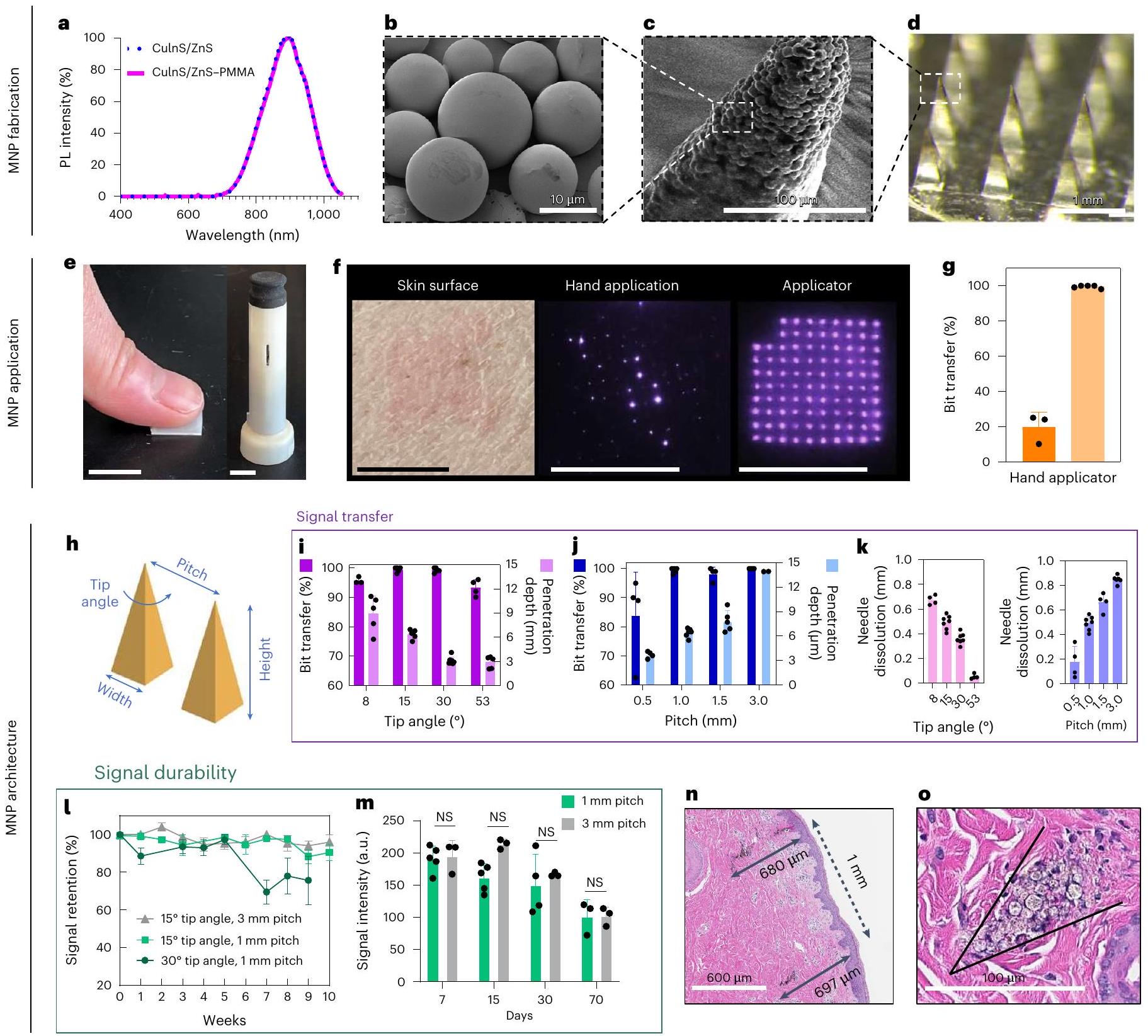
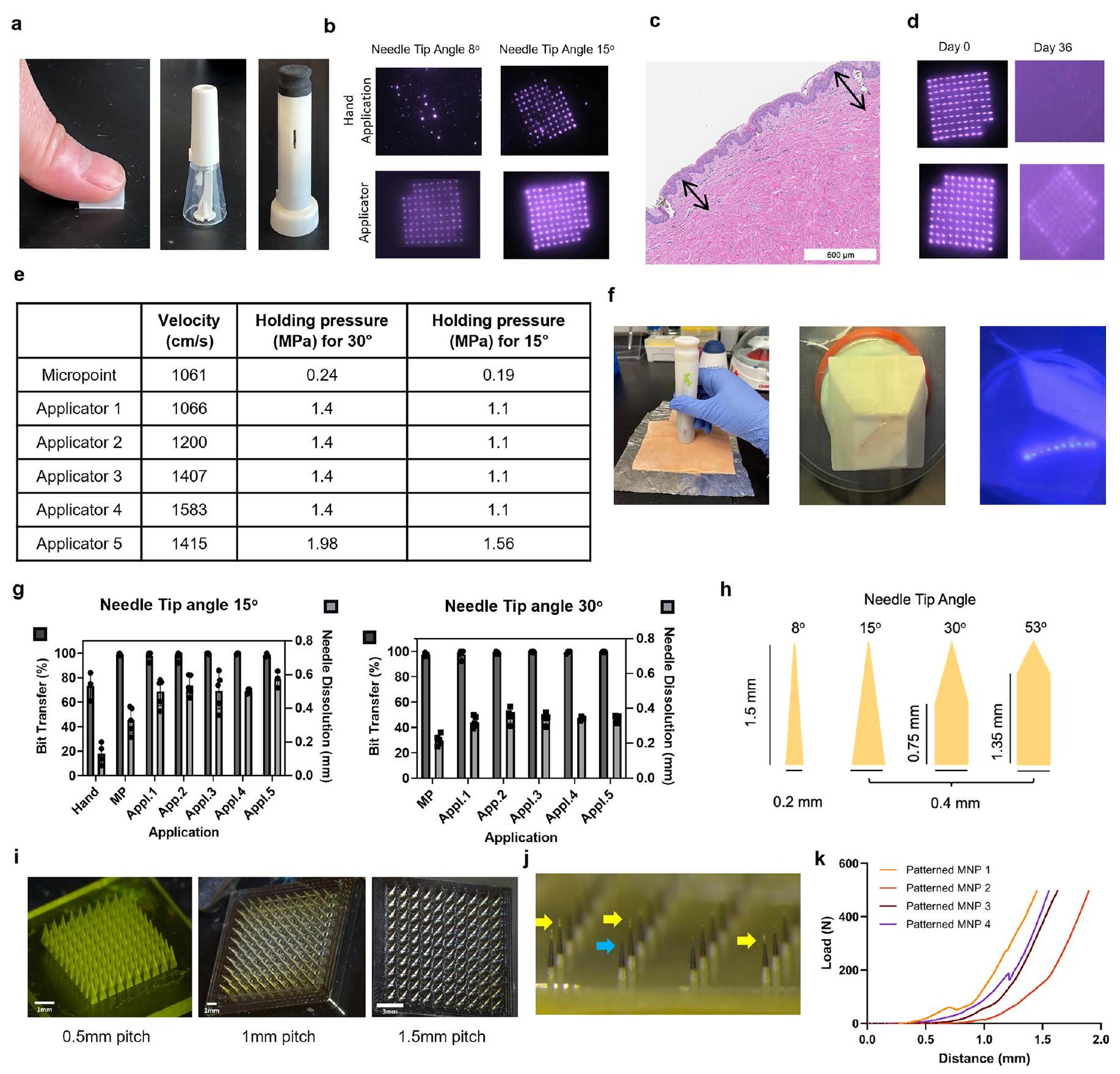
تم توليد تشفير إشارة NIR من QDs بعائد كمومي للضوء الفوتوني بنسبة 77% وقمة شدة الفوتولومينسنس عند (مرجع 28؛ الشكل 2أ). تم إحاطة QDs في جزيئات PMMA لزيادة حجم الجزيئات وبالتالي تقليل الإزالة البيولوجية وتعزيز استقرار النظام وتوافقه الحيوي . لم تؤدي إحاطة PMMA إلى تغيير في طول موجة الانبعاث القمة (الشكل 2أ) أو انخفاض كبير في العائد الكمومي للفوتولومينسنس، حيث ظل عند 73% بعد الإحاطة. تم ضبط قطر جزيئات QD-PMMA (صبغة OPMR) ليكون تقريبًا , وتم تأكيد الحجم المتوسط باستخدام المجهر الإلكتروني الماسح (SEM؛ الشكل 2ب). لتقييم توصيل OPMR الفعال، تم استخدام MNPs مع مصفوفة ، وتم تحميلها بصبغة OPMR عند أطراف الإبر وخلط البوليمر كدعم (الشكل الممتد 1هـ). تحتوي كل من الإبر الدقيقة الـ 100 على صبغة OPMR عند الطرف (الشكل 2ج)، وتشكل معًا مصفوفة ، مع كل إبرة تتوافق مع بت NIR واحد (الشكل 2د). نظرًا لأن توصيل المعلومات تحت الجلد بدقة هو خطوة أولى حاسمة، تم تصميم تطبيق MNP وهيكله لنقل الصبغة بشكل متسق ودوام إشارة أمثل. نحو ذلك، تم التحقيق في ثلاثة معلمات حرجة: (1) نقل البت، النسبة المئوية للإبر المسمى NIR التي تم نقلها إلى
وقابلة للاكتشاف في الجلد كبتات من إجمالي عدد الإبر الدقيقة المحملة بالصبغة، حيث يشير ذلك إلى تشفير المعلومات الأولية في الجلد؛ (2) عمق الاختراق، العمق تحت الجلد حيث تودع أطراف الإبر الصبغة، لأن ذلك قد يؤثر على دوام الإشارة؛ و (3) ذوبان الإبرة، حيث يترجم ذلك إلى كمية الحمولة الموصلة إلى الجلد. لهذه الدراسات، تمت إزالة أربع إبر في أحد زوايا لتوجيه اللاصقة، مما ترك اللاصقة مع 96 إبرة.
أولاً، قمنا بمقارنة نتائج تطبيق MNPs يدويًا ومع تطبيقات الربيع (الشكل 2هـ والشكل الممتد 2أ). أدى التطبيق اليدوي إلى نقل صبغة ضعيف وغير متسق عند فحصه باستخدام نظام تصوير NIR (الشكل 2و)، مع أقل من نقل بت، بينما أظهرت التطبيقات 100% نقل بت ( “; الشكل 2g والشكل الإضافي 2b). بالنسبة لعمق الاختراق، لم تصل رؤوس الإبر إلى عمق أكبر منمع تطبيق اليد، مما ترك معظم الصبغة متراكمة بالقرب من البشرة (الشكل البياني الممتد 2c) مما أدى إلى اختفاء معظم إشارات NIR بعد شهر واحد (الشكل البياني الممتد 2d). وقد أدى ذلك إلى افتراض أن ترسيبًا أعمق باستخدام جهاز تطبيق مناسب ضروري للحصول على إشارات NIR دائمة. في هذا السياق، تم اختبار أجهزة تطبيق الربيع المخصصة مع مجموعة من سرعات التأثير وضغوط الإمساك (الشكل البياني الممتد 2e) على جلد الخنازير خارج الجسم (الشكل البياني الممتد 2f)، مما أدى إلى اختيار سرعة تأثير قدرهاوالاحتفاظ بالضغط عند 1.1 ميغاباسكال للاستخدام في بقية الدراسات (الشكل البياني الممتد 2g).
ثانيًا، قمنا بتحسين متغيرين في تصميم MNP يؤثران على أداء MNP: (1) زاوية طرف الإبرة، التي ترتبط بنسبة عرض الإبرة إلى ارتفاعها وحدّة الإبرة الدقيقة، و(2) المسافة، وهي المسافة بين إبرتين من المركز إلى المركز (الشكل 2 هـ). أربع زوايا طرف مختلفة، و مع ارتفاع إبرة ثابت يبلغ 1.5 مم (الشكل 2h من البيانات الموسعة)، وأربعة خطوات مختلفة،و 3 مم مع ثابتتم اختبار زوايا الرأس (الشكل 2i من البيانات الموسعة) لأقصى نقل للبت، وعمق الاختراق، وانحلال الإبرة مع الحفاظ على الحد الأدنى من حجم اللصقة. بالنسبة لجميع زوايا الرأس الأربعة، كانت أعماق الاختراق ضمن طبقة الأدمة.، ولكن فقط الـ و زوايا النصائح أدت إلىنقل بت; الشكل 2i). الـزاوية الطرف كانت هشة جداً (الشكل 2 ج من البيانات الموسعة) بينماكانت زاوية الإبرة غير حادة عند الاختراق. بالنسبة لتباعد الإبر، فقد أودعت جميع المسافات الصبغة داخل الأدمة، ولكن فقط الجسيمات النانوية المغناطيسية ذات المسافة المتساوية أو الأكبر من 1 مم أدت إلىنقل بت; الشكل 2j). لوحظ اتجاه تنازلي في ذوبان الإبرة مع زيادة زوايا الرأس، واتجاه تصاعدي مع زيادة المسافات (n=4-7; الشكل 2k).
طلبات OPMR MNP للتسليم الفعال
بمجرد أن يتم ترسيب جزيئات الصبغة في الجلد، يجب أن تستمر إشارات الأشعة تحت الحمراء القريبة (NIR) للسماح بقراءات دقيقة للمعلومات. لهذا الغرض، يجب أن تكون زوايا الطرف ( و ) وارتفاعات ( 1 مم و 3 مم ) التي أدت إلى تم اختبار نقل البتات من حيث متانة الإشارة في الجسم الحي. تتضمن متانة الإشارة معلمين: (1) احتفاظ الإشارة، وهو النسبة المئوية للبتات القابلة للكشف في نطاق الأشعة تحت الحمراء القريبة من إجمالي البتات المنقولة، و(2) شدة الإشارة، وهي قيمة سطوع البكسل (بوحدات قياسية) للبتات في نطاق الأشعة تحت الحمراء القريبة. يعتبر احتفاظ الإشارة وشدة الإشارة مؤشرات كمية ونوعية رئيسية لمتانة الإشارة، على التوالي. في هذه الدراسة، تم اختيار نموذج خنزير يوركشاير بسبب تشابه هيكل الجلد (البشرة والأدمة) وخصائصه الميكانيكية مقارنةً بالجلد البشري. (الشكل البياني الموسع 2k). تم استخدام خوارزميات العتبة التكيفية لتحليل احتفاظ الإشارة (الشكل البياني الموسع 3a) وشدة الإشارة (الشكل البياني الموسع 3b) “. عندما تتكون ثلاث مجموعات MNP ( و ) تم تطبيقها (الشكل البياني الإضافي 3c) وتم تصويرها أسبوعيًا لمدة 10 أسابيع (الشكل البياني الإضافي 3d)، نتائج المجموعات فياحتفاظ الإشارة في الأسبوع العاشر، بينماأظهر المجموعة احتفاظًا أقل بالإشارةفي الأسبوع التاسع (; الشكل 2ل). بالنظر إلى أن و المجموعات اخترقت الجلد بشكل أعمق و على التوالي) من مجموعة (; الشكل يبدو أن عمق الاختراق يؤثر على احتفاظ الإشارة داخل الجلد.
أمثلة على العلاجات المعتمدة على mRNA التي هي قيد التطوير حاليًا
الشكل 1| مخطط لتقنية OPMR المستخدمة في تسجيل المعلومات الطبية. أ، صبغة QD الفلورية NIR المحاطة بجزيئات PMMA الدقيقة (المكون الأسود من طرف الإبرة) محملة مع mRNA المحاطة في LNPs (المكون الأزرق الفاتح من طرف الإبرة) في إبر دقيقة يتم الاحتفاظ بها سليمة بواسطة دعم بوليمري قابل للذوبان. عند تطبيق MNP، يتم إيداع جزيئات الصبغة الدقيقة في طبقة الأدمة بنمط محدد مسبقًا ترمز المعلومات الطبية، بينما يتم امتصاص mRNA-LNPs بواسطة خلايا المناعة، مما يؤدي إلى تحفيز المناعة. يتم تصوير أنماط NIR ومعالجتها لاسترجاع المعلومات الطبية على الشاشة. ب، توفر تقنية OPMR المدعومة بالتعلم العميق (DL) سعة ترميز كبيرة في إلىنطاق من خلال الاستفادة من الميزة الثنائية لقطع الميكرونيدل OPMR، مما يجعل التكنولوجيا قابلة للتطبيق على العدد المتزايد بسرعة من العلاجات القائمة على mRNA التي هي قيد التطوير حاليًا.
عندما تكون شدة الإشارات لأفضل مجموعتين أداءً ( و تم تحليل ( ) لمدة 70 يومًا، على الرغم من أن الشدة العامة انخفضت تدريجيًا مع مرور الوقت، لم يحدث فرق كبير بين المجموعتين (; الشكل 2 م )، مما يشير إلى أن شدة الإشارة لا تتأثر طالما تم وضع الصبغة بعمق أكبر من عمق العتبة (على سبيل المثال، ). لأن جزيئات النانو ذات المسافة 1 مم أدت بنفس كفاءة -عرض MNPs مع تقديم حجم رقعة أصغر بتسع مرات،تصميم بفجوة 1 مم وزاوية الرأستم اختيارها لبقية الدراسات. ونتيجة لذلك، أدت هذه المعلمات المحسّنة للتطبيق والهندسة المعمارية إلى عمق اختراق متسق بالقرب من (الشكل 2ن)، مما يودع جزيئات الصبغة بفاعلية داخل الأدمة (الشكل 20).
رمز تصحيح الخطأ للتشفير الزمني القوي
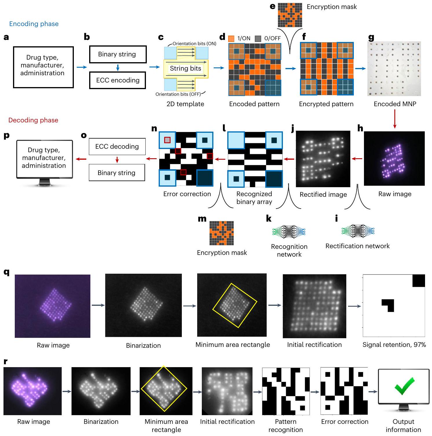
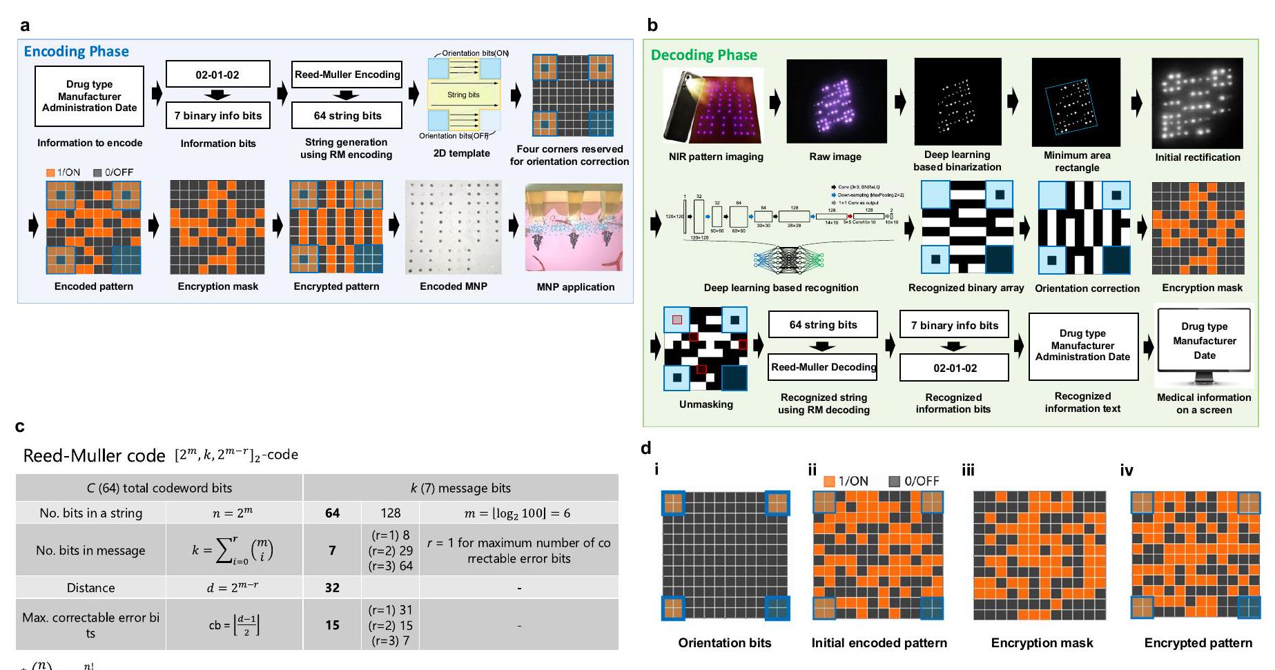
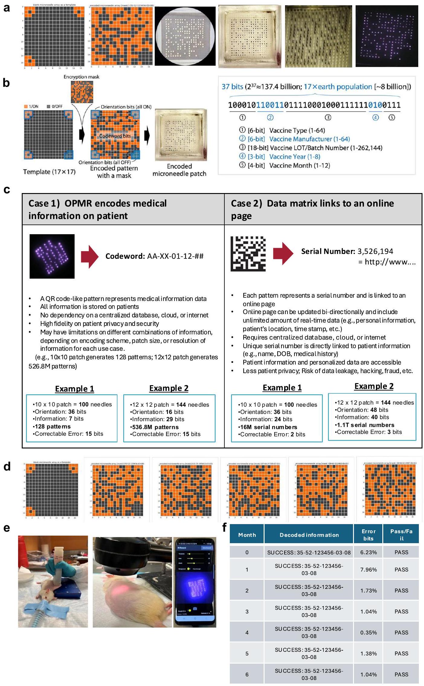
تقوم تقنية OPMR لدينا بتشفير المعلومات من خلال طباعة أنماط ثنائية الأبعاد (2D) على الجسيمات النانوية المغناطيسية (MNPs)، مستفيدة من الخاصية الثنائية لقطع الإبر المجهرية (أي أن الإبرة المجهرية (القطعة) إما موجودة (تشغيل) أو غائبة (إيقاف)). قد يتعرض صبغة OPMR، بمجرد إيداعها في الجلد، لتقليل إشارة NIR بسبب التنظيف البلعمي، أو تلاشي الضوء. أو الأضرار الجسدية مثل الإصابة أو الندوب. للتخفيف من هذه الفساد المحتمل في البيانات، يتميز نظامنا بـ (1) نظام تصحيح الأخطاء الذي يقدم تكرارًا لتعويض تدهور الإشارة الزمنية و (2) معالجة الصور المعتمدة على التعلم العميق لضمان قراءات نمط موثوقة على الرغم من التغيرات المكانية. يتكون خط الأنابيب العام من مرحلتين: مرحلة الترميز ومرحلة فك الترميز (الشكل 4a,b من البيانات الموسعة).
تم تطوير مرحلة الترميز للتخفيف من فقدان بتات MNP المحتمل الذي قد يتسبب في تمثيل خاطئ لنمط ما بمرور الوقت، وبالتالي توفير متانة زمنية. خلال الترميز، يتم تحويل المعلومات ذات الأهمية إلى نمط يمكن ترميزه على MNP. أولاً، تم تحديد المعلومات التي سيتم تسجيلها على المريض (الشكل 3أ). ثم تم ترجمتها إلى سلسلة ثنائية مشفرة (الشكل 3ب) مع رمز تصحيح الأخطاء (ECC). أضافت ECC تكراراً لبتات المعلومات كوسيلة للدفاع ضد فساد البيانات وبالتالي ضمان استرداد المعلومات بشكل موثوق على المدى الطويل.من بين العديد من أنواع رموز تصحيح الأخطاء، يوجد رمز ريد-مولر (RM)تم اختيار (الشكل 4c من البيانات الموسعة) لبرنامج OPMR نظرًا لملاءمته لتصحيح الأخطاء المستقلة وغير المجمعة وقدرته القوية على ترميز ملايين ومليارات من تركيبات المعلومات المصاحبة لـ
الشكل 2 | مواد MNP، التصميم والتطبيق لتوصيل OPMR الفعال. أ، شدة الفوتولومينسنس (PL) المعيارية لـQD الذي يصل ذروته عند 897 نانومتر مع أو بدون تغليف PMMA. ب، صورة SEM لجزيئات PMMA الدقيقة مع بلورات QD النانوية محاطة بها. (تم إجراء SEM مرتين.) ج، صورة SEM لطرف إبرة دقيقة واحدة محملة بجزيئات QD-PMMA الدقيقة. (تم إجراء SEM مرة واحدة.) د، صورة بصرية لمسبار نانوي محمل بجزيئات QD-PMMA عند أطراف الإبرة. (تم تكرار التصوير البصري)أوقات.) e، صورة لتطبيقات MNP يدويًا ومع جهاز تطبيق نابض. قضبان القياس،لا تترك جزيئات OPMR MNPs آثارًا مرئية؛ يتم عرض نقل أفضل لجزء NIR مع جهاز التطبيق، كما هو موضح في الصور. مقياس الأشرطة، 1 سم. البيانات تظهر أن نقل جزء NIR أفضل يتم تحقيقه مع جهاز التطبيق؛. تم ذلك باستخدام نسخ بيولوجية من 3-5 حيوانات. h، متغيرات بنية MNP التي يمكن أن تؤثر
جودة OPMR. تم تقييم نقل البيت وعمق اختراق الجلد لزوايا رؤوس إبر مختلفة؛تم تقييم نقل بت وعمق اختراق الجلد لدرجات مختلفة؛تم تقييم ذوبان الإبرة لزوايا ونسب مختلفة للرأس؛بيولوجي، س.د. (تجارب فيتمت في جلد الخنازير الخارجي.) أنا، احتفاظ الإشارة لهياكل MNP المختلفة على مدى عشرة أسابيع؛شدة الإشارات للجزيئات النانوية المغناطيسية المطبقة مع مسافات 1 مم و 3 مم لمدة 70 يومًا؛بيولوجي، س.د؛ غير مهم إحصائيًا. ن، ع، صورة تمثيلية للهستولوجيا لقطعتين (ن؛ تم تكرار تصوير الهستولوجيا أكثر من 30 مرة) وللجزيئات الدقيقة الكروية QD-PMMA (ع) المودعة بشكل جيد داخل الأدمة في جلد الخنزير. (تم إجراء التجارب في 1-ع في الخنازير يوركشاير). ع، السهم الأسود في الشكل يبرز المكان الذي تم فيه إدخال طرف الإبرة الدقيقة وترك أثرًا من جزيئات النقاط الكمومية. قدرات تصحيح الأخطاء المحددة مسبقًا (الجدول 1). على سبيل المثال، يمكن توليد 128 نمطًا مختلفًا باستخداموتم فك تشفيره بدقة حتى عند وجود 15 بت خطأ. بالمثل،يمكن لنظام MNP ترميز 137.4 مليار نمط مختلف وتصحيح 31 بت خطأ. وهذا يعني أنه يمكن توليد مليارات من الأنماط المختلفة واستخدامها لترميز معلومات طبية مختلفة بحجم رقعة يبلغ فقط.
بمجرد توليد سلسلة ثنائية مشفرة، تم رسمها في نمط ثنائي الأبعاد بزاوية ثابتة (الشكل 3c). تتكون الأنماط المولدة من بتات 1 (تشغيل) حيث تكون الإبر الدقيقة مملوءة بصبغة NIR وبتات 0 (إيقاف) حيث لا تحتوي الإبر الدقيقة على أي صبغة فلورية (الشكل 3d). بعد ذلك، تمت إضافة قناع تشفير إلى النمط المشفر لضمان خصوصية البيانات الطبية الشخصية (الشكل 3e والشكل الإضافي 4d). ثم تم تشفير النمط المشفر (الشكل 3f) على
الشكل 3 | الشبكات المعتمدة على التعلم العميق تسمح بالتشفير وفك التشفير بدون معلمات لـ OPMR. أ، أمثلة على المعلومات الطبية التي يمكن تشفيرها على OPMR MNP. ب، يتم تحويل بيانات المعلومات إلى سلسلة ثنائية مشفرة قبل ECC. ج، يتم تشفير بيانات المعلومات الثنائية وفقًا لقالب ثنائي الأبعاد. د، تصبح المصفوفة ثنائية الأبعاد نمطًا مشفرًا. هـ، يتم تطبيق قناع تشفير لحماية خصوصية المريض.يتم توليد النمط المشفر لتشفير MNP. g، يتم تصنيع MNP المشفر. h، تبدأ مرحلة فك التشفير مع الحصول على الصورة الخام. i، يتم تصحيح الصورة الخام في البداية عبر تقنية التعلم العميق. شبكة التصحيح. الصورة المصححة بتنسيق مربع بالأبيض والأسود. يتم التعرف على البتات بواسطة شبكة التعرف المعتمدة على التعلم العميق. شبكة التعرف تخرج مصفوفة ثنائية. يتم عكس خطوة التشفير عن طريق إزالة قناع التشفير. يتم تحديد بتات الخطأ. يتم تصحيح بتات الخطأ. يتم ترجمة سلسلة البتات المشفرة مرة أخرى إلى المعلومات الأصلية وعرضها على الشاشة. تحليل احتفاظ الإشارة يقيس عدد بتات NIR المكتشفة لـ 96 بت من MNPs. تحليل قابلية فك النمط يفك تشفير MNPs المنمطة ويقيم ما إذا كانت قد تم فك تشفيرها بنجاح أم لا. تم تصنيع MNP الفيزيائي عن طريق تحميل الصبغة بشكل انتقائي فقط على الإبر التي تمثل الحالة ON، مما أسفر عن حوالي 50% من الإبر في حالة ON و50% في حالة OFF (الشكل 3g).
شبكات التعلم العميق لفك التشفير القوي مكانيًا
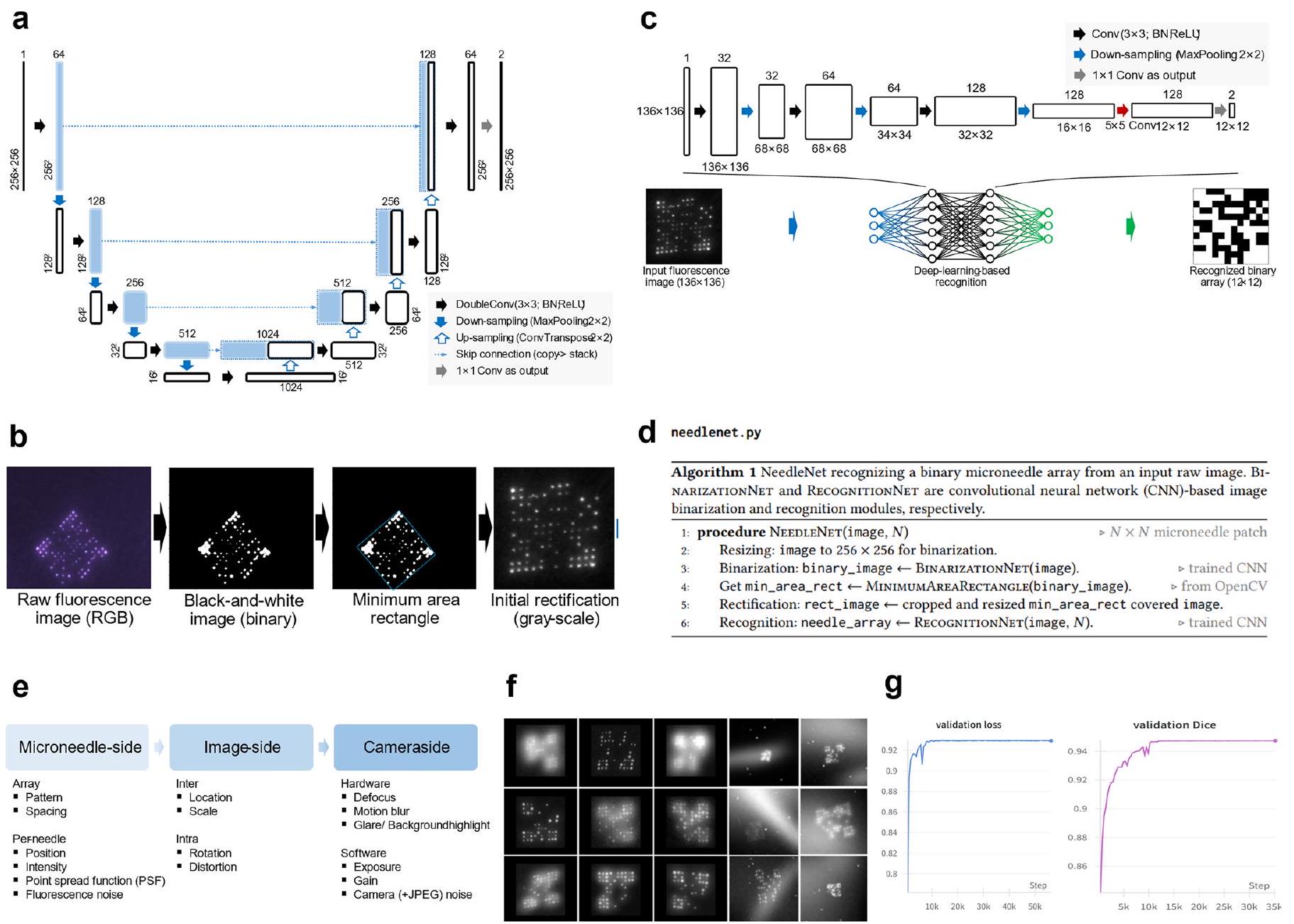
تجعل التوزيعات المكانية للأصباغ الفلورية نظام OPMR عرضة لتشوهات البت. تم تطوير مرحلة فك التشفير لتعويض هذه التغيرات المكانية بين إشارات البت الملتقطة ولضمان متانة OPMR المكانية. تبدأ مرحلة فك التشفير باكتساب الصور الخام (الشكل 3h). بمجرد اكتسابها، تم تصحيح كل صورة خام إلى تنسيق مربع ثنائي باستخدام شبكة تصحيح قائمة على التعلم العميق (الشكل 3i)، والتي تتضمن شبكة U-Net.شبكة التثنية (الشكل التوضيحي الممتد 5a) التي تحول الألوان الحمراء والخضراء والزرقاء الخام صورة (RGB) إلى صورة ثنائية بالأبيض والأسود ووظيفة مستطيل الحد الأدنى (الشكل الإضافي 5b) التي تجد وتقص وتدور منطقة MNP. بعد التصحيح (الشكل 3j)، تم إدخال الصورة في شبكة تعرف قائمة على التعلم العميق (الشكل 3k)، تم تطويرها من خلال تدريب شبكة عصبية تلافيفية. (الشكل البياني الممتد 5c، d) مع 650,000 صورة اصطناعية (الشكل البياني الممتد 5e-g). مع هذين الخطوتين في التعلم العميق، تم تحويل صورة خام بنجاح إلى مصفوفة ثنائية (الشكل 3l). في هذه المرحلة، قد تحتوي المصفوفة الثنائية على نمط تالف بسبب إشارات غير مكتشفة أو تم اكتشافها بشكل خاطئ. من أجل فك تشفير النمط بدقة، تمت إزالة قناع التشفير (الشكل 3m)، وتم تصحيح النمط المفكوك (الشكل 3n) باستخدام RM ECC قبل تحويله مرة أخرى إلى سلسلة ثنائية (الشكل 30). ثم تمت ترجمة المصفوفة واسترجاعها على الشاشة (الشكل 3p). إن سير العمل الكامل من التشفير إلى فك التشفير تلقائي تمامًا، ولا يتطلب أي
الجدول 1 | سعة المعلومات لمصفوفة الجسيمات النانوية ثنائية الأبعاد بناءً على تصحيح الأخطاء RM
حجم المصفوفة
حجم الباتش
إجمالي عدد البتات
بتات التوجيه
بتات الترميز
RM(r, m)
وحدات المعلومات
عدد قطع المعلومات القابلة للتشفير
بتات الخطأ القابلة للتصحيح
100
٣٦
64
RM(1, 6)
٧
128 (أي، )
15
١٤٤
16
128
RM(1, 7)
٨
256 (أي، )
31
١٤٤
16
128
RM(2, 7)
٢٩
536.8 مليون (أي، )
15
٢٨٩
٣٣
256
RM(1, 8)
9
512 (أي، )
63
٢٨٩
٣٣
256
RM(2, 8)
37
137.4 مليار (أي، )
31
يشير إلى رمز RM للطلبوطول. مدخلات المستخدم أو التلاعبات اليدوية في العتبات بسبب الطبيعة ‘من النهاية إلى النهاية’ لهذا النهج في تعلم الآلة.
الحفاظ على بيانات المعلومات على المدى الطويل في الخنازير
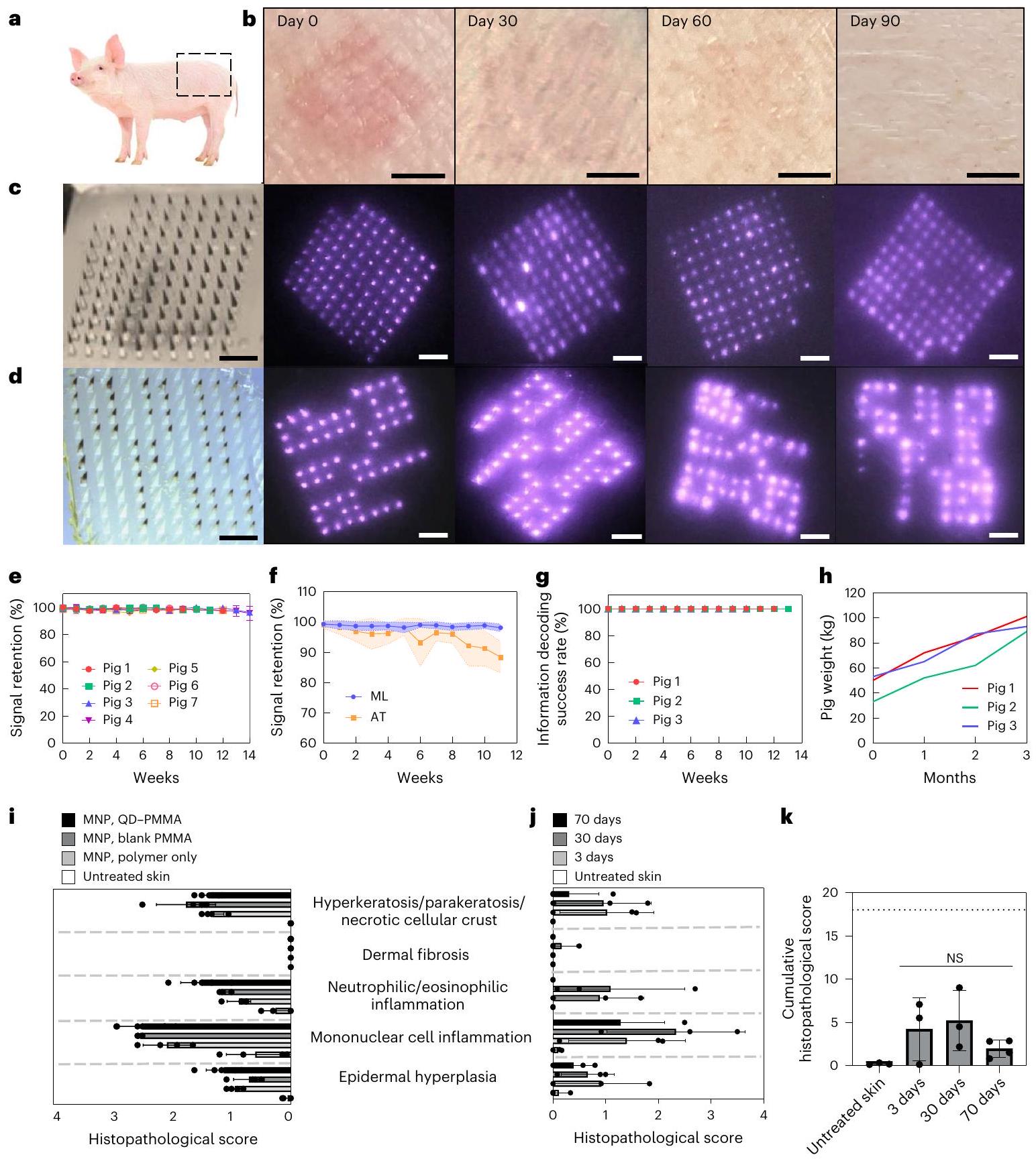
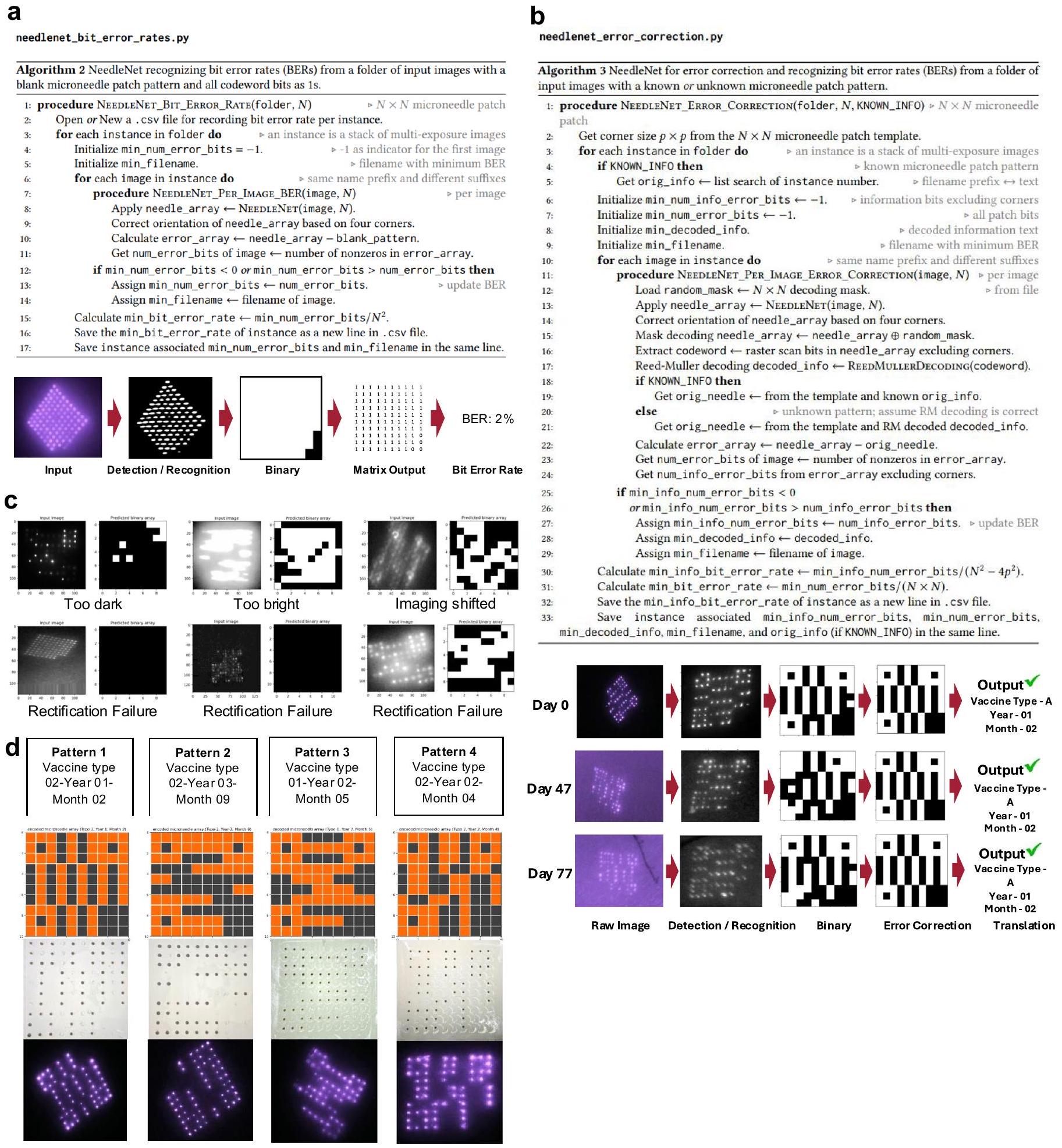
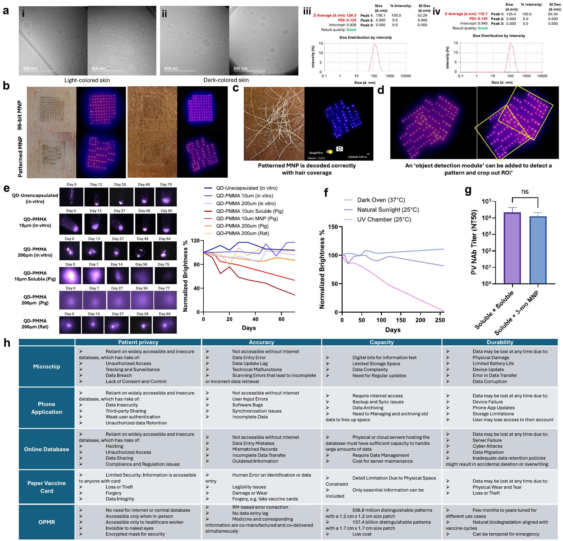
تم تحليل احتفاظ الإشارة وقابلية فك الشيفرة النمطية بشكل طولي في نموذج خنزير حي، حيث يُعرف أن جلد الخنازير يشبه جلد الإنسان بشكل كبير.للاحتفاظ بالإشارة (الشكل 3q)، تم قياس عدد بتات NIR التي تم الاحتفاظ بها في الجلد على مر الزمن (الشكل التمديدي 6a). تم الكشف عن كل من بتات ON و OFF للعثور على نسبة بتات ON من إجمالي البتات المنقولة، وتم إخراج هذه القيمة كنسبة احتفاظ الإشارة. بالنسبة لقابلية فك تشفير النمط (الشكل 3r)، تم فك تشفير MNPs المنقوشة وتمت مقارنة المخرجات بالمعلومات المشفرة المقصودة (الشكل التمديدي 6b). إذا كانت المعلومات المسترجعة تتطابق بدقة مع المعلومات الأصلية، فإن النمط يُعتبر قد تم فك تشفيره بنجاح؛ وإلا، فإنه يُعتبر غير ناجح (الشكل التمديدي 6c).
تم تطبيق الجسيمات النانوية المغناطيسية على مناطق الجوانب في خنازير يوركشاير (الشكل 4أ) وتم تصويرها أسبوعيًا لمدة ثلاثة أشهر. من أجل الاحتفاظ بالإشارة، تم تطبيق ما مجموعه 24 جسيمًا نانويًا مغناطيسيًا بقدرة 96 بت على سبعة خنازير. كانت الأصباغ غير مرئية للعين المجردة طوال فترة المراقبة التي استمرت ثلاثة أشهر، مما أدى إلى عدم تمييز مواقع تطبيق البقع تمامًا (الشكل 4ب)؛ ومع ذلك، ظلت إشارات الأشعة تحت الحمراء القريبة مرئية طوال فترة المراقبة بأكملها (الشكل 4ج). بالنسبة لقابلية فك تشفير الأنماط، تم استخدام ما مجموعه 21 جسيمًا نانويًا مغناطيسيًا مزخرفًا بأربعة أنماط مختارة عشوائيًا.تم تطبيق الأنماط (الشكل 6d من البيانات الموسعة) على ثلاثة خنازير مختلفة وتم تصويرها أسبوعيًا لمدة ثلاثة أشهر (الشكل 4d).
انخفضت شدة الإشارة لهذه اللصقات المطبقة مع مرور الوقت (الشكل 2 م)، ولكن مع نطاق الكاميرا واكتساب الصورة (البيانات الموسعة الشكل 1أ-ج)، ظلت أجزاء NIR قابلة للاكتشاف، مما أدى إلى احتفاظ الإشارات بـفي 4 أسابيع،في 8 أسابيع وعند 12 أسبوعًا (الشكل 4e). كان عدد بتات الخطأ ضمن عتبة 15% من بتات الخطأ القابلة للتصحيح لرمز RM المختار (الجدول 1). كانت هذه النظام التلقائي بالكامل لعد البتات يعالج صور MNP بعمق 96 بت بسرعة متوسطة تبلغ 0.043 ثانية لكل صورة (الصور). كانت هذه النتائج المدعومة بتعلم الآلة مقارنة بشكل إيجابي مع خوارزمية العتبة التكيفية المستخدمة سابقًا من حيث كل من اكتشاف البت والدقة (;الشكل 4f). من أجل الحفاظ على المعلومات، تم فك تشفير جميع 21 من الجسيمات النانوية المغناطيسية المنقوشة بنجاح عبر جميع الخنازير الثلاثة على مدى ثلاثة أشهر (الشكل 4g). هذا يظهر أن نظام تصحيح الأخطاء RM ECC لدينا نجح في تصحيح 1-2% من فقدان البيانات واسترجاع المعلومات بدقة مع مرور الوقت. جميعتقرأ بصمات MNP المعلومات الصحيحة على الرغم من التشوهات المكانية الواضحة الناتجة عن نمو الحيوانات منإلىوتجدد خلايا البشرة خلال الثلاثة أشهرالشكل 4 ح. هذا النظام التلقائي بالكامل لفك تشفير الأنماط قام بتحليلصور MNP المنقوشة بسرعة متوسطة تبلغ 0.066 ثانية لكل صورةصور) على جهاز لابتوب مزود بمعالج إنتل كور i7 من الجيل العاشر، مما يشير إلى أن فك التشفير التلقائي من غير المحتمل أن يكون مصدر تأخير ملحوظ في سير عمل OPMR.
التوافق الحيوي لنظام OPMR
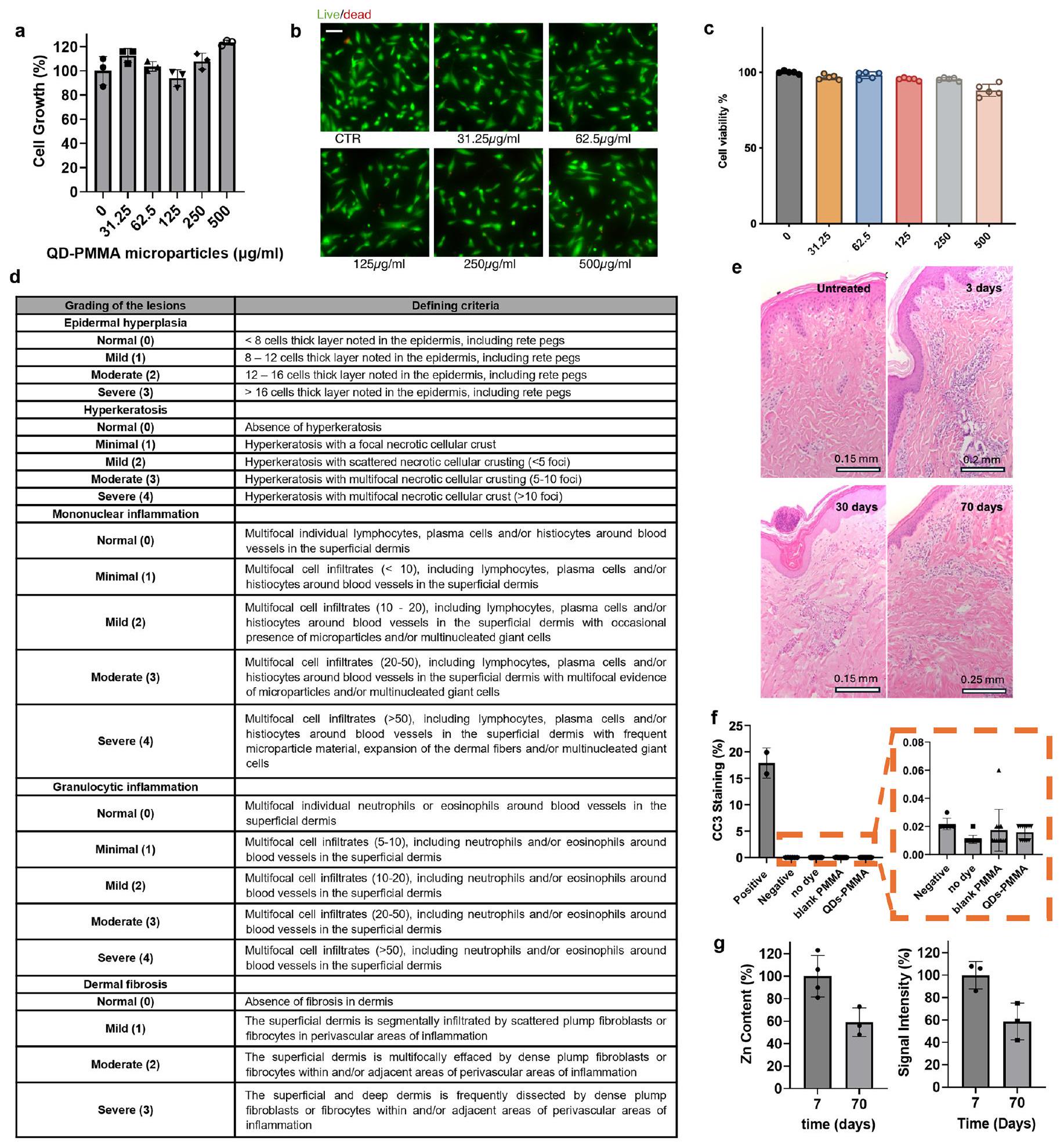
لفهم التوافق الحيوي على المدى الطويل للـ OPMR، تم فحص السمية الخلوية لصبغة OPMR أولاً في المختبر دون إشارة إلى السمية الخلوية (الشكل الإضافي 7 أ-ج). بعد ذلك، تم فحص الاستجابات الالتهابية والخلوية المحلية في منطقة تطبيق جزيئات النانو المغناطيسية. عند تطبيق جزيئات النانو المغناطيسية، لوحظ احمرار خفيف في البداية واختفى خلال 30 دقيقة (الشكل 2 و). بالنسبة للتقييمات النسيجية، تم استئصال مقاطع الأنسجة التي لم يتم تطبيق أي شيء عليها وتلك التي تحتوي على جزيئات نانو مغناطيسية تحتوي على بوليمر، وجزيئات ميكروية من PMMA فارغة وجزيئات ميكروية من QD-PMMA بعد 3 أيام من التطبيق. كانت درجات الآفات الجلدية لمجموعة جزيئات النانو المغناطيسية QD-PMMA مشابهة من حيث الشدة للمجموعات الضابطة الأخرى لجزيئات النانو المغناطيسية (الشكل الإضافي 7 د)، مما يشير إلى أن الآفات الملحوظة كانت ناتجة عن صدمة من اختراق الإبرة نفسها، بغض النظر عن محتوى PMMA أو صبغة QD. (الشكل 4i). أظهرت مقاطع الجلد المسترجعة بعد 3 و30 و70 يومًا من تطبيق جزيئات النانو المغناطيسية QDPMMA (الشكل التمديدي 7e، f) التهابًا جلديًا طفيفًا إلى معتدل في كل من اليومين 30 و70، وغيابًا تامًا للتقرن المفرط في اليوم 70، وعدم وجود دليل على التليف في أي نقطة زمنية تم فحصها (الشكل 4j). لم تكن هناك اختلافات سريرية أو إحصائية ذات دلالة بين الدرجات النسيجية التراكمية لعينات الجلد غير المعالجة وعينات الجلد المعالجة بجزيئات النانو المغناطيسية (الشكل 4k). أخيرًا، تم ملاحظة إزالة QD الناجحة من أنسجة الجلد مع مرور الوقت أيضًا (الشكل التمديدي 7g).
التوصيل المشترك لـ OPMR ولقاح mRNA لـ SARS-CoV-2
يمكن لجهاز OPMR MNP توصيل العلاجات للمرضى من خلال إضافة حمولة ثانوية. (الشكل البياني الممتد 8). لقد أظهرنا توصيلًا فعالًا وآمنًا لمركب OPMR ولقاح mRNA الذي يشفر بروتين السنبلة الخاص بمنطقة ارتباط مستقبل SARS-CoV-2 (RBD)، محاطًا في جزيئات نانوية دهنية (LNPs) في نموذج من الجرذان (الشكل 5a).
أولاً، لتقييم أداء OPMR عند تقديمه مع mRNA المحاط في LNPs، تم دراسة قابلية فك تشفير النمط لـ OPMR مع وبدون mRNA-LNPs. لهذا الاختبار،تم تطبيق MNPs المنقوشة (الشكل البياني الموسع 6d) مع وبدون mRNA-LNPs (الشكل البياني الموسع 9a-d) على جرذان ويستار وتم تصويرها لمدة ستة أشهر (الشكل البياني الموسع 9e). A17تم إضافة مجموعة MNP المنقوشة لإظهار جدوى تسجيل مليارات الأنماط المختلفة على المدى الطويل (الجدول 1). آثار كل من (الشكل 5ب) و (الشكل 5ج) ظلت مجموعات الجسيمات النانوية المغناطيسية قابلة للاكتشاف وقابلة للتشفير بنجاح (الشكل 5د) خلال فترة المراقبة التي استمرت ستة أشهر بالكامل ( “; الشكل 5e والشكل الإضافي 9f). هذهتشير معدلات النجاح إلى أن تحقيق OPMR طويل الأمد ممكن مع توصيل mRNA-LNP.
ثانيًا، لدراسة فعالية توصيل لقاح mRNA باستخدام OPMR، تم أولاً تحديد سلامة LNPs مع وبدون صبغة OPMR في المختبر. عند تحليلها باستخدام مجهر الإلكترون الناقل بالتبريد (cryo-TEM؛ الشكل 5f) والتشتت الضوئي الديناميكي (DLS؛ الشكل 5g والشكل الإضافي 10a)، ظلت mRNA-LNPs مستقرة ومتجانسة بحجم متوسط من و مع وبدون صبغة OPMR، على التوالي. ظلت كلا خيوط mRNA و LNPs سليمة مع سلامة mRNA من و (الشكل 5h)، وكفاءات احتواء mRNA لـ و (الشكل 5i)، مع وبدون صبغة OPMR، على التوالي، أيضًا. بعد ذلك، لتقييم توصيل لقاح mRNA مع OPMR في الجسم الحي، تم اختبار ثلاث مجموعات من
الشكل 4 | الفعالية طويلة الأمد لـ OPMR في نموذج الخنازير. أ، تم تطبيق الجسيمات النانوية المغناطيسية على منطقة الجنب للخنازير من سلالة يوركشاير. ب، الأصباغ الخاصة بـ OPMR المودعة في جلد الخنزير غير مرئية للعين المجردة. مقياس الرسم، 5 مم. ج، تظل إشارات NIR للجسيمات النانوية المغناطيسية 96 بت قابلة للاكتشاف لمدة ثلاثة أشهر في الخنازير. مقياس الرسم، 2 مم. د، تظل إشارات NIR للجسيمات النانوية المغناطيسية المنقوشة قابلة للتشفير لمدة ثلاثة أشهر في الخنازير. مقياس الرسم، 2 مم. هـ، احتفاظ إشارة NIR بـفي عمر 12 أسبوعًا في الخنازير؛تتفوق أنظمة معالجة الصور المخصصة المعتمدة على التعلم الآلي (ML) على خوارزمية العتبة التكيفية (AT) عبر سبعة خنازير., س.د. ج، أ معدل نجاح فك تشفير المعلومات حدث لمدة 12 أسبوعًا في الخنازير؛تمت دراسة MNPs عبر ثلاثة خنازير. زادت أوزان الخنازير أكثر من الضعف خلال فترة المراقبة التي استمرت ثلاثة أشهر. i، تسجيل تاريخي مرضي لجلد الخنازير بدون علاج ومع MNPs المحملة بالبوليمر فقط، جزيئات PMMA الدقيقة وجزيئات QD-PMMA الدقيقة؛عدد الجزيئات النانوية لكل مجموعة، ست شرائح لكل جزيء نانوي. (جلد غير معالج) وكانت جزيئات النانو PMMA-QDالتكرارات البيولوجية؛ كانت جزيئات النانو PMMA الفارغة وجزيئات النانو المصنوعة من البوليمر فقطالتكرارات البيولوجية؛ ست عينات نسيجية لكل تكرار بيولوجي، الانحراف المعياري.) ج، التقييم النسيجي للجلد غير المعالج للخنازير والجلد الخنزير مع تطبيق QD-PMMA MNP، بعد 3 و30 و70 يومًا من التطبيق؛عدد الجزيئات النانوية لكل مجموعة، ست شرائح لكل جزيء نانوي. (الجلد غير المعالج، 3 أيام و30 يومًا كان)التكرارات البيولوجية؛ كانت 70 يومًاالتكرارات البيولوجية؛ ست عينات نسيجية لكل تكرار بيولوجي، الانحراف المعياري.) ك، يظهر التقييم النسيجي التراكمي زيادة قصيرة لمجموعة جزيئات النانو QD-PMMA التي تنخفض مع مرور الوقت (تظهر الخط المنقط الحد الأقصى للدرجة الإجمالية البالغة 18). (الجلد غير المعالج، 3 أيام و30 يومًا كان لديهالتكرارات البيولوجية؛ كانت 70 يومًاالتكرارات البيولوجية؛ ست عينات نسيجية لكل تكرار بيولوجي. تحليل التباين أحادي الاتجاه (ANOVA)؛ فترة الثقة، ). استجابات المناعية: (1) مجموعة التحكم التي تلقت حقنًا عضلية (IM)، (2) مجموعة MNP المعتمدة على mRNA المحملة باللقاح فقط و (3) مجموعة MNP المعتمدة على mRNA-OPMR المحملة باللقاح وOPMR. بالنسبة لهذه المجموعات، تم تطبيق الجرعات الأولية في اليوم 0، وجرعات التعزيز تمت المتابعة بعد 28 يومًا (الشكل 5 أ). أظهرت المجموعات الثلاث مستويات متقاربة من عناوين الأجسام المضادة من نوع IgG بعد التعزيز.; الشكل 5j) بالإضافة إلى مستويات عيار الأجسام المضادة المحايدة للفيروس الزائف (; الشكل 5k)، مما يشير إلى أن جزيئات النانو المغناطيسية OPMR قدمت حماية غير أدنى بنجاح
الشكل 5 | الجسيمات النانوية المغناطيسية التي توصل معًا OPMR ولقاح mRNA القوي تسجل المعلومات وتثير المناعة في الجرذان. أ، تم تطبيق الجرعات الأولية والمعززة في اليومين 0 و28، على التوالي، باستخدام الجسيمات النانوية المغناطيسية OPMR-mRNA لتقييم التوصيل المشترك لـ OPMR وmRNA. ب، صورة بصرية لـنمط OPMR MNP وأثره في الأشعة تحت الحمراء القريبة في الجرذان على مدى 180 يومًا. قضبان القياس، 2 مم. ج، صورة بصرية لـ نمط OPMR MNP وأثره في الأشعة تحت الحمراء القريبة في الجرذان على مدى 180 يومًا. قضبان القياس، 2 مم. د، أظهرت جميع الأنماط عددًا قابلًا للتصحيح من بتات الخطأ وتم فك تشفيرها بنجاح. هـ، أظهرت المجموعات الثلاث جميعها (نمط OPMR MNP، MNP mRNA OPMR المنقوشة وتم فك تشفير OPMR MNP المنقوش بنجاح على مدى ستة أشهر؛تظهر صور Cryo-TEM لمحلول اللقاح وجود جزيئات mRNA-LNP سليمة ومتجانسة مع وبدون صبغة OPMR. (تم إجراء TEM مرة واحدة.) g، تحليل DLS يظهر أحجام LNP قابلة للمقارنة مع وبدون صبغة OPMR؛.h، محلل الشظايا تحليل يظهر تكاملات mRNA قابلة للمقارنة مع وبدون صبغة OPMR؛يظهر اختبار Ribogreen كفاءات تعبئة mRNA قابلة للمقارنة مع وبدون صبغة OPMR؛. مجموعة التحكم IM، مجموعة mRNA MNP ومجموعة mRNAOPMR MNP تحفز مستويات متشابهة من عيار IgG في الجرذان؛مجموعة التحكم IM، مجموعة mRNA MNP ومجموعة mRNA-OPMR MNP تحفز مستويات متشابهة من عناوين الأجسام المضادة المحايدة (NAb) بعد التعزيز في الجرذان. استجابة الجرذان الساذجة موضحة كخط متقطع؛تم تخزين جزيئات النانو MNPs التي تشفر اللوكيفيراز OPMR-mRNA في درجة حرارة الغرفة لمدة ثلاثة أشهر وتم تطبيقها على الفئران لدراسة مدة الصلاحية، وتم قياس تعبيرات اللوكيفيراز باستخدام نظام تصوير حي. الدوائر الحمراء هي مناطق مختارة من الاهتمام (ROI) لقياس الإشعاع. m، تعبيرات اللوكيفيراز لجزيئات النانو المخزنة لمدة شهر واحد وثلاثة أشهر قابلة للمقارنة مع تلك الخاصة باللصقات الطازجة؛. NT50، مستويات العنوان المحايد ضد SARS-CoV-2 مقارنةً بالتحكمات IM. هذا يُظهر أن التوصيل المشترك للعلاجات الفعالة القائمة على mRNA ممكن مع تقنية OPMR MNP الخاصة بنا.
أخيرًا، لتقييم مدة صلاحية جزيئات النانو المغناطيسية (MNPs) المحملة بـ mRNA الذي يشفر لوسيفيراز اليراعة (FLuc) وصبغة OPMR، تم تخزين جزيئات النانو في درجة حرارة الغرفة لمدة ثلاثة أشهر وتم تطبيقها على الفئران في نقاط زمنية مختلفة. عند قياس الإضاءة الحيوية لتعبير FLuc باستخدام نظام تصوير حي بعد 6 ساعات من التطبيق (الشكل 51)، لم توجد اختلافات كبيرة بين اللصقات الطازجة وتلك المخزنة لمدة شهر واحد أو ثلاثة أشهر.؛ الشكل 5 م)، مما يبرز إمكانية تخزين وتوزيع وتطبيق هذه الرقع عند الطلب لتوصيل العلاجات القائمة على mRNA والتسجيل.
آوتلوك
هنا قمنا بتطوير تقنية OPMR المعتمدة على الإبر الدقيقة والتي يمكنها تخزين المعلومات تحت الجلد مع قوة زمنية ومكانية ممتازة وسعة ترميز تصل إلى المليارات. إن عرض توصيل OPMR مع استرجاع معلومات موثوق ولقاح mRNA قوي تم توضيحه في هذا العمل يشير إلى إمكانية ترجمة هذه التقنية للاستخدامات السريرية. في حالات الطوارئ مثل الأوبئة أو الكوارث الطبيعية، أو في مخيمات اللاجئين أو العسكرية، يمكن إعطاء لاصقات OPMR عند الطلب ويمكن أن تساعد العاملين في مجال الرعاية الصحية في اتخاذ قرارات مناسبة بشأن جرعات المتابعة دون مخاطر على سرية المرضى. لتعزيز موثوقية OPMR على المدى الطويل، تم دراسة سيناريوهات حالة مختلفة (على سبيل المثال، تضعيف إشارة النمط بسبب صبغة الجلد، الشعر أو تداخل النمط؛ الشكل الإضافي 10ب-د) لفترة طويلة من الزمن (على سبيل المثال، استقرار جزيئات النانو المغناطيسية OPMR لمدة عام واحد؛ الشكل الإضافييمكن التحقيق في ذلك في المستقبل. بشكل عام، هذه التكنولوجيا قابلة للتطبيق بسهولة على أي علاجات تعتمد على mRNA، نظرًا لتوافقها مع mRNA-LNPs وسعتها الكبيرة في الترميز لتكمل العدد المتزايد من علاجات mRNA التي هي قيد التطوير حاليًا. حيث تهدف علاجات mRNA إلى مكافحة مجموعة واسعة من الأمراض القابلة للتجنب وغير القابلة للعلاج، فإن هذه التكنولوجيا OPMR تقدم فرصة لجعل العدالة في الرعاية الصحية أقرب إلى الواقع.
المحتوى عبر الإنترنت
أي طرق، مراجع إضافية، ملخصات تقارير Nature Portfolio، بيانات المصدر، بيانات موسعة، معلومات تكميلية، شكر وتقدير، معلومات مراجعة الأقران؛ تفاصيل مساهمات المؤلفين والمصالح المتنافسة؛ وبيانات توفر البيانات والرموز متاحة علىhttps://doi.org/10.1038/s41563-024-02115-4.
References
Qin, S. mRNA-based therapeutics: powerful and versatile tools to combat diseases. Signal Transduct. Target. Ther. 7, 166 (2022).
RNActive -Derived Therapeutic Vaccine No. NCT00906243 (US National Library of Medicine, 2012); https://clinicaltrials.gov/ study/NCTOO9O6243
An Efficacy Study of Adjuvant Treatment With the Personalized Cancer Vaccine mRNA-4157 and Pembrolizumab in Participants With High-Risk Melanoma (KEYNOTE-942) No. NCTO3897881 (US National Library of Medicine, 2022); https://clinicaltrials.gov/ study/NCTO3897881
Shargel, L. & Yu, A. Applied Biopharmaceutics & Pharmacokinetics 6th edn (McGraw-Hill, 2012).
Blaschke, T. F. Adherence to medications: insights arising from studies on the unreliable link between prescribed and actual drug dosing histories. Annu. Rev. Pharmacol. Toxicol. 52, 275-301 (2012).
Baryakova, T. H. Overcoming barriers to patient adherence: the case for developing innovative drug delivery systems. Nat. Rev. Drug Discov. 22, 387-409 (2023).
Martin, L. R. The challenge of patient adherence. Ther. Clin. Risk Manag. 1, 189-199 (2005).
Bobo, F. T. Child vaccination in sub-Saharan Africa: increasing coverage addresses inequalities. Vaccine 40, 141-150 (2022).
Global Health Security: Immunization (Centers for Disease Control and Prevention, 2014); https://archive.cdc.gov/#/ details?url=https://www.cdc.gov/globalhealth/security/ immunization.htm
Adetokunboh, O. Missed opportunities for vaccination in Africa. Curr. Opin. Immunol. 71, 55-61 (2021).
Rainey, J. J. Reasons related to non-vaccination and under-vaccination of children in low and middle income countries: findings from a systematic review of the published literature, 1999-2009. Vaccine 29, 8215-8221 (2011).
Jalloh, M. F. Assessment of VaxTrac electronic immunization registry in an urban district in Sierra Leone: implications for data quality, defaulter tracking, and policy. Vaccine 38, 6103-6111 (2020).
Katib, A. A prototype of a novel cell phone application for tracking the vaccination coverage of children in rural communities. Comput. Methods Prog. Biomed. 122, 215-228 (2015).
Xiao, C. & Yu, A. Medical Smart Card System for Patient Record Management (Open Computing Facility, University of California, Berkeley, 2009); https://www.ocf.berkeley.edu/~step/White_ Paper/Xiao_Yu.pdf
Tanne, J. H. FDA approves implantable chip to access medical records. Br. Med. J. 329, 1064 (2004).
Charnock, V. Electronic healthcare records and data quality. Health Info. Lib. J. 36, 91-95 (2019).
Harman, L. B., Flite, C. A. & Bond, K. Electronic health records: privacy, confidentiality, and security. Virtual Mentor. 14, 712-719 (2012).
Keshta, I. Security and privacy of electronic health records: concerns and challenges. Egypt. Inform. J. 22, 177-183 (2021).
Ozair, F. F. Ethical issues in electronic health records: a general overview. Perspect. Clin. Res. 6, 73-76 (2015).
Meetoo, D. Smart tattoo: technology for monitoring blood glucose in the future. Br. J. Nurs. 28, 110-115 (2019).
Mchugh, K. J. Biocompatible near-infrared quantum dots delivered to the skin by microneedle patches record vaccination. Sci. Transl. Med. 11, eaay7162 (2019).
Baranov, M. V. Modulation of immune responses by particle size and shape. Front. Immunol. 11, 607945 (2021).
Choi, H. S. Renal clearance of quantum dots. Nat. Biotechnol. 25, 1165-1170 (2007).
Longmire, M. Clearance properties of nano-sized particles and molecules as imaging agents: considerations and caveats. Nanomedicine 3, 703-717 (2008).
Kolarsick, P. A. Anatomy and physiology of the skin. J. Dermatol. Nurses Assoc. 3, 203-213 (2011).
Lunney, J. K. Importance of the pig as a human biomedical model. Sci. Transl. Med. 13, eabd5758 (2021).
Ety Navon, O. M. Color image segmentation based on adaptive local thresholds. Image Vis. Comput. 23, 69-85 (2005).
Muller, D. E. Application of Boolean algebra to switching circuit design and to error detection. Trans. I.R.E. Prof. Group Electron. Comput. EC-3, 6-12 (1954).
Reed, I. A class of multiple-error-correcting codes and the decoding scheme. Trans. I.R.E. Prof. Group Inf. Theory 4, 38-49 (1954).
Ronneberger, O., Fischer, P. & Brox, T. U-Net: convolutional networks for biomedical image segmentation. In Medical Image Computing and Computer-Assisted Intervention – MICCAI 2015. Lecture Notes in Computer Science (eds Navab, N. et al.) Vol. 9351, 234-241 (Springer, Cham, 2015); https://doi.org/10.1007/978-3-319-24574-4_28
Krizhevsky, A. ImageNet classification with deep convolutional neural networks. Adv. Neural Inf. Process. Syst. 60, 84-90 (2012).
Seaton, M. Porcine models of cutaneous wound healing. ILAR J. 56, 127-138 (2015).
Koster, M. I. Making an epidermis. Ann. N. Y. Acad. Sci. 1170, 7-10 (2009).
Etra, J. W. & Skin, A. Rejection grading system for vascularized composite allotransplantation in a preclinical large animal model. Transplantation 103, 1385-1391 (2019).
vander Straeten, A. et al. A microneedle vaccine printer enables decentralized manufacturing of thermostable COVID-19 mRNA vaccines. Nat. Biotechnol. 42, 510-517 (2024).
Kim, E. Microneedle array delivered recombinant coronavirus vaccines: immunogenicity and rapid translational development. EBioMedicine 55, 102743 (2020).
Koh, K. J. Formulation, characterization and evaluation of mRNA-loaded dissolvable polymeric microneedles (RNApatch). Sci. Rep. 8, 11842 (2018).
Chen, W. Microneedles as a delivery system for gene therapy. Front. Pharm. 7, 137 (2016).
Publisher’s note Springer Nature remains neutral with regard to jurisdictional claims in published maps and institutional affiliations.
كوبالت النحاس الكبريتيتم شراء النقاط الكمومية من شركة ستريم كيميكالز. PMMA (الوزن الجزيئي،تم شراء ) من Sigma-Aldrich. تم تنفيذ تغليف QD باستخدام تقنية تبخر الاستحلاب بالمذيب. تم إذابة 100 ملغ من QDs و100 ملغ من PMMA في 2 مل من ثنائي كلورو الميثان (DCM). ثم أضيف محلول QD-PMMA إلى 20 مل من الباردمحلول بولي فينيل الكحول (PVA) ومستحلب عندلمدة دقيقة واحدة (خلاط ULTRA-TURRAX الرقمي T 18، IKA). تم صب المستحلب الناتج على الفور في 30 مل منتم تحضير محلول PVA وتم تحريكه بسرعة 250 دورة في الدقيقة طوال الليل للسماح بتبخر DCM. بعد ذلك، تم صب المحلول في أنبوب طرد مركزي سعة 50 مل وتم طرده مركزياً عندلجمع الجسيمات الدقيقة. تم التخلص من السائل العلوي وتم غسل الجسيمات الدقيقة ثلاث مرات عن طريق إضافة ماء معقم منزوع الأيونات يليه الطرد المركزي. أخيرًا، تم إعادة تعليق الجسيمات في كمية صغيرة من الماء وتم تصفيتها من خلالتم تصفية لإزالة التجمعات الكبيرة. ثم تم تجفيف الجسيمات الدقيقة QD وحفظها في الظلام، تحت فراغ حتى الاستخدام.
طيفية الفوتولومينسنس
تم قياس طيف انبعاث الفوتولومينسنس باستخدام كاميرا سيليكون مبردة حرارياً (PIXIS100، Teledyne Princeton Instruments). تم تحضير العينات في قوارير كوارتز عن طريق تعليق النقاط الكمية أو جزيئات النقاط الكمية- PMMA في السيكلوهكسان. تم تحفيز العينات باستخدام ليزر بزاوية 532 نانومتر (CPS532، Thorlabs). تم جمع الانبعاث وتركيزه باستخدام مرآتين بارابوليتين مغطاتين بالفضة، وتم تصفيته من خلال فلتر ديالكتيكي طويل الموجة بزاوية 800 نانومتر إلى مقياس الطيف قبل التصوير على كاميرا السيليكون. تم قياس بيانات عائد الفوتولومينسنس الكمي باستخدام ثنائي ضوئي سيليكوني (818-UV، Newport) متصل بمضخم قفل (SR830، Stanford Research)، باستخدام تحفيز ليزر مقطع بزاوية 405 نانومتر (LDM405، Thorlabs) وكرة تكامل بصرية (RTC-060-SF، Labsphere)..
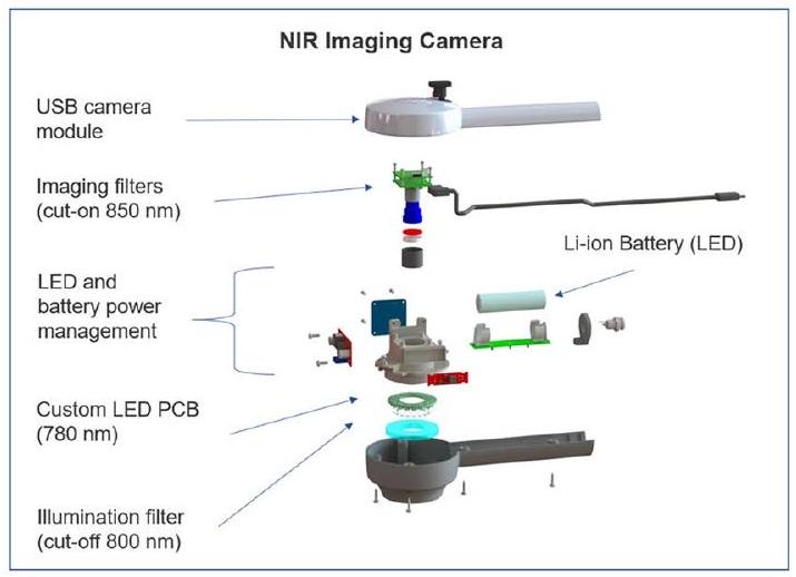
تصوير الفلورية NIR
نظام تصوير الفلورية بالأشعة تحت الحمراء القريبة (NIR) المتصل عبر الناقل التسلسلي العالمي (USB) والذي يتضمن وحدة مصباح ثنائي باعث للضوء (LED) مخصصة تصدر ضوء NIR بطول موجي أقصر عند 780 نانومتر ووحدة كاميرا متصلة عبر USB تلتقط صورة الفلورية المثارة لجزيئات الكوانتم (QD) عند ضوء NIR بطول موجي أطول.تم استخدامها لتصوير النقاط الكمومية، مع ذروة شدة الفوتولومينسنس عندتم تطوير تطبيق هاتف ذكي يعمل بنظام أندرويد يسمى ‘IR Record’ لالتقاط إشارة صبغة OPMR NIR ويمكنه حفظ الصور. تم تصميم البرنامج لالتقاط 30 صورة متتالية مع ستة إعدادات تعريض مختلفة وخمسة إعدادات كسب مختلفة. تتيح هذه الطريقة في المسح الضوئي التقاط إشارات NIR بتفاوت في الشدة على مر الزمن. من بين الصور الثلاثين، يتم اختيار صورة واحدة ذات أفضل نتائج قراءة تلقائيًا ومعالجتها. الوقت الإجمالي المطلوب لمسح رقعة OPMR واحدة على المريض هو حاليًا أكثر بقليل من دقيقتين. ومع ذلك، يمكن إجراء تحسينات إضافية من خلال دفع عملية المسح والتعرف إلى الوقت الحقيقي. لتعزيز الجانب الزمني من خط أنابيب اكتساب الصورة إلى التعرف على الصورة، يمكن إضافة (1) ‘وحدة كشف OPMR’ التي تحدد موقع رقعة صغيرة نسبيًا، (2) ‘وحدة التعريض التلقائي’ التي تلتقط وتخزن صورة واحدة كافية للتعرف بدلاً من التقاط سلسلة من الصور مع نطاق واسع من مستويات التعريض و(3) ‘مرحلة فك التشفير المعتمدة على الهاتف المحمول’ التي تعالج كل شيء في الوقت الفعلي إلى نظامنا التلقائي. مع هذه الأدوات، سيكون من الممكن إجراء مسح شامل والتعرف على أنماط عديدة بطريقة أكثر ملاءمة في السيناريوهات الواقعية. في الواقع، لاحظنا أن بيئة التصوير (على سبيل المثال، أضواء السقف في غرفة العمليات في منشأة الخنازير، ارتفاع السرير الذي وضعت عليه الخنازير والمسافة بين السرير والأضواء)، والأشخاص المسؤولون عن التصوير (على سبيل المثال، التعديل اليدوي لإعدادات الكاميرا والمسافة عن سطح جلد الخنزير) وحالة الخنازير (على سبيل المثال، أ الخدوش أو الفراء على جلد الخنزير) تؤثر على جودة الصور أكثر من الجودة الفعلية لإشارات NIR. تعكس هذه العوامل سيناريوهات التصوير في العالم الحقيقي بدقة أكبر حيث سيتم إجراء التصوير بواسطة محترفين صحيين مختلفين عبر نقاط رعاية مختلفة.
تصنيع قوالب بوليديميثيلسيلوكسان
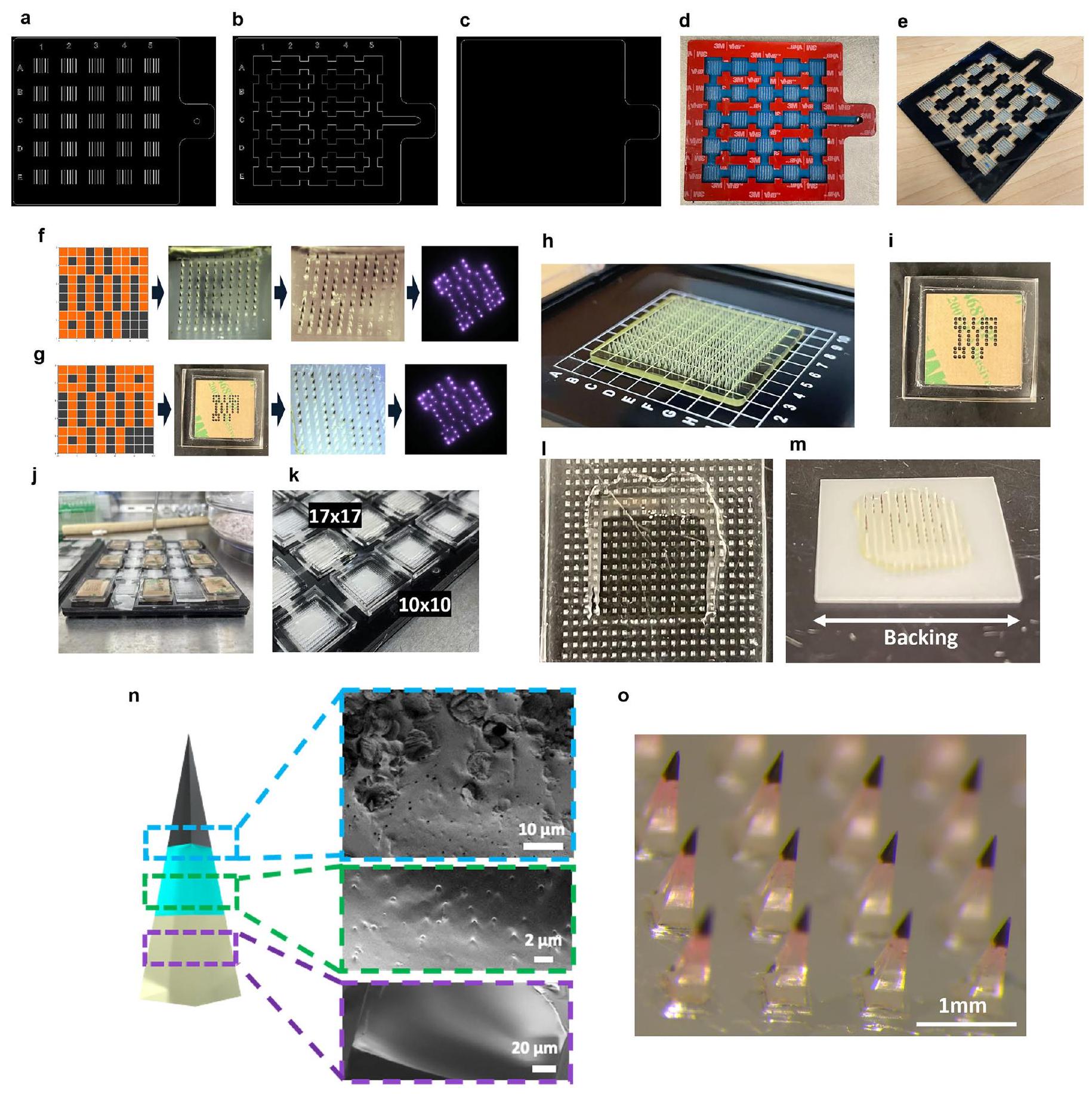
تم تصميم قوالب رئيسية إيجابية باستخدام برنامج تصميم بمساعدة الكمبيوتر (CAD) (SolidWorks، Dassault Systèmes) وتصنيعها خصيصًا على آلة طحن CNC ذات خمسة محاور باستخدام قوالب من فولاذ الأدوات. تم طباعة القوالب الرئيسية ذات المسافات بين الإبر الأصغر التي لم تكن مناسبة للمعالجة باستخدام CNC بتقنية الطباعة ثلاثية الأبعاد (3D) باستخدام راتنج HTL وطابعات ثلاثية الأبعاد عالية الدقة (Boston Micro Fabrication). تم استخدام هذه القوالب الرئيسية لإنشاء قوالب سلبية مصنوعة من بوليديميثيلسيلوكسان (PDMS؛ Sylgard 184، Dow Corning). تم خلط قاعدة PDMS ووكيل الربط وفقًا لتعليمات الشركة المصنعة، وصبها على القالب الرئيسي الإيجابي وتصلبها طوال الليل عندلإنشاء إيجابيات MNP إضافية، تم ملء لاصق نورلاند البصري القابل للتصلب بالأشعة فوق البنفسجية 61 (كرانبوري) في قوالب PDMS السلبية باستخدام جهاز الطرد المركزي عندلمدة دقيقة واحدة، تم وضعها في فرن تجفيف بالأشعة فوق البنفسجية في درجة حرارة الغرفة لمدة 20 دقيقة ثم تمت إزالتها يدويًا.
تركيب محلول mRNA-LNP
تم تصنيع LNPs لتغليف mRNAتم شراء mRNA المعدل بواسطة البسودوريدين الذي يشفر FLuc (TriLink BioTechnologies) وmRNA المعدل بواسطة البسودوريدين الذي يشفر بروتين السنبلة SARS-CoV-2 (رمز/سلالة المعاهد الوطنية للصحة) مع حذف موقع قطع الفيرين، واثنين من طفرات البرولين، وfoldon ثلاثي القوائم من أجل الاستقرار (ACROBiosystems). بالنسبة لتخليق LNP، تم استخدام Lipid 5 وهو lipid قابل للتأين (heptadecan-9-yl 8-((2-hydroxyethyl)(8-(nonyloxy)-8-oxooctyl)amino)octanoate؛ Organix)، و1,2-dioleoyl-sn-glycero-3-phosphoethanolamine (DOPE؛ Avanti)، والكوليسترول (Sigma-Aldrich) و1,2-dimyristoyl-sn-glycero-3-phosphoethanolamine--[ميثوكسي-(بوليمر الإيثيلين غليكول)-2000] (ملح الأمونيوم؛ C14-PEG2000؛ أفانتي) تم إذابته في الإيثانول بنسبة مولية منعلى التوالي. لتحضير الجسيمات النانوية الدهنية، تم إضافة المحلول الإيثاني بسرعة إلى محلول mRNA المخفف بمحلول سترات عند pH 3 بنسبة حجم (مائي/إيثانول). تم تعيين نسبة وزن الدهون القابلة للتأين إلى mRNA إلى 5، وكانت التركيز النهائي لـ mRNA هوتم تخزين جميع الأحماض النووية فيوتم السماح لها بالذوبان على الثلج قبل الاستخدام. ثم تم غسيل الجزيئات النانوية الدهنية لمدة لا تقل عن ساعتين في محلول ملحي مخفف بالفوسفات (PBS) عندفي كاسيت بحد قطع وزن جزيئي 20,000. من أجل تخليق محلول mRNA-LNP لتصنيع MNP، تم غسيل LNPs في ماء منزوع الأيونات لمدة ساعتين إضافيتين في، ثم ركزت على على فلتر أميكون عن طريق الطرد المركزي عندأخيرًا، تم تخفيف الخليط إلى محلول بوليمر مكون من PVA (Mowiol 4-88؛ الوزن الجزيئي، 31,000؛ سيغما-ألدريتش) وبولي فينيل بيروليدون (PVP؛ الوزن الجزيئي، 10,000؛ TCI) لتشكيل محلول اللقاح.
تصنيع إبر الميكرونيدل OPMR
تم تصنيع جزيئات النانو المغناطيسية OPMR باستخدام تقنية الطرد المركزي. لتحميل صبغة OPMR إلى جزيئات النانو المغناطيسية،من تشتت مائي لجزيئات QD-PMMA الدقيقة ) تم صرفه على قمة قالب PDMS سالب وتم الطرد المركزي عندلمدة 3 دقائق لتركيز الجسيمات الدقيقة عند أطراف الإبر. لضمان تحميل متساوٍ للصبغة عبر الجسيمات النانوية المغناطيسية، تم تدوير القالب.قبل إضافة أخرىتشتت الصبغة وتم الطرد المركزي لمدة 3 دقائق أخرى. جولة أخرى منتم تحميل تشتت الصبغة والطرد المركزي دون تدوير القالب هذه المرة. بالنسبة لجسم الإبرة الدقيقة والدعم،منبي في إيه/بي في بيتم إضافة المحلول وتم الطرد المركزي عند لمدة 5 دقائق. إضافي تم إضافة محلول PVA/PVP وتم الطرد المركزي على مرحلتين بفاصل زمني قدره 4 ساعات بينهما لضمان سطح مستوٍ للدعم بعد التجفيف. بعد التجفيف في درجة حرارة الغرفة لمدة 4.5 أيام،
تم لصق خلفية بلاستيك ديلرين أسيتال بشريط لاصق (سمك 0.508 مم؛ مكماستر-كار) على ظهر MNP قبل إزالته من القالب. بعد الإزالة، تم تجفيفه بشكل إضافي تحت الفراغ.لمدة 48 ساعة. ونتيجة لذلك، انتهى كل MNP بحد أقصى قدره 0.8 ملغ من QD-PMMA.
تم تصنيع جزيئات نانوية مغلفة بـ FLuc mRNA-OPMR عبر تحميل من خطوتين باستخدام تقنية الشفط. تم وضع قوالب PDMS على جهاز الشفط لتحميل الحمولة في القالب باستخدام ضغط سلبي، مستفيدين من خاصية نفاذية الهواء لـ PDMS. كخطوة أولى، تم استخدام 1 مل من تشتت مائي لجزيئات QD-PMMA الدقيقة. ) مع تم توزيع PVA/PVP على قالب PDMS، وتم تطبيق فراغ (-85 ميجا باسكال) على جهاز التفريغ طوال الليل. كخطوة ثانية،من جزيئات mRNA-LNPs المختلطة مع محلول PVA/PVP (نسبة 1:320 من mRNA إلى البوليمر بالوزن)تم صرفه وتركه ليجف طوال الليل تحت الفراغ. كخطوة نهائية،منتم توزيع محلول PVA/PVP لتشكيل قاعدة MNP وتركه ليجف طوال الليل تحت الفراغ. بمجرد أن جف، تم تثبيت قاعدة ديلرين لإزالة اللصقات، وتم تجفيف اللصقات المزالة بشكل إضافي في جهاز تجفيف تحت الفراغ المنزلي لمدة 48 ساعة وحتى الاستخدام. تم تصنيع MNPs التي تحتوي على mRNA فقط بنفس الطريقة مع استبعاد الخطوة الأولى. لدراسات مدة الصلاحية، تم تصنيع MNPs من FLuc mRNA-OPMR وتخزينها في جهاز تجفيف عند درجة حرارة الغرفة لعدة أشهر. لكل نقطة زمنية للاختبار، تم تصنيع دفعة جديدة من لصقات mRNA-OPMR كتحكم إيجابي.
تم تصنيع جزيئات النانو المغناطيسية (MNPs) من mRNA-SARS-CoV-2 المزخرفة من خلال تحميل من خطوتين باستخدام تقنية الشفط. لصنع جزيئات النانو المزخرفة، تم وضع شريط قناع مقصوص بالليزر (468 MP PEI Adhesive Transfer Tape Sheet، 3M) مع أنماط علىقوالب PDMS. ثم تم وضع قوالب PDMS على جهاز يعمل بالشفط لتحميل الشحنات في كل قالب باستخدام الضغط السلبي، مستفيدين من خاصية نفاذية الهواء لقوالب PDMS. كخطوة أولى، تم استخدام 1 مل من تشتت مائي لجزيئات QD-PMMA الدقيقة. ) مع تم توزيع PVA/PVP على سطح القناع الموجود على قالب PDMS، وتم تطبيق فراغ (-85 ميجا باسكال) طوال الليل. ثم تم إزالة القناع، وتم تنظيف سطح القالب باستخدام الإيثانول.امسح.كماالخطوة الثانيةمحلول اللقاح المصنوع من جزيئات الدهون النانوية mRNA-RBD لفيروس SARS-CoV-2تم خلط mRNA المغلف) مع محلول PVA/PVP بنسبة 1:320 من حيث الوزن، وتم توزيعه ونشره على سطح القالب، مغطياً حوالي 100 إبرة في المنتصف، وترك تحت الفراغ طوال الليل. كخطوة نهائية، تم تثبيت قاعدة ديلرين لإزالة اللصقات، وتم تجفيف اللصقات المزالة في جهاز تجفيف تحت فراغ منزلي لمدة 48 ساعة وحتى الاستخدام. كمجموعة تحكم، تم تصنيع جزيئات نانوية من mRNA RBD لفيروس SARS-CoV-2 دون إضافة صبغة OPMR؛ تم تصنيع هذه المجموعة بنفس الطريقة مع استبعاد الخطوة الأولى.رقع مزخرفة، لم يتم تحميل أي mRNA. بدلاً من ذلك، بعد تحميل الصبغة، 1 مل منتم إضافة محلول بوليمر PVA/PVP وانتشاره عبرمجموعة لتشكيل جسم الإبرة والدعم. تُظهر العملية لتوصيل جرعة كافية من لقاح mRNA باستخدام MNP في رسم علاقة التصميم في منشورنا الأخير.تم إنشاؤه لتلبية متطلبات الجرعات في البشر للقاحات mRNA الشائعة؛ ويظهر العلاقة بين حجم الإبرة الواحدة، وعدد الإبر لكل MNP والجرعة المقدمة في MNP واحد. باستخدام حجم الإبرة وكفاءة تحميل MNP من دراسة حول ذروة عناوين RBD المضادة لكوفيد، يتنبأ هذا النموذج بالتركيبة اللازمة من الحجم وعدد الإبر الدقيقة لتقديم الجرعات الكاملة من لقاح موديرنا (mRNA-1273) أو فايزر-بيونتيك (BNT162b1) ضد كوفيد-19. يتنبأ النموذج بأن 360 و 108 من الإبر الدقيقة والصيغ المدروسة ستقدم الجرعة الكاملة من لقاحات موديرنا وفايزر-بيونتيك، على التوالي. MNPs التي تحتوي على جرعة كافية أقل من 2 سم عبر.
تحليل المجهر الإلكتروني الماسح
تم استخدام SEM لتصوير جزيئات QD-PMMA الدقيقة و MNPs OPMR و OPMR-mRNA. تم طلاء العينات في البداية بطبقة رقيقة من الذهب باستخدام جهاز طلاء بالرش (Desk V، Denton Vacuum) ثم تم تصويرها. باستخدام مجهر إلكتروني مسح عالي الدقة (Zeiss Crossbeam 540) ومجهر شعاع الأيونات المركزة (Zeiss).
تطبيقات الجسيمات النانوية المغناطيسية خارج الجسم وداخل الجسم
تمت الموافقة على جميع إجراءات الحيوانات وإجراؤها وفقًا لإرشادات لجنة رعاية الحيوانات في معهد ماساتشوستس للتكنولوجيا. تم توفير خنازير يوركشاير أنثوية بعمر ثلاثة أشهر من مدرسة كامينغز للطب البيطري. تم شراء إناث الفئران ويستار بعمر ستة إلى ثمانية أسابيع من تشارلز ريفر. بالنسبة لتطبيقات MNP خارج الجسم، تم استخدام تطبيق يدوي، وجهاز تطبيق تجاري (Micropoint Biotechnologies) وجهاز تطبيق مخصص بسرعة عند التأثير تتراوح منوقوة التحمل ضمن نطاقتم اختبارها على جلد الخنازير المستأصل من الجنب والورك. بالنسبة لاختبارات الجلد البشري خارج الجسم، تم استخدام عينات من أنسجة جلدية متبرع بها (التبادل الوطني لأبحاث الأمراض، فيلادلفيا، بنسلفانيا). بالنسبة لتطبيقات MNP داخل الجسم، تم استخدام جهاز تطبيق مصمم خصيصًا بسرعة تأثيرتم استخدام قوة تثبيت تبلغ 1.1 ميغاباسكال لتطبيق اللصقات على منطقة الفخذ والورك في خنازير يوركشاير وعلى منطقة الظهر في جرذان ويستار. تم تطبيق اللصقات لمدة 5-10 دقائق للخنازير و10-20 دقيقة للجرذان.
تقييم عمق ترسيب الصبغة
لتقييم عمق ترسيب الديد في الجلد، تم استئصال أنسجة الجلد بعد تطبيقات MNP، وثبتت في محلول فورمالين 10% لمدة 48 ساعة، ثم تم نقلها إلىالإيثانول ثم تم تضمينه في شمع البارافين. تم قطع العينات عندعرض كللاسترجاع 30 شريحة لمصفوفة إبر دقيقة كاملة لتقييمات مقطع عرضي لأقصى عمق اختراق للإبر وعمق ترسيب الدواء. تم صبغ العينات بالهيماتوكسيلين والإيوزين وتم تحليلها باستخدام برنامج Aperio (لايكا بيوسيستمز). علاوة على ذلك، تم تجميد أجزاء من نسيج الجلد وتثبيتها في مركب درجة الحرارة المثلى للقطع من أجل التصوير المقطعي للكشف عن وجود قطع نير في الأدمة.
تحليل ذوبان الإبرة، نقل البت، احتفاظ الإشارة وتحليل شدة الإشارة
لتحديد ذوبان الإبر الدقيقة كدالة لمتغيرات معمارية مختلفة للإبر الدقيقة، تم تصوير الإبر الدقيقة قبل وبعد التطبيق باستخدام ميكروسكوب ضوئي ليكا DFC450. تم وضع اللصقات بطريقة عرضية للتصوير باستخدام برنامج LAS v.4.7. تم حساب طول الإبر الدقيقة باستخدام ImageJ (المعاهد الوطنية للصحة) لـعدد الإبر الدقيقة لكل MNP هو 3-5 MNPs لكل مجموعة. لتحليل نقل البيانات واحتفاظ الإشارة، تم استخدام ImageJ والعتبة التكيفية لحساب عدد البتات من الصور الملتقطة بالأشعة تحت الحمراء القريبة في نقاط زمنية مختلفة؛ تم إجراء هذه القياسات على خمس رقع لكل مجموعة. لتحليل شدة الإشارة، تم استخدام ImageJ والعنق التكيفية لتقييم أقصى قيمة بكسل في كل بت من الأشعة تحت الحمراء القريبة في الصور الملتقطة في نقاط زمنية مختلفة؛ تم إجراء هذه القياسات على 50-96 بت لكل رقعة لثلاث رقع لكل مجموعة. لتقييم شدة الإشارات بين نقاط زمنية مختلفة، تم استخدام مجموعة ثابتة من الكسب والتعرض لمقارنات عادلة.
RM ECC لتوليد مصفوفة ثنائية الأبعاد
تم ترميز معلومات البتات المثيرة للاهتمام مع تكرار باستخدام RM ECC. حزمة بايثون ‘reedmuller’ (https://pypi.org/project/تم استخدام Reed-Muller) لتشفير وتصحيح أخطاء RM. ثم تم تحويل سلسلة البتات الثنائية المشفرة إلى مصفوفة ثنائية الأبعاد بعد الرجوع إلى قالب ثنائي الأبعاد للتوجيه. كان رمز RM مع المعلمات و المشار إليه بـرمز، يشفرقطع المعلومات معبتات السلسلة وقادر على التصحيحبتات الخطأ المستقلة. هناك توازن بين عدد بتات المعلوماتوأقصى عدد من بتات الخطأ القابلة للتصحيحاعتمادًا على الطلب. لــ مع رمز RM(1,6)، عدد بتات السلسلة المتاحة هو، حيث هو الأرض وظيفة، مما يؤدي إلى ترميز 7 بتات معلومات في سلسلة مكونة من 64 بت. تم تخصيص الباقي، 36 بت، لتصحيح الاتجاه. تسعة بتات اتجاه (تم تخصيص (الحجم) لأربعة زوايا من المصفوفة ثنائية الأبعاد (ثلاث زوايا ON وزاوية أسفل اليمين OFF) لتوجيه التصحيح وتسجيل زوايا الدوران. تم ترتيب بتات سلسلة البتات الثنائية المشفرة بشكل متسلسل من أعلى اليسار إلى أسفل اليمين (مع استبعاد بتات التوجيه في الزوايا الأربع) بطريقة مسح نقطي. من أجل نقل رقم الهاتف مع، يمكن لشفرة RM(2، 7) ترميز 526.8 مليون مجموعة معلومات مختلفة وضمان تصحيح ما يصل إلى 15 بت خطأ. بالنسبة لـنقل رقم الهاتف مع، يمكن لشفرة RM(2,8) ترميز 137.4 مليار مجموعة معلومات مختلفة وضمان تصحيح 31 بت خطأ. وهذا يعني أنه يمكن تحديد القدرة المطلوبة على تصحيح الأخطاء مسبقًا ويمكن للمرء اختيار RM( ) خيار لتناسب حالة الاستخدام. مع RM ECC، يمكن لتقنية OPMR المقترحة توليد 137.4 مليار نمط مختلف مع يمكن أن تستوعب MNPs العدد الكبير من أنواع معلومات التطعيم، وثمانية مليارات شخص في السكان البشريين على الأرض، أو العدد المتزايد بسرعة من العلاجات المتاحة أو التي هي قيد التطوير حاليًا.
قناع التشفير لخصوصية بيانات المرضى
يوفر نظام OPMR مستوى عالٍ من الخصوصية الطبية، يتم تحقيقه من خلال التنفيذ الاستراتيجي لتقنيات الحفاظ على الأمان المتقدمة عبر ثلاث طبقات. أولاً، نستخدم كوانتم دوتس الفلورية NIR التي تتطلب طول موجة تحفيز محدد (الأشعة تحت الحمراء القريبة ذات الطول الموجي القصير عند 780 نانومتر) وفلتر تمرير عالي أو فلتر تمرير نطاق مثبت على الهاتف لاستقبال الضوء المنبعث فقط (عند ذروةثانياً، مشابه للأجهزة أو البرمجيات المحمية بكلمة مرور، لدينا إجراء تشفير محدد تم تنفيذه في نظام OPMR لدينا. يضمن إضافة نمط تشفير معروف وثابت خصوصية البيانات الطبية الشخصية في نظام OPMR. لفك تشفير المعلومات المشفرة، يجب أن يعرف المرء ليس فقط طريقة التشفير الأمامية، ولكن أيضًا مفتاح فك التشفير (أي، قناع التشفير الثابت). تم إضافة قناع تشفير، يتكون من نصف بتات ON ونصف بتات OFF تقريبًا، إلى النمط ثنائي الأبعاد الذي تم إنشاؤه في البداية بطريقة بكسل، من خلال تطبيق عامل منطقي حصري أو عامل XOR على النمط المشفر الأولي. إذا كان هناك بت ON موجود في نفس إحداثيات البكسل لكل من النمط المشفر ونمط القناع، تم عكس قيمة البكسل. جعلت هذه الخطوة عدد بكسلات ON و OFF متساويًا على MNP، مما جعل نظام التعرف قويًا أمام أي نمط خلال خطوة فك التشفير، لأن رمز RM منظم للغاية ويمكن أن يحتوي على عدد محدود من البكسلات في مناطق أو صفوف/أعمدة معينة. بعد عكس البكسلات بشكل عشوائي على النمط المشفر الخام، سيتكون النمط المشفر في المتوسط من نصف بكسلات ON ونصف بكسلات OFF، مما يجعل نظام التعرف قويًا أمام أي أنماط خلال خطوة فك التشفير.
توليد الصور الاصطناعية لتدريب الشبكات العميقة
تم إنشاء صور الفلورسنت المحاكاة لتدريب شبكات التعلم العميق. شملت مجموعة التدريب الاصطناعية مجموعة متنوعة من الأنماط، والمسافات بين وحدات البت في الأشعة تحت الحمراء القريبة، والمواقع، والشدة، ومستويات الضوضاء الخلفية والتباينات، ومقاييس الجسيمات النانوية المغناطيسية، والدورات (منإلىالتحريفات، جودة الصور، مستويات عدم التركيز، ضبابية الحركة، درجات السطوع، التعرضات، المكاسب والمزيد للنظر في التغيرات المحتملة في تشكيل الصورة على ثلاثة مستويات: تشكيل جزيئات النانو المغناطيسية، تشكيل صورة، والحصول على صورة فلورية. لالتقاط التغيرات الزمنية، تم تغيير نسبة البكسلات المضيئة. و لـ و أجزاء من مجموعة بيانات الصور، على التوالي. تم تطبيق شبكة تصحيح مدعومة بالتعلم العميق على هذه النماذج الاصطناعية لإنتاج نتائج متزاوجة. يمكن العثور على إعدادات المعلمات التفصيلية والشيفرة الخاصة بتوليد الصور الاصطناعية فيhttps://github.com/liuyang12/ecc-microneedle. تم إدخال هذه النتائج المزدوجة (للتدريب وللت validation) إلى شبكة التعرف المعتمدة على الشبكات العصبية التلافيفية لتتعلم تحويل الصور المدخلة إلى مصفوفات ثنائية. استخدمنا بيانات متزاوجة لتدريب النماذج لـ و MNPs بشكل منفصل، كل واحد مع 650,000 صورة (للتدريب وللتأكيد). هذا العدد الكبير في مجموعة التدريب الاصطناعية عزز من قوة نظام التعرف بشكل عام. تم استخدام Microsoft Excel الإصدار 2021، GraphPad Prism الإصدار 10، FIJI الإصدار 2017، Python الإصدار 3.8، PyTorch الإصدار 1.11 و MATLAB الإصدار R2023b لجميع تدريب الشبكة وتحليلها.
شبكة التعلم العميق لت binarization الصور
تم تعديل شبكة ثنائية الصورة مباشرة من شبكة الالتفاف الجاهزة، U-Net، التي تم اقتراحها في الأصل لتجزئة الصور الطبية الحيوية. أخذت شبكة الثنائي صورة أحادية القناة (رمادية اللون)صورة كمدخل وإخراج بقناتينقناع للتثبيت النهائي. استخدمنا دالة خسارة الانتروبيا المتقاطعة كمعيار للتدريب. خلال التدريب، استخدمنا نفس عملية توليد البيانات كما في شبكة التعرف. كانت هذه شبكة عصبية تلافيفية قائمة على الصور، وهي خفيفة ودقيقة وأسهل في التدريب مع كمية معينة من أمثلة التدريب. لمعالجة مشكلات التثبيت الناتجة عن الضوضاء النبضية، استخدمنا 250,000 صورة إضافية مع زيادة الضوضاء النبضية (أي، ضوضاء الملح والفلفل، ومسح عشوائي لمستطيلات صغيرة، وضبابية غاوسية عشوائية).
مستطيل الحد الأدنى من المساحة
كخطوة تصحيح ثانية، تم إنشاء مستطيل بأقل مساحة يغطي جميع الأجزاء البيضاء من منطقة المصفوفة ثنائية الأبعاد باستخدام تنفيذ جاهز بلغة بايثون من OpenCV، ‘cv.minAreaRect()’. ثم تم تدوير المنطقة المحددة وقصها وتغيير حجمها إلى حجم مستهدف. كان حجم القص النهائي هوأكبر من حجم المستطيل ذو الحد الأدنى من المساحة مع الحفاظ على مركز المستطيل كنقطة مرجعية. كانت هذه الخطوة التصحيحية ضرورية لتدريب الشبكة بكفاءة على نموذج التعرف، لأن الحفاظ على المصفوفات ثنائية الأبعاد مركزة ومُعَيرة على نفس المقياس يقلل من كمية التنوع المكاني (أي، زيادة البيانات للتدريب).
شبكة عصبية تلافيفية للتعرف على الصور
استخدمنا هيكل شبكة قائم على الشبكات العصبية التلافيفية لنموذج التعرف. استخدمنا نفس الشبكة سواء كانت MNP أو كانت أحجام صور الإدخال و ، على التوالي، وتم تدريب هذين النموذجين بشكل منفصل باستخدام عينات تدريب بأحجام متCorresponding. تم تطبيق عملية الالتفاف على صورة أحادية القناة المدخلة بواسطةنواة مع حشو صفري (إضافة أصفار إلى الحدود للحفاظ على نفس حجم الصورة) ثم تم تقليل الحجم إلى نصف الحجم. نفستم تكرار الالتفاف وتقليل العينة مرتين. استخدمت الطبقة الثانية من الالتفاف بدون حشو للحصول على حجم مصفوفة ثنائية الهدف. بعد ثلاث طبقات من الالتفاف بالإضافة إلى تقليل العينة، تم الالتفاف على موتر 128 قناة بواسطة نواة بدون حشو وأخيرًا تم الالتفاف بواسطة نواة للحصول على قناع ثنائي القناة لمصفوفة ثنائية نهائية. استخدمنا دالة خسارة الانتروبيا المتقاطعة كمعيار للتدريب. تطلب BinarizationNet حجم MNP ( ) كمدخل، بينما كان مستقلًا عن حجم MNP. يستخدم نظام OPMR الحالي جهاز كمبيوتر محمول لتشغيل أكواد معالجة الصور والتعرف على الأنماط المعتمدة على بايثون. في المستقبل، يمكن تحويل هذه البنية الخالية تمامًا من المعلمات، من البداية إلى النهاية، إلى تطبيق هاتف ذكي قائم على جافا ودمجها مع جهاز اكتساب الصور الخاص بنا لجعلها نظامًا محمولًا مستقلًا. يمكن أن يمهد هذا الوحدة القابلة للتوصيل والتشغيل الطريق لنشر سهل لـ OPMR. ستقرب هذه التطورات تكنولوجيا OPMR خطوة واحدة نحو أن تكون قابلة للترجمة سريريًا وقابلة للتطبيق بسهولة في الميدان. لاستخدام جميع تدريب الشبكة والتحليل، تم استخدام Microsoft Excel v.2021 وGraphPad Prism v.10 وFIJI v.2017 وPython v.3.8 وPyTorch v.1.11 وMATLAB v.R2023b.
التقييم النسيجي لتطبيقات MNP لـ OPMR في الخنازير
تم استئصال أنسجة جلد الخنازير بعد نقاط زمنية مختلفة بعد تطبيق MNP لـ OPMR لتقييمات شبه كمية نسيجية لتأثير الصبغة في الجلد مع مرور الوقت. تم تقييم مقاطع الجلد بواسطة أخصائي أمراض بيطرية معتمد (S.E.C.). تم فحص المقاطع وتصويرها باستخدام مجهر أوليمبوس BX45 متصل بـ
كاميرا رقمية DP26 (أوليمبوس). تم تصنيف آفات الجلد من حيث فرط التقرن، والتهاب العدلات والإيوزينوفيل، والتهاب الكريات البيضاء أحادية النواة والتليف الجلدي. تم تصنيف التهاب العدلات/الإيوزينوفيل، والتهاب أحادي النواة وآفات فرط التقرن بدرجة عددية من 0 إلى 4، حيث طبيعي، الحد الأدنى، خفيف، معتدل و4= شديد. تم تصنيف فرط التقرن الجلدي والتليف الجلدي من 0 إلى 3، حيث 0 = طبيعي، خفيف، معتدل و شديد. تم حساب متوسط الدرجات لكل معلمة من 6-12 مقطع جلد من 3-6 رقع لكل مجموعة من 2-3 خنازير. تم مقارنة درجات النسيج المحددة بين المجموعات التجريبية أو بين النقاط الزمنية بواسطة اختبار مان ويتني -اختبار.
بالإضافة إلى ذلك، تم تطبيق قياس صبغة الكاسبيز-3 (CC3) على أنسجة الخنازير بدون علاج، ومع تطبيقات MNP المحملة فقط ببوليمر PVA/PVP، أو جزيئات PMMA فارغة أو جزيئات QD-PMMA قبل ثلاثة أيام من استئصال الجلد. لدراسة ما إذا كان نظام OPMR ينشط آليات موت الخلايا المبرمج، تم صبغ عينات الأنسجة حيث تم إيداع صبغة OPMR بـ CC3، وهو علامة تستخدم للكشف عن التغيرات في الشكل الخلوي (على سبيل المثال، الانكماش والانحلال، تكثف النواة والتجزئة) والتي تكون مفيدة بشكل خاص للكشف عن الخلايا المبرمجة للموت. تم إجراء تحليل الصورة ثنائية الأبعاد لعشر مقاطع عرضية من الأنسجة من كل مجموعة تم اختبارها وصبغها بـ CC3 باستخدام برنامج مفتوح المصدر QuPath وImageJ. تم تعريف منطقة عمقها حوالي 2 مم وطولها 8 مم عبر سطح البشرة يدويًا لجميع الصور. تم تطبيق خطوات المعالجة المسبقة على كل صورة لتحضيرها للتحليل اللاحق باستخدام محدد تحليل الصور. تم إنشاء المحدد لعزل عينات الأنسجة من خلفية الصورة وتحديد هذه المقاطع كمناطق ذات اهتمام في البرنامج. أولاً، تم تطبيق تحويل لون على الصورة، مما وفر طريقة واضحة وثنائية للتباين بين المناطق الملونة إيجابيًا (بني) وسلبيًا (أرجواني) في الأنسجة. ثم، تم تصدير مناطق الاهتمام إلى ImageJ. في الصورة الثنائية المصدرة، تم تعيين عتبة الشدة التي تشير إلى الصبغة الإيجابية. تم تعيين هذه العتبة من خلال التحقق يدويًا، مع التجربة والخطأ، أي عتبة أدت إلى تصوير أكثر دقة للصبغة الإيجابية مقابل السلبية. باستخدام وظيفة العتبة في ImageJ، تمكنا من العثور على نسبة منطقة الأنسجة التي تتجاوز تلك العتبة، والتي تتوافق مع المناطق الإيجابية لـ CC3. أظهر التحليل الكمي لصبغة CC3 عدم وجود اختلافات في نسبة الخلايا المبرمجة للموت بين المجموعات الضابطة والتجريبية، مما يشير إلى عدم وجود إشارة من المناعية في مقاطع الجلد.
تحليل سمية صبغة OPMR
لتحليل سمية صبغة OPMR (جزيئات نانوية QD محاطة بـ PMMA) , تم زراعة خلايا HeLa في وسط دالبكو المعدل عالي الجلوكوز مع صبغة الفينول الأحمر (DMEM، Invitrogen) مضافًا إليه 10% مصل بقري جنيني (Invitrogen) و1% مضاد حيوي (Invitrogen). تم زراعة حوالي 5000 خلية في طبق 96 بئر في وسط نمو كامل. بعد عشرين ساعة من الزراعة، تم استبدال الوسط بوسط جديد يحتوي على جزيئات QD-PMMA بتراكيز مختلفة وتم حضن الخلايا لمدة 20 ساعة. ثم تمت إزالة الوسط، وتم غسل الخلايا مرة واحدة بمحلول PBS، وتم إضافة MTS (Abcam) عند في DMEM، وتم حضن الخلايا لمدة 4 ساعات وتم قياس الامتصاص عند 490 نانومتر.
بالإضافة إلى ذلك، تم زراعة الخلايا الليفية الجلدية البشرية أيضًا في وسط نمو الخلايا الليفية (Invitrogen) مضافًا إليه 1% بنسلين/ستربتوميسين (Invitrogen). تم زراعة الخلايا بكثافة 5000 خلية لكل بئر في طبق 96 بئر يحتوي على وسط نمو كامل. بعد عشرين ساعة من الزراعة، تم استبدال الوسط بوسط جديد يحتوي على تراكيز متغيرة من جزيئات QD-PMMA، إلى جانب وسط تحكم جديد، وتم حضن الخلايا لمدة 20 ساعة أخرى. لتقييم حيوية الخلايا، تم إجراء اختبار الحياة والموت ومجموعة عد الخلايا-8 (CCK-8). بالنسبة لاختبار الحياة والموت، تم شفط وسط الخلايا، وتم غسل الخلايا برفق مرة واحدة بمحلول PBS.
تم تقييم حيوية الخلايا باستخدام مجموعة حيوية/سمية LIVE/DEAD (Invitrogen، L3224) وفقًا لبروتوكول الشركة المصنعة وتم تصويرها تحت مجهر DeltaVision Ultra. تم حساب نسبة الخلايا الحية إلى إجمالي الخلايا كنسبة الخلايا القابلة للحياة إلى إجمالي الخلايا باستخدام ImageJ. بالنسبة لاختبار CCK-8 (Sigma-Aldrich)، بعد إزالة الوسط وغسل PBS، تم حضن الخلايا مع محلول CCK-8 لمدة 4 ساعات وتم قياس الامتصاص عند 450 نانومتر. تم تطبيع عدد الخلايا القابلة للحياة من كل مجموعة تجريبية إلى التحكم غير المعالج.
تحليل تركيز mRNA-LNP وكفاءة التغطية
تم قياس تركيز mRNA وكفاءة التغطية في LNPs باستخدام اختبار Quant-iT RiboGreen (Thermo Fisher) وإجراء معدل موصوف في مكان آخر . تم تقييم mRNA المغلف في LNPs عن طريق قياس الفرق في تركيزات mRNA في محلول Tris-EDTA وفي محلول Triton X-100. تم قياس تركيز mRNA الكلي عن طريق تخفيف mRNA-LNPs في محلول Triton X-100. لقياس تحميل mRNA في MNPs بالكتلة، تم قطع الإبر الدقيقة وحلها في Tris-EDTA وTriton X-100. بطرح mRNA غير المغلف من mRNA الكلي نحصل على كفاءة تغليف mRNA.
تقييم جودة mRNA وLNP
تم تقييم mRNA-LNPs نوعيًا باستخدام cryo-TEM مع وبدون جزيئات QD-PMMA. تم إضافة العينات () إلى شبكات TEM المغلفة بالكربون وتم امتصاص المحلول الزائد. بعد ذلك، تم تجميد العينات باستخدام جهاز 930 Gatan Cryo-Plunge3 (Gatan). تم تصوير جميع العينات باستخدام مجهر بندقية انبعاث حقل JEOL 2100 (JEOL) عند جهد تسريع 200 كيلو فولت. تم تحليل الحجم الهيدروديناميكي والتوزيع المتعدد لـ mRNA-LNPs باستخدام DLS على Zetasizer Nano-NS (Malvern Instruments) مع وبدون جزيئات QD-PMMA. تم تخفيف بعض من العينات في من الماء UltraPure في قوارير بولي ستيرين لقياسات الحجم. تم إجراء ثلاث نسخ فنية لكل عينة. تم تقييم سلامة mRNA في LNPs باستخدام محلل شظايا RNA FEMTO pulse (Agilent Technologies) مع وبدون جزيئات QD-PMMA. تم تحميل بعض من العينات مع تخفيف في الماء UltraPure لكل قناة. تم إجراء ثلاث نسخ فنية لكل عينة.
التطعيم ضد SARS-CoV-2 باستخدام جزيئات النانو المغلفة بالـ mRNA-OPMR في الجرذان
تم استخدام إناث من جرذان ويستار تتراوح أعمارها بين ستة وثمانية أسابيع لاختبارات المناعية الناتجة عن التطعيم عبر الحقن العضلي، وإدارة جزيئات النانو من الحمض النووي الريبي المرسال، وإدارة جزيئات النانو من الحمض النووي الريبي المرسال مع OPMR. كل جزيء نانو احتوى علىمن mRNA المغلف الذي يشفر بروتين RBD لفيروس SARS-CoV-2، وكعينة تحكم إيجابية، تم إعطاء جرعة مطابقة من mRNA-LNPs الطازجة المعلقة في PBS عن طريق الحقن العضلي.تم تطبيق جزيئات النانو المغلفة (MNPs) على منطقة الظهر باستخدام جهاز تطبيق لمدة 20 دقيقة، وتم إعطاء حقن عضلية في عضلة الفخذ. تلقت جميع الحيوانات جرعة معززة بنفس الطريقة (أي، حقن عضلي، جزيئات نانو mRNA أو جزيئات نانو mRNA-OPMR) بعد 28 يومًا من الجرعة الأساسية. تم سحب دم الفئران بعد 3 و7 أسابيع من الجرعة الأساسية، وتم إجراء اختبار الامتزاز المناعي المرتبط بالإنزيم (ELISA) لتقييم عناوين ارتباط الأجسام المضادة ضد RBD.
عناوين ارتباط الأجسام المضادة لـ SARS-CoV-2 ضد RBD
تم تحليل عناوين ارتباط الأجسام المضادة المضادة لـ RBD في الجرذان باستخدام اختبار ELISA للكشف عن بروتين S-RBD لفيروس SARS-CoV-2. بروتين S-RBD المؤتلف لفيروس SARS-CoV-2لكل بئر، بين عشية وضحاها في; تم استخدام ACROBiosystems، SPN-C52H9 لالتقاط عناوين IgG المضادة لـ RBD في مصل الجرذان (تخفيفات متسلسلة في PBS، ساعتان في تم استخدام الأجسام المضادة متعددة النسائل (pAb) ضد IgG الفأر المرتبطة بإنزيم البيروكسيداز من الفجل (Abcam، ab112767) كأجسام مضادة ثانوية (تخفيف 1:10,000 في محلول الحجب،لكل بئر، ساعة واحدة عند )، و 3،5،3’، تم استخدام -تترا ميثيل بنزيدين (TMB) كركيزةالحضانة قبل الإضافةمنتم حساب عناوين النقاط النهائية على أنها التخفيف الذي أصدر كثافة بصرية تتجاوزالخلفية الناتجة عن مصل من الفئران الساذجة.
اختبار تحييد قائم على الفيروس الزائف
تم إنتاج الفيروسات الزائفة SARS-CoV-2 من سلالة WA1/2020 (Wuhan/WIV04/2019، رقم الوصول في المبادرة العالمية لمشاركة جميع بيانات الإنفلونزا (GISAID) EPI_ISL_402124)، والتي تعبر عن جين تقرير اللوكيفيراز. باختصار، تم استخدام البلازميد التعبوي psPAX2 (برنامج موارد وعوامل الإيدز)، وبلازميد تقرير اللوكيفيراز pLenti-CMV Puro-Luc (Addgene) وبلازميد pcDNA3.1 الذي يعبر عن بروتين السنبلة SARS-CoV-2 S.تم نقل المتغيرات بشكل مشترك إلى خلايا HEK293T (ATCC، تم اختبارها ضد الميكوبلازما) باستخدام Lipofectamine 2000 (ثيرمو فيشر). تم جمع السوبرناتانت الذي يحتوي على الفيروسات الزائفة بعد 48 ساعة من النقل، ثم تم تنقيته عن طريق الطرد المركزي والترشيح باستخداممرشح. لتحديد نشاط التحييد لعينة البلازما أو المصل من المشاركين، تم زراعة خلايا HEK293T-hACE2 في صفائح زراعة الأنسجة ذات 96 بئرًا بكثافةخلايا لكل بئر طوال الليل. تم إعداد تخفيفات متسلسلة ثلاثية من مصل أو عينات بلازما معطلة حرارياً وخلطها معفيروس زائف. تم حضن المزيج في لمدة ساعة واحدة قبل إضافتها إلى خلايا HEK293T-hACE2. بعد ثماني وأربعين ساعة من العدوى، تم تحلل الخلايا في اختبار لوكفيراز Steady-Glo (بروماجا) وفقًا لتعليمات الشركة المصنعة. تم تعريف عناوين تحييد SARS-CoV-2 على أنها تخفيف العينة الذي عنده تمت ملاحظة انخفاض في وحدات الضوء النسبية مقارنة بمتوسط آبار التحكم في الفيروس. تم تحليل النتائج باستخدام تحليل التباين الثنائي العادي (اختبار المقارنات المتعددة لسيداك).
تعبير mRNA لإنزيم اللمعان من اليراعات في الجرذان
تم استخدام إناث من جرذان ويستار تتراوح أعمارها بين ستة وثمانية أسابيع لاختبار توصيل mRNA الذي يشفر FLuc عند إعطائه مع جزيئات النانو المغناطيسية OPMR. جزيئات النانو المغناطيسية المحملة بـ mRNA FLuc محاط في LNPs مع أو بدون صبغة OPMR في تم تصنيع المصفوفات وتطبيقها على المنطقة الخلفية باستخدام جهاز تطبيق مخصص بسرعة وقوة الثبات 1.1 ميغاباسكال لمدة 10 دقائق. بعد ست ساعات من التطبيق، تم تصوير الفئران لتصوير البيولومينسنس للتعبير عن لوكفيراز باستخدام نظام تصوير حي كان أيضًا نظام تصوير حركي (PerkinElmer). قبل خمس عشرة دقيقة من التصوير، تم حقن الفئران بمحلول IVISbrite D-luciferin ملح البوتاسيوم XenoLight (PerkinElmer) عن طريق البطن.تم قياس اللمعان باستخدام برنامج LivingImage (بيركين إلمر).
التحليل الإحصائي
تم إجراء جميع التجارب في المختبر وفي الظروف الحية التجريبية ثلاث مرات أو خمس مرات ما لم يُذكر خلاف ذلك. تم إجراء جميع التجارب الحية مع خمس أو ست تكرارات تجريبية ما لم يُذكر خلاف ذلك. تم إجراء التحليلات الإحصائية باستخدام برنامج GraphPad Prism باستخدام اختبار Student ذو الجانبين.-اختبار للمقارنات الثنائية (عدم الدلالة الإحصائية،للمقارنات المتعددة، تم استخدام تحليل التباين الأحادي (ANOVA) ما لم يُذكر خلاف ذلك. في جميع الأشكال، تُعرض البيانات كقيم متوسطة، ويُستخدم ± الانحراف المعياري كأشرطة خطأ.
إعلانات الأخلاقيات والشمولية
معهد ماساتشوستس للتكنولوجيا ملتزم بتوفير بيئة آمنة ومحترمة وودية وزمالة لفائدة كل من يحضر، ومن أجل تعزيز المصالح التي تجمعنا. تم الموافقة على جميع إجراءات الحيوانات وأدائها وفقًا لإرشادات لجنة رعاية الحيوانات في معهد ماساتشوستس للتكنولوجيا. تم توفير خنازير يوركشاير أنثوية عمرها ثلاثة أشهر من مدرسة كامينغز للطب البيطري (البروتوكول 0919-058-22). تم شراء جرذان ويستار أنثوية عمرها ستة أسابيع من تشارلز ريفر (البروتوكول 0916-057-20).
ملخص التقرير
معلومات إضافية حول تصميم البحث متاحة في ملخص تقارير مجموعة نيتشر المرتبط بهذه المقالة.
توفر البيانات
جميع البيانات التي تم توليدها أو تحليلها خلال هذه الدراسة مدرجة في المقال المنشور وفي الأشكال الإضافية، ومتاحة من المؤلفين المقابلين عند الطلب.
توفر الشيفرة
الرموز المستخدمة في ثنائية الصور، وتصحيح الصور، والتعرف على الصور خلال هذه الدراسة مدرجة في المقال المنشور وفي الأشكال البيانية للبيانات الموسعة ومتاحة عبر GitHub علىhttps://github. com/liuyang12/ecc-microneedle.
References
de Mello, J. C., Wittmann, H. F. & Friend, R. H. An improved experimental determination of external photoluminescence quantum efficiency. Adv. Mater. 9, 230-232 (1997).
Fenton, O. Customizable lipid nanoparticle materials for the delivery of siRNAs and mRNAs. Angew. Chem. Int. Ed. 57, 13582-13586 (2018).
Hassett, K. J. Optimization of lipid nanoparticles for intramuscular administration of mRNA vaccines. Mol. Ther. Nucleic Acids 15, 1-11 (2019).
Sabnis, S. Amino lipid series for mRNA delivery: improved endosomal escape and sustained pharmacology and safety in non-human primates. Mol. Ther. 26, 1509-1519 (2018).
Lewinski, N., Colvin, V. & Drezek, R. Cytotoxicity of nanoparticles. Small 4, 26-49 (2008).
Hon, T. et al. Highly accurate long-read HiFi sequencing data for five complex genomes. Sci. Data 7, 399 (2020).
شكر وتقدير
نشكر مركز روبرت أ. سوانسون (1969) للتكنولوجيا الحيوية في معهد كوك (معرف مورد البحث SCR_018674) على الدعم الفني، وبشكل خاص على دعم مركز الأنسجة، ومركز المواد النانوية، ومركز البيوميكرو، ومرافق تصوير الحيوانات والاختبارات السريرية السابقة. نعترف بمرافق الحيوانات في معهد ماساتشوستس للتكنولوجيا لدراسات الخنازير والجرذان. كما تم دعم هذا العمل جزئيًا من قبل منحة دعم معهد كوك (الأساسية) P30-CA14051 من المعهد الوطني للسرطان. نشكر مؤسسة بيل وميليندا غيتس (BMGF) على دعم هذا المشروع بموجب رقم المنحة INV-007842 (A.J.، R.L.). الاستنتاجات والآراء المعبر عنها في هذا العمل هي آراء المؤلف (المؤلفين) فقط ولا ينبغي نسبها إلى المؤسسة. Y.L. هو متلقي زمالة زمالة تاكيدا من برنامج MIT-Takeda ويعبر عن شكره. نشكر A. Lancho و A. Fengler على المناقشة حول ECCs.
مساهمات المؤلفين
تمت عملية التصور بواسطة م.ك، ج.هـ وأ.ج. تم تنفيذ المنهجية بواسطة م.ك، ج.هـ، ي.ل، ج.ل.د، أ.ك، ب.ف، ل.م، ل.هـ.ت، س.إ.س، ج.ف.ب وأ.د. تم التحقيق بواسطة ج.هـ، م.ك، ي.ل، ج.ل.د، أ.ب، ت.أ.ف، أ.ك، ب.ف، ل.هـ.ت، س.إ.س، إ.ي.و، ك.ت، ل.ز، ب.إ، س.ك.أ، ل.ل، ج.س، س.ك.ل، أ.ل، ج.ف.ب وس.ب. تم التصور بواسطة ج.هـ، م.ك وي.ل. تم الحصول على التمويل بواسطة أ.ج، ر.ل وي.ل. كانت الإشراف بواسطة أ.ج، ر.ل، م.ج.ب، د.هـ.ب وأ.د. تمت كتابة المسودة الأصلية بواسطة ج.هـ وم.ك. تمت الكتابة (المراجعة والتحرير) بواسطة ج.هـ، م.ك، أ.ك، أ.ج ور.ل.
المصالح المتنافسة
R.L. هو مؤسس وعضو في مجلس إدارة موديرنا. قائمة الكيانات التي يشارك فيها R.L.، سواء كانت مدفوعة أو غير مدفوعة، موجودة في الملاحظة التكميلية 1. قائمة الكيانات التي يشارك فيها A.J.، أو كان مشاركًا فيها مؤخرًا، سواء كانت مدفوعة أو غير مدفوعة، موجودة في الملاحظة التكميلية 2. المؤلفون الآخرون يعلنون عدم وجود مصالح متنافسة.
الشكل البياني الممتد 1 | نظام تسجيل السجلات الطبية على المريض القائم على إبر الميكرو (OPMR). (أ) صبغة OPMR، بمجرد إيداعها في الجلد، يمكن اكتشافها بواسطة نظام تصوير NIR محمول مصمم خصيصًا. نظام تصوير الفلورسنت NIR المتصل عبر USB يتضمن وحدة LED مخصصة تصدر ضوء NIR بطول موجي أقصر عند 780 نانومتر ووحدة كاميرا متصلة عبر USB تلتقط صورة الفلورسنت المثارة عند ضوء NIR بطول موجي أطول عند . (ب) هاتف ذكي يعمل بنظام أندرويد مع تطبيق مخصص تم تطويره ‘IR Record’ مُحسّن لالتقاط إشارة صبغة OPMR NIR وحفظ الصور. (ج) البرنامج يأخذ 30 صورة متتالية مع ستة إعدادات تعرض مختلفة وخمسة إعدادات زيادة مختلفة. تسمح هذه الطريقة في المسح الضوئي بالتقاط إشارات NIR مع كثافات متغيرة على مر الزمن. من بين 30 صورة، يتم اختيار صورة واحدة بأفضل نتائج القراءة تلقائيًا ومعالجتها. (د) يمكن تحميل هذا النظام OPMR مع علاجات mRNA للسرطان، والأمراض المعدية، والأمراض الوراثية،
الأيضية، والأمراض القلبية الوعائية، والأمراض العصبية النمائية، والمزيد قيد التطوير حاليًا من جميع أنحاء العالم. تجعل منطقة الإطار المفتوح (ORF) من خيوط mRNA التكنولوجيا قابلة للتطبيق بسهولة لمجموعة متنوعة من الأمراض. (هـ) مخطط لعملية تصنيع MNP. i. يتم توزيع محلول QD-PMMA على قمة قالب PDMS سالب، والذي تم صنعه باستخدام قالب معدني مصمم خصيصًا. ii. يتم تركيز جزيئات QD-PMMA عند أطراف الإبر إما عن طريق الطرد المركزي أو تطبيق الفراغ تحت قالب PDMS. iii. يتم توزيع محلول البوليمر PVP-PVA على قمة قالب PDMS لملء بقية الإبر. iv. يدخل محلول البوليمر إلى تجاويف الإبر عبر الطرد المركزي أو تطبيق الفراغ ويشكل الإبر وطبقة رقيقة من الدعم للحفاظ على الإبر الدقيقة سليمة. v. بمجرد أن يجف البوليمر، يتم تثبيت دعم ديلرين على اللصقة وتتم إزالة اللصقة عموديًا من قالب PDMS.
الشكل البياني الممتد 2 | انظر الصفحة التالية للتسمية.
الشكل البياني الممتد 2 | تقييم تطبيق MNP مع مختلف أدوات التطبيق وهياكل MNP. (أ) تم تطبيق MNPs على جلد الخنازير ex vivo يدويًا (يسار)، تجاري (Micropoint، شنتشن، الصين) (وسط) وأدوات تطبيق مخصصة تعمل بنابض (يمين). (ب) تظهر صور NIR لـ MNPs مع زوايا طرف إبرة مختلفة أن القطع تُنقل بشكل أفضل عند تطبيقها باستخدام أداة تطبيق بدلاً من اليد. (ج) التصوير النسيجي لجلد الخنازير حيث لم تخترق MNPs أكثر من عمق، مع التطبيق اليدوي، مما ترك معظم الأصباغ المودعة بالقرب من طبقة البشرة، تم إجراء التصوير أكثر من 30 مرة. (د) أظهرت صور NIR في vivo لـ MNPs المطبقة بشكل سطحي في اليوم 0 واليوم 36 بعد التطبيق في الخنازير انخفاضًا دراماتيكيًا بعد شهر واحد، مما أدى إلى الافتراض بأنها تتساقط مع الطبقة العليا من البشرة وأن إيداع الصبغة بشكل أعمق يمكن أن يؤدي إلى دوام إشارة NIR لفترة أطول. (هـ) تم اختبار Micropoint وخمسة أدوات تطبيق مصممة خصيصًا تعمل بنابض مع سرعات تأثير قابلة للتعديل وضغوط تثبيت لـ MNPs مع زوايا طرفين مختلفتين. (و) تم اختبار أدوات التطبيق على جلد الخنازير (يسار). بعد التطبيق، تم تثبيت الأنسجة في الفورمالين وتم تضمينها في البارافين للتقييم العرضي لأقصى عمق اختراق الإبرة وعمق الإيداع (وسط). علاوة على ذلك، تم تجميد أجزاء من الأنسجة الجلدية وتثبيتها في مركب درجة حرارة القطع المثلى للتصوير العرضي للكشف عن وجود قطع NIR في الأدمة، مما يظهر اختراق مصفوفة من 10 إبر (يمين). (ز) نتائج نقل قطع NIR وذوبان الإبرة لزاويتين مختلفتين لطرف الإبرة ولإعدادات تطبيق مختلفة (الإبر لها ارتفاع 1.5 مم، قاعدة 0.4 مم وفتحة 1 مم)، ، بيولوجية، S.D. (ح) تم اختبار أربع زوايا مختلفة لطرف الإبرة لتحسين هيكل MNP، ،
بيولوجية. (ط) تم اختبار أربع فتحات مختلفة، ، و3 مم، لتحسين هيكل MNP. (ي) بالنسبة للإبر ذات زاوية الطرف ، لا تصل الصبغة إلى نهايات أطراف الإبر (المشار إليها بالأسهم الصفراء)، والإبر أكثر عرضة للكسر عند إزالتها من قالب PDMS السالب بسبب هياكلها الرقيقة عند الأطراف (المشار إليها بالأسهم الزرقاء). (ك) لتقييم المتانة الميكانيكية لـ MNP عند اختراق الجلد، أجرينا اختبارات ضغط ميكانيكي على MNPs ذات النمط ( ) باستخدام Instron 5943 (نورود، ماساتشوستس). يجب أن تخترق الإبر الدقيقة الطبقة القرنية دون تمزق أو انحناء لاختراق الجلد بشكل صحيح (https://link.springer.com/article/10.1007/s40820-021-00611-9). الضغط المطلوب لثقب جلد الإنسان معروف بأنه حوالي 100 psi، وهو ما يعادل 0.689 MPa (https://pubmed.ncbi.nlm.nih.gov/1757138/). لذلك، فإن الحد الأدنى من القوة المطلوبة لثقب جلد الإنسان باستخدام MNP الخاص بنا (حوالي 50 إبرة دقيقة بأبعاد قاعدة الإبرة ) هو 5.512 N (Eq.1)، مما يعني أن لصقة الإبر الدقيقة تحتاج إلى تحمل حد أدنى من 5.512 N من قوة الضغط لاختراق جلد الإنسان. (Eq. 1). باستخدام Instron 5943، تم ضغط لصقات الإبر الدقيقة بمعدل ، وتم قياس الحد الأقصى من الحمل، والحمل عند الكسر، ومعامل يونغ باستخدام خلية تحميل ثابتة من Instron وبرنامج Instron Bluehill 3. بالنسبة لجميع اللصقات، وصلت قياسات قوة الضغط إلى الحد الأقصى من خلية التحميل قبل أن تصل الألواح إلى قاعدة الإبر، مما يشير إلى أن لصقة الإبر الدقيقة لدينا يمكن أن تتحمل أكثر من 500 N، متجاوزة بسهولة الحد الأدنى من القوة المطلوبة لاختراق جلد الإنسان.
الشكل البياني الممتد 4 | ترميز وفك ترميز المعلومات الطبية على
MNP. (أ) خلال مرحلة الترميز، يتم تحويل بيانات المعلومات إلى نمط يمكن ترميزه على MNP. تعوض مرحلة الترميز عن فقدان قطع فردية من لصقة الإبر الدقيقة بمرور الوقت وتضمن تصحيح الأخطاء حتى نسبة معينة من فساد القطع. بمجرد تحديد نوع بيانات المعلومات المراد تسجيلها، يتم ترجمتها إلى سلسلة ثنائية ثم إلى قطع معلومات. نظرًا لأن النظام عرضة لأخطاء غير متوقعة مثل فقدان القطع بسبب صدمات بيئية، وتدهور زمني لصبغة الفلورسنت، وإشارة إيجابية كاذبة من ضوضاء الخلفية، تتم إضافة تكرار إلى قطع المعلومات باستخدام رمز تصحيح الأخطاء Reed-Muller. ثم يتم رسم السلسلة الناتجة إلى نمط ثنائي الأبعاد يناسب قالبًا مع أربعة زوايا محفوظة للتوجيه. يتم ترتيب قطع الترميز بالتسلسل من أعلى اليسار إلى أسفل اليمين لتوليد نمط مشفر أولي. كما تمت إضافة قناع تشفير لضمان خصوصية البيانات الطبية الشخصية. (ب) مرحلة فك الترميز تترجم بشكل صحيح الصورة الخام المكتسبة مرة أخرى إلى المعلومات الطبية التي تم تسجيلها في الأصل على المرضى خلال مرحلة الترميز. تأخذ مرحلة فك الترميز في الاعتبار عدم الكمال المكاني المحتمل وتقوم بإنشاء نظام قوي للتعرف على الصور يعتمد على التعلم العميق (DL). يتم تحويل الصورة الخام إلى صورة ثنائية بالأبيض والأسود باستخدام شبكة ثنائية تعتمد على DL. يتم تحويل الصورة الخام RGB إلى صورة ثنائية بالأبيض والأسود، وتصحيحها إلى هندسة مربعة متوافقة ومحاذاة، وقصها و تم تدوير الصورة لتحديد منطقة MNP من خلال إيجاد مستطيل بأقل مساحة. ثم يتم إدخالها في شبكة تعرف على الصور تعتمد على التعلم العميق. يتم إعادة توجيه المصفوفة الثنائية المعترف بها، ويتم عكس خطوة التشفير. يتم إعادة رسم المصفوفة إلى وحدات ثنائية لخطوة فك تشفير تصحيح الأخطاء Reed-Muller وتحويلها مرة أخرى إلى السلسلة المقابلة. أخيرًا، يتم ترجمتها مرة أخرى إلى نص المعلومات الطبية المقابل ويتم استرجاعها على الشاشة. (ج) يضيف RM ECC تكرارًا إلى بتات المعلومات بحيث يمكن استعادة الرسالة المرسلة بدقة حتى عندما يتم قلب بعض البتات بشكل خاطئ. يقوم RM ECC بتصحيح بتات ثنائية مستقلة وغير قائمة على الكتل وهو خيار جيد لنظام OPMR لأن الارتباط المكاني بين بتات الميكرونيدل الفردية لا يمكن افتراضه بالنسبة لـ MNPs في OPMR، وهذا سيضمن استرجاع بيانات موثوق على المدى الطويل. (د) إضافة نمط تشفير معروف وثابت يضمن خصوصية البيانات الطبية الشخصية لنظام OPMR. 1. يتم تحديد عدد بتات التوجيه من خلال طرح بتات الترميز وفقًا لرمز RM من العدد الإجمالي للبتات على MNP. يتم تخصيص بتات التوجيه في الزوايا الأربع لـ MNP مع الزاوية السفلى اليمنى OFF. 2. يتم أولاً إنشاء نمط كمصفوفة ثنائية الأبعاد تحتوي تقريبًا على نصف بتات ON ونصف بتات OFF. 3. يتم إضافة قناع تشفير إلى النمط الذي تم إنشاؤه في البداية. 4. بعد قلب البكسلات بشكل عشوائي على النمط المشفر الخام، سيتكون النمط المشفر من نصف بكسلات ON ونصف بكسلات OFF في المتوسط، مما يجعل نظام التعرف قويًا أمام أي أنماط أثناء خطوة فك التشفير.
الشكل 5 من البيانات الموسعة | شبكات التعلم العميق لت binarization الصور، والتصحيح، والتوليد. (أ) هيكل شبكة binarization الصور يستخدم U-Net تم تعديله من شبكة الأعصاب التلافيفية (CNN) الجاهزة لتقسيم الصور الطبية. هذه شبكة ConvNet تعتمد على الصور، وهي خفيفة ودقيقة إلى حد ما وأسهل في التدريب مع كمية معينة من أمثلة التدريب. (DoubleConv: طبقات تلافيفية مزدوجة؛ BN: تطبيع الدفعة؛ ReLU: وحدة خطية مصححة كوظيفة تنشيط غير خطية؛ و Skip connection: المدخلات أو المخرجات من الطبقات السابقة يتم نسخها مباشرة ثم تكديسها كمدخلات للطبقة الحالية.) (ب) يتم تدوير منطقة المصفوفة ثنائية الأبعاد، وقصها، وإعادة حجمها إلى حجم مستهدف بعد خطوة binarization وقبل خطوة التصحيح. يتم إنشاء مستطيل بأقل مساحة تغطي جميع الأجزاء البيضاء من منطقة المصفوفة ثنائية الأبعاد. الحجم النهائي للقص هوأكبر من حجم المستطيل ذو المساحة الدنيا مع الحفاظ على مركز المستطيل كنقطة مرجعية. (ج) يتم استخدام هيكل شبكة قائم على الشبكة العصبية التلافيفية (CNN) لنموذج التعرف على الصور. (Conv: طبقة تلافيفية؛ BN: تطبيع الدفعة؛ وReLU: وحدة خطية مصححة كدالة تنشيط غير خطية.) (د) يتطلب RecognitionNet حجم رقعة الإبر الدقيقة (NxN) كـ الإدخال، بينما شبكة BinarizatioNet مستقلة عن حجم رقعة الإبر الدقيقة. (هـ) تم بناء 650,000 نموذج اصطناعي ومحاكي للتدريب لنظام التعرف القوي. تم توليد صور الفلورسنت الاصطناعية مع احتمالية وجود تباينات في الصور على ثلاثة مستويات: 1) جودة الإبر الدقيقة، 2) اكتساب الصورة، و 3) الأجهزة والبرمجيات الخاصة بالكاميرا. تم تطبيق الشبكة السابقة المستخدمة للتصحيح على هذه النماذج الاصطناعية لإخراج نتائج متزاوجة، والتي تم إدخالها بعد ذلك إلى شبكة التعرف المعتمدة على الشبكات العصبية التلافيفية (CNN) لتتعلم ربط الصور المصححة بالمصفوفات الثنائية. (و) أمثلة على صور رقع اصطناعية مع تشويه، دوران، عدم تركيز، ضباب حركة، زيادة في ضوضاء الخلفية، انخفاض في التباين، والمزيد. (ز) أداء التحقق من صحة شبكة U-Net لت binarization الصور وشبكة CNN للتعرف على الصور هو 0.9297 و 0.9473، على التوالي، من حيث معامل سورنسن-دايس (مقياس من 0 إلى 1؛ كلما كان أعلى كان أفضل). الرسم البياني الأيسر يظهر خسارة التحقق من صحة شبكة U-Net لت binarization الصور، والرسم البياني الأيمن يظهر خسارة التحقق من صحة شبكة CNN للتعرف على الصور. أدت نماذج U-Net (لت binarization الصور) و CNN (لتعرف الصور) إلى احتفاظ بالإشارات بنسبة تزيد عن 98% على مدى 12 أسبوعًا مع صور حقيقية للخنازير دون ضبط دقيق.
الشكل البياني الممتد 6 | انظر الصفحة التالية للتعليق.
الشكل التوضيحي الممتد 6 | احتفاظ إشارة MNP، قابلية فك تشفير النمط، والتعرف على المعلومات. (أ) تم برمجة كود ‘needlenet_bit_error_rates’ لنظام معالجة الصور التلقائي لتحليل احتفاظ الإشارة لـ MNPs بقدرة 96 بت. يقوم بت quantifying عدد بتات NIR التي تم الحفاظ عليها واكتشافها لـ MNPs بقدرة 96 بت ويخرج العدد كـ ‘احتفاظ الإشارة’.صورة الفلورسنت الخام RGB يتم تحويلها إلى تدرج الرمادي ثم إلى الأبيض والأسود، حيث يتم استخدام أداة مستطيل الحد الأدنى من المساحة للدوران والقص وإعادة الحجم إلى حجم مستهدف لتحقيق التعرف الفعال. يتم التعرف على كل بت كبتات ON أو OFF للعثور على نسبة بتات ON من بين 96 بتًا تم نقلها في اليوم 0. يتم إخراجها كمعدل خطأ بت، والذي يمكن تحويله إلى نسبة احتفاظ الإشارة %. (ب) تم برمجة كود ‘needlenet_error_correction’ لفك تشفير MNPs المنقوشة وترجمة صور NIR لـ MNPs المنقوشة مرة أخرى إلى المعلومات الطبية التي تم ترميزها في الأصل. يتم تحويل صورة RGB الخام إلى تدرج الرمادي باستخدام شبكات ثنائية تعتمد على التعلم العميق ويتم قصها وتدويرها باستخدام الحد الأدنى. وظيفة مستطيل المساحة. يتم التعرف على كل بت كبت إما ON أو OFF باستخدام شبكات التعرف المعتمدة على DL. يتم تصحيح بتات الخطأ بواسطة RM ECC، ويتم ترجمة النمط المصحح إلى بيانات المعلومات. إذا كانت هذه المعلومات المسترجعة تتطابق بدقة مع بيانات المعلومات الأصلية التي تم ترميزها على MNP، فإن النمط يُعالج على أنه ‘تم فك تشفيره بنجاح’. (ج) حالات التحدي الأولية التي تم استخدامها كدليل لتحسين المحاكاة لتدريب شبكة قوية للتعرف على الصور. الأسباب الرئيسية لفشل التعرف على الصور الأولية هي أخطاء التصحيح بسبب الضوضاء الشديدة على الحدود، تشويه الصورة، وعتبة الهيستوغرام العالمية الكبيرة جدًا أو الصغيرة جدًا. أدت التغيرات المكانية المحاكية (مثل التعرض المفرط أو الناقص، الضوضاء الخلفية العالية، تباينات السطوع العالية) إلى تحسين شبكة التعرف على الصور. (د) أربعة أنواع مختلفة منتم ترميز الأنماط على الجسيمات النانوية المغناطيسية لتقييم احتفاظ الإشارة والحفاظ على المعلومات في الخنازير. تم تخصيص معلومات طبية عشوائية لكل نمط.
الشكل البياني الممتد 7| انظر الصفحة التالية للتعليق.
الشكل البياني الموسع 7 | تقييم سلامة الجسيمات النانوية المحملة بـ QD-PMMA. (أ) تم تحليل السمية الخلوية لصبغة OPMR (QD-PMMA) باستخدام خلايا هيلا.تم إجراء اختبار السمية الخلوية مرة واحدة.) عندما تم حضن تركيزات مختلفة من جزيئات QD-PMMA الدقيقة لمدة 20 ساعة، لم يكن هناك تأثير على نسبة نمو الخلايا % (ب) تم أيضًا تحليل السمية الخلوية لـ QD-PMMA باستخدام الخلايا الليفية الجلدية البشرية البالغة التي تم حضنها مع QD-PMMA لمدة 20 ساعة وتم تقييم السمية عبر: اختبار Live/Dead، الذي يستخدم الكالسئين-AM الفلوري الأخضر للإشارة إلى الخلايا الحية والاثيديم هوموديمر-1 الفلوري الأحمر لتلوين الخلايا الميتة، واختبار CCK-8، الذي يقيس النشاط الأيضي للخلايا القابلة للحياة. (مقياس الشريط (ج) جميع تركيزات QD-PMMA أظهرت بقاء الخلايا فوق مما يشير إلى أن جزيئات QD-PMMA الدقيقة ليست سامة للخلايا،معايير التقييم المستخدمة للآفات المجهرية التي لوحظت في جلد الخنازير النسيجي بعد تطبيق MNP. تم استرجاع مقاطع الجلد بعد 3 و30 و70 يومًا من تطبيق OPMR-MNP وتم معالجتها وصبغها بصبغة الهيماتوكسيليين والإيوزين لتحليل التوافق الحيوي للتقييم النسيجي، حيث أظهرت التهابًا تحت حاد خفيف إلى معتدل في الأدمة السطحية في اليوم الثالث بعد التطبيق، وكان هذا التهاب الجلد ضئيلًا في اليومين 30 و70 بعد التطبيق. في اليوم الثالث بعد التطبيق، كانت الأدمة في مواقع حقن الإبر المجهرية غالبًا ما تكون متسللة بواسطة الخلايا النسجية، والحمضات، والعدلات، وخلايا عملاقة أجنبية متعددة النوى، متناثرة حول الأوعية الدموية. كانت البشرة في مواقع الحقن مفرطة التنسج قليلاً و/أو مفرطة الكيراتين، تحتوي على رقائق كيراتين نواة. حطام خلوي نخر (موضح في اليوم 30 بعد التطبيق). المجموعات: جلد غير معالج، 3 أيام، 30 يومًا،تكرارات بيولوجية (عينات من 3 خنازير مختلفة)، مجموعة 70 يومًا،التكرارات البيولوجية (عينات من 4 خنازير مختلفة)، 6 عينات نسيجية لكل تكرار بيولوجي. (ف) قياس صبغة CC3 في نسيج الخنزير بدون معالجة، مع تطبيقات MNP المحملة فقط ببوليمر PVA/PVP، وجزيئات PMMA الفارغة، وجزيئات QD-PMMA تم تطبيقها قبل 3 أيام من استئصال الجلد. لدراسة ما إذا كان نظام OPMR ينشط آليات موت الخلايا المبرمج، تم صبغ عينات النسيج بكاسبيز-3 المنقسم (CC3). وأظهرت التحليلات الكمية عدم وجود اختلافات في نسبة الخلايا المبرمجة للموت بين المجموعات الضابطة والتجريبية، مما يشير إلى عدم وجود إشارة مناعية في مقاطع الجلد.التكرارات البيولوجية (عينات من 4 خنازير مختلفة)، 6 عينات نسيجية لكل تكرار بيولوجي، الانحراف المعياري (غ) لتأكيد إزالة الكوانتم من نسيج الجلد، تم تقييم محتوى الزنك (Zn) في جلد الخنزير بعد 7 أيام و70 يومًا من تطبيق OPMR-MNP باستخدام تحليل مطياف الانبعاث الضوئي البلازمي المقترن بالحث (ICP-OES، Agilent ICP-OES 5100 VDV) بعد استئصال النسيج وذوبانه في وسط Aqua Regia (حمض النيتريك: حمض الهيدروكلوريك 1:3). ونتيجة لذلك، لاحظنامن الواضح أن هناك انخفاضًا في محتوى الزنك المقاس بعد 70 يومًا من التطبيق كما هو موضح أدناه. كان هذا الانخفاض في محتوى الزنك المقاس متوافقًا مع مقدار انخفاض الإشارة للرقعة المطبقة، مما يشير إلى أن انخفاض الإشارة مع مرور الوقت يُعزى إلى إزالة الكوانتم دوت من أنسجة الجلد.بيولوجي، س.د.
الشكل البياني الممتد 8 | انظر الصفحة التالية للتعليق.
الشكل 8 من البيانات الموسعة | تصنيع الجسيمات النانوية المغناطيسية مع OPMR و mRNA-LNPs. تم تصميم وتصنيع أجهزة التفريغ الفراغي بشكل مخصص؛ (أ) الطبقة العلوية، (ب) الطبقة الوسطى و (ج) الطبقة السفلية للجهاز تم تصميمها باستخدام نمذجة CAD ثنائية الأبعاد. تم صنع القطعتين العلوية والسفلية عن طريق قطع صفائح الأكريليك بسماكة 1/8 بوصة بالليزر (McMaster-Carr 8589K41)، وتم صنع القطعة الوسطى عن طريق قطع بالليزر.-ورقة لاصقة من رغوة الأكريليك بسماكة (McMaster-Carr 1630N24). تم لصق الطبقة الوسطى بالطبقة العليا أولاً (د)، ثم تم لصق الطبقة السفلية لإكمال التجميع (هـ). تم تصنيع الجسيمات النانوية المغناطيسية المنقوشة إما عن طريق طرق إبر محملة بالكمية الكمية يدويًا (و) أو عن طريق تحميل الكميات الكمية بشكل انتقائي باستخدام قناع (ز). لم تؤثر هاتان الطريقتان على نتائج نقل الإشارة أو طول عمر الإشارة. يمكن إنشاء أقنعة منقوشة عن طريق ثقب الثقوب أو حرق الليزر أثناء تصنيع الجسيمات النانوية المغناطيسية. يمكن أن تتضمن طرق أخرى قابلة للتطبيق لتصنيع الجسيمات النانوية المغناطيسية المنقوشة قوالب تتكون من دبابيس قابلة للتعديل. بمجرد إنشاء نمط، يمكن لروبوت دفع الدبابيس المختارة لإنشاء قالب إيجابي يمثل النمط المطلوب في الوقت الحقيقي. يمكن أن تضغط مصفوفات الدبابيس القابلة للتعديل أيضًا مباشرة ضد الجسيمات النانوية المغناطيسية الكاملة المعدة مسبقًا لثقب إبر محددة. (ح) قوالب رئيسية منتتم طباعة مصفوفات الإبر بتقنية الطباعة ثلاثية الأبعاد لصنع قوالب سلبية من PDMS. (i) يتم وضع قناع مزخرف فوق قالب PDMS. (j) QD-
يتم توزيع محلول PMMA على PDMS المmasked تحت فراغ لتحميل الصبغة بشكل انتقائي في رؤوس الإبر بنمط معين. (ك) ثم يتم إضافة محلول mRNA-LNP-polymer إلى القالب لتصنيع mRNA-OPMR MNP. (ل) بصمة محلول mRNA-LNP-polymer الموزع أثناء التجفيف. (م) بمجرد أن يجف البوليمر، يتم تثبيت دعامة Delrin على ظهر MNP، ويتم إزالة اللصقة من قالب PDMS لتجفيفها بشكل إضافي في جهاز تجفيف تحت فراغ لمدة 48 ساعة. بمجرد تطبيق MNP على الجلد، تم تصميم الإبر الدقيقة لتذوب بسهولة في غضون دقائق، مما يوفر في الوقت نفسه جرعة ذات صلة سريرياً من لقاح mRNA ونمط NIR يمثل معلومات طبية متCorresponding (مثل نوع اللقاح، الشركة المصنعة، تاريخ التطعيم) إلى طبقة الأدمة، مما ينتج عنه فعالية المناعية وتسجيل معلومات طويلة الأمد على المرضى. (ن) صور مجهر إلكتروني مسح مقطعي (SEM) لإبرة OPMR-mRNA-MNP بالقرب من الطرف تظهر صبغة OPMR (جزيئات QD-PMMA؛ )، بالقرب من منطقة منتصف الجسم حيث يتم تحميل محلول لقاح mRNA وعند مؤخرة الإبرة حيث يظهر مادة PVA-PVP سطحًا أملسًا. تم إجراء التصوير مرة واحدة. (o) ستحتوي إبر MNP المحملة بـ OPMR-mRNA على صبغة OPMR مركزة عند أطراف الإبر واللقاح محمل في أجسام الإبر كما هو موضح بصبغة باللون الوردي.
الشكل البياني الممتد 9 | انظر الصفحة التالية للتعليق.
الشكل 9 من البيانات الموسعة | تقييم ترميز وفك ترميز المعلومات باستخدام جزيئات النانو المغناطيسية OPMR. (أ) أالنمط يوضح إمكانية تسجيل مليارات من الأنماط المختلفة. تم تخصيص ثمانية بتات في كل زاوية لتوجيه الرقعة. تم إنشاء نمط مشفر للتشفير. تم تصنيع قناع باستخدام القطع بالليزر. تم تحميل أصباغ QD الفلورية بشكل انتقائي في القالب السلبي من PDMS. تم إزالة MNP المنقوش من القالب. تم تطبيق MNP المنقوش على الفئران لتقييمات OPMR طويلة الأمد. (ب) يمكن أن تكون المعلومات المشفرة على رقعة الإبر الدقيقة بسيطة (مثل نوع اللقاح، الشركة المصنعة، رقم الدفعة، سنة وشهر التطعيم)، أو معقدة (مثل إدخالات عبوات الأدوية، المكونات النشطة، التحذيرات والاحتياطات، معلومات الوصفات الطبية). استراتيجيتنا الحالية للتخزين تستخدم بتات المعلومات كأرقام فهرسة لتشفير 1) المعلومات الطبية عن طريق فصل 37 بتًا إلى كتل، حيث كل واحدة منها هي فهرس لقطعة من المعلومات، أو 2) معرف فريد لكل مريض، يمكن أن يغطي سكان العالم البشري بأسره. وعشرات الأجيال بعد ذلك مع MNP. يمكننا أيضًا استخدام هذه الأرقام الفهرسية للبحث في مكتبة تحتوي على مئات الصفحات من الوثائق. يمكن تحديد المعلومات المراد ترميزها وفقًا لحالة الاستخدام مع تعظيم استخدام بتات المريض. (ج) يمكن تحسين قابلية الترميز من خلال استخدام كل نمط كـ ‘مصفوفة بيانات’، والتي تعين رقم تسلسلي لكل نمط يمكن ربطه بصفحة على الإنترنت. مع نهج ‘مصفوفة البيانات’، يمكن ترميز 1.1 تريليون رقم تسلسلي مختلف مع تصحيح. (د) خمسة مختارين عشوائيًا كممثلينتظهر الأنماط كأمثلة. (هـ) تطبيق وتصوير الجسيمات النانوية المغناطيسية على فأر باستخدام جهاز التطبيق ونظام التصوير المخصص لدينا. (و)تم فك تشفير ترميز OPMR MNP لـ ’35-52-123456-03-08′ بشكل صحيح لفترة مراقبة مدتها 6 أشهر. تتقلب عدد بتات الخطأ على مدار فترة المراقبة بسبب تهيج جلد الجرذ، وشعر الجرذ الذي يتداخل مع الإشارة، وعوامل بيئية أخرى، ولكن تم تصحيح هذه بتات الخطأ بنجاح باستخدام RMECC.
الشكل البياني الممتد 10 | انظر الصفحة التالية للتعليق.
البيانات الموسعة الشكل 10 | التوصيل المشترك للقاح OPMR و mRNA-LNP الذي يشفر لـ SARS-CoV-2. (أ) صور Cryo-TEM لمحلول اللقاح تظهر mRNA-LNPs سليمة وموزعة بشكل جيد مع (i) وبدون (ii) صبغة OPMR (تم إجراء TEM مرة واحدة). تحليل DLS يظهر متوسطات Z قابلة للمقارنة لـ mRNA-LNPs مع (iii) وبدون (iv) صبغة OPMR (تم إجراء DLS لثلاث تكرارات). (ب) تم تطبيق MNPs ذات 96 بت وMNPs المنقوشة على جلد الخنازير الفاتح اللون وجلد الموتى البشري الداكن اللون لتقييم امتصاص الضوء المحتمل، وقد أظهرت BER قابلة للمقارنة من، وأداء فك الشيفرة الناجح للنمط. هذا يُظهر أن صبغة الجلد لا تؤثر بشكل كبير على أداء OPMR. (ج) تم اختبار أداء OPMR مع حجب رؤية النمط بسبب تغطية الشعر لتقييم النتائج المحتملة غير المثلى. اخترقت إشارات NIR، مما أدى إلى تصوير وفك شيفرة النمط بنجاح. (د) فيما يتعلق بالمضاعفات المحتملة الناجمة عن الأنماط المتداخلة أو القريبة من بعضها، يمكن أن تساعد ميزة “الصندوق المحيط” وحدة اكتشاف الكائنات. تقوم وحدة اكتشاف الكائنات بالكشف عن النمط بسهولة دون الحاجة إلى صندوق محيط إضافي، ولكن حجز الحواف الخارجية للأنماط سيسهل قص “أقل مساحة مربعة” لعزل النمط واكتشافه. (هـ) تم قياس تلاشي QD-PMMA وعمر تخزين mRNA-OPMR MNP في إعدادات مختلفة. سطوع الإشارة لـ 1) جزيئات QD غير المغلفة، 2) حجم جزيئات QD-PMMA، و 3)حجم جزيئات QD-PMMA في PBS في أنابيب إيبندورف، 4)حجم جزيئات QD-PMMA و 5)حجم QD-PMMA جزيئات في طبقة الجلد لخميرة حية، و 6)تم تقييم حجم جزيئات QD-PMMA في طبقة الأدمة لدى الجرذان الحية لمدة 70 يومًا. ونتيجة لذلك، لم تظهر جميع المجموعات في المختبر ومجموعة الجرذان الحية أي انخفاض في سطوع إشارة QD، بينما أظهرت مجموعات الخنازير الحية بما في ذلك المجموعات المعالجة بـ MNP انخفاضًا تدريجيًا في الإشارة مع مرور الوقت. وهذا يدل على أن انخفاض الإشارة مع مرور الوقت ناتج عن إزالة QD من نسيج الجلد بدلاً من التثبيط. (f) سطوع الإشارة لـتم قياس حجم صبغة OPMR في PBS في أنابيب إيبندورف في حاضنة مظلمة، عند تعرضها لأشعة الشمس الطبيعية، وعند التعرض المباشر لأشعة UV فقط (UVP CL-1000 Ultraviolet Crosslinker، 365 نانومتر) لمدة 250 يومًا. ظلت سطوع إشارة جزيئات QD دون تغيير لمجموعة الفرن المظلم، وتم تقليلها بـلمجموعة ضوء الشمس الطبيعي، وتم تقليله بنسبة 98.27لمجموعة غرفة الأشعة فوق البنفسجية. وهذا يدل على أن التبريد يتأثر بشكل أكبر بالتعرض للأشعة فوق البنفسجية. (ز) تم تطبيق جزيئات النانو المغناطيسية mRNA-OPMR RBD SARS-CoV-2 بعد تخزينها في مجفف عند درجة حرارة الغرفة لمدة 3 أشهر. تم تحضير الجرذان بحقن عضلي منمن mRNA-LNPs الطازجة القابلة للذوبان والمعززة بـ MNPs المخزنة لمدة 3 أشهر، S.D.)، ولم يكن هناك فرق ملحوظ مقارنة بمجموعة المحفزة القابلة للذوبان الطازجة والمجموعة المعززة القابلة للذوبان الطازجة (، S.D.). تظهر هذه النتائج عمر تخزين واعد لجزيئات النانو المغلفة بالـ mRNA-LNP-OPMR. (ح) تم تلخيص مزايا تقنية OPMR في جدول. تم تفصيل المقارنات بين الشرائح الدقيقة، والهواتف، وقواعد البيانات عبر الإنترنت، وبطاقات اللقاح الورقية، وOPMR.
محفظة الطبيعة
المؤلف (المؤلفون) المراسلون: روبرت لانجر، آنا جاكلينك آخر تحديث بواسطة المؤلفين: 7 نوفمبر 2024
ملخص التقرير
تتمنى Nature Portfolio تحسين قابلية إعادة إنتاج العمل الذي ننشره. يوفر هذا النموذج هيكلًا للاتساق والشفافية في التقرير. لمزيد من المعلومات حول سياسات Nature Portfolio، يرجى الاطلاع على سياسات التحرير وقائمة مراجعة سياسة التحرير.
يرجى عدم ملء أي حقل بـ “غير قابل للتطبيق” أو n/a. يرجى الرجوع إلى نص المساعدة لمعرفة النص الذي يجب استخدامه إذا كان العنصر غير ذي صلة بدراستك. للتقديم النهائي: يرجى التحقق بعناية من إجاباتك للتأكد من دقتها؛ لن تتمكن من إجراء تغييرات لاحقًا.
الإحصائيات
لجميع التحليلات الإحصائية، تأكد من أن العناصر التالية موجودة في أسطورة الشكل، أسطورة الجدول، النص الرئيسي، أو قسم الطرق.
مؤكد □ □ حجم العينة بالضبطلكل مجموعة/شرط تجريبي، معطاة كرقم منفصل ووحدة قياس □ □ بيان حول ما إذا كانت القياسات قد أُخذت من عينات متميزة أو ما إذا كانت نفس العينة قد تم قياسها عدة مرات □ ✓ اختبار(ات) الإحصاء المستخدمة وما إذا كانت أحادية الجانب أو ثنائية الجانب يجب أن تُوصف الاختبارات الشائعة فقط بالاسم؛ واصفًا التقنيات الأكثر تعقيدًا في قسم الطرق. □ □ وصف لجميع المتغيرات المرافقة التي تم اختبارها □ □ وصف لأي افتراضات أو تصحيحات، مثل اختبارات الطبيعية والتعديل للمقارنات المتعددة □
وصف كامل للمعلمات الإحصائية بما في ذلك الاتجاه المركزي (مثل المتوسطات) أو تقديرات أساسية أخرى (مثل معامل الانحدار) والمتغيرات (مثل الانحراف المعياري) أو التقديرات المرتبطة بعدم اليقين (مثل فترات الثقة) □ ✓ لاختبار الفرضية الصفرية، فإن إحصائية الاختبار (على سبيل المثال، ) مع فترات الثقة، أحجام التأثير، درجات الحرية وقيمة ملحوظة أعطِالقيم كقيم دقيقة كلما كان ذلك مناسبًا. □ □ لتحليل بايزي، معلومات حول اختيار القيم الأولية وإعدادات سلسلة ماركوف مونت كارلو □ □ لتصميمات هرمية ومعقدة، تحديد المستوى المناسب للاختبارات والتقارير الكاملة عن النتائج □ V تقديرات أحجام التأثير (مثل حجم تأثير كوهين)مؤشر بيرسون (r)، مما يدل على كيفية حسابها تحتوي مجموعتنا على الويب حول الإحصائيات لعلماء الأحياء على مقالات تتناول العديد من النقاط المذكورة أعلاه.
البرمجيات والشيفرة
معلومات السياسة حول توفر كود الكمبيوتر جمع البيانات سجل IR الإصدار 2 تحليل البيانات Microsoft Excel 2021، GraphPad Prism 10، Image J FIJI 2017، Python 3.8؛ PyTorch 1.11؛ MATLAB R2023b بالنسبة للمخطوطات التي تستخدم خوارزميات أو برامج مخصصة تكون مركزية في البحث ولكن لم يتم وصفها بعد في الأدبيات المنشورة، يجب أن تكون البرمجيات متاحة للمحررين والمراجعين. نحن نشجع بشدة على إيداع الشيفرة في مستودع مجتمعي (مثل GitHub). راجع إرشادات مجموعة Nature لتقديم الشيفرة والبرمجيات لمزيد من المعلومات.
بيانات
معلومات السياسة حول توفر البيانات يجب أن تتضمن جميع المخطوطات بيانًا عن توفر البيانات. يجب أن يوفر هذا البيان المعلومات التالية، حيثما ينطبق:
رموز الانضمام، معرفات فريدة، أو روابط ويب لمجموعات البيانات المتاحة للجمهور
وصف لأي قيود على توفر البيانات
بالنسبة لمجموعات البيانات السريرية أو بيانات الطرف الثالث، يرجى التأكد من أن البيان يتماشى مع سياستنا.
جميع البيانات التي تم توليدها أو تحليلها خلال هذه الدراسة مدرجة في المقال المنشور وفي معلومات الأشكال التكميلية للبيانات الممتدة، ومتاحة من المؤلفين المقابلين عند الطلب المعقول.
البحث الذي يتضمن مشاركين بشريين، بياناتهم، أو مواد بيولوجية
معلومات السياسة حول الدراسات التي تشمل مشاركين بشريين أو بيانات بشرية. انظر أيضًا معلومات السياسة حول الجنس، الهوية/التقديم الجنسي، والتوجه الجنسي والعرق، والاثنية والعنصرية.
التقارير عن الجنس والنوع الاجتماعي
غير متوفر
التقارير عن العرق أو الإثنية أو غيرها من المجموعات الاجتماعية ذات الصلة
غير متوفر
خصائص السكان
غير متوفر
التوظيف
غير متوفر
رقابة الأخلاقيات
غير متوفر
يرجى ملاحظة أنه يجب أيضًا تقديم معلومات كاملة حول الموافقة على بروتوكول الدراسة في المخطوطة.
التقارير المتخصصة في المجال
يرجى اختيار الخيار أدناه الذي يناسب بحثك بشكل أفضل. إذا لم تكن متأكدًا، اقرأ الأقسام المناسبة قبل اتخاذ قرارك. علوم الحياة العلوم السلوكية والاجتماعية □ العلوم البيئية والتطورية والبيئية لنسخة مرجعية من الوثيقة بجميع الأقسام، انظرnature.com/documents/nr-reporting-summary-flat.pdf
تصميم دراسة العلوم الحياتية
يجب على جميع الدراسات الإفصاح عن هذه النقاط حتى عندما يكون الإفصاح سلبياً. حجم العينة تم إجراء جميع التجارب في المختبر وفي الظروف الخارجية ثلاث مرات تجريبية أو خمس مرات تجريبية ما لم يُذكر خلاف ذلك. تم استخدام 6 عينات نسيجية لكل تكرار بيولوجي ما لم يُذكر خلاف ذلك. استبعاد البيانات لم يتم استبعاد أي بيانات إذا تم تطبيق لاصقة الإبر الدقيقة بشكل مناسب. تم إجراء جميع التجارب الحية مع خمسة أو ستة تكرارات تجريبية ما لم يُذكر خلاف ذلك. تم اختيار هذه الأعداد لأنها أظهرت تناسقًا جيدًا وقابلية للتكرار.
التكرار جميع التجارب في المختبر وفي الظروف الخارجية تم تنفيذها في ثلاث نسخ تجريبية أو خمس نسخ تجريبية ما لم يُذكر خلاف ذلك. 6 عينات نسيجية لكل تكرار بيولوجي. تم توزيع الحيوانات عشوائيًا على مجموعات تجريبية بناءً على نظام ترقيم عشوائي تم تعيينه عند الوصول. تم توزيع جميع الدراسات غير الحيوانية (مثل، في المختبر، خارج الجسم) عشوائيًا من خلال اختيار عينات الأنسجة عشوائيًا عند التقديم.
مُعَمي لم يتم إجراء أي تعمية. دراسات OPMR هي في الأساس دراسات طولية، لذلك يجب علينا تتبع الحيوانات الفردية على المدى الطويل.
تصميم دراسة العلوم السلوكية والاجتماعية
يجب على جميع الدراسات الإفصاح عن هذه النقاط حتى عندما يكون الإفصاح سلبياً.
وصف الدراسة
غير متوفر
عينة البحث
غير متوفر
استراتيجية أخذ العينات
غير متوفر
جمع البيانات
غير متوفر
توقيت
غير متوفر
استبعاد البيانات
غير متوفر
عدم المشاركة
غير متوفر
العشوائية
غير متوفر
تصميم دراسة العلوم البيئية والتطورية والبيئية
يجب على جميع الدراسات الإفصاح عن هذه النقاط حتى عندما يكون الإفصاح سلبياً.
وصف الدراسة
غير متوفر
عينة البحث
غير متوفر
استراتيجية أخذ العينات
غير متوفر
جمع البيانات
غير متوفر
توقيت ومقياس مكاني
غير متوفر
استثناءات البيانات
□
غير متوفر
إعادة الإنتاج
غير متوفر
العشوائية
غير متوفر
مُعَمي
غير متوفر
هل شمل البحث العمل الميداني؟
□
العمل الميداني، الجمع والنقل
ظروف الميدان غير متوفر
الموقع غير متوفر
الوصول والاستيراد/التصدير غير متوفر
اضطراب غير متوفر
التقارير عن مواد وأنظمة وطرق محددة
نحتاج إلى معلومات من المؤلفين حول بعض أنواع المواد والأنظمة التجريبية والأساليب المستخدمة في العديد من الدراسات. هنا، يرجى الإشارة إلى ما إذا كانت كل مادة أو نظام أو طريقة مدرجة ذات صلة بدراستك. إذا لم تكن متأكدًا مما إذا كان عنصر القائمة ينطبق على بحثك، يرجى قراءة القسم المناسب قبل اختيار رد.
المواد والأنظمة التجريبية
طرق
غير متوفر
مشارك في الدراسة
غير متوفر
مشارك في الدراسة
□
✓ الأجسام المضادة
غير متوفر
□
□
– خطوط خلايا حقيقية النواة
غير متوفر
□
غير متوفر
□
غير متوفر
□
□
□
غير متوفر
□
غير متوفر
□
غير متوفر
□
الأجسام المضادة
الأجسام المضادة المستخدمة بروتين S المعاد تركيبه من فيروس SARS-CoV-2 RBD (ACROBiosystems، SPN-C52H9) مستحضرات الأجسام المضادة من الماعز ضد IgG الفأر المرتبطة بإنزيم البيروكسيداز من الفجل (Abcam، ab112767) مستحضرات الأجسام المضادة من الماعز pAb إلى IgG الفأر المرتبطة بإنزيم البيروكسيداز من الفجل (Abcam، ab112767) التحقق تم التحقق من جميع الأجسام المضادة باستخدام عينات التحكم الإيجابية والسلبية وتم استخدامها وفقًا لتوصيات الشركة المصنعة.
خطوط خلايا حقيقية النواة
معلومات السياسة حول خطوط الخلايا والجنس والنوع في البحث
مصدر(s) خط الخلايا
خلايا هيلا (هنريتا لاكس) وخلايا HEK293T-hACE2 (الكلى الجنينية البشرية) (ATCC، تم اختبارها من حيث الميكوبلازما)
المصادقة
جميع خطوط الخلايا المستخدمة في الدراسة هي خطوط تجارية تم التحقق من صحتها من قبل المورد.
تلوث الميكوبلازما
هذه السلالات الخلوية لا يتم فحصها بشكل فردي من أجل الميكوبلازما؛ ولكن يتم الحفاظ عليها في حاضنات متخصصة وفقًا لبروتوكول إزالة التلوث Contracon لمنع تلوث الميكوبلازما.
الخطوط التي يتم التعرف عليها بشكل خاطئ بشكل شائع (انظر سجل ICLAC)
غير متوفر
علم الحفريات وعلم الآثار
أصل العينة
غير متوفر
إيداع العينة
غير متوفر
طرق التأريخ
غير متوفر
□ ضع علامة في هذا المربع لتأكيد أن التواريخ الخام والمعايرة متاحة في الورقة أو في المعلومات التكميلية. الإشراف الأخلاقي
غير متوفر
يرجى ملاحظة أنه يجب أيضًا تقديم معلومات كاملة حول الموافقة على بروتوكول الدراسة في المخطوطة.
الحيوانات وغيرها من الكائنات البحثية
معلومات السياسة حول الدراسات التي تشمل الحيوانات؛ إرشادات ARRIVE الموصى بها للإبلاغ عن أبحاث الحيوانات، والجنس والنوع في البحث
الحيوانات المخبرية الحيوانات البرية
التقارير عن الجنس عينات تم جمعها من الميدان الإشراف الأخلاقي
تمت الموافقة على جميع إجراءات الحيوانات وإجراؤها وفقًا لإرشادات لجنة رعاية الحيوانات في معهد ماساتشوستس للتكنولوجيا. تم توفير خنازير يوركشاير أنثوية بعمر ثلاثة أشهر من قبل كلية كامينغز للطب البيطري (غرافتون، ماساتشوستس) (البروتوكول 0919-058-22). تم شراء إناث من جرذان ويستار بعمر ستة أسابيع من تشارلز ريفر (كامبريدج، ماساتشوستس) (البروتوكول 0916-057-20). لم يتم استخدام حيوانات برية في هذه الدراسة. فئران ويستار إناث وخنازير إناث. لم يتم استخدام عينات تم جمعها من الميدان في هذه الدراسة. تمت الموافقة على جميع إجراءات الحيوانات وإجراؤها وفقًا لإرشادات لجنة رعاية الحيوانات في معهد ماساتشوستس للتكنولوجيا، وتم الإشراف عليها من قبل قسم الطب المقارن (DCM) في منشأة معتمدة من AALAC.
يرجى ملاحظة أنه يجب أيضًا تقديم معلومات كاملة حول الموافقة على بروتوكول الدراسة في المخطوطة.
البيانات السريرية
معلومات السياسة حول الدراسات السريرية يجب أن تتوافق جميع المخطوطات مع إرشادات ICMJE لنشر الأبحاث السريرية ويجب أن تتضمن جميع التقديمات قائمة مراجعة CONSORT مكتملة.
تسجيل التجارب السريرية
غير متوفر
بروتوكول الدراسة
غير متوفر
جمع البيانات
غير متوفر
النتائج
غير متوفر
البحث ذو الاستخدام المزدوج الذي يثير القلق
معلومات السياسة حول البحث الثنائي الاستخدام الذي يثير القلق
المخاطر
هل يمكن أن يشكل الاستخدام العرضي أو المتعمد أو المتهور للمواد أو التقنيات الناتجة عن العمل، أو تطبيق المعلومات المقدمة في المخطوطة، تهديدًا لـ:
لا
نعم
الصحة العامة
الأمن القومي
المحاصيل و/أو الماشية
النظم البيئية
أي منطقة مهمة أخرى
تجارب مثيرة للقلق
هل يتضمن العمل أيًا من هذه التجارب المثيرة للقلق:
لا
نعم
إكس
□ إظهار كيفية جعل اللقاح غير فعال
إكس
□ منح مقاومة للمضادات الحيوية المفيدة علاجياً أو للعوامل المضادة للفيروسات
x
□ تعزيز ضراوة مُمْرِض أو جعل مُمْرِض غير ضار ضاراً
إكس
□ زيادة قابلية انتقال العامل الممرض
إكس
□ تغيير نطاق المضيف لمرض معدي
x
□ تمكين التهرب من أساليب التشخيص/الكشف
x
□ تمكين تسليح عامل بيولوجي أو سم
إكس
□ أي تركيبة محتملة أخرى من التجارب والعوامل الضارة
النباتات
مخزونات البذور
أنماط جينية نباتية جديدة
المصادقة □ غير متوفر
تسلسل شريحة الكروماتين
إيداع البيانات
غير متوفر تأكيد أن كلا من البيانات الخام والبيانات النهائية المعالجة قد تم إيداعها في قاعدة بيانات عامة مثل GEO. غير متوفر، يرجى تأكيد أنك قد قمت بإيداع أو توفير الوصول إلى ملفات الرسم البياني (مثل ملفات BED) للقيم المحددة.
روابط الوصول إلى البيانات
قد تبقى خاصة قبل النشر. □ غير متوفر
الملفات في تقديم قاعدة البيانات غير متوفر جلسة متصفح الجينوم (على سبيل المثال UCSC)
غير متوفر
المنهجية
نسخ عمق التسلسل الأجسام المضادة
معايير الاتصال القصوى جودة البيانات
غير متوفر غير متوفر غير متوفر □ غير متوفر □ غير متوفر
تدفق الخلايا
المؤامرات
أكد أن: غير متوفر. توضح تسميات المحاور العلامة والفلوركروم المستخدم (مثل CD4-FITC). غير متوفر. محاور المقياس مرئية بوضوح. قم بتضمين الأرقام على المحاور فقط للرسم البياني في أسفل اليسار من المجموعة (المجموعة هي تحليل للعلامات المتطابقة). غير متوفر. جميع الرسوم البيانية هي رسوم بيانية متساوية الارتفاع مع نقاط شاذة أو رسوم بيانية بالألوان الزائفة. لا توجد قيمة عددية لعدد الخلايا أو النسبة المئوية (مع الإحصائيات) المقدمة. المنهجية
تحضير العينة
غير متوفر
آلة
غير متوفر
برمجيات
غير متوفر
وفرة تجمع الخلايا
غير متوفر
استراتيجية البوابة
غير متوفر
N/A ضع علامة في هذا المربع لتأكيد أنه تم تقديم رقم يوضح استراتيجية البوابة في المعلومات التكميلية.
التصوير بالرنين المغناطيسي
تصميم تجريبي
نوع التصميم
مواصفات التصميم مقاييس الأداء السلوكي
غير متوفر
غير متوفر غير متوفر
نوع (أنواع) التصوير
شدة المجال
معلمات التسلسل والتصوير مجال الاستحواذ تصوير الرنين المغناطيسي بالانتشار
غير متوفر غير متوفر غير متوفر غير متوفر
غير مستخدم
التحضير المسبق برمجيات المعالجة المسبقة غير متوفر
التطبيع غير متوفر
قالب التطبيع غير متوفر إزالة الضوضاء والعيوب غير متوفر تقييد الحجم غير متوفر
النمذجة الإحصائية والاستدلال نوع النموذج والإعدادات غير متوفر النتيجة (النتائج) المختبرة غير متوفر
حدد نوع التحليل: □ الدماغ بالكامل □ قائم على منطقة محددة □ كلاهما
نوع الإحصاء للاستدلال
غير متوفر
غير متوفر (انظر إكلوند وآخرون 2016)
تصحيح
غير متوفر
النماذج والتحليل
غير متوفر
مشارك في الدراسة
غير متوفر
الاتصال الوظيفي و/أو الفعال
النمذجة متعددة المتغيرات أو التحليل التنبؤي
الاتصال الوظيفي و/أو الفعال
غير متوفر
غير متوفر
تحليل الرسوم البيانية النمذجة متعددة المتغيرات والتحليل التنبؤي
Medical interventions often require timed series of doses, thus necessitating accurate medical record-keeping. In many global settings, these records are unreliable or unavailable at the point of care, leading to less effective treatments or disease prevention. Here we present an invisible-to-the-naked-eye on-patient medical record-keeping technology that accurately stores medical information in the patient skin as part of microneedles that are used for intradermal therapeutics. We optimize the microneedle design for both a reliable delivery of messenger RNA (mRNA) therapeutics and the near-infrared fluorescent microparticles that encode the on-patient medical record-keeping. Deep learning-based image processing enables encoding and decoding of the information with excellent temporal and spatial robustness. Long-term studies in a swine model demonstrate the safety, efficacy and reliability of this approach for the co-delivery of on-patient medical record-keeping and the mRNA vaccine encoding severe acute respiratory syndrome coronavirus 2 (SARS-CoV-2). This technology could help healthcare workers make informed decisions in circumstances where reliable record-keeping is unavailable, thus contributing to global healthcare equity.
Medical interventions often require specifically timed doses, necessitating accurate medical record-keeping. In particular, mRNA-based delivery systems have proved to be a versatile platform for vaccine and therapeutics development against incurable diseases , often requiring multiple doses. To name a few examples, clinical trials for RNAactive CV7201 for rabies (CureVac) , RNActive CV9103 for prostate cancer (CureVac) and mRNA-4157-P201 for melanoma (Moderna) required 3, 5 and 9 dose administrations, respectively. Many other therapeutics
and most of the recommended childhood immunizations require multiple doses (for example, the hepatitis B and polio vaccines) as well . Failure to complete these timed doses risks suboptimal protection. Currently, as many as of patients fail to comply with medical treatments around the globe, and poor adherence is responsible for 125,000 annual deaths in the United States alone .In sub-Saharan Africa, of children between 12 and 23 months fail to complete recommended childhood vaccinations . Several factors contribute to these gaps,
including lack of access or inability to afford medical treatments and vaccines, but a major contributor is inadequate medical record-keeping systems . Conventional methods like paper-based cards and online databases present serious risks of losing access to medical histories, and therefore the risk of missing or mistimed follow-up doses. For that reason, a handful of medical record-keeping means have emerged based on fingerprint scanning , cell phone applications , microchips and more. However, these approaches have raised privacy concerns related to individual, identifiable medical data storage in a centralized database, which poses a risk of data breech, misuse or quality implications .
We developed a robust on-patient medical record-keeping (OPMR) technology using a dissolvable microneedle patch (MNP) that delivers a quantum dot (QD)-based near-infrared (NIR) fluorescent dye encapsulated in poly(methyl methacrylate) (PMMA) microparticles into the skin to encode medical information (Fig. 1a). This dye, once deposited into the dermis, is invisible to the naked eye, offering patient data privacy and anonymity, but provides discrete NIR signals that can be detected using a NIR imaging system (Extended Data Fig. 1a-c). By depositing the dye in a predefined pattern that correlates to a specific set of information, the technology can be imaged by healthcare workers to support next-dose decisions without requiring internet connectivity or the use of centralized databases.
Herein, to enable the OPMR with excellent information capacity, security and reliability, we designed the MNP architecture and administration for consistent and optimal data transfer and longevity; achieved an information capacity of billions of encoded patterns using an error correcting code; and developed a temporally and spatially reliable information retrieval system using machine learning. Further, we successfully co-delivered the OPMR with a potent mRNA vaccine encapsulated in lipid nanoparticles (LNPs) that encodes the SARS-CoV-2 spike protein (Fig.1a). This demonstrates that our OPMR-mRNA MNP technology can co-deliver mRNA therapeutics and corresponding medical information simultaneously. The scope of its application is potentially expandable to any mRNA therapeutics, considering its biocompatibility with mRNA-LNPs and its large encoding capacity in the range of to that accommodates the fast-growing number of mRNA therapeutics under development (Fig. 1b and Extended Data Fig. 1d). This tool could help healthcare workers make informed decisions on follow-up doses in the field where reliable record-keeping is unavailable, and therefore improve medical adherence and complete immunization for the global population.
OPMR MNP materials and architecture
The NIR signal encoding was generated from QDs with a photoluminescence quantum yield of 77% and photoluminescence intensity peak at (ref. 28; Fig. 2a). The QDs were encapsulated in PMMA microparticles to increase the size of the particles and thus mitigate biological clearance and enhance the stability and biocompatibility of the system . The PMMA encapsulation did not result in a shift of the peak emission wavelength (Fig. 2a) or major decrease of the photoluminescence quantum yield, as it remained at 73% post-encapsulation. The diameter of the QD-PMMA microparticles (OPMR dye) was tuned to be roughly , and the average size was confirmed with scanning electron microscopy (SEM;Fig. 2b). For the evaluation of effective OPMR delivery, MNPs with a array were used, and were loaded with OPMR dye at the needle tips and a polymer blend as backing (Extended Data Fig. 1e). Each of the 100 microneedles contains OPMR dye at the tip (Fig. 2c), and collectively they form a array, with each needle corresponding to a single NIR bit (Fig. 2d). Because accurate intradermal information delivery is a critical first step, the MNP application and architecture were designed for consistent dye transfer and optimal signal durability. Towards that end, the three most critical parameters were investigated: (1) bit transfer, the percentage of NIR-labelled needles transferred to
and detectable in the skin as bits out of the total number of applied dye-loaded microneedles, as this indicates the initial information encoding to the skin; (2) penetration depth, the intradermal depth where the needle tips deposit the dye, because this may affect signal durability; and (3) needle dissolution, as this translates to the amount of cargo delivered to the skin. For these studies, four needles in one corner of a were removed for patch orientation, leaving the patch with 96 needles.
First, we compared the results of applying MNPs by hand and with spring applicators (Fig. 2e and Extended Data Fig. 2a). The hand application resulted in poor and inconsistent dye transfer when inspected with the NIR imaging system (Fig. 2f), with less than bit transfer, while applicators exhibited 100% bit transfer ( ;Fig. 2g and Extended Data Fig. 2b). For penetration depth, needle tips did not reach deeper than with the hand application, leaving most of the dye deposited near the epidermis (Extended Data Fig. 2c) and resulting in most of the NIR signals being gone after one month (Extended Data Fig. 2d). This led to an assumption that a deeper deded deposition with a proper applicator is necessary for durable NIR signals. Towards that end, custom spring applicators with a range of impact velocities and holding pressures (Extended Data Fig. 2e) were tested in ex vivo pig skin (Extended Data Fig. 2f), leading to the selection of an impact velocity of and holding pressure of 1.1 MPa for use in the rest of the studies (Extended Data Fig. 2g).
Second, we optimized two MNP design variables that affected the MNP performance: (1) needle tip angle, which correlates to the needle width-to-height ratio and sharpness of the microneedle, and (2) pitch, which is the spacing between two needles from centre to centre (Fig. 2 h ). Four different tip angles, and with a fixed 1.5 mm needle height (Extended Data Fig. 2h), and four different pitches, and 3 mm with a fixed tip angle (Extended Data Fig. 2i), were tested for the maximum bit transfer, penetration depth and needle dissolution while maintaining the minimum patch size. For all four tip angles, penetration depths were well within the dermis layer , but only the and tip angles resulted in bit transfer ( ; Fig. 2i). The tip angle was too fragile (Extended Data Fig. 2 j) whereas the tip angle was too blunt upon penetration. For needle spacings, all pitches deposited the dye within the dermis, but only the MNPs with a pitch equal to or larger than 1 mm resulted in bit transfer ( ; Fig. 2j). For needle dissolution, a decreasing trend was observed with increasing tip angles, and an increasing trend was observed with increasing pitches (n=4-7;Fig.2k).
OPMR MNP applications for effective delivery
Once the dye particles are deposited in the skin, the NIR signals must last to allow accurate information readings. For this purpose, the tip angles ( and ) and pitches ( 1 mm and 3 mm ) that resulted in bit transfer were tested for signal durability in vivo. Signal durability involves two parameters: (1) signal retention, the percentage of detectable NIR bits out of the total transferred bits, and (2) signal intensity, the pixel brightness value (in a.u.) of NIR bits. Signal retention and signal intensity are key quantitative and qualitative indications of signal durability, respectively. For this study, a Yorkshire pig model was chosen due to its similar epidermis-dermis skin structure and mechanical properties compared to human skin (Extended Data Fig. 2k). Adaptive threshold algorithms were used for the signal retention (Extended Data Fig. 3a) and signal intensity (Extended Data Fig. 3b) analyses . When three MNP groups ( and ) were applied (Extended Data Fig. 3c) and imaged weekly for 10 weeks (Extended Data Fig. 3d), the groups resulted in signal retention on week 10, whereas the group exhibited an inferior signal retention of on week 9 ( ; Fig. 2l). Considering that the and groups penetrated the skin more deeply ( and , respectively) than the group ( ; Fig. ), penetration depth seems to affect intradermal signal retention.
Examples of mRNA therapeutics currently under development
Fig. 1| Schematic of the OPMR technology used for medical information record-keeping. a, NIR fluorescent QD dye encapsulated in PMMA microparticles (black component of the needle tip) is co-loaded with mRNA encapsulated in LNPs (light blue component of the needle tip) into microneedles that are held intact by a dissolvable polymer backing. Upon MNP application, dye microparticles are deposited into the dermis layer in a predefined pattern that
encodes medical information, while mRNA-LNPs are uptaken by immune cells, inducing immunogenicity. NIR patterns are imaged and processed for medical information retrieval on a screen. b, The deep learning (DL)-assisted OPMR technology offers a large encoding capacity in the to range by leveraging the binary feature of OPMR microneedle bits, making the technology applicable to the fast-growing number of mRNA therapeutics currently under development.
When signal intensities of the two best performing groups ( and ) were analysed for 70 days, although the overall intensity gradually decreased over time, no substantial difference occurred between the two groups ( ;Fig. 2 m ), indicating that signal intensity is not affected as long as dye is deposited deeper than a threshold depth (for example, ). Because 1 -mm-pitch MNPs performed just as well as -pitch MNPs while offering a nine times smaller patch size, a design with 1 mm pitch and tip angle was selected for the rest of the studies. As a result, these optimized application and architecture parameters yielded a consistent penetration depth near (Fig. 2n), effectively depositing the dye particles well within the dermis (Fig. 20).
Error correcting code for temporally robust encoding
Our OPMR technology encodes information by imprinting two-dimensional (2D) patterns on MNPs, leveraging the binary feature of microneedle bits (that is, a microneedle (bit) is either present (ON) or absent (OFF)). The OPMR dye, once deposited in the skin, may experience NIR signal reduction due to phagocytic clearance, photobleaching
or physical damage such as injury or scarring. To mitigate these potential data corruptions, our system features (1) an error correction scheme that introduces redundancy to compensate for temporal signal decay and (2) deep learning-based image processing to ensure reliable pattern readings despite spatial variations. The overall pipeline consists of two phases: an encoding phase and a decoding phase (Extended Data Fig. 4a,b).
The encoding phase was developed to mitigate potential MNP bit losses that may cause false representation of a pattern over time, and thereby provide temporal robustness. During encoding, information of interest gets converted to a pattern that can be encoded on a MNP. First, the information to be recorded on the patient was determined (Fig. 3a). Then, it was translated to an encoded binary string (Fig. 3b) with an error correcting code (ECC). The ECC added redundancy to the information bits as a means to defend against data corruption and therefore ensure reliable long-term information recovery . Among the many types of ECCs, a Reed-Muller (RM) code (Extended Data Fig. 4c) was selected for the OPMR due to its suitability for correcting independent, non-grouped errors and its powerful encoding capacity of millions and billions of information combinations accompanied by
Fig. 2 | MNP materials, design and application for effective OPMR delivery.
a, Normalized photoluminescence (PL) intensity of QD that peaks at 897 nm with or without PMMA encapsulation.b, SEM image of PMMA microparticles with QD nanocrystals encapsulated within. (SEM repeated twice.) c, SEM image of a single microneedle tip loaded with QD-PMMA microparticles. (SEM performed once.) d, Optical image of a MNP loaded with QD-PMMA microparticles at the needle tips. (Optical imaging repeated times.) e, Photo of MNP applications by hand and with a spring applicator. Scale bars, , The OPMR MNPs do not leave visible footprints; better NIR bit transfer is exhibited with an applicator, as shown in the photos. Scale bars, 1 cm .g, Data showing that better NIR bit transfer is achieved with an applicator; . It was done with biological replicates of 3-5 animals. h, MNP architecture variables that can affect
OPMR quality. i, Bit transfer and skin penetration depth evaluated for different needle tip angles; , biological, s.d. j, Bit transfer and skin penetration depth evaluated for different pitches; , biological, s.d. k, Needle dissolution evaluated for different tip angles and pitches; , biological, s.d. (Experiments in were performed in ex vivo pig skin.) I, Signal retention of different MNP architectures over ten weeks; , biological, s.d. m, Signal intensities of applied MNPs with 1 mm and 3 mm pitches for 70 days; , biological, s.d.; NS, not significant. n,o, Representative histology image of two bits (n; histology imaging repeated >30 times) and of spherical QD-PMMA microparticles (o) deposited well within the dermis of pig skin. (Experiments in 1-o were performed in vivo in Yorkshire pigs). o, The black arrow in the figure highlights where a microneedle tip was inserted and left a trace of quantum dot microparticles.
its predetermined error correction capabilities (Table 1). For example, 128 different patterns can be generated with a and accurately decoded even when 15 error bits are present. Similarly, a MNP can encode 137.4 billion different patterns and correct 31 error bits. This means billions of different patterns can be generated and used for encoding different medical information with a patch size of only .
Once the encoded binary string was generated, it was mapped into a 2D pattern with a fixed orientation (Fig. 3c). The generated patterns consist of 1-bits (ON) where microneedles are filled with NIR dye and 0-bits (OFF) where microneedles do not contain any fluorescent dye (Fig. 3d). Next, an encryption mask was added to the encoded pattern to ensure the privacy of personal medical data (Fig. 3e and Extended Data Fig. 4d). The encrypted pattern (Fig. 3f) was then encoded on a
Fig. 3 | Deep learning-based networks allow parameter-free encoding and decoding of the OPMR. a, Examples of medical information that can be encoded on an OPMR MNP.b, Information data are converted to an encoded binary string before ECC.c, Binary information data are encoded following a 2D template. d, The 2D array becomes an encoded pattern. e, An encryption mask is applied for patient privacy. , The encrypted pattern is generated for MNP encoding. g, Encoded MNP is fabricated. h, Decoding phase begins with raw image acquisition. i, Raw image is initially rectified via a deep learning-based
rectification network.j, Rectified image is in a black-and-white square format. k, Bits are recognized by a deep learning-based recognition network. 1, Recognition network outputs a binary array. m, Encryption step is reversed by removing the encryption mask. n, Error bits are identified. o, Error bits are corrected. p, Encoded binary string is translated back to the original information and output on a screen. q, Signal retention analysis quantifies the number of detected NIR bits for 96-bit MNPs. r, Pattern decodability analysis decodes patterned MNPs and evaluates whether they were decoded successfully or not.
physical MNP. The encoded MNP was fabricated by selectively loading dye to only the ON-bit needles, yielding roughly 50% ON-bit and 50% OFF-bit needles (Fig. 3g).
Deep learning networks for spatially robust decoding
The spatial distribution of fluorescent dyes makes the OPMR system susceptible to bit distortions. The decoding phase was developed to compensate for these spatial variations between captured bit signals and to ensure spatial OPMR robustness. The decoding phase begins with the acquisition of raw images (Fig. 3h). Once acquired, each raw image was rectified to a square, binary format using a deep learning-based rectification network (Fig. 3i), which involves a U-Net-based binarization network (Extended Data Fig. 5a) that converts the raw red-green-blue
(RGB) image to a black-and-white binary image and a minimum area rectangle function (Extended Data Fig. 5b) that finds, crops and rotates the MNP region. After rectification (Fig. 3j), the image was fed into a deep learning-based recognition network (Fig. 3k), developed by training a convolutional neural network (Extended Data Fig. 5c,d) with 650,000 synthetic images (Extended Data Fig. 5e-g). With these two deep learning steps, a raw image was successfully converted to a binary array (Fig. 3l). At this point, the binary array may have a corrupt pattern due to undetected or falsely detected signal bits. For accurate pattern decoding, the encryption mask was removed (Fig. 3m), and the deciphered pattern (Fig. 3n) was corrected with the RM ECC before being converted back to a binary string (Fig. 30). The array was then, finally, translated and retrieved on a screen (Fig. 3p). The entire encoding-to-decoding workflow is completely automatic, requiring no
Table 1 | Information capacity of 2D MNP array based on RM ECC
Array size
Patch size
Total bit number
Orientation bits
Encoding bits
RM(r, m)
Information units
Number of pieces of encodable information
Correctable error bits
100
36
64
RM(1, 6)
7
128 (that is, )
15
144
16
128
RM(1, 7)
8
256 (that is, )
31
144
16
128
RM(2, 7)
29
536.8 million (that is, )
15
289
33
256
RM(1, 8)
9
512 (that is, )
63
289
33
256
RM(2, 8)
37
137.4 billion (that is, )
31
denotes the RM code of order and length .
user input or manual threshold manipulations due to the ‘end-to-end’ nature of this machine learning approach.
Long-term information data preservation in swine
Signal retention and pattern decodability were analysed longitudinally in a live pig model, as swine skin is known to be a close mimic of human skin . For signal retention (Fig. 3q), the number of NIR bits that were preserved in the skin was quantified over time (Extended Data Fig. 6a). Each of the ON and OFF bits was detected to find the percentage of ON bits out of the total transferred bits, and this value was output as the signal retention percentage. For pattern decodability (Fig. 3r), patterned MNPs were decoded and outputs were compared to the intended encoded information (Extended Data Fig. 6b). If the retrieved information accurately matched the original information, then the pattern was designated as successfully decoded; otherwise, it was designated as unsuccessful (Extended Data Fig. 6c).
MNPs were applied on the flank areas of Yorkshire pigs (Fig. 4a) and imaged weekly for three months. For signal retention, a total of 24 96-bit MNPs were applied on seven pigs. The dyes were invisible to the naked eye throughout the three-month monitoring period, resulting in completely indistinguishable patch application sites (Fig. 4b); however, the NIR signals remained visible during the entire monitoring period (Fig. 4c). For pattern decodability, a total of 21 patterned MNPs with four randomly selected patterns (Extended Data Fig. 6d) were applied to three different pigs and imaged weekly for three months (Fig.4d).
The signal intensity of these applied patches decreased over time (Fig. 2 m ), but with the scope of the camera and image acquisition (Extended Data Fig. 1a-c), NIR bits remained detectable, resulting in signal retentions of at 4 weeks, at 8 weeks and at 12 weeks (Fig. 4e). The number of error bits was well within the 15% correctable error bit threshold of the RM code of choice (Table 1). This completely automatic bit counting system processed 96-bit MNP images at an average speed of 0.043 second per image ( images). These machine learning-assisted results compared favourably to the adaptive threshold algorithm used previously in terms of both bit detection and precision ( ;Fig. 4f). For information preservation, all 21 patterned MNPs were decoded successfully across all three pigs over three months (Fig. 4g). This shows that our RM ECC successfully corrected the 1-2% of bit loss and retrieved accurate information over time. All of the MNP footprints read out correct information despite evident spatial distortions caused by animal growth from to and epidermal cell turnovers during the three months Fig. 4 h. This completely automatic pattern decoding system analysed , patterned MNP images at an average speed of 0.066 second per image ( images) on a laptop with an Intel Core i7 tenth generation processor, suggesting that automated decoding is unlikely to be a noticeable source of delay in an OPMR workflow.
Biocompatibility of the OPMR system
To understand the long-term biocompatibility of the OPMR, the cytotoxicity of the OPMR dye was first examined in vitro with no
indication of cytotoxicity (Extended Data Fig. 7a-c). Next, local inflammatory and cellular responses in the area of MNP administration were examined. Upon MNP application, a mild erythema was initially observed and disappeared within 30 min (Fig. 2f). For histopathological evaluations, tissue sections applied with nothing and with MNPs containing polymer, blank PMMA microparticles and QD-PMMA microparticles were excised 3 days post-application. Cutaneous lesion scores for the QD-PMMA MNP group were comparable in severity to the other MNP control groups (Extended Data Fig.7d), suggesting that the observed lesions were induced by trauma from needle penetration itself, irrespective of the PMMA or QD dye content (Fig. 4i). Skin sections retrieved 3, 30 and 70 days after QDPMMA MNP application (Extended Data Fig. 7e,f) exhibited minimal to mild dermal inflammation at both days 30 and 70, a complete absence of hyperkeratosis at day 70 and no evidence of fibrosis at any examined time point (Fig.4j). There were no clinically or statistically significant differences between cumulative histopathological scores of untreated and MNP-applied skin samples (Fig. 4k). Lastly, successful QD clearance from the skin tissue over time was also observed (Extended Data Fig. 7g).
Co-delivery of OPMR and mRNA vaccine for SARS-CoV-2
The OPMR MNP can co-deliver therapeutics to patients by adding a secondary cargo (Extended Data Fig. 8). We demonstrated efficient and safe co-delivery of the OPMR and an mRNA vaccine encoding SARS-CoV-2 receptor-binding-domain (RBD) spike protein, encapsulated in LNPs in a rat model (Fig. 5a).
First, to evaluate the performance of the OPMR when co-delivered with mRNA encapsulated in LNPs, the pattern decodability of the OPMR was studied with and without mRNA-LNPs. For this test, patterned MNPs (Extended Data Fig. 6d) with and without mRNA-LNPs (Extended Data Fig. 9a-d) were applied to Wistar rats and imaged for six months (Extended Data Fig. 9e). A17 patterned MNP group was added to demonstrate the feasibility of recording billions of different patterns in the long term (Table 1). The footprints of both (Fig. 5b) and (Fig. 5c) MNP groups remained detectable and successfully decodable (Fig.5d) during the entire six-month monitoring period ( ;Fig.5e and Extended Data Fig. 9f). These success rates indicate that a long-lasting OPMR is feasible with mRNA-LNP delivery.
Second, to study the efficacy of mRNA vaccine delivery with the OPMR, the integrity of LNPs was first characterized with and without the OPMR dye in vitro. When analysed with cryo-transmission electron microscopy (cryo-TEM; Fig. 5f) and dynamic light scattering (DLS; Fig. 5g and Extended Data Fig. 10a), the mRNA-LNPs remained stable and monodispersed with average sizes of and with and without OPMR dye, respectively. Both mRNA strands and LNPs remained intact with mRNA integrities of and (Fig. 5h), and mRNA encapsulation efficiencies of and (Fig. 5i), with and without OPMR dye, respectively, as well. Next, to evaluate the mRNA vaccine delivery with the OPMR in vivo, three groups were tested for
Fig. 4 | Long-term efficacy of OPMR in a swine model. a, MNPs were applied on the flank area of Yorkshire pigs.b, OPMR dyes deposited in the pig skin are invisible to the naked eye. Scale bars, 5 mm . c, NIR signals of 96-bit MNPs remain detectable for three months in pigs. Scale bars, 2 mm . d, NIR signals of patterned MNPs remain decodable for three months in pigs. Scale bars, 2 mm . e, NIR signal retention of at 12 weeks in pigs; MNPs across seven pigs.f, Machine learning (ML)-based custom image processing system outperforms adaptive threshold (AT) algorithm; , s.d. g, A information decoding success rate occurred for 12 weeks in pigs; MNPs across three pigs.h, Pig weights more than doubled during the three-month monitoring period. i, Histopathological scoring of pig skin with no treatment and with MNPs loaded with polymer only, PMMA microparticles and QD-PMMA microparticles; MNPs per group, six slides per MNP. (Untreated skin
and QD-PMMA MNPs had biological replicates; the blank PMMA MNP and polymer-only MNP had biological replicates; six tissue samples per biological replicate, s.d.) j, Histopathological scoring of untreated pig skin and pig skin with QD-PMMA MNP applied, at 3, 30 and 70 days after application; MNPs per group, six slides per MNP. (Untreated skin, 3 days and 30 days had biological replicates; 70 days had biological replicates; six tissue samples per biological replicate, s.d.) k, Cumulative histopathological scoring shows a brief increase for the QD-PMMA MNP group that decreases over time (dotted line shows the maximum total score of 18). (Untreated skin, 3 days and 30 days had biological replicates; 70 days had biological replicates; six tissue samples per biological replicate. One-way analysis of variance (ANOVA); confidence interval, ).
immunogenic responses: (1) a control group with intramuscular (IM) injections, (2) an mRNA MNP group loaded with vaccine only and (3) an mRNA-OPMR MNP group co-loaded with vaccine and OPMR. For these groups, prime doses were applied on day 0 , and booster doses
followed 28 days after (Fig. 5a). All three groups exhibited comparable post-boost immunoglobulin G (IgG) titre levels ( ; Fig. 5j) as well as pseudovirus neutralizing antibody titre levels ( ; Fig. 5k), indicating that the OPMR MNPs successfully provided non-inferior protection
Fig. 5 | MNPs that co-deliver the OPMR and potent mRNA vaccine successfully record information and induce immunogenicity in rats. a, Prime and booster doses were applied on days 0 and 28 , respectively, with OPMR-mRNA MNPs to assess the co-delivery of OPMR and mRNA.b, Optical image of a patterned OPMR MNP and its NIR footprint in rats over 180 days. Scale bars, 2 mm . c, Optical image of a patterned OPMR MNP and its NIR footprint in rats over 180 days. Scale bars, 2 mm . d, All patterns exhibited a correctable number of error bits and were successfully decoded. e, All three groups ( patterned OPMR MNP, patterned OPMR-mRNA MNP and patterned OPMR MNP) were successfully decoded over six months; , Cryo-TEM images of vaccine solution show intact, monodispersed mRNA-LNPs with and without OPMR dye. (TEM performed once.) g, DLS analysis shows comparable LNP sizes with and without OPMR dye; .h, Fragment analyser
analysis shows comparable mRNA integrities with and without OPMR dye; . i, Ribogreen assay shows comparable mRNA encapsulation efficiencies with and without OPMR dye; . , IM control group, mRNA MNP group and mRNAOPMR MNP group induce comparable IgG titre levels in rats; , IM control group, mRNA MNP group and mRNA-OPMR MNP group induce comparable post-boost pseudovirus neutralizing antibody (NAb) titre levels in rats. Naive rat response is shown as a dashed line; , OPMR-mRNA MNPs encoding luciferase were stored at room temperature for three months and applied to rats for a shelf-life study, and their luciferase expressions were quantified using an in vivo imaging system. Red circles are selected regions of interest (ROI) to measure the radiance. m, Luciferase expressions of MNPs stored for one month and three months are comparable with those of fresh patches; . NT50, levels of neutralizing titer.
against SARS-CoV-2 compared to the IM controls. This demonstrates that co-delivery of effective mRNA therapeutics is feasible with our OPMR MNP technology.
Finally, to evaluate the shelf life of the OPMR-mRNA MNPs, MNPs loaded with mRNA encoding firefly luciferase (FLuc) and OPMR dye were stored at room temperature for three months and applied to rats at different time points. When the bioluminescence of the FLuc expression was quantified with an in vivo imaging system at 6 h post-application (Fig.51), no substantial differences existed between fresh patches and those stored for one month or three months ( ; Fig. 5 m ), highlighting the possibility of storing, distributing and applying these patches on-demand for mRNA therapeutics delivery and recording.
Outlook
Here we developed a robust microneedle-based OPMR technology that can store information intradermally with excellent temporal and spatial robustness and an encoding capacity up to the billions. The demonstration of the co-delivery of the OPMR with reliable information retrieval and a potent mRNA vaccine exhibited in this work suggests a potential translation of the technology to clinical uses. In cases of emergency like in a pandemic or natural disaster, or at refugee or military camps, OPMR patches can be administered on-demand and can help healthcare workers make appropriate decisions on the follow-up dose administrations without patient confidentiality risks. To further strengthen the OPMR’s long-term reliability, different case scenarios (for example, pattern signal attenuation due to skin pigment, hair or
pattern overlap; Extended Data Fig. 10b-d) for a prolonged period of time (for example, OPMR MNP stability for one year; Extended Data Fig. ) could be investigated in the future. Overall, this technology is readily applicable for any mRNA therapeutics, considering its compatibility with mRNA-LNPs and its large encoding capacity to complement the increasing number of mRNA therapeutics currently under development. As mRNA therapeutics aim to combat a wide range of preventable and incurable diseases, this OPMR technology presents an opportunity to bring healthcare equity one step closer to reality (Extended Data Fig. 10h).
Online content
Any methods, additional references, Nature Portfolio reporting summaries, source data, extended data, supplementary information, acknowledgements, peer review information; details of author contributions and competing interests; and statements of data and code availability are available at https://doi.org/10.1038/s41563-024-02115-4.
References
Qin, S. mRNA-based therapeutics: powerful and versatile tools to combat diseases. Signal Transduct. Target. Ther. 7, 166 (2022).
RNActive -Derived Therapeutic Vaccine No. NCT00906243 (US National Library of Medicine, 2012); https://clinicaltrials.gov/ study/NCTOO9O6243
An Efficacy Study of Adjuvant Treatment With the Personalized Cancer Vaccine mRNA-4157 and Pembrolizumab in Participants With High-Risk Melanoma (KEYNOTE-942) No. NCTO3897881 (US National Library of Medicine, 2022); https://clinicaltrials.gov/ study/NCTO3897881
Shargel, L. & Yu, A. Applied Biopharmaceutics & Pharmacokinetics 6th edn (McGraw-Hill, 2012).
Blaschke, T. F. Adherence to medications: insights arising from studies on the unreliable link between prescribed and actual drug dosing histories. Annu. Rev. Pharmacol. Toxicol. 52, 275-301 (2012).
Baryakova, T. H. Overcoming barriers to patient adherence: the case for developing innovative drug delivery systems. Nat. Rev. Drug Discov. 22, 387-409 (2023).
Martin, L. R. The challenge of patient adherence. Ther. Clin. Risk Manag. 1, 189-199 (2005).
Bobo, F. T. Child vaccination in sub-Saharan Africa: increasing coverage addresses inequalities. Vaccine 40, 141-150 (2022).
Global Health Security: Immunization (Centers for Disease Control and Prevention, 2014); https://archive.cdc.gov/#/ details?url=https://www.cdc.gov/globalhealth/security/ immunization.htm
Adetokunboh, O. Missed opportunities for vaccination in Africa. Curr. Opin. Immunol. 71, 55-61 (2021).
Rainey, J. J. Reasons related to non-vaccination and under-vaccination of children in low and middle income countries: findings from a systematic review of the published literature, 1999-2009. Vaccine 29, 8215-8221 (2011).
Jalloh, M. F. Assessment of VaxTrac electronic immunization registry in an urban district in Sierra Leone: implications for data quality, defaulter tracking, and policy. Vaccine 38, 6103-6111 (2020).
Katib, A. A prototype of a novel cell phone application for tracking the vaccination coverage of children in rural communities. Comput. Methods Prog. Biomed. 122, 215-228 (2015).
Xiao, C. & Yu, A. Medical Smart Card System for Patient Record Management (Open Computing Facility, University of California, Berkeley, 2009); https://www.ocf.berkeley.edu/~step/White_ Paper/Xiao_Yu.pdf
Tanne, J. H. FDA approves implantable chip to access medical records. Br. Med. J. 329, 1064 (2004).
Charnock, V. Electronic healthcare records and data quality. Health Info. Lib. J. 36, 91-95 (2019).
Harman, L. B., Flite, C. A. & Bond, K. Electronic health records: privacy, confidentiality, and security. Virtual Mentor. 14, 712-719 (2012).
Keshta, I. Security and privacy of electronic health records: concerns and challenges. Egypt. Inform. J. 22, 177-183 (2021).
Ozair, F. F. Ethical issues in electronic health records: a general overview. Perspect. Clin. Res. 6, 73-76 (2015).
Meetoo, D. Smart tattoo: technology for monitoring blood glucose in the future. Br. J. Nurs. 28, 110-115 (2019).
Mchugh, K. J. Biocompatible near-infrared quantum dots delivered to the skin by microneedle patches record vaccination. Sci. Transl. Med. 11, eaay7162 (2019).
Baranov, M. V. Modulation of immune responses by particle size and shape. Front. Immunol. 11, 607945 (2021).
Choi, H. S. Renal clearance of quantum dots. Nat. Biotechnol. 25, 1165-1170 (2007).
Longmire, M. Clearance properties of nano-sized particles and molecules as imaging agents: considerations and caveats. Nanomedicine 3, 703-717 (2008).
Kolarsick, P. A. Anatomy and physiology of the skin. J. Dermatol. Nurses Assoc. 3, 203-213 (2011).
Lunney, J. K. Importance of the pig as a human biomedical model. Sci. Transl. Med. 13, eabd5758 (2021).
Ety Navon, O. M. Color image segmentation based on adaptive local thresholds. Image Vis. Comput. 23, 69-85 (2005).
Muller, D. E. Application of Boolean algebra to switching circuit design and to error detection. Trans. I.R.E. Prof. Group Electron. Comput. EC-3, 6-12 (1954).
Reed, I. A class of multiple-error-correcting codes and the decoding scheme. Trans. I.R.E. Prof. Group Inf. Theory 4, 38-49 (1954).
Ronneberger, O., Fischer, P. & Brox, T. U-Net: convolutional networks for biomedical image segmentation. In Medical Image Computing and Computer-Assisted Intervention – MICCAI 2015. Lecture Notes in Computer Science (eds Navab, N. et al.) Vol. 9351, 234-241 (Springer, Cham, 2015); https://doi.org/10.1007/978-3-319-24574-4_28
Krizhevsky, A. ImageNet classification with deep convolutional neural networks. Adv. Neural Inf. Process. Syst. 60, 84-90 (2012).
Seaton, M. Porcine models of cutaneous wound healing. ILAR J. 56, 127-138 (2015).
Koster, M. I. Making an epidermis. Ann. N. Y. Acad. Sci. 1170, 7-10 (2009).
Etra, J. W. & Skin, A. Rejection grading system for vascularized composite allotransplantation in a preclinical large animal model. Transplantation 103, 1385-1391 (2019).
vander Straeten, A. et al. A microneedle vaccine printer enables decentralized manufacturing of thermostable COVID-19 mRNA vaccines. Nat. Biotechnol. 42, 510-517 (2024).
Kim, E. Microneedle array delivered recombinant coronavirus vaccines: immunogenicity and rapid translational development. EBioMedicine 55, 102743 (2020).
Koh, K. J. Formulation, characterization and evaluation of mRNA-loaded dissolvable polymeric microneedles (RNApatch). Sci. Rep. 8, 11842 (2018).
Chen, W. Microneedles as a delivery system for gene therapy. Front. Pharm. 7, 137 (2016).
Publisher’s note Springer Nature remains neutral with regard to jurisdictional claims in published maps and institutional affiliations.
Commercial CuInS QDs were purchased from Strem Chemicals. PMMA (molecular weight, ) was purchased from Sigma-Aldrich. QD encapsulation was performed following the solvent emulsification evaporation technique. Some 100 mg of QDs and 100 mg of PMMA were dissolved in 2 ml dichloromethane (DCM). The QD-PMMA solution was then added to 20 ml of cold polyvinyl alcohol (PVA) solution and emulsified at for 1 min (T 18 digital ULTRA-TURRAX homogenizer, IKA). The resulting emulsion was immediately poured into 30 ml of PVA solution and stirred at 250 rpm overnight to allow the evaporation of the DCM. Following this, the solution was poured into a 50 ml centrifuge tube and centrifuged at to collect the microparticles. The supernatant was discarded and the microparticles were washed three times by adding sterile deionized water followed by centrifugation. Finally, the particles were resuspended in a small amount of water and filtered through a filter to remove large aggregates. The QD-microparticles were then dried and kept in the dark, under vacuum until use.
Photoluminescence spectroscopy
Photoluminescence emission spectra were measured using a thermoelectrically cooled silicon camera (PIXIS100, Teledyne Princeton Instruments). Samples were prepared in quartz cuvettes by suspending QDs or QD-PMMA microparticles in cyclohexane. The samples were excited using a 532 nm laser (CPS532, Thorlabs). The emission was collected and focused using two silver-coated off-axis parabolic mirrors, and it was filtered through an 800 nm long-pass dielectric filter into a monochromator before imaging on the silicon camera. Photoluminescence quantum yield data were measured using a silicon photodiode (818-UV, Newport) coupled to a lock-in amplifier (SR830, Stanford Research), using chopped 405 nm laser excitation (LDM405, Thorlabs) and an optical integration sphere (RTC-060-SF, Labsphere) .
NIR fluorescence imaging
A universal serial bus (USB)-connected NIR fluorescence imaging system involving a custom light-emitting diode (LED) module that emits shorter-wavelength NIR light at 780 nm and a USB-connected camera module that captures the excited QD fluorescence image at longer-wavelength NIR light at were used to image the QDs, with a photoluminescence intensity peak at . An Android smartphone application ‘IR Record’ was custom developed to capture the OPMR NIR dye signal and can save images. The software is designed to take 30 consecutive images with six different exposure settings and five different gain settings. This bracket scanning method allows the capture of NIR signals with varying intensities over time. Among the 30 images, one image with the best reading results gets automatically chosen and processed. The total amount of required time to scan one OPMR patch on a patient is currently slightly over 2 min . However, further improvements can be made by pushing the scanning and recognition process to real time. To further advance the temporal aspect of the image-acquisition-to-image-recognition pipeline, (1) an ‘OPMR detection module’ that pinpoints the location of a relatively small patch, (2) an ‘auto-exposure module’ that acquires and stores one sufficient image for recognition instead of capturing a series of images with a wide window of exposure levels and (3) a mobile-deployed decoding phase’ that processes everything on the fly can be added to our automatic system. With these implements, comprehensive scanning and recognition of numerous patterns will be executable in a more timely manner in real-world scenarios. In reality, we observed that the imaging environment (for example, ceiling lights in the surgical room of the pig facility, the height of the bed that the pigs were placed on and the distance between the bed and lights), the imaging personnel (for example, manual adjustment of the camera settings and distance away from the surface of the pig skin) and the pigs’ condition (for example, a
scratch or fur on the pig skin) affect the quality of the images more than the actual quality of the NIR signals. These factors reflect real-world imaging scenarios more accurately as imaging will be performed by different healthcare professionals across different points of care.
Polydimethylsiloxane mould fabrication
Positive master moulds were designed using a computer-aided design (CAD) software (SolidWorks, Dassault Systèmes) and custom manufactured on a five-axis computer numerical control (CNC) milling machine using tool steel blanks. Master moulds with smaller needle spacings that were not suitable for CNC machining were three-dimensionally (3D) printed with HTL resin and ultra-high-resolution 3D printers (Boston Micro Fabrication). These master moulds were used to generate negative moulds made of polydimethylsiloxane (PDMS; Sylgard 184, Dow Corning). The PDMS base and crosslinking agent were mixed according to the manufacturer’s instructions, poured onto the positive master mould and cured overnight at . To create additional MNP positives, UV-curable Norland Optical Adhesive 61 (Cranbury) was filled into the PDMS negative moulds using a centrifuge at for 1 min , placed in a UV-curing oven at room temperature for 20 min and manually removed.
The mRNA-LNP solution synthesis
LNPs were synthesized to encapsulate mRNA . Pseudouridinemodified mRNA encoding FLuc (TriLink BioTechnologies) and pseudouridine-modified mRNA encoding the SARS-CoV-2 spike protein (National Institutes of Health code/strain) with furin cleavage site deletion, two proline mutations and a trimerization foldon for stability (ACROBiosystems) were purchased. For the LNP synthesis, Lipid 5 ionizable lipid (heptadecan-9-yl 8-((2-hydroxyethyl)(8-(nonyloxy)-8-oxooctyl)amino)octanoate; Organix), 1,2-dioleoyl-sn-glycero-3-p hosphoethanolamine (DOPE; Avanti), cholesterol (Sigma-Aldrich) and1,2-dimyristoyl-sn-glycero-3-phosphoethanolamine- -[methoxy-(polyethyleneglycol)-2000] (ammonium salt; C14-PEG2000; Avanti) were dissolved in ethanol at a molar ratio of , respectively. To prepare the LNPs, the ethanoic solution was rapidly added to and mixed with an mRNA solution buffered with citrate at pH 3 at volume ratio (aqueous/ethanol). The ionizable lipid to mRNA weight ratio was set to 5, and the final mRNA concentration was . All nucleic acids were stored at and were allowed to thaw on ice prior to use. The LNPs were then dialysed for at least 2 h in phosphate buffered saline (PBS) at in a 20,000 molecular weight cut-off cassette. For mRNA-LNP solution synthesis for MNP fabrications, LNPs were further dialysed in deionized water for an additional 2 h at , and then concentrated to on an Amicon filter by centrifuging at . Finally, the mixture was diluted to a polymer solution made of PVA (Mowiol 4-88; molecular weight, 31,000 ; Sigma-Aldrich) and polyvinylpyrrolidone (PVP; molecular weight, 10,000 ; TCI) to form the vaccine solution.
OPMR microneedle fabrication
OPMR MNPs were fabricated using a centrifugation technique. To load the OPMR dye to the MNPs, of an aqueous dispersion of QD-PMMA microparticles ( ) was dispensed on top of a negative PDMS mould and centrifuged at for 3 min to concentrate the microparticles at the needle tips. To ensure an even loading of the dye across the MNP, the mould was rotated before adding another of the dye dispersion and was centrifuged for another 3 min . One more round of of the dye dispersion loading and centrifugation was done without rotating the mould this time. For the microneedle body and backing, of PVA/PVP solution was added and centrifuged at for 5 min . An additional of the PVA/PVP solution was added and centrifuged in two steps with a 4 h time interval in between to ensure a flat surface of the backing after drying. After drying at room temperature for 4.5 days, a
Delrin acetal plastic backing with adhesive tape ( 0.508 mm thickness; McMaster-Carr) was attached to the back of the MNP before removal from the mould. Once removed, it was further dried under vacuum for 48 h . As a result, each MNP ended up with a maximum of 0.8 mg QD-PMMA.
FLuc mRNA-OPMR MNPs were fabricated via a two-step loading using a vacuum-through technique. PDMS moulds were placed on a vacuum-through device to load cargos into the mould using negative pressure, leveraging the air permeable characteristic of PDMS. As the first step, 1 ml of an aqueous dispersion of QD-PMMA microparticles ( ) with PVA/PVP was dispensed on the PDMS mould, and a vacuum ( -85 MPa ) was applied to the vacuum-through device overnight. As the second step, of FLuc mRNA-LNPs mixed with PVA/PVP solution (1:320 mRNA to polymer ratio by weight ) was dispensed and left to dry overnight under vacuum. As the final step, of PVA/PVP solution was dispensed to form the MNP backing and was left to dry overnight under vacuum. Once dried, a Delrin backing was attached for patch removal, and the removed patches were further dried in a desiccator under house vacuum for 48 h and until use. The mRNA-only MNPs were fabricated the same way excluding the first step. For the shelf-life studies, FLuc mRNA-OPMR MNPs were fabricated and stored in a desiccator at room temperature for months. For each test time point, a fresh batch of mRNA-OPMR patches was fabricated as a positive control.
Patterned SARS-CoV-2 mRNA-OPMR MNPs were fabricated via the two-step loading using the vacuum-through technique. To make patterned MNPs, laser-cut mask tapes ( 468 MP PEI Adhesive Transfer Tape Sheet, 3M) with patterns were placed on PDMS moulds. Then, the PDMS moulds were placed on a vacuum-through device to load cargos into each mould using negative pressure, leveraging the air permeable characteristic of PDMS. As the first step, 1 ml of an aqueous dispersion of QD-PMMA microparticles ( ) with PVA/PVP was dispensed onto the top of the mask on the PDMS mould, and a vacuum ( -85 MPa ) was applied overnight. Then, the mask was removed, and the mould surface was cleaned with an ethanol wipe.As the second step, of vaccine solution made of SARS-CoV-2 RBD mRNA-LNPs ( encapsulated mRNA) mixed with PVA/PVP solution at a 1:320 mRNA-to-polymer ratio by weight was dispensed and spread on top of the mould, covering approximately 100 needles in the centre, and was left under vacuum overnight. As the final step, a Delrin backing was attached for patch removal, and the removed patches were further dried in a desiccator under house vacuum for 48 h and until use. As a control group, SARS-CoV-2 RBD mRNA MNPs without the addition of OPMR dye were fabricated; this group was fabricated the same way excluding the first step. For patterned patches, no mRNA was loaded. Instead, after the dye loading, 1 ml of PVA/ PVP polymer solution was added and spread across the array to form the needle body and backing. The process to deliver a sufficient dose of mRNA vaccine with the MNP is shown in a design relationship plot in our recent publication , created to meet dosing requirements in humans for common mRNA vaccines; it illustrates the relationship among single microneedle volume, microneedles per MNP and dose delivered in a single MNP. Using the needle volume and MNP loading efficiency from a study on anti-COVID RBD titre peaks, this model predicts the combination of volume and number of microneedles necessary to deliver the full Moderna (mRNA-1273) or Pfizer-BioNTech (BNT162b1) COVID-19 vaccine doses. The model predicts that 360 and 108 of the studied microneedles and formulations would deliver the full dose of the Moderna and Pfizer-BioNTech vaccines, respectively. MNPs containing a sufficient dose are less than 2 cm across.
Scanning electron microscopy analysis
SEM was used to image the QD-PMMA microparticles and the OPMR and OPMR-mRNA MNPs. Samples were initially coated with a thin layer of Au using a sputter coater (Desk V, Denton Vacuum) and then imaged
using high-resolution SEM (Zeiss Crossbeam 540 Scanning Electron Microscope and Focused Ion Beam, Zeiss).
Ex vivo and in vivo MNP applications
All animal procedures were approved and performed under the guidelines of the Massachusetts Institute of Technology Committee on Animal Care. Three-month-old female Yorkshire pigs were provided by Cummings School of Veterinary Medicine. Six-week-old to eight-week-old female Wistar rats were purchased from Charles River. For the ex vivo MNP applications, hand application, a commercial spring applicator (Micropoint Biotechnologies) and custom-made applicators with a velocity upon impact ranging from and holding force within the range of were tested on excised flank and hip skins of pig cadavers. For ex vivo human skin tests, donated cadaveric skin tissue samples (National Disease Research Interchange, Philadelphia, PA) were used. For in vivo MNP applications, a custom-made applicator with a velocity on impact of and holding force of 1.1 MPa was used to apply patches on the flank and hip areas of Yorkshire pigs and on the back area of Wistar rats. Patches were applied for 5-10 min for pigs and 10-20 min for rats.
Dye deposition depth assessment
To assess the dede deposition depth in the skin, skin tissues were excised after MNP applications, fixed in 10% formalin buffer for 48 h , transferred to ethanol and then embedded in paraffin wax. Samples were sectioned at width every to retrieve 30 slices for one entire microneedle array for cross-sectional evaluations of the maximum needle penetration and ded deposition depths. The samples were stained with hematoxylin and eosin and analysed with Aperio software (Leica Biosystems). Furthermore, parts of the skin tissue were frozen and fixed in Optimal Cutting Temperature compound for cross-sectional imaging to detect the presence of the NIR bits in the dermis.
Needle dissolution, bit transfer, signal retention and signal intensity analyses
To quantify microneedle dissolution as a function of different MNP architecture variables, MNPs were imaged before and after application using a Leica DFC450 optical microscope. The patches were placed in a transverse manner for imaging using LAS v.4.7 software. Microneedle length was calculated using ImageJ (National Institutes of Health) for microneedles per MNP for 3-5 MNPs per group. For bit transfer and signal retention analyses, ImageJ and the adaptive threshold were used to count the number of bits from captured NIR images at different time points; these measurements were performed on five patches per group. For the signal intensity analysis, ImageJ and the adaptive threshold were used to evaluate the maximum pixel value in each NIR bit in captured images at different time points; these measurements were performed on 50-96 bits per patch for three patches per group. To evaluate the signal intensities between different time points, a consistent gain and exposure combination was used for fair comparisons.
RM ECC for 2D binary array generation
Information bits of interest were encoded with redundancy using a RM ECC. The Python package ‘reedmuller’ (https://pypi.org/project/ reedmuller/) was used for RM error correction encoding and decoding. The encoded binary string was then mapped to a 2D binary array after referring to a 2D template for orientation. The RM code with parameters and , denoted the code, encodes information bits with string bits and is capable of correcting independent error bits. There is a trade-off between the number of information bits and the maximum correctable error bits , depending on the order . For a with a RM(1,6) code, the number of available string bits is , where is the floor
function, which results in encoding 7 information bits into a 64-bit string. The rest, 36 bits, were reserved for orientation correction. Nine orientation bits ( size) were allocated to four corners of the 2D array (three corners ON and the bottom right corner OFF) for patch orientation and registration of rotation angles. Encoded binary string bits were arranged sequentially from top left to bottom right (omitting orientation bits at the four corners) in a raster scan manner. For a MNP with , the RM(2, 7) code can encode 526.8 million different information combinations and guarantee to correct up to 15 error bits. For a MNP with , the RM(2,8) code can encode 137.4 billion different information combinations and guarantee to correct 31 error bits. This means that the desired error correction capability can be predetermined and one can select a RM( ) option to fit the use case. With a RM ECC, the proposed OPMR technology can generate 137.4 billion different patterns with MNPs, which can accommodate the large number of varieties of vaccination information, the eight billion people in the human population on Earth or the fast-growing number of therapeutics that are available or currently under development.
Encryption mask for patient data privacy
The OPMR system provides a high level of medical privacy, achieved through the strategic implementation of advanced security-preserving technologies across three layers. First, we use NIR fluorescent QDs that require a specific excitation wavelength (short-wavelength NIR at 780 nm ) and a high-pass or band-pass filter mounted on the phone to receive only the emitted light (at the peak of ). Second, similar to password-protected hardware or software, we have a specific encryption procedure implemented in our OPMR system. Adding a known and fixed encryption pattern ensures the privacy of the personal medical data in the OPMR system. To decipher the encoded information, one must know not only the forward encoding method, but also the decipher key (that is, the fixed encryption mask). An encryption mask, composed of roughly half ON bits and half OFF bits, was added to the initially generated 2D pattern in a pixel-wise manner, applying a logical exclusive operator or an XOR operator on the initial encoded pattern. If an ON bit existed on the same pixel coordinate of both the encoded pattern and mask pattern, the pixel value was flipped. This step made the number of ON and OFF pixels even on the MNP, which made the recognition system robust to any pattern during the decoding step, because a RM code is highly structured and can have limited pixels in certain region or rows/columns. After randomly flipping pixels on the raw encoded pattern, the encrypted pattern will consist of half ON and half OFF pixels on average, which makes the recognition system robust to any patterns during the decoding step.
Synthetic image generation for deep learning network training
Simulated fluorescence images were constructed for training deep learning networks. The synthetic training set included a variety of patterns, NIR bit spacings, locations, intensities, background noise levels and contrasts, MNP scales, rotations (from to ), distortions, image qualities, defocusing levels, motion blurs, brightnesses, exposures, gains and more to consider potential image formation variations at three levels: the formation of a MNP, the formation of an image and the acquisition of a fluorescence image. To capture the temporal variations, the percentage of ON pixels was varied by and , for the and portions of the image dataset, respectively. A deep learning-assisted rectification network was applied to these synthetic models to output paired results. Detailed parameter settings and code for synthetic image generation can be found at https://github.com/liuyang12/ecc-microneedle. These paired results were input ( for training and for validation) to a convolutional-neural-network-based recognition network for it to learn the mapping of the input images to binary arrays. We used paired data to train models for and MNPs separately, each
with 650,000 images ( for training and for validation). This large number in the synthetic training set improved the robustness of the recognition system overall. For all network training and analysis, Microsoft Excel v.2021, GraphPad Prism v.10, FIJI v.2017, Python v.3.8, PyTorch v.1.11 and MATLAB v.R2023b were used.
Deep learning network for image binarization
The image binarization network was directly adapted from an off-the-shelf convolution network, U-Net, which was originally proposed for biomedical image segmentation. The binarization network took a single-channel (greyscale) image as an input and output a two-channel mask for final binarization. We used the cross-entropy loss function as the training criterion. During training, we used the same data generation process as the recognition network. This was an image-based ConvNet, which is light and accurate and easier to train with a given amount of training examples. To address binarization issues caused by impulse noise, we used 250,000 additional images with impulse noise augmentation (that is, salt and pepper noise, random erasing small rectangles and random Gaussian blur).
Minimum area rectangle
As the second rectification step, a rectangle with the minimum area that covers all the white bits of the 2D array region was generated using an off-the-shelf Python implementation by OpenCV, ‘cv.minAreaRect()’. The selected region was then rotated, cropped and resized to a target size. The final crop size was larger than the size of the minimum area rectangle while preserving the centre of the rectangle as a reference point. This rectification step was essential for efficient network training of the recognition model because keeping 2D arrays centred and normalized to the same scale reduces the amount of spatial variety (that is, data augmentation for training).
Convolutional neural network for image recognition
We used a convolutional-neural-network-based network structure for the recognition model. We used the same network whether the MNP was or . The input image sizes were and , respectively, and these two models were trained separately using training samples with corresponding sizes. The input single-channel image was convolved by a kernel with zero padding (adding zeros to the boundaries to keep the same image size) and then down-sampled to half the size. The same convolution and down-sampling were repeated two times. The second convolution layer used no padding to obtain the target binary array size. After three convolution plus down-sampling layers, the 128 -channel tensor was convolved by a kernel without padding and finally convolved by a kernel to get a two-channel mask for the final binary array. We used the cross-entropy loss function as the training criterion. BinarizationNet required the MNP size ( ) as the input, while it was independent of the MNP size. The current OPMR system uses a laptop to run Python-based image processing and pattern recognition codes. In the future, this fully parameter-free, end-to-end structure could be converted to a Java-based smartphone application and combined with our image acquisition device to make it a stand-alone mobile system. This plug-and-play module could pave the way for easy deployment of the OPMR. These advancements would bring the OPMR technology one step closer to being clinically translational and readily applicable in the field. For all network training and analysis, Microsoft Excel v.2021, GraphPad Prism v.10, FIJI v.2017, Python v.3.8, PyTorch v.1.11 and MATLAB v.R2023b were used.
Histological evaluation of OPMR MNP applications in swine
Pig skin tissues were excised after various time points post OPMR MNP application for histological semi-quantitative evaluations of the effect of the dye in the skin over time. The skin sections were evaluated by a board-certified veterinary pathologist (S.E.C.). Sections were examined and photographed using an Olympus BX45 microscope attached to a
DP26 digital camera (Olympus). Skin lesions were graded for epidermal hyperplasia, hyperkeratosis, neutrophilic and eosinophilic inflammation, mononuclear leucocytic inflammation and dermal fibrosis. Neutrophilic/eosinophilic inflammation, mononuclear inflammation and hyperkeratotic lesions were graded with a numerical score from 0 to 4 , in which normal, minimal, mild, moderate and 4= severe. Epidermal hyperplasia and dermal fibrosis were graded from 0 to 3 , in which 0 = normal, mild, moderate and severe. The scores for each parameter were averaged from 6-12 skin sections from 3-6 patches per group from 2-3 pigs. Specific histopathology scores between experimental groups or between time points were compared by a Mann Whitney -test.
Additionally, quantification of cleaved caspase-3 (CC3) staining of pig tissue with no treatment, and with MNP applications loaded with just PVA/PVP polymer, blank PMMA microparticles or QD-PMMA microparticles were applied three days prior to skin excision. To study if the OPMR system activates cell apoptotic mechanisms, tissue samples where the OPMR dye was deposited were stained with CC3, which is a marker used to detect changes in cellular morphology (for example, shrinkage and degeneration, nuclear condensation and fragmentation) and which is particularly useful to detect apoptotic cells. The 2D image analysis of ten tissue cross-sections from each group tested and stained with CC3 was performed using the open-source software QuPath and ImageJ. An area approximately 2 mm in depth and 8 mm in length across the epidermis surface was manually defined for all images. Preprocessing steps were applied on each image to prepare them for subsequent analysis using an image analysis thresholder. The thresholder was created to isolate tissue samples from the image background and designate these sections as regions of interest in the software. First a colour transform on the image was applied, which provided a clear and binary way to contrast between positive (brown) and negative (purple) stained areas in the tissue. Then, the regions of interest were exported to ImageJ. In the exported binary image, the threshold of intensity indicating the positive staining was set. This threshold was set by manually checking, with trial and error, which threshold resulted in a more accurate depiction of positive versus negative staining. Using the threshold function of ImageJ, we were able to find the percentage of tissue area that clears that threshold, which corresponds to CC3-positive regions. Quantitative analysis of CC3 staining showed no differences in the apoptotic cell percentage between the control and experimental groups, indicating there was no signal of immunoreactivity in the skin sections.
OPMR dye cytotoxicity analysis
For the OPMR dye (QD nanoparticles encapsulated in PMMA) cytotoxicity analysis , HeLa cells were cultured in high glucose Dulbecco’s Modified Eagles Medium with phenol red (DMEM, Invitrogen) supplemented with 10% fetal bovine serum (Invitrogen) and 1% antibiotic (Invitrogen). Some 5,000 cells were seeded in a 96-well plate in full growth medium. Twenty hours after seeding, the media were replaced with fresh media including QD-PMMA microparticles at different concentrations and the cells were incubated for 20 h . Then the media were removed, cells were washed once with PBS buffer, MTS (Abcam) was added at in DMEM, cells were incubated for 4 h and absorbance was measured at 490 nm .
Additionally, human dermal fibroblasts were also cultured in fibroblast growth media (Invitrogen) supplemented with 1% penicillin/streptomycin (Invitrogen). Cells were seeded at a density of 5,000 cells per well in a 96-well plate containing full growth medium. Twenty hours post-seeding, the media were replaced with fresh media containing varying concentrations of QD-PMMA microparticles, alongside a fresh media control, and the cells were further incubated for 20 h . To assess cell viability, the live-dead assay and Cell Counting Kit-8 (CCK-8) were conducted. For the live-dead assay, cell media were aspirated, and the cells were gently washed once with PBS buffer.
Cell viability was evaluated using the LIVE/DEAD Viability/Cytotoxicity Kit (Invitrogen, L3224) according to the manufacturer’s protocol and imaged under a DeltaVision Ultra microscope. The ratio of live cells to total cells was quantified as the ratio of viable cells to total cells using ImageJ. For the CCK-8 assay (Sigma-Aldrich), following media removal and PBS washing, cells were incubated with CCK-8 solution for 4 h and absorbance was measured at 450 nm . The viable cell number from each experimental group was normalized to the untreated control.
The mRNA-LNP concentration and encapsulation efficiency analyses
The mRNA concentration and encapsulation efficiency in the LNPs was quantified using a Quant-iT RiboGreen assay (Thermo Fisher) and a modified procedure described elsewhere . The encapsulated mRNA in LNPs were evaluated by quantifying the difference of mRNA concentrations in Tris-EDTA buffer and in Triton X-100 buffer. The concentration of total mRNA was quantified by diluting mRNA-LNPs in Triton X-100 buffer. To quantify mRNA loading in MNPs by mass, microneedles were cut and dissolved in Tris-EDTA and Triton X-100. Subtracting the unencapsulated mRNA from the total mRNA yields the mRNA encapsulation efficiency.
The mRNA and LNP quality assessments
The mRNA-LNPs were qualitatively assessed using cryo-TEM with and without QD-PMMA microparticles. Samples ( ) were added onto the carbon-coated copper TEM grids and the excess solution was blotted. Next, samples were plunge-frozen using a 930 Gatan Cryo-Plunge3 (Gatan). All the samples were imaged using aJEOL 2100 field-emission gun microscope (JEOL) at 200 kV acceleration voltage. Hydrodynamic size and polydispersity of mRNA-LNPs were analysed using DLS on a Zetasizer Nano-NS (Malvern Instruments) with and without QD-PMMA microparticles. Some of the samples was diluted in of UltraPure water in polystyrene cuvettes for size measurements. Three technical replicates were performed for each sample. The mRNA integrities in LNPs were assessed using a FEMTO pulse RNA fragment analyser (Agilent Technologies) with and without QD-PMMA microparticles. Some of the samples with dilution in UltraPure water was loaded per lane. Three technical replicates were performed for each sample.
SARS-CoV-2 vaccination with mRNA-OPMR MNPs in rats
Six-week-old to eight-week-old female Wistar rats were used for immunogenicity tests from vaccination via IM injection, mRNA MNP administration and mRNA-OPMR MNP administration. Each MNP contained of encapsulated mRNA encoding SARS-CoV-2S-protein RBD, and as a positive control, a matching dose of fresh mRNA-LNPs suspended in PBS was administered intramuscularly ( per group). MNPs were applied to the back area using an applicator for 20 min , and IM injections were administered to the thigh muscle. All animals received a booster dose via the same method (that is, IM injection, mRNA MNP or mRNA-OPMR MNP) at 28 days after the prime dose. Rats were bled at 3 and 7 weeks after the prime dose, and enzyme-linked immunosorbent assay (ELISA) was performed to evaluate anti-RBD binding titres.
SARS-CoV-2 anti-RBD binding titres
Anti-RBD binding titres in rats were analysed using SARS-CoV-2 S-protein RBD ELISA detection. Recombinant SARS-CoV-2 S-protein RBD per well, overnight at ; ACROBiosystems, SPN-C52H9) was used to capture anti-RBD IgG titres in rat serum (serial dilutions in PBS, 2 h at ). Goat polyclonal antibody ( pAb ) to rat IgG horseradish peroxidase conjugates (Abcam, ab112767) were used as secondary antibodies (1:10,000 dilution in blocking buffer, per well, 1 h at ), and 3,5,3′, -tetramethylbenzidine (TMB) was used as a substrate incubation before addition of of ). End-point titres were calculated as the dilution that
emitted an optical density exceeding of the background produced by serum from naive mice.
Pseudovirus-based neutralization assay
The SARS-CoV-2 pseudoviruses WA1/2020 strain (Wuhan/WIV04/2019, Global Initiative on Sharing All Influenza Data (GISAID) accession no. EPI_ISL_402124), expressing a luciferase reporter gene, were generated. In brief, the packaging plasmid psPAX2 (AIDS Resource and Reagent Program), luciferase reporter plasmid pLenti-CMV Puro-Luc (Addgene) and spike protein expressing pcDNA3.1 SARS-CoV-2 S of variants were co-transfected into HEK293T cells (ATCC, mycoplasma tested) using Lipofectamine 2000 (Thermo Fisher). The supernatants containing the pseudotype viruses were collected 48 h after transfection, and then were purified by centrifugation and filtration with a filter. To determine the neutralization activity of the plasma or serum samples from participants, HEK293T-hACE2 cells were seeded in 96-well tissue culture plates at a density of cells per well overnight. Threefold serial dilutions of heat-inactivated serum or plasma samples were prepared and mixed with pseudovirus. The mixture was incubated at for 1 h before being added to HEK293T-hACE2 cells. Forty-eight hours after infection, cells were lysed in Steady-Glo Luciferase Assay (Promega) according to the manufacturer’s instructions. SARS-CoV-2 neutralization titres were defined as the sample dilution at which a reduction in relative light unit was observed relative to the average of the virus control wells. The results were analysed using ordinary two-way ANOVA (Sidak’s multiple comparisons test).
Firefly luciferase mRNA expression in rats
Six-week-old to eight-week-old female Wistar rats were used to test the delivery of mRNA encoding FLuc when administered with OPMR MNPs. MNPs loaded with FLuc mRNA encapsulated in LNPs with or without OPMR dye in arrays were fabricated and applied to the back area using a custom applicator with a velocity of and holding force of 1.1 MPa for 10 min . Six hours after application, rats were imaged for bioluminescence of luciferase expression using an in vivo imaging system that was also a kinetic imaging system (PerkinElmer). Fifteen minutes prior to imaging, rats were injected with IVISbrite D-luciferin potassium salt XenoLight (PerkinElmer) intraperitoneally at . Luminescence was quantified using LivingImage software (PerkinElmer).
Statistical analysis
All in vitro and ex vivo experiments were performed in experimental triplicate or quintuplicate unless noted otherwise. All in vivo experiments were performed with five or six experimental replicates unless noted otherwise. Statistics analyses were performed using GraphPad Prism software using a two-tailed Student’s -test for pairwise comparisons (non-statistical significance, ). For multiple comparisons, one-way ANOVA was used unless noted otherwise. In all figures, data are presented as mean values, and ± s.d. is used for error bars.
Ethics and inclusion declarations
Massachusetts Institute of Technology is dedicated to offering a safe, respectful, friendly and collegial environment for the benefit of everyone who attends, and for the advancement of the interests that bring us together. All animal procedures were approved and performed under the guidelines of the Massachusetts Institute of Technology Committee on Animal Care. Three-month-old female Yorkshire pigs were provided by Cummings School of Veterinary Medicine (protocol 0919-058-22). Six-week-old female Wistar rats were purchased from Charles River (protocol 0916-057-20).
Reporting summary
Further information on research design is available in the Nature Portfolio Reporting Summary linked to this article.
Data availability
All data generated or analysed during this study are included in the published Article and Extended Data figures and are available from the corresponding authors upon request.
Code availability
Codes used for image binarization, image rectification and image recognition during this study are included in the published Article and Extended Data figures and are available via GitHub at https://github. com/liuyang12/ecc-microneedle.
References
de Mello, J. C., Wittmann, H. F. & Friend, R. H. An improved experimental determination of external photoluminescence quantum efficiency. Adv. Mater. 9, 230-232 (1997).
Fenton, O. Customizable lipid nanoparticle materials for the delivery of siRNAs and mRNAs. Angew. Chem. Int. Ed. 57, 13582-13586 (2018).
Hassett, K. J. Optimization of lipid nanoparticles for intramuscular administration of mRNA vaccines. Mol. Ther. Nucleic Acids 15, 1-11 (2019).
Sabnis, S. Amino lipid series for mRNA delivery: improved endosomal escape and sustained pharmacology and safety in non-human primates. Mol. Ther. 26, 1509-1519 (2018).
Lewinski, N., Colvin, V. & Drezek, R. Cytotoxicity of nanoparticles. Small 4, 26-49 (2008).
Hon, T. et al. Highly accurate long-read HiFi sequencing data for five complex genomes. Sci. Data 7, 399 (2020).
Acknowledgements
We thank the Koch Institute’s Robert A. Swanson (1969) Biotechnology Center (research resource identifier SCR_018674) for technical support, specifically the Histology core, the Nanotechnology Materials core, the BioMicroCenter and the Animal Imaging and Preclinical Testing facilities. We acknowledge the MIT animal facilities for swine and rat studies. This work was also supported in part by the Koch Institute Support (core) grant P30-CA14051 from the National Cancer Institute. We thank the Bill & Melinda Gates Foundation (BMGF) for supporting this project under grant number INV-007842 (A.J., R.L.). The conclusions and opinions expressed in this work are those of the author(s) alone and shall not be attributed to the Foundation. Y.L. is a fellowship recipient of the Takeda Fellowship from the MIT-Takeda Program and expresses thanks. We thank A. Lancho and A. Fengler for discussion about ECCs.
Author contributions
Conceptualization was done by M.K., J.H. and A.J. Methodology was done by M.K., J.H., Y.L., J.L.D., A.K., B.F., L.M., L.H.T., S.E.C., C.F.P. and F.D. Investigation was done by J.H., M.K., Y.L., J.L.D., A.P., T.A.F., A.K., B.F., L.H.T., S.E.C., E.Y.W., K.T., L.Z., B.E., S.K.A., L.L., C.S., S.Q.L., A.L., C.F.P. and S.P. Visualization was done by J.H., M.K. and Y.L. Funding was acquired by A.J., R.L. and Y.L. Supervision was by A.J., R.L., M.G.B., D.H.B. and F.D. Writing the original draft was done by J.H. and M.K. Writing (review and editing) was done by J.H., M.K., A.K., A.J. and R.L.
Competing interests
R.L. is a founder and board member of Moderna. A list of entities with which R.L. is involved, compensated or uncompensated, is in Supplementary Note 1. A list of entities with which A.J. is involved, or has been recently involved, compensated or uncompensated, is in Supplementary Note 2. The other authors declare no competing interests.
Correspondence and requests for materials should be addressed to Robert Langer or Ana Jaklenec.
Peer review information Nature Materials thanks Meltem Avci-Adali, Wubin Bai, Yonghui Wu and the other, anonymous, reviewer(s) for their contribution to the peer review of this work.
Extended Data Fig. 1 | NIR QD microneedle-based on-patient medical recordkeeping (OPMR) system. (a) OPMR dye, once deposited in the skin, can be detected by a custom-made handheld NIR imaging system. USB-connected NIR fluorescence imaging system involving a custom LED module emits shorterwavelength NIR light at 780 nm and a USB-connected camera module that captures the excited QD fluorescence image at longer-wavelength NIR light at . (b) Android smartphone with a custom-developed phone application ‘IR Record’ is optimized to capture the OPMR NIR dye signal and saves images. (c) The software takes 30 consecutive images with six different exposure and five different gain settings. This bracket scanning method allows capturing of NIR signals with varying intensities over time. Among the 30 images, one image with the best reading results gets automatically chosen and processed. (d) This OPMR system can be co-loaded with mRNA therapeutics for cancer, infectious, genetic,
metabolic, cardiovascular, and neurodevelopmental diseases, and more currently under development from around the globe. The open reading frame (ORF) region of mRNA strands makes the technology easily applicable for a variety of diseases. (e) Schematic of MNP fabrication process. i. QD-PMMA solution is dispensed on top of a PDMS negative mold, which is made with a custom-designed metal master mold. ii. QD-PMMA microparticles are concentrated at the needle tips either by centrifugation or application of vacuum beneath the PDMS mold. iii. The PVP-PVA polymer blend solution is dispensed on top of the PDMS mold to fill the rest of the needles. iv. The polymer solution enters the needle cavities via centrifugation or application of vacuum and form needles and a thin layer of backing for the MNP hold to microneedles intact. v. Once the polymer dries, a Delrin backing is attached on the patch and the patch is removed vertically from the PDMS mold.
Extended Data Fig. 2 | See next page for caption.
Extended Data Fig. 2 | Evaluation of MNP application with different applicators and MNP architectures. (a) MNPs were applied to ex vivo pig skin by hand (left), commercial (Micropoint, Shenzhen, China) (middle) and custom spring-loaded applicators (right). (b) NIR images of MNPs with two different needle tip angles show that bits are better transferred when applied with an applicator than by hand. (c) Histological imaging of pig skin where MNPs did not penetrate more than in depth, with hand application, leaving most of the dyes deposited near the epidermis layer, Imaging was performed >30 times. (d) In vivo NIR images of shallowly applied MNPs on day 0 and day 36 postapplication in pigs demonstrated a dramatic decrease after 1 month, leading to the assumption that they are shed off with the top layer of the epidermis and that a deeper deposition of the dye can lead to a longer NIR signal durability. (e) The Micropoint and five custom-designed spring-loaded applicators with tunable impact velocities and holding pressures were tested for MNPs with two different tip angles. (f) Applicators were tested on pig skin (left). After the application, tissue was fixed in formalin and embedded in paraffin for crosssectional evaluation of the maximum needle penetration and depedition depths (middle). Furthermore, parts of the skin tissue were frozen and fixed in Optimal Cutting Temperature compound for cross-sectional imaging to detect the presence of the NIR bits in the dermis, showing penetration of a 10-needle array (right). (g) NIR bit transfer and needle dissolution results for 2 different needle tip angles and for different application parameters (needles have 1.5 mm height, 0.4 mm base and 1 mm pitch), , biological, S.D. (h) Four different microneedle tip angles were tested for MNP architecture optimization, ,
biological. (i) Four different pitches, , and 3 mm , were tested for MNP architecture optimization. (j) For needles with tip angle, the dye does not reach the very ends of the needle tips (pointed out with yellow arrows), and the needles are more prone to breakage upon removal from the PDMS negative mold because of their thin structures at the tips (pointed out with blue arrows). (k) To assess the mechanical robustness of the MNP upon skin penetration, we performed mechanical compression tests on patterned MNPs ( ) using Instron 5943 (Norwood, MA). Microneedles must pierce the stratum corneum without rupturing or bending for proper skin penetration (https://link.springer.com/article/10.1007/s40820-021-00611-9). The pressure required to puncture human skin is known to be roughly 100 psi , which is equivalent to 0.689 MPa (https://pubmed.ncbi.nlm.nih.gov/1757138/). Therefore, the minimum force required to puncture human skin with our patterned MNP (roughly 50 microneedles with the needle base dimension of ) is 5.512 N (Eq.1), which means one microneedle patch needs to endure minimum of 5.512 N of compression force to pierce human skin. (Eq. 1). With Instron 5943, the microneedle patches were compressed at a rate of , and the maximum load, load at break, and Young’s modulus were measured with Instron static load cell and Instron Bluehill 3 software. For all patches, the compression force measurements reached the upper limit of the load cell before the platens reached the base of the needles, indicating that our microneedle patch can endure more than 500 N , easily exceeding the minimum force to endure for human skin penetration.
Extended Data Fig. 4 | Encoding and decoding of medical information on
MNP. (a) During the encoding phase, information data is converted to a pattern that can be encoded on a MNP. Encoding phase compensates for the loss of individual bits of a microneedle patch over time and ensures error correction up to a certain percentage of bit corruption. Once the type of information data to be recorded is determined, it is translated to a binary string and then to information bits. Since the system is prone to unforeseeable errors such as missing bits from environmental trauma, temporal decay of fluorescent dye, and false positive signal from background noise, redundancy is added to the information bits using Reed-Muller error correcting code. Then, the generated string is mapped to a 2D pattern that fits in a template with four corners reserved for orientation. Encoding bits are arranged sequentially from top left to bottom right to generate an initial encoded pattern. An encryption mask is also added to ensure the privacy of personal medical data. (b) Decoding phase correctly translates acquired raw image back to the medical information that was originally recorded on patients during encoding phase. Decoding phase takes potential spatial imperfectness into consideration and makes a robust image recognition system based on deep learning (DL). Raw image is binarized using a DL-based image binarization network. Raw RGB image is converted to a BnW binary image, rectified to an axis-aligned and upright square geometry, and cropped and
rotated to identify the MNP region by finding a minimum area rectangle. It is then fed into a DL-based image recognition network. The recognized binary array is re-oriented, and encryption step is reversed. Array is remapped into binary units for the Reed-Muller error correction decoding step and converted back to the corresponding string. Finally, it is translated back to the corresponding medical information text and is retrieved on a screen. (c) RM ECC adds redundancy to information bits so the transmitted message can be accurately recovered even when some bits are erroneously flipped. RM ECC corrects independent, non-block-based binary bits and is a good option for the OPMR system because spatial correlation between individual microneedle bits cannot be assumed for OPMR MNPs, and this would ensure a reliable long-term data retrieval. (d) Adding a known and fixed encryption pattern ensures the privacy of personal medical data of the OPMR system. i. The number of orientation bits are determined by subtracting encoding bits as per RM code from the total number of bits on an MNP. The orientation bits are allocated at four corners of the MNP with the bottom right corner OFF. ii. A pattern is first generated as a 2D array with roughly half ON-bits and half OFF-bits. iii. An encryption mask is added to the initially generated pattern. iv. After randomly flipping pixels on the raw encoded pattern, the encrypted pattern will consist of half ON and OFF pixels on average, which makes the recognition system robust to any patterns during the decoding step.
Extended Data Fig. 5 | Deep learning networks for image binarization, rectification, and generation. (a) Image binarization network structure uses a U-Net adapted from an off-the-shelf Convolutional Neural Networks (CNN) for Biomedical Image Segmentation,. This is an image-based ConvNet, which is light and fairly accurate and easier to train with a certain amount of training examples. (DoubleConv: double convolutional layers; BN: batch normalization; ReLU: Rectified Linear Unit as the nonlinear activation function; and Skip connection: input or outputs from previous layers directly copied and then stacked as the input of current layer.) (b) 2D array region is rotated, cropped and resized a target size after the binarization step and before the rectification step. A rectangle with the minimum area that covers all the white bits of the 2D array region is generated. The final crop size is larger than the size of minimumarea rectangle while preserving the center of the rectangle as a reference point. (c) A convolutional neural network (CNN)-based network structure is used for the image recognition model. (Conv: convolutional layer; BN: batch normalization; and ReLU: Rectified Linear Unit as the nonlinear activation function.) (d) RecognitionNet requires the microneedle patch size ( NxN ) as
the input, while BinarizatioNet is independent of the microneedle patch size. (e) 650,000 simulated synthetic and paired train models were constructed for robust recognition network. Simulated fluorescence images were generated with potential image variations in three levels:1) the quality of microneedles, 2) image acquisition, and 3) camera hardware and software. The previously used rectification network was applied to these synthetic models to output paired results, which were then input to a convolutional neural network (CNN)based recognition network for it to learn the mapping of rectified images to binary arrays. (f) Examples of synthetic patch images with distortion, rotation, defocusing, motion blur, increased background noise, lowered contrast, and more. (g) The validation performances of the image binarization U-Net and image recognition CNN are 0.9297 and 0.9473 , respectively, in terms of Sørensen-Dice coefficient (scale of 0 to 1; higher is better). The left plot shows the validation loss of the image binarization U-Net, and the right plot shows the validation loss of the image recognition CNN. The U-Net (for image binarization) and CNN (for image recognition) models resulted in signal retentions over 98% over 12 weeks with real-world pig images without fine tuning.
Extended Data Fig. 6 | See next page for caption.
Extended Data Fig. 6 | MNP signal retention, pattern decodability, and information recognition. (a) ‘needlenet_bit_error_rates’ code for automatic image processing system is programmed to analyze signal retention for 96-bit MNPs. It quantifyies the number of NIR bits that are preserved and detected for 96-bit MNPs and output the number as ‘signal retention ‘. Raw fluorescence RGB image is converted to gray-scale and then to BnW , of which a minimum area rectangle tool rotates, crops, and resizes to a target size for an efficient recognition. Each bit is then recognized as either ON or OFF bits to find the percentage of ON-bits out of the 96 bits that were originally transferred on day 0 . It is output as bit error rate, which is convertible to the signal retention %. (b) ‘needlenet_error_correction’ code is programmed to decode patterned MNPs and translate the NIR images of patterned MNPs back to the medical information that was originally encoded. Raw RGB image is converted to greyscale using DL-based binarization networks and cropped and rotated using the minimum
area rectangle function. Each bit is recognized as either ON or OFF bit using DL-based recognition networks. Error bits are corrected by the RM ECC, and the corrected pattern is translated to information data. If this retrieved information accurately matches the original information data that was encoded on the MNP, then the pattern is processed as ‘successfully decoded’. (c) Initial challenging cases that were used as a guidance to improve simulations for training a robust image recognition network. Main causes of initial image recognition failure are rectification errors due to severe boundary noise, image distortion, and too large or too small global histogram threshold. Simulated spatial variations (e.g., underand over- exposure, high background noise, high brightness variances) improved the image recognition network. (d) Four different patterns were encoded on MNPs for the signal retention and information preservation evaluations in swine. Random medical information was assigned to each pattern.
Extended Data Fig. 7| See next page for caption.
Extended Data Fig. 7 | Safety evaluation of QD-PMMA loaded MNPs. (a) The cytotoxicity of the OPMR dye (QD-PMMA) was analyzed using HeLa cells. ( , S.D., cytotoxicity test performed once.) When different concentrations of QD-PMMA microparticles were incubated for 20 hours, there was no effect on the cell growth % (b) The cytotoxicity of the QD-PMMA was also analyzed using adult human dermal fibroblasts that were incubated with QD-PMMA for 20 hours and assessed for toxicity via: Live/Dead assay, which uses green-fluorescent calcein-AM to indicate live cells and red-fluorescent ethidium homodimer-1 to stain dead cells, and the CCK-8 assay, which quantifies the metabolic activity of viable cells. (Scale bar ) (c) All QD-PMMA concentrations exhibited cell viability above , indicating that the QD-PMMA microparticles are not cytotoxic, , S.D. (d) Grading criteria used for microscopic lesions seen in histopathological pig skins after MNP applications. (e) Skin sections were retrieved 3,30 , and 70 days post OPMR-MNP application and were processed and stained with hematoxylin and eosin for biocompatibility analyses for histopathological evaluation, showing mild to moderate subacute inflammation in the superficial dermis at day 3 post-application, and this dermatitis was minimal at day 30 and 70 post-application. At day 3 post-application, the dermis at the sites of microneedle injections was often infiltrated by histocytes, eosinophils, neutrophils, and occasional multinucleated foreign body giant cells, scattered around blood vessels. The epidermis at the sites of injections was slightly hyperplastic and/or hyperkeratotic, containing nucleated keratin flakes,
necrotic cellular debris (depicted at day 30 post-application). Groups: untreated skin, 3 days, 30 days, biological replicates (samples from 3 different pigs), group 70 days, biological replicates (samples from 4 different pigs), 6 tissue samples per biological replicate. (f) Quantification of CC3 staining of pig tissue with no treatment, with MNP applications loaded with just PVA/PVP polymer, blank PMMA microparticles, and QD-PMMA microparticles were applied 3 days prior to skin excision. To study if the OPMR system activates cell apoptotic mechanisms, tissue samples were stained with cleaved caspase-3 (CC3). And quantitative analysis showed no differences in the apoptotic cell % between the control and experimental groups, indicating there was no signal of immunoreactivity in the skin sections. biological replicates (samples from 4 different pigs), 6 tissue samples per biological replicate, S.D. (g) To confirm the QD clearance from skin tissue, the zinc ( Zn ) content in pig skin 7 days and 70 days post OPMR-MNP application was evaluated using inductively coupled plasma optical emission spectrometer (ICP-OES, Agilent ICP-OES 5100 VDV ) analysis after excising the tissue and dissolving it in Aqua Regia medium (nitric acid: hydrochloric acid 1:3). As a result, we observed of QD clearance at 70 days post-application as shown below. This reduction in the measured Zn content was conforming with the amount of signal reduction of the applied patch, which suggests the signal decrease over time is attributed to the QD clearance from the skin tissue. , biological, S.D.
Extended Data Fig. 8 | See next page for caption.
Extended Data Fig. 8 | Fabrication of MNPs with OPMR and mRNA-LNPs. Vacuum through devices were custom designed and fabricated; (a) Top, (b) middle and (c) bottom layers of the device were designed using 2D CAD modeling. The top and bottom pieces were made by laser cutting 1/8′-thick Acrylic sheets (McMaster-Carr 8589K41), and the middle piece was made by laser cutting -thick Acrylic foam adhesive sheet (McMaster-Carr 1630N24). The middle layer was adhered to the top layer first (d), and then the bottom layer was adhered to complete the assembly (e). Patterned MNPs were fabricated either by manually knocking out QD-loaded needles (f) or by selectively loading QDs using a mask (g). These two methods did not make a difference in signal transfer or signal longevity results. Patterned masks can be created by hole-punching or laser-burning during MNP fabrications. Other viable approaches to fabricate patterned MNPs could involve molds that consist of adjustable pins. Once a pattern is generated, a robot could push selected pins to create a positive mold representing the desired pattern in real time. The adjustable pin arrays could also directly press against pre-made full array MNPs to punch out specific needles. (h) Master molds of needle arrays are 3D printed for negative PDMS mold fabrication. (i) A patterned mask is placed on top of the PDMS mold. (j) QD-
PMMA solution is dispensed on the masked PDMS under vacuum to selectively load the dye into the needle tips in a pattern. (k) The mRNA-LNP-polymer solution is then added to the mold for the mRNA-OPMR MNP fabrication. (I) The footprint of dispensed mRNA-LNP-polymer solution while drying. (m) Once the polymer dries, a Delrin backing is attached to the back of the MNP, and the patch is removed from the PDMS mold to be further dried in a desiccator under vacuum for 48 hours. Once an MNP is applied to the skin, the microneedles are designed to readily dissolve within minutes, simultaneously delivering a clinically relevant dose of mRNA vaccine and an NIR pattern that represents corresponding medical information (e.g., vaccine type, manufacturer, vaccination date) to the dermis layer, producing effective immunogenicity and long-term information recording on patients. (n) Cross-sectional scanning electron microscopy (SEM) images of an OPMR-mRNA-MNP needle near the tip showing the OPMR dye (QD-PMMA microparticles; ), near the mid-body region where mRNA vaccine solution is loaded and at the backing of the needle where the PVA-PVP material is showing a smooth surface. Imaging was performed once. (o) Co-loaded OPMR-mRNA MNP needles will have OPMR dye concentrated at the needle tips and vaccine loaded in the needle bodies as depicted with pink color dye.
Extended Data Fig. 9 | See next page for caption.
Extended Data Fig. 9 | Evaluation of information encoding and decoding with OPMR MNPs. (a) A pattern demonstrates the feasibility of recording billions of different patterns. Eight bits in each corner were assigned for patch orientation. An encrypted pattern was generated for encoding. A mask was fabricated using laser cutting. QD fluorescent dyes were selectively loaded to the PDMS negative mold. Patterned MNP was demolded from mold. The patterned MNP was applied to rats for long-term OPMR evaluations. (b) Encoded information on a microneedle patch can be simple (e.g., vaccine type, manufacturer, LOT/batch number, vaccination year and month), or complex (e.g., drug package inserts, active ingredient, warnings and precautions, prescribing information). Our current strategy of storage uses information bits as indexing numbers to encode 1) medical information by separating 37 bits into blocks, of which each is an index for a piece of information, or 2) unique identifier for each patient, which can cover the entire world’s human population
and tens of generations after that with a MNP. We can also use these indexing numbers to look up a library of hundreds of pages of documents. The information to encode can be determined as per use case while maximizing the usage of the on-patient bits. (c) Encodability can potentially be enhanced by utilizing each pattern as a ‘data matrix’, which assigns a serial number per pattern that can be linked to an online page. With the ‘data matrix’ approach, 1.1 trillion different serial numbers can be encoded with a patch. (d) Five randomly selected representative patterns are shown as examples. (e) Application and imaging of MNP on a rat using our custom applicator and imaging system. (f) OPMR MNP encoding for ’35-52-123456-03-08′ was decoded correctly for a 6-month monitoring period. The number of error bits fluctuate over the course of monitoring period due to rat skin irritation, rat fur interfering with the signal, and other environmental factors, but these error bits are successfully corrected with RMECC.
Extended Data Fig. 10 | See next page for caption.
Extended Data Fig. 10 | Co-delivery of OPMR and mRNA-LNP vaccine encoding for SARS-CoV-2. (a) Cryo-TEM images of vaccine solution show intact, welldispersed mRNA-LNPs with (i) and without (ii) the OPMR dye (TEM performed once). DLS analysis show comparable Z-averages of mRNA-LNPs with (iii) and without (iv) the OPMR dye (DLS performed for 3 replicates). (b) 96 -bit MNPs and patterned MNPs were applied to light-colored porcine skin and dark-colored human cadaveric skin to assess potential light absorbance, they exhibited comparable BER of , and successful pattern decodability. This demonstrates that the pigment of skin does not substantially affect OPMR performance. (c) OPMR performance with obstructed viewing of the pattern due to hair coverage was tested to assess potential suboptimal results. NIR signals penetrated through, resulting in successful pattern imaging and decoding. (d) Regarding potential complications arising from patterns that are overlapped or in close proximity, a ‘bounding box’ feature can help the object detection module. Object detection module easily detects pattern without an additional bounding box, but reserving the outer-most edges of patterns will facilitate ‘minimum square area’ cropping for pattern isolation and detection. (e) Quenching of QD-PMMA and shelf-life of mRNA-OPMR MNP were quantified in different settings. Signal brightness of 1) unencapsulated QD particles, 2) size QD-PMMA particles, and 3) size QD-PMMA particles in PBS in Eppendorf tubes, 4) size QD-PMMA particles and 5) size QD-PMMA
particles in a live pig’s dermis layer, and 6) size QD-PMMA particles in a live rat’s dermis layer were assessed for 70 days. As a result, all the in vitro groups and in vivo rat group exhibited no reduction in QD signal brightness, whereas the in vivo pig groups including the MNP applied groups exhibited a gradual signal reduction over time. This signifies that the signal reduction over time is caused by the QD clearance from the skin tissue rather than quenching. (f) Signal brightness of size OPMR dye in PBS in Eppendorf tubes in a dark incubator was quantified, when exposed to natural sunlight, and when directly exposed to UV light-only (UVP CL-1000 Ultraviolet Crosslinker, 365 nm ) for 250 days. The signal brightness of QD particles remained unchanged for the dark oven group, was reduced by for the natural sunlight group, and was reduced by 98.27 for the UV chamber group. This signifies that quenching is most affected by the UV exposure. (g) SARS-CoV-2 RBD mRNA-OPMR MNPs were applied after being stored in a desiccator at RT for 3 months. Rats were primed with an IM injection of of fresh mRNA-LNPs soluble and boosted with the 3 -month stored MNPs ( , S.D.), and there was no notable difference compared to fresh soluble primed and fresh soluble boosted group ( , S.D.). These results showcase a promising shelf-life of mRNA-LNP-OPMR MNPs. (h) The advantages of the OPMR technology is summarized in a table. Comparisons between microchips, phones, online databases, paper vaccine cards, and OPMR are detailed.
natureportfolio
Corresponding author(s):
Robert Langer, Ana Jaklenec
Last updated by author(s): November 07, 2024
Reporting Summary
Nature Portfolio wishes to improve the reproducibility of the work that we publish. This form provides structure for consistency and transparency in reporting. For further information on Nature Portfolio policies, see our Editorial Policies and the Editorial Policy Checklist.
Please do not complete any field with “not applicable” or n/a. Refer to the help text for what text to use if an item is not relevant to your study.
For final submission: please carefully check your responses for accuracy; you will not be able to make changes later.
Statistics
For all statistical analyses, confirm that the following items are present in the figure legend, table legend, main text, or Methods section.
Confirmed
□
□ The exact sample size for each experimental group/condition, given as a discrete number and unit of measurement
□
□ A statement on whether measurements were taken from distinct samples or whether the same sample was measured repeatedly
□ ✓
The statistical test(s) used AND whether they are one- or two-sided
Only common tests should be described solely by name; describe more complex techniques in the Methods section.
□
□ A description of all covariates tested
□
□ A description of any assumptions or corrections, such as tests of normality and adjustment for multiple comparisons
□
A full description of the statistical parameters including central tendency (e.g. means) or other basic estimates (e.g. regression coefficient)
AND variation (e.g. standard deviation) or associated estimates of uncertainty (e.g. confidence intervals)
□ ✓
For null hypothesis testing, the test statistic (e.g. ) with confidence intervals, effect sizes, degrees of freedom and value noted Give values as exact values whenever suitable.
□
□ For Bayesian analysis, information on the choice of priors and Markov chain Monte Carlo settings
□
□ For hierarchical and complex designs, identification of the appropriate level for tests and full reporting of outcomes
□ V
Estimates of effect sizes (e.g. Cohen’s , Pearson’s r), indicating how they were calculated
Our web collection on statistics for biologists contains articles on many of the points above.
Software and code
Policy information about availability of computer code
Data collection
IR Record v2
Data analysis Microsoft Excel 2021, GraphPad Prism 10, Image J FIJI 2017, Python 3.8; PyTorch 1.11; MATLAB R2023b
For manuscripts utilizing custom algorithms or software that are central to the research but not yet described in published literature, software must be made available to editors and reviewers. We strongly encourage code deposition in a community repository (e.g. GitHub). See the Nature Portfolio guidelines for submitting code & software for further information.
Data
Policy information about availability of data
All manuscripts must include a data availability statement. This statement should provide the following information, where applicable:
Accession codes, unique identifiers, or web links for publicly available datasets
A description of any restrictions on data availability
For clinical datasets or third party data, please ensure that the statement adheres to our policy
All data generated or analyzed during this study are included in the published article and Extended DataSupplementary Figures Information and are available from the corresponding authors upon reasonable request.
Research involving human participants, their data, or biological material
Policy information about studies with human participants or human data. See also policy information about sex, gender (identity/presentation), and sexual orientation and race, ethnicity and racism.
Reporting on sex and gender
N/A
Reporting on race, ethnicity, or other socially relevant groupings
N/A
Population characteristics
N/A
Recruitment
N/A
Ethics oversight
N/A
Note that full information on the approval of the study protocol must also be provided in the manuscript.
Field-specific reporting
Please select the one below that is the best fit for your research. If you are not sure, read the appropriate sections before making your selection.
Life sciences
Behavioural & social sciences □ Ecological, evolutionary & environmental sciences
For a reference copy of the document with all sections, see nature.com/documents/nr-reporting-summary-flat.pdf
Life sciences study design
All studies must disclose on these points even when the disclosure is negative.
Sample size All in vitro and ex vivo experiments were performed in experimental triplicate or quintuplicate unless noted otherwise. 6 tissue samples per biological replicate were used unless noted oth
Data exclusions No data was excluded if a microneedle patch was appropriately applied.
All in vivo experiments were performed with five or six experimental replicates unless noted otherwise. These numbers were selected as they exhibited good consistency and repeatability
Replication All in vitro and ex vivo experiments were performed in experimental triplicate or quintuplicate unless noted otherwise. 6 tissue samples per biological replicat
Randomization Animals were randomized into experimental groups based on a random numbering system assigned upon arrival. All non-animal studies (e.g., in vitro, ex vivo) were randomized by randomly selecting tissue samples upon application.
Blinding
No blinding was performed. OPMR studies are mainly longitudinal, therefore, we must track individual animals for long-term.
Behavioural & social sciences study design
All studies must disclose on these points even when the disclosure is negative.
Study description
N/A
Research sample
N/A
Sampling strategy
N/A
Data collection
N/A
Timing
N/A
Data exclusions
N/A
Non-participation
N/A
Randomization
N/A
Ecological, evolutionary & environmental sciences study design
All studies must disclose on these points even when the disclosure is negative.
Study description
N/A
Research sample
N/A
Sampling strategy
N/A
Data collection
N/A
Timing and spatial scale
N/A
Data exclusions
□
N/A
Reproducibility
N/A
Randomization
N/A
Blinding
N/A
Did the study involve field work?
□
Field work, collection and transport
Field conditions
N/A
Location
N/A
Access & import/export
N/A
Disturbance
N/A
Reporting for specific materials, systems and methods
We require information from authors about some types of materials, experimental systems and methods used in many studies. Here, indicate whether each material, system or method listed is relevant to your study. If you are not sure if a list item applies to your research, read the appropriate section before selecting a response.
Materials & experimental systems
Methods
n/a
Involved in the study
n/a
Involved in the study
□
✓ Antibodies
N/A
□
□
– Eukaryotic cell lines
N/A
□
N/A
□
N/A
□
□
□
N/A
□
N/A
□
N/A
□
Antibodies
Antibodies used
Recombinant SARS-CoV-2 S-protein RBD (ACROBiosystems, SPN-C52H9)
Goat pAb to rat IgG horseradish peroxidase conjugates (Abcam, ab112767)
Goat pAb to rat IgG horseradish peroxidase conjugates (Abcam, ab112767)
Validation
All antibodies were validated using positive and negative control samples and were utilized according to the manufacturer’s recommendations.
Eukaryotic cell lines
Policy information about cell lines and Sex and Gender in Research
Cell line source(s)
HeLa (Henrietta Lacks) cells and HEK293T-hACE2 cells (Human Embryonic Kidney) (ATCC, mycoplasma tested)
Authentication
All cell lines used in the study are commercial lines that have been authenticated by the supplier.
Mycoplasma contamination
These cell lines are not individually checked for Mycoplasma; but they are maintained in specialized incubators following the Contracon decontamination protocol to prevent Mycoplasma contamination.
Commonly misidentified lines (See ICLAC register)
N/A
Palaeontology and Archaeology
Specimen provenance
N/A
Specimen deposition
N/A
Dating methods
N/A
□ Tick this box to confirm that the raw and calibrated dates are available in the paper or in Supplementary Information.
Ethics oversight
N/A
Note that full information on the approval of the study protocol must also be provided in the manuscript.
Animals and other research organisms
Policy information about studies involving animals; ARRIVE guidelines recommended for reporting animal research, and Sex and Gender in Research
Laboratory animals
Wild animals
Reporting on sex
Field-collected samples
Ethics oversight
All animal procedures were approved and performed under the guidelines of the Massachusetts Institute of Technology Committee on Animal Care. Three months old, female, Yorkshire pigs were provided by Cummings School of Veterinary Medicine (Grafton, MA) (Protocol 0919-058-22).
Six weeks old, female, Wistar rats were purchased by Charles River (Cambridge, MA) (Protocol 0916-057-20).
No wild animals were used in this study.
Female Wistar rats and female swine.
No field collected samples were used in this study.
All animal procedures were approved and performed under the guidelines of the Massachusetts Institute of Technology Committee on Animal Care and were overseen by the Division of Comparative Medicine (DCM) in an AALAC accredited facility.
Note that full information on the approval of the study protocol must also be provided in the manuscript.
Clinical data
Policy information about clinical studies
All manuscripts should comply with the ICMJE guidelines for publication of clinical research and a completed CONSORT checklist must be included with all submissions.
Clinical trial registration
N/A
Study protocol
N/A
Data collection
N/A
Outcomes
N/A
Dual use research of concern
Policy information about dual use research of concern
Hazards
Could the accidental, deliberate or reckless misuse of agents or technologies generated in the work, or the application of information presented in the manuscript, pose a threat to:
No
Yes
Public health
National security
Crops and/or livestock
Ecosystems
Any other significant area
Experiments of concern
Does the work involve any of these experiments of concern:
No
Yes
X
□ Demonstrate how to render a vaccine ineffective
X
□ Confer resistance to therapeutically useful antibiotics or antiviral agents
x
□ Enhance the virulence of a pathogen or render a nonpathogen virulent
X
□ Increase transmissibility of a pathogen
X
□ Alter the host range of a pathogen
x
□ Enable evasion of diagnostic/detection modalities
x
□ Enable the weaponization of a biological agent or toxin
X
□ Any other potentially harmful combination of experiments and agents
Plants
Seed stocks
Novel plant genotypes
Authentication □
N/A
ChIP-seq
Data deposition
N/A Confirm that both raw and final processed data have been deposited in a public database such as GEO.
N/Aconfirm that you have deposited or provided access to graph files (e.g. BED files) for the called peaks.
Data access links
May remain private before publication. □ N/A
Files in database submission
N/A
Genome browser session
(e.g. UCSC)
N/A
Methodology
Replicates
Sequencing depth
Antibodies
Peak calling parameters
Data quality
N/A
N/A
N/A
□ N/A
□
N/A
Flow Cytometry
Plots
Confirm that:
N/AThe axis labels state the marker and fluorochrome used (e.g. CD4-FITC).
N/AThe axis scales are clearly visible. Include numbers along axes only for bottom left plot of group (a ‘group’ is an analysis of identical markers).
N/AAll plots are contour plots with outliers or pseudocolor plots.
N/AA numerical value for number of cells or percentage (with statistics) is provided.
Methodology
Sample preparation
N/A
Instrument
N/A
Software
N/A
Cell population abundance
N/A
Gating strategy
N/A
N/ATick this box to confirm that a figure exemplifying the gating strategy is provided in the Supplementary Information.