لاصقة بيولوجية قابلة للارتداء قادرة على إنتاج استجابات متعددة للضوء والكهرباء دون التأثير على الأنشطة اليومية مطلوبة بشدة لعلاج سرطان الجلد، لكنها لا تزال تمثل تحديًا رئيسيًا. هنا، يتم تقديم اللاصقة المعززة بالتحفيز الكهربائي والحراري الضوئي القابلة للتطبيق على الجلد (eT-patch) التي تتكون من هلام أيوني شفاف مع MXene.تم تطوير واستخدام العلاج بالتحفيز الضوئي لعلاج الميلانوما تحت تأثير الدوبين.. تم تصميم رقعة eT-patch بخصائص فوتوحرارية وكهربائية متفوقة بفضل الجل الأيوني المدعوم بـ MXene، مما يوفر كفاءة عالية في تحويل الطاقة الفوتوحرارية وموصلية كهربائية كوسيط. في الوقت نفسه، فإن رقعة eT-patch القائمة على الجل الأيوني، التي تتمتع بشفافية بصرية ممتازة، تحقق المراقبة في الوقت الحقيقي لاستجابة الجلد وعملية علاج الميلانوما تحت التحفيز الفوتوحراري والكهربائي (PES) كعلاج مشترك. كشفت دراسة خلوية منهجية حول آلية مكافحة الأورام لرقعة eT-patch تحت علاج PES أن رقعة eT-patch تحت علاج PES يمكن أن تحفز بشكل متزامن موت الخلايا السرطانية (الموت المبرمج) والموت النخرى، مما يؤدي معًا إلى موت خلايا الميلانوما. نظرًا للمزايا الواضحة المتعلقة بالأمان النسبي وقلة الآثار الجانبية في الأعضاء السليمة، توفر رقعة eT-patch المطورة استراتيجية علاجية واعدة من حيث التكلفة لعلاج أورام الجلد وستفتح طريقًا جديدًا للتطبيقات الطبية الحيوية للجل الأيوني.
الميلانوما هي ورم جلدي خبيث ينشأ في الخلايا الصبغية، وهو يتسم بسهولة الانتشار وعدوانيته مما يؤدي إلى انخفاض معدل البقاء على قيد الحياة.نتيجة لذلك، تظل استراتيجية علاج بسيطة وقابلة للتطبيق وفعالة للغاية تحديًا كبيرًا في هذا المجال. لقد قدمت اللصقات البيولوجية الذكية والقابلة للارتداء مؤخرًا نهجًا واعدًا ومساعدًا لعلاج أورام الجلد من خلال التحفيز الخارجي لإحداث ضرر لا يمكن عكسه في خلايا الورم. حتى الآن، تم تطوير مجموعة متنوعة من اللصقات الكهربائية ذاتية الطاقة والعلاج الحراري القائم على حرارة جول لعلاج أورام الجلد.. ومع ذلك، فإن معظم الرقع المبلغ عنها لها هيكل معقد وعملية تصنيع تتطلب متطلبات عالية لـ صعوبة المعدات والتحضير، مما يجعل تحضيرها مكلفًا ويستغرق وقتًا طويلاً. هذه العيوب تحد من إمكانياتها السريرية، لذا فإن تطوير لاصقة بسيطة ومتعددة الاستجابة وقابلة للارتداء أمر ضروري لعلاج الميلانوما بشكل فعال. تتمتع الهلام الأيوني كمواد مرنة بتركيب مشابه للهلام المائي وعادة ما يتم تصنيعها عن طريق خلط البوليمرات العضوية مع مواد إلكتروليتية ملحية يمكن تحليلها كأيونات.تتوزع السوائل الأيونية في هيكل إطار الجل الأيوني، مما يمنحها بعض الخصائص الإضافية أو المتفوقة مثل الموصلية الأيونية الأعلى.
موصليةالاستقرار الكهروكيميائي والحراريوقدرة مضادة للبكتيريا أفضلتعديل إطار الهلام الأيوني هو وسيلة قابلة للتطبيق للحصول على لاصقات هلامية أيونية تتمتع بشفافية بصرية معينة، وموصلية كهربائية، وخصائص ضوئية حرارية.على الرغم من أن الهلام الأيوني هو مرشح جيد لرقعة قابلة للارتداء، لتعزيز استجابته الكهروضوئية لزيادة تحويل الطاقة الضوئية وفعالية التحفيز الكهربائي (ES) لعلاج الأورام الجلدية بشكل فعال، غالبًا ما يتطلب الأمر إضافة عوامل حرارية ضوئية.
MXene، كفئة من المواد الطبقية ثنائية الأبعاد (2D) المتعددة الاستخدامات، أصبحت نقطة بحث ساخنة بسبب ميزاتها المتفوقة مثل سهولة التحضير، والموصلية الكهربائية الجيدة، وخصائص الفوتوحرارية، وما إلى ذلك.إن إضافة MXene إلى الجل الأيوني سيكون حلاً ممتازًا للحصول على رقع ذات خصائص ضوئية حرارية محسّنة، ويمكن أن تعزز أيضًا الخصائص الشدّة وكذلك الموصلية الكهربائية، مما سيكون منصة واعدة لتحفيز التحفيز الكهربائي والعلاج الضوئي الحراري لعلاج الأورام خلال التحفيز المشترك الضوئي الكهربائي. من المهم أن التحفيز الكهربائي هو منظم فعّال للنشاط الخلوي مع مزايا الأضرار القليلة، وانخفاض استجابة المناعة، وإمكانية التشغيل المتكرر.مقارنةً بالتحفيزات الميكانيكية أو الكيميائية الأخرى، أثبت التحفيز الكهربائي أنه تقنية ممتازة لتنظيم هجرة الخلايا.انتشارالتمايزوالموتلتطبيقات في شفاء الجروحالشفاء العصبيوخاصة علاج السرطان. ويؤدي ES إلى خلل في وظيفة الميتوكوندريا، مما يؤدي في النهاية إلى إنتاج عاصفة من الجذور الحرة للأكسجين، مما يؤدي إلى تعطيل توازن الأكسدة والاختزال الخلوي وتلف الحمض النووي، مما يؤدي في النهاية إلى موت الخلايا يمكن أن تحقق ES مع الرقع تغطية كاملة للورم بأكمله، مما يتجنب خطر هروب خلايا الورم أثناء العلاج.. لذلك، فإن الاستخدام المشترك للتحفيز الكهربائي مع طرق علاجية أخرى، مثل العلاج الحراري بالليزر، الذي تم تطويره بشكل مكثف لعلاج الأورام بسبب قلة تدخله وضرره الطفيف على الأنسجةسيكون واعدًا لتحقيق علاج الأورام عالي الفعالية. تعتمد المعالجة الحرارية الضوئية على المواد الحساسة للضوء لامتصاص الضوء الساقط وتحويل الطاقة الضوئية الممتصة إلى حرارة، مما يؤدي إلى زيادة سريعة في درجة حرارة الخلايا المحلية على مدى فترة زمنية معينة وتدمير الأورام عند درجات حرارة مرتفعة.. ومع ذلك، فإن المواد الضوئية الحرارية التقليدية، التي تتكون أساسًا من المواد النانوية المعدنية، غالبًا ما تكون صعبة في تحقيق اتصال وثيق مع الجلد، ولا يمكن الكشف عن حالة الجلد تحت المواد ذات الشفافية الضعيفة بصريًا في الوقت الحقيقي أثناء العلاج، مما قد يؤدي إلى ارتفاع درجة الحرارة وحروق الجلد.
في هذا العمل، قمنا بتطوير لاصقة بيولوجية كهربائية حرارية قابلة للارتداء (eT-patch) تعتمد على هلام أيوني شفاف مع إضافة MXene، مع تحسين الموصلية الكهربائية وخصائص الفوتوحرارية للتطبيقات الطبية الحيوية. تمتاز اللاصقة المطورة eT-patch بشفافية بصرية جيدة لتحقيق فحص بصري في الوقت الحقيقي لتأثير العلاج على الميلانوما تحت العلاج المشترك بالحرارة الضوئية (PTT) والعلاج الكهربائي (PES). تم تأكيد تثبيط الورم بشكل كبير بعد علاج PES، وكُشفت آليات مكافحة الورم للاصقة eT-patch من مستويات الخلايا، كما هو موضح بشكل تخطيطي في الشكل 1. نظرًا لأن علاج PES يعتبر آمنًا نسبيًا، حيث لم يُلاحظ أي ضرر واضح للأعضاء الرئيسية للفئران المزروعة بالميلانوما، فإن اللاصقة eT-patch لديها تطبيقات سريرية محتملة لعلاج أورام الجلد.
النتائج
تحضير وتوصيف رقعة eT
أولاً، طبقات النانو من MXeneتم الحصول عليها عن طريق النقشمسحوق (ماكس) في محلول مختلط من فلوريد الليثيوم وحمض الهيدروكلوريك، ثم تم الطرد المركزي وتفريقه في الماء للحصول على طبقات نانوية منالمستحلبات، وبعد التجفيف بالتجميد للحصول على مسحوق MXene. كما هو موضح في الشكل 2a و b، تم ملاحظة الهيكل الطبقي ثنائي الأبعاد فائق النحافة للعينة بوضوح من خلال المجهر الإلكتروني الماسح (SEM)، وكذلك تصوير المجهر الإلكتروني الناقل (TEM) باستخدام الألومينا الأنودية ذات المسام النانوية كدعم، مما يدل على التحضير الناجح للمواد الكيميائية المستخرجة.الأغشية النانوية. سمك الـتبلغ سماكة النانوصفائح المقاسة بواسطة المجهر الذري القوي حوالي 2 نانومتر، كما هو موضح في الشكل 2c، وهو ما يتوافق مع العمل المبلغ عنه سابقًا.. بعد ذلك، هيكل البلورة لـتم قياس النانوصفائح باستخدام حيود الأشعة السينية (XRD). كما هو موضح، نمط حيود الأشعة السينية لمسحوق MAX معفي الشكل 2d، انزاح ذروة (002) بشكل واضح منإلى زاوية أصغر منوتم اختفاء القمم الثلاثة المخصصة لـ (101) و(104) و(105) المرتبطة بـ MAX، بسبب التحول منمنلتحقيق التداخل، وإدخال إلى السطح من ومجموعات النهاية -F للحصول على مزيد من المعلومات حول المجموعات السطحية لـالأغشية النانوية، تم اكتشافها بواسطة مطياف الأشعة السينية للالكترونات (XPS). يظهر طيف XPS للأغشية النانوية قممًا نموذجية لـ، ، ، و F1s من 0 إلى 1000 إلكترون فولت (الشكل التوضيحي 1)، وكانت القمم الموجودة عند، و976 إلكترون فولت، على التوالي. وأكدت النتيجة أن سطح الأوراق النانوية المحضرة يحتوي على
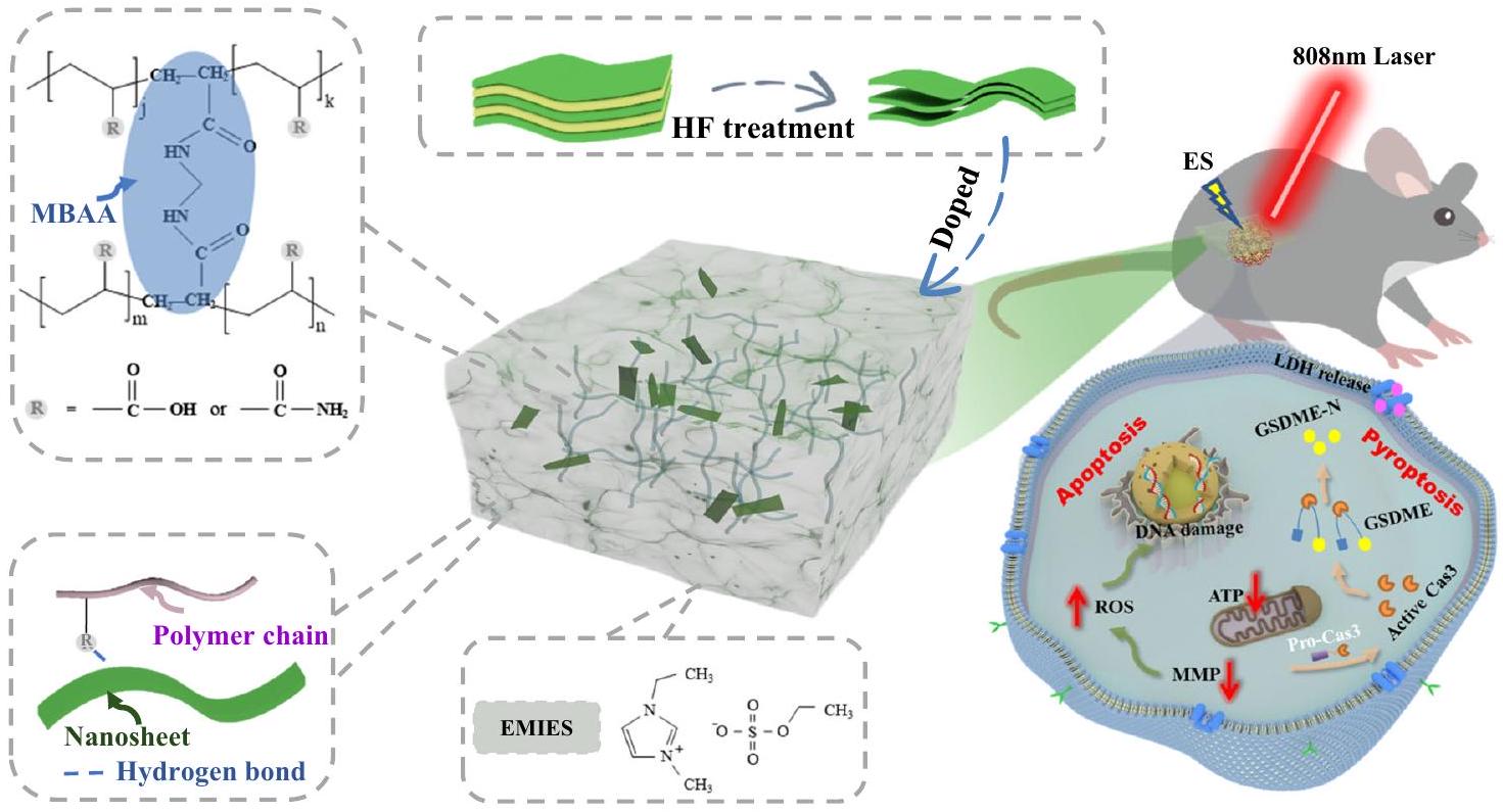
الشكل 1 | لاصقة eT القائمة على الهلام الأيوني لعلاج الأورام تحت الجلد. توضيح تخطيطي للاصقة البيولوجية eT التي تم إعدادها من هلام أيوني مخلوط بـ MXene وتطبيقها لعلاج أورام الجلد تحت التحفيز الضوئي الحراري والكهربائي المتزامن.
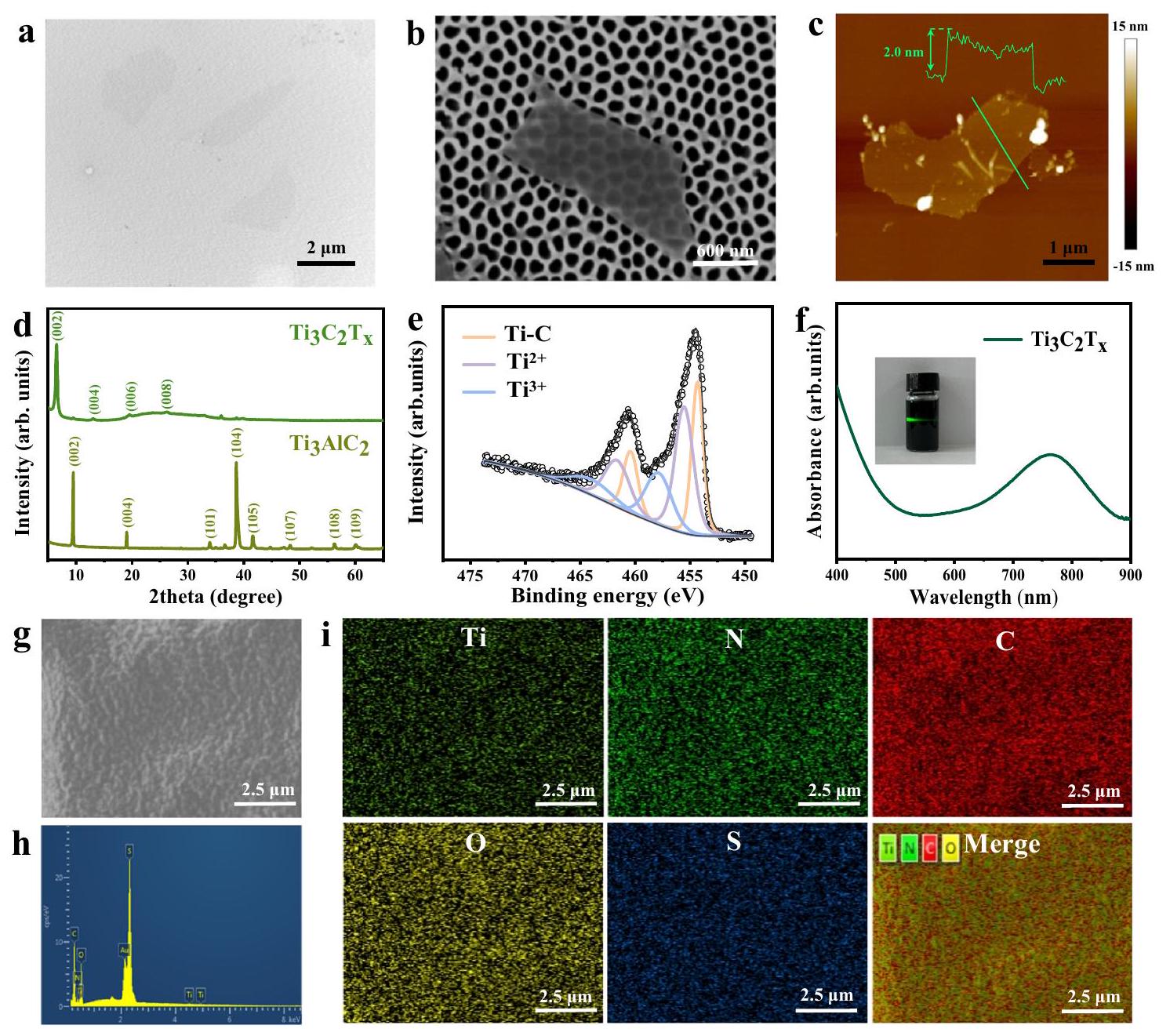
الشكل 2 | توصيفات العوامل الضوئية الحرارية MXene والهلام الأيوني. صورة TEM (أ)، صورة SEM (ب)، وصورة AFM وتحليل ارتفاع المسح الخطي (ج) لـعلى التوالي.حيود الأشعة السينية لـ و طيف التحليل الطيفي للأشعة السينية عالية الدقة لمنطقة Ti 2pرقائق نانوية.طيف الامتصاص للأشعة فوق البنفسجية والمرئية للسائل المستخرج
محلول تشتت رقائق النانو. صورة SEM لجيل الأيونات المدعوم بـ MXene. تحليل الطيف الكمي للطاقة لجيل الأيونات. تحليل خرائط العناصر SEM لرقعة eT. تم تكرار التجارب ثلاث مرات مع الحصول على نتائج مشابهة. بشكل رئيسي مجموعات النهاية السطحية لـ، و الشكل 2e يظهر التيتانيومطيف منتتوافق الأوراق النانوية مع 454.3 و 460.5 إلكترون فولت مع رابطة التيتانيوم-الكربون. تُنسب القمم عند 455.5 و 461.6 إلكترون فولت بشكل رئيسي إلىبينما قممكانت تقع عند 457.8 و 464.3 إلكترون فولت ولم يتم الكشف عن أي قمة عند 488.8 إلكترون فولت، مما يشير إلى عدم وجود أكسدة لـحدثت الأوراق النانوية أثناء عملية الفصل. الـالأغشية النانوية التي تم الحصول عليها عن طريق النقش يمكن أن تتوزع بشكل جيد في الماء وتظهر تأثير تيندال ملحوظ (كما هو موضح في الصورة المصغرة في الشكل 2f). في الوقت نفسه، فإن الخصائص البصرية للمستحلبتم فحص النانوصفائح في الماء باستخدام مطيافية امتصاص الأشعة فوق البنفسجية (الشكل 2f) وتم ملاحظة ذروة امتصاص كبيرة عند 768 نانومتر، والتي يمكن أن تتوافق بشكل جيد مع ليزر 808 نانومتر للعلاج الحراري اللاحق للسرطان. في هذا العمل، اخترنا خلايا B16F10 للتجارب التالية، والتي مشتقة من خلايا ورم عفوي من فأر C57BL/6J. توافقها الحيويتم التحقق من النانوصفائح بشكل إضافي باستخدام اختبار MTT القياسي (الشكل التوضيحي 2)، حيث أظهرت النتائج بقاء جيد للخلايا B16F10 بعد الحضانة معلأوراق النانو. تم ملاحظة ارتفاع درجة الحرارة الواضح في وسط MXene المعرض لأشعة الليزر بطول موجي 808 نانومتر، مقارنةً بالنقي. (الشكل التكميلي 3). كانت حيوية خلايا B16F10 التي تم تحضينها مع MXene تتناقص تدريجياً مع زيادة قوة الليزر (الشكل التكميلي 4). علاوة على ذلك، كانت كفاءة التحويل الضوئي الحراري لـالأغشية النانوية كانت محسوب ليكون (الشكل التوضيحي التكميلي 5)، أعلى من تلك الخاصة بعموديات الذهب النانوية و لذلك، تم استخدام طبقة النانو MXene كعامل ضوئي حراري مثالي للتطعيم في لاصقات الهلام الأيوني في التجارب التالية.
تم تحضير لاصقات الهلام الأيوني المدعمة بأوراق MXene النانوية بواسطة بلمرة مصباح الأشعة فوق البنفسجية في قالب (الشكل التكميلي 6). تم ملاحظة السطح النسبي المستوي والبنية الداخلية المسامية الطبقية لللاصقة من صور SEM، كما هو موضح في الشكل 2g والشكل التكميلي 7. أظهر طيف الطاقة SEM ورسم الخرائط العنصرية بوضوح التشتت المتجانس لعنصر التيتانيوم في الهلام الأيوني (الشكل 2h، i)، مما يدل على نجاح عملية الدوبينغ لـ MXene في الهلام الأيوني. بسبب التفاعل مع شبكة البوليمر لتشكيل روابط هيدروجينية بعد إضافة، فإن الرقع المركبة الناتجة تتمتع بمرونة كبيرة بعد الت doping، مقارنةً باللصقات المصنوعة من هلامات أيونية نقية. تم اختبار منحنيات الإجهاد والانفعال لللصقات بواسطة جهاز اختبار عالمي (الشكل 3a والشكل التكميلي 8). كما يتضح من الشكل 3b و 3c، فإن الإطالة عند الكسر والصلابة للهلامات الأيونية زادت مع زيادة محتوى الدوبينغ.التمدد عند الكسر (عند 5.75 ميغاباسكال) للهلام الأيوني المدعوم بـكانمع زيادة تردد الاهتزاز، يزيد معامل التخزين ( ) ومعامل الفقدان ( تم تعزيز ( ) من eT -patch (الشكل التوضيحي 9). مع زيادة التردد، قيمة أعلى من ذلك لـعند الترددات العالية، التي تتصرف كأنها لزجة
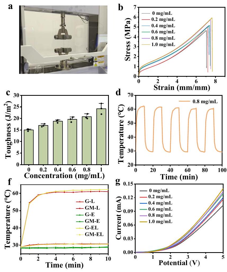
الشكل 3 | قياس أداء رقعة eT. أ صورة لرقعة هلامية أيونية أثناء التمدد. منحنيات الإجهاد والانفعال النموذجية (ب) والصلابة (ج) للرقعة المضافة إليها تركيزات مختلفة من MXene. ). ملف درجة حرارة الدراجات لرقعة eT المضافة بـMXene تحت إشعاع ليزر 808 نانومتر فيصور حرارية (e) ومنحنيات درجة الحرارة مقابل الزمن (f) لرقع eT ورقع الجل الأيوني النقي بدون إضافة MXene تحت الليزر، ES،
تحفيز الليزر وES المتزامن، على التوالي. منحنيات التيار-الجهد (g) والقيم الحالية المقابلة (h) عند 5 فولت لرقع الهلام الأيوني المخدرة بـ MXene بتركيزات مختلفة ( ). منحنيات الوقت الحالي لرقع eT ورقع الهلام الأيوني النقي تحت الإشعاع بالليزر 808 نانومتر في حالة الإيقاف/التشغيل. تُعرض البيانات كمتوسطSD.
مادةبشكل ملحوظ، تم تحسين أداء تحويل الفوتوحراري للرقع بشكل كبير بعد إضافة MXene، وارتفعت درجة حرارة الرقع تدريجياً مع زيادة تركيز MXene تحت إشعاع ليزر 808 نانومتر لمدة 10 دقائق (الشكل التوضيحي 10a). في الوقت نفسه، ارتفعت درجة حرارة الرقع بشكل واضح مع زيادة قوة الليزر وسمك الرقع (الشكل التوضيحي 10b، c). بشكل جذاب، كانت درجة الحرارة المقاسة في مناطق مختلفة من نفس الرقعة متشابهة أساساً (الشكل التوضيحي 10d)، مما يشير إلى أن إضافة MXene داخل رقعة الجل الأيوني متجانسة أساساً. في التجارب اللاحقة، تم إضافة MXene إلى رقعة الجل الأيوني عندتم استخدامه، حيث تصل درجة حرارته إلى حواليبعد تعرضه لأشعة الليزر بطول موجي 808 نانومتر لمدة 10 دقائق فيمن الجدير بالذكر أن الرقعة أظهرت استقرارًا حراريًا متفوقًا حتى عند تعرضها لليزر بزاوية 808 نانومتر لمدة 10 دقائق في كل مرة لخمس دورات (الشكل 3د). علاوة على ذلك، تم مراقبة تغيرات درجة الحرارة لمجموعات مختلفة (تخديم MXene في الجل (GM) والجل النقي (G)، مع التحفيز الكهربائي (E)، وإشعاع الليزر (L)، والعلاج المتزامن للتحفيز الكهربائي والليزر (EL)، على التوالي) من خلال التصوير الحراري ورسمها في منحنيات (الشكل 3هـ، و). أظهرت النتائج أن درجة حرارة رقعة الجل الأيوني النقي لم تتغير بشكل ملحوظ بعد الليزر والتحفيز الكهربائي، حيث كان للتحفيز الكهربائي تأثير ضئيل على تحويل درجة حرارة رقعة الجل الأيوني المخدوم بـ MXene، بينما زادت درجة حرارة رقعة الجل الأيوني المخدوم بـ MXene بشكل واضح مع زيادة وقت الإشعاع.
الرقعة المصنعة تتمتع بتوصيلية جيدة حتى لو تم لفها، حيث يمكن للتيار المار من خلالها أن يضيء بنجاح صمام ثنائي باعث للضوء تجاري (الشكل التكميلي 11 أ، ب). ثم تم قياس منحنيات التيار-الجهد للرقع المخدرة بتركيزات مختلفة من MXene باستخدام محطة عمل كيميائية كهربائية. (الشكل 3g)، تم تسجيل قيم التيار عند 5 فولت في تجارب متوازية عدة مرات (الشكل 3h)، مما أظهر أن قيم التيار ارتفعت تدريجياً مع زيادة محتوى تشبع MXene. قمنا أيضًا بقياس مقاومة اللصقات لتأكيد سلوكياتها في التيار-الجهد (الشكل التكميلي 12a). تم حساب قيم المقاومة لللصقات قبل وبعد تشبعها بـ MXene لتكون حوالي 1539 و (الشكل التكميلي 12b)، على التوالي. ومن المRemarkably، بالمقارنة مع رقعة الهلام الأيوني النقي، لوحظ تغيير واضح في التيار من رقعة الهلام الأيوني المدعمة بـ MXene (الشكل 3i)، نتيجة للتأثير الضوئي الحراري الممتاز في رقائق MXene التي تسببت في ارتفاع درجة الحرارة لتحفيز نقل الحرارة إلى السائل الأيوني المحيط. وهذا يعزز هجرة الحاملات بسرعة ويؤدي في النهاية إلى زيادة التيار.
لإثبات أن اللصقة الأيونية المطورة هنا تمتلك مزايا واضحة على لصقة الهيدروجيل، تم تحضير هيدروجيل بولي (أكريلاميد-كواكريليك) المدعوم بـ MXene كعنصر تحكم، وتم فحص الاستقرار الحراري، والتوصيل الكهربائي، وخصائص الشد لللاصقات الهيدروجيل. كما هو موضح في الشكل S13، أظهرت النتائج بوضوح الاستقرار الحراري الضعيف للدورة لللصقة الهيدروجيل مقارنة باللصقة eT (الشكل التكميلي 13a والشكل 3d)، كما يتضح من ارتفاع درجة الحرارة غير القابل للتحكم لللاصقات الهيدروجيل المدعومة بـ MXene تحت إشعاع الليزر المتقطع. كما أظهرت لصقات الجل الأيوني أيضًا توصيلًا كهربائيًا أفضل وقوة كسر أعلى وامتداد أكبر من لصقات الهيدروجيل (الأشكال التكملية 13b، c).
قبل التطبيقات الطبية الحيوية العملية، تم اختبار التوافق الحيوي لللصقة eT. أولاً، قمنا بزراعة خلايا B16F10 مع وسط الاستخراج () لمدة 24 ساعة، عن طريق نقع لصقات ذات جودة مختلفة في وسط الزراعة. كما هو موضح في الشكل التكميلي 14a،
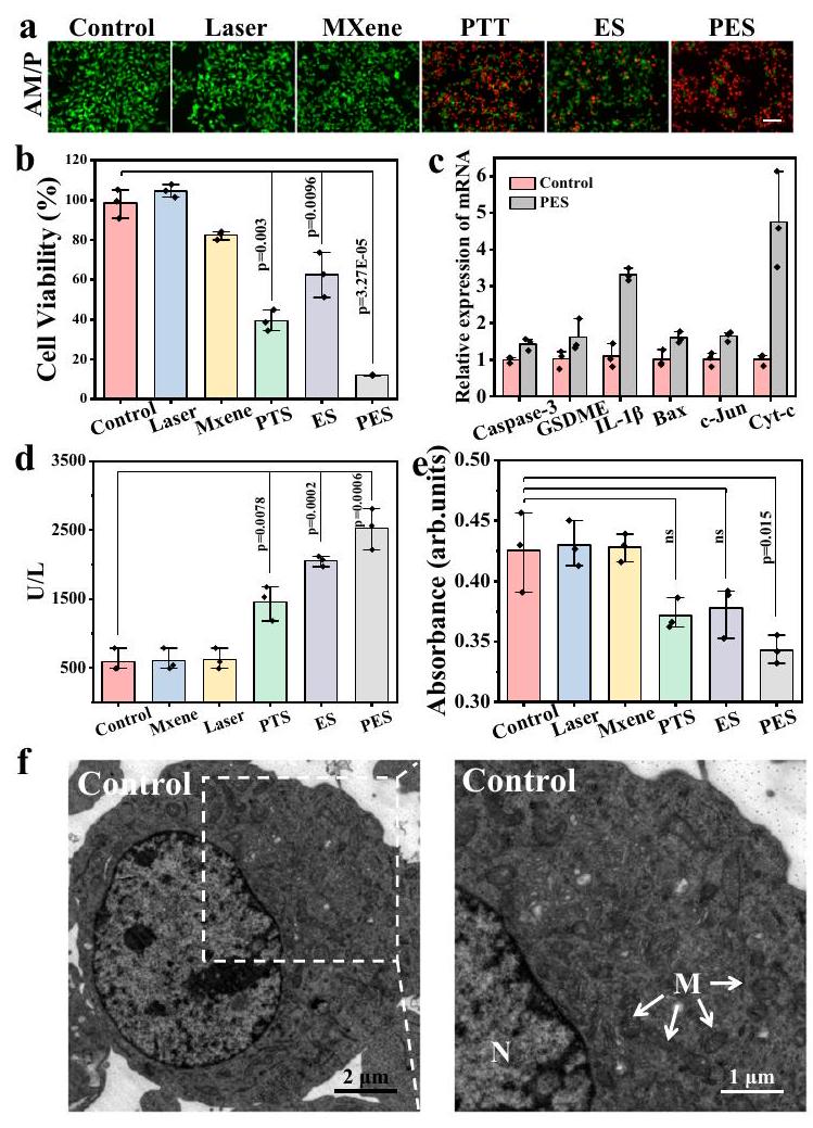
الشكل 4 | دراسة آلية PES لتحفيز موت الخلايا. أ صور فلورية لتلوين الخلايا الحية/الميتة لخلايا B16F10 بعد العلاجات المختلفة (التحكم، الليزر، MXene، PTT، ES، و PES). مقياس الرسم هو. ب بقاء الخلايا لخلايا B16F10 بعد العلاجات المختلفة.تم حساب القيم باستخدام اختبارين ذي طرفين– (). ج، تحليلات RT-PCR لـ Caspase-3، GSDME، IL-1, Bax، c-Jun، تعبير Cyt-c داخل خلايا B16F10 قبل وبعد علاجات PES. د مستويات إطلاق اللاكتات ديهيدروجيناز (LDH) من خلايا B16F10 بعد العلاجات تحت ظروف مختلفة.تم حساب القيم باستخدام اختبارين ذي طرفين– (). هـ تغيير محتوى ATP داخل خلايا B16F10 تحت نفس عدد الخلايا بعد العلاجات المختلفة التي تم اختبارها باستخدام مجموعة اختبار ATP.تم حساب القيم باستخدام اختبارين ذي طرفين–
– ().صور Bio-TEM لخلايا B16F10 قبل وبعد علاجات PES. “M” تمثل الميتوكوندريا، “N” تمثل نواة الخلية. ج صور فلورية لـ ROS داخل خلايا B16F10 تم اكتشافها باستخدام مسبار DCFH-DA بعد العلاجات المختلفة. مقياس الرسم هو.صور الفلورسنت لمونومر JC-1 والكتل، وصورهم المدمجة داخل خلايا B16F10 الملطخة بـ JC-1 بعد العلاجات المختلفة. مقياس الرسم هو. ط صور فلورية لتمزقات خيوط DNA المزدوجة داخل خلايا B16F10 بعد العلاجات المختلفة (تتوافق مع الفلورسنت الحمراء لـ) تليها صبغة DAPI (تتوافق مع الفلورسنت الزرقاء). مقياس الرسم هو. تم تكرار التجارب ثلاث مرات مع الحصول على نتائج مشابهة. البيانات مقدمة كمتوسط SD.
تم الحفاظ على بقاء جيد لخلايا B16F10 عند تركيز من وسط الاستخراج. قمنا بمزيد من فحص بقاء الخلايا لخلايا B16F10 التي تم زراعتها مع تركيزات أعلى من وسط استخراج اللصقة () لمدة 10 دقائق تليها زراعة مستمرة في وسط عادي لمدة 24 ساعة. كما هو موضح في الشكل التكميلي 14b، كانت معدلات بقاء الخلايا لجميع الخلايا المختبرة أعلى من، مما يشير إلى توافق حيوي جيد لللصقة بعد العلاج لمدة 10 دقائق. لإثبات أن اللصقة eT تمتلك توافقًا حيويًا جيدًا على مستوى الأنسجة، تم اختبار أنسجة لحم الخنزير الملطخة باستخدام الهيماتوكسيلين-إيوزين قبل وبعد لصق اللصقة (الشكل التكميلي 15). تشير النتيجة إلى أن اللصقات eT لا يمكن أن تدمر الأنسجة، مما يجعلها مناسبة للعلاج المحتمل لسرطان الجلد. على الرغم من أن تعليق MXene أسود، فإن خلط MXene مع الجل الأيوني يصبح شفافًا بسبب تغيير خصائصه البصرية الناتجة عن التحول من حالة الغرويات إلى حالة الجل. كما هو موضح في الشكل التكميلي 16، على الرغم من أن نفاذية اللصقات قد انخفضت تدريجيًا مع زيادة تركيزات MXene المدعوم، إلا أن الشفافية البصرية لللصقات لا تزال جيدة عند من دعامات MXene. وبالتالي، يمكن استخدام اللصقة eT المعدة لفحص استجابة الجلد وتقييم تأثيرات العلاج خلال عملية العلاج.
آليات موت الخلايا أثناء علاج PES
قبل تطبيق اللصقة eT لعلاج أورام الجلد، قمنا بفحص منهجي وكشف عن التأثيرات وآلية موت الخلايا لتحفيز الضوء الكهربائي لخلايا B 16 F 10. تم تطبيق تيارات مختلفة أولاً لعلاج خلايا B16F10 المزروعة على زجاجات ITO لمدة
10 دقائق، ثم تم اختبار بقاء الخلايا باستخدام صبغة الفلورسنت الحية/الميتة واختبار MTT (الشكل التكميلي 17)، والذي أظهر بوضوح أن بقاء الخلايا قد انخفض تدريجيًا مع زيادة التيار المحفز، مما يشير إلى أن زيادة التيار قد حسنت معدل قتل خلايا السرطان. ثم اخترنا قيمة التيار للتجارب اللاحقة. بعد ذلك، تم علاج خلايا B16F10 بطرق مختلفة (الشكل 4a، b)، وأشارت النتائج إلى أن فعالية قتل الخلايا لعلاج PES كانت أقوى بشكل ملحوظ من العلاجات الأخرى. من الواضح أن إشعاع الليزر في مجموعة التجارب PES (تطبيق كل من التحفيز الكهربائي والحراري الضوئي) لم يكن له فقط تأثير التيار المعزز، ولكن أيضًا تأثير حراري ضوئي، وكلاهما مفيد لقتل خلايا السرطان. بالإضافة إلى ذلك، اخترنا الخلايا الليفية الفأرية من خلايا L929 كخلايا طبيعية للتحقق مما إذا كانت الطريقة يمكن أن تسبب ضررًا أقل للخلايا الطبيعية. كما هو موضح في الشكل التكميلي 18، كانت خلايا L929 المختبرة تتمتع ببقاء خلايا أعلى مقارنة بخلايا B16F10. لذلك، كانت خلايا الورم أكثر عرضة للتضرر خلال عملية العلاج. من الواضح أننا لاحظنا أن الخلايا الميتة الناتجة عن ES أظهرت انتفاخًا واضحًا للخلايا، وهو سمة نموذجية من البيروبتوسيس، بينما كان موت الخلايا الناتج عن PTT هو موت الخلايا المبرمج بسبب التجاعيد الواضحة في شكل الخلية (الشكل التكميلي 19). وبالتالي، يمكن تحفيز كل من البيروبتوسيس وموت الخلايا المبرمج أثناء علاج PES، وكلاهما مفيد لعلاجات السرطان. للتحقق من أن PES يمكن أن يحفز البيروبتوسيس وموت الخلايا المبرمج لخلايا السرطان، تم إجراء تعبيرات الجينات ذات الصلة قبل وبعد علاج PES باستخدام RT-PCR (تفاعل البوليميراز المتسلسل العكسي). كما هو موضح في الشكل 4c، كانت تعبيرات الجينات
للمؤشرات من البيروبتوسيس (GSDME، Caspase-3، IL-1) وموت الخلايا المبرمج (Bax، c-Jun، Cyt-c) داخل خلايا B16F10 مرتفعة بشكل ملحوظ بعد علاج PES، مقارنة بمجموعة التحكم. لتأكيد أن البيروبتوسيس تم تحفيزه بواسطة ES، تم الكشف عن مستويات إطلاق اللاكتات ديهيدروجيناز (LDH) كعلامة حيوية كلاسيكية للبيروبتوسيسالتي تم الكشف عن مستويات إطلاقها بعد العلاجات المختلفة باستخدام مجموعة اختبار LDH التجارية. كما هو موضح في الشكل 4d، كانت مستويات إطلاق LDH من خلايا B16F10 في مجموعات PES و ES أعلى بشكل ملحوظ من تلك الموجودة في المجموعات الأخرى. في الوقت نفسه، وُجد أن مستوى ATP داخل خلايا B16F10 بعد علاج PES كان أقل من ذلك تحت العلاجات الأخرى (الشكل 4e). أظهرت هذه النتائج أن البيروبتوسيس القوي للخلايا تم تحفيزه بواسطة استراتيجية PES. علاوة على ذلك، لوحظ تدمير واضح للميتوكوندريا مع تجويف ملحوظ وانتفاخ من صور المجهر الإلكتروني الناقل البيولوجي (Bio-TEM)، كما هو موضح في الشكل 4f، مما يشير إلى خلل الميتوكوندريا بواسطة. بالإضافة إلى ذلك، تم اختبار مستوى ROS داخل خلايا B16F10 باستخدام 2,7-Dichlorodihydrofluorescein diacetate (DCFH-DA) الذي يمكن تحليله لتوليد أغشية DCFH غير القابلة للاختراق، والتي تنتشر داخليًا وتتعرض للأكسدة بواسطة الجذور الحرة غير المحددة لتوليد DCF الفلوري الأخضر. كما هو موضح في الشكل 4g، لوحظ تصوير فلوري أخضر مميز لخلايا B16F10 من مجموعات PTT وES. ومع ذلك، أظهرت خلايا B16F10 صور فلورية خضراء ضعيفة بسبب تمزق غشاء الخلية بعد التحفيز المشترك بالليزر والكهرباء. نظرًا لأن جهد غشاء الميتوكوندريا (MMP) مهم للحفاظ على وظيفة الميتوكوندريا الخلوية، وغالبًا ما يرتبط انخفاض MMP بتراكم ROS، تم فحص MMP داخل الخلايا بعد العلاجات المختلفة باستخدام مجموعة اختبار JC-1 (الشكل 4h)، والتي أكدت أن MMP داخل الخلايا بعد علاج PES قد انخفض بشكل كبير، مما أدى إلى اختفاء الفلورسنت الأحمر وتوليد الفلورسنت الأخضر. كما هو معروف أن ارتفاع مستوى ROS يمكن أن يؤدي إلى تلف الحمض النووي في الخلايا ، ثم استخدمنا صبغة المناعية الفلورية للتحقيق في الانكسارات المحتملة في خيوط الحمض النووي المزدوجة التي قد تحدث بعد العلاجات المختلفة. كما هو موضح في الشكل 4i، تسبب علاج PES في أعلى درجة من تلف الحمض النووي في مجموعات الاختبار (PTT وES وPES)، مما أكد بشكل غير مباشر توليد مستويات أعلى من ROS من علاج PES، وأن ROS الناتجة يمكن أن تتسبب في تلف شديد للحمض النووي في النواة.
تقييم الفعالية العلاجية للرقعة الإلكترونية لعلاج الميلانوما
لتقييم تأثير العلاج للرقعة الإلكترونية لعلاج ورم الجلد تحت PES، تم إنشاء نموذج الفئران C57BL/6J الحاملة لورم B16F10 من خلال زراعة خلايا الورم تحت الجلد، وعرض بروتوكول العلاج في الشكل 5a. تم تقسيم الفئران الحاملة للورم بحجم حوالي أولاً بشكل عشوائي إلى خمس مجموعات تشمل مجموعة التحكم، الليزر، PTT، ES، وPES. بعد ذلك، تم علاج الفئران بطرق مختلفة لمدة 10 دقائق لكل علاج على مدى يومين متتاليين، ثم تم مراقبتها لمدة 13 يومًا. تم تقدير تأثير العلاج لطرق مختلفة بعد 15 يومًا. لكشف أين تتدفق الكهرباء، تم الكشف عن قيم التيار المتدفقة عبر الرقعة الإلكترونية قبل وبعد تغطيتها على الورم وحسابها، كما هو موضح في الشكل S20. تم حساب قيمة التيار المار عبر الرقعة المستقلة (المشار إليها بـ ) لتكون حوالي عند 5 فولت. من الجدير بالذكر أن قيمة التيار ( ) المسجلة عند 5 فولت زادت إلى بعد التصاق الرقعة بالورم، مما يشير إلى وجود علاقة مقاومة متوازية بين الورم والرقعة الإلكترونية. يمكن أن يتدفق التيار الكهربائي عبر الطبقة السطحية للورم عبر الرقعة، كما هو موضح في الشكل S20b. لفهم أفضل، رسمنا مخططات الدائرة المقابلة لكل من الحالتين (الشكل التكميلي 20c، d)، وتم وضع علامة على اتجاه تدفق التيار أثناء العلاج بشكل متناسب. من أجل تقدير العمق الفعال للعلاج، تم أخذ أنسجة الورم من الفئران بعد علاج PES لصبغ H&E وTUNEL. كما يتضح من الشكل التكميلي 21، يمكن أن يصل العمق الفعال للطريقة
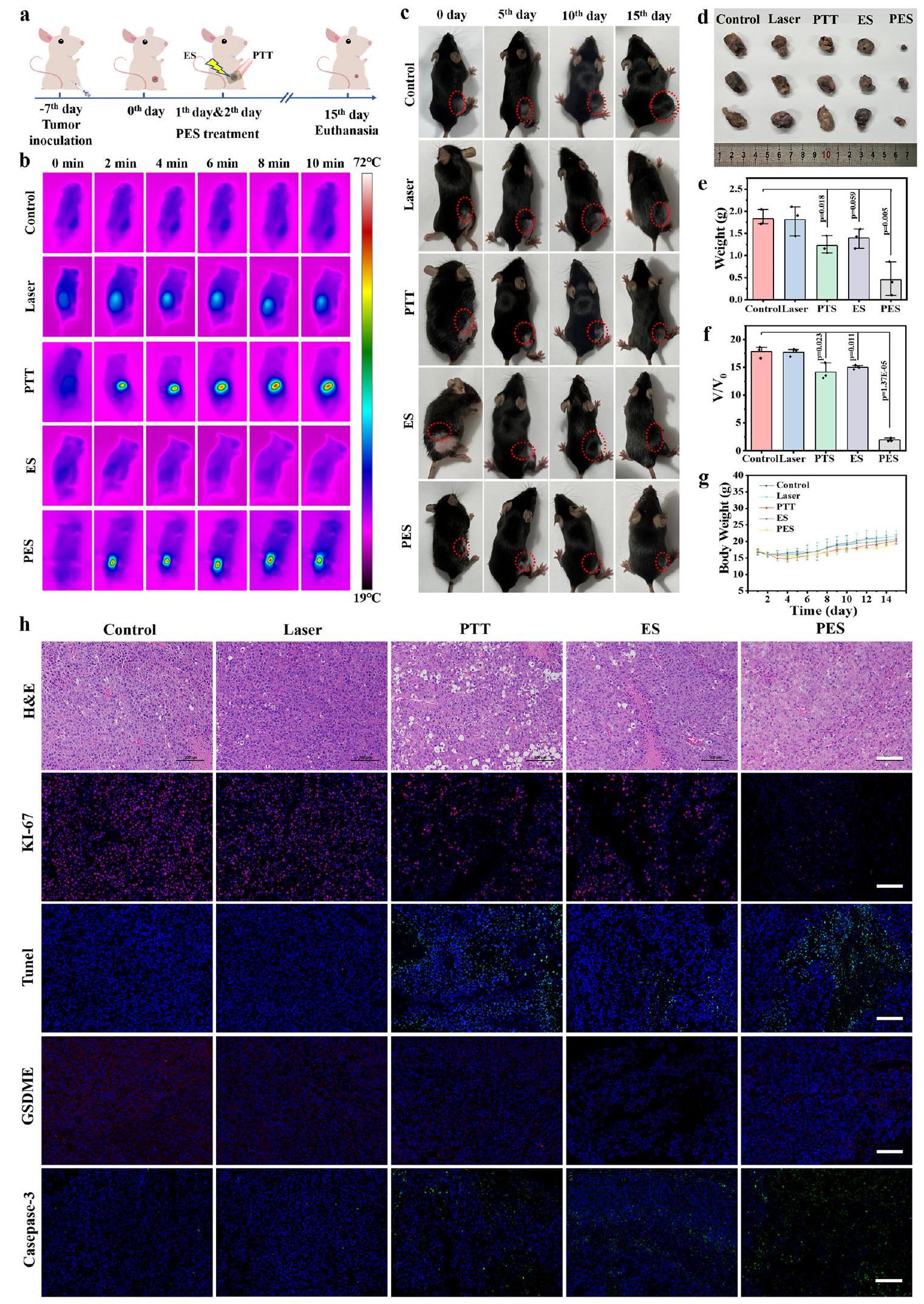
لإزالة الورم إلى . بشكل خاص، عرضت الصور الحرارية بالأشعة تحت الحمراء في الوقت الحقيقي للفئران الحاملة للورم في مجموعات PTT وPES زيادة سريعة في درجة الحرارة المحلية خلال عملية العلاج، مما يوحي بأن علاجات PTT وPES مناسبة تمامًا لإزالة الورم (الشكل 5b). أظهرت التصوير الحراري للورم وملفات درجة الحرارة التي تركزت على الورم والمنطقة المحيطة (الشكل التكميلي 22) أن درجة حرارة المنطقة المحيطة بالورم حوالي ، وهو أقل احتمالًا لتسبب في تلف الأنسجة الطبيعية المحيطة أثناء علاج الورم. تم تسجيل أحجام الأورام للفئران بعد العلاجات المختلفة من خلال الصور الضوئية (الشكل 5c)، مما أثبت أن تأثير التثبيط الكبير قد تم تعزيزه تحت علاج PES. تم تشريح أنسجة الورم بعد العلاجات المختلفة ووزنها، وكما هو موضح في الشكل 5d، e، الشكل التكميلي 23 (يمكن العثور على أوزان أجسام الفئران والأورام في مجموعات مختلفة في الجدول التكميلي 2)، فإن الرقعة الإلكترونية لها تأثير مضاد للورم متفوق. تم حساب معدل التثبيط المتوسط بناءً على حجم الورم النسبي ليكون ، 14.13، 14.98، و1.96، على التوالي، كما هو موضح في الشكل 5f (يمكن العثور على أحجام الأورام المطلقة في مجموعات مختلفة في الجدول التكميلي 3). ولم يكن هناك فقدان كبير في وزن الجسم للفئران تحت المجموعات المختلفة (الشكل 5g)، مما يدل على انخفاض السمية النظامية للمواد والطريقة. لتقييم فعالية العلاج بشكل أفضل، تم إجراء مزيد من صبغات الهيماتوكسيلين والإيوزين (H&E) وKi-67، وأظهرت النتائج أن علاج PES مع الرقعة الإلكترونية له أكبر ضرر للورم وتثبيط التكاثر (الشكل 5h). للتحقق من درجة الضرر للورم بعد علاج PES، تم إجراء صور TEM الحيوية لمراقبة شكل الخلايا من موقع الورم. كما هو موضح في الشكل التكميلي 24، تم تدمير العضيات الميتوكوندريا بشكل خطير بعد PES، مقارنة بمجموعة التحكم. علاوة على ذلك، كشفت صور المناعية الفلورية TUNEL لأنسجة الورم أن علاجات PTT وPES للرقعة الإلكترونية حصلت على أعلى مستوى من موت خلايا السرطان. لاحظ أن الكاسبيز-3 كعلامة على الموت الخلوي يمكن تنشيطه لقطع بروتين GSDME لتحفيز موت الخلايا ، كما هو موضح في الشكل 5h، كان مستوى تعبير بروتين GSDME منخفضًا بشكل كبير في مجموعات ES وPES لأن المزيد من الكاسبيز-3 تم تنشيطه بعد ES لقطع GSDME. من الواضح أن كل من موت الخلايا والموت الخلوي تم تحفيزهما تحت علاج PES للرقعة الإلكترونية، وكلاهما سيفيد فعالية مضادة للورم للطريقة. بشكل جذاب، تحسنت نسبة بقاء الفئران الحاملة للورم بشكل كبير بعد علاج PES (الشكل التكميلي 25). تم تقييم انتشار الورم بعد العلاج أيضًا من خلال تصوير H&E. كما هو موضح في الشكل التكميلي 26، لم يتم ملاحظة أي عقيدات في رئتي الفئران من جميع المجموعات، مما يشير إلى أن العلاج لم يكن له تأثير محفز على انتشار السرطان.
بعد ذلك، تم فحص الأنسجة الطبيعية المحيطة بالورم في مجموعة PES تحت أيام مختلفة باستخدام صبغة H&E. كما هو موضح في الشكل التكميلي 27a، لا تتعرض الأنسجة الطبيعية المحيطة بالورم للتلف خلال عملية PTT. على الرغم من أن الأنسجة التي فوق الورم قد دمرت، إلا أن أنسجة الجلد والعضلات للفئران قد عادت إلى طبيعتها بعد 15 يومًا، كما يتضح من صور صبغة H&E (الشكل التكميلي 27b). تم أيضًا فحص الأعضاء الرئيسية للفئران بعد العلاجات المختلفة بواسطة صبغة H&E (الشكل التكميلي 28). أظهرت النتيجة أن علاج PES للرقعة الإلكترونية ليس له تغييرات مرضية واضحة في الفئران. بالإضافة إلى ذلك، لم تكن هناك شذوذات كبيرة في اختبارات الدم الروتينية للعلامات الحيوية الدموية في كل مجموعة علاج مقارنة بمجموعة التحكم، ولم تتمكن تحليل التجميع من تمييزها تمامًا (الأشكال التكملية 29، 30). جميع النتائج المذكورة أعلاه تشير إلى أن علاج PES للسرطان باستخدام الرقعة الإلكترونية له سلامة بيولوجية ممتازة.
نقاش
باختصار، قمنا بتطوير رقعة إلكترونية قابلة للارتداء واستجابة مزدوجة تتكون من هلام أيوني مخلوط بـ MXene، والتي تم تطبيقها لعلاج الميلانوما بشكل فعال من خلال الحرارة الضوئية والكهربائية.
الشكل 5 | العلاج في الجسم الحي مع نموذج الميلانوما. أ مخطط توضيحي لخلايا B16F10
حجم الأورام التي تم تشريحها من كل مجموعة بعد العلاج لمدة 15 يومًا. تم حساب القيم بواسطة اختبارات – ذات طرفين. ( ). ج منحنيات وزن الجسم للفئران الحاملة للورم في مجموعات مختلفة. ( ). ح صبغات H&E، Tunel، Ki-67، GSDME وكاسبيز-3 لأنسجة الورم المجمعة من الفئران المقابلة في اليوم الخامس عشر من كل علاج. مقياس الشريط هو . تم تكرار التجارب ثلاث مرات مع الحصول على نتائج مماثلة. تم تكرار التجارب ثلاث مرات مع الحصول على نتائج مماثلة. البيانات مقدمة كمتوسط SD. العلاج المشترك بالتحفيز الكهربائي (PES). الشفافية الجيدة للرقعة الإلكترونية eT-patch تحقق فحصًا بصريًا في الوقت الحقيقي لاستجابة الجلد وتقييم تأثير العلاج على ورم الجلد. تم الكشف أوليًا عن الآليات الخلوية الأساسية لعلاج PES، والتي كانت أن كل من الموت الخلوي المبرمج (apoptosis) والموت الخلوي الالتهابي (pyroptosis) تم تحفيزهما، مما يرتبط بتقليل MMP الناتج عن ROS وإصابة الحمض النووي. بشكل مثير للإعجاب، فإن علاج PES لسرطان الجلد باستخدام الرقعة الإلكترونية eT-patch القابلة للتصنيع بسهولة يتمتع بسلامة حيوية عالية واستقرار، وأعراض جانبية أقل، وتعديل مرن. تقدم هذه الدراسة فرصًا جديدة لاستكشاف مواد الرقع لعلاج الميلانوما بشكل فعال مع تجنب مخاطر الجراحة، وستوسع التطبيقات الطبية الحيوية للهلام الأيوني.
طرق
بيان الأخلاقيات
تم تربية جميع الفئران ورعايتها وفقًا للإرشادات الخاصة بالحيوانات المخبرية في جامعة جيلين. في الوقت نفسه، تمت الموافقة على جميع الإجراءات المتعلقة بالحيوانات من قبل لجنة إدارة الحيوانات المخبرية في جامعة جيلين.
تركيب لاصقة هلامية أيونية مدعمة بـ MXene
من المعروف أن بولي أكريلاميد (الهيدروجيل) له توافق حيوي جيد للتطبيقات الطبية الحيوية. الأكريلاميد غير قابل للذوبان في السوائل الأيونية، بينما حمض الأكريليك النقي قابل للذوبان في السوائل الأيونية. يمكن أن يتفاعل الأكريلاميد وحمض الأكريليك في السوائل الأيونية بشكل عشوائي لتشكيل هلام أيوني، والذي يتمتع بقوة كسر عالية من خلال تبديد الطاقة عبر الروابط الهيدروجينية. لذلك، تم اختيار الأكريلاميد وحمض الأكريليك كمواد سابقة لتحضير رقع الهلام الأيوني في هذا العمل. أولاً، تم إذابة 0.54 جرام من حمض الأكريليك (AA) و1.5975 جرام من الأكريلاميد (AAM) بالكامل في 2 مل من محلول 1-إيثيل-3 ميثيل إيميدازوليوم (EMIES) للحصول على محلول متجانس. تم وزن كمية معينة من مسحوق MXene وجعلها موزعة بشكل متساوٍ في 1.3 مل من محلول EMIES لخلطها جيدًا. بعد ذلك، تم استخدام الرابط المتقاطع N، N’-ميثيلين ثنائي (أكريلاميد) (MBAA، ) ومبادر الضوء إيرغاكورتمت إضافتها للحصول على محلول السلف. تم صب محلول السلف في القالب ثم تم تعريضه للأشعة فوق البنفسجية (تقريباً ) لمدة 5 دقائق. أخيرًا، تم الحصول على لاصقة هلامية أيونية مدعمة بـ MXene للاستخدام.
زراعة الخلايا
تم الحصول على خلايا الميلانوما (B16F10) من مجموعة الثقافة الأمريكية (ATCC، الولايات المتحدة الأمريكية). تم زراعة خلايا BF16-F10 في وسط 1640 معمصل الجنين البقري (FBS) والبنسيلين ستربتوميسين وتم الحفاظ عليه في و حاضنة مرطبة.
تقييم التوافق الحيوي لرقعة MXene والهلام الأيوني
عادةً ما تم الكشف عن سمية الجل الأيوني و MXene باستخدام اختبار MTT القياسي. خلايا B16F10 (تم زراعة ( ) في كل بئر من أطباق 96 بئر للتكاثر. بعد ذلك، تم غسل الخلايا باستخدام PBS ثلاث مرات وتم تحضينها مع تركيزات مختلفة من MXene المذاب في وسط 1640 الكامل عندفيلمدة 24 ساعة. بعد ذلك، تم تنظيف الخلايا باستخدام PBS ثلاث مرات ثممحلول MTT ( ) تم إضافته إلى من 1640 تم الانتهاء من الوسط للحضن لمدة 4 ساعات أخرى. أخيرًا، تم إزالة محلول كل بئر وتم إضافة DMSO إلى كل بئر بعد التفاعل لذوبان بلورات الفورمازان الأرجوانية. تم قياس امتصاص الآبار باستخدام قارئ الميكرو بلايت عند طول موجي 570 نانومتر. في الوقت نفسه، لتقييم سمية لاصقة الهلام الأيوني، تم الحصول على مستخلصات الهلام عن طريق نقع كتل مختلفة من الهلام في الوسط لمدة 10 دقائق للحصول على تركيزات مختلفة من مستخلصات الهلام تتراوح من 2 إلى. بعد ذلك، تم معالجة تركيزات مختلفة من مستخلصات الجل مع خلايا B16F10 لمدة 24 ساعة وتم الكشف عنها باستخدام اختبار MTT بناءً على الإجراءات المذكورة أعلاه.
حيوية خلايا B16-F10 بعد التحفيز الكهربائي تحت تيارات مختلفة
باختصار، تم زراعة خلايا B16F10 على زجاج موصل (زجاج ITO) للحضانة فيحاضنة فيلمدة 12 ساعة. بعد ذلك، تم غسل الخلايا باستخدام PBS ثلاث مرات وعولجت بتيارات مختلفة (، و ) تحت نظام ثلاثة أقطاب لمدة 10 دقائق. تم اعتبار قطب الخلية كقطب عمل وتم تعيين ورقة البلاتين كقطب مضاد. كان القطب المرجعي هو قطب Ag/AgCl. بعد التحفيز الكهربائي للخلية، تم زراعة الخلايا بشكل متتابع لمدة 30 دقيقة. بعد ذلك، تم صبغ الخلايا باستخدام مجموعة اختبار الخلايا الحية والميتة لمدة 20 دقيقة ثم تم تنظيفها باستخدام محلول PBS ثلاث مرات. تم ملاحظة التصوير الفلوري للخلايا باستخدام المجهر الفلوري تحت كائن. في هذه الأثناء، لتقدير حيوية الخلايا بعد التحفيز الكهربائي تحت تيارات مختلفة، تم هضم الخلايا باستخدام التربتاز وتم الكشف عنها باستخدام مجموعة اختبار MTT بناءً على الإجراءات التجريبية المذكورة أعلاه.
حيوية خلايا B16-F10 التي تم حضنها مع MXene بعد التعرض للإشعاع بالليزر تحت قوى ليزر مختلفة
عادةً، خلايا B16F10 (تم زراعة الخلايا) في كل بئر من صفيحة 96 بئرًا لت incubate لمدة 12 ساعة. ثم تم معالجة الخلايا المنظفة بـ MXene (تم إذابته في وسط كامل لمدة 24 ساعة. بعد ذلك، تم غسل الخلايا لإزالة محلول MXene وأضيف وسط زراعة جديد للتعرض لليزر بزاوية 808 نانومتر لمدة 10 دقائق تحت قوى ليزر مختلفة.، و ). بعد ذلك، تم حضن الخلايا لمدة ساعتين إضافيتين. أخيرًا، تم الكشف عن حيوية خلايا B16F10 باستخدام اختبار MTT القياسي.
تقييم تأثير العلاج لخلايا B16-F10 تحت طرق مختلفة
تم تقييم كفاءة العلاج الكهربائي الحراري المضاد للسرطان في المختبر في خلايا السرطان B16F10. هناك ست مجموعات علاجية مختلفة مقسمة: مجموعة التحكم، الليزر، MXene، العلاج الحراري الضوئي (MXene + ليزر)، ES، و PES (MXene + ليزر + ES). خلايا B16F10 (تم زراعة الخلايا أولاً على زجاجات ITO وتم حضنها لمدة 12 ساعة. ثم تم غسل الخلايا باستخدام PBS ثلاث مرات وتم حضن ثلاث مجموعات بوسط الثقافة المحتوي على MXene. ) لمدة 24 ساعة. لم تتلق مجموعة التحكم أي علاج. تعرضت مجموعتا الليزر والعلاج الضوئي الحراري لليزر بقدرة 808 نانومتر.لمدة 10 دقائق. تم تحفيز مجموعتي ES و PES بـتم تطبيق تيار لمدة 10 دقائق، وكانت الفارق هو أن مجموعة PES تم علاجها باستخدام إشعاع الليزر بطول موجي 808 نانومتر بالإضافة إلى التحفيز الكهربائي. بعد العلاج، تم زراعة الخلايا بشكل متتابع لمدة 30 دقيقة. بعد ذلك، تم إزالة الخلايا من زجاجات ITO باستخدام التربسين وتم حضنها في أطباق 96 بئر لاختبار MTT. في الوقت نفسه، تم الكشف عن صور الفلورسنت لخلايا B16F10 بعد العلاجات من خلال صبغة التلوين الحية/الميتة (كالسيوم-AM: ). ثم تم غسل الخلايا باستخدام PBS ثلاث مرات لمدة 15 دقيقة وتمت ملاحظتها باستخدام المجهر الفلوري تحت هدف.
كشف كسور الشريط المزدوج للحمض النووي
تم اختبار تلف الحمض النووي داخل الخلايا بعد علاجات مختلفة باستخدام التألق المناعي. تم تثبيت خلايا B16F10 بـبارافورمالدهيد في PBS لمدة 15 دقيقة عندبعد العلاجات المختلفة. بعد ذلك، تم غسل الخلايا باستخدام PBS ثلاث مرات وتم اختراقها بواسطةترايتون X-100 لمدة 15 دقيقة لتحسين نفاذية غشاء الخلية في درجة حرارة الغرفة. بعد غسلها ثلاث مرات بمحلول PBS، تم حجب الخلايا باستخدام 1% BSA في PBS لمدة ساعة واحدة في درجة حرارة الغرفة وتم تحضينها مع الأجسام المضادة الأولية المخففة.الأجسام المضادة (ab81299) فيبين عشية وضحاها. في اليوم التالي، تم غسل الخلايا ثلاث مرات بمحلول PBS وتم تحضينها مع الأجسام المضادة الثانوية المربوطة بـ Cy3 المخففة في الظلام عند درجة حرارة الغرفة لمدة ساعة واحدة. ثمتم إضافة DAPI لصبغ نواة الخلية وتم غسل الخلايا ثلاث مرات بمحلول PBS وتمت ملاحظتها تحت المجهر الفلوري.
جهد غشاء الميتوكوندريا لخلايا B16F10 بعد العلاجات
تم قياس الجهد الغشائي للميتوكوندريا داخل الخلايا باستخدام مجموعة الفحص التجارية JC-1. تم صبغ الخلايا باستخدام مجموعة فحص JC-1 لمدة 15 دقيقة بعد علاجات مختلفة. بعد ذلك، تم غسل الخلايا باستخدام PBS ثلاث مرات. تم ملاحظة وجمع صور الفلورسنت للخلايا باستخدام ميكروسكوب لايكا DMI6000B مع كاشف فلوروسنت.هدف (EM: (JC-1 مونومر) و EM: (مجموعة JC-1)).
كشف الأدينوزين ثلاثي الفوسفات (ATP) في خلايا B16F10 بعد التحفيز
تم استخدام مجموعة اختبار ATP التجارية للكشف عن التغيرات الطاقية المتعلقة بخلايا B16F10 بعد تحفيزات مختلفة. بعد حضانة الخلايا المحفزة لمدة 30 دقيقة، تم تنظيف الخلايا ثلاث مرات بمحلول PBS. بعد ذلك، تم جمع الخلايا باستخدام محلول التربسين وتم إزالة السائل العلوي. ثمتم إضافة محلول مستخلص الخلايا إلى أنبوب الطرد المركزي وتم استخدام الموجات فوق الصوتية في حمام ثلجي لمدة دقيقة واحدة. ثم تم طرد الخليط عند 3420 ج.لمدة 10 دقائق وتمت إزالة السائل العلوي من أنبوب الطرد المركزي. الكلوروفورمثم أُضيف إلى الأنبوب وتم خلطه مع تذبذبات كاملة قبل أن يتم الطرد المركزي مرة أخرى عند 3420 ج.لمدة 3 دقائق. تم خلط السائل الطافي النهائي مع السائل العامل وفقًا للتعليمات، وتم الكشف عن الامتصاص عند 340 نانومتر باستخدام علامة إنزيمية.
تجارب الحيوانات
تم شراء فئران C57BL/6J (عمرها 4 أسابيع، إناث) من شركة بكين HFK للتكنولوجيا الحيوية المحدودة (بكين، الصين). تم تربية جميع الفئران ورعايتها وفقًا للإرشادات الخاصة بالحيوانات المخبرية في جامعة جيلين. في الوقت نفسه، تمت الموافقة على جميع الإجراءات المتعلقة بالحيوانات من قبل لجنة إدارة الحيوانات المخبرية في جامعة جيلين. يجب ألا يتجاوز قطر الأورام 1.5 سم خلال عملية العلاج؛ إذا تجاوزت ذلك أو أثرت على جودة بقاء الحيوان، يجب قتل الفئران. في عملنا، قمنا بمراقبة وتسجيل حجم الورم خلال عملية التجربة. في اليوم الخامس عشر، وجدنا أن الأورام في مجموعة التحكم ومجموعة الليزر تجاوزت الحد القياسي وبلغت الدورة التجريبية، ووفقًا للوائح لجنة إدارة الحيوانات المخبرية في جامعة جيلين، قمنا بقتل الفئران في الوقت المناسب، امتثالًا للوائح. قبل وبعد العلاج، تم تنظيف مواقع الأورام في المجموعات المختلفة باستخدام الإيثانول 75% لتجنب العدوى. تم تعقيم الأنسجة الإضافية المتضررة من الفئران وتم تنظيف وتعقيم بيئة الفئران بانتظام. بالإضافة إلى ذلك، تم تحديد عدد الفئران إلى خمسة في كل قفص وتم تربيتها في بيئة معقمة ومهوّاة دون أي علاج بعد العلاج. تم الحفاظ على بيئة الحيوانات عند درجة حرارةوعندالرطوبة مع دورة ضوء/ظلام مدتها 12 ساعة، مع الوصول الحر إلى الطعام والماء القياسي. تم حساب حجم الورم على النحو التالي: حجم الورمطول الورم.
علاج في الجسم الحي باستخدام نموذج الميلانوما
تم إنشاء نموذج الورم عن طريق الحقن تحت الجلد بخلايا B16F10الخلايا فيPBS) في الجزء الخلفي من الفئران. بعد تلقيح خلايا الورم لمدة 7 أيام (قيمة الورم )، تم تقسيم الفئران عشوائيًا إلى خمس مجموعات ( ) كما يلي: (i) التحكم، (ii) الليزر، (iii) PTT، (iv) ES، (v) PES. بالنسبة لمجموعة علاج الليزر، تم تعريض الأورام في كل فأر لليزر بزاوية 808 نانومتر (، مرة واحدة) لمدة يومين متتاليين؛ وللمجموعة التجريبية PTT، تم تطبيق لصقة eT على سطح الورم وتركيز الليزر على موقع الورم. بالنسبة للمجموعة التجريبية ES، تم إجراء ES مع التيار بواسطة مصدر جهد مستقر للتيار المباشر من خلال رقعة eT التي تغطي الورم؛ بينما بالنسبة لمجموعة PES، تم تطبيق كل من الليزر والتحفيز الكهربائي على رقعة eT على سطح الجلد للورم الميلانيني. درجة حرارة سطح الورم و تمت مراقبة وتسجيل الصور الحرارية بواسطة نظام التصوير الحراري بالأشعة تحت الحمراء. تم قياس حجم الورم كل يومين باستخدام الكالبر.
ملخص التقرير
معلومات إضافية حول تصميم البحث متاحة في ملخص تقارير مجموعة ناتشر المرتبط بهذه المقالة.
توفر البيانات
البيانات الرئيسية التي تدعم النتائج في هذه الدراسة متاحة ضمن الورقة والمعلومات التكميلية. جميع مجموعات البيانات الخام والمحللة التي تم إنشاؤها خلال الدراسة متاحة من المؤلفين المقابلين عند الطلب.
References
Zeng, H. et al. Melanoma and nanotechnology-based treatment. Front. Oncol. 12, 858185 (2022).
Du, S. et al. Self-powered and photothermal electronic skin patches for accelerating wound healing. Nano Energy 93, 106906 (2022).
Yao, S. et al. Self-driven electrical stimulation promotes cancer catalytic therapy based on fully conjugated covalent organic framework nanocages. Adv. Funct. Mater. 32, 2209142 (2022).
Ding, Y. et al. Preparation of high-performance ionogels with excellent transparency, good mechanical strength, and high conductivity. Adv. Mater. 29, 1704253 (2017).
Hyun, W. J., Thomas, C. M. & Hersam, M. C. Nanocomposite ionogel electrolytes for solid-state rechargeable batteries. Adv. Energy Mater. 10, 2203988 (2020).
Ren, Y. et al. Ionic liquid-based click-ionogels. Sci. Adv. 5, eaax0648 (2019).
Tang, X. et al. A novel ionic liquid-based electrolyte assisting the high performance of low-temperature supercapacitors. J. Mater. Chem. A 10, 18374-18382 (2022).
Luo Z., Li W., Yan J., Sun J. Roles of ionic liquids in adjusting nature of ionogels: a mini review. Adv. Funct. Mater. 32, 2203988 (2022).
Huang, H. et al. Biomedical engineering of two-dimensional MXenes. Adv. Drug Deliv. Rev. 184, 114178 (2022).
Long, Y., Li, J., Yang, F., Wang, J. & Wang, X. Wearable and implantable electroceuticals for therapeutic electrostimulations. Adv. Sci. 8, 2004023 (2021).
Chu, B. et al. Triboelectric current stimulation alleviates in vitro cell migration and in vivo tumor metastasis. Nano Energy 100, 107471 (2022).
Schofield, Z. et al. Bioelectrical understanding and engineering of cell biology. J. R. Soc. Interface 17, 20200013 (2020).
Dong, Z. Y. et al. Ascl1 regulates electric field-induced neuronal differentiation through PI3K/Akt pathway. Neuroscience 404, 141-152 (2019).
Qi, G. et al. Wet-chemical electro-plasmonic modulation of metasurfaced cell-electrode interfaces for effective and selective entropic killing of cancer cells. Anal. Chem. 93, 13624-13631 (2021).
Chen, S. W. et al. A facile, fabric compatible, and flexible borophene nanocomposites for self-powered smart assistive and wound healing applications. Adv. Sci. (Weinh.) 9, e2201507 (2022).
Song, S. et al. Electrical stimulation of human neural stem cells via conductive polymer nerve guides enhances peripheral nerve recovery. Biomaterials 275, 120982 (2021).
Ma K. et al. Ultrasound-activated -based Trojan nanogenerators for combined targeted electro-stimulation and enhanced catalytic therapy of tumor. Nano Energy 87, 106208 (2021).
Qi G. et al. Imaging guided endogenic -augmented electrochemo-sonodynamic domino co-therapy of tumor in vivo. Adv. Mater. 35, 2208414 (2022).
Qi, G., Zhang, M., Tang, J. & Jin, Y. Molecular/nanomechanical insights into electrostimulation-inhibited energy metabolism
mechanisms and cytoskeleton damage of cancer cells. Adv. Sci. (Weinh.) 10, e2207165 (2023).
Yao, S. et al. Bioinspired electron polarization of nanozymes with a human self-generated electric field for cancer catalytic therapy. Adv. Mater. 34, e2109568 (2022).
Zhao, C. et al. Two-dimensional borocarbonitride nanosheetengineered hydrogel as an all-In-one platform for melanoma therapy and skin regeneration. Chem. Mat. 34, 6568-6581 (2022).
Vankayala, R. & Hwang, K. C. Near-infrared-light-activatable nanomaterial-mediated phototheranostic nanomedicines: an emerging paradigm for cancer treatment. Adv. Mater. 30, e1706320 (2018).
Jiang, Y., Li, J., Zhen, X., Xie, C. & Pu, K. Dual-peak absorbing semiconducting copolymer nanoparticles for first and second nearinfrared window photothermal therapy: a comparative study. Adv. Mater. 30, e1705980 (2018).
Fan, L., Wen, P., Zhao, X., Zou, J. & Kim, F. Langmuir-blodgett assembly of nanosheets for planar microsupercapacitors. ACS Appl. Nano Mater. 5, 4170-4179 (2022).
Liu, H. et al. Approaching intrinsic dynamics of MXenes hybrid hydrogel for 3D printed multimodal intelligent devices with ultrahigh superelasticity and temperature sensitivity. Nat. Commun. 13, 3420 (2022).
Akhlamadi, G., Goharshadi, E. K. & Liimatainen, H. Ultrahigh fluid sorption capacity of superhydrophobic and tough cryogels of cross-linked cellulose nanofibers, cellulose nanocrystals, and Ti3C2Tx MXene nanosheets. J. Mater. Chem. A 10, 24746-24760 (2022).
Yazdanparast, S., Soltanmohammad, S., Fash-White, A., Tucker, G. J. & Brennecka, G. L. Synthesis and surface chemistry of 2D TiVC solid-solution MXenes. ACS Appl. Mater. Interfaces 12, 20129-20137 (2020).
Hou, S., Xu, C., Ju, X. & Jin, Y. Interfacial assembly of heterojunction for high-performance photodetectors. Adv. Sci. (Weinh.) 9, e2204687 (2022).
Zeng, J., Goldfeld, D. & Xia, Y. A plasmon-assisted optofluidic (PAOF) system for measuring the photothermal conversion efficiencies of gold nanostructures and controlling an electrical switch. Angew. Chem. Int. Ed. Engl. 52, 4169-4173 (2013).
Liu, T. et al. Drug delivery with PEGylated nano-sheets for combined photothermal and chemotherapy of cancer. Adv. Mater. 26, 3433-3440 (2014).
Wen, B. et al. Biomineralization-inspired mineralized hydrogel promotes the repair and regeneration of dentin/bone hard tissue. NPJ Regen. Med 8, 11 (2023).
Fan, J. X. et al. Epigenetics-based tumor cells pyroptosis for enhancing the immunological effect of chemotherapeutic nanocarriers. Nano Lett. 19, 8049-8058 (2019).
Li, Y. et al. Mechano-responsive leapfrog micelles enable interactive apoptotic and ferroptotic cancer therapy. Adv. Funct. Mater. 32, 2112000 (2022).
Shi, J., Gao, W. & Shao, F. Pyroptosis: gasdermin-mediated programmed necrotic cell death. Trends Biochem. Sci. 42, 245-254 (2017).
Ding, B. et al. ZIF-8 nanoparticles evoke pyroptosis for high efficiency cancer immunotherapy. Angew. Chem. Int. Ed. 62, e202215307 (2023).
Huang, Y. et al. In situ silver-based electrochemical oncolytic bioreactor. Adv. Mater. 34, e2109973 (2022).
Wang, Y. et al. Chemotherapy drugs induce pyroptosis through caspase-3 cleavage of a gasdermin. Nature 547, 99-103 (2017).
شكر وتقدير
تم دعم هذا العمل من قبل المؤسسة الوطنية للعلوم الطبيعية في الصين (رقم المنحة 22004117 (G.H.Q) و 21675146 (Y.D.J.))، والأكاديمية الصينية للعلوم من خلال منحة مساعد البحث الخاصة (G.H.Q.)، ومشروع التعاون عبر المختبرات الرئيسية لتحليل الكهرباء (SKLEACIC2O2003 (Y.D.J.)).
مساهمات المؤلفين
قام ي.د.ج. بتصور المشروع. صمّم إكس.ك.ج. و ج.هـ.ق. التجربة. شارك إكس.ك.ج. و س.ب.هـ. و إكس.ك.د. في إعداد المواد. قام ج.هـ.ق. بإجراء تجارب الخلايا. قام إكس.ك.ج. و ج.ك. و ج.هـ.ق. بإجراء تجارب الخلايا والفئران. جمع إكس.ك.ج. جميع البيانات. قام إكس.ك.ج. و ج.هـ.ق. و ي.د.ج. بتحليل البيانات. كتب إكس.ك.ج. و ج.هـ.ق. و ي.د.ج. المخطوطة. ساهم جميع المؤلفين في المناقشة خلال المشروع بأكمله. جميع المؤلفين قد وافقوا على النسخة النهائية من المخطوطة.
يجب توجيه المراسلات والطلبات للحصول على المواد إلى قوهوا تشي أو يونغدونغ جين.
معلومات مراجعة الأقران تشكر مجلة Nature Communications هان لين والمراجعين المجهولين الآخرين على مساهمتهم في مراجعة هذا العمل. يتوفر ملف مراجعة الأقران.
معلومات إعادة الطباعة والتصاريح متاحة على http://www.nature.com/reprints ملاحظة الناشر: تظل شركة سبرينجر ناتشر محايدة فيما يتعلق بالمطالبات القضائية في الخرائط المنشورة والانتماءات المؤسسية.
المختبر الوطني الرئيسي للكيمياء التحليلية الكهربائية، معهد تشانغتشون للكيمياء التطبيقية، الأكاديمية الصينية للعلوم، تشانغتشون 130022، الصين.مدرسة الكيمياء التطبيقية والهندسة، جامعة العلوم والتكنولوجيا في الصين، هيفي 230026، الصين.مختبر قوانغدونغ الرئيسي للقياسات الحيوية وتصوير الموجات فوق الصوتية، كلية الهندسة الطبية الحيوية، كلية الطب بجامعة شنتشن، جامعة شنتشن، شنتشن 518060، الصين. البريد الإلكتروني:ghqi@ciac.ac.cn; ydjin@ciac.ac.cn
A wearable electrostimulation-augmented ionic-gel photothermal patch doped with MXene for skin tumor treatment
Received: 22 March 2023
Accepted: 12 January 2024
Published online: 26 January 2024
(A) Check for updates
Xingkai Ju , Jiao Kong , Guohua Qi , Shuping Hou , Xingkang Diao , Shaojun Dong (1) & Yongdong Jin (1)
Abstract
A wearable biological patch capable of producing multiple responses to light and electricity without interfering with daily activities is highly desired for skin cancer treatment, but remains a key challenge. Herein, the skin-mountable electrostimulation-augmented photothermal patch (eT-patch) comprising transparent ionic gel with MXene ( ) doping is developed and applied for the treatment of melanoma under photostimulation at . The eT-patch designed has superior photothermal and electrical characteristics owing to ionic gels doped with MXene which provides high photothermal conversion efficiency and electrical conductivity as a medium. Simultaneously, the ionic gel-based eT-patch having excellent optical transparency actualizes real-time observation of skin response and melanoma treatment process under photothermal and electrical stimulation (PES) co-therapy. Systematical cellular study on anti-tumor mechanism of the eT-patch under PES treatment revealed that eT-patch under PES treatment can synergically trigger cancer cell apoptosis and pyroptosis, which together lead to the death of melanoma cells. Due to the obvious advantages of relatively safe and less side effects in healthy organs, the developed eT-patch provides a promising cost-effective therapeutic strategy for skin tumors and will open a new avenue for biomedical applications of ionic gels.
Melanoma is a malignant skin tumor arising in melanocytes, which is easily metastasized and aggressive resulting in a low survival rate . Consequently, a simple, feasible, and high-effective treatment strategy remains a formidable challenge in the field. Smart and wearable biopatches have recently provided a promising and auxiliary approach for skin tumor treatment via external stimulation to trigger irreversible tumor cell damage. So far, a variety of self-powered electrical patches and hyperthermia based on Joule heat have been developed for skin tumor treatment . However, most of the reported patches have a complex structure and fabrication process with high requirements for
equipment and preparation difficulty, making their preparation costly and time consuming. These shortcomings limit their potential clinical applications, so that the development of a simple, multi-responsive and wearable patch is urgently needed for effective melanoma treatment.
lonic gels as flexible materials have a similar structure to hydrogels and are usually made by mixing organic polymers with salt electrolyte materials that can be electrolyzed as ions . Ionic liquids disperse in the ionic gel framework structure, which renders them some additional or superior properties such as higher ionic
conductivity , electrochemical and thermal stability and better antibacterial ability . Modulating an ionic gel framework is a feasible means to obtain ionic gel patches with certain optical transparency, electrical conductivity, and photothermal properties . Although ionic gel is a good candidate for a wearable patch, to enhance its photoelectric response to boost photothermal conversion and electrical stimulation (ES) efficacy for effective skin tumor treatment, the doping of photothermal agents is often required.
MXene, as a class of versatile two-dimensional (2D) layered materials, has become the research hotspot due to superior features such as simple preparation, good electroconductivity, photothermal properties and so on . Doping ionic gel with MXene would be a good excellentution to obtain patches with improved photothermal properties and could further enhance tensile property as well as electrical conductivity, which would be a promising platform to trigger ES and photothermal treatment (PTT) for tumor treatment during photoelectric co-stimulation. Importantly, ES is an effective physical modulator of cellular activity with the advantages of little damage, low induction of immune response, and repeatable operation . Compared to other mechanical or chemical stimulations, ES has been proven an excellent technique to regulate cell migration , proliferation , differentiation , and death for applications in wound healing , neurological recovery and especially cancer therapy . And ES induces mitochondrial dysfunction, which ultimately leads to oxygen radical storm production, which leads to disruption of cellular redox homeostasis and DNA damage, ultimately leading to cell death . ES with patches can achieve full coverage of the entire tumor, avoiding the risk of tumor cells escaping during treatment . Therefore, the combined use of ES with other therapeutic methods, such as PTT, which has been extensively developed for tumor treatment due to its low invasiveness and minimal tissue damage , would be promising for achieving highefficacy tumor treatment. The PTT relies on photosensitizers to absorb incident light and convert the absorbed photon energy into heat, resulting in a rapid increase of local cell temperature over a certain period of time and the destruction of tumors at high temperatures . However, traditional photothermal materials, which are mainly made of metal nanomaterials, are often difficult to make close contact with the skin, and the condition of the skin beneath the materials with poor transparency cannot be visually detected in real-time during treatment, which may lead to overheating to skin burns.
In this work, we developed a wearable biological electrothermal patch (eT-patch) based on transparent ionic gel with MXene doping, with improved electrical conductivity and photothermal properties for biomedical applications. The developed eT-patch possesses good optical transparency to actualize real-time visual inspection of the treatment effect of melanoma under the PTT and ES (PES) cotreatment. Significant tumor suppression was affirmed after the PES treatment, and the anti-tumor mechanisms of the eT-patch were revealed from cell levels, as schematically shown in Fig. 1. Since the PES treatment is relatively safe, as no evident damage to the main organs of mice transplanted with melanoma, the eT-patch has potential clinical applications for skin tumor treatment.
Results
Preparation and characterization of eT-patch
Firstly, the MXene nanosheets of were obtained by etching powder (MAX) in a mixed solution of lithium fluoride and hydrochloric acid, then centrifuged and dispersed into water to obtain nanosheets of colloids, and after freeze-drying to obtain MXene powder. As shown in Fig. 2a, b, the ultrathin 2D layered structure of the sample was clearly observed from the scanning electron microscopy (SEM), and also transmission electron microscopy (TEM) imaging using nanopore-arrayed anodic alumina as a support, indicating successful preparation of the chemically exfoliated nanosheets. The thickness of the nanosheets measured by atomic force microscopy is about 2 nm , as shown in Fig. 2c, which is consistent with the work reported previously . Subsequently, the crystal structure of nanosheets was measured using X-ray diffraction (XRD). As shown the XRD pattern of MAX powder with in Fig. 2d, the (002) peak distinctly shifted from to a smaller angle of and the three peaks assigned to (101), (104), and (105) associated with MAX were disappeared, due to the transformation of from to achieve the intercalation, and the introduction to the surface of and -F end groups . To obtain further information on the surface groups of nanosheets, they were detected by X-ray photoelectron spectroscopy (XPS). The XPS spectrum of the nanosheets shows typical peaks of , , , and F1s from 0 to 1000 eV (Supplementary Fig. 1), the peaks of which were located at , and 976 eV , respectively. The result confirmed that the surface of nanosheets prepared contains
Fig. 1 | lonic gel based eT-patch for subcutaneous tumor therapy. Schematic illustration of the biological eT-patch prepared from ionic gel doped with MXene and their application for skin tumor therapy under synergistic photothermal and electrical stimulation.
Fig. 2 | Characterizations of photothermal agents MXene and ionic gels. TEM image (a), SEM image (b), and AFM image and line-scan height analysis (c) of the , respectively. X-ray diffraction of the and . e Highresolution X-ray photoelectron spectroscopy spectra of the Ti 2p region of nanoflakes. UV-Vis absorbance spectrum of the liquid exfoliated
nanoflakes dispersion solution. g SEM image of the MXene doped ionic gels. h Energy-dispersive spectroscopy of the ionic gels. i SEM elemental mapping analyses of the eT-patch. The experiments were repeated for three times with similar results obtained.
mainly surface end groups of , and . Figure. 2e shows the Ti spectrum of nanosheets, while 454.3 and 460.5 eV correspond to the Ti-C bond. The peaks at 455.5 and 461.6 eV are mainly attributed to , while the peaks of were located at 457.8 and 464.3 eV and no peak is detected at 488.8 eV , indicating that no oxidation of nanosheets occurred during the delamination process . The nanosheets obtained by etching can be well dispersed in water and exhibit a significant Tyndall effect (as shown in the inset of Fig. 2f). Meanwhile, the optical properties of the colloidal nanosheets in water were examined using UV absorption spectroscopy (Fig. 2f) and observed a significant absorption peak at 768 nm , which can be coupled very well with the 808 nm laser for subsequent PTT of cancer. In this work, we selected B16F10 cells for the next experiments which are derived from C57BL/6J mouse spontaneous tumor cells. The biocompatibility of nanosheets was further checked using the standardized MTT assay (Supplementary Fig. 2), the result of which manifested the good cell viability of B16F10 cells after incubated with of the nanosheets. The evident temperature elevation of MXene medium irradiated by 808 nm laser was observed, compared with pure (Supplementary Fig. 3). The cell viability of B16F10 cells incubated with MXene was gradually reduced with laser power boosted (Supplementary Fig. 4). Moreover, the photothermal conversion efficiency of nanosheets was
calculated to be (Supplementary Fig. 5), higher than that of Au nanorods and . Therefore, the MXene nanosheet as an ideal photothermal agent was used for doping in ionic gel patches in the following experiments.
The ionic gel patches doped with MXene nanosheets were then prepared by ultraviolet lamp polymerization in a mold (Supplementary Fig. 6). The relatively flat surface and internal porous lamellar structure of the patch were observed from SEM images, as displayed in Fig. 2g and Supplementary Fig. 7. The SEM energy spectrum and element mapping clearly showed uniform dispersion of Ti element in the ionic gel (Fig. 2h, i), indicating the successful doping of MXene in ionic gel. Due to the interaction with the polymer network to form hydrogen bonding after the addition of , the resultant composite patches possess great ductility after the doping, compared with patches made of pure ionic gels. The stress-strain curves of the patches were tested by a universal tester (Fig. 3a and Supplementary Fig. 8). As seen from Fig. 3b, c, the elongation at break and toughness of the ionic gels boosted with increasing the doping content of . The elongation at break (at 5.75 MPa ) of ionic gel doped with was . With the increase of vibration frequency, storage modulus ( ) and loss modulus ( ) of eT -patch were boosted (Supplementary Fig. 9). As the increase of frequency, the value of is higher than that of at higher frequencies, which behaves as a sticky
Fig. 3 | eT-patch performance measurement. a Photograph of an ionic gel patch during stretching. Typical stress-strain curves (b) and toughness (c) of the patch doped with different concentrations of MXene ( ). Cycling temperature profile for the eT-patch doped with MXene under 808 nm laser irradiation at . Thermal images (e) and temperature versus time curves (f) for the eT-patch and pure ionic gel patches without MXene doping under Laser, ES,
Laser and ES simultaneous stimulation, respectively. Current-voltage curves (g) and the corresponding current values (h) at 5 V of ionic gel patches doped with MXene of different concentrations ( ). i Current-time curves for the eT-patches and pure ionic gel patches under the off/on of 808 nm laser irradiation. Data are presented as mean SD.
material . Remarkably, the photothermal conversion performance of the patches was significantly improved after MXene doping and the temperature of patches was gradually exalted with concentration of MXene rising under 808 nm laser irradiation for 10 min (Supplementtary Fig. 10a). Meanwhile, the temperature of patches was apparently elevated with increasing the laser power and thickness of patches (Supplementary Fig. 10b, c). Attractively, temperature measured at different regions of a same patch was basically the same (Supplementary Fig. 10d), which implies that the MXene doping within ionic gel patch is basically uniform. In the subsequent experiments, the ionic gel patch doped with MXene at was used, the temperature of which is up to about after 808 nm laser irradiation for 10 min at . Notably, the patch displayed superior thermal stability even if it was illuminated by an 808 nm laser for 10 min each time for five cycles (Fig. 3d). Moreover, the temperature variations of different groups (MXene doping in gels (GM) and pure gels (G), with electrical stimulation (E), laser irradiation (L), and simultaneous treatment of ES and laser (EL), respectively) were monitored through thermal imaging and plotted into curves (Fig. 3e, f). The results demonstrated that the temperature of pure ionic gel patch did not change significantly after the laser and ES, where ES had a little effect on the temperature conversion of the ionic gel patch doped with MXene, while the temperature of ionic gel patch doped with MXene increased evidently with irradiation time lengthened.
The fabricated patch has a good conductivity even if it is twisted, as current passing through it can successfully light up a commercial light-emitting diode (Supplementary Fig. 11a, b). The currentvoltage curves of patches doped with different concentrations of MXene were then measured using an electrochemical workstation
(Fig. 3g), the current values at 5 V were recorded in parallel experiments several times (Fig. 3h), which manifested that the current values gradually raised with increasing the MXene doping content. We further measured the impedance of the patches to corroborate with their current-voltage behaviors (Supplementary Fig. 12a). The impedance values of the patches before and after doping with MXene were calculated to be about 1539 and (Supplementary Fig. 12b), respectively. Remarkably, compared with pure ionic gel patch, an apparent change of current was observed from the MXene-doped ionic gel patch (Fig. 3i), as a result of excellent photothermal effect in MXene nanosheets that caused temperature rising to induce heat transfer to the surrounding ionic liquid. It promotes rapid carrier migration and eventually leads to the increase of current.
To prove that the ionic patch developed herein possesses evident advantages over hydrogel patch, the poly (acrylamide-co-acrylic acid) hydrogel doped with MXene, as a control, was prepared and the thermal stability, electrical conductivity and stretching properties of the hydrogel patches were examined. As shown in Fig. S13, the results clearly indicated the poor cycling thermal stability of the hydrogel patch compared with the eT-patch (Supplementary Fig. 13a and Fig. 3d), as evidenced by uncontrollable temperature elevation of the MXene-doped hydrogel patches under intermittent laser irradiation. The ionic gel patches also displayed better electrical conductivity and higher breaking strength and strain than the hydrogel patches (Supplementarys Fig. 13b, c).
Prior to practical biomedical applications, the biocompatibility of the eT-patch was tested. First, we incubated B16F10 cells with extraction medium ( ) for 24 h , by soaking patches of different quality with culture medium. As shown in Supplementary Fig. 14a, the
Fig. 4 | Mechanism study of PES for inducing cell death. a Live/dead staining fluorescence images of B16F10 cells after the different treatments (Control, Laser, MXene, PTT, ES, and PES). The scale bar is . b Cell viability of B16F10 cells after the different treatments. values were calculated by two-tailed -tests. ( ). c, RT-PCR analyses of Caspase-3, GSDME, IL-1 , Bax, c-Jun, Cyt-c expression within B16F10 cells before and after the PES treatments . d The lactate dehydrogenase (LDH) release levels from B16F10 cells after the treatments under different conditions. values were calculated by two-tailed -tests. ( ). e ATP content change within B16F10 cells under the same cell number after different treatments tested using the ATP assay kit. values were calculated by two-tailed –
tests. ( ). Bio-TEM images of B16F10 cells before and after the PES treatments. “M” represents mitochondria, “N” represents cell nucleus. g Fluorescence images of ROS within B16F10 cells detected using the DCFH-DA probe after the different treatments. The scale bar is . The fluorescence images of the JC-1 monomer and aggregate, and their merged images within B16F10 cells stained by JC-1 after the different treatments. The scale bar is . i Fluorescence images of DNA doublestrand breaks within B16F10 cells after the different treatments (corresponding to red fluorescence of ) followed by DAPI staining (corresponding to blue fluorescence). The scale bar is . The experiments were repeated for three times with similar results obtained. Data are presented as mean SD.
good cell viability of B16F10 cells was maintained at a concentration of of extract medium. We further examined the cell viability of B16F10 cells incubated with higher concentrations of patch extraction medium ( ) for 10 min followed by continual culture in a normal medium for 24 h . As shown in Supplementary Fig. 14b, the cell survival rates of all tested cells were higher than , which implied good biocompatibility of the patch after treatment for 10 min . To further prove that the eT-patch possesses good biocompatibility at tissue level, the pork tissues stained using hematoxylin-eosin were tested before and after patch pasting (Supplementary Fig. 15). The result indicates that the eT-patches cannot destroy the tissue, which is suitable for potential treatment of skin cancer. Although the suspension of MXene is black, the mixing of MXene with ionic gel becomes transparent due to its optical property change caused by transformation from a colloid state to a gel state. As shown in Supplementary Fig. 16, though the transmittance of patches was gradually reduced with increasing the concentrations of doped MXene, optical transparency of the patches is still good at of MXene doping. Consequently, the eT-patch prepared can be used to inspect skin response and evaluate treatment effects during the treatment process.
Mechanisms of cell death during PES treatment
Before applying the eT-patch to skin tumor treatment, we systematically examined and revealed the effects and cell death mechanism of photoelectric stimulation for B 16 F 10 cells. The different currents were firstly applied to treat B16F10 cells cultured on the ITO glasses for
10 min , then the cell viability was tested using the live/dead fluorescent staining and MTT assay (Supplementary Fig. 17), which clearly showed that cell viability was gradually decreased with the stimulation current boosted, indicating that the increasing of current has improved the killing rate of cancer cells. We then chose current value of for the subsequent experiments. Subsequently, B16F10 cells were treated with different methods (Fig. 4a, b), the results of which indicated that the cell killing efficacy of PES treatment was dramatically stronger than other treatments. Obviously, laser irradiation in the PES experimental group (applying both electric and photothermal stimulation) had not only an augmented-current effect, but also a photothermal effect, both of which are beneficial for killing cancer cells. In addition, we selected the mouse fibroblasts of L929 cells as the normal cells to check if the method can cause less damage to normal cells. As shown in Supplementary Fig. 18, the tested L929 cells had higher cell survival compared to B16F10 cells. Therefore, the tumor cells were more likely to be damaged during the treatment process. Apparently, we noted that the dead cells induced by ES displayed evident cell swelling which is typical characteristic of pyroptosis , whereas the PTT triggered cell death was apoptosis owing to the obvious wrinkling of cell morphology (Supplementary Fig. 19) . Thus, both cell pyroptosis and apoptosis can be triggered during PES treatment, both of which are beneficial to cancer treatments. To validate that the PES can induce cancer cells pyroptosis and apoptosis, the relevant gene expressions before and after the PES treatment were performed using the RT-PCR (reverse transcription polymerase chain reaction). As shown in Fig. 4c, the gene
expressions of markers from pyroptosis (GSDME, Caspase-3, IL-1 ) and apoptosis (Bax, c-Jun, Cyt-c) within B16F10 cells were significantly elevated after the PES treatment, compared with control group. To further confirm that pyroptosis was triggered by ES, the lactate dehydrogenase (LDH) as a classical pyroptosis biomarker whose release levels were detected after the different treatments using commercial LDH assay kit. As shown in Fig. 4d, LDH release levels from B16F10 cells in PES and ES groups were palpably higher than that of other groups. Simultaneously, the ATP level within B16F10 cells after the PES treatment was found lower than that under other treatments (Fig. 4e). These results demonstrated that strong pyroptosis of cells was induced by the PES strategy. Furthermore, an obvious mitochondrial destruction with noticeable cavitation and swelling was observed from biological transmission electron microscopy (Bio-TEM) images, as shown in Fig. 4f, which indicates the mitochondrial dysfunction by . In addition, the ROS level within B16F10 cells was tested using the 2,7-Dichlorodihydrofluorescein diacetate (DCFH-DA) which can be hydrolyzed to generate DCFH impermeable membranes, which diffuse internally and be oxidized by non-specific oxygen radicals to generate green fluorescent DCF. As shown in Fig. 4g, distinct green fluorescence imaging of B16F10 cells was observed from PTT and ES groups. However, B16F10 cells showed weak green fluorescence images owing to cell membrane rupture after the laser and electric co-stimulation. Since mitochondrial membrane potential (MMP) is important for maintaining the cellular mitochondrial function and a decrease in MMP is usually associated with the accumulation of ROS, the MMP within cells was examined after the different treatments using JC-1 assay kit (Fig. 4h), the results of which affirmed that the MMP within cells after PES treatment was significantly reduced, resulting in the disappearance of red fluorescence and generation of green fluorescence. As known that ROS level elevating can induce DNA damage in cells , we then used immunofluorescence staining to investigate the possible DNA double-strand breaks that may occur after different treatments. As shown in Fig. 4i, the PES treatment caused the highest degree of DNA damage in test groups (PTT, ES, and PES), which indirectly affirmed the generation of higher ROS levels from PES treatment, and that the ROS produced can severely damage DNA in the nucleus.
Therapeutic efficacy evaluation of eT-patch for melanoma treatment
To evaluate the treatment effect of the eT-patch for skin tumor under PES, the B16F10 tumor-bearing C57BL/6J mice model was established through subcutaneous transplantation of tumor cells and the therapeutic protocol is displayed in Fig. 5a. The tumor-bearing mice with a volume of about were first randomly divided into five groups including control, Laser, PTT, ES, and PES groups. Subsequently, the mice were treated with different methods for 10 min per treatment under two consecutive days, and then observed for 13 days. The treatment effect of different methods was estimated after 15 days. To reveal where the current flows, the current values flowing through the eT-patch before and after its covering on the tumor were detected and calculated, as shown in Fig. S20. The current value passing through the free-standing patch (denoted as ) was calculated to be about at 5 V . Notably, the current value ( ) recorded at 5 V was found increased to after the patch attaching to the tumor, indicating that there is a parallel resistance relationship between the tumor and the eT-patch. An electric current can flow through the superficial layer of the tumor via the patch, as depicted in Fig. S20b. For better understanding, we drew the circuit diagrams corresponding to each of the two cases (Supplementary Fig. 20c, d), and the direction of current flow during the treatment is also labeled correspondingly. In order to estimate the effective depth of the treatment, tumor tissues of mice after the PES treatment were taken for H&E and TUNEL staining. As seen from Supplementary Fig. 21, the effective depth of the method
for tumor ablation can reach . Specially, the real-time infrared thermal images of tumor-bearing mice in the PTT and PES groups displayed a rapid increase of local temperature during the treatment process, which implies that the PTT and PES treatments are well suitable for tumor elimination (Fig. 5b). The thermal imaging of tumor and temperature profiles focused on tumor and the surrounded area (Supplementary Fig. 22) showed that the temperature of tumor surrounded area is about , which is less possible to damage the surrounding normal tissues during tumor treatment. The tumor sizes of mice after different treatments were recorded through optical photographs (Fig. 5c), which proved that significant suppression effect was enhanced under the PES treatment. The tumor tissues after the different treatments were dissected and weighed, and as shown in Fig. 5d, e, Supplementary Fig. 23 (The weights of mice body and tumors in different groups can be found at Supplementary Table 2), the eT-patch has a superior anti-tumor effect. The average suppression rate based on relative tumor volume was calculated to be , 14.13, 14.98, and 1.96, respectively, as shown in Fig. 5f (The absolute tumor volumes in different groups can be found at Supplementary Table 3). And the body weight of mice under different groups has no significant loss (Fig. 5g), demonstrating low systemic toxicity of the materials and method. To better evaluate the treatment efficacy, further hematoxylin and eosin (H&E) and Ki-67 staining were performed, and the results of which showed that PES therapy with eT-patch has the most significant tumor damage and proliferation inhibition (Fig. 5h). To further check the damage degree of the tumor after PES treatment, the bio-TEM images were conducted to observe morphology of cells from tumor site. As shown in Supplementary Fig. 24, the organelles of mitochondria were seriously destroyed after PES, compared with control group. Moreover, TUNEL immunofluorescence images of tumor tissues revealed that the PTT and PES therapies of eT-patch obtained the highest level of cancer cell apoptosis. Note that caspase-3 as a pyroptosis marker can be activated to cut GSDME protein to trigger cell pyroptosis , as shown in Fig. 5h, the GSDME protein expression level was significantly reduced in the ES and PES groups because more caspase-3 were activated after the ES to cut GSDME. Clearly, both cell apoptosis and pyroptosis were triggered under the PES treatment of the eT-patch, both of which will benefit to the antitumor efficacy of the method. Attractively, the survival rate of tumorbearing mice was greatly improved after the PES treatment (Supplementary Fig. 25). The metastasis of tumor after the treatment was also evaluated through H&E imaging. As shown in Supplementary Fig. 26, no nodules were observed in lungs of mice of all groups, indicating that the treatment had no promoting effect on the metastasis of cancer.
Subsequently, the normal tissues around the tumor in the PES group were checked under different days using the H&E staining. As shown in Supplementary Fig. 27a, the normal tissue around the tumor does not damage during PTT process. Although the tissue just above the tumor was destroyed, the skin and muscle tissues of the mice had returned to normal after 15 days, as seen from H&E staining images (Supplementary Fig. 27b). The major organs of mice after the different treatments were also checked by H&E staining (Supplementary Fig. 28). The result manifested that eT-patch PES treatment has no obvious pathological changes in mice. In addition, there were no significant abnormalities in the routine blood tests of hematological biomarkers in each treatment group compared with the control group, and clustering analysis could not distinguish them completely (Supplementary Figs. 29, 30). All the above results indicated that the PES treatment of cancer using eT-patch has excellent biological safety.
Discussion
In summary, we developed a wearable and dual-responsive eT-patch composed of ionic gel doped with MXene, which were applied for efficient melanoma treatment by photothermal and electrical
Fig. 5 | In vivo treatment with melanoma model. a Schematic diagram of B16F10
volume of tumors dissected from each group after therapy for 15 days. values were calculated by two-tailed -tests. ( ). g The body weight curves of tumorbearing mice in different groups. ( ). h H&E, Tunel, Ki-67, GSDME and caspase-3 staining of tumor tissues collected from the corresponding mice at 15th day of each treatment. The scale bar is . The experiments were repeated for three times with similar results obtained. The experiments were repeated for three times with similar results obtained. Data are presented as mean SD.
stimulation (PES) co-therapy. The good transparency of the eT-patch achieves real-time visually inspecting of skin response and assesses treatment effect of skin tumor. The underlying cellular mechanisms of PES treatment were preliminarily revealed, which was that both apoptosis and pyroptosis were triggered that associating with ROSinduced MMP reduction and DNA damage. Impressively, the PES treatment of skin cancer with the easily fabricated eT-patch possesses high biosafety and stability, fewer side effects, and flexible modulation. This study offers new opportunities to explore patch materials for effective melanoma treatment avoiding surgery risk and will broaden the biomedical applications of ionic gels.
Methods
Ethics statement
All mice were raised and cared according to the guidelines on Laboratory Animals of Jilin University. Meanwhile, all animal procedure was approved by the Laboratory Animal Management Committee of Jilin University.
Synthesis of MXene doped ionic gel patch
It is known that polyacrylamide (hydrogel) has good biocompatibility for biomedical applications. The acrylamide is insoluble in ionic liquids, while pure polyacrylic acid is soluble in ionic liquids. The acrylamide and polyacrylic acid in ionic liquids can randomly copolymer to form ionic gel, which has high fracture strength by dissipating energy through hydrogen bonding. Therefore, the acrylamide and acrylic acid were selected as precursors for the preparation of ionic gel patches in this work. First, 0.54 g of acrylic acid (AA) and 1.5975 g of acrylamide (AAM) were fully dissolved in 2 mL of 1-ethyl-3methylimidazolium (EMIES) solution to obtain a homogeneous solution. A certain mass of MXene powder was weighted and made to be uniformly dispersed into 1.3 mL of EMIES solution to mix thoroughly. Subsequently, the crosslinker N, N’-Methylenebis (acrylamide) (MBAA, ) and photoinitiator Irgacure were added to obtain the precursor solution. The precursor solution was poured into the mold and then irradiated under ultraviolet light (proximately ) for 5 min . Finally, the MXene doped ionic gel patch was obtained for use.
Cell Culture
The melanoma cells (B16F10) were obtained from the American Type Culture Collection (ATCC, USA). BF16-F10 cells were cultured into 1640 medium with fetal bovine serum (FBS) and penicillinstreptomycin and maintained in a and humidified incubator.
Biocompatibility evaluation of MXene and ionic gel patch
Typically, the toxicity of ionic gel and MXene was detected using the standardized MTT assay. The B16F10 cells ( ) were planted into the each well of 96 -well plate for incubation. After that, the cells were washed using PBS three times and incubated with different concentrations of MXene dissolved with 1640 complete medium at in for 24 h . Subsequently, the cells were cleaned using the PBS three times and then of MTT solution ( ) was added into of 1640 complete medium to incubate for another 4 h . Finally, the solution of each well was removed and of DMSO were added into each well after reaction to dissolve purple formazan crystals. The absorbance of the wells was detected on a microplate reader with a measurement wavelength of 570 nm . Simultaneously, to assess the toxicity of ionic gel patch, the gel extracts were obtained by soaking different masses of gel in the medium for 10 min to obtain different concentrations of gel extracts from 2 to . Whereafter, the different concentrations of gel extracts were treated with B16F10 cells for 24 h and detected using the MTT assay based on the above procedures.
Cell viability of B16-F10 after electrical stimulation under different currents
Briefly, the B16F10 cells were seeded on conductive glass (ITO glass) to incubate in incubator at for 12 h . After that, the cells were washed using PBS three times and treated with different currents ( , and ) under three electrode system for 10 min . The cell electrode was considered as working electrode and the platinum sheet was set as counter electrode. The reference electrode was Ag/ AgCl electrode. After ES for cells, the cells were sequentially cultured for 30 min . Subsequently, the cells were stained with live-dead cell assay kit for 20 min and then cleaned using the PBS three times. The fluorescence imaging of cells was observed using the fluorescence microscope under object. Meanwhile, to further estimate the cell viability after ES under different currents, the cells were digested using the tryptase and detected using the MTT assay kit based on the above experimental procedures.
Cell viability of B16-F10 cells incubated with MXene after laser irradiation under different laser powers
Typically, the B16F10 cells ( cells) were seeded in each well of a 96 -well plate to incubate for 12 h . The cells cleaned were then treated with MXene ( ) dissolved into complete medium for 24 h . Lately, the cells were washed to remove the MXene solution and added fresh culture medium to illuminate by 808 nm laser for 10 min under different laser powers ( , and ). Subsequently, the cells were incubated for another 2 h . Finally, the cell viability of B16F10 cells was detected using the standardized MTT assay.
Evaluation of treatment effect of B16-F10 cells under different methods
The anticancer efficiency of electrothermal treatment in vitro was evaluated in B16F10 cancer cells. There are six different treatment groups divided: control, laser, MXene, PTT (MXene + laser), ES, and PES (MXene + laser + ES). B16F10 cells ( cells) were first seeded on the ITO glasses and incubated for 12 h . Then the cells were washed using the PBS three times and three groups were incubated with culture medium contained with MXene ( ) for 24 h . The control group did not receive any treatment. The laser and PTT groups were exposed to the 808 nm laser at a power of for 10 min . The ES and PES groups were stimulated with current for 10 min , the difference of which was that the PES group was treated using 808 nm laser irradiation along with ES. After the treatment, the cells were sequentially cultured for 30 min . Subsequently, the cells were removed from ITO glasses using trypsin and incubated in 96-well plates for MTT assay. Simultaneously, the fluorescence images of B16F10 cells after the treatments were detected through the live/ dead dye staining (Calcein-AM: ). The cells were then washed using the PBS three times for 15 min and observed using the fluorescence microscope under objective.
Detection of DNA double-strand breaks
The DNA damage within cells after different treatments was tested using the immunofluorescence. The B16F10 cells were fixed with paraformaldehyde in PBS for 15 min at after the different treatments. After that, the cells were washed using the PBS three times and permeabilized by Triton X-100 for 15 min to improve the permeability of the cell membrane at room temperature. After washing three times with PBS, cells were blocked with 1% BSA in PBS for 1 h at room temperature and incubated with diluted primar antibody (ab81299) at overnight. The next day, cells were washed three times with PBS and incubated with diluted Cy3-conjugated secondary antibody in the dark at room temperature for 1 h . Then of DAPI was added to stain cell nucleus and the cells were washed three times with PBS and observed under a fluorescence microscope.
Mitochondrial membrane potential of B16F10 cell after treatments
The MMP within cells were measured using the commercialized assay kit of JC-1. The cells were stained with JC-1 assay kit for 15 min after different treatments. Subsequently, the cells were washed using the PBS by three times. The fluorescence imaging of cells was observed and collected using Leica DMI6000B microscope with a fluorescence detector with objective (EM: (JC-1 monomer) and EM: (JC-1 aggregate)).
Adenosine triphosphate (ATP) detection in B16F10 cells after stimulation
Commercial ATP Assay Kit was used to detect related energy changes of B16F10 cells after different stimulations. After incubating the stimulated cells for 30 min , the cells were cleaned three times with PBS. Subsequently the cells were collected with trypsin solution and the supernatant was removed. Then of cell extract solution was added to a centrifuge tube and ultrasound in an ice bath for 1 min . The mixture was then centrifuged at 3420 g at for 10 min and the supernatant was removed from the centrifuge tube. The chloroform was then added to the tube and mixed with full oscillations before being centrifuged again at 3420 g at for 3 min . The final supernatant was mixed with the working liquid according to the instructions, and the absorption was detected at 340 nm using an enzyme marker.
Animal experiments
C57BL/6J mice (4 weeks old, female) were purchased from Beijing HFK Biotechnology Ltd (Beijing, China). All mice were raised and cared according to the guidelines on Laboratory Animals of Jilin University. Meanwhile, all animal procedure was approved by the Laboratory Animal Management Committee of Jilin University. The diameter size of tumors cannot exceed 1.5 cm in therapeutic process ; if it exceeds or affects the quality of the animal’s survival, the mice should be euthanized. In our work, we observed and recorded the tumor size during the experiment process. On the 15th day, we found that the tumors in the control group and the laser group exceeded the standard and reached the experimental cycle, and according to the regulations of the Jilin University Laboratory Animal Management Committee, we euthanized the mice in a timely manner, in compliance with the regulations. Before and after treatment, the tumor sites of different groups were cleaned with 75% ethanol to avoid infection. The additional tissue damage of mice was disinfected and the living environment of mice is cleaned up and sterilized regularly. In addition, the mice were limited to five per cage and cultured in a sterile, ventilated environment without any treatment after therapy. The living environment of animals was maintained at a temperature of and at humidity with a 12 h light/dark cycle, with free access to standard food and water. Tumor volume was calculated as follows: tumor volume tumor length .
In vivo treatment with melanoma model
The tumor model was established by subcutaneous injection with B16F10 cells ( cells in PBS) in the hind part of the mice. After inoculation of tumor cells for 7 days (tumor value ), the mice were randomly divided into five groups ( ) as follows: (i) control, (ii) laser, (iii) PTT, (iv) ES, (v) PES. For the laser treatment group, tumors in each mouse were exposed to 808 nm laser ( , once) for two consecutive days; and for the PTT experimental group, the eT-patch was applied to the surface of the tumor and the laser focus on the tumor site. For the ES experimental group, the ES was performed with a current by direct current stabilized voltage supply through the eT-patch that covered the tumor; while for the PES group, both laser and ES were applied to the eT-patch on the skin surface of melanoma. Tumor surface temperature and
thermal images were monitored and recorded by an infrared thermography system. Tumor size was measured every 2 days with calipers.
Reporting summary
Further information on research design is available in the Nature Portfolio Reporting Summary linked to this article.
Data availability
The main data supporting the results in this study are available within the paper and its Supplementary Information. All raw and analyzed datasets generated during the study are available from the corresponding authors upon request.
References
Zeng, H. et al. Melanoma and nanotechnology-based treatment. Front. Oncol. 12, 858185 (2022).
Du, S. et al. Self-powered and photothermal electronic skin patches for accelerating wound healing. Nano Energy 93, 106906 (2022).
Yao, S. et al. Self-driven electrical stimulation promotes cancer catalytic therapy based on fully conjugated covalent organic framework nanocages. Adv. Funct. Mater. 32, 2209142 (2022).
Ding, Y. et al. Preparation of high-performance ionogels with excellent transparency, good mechanical strength, and high conductivity. Adv. Mater. 29, 1704253 (2017).
Hyun, W. J., Thomas, C. M. & Hersam, M. C. Nanocomposite ionogel electrolytes for solid-state rechargeable batteries. Adv. Energy Mater. 10, 2203988 (2020).
Ren, Y. et al. Ionic liquid-based click-ionogels. Sci. Adv. 5, eaax0648 (2019).
Tang, X. et al. A novel ionic liquid-based electrolyte assisting the high performance of low-temperature supercapacitors. J. Mater. Chem. A 10, 18374-18382 (2022).
Luo Z., Li W., Yan J., Sun J. Roles of ionic liquids in adjusting nature of ionogels: a mini review. Adv. Funct. Mater. 32, 2203988 (2022).
Huang, H. et al. Biomedical engineering of two-dimensional MXenes. Adv. Drug Deliv. Rev. 184, 114178 (2022).
Long, Y., Li, J., Yang, F., Wang, J. & Wang, X. Wearable and implantable electroceuticals for therapeutic electrostimulations. Adv. Sci. 8, 2004023 (2021).
Chu, B. et al. Triboelectric current stimulation alleviates in vitro cell migration and in vivo tumor metastasis. Nano Energy 100, 107471 (2022).
Schofield, Z. et al. Bioelectrical understanding and engineering of cell biology. J. R. Soc. Interface 17, 20200013 (2020).
Dong, Z. Y. et al. Ascl1 regulates electric field-induced neuronal differentiation through PI3K/Akt pathway. Neuroscience 404, 141-152 (2019).
Qi, G. et al. Wet-chemical electro-plasmonic modulation of metasurfaced cell-electrode interfaces for effective and selective entropic killing of cancer cells. Anal. Chem. 93, 13624-13631 (2021).
Chen, S. W. et al. A facile, fabric compatible, and flexible borophene nanocomposites for self-powered smart assistive and wound healing applications. Adv. Sci. (Weinh.) 9, e2201507 (2022).
Song, S. et al. Electrical stimulation of human neural stem cells via conductive polymer nerve guides enhances peripheral nerve recovery. Biomaterials 275, 120982 (2021).
Ma K. et al. Ultrasound-activated -based Trojan nanogenerators for combined targeted electro-stimulation and enhanced catalytic therapy of tumor. Nano Energy 87, 106208 (2021).
Qi G. et al. Imaging guided endogenic -augmented electrochemo-sonodynamic domino co-therapy of tumor in vivo. Adv. Mater. 35, 2208414 (2022).
Qi, G., Zhang, M., Tang, J. & Jin, Y. Molecular/nanomechanical insights into electrostimulation-inhibited energy metabolism
mechanisms and cytoskeleton damage of cancer cells. Adv. Sci. (Weinh.) 10, e2207165 (2023).
Yao, S. et al. Bioinspired electron polarization of nanozymes with a human self-generated electric field for cancer catalytic therapy. Adv. Mater. 34, e2109568 (2022).
Zhao, C. et al. Two-dimensional borocarbonitride nanosheetengineered hydrogel as an all-In-one platform for melanoma therapy and skin regeneration. Chem. Mat. 34, 6568-6581 (2022).
Vankayala, R. & Hwang, K. C. Near-infrared-light-activatable nanomaterial-mediated phototheranostic nanomedicines: an emerging paradigm for cancer treatment. Adv. Mater. 30, e1706320 (2018).
Jiang, Y., Li, J., Zhen, X., Xie, C. & Pu, K. Dual-peak absorbing semiconducting copolymer nanoparticles for first and second nearinfrared window photothermal therapy: a comparative study. Adv. Mater. 30, e1705980 (2018).
Fan, L., Wen, P., Zhao, X., Zou, J. & Kim, F. Langmuir-blodgett assembly of nanosheets for planar microsupercapacitors. ACS Appl. Nano Mater. 5, 4170-4179 (2022).
Liu, H. et al. Approaching intrinsic dynamics of MXenes hybrid hydrogel for 3D printed multimodal intelligent devices with ultrahigh superelasticity and temperature sensitivity. Nat. Commun. 13, 3420 (2022).
Akhlamadi, G., Goharshadi, E. K. & Liimatainen, H. Ultrahigh fluid sorption capacity of superhydrophobic and tough cryogels of cross-linked cellulose nanofibers, cellulose nanocrystals, and Ti3C2Tx MXene nanosheets. J. Mater. Chem. A 10, 24746-24760 (2022).
Yazdanparast, S., Soltanmohammad, S., Fash-White, A., Tucker, G. J. & Brennecka, G. L. Synthesis and surface chemistry of 2D TiVC solid-solution MXenes. ACS Appl. Mater. Interfaces 12, 20129-20137 (2020).
Hou, S., Xu, C., Ju, X. & Jin, Y. Interfacial assembly of heterojunction for high-performance photodetectors. Adv. Sci. (Weinh.) 9, e2204687 (2022).
Zeng, J., Goldfeld, D. & Xia, Y. A plasmon-assisted optofluidic (PAOF) system for measuring the photothermal conversion efficiencies of gold nanostructures and controlling an electrical switch. Angew. Chem. Int. Ed. Engl. 52, 4169-4173 (2013).
Liu, T. et al. Drug delivery with PEGylated nano-sheets for combined photothermal and chemotherapy of cancer. Adv. Mater. 26, 3433-3440 (2014).
Wen, B. et al. Biomineralization-inspired mineralized hydrogel promotes the repair and regeneration of dentin/bone hard tissue. NPJ Regen. Med 8, 11 (2023).
Fan, J. X. et al. Epigenetics-based tumor cells pyroptosis for enhancing the immunological effect of chemotherapeutic nanocarriers. Nano Lett. 19, 8049-8058 (2019).
Li, Y. et al. Mechano-responsive leapfrog micelles enable interactive apoptotic and ferroptotic cancer therapy. Adv. Funct. Mater. 32, 2112000 (2022).
Shi, J., Gao, W. & Shao, F. Pyroptosis: gasdermin-mediated programmed necrotic cell death. Trends Biochem. Sci. 42, 245-254 (2017).
Ding, B. et al. ZIF-8 nanoparticles evoke pyroptosis for high efficiency cancer immunotherapy. Angew. Chem. Int. Ed. 62, e202215307 (2023).
Huang, Y. et al. In situ silver-based electrochemical oncolytic bioreactor. Adv. Mater. 34, e2109973 (2022).
Wang, Y. et al. Chemotherapy drugs induce pyroptosis through caspase-3 cleavage of a gasdermin. Nature 547, 99-103 (2017).
Acknowledgements
This work was supported by the National Natural Science Foundation of China (grant No. 22004117 (G.H.Q) and 21675146 (Y.D.J.)), Chinese Academy of Sciences for Special Research Assistant Grant (G.H.Q.), the State Key Laboratory of Electrical Analysis Cross-cooperation project (SKLEACIC2O2003(Y.D.J.)).
Author contributions
Y.D.J. conceived the project. X.K.J. and G.H.Q. designed the experiment. X.K.J., S.P.H., and X.K.D. participated in material preparation. G.H.Q. carried out cell experiments. X.K.J., J.K., and G.H.Q. carried out cell and mice experiments. X.K.J. collected all the data. X.K.J., G.H.Q., and Y.D.J. analyzed the data. X.K.J., G.H.Q., and Y.D.J. wrote the manuscript. All the authors contributed to the discussion during the whole project. All authors have given approval to the final version of the manuscript.
Correspondence and requests for materials should be addressed to Guohua Qi or Yongdong Jin.
Peer review information Nature Communications thanks Han Lin and the other anonymous reviewer(s) for their contribution to the peer review of this work. A peer review file is available.
Reprints and permissions information is available at http://www.nature.com/reprints
Publisher’s note Springer Nature remains neutral with regard to jurisdictional claims in published maps and institutional affiliations.
State Key Laboratory of Electroanalytical Chemistry, Changchun Institute of Applied Chemistry, Chinese Academy of Sciences, Changchun 130022, China. School of Applied Chemistry and Engineering, University of Science and Technology of China, Hefei 230026, China. Guangdong Key Laboratory of Biomedical Measurements and Ultrasound Imaging, School of Biomedical Engineering, Shenzhen University Medical School, Shenzhen University, Shenzhen 518060, China. e-mail: ghqi@ciac.ac.cn; ydjin@ciac.ac.cn