هيدروجيل قائم على الألجينات والكيتوزان يعزز نشاط العامل المضاد للبكتيريا عند التطبيق الموضعي Alginate and Chitosan-Based Hydrogel Enhance Antibacterial Agent Activity on Topical Application

المجلة: Infection and Drug Resistance
DOI: https://doi.org/10.2147/idr.s456403
PMID: https://pubmed.ncbi.nlm.nih.gov/38444772
تاريخ النشر: 2024-03-01

هيدروجيل قائم على الألجينات والكيتوزان يعزز نشاط العامل المضاد للبكتيريا عند التطبيق الموضعي

نصر الواثني سيب سوهاندي محمد فاضل غساني برناما أنيسة مطمئنة ننج ساني نوربانية دسرا ويدي شافرا خالد م الأمين د قسم الصيدلة والتكنولوجيا الصيدلانية، كلية الصيدلة، جامعة بادجادجاران، جاتينانغور، 45363، إندونيسيا؛ كلية الدراسات العليا لعلوم الصيدلة، جامعة كوماموتو، كوماموتو، 862-0973، اليابان

المراسلة: نصرول واثوني، قسم الصيدلة والتكنولوجيا الصيدلانية، كلية الصيدلة، جامعة بادجادجاران، جاتينانغور، 45363، إندونيسيا، هاتف +62-22-842-888-888، البريد الإلكترونيnasrul@unpad.ac.id

الملخص

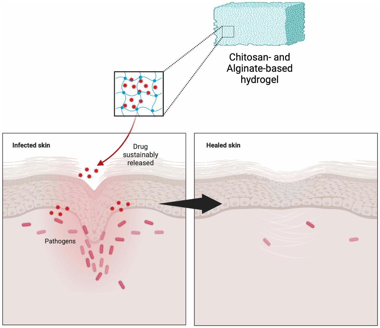
يمكن أن تصبح العدوى الموضعية غير المعالجة مزمنة، مما يسبب مشاكل صحية خطيرة. الالتصاق الجلدي الأمثل أمر حاسم في معالجة مثل هذه العدوى. في هذا السياق، يظهر الكيتوزان والألجينات كمرشحين واعدين للاستخدام كأساس في تطوير الهيدروجيلات الموضعية. الهدف من هذه المراجعة هو فحص الأدبيات حول تركيبات الهيدروجيل الموضعية التي تستخدم الكيتوزان والألجينات كأساس، وخاصة في سياق العوامل المضادة للبكتيريا الموضعية. تتضمن منهجية البحث مراجعة أدبية من خلال فحص المقالات المنشورة في قواعد بيانات مثل PubMed وScopus وScienceDirect وGoogle Scholar. كانت الكلمات الرئيسية المستخدمة خلال البحث هي “الألجينات”، “الكيتوزان”، “الهيدروجيل”، و”المضادات الحيوية”. يعمل الكيتوزان والألجينات كأساس في الهيدروجيلات الموضعية لتوصيل مكونات نشطة متنوعة، وخاصة العوامل المضادة للبكتيريا، كما تشير نتائج البحث. لقد أظهر كلاهما فعالية مضادة للبكتيريا ملحوظة، كما يتضح من انخفاض عدد مستعمرات البكتيريا وزيادة مناطق التثبيط. هذا يدعم بقوة فكرة أن الكيتوزان والألجينات يمكن أن تستخدم معًا لصنع هيدروجيلات موضعية تقتل البكتيريا بشكل فعال. في الختام، تظهر الهيدروجيلات المعتمدة على الكيتوزان والألجينات إمكانات كبيرة في علاج العدوى البكتيرية على سطح الجلد. إن دمج الكيتوزان والألجينات في تركيبات الهيدروجيل يساعد في الاحتفاظ بالعوامل المضادة للبكتيريا، مما يسمح بإطلاقها التدريجي على مدى فترة مثالية. لذلك، فإن الهيدروجيلات المصممة خصيصًا مع الكيتوزان والألجينات لديها القدرة على أن تكون حلاً لمواجهة التحديات في علاج العدوى البكتيرية الموضعية.

الكلمات المفتاحية: ألجينات، كيتوزان، هيدروجيل، موضعي، عامل مضاد للبكتيريا

مقدمة

توفر البشرة الطبيعية والصحية بشكل طبيعي حاجزًا قويًا ضد غزو الميكروبات. إن المساس بهذا الحاجز يجعل الأفراد عرضة للإصابات. تسمح إصابات الجلد مثل الصدمات الجسدية، وعضات الحشرات، والحروق، والعمليات الجراحية بدخول البكتيريا المسببة للأمراض، مما يؤدي إلى التهابات الجلد والأنسجة الرخوة. شفاء الجروح هو مشروع معقد، وتعزيز إصلاح الجلد الفعال يمثل تحديًا سريريًا كبيرًا. المكورات العنقودية الذهبية والمكورات العقدية من المجموعة A تسبب عادةً الجروح الجلدية الأولية والثانوية والصغيرة. يستخدم المتخصصون في الرعاية الصحية عادةً المضادات الحيوية الموضعية لعلاج والوقاية من التهابات الجلد. هذه الطريقة مناسبة تمامًا لتوصيل الأدوية مباشرة إلى موقع العمل، مما يضمن توافر حيوي مضاد للبكتيريا في الأنسجة المصابة. بالإضافة إلى ذلك، تشمل مزايا المضادات الحيوية الموضعية الحد الأدنى من استخدام الأدوية، وانخفاض التكلفة، وعدم إحداث اضطراب في الفلورا الميكروبية المعوية. ومع ذلك، قد تكون المضادات الحيوية المطبقة موضعياً مرتبطة ببعض الآثار الجانبية المحلية وتطور التهاب الجلد التماسي. تحد العمق الضحل لاختراق المضادات الحيوية الموضعية من استخدامها في العدوى السطحية، ويمكن أن يؤدي استخدامها الواسع وغير المقيد إلى مقاومة البكتيريا.
يواجه استخدام المضادات الميكروبية الموضعية تحديات مثل زيادة مقاومة البكتيريا، وردود الفعل التحسسية المحلية، والقلق بشأن التطور المحتمل لمقاومة المضادات الحيوية مع استخدام المطهرات. لذلك، فإن نظام توصيل الأدوية الأمثل للأدوية المضادة للبكتيريا أمر حاسم لتعظيم فعالية الأدوية المعطاة. حاليًا، يوفر استخدام الهيدروجيل للعلاج الموضعي مزايا متنوعة لمعالجة القضايا المرتبطة بالأدوية الموضعية. من خلال صياغتها في هيدروجيل، يمكن تعديل إطلاق الدواء ليحدث تدريجيًا على مدى فترة طويلة. بالإضافة إلى ذلك، فإن الطبيعة القائمة على الماء للهلاميات الهيدروجينية تكون عمومًا أكثر توافقًا مع أنسجة الجلد التالفة. كما
لقد أثبتت الأدلة أن استخدام الهيدروجيل له إمكانيات كبيرة في مجال ضمادات الجروح بسبب خصائصه الفيزيائية التي تشبه الأنسجة الحية وخصائصه الممتازة مثل محتوى الماء العالي، ونفاذية الأكسجين، والنعومة. كما هو معروف، فإن الجروح هي مناطق من الأنسجة التالفة عرضة للإصابة بالعدوى من مسببات الأمراض. في هذا الصدد، فإن عدة أنواع من الهيدروجيل القائم على البوليمر لا تعمل فقط كحاملات للأدوية، بل تمتلك أيضًا قدرات جذابة لمنع العدوى.
من بين الخيارات المختلفة للبوليمرات المتاحة، تُفضل البوليمرات الطبيعية غالبًا بسبب طبيعتها القابلة للتحلل وغير السامة نسبيًا. تتمتع أنواع معينة من البوليمرات الطبيعية المتعددة السكاريد بخصائص جوهرية يمكن أن تمنع نمو البكتيريا المسببة للأمراض. على سبيل المثال، يمتلك الكيتوزان خصائص مضادة للبكتيريا من خلال التفاعل الأيوني مع المكونات المشحونة سلبًا على جدران الخلايا البكتيرية. يمكن تحقيق هذه الفعالية الجوهرية بتركيز منخفض من الكيتوزان ). على الرغم من أن استخدام الكيتوزان بمفرده يمكن أن يُستخدم لإنشاء الهيدروجيل، إلا أن الهيدروجيل المتكون قد لا يمتلك القدرة المثلى على التجلط لحمل الدواء المقصود بشكل فعال. الحل يكمن في دمجه مع بوليمر بوليسكريد آخر، ألجينات، الذي يعزز قدرة تحميل الدواء في تركيبة الهيدروجيل. استخدام رابط متعدد القيم مثل تشكّل عملية الجلطة في الألجينات إطارًا متشابكًا يحتجز الدواء. ومن ثم، فإن الجمع بين هذين البوليمرين يحمل وعدًا في معالجة القضايا المتعلقة بعلاج العدوى البكتيرية الموضعية. وبالتالي، فإن المعرفة بشأن الاستخدام المحتمل للكيتوزان والألجينات، التي يمكن أن تتطور أكثر مع التغيرات في تركيبات البوليمر، تعتبر أساسية كأساس لتطوير تركيبات الهيدروجيل في علاج العدوى البكتيرية الموضعية. لمعالجة هذه القضايا، يقدم هذا الاستعراض ملخصًا لمختلف أنواع الهيدروجيل المستندة إلى الألجينات والكيتوزان وغيرها التي تظهر نشاطًا مضادًا للبكتيريا.

طريقة

تم إجراء هذه المراجعة من خلال البحث المنهجي عن المقالات ذات الصلة التي تناقش استخدام الهيدروجيلات القائمة على الكيتوزان والألجينات لتوصيل العوامل المضادة للبكتيريا. تم إجراء بحث عن المقالات في قواعد البيانات، بما في ذلك PubMed وScopus وScienceDirect وGoogle Scholar، باستخدام الكلمات الرئيسية “الألجينات”، “الكيتوزان”، “الهيدروجيل”، و”المضادات الحيوية”، دون تقييد سنة النشر. تم تطبيق معايير الإدراج لتصفية المقالات التي سيتم مناقشتها في هذه المراجعة، بما في ذلك: المقالات المنشورة باللغة الإنجليزية؛ الدراسات التي تستخدم الهيدروجيلات كشكل جرعة؛ استخدام الكيتوزان و/أو الألجينات كأساس للهيدروجيل؛ ووجود مواد فعالة دوائياً تم اختبارها لاحقاً من حيث فعاليتها المضادة للبكتيريا. شملت معايير الاستبعاد في هذه المراجعة المقالات ذات تصاميم الدراسة مثل تقارير الحالة، وسلاسل الحالات، ومقالات المراجعة؛ الدراسات ضمن المقالات التي تفتقر إلى اختبار مضاد للبكتيريا للصيغ المعدة؛ والمقالات التي تحتوي على بيانات غير مكتملة، مثل تلك التي تفتقر إلى معلومات حول المواد الفعالة المستخدمة وأنواع البكتيريا المستخدمة في اختبار الفعالية المضادة للبكتيريا.

عدوى موضعية

بروبيونيباكتيريوم أكنيس

بروبيونيباكتيريوم أكنيز هو بكتيريا موجبة الجرام تحتوي على طبقة سميكة من الببتيدوجليكان في جدارها الخلوي. هذه البكتيريا لا هوائية وتنتج إنزيمات ليباز تكسر الأحماض الدهنية الحرة من دهون الجلد. يمكن أن تؤدي الأحماض الدهنية إلى التهاب الأنسجة. لذلك، يساهم بروبيونيباكتيريوم أكنيز في حدوث حب الشباب ويشارك في مسببات حب الشباب. حب الشباب هو شكل من أشكال الالتهاب المزمن الذي يؤثر على وحدة الشعر والغدد الدهنية، والتي تتكون من بصيلات الشعر والغدد الدهنية.
حالياً، تُستخدم المستحضرات الموضعية من الإريثروميسين والكليندامايسين كمضادات حيوية لعلاج حب الشباب مع الالتهاب الناتج عن بروبيونيباكتيريوم أكنيز. في السوق، يُعرف الإريثروميسين باسم كورساتروcin، دوتروسين، دوراميسين، إيريميد، إيريسانبي، وتروفيلون. في الوقت نفسه، يُعرف الكليندامايسين باسم ميديكلاين، كلينبرسين، نوفاكليند، وأوبيكلام.

كوريباكتيريوم

كوريباكتيريوم هو بكتيريا موجبة الجرام لا تشكل أبواغ، لها شكل عصوي، وهي هوائية. بعض أنواع بكتيريا كوريباكتيريوم يمكن أن تنتج سمومًا، مثل السموم الداخلية، التي يمكن أن تسبب الأمراض. هذه البكتيريا تسبب الإريثرما، وهي عدوى جلدية سطحية تؤثر على طيات الجسم، مثل الإبطين والفخذ. تشمل أعراض هذه المرض بقعًا وردية وقشرية مصحوبة بحكة وإحساس بالحرق. تسبب هذه البكتيريا الإريثرما من خلال غزو الطبقة القرنية، تليها التكاثر في ظروف مثل الحرارة والرطوبة.
ثم تقوم البكتيريا بحل ألياف الكيراتين في المساحات بين الخلايا وداخل الخلايا، مما ينتج البورفيرينات. العلاج الأول هو الإريثروميسين الموضعي، الكليندامايسين، أو كريم الميكونازول. تشمل بعض الأسماء التجارية الشائعة للكليندامايسين الموضعي Anerocid، Acne Clin، وBenzasil. في الوقت نفسه، تشمل بعض الأسماء التجارية الشائعة لكريم الميكونازول MonistatDerm وMicatin.

ستافيلوكوكوس إpidermidis

ستافيلوكوكوس إpidermidis هو بكتيريا موجبة الجرام تشكل جزءًا من الميكروبيوم البشري الطبيعي، بما في ذلك ميكروبيوم الجلد. ستافيلوكوكوس إpidermidis تعيش بدون أكسجين وتشكل مستعمرات بيضاء كبيرة متماسكة بقطر حوالي بعد نموها طوال الليل. لا تقوم بتفكيك خلايا الدم الحمراء على أجار الدم. هذه البكتيريا هي ممرض انتهازي يمكن أن تظهر ضراوة عند مهاجمة جسم الإنسان. يمكن أن تسبب ستافيلوكوكوس إpidermidis عدوى نتيجة زراعة الأجهزة الطبية، مثل الأجهزة القلبية والعظمية. يمكن أن تحمي تكوين الأغشية الحيوية، البوليمرات الخارجية، وآليات أخرى هذه البكتيريا من المضادات الحيوية. يمكن أن تدخل البكتيريا إلى الجلد من خلال الخدوش، الجروح، أو أي ضرر آخر للجلد. يمكن أن تنتج ستافيلوكوكوس إpidermidis إنزيمات تكسر زيوت الجلد، مما يؤدي إلى تكوين حب الشباب، وتنتج سمومًا تسبب الالتهاب في الجلد.
يتضمن علاج عدوى ستافيلوكوكوس إpidermidis المضادات الحيوية، اعتمادًا على شدة العدوى وقابلية البكتيريا للمضادات الحيوية. غالبًا ما يستخدم الأطباء الفانكومايسين لعلاج عدوى ستافيلوكوكوس إpidermidis المقاومة للميثيسيلين. يُعرف الفانكومايسين في السوق باسم كلوفان، فانسولون، ولاديرفان.

ستافيلوكوكوس أوريوس

ستافيلوكوكوس أوريوس ينتمي إلى جنس ستافيلوكوكوس ضمن الفيرميكوت، بقطر ومترتب في “عناقيد تشبه العنب” تحت المجهر. يمكن أن تزدهر كل من الهوائية واللاهوائية ولها درجة حرارة نمو مثالية تبلغ عند درجة حموضة 7.4. المستعمرات على أطباق أجار الدم سميكة، لامعة، ومستديرة بقطر لا تشكل S. aureus أبواغ أو سوط، لكنها تمتلك غلافًا، يمكن أن تنتج صبغة صفراء ذهبية، وتخمر المانيتول.
ستافيلوكوكوس أوريوس هو بكتيريا موجبة الجرام تسبب أمراض سريرية متنوعة. على الجلد السليم، عادةً لا تسبب S. aureus عدوى. ومع ذلك، إذا دخلت البكتيريا إلى مجرى الدم، يمكن أن تؤدي إلى عدوى متنوعة، مثل عدوى الجلد والأنسجة الرخوة (SSTIs)، والتي تعتبر من أكثر العدوى البكتيرية شيوعًا في جميع أنحاء العالم. تشمل المضادات الحيوية الموضعية الشائعة المستخدمة للوقاية والعلاج من العدوى الجلدية التي تسببها هذه الممرضات حمض الفوسيديك (FA) والموبيروسين. تم استخدام هذه الأدوية كعلاج موضعي من الخط الأول للعدوى السطحية SSTIs (مثل، القوباء) في العديد من البلدان.

ستربتوكوكوس بيوجينيس

ستربتوكوكوس بيوجينيس هو بكتيريا مصنفة في شعبة الفيرميكوت وتعتبر بكتيريا موجبة الجرام. تُعرف أيضًا باسم “ستربتوكوكوس المجموعة A” (GAS)، ستربتوكوكوس بيوجينيس هو ممرض أصاب 18.1 مليون شخص في جميع أنحاء العالم، مما أدى إلى 500,000 حالة وفاة سنويًا. S. بيوجينيس هو لا هوائي اختياري، يزدهر بشكل أفضل في 5 إلى من ثاني أكسيد الكربون، ويشكل مستعمرات بدقة على أطباق أجار الدم. الخصائص المستضدية للبروتين M تقسم سلالات GAS إلى أنواع مصلية مختلفة، مما يجعلها البروتين السطحي الرئيسي الموجود في جدار الخلية لـ S. بيوجينيس. تم تحديد أكثر من 80 نوعًا مصلية مختلفة بناءً على البروتين.
ستربتوكوكوس بيوجينيس هو واحد من البكتيريا التي تسبب عدوى الجلد والأنسجة الرخوة (SSTIs). علاوة على ذلك، لا يسبب أي ممرض آخر مثل هذه الكيانات السريرية المتنوعة مثل S. بيوجينيس. على وجه الخصوص، يؤدي هذا الكائن إلى العدوى في الطبقة القرنية السطحية (القوباء)، البشرة السطحية (الحمامى)، الأنسجة تحت الجلد (التهاب النسيج الخلوي)، اللفافة (التهاب اللفافة الناخر)، أو العضلات (التهاب العضلات والنخر العضلي). يمكن تحقيق العلاج الموضعي للعدوى التي تسببها ستربتوكوكوس بيوجينيس باستخدام الموبيروسين، حيث يعادل الموبيروسين العوامل المضادة للميكروبات الجهازية الفموية ويمكن استخدامه عندما يكون عدد الآفات محدودًا.

الهيدروجيل كدواء موضعي واعد

تعريف الهيدروجيل

تعتبر تركيبات الهيدروجيل في الطب شكلًا من أشكال الجرعات الصيدلانية المستخدمة لتوصيل الأدوية إلى جسم الإنسان أو الحيوان. الهيدروجيل هي مواد قائمة على الماء لديها القدرة على الاحتفاظ بكمية كبيرة من الماء داخل هيكلها. تتكون تركيبات الهيدروجيل الطبية عادةً من شبكة ثلاثية الأبعاد تمتص وتحتفظ بكمية كبيرة من الماء أو سوائل أخرى، مثل حلول الأدوية. تجعل الخصائص الممتصة للماء للهيدروجيل مثالية لمجموعة متنوعة من التطبيقات الطبية، بما في ذلك تصنيع المنتجات الصيدلانية.
يمكن أن تأخذ تركيبات الهيدروجيل في الطب أشكالًا مختلفة، بما في ذلك الجل العابر للجلد المطبق على الجلد أو العينين أو الأذنين، بالإضافة إلى الجل المستخدم للعناية بالجروح أو توصيل الأدوية من خلال الأجهزة الطبية مثل القسطرات أو الدعامات. يمكن تصميم الهيدروجيل لإطلاق الأدوية ببطء على مدى فترة زمنية محددة، مما يسمح بجرعات دقيقة ومتسقة من الأدوية. يمكن أن يعزز ذلك كفاءة العلاج ويقلل من الآثار الجانبية المرتبطة بتقلبات مستويات الأدوية في مجرى الدم.
تتمتع تركيبات الهيدروجيل بتطبيقات متنوعة، مثل توصيل الأدوية الموضعية (على الجلد أو العينين)، شفاء الجروح، أو توصيل الأدوية عبر قنوات الجسم الأعمق. تقدم تركيبات الهيدروجيل القدرة على التحكم في إطلاق الأدوية، وتعزيز اختراق الأدوية في الأنسجة المستهدفة، وتحسين راحة المريض.

المواد المساعدة المستخدمة بشكل شائع

المواد المساعدة (المعروفة أيضًا باسم الإضافات أو المواد المساعدة) المستخدمة بشكل شائع في تركيبات الهيدروجيل الصيدلانية هي مواد تساعد في تشكيل وصيانة وتعزيز جودة تحضير الهيدروجيل. يتم تلخيص المواد المساعدة الشائعة المستخدمة في تركيبات الهيدروجيل الصيدلانية في الجدول 1.

طريقة التحضير

تشمل عملية إنتاج تركيبات الهيدروجيل الصيدلانية سلسلة من الخطوات المصممة لإنتاج هيدروجيل مستقر مناسب لتطبيقات طبية أو صيدلانية محددة. قد تختلف العملية اعتمادًا على المواد المستخدمة وتركيبة الهيدروجيل المرغوبة. توضح الشكل 1 المخطط الشائع في إنتاج تركيبات الهيدروجيل.

تحضير ووزن المواد الخام

تتضمن الخطوة الأولى قياس وتحضير المواد الخام، بما في ذلك عوامل التجلط، الماء، المواد الحافظة، معدلات الحموضة، معززات القوام، وغيرها من المواد المساعدة المستخدمة في تركيبة الهيدروجيل.

الخلط

تُخلط المواد الخام بكميات وترتيب محددين. يتم الخلط بعناية لتحقيق التجانس والتوزيع المتساوي. يمكن القيام بذلك يدويًا أو باستخدام معدات مثل الخلاطات أو المحركات.
الجدول I المواد المساعدة المستخدمة عادة في صياغة الهيدروجيل الموضعي
مادة مساعدة الاستخدام
وسيلة الهيدروجيل هو في الأساس مصفوفات قائمة على الماء، والماء هو مكون أساسي يشكل هيكل الهيدروجيل.
عوامل التجلط تُستخدم عوامل التجلط مثل الكاربومرات، صمغ الجيلان، الأجاروز، أو السليلوز كربوكسي ميثيل الصوديوم (NaCMC) لتوفير الكثافة واللزوجة المناسبة للهيدروجيل.
معدلات الحموضة تُستخدم مواد مثل هيدروكسيد الصوديوم أو حمض الستريك لضبط درجة حموضة الهيدروجيل لتناسب متطلبات تطبيقه الطبي.
مواد حافظة تتطلب بعض تركيبات الهيدروجيل مواد حافظة مثل كلوريد البنزالكونيوم أو الفينول لمنع نمو الكائنات الدقيقة.
معدلات القوام تُستخدم بعض المواد المساعدة مثل الجلايكول البروبيلي أو الإيثانول لتنظيم القوام واللزوجة للهيدروجيل.
معززات التماسك تُستخدم مواد مساعدة مثل حمض البولي أكريليك لتعزيز تماسك الهيدروجيل.
ملونات وعطور في بعض الحالات، قد تُضاف ملونات أو عطور لأغراض جمالية أو لامتثال المرضى.
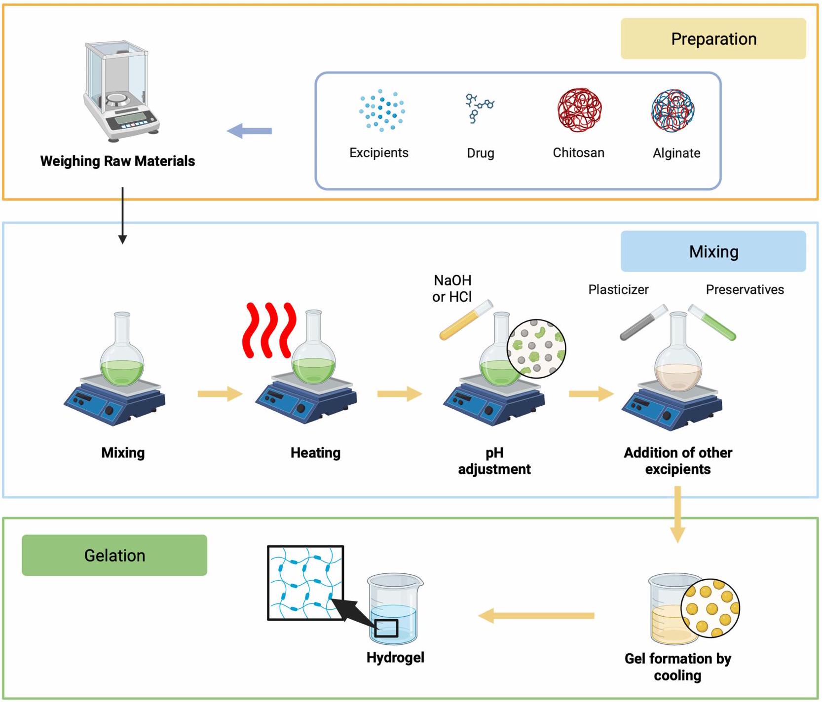
الشكل I مخطط في تصنيع شكل جرعة الهيدروجيل القائم على الكيتوزان والألجينات. تم إنشاؤه باستخدام Biorender.com.

التسخين

قد تكون عملية التسخين مطلوبة، اعتمادًا على التركيبة. تتطلب بعض الهيدروجيل تسخينًا لطيفًا للمساعدة في ذوبان عوامل التجلط وتشكيل الجل مع تبريد الهيدروجيل.

ضبط الحموضة

إذا لزم الأمر، يتم ضبط درجة حموضة المحلول وفقًا لمتطلبات تطبيق الدواء. يمكن القيام بذلك عن طريق إضافة حمض أو قاعدة.

إضافة المواد الحافظة والمواد المساعدة الأخرى

تُضاف المواد الحافظة والمواد المساعدة الأخرى، مثل معدلات القوام، الأدوية، أو الملونات والعطور، إلى الخليط وفقًا للتركيبة المرغوبة.

التجلي

تبدأ عملية التجلي عندما يبدأ الهيدروجيل في التبريد بعد الخلط وقد تتطلب تبريدًا تدريجيًا أو تغييرات في درجة الحرارة لتحقيق القوام المطلوب.

الترشيح والتعقيم

يمكن بعد ذلك ترشيح الهيدروجيل لإزالة الجسيمات الخشنة ثم تعقيمه إذا كان مخصصًا للتطبيقات الطبية.

التعبئة والتغليف

بمجرد أن يصبح الهيدروجيل جاهزًا، يمكن تعبئته في حاويات أو تغليفات مناسبة، مثل الأنابيب، الزجاجات، أو الأكياس، اعتمادًا على المنتج النهائي المرغوب.
من المهم ملاحظة أن عملية تصنيع الهيدروجيل يمكن أن تختلف بشكل كبير اعتمادًا على نوع الدواء المراد تطبيقه، مثل الهيدروجيل عبر الجلد، هيدروجيل العين، أو هيدروجيل العناية بالجروح. بالإضافة إلى ذلك، يجب اتباع لوائح ومعايير صارمة في إنتاج تركيبات الهيدروجيل لضمان جودة الدواء وسلامته وفعاليته.

توصيف شكل جرعة الهيدروجيل

تشمل الخصائص المهمة لتوصيف الهيدروجيل درجة الحموضة؛ المجهر الإلكتروني الماسح (SEM) لتوفير معلومات حول تركيب العينة، الطبوغرافيا السطحية، وخصائص أخرى؛ مطيافية الأشعة تحت الحمراء بتحويل فورييه، أو FTIR، لتحديد التركيب الكيميائي لمادة ما؛ قياسات الانتفاخ؛ حيود الأشعة السينية لفهم ما إذا كان البوليمر يحتفظ بهيكله البلوري؛ دراسات إطلاق الدواء في المختبر لفهم آلية الإطلاق خلال فترة التطبيق؛ اللزوجة؛ دراسات الانتشار؛ اختبارات تهيج الجلد في الجسم الحي التي أجريت على الأرانب لفحص علامات الوذمة والاحمرار؛ وحجم مسام الأنسجة المقاس بأدوات مثل المجاهر الإلكترونية، ومقاييس المسامية الزئبقية، وغيرها. من بين الخصائص العديدة للهيدروجيل التي تم التحقيق فيها، يعد اختبار درجة الحموضة وإطلاق الدواء في المختبر معيارين قد يؤثران مباشرة على الفعالية المضادة للبكتيريا للمضاد الحيوي المعطى. تعد ظروف درجة حموضة التركيبة ضرورية لضبطها لتقليد درجة حموضة الأنسجة أو الغشاء المخاطي المصاب. يمكن أن تدعم ظروف درجة حموضة التركيبة المتوافقة مع درجة حموضة موقع العلاج عملية إطلاق الدواء نحو مستعمرات البكتيريا كهدف لعمله. بعد ذلك، يصبح الاختبار في المختبر عاملاً في تأكيد إطلاق الدواء في وسط الإطلاق، في هذه الحالة، موقع العدوى الموضعي، بعيدًا عن افتراض قيم درجة الحموضة. من هذه الاختبارات، يمكن تحديد القيم الدقيقة لكمية الدواء التي يمكن إطلاقها نحو البكتيريا المعدية ومدة إطلاق الدواء بدقة.

فعالية الكيتوزان والألجينات كبوليمرات طبيعية قابلة للتحلل الحيوي قائمة على الهيدروجيل

الألجينات

الألجينات هي بوليسكريات أنيونية، أملاح حمض الألجينيك الموجودة بشكل رئيسي في الطحالب البنية، تشكل حوالي من مادتها الجافة. تحتوي المصفوفة بين الخلايا على الألجينات في شكل جل، والتي تشمل أيونات مثل الصوديوم، الكالسيوم، المغنيسيوم، السترونتيوم، والباريوم. لذلك، فإن وجود الكاتيونات متعددة التكافؤ يجعل الألجينات تتشكل بسهولة في جل. تختلف قدرة الألجينات على تشكيل جل أو التجلي بناءً على الوزن الجزيئي، الهيكل الجزيئي، وتركيز عامل التجلي. يمكن تشكيل جل الألجينات من خلال الروابط الهيدروجينية عند درجة حموضة منخفضة أو من خلال التفاعل الأيوني مع الكاتيونات أحادية أو متعددة التكافؤ.
يستخدم الألجينات على نطاق واسع في الصناعة لأنه يحتفظ بالماء، يشكل جل، يعمل كعامل مكثف، ويوفر الاستقرار. علاوة على ذلك، يُعرف الألجينات بتوافقه الحيوي ومناعيته، فضلاً عن سميته المنخفضة. يعمل الألجينات كعامل مكثف في الطعام، منظم توازن، مستحلب، وفاعل فيلم مقاوم للزيوت الرقيقة. يمكن أن يشكل الألجينات أفلامًا تُستخدم في التطبيقات التجميلية والطبية، مثل كبسولات الأدوية وضمادات الجروح. كما يتم تطوير الألجينات في تكنولوجيا هندسة الأنسجة لإنتاج الهيدروجيل، والكرات، والميكرو/النانو جزيئات (NPs)، لمجموعة متنوعة من التطبيقات مثل شفاء الجروح، توصيل البروتين، وتغليف الخلايا. يشكل الألجينات بسهولة هيدروجيل في وجود عوامل الربط مثل الكاتيونات ثنائية التكافؤ. لذلك، يعد الألجينات ضروريًا ليس فقط لصناعة النسيج ولكن أيضًا للأدوية، والمكونات الغذائية، والتجميلية. على الرغم من عدم تصنيفه كمضاد حيوي، إلا أن الألجينات تتمتع بامتصاص عالٍ، وخصائص مضادة للبكتيريا، ويمكن أن تسرع من شفاء الجروح. لذلك، يمكن استخدام الألجينات كضمادة جروح أولية وكوسيلة لتوصيل الأدوية الموضعية إلى الجروح المصابة. علاوة على ذلك، فإن أصل الألجينات من الطحالب البنية واحتوائه على مركبات نشطة مثل اليود ذات الخصائص المضادة للبكتيريا يجعله مناسبًا لإنشاء هيدروجيل مركب بخصائص مضادة للبكتيريا.
يعد الألجينات واحدًا من البوليمرات الطبيعية المستخدمة بشكل متكرر في أنظمة توصيل الأدوية والبروتين. يتمتع الألجينات بتوافق حيوي ممتاز وعدم سميته، ويمكن للباحثين إنتاجه باستخدام طرق بسيطة. يمكن أن تعمل هيدروجيل الألجينات
كوسائط توصيل للجزيئات التي تستهدف الأنسجة بدقة. يمكن أن يؤدي استخدام الألجينات إلى تغيير الخصائص الفيزيائية والكيميائية للأدوية، مما يعزز فعاليتها وسلامتها في أنظمة توصيل الأدوية.
الألجينات هو بوليمر خطي يتكون من كتل حمض الد-مانورونيك مرتبطة بـ وكتل حمض الل-جولورونيك مرتبطة بـ . تتكون هذه الكتل من بقايا G متتالية (GGGGGG)، وبقايا M متتالية (MMMMMM)، وبقايا G وM متناوبة (GMGMGM). من المعروف أن الكتل G فقط من الألجينات تشارك في تشكيل الروابط بين الجزيئات مع الكاتيونات ثنائية التكافؤ (مثل ) خلال عملية تشكيل الهيدروجيل.

الكيتوزان

الكيتوزان هو منتج ناتج عن نزع الأسيتيل الجزئي من الكيتين، ويتميز بخصائص مضادة للبكتيريا ممتازة، وقوة لاصقة، ونفاذية للأكسجين، وعدم السمية، وسهولة التحلل الحيوي، مما يجعله يحمل إمكانات كبيرة للتطبيقات الطبية، مثل الضمادات الطبية المضادة للبكتيريا. الجانب الحاسم للهلامات الهيدروجينية المعتمدة على الكيتوزان لاستخدامها في الضمادات الطبية هو قبولها من قبل المضيف. استخدم الباحثون السكريات المتعددة المؤكسدة، مثل الجلوكومانان، كروابط حيوية صديقة للبيئة. استخدم الباحثون تفاعل قاعدة شيف (رابطة إيمين القابلة للعكس) لتصميم هلامات هيدروجينية قابلة للحقن وقابلة للشفاء الذاتي. مبدأ تفاعل قاعدة شيف هو الأساس لتصميم ضمادة هلامية CS/OKGM. يقوم التفاعل بأكسدة الجلوكومانان لتكوين جلوكومانان كونجاك المؤكسد (OKGM)، والذي يتم ربطه بعد ذلك مع الكيتوزان لإنشاء هلام هيدروجيني من خلال تفاعل قاعدة شيف. نظرًا للطبيعة الديناميكية لرابطة قاعدة شيف، يظهر الهلام الهيدروجيني قدرات ممتازة على الشفاء الذاتي والحقن.
تتكون أفلام الهيدروجيل من عدة بوليمرات تُستخدم لاحتجاز ويعزز من اتساق ومرونة فيلم الهيدروجيل. الكيتوزان والألجينات هما بوليمرات مستخدمة بشكل شائع، إلى جانب المواد المضافة مثل الجلسرين والبروبيلين غليكول. وقد وُجد أن الألجينات المختلطة مع الكيتوزان يمكن أن تشكل أفلام هيدروجيل بشكل فعال باستخدام لكن الجودة الفيزيائية الكيميائية لا تزال بحاجة إلى تحسين. كما أن سلامة وفعالية علاج RAS تحتاج أيضًا إلى تقييم. تتيح الخصائص الذاتية الشفاء والقابلة للحقن للهلام المائي ملء أشكال الجروح المختلفة أو تغطية الأسطح الجلدية التي تتطلب المراقبة. تولد الأيونات موصلية كهربائية، مما يمكّن الهلام المائي من العمل كحساس ويفتح الطريق للبحث عن ضمادات هلام مائي متعددة الوظائف تجمع بين الوظائف الطبية الحيوية والحساسية. يمكن أن تتسبب وجود الأنواع التفاعلية من الأكسجين (ROS) التي تتولد بكثرة حول الأنسجة التالفة في إلحاق الضرر بالحمض النووي والبروتينات، مما يعيق تجديد الأنسجة.
من بين البوليمرات الحيوية، تعتبر السكريات المتعددة الأكثر بروزًا في التطبيقات المتعلقة بالصحة نظرًا لأنشطتها البيولوجية الواسعة. توجد في تكوينات متنوعة: ألياف، أغشية، هيدروجيل، كبسولات، هياكل نانوية، ميكيلات، إلخ. تشمل البوليمرات الحيوية الشائعة في السنوات الأخيرة الكيتوزان (CHT) والألجينات (ALG)، والتي تُستخدم على نطاق واسع في شفاء الجروح ومشاكل الجلد الأخرى. إن دمج البيومكرومولكولات مثل الكيتوزان مع الصوديوم ألجينات لتطوير الأفلام يظهر أيضًا مزايا جذابة حيث يمكن تعزيز خصائص كل فيلم. علاوة على ذلك، أثبتت مجموعة الكيتوزان والألجينات أنها تعمل كغشاء يُستخدم في تجديد الخلايا وشفاء الجروح. بالإضافة إلى ذلك، تشير العديد من الدراسات إلى أن دمج أنواع مختلفة من الزيوت الأساسية يمكن أن يعزز خصائص الهلاميات والأغشية، لا سيما من خلال تحسين قدرتها المضادة للميكروبات، مما يجعلها جذابة لعلاج أمراض الجلد أو تعبئة المواد الغذائية.

تطبيق هيدروجيل قائم على الألجينات والكيتوزان لعلاج البكتيريا الموضعي

الجدول 2 يلخص استخدام الكيتوزان والألجينات كأساس لصيغ الهيدروجيل الموضعية لأغراض مضادة للبكتيريا. تشير نتائج البحث إلى أن الكيتوزان والألجينات لهما تطبيقات واسعة، تشمل مواد فعالة متنوعة وأنواع بكتيرية مختلفة. بالإضافة إلى ذلك، يمكن دمج الكيتوزان والألجينات مع خلطات أساسية أخرى، مثل الجيلاتين، كربوكسي ميثيل السليلوز الصوديوم (NaCMC)، البكتين، بولي فينيل الكحول (PVA)، غليسيروفوسفات الصوديوم، وشرائح الجلايكوبوليمر. فيما يلي مناقشة أكثر تفصيلاً حول إمكانيات الألجينات-كيتوزان كأساس لهيدروجيل موضعي للتطبيقات المضادة للبكتيريا.

هيدروجيل قائم على الألجينات والكيتوزان

يمكن أن يُستخدم هيدروجيل الألجينات-كيتوزان لمجموعة متنوعة من المواد الفعالة، بما في ذلك المعادن غير العضوية، والكيانات الكيميائية، والمستخلصات. هيدروجيل الألجينات-كيتوزان يحمل بنجاح أكسيد الزنك (ZnO) للمواد الفعالة القائمة على المعادن غير العضوية.
الجدول 2 قابلية استخدام الكيتوزان والألجينات كأساس للهيدروجيل الموضعي في علاج البكتيريا الموضعية
قاعدة الهيدروجيل المادة الفعالة تركيز الدواء بكتيريا فعالية مضادة للبكتيريا مرجع
الألجينات-الكيتوزان بيربرين المكورات العنقودية الذهبية وحدة تشكيل المستعمرات من . الذهبية (تأثير تآزري من خلال تثبيط المكورات العنقودية الذهبية (MRSA) [6I]
كارفاكرول المكورات العنقودية الذهبية والإشريكية القولونية وحدة تشكيل المستعمرات من الإشريكية القولونية [62]
سيبروفلوكساسين، أموكسيسيلين وفانكومايسين لكل دواء المكورات العنقودية الذهبية قطر منطقة التثبيط [63]
الكركمين- -معقد السيكلودكسترين 10% (وزن/وزن) إي. كولاي و س. أوريس وحدة تشكيل المستعمرات من الإشريكية القولونية والمكورات العنقودية الذهبية [64]
هيسبيريدين 10% (وزن/حجم) المكورات العنقودية الذهبية و الزائفة الزنجارية وحدة تشكيل المستعمرات من S. aureus و P. aeruginosa [65]
هيسبيريدين 10% (وزن/وزن) المكورات العنقودية الذهبية و الزائفة الزنجارية وحدة تشكيل المستعمرات من S. aureus و P. aeruginosa [66]
ميترونيدازول 1% (وزن/حجم) المكورات العنقودية الذهبية والإشريكية القولونية وحدة تشكيل المستعمرات من . الذهبية والإشريكية القولونية [67]
ميترونيدازول المكورات العنقودية الذهبية والإشريكية القولونية قطر منطقة التثبيط [68]
ميترونيدازول 4% (وزن/حجم) المكورات العنقودية الذهبية وجرثومة غاردنريلا المهبلية وحدة تشكيل المستعمرات من S. aureus و G. vaginalis [69]
أكسيد النيتريك 2% (وزن/وزن) المكورات العنقودية الذهبية والإشريكية القولونية وحدة تشكيل المستعمرات من S. aureus و E. coli [70]
مستخلص أوراق السولانوم نيجروم L. المكورات العنقودية الذهبية، الزائفة الزنجارية وباسيلاس سوبتيليس وحدة تشكيل المستعمرات معدل إغلاق الجروح [71]
حمض التانيك 10% (وزن/حجم) إشريشيا كولاي و ستافيلوكوكوس أوريوس وحدة تشكيل المستعمرات من S. aureus و E. coli [72]
هيدروكلوريد التتراسيكلين إشريشيا كولاي و ستافيلوكوكوس أوريوس قطر منطقة التثبيط [73]
تيلميكوسين المكورات العنقودية الذهبية قطر منطقة التثبيط [74]
توبرايسين غير متوفر إشريشيا كولاي و ستافيلوكوكوس أوريوس وحدة تشكيل المستعمرات من . الذهبية و E. coli [75]
توبرايسين المكورات العنقودية الذهبية والإشريكية القولونية قتل حوالي 95% من المكورات العنقودية الذهبية عند تركيز مل و E. coli بتركيز [76]
فانكومايسين 0.1% (وزن/حجم) المكورات العنقودية الذهبية وحدة تشكيل المستعمرات من S. aureus [77]
أكسيد الزنك المكورات العنقودية الذهبية (S. aureus)، الإشريكية القولونية (E. coli)، العصوية الرقيقة (B. subtilis) قطر منطقة التثبيط [78]
جل الألجينات-الكيتوزان-الجيلاتين الدوبامين 0.2% (وزن/حجم) المكورات العنقودية الذهبية والإشريكية القولونية قطر منطقة التثبيط [79]
ليفوفلوكساسين إي. كولاي و س. أوريس قطر منطقة التثبيط [80]
ألجينات-كيتوزان-جيلاتين-ناCMC مستخلص المشيمة البشرية 10% (حجم/حجم) المكورات العنقودية الذهبية والإشريكية القولونية وحدة تشكيل المستعمرات من S. aureus و E. coli سمك الحبيبات TGF- أنا أعبر عن معدل إغلاق الجروح [8I]
ألجينات-كيتوزان-بكتين دوكسيسيكلين 2% (وزن/وزن) المكورات العنقودية الذهبية وحدة تشكيل المستعمرات من S. aureus. [82]
ألجينات-كيتوزان PVA 5-فلورويوراسيل 2% (وزن/حجم) إي. كولاي و س. أوريس قطر منطقة التثبيط [83]
مستخلص زهرة القطيفة الطبية 20% (وزن/حجم) أسيتيتوباكتر باومانني، ستافيلوكوكوس إيبيديرميديس، بروتيوس ميرابيلس، وستافيلوكوكوس أوريوس. قطر منطقة التثبيط [84]
ألجينات-كيتوزان صوديوم غليسيروفوسفات هيدروكلوريد الفانكومايسين المكورات العنقودية الذهبية والإشريكية القولونية وحدة تشكيل المستعمرات من . الذهبية والإشريكية القولونية [85]
مركب الكيتوزان-بلورونيك الكركمين النانوي 15% (وزن/حجم) إي. كولاي، س. تايفيموريوم، ب. أيروجينوزا، وس. أوريوس قطر منطقة التثبيط [86]
كيتوزان-ناCMC ألانطوين 0.5% (وزن/حجم) المكورات العنقودية الذهبية والإشريكية القولونية وحدة تشكيل المستعمرات من . أوريوس و . القولونية و معدل إغلاق الجروح [87]
ملاحظات: يشير إلى زيادة في قيمة المعامل؛ يشير إلى انخفاض في قيمة المعامل.
المواد. يمكن تطبيقه على كل من المستقلبات الثانوية والمركبات الكيميائية الاصطناعية. لقد وجد الباحثون أيضًا أن هذه القاعدة خيار مناسب لتطوير توصيل المكونات الفعالة الصيدلانية المسوقة، بما في ذلك التتراسيكلين، التوبراميسين، والفانكومايسين. تُظهر إحدى الدراسات قابلية استخدام هيدروجيل قائم على الألجينات والكيتوزان في توصيل مستخلص المكون الفعال، وهو مستخلص أوراق السولانوم نيجروم L. ومن المثير للاهتمام أن هذا الشكل الجرعي متوافق أيضًا مع توصيل المركبات المعدلة على النانو، مثل الكركمين المحمّل في -سيكلوديكسترين (كركمين- معقد السيكلودكسترين). تظهر الفائدة الكبيرة لاستخدام الهيدروجيل القائم على الألجينات والكيتوزان من خلال اختباره كعامل مضاد للبكتيريا. لقد اختبر الباحثون فعالية هذا الهيدروجيل ضد أنواع مختلفة من البكتيريا، بما في ذلك S. aureus و P. aeruginosa. بالإضافة إلى ذلك، قاموا بإجراء اختبارات على بكتيريا غير موضعية، مثل E. coli و B. subtilis، وكذلك الفطريات الموضعية، مثل G. vaginalis. تشير نتائج البحث إلى أن الدراسات التي تستخدم الهيدروجيل القائم على الألجينات والكيتوزان يمكن أن توفر تأثيرًا جيدًا مضادًا للبكتيريا. تعكس معلمات منطقة التثبيط الأكبر وانخفاض عدد مستعمرات البكتيريا التأثير الجيد المضاد للبكتيريا للدراسات التي تستخدم الهيدروجيل القائم على الألجينات والكيتوزان.
أظهرت دراسات متعددة أن هناك اختلافات طفيفة في تقنيات تحضير الهيدروجيل مع مزيج من الكيتوزان والألجينات. في حالة الكيتوزان المعدل (كيتوزان السليلوز الكربوكسي ميثيل)، يتم إجراء تحضير البوليمر ببساطة باستخدام الماء المقطر. من ناحية أخرى، بالنسبة للصيغة الأساسية للكيتوزان، يتم المساعدة في الذوبان باستخدام محلول مخفف من حمض الأسيتيك. ومع ذلك، لا يوجد فرق كبير بين استخدام هذين النوعين من البوليمر، سواء من حيث الخصائص الفيزيائية أو أداء أنشطة توصيل العامل المضاد للبكتيريا. تشير الدراسة العامة إلى أن تحميل العوامل المضادة للبكتيريا في هذا الهيدروجيل يؤدي إلى تأثير إطلاق دواء مستدام. على سبيل المثال، يحدث إطلاق الهيسبيريدين كالمادة الفعالة في الهيدروجيل بشكل تدريجي على مدى 14 يومًا، ليصل إلى إطلاق يشير هذا إلى التوافق مع نتائج مختلفة تقترح أن الهيدروجيل يمكن أن يعمل كوسيط دوائي طويل الأمد مناسب لعلاج الحالات المرضية المزمنة، مثل العدوى، كما هو موضح في الشكل 2.
الشكل 2 آلية عمل الهيدروجيل القائم على الكيتوزان والألجينات في توصيل العوامل المضادة للبكتيريا لتحسين العدوى البكتيرية الموضعية. تم إنشاؤه باستخدام Biorender.com.

الألجينات-الكيتوزان المدمج مع بوليمرات أخرى

بشكل عام، يهدف إضافة بوليمرات أخرى إلى قاعدة الهيدروجيل إلى تعزيز أو تحسين خصائص الهيدروجيل المصنوع. تم استخدام الجيلاتين، وهو مشتق من البروتينات الطبيعية، في تخليق الهيدروجيل الطبي بسبب عدم تحفيزه للمناعة. علاوة على ذلك، يتم استخدام الجيلاتين على نطاق واسع كقاعدة واحدة وأيضًا في تركيبات بسبب قدرته على تعزيز التصاق الخلايا وامتلاكه توافق حيوي ممتاز. يمكن أن يؤدي إضافة PVA إلى المزيج الأساسي إلى تشكيل هيدروجيل أقوى ونسبيًا صلب من خلال عمليات الربط الفيزيائي. تمتلك الهيدروجيل التي تحتوي على NaCMC إمكانات كبيرة لتلبية متطلبات ضمادات العناية بالجروح الفعالة، خاصة عند مزجها مع مضادات الميكروبات الطبيعية. يمكن أن توفر إضافة البكتين-السليلوز هيدروجيل أكثر استقرارًا مع توافق حيوي جيد وقدرة على إيقاف النزيف. بالإضافة إلى ذلك، يتم استخدام بوليمرات زخرفية أخرى مثل فوسفات الجلسرين الصوديوم وحمض بلورونيك بشكل عام للحصول على خصائص إطلاق حساسة لـ pH أو درجة الحرارة. تظهر اختلافات كبيرة في تأثير دمج مواد بوليمر إضافية في ملف إطلاق الدواء. يؤدي استخدام بوليمر بلورونيك إلى تأثير انفجاري في فترة الإطلاق الأولية، ليصل إلى في اليوم الأول. ومع ذلك، يستمر الإطلاق المستدام بعد تأثير الانفجار، ممتدًا حتى اليوم 21. وهذا يقدم ميزة واضحة، خاصة في ظروف مستويات العدوى العالية، حيث يكون الحفاظ على تركيز دواء مرتفع في المراحل الأولية أمرًا حاسمًا.

التحديات والقيود

على الرغم من أن الكيتوزان والألجينات تظهر إمكانات ممتازة كأساس لتصنيع الهيدروجيل، إلا أنها لا تزال تقدم بعض القيود. بناءً على النتائج التي تم مناقشتها في هذه المراجعة، على الرغم من أن الكيتوزان والألجينات لهما ملفات سمية منخفضة نسبيًا، إلا أنه لا يوجد تجارب سريرية مباشرة أجريت على البشر. وبالتالي، فإن ضمان السلامة فيما يتعلق باستخدام الكيتوزان والألجينات يحتاج إلى مزيد من الاستكشاف حتى يمكن للاختبارات المتقدمة أن تمنع الآثار الجانبية غير المرغوب فيها. بالإضافة إلى ذلك، فإن استخدام البوليمرات الطبيعية مثل الكيتوزان يسمح عمومًا بالذوبان عند pH منخفض، بينما يصبح غير قابل للذوبان عند قيم pH عالية. يمثل هذا تحديًا حيث يجب مراعاة تأثير هذه المواد على تغييرات pH في عمليات تصنيع الهيدروجيل. يمكن أن تؤدي ظروف pH غير المناسبة إلى عدم التوافق مع قاعدة البوليمر، مما يؤدي إلى تشكيل غير مثالي لتركيبة الهيدروجيل.

وجهة نظر مستقبلية

أصبحت الهيدروجيل مؤخرًا مفضلة في مجال توصيل الأدوية الموضعية، خاصة لتوصيل الأدوية المضادة للبكتيريا التي تكون عادة مزمنة في طبيعتها. تقدم الهيدروجيل فوائد ذات صلة، بشكل أساسي بسبب خصائصها اللاصقة الممتازة، مما يسمح لها بالالتصاق لفترة طويلة. يظهر الكيتوزان والألجينات، كأساس للهيدروجيل، خصائص مناسبة للتطبيقات الموضعية. كبوليمرات طبيعية، لديهم أيضًا ميزة السمية المنخفضة نسبيًا. على الرغم من أن إمكاناتهم في توصيل الأدوية المضادة للبكتيريا واضحة، إلا أن الباحثين اختبروا فقط استخدامها الموضعي ضد بكتيريا مثل S. aureus و P. aeruginosa. بالنظر إلى تأثيرها الإيجابي على بكتيريا معينة، من المحتمل أن يظهر هذا النظام لتوصيل الأدوية فعالية مماثلة ضد سلالات بكتيرية أخرى. لذلك، يحتاج الباحثون إلى إجراء مزيد من الدراسات لتحديد إمكانات الهيدروجيل القائم على الألجينات والكيتوزان في مكافحة بكتيريا أخرى تسبب عدوى جلدية، مثل P. acnes و Corynebacterium و S. epidermidis و S. pyogenes. علاوة على ذلك، يمتد التأثير الإيجابي إلى ما هو أبعد من مزيج الألجينات والكيتوزان وحده؛ فإن استكشاف إمكانات دمج بوليمرات أخرى أمر حاسم أيضًا. بالإضافة إلى NaCMC والجيلاتين والبيكتين وPVA، يجب دراسة المزيد من البوليمرات التي تدعم خصائص الهيدروجيل القائم على الألجينات والكيتوزان من أجل العثور على أفضل تركيبة هيدروجيل. كاقتراح، يُنصح بشدة بإجراء تحقق سريري لتقييم الفعالية المضادة للبكتيريا وسلامة الهيدروجيل المصنوعة من الكيتوزان والألجينات. يضمن ذلك أن المنتجات المستمدة من هذه الهيدروجيل لا تمتلك فقط دعمًا دليليًا ولكن يمكن أيضًا استخدامها على نطاق واسع من قبل الجمهور في السوق.

الخاتمة

كبوليمرات طبيعية، يظهر الكيتوزان والألجينات إمكانات كبيرة كأسس لتركيبات الهيدروجيل. يمكن الحصول على تركيبات الهيدروجيل القائمة على الكيتوزان والألجينات من خلال تقنية بسيطة، وهي عملية التجلط. كأسس للهيدروجيل الموضعية، يتوافق الكيتوزان والألجينات مع مجموعة واسعة من المواد الفعالة، بما في ذلك المركبات المعزولة والمركبات الاصطناعية والمستخلصات الطبيعية. علاوة على ذلك، يمكن دمج استخدام الكيتوزان والألجينات مع بوليمرات أخرى لتشكيل هيدروجيل بمعايير محددة، مثل PVA و NaCMC و الجيلاتين و البيكتين،
بوليمر بلورونيك، وفوسفات الجلسرين الصوديوم. يمكن أن يؤدي استخدام هيدروجيل الكيتوزان-الألجينات إلى تمديد مدة إطلاق العوامل المضادة للبكتيريا وقد أظهر فعالية في تقليل عدد مستعمرات البكتيريا وتثبيط مناطق النمو في اختبارات أقراص اللوحة.

الشكر

نود أن نشكر رئيس جامعة بادجادجاران على دعم APC.

التمويل

صندوق كيديركا المطابق، وزارة التعليم والثقافة، جمهورية إندونيسيا (20/E1/PPK/KS.03.00/2023).

الإفصاح

يبلغ المؤلفون عن عدم وجود تضارب في المصالح في هذا العمل.

References

  1. Okamoto S, Ogai K, Mukai K, Sugama J. Association of skin microbiome with the onset and recurrence of pressure injury in bedridden elderly people. Microorganisms. 2021;9(8). doi:10.3390/microorganisms9081603
  2. Gould L, Li WW. Defining complete wound closure: closing the gap in clinical trials and practice. Wound Repair Regener. 2019;27(3). doi:10.1111/wrr.12707
  3. Buzzá HH, Alves F, Tomé AJB, et al. Porphyrin nanoemulsion for antimicrobial photodynamic therapy: effective delivery to inactivate biofilm-related infections. Proc Natl Acad Sci U S A. 2022;119(46). doi:10.1073/pnas. 2216239119
  4. Punjataewakupt A, Napavichayanun S, Aramwit P. The downside of antimicrobial agents for wound healing. Eur J Clin Microbiol Infect Dis. 2019;38(1). doi:10.1007/s10096-018-3393-5
  5. Landeck L, John SM, Geier J. Topical ophthalmic agents as allergens in periorbital dermatitis. Br J Ophthalmol. 2014;98(2). doi:10.1136/ bjophthalmol-2013-304197
  6. Bandyopadhyay D. Topical antibacterials in dermatology. Indian J Dermatol. 2021;66(2). doi:10.4103/ijd.IJD_99_18
  7. Neri I, Miraglia Del Giudice M, Novelli A, Ruggiero G, Pappagallo G, Galli L. Ideal features of topical antibiotic therapy for the treatment of impetigo: an Italian expert consensus report. Curr Ther Res Clin Exp. 2023;98. doi:10.1016/j.curtheres.2022.100690
  8. Moeini A, Pedram P, Makvandi P, Malinconico M, Ayala G. Wound healing and antimicrobial effect of active secondary metabolites in chitosan-based wound dressings: a review. Carbohydr Polym. 2020;233. doi:10.1016/j.carbpol.2020.115839
  9. Siavash M, Noursina A. The ideal wound dressing. Burns. 2023. doi:10.1016/j.burns.2023.04.007
  10. Rezvani Ghomi E, Khalili S, Nouri Khorasani S, Esmaeely Neisiany R, Ramakrishna S. Wound dressings: current advances and future directions. J Appl Polym Sci. 2019;136(27). doi:10.1002/app. 47738
  11. Williamson DA, Carter GP, Howden BP. Current and emerging topical antibacterials and antiseptics: agents, action, and resistance patterns. Clin Microbiol Rev. 2017;30(3). doi:10.1128/CMR.00112-16
  12. Alsaab HO, Alharbi FD, Alhibs AS, et al. PLGA-based nanomedicine: history of advancement and development in clinical applications of multiple diseases. Pharmaceutics. 2022;14(12). doi:10.3390/pharmaceutics14122728
  13. Yanat M, Schroën K. Preparation methods and applications of chitosan nanoparticles; with an outlook toward reinforcement of biodegradable packaging. React Funct Polym. 2021;161. doi:10.1016/j.reactfunctpolym.2021.104849
  14. Schmieg B, Döbber J, Kirschhöfer F, Pohl M, Franzreb M. Advantages of hydrogel-based 3D-printed enzyme reactors and their limitations for biocatalysis. Front Bioeng Biotechnol. 2019;6. doi:10.3389/fbioe.2018.00211
  15. Aswathy SH, Narendrakumar U, Manjubala I. Commercial hydrogels for biomedical applications. Heliyon. 2020;6(4). doi:10.1016/j.heliyon.
  16. Kibungu C, Kondiah PPD, Kumar P, Choonara YE. This review recent advances in chitosan and alginate-based hydrogels for wound healing application. Front Mater. 2021;8. doi:10.3389/fmats.2021.681960
  17. Ghobashy MM. The application of natural polymer-based hydrogels for agriculture. In: Hydrogels Based on Natural Polymers. Elsevier; 2019. doi10.1016/B978-0-12-816421-1.00013-6
  18. Sanchez-Salvador JL, Balea A, Monte MC, Negro C, Blanco A. Chitosan grafted/cross-linked with biodegradable polymers: a review. Int J Biol Macromol. 2021;178. doi:10.1016/j.ijbiomac.2021.02.200
  19. Yilmaz Atay H. Antibacterial activity of chitosan-based systems. Function Chitosan. 2020;457-489. doi:10.1007/978-981-15-0263-7_15
  20. Long S, Xie C, Lu X. Natural polymer-based adhesive hydrogel for biomedical applications. Biosurf Biotribol. 2022;8(2). doi:10.1049/bsb2.12036
  21. Bao Z, Xian C, Yuan Q, Liu G, Wu J. Natural polymer-based hydrogels with enhanced mechanical performances: preparation, structure, and property. Adv Healthc Mater. 2019;8(17). doi:10.1002/adhm. 201900670
  22. Abdel Maksoud MIA, Ghobashy MM, Kodous AS, et al. Insights on magnetic spinel ferrites for targeted drug delivery and hyperthermia applications. Nanotechnol Rev. 2022;11(1). doi:10.1515/ntrev-2022-0027
  23. Motelica L, Ficai D, Oprea O, et al. Biodegradable alginate films with ZnO nanoparticles and citronella essential oil-A novel antimicrobial structure. Pharmaceutics. 2021;13(7). doi:10.3390/pharmaceutics13071020
  24. Tomić SL, Babić Radić MM, Vuković JS, Filipović V, Nikodinovic-Runic J, Vukomanović M. Alginate-based hydrogels and scaffolds for biomedical applications. Mar Drugs. 2023;21(3). doi:10.3390/md21030177
  25. Suhandi C, Mohammed AFA, Wilar G, El-Rayyes A, Wathoni N. Effectiveness of mesenchymal stem cell secretome on wound healing: a systematic review and meta-analysis. Tissue Eng Regen Med. 2023. doi:10.1007/s13770-023-00570-9
  26. Ma Y, Zhang N, Wu S, Huang H, Cao Y. Antimicrobial activity of topical agents against Propionibacterium acnes: an in vitro study of clinical isolates from a hospital in Shanghai, China. Front Med. 2016;10(4). doi:10.1007/s11684-016-0480-9
  27. Salemi SZ, Memar MY, Kafil HS, et al. The prevalence and antibiotics susceptibility patterns of Corynebacterium minutissimum isolates from skin lesions of patients with suspected erythrasma from Tabriz, Iran. Can J Infect Dis Med Microbiol. 2022;2022. doi:10.1155/2022/4016173
  28. Pinheiro L, Brito CI, Pereira VC, de Oliveira A, Camargo CH. Reduced susceptibility to vancomycin and biofilm formation in methicillin-resistant Staphylococcus epidermidis isolated from blood cultures. Mem Inst Oswaldo Cruz. 2014;109(7). doi:10.1590/0074-0276140120
  29. Guo Y, Song G, Sun M, Wang J, Wang Y. Prevalence and therapies of antibiotic-resistance in Staphylococcus aureus. Front Cell Infect Microbiol. 2020;10. doi:10.3389/fcimb.2020.00107
  30. Tayeb-Fligelman E, Tabachnikov O, Moshe A, et al. The cytotoxic Staphylococcus aureus PSM reveals a cross- amyloid-like fibril. Science. 2017;355(6327). doi:10.1126/science.aaf4901
  31. Nong Y, Taiaroa G, Pasricha S, et al. Clinical relevance of topical antibiotic use in coselecting for multidrug-resistant staphylococcus aureus: insights from in vitro and ex vivo models. Antimicrob Agents Chemother. 2021;65(5). doi:10.1128/AAC.02048-20
  32. Jespersen MG, Lacey JA, Tong SYC, Davies MR. Global genomic epidemiology of Streptococcus pyogenes. Infect Genet Evol. 2020;86. doi:10.1016/j.meegid.2020.104609
  33. Avire NJ, Whiley H, Ross K. A review of streptococcus pyogenes: public health risk factors, prevention and control. Pathogens. 2021;10(2). doi:10.3390/pathogens10020248
  34. Chowdhury S, Khakzad H, Bergdahl GE, et al. Streptococcus pyogenes forms serotype- and local environment-dependent interspecies protein complexes. mSystems. 2021;6(5). doi:10.1128/msystems.00271-21
  35. Stevens DL, Bryant AE. Impetigo, Erysipelas and Cellulitis. Search life-sciences literature; 2016.
  36. Thakur S, Thakur VK, Arotiba OA. History, Classification, Properties and Application of Hydrogels: An Overview. Springer; 2018. doi:10.1007/ 978-981-10-6077-9_2
  37. Elsayed MM. Hydrogel preparation technologies: relevance kinetics, thermodynamics and scaling up aspects. J Polym Environ. 2019;27(4). doi:10.1007/s10924-019-01376-4
  38. Jiang P, Yan C, Ji Z, et al. Drawing high-definition and reversible hydrogel paintings with grayscale exposure. ACS Appl Mater Interfaces. 2019;11 (45). doi:10.1021/acsami.9b14342
  39. Ickenstein LM, Garidel P. Hydrogel formulations for biologicals: current spotlight from a commercial perspective. Ther Deliv. 2018;9(3). doi:10.4155/tde-2017-0085
  40. Li J, Mooney DJ. Designing hydrogels for controlled drug delivery. Nat Rev Mater. 2016;1(12). doi:10.1038/natrevmats. 2016.71
  41. Almoshari YH. Novel hydrogels for topical applications: an updated comprehensive review based on source. Gels. 2022;8(3). doi:10.3390/ gels8030174
  42. Thang NH, Chien TB, Cuong DX. Polymer-based hydrogels applied in drug delivery: an overview. Gels. 2023;9(7). doi:10.3390/gels9070523
  43. Ciolacu DE, Nicu R, Ciolacu F. Cellulose-based hydrogels as sustained drug-delivery systems. Materials. 2020;13(22). doi:10.3390/ma13225270
  44. Yang J, Chen Y, Zhao L, et al. Preparation of a chitosan/carboxymethyl chitosan/AgNPs polyelectrolyte composite physical hydrogel with self-healing ability, antibacterial properties, and good biosafety simultaneously, and its application as a wound dressing. Compos B Eng. 2020:197. doi:10.1016/j.compositesb.2020.108139
  45. Ahmed EM. Hydrogel: preparation, characterization, and applications: a review. Adv Res. 2015;6(2). doi:10.1016/j.jare.2013.07.006
  46. Gopala Kumari SV, Manikandan NA, Pakshirajan K, Pugazhenthi G. Sustained drug release and bactericidal activity of a novel, highly biocompatible and biodegradable polymer nanocomposite loaded with norfloxacin for potential use in antibacterial therapy. J Drug Deliv Sci Technol. 2020;59. doi:10.1016/j.jddst.2020.101900
  47. Li S, Shi X, Xu B, et al. In vitro drug release and antibacterial activity evaluation of silk fibroin coated vancomycin hydrochloride loaded poly (lactic-co-glycolic acid) (PLGA) sustained release microspheres. J Biomater Appl. 2022;36(9). doi:10.1177/08853282211064098
  48. Saliani M, Jalal R, Goharshadi EK. Effects of pH and temperature on antibacterial activity of zinc oxide nanofluid against Escherichia coli O157: H7 and staphylococcus aureus. Jundishapur J Microbiol. 2015;8(2). doi:10.5812/jjm. 17115
  49. Zakharova L, Pashirova T, Kashapov R, Gabdrakhmanov D, Sinyashin O. Drug delivery mediated by confined nanosystems: structure-activity relations and factors responsible for the efficacy of formulations. In: Nanostructures for Drug Delivery. Elsevier; 2017. doi10.1016/B978-0-323-46143-6.00024-5
  50. Sosnik A. Alginate particles as platform for drug delivery by the oral route: state-of-the-art. ISRN Pharm. 2014;2014. doi:10.1155/2014/926157
  51. Yang J, Han S, Zheng H, Dong H, Liu J. Preparation and application of micro/nanoparticles based on natural polysaccharides. Carbohydr Polym. 2015;123. doi:10.1016/j.carbpol.2015.01.029
  52. Silva SS, Fernandes EM, Pina S, et al. 2.11 Polymers of biological origin. In: Comprehensive Biomaterials II. Elsevier; 2017. doi:10.1016/B978-0-12-803581-8.10134-1
  53. Hassabo AG, Mohamed AL. Extraction, structural properties, and applications of alginic acid. In: Natural Gums. Elsevier; 2023. doi10.1016/b978-0-323-99468-2.00023-1
  54. Lee KY, Mooney DJ. Alginate: properties and biomedical applications. Prog Polym Sci. 2012;37(1). doi:10.1016/j.progpolymsci.2011.06.003
  55. Jarrah R, Sammak S, Onyedimma C, et al. The role of alginate hydrogels as a potential treatment modality for spinal cord injury: a comprehensive review of the literature. Neurospine. 2022;19(2). doi:10.14245/ns.2244186.093
  56. Milanda T, Cindana Mo FR, Mohammed AFA, et al. Alginate/chitosan-based hydrogel film containing -mangostin for recurrent aphthous stomatitis therapy in rats. Pharmaceutics. 2022;14(8). doi:10.3390/pharmaceutics14081709
  57. Chen H, Cheng J, Ran L, et al. An injectable self-healing hydrogel with adhesive and antibacterial properties effectively promotes wound healing. Carbohydr Polym. 2018:201. doi:10.1016/j.carbpol.2018.08.090
  58. Duceac IA, Coseri S. Biopolymers and their derivatives: key components of advanced biomedical technologies. Biotechnol Adv. 2022;61. doi:10.1016/j.biotechadv.2022.108056
  59. Baysal K, Aroguz AZ, Adiguzel Z, Baysal BM. Chitosan/alginate crosslinked hydrogels: preparation, characterization and application for cell growth purposes. Int J Biol Macromol. 2013;59. doi:10.1016/j.ijbiomac.2013.04.073
  60. Istiqomah A, Utami MR, Firdaus M, Suryanti V, Kusumaningsih T. Antibacterial chitosan-Dioscorea alata starch film enriched with essential oils optimally prepared by following response surface methodology. Food Biosci. 2022;46. doi:10.1016/j.fbio.2022.101603
  61. Hu H, Zhong D, Li W, et al. Microalgae-based bioactive hydrogel loaded with quorum sensing inhibitor promotes infected wound healing. Nano Today. 2022:42. doi:10.1016/j.nantod.2021.101368
  62. Cheng M, Cui Y, Guo Y, et al. Design of carboxymethyl chitosan-reinforced pH -responsive hydrogels for on-demand release of carvacrol and simulation of release kinetics. Food Chem. 2023:405. doi:10.1016/j.foodchem.2022.134856
  63. Khan YA, Ozaltin K, Bernal-Ballen A, Di Martino A. Chitosan-alginate hydrogels for simultaneous and sustained releases of ciprofloxacin, amoxicillin and vancomycin for combination therapy. J Drug Deliv Sci Technol. 2021;61. doi:10.1016/j.jddst.2020.102126
  64. Kiti K, Suwantong O. Bilayer wound dressing based on sodium alginate incorporated with curcumin- -cyclodextrin inclusion complex/chitosan hydrogel. Int J Biol Macromol. 2020;164. doi:10.1016/j.jjbiomac.2020.09.013
  65. Bagher Z, Ehterami A, Nasrolahi M, Azimi M, Salehi M. Hesperidin promotes peripheral nerve regeneration based on tissue engineering strategy using alginate/chitosan hydrogel: in vitro and in vivo study. Int J Polym Mater Polym Biomater. 2021;70(5). doi:10.1080/00914037.2020.1713781
  66. Bagher Z, Ehterami A, Safdel MH, et al. Wound healing with alginate/chitosan hydrogel containing hesperidin in rat model. J Drug Deliv Sci Technol. 2020:55. doi:10.1016/j.jddst.2019.101379
  67. Permana AD, Asri RM, Amir MN, et al. Development of thermoresponsive hydrogels with mucoadhesion properties loaded with metronidazole gel-flakes for improved bacterial vaginosis treatment. Pharmaceutics. 2023;15(5). doi:10.3390/pharmaceutics15051529
  68. Feyissa Z, Edossa GD, Gupta NK, Negera D. Development of double crosslinked sodium alginate/chitosan based hydrogels for controlled release of metronidazole and its antibacterial activity. Heliyon. 2023;9(9). doi:10.1016/j.heliyon.2023.e20144
  69. Tentor F, Siccardi G, Sacco P, et al. Long lasting mucoadhesive membrane based on alginate and chitosan for intravaginal drug delivery. Mater Sci Mater Med. 2020;31(3). doi:10.1007/s10856-020-6359-y
  70. Zhang M, Fan Z, Zhang J, et al. Multifunctional chitosan/alginate hydrogel incorporated with bioactive glass nanocomposites enabling photothermal and nitric oxide release activities for bacteria-infected wound healing. Int J Biol Macromol. 2023:232. doi:10.1016/j.ijbiomac.2023.123445
  71. Najafpour F, Arabzadeh S, Kalalinia F, et al. Evaluation of wound healing effect of Solanum nigrum L. leaf extract-loaded sodium alginate nanoparticles embedded in chitosan hydrogel, In vivo study. Nanomed J. 2022;9(1). doi:10.22038/NMJ.2022.62218.1644
  72. Jafari H, Ghaffari-bohlouli P, Podstawczyk D, Nie L, Shavandi A. Tannic acid post-treatment of enzymatically crosslinked chitosan-alginate hydrogels for biomedical applications. Carbohydr Polym. 2022;295. doi:10.1016/j.carbpol.2022.119844
  73. Chen H, Xing X, Tan H, et al. Covalently antibacterial alginate-chitosan hydrogel dressing integrated gelatin microspheres containing tetracycline hydrochloride for wound healing. Mater Sci Eng C. 2017;70(Part 2). doi:10.1016/j.msec.2016.08.086
  74. Zhou K, Wang X, Chen D, et al. Enhanced treatment effects of tilmicosin against staphylococcus aureus cow mastitis by self-assembly sodium alginate-chitosan nanogel. Pharmaceutics. 2019;11(10). doi:10.3390/pharmaceutics11100524
  75. Xiao W, Gu S, Yuan W. Injectable nanocomposite hydrogels with the NIR-triggered low-temperature photothermal effect and low-dose antibiotic release. ACS Appl Polym Mater. 2023;5(7). doi:10.1021/acsapm.3c00676
  76. Shi M, Xu Y, Li S, Wang L, Gu J, Zhang YX. The development of a polysaccharide-based hydrogel encapsulating tobramycin-loaded gelatine microspheres as an antibacterial system. Gels. 2023;9(3). doi:10.3390/gels9030219
  77. Pawar V, Topkar H, Srivastava R. Chitosan nanoparticles and povidone iodine containing alginate gel for prevention and treatment of orthopedic implant associated infections. Int Biol Macromol. 2018;115. doi:10.1016/j.ijbiomac.2018.04.166
  78. Zhang M, Qiao X, Han W, Jiang T, Liu F, Zhao X. Alginate-chitosan oligosaccharide-ZnO composite hydrogel for accelerating wound healing. Carbohydr Polym. 2021;266. doi:10.1016/j.carbpol.2021.118100
  79. Lu Y, Xu J, Su Y, et al. A biocompatible double-crosslinked gelatin/ sodium alginate/dopamine/quaterniazed chitosan hydrogel for wound dressings based on 3D bioprinting technology. Int J Bioprint. 2022;9(2). doi:10.18063/IJB. 689
  80. Cao J, Xiao L, Shi X. Injectable drug-loaded polysaccharide hybrid hydrogels for hemostasis. RSC Adv. 2019;9(63). doi:10.1039/c9ra07116d
  81. Seifi S, Shamloo A, Tavoosi SN, Almasi-Jaf A, Shaygani H, Sayah MR. A novel multifunctional chitosan-gelatin/carboxymethyl cellulose-alginate bilayer hydrogel containing human placenta extract for accelerating full-thickness wound healing. Int J Biol Macromol. 2023. doi:10.1016/j. ijbiomac.2023.126929
  82. Amante C, Esposito T, Del Gaudio P, et al. A novel three-polysaccharide blend in situ gelling powder for wound healing applications. Pharmaceutics. 2021;13(10). doi:10.3390/pharmaceutics13101680
  83. Chen M, Zhai X, Pan Y, Tan H. Covalent and environment-responsive biopolymer hydrogel for drug delivery and wound healing. J Macromol Sci Part A. 2021;58(11). doi:10.1080/10601325.2021.1929316
  84. Ghasemi AH, Farazin A, Mohammadimehr M, Naeimi H. Fabrication and characterization of biopolymers with antibacterial nanoparticles and Calendula officinalis flower extract as an active ingredient for modern hydrogel wound dressings. Mater Today Commun. 2022;31. doi:10.1016/j. mtcomm.2022.103513
  85. Sun M, Cheng L, Xu Z, et al. Preparation and characterization of vancomycin hydrochloride-loaded mesoporous silica composite hydrogels. Front Bioeng Biotechnol. 2022:10. doi:10.3389/fbioe.2022.826971
  86. Dang LH, Nguyen TH, Tran HLB, Doan VN, Tran NQ. Injectable nanocurcumin-formulated chitosan-g-pluronic hydrogel exhibiting a great potential for burn treatment. J Healthc Eng. 2018;2018. doi:10.1155/2018/5754890
  87. Haki M, Shamloo A, Eslami SS, Mir-Mohammad-Sadeghi F, Maleki S, Hajizadeh A. Fabrication and characterization of an antibacterial chitosan-coated allantoin-loaded NaCMC/SA skin scaffold for wound healing applications. Int J Biol Macromol. 2023;253. doi:10.1016/j. ijbiomac.2023.127051
  88. Suhandi C, Alfathonah SS, Hasanah AN. Potency of xanthone derivatives from garcinia mangostana L. for COVID-19 treatment through angiotensin-converting enzyme 2 and main protease blockade: a computational study. Molecules. 2023;28(13). doi:10.3390/molecules28135187
  89. Suhandi C, Wilar G, Lesmana R, et al. Propolis-based nanostructured lipid carriers for -mangostin delivery: formulation, characterization, and in vitro antioxidant activity evaluation. Molecules. 2023;28(16). doi:10.3390/molecules28166057
  90. Suharyani I, Suhandi C, Rizkiyan Y, et al. Molecular docking in prediction of -mangostin/cyclodextrin inclusion complex formation. In: AIP Conference Proceedings. Vol. 2706; 2023. doi:10.1063/5.0120782.
  91. Andreazza R, Morales A, Pieniz S, Labidi J. Gelatin-based hydrogels: potential biomaterials for remediation. Polymers. 2023;15(4). doi:10.3390/ polym15041026
  92. Oliveira RN, McGuinness GB. Blended Gels of Sodium Carboxymethyl Cellulose Incorporating Antimicrobials for Absorbance and Wound Healing Applications. Springer; 2018. doi:10.1007/978-3-319-76573-0_39-1
  93. Chen W, Yuan S, Shen J, Chen Y, Xiao Y. A composite hydrogel based on pectin/cellulose via chemical cross-linking for hemorrhage. Front Bioeng Biotechnol. 2021;8. doi:10.3389/fbioe.2020.627351
  94. Deng A, Kang X, Zhang J, Yang Y, Yang S. Enhanced gelation of chitosan/ -sodium glycerophosphate thermosensitive hydrogel with sodium bicarbonate and biocompatibility evaluated. Mater Sci Eng C. 2017;78. doi:10.1016/j.msec.2017.04.109
  95. Aslam M, Barkat K, Malik NS, et al. pH sensitive pluronic acid/agarose-hydrogels as controlled drug delivery carriers: design, characterization and toxicity evaluation. Pharmaceutics. 2022;14(6). doi:10.3390/pharmaceutics14061218
  96. Ghobashy MM, Elbarbary AM, Hegazy DE, Maziad NA. Radiation synthesis of pH -sensitive 2-(dimethylamino)ethyl methacrylate/ polyethylene oxide/ZnS nanocomposite hydrogel membrane for wound dressing application. J Drug Deliv Sci Technol. 2022;73. doi:10.1016/j.jddst.2022.103399
  97. El-banna FS, Mahfouz ME, Leporatti S, El-Kemary M, Hanafy NAN. Chitosan as a natural copolymer with unique properties for the development of hydrogels. Appl Sci. 2019;9(11). doi:10.3390/app9112193
Infection and Drug Resistance
Dovepress

انشر عملك في هذه المجلة

إن مجلة العدوى ومقاومة الأدوية هي مجلة دولية محكمة مفتوحة الوصول تركز على العلاج الأمثل للعدوى (البكتيرية والفطرية والفيروسية) وتطوير واستحداث استراتيجيات وقائية لتقليل تطور وانتشار المقاومة. تهتم المجلة بشكل خاص وباء مقاومة المضادات الحيوية وآليات تطوير وانتشار المقاومة في كل من المستشفيات والمجتمع. نظام إدارة المخطوطات بالكامل عبر الإنترنت ويشمل نظام مراجعة الأقران سريع وعادل، وهو سهل الاستخدام. زيارةhttp://www.dovepress.com/testimonials.php لقراءة اقتباسات حقيقية من مؤلفين منشورين.

Journal: Infection and Drug Resistance
DOI: https://doi.org/10.2147/idr.s456403
PMID: https://pubmed.ncbi.nlm.nih.gov/38444772
Publication Date: 2024-03-01

Alginate and Chitosan-Based Hydrogel Enhance Antibacterial Agent Activity on Topical Application

Nasrul Wathoni , Cecep Suhandi , Muhammad Fadhil Ghassani Purnama , Annisa Mutmainnah , Neng Sani Nurbaniyah , Desra Widdy Syafra , Khaled M Elamin D Department of Pharmaceutics and Pharmaceutical Technology, Faculty of Pharmacy, Universitas Padjadjaran, Jatinangor, 45363, Indonesia; Graduate School of Pharmaceutical Sciences, Kumamoto University, Kumamoto, 862-0973, Japan

Correspondence: Nasrul Wathoni, Department of Pharmaceutics and Pharmaceutical Technology, Faculty of Pharmacy, Universitas Padjadjaran, Jatinangor, 45363, Indonesia, Tel +62-22-842-888-888, Email nasrul@unpad.ac.id

Abstract

Untreated topical infections can become chronic, posing serious health issues. Optimal skin adherence is crucial in addressing such infections. In this context, chitosan and alginate emerge as promising candidates for use as a foundation in the development of topical hydrogels. The aim of this review is to examine the literature on topical hydrogel formulations that use chitosan and alginate as foundations, specifically in the context of topical antibacterial agents. The research methodology involves a literature review by examining articles published in databases such as PubMed, Scopus, ScienceDirect, and Google Scholar. The keywords employed during the research were “Alginate”, “Chitosan”, “Hydrogel”, and “Antibacterial”. Chitosan and alginate serve as bases in topical hydrogels to deliver various active ingredients, particularly antibacterial agents, as indicated by the search results. Both have demonstrated significant antibacterial effectiveness, as evidenced by a reduction in bacterial colony counts and an increase in inhibition zones. This strongly supports the idea that chitosan and alginate could be used together to make topical hydrogels that kill bacteria that work well. In conclusion, chitosan and alginate-based hydrogels show great potential in treating bacterial infections on the skin surface. The incorporation of chitosan and alginate into hydrogel formulations aids in retaining antibacterial agents, allowing for their gradual release over an optimal period. Therefore, hydrogels specifically formulated with chitosan and alginate have the potential to serve as a solution to address challenges in the treatment of topical bacterial infections.

Keywords: Alginate, chitosan, hydrogel, topical, antibacterial agent

Introduction

Normal and healthy skin naturally provides a robust barrier against pathogen invasion. Compromising this barrier makes individuals vulnerable to infections. Skin injuries such as physical trauma, insect bites, burns, and surgical incisions allow the entry of pathogenic bacteria, leading to skin and soft tissue infections. Wound healing is a complex project, and promoting effective skin repair presents a significant clinical challenge. Staphylococcus aureus and Streptococcus group A commonly cause primary, secondary, and minor skin wounds Healthcare professionals commonly use topical antibiotics to treat and prevent skin infections. This method is well-suited for delivering drugs directly to the site of action, ensuring antibacterial bioavailability in infected tissues. Additionally, the advantages of topical antibiotics include minimal drug usage, low cost, and no disruption of intestinal microbial flora. However, topically applied antibiotics may be associated with some local side effects and the development of contact dermatitis. The shallow penetration depth of topical antibiotics limits their use to superficial infections, and their widespread, unrestricted use can lead to bacterial resistance.
The use of topical antimicrobials faces challenges such as an increase in bacterial resistance, local hypersensitivity reactions, and concerns regarding the potential development of antibiotic resistance with antiseptic use. Therefore, an optimal drug delivery system for antibacterial medications is crucial to maximize the efficacy of the administered drugs. Currently, the use of hydrogels for topical treatment offers various advantages to address issues associated with topical medications. Through formulation into hydrogels, drug release can be modified to occur gradually over an extended period. Additionally, the water-based nature of hydrogels is generally more compatible with damaged skin tissues. As
evidence, the application of hydrogels has proven to have great potential in the field of wound dressings due to their physical properties closely resembling living tissues and excellent characteristics such as high-water content, oxygen permeability, and softness. As known, wounds are areas of damaged tissue vulnerable to pathogen infection. In this regard, several types of polymer-based hydrogels not only function as drug carriers but also possess attractive capabilities to prevent infections.
Among various polymer options available, natural polymers are often preferred due to their biodegradable and relatively non-toxic nature. Certain types of natural polysaccharide polymers have intrinsic properties that can prevent the growth of pathogenic bacteria. For example, chitosan possesses antibacterial properties by interacting ionically with negatively charged components on the bacterial cell walls. This intrinsic efficacy can be achieved with a minimal chitosan concentration ( ). Although the use of chitosan alone can be utilized to create hydrogels, the formed hydrogel may not have optimal gelling capacity to effectively carry the intended drug. The solution lies in combining it with another polysaccharide polymer, alginate, which enhances the drug loading capacity in the hydrogel formulation. Utilizing a multivalent cross-linker such as , the gelation process in alginate forms a cross-linked framework that encapsulates the drug. Hence, the combination of these two polymers holds promise for addressing issues related to topical bacterial infection treatment. Consequently, knowledge regarding the prospective use of chitosan and alginate, which may still be further developed with variations in polymer combinations, is crucial as a foundation for the development of hydrogel formulations in topical bacterial infection treatment. To address these issues, this review presents a summary of various types of hydrogels based on alginate, chitosan, and others that exhibit antibacterial activity.

Method

This review was conducted through the systematic search of relevant articles discussing the use of chitosan and alginatebased hydrogels for the delivery of antibacterial agents. Article searches were performed on databases, including PubMed, Scopus, ScienceDirect, and Google Scholar, utilizing the keywords “Alginate”, “Chitosan”, “Hydrogel”, And “Antibacterial”, without restricting the publication year. Inclusion criteria were applied to filter articles to be discussed in this review, including: articles published in English; studies employing hydrogels as a dosage form; the use of chitosan and/or alginate as the hydrogel base; and the inclusion of pharmacologically active substances subsequently tested for their antibacterial efficacy. Exclusion criteria in this article review encompassed: articles with study designs such as case reports, case series, and review articles; studies within articles lacking antibacterial testing of the prepared formulations; and articles with incomplete data, such as those lacking information on the active substances used and bacterial species employed in the antibacterial efficacy testing.

Topical Infection

Propionibacterium Acnes

Propionibacterium acnes is a gram-positive bacterium with a thick peptidoglycan layer in its cell wall. The bacterium is anaerobic and produces lipase enzymes that break down free fatty acids from skin lipids. Fatty acids can trigger tissue inflammation. Therefore, Propionibacterium acnes contributes to the occurrence of acne and is involved in the pathogenesis of acne. Acne is a form of chronic inflammation that affects the pilosebaceous unit, consisting of hair follicles and sebaceous glands.
Currently, topical preparations of Erythromycin and Clindamycin are used as antibiotics to treat acne with inflammation caused by Propionibacterium acnes. On the market, erythromycin is known as Corsatrocin, Dothrocyn, Duramycin, Erymed, Erysanbe, and Trovilon. Meanwhile, Clindamycin is recognized as Mediklin, Clinbercin, Nufaclind, and Opiclam.

Corynebacterium

Corynebacterium is a gram-positive bacterium that does not form spores, has a bacillus shape, and is aerobic. Some species of Corynebacterium bacteria can produce toxins, such as endotoxins, which can cause diseases. This bacterium causes erythrasma, a superficial skin infection that affects body folds, such as the armpits and groin. Symptoms of this disease include pink and scaly patches accompanied by itching and a burning sensation. This bacterium causes erythrasma by invading the stratum corneum, followed by proliferating in conditions such as heat and humidity. The
bacteria then dissolve keratin fibrils in intercellular spaces and within cells, producing porphyrins. The first-line therapy is topical erythromycin, clindamycin, or miconazole cream. Some common brand names for topical clindamycin include Anerocid, Acne Clin, and Benzasil. Meanwhile, some common brand names for miconazole cream include MonistatDerm and Micatin.

Staphylococcus Epidermidis

Staphylococcus epidermidis is a gram-positive bacterium that forms part of the normal human microbiota, including the skin microbiota. Staphylococcus epidermidis survives without oxygen and forms cohesive, large, white colonies with a diameter of about after overnight growth. It does not break down red blood cells on blood agar. This bacterium is an opportunistic pathogen that can exhibit virulence when attacking the human body. Staphylococcus epidermidis can cause infections resulting from the implantation of medical devices, such as cardiac and orthopedic devices. The formation of biofilm, exopolymers, and other mechanisms can protect this bacterium from antibiotics. The bacteria can enter the skin through scratches, wounds, or other skin damage. Staphylococcus epidermidis can produce enzymes that break down skin oils, triggering the formation of acne, and produce toxins that cause inflammation in the skin.
The treatment of Staphylococcus epidermidis infections involves antibiotics, depending on the severity of the infection and the bacteria’s susceptibility to antibiotics. Medical doctors often use vancomycin to treat methicillin-resistant Staphylococcus epidermidis infections. Vancomycin is known on the market as Klosvan, Vancolon, and Ladervan.

Staphylococcus Aureus

Staphylococcus aureus belongs to the genus Staphylococcus within the Firmicutes, with a diameter of and arranged in “grape-like clusters” under the microscope. It can thrive both aerobically and anaerobically and has an optimal growth temperature of at a pH of 7.4 . Colonies on blood agar plates are thick, shiny, and round with a diameter of S. aureus does not form spores or flagella, but it possesses a capsule, can produce a golden-yellow pigment, and ferments mannitol.
Staphylococcus aureus is a gram-positive bacterium that causes various clinical diseases. On healthy skin, S. aureus typically does not cause infections. However, if the bacteria enter the bloodstream, they can lead to various infections, such as skin and soft tissue infections (SSTIs), which are among the most common bacterial infections worldwide. Common topical antibiotics used for the prevention and treatment of skin infections caused by this pathogen include fusidic acid (FA) and mupirocin. These drugs have been employed as the first-line topical treatment for superficial SSTIs (eg, impetigo) in many countries.

Streptococcus Pyogenes

Streptococcus pyogenes is a bacterium classified in the phylum Firmicutes and is categorized as a Gram-positive bacterium. Also known as “group A streptococcus” (GAS), Streptococcus pyogenes is a pathogen that has infected 18.1 million people worldwide, resulting in 500,000 deaths annually. S. pyogenes is a facultative anaerobe, thrives best in 5 to carbon dioxide, and forms colonies precisely on blood agar plates. The antigenic characteristics of protein M further divide GAS strains into different serotypes, making it the primary surface protein found in the cell wall of S. pyogenes. More than 80 different serotypes have been identified based on protein.
Streptococcus pyogenes is one of the bacteria causing skin and soft tissue infections (SSTIs). Furthermore, no other pathogen causes such diverse clinical entities as S. pyogenes. Specifically, this organism induces infections in the superficial keratin layer (impetigo), superficial epidermis (erysipelas), subcutaneous tissue (cellulitis), fascia (necrotizing fasciitis), or muscles (myositis and myonecrosis). Topical therapy for infections caused by Streptococcus pyogenes can be accomplished with mupirocin, where mupirocin is equivalent to oral systemic antimicrobial agents and can be used when the number of lesions is limited.

Hydrogel as Promising Topical Drug Delivery

Definition of Hydrogel

Hydrogel formulations in medicine are a pharmaceutical dosage form used to deliver drugs into the human or animal body. Hydrogels are water-based materials with the capacity to retain a significant amount of water within their structure. Medicinal hydrogel formulations typically consist of a three-dimensional network that absorbs and retains a substantial amount of water or other fluids, such as drug solutions. The water-absorbing properties of hydrogels make them ideal for various medical applications, including pharmaceutical product manufacturing.
Hydrogel formulations in medicine can take various forms, including transdermal gels applied to the skin, eyes, or ears, as well as gels used for wound care or drug delivery through medical devices such as catheters or stents. Hydrogels can be designed to release drugs slowly over a specific period of time, allowing for precise and consistent drug dosages. This can enhance treatment efficiency and reduce side effects associated with fluctuations in drug levels in the bloodstream.
Hydrogel formulations have various applications, such as topical drug delivery (on the skin or eyes), wound healing, or drug delivery through deeper body channels. Hydrogel formulations offer the ability to control drug release, enhance drug penetration into target tissues, and improve patient comfort.

Commonly Used Excipients

Excipients (also referred to as additives or auxiliary substances) commonly used in pharmaceutical hydrogel formulations are substances that aid in forming, maintaining, and enhancing the quality of the hydrogel preparation. Common excipients utilized in pharmaceutical hydrogel formulations are summarized in Table 1.

Preparation method

The production of hydrogel pharmaceutical formulations involves a series of steps designed to produce a stable hydrogel suitable for specific medical or pharmaceutical applications. The process may vary depending on the materials used and the desired hydrogel formulation. Figure 1 illustrates the common scheme in producing hydrogel formulations.

Preparing and Weighing of Raw Materials

The first step involves measuring and preparing raw materials, including gelling agents, water, preservatives, pH adjusters, consistency enhancers, and other excipients used in the hydrogel formulation.

Mixing

Raw materials are mixed in specified quantities and order. Mixing is performed carefully to achieve homogeneity and even distribution. This can be done manually or using equipment such as mixers or stirrers.
Table I Excipients Commonly Used in Formulating Topical Hydrogel
Excipient Usage
Vehicle Hydrogels are essentially water-based matrices, and water is a primary component that forms the hydrogel structure.
Gelling Agents Gelling agents such as carbomers, gellan gum, agarose, or sodium carboxymethyl cellulose (NaCMC) are used to provide the hydrogel with appropriate density and viscosity.
pH Adjusters Substances like sodium hydroxide or citric acid are employed to adjust the pH of the hydrogel to suit the requirements of its medicinal application.
Preservatives Certain hydrogel formulations require preservatives such as benzalkonium chloride or phenol to prevent the growth of microorganisms.
Texture Modifiers Some excipients like propylene glycol or ethanol are used to regulate the texture and viscosity of the hydrogel.
Consistency Enhancers Excipients like polyacrylic acid glycol are employed to enhance the cohesiveness of the hydrogel.
Colorants and Fragrances In some cases, colorants or fragrances may be added for aesthetic purposes or patient compliance.
Figure I Scheme in manufacturing topical chitosan and alginate-based hydrogel dosage form. Created with Biorender.com.

Heating

The heating process may be required, depending on the formulation. Some hydrogels require gentle heating to assist in the dissolution of gelling agents and form the gel as the hydrogel cools.

pH Adjustment

If necessary, the solution’s pH is adjusted according to the drug application requirements. This can be done by adding acid or base.

Preservatives and Other Excipients Addition

Preservatives and other excipients, such as texture modifiers, drugs, or colorants and fragrances, are added to the mixture according to the desired formulation.

Gelation

The gelation process begins as the hydrogel starts to cool after mixing and may require gradual cooling or temperature changes to achieve the desired consistency.

Filtration and Sterilization

The hydrogel can then be filtered to remove coarse particles and subsequently sterilized if intended for medical applications.

Filling and Packaging

Once the hydrogel is ready, it can be filled into appropriate containers or packaging, such as tubes, bottles, or sachets, depending on the desired end product.
It is important to note that the hydrogel manufacturing process can vary significantly depending on the type of drug to be applied, such as transdermal hydrogel, eye hydrogel, or wound care hydrogel. Additionally, strict regulations and standards must be followed in the production of hydrogel formulations to ensure drug quality, safety, and efficacy.

Characterization of Hydrogel Dosage Form

Important characteristics for the characterization of hydrogels include pH ; scanning electron microscopy (SEM) to provide information about sample composition, surface topography, and other properties; Fourier transform infrared spectroscopy, or FTIR, to identify the chemical structure of a substance; swelling measurements; X-ray diffraction to understand whether the polymer retains its crystalline structure; in-vitro drug release studies to comprehend the release mechanism during the application period; rheology; spreading studies; in vivo skin irritation tests conducted on rabbits to examine for signs of edema and erythema; and tissue pore size measured with tools such as electron microscopes, mercury porosimeters, and others. Among the several hydrogel characteristics that were investigated, pH testing and in vitro release are two criteria that might directly influence the antibacterial effectiveness of the administered antibiotic. The formulation’s pH conditions are crucial to be adjusted to mimic the pH of the infected tissue or mucosa. Formulation pH conditions aligned with the treatment site’s pH can support the drug release process towards bacterial colonies as the target of its action. Subsequently, in vitro testing becomes a factor in confirming drug release into the release medium, in this case, the topical infection site, beyond assuming pH values. From these tests, the precise values of the amount of drug that can be released towards infectious bacteria and the duration of drug release can be accurately determined.

Potency of Chitosan and Alginate as Natural Biodegradable Polymers-Based Hydrogel

Alginate

Alginates are anionic polysaccharides, salts of alginic acid predominantly present in brown algae, constituting about of their dry matter. The intercellular matrix contains alginate in the form of a gel, which includes ions like sodium, calcium, magnesium, strontium, and barium. Therefore, the presence of multivalent cations makes alginate easily form a gel. The ability of alginate to form a gel or gelation varies based on molecular weight, molecular structure, and gelation agent concentration. Alginate gel can be formed through hydrogen bonding at low pH or through ionic interaction with mono- or polyvalent cations.
Alginate is widely used in the industry because it retains water, forms gel, acts as a thickener, and provides stability. Moreover, alginate is known for its biocompatibility and immunogenicity, as well as its low toxicity. Alginate functions as a thickening agent in food, a balance regulator, an emulsifier, and a thin oil-resistant film former. Alginate can form films used in cosmetic and pharmaceutical applications, such as drug capsules and wound dressings. Alginate is also developed in tissue engineering technology for the production of hydrogels, beads, and micro/nanoparticles (NPs), for various applications such as wound healing, protein delivery, and cell encapsulation. Alginate readily forms hydrogels in the presence of cross-linking agents such as divalent cations. Therefore, alginate is essential not only for the textile industry but also for pharmaceuticals, food, and cosmetic ingredients. Although not classified as an antibiotic, alginate has high absorbency, antibacterial properties, and can accelerate wound healing. Therefore, alginate can be used as a primary wound dressing and as a medium for topical drug delivery to infected wounds. Furthermore, alginate’s origin from brown algae and its inclusion of active compounds like iodine with antibacterial properties make it suitable for creating composite hydrogels with antibacterial properties.
Alginate is one of the natural polymers frequently used in drug and protein delivery systemsAlginate exhibits excellent biocompatibility and non-toxicity, and researchers can produce it using simple methods. Alginate hydrogels
can function as delivery vectors for molecules precisely targeting tissues. The use of alginate can alter the physicochemical characteristics of drugs, enhancing their effectiveness and safety in drug delivery systems.
Alginate is a linear polymer composed of d-mannuronic acid blocks linked by and l-guluronic acid blocks linked by . These blocks consist of consecutive G residues (GGGGGG), consecutive M residues (MMMMMM), and alternating G and M residues (GMGMGM). It is known that only G blocks of alginate participate in the formation of cross-links between molecules with divalent cations (such as ) during the hydrogel formation process.

Chitosan

Chitosan is a product resulting from the partial deacetylation of chitin, possessing excellent antibacterial properties, adhesive power, oxygen permeability, non-toxicity, and easy biodegradation, holding great potential for medical applications, such as antibacterial medical dressings. The crucial aspect of chitosan-based hydrogels for use in medical dressings is their host acceptance. Researchers have employed oxidized polysaccharides, like glucomannan, as environmentally friendly bio-crosslinkers. The researchers utilized the Schiff base reaction (reversible imine bond) to design injectable self-healing hydrogels. The Schiff base reaction principle is the basis for designing the CS/OKGM hydrogel dressing. The reaction oxidizes glucomannan to form oxidized konjac glucomannan (OKGM), which is then cross-linked with CS to create a hydrogel through the Schiff base reaction. Due to the dynamic nature of the Schiff base bond, the hydrogel demonstrates excellent self-healing and injectability capabilities.
Hydrogel films consist of several polymers used to entrap and enhance the consistency and elasticity of the hydrogel film. Chitosan and alginate are commonly used polymers, along with plasticizers like glycerin and propylene glycol. It has been found that alginate mixed with chitosan can effectively form hydrogel films using , but the physicochemical quality still needs improvement. The safety and efficacy of RAS treatment also need evaluation. The self-healing and injectable properties enable hydrogels to fill various wound shapes or cover skin surfaces that require monitoring. Ions generate electrical conductivity, enabling the hydrogel to function as a sensor and paving the way for research on new multifunctional hydrogel dressings that combine biomedical functions and sensing. The presence of reactive oxygen species (ROS) generated massively around damaged tissues can cause damage to DNA and proteins, inhibiting tissue regeneration.
Among biopolymers, polysaccharides are the most prominent in health-related applications due to their broad biological activities. They exist in various configurations: fibers, membranes, hydrogels, capsules, nanostructures, micelles, etc. Common biopolymers in recent years include chitosan (CHT) and alginate (ALG), widely used for wound healing and other skin issues. Combining biomacromolecules such as chitosan with sodium alginate for film development also demonstrates attractive advantages as the characteristics of each film can be enhanced. Moreover, the combination of CHT and ALG has proven to act as a membrane applied to cell regeneration and wound healing. Additionally, several studies suggest that combining various types of essential oils can enhance the properties of hydrogels and membranes, particularly by improving their antimicrobial potential, making them appealing for skin pathology treatment or food packaging.

Alginate and Chitosan-Based Hydrogel Application for Topical Bacterial Treatment

Table 2 summarizes the use of chitosan and alginate as the foundation for topical hydrogel formulations for antibacterial purposes. The search results indicate that chitosan and alginate have extensive applications, encompassing diverse active substances and various bacterial types. In addition, chitosan and alginate can be combined with other base mixtures, such as gelatin, sodium carboxymethylcellulose (NaCMC), pectin, polyvinyl alcohol (PVA), sodium glycerophosphate, and g-pluronic copolymer. The following is a more detailed discussion of the potential of alginate-chitosan as a hydrogel base for topical antibacterial applications.

Alginate-Chitosan-Based Hydrogel

Alginate-chitosan hydrogel can apply to various active substances, including inorganic metals, chemical entities, and extracts. Alginate-chitosan-based hydrogel successfully carries zinc oxide ( ZnO ) for inorganic metal-based active
Table 2 Applicability of Chitosan and Alginate as a Base for Topical Hydrogel in Topical Bacterial Treatment
Hydrogel Base Active Pharmacological Ingredient Drug Concentration Bacteria Antibacterial Effectivity Reference
Alginate-Chitosan Berberine S. aureus Colony forming unit of . aureus (synergistic effect through inhibiting Staphylococcus aureus (MRSA) [6I]
Carvacrol S. aureus and E. coli Colony forming unit of E. coli [62]
Ciprofloxacin, amoxicillin and vancomycin for each drug S. aureus Diameter of inhibitory zone [63]
Curcumin- -cyclodextrin complex 10% (w/w) E. coli and S. aureus Colony forming unit of E. coli and S. aureus [64]
Hesperidin 10% (w/v) Staphylococcus aureus dan Pseudomonas aeruginosa Colony forming unit of S. aureus and P. aeruginosa [65]
Hesperidin 10% (w/w) S. aureus and P. aeruginosa Colony forming unit of S. aureus and P. aeruginosa [66]
Metronidazole 1% (w/v) S. aureus dan E. coli Colony forming unit of . aureus and E. coli [67]
Metronidazole S. aureus and E. coli Diameter of inhibitory zone [68]
Metronidazole 4% (w/v) Staphylococcus aureus and Gardnerella vaginalis Colony forming unit of S. aureus dan G. vaginalis [69]
Nitric oxide 2% (w/w) S. aureus and E. coli Colony forming unit of S. aureus dan E. coli [70]
Solanum nigrum L. leave extract Staphylococcus aureus, Pseudomonas aeruginosa and Bacillus subtilis Colony forming unit and wound closure rate [71]
Tannic acid 10% (w/v) Escherichia coli and Staphylococcus aureus Colony forming unit of S. aureus dan E. coli [72]
Tetracycline hydrochloride Escherichia coli and Staphylococcus aureus Diameter of inhibitory zone [73]
Tilmicosin S. aureus Diameter of inhibitory zone [74]
Tobramycin N/A Escherichia coli and Staphylococcus aureus Colony forming unit of . aureus dan E. coli [75]
Tobramycin S. aureus and E. coli Kill almost 95% S. aureus at concentration of mL and E . coli at concentration of [76]
Vancomycin 0.1% (w/v) S. aureus Colony forming unit of S. aureus [77]
Zinc oxide Staphylococcus aureus (S. aureus), Escherichia coli (E. coli), bacillus subtilis (B. subtilis) Diameter of inhibitory zone [78]
Alginate-ChitosanGelatin Dopamine 0.2% (w/v) S. aureus and E. coli Diameter of inhibitory zone [79]
Levofloxacin E. coli and S. aureus Diameter of inhibitory zone [80]
Alginate-Chitosan-Gelatin-NaCMC Human placenta extract 10% (v/v) S. aureus and E. coli Colony forming unit of S. aureus dan E. coli, granulation thickness, TGF- I expression and wound closure rate [8I]
Alginate-ChitosanPectin Doxycycline 2% (w/w) Staphylococcus aureus Colony forming unit of S. aureus. [82]
Alginate-ChitosanPVA 5-fluorouracil 2% (w/v) E. coli and S. aureus Diameter of inhibitory zone [83]
Calendula officinalis flower extract 20% (w/v) Acinetobacter baumannii, Staphylococcus epidermidis, Proteus mirabilis, and Staphylococcus aureus. Diameter of inhibitory zone [84]
Alginate-ChitosanSodium Glycerophosphate Vancomycin hydrochloride S. aureus and E. coli Colony forming unit of . aureus and E. coli [85]
Chitosan-pluronic copolymer Nano-curcumin 15% (w/v) E.coli, S. typhimurium, P. aeruginosa, and S. aureus Diameter of inhibitory zone [86]
Chitosan-NaCMC Allantoin 0.5% (w/v) S. aureus and E. coli Colony forming unit of . aureus and . coli and wound closure rate [87]
Notes: indicates an increase in value of the parameter; indicates a decrease in value of the parameter.
substances. It can be applied to both secondary metabolites and synthetic chemical compounds. Researchers have also found this base to be a relevant choice for developing the delivery of marketed active pharmaceutical ingredients, including tetracycline, tobramycin, and vancomycin. One study demonstrates the applicability of alginate-chitosan-based hydrogel in delivering the active ingredient extract, namely Solanum nigrum L. leaf extract. Interestingly, this dosage form is also compatible for the delivery of modified compounds on a nanoscale, such as curcumin loaded in -cyclodextrin (curcumin- cyclodextrin complex). The significant benefit of using alginate-chitosan-based hydrogel is evident from its testing as an antibacterial agent. Researchers have tested this hydrogel’s effectiveness against various types of bacteria, including S. aureus and P. aeruginosa. In addition, they have conducted tests on non-topical bacteria, such as E. coli and B. subtilis, as well as topical fungi, such as G. vaginalis. Search results indicate that studies utilizing alginate-chitosan-based hydrogels can provide a good antibacterial effect. The larger inhibition zone parameters and decreased bacterial colony counts reflect the good antibacterial effect of studies utilizing alginate-chitosan-based hydrogel.
Multiple studies have demonstrated that there are slight differences in the preparation techniques of hydrogels with a combination of chitosan and alginate. In the case of modified chitosan (carboxymethyl cellulose chitosan), polymer preparation is carried out simply using distilled water. On the other hand, for the base form of chitosan, dissolution is assisted using a dilute solution of acetic acid. Nevertheless, there is no significant difference between the use of these two polymer types, both in terms of physical characteristics and the performance of antibacterial agent delivery activities. The overall study indicates that loading antibacterial agents into this hydrogel results in a sustained drug release effect. For instance, the release of hesperidin as the active substance in the hydrogel experiences a gradual release over 14 days, reaching a release of This indicates conformity with various findings suggesting that hydrogels can serve as long-term drug mediators suitable for treating chronic pathological conditions, such as infections, as illustrated in Figure 2.
Figure 2 Mechanism of action of chitosan and alginate-based hydrogel in delivering antibacterial agents for ameliorating topical bacterial infection. Created with Biorender. com.

Alginate-Chitosan Combined with Other Polymers

Generally, the addition of other polymers to the hydrogel base aims to enhance or improve the characteristics of the formulated hydrogel. Gelatin, a derivative of natural proteins, has been used for medical hydrogel synthesis due to its non-immunogenicity. Moreover, gelatin is widely used both as a single base and in combination because of its capacity to enhance cell adhesion and its excellent biocompatibility. The addition of PVA to the base mixture can form a stronger and relatively rigid hydrogel through physical crosslinking processes. Hydrogels containing NaCMC have significant potential to meet the requirements of effective wound care dressings, especially when mixed with natural antimicrobials. The addition of pectin-cellulose can provide a more stable hydrogel with good biocompatibility and haemostatic ability. Additionally, the use of other decorative polymers such as sodium glycerophosphate and Pluronic acid is generally employed to obtain pH or temperature-sensitive release characteristics. Significant differences in the impact of incorporating additional polymer materials are evident in the drug release profile. The use of pluronic copolymer induces a burst effect in the initial release period, reaching on the first day. Nevertheless, sustained release continues post the burst effect, extending up to day 21 . This presents a distinct advantage, especially in conditions of high infection levels, where maintaining a high drug concentration in the initial stages is crucial.

Challenges and Limitations

Although chitosan and alginate exhibit excellent potential as the foundation for hydrogel fabrication, they still present certain limitations. Based on the results discussed in this review, despite chitosan and alginate having relatively low toxicity profiles, there is a lack of direct clinical trials conducted on humans. Consequently, the assurance of safety regarding the use of chitosan and alginate needs further exploration so that advanced-stage testing can prevent undesirable side effects. Additionally, the use of natural polymers like chitosan generally allows dissolution at low pH , while becoming insoluble at high pH values. This poses a challenge as the impact of these materials on the pH changes in hydrogel manufacturing processes must be considered. Inappropriate pH conditions can lead to incompatibility with the polymer base, resulting in suboptimal formation of the hydrogel formulation.

Future Perspective

Hydrogels have become a recent favorite in the field of topical drug delivery, particularly for the delivery of antibacterial drugs that are generally chronic in nature. Hydrogels offer relevant benefits, primarily due to their excellent adhesive properties, allowing them to adhere for a considerable duration. Chitosan and alginate, as hydrogel bases, exhibit characteristics suitable for topical applications. As natural polymers, they also have the advantage of relatively low toxicity. Although their potential for delivering antibacterial drugs is evident, researchers have only tested their topical use against bacteria such as S . aureus and P . aeruginosa. Considering their positive impact on specific bacteria, it is plausible that this drug delivery system may show similar efficacy against other bacterial strains. Therefore, researchers need to conduct further studies to determine the potential of the alginate-chitosan-based hydrogel in combating other bacteria that cause skin infections, such as P. acnes, Corynebacterium, S. epidermidis, and S. pyogenes. Furthermore, the positive impact extends beyond the combination of alginate and chitosan alone; exploring the potential of combining other polymers is also crucial. In addition to NaCMC, gelatin, pectin, and PVA, more polymers that support the properties of alginate-chitosan-based hydrogels need to be studied in order to find the best hydrogel formulation. As a recommendation, it is strongly advised to carry out clinical validation to assess the antibacterial effectiveness and safety of hydrogels constructed from chitosan and alginate. This ensures that the products derived from these hydrogels not only possess evidential support but can also be widely utilized by the general public in the market.

Conclusion

As natural polymers, chitosan and alginate exhibit considerable potential as bases for hydrogel formulations. Hydrogel formulations based on chitosan and alginate can be obtained through a simple technique, namely the gelation process. As bases for topical hydrogels, chitosan and alginate are compatible with a wide range of active substances, including isolated compounds, synthetic compounds, and natural extracts. Furthermore, the use of chitosan and alginate can be further combined with other polymers to form hydrogels with specific criteria, such as PVA, NaCMC, gelatin, pectin,
pluronic copolymer, and sodium glycerophosphate. The utilization of chitosan-alginate hydrogels can extend the release duration of antibacterial agents and has demonstrated effectiveness in reducing bacterial colony counts and inhibiting growth zones in plate disc tests.

Acknowledgments

We would like to thank The Rector of Universitas Padjadjaran for the APC.

Funding

Kedaireka Matching Fund, Ministry of Education and Culture, Republic of Indonesia (20/E1/PPK/KS.03.00/2023).

Disclosure

The authors report no conflicts of interest in this work.

References

  1. Okamoto S, Ogai K, Mukai K, Sugama J. Association of skin microbiome with the onset and recurrence of pressure injury in bedridden elderly people. Microorganisms. 2021;9(8). doi:10.3390/microorganisms9081603
  2. Gould L, Li WW. Defining complete wound closure: closing the gap in clinical trials and practice. Wound Repair Regener. 2019;27(3). doi:10.1111/wrr.12707
  3. Buzzá HH, Alves F, Tomé AJB, et al. Porphyrin nanoemulsion for antimicrobial photodynamic therapy: effective delivery to inactivate biofilm-related infections. Proc Natl Acad Sci U S A. 2022;119(46). doi:10.1073/pnas. 2216239119
  4. Punjataewakupt A, Napavichayanun S, Aramwit P. The downside of antimicrobial agents for wound healing. Eur J Clin Microbiol Infect Dis. 2019;38(1). doi:10.1007/s10096-018-3393-5
  5. Landeck L, John SM, Geier J. Topical ophthalmic agents as allergens in periorbital dermatitis. Br J Ophthalmol. 2014;98(2). doi:10.1136/ bjophthalmol-2013-304197
  6. Bandyopadhyay D. Topical antibacterials in dermatology. Indian J Dermatol. 2021;66(2). doi:10.4103/ijd.IJD_99_18
  7. Neri I, Miraglia Del Giudice M, Novelli A, Ruggiero G, Pappagallo G, Galli L. Ideal features of topical antibiotic therapy for the treatment of impetigo: an Italian expert consensus report. Curr Ther Res Clin Exp. 2023;98. doi:10.1016/j.curtheres.2022.100690
  8. Moeini A, Pedram P, Makvandi P, Malinconico M, Ayala G. Wound healing and antimicrobial effect of active secondary metabolites in chitosan-based wound dressings: a review. Carbohydr Polym. 2020;233. doi:10.1016/j.carbpol.2020.115839
  9. Siavash M, Noursina A. The ideal wound dressing. Burns. 2023. doi:10.1016/j.burns.2023.04.007
  10. Rezvani Ghomi E, Khalili S, Nouri Khorasani S, Esmaeely Neisiany R, Ramakrishna S. Wound dressings: current advances and future directions. J Appl Polym Sci. 2019;136(27). doi:10.1002/app. 47738
  11. Williamson DA, Carter GP, Howden BP. Current and emerging topical antibacterials and antiseptics: agents, action, and resistance patterns. Clin Microbiol Rev. 2017;30(3). doi:10.1128/CMR.00112-16
  12. Alsaab HO, Alharbi FD, Alhibs AS, et al. PLGA-based nanomedicine: history of advancement and development in clinical applications of multiple diseases. Pharmaceutics. 2022;14(12). doi:10.3390/pharmaceutics14122728
  13. Yanat M, Schroën K. Preparation methods and applications of chitosan nanoparticles; with an outlook toward reinforcement of biodegradable packaging. React Funct Polym. 2021;161. doi:10.1016/j.reactfunctpolym.2021.104849
  14. Schmieg B, Döbber J, Kirschhöfer F, Pohl M, Franzreb M. Advantages of hydrogel-based 3D-printed enzyme reactors and their limitations for biocatalysis. Front Bioeng Biotechnol. 2019;6. doi:10.3389/fbioe.2018.00211
  15. Aswathy SH, Narendrakumar U, Manjubala I. Commercial hydrogels for biomedical applications. Heliyon. 2020;6(4). doi:10.1016/j.heliyon.
  16. Kibungu C, Kondiah PPD, Kumar P, Choonara YE. This review recent advances in chitosan and alginate-based hydrogels for wound healing application. Front Mater. 2021;8. doi:10.3389/fmats.2021.681960
  17. Ghobashy MM. The application of natural polymer-based hydrogels for agriculture. In: Hydrogels Based on Natural Polymers. Elsevier; 2019. doi10.1016/B978-0-12-816421-1.00013-6
  18. Sanchez-Salvador JL, Balea A, Monte MC, Negro C, Blanco A. Chitosan grafted/cross-linked with biodegradable polymers: a review. Int J Biol Macromol. 2021;178. doi:10.1016/j.ijbiomac.2021.02.200
  19. Yilmaz Atay H. Antibacterial activity of chitosan-based systems. Function Chitosan. 2020;457-489. doi:10.1007/978-981-15-0263-7_15
  20. Long S, Xie C, Lu X. Natural polymer-based adhesive hydrogel for biomedical applications. Biosurf Biotribol. 2022;8(2). doi:10.1049/bsb2.12036
  21. Bao Z, Xian C, Yuan Q, Liu G, Wu J. Natural polymer-based hydrogels with enhanced mechanical performances: preparation, structure, and property. Adv Healthc Mater. 2019;8(17). doi:10.1002/adhm. 201900670
  22. Abdel Maksoud MIA, Ghobashy MM, Kodous AS, et al. Insights on magnetic spinel ferrites for targeted drug delivery and hyperthermia applications. Nanotechnol Rev. 2022;11(1). doi:10.1515/ntrev-2022-0027
  23. Motelica L, Ficai D, Oprea O, et al. Biodegradable alginate films with ZnO nanoparticles and citronella essential oil-A novel antimicrobial structure. Pharmaceutics. 2021;13(7). doi:10.3390/pharmaceutics13071020
  24. Tomić SL, Babić Radić MM, Vuković JS, Filipović V, Nikodinovic-Runic J, Vukomanović M. Alginate-based hydrogels and scaffolds for biomedical applications. Mar Drugs. 2023;21(3). doi:10.3390/md21030177
  25. Suhandi C, Mohammed AFA, Wilar G, El-Rayyes A, Wathoni N. Effectiveness of mesenchymal stem cell secretome on wound healing: a systematic review and meta-analysis. Tissue Eng Regen Med. 2023. doi:10.1007/s13770-023-00570-9
  26. Ma Y, Zhang N, Wu S, Huang H, Cao Y. Antimicrobial activity of topical agents against Propionibacterium acnes: an in vitro study of clinical isolates from a hospital in Shanghai, China. Front Med. 2016;10(4). doi:10.1007/s11684-016-0480-9
  27. Salemi SZ, Memar MY, Kafil HS, et al. The prevalence and antibiotics susceptibility patterns of Corynebacterium minutissimum isolates from skin lesions of patients with suspected erythrasma from Tabriz, Iran. Can J Infect Dis Med Microbiol. 2022;2022. doi:10.1155/2022/4016173
  28. Pinheiro L, Brito CI, Pereira VC, de Oliveira A, Camargo CH. Reduced susceptibility to vancomycin and biofilm formation in methicillin-resistant Staphylococcus epidermidis isolated from blood cultures. Mem Inst Oswaldo Cruz. 2014;109(7). doi:10.1590/0074-0276140120
  29. Guo Y, Song G, Sun M, Wang J, Wang Y. Prevalence and therapies of antibiotic-resistance in Staphylococcus aureus. Front Cell Infect Microbiol. 2020;10. doi:10.3389/fcimb.2020.00107
  30. Tayeb-Fligelman E, Tabachnikov O, Moshe A, et al. The cytotoxic Staphylococcus aureus PSM reveals a cross- amyloid-like fibril. Science. 2017;355(6327). doi:10.1126/science.aaf4901
  31. Nong Y, Taiaroa G, Pasricha S, et al. Clinical relevance of topical antibiotic use in coselecting for multidrug-resistant staphylococcus aureus: insights from in vitro and ex vivo models. Antimicrob Agents Chemother. 2021;65(5). doi:10.1128/AAC.02048-20
  32. Jespersen MG, Lacey JA, Tong SYC, Davies MR. Global genomic epidemiology of Streptococcus pyogenes. Infect Genet Evol. 2020;86. doi:10.1016/j.meegid.2020.104609
  33. Avire NJ, Whiley H, Ross K. A review of streptococcus pyogenes: public health risk factors, prevention and control. Pathogens. 2021;10(2). doi:10.3390/pathogens10020248
  34. Chowdhury S, Khakzad H, Bergdahl GE, et al. Streptococcus pyogenes forms serotype- and local environment-dependent interspecies protein complexes. mSystems. 2021;6(5). doi:10.1128/msystems.00271-21
  35. Stevens DL, Bryant AE. Impetigo, Erysipelas and Cellulitis. Search life-sciences literature; 2016.
  36. Thakur S, Thakur VK, Arotiba OA. History, Classification, Properties and Application of Hydrogels: An Overview. Springer; 2018. doi:10.1007/ 978-981-10-6077-9_2
  37. Elsayed MM. Hydrogel preparation technologies: relevance kinetics, thermodynamics and scaling up aspects. J Polym Environ. 2019;27(4). doi:10.1007/s10924-019-01376-4
  38. Jiang P, Yan C, Ji Z, et al. Drawing high-definition and reversible hydrogel paintings with grayscale exposure. ACS Appl Mater Interfaces. 2019;11 (45). doi:10.1021/acsami.9b14342
  39. Ickenstein LM, Garidel P. Hydrogel formulations for biologicals: current spotlight from a commercial perspective. Ther Deliv. 2018;9(3). doi:10.4155/tde-2017-0085
  40. Li J, Mooney DJ. Designing hydrogels for controlled drug delivery. Nat Rev Mater. 2016;1(12). doi:10.1038/natrevmats. 2016.71
  41. Almoshari YH. Novel hydrogels for topical applications: an updated comprehensive review based on source. Gels. 2022;8(3). doi:10.3390/ gels8030174
  42. Thang NH, Chien TB, Cuong DX. Polymer-based hydrogels applied in drug delivery: an overview. Gels. 2023;9(7). doi:10.3390/gels9070523
  43. Ciolacu DE, Nicu R, Ciolacu F. Cellulose-based hydrogels as sustained drug-delivery systems. Materials. 2020;13(22). doi:10.3390/ma13225270
  44. Yang J, Chen Y, Zhao L, et al. Preparation of a chitosan/carboxymethyl chitosan/AgNPs polyelectrolyte composite physical hydrogel with self-healing ability, antibacterial properties, and good biosafety simultaneously, and its application as a wound dressing. Compos B Eng. 2020:197. doi:10.1016/j.compositesb.2020.108139
  45. Ahmed EM. Hydrogel: preparation, characterization, and applications: a review. Adv Res. 2015;6(2). doi:10.1016/j.jare.2013.07.006
  46. Gopala Kumari SV, Manikandan NA, Pakshirajan K, Pugazhenthi G. Sustained drug release and bactericidal activity of a novel, highly biocompatible and biodegradable polymer nanocomposite loaded with norfloxacin for potential use in antibacterial therapy. J Drug Deliv Sci Technol. 2020;59. doi:10.1016/j.jddst.2020.101900
  47. Li S, Shi X, Xu B, et al. In vitro drug release and antibacterial activity evaluation of silk fibroin coated vancomycin hydrochloride loaded poly (lactic-co-glycolic acid) (PLGA) sustained release microspheres. J Biomater Appl. 2022;36(9). doi:10.1177/08853282211064098
  48. Saliani M, Jalal R, Goharshadi EK. Effects of pH and temperature on antibacterial activity of zinc oxide nanofluid against Escherichia coli O157: H7 and staphylococcus aureus. Jundishapur J Microbiol. 2015;8(2). doi:10.5812/jjm. 17115
  49. Zakharova L, Pashirova T, Kashapov R, Gabdrakhmanov D, Sinyashin O. Drug delivery mediated by confined nanosystems: structure-activity relations and factors responsible for the efficacy of formulations. In: Nanostructures for Drug Delivery. Elsevier; 2017. doi10.1016/B978-0-323-46143-6.00024-5
  50. Sosnik A. Alginate particles as platform for drug delivery by the oral route: state-of-the-art. ISRN Pharm. 2014;2014. doi:10.1155/2014/926157
  51. Yang J, Han S, Zheng H, Dong H, Liu J. Preparation and application of micro/nanoparticles based on natural polysaccharides. Carbohydr Polym. 2015;123. doi:10.1016/j.carbpol.2015.01.029
  52. Silva SS, Fernandes EM, Pina S, et al. 2.11 Polymers of biological origin. In: Comprehensive Biomaterials II. Elsevier; 2017. doi:10.1016/B978-0-12-803581-8.10134-1
  53. Hassabo AG, Mohamed AL. Extraction, structural properties, and applications of alginic acid. In: Natural Gums. Elsevier; 2023. doi10.1016/b978-0-323-99468-2.00023-1
  54. Lee KY, Mooney DJ. Alginate: properties and biomedical applications. Prog Polym Sci. 2012;37(1). doi:10.1016/j.progpolymsci.2011.06.003
  55. Jarrah R, Sammak S, Onyedimma C, et al. The role of alginate hydrogels as a potential treatment modality for spinal cord injury: a comprehensive review of the literature. Neurospine. 2022;19(2). doi:10.14245/ns.2244186.093
  56. Milanda T, Cindana Mo FR, Mohammed AFA, et al. Alginate/chitosan-based hydrogel film containing -mangostin for recurrent aphthous stomatitis therapy in rats. Pharmaceutics. 2022;14(8). doi:10.3390/pharmaceutics14081709
  57. Chen H, Cheng J, Ran L, et al. An injectable self-healing hydrogel with adhesive and antibacterial properties effectively promotes wound healing. Carbohydr Polym. 2018:201. doi:10.1016/j.carbpol.2018.08.090
  58. Duceac IA, Coseri S. Biopolymers and their derivatives: key components of advanced biomedical technologies. Biotechnol Adv. 2022;61. doi:10.1016/j.biotechadv.2022.108056
  59. Baysal K, Aroguz AZ, Adiguzel Z, Baysal BM. Chitosan/alginate crosslinked hydrogels: preparation, characterization and application for cell growth purposes. Int J Biol Macromol. 2013;59. doi:10.1016/j.ijbiomac.2013.04.073
  60. Istiqomah A, Utami MR, Firdaus M, Suryanti V, Kusumaningsih T. Antibacterial chitosan-Dioscorea alata starch film enriched with essential oils optimally prepared by following response surface methodology. Food Biosci. 2022;46. doi:10.1016/j.fbio.2022.101603
  61. Hu H, Zhong D, Li W, et al. Microalgae-based bioactive hydrogel loaded with quorum sensing inhibitor promotes infected wound healing. Nano Today. 2022:42. doi:10.1016/j.nantod.2021.101368
  62. Cheng M, Cui Y, Guo Y, et al. Design of carboxymethyl chitosan-reinforced pH -responsive hydrogels for on-demand release of carvacrol and simulation of release kinetics. Food Chem. 2023:405. doi:10.1016/j.foodchem.2022.134856
  63. Khan YA, Ozaltin K, Bernal-Ballen A, Di Martino A. Chitosan-alginate hydrogels for simultaneous and sustained releases of ciprofloxacin, amoxicillin and vancomycin for combination therapy. J Drug Deliv Sci Technol. 2021;61. doi:10.1016/j.jddst.2020.102126
  64. Kiti K, Suwantong O. Bilayer wound dressing based on sodium alginate incorporated with curcumin- -cyclodextrin inclusion complex/chitosan hydrogel. Int J Biol Macromol. 2020;164. doi:10.1016/j.jjbiomac.2020.09.013
  65. Bagher Z, Ehterami A, Nasrolahi M, Azimi M, Salehi M. Hesperidin promotes peripheral nerve regeneration based on tissue engineering strategy using alginate/chitosan hydrogel: in vitro and in vivo study. Int J Polym Mater Polym Biomater. 2021;70(5). doi:10.1080/00914037.2020.1713781
  66. Bagher Z, Ehterami A, Safdel MH, et al. Wound healing with alginate/chitosan hydrogel containing hesperidin in rat model. J Drug Deliv Sci Technol. 2020:55. doi:10.1016/j.jddst.2019.101379
  67. Permana AD, Asri RM, Amir MN, et al. Development of thermoresponsive hydrogels with mucoadhesion properties loaded with metronidazole gel-flakes for improved bacterial vaginosis treatment. Pharmaceutics. 2023;15(5). doi:10.3390/pharmaceutics15051529
  68. Feyissa Z, Edossa GD, Gupta NK, Negera D. Development of double crosslinked sodium alginate/chitosan based hydrogels for controlled release of metronidazole and its antibacterial activity. Heliyon. 2023;9(9). doi:10.1016/j.heliyon.2023.e20144
  69. Tentor F, Siccardi G, Sacco P, et al. Long lasting mucoadhesive membrane based on alginate and chitosan for intravaginal drug delivery. Mater Sci Mater Med. 2020;31(3). doi:10.1007/s10856-020-6359-y
  70. Zhang M, Fan Z, Zhang J, et al. Multifunctional chitosan/alginate hydrogel incorporated with bioactive glass nanocomposites enabling photothermal and nitric oxide release activities for bacteria-infected wound healing. Int J Biol Macromol. 2023:232. doi:10.1016/j.ijbiomac.2023.123445
  71. Najafpour F, Arabzadeh S, Kalalinia F, et al. Evaluation of wound healing effect of Solanum nigrum L. leaf extract-loaded sodium alginate nanoparticles embedded in chitosan hydrogel, In vivo study. Nanomed J. 2022;9(1). doi:10.22038/NMJ.2022.62218.1644
  72. Jafari H, Ghaffari-bohlouli P, Podstawczyk D, Nie L, Shavandi A. Tannic acid post-treatment of enzymatically crosslinked chitosan-alginate hydrogels for biomedical applications. Carbohydr Polym. 2022;295. doi:10.1016/j.carbpol.2022.119844
  73. Chen H, Xing X, Tan H, et al. Covalently antibacterial alginate-chitosan hydrogel dressing integrated gelatin microspheres containing tetracycline hydrochloride for wound healing. Mater Sci Eng C. 2017;70(Part 2). doi:10.1016/j.msec.2016.08.086
  74. Zhou K, Wang X, Chen D, et al. Enhanced treatment effects of tilmicosin against staphylococcus aureus cow mastitis by self-assembly sodium alginate-chitosan nanogel. Pharmaceutics. 2019;11(10). doi:10.3390/pharmaceutics11100524
  75. Xiao W, Gu S, Yuan W. Injectable nanocomposite hydrogels with the NIR-triggered low-temperature photothermal effect and low-dose antibiotic release. ACS Appl Polym Mater. 2023;5(7). doi:10.1021/acsapm.3c00676
  76. Shi M, Xu Y, Li S, Wang L, Gu J, Zhang YX. The development of a polysaccharide-based hydrogel encapsulating tobramycin-loaded gelatine microspheres as an antibacterial system. Gels. 2023;9(3). doi:10.3390/gels9030219
  77. Pawar V, Topkar H, Srivastava R. Chitosan nanoparticles and povidone iodine containing alginate gel for prevention and treatment of orthopedic implant associated infections. Int Biol Macromol. 2018;115. doi:10.1016/j.ijbiomac.2018.04.166
  78. Zhang M, Qiao X, Han W, Jiang T, Liu F, Zhao X. Alginate-chitosan oligosaccharide-ZnO composite hydrogel for accelerating wound healing. Carbohydr Polym. 2021;266. doi:10.1016/j.carbpol.2021.118100
  79. Lu Y, Xu J, Su Y, et al. A biocompatible double-crosslinked gelatin/ sodium alginate/dopamine/quaterniazed chitosan hydrogel for wound dressings based on 3D bioprinting technology. Int J Bioprint. 2022;9(2). doi:10.18063/IJB. 689
  80. Cao J, Xiao L, Shi X. Injectable drug-loaded polysaccharide hybrid hydrogels for hemostasis. RSC Adv. 2019;9(63). doi:10.1039/c9ra07116d
  81. Seifi S, Shamloo A, Tavoosi SN, Almasi-Jaf A, Shaygani H, Sayah MR. A novel multifunctional chitosan-gelatin/carboxymethyl cellulose-alginate bilayer hydrogel containing human placenta extract for accelerating full-thickness wound healing. Int J Biol Macromol. 2023. doi:10.1016/j. ijbiomac.2023.126929
  82. Amante C, Esposito T, Del Gaudio P, et al. A novel three-polysaccharide blend in situ gelling powder for wound healing applications. Pharmaceutics. 2021;13(10). doi:10.3390/pharmaceutics13101680
  83. Chen M, Zhai X, Pan Y, Tan H. Covalent and environment-responsive biopolymer hydrogel for drug delivery and wound healing. J Macromol Sci Part A. 2021;58(11). doi:10.1080/10601325.2021.1929316
  84. Ghasemi AH, Farazin A, Mohammadimehr M, Naeimi H. Fabrication and characterization of biopolymers with antibacterial nanoparticles and Calendula officinalis flower extract as an active ingredient for modern hydrogel wound dressings. Mater Today Commun. 2022;31. doi:10.1016/j. mtcomm.2022.103513
  85. Sun M, Cheng L, Xu Z, et al. Preparation and characterization of vancomycin hydrochloride-loaded mesoporous silica composite hydrogels. Front Bioeng Biotechnol. 2022:10. doi:10.3389/fbioe.2022.826971
  86. Dang LH, Nguyen TH, Tran HLB, Doan VN, Tran NQ. Injectable nanocurcumin-formulated chitosan-g-pluronic hydrogel exhibiting a great potential for burn treatment. J Healthc Eng. 2018;2018. doi:10.1155/2018/5754890
  87. Haki M, Shamloo A, Eslami SS, Mir-Mohammad-Sadeghi F, Maleki S, Hajizadeh A. Fabrication and characterization of an antibacterial chitosan-coated allantoin-loaded NaCMC/SA skin scaffold for wound healing applications. Int J Biol Macromol. 2023;253. doi:10.1016/j. ijbiomac.2023.127051
  88. Suhandi C, Alfathonah SS, Hasanah AN. Potency of xanthone derivatives from garcinia mangostana L. for COVID-19 treatment through angiotensin-converting enzyme 2 and main protease blockade: a computational study. Molecules. 2023;28(13). doi:10.3390/molecules28135187
  89. Suhandi C, Wilar G, Lesmana R, et al. Propolis-based nanostructured lipid carriers for -mangostin delivery: formulation, characterization, and in vitro antioxidant activity evaluation. Molecules. 2023;28(16). doi:10.3390/molecules28166057
  90. Suharyani I, Suhandi C, Rizkiyan Y, et al. Molecular docking in prediction of -mangostin/cyclodextrin inclusion complex formation. In: AIP Conference Proceedings. Vol. 2706; 2023. doi:10.1063/5.0120782.
  91. Andreazza R, Morales A, Pieniz S, Labidi J. Gelatin-based hydrogels: potential biomaterials for remediation. Polymers. 2023;15(4). doi:10.3390/ polym15041026
  92. Oliveira RN, McGuinness GB. Blended Gels of Sodium Carboxymethyl Cellulose Incorporating Antimicrobials for Absorbance and Wound Healing Applications. Springer; 2018. doi:10.1007/978-3-319-76573-0_39-1
  93. Chen W, Yuan S, Shen J, Chen Y, Xiao Y. A composite hydrogel based on pectin/cellulose via chemical cross-linking for hemorrhage. Front Bioeng Biotechnol. 2021;8. doi:10.3389/fbioe.2020.627351
  94. Deng A, Kang X, Zhang J, Yang Y, Yang S. Enhanced gelation of chitosan/ -sodium glycerophosphate thermosensitive hydrogel with sodium bicarbonate and biocompatibility evaluated. Mater Sci Eng C. 2017;78. doi:10.1016/j.msec.2017.04.109
  95. Aslam M, Barkat K, Malik NS, et al. pH sensitive pluronic acid/agarose-hydrogels as controlled drug delivery carriers: design, characterization and toxicity evaluation. Pharmaceutics. 2022;14(6). doi:10.3390/pharmaceutics14061218
  96. Ghobashy MM, Elbarbary AM, Hegazy DE, Maziad NA. Radiation synthesis of pH -sensitive 2-(dimethylamino)ethyl methacrylate/ polyethylene oxide/ZnS nanocomposite hydrogel membrane for wound dressing application. J Drug Deliv Sci Technol. 2022;73. doi:10.1016/j.jddst.2022.103399
  97. El-banna FS, Mahfouz ME, Leporatti S, El-Kemary M, Hanafy NAN. Chitosan as a natural copolymer with unique properties for the development of hydrogels. Appl Sci. 2019;9(11). doi:10.3390/app9112193
Infection and Drug Resistance
Dovepress

Publish your work in this journal

Infection and Drug Resistance is an international, peer-reviewed open-access journal that focuses on the optimal treatment of infection (bacterial, fungal and viral) and the development and institution of preventive strategies to minimize the development and spread of resistance. The journal is specifically concerned with the epidemiology of antibiotic resistance and the mechanisms of resistance development and diffusion in both hospitals and the community. The manuscript management system is completely online and includes a very quick and fair peer-review system, which is all easy to use. Visit http://www.dovepress.com/testimonials.php to read real quotes from published authors.