المجلة: Scientific Reports، المجلد: 15، العدد: 1
DOI: https://doi.org/10.1038/s41598-025-91773-8
PMID: https://pubmed.ncbi.nlm.nih.gov/40000887
تاريخ النشر: 2025-02-25
DOI: https://doi.org/10.1038/s41598-025-91773-8
PMID: https://pubmed.ncbi.nlm.nih.gov/40000887
تاريخ النشر: 2025-02-25
افتح
هيكل مسامي ثلاثي الأبعاد من رقائق Ti3C2 MXene المزالة بواسطة السائل الأيوني لتحسين الاستشعار الكهروكيميائي للتريبتوفان في عينات حقيقية
يعد القياس الدقيق لمستويات التريبتوفان (Trp) أمرًا حيويًا للأغراض السريرية والبحثية، مثل تقييم التغذية، وتشخيص الاضطرابات، وإدارة الحالات، ودراسة دور التريبتوفان في الفيزيولوجيا المرضية. هنا، يتم إدخال كلوريد 1-أوكتيل-3-ميثيل إيميدازوليوم.
السوائل الأيونية (ILs) بين الطبقات من
تؤدي MXenes إلى هيكل ثلاثي الأبعاد مسامي مع مساحة سطح نشطة كبيرة وتباعد عالي بين الطبقات.
-تباعد). محصور
الأيونات تعزز المواقع النشطة كهربائياً ومسارات نقل التربتوفان في
واجهات MXene و IL وتحسين كفاءة نقل الإلكترون لأكسدة التربتوفان، مما يحسن
استقرار MXene من خلال القوة
والكهرباء الساكنة
تفاعلات MXene-IL. تحت الظروف المثلى، أظهر المستشعر نطاق كشف واسع لـ Trp، يتراوح من 0.001 إلى
، مع حد منخفض للكشف عن
نظرًا لاستقراره الاستثنائي، واختياره، وقابليته للتكرار، فإن IL-Ti المقترح
أظهر /GCE إمكانيات كبيرة في الكشف عن التربتوفان في حبيبات الأحماض الأمينية الحقيقية وعينات البول.
الكلمات المفتاحية: الكهروكيميائية، التريبتوفان
MXene، سائل أيوني، فصل
التريبتوفان (Trp) مطلوب لإنتاج السيروتونين، وعمليات الأيض للطاقة، وإصلاح الحمض النووي. يمكن تحويله إلى فيتامين B3 وتشكيل مركبات الإندول، مثل الكينورينين وحمض الكينولين. مسار الكينورينين يحول التريبتوفان بعيدًا عن السيروتونين، ويرتبط بتثبيط المناعة ونمو الأورام. تعتبر مستويات التربتوفان الكافية ضرورية للصحة؛ يمكن أن يسبب نقص التربتوفان اضطرابات المزاج، ومشاكل النوم، وضعف المناعة.
تقنيات التحليل الشائعة، مثل الكروماتوغرافيا السائلة عالية الأداء (HPLC)
الكروماتوغرافيا الغازية-مطياف الكتلة (GC-MS)
اختبار الامتصاص المناعي المرتبط بالإنزيم (ELISA)، الاختبارات اللونية
تُستخدم أجهزة تحليل الأحماض الأمينية لقياس مستويات التربتوفان في العينات. على الرغم من حساسيتها وانتقائيتها، فإن لهذه الطرق عيوبًا. تعتبر طرق الكروماتوغرافيا السائلة عالية الأداء (HPLC) وكروماتوغرافيا الغاز مع مطياف الكتلة (GC-MS) مكلفة وتستغرق وقتًا طويلاً، خاصةً بالنسبة للمصفوفات المعقدة، وتتطلب معدات متخصصة لتحضير العينات. كما أن تحويل التربتوفان للتحليل المتطاير يتطلب جهدًا كبيرًا. تتطلب تقنية ELISA أجسامًا مضادة محددة، والتي يمكن أن تكون مكلفة وصعبة، وهناك خطر الحصول على نتائج إيجابية خاطئة بسبب التفاعل المتبادل مع مركبات مشابهة. بالمقارنة مع الطرق الأخرى، فإن الاختبارات اللونية تتمتع بحساسية وانتقائية أقل، وهي أكثر عرضة للتداخل من المركبات الإضافية في العينة. بدلاً من ذلك، فإن التقنيات الكهروكيميائية تتمتع بانتقائية عالية وتكلفة فعالة، مما يجعلها مناسبة لمجموعة متنوعة من التطبيقات والمستخدمين.
يمكنهم اكتشاف التربتوفان دون تدخل من مركبات أخرى في العينة، مما يبسط عملية الكشف ويقلل من التداخلات المحتملة.
لقد أدت تحديات التداخل في الكشف عن التربتوفان الكهربائي الكيميائي إلى تطوير استراتيجيات متعددة لتعزيز الانتقائية. تشمل الأساليب الرئيسية تعديل الأقطاب الكهربائية، حيث يتم طلاء سطح القطب بمواد تتفاعل بشكل انتقائي مع التربتوفان، والتحكم الدقيق في ظروف الإلكتروليت، مثل الرقم الهيدروجيني، وقوة الأيونات، والتركيب. بالإضافة إلى ذلك، تساهم الفروق في الجهد الاختزالي بين الأنواع المختلفة بشكل كبير في تحسين الحساسية في الأنظمة الكهربائية الكيميائية.
. من بين الأقطاب الكهربائية المعدلة المبلغ عنها (عضوية
غير عضوي
المواد النانوية
، إلخ)، يوفر استخدام MXenes في الكشف الكهروكيميائي عن Trp عدة مزايا
.
التريبتوفان (Trp) مطلوب لإنتاج السيروتونين، وعمليات الأيض للطاقة، وإصلاح الحمض النووي. يمكن تحويله إلى فيتامين B3 وتشكيل مركبات الإندول، مثل الكينورينين وحمض الكينولين. مسار الكينورينين يحول التريبتوفان بعيدًا عن السيروتونين، ويرتبط بتثبيط المناعة ونمو الأورام.
MXenes هي فئة من الكربيدات والنيتريدات والكربونيتريدات المعدنية الانتقالية ثنائية الأبعاد (2D) مع الصيغة العامة
حيث يمثل M المعدن الانتقالي (مثل التيتانيوم، الفاناديوم، الكروم، الموليبدينوم)، وX يدل على الكربون و/أو النيتروجين، وn هو عدد صحيح (عادةً 1 أو 2 أو 3)، وTx يمثل نهايات السطح (مثل -O، -OH، -F)
تجعل الخصائص الفريدة لـ MXenes، مثل توصيلها الكهربائي الممتاز، كيمياء السطح القابلة للتعديل، المساحة السطحية العالية، والنشاط التحفيزي، منها مرشحة واعدة لتطوير أجهزة استشعار كهروكيميائية متقدمة لاكتشاف التربتوفان. ومع ذلك، بسبب التفاعلات القوية بين طبقات MXene النانوية، فإنها تميل بشكل طبيعي إلى إعادة التكدس وتشكيل هياكل متعددة الطبقات.
يمكن أن تؤدي عملية إعادة ترتيب هذه الطبقات إلى فقدان الخصائص الفريدة للرقائق النانوية من نوع MXene، مثل المساحة السطحية العالية وإمكانية الوصول إلى المواقع النشطة.
تم استخدام استراتيجيات متنوعة لمعالجة قضايا إعادة التكديس والحفاظ على الخصائص المرغوبة لأوراق MXene النانوية، بما في ذلك إدخال جزيئات الفضاء.
، ودمجها مع المواد القائمة على الكربون
تعديل سطح رقائق MXene النانوية بمجموعات وظيفية
، واستخدام المواد النانوية
أملاح الإيميدازوليوم، كفئة شائعة من السوائل الأيونية عند درجة حرارة الغرفة، تُعتبر إلكتروليتات واعدة لتقدم التقنيات الكهروكيميائية بسبب توصيلها الأيوني العالي، ونطاق ذوبانها الواسع، وملاءمتها الحيوية الجيدة، واستقرارها الحراري الاستثنائي، وخصائصها الكبيرة في الترطيب وتكوين الروابط الهيدروجينية. يُقترح استخدام السوائل الأيونية كبديل صديق للبيئة للمذيبات الجزيئية التقليدية من أجل التداخل والتفكيك للمواد متعددة الطبقات دون تغيير كبير في كيميائها السطحية أو خصائصها الإلكترونية. يساعد ذلك في الحفاظ على الخصائص الفريدة للـ MXenes، مثل توصيلها الكهربائي العالي ونشاطها الكهروكيميائي. ومع ذلك، فإن تطبيق التفكيك المدعوم بالسوائل الأيونية لـ
MXenes لا تزال في مرحلة الطفولة.
.
في هذه الدراسة، كلوريد 1-أوكتيل-3-ميثيل إيميدازوليوم،
تم إدراجه في
MXene، مما ينتج عنه أكبر
-تباعد MXene المتفكك، مما يترجم إلى مساحة سطح محددة أكبر، واستقرار هيكلي ممتاز، وأداء كهربائي محسن بشكل كبير. ترتبط مجموعة الإيميدازول بشكل محدد مع التربتوفان بسبب شحنتها الإيجابية وطبيعتها الناقصة من الإلكترونات، و
التفاعلات، مما يسهل التفاعلات الانتقائية لنقل الإلكترون في أكسدة التربتوفان. بالمقارنة مع المواد المتقشرة
-GCE و GCE العاري، مفصول IL
كان لديه تيار أكسدة كهربائي متفوق وجهد زائد أقل تجاه التربتوفان مع استقرار ممتاز، وقابلية تكرار كبيرة، وحد أدنى منخفض للكشف. تم إزالة الطبقات من السائل الأيوني
تم استخدام مستشعر كهروكيميائي بنجاح لمراقبة التربتوفان في حبيبات الأحماض الأمينية وعينات مصل الدم البشري (الشكل 1).
قسم التجارب المواد والأجهزة والتركيب
يوفر القسم S1 من المعلومات الداعمة تفاصيل عن المواد والمواد الكيميائية والأدوات المستخدمة.

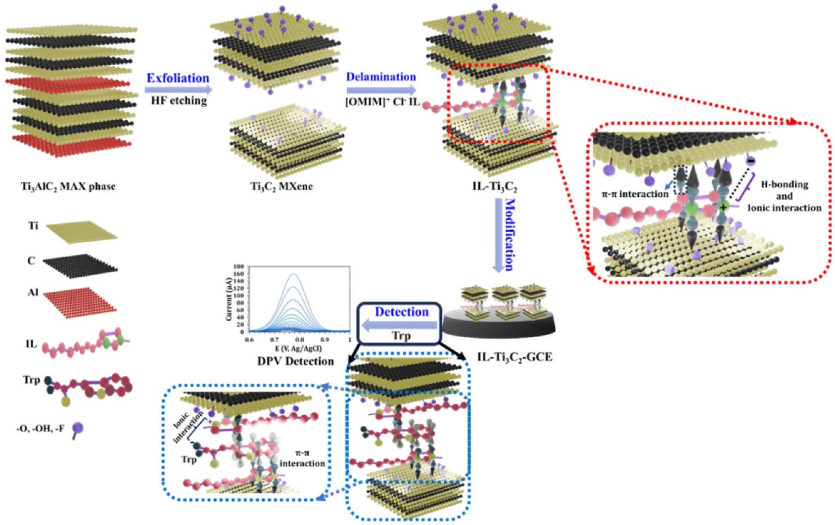
الشكل 1. التحضير التخطيطي لـ
لتحسين الاستشعار الكهروكيميائي لـ Trp. تم رسم هذه الصورة باستخدام Blender 4.0، وهو برنامج مجاني ومفتوح المصدر لإنشاء الرسوم ثلاثية الأبعاد.
تركيب MXene Ti3C2
اثنان جرام من الـ
تم إضافة مسحوق مرحلة MAX إلى 25 مل من HF المركز
) لمدة 18 ساعة من النقش. الـ
ثم تم الطرد المركزي مع الماء المقطر حتى كان الرقم الهيدروجيني للسائل العلوي أكبر من 6. الناتج
تم تجفيف مسحوق MXene تحت الفراغ لمدة 12 ساعة عند
.
مسار التخليق لسائل الأيونات 1-أوكتيل-3-ميثيل إيميدازوليوم كلوريد [OMIM]+ CI- IL
للتخليق
السائل الأيوني 1-أوكتيل-3-ميثيل إيميدازوليوم كلوريد،
تم خلط 1-ميثيل إيميدازول أولاً مع
من 1-كلوروأوكتان في دورق دائري القاع ذو عنق واحد يحتوي على 25 مل من رباعي كلوريد الكربون. ثم تم تسخين المزيج تحت الغليان لمدة 72 ساعة عند
في حمام زيت لضمان اكتمال التفاعل. عند الانتهاء، تم الحصول على منتج لزج بلون أصفر باهت،
، تشكلت في قاع القارورة، وتم التخلص من المذيب. تم غسل المنتج النهائي عدة مرات بمادة الكربون رباعي الكلور لإزالة أي شوائب. بعد تبخير المذيب عند
تم الحصول على المنتج بأعلى نقاء، مع عائد قدره
آلية التخليق موضحة في المعلومات الداعمة (الشكل S-1).
تحضير رقائق IL-Ti3C2
أولاً، تقشير
تم الحصول على MXene عن طريق الموجات فوق الصوتية لـ
محلول مائي لمدة 30 دقيقة. بعد ذلك، كميات مختلفة من الـ
إلى 1.3 ملغ
تمت إضافتها إلى التعليق الناتج، وحدثت عملية الفصل بين الطبقات لـ
تم إجراء معالجة MXene عن طريق العلاج بالموجات فوق الصوتية لمدة 30 دقيقة. الهيكل المسطح لـ
-جزيئات IL المترافقة تجعلها مثالية كمواد متداخلة بين طبقات MXenes. هذه التكوين الفريد يمكّن من تفاعلات غير تساهمية فعالة.
التفاعلات، التي لا تغير بشكل كبير الهيكل المترافق لـ
MXene. بالإضافة إلى ذلك، فإن الجذب الكهروستاتيكي بين جزيئات IL المشحونة إيجابيًا وطبقات MXene المشحونة سلبًا يعزز بشكل أكبر إدخال IL. وبالتالي، يتم الحفاظ على الموصلية الإلكترونية لـ MXene، مما يضمن نشاطًا كهربائيًا استثنائيًا وأداءً عاليًا في التيار.
تحضير المستشعر الكهروكيميائي
تم صقل القطب الكربوني الزجاجي (GCE) أولاً باستخدام معلقات الألومينا (0.3 و
) ثم تم تنظيفه بالإيثانول والماء المقطر. ثم،
من
تم إسقاط الم dispersion المائي على GCE المنظف وتجفيفه في درجة حرارة الغرفة العادية للحصول على
.
تحليل عينة حقيقية
تم شراء حبيبات الأحماض الأمينية من صيدلية محلية. تم إذابة قرص واحد (يحتوي على 500 ملغ من التربتوفان) في 1000 مل من الماء المقطر الدافئ بمساعدة الموجات فوق الصوتية لمدة 15 دقيقة، ثم تم الطرد المركزي عند 2000 دورة في الدقيقة، بعد ذلك تم جمع السائل العلوي. من أجل الأكسدة الكهربائية للتربتوفان، تم تخفيف 1 مل من المحلول المحضر بمقدار 1000 مرة مع محلول فوسفات البوتاسيوم 0.10 م عند درجة حموضة 7.5. تم تحقيق الكشف عن التربتوفان في عينات بول المتطوعين الأصحاء عن طريق تخفيف عينات البول.
بنسبة 1:5.
النتائج والمناقشة
توصيف [OMIM]+ CI- من خلال تحليلات 1HNMR و FTIR
يتم تقديم طيف HNMR للسائل الأيوني المُركب في المعلومات الداعمة (الشكل S-2). القمة عند
يتوافق مع
مجموعة سلسلة الأوكتيل
بينما يُنسب النطاق من 1.23 إلى 1.91 جزء في المليون إلى
المجموعات في سلسلة الأوكتيل
القمم الملحوظة عند
تُنسب أيضًا إلى
المجموعات في سلسلة الأوكتيل
. الإشارات في
يتوافق مع آخر
مجموعة في سلسلة الأوكتيل مرتبطة بـ -N في حلقة الإيميدازوليوم
، والمنطقة من 5.05 إلى 5.48 جزء في المليون مرتبطة بـ
مجموعة بديلة على الحلقة
. أخيرًا، القمم في
جزء في المليون
مجموعة في حلقة الإيميدازوليوم
طيف FTIR لـ
(الشكل
يكشف عن اهتزاز الانحناء لـ
رابطة عند
، الذي يرتبط بحلقة الإيميدازوليوم. اهتزاز الشد لـ
تم ملاحظة الرابطة في حلقة الإيميدازوليوم عند
تُنسب القمم في نطاق 1373-1451 سم-1 إلى اهتزازات الشد لـ
السندات و
مجموعة في ميثيل إيميدازول. النطاقات عند 1490 و
تتوافق مع
الروابط في حلقة الإيميدازوليوم العطرية. اهتزاز الشد لـ
تمت ملاحظة الرابطة عند
والأليفاتية
الروابط في سلسلة الأوكتيل ظهرت في نطاق
. بالإضافة إلى ذلك، تم ملاحظة اهتزاز ضعيف لتمدد O-H عند
من المحتمل أن يكون ذلك بسبب وجود رطوبة طفيفة في الهيكل.
توصيف المواد النانوية المحضرة
النقش الفعال والفصل
تم تأكيدها من خلال تحليل حيود الأشعة السينية (XRD) (الشكل 2A). تم تحديد قمم الحيود عند
، و
تتوافق مع القيم المرتبطة بالهيكل المغلق السداسي للطور النقي
وتتوافق مع رقم بطاقة ملف JCPDS PDF.
تم الإشارة إلى انخفاض في البلورية من خلال قمم أوسع وشدات أقل في أنماط حيود الأشعة السينية.
، جنبًا إلى جنب مع اختفاء 104 انعكاسات لـ
، مما يدل على إزالة طبقات الألمنيوم بعد النقش بحمض الهيدروفلوريك. تم نقل قمة (002) من
إلى
، مما يعكس زيادة في المسافة بين الطبقات من 0.9 نانومتر إلى 1.06 نانومتر نتيجة للنقش واستبدال الألمنيوم بـ -F و
المجموعات الوظيفية. من المهم أن قمة حيود 002 لـ
العينة انتقلت إلى زوايا أقل
المسافة بين الطبقات الموسعة بالنسبة لتلك التي في الحالة الأصلية
تم حسابه ليكون 1.85 نانومتر. يمكن أن يُعزى ذلك إلى التداخل لـ [OMIM]
الأيونات في الطبقات الداخلية للـ MXene خلال عملية الفصل. علاوة على ذلك، فإن الزيادة في المساحة السطحية والمسامية لـ
تم تحديده من خلال تحليلات بروناور-إيميت-تيلر (BET) (الشكل 2B) وتحليلات بارّيت-جويتر-هاليندا (BJH) (الإطار في الشكل 2B). مساحة السطح والمسامية لـ IL-
تم حسابها لتكون
و 14.95 نانومتر، على التوالي، والتي هي أكبر من تلك الخاصة بـ
MXene
و 11.8 نانومتر، على التوالي). بالإضافة إلى ذلك، أكد المجهر الإلكتروني الماسح (SEM) المسافة الأكبر بين الطبقات لـ

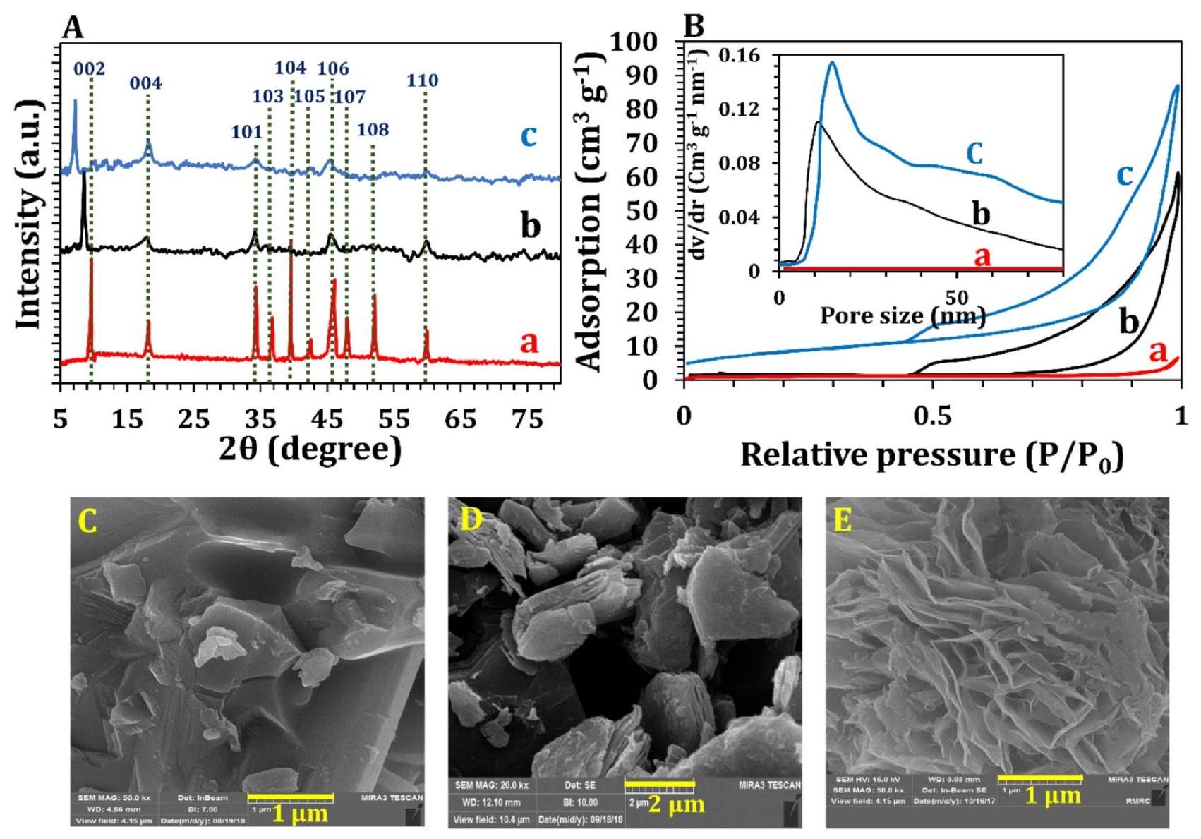
الشكل 2. (أ) حيود الأشعة السينية، و (ب) BET المتعلقة بالمواد النانوية: (أ)
مرحلة ماكس، (ب)
MXene، و (ج) IL
. FE-SEM لـ (E)
مرحلة ماكس، (D)
MXene، و (E) IL-
.
ال
أوراق نانوية من MXene بسبب التداخل مع [OMIM]
الأيونات الموجبة، كما هو موضح في الشكل 2C إلى E.
عرضت هيكلًا مضغوطًا وكثيفًا دون تقشر/تفتت قبل التعديل (الشكل 2C). بعد النقش، كانت النتيجة
أظهر الـ MXene هيكلًا فضفاضًا يشبه الأكورديون مع فجوة معينة بين الطبقات (الشكل 2D). بعد إدخال السائل الأيوني، كانت صورة SEM للسائل الأيوني-
أظهر العينة هيكلًا أكثر انفتاحًا من طبقات الـ MXene (الشكل 2E).
النشاط الكهروتحفيزي لـ IL-Ti3C2/GCE لأكسدة التربتوفان
تم إجراء اختبارات CV لتقييم الخصائص الكهروكيميائية لـ IL-
/GCE كمادة كهربائية محفزة لأكسدة 1 مللي مول من التربتوفان عند معدل مسح
في
تظهر الشكل 3A غياب تيار فاراداي في الفولتموجرامات المستخرجة من GCE العاري.
) ،
(ب’)، و
(
) في
. الـ IL-
(الشكل 3A، ج) أظهر المحفز استجابة تيار ذروة الأكسدة غير القابلة للعكس عالية للغاية
لـ Trp، والذي هو أكبر بكثير من GCE العاري (
الشكل 3A، أ)
الشهادة العامة للتعليم
، الشكل 3A، ب). الزيادة في التيار من
GCE مقارنةً مع GCE العاري (6.4 مرة) و
يمكن أن يُعزى (3.2 مرة) إلى سهولة وصول جزيئات التربتوفان إلى المواقع النشطة لمادة MXene بين الطبقات بدلاً من المنطقة الخارجية للجسيمات متعددة الطبقات. إن إدخال جزيئات السائل الأيوني بين الموصلين
الطبقات الداخلية وفرت هيكلًا مساميًا ثلاثي الأبعاد مع زيادة في المسافة بين الطبقات من
إلى IL-
، مما يضمن تعرض الأنواع النشطة بشكل كافٍ إلى التربتوفان ويسهل نقل الإلكترونات لمنع إعادة تكديس الـ
MXenes. الشكل S-4 يظهر أنه، على عكس
MXenes
يتم توزيعه بشكل موحد ويظل مستقرًا في المحلول المائي لمدة لا تقل عن 30 يومًا. قد تكون خشونة السطح، أو النشاط الجوهري (التأثيرات التآزرية)، أو مزيج من الاثنين هي المسؤولة عن زيادة النشاط. الثوابت المعدلة الظاهرة (
) و
تمت مقارنة التعديلات المختلفة لـ GCE من خلال نهج EIS (الشكل 3B). في
محلول يحتوي على 1 مللي مول من التربتوفان، يتم تمثيل معدل الأكسدة الكهروتحفيزية بواسطة
الذي يتم تحديده من خلال المعادلة (1)
أظهرت النتائج أن الأيونات السائلة المدخلة بين طبقات المساحة عززت بشكل كبير أداء الـ
GCE بالمقارنة مع
GCE ، مما يؤدي إلى زيادة تقارب 3.6 مرة
و 2.5 ضعف
التحسينات (الجدول S-1). تشير النتائج إلى أن النشاط المحسن (69.4%) يُعزى في الغالب إلى المساحة السطحية الفعالة الأكبر لـ
، مع تأثيرات تآزرية تمثل التحسن المتبقي
لتحديد مقاومة انتشار واربورغ لجزيئات التربتوفان في
معامل واربورغ
يتم تحديده من الميل عند الترددات المنخفضة عن طريق رسم
ضد
. الـ

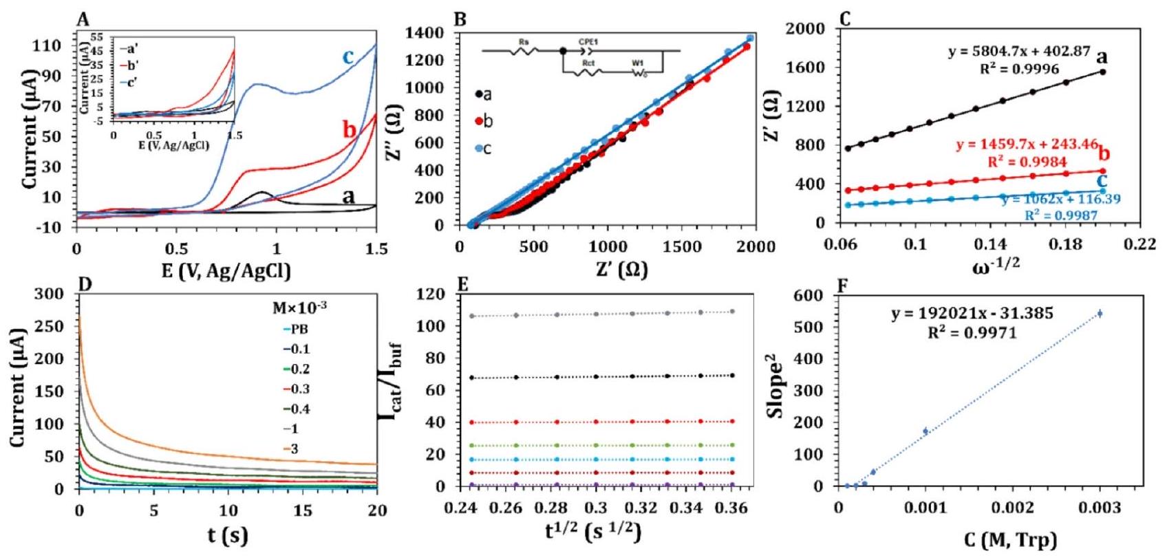
الشكل 3. (أ) منحنيات الجهد الكهربائي المختلفة للقطب الكهربائي الكربوني المعدل في غياب (
إلى
) ووجود
(أ إلى ج). (ب) مخططات نايكويست و (ج) الرسم البياني الملائم خطياً لـ
ضد
في منطقة التردد المنخفض من مخططات نايكويست في محلول PBS
. جائزة التعليم العام (GCE)
و )
(ب’ و ب)، و
( ج و
). (D) كرونوأمبيروجرامات IL-
في تركيزات مختلفة من Trp. (E) و (F) توضحان الرسوم البيانية لـ
مقابل
في تركيزات مختلفة من التربتوفان ومربع الميل في (E) المرتبطة بالتركيزات المعنية، على التوالي. الشرط: 0.85 فولت، التي تم الحصول على التيار عند 15 ثانية.
قيم GCE العارية (الشكل 3C، أ)
/GCE (الشكل 3C، ب) هي 5804.7 و
على التوالي، في حين أن
من IL-
هو
(الشكل 3C، ج). الجزء السفلي
من IL-
يشير إلى انتشار أسرع وحركيات نقل الشحنة فارادي في المادة النانوية المقترحة. علاوة على ذلك، تم دراسة آلية النشاط الكهروكيميائي من خلال CV من خلال تقييم المساحات السطحية النشطة كهربائياً (ESAs) عند معدلات مسح مختلفة (20-250) في محلول 0.1 م KCl يحتوي على
زوج الأكسدة والاختزال (الشكل S-5). وفقًا لمعادلة راندلز-سيفيك.
، أظهرت النتائج التي تم الحصول عليها زيادة بمقدار 2.3 مرة و7 مرات في ESA لـ IL-
الشهادة العامة للتعليم
(الشكل S-5A-B) مقارنةً مع تلك الخاصة بـ
(الشكل S-5C-D) و GCE (
) (الشكل S-5E-F)، على التوالي. في هذه المعادلة، A تمثل مساحة سطح القطب الكهربائي،
يمثل معامل الانتشار
v تمثل معدل المسح
يمثل C تركيز
، ويمثل n عدد الإلكترونات
تم استخدام طريقة الكرونوكولومترية لتأكيد الزيادة في المساحة السطحية بسبب إدخال السائل الأيوني داخل طبقات الماكسين في نفس المحلول (الشكل S-6A). استنادًا إلى المعادلة التالية (3)، تم الحصول على المساحات السطحية الفعالة من ميل الرسم البياني لـ Q مقابل
(الشكل S-6B) للـ GCE العاري،
جي سي إي، و IL-
جي سي إي كانت
، و
على التوالي، وهو ما يتماشى مع نتائج السيرة الذاتية.
من المثير للاهتمام، مقارنة تيار ESA وتيار الأكسدة الكهربائية لـ
(بمعدل مسح قدره
، الشكل 3A) في الـ
و
كشف عن زيادة بمقدار 3.2 مرة في النشاط الكهروكيميائي، والذي تم نسبته بشكل أساسي إلى زيادة بمقدار 2.2 مرة (ما يعادل
) في المساحة السطحية الفعالة وزيادة بمقدار 1 مرة (تعادل
) في التأثيرات التآزرية، والتي تتماشى مع النتائج المستخلصة من تحليل EIS. بالإضافة إلى ذلك، فإن القدرات المحسنة للإلكتروكيميائية لـ
تم تقييم GCE في أكسدة التربتوفان عند جهد ثابت قدره 0.85 فولت من خلال طريقة الكرونوأمبريومترية. ثابت معدل التحفيز (
لأكسدة التربتوفان باستخدام IL-
الشهادة العامة للتعليم
(الشكل 3D-F) هو 4.85 مرة أكبر من ذلك لـ
(الشكل S-7). أظهرت هذه النتائج أن التجميع الذاتي للمواد المدخلة
أيونات في بعدين
تشكلت طبقات MXene النانوية إطارًا ثلاثي الأبعاد أكثر انفتاحًا مع زيادة
-تباعد من خلال العفوية
التكديس، التفاعلات الكهروستاتيكية، وروابط الهيدروجين. هذا النانو-material المستقر يتيح وصولاً أكبر إلى المواقع النشطة للـ MXenes بين الطبقات.
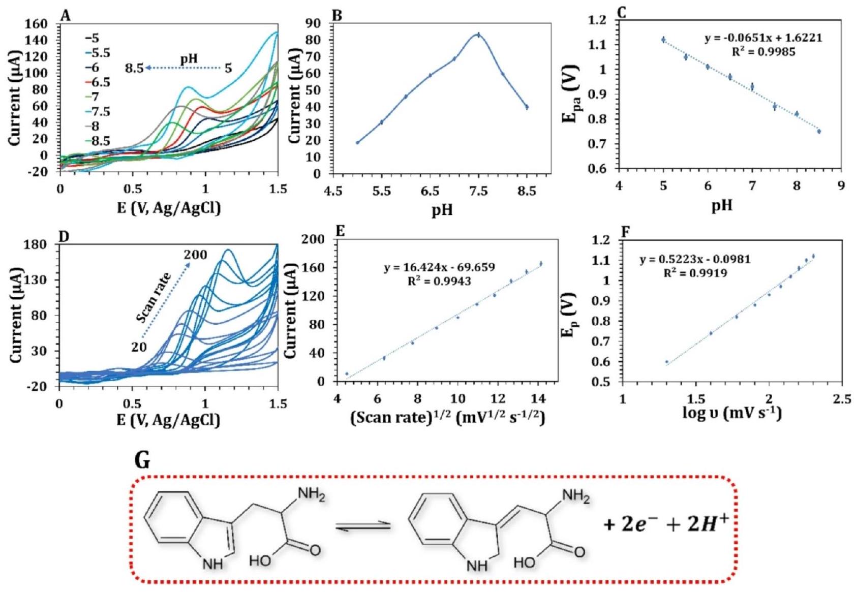
تحسين ظروف مختلفة للأكسدة الكهربائية للتربتوفان
أثر كمية IL على أداء المستشعر
من خلال تغيير نسبة الكتلة للـ IL بشكل منهجي إلى
تم الحصول على تحميل IL المثالي من 0.1 إلى 1.3 وقياس منحنيات CV المقابلة (الشكل S-8 A). يمكن ملاحظة زيادة تدريجية في تيار الاستجابة بوضوح مع زيادة نسبة الكتلة لـ ILs إلى
يزداد MXenes إلى 0.9 في

الشكل 4. (أ) منحنيات الفولتامترية، (ب) تيار الذروة الأنودية، و(ج) جهد الذروة الأنودية لـ
كنتيجة لدرجة الحموضة في محلول PBS
) +1 مللي مولار تريبتوفان. (D) منحنيات الجهد الحالي لـ 0.1 مللي مولار تريبتوفان على
في PBS (0.1 م،
) بمعدلات مسح مختلفة (من 20 إلى
)، (E) العلاقة الخطية بين التيارات القصوى مقابل الجذر التربيعي لمعدل المسح و (F)
مقابل
. (G) الآلية المقترحة للأكسدة الكهروكيميائية للتربتوفان عند IL-
.
| عينة | معيار (
|
المتوسط المكتشف | مضاف | المتوسط المؤسس | خطأ نسبي (%) | SD | نسبة الانحراف المعياري (%) | نسبة الاسترداد (%) |
|
|
| حبوب الأحماض الأمينية | ٢.٤٤٨ | 2.395 (2.408, 2.401, 2.43) | 0 | 0 | 1.606 | 0.0187 | 0.077 | – | ٣.٦٣٩ | ٤.٣٠ |
| ٥ | 7.433 (7.558، 7.389، 7.352) | 0.2013 | 0.1098 | 1.477 | 99.79 | 0.2365 | ||||
| 10 | 12.593 (12.28، 12.64، 12.86) | 1.167 | 0.2928 | 2.325 | ١٠١.١٦ | 0.8597 | ||||
| 50 | 51.923 (50.23، 51.89، 53.65) | 1.0003 | 1.710 | ٣.٢٩٣ | ٩٨.٩٩ | 0.5313 | ||||
| بول | 85.65 | ٨٣.٩٦ (٨٧.٢٨، ٨٠.٣٦، ٨٤.٢٤) | 0 | 0 | 1.973 | 3.468 | 0.0413 | – | 0.8439 | |
| ٥ | 91.73 (92.85، 88.64، 93.72) | 1.198 | 2.716 | 2.961 | ١٠١.١٩ | 0.6927 | ||||
| 10 | ٩٦.٠٩٦ (٩٨.٥٦، ٩٦.٤٥، ٩٣.٢٨) | 0.4669 | 2.657 | ٢.٧٦٥ | 100.46 | 0.2911 | ||||
| 50 | 137.91 (140.26، 138.63، 134.84) | 1.666 | ٢.٧٨٠ | 2.016 | ١٠١.٦٦ | 1.407 |
الجدول 1. الكشف الكهروكيميائي عن التربتوفان في عينة من الحياة الواقعية.

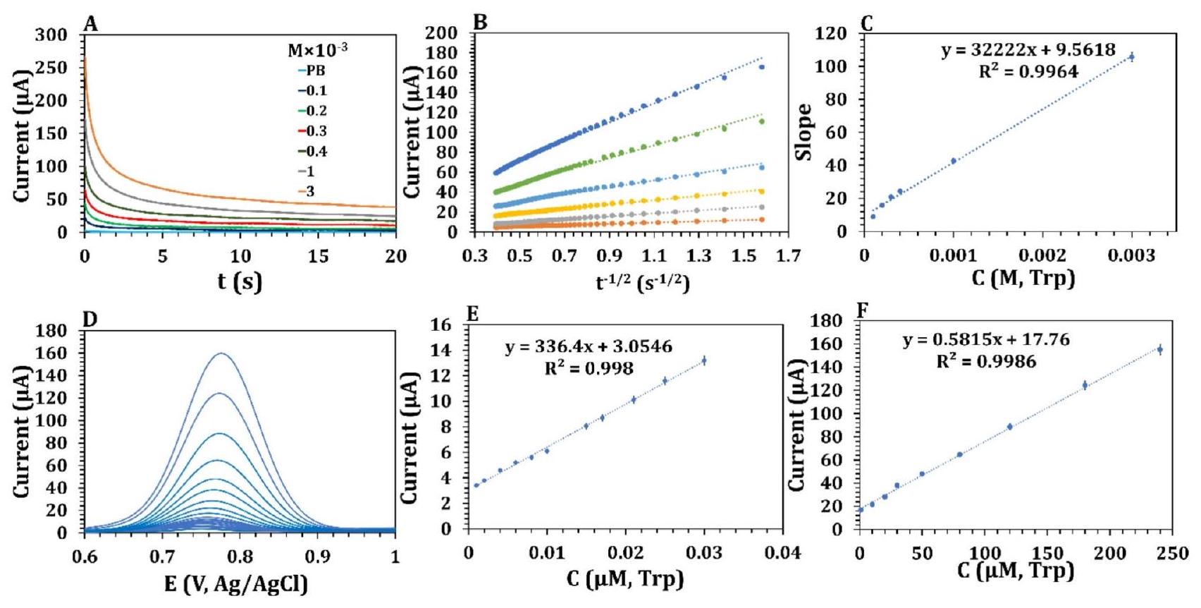
الشكل 5. (أ) قياس الكرونوأمبرو متحصل عليه عند
مع تركيزات متغيرة من التربتوفان. (ب) (ط) رسم التيارات مقابل.
، و (C) رسم بياني للمنحدرات من (B) مقابل تركيز التربتوفان. (D) DPV لـ IL-
في نطاق التركيز من 0.001 إلى
في PBS
. (E) منحنى المعايرة لـ Trp على مدى من 0.001 إلى
و
.
mM تريبتوفان (الشكل S-8B). أدى زيادة إضافية في كمية IL إلى انخفاض في التيار بسبب عدم استقرار الـ
أوراق MXene وسد المسام في النانوكاتاليست.
أثر حجم IL-Ti3C2 على تحديد التربتوفان
علاوة على ذلك، قمنا بالتحقيق في تأثير الـ
الحجم على الاستجابة الحالية (الشكل S-8 C). كما هو موضح في الشكل S-8D، كان هناك زيادة كبيرة في الاستجابة الحالية مع زيادة الحجم من 5 إلى
. ومع ذلك، انخفضت الاستجابة الحالية بشكل حاد عندما زاد الحجم من 40 إلى
.
أثر الـ
التركيز على تحليل TRP
تركيز IL-
تم فحص الجرعات المختلفة المحملة على GCE في PB
كما هو موضح في الشكل S-8E، أظهر استجابة CV للـ GCE المعدل تيارًا أقصى عند
(الشكل S-8 F)، الذي انخفض مع زيادة التركيز. يمكن أن يُعزى هذا الظاهرة إلى انخفاض معدلات التفاعل الناتجة عن تأثيرات التشبع حيث تكون جميع المواقع النشطة على القطب مشغولة. بالإضافة إلى ذلك، يمكن أن تؤدي التركيزات المتزايدة إلى قيود في نقل الكتلة تعيق انتشار التربتوفان إلى القطب. على وجه التحديد، فإن التركيزات المتزايدة من
يمكن أن يخلق طبقة أكثر سمكًا تعيق نقل الإلكترونات بين القطب الكهربائي وجزيئات التريبتوفان، مما يقلل من حساسية واستجابة النظام للتريبتوفان. بالإضافة إلى ذلك، يمكن أن تؤدي التركيزات الأعلى من السائل الأيوني إلى وسط أكثر لزوجة يقيّد بشكل أكبر انتشار التريبتوفان إلى واجهة القطب الكهربائي ويقلل من التركيز الفعال للمحلول المتاح للتفاعل.
تأثير الرقم الهيدروجيني وآلية الأكسدة الكهربائية للتربتوفان
تدرس هذه الدراسة تأثير درجة حموضة محلول PB على شدة قمة أكسدة التربتوفان (1 مللي مول). توضح الأشكال 4 A و B أن تيار استجابة الأكسدة الكهربائية يزداد مع زيادة درجة الحموضة من 5.6 إلى 7.5 وينخفض مع زيادة درجة الحموضة. للتربتوفان نقطة تعادل.
مشحون سلبياً في المحلول عند
، ويسبب جذبًا كهربائيًا ساكنًا بين جزيئات التربتوفان المشحونة سلبًا وجزيئات السائل الأيوني المشحونة إيجابًا. بالإضافة إلى ذلك، فإن بنية حلقة الإيميدازوليوم للسائل الأيوني على سطح النانوكاتاليست وجزيئات التربتوفان تنجذب إلى بعضها البعض بواسطة
التكديس. علاوة على ذلك، فإن المجموعات الوظيفية على
يمكن أن يتفاعل MXene مع مجموعات الكربوكسيل والأمين على التربتوفان عبر الروابط الهيدروجينية، مما يزيد من تركيزها على سطح القطب. ومن المهم أن جهد الذروة الأنودية انتقل سلبًا نحو جهد أقل مع زيادة الرقم الهيدروجيني وفقًا لمعادلة الانحدار الخطي.
(الشكل 4C). كانت قيمة الميل قريبة من القيمة النظرية -0.059، مما يشير إلى سلوك الأكسدة مع عدد متساوٍ من الإلكترونات والبروتونات. تم تحديد كمية نقل الإلكترون من خلال إجراء تجارب التحليل الكهربائي الكولومترية. وفقًا لمعادلة فاراداي.
، حيث
، و
تم حساب عدد نقل الإلكترون ليكون 2. تم أكسدة التربتوفان الكهروكيميائية لـ
في 0.1 مليمول من PB عند سرعات مسح مختلفة (
تم التحقيق في ذلك (الشكل 4D). أظهرت النتائج أن العملية تتحكم فيها الانتشار، مع رسم بياني خطي لتيار ذروة الأكسدة مقابل الجذر التربيعي لمعدل المسح.

الشكل 6. (أ) انتقائية الـ
نحو أكسدة التربتوفان (
) في وجود مجموعة متنوعة من المركبات المتداخلة المحتملة (كل منها عند
). (ب) استقرار الـ
GCE على مدى 15 يومًا. (C) تم الحصول على DPVs للبول البشري مع إضافة مواد.
.
(الشكل 4E). علاوة على ذلك، تغيرت تيارات الذروة الأنودية بشكل لا رجعة فيه نحو جهد إيجابي أعلى مع زيادة معدل المسح. طريقة لافيرون (المعادلة 5)
تم استخدامه لتحديد قيمة
الذي تم تحديده ليكون 0.92 استنادًا إلى ميل الشكل 4F.
يمثل الجهد الرسمي، ثابت المعدل غير المتجانس، معامل نقل الإلكترون، وعدد الإلكترونات بـ
، و ن، على التوالي. علاوة على ذلك،
قيمة لـ
الأكسدة الكهربائية عند
يمكن حساب GCE من خلال المعادلة التالية (6)
.
أين
هو نصف القيمة المحتملة الرسمية. وبالتالي، فإن قيم
وعدد نقل الإلكترونات في
تم العثور على GCE بقيم 0.47 و 2.0، على التوالي، بما يتماشى مع نتائج التحليل الكولومتري. من خلال استقراء الرسم البياني لـ
مقابل معدل المسح وحساب
تم الحصول عليه أيضًا من خلال التقاطع من معادلة لافيرون. قيمة
يمكن أن يكون
. بشكل عام، آلية الأكسدة الكهربائية للتريبتوفان عند
موضح في الشكل 4G. بالإضافة إلى ذلك، معادلة كوتريل
يمكن استخدامه لتحديد معامل الانتشار لـ Trp، مع قيمة مقدرة بـ
(الشكل 5A إلى C).
التحديد التحليلي لـ trp على IL-Ti3C2/GCE
لإجراء دراسة التركيز، تم فصل IL
تم استخدامه لتسجيل الاستجابة الحالية عبر DPV. توضح الشكل 5D الزيادة التدريجية في الحد الأقصى للتيار مع زيادة تركيز التربتوفان ضمن النطاق من
مع نطاقين خطيين،
(الشكل 5E) و
(الشكل 5F). الحد الأدنى من الكشف لـ IL-
تم حساب /GCE ليكون 0.06 نانومول. يمكن أن يُعزى الانخفاض الكبير في حد الكشف (LOD) لـ Trp إلى مجموعة من عاملين مساهمين. (i) الزيادة في المساحة السطحية مع زيادة المسافة بين الطبقات من الحالة الأصلية
إلى IL-مفصول
يجعلها أكثر وصولاً إلى المزيد من المواقع النشطة من MXenes بين الطبقات. (ii) الـ
تفاعلات مجموعة الإيميدازوليوم من السائل الأيوني مع الحلقة العطرية للتربتوفان سهلت نقل الشحنة، مما عزز أداء منصة الاستشعار.
قدرة مقاومة التداخل، الاستقرار، القابلية للتكرار، والاتساق
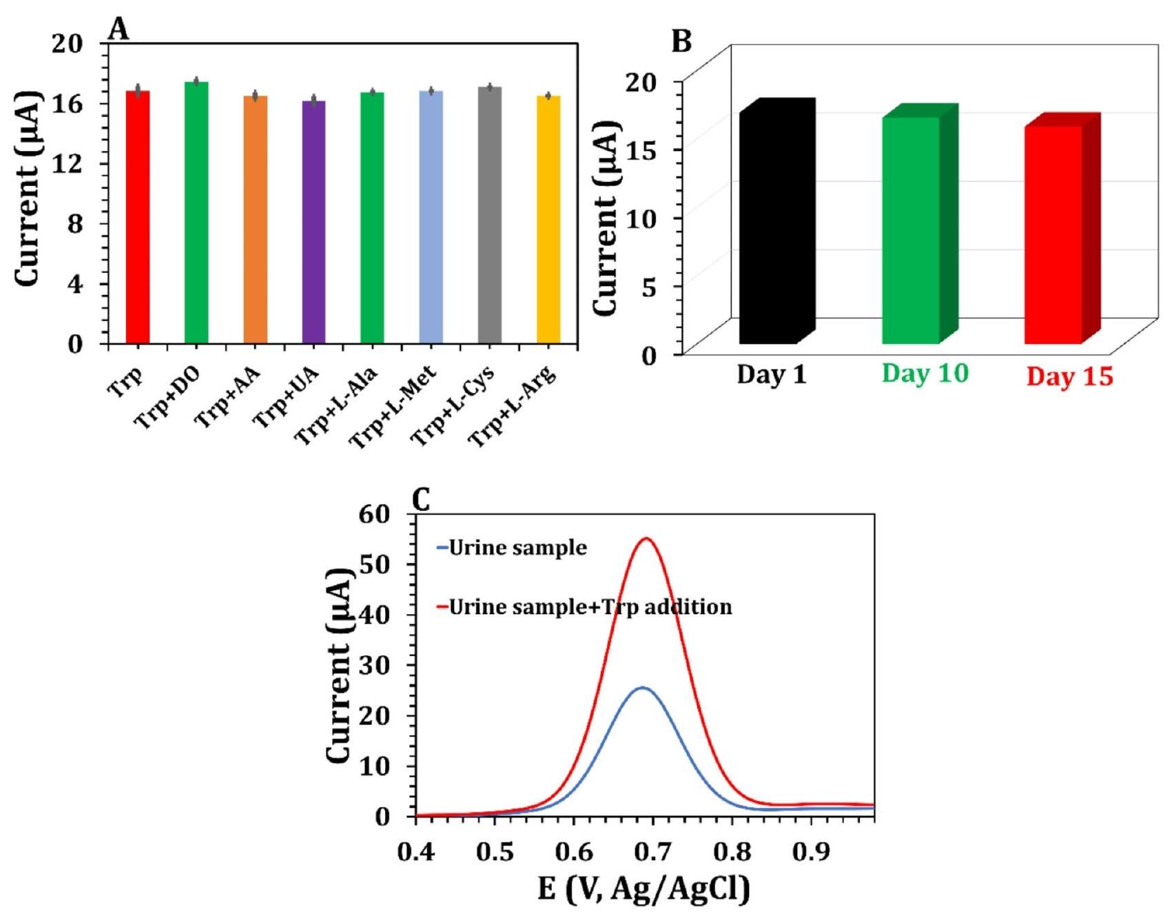
تقييم المواد التحليلية بشكل انتقائي في بيئة فسيولوجية معقدة تحتوي على مواد متداخلة هو التحدي الرئيسي في تطوير المستشعرات. كما يتضح من نتائج DPV (الشكل S-9 A والشكل 6A)، لم تؤثر المواد المتداخلة مثل الدوبامين (DO)، وحمض الأسكوربيك (AA)، وحمض اليوريك (UA)، واللايسين (L-Ala)، والميثيونين (Met)، والسيستين (L-Cys)، والأرجينين (L-Arg) على كشف التربتوفان (Trp) عند تركيزات أعلى بعشر مرات.
من
تركيز
). إن عمر التخزين لجهاز الاستشعار الحيوي أمر حاسم للتطبيقات العملية، والتغير الضئيل في تيار DPV في
تركيز TRP في PB على مدى 15 يومًا (الشكل S-9B)، بحوالي
احتفاظ الاستجابة الحالية الأولية (الشكل 6B) أظهر استقرارًا ملحوظًا. بالإضافة إلى ذلك، فإن استقرار الـ
تم تقييمه من خلال إجراء 5 اختبارات CV متتالية في محلول PB بتركيز 0.1 م. أظهرت النتائج أن كل من الاستجابة والشكل العام لمنحنيات CV ظلا تقريبًا دون تغيير، مما يشير إلى أن [OMIM] المحتجز كيميائيًا
الأيونات ضرورية في الحماية
من الأكسدة (الشكل S-10). لتقييم قابلية التكرار لـ
تم تسجيل تيار الأكسدة لإحدى الأقطاب الكهربائية المحضرة عند
و
تم قياس Trp لثلاث قياسات منفصلة على مدى ثلاثة أيام (بين الأيام) (الجدول S-2) وداخل يوم واحد (داخل اليوم) (الجدول S-3). بشكل عام، كانت RSD
مع قيم t أصغر للتحليلات بين الأيام وداخل اليوم من
في
، مما يشير إلى عدم وجود أخطاء منهجية كبيرة مع تكرارية جيدة. بالنسبة لإمكانية التكرار، تم استخدام خمسة
تم إعداد GCEs، وتم تسجيل استجابتها الحالية في
، و
حلول Trp عبر DPV. أظهرت الدراسة قابلية إعادة إنتاج مرضية، مع RSD منخفض يتراوح من 1.55 إلى
وقيم t أصغر من
(الجدول S-4). الجدول S-5 قارن بين أجهزة الاستشعار الكهروكيميائية المنشورة سابقًا لـ Trp لتقييم قدرات الكشف عن IL-
جهاز GCE. أظهر المستشعر الكهروكيميائي أداءً تحليليًا ملحوظًا يمكن مقارنته، وفي بعض الجوانب يتفوق على، الطرق الفولتمترية المعتمدة سابقًا للكشف عن التربتوفان.
التطبيق في عينات حقيقية
لإظهار الإمكانية المحتملة لتطبيق القطب المقترح في صناعة المواد الغذائية والرعاية الصحية، IL
تم استخدام /GCE للكشف عن التربتوفان في حبيبات الأحماض الأمينية والبول البشري (الجدول 1). كانت كمية التربتوفان في حبيبات الأحماض الأمينية المحسوبة لكل قرص 492 ملغ، وهو قريب من القيمة النظرية البالغة 500 ملغ مع
خطأ نسبي. بالإضافة إلى ذلك، تم إجراء اختبار الاسترداد للتربتوفان عبر طريقة الإضافة القياسية عن طريق إضافة عينات بتركيزات مختلفة من التربتوفان. تراوحت استردادات حبيبات الأحماض الأمينية من 98.99 إلى
، مع قيم RSD تتراوح من 0.077 إلى
. علاوة على ذلك، أظهرت النتائج التي تم الحصول عليها من خلال المستشعر المطور أن هذا المستشعر الحيوي كان لديه دقة ممتازة دون أي خطأ منهجي، كما يتضح من القيم الصغيرة الملاحظة
مقارنة مع
.
بالإضافة إلى ذلك، تم تحليل عينات مصل الإنسان المخفف لقياس التربتوفان. زادت قمة DPV عند 0.75 فولت مع إضافة التربتوفان.
)، مما يشير إلى أنه تم الكشف عن أكسدة التربتوفان في عينات البول (الشكل 6C). تراوحت قيم الاسترداد لعينات البول المضافة إليها محلول معيار التربتوفان من 100.46 إلى
، وكانت قيم RSD لجميع الاختبارات الثلاثة المستقلة أقل من
. هذه النتائج توضح أن المطور
إن GCE فعّالة للغاية في تحديد محتوى التربتوفان في حبيبات الأحماض الأمينية الحقيقية أو البول.
الخاتمة
في هذه الدراسة، تم زيادة المسافة بين الطبقات في
تم تحقيق MXenes من خلال إدخال [OMIM]
أيونات IL داخل طبقات MXene، مما يؤدي إلى تحسين الأداء الكهروكيميائي لأكسدة التربتوفان. واجهة الـ
أظهر GCE استقرارًا ملحوظًا بسبب التفاعلات المواتية بين MXene وIL، مما يمنع بشكل فعال تسرب الإلكترود. وبالتالي، فإن
تزيد الأيونات من توفر المواقع النشطة كهربائياً، وتزيد من نقل الإلكترونات في تفاعلات الأكسدة والاختزال السطحية، وتحسن المسارات لجزيئات التربتوفان. أظهر المستشعر المقترح نتائج كشف ناجحة على مدى واسع من التركيزات، من 0.001 إلى
، مع حد كشف منخفض يبلغ 0.06 نانومول، مما يدل على انتقائية قوية واستقرار. علاوة على ذلك، فإن الفعالية العملية لـ
تم إثبات GCE من خلال الكشف الناجح عن التربتوفان في حبيبات الأحماض الأمينية وعينات مصل الدم البشري، مما أسفر عن نتائج مرضية ومُعتمدة.
توفر البيانات
تتوفر مجموعات البيانات المستخدمة و/أو التي تم تحليلها خلال الدراسة الحالية من المؤلف المراسل عند الطلب المعقول.
تاريخ الاستلام: 19 نوفمبر 2024؛ تاريخ القبول: 24 فبراير 2025
نُشر على الإنترنت: 25 فبراير 2025
References
- Sinha, A. K. et al. Dietary fibre directs microbial Tryptophan metabolism via metabolic interactions in the gut microbiota. Nat. Microbiol.9, 1964-1978 (2024).
- Sakurai, M. et al. Serum metabolic profiles of the Tryptophan-Kynurenine pathway in the high risk subjects of major depressive disorder. Sci. Rep.10, 1961 (2020).
- Kaleta, M. et al. Patients with neurodegenerative proteinopathies exhibit altered Tryptophan metabolism in the serum and cerebrospinal fluid. ACS Chem. Neurosci.15, 582-592 (2024).
- Desmons, A. et al. High performance liquid chromatography-tandem mass spectrometry quantification of Tryptophan metabolites in human serum and stool – Application to clinical cohorts in inflammatory bowel diseases. J. Chromatogr. A. 1685, 463602 (2022).
- Kałuzna-Czaplinska, J. & Michalska, M. Rynkowski determination of Tryptophan in urine of autistic and healthy children by gas chromatography/mass spectrometry. Med. Sci. Monit.16, CR488-CR492 (2010).
- Wang, W. et al. Peptide aptamer-based colorimetric sensor for the detection of L-tryptophan in Porcine serum. Microchem J.197, 109896 (2024).
- Wu, Y., Wang, T. & Zhang, C. Xing A rapid and specific colorimetric method for free Tryptophan quantification. Talanta176, 604-609 (2018).
- Momeni, F. & Khoshfetrat, S. M. Zarei electrochemical Sandwich-Type aptasensor based on the multifunctional Catechol-Loaded
for detection of cardiac troponin I. ACS appl. Nano Mater.6, 19239-19248 (2023). - Zhang, Y. Wu new perspective crosslinking electrochemistry and other research fields: beyond electrochemical reactors. Chem. Sci.15, 6608-6621 (2024).
- Tasić, Ž. Z. et al. Electrochemical determination of L-tryptophan in food samples on graphite electrode prepared from waste batteries. Sci. Rep.12, 5469 (2022).
- Sadok, I., Tyszczuk-Rotko, K. & Mroczka, R. Staniszewska simultaneous voltammetric analysis of Tryptophan and kynurenine in culture medium from human cancer cells. Talanta209, 120574 (2020).
- Zhang, R. et al. Functionalized PProDOT@ nitrogen-doped carbon Hollow spheres composites for electrochemical sensing of Tryptophan. Carbon161, 842-855 (2020).
- Nasimi, H. et al. Electrochemical sensors for screening of tyrosine and Tryptophan as biomarkers for diseases: A narrative review. Microchem J.190, 108737 (2023).
- Khan, Z. A., Hong, P. J. S. & Lee, C. H. Y. Hong recent advances in electrochemical and optical sensors for detecting Tryptophan and melatonin. Int. J. Nanomed. 16, 6861-6888 (2021).
- Khoshfetrat, S. M. & Mamivand, S. Darband Hollow-like three-dimensional structure of Methyl orange-delaminated Ti3C2 MXene nanocomposite for high-performance electrochemical sensing of Tryptophan. Microchim. Acta. 191, 546 (2024).
- Xia, H. L. et al. Size-and Emission-Controlled synthesis of Full-Color luminescent Metal-Organic frameworks for Tryptophan detection. Angew. Chem.135, e202308506 (2023).
- Rezaei, F. & Ashraf, N. Zohuri A smart electrochemical sensor based upon hydrophilic core-shell molecularly imprinted polymer for determination of L-tryptophan. Microchem. J185, 108260 (2023).
- Mohammadi, F. & Roushani, M. Valipour development of a label-free impedimetric aptasensor based on Zr -MOF and Titaniom carbide nanosheets for detection of L-tryptophan. Bioelectrochemistry155, 108584 (2024).
- Qian, J. et al. Interfacial superassembly of flower-like NiMn-LDH@poly-l-lysine composites for selective electrochemical sensing of Tryptophan. Anal. Chim. Acta. 1237, 340608 (2023).
- Liu, Y., Wang, L. & Yang, L. A superior sensor for the electrochemical detection of Tryptophan in food samples using Ag-doped TiO 2 nanoparticles modified glassy carbon electrode. Int. J. Electrochem. Sci.16, 210534 (2021).
- Pan, Q. et al. Development of a chiral electrochemical sensor based on copper-amino acid mercaptide nanorods for enantioselective discrimination of Tryptophan enantiomers. Anal. Chim. Acta. 1272, 341480 (2023).
- Garg, S., Singh, A. & Parmar, A. S. Rosy Boron carbon Nitride-Assisted Electro-Functionalization of Screen-Printed electrode for Tryptophan sensing. ACS Appl. Nano Mater.6, 14849-14860 (2023).
- Karimian, M. & Dashtian, K. Zare-Dorabei microfluidic chip and chiroptical gold nanoparticle-based colorimetric sensor for enantioselective detection of L-tryptophan. Talanta266, 125138 (2024).
- Singh, S. et al. Facile and affordable design of MXene-Co3O4-Based nanocomposites for detection of hydrogen peroxide in Cancer cells: toward portable tool for Cancer management. Small19, 2208209 (2023).
- Zhang, L. et al. A polyoxometalate/chitosan-Ti3C2Tx MXene nanocomposite constructed by electrostatically mediated strategy for electrochemical detecting L-tryptophan in milk. Food Chem. 458, 140309 (2024).
- Xue, C. et al. An ionic liquid-modified PEDOT/Ti3C2TX based molecularly imprinted electrochemical sensor for pico-molar sensitive detection of L-Tryptophan in milk. Food Chem. 449, 139114 (2024).
- Wang, F., Wang, H. & Cui, X. Liu upconversion fluorescence of MXene nanosheets and the sensitive detection of l-tryptophan. Sens. Diagnostics. 1, 1080-1087 (2022).
- Downes, M., Shuck, C. E., McBride, B. & Busa, J. Y. Gogotsi comprehensive synthesis of Ti3C2Tx from MAX phase to MXene. Nat. Protoc. 19, 1-28 (2024).
- Amara, U., Hussain, I., Ahmad, M. & Mahmood, K. Zhang 2D MXene-based biosensing: a review. Small19, 2205249 (2023).
- Khoshfetrat, S. M. Chegeni rational design of Ti3C2 MXene nanocomposite with bromophenol blue for efficient signal amplification: sensitive electrochemical detection of cardiac troponin I in patient plasma. Sens. Actuators B Chem.397, 134668 (2023).
- Fang, H. et al. Stabilizing Ti3C2T x MXene flakes in air by removing confined water. Proc. Natl. Acad. Sci., 121 e2400084121 (2024).
- Cao, W. et al. A review of how to improve Ti3C2Tx MXene stability. Chem. Eng. J. 496, 154097 (2024).
- Naguib, M. et al. Two-dimensional nanocrystals produced by exfoliation of Ti3AlC2, MXenes, Jenny Stanford Publishing2011, pp. 15-29.
- Ghidiu, M., Lukatskaya, M. R., Zhao, M. Q. & Gogotsi, Y. Barsoum conductive two-dimensional titanium carbide ‘clay’with high volumetric capacitance. Nature516, 78-81 (2014).
- Momeni, F., Khoshfetrat, S. M. & Bagheri, H. Zarei Ti3C2 MXene-based nanozyme as coreaction accelerator for enhancing electrochemiluminescence of glucose biosensing. Biosens. Bioelectron.250, 116078 (2024).
- Mehdi Khoshfetrat, S., Moradi, M. & Zhaleh, H. Hosseini multifunctional Methyl orange-delaminated Ti3C2 MXene for non-enzymatic/metal-free electrochemical detection of hydrogen peroxide and hydrazine. Microchem J.205, 111382 (2024).
- Xu, T. et al. Ultralight MXene/carbon nanotube composite aerogel for high-performance flexible supercapacitor. Adv. Compos. Hybrid. Mater.6, 108 (2023).
- Li, Y. et al. MXene-Graphene Field-Effect transistor sensing of influenza virus and SARS-CoV-2. ACS Omega. 6, 6643-6653 (2021).
- Zhang, Q. et al. Growth of AgI semiconductors on tailored 3D porous Ti3C2 MXene/graphene oxide aerogel to develop sensitive and selective signal-on photoelectrochemical sensor for H2S determination. Anal. Chim. Acta. 1245, 340845 (2023).
- Zhang, C. J. et al. Oxidation stability of colloidal two-dimensional titanium carbides (MXenes). Chem. Mater.29, 4848-4856 (2017).
- Song, X., Gao, H. & Yuan, R. Xiang trimetallic nanoparticle-decorated MXene nanosheets for catalytic electrochemical detection of carcinoembryonic antigen via exo III-aided dual recycling amplifications. Sens. Actuators B Chem.359, 131617 (2022).
- Huang, J., Gu, H., Feng, X. & Wang, G. Chen copper Nanoparticle/N-Doped Ti3C2Tx MXene hybrids with enhanced Peroxidaselike activity for colorimetric glucose sensing. ACS Appl. Nano Mater.5, 15531-15538 (2022).
- Liao, L. et al. Fabrication of cobaltous sulfide Nanoparticle-Modified 3D MXene/Carbon foam hybrid aerogels for All-Solid-State supercapacitors. ACS Appl. Mater. Interfaces. 13, 28222-28230 (2021).
- Wang, B. & Khoshfetrat, S. M. Mohamadimanesh Peroxidase-like manganese oxide nanoflowers-delaminated Ti3C2 MXene for ultrasensitive dual-mode and real-time detection of H 2 O 2 released from cancer cells. Microchem J.207, 111796 (2024).
- Liang, K. et al. Engineering the interlayer spacing by Pre-Intercalation for high performance supercapacitor MXene electrodes in room temperature ionic liquid. Adv. Funct. Mater.31, 2104007 (2021).
- Yan, F. et al. A coupled conductor of ionic liquid with Ti3C2 MXene to improve electrochemical properties. J. Mater. Chem. A. 9, 442-452 (2021).
- Amara, U. et al. Fabrication of ionic liquid stabilized MXene interface for electrochemical dopamine detection. Mikrochim Acta. 189, 64 (2022).
- Lu, B. et al. Ionic liquid exfoliated Ti3C2Tx MXene nanosheets for photoacoustic imaging and synergistic photothermal/ chemotherapy of cancer. J. Mater. Chem. B. 10, 1226-1235 (2022).
- Jing, M. et al. Ionic liquid etched and microwave-assisted delaminated MXene as an excellent electrocatalyst for the hysteretic negative reaction of vanadium redox flow batteries. Chem. Eng. J.455, 140789 (2023).
- Chia, H. L. et al. MXene titanium Carbide-based biosensor: strong dependence of exfoliation method on performance. Anal. Chem.92, 2452-2459 (2020).
- Karimi Shervedani, R. & Torabi, M. Yaghoobi Binder-free prickly nickel nanostructured/reduced graphene oxide composite: A highly efficient electrocatalyst for hydrogen evolution reaction in alkaline solutions. Electrochim. Acta. 244, 230-238 (2017).
- Suhaimi, N. F., Baharin, S. N. A., Jamion, N. A. & Mohd Zain, Z. Sambasevam Polyaniline-chitosan modified on screen-printed carbon electrode for the electrochemical detection of perfluorooctanoic acid. Microchem J.188, 108502 (2023).
- Bard, A. J., Faulkner, L. R. & White, H. S. Electrochemical Methods: Fundamentals and Applications (Wiley, 2022).
- Zhang, X., Bao, N. & Luo, X. Ding patchy gold coated Fe3O4 nanospheres with enhanced catalytic activity applied for paper-based bipolar electrode-electrochemiluminescence aptasensors. Biosens. Bioelectron.114, 44-51 (2018).
- Li, J. et al. A graphene oxide-based electrochemical sensor for sensitive determination of 4-nitrophenol. J. Hazard. Mater.201, 250-259 (2012).
- Laviron, E. General expression of the linear potential sweep voltammogram in the case of diffusionless electrochemical systems. J. Electroanal. Chem. Interfacial. Electrochem.101, 19-28 (1979).
- Chen, Y. et al. Facile and fast synthesis of three-dimensional Ce-MOF/Ti3C2TX MXene composite for high performance electrochemical sensing of L-Tryptophan. J. Solid State Chem.308, 122919 (2022).
- Sangili, A. et al. Highly selective voltammetric sensor for l-Tryptophan using Composite-Modified electrode composed of
microsphere decorated on reduced graphene oxide. J. Phys. Chem. C. 124, 25821-25834 (2020). - Manavalan, S., Ganesamurthi, J., Chen, S. M., Veerakumar, P. & Murugan, K. A robust Mn@FeNi-S/graphene oxide nanocomposite as a high-efficiency catalyst for the non-enzymatic electrochemical detection of hydrogen peroxide. Nanoscale12, 5961-5972 (2020).
شكر وتقدير
تمت هذه العمل بفضل الدعم المالي من مركز أبحاث المستشعرات الحيوية والطاقة، جامعة آية الله البروجردي.
مساهمات المؤلفين
كتب السيد مهدي خوشفترات النص الرئيسي للمخطوطة، وأعد الأشكال، وشارك في تحليل البيانات، والحسابات، ومصادر التمويل، والتحقق. بالإضافة إلى ذلك، كتب س.م. خوشفترات ردود المراجعين على المقال. قامت مبيّنة مطهري بإجراء البحث، وتقييم المعلومات، وتصميم الاختبارات. شاركت سمانة ميرسيان في تطوير البرمجيات، والتحرير، والتحليل الرسمي للبحث. يقوم كل مؤلف بتقييم وتصديق التقرير النهائي للعمل.
الإعلانات
المصالح المتنافسة
يعلن المؤلفون عدم وجود مصالح متنافسة.
معلومات إضافية
معلومات إضافية النسخة الإلكترونية تحتوي على مواد إضافية متاحة علىhttps://doi.org/10.1038/s41598-025-91773-8.
يجب توجيه المراسلات والطلبات للحصول على المواد إلى س.م.ك.
معلومات إعادة الطبع والتصاريح متاحة علىwww.nature.com/reprints.
ملاحظة الناشر: تظل شركة سبرينغر ناتشر محايدة فيما يتعلق بالمطالبات القضائية في الخرائط المنشورة والانتماءات المؤسسية.
معلومات إعادة الطبع والتصاريح متاحة علىwww.nature.com/reprints.
ملاحظة الناشر: تظل شركة سبرينغر ناتشر محايدة فيما يتعلق بالمطالبات القضائية في الخرائط المنشورة والانتماءات المؤسسية.
الوصول المفتوح هذه المقالة مرخصة بموجب رخصة المشاع الإبداعي النسبية-غير التجارية-بدون اشتقاقات 4.0 الدولية، التي تسمح بأي استخدام غير تجاري، ومشاركة، وتوزيع، وإعادة إنتاج في أي وسيلة أو صيغة، طالما أنك تعطي الائتمان المناسب للمؤلفين الأصليين والمصدر، وتوفر رابطًا لرخصة المشاع الإبداعي، وتوضح إذا قمت بتعديل المادة المرخصة. ليس لديك إذن بموجب هذه الرخصة لمشاركة المواد المعدلة المشتقة من هذه المقالة أو أجزاء منها. الصور أو المواد الأخرى من طرف ثالث في هذه المقالة مشمولة في رخصة المشاع الإبداعي الخاصة بالمقالة، ما لم يُشار إلى خلاف ذلك في سطر الائتمان للمادة. إذا لم تكن المادة مشمولة في رخصة المشاع الإبداعي الخاصة بالمقالة وكان استخدامك المقصود غير مسموح به بموجب اللوائح القانونية أو يتجاوز الاستخدام المسموح به، فستحتاج إلى الحصول على إذن مباشرة من صاحب حقوق الطبع والنشر. لعرض نسخة من هذه الرخصة، قم بزيارةhttp://creativecommo ns.org/licenses/by-nc-nd/4.0/.
© المؤلف(ون) 2025
© المؤلف(ون) 2025
قسم الكيمياء، كلية العلوم الأساسية، جامعة آية الله بروجردي، بروجرد، إيران. معهد الميكروإلكترونيات والميكروأجهزة الاستشعار، جامعة يوهانس كيبلر في لينز، لينز، النمسا. البريد الإلكتروني:m.khoshfetrat@gmail.com; sm.khoshfetrat@abru.ac.ir
Journal: Scientific Reports, Volume: 15, Issue: 1
DOI: https://doi.org/10.1038/s41598-025-91773-8
PMID: https://pubmed.ncbi.nlm.nih.gov/40000887
Publication Date: 2025-02-25
DOI: https://doi.org/10.1038/s41598-025-91773-8
PMID: https://pubmed.ncbi.nlm.nih.gov/40000887
Publication Date: 2025-02-25
OPEN
3D porous structure of ionic liquid-delaminated
MXene nanosheets for enhanced electrochemical sensing of tryptophan in real samples
Accurate measurement of tryptophan (Trp) levels is crucial for clinical and research purposes, such as nutritional assessment, disorder diagnosis, condition management, and the study of the role of Trp in disease pathophysiology. Herein, the intercalation of 1-octyl-3-methylimidazolium chloride
ionic liquids (ILs) between the layers of
MXenes results in a 3D porous structure with a large active surface area and high interlayer spacing (
-spacing). Confined
ions enhance the electroactive sites and Trp transfer pathways at the
MXene and IL interfaces and improve the electron transfer efficiency for Trp oxidation, improving
MXene stability via strong
and electrostatic
MXene-IL interactions. Under optimal conditions, the sensor demonstrated a broad detection range for Trp, ranging from 0.001 to
, with a low limit of detection of
. Owing to its exceptional stability, selectivity, and reproducibility, the proposed IL-Ti
/GCE exhibited significant potential for detecting Trp in real amino acid granules and urine samples.
Keywords Electrochemical, Tryptophan,
MXene, Ionic liquid, Delamination
Tryptophan (Trp) is required for serotonin production, energy metabolism, and DNA repair. It can be converted to vitamin B3 and form indole compounds, such as kynurenine and quinolinic acid. The kynurenine pathway diverts Trp from serotonin, associated with immunosuppression and tumor growth . Adequate Trp levels are crucial for health; Trp deficiency can cause mood disorders, sleep issues, and impaired immunity
. Common analytical techniques, such as high-performance liquid chromatography (HPLC)
, gas chromatography-mass spectrometry (GC-MS)
, enzyme-linked immunosorbent assay (ELISA), colorimetric assays
, and amino acid analyzers, are used to measure Trp levels in samples. Despite their sensitivity and selectivity, these methods have drawbacks. HPLC and GC-MS methods are expensive and time-consuming, particularly for complex matrices, and require specialized equipment for sample preparation. Derivatization of Trp for volatile analysis is also laborious. ELISA requires specific antibodies, which can be costly and challenging, and there is a risk of falsepositive results due to cross-reactivity with similar compounds. Compared with other methods, colorimetric assays have lower sensitivity and specificity and are more susceptible to interference from additional compounds in the sample. Alternatively, electrochemical techniques are highly selective and cost-effective, making them suitable for various applications and users
. They can detect Trp without interference from other compounds in the sample, simplifying the detection process and reducing potential interferences
. The challenge of interference in electrochemical Trp detection has led to the development of several strategies to enhance selectivity. Key approaches include electrode modification, where the electrode surface is coated with materials that selectively interact with Trp, and careful control of electrolyte conditions, such as pH , ionic strength, and composition. Additionally, the differences in redox potential among various species significantly contribute to improving sensitivity in electrochemical systems
. Among the reported modified electrodes (organic
, inorganic
, nanomaterials
, etc.), the use of MXenes in the electrochemical detection of Trp offers several advantages
.
Tryptophan (Trp) is required for serotonin production, energy metabolism, and DNA repair. It can be converted to vitamin B3 and form indole compounds, such as kynurenine and quinolinic acid. The kynurenine pathway diverts Trp from serotonin, associated with immunosuppression and tumor growth
MXenes are a class of two-dimensional (2D) transition metal carbides, nitrides, and carbonitrides with the general formula
, where M represents the transition metal (e.g., titanium, vanadium, chromium, molybdenum), X denotes carbon and/or nitrogen, n is an integer (typically 1,2 , or 3 ), and Tx represents the surface terminations (e.g., -O, -OH, -F)
. The unique properties of MXenes, such as their excellent electrical conductivity, tunable surface chemistry, high specific surface area, and catalytic activity, make them promising candidates for the development of advanced electrochemical sensors for the detection of Trp. However, owing to the strong van der Waals and electrostatic interactions between the MXene nanosheets, they naturally tend to restack and form multilayered structures
. This restacking process can lead to the loss of the unique properties of individual MXene nanosheets, such as high surface area and active site accessibility
. Various strategies have been used to address restacking issues and maintain the desirable properties of MXene nanosheets, including introducing spacer molecules
, combining them with carbon-based materials
, modifying the surface of MXene nanosheets with functional groups
, and using nanomaterials
. Imidazolium-based salts, as a popular class of room-temperature ILs, are promising electrolytes for advancing electrochemical technologies because of their high ionic conductivity, broad solubility range, good biocompatibility, exceptional thermal stability, and significant hydrophilic and hydrogen bonding properties. ILs are suggested as a green alternative to traditional molecular solvents for the intercalation and delamination of multilayer materials without significantly altering their surface chemistry or electronic properties. This helps maintain the unique characteristics of MXenes, such as their high electrical conductivity and electrochemical activity. However, the application of ILassisted delamination of
MXenes is still in the infancy stage
.
In this study, 1 -octyl-3-methylimidazolium chloride,
, was intercalated into
MXene, producing a larger
-spacing of delaminated MXene, which translates to a greater specific surface area, excellent structural stability, and significantly improved electrocatalytic performance. The imidazole group binds specifically to Trp owing to its positive charge, electron-deficient nature, and
interactions, facilitating selective interactions for electron transfer in Trp oxidation. Compared with exfoliated
-GCE and bare GCE, IL-delaminated
had a superior electrooxidation current and a lower overpotential toward Trp with excellent stability, great repeatability, and a low detection limit. The IL-delaminated
electrochemical sensor was successfully used to monitor Trp in amino acid granules and human serum samples (Fig. 1).
Experimental section Materials, apparatus, and synthesis
Section S1 of the Supporting Information provides details of the materials, reagents, and instruments used.

Fig. 1. Schematic preparation of the
for enhanced electrochemical sensing of Trp. This Figure was drawn using Blender 4.0, a free and open-source 3D creation software.
Synthesis of the Ti3C2 MXene
Two grams of the
MAX phase powder were added to 25 mL of concentrated HF (
) for 18 h of etching. The
was then centrifuged with distilled water until the pH of the supernatant was greater than 6. The resulting
MXene powder was dried under vacuum for 12 h at
.
Synthesis pathway for the 1-octyl-3-methylimidazolium chloride ionic liquid [OMIM]+ CI- IL
To synthesize
of the ionic liquid 1-octyl-3-methylimidazolium chloride,
of 1-methylimidazole was first mixed with
of 1-chlorooctane in a single-neck round-bottom flask containing 25 mL of carbon tetrachloride. The mixture was then refluxed for 72 h at
in an oil bath to ensure that the reaction was complete. Upon completion, a viscous pale-yellow product,
, formed at the bottom of the flask, and the solvent was discarded. The final product was washed several times with fresh carbon tetrachloride to remove any impurities. After evaporating the solvent at
, the highest purity product was obtained, with a yield of
. The synthesis mechanism is shown in the Supporting Information (Figure S-1).
Preparation of the IL-Ti3C2 flakes
First, the exfoliation of
MXene was achieved by ultrasonication of a
aqueous solution for 30 min . Subsequently, different amounts of the
to 1.3 mg
were added to the resulting suspension, and delamination of the
MXene was conducted via ultrasonic treatment for 30 min . The planar structure of
-conjugated IL molecules makes them ideal intercalators between the layers of MXenes. This unique configuration enables effective non-covalent
interactions, which do not significantly alter the conjugated structure of
MXene. Additionally, the electrostatic attraction between positively charged IL molecules and negatively charged MXene layers further promotes IL intercalation. Consequently, the electronic conductivity of the MXene is preserved, ensuring exceptional electrocatalytic activity and high current performance.
Preparation of the electrochemical sensor
The glassy carbon electrode (GCE) was first polished with alumina slurries ( 0.3 and
) and then cleaned with ethanol and deionized water. Then,
of the
aqueous dispersion was drop-cast onto the cleaned GCE and dried at ambient room temperature to obtain the
.
Real sample analysis
Amino acid granules were purchased from a local drugstore. One tablet (containing 500 mg of Trp) was dissolved in 1000 mL of warm distilled water with the aid of ultrasonication for 15 min and then centrifuged at 2000 rpm , after which the supernatant was collected. For the electrooxidation of Trp, 1 mL of the prepared solution was diluted 1000 -fold with 0.10 M PB at pH 7.5 . The detection of Trp in urine was achieved by diluting healthy volunteers’ urine samples with
at a 1:5 ratio.
Results and discussion
Characterization of the [OMIM]+ CI- via 1HNMR and FTIR analyses
The HNMR spectrum of the synthesized ionic liquid is presented in the Supporting Information (Figure S-2). The peak at
corresponds to the
group of the octyl chain
, whereas the region from 1.23 to 1.91 ppm is attributed to the
groups in the octyl chain
. The peaks observed at
ppm are also attributed to the
groups in the octyl chain
. The signals at
correspond to another
group in the octyl chain attached to the -N in the imidazolium ring
, and the region from 5.05 to 5.48 ppm is associated with the
group substituent on the ring
. Finally, the peaks at
ppm correspond to the
group in the imidazolium ring
. The FTIR spectrum of
(Figure
reveals the bending vibration of the
bond at
, which is linked to the imidazolium ring. The stretching vibration of the
bond in the imidazolium ring was observed at
. The peaks in the range of 1373-1451 cm-1 are attributed to the stretching vibrations of
bonds and the
group in methyl imidazole. The bands at 1490 and
correspond to the
bonds in the aromatic imidazolium ring. The stretching vibration of the
bond was observed at
, and the aliphatic
bonds in the octyl chain appeared in the range of
. Additionally, a weak O-H stretching vibration was observed at
, likely due to the presence of slight moisture in the structure.
Characterization of the prepared nanomaterials
The effective etching and delamination of
were confirmed via X-ray diffraction (XRD) analysis (Fig. 2A). The diffraction peaks identified at
, and
are consistent with the values associated with the hexagonal close-packed structure of pure-phase
and correspond to JCPDS PDF file card no.
. A decrease in crystallinity was indicated by broader peaks and lower intensities in the XRD patterns of
, along with the disappearance of 104 reflections of
, which signifies the removal of Al layers after HF etching. The (002) peak shifted from
to
, reflecting an increase in the d-spacing from 0.9 nm to 1.06 nm as a result of etching and the replacement of Al with -F and
functional groups. Importantly, the 002 diffraction peak of the
sample shifted to lower angles
. The expanded interlayer spacing relative to that of pristine
was calculated to be 1.85 nm . This can be attributed to the intercalation of [OMIM]
cations in the MXene interlayers during the delamination process. Furthermore, the increase in the surface area and porosity of the
was determined through Brunauer-Emmett-Teller (BET) (Fig. 2B) and Barrett-Joyner-Halenda (BJH) analyses (inset of Fig. 2B). The surface area and porosity of IL-
were calculated to be
and 14.95 nm , respectively, which are greater than those of
MXene (
and 11.8 nm , respectively). In addition, SEM confirmed the larger interlayer spacing of

Fig. 2. (A) XRD, and (B) BET related nanomaterials: (a)
MAX phase, (b)
MXene, and (c) IL
. FE-SEM of (E)
MAX phase, (D)
MXene, and (E) IL-
.
the
MXene nanosheets due to the intercalation of [OMIM]
cations, as shown in Fig. 2C to E .
exhibited a compact and dense structure without exfoliation/delamination before modification (Fig. 2C). Following etching, the resulting
MXene exhibited a loosely accordion-like structure with a certain gap between the layers (Fig. 2D). After the IL intercalation, the SEM image of the IL-
sample revealed a more open-layered structure of the MXene (Fig. 2E).
Electrocatalytic activity of the IL-Ti3C2/GCE for the oxidation of trp
CV tests were conducted to assess the electrochemical properties of the IL-
/GCE as an electrocatalytic material for 1 mM Trp oxidation at a scan rate of
in
. Figure 3A shows the absence of a faradaic current in the voltammograms obtained from the bare GCE (
),
(b’), and
(
) in
. The IL-
(Fig. 3A, c) catalyst exhibited an extremely high irreversible oxidation peak current response of
for Trp, which is much greater than that of the bare GCE (
, Fig. 3A, a) and
GCE
, Fig. 3A, b). The increase in the current of the
GCE compared with that of the bare GCE (6.4-fold) and
(3.2-fold) can be attributed to the greater accessibility of Trp molecules to active MXene sites between layers rather than the outer area of multilayer particles. The intercalation of the IL molecules between the conductive
interlayers provided a 3D porous structure with increasing d-spacing from
to IL-
, ensuring adequate exposure of the active species to the Trp and facilitating electron transfer to prevent restacking of the
MXenes. Figure S-4 shows that, unlike
MXenes,
is uniformly dispersed and remains stable in aqueous solution for at least 30 days. The surface roughness, intrinsic activity (synergistic effects), or a combination of the two may be responsible for the increase in activity. The apparent rate constants (
) and
for different GCE modifications were compared via the EIS approach (Fig. 3B). In the
solution containing 1 mM Trp , the rate of electrocatalytic oxidation is represented by
, which is determined via the Eq. (1)
. The results showed that the ILs intercalated in between the layer space significantly enhanced the performance of the
GCE in comparison to the
GCE , resulting in approximately 3.6 -fold
and 2.5 -fold
enhancements (Table S-1). The results indicate that the enhanced activity (69.4%) is mostly attributable to the larger effective surface area of the
, with synergistic effects accounting for the remaining improvement
. To determine the Warburg diffusion resistance of Trp molecules in the
, the Warburg coefficient
is determined from the slope at low frequencies by graphing
versus
. The

Fig. 3. (A) CVs of the different modified GCE in the absence (
to
) and presence of
(a to c). (B) Nyquist plots and (C) linearly fitted graph of
against
in the low-frequency region of Nyquist plots in solution PBS (
. Bare GCE (
and a ),
(b’ and b), and
( c and
). (D) Chronoamperograms of IL-
in various Trp concentrations. (E) and (F) depict the plots of
vs.
in various Trp concentrations and square of the slopes in associated (E) against the respective concentrations, respectively. Condition: 0.85 V , obtained current at 15 s .
values of the bare GCE (Fig. 3C, a) and
/GCE (Fig. 3C, b) are 5804.7 and
respectively, whereas the
of the IL-
is
(Fig. 3C, c). The lower
of the IL-
suggests faster diffusion and Faradic charge-transfer kinetics in the proposed nanomaterial. Moreover, the electrocatalytic activity mechanism was studied via CV by evaluating the electroactive surface areas (ESAs) at various scan rates (20-250) in a 0.1 M KCl solution containing an
redox couple (Figure S-5). According to the Randles-Sevcik Eq.
, the obtained results indicated a 2.3 -fold and 7-fold increase in the ESA of the IL-
GCE
(Figure S-5A-B) compared with those of the
(Figure S-5C-D) and GCE (
) (Figure S-5E-F), respectively. In this equation, A represents the electrode surface area,
represents the diffusion coefficient
, v represents the scan rate
, C represents the concentration of
, and n represents the number of electrons
. The chronocoulometric method was used to further confirm the increase in surface area due to the intercalation of the IL within the MXene layers in the same solution (Figure S-6A). Based on the following Eq. (3), the ESAs obtained from the slope of the plot of Q versus
(Figure S-6B) for the bare GCE,
GCE, and IL-
GCE were
, and
, respectively, which is consistent with the CV results.
Interestingly, comparing the ESA and electrooxidation current of
(at a scan rate of
, Fig. 3A) in the
and
revealed a 3.2 -fold increase in the electrocatalytic activity, which was attributed primarily to a 2.2 -fold increase (equivalent to
) in the effective surface area and a 1 -fold increase (equivalent to
) in the synergistic effects, which aligns with the findings from the EIS analysis. Additionally, the improved electrocatalytic capabilities of the
GCE in Trp oxidation at a constant potential of 0.85 V were assessed via the chronoamperometric method. The catalytic rate constant (
for Trp oxidation using the IL-
GCE
(Fig. 3D-F) is 4.85 times greater than that of the
(Figure S-7). These results demonstrated that the self-assembly of intercalated
ions into 2D
MXene nanosheets formed a more open 3D framework with increasing
-spacing through spontaneous
stacking, electrostatic interactions, and H -bonding. This stable nanomaterial allows for greater access to the active sites of MXenes between layers.
Optimization of various conditions for the electrooxidation of trp
Effect of the amount of IL on sensor performance
By systematically changing the mass ratio of the IL to
MXene from 0.1 to 1.3 and measuring the corresponding CV curves, the ideal IL loading was obtained (Figure S-8 A). A progressive increase in response current is clearly observable as the mass ratio of ILs to
MXenes increases to 0.9 in

Fig. 4. (A) CVs, (B) anodic peak current, and (C) anodic peak potential of the
as a function of pH in solution PBS (
) +1 mM Trp. (D) CVs of 0.1 mM Trp on
in PBS ( 0.1 M ,
) at different scan rates (from 20 to
), (E) Linear relationship between peak currents vs. square root of scan rate and (F)
vs.
. (G) The suggested electrocatalytic oxidation mechanism of Trp at the IL-
.
| Sample | Standard (
|
Average detected | Added | Average Founded | Relative error (%) | SD | RSD (%) | Recovery (%) |
|
|
| Amino Acid Granules | 2.448 | 2.395 (2.408,2.401,2.43) | 0 | 0 | 1.606 | 0.0187 | 0.077 | – | 3.639 | 4.30 |
| 5 | 7.433 (7.558, 7.389, 7.352) | 0.2013 | 0.1098 | 1.477 | 99.79 | 0.2365 | ||||
| 10 | 12.593 (12.28, 12.64, 12.86) | 1.167 | 0.2928 | 2.325 | 101.16 | 0.8597 | ||||
| 50 | 51.923 (50.23, 51.89, 53.65) | 1.0003 | 1.710 | 3.293 | 98.99 | 0.5313 | ||||
| Urine | 85.65 | 83.96 (87.28, 80.36, 84.24) | 0 | 0 | 1.973 | 3.468 | 0.0413 | – | 0.8439 | |
| 5 | 91.73 (92.85, 88.64, 93.72) | 1.198 | 2.716 | 2.961 | 101.19 | 0.6927 | ||||
| 10 | 96.096 (98.56, 96.45, 93.28) | 0.4669 | 2.657 | 2.765 | 100.46 | 0.2911 | ||||
| 50 | 137.91 (140.26, 138.63, 134.84) | 1.666 | 2.780 | 2.016 | 101.66 | 1.407 |
Table 1. Electrochemical detection of trp in real-life sample.

Fig. 5. (A) Chronoamperometric obtained at
with varying concentrations of Trp. (B) (i) plot currents vs.
, and (C) plot of the slopes from (B) vs. concentration of Trp. (D) DPV of IL-
in the concentration range of 0.001 to
in in PBS
. (E) Calibration curve of Trp over the ranges of 0.001 to
and
.
mM Trp (Figure S-8B). A further increase in the amount of IL resulted in a lower current due to the instability of the
MXene sheets and the blockage of the pores of the nanocatalyst.
Effect of the IL-Ti3C2 volume on the determination of trp
Furthermore, we investigated the influence of the
volume on the current response (Figure S-8 C). As shown in Figure S-8D, there was a significant increase in the current response with increasing volume from 5 to
. However, the current response sharply decreased when the volume increased from 40 to
.
Effect of the
concentration on trp analysis
The concentration of IL-
loaded on the GCE was examined at various dosages in PB (
). As depicted in Figure S-8E, the CV response of the modified GCE exhibited a maximum current at
(Figure S-8 F), which decreased with increasing concentration. This phenomenon can be attributed to reduced reaction rates resulting from saturation effects where all active sites on the electrode are occupied. In addition, increased concentrations can lead to mass transport limitations that hinder the diffusion of Trp to the electrode. Specifically, increased concentrations of
can create a thicker layer that impedes electron transfer between the electrode and the Trp molecules, thereby reducing the sensitivity and response of the system to Trp. In addition, higher concentrations of the ionic liquid can result in a more viscous medium that further restricts the diffusion of tryptophan to the electrode interface and reduces the effective concentration of analyte available for interaction.
Effect of the pH and electrooxidation mechanism of trp
This study examined the impact of the pH of the PB solution on the intensity of the Trp oxidation peak ( 1 mM ). Figure 4 A and B show that the electrooxidation response current increases as the pH increases from 5.6 to 7.5 and decreases with increasing pH . Trp has an isoelectric point of
, is negatively charged in solution at
, and causes electrostatic attraction between the negatively charged Trp and positively charged IL molecules. In addition, the imidazolium ring structure of the IL on the surface of the nanocatalyst and the Trp molecules are attracted to each other by
stacking. Moreover, the functional groups on the
MXene can interact with the carboxyl and amino groups on Trp via hydrogen bonding, which enriches them on the electrode surface. Importantly, the anodic peak potential shifted negatively toward a lower potential with increasing pH according to the linear regression equation
(Fig. 4C). The slope value was close to the theoretical value of -0.059 , indicating oxidation behavior with an equal number of electrons and protons. The amount of electron transfer was quantified by conducting coulometric bulk electrolysis experiments. According to the Faraday Eq.
, where
, and
, the electron transfer number was calculated to be 2. The electrochemical Trp oxidation of the
in 0.1 M PB at different scan rates (
) was investigated (Fig. 4D). The results showed that the process is diffusion-controlled, with a linear plot of the oxidation peak current versus the square root of the scan rate

Fig. 6. (A) Selectivity of the
towards Trp oxidation (
) in the presence of variety of potential interfering compounds (each at
). (B) Stability of the
GCE over 15 days. (C) DPVs obtained for human urine coupled with spiked
.
(Fig. 4E). Moreover, the anodic peak currents irreversibly changed toward a higher positive potential as the scan rate increased. The Laviron method (Eq. 5)
was used to determine the value of
, which was determined to be 0.92 based on the slope of Fig. 4F.
The formal potential, heterogeneous rate constant, electron transfer coefficient, and number of electrons are represented by
, and n , respectively. Furthermore, the
value of
electrooxidation at the
GCE can be calculated via the following Eq. (6)
.
Where
is half of the formal potential value. Consequently, the values of
and the number of electron transfers at the
GCE were found to be 0.47 and 2.0 , respectively, in agreement with the results of the coulometric analysis. By extrapolating the plot of
versus the scan rate and calculating
was also obtained through the intercept from Laviron’s equation. The value of
can be
. Overall, the electrooxidation mechanism of Trp at the
is shown in Fig. 4G. In addition, the Cottrell equation
can be used to determine the diffusion coefficient of Trp, with an estimated value of
(Fig. 5A to C).
Analytical determination of trp on the IL-Ti3C2/GCE
To conduct the concentration study, an IL-delaminated
was used to record the current response via DPV. Figure 5D shows the gradual increase in the maximum current as the concentration of Trp increased within the range of
, with two linear ranges,
(Fig. 5E) and
(Fig. 5F). The LOD for the IL-
/GCE was calculated to be 0.06 nM . The considerable decrease in the LOD for Trp may be attributed to a combination of two contributing factors. (i) The increase in surface area with increasing d-spacing from pristine
to IL-delaminated
makes it more accessible to more active sites of MXenes between the layers. (ii) The
interactions between the imidazolium group of the IL and the Trp aromatic ring facilitated charge transfer, thereby enhancing the performance of the sensing platform.
Anti-interference ability, stability, reproducibility, and repeatability
Selectively assessing analytes in a complex physiological environment with interfering species is the main challenge in sensor development. As illustrated by the DPV results (Figure S-9 A and Fig. 6A), interfering substances such as dopamine (DO), ascorbic acid (AA), uric acid (UA), L-alanine (L-Ala), methionine (Met), L-cysteine (L-Cys), and L-arginine (L-Arg) did not affect Trp detection at concentrations 10 times greater ( 10
than the
concentration (
). The shelf life of the biosensor is crucial for practical applications, and the negligible change in DPV current in
Trp in PB over 15 days (Figure S-9B), with approximately
retention of the initial current response (Fig. 6B), demonstrated remarkable stability. In addition, the stability of the
was evaluated by conducting 5 consecutive CV tests in a 0.1 M PB solution. The results showed that both the response and the overall shape of the CV curves remained almost unchanged, suggesting that the chemically confined [OMIM]
ions are essential in protecting
from oxidation (Figure S-10). To assess the repeatability of the
GCE , the oxidation current of one prepared electrode was recorded at
and
Trp for three separate measurements across a span of three days (interday) (Table S-2) and within 1 day (intraday) (Table S-3). Overall, the RSD was
, with smaller t values for interday and intraday analyses than
at
, indicating no significant systematic errors with good repeatability. For reproducibility, five different independent
GCEs were prepared, and their current response was recorded in
, and
Trp solutions via DPV. The study demonstrated satisfactory reproducibility, with a low RSD ranging from 1.55 to
and smaller t values than
(Table S-4). Table S-5 compared previously published electrochemical sensors for Trp to assess the detection capabilities of IL-
GCE. The electrochemical sensor demonstrated notable analytical performance that is comparable to and in certain aspects superior to, previously established voltammetric methods for Trp detection.
Application in real samples
To demonstrate the potential applicability of the proposed electrode in the food industry and healthcare, the IL
/GCE was applied for the detection of Trp in amino acid granules and human urine (Table 1). The amino acid granule Trp calculated per tablet was 492 mg , which was close to the theoretical value of 500 mg with a
relative error. Additionally, the recovery test of Trp was performed via the standard addition method by spiking the samples with various concentrations of Trp. The amino acid granule recoveries ranged from 98.99 to
, with RSD values ranging from 0.077 to
. Moreover, the results obtained via the developed sensor showed that this biosensor had excellent accuracy with no systematic error, as evidenced by the small observed
compared with
.
In addition, diluted human serum samples were analyzed to measure Trp. The DPV peak at 0.75 V increased with Trp addition (
), indicating that the oxidation of Trp was detected in the urine samples (Fig. 6C). The recovery values for the urine samples spiked with the Trp standard solution varied from 100.46 to
, and the RSD values of all three independent tests were less than
. These results demonstrate that the developed
GCE is highly effective for determining the Trp content in real amino acid granules or urine.
Conclusion
In this study, an increased d-spacing in
MXenes was achieved through the intercalation of [OMIM]
IL cations within the MXene layers, resulting in enhanced electrocatalytic performance for the oxidation of Trp. The interface of the
GCE exhibited notable stability due to favorable interactions between the MXene and the IL, effectively inhibiting electrode leaching. Consequently, the physically confined
ions increase the availability of electroactive sites, increase electron transfer in surface redox reactions, and improve pathways for Trp molecules. The proposed sensor exhibited successful detection results over a wide range of concentrations, from 0.001 to
, with a low detection limit of 0.06 nM , indicating strong selectivity and stability. Moreover, the practical efficacy of the
GCE was demonstrated by the successful detection of Trp in amino acid granules and human serum samples, which produced satisfactory and validated outcomes.
Data availability
The datasets used and/or analyzed during the current study available from the corresponding author on reasonable request.
Received: 19 November 2024; Accepted: 24 February 2025
Published online: 25 February 2025
References
- Sinha, A. K. et al. Dietary fibre directs microbial Tryptophan metabolism via metabolic interactions in the gut microbiota. Nat. Microbiol.9, 1964-1978 (2024).
- Sakurai, M. et al. Serum metabolic profiles of the Tryptophan-Kynurenine pathway in the high risk subjects of major depressive disorder. Sci. Rep.10, 1961 (2020).
- Kaleta, M. et al. Patients with neurodegenerative proteinopathies exhibit altered Tryptophan metabolism in the serum and cerebrospinal fluid. ACS Chem. Neurosci.15, 582-592 (2024).
- Desmons, A. et al. High performance liquid chromatography-tandem mass spectrometry quantification of Tryptophan metabolites in human serum and stool – Application to clinical cohorts in inflammatory bowel diseases. J. Chromatogr. A. 1685, 463602 (2022).
- Kałuzna-Czaplinska, J. & Michalska, M. Rynkowski determination of Tryptophan in urine of autistic and healthy children by gas chromatography/mass spectrometry. Med. Sci. Monit.16, CR488-CR492 (2010).
- Wang, W. et al. Peptide aptamer-based colorimetric sensor for the detection of L-tryptophan in Porcine serum. Microchem J.197, 109896 (2024).
- Wu, Y., Wang, T. & Zhang, C. Xing A rapid and specific colorimetric method for free Tryptophan quantification. Talanta176, 604-609 (2018).
- Momeni, F. & Khoshfetrat, S. M. Zarei electrochemical Sandwich-Type aptasensor based on the multifunctional Catechol-Loaded
for detection of cardiac troponin I. ACS appl. Nano Mater.6, 19239-19248 (2023). - Zhang, Y. Wu new perspective crosslinking electrochemistry and other research fields: beyond electrochemical reactors. Chem. Sci.15, 6608-6621 (2024).
- Tasić, Ž. Z. et al. Electrochemical determination of L-tryptophan in food samples on graphite electrode prepared from waste batteries. Sci. Rep.12, 5469 (2022).
- Sadok, I., Tyszczuk-Rotko, K. & Mroczka, R. Staniszewska simultaneous voltammetric analysis of Tryptophan and kynurenine in culture medium from human cancer cells. Talanta209, 120574 (2020).
- Zhang, R. et al. Functionalized PProDOT@ nitrogen-doped carbon Hollow spheres composites for electrochemical sensing of Tryptophan. Carbon161, 842-855 (2020).
- Nasimi, H. et al. Electrochemical sensors for screening of tyrosine and Tryptophan as biomarkers for diseases: A narrative review. Microchem J.190, 108737 (2023).
- Khan, Z. A., Hong, P. J. S. & Lee, C. H. Y. Hong recent advances in electrochemical and optical sensors for detecting Tryptophan and melatonin. Int. J. Nanomed. 16, 6861-6888 (2021).
- Khoshfetrat, S. M. & Mamivand, S. Darband Hollow-like three-dimensional structure of Methyl orange-delaminated Ti3C2 MXene nanocomposite for high-performance electrochemical sensing of Tryptophan. Microchim. Acta. 191, 546 (2024).
- Xia, H. L. et al. Size-and Emission-Controlled synthesis of Full-Color luminescent Metal-Organic frameworks for Tryptophan detection. Angew. Chem.135, e202308506 (2023).
- Rezaei, F. & Ashraf, N. Zohuri A smart electrochemical sensor based upon hydrophilic core-shell molecularly imprinted polymer for determination of L-tryptophan. Microchem. J185, 108260 (2023).
- Mohammadi, F. & Roushani, M. Valipour development of a label-free impedimetric aptasensor based on Zr -MOF and Titaniom carbide nanosheets for detection of L-tryptophan. Bioelectrochemistry155, 108584 (2024).
- Qian, J. et al. Interfacial superassembly of flower-like NiMn-LDH@poly-l-lysine composites for selective electrochemical sensing of Tryptophan. Anal. Chim. Acta. 1237, 340608 (2023).
- Liu, Y., Wang, L. & Yang, L. A superior sensor for the electrochemical detection of Tryptophan in food samples using Ag-doped TiO 2 nanoparticles modified glassy carbon electrode. Int. J. Electrochem. Sci.16, 210534 (2021).
- Pan, Q. et al. Development of a chiral electrochemical sensor based on copper-amino acid mercaptide nanorods for enantioselective discrimination of Tryptophan enantiomers. Anal. Chim. Acta. 1272, 341480 (2023).
- Garg, S., Singh, A. & Parmar, A. S. Rosy Boron carbon Nitride-Assisted Electro-Functionalization of Screen-Printed electrode for Tryptophan sensing. ACS Appl. Nano Mater.6, 14849-14860 (2023).
- Karimian, M. & Dashtian, K. Zare-Dorabei microfluidic chip and chiroptical gold nanoparticle-based colorimetric sensor for enantioselective detection of L-tryptophan. Talanta266, 125138 (2024).
- Singh, S. et al. Facile and affordable design of MXene-Co3O4-Based nanocomposites for detection of hydrogen peroxide in Cancer cells: toward portable tool for Cancer management. Small19, 2208209 (2023).
- Zhang, L. et al. A polyoxometalate/chitosan-Ti3C2Tx MXene nanocomposite constructed by electrostatically mediated strategy for electrochemical detecting L-tryptophan in milk. Food Chem. 458, 140309 (2024).
- Xue, C. et al. An ionic liquid-modified PEDOT/Ti3C2TX based molecularly imprinted electrochemical sensor for pico-molar sensitive detection of L-Tryptophan in milk. Food Chem. 449, 139114 (2024).
- Wang, F., Wang, H. & Cui, X. Liu upconversion fluorescence of MXene nanosheets and the sensitive detection of l-tryptophan. Sens. Diagnostics. 1, 1080-1087 (2022).
- Downes, M., Shuck, C. E., McBride, B. & Busa, J. Y. Gogotsi comprehensive synthesis of Ti3C2Tx from MAX phase to MXene. Nat. Protoc. 19, 1-28 (2024).
- Amara, U., Hussain, I., Ahmad, M. & Mahmood, K. Zhang 2D MXene-based biosensing: a review. Small19, 2205249 (2023).
- Khoshfetrat, S. M. Chegeni rational design of Ti3C2 MXene nanocomposite with bromophenol blue for efficient signal amplification: sensitive electrochemical detection of cardiac troponin I in patient plasma. Sens. Actuators B Chem.397, 134668 (2023).
- Fang, H. et al. Stabilizing Ti3C2T x MXene flakes in air by removing confined water. Proc. Natl. Acad. Sci., 121 e2400084121 (2024).
- Cao, W. et al. A review of how to improve Ti3C2Tx MXene stability. Chem. Eng. J. 496, 154097 (2024).
- Naguib, M. et al. Two-dimensional nanocrystals produced by exfoliation of Ti3AlC2, MXenes, Jenny Stanford Publishing2011, pp. 15-29.
- Ghidiu, M., Lukatskaya, M. R., Zhao, M. Q. & Gogotsi, Y. Barsoum conductive two-dimensional titanium carbide ‘clay’with high volumetric capacitance. Nature516, 78-81 (2014).
- Momeni, F., Khoshfetrat, S. M. & Bagheri, H. Zarei Ti3C2 MXene-based nanozyme as coreaction accelerator for enhancing electrochemiluminescence of glucose biosensing. Biosens. Bioelectron.250, 116078 (2024).
- Mehdi Khoshfetrat, S., Moradi, M. & Zhaleh, H. Hosseini multifunctional Methyl orange-delaminated Ti3C2 MXene for non-enzymatic/metal-free electrochemical detection of hydrogen peroxide and hydrazine. Microchem J.205, 111382 (2024).
- Xu, T. et al. Ultralight MXene/carbon nanotube composite aerogel for high-performance flexible supercapacitor. Adv. Compos. Hybrid. Mater.6, 108 (2023).
- Li, Y. et al. MXene-Graphene Field-Effect transistor sensing of influenza virus and SARS-CoV-2. ACS Omega. 6, 6643-6653 (2021).
- Zhang, Q. et al. Growth of AgI semiconductors on tailored 3D porous Ti3C2 MXene/graphene oxide aerogel to develop sensitive and selective signal-on photoelectrochemical sensor for H2S determination. Anal. Chim. Acta. 1245, 340845 (2023).
- Zhang, C. J. et al. Oxidation stability of colloidal two-dimensional titanium carbides (MXenes). Chem. Mater.29, 4848-4856 (2017).
- Song, X., Gao, H. & Yuan, R. Xiang trimetallic nanoparticle-decorated MXene nanosheets for catalytic electrochemical detection of carcinoembryonic antigen via exo III-aided dual recycling amplifications. Sens. Actuators B Chem.359, 131617 (2022).
- Huang, J., Gu, H., Feng, X. & Wang, G. Chen copper Nanoparticle/N-Doped Ti3C2Tx MXene hybrids with enhanced Peroxidaselike activity for colorimetric glucose sensing. ACS Appl. Nano Mater.5, 15531-15538 (2022).
- Liao, L. et al. Fabrication of cobaltous sulfide Nanoparticle-Modified 3D MXene/Carbon foam hybrid aerogels for All-Solid-State supercapacitors. ACS Appl. Mater. Interfaces. 13, 28222-28230 (2021).
- Wang, B. & Khoshfetrat, S. M. Mohamadimanesh Peroxidase-like manganese oxide nanoflowers-delaminated Ti3C2 MXene for ultrasensitive dual-mode and real-time detection of H 2 O 2 released from cancer cells. Microchem J.207, 111796 (2024).
- Liang, K. et al. Engineering the interlayer spacing by Pre-Intercalation for high performance supercapacitor MXene electrodes in room temperature ionic liquid. Adv. Funct. Mater.31, 2104007 (2021).
- Yan, F. et al. A coupled conductor of ionic liquid with Ti3C2 MXene to improve electrochemical properties. J. Mater. Chem. A. 9, 442-452 (2021).
- Amara, U. et al. Fabrication of ionic liquid stabilized MXene interface for electrochemical dopamine detection. Mikrochim Acta. 189, 64 (2022).
- Lu, B. et al. Ionic liquid exfoliated Ti3C2Tx MXene nanosheets for photoacoustic imaging and synergistic photothermal/ chemotherapy of cancer. J. Mater. Chem. B. 10, 1226-1235 (2022).
- Jing, M. et al. Ionic liquid etched and microwave-assisted delaminated MXene as an excellent electrocatalyst for the hysteretic negative reaction of vanadium redox flow batteries. Chem. Eng. J.455, 140789 (2023).
- Chia, H. L. et al. MXene titanium Carbide-based biosensor: strong dependence of exfoliation method on performance. Anal. Chem.92, 2452-2459 (2020).
- Karimi Shervedani, R. & Torabi, M. Yaghoobi Binder-free prickly nickel nanostructured/reduced graphene oxide composite: A highly efficient electrocatalyst for hydrogen evolution reaction in alkaline solutions. Electrochim. Acta. 244, 230-238 (2017).
- Suhaimi, N. F., Baharin, S. N. A., Jamion, N. A. & Mohd Zain, Z. Sambasevam Polyaniline-chitosan modified on screen-printed carbon electrode for the electrochemical detection of perfluorooctanoic acid. Microchem J.188, 108502 (2023).
- Bard, A. J., Faulkner, L. R. & White, H. S. Electrochemical Methods: Fundamentals and Applications (Wiley, 2022).
- Zhang, X., Bao, N. & Luo, X. Ding patchy gold coated Fe3O4 nanospheres with enhanced catalytic activity applied for paper-based bipolar electrode-electrochemiluminescence aptasensors. Biosens. Bioelectron.114, 44-51 (2018).
- Li, J. et al. A graphene oxide-based electrochemical sensor for sensitive determination of 4-nitrophenol. J. Hazard. Mater.201, 250-259 (2012).
- Laviron, E. General expression of the linear potential sweep voltammogram in the case of diffusionless electrochemical systems. J. Electroanal. Chem. Interfacial. Electrochem.101, 19-28 (1979).
- Chen, Y. et al. Facile and fast synthesis of three-dimensional Ce-MOF/Ti3C2TX MXene composite for high performance electrochemical sensing of L-Tryptophan. J. Solid State Chem.308, 122919 (2022).
- Sangili, A. et al. Highly selective voltammetric sensor for l-Tryptophan using Composite-Modified electrode composed of
microsphere decorated on reduced graphene oxide. J. Phys. Chem. C. 124, 25821-25834 (2020). - Manavalan, S., Ganesamurthi, J., Chen, S. M., Veerakumar, P. & Murugan, K. A robust Mn@FeNi-S/graphene oxide nanocomposite as a high-efficiency catalyst for the non-enzymatic electrochemical detection of hydrogen peroxide. Nanoscale12, 5961-5972 (2020).
Acknowledgements
This work was made possible through financial support from the Biosensor and Energy Research Center, Ayatollah Boroujerdi University.
Author contributions
Seyyed Mehdi Khoshfetrat wrote the main manuscript text, prepared the figures and involved in data analysis, computation, finance sources, and validation In addition, S.M.Khoshferat wrote the responses of the reviewers of the article. Mobina Motahari carried out research, assess information, and design tests. Samaneh Mirsian involved in the development of software, editing, and formal analysis of research. Each author evaluates and certifies the work’s final report.
Declarations
Competing interests
The authors declare no competing interests.
Additional information
Supplementary Information The online version contains supplementary material available at https://doi.org/1 0.1038/s41598-025-91773-8.
Correspondence and requests for materials should be addressed to S.M.K.
Reprints and permissions information is available at www.nature.com/reprints.
Publisher’s note Springer Nature remains neutral with regard to jurisdictional claims in published maps and institutional affiliations.
Reprints and permissions information is available at www.nature.com/reprints.
Publisher’s note Springer Nature remains neutral with regard to jurisdictional claims in published maps and institutional affiliations.
Open Access This article is licensed under a Creative Commons Attribution-NonCommercial-NoDerivatives 4.0 International License, which permits any non-commercial use, sharing, distribution and reproduction in any medium or format, as long as you give appropriate credit to the original author(s) and the source, provide a link to the Creative Commons licence, and indicate if you modified the licensed material. You do not have permission under this licence to share adapted material derived from this article or parts of it. The images or other third party material in this article are included in the article’s Creative Commons licence, unless indicated otherwise in a credit line to the material. If material is not included in the article’s Creative Commons licence and your intended use is not permitted by statutory regulation or exceeds the permitted use, you will need to obtain permission directly from the copyright holder. To view a copy of this licence, visit http://creativecommo ns.org/licenses/by-nc-nd/4.0/.
© The Author(s) 2025
Department of Chemistry, Faculty of Basic Sciences, Ayatollah Boroujerdi University, Boroujerd, Iran. Institute of Microelectronics and Microsensors, Johannes Kelper University Linz, Linz, Austria. email: m.khoshfetrat@gmail.com; sm.khoshfetrat@abru.ac.ir

