أيض النحاس وموات النحاس: الآليات الجزيئية وآفاق العلاج في الأمراض التنكسية العصبية Copper Metabolism and Cuproptosis: Molecular Mechanisms and Therapeutic Perspectives in Neurodegenerative Diseases

المجلة: Current Medical Science، المجلد: 44، العدد: 1
DOI: https://doi.org/10.1007/s11596-024-2832-z
PMID: https://pubmed.ncbi.nlm.nih.gov/38336987
تاريخ النشر: 2024-02-01

أيض النحاس وموات النحاس: الآليات الجزيئية وآفاق العلاج في الأمراض التنكسية العصبية

شياو شيا بان هاو وان زين زينغ وان يا-تين تان شي-مين هونغ شيا بان شين-يو تشين كون هوانغ تشانغ تشي كون شونغ قسم تشريح الإنسان وعلم الأعصاب، كلية العلوم الطبية الأساسية، جامعة جنوب الوسط، تشانغشا 430013، الصين قسم الغدد الصماء، مستشفى شيانغيا الثالث، جامعة جنوب الوسطى، تشانغشا 430013، الصين قسم الأمراض الجلدية، مستشفى شيانغيا، جامعة جنوب الوسطى، تشانغشا 430013، الصين المستشفى التابع، جامعة الطب في منغوليا الداخلية، هولون بوا، 010050، الصين المختبر الرئيسي للطوارئ والصدمة بوزارة التعليم، جامعة هاينان الطبية، هايكو 571199، الصين مختبر هونان الرئيسي لطب العيون، تشانغشا 430013، الصين

© المؤلف(ون) 2024

الملخص

[الملخص] النحاس عنصر أساسي نادر، ويلعب دورًا حيويًا في العديد من العمليات الفسيولوجية داخل جسم الإنسان. خلال الأيض الطبيعي، يحافظ جسم الإنسان على توازن النحاس. يمكن أن يؤثر نقص النحاس أو زيادته سلبًا على وظيفة الخلايا. لذلك، يتم تنظيم توازن النحاس بشكل صارم. تشير الدراسات الحديثة إلى أن النحاس يمكن أن يحفز شكلًا محددًا من موت الخلايا، وهو ما يعرف بالكوبروتوبسيس، الذي يتم تحفيزه بواسطة مستويات مفرطة من النحاس داخل الخلايا. يؤدي الكوبروتوبسيس إلى تجميع البروتينات الميتوكوندرية المليئة بالليبيدات، وفقدان بروتينات مجموعات الحديد والكبريت. في الأمراض التنكسية العصبية، يرتبط حدوث وتقدم الاضطرابات العصبية بتوازن النحاس. تلخص هذه المراجعة التقدم في توازن النحاس والكوبروتوبسيس في الجهاز العصبي والأمراض التنكسية العصبية. وهذا يقدم آفاق بحثية توفر رؤى جديدة حول العلاج المستهدف للأمراض التنكسية العصبية بناءً على الكوبروتوبسيس.

الكلمات الرئيسية: الكوبروتوزيس؛ استقلاب النحاس؛ توازن النحاس؛ التنكس العصبي؛ الأمراض التنكسية العصبية
تم اكتشاف النحاس لأول مرة في أنسجة الحيوانات منذ أكثر من 100 عام النحاس يشارك في عمليات فسيولوجية هامة، مثل استقلاب الطاقة، والتنفس الميتوكوندري، وتخليق الإنزيمات، والإجهاد التأكسدي. النحاس يعمل كحماية لصحة الإنسان. عادةً، يمتص الجسم كمية ضئيلة من النحاس للحفاظ على مستوى مستقر ومناسب داخل الخلايا. يمكن أن يؤدي اضطراب توازن النحاس إلى زيادة مستويات النحاس، مما يؤدي بدوره إلى سمية النحاس وموت الخلايا، بينما يمكن أن يؤدي نقص النحاس إلى أمراض. . وبالتالي، يتم تنظيم كل من التمثيل الغذائي للنحاس على المستوى الجهازي والخلوي بشكل صارم. يمكن أن تكون الشذوذات في توازن النحاس
ناجم عن الطفرات الجينية، الشيخوخة الخلوية، أو العوامل البيئية، مما يؤدي إلى السرطان، الالتهابات، والأمراض التنكسية العصبية .
موت الخلايا هو عملية أساسية في تطوير الكائنات الحية يمكن تصنيف الأشكال المختلفة لموت الخلايا إلى أنواع تنظيمية وغير تنظيمية. ، مثل الاستماتة، والنخر المبرمج، والحديدية. الاستماتة هي نوع كلاسيكي من موت الخلايا الذي يؤدي إلى إزالة الخلايا التالفة للحفاظ على الأنشطة الفسيولوجية الطبيعية. يمكن أن تشكل الخلايا المستميتة أجسام استماتية، والتي يتم ابتلاعها بعد ذلك بواسطة البلعميات. تتم أيضًا تنظيم النخر المبرمج، لكنه يختلف عن الاستماتة، التي تساهم في الدفاع ضد العدوى الفيروسية والبكتيرية، والالتهابات متعددة الأعضاء، وما إلى ذلك. الفيروبتوز هو موت خلوي يعتمد على الحديد وغير مرتبط بالاستماتة، ويحدث نتيجة لعدم توازن الأكسدة والاختزال. يحدث الفيروبتوز بشكل رئيسي من خلال مسارات تعتمد على الناقلات وتنظمها الإنزيمات. الكوبروتوبسيس هو ظاهرة تم اكتشافها مؤخرًا ناتجة عن زيادة أيونات النحاس. تحدث بشكل رئيسي داخل الميتوكوندريا، حيث يرتبط النحاس بالبروتينات المليئة بالليبوي في دورة حمض ثلاثي الكربوكسيليك (TCA)، مما يؤدي إلى تجمع البروتينات وفقدان
للبروتينات الحاوية على مجموعات الحديد والكبريت، وإجهاد البروتينات السام، وفي النهاية، موت الخلايا لقد وفرت اكتشاف الكوبروتوبسيس فرصًا جديدة للتحقيق في الأمراض والإصابات النظامية.
الحالة الفسيولوجية ووظيفة الخلايا العصبية يتم تنظيمها بواسطة النحاس يمكن أن يؤدي عدم التوازن في توازن النحاس إلى تلف عصبي، أو حتى آفات تنكسية. بالإضافة إلى ذلك، فإن الخلايا النجمية والميكروغليا ضرورية لتنظيم توازن النحاس داخل الدماغ. تم استخدام مجموعة متنوعة من الأساليب العلاجية لعلاج الحالات المرتبطة باضطراب توازن النحاس في الدماغ، بما في ذلك الاضطرابات العصبية التنكسية الرئيسية. تساهم الكوبروبتوز في فهمنا لتطور الأمراض وإدارة الأمراض التنكسية العصبية. توفر الدراسة الحالية نظرة عامة على وظائف وآليات النحاس والكوبروبتوز في الجهاز العصبي. بالإضافة إلى ذلك، تم مناقشة الإمكانيات المستقبلية للبحث والعلاج بناءً على توازن النحاس والكوبروبتوز في الجهاز العصبي والاضطرابات التنكسية العصبية.

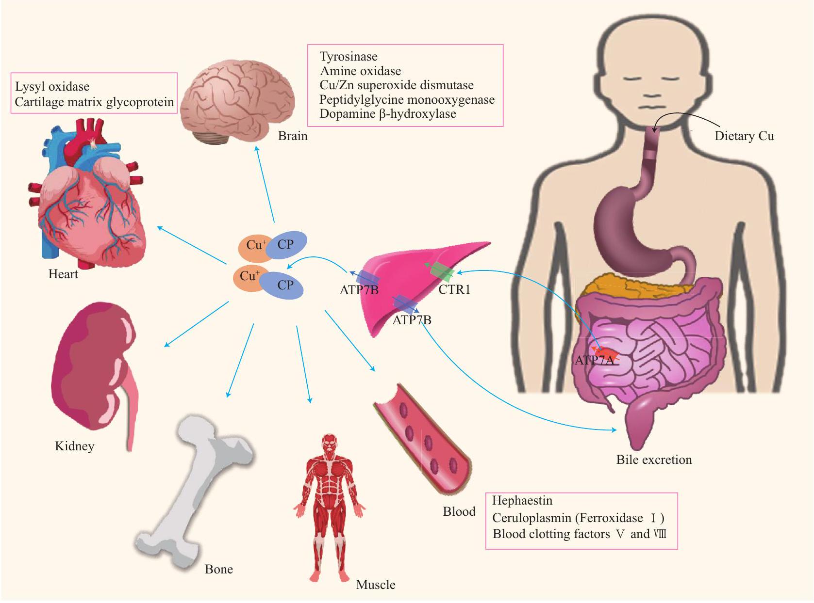
1 الأيض النحاسي النظامي والخلايا في جسم الإنسان

1.1 التمثيل الغذائي للنحاس في الجسم البشري

النحاس هو مكون حيوي لما لا يقل عن 20 إنزيمًا موزعة في أنسجة حيوية مختلفة. في عام 1928، أفاد هارت أن فقر الدم في الجرذان يمكن حله فقط من خلال المكملات التي تحتوي على النحاس مع الحديد. في الجسم البشري الطبيعي، النحاس موجود في العضلات والعظام، النحاس موجود في الكبد، والباقي النحاس موجود في الدم يتم امتصاص النحاس بشكل رئيسي في الأمعاء الدقيقة، ويتم ذلك بواسطة بروتين ناقل النحاس 1 (CTR1، المعروف أيضًا باسم SLC31A1). النحاس يتم تحويله من إلى بواسطة مختزلات المعادن، مثل السيتوكروم الاثني عشر ومستضد الظهارة البروستاتية ذو الستة أغشية لنقل الامتصاص من خلال دراسة المرضى الذين يعانون من مرض ويلسون المميز بنقص ATP7B الناتج عن زيادة النحاس، اكتشف الباحثون أن الانخفاض في النشاط النسخي والتعبير عن CTR1 قد يمثل آلية تنظيم خلوية تكيفية استجابةً لاضطراب النحاس. تمنع هذه الآلية دخول النحاس الزائد إلى خلايا الأمعاء الدقيقة. ناقل المعادن ثنائية التكافؤ 1 (DMT1) ينقل أيضًا أيونات النحاس ثنائية التكافؤ. يتم نقل النحاس عبر الغشاء القاعدي الجانبي لخلايا الظهارة المعوية الدقيقة عبر بروتين ATP7A، ومن ثم إلى الدورة الدموية البابية، حيث يصل في النهاية إلى الكبد. ATP7A يوجد بشكل أساسي في الغشاء القاعدي الجانبي. يلعب دورًا حاسمًا في مرض منكس، الذي يتميز بنقص النحاس الناتج
من الشذوذات الجينية في ATP7A. تؤدي النقل غير السليم للنحاس في خلايا الظهارة المعوية الدقيقة إلى استقلاب غير طبيعي للنحاس. .
بغض النظر عن مستوى النحاس في الجسم، يتم تفعيل عدد من الآليات التنظيمية للحفاظ على استقلاب النحاس الطبيعي. قد ينظم عامل النسخ بروتين الخصوصية 1 (Sp1) استقلاب النحاس. يرتبط Sp1 بصناديق GC الموجودة في محفز CTR1، وتتم إدارة مستوياته بواسطة مستويات النحاس العالية والمنخفضة. تمنع مستويات النحاس المرتفعة ارتباط Sp1 بمحفزات CTR1 وSp1. عامل النسخ المرتبط بالمعادن (MTF1) مرتبط بتنظيم توازن النحاس. يؤدي وجود النحاس الزائد إلى تنشيط النسخ للميتالوثيونين والتعبير النووي عن MTF1، مما يؤدي إلى زيادة مستويات الميتالوثيونين. وعلى العكس، عندما يكون النحاس ناقصًا، يعزز MTF1 نسخ وتعبير CTR1B، مما يؤدي إلى زيادة امتصاص النحاس والحفاظ على توازن النحاس. أيونات النحاس لا تتداول بحرية في مجرى الدم. بدلاً من ذلك، حوالي تتحد أيونات النحاس مع السيرولوبلاسمين لتكوين مركبات لا يمكن تبادلها. يحدث تبادل أيونات النحاس عندما تصل هذه المركبات إلى الأعضاء والأنسجة، مثل القلب والكبد والدماغ والكلى والأمعاء والرئتين والطحال. (الشكل 1).
الكبد هو العضو الرئيسي في الجسم لتخزين النحاس. تحدث التنظيم المعقد والمنظم بشكل كبير لتمثيل النحاس داخل الخلايا الكبدية. يتم إفراز النحاس بطريقة تعتمد على ATP7B للإفراز الحويصلي، أو يرتبط بالسيرولوبلاسمين للإفراج إلى الدورة الدموية، من أجل الوصول إلى أعضاء وأنسجة أخرى. في الكبد، يتمثل المسار الرئيسي للتخلص من النحاس الداخلي في إفراز الحويصلات التي تنتجها جهاز جولجي، والتي تنقل النحاس للتصدير إلى الصفراء عبر ATP7B، تليها الإخراج في البراز. تقوم الخلايا العصبية بإفراز النحاس عند المشبك لتغليفه في الحويصلات. تحتوي الحويصلات المشبكية على مستويات عالية من النحاس. بعد إزالة استقطاب الخلايا، يتم إفراز النحاس في الفجوة المشبكية لتعديل وظائف مستقبلات الغشاء المختلفة. .

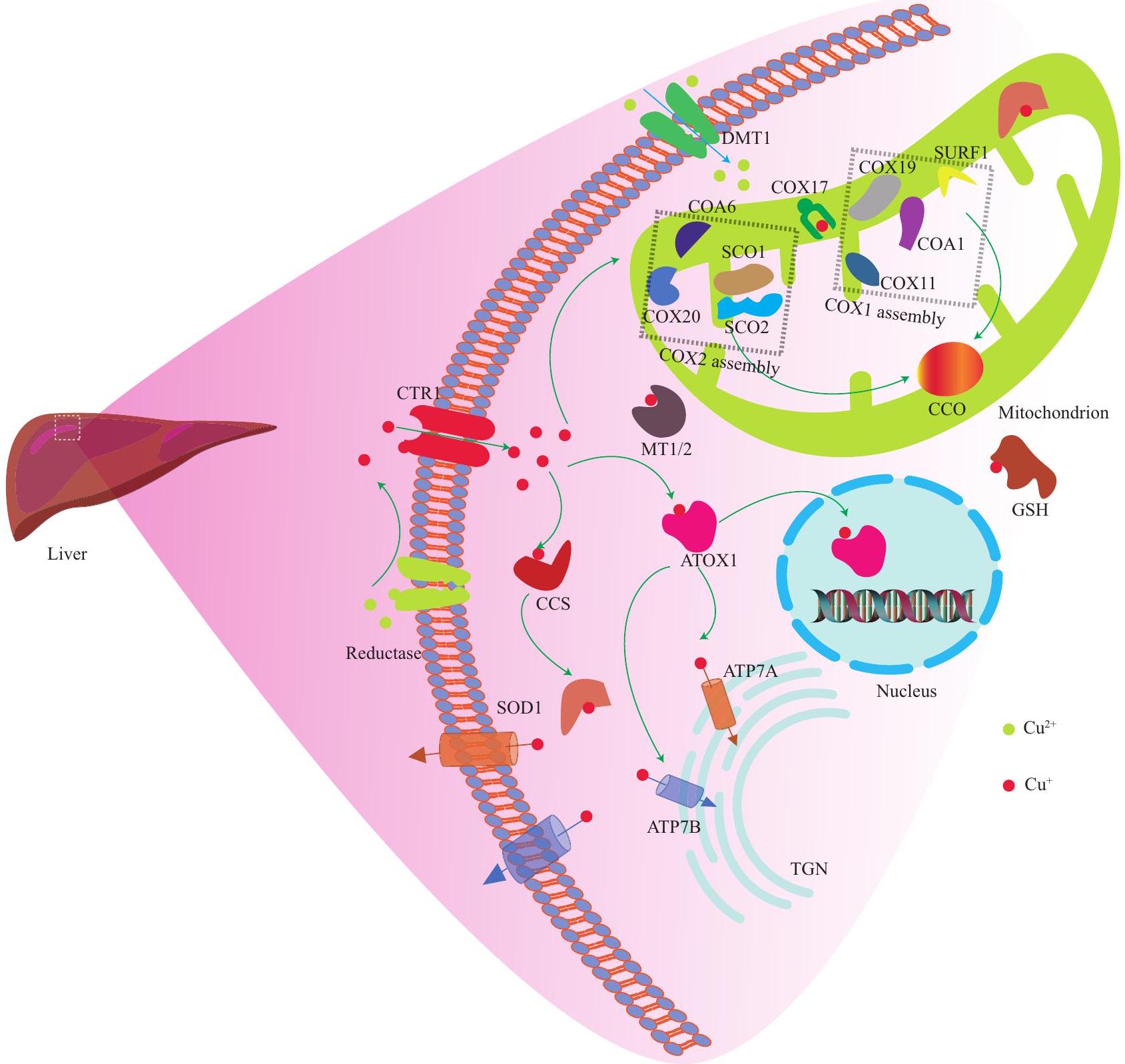
1.2 استقلاب النحاس الخلوي في جسم الإنسان

تنقل الخلايا الكبدية النحاس إلى الخلايا عبر CTR1 يتم تنظيم نقل النحاس داخل الخلايا إلى الأهداف الرئيسية بشكل فعال بواسطة بروتينات الشابيرون للنحاس داخل السيتوبلازم (الشكل 2).

1.2.1 الت Chelation

يرتبط الجلوتاثيون بالنحاس من أجل إزالة السموم. يقوم هذا بالتقاط الجذور الحرة، ويرتبط بأيونات المعادن الثقيلة، مثل الزئبق، والكادميوم، والزرنيخ، من أجل الإخراج. النحاس مرتبط بالميتالوثيونين 1/2، الذي يرتبط بأيونات النحاس عبر بقايا السيستين بطريقة تعتمد على درجة الحموضة. .

1.2.2 COX17 شابير النحاس COX17 يرتبط

أيونات النحاس في الفضاء بين أغشية الميتوكوندريا. النحاس ضروري لتخليق
الشكل 1 مسار استقلاب النحاس (Cu) والإنزيمات الرئيسية المحتوية على النحاس في الأعضاء المختلفة. يتم امتصاص النحاس من خلال الظهارة المعوية الدقيقة، وينتقل إلى الدورة الدموية البابية ليصل إلى الكبد. ثم يتم توزيعه إلى مختلف الأعضاء والأنسجة في الجسم، بما في ذلك الدماغ، القلب، الكلى، العظام، العضلات، والدم. العديد من الإنزيمات المحتوية على النحاس المذكورة ضرورية لوظائف الأعضاء المختلفة.
بروتينات الميتوكوندريا، بما في ذلك السيتوكروم ج أكسيداز (COX). يتكون COX من 11 وحدة بروتينية، ويتطلب 18 بروتينًا للتجميع الدقيق. يتكون المركز الحفاز لـ COX من COX1/2/3، الذي يتم ترميزه بواسطة الحمض النووي الميتوكوندري، ويشمل 3 أيونات نحاس. يقع اثنان من أيونات النحاس في مركز CuA، ويقع أيون نحاس واحد في مركز CuB. عندما يرتبط COX17 بالنحاس، ينقل أيوني النحاس إلى بروتين تجميع السيتوكروم ج أوكسيداز 2 (SCO2). ثم يقوم SCO2 بتسليم أيونات النحاس إلى SCO1 في معقد SCO2-SCO1-COX2. أخيرًا، يتم نقل أيونات النحاس إلى موقع CuA في المركز التحفيزي لـ COX2، من أجل إكمال تجميع الوحدة الفرعية. تتطلب هذه العملية يربط COX17 أيون نحاس إضافي بـ COX11، ويتم نقل هذا الأيون النحاسي إلى موقع CuB الموجود في المركز التحفيزي لـ COX1، مما يختتم تجميع COX1. . بالإضافة إلى ذلك، تتضمن COX مجموعة هيم أثناء التمعدن، تليها دمج الوحدات الفرعية المتبقية، مما يُكمل تجميع COX خلال الفسفرة التأكسدية. الميتوكوندريا تولد وتنقل إشارة أكسدة-اختزال لتنظيم نقل النحاس. الطفرات في SCO1
وقد يؤثر SCO2 على سلامة هذه المسار الإشاري نقص SCO1 يؤدي إلى التحلل السريع لـ CTR1، مما يشير إلى الرابط الوظيفي بين SCO1 و CTR1. .
1.2.3 شابيرون النحاس لإنزيم سوبر أكسيد ديسموتاز (CCS) يرتبط النحاس بـ CCS، وينقل النحاس إلى SOD1. هذا يعزز تحلل الأنواع التفاعلية من الأكسجين (ROS)، ويقلل من تراكم ROS، ويحمي الخلايا من أضرار الجذور الحرة. نقص SOD1 سيزيد من الإجهاد التأكسدي. يتم تنظيم تعبير CCS من خلال التغذية الراجعة السلبية من النحاس. إن زيادة محتوى النحاس داخل الخلايا ستؤدي إلى زيادة تحلل CCS. تشير الدراسات الحديثة إلى أن النحاس يمكن أن يصل مباشرة إلى SOD1 من CTR1 عبر حوامل النحاس من خلال تشكيل معقد CTR1-CCS-SOD1. .
1.2.4 بروتين مضاد الأكسدة 1 (ATOX1)/ATP7A/B يرتبط ATOX1 بالنحاس، من أجل توصيل النحاس إلى ATP7B على الشبكة الغولجية العبور (ATP7A في خلايا أخرى). هذا يعزز تخليق البروتينات الإنزيمية المسبقة للنحاس، بما في ذلك أكسيداز الليسين، التيروزيناز، والسيرولوبلاسمين. ATOX1 يعتمد على النحاس
الشكل 2 ملخص آلية تنظيم توازن النحاس في الخلايا الكبدية
خارجي يتم تقليصه إلى بواسطة الاختزال (سايتوكروم الاثني عشر) ومستضد الظهارة البروستاتية ذو الستة أغشية. يتم نقله إلى الخلية عبر CTR1، ويتم تسليمه إلى مختلف ناقلات النحاس لأداء وظائف مختلفة. على سبيل المثال، يقوم CCS بتسليم إلى SOD1، يقدم ATOX1 إلى النواة وجهاز جولجي، وCOX17 يوصل إلى الميتوكوندريا لتجميع CCO. في هذه الأثناء، الميتالوثيونين داخل الخلايا ويمكن أن يرتبط الجلوتاثيون . يمكن لـ DMT1 توصيل إلى الخلية. تسليم تعتبر COX17 ضرورية لتجميع كل من COX1 و COX2 في الميتوكوندريا. يتكون COX1 من SURF1 و COA1 و COX11 و COX19، بينما يتكون COX2 من SCO1 و SCO2 و COX20 و COA6. يتكون المجمع الهولوإنزيمي الناضج من هذين الجزئين، ويشمل مجموعة هيم أثناء التمعدن، تليها دمج الوحدات الفرعية المتبقية، مما ينهى تجميع COX خلال الفسفرة التأكسدية.
منظم النسخ الذي يساهم في تكاثر الخلايا. قد تواجه الفئران التي تفتقر إلى جين ATOX1 وفيات حول الولادة نتيجة لعدم التوازن الطبيعي للنحاس. يمكن لبروتين ATP7A/B داخل الخلايا تنظيم مستويات النحاس والمشاركة في نقل النحاس بين غشاء الخلية ومختلف الحُجرات داخل الخلايا بطريقة تعتمد على ATP. يتفاعل ATOX1 مع الطرف الأميني لبروتين ATP7A/B لتنظيم نشاطه أثناء نقل النحاس من خلال تعديل معدل تحلل ATP.
بالإضافة إلى ذلك، يقوم البروتين بضبط توازن النحاس بشكل ديناميكي، اعتمادًا على مستوى النحاس داخل الخلايا، حيث يعيد ATP7A توطينه إلى الغشاء البلازمي أو يعيد ATP7B توطينه إلى الحويصلات، لتسهيل تصدير النحاس الزائد. .
النحاس يرتبط بـ ATOX1 أو بروتينات شابير غير محددة لدخول النواة من أجل تنظيم عدة مسارات نقل الإشارة. يتفاعل ATOX1 مع البروتين الغني بالسيستين 2 (CRIP2)، وينقل النحاس إلى CRIP2، مما يؤدي إلى
تحلل CRIP2، مما يؤدي إلى زيادة مستويات ROS، وتنشيط البلعمة الذاتية . في النواة، ينظم النحاس تعبير الجينات، وتخليق البروتين اللاحق من خلال تنظيم عوامل النسخ. علاوة على ذلك، ينظم النحاس عوامل النسخ الرئيسية، بما في ذلك NF- قد يؤدي العلاج بتركيزات مختلفة من كلوريد النحاس إلى تنشيط أو تثبيط NF- تُفعّل عوامل النسخ الرئيسية، بما في ذلك AP-1 و p53، بواسطة النحاس الزائد .

2 توازن النحاس في جسم الإنسان

يمكن أن يتسبب كل من نقص النحاس وزيادة النحاس في تلف الخلايا. لذلك، يتم الحفاظ على كمية النحاس في الجسم ضمن نطاق معقول، وهو ما يعرف بتوازن النحاس. النحاس يلعب دورًا حيويًا في العديد من العمليات الأيضية لضمان سير الأنشطة اليومية بشكل منظم. تشير الأدلة بشكل متزايد إلى أن الاختلالات في توازن النحاس مرتبطة بتطور عدة أمراض، بما في ذلك مرض منكس. مرض ويلسون الأمراض التنكسية العصبية، والسرطان.

2.1 توازن النحاس في الجهاز العصبي

الدماغ هو ثاني أكبر عضو يتراكم فيه النحاس. توازن النحاس مهم بنفس القدر في الجهاز العصبي، وكميات ضئيلة من النحاس ضرورية لأداء التطور والوظيفة الطبيعية للدماغ. وقد أوضحت الدراسات دور النحاس في الدماغ. يمر النحاس عبر الكبد ويدخل الدماغ. ويعمل كعامل مساعد في الإنزيمات المعتمدة على النحاس التي لها وظائف فسيولوجية مهمة، بما في ذلك ما يلي: الدوبامين. -هيدروكسيلاز مثل المونوأوكسيجيناز، الذي يحفز تخليق النورإpinephrine سيتوكروم ج mitochondrial أوكسيداز، الذي له دور ميتوكوندري أمين الأكسيداز، التي تصنع الناقلات العصبية ؛ أكسيدات التيروزيناز، التي تشكل الميلانين ؛ أكسيداز السلفهيدريل، الذي يحافظ على التركيب الطبيعي للشعر علاوة على ذلك، يشارك النحاس في مسارات تنظيمية أخرى في الجهاز العصبي، مثل تخليق SOD1، الذي يقوم بتفكيك السوبر أكسيد لحماية الخلايا من أضرار الإجهاد التأكسدي. علاوة على ذلك، ينظم النحاس نشاط مستقبلات الأحماض الأمينية والبيورين. بالإضافة إلى ذلك، يمكن للنحاس تنظيم النقل المشبكي والإشارات ذات الصلة من خلال تنظيم ATP7A في المشابك. بالإضافة إلى ارتباط الكالسيوم أو الزنك، وتعبير الميتالوثيونين . كما أنه ينظم وظيفة عوامل التغذية العصبية المشتقة من الدماغ وعوامل نمو الأعصاب .
توزيع النحاس في الدماغ غير متساوٍ، حيث لوحظت تركيزات أعلى في كل من النواة الزرقاء والمادة السوداء. لا يزال النقل والتوزيع النحاسي داخل الدماغ غير واضح. وقد تم الافتراض أن النحاس يعبر
حاجز الدم-الدماغ (BBB) كأيونات نحاس حرة، ويتم إطلاقه في نسيج الدماغ والسائل الدماغي الشوكي. يبدو أن الحاجز الدموي الدماغي هو الطريق الرئيسي لدخول النحاس إلى نسيج الدماغ، ومن المحتمل أن يحافظ الحاجز الدموي الدماغي وحاجز السائل الدماغي الشوكي على توازن النحاس في الدماغ. علاوة على ذلك، تعبر خلايا كل من الحاجز الدموي الدماغي وحاجز السائل الدماغي الشوكي عن بروتينات تشارك في نقل النحاس. تعبر خلايا الحاجز الدموي الدماغي عن مستويات أعلى من بروتينات ناقل النحاس، بما في ذلك CTR1 وDMT1 وATP7B مقارنة بنسيج الدماغ. يتم نقل النحاس بسهولة أكبر إلى نسيج الدماغ عبر الشعيرات الدموية الدماغية، مقارنة بالنقل عبر الضفيرة المشيمية. .
أفادت دراسة حديثة أن تركيز النحاس أعلى في الخلايا الدبقية مقارنة بالعصبونات. لقد أظهرت الدراسات أن الخلايا النجمية، التي تُعتبر أكثر أنواع الخلايا الدبقية وفرة في الجهاز العصبي المركزي، لها تأثير كبير على الصحة والأمراض. في الظروف العادية، تشارك الخلايا النجمية في وظائف فسيولوجية رئيسية، بما في ذلك تنظيم نشاط المشابك النمائية والوظيفية، وحاجز الدم في الدماغ، والدعم الأيضي للخلايا العصبية، وإنتاج العوامل العصبية المغذية. قد تلعب مسارات الإشارات النحاسية بين الخلايا العصبية والخلايا النجمية دورًا مهمًا في معالجة الإشارات في الدماغ. لقد تم اعتبار أن الخلايا النجمية تمتص النحاس عبر CTR1. وقد أفادت دراسات إضافية بمشاركة DMT1 في امتصاص النحاس من خلال الخلايا النجمية. يسهل كل من CTR1 و DMT1 نقل تطلق الخلايا النجمية مختزلات جزيئية صغيرة في الجسم الحي، والتي تقلل مستويات لمساعدة الخلايا على امتصاص النحاس علاوة على ذلك، قد يكون البروتين المرتبط بالبريون، الذي يظهر ارتباطًا منخفض الألفة بالنحاس، وسيطًا لامتصاص النحاس من خلال الخلايا الدبقية. .
تم تحديد مستويات النحاس المرتفعة بشكل ملحوظ لدى المرضى المصابين بمرض ويلسون. تم الإبلاغ عن تركيزات غير طبيعية من النحاس في المرضى الذين يعانون من اضطرابات تنكس عصبي أخرى، مما يشير إلى وجود مسارات إشارات نحاسية محددة في الدماغ. تشمل مسارات الإشارات النحاسية المعقدة داخل الدماغ عمليات الأكسدة والاختزال، التي تتضمن أيونات النحاس غير المستقرة، وإطلاق الناقلات العصبية من المشابك، والتعاون بين الخلايا العصبية والخلايا الدبقية. تشمل الجزيئات المنظمة، مثل ATP7A/B. CTR1 وأتوكس1 هي وظيفة رئيسية في توازن النحاس في الجهاز العصبي.

2.2 عدم توازن النحاس في مرض منكس ومرض ويلسون

خلال عدم توازن النحاس في الجسم، يمكن أن يؤدي نقص النحاس أو تراكم النحاس إلى أضرار كبيرة، والتي تم ربطها بمختلف الأمراض. خلال تكوين الجنين والتطور المبكر، يعتبر تناول النحاس الكافي مهمًا، خاصة في الجهاز العصبي المركزي. تعتبر سمية النحاس، بسبب مستويات النحاس المفرطة، خاصة
يؤثر على الدماغ والكبد .
2.2.1 مرض منكس
مرض منكس، الذي تم الإبلاغ عنه لأول مرة بواسطة منكس في هو اضطراب وراثي نادر يتميز بتمثيل غير طبيعي للنحاس هذا الاضطراب موروث بطريقة متنحية مرتبطة بالكروموسوم X، وهو نتيجة لطفرة في ATP7A. تؤدي الطفرات الجينية إلى ضعف وظيفة الأمعاء لـ ATP7A، مما يؤدي إلى تقليل دخول النحاس إلى مجرى الدم، وتراكم النحاس في خلايا الأمعاء. إذا لم يمكن نقل النحاس إلى الأعضاء المختلفة في الجسم، بما في ذلك الكبد والدماغ، فإن ذلك سيؤدي في النهاية إلى نقص حاد في النحاس في الجسم، وضعف في تخليق العديد من الإنزيمات المهمة. على سبيل المثال، ضعف الدوبامين قد تؤدي وظيفة أحادي الأكسجيناز الشبيهة بالهيدروكسيلاز إلى خلل في الوظيفة المشبكية ونمو المحاور. تتميز علم الأمراض في رباعية فالو بوجود تشوهات وعائية خلقية في حديثي الولادة. غالبًا ما يظهر المرضى المصابون بمرض منكس بأعراض مثل فقدان العظام، وارتخاء الجلد، والأمبولة، والكسور التلقائية. .
دراسة عن أشارت الطفرات الجينية إلى أن الفحص الجيني هو وسيلة تشخيصية موثوقة لمرض منكس. يعتمد علاج مرض منكس على كمية النحاس الممتص من الأمعاء، وكمية النحاس المتداولة التي تصل إلى الدماغ. على سبيل المثال، الحقن تحت الجلد يمكن أن تعزز مركبات الهيستيدين لتجاوز حواجز الامتصاص المعوي نتائج العلاج. .
2.2.2 مرض ويلسون
مرض ويلسون هو مرض ناتج عن طفرة جينية، يتميز بتراكم النحاس بشكل مرضي. تُعزى آلية حدوث مرض ويلسون بشكل أساسي إلى الطفرات في الجين الذي يؤدي إلى تعطيل مضخة ATPase الناقلة للنحاس عبر الغشاء، وعرقلة إخراج النحاس من القناة الصفراوية، وفي النهاية disruption توازن النحاس في الجسم. علاوة على ذلك، فإن زيادة النحاس في الخلايا الكبدية تؤدي إلى تليف الكبد وتليف الكبد. النحاس الذي يتم إطلاقه من خلايا الكبد يتراكم تدريجياً في أعضاء أخرى، مما يؤدي إلى سمية خارج الكبد. .
تركيزات النحاس في المرضى الذين يعانون من مرض ويلسون قد تكون طيات أعلى من تلك الموجودة لدى الأفراد الأصحاء مما يشير إلى الارتباط القوي بين مستويات النحاس في الدماغ وشدة الأمراض العصبية. يتم تخفيف التأثيرات السامة للنحاس في البداية بواسطة الخلايا النجمية، مصحوبة بتكاثرها. يتم بعد ذلك زيادة تخليق الميتالوثيونين لزيادة قدرته على تخزين النحاس. في النهاية، ستؤدي مستويات النحاس العالية إلى ضعف الخلايا النجمية، واضطراب الحاجز الدموي الدماغي، ومجموعة متنوعة من أمراض أنسجة الدماغ. ترتبط الشذوذات الشكلية والوظيفية في الشبكية في مرض ويلسون بشدة الأمراض الدماغية والضعف العصبي. .
تشخيص مرض ويلسون يعتمد على السريرية-
أعراض الحالة، قياس استقلاب النحاس، وتحليل الجينات. يمكن عكس زيادة النحاس من خلال علاجات التChelation والزنك الفموي. يمكن أن يزيد D-penicillamine وtrientine من إخراج النحاس في البول، بينما يمكن أن يقلل الزنك الفموي من امتصاص النحاس في الجهاز الهضمي. يمكن أن يؤدي العلاج الدوائي الدقيق والمبكر إلى تحسين وظائف الكبد والإنزيمات الكبدية خلال 2-6 أشهر، ويمكن ملاحظة تحسنات عصبية في 50%. من المرضى خلال 1-3 سنوات .

2.3 توازن النحاس والأمراض التنكسية العصبية

أفادت العديد من الدراسات أن التغيرات في توازن النحاس تحدث خلال الأمراض التنكسية العصبية التدريجية. قد يلعب كل من زيادة وانخفاض مستويات النحاس دورًا مميزًا في الأمراض التنكسية العصبية.
2.3.1 مرض الزهايمر (AD) وتوازن النحاس مرض الزهايمر هو اضطراب عصبي تنكسي شائع قد ينشأ من عوامل متنوعة، مثل العمر والبيئة والوراثة. من المحتمل أن يؤدي زيادة متوسط العمر المتوقع للإنسان في السنوات القادمة إلى زيادة عدد المرضى بـ وفقًا لفرضية “تسلسل الأميلويد” التقليدية، فإن علم أمراض الزهايمر يتمثل بشكل أساسي في تراكم الأميلويد- الببتيدات وبروتينات تاو. بعد ذلك، ستؤدي هذه المعالجة المعيبة إلى تكوين لويحات أميلويد وتشابكات عصبية في المادة الرمادية، مصحوبة بإجهاد أكسيدي خلوي، وإصابة وعائية، والتهاب عصبي، وآفات تنكس عصبي. .
توازن النحاس يلعب دورًا محوريًا في مسببات مرض الزهايمر. النحاس يتفاعل بشكل محتمل مع عدة عوامل مرضية رئيسية، بما في ذلك وتاو. يرتبط بأيونات النحاس بفاعلية عالية يمكن لأيونات النحاس أن تحفز ترسيب في المختبر. ومع ذلك، فإن أيونات النحاس المعزولة تحفز التدهور، وإضعاف إنتاج الجذور الحرة الهيدروكسيلية والأضرار التأكسدية، مما يؤدي في النهاية إلى تقليل موت الخلايا يمكن أن يؤثر النحاس الزائد سلبًا على قدرة الدماغ على التخلص من . علاوة على ذلك، -معقد النحاس يثبط تعبير بروتين مستقبلات الليبوبروتين المرتبطة-1، مما يقلل من إزالة المواد السامة عصبيًا تم تحديد أن النحاس يتواجد بتركيزات مرتفعة بشكل ملحوظ في بقع العمر لدى المرضى الذين يعانون من مرض الزهايمر، مما يشير إلى أن هذا قد يلعب دورًا في تحفيز تكوين اللويحات في الدماغ. علاوة على ذلك، يعزز النحاس فسفرة بروتينات التاو والسمية العصبية لم aggregates بروتين التاو. بروتينات التاو المرتبطة بالنحاس تعزز تلف الدماغ من خلال تحفيز النشاط الأكسدي. التعرض المزمن للنحاس النظامي يؤدي إلى عدم تنظيم كيناز تاو المرتبط CDK5 ومجمع البروتينات المشبكية complexin1/2 لقد أفادت الدراسات بنتائج متضاربة حول مستويات النحاس في الدماغ في مرض الزهايمر. ومع ذلك، قد تشير المستويات المرتفعة من النحاس القابل للتغير إلى
تساهم في تلف الأنسجة التأكسدي في دماغ المرضى الذين يعانون من .
في حالات الالتهاب العصبي، يزيد النحاس من تأثير على تنشيط الخلايا الدبقية الصغيرة، تليها السمية العصبية. النحاس-A المجمعات تنشط الخلايا الدبقية الصغيرة، وتساعد على إفراز TNF- و لا في NF بطريقة تعتمد على . علاوة على ذلك، النحاس- المجمعات تشارك في TNF- مسار الإشارة وتفعيل الكاسبيز-3 المتزامن الناتج عن الإجهاد التأكسدي، مما يؤدي إلى الالتهاب العصبي لقد تم اقتراح أن النحاس يعدل الأنماط الظاهرية المؤيدة للالتهابات والمضادة للالتهابات للخلايا الدبقية الصغيرة من خلال تنظيم مسارات إشارات أكسيد النيتريك و S-نيتروسوثيول. . بالإضافة إلى ذلك، يمكن أن تؤدي وفاة الخلايا العصبية إلى تحفيز الالتهاب في مرض الزهايمر. تؤثر أنواع مختلفة من وفاة الخلايا العصبية على الالتهاب من خلال آليات مختلفة، مثل موت الخلايا العصبية المبرمج، مما يؤدي إلى تعطيل التوازن الطبيعي للخلايا الدبقية الصغيرة. تمثل النخر الخلوي، والنخر الحراري، والنخر الحديدي أشكالًا من موت الخلايا القابلة للذوبان تتميز بإطلاق DAMPs، بما في ذلك HMGB1، وبروتينات الصدمة الحرارية، والأحماض النووية. . هذا يسبب ضررًا عصبيًا التهابيًا مستمرًا، ويحفز تنشيط الميكروغليا بشكل غير طبيعي قد تحسن التدخلات ضد موت الخلايا العصبية البيئة العصبية الالتهابية في مرض الزهايمر. علاوة على ذلك، قد يوفر توقيت وآلية موت الخلايا العصبية رؤى حول علاج الأمراض التنكسية العصبية.
تخزن الخلايا النجمية كميات كبيرة من النحاس. وقد أظهرت الأدلة في المختبر أن الخلايا النجمية ذات الوظيفة المعطلة تتراكم النحاس من خلال آلية تعتمد على CTR1 أو DMT1، وعائلة بروتينات ZIP. النحاس الحر قد يسبب آفات عصبية تنكسية قد يؤدي زيادة بروتينات الشابيرون النحاسية في الخلايا الدبقية إلى إعاقة نقل النحاس من الخلايا الدبقية إلى الخلايا العصبية. تشير التجارب في المختبر إلى أن تعمل اللويحات والبروتوفibrils كمنبهات داخلية، مما يؤدي إلى تنشيط الخلايا النجمية أو تكاثر الخلايا النجمية التفاعلية. يمكن للخلايا النجمية أن تعيق إزالة الميكروغليا لـ لويحات عن طريق إفراز جزيئات حساسة للجليكوزامينوجليكان، مما يعزز بشكل غير مباشر تراكم في دماغ مرض الزهايمر . بالإضافة إلى ذلك، إدارة يمكن أن يقلل (atsm) بشكل كبير من إفراز NO و MCP1 و IL-6 في الخلايا النجمية، مما قد يرتبط بتعزيز النحاس الخلوي وميتالوثيونين-1 في الخلايا النجمية. .
مضاد الأكسدة الجلوتاثيون مهم في مرض الزهايمر. وغالبًا ما يُستخدم لإظهار ضعف الإدراك الخفيف ومرض الزهايمر. لقد تم اعتبار أن رفع مستويات الجلوتاثيون قد يعيق أو يبطئ تقدم مرض الزهايمر. وقد اعتُبر أن المستويات العالية من النحاس تقلل من إنتاج الجلوتاثيون، مما يؤدي إلى زيادة الجذور الحرة المؤكسدة، والأضرار الناتجة عن الإجهاد التأكسدي، والتي قد تساهم في مسببات المرض.
. بروتين آخر مهم مرتبط بمرض الزهايمر هو SOD1، حيث أن موقع الربط مشابه لذلك لـ . نتيجة لذلك، قد يؤثر سلبًا على السلامة الهيكلية لبروتين SOD1، مما يؤدي إلى عجز في جمع المعادن الخلوية. .
تم إدارة مرض الزهايمر باستخدام بعض الجزيئات الصغيرة، بما في ذلك دونيبيزيل، جالانتامين، ريفاستيجمين، وميمانتين. ومع ذلك، لا يوجد علاج دائم لمرض الزهايمر. هذه الأدوية هي مضادات أكسدة محتملة، يمكن استخدامها لتقليل التجمع، وتحسين الأعراض العصبية تم تحديد دواء تجريبي، الكليوكينول، لتقليل ترسيب بعد 3 أشهر من الإدارة الفموية في نماذج حيوانية في تجربة سريرية واحدة، عانى 36 مريضًا من مرض الزهايمر المعتدل من تخفيف في التدهور المعرفي باستخدام الكلوروكين. تجري عدد من التجارب السريرية المرتبطة بالأمراض التنكسية العصبية (الجدول 1).
2.3.2 مرض هنتنغتون (HD) وتوازن النحاس HD هو اضطراب سائد جسديًا في الجهاز العصبي التكرارات غير الطبيعية لتسلسل البوليجلوتمين في الطرف N لبروتين هنتينتين هي الآلية الجزيئية الكامنة وراء مرض هنتنغتون. تتجمع بروتينات هنتينتين المتحورة، مما يؤدي إلى الإجهاد التأكسدي وأعراض التنكس العصبي. .
لقد أظهرت الدراسات أن مستوى أيونات النحاس في النواة المذنبة مرتفع في كل من الأشخاص والحيوانات المصابة بمرض هنتنغتون. تشير بعض الدراسات إلى أن تراكم النحاس يعزز الوظائف الضارة للبروتينات الطافرة. . بالإضافة إلى ذلك، يرتبط النحاس بمخلفات الهيستيدين الموجودة في الطرف N-النهائي لبروتينات هنتنجتين من المRemarkably، يظهر النحاس فقط affinity ربط تجاه بروتينات هنتنجتين التي تتكون من 17-68 بقايا جلوتامين. إن استخدام خالطات النحاس يعيق تشكيل تجمعات بروتينات هنتنجتين الطافرة، بينما يسهل استهلاك النحاس المعزز إنشاء التجمعات. قد يساهم النحاس أيضًا في تقدم مرض هنتنغتون عن طريق عرقلة الإنزيمات المرتبطة بالتنفس الميتوكوندري. يحدث انخفاض في إزالة اللاكتات في النواة المذنبة لدى المرضى الذين يعانون من وقد أظهرت الأبحاث أن استخدام مثبطات إنزيم اللاكتات ديهيدروجيناز يمكن أن يؤدي إلى التنكس العصبي في الفئران. .
عوامل خلب النحاس، مثل الكليوكوينول، والتترايثيوموليبدات، والباتوكوبروين ثنائي السلفونات، يمكن أن تخفف استخدام هذه العوامل الخالطة في نماذج الفئران لمرض هنتنغتون قلل بشكل كبير من الشذوذات المرضية والسلوكية، وزاد من معدلات البقاء في نماذج ذبابة الفاكهة لمرض هنتنغتون. .
2.3.3 التصلب الجانبي الضموري (ALS) وتوازن النحاس يؤدي التصلب الجانبي الضموري إلى تدهور انتقائي في الخلايا العصبية الحركية ومن ثم الوفاة الميزة السريرية الرئيسية لها هي ضمور العضلات التدريجي والضعف، مما يؤدي إلى فشل تنفسي ووفاة المرضى. . لا يزال سبب مرض التصلب الجانبي الضموري غير مؤكد. وقد تم اقتراح وجود أصل وراثي في من الحالات،
الجدول 1 التجارب السريرية في الأمراض العصبية التنكسية المرتبطة بالنحاس (البيانات المستمدة من ClinicalTrials.gov)
التدخلات شرط مراحل الدراسة النتائج الموقع
مسح PET GE180 م الثاني تم استخدام GE180 لتحليل الالتهاب الإقليمي والعالمي في دماغ المرضى الذين يعانون من مرض الزهايمر ومرض باركنسون، ووجد أن زيادة GE180 في الدماغ ككل مرتبطة بوظيفة معرفية أضعف، بما في ذلك الفص الجبهي/الحدبي/الجدارية/الصدغي. نيفادا، الولايات المتحدة الأمريكية
قرص زنك سيستين محتجز في المعدة م.ع الثاني كان للمادة المقارنة النشطة المعطاة عن طريق الفم تحمل أفضل، مقارنة بأسيتات الزنك الفموي، وقد أدت إلى تقليل مستويات النحاس غير المرتبط بالسيرولوبلازمين في المصل، وزيادة مستويات الزنك في المصل. فلوريدا، الولايات المتحدة الأمريكية
يوم النحاس م.ع الثاني تم العثور على تغييرات في الوظيفة الإدراكية، وبيتا-أميلويد في مستشفى جامعة سارلاند، والسائل الدماغي الشوكي، وحجم الدماغ.
يوم النحاس التصلب الجانبي الضموري الثاني لا يوجد منشور أريزونا، الولايات المتحدة
(أتم) التصلب الجانبي الضموري الثاني لا يوجد منشور نيو ساوث ويلز/فيكتوريا، أستراليا
(أتم) MS أنا لا يوجد منشور
(أتم) PD أنا جرعة الدواء كانت يوم، الذي تم تحمله بشكل جيد في دراسة ALS.
زيت جوز الهند – إبيغالوكاتشين غالات MS الثاني لا يوجد منشور فالنسيا، إسبانيا
برنامج تمارين متعددة الأنماط PD لا لا يوجد منشور مستشفى تشانغ غونغ التذكاري
دراسة رصدية: التعرض للنحاس PD لا يوجد منشور إسرنيا/نابولي، إيطاليا
إدارة التعب:
برنامج الأفراد (MFIP) PD لا لا يوجد منشور نوفا سكوشا، كندا
لم يتم البحث عنه إتش دي
AD: مرض الزهايمر؛ ALS: التصلب الجانبي الضموري؛ MS: التصلب المتعدد؛ PD: مرض باركنسون؛ HD: مرض هنتنغتون
بينما قد تفسر العوامل البيئية، تراكم السموم العصبية، تلف الإجهاد التأكسدي، وعوامل النمو غير الكافية المرضى الذين .
قد يلعب بروتين SOD1 الطافر دورًا في مسببات مرض التصلب الجانبي الضموري العائلي. خلال تقدم مرض التصلب الجانبي الضموري، يرتبط CCS بشكل خاطئ ببروتين SOD1 الطافر، مما يقلل من توصيل النحاس إلى الميتوكوندريا، ويؤدي إلى تراكم بروتين SOD1 غير الطبيعي. . بدوره، يولد هذا سمية عصبية حركية زيادة التعبير عن CCS في SOD1 نموذج الفأر الطافر يسرع العجز العصبي، ويقلل من مدة بقاء الفئران، بينما يخفف إعطاء أيون النحاس الأعراض. زيادة تعبير CCS تغير نشاط السيتوكروم c أوكسيداز في طفرة SOD1 تزيد السمية الإضافية الناتجة عن فرط التعبير عن CCS من توصيل النحاس إلى SOD1، لكنها تقلل من توصيلات أخرى، مما يؤثر على الوظيفة الطبيعية للإنزيمات الأخرى. . وبالتالي، قد يسهم عدم تنظيم توازن النحاس وضعف وظيفة الإنزيمات المعتمدة على النحاس في السمية المكتسبة لدى المرضى الذين يحملون طفرات في جين SOD1، وتطور مرض التصلب الجانبي الضموري (ALS). تم تحديد تركيزات النحاس المرتفعة في العضلات الهيكلية والحبل الشوكي لترافق تقدم المرض خلال المرحلة ما قبل الأعراض في SOD1. فئران متحورة .
تم الكشف عن وجود النحاس الزائد في السائل الدماغي الشوكي لدى الأشخاص والحيوانات المصابة بمرض التصلب الجانبي الضموري. زاد تعبير الناقل البشري للنحاس 1 من تركيزات النحاس داخل الحبل الشوكي، مما أعاد نشاط SOD1 والسيرولوبلاسمين. كلما كانت نقص النحاس أكثر شدة في الطفرة SOD1، كانت الأعراض السريرية لمرض ALS أكثر شدة. .
عدة عوامل خلب النحاس، بما في ذلك D-البنسيلامين وثلاثي ثيو موليبيدات لقد أظهرت تأثيرًا مسكنًا على التصلب الجانبي الضموري. بالإضافة إلى ذلك، فإن ثنائي الأسيتيل (4-ميثيلثيوسيماكربازونات) النحاس (II) الذي هو أيونوفور النحاس، تم تحديده كدواء مهم لعلاج مرض التصلب الجانبي الضموري. . لقد أظهر (atsm) أنه يعزز الوظيفة الحركية ويحسن قابلية العيش في SOD1. و نماذج الفئران الطافرة . في الـ لقد أظهر نموذج الفأر الطافري أن العلاج بالتترايثيوموليبدات قد يطيل البقاء، ويخفف من ضمور العضلات، ويقلل من فقدان الخلايا العصبية الحركية، بينما يثبط نشاط بروتينات SOD1 الطافرة، ويقلل من تجمع البروتينات الطافرة. .
2.3.4 مرض باركنسون وتوازن النحاس. تشمل الأعراض السريرية الرئيسية لمرض باركنسون الاهتزازات أثناء الراحة، وتعديلات في توتر العضلات، وبطء الحركة، وعدم الاستقرار الوضعي. المرض
تتميز علم الأمراض بتقليل عدد الخلايا العصبية الدوبامينية، وتكوين تجمعات بروتينية تتكون من بروتوفيريلز الساينوكليين عدة طفرات جينية من -السنوكلين قد يوجد في الوراثي يحدث مرض باركنسون في أشكال وراثية وعرضية. قد تؤثر عوامل مختلفة، مثل الشيخوخة والبيئة، على تطور مرض باركنسون. لقد تم اعتبار على نطاق واسع أن الجمع بين القابلية الوراثية والعوامل البيئية يحفز علاوة على ذلك، تم اعتبار إصابة الإجهاد التأكسدي والخلل الوظيفي في الميتوكوندريا من العوامل التي تعزز تقدم مرض باركنسون. .
أظهرت التشريحات أن أدمغة المرضى الذين يعانون من مرض باركنسون تحمل مستويات أعلى من الضرر التأكسدي للبروتينات والحمض النووي والدهون مقارنة بالأدمغة الصحية. . بالإضافة إلى ذلك، انخفضت مستويات الجلوتاثيون في المادة السوداء لدى المرضى الذين يعانون من مرض باركنسون بـ تشكل أوليغومرات ألفا-ساينوكليين تكوين أميلويدي سام أظهرت التجارب الأولية في المختبر أن تركيزات المليمول من النحاس تسهل تطوير الهيدريدات الأميلويدية غير المطوية جزئيًا، مما يجعلها أكثر عرضة للتجمع. النطاق الطرفي N لـ -السنوكلين يحتوي على موقع لربط النحاس . يتم تعزيز هذه العملية من خلال تقليل التنافر الكهروستاتيكي للشحنات السلبية أسيتيل ن-terminal تم اكتشاف -سينوكليين مؤخرًا في الدماغ هذا التعديل يعزز الطي الحلزوني للبروتين، ويقلل من تجمع البروتينات. . على الرغم من أن هذا التعديل لم يؤثر على القدرة على -السيتوكين للارتباط ، هذا تدخل في ارتباطه بـ تعمل أيونات النحاس كسيف ذي حدين، وقد تكون مستويات النحاس المنخفضة مرتبطة بتقدم مرض باركنسون. مستويات النحاس أقل في مناطق الدماغ التي تعاني من أكبر ضرر لدى المرضى المصابين بمرض باركنسون، بما في ذلك المادة السوداء والنواة الزرقاء، مع انخفاض في عند مقارنتها بالعقول الصحية .
آلية استقلاب النحاس في مرض باركنسون تشير إلى أنه يمكن تخفيف المرض من خلال تقليل النحاس أو إضافته. المركب 8 -هيدروكسيكوانولين-2-كربوكسيالديهيد إيزونيكوتينويل هيدرازون يمكنه عبور الحاجز الدموي الدماغي، والارتباط بشكل تنافسي بـ أو ، مما يمنع بشكل فعال تجمع البروتين في المختبر التجارب السريرية على (atsm) اقترحت تراكمه في النواة المذنبة لدى المرضى الذين يعانون من مرض باركنسون خلال تقدم المرض . علاوة على ذلك، وُجد أن يمكن استخدام (atsm) لاستعادة وظيفة SOD1 المفقودة للنحاس، وإنقاذ فقدان الخلايا العصبية. بالإضافة إلى ذلك، كشفت نتائج الاختبارات لنموذج الفأر الجيني لمرض باركنسون أن (atsm) أنقذ فقدان الخلايا الدوبامينية، وحسن من خلل الحركة .

3 الكوبروتوسيس في جسم الإنسان

3.1 تعريف الكوبروتوس

دراسة عام 2022 بحثت في آليات
توازن النحاس، وأفاد بأن النحاس يرتبط بالبروتينات المليئة بالليبوي في دورة TCA ، مما يؤدي إلى تجميع البروتينات الملبسة بالليبويد، مما ينتج عنه إجهاد بروتيني وموت الخلايا. للمرة الأولى، تم اقتراح الكوبرتوبيس، الذي يتم تنظيمه بواسطة النحاس خلال التنفس الميتوكوندري. هذه الصورة من الموت تتناقض مع جميع أشكال الموت الخلوي المعترف بها الأخرى. .
الايونوفور النحاسي إلسكلومول يرتبط بالنحاس لتسهيل نقله إلى الخلايا يمكن استخدام هذا للتحقيق في سمية النحاس. أظهرت دراسات إضافية أن موت الخلايا الناتج عن أيونوفور النحاس يعتمد فقط على مستويات عالية من النحاس. إيليسكلومول يحفز موت الخلايا المبرمج المعتمد على أنواع الأكسجين التفاعلية على الرغم من أن الكاسبيز 3، وهو علامة مميزة للاستماتة، يبقى غير نشط خلال موت الخلايا الناتج عن الإليسكلومول. لم يؤثر وجود مثبطات موت متعددة، أو إزالة BAX و BAK1 على احتمال الموت، مما يشير إلى أن هذا قد يكون نوعًا جديدًا من موت الخلايا. الخلايا التي تعتمد على التنفس الميتوكوندري تكون أكثر استجابة بحوالي 1000 مرة لأيونات النحاس مقارنة بالخلايا المعتمدة على التحلل السكري. . وهذا يعني أن موت الخلايا الناتج عن أيونات النحاس قد يكون مرتبطًا بالتنفس الميتوكوندري. كشفت فحص شامل للجينوم باستخدام تقنية كريسبر/كاس9 لفقدان الوظيفة عن 7 جينات نجحت في إنقاذ موت الخلايا الناتج عن أيونات النحاس، بما في ذلك FDX1، الذي هو الهدف المباشر للإليسكلومول. FDX1 يشفر إنزيم اختزال يقوم بتقليل إلى . LIPT1 و LIAS و DLD ترمز لمسار حمض الليبويك، بينما ، و مسؤولة عن ليبويلايشن البروتين . إن تثبيط FDX1 و LIAS يؤدي إلى مقاومة ضد موت الخلايا الناتج عن النحاس. أظهرت الفحوصات المتزامنة لقواعد البيانات والدراسات المناعية النسيجية درجة عالية من الارتباط بين FDX1 والبروتينات في مسار حمض الليبويك، مما يشير إلى أن FDX1 قد يعمل كمنظم علوي لأسيتيل البروتينات الدهنية. قد يحدث اكتساب سام لوظيفة بسبب ارتباط النحاس بالبروتينات TCA المليئة بحمض الليبويك، والذي قد يتم تحفيزه بواسطة التكتل غير الطبيعي للبروتينات المليئة بحمض الليبويك. أظهرت تحليلات مطياف الكتلة أن سمية النحاس يمكن أن تؤدي إلى فقدان بروتينات مجموعة الحديد-الكبريت المعتمدة على FDX1 وإجهاد بروتيني. ومع ذلك، لا يزال يتعين استكشاف ذلك. أخيرًا، تم التحقق من استنتاجات هذه الدراسة باستخدام نموذج خلوي يعبر عن بروتين SLC31A1 بشكل مفرط، ونموذج قديم. نموذج حيواني (الشكل 3).
لا يزال تحديد الآليات التنظيمية للأمراض الناتجة عن عدم التوازن في توازن النحاس يمثل تحديًا. لقد تم التحقيق في أضرار الإجهاد التأكسدي في عدد كبير من الدراسات. أيونوفور النحاس NSC319726 يفعل النحاس، مما يعزز إنتاج تم ملاحظة أن تفعيل البلعمة الذاتية يحمي
الشكل 3 عملية الاكتشاف الأولية وآلية الكوبروتوس
تشير التجارب إلى أن موت الخلايا الناتج عن الإليسكلومول قد يكون نوعًا جديدًا من موت الخلايا. أظهرت الخلايا المزروعة مع المصل حساسية أعلى تجاه الإليسكلومول. بعد استنفاد الجلوتاثيون عبر BSO، أظهرت الخلايا حساسية متزايدة تجاه الإليسكلومول. ومع ذلك، أنقذت عملية ربط أيونات النحاس من خلال TTM موت الخلايا الناتج عن الإليسكلومول. بعد علاج لمدة ساعتين مع الإليسكلومول وDSF وNSC319726، زادت مستويات النحاس داخل الخلايا بمقدار 5-10 مرات. حدث موت الخلايا لأكثر من 24 ساعة بعد العلاج. لم يؤثر استخدام عدة مثبطات لموت الخلايا على بدء الموت الناتج عن النحاس. كان للعلاج بمضادات الأكسدة الميتوكوندرية، والأحماض الدهنية، ومثبطات وظيفة الميتوكوندريا تأثير كبير على بقاء الخلايا. ومع ذلك، لم يؤثر مفكك الميتوكوندريا FCCP على بقاء الخلايا. حددت فحص فقدان الوظيفة على مستوى الجينوم باستخدام CRISPR/Cas9 7 جينات أنقذت موت الخلايا الناتج عن أيونات النحاس. نظمت التنفس الميتوكوندري هذا الموت الجديد الناتج عن أيونات النحاس، وتم تحديد منظم رئيسي، FDX1. يرتبط النحاس بالمكونات المربوطة بالليبويليت في دورة TCA، مما يؤدي إلى تجميع البروتينات المربوطة بالليبويليت، مما يؤدي إلى فقدان بروتينات الكتلة الحديدية والكبريتية، وإجهاد بروتيني، وفي النهاية، موت الخلايا. BSO: بيوثيونين سلفوكسيمين؛ DSF: ديسلفيرام؛ NSC319726: أيونوفور النحاس؛ TTM: تيترا ثيو موليبدات
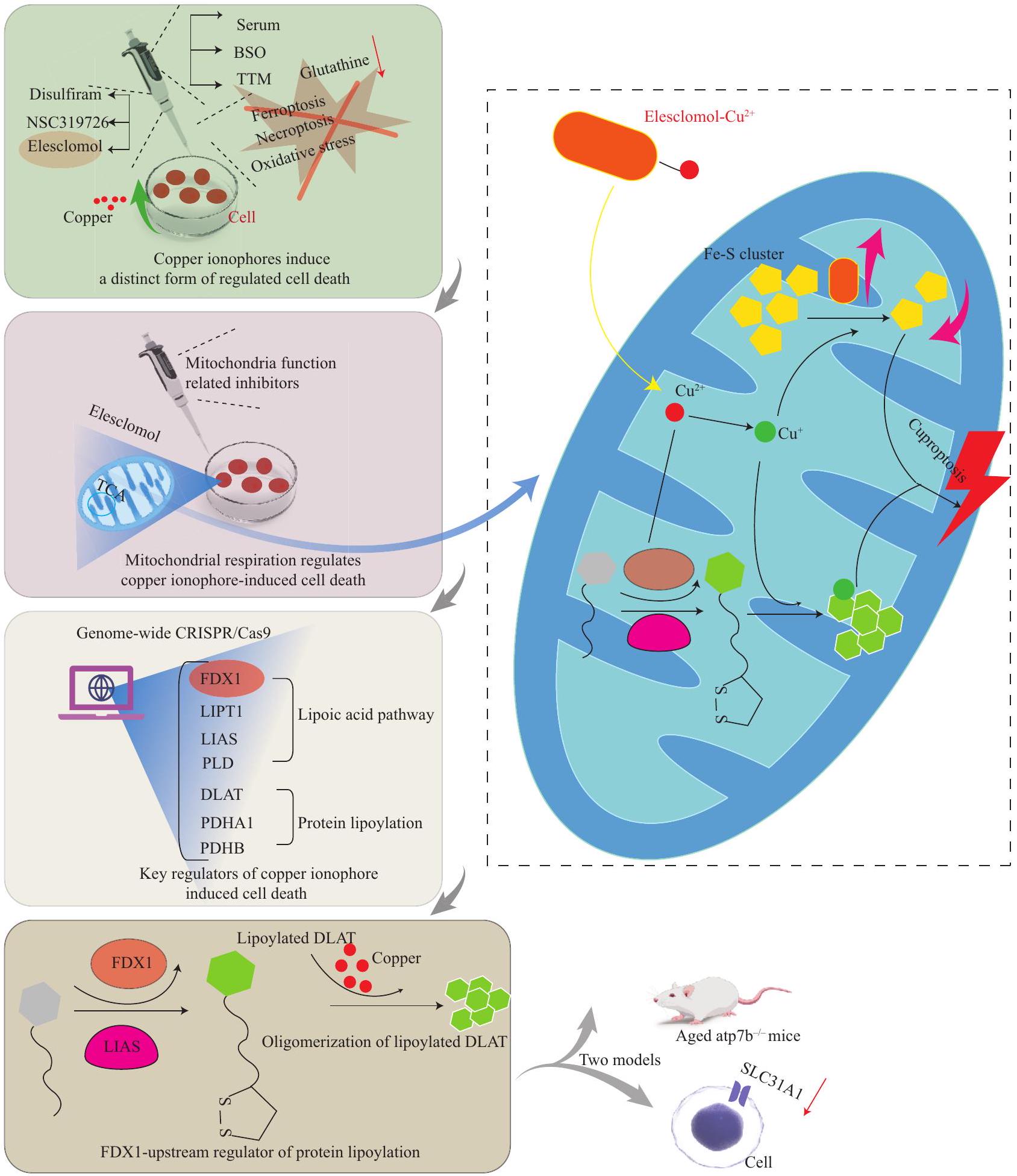
الكبدية من الموت الناتج عن النحاس في أنسجة الكبد للأفراد المصابين بمرض ويلسون، وفي الحيوانات التي تعاني من نقص ATP7B . في دراسة أخرى، حفز النحاس تطور الأورام من خلال تنشيط مسار إشارة PI3K-AKT. يمكن أن يقلل تقليل CTR1 أو تثبيط محور CTR1-Cu باستخدام مخلبات النحاس من تطور الورم وإشارة AKT. علاوة على ذلك، يتم تنظيم CTR1 سلبًا بواسطة Nedd41. أسست هذه النتائج رابطًا بين Nedd41-CTR1-Cu وإشارة PDK1-AKT المسرطنة . MTF1 هو عامل نسخ يرتبط بالمعادن يلعب دورًا رئيسيًا في الكوبروبتوسيس . علاوة على ذلك، , وهو جين مسرطن معبر عنه على نطاق واسع، ينظم بروتينات الكتلة الحديدية والكبريتية وتخليق الجلوتاثيون . يتم أيضًا التحقيق في الالتهاب الناتج عن الكوبروبتوسيس، مع HMGB1 كوسيط مناعي مهم . بالإضافة إلى ذلك، قد يكون الكوبروبتوسيس مرتبطًا بالالتهام الذاتي .
يعتبر الكوبروبتوسيس هدفًا علاجيًا محتملاً في السرطان. لقد حققت العديد من الدراسات المنشورة مؤخرًا في الكوبروبتوسيس باستخدام قواعد بيانات السرطان وطرق المعلوماتية الحيوية. تم استخدام نماذج مختلفة لدراسة خطر السرطان، والتحقيق في مناعة الورم، والعلاج، والتنبؤ. على سبيل المثال، في الورم الدبقي، قد تنظم بروتينات التنظيم الجيني الكوبروبتوسيس من خلال تغيير تعبير PD-L1 وFDX1 . في الميلانوما العينية، يتم نقل النحاس إلى الميتوكوندريا، مما يولد كميات كبيرة من ROS، ويحفز هجرة خلايا السرطان . تم تنظيم LIPT1 وPDHA1 وSLC31A1 المرتبطة بالكوبروبتوسيس بشكل متزايد في الميلانوما . أظهرت 10 lncRNAs المرتبطة بالكوبروبتوسيس كفاءة تشخيصية أعلى في سرطان الكلى الخلوي الواضح . تم تقليل تعبير FDX1 بشكل كبير في سرطان الكبد الخلوي (HCC)، بينما ارتبط تعبيره المرتفع بالبقاء الممتد . قد يحفز LIPT1 نمو وتسلل وتحرك خلايا HCC . في الأورام الدبقية منخفضة الدرجة، تم بناء نموذج تنبؤي دقيق يعتمد على 5 جينات مرتبطة بالكوبروبتوسيس ، ووجد أن تعبير ATP7B قد انخفض، بينما زاد تعبير SLC31A1 وFDX1 وDLAT وLIAS .
تم استخدام دراسة الكوبروبتوسيس بشكل متزايد لأغراض علاجية. تم تطوير بوليمر حساس لـ ROS لتغليف الإليسكلومول والنحاس في نانو جزيئات، والتي بدورها، يتم تنشيطها بواسطة ROS داخل الخلايا الزائدة عند دخولها خلايا السرطان. تعمل مركبات الإليسكلومول والنحاس بشكل تآزري ضد خلايا السرطان وتسبب الكوبروبتوسيس . تم تصميم منصة نانو بوليمر تنسيق النحاس (I) 1،2،4-ترايازول المهندسة بواسطة أوكسيد الجلوكوز، والمعروفة أيضًا باسم ، في سرطان المثانة. يمكن أن يجعل هذا خلايا السرطان أكثر عرضة للكوبروبتوسيس عندما يتم استنفاد الجلوكوز والجلوتاثيون . يمكن تطوير إطار عضوي ثنائي المعدن غير متبلور
لاستغلال التأثيرات التآزرية للكوبروبتوسيس والفيروبتوسيس ضد السرطان .
للنحاس والحديد هياكل مشابهة، وكلاهما ضروري لوظيفة الكائن الحي . يؤدي عدم التوازن في مستويات النحاس والحديد عادةً إلى توليد جذور حرة مؤكسدة ضارة. مشابهًا لتوازن النحاس، ينظم الجسم توازن الحديد بشكل صارم . يمكن أن يؤدي خلل توازن الحديد إلى الفيروبتوسيس، وهو نوع من موت الخلايا المنظم الذي تم التعرف عليه في . تتضمن الآلية الرئيسية تحفيز الأحماض الدهنية غير المشبعة بشدة على غشاء الخلية في وجود الحديد ثنائي التكافؤ أو ليبوكسجيناز، مما يؤدي إلى أكسدة الدهون وموت الخلايا . تم ملاحظة تداخل بين توازن النحاس والحديد، وتم تحديد الآليات التي تنظم الحديد والنحاس. يؤثر النحاس بشكل إيجابي على توازن الحديد، بينما يعيق الحديد استقلاب النحاس. على سبيل المثال، بعد استنفاد مخازن الحديد في الجسم، يتم إعادة توزيع النحاس إلى الأنسجة الحيوية للحفاظ على توازن الحديد، مما يسهل امتصاص الحديد بمساعدة الناقل الأيوني المعدني ثنائي التكافؤ DMT1 . بالإضافة إلى ذلك، قد يعزز النحاس المستند إلى الأمعاء نقل الحديد، مع تعزيز النحاس الكبدي لتخليق السيرولوبلاسمين، مما يمكّن من أكسدة الحديد بعد إطلاقه . خلال نقص الحديد، يقوم عامل النسخ المستحث بنقص الأكسجين (HIF) بتنشيط العديد من الجينات المعوية المرتبطة بامتصاص الحديد . يزيد النحاس من نشاط ارتباط الحمض النووي لـ HIF، مما يشير إلى رابط محتمل بين مسار إشارة HIF، وتوازن الحديد والنحاس . كشفت الدراسات أن الحديد الزائد يمكن أن يؤثر على استقلاب النحاس الطبيعي . لذلك، من الضروري التأكيد على تأثير استهلاك الحديد على توازن النحاس.
كشفت الدراسات أن ربط أيونات النحاس بواسطة الكوبرزون يؤدي إلى الإفراج السريع عن أيونات الحديد من بروتين التخزين، الفيريتين، مما يؤدي إلى أكسدة الدهون الناتجة عن الحديد والفيروبتوسيس، وينتج عنه فقدان الخلايا الدبقية . كشفت دراسة حديثة أن منظم توازن النحاس COMMD10 ينظم بدء الفيروبتوسيس . يمكن أن يؤدي مركب ديسلفيرام-النحاس إلى تحفيز الفيروبتوسيس في السرطان. ينشط هذا بشكل كبير فسفرة p62، ويعزز الارتباط التنافسي لـ Keap1، مما يطيل من عمر NRF2 النصفي، ويحفز الارتفاع التعويضي لـ NRF2 . يحفز النحاس الفيروبتوسيس من خلال تحفيز التحلل الذاتي لـ GPX4. يعمل TAX1BP1 كمتلقي للالتهام الذاتي لتحلل GPX4 أو الفيروبتوسيس الناتج عن النحاس . حددت دراسات مختلفة العلاقة بين الكوبروبتوسيس والفيروبتوسيس في أورام مختلفة، مثل سرطان الغدة الرئوية، حيث تم تحديد المجموعات المرتبطة بالكوبروبتوسيس والفيروبتوسيس . في سرطان القولون والمستقيم، كان لدى المرضى الذين يعانون من
انخفاض تعبير الجينات المرتبطة بالكوبروبتوسيس والفيروبتوسيس معدلات بقاء أعلى . في HCC، تم تحديد 7 جينات رئيسية مرتبطة بالكوبروبتوسيس والفيروبتوسيس كعلامات حيوية لتوقع أسوأ .

3.3 الكوبروبتوسيس والأمراض التنكسية العصبية

كشفت عدد من الدراسات عن العلاقة بين التغيرات في توازن النحاس وتقدم مختلف الاضطرابات التنكسية العصبية . في AD، يرتبط البروتين الرئيسي للمرض بشغف عالٍ لربط أيونات النحاس، ويتداخل النحاس الزائد مع إزالة . تم الكشف عن مستويات عالية من أيونات النحاس في النواة المتوسطة لمرضى مرض هنتنغتون. في مرض التصلب الجانبي الضموري، يرتبط CCS بشكل خاطئ ببروتينات SOD1 الطافرة، مما يؤدي إلى توصيل غير صحيح للنحاس وتراكم غير طبيعي لـ SOD1، مما يؤدي بعد ذلك إلى تحفيز سمية الخلايا العصبية الحركية. بالنسبة للمرضى الذين يعانون من مرض باركنسون، قد يسرع النحاس من تقدم المرض من خلال آليات متعددة تتوسطها مستويات النحاس المتزايدة أو المنخفضة. تتطلب الآلية الدقيقة للنحاس في الاضطرابات العصبية التنكسية المتقدمة مزيدًا من التحقيق. . أظهرت الدراسات الحديثة وجود صلة بين عدم تنظيم توازن النحاس في مرضى مرض منكس، ومسار UCHL1/PARK5 المسبب للأمراض في مرض باركنسون. يقع UCHL1/PARK5 في مجرى ATP7A، مما يشير إلى أن تثبيط UCHL1/PARK5 يحمي طفرات ATP7A من عدم تنظيم توازن النحاس. . تم تحديد متغير بديل جديد لـ ATP7A، p.Met1311Val، في الأفراد الذين يعانون من مرض التصلب الجانبي الضموري، مما زاد من تراكم النحاس في الخلايا الليفية، وانخفضت البقاء، وأدى إلى عيوب حركية في خلايا عصبية حركية ذبابة الفاكهة. . أفادت دراسة تناولت مرض منكس ومرض ويلسون عن طريقة جديدة لتنظيم توازن النحاس. أشارت النتائج إلى أن بروتينات ATP7 ومجمع جولجي الأوليغومري المحفوظة معًا تثبت مستويات النحاس، مما يساهم في الحفاظ على وظيفة الميتوكوندريا وسلامة المشابك. . نظرًا لأن زيادة النحاس تحفز الكوبروبتوسيس، الذي يحدث بشكل رئيسي في الميتوكوندريا، وتسبب إصابة الإجهاد التأكسدي، فمن المحتمل أن تكون آلية مرضية محتملة وراء الاضطرابات العصبية المختلفة تتوسطها الكوبروبتوسيس. من الجدير بالذكر أن الدراسات التي استخدمت الكليوكينول أفادت بنتائج علاجية إيجابية لمرض باركنسون ومرض الزهايمر، بما في ذلك انخفاض تنشيط الميكروغليا في الحبل الشوكي مع التهاب الدماغ والنخاع، مما أدى إلى تعزيز الأعراض السريرية.
. أفادت دراسة تناولت تقدم الأمراض العصبية بناءً على العلاقة بين الكوبروبتوسيس والفيروبتوسيس أن النحاس يحفز الفيروبتوسيس. هذا يؤدي إلى فقدان الخلايا الدبقية في التصلب المتعدد، ويؤدي إلى موت الخلايا في أمراض عصبية أخرى. . تم الإبلاغ عن جينات مرتبطة بالكوبروبتوسيس في مرض الزهايمر، كما تم الإبلاغ عن تباين مناعي كبير في مرضى مرض الزهايمر عبر مجموعات فرعية مختلفة من الكوبروبتوسيس. تم الإبلاغ أيضًا عن MYT1L و PDE4D و SNAP91،
, و تم تحديدها كجينات فريدة ذات قدرات تنبؤية في تحليل مرض الزهايمر (الجدول 2). 4 المناقشة

تم استخدام جزيئات النانو القائمة على النحاس على نطاق واسع في الحياة الإنتاجية بسبب خصائصها المتميزة.

. في المجال الطبي الحيوي، يمكن استخدام جزيئات النانو CuO كأجهزة استشعار حيوية للكشف عن علامات المرض. . مؤخرًا، تم تطوير جزيئات النانو CuO كطلاءات سطحية مضادة للفيروسات لمنع انتقال الفيروسات. . ومع ذلك، فإن زيادة النحاس لها آثار سلبية. كشفت دراسة أن التعرض المطول للنحاس قد يسبب تدهورًا إدراكيًا، وظهور . وبالمثل، يمكن أن يؤدي الاستخدام الواسع للجرعات العالية من النحاس في الزراعة والثروة الحيوانية إلى تلوث بيئي خطير، مما يعرض صحة الإنسان للخطر. . لذلك، من الضروري وضع قوانين عقلانية لانبعاث النحاس، وتقليل استخدام النحاس في الزراعة العضوية. يعتمد الكوبروبتوسيس على عدم التوازن في توازن النحاس، مما يشير إلى الحاجة إلى مزيد من التحقيق في الآليات الأساسية، مثل المنظمات الإضافية المرتبطة بالنقل الغشائي، وتوزيع مستويات النحاس المتفاوتة. فرضية محتملة هي أن هيكل الناقلات النحاسية قد يتغير، أو قد يتم تحفيز تعديلات أوليغومرية لتنظيم مسار النقل عند ارتباط النحاس.
. COMMD1 هو المنظم الوحيد المحدد للنقل الغشائي الذي لديه قدرة ربط النحاس، وهو منظم مباشر لتوازن النحاس الخلوي. . وقد تم الإبلاغ عن أن الفئران التي تعاني من عيوب محددة في الكبد والتي تشمل COMMD1 و COMMD6 و COMMD9 تحفز تراكم النحاس المماثل في الكبد. قد تلعب COMMD6 و COMMD9 دورًا مشابهًا لـ COMMD1 , مما يشير إلى وجود منظمات غير معروفة أخرى تنظم بشكل خاص نقل النحاس. لذلك، لا تزال الأسئلة التالية بحاجة إلى معالجة: (1) كيف تشعر آليات النقل بمستويات النحاس لتنظيم توزيع ناقلات النحاس؟ (2) هل هناك آلية تنظيمية محددة للنحاس مسؤولة عن تنظيم النقل الغشائي للنحاس؟ (3) ما هي آلية توازن النحاس داخل الخلايا بين العضيات؟ (4) ما هي الاختلافات في نقل النحاس بين الخلايا العصبية وغير العصبية؟ نموذج عصبي مناسب مطلوب لإعادة النظر في آلية نقل النحاس. لقد كشفت عدة منظمات رئيسية متورطة في الكوبروبتوسيس عن صلة بين الفيروبتوسيس والالتهام الذاتي. على سبيل المثال، في الورم الدبقي، يرتبط FDX1 مع جينات أخرى ذات صلة بتعبير علامات الالتهام الذاتي. . يمكن أن يرتبط النحاس مع البروتينات ذات الصلة لتحفيز الالتهام الذاتي. . في طفرات ATP7B، الجدول 2 بعض مسارات الإشارة لتوازن النحاس والكوبروبتوسيس في الظروف العصبية المرضية والأورام
منظمات مسارات الإشارة أو الجزيئات المستهدفة بواسطة Cu
الوظيفة المرض مسار الإشارة المحتمل المراجع EREG
يمكن أن يؤثر EREG على المناعة والكوبروبتوسيس الورم الدبقي يؤثر EREG على تعبير PDL1/المتعلق بالكوبروبتوسيس من خلال التأثير على تعبير FDX1. [201] FDX1
مرتبط بالتسلل المناعي الورم الدبقي كانت جينات الكوبروبتوسيس المرتبطة بـ FDX1 مرتبطة إيجابيًا بتعبير جينات علامات الالتهام الذاتي LIPT2 و NNAT، والتي تشارك في الترابط، قد تكون العلامة غير المعروفة Atg5 و Atg12 و BECN-1. [200] MAP1LC3A
منظم الالتهام الذاتي، MAP1LC3A المعروف باسم LC3 مرض ويلسون زاد النحاس من تعبير MAP1LC3A. في خلايا ATP7B المعطلة، يكون mTOR أقل نشاطًا، وينفصل عن الليزوزومات. ينتقل عامل النسخ EB من الركيزة mTOR إلى النواة، ويتم تنشيط جينات الالتهام الذاتي لحماية الخلايا من الموت الناتج عن النحاس. ATP7
تتواجد بروتينات ATP7A و ATP7B في جهاز جولجي، وتنظم توازن النحاس. التنكس العصبي تتطلب سلامة آليات توازن النحاس المعتمدة على جولجي، والتي تتطلب مجمع ATP7 و COG، الحفاظ على سلامة وظيفة الميتوكوندريا. [233] ATP7A
يمكن أن تسبب طفرة ATP7A مرض منكس بسبب نقص النحاس النظامي. مرض منكس يزيد عدم توازن النحاس، بسبب العيوب في ATP7A، من تعبير UCHL1، الذي يتطلب بدوره، من أجل الآلية المرضية لعدم توازن النحاس. مرض باركنسون [231]
CTR1 CTR1 هو ناقل نحاس في غشاء الخلية. السرطان يعزز النحاس من تكوين الأورام من خلال تنشيط مسار PDK1- Nedd41 الذي ينظم CTR1 سلبًا من خلال ubiquitination AKT والتدهور اللاحق/ Nedd41-CTR1-النحاس-PDK1-AKT. [196]
الفيريتين الفيريتين هو بروتين تخزين الحديد. إصابة إزالة الميالين تشكل Cuprizone النحاس بسرعة وتحرر الحديد من الفيريتين، مما يحفز أكسدة الدهون المعتمدة على الحديد وفقدان الخلايا الدبقية السريع من خلال الفيروبتوسيس. [222]
MTFI تعتبر عوامل النسخ المرتبطة بالمعادن الكلاسيكية مرتبطة ارتباطًا وثيقًا بتوازن النحاس والكوبروبتوسيس. تحميل النحاس أو نقصه يؤدي تحميل النحاس إلى تنشيط النسخ لـ MTF1 الذي يرتبط بـ MRE من CTR1B من خلال مسارات MTF1 و MRE المعتمدة، ويعزز نسخها وتعبيرها، ويعزز التعبير النووي لـ MTF1، الذي بدوره، يقدم النحاس، ويحافظ على توازن النحاس عندما يكون النحاس ناقصًا. [28]
p53 قد يعزز p53 الكوبروبتوسيس من خلال تثبيط التحلل السكري، وتعزيز الأيض الميتوكوندري. السرطان تنظم p53 FDXR الذي يشفر إنزيم اختزال الفيريدوكسين، وهو بروتين صغير مثبط للورم miPEP133، المسؤول عن نقل الإلكترونات من NADPH إلى FDX1/2، الذي يتم تشفيره بواسطة النسخة الأولية ومن ثم إلى السيتوكروم P450 لتجمعات الحديد-كبريت من miR-34a الذي يتم تفعيله بواسطة p53، وقد وُجد أنه يساهم في التكوين الحيوي (تقوم p53 بتحفيز التعبير عن يتفاعل الكلاستر مع HSPA9 لإضعاف وظيفته كمكونات هيكلية ISCU و FXN). في الميتوكوندريا، يعمل HSPA9 كبروتين شابير لتجميع الكلاستر الحديدي-الكبريتي. [198]
ديisulfiram دي سولفيرام هو ناقل للنحاس، يتحد مع النحاس لتعزيز موت الخلايا. سرطان ديisulfiram مع عزز إنتاج ROS، الديسلفيرام- أدى المركب التالف المعقد للسرطان إلى تنشيط مسارات p38، وقام بتثبيط خلايا NF-kB من خلال كبح نشاط البروتيازوم، ومسار الإشارات، من أجل تحفيز موت خلايا السرطان. مما زاد من تفاعلات اليوبكويتين-بروتين. [256]
(مستمر من الصفحة السابقة)
منظمات مسارات الإشارة أو الجزيئات المستهدفة بواسطة النحاس وظيفة مرض مسار الإشارة المحتمل المراجع
كوممد10 COMMD10 هو مثبط للسرطان ومنظم لعملية استقلاب النحاس. سرطان COMMD10 يثبط التغذية الراجعة الإيجابية HIF1a/CP COMMD10، HIF1 ، CP، و SLC7A11 حلقة لتعزيز حساسية الإشعاع عن طريق تعطيل قد تكون أهداف جديدة محتملة وعوامل توازن تنبؤية. المؤشرات الحيوية لسرطان الكبد المقاوم للإشعاع. [223]
NRF2 NRF2 مسؤول عن تنظيم استجابة مضادات الأكسدة، ويلعب دورًا حاسمًا في التخفيف من الفيروبتوسيس. سرطان يعمل DSF/Cu على تنشيط الفسفرة بشكل كبير. يمكن أن يعزز DSF/Cu السمية الخلوية لـ p62، مما يسهل الارتباط التنافسي مع السورافينيب، ويوقف نمو الورم، سواء في المختبر أو في الجسم الحي، من خلال تثبيط مسار إشارة NRF2 وMAPK كيناز في الوقت نفسه. [224]
جي بي إكس 4 يلعب GPX4 دورًا رئيسيًا في حجب الفيروبتوز من خلال القضاء على هيدروبيروكسيدات الفوسفوليبيد. سرطان النحاس يحفز التحلل الماكروتلقائي لـ GPX4 لقيادة الفيروبتوزيس و TAX1BP1، وهذا يعمل كمتلقي تلقائي لـ GPX4 من خلال الارتباط المباشر ببروتين GPX4 سيستين C107 و C148. التحلل والانهيار اللاحق لـ GPX4 استجابةً لضغط النحاس. [225]
HMGB1 HMGB1، نمط جزيئي مرتبط بالضرر، يتم إفرازه بواسطة خلايا الكوبروتوز لتInitiate الالتهاب. تسبب تراكم النحاس في نقص ATP في خلايا الكوبروتوس، حيث تنشط AGERates AMPK لتعزيز فسفرة HMGB1، مما يؤدي إلى إنتاج السيتوكينات الالتهابية المعتمدة، مما ينتج عنه زيادة في إفراز DAMP خارج الخلية. وقد انخفض بشكل كبير. [199]
GNAQ يلعب GNAQ دورًا مهمًا في إشارات GPCR. ميلانوما العنبية في خلايا تحمل طفرات GNAQ، ينتج Cu-ES أنواع الأكسجين التفاعلية (ROS). بعد ذلك، يعزز هذا فسفرة YAP، ويمنع تراكمه في النواة. إن تعطيل YAP يقلل من تعبير SNAI2، مما يؤدي بدوره إلى قمع هجرة خلايا UM. [202]
Cu: النحاس؛ mTOR: الهدف الثديي من الراباميسين؛ COG: جولجي أوليغومري المحفوظ؛ Nedd41: نEDD4 مثل E3 ليغاز البروتين يوبكويتين؛ MT: ميتالوثيونين؛ MRE: عنصر الاستجابة المعدنية؛ COMMD10: مجال MURR1 لتمثيل النحاس 10؛ NRF2: عامل النواة المرتبط بالحديد 2؛ TAX1BP1: بروتين ربط Tax1 1؛ GPX4: بيروكسيداز الجلوتاثيون 4؛ HMGB1: مجموعة عالية الحركة صندوق 1؛ ATP: أدينوسين ثلاثي الفوسفات؛ AMPK: كيناز البروتين المنشط بواسطة AMP؛ AGER (RAGE): مستقبل محدد لمنتجات الجليكوزيل المتقدمة؛ GNAQ: بروتين ربط نيوكليوتيد الجوانين.
تنشيط البلعمة الذاتية يمكن أن يحمي خلايا الكبد من الموت الناتج عن النحاس التفاعل بين الكوبروبتوسيس والفيروبتوسيس يحدث عمومًا بعد اختلال توازن النحاس والحديد. يمكن أن ينظم COMMD10، وهو منظم لتوازن النحاس، حدوث الفيروبتوسيس. تتركز الأبحاث الحالية حول هذه الوفيات الخلوية على السرطان. معقد الديسلفيرام-النحاس يحفز الفيروبتوز، ويحفز النحاس الفيروبتوز من خلال تحفيز تحلل GPX4، الذي يعد منظمًا رئيسيًا في الالتهام الذاتي. قد تكون المستويات العالية من أنواع الأكسجين التفاعلية، والإجهاد التأكسدي، والالتهاب هي السمات المشتركة لهذه الوفيات الخلوية. استنادًا إلى تشابهها والدراسات السابقة، من المعقول أن نفترض أن هناك مركبًا تنظيميًا يعمل كآلية شائعة لوفيات الخلايا، والتي تشبه الـ PANoptosome في الـ PANoptosis. .
نظرًا للوظائف المهمة للميكروغليا والأستروسيتات في الجهاز العصبي من الضروري البحث في وظيفة النحاس على الخلايا الدبقية الصغيرة والخلايا النجمية في حالات المرض المرتبطة بالتعرف على الشواذ وإزالتها. الببتيدات وبروتينات التاو بواسطة الجهاز المناعي لا يزال دور الميكروغليا وتكاثر الأستروسيتات التفاعلية في استجابة سمية النحاس بحاجة إلى توضيح. تم اقتراح إرشادات للتدخل الذي يتوسطه الأستروسيتات وأيونات النحاس غير المستقرة بناءً على النشاط العصبي في استقلاب الأستروسيتات والعصبونات. على الرغم من أن تركيز البحث الحالي كان على الجهاز العصبي، من المهم الاعتراف بتنوع الأمراض التنكسية العصبية، مع آثار ضارة لوحظت في الجسم كله. يمكن التحكم في تناول النحاس من خلال الطب الدقيق، وخطط العلاج الفردية المستندة إلى الوراثة، والنظام الغذائي، ونمط الحياة، والثقافة، والوصول إلى الموارد، نظرًا للتأثيرات المرضية طويلة الأمد لتمثيل النحاس غير الطبيعي في مرض الزهايمر.
الكشف المبكر من خلال أساليب متقدمة، والعلاج باستراتيجيات متنوعة للأمراض التنكسية العصبية أمران في غاية الأهمية. لقد أظهرت الخلايا العصبية المشتقة من خلايا جذعية جنينية بشرية أنها تعيد تمثيل التطور العصبي المحدد للفرد بدقة. تسمح ميكروسكوبية تحديد المواقع على مستوى الجزيء الواحد بتكوين وتجميع وتفكك معقدات البروتين في الوقت الحقيقي. استخدام تحرير الجينات بتقنية كريسبر في المختبر يمكن أن تساهم في دراسة وظائف الخلايا ومسارات الإشارات. بالإضافة إلى ذلك، يجب أن تستند الدراسات إلى نماذج معقدة في الجسم الحي. يجب أن تكون الاتجاهات البحثية الجديدة موجهة بشكل مثالي بواسطة نموذج حيواني واحد ينتمي إلى نفس فئة الأمراض التنكسية العصبية، من أجل مقارنة أوجه التشابه والاختلاف بين الأمراض المختلفة. تمثل مواد خلب النحاس والآيونوفورات العلاجات الدوائية الرئيسية لخلل توازن النحاس في الدماغ. يجب تصميم الخافضات لتجاوز الحاجز الدموي الدماغي بأمان، واستهداف شبكات عصبية محددة، دون التداخل.
مع الوظائف الفسيولوجية الطبيعية للأعصاب الطرفية والدماغ. علاوة على ذلك، ستكون استراتيجية العلاج “دواء مركب متعدد الأهداف” مناسبة للتحكم في مرض الزهايمر. الاستراتيجية الحالية لكيميائيي الأدوية في مكافحة مرض الزهايمر هي تصميم والتحقيق في أدوية متعددة الوظائف ذات خصائص مضادة لـ التأثيرات، تثبيط الأستيل كولينستراز، مضادات الأكسدة، وأنشطة خالطات المعادن لقد أظهرت الأدوية العشبية الصينية إمكانيات علاجية واعدة لقد أنشأت الصين مكتبة كبيرة من الأدوية التقليدية. واحدة من الأدوية القديمة المضادة للسرطان المسجلة هي الكركمين، الذي يتم استخراجه من جذور النباتات. لقد أظهرت الدراسات أن الكركمين يوفر الحماية ضد علاوة على ذلك، قد تبطئ الفلافونويدات، التي تعتبر مضادات أكسدة هامة وجزيئات إشارة، عملية المرض، وتحسن الإدراك العصبي لدى مرضى الزهايمر. قد تحتاج العلاجات الدوائية إلى أخذ ما يلي بعين الاعتبار: (1) استخدام مركبات متعددة الوظائف أو أدوية جزيئية مفردة لتأخير ظهور المرض؛ (2) الجرعة المثالية عند إعطاء الدواء؛ (3) الوقت الأمثل للإعطاء.

5 الخاتمة

النحاس متورط في مسارات حيوية رئيسية، مثل التنفس الميتوكوندري، وآليات مضادات الأكسدة، وبدء الموت الخلوي. يؤدي وجود النحاس الزائد في الخلايا إلى نشاط اختزالي، وينتج جذور الهيدروكسيل، التي تسبب الإجهاد التأكسدي وتلف الخلايا. لقد تم ربط النحاس بتجمع البروتينات الضار أو الالتهاب العصبي، مما يؤدي إلى تقدم مجموعة من الأمراض داخل الجهاز العصبي. لذلك، يجب تنظيم النحاس داخل الخلايا بشكل دقيق للحفاظ على توازن النحاس في الجسم، خاصة في الدماغ. تم تحديد توازن النحاس كهدف رئيسي لتوضيح الرابط بين استقلاب النحاس في الخلايا الدبقية واستقلاب الخلايا العصبية، ولعلاج الأمراض التنكسية العصبية. من الضروري التأكد من أن النحاس يتم تناوله بكميات مناسبة لدعم الأنشطة الحياتية الطبيعية، ولكن دون التأثيرات الضارة لسمية النحاس. توفر آليات توازن النحاس رؤى علاجية محتملة حول الاضطرابات الوراثية لاستقلاب النحاس والأمراض التنكسية العصبية، مثل مرض ويلسون، ومرض منكس، ومرض الزهايمر، ومرض باركنسون، ومرض التصلب الجانبي الضموري، ومرض هنتنغتون. ومع ذلك، لا تزال هناك عدد من الأسئلة غير المجابة حول الآليات الخلوية لامتصاص النحاس، والنقل، والاستخدام. تواصل الدراسات الجارية التي تحقق في ناقلات النحاس، والمعدنية الحاملة، وبروتيازات النحاس في مجموعة واسعة من الحالات المرضية توسيع مجال العلاجات الحالية، وتوفير أهداف علاجية إضافية.

الوصول المفتوح

هذه المقالة مرخصة بموجب رخصة المشاع الإبداعي النسب 4.0 الدولية https://creativeco-
mmons.org/licenses/by/4.0/)، الذي يسمح بالاستخدام والمشاركة والتكيف والتوزيع وإعادة الإنتاج في أي وسيلة أو صيغة، طالما أنك تعطي الائتمان المناسب للمؤلفين الأصليين والمصدر، وتوفر رابطًا لرخصة المشاع الإبداعي، وتوضح ما إذا تم إجراء تغييرات. الصور أو المواد الأخرى من طرف ثالث في هذه المقالة مشمولة في رخصة المشاع الإبداعي الخاصة بالمقال، ما لم يُشار إلى خلاف ذلك في سطر الائتمان للمواد. إذا لم تكن المادة مشمولة في رخصة المشاع الإبداعي الخاصة بالمقال وكان استخدامك المقصود غير مسموح به بموجب اللوائح القانونية أو يتجاوز الاستخدام المسموح به، فستحتاج إلى الحصول على إذن مباشرة من صاحب حقوق الطبع والنشر. لعرض نسخة من هذه الرخصة، قم بزيارة http://creativecommons.org/licenses/by/4.0/.

توفر البيانات الداعمة

تم الحصول على بيانات الجدول منClinicalTrials.gov، وهو قاعدة بيانات عامة. تم الحصول على موافقة الأخلاقيات للمرضى المشاركين في قاعدة البيانات. يمكن للمستخدمين تنزيل البيانات ذات الصلة مجانًا لأغراض البحث، ونشر المقالات ذات الصلة.

بيان تضارب المصالح

يعلن المؤلفون عدم وجود أي تضارب في المصالح. المؤلف كون شينغ هو عضو في اللجنة التحريرية الشابة لمجلة العلوم الطبية الحالية. تم التعامل مع الدراسة من قبل محرر آخر، وقد خضعت لعملية مراجعة دقيقة من قبل الأقران. لم يكن المؤلف كون شينغ مشاركًا في مراجعة المجلة أو القرار المتعلق بالمخطوطة.

REFERENCES

1 Pfeiffer CC, Braverman ER. Zinc, the brain, and behavior. Biol Psychiatry, 1982,17(4):513-532
2 Festa RA, Thiele DJ. Copper: an essential metal in biology. Curr Biol, 2011,21(21):R877-R883
3 Maung MT, Carlson A, Olea-Flores M, et al. The molecular and cellular basis of copper dysregulation and its relationship with human pathologies. FASEB J, 2021,35(9):e21810
4 Gromadzka G, Tarnacka B, Flaga A, et al. Copper Dyshomeostasis in Neurodegenerative Diseases-Therapeutic Implications. Int J Mol Sci, 2020,21(23):9259
5 Zhao WJ, Fan CL, Hu XM, et al. Regulated Cell Death of Retinal Ganglion Cells in Glaucoma: Molecular Insights and Therapeutic Potentials. Cell Mol Neurobiol, 2023,43(7):3161-3178
6 Zhang Q, Hu XM, Zhao WJ, et al. Targeting Necroptosis: A Novel Therapeutic Option for Retinal Degenerative Diseases. Int J Biol Sci, 2023,19(2):658-674
7 Wan H, Yan YD, Hu XM, et al. Inhibition of mitochondrial VDAC1 oligomerization alleviates apoptosis and necroptosis of retinal neurons following OGD/R injury. Ann Anat, 2023,247:152049
8 Yan WT, Zhao WJ, Hu XM, et al. PANoptosis-like cell death in ischemia/reperfusion injury of retinal neurons. Neural Regen Res, 2023,18(2):357-363
9 Tang D, Kang R, Berghe TV, et al. The molecular machinery of regulated cell death. Cell Res, 2019, 29(5):347-364
10 Yang YD, Li ZX, Hu XM, et al. Insight into Crosstalk Between Mitophagy and Apoptosis/Necroptosis: Mechanisms and Clinical Applications in Ischemic
Stroke. Curr Med Sci, 2022,42(2):237-248
11 Yan WT, Yang YD, Hu XM, et al. Do pyroptosis, apoptosis, and necroptosis (PANoptosis) exist in cerebral ischemia? Evidence from cell and rodent studies. Neural Regen Res, 2022,17(8):1761-1768
12 Chen XY, Dai YH, Wan XX, et al. ZBP1-Mediated Necroptosis: Mechanisms and Therapeutic Implications. Molecules, 2022,28(1):52
13 Chen J, Wang Y, Li M, et al. Netrin-1 Alleviates Early Brain Injury by Regulating Ferroptosis via the PPAR Nrf2/GPX4 Signaling Pathway Following Subarachnoid Hemorrhage. Transl Stroke Res, 2024,15(1):219-237
14 Zhang Q, Wan XX, Hu XM, et al. Targeting Programmed Cell Death to Improve Stem Cell Therapy: Implications for Treating Diabetes and Diabetes-Related Diseases. Front Cell Dev Biol, 2021,9:809656
15 Hu XM, Zhang Q, Zhou RX, et al. Programmed cell death in stem cell-based therapy: Mechanisms and clinical applications. World J Stem Cells, 2020,12(8):787-802
16 Mou YH, Wang J, Wu JC, et al. Ferroptosis, a new form of cell death: opportunities and challenges in cancer. J Hematol Oncol, 2019,29,12(1):34
17 Tsvetkov P, Coy S, Petrova B, et al. Copper induces cell death by targeting lipoylated TCA cycle proteins. Science, 2022,375(6586):1254-1261
18 An Y, Li S, Huang X, et al. The Role of Copper Homeostasis in Brain Disease. Int J Mol Sci, 2022,23 (22):13850
19 Pal A, Rani I, Pawar A, et al. Microglia and Astrocytes in Alzheimer’s Disease in the Context of the Aberrant Copper Homeostasis Hypothesis. Biomolecules, 2021, 11(11):1598
20 Cobine PA, Brady DC. Cuproptosis: Cellular and molecular mechanisms underlying copper-induced cell death. Mol Cell, 2022,82(10):1786-1787
21 Arredondo M, Núñez MT. Iron and copper metabolism. Mol Aspects Med, 2005,26(4-5):313-327
22 Nutrition classics. The Journal of Biological Chemistry, Vol. LXXV II, 1928: Iron in nutrition. VII. Copper as a supplement to iron for hemoglobin building in the rat. By E.B. Hart, H. Steenbock, J. Waddell, and C.A. Elvehjem. Nutr Rev, 1987,45(6):181-183
23 Khoshbin K, Camilleri M. Effects of dietary components on intestinal permeability in health and disease. Am J Physiol Gastrointest Liver Physiol, 2020,319(5):G589-G608
24 Nose Y, Wood LK, Kim BE, et al. Ctr1 is an apical copper transporter in mammalian intestinal epithelial cells in vivo that is controlled at the level of protein stability. J Biol Chem, 2010,285(42):32385-32392
25 Lane DJR, Bae DH, Merlot AM, et al. Duodenal cytochrome b (DCYTB) in iron metabolism: an update on function and regulation. Nutrients, 2015,7(4):22742296
26 Zimnicka AM, Maryon EB, Kaplan JH. Human copper transporter hCTR1 mediates basolateral uptake of copper into enterocytes: implications for copper homeostasis. J Biol Chem, 2007,282(36):26471-26480
27 Schuchardt JP, Hahn A. Intestinal Absorption and Factors Influencing Bioavailability of Magnesium-An Update. Curr Nutr Food Sci, 2017,13(4):260-278
28 Lutsenko S. Dynamic and cell-specific transport
networks for intracellular copper ions. J Cell Sci, 2021, 134(21):jcs240523
29 Nyasae L, Bustos R, Braiterman L, et al. Dynamics of endogenous ATP7A (Menkes protein) in intestinal epithelial cells: copper-dependent redistribution between two intracellular sites. Am J Physiol Gastrointest Liver Physiol, 2007,292(4):G1181-G1194
30 Liang ZD, Tsai WB, Lee MY, et al. Specificity protein 1 (sp1) oscillation is involved in copper homeostasis maintenance by regulating human high-affinity copper transporter 1 expression. Mol Pharmacol, 2012,81(3):455-464
31 Zhao J, Guo S, Schrodi SJ, et al. Cuproptosis and cuproptosis-related genes in rheumatoid arthritis: Implication, prospects, and perspectives. Front Immunol, 2022,13:930278
32 Linder MC. Ceruloplasmin and other copper binding components of blood plasma and their functions: an update. Metallomics, 2016,8(9):887-905
33 Xie J, Yang Y, Gao Y, et al. Cuproptosis: mechanisms and links with cancers. Mol Cancer, 2023,22(1):46
34 Casareno RL, Waggoner D, Gitlin JD. The copper chaperone CCS directly interacts with copper/zinc superoxide dismutase. J Biol Chem, 1998,273(37):23625-23628
35 Punter FA, Adams DL, Glerum DM. Characterization and localization of human COX17, a gene involved in mitochondrial copper transport. Hum Genet, 2000,107(1):69-74
36 Pierson H, Muchenditsi A, Kim B-E, et al. The Function of ATPase Copper Transporter ATP7B in Intestine. Gastroenterology, 2018,154(1):168-180.e165
37 Telianidis J, Hung YH, Materia S, et al. Role of the P-Type ATPases, ATP7A and ATP7B in brain copper homeostasis. Front Aging Neurosci, 2013,5:44
38 Liu H, Lai W, Liu X, et al. Exposure to copper oxide nanoparticles triggers oxidative stress and endoplasmic reticulum (ER)-stress induced toxicology and apoptosis in male rat liver and BRL-3A cell. J Hazard Mater, 2021,401:123349
39 van Rensburg MJ, van Rooy M, Bester MJ, et al. Oxidative and haemostatic effects of copper, manganese and mercury, alone and in combination at physiologically relevant levels: An ex vivo study. Hum Exp Toxicol, 2019,38(4):419-433
40 Comes G, Fernandez-Gayol O, Molinero A, et al. Mouse metallothionein-1 and metallothionein-2 are not biologically interchangeable in an animal model of multiple sclerosis, EAE. Metallomics, 2019,11(2):327337
41 Leary SC, Winge DR, Cobine PA. “Pulling the plug” on cellular copper: the role of mitochondria in copper export. Biochim Biophys Acta, 2009,1793(1):146-153
42 Horn D, Barrientos A. Mitochondrial copper metabolism and delivery to cytochrome c oxidase. IUBMB Life, 2008,60(7):421-429
43 Pacheu-Grau D, Wasilewski M, Oeljeklaus S, et al. COA6 Facilitates Cytochrome c Oxidase Biogenesis as Thiol-reductase for Copper Metallochaperones in Mitochondria. J Mol Biol, 2020,432(7):2067-2079
44 Robinson NJ, Winge DR. Copper metallochaperones. Annu Rev Biochem, 2010,79:537-562
45 Jett KA, Leary SC. Building the CuA site of cytochrome c oxidase: A complicated, redox-dependent process driven by a surprisingly large complement of accessory proteins. J Biol Chem, 2018,293(13):4644-4652
46 Leary SC. Redox regulation of SCO protein function: controlling copper at a mitochondrial crossroad. Antioxid Redox Signal, 2010,13(9):1403-1416
47 Morgada MN, Abriata LA, Cefaro C, et al. Loop recognition and copper-mediated disulfide reduction underpin meta 1 site assembly of CuA in human cytochrome oxidase. Proc Natl Acad Sci U S A, 2015, 112(38):11771-11776
48 Zhou J, Li XY, Liu YJ, et al. Full-coverage regulations of autophagy by ROS: from induction to maturation. Autophagy, 2022,18(6):1240-1255
49 Bompiani KM, Tsai CY, Achatz FP, et al. Copper transporters and chaperones CTR1, CTR2, ATOX1, and CCS as determinants of cisplatin sensitivity. Metallomics, 2016,8(9):951-962
50 Skopp A, Boyd SD, Ullrich MS, et al. Copper-zinc superoxide dismutase (Sod1) activation terminates interact ion between its copper chaperone (Ccs) and the cytosolic metal-binding domain of the copper importer Ctr1. Biometals, 2019,32(4):695-705
51 Gupta A, Lutsenko S. Human copper transporters: mechanism, role in human diseases and therapeutic potential. Future Med Chem, 2009,1(6):1125-1142
52 Hamza I, Faisst A, Prohaska J, et al. The metallochaperone Atox1 plays a critical role in perinatal copper homeostasis. Proc Natl Acad Sci USA, 2001,98(12):6848-6852
53 Itoh S, Kim HW, Nakagawa O, et al. Novel role of antioxidant-1(Atox1) as a copper-dependent transcription factor involved in cell proliferation. J Biol Chem, 2008,283(14):9157-9167
54 Mattie MD, McElwee MK, Freedman JH. Mechanism of copper-activated transcription: activation of AP-1, and the JNK/SAPK and p38 signal transduction pathways. J Mol Biol, 2008,383(5):1008-1018
55 Satake H, Suzuki K, Aoki T, et al. Cupric ion blocks NF kappa B activation through inhibiting the signalinduced phosphorylation of I kappa B alpha. Biochem Biophys Res Commun, 1995,216(2):568-573
56 Chen L, Li N, Zhang M, et al. APEX2-based Proximity Labeling of Atox1 Identifies CRIP2 as a Nuclear Copperbinding Protein that Regulates Autophagy Activation. Angew Chem Int Ed Engl, 2021,60(48):25346-25355
57 An Y, Li S, Huang X, et al. The Role of Copper Homeostasis in Brain Disease. Int J Mol Sci, 2022, 23(22):13850
58 Tong X, Tang R, Xiao M, et al. Targeting cell death pathways for cancer therapy: recent developments in necroptosis, pyroptosis, ferroptosis, and cuproptosis research. J Hematol Oncol, 2022,15(1):174
59 Tümer Z, Møller LB. Menkes disease. Eur J Hum Genet, 2010,18(5):511-518
60 Członkowska A, Litwin T, Dusek P, et al. Wilson disease. Nat Rev Dis Primers, 2018,4(1):21
61 Bush AI. Metals and neuroscience. Curr Opin Chem Biol, 2000,4(2):184-191
62 DiDonato M, Narindrasorasak S, Forbes JR, et al. Expression, purification, and metal binding properties
of the N-terminal domain from the wilson disease putative copper-transporting ATPase (ATP7B). J Biol Chem, 1997,272(52):33279-33282
63 Lönnerdal B. Intestinal regulation of copper homeostasis: a developmental perspective. Am J Clin Nutr, 2008,88(3):846S-850S
64 Aisen P, Enns C, Wessling-Resnick M. Chemistry and biology of eukaryotic iron metabolism. Int J Biochem Cell Biol, 2001,33(10):940-959
65 Akil M, Schwartz JA, Dutchak D, et al. The psychiatric presentations of Wilson’s disease. J Neuropsychiatry Clin Neurosci, 1991,3(4):377-382
66 Mairet-Coello G, Tury A, Esnard-Feve A, et al. FADlinked sulfhydryl oxidase QSOX: topographic, cellular, and subcellular immunolocalization in adult rat central nervous system. J Comp Neurol, 2004,473(3):334-363
67 Trombley PQ, Horning MS, Blakemore LJ. Interactions between carnosine and zinc and copper: implications for neuromodulation and neuroprotection. Biochemistry (Mosc), 2000,65(7):807-816
68 Johnson KA, Conn PJ, Niswender CM. Glutamate receptors as therapeutic targets for Parkinson’s disease. CNS Neurol Disord Drug Targets, 2009,8(6):475-491
69 D’Ambrosi N, Rossi L. Copper at synapse: Release, binding and modulation of neurotransmission. Neurochem Int, 2015,90:36-45
70 Moriya M, Ho YH, Grana A, et al. Copper is taken up efficiently from albumin and alpha2-macroglobulin by cultured human cells by more than one mechanism. Am J Physiol Cell Physiol, 2008,295(3):C708-C721
71 Montes S, Rivera-Mancia S, Diaz-Ruiz A, et al. Copper and copper proteins in Parkinson’s disease. Oxid Med Cell Longev, 2014,2014:147251
72 Wu W, Ruan X, Gu C, et al. Blood-cerebrospinal fluid barrier permeability of metals/metalloids and its determinants in pediatric patients. Ecotoxicol Environ Saf, 2023,266:115599
73 Scheiber IF, Mercer JFB, Dringen R. Metabolism and functions of copper in brain. Prog Neurobiol, 2014,116:33-57
74 Dringen R, Scheiber IF, Mercer JFB. Copper metabolism of astrocytes. Front Aging Neurosci, 2013,5:9
75 Howell SB, Safaei R, Larson CA, et al. Copper transporters and the cellular pharmacology of the platinum-containing cancer drugs. Mol Pharmacol, 2010,77(6):887-894
76 Garza-Lombó C, Posadas Y, Quintanar L, et al. Neurotoxicity Linked to Dysfunctional Metal Ion Homeostasis and Xenobiotic Metal Exposure: Redox Signaling and Oxidative Stress. Antioxid Redox Signal, 2018,28(18):1669-1703
77 Varela-Nallar L, Toledo EM, Chacón MA, et al. The functional links between prion protein and copper. Biol Res, 2006,39(1):39-44
78 Stuerenburg HJ. CSF copper concentrations, bloodbrain barrier function, and coeruloplasmin synthesis during the treatment of Wilson’s disease. J Neural Transm (Vienna), 2000,107(3):321-329
79 Choi BS, Zheng W. Copper transport to the brain by the blood-brain barrier and blood-CSF barrier. Brain Res, 2009,1248:14-21
80 Kaler SG. ATP7A-related copper transport diseases-
emerging concepts and future trends. Nat Rev Neurol, 2011,7(1):15-29
81 Nishihara E, Furuyama T, Yamashita S, et al. Expression of copper trafficking genes in the mouse brain. Neuroreport, 1998,9(14):3259-3263
82 Barber RG, Grenier ZA, Burkhead JL. Copper Toxicity Is Not Just Oxidative Damage: Zinc Systems and Insight from Wilson Disease. Biomedicines, 2021,9(3):316
83 Mercer JF, Ambrosini L, Horton S, et al. Animal models of Menkes disease. Adv Exp Med Biol, 1999,448:97108
84 Menkes JH, Alter M, Steigleder GK, et al. A sex-linked recessive disorder with retardation of growth, peculiar hair, and focal cerebral and cerebellar degeneration. Pediatrics, 1962,29:764-779
85 Møller LB, Mogensen M, Horn N. Molecular diagnosis of Menkes disease: genotype-phenotype correlation. Biochimie, 2009,91(10):1273-1277
86 Shim H, Harris ZL. Genetic defects in copper metabolism. J Nutr, 2003,133(5 Suppl 1):1527S-1531S
87 Kaler SG, Gahl WA, Berry SA, et al. Predictive value of plasma catecholamine levels in neonatal detection of Menkes disease. J Inherit Metab Dis, 1993,16(5):907908
88 Szauter KM, Cao T, Boyd CD, et al. Lysyl oxidase in development, aging and pathologies of the skin. Pathol Biol (Paris), 2005,53(7):448-456
89 Royce PM, Camakaris J, Danks DM. Reduced lysyl oxidase activity in skin fibroblasts from patients with Menkes’ syndrome. Biochem J, 1980,192(2):579-586
90 Sarkar B, Lingertat Walsh K, Clarke JT. Copper-histidine therapy for Menkes disease. J Pediatr, 1993,123(5):828830
91 George DH, Casey RE. Menkes disease after copper histidine replacement therapy: case report. Pediatr Dev Pathol, 2001,4(3):281-288
92 Kim JH, Lee BH, Kim YM, et al. Novel mutations and clinical outcomes of copper-histidine therapy in Menkes disease patients. Metab Brain Dis, 2015,30(1):75-81
93 Cumings JN. The copper and iron content of brain and liver in the normal and in hepato-lenticular degeneration. Brain, 1948,71(Pt. 4):410-415
94 Przybyłkowski A, Gromadzka G, Chabik G, et al. Liver cirrhosis in patients newly diagnosed with neurological phenotype of Wilson’s disease. Funct Neurol, 2014,29 (1):23-29
95 Walshe JM, Potter G. The pattern of the wholebody distribution of radioactive copper ( ) in Wilson’s Disease and various control groups. Q J Med, 1977,46(184):445-462
96 Horoupian DS, Sternlieb I, Scheinberg IH. Neuropathological findings in penicillamine-treated patients with Wilson’s disease. Clin Neuropathol, 1988,7(2):62-67
97 Bertrand E, Lewandowska E, Szpak GM, et al. Neuropathological analysis of pathological forms of astroglia in Wilson’s disease. Folia Neuropathol, 2001,39(2):73-79
98 Langwińska-Wośko E, Litwin T, Szulborski K, et al. Optical coherence tomography and electrophysiology of retinal and visual pathways in Wilson’s disease. Metab Brain Dis, 2016,31(2):405-415
99 Członkowska A, Litwin T, Karliński M, et al. D-penicillamine versus zinc sulfate as first-line therapy for Wilson’s disease. Eur J Neurol, 2014,21(4):599-606
100 European Association for Study of L. EASL Clinical Practice Guidelines: Wilson’s disease. J Hepatol, 2012,56(3):671-685
101 Barnes DE, Yaffe K. The projected effect of risk factor reduction on Alzheimer’s disease prevalence. Lancet Neurol, 2011,10(9):819-828
102 Morris GP, Clark IA, Vissel B. Inconsistencies and controversies surrounding the amyloid hypothesis of Alzheimer’s disease. Acta Neuropathol Commun, 2014,2:135
103 Zhang YL, Wang J, Zhang ZN, et al. The relationship between amyloid-beta and brain capillary endothelial cells in Alzheimer’s disease. Neural Regen Res, 2022,17(11):2355-2363
104 Gaggelli E, Kozlowski H, Valensin D, et al. Copper homeostasis and neurodegenerative disorders (Alzheimer’s, prion, and Parkinson’s diseases and amyotrophic lateral sclerosis). Chem Rev, 2006,106 (6):1995-2044
105 Atwood CS, Scarpa RC, Huang X, et al. Characterization of copper interactions with alzheimer amyloid beta peptides: identification of an attomolar-affinity copper binding site on amyloid beta1-42. J Neurochem, 2000,75(3):1219-1233
106 Newcombe EA, Camats-Perna J, Silva ML, et al. Inflammation: the link between comorbidities, genetics, and Alzheimer’s disease. J Neuroinflammation, 2018, 15(1):276
107 Lovell MA, Robertson JD, Teesdale WJ, et al. Copper, iron and zinc in Alzheimer’s disease senile plaques. J Neurol Sci, 1998,158(1):47-52
108 Su XY, Wu WH, Huang ZP, et al. Hydrogen peroxide can be generated by tau in the presence of II ). Biochem Biophys Res Commun, 2007,358(2):661-665
109 Yu J, Luo X, Xu H, et al. Identification of the key molecules involved in chronic copper exposureaggravated memory impairment in transgenic mice of Alzheimer’s disease using proteomic analysis. J Alzheimers Dis, 2015,44(2):455-469
110 James SA, Volitakis I, Adlard PA, et al. Elevated labile Cu is associated with oxidative pathology in Alzheimer disease. Free Radic Biol Med, 2012,52(2):298-302
111 Yu F, Gong P, Hu Z, et al. Cu ( II ) enhances the effect of Alzheimer’s amyloid- peptide on microglial activation. J Neuroinflammation, 2015,12:122
112 Lu J , Wu Dm, Zheng Yi, et al. Trace amounts of copper exacerbate beta amyloid-induced neurotoxicity in the cholesterol-fed mice through TNF-mediated inflammatory pathway. Brain Behav Immun, 2009,23 (2):193-203
113 Rossi-George A, Guo CJ. Copper disrupts S-nitrosothiol signaling in activated BV2 microglia. Neurochem Int, 2016,99:1-8
114 Krasemann S, Madore C, Cialic R, et al. The TREM2APOE Pathway Drives the Transcriptional Phenotype of Dysfunctional Microglia in Neurodegenerative Diseases. Immunity, 2017,47(3):566-581.e9
115 Chen Y, Chen J, Wei H, et al. Akkermansia muciniphila-Nlrp3 is involved in the neuroprotection
of phosphoglycerate mutase 5 deficiency in traumatic brain injury mice. Front Immunol, 2023,14:1172710
116 Zhou Z, Shang L, Zhang Q, et al. DTX3L induced NLRP3 ubiquitination inhibit R28 cell pyroptosis in OGD/R injury. Biochim Biophys Acta Mol Cell Res, 2023,1870(3):119433
117 He YF, Hu XM, Khan MA, et al. HSF1 Alleviates Brain Injury by Inhibiting NLRP3-Induced Pyroptosis in a Sepsis Model. Mediators Inflamm, 2023,2023:2252255
118 Huang Y, Wang S, Huang F, et al. c-FLIP regulates pyroptosis in retinal neurons following oxygen-glucose deprivation/recovery via a GSDMD-mediated pathway. Ann Anat, 2021,235:151672
119 Liao LS, Lu S, Yan WT, et al. The Role of HSP90 in Methamphetamine/Hyperthermia-Induced Necroptosis in Rat Striatal Neurons. Front Pharmacol, 2021,12:716394
120 Yan WT, Lu S, Yang YD, et al. Research trends, hot spots and prospects for necroptosis in the field of neuroscience. Neural Regen Res, 2021,16(8):16281637
121 Hu XM, Li ZX, Lin RH, et al. Guidelines for Regulated Cell Death Assays: A Systematic Summary, A Categorical Comparison, A Prospective. Front Cell Dev Biol, 2021,9:634690
122 Wakhloo D, Oberhauser J, Madira A, et al. From cradle to grave: neurogenesis, neuroregeneration and neurodegeneration in Alzheimer’s and Parkinson’s diseases. Neural Regen Res, 2022,17(12):2606-2614
123 Banjara M, Ghosh C. Sterile Neuroinflammation and Strategies for Therapeutic Intervention. Int J Inflam, 2017,2017:8385961
124 Scheiber IF, Dringen R. Astrocyte functions in the copper homeostasis of the brain. Neurochem Int, 2013,62(5):556-565
125 Pal A, Vasishta Rk, Prasad R. Hepatic and hippocampus iron status is not altered in response to increased serum ceruloplasmin and serum “free” copper in Wistar rat model for non-Wilsonian brain copper toxicosis. Biol Trace Elem Res, 2013,154(3):403-411
126 Qian Y, Zheng Y, Taylor R, et al. Involvement of the molecular chaperone Hspa5 in copper homeostasis in astrocytes. Brain Res, 2012,1447:9-19
127 Pike CJ, Cummings BJ, Monzavi R, et al. Beta-amyloid-induced changes in cultured astrocytes parallel reactive astrocytosis associated with senile plaques in Alzheimer’s disease. Neuroscience, 1994,63(2):517531
128 DeWitt DA, Perry G, Cohen M, et al. Astrocytes regulate microglial phagocytosis of senile plaque cores of Alzheimer’s disease. Exp Neurol, 1998,149(2):329340
129 Choo XY, Liddell JR, Huuskonen MT, et al. Cu (atsm) Attenuates Neuroinflammation. Front Neurosci, 2018,12:668
130 Mandal PK, Saharan S, Tripathi M, et al. Brain glutathione levels–a novel biomarker for mild cognitive impairment and Alzheimer’s disease. Biol psychiatry, 2015,78(10):702-710
131 Uttamsingh V, Keller DA, Anders MW. Acylase I-catalyzed deacetylation of N-acetyl-L-cysteine and S-alkyl-N -acetyl-L-cysteines. Chem Res Toxicol,
1998,11(7):800-809
132 Ashraf A, So PW. Spotlight on Ferroptosis: IronDependent Cell Death in Alzheimer’s Disease. Front Aging Neurosci, 2020,12:196
133 Derry PJ, Hegde ML, Jackson GR, et al. Revisiting the intersection of amyloid, pathologically modified tau and iron in Alzheimer’s disease from a ferroptosis perspective. Prog Neurobiol, 2020,184:101716
134 Bush AI. Drug development based on the metals hypothesis of Alzheimer’s disease. J Alzheimers Dis, 2008,15(2):223-240
135 Walker FO. Huntington’s disease. Lancet, 2007,369 (9557):218-228
136 Arrasate M, Finkbeiner S. Protein aggregates in Huntington’s disease. Exp Neurol, 2012,238(1):1-11
137 Dexter DT, Carayon A, Javoy-Agid F, et al. Alterations in the levels of iron, ferritin and other trace metals in Parkinson’s disease and other neurodegenerative diseases affecting the basal ganglia. Brain, 1991,114(Pt4):19531975
138 Tabrizi SJ, Ghosh R, Leavitt BR. Huntingtin Lowering Strategies for Disease Modification in Huntington’s Disease. Neuron, 2019,101(5):801-819
139 Xiao G, Fan Q, Wang X, et al. Huntington disease arises from a combinatory toxicity of polyglutamine and copper binding. Proc Natl Acad Sci U S A, 2013,110(37):14995-15000
140 Fox JH, Kama JA, Lieberman G, et al. Mechanisms of copper ion mediated Huntington’s disease progression. PLoS One, 2007,2(3):e334
141 Pamp K, Bramey T, Kirsch M, et al. NAD(H) enhances the Cu ( II )-mediated inactivation of lactate dehydrogenase by increasing the accessibility of sulfhydryl groups. Free Radic Res, 2005,39(1):31-40
142 Harms L, Meierkord H, Timm G, et al. Decreased N -acetyl-aspartate/choline ratio and increased lactate in the frontal lobe of patients with Huntington’s disease: a proton magnetic resonance spectroscopy study. J Neurol Neurosurg Psychiatry, 1997,62(1):27-30
143 Sheline CT, Choi DW. toxicity inhibition of mitochondrial dehydrogenases in vitro and in vivo. Ann Neurol, 2004,55(5):645-653
144 Cherny RA, Ayton S, Finkelstein DI, et al. PBT2 Reduces Toxicity in a C. elegans Model of polyQ Aggregation and Extends Lifespan, Reduces Striatal Atrophy and Improves Motor Performance in the R6/2 Mouse Model of Huntington’s Disease. J Huntingtons Dis, 2012,1(2):211-219
145 Boillée S, Vande Velde C, Cleveland DW. ALS: a disease of motor neurons and their nonneuronal neighbors. Neuron, 2006,52(1):39-59
146 Swinnen B, Robberecht W. The phenotypic variability of amyotrophic lateral sclerosis. Nat Rev Neurol, 2014,10(11):661-670
147 Feldman EL, Goutman SA, Petri S, et al. Amyotrophic lateral sclerosis. Lancet, 2022,400(10360):1363-1380
148 Hardiman O, Al-Chalabi A, Chio A, et al. Amyotrophic lateral sclerosis. Nat Rev Dis Primers, 2017,3:17071
149 Gil-Bea FJ, Aldanondo G, Lasa-Fernández H, et al. Insights into the mechanisms of copper dyshomeostasis in amyotrophic lateral sclerosis. Expert Rev Mol Med, 2017,19:e7
150 Son M, Puttaparthi K, Kawamata H, et al. Overexpression of CCS in G93A-SOD1 mice leads to accelerated neurologi cal deficits with severe mitochondrial pathology. Proc Natl Acad Sci USA, 2007,104(14):6072-6077
151 Williams JR, Trias E, Beilby PR, et al. Copper delivery to the CNS by CuATSM effectively treats motor neuron disease in SOD(G93A) mice co-expressing the Copper-Chaperone-for-SOD. Neurobiol Dis, 2016,89:1-9
152 Cabreiro F, Ackerman D, Doonan R, et al. Increased life span from overexpression of superoxide dismutase in Caenorhabditis elegans is not caused by decreased oxidative damage. Free Radic Biol Med, 2011,51(8):1575-1582
153 Enge TG, Ecroyd H, Jolley DF, et al. Longitudinal assessment of metal concentrations and copper isotope ratios in the G93A SOD1 mouse model of amyotrophic lateral sclerosis. Metallomics, 2017,9(2):161-174
154 Roos PM, Vesterberg O, Syversen T, et al. Metal concentrations in cerebrospinal fluid and blood plasma from patients with amyotrophic lateral sclerosis. Biol Trace Elem Res, 2013,151(2):159-170
155 Hilton JB, Kysenius K, White AR, et al. The accumulation of enzymatically inactive cuproenzymes is a CNS-specific phenomenon of the SOD1G37R mouse model of ALS and can be restored by overexpressing the human copper transporter hCTR1. Exp Neurol, 2018,307:118-128
156 Tokuda E, Okawa E, Ono Si. Dysregulation of intracellular copper trafficking pathway in a mouse model of mutant copper/zinc superoxide dismutaselinked familial amyotrophic lateral sclerosis. J Neurochem, 2009,111(1):181-191
157 Hottinger AF, Fine EG, Gurney ME, et al. The copper chelator d-penicillamine delays onset of disease and extend s survival in a transgenic mouse model of familial amyotrophic lateral sclerosis. Eur J Neurosci, 1997,9(7):1548-1551
158 Tokuda E, Ono Si, Ishige K, et al. Ammonium tetrathiomolybdate delays onset, prolongs survival, and slows progression of disease in a mouse model for amyotrophic lateral sclerosis. Exp Neurol, 2008,213(1):122-128
159 Roberts BR, Lim NKH, McAllum EJ, et al. Oral treatment with Cu ( II )(atsm) increases mutant SOD1 in vivo but protects motor neurons and improves the phenotype of a transgenic mouse model of amyotrophic lateral sclerosis. J Neurosci, 2014,34(23):8021-8031
160 Hilton JB, Mercer SW, Lim NKH, et al. (atsm) improves the neurological phenotype and survival of SOD1G93A mice and selectively increases enzymatically active SOD1 in the spinal cord. Sci Rep, 2017,7:42292
161 Tokuda E, Okawa E, Watanabe S, et al. Dysregulation of intracellular copper homeostasis is common to transgenic mice expressing human mutant superoxide dismutase-1s regardless of their copper-binding abilities. Neurobiol Dis, 2013,54:308-319
162 DeLazzari F, Bubacco L, Whitworth AJ, et al. Superoxide Radical Dismutation as New Therapeutic Strategy in Parkinson’s Disease. Aging Dis, 2018,9(4):716-728
163 Reich SG, Savitt JM. Parkinson’s Disease. Med Clin
North Am, 2019,103(2):337-350
164 Karimi-Moghadam A, Charsouei S, Bell B, et al. Parkinson Disease from Mendelian Forms to Genetic Susceptibility: New Molecular Insights into the Neurodegeneration Process. Cell Mol Neurobiol, 2018,38(6):1153-1178
165 Tolosa E, Garrido A, Scholz SW, et al. Challenges in the diagnosis of Parkinson’s disease. Lancet Neurol, 2021,20(5):385-397
166 Youdim MB, Ben-Shachar D, Riederer P. Is Parkinson’s disease a progressive siderosis of substantia nigra resulting in iron and melanin induced neurodegeneration? Acta Neurol Scand Suppl, 1989,126:47-54
167 Riederer P, Sofic E, Rausch WD, et al. Transition metals, ferritin, glutathione, and ascorbic acid in parkinsonian brains. J Neurochem, 1989,52(2):515-520
168 Perry TL, Godin DV, Hansen S. Parkinson’s disease: a disorder due to nigral glutathione deficiency? Neurosci Lett, 1982,33(3):305-310
169 Jenner P. Oxidative stress in Parkinson’s disease. Ann Neurol, 2003,53:S26-S36
170 Bisaglia M, Mammi S, Bubacco L. Structural insights on physiological functions and pathological effects of alpha-synuclein. FASEB J, 2009,23(2):329-340
171 Binolfi A, Rasia RM, Bertoncini CW, et al. Interaction of alpha-synuclein with divalent metal ions reveals key differences: a link between structure, binding specificity and fibrillation enhancement. J Am Chem Soc, 2006,128(30):9893-9901
172 Dudzik CG, Walter ED, Millhauser GL. Coordination features and affinity of the site in the -synuclein protein of Parkinson’s disease. Biochemistry, 2011,50 (11):1771-1777
173 McDowall JS, Brown DR. Alpha-synuclein: relating metals to structure, function and inhibition. Metallomics, 2016,8(4):385-397
174 Anderson JP, Walker DE, Goldstein JM, et al. Phosphorylation of Ser-129 is the dominant pathological modification of alpha-synuclein in familial and sporadic Lewy body disease. J Biol Chem, 2006,281(40):2973929752
175 Dikiy I, Eliezer D. N-terminal acetylation stabilizes N-terminal helicity in lipid- and micelle-bound -synuclein and increases its affinity for physiological membranes. J Biol Chem, 2014,289(6):3652-3665
176 Mason RJ, Paskins AR, Dalton CF, et al. Copper Binding and Subsequent Aggregation of -Synuclein Are Modulated by N-Terminal Acetylation and Ablated by the H50Q Missense Mutation. Biochem, 2016,55(34):4737-4741
177 Bisaglia M, Bubacco L. Copper Ions and Parkinson’s Disease: Why Is Homeostasis So Relevant? Biomolecules, 2020,10(2):195
178 Uitti RJ, Rajput AH, Rozdilsky B, et al. Regional metal concentrations in Parkinson’s disease, other chronic neurological diseases, and control brains. Can J Neurol Sci, 1989,16(3):310-314
179 de Freitas LV, da Silva CCP, Ellena J, et al. Structural and vibrational study of 8-hydroxyquinoline-2carboxaldehyde isonicotinoyl hydrazine-a potential metal-protein attenuating compound (MPAC) for the treatment of Alzheimer’s disease. Spectrochim Acta A
Mol Biomol Spectrosc, 2013,116:41-48
180 McAllum EJ, Lim NKH, Hickey JL, et al. Therapeutic effects of (atsm) in the SOD1-G37R mouse model of amyotrophic lateral sclerosis. Amyotroph Lateral Scler Frontotemporal Degener, 2013,14(7-8):586-590
181 Wang M, Wan H, Wang S, et al. RSK3 mediates necroptosis by regulating phosphorylation of RIP3 in rat retinal ganglion cells. J Anat, 2020,237(1):29-47
182 Guo LM, Wang Z, Li SP, et al. RIP3/MLKL-mediated neuronal necroptosis induced by methamphetamine at . Neural Regen Res, 2020,15(5):865-874
183 Wang Z, Guo LM, Wang Y, et al. Inhibition of HSP90 protects cultured neurons from oxygen-glucose deprivation induced necroptosis by decreasing RIP3 expression. J Cell Physiol, 2018,233(6):4864-4884
184 Chen J, Li M, Liu Z, et al. Molecular mechanisms of neuronal death in brain injury after subarachnoid hemorrhage. Front Cell Neurosci, 2022,16:1025708
185 Hunsaker EW, Franz KJ. Emerging Opportunities To Manipulate Metal Trafficking for Therapeutic Benefit. Inorg Chem, 2019,58(20):13528-13545
186 Zheng P, Zhou C, Lu L, et al. Elesclomol: a copper ionophore targeting mitochondrial metabolism for cancer therapy. J Exp Clin Cancer Res, 2022,41(1):271
187 Hasinoff BB, Yadav AA, Patel D, et al. The cytotoxicity of the anticancer drug elesclomol is due to oxidative stress indirectly mediated through its complex with Cu ( II ). J Inorg Biochem, 2014, 137: 22-30
188 Yang W, Wang Y, Huang Y, et al. 4-Octyl itaconate inhibits aerobic glycolysis by targeting GAPDH to promote cuproptosis in colorectal cancer. Biomed Pharmacother, 2023,159:114301
189 Tsvetkov P, Detappe A, Cai K, et al. Mitochondrial metabolism promotes adaptation to proteotoxic stress. Nat Chem Biol, 2019,15(7):681-689
190 Solmonson A, DeBerardinis RJ. Lipoic acid metabolism and mitochondrial redox regulation. J Biol Chem, 2018,293(20):7522-7530
191 Lutsenko S. Atp7b-/- mice as a model for studies of Wilson’s disease. Biochem Soc Trans, 2008,36(Pt 6):1233-1238
192 Ling P, Yang P, Gao X, et al. ROS generation strategy based on biomimetic nanosheets by self-assembly of nanozymes. J Mater Chem B, 2022,10(46):9607-9612
193 Xue J, Yang G, Ding H, et al. Role of NSC319726 in ovarian cancer based on the bioinformatics analyses. Onco Targets Ther, 2015,8:3757-3765
194 Shimada K, Reznik E, Stokes ME, et al. CopperBinding Small Molecule Induces Oxidative Stress and Cell-Cycle Arrest in Glioblastoma-Patient-Derived Cells. Cell Chem Biol, 2018,25(5):585-594.e587
195 Polishchuk EV, Merolla A, Lichtmannegger J, et al. Activation of Autophagy, Observed in Liver Tissues From Patients With Wilson Disease and From ATP7B-Deficient Animals, Protects Hepatocytes From Copper-Induced Apoptosis. Gastroenterology, 2019,156(4):1173-1189.e1175
196 Guo J, Cheng J, Zheng N, et al. Copper Promotes Tumorigenesis by Activating the PDK1-AKT Oncogenic Pathway in a Copper Transporter 1 Dependent Manner. Adv Sci (Weinh), 2021,8(18):e2004303
197 Chen GH, Lv W, Xu YH, et al. Functional analysis of
MTF-1 and MT promoters and their transcriptional response to zinc ( Zn ) and copper ( Cu ) in yellow catfish Pelteobagrus fulvidraco. Chemosphere, 2020,246: 125792
198 Hu W, Zhang C, Wu R, et al. Glutaminase 2, a novel p53 target gene regulating energy metabolism and antioxidant function. Proc Natl Acad Sci U S A, 2010,107(16):7455-7460
199 Liu J, Liu Y, Wang Y, et al. HMGB1 is a mediator of cuproptosis-related sterile inflammation. Front Cell Dev Biol, 2022,10:996307
200 Lu H, Zhou L, Zhang B, et al. Cuproptosis key gene FDX1 is a prognostic biomarker and associated with immune infiltration in glioma. Front Med (Lausanne), 2022,9:939776
201 Zhou Y, Xiao D, Jiang X, et al. EREG is the core oncoimmunological biomarker of cuproptosis and mediates the cross-talk between VEGF and CD99 signaling in glioblastoma. J Transl Med, 2023,21(1):28
202 Li Y, Yang J, Zhang Q, et al. Copper ionophore elesclomol selectively targets GNAQ/11-mutant uveal melanoma. Oncogene, 2022,41(27):3539-3553
203 Lv H, Liu X, Zeng X, et al. Comprehensive Analysis of Cuproptosis-Related Genes in Immune Infiltration and Prognosis in Melanoma. Front Pharmacol, 2022,13:930041
204 Xu S, Liu D, Chang T, et al. Cuproptosis-Associated lncRNA Establishes New Prognostic Profile and Predicts Immunotherapy Response in Clear Cell Renal Cell Carcinoma. Front Genet, 2022,13:938259
205 Zhang Z, Zeng X, Wu Y, et al. Cuproptosis-Related Risk Score Predicts Prognosis and Characterizes the Tumor Microenvironment in Hepatocellular Carcinoma. Front Immunol, 2022,13:925618
206 Yan C, Niu Y, Ma L, et al. System analysis based on the cuproptosis-related genes identifies LIPT 1 as a novel therapy target for liver hepatocellular carcinoma. J Transl Med, 2022,20(1):452
207 Bao JH, Lu WC, Duan H, et al. Identification of a novel cuproptosis-related gene signature and integrative analyses in patients with lower-grade gliomas. Front Immunol, 2022,13:933973
208 LiuH, Tang T. Pan-cancer genetic analysis of cuproptosis and copper metabolism-related gene set. Front Oncol, 2022,12:952290
209 Guo B, Yang F, Zhang L, et al. Cuproptosis Induced by ROS Responsive Nanoparticles with Elesclomol and Copper Combined with PD-L1 for Enhanced Cancer Immunotherapy. Adv Mater, 2023,35(22):e2212267
210 Xu Y, Liu SY, Zeng L, et al. An Enzyme-Engineered Nonporous Copper(I) Coordination Polymer Nanoplat form for Cuproptosis-Based Synergistic Cancer Therapy. Adv Mater, 2022,34(43):e2204733
211 Li T, Wang D, Meng M, et al. Copper-Coordinated Covalent Organic Framework Produced a Robust Fenton-Like Effect Inducing Immunogenic Cell Death of Tumors. Macromol Rapid Commun, 2023,44(11):e2200929
212 Collins JF, Prohaska JR, Knutson MD. Metabolic crossroads of iron and copper. Nutr Rev, 2010,68(3):133147
213 Jhelum P, David S. Ferroptosis: copper-iron connection
in cuprizone-induced demyelination. Neural Regen Res, 2022,17(1):89-90
214 Gulec S, Collins JF. Molecular mediators governing iron-copper interactions. Annu Rev Nutr, 2014,34:95116
215 Tang D, Chen X, Kang R, et al. Ferroptosis: molecular mechanisms and health implications. Cell Res, 2021,31(2):107-125
216 Jiang X, Stockwell BR, Conrad M. Ferroptosis: mechanisms, biology and role in disease. Nat Rev Mol Cell Biol, 2021,22(4):266-282
217 Fleming MD, Trenor CC, 3rd, Su MA, et al. Microcytic anaemia mice have a mutation in Nramp2, a candidate iron transporter gene. Nat Genet, 1997,16(4):383-386
218 PatelBN, David S.A novel glycosylphosphatidylinositolanchored form of ceruloplasmin is expressed by mammalian astrocytes. J Biol Chem, 1997,272(32): 20185-20190
219 Mastrogiannaki M, Matak P, Keith B, et al. HIF-2alpha, but not HIF-1 alpha, promotes iron absorption in mice. J Clin Invest, 2009,119(5):1159-1166
220 Ravia JJ, Stephen RM, Ghishan FK, et al. Menkes Copper ATPase (Atp7a) is a novel metal-responsive gene in rat duodenum, and immunoreactive protein is present on brush-border and basolateral membrane domains. J Biol Chem, 2005,280(43):36221-36227
221 Ha JH, Doguer C, Collins JF. Consumption of a HighIron Diet Disrupts Homeostatic Regulation of Intestinal Copper Absorption in Adolescent Mice. Am J Physiol Gastrointest Liver Physiol, 2017,313(4):G535-G360
222 Jhelum P, Santos-Nogueira E, Teo W, et al. Ferroptosis Mediates Cuprizone-Induced Loss of Oligodendrocytes and Demyelination. J Neurosci, 2020,40(48):9327-9341
223 Yang M, Wu X, Hu J, et al. COMMD10 inhibits HIF1 CP loop to enhance ferroptosis and radiosensitivity by disrupting balance in hepatocellular carcinoma. J Hepatol, 2022,76(5):1138-1150
224 Ren X, Li Y, Zhou Y, et al. Overcoming the compensatory elevation of NRF2 renders hepatocellular carcinoma cells more vulnerable to disulfiram/copper-induced ferroptosis. Redox Biol, 2021,46:102122
225 Xue Q, Yan D, Chen X, et al. Copper-dependent autophagic degradation of GPX4 drives ferroptosis. Autophagy, 2023,19(7):1982-1996
226 Shen Y, Li D, Liang Q, et al. Cross-talk between cuproptosis and ferroptosis regulators defines the tumor microenvironment for the prediction of prognosis and therapies in lung adenocarcinoma. Front Immunol, 2023,13:1029092
227 Li Y, Wang RY, Deng YJ, et al. Molecular characteristics, clinical significance, and cancer immune interactions of cuproptosis and ferroptosis-associated genes in colorectal cancer. Front Oncol, 2022,12:975859
228 Zhao C, Zhang Z, Jing T. A novel signature of combing cuproptosis-with ferroptosis-related genes for prediction of prognosis, immunologic therapy responses and drug sensitivity in hepatocellular carcinoma. Front Oncol, 2022,12:1000993
229 Gromadzka G, Tarnacka B, Flaga A, et al. Copper Dyshomeostasis in Neurodegenerative Diseases-Therapeutic Implications. Int J Mol Sci, 2020,21(23):9259
230 Chen L, Min J, Wang F. Copper homeostasis and cuproptosis in health and disease. Signal Transduct Target Ther, 2022,7(1):378
231 Zlatic SA, Vrailas-Mortimer A, Gokhale A, et al. Rare Disease Mechanisms Identified by Genealogical Proteomics of Copper Homeostasis Mutant Pedigrees. Cell Syst, 2018,6(3):368-380.e366
232 Bakkar N, Starr A, Rabichow BE, et al. The M1311V variant of ATP7A is associated with impaired trafficking and copper homeostasis in models of motor neuron disease. Neurobiol Dis, 2021,149:105228
233 Hartwig C, Méndez GM, Bhattacharjee S, et al. GolgiDependent Copper Homeostasis Sustains Synaptic Development and Mitochondrial Content. J Neurosci, 2021,41(2):215-233
234 Choi BY, Jang BG, Kim JH, et al. Copper/zinc chelation by clioquinol reduces spinal cord white matter damage and behavioral deficits in a murine MOG-induced multiple sclerosis model. Neurobiol Dis, 2013,54:382391
235 Lai Y, Lin C, Lin X, et al. Identification and immunological characterization of cuproptosis-related molecular clusters in Alzheimer’s disease. Front Aging Neurosci, 2022,14:932676
236 Gawande MB, Goswami A, Felpin FX, et al. Cu and Cu-Based Nanoparticles: Synthesis and Applications in Catalysis. Chem Rev, 2016,116(6):3722-3811
237 Verma N, Kumar N. Synthesis and Biomedical Applications of Copper Oxide Nanoparticles: An Expanding Horizon. ACS Biomater Sci Eng, 2019,5(3):1170-1188
238 Mani VM, Kalaivani S, Sabarathinam S, et al. Copper oxide nanoparticles synthesized from an endophytic fungus Aspergillus terreus: Bioactivity and anti-cancer evaluations. Environ Res, 2021,201:111502
239 Imani SM, Ladouceur L, Marshall T, et al. Antimicrobial Nanomaterials and Coatings: Current Mechanisms and Future Perspectives to Control the Spread of Viruses Including SARS-CoV-2. ACS Nano, 2020,14(10):12341-12369
240 Brewer GJ. Copper-2 Hypothesis for Causation of the Current Alzheimer’s Disease Epidemic Together with Dietary Changes That Enhance the Epidemic. Chem Res Toxicol, 2017,30(3):763-768
241 McCann CJ, Jayakanthan S, Siotto M, et al. Single nucleotide polymorphisms in the human ATP7B gene modify the properties of the ATP7B protein. Metallomics, 2019,11(6):1128-1139
242 Clifford RJ, Maryon EB, Kaplan JH. Dynamic internalization and recycling of a metal ion transporter: Cu homeostasis and CTR1, the human uptake
system. J Cell Sci, 2016,129(8):1711-1721
243 Narindrasorasak S, Kulkarni P, Deschamps P, et al. Characterization and copper binding properties of human COMMD1 (MURR1). Biochemistry, 2007,46(11):3116-3128
244 Hu XM, Zheng SY, Zhang Q, et al. PANoptosis signaling enables broad immune response in psoriasis: From pathogenesis to new therapeutic strategies. Comput Struct Biotechnol J, 2023,23:64-76
245 Yang GJ, Liu H, Ma DL, et al. Rebalancing metal dyshomeostasis for Alzheimer’s disease therapy. J Biol Inorg Chem, 2019,24(8):1159-1170
246 Tulinska J, Mikusova ML, Liskova A, et al. Copper Oxide Nanoparticles Stimulate the Immune Response and Decrease Antioxidant Defense in Mice After SixWeek Inhalation. Front Immunol, 2022,13:874253
247 Stamenković S, Dučić T, Stamenković V, et al. Imaging of glial cell morphology, SOD1 distribution and elemental composition in the brainstem and hippocampus of the ALS hSOD1G93A rat. Neuroscience, 2017,357:37-55
248 Wen MH, Xie X, Tu J, et al. Generation of a genetically modified human embryonic stem cells expressing fluorescence tagged ATOX1. Stem Cell Res, 2019,41:101631
249 Chen H, Xie X, Chen TY. Single-molecule microscopy for in-cell quantification of protein oligomeric stoichiometry. Curr Opin Struct Biol, 2021,66:112-118
250 Gupta D, Bhattacharjee O, Mandal D, et al. CRISPRCas9 system: A new-fangled dawn in gene editing. Life Sci, 2019,232:116636
251 Wang X, Zhou M, Liu Y, et al. Cope with copper: From copper linked mechanisms to copper-based clinical cancer therapies. Cancer Lett, 2023,561:216157
252 Parpura V, Heneka MT, Montana V, et al. Glial cells in (patho)physiology. J Neurochem, 2012,121(1):4-27
253 Xu MB, Rong PQ, Jin TY, et al. Chinese Herbal Medicine for Wilson’s Disease: A Systematic Review and Meta-Analysis. Front Pharmacol, 2019,10:277
254 Simunkova M, Alwasel SH, Alhazza IM, et al. Management of oxidative stress and other pathologies in Alzheimer’s disease. Arch Toxicol, 2019,93(9):24912513
255 Airoldi C, La Ferla B, D Orazio G, et al. Flavonoids in the Treatment of Alzheimer’s and Other Neurodegenerative Diseases. Curr Med Chem, 2018,25(27):3228-3246
256 Wang D, Tian Z, Zhang P, et al. The molecular mechanisms of cuproptosis and its relevance to cardiovascular disease. Biomed Pharmacother, 2023, 163:114830
(Received Nov. 24, 2023, accepted Dec. 17, 2023)

  1. Xiao-xia BAN, E-mail: xxb19982021@163.com
    *Corresponding authors, Kun XIONG, E-mail: xiongkun2001 @ 163.com; Qi ZHANG, E-mail: zhangqi2014@csu.edu.cn *The study was supported by grants from the National Natural Science Foundation of China (No. 81971891, No. 82172196 and No. 82372507), the Natural Science Foundation of Hunan Province (No. 2023JJ40804), and the Key Laboratory of Emergency and Trauma of Ministry of Education (Hainan Medical University, No. KLET-202210).

Journal: Current Medical Science, Volume: 44, Issue: 1
DOI: https://doi.org/10.1007/s11596-024-2832-z
PMID: https://pubmed.ncbi.nlm.nih.gov/38336987
Publication Date: 2024-02-01

Copper Metabolism and Cuproptosis: Molecular Mechanisms and Therapeutic Perspectives in Neurodegenerative Diseases*

Xiao-xia BAN , Hao WAN , Xin-xing WAN , Ya-ting TAN , Xi-min , Hong-xia BAN , Xin-yu CHEN , Kun HUANG , Qi ZHANG , Kun XIONG Department of Human Anatomy and Neurobiology, School of Basic Medical Science, Central South University, Changsha 430013, China Department of Endocrinology, Third Xiangya Hospital, Central South University, Changsha 430013, China Department of Dermatology, Xiangya Hospital, Central South University, Changsha 430013, China Affiliated Hospital, Inner Mongolia Medical University, Hohhot 010050, China Key Laboratory of Emergency and Trauma of Ministry of Education, Hainan Medical University, Haikou 571199, China Hunan Key Laboratory of Ophthalmology, Changsha 430013, China

© The Author(s) 2024

Abstract

[Abstract] Copper is an essential trace element, and plays a vital role in numerous physiological processes within the human body. During normal metabolism, the human body maintains copper homeostasis. Copper deficiency or excess can adversely affect cellular function. Therefore, copper homeostasis is stringently regulated. Recent studies suggest that copper can trigger a specific form of cell death, namely, cuproptosis, which is triggered by excessive levels of intracellular copper. Cuproptosis induces the aggregation of mitochondrial lipoylated proteins, and the loss of iron-sulfur cluster proteins. In neurodegenerative diseases, the pathogenesis and progression of neurological disorders are linked to copper homeostasis. This review summarizes the advances in copper homeostasis and cuproptosis in the nervous system and neurodegenerative diseases. This offers research perspectives that provide new insights into the targeted treatment of neurodegenerative diseases based on cuproptosis.

Key words: cuproptosis; copper metabolism; copper homeostasis; neurodegeneration; neurodegenerative disease
Copper was first detected in animal tissues more than 100 years ago . Copper participates in significant physiological processes, such as energy metabolism, mitochondrial respiration, enzyme synthesis, and oxidative stress . Copper serves as a safeguard for human health. Typically, the body only absorbs a minute amount of copper to maintain a stable and appropriate level within cells. The disruption of copper homeostasis can elevate copper levels, which in turn, leads to copper toxicity and cell death, while copper deficiency can lead to diseases . Thus, both systemic and cellular copper metabolism are tightly regulated. Abnormalities in copper homeostasis can be
caused by genetic mutations, cellular senescence, or environmental factors, leading to cancer, inflammation, and neurodegenerative diseases .
Cell death is an essential process in organism development . The different forms of cell death can be classified into regulatory and non-regulatory types , such as apoptosis, necroptosis, and ferroptosis. Apoptosis is a classical cell death that results in the removal of injured cells to maintain normal physiological activities. Apoptotic cells can form apoptosis bodies, which are subsequently phagocytosed by macrophages . Necroptosis is also regulated, but it is distinct from apoptosis, which contributes to defending against viral and bacterial infections, multiorgan inflammations, and so on . Ferroptosis is an iron-dependent and non-apoptotic cell death that is caused by redox imbalance. Ferroptosis mainly occurs through transporter-dependent and enzyme-regulated pathways . Cuproptosis is a recently discovered phenomenon caused by the excess of copper ions. It occurs predominantly within the mitochondria, where copper binds to lipoylated proteins of the tricarboxylic acid (TCA) cycle, leading to protein aggregation, loss
of iron-sulfur cluster proteins, proteotoxic stress, and ultimately, cell death . The discovery of cuproptosis has provided new opportunities for the investigation of systemic illnesses and injuries.
The physiological state and function of neurons are regulated by copper . An imbalance in copper homeostasis can lead to neuronal damage, or even degenerative lesions . In addition, astrocytes and microglia are crucial for the regulation of copper homeostasis within the brain . Various therapeutic approaches have been utilized to treat conditions associated to disrupted copper homeostasis in the brain, including major neurodegenerative disorders . Cuproptosis contributes to our understanding of disease development, and the management of neurodegenerative diseases. The present study provides an overview of the functions and mechanisms of copper and cuproptosis in the nervous system. In addition, the potential for future research and therapy based on copper homeostasis and cuproptosis in the nervous system and neurodegenerative disorders was discussed.

1 SYSTEMIC AND CELLULAR COPPER METABOLISM IN THE HUMAN BODY

1.1 Systemic Copper Metabolism in the Human Body

Copper is a crucial component of at least 20 enzymes distributed in various vital tissues . In 1928, Hart reported that anemia in rats can only be resolved via supplementation with copper combined with iron . In the normal human body, of copper is located in the muscle and bone, of copper is located in the liver, and the remaining of copper is located in the blood . Copper is mainly absorbed in the small intestines, and is mediated by copper transporter protein 1 (CTR1, also known as SLC31A1) . Copper is converted from to by metal reductases, such as duodenal cytochrome and six-transmembrane epithelial antigen of the prostate for absorption . By studying patients with Wilson disease characterized by copper-overload ATP7B deficiency, researchers have discovered that the decrease in transcriptional activity and expression of CTR1 may represent an adaptive cellular regulation mechanism in response to copper disruption. This mechanism prevents excessive copper from entering the small intestinal cells . Divalent metal transporter 1 (DMT1) also transports divalent copper ions . Copper is transported through the basolateral membrane of small intestinal epithelial cells via the ATP7A protein, and subsequently into the portal circulation, where this ultimately reaches the liver . ATP7A is predominantly found in the basolateral membrane. It plays a crucial role in Menkes disease, which is characterized by copper deficiency resulting
from genetic abnormalities in ATP7A. Defective copper transportation in small intestinal epithelial cells results in abnormal copper metabolism .
Regardless of the level of copper in the body, a number of regulatory mechanisms are activated to maintain normal copper metabolism. Transcription factor specificity protein 1 (Sp1) may regulate copper metabolism. Sp1 binds to GC boxes located at the CTR1 promoter, and its levels are regulated by high and low levels of copper. Elevated copper levels inhibit the binding of Sp1 to the CTR1 and Sp1 promoters . Metal-binding transcription factor (MTF1) is correlated to the regulation of copper homeostasis. Excess copper leads to the transcriptional activation of metallothionein and the nuclear expression of MTF1, resulting in enhanced metallothionein levels. Conversely, when copper is deficient, MTF1 promotes CTR1B transcription and expression, resulting in increased copper uptake, and the maintenance of copper homeostasis . Copper ions do not freely circulate in the bloodstream. Instead, approximately of copper ions bind to ceruloplasmin to form complexes, which cannot be exchanged. Copper ion exchange occurs when these complexes reach the organs and tissues, such as the heart, liver, brain, kidneys, intestines, lungs, and spleen (fig. 1).
The liver is the primary organ in the body for copper storage. The complex and highly organized regulation of copper metabolism occurs within hepatocytes . Copper is secreted in an ATP7Bdependent manner for vesicular excretion, or bound to ceruloplasmin for release into the circulation, in order to reach other organs and tissues. In the liver, the primary pathway for eliminating endogenous copper is mediated via the secretion of vesicles produced by the Golgi apparatus, which transport copper for export to the bile via ATP7B, followed by excretion in feces . Neurons release copper at the synapse for encapsulation in vesicles. Synaptic vesicles contain high levels of copper. Following cellular depolarization, copper is released into the synaptic gap to modulate various membrane receptor functions .

1.2 Cellular Copper Metabolism in the Human Body

Hepatocytes transfer copper into cells via CTR1 . Intracellular copper transportation to primary targets is effectively regulated by copper chaperone proteins within the cytoplasm (fig. 2).

1.2.1 Chelation

Glutathione binds copper for detoxification. This scavenges free radicals, and binds to heavy metal ions, such as mercury, cadmium, and arsenic, for excretion . Copper is chelated by metallothionein1/2, which binds copper ions via cysteine residues in a pH -dependent manner .

1.2.2 COX17 Copper chaperone COX17 binds

copper ions in the space between the mitochondrial membranes. Copper is crucial for the synthesis of
Fig. 1 The pathway of systemic copper ( Cu ) metabolism and key copper-containing enzymes in different organs Cu is absorbed through the small intestinal epithelium, and transported into the portal circulation to reach the liver. Then, it is distributed to various organs and tissues in the body, including the brain, heart, kidneys, bone, muscle, and blood. Several of the listed Cu -containing enzymes are essential to the functions of different organs.
mitochondrial proteins, including cytochrome c oxidase (COX). COX comprises of 11 protein subunits, and requires 18 proteins for accurate assembly . The catalytic center of COX comprises of COX1/2/3, which is encoded by mitochondrial DNA, and includes 3 copper ions. Two copper ions are situated in the CuA center, and one copper ion is located in the CuB center . When COX17 binds copper, this transports the 2 copper ions to cytochrome c oxidase assembly protein 2 (SCO2). Then, SCO2 delivers the copper ions to SCO1 in the SCO2-SCO1-COX2 complex. Finally, the copper ions are transported to the CuA site at the catalytic COX2 center, in order to complete the subunit assembly. This process requires . COX17 links an additional copper ion to COX11, and this copper ion is transferred to the CuB site situated in the catalytic center of COX1, thereby concluding the COX1 assembly . In addition, COX incorporates a heme group during metallization, followed by the integration of the remaining subunits, finalizing the COX assembly during oxidative phosphorylation . Mitochondria generates and transfers a redox signal to regulate the transport of copper. Mutations in SCO1
and SCO2 may impact the integrity of this signaling pathway . SCO1 deficiency induces the rapid degradation of CTR1, indicating the functional link between SCO1 and CTR1 .
1.2.3 Copper Chaperone of Superoxide Dismutase (CCS) Copper binds to CCS, and delivers copper to SOD1. This promotes the catabolism of reactive oxygen species (ROS), reduces ROS accumulation, and protects cells from free radical damage. The deficiency of SOD1 would increase oxidative stress . The expression of CCS is regulated by the negative feedback from copper. An increase in intracellular copper content would result in elevated CCS degradation . Recent studies have suggested that copper can directly reach SOD1 from CTR1 via copper chaperones by forming the CTR1-CCS-SOD1 complex .
1.2.4 Antioxidant Protein 1 (ATOX1)/ATP7A/B ATOX1 binds copper, in order to deliver copper to ATP7B on the trans-Golgi network (ATP7A in other cells). This promotes the synthesis of copper proenzymes, including lysine oxidase, tyrosinase, and ceruloplasmin . ATOX1 is a copper-dependent
Fig. 2 The summary of the mechanism for regulating copper homeostasis in hepatocytes
Extracellular is reduced to by the reductase (duodenal cytochrome and six-transmembrane epithelial antigen of the prostate). is transported into the cell via CTR1, and delivered to different copper chaperones to perform different functions. For example, CCS delivers to SOD1, ATOX1 delivers to the nucleus and Golgi, and COX17 delivers to the mitochondria for CCO assembly. Meanwhile, intracellular metallothionein and glutathione can chelate . DMT1 can deliver into the cell. The delivery of by COX17 is necessary for the assembly of both the COX1 and COX2 in the mitochondria. COX1 comprises of SURF1, COA1, COX11 and COX19, while COX2 comprises of SCO1, SCO2, COX20 and COA6. The mature holoenzyme complex comprises of these two parts, and incorporates a heme group during metallization, followed by the integration of the remaining subunits, finalizing the COX assembly during oxidative phosphorylation.
transcriptional regulator that contributes to cell multiplication. Mice that lack the ATOX1 gene may encounter perinatal mortality as a result of abnormal copper homeostasis . Intracellular ATP7A/B can regulate copper levels, and participate in copper transport between the cell membrane and various intracellular compartments in an ATP-dependent manner. ATOX1 interacts with the amino terminus of ATP7A/B to regulate its activity during copper transport by modulating the rate of ATP hydrolysis.
In addition, the protein dynamically adjusts copper homeostasis, depending on the level of intracellular copper, with ATP7A re-localizing to the plasma membrane or ATP7B re-localizing to the vesicles, in order to facilitate the export of excess copper .
Copper binds to ATOX1 or other unidentified chaperone proteins to enter the nucleus for the regulation of several signal transduction pathways . ATOX1 interacts with cysteine-rich protein 2 (CRIP2), and transfers copper to CRIP2, inducing
CRIP2 degradation, thereby raising the ROS levels, and activating autophagy . In the nucleus, copper regulates the gene expression, and the subsequent protein synthesis by regulating transcription factors. Furthermore, copper regulates key transcription factors, including NF- . The treatment with different concentrations of copper chloride may induce the activation or inhibition of NF- . Key transcription factors, including AP-1 and p53, are activated by excess copper .

2 COPPER HOMEOSTASIS IN THE HUMAN BODY

Both copper deficiency and copper overload can cause cellular damage. Therefore, the amount of copper in the body is maintained within a reasonable range, which is known as copper homeostasis . Copper plays a crucial role in numerous metabolic processes for the orderly functioning of daily activities . Evidence increasingly suggests that imbalances in copper homeostasis are associated with the development of several diseases, including Menkes disease , Wilson disease , neurodegenerative disorders, and cancer.

2.1 Copper Homeostasis in the Nervous System

The brain is the second largest copper-accumulating organ . Copper homeostasis is equally important in the nervous system, and trace amounts of copper are necessary to perform normal brain development and function. Studies have elucidated the role of copper in the brain. Copper passes through the liver, and enters the brain. It serves as a cofactor in copper-dependent enzymes with important physiological functions, including the following: dopamine -hydroxylaselike monooxygenase, which catalyzes norepinephrine synthesis ; cytochrome c mitochondrial oxidase, which has a mitochondrial role ; amine oxidases, which synthesize neurotransmitters ; tyrosinase oxidases, which form melanin ; sulfhydryl oxidase, which maintains the normal structure of hair . Furthermore, copper is involved in other regulatory pathways in the nervous system, such as the synthesis of SOD1, which breaks down superoxide to protect cells from oxidative stress damage. Moreover, copper regulates the activity of amino acid and purine receptors . In addition, copper can regulate synaptic transmission and related signaling by regulating ATP7A at synapses , as well as the calcium or zinc binding, and metallothionein expression . It also regulates the function of brain-derived neurotrophic factors and nerve growth factors .
Copper distribution in the brain is uneven, with higher concentrations observed in both the locus coeruleus and substantia nigra . The copper transportation and distribution within the brain remains unclear. It was hypothesized that copper crosses the
blood-brain barrier (BBB) as free copper ions, and is released into the brain parenchyma and cerebrospinal fluid. The BBB appears as the primary route for copper entry into the brain parenchyma, and the BBB and blood-cerebrospinal fluid barrier likely maintains copper homeostasis in the brain. Furthermore, both BBB and blood-cerebrospinal fluid barrier cells express proteins involved in copper transport. Cells in the BBB express higher levels of copper transporter proteins, including CTR1, DMT1 and ATP7B than the brain parenchyma. Copper is transported more easily to the brain parenchyma via cerebral capillaries, as compared with transportation via the choroid plexus .
A recent study reported that the concentration of copper is higher in glial cells than in neurons . Astrocytes, which are the most abundant glial cells found in the central nervous system, have been shown to have a significant impact on both health and diseases. Under typical circumstances, astrocytes participate in key physiological functions, including the regulation of developmental and functional synapse activity, and the BBB , the metabolic support for neurons, and the production of neurotrophic factors. Copper signaling pathways between neurons and astrocytes potentially play a significant role in brain signal processing . It has been commonly considered that astrocytes absorb copper via CTR1. Additional studies have reported the involvement of DMT1 in copper uptake through astrocytes . CTR1 and DMT1 facilitate the transportation of . Astrocytes release smallmolecule reductants in vivo, which reduce levels to aid the cellular uptake of copper . Furthermore, the prion-related protein, which exhibits low-affinity binding to copper, may mediate copper uptake through astrocytes .
Significantly elevated copper levels have been identified in patients with Wilson disease . Abnormal copper concentrations have been reported in patients with other neurodegenerative disorders, indicating the existence of specific copper signaling pathways in the brain. The complex copper signaling pathways within the brain include redox processes, which involve unstable copper ions, the release of neurotransmitters from synapses, and the cooperation between neuronal and glial cells. Regulating molecules, such as ATP7A/B , CTR1 , and ATOX1 , is a key function in copper homeostasis in the nervous system.

2.2 Copper Homeostatic Imbalance in Menkes Disease and Wilson Disease

During defective copper homeostasis in the body, copper deficiency or copper accumulation can lead to substantial damage, which has been linked to various diseases . During embryogenesis and early development, adequate copper intake is important, especially in the central nervous system. Copper toxicity, due to excessive copper levels, particularly
affects the brain and liver .
2.2.1 Menkes Disease
Menkes disease, which was first reported by Menkes in , is a rare genetic disorder characterized by abnormal copper metabolism . This disorder is inherited in an X-linked recessive manner, and is the result of mutated ATP7A. gene mutations result in the impaired intestinal function of ATP7A, leading to the reduced entry of copper into the bloodstream, and accumulation of copper in intestinal cells . If copper cannot be transported to the different organs of the body, including the liver and brain, this would ultimately lead to severe systemic copper deficiency, and impaired synthesis of several important enzymes . For instance, the impairment of dopamine -hydroxylase-like monooxygenase function may result in defective synaptic function and axonal growth . The pathology of the tetralogy of Fallot is characterized by inborn vascular anomalies in newborns . Patients with Menkes disease often present with symptoms, such as bone loss, skin laxity, aneurysms, and spontaneous fractures .
A study of gene mutations suggested that genetic screening is a reliable diagnostic method for Menkes disease . The treatment of Menkes disease depends on the copper absorbed through the gut, and the amount of circulating copper that reaches the brain . For example, the prompt subcutaneous injection of histidine complexes to overcome intestinal absorption barriers can enhance the treatment outcomes .
2.2.2 Wilson Disease
Wilson disease is a genetic mutation-causing disease, which features the pathological accumulation of copper . The pathogenesis of Wilson disease is primarily attributed to mutations in the gene, which leads to the inactivation of the transmembrane copper-transporting ATPase, the obstruction of copper excretion from the biliary tract, and the eventual disruption of copper homeostasis . Furthermore, copper overload in hepatocytes leads to cirrhosis and liver fibrosis . The copper released from liver cells gradually accumulates in other organs, and leads to extrahepatic toxicity .
Copper concentrations in patients with Wilson disease may be folds higher than those in healthy individuals , suggesting the strong association between copper levels in the brain and neuropathological severity. The toxic effects of copper are initially buffered by astrocytes, accompanied by its proliferation. The synthesis of metallothionein is subsequently upregulated to increase its copper storage ability. Eventually, high copper levels would lead to astrocyte impairment, BBB dysfunction, and diverse brain tissue pathologies . Morphological and functional abnormalities of the retina in Wilson disease are correlated to the severity of brain pathology and neurological impairment .
The diagnosis of Wilson disease is based on clini-
cal symptoms, the measurement of copper metabolism, and the analysis of genes. Copper overload can be reversed via chelation therapies and oral zinc. D-penicillamine and trientine can increase urinary copper excretion, while oral zinc can reduce copper absorption in the digestive tract . Accurate and early pharmacological treatment can lead to improved liver function and transaminases within 2-6 months, and neurological improvements can be observed in 50% of patients within 1-3 years .

2.3 Copper Homeostasis and Neurodegenerative Diseases

Numerous studies have reported that changes in copper homeostasis occur during progressive neurodegenerative diseases . Both the increase and decrease in copper levels may play a distinct role in neurodegenerative diseases.
2.3.1 Alzheimer’s Disease (AD) and Copper Homeostasis AD is a prevalent neurodegenerative disorder that may stem from diverse factors, such as age, environment, and genetics. The increase in human life expectancy in the coming years would likely result in a growth in the number of patients with . According to the conventional “amyloid cascade hypothesis”, AD pathology is primarily mediated via the accumulation of amyloid- peptide and tau proteins. Subsequently, this defective processing would result in the formation of amyloid plaques and downstream neurofibrillary tangles in the grey matter, accompanied by cellular oxidative stress, vascular injury, neuroinflammation, and neurodegenerative lesions .
Copper homeostasis plays a pivotal role in the pathogenesis of AD. Copper potentially interacts with several key pathological factors, including and tau. binds to copper ions with high affinity . Copper ions can induce precipitation in vitro. However, isolated copper ions stimulate degradation, and impair the production of hydroxyl radicals and oxidative damage, ultimately reducing cell death . Excessive copper can impair the brain’s capacity to eliminate . Furthermore, the -copper complex inhibits the expression of lipoprotein receptor-related protein-1, thereby diminishing the removal of neurotoxic . Copper has been identified to be significantly enriched in age spots of patients with AD, indicating that this may play a role in triggering plaque formation in the brain . Furthermore, copper enhances the phosphorylation of tau proteins and the neurotoxicity of tau protein aggregates. The tau proteins that are bound to copper promote brain damage by inducing redox activity . Chronic exposure to systemic copper leads to the dysregulation of tau-related kinase CDK5 and synaptic protein complexin1/2 . Studies have reported conflicting findings on brain copper levels in AD. However, elevated levels of labile copper may
contribute to oxidative tissue damage in the brain of patients with .
In cases of neuroinflammation, copper exacerbates the impact of on microglial activation, followed by neurotoxicity. Copper-A complexes activate microglia, and facilitate the release of TNF- and NO in an NF -dependent manner . Furthermore, copper- complexes participate in the TNF- signaling pathway and concurrent caspase-3 activation that results from oxidative stress, leading to neuroinflammation . It has been proposed that copper modifies the proinflammatory and anti-inflammatory phenotypes of microglia by regulating the NO and S -nitrosothiol signaling pathways . In addition, neuronal death can drive inflammation in AD. Different types of neuronal death affect inflammation via different mechanisms, such as neuronal apoptosis, leading to the disruption of normal microglial homeostasis . Necroptosis, pyroptosis, and ferroptosis represent soluble cell death characterized by the release of DAMPs, including HMGB1, heat shock proteins, and nucleic acid . This causes sustained neuroinflammatory damage, and triggers aberrant microglial activation . Interventions against neuronal death may improve the neuroinflammatory environment in AD. Furthermore, the timing and mechanism of neuronal death may provide insights into the treatment of neurodegenerative diseases.
Astrocytes store significant amounts of copper. In vitro evidence has revealed that astrocytes with impaired function accumulate copper via a CTR1-dependent mechanism or DMT1, and the ZIP family of proteins . Free copper may induce degenerative neuronal lesions . The enrichment of copper chaperone proteins in astrocytes may impair copper transport from astrocytes to neurons . In vitro experiments have indicated that plaques and protofibrils act as endogenous stimuli, leading to astrocyte activation or reactive astrocyte proliferation . Astrocytes can impede the microglia-mediated clearance of plaques by secreting glycosaminoglycan-sensitive molecules, indirectly promoting accumulation in the AD brain . In addition, the administration of (atsm) can substantially decrease the secretion of NO, MCP1, and IL-6 in astrocytes, which may be linked to the amplification of cellular copper and metallothionein-1 in astrocytes .
The antioxidant glutathione is critical in AD. This is frequently used to demonstrate mild cognitive impairment and AD pathology . It has been considered that elevating glutathione levels may impede or decelerate the progression of AD. High levels of copper have been considered to decrease the production of glutathione, leading to an excess of oxidative free radicals, and consequent oxidative stress damage, which may contribute to the pathogenesis of
. Another significant protein involved in AD is SOD1, in which the binding site is similar to that of . As a result, may impair the structural integrity of SOD1, leading to defective cellular metal scavenging .
AD has been managed with some small molecules, including donepezil, galantamine, rivastigmine, and memantine. However, no permanent cure exists for AD. These drugs are potential antioxidants, which can be used to decrease aggregation, and improve neurological symptoms . An experimental drug, clioquinol, was identified to decrease deposition after 3 months of oral administration in animal models . In one clinical trial, 36 patients with moderate AD experienced relief in cognitive decline with chloroquine . A number of clinical trials associated with neurodegenerative diseases are undergoing (table 1).
2.3.2 Huntington’s Disease (HD) and Copper Homeostasis HD is an autosomal dominant disorder of the nervous system . Abnormal repeats of the N-terminal polyglutamine sequence of Huntingtin protein is the molecular mechanism underlying HD pathogenesis. The mutated Huntingtin proteins aggregate, and result in oxidative stress and neurodegenerative symptoms .
In both people and animals with HD, the striatum has been shown to have increased levels of copper ions . Some studies have indicated that copper buildup enhances the deleterious functions of mutant proteins . In addition, copper binds to histidine residues located at the N-terminal end of Huntingtin proteins . Remarkably, merely copper exhibits a binding affinity towards Huntingtin proteins that comprise of 17-68 glutamine residues. The use of copper chelators hinders the formation of clusters of mutant Huntingtin proteins, while amplified copper consumption facilitates the creation of clusters . Copper might also contribute to advanced HD by obstructing enzymes associated with mitochondrial respiration . A decrease in lactate removal occurs in the striatum of patients with , and research has shown that the use of lactate dehydrogenase inhibitors can induce neurodegeneration in mice .
Copper chelators, such as clioquinol, tetrathiomolybdate, and bathocuproine disulfonate, can alleviate . The use of these chelators in mouse models of HD significantly attenuated the pathological and behavioral abnormalities, and enhanced the survival rates in Drosophila models of HD .
2.3.3 Amyotrophic Lateral Sclerosis (ALS) and Copper Homeostasis ALS leads to selective motor neuron degeneration and subsequent death . Its primary clinical feature is progressive muscle atrophy and weakness, leading to respiratory failure and death in patients . The cause of ALS remains uncertain. A genetic etiology has been suggested in of cases,
Table 1 Clinical trials in copper-related neurodegenerative diseases (data obtained from ClinicalTrials.gov)
Interventions Condition Study phases Results Location
GE180 PET Scan AD II GE180 was used to analyze the regional and global inflammation in the brain of patients with AD and PD , and greater whole brain GE180 was found to be correlated to poorer cognitive function, including the frontal/cingulate/ parietal/temporal lobe. Nevada, USA
Gastro-retentive zinc cysteine tablet AD II The orally administered active comparator material was associated with better tolerability, when compared to oral zinc acetate, and it induced a reduction in serum nonceruloplasmin bound copper levels, and an elevation in serum zinc levels Florida, USA
day of copper AD II Changes were found in cognitive function, beta-amyloid in University Hospital, Saarland the CSF, and volumetric in the brain.
day of copper ALS II No posted Arizona, United States
(atsm) ALS II No posted New South Wales/Victoria, Australia
(atsm) MS I No posted
(atsm) PD I The drug dose was day , which was well-tolerated in the ALS study.
Coconut oilepigallocatechin gallate MS II No posted Valencia, Spain
Multimodal exercise program PD No No posted Chang Gung Memorial Hospital
Observational study: copper exposure PD No posted Isernia/Napoli, Italy
Managing fatigue:
The Individual program (MFIP) PD No No posted Nova Scotia, Canada
Not researched HD
AD: Alzheimer’s disease; ALS: amyotrophic lateral sclerosis; MS: multiple sclerosis; PD: Parkinson’s disease; HD: Huntington’s disease
while environmental factors, neurotoxin accumulation, oxidative stress damage, and inadequate growth factors may account for of patients with .
The mutant SOD1 protein may play a role in familial ALS pathogenesis. During the progression of ALS, CCS erroneously binds to mutant SOD1, thereby decreasing the copper delivery to mitochondria, and leading to the buildup of abnormal SOD1 . In turn, this generates motor neuronal toxicity . The overexpression of CCS in the SOD1 mutant mouse model expedites neurological deficits, and shortens the survival time of mice, while the administration of copper ionophore ameliorates the symptoms . The increased expression of CCS alters cytochrome c oxidase activity in SOD1 mutation . The additional toxicity induced by the CCS overexpression increases the delivery of copper to SOD1, but decreases other deliveries, thereby affecting the normal function of other enzymes . Thus, the dysregulation of copper homeostasis and impairment of copper-dependent enzyme function may contribute to the acquired toxicity in patients with mutant SOD1, and the development of ALS. Elevated copper concentrations in skeletal muscle and the spinal cord were identified to accompany disease progression during the pre-symptomatic phase in SOD1 mutant mice .
Excess copper was detected in the cerebrospinal fluid in people and animals with ALS . The expression of human copper transporter 1 augmented the copper concentrations within the spinal cord, thereby reinstating SOD1 and ceruloplasmin activity . The more severe the copper deficiency in mutant SOD1, the more severe the clinical symptoms of ALS .
Several copper chelators, including D-penicillamine and tetrathiomolybdate , have demonstrated a palliative effect on ALS. In addition, diacetylbis (4-methylthiosemicarbazonato) copper (II) , which is a copper ionophore, has been identified as a significant drug for treating ALS . (atsm) has been shown to enhance motor function and improve livability in SOD1 and mutant mouse models . In the mutant mouse model, the treatment with tetrathiomolybdate has been shown to extend survival, alleviate muscle atrophy, and reduce motor neuron loss, while inhibiting the activity of SOD1 mutant proteins, and diminishing mutant protein aggregation .
2.3.4 Parkinson’s Disease and Copper Homeostais The primary clinical manifestations of PD include resting tremors, muscle tone modifica-tions, bradykinesia, and postural instability . The disease
pathology is characterized by the reduction of dopaminergic neurons, and the formation of protein aggregates that comprise of -synuclein protofibrils . Several genetic mutations of -synuclein may exist in hereditary . PD occurs in both inherited and sporadic forms. Various factors, such as aging and the environment, may affect PD development . It has been widely considered that the combination of genetic susceptibility and environmental factors triggers . Furthermore, oxidative stress injury and mitochondrial dysfunction were considered to promote PD progression .
Autopsies have shown that the brains of patients with PD carry higher levels of oxidative damage to proteins, DNA, and lipids than healthy brains . In addition, glutathione levels in the substantia nigra of patients with PD decreased by . Alpha-synuclein oligomers form a toxic amyloid conformation . Initial in vitro experiments have revealed that millimolar concentrations of copper facilitate the development of partially-folded amyloid heterodimers, rendering them increasingly vulnerable to aggregation . The N-terminal domain of -synuclein contains a copper-binding site . This process is enhanced by reducing the electrostatic repulsion of negative charges . N-terminal acetylated -synuclein was recently discovered in the brain . This modification promotes the protein’s helical folding, and decreases protein aggregation . Although this modification did not affect the ability of -synuclein to bind , this interfered with its binding to . Copper ions act as a double-edged sword, and reduced copper levels may be involved in PD progression . Copper levels are lower in brain regions with the most damage in patients with PD, including the substantia nigra and locus coeruleus, with a reduction of , when compared to healthy brains .
The mechanism of copper metabolism in PD suggests that PD can be alleviated via copper reduction or supplementation. A compound 8 -hydroxyquinoline-2-carboxaldehyde isonicotinoyl hydrazone can cross the BBB , and competitively bind to or , effectively inhibiting the protein aggregation in vitro . Clinical trials on (atsm) have suggested its accumulation in the striatum of patients with PD during disease progression . Furthermore, it was found that (atsm) can be used to restore copper-deficient SOD1 function, and rescue neuronal loss. In addition, the testing results for the genetic mouse model of PD revealed that (atsm) rescued the dopaminergic cell loss, and improved motor dysfunction .

3 CUPROPTOSIS IN THE HUMAN BODY

3.1 Definition of Cuproptosis

A 2022 study investigated the mechanisms of
copper homeostasis, and reported that copper binds to the lipoylated proteins of the TCA cycle , inducing lipoylated protein aggregation, and resulting in proteotoxic stress and cell death. For the first time, cuproptosis was proposed, which is regulated by copper during mitochondrial respiration. This form of death contrasts with all other recognized forms of cell death .
Copper ionophore elesclomol binds copper to facilitate its transportation into cells . This can be used to investigate copper toxicity. Further studies have demonstrated that cell death induced by copper ionophore is merely contingent on high levels of copper . Elesclomol triggers ROS-dependent apoptosis , although caspase 3, an apoptotic marker, remains inactive during elesclomol-induced cell death. The involvement of multiple death inhibitors, or the removal of BAX and BAK1 did not alter the probability of death, suggesting that this may be a new type of cell death. Cells that are dependent on mitochondrial respiration are approximately 1000 folds more responsive to copper ionophore than glycolysis-dependent cells . This implies that copper ionophore-induced cell death is potentially linked to mitochondrial respiration. A genome-wide CRISPR/ Cas9 loss-of-function screening revealed 7 genes that successfully rescued copper ionophore-induced cell death, including FDX1, which is the direct target of elesclomol . FDX1 encodes a reductase that reduces to . LIPT1, LIAS, and DLD encode the lipoic acid pathway, while , and are responsible for protein lipoylation . The knockdown of FDX1 and LIAS leads to the resistance against copper-induced cell death. The concurrent screening of databases and immunohistochemical studies revealed a high degree of correlation between FDX1 and proteins in the lipoic acid pathway, suggesting that FDX1 might serve as an upstream regulator of proteolipid acylation. Toxic gain of function may occur due to copper binding to lipoylated TCA proteins, which is potentially induced by the abnormal oligomerization of lipoylated proteins. Mass spectrometry analysis has demonstrated that copper toxicity can lead to the loss of FDX1-dependent iron-sulfur cluster proteins and proteotoxic stress. However, this remains to be explored. Finally, the conclusions of this study were validated using a cellular model that overexpressed the SLC31A1 protein, and an aged animal model (fig. 3).
The determination of the regulatory mechanisms of diseases induced by the imbalance in copper homeostasis remains challenging. Oxidative stress damage has been investigated in a significant number of studies . Copper ionophore NSC319726 activates copper, promoting the production of . The activation of autophagy was observed to protect
Fig. 3 The initial discovery process and mechanism of cuproptosis
The experiments suggest that the cell death induced by elesclomol might be a novel cell death type. Cells cultured with serum exhibited higher sensitivity towards elesclomol. After the depletion of glutathione via BSO, the cells exhibited heightened sensitivity to elesclomol. However, the chelation of copper ions through the TTM rescued the elesclomol-induced cell death. After the 2-h pulse treatment with elesclomol, DSF and NSC319726, the intracellular copper levels increased by 5-10 times. Cell death occurred for more than 24 h following treatment. The use of several cell death inhibitors did not affect the onset of copper-induced death. The treatment with mitochondrial antioxidants, fatty acids, and inhibitors of mitochondrial function had a significant impact on cell viability. However, mitochondrial uncoupler FCCP did not affect the cell survival. A genome-wide CRISPR/Cas9 loss-of-function screening identified 7 genes that rescued the copper ionophore-induced cell death. Mitochondrial respiration regulated this novel copper ionophore death, and a key regulator, FDX1, was identified. Copper binds to the lipoylated components of the TCA cycle, inducing lipoylated protein aggregation, and resulting in the loss of iron-sulfur cluster proteins, proteotoxic stress, and ultimately, cell death. BSO: buthionine sulfoximine; DSF: disulfiram; NSC319726: copper ionophore; TTM: tetrathiomolybdate
hepatocytes from copper-induced death in liver tissues of individuals with Wilson disease, and in ATP7B-deficient animals . In another study, copper stimulated the development of tumors by activating the PI3K-AKT signaling pathway. Reducing CTR1 or inhibiting the CTR1-Cu axis with copper chelators can reduce tumor development and AKT signaling. Furthermore, CTR1 is negatively regulated by Nedd41. These results established a link between Nedd41-CTR1-Cu and PDK1-AKT oncogenic signaling . MTF1 is a classic metal-binding transcription factor that plays a key role in cuproptosis . Furthermore, , which is a widely expressed oncogene, regulates iron-sulfur cluster proteins and glutathione synthesis . Cuproptosis-induced inflammation is also under investigation, with HMGB1 as a significant immune mediator . In addition, cuproptosis may be associated with autophagy .
Cuproptosis is considered as a potential therapeutic target in cancer. Numerous recently published studies have investigated cuproptosis using cancer databases and bioinformatics approaches. Different models have been used to study the risk of cancer, and investigate tumor immunity, treatment, and prognosis. For instance, in glioblastoma, epigenetic regulatory proteins may regulate cuproptosis by altering the expression of PD-L1 and FDX1 . In uveal melanoma, copper is translocated to the mitochondria, generating large amounts of ROS, and inducing cancer cell migration . The LIPT1, PDHA1, and SLC31A1 linked to cuproptosis were upregulated in melanoma . A total of 10 cuproptosis-associated lncRNAs exhibited higher diagnostic efficiency in clear cell renal cell carcinoma . The FDX1 expression was significantly downregulated in hepatocellular carcinoma (HCC), while its elevated expression was linked to extended survival . LIPT1 may stimulate the growth, infiltration, and movement of HCC cells . In lowgrade gliomas, an accurate prognostic model based on 5 genes related to cuproptosis was built, and it was found that the expression of ATP7B decreased, while the expression of SLC31A1, FDX1, DLAT, and LIAS increased .
The study of cuproptosis has been increasingly utilized for therapeutic purposes. A ROS-sensitive polymer was developed to encapsulate elesclomol and copper in nanoparticles, which in turn, are activated by excessive intracellular ROS upon entry into cancer cells. The elesclomol and copper complexes synergistically act against cancer cells and induce cuproptosis . The glucose oxidase-engineered nonporous copper (I) 1,2,4-triazole coordination polymer nanoplatform, which is also known as , was engineered in bladder cancer. This can make cancer cells more susceptive to cuproptosis when glucose and glutathione are depleted . A hollow amorphous bimetal organic
framework can be developed to leverage the synergistic effects of cuproptosis and ferroptosis against cancer .
Copper and iron have similar structures, and are both essential for the functioning of an organism . The imbalance in copper and iron levels typically leads to the generation of detrimental oxidative free radicals. Similar to copper homeostasis, the body tightly regulates iron homeostasis . Defective iron homeostasis can lead to ferroptosis, which is a regulatory cell death identified in . The main mechanism involves the catalysis of highly unsaturated fatty acids on the cell membrane in the presence of divalent iron or lipoxygenase, resulting in lipid peroxidation and cell death . An overlap between copper and iron homeostasis has been observed, and the mechanisms that regulate iron and copper have been identified. Copper has a positive impact on iron homeostasis, while iron hinders copper metabolism. For instance, following the depletion of iron stores in the body, copper is redistributed to tissues crucial for maintaining iron homeostasis, thereby facilitating iron absorption with the help of the divalent metal ion transporter DMT1 . In addition, gut-based copper may enhance iron transportation, with hepatic copper promoting the synthesis of ceruloplasmin, thereby enabling the oxidation of iron after its release . During iron deficiency, hypoxia-inducible transcription factor (HIF) transactivates numerous intestinal genes that are linked to iron absorption . Copper increases the DNA-binding activity of HIF, suggesting a possible link between the HIF signaling pathway, and both iron and copper homeostasis . Studies have revealed that excess iron can affect normal copper metabolism . Thus, it is necessary to emphasize the influence of iron consumption on copper homeostasis.
Studies have revealed that the chelation of copper ions by cuprizone results in the rapid release of iron ions from the storage protein, ferritin, which leads to ironinduced lipid peroxidation and ferroptosis, and results in the loss of oligodendrocytes . A recent study revealed that copper homeostatic regulator COMMD10 regulates the onset of ferroptosis . The disulfiramcopper complex can trigger ferroptosis in cancer. This significantly activates p62 phosphorylation, and promotes the competitive binding of Keap1, thereby prolonging the half-life of NRF2, and inducing the compensatory elevation of NRF2 . Copper stimulates ferroptosis by inducing the autophagic degradation of GPX4. TAX1BP1 acts as an autophagy receptor for GPX4 degradation or copper-induced ferroptosis . Various studies have identified the correlation between cuproptosis and ferroptosis in different tumors, such as lung adenocarcinoma, in which clusters associated with cuproptosis and ferroptosis were identified . In colorectal cancer, patients with
low expression of genes linked to cuproptosis and ferroptosis had higher survival rates . In HCC, 7 key genes that linked cuproptosis and ferroptosis were identified as biomarkers of poorer prognosis .

3.3 Cuproptosis and Neurodegenerative Diseases

A number of studies have revealed the correlation between changes in copper homeostasis and the progression of various neurodegenerative disorders . In AD , the key pathology protein has high affinity for binding to copper ions, and excess copper interferes with the removal of . High levels of copper ions have been detected in the striatum of HD patients. In ALS, CCS erroneously binds to mutant SOD1 proteins, resulting in incorrect copper delivery and anomalous SOD1 accumulation, and subsequently triggering the toxicity of motor neurons. For patients with PD, copper may accelerate the disease progression through multiple mechanisms mediated via increased or reduced copper levels. The precise mechanism of copper in advanced neurodegenerative disorders requires further investigation . Recent studies have established a connection between the dysregulation of copper homeostasis in patients with Menkes disease, and the UCHL1/PARK5 pathogenic pathway in PD. UCHL1/ PARK5 is located downstream or parallel to ATP7A, indicating that the inhibition of UCHL1/PARK5 protects ATP7A mutants against the dysregulation of copper homeostasis . A novel ATP7A substitution variant, p.Met1311Val, was identified in individuals with ALS, which increased copper accumulation in fibroblasts, decreased survival, and induced motor defects in Drosophila motor neurons . A study that investigated Menkes disease and Wilson disease reported a novel method of regulating copper homeostasis. The findings suggested that ATP7 proteins and the conserved oligomeric Golgi complex together stabilize the copper levels, thereby contributing to the maintenance of mitochondrial function and synaptic integrity .
Since copper excess induces cuproptosis, which mainly occurs in mitochondria, and triggers oxidative stress injury, it is plausible that a potential pathogenic mechanism underlying various neurological disorders is mediated via cuproptosis. Notably, studies that used clioquinol have reported positive therapeutic outcomes for PD and AD, including decreased activation of microglia in the spinal cord with encephalomyelitis, leading to enhanced clinical symptoms . A study that investigated the progression of neurological diseases based on the association between cuproptosis and ferroptosis reported that copper triggers ferroptosis. This induces oligodendrocyte loss in multiple sclerosis, and leads to cell death in other neurological diseases . Cuproptosis-related genes have been reported in AD, and significant immune heterogeneity in patients with AD across various subgroups of cuproptosis has also
been reported. In addition, MYT1L, PDE4D, SNAP91, , and have been identified as unique genes with predictive capabilities in the AD analysis (table 2).

4 DISCUSSION

Copper-based nanoparticles have been widely used in production life due to their outstanding properties . In the biomedical field, CuO nanoparticles can be used as biosensors for the detection of disease markers . Recently, CuO nanoparticles have been developed as antiviral surface coatings to inhibit viral transmission . However, excess copper has adverse effects. A study revealed that prolonged exposure to copper may cause cognitive decline, and the occurrence of . Similarly, the widespread use of high doses of copper in agriculture and livestock can lead to serious environmental pollution, ultimately endangering human health . Therefore, it is crucial to establish rational laws for copper emission, and reduce copper use in organic agriculture.
Cuproptosis depends on the imbalance in copper homeostasis, suggesting the need for further investigation into the underlying mechanisms, such as additional regulators associated with membrane transport, and the distribution of varying levels of copper. A possible hypothesis is that the structure of copper transporters may be altered, or oligomeric modifications may be induced to regulate the transport pathway upon the binding of copper . COMMD1 is the only identified membrane transport regulator that has copper binding capacity, and is a direct regulator of cellular copper homeostasis . It has been reported that mice with liver-specific defects that involved COMMD1, COMMD6, and COMMD9 induce similar copper accumulation in the liver. COMMD6 and COMMD9 may play a role similar to COMMD1 , suggesting the presence of other unknown modulators that specifically regulate copper transport. Therefore, the following questions remain to be addressed: (1) How do transport mechanisms sense copper levels to regulate the distribution of copper transporters? (2) Is there a copper-specific regulatory mechanism responsible for regulating the membrane transport of copper? (3) What is the intracellular copper homeostatic mechanism between organelles? (4) What are the differences in copper transport between neuronal and non-neuronal cells? A suitable neuronal model is required to revisit the mechanism of copper transport. Several key regulators involved in cuproptosis have revealed a connection between ferroptosis and autophagy. For example, in glioblastoma, FDX1 with other related genes is linked to the expression of autophagy markers . Copper can bind with related proteins to trigger autophagy . In ATP7B mutants, the
Table 2 Some signaling pathways of copper homeostasis and cuproptosis in neuropathological conditions and tumors
Regulators of signaling pathways or molecules targeted by Cu Function Disease Possible signaling pathway References
EREG EREG can influence immunity and cuproptosis Glioblastoma EREG affects the expression of PDL1/related to cuproptosis by influencing the expression of FDX1. [201]
FDX1 Associated with immune infiltration Glioma Cuproptosis genes related to FDX1 were positively LIPT2 and NNAT, which are involved in correlated to the expression of autophagy marker genes lipoylation, may be the unidentified marker Atg5, Atg12, and BECN-1. genes for cuproptosis. [200]
MAP1LC3A Autophagy regulator, MAP1LC3A known as LC3 Wilson disease Copper increased the expression of MAP1LC3A. In ATP7B-knockout cells, mTOR is less active, and is dissociated from lysosomes. The mTOR substrate transcription factor EB translocates into the nucleus, and autophagyrelated genes are activated to protect cells from copper-induced death.
ATP7 ATP7A and ATP7B proteins localize to the Golgi, and regulate copper homeostasis. Neurodegeneration The integrity of Golgi-dependent copper homeostasis mechanisms, which require the ATP7 and COG complex, is necessary to maintain mitochondria functional integrity. [233]
ATP7A The mutation of ATP7A can cause Menkes disease from systemic copper depletion. Menkes disease Copper dyshomeostasis, due to defects in ATP7A, increases A connection between copper dyshomeostasis the expression of UCHL1, which in turn, is required for the and the UCHL1/PARK5 pathway of Parkinson pathomechanism of copper dyshomeostasis. disease [231]
CTR1 CTR1 is a copper transporter in the cell membrane. Cancer Copper promotes tumorigenesis by activating the PDK1- Nedd41 negatively regulated CTR1 through AKT oncogenic pathway in a CTR1-dependent manner. ubiquitination and subsequent degradation/ Nedd41-CTR1-copper-PDK1-AKT. [196]
Ferritin Ferritin is an iron storage protein. Demyelination injury Cuprizone chelates copper and rapidly mobilizes iron from CZ induces demyelination via the ferroptosisferritin, which triggers iron-mediated lipid peroxidation and mediated rapid loss of oligodendrocytes. oligodendrocyte loss through ferroptosis. [222]
MTFI Classical metal binding transcription factors are closely related to copper homeostasis and cuproptosis. Copper loading or deficient Copper loading induces the transcriptional activation of MTF1 binds to the MRE of CTR1B to MT through the MTF1 and MRE-dependent pathways, and promote its transcription and expression, promotes the nuclear expression of MTF1, which in turn, which introduces copper, and maintains promotes MT expression. copper homeostasis when copper is depleted. [28]
p53 p53 might promote cuproptosis by inhibiting glycolysis, and enhancing mitochondrial metabolism. Cancer p53 regulates FDXR that encodes a ferredoxin reductase Tumor suppressor microprotein miPEP133, responsible for electron transport from NADPH to FDX1/2, which is encoded by the primary transcript and subsequently to cytochrome P450 for Fe-S cluster of miR-34a activated by p53, was found to biogenesis (p53 induces the expression of cluster interact with HSPA9 to impair its function scaffold components ISCU and FXN). in the mitochondria/HSPA9 serves as a chaperone protein for the Fe-S cluster assembly. [198]
Disulfiram Disulfiram is a copper carrier, which combines with copper to promote cell death. Cancer Disulfiram combined with promoted ROS production, The disulfiram- complex damaged cancer activated the p38 pathways, and inhibited the NF-kB cells by restraining proteasome activity, and signaling pathway, in order to induce cancer cell death. increasing ubiquitin-protein interactions. [256]
(Continued from the previous page)
Regulators of signaling pathways or molecules targeted by Cu Function Disease Possible signaling pathway References
COMMD10 COMMD10 is a cancer suppressor and copper metabolism regulator. Cancer COMMD10 inhibits the HIF1a/CP positive feedback COMMD10, HIF1 , CP, and SLC7A11 loop to enhance radiosensitivity by disrupting might be potential new targets and predictive balance. biomarkers of radioresistant HCC. [223]
NRF2 The NRF2 is responsible for the regulation of antioxidant response, and plays a critical role in mitigating ferroptosis. Cancer DSF/Cu dramatically activates the phosphorylation DSF/Cu could strengthen the cytotoxicity of of p62, which facilitates the competitive binding of sorafenib, and arrest tumor growth, both in vitro Keap1, thereby prolonging the half-life of NRF2. and in vivo, by simultaneously inhibiting the signal pathway of NRF2 and MAPK kinase. [224]
GPX4 GPX4 plays a master role in blocking ferroptosis by eliminating phospholipid hydroperoxides. Cancer Copper induces the macroautophagic degradation Exogenous copper increases GPX4 ubiquitinaof GPX4 to drive ferroptosis and TAX1BP1, and tion and the formation of GPX4 aggregates this acts as an autophagic receptor for GPX4 by directly binding to GPX4 protein cysteines degradation and subsequent ferroptosis in response C107 and C148. to copper stress. [225]
HMGB1 HMGB1, a damage-associated molecular pattern, is released by cuproptotic cells to initiate inflammation. Copper accumulation-induced ATP depletion activ- In HMGB1-deficient cuproptotic cells, AGERates AMPK to promote HMGB1 phosphorylation, dependent inflammatory cytokine production is resulting in increased DAMP extracellular release. greatly reduced. [199]
GNAQ GNAQ plays an important role in GPCR signaling. Uveal melanoma In GNAQ mutated cells, Cu-ES produces ROS. Subsequently, this promotes YAP phosphorylation, and inhibits its nuclear accumulation. The inactivation of YAP downregulates the expression of SNAI2, which in turn, suppresses the migration of UM cells. [202]
Cu: copper; mTOR: mammalian target of rapamycin; COG: conserved oligomeric Golgi; Nedd41: NEDD4 like E3 ubiquitin protein ligase; MT: metallothionein; MRE: metal responsive element; COMMD10: copper metabolism MURR1 domain 10; NRF2: nuclear factor erythroid 2-related factor 2; TAX1BP1: Tax1 binding protein 1; GPX4: glutathione peroxidase 4; HMGB1: highmobility group box 1; ATP: adenosine triphosphate; AMPK: AMP-activated protein kinase; AGER (RAGE): advanced glycosylation end product-specific receptor; GNAQ: guanine nucleotidebinding protein
activation of autophagy can protect hepatocytes from copper-induced death . The interaction between cuproptosis and ferroptosis generally occurs after the imbalance of copper homeostasis and iron homeostasis. COMMD10, which is a copper homeostatic regulator, can regulate the occurrence of ferroptosis . Present research on these cell deaths is focused on cancer. The disulfiram- Cu complex triggers ferroptosis, and copper stimulates ferroptosis by inducing the degradation of GPX4, which is a key regulator in autophagy . High levels of ROS, oxidative stress, and inflammation may be the common features of these cell deaths. Based on its similarity and previous studies, it is reasonable to presume that there is a regulatory complex that acts as a common mechanism of cell death, which is similar to the PANoptosome in PANoptosis .
Given the important functions of microglia and astrocytes in the nervous system , it is critical to research the function of copper on microglia and astrocytes in disease states associated with the recognition and clearance of abnormal peptides and tau proteins by the immune system . The role of microglia and reactive astrocyte proliferation in copper toxicity response remains to be elucidated. Guidelines for astrocyte-mediated intervention and unstable copper ions based on neuronal activity in astrocyte-neuron metabolism have been proposed . Although the focus of the present research was on the nervous system, it is important to recognize the heterogeneity of neurodegenerative diseases, with detrimental effects observed in the whole body. Copper intake can be controlled through precision medicine, and individualized treatment plans based on genetics, diet, lifestyle, culture, and access to resources, given the long-term pathological effects of abnormal copper metabolism in AD.
Early detection through advanced approaches, and treatment with various strategies for neurodegenerative diseases are very important. For disease investigation, neurons derived from human embryonic stem cells have been shown to faithfully recapitulate the specific neural development of an individual . Single-molecule localization microscopy allows for the formation, assembly, and dissociation of protein complexes in real time . The use of CRISPR gene editing in vitro can contribute to the study of cellular functions and signaling pathways. In addition, studies should be based on complex models in vivo. Novel research directions are ideally guided by one animal model with the same category of neurodegenerative pathology, in order to compare the similarities and differences of various diseases. Copper chelators and ionophores represent the main drug therapies for impaired copper homeostasis in the brain . Chelators should be designed to safely cross the BBB , and target specific neural networks, without interfering
with the normal physiological functions of peripheral nerves and the brain. Furthermore, a “combination drug-multitarget” therapeutic strategy would be appropriate for controlling AD. The present strategy of medicinal chemists in combating AD is to design and investigate multifunctional drugs with anti- effects, acetylcholinesterase inhibition, antioxidants, and metal chelator activities . Chinese herbal medicines have exhibited promising therapeutic potential . China has constructed a large library of traditional medicines. One of the ancient anticancer drugs recorded is curcumin, which is derived from the rhizomes of plants. Curcumin has been shown to be protective against . Furthermore, flavonoids, which are important antioxidants and signaling molecules, may slow the disease process, and improve neurocognition in AD patients . Pharmacological treatments may need to consider the following: (1) the use of multifunctional compounds or single-molecule drugs to delay the onset of the disease; (2) the ideal dose when administering the drug; (3) the optimal time of administration.

5 CONCLUSION

Copper is involved in key biochemical pathways, such as mitochondrial respiration, antioxidant mechanisms, and death initiation. Excess copper in cells induces redox activity, and generates hydroxyl radicals, which induce oxidative stress and cellular damage. Copper has been implicated in deleterious protein aggregation or neuroinflammation, leading to the progression of various diseases within the nervous system. Therefore, intracellular copper must be tightly regulated to maintain copper homeostasis in the body, especially in the brain. Copper homeostasis has been identified as a key target for delineating the link between astrocyte copper metabolism and neuronal metabolism, and for the treatment of neurodegenerative diseases. It is critical to ensure that copper is ingested in appropriate amounts to support normal living activities, but without the deleterious effects of copper toxicity. The mechanisms of copper homeostasis provide potential therapeutic insights into genetic disorders of copper metabolism and neurodegenerative diseases, such as Wilson disease, Menkes disease, AD, PD, ALS, and HD. However, a number of unanswered questions on the cellular mechanisms of copper uptake, transport, and utilization remain. Ongoing studies that investigate copper transporters, metallochaperones, and copper proteases in a wide range of pathological conditions continue to broaden the field of existing treatments, and provide additional therapeutic targets.

Open Access

This article is licensed under a Creative Commons Attribution 4.0 International License https://creativeco-
mmons.org/licenses/by/4.0/), which permits use, sharing, adaptation, distribution and reproduction in any medium or format, as long as you give appropriate credit to the original author(s) and the source, provide a link to the Creative Commons licence, and indicate if changes were made. The images or other third party material in this article are included in the article’s Creative Commons licence, unless indicated otherwise in a credit line to the material. If material is not included in the article’s Creative Commons licence and your intended use is not permitted by statutory regulation or exceeds the permitted use, you will need to obtain permission directly from the copyright holder. To view a copy of this licence, visit http://creativecommons.org/licenses/by/4.0/.

Availability of Supporting Data

The table data were obtained from ClinicalTrials.gov, which is a public database. Ethics approval was obtained for patients involved in the database. Users can download relevant data for free for research, and publish relevant articles.

Conflict of Interest Statement

The authors declare no conflicts of interest. Author Kun XIONG is a member of the Young Editorial Board for Current Medical Science. The study was handled by another editor, and has undergone a rigorous peer review process. Author Kun XIONG was not involved in the journal’s review of or decision related to the manuscript.

REFERENCES

1 Pfeiffer CC, Braverman ER. Zinc, the brain, and behavior. Biol Psychiatry, 1982,17(4):513-532
2 Festa RA, Thiele DJ. Copper: an essential metal in biology. Curr Biol, 2011,21(21):R877-R883
3 Maung MT, Carlson A, Olea-Flores M, et al. The molecular and cellular basis of copper dysregulation and its relationship with human pathologies. FASEB J, 2021,35(9):e21810
4 Gromadzka G, Tarnacka B, Flaga A, et al. Copper Dyshomeostasis in Neurodegenerative Diseases-Therapeutic Implications. Int J Mol Sci, 2020,21(23):9259
5 Zhao WJ, Fan CL, Hu XM, et al. Regulated Cell Death of Retinal Ganglion Cells in Glaucoma: Molecular Insights and Therapeutic Potentials. Cell Mol Neurobiol, 2023,43(7):3161-3178
6 Zhang Q, Hu XM, Zhao WJ, et al. Targeting Necroptosis: A Novel Therapeutic Option for Retinal Degenerative Diseases. Int J Biol Sci, 2023,19(2):658-674
7 Wan H, Yan YD, Hu XM, et al. Inhibition of mitochondrial VDAC1 oligomerization alleviates apoptosis and necroptosis of retinal neurons following OGD/R injury. Ann Anat, 2023,247:152049
8 Yan WT, Zhao WJ, Hu XM, et al. PANoptosis-like cell death in ischemia/reperfusion injury of retinal neurons. Neural Regen Res, 2023,18(2):357-363
9 Tang D, Kang R, Berghe TV, et al. The molecular machinery of regulated cell death. Cell Res, 2019, 29(5):347-364
10 Yang YD, Li ZX, Hu XM, et al. Insight into Crosstalk Between Mitophagy and Apoptosis/Necroptosis: Mechanisms and Clinical Applications in Ischemic
Stroke. Curr Med Sci, 2022,42(2):237-248
11 Yan WT, Yang YD, Hu XM, et al. Do pyroptosis, apoptosis, and necroptosis (PANoptosis) exist in cerebral ischemia? Evidence from cell and rodent studies. Neural Regen Res, 2022,17(8):1761-1768
12 Chen XY, Dai YH, Wan XX, et al. ZBP1-Mediated Necroptosis: Mechanisms and Therapeutic Implications. Molecules, 2022,28(1):52
13 Chen J, Wang Y, Li M, et al. Netrin-1 Alleviates Early Brain Injury by Regulating Ferroptosis via the PPAR Nrf2/GPX4 Signaling Pathway Following Subarachnoid Hemorrhage. Transl Stroke Res, 2024,15(1):219-237
14 Zhang Q, Wan XX, Hu XM, et al. Targeting Programmed Cell Death to Improve Stem Cell Therapy: Implications for Treating Diabetes and Diabetes-Related Diseases. Front Cell Dev Biol, 2021,9:809656
15 Hu XM, Zhang Q, Zhou RX, et al. Programmed cell death in stem cell-based therapy: Mechanisms and clinical applications. World J Stem Cells, 2020,12(8):787-802
16 Mou YH, Wang J, Wu JC, et al. Ferroptosis, a new form of cell death: opportunities and challenges in cancer. J Hematol Oncol, 2019,29,12(1):34
17 Tsvetkov P, Coy S, Petrova B, et al. Copper induces cell death by targeting lipoylated TCA cycle proteins. Science, 2022,375(6586):1254-1261
18 An Y, Li S, Huang X, et al. The Role of Copper Homeostasis in Brain Disease. Int J Mol Sci, 2022,23 (22):13850
19 Pal A, Rani I, Pawar A, et al. Microglia and Astrocytes in Alzheimer’s Disease in the Context of the Aberrant Copper Homeostasis Hypothesis. Biomolecules, 2021, 11(11):1598
20 Cobine PA, Brady DC. Cuproptosis: Cellular and molecular mechanisms underlying copper-induced cell death. Mol Cell, 2022,82(10):1786-1787
21 Arredondo M, Núñez MT. Iron and copper metabolism. Mol Aspects Med, 2005,26(4-5):313-327
22 Nutrition classics. The Journal of Biological Chemistry, Vol. LXXV II, 1928: Iron in nutrition. VII. Copper as a supplement to iron for hemoglobin building in the rat. By E.B. Hart, H. Steenbock, J. Waddell, and C.A. Elvehjem. Nutr Rev, 1987,45(6):181-183
23 Khoshbin K, Camilleri M. Effects of dietary components on intestinal permeability in health and disease. Am J Physiol Gastrointest Liver Physiol, 2020,319(5):G589-G608
24 Nose Y, Wood LK, Kim BE, et al. Ctr1 is an apical copper transporter in mammalian intestinal epithelial cells in vivo that is controlled at the level of protein stability. J Biol Chem, 2010,285(42):32385-32392
25 Lane DJR, Bae DH, Merlot AM, et al. Duodenal cytochrome b (DCYTB) in iron metabolism: an update on function and regulation. Nutrients, 2015,7(4):22742296
26 Zimnicka AM, Maryon EB, Kaplan JH. Human copper transporter hCTR1 mediates basolateral uptake of copper into enterocytes: implications for copper homeostasis. J Biol Chem, 2007,282(36):26471-26480
27 Schuchardt JP, Hahn A. Intestinal Absorption and Factors Influencing Bioavailability of Magnesium-An Update. Curr Nutr Food Sci, 2017,13(4):260-278
28 Lutsenko S. Dynamic and cell-specific transport
networks for intracellular copper ions. J Cell Sci, 2021, 134(21):jcs240523
29 Nyasae L, Bustos R, Braiterman L, et al. Dynamics of endogenous ATP7A (Menkes protein) in intestinal epithelial cells: copper-dependent redistribution between two intracellular sites. Am J Physiol Gastrointest Liver Physiol, 2007,292(4):G1181-G1194
30 Liang ZD, Tsai WB, Lee MY, et al. Specificity protein 1 (sp1) oscillation is involved in copper homeostasis maintenance by regulating human high-affinity copper transporter 1 expression. Mol Pharmacol, 2012,81(3):455-464
31 Zhao J, Guo S, Schrodi SJ, et al. Cuproptosis and cuproptosis-related genes in rheumatoid arthritis: Implication, prospects, and perspectives. Front Immunol, 2022,13:930278
32 Linder MC. Ceruloplasmin and other copper binding components of blood plasma and their functions: an update. Metallomics, 2016,8(9):887-905
33 Xie J, Yang Y, Gao Y, et al. Cuproptosis: mechanisms and links with cancers. Mol Cancer, 2023,22(1):46
34 Casareno RL, Waggoner D, Gitlin JD. The copper chaperone CCS directly interacts with copper/zinc superoxide dismutase. J Biol Chem, 1998,273(37):23625-23628
35 Punter FA, Adams DL, Glerum DM. Characterization and localization of human COX17, a gene involved in mitochondrial copper transport. Hum Genet, 2000,107(1):69-74
36 Pierson H, Muchenditsi A, Kim B-E, et al. The Function of ATPase Copper Transporter ATP7B in Intestine. Gastroenterology, 2018,154(1):168-180.e165
37 Telianidis J, Hung YH, Materia S, et al. Role of the P-Type ATPases, ATP7A and ATP7B in brain copper homeostasis. Front Aging Neurosci, 2013,5:44
38 Liu H, Lai W, Liu X, et al. Exposure to copper oxide nanoparticles triggers oxidative stress and endoplasmic reticulum (ER)-stress induced toxicology and apoptosis in male rat liver and BRL-3A cell. J Hazard Mater, 2021,401:123349
39 van Rensburg MJ, van Rooy M, Bester MJ, et al. Oxidative and haemostatic effects of copper, manganese and mercury, alone and in combination at physiologically relevant levels: An ex vivo study. Hum Exp Toxicol, 2019,38(4):419-433
40 Comes G, Fernandez-Gayol O, Molinero A, et al. Mouse metallothionein-1 and metallothionein-2 are not biologically interchangeable in an animal model of multiple sclerosis, EAE. Metallomics, 2019,11(2):327337
41 Leary SC, Winge DR, Cobine PA. “Pulling the plug” on cellular copper: the role of mitochondria in copper export. Biochim Biophys Acta, 2009,1793(1):146-153
42 Horn D, Barrientos A. Mitochondrial copper metabolism and delivery to cytochrome c oxidase. IUBMB Life, 2008,60(7):421-429
43 Pacheu-Grau D, Wasilewski M, Oeljeklaus S, et al. COA6 Facilitates Cytochrome c Oxidase Biogenesis as Thiol-reductase for Copper Metallochaperones in Mitochondria. J Mol Biol, 2020,432(7):2067-2079
44 Robinson NJ, Winge DR. Copper metallochaperones. Annu Rev Biochem, 2010,79:537-562
45 Jett KA, Leary SC. Building the CuA site of cytochrome c oxidase: A complicated, redox-dependent process driven by a surprisingly large complement of accessory proteins. J Biol Chem, 2018,293(13):4644-4652
46 Leary SC. Redox regulation of SCO protein function: controlling copper at a mitochondrial crossroad. Antioxid Redox Signal, 2010,13(9):1403-1416
47 Morgada MN, Abriata LA, Cefaro C, et al. Loop recognition and copper-mediated disulfide reduction underpin meta 1 site assembly of CuA in human cytochrome oxidase. Proc Natl Acad Sci U S A, 2015, 112(38):11771-11776
48 Zhou J, Li XY, Liu YJ, et al. Full-coverage regulations of autophagy by ROS: from induction to maturation. Autophagy, 2022,18(6):1240-1255
49 Bompiani KM, Tsai CY, Achatz FP, et al. Copper transporters and chaperones CTR1, CTR2, ATOX1, and CCS as determinants of cisplatin sensitivity. Metallomics, 2016,8(9):951-962
50 Skopp A, Boyd SD, Ullrich MS, et al. Copper-zinc superoxide dismutase (Sod1) activation terminates interact ion between its copper chaperone (Ccs) and the cytosolic metal-binding domain of the copper importer Ctr1. Biometals, 2019,32(4):695-705
51 Gupta A, Lutsenko S. Human copper transporters: mechanism, role in human diseases and therapeutic potential. Future Med Chem, 2009,1(6):1125-1142
52 Hamza I, Faisst A, Prohaska J, et al. The metallochaperone Atox1 plays a critical role in perinatal copper homeostasis. Proc Natl Acad Sci USA, 2001,98(12):6848-6852
53 Itoh S, Kim HW, Nakagawa O, et al. Novel role of antioxidant-1(Atox1) as a copper-dependent transcription factor involved in cell proliferation. J Biol Chem, 2008,283(14):9157-9167
54 Mattie MD, McElwee MK, Freedman JH. Mechanism of copper-activated transcription: activation of AP-1, and the JNK/SAPK and p38 signal transduction pathways. J Mol Biol, 2008,383(5):1008-1018
55 Satake H, Suzuki K, Aoki T, et al. Cupric ion blocks NF kappa B activation through inhibiting the signalinduced phosphorylation of I kappa B alpha. Biochem Biophys Res Commun, 1995,216(2):568-573
56 Chen L, Li N, Zhang M, et al. APEX2-based Proximity Labeling of Atox1 Identifies CRIP2 as a Nuclear Copperbinding Protein that Regulates Autophagy Activation. Angew Chem Int Ed Engl, 2021,60(48):25346-25355
57 An Y, Li S, Huang X, et al. The Role of Copper Homeostasis in Brain Disease. Int J Mol Sci, 2022, 23(22):13850
58 Tong X, Tang R, Xiao M, et al. Targeting cell death pathways for cancer therapy: recent developments in necroptosis, pyroptosis, ferroptosis, and cuproptosis research. J Hematol Oncol, 2022,15(1):174
59 Tümer Z, Møller LB. Menkes disease. Eur J Hum Genet, 2010,18(5):511-518
60 Członkowska A, Litwin T, Dusek P, et al. Wilson disease. Nat Rev Dis Primers, 2018,4(1):21
61 Bush AI. Metals and neuroscience. Curr Opin Chem Biol, 2000,4(2):184-191
62 DiDonato M, Narindrasorasak S, Forbes JR, et al. Expression, purification, and metal binding properties
of the N-terminal domain from the wilson disease putative copper-transporting ATPase (ATP7B). J Biol Chem, 1997,272(52):33279-33282
63 Lönnerdal B. Intestinal regulation of copper homeostasis: a developmental perspective. Am J Clin Nutr, 2008,88(3):846S-850S
64 Aisen P, Enns C, Wessling-Resnick M. Chemistry and biology of eukaryotic iron metabolism. Int J Biochem Cell Biol, 2001,33(10):940-959
65 Akil M, Schwartz JA, Dutchak D, et al. The psychiatric presentations of Wilson’s disease. J Neuropsychiatry Clin Neurosci, 1991,3(4):377-382
66 Mairet-Coello G, Tury A, Esnard-Feve A, et al. FADlinked sulfhydryl oxidase QSOX: topographic, cellular, and subcellular immunolocalization in adult rat central nervous system. J Comp Neurol, 2004,473(3):334-363
67 Trombley PQ, Horning MS, Blakemore LJ. Interactions between carnosine and zinc and copper: implications for neuromodulation and neuroprotection. Biochemistry (Mosc), 2000,65(7):807-816
68 Johnson KA, Conn PJ, Niswender CM. Glutamate receptors as therapeutic targets for Parkinson’s disease. CNS Neurol Disord Drug Targets, 2009,8(6):475-491
69 D’Ambrosi N, Rossi L. Copper at synapse: Release, binding and modulation of neurotransmission. Neurochem Int, 2015,90:36-45
70 Moriya M, Ho YH, Grana A, et al. Copper is taken up efficiently from albumin and alpha2-macroglobulin by cultured human cells by more than one mechanism. Am J Physiol Cell Physiol, 2008,295(3):C708-C721
71 Montes S, Rivera-Mancia S, Diaz-Ruiz A, et al. Copper and copper proteins in Parkinson’s disease. Oxid Med Cell Longev, 2014,2014:147251
72 Wu W, Ruan X, Gu C, et al. Blood-cerebrospinal fluid barrier permeability of metals/metalloids and its determinants in pediatric patients. Ecotoxicol Environ Saf, 2023,266:115599
73 Scheiber IF, Mercer JFB, Dringen R. Metabolism and functions of copper in brain. Prog Neurobiol, 2014,116:33-57
74 Dringen R, Scheiber IF, Mercer JFB. Copper metabolism of astrocytes. Front Aging Neurosci, 2013,5:9
75 Howell SB, Safaei R, Larson CA, et al. Copper transporters and the cellular pharmacology of the platinum-containing cancer drugs. Mol Pharmacol, 2010,77(6):887-894
76 Garza-Lombó C, Posadas Y, Quintanar L, et al. Neurotoxicity Linked to Dysfunctional Metal Ion Homeostasis and Xenobiotic Metal Exposure: Redox Signaling and Oxidative Stress. Antioxid Redox Signal, 2018,28(18):1669-1703
77 Varela-Nallar L, Toledo EM, Chacón MA, et al. The functional links between prion protein and copper. Biol Res, 2006,39(1):39-44
78 Stuerenburg HJ. CSF copper concentrations, bloodbrain barrier function, and coeruloplasmin synthesis during the treatment of Wilson’s disease. J Neural Transm (Vienna), 2000,107(3):321-329
79 Choi BS, Zheng W. Copper transport to the brain by the blood-brain barrier and blood-CSF barrier. Brain Res, 2009,1248:14-21
80 Kaler SG. ATP7A-related copper transport diseases-
emerging concepts and future trends. Nat Rev Neurol, 2011,7(1):15-29
81 Nishihara E, Furuyama T, Yamashita S, et al. Expression of copper trafficking genes in the mouse brain. Neuroreport, 1998,9(14):3259-3263
82 Barber RG, Grenier ZA, Burkhead JL. Copper Toxicity Is Not Just Oxidative Damage: Zinc Systems and Insight from Wilson Disease. Biomedicines, 2021,9(3):316
83 Mercer JF, Ambrosini L, Horton S, et al. Animal models of Menkes disease. Adv Exp Med Biol, 1999,448:97108
84 Menkes JH, Alter M, Steigleder GK, et al. A sex-linked recessive disorder with retardation of growth, peculiar hair, and focal cerebral and cerebellar degeneration. Pediatrics, 1962,29:764-779
85 Møller LB, Mogensen M, Horn N. Molecular diagnosis of Menkes disease: genotype-phenotype correlation. Biochimie, 2009,91(10):1273-1277
86 Shim H, Harris ZL. Genetic defects in copper metabolism. J Nutr, 2003,133(5 Suppl 1):1527S-1531S
87 Kaler SG, Gahl WA, Berry SA, et al. Predictive value of plasma catecholamine levels in neonatal detection of Menkes disease. J Inherit Metab Dis, 1993,16(5):907908
88 Szauter KM, Cao T, Boyd CD, et al. Lysyl oxidase in development, aging and pathologies of the skin. Pathol Biol (Paris), 2005,53(7):448-456
89 Royce PM, Camakaris J, Danks DM. Reduced lysyl oxidase activity in skin fibroblasts from patients with Menkes’ syndrome. Biochem J, 1980,192(2):579-586
90 Sarkar B, Lingertat Walsh K, Clarke JT. Copper-histidine therapy for Menkes disease. J Pediatr, 1993,123(5):828830
91 George DH, Casey RE. Menkes disease after copper histidine replacement therapy: case report. Pediatr Dev Pathol, 2001,4(3):281-288
92 Kim JH, Lee BH, Kim YM, et al. Novel mutations and clinical outcomes of copper-histidine therapy in Menkes disease patients. Metab Brain Dis, 2015,30(1):75-81
93 Cumings JN. The copper and iron content of brain and liver in the normal and in hepato-lenticular degeneration. Brain, 1948,71(Pt. 4):410-415
94 Przybyłkowski A, Gromadzka G, Chabik G, et al. Liver cirrhosis in patients newly diagnosed with neurological phenotype of Wilson’s disease. Funct Neurol, 2014,29 (1):23-29
95 Walshe JM, Potter G. The pattern of the wholebody distribution of radioactive copper ( ) in Wilson’s Disease and various control groups. Q J Med, 1977,46(184):445-462
96 Horoupian DS, Sternlieb I, Scheinberg IH. Neuropathological findings in penicillamine-treated patients with Wilson’s disease. Clin Neuropathol, 1988,7(2):62-67
97 Bertrand E, Lewandowska E, Szpak GM, et al. Neuropathological analysis of pathological forms of astroglia in Wilson’s disease. Folia Neuropathol, 2001,39(2):73-79
98 Langwińska-Wośko E, Litwin T, Szulborski K, et al. Optical coherence tomography and electrophysiology of retinal and visual pathways in Wilson’s disease. Metab Brain Dis, 2016,31(2):405-415
99 Członkowska A, Litwin T, Karliński M, et al. D-penicillamine versus zinc sulfate as first-line therapy for Wilson’s disease. Eur J Neurol, 2014,21(4):599-606
100 European Association for Study of L. EASL Clinical Practice Guidelines: Wilson’s disease. J Hepatol, 2012,56(3):671-685
101 Barnes DE, Yaffe K. The projected effect of risk factor reduction on Alzheimer’s disease prevalence. Lancet Neurol, 2011,10(9):819-828
102 Morris GP, Clark IA, Vissel B. Inconsistencies and controversies surrounding the amyloid hypothesis of Alzheimer’s disease. Acta Neuropathol Commun, 2014,2:135
103 Zhang YL, Wang J, Zhang ZN, et al. The relationship between amyloid-beta and brain capillary endothelial cells in Alzheimer’s disease. Neural Regen Res, 2022,17(11):2355-2363
104 Gaggelli E, Kozlowski H, Valensin D, et al. Copper homeostasis and neurodegenerative disorders (Alzheimer’s, prion, and Parkinson’s diseases and amyotrophic lateral sclerosis). Chem Rev, 2006,106 (6):1995-2044
105 Atwood CS, Scarpa RC, Huang X, et al. Characterization of copper interactions with alzheimer amyloid beta peptides: identification of an attomolar-affinity copper binding site on amyloid beta1-42. J Neurochem, 2000,75(3):1219-1233
106 Newcombe EA, Camats-Perna J, Silva ML, et al. Inflammation: the link between comorbidities, genetics, and Alzheimer’s disease. J Neuroinflammation, 2018, 15(1):276
107 Lovell MA, Robertson JD, Teesdale WJ, et al. Copper, iron and zinc in Alzheimer’s disease senile plaques. J Neurol Sci, 1998,158(1):47-52
108 Su XY, Wu WH, Huang ZP, et al. Hydrogen peroxide can be generated by tau in the presence of II ). Biochem Biophys Res Commun, 2007,358(2):661-665
109 Yu J, Luo X, Xu H, et al. Identification of the key molecules involved in chronic copper exposureaggravated memory impairment in transgenic mice of Alzheimer’s disease using proteomic analysis. J Alzheimers Dis, 2015,44(2):455-469
110 James SA, Volitakis I, Adlard PA, et al. Elevated labile Cu is associated with oxidative pathology in Alzheimer disease. Free Radic Biol Med, 2012,52(2):298-302
111 Yu F, Gong P, Hu Z, et al. Cu ( II ) enhances the effect of Alzheimer’s amyloid- peptide on microglial activation. J Neuroinflammation, 2015,12:122
112 Lu J , Wu Dm, Zheng Yi, et al. Trace amounts of copper exacerbate beta amyloid-induced neurotoxicity in the cholesterol-fed mice through TNF-mediated inflammatory pathway. Brain Behav Immun, 2009,23 (2):193-203
113 Rossi-George A, Guo CJ. Copper disrupts S-nitrosothiol signaling in activated BV2 microglia. Neurochem Int, 2016,99:1-8
114 Krasemann S, Madore C, Cialic R, et al. The TREM2APOE Pathway Drives the Transcriptional Phenotype of Dysfunctional Microglia in Neurodegenerative Diseases. Immunity, 2017,47(3):566-581.e9
115 Chen Y, Chen J, Wei H, et al. Akkermansia muciniphila-Nlrp3 is involved in the neuroprotection
of phosphoglycerate mutase 5 deficiency in traumatic brain injury mice. Front Immunol, 2023,14:1172710
116 Zhou Z, Shang L, Zhang Q, et al. DTX3L induced NLRP3 ubiquitination inhibit R28 cell pyroptosis in OGD/R injury. Biochim Biophys Acta Mol Cell Res, 2023,1870(3):119433
117 He YF, Hu XM, Khan MA, et al. HSF1 Alleviates Brain Injury by Inhibiting NLRP3-Induced Pyroptosis in a Sepsis Model. Mediators Inflamm, 2023,2023:2252255
118 Huang Y, Wang S, Huang F, et al. c-FLIP regulates pyroptosis in retinal neurons following oxygen-glucose deprivation/recovery via a GSDMD-mediated pathway. Ann Anat, 2021,235:151672
119 Liao LS, Lu S, Yan WT, et al. The Role of HSP90 in Methamphetamine/Hyperthermia-Induced Necroptosis in Rat Striatal Neurons. Front Pharmacol, 2021,12:716394
120 Yan WT, Lu S, Yang YD, et al. Research trends, hot spots and prospects for necroptosis in the field of neuroscience. Neural Regen Res, 2021,16(8):16281637
121 Hu XM, Li ZX, Lin RH, et al. Guidelines for Regulated Cell Death Assays: A Systematic Summary, A Categorical Comparison, A Prospective. Front Cell Dev Biol, 2021,9:634690
122 Wakhloo D, Oberhauser J, Madira A, et al. From cradle to grave: neurogenesis, neuroregeneration and neurodegeneration in Alzheimer’s and Parkinson’s diseases. Neural Regen Res, 2022,17(12):2606-2614
123 Banjara M, Ghosh C. Sterile Neuroinflammation and Strategies for Therapeutic Intervention. Int J Inflam, 2017,2017:8385961
124 Scheiber IF, Dringen R. Astrocyte functions in the copper homeostasis of the brain. Neurochem Int, 2013,62(5):556-565
125 Pal A, Vasishta Rk, Prasad R. Hepatic and hippocampus iron status is not altered in response to increased serum ceruloplasmin and serum “free” copper in Wistar rat model for non-Wilsonian brain copper toxicosis. Biol Trace Elem Res, 2013,154(3):403-411
126 Qian Y, Zheng Y, Taylor R, et al. Involvement of the molecular chaperone Hspa5 in copper homeostasis in astrocytes. Brain Res, 2012,1447:9-19
127 Pike CJ, Cummings BJ, Monzavi R, et al. Beta-amyloid-induced changes in cultured astrocytes parallel reactive astrocytosis associated with senile plaques in Alzheimer’s disease. Neuroscience, 1994,63(2):517531
128 DeWitt DA, Perry G, Cohen M, et al. Astrocytes regulate microglial phagocytosis of senile plaque cores of Alzheimer’s disease. Exp Neurol, 1998,149(2):329340
129 Choo XY, Liddell JR, Huuskonen MT, et al. Cu (atsm) Attenuates Neuroinflammation. Front Neurosci, 2018,12:668
130 Mandal PK, Saharan S, Tripathi M, et al. Brain glutathione levels–a novel biomarker for mild cognitive impairment and Alzheimer’s disease. Biol psychiatry, 2015,78(10):702-710
131 Uttamsingh V, Keller DA, Anders MW. Acylase I-catalyzed deacetylation of N-acetyl-L-cysteine and S-alkyl-N -acetyl-L-cysteines. Chem Res Toxicol,
1998,11(7):800-809
132 Ashraf A, So PW. Spotlight on Ferroptosis: IronDependent Cell Death in Alzheimer’s Disease. Front Aging Neurosci, 2020,12:196
133 Derry PJ, Hegde ML, Jackson GR, et al. Revisiting the intersection of amyloid, pathologically modified tau and iron in Alzheimer’s disease from a ferroptosis perspective. Prog Neurobiol, 2020,184:101716
134 Bush AI. Drug development based on the metals hypothesis of Alzheimer’s disease. J Alzheimers Dis, 2008,15(2):223-240
135 Walker FO. Huntington’s disease. Lancet, 2007,369 (9557):218-228
136 Arrasate M, Finkbeiner S. Protein aggregates in Huntington’s disease. Exp Neurol, 2012,238(1):1-11
137 Dexter DT, Carayon A, Javoy-Agid F, et al. Alterations in the levels of iron, ferritin and other trace metals in Parkinson’s disease and other neurodegenerative diseases affecting the basal ganglia. Brain, 1991,114(Pt4):19531975
138 Tabrizi SJ, Ghosh R, Leavitt BR. Huntingtin Lowering Strategies for Disease Modification in Huntington’s Disease. Neuron, 2019,101(5):801-819
139 Xiao G, Fan Q, Wang X, et al. Huntington disease arises from a combinatory toxicity of polyglutamine and copper binding. Proc Natl Acad Sci U S A, 2013,110(37):14995-15000
140 Fox JH, Kama JA, Lieberman G, et al. Mechanisms of copper ion mediated Huntington’s disease progression. PLoS One, 2007,2(3):e334
141 Pamp K, Bramey T, Kirsch M, et al. NAD(H) enhances the Cu ( II )-mediated inactivation of lactate dehydrogenase by increasing the accessibility of sulfhydryl groups. Free Radic Res, 2005,39(1):31-40
142 Harms L, Meierkord H, Timm G, et al. Decreased N -acetyl-aspartate/choline ratio and increased lactate in the frontal lobe of patients with Huntington’s disease: a proton magnetic resonance spectroscopy study. J Neurol Neurosurg Psychiatry, 1997,62(1):27-30
143 Sheline CT, Choi DW. toxicity inhibition of mitochondrial dehydrogenases in vitro and in vivo. Ann Neurol, 2004,55(5):645-653
144 Cherny RA, Ayton S, Finkelstein DI, et al. PBT2 Reduces Toxicity in a C. elegans Model of polyQ Aggregation and Extends Lifespan, Reduces Striatal Atrophy and Improves Motor Performance in the R6/2 Mouse Model of Huntington’s Disease. J Huntingtons Dis, 2012,1(2):211-219
145 Boillée S, Vande Velde C, Cleveland DW. ALS: a disease of motor neurons and their nonneuronal neighbors. Neuron, 2006,52(1):39-59
146 Swinnen B, Robberecht W. The phenotypic variability of amyotrophic lateral sclerosis. Nat Rev Neurol, 2014,10(11):661-670
147 Feldman EL, Goutman SA, Petri S, et al. Amyotrophic lateral sclerosis. Lancet, 2022,400(10360):1363-1380
148 Hardiman O, Al-Chalabi A, Chio A, et al. Amyotrophic lateral sclerosis. Nat Rev Dis Primers, 2017,3:17071
149 Gil-Bea FJ, Aldanondo G, Lasa-Fernández H, et al. Insights into the mechanisms of copper dyshomeostasis in amyotrophic lateral sclerosis. Expert Rev Mol Med, 2017,19:e7
150 Son M, Puttaparthi K, Kawamata H, et al. Overexpression of CCS in G93A-SOD1 mice leads to accelerated neurologi cal deficits with severe mitochondrial pathology. Proc Natl Acad Sci USA, 2007,104(14):6072-6077
151 Williams JR, Trias E, Beilby PR, et al. Copper delivery to the CNS by CuATSM effectively treats motor neuron disease in SOD(G93A) mice co-expressing the Copper-Chaperone-for-SOD. Neurobiol Dis, 2016,89:1-9
152 Cabreiro F, Ackerman D, Doonan R, et al. Increased life span from overexpression of superoxide dismutase in Caenorhabditis elegans is not caused by decreased oxidative damage. Free Radic Biol Med, 2011,51(8):1575-1582
153 Enge TG, Ecroyd H, Jolley DF, et al. Longitudinal assessment of metal concentrations and copper isotope ratios in the G93A SOD1 mouse model of amyotrophic lateral sclerosis. Metallomics, 2017,9(2):161-174
154 Roos PM, Vesterberg O, Syversen T, et al. Metal concentrations in cerebrospinal fluid and blood plasma from patients with amyotrophic lateral sclerosis. Biol Trace Elem Res, 2013,151(2):159-170
155 Hilton JB, Kysenius K, White AR, et al. The accumulation of enzymatically inactive cuproenzymes is a CNS-specific phenomenon of the SOD1G37R mouse model of ALS and can be restored by overexpressing the human copper transporter hCTR1. Exp Neurol, 2018,307:118-128
156 Tokuda E, Okawa E, Ono Si. Dysregulation of intracellular copper trafficking pathway in a mouse model of mutant copper/zinc superoxide dismutaselinked familial amyotrophic lateral sclerosis. J Neurochem, 2009,111(1):181-191
157 Hottinger AF, Fine EG, Gurney ME, et al. The copper chelator d-penicillamine delays onset of disease and extend s survival in a transgenic mouse model of familial amyotrophic lateral sclerosis. Eur J Neurosci, 1997,9(7):1548-1551
158 Tokuda E, Ono Si, Ishige K, et al. Ammonium tetrathiomolybdate delays onset, prolongs survival, and slows progression of disease in a mouse model for amyotrophic lateral sclerosis. Exp Neurol, 2008,213(1):122-128
159 Roberts BR, Lim NKH, McAllum EJ, et al. Oral treatment with Cu ( II )(atsm) increases mutant SOD1 in vivo but protects motor neurons and improves the phenotype of a transgenic mouse model of amyotrophic lateral sclerosis. J Neurosci, 2014,34(23):8021-8031
160 Hilton JB, Mercer SW, Lim NKH, et al. (atsm) improves the neurological phenotype and survival of SOD1G93A mice and selectively increases enzymatically active SOD1 in the spinal cord. Sci Rep, 2017,7:42292
161 Tokuda E, Okawa E, Watanabe S, et al. Dysregulation of intracellular copper homeostasis is common to transgenic mice expressing human mutant superoxide dismutase-1s regardless of their copper-binding abilities. Neurobiol Dis, 2013,54:308-319
162 DeLazzari F, Bubacco L, Whitworth AJ, et al. Superoxide Radical Dismutation as New Therapeutic Strategy in Parkinson’s Disease. Aging Dis, 2018,9(4):716-728
163 Reich SG, Savitt JM. Parkinson’s Disease. Med Clin
North Am, 2019,103(2):337-350
164 Karimi-Moghadam A, Charsouei S, Bell B, et al. Parkinson Disease from Mendelian Forms to Genetic Susceptibility: New Molecular Insights into the Neurodegeneration Process. Cell Mol Neurobiol, 2018,38(6):1153-1178
165 Tolosa E, Garrido A, Scholz SW, et al. Challenges in the diagnosis of Parkinson’s disease. Lancet Neurol, 2021,20(5):385-397
166 Youdim MB, Ben-Shachar D, Riederer P. Is Parkinson’s disease a progressive siderosis of substantia nigra resulting in iron and melanin induced neurodegeneration? Acta Neurol Scand Suppl, 1989,126:47-54
167 Riederer P, Sofic E, Rausch WD, et al. Transition metals, ferritin, glutathione, and ascorbic acid in parkinsonian brains. J Neurochem, 1989,52(2):515-520
168 Perry TL, Godin DV, Hansen S. Parkinson’s disease: a disorder due to nigral glutathione deficiency? Neurosci Lett, 1982,33(3):305-310
169 Jenner P. Oxidative stress in Parkinson’s disease. Ann Neurol, 2003,53:S26-S36
170 Bisaglia M, Mammi S, Bubacco L. Structural insights on physiological functions and pathological effects of alpha-synuclein. FASEB J, 2009,23(2):329-340
171 Binolfi A, Rasia RM, Bertoncini CW, et al. Interaction of alpha-synuclein with divalent metal ions reveals key differences: a link between structure, binding specificity and fibrillation enhancement. J Am Chem Soc, 2006,128(30):9893-9901
172 Dudzik CG, Walter ED, Millhauser GL. Coordination features and affinity of the site in the -synuclein protein of Parkinson’s disease. Biochemistry, 2011,50 (11):1771-1777
173 McDowall JS, Brown DR. Alpha-synuclein: relating metals to structure, function and inhibition. Metallomics, 2016,8(4):385-397
174 Anderson JP, Walker DE, Goldstein JM, et al. Phosphorylation of Ser-129 is the dominant pathological modification of alpha-synuclein in familial and sporadic Lewy body disease. J Biol Chem, 2006,281(40):2973929752
175 Dikiy I, Eliezer D. N-terminal acetylation stabilizes N-terminal helicity in lipid- and micelle-bound -synuclein and increases its affinity for physiological membranes. J Biol Chem, 2014,289(6):3652-3665
176 Mason RJ, Paskins AR, Dalton CF, et al. Copper Binding and Subsequent Aggregation of -Synuclein Are Modulated by N-Terminal Acetylation and Ablated by the H50Q Missense Mutation. Biochem, 2016,55(34):4737-4741
177 Bisaglia M, Bubacco L. Copper Ions and Parkinson’s Disease: Why Is Homeostasis So Relevant? Biomolecules, 2020,10(2):195
178 Uitti RJ, Rajput AH, Rozdilsky B, et al. Regional metal concentrations in Parkinson’s disease, other chronic neurological diseases, and control brains. Can J Neurol Sci, 1989,16(3):310-314
179 de Freitas LV, da Silva CCP, Ellena J, et al. Structural and vibrational study of 8-hydroxyquinoline-2carboxaldehyde isonicotinoyl hydrazine-a potential metal-protein attenuating compound (MPAC) for the treatment of Alzheimer’s disease. Spectrochim Acta A
Mol Biomol Spectrosc, 2013,116:41-48
180 McAllum EJ, Lim NKH, Hickey JL, et al. Therapeutic effects of (atsm) in the SOD1-G37R mouse model of amyotrophic lateral sclerosis. Amyotroph Lateral Scler Frontotemporal Degener, 2013,14(7-8):586-590
181 Wang M, Wan H, Wang S, et al. RSK3 mediates necroptosis by regulating phosphorylation of RIP3 in rat retinal ganglion cells. J Anat, 2020,237(1):29-47
182 Guo LM, Wang Z, Li SP, et al. RIP3/MLKL-mediated neuronal necroptosis induced by methamphetamine at . Neural Regen Res, 2020,15(5):865-874
183 Wang Z, Guo LM, Wang Y, et al. Inhibition of HSP90 protects cultured neurons from oxygen-glucose deprivation induced necroptosis by decreasing RIP3 expression. J Cell Physiol, 2018,233(6):4864-4884
184 Chen J, Li M, Liu Z, et al. Molecular mechanisms of neuronal death in brain injury after subarachnoid hemorrhage. Front Cell Neurosci, 2022,16:1025708
185 Hunsaker EW, Franz KJ. Emerging Opportunities To Manipulate Metal Trafficking for Therapeutic Benefit. Inorg Chem, 2019,58(20):13528-13545
186 Zheng P, Zhou C, Lu L, et al. Elesclomol: a copper ionophore targeting mitochondrial metabolism for cancer therapy. J Exp Clin Cancer Res, 2022,41(1):271
187 Hasinoff BB, Yadav AA, Patel D, et al. The cytotoxicity of the anticancer drug elesclomol is due to oxidative stress indirectly mediated through its complex with Cu ( II ). J Inorg Biochem, 2014, 137: 22-30
188 Yang W, Wang Y, Huang Y, et al. 4-Octyl itaconate inhibits aerobic glycolysis by targeting GAPDH to promote cuproptosis in colorectal cancer. Biomed Pharmacother, 2023,159:114301
189 Tsvetkov P, Detappe A, Cai K, et al. Mitochondrial metabolism promotes adaptation to proteotoxic stress. Nat Chem Biol, 2019,15(7):681-689
190 Solmonson A, DeBerardinis RJ. Lipoic acid metabolism and mitochondrial redox regulation. J Biol Chem, 2018,293(20):7522-7530
191 Lutsenko S. Atp7b-/- mice as a model for studies of Wilson’s disease. Biochem Soc Trans, 2008,36(Pt 6):1233-1238
192 Ling P, Yang P, Gao X, et al. ROS generation strategy based on biomimetic nanosheets by self-assembly of nanozymes. J Mater Chem B, 2022,10(46):9607-9612
193 Xue J, Yang G, Ding H, et al. Role of NSC319726 in ovarian cancer based on the bioinformatics analyses. Onco Targets Ther, 2015,8:3757-3765
194 Shimada K, Reznik E, Stokes ME, et al. CopperBinding Small Molecule Induces Oxidative Stress and Cell-Cycle Arrest in Glioblastoma-Patient-Derived Cells. Cell Chem Biol, 2018,25(5):585-594.e587
195 Polishchuk EV, Merolla A, Lichtmannegger J, et al. Activation of Autophagy, Observed in Liver Tissues From Patients With Wilson Disease and From ATP7B-Deficient Animals, Protects Hepatocytes From Copper-Induced Apoptosis. Gastroenterology, 2019,156(4):1173-1189.e1175
196 Guo J, Cheng J, Zheng N, et al. Copper Promotes Tumorigenesis by Activating the PDK1-AKT Oncogenic Pathway in a Copper Transporter 1 Dependent Manner. Adv Sci (Weinh), 2021,8(18):e2004303
197 Chen GH, Lv W, Xu YH, et al. Functional analysis of
MTF-1 and MT promoters and their transcriptional response to zinc ( Zn ) and copper ( Cu ) in yellow catfish Pelteobagrus fulvidraco. Chemosphere, 2020,246: 125792
198 Hu W, Zhang C, Wu R, et al. Glutaminase 2, a novel p53 target gene regulating energy metabolism and antioxidant function. Proc Natl Acad Sci U S A, 2010,107(16):7455-7460
199 Liu J, Liu Y, Wang Y, et al. HMGB1 is a mediator of cuproptosis-related sterile inflammation. Front Cell Dev Biol, 2022,10:996307
200 Lu H, Zhou L, Zhang B, et al. Cuproptosis key gene FDX1 is a prognostic biomarker and associated with immune infiltration in glioma. Front Med (Lausanne), 2022,9:939776
201 Zhou Y, Xiao D, Jiang X, et al. EREG is the core oncoimmunological biomarker of cuproptosis and mediates the cross-talk between VEGF and CD99 signaling in glioblastoma. J Transl Med, 2023,21(1):28
202 Li Y, Yang J, Zhang Q, et al. Copper ionophore elesclomol selectively targets GNAQ/11-mutant uveal melanoma. Oncogene, 2022,41(27):3539-3553
203 Lv H, Liu X, Zeng X, et al. Comprehensive Analysis of Cuproptosis-Related Genes in Immune Infiltration and Prognosis in Melanoma. Front Pharmacol, 2022,13:930041
204 Xu S, Liu D, Chang T, et al. Cuproptosis-Associated lncRNA Establishes New Prognostic Profile and Predicts Immunotherapy Response in Clear Cell Renal Cell Carcinoma. Front Genet, 2022,13:938259
205 Zhang Z, Zeng X, Wu Y, et al. Cuproptosis-Related Risk Score Predicts Prognosis and Characterizes the Tumor Microenvironment in Hepatocellular Carcinoma. Front Immunol, 2022,13:925618
206 Yan C, Niu Y, Ma L, et al. System analysis based on the cuproptosis-related genes identifies LIPT 1 as a novel therapy target for liver hepatocellular carcinoma. J Transl Med, 2022,20(1):452
207 Bao JH, Lu WC, Duan H, et al. Identification of a novel cuproptosis-related gene signature and integrative analyses in patients with lower-grade gliomas. Front Immunol, 2022,13:933973
208 LiuH, Tang T. Pan-cancer genetic analysis of cuproptosis and copper metabolism-related gene set. Front Oncol, 2022,12:952290
209 Guo B, Yang F, Zhang L, et al. Cuproptosis Induced by ROS Responsive Nanoparticles with Elesclomol and Copper Combined with PD-L1 for Enhanced Cancer Immunotherapy. Adv Mater, 2023,35(22):e2212267
210 Xu Y, Liu SY, Zeng L, et al. An Enzyme-Engineered Nonporous Copper(I) Coordination Polymer Nanoplat form for Cuproptosis-Based Synergistic Cancer Therapy. Adv Mater, 2022,34(43):e2204733
211 Li T, Wang D, Meng M, et al. Copper-Coordinated Covalent Organic Framework Produced a Robust Fenton-Like Effect Inducing Immunogenic Cell Death of Tumors. Macromol Rapid Commun, 2023,44(11):e2200929
212 Collins JF, Prohaska JR, Knutson MD. Metabolic crossroads of iron and copper. Nutr Rev, 2010,68(3):133147
213 Jhelum P, David S. Ferroptosis: copper-iron connection
in cuprizone-induced demyelination. Neural Regen Res, 2022,17(1):89-90
214 Gulec S, Collins JF. Molecular mediators governing iron-copper interactions. Annu Rev Nutr, 2014,34:95116
215 Tang D, Chen X, Kang R, et al. Ferroptosis: molecular mechanisms and health implications. Cell Res, 2021,31(2):107-125
216 Jiang X, Stockwell BR, Conrad M. Ferroptosis: mechanisms, biology and role in disease. Nat Rev Mol Cell Biol, 2021,22(4):266-282
217 Fleming MD, Trenor CC, 3rd, Su MA, et al. Microcytic anaemia mice have a mutation in Nramp2, a candidate iron transporter gene. Nat Genet, 1997,16(4):383-386
218 PatelBN, David S.A novel glycosylphosphatidylinositolanchored form of ceruloplasmin is expressed by mammalian astrocytes. J Biol Chem, 1997,272(32): 20185-20190
219 Mastrogiannaki M, Matak P, Keith B, et al. HIF-2alpha, but not HIF-1 alpha, promotes iron absorption in mice. J Clin Invest, 2009,119(5):1159-1166
220 Ravia JJ, Stephen RM, Ghishan FK, et al. Menkes Copper ATPase (Atp7a) is a novel metal-responsive gene in rat duodenum, and immunoreactive protein is present on brush-border and basolateral membrane domains. J Biol Chem, 2005,280(43):36221-36227
221 Ha JH, Doguer C, Collins JF. Consumption of a HighIron Diet Disrupts Homeostatic Regulation of Intestinal Copper Absorption in Adolescent Mice. Am J Physiol Gastrointest Liver Physiol, 2017,313(4):G535-G360
222 Jhelum P, Santos-Nogueira E, Teo W, et al. Ferroptosis Mediates Cuprizone-Induced Loss of Oligodendrocytes and Demyelination. J Neurosci, 2020,40(48):9327-9341
223 Yang M, Wu X, Hu J, et al. COMMD10 inhibits HIF1 CP loop to enhance ferroptosis and radiosensitivity by disrupting balance in hepatocellular carcinoma. J Hepatol, 2022,76(5):1138-1150
224 Ren X, Li Y, Zhou Y, et al. Overcoming the compensatory elevation of NRF2 renders hepatocellular carcinoma cells more vulnerable to disulfiram/copper-induced ferroptosis. Redox Biol, 2021,46:102122
225 Xue Q, Yan D, Chen X, et al. Copper-dependent autophagic degradation of GPX4 drives ferroptosis. Autophagy, 2023,19(7):1982-1996
226 Shen Y, Li D, Liang Q, et al. Cross-talk between cuproptosis and ferroptosis regulators defines the tumor microenvironment for the prediction of prognosis and therapies in lung adenocarcinoma. Front Immunol, 2023,13:1029092
227 Li Y, Wang RY, Deng YJ, et al. Molecular characteristics, clinical significance, and cancer immune interactions of cuproptosis and ferroptosis-associated genes in colorectal cancer. Front Oncol, 2022,12:975859
228 Zhao C, Zhang Z, Jing T. A novel signature of combing cuproptosis-with ferroptosis-related genes for prediction of prognosis, immunologic therapy responses and drug sensitivity in hepatocellular carcinoma. Front Oncol, 2022,12:1000993
229 Gromadzka G, Tarnacka B, Flaga A, et al. Copper Dyshomeostasis in Neurodegenerative Diseases-Therapeutic Implications. Int J Mol Sci, 2020,21(23):9259
230 Chen L, Min J, Wang F. Copper homeostasis and cuproptosis in health and disease. Signal Transduct Target Ther, 2022,7(1):378
231 Zlatic SA, Vrailas-Mortimer A, Gokhale A, et al. Rare Disease Mechanisms Identified by Genealogical Proteomics of Copper Homeostasis Mutant Pedigrees. Cell Syst, 2018,6(3):368-380.e366
232 Bakkar N, Starr A, Rabichow BE, et al. The M1311V variant of ATP7A is associated with impaired trafficking and copper homeostasis in models of motor neuron disease. Neurobiol Dis, 2021,149:105228
233 Hartwig C, Méndez GM, Bhattacharjee S, et al. GolgiDependent Copper Homeostasis Sustains Synaptic Development and Mitochondrial Content. J Neurosci, 2021,41(2):215-233
234 Choi BY, Jang BG, Kim JH, et al. Copper/zinc chelation by clioquinol reduces spinal cord white matter damage and behavioral deficits in a murine MOG-induced multiple sclerosis model. Neurobiol Dis, 2013,54:382391
235 Lai Y, Lin C, Lin X, et al. Identification and immunological characterization of cuproptosis-related molecular clusters in Alzheimer’s disease. Front Aging Neurosci, 2022,14:932676
236 Gawande MB, Goswami A, Felpin FX, et al. Cu and Cu-Based Nanoparticles: Synthesis and Applications in Catalysis. Chem Rev, 2016,116(6):3722-3811
237 Verma N, Kumar N. Synthesis and Biomedical Applications of Copper Oxide Nanoparticles: An Expanding Horizon. ACS Biomater Sci Eng, 2019,5(3):1170-1188
238 Mani VM, Kalaivani S, Sabarathinam S, et al. Copper oxide nanoparticles synthesized from an endophytic fungus Aspergillus terreus: Bioactivity and anti-cancer evaluations. Environ Res, 2021,201:111502
239 Imani SM, Ladouceur L, Marshall T, et al. Antimicrobial Nanomaterials and Coatings: Current Mechanisms and Future Perspectives to Control the Spread of Viruses Including SARS-CoV-2. ACS Nano, 2020,14(10):12341-12369
240 Brewer GJ. Copper-2 Hypothesis for Causation of the Current Alzheimer’s Disease Epidemic Together with Dietary Changes That Enhance the Epidemic. Chem Res Toxicol, 2017,30(3):763-768
241 McCann CJ, Jayakanthan S, Siotto M, et al. Single nucleotide polymorphisms in the human ATP7B gene modify the properties of the ATP7B protein. Metallomics, 2019,11(6):1128-1139
242 Clifford RJ, Maryon EB, Kaplan JH. Dynamic internalization and recycling of a metal ion transporter: Cu homeostasis and CTR1, the human uptake
system. J Cell Sci, 2016,129(8):1711-1721
243 Narindrasorasak S, Kulkarni P, Deschamps P, et al. Characterization and copper binding properties of human COMMD1 (MURR1). Biochemistry, 2007,46(11):3116-3128
244 Hu XM, Zheng SY, Zhang Q, et al. PANoptosis signaling enables broad immune response in psoriasis: From pathogenesis to new therapeutic strategies. Comput Struct Biotechnol J, 2023,23:64-76
245 Yang GJ, Liu H, Ma DL, et al. Rebalancing metal dyshomeostasis for Alzheimer’s disease therapy. J Biol Inorg Chem, 2019,24(8):1159-1170
246 Tulinska J, Mikusova ML, Liskova A, et al. Copper Oxide Nanoparticles Stimulate the Immune Response and Decrease Antioxidant Defense in Mice After SixWeek Inhalation. Front Immunol, 2022,13:874253
247 Stamenković S, Dučić T, Stamenković V, et al. Imaging of glial cell morphology, SOD1 distribution and elemental composition in the brainstem and hippocampus of the ALS hSOD1G93A rat. Neuroscience, 2017,357:37-55
248 Wen MH, Xie X, Tu J, et al. Generation of a genetically modified human embryonic stem cells expressing fluorescence tagged ATOX1. Stem Cell Res, 2019,41:101631
249 Chen H, Xie X, Chen TY. Single-molecule microscopy for in-cell quantification of protein oligomeric stoichiometry. Curr Opin Struct Biol, 2021,66:112-118
250 Gupta D, Bhattacharjee O, Mandal D, et al. CRISPRCas9 system: A new-fangled dawn in gene editing. Life Sci, 2019,232:116636
251 Wang X, Zhou M, Liu Y, et al. Cope with copper: From copper linked mechanisms to copper-based clinical cancer therapies. Cancer Lett, 2023,561:216157
252 Parpura V, Heneka MT, Montana V, et al. Glial cells in (patho)physiology. J Neurochem, 2012,121(1):4-27
253 Xu MB, Rong PQ, Jin TY, et al. Chinese Herbal Medicine for Wilson’s Disease: A Systematic Review and Meta-Analysis. Front Pharmacol, 2019,10:277
254 Simunkova M, Alwasel SH, Alhazza IM, et al. Management of oxidative stress and other pathologies in Alzheimer’s disease. Arch Toxicol, 2019,93(9):24912513
255 Airoldi C, La Ferla B, D Orazio G, et al. Flavonoids in the Treatment of Alzheimer’s and Other Neurodegenerative Diseases. Curr Med Chem, 2018,25(27):3228-3246
256 Wang D, Tian Z, Zhang P, et al. The molecular mechanisms of cuproptosis and its relevance to cardiovascular disease. Biomed Pharmacother, 2023, 163:114830
(Received Nov. 24, 2023, accepted Dec. 17, 2023)

  1. Xiao-xia BAN, E-mail: xxb19982021@163.com
    *Corresponding authors, Kun XIONG, E-mail: xiongkun2001 @ 163.com; Qi ZHANG, E-mail: zhangqi2014@csu.edu.cn *The study was supported by grants from the National Natural Science Foundation of China (No. 81971891, No. 82172196 and No. 82372507), the Natural Science Foundation of Hunan Province (No. 2023JJ40804), and the Key Laboratory of Emergency and Trauma of Ministry of Education (Hainan Medical University, No. KLET-202210).