ارتباطات قوية لبروتين السيلينوبروتين P في المصل مع الوفيات بسبب جميع الأسباب والوفيات الناتجة عن السرطان وأمراض القلب والأوعية الدموية وأمراض الجهاز التنفسي والجهاز الهضمي لدى كبار السن الألمان Strong associations of serum selenoprotein P with all-cause mortality and mortality due to cancer, cardiovascular, respiratory and gastrointestinal diseases in older German adults

المجلة: European Journal of Epidemiology، المجلد: 39، العدد: 2
DOI: https://doi.org/10.1007/s10654-023-01091-4
PMID: https://pubmed.ncbi.nlm.nih.gov/38198038
تاريخ النشر: 2024-01-10

ارتباطات قوية لبروتين السيلينوبروتين P في المصل مع الوفيات بسبب جميع الأسباب والوفيات الناتجة عن السرطان وأمراض القلب والأوعية الدموية وأمراض الجهاز التنفسي والجهاز الهضمي لدى كبار السن الألمان

بن شوتكر (D • برنارد هوليشيك ساندرا هيبسيير جوزيف كوهريل لوتز شومبورغ هيرمان برينر

تاريخ الاستلام: 21 مارس 2023 / تاريخ القبول: 11 ديسمبر 2023 / تاريخ النشر على الإنترنت: 10 يناير 2024
© المؤلف(ون) 2023

الملخص

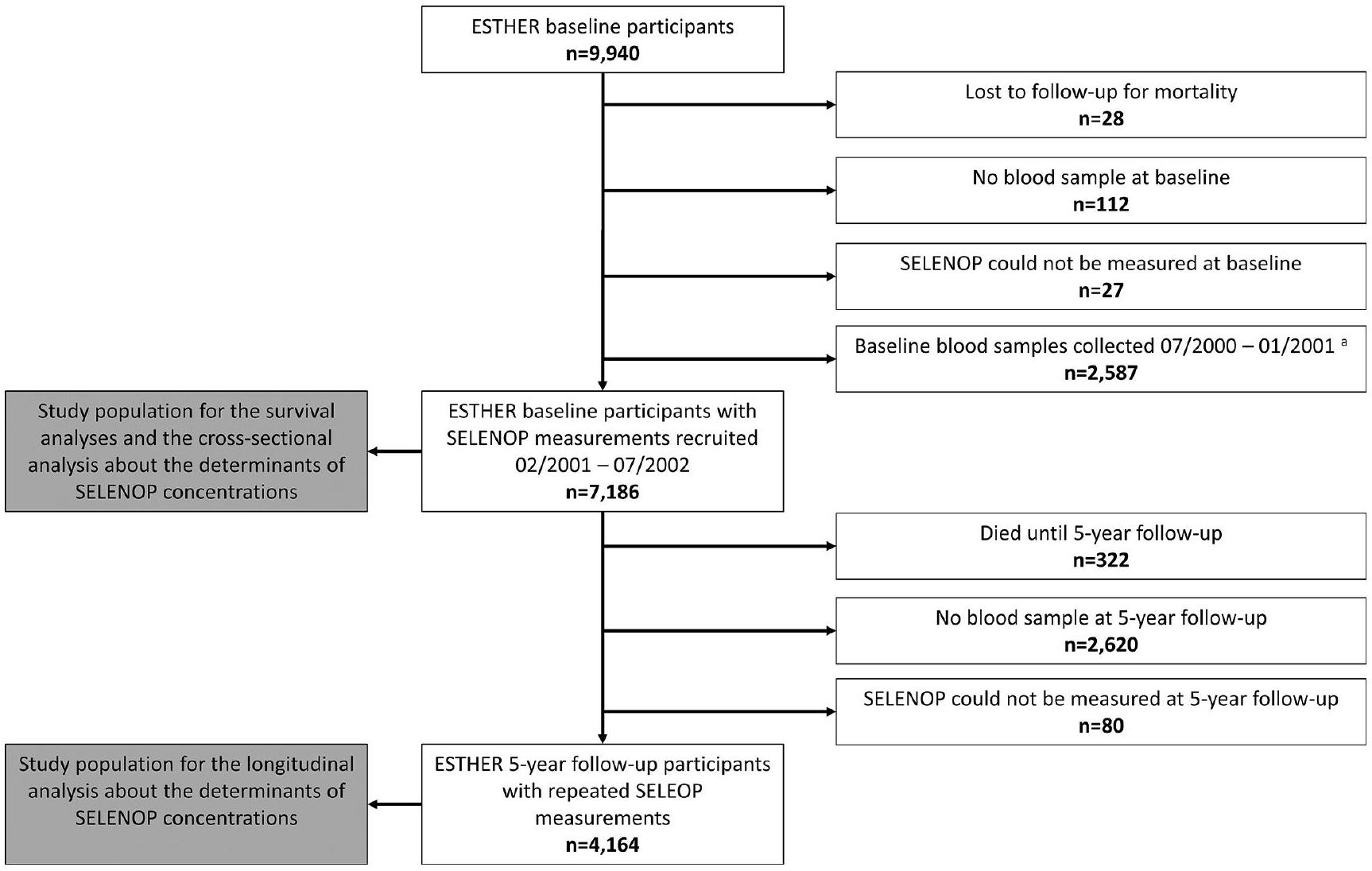
خلفية السيلينيوم هو معدن أساسي نادر. الوظيفة الرئيسية لبروتين السيلينوب (SELENOP) هي نقل السيلينيوم، ولكن تم وصفه أيضًا بتأثيرات مضادة للأكسدة. الطرق لتقييم العلاقة بين قياسات متكررة لتركيز SELENOP في المصل مع الوفيات الناتجة عن جميع الأسباب والوفيات المحددة بسبب السبب، تم قياس SELENOP في المصل عند خط الأساس والمتابعة بعد 5 سنوات في 7,186 و 4,164 مشاركًا من دراسة ESTHER، وهي دراسة قائمة على السكان الألمان تتراوح أعمارهم بين 50-74 عامًا عند خط الأساس. النتائج خلال 17.3 عامًا من المتابعة، توفي 2,126 من المشاركين في الدراسة ( توفيت. كانت علاقة تركيز السيلينوب في المصل مع الوفيات لجميع الأسباب على شكل حرف L، حيث كانت الوفيات أعلى بشكل ملحوظ عند تركيزات السيلينوب. ، الذي يقع بالقرب من حد الثلث السفلي ( ). كانت الوفيات لجميع الأسباب للمشاركين في أدنى فئة من SELENOP مرتفعة بشكل ملحوظ مقارنة بالمواضيع في أعلى فئة (نسبة المخاطر [ فترة الثقة]: 1.35 [1.21-1.50]). كان SELENOP في الثلث الأدنى مرتبطًا أيضًا بزيادة وفيات القلب والأوعية الدموية (1.24 [1.04-1.49])، وفيات السرطان (1.31 [1.09-1.58])، وفيات الأمراض التنفسية (2.06 [1.28-3.32]) وفيات الأمراض المعوية (2.04 [1.25-3.32]). كان خطر الوفاة بسبب جميع الأسباب لأولئك في الثلث الأدنى من SELENOP أكثر من ضعف قوته في الرجال مقارنة بالنساء (تفاعل SELENOP والجنس؛ الاستنتاجات في هذه الدراسة الكبيرة، كانت تركيزات SELENOP في المصل مرتبطة عكسياً بالوفيات الناتجة عن جميع الأسباب والوفيات المحددة بسبب معين. قد تفسر الارتباطات العكسية المتسقة مع نتائج الوفيات المتعددة من خلال ضعف نقل السيلينيوم ونقص السيلينيوم في عدة أعضاء. هناك حاجة إلى تجارب لاختبار فعالية مكملات السيلينيوم في الأشخاص الذين لديهم تركيز منخفض من SELENOP في البداية. تسجيل التجربة تم تسجيله بأثر رجعي في سجل التجارب السريرية الألماني في 14 فبراير 2018 (ID: DRKS00014028).

كلمات مفتاحية سيلينيوم سيلينوبروتين P علامة حيوية الشيخوخة الوفاة دراسة جماعية
قائمة الاختصارات
فترة الثقة 95% فترة الثقة 95%
بيرنه الدراسة البلجيكية بين الجامعات حول التغذية والصحة
كوف معامل التباين
مرض القلب والأوعية الدموية أمراض القلب والأوعية الدموية
تصفية الكرياتينين التقديرية معدل تصفية الكلى المقدر
إستير دراسة وبائية حول فرص
الوقاية، الكشف المبكر والتحسين
الأطباء العامون الأطباء العامون
جي بي إكس إنزيم الجلوتاثيون بيروكسيداز
الموارد البشرية نسبة المخاطر
المنظمة الدولية للهجرة معهد الطب
NHANES
المسح الوطني للصحة والتغذية
MPP مشروع مالمو الوقائي
السيد التوزيع العشوائي المندلي
أو نسبة الأرجحية
PREVEND الوقاية من مرض الكلى والأوعية الدموية في المرحلة النهائية
التجارب السريرية العشوائية التجارب السريرية العشوائية المضبوطة
اختر تجربة الوقاية من السرطان باستخدام السيلينيوم وفيتامين E
سيلينوب سيلينوبروتين P

الخلفية

السيلينيوم هو معدن أساسي نادر وله وظائف فسيولوجية بشكل رئيسي من خلال دمجه في السيلينوبروتينات. سيلينوبروتين P (SELENOP) هو أكثر السيلينوبروتينات الدائرة وفرة ويشكل أكثر من السيلينيوم في الدم [2، 3]. البروتين السيليني الرئيسي الآخر في الدم (20-30%) هو بيروكسيداز الجلوتاثيون خارج الخلية (GPX) 3 [2، 3]. علاوة على ذلك، يمكن أن تحتوي بروتينات البلازما على السيلينيوم في شكل سيلينوميثيون غير محدد الإدخال، وخاصة الألبومين، الذي يمثل السيلينيوم في الدم [2، 3]. الوظيفة الرئيسية لـ SELENOP هي نقل السيلينيوم في جميع أنحاء الجسم، وتزويد العنصر النTrace الضروري للأنسجة المختلفة بطريقة هرمية، مما يساهم في التخليق الحيوي للبروتينات السيلينية المرتبطة بالدفاع المضاد للأكسدة [4]. SELENOP ليس فقط علامة حيوية حساسة لحالة السيلينيوم عند تناول كميات منخفضة إلى متوسطة من السيلينيوم [5]، ولكنه يشكل أيضًا علامة حيوية وظيفية لتزويد السيلينيوم بسبب امتصاصه المحدد بواسطة المستقبلات في المواقع المستهدفة [6].
انخفاض تناول السيلينيوم يؤدي إلى ضعف تعبير الإنزيمات التي تكون منخفضة في الترتيب الهرمي للبروتينات السيلينية، من بينها GPX1 داخل الخلايا وGPX3 خارج الخلايا الأكثر حساسية لنقص السيلينيوم [7-9]. الفئران
الأشخاص الذين يعانون من تعبير غير طبيعي لجين GPX1 لديهم كميات عالية من الدهون المؤكسدة في الكبد وزيادة في إطلاق بيروكسيد الهيدروجين إلى النظام الدوري. هناك أدلة متزايدة على أن GPX1 هو حجر الزاوية الضروري لنظام الدفاع المضاد للأكسدة في الإنسان. تم ربط التعبير غير الطبيعي لـ GPX3 المشتق من الكلى، على سبيل المثال، بتعزيز thrombosis الشرياني المعتمد على الصفائح الدموية، والتصلب الجانبي الضموري، والسرطان. قد تؤثر الوظائف المضادة للأكسدة لأعضاء عائلة GPX وبروتينات السيلينيوم الأخرى على عمر الفرد. وفقًا لنظرية الجذور الحرة للشيخوخة، يمكن أن يؤدي عدم التوازن بين القدرات المؤكسدة والمضادة للأكسدة في الخلايا إلى تلف الحمض النووي والبروتينات والدهون الغشائية، مما يمكن أن يؤدي إلى شيخوخة الخلايا، وظهور الأمراض المرتبطة بالعمر، وفي النهاية وفاة مبكرة.
على عكس الولايات المتحدة الأمريكية، تتمتع الدول الأوروبية بتوافر منخفض إلى معتدل من السيلينيوم الغذائي بسبب انخفاض استهلاك السيلينيوم، والذي يُعزى إلى كل من انخفاض تركيز السيلينيوم في التربة (وبالتالي في النباتات واللحوم كمصادر رئيسية للسيلينيوم الغذائي) وانخفاض استخدام المكملات الغذائية. بينما تتوفر دراسة كبيرة حول حالة السيلينيوم والوفيات في عموم السكان البالغين من الولايات المتحدة الأمريكية من خلال مسح الصحة والتغذية الوطني (NHANES)، قمنا بإجراء أكبر دراسة حول هذا الموضوع من أوروبا حتى الآن. كان الهدف الأساسي من هذه الدراسة هو تقييم ما إذا كان تركيز SELENOP في المصل مرتبطًا عكسيًا بالوفيات الناتجة عن جميع الأسباب والوفيات المحددة بسبب أسباب معينة في عينة كبيرة من كبار السن من ألمانيا. وكان الهدف الثانوي من هذه الدراسة هو تحديد العوامل المؤثرة على تركيزات SELENOP في تصميم دراسة مقطعية وطولية، وهو ما لم يتم القيام به من قبل. كما تهدف هذه التحليلات الإضافية إلى إيجاد المجموعة المثلى من المتغيرات المرافقة في نماذج البقاء.

طرق

تصميم الدراسة

تستند هذه التحقيقات إلى دراسة ESTHER (الاسم الكامل بالألمانية: “الدراسة الوبائية لفرص الوقاية، والكشف المبكر، والعلاج المحسن للأمراض المزمنة في السكان المسنين”)، وهي دراسة مستمرة على مجموعة من المشاركين، تم الإبلاغ عن تفاصيلها في أماكن أخرى [24، 25]. باختصار، شملت 9,940 من الرجال والنساء، الذين تتراوح أعمارهم سنوات في البداية، تم تجنيدهم من قبل أطبائهم العامين (GPs) خلال فحص صحي روتيني بين 07/2000 و 07/2002 في ولاية سارلاند الألمانية. يتم تقديم هذا الفحص الصحي كل عامين لجميع الألمان الذين تبلغ أعمارهم 35 عامًا أو أكثر مجانًا لـ
جميع الأشخاص الذين لديهم تأمين صحي قانوني والأشخاص الذين لديهم تأمين صحي خاص عادة ما يتم تعويض تكاليفهم. في الفئة العمرية لدراسة ESTHER (50-75 سنة)، الفروق في استخدام الفحص الصحي حسب الجنس والحالة الاجتماعية والاقتصادية صغيرة جداً [26]، مما يشير إلى عدم وجود انحياز في الاختيار بسبب سلوك الوعي الصحي. علاوة على ذلك، كانت انتشار الأمراض المزمنة الشائعة في دراسة ESTHER، مثل ارتفاع ضغط الدم ومرض السكري، مشابهة لتلك المبلغ عنها لمجموعات عمرية مماثلة من خلال المسح الوطني الألماني للمقابلات الصحية والفحوصات 1998 (BGS98) [27، 28]، وهو مسح تمثيلي لألمانيا. معدل المشاركة الفعلي لدراسة ESTHER في البداية غير معروف لأن الأطباء العامين لم يوثقوا عدد المشاركين المحتملين الذين اقتربوا منهم. نفترض أنه أعلى بكثير من معدل المشاركة في BGS98، الذي كان [28]، حيث لم يتم إرسال رسائل غير شخصية للمشاركين في ESTHER، بل تم الاقتراب منهم بشأن الدراسة من قبل طبيبهم العام المعروف أثناء وجودهم في عيادة الطبيب لإجراء الفحص الصحي على أي حال.
تم إعادة الاتصال بمشاركي دراسة ESTHER عبر البريد كل سنوات لمدة 20 عامًا حتى الآن. قدموا معلومات مفصلة عن حالتهم الصحية في استبيانات وعينات دم ومنذ المتابعة بعد 5 سنوات، تبرعوا بمزيد من عينات الدم في ممارسات أطبائهم العامين. تم طرد العينات المأخوذة من الدم، وشحنها إلى مركز الدراسة، وتخزينها في . منذ المتابعة التي استمرت 8 سنوات، يقوم الأطباء العامون أيضًا بملء استبيانات حول الحالة الصحية لمرضاهم، لأنه مع استمرار المتابعة، يصل المزيد والمزيد من المشاركين في الدراسة إلى حالة صحية مناسبة للعمر لا يمكنهم فيها إكمال الاستبيانات. كانت معدلات الاستجابة في المتابعات بين الناجين مع معلومات صحية عن المشاركين في الدراسة من خلال استبيان إما من المشارك أو من طبيبهم العام , ، و لـ ، والمتابعة لمدة 17 عامًا (تنظيف بيانات المتابعة لمدة 20 عامًا قيد الانتظار).

تقييم المتغيرات المشتركة

تستخدم هذه التحليل بيانات من التقييم الأساسي والمتابعة بعد 5 سنوات، حيث شاركوا مرة أخرى. تم الحصول على معلومات حول الخصائص الاجتماعية والديموغرافية، والتعليم المدرسي، ونمط الحياة والنظام الغذائي من المشاركين في الدراسة من خلال استبيان شامل في البداية وبعد 5 سنوات. في البداية، تم تقييم وتوثيق الطول والوزن وضغط الدم الانقباضي وارتفاع ضغط الدم وأمراض القلب التاجية واضطرابات الدهون ومرض السكري على نموذج موحد من قبل الأطباء العامين خلال الفحص الصحي. في المتابعة بعد 5 سنوات، تم الإبلاغ عن الوزن والأمراض من قبل المشاركين، وتم التحقق من الأخيرة من خلال الاستفسار لدى أطباء المشاركين في الدراسة. الأدوية ضد اضطرابات الدهون.
تم تسجيل مرض السكري لتكملة معلومات التشخيص. تم تعريف مرض القلب والأوعية الدموية السائد (CVD) من خلال تقارير الأطباء عن مرض الشريان التاجي، وتاريخ ذاتي للإصابة بالنوبات القلبية، أو السكتة الدماغية، أو إعادة توعية الشرايين التاجية (تجاوز أو دعامة). تم توفير معلومات عن تاريخ الحياة الكامل للسرطان (رموز ICD-10 C00-C97، باستثناء C44) من قبل سجل سرطان سارلاند. تم قياس بروتين سي التفاعلي بواسطة التوربيديمترية في كل من الأساس والمتابعة بعد 5 سنوات، وتم تعريف الالتهاب على أنه مستويات بروتين سي التفاعلي. تم قياس تركيز البروتين الكلي في عينات المصل باستخدام طريقة البيوريت الحركية.
تم استخدام جهاز التحليل الآلي Diasorin-Liaison (شركة Diasorin، ستيلووتر، الولايات المتحدة الأمريكية) لقياس الخط الأساسي مستويات في النساء. تم استخدام نظام IDS-iSYS الآلي (Immunodiagnostic Systems GmbH، فرانكفورت ماين، ألمانيا) لإجراء القياسات الأساسية للرجال ولكلا الجنسين في المتابعة بعد 5 سنوات. تم توحيد كلا الاختبارين المناعيين بأثر رجعي مع الطريقة القياسية الذهبية LC-MS/MS وقد تم وصف التفاصيل في مكان آخر [30]. كانت الحدود الدنيا لنقص فيتامين د ( نقص فيتامين د )، التي اقترحها المعهد الأمريكي للطب (IOM)، تم استخدامها لتحديد حالة فيتامين د [31].
تم قياس مستويات الكرياتينين من عينات المصل باستخدام طريقة جافي الحركية. تم تحديد معدل الترشيح الكبيبي التقديري (eGFR) باستخدام معادلة الكرياتينين CKD-EPI لعام 2021. تم تعريف ضعف الكلى بواسطة eGFR. .

قياسات SELENOP

تم شحن جميع عينات المصل التي تم جمعها في البداية والمتابعة بعد 5 سنوات إلى مختبر معهد الغدد الصماء التجريبية، جامعة شاريتيه للطب، برلين. تم تطوير اختبار ELISA المناعي لتحديد كمية SELENOP باستخدام الأجسام المضادة وحيدة النسيلة [33]، وتم استخدامه لقياس تركيز SELENOP في المصل بين عامي 2010 و2012. تم معايرة اختبار ELISA مقابل المادة المرجعية القياسية (SRM) 1950، التي تم تطويرها في عينة أمريكية [34] ولا تزال المادة المرجعية القياسية الوحيدة المتاحة لقياسات SELENOP. سيكون من المرغوب فيه تطوير مادة مرجعية قياسية مع عينة أوروبية في المستقبل لأن السكان الأوروبيين أقل تنوعًا من الأمريكيين من حيث الأعراق. في دراستنا، كان معامل التباين بين الاختبارات (COV) لاختبار SELENOP هو لتحكم بتركيز منخفض و لتحكم بتركيز عالٍ. تم اختبار معامل التباين داخل الاختبار ضد مصل الإنسان المخفف وكان .
قياسات SELENOP من المشاركين في الدراسة الذين تم تجنيدهم قبل كان يجب التخلص منه )
لأنها كانت مرتبطة بتركيزات البروتين الكلي في العينات ( ); ربما نتيجة لاختلاف ظروف تخزين هذه العينات مقارنة بتلك التي تم جمعها لاحقًا. تركيز SELENOP في العينات التي تم جمعها منذ ( ) لم تظهر أي ارتباط مع تركيز البروتين الكلي ( , من بين هؤلاء 7,186 مشاركًا، تبرع بعينة دم أخرى في المتابعة بعد 5 سنوات وتم إجراء قياس SELENOP مرة أخرى. في عينة مختارة عشوائيًا من 59 مشاركًا في الدراسة، تم قياس تركيز السيلينيوم في المصل بالإضافة إلى ذلك وكانت العلاقة مع تركيز SELENOP عالية: فترة الثقة 95% ): ).

تحديد الوفيات

يمكن الحصول على معلومات حول الحالة الحيوية بين عامي 2000 و 2018 لـ المشاركين في المجموعة من خلال الاستفسار في مكاتب تسجيل السكان. من السلطات الصحية المحلية، يمكن الحصول على السبب الكامن وراء الوفاة من حيث رموز التصنيف الدولي للأمراض – الإصدار العاشر (ICD-10) لـ للمشاركين المتوفين.

التحليلات الإحصائية

مشاركون في فحص ESTHER الأساسي ( ) تم استبعادهم من هذه الدراسة إذا فقدوا للمتابعة بشأن الوفيات ( )، بشرط عدم تقديم عينات دم ( ) أو لم يكن من الممكن قياس SELENOP ( )، أو إذا تم تجنيدهم بين و ( )، مما أسفر عن حجم عينة إجمالي قدره الموضوعات لتحليلات البقاء (انظر الشكل 1 لمخطط التدفق).
لتقييم محددات تركيز منخفض من SELENOP في التحليل العرضي، تم استخدام نموذج الانحدار اللوجستي مع الاختيار الأمامي (معيار الدخول: تم استخدام جميع الخصائص الأساسية الموضحة في الجدول 1 كمتغيرات مستقلة، وتم استخدام أدنى تيرتيل من SELENOP مقابل التيرتيلات المتوسطة والعليا كمتغير تابع. لتقييم محددات تركيز SELENOP المنخفض في التحليل الطولي، تم تطبيق نفس النموذج ولكن تم استخدام قياسات SELENOP من المتابعة بعد 5 سنوات كمتغير تابع وتم إضافة تركيز SELENOP الأساسي كمتغير مستقل إضافي. بينما تم إجراء التحليل العرضي مع جميع المشاركين البالغ عددهم 7,186.
الشكل 1 مخطط تدفق السكان المدروسين المستخدمين في التحليلات المختلفة
كان يجب التخلص من قياسات SELENOP من عينات الدم المجمعة قبل 02/2001 لأنها كانت مرتبطة بـ
تركيزات البروتين الكلي في العينات؛ ربما نتيجة لاختلاف ظروف تخزين هذه العينات مقارنة بتلك التي تم جمعها لاحقًا
الجدول 1 وصف الخصائص الأساسية لسكان الدراسة
الخصائص الأساسية ن (%) يعني SD
العمر (بالسنوات) 7186
الجنس (ذكر) 7186 3219 (44.8)
التعليم المدرسي (السنوات) ٧٠٠٠
٥٢٦٤ (٧٥.٢)
10-11 952 (13.6)
784 (11.2)
مؤشر كتلة الجسم (BMI) ) 7178
< 25 1955 (27.2)
25-<30 ٣٣٧٥ (٤٧.٠)
1848 (25.8)
سلوك التدخين 6989
مدخن أبداً 3548 (50.8)
مدخن سابق 2299 (32.9)
مدخن حالي 1142 (16.3)
النشاط البدني 7163
غير نشط 1571 (21.9)
منخفض ٣٢٨١ (٤٥.٨)
متوسط أو عالي 2311 (32.3)
استهلاك الكحول ٦٤٧٨
ممتنع 2135 (33.0)
معتدل 3909 (60.3)
عالي ٣٣٤ (٥.٢)
مرتفع جداً 100 (1.5)
التغذية (الحصص اليومية)
لحم 6456
خبز 5980
حليب/جبن/بيض 6539
الفواكه/الخضروات 6813
سمك 6754
مكملات الفيتامينات/المعادن (يوميًا) 7017 991 (14.1)
الأمراض/الحالات
سرطان 7186 458 (6.4)
مرض القلب والأوعية الدموية 7185 1200 (16.7)
داء السكري 7073 1062 (15.0)
خلل شحميات الدم 7075 3100 (43.8)
ارتفاع ضغط الدم 7071 3132 (44.3)
فشل القلب 7136 755 (10.6)
ضعف الكلى 7165 ١١٩٤ (١٦.٧)
التهاب 7097 ٢٧٦٨ (٣٩.٠)
حالة فيتامين د 7020
كافٍ 2795 (39.8)
غير كافٍ 3146 (44.8)
نقص 1079 (15.4)
إجمالي البروتين (غ/ل) 7186
الجدول 1 (مستمر)
الخصائص الأساسية ن (%) يعني SD
خط الأساس SELENOP (ملغ/لتر) 7186
متابعة لمدة 5 سنوات SELENOP (ملغ/لتر) 4164
الاختصارات: مؤشر كتلة الجسم (BMI)؛ مرض القلب والأوعية الدموية (CVD)؛ عدد المشاركين الذين لديهم بيانات؛ SELENOP، بروتين السيلين؛ SD، الانحراف المعياري
نساء والرجال الإيثانول في اليوم
تاريخ أي نوع من السرطان باستثناء سرطان الجلد غير الميلانيني
تم تعريف مرض القلب والأوعية الدموية على أنه مرض الشريان التاجي أو تاريخ من احتشاء عضلة القلب أو السكتة الدماغية.
معدل الترشيح الكبيبي المقدر (eGFR)
بروتين سي التفاعلي
نقص فيتامين د، 25-هيدروكسي فيتامين د (25(OH)D) <30 نانومول/لتر؛ غير كافٍ، 25(OH)D 30-<50 نانومول/لتر؛ كافٍ، 25(OH)D
الموضوعات، تم إجراء التحليل الطولي مع أولئك المشاركون الذين لديهم قياسات متكررة لـ SELENOP في المتابعة بعد 5 سنوات (انظر الشكل 1 لمخطط التدفق).
تم استخدام نماذج المخاطر النسبية لكوك لتقدير نسب المخاطر لتيرتيلات SELENOP (التيرتيل الأعلى كفئة مرجعية) فيما يتعلق بالوفيات بسبب جميع الأسباب والوفيات الناتجة عن أمراض القلب والأوعية الدموية، السرطان، الأمراض التنفسية، الأمراض المعوية، والأمراض النفسية/العصبية، على التوالي. تم استخدام نموذجين: نموذج معدل حسب العمر والجنس ونموذج رئيسي معدل بشكل شامل. تتكون مجموعة المتغيرات في النموذج الرئيسي من جميع العوامل المرتبطة بتركيز SELENOP (سواء في التحليل العرضي أو الطولي) وكذلك بالوفيات بسبب جميع الأسباب. إذا لم يُذكر خلاف ذلك، فقد استخدمت جميع نماذج المخاطر النسبية لكوك قياسات SELENOP المتكررة وتقييمات المتغيرات من المتابعة لمدة 5 سنوات من خلال ملاءمة جميع متغيرات النموذج باستثناء الجنس كمتغيرات تعتمد على الزمن باستخدام إضافة لإجراء SAS PHREG الموصوف في مكان آخر. بالنسبة للمشاركين في الدراسة الذين لم يشاركوا في المتابعة لمدة 5 سنوات أو الذين لم يتبرعوا بعينة دم في تلك المتابعة، تم الافتراض أن خصائصهم الأساسية وتخصيصهم لتيرتيل SELENOP لم تتغير بين الأساس والمتابعة لمدة 5 سنوات.
تم رسم علاقة التركيز-الاستجابة بين تركيز SELENOP الأساسي ومعدل الوفيات لجميع الأسباب باستخدام الأشكال المكعبة المقيدة مع تحليل المخاطر النسبي لكوكس ومجموعة المتغيرات المشتركة للنموذج الرئيسي [36]. تم استخدام خمس عقد عند النسب المئوية 10 و25 و50 و75 و90 لتركيز SELENOP، وتم استخدام الوسيط كمرجع.
في تحليل الحساسية، تم استخدام قياسات SELENOP الأساسية فقط لجميع نماذج الانحدار النسبية لكوكس التي استخدمت نمذجة تعتمد على الوقت لتركيز SELENOP وجميع المتغيرات. في تحليل حساسية إضافي، تم تكرار التحليلات بدون
نموذج يعتمد على الزمن باستخدام إما فقط الوفيات التي حدثت في وقت سابق (السنة 1-9) أو لاحقًا (السنة 10-18) خلال فترة المتابعة.
تم إجراء تحليلات فرعية حسب الجنس، والفئات العمرية سنوات)، أمراض القلب والأوعية الدموية وتاريخ السرطان. تم اختبار التفاعلات المحتملة بين مستويات SELENOP والمتغيرات المرافقة للنموذج الرئيسي من أجل الدلالة الإحصائية من خلال إضافة مصطلحات المنتج إلى النموذج.
تم استخدام التقدير المتعدد للتعامل بشكل مناسب مع القيم المفقودة في خط الأساس والمتابعة لمدة 5 سنوات. تم استخدام طريقة سلسلة ماركوف مونت كارلو (MCMC) من إجراء SAS PROC MI لتقدير 5 مجموعات بيانات. تم استخدام جميع المتغيرات الموضحة في الجدول 1 لنموذج التقدير. تراوحت نسبة القيم المفقودة المقدرة من 0 إلى (الجدول 1). لم تكن هناك أسباب للتخلي عن فرضية الغياب العشوائي المعروفة. تم إجراء جميع التحليلات في مجموعات البيانات الخمس المعوضة وتم دمج نتائج مجموعات البيانات الفردية بواسطة إجراء SAS PROC MIANALYZE.
كانت جميع الاختبارات الإحصائية ثنائية الجانب باستخدام مستوى ألفا 0.05 وتم إجراء جميع التحليلات باستخدام حزمة البرمجيات SAS، الإصدار 9.4 (كاري، نورث كارولينا، الولايات المتحدة الأمريكية).

النتائج

خصائص عينة الدراسة

كان متوسط عمر المشاركين في الدراسة البالغ عددهم 7,186 هو 62.3 سنة و كانوا ذكورًا. وتظهر الخصائص الأخرى لسكان الدراسة في البداية في الجدول 1.
الجدول 2: الارتباطات العرضية والطولية لخصائص الخط الأساس مع الثلث السفلي من تركيز بروتين السيلين في مصل الدم
خاصية تحليل مقطعي تحليل طولي )
OR (95%CI) OR (95%CI)
العمر (لكل 10 سنوات) 1.15 (1.06; 1.24) 1.26 (1.12; 1.41)
الجنس (ذكر) 1.53 (1.37; 1.71)
مؤشر كتلة الجسم (BMI) )
< 25 مرجع
25-<30 0.79 (0.70; 0.89)
0.65 (0.57; 0.76)
سلوك التدخين
مدخن أبداً مرجع
مدخن سابق مرجع
مدخن حالي 1.41 (1.15; 1.73)
النشاط البدني
غير نشط مرجع
منخفض مرجع
متوسط أو عالي 0.81 (0.73; 0.91)
استهلاك الكحول
ممتنع 1.18 (1.05; 1.33)
معتدل مرجع
عالي مرجع
مرتفع جداً 1.76 (1.17; 2.64)
مكملات الفيتامينات/المعادن المتعددة (يوميًا) 0.63 (0.54; 0.74)
الأمراض/الحالات
سرطان 1.35 (1.02; 1.79)
مرض القلب والأوعية الدموية 1.31 (1.14; 1.50) 1.30 (1.08; 1.56)
داء السكري 0.57 (0.49; 0.67)
خلل شحميات الدم 0.82 (0.71; 0.94)
التهاب 1.36 (1.22; 1.51)
حالة فيتامين د
كافٍ مرجع مرجع
غير كافٍ 1.18 (1.05; 1.32) مرجع
ناقص 1.73 (1.50; 2.01) 1.39 (1.13; 1.70)
خط الأساس SELENOP (حسب ) غير متوفر 0.55 (0.51; 0.58)
الاختصارات: مؤشر كتلة الجسم (BMI)؛ مرض القلب والأوعية الدموية (CVD)؛ غير قابل للتطبيق (NA)؛ نسبة الأرجحية (OR) نسبة الأرجحية و فترة الثقة؛ المرجع، فئة المرجع؛ SELENOP، بروتين السيلين
نتيجة نموذج الانحدار اللوجستي مع الاختيار الأمامي. يحتوي النموذج على جميع المتغيرات المدرجة في العمود
بروتين سي التفاعلي
نقص فيتامين د، 25-هيدروكسي فيتامين د (25(OH)D) غير كافٍ، ؛ كافٍ، نانومول/لتر

توزيع SELENOP

كان متوسط تركيز SELENOP (الانحراف المعياري) 4.8 (1.4) في الأساس وتوزيعها تقريبا بشكل طبيعي (الشكل التوضيحي 1). مع كان متوسط تركيز السيلينوب (الانحراف المعياري) أقل في المتابعة بعد 5 سنوات مقارنةً بالخط الأساسي.

عوامل تركيز السيلينوب

في نموذج الانحدار اللوجستي مع جميع الخصائص الأساسية من الجدول 1 كمتغيرات مستقلة وتيرتي SELENOP السفلية مقابل الوسطى والعليا كمتغير تابع، تم تحديد العوامل التالية لتركيز منخفض من SELENOP من خلال الاختيار الأمامي: العمر الأكبر، الجنس الذكري، مؤشر كتلة الجسم (عكسي)، النشاط البدني المتوسط أو العالي (عكسي)، عدم استهلاك الكحول أو استهلاكه بشكل مرتفع جداً، استخدام مكملات الفيتامينات والمعادن اليومية (عكسي)، أمراض القلب والأوعية الدموية، داء السكري (عكسي)، الالتهاب، ونقص أو عدم كفاية فيتامين د (الجدول 2). في التحليل الطولي باستخدام نفس الأساليب، تم استخدام تيرتي SELENOP السفلية من المتابعة لمدة 5 سنوات كمتغير تابع. كما هو متوقع، كان تركيز SELENOP الأساسي هو أقوى محدد لتركيز SELENOP في المتابعة بعد 5 سنوات. من محددات التركيز المنخفض لـ SELENOP في التحليل العرضي، تم اختيار العمر، وأمراض القلب والأوعية الدموية، ونقص فيتامين د أيضاً للنموذج النهائي للتحليل الطولي. تم إضافة عوامل أخرى، لم يتم اختيارها سابقاً في التحليل العرضي، وهي التدخين الحالي، تاريخ السرطان، والخلل الدهني (عكسي).

ارتباط تركيز السيلينوب مع الوفيات

خلال فترة متابعة متوسطة تبلغ 17.3 سنة (الحد الأدنى: 15.9، الحد الأقصى: 17.9 سنة)، توفي 2,126 (29.6%) من المشاركين في الدراسة. كانت الأسباب الرئيسية للوفيات مخصصة لأمراض القلب والأوعية الدموية. )، السرطان ( الأمراض التنفسية ( أمراض الجهاز الهضمي الأمراض النفسية/العصبية وأسباب أخرى/غير معروفة .
كانت علاقة التركيز-الاستجابة لتركيزات SELENOP مع الوفيات من جميع الأسباب على شكل حرف L (الشكل 2). بينما لم يُلاحظ أي ارتباط عند تركيزات SELENOP التي تزيد عن الوسيط، أظهر منحنى المكعبات المقيدة زيادة في الوفيات مع انخفاض تركيزات SELENOP دون الوسيط. )، التي وصلت إلى دلالة إحصائية عند تركيزات SELENOP أقل من . هذا العتبة للدلالة الإحصائية قريبة من الحد الأدنى للثلث السفلي ( ).
تظهر الجدول 3 ارتباطات تيرتيلات SELENOP مع الوفيات لجميع الأسباب والوفيات المحددة بسبب على مدى 17 عامًا باستخدام
الشكل 2 العلاقة بين تركيز بروتين السيلينوبروتين P الأساسي والاستجابة للموت لجميع الأسباب على مدى 17 عامًا. تم تقييم المنحنيات بواسطة مكعبات مقيدة مع عقد عند النسبة المئوية 10 و25 و50 و75 و90 لتركيز بروتين السيلينوبروتين P، وتم استخدام الوسيط كمرجع. نظرًا لأن ماكرو SAS لعلاقات التركيز والاستجابة لا يعمل مع مجموعات بيانات متعددة، تم اتخاذ القرار مسبقًا لاستخدام مجموعة البيانات المملوءة رقم 1 لهذا التحليل.
النمذجة المعتمدة على الزمن لقياسات SELENOP وجميع المتغيرات الأخرى باستثناء الجنس. على الرغم من أن تقديرات التأثير للنموذج المعدل حسب العمر والجنس قد تراجعت قليلاً في النموذج الرئيسي، إلا أن الثلث السفلي من SELENOP (مقارنة بالثلث العلوي) ظل مرتبطًا بشكل إحصائي ملحوظ بجميع نتائج الوفيات باستثناء الوفيات بسبب الأمراض النفسية/العصبية وأسباب أخرى/غير معروفة. تضمن النموذج الرئيسي جميع المحددات المعروفة لتركيز SELENOP المنخفض (سواء في التحليلات المقطعية أو الطولية، الجدول 2)، والتي كانت مرتبطة أيضًا بالوفيات بسبب جميع الأسباب (باستثناء استخدام الفيتامينات المتعددة/المعادن؛ الجدول التكميلي 1). كانت قوة الارتباطات في النموذج الرئيسي قابلة للمقارنة لوفيات جميع الأسباب، ووفيات الأمراض القلبية الوعائية، ووفيات السرطان (حوالي 1.3 ضعف زيادة في الوفيات في الثلث العلوي)، في حين كانت الارتباطات مع وفيات الأمراض التنفسية والجهاز الهضمي قوية بشكل خاص (حوالي 2 ضعف زيادة في الوفيات). لم يكن الثلث الأوسط من SELENOP مرتبطًا بأي نتيجة عند مقارنته بالثلث العلوي.
تمت ملاحظة تقديرات تأثير أضعف قليلاً لمعظم النتائج عندما تم استخدام تركيز SELENOP الأساسي فقط وقيم المتغيرات المرافقة ولم يتم نمذجة المتغيرات كمتغيرات تعتمد على الزمن (الجدول التكميلي 2). إذا تم تحليل الوفيات التي حدثت في وقت مبكر (السنة 1-9) أو لاحقاً (السنة 10-18) خلال المتابعة بشكل منفصل، كانت تقديرات التأثير لوفيات جميع الأسباب هي نفسها في كلا التحليلين (الجدول التكميلي 3). ومع ذلك، كانت هناك اختلافات.
للموت المحدد بسبب السبب. كانت الفئة السفلية من SELENOP مرتبطة فقط بوفيات السرطان التي حدثت لاحقًا في المتابعة. على النقيض من ذلك، كانت العلاقات بين تركيز SELENOP ووفيات الأمراض القلبية الوعائية ووفيات غير الناتجة عن الأمراض القلبية الوعائية أو السرطان أقوى مع الوفيات التي حدثت في وقت مبكر من المتابعة، ولكن كانت العلاقة لا تزال قابلة للاكتشاف مع الوفيات اللاحقة (على الرغم من أنها لم تكن ذات دلالة إحصائية لوفيات الأمراض القلبية الوعائية).
في تحليلات المجموعات الفرعية، لوحظ ارتباط أقوى بين أدنى تيرتيل من SELENOP والوفيات لجميع الأسباب ووفيات السرطان بين الرجال مقارنة بالنساء، في حين كانت تقديرات التأثير لوفيات القلب والأوعية الدموية متقاربة (الجدول 4). في اختبارات التفاعلات المحتملة لتركيز SELENOP مع متغيرات النموذج الرئيسي لنتيجة الوفيات لجميع الأسباب، كانت عبارة التفاعل بين أدنى تيرتيل من SELENOP (مقابل أعلى تيرتيل) والجنس هي الوحيدة ذات الدلالة الإحصائية. ، الجدول التكميلي 4).
على الرغم من عدم ملاحظة أي تفاعلات لتركيز SELENOP مع العمر، وأمراض القلب والأوعية الدموية، وتاريخ السرطان، إلا أنه كانت هناك بعض الاختلافات في النتائج وفقًا لهذه العوامل. كانت تقديرات التأثير للوفيات بسبب جميع الأسباب ووفيات أمراض القلب والأوعية الدموية أقوى بين الأشخاص الذين تقل أعمارهم عن 65 عامًا مقارنة بالأشخاص الأكبر سنًا. علاوة على ذلك، كانت تقديرات التأثير لجميع نتائج الوفيات أقوى بين الأشخاص الذين يعانون من أمراض القلب والأوعية الدموية في البداية. وأخيرًا، كانت تقديرات التأثير للسرطان
الجدول 3 ارتباطات تركيز السيلينوبروتين P (SELENOP) في المصل مع الوفيات الناتجة عن جميع الأسباب والوفيات المحددة بسبب معين خلال 17.3 سنة من المتابعة
نتيجة الثلاثيات SELENOP نموذج معدل حسب العمر والجنس النموذج الرئيسي
HR (95%CI) HR (95%CI)
الوفيات لجميع الأسباب تي 1 ٢٣٩٥ 857 (35.8) 1.48 (1.33; 1.64) 1.35 (1.21; 1.50)
T2 ٢٣٩٥ 653 (27.3) 1.02 (0.91; 1.14) 1.02 (0.91; 1.14)
T3 ٢٣٩٦ 616 (25.7) مرجع مرجع
وفيات الأمراض القلبية الوعائية تي 1 ٢٣٩٥ 275 (11.5) 1.33 (1.12; 1.59) 1.24 (1.04; 1.49)
T2 ٢٣٩٥ 217 (9.1) 0.85 (0.70; 1.04) 0.86 (0.71; 1.05)
T3 ٢٣٩٦ 217 (9.1) مرجع مرجع
وفيات السرطان تي 1 ٢٣٩٥ 271 (11.3) 1.47 (1.22; 1.77) 1.31 (1.09; 1.58)
T2 ٢٣٩٥ 225 (9.4) 1.11 (0.91; 1.35) 1.12 (0.92; 1.36)
T3 ٢٣٩٦ 200 (8.4) مرجع مرجع
وفيات الأمراض التنفسية تي 1 ٢٣٩٥ 61 (2.6) 2.37 (1.49; 3.77) 2.06 (1.28; 3.32)
T2 ٢٣٩٥ 19 (0.8) 0.88 (0.50; 1.55) 0.87 (0.49; 1.53)
T3 ٢٣٩٦ 31 (1.3) مرجع مرجع
وفيات الأمراض المعوية تي 1 ٢٣٩٥ 54 (2.2) 2.22 (1.37; 3.59) 2.04 (1.25; 3.32)
T2 ٢٣٩٥ 25 (1.0) 1.05 (0.60; 1.83) 1.03 (0.59; 1.80)
T3 ٢٣٩٦ ٢٦ (١.١) مرجع مرجع
وفيات الأمراض النفسية/العصبية T1 ٢٣٩٥ ٤٨ (٢.٠) 1.47 (0.95; 2.29) 1.40 (0.89; 2.19)
T2 ٢٣٩٥ ٤٤ (١.٨) 1.15 (0.72; 1.82) 1.15 (0.73; 1.84)
T3 ٢٣٩٦ 31 (1.3) مرجع مرجع
سبب الوفاة الآخر/غير المعروف تي 1 ٢٣٩٥ 148 (6.2) 1.34 (1.04; 1.72) 1.27 (0.98; 1.63)
T2 ٢٣٩٥ 123 (5.1) 1.16 (0.89; 1.49) 1.18 (0.91; 1.53)
T3 ٢٣٩٦ 111 (4.6) مرجع مرجع
الاختصارات: CVD، مرض القلب والأوعية الدموية؛ HR ( نسبة المخاطر و فترة الثقة؛ حجم العينة؛ عدد الوفيات؛ T1، T2، T3، الثلث السفلي، الثلث الأوسط والثلث العلوي، على التوالي؛ Ref، فئة المرجع؛ SELENOP، بروتين السيلين
مطبوع بخط عريض: ذو دلالة إحصائية )
حدود التركيزات الثلاثية لـ SELENOP عند خط الأساس: الثلث ; الثلث الثالث: الحدود لفئات تركيز SELENOP عند المتابعة بعد 5 سنوات: الفئة 1: ; الثلث الثالث:
تم تعديلها حسب العمر، الجنس، مؤشر كتلة الجسم، التدخين الحالي، النشاط البدني، استهلاك الكحول، تاريخ السرطان، أمراض القلب والأوعية الدموية، داء السكري، اضطرابات الدهون، الالتهاب (بروتين سي التفاعلي) )، وحالة فيتامين د. تم نمذجة تيرتيلات تركيز SELENOP وجميع المتغيرات الأخرى باستثناء الجنس كمتغيرات تعتمد على الزمن من خلال اعتبار قيمها في المتابعة بعد 5 سنوات من دراسة ESTHER
رموز التصنيف الدولي للأمراض 10 I00-I99
رموز التصنيف الدولي للأمراض 10 C00-C97
رموز التصنيف الدولي للأمراض 10 J00-J99
رموز التصنيف الدولي للأمراض 10 K00-K93
رموز التصنيف الدولي للأمراض 10 F00-G99
رموز التصنيف الدولي للأمراض 10 A00-B99، D50-D90، E00-E90، M00-M99، N00-N40، R00-R99، S00-T98
كانت الوفيات أعلى بين الأشخاص الذين ليس لديهم تاريخ مرضي للسرطان مقارنة بالأشخاص الذين لديهم تاريخ مرضي للسرطان.

نقاش

ملخص

في هذه الدراسة الكبيرة المستندة إلى السكان مع قياسات متكررة، كانت تركيزات SELENOP في المصل مرتبطة بشكل قوي وإحصائيًا مع الوفيات لجميع الأسباب والوفيات بسبب أمراض القلب والأوعية الدموية، السرطان، الأمراض التنفسية، والأمراض المعوية. كانت العلاقة بين تركيزات SELENOP والوفيات لجميع الأسباب هي
تم العثور على ارتباط غير خطي عكسي مع الوفيات يبدأ في الزيادة بشكل ملحوظ عند تركيزات SELENOP أقل من . بالنسبة للأشخاص الذين لديهم تركيزات من SELENOP ، زادت الوفيات بـ كانت الوفيات الزائدة أعلى بأكثر من الضعف لدى الرجال مقارنة بالنساء (زادت الوفيات بـ مقابل ) وتم تحديد تفاعل ذو دلالة إحصائية لتركيز SELENOP مع الجنس.

مقارنة عامة مع الدراسات السابقة والجدة

متوسط تركيز السيلينوب في خط الأساس في دراسة إستر كان قابلاً للمقارنة مع ذلك من التعديل الألماني لعوامل خطر القلب والأوعية الدموية (MoKaRi)
الجدول 4 تحليلات تحتية لارتباط مستوى تركيز السيلينوبروتين P في مصل الدم في الثلث الأدنى مقابل الثلث الأعلى مع الوفيات من جميع الأسباب والوفيات المحددة الأسباب خلال 17.3 سنة من المتابعة
مجموعة فرعية الوفيات لجميع الأسباب وفيات الأمراض القلبية الوعائية وفيات السرطان
(%) الموارد البشرية (95%فترة الثقة) (%) الموارد البشرية (95% فترة الثقة) (%) الموارد البشرية (95% فترة الثقة)
إجمالي السكان 7186 2126 (29.6) 1.35 (1.21; 1.50) 709 (9.9) 1.24 (1.04; 1.49) 696 (9.7) 1.31 (1.09; 1.58)
نساء 3967 912 (23.0) 1.20 (1.03; 1.42) ٣٠٣ (٧.٦) 1.22 (0.92; 1.61) 284 (7.2) 1.16 (0.86; 1.55)
رجال ٣٢١٩ 1214 (37.7) 1.47 (1.28; 1.70) ٤٠٦ (١٢.٦) 1.27 (1.01; 1.62) 412 (12.8) 1.42 (1.11; 1.82)
< 65 سنة ٤٣٢٤ 802 (18.6) 1.48 (1.24; 1.75) 210 (4.9) 1.45 (1.04; 2.04) 336 (7.8) 1.25 (0.95; 1.64)
سنوات 2862 1324 (46.3) 1.26 (1.10; 1.44) 499 (17.4) 1.15 (0.93; 1.42) 360 (12.6) 1.33 (1.03; 1.73)
لا أمراض قلبية وعائية 6010 1525 (25.4) 1.30 (1.15; 1.47) ٤٥٩ (٧.٦) 1.20 (0.95; 1.49) ٥٤٨ (٩.١) 1.26 (1.02; 1.55)
مرض القلب والأوعية الدموية 1176 601 (51.1) 1.52 (1.24; 1.86) 250 (21.3) 1.33 (0.98; 1.80) 148 (12.6) 1.58 (1.02; 2.45)
لا تاريخ للإصابة بالسرطان 6728 1897 (28.2) 1.36 (1.21; 1.52) 652 (9.7) 1.20 (1.00; 1.45) 575 (8.6) 1.32 (1.08; 1.63)
تاريخ السرطان ٤٥٨ 229 (50.0) 1.26 (0.91; 1.73) 57 (12.5) 1.60 (0.77; 3.32) 121 (26.4) 1.21 (0.78; 1.88)
الاختصارات: CVD، مرض القلب والأوعية الدموية؛ HR ( نسبة المخاطر و فترة الثقة؛ حجم العينة؛ عدد الوفيات؛ T1، T2، T3، الثلث السفلي، الثلث الأوسط والثلث العلوي، على التوالي؛ Ref، فئة المرجع؛ SELENOP، بروتين السيلين
مطبوع بخط عريض: ذو دلالة إحصائية )
تم تعديلها حسب العمر، الجنس، مؤشر كتلة الجسم، التدخين الحالي، النشاط البدني، استهلاك الكحول، تاريخ السرطان، أمراض القلب والأوعية الدموية، داء السكري، اضطرابات الدهون، الالتهاب (بروتين سي التفاعلي) و حالة فيتامين د. تم نمذجة tertiles تركيز SELENOP وجميع المتغيرات الأخرى باستثناء الجنس كمتغيرات تعتمد على الزمن من خلال اعتبار قيمها في المتابعة بعد 5 سنوات من دراسة ESTHER.
الدراسة ( )، دراسة مقطعية شملت 51 مشاركًا بمتوسط عمر (61 عامًا) مقارنةً بدراسة ESTHER (62 عامًا) [37].
على حد علمنا، تعتبر ESTHER الدراسة الثانية القائمة على السكان التي تقيم العلاقة بين تركيزات SELENOP والوفيات، حيث أن جميع الدراسات السابقة، باستثناء مشروع مالمو الوقائي (MPP) [38]، قيست فيها السيلينيوم في المصل لتحديد حالة السيلينيوم. ومع ذلك، فإن نتائجنا قابلة للمقارنة جزئيًا مع هذه الدراسات لأن SELENOP يمثل أكثر من من السيلينيوم المتداول [2،3] وقد وُجد أن تركيز السيلينيوم في المصل و SELENOP مرتبطان ارتباطًا وثيقًا في دراستنا ( ) ودراسات أخرى ( [38-41]). الجوانب الجديدة في دراستنا هي قياسات SELENOP المتكررة خلال المتابعة ونقاط نهاية الوفيات المحددة أكثر من وفيات القلب والأوعية الدموية والسرطان.

العمر و SELENOP

كان كل 10 سنوات من التقدم في العمر مرتبطًا بزيادة بمقدار 1.15 و 1.26 مرة في احتمالية وجود تركيز منخفض من SELENOP (مصنف في الثلث السفلي من SELENOP) في التحليل المقطعي والطولي، على التوالي. كانت نتائج الدراسات المقطعية السابقة مختلطة وهناك تقارير عن عدم وجود علاقة بين مؤشرات حالة السيلينيوم والعمر [41، 42] بالإضافة إلى تقارير عن علاقات إيجابية [43، 44] وعكسية مع العمر [45]. يمكن تفسير النتائج المختلطة من خلال اختلاف نطاقات العمر في الدراسات، والاختلافات حسب الجنس، وحقيقة أن حجم العينة الكبير مطلوب لاكتشاف عامل خطر ضعيف مع نسبة احتمالات تقارب 1.2.

علاقة حالة السيلينيوم بالوفيات

تعتبر دراسة MPP السويدية هي الدراسة الوحيدة القائمة على السكان، التي قيمت أيضًا العلاقة بين تركيزات SELENOP والوفيات [38]. اتفقت نتائجنا مع النتائج، حيث كان أدنى خُمس من SELENOP مرتبطًا بقوة مع الوفيات من جميع الأسباب (HR [95%CI]: 1.51 [1.32؛ 1.72]) ووفيات القلب والأوعية الدموية (HR [95%CI]: 1.61 [1.32؛ 2.09]) عند مقارنتها بخمسات SELENOP 2-5. لم يتم تحليل نتائج وفيات الأسباب الأخرى.
تم إجراء بحث منهجي في PubMed في 6 ديسمبر 2023 باستخدام الكلمات الرئيسية “السيلينيوم [العنوان/الملخص]” و”الوفيات [العنوان/الملخص]” لتحديد دراسات أخرى قائمة على السكان، والتي أبلغت عن نتائج حول العلاقة بين السيلينيوم المقاس في المصل/البلازما وأي نتيجة للوفيات والنتائج التفصيلية موضحة في الجدول التكميلي 5. بعد إزالة 3 منشورات مكررة من نفس المجموعة [46-48]، تم تحديد 21 دراسة. فيما يتعلق بالوفيات من جميع الأسباب، أبلغت 5 دراسات عن علاقة غير دالة [42، 49-52] وأبلغت الغالبية من 11 دراسة عن علاقة عكسية ذات دلالة إحصائية [22، 23، 53-61] مع تركيز السيلينيوم في المصل/البلازما. فيما يتعلق بوفيات القلب والأوعية الدموية، أبلغت 5 دراسات عن نتائج غير دالة [22، 50، 54، 57، 62] و5 دراسات عن نتائج ذات دلالة [23، 58، 60، 61، 63]. بالنسبة لوفيات السرطان، أبلغت 4 دراسات عن نتائج غير دالة [22، 57، 58، 64] و3 دراسات عن نتائج ذات دلالة [54، 65، 66]. الاختلافات في القوة الإحصائية هي التفسير الأكثر احتمالًا للنتائج غير الدالة لأن جميع النتائج أشارت في نفس الاتجاه. بالنسبة لأسباب الوفيات الأخرى، كان هناك تقرير واحد فقط عن وفيات الأمراض التنفسية من المسح الوطني البريطاني للنظام الغذائي والتغذية [57]. اتفقت نتائج دراستنا مع دراسة قوية، علاقة عكسية لتركيز السيلينيوم في المصل مع ما سبق ذكره
تم ملاحظة النتيجة في نموذج معدل حسب العمر والجنس (HR [95%CI] لكل زيادة بمقدار 1 SD: 0.65 [0.51؛ 0.82]) لكنها فقدت دلالتها الإحصائية في نموذج معدل بشكل أكثر شمولاً (OR لم يتم الإبلاغ عنه).
أجريت دراسة تحليلية لدراسة الوقاية الهولندية من أمراض الكلى والأوعية الدموية في المرحلة النهائية (PREVEND) [42] وتحليلين مع مجموعات مختلفة من NHANES الأمريكية [47، 48] أيضًا أجروا تحليلات تركيز-استجابة ووجدوا، بما يتماشى مع نتائجنا، علاقة غير خطية لتركيزات السيلينيوم مع الوفيات من جميع الأسباب. كما لاحظت الدراسات الثلاث علاقة عكسية قوية بين تركيزات السيلينيوم والوفيات في الثلث السفلي بينما لم تكن هناك علاقة في الثلث الأوسط من توزيع السيلينيوم (ما يسمى بالعلاقة على شكل L). كانت العلاقة بين حالة السيلينيوم العالية والوفيات غير متسقة. بينما لم يكن هناك زيادة في الوفيات في الأشخاص ذوي الحالة العالية من السيلينيوم في دراستنا و NHANES من 2011 إلى 2016 [48]، تم اقتراح انخفاض معتدل في الوفيات في دراسة PREVEND [42]، وتم اقتراح زيادة معتدلة في الوفيات في NHANES من 1988 إلى 1994 [47]. من الجدير بالذكر، أن أي من الانحرافات عن “عدم وجود علاقة” لم تصل إلى دلالة إحصائية عند تركيزات السيلينيوم العالية.

التفاعل مع الجنس

لقد اكتشفنا تفاعلًا ذا دلالة إحصائية بين تركيز SELENOP والجنس بالنسبة لنتيجة الوفيات من جميع الأسباب في دراستنا. من المثير للاهتمام، أن هذا التفاعل تم اكتشافه أيضًا في دراسة PREVEND بالنسبة لوفيات جميع الأسباب وفي الدراسة البلجيكية بين الجامعات حول التغذية والصحة (BIRNH) بالنسبة لوفيات السرطان، وقد لاحظت كلا الدراستين أيضًا علاقة أقوى لحالة السيلينيوم المنخفضة مع (السرطان) الوفيات في الرجال مقارنةً بالنساء [42، 65]. على النقيض من ذلك، لم يتم ملاحظة أي اختلاف بين الجنسين في NHANES الأمريكية [22، 47] ودراسة صينية [50]، بينما لم تقم جميع الدراسات المقارنة الأخرى بالإبلاغ عن نتائج مصنفة حسب الجنس. ربما يكون هذا التفاعل مع الجنس خاصًا للدول الأوروبية ذات الإمدادات المنخفضة إلى المتوسطة من السيلينيوم كما تم اكتشافه الآن في دراسات من بلجيكا وهولندا وألمانيا. قد نتكهن فقط حول أسباب الاختلاف بين الجنسين الملحوظ. من المحتمل أن تلعب الاختلافات الخاصة بالجنس في التنظيم الهرمي للتعبير عن السيلينوبروتين دورًا [9]. عندما تم حقن السيلينيوم في الفئران التي تعاني من نقص السيلينيوم، كانت الإناث تميل إلى الاحتفاظ بالعنصر النTrace بشكل أكثر كفاءة من الذكور في معظم الأعضاء باستثناء الغدد التناسلية [67]. قد يكون احتفاظ السيلينيوم في هذه الأعضاء ذا أهمية خاصة لحمايتها من السرطان لأننا لاحظنا الاختلاف بين الجنسين بالنسبة لوفيات السرطان وليس لوفيات القلب والأوعية الدموية في دراستنا. من اللافت للنظر، أن التحليلات الميتا الخاصة بالجنس لعلاقات حالة السيلينيوم العالية وخطر السرطان أشارت أيضًا
في اتجاه اختلاف الجنس [68]. كانت ORs [95%CIs] 0.74 (0.64؛ 0.86) و 0.90 (0.86؛ 0.95) بين الرجال والنساء، على التوالي، وتداخلت بشكل ضئيل. لقد تم وصف تأثيرات مضادة للسرطان للسيلينيوم من خلال تأثيراته المضادة للأكسدة وتأثيراته على استقرار الحمض النووي [69].

تحليلات تتناول إمكانية العكس في السببية

ستكون العلاقة العكسية في هذا السياق موجودة إذا كانت تركيزات السيلينوب (SELENOP) المنخفضة ناتجة عن الأمراض وليس العكس، وأن الزيادة الملحوظة في الوفيات بين الأشخاص الذين لديهم تركيزات منخفضة من السيلينوب ناتجة عن الأمراض بينما تركيز السيلينوب المنخفض هو مجرد متفرج غير متورط. تم إجراء تحليلين حساسيين لمعالجة إمكانية وجود علاقة عكسية. أولاً، استبعدنا الأشخاص الذين لديهم تاريخ من أمراض القلب والأوعية الدموية أو السرطان من التحليلات (الجدول 4). بالنسبة لوفيات جميع الأسباب، لم يتغير الأمر كثيرًا. كانت النتائج لوفيات أمراض القلب والأوعية الدموية أضعف قليلاً (HR [95%CI]: 1.20 (0.95؛ 1.49)) في الأشخاص الذين ليس لديهم تاريخ من أمراض القلب والأوعية الدموية مقارنة بالأشخاص الذين لديهم تاريخ من أمراض القلب والأوعية الدموية (HR [95%CI]: 1.33 (0.98؛ 1.80)). على الرغم من أنها ليست ذات دلالة إحصائية، إلا أن كلا التقديرات النقطية متشابهة إلى حد كبير. بالنسبة لوفيات السرطان، كان الأمر عكس ذلك، مع نتائج أقوى قليلاً في الأشخاص الذين ليس لديهم تاريخ من السرطان في البداية (HR [95%CI]: 1.32 (1.08؛ 1.63)) مقارنة بالمشاركين في الدراسة الذين لديهم تاريخ من السرطان (HR [95%CI]: 1.21 (0.78؛ 1.88)). كانت الفئة الدنيا من السيلينوب مرتبطة فقط بوفيات السرطان التي حدثت لاحقًا في المتابعة. ثانيًا، استبعدنا الأحداث التي حدثت في السنوات التسع الأولى من المتابعة، والتي قد تكون ناتجة عن أمراض موجودة بالفعل (ربما غير مشخصة) في البداية (الجدول التكميلي 3). مرة أخرى، كانت النتائج لوفيات جميع الأسباب غير متغيرة وكانت نتائج وفيات السرطان أقوى من التحليل الرئيسي. على النقيض من ذلك، وبالمثل للتحليل الأول الذي يتناول العلاقة العكسية، كانت الارتباطات بين تركيز السيلينوب ووفيات أمراض القلب والأوعية الدموية أضعف ولم تكن ذات دلالة إحصائية (1.12 (0.89؛ 1.41)). وبالتالي، توصل التحليلان اللذان يتناولان العلاقة العكسية إلى نفس الاستنتاج. بينما لم تثير النتائج أي مخاوف بشأن التحيز المحتمل بسبب العلاقة العكسية لوفيات جميع الأسباب ووفيات السرطان، لا يمكن استبعادها كشرح للاختلاف الملحوظ في تركيزات السيلينوب ووفيات أمراض القلب والأوعية الدموية.

نقاط القوة والقيود

تشمل نقاط القوة في هذه الدراسة النمذجة الزمنية لتركيزات SELENOP والمتغيرات المرتبطة بها، والتعديل لمجموعة واسعة من العوامل المربكة المحتملة، والمتابعة المستندة إلى السجل شبه الكاملة، واستخدام اختبار SELENOP المعاير. علاوة على ذلك، فإن الحجم الكبير للعيّنة وعدد الوفيات الكبير التي تم تحديدها على مدى 17 عامًا.
تمكننا المتابعة من الحصول على تقديرات دقيقة للتأثير ومعالجة المزيد من أسباب الوفاة بدلاً من أمراض القلب والأوعية الدموية والسرطان فقط.
القيود الرئيسية لدراستنا الاستباقية القائمة على الملاحظة هي طبيعتها الملاحظة. على الرغم من التعديل لعوامل الارتباك المحتملة المعروفة، لا يمكننا استبعاد أن تركيز SELENOP المنخفض في المصل هو مجرد علامة غير محددة على حالة صحية سيئة، والتي تتداخل مع عوامل أخرى لم تؤخذ بعين الاعتبار.
حد آخر هو أن SRM1950 الذي استخدمنا لتوحيد اختبار SELENOP لدينا تم تطويره في عينة أمريكية. من المشاركين في الدراسة من أصل قوقازي بينما كانت النسب الكبيرة نسبياً من أصل أفريقي أمريكي أو إسباني. المشاركون في دراسة ESTHER هم تقريباً جميعهم من أصل قوقازي. ومع ذلك، فإن هذه القيود تؤثر فقط على قيم SELENOP المطلقة. المقارنات النسبية لثلاثيات SELENOP لنتائج الوفيات لا تتأثر بـ SRM المختار و HRs المبلغ عنها و كانت قيم المؤشرات الحيوية ستكون متطابقة تمامًا مع مادة مرجعية معيارية تم تطويرها خصيصًا للسكان الأوروبيين. حيث أنه حتى الآن، لا توجد مثل هذه المادة المرجعية المتاحة، لا يزال هناك حاجة لتطويرها وإجراء مسح بها لتحديد القيم المرجعية لتركيزات SELENOP بين السكان الأوروبيين.

آثار الصحة العامة

أظهرت تجربة سريرية أن سيلينيت الصوديوم يمكن أن يزيد بشكل فعال من تركيز SELENOP في المصل جنبًا إلى جنب مع تركيز السيلينيوم في المصل [39]. وقد لاحظت التحليلات التلوية للدراسات الرصدية ارتباطات ذات دلالة إحصائية بين انخفاض تركيز السيلينيوم في المصل/البلازما وزيادة وفيات جميع الأسباب، وأمراض القلب والأوعية الدموية، والسرطان [70-72]. ومع ذلك، لم تكتشف التحليلات التلوية للتجارب السريرية العشوائية (RCTs) تأثير مكملات السيلينيوم على هذه النتائج المتعلقة بالوفيات [72، 73]. يُعزى الجزء الأكبر من الوزن في هذه التحليلات التلوية إلى تجربة الوقاية من السرطان باستخدام السيلينيوم وفيتامين E (SELECT) [74]. تحتوي هذه التجربة الكبيرة على قيد رئيسي قد يفسر النتائج السلبية. تم إجراؤها في الولايات المتحدة، وهي دولة ذات حالة سيلينيوم مرتفعة، وكان متوسط تركيز السيلينيوم في المصل عند خط الأساس تركيز هذا السيروم يتجاوز بشكل ملحوظ العتبة لـ الذي يكفي بالفعل لتشبع نشاط GPX المضاد للأكسدة عند أقصى حد له [75]. وبالتالي، من خلال عدم أخذ العلاقة على شكل حرف L بين تركيز السيلينيوم والوفيات في الاعتبار، لم يتمكن معظم سكان الدراسة من الاستفادة من التدخل.
بالإضافة إلى ذلك، هناك العديد من دراسات التوزيع العشوائي المندلي (MR) حتى الآن عن السيلينيوم وأمراض مختلفة. لم تجد معظمها علاقة، بما في ذلك العلاقة بين تركيزات السيلينيوم في المصل وطول العمر [76]. ومع ذلك، هناك أيضًا عدد متزايد من دراسات MR، التي
تأسيس علاقات بين تركيزات السيلينيوم المحددة وراثيًا والأمراض (مثل سرطان القولون والمستقيم، مرض الكبد الدهني غير الكحولي، داء السكري من النوع 2، مرض الكلى المزمن، التهاب القولون التقرحي، والفصام [7782]). المشكلة نفسها المتمثلة في عدم اعتبار العلاقة على شكل حرف L بين تركيزات السيلينيوم والوفيات تؤثر أيضًا على دراسات MR. يمكن تعلم درس من مستويات 25(OH)D، التي لها أيضًا علاقة على شكل حرف L مع الوفيات. على عكس العديد من دراسات MR الأخرى، التي استخدمت النطاق الكامل من المستويات ولم يُلاحظ أي علاقة، واحدة جديدة تستخدم فقط المشاركين ذوي المستوى المنخفض المستويات اكتشفت علاقة سببية بين المستويات والوفيات الناتجة عن جميع الأسباب [83]. هناك حاجة إلى تجارب جديدة ودراسات MR تشمل فقط الأشخاص الذين لديهم حالة سيلينيوم منخفضة.

الاستنتاجات

حتى الآن، دراستنا هي ثاني أكبر دراسة قائمة على السكان حول ارتباط حالة السيلينيوم والوفيات على مستوى العالم بعد دراسة NHANES الأمريكية [47]، وبالتالي فهي أكبر دراسة من أوروبا حتى الآن، حيث إمدادات السيلينيوم غير كافية. كان الثلث الأدنى من SELENOP مرتبطًا بشكل قوي بالوفيات الناتجة عن جميع الأسباب، وأمراض القلب والأوعية الدموية، والسرطان، وأمراض الجهاز التنفسي، وأمراض الجهاز الهضمي. وُجد أن ارتباط تركيزات SELENOP بالوفيات الناتجة عن جميع الأسباب هو ارتباط غير خطي عكسي، حيث بدأ الخطر في الزيادة عند تركيزات SELENOP. كانت الروابط أقوى بين الرجال مقارنة بالنساء.
قد يُفسر ارتباط تركيز SELENOP بمخرجات الوفيات الرئيسية المتعددة من خلال دوره الأساسي في نقل السيلينيوم وبالتالي تأثيراته المضادة للأكسدة بشكل غير مباشر. تشير العلاقة على شكل L التي لوحظت بين تركيزات SELENOP والوفيات لجميع الأسباب إلى أن التجارب الجديدة التي تختبر فعالية مكملات السيلينيوم لدى الأشخاص الذين لديهم تركيز منخفض من SELENOP قد تكون واعدة على الرغم من النتائج السلبية من التجارب السابقة، التي لم تشمل نسبة كافية من المشاركين في الدراسة الذين لديهم حالة منخفضة من السيلينيوم.
معلومات إضافية النسخة الإلكترونية تحتوي على مواد إضافية متاحة علىhttps://doi.org/10.1007/s10654-023-01091-4.
الشكر والتقدير يشكر المؤلفون أولريكه هاوغ على الدعم التنظيمي الكبير خلال بدء المشروع.
مساهمات المؤلفين: صمم BS البحث، وحلل البيانات، وصاغ المخطوطة. أشرف LS على قياسات SELENOP وقام SH بإجراءها. وقدم HB المساهمة في جمع البيانات في دراسة ESTHER. قرأ BH وSB وJK وLS وHB مسودة المخطوطة وعلقوا عليها ووافقوا على المخطوطة النهائية.
تم تمويل الوصول المفتوح من خلال مشروع
تم إجراء هذه الدراسة في سياق مشروع المساعدة الألمانية لمكافحة السرطان رقم 108250. تم تمويل دراسة ESTHER من قبل وزارة العلوم والبحث والفنون في ولاية بادن-فورتمبيرغ (شتوتغارت، ألمانيا)، ووزارة التعليم والبحث الفيدرالية (برلين، ألمانيا) ووزارة شؤون الأسرة وكبار السن والنساء والشباب الفيدرالية (برلين، ألمانيا).
توفر البيانات لن يتم مشاركة أي بيانات لأن مركز أبحاث السرطان الألماني هو مالك بيانات دراسة ESTHER ولا يوفر الوصول المفتوح للبيانات لهذه الدراسة الجماعية.

الإعلانات

الأخلاقيات، الموافقة والتصاريح تم اعتماد دراسة ESTHER من قبل لجنة الأخلاقيات بكلية الطب بجامعة هايدلبرغ (رقم المرجع: S-58/200) والجمعية الطبية في سارلاند. تم الحصول على موافقة خطية مستنيرة من جميع المشاركين في الدراسة. تُجرى الدراسة وفقًا لإعلان هلسنكي.
الموافقة على النشر غير قابلة للتطبيق؛ لا تظهر بيانات أي مشارك فردي.
المصالح المتنافسة يمتلك L.S. أسهمًا في شركة selenOmed GmbH، برلين، وهي شركة معنية بتقييم حالة السيلينيوم. جميع المؤلفين الآخرين ليس لديهم أي تضارب في المصالح.
الوصول المفتوح هذه المقالة مرخصة بموجب رخصة المشاع الإبداعي النسب 4.0 الدولية، التي تسمح بالاستخدام والمشاركة والتكيف والتوزيع وإعادة الإنتاج في أي وسيلة أو صيغة، طالما أنك تعطي الائتمان المناسب للمؤلفين الأصليين والمصدر، وتوفر رابطًا لرخصة المشاع الإبداعي، وتوضح ما إذا تم إجراء تغييرات. الصور أو المواد الأخرى من طرف ثالث في هذه المقالة مشمولة في رخصة المشاع الإبداعي للمقالة، ما لم يُشار إلى خلاف ذلك في سطر الائتمان للمواد. إذا لم تكن المواد مشمولة في رخصة المشاع الإبداعي للمقالة وكان استخدامك المقصود غير مسموح به بموجب اللوائح القانونية أو يتجاوز الاستخدام المسموح به، ستحتاج إلى الحصول على إذن مباشرة من صاحب حقوق الطبع والنشر. لعرض نسخة من هذه الرخصة، قم بزيارة http://creativecommons. org/licenses/by/4.0/.

References

  1. Labunskyy VM, Gladyshev VN. Role of reactive oxygen species-mediated signaling in aging. Antioxid Redox Signal. 2013;19(12):1362-72. https://doi.org/10.1089/ars.2012.4891
  2. Hinojosa Reyes L, Marchante-Gayon JM, Garcia Alonso JI, SanzMedel A. Quantitative speciation of selenium in human serum by affinity chromatography coupled to post-column isotope dilution analysis ICP-MS. J Anal at Spectrom. 2003;18:1210-6. https:// doi.org/10.1039/B305455A
  3. Letsiou S, Nomikos T, Panagiotakos DB, Pergantis SA, Fragopoulou E, Pitsavos C, Stefanadis C, Antonopoulou S. Gen-der-specific distribution of selenium to serum selenoproteins: associations with total selenium levels, age, Smoking, body mass index, and physical activity. BioFactors. 2014;40:524-35. https:// doi.org/10.1002/biof. 1176
  4. Schomburg L, Selenoprotein. P – selenium transport protein, enzyme and biomarker of selenium status. Free Radic Biol Med. 2022;191:150-63. https://doi.org/10.1016/j. freeradbiomed.2022.08.022
  5. Hurst R, Armah CN, Dainty JR, Hart DJ, Teucher B, Goldson AJ, Broadley MR, Motley AK, Fairweather-Tait SJ. Establishing optimal selenium status: results of a randomized, double-blind, placebo-controlled trial. Am J Clin Nutr. 2010;91:923-31. https:// doi.org/10.3945/ajcn.2009.28169
  6. Toh P, Nicholson JL, Vetter AM, Berry MJ, Torres DJ. Selenium in Bodily Homeostasis: Hypothalamus, hormones, and highways of communication. Int J Mol Sci. 2022;23. https://doi. org/10.3390/ijms232315445
  7. Sunde RA. Selenium regulation of selenoprotein enzyme activity and transcripts in a pilot study with founder strains from the Collaborative Cross. PLoS ONE. 2018;13:e0191449. https://doi. org/10.1371/journal.pone. 0191449
  8. McCann JC, Ames BN. Adaptive dysfunction of selenoproteins from the perspective of the triage theory: why modest selenium deficiency may increase risk of Diseases of aging. FASEB J. 2011;25:1793-814. https://doi.org/10.1096/fj.11-180885
  9. Schomburg L, Schweizer U. Hierarchical regulation of selenoprotein expression and sex-specific effects of selenium. Biochim Biophys Acta. 2009;1790:1453-62. https://doi.org/10.1016/j. bbagen.2009.03.015
  10. Esposito LA, Kokoszka JE, Waymire KG, Cottrell B, MacGregor GR, Wallace DC. Mitochondrial oxidative stress in mice lacking the glutathione peroxidase-1 gene. Free Radic Biol Med. 2000;28:754-66. https://doi.org/10.1016/ s0891-5849(00)00161-1
  11. Lubos E, Loscalzo J, Handy DE. Glutathione peroxidase-1 in health and Disease: from molecular mechanisms to therapeutic opportunities. Antioxid Redox Signal. 2011;15:1957-97. https:// doi.org/10.1089/ars.2010.3586
  12. Jin RC, Mahoney CE, Coleman Anderson L, Ottaviano F, Croce K, Leopold JA, Zhang YY, Tang SS, Handy DE, Loscalzo J. Glutathione peroxidase-3 deficiency promotes platelet-dependent Thrombosis in vivo. Circulation. 2011;123:1963-73. https://doi. org/10.1161/circulationaha.110.000034
  13. Restuadi R, Steyn FJ, Kabashi E, Ngo ST, Cheng FF, Nabais MF, Thompson MJ, Qi T, Wu Y, Henders AK, et al. Functional characterisation of the Amyotrophic Lateral Sclerosis risk locus GPX3/TNIP1. Genome Med. 2022;14. https://doi.org/10.1186/ s13073-021-01006-6
  14. Chang C, Worley BL, Phaëton R, Hempel N. Extracellular glutathione peroxidase GPx 3 and its role in Cancer. Cancers (Basel). 2020;12. https://doi.org/10.3390/cancers12082197
  15. Zhang L, Zeng H, Cheng WH. Beneficial and paradoxical roles of selenium at nutritional levels of intake in healthspan and longevity. Free Radic Biol Med. 2018. https://doi.org/10.1016/j. freeradbiomed.2018.05.067
  16. Wang N, Tan HY, Li S, Xu Y, Guo W, Feng Y. Supplementation of Micronutrient Selenium in Metabolic Diseases: Its Role as an Antioxidant. Oxid Med Cell Longev 2017;2017:7478523. https:// doi.org/10.1155/2017/7478523
  17. Barja G. Updating the mitochondrial free radical theory of aging: an integrated view, key aspects, and confounding concepts. Antioxid Redox Signal. 2013;19:1420-45. https://doi.org/10.1089/ ars.2012.5148
  18. Schöttker B, Brenner H, Jansen EH, Gardiner J, Peasey A, Kubinova R, Pajak A, Topor-Madry R, Tamosiunas A, Saum KU, et al. Evidence for the free radical/oxidative stress theory of ageing from the CHANCES consortium: a meta-analysis of individual participant data. BMC Med. 2015;13. https://doi.org/10.1186/ s12916-015-0537-7
  19. Rayman MP. The importance of selenium to human health. Lancet. 2000;356:233-41. https://doi.org/10.1016/ s0140-6736(00)02490-9
  20. Rayman MP. Selenium and human health. Lancet. 2012;379:125668. https://doi.org/10.1016/s0140-6736(11)61452-9
  21. Wolters M, Hermann S, Golf S, Katz N, Hahn A. Selenium and antioxidant vitamin status of elderly German women. Eur J Clin Nutr. 2005;60:85. https://doi.org/10.1038/sj.ejcn. 1602271
  22. Goyal A, Terry MB, Siegel AB. Serum antioxidant nutrients, vitamin A, and mortality in U.S. adults. Cancer Epidemiol Biomarkers Prev. 2013;22:2202-11. https://doi.org/10.1158/1055-9965. epi-13-0381
  23. Xing X, Xu M, Yang L, Shao C, Wang Y, Qi M, Niu X, Gao D. Association of selenium and cadmium with Heart Failure and mortality based on the National Health and Nutrition Examination Survey. J Hum Nutr Diet. 2023;36:1496-506. https://doi. org/10.1111/jhn. 13107
  24. Schöttker B, Haug U, Schomburg L, Köhrle J, Perna L, Müller H, et al. Strong associations of 25-hydroxyvitamin D concentrations with all-cause, cardiovascular, cancer, and Respiratory Disease mortality in a large cohort study. Am J Clin Nutr. 2013;97:78293. https://doi.org/10.3945/ajcn.112.047712
  25. Zhu A, Kuznia S, Niedermaier T, Holleczek B, Schöttker B, Brenner H. Vitamin D-binding protein, total, nonbioavailable, bioavailable, and free 25 -hydroxyvitamin D , and mortality in a large population-based cohort of older adults. J Intern Med. 2022;292:463-76. https://doi.org/10.1111/joim. 13494
  26. Kahl H, Holling H, Kamtsiuris P. [Utilization of health screening studies and measures for health promotion]. Gesundheitswesen 1999, 61 Spec No:S163-8, S163-S168.
  27. Neuhauser H, Diederichs C, Boeing H, Felix SB, Jünger C, Lorbeer R, Meisinger C, Peters A, Völzke H, Weikert C, et al. Hypertension in Germany. Dtsch Arztebl Int. 2016;113:809-15. https:// doi.org/10.3238/arztebl.2016.0809
  28. Icks A, Moebus S, Feuersenger A, Haastert B, Jöckel KH, Giani G. Diabetes prevalence and association with social status-widening of a social gradient? German national health surveys 19901992 and 1998. Diabetes Res Clin Pract. 2007;78:293-7. https:// doi.org/10.1016/j.diabres.2007.04.005
  29. Pearson TA, Mensah GA, Alexander RW, Anderson JL, Cannon RO, Criqui III, et al. Markers of inflammation and cardiovascular disease: application to clinical and public health practice: a statement for healthcare professionals from the Centers for Disease Control and Prevention and the American Heart Association. Circulation. 2003;107:499-511. https://www.ahajournals.org/ doi/10.1161/01.CIR.0000052939.59093.45
  30. Schöttker B, Jansen EH, Haug U, Schomburg L, Köhrle J, Brenner H. Standardization of misleading immunoassay based 25-hydroxyvitamin D levels with liquid chromatography tan-dem-mass spectrometry in a large cohort study. PLoS ONE. 2012;7:e48774. https://doi.org/10.1371/journal.pone. 0048774
  31. Institute of Medicine. Dietary reference intakes for calcium and vitamin D. Washington DC: National Academy Press; 2011.
  32. Inker LA, Eneanya ND, Coresh J, Tighiouart H, Wang D, Sang Y, Crews DC, Doria A, Estrella MM, Froissart M, et al. New Cre-atinine- and cystatin C-Based equations to Estimate GFR without Race. N Engl J Med. 2021;385:1737-49. https://doi.org/10.1056/ NEJMoa2102953
  33. Hybsier S, Schulz T, Wu Z, Demuth I, Minich WB, Renko K, Rijntjes E, Kohrle J, Strasburger CJ, Steinhagen-Thiessen E, et al. Sex-specific and inter-individual differences in biomarkers of selenium status identified by a calibrated ELISA for selenoprotein P. Redox Biol. 2017;11:403-14. https://doi.org/10.1016/j. redox.2016.12.025
  34. Ballihaut G, Kilpatrick LE, Davis WC. Detection, identification, and quantification of selenoproteins in a candidate human plasma standard reference material. Anal Chem. 2011;83:8667-74. https://doi.org/10.1021/ac2021147
  35. SAS OnlineDoc® Version 8. Chapter 49: The PHREG procedure. Institute Inc SAS, Cary, NC, USA. 2023. p. 2622-2635. http:// www.math.wpi.edu/saspdf/stat/chap49.pdf. Accessed Dec 2023.
  36. Desquilbet L, Mariotti F. Dose-response analyses using restricted cubic spline functions in public health research. Stat Med. 2010;29:1037-57. https://doi.org/10.1002/sim. 3841
  37. Müller SM, Dawczynski C, Wiest J, Lorkowski S, Kipp AP, Schwerdtle T. Functional biomarkers for the Selenium Status in a human nutritional intervention study. Nutrients. 2020;12. https:// doi.org/10.3390/nu12030676
  38. Schomburg L, Orho-Melander M, Struck J, Bergmann A, Melander O. Selenoprotein-P Deficiency predicts Cardiovascular Disease and Death. Nutrients. 2019;11. https://doi.org/10.3390/ nu11081852
  39. Brodin O, Hackler J, Misra S, Wendt S, Sun Q, Laaf E, Stoppe C, Björnstedt M, Schomburg L. Selenoprotein P as Biomarker of Selenium Status in clinical trials with therapeutic dosages of Selenite. Nutrients. 2020;12. https://doi.org/10.3390/nu12041067
  40. Demircan K, Bengtsson Y, Sun Q, Brange A, Vallon-Christersson J, Rijntjes E, Malmberg M, Saal LH, Rydén L, Borg , et al. Serum selenium, selenoprotein P and glutathione peroxidase 3 as predictors of mortality and recurrence following Breast cancer diagnosis: a multicentre cohort study. Redox Biol. 2021;47:102145. https://doi.org/10.1016/j.redox.2021.102145
  41. Hughes DJ, Fedirko V, Jenab M, Schomburg L, Méplan C, Freisling H, Bueno-de-Mesquita HB, Hybsier S, Becker NP, Czuban M, et al. Selenium status is associated with Colorectal cancer risk in the European prospective investigation of cancer and nutrition cohort. Int J Cancer. 2015;136:1149-61. https://doi.org/10.1002/ ijc. 29071
  42. Al-Mubarak AA, Grote Beverborg N, Suthahar N, Gansevoort RT, Bakker SJL, Touw DJ, de Boer RA, van der Meer P, Bomer N. High selenium levels associate with reduced risk of mortality and new-onset Heart Failure: data from PREVEND. Eur J Heart Fail. 2022;24:299-307. https://doi.org/10.1002/ejhf. 2405
  43. Isobe Y, Asakura H, Tsujiguchi H, Kannon T, Takayama H, Takeshita Y, Ishii KA, Kanamori T, Hara A, Yamashita T, et al. Alcohol intake is Associated with elevated serum levels of selenium and selenoprotein P in humans. Front Nutr. 2021;8:633703. https://doi.org/10.3389/fnut.2021.633703
  44. Rasmussen LB, Hollenbach B, Laurberg P, Carlé A, Hög A, Jørgensen T, Vejbjerg P, Ovesen L, Schomburg L. Serum selenium and selenoprotein P status in adult Danes -8-year followup. J Trace Elem Med Biol. 2009;23:265-71. https://doi.org/10.1016/j. jtemb.2009.03.009
  45. Lloyd B, Lloyd RS, Clayton BE. Effect of Smoking, alcohol, and other factors on the selenium status of a healthy population. J Epidemiol Community Health. 1983;37:213-7. https://doi. org/10.1136/jech.37.3.213
  46. Ray AL, Semba RD, Walston J, Ferrucci L, Cappola AR, Ricks MO, Xue QL, Fried LP. Low serum selenium and total carotenoids predict mortality among older women living in the community: the women’s health and aging studies. J Nutr. 2006;136:172-6. https://doi.org/10.1093/jn/136.1.172
  47. Bleys J, Navas-Acien A, Guallar E. Serum selenium levels and all-cause, cancer, and cardiovascular mortality among US adults. Arch Intern Med. 2008;168:404-10. https://doi.org/10.1001/ archinternmed.2007.74
  48. Zhao S, Wang S, Yang X, Shen L. Dose-response relationship between multiple trace elements and risk of all-cause mortality: a prospective cohort study. Front Nutr. 2023;10:1205537. https:// doi.org/10.3389/fnut.2023.1205537
  49. Marniemi J, Järvisalo J, Toikka T, Räihä I, Ahotupa M, Sourander L. Blood vitamins, mineral elements and inflammation markers as risk factors of vascular and non-vascular Disease mortality in an elderly population. Int J Epidemiol. 1998;27:799-807. https:// doi.org/10.1093/ije/27.5.799
  50. Wei WQ, Abnet CC, Qiao YL, Dawsey SM, Dong ZW, Sun XD, Fan JH, Gunter EW, Taylor PR, Mark SD. Prospective study of
    serum selenium concentrations and esophageal and gastric cardia cancer, Heart Disease, stroke, and total death. Am J Clin Nutr. 2004;79:80-5. https://doi.org/10.1093/ajcn/79.1.80
  51. González S, Huerta JM, Fernández S, Patterson AM, Lasheras C. Homocysteine increases the risk of mortality in elderly individuals. Br J Nutr. 2007;97:1138-43. https://doi.org/10.1017/ s0007114507691958
  52. Suadicani P, Hein HO, Gyntelberg F. Serum selenium level and risk of Lung cancer mortality: a 16-year follow-up of the Copenhagen Male Study. Eur Respir J. 2012;39:1443-8. https://doi. org/10.1183/09031936.00102711
  53. Kilander L, Berglund L, Boberg M, Vessby B, Lithell H. Education, lifestyle factors and mortality from Cardiovascular Disease and cancer. A 25-year follow-up of Swedish 50-year-old men. Int J Epidemiol. 2001;30:1119-26. https://doi.org/10.1093/ ije/30.5.1119
  54. Akbaraly NT, Arnaud J, Hininger-Favier I, Gourlet V, Roussel AM, Berr C. Selenium and mortality in the elderly: results from the EVA study. Clin Chem. 2005;51:2117-23. https://doi. org/10.1373/clinchem.2005.055301
  55. Walston J, Xue Q, Semba RD, Ferrucci L, Cappola AR, Ricks M, Guralnik J, Fried LP. Serum antioxidants, inflammation, and total mortality in older women. Am J Epidemiol. 2006;163:18-26. https://doi.org/10.1093/aje/kwj007
  56. Lauretani F, Semba RD, Bandinelli S, Ray AL, Ruggiero C, Cherubini A, Guralnik JM, Ferrucci L. Low plasma selenium concentrations and mortality among older community-dwelling adults: the InCHIANTI Study. Aging Clin Exp Res. 2008;20:1538. https://doi.org/10.1007/BF03324762
  57. Bates CJ, Hamer M, Mishra GD. Redox-modulatory vitamins and minerals that prospectively predict mortality in older British people: the National Diet and Nutrition Survey of people aged 65 years and over. Br J Nutr. 2011;105:123-32. https://doi. org/10.1017/s0007114510003053
  58. Alehagen U, Johansson P, Bjornstedt M, Rosen A, Post C, Aaseth J. Relatively high mortality risk in elderly Swedish subjects with low selenium status. Eur J Clin Nutr. 2016;70:91-6. https://doi. org/10.1038/ejcn.2015.92
  59. Giovannini S, Onder G, Lattanzio F, Bustacchini S, Di Stefano G, Moresi R, Russo A, Bernabei R, Landi F. Selenium concentrations and mortality among Community-Dwelling older adults: results from IlSIRENTE Study. J Nutr Health Aging. 2018;22:608-12. https://doi.org/10.1007/s12603-018-1021-9
  60. Virtamo J, Valkeila E, Alfthan G, Punsar S, Huttunen JK, Karvonen MJ. Serum selenium and the risk of coronary Heart Disease and Stroke. Am J Epidemiol. 1985;122:276-82. https://doi. org/10.1093/oxfordjournals.aje.a114099
  61. Shi L, Yuan Y, Xiao Y, Long P, Li W, Yu Y, Liu Y, Liu K, Wang H, Zhou L, et al. Associations of plasma metal concentrations with the risks of all-cause and Cardiovascular Disease mortality in Chinese adults. Environ Int. 2021;157. https://doi.org/10.1016/j. envint.2021.106808
  62. Kok FJ, de Bruijn AM, Vermeeren R, Hofman A, van Laar A, de Bruin M, Hermus RJ, Valkenburg HA. Serum selenium, vitamin antioxidants, and cardiovascular mortality: a 9-year follow-up study in the Netherlands. Am J Clin Nutr. 1987;45:462-8. https:// doi.org/10.1093/ajcn/45.2.462
  63. Salonen JT, Alfthan G, Huttunen JK, Pikkarainen J, Puska P. Association between cardiovascular death and Myocardial Infarction and serum selenium in a matched-pair longitudinal study. Lancet. 1982;2:175-9. https://doi.org/10.1016/s0140-6736(82)91028-5
  64. Criqui MH, Bangdiwala S, Goodman DS, Blaner WS, Morris JS, Kritchevsky S, Lippel K, Mebane I, Tyroler HA. Selenium, retinol, retinol-binding protein, and uric acid. Associations with cancer mortality in a population-based prospective
    case-control study. Ann Epidemiol. 1991;1:385-93. https://doi. org/10.1016/1047-2797(91)90008-z
  65. Kornitzer M, Valente F, De Bacquer D, Neve J, De Backer G. Serum selenium and cancer mortality: a nested case-control study within an age- and sex-stratified sample of the Belgian adult population. Eur J Clin Nutr. 2004;58:98-104. https://doi. org/10.1038/sj.ejcn. 1601754
  66. Kok FJ, de Bruijn AM; Hofman, A.; Vermeeren, R.; Valkenburg, H.A. Is serum selenium a risk factor for cancer in men only? Am J Epidemiol. 1987;125:12-16. https://doi.org/10.1093/oxfordjournals.aje.a114493
  67. Brown DG, Burk RF. Selenium retention in tissues and sperm of rats fed a Torula yeast diet. J Nutr. 1973;103:102-8. https://doi. org/10.1093/jn/103.1.102
  68. Cai X, Wang C, Yu W, Fan W, Wang S, Shen N, Wu P, Li X, Wang F. Selenium exposure and Cancer risk: an updated Meta-analysis and Meta-regression. Sci Rep. 2016;6. https://doi.org/10.1038/ srep19213
  69. Whanger PD. Selenium and its relationship to cancer: an update. Br J Nutr. 2004;91:11-28. https://doi.org/10.1079/bjn20031015
  70. Xiang S, Dai Z, Man C, Fan Y. Circulating Selenium and Cardiovascular or all-cause mortality in the General Population: a Meta-analysis. Biol Trace Elem Res. 2020;195:55-62. https://doi. org/10.1007/s12011-019-01847-8
  71. Kuria A, Tian H, Li M, Wang Y, Aaseth JO, Zang J, Cao Y. Selenium status in the body and Cardiovascular Disease: a systematic review and meta-analysis. Crit Rev Food Sci Nutr. 2021;61:361625. https://doi.org/10.1080/10408398.2020.1803200
  72. Vinceti M, Filippini T, Del Giovane C, Dennert G, Zwahlen M, Brinkman M, Zeegers MP, Horneber M, D’Amico R, Crespi CM. Selenium for preventing cancer. Cochrane Database Syst Rev. 2018;1(Cd005195). https://doi.org/10.1002/14651858. CD005195.pub4
  73. Jenkins DJA, Kitts D, Giovannucci EL, Sahye-Pudaruth S, Paquette M, Blanco Mejia S, Patel D, Kavanagh M, Tsirakis T, Kendall CWC, et al. Selenium, antioxidants, Cardiovascular Disease, and all-cause mortality: a systematic review and meta-analysis of randomized controlled trials. Am J Clin Nutr. 2020;112:1642-52. https://doi.org/10.1093/ajcn/nqaa245
  74. Lippman SM, Klein EA, Goodman PJ, Lucia MS, Thompson IM, Ford LG, Parnes HL, Minasian LM, Gaziano JM, Hartline JA, et al. Effect of selenium and vitamin E on risk of Prostate cancer and other cancers: the selenium and vitamin E Cancer Prevention Trial (SELECT). JAMA. 2009;301:39-51. https://doi. org/10.1001/jama.2008.864
  75. Duffield AJ, Thomson CD, Hill KE, Williams S. An estimation of selenium requirements for New zealanders. Am J Clin Nutr. 1999;70:896-903. https://doi.org/10.1093/ajcn/70.5.896
  76. Yu Z, Zhang F, Xu C, Wang Y. Association between circulating antioxidants and longevity: insight from mendelian randomization study. Bio Med Res Int. 2022;2022:4012603. https://doi. org/10.1155/2022/4012603
  77. Cornish AJ, Law PJ, Timofeeva M, Palin K, Farrington SM, Palles C, Jenkins MA, Casey G, Brenner H, Chang-Claude J, et al. Modifiable pathways for Colorectal cancer: a mendelian randomisation analysis. Lancet Gastroenterol Hepatol. 2020;5:5562. https://doi.org/10.1016/s2468-1253(19)30294-8
  78. Rath AA, Lam HS, Schooling CM. Effects of selenium on coronary artery Disease, type 2 Diabetes and their risk factors: a mendelian randomization study. Eur J Clin Nutr. 2021;75:1668-78. https://doi.org/10.1038/s41430-021-00882-w
  79. Chen L, Fan Z, Sun X, Qiu W, Mu W, Chai K, Cao Y, Wang G, Lv G. Diet-derived antioxidants and nonalcoholic fatty Liver Disease: a mendelian randomization study. Hepatol Int. 2023;17:326-38. https://doi.org/10.1007/s12072-022-10443-3
  80. Fu S, Zhang L, Ma F, Xue S, Sun T, Xu Z. Effects of Selenium on chronic Kidney Disease: a mendelian randomization study. Nutrients. 2022;14. https://doi.org/10.3390/nu14214458
  81. Deng MG, Cui HT, Nie JQ, Liang Y, Chai C. Genetic association between circulating selenium level and the risk of schizophrenia in the European population: a two-sample mendelian randomization study. Front Nutr. 2022;9:969887. https://doi.org/10.3389/ fnut.2022.969887
  82. Chen J, Ruan X, Yuan S, Deng M, Zhang H, Sun J, Yu L, Satsangi J, Larsson SC, Therdoratou E, et al. Antioxidants, minerals
    and vitamins in relation to Crohn’s Disease and ulcerative Colitis: a mendelian randomization study. Aliment Pharmacol Ther. 2023;57:399-408. https://doi.org/10.1111/apt. 17392
  83. Sutherland JP, Zhou A, Hyppönen E, Vitamin D. Deficiency increases Mortality Risk in the UK Biobank: a nonlinear mendelian randomization study. Ann Intern Med. 2022;175:1552-9. https://doi.org/10.7326/m21-3324
Publisher’s Note Springer Nature remains neutral with regard to jurisdictional claims in published maps and institutional affiliations.

  1. Josef K?hrle, Lutz Schomburg and Hermann Brenner are shared last authorship.
  2. Ben Schöttker
    b.schoettker@dkfz.de
    1 Division of Clinical Epidemiology and Aging Research, German Cancer Research Center, Im Neuenheimer Feld 581, 69120 Heidelberg, Germany
    2 Saarland Cancer Registry, Neugeländstraße 9, 66117 Saarbrücken, Germany
    Institut für Experimentelle Endokrinologie, Max Rubner Center (MRC) for Cardiovascular Metabolic Renal Research, Charité University Medicine Berlin, CCM, Hessische Straße 4A, 10115 Berlin, Germany
    4 Division of Preventive Oncology, German Cancer Research Center, Im Neuenheimer Feld 460, 69120 Heidelberg, Germany

Journal: European Journal of Epidemiology, Volume: 39, Issue: 2
DOI: https://doi.org/10.1007/s10654-023-01091-4
PMID: https://pubmed.ncbi.nlm.nih.gov/38198038
Publication Date: 2024-01-10

Strong associations of serum selenoprotein P with all-cause mortality and mortality due to cancer, cardiovascular, respiratory and gastrointestinal diseases in older German adults

Ben Schöttker (D • Bernd Holleczek Sandra Hybsier Josef Köhrle Lutz Schomburg Hermann Brenner

Received: 21 March 2023 / Accepted: 11 December 2023 / Published online: 10 January 2024
© The Author(s) 2023

Abstract

Background Selenium is an essential trace mineral. The main function of selenoprotein P (SELENOP) is to transport selenium but it has also been ascribed anti-oxidative effects. Methods To assess the association of repeated measurements of serum SELENOP concentration with all-cause and causespecific mortality serum SELENOP was measured at baseline and 5-year follow-up in 7,186 and 4,164 participants of the ESTHER study, a German population-based cohort aged 50-74 years at baseline. Results During 17.3 years of follow-up, 2,126 study participants ( ) died. The relationship of serum SELENOP concentration with all-cause mortality was L-shaped, with mortality being significantly higher at SELENOP concentrations , which is near the bottom tertile’s cut-off ( ). All-cause mortality of participants in the bottom SELENOP tertile was significantly increased compared to subjects in the top tertile (hazard ratio [ confidence interval]: 1.35 [1.21-1.50]). SELENOP in the bottom tertile was further associated with increased cardiovascular mortality (1.24 [1.04-1.49]), cancer mortality (1.31 [1.09-1.58]), respiratory disease mortality (2.06 [1.28-3.32]) and gastrointestinal disease mortality ( 2.04 [1.25-3.32]). The excess risk of all-cause mortality for those in the bottom SELENOP tertile was more than twice as strong in men as in women (interaction of SELENOP and sex; ). Conclusions In this large cohort study, serum SELENOP concentration was inversely associated with all-cause and causespecific mortality. Consistent inverse associations with multiple mortality outcomes might be explained by an impaired selenium transport and selenium deficiency in multiple organs. Trials testing the efficacy of selenium supplements in subjects with low baseline SELENOP concentration are needed. Trial registration Retrospectively registered in the German Clinical Trials Register on Feb 14, 2018 (ID: DRKS00014028).

Keywords Selenium Selenoprotein P Biomarker Ageing Mortality Cohort study
List of Abbreviations
95%CI 95% confidence interval
BIRNH Belgian Interuniversity Study on Nutrition and Health
COV Coefficient of variation
CVD Cardiovascular disease
eGFR Estimated glomerular filtration rate
ESTHER Epidemiologische Studie zu Chancen der
Verhütung, Früherkennung und optimierten
GPs General practitioners
GPX Glutathione peroxidase
HR Hazard ratio
IOM Institute of Medicine
NHANES
National Health and Nutrition Examination Survey
MPP Malmö Preventive Project
MR Mendelian Randomization
OR Odds ratio
PREVEND Prevention of Renal and Vascular End-Stage Disease
RCTs Randomized controlled trials
SELECT Selenium and Vitamin E Cancer Prevention Trial
SELENOP Selenoprotein P

Background

Selenium is an essential trace mineral and has physiological functions mainly through its incorporation in selenoproteins [1]. Selenoprotein P (SELENOP) is the most abundant circulating selenoprotein and accounts for more than of selenium in blood [2, 3]. The other major selenoprotein in blood (20-30%) is extracellular glutathione peroxidase (GPX) 3 [2,3]. Furthermore, plasma proteins can contain selenium in the form of unspecifically-inserted selenomethionine, in particular albumin, which accounts for of selenium in blood [2, 3]. The main function of SELENOP is to transport selenium throughout the body, and to supply the essential trace element to the different tissues in a hierarchical manner, thereby contributing to the biosynthesis of selenoproteins implicated in the antioxidative defence [4]. SELENOP is not only a sensitive biomarker for selenium status at low-to-moderate selenium intakes [5], but constitutes also a functional biomarker of selenium supply due to its receptor-specific uptake at target sites [6].
Low intake of selenium leads to poor expression of enzymes that are low in the hierarchical order of selenoproteins, among which intracellular GPX1 and extracellular GPX3 are most sensitive to selenium deficiency [7-9]. Mice
with impaired GPX1 gene expression have high amounts of peroxidized lipids in the liver and an increased release of hydrogen peroxide into the circulatory system [10]. There is emerging evidence that GPX1 is an indispensable cornerstone of the human antioxidant defense system [11]. Impaired expression of kidney-derived GPX3 has been associated with e.g. promoting platelet-dependent arterial thrombosis [12], amyotrophic lateral sclerosis [13] and cancer [14]. The anti-oxidative functions of the GPX family members and other selenoproteins may have an impact on the lifespan [15, 16]. According to the free radical theory of ageing, an imbalance of oxidative and anti-oxidative capacities in cells can lead to damage to DNA, proteins and membrane lipids, which can result in cell senescence, the manifestation of age-related diseases and ultimately a premature death [17, 18].
In contrast to the USA, European countries have low-tomoderate dietary selenium supply due to lower selenium intake, which is caused by both a lower selenium concentration in soil (and subsequently in plants and meat as the main dietary selenium sources) and a lower use of dietary supplements [19-21]. Whereas with the National Health and Nutrition Examination Survey (NHANES) a large study about selenium status and mortality in the general adult population is available from the USA [22,23], we conducted the largest study on this topic from Europe so far. The primary aim of this study was to assess whether the serum SELENOP concentration is inversely associated with all-cause and cause-specific mortality in a large cohort of older adults from Germany. The secondary aim of this study was to identify the determinants of SELENOP concentrations both in a cross-sectional and longitudinal study design, which was never done before. These additional analyses also have the purpose of finding the optimal set of covariates in the survival models.

Methods

Study design

This investigation is based on the ESTHER study (Full German name: “Epidemiologische Studie zu Chancen der Verhütung, Früherkennung und optimierten Therapie chronischer Erkrankungen in der älteren Bevölkerung”), an ongoing cohort study, details of which have been reported elsewhere [24, 25]. Briefly, 9,940 men and women, aged years at baseline, were recruited by their general practitioners (GPs) during a routine health check-up between 07/2000 and 07/2002 in the German federal state Saarland. This health check-up is offered every two years to all Germans aged 35 years and older free of charge for
all with statutory health insurance and people with private health insurance usually get the costs reimbursed. In the age range of the ESTHER study (50-75 years), differences in the utilization of the health check-up according to sex and socioeconomic status are very small [26], speaking against a selection bias by health-conscious behavior. Furthermore, the prevalences of common chronic diseases in the ESTHER study, like hypertension and diabetes mellitus, were similar to those reported for comparable age groups by the population-based German National Health Interview and Examination Survey 1998 (BGS98) [27, 28], which is a representative survey for Germany. The actual participation rate of the ESTHER study at baseline is unknown because the GPs did not document how many potential study participants they approached. We assume that it is much higher than the participation rate of the BGS98, which was [28], as the ESTHER participants were not sent impersonal letters but were approached about the study by their familiar GP while they were in the GP’s practice for the health check-up anyway.
The ESTHER study participants have been re-contacted by mail every years for 20 years so far. They provided detailed information about their health status in questionnaires and blood samples and since the 5-year follow-up, they donated further blood samples at their GP’s practices. Provided blood samples were centrifuged, shipped to the study center, and stored at . Since the 8 -year followup, the GPs also fill questionnaires about the health status of their patients because with ongoing follow-up more and more study participants reach an age-appropriate state of health in which they cannot complete questionnaires. The response rates in the follow-ups among survivors with health-related information about study participants by either the participant’s or GP’s questionnaire were , , and for the , and 17-year follow-up (data cleaning of the 20-year follow-up is pending).

Assessment of covariates

This analysis uses data from the baseline assessment and the 5 -year follow-up, in which participated again. Information on socio-demographic characteristics, school education, lifestyle and diet were obtained by a comprehensive questionnaire from the study participants at baseline and at 5-year follow-up. At baseline, height, weight, systolic blood pressure, hypertension, coronary heart disease, dyslipidaemia and diabetes mellitus were assessed and documented on a standardized form by the GPs during the health check-up. At 5-year follow-up, weight and diseases were self-reported and the latter were validated by inquiry at the study participant’s GPs. Drugs against dyslipidaemia
and diabetes mellitus were recorded to complement diagnosis information. Prevalent cardiovascular disease (CVD) was defined by physician-reported coronary heart disease, a self-reported history of myocardial infarction, stroke, or revascularisation of the coronary arteries (bypass or stent). Information on a life-time history of cancer (ICD-10-codes C00-C97, except C44) was provided by the Saarland Cancer Registry. C-reactive protein was measured by turbidimetry at both baseline and 5-year follow-up and inflammation was defined as C-reactive protein levels [29]. The total protein concentration in the serum samples was measured with the kinetic biuret method.
The automated Diasorin-Liaison analyzer (Diasorin Inc., Stillwater, USA) was employed to measure baseline levels in women. The automated IDS-iSYS (Immunodiagnostic Systems GmbH, Frankfurt Main, Germany) was used to conduct the baseline measurements for the men and for both sexes at the 5-year follow-up. Both immunoassays were standardized retrospectively with the gold standard method LC-MS/MS and the details have been described elsewhere [30]. The cut-offs for vitamin D deficiency ( ) and vitamin D insufficiency ( ), proposed by the US-American Institute of Medicine (IOM), were used to define vitamin D status [31].
Creatinine levels were measured from serum samples with a kinetic Jaffé method. The estimated glomerular filtration rate (eGFR) was determined with the 2021 CKD-EPI creatinine equation [32]. Renal impairment was defined by an eGFR .

SELENOP measurements

All serum samples collected at baseline and 5-year followup were shipped to the laboratory of the Institute for Experimental Endocrinology, Charité University Medicine, Berlin. An immunometric ELISA for SELENOP quantification using monoclonal antibodies was developed [33], and used to measure the serum SELENOP concentration between 2010 and 2012. The ELISA was calibrated against the standard reference material (SRM) 1950, which was developed in an US-American sample [34] and is still the only SRM available for SELENOP measurements. A future development of a SRM with a European sample would be desirable because the European population is less heterogenous than the US-American with respect to ethnicities. In our study, the inter-assay coefficient of variation (COV) of the SELENOP assay was for a low concentration control and for a high concentration control. The intra-assay COV was tested against diluted human serum and was .
The SELENOP measurements from the study participants recruited before had to be discarded ( )
because they correlated with the total protein concentrations of the samples ( ); possibly a result of different storage conditions of these samples compared to later collected ones. The SELENOP concentration of the samples collected since ( ) showed no correlation with the total protein concentration ( , ). From these 7,186 participants, donated another blood sample at the 5-year follow-up and a repeated SELENOP measurement was conducted. In a randomly picked sample of 59 study participants, the serum selenium concentration was additionally measured and the correlation with the SELENOP concentration was high: (95% confidence interval ( ): ).

Mortality ascertainment

Information about the vital status between 2000 and 2018 could be obtained for of the cohort’s participants by means of inquiry at the residents’ registration offices. From local health authorities, the underlying cause of death in terms of ICD-10 codes could be obtained for of the deceased participants.

Statistical analyses

Participants of the ESTHER baseline examination ( ) were excluded from this investigation if they were lost to follow-up for mortality ( ), provided no blood samples ( ) or SELENOP could not be measured ( ), or if they were recruited between and ( ), which resulted in a total sample size of subjects for the survival analyses (see Fig. 1 for flow-chart).
To assess the determinants of a low SELENOP concentration in cross-sectional analysis, a logistic regression model with forward selection (entry criterion: ) was employed with all baseline characteristics shown in Table 1 as independent variables and the baseline bottom SELENOP tertile versus the middle and top SELENOP tertiles as the dependent variable. To assess the determinants of a low SELENOP concentration in longitudinal analysis, the same model was applied but the SELENOP measurements from the 5-year follow-up were used as the dependent variable and the baseline SELENOP concentration was added as an additional independent variable. Whereas the crosssectional analysis was conducted with all included 7,186
Fig. 1 Flow-chart of the study populations utilized for the different analyses
The SELENOP measurements from the blood samples collected before 02/2001 had to be discarded because they correlated with the
total protein concentrations of the samples; possibly a result of different storage conditions of these samples compared to later collected ones
Table 1 Description of the baseline characteristics of the study population
Baseline characteristics n (%) Mean SD
Age (years) 7186
Sex (Male) 7186 3219 (44.8)
School education (years) 7000
5264 (75.2)
10-11 952 (13.6)
784 (11.2)
BMI ( ) 7178
< 25 1955 (27.2)
25-<30 3375 (47.0)
1848 (25.8)
Smoking behaviour 6989
Never smoker 3548 (50.8)
Former smoker 2299 (32.9)
Current smoker 1142 (16.3)
Physical activity 7163
Inactive 1571 (21.9)
Low 3281 (45.8)
Medium or high 2311 (32.3)
Alcohol consumption 6478
Abstainer 2135 (33.0)
Moderate 3909 (60.3)
High 334 (5.2)
Very high 100 (1.5)
Nutrition (portions per day)
Meat 6456
Bread 5980
Milk/cheese/eggs 6539
Fruits/vegetables 6813
Fish 6754
Multivitamin/mineral supplements (daily) 7017 991 (14.1)
Diseases/conditions
Cancer 7186 458 (6.4)
CVD 7185 1200 (16.7)
Diabetes mellitus 7073 1062 (15.0)
Dyslipidemia 7075 3100 (43.8)
Hypertension 7071 3132 (44.3)
Heart failure 7136 755 (10.6)
Renal impairment 7165 1194 (16.7)
Inflammation 7097 2768 (39.0)
Vitamin D status 7020
Sufficient 2795 (39.8)
Insufficient 3146 (44.8)
Deficient 1079 (15.4)
Total protein (g/L) 7186
Table 1 (continued)
Baseline characteristics n (%) Mean SD
Baseline SELENOP (mg/L) 7186
5-year follow-up SELENOP (mg/L) 4164
Abbreviations: BMI, body mass index; CVD, cardiovascular disease; , number of participants with data; SELENOP, selenoprotein P; SD, standard deviation
women and men ethanol per day
History of any cancer except non-melanoma skin cancer
CVD was defined as coronary heart disease or history of myocardial infarction or stroke
Estimated glomerular filtration rate (eGFR)
C-reactive protein
Vitamin D deficient, 25-hydroxyvitamin D (25(OH)D) <30 nmol/L; insufficient, 25(OH)D 30-<50 nmol/L; sufficient, 25(OH)D
subjects, the longitudinal analysis was conducted with those participants with repeated SELENOP measurements at the 5-year follow-up (see Fig. 1 for flow-chart).
Cox proportional hazards models were used to estimate HRs for SELENOP tertiles (top tertile as the reference category) with respect to all-cause mortality and mortality due to CVD, cancer, respiratory diseases, gastrointestinal diseases, and psychiatric/neurological diseases, respectively. Two models were used: an age- and sex-adjusted and a comprehensively adjusted main model. The set of covariates for the main model consisted of all factors, which were both associated with the SELENOP concentration (in either cross-sectional or longitudinal analysis) as well as with allcause mortality. If not stated otherwise, all Cox proportional hazards models used repeated SELENOP measurements and covariate assessments from the 5-year follow-up by fitting all variables of the model except sex as time-dependent variables using an addition to the SAS PHREG procedure described elsewhere [35]. For study participants who did not participate in the 5-year follow-up or who did not donate a blood sample at that follow-up, it was assumed that their baseline characteristics and their allocation to the SELENOP tertile did not change between baseline and 5-year follow-up.
The concentration-response relationship between the baseline SELENOP concentration and all-cause mortality was plotted with restricted cubic splines using Cox proportional hazards regression and the covariate set of the main model [36]. Five knots at the 10th, 25th, 50th, 75th and 90th SELENOP concentration percentile were used and the median was used as the reference.
In sensitivity analysis, only the baseline SELENOP measurements were used for all Cox proportional hazards regression models that used time-dependent modelling of the SELENOP concentration and all covariates. In a further sensitivity analysis, analyses were repeated without
time-dependent modelling using either only deaths that occurred earlier (year 1-9) or later (year 10-18) during follow-up.
Subgroup analyses were carried out by sex, age groups ( years), CVD and history of cancer. Potential interactions of the SELENOP tertiles with the covariates of the main model were tested for statistical significance by adding product terms to the model.
Multiple imputation was employed to adequately deal with missing baseline and 5-year follow-up covariate valus. The Markov Chain Monte Carlo (MCMC) method of the SAS procedure PROC MI was employed to impute 5 data sets. All variables shown in Table 1 were used for the imputation model. The proportion of imputed missing valus varied from 0 to (Table 1). No reasons to discard the missing at random assumption were known. All analyses were performed in the 5 imputed data sets and results of the individual data sets were combined by the SAS procedure PROC MIANALYZE.
All statistical tests were two-sided using an alpha level of 0.05 and all analyses were conducted with the software package SAS, version 9.4 (Cary, North Carolina, USA).

Results

Characteristics of the study population

The mean age of the 7,186 included study participants was 62.3 years and were male. Further characteristics of the study population at baseline are shown in Table 1.
Table 2 Cross-sectional and longitudinal associations of baseline characteristics with the bottom tertile of the selenoprotein P serum concentration
Characteristic Cross-sectional analysis Longitudinal analysis ( )
OR (95%CI) OR (95%CI)
Age (per 10 years) 1.15 (1.06; 1.24) 1.26 (1.12; 1.41)
Sex (Male) 1.53 (1.37; 1.71)
BMI ( )
< 25 Ref
25-<30 0.79 (0.70; 0.89)
0.65 (0.57; 0.76)
Smoking behaviour
Never smoker Ref
Former smoker Ref
Current smoker 1.41 (1.15; 1.73)
Physical activity
Inactive Ref
Low Ref
Medium or high 0.81 (0.73; 0.91)
Alcohol consumption
Abstainer 1.18 (1.05; 1.33)
Moderate Ref
High Ref
Very high 1.76 (1.17; 2.64)
Multivitamin/-mineral supplements (daily) 0.63 (0.54; 0.74)
Diseases/conditions
Cancer 1.35 (1.02; 1.79)
CVD 1.31 (1.14; 1.50) 1.30 (1.08; 1.56)
Diabetes mellitus 0.57 (0.49; 0.67)
Dyslipidemia 0.82 (0.71; 0.94)
Inflammation 1.36 (1.22; 1.51)
Vitamin D status
Sufficient Ref Ref
Insufficient 1.18 (1.05; 1.32) Ref
Deficient 1.73 (1.50; 2.01) 1.39 (1.13; 1.70)
Baseline SELENOP (per ) NA 0.55 (0.51; 0.58)
Abbreviations: BMI, body mass index; CVD, cardiovascular disease; NA, not applicable; OR ( ), odds ratio and confidence interval; Ref, reference category; SELENOP, selenoprotein P
Result of a logistic regression model with forward selection. The model contains all variables listed in the column
C-reactive protein
Vitamin D deficient, 25-hydroxyvitamin D (25(OH)D) ; insufficient, ; sufficient, nmol/L

Distribution of SELENOP

The mean (SD) SELENOP concentration was 4.8 (1.4) at baseline and approximately normally distributed (Suppl. Figure 1). With , the mean (SD) SELENOP concentration was lower at the 5-year follow-up than at baseline.

Determinants of SELENOP concentration

In a logistic regression model with all baseline characteristics of Table 1 as independent variables and the baseline bottom SELENOP tertile versus the middle and top SELENOP tertiles as the dependent variable, the following determinants of a low SELENOP concentration was identified with forward selection: older age, male sex, BMI (inverse), medium or high physical activity (inverse), no or very high alcohol consumption, daily multivitamin/-mineral supplements use (inverse), CVD, diabetes mellitus (inverse), inflammation, and vitamin D deficiency or insufficiency (Table 2). In the longitudinal analysis using the same methods, the bottom SELENOP tertile from the 5-year follow-up was used as the dependent variable. As expected, the baseline SELENOP concentration was the strongest determinant of the SELENOP concentration at the 5-year follow-up. From the determinants of a low SELENOP concentration in the cross-sectional analysis, age, CVD and vitamin D deficiency were also selected for the final model of the longitudinal analysis. Further factors, not previously selected in the cross-sectional analysis, were current smoking, history of cancer, and dyslipidemia (inverse).

Association of SELENOP concentration with mortality

During a median follow-up time of 17.3 years (min: 15.9, max: 17.9 years), 2,126 (29.6%) study participants died. The primary causes of deaths were allocated to CVD ( ), cancer ( ), respiratory diseases ( ), gastrointestinal diseases ( ), psychiatric/neurological diseases , and other/unknown causes .
The concentration-response relationship of SELENOP concentrations with all-cause mortality was L-shaped (Fig. 2). Whereas no association was observed at SELENOP concentrations greater than the median, the restricted cubic splines curve showed increasing mortality with decreasing SELENOP concentrations below the median ( ), which reached statistical significance at SELENOP concentrations below . This threshold for statistical significance is near the cut-off of the bottom tertile ( ).
Table 3 shows the associations of SELENOP tertiles with 17-year all-cause and cause-specific mortality using
Fig. 2 Concentration-response relationship between baseline serum selenoprotein P concentration and 17-year all-cause mortality Curves were assessed by restricted cubic splines with knots at the 10th, 25th, 50th, 75th and 90th selenoprotein P concentration percentile and the median as used as reference. As the SAS macro for concentration-response relationships does not work with multiple data sets, it was decided à priori to use imputed data set number 1 for this analysis
time-dependent modelling for the SELENOP measurements and all covariates except sex. Although effect estimates of the age- and sex adjusted model got slightly attenuated in the main model, the bottom SELENOP tertile (compared to the top tertile) remained statistically significantly associated with all mortality outcomes except mortality due to psychiatric/neurological diseases and other/unknown causes. The main model incorporated all identified determinants of a low SELENOP concentration (either in cross-sectional or longitudinal analyses, Table 2), which were also associated with all-cause mortality (which were all except multivitamin/mineral use; Suppl. Table 1). The strengths of the associations in the main model were comparable for all-cause mortality, CVD mortality, and cancer mortality (around 1.3-fold increased mortality in the top tertile), whereas the associations with respiratory and gastrointestinal disease mortality were particularly strong (around 2-fold increase mortality). The middle SELENOP tertile was not associated with any outcome when compared to the top tertile.
Slightly weaker effect estimates were observed for most outcomes when only the baseline SELENOP concentration and covariate values were used and the variables were not modelled as time-dependent variables (Suppl. Table 2). If deaths, which occurred earlier (year 1-9) or later (year 10-18) during follow-up, were analysed distinctly, the effect estimate for all-cause mortality was the same in both analyses (Suppl. Table 3). However, there were differences
for cause-specific mortality. The bottom SELENOP tertile was only associated with cancer deaths that occurred later in follow-up. In contrast, the associations of the SELENOP concentration with CVD mortality and with deaths neither caused by CVD nor cancer were stronger with deaths that occurred earlier in the follow-up but an association was still detectable with later deaths (albeit not statistical significantly for CVD mortality).
In subgroup analyses, a stronger association of the bottom SELENOP tertile with all-cause mortality and cancer mortality was observed among men than among women, whereas the effect estimates for CVD mortality were comparable (Table 4). In tests for potential interactions of the SELENOP concentration with the variables of the main model for the outcome all-cause mortality, the interaction term of the bottom SELENOP tertile (versus the top tertile) and sex was the only statistically significant one ( , Suppl. Table 4).
Although no interactions of the SELENOP concentration with age, CVD and history of cancer, were observed, there were some differences in the results according to these factors. Effect estimates for all-cause mortality and CVD mortality were stronger among subjects younger than 65 years compared to older subjects. Furthermore, effect estimates for all mortality outcomes were stronger among subjects with CVD at baseline. Lastly, the effect estimate for cancer
Table 3 Associations of the selenoprotein P (SELENOP) serum concentration with all-cause and cause specific mortality during 17.3 years of follow-up
Outcome SELENOP tertiles Age and sex adjusted model Main model
HR (95%CI) HR (95%CI)
All-cause mortality T1 2395 857 (35.8) 1.48 (1.33; 1.64) 1.35 (1.21; 1.50)
T2 2395 653 (27.3) 1.02 (0.91; 1.14) 1.02 (0.91; 1.14)
T3 2396 616 (25.7) Ref Ref
CVD mortality T1 2395 275 (11.5) 1.33 (1.12; 1.59) 1.24 (1.04; 1.49)
T2 2395 217 (9.1) 0.85 (0.70; 1.04) 0.86 (0.71; 1.05)
T3 2396 217 (9.1) Ref Ref
Cancer mortality T1 2395 271 (11.3) 1.47 (1.22; 1.77) 1.31 (1.09; 1.58)
T2 2395 225 (9.4) 1.11 (0.91; 1.35) 1.12 (0.92; 1.36)
T3 2396 200 (8.4) Ref Ref
Respiratory disease mortality T1 2395 61 (2.6) 2.37 (1.49; 3.77) 2.06 (1.28; 3.32)
T2 2395 19 (0.8) 0.88 (0.50; 1.55) 0.87 (0.49; 1.53)
T3 2396 31 (1.3) Ref Ref
Gastrointestinal disease mortality T1 2395 54 (2.2) 2.22 (1.37; 3.59) 2.04 (1.25; 3.32)
T2 2395 25 (1.0) 1.05 (0.60; 1.83) 1.03 (0.59; 1.80)
T3 2396 26 (1.1) Ref Ref
Psychiatric/neurological disease mortality T1 2395 48 (2.0) 1.47 (0.95; 2.29) 1.40 (0.89; 2.19)
T2 2395 44 (1.8) 1.15 (0.72; 1.82) 1.15 (0.73; 1.84)
T3 2396 31 (1.3) Ref Ref
Other/unknown cause of death T1 2395 148 (6.2) 1.34 (1.04; 1.72) 1.27 (0.98; 1.63)
T2 2395 123 (5.1) 1.16 (0.89; 1.49) 1.18 (0.91; 1.53)
T3 2396 111 (4.6) Ref Ref
Abbreviations: CVD, cardiovascular disease; HR ( ), hazard ratio and confidence interval; , sample size; number of deaths; T1, T2, T3, bottom, middle and top tertile, respectively; Ref, Reference category; SELENOP, selenoprotein P
Printed in bold: Statistically significant ( )
Cut-offs for the SELENOP concentration tertiles at baseline: Tertile ; Tertile 3: . Cut-offs for the SELENOP concentration tertiles at 5-year follow-up: Tertile 1: ; Tertile 3:
Adjusted for age, sex, BMI, current smoking, physical activity, alcohol consumption, history of cancer, cardiovascular disease, diabetes mellitus, dyslipidaemia, inflammation (C-reactive protein ), and vitamin D status. SELENOP concentration tertiles and all covariates except sex were modelled as time-dependent variables by considering their values at the 5-year follow-up of the ESTHER study
ICD 10 codes I00-I99
ICD 10 codes C00-C97
ICD 10 codes J00-J99
ICD 10 codes K00-K93
ICD 10 codes F00-G99
ICD 10 codes A00-B99, D50-D90, E00-E90, M00-M99, N00-N40, R00-R99, S00-T98
mortality was stronger among subjects without a history of cancer compared to subjects with a history of cancer.

Discussion

Summary

In this large population-based cohort study with repeated measurements, the serum SELENOP concentration was strongly and statistically significantly associated with allcause mortality and mortality due to CVD, cancer, respiratory diseases, and gastrointestinal diseases. The association of SELENOP concentrations with all-cause mortality was
found to be a non-linear inverse association with mortality starting to increase significantly at SELENOP concentrations below . For subjects with SELENOP concentrations , mortality was increased by . The excess mortality was more than twice as high for men compared to women (mortality increased by vs. ) and a statistically significant interaction of the SELENOP concentration with sex was determined.

General comparison with previous studies & novelty

The mean baseline SELENOP concentration in the ESTHER study ( ) was comparable with the one from the German Modulation of Cardiovascular Risk Factors (MoKaRi)
Table 4 Subgroup analyses for the association of bottom versus top tertile of the selenoprotein P serum concentration with all-cause and causespecific mortality during 17.3 years of follow-up
Subgroup All-cause mortality CVD mortality Cancer mortality
(%) HR (95%CI) (%) HR (95%CI) (%) HR (95%CI)
Total population 7186 2126 (29.6) 1.35 (1.21; 1.50) 709 (9.9) 1.24 (1.04; 1.49) 696 (9.7) 1.31 (1.09; 1.58)
Women 3967 912 (23.0) 1.20 (1.03; 1.42) 303 (7.6) 1.22 (0.92; 1.61) 284 (7.2) 1.16 (0.86; 1.55)
Men 3219 1214 (37.7) 1.47 (1.28; 1.70) 406 (12.6) 1.27 (1.01; 1.62) 412 (12.8) 1.42 (1.11; 1.82)
< 65 years 4324 802 (18.6) 1.48 (1.24; 1.75) 210 (4.9) 1.45 (1.04; 2.04) 336 (7.8) 1.25 (0.95; 1.64)
years 2862 1324 (46.3) 1.26 (1.10; 1.44) 499 (17.4) 1.15 (0.93; 1.42) 360 (12.6) 1.33 (1.03; 1.73)
No CVD 6010 1525 (25.4) 1.30 (1.15; 1.47) 459 (7.6) 1.20 (0.95; 1.49) 548 (9.1) 1.26 (1.02; 1.55)
CVD 1176 601 (51.1) 1.52 (1.24; 1.86) 250 (21.3) 1.33 (0.98; 1.80) 148 (12.6) 1.58 (1.02; 2.45)
No history of cancer 6728 1897 (28.2) 1.36 (1.21; 1.52) 652 (9.7) 1.20 (1.00; 1.45) 575 (8.6) 1.32 (1.08; 1.63)
History of cancer 458 229 (50.0) 1.26 (0.91; 1.73) 57 (12.5) 1.60 (0.77; 3.32) 121 (26.4) 1.21 (0.78; 1.88)
Abbreviations: CVD, cardiovascular disease; HR ( ), hazard ratio and confidence interval; , sample size; number of deaths; T1, T2, T3, bottom, middle and top tertile, respectively; Ref, Reference category; SELENOP, selenoprotein P
Printed in bold: Statistically significant ( )
Adjusted for age, sex, BMI, current smoking, physical activity, alcohol consumption, history of cancer, cardiovascular disease, diabetes mellitus, dyslipidaemia, inflammation (C-reactive protein ), and vitamin D status. SELENOP concentration tertiles and all covariates except sex were modelled as time-dependent variables by considering their values at the 5-year follow-up of the ESTHER study
study ( ), a cross-sectional study of 51 participants with a mean age (61 years) comparable with the one of the ESTHER study (62 years) [37].
To the best of our knowledge, ESTHER is the second population-based cohort study to assess the association of SELENOP concentrations with mortality as all previous studies, except the Malmö Preventive Project (MPP) [38], measured serum selenium to determine the selenium status. However, our results are partly comparable with these studies because SELENOP accounts for more than of circulating selenium [2,3] and serum selenium and SELENOP concentration were found to be highly correlated in our study ( ) and other studies ( [38-41]). Novel aspects in our study are the repeated SELENOP measurements during follow-up and more cause-specific mortality endpoints than CVD and cancer mortality.

Age and SELENOP

Each 10 years of advancing age were associated with a 1.15 and 1.26 -fold increased odds of having a low SELENOP concentration (categorized in the bottom SELENOP tertile) in the cross-sectional and longitudinal analysis, respectively. Results of previous cross-sectional studies were mixed and there are reports of no association of biomarkers of selenium status with age [41, 42] as well as reports of positive [43, 44] and inverse associations with age [45]. The mixed results could be explained by different age-ranges in the studies, sex-specific differences, and the fact that a large sample size is needed to detect a weak risk factor with an odds ratio of approx. 1.2.

Association of selenium status with mortality

The Swedish MPP is the only population-based cohort study, which also assessed the association of SELENOP concentrations and mortality [38]. In agreement with our results, the lowest SELENOP quintile was strongly associated with all-cause mortality (HR [95%CI]: 1.51 [1.32; 1.72]) and CVD mortality (HR [95%CI]: 1.61 [1.32; 2.09]) when compared to SELENOP quintiles 2-5. Other causespecific mortality outcomes were not analyzed.
A systematic PubMed search was conducted at Dec 6th, 2023 using the key words “selenium [title/abstract]” and “mortality [title/abstract]” to identify other populationbased cohort studies, which reported results on the association of measured serum/plasma selenium and any mortality outcome and the detailed results are shown in Suppl. Table 5. After removing 3 duplicate publications from the same cohort [46-48], 21 studies were identified. Regarding allcause mortality, 5 studies reported a non-significant [42, 49-52] and the majority of 11 studies a statistical significant [22, 23, 53-61] inverse association of the serum/plasma selenium concentration with all-cause mortality. Regarding CVD mortality, 5 studies reported non-significant [22, 50, 54, 57, 62] and 5 studies significant findings [23, 58, 60, 61, 63]. For cancer mortality, 4 studies reported non-significant [22, 57, 58, 64] and 3 studies significant findings [54, 65, 66]. Differences in statistical power are the most likely explanation for the non-significant results because all results pointed in the same direction. For other causes of mortality, there was only one report on respiratory disease mortality from the British National Diet and Nutrition Survey [57]. In agreement with our study, a strong, inverse association of the serum selenium concentration with the aforementioned
outcome was observed in an age- and sex-adjusted model (HR [95%CI] per 1 SD increase: 0.65 [0.51; 0.82]) but it lost its statistical significance in a more comprehensively adjusted model (OR not reported).
An analysis of the Dutch Prevention of Renal and Vascular End-Stage Disease (PREVEND) study [42] and two analyses with different cohorts of the US-American NHANES [47, 48] also conducted concentration-response analyses and, consistent with our results, found a non-linear association of selenium concentrations with all-cause mortality. The three studies also observed a strong inverse association of selenium concentrations and mortality in the bottom tertile whereas there was no association in the middle tertile of the selenium distribution (a so-called L-shaped association). The association of a high selenium status with mortality was inconsistent. Whereas there was no increased mortality in subjects with a high selenium status in our study and the NHANES from 2011 to 2016 [48], a modestly decreased mortality was suggested in the PREVEND study [42], and a modestly increased mortality was suggested in the NHANES from 1988 to 1994 [47]. Of note, none of the deviations from “no association” reached statistical significance at higher selenium concentrations.

Interaction with sex

We detected a statistically significant interaction of the SELENOP concentration with sex for the outcome all-cause mortality in our study. Interestingly, this interaction was also detected in the PREVEND study for all-cause mortality and in the Belgian Interuniversity Study on Nutrition and Health (BIRNH) for cancer mortality, and both studies also observed a stronger association of a low selenium status with (cancer) mortality in men than in women [42, 65]. In contrast, no sex difference was observed in the US-American NHANES [22, 47] and a Chinese study [50], while all other comparable studies did not report results stratified by sex. Maybe, this interaction with sex is specific for European countries with low-to-moderate selenium supply as it was now detected in studies from Belgium, the Netherlands and Germany. We may only speculate on the reasons for the observed sex difference. Presumably sex-specific differences in the hierarchical regulation of selenoprotein expression play a role [9]. When selenium was injected into selenium deficient rats, females tended to retain the trace element more efficiently than males in most organs with the exception of the gonads [67]. The retention of selenium in these organs might be of particular importance to protect them against cancer because we observed the sex difference for cancer mortality and not for CVD mortality in our study. Strikingly, sex-specific meta-analyses for the associations of high selenium status and cancer risk pointed also
in the direction of a sex difference [68]. The ORs [95%CIs] were 0.74 (0.64; 0.86) and 0.90 (0.86; 0.95) among men and women, respectively, and overlapped just minimally. Selenium has been ascribed anti-cancerogenic effects by antioxidative effects and effects on DNA stability [69].

Analyses addressing potential reverse causality

Reverse causality in this context would be present if low SELENOP concentrations are caused by diseases and not the other way around and that the observed increased mortality of subjects with low SELENOP concentrations is caused by the diseases whereas the low SELENOP concentration is an uninvolved bystander. Two sensitivity analyses were carried out to address potential reverse causality. First, we excluded subjects with a history of CVD or cancer from the analyses (Table 4). For all-cause mortality, it did not change much. Results for CVD mortality were slightly weaker (HR [95%CI]: 1.20 (0.95; 1.49)) in subjects without a history of CVD than among subjects with CVD (HR [95%CI]: 1.33 (0.98; 1.80)). Albeit not statistically significant, both point estimates are quite similar. For cancer mortality, it was the opposite, with a slightly stronger results in subjects without a history of cancer at baseline (HR [95%CI]: 1.32 (1.08; 1.63)) compared to study participants with a history of cancer (HR [95%CI]: 1.21 (0.78; 1.88)). The bottom SELENOP tertile was only associated with cancer deaths that occurred later in follow-up. Second, we excluded events that happened in the first 9 years of follow-up, which could have been caused by already existing (maybe undiagnosed) diseases at baseline (Suppl. Table 3). Again here, results for all-cause mortality were unaltered and results for cancer mortality were stronger than in the main analysis. In contrast, and similarly to the first analysis addressing reverse causality, the associations of the SELENOP concentration with CVD mortality were weaker and not statistically significant (1.12 (0.89; 1.41)). Thus, the two analyses addressing reverse causality come to the same conclusion. Whereas the results raised no concerns towards potential bias by reverse causality for allcause mortality and cancer mortality, it cannot be excluded as an explanation for the observed association of SELONOP concentrations and CVD mortality.

Strengths and limitations

Strengths of this study include the time-dependent modelling of SELENOP concentrations and covariates, the adjustment for a wide range of potential confounders, an almost complete registry-based follow-up, and the use of a calibrated SELENOP assay. Furthermore, the large size of the cohort and a large number of deaths identified via 17 years
of follow-up enabled us to obtain precise effect estimates and address more causes of death than just CVD and cancer.
The main limitation of our prospective cohort study is its observational nature. Despite adjustment for known potential confounders, we cannot rule out that a low serum SELENOP concentration is only a non-specific marker for a poor health status, which is confounded by other unconsidered factors.
Another limitation is that the SRM1950 used to standardize our SELENOP assay was developed in an US-American sample with of the study participants being of Caucasian ethnicity while relatively large proportions were of African American or Hispanic ethnicity. The ESTHER study participants are almost all of Caucasian ethnicity. However, this limitation only affects absolute SELENOP values. The relative comparisons of SELENOP tertiles for mortality outcomes are not affected by the chosen SRM and the reported HRs and CIs would have been exactly the same with a SRM specifically developed for the European population. As, up to date, no such SRM is available, there is still a need to develop it and to conduct a survey with it to establish reference values for SELENOP concentrations among the European population.

Public health implications

A clinical trial showed that sodium selenit can effectively increase the serum SELENOP concentration alongside the serum selenium concentration [39]. Meta-analyses of observational studies observed statistical significant associations of a low serum/plasma selenium concentration with increased all-cause, CVD and cancer mortality [70-72]. However, meta-analyses of randomized controlled trials (RCTs) did not detect an effect of selenium supplementation on these mortality outcomes [72, 73]. The majority of the weight in these meta-analyses is attributed to the Selenium and Vitamin E Cancer Prevention Trial (SELECT) trial [74]. This large trial has a major limitation that may explain the null results. It was conducted in the US, a country with high selenium status, and the median baseline serum selenium concentration was . This serum concentration markedly exceeds the threshold of , which is already sufficient to saturate the anti-oxidative GPX activity at its maximum [75]. Thus, by not taking into account the L-shaped association of the selenium concentration and mortality, the majority of the study population could not profit from the intervention.
In addition, there are many Mendelian randomization (MR) studies to date for selenium and various diseases. Most did not find a relationship including one between serum selenium concentrations and longevity [76]. However, there is also a growing number of MR studies, that
establish relationships between genetically determined selenium concentrations and diseases (e.g., for colorectal cancer, nonalcoholic fatty liver disease, type 2 diabetes, chronic kidney disease, ulcerative colitis, and schizophrenia [7782]). The same problem of not considering the L-shaped association of selenium concentrations with mortality also affects MR studies. A lesson can be learned from 25(OH)D levels, which also have an L-shaped association with mortality. In contrast to several other MR studies, which used the whole range of levels and observed no relationship, a new one using only subjects with low levels detected a causal relationship between levels and all-cause mortality [83]. New trials and MR studies are needed that include only subjects with a low selenium status.

Conclusions

To date, our study is the second largest population-based cohort study on the association of selenium status and mortality worldwide after the US-American NHANES [47] and thus the largest study from Europe so far, where selenium supply is insufficient. The bottom SELENOP tertile was strongly associated with all-cause, CVD, cancer, respiratory disease and gastrointestinal disease mortality. The association of SELENOP concentrations with all-cause mortality was found to be a non-linear inverse association with risk starting to increase at SELENOP concentrations . Associations were stronger among men than women.
The associations of the SELENOP concentration with multiple major mortality outcomes might be explained by its essential role in selenium transport and hence its indirect anti-oxidative effects. The observed L-shaped association of SELENOP concentrations and all-cause mortality study implies that new trials testing the efficacy of selenium supplementation in subjects with a low SELENOP concentration would be promising despite the null results from previous trials, which did not include a sufficient fraction of study participants with a low selenium status.
Supplementary Information The online version contains supplementary material available at https://doi.org/10.1007/s10654-023-01091-4.
Acknowledgements The authors thank Ulrike Haug for considerable organizational support during the initiation of the project.
Authors contributions BS designed research, analyzed data and drafted the manuscript. LS supervised SELENOP measurements and SH conducted them. , and HB contributed to data collection in the ESTHER study. BH, SB, JK, LS and HB read and commented on the manuscript draft and approved the final manuscript.
Funding Open Access funding enabled and organized by Projekt
DEAL. This study was conducted in the context of the German Cancer Aid project number 108250. The ESTHER study was funded by the Baden-Württemberg state Ministry of Science, Research and Arts (Stuttgart, Germany), the Federal Ministry of Education and Research (Berlin, Germany) and the Federal Ministry of Family Affairs, Senior Citizens, Women and Youth (Berlin, Germany).
Data Availability No data will be shared because the German Cancer Research Center is the owner of the data of the ESTHER study and does not provide open data access for this cohort study.

Declarations

Ethics, consent and permissions The ESTHER study has been approved by the ethics committee of the Medical Faculty of Heidelberg University (reference number: S-58/200) and the Medical Association of Saarland. Written informed consent has been obtained from all study participants. The study is being conducted in accordance with the Declaration of Helsinki.
Consent for publication Not applicable; no individual participant’s data are shown.
Competing interests L.S. holds shares of selenOmed GmbH, Berlin, a company involved in selenium status assessment. All other authors have no conflicts of interest.
Open Access This article is licensed under a Creative Commons Attribution 4.0 International License, which permits use, sharing, adaptation, distribution and reproduction in any medium or format, as long as you give appropriate credit to the original author(s) and the source, provide a link to the Creative Commons licence, and indicate if changes were made. The images or other third party material in this article are included in the article’s Creative Commons licence, unless indicated otherwise in a credit line to the material. If material is not included in the article’s Creative Commons licence and your intended use is not permitted by statutory regulation or exceeds the permitted use, you will need to obtain permission directly from the copyright holder. To view a copy of this licence, visit http://creativecommons. org/licenses/by/4.0/.

References

  1. Labunskyy VM, Gladyshev VN. Role of reactive oxygen species-mediated signaling in aging. Antioxid Redox Signal. 2013;19(12):1362-72. https://doi.org/10.1089/ars.2012.4891
  2. Hinojosa Reyes L, Marchante-Gayon JM, Garcia Alonso JI, SanzMedel A. Quantitative speciation of selenium in human serum by affinity chromatography coupled to post-column isotope dilution analysis ICP-MS. J Anal at Spectrom. 2003;18:1210-6. https:// doi.org/10.1039/B305455A
  3. Letsiou S, Nomikos T, Panagiotakos DB, Pergantis SA, Fragopoulou E, Pitsavos C, Stefanadis C, Antonopoulou S. Gen-der-specific distribution of selenium to serum selenoproteins: associations with total selenium levels, age, Smoking, body mass index, and physical activity. BioFactors. 2014;40:524-35. https:// doi.org/10.1002/biof. 1176
  4. Schomburg L, Selenoprotein. P – selenium transport protein, enzyme and biomarker of selenium status. Free Radic Biol Med. 2022;191:150-63. https://doi.org/10.1016/j. freeradbiomed.2022.08.022
  5. Hurst R, Armah CN, Dainty JR, Hart DJ, Teucher B, Goldson AJ, Broadley MR, Motley AK, Fairweather-Tait SJ. Establishing optimal selenium status: results of a randomized, double-blind, placebo-controlled trial. Am J Clin Nutr. 2010;91:923-31. https:// doi.org/10.3945/ajcn.2009.28169
  6. Toh P, Nicholson JL, Vetter AM, Berry MJ, Torres DJ. Selenium in Bodily Homeostasis: Hypothalamus, hormones, and highways of communication. Int J Mol Sci. 2022;23. https://doi. org/10.3390/ijms232315445
  7. Sunde RA. Selenium regulation of selenoprotein enzyme activity and transcripts in a pilot study with founder strains from the Collaborative Cross. PLoS ONE. 2018;13:e0191449. https://doi. org/10.1371/journal.pone. 0191449
  8. McCann JC, Ames BN. Adaptive dysfunction of selenoproteins from the perspective of the triage theory: why modest selenium deficiency may increase risk of Diseases of aging. FASEB J. 2011;25:1793-814. https://doi.org/10.1096/fj.11-180885
  9. Schomburg L, Schweizer U. Hierarchical regulation of selenoprotein expression and sex-specific effects of selenium. Biochim Biophys Acta. 2009;1790:1453-62. https://doi.org/10.1016/j. bbagen.2009.03.015
  10. Esposito LA, Kokoszka JE, Waymire KG, Cottrell B, MacGregor GR, Wallace DC. Mitochondrial oxidative stress in mice lacking the glutathione peroxidase-1 gene. Free Radic Biol Med. 2000;28:754-66. https://doi.org/10.1016/ s0891-5849(00)00161-1
  11. Lubos E, Loscalzo J, Handy DE. Glutathione peroxidase-1 in health and Disease: from molecular mechanisms to therapeutic opportunities. Antioxid Redox Signal. 2011;15:1957-97. https:// doi.org/10.1089/ars.2010.3586
  12. Jin RC, Mahoney CE, Coleman Anderson L, Ottaviano F, Croce K, Leopold JA, Zhang YY, Tang SS, Handy DE, Loscalzo J. Glutathione peroxidase-3 deficiency promotes platelet-dependent Thrombosis in vivo. Circulation. 2011;123:1963-73. https://doi. org/10.1161/circulationaha.110.000034
  13. Restuadi R, Steyn FJ, Kabashi E, Ngo ST, Cheng FF, Nabais MF, Thompson MJ, Qi T, Wu Y, Henders AK, et al. Functional characterisation of the Amyotrophic Lateral Sclerosis risk locus GPX3/TNIP1. Genome Med. 2022;14. https://doi.org/10.1186/ s13073-021-01006-6
  14. Chang C, Worley BL, Phaëton R, Hempel N. Extracellular glutathione peroxidase GPx 3 and its role in Cancer. Cancers (Basel). 2020;12. https://doi.org/10.3390/cancers12082197
  15. Zhang L, Zeng H, Cheng WH. Beneficial and paradoxical roles of selenium at nutritional levels of intake in healthspan and longevity. Free Radic Biol Med. 2018. https://doi.org/10.1016/j. freeradbiomed.2018.05.067
  16. Wang N, Tan HY, Li S, Xu Y, Guo W, Feng Y. Supplementation of Micronutrient Selenium in Metabolic Diseases: Its Role as an Antioxidant. Oxid Med Cell Longev 2017;2017:7478523. https:// doi.org/10.1155/2017/7478523
  17. Barja G. Updating the mitochondrial free radical theory of aging: an integrated view, key aspects, and confounding concepts. Antioxid Redox Signal. 2013;19:1420-45. https://doi.org/10.1089/ ars.2012.5148
  18. Schöttker B, Brenner H, Jansen EH, Gardiner J, Peasey A, Kubinova R, Pajak A, Topor-Madry R, Tamosiunas A, Saum KU, et al. Evidence for the free radical/oxidative stress theory of ageing from the CHANCES consortium: a meta-analysis of individual participant data. BMC Med. 2015;13. https://doi.org/10.1186/ s12916-015-0537-7
  19. Rayman MP. The importance of selenium to human health. Lancet. 2000;356:233-41. https://doi.org/10.1016/ s0140-6736(00)02490-9
  20. Rayman MP. Selenium and human health. Lancet. 2012;379:125668. https://doi.org/10.1016/s0140-6736(11)61452-9
  21. Wolters M, Hermann S, Golf S, Katz N, Hahn A. Selenium and antioxidant vitamin status of elderly German women. Eur J Clin Nutr. 2005;60:85. https://doi.org/10.1038/sj.ejcn. 1602271
  22. Goyal A, Terry MB, Siegel AB. Serum antioxidant nutrients, vitamin A, and mortality in U.S. adults. Cancer Epidemiol Biomarkers Prev. 2013;22:2202-11. https://doi.org/10.1158/1055-9965. epi-13-0381
  23. Xing X, Xu M, Yang L, Shao C, Wang Y, Qi M, Niu X, Gao D. Association of selenium and cadmium with Heart Failure and mortality based on the National Health and Nutrition Examination Survey. J Hum Nutr Diet. 2023;36:1496-506. https://doi. org/10.1111/jhn. 13107
  24. Schöttker B, Haug U, Schomburg L, Köhrle J, Perna L, Müller H, et al. Strong associations of 25-hydroxyvitamin D concentrations with all-cause, cardiovascular, cancer, and Respiratory Disease mortality in a large cohort study. Am J Clin Nutr. 2013;97:78293. https://doi.org/10.3945/ajcn.112.047712
  25. Zhu A, Kuznia S, Niedermaier T, Holleczek B, Schöttker B, Brenner H. Vitamin D-binding protein, total, nonbioavailable, bioavailable, and free 25 -hydroxyvitamin D , and mortality in a large population-based cohort of older adults. J Intern Med. 2022;292:463-76. https://doi.org/10.1111/joim. 13494
  26. Kahl H, Holling H, Kamtsiuris P. [Utilization of health screening studies and measures for health promotion]. Gesundheitswesen 1999, 61 Spec No:S163-8, S163-S168.
  27. Neuhauser H, Diederichs C, Boeing H, Felix SB, Jünger C, Lorbeer R, Meisinger C, Peters A, Völzke H, Weikert C, et al. Hypertension in Germany. Dtsch Arztebl Int. 2016;113:809-15. https:// doi.org/10.3238/arztebl.2016.0809
  28. Icks A, Moebus S, Feuersenger A, Haastert B, Jöckel KH, Giani G. Diabetes prevalence and association with social status-widening of a social gradient? German national health surveys 19901992 and 1998. Diabetes Res Clin Pract. 2007;78:293-7. https:// doi.org/10.1016/j.diabres.2007.04.005
  29. Pearson TA, Mensah GA, Alexander RW, Anderson JL, Cannon RO, Criqui III, et al. Markers of inflammation and cardiovascular disease: application to clinical and public health practice: a statement for healthcare professionals from the Centers for Disease Control and Prevention and the American Heart Association. Circulation. 2003;107:499-511. https://www.ahajournals.org/ doi/10.1161/01.CIR.0000052939.59093.45
  30. Schöttker B, Jansen EH, Haug U, Schomburg L, Köhrle J, Brenner H. Standardization of misleading immunoassay based 25-hydroxyvitamin D levels with liquid chromatography tan-dem-mass spectrometry in a large cohort study. PLoS ONE. 2012;7:e48774. https://doi.org/10.1371/journal.pone. 0048774
  31. Institute of Medicine. Dietary reference intakes for calcium and vitamin D. Washington DC: National Academy Press; 2011.
  32. Inker LA, Eneanya ND, Coresh J, Tighiouart H, Wang D, Sang Y, Crews DC, Doria A, Estrella MM, Froissart M, et al. New Cre-atinine- and cystatin C-Based equations to Estimate GFR without Race. N Engl J Med. 2021;385:1737-49. https://doi.org/10.1056/ NEJMoa2102953
  33. Hybsier S, Schulz T, Wu Z, Demuth I, Minich WB, Renko K, Rijntjes E, Kohrle J, Strasburger CJ, Steinhagen-Thiessen E, et al. Sex-specific and inter-individual differences in biomarkers of selenium status identified by a calibrated ELISA for selenoprotein P. Redox Biol. 2017;11:403-14. https://doi.org/10.1016/j. redox.2016.12.025
  34. Ballihaut G, Kilpatrick LE, Davis WC. Detection, identification, and quantification of selenoproteins in a candidate human plasma standard reference material. Anal Chem. 2011;83:8667-74. https://doi.org/10.1021/ac2021147
  35. SAS OnlineDoc® Version 8. Chapter 49: The PHREG procedure. Institute Inc SAS, Cary, NC, USA. 2023. p. 2622-2635. http:// www.math.wpi.edu/saspdf/stat/chap49.pdf. Accessed Dec 2023.
  36. Desquilbet L, Mariotti F. Dose-response analyses using restricted cubic spline functions in public health research. Stat Med. 2010;29:1037-57. https://doi.org/10.1002/sim. 3841
  37. Müller SM, Dawczynski C, Wiest J, Lorkowski S, Kipp AP, Schwerdtle T. Functional biomarkers for the Selenium Status in a human nutritional intervention study. Nutrients. 2020;12. https:// doi.org/10.3390/nu12030676
  38. Schomburg L, Orho-Melander M, Struck J, Bergmann A, Melander O. Selenoprotein-P Deficiency predicts Cardiovascular Disease and Death. Nutrients. 2019;11. https://doi.org/10.3390/ nu11081852
  39. Brodin O, Hackler J, Misra S, Wendt S, Sun Q, Laaf E, Stoppe C, Björnstedt M, Schomburg L. Selenoprotein P as Biomarker of Selenium Status in clinical trials with therapeutic dosages of Selenite. Nutrients. 2020;12. https://doi.org/10.3390/nu12041067
  40. Demircan K, Bengtsson Y, Sun Q, Brange A, Vallon-Christersson J, Rijntjes E, Malmberg M, Saal LH, Rydén L, Borg , et al. Serum selenium, selenoprotein P and glutathione peroxidase 3 as predictors of mortality and recurrence following Breast cancer diagnosis: a multicentre cohort study. Redox Biol. 2021;47:102145. https://doi.org/10.1016/j.redox.2021.102145
  41. Hughes DJ, Fedirko V, Jenab M, Schomburg L, Méplan C, Freisling H, Bueno-de-Mesquita HB, Hybsier S, Becker NP, Czuban M, et al. Selenium status is associated with Colorectal cancer risk in the European prospective investigation of cancer and nutrition cohort. Int J Cancer. 2015;136:1149-61. https://doi.org/10.1002/ ijc. 29071
  42. Al-Mubarak AA, Grote Beverborg N, Suthahar N, Gansevoort RT, Bakker SJL, Touw DJ, de Boer RA, van der Meer P, Bomer N. High selenium levels associate with reduced risk of mortality and new-onset Heart Failure: data from PREVEND. Eur J Heart Fail. 2022;24:299-307. https://doi.org/10.1002/ejhf. 2405
  43. Isobe Y, Asakura H, Tsujiguchi H, Kannon T, Takayama H, Takeshita Y, Ishii KA, Kanamori T, Hara A, Yamashita T, et al. Alcohol intake is Associated with elevated serum levels of selenium and selenoprotein P in humans. Front Nutr. 2021;8:633703. https://doi.org/10.3389/fnut.2021.633703
  44. Rasmussen LB, Hollenbach B, Laurberg P, Carlé A, Hög A, Jørgensen T, Vejbjerg P, Ovesen L, Schomburg L. Serum selenium and selenoprotein P status in adult Danes -8-year followup. J Trace Elem Med Biol. 2009;23:265-71. https://doi.org/10.1016/j. jtemb.2009.03.009
  45. Lloyd B, Lloyd RS, Clayton BE. Effect of Smoking, alcohol, and other factors on the selenium status of a healthy population. J Epidemiol Community Health. 1983;37:213-7. https://doi. org/10.1136/jech.37.3.213
  46. Ray AL, Semba RD, Walston J, Ferrucci L, Cappola AR, Ricks MO, Xue QL, Fried LP. Low serum selenium and total carotenoids predict mortality among older women living in the community: the women’s health and aging studies. J Nutr. 2006;136:172-6. https://doi.org/10.1093/jn/136.1.172
  47. Bleys J, Navas-Acien A, Guallar E. Serum selenium levels and all-cause, cancer, and cardiovascular mortality among US adults. Arch Intern Med. 2008;168:404-10. https://doi.org/10.1001/ archinternmed.2007.74
  48. Zhao S, Wang S, Yang X, Shen L. Dose-response relationship between multiple trace elements and risk of all-cause mortality: a prospective cohort study. Front Nutr. 2023;10:1205537. https:// doi.org/10.3389/fnut.2023.1205537
  49. Marniemi J, Järvisalo J, Toikka T, Räihä I, Ahotupa M, Sourander L. Blood vitamins, mineral elements and inflammation markers as risk factors of vascular and non-vascular Disease mortality in an elderly population. Int J Epidemiol. 1998;27:799-807. https:// doi.org/10.1093/ije/27.5.799
  50. Wei WQ, Abnet CC, Qiao YL, Dawsey SM, Dong ZW, Sun XD, Fan JH, Gunter EW, Taylor PR, Mark SD. Prospective study of
    serum selenium concentrations and esophageal and gastric cardia cancer, Heart Disease, stroke, and total death. Am J Clin Nutr. 2004;79:80-5. https://doi.org/10.1093/ajcn/79.1.80
  51. González S, Huerta JM, Fernández S, Patterson AM, Lasheras C. Homocysteine increases the risk of mortality in elderly individuals. Br J Nutr. 2007;97:1138-43. https://doi.org/10.1017/ s0007114507691958
  52. Suadicani P, Hein HO, Gyntelberg F. Serum selenium level and risk of Lung cancer mortality: a 16-year follow-up of the Copenhagen Male Study. Eur Respir J. 2012;39:1443-8. https://doi. org/10.1183/09031936.00102711
  53. Kilander L, Berglund L, Boberg M, Vessby B, Lithell H. Education, lifestyle factors and mortality from Cardiovascular Disease and cancer. A 25-year follow-up of Swedish 50-year-old men. Int J Epidemiol. 2001;30:1119-26. https://doi.org/10.1093/ ije/30.5.1119
  54. Akbaraly NT, Arnaud J, Hininger-Favier I, Gourlet V, Roussel AM, Berr C. Selenium and mortality in the elderly: results from the EVA study. Clin Chem. 2005;51:2117-23. https://doi. org/10.1373/clinchem.2005.055301
  55. Walston J, Xue Q, Semba RD, Ferrucci L, Cappola AR, Ricks M, Guralnik J, Fried LP. Serum antioxidants, inflammation, and total mortality in older women. Am J Epidemiol. 2006;163:18-26. https://doi.org/10.1093/aje/kwj007
  56. Lauretani F, Semba RD, Bandinelli S, Ray AL, Ruggiero C, Cherubini A, Guralnik JM, Ferrucci L. Low plasma selenium concentrations and mortality among older community-dwelling adults: the InCHIANTI Study. Aging Clin Exp Res. 2008;20:1538. https://doi.org/10.1007/BF03324762
  57. Bates CJ, Hamer M, Mishra GD. Redox-modulatory vitamins and minerals that prospectively predict mortality in older British people: the National Diet and Nutrition Survey of people aged 65 years and over. Br J Nutr. 2011;105:123-32. https://doi. org/10.1017/s0007114510003053
  58. Alehagen U, Johansson P, Bjornstedt M, Rosen A, Post C, Aaseth J. Relatively high mortality risk in elderly Swedish subjects with low selenium status. Eur J Clin Nutr. 2016;70:91-6. https://doi. org/10.1038/ejcn.2015.92
  59. Giovannini S, Onder G, Lattanzio F, Bustacchini S, Di Stefano G, Moresi R, Russo A, Bernabei R, Landi F. Selenium concentrations and mortality among Community-Dwelling older adults: results from IlSIRENTE Study. J Nutr Health Aging. 2018;22:608-12. https://doi.org/10.1007/s12603-018-1021-9
  60. Virtamo J, Valkeila E, Alfthan G, Punsar S, Huttunen JK, Karvonen MJ. Serum selenium and the risk of coronary Heart Disease and Stroke. Am J Epidemiol. 1985;122:276-82. https://doi. org/10.1093/oxfordjournals.aje.a114099
  61. Shi L, Yuan Y, Xiao Y, Long P, Li W, Yu Y, Liu Y, Liu K, Wang H, Zhou L, et al. Associations of plasma metal concentrations with the risks of all-cause and Cardiovascular Disease mortality in Chinese adults. Environ Int. 2021;157. https://doi.org/10.1016/j. envint.2021.106808
  62. Kok FJ, de Bruijn AM, Vermeeren R, Hofman A, van Laar A, de Bruin M, Hermus RJ, Valkenburg HA. Serum selenium, vitamin antioxidants, and cardiovascular mortality: a 9-year follow-up study in the Netherlands. Am J Clin Nutr. 1987;45:462-8. https:// doi.org/10.1093/ajcn/45.2.462
  63. Salonen JT, Alfthan G, Huttunen JK, Pikkarainen J, Puska P. Association between cardiovascular death and Myocardial Infarction and serum selenium in a matched-pair longitudinal study. Lancet. 1982;2:175-9. https://doi.org/10.1016/s0140-6736(82)91028-5
  64. Criqui MH, Bangdiwala S, Goodman DS, Blaner WS, Morris JS, Kritchevsky S, Lippel K, Mebane I, Tyroler HA. Selenium, retinol, retinol-binding protein, and uric acid. Associations with cancer mortality in a population-based prospective
    case-control study. Ann Epidemiol. 1991;1:385-93. https://doi. org/10.1016/1047-2797(91)90008-z
  65. Kornitzer M, Valente F, De Bacquer D, Neve J, De Backer G. Serum selenium and cancer mortality: a nested case-control study within an age- and sex-stratified sample of the Belgian adult population. Eur J Clin Nutr. 2004;58:98-104. https://doi. org/10.1038/sj.ejcn. 1601754
  66. Kok FJ, de Bruijn AM; Hofman, A.; Vermeeren, R.; Valkenburg, H.A. Is serum selenium a risk factor for cancer in men only? Am J Epidemiol. 1987;125:12-16. https://doi.org/10.1093/oxfordjournals.aje.a114493
  67. Brown DG, Burk RF. Selenium retention in tissues and sperm of rats fed a Torula yeast diet. J Nutr. 1973;103:102-8. https://doi. org/10.1093/jn/103.1.102
  68. Cai X, Wang C, Yu W, Fan W, Wang S, Shen N, Wu P, Li X, Wang F. Selenium exposure and Cancer risk: an updated Meta-analysis and Meta-regression. Sci Rep. 2016;6. https://doi.org/10.1038/ srep19213
  69. Whanger PD. Selenium and its relationship to cancer: an update. Br J Nutr. 2004;91:11-28. https://doi.org/10.1079/bjn20031015
  70. Xiang S, Dai Z, Man C, Fan Y. Circulating Selenium and Cardiovascular or all-cause mortality in the General Population: a Meta-analysis. Biol Trace Elem Res. 2020;195:55-62. https://doi. org/10.1007/s12011-019-01847-8
  71. Kuria A, Tian H, Li M, Wang Y, Aaseth JO, Zang J, Cao Y. Selenium status in the body and Cardiovascular Disease: a systematic review and meta-analysis. Crit Rev Food Sci Nutr. 2021;61:361625. https://doi.org/10.1080/10408398.2020.1803200
  72. Vinceti M, Filippini T, Del Giovane C, Dennert G, Zwahlen M, Brinkman M, Zeegers MP, Horneber M, D’Amico R, Crespi CM. Selenium for preventing cancer. Cochrane Database Syst Rev. 2018;1(Cd005195). https://doi.org/10.1002/14651858. CD005195.pub4
  73. Jenkins DJA, Kitts D, Giovannucci EL, Sahye-Pudaruth S, Paquette M, Blanco Mejia S, Patel D, Kavanagh M, Tsirakis T, Kendall CWC, et al. Selenium, antioxidants, Cardiovascular Disease, and all-cause mortality: a systematic review and meta-analysis of randomized controlled trials. Am J Clin Nutr. 2020;112:1642-52. https://doi.org/10.1093/ajcn/nqaa245
  74. Lippman SM, Klein EA, Goodman PJ, Lucia MS, Thompson IM, Ford LG, Parnes HL, Minasian LM, Gaziano JM, Hartline JA, et al. Effect of selenium and vitamin E on risk of Prostate cancer and other cancers: the selenium and vitamin E Cancer Prevention Trial (SELECT). JAMA. 2009;301:39-51. https://doi. org/10.1001/jama.2008.864
  75. Duffield AJ, Thomson CD, Hill KE, Williams S. An estimation of selenium requirements for New zealanders. Am J Clin Nutr. 1999;70:896-903. https://doi.org/10.1093/ajcn/70.5.896
  76. Yu Z, Zhang F, Xu C, Wang Y. Association between circulating antioxidants and longevity: insight from mendelian randomization study. Bio Med Res Int. 2022;2022:4012603. https://doi. org/10.1155/2022/4012603
  77. Cornish AJ, Law PJ, Timofeeva M, Palin K, Farrington SM, Palles C, Jenkins MA, Casey G, Brenner H, Chang-Claude J, et al. Modifiable pathways for Colorectal cancer: a mendelian randomisation analysis. Lancet Gastroenterol Hepatol. 2020;5:5562. https://doi.org/10.1016/s2468-1253(19)30294-8
  78. Rath AA, Lam HS, Schooling CM. Effects of selenium on coronary artery Disease, type 2 Diabetes and their risk factors: a mendelian randomization study. Eur J Clin Nutr. 2021;75:1668-78. https://doi.org/10.1038/s41430-021-00882-w
  79. Chen L, Fan Z, Sun X, Qiu W, Mu W, Chai K, Cao Y, Wang G, Lv G. Diet-derived antioxidants and nonalcoholic fatty Liver Disease: a mendelian randomization study. Hepatol Int. 2023;17:326-38. https://doi.org/10.1007/s12072-022-10443-3
  80. Fu S, Zhang L, Ma F, Xue S, Sun T, Xu Z. Effects of Selenium on chronic Kidney Disease: a mendelian randomization study. Nutrients. 2022;14. https://doi.org/10.3390/nu14214458
  81. Deng MG, Cui HT, Nie JQ, Liang Y, Chai C. Genetic association between circulating selenium level and the risk of schizophrenia in the European population: a two-sample mendelian randomization study. Front Nutr. 2022;9:969887. https://doi.org/10.3389/ fnut.2022.969887
  82. Chen J, Ruan X, Yuan S, Deng M, Zhang H, Sun J, Yu L, Satsangi J, Larsson SC, Therdoratou E, et al. Antioxidants, minerals
    and vitamins in relation to Crohn’s Disease and ulcerative Colitis: a mendelian randomization study. Aliment Pharmacol Ther. 2023;57:399-408. https://doi.org/10.1111/apt. 17392
  83. Sutherland JP, Zhou A, Hyppönen E, Vitamin D. Deficiency increases Mortality Risk in the UK Biobank: a nonlinear mendelian randomization study. Ann Intern Med. 2022;175:1552-9. https://doi.org/10.7326/m21-3324
Publisher’s Note Springer Nature remains neutral with regard to jurisdictional claims in published maps and institutional affiliations.

  1. Josef K?hrle, Lutz Schomburg and Hermann Brenner are shared last authorship.
  2. Ben Schöttker
    b.schoettker@dkfz.de
    1 Division of Clinical Epidemiology and Aging Research, German Cancer Research Center, Im Neuenheimer Feld 581, 69120 Heidelberg, Germany
    2 Saarland Cancer Registry, Neugeländstraße 9, 66117 Saarbrücken, Germany
    Institut für Experimentelle Endokrinologie, Max Rubner Center (MRC) for Cardiovascular Metabolic Renal Research, Charité University Medicine Berlin, CCM, Hessische Straße 4A, 10115 Berlin, Germany
    4 Division of Preventive Oncology, German Cancer Research Center, Im Neuenheimer Feld 460, 69120 Heidelberg, Germany