الأيض الخلوي للزنك وإشارات الزنك: من الوظائف البيولوجية إلى الأمراض والأهداف العلاجية Cellular zinc metabolism and zinc signaling: from biological functions to diseases and therapeutic targets

المجلة: Signal Transduction and Targeted Therapy، المجلد: 9، العدد: 1
DOI: https://doi.org/10.1038/s41392-023-01679-y
PMID: https://pubmed.ncbi.nlm.nih.gov/38169461
تاريخ النشر: 2024-01-03

الأيض الخلوي للزنك وإشارات الزنك: من الوظائف البيولوجية إلى الأمراض والأهداف العلاجية

بونان تشن ، بيياو يو ، واي نوك تشان ، فودا شيه ، ييغان تشانغ ، لي ليانغ ، كام تونغ ليونغ © ، كوك واي لو (د) ، جون يو ، غاري م. ك. تسي ، وي كانغ وكا فاي تو

الملخص

يعد أيض الزنك على المستوى الخلوي أمرًا حيويًا للعديد من العمليات البيولوجية في الجسم. من الملاحظات الرئيسية هو اضطراب التوازن الخلوي، الذي غالبًا ما يتزامن مع تقدم المرض. باعتباره عاملًا أساسيًا في الحفاظ على التوازن الخلوي، أصبح الزنك الخلوي محط اهتمام متزايد في سياق تطور الأمراض. تشير الأبحاث المكثفة إلى تورط الزنك في تعزيز الخباثة والغزو في خلايا السرطان، على الرغم من تركيزه المنخفض في الأنسجة. وقد أدى ذلك إلى تزايد الأدبيات التي تحقق في أيض الزنك الخلوي، لا سيما وظائف ناقلات الزنك وآليات التخزين خلال تقدم السرطان. يخضع نقل الزنك لسيطرة عائلتين رئيسيتين من الناقلات: SLC30 (ZnT) لإفراز الزنك وSLC39 (ZIP) لامتصاص الزنك. بالإضافة إلى ذلك، يتم تخزين هذا العنصر الأساسي بشكل رئيسي بواسطة الميتالوثيونينات (MTs). تجمع هذه المراجعة المعرفة حول الوظائف الحيوية لإشارات الزنك الخلوية وتبرز المسارات الجزيئية المحتملة التي تربط أيض الزنك بتقدم المرض، مع تركيز خاص على السرطان. كما نلخص التجارب السريرية التي تشمل أيونات الزنك. نظرًا للتموضع الرئيسي لناقلات الزنك على غشاء الخلية، فإن الإمكانيات للعلاجات المستهدفة، بما في ذلك الجزيئات الصغيرة والأجسام المضادة وحيدة النسيلة، تقدم آفاقًا واعدة للاستكشاف المستقبلي.

نقل الإشارة والعلاج الموجه (2024) 9:6 ؛https://doi.org/10.1038/s41392-023-01679-y

مقدمة

باعتباره عنصراً أثرياً حيوياً، يُعد الزنك ضرورياً للعديد من الوظائف البيولوجية، وقد تم ربط اختلال توازنه بمجموعة متنوعة من الأمراض، بما في ذلك السرطان. فهم تعقيدات استقلاب الزنك على المستوى الخلوي، بما في ذلك امتصاصه، ونقله داخل الخلية، واستخدامه، وتخزينه، وطرده، يمكن أن يسلط الضوء على التأثيرات المختلفة للزنك في فسيولوجيا الخلية ومرضها. الزنك، وهو مكون أساسي في تنظيم التوازن الخلوي، يحظى باهتمام متزايد لدوره في السرطان.
من الجدير بالذكر أن هناك مجموعة واسعة من الدراسات تؤكد الدور الحاسم لتوازن الزنك عبر مختلف الأنظمة البيولوجية. يُقدّر أن الزنك يرتبط بحوالي 3000 بروتين داخل الجسم، مما يمثل حوالي من البروتيوم البشري، مع أكثر من من الجينات في أجسام الإنسان التي ترمز لبروتينات تحتوي على مجالات إصبع الزنك. وبالتالي، يحتل الزنك موقعًا محوريًا خلال العديد من العمليات الفسيولوجية، بما في ذلك تقدم دورة الخلية، الوظائف المناعية، الانقسام الاختزالي والعديد من العمليات الفسيولوجية الأخرى. يتميز أيض الزنك داخل الخلايا وإشارات الزنك بدقة استثنائية. تظل تركيزات الزنك الحر في السيتوبلازم ضمن نطاق البيكومولار، في حين يُقدّر مستوى الزنك الكلي بحوالي
توازن الزنك الخلوي يتم تنظيمه بدقة بواسطة شبكة من البروتينات، والتي تشمل عائلات ناقلات المذاب (SLC) مثل SLC30
(ZnT) وSLC39 (بروتينات شبيهة بـ Zrt وIrt/ZIP)، بالإضافة إلى البروتينات المرتبطة بالزنك (MTs). هذه البروتينات ضرورية في الحفاظ على توازن الزنك الخلوي. تقليديًا، يعمل عضوان من عائلة الناقلات في اتجاهين متعاكسين لتحقيق هذا التوازن. عائلة SLC30، التي ترمز لبروتينات ZnT، تسهل خروج الزنك من خلال نقل الزنك من السيتوبلازم إلى تجويف العضيات أو الفضاء خارج الخلية. على العكس من ذلك، تعمل عائلة SLC39، المعروفة أيضًا بعائلة ZIP، في تدفق الزنك، حيث تنقل الزنك إلى داخل السيتوبلازم من الفضاء خارج الخلوي أو من مخزن داخلي خلوي، مما يؤدي إلى رفع مستويات الزنك بشكل فعال. في هذه الأثناء، تتولى بروتينات ميتالوثيونين بشكل رئيسي تخزين الزنك داخل الخلية، مما يحميها من السمية المحتملة مع ضمان توفره عند الحاجة. زاد ارتباط أيض الزنك الخلوي بتقدم المرض بشكل متزايد. ستستعرض هذه المراجعة الدور المحتمل لأيض الزنك الخلوي في البيولوجيا، وتكوّن الأورام، وتطبيقات الأدوية.

تنظيم إشارات الزنك الخلوية
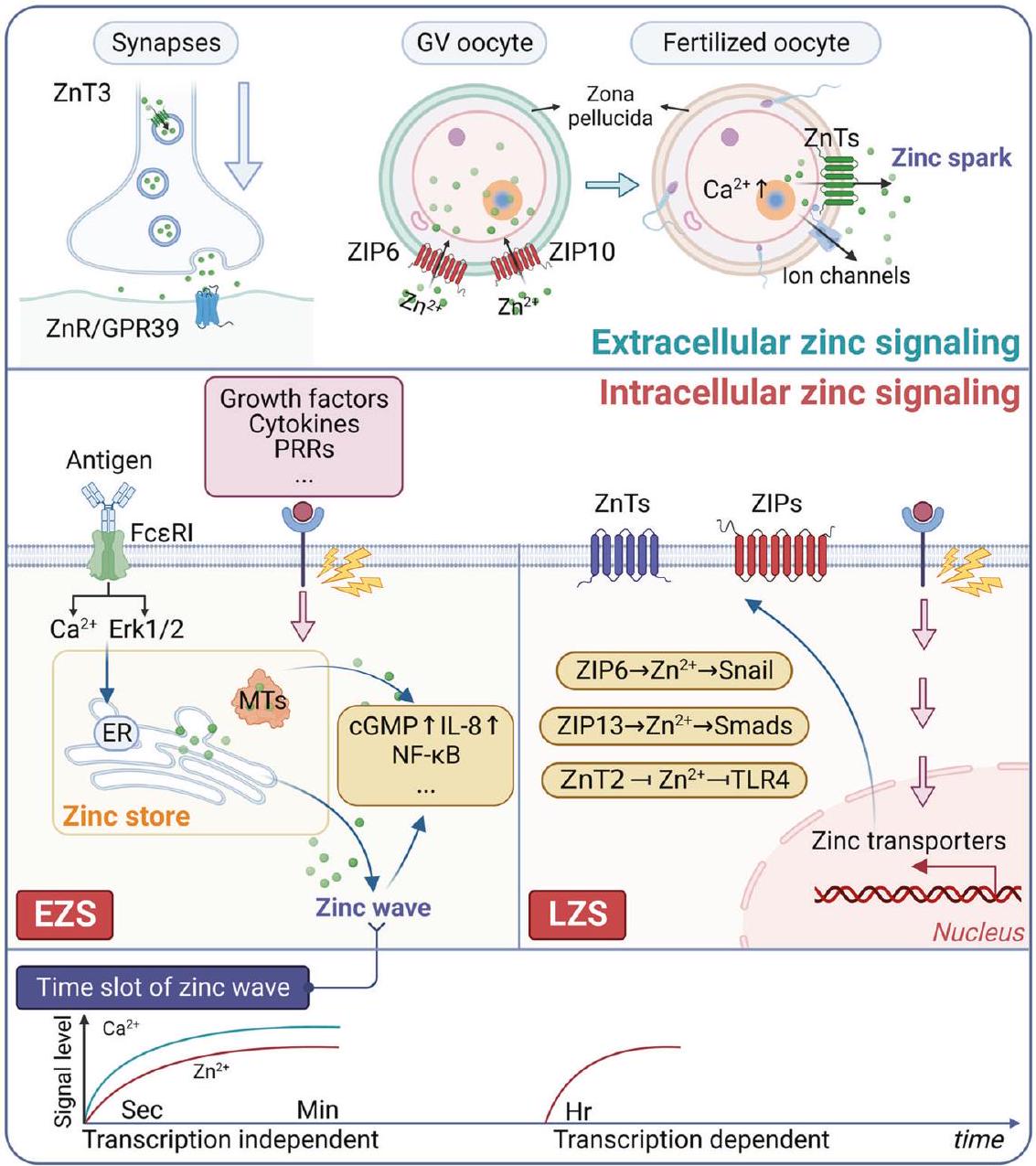
توزيع الزنك

الزنك منتشر في مختلف أنسجة الإنسان. يمتلك البالغون عادة محتوى من الزنك يتراوح بين 1.4 إلى تقريبًا يقع الزنك في العضلات وكذلك في العظام. بالإضافة إلى ذلك، حوالي الزنك موجود في الجلد والكبد. الباقي من الزنك كان
متناثرة في أنسجة أخرى. من الجدير بالذكر أن أعلى تركيز للزنك وُجد في شبكية العين والمشيمية. بالإضافة إلى ذلك، يوجد الزنك بكميات كبيرة في البروستاتا والعظام والكبد والكلى.
من الجدير بالذكر أن معظم الزنك يوجد داخل الخلايا. حوالي 30-40% من المحتوى يتواجد في نوى الخلايا، مع توزيع حوالي نصفه عبر السيتوسول، العضيات، والحويصلات المحددة، بينما يرتبط الزنك المتبقي بأغشية الخلايا. استنادًا إلى الأبحاث الحالية، يمكن تمييز إجمالي مخزون الزنك، الذي يشمل كلًا من الحيزات داخل الخلية وخارجها، إلى ثلاث فئات مميزة. أولاً، يشير مصطلح “الزنك غير المتحرك” إلى الزنك المرتبط بإحكام بالبروتينات المعدنية أو الإنزيمات المعدنية، ويعمل كمكون هيكلي أو كعامل مساعد. هذا الشكل من الزنك مستقر وغير تفاعلي. ثانياً، “الزنك المتحرك التفاعلي” أو “الزنك القابل للتبادل” مرتبط بشكل غير محكم بالروابط منخفضة الوزن الجزيئي وبالبروتينات المعدنية. هذا الشكل قابل للتبادل وتفاعلي. ومن الجدير بالذكر أن هذا الشكل المتحرك يشكل حوالي 5% من إجمالي الزنك داخل الخلايا، ويلعب دوراً محورياً في تفاعلات نقل الزنك وعمليات الإشارة. أخيرًا، يُعتبر تجمع “الزنك الحر” شكلاً تفاعليًا آخر من العنصر. في خلايا الثدييات والسوائل خارج الخلوية، مع ذلك، تكون تركيزات هذا الزنك منخفضة جدًا، حيث تتراوح القيم تقريبًا بين 5 بيكو مول و
تحافظ البروتينات المرتبطة بالميثيونين (MTs)، المعروفة عاميًا باسم “تخزين الزنك”، على مستويات الزنك الحر داخل الخلايا من خلال تفاعلها مع السيستين. بالإضافة إلى البروتينات المرتبطة بالميتالوثيونين (MTs)، يلعب أعضاء عائلة ناقلات الزنك، بما في ذلك ZIPs وZnTs، دورًا حيويًا في إدارة توازن الزنك. ومن اللافت أن نشاط نقل الزنك الخلوي لـ ZnT7 ضروري لتنظيم تموضع ERp44 داخل جهاز جولجي، وهو عضية خلوية محددة. من الجدير بالذكر أن العديد من الإنزيمات الإفرازية تحصل على الزنك الخلوي الأساسي في مجمع جولجي. علاوة على ذلك، كعامل مرافق جزيئي يعمل في مسار الإفراز المبكر، يمكن لـ ERp44 أن يرتبط بالزنك للتحكم في ارتباط البروتين وإطلاقه، وبالتالي إدارة نقل البروتين واستقراره.
في الآونة الأخيرة، جذب الدور الأساسي والمتعدد الأوجه للزنك كجزيء إشارة اهتمامًا كبيرًا. تنشأ إشارات الزنك من ثلاثة مصادر رئيسية: الإفراز الحويصلي، نقل الزنك الذي تسهله ناقلات الزنك لدخول أو خروج من الخلية أو العضية، وربط أو انفصال بروتينات الميتالوثيونين مع الزنك. سيتم توضيح هذه الجوانب في الأقسام التالية.

الإشارات الخلوية للزنك

تتراوح التركيزات الكلية للزنك في الخلايا من في حين أن تركيز الزنك الحر (القابل للتغير) في الخلايا حقيقية النواة يكون في نطاق البيكومولار، كما ذُكر سابقًا لكل نوع خلية محدد. من الجدير بالذكر أن السيتوبلازم يحتوي على كمية ضئيلة من الزنك الحر لأن الزنك داخل الخلية محجوز بشكل رئيسي في العضيات مثل الشبكة الإندوبلازمية، جهاز جولجي، والميتوكوندريا، والتي تُعرف بمخزن الزنك. تشير الأدلة المتزايدة إلى أن الزنك لا يعمل فقط كناقل عصبي للتواصل بين الخلايا، بل أيضًا كجزيء إشارة داخل الخلية، مما يسهل نقل العديد من سلاسل الإشارات استجابةً للمحفزات الخارجية. وقد أدى ذلك إلى مفهوم الزنك باعتباره “الكالسيوم لـ “القرن”.
كما ذُكر سابقًا، هناك مساران لإطلاق أيونات الزنك داخل الخلايا، وهما من مخازن الزنك داخل الخلايا أو من مواقع الزنك/الكبريتات في البروتينات، مثل في البروتينات المرتبطة بالميتالوثيونين (MTs). قد تنشأ الزيادات المؤقتة في الزنك من آليات مختلفة، بما في ذلك التدفق الخارج من الحويصلات المعروفة بالزنكوزومات. أو التغيرات في الجهد التأكسدي الخلوي التي تسهلها البروتينات السيتوسولية. من المهم أن نعرف أنه في معظم الحالات، ينشأ إشارات الزنك من اضطراب توازن الزنك داخل الخلايا، بشكل مؤقت وسريع. يلعب عمل ناقلات أيونات الزنك والبروتينات الغنية بالميثيونين (MTs) في الخلية دورًا في الحفاظ على توازن الزنك في السيتوبلازم، والذي يُشار إليه بـ “التخزين المؤقت” و”التخميد”، وهما معلمان أساسيان يحددان توفر الزنك وعمليات الإشارة الخاصة به. على وجه التحديد، ينطوي “التخزين المؤقت” على ارتباط الزنك بالبروتينات مثل MT،
الذي يساعد في الحفاظ على تركيز الزنك في نطاق البيكو مول في السيتوسول. تتميز بيولوجيا البروتينات الغنية بالميثيونين (MTs) بارتباطها بالزنك، وحركتها داخل الخلية، ونقل الزنك إلى مختلف الحويصلات الخلوية، بما في ذلك الحويصلات خارج الخلية، والحويصلات النهائية، والنواة، والميتوكندريا. سيسرع العامل المخلبي هذه العملية، ولكن إذا كان تعبير الجين مثل MT متورطًا، فإن عملية “التعويض” ستكون بطيئة. من ناحية أخرى، فإن “التخميد” مسؤول عن تعديل التغيرات العابرة في تركيزات الزنك تحت ظروف الخلايا غير المستقرة، مما يؤدي في النهاية إلى إعادة تركيزات السيتوسول إلى مستوياتها الراحة. في عملية “التخميد”، تقوم ناقلات الزنك بتنظيم الزنك الخلوي من خلال استيراده وتوزيعه وتصديره وتوفير الزنك للبروتينات المعتمدة على الزنك. على سبيل المثال، يحمل الزنك لإنزيمات مسار الإفراز، بينما يوفر الزنك للحويصلات الإفرازية. علاوة على ذلك، فإن MT مسؤول أيضًا عن كتم الزنك من خلال نقل الزنك وعزله في الحويصلات الخلوية، وبالتالي التحكم في تركيزات الأيونات بشكل حركي.
من حيث السلاسل الزمنية، يعمل الزنك داخل الخلايا كرسول ثانوي، وتنقسم تقلبات تركيزه إلى نوعين رئيسيين: الإشارة السريعة للزنك المبكرة (EZS) والإشارة المتأخرة للزنك. (الشكل 1). أكدت الدراسة أيضًا أن EZS مستقل عن النسخ، ويحدث على مدى زمني يتراوح من ثوانٍ إلى دقائق، ويُعرف باسم “موجة الزنك”. تمت ملاحظة هذه الظاهرة لأول مرة في خلايا البدينة وتنتج عن تحفيز مستقبل Fcɛ إبسيلون I (FceRI)، مما يسبب زيادة مؤقتة ومستقلة عن النسخ في الزنك داخل الخلية. موجة الزنك” تنشأ في المنطقة المحيطة بالنواة، بما في ذلك الشبكة الإندوبلازمية، وتعتمد على تدفق الكالسيوم وتنشيط MEK. ومع ذلك، لا يزال الآلية الدقيقة لـ “موجة الزنك” في الخلايا غير مفهومة جيدًا. في المقابل، يتطلب LZS نسخ بروتينات نقل الزنك وله تأثيرات تدوم لفترات أطول تمتد لساعات. في هذه الحالة، تحفز محفزات خارج الخلية متنوعة، بما في ذلك السيتوكينات وعوامل النمو، التعديل النسخي للبروتينات المرتبطة بالزنك مثل ZIPs وZnTs. ونتيجة لذلك، تنظم التغيرات في توازن الزنك داخل الخلية الأهداف الجزيئية التالية، بالإضافة إلى بروتين كيناز C (PKC)، وتنشيط ERK1/2 الذي يؤدي إلى موت الخلايا العصبية، وبروتين كيناز المعتمد على cAMP (PKA)، وبروتين كيناز II المعتمد على الكالسيوم/الكالمودولين (CaMKII)، والفوسفوديسترازات (PDEs)، وبروتينات الفسفاتاز التيروزينية (PTPs)، وعوامل النسخ مثل NF-кВ.
من الجدير بالذكر أن ارتفاع مستوى الزنك داخل الخلايا له تأثير ذو اتجاهين. فمن ناحية، يشارك الزنك في مسارات الإشارات الخلوية المختلفة، مما يساهم في عمليات مثل تكاثر الخلايا وتمايزها. على سبيل المثال، يعزز الزنك تطور الجهاز العصبي المركزي الجنيني من خلال التأثير على مسارات إشارات STAT1 وSTAT3. من المثير للاهتمام، أنه تم إثبات أن الزنك له دور أكثر أهمية في تكوين الدم مقارنة بالحديد، على الأقل في الخلايا الجذعية الدموية المبكرة. في الإشارات المتعلقة بوظيفة المناعة، يعزز الزنك تطور الخلايا التنظيمية الخلايا، كما يحفزها عامل النسخ Foxp3. من ناحية أخرى، يمكن أن يؤدي تراكم الزنك داخل الخلايا بشكل مفرط إلى الاستماتة. يمكن أن يؤدي تراكم الزنك المستمد من الميتوكوندريا إلى إضعاف بنية ووظيفة الميتوكوندريا، مما يؤثر سلبًا على تطور الحيوان وطول عمره في دودة الخيطيات Caenorhabditis elegans. أظهرت الدراسات أيضًا أن إطلاق الزنك داخل الخلايا قد يحدث استجابة للإجهاد التأكسدي أو النيتروزاتي، مما قد يؤدي إلى إطلاق الزنك من بروتين MT، وهو بروتين مخزن للزنك، وبالتالي تعزيز عمليات الموت المبرمج. علاوة على ذلك، في مسار موت خلوي محدد، يؤدي إطلاق الزنك والكالسيوم داخل الخلايا العصبية إلى الفسفرة اللاحقة لقناة البوتاسيوم Kv2.1. في الختام، على الرغم من انخفاض تركيزات الزنك الحر داخل الخلايا، يلعب إشارات الزنك داخل الخلايا دورًا واسعًا وحيويًا في الوظائف الفسيولوجية.

الإشارات الزنك خارج الخلية

الزنك خارج الخلية هو وسيط إشاري مهم في الأنظمة الصماء، والباراكرين، والأوتوكرين. يعمل كليجاند لـ
الشكل 1: إشارات الزنك في المناطق داخل الخلوية وخارج الخلوية. إشارات الزنك خارج الخلية تشارك بشكل رئيسي في الوظائف الفسيولوجية للمشابك العصبية والخلايا الجرثومية. بالمقابل، إشارات الزنك داخل الخلية تنقسم أساسًا إلى جزأين، EZS وLZS، اللذين يمارسان وظائف بيولوجية من خلال تنشيط المسارات التالية، مثل إشارات الالتهاب. ومن المثير للاهتمام أن الشبكة الإندوبلازمية تطلق الزنك لتوليد موجة زنك محددة، تُلاحظ خلال عدة دقائق بعد تحفيز FceRI في خلايا البدينة. EZS: إشارات الزنك المبكرة، LZS: إشارات الزنك المتأخرة. النقاط الخضراء تمثل الزنك.
قنوات مستقبلات مختلفة على غشاء البلازما، بما في ذلك مستقبل استشعار الزنك (ZnR/GPR39) الذي ينظم الإثارة العصبية، مستقبلات -ميثيل-دي-أسبارتات (NMDA) مستقبلات حمض الألفا-أمينو-3-هيدروكسي-5-ميثيل-4-إيزوكسازولبروبيونيك (AMPA) قنوات الكالسيوم المعتمدة على الجهد (VDCC) و – مستقبلات حمض الغاما-أمينوبيوتيريك A (GABAA). لقد أكدت التقدمات في علم الأحياء الخلوي والكيمياء على وجود ووظيفة الزنك الحر أو المتغير في الاستجابات الخلوية، وخاصة دوره كناقل عصبي في الحويصلات المشبكية. تشير التقلبات في تركيزات الزنك في الدماغ، التي تتوافق مع التجارب الفسيولوجية والذكريات طويلة الأمد، إلى أن الزنك الحر مرتبط ارتباطًا قويًا بأداء الناقلات العصبية. علاوة على ذلك، يقوم الزنك المنطلق من المشبك بتنشيط مستقبل مرتبط بالبروتين G مباشرة (mZnR/GPR39)، الذي يستشعر التغيرات في تركيز الزنك خارج الخلية وبالتالي ينظم الإثارة العصبية.
بالإضافة إلى ذلك، تطلق الأجنة الثديية المخصبة شرارات الزنك. تنسق أيونات الزنك التي تُفرز عبر الإخراج الخلوي مع التغيرات العابرة في الكالسيوم الخلوي، مما يغير بنية المنطقة الشفافة لمنع تعدد الإخصاب (الشكل 1).

الإشارات الزنك وتكوّن الأورام

في الظروف العادية، تلبية تركيز الزنك لمتطلبات العمليات الحيوية للطاقة، والتركيب، والتحلل، ضرورية لـ
تجسيد الأنشطة الحالية للخلايا، مثل الوظيفة، والنمو، والتكاثر. تفسر عدة آليات الوظيفة المضادة للأورام للزنك، وتشمل تلف الحمض النووي، وإصلاح الحمض النووي، ووظيفة الجهاز المناعي، والإجهاد التأكسدي، والالتهاب. مع تغير نشاط الخلية، يجب تعديل أيضها لتلبية أي متطلبات جديدة للطاقة البيولوجية/التركيبية/التحللية. يمكن أن تؤدي التغيرات في تركيزات الزنك التي تتجاوز قدرة الخلية على التنسيق إلى تكوين الأورام، حيث يوفر الزنك المتطلبات البيولوجية الطاقية/التركيبية للورم الخبيث، مثل التعبير الشاذ عن ناقلات الزنك واضطراب تنظيم بروتينات ربط MTs.
في الواقع، تنشيط الزنك لمسارين من مسارات كيناز البروتين المنشط بالميتوجين (MAPK) المرتبطة بتكوين الأورام، وهما كيناز الإشارة خارج الخلية المرتبط (ERK) وكيناز N-نهائي لـ c-Jun (JNK)، يلعب دورًا هامًا. هذه البروتينات كينازات سيرين/ثريونين المعروفة باسم MAPKs، بما في ذلك ERK وJNK، تنظم تكاثر الخلايا، التمايز، والموت المبرمج في تكون الأورام. فيما يتعلق بإشارات الزنك المتأخرة، يقوم STAT3 بتحفيز النشاط النسخي لـ ZIP6 في سمك الزرد. نتيجة لذلك، يؤدي التعبير المعتمد على STAT3 لبروتين ZIP6 إلى تنشيط لاحق لمثبط النسخ Snail، الذي يساهم في الانتقال الظهاري-اللحمي (EMT) خلال التطور الجنيني ويرتبط بآليات انتشار الأورام (الشكل 1). وبالمثل، يحفز ZIP4 الهجرة والغزو التي تعزز EMT من خلال إشارة PI3K/Akt.
المسار في سرطان البلعوم الأنفي (NPC). بالإضافة إلى ذلك، يؤدي التعبير المرتفع لبروتين ZIP13 إلى تنشيط مسار Src/FAK، مما يؤدي إلى زيادة تعبير الجينات المعززة لانتشار الأورام وتقليل تعبير جينات مثبطات الأورام في سرطان المبيض. بشكل عام، يبدو أن خلايا السرطان تحتاج إلى تحفيز مسارات الأورام بواسطة الزنك للحفاظ على عدوانيتها.
من الواضح أن إشارات الزنك الخلوية تستفيد من تخزين وإطلاق العضيات والهياكل تحت الخلوية، والتي يتم تنظيمها بدقة بواسطة ناقلات الزنك والبروتينات المرتبطة بالميتالوثيونين. لذلك، يتطلب الحفاظ على توازن الزنك تعاونًا خلويًا معقدًا بين هذه البروتينات الوظيفية. من الناحية النظرية، هل يمكن أن تتحول الخلايا الطبيعية إلى خلايا سرطانية إذا تم تعطيل توازن الزنك؟ لقد أكدت العديد من الدراسات أن اضطراب تنظيم بروتينات ناقلات الزنك لا يؤثر فقط على تكاثر الخلايا وموتها المبرمج، بل يسبب أيضًا تغييرات في مسارات الإشارات المختلفة، مما يعزز تقدم السرطان. من اللافت أن قناة الكاتيون الليزوزومية MCOLN1 تم تحديدها كوسيط حاسم في تدفق الزنك إلى السيتوبلازم، مما يتيح التحكم الدقيق في الالتهام الذاتي المسبب للسرطان في الخلايا السرطانية. بالإضافة إلى ذلك، أظهرت التغيرات في توازن الزنك قدرتها على تعديل البيئة المناعية للورم، مما يمارس تأثيرًا كبيرًا على تطور السرطان. علاوة على ذلك، فإن تورط الزنك في إزالة سموم المعادن الثقيلة يشير إلى أن تعطله قد يؤثر سلبًا على مسارات إزالة السموم، مما يؤدي إلى إجهاد خلوي وتطور السرطان لاحقًا. في الختام، الرابط المعقد بين توازن الزنك والسرطان هو مجال ناشئ يستدعي المزيد من البحث لفهم الآليات الأساسية التي تحكم الانتقال من اضطراب توازن الزنك إلى تكوّن الأورام بشكل كامل.

تنظيم أيض الزنك الخلوي

المعرفة الأساسية بناقلات الزنك
مُجمّعات ZIP. تتألف عائلة SLC39 من أربع مجموعات متميزة بناءً على تشابه تسلسل الأحماض الأمينية: العائلة الفرعية الأولى (ZIP9)؛ العائلة الفرعية الثانية (ZIP1، 2، و3)؛ العائلة الفرعية LIV-1 (ZIP4، 5، 6، 7، 8، 10، 12، 13، و14)؛ والعائلة الفرعية gufA التي تحتوي على ZIP11. جميع بروتينات ZIP تحتوي على ثمانية مجالات عابرة للغشاء (TM) مع بقايا هيستيدين محفوظة داخل TM 4 و 5، يُعتقد أنها تشارك في نقل الزنك. تقع النهايات الطرفية C والطرفية N لبروتين ZIP إما على سطح الخلية أو داخل تجويف العضية. من المتوقع أن تمتلك أعضاء عائلة LIV-I، باستثناء ZIP13، مجالًا مهمًا واحدًا في الطرف الأميني خارج الخلية، يُعتقد أنه يعمل كمستشعرات للزنك خارج الخلية. مؤخرًا، قدمت الأبحاث رؤى حول الهيكل التفصيلي لناقلات ZIP، بما في ذلك دقة عالية Åهيكل مجهر إلكتروني بالتبريد لناقل من عائلة ZIP من بوردتيلا برونكيسبتكا تم الحصول عليه في حالة مواجهة للداخل ومثبطة. يتألف كل بروتومر من هذا الناقل المتماثل من تسعة حلزونات عابرة للغشاء وثلاثة أيونات معدنية. في هذا الهيكل، يشكل أيونان معدنيان معًا بنية مسام ثنائية النواة، بينما يقع الأيون الثالث في موقع خروج يواجه السيتوبلازم. ومن الجدير بالذكر أن موقع الخروج هذا مغطى بحلقة، حيث تتفاعل بقايا الهيستيدين الموجودة على هذه الحلقة مع أيون موقع الخروج، مما ينظم إطلاقه بشكل حاسم. قد يوفر فهم بنية ووظيفة ناقلات ZIP رؤى قيمة لتطوير استراتيجيات علاجية جديدة تستهدف ناقلات الزنك لعلاج مختلف الأمراض البشرية.
عادةً ما يتم تخليق بروتينات ZIP على الريبوسومات المرتبطة بالشبكة الإندوبلازمية (ER) ثم تُنقل لاحقًا إلى مختلف الحُجرات داخل الخلية. مماثل لتعبيرات البروتين الأخرى، غالبًا ما يتم التعرف على بروتينات الطفرات غير المستقرة من نوع ZIP في الشبكة الإندوبلازمية. بعد ذلك، تخضع للانتقال العكسي والتحلل بواسطة البروتيازومات السيتوسولية بطريقة مستقلة عن اليوبكويتين، كما هو الحال في طفرة ZIP13. بصرف النظر عن التوضع داخل الخلايا لبعض أعضاء عائلة ZIP، فإن الغالبية العظمى من ناقلات ZIP تقع على غشاء البلازما، مما يسهل
امتصاص أيونات المعادن إلى داخل الخلايا. يقع ZIP7 في جهاز جولجي والشبكة الإندوبلازمية، بينما ZIP13، الأقرب تطوريًا إلى ZIP7، يتموضع في جهاز جولجي والحويصلات السيتوبلازمية. ZIP13 مسؤول عن تحريك الزنك من تجويف هذه الحويصلات ويلعب أدوارًا حاسمة في الإشارات الخلوية، بما في ذلك BMP/TGF- مسار الإشارة، من خلال تنظيم انتقال بروتينات Smad إلى النواة والحفاظ على توازن الشبكة الإندوبلازمية.
تتأثر مستويات التعبير للعديد من بروتينات ZIP، مثل ZIP1 و3 و4 و8 و12، على سطح الخلية بتركيزات الزنك المتاحة. يعمل ZIP10 كناقل للزنك على سطح الخلية. يتم تنظيم نسخ ZIP10 بشكل مرتفع في الخلايا التي تعاني من نقص الزنك وتنخفض في ظروف فائض الزنك. يتم تنظيم نسخ الزنك من خلال إيقاف نسخ بوليميراز II بواسطة عامل النسخ المرتبط بعنصر استجابة المعادن 1 (MTF-1). علاوة على ذلك، يختلف تموضع بعض بروتينات ZIP حسب توفر الزنك والحالات الفسيولوجية المحددة. أثناء تناول كمية كافية من الزنك، يتموضع Zip5 عند الغشاء البلازمي القاعدي الجانبي في الخلايا المستقطبة. بنفس الطريقة، ينتقل بروتين ZIP14 إلى الغشاء الجيبي لخلايا الكبد في الفئران خلال الأحداث الالتهابية الحادة. نتيجة لذلك، يعزز هذا امتصاص الزنك كجزء من الاستجابة الفورية للالتهاب.
بينما يُعرف أعضاء عائلة ZIP بدورهم الأساسي في نقل الزنك، يمكنهم أيضًا تحريك معادن أخرى مثل المنغنيز والكادميوم. أظهرت الدراسات الكيميائية الحيوية أن بروتين ZIP8، على وجه الخصوص، يمكنه نقل الكادميوم والمنغنيز. يتم تنظيم تعبير mRNA الخاص بـ ZIP8 بشكل زائد بواسطة الكادميوم بطريقة تعتمد على NF-kB، مما يساهم في خطر السمية الرئوية الناتجة عن الكادميوم عند التعرض لدخان السجائر. ZIP14 مرتبط تطوريًا ارتباطًا وثيقًا بـ ZIP8. مماثل لـ ZIP8، يمتلك ZIP14 القدرة على تحريك عدة أيونات ثنائية التكافؤ، بما في ذلك الكادميوم والمنغنيز. علاوة على ذلك، فإن ZIP14 وZIP8 قادران على نقل الحديد. يلعب ZIP14 دورًا حيويًا كناقل للحديد في الجسم الحي، خاصة في حالات زيادة الحديد. تُعتبر قدرة ZIP14 على نقل الحديد غير المرتبط بالترانسفيرين (NTBI) مساهمة حيوية في توازن الحديد. من المثير للاهتمام أن ZIP14 يمتلك نوعين من المتغيرات المشطوبة: ZIP14A و ZIP14B. تتواجد هذه المتغيرات على غشاء البلازما وتشارك في امتصاص الزنك. في الخلايا المستقطبة، يقع ZIP14A و ZIP14B حصريًا على السطح القمي.
ZnTs. تنتمي عائلة ZnT إلى عائلة ميسري انتشار الكاتيونات (CDF) من البروتينات. تقع معظم ZnTs داخل أغشية العضيات، وتؤدي وظائف مختلفة، مثل ملء مخازن الزنك الحويصلية، وتزويد العضيات بالزنك، وتحميل الحويصلات الإفرازية بالزنك للعمليات البيولوجية الأساسية. يُستدل على بنية بروتينات ZnT من نظائرها في الإشريكية القولونية YiiP. التي تحتوي على ستة حليلات عبر الغشاء (الحليلات عبر الغشاء I-VI) ونهاياتها N و C تقع على الجانب السيتوبلازمي. ، من ناحية أخرى، يمتلك منطقة N-نهائية طويلة بشكل غير عادي تحتوي على تسعة مجالات محتملة عبر الغشاء. من المتوقع أيضًا أن تحتوي الناقلات على موقع ارتباط بالزنك محفوظ على الحلزونين عبر الغشائي الثاني والخامس، مع بقايا حاسمة تحدد تخصصها المعدني. من اللافت أن ZnT 10 يظهر الخصائص الجزيئية لناقل المنغنيز، ويرجع ذلك على الأرجح إلى احتوائه على بقايا Asn بدلاً من His في اللولب العابر للغشاء الثاني. علاوة على ذلك، فإن طول وتسلسل الأحماض الأمينية للمجال الهيكلي الأولي عبر الغشاء (TM) في بروتينات ZnT، المعروف باحتوائه على إشارات استهداف داخل الخلية، يظهر تباينات كبيرة بين بروتينات ZnT المختلفة. بناءً على تشابهات تسلسل البروتين، يمكن تصنيف أعضاء عائلة ZnT إلى أربع مجموعات: (1) ZnT 6 و ZnT 9، (2) ZnT 1 و ZnT 10، (3) و ، و(4) ZnT 5 وZnT 7. ومن المثير للاهتمام أن الأعضاء الذين ينتمون إلى نفس العائلة الفرعية يظهرون مواقع خلوية وخصائص وظيفية متشابهة (الشكل 2).
وظيفيًا، في عائلة SLC30، يعمل ZnT1 بشكل أساسي كناقل للزنك على غشاء الخلية، حيث ينقل أيونات الزنك السيتوبلازمية عبر الغشاء إلى الفضاء خارج الخلية، بينما تقوم البروتينات الأخرى…
تقع بروتينات ZnT على أغشية العضيات داخل الخلوية. بالإضافة إلى ذلك، يشارك ZIP10 وZnT1 في إعادة امتصاص الزنك في الكلى. أعضاء الفصيلة الفرعية الثانية من بروتينات SLC30 ( ، وZnT 8) تلعب دورًا رئيسيًا في الأنسجة الإفرازية، حيث يشارك ZnT 3 في النقل العصبي، وZnT 8
في تخزين الأنسولين، ZnT 4 في إفراز البروستاتا، وZnT 2 في الرضاعة. بالإضافة إلى ذلك، TMEM163 هو ناقل زنك تم اكتشافه حديثًا وله بنية مجال عابر للغشاء متوقعة ووظيفة مشابهة لعائلة البروتينات الفائقة CDF. يفترض البعض أن TMEM163 قد يكون جديدًا
الشكل 2 بنية البروتين وتطور عائلة الجينات لناقلات الزنك. أ رسم كرتوني للهياكل المتوقعة لبروتينات ناقل ZnT وZIP. الصورة على اليسار تظهر نموذجًا ذريًا لـ ZnT، وهو إعادة بناء لولبية لـ YiiP بناءً على بنية الأشعة السينية (رمز معرف PDB: ). بالتفصيل، يُقترح الطوبولوجيا التخطيطية لناقلات ZnT استنادًا إلى البنية ثلاثية الأبعاد لنظيرها في الإشريكية القولونية YiiP. من المرجح أن تحتوي ناقلات ZnT على ستة مجالات عبر الغشاء مقسمة إلى حزمتين. على وجه التحديد، تحتوي إحدى حزم ZnT على أربعة مجالات عبر الغشاء (MI، MII، MIV، وMV)، بينما تتألف الحزمة الأخرى من مجالين عبر الغشاء (MIII وMVI). يمكن لكل مجال من مجالات الحزمة الأولى أن يرتبط بالزنك بشكل مستقل، منسق رباعيًا بواسطة اثنين من D (الأسبارتات) واثنين من H (الهيستيدين) في النظائر الثديية. وبالمثل، تعرض الصورة على اليمين المجالات المحتملة عبر الغشاء لعائلة ZIP (رمز معرف PDB: 7z6m). علاوة على ذلك، تُعرض بنية الطوبولوجيا لعائلة ZIP، المكونة من 8 مجالات عبر الغشاء مع مجال كبير في الطرف N ومجال صغير في الطرف C. يُظهر التوزيع المكاني أنها تتألف من ثلاثة أجزاء، حيث يحتوي الجزء الأيسر والجزء الأيمن كل منهما على ثلاثة مجالات عبر الغشاء (باللون الأحمر)، وفي الوسط يوجد مجالان عبر الغشاء (باللون الأزرق). يمكن للزنك أن يرتبط بالموقع النشط للمجال عبر الغشاء الرابع والخامس، اللذين يحتويان على أنماط محفوظة HND (هيستيدين، أسباراجين، أسبارتات) وHEH (هيستيدينان وحمض الجلوتاميك واحد) على التوالي. ب شجرة تطور عائلة الجينات والنمط المتغاير لناقلات ZnTs وZIPs. تم وضع أطوال الأنماط المتغايرة المختلفة أمام كل نمط، وتشير الخطوط الملونة إلى مواقع المجالات الوظيفية لكل نمط. تنتمي ناقلات ZnTs إلى عائلة ميسري انتشار كاتيونات الزنك (CDF)، المسؤولة عن نقل الزنك من داخل الخلية إلى خارجها. وتنقسم ناقلات ZIP إلى أربع عائلات فرعية، وهي العائلة الفرعية ZIP I (ZIP9)، العائلة الفرعية GufA (ZIP11)، العائلة الفرعية ZIP II (ZIP1-3)، والعائلة الفرعية LIV-1 (ZIP4-8، ZIP10، ZIP12-14).
عضو في بروتينات ناقل الزنك ZnT في الثدييات. تشير الاكتشافات الحديثة إلى دوره الهام في الحفاظ على توازن الزنك في كل من الأعصاب والدم.
المعرفة الأساسية بأنظمة الترجمة الآلية
المتالثيونينات الثديية هي عائلة فائقة من الببتيدات غير الإنزيمية التي تتكون عادة من 61-68 حمضًا أمينيًا. تتميز بارتفاع محتواها من السيستين، حيث تشكل حوالي 30% من أحماضها الأمينية، في حين أن الأحماض الأمينية العطرية غائبة، وبقايا الهيستيدين موجودة بشكل نادر. ومع ذلك، تحتوي على مجموعات ثيول وفيرة تمكنها من الارتباط بالمعادن الثقيلة. تمتلك البروتينات المعدنية (MTs)، بمجموعاتها الوفيرة من الثيول، القدرة على الارتباط بما يصل إلى 7 ذرات زنك: 3 ذرات زنك في الـ النطاق و4 ذرات زنك في النطاق أ. تمكن هذه القدرة الفريدة البروتينات المرتبطة بالميثيونين من العمل كاحتياطي خلوي للزنك. من المهم التأكيد على أنه بينما يمكن للبروتينات المرتبطة بالميثيونين أن ترتبط بمعادن أساسية أخرى مثل النحاس ومعادن غير أساسية مثل الكادميوم، فإن الشكل السائد في الأنسجة البشرية هو البروتين المرتبط بالزنك.
يمكن تصنيف بروتينات الميتالوثيونين البشرية إلى أربع فئات، وهي MT1 إلى MT4، وتشمل ما مجموعه أحد عشر شكلًا وظيفيًا، ينتمي منها ثمانية إلى الفئة 1. MT1 و MT2 هما الأشكال المتغايرة السائدة الموزعة في جميع أنحاء الجسم البشري والمعبر عنها في مختلف الأعضاء. وعلى العكس من ذلك، يوجد MT3 بشكل رئيسي في الجهاز العصبي المركزي، بينما يوجد MT4 بشكل أساسي في الجلد والطبقة الطلائية المتعددة الطبقات الأخرى، مما يمثل الأشكال المتغايرة الثانوية. جميع الأشكال المتغايرة لها وزن جزيئي تقريبي يبلغ 7 كيلو دالتون وتفتقر إلى الأحماض الأمينية العطرية. علاوة على ذلك، تتكون من عشرين بقايا سيستين، مما يمنح البروتينات المعدنية (MTs) خصائص مميزة بسبب خصائص مجموعات الثيول. بالإضافة إلى ذلك، يتم تنظيم نسخ جينات MT1/2 بواسطة MTF-1، وهو عامل نسخ يحتوي على إصبع زنك ينظم تعبير الجينات المستجيبة للمعادن. الزنك هو المعدن الوحيد المعروف الذي ينشط MTF-1؛ ومع ذلك، تقترح الدراسات أن الإجهاد التأكسدي قد يساهم أيضًا في تنشيط MTF-1. MTF-1 يشارك في تنظيم النسخ المستجيب للزنك لكل من ZnT 1 و ZnT 2 ويثبط التعبير عن ZIP10، مؤكداً على دوره الحيوي في توازن الزنك.
في البشر، يتم ترميز MTs هيكليًا بواسطة عائلة من الجينات الموجودة على الكروموسوم ، والتي تتألف من 11 عضوًا وظيفيًا على الأقل: تتكون جينات MT1 من 18 شكلًا مغايرًا، بما في ذلك 10 جينات وظيفية (MT1A، MT1B، MT1E، MT1F، MT1G، MT1H، MT1M، وMT1X) و8 جينات زائفة (MT1CP، MT1DP، MT1JP، MT1L، MT1LP، MT1XP1، MT1P3، وMT1P1)، بالإضافة إلى MT2 (المعروف أيضًا باسم MT2A)، MT3، وMT4 (الشكل 3). من اللافت للنظر، أن مخزون الزنك، MT، يمكن أن يعمل كلاً من مستقبل للزنك ومانح للزنك، مثل وجهي عملة واحدة.

دور أيض الزنك الخلوي تحت الظروف الفسيولوجية

الدور الفسيولوجي لناقلات الزنك
دعم وظيفة الجهاز المناعي. تعتبر الخلايا التائية مكونًا حيويًا في الجهاز المناعي. من بين أعضاء عائلة ZIP الأربعة عشر، يتم التعبير عن ZIP6 و8 و13 بشكل عالي في الإنسان الخلايا، مع ZIP6
متمركزة بشكل رئيسي في طوافات الدهون المشاركة في تكوين المشبك المناعي (IS) بعد تحفيز مستقبلات الخلايا التائية (TCR). من الجدير بالذكر أن الفسفرة على التيروسين لبروتين ZIP6 لوحظت تزداد بعد خمس دقائق من تحفيز مستقبلات الخلايا التائية (TCR) بسبب تفاعله مع Zap70، وهو كيناز حيوي يشارك في الإشارات المبكرة لمستقبلات الخلايا التائية. بالإضافة إلى ذلك، تؤدي النشاط النسخي لـ ZIP6 إلى تدفق الزنك، مما يعزز تعبير بروتينات المعادن (MTs)، التي تلعب دورًا حيويًا في الدعم تكاثر الخلايا وهو ضروري لـ بقاء الخلايا وتكاثرها لدى كبار السن. يتم التعبير عن ZIP8 و ZIP13 بشكل أساسي على غشاء الليسوسوم وغشاء الشبكة الإندوبلازمية/جولجي في خلايا T، على التوالي. أثناء تنشيط الخلايا التائية، يسهل بروتين ZIP8 نقل الزنك من الليسوسوم إلى السيتوبلازم، مما يؤدي إلى زيادة إنتاج IFN- من الجدير بالذكر أن تعبير ZIP8 يمكن تحفيزه استجابة لتحفيز الليبوبوليساكاريد (LPS)، مما يؤدي إلى تعزيز الإنتاج في المراحل اللاحقة لمسار mTORC1S6K. علاوة على ذلك، يُعتبر ZIP8 جينًا مستهدفًا في المسار السفلي لـ NF-KB، الذي ينظم الاستجابات الالتهابية السلبية من خلال تقليل نشاط Iкк بواسطة الزنك. بالمقارنة، لنقص ZIP8 تأثير كبير على تدفق الزنك في خلايا T الفعالة وينتج عنه تقليل في الإشارات التي يتم تحفيزها بواسطة مستقبلات TCR، بما في ذلك إشارات NF-кВ وMAPK، والتي تشارك في تمايز خلايا T المساعدة (Th)17. وبالمثل، الفئران التي تفتقر إلى ZIP3 تظهر انخفاضًا الخلايا الثيموسية المزدوجة الإيجابية (DP) ولكن زادت و الخلايا التيموسية الإيجابية المفردة، مما يشير إلى دوره في التنظيم تطور الخلايا. تفتح هذه النتائج آفاقًا جديدة للعلاج المناعي لتحسين التشخيص من خلال تعديل جينات عائلة ناقلات الزنك في الأورام أو الخلايا المناعية.
لا شك أن الفرع التكيفي للجهاز المناعي يعتمد على كل من الخلايا البائية والخلايا التائية. يلعب ZIP9 و ZIP10 أدوارًا أساسية في مسارات إشارات مستقبلات الخلايا، مؤثرة على تنشيط الخلية (الشكل 4). ينبع إطلاق الزنك في خلايا B من جهاز جولجي، حيث يلعب ZIP9 دورًا حيويًا كمشارك في نقل الزنك. من ناحية أخرى، يلعب ZIP10 أدوارًا مختلفة في المراحل المبكرة والمتأخرة من تطور الخلايا البائية، حيث ينظم مسارات إشارة مميزة. يتم تنظيم تعبير ZIP10 ميكانيكيًا بطريقة تعتمد على STAT3/STAT5، مما يعزز بقاء الخلايا البائية المبكرة عن طريق تثبيط تنشيط الكاسبيز. بالإضافة إلى ذلك، ثبت أن نقص ZIP10 في خلايا B الناضجة يخفف من كلا الأمرين الاستجابات المناعية المعتمدة على الخلايا وغير المعتمدة عليها في الجسم الحي. تعمل ZIP10 كمنظم إيجابي لـ CD45R في نقل إشارة مستقبلات المستضدات في خلايا B، مما يلعب دورًا حاسمًا في تحديد عتبة الاستجابات المناعية البشرية. في خطوط خلايا سرطان الكبدة الكبدية (HCC)، وُجد أن تعبير ZIP10 مرتبط إيجابيًا بالخلايا اللمفاوية المتسللة إلى الورم وبعض نقاط التفتيش المناعية، بما في ذلك CTLA4 وTIM3 وTGFB1. علاوة على ذلك، يعتبر ZIP10 ضروريًا للحفاظ على توازن الزنك داخل الخلايا البلعمية، حيث يشارك الزنك في الاستجابات المضادة للميكروبات. البالعات المُنشطة، على الرغم من أهميتها في الاستجابات المناعية، يمكنها أيضًا إفراز كميات كبيرة من السيتوكينات الالتهابية، والتي قد يكون لها القدرة على إلحاق الضرر بالمضيف. تم تحديد ZIP10 كناقل مهم للزنك في الخلايا البلعمية يقوم بتنشيط الخلايا البلعمية ويعزز تعبير السيتوكينات.
الشكل 3 بنية البروتين وتطور عائلة الجينات لـ MTs. أ رسم تخطيطي للبنية المتوقعة لبروتين MT2، والذي تم نمذجته من البنية المعاد بناؤها بالأشعة السينية (رمز معرف PDB: 4 mt 2). تم تحديد البنية البلورية لمعدن الثيونين في كبد الجرذ بدقة عند دقة آ، محققًا قيمة R منخفضة تبلغ 0.176 لجميع البيانات الملاحظة. تمثيل تخطيطي لارتباط الزنك في البروتينات المعدنية (MTs). تحتوي البروتينات المعدنية على مجموعات ثيول وفيرة قادرة على الارتباط بالمعادن الثقيلة. بسبب المحتوى العالي من الثيول، يمكن للبروتينات المعدنية أن ترتبط بما يصل إلى 7 ذرات زنك، مع وجود 3 ذرات زنك في النطاق و4 ذرات زنك في المجال. ج شجرة تطور عائلة الجينات وأشكال MTs الموضحة في الشكل X. تم وضع أطوال الأشكال أمام كل شكل، وتشير الخطوط الملونة إلى موقع المجال الوظيفي لكل شكل. تُصنف MTs إلى أربع عائلات فرعية: MT1 (بما في ذلك MT1A، MT1B، MT1E، MT1F، MT1G، MT1H، MT1M، وMT1X)، MT2 (بما في ذلك MT2A)، MT3، وMT4. بينما يتم التعبير عن MT1 وMT2 بشكل عام، يُعبر عن MT3 بشكل رئيسي في الجهاز العصبي المركزي، ويُعبر عن MT4 بشكل أساسي في الجلد وأنسجة الظهارة الطبقية الأخرى.
نقص الزنك (ZD) الناجم عن تثبيط ZIP10 يؤدي إلى تراكم p53 في السيتوبلازم وانتقال AIF إلى النواة، مما يؤدي في النهاية إلى تحفيز الاستماتة. لذا، قد يكون استهداف ZIP10 نهجًا واعدًا لحماية الكبد من أضرار الالتهاب.
من الجدير بالذكر أن الإنتان هو عدوى جهازية حادة تنجم عن غزو البكتيريا الممرضة للدورة الدموية وإنتاج السموم. تم ربط مستويات الزنك الدائرية الأقل من المتوقع بارتفاع معدل الوفيات لدى مرضى الإنتان، حيث تم تحديد MT وZIP8 كاثنين من أكثر الجينات التي تم تنظيمها بشكل مفرط في غير الناجين. لقد تبين أن ناقل ZIP8، على وجه الخصوص، هو الأكثر ارتفاعًا في التعبير استجابة للسيتوكينات والبكتيريا وتعفن الدم، مما يشير إلى دوره الفريد في وظيفة المناعة الفطرية. باعتباره أقرب نظير لـ ZIP8، يشارك ZIP14 أيضًا في الاستجابة للإنتان ويرتبط بالتأثيرات المضادة للالتهابات المفيدة لمكملات الزنك الغذائية أثناء الإنتان، مما يشير إلى إمكانيته كهدف علاجي. بالإضافة إلى ذلك، وجود “موجات الزنك” في خلايا البدينة يوفر دليلاً إضافياً على تورط ناقلات الزنك في الوظائف المناعية. من المحتمل حدوث إطلاق الزنك من الشبكة الإندوبلازمية
يتم التوسط بواسطة ZIP7، حيث أن ZIP7 يتواجد بشكل رئيسي في الشبكة الإندوبلازمية، ومنع إسكات ZIP باستخدام siRNA حدوث موجة الزنك. بالإضافة إلى ذلك، تساهم قنوات الكالسيوم ذات الجهد النوعي من النوع L (LTCCs) أيضًا في موجة الزنك، التي تتفاعل مع ZnT1 وتعدل تدفق الزنك من الفضاء خارج الخلوي إلى السيتوبلازم.
مساعدة في التكاثر. خلال النضج الانقسامي الاختزالي، زاد إجمالي الزنك داخل الخلايا بحوالي 50%. بعد الإخصاب، قامت البويضات الغنية بالزنك بإحداث شرارات الزنك، التي خفضت تركيز الزنك بحوالي يتطلب دور شرارات الزنك مزيدًا من التحقيق، لكن بعض الأدلة تشير إلى أن هذه التغيرات في مستويات الزنك ضرورية للخطوات التطورية اللاحقة وقد تلعب دورًا في العمليات المعتمدة على الزنك التي تنظم خروج البويضة من الانقسام الاختزالي الأول. يُعتقد أن عائلة ناقلات ZIP تنظم تدفق الزنك، وZIP6 وZIP10، اللذان يشتركان في… تطابق التسلسل ويقعان في نفس الفرع لشجرة النشوء والتطور لعائلة ZIP، تُعبَّر عنها بشكل عالي في البويضة خلال نافذة النضج الانقسامي (الشكل 4). يُعد المركب المختلط ZIP6/ZIP10 أيضًا حاسمًا في تحفيز الانقسام الخلوي الذي يتم بواسطة الزنك،
الشكل 4 الوظائف الفسيولوجية الرئيسية لناقلات الزنك. تُصنف وظائف ناقلات الزنك أساسًا إلى ستة أجزاء: المناعة، التكاثر، العضلات، وظيفة الأمعاء، استقلاب الجليكوليبيد، ووظيفة العصبون. علاوة على ذلك، تُقسم الوظيفة التي يمثلها كل قطاع بشكل رئيسي إلى ثلاثة أجزاء. كل جزء يتوافق مع ناقلات زنك محددة. الدائرة الخارجية تمثل الأمراض والسرطانات الناجمة عن خلل في ناقلات الزنك. متلازمة إهلرز-دانلوس الفقرية-اليدوية-التشوهية SCD-EDS، التهاب الجلد الحاد المعوي AE، مرض التهاب الأمعاء IBD.
تشكيل مركب انقسامي معتمد على الزنك يتكون من ZIP6 وZIP10، STAT3، و ستاثمين، الذي يلعب أدوارًا في مسارات الانقسام الخلوي المثبتة. كمثال، يشارك في عمليات مثل إعادة تنظيم الأنابيب الدقيقة المعتمدة على ستاثمين أو تكثيف الكروماتين الذي يتم بوساطة هيستون H3. من أجل الاستقرار يعمل ستاثمين طوال الانقسام الخيطي، وSTAT3 كعامل مؤثر لمركب ZIP6/ZIP10 المختلط الذي ينظم تعبير كلا الجينين. غالبًا ما تكون مستويات الزنك أعلى في أنسجة السرطان مقارنة بالأنسجة الطبيعية، ربما بسبب الطلب المتزايد لنمو الورم. بالإضافة إلى استخدام عوامل التمعدن بالزنك لتثبيط النمو التكاثري لأنسجة السرطان، نهج محتمل آخر هو استخدام الأجسام المضادة التي تعيق ZIP6 أو ZIP10 لعرقلة الانقسام الخلوي في تطور السرطان.
الحفاظ على وظيفة العضلات. تشير الأبحاث إلى أن حوالي يوجد الزنك في الجسم في الأنسجة التي تتميز ببطء في أيض الزنك، مثل العضلات الهيكلية والعظام. يلعب الزنك دورًا حيويًا في تثبيت الأنسولين، مما يؤدي إلى تأثير تآزري على تحفيز الأنسولين لخلايا العضلات. من ناحية أخرى، التغذية
يمكن أن يعيق نقص الزنك نمو العضلات الهيكلية وإصلاحها وتمايز الخلايا العضلية الأولية. ZIP7، المعروف بـ “حارس الزنك”، يتموضع على غشاء الشبكة الإندوبلازمية وجهاز جولجي. لقد تم دراسته بشكل مكثف لدوره في تمايز العضلات الهيكلية وتنظيم استقلاب الجلوكوز. (الشكل 4). تم العثور على ارتباط تموضع الزنك في الخلايا العضلية الأولية والخيوط العضلية المتمايزة مع التغير في تموضع بروتين ZIP7. إسكات ZIP7 يقلل بشكل كبير من مستويات الزنك داخل الخلايا ويثبط فسفرة Akt، مما يؤدي إلى انخفاض عدد الخلايا المتمايزة، حتى في وجود الزنك خارج الخلايا.
وبالمثل، في الخلايا العضلية الأولية، يؤدي تثبيط ZIP8 إلى إعاقة تكوين الألياف العضلية عن طريق التسبب في انخفاض كبير في مستويات المنغنيز والحديد والزنك والكالسيوم داخل الخلايا، مما يؤدي إلى تقليل التمايز والتكاثر للخلايا العضلية الأولية. (الشكل 4). بالمقارنة، يلعب ZIP13 أدوارًا حاسمة في تطور العظام والأسنان والأنسجة الضامة. تم ربط الطفرات في ZIP13 بالشكل العظمي-اليدوي الخَلقي لمتلازمة إهلرز-دانلوس (SCD-EDS)، تتميز بوجود شذوذات في الأنسجة الصلبة والأنسجة الضامة. تظهر الفئران التي تم تعطيل جين ZIP13 لديها تأخر في النمو
وتشوهات في العظام والأنسجة الضامة، تعكس الأنماط الظاهرية التي لوحظت في مرضى متلازمة إهلرز-دانلوس المرتبطة بمرض الخلايا المنجلية.
علاوة على ذلك، تلعب ناقلات الزنك دورًا مباشرًا في تنظيم قنوات الكالسيوم، وتعديل إشارات الكالسيوم، وبالتالي التأثير على انقباض العضلات (الشكل 4). على سبيل المثال، يتيح تفاعل ZnT1 مع قنوات الكالسيوم من النوع L دخول الزنك من الفضاء خارج الخلية إلى غشاء الخلية، مما يساهم في إشارات الكالسيوم المشاركة في اقتران الإثارة-الانقباض في العضلات الهيكلية. بالإضافة إلى ذلك، يقوم ZnT1 بتثبيط نشاط قنوات الكالسيوم من النوع L مباشرةً عن طريق الارتباط المباشر بـ -الوحدة الفرعية، يشارك كل من ZIP7 و ZnT7 في تنظيم إطلاق الزنك إلى الشبكة الساركوبلازمية (SR) في العضلات الهيكلية. يمكن للزنك داخل الخلية بعد ذلك تعديل إطلاق الكالسيوم الذي يتوسطه مستقبل الريانودين (RyR) من الشبكة الساركوبلازمية. ومن الجدير بالذكر أن الذيل الطرفي C السيتوبلازمي لـ ZnT1 وحده يمكن أن يثبط القناة، مما يشير إلى أن تثبيط قنوات الكالسيوم من النوع L بواسطة ZnT1 مستقل عن وظيفة قناة الزنك.
تنظيم وظيفة الجهاز الهضمي (GI). يطلق المركب الغذائي الزنك، الذي يتم امتصاصه بشكل أساسي بواسطة الخلايا المعوية في الجزء العلوي من الأمعاء الدقيقة. تنشأ خلايا السطح اللمعي لظهارة الأمعاء من الخلايا الجذعية المعوية (iSCs) وتتكون من أنواع مختلفة من الخلايا، بما في ذلك الخلايا المعوية، وخلايا الكأس، والخلايا الصماء المعوية، وخلايا التفت، وخلايا بانث. تعبر هذه الخلايا عن أعضاء من عائلتي ZIP وZnT المشاركة في نقل الزنك. ZIP4 مهم بشكل خاص لامتصاص الزنك ومرتبط ارتباطًا وثيقًا بهذه العملية. فقدان ZIP4 أثناء التطور الجنيني يؤدي إلى الوفاة. (الشكل 4). أظهرت الأبحاث السابقة أن ZIP4 يتموضع بشكل رئيسي في الحدود الفرشية القمية للخلايا المعوية، مما يسهل امتصاص الزنك من تجويف الأمعاء. علاوة على ذلك، يتم تنظيم تعبير ZIP4 من خلال عمليات تحلل بروتيني تستجيب لتغيرات تركيز الزنك داخل الخلايا المعوية. يمكن أن تؤدي الطفرات في جين ZIP4 إلى التهاب الجلد الطرفي المعوي، وهو اضطراب أيضي متنحي نادر يتميز بنقص الزنك، ويُلاحظ عادةً في الرضع. في حالة نقص الزنك، يتم نقل ZIP4 إلى السطح القمي لخلايا الظهارة المعوية الدقيقة. ومع ذلك، عندما تكون مستويات الزنك كافية، يصبح mRNA الخاص بـ ZIP4 غير مستقر، ويتم إدخال البروتين إلى الداخل وتحلله بسرعة. يلعب ZnT1 المعوي دورًا حيويًا في اكتساب الزنك ومعالجته. يتم التعبير عنه بشكل كبير في ظهارة المريء، والاثني عشر في الأمعاء الدقيقة، والقولون الأعور في الأمعاء الغليظة، مما يشير إلى دوره في تدفق الزنك وامتصاصه إلى الدورة الدموية الجهازية. من اللافت أن تعبير ZnT 1 يتأثر بتناول مكملات الزنك الغذائية. عند تناول مكملات الزنك، يحدث زيادة في تعبير mRNA الخاص بـ ZnT1. نتيجة لذلك، يلعب كل من ZIP4 وZnT 1 أدوارًا حيوية في تنظيم تناول الزنك.
يلعب الزنك دورًا حيويًا في الحفاظ على التوازن الداخلي لخلايا الظهارة المعوية، ويمكن أن يؤدي نقصه إلى تغييرات في سلامتها ووظيفتها. تلعب ناقلات الزنك دورًا هامًا في تنظيم وظيفة الخلايا لدعم التوازن الداخلي لظهارة الأمعاء. من بينها، ثبت أن ZnT 2 يُعبَّر عنه بشكل رئيسي في خلايا بانث، التي تقع داخل تجاويف ليبركون. في هذه الخلايا الإفرازية المتخصصة، يعد امتصاص الزنك بواسطة ZnT2 إلى الحويصلات داخل الخلوية أمرًا حيويًا للتحكم في مستويات الزنك في السيتوبلازم ووظيفة الخلية. ZIP4، كما ذُكر سابقًا، مهم لامتصاص الزنك في الأمعاء وهو ضروري لتمايز وصيانة خلايا بانث. بالإضافة إلى ذلك، يساهم ZIP4 أيضًا في تكاثر خلايا الظهارة المعوية. الفئران التي تفتقر إلى ZIP4 تظهر اضطرابًا في سلامة الزغابات، مما يبرز أهمية ZIP4 في الحفاظ على بنية الظهارة المعوية. كما أن ZIP7، الموجود في الشبكة الإندوبلازمية، يعبر عنه بشكل كبير في الخبايا المعوية. علاوة على ذلك، فإن حذف ZIP7 يعزز بشكل كبير استجابة الإجهاد في الشبكة الإندوبلازمية للخلايا الجذعية المتكاثرة، مما يؤدي إلى الاستماتة ويعطل تكاثر خلايا الظهارة المعوية وجفافها. في الواقع، تشير النتائج إلى أن ZIP7 يلعب دورًا حيويًا في تعزيز كل من التكاثر والحفاظ على الخصائص الجذعية في خلايا الظهارة المعوية.
الأيض الخلوي للزنك وإشارات الزنك: من الوظائف البيولوجية إلى… تشين وآخرون.
تشير الدراسات الحديثة إلى أن الزنك يلعب دورًا حيويًا في الحفاظ على سلامة الحواجز المخاطية، وهو ما يرتبط بالاستجابات المناعية لأمراض الجهاز الهضمي في الغشاء المخاطي. (الشكل 4). يوجد ZIP14 في الغشاء القاعدي الجانبي للخلايا المعوية على طول الزغابة، وهو وفير بشكل خاص في المنطقة القريبة من الأمعاء الدقيقة. لقد ثبت أن حذف ZIP14 في الأمعاء يؤدي إلى تدهور وظيفة الحاجز. السبب هو أن ZIP14 يحافظ على الحاجز المعوي من خلال تثبيت فسفرة الأوكلودين، المعروف بأنه بروتين الوصلات الضيقة. كشفت الدراسات أن الفئران التي تفتقر إلى ZIP14 تظهر اضطرابًا في مركب الوصلات الضيقة وزيادة في النفاذية، ربما بسبب ضعف التنشيط المعتمد على الزنك لمستقبل ZnR/GPR39. يؤدي غياب ZIP14 في الفئران إلى تقليل نقل الزنك إلى الخلايا المعوية، مما يؤدي بدوره إلى مجموعة من الأمراض. وتشمل هذه انخفاض وظيفة الحاجز المعوي، السمنة، هزال العضلات، ضعف معالجة الجلوكوز، وعيوب هيكلية تظهر مع التقدم في العمر.
في الأمعاء الدقيقة، يلعب ZnT 2 دورًا حيويًا في تنظيم تركيز الزنك في السيتوبلازم، وهو أمر ضروري لتعبير مستقبلات Toll-like 4 (TLR4) وبدء تنشيط NF-к المرتبط بالعوامل الممرضة. الانتقال، بالإضافة إلى إفراز السيتوكينات استجابةً للتحديات العدوانية. علاوة على ذلك، فإن ZnT 2 ضروري لتطور تكوين الليسوسومات والالتهام الذاتي المحفز بواسطة البكتيريا، تسهيل دفاع مضيف قوي وآلية حل ضد مسببات الأمراض المعوية. في الختام، تشير هذه الأدلة إلى أن ZnT 2 يعمل كمعدل مبتكر للالتهاب المخاطي في خلايا القولون ويلعب دورًا حيويًا في التعامل مع التهاب القولون المعدي، مما يفتح إمكانيات للتلاعب بـ ZnT 2 كاستراتيجية علاجية جديدة لبعض الالتهابات المعوية. يعد ZIP8 ضروريًا لتنشيط الخلايا التائية، وقد أبرزت الدراسات الحديثة أهميته في وظيفة الخلايا التائية والمناعة الفطرية، مما قد يكون له تداعيات هامة في سياق مرض التهاب الأمعاء (IBD). في دراسة أجراها لي وآخرون، تم تحديد ارتباط جديد بين مرض كرون (CD) وZIP8. أظهر الحاملون الأصحاء لمتغير ZIP8 تغييرات في الميكروبيوتا المعوية التي تداخلت جزئياً مع تلك التي لوحظت في مرضى داء كرون. وهذا يشير إلى أن الاضطرابات في توازن الزنك قد تكون مرتبطة بعدم التوازن البيئي في الأمعاء، مما قد يساهم في الفيزيولوجيا المرضية لداء كرون.
الحفاظ على وظائف الخلايا العصبية. كمنظم عصبي، يعتبر الزنك ضروريًا في إدارة نقل الإشارات المشبكية المتنوعة، مثل الجلوتاماتيرجية، والجابايرجية، والجليسينيرجية. بالإضافة إلى ذلك، فإنه ينظم كل من اللدونة المشبكية قصيرة الأمد وطويلة الأمد، ويعزز المعالجة السمعية، ويُحسّن تمييز المحفزات الحسية. بعد النشاط الفسيولوجي، يتم إطلاق الزنك الحويصلي ويعمل على تعديل النقل العصبي من خلال التفاعل مع مستقبلات الناقل العصبي في الخلايا العصبية بعد المشبكية وتنشيط إشارة mZnR/GPR39.
حتى الآن، تم وصف الوظائف المحددة لناقلات الزنك في الدماغ (الشكل 4). ZnT3، ناقل زنك غشائي مسؤول عن تركيز الزنك في الحويصلات قبل المشبكية العصبية ويتم إفرازه مع الغلوتامات عند إزالة الاستقطاب، يلعب دورًا حيويًا في الحفاظ على وظائف الخلايا العصبية. يُظهر تعبيرًا سائدًا في الدماغ، لا سيما في المناطق الرئيسية مثل الحُصين، اللوزة الدماغية، والقشرة الدماغية. في مناطق مختلفة من الدماغ، بما في ذلك القشرة الدماغية، والحصين، واللوزة الدماغية، والنواة القوقعية الظهرية (DCN)، يتواجد الناقل بكثرة في الخلايا العصبية المثيرة، ويلعب دورًا حيويًا في توجيه الزنك إلى الحويصلات قبل المشبكية. عند النشاط المشبكي، يتم إطلاق الزنك الحويصلي من النهايات العصبية الغنية بـ ZnT 3 وينتشر عبر الشق المشبكي للتعديل على مستقبلات ما بعد المشبكية المتعددة، | بما في ذلك مستقبل N-ميثيل-د-أسبارتات (NMDAR) الحساس للزنك. حذف ZnT 3 يؤدي إلى تثبيط Erk الإشارة في محطات MF، مما يؤدي إلى إطلاق فسفاتاز MAPK وإضعاف عمليات الذاكرة المعتمدة على الحُصين.
تم اقتراح أن ZnT1، ناقل الزنك الآخر، يتفاعل مع مستقبلات NMDA في المشابك. يرتبط ZnT1 بشكل خاص بذيل C لوحدة GluN2A من مستقبل NMDAR. قد يتأثر هذا المركب ZnT1/GluN2A باللدونة المشبكية، وأدى اضطراب تعبير ZnT1 إلى تغييرات كبيرة في شكل الأشواك الشجرية. الأهداف الأساسية للزنك المُطلق هي مستقبلات NMDAR التي تحتوي على GluN2A، والتي تستجيب لمستويات نانومولارية من الزنك خارج الخلية، مما يؤدي إلى تثبيط وظيفة المستقبل. علاوة على ذلك، يلعب بروتين ZIP12، الذي يُعبَّر عنه حصريًا في الجهاز العصبي المركزي، دورًا حيويًا في تمايز الخلايا العصبية، بما في ذلك بلمرة التوبولين وتمديد الزوائد العصبية، من خلال تسهيل امتصاص الزنك إلى السيتوسول. لوحظ التعبير المفرط لبروتين ZIP12 في الفصام.
بالإضافة إلى ذلك، تعبر مجموعات عصبية مختلفة داخل الحصين عن ناقلات الزنك في غشاء البلازما ZIP1 وZIP3. بينما يتحكم ZIP1 في تدفق الزنك إلى الخلايا بعد المشبكية، يدير ZIP3 إعادة امتصاص الزنك في خلايا الحبيبات المسننة. SHANK3، بروتين هيكلي حاسم في المنطقة ما بعد المشبكية للمشابك المثيرة الغلوتاماتية، حساس لتغيرات تركيزات الزنك. يُوجد ZIP4 في المنطقة ما بعد المشبكية ويتفاعل مع HOMER1 وSHANK3.
علاوة على ذلك، تم الإبلاغ بشكل متكرر عن طفرات في ZIP8 فيما يتعلق بتطور الفصام. أشارت دراسات الارتباط على مستوى الجينوم (GWAS) إلى أن متغيرًا محددًا من ناقل الزنك ZIP8 مرتبط بشكل كبير بخطر الإصابة بالفصام ومرض باركنسون (PD). تؤدي الطفرات الشديدة المتماثلة في وظيفة ZIP8 إلى اضطراب خلقي من النوع الثاني في الجليكوزيل، مما يزيد من خطر الإصابة بالفصام. علاوة على ذلك، قد يساهم نقص وظيفة ZIP8 في زيادة المخاطر النفسية من خلال التسبب في نقص وظيفة مستقبلات الجلوتامات وزيادة الالتهاب. ونتيجة لذلك، قد يكون تعزيز وظيفة الجلوتامات بشكل انتقائي واستهداف الآليات المضادة للالتهاب مفيدًا لمرضى الفصام الذين يعانون من نقص وظيفة ZIP8. في الختام، تعتبر ناقلات الزنك ضرورية في الخلايا العصبية للحفاظ على الوظيفة العصبية بشكل أساسي من خلال الحفاظ على توازن أيونات الزنك داخل الخلايا.
المشاركة في استقلاب الجلوكوليبيد. يُعترف على نطاق واسع بدور الزنك في تكوين بلورات الأنسولين، حيث يتبلور الأنسولين في شكل سداسيات عندما يكون هناك ذرتان أو أكثر من الزنك. من الجدير بالذكر أن اضطراب تنظيم الزنك الجهازي قد تم إثباته في كل من داء السكري من النوع الأول والنوع الثاني (الشكل 4). البنكرياس -الخلايا، على وجه الخصوص، تحتوي على تركيزات مرتفعة من الزنك مقارنة بأنواع الخلايا الأخرى. لذلك، إذا كان البنكرياس – تحافظ الخلايا على تركيزات كافية من الزنك، ويتطلب ذلك تنشيط ناقلات الزنك.
، موجود في – خلايا الجزر، تُعتبر الناقل الأكثر دراسة لنقل الزنك المتورط في تكوين وإفراز الأنسولين. على وجه الخصوص، أظهر المتغير C من ZnT8 عند تعدد الأشكال النووي المفرد (SNP) rs13266634 زيادة في الأفراد المصابين بمرض السكري من النوع الثاني، مما يشير إلى تأثيره المحتمل على خطر الإصابة بالسكري. من الجدير بالذكر أن التعدد الأشكال في ZnT8 مرتبط بكل من داء السكري من النوع الأول والنوع الثاني. علاوة على ذلك، يتم الكشف عن الأجسام المضادة الذاتية لـ ZnT8 في حوالي من الحالات الجديدة التي تم تأكيدها سريريًا بأنها مصابة بمرض السكري من النوع الأول ضمن مجموعة المرضى. عند دمجها مع علامات الكشف الموجودة مسبقًا مثل بروتين التيروسين فوسفاتاز IA2، يزداد الكشف عن الاستجابات المناعية الذاتية المرتبطة بمرض السكري من النوع الأول إلى في البداية. من المثير للاهتمام، لوحظ وجود ارتباط مميز بين تعبير جيني ZnT 3 وZnT8 في خلايا INS-1E التي تفرز الأنسولين. تؤدي الظروف التي تسبب زيادة في تعبير ZnT 3، مثل تركيز الجلوكوز العالي أو علاج DEDTC، إلى تقليل تعبير ZnT 8. على العكس من ذلك، يؤدي تثبيط ZnT 3 إلى زيادة تعبير ZnT8، والعكس صحيح. بالإضافة إلى ذلك، -تعبر الخلايا عن ZIP4 وZIP6 وZIP7، التي تلعب دورًا في امتصاص الزنك إلى -الخلايا، الضرورية لتغليف الأنسولين بشكل صحيح، الذي هو مطلوب للتغليف الصحيح للأنسولين (الشكل 4).
حالياً، ركزت الغالبية العظمى من الدراسات على الخلايا، مع وجود عدد محدود فقط من الدراسات التي تشمل خلايا ألفا. خلايا ألفا مسؤولة عن إفراز هرمون الجلوكاجون، الذي يعد ضروريًا لتنظيم والسيطرة على نقص السكر في الدم في النظام الأيضي للجسم، ويلعب الزنك دورًا حيويًا كجزيء إشارة في إفراز الجلوكاجون. ومن المثير للاهتمام، أن الإفراط في التعبير عن في الخلايا يؤدي إلى تثبيط إفراز الجلوكاجون، مما قد يحمل فوائد محتملة لمرض السكري من النوع الثاني. درس الباحثون تعبير ناقلات الزنك باستخدام قياسات الفلورسنت. تم العثور على ZIP1 و ZIP14 كأكثر ناقلات التدفق الداخلة وفرة في خلايا ألفا البنكرياسية، في حين كانت ZnT4 و ZnT5 و ZnT8 هي ناقلات التدفق الخارجة السائدة.
علاوة على ذلك، تم إثبات أن الزنك يمارس تأثيرًا مقلدًا للأنسولين على الأعضاء المستهدفة، بما في ذلك الخلايا الدهنية. على وجه التحديد، يحفز تكوين الدهون في خلايا الدهون، حتى في غياب الأنسولين. من بين جميع وظائف ناقلات الزنك في استقلاب الدهون، جذب دور ZIP13 في تحويل الخلايا الدهنية البيضاء إلى خلايا دهنية بنية اهتمامًا كبيرًا في السنوات الأخيرة. تحول الخلايا الدهنية البيضاء إلى خلايا دهنية بنية يعني تحويل الخلايا الدهنية البيضاء التي تخزن الطاقة إلى خلايا دهنية بيجية، وهي خلايا دهنية بنية تستهلك الطاقة. تم الإبلاغ عن ضمور الدهون في المرضى الذين يعانون من متلازمة إيرز-دانلوس مع طفرات تؤدي إلى فقدان وظيفة ZIP13. علاوة على ذلك، تم إثبات أن ZIP13 هو منظم هام لتمايز الخلايا الدهنية البيج، ويعمل على تنظيم سلبي لـ C/EBP- مستويات البروتين. وهذا يشير إلى الأهمية الفسيولوجية لـ ZIP13-C/EBP- الدور المحوري في تكوين الخلايا الدهنية البيجية وتوليد الحرارة، كما يبرز إمكاناته في علاج السمنة. فوق كل شيء، لا يساهم اضطراب استقلاب الجلوكوليبيد فقط في عملية الإصابة بالسكري والسمنة، بل يشارك أيضًا في التسرطن. مشيرًا إلى الوظيفة الفريدة لناقلات الزنك في كل من التحقيقات السريرية وما قبل السريرية.
الدور الفسيولوجي للبروتينات الغنية بالميثيونين (MTs)
المشاركة في تكاثر الخلايا، التمايز، والموت المبرمج. أظهرت العديد من الدراسات أن البروتينات المعدنية (MTs) تنظم الزنك، لا سيما فيما يتعلق بتنظيم دورة الخلية وتكاثر الخلايا. يقيم MT بشكل رئيسي في السيتوبلازم. يبدو أن تركيزه الأقصى يظهر خلال مراحل متأخرة من الطور G1 ومرحلة الانتقال من G1 إلى S في دورة الخلية. قد يكون امتصاص النواة للبروتينات المعدنية المرتبطة (MTs) مرتبطًا بحماية الخلايا من تلف الحمض النووي، والبرمجة الخلوية للموت (الاستماتة)، ونقل الجينات خلال مراحل مختلفة من دورة الخلية.
بالإضافة إلى ذلك، يعمل MT كمانح للزنك لمجموعة من البروتينات المعدنية وعوامل النسخ. البروتينات المرتبطة بالحمض النووي والتي تحتوي على مجالات إصبع الزنك تلعب دورًا حيويًا في تنظيم عمليات نسخ الحمض النووي. يحتوي المجال المركزي لبروتين p53 على نمط إصبع الزنك، الذي يعتمد على الزنك من أجل الاستقرار الهيكلي. الأبوثيونين، المعروف أيضًا باسم MT الخالي من الزنك، لديه القدرة على إزالة الزنك من p53، مما يؤدي إلى تقليل نشاطه النسخي وبالتالي قمع قدرته على الارتباط بالحمض النووي. تُلاحظ تفاعلات مماثلة مع الوحدة الفرعية p50 من NF-kB، حيث يلعب MT دورًا في تثبيت مركب p50-DNA. وقد تم الإبلاغ عن مثل هذه التفاعلات على نطاق واسع لعوامل النسخ الأخرى، بما في ذلك Sp1 وTFIIIA. تشير الأدلة إلى أن MT يمكن أن ي modulate النشاط الخلوي من خلال تنظيم الزنك. على سبيل المثال، البروتين Bmi1، وهو عضو في مجموعة Polycomb (PcG)، يعمل كمعدل وراثي حاسم لسلوك الخلايا الجذعية، بما في ذلك جوانب مثل التمايز والتجديد الذاتي، خلال كل من النضج الطبيعي وفي أنظمة الأعضاء المتقدمة. يلعب MT1 دورًا ميسّرًا في هذا التعديل من خلال تعزيز المقاومة، لا سيما عن طريق تحسين قدرة الخلايا على مكافحة الإجهاد التأكسدي الذي تواجهه في بيئتها الدقيقة، داخل تجمعات الخلايا الساتلية. من الجدير بالذكر أنه في الخلايا المتغصنة المعالجة بكلوريد الزنك ، فشل نقص MT1 في تعزيز نمط تنظيمي يهدف بشكل خاص إلى تعديل سلوك الخلايا التائية أو تحفيز التكاثر، مثل النمو النشط، لـ خلايا تي. بالإضافة إلى ذلك، فإن المساهمة المهمة لـ MT3 في تمايز الخلايا العظمية تتمثل في مكافحة الإجهاد التأكسدي. تثبيطه لخلايا الدهون 3T3-L1
التفريق هو وظيفة غير مباشرة، تتضمن كبت PPAR النشاط النسخي وانخفاض مستويات أنواع الأكسجين التفاعلية (ROS) خلال المراحل المبكرة من تكوين الخلايا الدهنية. هذا يشير إلى أن MT3 قد يكون هدفًا جديدًا للوقاية من السمنة وعلاجها.
علاوة على ذلك، تم العثور على بروتينات الربط بالمعادن (MTs) متورطة أيضًا في عملية الموت المبرمج (الاستماتة). وقد حددت الأبحاث الحديثة بروتين XAF1 كمثبط لبروتين MT2A، مما يعزز الاستماتة في الاستجابات الخلوية للمعادن الثقيلة. XAF1، هدف نسخي حصري لـ MTF-1 يشارك في إشارات الموت المبرمج، يعارض تأثيرات البقاء لـ MT2A، الذي يتم تنظيمه أيضًا بواسطة MTF-1. لذلك، يؤدي تحفيز XAF1 بواسطة المعادن الثقيلة إلى تحول استجابة الإجهاد نحو الاستماتة من خلال زعزعة استقرار MT2A. بالإضافة إلى ذلك، يخفف MT من الضرر الناتج عن النترات والموت الخلوي الناجم عن NOX المحفز بواسطة الأنجيوتنسين II (Ang II). بشكل أكثر تحديدًا، يعمل MT2A كبروتين مضاد للموت المبرمج عن طريق تقليل تعبير الكاسباز-3، والكاسباز-9، والكاسباز-12، وBAX. بالإضافة إلى ذلك، يحمي MT2A من فشل القلب الناجم عن إجهاد الشبكة الإندوبلازمية من خلال تقليل موت الخلايا المبرمج في عضلة القلب.
الحفاظ على التوازن التأكسدي. يُعرّف الإجهاد التأكسدي بأنه عدم توازن بين المؤكسدات ومضادات الأكسدة، والذي ينشأ من التوليد المفرط لأنواع الأكسجين التفاعلية (ROS) وانخفاض معدل إزالتها بواسطة نظام الدفاع المضاد للأكسدة. الإنتاج المفرط لأنواع الأكسجين التفاعلية، بما في ذلك الأنيون الفائق وبيروكسيد الهيدروجين ، جذور الهيدروكسيل ( )، وإنزيم نازعة هيدروجين النيكوتيناميد أدينين ثنائي النوكليوتيد فوسفات (NADPH-oxidase) (NOX)، إلى جانب انخفاض القدرة المضادة للأكسدة، يساهم في خلل مرضي يؤدي إلى الإجهاد التأكسدي والالتهاب. علاوة على ذلك، فإن هذه الحالة ستتسبب في تلف الخلايا والأنسجة، مما يؤدي في النهاية إلى أمراض مزمنة مثل السمنة والسكري والسرطان.
بجانب مضادات الأكسدة داخل الخلايا مثل الجلوتاثيون (GSH)، أوكسجيناز الهيم-1 (HO-1)، سوبر أكسيد ديسموتاز-1، ونيكوتيناميد أدينين دينوكليوتيد فوسفات (NAPDH)، تعمل البروتينات الغنية بالميثيونين (MTs) أيضًا كعازل أكسدة واختزال من خلال التفاعل مع الأنواع التفاعلية واحتجازها. بالإضافة إلى ذلك، باعتبارها مصدرًا رئيسيًا للزنك داخل الخلايا، تلعب البروتينات المعدنية دورًا حيويًا في التفعيل التحفيزي والاستقرار الهيكلي للإنزيمات المعدنية. من الجدير بالذكر، أنه يساعد في الاستقرار الهيكلي لإنزيم أكسيد النيتريك سينثاز (NOS)، MMP-9، وإنزيم سوبر أكسيد ديسموتاز (Cu/Zn SOD). علاوة على ذلك، تصبح إنزيمات الترنسفيراز المعدني نشطة بشكل خاص عندما يتم حجب وجود الشكل المختزل من GSH. في هذه الحالة، تقوم البروتينات المعدنية (MTs) بتحييد الجذور الحرة بفعالية باستخدام آلية الأكسدة والاختزال Zn-MT. تساهم البروتينات المعدنية في تكوين مجموعة جديدة من الثيول في سيتوسول الخلية، مما يخفف من الآثار الضارة التي تسببها عوامل استنزاف GSH. يقومون بالتقاط أنواع الأكسجين التفاعلية (ROS) من خلال مجموعات الثيول الموجودة في بقايا السيستين، مما يظهر نشاطًا مضادًا للأكسدة أقوى من معظم مضادات الأكسدة المعروفة. من اللافت للنظر أن MT2A يظهر قدرة أكبر بمئة ضعف على التقاط الجذور الحرة وجذور البيروكسيل مقارنة بـ GSH. استجابة للإجهاد التأكسدي، تزداد تعبيرات MT2A وHO-1 بسبب أنواع الأكسجين التفاعلية (ROS). تقوم بروتينات MTs أيضًا بتعديل فسفرة ERK وتنظيم أنواع الأكسجين التفاعلية (ROS) من خلال HO-1. لقد تم ربط فعالية MT3 في القضاء على أنواع الأكسجين التفاعلية بشكل ملحوظ بقدرتها على الارتباط بالمعادن.
تعبير MTs يخضع لتنظيم ديناميكي بواسطة كل من الإجهاد التأكسدي ومستويات الزنك الخلوية. تحت تأثير الإجهاد التأكسدي، تتكون روابط ثنائية الكبريتيد، مما يؤدي إلى إطلاق المعادن المرتبطة، وخاصة الزنك، من البروتينات المرتبطة بالميتالوثيونين (MTs). وعلى الرغم من أن الزنك يفتقر إلى القدرة الحمراء الذاتية، إلا أنه يُعتبر عاملاً مضادًا للأكسدة قويًا وحيويًا. ربطت عدة دراسات بين نقص الزنك الخلوي وارتفاع مستويات المؤكسدات ومعايير الأكسدة. تنشأ خصائص الزنك المضادة للأكسدة من تدخله المباشر وغير المباشر مع الهياكل المستهدفة. تشمل هذه الوظائف تحفيز تعبير البروتينات المعدنية (MT) وتخليق الجلوتاثيون (GSH)، وتنظيم إنتاج المؤكسدات، والارتباط بالسيستئينات (جنبًا إلى جنب مع إطلاقها بواسطة مؤكسدات أخرى)، وتعديل إشارات الأكسدة والاختزال. عادةً ما يُوجد البروتين المعدني في السيتوبلازم، لكنه يمكن أن ينتقل أيضًا إلى النواة لحماية الحمض النووي من الضرر والتفاعل مع عوامل النسخ، وسيتم التوسع في ذلك لاحقًا.
بالإضافة إلى ذلك، يمتلك MT1 وMT2 تأثيرات مختلفة على مستويات أنواع الأكسجين التفاعلية (ROS) في الأعضاء والأنسجة المختلفة. يعزز MT1/2 الذي يتم تحفيزه نسخيًا نظام الدفاع في الكبد ضد السمية الكحولية من خلال تقليل أنواع الأكسجين التفاعلية والالتهاب. علاوة على ذلك، يحفز IL-22Fc إنتاج البروتينات المعدنية في الكبد، مما يؤدي إلى تقليل إنتاج أنواع الأكسجين التفاعلية في الكبد، وتنشيط كينازات الإجهاد، والوظائف الالتهابية، مما يؤدي إلى تحسن التهاب الكبد الدهني غير الكحولي. تلعب MTs دورًا حيويًا في التأثيرات المضادة للأكسدة لمركب D609، وهو مركب يحمي خلايا RPE من موت الخلايا التأكسدي الناجم عن يودات الصوديوم (SI). قد يؤدي التعبير غير المنظم لبروتينات الميتالوثيونين (MT) في خلايا العضلات الملساء للشريان الأورطي الصاعد لدى المرضى الذين يعانون من صمام الأورطي ثنائي الشرف (BAV) إلى استجابة غير كافية للإجهاد التأكسدي، مما قد يحفز تكوين تمدد الأوعية الدموية. مؤخرًا، أظهر MT3 وعدًا في أبحاث الطب الترجمي المستقبلية في تكوين العظام بسبب قدراته الفعالة على القضاء على أنواع الأكسجين التفاعلية (ROS).
بالإضافة إلى ذلك، يعزز عامل النسخ MTF-1 الحماية الخلوية ضد الإجهاد التأكسدي، حيث يستجيب للتغيرات في حالة الأكسدة والاختزال داخل الخلية. على وجه التحديد، يقوم MTF-1 بتحفيز تعبير جين السيلينوبروتين 1 (Sepw1)، المسؤول عن ترميز بروتين مضاد للأكسدة يرتبط بـ GSH ويقوم بفعالية بالتقاط الجذور الحرة. علاوة على ذلك، يمكن تنشيط MTF1 بواسطة Sirt6، مما يوفر حماية للكبد ضد أمراض الكبد المرتبطة بالكحول.
تنظيم التفاعلات الالتهابية. استكشفت أبحاث واسعة الآثار المترتبة على البروتينات المعدنية في الالتهاب. كما ذُكر سابقًا، يعمل الإجهاد التأكسدي كعامل محفز قوي لإطلاق السيتوكينات الالتهابية، في حين أن MT1/2 يثبط بفعالية تنشيط السيتوكينات المسببة للالتهابات مثل IL-6 و IL-12 و TNF أظهرت الدراسات أن الإندوتوكسين البكتيري LPS يحفز بشكل حاد تعبير MT1 في أعضاء مختلفة، مثل الكبد، القلب، الكلى، وأنسجة الدماغ المشاركة في الاستجابة الجهازية. في البيئة الخلوية للبلعميات المصابة بـ Histoplasma capsulatum، يتم تنظيم تركيزات تعبير MT1 وMT2 بواسطة تنشيط مسارات الإشارة STAT3 وSTAT5، والتي تشارك أيضًا في استيراد الزنك، وبالتالي تنظيم ZIP2. كشفت أبحاث ليو وآخرين أن تثبيط MT2 يزيد من إنتاج IL-6 المحفز بواسطة LPS في الخلايا البطانية. مشيرًا إلى دور وقائي ضد الاستجابات الالتهابية. وبالمثل، فإن غياب MT1/2 يزيد بشكل كبير من الضرر التأكسدي والالتهاب الكلوي الناجم عن نقص الأكسجة المتقطع، مع تورط مسار الإشارة Nrf2.
NF-кВ، عامل نسخ مرتبط بالالتهاب الحاسم، يتوسط تعبير جين MT1. استعادة تعبير MT1 في الخلايا التي تفتقر إلى MT تؤدي إلى استعادة مستويات الوحدة الفرعية p65 من NF-кВ، إلى جانب زيادة لاحقة في نشاط NF-кВ المرتبط بالإشارات الخلوية، وتحسن في الحماية ضد الاستماتة. تشير هذه النتائج إلى أن MT1 يلعب دورًا هامًا كمنظم إيجابي لنشاط NF-кB. على النقيض من ذلك، ينظم MT2A الاستجابة الالتهابية للخلية عن طريق تثبيط NF-кB وعامل LPS المرتبط بالتعبير الزائد في الخلايا البطانية (EOLA1). أظهرت زيادة تعبير MT2 القدرة على تقليل نشاط NF-кB في خلايا الأورام، والخلايا الليفية للكيلويد، وخلايا عضلة القلب. علاوة على ذلك، يعمل الزنك كمثبط قوي وانتقائي لإنترفيرون- الإشارة، مما يؤدي إلى ارتفاع مستويات MT.
لتلخيص ذلك، تمتلك البروتينات المعدنية (MTs) قدرة واسعة ومعقدة على تنظيم الاستجابات الالتهابية. فهي تؤدي وظائف حاسمة في الحفاظ على التوازن من خلال كبح إطلاق السيتوكينات المؤيدة للالتهاب وإدارة الإجهاد التأكسدي. كما تؤثر البروتينات المعدنية على التفاعلات الالتهابية من خلال تأثيرها على مسارات نقل الإشارة الأساسية وتعبير عوامل النسخ المتنوعة. يشكل التفاعل المعقد بين البروتينات المعدنية والعناصر الحيوية مثل الزنك شبكة معقدة من الآليات الوقائية.
تسهيل إزالة سموم المعادن. لا تقتصر وظيفة البروتينات المعدنية على تنظيم توازن الزنك فحسب، بل تلعب أيضًا أدوارًا مهمة في
إزالة السموم من المعادن الثقيلة، وخاصة الكادميوم والزرنيخ. الكادميوم، المدرج كواحد من أخطر المواد على صحة الإنسان، يتراكم في مختلف الأعضاء مسببًا إجهادًا تأكسديًا شديدًا وتأثيرات ضارة أخرى. يصبح الدور الوقائي للبروتينات المرتبطة بالميتالوثيونين (MTs) ضد سمية الكادميوم ملحوظًا بشكل خاص هنا. يمكن أن يؤدي التعرض للكادميوم إلى إزاحة الزنك من MTs وبروتينات أخرى، مما يؤدي إلى ارتفاع مستويات الزنك في السيتوبلازم. وهذا بدوره ينشط عامل النسخ المعدني 1 (MTF-1)، مما يحفز الإفراط في التعبير عن MT. من المثير للاهتمام أن نسبة الكادميوم إلى الزنك في البروتينات المعدنية تحدد مستوى الحماية التي تقدمها للخلايا ضد سمية الكادميوم. فكلما كانت نسبة الكادميوم إلى الزنك أقل، كانت الخلايا أكثر حماية، بينما تؤدي زيادة هذه النسبة إلى تقليل هذه الحماية بسبب انخفاض توفر مواقع الزنك لتفاعل الكادميوم. تم توضيح فعالية آلية الحماية هذه بشكل واضح في دراسة أُجريت بين الأفراد الذين يعيشون في منطقة ملوثة بالكادميوم في الصين. وجدت الدراسة أن الأفراد الذين لديهم حالة زنك جيدة كان لديهم انتشار أقل بشكل ملحوظ لخلل وظائف الأنابيب الكلوية مقارنةً بأولئك الذين لديهم مستويات أقل من الزنك في المصل والشعر.
يمكن أن يؤدي التعرض للزرنيخ إلى السمية، التي تحدث بشكل رئيسي بسبب توليد وسائط الأكسجين التفاعلية أثناء دورانه في التفاعلات التأكسدية والاختزالية وتنشيطه الأيضي. يعمل الزنك كحاجز حيوي ضد السمية الحادة للزرنيخ من خلال آليتين وقائيتين مميزتين: استعادة النشاط المضاد للأكسدة وزيادة التعبير عن بروتينات التثبيت المعدنية (MTs). تعزيز تنشيط عنصر استجابة المعادن (MRE) وعنصر استجابة مضادات الأكسدة (ARE)، الذي يسهل بواسطة العناصر الغذائية الأساسية مثل الزنك، يحمل إمكانية أن يكون مفيدًا في تقليل سمية الزرنيخ. هذه العناصر حاسمة لأنها يمكن أن تنسخ تعبير البروتينات المرتبطة بالمعادن (MTs)، لا سيما من خلال تقليل السمية الخلوية الناتجة عن أنواع الأكسجين التفاعلية (ROS)، مما يضيف طبقة إضافية من الحماية ضد الآثار الضارة للزرنيخ. وهكذا، تسهم العلاقة متعددة الأوجه بين البروتينات الغنية بالميثيونين والزنك في كل من إزالة سموم المعادن الثقيلة وأيض الزنك. وظيفتهما التعاونية تحمي سلامة الخلايا من سمية المعادن الثقيلة.
الأيض الخلوي للزنك في تكوين الأورام
كما ذُكر سابقًا، هناك علاقة بين تغيرات مستويات الزنك وتقدم السرطان. ومع ذلك، من الضروري الاعتراف بأن طبيعة هذه العلاقة قد تختلف بين أنواع السرطان المختلفة. تؤكد التأثيرات المتعددة الأوجه للزنك في تعزيز أو تثبيط نمو الأورام على هذه التعقيدات، مع وجود آليات مميزة تعمل في أنواع السرطان المختلفة. تتراكم الأدلة الحديثة التي تشير إلى وجود صلة بين نقص الزنك وتطور السرطانات. تشارك العديد من العمليات في النشاط المضاد للأورام للزنك، بما في ذلك تلف وإصلاح الحمض النووي، التأكسج، المناعة، وعملية الالتهاب. ومع ذلك، من المهم ملاحظة أن زيادة مستوى تركيز الزنك سمحت أيضًا بمعدل محسّن للسرطان. نظرًا لأن الزنك يتميز دائمًا بلعب دور حاسم في توقف النمو بعد الانقسام الاختزالي الأول، كما يساهم أيضًا في تكاثر خلايا السرطان. علاوة على ذلك، يعتمد تنظيم الزنك تجاه السرطان بشكل كبير على مشاركة ناقلات الزنك. التعبير غير الطبيعي لهاتين العائلتين هو في الأساس نتيجة لخلل في تنظيم الجينات وانتقالها من العضيات، مما يؤدي إلى تكون الأورام بشكل رئيسي من خلال طريقتين، تنظيم الأهداف الجزيئية التالية وحالة عدم استقرار توازن الزنك. استنادًا إلى هذه النقطة، قمنا بتلخيص عدة أنواع من السرطان يرتبط تطورها ارتباطًا وثيقًا بناقلات الزنك.
سرطان الثدي (BC). أظهرت الدراسات أن سرطانات الثدي، إلى جانب خطوط الخلايا الخبيثة، تظهر تراكمًا أعلى للزنك مقارنةً بالظهارة الثديية الطبيعية. علاوة على ذلك، تم ربط درجة تراكم الزنك بتقدم السرطان وخطورته. ZIP6 (المعروف أيضًا باسم LIV-1)، تم التعرف عليه في البداية كجين يتوسطه الإستروجين منذ عام 1988. “هو” أو “هي” (حسب السياق)
يُلاحظ ارتفاع تنظيمه في سرطانات الثدي الإيجابية لمستقبلات الإستروجين ويظهر ارتباطًا إيجابيًا مع حالة مستقبلات الإستروجين. خلال التكوّن الجنيني في سمك الزبرا، يتم تنشيط zip6 بواسطة STAT3. يؤدي التعبير المرتفع لـ zip6 إلى احتجاز نووي لـ Snail، المعروف أيضًا بأنه عامل نسخ يحتوي على إصبع زنك، والذي يقوم بعد ذلك بكبت تعبير E-cadherin، مما يؤدي إلى هجرة الخلايا. (الشكل 5). في الواقع، تقوم الإي-كادهرين بوظيفتها كبروتين غليكوبروتيني عابر للغشاء يتم تحفيزه بواسطة الكالسيوم، ويرتبط انخفاض تعبيره بانتشار سرطان الثدي. لاحظت أبحاث تايلور وجود ارتباط إيجابي بين STAT3 وZIP6 في عينات سرطان الثدي. علاوة على ذلك، فإن تحفيز تعبير ZIP6 بواسطة STAT3 يؤدي إلى انتقال ZIP6 إلى غشاء البلازما ويسهل تدفق الزنك، والذي يتم تحفيزه بواسطة القطع عند الطرف الأميني. وبالتالي، يؤدي تدفق الزنك إلى تنشيط تدفق الزنك/إنزيم GSK-3 مسار تثبيط/تنشيط Snail/فقدان E-cadherin، مما يؤدي إلى تدوير الخلية وانفصالها (الشكل 5).
ومع ذلك، وعلى الرغم من الاكتشافات السابقة، لم يتم بعد تحديد رابط قوي بين ZIP6 وانتشار السرطان إلى العقد اللمفاوية بشكل كامل. هناك أدلة تشير إلى أن ZIP6 مرتبط سلبًا بعملية التحول الظهاري البطاني (EMT). يتم تقليل تنظيم E-cadherin في حالة كبت ZIP6. في خلايا BC، يؤدي التعرض لمستويات عالية من الجلوكوز إلى ارتفاع ملحوظ في مستويات الزنك داخل الخلية، كما يؤدي إلى انخفاض في تعبير mRNA لبروتين ZIP6 في سياق نقص الأكسجة. يرتبط هذا التثبيط في تعبير ZIP6 بزيادة بقاء الخلايا وانخفاض تعبير E-cadherin. نقص الأكسجة، الذي ينشأ بسبب التكاثر العدواني لخلايا الورم، قد أُثبت سابقًا أنه يحفز خلايا سرطان الثدي على الخضوع لعملية التحول الطلائي اللحمي، مما يعزز بقاء الخلايا وتقدم المرض الخبيث. وبالمثل، يؤدي تثبيط ZIP6 إلى تعطيل توازن مستويات الزنك داخل الخلايا، مما يؤدي إلى جعل الخلايا أكثر تحملاً في البيئات منخفضة الأكسجين. علاوة على ذلك، تشير بعض الأدلة إلى أن ZIP6 مرتبط بتوقعات أكثر إيجابية. ومثال على ذلك هو أن ZIP6 يعمل كعلامة بيولوجية لسرطان الثدي من النوع اللمعي-أ الإيجابي لمستقبلات الإستروجين، وهو نوع جزيئي مرتبط بتوقعات أكثر إيجابية.
من بين عائلة ناقلات الزنك ZIP، يظهر ZIP10 أعلى درجة تشابه مع ZIP6، حيث يشتركان في تطابق التسلسل، مما يشير إلى أنهم من المحتمل أن يمتلكوا أدوارًا مماثلة في تنظيم هجرة الخلايا. كمؤشر على الانتشار والعدوانية في تطور السرطان، تمتد الأهمية السريرية لـ ZIP10 إلى ارتباطه بمستقبلات الإستروجين ERBB3 وSTAT3 بين حالات سرطان الثدي. مثل ZIP6 المذكور سابقًا. في الانقسام الخيطي، يؤدي تدفق الزنك الناتج عن التغاير بين ZIP6/ZIP10 إلى تكوين STAT3 من STAT3. بي واي يعمل STAT3 كشكل من أشكال البروتين التي يتم تعزيزها نسخيًا، دافعًا العديد من خصائص السرطان الخبيثة، مثل التحول الطلائي اللحمي (EMT) في سرطانات الثدي الإيجابية لمستقبل HER2. اكتشف تشاندلر وآخرون أن الزيادة المرتفعة في وجود ZIP10 بالإضافة إلى الانخفاض في ZIP4 وZIP7 وZIP11 كانت آليات متسقة مرتبطة بتراكم الزنك الزائد في خلايا الغدد الثديية الخبيثة.
علاوة على ذلك، تم إثبات أن تعبير ZIP7 مرتفع بشكل ملحوظ في خلايا سرطان الثدي. يعمل ZIP7 كناقل للزنك، حيث ينقل الزنك من المخازن داخل الخلوية (أي الشبكة الإندوبلازمية، جهاز جولجي) إلى السيتوبلازم عند التحفيز بواسطة فسفرة CK2. (الشكل 5). يعزز التعبير المرتفع لـ ZIP7 تكاثر وعدوانية خلايا MCF-7 المقاومة للتاموكسيفين من خلال تنشيط مستقبل عامل نمو البشرة (EGFR)، ومستقبل عامل النمو الشبيه بالأنسولين 1 (IGF1R)، وتيروسين كيناز Src. يعد ZIP7 المنشط ضروريًا لتكاثر سرطان الثدي الإيجابي لمستقبلات الإستروجين المقاوم للأدوية. بالإضافة إلى ذلك، من المهم جدًا ملاحظة أن ZIP7 يلعب دورًا حيويًا في الفيروبتوز، مما قد يخلق صلة بين قابلية الفيروبتوز وخلايا المقاومة للعلاج، كما هو موضح في المرجع. من الناحية الميكانيكية، يؤدي فرط التعبير عن ZIP7 إلى تحريك الزنك من الشبكة الإندوبلازمية وجهاز جولجي، تحفيز إشارة كيناز التيروزين بالإضافة إلى تعزيز عدوانية خلايا MCF7 (الشكل 5). بالإضافة إلى ذلك، فإن تعبير ZIP13 وتحريك الزنك من الشبكة الإندوبلازمية/جولجي ضروريان لتحفيز BMP/TGF-
الشكل 5 الآلية الجزيئية لناقلات الزنك والبروتينات المرتبطة بالزنك في سرطان الثدي وسرطان البروستاتا. الشكل الأيسر يمثل آلية التكاثر والعملية الانتقالية الظهارية-اللحائية (EMT) التي تتم بواسطة ZIP في سرطان الثدي. يقع ZIP7 على الشبكة الإندوبلازمية ويُعبّر عنه بشكل عالي في خلايا سرطان الثدي المقاومة للتاموكسيفين. بعد فسفرة CK2، يتم تحفيز ZIP7 لنقل الزنك من المخازن داخل الخلوية، مثل جهاز جولجي. بعد ذلك، يمكن لتركيز الزنك المتزايد أن يعزز التكاثر من خلال تنشيط البروتينات الفسفوتيزية (PTPs) ومسارات AKT وERK1/2. يقع ZIP6 وZIP10 على غشاء الخلية. بالإضافة إلى ذلك، يتم تحفيز ZIP6 بواسطة STAT3 ثم ينتقل إلى غشاء البلازما، مما يعزز تراكم الزنك داخل الخلية. التدفق الزنكي الناتج عن ZIP6 وZIP6/ZIP10 المختلط يحفز مسار AKT ويثبط GSK-3. “، مما يعزز في النهاية عملية الانتقال الطلائي اللحمي (EMT) عن طريق تقليل انتقال Snail إلى النواة. يلعب MT2A دورًا مزدوجًا في توازن الزنك وتكاثر خلايا سرطان الثدي. يمكنهم معقدة أيونات الزنك لتقليل الاستماتة الناتجة عن سمية الزنك، وفي الوقت نفسه إطلاق أيونات الزنك لتعزيز تكاثر الخلايا السرطانية من خلال تنشيط cdc25A. توضح الصورة على اليمين آلية ناقل الزنك المتورط في سرطان البروستاتا. يقوم RREB-1 بخفض تعبير ZIP1، مما يؤدي إلى اختلال توازن الزنك في خلايا البروستاتا. أدى خفض تعبير ZIP1 إلى تقليل تدفق الزنك، وبالتالي تقليل مستوى تعبير مسام Bax، وهو القناة المسؤولة عن إطلاق cyto-C إلى السيتوبلازم. ونتيجة لذلك، يتم تثبيط الاستماتة التي يحفزها cyto-C. علاوة على ذلك، يؤدي انخفاض تركيز الزنك إلى تخفيف تثبيط m-aconitase، الذي يحفز أكسدة السترات في دورة حمض الستريك (TCA). في الوقت نفسه، تم تقليل التأثير المثبط للزنك على مسار الإشارة NF-кB، وكذلك التأثير المثبط على تعبير HIF-1. ، PSA، AP-N، و VEGF، التي تسهم في الغزو والتكاثر. بالإضافة إلى ذلك، يقوم HOXB13 بزيادة تنظيم تعبير ZnT4 في سرطان البروستاتا من خلال التنظيم النسخي. EMT الانتقال الظهاري-اللحمي، CK2 كايسين كيناز 2، PTPs فوسفاتازات التيروزين البروتينية، RREB-1 بروتين ربط عنصر الاستجابة لـ Ras 1، m-أكونيتاز الأكونيتاز الميتوكوندري، cyro-C سايتوكروم C، PSA المستضد النوعي للبروستاتا، AP-N نشاط منشط البلازمينوجين من نوع اليوروكيناز وأمينوببتيداز N، VEGF عامل نمو بطانة الأوعية الدموية، TCA حمض ثلاثي الكربوكسيل
الإشارة في النسيج الضام. أدى الإفراط في التعبير عن ZnT 2 إلى تحولات في دورة الخلية، وزيادة في الاستماتة، وانخفاض في قدرات التكاثر والغزو داخل خلايا MDA-MB-231. لتلخيص ذلك، باعتبارها عامل خطر لسرطان الثدي، يتم تنظيم أيونات الزنك بواسطة بروتينات النقل ZIPs و ZnTs. وعلى عكس ZnTs، فإن بروتينات النقل المسؤولة عن تدفق الزنك إلى الداخل، ZIPs، تبدو كجينات مسرطنة في سرطان الثدي.
بالفعل، هناك أدلة على التباين الميكانيكي في وظيفة ناقلات الزنك عبر الأنواع الفرعية المختلفة من سرطان الثدي. تم العثور على ارتباط ملحوظ بين تعبير mRNA الخاص بـ ZIP6 وتحسن البقاء الكلي (OS) بين جميع المرضى، وكذلك بين المرضى الذين يعانون من أورام لومينال A وأورام إيجابية HER2. على العكس من ذلك، في أنواع سرطان الثدي من النوع لومينال ب والثلاثي السلبي (TNBC)، أظهر المرضى الذين لديهم مستويات عالية من تعبير ZIP6 بقاءً عامًا أسوأ. بالإضافة إلى ذلك، في سياق هذا التغاير، يلعب ناقل ZIP4 دورًا مميزًا، خاصة في TNBC. يؤدي التعبير المرتفع لـ ZIP4 إلى زيادة تدفق الزنك ويعزز الورمية في TNBC. من المثير للاهتمام، تركيز الزنك داخل الخلايا في خط الخلايا BrM2، الذي انتشر إلى
تم العثور على نسيج الدماغ بمعدل ضعف ما هو موجود في خط خلايا سرطان الثدي الثلاثي السلبية MDA-MB231. بالإضافة إلى ذلك، تم إثبات زيادة التعبير عن ZIP8 وZIP9 وZIP13 في خلايا BrM2. يُفترض وجود علاقة بين تركيز الزنك داخل الخلايا وإمكانات انتقال خلايا سرطان الثدي.
ومع ذلك، فإن تراكم الزنك الزائد عادة ما يحفز الاستماتة، مما يستلزم وجود آليات في خلايا الثدي الخبيثة لحماية نفسها من موت الخلايا الناجم عن الزنك. تعمل البروتينات المعدنية (MTs) كعوازل للزنك الخلوي وتحمي الخلايا من سمية الزنك. من المعروف أن أورام الثدي تتراكم الزنك بشكل مفرط، حيث تظهر خزعات الأنسجة من سرطان القنوات الغازي زيادة في التعبير عن البروتينات المعدنية (MTs) تصل إلى من الحالات، تعكس تراكم الزنك الشاذ وترتبط بسوء التشخيص. علاوة على ذلك، يرتبط تعبير MT عكسيًا بتعبير مستقبلات الإستروجين، مما يشير إلى دور وقائي مهم لتجاوز التعبير عن MT في سرطان الثدي عالي الغزو وضعيف التمايز. على وجه التحديد، أظهرت بيانات TCGA أن المرضى الذين لديهم مستقبلات الإستروجين ألفا إيجابية كان لديهم تركيزات منخفضة من جينات MT1. مع ذلك، يجب ملاحظة
الشكل 6 الآلية الجزيئية لناقلات الزنك والبروتينات الغنية بالميثيونين في سرطان البنكرياس. يعزز ZIP4 تكون سرطان البنكرياس بشكل رئيسي من خلال عاملين نسخيين، CREB و ZEB1. يعزز ZEB1 عملية الانتقال الطلائي اللحمي (EMT) عن طريق تثبيط تعبير ZO-1 و CLDN1 وتحفيز نسخ ITGA3. علاوة على ذلك، يحفز ZEB1 الإنتجرين لفسفرة JNK وفي النهاية يمنع ENT1، ناقل الجيمسيتابين، مما يؤدي إلى مقاومة كيميائية. بالإضافة إلى ذلك، الزنك الخلوي الذي يفرزه MT1G يثبط NF-кВ، مما يثبط مقاومة سرطان البنكرياس للعلاج الكيميائي. نسخ CREB miR-373 تزيد من الانتشار، الغزو، والتكاثر عن طريق تنشيط مسار Hippo مع تثبيط تعبير TP53INP1 وCD44. بالإضافة إلى ذلك، PHLPP2، الذي يثبطه miR-373، يشكل دورة خبيثة من خلال قمع CREB. ومع ذلك، يمكن للجزيء الصغير circ ANAPC7 أن يمنع miR-373. كهدف لإزالة الفسفرة من PHLPP2، يزيد AKT من التكاثر عن طريق تنظيم دوراني D1 ويعزز هزال العضلات عن طريق فسفرة STAT5. من العوامل الأخرى التي يروج لها CREB والتي تعزز هزال العضلات هو RAB27B. ميكانيكيًا، يعزز RAB27B إطلاق HSP70 وHSP90 من الجسم متعدد الحويصلات (MVB). بالإضافة إلى ذلك، يؤدي مسار IL-6/STAT3/cyclin D1 الذي يتوسطه CREB إلى التكاثر في سرطان البنكرياس. يمكن لـ ZIP4 كبح الاستماتة عن طريق تثبيط نشاط الكاسباز9 والكاسباز7. يتم تقليل تعبير ZIP3 بواسطة RREB-1. ZO-1، زونولا أوكلودينز-1؛ ITGA3، وحدة ألفا 3 من الإنتجرين؛ JNK، كيناز c-Jun الطرفي؛ MVB، الجسم متعدد الحويصلات؛ EMT، الانتقال الطلائي-اللحمي.
ليس كل خلايا سرطان الثدي الخبيثة تعبر عن بروتينات MTs، مما يشير إلى وجود آليات بديلة لمنع السمية الناتجة عن الزنك. يظهر ZnT2، مثل MTs، تعبيرًا يستجيب للزنك بسبب وجود مواقع استجابة للمعادن (MREs) في محفزه، كما ذُكر سابقًا. لوحظ زيادة التعبير عن ZnT 2 في خلايا سرطان الثدي الخالية من MT (T47D). وهو مرتبط إيجابيًا بتراكم الزنك، مما يمنح تأثيرًا وقائيًا ضد السمية الخلوية الناجمة عن زيادة الزنك.
بالإضافة إلى ذلك، يُلاحظ فرط التعبير عن MT بشكل رئيسي في النوع الفرعي من سرطان الثدي المعروف بسرطان القنوات الغازي، ويرتبط بتثبيط p53 ومقاومة الموت المبرمج. كما ذُكر سابقًا، كان بإمكان apo-MT إزالة الزنك من p53، وتقليل النشاط النسخي اللاحق، لكنه كان غير قادر على الارتباط بالحمض النووي. علاوة على ذلك، يمكن للميكروتيوبولات أن تؤثر على نمو سرطان الثدي من خلال تأثيرات على دورة الخلية. في خلايا سرطان الثدي، يؤدي تثبيط MT2A إلى زيادة تعبير جين التغيرات الطفرية في التليف العصبي التأتبي (ATM) وانخفاض متزامن في مستويات دورة انقسام الخلية 25 A (Cdc25A). الذي يُعرف بلعب دور محوري في تسهيل انتقال دورة الخلية من المرحلة G1 إلى المرحلة S. ومن المثير للاهتمام أن cdc25c، التي نشأت أيضًا من عائلة بروتين cdc25، تم تصنيفها كبروتين معدني مرتبط بالزنك. دورها يشمل إزالة الفسفرة وتنشيط مركب Cyclin B/cdk1، الذي يتحكم بعد ذلك في بدء وتقدم الانقسام الخيطي. من ناحية أخرى، يُعرف p53 بأنه الركيزة المرتبطة بـ ATM في التعامل مع تلف الحمض النووي. يؤدي التحفيز اللاحق لنشاط النسخ لمثبط CDK p21 CIP1/WAF1 إلى توقف النمو في مرحلة G1. وبالتالي، قد يعمل MT2A كمانح للزنك ومن المحتمل أن يعزز تقدم دورة الخلية من خلال المسار المعتمد على ATM-cdc25A في سرطان الثدي.
لتلخيص ذلك، فإن استقلاب الزنك له دور حاسم في مسببات سرطان الثدي، حيث أن ناقلات الزنك، وخاصة ZIP6 وZIP7 وZIP10، إلى جانب بروتينات الربط بالمعادن (MTs) وZnT2، لها تأثيرات عميقة على العمليات الخلوية مثل هجرة الخلايا، وبقاء الخلايا، والموت المبرمج. هذه الجزيئات لا تؤثر فقط على توازن الزنك داخل خلايا السرطان، بل تعدل أيضًا مسارات الإشارة المهمة والاستجابات الخلوية للبيئات منخفضة الأكسجين، مما يؤثر بذلك على تطور المرض ونتيجته.
سرطان البروستاتا. من بين جميع الأنسجة الرخوة في أجسام الإنسان، تحتوي أنسجة البروستاتا الطبيعية والمفرطة النمو على أعلى تركيزات من تراكم الزنك. من ناحية أخرى، كانت تركيزات الزنك المكتشفة في سرطان البروستاتا منخفضة بشكل كبير. المنطقة المحيطية، التي وُجد أنها مصدر سرطان البروستاتا، مسؤولة عن إفراز السائل البروستاتي. أحد المكونات الأساسية والمميزة لهذا السائل هو التركيز العالي بشكل ملحوظ من السترات. تقليديًا، يتم أكسدة السترات في دورة حمض التريكربوكسيليك (TCA)، في حين أن المستويات العالية من الزنك الخلوي في خلايا البروستاتا الطبيعية تمنع هذه العملية عن طريق تثبيط نشاط الأكونيتاز الميتوكوندري (m-aconitase). (الشكل 5). علاوة على ذلك، للحفاظ على وظيفة البروستاتا الطبيعية، تحفز مستويات الزنك الفسيولوجية موت الخلايا المبرمج من خلال آليات مختلفة في خلايا البروستاتا. وتشمل هذه زيادة نسبة Bax/Bcl-2 في الميتوكوندريا، تحفيز تحلل HIF-1a، والمشاركة في مسار NF-KB (الشكل 6). بالإضافة إلى ذلك، يشارك الزنك أيضًا في تثبيط الغزو والالتصاق في خلايا سرطان البروستاتا الخبيثة بعدة طرق: يمنع بشدة النشاط الإنزيمي لمستضد البروستاتا النوعي (PSA) ويكبح غزو
الأيض الخلوي للزنك وإشارات الزنك: من الوظائف البيولوجية إلى… تشين وآخرون.
خلايا LNCaP، يقلل من تعبير عامل نمو بطانة الأوعية الدموية (VEGF)، إنترلوكين (IL)-6، IL-8، ميتالوبروتيناز المصفوفة-9 (MMP9)، جزيء الالتصاق الخلوي-1 (ICAM1)، قلل من نشاط منشط البلازمينوجين من نوع اليوروكيناز وأمينوببتيداز N (AP-N) (الشكل 5). للأسف، خلايا سرطان البروستاتا تحتوي على مستويات أقل بكثير من الزنك، وبالتالي فهي غير قادرة على تثبيط نشاط م-أكونيتاز، مما يؤدي في النهاية إلى عدم القدرة على الحصول على سائل البروستاتا الطبيعي مع السترات في الأنسجة. كما أن نشاط م-أكونيتاز يمكن أن يساهم في تكاثر وهجرة خلايا سرطان البروستاتا. في الواقع، تركيز الزنك المنخفض في الخلايا الخبيثة يمتلك آليات مثل تقليل تنظيم ZIP وزيادة تنظيم ZnT.
يتموضع ZIP1 بشكل رئيسي عند الغشاء القاعدي الجانبي. كل من خلايا الغدة البروستاتية الظهارية الطبيعية والمفرطة النمو تعبر عن ZIP1 في موقعها، حيث ينقل الزنك من البلازما إلى داخل الخلية. في معظم الحالات، يلعب دورًا رئيسيًا في تراكم الزنك في خلايا الظهارة المتضخمة البروستاتية الحميدة. على النقيض من ذلك، يتم تقليل تنظيم ZIP1 في الخلايا الخبيثة، مما يؤدي إلى عدم القدرة على تراكم الزنك. لذلك، يمكن وصف سرطان البروستاتا بأنه ورم ناقص لـ ZIP1. كان تعبير ZIP1 وZIP2 الذي تم اكتشافه بواسطة تفاعل البوليميراز المتسلسل الموضعي العكسي (RT-in situ-PCR) أقل في خلايا الظهارة البروستاتية للأمريكيين من أصل أفريقي مقارنة بالرجال القوقازيين، مما قد يكون مرتبطًا بزيادة قابلية الأمريكيين من أصل أفريقي للإصابة بسرطان البروستاتا. من المثير للاهتمام أن الإفراط في التعبير عن ZIP1 يمكن أن يجعل خلايا الظهارة البروستاتية المسببة للأورام (RWPE2) أكثر حساسية للموت المبرمج الذي تحفزه عوامل تحفيز موت الخلايا المرتبطة بعامل نخر الورم (TRAIL). أُظهر أن مناطق المحفز الأساسية، التي تساهم في تنظيم تعبير ZIP1، يتم تعديلها بواسطة SP1 وكذلك CREB. تم تنظيم RREB-1، وهو العامل التالي لـ ERK في مسار Ras/Raf/MAPK، بشكل زائد في تطور سرطان البروستاتا. تثبيط تعبير ZIP1 في سرطان البروستاتا يشير إلى تحفيز RREB1، والذي قد يصبح أحد الاحتمالات لانخفاض تعبير ناقل الزنك في مرض البروستاتا الخبيث. (الشكل 5). بالإضافة إلى ذلك، يبدو أن الزيادة السريعة في مستويات الزنك التي يسببها ZIP1 تعتمد على الأندروجين. علاوة على ذلك، من خلال عمله كمستقبل أندروجيني على غشاء الخلية، يسهل ZIP9 آلية الاستماتة المعتمدة على التستوستيرون في سرطان البروستاتا.
على عكس ZIP1، فإن ZIP2 وZIP3 بالكاد يتم توطينهما في الغشاء القاعدي الجانبي، حيث يقتصر كلاهما بشكل رئيسي على الغشاء القمي لنسيج البروستاتا. تشير الدراسات على خطوط الخلايا إلى أن الدور الوظيفي لـ ZIP2 و ZIP3 هو نقل أو إعادة امتصاص الزنك من السائل البروستاتي إلى الظهارة. بدلاً من تراكم الزنك الخلوي من الدورة الدموية، وهو الوظيفة الأساسية لـ ZIP1. تُظهر مقاطع نسيج البروستاتا البشرية التي تم فحصها بواسطة الفحص المناعي النسيجي تنظيمًا منخفضًا بشكل ملحوظ لـ ZIP2 وZIP3 في غدد السرطان الغدي، مما يؤدي إلى خلل في تراكم الزنك. لذا، من المعقول أن نقترح أن ZIP1 وZIP2 وZIP3، وجميعها تنتمي إلى عائلة ZIP، تعمل كجينات كابحة للأورام في تكوين سرطان البروستاتا.
فيما يتعلق بعائلة ناقلات ZnT، فإن مستوى ZnT 4 في سرطان البروستاتا أعلى بخمس مرات مقارنة بالأنسجة الطبيعية. علاوة على ذلك، يتم تحفيز ZnT 4، وكذلك ZnT 10، بشكل كبير بواسطة HOXB13. يؤدي إدخال HOXB13 الغريب إلى خفض مستويات الزنك داخل الخلايا في خلايا سرطان البروستاتا وتنشيط إشارة NF-кВ، مما يعزز غزو سرطان البروستاتا. بالإضافة إلى ذلك، وُجد أن mRNA الخاص بـ ZnT 4 مفرط التعبير في عينات الأورام التي تم الحصول عليها من خلال استئصال البروستاتا الجذري مقارنة بالأنسجة الطبيعية. من المثير للاهتمام، أن ZnT5 كان معبرًا عنه أيضًا بمستويات عالية في نسيج البروستاتا البشري. لدراسة أعمق لتأثير التغيرات في مستويات تعبير ناقلات الزنك على تكوّن سرطان البروستاتا دلالات مهمة للعلاج السريري.
بالإضافة إلى ذلك، الدراسات التي تحقق في العلاقة بين تعبير MT والحالات المرضية/الخبيثة في البروستاتا محدودة بشدة، ولا تزال الآليات التنظيمية للزنك على تعبير MTs في خلايا البروستاتا غير واضحة. MT1/2
لوحظ انخفاض التنظيم في تضخم البروستاتا الحميد (BPH)، وخلايا PC-3، والأنسجة الخبيثة للبروستاتا البشرية. يتم تعزيز تعبير MT1/2 بشكل ملحوظ بواسطة علاج الزنك في كل من خلايا PC-3 وBPH، بالتزامن مع استعادة تركيزات الزنك داخل الخلايا. على وجه التحديد، في خلايا BPH، تم تحديد MT3، الذي يعمل كعامل مثبط للنمو، وكانت مستوياته مرتفعة بفعل الزنك. علاوة على ذلك، يُعد تعبير MT3 سمة مميزة توجد حصريًا في خلايا BPH. MT1h، أحد مكونات عائلة MT1، ينخفض عادة في سرطان البروستاتا. لوحظ التثبيت الميثيلي الثقيل لمروجها. يمارس MT1h دوره كمثبط للأورام من خلال تنشيط ميثيلترانسفيراز هستون اليوكروماتين 1 (EHMT1)، مما يؤدي إلى ميثلة الهستون وقد يثبط التعبير الجيني.
سرطان البنكرياس (PC). على الرغم من الجهود البحثية الهائلة في السنوات القليلة الماضية، لا يزال سرطان البنكرياس من أكثر الأمراض تدميراً وله أعلى معدل وفيات بين جميع أنواع السرطان. تشير الأدلة المتزايدة إلى وجود ارتباط قوي بين ناقلات الزنك ونمو وتقدم سرطان البروستاتا. ومع ذلك، فإن مستويات الزنك والآليات الجزيئية التي من خلالها تنظم ناقلات الزنك نمو السرطان في سرطان البروستاتا لم تُفهم بعد بشكل كامل. لذلك، من الضروري دراسة تأثيرات ناقلات الزنك في تكوّن سرطان البروستاتا.
يُوصف الإفراط في التعبير عن ZIP4 على نطاق واسع في أنسجة وسلالات خلايا سرطان البنكرياس البشري، مما يساهم في نمو الورم. من الواضح أن الدور المحتمل الذي يشارك فيه ZIP4 في نمو وانتقال سرطان البنكرياس قد يكون متعدد الجوانب. إن تعطيل ZIP4 قادر على كبح تكاثر سرطان البنكرياس من خلال تقليل تعبير السيكلين D1، الذي يعمل كهدف نهائي لمسار CREB/miR-373/PHLPP2 ومسار CREB/IL-6/STAT3. يتم تنشيط كلا المسارين بواسطة الإفراط في التعبير عن ZIP4، مما يؤدي إلى تكاثر خلايا سرطان البنكرياس. (الشكل 6). يساهم ZIP4 في التوسط في النقائل بالإضافة إلى تكاثر خلايا سرطان البنكرياس. ZEB1 هو العامل النسخي الأكثر أهمية المرتبط بالتحول الطلائي اللحمي في سرطان البنكرياس، حيث يعزز الخصائص الجذعية، والغزو، والنقائل في سرطان البنكرياس. من الجدير بالذكر أن ZIP4 يحفز تعبير ZEB1، والذي يتم ميكانيكيًا من خلال STAT3 المفسفر. أشار تقرير آخر إلى أن ZIP4 ينشط هجرة الخلايا السرطانية وغزوها من خلال التوسط في تثبيط ZEB1 لتعبير ZO-1 و Claudin-1 (الشكل 6). بالإضافة إلى ذلك، يستطيع ZIP4 تحفيز تعبير YAP1 عن طريق تنشيط مسار miR-373LATS2 في سرطان البنكرياس، مما يعزز تكوين الأعضاء والتلاصق الخلوي من خلال زيادة تعبير ITGA3. من الجدير بالذكر أن زيادة تنظيم ZEB1 قيدت تعبير ناقل الجيمسيتابين عبر ITGA3/ITGB1/a3 إشارة ومسار c-JNK، الذي يؤدي إلى مقاومة العلاج الكيميائي في المختبر وفي الجسم الحي. علاوة على ذلك، يلعب ZIP4 دورًا بارزًا في الهزال المرتبط بسرطان البنكرياس، حيث يسهل إطلاق HSP70 وHSP90 عبر الحويصلات خارج الخلوية، مما يحفز ضمور العضلات. بينما قام CircANAPC7 بتثبيط فقدان العضلات الذي يتوسطه ZIP4/miR-373 جزئيًا من خلال STAT5/TGF الإشارة في الحاسوب. تشير هذه النتائج إلى أن ZIP4 قد يكون هدفًا محتملاً لتشخيص وعلاج سرطان البنكرياس (الشكل 6).
يمكن الاستنتاج أن الإفراط الشاذ في التعبير عن ZIP4 يرفع تركيزات الزنك في خلايا سرطان البنكرياس. باستخدام نموذج الفئران العارية مع الزرع تحت الجلد، وجدت دراسة أن تم الكشف عن كمية أكبر من الزنك في الأورام المزروعة بخلايا MIA-ZIP4 التي تعبر بشكل مستقر عن ZIP4 مقارنة بالمجموعة الطبيعية. ومع ذلك، تشير المؤشرات السريرية وما قبل السريرية إلى أن الزنك ينخفض بشكل مستمر وكبير في المرحلة المبكرة من سرطان البنكرياس مقارنة بالأنسجة الطبيعية أو الحميدة للبنكرياس، وهو حدث خبيث أساسي. في الواقع، يُعزى الانخفاض في مستويات الزنك في آفات الورم داخل الظهارة البنكرياسية (PanIN) والسرطان إلى تقليل تنظيم بروتين ربط عنصر الاستجابة لـ Ras 1 (RREB-1) وكتم ZIP3. أثبتت دراسة أخرى أن خلايا سرطان البروستاتا حساسة لتركيزات عالية من الزنك. تعرض خلايا سرطان البروستاتا لتركيزات فيزيولوجية من الزنك ( ) يمكن أن يؤدي إلى موت الخلايا السامة، والذي يتميز بزيادة تنظيم
الشكل 7 الآلية الجزيئية لناقلات الزنك والمعدن الثيونين في سرطان القولون والمستقيم (CRC)، وسرطان المعدة (GC)، وسرطان المريء الحرشفي (ESCC). في سرطان القولون والمستقيم، يرتبط ZnT9 و -كاتينين يحفز نسخ CCND1 وMYC وMMP7، مما يؤدي إلى التكاثر والهجرة. يمكن لـ KCTD9 أن يحل محل ارتباط ZnT9 و -كاتينين. علاوة على ذلك، يحتوي ZIP14 على نسختين بديلتين من التوصيل، ZIP14-4A وZIP14-4B. يتم تنظيم ZIP14-4B بشكل زائد بواسطة SRPK1 وSRSF1، وهما هدفان تابعان لإشارة Wnt، مما يؤدي إلى زيادة الامتصاص. فيما يتعلق بالبيئة الدقيقة لسرطان المعدة، فإن ZIP7 هو الهدف الأعلى في السلسلة لـ مسار الإشارة. في سرطان المعدة، يؤدي التحلل الذاتي للبروتينات MT1E وMT1M وMT1X الذي يبدأ بتفاعل USP2-E2F4 إلى زيادة حويصلات تخزين الزنك داخل الخلايا، مما يعزز نمو خلايا سرطان المعدة. بالمقابل، يثبط MT2A عامل النسخ NF- عن طريق تحرير الزنك الخلوي وبالتالي يثبط في النهاية تكاثر خلايا سرطان الغدد اللمفاوية. أما بالنسبة لسرطان المريء الحرشفي، فإن ZIP6 ينشط مسارات الإشارات PI3K/AKT وMAPK/ERK، مما يؤدي إلى الإفراط في التعبير عن الجينات المسرطنة التالية مثل MMP1 وMMP3 وMYC وSLUG. في الوقت نفسه، يعزز الزنك الخلوي المحرر بواسطة MT2A الوظيفة المسرطنة لـ IGFBP2. NF-кBIA، مثبط NF-кB ألفا؛ IGFBP2، بروتين ربط عامل النمو الشبيه بالأنسولين 2.
تعبير جين ناقل الزنك ZnT1. كشفت دراسة أخرى أن مستويات أعلى من كلوريد الزنك ( ) قلل بشكل كبير من تكاثر خلايا MIA-ZIP4، مما يشير إلى أن الزنك يحفز تكاثر خلايا سرطان البنكرياس فقط عند تركيزات منخفضة نسبيًا. بالإضافة إلى ذلك، قد يعمل الزنك الذي توفره MT مع عوامل النسخ. أظهرت الأبحاث أن MT1G يلعب دورًا حاسمًا كمثبط للأورام في خلايا جذور سرطان البنكرياس. يرتبط انخفاض تنظيم MT1G، الناتج عن فرط مثيلة محفزه، بالحفاظ على خصائص جذور سرطان البنكرياس. من الناحية الميكانيكية، يمارس MT1G تأثيرًا تنظيميًا سلبيًا على إشارة NF-кB ويسهل تحلل NF وحدة p65 الفرعية عن طريق زيادة تنظيم تعبير إنزيم الربط E3 TRAF7، مما يؤدي إلى قمع خصائص جذور سرطان البنكرياس الغدي (PDAC).
من الواضح أن الزنك ضروري لوظائف الخلايا، والنمو، والتكاثر، والتمثيل الغذائي. لذلك، طورت الخلايا الطبيعية آليات توازن داخلية للحفاظ على مستويات الزنك المطلوبة لديها ومنع الآثار السلبية المحتملة لتركيزات الزنك الزائدة. ومع ذلك، فقد فقدت الخلايا الخبيثة هذه الظروف الوقائية الطبيعية. تحتاج خلايا سرطان البروستاتا إلى كميات زائدة من الزنك لدعم التكاثر، ومن ناحية أخرى، تتجنب الآثار السلبية للزنك من خلال آليات تنظيمية أخرى.
سرطان القولون والمستقيم (CRC). ومن الجدير بالذكر أن تحليلًا تلويًا للدراسات البشرية أشار إلى أن تناول الزنك بمستويات أعلى كان مرتبطًا عكسيًا مع خطر الإصابة العام بسرطانات الجهاز الهضمي، وخاصة سرطان القولون والمستقيم. تم الإبلاغ عن أن الزنك يمكن أن يثبط تكاثر خلايا سرطان القولون عن طريق إيقاف دورة الخلية في مرحلة G2/M وتعطيل استقرار الأنابيب الدقيقة في تواصل الخلايا مع بعضها البعض. لذا، قد تكون ناقلات الزنك متورطة في اضطرابات الجهاز الهضمي.
من خلال التحليل المعلوماتي الحيوي لبيانات المصفوفات الدقيقة في قاعدة بيانات GEO، تم تحديد أن ZnT 10 هو واحد من بين عشرة جينات مرشحة موصى بها مرتبطة بسرطان القولون والمستقيم. أفادت دراسة حديثة باستمرار أن ZnT 10 هو علامة مثيلة في سرطان القولون والمستقيم، وأن النمط الإبيجيني للمثيلة مرتبط بشكل كبير بطفرات KRAS وBRAF في سرطان القولون والمستقيم. على النقيض من ذلك، يرتبط انخفاض تعبير ZnT 10 بالأورام العدوانية
الأنماط الظاهرية والنتائج السريرية السيئة لدى مرضى سرطان القولون والمستقيم. يعمل ZnT10 كحمض نووي ريبوزي تنافسي داخلي لـ miR-21c لزيادة تنظيم تعبير جين مثبط الورم APC، مما يثبط تقدم وانتشار سرطان القولون والمستقيم.
بالإضافة إلى ذلك، ZnT 9 هو المنشط المساعد لـ نسخ الجينات المُدار بواسطة بيتا-كاتين، الذي يعمل كحدث حاسم في مسار إشارة Wnt وتطور وتقدم سرطان القولون. من الجدير بالذكر، ارتباط ZnT 9 و -كاتينين يمكن استبداله تنافسياً بواسطة KCTD9، وهو جين مثبط للأورام يرتبط سلبياً بمرحلة سرطان القولون والمستقيم السريرية، مما يثبط بشكل كبير نسخ الجينات المسرطنة التالية، بما في ذلك MYC وCCND1 وMMP7 (الشكل 7). في الواقع، يلعب ZIP7 أيضًا دورًا حيويًا في تجديد الظهارة المعوية الذاتية. الأورام القولونية المستقيمية تظهر مستويات تعبير أعلى لبروتين ZIP7 مقارنة بأنسجة القولون الطبيعية. تم إثبات أن تثبيط ZIP7 أدى إلى توقف دورة الخلية في مرحلة G2/M وعزز الاستماتة في خلايا سرطان القولون والمستقيم. علاوة على ذلك، أدى تقليل تنظيم ZIP7 إلى تعزيز انقسام PARP، وزيادة تعبير Bad وCaspase-9 وCaspase-3 المنقسم، وقمع تعبير Bcl-2 في سرطان القولون والمستقيم.
القص البديل هو خطوة حاسمة في توليد تنوع البروتينات، وقد لوحظ اختلال تنظيمه في تكون السرطان. من الجدير بالذكر أن التوصيل البديل لجين ZIP14 وُجد أنه يُنظَّم بواسطة مسار Wnt في سرطان القولون والمستقيم، على الأرجح من خلال تنظيم SRPK1 وSRSF1. (الشكل 7). يحتوي ZIP14 على إكسونين متبادلين حصريًا، 4 أ و4 ب، ونسبة الإكسون تم تقليله بشكل كبير في الأورام الغدية والسرطانات، مما قد يُستخدم كعلامة ورمية لتحديد سرطان القولون والمستقيم والآفات ما قبل السرطانية. على وجه التحديد، يُوجد الشكل المتغاير للإكسون 4B من ZIP14 بارتباط أعلى بثماني مرات لـ منذ النسخة المتغيرة A للإكسون 4، المعروفة كمسرطن قوي. علاوة على ذلك، لقد تبين أنه يؤثر على عدة عمليات خلوية، بما في ذلك الاستماتة، والتمايز، ونمو الخلايا، وخاصة تثبيط إصلاح عدم تطابق الحمض النووي. مما يؤدي بذلك إلى بدء تكون السرطان في القولون والمستقيم.
بعيدًا عن أدوار ناقلات ZIP وZnT في سرطان القولون والمستقيم، تمتد مراجعتنا لتشمل بروتينات مثل MTs التي تنظم الزنك الخلوي
الأيض. ومن المثير للاهتمام أن هذه البروتينات الغنية بالميثيونين غالبًا ما تعمل كجينات كابحة للأورام في سرطان القولون والمستقيم. تم تحديد علاقة ملحوظة بين انخفاض تعبير MT1B أو MT1H أو MT1L وزيادة خطر النتائج السلبية. بالإضافة إلى ذلك، نموذج مكون من أربعة جينات مميزة، يتألف من MT1F وMT1G وMT1L وMT1X، تنبأ بفعالية بالبقاء على قيد الحياة وتوقعات سرطان القولون والمستقيم. وقد تم الإبلاغ أن الزنك يعزز بشكل قوي تعبير MT ويكون سامًا للخلايا السرطانية. انخفض تعبير MT2A في سرطان القولون والمستقيم وكان مرتبطًا بمرحلة الورم M لدى المريض. أوضحت الدراسة الحالية ميكانيكيًا أن زيادة تنظيم MT2A عززت تعبير MST1 وLATS2 وYAP1 المفسفرة، مما أدى بدوره إلى تثبيط مسار إشارات هيبو والتحكم في تكاثر خلايا سرطان القولون والمستقيم وانتشارها إلى الكبد. ومع ذلك، لا يزال من غير الواضح ما إذا كان دور MT في التحكم في مسار الإشارة MST1/LATS2/YAP1 يعتمد على تنظيمه للزنك. لذلك، يتطلب دور الزنك وآليته التنظيمية في سرطان القولون والمستقيم مزيدًا من التحقيق المتعمق.
سرطان المعدة (GC). في دراسات سرطان المعدة، العلاقة بين تناول الزنك وسرطان المعدة متناقضة. من ناحية، تشير العديد من الدراسات إلى أن انخفاض تناول الزنك قد يزيد من خطر الإصابة بسرطان المعدة. على سبيل المثال، تعتبر منطقتا سيشيان ولينشيان من المناطق عالية الخطورة لسرطان الجهاز الهضمي العلوي في كل من الصين والعالم، حيث يعاني الأفراد من تناول الزنك أقل من الجرعة اليومية الموصى بها، بالإضافة إلى معدلات أعلى للإصابة والوفيات بسرطان المعدة مقارنة بالمناطق الأخرى. ومع ذلك، كشفت دراسة تلويّة أن تناول الزنك كان مرتبطًا بشكل كبير بخطر الإصابة بسرطان المعدة في آسيا ولكن ليس في أمريكا وأوروبا. قد يكون التباين في نتائج تناول الزنك المرتبطة بخطر سرطان المعدة ناتجًا عن الاختلافات في خلفية تعبير ناقلات الزنك.
كشفت عدة طرق حيوية معلوماتية أن التعبير العالي لخمس جينات (ZnT1، 5-7، و9) كان مرتبطًا بشكل كبير بتحسن البقاء الكلي (OS)، والبقاء على قيد الحياة بعد التقدم الأول (FPS)، والبقاء على قيد الحياة بعد التقدم (PPS)، في حين تم تنظيمها بشكل مرتفع ، وكانت 10 تعبيرات مرتبطة بشكل ملحوظ بسوء البقاء الكلي، والفترة الخالية من التقدم، وفترة البقاء بعد التقدم. بالإضافة إلى ذلك، أشارت تحليلات ssGSEA إلى أن جينات عائلة SLC30 كانت مرتبطة ارتباطًا وثيقًا بتسلل الخلايا المناعية، مما يشير إلى أن ZnTs تسبب تكون الأورام جزئيًا بسبب تسلل المناعة.
في مجموعات بيانات GTEx وTCGA، كان تعبير ZIP10 على مستوى الرنا المرسال مرتفعًا في خلايا سرطان المعدة الخبيثة مقارنةً بالعينات الطبيعية والعينات المجاورة غير الورمية. أظهرت دراسة سابقة أن تعبير ZIP10 كان مرتبطًا بتنشيط STAT في عينات لمفوما الخلايا البائية. في سرطان المعدة، قد يمارس المثبط الطبيعي الجديد لبروتين STAT3 المسمى XYA-2 نشاطه المضاد للسرطان من خلال تثبيط تعبير جيني MYC وZIP10، وهما جينان تابعان لـ STAT3، بشكل تآزري في المختبر وفي الجسم الحي. في هذه الأثناء، ZIP6، وهو هدف آخر تابع لـ STAT3، يشارك في تطور السرطان من خلال تكوين هيتيروديمر مع ZIP10. بالإضافة إلى ذلك، زاد مستوى mRNA الخاص بـ ZIP7 في كل من أنسجة وسلالات خلايا سرطان المعدة، مما عزز تكاثر الخلايا وهجرتها، في حين قام بتثبيط الاستماتة في سرطان المعدة. على وجه التحديد، تم تنظيم ZIP7 سلبياً بواسطة miR-139-5p وتنظيمه إيجابياً لتطور سرطان المعدة من خلال مسار إشارات Akt/mTOR، مما يشير إلى أن ZIP7 قد يكون جين هدف محتمل لعلاج سرطان المعدة (الشكل 7).
من المقلق أن تقليل التعبير عن MT1 أو MT2 قد لوحظ في سرطان المعدة، وهو نمط مرتبط بتوقعات أسوأ. لوحظ انخفاض في تعبير MT2A وزنكفينجر الميلويدي 1 (MZF1) في العينات السريرية التي تخضع لتحول خبيث في المعدة. من المثير للاهتمام، تم الإبلاغ عن دور مهم يلعبه تراكم الزنك في التحكم في السرطان من خلال تدفق الالتهام الذاتي. التحلل الذاتي للبروتينات MT1E وMT1M وMT1X، الذي يبدأ بواسطة E2F4 في سرطان المعدة، يؤدي إلى زيادة في الحويصلات المخزنة للزنك داخل الأوتوفاجوسومات. وهذا بدوره يقلل من مستويات الزنك الحر داخل الخلايا ويسهل نمو وغزو خلايا سرطان المعدة. تقدم هذه النتائج رؤية جديدة حول كيفية تنظيم الأوتوفاجي لتوازن الزنك في خلايا السرطان. تماشيًا مع ذلك، أشارت الأدلة الحديثة إلى
الأيض الخلوي للزنك وإشارات الزنك: من الوظائف البيولوجية إلى… تشين وآخرون.

المبنى 17
أن MT1M لديها القدرة على تقليل الخباثة وخصائص الخلايا الجذعية لسرطان المعدة عن طريق تثبيط GLI1، وهو مكون من مكونات مسار إشارات Hedgehog، المعروف بعدد كبير من مجالات الزنك الإصبعي. علاوة على ذلك، تم العثور على تجمع جينات MT1 مفرط الميثلة في سرطان المعدة المرتبط بفيروس إبشتاين-بار (EBVaGC)، مما يشير إلى وجود أدوار مضادة مكررة لفيروس إبشتاين-بار بين جينات MT1 المختلفة. توفر بروتينات MT1 حماية خلوية ضد الإجهاد التأكسدي من خلال خصائصها المضادة للأكسدة، التي تفسر وظائفها المضادة لفيروس إبشتاين-بار.
علاوة على ذلك، في خطوط خلايا سرطان المعدة البشرية والأورام الأولية، وُجد أن عامل النسخ MZF1 قد تم كتمه بشكل إبجيني، وهو اكتشاف مرتبط بـ MT2A. يعمل MZF1 على منع تكون سرطان المعدة من خلال ارتباطه بـ MT2A ليرتبط بمروج NFKBIA (الشكل 7). ومن الجدير بالذكر أن هذا التأثير المثبط للورم يمكن تحفيزه بواسطة ثلاثي كبريتيد الدي أليل (DATS)، وهو مركب مشتق من الثوم معروف بقدرته على إيقاف تقدم سرطان المعدة. تماشياً مع قدرة الزنك على تثبيط تنشيط NF-kB في خلايا السرطان، من المحتمل أن يلعب التخلّب بالزنك دورًا في النشاط المضاد لسرطان المعدة لمسار MT2A/MZF1-NF-kB الذي يتوسطه DATS. يتحكم MT2A في نفس الوقت في بروتينات رابطة للزنك عن طريق إضافة أو إزالة الزنك، وهو قابل للتحفيز نسخيًا بواسطة هذه البروتينات لاستهداف منطقة المحفز الخاصة به، والتي تحتوي على العديد من العناصر التنظيمية، مثل MRE. لذلك، يرتبط التعبير المنخفض لـ MZF1/MT2A بشكل كبير بخطورة سرطان المعدة وسوء نتائج المرضى. بالإضافة إلى ذلك، يعيق MT2A نمو الخلايا عبر التحلل المبرمج وتوقف الطور G2/M، مما يؤثر سلبًا على المسار من خلال التنظيم التصاعدي لـ وتقليل تنظيم تعبير p-lkB-a وcyclin D1. تم تحديد ApoMT (MT الخالي من المعادن) كعامل محتمل لاستخراج الزنك من NF-κB، مما يؤدي إلى تعطيل نشاط النسخ الذي يتوسطه NF-κB بسبب ارتباط الزنك. في الختام، يبدو أن استهداف سرطان المعدة من خلال التدخل في استقلاب الزنك هو نهج قابل للتطبيق (الشكل 7).
سرطان الخلايا الحرشفية للمريء (ESCC). نوع آخر أساسي من أورام الجهاز الهضمي هو ESCC. في الواقع، نقص الزنك في النظام الغذائي يعزز تأثيرات بعض النيتروزامينات التي تعمل كعوامل مسرطنة للمريء في القوارض. أظهرت دراسة استخدمت التألق بالأشعة السينية لقياس تركيزات الزنك في الأنسجة أن تركيز الزنك مرتبط عكسياً بخطر الإصابة بسرطان المريء الحرشفي الحليمي (ESCC). أدى تعويض الزنك بسرعة إلى تحفيز موت الخلايا المبرمج في خلايا الظهارة المريئية وبالتالي قلل بشكل كبير من تطور سرطان الخلايا الحرشفية المريئي.
ومع ذلك، فإن بروتينات ZIPs، التي تنقل الزنك إلى داخل الخلايا، مرتبطة بسرطان المريء الحرشفي الخلوي (ESCC). أظهر التلوين المناعي النسيجي لأنسجة ESCC أن التعبير الأعلى عن ZIP6 تنبأ بتوقعات غير مواتية لدى الأفراد المصابين بـ ESCC المتقدم. زيادة التعبير عن ZIP6 هو حدث “مبكر” أو “متوسط” في تطور سرطان المريء الحرشفي الخبيث، مما يشير إلى أن ZIP6 يمكن أن يكون كاشفًا مبكرًا للأشخاص ذوي المخاطر العالية وعلامة تنبؤية. كشف تشنغ وآخرون أن الإفراط في التعبير عن ZIP6 أو ارتفاع مستويات الزنك داخل الخلايا في خلايا السرطان يؤدي إلى تنشيط كبير في إشارات PI3K/AKT و MAPK/ERK، مما يزيد من تنظيم الجينات المسرطنة التالية مثل MMP1 و MMP3 و MYC و SLUG. قد يكون هذا التنظيم التصاعدي لهذه الجزيئات هو الآلية الأساسية للأنماط الظاهرية العدوانية لسرطان المريء الحرشفي مع فرط التعبير عن ZIP6 (الشكل 7).
وبالمثل، أشارت الدراسات إلى أن تعبير بروتين ZIP5 والـ mRNA كان الأعلى في سرطان المريء الحرشفي (ESCC)، ومتوسطًا في الأورام المجاورة للسرطان، والأدنى في الأنسجة الطبيعية. وجد كومار وآخرون أن اضطراب توازن الزنك في تكوّن أورام المريء ينعكس بشكل رئيسي في زيادة تنظيم ZIP5 وانخفاض تنظيم بروتين استقلاب الزنك MT1G باستخدام ميكروأري الحمض النووي التكميلي. بالإضافة إلى ذلك، أدى تقليل تنظيم ZIP5 إلى انخفاض تعبير COX2 وزيادة تعبير E-cadherin في زرعات KYSE170K. يُعد إنزيم COX2 أساسًا جزيئيًا ضروريًا لتقدم السرطان، حيث يعزز تكاثر وقدرة الغزو للأورام ويمنع موت الخلايا السرطانية المبرمج. بشكل جماعي، قد يكون تثبيط ZIP5 بواسطة الحمض النووي الريبي المتداخل الصغير استراتيجية علاجية جديدة لسرطان المريء الحرشفي مع زيادة تعبير ZIP5.
الأيض الخلوي للزنك وإشارات الزنك: من الوظائف البيولوجية إلى…
تشن وآخرون
الشكل 8 التغيرات الجينية وتغيرات mRNA لجينات ناقلات الزنك وعائلة MT في مرضى السرطان الشامل. يوضح الشكل العلوي التغيرات الجينية لناقلات الزنك. من بين عينات السرطان الشامل المستفسر عنها، أظهرت تغيرات في عدد النسخ، وطفرات، وتغيرات في تعبير mRNA. تعرض الصورة السفلية التغيرات الجينية للأورام المتوسطة، حيث 324 ( ) من عينات السرطان الشامل التي تم الاستعلام عنها أظهرت شذوذات في عدد النسخ، وطفرات، وتغيرات في تعبير mRNA. يتضمن هذا المخطط ما مجموعه 30 نوعًا من السرطان، معلمة بألوان مختلفة. مصدر البيانات هو من تحليل السرطان الشامل لمجموعة الجينومات الكاملة في cbioportal (https://www.cbioportal.org/study/summary?id=pancan_pcawg_2020)
على الرغم من أن بعض الدراسات أظهرت أن تناول أيونات الزنك قد يثبط نمو الأورام، إلا أن التقارير الغالبة تركز على الدور المحفز للزنك في بدء وتطور الأورام، أو حتى دفع الانتشار النقائلي. يعمل MT2A، كمانح للزنك، على تحفيز IGFBP2 ويثبط تعبير E-cadherin من خلال بروتين إصبع الزنك. عزز بروتين IGFBP2 المؤتلف هجرة وخطورة خلايا سرطان المريء الحرشفية عبر مسارات الإشارة NF-кВ وAkt وErk.
في تحليلات التغير في عدد النسخ (CNV) والطفرات عبر جميع أنواع السرطان من قاعدة بيانات TCGA، أظهرت معظم جينات عائلة SLC30 و SLC39 تضخيمًا جينيًا، خاصةً SLC30A8 و SLC30A1 و SLC30A10 و SLC39A1 و SLC39A4. ومن الجدير بالذكر، أن
تم التضاعف المشترك لجين SLC30A8 وSLC39A4 في جميع مرضى السرطان تقريبًا. ومن المثير للاهتمام أن الحالات التي تظهر حذفًا لجين SLC39A14 تبدو أكثر من تلك التي تظهر تضاعفًا (الشكل 8). على الرغم من أن بروتينات ZIP تُعتبر عادةً جينات مسرطنة في السرطان، إلا أن سرطان البروستاتا يشكل استثناءً. كما أشارت الدراسات إلى أن وظيفة ناقلات الزنك قد تكون متناقضة بين أنواع السرطان المختلفة. أثناء تعمقنا في التغيرات الجينية في MTs، يجذب انتباهنا التباين المذهل والمتسق الذي لوحظ بين جميع أعضاء MTs (الشكل 8). ومن الجدير بالذكر أن مجموعة البيانات القوية من مرضى الأورام النموذجيين تعرض اتجاهات متجانسة بشكل ملحوظ في التغيرات الجينية بين جميع أعضاء MTs. وتتمثل هذه التغيرات بشكل رئيسي في…
الأيض الخلوي للزنك وإشارات الزنك: من الوظائف البيولوجية إلى… تشين وآخرون.
تشمل التوسعات والتسلسلات العميقة، مما يشير إلى أدوار محورية للبروتينات المعدنية في سياق السرطانات. على الرغم من الاتجاهات المماثلة في تغيرات الجينات، لوحظت ملفات تعبير mRNA مختلفة لأعضاء البروتينات المعدنية المختلفة. تشير هذه الملاحظة المثيرة إلى تورط آليات تنظيم نسخ معقدة تتحكم في جينات البروتينات المعدنية. قد تنشأ التنوع في مستويات تعبير mRNA بسبب عوامل عديدة، قد تكون مرتبطة بالسياق الخلوي، خصوصية الأنسجة، وحتى أنواع السرطان. لذلك، لا يزال البحث في ناقلات الزنك والبروتينات المعدنية في تكوين الأورام في بداياته.
سرطانات أخرى. لوحظ اضطراب في توازن الزنك لدى المرضى الذين يعانون من أنواع مختلفة من السرطانات. أبرزت الدراسات أهمية الإنزيمات المحتوية على الزنك المسماة بالميتالوبرتينازات المصفوفية (MMPs)، والتي يمكن تنشيطها بواسطة الزنك. لقد ثبت أن ZIP4، على وجه الخصوص، ينظم تعبير MMP2 وMMP9، مما يؤثر على تركيز الزنك ويعزز غزو وهجرة خلايا الورم الكبدي. من الجدير بالذكر أن تعبير ZIP4 مرتبط بنتائج ما بعد زراعة الكبد في مرضى سرطان الكبد، مما يجعله هدفًا علاجيًا محتملاً وعلامة تنبؤية لزراعة الكبد في حالات سرطان الكبد.
بالإضافة إلى ذلك، فإن سرطان المبيض، وهو أكثر الأورام النسائية فتكًا، يظهر تقدمًا سريعًا وانتشارًا واسع النطاق للانبثاث. من الجدير بالذكر أن ZIP13 وُجد أنه يعزز تكاثر وغزو والتصاق وانتشار خلايا سرطان المبيض في المختبر وفي الجسم الحي. تشمل الآليات الأساسية اضطراب توزيع الزنك داخل الخلايا وتنشيط مسار Src/FAK، مما يؤدي في النهاية إلى انتقال سرطان المبيض.
تُعد ذبابة الفاكهة (Drosophila melanogaster) نموذجًا قويًا لدراسات بيولوجيا السرطان. يعمل بروتين ZnT 7 في ذبابة الفاكهة (dZnT7) كمثبط للأورام، حيث ينظم سلبًا إشارة JNK. خفض تعبير dZnT7 يحفز تنشيط JNK، مما يعزز كل من الالتهام الذاتي الخلوي الذاتي وغير الذاتي، مما يؤدي في النهاية إلى فرط نمو الورم وهجرته.
بالإضافة إلى ذلك، يؤدي تنشيط ZIP9 من خلال ارتباط التستوستيرون إلى زيادة في الزنك السيتوبلازمي في خلايا الميلانوما، مما يعزز تكاثر السرطان. في الأورام الدبقية، يلعب MT3 دورًا رئيسيًا في تنظيم تدفق الالتهام الذاتي من خلال تحمض الليسوسومات المعتمد على الزنك، المساهمة في مقاومة خلايا الورم الدبقي للعلاج بالإشعاع. لذلك، قد يؤدي استهداف MT3 إلى تعزيز فعالية العلاج بالإشعاع. من خلال توضيح اضطراب توازن الزنك وتداعياته في تطور السرطان، توفر هذه النتائج رؤى قيمة حول استراتيجيات علاجية محتملة لأنواع مختلفة من السرطان. قد يمهد المزيد من البحث في هذا المجال الطريق لتحسين علاج وإدارة السرطان.

الأيض الخلوي للزنك في أمراض القلب والأوعية الدموية

الأمراض غير السارية، مثل أمراض القلب والأوعية الدموية (CVD) والسرطان، هي الأسباب الرئيسية للوفاة في جميع أنحاء العالم. العلاقة بين الزنك وأمراض القلب والأوعية الدموية موضوع معقد ومتعدد الجوانب. تشير الأدلة إلى أن الزنك قد يكون وقائيًا ضد بعض أمراض القلب والأوعية الدموية، على الرغم من أن الآليات الدقيقة غير مفهومة تمامًا. هنا، نركز على توضيح الدور الحاسم للزنك في تطور أمراض القلب والأوعية الدموية، وبشكل خاص فيما يتعلق بتصلب الشرايين، اعتلال عضلة القلب السكري، إصابة نقص التروية/إعادة التروية القلبية، وحوادث القلب.
تصلب الشرايين (AS). من المعروف أن البيئات المفرطة في الدهون وعوامل الالتهاب تسهم بشكل كبير في تطور تصلب الشرايين. تشير الأبحاث الحديثة إلى الدور الحاسم لـ ZD في تطور هذه الحالة. يمارس الزنك تأثيرًا على جوانب مختلفة مميزة لالتهاب الفقار اللاصق، بما في ذلك زيادة الموت الخلوي المبرمج واضطراب مستويات أكسيد النيتريك. يُنتج أكسيد النيتريك في خلايا البطانة (ECs) ويعمل كعامل موسع للأوعية الدموية مشتق من البطانة. يحدث انخفاض في توفر أكسيد النيتريك عندما يكون هناك انخفاض في تعبير أو نشاط سينثاز أكسيد النيتريك البطاني (eNOS)، الذي يشارك بنشاط في العملية التصلبية. بالإضافة إلى ذلك، فإنه يحتوي على
لقد تم اقتراح أن تقليل إنتاج أكسيد النيتريك في المناطق المعرضة للتصلب العصيدي، إلى جانب زيادة تعبير ZnT 1 وMT، قد يؤدي إلى انخفاض الزنك الحر داخل الخلايا. أظهرت الدراسات التي استخدمت فئران Zip13-KO مستويات مرتفعة من علامة تليف القلب Col1a1 والجين المرتبط بالتهاب الأوعية الدموية eNOS، مما يشير إلى الأهمية الفسيولوجية لـ ZIP13 في الحفاظ على التوازن القلبي الوعائي من خلال حل الالتهاب واستجابة الإجهاد.
علاوة على ذلك، فإن تحفيز موت الخلايا المبرمج (الاستماتة) في خلايا البطانة الوعائية استجابة للإجهاد التأكسدي هو سمة مميزة للتصلب العصيدي. كما يرتبط الزنك بعملية الاستماتة والتكاثر في خلايا العضلات الملساء الوعائية، التي تُعد المساهم الرئيسي في تكوين لويحات تصلب الشرايين. يلعب المنظمان ZnT3 وZnT10 أدوارًا حاسمة في شيخوخة خلايا العضلات الملساء الوعائية، وهما عرضة للتقليل في التعبير بفعل الأنجيوتنسين II والزنك. تُفعّل مسارات إشارات الأنجيوتنسين II مع التقدم في العمر وتساهم في تطور تصلب الشرايين وشيخوخة الأوعية الدموية. من المثير للاهتمام، لوحظ انخفاض في تعبير الكاتالاز، مما يؤدي إلى تراكم أنواع الأكسجين التفاعلية وتحفيز الشيخوخة. ويعمل ZnT 10 على منع زيادة مستويات أنواع الأكسجين التفاعلية (ROS) من خلال تعديل تعبير الكاتالاز.
إصابة نقص التروية/إعادة التروية القلبية (I/R). إصابة نقص التروية/إعادة التروية القلبية (I/R) هي حالة قلبية وعائية شائعة مرتبطة بمعدل وفيات مرتفع. كشفت الدراسات الحديثة عن أهمية توازن الزنك في خلايا عضلة القلب أثناء إعادة التدفق، حيث يساهم فقدان الزنك عند إعادة التدفق في إصابة الإقفار/إعادة التدفق. لقد تم إثبات الدور الحاسم لناقلات ZIP في الحفاظ على توازن الزنك، حيث يلعب ZIP2 دورًا هامًا في هذه العملية. حذف جين ZIP2 زاد بشكل ملحوظ من إصابة القلب الناتجة عن الإقفار/إعادة التدفق (I/R)، في حين أظهر رفع مستوى ZIP2 القدرة على التخفيف من إصابة I/R. تشير هذه النتائج إلى أن ZIP2 يمارس تأثيرًا وقائيًا على القلب ضد إصابة I/R من خلال استعادة توازن الزنك. بالإضافة إلى ذلك، لقد ثبت أن ZD ينشط STAT3 من خلال إجهاد الشبكة الإندوبلازمية الناتج عن ER إطلاق الكالسيوم وتنشيط CaMKII اللاحق، مما يعزز النشاط النسخي لـ ZIP9 ويحمي من نقص الزنك الخلوي.
من ناحية أخرى، يؤدي زيادة تنظيم ZIP7 إلى عرقلة تراكم PINK1 وParkin في الميتوكوندريا عن طريق زيادة تدفق الزنك إلى السيتوسول، مما يساهم في نشوء إصابة إعادة تدفق الدم إلى عضلة القلب من خلال تثبيط الميتوفاجي أثناء إعادة التدفق. وبالتالي، يُعتبر زيادة تنظيم ZIP7 سمة مهمة لإصابة إعادة تدفق الدم إلى عضلة القلب وقد يمثل هدفًا علاجيًا جديدًا لإصابة إعادة تدفق الدم إلى عضلة القلب وأمراض القلب الأخرى الناجمة عن الإجهاد التأكسدي أو خلل الميتوكوندريا.
ذاتي المنشأ لقد ثبت أن له تأثيرًا وقائيًا كبيرًا ضد إصابة الإقفار/إعادة التدفق، والتي يتم التوسط فيها بواسطة المجال الطرفي C للبروتين من خلال تنشيط إشارة RasERK. بالإضافة إلى ذلك، يؤدي وظائف متعددة، مثل الارتباط بـ Raf1 وتحفيز سلسلة تفاعلات ERK. بالإضافة إلى ذلك، فإنه يعيق نشاط قنوات الكالسيوم ذات الجهد المنخفض (LTCC) من خلال التفاعل مع – وحدة فرعية من قناة الكالسيوم المعتمدة على الجهد. لقد تم الاعتراف على نطاق واسع بأهمية تنشيط سلسلة ERK في تعزيز بقاء الخلايا بعد إصابة الإقفار/إعادة التدفق. تشير الأدلة الحديثة إلى أن تنشيط/زيادة التعبير لعامل النواة (المشتق من الأرثرويد 2) المشابه 2 (NRF2) يزيد من محتوى الزنك الكلي في خلايا بطانة الشرايين التاجية البشرية (HCAEC) مع تغييرات طفيفة في خلايا العضلات الملساء للشرايين التاجية البشرية (HCASMC)، وهو ما يتوافق مع التغيرات الملحوظة في تعبير بروتينات ZnT 1 وMT. تسلط هذه النتيجة الضوء بشكل أكبر على التفاعل المعقد بين الزنك، وأنواع الأكسجين التفاعلية (ROS)، والدفاعات المضادة للأكسدة الذاتية التي ينظمها NRF2.
اعتلال عضلة القلب السكري (DCM). اعتلال عضلة القلب السكري هو مضاعفة شائعة وخطيرة لمرض السكري. تم إثبات وجود صلة بين نقص الزنك الجهازي وزيادة حدوث مرض السكري ومضاعفات القلب والأوعية الدموية المرتبطة بالسكري. ومن الجدير بالذكر أنه في الفئران المصابة بالسكري، أظهرت مكملات الزنك حماية كبيرة ضد تطور اعتلال عضلة القلب السكري من خلال تحفيز إنتاج الميتالوثيونين القلبي. ثبت أن الترجمة الآلية فعالة في مكافحة التليف القلبي تحت ظروف الإجهاد مثل مرض السكري والتعرض للنيكوتين.
تقدم بروتينات الميتالوثيونين حماية لخلايا عضلة القلب بشكل أساسي من خلال تأثيرات مضادة للأكسدة تعتمد على الزنك. خلال المراحل المبكرة من مرض السكري، تتعرض الميتوكوندريا القلبية لموت خلوي مبرمج يعتمد على إطلاق السيتوكروم ج. ومع ذلك، تقوم بروتينات الميتالوثيونين بشكل كبير بتثبيط هذا الموت الخلوي القلبي المبكر الناجم عن السكري من خلال قمع الإجهاد التأكسدي الميتوكوندري، وخاصة استنفاد الجلوتاثيون (GSH)، مما يمنع بشكل كبير تطور اعتلال عضلة القلب السكري. علاوة على ذلك، يقوم MT بكبح الضرر الناتج عن النتروز الناتج عن NOX والذي يحفزه Ang II، وكذلك موت الخلايا في كل من القلوب غير المصابة بالسكري والمصابة بالسكري في المراحل المبكرة من عملية الإصابة، مما يمنع بشكل فعال تطور اعتلال عضلة القلب الناتج عن Ang II في المراحل اللاحقة. علاوة على ذلك، يحسن MT من توليد أنواع الأكسجين التفاعلية وتليف القلب على الرغم من استمرار اضطراب تقلص خلايا القلب وتركيز الكالسيوم داخل الخلايا. يمكن لكل من فرط التعبير عن MT والإدارة المباشرة لـ MT تقليل اعتلال عضلة القلب السكري عن طريق تثبيط الضرر الناتج عن النيتروسيل الناتج عن البيروأكسينيتريت وإنتاج أنواع الأكسجين التفاعلية في قلوب مرضى السكري.
مؤخرًا، تم إثبات أن الميتالوثيونين المضاد للأكسدة القلبي الداخلي المحفز بواسطة الزنك يعيق تحفيز TRB3، مما يحافظ على إشارة Akt2 ويمنع اعتلال عضلة القلب السكري. إن تطوير محفزات دوائية للميتالوثيونين القلبي يحمل وعدًا كإجراء وقائي ضد اعتلال عضلة القلب لدى مرضى السكري. في الختام، يمثل تحفيز بروتينات التتبع المعدنية (MTs) نهجًا علاجيًا محتملاً للوقاية من اعتلال عضلة القلب السكري.
حدث قلبي. مستوى الزنك في أنسجة القلب حوالي 1 جرام أو أقل، وقد ثبت أن له علاقة إيجابية مع نسبة القذف في البشر. بتركيز 1 نانومولار، يمكن للزنك أن ينشط RyR2 مباشرة، الذي يمتلك تقاربًا أعلى بكثير للزنك من (حوالي ثلاثة أضعاف)، مما يوفر تفسيرًا ميكانيكيًا أساسيًا للارتباط بين اختلال توازن الزنك وبعض أمراض عضلة القلب. هو منظم سلبي داخلي للقناة الكلسية طويلة الأمد (LTCC)، خاصة في القلب، حيث يبدو أنه يشارك في إعادة تشكيل الكهرباء القلبية بعد الرجفان الأذيني. لوحظ زيادة في تعبير ZnT 1 لدى المرضى الذين يعانون من الرجفان الأذيني. ميكانيكيًا، تم إثبات أنه ينظم قناة الكالسيوم ذات الجهد الطويل (LTCC) من خلال التفاعل مع الوحدة الفرعية التنظيمية a1 الخاصة بها، مما يحد من تعبير قناة الكالسيوم ذات الجهد الطويل على غشاء البلازما.
علاوة على ذلك، يمكن أن تكون مستويات الزنك في المصل مؤشراً تشخيصياً صالحاً للاحتشاء العضلي القلبي الحاد (MI). تشير بيانات التحليل التلوي إلى أن انخفاض تناول الزنك الغذائي مرتبط بزيادة انتشار مرض الشريان التاجي (CAD)، وهناك علاقة مباشرة بين حالة الزنك والنوبة القلبية (MI). ZIP13 مسؤول في النهاية عن تحريك CaMKII، في حين أن قمع ZIP13 يزيد من تفاقم احتشاء عضلة القلب من خلال زعزعة استقرار إشارات الميتوكوندريا. علاوة على ذلك، فيما يتعلق بأمراض صمامات الأبهر التكلسية، والتي تُعد من أكثر اضطرابات صمامات القلب انتشارًا، فإن تعبير ZIP13 يزداد بشكل ملحوظ. وبالمثل، أدى تثبيط ZIP13 إلى تثبيط خلايا الفاصل بين صمامات الإنسان في نموذج التكلس المختبري. وبالتالي، قد تحدث تغييرات في تعبير ZIP13 بسبب الإجهاد القلبي، مما قد يسبب أمراض القلب والأوعية الدموية أو يعزز تطورها المرضي. بالإضافة إلى ذلك، تم إثبات أن ZnT 5 مرتبط بوظيفة القلب، ونقصه يسبب هشاشة العظام والموت القلبي المفاجئ.
أثناء تضخم القلب، تم تقليل التعبير عن ZIP2. يؤدي تثبيط ZIP2 إلى تحفيز تعبير عامل تنظيم الإنترفيرون (IRF) 7، والذي بدوره يثير تنشيط تطوير ZIP2. ونتيجة لذلك، يعمل IRF7 كمنظم تغذية راجعة لضبط تعبير ZIP2 وفقًا لنشاطه. استنادًا إلى نماذج الفئران المتسلسلة المعدلة وراثيًا، تم التأكيد على أن IRF7 وIRF8 وIRF9 كانت عوامل مضادة للتضخم، والتي يتم تقليلها باستمرار في تضخم القلب وفشل القلب. للخلاصة، يمكن أن يوفر استخدام ZIP2 لتنظيم استقلاب الزنك الخلوي نهجًا مبتكرًا لعلاج هاتين المرضين.
علاوة على ذلك، يظهر الزنك كمثبط جديد لمرض تضيق الصمام الأبهري التكلسي (CAVD). الـ GPR39 منخفض في الشريان الأورطي المتكلس
الصمامات من مرضى تضيق الصمام التاجي التكلسي (CAVD). التأثير المضاد للتكلس للزنك على تكلس خلايا بينية الصمام البشري (hVIC) يتم، على الأقل جزئياً، من خلال تثبيط الاستماتة والتمايز العظمي عبر مسار الإشارة ERK1/2 المعتمد على GPR39. بالإضافة إلى ذلك، يلعب كل من ZIP13 وZIP14 أدوارًا مهمة في تكلس hVIC والتمايز العظمي في المختبر.
بالإضافة إلى ذلك، اعتلال عضلة القلب الناتج عن عدم تماسك البطين الأيسر (LVNC) هو اعتلال في عضلة القلب ناتج عن توقف التماسك، ويتميز بتشابك مفرط مع تجاويف بين التشابكات عميقة وعضلة قلبية مضغوطة رقيقة. تم تحديد ZIP8 كعامل حاسم في تشابك البطين وتكثيفه، مما يكشف عن منظم محتمل جديد لتطور عضلة البطين. وبناءً عليه، قد يُدرج في قائمة الجينات التي تستحق الفحص لدى المرضى الذين يعانون من عدم تكثف البطين أو أمراض أخرى تتضمن خللاً في تنظيم تحلل المصفوفة خارج الخلوية.
في الختام، تأثيرات الزنك على أمراض القلب والأوعية الدموية متعددة الجوانب. إن فهم الآليات التي يؤثر بها أيض الزنك الخلوي والآليات التنظيمية على هذه العمليات يحمل إمكانية تطوير استراتيجيات جديدة لعلاج أمراض القلب والأوعية الدموية.
الأيض الخلوي للزنك في الأمراض المناعية الذاتية
يلعب الزنك أدوارًا مختلفة في الأمراض المناعية الذاتية، بما في ذلك دوره كعامل مؤثر في الجهاز المناعي والالتهاب والتمثيل الغذائي. كما ذُكر سابقًا، تعمل عائلة ZIP وعائلة ZnT والبروتينات المعدنية (MTs) كمنظمات حاسمة لمستويات الزنك وتشارك في تطور أمراض مناعية ذاتية مختلفة، مثل إنتاج الأجسام المضادة الذاتية والاستجابات الالتهابية.
مرض مناعي ذاتي محدد هو داء السكري من النوع الأول، الذي يتميز بتدمير البنكرياس الخلايا التي تتوسطها خلايا T. بالإضافة إلى ذلك، يظهر الأفراد المصابون بمرض السكري من النوع الأول أجسامًا مضادة ذاتية متداولة تستهدف عدة مستضدات الخلايا الذاتية. في عام 2007، حدد الباحثون الأجسام المضادة الذاتية لناقل الزنك 8 التي تم التعرف عليها منذ ذلك الحين كواحدة من الأجسام المضادة الذاتية الأربعة الرئيسية للجزر إلى جانب الأجسام المضادة الذاتية لـ GAD65 (GADA)، الأجسام المضادة الذاتية لمستضد الجزر-2 (IA-2A) والأجسام المضادة الذاتية للأنسولين (IAA). في الدراسات المستقبلية التي تشمل الأقارب الوراثيين من الدرجة الأولى للأشخاص المصابين بمرض السكري من النوع الأول، يظهر عادةً ZnT8A حوالي سن 3-4 سنوات ويستمر حتى ظهور المرض السريري. تعمل كعلامات قيمة لمرض السكري من النوع الأول الذي يبدأ في مرحلة الطفولة. من الجدير بالذكر أن الأجسام المضادة لـ ZnT8A عادة ما تتطور في وقت لاحق لدى الأفراد الشباب مقارنة بالأجسام المضادة لـ IAA و GADA. يمكن لوجود ZnT8A، بالإضافة إلى إيجابية IA-2A و ZnT8A، تحديد الأفراد الذين يعانون من مرحلة ما قبل السكري والذين هم عرضة لخطر مرتفع للتطور السريع إلى داء السكري من النوع الأول السريري. علاوة على ذلك، يرتبط أليل HLA من الفئة الأولى A*24، الذي يُعتبر مرتبطًا بزيادة الاستعداد للإصابة بمرض السكري من النوع الأول، ارتباطًا سلبيًا بوجود ZnT8A عند التشخيص وقبله، مع الأخذ في الاعتبار عمر بداية المرض.
أثبتت الدراسات أن الخلايا في الأفراد المصابين بالسكري تتعرف على مجموعة من ببتيدات ZnT 8 في مناطق مختلفة من البروتين، بما في ذلك مناطق العبور الغشائي/الحلقة والمناطق الطرفية C. علاوة على ذلك، معزول الخلايا من الأفراد المصابين بالسكري تظهر إفرازًا أكبر لإنترفيرون- عند التحفيز بواسطة معظم الناضجين تستهدف الاستجابات المنطقة الطرفية C للبروتين، بينما فقط التعرف على المنطقة الطرفية N. ضمن المنطقة الطرفية C، يمكن لـ ZnT8A استهداف الحمض الأميني 325 من ZnT8 بشكل محدد، ويتم تحديد هذه الخصوصية بواسطة تعدد الأشكال SLC30A8 rs13266634. من المثير للاهتمام أن التردد الأعلى لـ ZnT8A في المرضى الذين بدأ لديهم المرض في مرحلة الطفولة يعود أساسًا إلى زيادة عدد المرضى الذين لديهم ZnT8A غير مقيد بـ aa325. بالإضافة إلى ذلك، يلعب الحمض الأميني المشفر بواسطة الكودون المتعدد الأشكال 325 (أرجينين، تريبتوفان، جلوتامين) دورًا هامًا في التفاعل الذاتي الخلطي لهذا البروتين.
بالإضافة إلى خلايا T، اكتشف تجربة سريرية مواقع مستضدية جديدة خفية لخلايا B في المستضد الذاتي ZnT8، والتي أظهرت مستويات منخفضة من الأجسام المضادة الذاتية الطبيعية في مرض السكري. انخفضت مستويات ZnT8A بسرعة بعد بدء مرض السكري،
الأيض الخلوي للزنك وإشارات الزنك: من الوظائف البيولوجية إلى… تشين وآخرون.
يعكس الفقدان المستمر لـ – كتلة الخلية. على الرغم من أن داء السكري من النوع الأول مرتبط عادة بأمراض الغدد الصماء المناعية الذاتية العضوية المحددة الأخرى، إلا أن هناك أدلة قليلة على وجود ارتباط بين ZnT8A وعلامات مرض أديسون (210HA)، والتهاب الغدة الدرقية المناعي الذاتي (TPOA)، وفقر الدم الخبيث (ATP4A-A)، أو داء الزلاقي (TGA). تشير هذه النتائج إلى أن الأجسام المضادة للجزر ليست ممرضة في داء السكري من النوع الأول، بل هي نتيجة لتدمير الجهاز المناعي. -الخلايا. من منظور سريري، يُقترح تقليل نشاط نقل ZnT8 أو خفض تعبيره الخلوي كاستراتيجية مضادة للسكري، تحاكي التأثير الوقائي لنقص كفاية SLC30A8 في البشر.
كما ذُكر سابقًا، يعد ZnT 3 ضروريًا لنقل الزنك الحويصلي المشبكي، والذي يمكن أن يؤثر على مسارات الإشارة المختلفة في المراحل اللاحقة. أشارت الدراسات السابقة إلى أن إطلاق/تدفق الزنك قد يكون حدثًا أوليًا في إنتاج أنواع الأكسجين التفاعلية (ROS) الناتجة عن تنشيط أوكسيداز NADPH في التهاب الدماغ والنخاع المناعي التجريبي (EAE). في الفئران، يؤدي حذف جين ZnT3 إلى تقليل الأعراض السريرية لـ EAE المحفز بواسطة MOG35-55. يصاحب هذا التحسن تقليل إزالة الميالين وتسلل الخلايا المناعية المسببة للالتهاب الدماغي في الحبل الشوكي. علاوة على ذلك، يمنع حذف جين ZnT 3 تكوين بقع الزنك المشبكي الشاذة المرتبطة بـ EAE، وتنشيط MMP-9، واضطراب الحاجز الدموي الدماغي. بالإضافة إلى ذلك، أظهر بنكوا وهيدالغو أن MT2 يمكن أن يصبح مرشحًا علاجيًا محتملاً في التصلب المتعدد، حيث قلل من تعبير السيتوكينات في الجهاز العصبي المركزي ومنع موت الخلايا العصبية المبرمج في نموذج التهاب الدماغ والنخاع المناعي التجريبي (EAE).
كشفت دراسات الارتباط على مستوى الجينوم عن وجود ارتباط بين التغير الجيني المفرد rs13107325 في جين SLC39A8/ZIP8 ومرض كرون. علاوة على ذلك، أظهرت بيانات المصفوفة الدقيقة من مرضى التهاب المفاصل الروماتويدي (RA) زيادة كبيرة في تعبير ZIP8 في الخلايا الوحيدة المحيطية مقارنة بالأشخاص الأصحاء. تلعب الخلايا الوحيدة والبلعميات أدوارًا حاسمة في الفيزيولوجيا المرضية لالتهاب المفاصل الروماتويدي من خلال تقديم إشارات تحفيزية معززة وإنتاج السيتوكينات المسببة للالتهاب. نظرًا لأن ZIP8 يُعبَّر عنه بشكل مستمر في الخلايا الوحيدة والبلعميات في حالة الراحة، فإن ذلك يشير إلى أن تدفق الزنك الذي يُوسّطه ZIP8 يعزز الحالات الالتهابية في التهاب المفاصل الروماتويدي. لذلك، قد يمثل ZIP8 هدفًا علاجيًا محتملاً لمختلف الاضطرابات الالتهابية.
في الختام، يلعب تنظيم استقلاب الزنك الخلوي ومشاركة ناقلات الزنك والبروتينات المرتبطة بالميتالوثيونين دورًا حيويًا في الأمراض المناعية الذاتية. وهذا يوفر رؤى قيمة حول الأهداف العلاجية المحتملة والاستراتيجيات لإدارة هذه الحالات المعقدة.

الأيض الخلوي للزنك في الأمراض المعدية

الزنك، وهو معدن ثنائي التكافؤ، يلعب دورًا حيويًا في التفاعلات بين العائل والممرض من خلال تأثيره على نمو الميكروبات، والمرضية، والدفاعات المناعية للعائل. داخل خلايا المناعة الفطرية والتكيفية، توجد آليتان مميزتان ومتعارضتان تعتمد على الزنك لمكافحة غزو الممرض: المناعة الغذائية وسمية الزنك. من الجدير بالذكر أن المناعة الغذائية هي آلية تستخدمها خلايا المناعة لتقليل توفر الزنك في العائل، مما يعيق نمو الممرض. وبالتوازي، يمكن أن يؤدي الارتفاع المفرط في محتوى الزنك داخل الخلايا الوحيدة إلى تحفيز سمية الزنك في الممرضات، مما يؤدي إلى موتها المبرمج. يبرز هذا التفاعل المثير بين مسارات الزنك تأثيره المتعدد الأوجه على الديناميكية بين العائل والممرض.
من ناحية، تعمل المناعة الغذائية كآلية تستخدمها خلايا المناعة لتقليل توفر الزنك في الفجوة البلعومية أو السيتوبلازم، مما يحد من وصوله ويخلق ظاهرة تقيد أيونات المعادن الانتقالية الأساسية، بما في ذلك الحديد والزنك والسيلينيوم والمنغنيز، عند واجهة العائل والممرض. هذه الاستراتيجية لتقييد المغذيات تجوع الممرضات الغازية. من الجدير بالذكر أن الدراسات المختبرية أظهرت إمكانية استراتيجيات تحديد الزنك في مكافحة مقاومة الكاربينيم الناتجة عن إنزيمات الزنك ميتالو- -البيتالاكتمازات، كما يتضح من
استعادة حساسية الكاربابينيم في الأكنيتوباكتر بوماني وتحسن البقاء على قيد الحياة في الفئران المصابة بالأسبيرجيلوس فوميغاتوس عندما تم تجويع الممرضات بمُخلبات الزنك. قد يكون هذا النهج بمثابة علاج مساعد للعوامل الممرضة التي يصعب علاجها مثل فطر الأسبرجيلوس الفوميغاتوس. لقد كشف مطياف الكتلة بالبلازما المقترنة تحريضياً مع الاستئصال بالليزر (LA-ICPMS) أن الخراجات النسيجية التي يسببها المكورات العنقودية الذهبية تظهر مستويات أقل بكثير من الزنك القابل للكشف مقارنة بالمستويات العالية من الزنك في الأنسجة السليمة المحيطة. بينما لا تزال العوامل المحددة المسؤولة عن حجز الزنك داخل الخراجات غير معروفة، يبدو أن غياب الزنك الغذائي داخل الخراج يمثل استراتيجية مناعية للسيطرة على العدوى. ومن المثير للاهتمام، أنه استجابة لحجز الزنك، طورت البكتيريا آليات لتجاوز هذا القيد من خلال التعبير عن ناقلات زنك ذات تقارب عالٍ. يمكن تصنيف أنظمة امتصاص الزنك هذه إلى مجموعتين. تشمل الفئة الأولى عائلات ناقلات الزنك التي لها تماثل مع نظام النقل عالي التقارب ZnuABC في الإشريكية القولونية. بالإضافة إلى ذلك، يعبر كل من N. gonorrhoeae و N. meningitides عن نظام استيراد الزنك المحدد المسمى ZnuC وZnuB وZnuA لتحسين حالة الزنك داخل الخلية. الفئة الثانية من ناقلات الزنك مماثلة لناقلات عائلة ZIP في حقيقيات النوى، ولكن تم اكتشاف نظائر ZIP حصريًا في الإشريكية القولونية.
من ناحية أخرى، في بعض العدوى مثل المتفطرة السلية، يزداد محتوى الزنك في الفاجوسوم بشكل مفرط، مما يؤدي إلى تسمم العامل الممرض بالزنك. عندما يتم تحفيز الخلايا الوحيدة بجدار خلية المتفطرة البقرية BCG، فإنها تحفز تعبير ZIP8، مما يشير إلى أن الزنك الخارجي يمكن أن يُسحب لتغذية استراتيجية تسميم الزنك لدى العائل. المناعة الغذائية وتسمم المعادن هما استراتيجيتان مناعيتان ممكنتان للحد من نمو الممرضات والسيطرة على العدوى. تؤثر المناعة الغذائية بشكل أساسي على الوظائف الإنزيمية والتمثيل الغذائي، في حين يساهم فرط المعادن في توليد أنواع الأكسجين التفاعلية، وأنواع النيتروجين التفاعلية، وسوء تمعدن البروتينات، والتوقف التنفسي اللاحق.
على وجه الخصوص، داخل الخلايا البلعمية، يُلاحظ خطان من دفاع المضيف: حجز الزنك وتسميم الزنك. حجز الزنك بواسطة بروتينات الربط بالزنك (MTs) يحرم الممرضات من هذا العنصر الغذائي الأساسي، مما يجعلها عرضة للقتل بواسطة الأكسيد الفائق. إصابة البلاعم بـ M. tuberculosis تحفز تسمم الزنك في كل من العائل والبكتيريا داخل الخلايا، مما يشير إلى أن التفاعل بين العائل والممرض يعطل توازن الزنك في كلا الكائنين. السيتوكينات TNFa و IFN تعزيز تراكم الزنك في الفاجوسوم في البلاعم المصابة بـ Mycobacterium avium في الفئران، وتزداد مستويات الزنك في الفاجوسوم مع مرور الوقت استجابةً للعدوى بـ Mycobacterium tuberculosis. عدوى السل تزيد أيضًا من تنظيم تعبير ZnT 1 في البلاعم البشرية، الذي من المحتمل أن يسهل زيادة مستويات الزنك في الحويصلات البلعمية للبلعميات بالتزامن مع . السل بالإضافة إلى ذلك، تم تحديد ZIP8 كجهاز تحكم في ردود الفعل الالتهابية للبلعميات. يتم تنظيم تعبيره بشكل زائد بواسطة LPS و TNF، وتشمل الآلية التنظيم المباشر بواسطة عامل النسخ NF-кB. كما يقوم LPS أيضًا بزيادة تنظيم mRNA الخاص بـ ZIP14 من البلاعم البشرية الأولية، والذي يعمل كعامل محدد للاستجابة الالتهابية. علاوة على ذلك، يمتلك المتفطر السُلّي استراتيجية مضادة للدفاع تتضمن طرد الزنك الداخل عبر مضخة الطرد ATPase من نوع P1B، CtpC، لمقاومة سمية الزنك. البكتيريا المتحولة التي تفتقر إلى CtpC حساسة للغاية للزنك، وتراكم المعدن بسرعة، وتُقتل بواسطة البلاعم البشرية. تتبنى البلاعم آلية تسمم بالزنك مماثلة لمهاجمة الإشريكية القولونية غير الممرضة، مما يشير إلى أن تسمم الزنك هو استراتيجية دفاع عامة ضد البكتيريا داخل الخلايا. تسبب العدوى بالمكورات العقدية “انفجارًا في الزنك الحر” داخل الخلايا البلعمية وتزيد من مستويات البروتينات المرتبطة بالزنك، MT1 وMT2، و على الرغم من أنه لم يثبت بعد أن البالعات الكبيرة قادرة على التسمم المعدني ضد أنواع النيسرية الممرضة، فقد تبين أن هذه الخلايا المناعية يمكنها تعزيز تراكم الزنك في السيتوبلازم و
الحويصلات البلعومية عبر بروتينات ZIP. يشير هذا إلى أن سمية الزنك التي يسببها المضيف قد تكون ذات صلة بعدوى النيسرية الممرضة. لذلك، يمكن لمستويات عالية من الزنك داخل البلاعم أن تمارس تأثيرات قاتلة للبكتيريا بشكل مباشر.
فوق كل شيء، يؤثر أيض الزنك الخلوي على تفاعلات العائل والممرض من خلال المناعة الغذائية وسمية الزنك، مما يؤثر على نمو الممرض وآليات دفاع العائل. يوفر تعديل الزنك أهدافًا علاجية محتملة في الأمراض المعدية.
الأيض الخلوي للزنك في الأمراض التنكسية العصبية
تم اقتراح أن التغيرات في توازن الزنك مرتبطة ارتباطًا وثيقًا بتطور بعض الأمراض التنكسية العصبية. في المرضى الذين يعانون من مرض باركنسون (PD)، ومرض الزهايمر (AD)، والتصلب الجانبي الضموري (ALS)، هناك زيادة كبيرة في محتوى الزنك داخل السائل الدماغي الشوكي. من ناحية أخرى، تم إثبات أن نقص الزنك يؤثر على تكوين الخلايا العصبية بالإضافة إلى زيادة موت الخلايا العصبية المبرمج، مما يؤدي إلى ضعف في التعلم والذاكرة، مما يبرز أهمية توضيح دور استقلاب الزنك الخلوي في مسببات هذه الأمراض.
يلعب تغير تعامل الخلايا العصبية مع الزنك دورًا محوريًا في مرض الزهايمر. تم العثور على أن الزنك الذي يُفرز أثناء النقل العصبي يرتبط بالأميلويد- الببتيدات، مما يسرع من تجميع الأميلويد- إلى أوليغومرات تعيق وظيفة المشابك العصبية. تشير دراسات متعددة إلى أن ZnT 3 ضروري لتقليل خطر عن طريق تسهيل إخراج الزنك العصبي. لوحظ أن مستوى تعبير ZnT 3 في القشرة ينخفض مع التقدم في العمر لدى الأفراد المصابين بمرض الزهايمر والأفراد الأصحاء. بالإضافة إلى ذلك، قد يكون هناك متغير نادر في عدد النسخ لجين ZnT 3 مشاركًا في التحديد الأحادي الجين لمرض الزهايمر المبكر الظهور السائد جسديًا. لقد ثبت أن ناقلات المعادن مثل CQ وPBT2، التي تحافظ على توازن أيونات المعادن، تعيد الوظائف الإدراكية، وترفع مستويات الزنك في الحُصين، وتعيد مستويات البروتينات الرئيسية المشاركة في التعلم/الذاكرة واللدونة المشبكية في الفئران التي تم تعطيل جين ZnT3 لديها. يثير هذا السؤال المثير للاهتمام حول ما إذا كان من الممكن أن تعمل ناقلات المعادن كبديل لناقل الزنك. لقد تبين أن ناقلات أخرى، مثل vGlut1، قد تعوض نقص ZnT3 عن طريق تحميل الزنك في الحويصلات المشبكية. بدورهم، لانغ وآخرون أظهرت الدراسة أن الإفراط في التعبير عن نظير ذبابة الفاكهة للبروتين البشري ZIP1 يؤدي إلى تراكم الزنك في A – التعبير في أدمغة الذباب، وتثبيط تعبير ZIP1 يقلل من ترسبات الألياف الدقيقة وتحسن الإدراك 16. ارتباط الزنك بالأميلويد- يتأثر أيضًا بـ MT3 الذي تفرزه الخلايا النجمية. علاوة على ذلك، قد يسهل انخفاض مستويات MT3 خارج الخلايا التي لوحظت في مرض الزهايمر فرط التمعدن في الأميلويد. بواسطة الزنك. أظهرت دراسة تستخدم بيانات المصفوفة الدقيقة من قشرة الدماغ الأمامية البشرية أن تعبير جيني ZNT3 وZNT4 ينخفض بشكل كبير مع التقدم في العمر، في حين أن تعبير جيني ZIP1 وZIP9 وZIP13 يزداد بشكل كبير.
أكدت الملاحظات المختبرية ارتفاع تركيز الزنك داخل اللويحات الشيخوخية. يظهر مرضى الزهايمر تغيرات في بروتينات ZnT ( ، و ) 5. يتم التعبير عن ZnT 1 و ZnT 4 في جميع أنحاء اللويحة الشيخوخية، في حين أن ، وZnT 6 يتموضعان على محيط اللويحة. التعبير ينخفض بشكل كبير في قشرة الفص الجبهي لدى المرضى الذين يعانون من مماثل للحالة في فئران APP/PS1. قد يساهم خلل في ZnT10 في ترسيب وتكوين اللويحات الشيخوخية. أظهرت الأبحاث الحديثة أن ZIP9 يلعب دورًا رئيسيًا في تأثيرات DHT في فئران APP/PS1. تؤثر ZIP9 بشكل خاص على مستويات التعبير لبروتينات المشابك العصبية، بما في ذلك PSD95 وdrebrin وSYP. كما تؤثر على كثافة الأشواك الشجرية في الحصين. تتم هذه التغيرات من خلال مسار الإشارة ERK1/2-elF4E، الذي بدوره يؤثر على عمليات التعلم والذاكرة. لذلك، تشير الأدلة التجريبية الجديدة إلى أن مكملات الأندروجين تحسن التعلم والذاكرة في مرض الزهايمر.
بالإضافة إلى مرض الزهايمر، تُعتبر التغيرات في توازن الزنك داخل الخلايا عاملاً حاسماً في تطور مرض باركنسون.
تدعم الأدلة الساحقة الفكرة القائلة بأن مستويات الزنك داخل الخلايا المفرطة متورطة في تطور المرض. يتفاعل الزنك مباشرة مع -سينوكلين، عامل مسبب لمرض باركنسون وأمراض التنكس العصبي الأخرى، يعزز تجمعه. علاوة على ذلك، قد يساهم الزنك المنبعث من النهايات العصبية القشرية المخططة بشكل رئيسي في التأثيرات الضارة المرتبطة بالأعراض الحركية والمعرفية في مرض باركنسون، حيث يعمل بتآزر مع الغلوتامات. لقد تم التعرف منذ فترة طويلة على أن الإفراط في النقل الجلوتاماتي القشري المخطط له مساهمته في تطور أعراض مرض باركنسون والسمية العصبية، مما يؤدي إلى تدهور الخلايا العصبية.
العلاقة بين مستويات الزنك ومرض هنتنغتون (HD) تقدم نتائج متضاربة. يساهم خلل التشابك العصبي بشكل كبير في مسببات المرض مع لعب الزنك الحويصلي دورًا هامًا في وظيفة المشابك العصبية. تم قياس مستويات متزايدة من الزنك في مرضى داء هنتنغتون، مما يشير إلى أن البروتين هنتنغتين المتحول (mHtt) قد يعيق أيض الزنك. قلل من تعبير ZnT3 عن طريق قمع ارتباط Sp1 بمروج ZnT3. نتيجة لذلك، يقلل من مستويات الزنك الحويصلي في أدمغة فئران N171-82Q المعدلة وراثيًا المصابة بمرض هنتنغتون. ومع ذلك، لوحظ نقص الزنك في الحصين وقشرة الدماغ في نموذج الفأر R6/1 لمرض هنتنغتون. أظهرت الدراسات السابقة مستويات أعلى بكثير من الزنك في السائل الدماغي الشوكي لمرضى التصلب الجانبي الضموري. وبالمثل، فإن مستويات بروتين ZnT3 وZnT6 تنخفض بشكل ملحوظ وكبير في الحبل الشوكي لمرضى التصلب الجانبي الضموري، في حين تظهر مستويات ZnT5 ميلاً للانخفاض، على الرغم من عدم كونها ذات دلالة إحصائية. من المهم أن عدم تنظيم الزنك قد تم تحديده مؤخرًا كإجراء محتمل يسبب عدم التوازن في التوزيع النووي السيتوبلازمي لبروتين SFPQ في الاضطرابات التنكسية العصبية، والتي تشمل كل من مرض الزهايمر (AD) والتصلب الجانبي الضموري (ALS). SFPQ، بروتين نووي مرتبط بالحمض النووي الريبي موجود في كل مكان، يشارك بشكل معقد في جوانب متنوعة من تكوين الحمض النووي الريبي، وقد تم ربطه ارتباطًا وثيقًا بالاضطرابات العصبية المرضية، بما في ذلك مرض الزهايمر والتصلب الجانبي الضموري.
في الختام، يبدو أن استقلاب الزنك الخلوي يلعب دورًا حيويًا في مسببات الأمراض التنكسية العصبية. يمكن أن يؤدي اضطراب توازن الزنك إلى تكوين اللويحات الشيخوخية في ويساهم في تجمع سينوكلين في مرض باركنسون. تم ربط نقص الزنك واضطرابه بخلل في المشابك العصبية وضعف في التعلم والذاكرة. قد يوفر فهم العلاقة المعقدة بين الزنك والأمراض التنكسية العصبية استراتيجيات علاجية محتملة لإدارة هذه الحالات.

الأهداف العلاجية لأيض الزنك الخلوي

في مجال البحث الطبي، أصبح تحديد وفهم الأهداف العلاجية لتمثيل الزنك الخلوي مجالًا مثيرًا للدراسة. التوازن الدقيق للزنك داخل الخلايا ضروري للحفاظ على مختلف العمليات الخلوية والصحة الفسيولوجية العامة. في هذا المقال، نتعمق في أهمية الأهداف العلاجية المتعلقة بتمثيل الزنك الخلوي، مسلطين الضوء على تداعياتها المحتملة على صحة الإنسان وتطوير تدخلات علاجية جديدة.

ناقلات الزنك

الإمكانات العلاجية لناقلات الزنك في التسرطن. لا تساهم ناقلات الزنك بشكل كبير فقط في بداية وتقدم السرطان، بل ترتبط أيضًا بتطور مقاومة العلاج الكيميائي ومقاومة العلاج الإشعاعي. وهذا يجعل ناقلات الزنك أهدافًا محتملة لتحقيق اختراقات في علاج السرطان. تركز الاستراتيجيات العلاجية الحالية بشكل أساسي على عائلة ناقلات ZIP، باستخدام مجموعة متنوعة من الأساليب، بما في ذلك مركبات الأجسام المضادة المرتبطة بالأدوية (ADCs)، والـ siRNAs، والمثبطات الطبيعية (الجدول 1). لقد أظهرت هذه العلاجات فعالية واعدة، وبناءً عليه، نفترض أن استهداف ناقلات الزنك قد يصبح نقطة محورية في تطوير أدوية مضادة للسرطان في المستقبل.
الجدول 1. مستويات التعبير، الارتباطات السريرية المرضية، والجزيئات الصغيرة المحتملة لناقلات الزنك في التسرطن
عضو نوع السرطان تعبير علامة تشخيصية علامة تنبؤية جزيئات صغيرة المراجع
زيب4 سرطان الخلايا الكبدية مرتفع التعبير ٣٩٧
الأورام الدبقية مرتفع التعبير ٦٧٩
قيادة العمليات الخاصة التابعة للجيش الأمريكي مرتفع التعبير ٥٨٥
الحاسوب الشخصي مرتفع التعبير ٣٩١، ٣٩٣، ٣٩٤، ٣٩٦، ٣٩٩
شخصية غير قابلة للعب مرتفع التعبير ٧٧
سرطان الرئة غير صغير الخلايا مرتفع التعبير ٥٨٣
ZIP5 ESCC مرتفع التعبير مي آر-193ب ٥٩٦
زيب6 ESCC مرتفع التعبير ٤٤٧
قبل الميلاد مرتفع التعبير SGN-LIV1A/LV (NCT01969643، NCT03310957، NCT03424005، NCT01042379، NCT04032704، NCT02093858) ١٥٦٬٦٠١٬٦٨٠
جسم مضاد ZIP6-Y ١٥٦
فاسلوديكس، 4-هيدروكسي تاموكسيفين ١٥٧
M1S9 ٦٠٢
زيب7 قبل الميلاد مرتفع التعبير دي إم إيه تي، تي بي بي ١٦٨
اللمفوما التائية الحادة مرتفع التعبير NVS-ZP7-4 ٦٠٣
زيب9 سرطان الخلايا الكبدية مرتفع التعبير ٦٨١
سرطان المثانة مرتفع التعبير دوتاستيريد ٦٠٤٬٦٠٥
الميلانوما مرتفع التعبير بيكالوتاميد ٤٦١
زيب10 سرطان العظم مرتفع التعبير 666-15، GSK690693 ٥٨٩
قبل الميلاد مرتفع التعبير جسم مضاد ZIP10B ١٥٦٬٣٣٤
GC مرتفع التعبير إكس واي إيه-2 ٦٨٢
زيب13 سرطان المبيض مرتفع التعبير ٧٣
زيب14 CRC تعني “رمز تصحيح الخطأ الدوري” مرتفع التعبير ٤١٧٬٦٨٣
غالبًا ما يحد تطور المقاومة الكيميائية من نجاح العلاجات المضادة للسرطان. تُعزى المقاومة المكتسبة إلى حد ما إلى التغاير داخل الورم، والذي يوجهه بشكل رئيسي خلايا السرطان الجذعية (CSCs). علاوة على ذلك، قد يكون الاختلاف بين خلايا السرطان الجذعية (CSCs) والخلايا غير الجذعية داخل بيئة الورم الدقيقة ناتجًا بشكل أساسي عن عملية خلوية بيولوجية تسمى الانتقال الطلائي اللحمي (EMT). تنشيط برنامج التحول الطلائي اللحمي يمكن الخلايا الورمية من مقاومة العوامل العلاجية، وهو ما يتوافق مع خاصية خلايا السرطان الجذعية. كما ذُكر سابقًا، فإن ناقلات الزنك تلعب دورًا حيويًا في خصائص الخلايا الجذعية وبرامج الانتقال الطوري الظهاري-اللحمي (EMT)، مما يعكس وظيفتها في مقاومة العلاج الكيميائي. على سبيل المثال، يزيد ناقل ZIP4 من مقاومة الجيمسيتابين بشكل رئيسي بسبب تنشيط ZEB1، عبر p-STAT3 في خلايا سرطان البنكرياس. بعبارة أخرى، قام ZIP4 بزيادة تنظيم تعبير ZEB1 في سرطان البنكرياس، مما أدى بدوره إلى تقليل كبير في تنظيم بروتين امتصاص الجيمسيتابين ENT1 بواسطة الإنتجرين. ، مما يحد في النهاية من امتصاص الدواء من خلال تنشيط كيناز MAP JNK. بالإضافة إلى ذلك، ثبت أن ZEB1 يمنح مقاومة دوائية لسرطان البروستاتا عن طريق قمع miR-20331. وجد نبهن وآخرون أن نشاط الجيمسيتابين يتطلب تنشيط الكاسباز في الورم النخاعي المتعدد. من المثير للاهتمام أن ZIP4 ينظم موت خلايا سرطان البنكرياس المبرمج من خلال تكسير الكاسبيز. حتى الآن، ظلت العلاجات القائمة على الجيمسيتابين هي المعيار المتبع لعلاج سرطان البنكرياس المتقدم. من الواضح أن تثبيط ZIP4 مع الجيمسيتابين قد يكون نهجًا جديدًا واعدًا لعلاج انتقال سرطان البنكرياس ومقاومة الأدوية.
علاوة على ذلك، أكدت دراسة أخرى أن ZIP4 يسهل التحول الطلائي البطاني للسرطان في سرطان الرئة غير صغير الخلايا. الآلية تتم من خلال تنشيط مسار Snail-N-cadherin. وبالمثل، أثار siZIP4 نمطًا ظهاريًا في سرطان الرئة غير صغير الخلايا، وخفض تعبير علامات الخلايا الجذعية السرطانية، وزاد من حساسية السيسبلاتين. على النقيض من ذلك، داخل سرطان المبيض المصلي عالي الدرجة (HGSOC)، أدى فرط التعبير عن ZIP4 إلى زيادة المقاومة الكيميائية لكل من سيسبلاتين ودوكسوروبيسين.
ميكانيكيًا، يُعد ZIP4 منظمًا علويًا لـ NOTCH3، وهو علامة قابلة للتخزين لخلايا السرطان الجذعية في سرطان المبيض عالي الدرجة. يمكن لـ NOTCH3 تنظيم التكاثر، ومقاومة الحموضة، ومقاومة الأدوية في السرطانات. حاليًا، يُعد تطوير تقنيات توصيل siRNA أكثر كفاءة جزءًا نشطًا من أبحاث سرطان المبيض، واستهداف ZIP4 يحمل وعدًا ممتازًا. في دراسة على الساركوما العظمية، وجد المؤلفون أن تعبير ZIP10 يتم تحفيزه بواسطة العلاج الكيميائي وأن زيادة محتوى الزنك داخل الخلايا لاحقًا نشطت CREB وعززت تعبير ITGA10. من الجدير بالذكر أن ITGA10 تنبأت بسوء البقاء على قيد الحياة في سرطان العظام لأنها قد تعزز المقاومة الكيميائية من خلال إشارة PI3K/AKT. ومن اللافت أن مثبط CREB 666-15 وكذلك جزيء صغير آخر، مثبط PI3K/AKT المعروف باسم GSK690693، قللا من المقاومة الكيميائية في الخلايا السرطانية التي تعبر عن ZIP10 بشكل مفرط.
بالإضافة إلى دورها في التوسط في مقاومة العلاج الكيميائي في خلايا الورم، ساهم ناقلات الزنك أيضًا في مقاومة العلاج الكيميائي التي تتوسطها خلايا النسيج الضام. وقد تم الإبلاغ أن الاتصال في الفراغ بين خلايا السرطان وخلايا المصفوفة قد يكون أساسًا لتكاثر الورم ومقاومة العلاج الكيميائي. في البيئة الدقيقة للورم في نموذج سرطان الرئة، ZIP1 تُعد مجموعة فرعية من الخلايا الليفية المرتبطة بالسرطان (CAF) أكثر وفرة بعد العلاج الكيميائي وطورت وصلات فجوية قوية مع خلايا الورم عبر زيادة تنظيم بروتين CX43. وصفت هذه الدراسة إجراءً مثيرًا لإعادة تدوير الزنك. يسبب العلاج الكيميائي نخرًا في الخلايا السرطانية الميتة ويطلق الزنك غير المستقر إلى الحيز خارج الخلوي. في العلاج الكيميائي، يتم تثبيط خلايا الورم من امتصاص الزنك من الفضاء خارج الخلية، مما قد يؤدي إلى نقص الزنك في خلايا الورم. ومع ذلك، فإن ZIP1 تمتلك الخلايا الليفية القدرة على العمل كمخازن للزنك، مما يسمح بنقل الزنك من الخلايا الليفية إلى خلايا الورم، ويؤدي إلى تحفيز ضخ الدواء بواسطة ABCB1 ومقاومة العلاج الكيميائي. باختصار، تلعب ناقلات الزنك دورًا حيويًا في الورم.
البيئة الدقيقة، تساعد خلايا السرطان على توليد مقاومة كيميائية من خلال تنظيم تركيز الزنك.
من المعروف جيدًا أن العلاج الإشعاعي يحفز موت الخلايا المبرمج في الخلايا السرطانية عن طريق إتلاف الحمض النووي. الزنك ضروري لحماية الخلايا من تلف الحمض النووي، ويبدو أن دوره يكون معززًا في الخلايا السرطانية. ZD يؤثر بشكل كبير على دورة الخلية. على سبيل المثال، في سرطان المريء الخلوي الحرشفي (ESCC)، يقوم miR-193b بتنظيم تعبير ZIP5 وCyclin D1. في مرض السكري الزائد (ZD)، لوحظ أن miR-193b تم تثبيطه بواسطة المثيلة، مما يزيد من تعبير ZIP5. بعد ذلك، أدى فرط التعبير عن ZIP5 إلى زيادة محتوى الزنك في الخلايا، مما يقلل من تلف الحمض النووي الناتج عن العلاج الإشعاعي. بالإضافة إلى ذلك، مقاومة العلاج الإشعاعي تشكل حاجزًا رئيسيًا يحد من التوقعات الإيجابية في سرطان الأنف والبلعوم الأنفي، حيث قد تؤدي إلى تكرار الورم. وجد زينغ وآخرون أن زيادة تعبير ZIP4 نشطت مسار PI3K/AKT لتحفيز التحول الطلائي البطاني (EMT) في خط خلايا سرطان الأنف البلعومي C666-1. وبناءً عليه، أدى تثبيط ZIP4 إلى زيادة الاستماتة الناتجة عن الإشعاع في خلايا C666-1 خارج الجسم وداخله. والأهم من ذلك، قد يكون استهداف ZIP4 بالتزامن مع العلاج الإشعاعي علاجًا جديدًا فعالًا لعلاج سرطان الأنف والبلعوم.
ADC هو دواء جديد مضاد للسرطان يتكون من جسم مضاد وحيد النسيلة مرتبط بدواء سام للخلايا عبر رابط كيميائي. ZIP6 هو الهدف السطحي الخلوي الذي يلعب دورًا حاسمًا في تطور السرطان، وهو بلا شك أفضل مرشح لعلاج ADC. نتيجة لذلك، يتم تطوير مثبطات ZIP6، وهو هدف واعد. على سبيل المثال، Seattle Genetics (SGN)-LIV1A أو لاديراتوزوماب فيدوتين (LV)، حالياً في التجارب السريرية لسرطان الثدي النقيلي. LIV-1، المعروف أيضًا باسم ZIP6، هو بروتين عابر للغشاء يتم التعبير عنه بشكل مفرط في سرطان الثدي. كعامل مضاد للأجسام المضادة المرتبط بالدواء (ADC)، يتكون (SGN)-LIV1A من جسم مضاد يرتبط بشكل محدد بـ ZIP6 على خلايا سرطان الثدي وحمولة دوائية سامة قوية. عند الارتباط بخلايا سرطان الثدي الإيجابية لـ ZIP6، يقوم (SGN)-LIV1A بتوصيل الدواء السام مباشرة إلى خلايا السرطان، مما يؤدي إلى موت الخلايا.
بالإضافة إلى المركبات المرتبطة بالأجسام المضادة (ADCs) التي تستهدف ZIP6، تم الإبلاغ عن بعض الجزيئات الصغيرة. على سبيل المثال، M9S1 المستخلص من مورينغا أوليفيرا قام بخفض تعبير ZIP6 بشكل كبير في ورم MDA-MB231، العلاج بببتيد مثبط STAT3 القابل لاختراق الخلايا (#573096، سيغما). بالإضافة إلى ذلك، أظهرت مضادات الإستروجين (فاسلوديكس و4-هيدروكسي تاموكسيفين، كل منهما بتركيز 100 نانومتر) أيضًا أن تعبير LIV-1 قد انخفض في خلايا MCF7. من خلال الفحص الظاهري للمركبات، تم تحديد مثبط لـ ZIP7، وهو NVS-ZP7-4، يهيمن على مسار إشارات نوتش في خطوط خلايا اللوكيميا اللمفاوية الحادة للخلايا التائية (T-ALL) ويحفز الموت المبرمج عن طريق إحداث إجهاد في الشبكة الإندوبلازمية. وجدت مجموعة بحثية أخرى أن إعطاء مثبطات CK2، مثل DMAT (دايميثيلامينو-4،5،6،7-تترا برومو-1H-بنزاميدازول) أو TBB (4،5،6،7-تترا بروموبنزوتريازول)، قام بتثبيط نشاط ZIP7 وكان يتحمله مرضى السرطان بشكل جيد. ميزة أخرى لاستهداف ZIP7 في السرطان هي أنه يثبط تحريك كمية كبيرة من كينازات التيروزين، مما يمنع خلايا السرطان من الانتقال إلى مسار إشارات آخر للتجدد.
بالإضافة إلى ذلك، يعزز التستوستيرون تكاثر الميلانوما من خلال تنشيط ZIP9. مثبط مستقبلات الأندروجين الكلاسيكي المعتمد من إدارة الغذاء والدواء بيكالوتاميد يثبط أيضًا ZIP9، وبالتالي فهو مضاد للدور المحفز للأورام للتستوستيرون في الميلانوما. مشيرًا إلى أن ZIP9 قد يكون هدفًا فعالًا لسرطان الميلانوما وأنواع أخرى من السرطان. وبالمثل، تظهر أدلة جديدة أن أندروجينًا آخر، وهو ديهدروتستوستيرون، يمكن أن يزيد من الهجرة والغزو عبر إشارة داخل الخلية بواسطة ZIP9 من خلال مسار Gai/MAPK/MMP9 في سرطان المثانة. علاوة على ذلك، يمكن تثبيط تقدم سرطان المثانة المعتمد على ZIP9 بواسطة دوتاستيريد، وهو مثبط لإنزيم 5أ-ريدكتاز.
من الجدير بالذكر أن عامل النسخ STAT3 تم تنشيطه بقوة وكان مرتبطًا بنتيجة أسوأ في سرطان المعدة. تم مؤخرًا تحديد XYA-2، وهو مثبط جديد للمنتج الطبيعي لـ STAT3. يقوم بشكل تآزري بقمع تعبير MYC وZIP10 (جينان تابعان لـ STAT3)، مما يمارس نشاطًا مضادًا للسرطان. علاوة على ذلك، يلعب الهتيرومر ZIP6/ZIP10 دورًا أساسيًا في الانقسام الخلوي المحفز بالزنك، ويشمل ذلك سرطان الثدي.
الانتشار. لذلك، قد يكون استهداف الهتيرومر ZIP6/ZIP10 نهجًا مهمًا لوقف غزو سرطان الثدي. نيمانون وآخرون استخدمت بقايا ZIP6 من 240 إلى 253 (ZIP6-Y) وبقايا ZIP10 من 46 إلى 59 (ZIP10B) لاستهداف ZIP6 وZIP10، مما يمنع تكوين الهتيرومر بينهما وبالتالي يعيق تقدم الانقسام الخيطي.
تركزت معظم الدراسات العلاجية المستهدفة للأورام على ناقلات الزنك من نوع ZIPs، في حين أجريت تحقيقات أقل على ناقلات ZnTs. ومع ذلك، هناك عدة أنواع من الأورام، مثل سرطان البنكرياس و GC، تُظهر مستويات تعبير منخفضة من ZnTs. استهداف الجينات منخفضة التعبير هو استراتيجية قابلة للتطبيق. تقنيات العلاج الجيني، مثل الناقلات الفيروسية أو أنظمة التوصيل القائمة على الجسيمات النانوية، يمكن استخدامها لتوصيل ناقلات الزنك (ZnTs) بشكل خاص إلى خلايا الورم، مما يعزز تعبير هذه الناقلات منخفضة التعبير ويوفر تأثيرًا علاجيًا مستهدفًا. بدلاً من ذلك، يمكن استخدام أنظمة التوصيل القائمة على الجسيمات النانوية من قد تقدم منشطات s نهجًا علاجيًا مستهدفًا. من خلال استهداف بروتينات ناقلات الزنك منخفضة التعبير، وخاصة أعضاء عائلة ZnT، يظهر منظور جديد لتعطيل توازن الزنك في خلايا السرطان.
بفكر عميق، يلعب الزنك دورًا فسيولوجيًا أساسيًا في الخلايا، حيث يقدم تأثيرًا مزدوجًا في علاج الأورام. إن استهداف الزنك أو ناقلات الزنك لعلاج الأورام يظهر واعدًا، ولكن يجب مراعاة الآثار السامة المحتملة. يمكن أن يؤدي تثبيط ناقلات الزنك أو ترسيب الزنك إلى تعطيل العمليات الحيوية لبقاء وتكاثر خلايا السرطان، مما يظهر إمكانات كاستراتيجية مضادة للسرطان. من الواضح أن إمكانات استهداف ناقلات الزنك في علاج السرطان قد تم التعرف عليها، وتطوير أدوية جزيئية صغيرة مستهدفة لمرضى السرطان في العيادات وشيك. تم تلخيص الجزيئات الصغيرة التي تستهدف ناقلات الزنك النشطة بشكل غير طبيعي في الجدول 1. ومع ذلك، فإن أهمية الزنك في الوظائف الخلوية الطبيعية، بما في ذلك إصلاح الحمض النووي… والاستجابات المناعية، يتطلب الأمر الحذر لتقليل التأثيرات السامة غير المستهدفة. من الضروري تحسين العلاجات المستهدفة للزنك بدقة لتحقيق السمية الانتقائية للخلايا السرطانية دون الإضرار بالأنسجة السليمة. فهم الآليات الجزيئية المحددة للزنك في تكوين الأورام أمر محوري لتطوير علاجات مستهدفة منخفضة السمية.
استهداف ناقلات الزنك في أمراض أخرى. بينما تركزت معظم الأبحاث الحالية على استهداف ناقلات الزنك في الأمراض الورمية، فإن هذه البروتينات الأساسية ليست مقتصرة على التطبيقات الأورام. تكشف الأدلة الناشئة عن إمكاناتها الكبيرة كأهداف علاجية لمجموعة من الاضطرابات، بما في ذلك فقر الدم، والسكري، والضمور العضلي الخبيث، وتليف الكبد. يلخص الجدول 2 القيمة السريرية لاستهداف ناقلات الزنك خارج نطاق علاج السرطان.
أحرزت الأبحاث الناشئة التي تشمل نموذجين مميزين، بما في ذلك سمك الزرد الطافر في جين zip10 وكذلك الفئران ناقصة Zip10 في تكوين الدم، تقدمًا كبيرًا في فهمنا لتكوين الدم. من المثير للاهتمام أن كلا النموذجين أظهرا تدهورًا في تكوين الدم أكثر وضوحًا من نظرائهما الذين يفتقرون إلى مستقبل الترانسفيرين 1، وهو الحارس المعروف للحديد. تشير نتائج البحث إلى تأثير أكبر للزنك مقارنة بالحديد في الخلايا الجذعية المكونة للدم المبكرة (HSCs)، مما يبرز أهمية ZIP10 وتوازن الزنك في تعزيز تكاثر وتمايز خلايا الدم الجذعية الجنينية. وهكذا، يفتح هذا آفاقًا جديدة لتطوير استراتيجيات علاجية ضد فقر الدم الجنيني المبكر من خلال استهداف ZIP10.
كما ذُكر في القسم السابق، الزنك وبروتينات ناقله متورطة في تخليق الأنسولين وإفرازه واستخدامه. أظهرت دراسة معينة الضوء على Zip5، الذي وُجد أنه منخفض التنظيم في البنكرياس – خلايا نموذج فأر مصاب بالسكري. من المثير للاهتمام، كشفت الدراسة أن تدفق الزنك عبر Zip5 يحفز تعبير Glut2 من خلال تنشيط Pgc-1a الذي يتوسطه Sirt1، مما يقترح Zip5 كهدف علاجي محتمل للأمراض المرتبطة بالسكري. بالإضافة إلى ذلك، تبدو ناقلات الزنك، وبشكل خاص ZIP14، كعوامل قد تغير قواعد اللعبة في العلاج.
الجدول 2. إمكانية استهداف استقلاب الزنك في عدة أمراض
بروتين مرض تعبير القيمة الحالية أو المحتملة للاستهداف
ZIP5 داء السكري منخفض التعبير الهدف العلاجي المحتمل للأمراض المرتبطة بالسكري.
زيب10 مرض الدم النخاعي منخفض التعبير استهداف ZIP10 قد يكون استراتيجية علاجية جديدة ضد فقر الدم الجنيني المبكر.
زيب14 العضلات التنكسية مرتفع التعبير يؤكد على أهمية تنظيم توازن الزنك في ضمور العضلات الناجم عن السرطان النقيلي ويقترح مسار علاج جديد من خلال استهداف ZIP14.
تليف الكبد مرتفع التعبير طريق علاجي جديد محتمل لمنع تليف الكبد الناجم عن موت الحديد.
ZnT8 السكري منخفض النشاط
لتحقيق تصنيف مبكر أكثر دقة لمرض السكري وتحديد المرضى الذين سيحتاجون بسرعة إلى علاج بالأنسولين (NCT02287506).
فائدة المراقبة المستمرة للجلوكوز الممسوحة بشكل متقطع في تشخيص داء السكري من النوع الناضج لدى الشباب (MODY) للمرضى (NCT05918484).
آلية TCF7L2 وZnT8 في متلازمة الأيض الناجمة عن مضادات الذهان (NCT02093858).
دراسة ما إذا كان تعدد الأشكال ZIP8 rs13266634 مرتبطًا بحساسية داء السكري من النوع 2 ودراسة تأثير مكملات الزنك على التحكم في نسبة السكر في الدم لدى مرضى السكري من النوع 2 (NCT03112382، المرحلة 4).
MT1/2 ميلادي تعديل تعبير MT-I/II هو هدف علاجي محتمل لعلاج بداية وتطور ضعف الإدراك.
تكوّن أوعية دموية جديدة في العين MT1/2 هو هدف علاجي جديد محتمل للأمراض التي تنطوي على تكوّن الأوعية الدموية في العين.
من ضمور العضلات الخبيث. لوحظ ارتفاع ملحوظ في تعبير ZIP14 في العضلات المتضررة من السرطان النقيلي. كشفت المزيد من التحقيقات أن تراكمات الزنك التي يسببها ZIP14 في خلايا العضلات النامية تؤدي إلى حذف السلسلة الثقيلة للميوسين. تؤكد هذه النتيجة على أهمية تنظيم توازن الزنك في الضمور العضلي الناجم عن السرطان النقائلي وتقترح طرقًا جديدة للعلاج من خلال استهداف ZIP14.
في سياق صحة الكبد، يحمل الزنك وبروتينات نقله أهمية كبيرة، خاصة في حالات تليف الكبد أو تليف الكبد المزمن. تم تعريض نموذج اضطرابات استقلاب الحديد، نموذج الفأر Trf-LKO، لحذف جيني محدد في الخلايا الكبدية لبروتين Trf. غياب تعبير Zip14 الكبدي قلل من تراكم الحديد في الكبد، مما خفف من التليف الكبدي الناجم عن موت الخلايا بالحديد والذي يُحفز بواسطة نظام غذائي عالي الحديد أو حقن CCl4. ومن الجدير بالذكر أن Zip14 يمكنه نقل أيونات الحديد بالإضافة إلى أيونات الزنك، مما يوفر مسارًا علاجيًا محتملاً آخر لمنع التليف الكبدي الناجم عن موت الخلايا بالحديد. وقبل كل شيء، تؤكد الأدوار المتنوعة لناقلات الزنك على إمكاناتها كأهداف علاجية. من المرجح أن يؤدي الاستكشاف المستمر لهذه البروتينات الناقلة إلى تحقيق رؤى أكثر أهمية وفتح الباب أمام مجموعة أوسع من التطبيقات العلاجية.

الإمكانات العلاجية للبروتينات الغنية بالميثيونين

بفضل قدرتها على ربط المعادن، تلعب البروتينات المعدنية دورًا مركزيًا في العديد من العمليات الفسيولوجية والمرضية. فهي تنظم بشكل خاص توازن الزنك والنحاس، وتحمي من الإجهاد التأكسدي، وتزيل السموم من المعادن الثقيلة. لقد تم دفع الحدود الاستكشافية للترجمات الآلية كعوامل علاجية محتملة بشكل كبير في الآونة الأخيرة.
تُظهر الاضطرابات التنكسية العصبية مثل مرض الزهايمر ومرض باركنسون غالبًا خللًا في توازن المعادن وإجهادًا تأكسديًا واضحًا، مما يمهد الطريق للتطبيق العلاجي المحتمل للبروتينات الغنية بالميثيونين (MTs). على الرغم من النظرة الواعدة الظاهرة، أدت بعض التحقيقات بشكل متناقض إلى نتائج معاكسة. على سبيل المثال، نموذج الفأر Tg2576 لمرض الزهايمر، عند تعريضه لنقص في MT1/2، أظهر إنقاذًا جزئيًا في معدلات الوفيات وتغيرات وزن الجسم التي تسبب بها بروتين الأميلويد البشري المسبق. بالإضافة إلى ذلك، لوحظ انخفاض في عبء لويحات الأميلويد في كل من القشرة الدماغية والحصين، على الرغم من أن التأثيرات العامة على سلسلة الأميلويد، والالتهاب العصبي، والسلوك معقدة بسبب حذف MT1/2. في دراسة أخرى تركز على التكوّن الوعائي الجديد في العين، وهو عامل مساهم في العمى،
تم العثور على أن MT1/2 يلعب أدوارًا مهمة في التكوّن الوعائي الشبكي والتشريحي. اقترح المؤلفون إمكانية استخدام MT1/2 كأهداف علاجية جديدة للأمراض التي تنطوي على تكوّن الأوعية الدموية في العين.
علاوة على ذلك، أظهرت البروتينات المعدنية المحتوية على الكبريت (MTs) إمكانية تطبيقها في علاج السرطان. تم الكشف عن تعبيرات غير طبيعية للبروتينات المعدنية المحتوية على الكبريت في العديد من أنواع السرطان، وغالبًا ما تظهر علاقة بين مستوى هذه البروتينات في نسيج الورم وتوقعات المرض. في سياق سرطان القولون والمستقيم (CRC)، يُنظر إلى البروتينات المعدنية المحتوية على الكبريت عادةً كجينات مسرطنة. هناك أدلة تجريبية تشير إلى دور SPINK1 في تعزيز بقاء الورم في سرطان القولون والمستقيم من خلال قمع البروتينات المعدنية المحتوية على الكبريت. ومع ذلك، ظهرت دراسات معاكسة تُظهر قدرة DC-SIGNR على تعزيز انتقال الخلايا السرطانية في سرطان القولون والمستقيم من خلال تعزيز MTs. تؤكد هذه النتائج المتعارضة التفاعل المعقد بين البروتينات المعدنية والآليات الخلوية خلال تطور السرطان.
في الختام، على الرغم من الإمكانات العلاجية الواضحة للـ MTs، فإن دورها معقد ويعتمد على السياق. لفهم الوظائف البيولوجية للـ MTs بشكل كامل وللاستفادة منها بفعالية في الاستراتيجيات العلاجية، نحتاج إلى فهم عميق لا يمكن تحقيقه إلا من خلال المزيد من الأبحاث المخصصة.

العلاجات القائمة على الزنك والقياس

بعيدًا عن استهداف مكونات أيض الزنك الخلوي، يُعد تطوير العلاجات القائمة على الزنك مجالًا ناشئًا بحد ذاته. إن استخدام أيونات الزنك أو مركبات الزنك كعوامل علاجية يحمل إمكانيات في تطبيقات طبية متنوعة، بما في ذلك شفاء الجروح، العلاجات المضادة للميكروبات، وتكميل الزنك للحالات المرتبطة بنقص الزنك. تم تلخيص التطبيقات السريرية لمكملات الزنك ومُخلبات الزنك في الجدول 3. وفي الوقت نفسه، يلخص الجدول 4 قياسات الزنك الحر الخلوي.
مكملات الزنك. يتم مناقشة أهمية الزنك في الحفاظ على الصحة العامة بشكل واسع في مراجعتنا. وبالمثل، يؤدي نقص الزنك إلى تأخر النمو عند الأطفال، وتأخر تطور الأعضاء التناسلية ونقص نشاط الغدد التناسلية، واضطرابات الجلد، وتساقط الشعر، والتأثيرات المشوهة للأجنة، بالإضافة إلى ضعف وظيفة الجهاز المناعي، مما يؤدي إلى زيادة القابلية للإصابة بالعدوى. نظرًا للدور الحيوي الذي يؤديه الزنك في مجموعة واسعة من الوظائف البيولوجية الأساسية، فإن معالجة نقص الزنك من خلال التغذية السليمة يمكن أن تسهم بشكل كبير في مختلف جوانب صحة الإنسان.
حددت الهيئة الأوروبية لسلامة الأغذية مدخولات يومية مرجعية مختلفة للزنك لفئات سكانية مختلفة
الأيض الخلوي للزنك وإشارات الزنك: من الوظائف البيولوجية إلى…
تشن وآخرون
الجدول 3. التطبيقات السريرية لمكملات الزنك والمُخلِّبات
مرض الجرعة وأنواع الزنك التأثير/التعليقات رقم تسجيل التجربة المراجع
التطبيقات السريرية لمكملات الزنك
مرحلة ما قبل السكري 30 ملغ جلوكونات الزنك/يوم، 90 يومًا. أدى تناول مكملات الزنك إلى انخفاض كبير في مؤشر كتلة الجسم وتحسن في مستوى الجلوكوز الصائم، والجلوكوز بعد ساعتين من الأكل، والهيموغلوبين السكري، والأنسولين، وحساسية الأنسولين، ومقاومة الأنسولين. ٦٨٤
داء السكري من النوع الثاني 30 ملغ كبريتات الزنك/يوم، 6 أشهر. تحسين مكملات الزنك لتركيز الجلوكوز الصائم ومؤشر HOMA. كما أظهرت وظيفة خلايا بيتا، وحساسية الأنسولين، ومقاومة الأنسولين تحسناً ملحوظاً. ٦٨٥
40 ملغ كبريتات الزنك/يوم، 12 أسبوعًا. لم يُلاحظ تأثير لمكملات الزنك على تركيزات مؤشرات الالتهاب أو التغير النسبي في تعبير جينات ناقل الزنك وجينات البروتين المرتبط بالميتالوثيونين. NCT01505803 686
50 ملغ جلوكونات الزنك/يوم، 8 أسابيع. تم رفع السعة الكلية لمضادات الأكسدة بشكل ملحوظ ( ) بعد تناول الزنك من قبل مرضى السكري من النوع الثاني. المؤشرات السريرية والجليكيمية. IRCT2015083102 ٦٨٧
السكري مع الثلاسيميا 25 ملغ كبريتات الزنك/يوم، 3 أشهر. مكملات الزنك تحسن توازن الجلوكوز في الثلاسيميا. NCT01772680 ٦٨٨
كما 45 ملغ من جلوكونات الزنك/يوم، لمدة 6 أشهر. أدى تناول مكملات الزنك إلى خفض مستويات بروتين سي التفاعلي (CRP) والإنترلوكين-6 (IL-6) في البلازما لدى الرجال والنساء. قد يكون للزنك تأثير وقائي على تصلب الشرايين بسبب وظائفه المضادة للالتهابات والمضادة للأكسدة. ٦٨٩
كوفيد-19 25 ملغ من الزنك العنصري على شكل كبسولة/يوم، لمدة 15 يومًا. يمكن أن يقلل الزنك الفموي من معدل الوفاة خلال 30 يومًا، ومعدل دخول وحدة العناية المركزة، ويمكن أن يختصر مدة الأعراض. NCT05212480. ٦٩٠
كوفيد-19 15 ملغ من الزنك في منتج نشط/يوم، 30 يومًا. إدارة منتج نشط (ABB C1 ) بناءً على مزيج من -غلوكان وخمائر البروبيوتيك S. cerevisiae المعززة بالسيلينيوم والزنك بالاشتراك مع لقاحات الإنفلونزا ولقاحات mRNA الخاصة بكوفيد-19 بدت قادرة على تحفيز المناعة المدربة. NCT04798677 ٦٩١
مرض بهجت 30 ملغ جلوكونات الزنك/يوم، 12 أسبوعًا. يمكن اعتبار مكملات جلوكونات الزنك كعلاج مساعد في تخفيف الالتهاب والقرح التناسلية لدى مرضى داء بهجت. ٦٩٢
30 ملغ جلوكونات الزنك/يوم، 12 أسبوعًا. أدى تناول مكملات الزنك إلى تحسن كبير في درجة مرض بهجت غير العيني وتعبير TLR-2. NCT05098678 ٦٩٣
فيروس نقص المناعة البشرية-1 10 ملغ كبريتات الزنك/يوم، 6 أشهر. لا يؤدي تناول مكملات الزنك إلى زيادة في الحمل الفيروسي لفيروس HIV-1 في البلازما وقد يقلل من المراضة الناجمة عن الإسهال. ٦٩٤
الكوليرا 30 ملغ أسيتات الزنك/يوم، حتى زوال الإسهال أو لمدة تصل إلى سبعة أيام. أدى تناول مكملات الزنك إلى تقليل مدة الإسهال وكمية البراز بشكل كبير لدى الأطفال المصابين بالكوليرا. NCT00226616 ٦٩٥
الملاريا 10 ملغ جلوكونات الزنك/يوم، متوسط فترة المتابعة: 331 يومًا لم يؤثر الزنك ولا المغذيات المتعددة على معدلات الملاريا NCT00623857 ٦٩٦
الثلاسيميا الكبرى 25 ملغ كبريتات الزنك/يوم، 18 شهرًا. أدى تناول مكملات الزنك إلى زيادة أكبر في كتلة العظام الكلية للجسم لدى المرضى الشباب المصابين بمرض الثلاسيميا الكبرى. NCT00459732 ٦٩٧
الغسيل الكلوي 78 ملغ من جلوكونات الزنك/يوم، لمدة شهرين. تكملة الزنك تحسن من تركيزات الألفا-1 البلازمية المرتفعة بشكل غير طبيعي والإجهاد التأكسدي وتحسن حالة السيلينيوم لدى مرضى الغسيل الكلوي طويل الأمد. ٦٩٨
34 ملغ غسيل دموي/يوم، 12 شهرًا. يقلل تناول مكملات الزنك من مؤشر استجابة الإريثروبويتين لدى المرضى الذين يخضعون لغسيل الكلى وقد يكون استراتيجية علاجية جديدة للمرضى الذين يعانون من فقر الدم الكلوي وانخفاض مستويات الزنك في الدم. ٦٩٩
سرطانات الرأس والعنق 25 ملغ برو-زينك (مسحوق مستخلص من غدة البروستاتا البقرية ثم مرتبط بالزنك) / يوم، لمدة شهرين. يمكن أن يؤخر تناول مكملات الزنك مع العلاج الإشعاعي تطور التهاب الغشاء المخاطي والتهاب الجلد الشديد لدى المرضى المصابين بسرطانات الرأس والعنق. ٧٠٠
سرطان القولون والمستقيم 308 ملغ كبريتات الزنك/يوم، 108 أيام. أدى تناول مكملات الزنك خلال دورات العلاج الكيميائي إلى زيادة نشاط إنزيم SOD والحفاظ على تركيزات فيتامين E، مما يشير إلى إنتاج جذور حرة مستقرة، والتي قد يكون لها تأثير إيجابي على علاج السرطان. NCT02106806 ٧٠١
70 ملغ كبريتات الزنك/يوم، 16 أسبوعًا مكملات الزنك على مؤشرات الإجهاد التأكسدي في سرطان القولون والمستقيم بعد الجراحة خلال دورات العلاج الكيميائي. NCT02106806
غلوكونات الزنك، جرعة غير معروفة، 8 أسابيع. مكمل الزنك في مريض سرطان القولون والمستقيم النقيلي المعالج بريجورافينيب (ZnCORRECT). NCT03898102
70 ملغ كبريتات الزنك/يوم، 4 أشهر. تعديل الاستجابة المناعية عن طريق مكملات الزنك الفموية في العلاج الكيميائي لسرطان القولون والمستقيم. NCT01261962
ESCC و GC 22.5 ملغ أكسيد الزنك/يوم، 15.25 سنة. كان تناول مكملات الزنك مرتبطًا بزيادة في إجمالي الوفيات ووفيات السكتة الدماغية. ٧٠٢
سرطان الجهاز الهضمي كبريتات الزنك، الجرعة غير معروفة تأثيرات مكملات الزنك على جودة الحياة لدى مرضى سرطان الجهاز الهضمي. NCT03819088
التطبيق السريري لمُخلِّبات الزنك
الصرع أسبوعان يوم كليوكينول، 6 أسابيع يوم كليوكينول، 8 أسابيع. لفحص النشاط المضاد للنوبات المحتمل لكليوكينول في مجموعة صغيرة من المراهقين المصابين بالصرع المقاوم للأدوية NCT05727943
السرطان الدموي 800 ملغ كليوكينول/يوم، 28 يومًا. لتقييم السمية المحددة للجرعة، والجرعة القصوى المحتملة التحمل، والجرعة الموصى بها للمرحلة الثانية من عقار الكليوكينول في المرضى الذين يعانون من أمراض دموية خبيثة متكررة أو مقاومة للعلاج. NCT00963495
الجدول 4. طرق القياس والتتبع للزنك داخل الخلايا
الفئة الاسم كد العُضيّات المستهدفة المراجع
فريت زيف ٧٠٣
زاب سي واي 1 جولجي، الشبكة الإندوبلازمية، الميتوكوندريا ٦٧٢٬٦٧٨
eCALWY-4 قسم الطوارئ، الميتوكوندريا ٦٧٥
eZinCh-2 الطوارئ، الميتوكوندريا ٦٧٦
GZnP1 ٧٠٤
بريت BLZinCh-1 الطوارئ، الميتوكوندريا ٦٧١
BLZinCh-2 الطوارئ، الميتوكوندريا ٦٧١
BLZinCh-3 قسم الطوارئ، الميتوكوندريا ٦٧١
LMW فلوازين-3-إيه إم الطوارئ، الميتوكوندريا ٧٠٥
زينباير (ZP) جولجي، الميتوكوندريا ٧٠٦
ZnAF ٧٠٧
رودزين-353 الميتوكوندريا ٧٠٨،٧٠٩
زيرف ٧١٠
TSQ السيتوبلازم ٧١١
مجموعات. تحدد هذه الإرشادات الخاصة بالاستيعاب نطاقًا من للرجال، للنساء، للنساء الحوامل، وحد أدنى من للأطفال الذين تتراوح أعمارهم بين 4 و10 سنوات. علاوة على ذلك، يقترحون حدًا أعلى لتناول الزنك، عند يوم للبالغين، و يوم للأطفال الذين تتراوح أعمارهم بين 4 و13 سنة، لمنع حدوث تسمم محتمل بالزنك. فيما يتعلق بالمكملات أو تدعيم الغذاء، فقد أذنت الاتحاد الأوروبي بعدة مركبات من الزنك. من بين هذه المركبات، يبرز كبريتات الزنك وأكسيد الزنك كخيارات شائعة نظرًا لتكلفتها الفعالة. كبريتات الزنك، كونها قابلة للذوبان في الماء وتتألف من الزنك وأكسيد الزنك، على الرغم من كونهما غير قابلين للذوبان في الماء ولكنهما يحتويان على كمية كبيرة يُستخدم الزنك على نطاق واسع. في الوقت نفسه، برز سترات الزنك كبديل واعد بسبب خصائصه الحسية. يحتوي هذا المركب على ما يصل إلى الزنك، قليل الذوبان في الماء، لا رائحة له، وذو تكلفة معقولة نسبيًا، مما يجعله خيارًا مثاليًا للتكملة. ومع ذلك، تظل البيانات المتعلقة بكفاءة امتصاص هذه المركبات في البشر محدودة إلى حد ما. أظهرت الأبحاث على الفئران أن تناول مكملات جلوكونات الزنك أو سترات الزنك أدى إلى زيادة كبيرة في تركيزات الزنك في البروستاتا، في حين أن كبريتات الزنك لم يكن لها تأثير. لذا، فإن فهم توصيات تناول الزنك وكفاءة المركبات المختلفة للزنك في المكملات الغذائية أمر بالغ الأهمية لتحقيق الاستفادة القصوى من الزنك لمختلف الفئات السكانية. ومع تقدم الأبحاث، سيكون من المهم متابعة هذه التطورات من أجل تحسين وتحديث الإرشادات وفقًا لذلك.
يفقد مرضى السكري الزنك بسبب زيادة إفرازه في البول، مما يؤدي إلى مضاعفات السكري. وُصف الزنك بأنه يمتلك تأثيرات مشابهة للأنسولين، لذا قد تكون مكملات الزنك مناسبة للأشخاص المصابين بالسكري. يمكن تلخيص الفائدة التحسينية لمكملات الزنك لدى مرضى السكري في التأثير المحتمل لخفض نسبة السكر في الدم للزنك، والتعديل المفيد للاضطرابات الأيضية المصاحبة والحالة المضادة للأكسدة المتدهورة، وتخفيف الآفات الكلوية. أظهرت دراسة تحليل تلوي أن مكملات الزنك قللت بشكل كبير من مؤشرات نسبة السكر في الدم، بما في ذلك مستوى السكر بعد ساعتين من تناول الطعام، وسكر الدم الصائم، والهيموغلوبين A1c، في جميع التجارب العشوائية المحكمة. الزنك له أيضًا تأثير إيجابي على دهون الدم. بالإضافة إلى ذلك، الجرعة المنخفضة ( يوم)، ومطول ( تناول الزنك من المكملات الغذائية لمدة (أسابيع) مع التحسين الحيوي المحتمل قد يكون مفيدًا في تقليل عوامل الخطر لمرض السكري من النوع الثاني وأمراض القلب والأوعية الدموية.
بالإضافة إلى ذلك، تحت الظروف الفسيولوجية، يرتبط الزنك بشكل تفضيلي بالبروتين المرتبط بالميتالوثيونين (MT)، مما ينشط MT بشكل إضافي ليؤدي وظيفته المضادة للإجهاد التأكسدي. أظهرت الدراسات أن مكملات الزنك
يخفف من التوتر المعدني والأكسدي في أنسجة الكلى لدى الفئران المصابة بالسكري الناتج عن الستربتوزوتوسين، مما يمنع تطور اعتلال الكلية السكري. أظهرت دراسة حيوانية أخرى أن تناول مكملات الزنك، على وجه الخصوص، يقلل من احتمال الإصابة بإصابة الكلى الناتجة عن فرط سكر الدم، والتي تشمل أيضًا عملية الإجهاد التأكسدي. وبالمثل، أظهرت دراسة على الحيوانات شملت فئران مصابة بالسكري الناتج عن الستربتوزوتوسين أن تناول مكملات الزنك قد يحمي من تلف الأعصاب الطرفية الناجم عن السكري من خلال تحفيز تخليق البروتينات الغنية بالميثيونين وتقليل الإجهاد التأكسدي.
بعيدًا عن بروتينات الربط بالزنك (MTs)، يؤثر تناول مكملات الزنك أيضًا بشكل كبير على تعبير ناقلات الزنك لدى المرضى المصابين بالسكري. من المثير للاهتمام أن تعبير mRNA لـ ZnT8، الناقل المرتبط ارتباطًا وثيقًا بإفراز الأنسولين وبالتالي بحالات السكري، أظهر تباينًا كبيرًا. ومن الجدير بالذكر أن مستويات أعلى من HbA1c، وهو مؤشر على التحكم طويل الأمد في الجلوكوز، وُجدت لدى المشاركين الذين أظهروا تعبير ZnT8 مقارنة بنظرائهم الذين لم يُكتشف لديهم تعبير ZnT8. بالإضافة إلى ذلك، لوحظ وجود ارتباط إيجابي بين الحمض النووي الريبوزي المرسال لـ ZnT 5 وZIP 3 حصريًا بين المشاركين الذين يتلقون مكملات الزنك. ومع ذلك، بدا أن نفس المكملات تلغي الارتباط بين ZnT5 وZIP10. بالإضافة إلى المكملات الأساسية، أحرزت الأبحاث الحديثة تقدمًا في تطبيق العلاجات القائمة على الزنك لإدارة مرض السكري. على سبيل المثال، مركبات تنسيق الزنك الجديدة وجسيمات أكسيد الزنك النانوية تم استكشافها لإمكاناتها في تحسين النتائج السريرية في مرض السكري.
الإسهال يؤدي إلى فقدان كبير للزنك، وقد ثبت أن مكملات الزنك فعالة في علاجها. ومع ذلك، لا يزال الآلية الدقيقة التي تكمن وراء التأثيرات العلاجية للزنك ودوره في الوقاية من الأمراض اللاحقة غير واضحة. قد يكون ذلك لأن الزنك لا غنى عنه في الحفاظ على وظيفة مناعية طبيعية. توصي منظمة الصحة العالمية بتناول مكملات الزنك إلى جانب أملاح الإماهة الفموية لإدارة الإسهال. على الرغم من فوائده، قد يؤدي تناول مكملات الزنك إلى بعض الآثار الجانبية. في الدراسات، عانى الرضع والأطفال الذين يتلقون جلوكونات الزنك (10 ملغ أو 20 ملغ من الزنك العنصري، على التوالي) من أيام أكثر من التقيؤ مقارنةً بمجموعة التحكم. بالإضافة إلى ذلك، أبلغت مراجعة منهجية واحدة عن خطر أعلى للقيء مع جلوكونات الزنك مقارنة بكبريتات الزنك أو أسيتات الزنك. لقد تم الاقتراح بأن الطعم غير المستساغ للزنك يساهم في التقيؤ، لكن هذا على الأرجح بسبب خصائص الزنك المهيجة للمعدة.
في الواقع، تم العثور على أن تركيزات أعلى من الزنك تعيق امتصاص عناصر أثرية أساسية أخرى، وخاصة
نحاس. وبالتالي، قد يستفيد المرضى الذين يعانون من فرط النحاس، مثل مرضى ويلسون، من العلاج بـ 50 ملغ من أسيتات الزنك ثلاث مرات أو أكثر يوميًا، والذي يظل فعالًا للغاية لمدة تصل إلى 10 سنوات. ومع ذلك، من الضروري توخي الحذر بشأن الآثار السلبية المحتملة. إحدى المخاوف هي أن تناول مكملات الزنك قد يؤدي إلى نقص النحاس، مما يسبب فقر دم حاد ونقص العدلات. علاوة على ذلك، أدى تناول مكملات تحتوي على 80 ملغ من الزنك يوميًا لمدة أسبوع إلى تثبيط ثقافات اللمفاويات المختلطة في الجسم، مما يوضح أن المستويات العالية من الزنك يمكن أن تعيق وظيفة الجهاز المناعي. لذا، لضمان الاستخدام الآمن والفعال لمكملات الزنك، يُنصح بتحديد الجرعة اليومية بحيث لا تتجاوز جرعات أعلى، خاصة الجرعات القصوى التي تزيد عن اليوم، قد يزيد من خطر الإصابة بسرطان البروستاتا العدواني. تتوافق هذه النتائج مع مستويات المدخول العليا المقبولة (ULs) للزنك المحددة في كل من الأمريكيين ( اليوم) والأوروبيون ( يوم)
مُخلبات الزنك. في البيئات المختبرية، يستخدم الباحثون مخلبات زنك محددة للتحقيق في العمليات التي تعتمد على الزنك. أحد أكثر المخلبات انتقائية وقابلة لاختراق الغشاء لأيونات الزنك استخدامًا هو -تيتراكسيس (2-بيريدينيل ميثيل)-1،2-إيثانديامين (TPEN). يظهر TPEN أعلى تقارب للزنك مقارنة بالمخلّبات الأخرى أظهرت تقارير عديدة أن استنزاف الزنك من الخلايا من خلال الإزالة الكيميائية يُعتبر استراتيجية محتملة لعلاج السرطان. ومع ذلك، من الضروري تفسير تأثيرات الزنك بحذر وتقييم أهميتها الفسيولوجية في مثل هذه الدراسات. تمكن قدرة TPEN القوية على الارتباط بالزنك من القضاء فعليًا على كامل مخزون استجابة الزنك، وهو حالة لا يمكن تحقيقها في الظروف الطبيعية أو المرضية، مما يؤدي إلى موت الخلايا المتوقع.
على النقيض من ذلك، حمض 2،3-ديميركابتو-1-بروبانسلفونيك (DMPS)، وهو مادة مخلبة للمعادن الثقيلة، لديه أعلى تقارب للنحاس. من المثير للاهتمام، تم تحديد DMPS أيضًا كمُخلّب للزنك وقد وُجد أنه يعارض بشكل فعال – المعادن البروتينية في سم الأفعى المعتمدة على – عامل التمعدن الآخر المستخدم على نطاق واسع هو EDTA (حمض الإيثيلين ديامين تترا أسيتيك)، الذي يشكل مركبات مستقرة مع أيونات المعادن المختلفة، بما في ذلك الزنك. على سبيل المثال، في سياق التعديل العلاجي في إصابات الدماغ الرضحية (TBI)، برز الزنك كهدف. زاد EDTA بشكل كبير من تعبير الجينات والبروتينات الواقية للأعصاب بعد إصابة الدماغ الرضحية.
كليوكينول، الذي يُستخدم حديثًا كعامل موضعي لعلاج بعض التهابات الجلد، جذب اهتمام الباحثين بسبب خصائصه في التخلّب مع الزنك والنحاس، مما يجعله مرشحًا محتملاً لـ يبدو أن النشاط المخلبي للزنك يلعب دورًا مباشرًا في إنتاج الهيم. يساهم كل من الزنك والنحاس في ترسيب وتثبيت لويحات الأميلويد، وقد أُظهر أن عوامل الإزالة (المُخلِّبات) تذيب ترسبات الأميلويد. من الجدير بالذكر أن الزنك ضروري لتخليق الهيم، والذي يُعرف بزيادته في دماغ مرضى الزهايمر مما يؤدي إلى الإجهاد التأكسدي، وترتبط كلويكوينول بالزنك مما يقلل من تخليق الهيم والإجهاد التأكسدي.
قياس الزنك. أدت صعوبة التمييز بين الزنك المرتبط بالبروتين والزنك غير المرتبط في التجارب إلى تطوير واستخدام طرق مختلفة للتحقيقات المحددة. يمكن تقسيم التقنيات المستخدمة إلى فئتين رئيسيتين: الطرق التحليلية وتقنيات الفلورة.
توفر الطرق التحليلية مثل مطيافية الامتصاص/الانبعاث الذري ومطيافية الكتلة بالبلازما المقترنة تحريضياً وسيلة بسيطة نسبيًا لقياس إجمالي الزنك، بما في ذلك الأشكال المرتبطة وغير المرتبطة. تُعد هذه الطرق مفيدة بشكل خاص في الحصول على رؤية شاملة لمحتوى الزنك داخل عينة معينة.
علاوة على ذلك، يُستخدم المجهر الفلوري/الطيفي بشكل أساسي لدراسة تجمع الزنك دون ارتباطه بالبروتين. هناك تقنيتان رئيسيتان في الفلورة تلعبان دورًا أساسيًا في هذا الجانب: عوامل التثبيت الفلورية/الفلوروجينية منخفضة الوزن الجزيئي (المجسات).
وبروتينات الفلورسنت المشفرة وراثيًا. عادةً ما تكون ثنائية الوظيفة وتتألف من عامل مخلب وفلوروفور، تعمل مجسات الجزيئات الصغيرة بشكل رئيسي على مبدأ نقل الإلكترون المحفز بالضوء (PET). يحدث نقل الإلكترون بالتحفيز الضوئي (PET) بين الفلوروفور والمكون المخلبي، مما يؤدي إلى إخماد الفلورة، ويتم تعطيل هذه العملية بواسطة ارتباط الزنك، مما يؤدي إلى زيادة الانبعاث.
أدت التطورات الإضافية في تقنيات الفلورة إلى الاستخدام الشائع لأجهزة استشعار نقل الطاقة بالرنين فورستر (FRET) ونقل الطاقة بالرنين البيولومينيسنت (BRET)، وكلاهما مشفر وراثيًا خصيصًا للزنك. تستخدم مجسات FRET، بطبيعتها النسبية المتأصلة، جزيئات مانحة ومستقبلة مترابطة، مرتبطة بواسطة تسلسل ببتيدي يحتوي على مجال ربط الزنك. تؤدي التغيرات في تركيز الزنك إلى تغييرات في التكوين تؤثر على نقل الطاقة وتؤثر على قوة انبعاث الفلورسنس. بالمقابل، يركز BRET على نقل الطاقة عبر المجالات الهيكلية الفلورية لللوكياز المانح والمستقبل. من المزايا الرئيسية التي تقدمها مجسات BRET مقاومتها للتلاشي الضوئي، وعدم وجود سمية ضوئية، وغياب التألق الذاتي الخلفي أثناء القياس. تجعل هذه الخصائص من تقنية BRET أداة لا تقدر بثمن لفحص التفاعلات الديناميكية والنشاط الإنزيمي في الخلايا الحية.
بالإضافة إلى ذلك، يتم استخدام مجسات محددة مشفرة جينياً مثل CALWY وZap/ZifCY وتلك المعتمدة على أنهدراز الكربون بشكل متزايد للحصول على تحكم محسن في تركيز الزنك داخل الخلايا وموقعه. توفر هذه المجسات مزايا مخصصة في إدارة المتغيرات داخل الخلوية، بما في ذلك التركيز، والتوطين، والمعايرة. مؤخرًا، تم تطوير مجموعة من المجسات الفلورية المبتكرة القابلة للاستهداف العضوي، والتي تشمل ZnDA-1H وZnDA2H وZnDA-3H. تتميز هذه المجسات المتطورة بجزيئات ربط HaloTag (HTL)، التي تسهل التمركز الدقيق داخل العضيات المحددة، وتوفر وسيلة ممتازة لدراسة الوظائف الفسيولوجية لأعضاء ZIP المقيمين في الشبكة الإندوبلازمية وجهاز جولجي.
في الختام، من الطرق التحليلية الشاملة إلى تقنيات الفلورة المحسنة مثل FRET وBRET، أصبح الباحثون الآن مزودين بأدوات متنوعة توفر رؤى متعددة الأبعاد حول سلوك الزنك وتفاعلاته. إن دمج هذه الأدوات ضمن السياق السريري قد يحدث ثورة في رعاية المرضى، مما يعزز عصرًا جديدًا من الطب الدقيق حيث يصبح قياس الزنك والتلاعب به مكونات حاسمة في الوقاية من الأمراض وتشخيصها وعلاجها.

الخاتمة والاتجاه المستقبلي

لا شك أن استقلاب الزنك الخلوي وإشارات الزنك لهما دور حاسم في مجموعة متنوعة من الوظائف البيولوجية، بدءًا من العمليات الخلوية الأساسية إلى تطور وتقدم العديد من الأمراض. يعمل الزنك كمنظم أساسي لتوازن الخلية كما يشارك في مسارات إشارات رئيسية تؤثر على نمو الخلايا وتكاثرها والاستجابات المناعية وإصلاح الحمض النووي. وقد تم ربط اضطراب استقلاب الزنك والإشارات بالعديد من الأمراض، بما في ذلك السرطان، واضطرابات التنكس العصبي، والأمراض المعدية.
تشير الأدلة إلى أن نطاقًا آمنًا من تناول الزنك مرتبط سلبًا بخطر الإصابة بالسرطان. ومع ذلك، تتطلب خلايا السرطان حتمًا المزيد من الزنك للحفاظ على الخصائص المسببة للسرطان والانتشار، والتي تعتمد وظيفيًا على ناقل الزنك. أظهرت الدراسات السابقة أن ناقل الزنك مرتفع ومنشط بشكل غير طبيعي في عدة أنواع من الأورام، وخاصة سرطانات الجهاز الهضمي. قد يكون الارتفاع الكبير في تنظيم ناقلات الزنك في سرطانات الجهاز الهضمي بسبب اعتماد امتصاص الزنك على الخلايا الظهارية في الجهاز الهضمي، والتي تُعد المنطقة الأكثر عرضة لاضطرابات توازن الزنك. في سرطان الثدي وسرطان المريء الحرشفي، يُعتبر ناقل الزنك ZIP6 علامة تشخيصية وتنبؤية. وبالمثل، يُعتبر ZIP10 علامة سرطانية بناءً على…
الأيض الخلوي للزنك وإشارات الزنك: من الوظائف البيولوجية إلى… تشين وآخرون.
الميثلة في سرطان القولون والمستقيم. التعبير الشاذ أو فرط تنشيط ناقلات الزنك قد يساهم أيضًا في مقاومة الورم، مما قد يكون عاملًا سيئًا في توقعات المرضى المصابين بالسرطان. لذلك، من المتوقع أن يؤدي استهداف ناقلات الزنك إلى تحسين فعالية علاجات الأورام. في الوقت نفسه، نظرًا لأن بروتينات ناقلات الزنك موزعة بشكل رئيسي على أغشية الخلايا، فإن تطوير جزيئات صغيرة أو أجسام مضادة وحيدة النسيلة للاستهداف المحدد أمر ممكن.
من الواضح أن استهداف ناقلات الزنك يقدم استراتيجيات محتملة لعلاج مختلف الأمراض، بما في ذلك السرطان، والاضطرابات العصبية التنكسية، والعدوى. ومع ذلك، لا يزال دراسة ناقلات الزنك في مراحلها الأولى. هناك العديد من القضايا التي يجب معالجتها، خاصة في أبحاث السرطان. أولاً، يجب توضيح الآلية الجزيئية لتعبير ناقلات الزنك بشكل أكبر. لا تزال معظم الآليات التنظيمية العليا لناقلات الزنك غير معروفة. لذلك، من الضروري توضيح العوامل النسخية الحاسمة في تنظيم تعبير ناقلات الزنك. في الوقت نفسه، يجب معالجة آليات التنظيم بعد النسخ وما بعده. بعد ذلك، لا تزال هناك فجوات معرفية عدة تتعلق بالأهمية السريرية لناقلات الزنك والمؤثرات التابعة لها في تكوين الأورام. نظرًا لاختلاف آليات عمل ناقلات ZIP وZnT تمامًا في أنواع السرطان المختلفة، فمن الضروري الكشف بشكل شامل عن الأدوار الوظيفية التفصيلية والآليات الكامنة. قد يسرع دراسة شاملة لإشارات ناقلات الزنك من تطوير نهج علاجي تركيبي موجه خصيصًا نحو ناقلات الزنك. علاوة على ذلك، بخلاف الخلية السرطانية نفسها، فإن ميكروبيوم الأمعاء، بما في ذلك البكتيريا والفيروسات، له دور حيوي في تكوين الأورام ويؤثر على فعالية العلاجات لدى مرضى السرطان، خاصة مرضى الجهاز الهضمي. نفترض أن ميكروبيوم الأمعاء قد يتحكم في تعبير ناقلات الزنك ويشارك في نقل الإشارات المتعلقة بالزنك. سيكون من محاور البحث كيفية إعادة تشكيل ناقلات الزنك بواسطة التغيرات في الميكروبيوم في بداية وتطور الورم. أخيرًا، يعد استهداف ناقلات الزنك واعدًا في القضاء على السرطان من خلال تطوير أدوية جزيئية صغيرة وأجسام مضادة وحيدة النسيلة. ومن الجدير بالذكر أنه بالنظر إلى أن معظم ناقلات الزنك توجد على سطح غشاء خلايا السرطان، فإن استهداف خلايا السرطان بواسطة الأدوية المرتبطة بالأجسام المضادة (ADCs) يعد أيضًا استراتيجية علاجية محتملة. في الوقت نفسه، من الضروري تقييم الفوائد والآثار الجانبية للأدوية التي تستهدف ناقلات الزنك بعناية وتطوير استراتيجيات توصيل جديدة. في الختام، تلعب ناقلات الزنك أدوارًا متعددة الجوانب في الأورام الصلبة، وتعمل كأدوات تشخيصية/تنبؤية وأهداف علاجية.
لا شك أن فهم استقلاب الزنك الخلوي وإشارات الزنك لا يزال في تطور مستمر، والتحقيقات المستقبلية في هذا المجال واعدة. إن إمكانيات العلاجات القائمة على الزنك، مثل مكملات الزنك ومزيلات الزنك، تستحق الاستكشاف في سياق أمراض محددة. سيكون فهم الجرعة المثلى، والتوقيت، والآثار الجانبية المحتملة لمكملات الزنك أو إزالة الزنك أمرًا حاسمًا للترجمة الناجحة لهذه الأساليب إلى الممارسة السريرية. بالإضافة إلى ذلك، قد يكون الكشف عن مستويات الزنك والتغيرات الجزيئية المرتبطة بالزنك في العينات البيولوجية بمثابة مؤشرات تشخيصية لمختلف الأمراض، مما يساعد في الكشف المبكر وتوجيه قرارات العلاج. في الختام، ستعمق جهود البحث في استقلاب الزنك الخلوي وإشارات الزنك نطاق فهمنا للعمليات البيولوجية الأساسية وتمهد الطريق للعلاجات الناشئة لمكافحة الأمراض.

الشكر والتقدير

تم دعم هذه الدراسة من قبل المؤسسة الوطنية للعلوم الطبيعية في الصين (NSFC) (2022، رقم 82272990)، وصندوق البحوث الصحية والطبية (HMRF، 08190586)، والمنحة المباشرة لجامعة تشونغ كونغ الصينية (2022.001 و2020.004)، ومؤسسة تشينغ يوي بوي الخيرية. نُقر بشبكة أبحاث TCGA (“http://cancergenome.nih.gov/) و cBioPortal لجينومات السرطان (https://www.cbioportal.org/) لتوفير
مجموعات البيانات والتحليل. تم إنشاء جزء من الصور بواسطة BioRender (https:// biorender.com/) و GEPIA2 (http://gepia2.cancer-pku.cn/#isoform). نحن نقدر أيضًا الدعم الفني من قسم المرافق الأساسية لجينوميات السرطان والبيولوجيا المرضية في قسم علم الأمراض التشريحي والخلوي، الجامعة الصينية في هونغ كونغ.

مساهمات المؤلف

قدم K.F.T. و W.K. التوجيهات بشأن هذا المخطوط. قام B.C. و P.Y. بصياغة المخطوط معًا. أعد B.C. و W.N.C. و P.Y. الأشكال والجدول. قام B.C. و F.X. و Y.Z. بمراجعة الأدبيات. قام L.L. و K.T.L. و K.W.L. و J.Y. و G.M.K.T. و W.K. بمراجعة المخطوط وتقديم التعليقات. قرأ جميع المؤلفين النسخة المنشورة من المخطوط ووافقوا عليها.

معلومات إضافية

المصالح المتنافسة: يعلن المؤلفون عدم وجود مصالح متنافسة.

REFERENCES

  1. Huang, L. & Tepaamorndech, S. The SLC30 family of zinc transporters – a review of current understanding of their biological and pathophysiological roles. Mol. Asp. Med. 34, 548-560 (2013).
  2. Kambe, T., Tsuji, T., Hashimoto, A. & Itsumura, N. The Physiological, Biochemical, and Molecular Roles of Zinc Transporters in Zinc Homeostasis and Metabolism. Physiol. Rev. 95, 749-784 (2015).
  3. Kimura, T. & Kambe, T. The Functions of Metallothionein and ZIP and ZnT Transporters: An Overview and Perspective. Int J. Mol. Sci. 17, 336 (2016).
  4. Hu, H. et al. New anti-cancer explorations based on metal ions. J. Nanobiotechnol. 20, 457 (2022).
  5. Stockwell, B. R., Jiang, X. & Gu, W. Emerging mechanisms and disease relevance of ferroptosis. Trends Cell Biol. 30, 478-490 (2020).
  6. Andreini, C., Bertini, I. & Rosato, A. Metalloproteomes: a bioinformatic approach. Acc. Chem. Res. 42, 1471-1479 (2009).
  7. Angus-Hill, M. L. et al. A Rsc3/Rsc30 zinc cluster dimer reveals novel roles for the chromatin remodeler RSC in gene expression and cell cycle control. Mol. Cell. 7, 741-751 (2001).
  8. Kim, A. M. et al. Zinc sparks are triggered by fertilization and facilitate cell cycle resumption in mammalian eggs. ACS Chem. Biol. 6, 716-723 (2011).
  9. Lo, M. N. et al. Single cell analysis reveals multiple requirements for zinc in the mammalian cell cycle. Elife 9, e51107 (2020).
  10. Haase, H. & Rink, L. Multiple impacts of zinc on immune function. Metallomics 6, 1175-1180 (2014).
  11. Que, E. L. et al. Quantitative mapping of zinc fluxes in the mammalian egg reveals the origin of fertilization-induced zinc sparks. Nat. Chem. 7, 130-139 (2015).
  12. Maret, W. Analyzing free zinc(II) ion concentrations in cell biology with fluorescent chelating molecules. Metallomics 7, 202-211 (2015).
  13. Hennigar, S. R., Kelley, A. M. & McClung, J. P. Metallothionein and zinc transporter expression in circulating human blood cells as biomarkers of zinc status: a systematic review. Adv. Nutr. 7, 735-746 (2016).
  14. Bafaro, E., Liu, Y., Xu, Y. & Dempski, R. E. The emerging role of zinc transporters in cellular homeostasis and cancer. Signal Transduct. Target Ther. 2, 17029- (2017).
  15. Calesnick, B. & Dinan, A. M. Zinc deficiency and zinc toxicity. Am. Fam. Physician 37, 267-270 (1988).
  16. Stefanidou, M., Maravelias, C., Dona, A. & Spiliopoulou, C. Zinc: a multipurpose trace element. Arch. Toxicol. 80, 1-9 (2006).
  17. Gilbert, R., Peto, T., Lengyel, I. & Emri, E. Zinc nutrition and inflammation in the aging retina. Mol. Nutr. Food Res. 63, e1801049 (2019).
  18. Pfeiffer, C. C. & Braverman, E. R. Zinc, the brain and behavior. Biol. Psychiatry 17, 513-532 (1982).
  19. Tapiero, H. & Tew, K. D. Trace elements in human physiology and pathology: zinc and metallothioneins. Biomed. Pharmacother. 57, 399-411 (2003).
  20. Costello, L. C., Fenselau, C. C. & Franklin, R. B. Evidence for operation of the direct zinc ligand exchange mechanism for trafficking, transport, and reactivity of zinc in mammalian cells. J. Inorg. Biochem. 105, 589-599 (2011).
  21. Maret, W. Zinc coordination environments in proteins as redox sensors and signal transducers. Antioxid. Redox Signal. 8, 1419-1441 (2006).
  22. Turan, B. & Tuncay, E. Impact of labile zinc on heart function: from physiology to pathophysiology. Int J. Mol. Sci. 18, 2395 (2017).
  23. Coyle, P., Philcox, J. C., Carey, L. C. & Rofe, A. M. Metallothionein: the multipurpose protein. Cell Mol. Life Sci. 59, 627-647 (2002).
  24. Outten, C. E. & O’Halloran, T. V. Femtomolar sensitivity of metalloregulatory proteins controlling zinc homeostasis. Science 292, 2488-2492 (2001).
Cellular zinc metabolism and zinc signaling: from biological functions to… Chen et al.
30
25. Blindauer, C. A. & Leszczyszyn, O. I. Metallothioneins: unparalleled diversity in structures and functions for metal ion homeostasis and more. Nat. Prod. Rep. 27, 720-741 (2010).
26. Wang, X. L., Schnoor, M. & Yin, L. M. Metallothionein-2: an emerging target in inflammatory diseases and cancers. Pharm. Ther. 244, 108374 (2023).
27. Amagai, Y. et al. Zinc homeostasis governed by Golgi-resident ZnT family members regulates ERp44-mediated proteostasis at the ER-Golgi interface. Nat. Commun. 14, 2683 (2023).
28. Fang, H. et al. Simultaneous tracking in multiple organelles using superresolution morphology-correlated organelle identification in living cells. Nat. Commun. 12, 109 (2021).
29. Frederickson, C. J., Koh, J. Y. & Bush, A. I. The neurobiology of zinc in health and disease. Nat. Rev. Neurosci. 6, 449-462 (2005).
30. Eide, D. J. The SLC39 family of metal ion transporters. Pflug. Arch. 447, 796-800 (2004).
31. Bin, B. H. et al. Molecular pathogenesis of spondylocheirodysplastic EhlersDanlos syndrome caused by mutant ZIP13 proteins. EMBO Mol. Med. 6, 1028-1042 (2014).
32. Wang, Z., Tymianski, M., Jones, O. T. & Nedergaard, M. Impact of cytoplasmic calcium buffering on the spatial and temporal characteristics of intercellular calcium signals in astrocytes. J. Neurosci. 17, 7359-7371 (1997).
33. Krezel, A. & Maret, W. Zinc-buffering capacity of a eukaryotic cell at physiological pZn. J. Biol. Inorg. Chem. 11, 1049-1062 (2006).
34. Atrián-Blasco, E. et al. Chemistry of mammalian metallothioneins and their interaction with amyloidogenic peptides and proteins. Chem. Soc. Rev. 46, 7683-7693 (2017).
35. Krezel, A. & Maret, W. Dual nanomolar and picomolar II binding properties of metallothionein. J. Am. Chem. Soc. 129, 10911-10921 (2007).
36. Colvin, R. A., Holmes, W. R., Fontaine, C. P. & Maret, W. Cytosolic zinc buffering and muffling: their role in intracellular zinc homeostasis. Metallomics 2, 306-317 (2010).
37. Ueda, S. et al. Early secretory pathway-resident Zn transporter proteins contribute to cellular sphingolipid metabolism through activation of sphingomyelin phosphodiesterase 1. Am. J. Physiol. Cell Physiol. 322, C948-c959 (2022).
38. Wagatsuma, T. et al. Pigmentation and TYRP1 expression are mediated by zinc through the early secretory pathway-resident ZNT proteins. Commun. Biol. 6, 403 (2023).
39. Chandler, P. et al. Subtype-specific accumulation of intracellular zinc pools is associated with the malignant phenotype in breast cancer. Mol. Cancer 15, 2 (2016).
40. Beyer, N. et al. ZnT 3 mRNA levels are reduced in Alzheimer’s disease postmortem brain. Mol. Neurodegener. 4, 53 (2009).
41. Chimienti, F., Devergnas, S., Favier, A. & Seve, M. Identification and cloning of a beta-cell-specific zinc transporter, , localized into insulin secretory granules. Diabetes 53, 2330-2337 (2004).
42. Maret, W. Redox biochemistry of mammalian metallothioneins. J. Biol. Inorg. Chem. 16, 1079-1086 (2011).
43. Hirano, T. et al. Roles of zinc and zinc signaling in immunity: zinc as an intracellular signaling molecule. Adv. Immunol. 97, 149-176 (2008).
44. Yamasaki, S. et al. Zinc is a novel intracellular second messenger. J. Cell Biol. 177, 637-645 (2007).
45. Bonaventura, P., Benedetti, G., Albarède, F. & Miossec, P. Zinc and its role in immunity and inflammation. Autoimmun. Rev. 14, 277-285 (2015).
46. Liu, W. et al. Lactate regulates cell cycle by remodelling the anaphase promoting complex. Nature 616, 790-797 (2023).
47. Wang, L. et al. Co-implantation of magnesium and zinc ions into titanium regulates the behaviors of human gingival fibroblasts. Bioact. Mater. 6, 64-74 (2021).
48. Xiao, W. et al. Therapeutic targeting of the USP2-E2F4 axis inhibits autophagic machinery essential for zinc homeostasis in cancer progression. Autophagy 18, 2615-2635 (2022).
49. Supasai, S. et al. Zinc deficiency affects the STAT1/3 signaling pathways in part through redox-mediated mechanisms. Redox Biol. 11, 469-481 (2017).
50. He, X. et al. The zinc transporter SLC39A10 plays an essential role in embryonic hematopoiesis. Adv. Sci. 10, e2205345 (2023).
51. Feske, S., Wulff, H. & Skolnik, E. Y. Ion channels in innate and adaptive immunity. Annu Rev. Immunol. 33, 291-353 (2015).
52. Chaigne-Delalande, B. & Lenardo, M. J. Divalent cation signaling in immune cells. Trends Immunol. 35, 332-344 (2014).
53. Ma, T. et al. A pair of transporters controls mitochondrial levels to maintain mitochondrial homeostasis. Protein Cell. 13, 180-202 (2022).
54. Chen, H. C. et al. Sub-acute restraint stress progressively increases oxidative/ nitrosative stress and inflammatory markers while transiently upregulating antioxidant gene expression in the rat hippocampus. Free Radic. Biol. Med. 130, 446-457 (2019).
55. Si, M. & Lang, J. The roles of metallothioneins in carcinogenesis. J. Hematol. Oncol. 11, 107 (2018).
56. Aras, M. A. & Aizenman, E. Redox regulation of intracellular zinc: molecular signaling in the life and death of neurons. Antioxid. Redox Signal. 15, 2249-2263 (2011).
57. McCord, M. C. & Aizenman, E. Convergent Ca2+ and signaling regulates apoptotic Kv2.1 K+ currents. Proc. Natl Acad. Sci. USA. 110, 13988-13993 (2013).
58. Millward, D. J. Nutrition, infection and stunting: the roles of deficiencies of individual nutrients and foods, and of inflammation, as determinants of reduced linear growth of children. Nutr. Res Rev. 30, 50-72 (2017).
59. Ren, M. et al. Associations between hair levels of trace elements and the risk of preterm birth among pregnant Wwomen: a prospective nested case-control study in Beijing Birth Cohort (BBC), China. Environ. Int. 158, 106965 (2022).
60. Chorin, E. et al. Upregulation of KCC2 activity by zinc-mediated neurotransmission via the mZnR/GPR39 receptor. J. Neurosci. 31, 12916-12926 (2011).
61. Anderson, C. T. et al. Modulation of extrasynaptic NMDA receptors by synaptic and tonic zinc. Proc. Natl Acad. Sci. USA. 112, E2705-E2714 (2015).
62. Medvedeva, Y. V., Ji, S. G., Yin, H. Z. & Weiss, J. H. Differential vulnerability of CA1 versus CA3 pyramidal neurons after ischemia: possible relationship to sources of accumulation and its entry into and prolonged effects on mitochondria. J. Neurosci. 37, 726-737 (2017).
63. Michelotti, F. C. et al. PET/MRI enables simultaneous in vivo quantification of -cell mass and function. Theranostics 10, 398-410 (2020).
64. Carver, C. M., Chuang, S. H. & Reddy, D. S. Zinc selectively blocks neurosteroidsensitive extrasynaptic GABAA receptors in the hippocampus. J. Neurosci. 36, 8070-8077 (2016).
65. Dostalova, Z. et al. Human gamma-aminobutyric acid type A receptors: high-level production and purification in a functional state. Protein Sci. 23, 157-166 (2014).
66. Sensi, S. L., Paoletti, P., Bush, A. I. & Sekler, I. Zinc in the physiology and pathology of the CNS. Nat. Rev. Neurosci. 10, 780-791 (2009).
67. Olesen, R. H. et al. Obesity and age-related alterations in the gene expression of zinc-transporter proteins in the human brain. Transl. Psychiatry 6, e838 (2016).
68. Ren, L. et al. Amperometric measurements and dynamic models reveal a mechanism for how zinc alters neurotransmitter release. Angew. Chem. Int Ed. Engl. 59, 3083-3087 (2020).
69. Hershfinkel, M. The zinc sensing receptor, ZnR/GPR39, in health and disease. Int J. Mol. Sci. 19, 439 (2018).
70. Ho, E. & Ames, B. N. Low intracellular zinc induces oxidative DNA damage, disrupts p53, NFkappa B, and AP1 DNA binding, and affects DNA repair in a rat glioma cell line. Proc. Natl Acad. Sci. USA. 99, 16770-16775 (2002).
71. Nuñez, N. N. et al. The zinc linchpin motif in the DNA repair glycosylase MUTYH: identifying the ligands and roles in damage recognition and repair. J. Am. Chem. Soc. 140, 13260-13271 (2018).
72. Lecane, P. S. et al. Motexafin gadolinium and zinc induce oxidative stress responses and apoptosis in B-cell lymphoma lines. Cancer Res. 65, 11676-11688 (2005).
73. Cheng, X. et al. Zinc transporter SLC39A13/ZIP13 facilitates the metastasis of human ovarian cancer cells via activating Src/FAK signaling pathway. J. Exp. Clin. Cancer Res. 40, 199 (2021).
74. Liu, M. et al. Zinc-dependent regulation of ZEB1 and YAP1 coactivation promotes epithelial-mesenchymal transition plasticity and metastasis in pancreatic cancer. Gastroenterology 160, 1771-1783.e1771 (2021).
75. Yang, J. et al. ZIP4 promotes muscle wasting and cachexia in mice with orthotopic pancreatic tumors by stimulating RAB27B-regulated release of extracellular vesicles from cancer cells. Gastroenterology 156, 722-734.e726 (2019).
76. Wagner, E. F. & Nebreda, A. R. Signal integration by JNK and p38 MAPK pathways in cancer development. Nat. Rev. Cancer 9, 537-549 (2009).
77. Zeng, Q. et al. Inhibition of ZIP4 reverses epithelial-to-mesenchymal transition and enhances the radiosensitivity in human nasopharyngeal carcinoma cells. Cell Death Dis. 10, 588 (2019).
78. Qi, J. et al. MCOLN1/TRPML1 finely controls oncogenic autophagy in cancer by mediating zinc influx. Autophagy 17, 4401-4422 (2021).
79. Su, X. et al. Disruption of zinc homeostasis by a novel platinum(IV)-terthiophene complex for antitumor immunity. Angew. Chem. Int Ed. Engl. 62, e202216917 (2023).
80. Jeong, J. & Eide, D. J. The SLC39 family of zinc transporters. Mol. Asp. Med. 34, 612-619 (2013).
81. Zhang, T., Sui, D. & Hu, J. Structural insights of ZIP4 extracellular domain critical for optimal zinc transport. Nat. Commun. 7, 11979 (2016).
82. Zhang, T. et al. Crystal structures of a ZIP zinc transporter reveal a binuclear metal center in the transport pathway. Sci. Adv. 3, e1700344 (2017).
83. Pang, C. et al. Structural mechanism of intracellular autoregulation of zinc uptake in ZIP transporters. Nat. Commun. 14, 3404 (2023).
84. Bogdan, A. R., Miyazawa, M., Hashimoto, K. & Tsuji, Y. Regulators of iron homeostasis: new players in metabolism, cell death, and disease. Trends Biochem Sci. 41, 274-286 (2016).
85. Jeong, J. et al. Promotion of vesicular zinc efflux by ZIP13 and its implications for spondylocheiro dysplastic Ehlers-Danlos syndrome. Proc. Natl Acad. Sci. USA. 109, E3530-E3538 (2012).
86. Bin, B. H. et al. Biochemical characterization of human ZIP13 protein: a homodimerized zinc transporter involved in the spondylocheiro dysplastic EhlersDanlos syndrome. J. Biol. Chem. 286, 40255-40265 (2011).
87. Lichten, L. A. et al. MTF-1-mediated repression of the zinc transporter Zip10 is alleviated by zinc restriction. PLoS One 6, e21526 (2011).
88. Ryu, M. S., Lichten, L. A., Liuzzi, J. P. & Cousins, R. J. Zinc transporters ZnT1 (Slc30a1), Zip8 (SIc39a8), and Zip10 (SIc39a10) in mouse red blood cells are differentially regulated during erythroid development and by dietary zinc deficiency. J. Nutr. 138, 2076-2083 (2008).
89. Liuzzi, J. P. et al. Responsive transporter genes within the murine intestinalpancreatic axis form a basis of zinc homeostasis. Proc. Natl Acad. Sci. USA. 101, 14355-14360 (2004).
90. Taylor, K. M. & Nicholson, R. I. The LZT proteins; the LIV-1 subfamily of zinc transporters. Biochim. Biophys. Acta 1611, 16-30 (2003).
91. Xin, Y. et al. Manganese transporter Slc39a14 deficiency revealed its key role in maintaining manganese homeostasis in mice. Cell Discov. 3, 17025 (2017).
92. Polesel, M. et al. Functional characterization of SLC39 family members ZIP5 and ZIP10 in overexpressing HEK293 cells reveals selective copper transport activity. Biometals 36, 227-237 (2023).
93. Boycott, K. M. et al. Autosomal-recessive intellectual disability with cerebellar atrophy syndrome caused by mutation of the manganese and zinc transporter gene SLC39A8. Am. J. Hum. Genet. 97, 886-893 (2015).
94. Jorge-Nebert, L. F. et al. Comparing gene expression during cadmium uptake and distribution: untreated versus oral Cd-treated wild-type and ZIP14 knockout mice. Toxicol. Sci. 143, 26-35 (2015).
95. Himeno, S., Yanagiya, T. & Fujishiro, H. The role of zinc transporters in cadmium and manganese transport in mammalian cells. Biochimie 91, 1218-1222 (2009).
96. Nebert, D. W. & Liu, Z. SLC39A8 gene encoding a metal ion transporter: discovery and bench to bedside. Hum. Genomics. 13, 51 (2019).
97. Liu, Z. et al. Cd2+ versus Zn2+ uptake by the ZIP8 HCO3-dependent symporter: kinetics, electrogenicity and trafficking. Biochem. Biophys. Res Commun. 365, 814-820 (2008).
98. Napolitano, J. R. et al. Cadmium-mediated toxicity of lung epithelia is enhanced through NF-кB-mediated transcriptional activation of the human zinc transporter ZIP8. Am. J. Physiol. Lung Cell Mol. Physiol. 302, L909-L918 (2012).
99. Girijashanker, K. et al. Slc39a14 gene encodes ZIP14, a metal/bicarbonate symporter: similarities to the ZIP8 transporter. Mol. Pharmacol. 73, 1413-1423 (2008).
100. Pinilla-Tenas, J. J. et al. Zip14 is a complex broad-scope metal-ion transporter whose functional properties support roles in the cellular uptake of zinc and nontransferrin-bound iron. Am. J. Physiol. Cell Physiol. 301, C862-C871 (2011).
101. Liuzzi, J. P. et al. Zip14 (SIc39a14) mediates non-transferrin-bound iron uptake into cells. Proc. Natl Acad. Sci. USA. 103, 13612-13617 (2006).
102. Wang, C. Y. et al. ZIP8 is an iron and zinc transporter whose cell-surface expression is up-regulated by cellular iron loading. J. Biol. Chem. 287, 34032-34043 (2012).
103. Jenkitkasemwong, S. et al. SLC39A14 is required for the development of hepatocellular iron overload in murine models of hereditary hemochromatosis. Cell Metab. 22, 138-150 (2015).
104. Kambe, T., Matsunaga, M. & Takeda, T. A. Understanding the contribution of zinc transporters in the function of the early secretory pathway. Int J. Mol. Sci. 18, 2179 (2017).
105. Davidson, H. W., Wenzlau, J. M. & O’Brien, R. M. Zinc transporter 8 (ZnT8) and beta cell function. Trends Endocrinol. Metab. 25, 415-424 (2014).
106. Suzuki, T. et al. Zinc transporters, ZnT5 and ZnT7, are required for the activation of alkaline phosphatases, zinc-requiring enzymes that are glycosylphosphatidylinositol-anchored to the cytoplasmic membrane. J. Biol. Chem. 280, 637-643 (2005).
107. Nishito, Y. & Kambe, T. Zinc transporter 1 (ZNT1) expression on the cell surface is elaborately controlled by cellular zinc levels. J. Biol. Chem. 294, 15686-15697 (2019).
108. Lichten, L. A. & Cousins, R. J. Mammalian zinc transporters: nutritional and physiologic regulation. Annu Rev. Nutr. 29, 153-176 (2009).
109. Wang, Y. et al. Zinc application alleviates the adverse renal effects of arsenic stress in a protein quality control way in common carp. Environ. Res. 191, 110063 (2020).
110. Dwivedi, O. P. et al. Loss of ZnT 8 function protects against diabetes by enhanced insulin secretion. Nat. Genet. 51, 1596-1606 (2019).
111. Henshall, S. M. et al. Expression of the zinc transporter ZnT 4 is decreased in the progression from early prostate disease to invasive prostate cancer. Oncogene 22, 6005-6012 (2003).
112. Sanchez, V. B., Ali, S., Escobar, A. & Cuajungco, M. P. Transmembrane 163 (TMEM163) protein effluxes zinc. Arch. Biochem. Biophys. 677, 108166 (2019).
113. Styrpejko, D. J. & Cuajungco, M. P. Transmembrane 163 (TMEM163) protein: a new member of the zinc efflux transporter family. Biomedicines 9, 220 (2021).
114. do Rosario, M. C. et al. Variants in the zinc transporter TMEM163 cause a hypomyelinating leukodystrophy. Brain 145, 4202-4209 (2022).
115. Kia, D. A. et al. Identification of candidate Parkinson disease genes by integrating genome-wide association study, expression, and epigenetic data sets. JAMA Neurol. 78, 464-472 (2021).
116. Yuan, Y. et al. A zinc transporter, transmembrane protein 163, is critical for the biogenesis of platelet dense granules. Blood 137, 1804-1817 (2021).
117. Braun, W. et al. Comparison of the NMR solution structure and the x-ray crystal structure of rat metallothionein-2. Proc. Natl Acad. Sci. USA. 89, 10124-10128 (1992).
118. Krężel, A. & Maret, W. The bioinorganic chemistry of mammalian metallothioneins. Chem. Rev. 121, 14594-14648 (2021).
119. Merlos Rodrigo, M. A. et al. Metallothionein isoforms as double agents – their roles in carcinogenesis, cancer progression and chemoresistance. Drug Resist. Updat. 52, 100691 (2020).
120. Go, Y. M., Chandler, J. D. & Jones, D. P. The cysteine proteome. Free Radic. Biol. Med. 84, 227-245 (2015).
121. Marreiro, D. D. et al. Zinc and oxidative stress: current mechanisms. Antioxidants. 6, 24 (2017).
122. Guo, L. et al. STAT5-glucocorticoid receptor interaction and MTF-1 regulate the expression of ZnT2 (Slc30a2) in pancreatic acinar cells. Proc. Natl Acad. Sci. USA. 107, 2818-2823 (2010).
123. Lu, Y. J. et al. Coordinative modulation of human zinc transporter 2 gene expression through active and suppressive regulators. J. Nutr. Biochem. 26, 351-359 (2015).
124. Mocchegiani, E., Giacconi, R. & Malavolta, M. Zinc signalling and subcellular distribution: emerging targets in type 2 diabetes. Trends Mol. Med. 14, 419-428 (2008).
125. O’Donnell, J. S., Teng, M. W. L. & Smyth, M. J. Cancer immunoediting and resistance to T cell-based immunotherapy. Nat. Rev. Clin. Oncol. 16, 151-167 (2019).
126. Kim, B., Kim, H. Y. & Lee, W. W. Zap70 regulates TCR-mediated Zip6 activation at the immunological synapse. Front. Immunol. 12, 687367 (2021).
127. Lee, W. W. et al. Age-dependent signature of metallothionein expression in primary CD4 T cell responses is due to sustained zinc signaling. Rejuvenation Res. 11, 1001-1011 (2008).
128. Pommier, A. et al. Inflammatory monocytes are potent antitumor effectors controlled by regulatory CD4+ T cells. Proc. Natl Acad. Sci. USA. 110, 13085-13090 (2013).
129. Aydemir, T. B., Liuzzi, J. P., McClellan, S. & Cousins, R. J. Zinc transporter ZIP8 (SLC39A8) and zinc influence IFN-gamma expression in activated human T cells. J. Leukoc. Biol. 86, 337-348 (2009).
130. Liu, M. J. et al. ZIP8 regulates host defense through zinc-mediated inhibition of NF-kappaB. Cell Rep. 3, 386-400 (2013).
131. Begum, N. A. et al. Mycobacterium bovis BCG cell wall and lipopolysaccharide induce a novel gene, BIGM103, encoding a 7-TM protein: identification of a new protein family having Zn-transporter and Zn-metalloprotease signatures. Genomics 80, 630-645 (2002).
132. Kim, B. et al. Cytoplasmic zinc promotes IL-1 beta production by monocytes and macrophages through mTORC1-induced glycolysis in rheumatoid arthritis. Sci. Signal. 15, eabi7400 (2022).
133. Kang, J. A. et al. ZIP8 exacerbates collagen-induced arthritis by increasing pathogenic T cell responses. Exp. Mol. Med. 53, 560-571 (2021).
134. Abd El-Rehim, D. M. et al. High-throughput protein expression analysis using tissue microarray technology of a large well-characterised series identifies biologically distinct classes of breast cancer confirming recent cDNA expression analyses. Int J. Cancer 116, 340-350 (2005).
135. Lee, D. S. W., Rojas, O. L. & Gommerman, J. L. B cell depletion therapies in autoimmune disease: advances and mechanistic insights. Nat. Rev. Drug Discov. 20, 179-199 (2021).
136. Taniguchi, M. et al. Essential role of the zinc transporter ZIP9/SLC39A9 in regulating the activations of Akt and Erk in B-cell receptor signaling pathway in DT40 cells. PLoS One 8, e58022 (2013).
137. Miyai, T. et al. Zinc transporter SLC39A10/ZIP10 facilitates antiapoptotic signaling during early B-cell development. Proc. Natl Acad. Sci. USA. 111, 11780-11785 (2014).
138. Hojyo, S. et al. Zinc transporter SLC39A10/ZIP10 controls humoral immunity by modulating B-cell receptor signal strength. Proc. Natl Acad. Sci. USA. 111, 11786-11791 (2014).
Cellular zinc metabolism and zinc signaling: from biological functions to… Chen et al.
139. Ma, Z. et al. SLC39A10 upregulation predicts poor prognosis, promotes proliferation and migration, and correlates with immune infiltration in hepatocellular carcinoma. J. Hepatocell. Carcinoma 8, 899-912 (2021).
140. Stafford, S. L. et al. Metal ions in macrophage antimicrobial pathways: emerging roles for zinc and copper. Biosci. Rep. 33, e00049 (2013).
141. Locati, M., Curtale, G. & Mantovani, A. Diversity, mechanisms, and significance of macrophage plasticity. Annu. Rev. Pathol. 15, 123-147 (2020).
142. Gao, H. et al. Metal transporter Slc39a10 regulates susceptibility to inflammatory stimuli by controlling macrophage survival. Proc. Natl Acad. Sci. USA. 114, 12940-12945 (2017).
143. Sriskandan, S. & Altmann, D. M. The immunology of sepsis. J. Pathol. 214, 211-223 (2008).
144. Wong, H. R. et al. Genome-level expression profiles in pediatric septic shock indicate a role for altered zinc homeostasis in poor outcome. Physiol. Genomics. 30, 146-155 (2007).
145. Besecker, B. et al. The human zinc transporter SLC39A8 (Zip8) is critical in zincmediated cytoprotection in lung epithelia. Am. J. Physiol. Lung Cell Mol. Physiol. 294, L1127-L1136 (2008).
146. Besecker, B. Y. et al. A comparison of zinc metabolism, inflammation, and disease severity in critically ill infected and noninfected adults early after intensive care unit admission. Am. J. Clin. Nutr. 93, 1356-1364 (2011).
147. Wessels, I. & Cousins, R. J. Zinc dyshomeostasis during polymicrobial sepsis in mice involves zinc transporter Zip14 and can be overcome by zinc supplementation. Am. J. Physiol. Gastrointest. Liver Physiol. 309, G768-G778 (2015).
148. Hogstrand, C., Kille, P., Nicholson, R. I. & Taylor, K. M. Zinc transporters and cancer: a potential role for ZIP7 as a hub for tyrosine kinase activation. Trends Mol. Med. 15, 101-111 (2009).
149. Adulcikas, J. et al. The zinc transporter SLC39A7 (ZIP7) harbours a highlyconserved histidine-rich N-terminal region that potentially contributes to zinc homeostasis in the endoplasmic reticulum. Comput Biol. Med. 100, 196-202 (2018).
150. Uchida, R. et al. L-type calcium channel-mediated zinc wave is involved in the regulation of IL-6 by stimulating non-IgE with LPS and IL-33 in mast cells and dendritic cells. Biol. Pharm. Bull. 42, 87-93 (2019).
151. Levy, S. et al. Molecular basis for zinc transporter 1 action as an endogenous inhibitor of L-type calcium channels. J. Biol. Chem. 284, 32434-32443 (2009).
152. Maret, W. Zinc in cellular regulation: the nature and significance of “zinc signals”. Int J. Mol. Sci. 18, 2285 (2017).
153. Kim, A. M., Vogt, S., O’Halloran, T. V. & Woodruff, T. K. Zinc availability regulates exit from meiosis in maturing mammalian oocytes. Nat. Chem. Biol. 6, 674-681 (2010).
154. Taylor, K. M. et al. Zinc transporter ZIP10 forms a heteromer with ZIP6 which regulates embryonic development and cell migration. Biochem J. 473, 2531-2544 (2016).
155. Kong, B. Y. et al. Maternally-derived zinc transporters ZIP6 and ZIP10 drive the mammalian oocyte-to-egg transition. Mol. Hum. Reprod. 20, 1077-1089 (2014).
156. Nimmanon, T. et al. The ZIP6/ZIP10 heteromer is essential for the zinc-mediated trigger of mitosis. Cell Mol. Life Sci. 78, 1781-1798 (2021).
157. Hogstrand, C. et al. A mechanism for epithelial-mesenchymal transition and anoikis resistance in breast cancer triggered by zinc channel ZIP6 and STAT3 (signal transducer and activator of transcription 3). Biochem. J. 455, 229-237 (2013).
158. Mulay, I. L. et al. Trace-metal analysis of cancerous and noncancerous human tissues. J. Natl Cancer Inst. 47, 1-13 (1971).
159. Chen, P. H. et al. Zinc transporter ZIP7 is a novel determinant of ferroptosis. Cell Death Dis. 12, 198 (2021).
160. Makhov, P. et al. Zinc chelation induces rapid depletion of the X-linked inhibitor of apoptosis and sensitizes prostate cancer cells to TRAIL-mediated apoptosis. Cell Death Differ. 15, 1745-1751 (2008).
161. Zhang, R. et al. Zinc regulates primary ovarian tumor growth and metastasis through the epithelial to mesenchymal transition. Free Radic. Biol. Med. 160, 775-783 (2020).
162. Hernandez-Camacho, J. D., Vicente-Garcia, C., Parsons, D. S. & Navas-Enamorado, I. Zinc at the crossroads of exercise and proteostasis. Redox Biol. 35, 101529 (2020).
163. Ohashi, K. et al. Zinc promotes proliferation and activation of myogenic cells via the PI3K/Akt and ERK signaling cascade. Exp. Cell Res. 333, 228-237 (2015).
164. Lee, H. Y. et al. Deletion of Jazf1 gene causes early growth retardation and insulin resistance in mice. Proc. Natl Acad. Sci. USA. 119, e2213628119 (2022).
165. Jinno, N., Nagata, M. & Takahashi, T. Marginal zinc deficiency negatively affects recovery from muscle injury in mice. Biol. Trace Elem. Res. 158, 65-72 (2014).
166. Lin, P. H. et al. Zinc in wound healing modulation. Nutrients 10, 16 (2017).
167. Postigo, A. A. & Dean, D. C. Differential expression and function of members of the zfh-1 family of zinc finger/homeodomain repressors. Proc. Natl Acad. Sci. USA. 97, 6391-6396 (2000).
168. Taylor, K. M. et al. Protein kinase CK2 triggers cytosolic zinc signaling pathways by phosphorylation of zinc channel ZIP7. Sci. Signal. 5, ra11 (2012).
169. Mnatsakanyan, H., Serra, R. S. I., Rico, P. & Salmeron-Sanchez, M. Zinc uptake promotes myoblast differentiation via Zip7 transporter and activation of Akt signalling transduction pathway. Sci. Rep. 8, 13642 (2018).
170. Nimmanon, T. et al. Phosphorylation of zinc channel ZIP7 drives MAPK, PI3K and mTOR growth and proliferation signalling. Metallomics 9, 471-481 (2017).
171. Mapley, J. I., Wagner, P., Officer, D. L. & Gordon, K. C. Computational and spectroscopic analysis of beta-indandione modified zinc porphyrins. J. Phys. Chem. A. 122, 4448-4456 (2018).
172. Giunta, C. et al. Spondylocheiro dysplastic form of the Ehlers-Danlos syndrome-an autosomal-recessive entity caused by mutations in the zinc transporter gene SLC39A13. Am. J. Hum. Genet. 82, 1290-1305 (2008).
173. Fukada, T. et al. The zinc transporter SLC39A13/ZIP13 is required for connective tissue development; its involvement in BMP/TGF-beta signaling pathways. PLoS One 3, e3642 (2008).
174. Shusterman, E. et al. Zinc transport and the inhibition of the L-type calcium channel are two separable functions of ZnT-1. Metallomics 9, 228-238 (2017).
175. Hennigar, S. R. & McClung, J. P. Zinc transport in the mammalian intestine. Compr. Physiol. 9, 59-74 (2018).
176. Geiser, J., Venken, K. J., De Lisle, R. C. & Andrews, G. K. A mouse model of acrodermatitis enteropathica: loss of intestine zinc transporter ZIP4 (SIc39a4) disrupts the stem cell niche and intestine integrity. PLoS Genet. 8, e1002766 (2012).
177. Dufner-Beattie, J., Kuo, Y. M., Gitschier, J. & Andrews, G. K. The adaptive response to dietary zinc in mice involves the differential cellular localization and zinc regulation of the zinc transporters ZIP4 and ZIP5. J. Biol. Chem. 279, 49082-49090 (2004).
178. Dufner-Beattie, J. et al. The acrodermatitis enteropathica gene ZIP4 encodes a tissue-specific, zinc-regulated zinc transporter in mice. J. Biol. Chem. 278, 33474-33481 (2003).
179. Kury, S. et al. Identification of SLC39A4, a gene involved in acrodermatitis enteropathica. Nat. Genet. 31, 239-240 (2002).
180. Wang, K. et al. A novel member of a zinc transporter family is defective in acrodermatitis enteropathica. Am. J. Hum. Genet. 71, 66-73 (2002).
181. Weaver, B. P., Dufner-Beattie, J., Kambe, T. & Andrews, G. K. Novel zincresponsive post-transcriptional mechanisms reciprocally regulate expression of the mouse Slc39a4 and Slc39a5 zinc transporters (Zip4 and Zip5). Biol. Chem. 388, 1301-1312 (2007).
182. Yu, Y. Y., Kirschke, C. P. & Huang, L. Immunohistochemical analysis of ZnT1, 4, 5, 6, and 7 in the mouse gastrointestinal tract. J. Histochem Cytochem. 55, 223-234 (2007).
183. McMahon, R. J. & Cousins, R. J. Regulation of the zinc transporter by dietary zinc. Proc. Natl Acad. Sci. USA. 95, 4841-4846 (1998).
184. Wu, J., Ma, N., Johnston, L. J. & Ma, X. Dietary nutrients mediate intestinal host defense peptide expression. Adv. Nutr. 11, 92-102 (2020).
185. Podany, A. B. et al. ZnT2-mediated zinc import into paneth cell granules is necessary for coordinated secretion and paneth cell function in mice. Cell Mol. Gastroenterol. Hepatol. 2, 369-383 (2016).
186. Hennigar, S. R. & Kelleher, S. L. TNFalpha post-translationally targets ZnT2 to accumulate zinc in lysosomes. J. Cell Physiol. 230, 2345-2350 (2015).
187. Ohashi, W. et al. Zinc transporter SLC39A7/ZIP7 promotes intestinal epithelial self-renewal by resolving ER stress. PLoS Genet. 12, e1006349 (2016).
188. Turner, J. R. Intestinal mucosal barrier function in health and disease. Nat. Rev. Immunol. 9, 799-809 (2009).
189. Higashimura, Y. et al. Zinc deficiency activates the IL-23/Th17 axis to aggravate experimental colitis in mice. J. Crohns Colitis 14, 856-866 (2020).
190. Hering, N. A., Fromm, M. & Schulzke, J. D. Determinants of colonic barrier function in inflammatory bowel disease and potential therapeutics. J. Physiol. 590, 1035-1044 (2012).
191. Guthrie, G. J. et al. Influence of ZIP14 (slc39A14) on intestinal zinc processing and barrier function. Am. J. Physiol. Gastrointest. Liver Physiol. 308, G171-G178 (2015).
192. Kim, J. et al. Deletion of metal transporter Zip14 (SIc39a14) produces skeletal muscle wasting, endotoxemia, Mef2c activation and induction of miR-675 and Hspb7. Sci. Rep. 10, 4050 (2020).
193. Aydemir, T. B. & Cousins, R. J. The multiple faces of the metal transporter ZIP14 (SLC39A14). J. Nutr. 148, 174-184 (2018).
194. McGourty, K. et al. ZnT2 is critical for TLR4-mediated cytokine expression in colonocytes and modulates mucosal inflammation in mice. Int J. Mol. Sci. 23, 11467 (2022).
195. Hennigar, S. R. et al. ZnT 2 is a critical mediator of lysosomal-mediated cell death during early mammary gland involution. Sci. Rep. 5, 8033 (2015).
196. Liu, M. J. et al. ZIP8 regulates host defense through zinc-mediated inhibition of NF-кВ. Cell Rep. 3, 386-400 (2013).
197. Li, D. et al. A pleiotropic missense variant in SLC39A8 is associated with Crohn’s disease and human gut microbiome composition. Gastroenterology 151, 724-732 (2016).
198. Vergnano, A. M. et al. Zinc dynamics and action at excitatory synapses. Neuron 82, 1101-1114 (2014).
199. Kalappa, B. I. et al. AMPA receptor inhibition by synaptically released zinc. Proc. Natl Acad. Sci. USA. 112, 15749-15754 (2015).
200. Huang, Y. Z., Pan, E., Xiong, Z. Q. & McNamara, J. O. Zinc-mediated transactivation of TrkB potentiates the hippocampal mossy fiber-CA3 pyramid synapse. Neuron 57, 546-558 (2008).
201. Pan, E. et al. Vesicular zinc promotes presynaptic and inhibits postsynaptic longterm potentiation of mossy fiber-CA3 synapse. Neuron 71, 1116-1126 (2011).
202. Eom, K. et al. Intracellular signaling facilitates mossy fiber input-induced heterosynaptic potentiation of direct cortical inputs in hippocampal CA3 pyramidal cells. J. Neurosci. 39, 3812-3831 (2019).
203. Anderson, C. T., Kumar, M., Xiong, S. & Tzounopoulos, T. Cell-specific gain modulation by synaptically released zinc in cortical circuits of audition. Elife 6, e29893 (2017).
204. Kumar, M., Xiong, S., Tzounopoulos, T. & Anderson, C. T. Fine control of sound frequency tuning and frequency discrimination acuity by synaptic zinc signaling in mouse auditory cortex. J. Neurosci. 39, 854-865 (2019).
205. Besser, L. et al. Synaptically released zinc triggers metabotropic signaling via a zinc-sensing receptor in the hippocampus. J. Neurosci. 29, 2890-2901 (2009).
206. Palmiter, R. D., Cole, T. B., Quaife, C. J. & Findley, S. D. ZnT-3, a putative transporter of zinc into synaptic vesicles. Proc. Natl Acad. Sci. USA. 93, 14934-14939 (1996).
207. Sikora, J., Kieffer, B. L., Paoletti, P. & Ouagazzal, A. M. Synaptic zinc contributes to motor and cognitive deficits in 6-hydroxydopamine mouse models of Parkinson’s disease. Neurobiol. Dis. 134, 104681 (2020).
208. Upmanyu, N. et al. Colocalization of different neurotransmitter transporters on synaptic vesicles is sparse except for VGLUT1 and ZnT3. Neuron 110, 1483-1497.e1487 (2022).
209. McAllister, B. B. & Dyck, R. H. Zinc transporter 3 (ZnT3) and vesicular zinc in central nervous system function. Neurosci. Biobehav. Rev. 80, 329-350 (2017).
210. Perez-Rosello, T. et al. Tonic zinc inhibits spontaneous firing in dorsal cochlear nucleus principal neurons by enhancing glycinergic neurotransmission. Neurobiol. Dis. 81, 14-19 (2015).
211. Sindreu, C., Palmiter, R. D. & Storm, D. R. Zinc transporter ZnT-3 regulates presynaptic Erk1/2 signaling and hippocampus-dependent memory. Proc. Natl Acad. Sci. USA. 108, 3366-3370 (2011).
212. Mellone, M. et al. Zinc transporter-1: a novel NMDA receptor-binding protein at the postsynaptic density. J. Neurochem. 132, 159-168 (2015).
213. Krall, R. F. et al. Synaptic zinc inhibition of NMDA receptors depends on the association of GluN2A with the zinc transporter ZnT1. Sci. Adv. 6, eabb1515 (2020).
214. Chowanadisai, W. et al. Neurulation and neurite extension require the zinc transporter ZIP12 (slc39a12). Proc. Natl Acad. Sci. USA. 110, 9903-9908 (2013).
215. Kambe, T., Yamaguchi-Iwai, Y., Sasaki, R. & Nagao, M. Overview of mammalian zinc transporters. Cell Mol. Life Sci. 61, 49-68 (2004).
216. Scarr, E. et al. Increased cortical expression of the zinc transporter SLC39A12 suggests a breakdown in zinc cellular homeostasis as part of the pathophysiology of schizophrenia. NPJ Schizophr. 2, 16002 (2016).
217. Bogdanovic, M. et al. The ZIP3 zinc transporter is localized to mossy fiber terminals and is required for kainate-induced degeneration of CA3 neurons. J. Neurosci. 42, 2824-2834 (2022).
218. De Benedictis, C. A. et al. Expression analysis of zinc transporters in nervous tissue cells reveals neuronal and synaptic localization of ZIP4. Int J. Mol. Sci. 22, 4511 (2021).
219. Pickrell, J. K. et al. Detection and interpretation of shared genetic influences on 42 human traits. Nat. Genet. 48, 709-717 (2016).
220. Park, J. H. et al. SLC39A8 deficiency: a disorder of manganese transport and glycosylation. Am. J. Hum. Genet. 97, 894-903 (2015).
221. Müller, N. Inflammation and the glutamate system in schizophrenia: implications for therapeutic targets and drug development. Expert Opin. Ther. Targets 12, 1497-1507 (2008).
222. Tseng, W. C. et al. Schizophrenia-associated SLC39A8 polymorphism is a loss-offunction allele altering glutamate receptor and innate immune signaling. Transl. Psychiatry 11, 136 (2021).
223. Derewenda, U. et al. Phenol stabilizes more helix in a new symmetrical zinc insulin hexamer. Nature 338, 594-596 (1989).
224. Barman, S. & Srinivasan, K. Diabetes and zinc dyshomeostasis: can zinc supplementation mitigate diabetic complications? Crit. Rev. Food Sci. Nutr. 62, 1046-1061 (2022).
225. Davidson, H. W., Wenzlau, J. M. & O’Brien, R. M. Zinc transporter 8 (ZnT8) and cell function. Trends Endocrinol. Metab. 25, 415-424 (2014).
226. Rutter, G. A. & Chimienti, F. SLC30A8 mutations in type 2 diabetes. Diabetologia 58, 31-36 (2015).
227. Tamaki, M. et al. The diabetes-susceptible gene SLC30A8/ZnT8 regulates hepatic insulin clearance. J. Clin. Invest. 123, 4513-4524 (2013).
228. Sladek, R. et al. A genome-wide association study identifies novel risk loci for type 2 diabetes. Nature 445, 881-885 (2007).
229. Fukunaka, A. & Fujitani, Y. Role of zinc homeostasis in the pathogenesis of diabetes and obesity. Int J. Mol. Sci. 19, 476 (2018).
230. Ma, Q. et al. ZnT8 loss-of-function accelerates functional maturation of hESCderived cells and resists metabolic stress in diabetes. Nat. Commun. 13, 4142 (2022).
231. Regnell, S. E. & Lernmark, Å. Early prediction of autoimmune (type 1) diabetes. Diabetologia 60, 1370-1381 (2017).
232. Lemaire, K. et al. Insulin crystallization depends on zinc transporter ZnT8 expression, but is not required for normal glucose homeostasis in mice. Proc. Natl Acad. Sci. USA. 106, 14872-14877 (2009).
233. Wenzlau, J. M. et al. The cation efflux transporter (Slc30A8) is a major autoantigen in human type 1 diabetes. Proc. Natl Acad. Sci. USA. 104, 17040-17045 (2007).
234. Smidt, K. et al. SLC30A3 responds to glucose- and zinc variations in beta-cells and is critical for insulin production and in vivo glucose-metabolism during beta-cell stress. PLoS One 4, e5684 (2009).
235. Petersen, A. B. et al. siRNA-mediated knock-down of ZnT 3 and ZnT 8 affects production and secretion of insulin and apoptosis in INS-1E cells. Apmis 119, 93-102 (2011).
236. Hardy, A. B. et al. Zip4 mediated zinc influx stimulates insulin secretion in pancreatic beta cells. PLoS One 10, e0119136 (2015).
237. Liu, Y. et al. Characterization of zinc influx transporters (ZIPs) in pancreatic cells: roles in regulating cytosolic zinc homeostasis and insulin secretion. J. Biol. Chem. 290, 18757-18769 (2015).
238. Gyulkhandanyan, A. V. et al. Investigation of transport mechanisms and regulation of intracellular in pancreatic alpha-cells. J. Biol. Chem. 283, 10184-10197 (2008).
239. Solomou, A. et al. Over-expression of Slc30a8/ZnT8 selectively in the mouse a cell impairs glucagon release and responses to hypoglycemia. Nutr. Metab. 13, 46 (2016).
240. Balaz, M. et al. Subcutaneous adipose tissue zinc-a2-glycoprotein is associated with adipose tissue and whole-body insulin sensitivity. Obesity 22, 1821-1829 (2014).
241. Wang, W. & Seale, P. Control of brown and beige fat development. Nat. Rev. Mol. Cell Biol. 17, 691-702 (2016).
242. Fukunaka, A. et al. Zinc transporter ZIP13 suppresses beige adipocyte biogenesis and energy expenditure by regulating C/EBP- expression. PLoS Genet. 13, e1006950 (2017).
243. Hay, N. Reprogramming glucose metabolism in cancer: can it be exploited for cancer therapy? Nat. Rev. Cancer 16, 635-649 (2016).
244. Luo, X. et al. Emerging roles of lipid metabolism in cancer metastasis. Mol. Cancer 16, 76 (2017).
245. Gumulec, J. et al. Insight to physiology and pathology of zinc(II) ions and their actions in breast and prostate carcinoma. Curr. Med. Chem. 18, 5041-5051 (2011).
246. Takahashi, Y., Ogra, Y. & Suzuki, K. T. Nuclear trafficking of metallothionein requires oxidation of a cytosolic partner. J. Cell Physiol. 202, 563-569 (2005).
247. Nagel, W. W. & Vallee, B. L. Cell cycle regulation of metallothionein in human colonic cancer cells. Proc. Natl Acad. Sci. USA. 92, 579-583 (1995).
248. Formigari, A., Santon, A. & Irato, P. Efficacy of zinc treatment against ironinduced toxicity in rat hepatoma cell line H4-II-E-C3. Liver Int. 27, 120-127 (2007).
249. Chen, W. Y. et al. Expression of metallothionein gene during embryonic and early larval development in zebrafish. Aquat. Toxicol. 69, 215-227 (2004).
250. Chen, W. Y., John, J. A., Lin, C. H. & Chang, C. Y. Expression pattern of metallothionein, MTF-1 nuclear translocation, and its dna-binding activity in zebrafish (Danio rerio) induced by zinc and cadmium. Environ. Toxicol. Chem. 26, 110-117 (2007).
251. Xia, N., Liu, L., Yi, X. & Wang, J. Studies of interaction of tumor suppressor p53 with apo-MT using surface plasmon resonance. Anal. Bioanal. Chem. 395, 2569-2575 (2009).
252. Rana, U. et al. Zinc binding ligands and cellular zinc trafficking: apo-metallothionein, glutathione, TPEN, proteomic zinc, and Zn-Sp1. J. Inorg. Biochem. 102, 489-499 (2008).
253. Huang, M., Shaw, I. C. & Petering, D. H. Interprotein metal exchange between transcription factor Illa and apo-metallothionein. J. Inorg. Biochem. 98, 639-648 (2004).
254. Parreno, V., Martinez, A. M. & Cavalli, G. Mechanisms of Polycomb group protein function in cancer. Cell Res. 32, 231-253 (2022).
Cellular zinc metabolism and zinc signaling: from biological functions to… Chen et al.
34
255. Di Foggia, V. et al. Bmi1 enhances skeletal muscle regeneration through MT1mediated oxidative stress protection in a mouse model of dystrophinopathy. J. Exp. Med. 211, 2617-2633 (2014).
256. Dünkelberg, S. et al. The interaction of sodium and zinc in the priming of T cell subpopulations regarding Th17 and treg cells. Mol. Nutr. Food Res. 64, e1900245 (2020).
257. Spiering, R. et al. Membrane-bound metallothionein 1 of murine dendritic cells promotes the expansion of regulatory T cells in vitro. Toxicol. Sci. 138, 69-75 (2014).
258. Li, S. et al. Metallothionein 3 promotes osteoblast differentiation in C2C12 cells via reduction of oxidative stress. Int J. Mol. Sci. 22, 4312 (2021).
259. Shin, C. H. et al. Identification of XAF1-MT2A mutual antagonism as a molecular switch in cell-fate decisions under stressful conditions. Proc. Natl Acad. Sci. USA. 114, 5683-5688 (2017).
260. Korkola, N. C. & Stillman, M. J. Structural role of cadmium and zinc in metallothionein oxidation by hydrogen peroxide: the resilience of metal-thiolate clusters. J. Am. Chem. Soc. 145, 6383-6397 (2023).
261. Ma, H. et al. HMBOX1 interacts with MT2A to regulate autophagy and apoptosis in vascular endothelial cells. Sci. Rep. 5, 15121 (2015).
262. Murphy, M. P. et al. Guidelines for measuring reactive oxygen species and oxidative damage in cells and in vivo. Nat. Metab. 4, 651-662 (2022).
263. Song, Q. X. et al. Potential role of oxidative stress in the pathogenesis of diabetic bladder dysfunction. Nat. Rev. Urol. 19, 581-596 (2022).
264. Vatner, S. F. et al. Healthful aging mediated by inhibition of oxidative stress. Ageing Res. Rev. 64, 101194 (2020).
265. Niu, B. et al. Application of glutathione depletion in cancer therapy: enhanced ROS-based therapy, ferroptosis, and chemotherapy. Biomaterials 277, 121110 (2021).
266. Otterbein, L. E., Foresti, R. & Motterlini, R. Heme oxygenase-1 and carbon monoxide in the heart: the balancing act between danger signaling and prosurvival. Circ. Res. 118, 1940-1959 (2016).
267. Maret, W. & Li, Y. Coordination dynamics of zinc in proteins. Chem. Rev. 109, 4682-4707 (2009).
268. Pluth, M. D., Tomat, E. & Lippard, S. J. Biochemistry of mobile zinc and nitric oxide revealed by fluorescent sensors. Annu Rev. Biochem. 80, 333-355 (2011).
269. Rowsell, S. et al. Crystal structure of human MMP9 in complex with a reverse hydroxamate inhibitor. J. Mol. Biol. 319, 173-181 (2002).
270. Choi, S., Liu, X. & Pan, Z. Zinc deficiency and cellular oxidative stress: prognostic implications in cardiovascular diseases. Acta Pharm. Sin. 39, 1120-1132 (2018).
271. D’Amico, E., Factor-Litvak, P., Santella, R. M. & Mitsumoto, H. Clinical perspective on oxidative stress in sporadic amyotrophic lateral sclerosis. Free Radic. Biol. Med. 65, 509-527 (2013).
272. Wu, W., Bromberg, P. A. & Samet, J. M. Zinc ions as effectors of environmental oxidative lung injury. Free Radic. Biol. Med. 65, 57-69 (2013).
273. Roel, M. et al. Crambescin C1 exerts a cytoprotective effect on HepG2 cells through metallothionein induction. Mar. Drugs 13, 4633-4653 (2015).
274. Cavalca, E. et al. Metallothioneins are neuroprotective agents in lysosomal storage disorders. Ann. Neurol. 83, 418-432 (2018).
275. Yang, M. & Chitambar, C. R. Role of oxidative stress in the induction of metallothionein-2A and heme oxygenase-1 gene expression by the antineoplastic agent gallium nitrate in human lymphoma cells. Free Radic. Biol. Med. 45, 763-772 (2008).
276. Qu, W., Pi, J. & Waalkes, M. P. Metallothionein blocks oxidative DNA damage in vitro. Arch. Toxicol. 87, 311-321 (2013).
277. Koh, J. Y. & Lee, S. J. Metallothionein-3 as a multifunctional player in the control of cellular processes and diseases. Mol. Brain. 13, 116 (2020).
278. Álvarez-Barrios, A. et al. Antioxidant defenses in the human eye: a focus on metallothioneins. Antioxidants 10, 89 (2021).
279. Maret, W. The redox biology of redox-inert zinc ions. Free Radic. Biol. Med. 134, 311-326 (2019).
280. Oteiza, P. I. Zinc and the modulation of redox homeostasis. Free Radic. Biol. Med. 53, 1748-1759 (2012).
281. Hübner, C. & Haase, H. Interactions of zinc- and redox-signaling pathways. Redox Biol. 41, 101916 (2021).
282. Kim, H. G. et al. The epigenetic regulator SIRT6 protects the liver from alcoholinduced tissue injury by reducing oxidative stress in mice. J. Hepatol. 71, 960-969 (2019).
283. Hwang, S. et al. Interleukin-22 ameliorates neutrophil-driven nonalcoholic steatohepatitis through multiple targets. Hepatology 72, 412-429 (2020).
284. Wang, B. et al. D609 protects retinal pigmented epithelium as a potential therapy for age-related macular degeneration. Signal Transduct. Target Ther. 5, 20 (2020).
285. Phillippi, J. A. et al. Basal and oxidative stress-induced expression of metallothionein is decreased in ascending aortic aneurysms of bicuspid aortic valve patients. Circulation 119, 2498-2506 (2009).
286. Bahadorani, S., Mukai, S., Egli, D. & Hilliker, A. J. Overexpression of metalresponsive transcription factor (MTF-1) in Drosophila melanogaster ameliorates life-span reductions associated with oxidative stress and metal toxicity. Neurobiol. Aging 31, 1215-1226 (2010).
287. Esposito, K. et al. Inflammatory cytokine concentrations are acutely increased by hyperglycemia in humans: role of oxidative stress. Circulation 106, 2067-2072 (2002).
288. Stankovic, R. K., Chung, R. S. & Penkowa, M. Metallothioneins I and II: neuroprotective significance during CNS pathology. Int J. Biochem. Cell Biol. 39, 484-489 (2007).
289. Inoue, K., Takano, H. & Satoh, M. Protective role of metallothionein in coagulatory disturbance accompanied by acute liver injury induced by LPS/D-GalN. Thromb. Haemost. 99, 980-983 (2008).
290. Inoue, K. et al. Role of metallothionein in coagulatory disturbance and systemic inflammation induced by lipopolysaccharide in mice. Faseb J. 20, 533-535 (2006).
291. Takano, H. et al. Protective role of metallothionein in acute lung injury induced by bacterial endotoxin. Thorax 59, 1057-1062 (2004).
292. Subramanian Vignesh, K. et al. Granulocyte macrophage-colony stimulating factor induced Zn sequestration enhances macrophage superoxide and limits intracellular pathogen survival. Immunity 39, 697-710 (2013).
293. Liu, Y. et al. EOLA1 protects lipopolysaccharide induced IL-6 production and apoptosis by regulation of MT2A in human umbilical vein endothelial cells. Mol. Cell Biochem. 395, 45-51 (2014).
294. Wu, H. et al. Metallothionein deletion exacerbates intermittent hypoxia-induced renal injury in mice. Toxicol. Lett. 232, 340-348 (2015).
295. Vasto, S. et al. Zinc and inflammatory/immune response in aging. Ann. N. Y. Acad. Sci. 1100, 111-122 (2007).
296. Majumder, S. et al. Loss of metallothionein predisposes mice to diethylnitrosamine-induced hepatocarcinogenesis by activating NF-kappaB target genes. Cancer Res. 70, 10265-10276 (2010).
297. Butcher, H. L. et al. Metallothionein mediates the level and activity of nuclear factor kappa B in murine fibroblasts. J. Pharm. Exp. Ther. 310, 589-598 (2004).
298. Pan, Y. et al. Metallothionein 2 A inhibits pathway activation and predicts clinical outcome segregated with TNM stage in gastric cancer patients following radical resection. J. Transl. Med. 11, 173 (2013).
299. Toh, P. P. et al. Modulation of metallothionein isoforms is associated with collagen deposition in proliferating keloid fibroblasts in vitro. Exp. Dermatol. 19, 987-993 (2010).
300. Cong, W. et al. Metallothionein prevents age-associated cardiomyopathy via inhibiting NF-kB pathway activation and associated nitrative damage to 2-OGD. Antioxid. Redox Signal. 25, 936-952 (2016).
301. Read, S. A. et al. Zinc is a potent and specific inhibitor of IFN- signalling. Nat. Commun. 8, 15245 (2017).
302. Chen, Q. Y., DesMarais, T. & Costa, M. Metals and mechanisms of carcinogenesis. Annu. Rev. Pharm. Toxicol. 59, 537-554 (2019).
303. Ganger, R. et al. Protective effects of zinc against acute arsenic toxicity by regulating antioxidant defense system and cumulative metallothionein expression. Biol. Trace Elem. Res. 169, 218-229 (2016).
304. Polykretis, P. et al. Cadmium effects on superoxide dismutase 1 in human cells revealed by NMR. Redox Biol. 21, 101102 (2019).
305. Petering, D. H., Loftsgaarden, J., Schneider, J. & Fowler, B. Metabolism of cadmium, zinc and copper in the rat kidney: the role of metallothionein and other binding sites. Environ. Health Perspect. 54, 73-81 (1984).
306. Chen, X. et al. The association between renal tubular dysfunction and zinc level in a Chinese population environmentally exposed to cadmium. Biol. Trace Elem. Res. 186, 114-121 (2018).
307. Hu, Y. et al. The role of reactive oxygen species in arsenic toxicity. Biomolecules 10, 240 (2020).
308. Rahman, M. T. & De Ley, M. Arsenic induction of metallothionein and metallothionein induction against arsenic cytotoxicity. Rev. Environ. Contam Toxicol. 240, 151-168 (2017).
309. Ho, E. Zinc deficiency, DNA damage and cancer risk. J. Nutr. Biochem. 15, 572-578 (2004).
310. Song, Y. et al. Marginal zinc deficiency increases oxidative DNA damage in the prostate after chronic exercise. Free Radic. Biol. Med. 48, 82-88 (2010).
311. Stepien, M. et al. Circulating copper and zinc levels and risk of hepatobiliary cancers in Europeans. Br. J. Cancer 116, 688-696 (2017).
312. Jayaraman, A. K. & Jayaraman, S. Increased level of exogenous zinc induces cytotoxicity and up-regulates the expression of the zinc transporter gene in pancreatic cancer cells. J. Nutr. Biochem. 22, 79-88 (2011).
313. Wu, X., Tang, J. & Xie, M. Serum and hair zinc levels in breast cancer: a metaanalysis. Sci. Rep. 5, 12249 (2015).
314. Seeler, J. F. et al. Metal ion fluxes controlling amphibian fertilization. Nat. Chem. 13, 683-691 (2021).
315. Kambe, T., Hashimoto, A. & Fujimoto, S. Current understanding of ZIP and ZnT zinc transporters in human health and diseases. Cell Mol. Life Sci. 71, 3281-3295 (2014).
316. Margalioth, E. J., Schenker, J. G. & Chevion, M. Copper and zinc levels in normal and malignant tissues. Cancer 52, 868-872 (1983).
317. Gammoh, N. Z. & Rink, L. Zinc in infection and inflammation. Nutrients 9, 624 (2017).
318. Cui, Y. et al. Levels of zinc, selenium, calcium, and iron in benign breast tissue and risk of subsequent breast cancer. Cancer Epidemiol. Biomark. Prev. 16, 1682-1685 (2007).
319. Santoliquido, P. M., Southwick, H. W. & Olwin, J. H. Trace metal levels in cancer of the breast. Surg. Gynecol. Obstet. 142, 65-70 (1976).
320. Taylor, K. M. et al. The emerging role of the LIV-1 subfamily of zinc transporters in breast cancer. Mol. Med. 13, 396-406 (2007).
321. Kasper, G. et al. Expression levels of the putative zinc transporter LIV-1 are associated with a better outcome of breast cancer patients. Int J. Cancer 117, 961-973 (2005).
322. Yamashita, S. et al. Zinc transporter LIVI controls epithelial-mesenchymal transition in zebrafish gastrula organizer. Nature 429, 298-302 (2004).
323. Kowalski, P. J., Rubin, M. A. & Kleer, C. G. E-cadherin expression in primary carcinomas of the breast and its distant metastases. Breast Cancer Res. 5, R217-R222 (2003).
324. Oka, H. et al. Expression of E-cadherin cell adhesion molecules in human breast cancer tissues and its relationship to metastasis. Cancer Res. 53, 1696-1701 (1993).
325. Lopez, V. & Kelleher, S. L. Zip6-attenuation promotes epithelial-to-mesenchymal transition in ductal breast tumor (T47D) cells. Exp. Cell Res. 316, 366-375 (2010).
326. Shen, H., Qin, H. & Guo, J. Concordant correlation of LIV-1 and E-cadherin expression in human breast cancer cell MCF-7. Mol. Biol. Rep. 36, 653-659 (2009).
327. Matsui, C. et al. Zinc and its transporter ZIP6 are key mediators of breast cancer cell survival under high glucose conditions. FEBS Lett. 591, 3348-3359 (2017).
328. Gao, T. et al. The mechanism between epithelial mesenchymal transition in breast cancer and hypoxia microenvironment. Biomed. Pharmacother. 80, 393-405 (2016).
329. Dave, B., Mittal, V., Tan, N. M. & Chang, J. C. Epithelial-mesenchymal transition, cancer stem cells and treatment resistance. Breast Cancer Res. 14, 202 (2012).
330. Chung, C. H., Bernard, P. S. & Perou, C. M. Molecular portraits and the family tree of cancer. Nat. Genet. 32, 533-540 (2002).
331. Tozlu, S. et al. Identification of novel genes that co-cluster with estrogen receptor alpha in breast tumor biopsy specimens, using a large-scale real-time reverse transcription-PCR approach. Endocr. Relat. Cancer 13, 1109-1120 (2006).
332. Althobiti, M. et al. Oestrogen-regulated protein SLC39A6: a biomarker of good prognosis in luminal breast cancer. Breast Cancer Res Treat. 189, 621-630 (2021).
333. Kambe, T. [Overview of and update on the physiological functions of mammalian zinc transporters]. Nihon Eiseigaku Zasshi. 68, 92-102 (2013).
334. Kagara, N., Tanaka, N., Noguchi, S. & Hirano, T. Zinc and its transporter ZIP10 are involved in invasive behavior of breast cancer cells. Cancer Sci. 98, 692-697 (2007).
335. Pal, D., Sharma, U., Singh, S. K. & Prasad, R. Association between ZIP10 gene expression and tumor aggressiveness in renal cell carcinoma. Gene 552, 195-198 (2014).
336. Pawlus, M. R., Wang, L. & Hu, C. J. STAT3 and HIF1alpha cooperatively activate HIF1 target genes in MDA-MB-231 and RCC4 cells. Oncogene 33, 1670-1679 (2014).
337. Armanious, H. et al. STAT3 upregulates the protein expression and transcriptional activity of beta-catenin in breast cancer. Int J. Clin. Exp. Pathol. 3, 654-664 (2010).
338. Chung, S. S., Giehl, N., Wu, Y. & Vadgama, J. V. STAT3 activation in HER2overexpressing breast cancer promotes epithelial-mesenchymal transition and cancer stem cell traits. Int J. Oncol. 44, 403-411 (2014).
339. Taylor, K. M. et al. ZIP7-mediated intracellular zinc transport contributes to aberrant growth factor signaling in antihormone-resistant breast cancer Cells. Endocrinology 149, 4912-4920 (2008).
340. Ziliotto, S. et al. Activated zinc transporter ZIP7 as an indicator of anti-hormone resistance in breast cancer. Metallomics 11, 1579-1592 (2019).
341. Huang, L., Kirschke, C. P., Zhang, Y. & Yu, Y. Y. The ZIP7 gene (SIc39a7) encodes a zinc transporter involved in zinc homeostasis of the Golgi apparatus. J. Biol. Chem. 280, 15456-15463 (2005).
342. de Nonneville, A. et al. Prognostic and predictive value of LIV1 expression in early breast cancer and by molecular subtype. Pharmaceutics 15, 938 (2023).
343. Vogel-Gonzalez, M., Musa-Afaneh, D., Rivera Gil, P. & Vicente, R. Zinc favors triple-negative breast cancer’s microenvironment modulation and cell plasticity. Int J. Mol. Sci. 22, 9188 (2021).
344. Yap, X. et al. Over-expression of metallothionein predicts chemoresistance in breast cancer. J. Pathol. 217, 563-570 (2009).
345. Jadhav, R. R. et al. Genome-wide DNA methylation analysis reveals estrogenmediated epigenetic repression of metallothionein-1 gene cluster in breast cancer. Clin. Epigenetics. 7, 13 (2015).
346. Lopez, V., Foolad, F. & Kelleher, S. L. ZnT2-overexpression represses the cytotoxic effects of zinc hyper-accumulation in malignant metallothionein-null T47D breast tumor cells. Cancer Lett. 304, 41-51 (2011).
347. Lim, D., Jocelyn, K. M., Yip, G. W. & Bay, B. H. Silencing the Metallothionein-2A gene inhibits cell cycle progression from G1- to S-phase involving ATM and cdc25A signaling in breast cancer cells. Cancer Lett. 276, 109-117 (2009).
348. Sun, L. et al. Zinc regulates the ability of Cdc25C to activate MPF/cdk1. J. Cell Physiol. 213, 98-104 (2007).
349. Banin, S. et al. Enhanced phosphorylation of p53 by ATM in response to DNA damage. Science 281, 1674-1677 (1998).
350. Deng, C. et al. Mice lacking p21CIP1/WAF1 undergo normal development, but are defective in G1 checkpoint control. Cell 82, 675-684 (1995).
351. Li, D., Stovall, D. B., Wang, W. & Sui, G. Advances of zinc signaling studies in prostate cancer. Int J. Mol. Sci. 21, 667 (2020).
352. Zhao, J. et al. Comparative study of serum zinc concentrations in benign and malignant prostate disease: a systematic review and meta-analysis. Sci. Rep. 6, 25778 (2016).
353. McNeal, J. E. Normal histology of the prostate. Am. J. Surg. Pathol. 12, 619-633 (1988).
354. Costello, L. C. & Franklin, R. B. A comprehensive review of the role of zinc in normal prostate function and metabolism; and its implications in prostate cancer. Arch. Biochem. Biophys. 611, 100-112 (2016).
355. Vartsky, D. et al. Prostatic zinc and prostate specific antigen: an experimental evaluation of their combined diagnostic value. J. Urol. 170, 2258-2262 (2003).
356. Dakubo, G. D. et al. Altered metabolism and mitochondrial genome in prostate cancer. J. Clin. Pathol. 59, 10-16 (2006).
357. Feng, P. et al. The involvement of Bax in zinc-induced mitochondrial apoptogenesis in malignant prostate cells. Mol. Cancer 7, 25 (2008).
358. Nardinocchi, L. et al. Zinc downregulates HIF-1alpha and inhibits its activity in tumor cells in vitro and in vivo. PLoS One 5, e15048 (2010).
359. Uzzo, R. G. et al. Zinc inhibits nuclear factor-kappa B activation and sensitizes prostate cancer cells to cytotoxic agents. Clin. Cancer Res. 8, 3579-3583 (2002).
360. Ishii, K. et al. Evidence that the prostate-specific antigen (PSA)/Zn2+ axis may play a role in human prostate cancer cell invasion. Cancer Lett. 207, 79-87 (2004).
361. Uzzo, R. G. et al. Diverse effects of zinc on NF-kappaB and AP-1 transcription factors: implications for prostate cancer progression. Carcinogenesis 27, 1980-1990 (2006).
362. Ishii, K. et al. Inhibition of aminopeptidase N (AP-N) and urokinase-type plasminogen activator (uPA) by zinc suppresses the invasion activity in human urological cancer cells. Biol. Pharm. Bull. 24, 226-230 (2001).
363. Singh, K. K., Desouki, M. M., Franklin, R. B. & Costello, L. C. Mitochondrial aconitase and citrate metabolism in malignant and nonmalignant human prostate tissues. Mol. Cancer 5, 14 (2006).
364. Fontana, F., Anselmi, M. & Limonta, P. Unraveling the peculiar features of mitochondrial metabolism and dynamics in prostate cancer. Cancers. 15, 1192 (2023).
365. Costello, L. C. et al. Human prostate cancer ZIP1/zinc/citrate genetic/metabolic relationship in the TRAMP prostate cancer animal model. Cancer Biol. Ther. 12, 1078-1084 (2011).
366. Costello, L. C. & Franklin, R. B. The clinical relevance of the metabolism of prostate cancer; zinc and tumor suppression: connecting the dots. Mol. Cancer 5, 17 (2006).
367. Franklin, R. B. et al. hZIP1 zinc uptake transporter down regulation and zinc depletion in prostate cancer. Mol. Cancer 4, 32 (2005).
368. An, Y. et al. A novel tetrapeptide fluorescence sensor for early diagnosis of prostate cancer based on imaging in healthy versus cancerous cells. J. Adv. Res. 24, 363-370 (2020).
369. Fong, L. Y. et al. Human-like hyperplastic prostate with low ZIP1 induced solely by Zn deficiency in rats. Proc. Natl Acad. Sci. USA. 115, E11091-e11100 (2018).
370. Costello, L. C., Franklin, R. B., Zou, J. & Naslund, M. J. Evidence that human prostate cancer is a ZIP1-deficient malignancy that could be effectively treated with a zinc ionophore (Clioquinol) approach. Chemotherapy 4, 152 (2015).
371. Huang, L., Kirschke, C. P. & Zhang, Y. Decreased intracellular zinc in human tumorigenic prostate epithelial cells: a possible role in prostate cancer progression. Cancer Cell Int. 6, 10 (2006).
372. Makhov, P. et al. Transcriptional regulation of the major zinc uptake protein hZip1 in prostate cancer cells. Gene 431, 39-46 (2009).
373. Thiagalingam, A. et al. RREB-1, a novel zinc finger protein, is involved in the differentiation response to Ras in human medullary thyroid carcinomas. Mol. Cell Biol. 16, 5335-5345 (1996).
374. Zhang, S. et al. p16 INK4a gene promoter variation and differential binding of a repressor, the ras-responsive zinc-finger transcription factor, RREB. Oncogene 22, 2285-2295 (2003).
Cellular zinc metabolism and zinc signaling: from biological functions to… Chen et al.
36
375. Gioeli, D. Signal transduction in prostate cancer progression. Clin. Sci. 108, 293-308 (2005).
376. Milon, B. C. et al. Ras responsive element binding protein-1 (RREB-1) downregulates hZIP1 expression in prostate cancer cells. Prostate 70, 288-296 (2010).
377. Aguirre-Portoles, C. et al. ZIP9 is a druggable determinant of sex differences in melanoma. Cancer Res. 81, 5991-6003 (2021).
378. Berg, A. H. et al. Identification and characterization of membrane androgen receptors in the ZIP9 zinc transporter subfamily: I. Discovery in female atlantic croaker and evidence ZIP9 mediates testosterone-induced apoptosis of ovarian follicle cells. Endocrinology 155, 4237-4249 (2014).
379. Thomas, P., Pang, Y., Dong, J. & Berg, A. H. Identification and characterization of membrane androgen receptors in the ZIP9 zinc transporter subfamily: II. Role of human ZIP9 in testosterone-induced prostate and breast cancer cell apoptosis. Endocrinology 155, 4250-4265 (2014).
380. Desouki, M. M. et al. hZip2 and hZip3 zinc transporters are down regulated in human prostate adenocarcinomatous glands. Mol. Cancer 6, 37 (2007).
381. Kelleher, S. L., McCormick, N. H., Velasquez, V. & Lopez, V. Zinc in specialized secretory tissues: roles in the pancreas, prostate, and mammary gland. Adv. Nutr. 2, 101-111 (2011).
382. Franklin, R. B. et al. Human ZIP1 is a major zinc uptake transporter for the accumulation of zinc in prostate cells. J. Inorg. Biochem. 96, 435-442 (2003).
383. Prasad, R. R. et al. Stage-specific differential expression of zinc transporter SLC30A and SLC39A family proteins during prostate tumorigenesis. Mol. Carcinog. 61, 454-471 (2022).
384. Kim, Y. R. et al. HOXB13 downregulates intracellular zinc and increases NFkappaB signaling to promote prostate cancer metastasis. Oncogene 33, 4558-4567 (2014).
385. Beck, F. W. et al. Differential expression of in human prostate tissues. Prostate 58, 374-381 (2004).
386. Inoue, K. et al. Osteopenia and male-specific sudden cardiac death in mice lacking a zinc transporter gene, Znt5. Hum. Mol. Genet. 11, 1775-1784 (2002).
387. Wei, H. et al. Differential expression of metallothioneins (MTs) 1,2 , and 3 in response to zinc treatment in human prostate normal and malignant cells and tissues. Mol. Cancer 7, 7 (2008).
388. Han, Y. C. et al. Metallothionein 1 h tumour suppressor activity in prostate cancer is mediated by euchromatin methyltransferase 1. J. Pathol. 230, 184-193 (2013).
389. Siegel, R. L., Miller, K. D., Wagle, N. S. & Jemal, A. Cancer statistics, 2023. CA Cancer J. Clin. 73, 17-48 (2023).
390. Costello, L. C. et al. Decreased zinc and downregulation of ZIP3 zinc uptake transporter in the development of pancreatic adenocarcinoma. Cancer Biol. Ther. 12, 297-303 (2011).
391. Li, M. et al. Aberrant expression of zinc transporter ZIP4 (SLC39A4) significantly contributes to human pancreatic cancer pathogenesis and progression. Proc. Natl Acad. Sci. USA. 104, 18636-18641 (2007).
392. Shakri, A. R. et al. Upregulation of ZIP14 and altered zinc homeostasis in muscles in pancreatic cancer cachexia. Cancers. 12, 3 (2019).
393. Li, M. et al. Down-regulation of ZIP4 by RNA interference inhibits pancreatic cancer growth and increases the survival of nude mice with pancreatic cancer xenografts. Clin. Cancer Res. 15, 5993-6001 (2009).
394. Liu, M. et al. ZIP4 promotes pancreatic cancer progression by repressing ZO-1 and Claudin-1 through a ZEB1-dependent transcriptional mechanism. Clin. Cancer Res. 24, 3186-3196 (2018).
395. Liu, M. et al. ZIP4 increases expression of transcription factor ZEB1 to promote Integrin signaling and inhibit expression of the gemcitabine transporter ENT1 in pancreatic cancer cells. Gastroenterology 158, 679-692.e671 (2020).
396. Shi, X. et al. Circular RNA ANAPC7 inhibits tumor growth and muscle wasting via PHLPP2-AKT-TGF- signaling axis in pancreatic cancer. Gastroenterology 162, 2004-2017.e2002 (2022).
397. Xu, X. et al. ZIP4, a novel determinant of tumor invasion in hepatocellular carcinoma, contributes to tumor recurrence after liver transplantation. Int J. Biol. Sci. 10, 245-256 (2014).
398. Zhang, Y. et al. ZIP4 regulates pancreatic cancer cell growth by activating IL-6/ STAT3 pathway through zinc finger transcription factor CREB. Clin. Cancer Res. 16, 1423-1430 (2010).
399. Zhang, Y. et al. A novel epigenetic CREB-miR-373 axis mediates ZIP4-induced pancreatic cancer growth. EMBO Mol. Med. 5, 1322-1334 (2013).
400. Shi, X. et al. Circular RNA ANAPC7 inhibits tumor growth and muscle wasting via PHLPP2-AKT-TGF-beta signaling axis in pancreatic cancer. Gastroenterology 162, 2004-2017.e2002 (2022).
401. Krebs, A. M. et al. The EMT-activator Zeb1 is a key factor for cell plasticity and promotes metastasis in pancreatic cancer. Nat. Cell Biol. 19, 518-529 (2017).
402. Franklin, R. B., Zou, J. & Costello, L. C. The cytotoxic role of RREB1, ZIP3 zinc transporter, and zinc in human pancreatic adenocarcinoma. Cancer Biol. Ther. 15, 1431-1437 (2014).
403. Li, K. et al. Metallothionein-1G suppresses pancreatic cancer cell stemness by limiting activin A secretion via NF-KB inhibition. Theranostics 11, 3196-3212 (2021).
404. Li, P. et al. Association between zinc intake and risk of digestive tract cancers: a systematic review and meta-analysis. Clin. Nutr. 33, 415-420 (2014).
405. Jaiswal, A. S. & Narayan, S. Zinc stabilizes adenomatous polyposis coli (APC) protein levels and induces cell cycle arrest in colon cancer cells. J. Cell Biochem. 93, 345-357 (2004).
406. Shangkuan, W. C. et al. Risk analysis of colorectal cancer incidence by gene expression analysis. PeerJ 5, e3003 (2017).
407. Yagi, K. et al. Three DNA methylation epigenotypes in human colorectal cancer. Clin. Cancer Res. 16, 21-33 (2010).
408. Hou, L., Liu, P. & Zhu, T. Long noncoding RNA SLC30A10 promotes colorectal tumor proliferation and migration via miR-21c/APC axis. Eur. Rev. Med Pharm. Sci. 24, 6682-6691 (2020).
409. Yao, H. et al. KCTD9 inhibits the Wnt/ -catenin pathway by decreasing the level of -catenin in colorectal cancer. Cell Death Dis. 13, 761 (2022).
410. Chen, Y. H. et al. Role of GAC63 in transcriptional activation mediated by betacatenin. Nucleic Acids Res. 35, 2084-2092 (2007).
411. Zhao, H. et al. Wnt signaling in colorectal cancer: pathogenic role and therapeutic target. Mol. Cancer 21, 144 (2022).
412. Barresi, V. et al. Transcriptome analysis reveals an altered expression profile of zinc transporters in colorectal cancer. J. Cell Biochem. 119, 9707-9719 (2018).
413. Sheng, N. et al. Knockdown of SLC39A7 inhibits cell growth and induces apoptosis in human colorectal cancer cells. Acta Biochim. Biophys. Sin. (Shanghai). 49, 926-934 (2017).
414. Jbara, A. et al. RBFOX2 modulates a metastatic signature of alternative splicing in pancreatic cancer. Nature 617, 147-153 (2023).
415. Marasco, L. E. & Kornblihtt, A. R. The physiology of alternative splicing. Nat. Rev. Mol. Cell Biol. 24, 242-254 (2023).
416. Wan, L. et al. Splicing factor SRSF1 promotes pancreatitis and KRASG12Dmediated pancreatic cancer. Cancer Discov. 13, 1678-1695 (2023).
417. Thorsen, K. et al. Alternative splicing of SLC39A14 in colorectal cancer is regulated by the Wnt pathway. Mol. Cell Proteom. 10, M110 002998 (2011).
418. Cao, X. et al. Cadmium induced BEAS-2B cells apoptosis and mitochondria damage via MAPK signaling pathway. Chemosphere 263, 128346 (2021).
419. Jin, Y. H. et al. Cadmium is a mutagen that acts by inhibiting mismatch repair. Nat. Genet. 34, 326-329 (2003).
420. Hung, K. C. et al. The expression profile and prognostic significance of metallothionein genes in colorectal cancer. Int J. Mol. Sci. 20, 3849 (2019).
421. Arriaga, J. M., Greco, A., Mordoh, J. & Bianchini, M. Metallothionein 1 G and zinc sensitize human colorectal cancer cells to chemotherapy. Mol. Cancer Ther. 13, 1369-1381 (2014).
422. Liu, X. et al. Metallothionein 2 A (MT2A) controls cell proliferation and liver metastasis by controlling the MST1/LATS2/YAP1 signaling pathway in colorectal cancer. Cancer Cell Int. 22, 205 (2022).
423. Arriaga, J. M. et al. Metallothionein expression in colorectal cancer: relevance of different isoforms for tumor progression and patient survival. Hum. Pathol. 43, 197-208 (2012).
424. Chen, H. et al. Nutrient intakes and adenocarcinoma of the esophagus and distal stomach. Nutr. Cancer 42, 33-40 (2002).
425. Rogers, M. A. et al. A case-control study of element levels and cancer of the upper aerodigestive tract. Cancer Epidemiol. Biomark. Prev. 2, 305-312 (1993).
426. Pakseresht, M. et al. Dietary habits and gastric cancer risk in north-west Iran. Cancer Causes Control. 22, 725-736 (2011).
427. He, Y. et al. Cancer incidence and mortality in Hebei province, 2013. Medicine 96, e7293 (2017).
428. Li, D. et al. Cancer survival in Cixian of China, 2003-2013: a population-based study. Cancer Med. 7, 1537-1545 (2018).
429. Liang, D. et al. Gastric cancer burden of last 40 years in North China (Hebei Province): a population-based study. Medicine 96, e5887 (2017).
430. Guo, Y. & He, Y. Comprehensive analysis of the expression of SLC30A family genes and prognosis in human gastric cancer. Sci. Rep. 10, 18352 (2020).
431. Guan, X. et al. Dual inhibition of MYC and SLC39A10 by a novel natural product STAT3 inhibitor derived from Chaetomium globosum suppresses tumor growth and metastasis in gastric cancer. Pharm. Res. 189, 106703 (2023).
432. Zhang, Y. et al. SLC39A7, regulated by miR-139-5p, induces cell proliferation, migration and inhibits apoptosis in gastric cancer via Akt/mTOR signaling pathway. Biosci. Rep. 40, BSR20200041 (2020).
433. Janssen, A. M. et al. Metallothionein in human gastrointestinal cancer. J. Pathol. 192, 293-300 (2000).
434. Lin, S. et al. Transcription factor myeloid zinc-finger 1 suppresses human gastric carcinogenesis by interacting with metallothionein 2 A. Clin. Cancer Res. 25, 1050-1062 (2019).
435. Cho, Y. H. et al. A role of metallothionein-3 in radiation-induced autophagy in glioma cells. Sci. Rep. 10, 2015 (2020).
436. Li, K. et al. MT1M regulates gastric cancer progression and stemness by modulating the Hedgehog pathway protein GLI1. Biochem. Biophys. Res. Commun. 670, 63-72 (2023).
437. Fiches, G. N. et al. Profiling of immune related genes silenced in EBV-positive gastric carcinoma identified novel restriction factors of human gammaherpesviruses. PLoS Pathog. 16, e1008778 (2020).
438. Takahashi, S. Molecular functions of metallothionein and its role in hematological malignancies. J. Hematol. Oncol. 5, 41 (2012).
439. Pan, Y. et al. Epigenetic upregulation of metallothionein 2 A by diallyl trisulfide enhances chemosensitivity of human gastric cancer cells to docetaxel through attenuating NF-кB activation. Antioxid. Redox Signal. 24, 839-854 (2016).
440. Habel, N. et al. Zinc chelation: a metallothionein 2 A ‘s mechanism of action involved in osteosarcoma cell death and chemotherapy resistance. Cell Death Dis. 4, e874 (2013).
441. Zalewska, M., Trefon, J. & Milnerowicz, H. The role of metallothionein interactions with other proteins. Proteomics 14, 1343-1356 (2014).
442. Kolenko, V., Teper, E., Kutikov, A. & Uzzo, R. Zinc and zinc transporters in prostate carcinogenesis. Nat. Rev. Urol. 10, 219-226 (2013).
443. Kim, C. H., Kim, J. H., Lee, J. & Ahn, Y. S. Zinc-induced NF-kappaB inhibition can be modulated by changes in the intracellular metallothionein level. Toxicol. Appl Pharmacol. 190, 189-196 (2003).
444. Fong, L. Y. & Magee, P. N. Dietary zinc deficiency enhances esophageal cell proliferation and N-nitrosomethylbenzylamine (NMBA)-induced esophageal tumor incidence in C57BL/6 mouse. Cancer Lett. 143, 63-69 (1999).
445. Abnet, C. C. et al. Zinc concentration in esophageal biopsy specimens measured by x-ray fluorescence and esophageal cancer risk. J. Natl Cancer Inst. 97, 301-306 (2005).
446. Fong, L. Y., Nguyen, V. T. & Farber, J. L. Esophageal cancer prevention in zincdeficient rats: rapid induction of apoptosis by replenishing zinc. J. Natl Cancer Inst. 93, 1525-1533 (2001).
447. Wu, C. et al. Genome-wide association study identifies common variants in SLC39A6 associated with length of survival in esophageal squamous-cell carcinoma. Nat. Genet. 45, 632-638 (2013).
448. Cui, X. B. et al. SLC39A6: a potential target for diagnosis and therapy of esophageal carcinoma. J. Transl. Med. 13, 321 (2015).
449. Cheng, X. et al. Solute carrier family 39 member 6 gene promotes aggressiveness of esophageal carcinoma cells by increasing intracellular levels of zinc, activating phosphatidylinositol 3 -kinase signaling, and up-regulating genes that regulate metastasis. Gastroenterology 152, 1985-1997.e1912 (2017).
450. Jin, J. et al. Knockdown of zinc transporter ZIP5 (SLC39A5) expression significantly inhibits human esophageal cancer progression. Oncol. Rep. 34, 1431-1439 (2015).
451. Kumar, A., Chatopadhyay, T., Raziuddin, M. & Ralhan, R. Discovery of deregulation of zinc homeostasis and its associated genes in esophageal squamous cell carcinoma using cDNA microarray. Int J. Cancer 120, 230-242 (2007).
452. Li, Q. et al. Knockdown of zinc transporter ZIP5 by RNA interference inhibits esophageal cancer growth in vivo. Oncol. Res. 24, 205-214 (2016).
453. Huang, J. X. et al. Relationship between COX-2 and cell cycle-regulatory proteins in patients with esophageal squamous cell carcinoma. World J. Gastroenterol. 16, 5975-5981 (2010).
454. Shimizu, M. et al. Metallothionein 2 A expression in cancer-associated fibroblasts and cancer cells promotes esophageal squamous cell carcinoma progression. Cancers. 13, 4552 (2021).
455. Wong, T. S., Gao, W. & Chan, J. Y. Transcription regulation of E-cadherin by zinc finger E -box binding homeobox proteins in solid tumors. Biomed. Res Int. 2014, 921564 (2014).
456. ICGC/TCGA Pan-Cancer Analysis of Whole Genomes Consortium. Pan-cancer analysis of whole genomes. Nature 578, 82-93 (2020).
457. Agrawal, A. et al. Zinc-binding groups modulate selective inhibition of MMPs. ChemMedChem 3, 812-820 (2008).
458. Puerta, D. T. & Cohen, S. M. Examination of novel zinc-binding groups for use in matrix metalloproteinase inhibitors. Inorg. Chem. 42, 3423-3430 (2003).
459. Lheureux, S., Braunstein, M. & Oza, A. M. Epithelial ovarian cancer: Evolution of management in the era of precision medicine. CA Cancer J. Clin. 69, 280-304 (2019).
460. Wei, T. et al. ZnT7 RNAi favors Raf(GOF)scrib(-/-)-induced tumor growth and invasion in Drosophila through JNK signaling pathway. Oncogene 40, 2217-2229 (2021).
461. Aguirre-Portolés, C. et al. ZIP9 is a druggable determinant of sex differences in melanoma. Cancer Res. 81, 5991-6003 (2021).
462. Jaiswal, S. & Libby, P. Clonal haematopoiesis: connecting ageing and inflammation in cardiovascular disease. Nat. Rev. Cardiol. 17, 137-144 (2020).
Cellular zinc metabolism and zinc signaling: from biological functions to… Chen et al.
463. Bekele, T. H. et al. Dietary recommendations for ethiopians on the basis of priority diet-related diseases and causes of death in ethiopia: an umbrella review. Adv. Nutr. 14, 895-913 (2023).
464. Mohammadifard, N. et al. Trace minerals intake: Risks and benefits for cardiovascular health. Crit. Rev. Food Sci. Nutr. 59, 1334-1346 (2019).
465. Libby, P. The changing landscape of atherosclerosis. Nature 592, 524-533 (2021).
466. Förstermann, U., Xia, N. & Li, H. Roles of vascular oxidative stress and nitric oxide in the pathogenesis of atherosclerosis. Circ. Res. 120, 713-735 (2017).
467. Conway, D. E. et al. Endothelial metallothionein expression and intracellular free zinc levels are regulated by shear stress. Am. J. Physiol. Cell Physiol. 299, C1461-C1467 (2010).
468. Hara, T. et al. Role of ScI39a13/ZIP13 in cardiovascular homeostasis. PLoS One 17, e0276452 (2022).
469. Allen-Redpath, K. et al. Marginal dietary zinc deficiency in vivo induces vascular smooth muscle cell apoptosis in large arteries. Cardiovasc Res. 99, 525-534 (2013).
470. Alcantara, E. H. et al. Long-term zinc deprivation accelerates rat vascular smooth muscle cell proliferation involving the down-regulation of JNK1/2 expression in MAPK signaling. Atherosclerosis 228, 46-52 (2013).
471. Patrushev, N., Seidel-Rogol, B. & Salazar, G. Angiotensin II requires zinc and downregulation of the zinc transporters ZnT 3 and ZnT 10 to induce senescence of vascular smooth muscle cells. PLoS One 7, e33211 (2012).
472. min, L. J., Mogi, M., Iwai, M. & Horiuchi, M. Signaling mechanisms of angiotensin II in regulating vascular senescence. Ageing Res Rev. 8, 113-121 (2009).
473. Reed, G. W., Rossi, J. E. & Cannon, C. P. Acute myocardial infarction. Lancet 389, 197-210 (2017).
474. McIntosh, R. et al. The critical role of intracellular zinc in adenosine A(2) receptor activation induced cardioprotection against reperfusion injury. J. Mol. Cell Cardiol. 49, 41-47 (2010).
475. Du, L. et al. The critical role of the zinc transporter Zip2 (SLC39A2) in ischemia/reperfusion injury in mouse hearts. J. Mol. Cell Cardiol. 132, 136-145 (2019).
476. Zhao, H. et al. Endoplasmic reticulum stress/Ca(2+)-calmodulin-dependent protein kinase/signal transducer and activator of transcription 3 pathway plays a role in the regulation of cellular zinc deficiency in myocardial ischemia/reperfusion injury. Front. Physiol. 12, 736920 (2021).
477. Zhang, H. et al. The zinc transporter ZIP7 (SIc39a7) controls myocardial reperfusion injury by regulating mitophagy. Basic Res. Cardiol. 116, 54 (2021).
478. Beharier, O. et al. ZnT-1 protects HL-1 cells from simulated ischemia-reperfusion through activation of Ras-ERK signaling. J. Mol. Med. 90, 127-138 (2012).
479. Bruinsma, J. J., Jirakulaporn, T., Muslin, A. J. & Kornfeld, K. Zinc ions and cation diffusion facilitator proteins regulate Ras-mediated signaling. Dev. Cell. 2, 567-578 (2002).
480. Lazarczyk, M. et al. Regulation of cellular zinc balance as a potential mechanism of EVER-mediated protection against pathogenesis by cutaneous oncogenic human papillomaviruses. J. Exp. Med. 205, 35-42 (2008).
481. Murphy, E. & Steenbergen, C. Mechanisms underlying acute protection from cardiac ischemia-reperfusion injury. Physiol. Rev. 88, 581-609 (2008).
482. Smith, M. J. et al. Redox and metal profiles in human coronary endothelial and smooth muscle cells under hyperoxia, physiological normoxia and hypoxia: Effects of NRF2 signaling on intracellular zinc. Redox Biol. 62, 102712 (2023).
483. Cai, L. et al. Attenuation by metallothionein of early cardiac cell death via suppression of mitochondrial oxidative stress results in a prevention of diabetic cardiomyopathy. J. Am. Coll. Cardiol. 48, 1688-1697 (2006).
484. Wang, Y. et al. Inactivation of GSK-3beta by metallothionein prevents diabetesrelated changes in cardiac energy metabolism, inflammation, nitrosative damage, and remodeling. Diabetes 58, 1391-1402 (2009).
485. Dong, F. et al. Metallothionein prevents high-fat diet induced cardiac contractile dysfunction: role of peroxisome proliferator activated receptor gamma coactivator 1alpha and mitochondrial biogenesis. Diabetes 56, 2201-2212 (2007).
486. Wang, J. et al. Cardiac metallothionein induction plays the major role in the prevention of diabetic cardiomyopathy by zinc supplementation. Circulation 113, 544-554 (2006).
487. Hu, N. et al. Cardiac-specific overexpression of metallothionein rescues nicotineinduced cardiac contractile dysfunction and interstitial fibrosis. Toxicol. Lett. 202, 8-14 (2011).
488. Zhou, G. et al. Metallothionein suppresses angiotensin II-induced nicotinamide adenine dinucleotide phosphate oxidase activation, nitrosative stress, apoptosis, and pathological remodeling in the diabetic heart. J. Am. Coll. Cardiol. 52, 655-666 (2008).
489. Zhang, Y. et al. Cardiac overexpression of metallothionein rescues cold exposure-induced myocardial contractile dysfunction through attenuation of cardiac fibrosis despite cardiomyocyte mechanical anomalies. Free Radic. Biol. Med. 53, 194-207 (2012).
Cellular zinc metabolism and zinc signaling: from biological functions to… Chen et al.
38
490. Cai, L. et al. Inhibition of superoxide generation and associated nitrosative damage is involved in metallothionein prevention of diabetic cardiomyopathy. Diabetes 54, 1829-1837 (2005).
491. Gu, J. et al. Metallothionein preserves Akt2 activity and cardiac function via inhibiting TRB3 in diabetic hearts. Diabetes 67, 507-517 (2018).
492. Dabravolski, S. A. et al. Interplay between homeostasis and mitochondrial functions in cardiovascular diseases and heart ageing. Int. J. Mol. Sci. 23, 6890 (2022).
493. Woodier, J., Rainbow, R. D., Stewart, A. J. & Pitt, S. J. Intracellular zinc modulates cardiac ryanodine receptor-mediated calcium release. J. Biol. Chem. 290, 17599-17610 (2015).
494. Gaburjakova, J. & Gaburjakova, M. The cardiac ryanodine receptor provides a suitable pathway for the rapid transport of zinc ( ). Cells 11, 868 (2022).
495. Mor, M. et al. ZnT-1 enhances the activity and surface expression of T-type calcium channels through activation of Ras-ERK signaling. Am. J. Physiol. Cell Physiol. 303, C192-C203 (2012).
496. Liu, B., Cai, Z. Q. & Zhou, Y. M. Deficient zinc levels and myocardial infarction : association between deficient zinc levels and myocardial infarction: a metaanalysis. Biol. Trace Elem. Res. 165, 41-50 (2015).
497. Wang, J. et al. Downregulation of the zinc transporter SLC39A13 (ZIP13) is responsible for the activation of CaMKII at reperfusion and leads to myocardial ischemia/reperfusion injury in mouse hearts. J. Mol. Cell Cardiol. 152, 69-79 (2021).
498. Chen, Z. et al. Zinc ameliorates human aortic valve calcification through GPR39 mediated ERK1/2 signalling pathway. Cardiovasc. Res. 117, 820-835 (2021).
499. Fang, Y. et al. Slc39a2-mediated zinc homeostasis modulates innate immune signaling in phenylephrine-induced cardiomyocyte hypertrophy. Front. Cardiovasc. Med. 8, 736911 (2021).
500. Jiang, D. S. et al. IRF8 suppresses pathological cardiac remodelling by inhibiting calcineurin signalling. Nat. Commun. 5, 3303 (2014).
501. Jiang, D. S. et al. Interferon regulatory factor 9 protects against cardiac hypertrophy by targeting myocardin. Hypertension 63, 119-127 (2014).
502. Jiang, D. S. et al. Interferon regulatory factor 7 functions as a novel negative regulator of pathological cardiac hypertrophy. Hypertension 63, 713-722 (2014).
503. Lin, W. et al. Zinc transporter Slc39a8 is essential for cardiac ventricular compaction. J. Clin. Invest. 128, 826-833 (2018).
504. Lehuen, A., Diana, J., Zaccone, P. & Cooke, A. Immune cell crosstalk in type 1 diabetes. Nat. Rev. Immunol. 10, 501-513 (2010).
505. Baekkeskov, S. et al. Identification of the 64 K autoantigen in insulin-dependent diabetes as the GABA-synthesizing enzyme glutamic acid decarboxylase. Nature 347, 151-156 (1990).
506. Vehik, K. et al. Hierarchical order of distinct autoantibody spreading and progression to type 1 diabetes in the TEDDY study. Diabetes Care. 43, 2066-2073 (2020).
507. Palmer, J. P. et al. Insulin antibodies in insulin-dependent diabetics before insulin treatment. Science 222, 1337-1339 (1983).
508. Achenbach, P. et al. Autoantibodies to zinc transporter 8 and SLC30A8 genotype stratify type 1 diabetes risk. Diabetologia 52, 1881-1888 (2009).
509. Kawasaki, E. et al. Differences in the humoral autoreactivity to zinc transporter 8 between childhood- and adult-onset type 1 diabetes in Japanese patients. Clin. Immunol. 138, 146-153 (2011).
510. Vermeulen, I. et al. Contribution of antibodies against IA-2 and zinc transporter 8 to classification of diabetes diagnosed under 40 years of age. Diabetes Care. 34, 1760-1765 (2011).
511. Wenzlau, J. M. et al. Kinetics of the post-onset decline in zinc transporter 8 autoantibodies in type 1 diabetic human subjects. J. Clin. Endocrinol. Metab. 95, 4712-4719 (2010).
512. Long, A. E. et al. Humoral responses to islet antigen-2 and zinc transporter 8 are attenuated in patients carrying HLA-A24 alleles at the onset of type 1 diabetes. Diabetes 62, 2067-2071 (2013).
513. Ye, J. et al. Attenuated humoral responses in HLA-A
24-positive individuals at risk of type 1 diabetes. Diabetologia 58, 2284-2287 (2015).
514. Énée, É. et al. is a major CD8+ T cell-recognized autoantigen in pediatric type 1 diabetes. Diabetes 61, 1779-1784 (2012).
515. Scotto, M. et al. Zinc transporter (ZnT)8(186-194) is an immunodominant CD8+ T cell epitope in HLA-A2 + type 1 diabetic patients. Diabetologia 55, 2026-2031 (2012).
516. Culina, S. et al. Islet-reactive CD8(+) T cell frequencies in the pancreas, but not in blood, distinguish type 1 diabetic patients from healthy donors. Sci. Immunol. 3, eaao4013 (2018).
517. Lampasona, V. & Liberati, D. Islet autoantibodies. Curr. Diab. Rep. 16, 53 (2016).
518. Wenzlau, J. M. et al. A common nonsynonymous single nucleotide polymorphism in the SLC30A8 gene determines ZnT8 autoantibody specificity in type 1 diabetes. Diabetes 57, 2693-2697 (2008).
519. Kawasaki, E. et al. Association between anti-ZnT8 autoantibody specificities and SLC30A8 Arg325Trp variant in Japanese patients with type 1 diabetes. Diabetologia 51, 2299-2302 (2008).
520. Shruthi, S., Mohan, V., Maradana, M. R. & Aravindhan, V. In silico identification and wet lab validation of novel cryptic cell epitopes in zinc transporter autoantigen. Int J. Biol. Macromol. 127, 657-664 (2019).
521. Hanna, S. J. et al. Slow progressors to type 1 diabetes lose islet autoantibodies over time, have few islet antigen-specific CD8(+) T cells and exhibit a distinct CD95(hi) B cell phenotype. Diabetologia 63, 1174-1185 (2020).
522. Wenzlau, J. M. et al. Changes in zinc transporter 8 autoantibodies following type 1 diabetes onset: the type 1 diabetes genetics consortium autoantibody workshop. Diabetes Care. 38, S14-S20 (2015).
523. Flannick, J. et al. Loss-of-function mutations in SLC30A8 protect against type 2 diabetes. Nat. Genet. 46, 357-363 (2014).
524. Choi, B. Y. et al. Zinc transporter 3 (ZnT3) gene deletion reduces spinal cord white matter damage and motor deficits in a murine MOG-induced multiple sclerosis model. Neurobiol. Dis. 94, 205-212 (2016).
525. Penkowa, M. & Hidalgo, J. Metallothionein I+II expression and their role in experimental autoimmune encephalomyelitis. Glia 32, 247-263 (2000).
526. Kim, B. et al. Cytoplasmic zinc promotes IL-1 production by monocytes and macrophages through mTORC1-induced glycolysis in rheumatoid arthritis. Sci. Signal. 15, eabi7400 (2022).
527. Yoon, B. R. et al. Preferential induction of the T cell auxiliary signaling molecule B7-H3 on synovial monocytes in rheumatoid arthritis. J. Biol. Chem. 291, 4048-4057 (2016).
528. Cassat, J. E. & Skaar, E. P. Metal ion acquisition in Staphylococcus aureus: overcoming nutritional immunity. Semin. Immunopathol. 34, 215-235 (2012).
529. Baum, M. K. et al. Randomized, controlled clinical trial of zinc supplementation to prevent immunological failure in HIV-infected adults. Clin. Infect. Dis. 50, 1653-1660 (2010).
530. Kehl-Fie, T. E. & Skaar, E. P. Nutritional immunity beyond iron: a role for manganese and zinc. Curr. Opin. Chem. Biol. 14, 218-224 (2010).
531. Bao, B. et al. Zinc supplementation decreases oxidative stress, incidence of infection, and generation of inflammatory cytokines in sickle cell disease patients. Transl. Res. 152, 67-80 (2008).
532. Laskaris, P. et al. Administration of zinc chelators improves survival of mice infected with aspergillus fumigatus both in monotherapy and in combination with caspofungin. Antimicrob. Agents Chemother. 60, 5631-5639 (2016).
533. Corbin, B. D. et al. Metal chelation and inhibition of bacterial growth in tissue abscesses. Science 319, 962-965 (2008).
534. Hantke, K. Bacterial zinc uptake and regulators. Curr. Opin. Microbiol. 8, 196-202 (2005).
535. Lappann, M. et al. In vitro resistance mechanisms of Neisseria meningitidis against neutrophil extracellular traps. Mol. Microbiol. 89, 433-449 (2013).
536. Botella, H. et al. Metallobiology of host-pathogen interactions: an intoxicating new insight. Trends Microbiol. 20, 106-112 (2012).
537. Branch, A. H., Stoudenmire, J. L., Seib, K. L. & Cornelissen, C. N. Acclimation to nutritional immunity and metal intoxication requires zinc, manganese, and copper homeostasis in the pathogenic neisseriae. Front Cell Infect. Microbiol. 12, 909888 (2022).
538. Ishida, T. J. A. J. B. S. R. Review on the role of ions in viral pathogenesis and the effect of ions for host cell-virus growth inhibition. Am. J. Biomed. Sci. Res. 2, 28-37, (2019).
539. Alamir, O. F., Oladele, R. O. & Ibe, C. Nutritional immunity: targeting fungal zinc homeostasis. Heliyon 7, e07805 (2021).
540. Subramanian Vignesh, K. & Deepe, G. S. Jr. Immunological orchestration of zinc homeostasis: the battle between host mechanisms and pathogen defenses. Arch. Biochem. Biophys. 611, 66-78 (2016).
541. Wagner, D. et al. Elemental analysis of Mycobacterium avium-, Mycobacterium tuberculosis-, and Mycobacterium smegmatis-containing phagosomes indicates pathogen-induced microenvironments within the host cell’s endosomal system. J. Immunol. 174, 1491-1500 (2005).
542. Botella, H. et al. Mycobacterial p(1)-type ATPases mediate resistance to zinc poisoning in human macrophages. Cell Host Microbe 10, 248-259 (2011).
543. Neyrolles, O., Wolschendorf, F., Mitra, A. & Niederweis, M. Mycobacteria, metals, and the macrophage. Immunol. Rev. 264, 249-263 (2015).
544. Neyrolles, O., Mintz, E. & Catty, P. Zinc and copper toxicity in host defense against pathogens: mycobacterium tuberculosis as a model example of an emerging paradigm. Front. Cell Infect. Microbiol. 3, 89 (2013).
545. Sayadi, A., Nguyen, A. T., Bard, F. A. & Bard-Chapeau, E. A. Zip14 expression induced by lipopolysaccharides in macrophages attenuates inflammatory response. Inflamm. Res. 62, 133-143 (2013).
546. Stocks, C. J. et al. Uropathogenic Escherichia coli employs both evasion and resistance to subvert innate immune-mediated zinc toxicity for dissemination. Proc. Natl Acad. Sci. USA. 116, 6341-6350 (2019).
547. Padilla-Benavides, T . et al. A novel -type -transporting ATPase is required for secreted protein metallation in mycobacteria. J. Biol. Chem. 288, 11334-11347 (2013).
548. Chandrangsu, P., Rensing, C. & Helmann, J. D. Metal homeostasis and resistance in bacteria. Nat. Rev. Microbiol. 15, 338-350 (2017).
549. Sensi, S. L. et al. The neurophysiology and pathology of brain zinc. J. Neurosci. 31, 16076-16085 (2011).
550. Szewczyk, B. Zinc homeostasis and neurodegenerative disorders. Front Aging Neurosci. 5, 33 (2013).
551. Walsh, D. M. et al. Naturally secreted oligomers of amyloid beta protein potently inhibit hippocampal long-term potentiation in vivo. Nature 416, 535-539 (2002).
552. Adlard, P. A. et al. Metal chaperones prevent zinc-mediated cognitive decline. Neurobiol. Dis. 81, 196-202 (2015).
553. Bjorklund, N. L. et al. Absence of amyloid oligomers at the postsynapse and regulated synaptic Zn2+ in cognitively intact aged individuals with Alzheimer’s disease neuropathology. Mol. Neurodegener. 7, 23 (2012).
554. Bush, A. I. The metallobiology of Alzheimer’s disease. Trends Neurosci. 26, 207-214 (2003).
555. Whitfield, D. R. et al. Depression and synaptic zinc regulation in Alzheimer disease, dementia with lewy bodies, and Parkinson disease dementia. Am. J. Geriatr. Psychiatry 23, 141-148 (2015).
556. Adlard, P. A., Parncutt, J. M., Finkelstein, D. I. & Bush, A. I. Cognitive loss in zinc transporter-3 knock-out mice: a phenocopy for the synaptic and memory deficits of Alzheimer’s disease? J. Neurosci. 30, 1631-1636 (2010).
557. Adlard, P. A. et al. A novel approach to rapidly prevent age-related cognitive decline. Aging Cell. 13, 351-359 (2014).
558. Lang, M. et al. Genetic inhibition of solute-linked carrier 39 family transporter 1 ameliorates a pathology in a Drosophila model of Alzheimer’s disease. PLoS Genet. 8, e1002683 (2012).
559. Meloni, G. et al. Metal swap between Zn 7 -metallothionein-3 and amyloid-betaCu protects against amyloid-beta toxicity. Nat. Chem. Biol. 4, 366-372 (2008).
560. Lyubartseva, G., Smith, J. L., Markesbery, W. R. & Lovell, M. A. Alterations of zinc transporter proteins and in preclinical Alzheimer’s disease brain. Brain Pathol. 20, 343-350 (2010).
561. Bosomworth, H. J., Adlard, P. A., Ford, D. & Valentine, R. A. Altered expression of ZnT10 in Alzheimer’s disease brain. PLoS One 8, e65475 (2013).
562. Song, L. et al. ZIP9 mediates the effects of DHT on learning, memory and hippocampal synaptic plasticity of male Tfm and APP/PS1 mice. Front Endocrinol. 14, 1139874 (2023).
563. Sikora, J. & Ouagazzal, A. M. Synaptic zinc: an emerging player in Parkinson’s disease. Int J. Mol. Sci. 22, 4724 (2021).
564. Valiente-Gabioud, A. A. et al. Structural basis behind the interaction of with the protein -synuclein and the peptide: a comparative analysis. J. Inorg. Biochem. 117, 334-341 (2012).
565. Sepers, M. D. & Raymond, L. A. Mechanisms of synaptic dysfunction and excitotoxicity in Huntington’s disease. Drug Discov. Today 19, 990-996 (2014).
566. Fourie, C. et al. Dietary zinc supplementation prevents autism related behaviors and striatal synaptic dysfunction in Shank3 Exon 13-16 mutant mice. Front. Cell Neurosci. 12, 374 (2018).
567. Lee, K. et al. Dietary zinc supplementation rescues fear-based learning and synaptic function in the Tbr1(+/-) mouse model of autism spectrum disorders. Mol. Autism 13, 13 (2022).
568. Squadrone, S., Brizio, P., Abete, M. C. & Brusco, A. Trace elements profile in the blood of Huntington’ disease patients. J. Trace Elem. Med. Biol. 57, 18-20 (2020).
569. Niu, L. et al. Disruption of zinc transporter ZnT 3 transcriptional activity and synaptic vesicular zinc in the brain of Huntington’s disease transgenic mouse. Cell Biosci. 10, 106 (2020).
570. Ayton, S. et al. Brain zinc deficiency exacerbates cognitive decline in the model of Huntington’s disease. Neurotherapeutics 17, 243-251 (2020).
571. Kaneko, M. et al. Zinc transporters ZnT3 and ZnT6 are downregulated in the spinal cords of patients with sporadic amyotrophic lateral sclerosis. J. Neurosci. Res. 93, 370-379 (2015).
572. Huang, J. et al. Structural basis of the zinc-induced cytoplasmic aggregation of the RNA-binding protein SFPQ. Nucleic Acids Res. 48, 3356-3365 (2020).
573. Gordon, P. M., Hamid, F., Makeyev, E. V. & Houart, C. A conserved role for the ALS-linked splicing factor SFPQ in repression of pathogenic cryptic last exons. Nat. Commun. 12, 1918 (2021).
574. Younas, N. et al. SFPQ and Tau: critical factors contributing to rapid progression of Alzheimer’s disease. Acta Neuropathol. 140, 317-339 (2020).
575. Bayik, D. & Lathia, J. D. Cancer stem cell-immune cell crosstalk in tumour progression. Nat. Rev. Cancer 21, 526-536 (2021).
576. Polyak, K. & Weinberg, R. A. Transitions between epithelial and mesenchymal states: acquisition of malignant and stem cell traits. Nat. Rev. Cancer 9, 265-273 (2009).
577. Medema, J. P. Cancer stem cells: the challenges ahead. Nat. Cell Biol. 15, 338-344 (2013).
578. Singh, A. & Settleman, J. EMT, cancer stem cells and drug resistance: an emerging axis of evil in the war on cancer. Oncogene 29, 4741-4751 (2010).
579. Holohan, C. et al. Cancer drug resistance: an evolving paradigm. Nat. Rev. Cancer 13, 714-726 (2013).
580. Nabhan, C. et al. Caspase activation is required for gemcitabine activity in multiple myeloma cell lines. Mol. Cancer Ther. 1, 1221-1227 (2002).
581. Cui, X. et al. ZIP4 confers resistance to zinc deficiency-induced apoptosis in pancreatic cancer. Cell Cycle 13, 1180-1186 (2014).
582. Hessmann, E., Johnsen, S. A., Siveke, J. T. & Ellenrieder, V. Epigenetic treatment of pancreatic cancer: is there a therapeutic perspective on the horizon? Gut 66, 168-179 (2017).
583. Jiang, Y. et al. ZIP4 promotes non-small cell lung cancer metastasis by activating snail-N-cadherin signaling axis. Cancer Lett. 521, 71-81 (2021).
584. Wu, D. M. et al. SLC39A4 expression is associated with enhanced cell migration, cisplatin resistance, and poor survival in non-small cell lung cancer. Sci. Rep. 7, 7211 (2017).
585. Fan, Q., Zhang, W., Emerson, R. E. & Xu, Y. ZIP4 is a novel cancer stem cell marker in high-grade serous ovarian cancer. Cancers 12, 3692 (2020).
586. Ivan, C. et al. Epigenetic analysis of the Notch superfamily in high-grade serous ovarian cancer. Gynecol. Oncol. 128, 506-511 (2013).
587. Geles, K. G. et al. NOTCH3-targeted antibody drug conjugates regress tumors by inducing apoptosis in receptor cells and through transendocytosis into ligand cells. Cell Rep. Med. 2, 100279 (2021).
588. Farra, R. et al. Strategies for delivery of siRNAs to ovarian cancer cells. Pharmaceutics 11, 547 (2019).
589. Li, H. et al. ZIP10 drives osteosarcoma proliferation and chemoresistance through ITGA10-mediated activation of the PI3K/AKT pathway. J. Exp. Clin. Cancer Res. 40, 340 (2021).
590. Chen, Q. et al. Carcinoma-astrocyte gap junctions promote brain metastasis by cGAMP transfer. Nature 533, 493-498 (2016).
591. Maynard, A. et al. Therapy-induced evolution of human lung cancer revealed by single-cell RNA sequencing. Cell 182, 1232-1251.e1222 (2020).
592. Ni, C. et al. ZIP1(+) fibroblasts protect lung cancer against chemotherapy via connexin-43 mediated intercellular transfer. Nat. Commun. 13, 5919 (2022).
593. Jia, C., Guo, Y. & Wu, F. G. Chemodynamic therapy via fenton and fenton-like nanomaterials: strategies and recent advances. Small 18, e2103868 (2022).
594. Ho, E., Wong, C. P. & King, J. C. Impact of zinc on DNA integrity and age-related inflammation. Free Radic. Biol. Med. 178, 391-397 (2022).
595. He, Y. et al. Evaluation of miR-21 and miR-375 as prognostic biomarkers in oesophageal cancer in high-risk areas in China. Clin. Exp. Metastasis. 34, 73-84 (2017).
596. Jin, J. et al. Methylation-associated silencing of miR-193b improves the radiotherapy sensitivity of esophageal cancer cells by targeting cyclin D1 in areas with zinc deficiency. Radiother. Oncol. 150, 104-113 (2020).
597. Kang, Y. et al. Advances in targeted therapy mainly based on signal pathways for nasopharyngeal carcinoma. Signal Transduct. Target Ther. 5, 245 (2020).
598. Criscitiello, C., Morganti, S. & Curigliano, G. Antibody-drug conjugates in solid tumors: a look into novel targets. J. Hematol. Oncol. 14, 20 (2021).
599. Nagayama, A., Vidula, N., Ellisen, L. & Bardia, A. Novel antibody-drug conjugates for triple negative breast cancer. Ther. Adv. Med. Oncol. 12, 1758835920915980 (2020).
600. Trail, P. A., Dubowchik, G. M. & Lowinger, T. B. Antibody drug conjugates for treatment of breast cancer: novel targets and diverse approaches in ADC design. Pharm. Ther. 181, 126-142 (2018).
601. Barroso-Sousa, R. & Tolaney, S. M. Clinical development of new antibody-drug conjugates in breast cancer: to infinity and beyond. BioDrugs 35, 159-174 (2021).
602. Lim, W. F., Mohamad Yusof, M. I., Teh, L. K. & Salleh, M. Z. Significant decreased expressions of CaN, VEGF, SLC39A6 and SFRP1 in MDA-MB-231 xenograft breast tumor mice treated with moringa oleifera leaves and seed residue (MOLSr) extracts. Nutrients 12, 2993 (2020).
603. Nolin, E. et al. Discovery of a ZIP7 inhibitor from a Notch pathway screen. Nat. Chem. Biol. 15, 179-188 (2019).
604. Chen, J. et al. Androgen dihydrotestosterone (DHT) promotes the bladder cancer nuclear AR-negative cell invasion via a newly identified membrane androgen receptor (mAR-SLC39A9)-mediated Gai protein/MAPK/MMP9 intracellular signaling. Oncogene 39, 574-586 (2020).
605. Seok, J. et al. Anti-oncogenic effects of dutasteride, a dual 5-alpha reductase inhibitor and a drug for benign prostate hyperplasia, in bladder cancer. J. Transl. Med. 21, 129 (2023).
606. Ashrafizadeh, M. et al. Noncoding RNAs as regulators of STAT3 pathway in gastrointestinal cancers: Roles in cancer progression and therapeutic response. Med. Res. Rev., 43, 1263-1321 (2023).
Cellular zinc metabolism and zinc signaling: from biological functions to… Chen et al.
607. Yang, J. et al. Gene profile identifies zinc transporters differentially expressed in normal human organs and human pancreatic cancer. Curr. Mol. Med. 13, 401-409 (2013).
608. Ferrari, G., Thrasher, A. J. & Aiuti, A. Gene therapy using haematopoietic stem and progenitor cells. Nat. Rev. Genet. 22, 216-234 (2021).
609. Pramanik, S. K. et al. Nanoparticles for super-resolution microscopy: intracellular delivery and molecular targeting. Chem. Soc. Rev. 51, 9882-9916 (2022).
610. Wandt, V. K. et al. Ageing-associated effects of a long-term dietary modulation of four trace elements in mice. Redox Biol. 46, 102083 (2021).
611. Vrieling, F. & Stienstra, R. Obesity and dysregulated innate immune responses: impact of micronutrient deficiencies. Trends Immunol. 44, 217-230 (2023).
612. Wang, X. et al. The zinc transporter Slc39a5 controls glucose sensing and insulin secretion in pancreatic -cells via Sirt1- and Pgc-1 -mediated regulation of Glut2. Protein Cell. 10, 436-449 (2019).
613. Wang, G. et al. Metastatic cancers promote cachexia through ZIP14 upregulation in skeletal muscle. Nat. Med. 24, 770-781 (2018).
614. Yu, Y. et al. Hepatic transferrin plays a role in systemic iron homeostasis and liver ferroptosis. Blood 136, 726-739 (2020).
615. Carvalho, C. S. et al. Blood cell responses and metallothionein in the liver, kidney and muscles of bullfrog tadpoles, Lithobates catesbeianus, following exposure to different metals. Environ. Pollut. 221, 445-452 (2017).
616. Chen, G. H. et al. Functional analysis of MTF-1 and MT promoters and their transcriptional response to zinc ( Zn ) and copper ( Cu ) in yellow catfish Pelteobagrus fulvidraco. Chemosphere 246, 125792 (2020).
617. Santoro, A. et al. The glutathione/metallothionein system challenges the design of efficient -activating copper complexes. Angew. Chem. Int Ed. Engl. 59, 7830-7835 (2020).
618. Zaręba, N. & Kepinska, M. The function of transthyretin complexes with metallothionein in Alzheimer’s disease. Int J. Mol. Sci. 21, 9003 (2020).
619. Manso, Y. et al. Characterization of the role of metallothionein-3 in an animal model of Alzheimer’s disease. Cell Mol. Life Sci. 69, 3683-3700 (2012).
620. Kang, Y. C. et al. Cell-penetrating artificial mitochondria-targeting peptideconjugated metallothionein 1 A alleviates mitochondrial damage in Parkinson’s disease models. Exp. Mol. Med. 50, 1-13 (2018).
621. Carrasco, J. et al. Metallothionein-I and -III expression in animal models of Alzheimer disease. Neuroscience 143, 911-922 (2006).
622. Manso, Y. et al. Characterization of the role of the antioxidant proteins metallothioneins 1 and 2 in an animal model of Alzheimer’s disease. Cell Mol. Life Sci. 69, 3665-3681 (2012).
623. Nakamura, S. et al. Role of metallothioneins 1 and 2 in ocular neovascularization. Invest Ophthalmol. Vis. Sci. 55, 6851-6860 (2014).
624. Tiwari, R. et al. SPINK1 promotes colorectal cancer progression by downregulating Metallothioneins expression. Oncogenesis 4, e162 (2015).
625. Na, H. et al. Novel roles of DC-SIGNR in colon cancer cell adhesion, migration, invasion, and liver metastasis. J. Hematol. Oncol. 10, 28 (2017).
626. Mendes Garrido Abregú, F., Caniffi, C., Arranz, C. T. & Tomat, A. L. Impact of zinc deficiency during prenatal and/or postnatal life on cardiovascular and metabolic diseases: experimental and clinical evidence. Adv. Nutr. 13, 833-845 (2022).
627. Read, S. A., Obeid, S., Ahlenstiel, C. & Ahlenstiel, G. The role of zinc in antiviral immunity. Adv. Nutr. 10, 696-710 (2019).
628. Gomes, M. J. C., Martino, H. S. D. & Tako, E. Zinc-biofortified staple food crops to improve zinc status in humans: a systematic review. Crit. Rev. Food Sci. Nutr. 63, 4966-4978 (2023).
629. Gibson, R. S., King, J. C. & Lowe, N. A review of dietary zinc recommendations. Food Nutr. Bull. 37, 443-460 (2016).
630. Fairweather-Tait, S. J. & de Sesmaisons, A. Approaches used to estimate bioavailability when deriving dietary reference values for iron and zinc in adults. Proc. Nutr. Soc. 78, 1-7 (2018).
631. Duan, M. et al. Zinc nutrition and dietary zinc supplements. Crit. Rev. Food Sci. Nutr. 63, 1277-1292 (2023).
632. Brown, K. H. et al. International Zinc Nutrition Consultative Group (IZiNCG) technical document #1. Assessment of the risk of zinc deficiency in populations and options for its control. Food Nutr. Bull. 25, S99-S203 (2004).
633. Tran, C. D. et al. Zinc absorption as a function of the dose of zinc sulfate in aqueous solution. Am. J. Clin. Nutr. 80, 1570-1573 (2004).
634. Sapota, A. et al. The bioavailability of different zinc compounds used as human dietary supplements in rat prostate: a comparative study. Biometals 27, 495-505 (2014).
635. Chukwuma, C. I. et al. A comprehensive review on zinc(II) complexes as antidiabetic agents: The advances, scientific gaps and prospects. Pharm. Res. 155, 104744 (2020).
636. Jansen, J., Karges, W. & Rink, L. Zinc and diabetes-clinical links and molecular mechanisms. J. Nutr. Biochem. 20, 399-417 (2009).
637. Tang, Y. et al. Zinc supplementation partially prevents renal pathological changes in diabetic rats. J. Nutr. Biochem. 21, 237-246 (2010).
638. Jayawardena, R. et al. Effects of zinc supplementation on diabetes mellitus: a systematic review and meta-analysis. Diabetol. Metab. Syndr. 4, 13 (2012).
639. Ranasinghe, P. et al. Effects of Zinc supplementation on serum lipids: a systematic review and meta-analysis. Nutr. Metab. 12, 26 (2015).
640. Pompano, L. M. & Boy, E. Effects of dose and duration of zinc interventions on risk factors for type 2 diabetes and cardiovascular disease: a systematic review and meta-analysis. Adv. Nutr. 12, 141-160 (2021).
641. Özcelik, D. et al. Zinc supplementation attenuates metallothionein and oxidative stress changes in kidney of streptozotocin-induced diabetic rats. Biol. Trace Elem. Res. 150, 342-349 (2012).
642. Barman, S., Pradeep, S. R. & Srinivasan, K. Zinc supplementation alleviates the progression of diabetic nephropathy by inhibiting the overexpression of oxidative-stress-mediated molecular markers in streptozotocin-induced experimental rats. J. Nutr. Biochem. 54, 113-129 (2018).
643. Liu, F. et al. Zinc supplementation alleviates diabetic peripheral neuropathy by inhibiting oxidative stress and upregulating metallothionein in peripheral nerves of diabetic rats. Biol. Trace Elem. Res. 158, 211-218 (2014).
644. Foster, M., Chu, A., Petocz, P. & Samman, S. Zinc transporter gene expression and glycemic control in post-menopausal women with Type 2 diabetes mellitus. J. Trace Elem. Med Biol. 28, 448-452 (2014).
645. Sakurai, H., Yoshikawa, Y. & Yasui, H. Current state for the development of metallopharmaceutics and anti-diabetic metal complexes. Chem. Soc. Rev. 37, 2383-2392 (2008).
646. Tang, K. S. The current and future perspectives of zinc oxide nanoparticles in the treatment of diabetes mellitus. Life Sci. 239, 117011 (2019).
647. Patel, A. et al. Therapeutic value of zinc supplementation in acute and persistent diarrhea: a systematic review. PLoS One 5, e10386 (2010).
648. Chang, M. N. et al. Effects of different types of zinc supplement on the growth, incidence of diarrhea, immune function, and rectal microbiota of newborn dairy calves. J. Dairy Sci. 103, 6100-6113 (2020).
649. Bhandari, N. et al. Substantial reduction in severe diarrheal morbidity by daily zinc supplementation in young north Indian children. Pediatrics 109, e86 (2002).
650. Brooks, W. A. et al. Effect of weekly zinc supplements on incidence of pneumonia and diarrhoea in children younger than 2 years in an urban, low-income population in Bangladesh: randomised controlled trial. Lancet 366, 999-1004 (2005).
651. Dong, J., Li, H. & Min, W. Preparation, characterization and bioactivities of Athelia rolfsii exopolysaccharide-zinc complex (AEPS-zinc). Int J. Biol. Macromol. 113, 20-28 (2018).
652. Martinelli, D. et al. MEDNIK syndrome: a novel defect of copper metabolism treatable by zinc acetate therapy. Brain 136, 872-881 (2013).
653. Camarata, M. A., Ala, A. & Schilsky, M. L. Zinc maintenance therapy for wilson disease: a comparison between zinc acetate and alternative zinc preparations. Hepatol. Commun. 3, 1151-1158 (2019).
654. Duncan, A., Yacoubian, C., Watson, N. & Morrison, I. The risk of copper deficiency in patients prescribed zinc supplements. J. Clin. Pathol. 68, 723-725 (2015).
655. Guo, C. H. & Wang, C. L. Effects of zinc supplementation on plasma copper/zinc ratios, oxidative stress, and immunological status in hemodialysis patients. Int J. Med. Sci. 10, 79-89 (2013).
656. Hemilä, H. Zinc lozenges and the common cold: a meta-analysis comparing zinc acetate and zinc gluconate, and the role of zinc dosage. JRSM Open. 8, 2054270417694291 (2017).
657. Granum, B. Opinion of the Scientific Committee on Consumer safety (SCCS) Final opinion on water-soluble zinc salts used in oral hygiene products. Regul. Toxicol. Pharmacol. 99, 249-250 (2018).
658. Franklin, R. B. & Costello, L. C. The important role of the apoptotic effects of zinc in the development of cancers. J. Cell Biochem. 106, 750-757 (2009).
659. Hashemi, M. et al. Cytotoxic effects of intra and extracellular zinc chelation on human breast cancer cells. Eur. J. Pharmacol. 557, 9-19 (2007).
660. Richter, M. et al. Zinc chelators inhibit eotaxin, RANTES, and MCP-1 production in stimulated human airway epithelium and fibroblasts. Am. J. Physiol. Lung Cell Mol. Physiol. 285, L719-L729 (2003).
661. Albulescu, L. O. et al. Preclinical validation of a repurposed metal chelator as an early-intervention therapeutic for hemotoxic snakebite. Sci. Transl. Med. 12, eaay8314 (2020).
662. Nyborg, J. K. & Peersen, O. B. That zincing feeling: the effects of EDTA on the behaviour of zinc-binding transcriptional regulators. Biochem J. 381, e3-e4 (2004).
663. Hellmich, H. L. et al. Protective effects of zinc chelation in traumatic brain injury correlate with upregulation of neuroprotective genes in rat brain. Neurosci. Lett. 355, 221-225 (2004).
664. Bareggi, S. R. & Cornelli, U. Clioquinol: review of its mechanisms of action and clinical uses in neurodegenerative disorders. CNS Neurosci. Ther. 18, 41-46 (2012).
665. Doraiswamy, P. M. & Finefrock, A. E. Metals in our minds: therapeutic implications for neurodegenerative disorders. Lancet Neurol. 3, 431-434 (2004).
666. Labbé, R. F., Vreman, H. J. & Stevenson, D. K. Zinc protoporphyrin: a metabolite with a mission. Clin. Chem. 45, 2060-2072 (1999).
667. Faller, P. & Hureau, C. Bioinorganic chemistry of copper and zinc ions coordinated to amyloid-beta peptide. Dalton Trans. 7, 1080-1094 (2009).
668. Jackson, K. W. & Mahmood, T. M. Atomic absorption, atomic emission, and flame emission spectrometry. Anal. Chem. 66, 252r-279r (1994).
669. Carter, K. P., Young, A. M. & Palmer, A. E. Fluorescent sensors for measuring metal ions in living systems. Chem. Rev. 114, 4564-4601 (2014).
670. Denk, C. et al. Design, synthesis, and evaluation of a low-molecular-weight (11) C-labeled tetrazine for pretargeted PET imaging applying bioorthogonal in vivo click chemistry. Bioconjug. Chem. 27, 1707-1712 (2016).
671. Aper, S. J., Dierickx, P. & Merkx, M. Dual Readout BRET/FRET Sensors for Measuring Intracellular Zinc. ACS Chem. Biol. 11, 2854-2864 (2016).
672. Wei, T. et al. Directed evolution of the genetically encoded zinc(II) FRET sensor ZapCY1. Biochim Biophys. Acta Gen. Subj. 1866, 130201 (2022).
673. Bacart, J. et al. The BRET technology and its application to screening assays. Biotechnol. J. 3, 311-324 (2008).
674. Qin, Y. et al. Measuring steady-state and dynamic endoplasmic reticulum and Golgi Zn2+ with genetically encoded sensors. Proc. Natl Acad. Sci. Usa. 108, 7351-7356 (2011).
675. Chabosseau, P. et al. Mitochondrial and ER-targeted eCALWY probes reveal high levels of free . ACS Chem. Biol. 9, 2111-2120 (2014).
676. Hessels, A. M. et al. eZinCh-2: a versatile, genetically encoded FRET sensor for cytosolic and intraorganelle Imaging. ACS Chem. Biol. 10, 2126-2134 (2015).
677. Hessels, A. M., Taylor, K. M. & Merkx, M. Monitoring cytosolic and ER Zn(2+) in stimulated breast cancer cells using genetically encoded FRET sensors. Metallomics 8, 211-217 (2016).
678. Park, J. G., Qin, Y., Galati, D. F. & Palmer, A. E. New sensors for quantitative measurement of mitochondrial . ACS Chem. Biol. 7, 1636-1640 (2012).
679. Lin, Y. et al. ZIP4 is a novel molecular marker for glioma. Neuro Oncol. 15, 1008-1016 (2013).
680. Saravanan, R. et al. Zinc transporter LIV1: a promising cell surface target for triple negative breast cancer. J. Cell Physiol. 237, 4132-4156 (2022).
681. Gou, Y. et al. The transcription of ZIP9 is associated with the macrophage polarization and the pathogenesis of hepatocellular carcinoma. Front Immunol. 13, 725595 (2022).
682. Changizzadeh, P. N., Mukkamalla, S. K. R. & Armenio, V. A. Combined checkpoint inhibitor therapy causing diabetic ketoacidosis in metastatic melanoma. J. Immunother. Cancer 5, 97 (2017).
683. Sveen, A. et al. The exon-level biomarker SLC39A14 has organ-confined cancerspecificity in colorectal cancer. Int J. Cancer 131, 1479-1485 (2012).
684. Karandish, M. et al. The effect of curcumin and zinc co-supplementation on glycemic parameters in overweight or obese prediabetic subjects: a phase 2 randomized, placebo-controlled trial with a multi-arm, parallel-group design. Phytother. Res. 35, 4377-4387 (2021).
685. Islam, M. R. et al. Zinc supplementation for improving glucose handling in prediabetes: a double blind randomized placebo controlled pilot study. Diabetes Res Clin. Pract. 115, 39-46 (2016).
686. Foster, M., Petocz, P. & Samman, S. Inflammation markers predict zinc transporter gene expression in women with type 2 diabetes mellitus. J. Nutr. Biochem. 24, 1655-1661 (2013).
687. Nazem, M. R. et al. Zinc supplementation ameliorates type 2 diabetes markers through the enhancement of total antioxidant capacity in overweight patients. Postgrad. Med. J. 99, 862-867 (2023).
688. Fung, E. B. et al. Zinc supplementation improves markers of glucose homeostasis in thalassaemia. Br. J. Haematol. 190, e162-e166 (2020).
689. Bao, B. et al. Zinc decreases C-reactive protein, lipid peroxidation, and inflammatory cytokines in elderly subjects: a potential implication of zinc as an atheroprotective agent. Am. J. Clin. Nutr. 91, 1634-1641 (2010).
690. Ben Abdallah, S. et al. Twice-Daily Oral Zinc in the Treatment of Patients With Coronavirus Disease 2019: A Randomized Double-Blind Controlled Trial. Clin. Infect. Dis. 76, 185-191 (2023).
691. Rodriguez, J. A. M. et al. Effect and tolerability of a nutritional supplement based on a synergistic combination of -glucans and selenium- and zinc-enriched saccharomyces cerevisiae (ABB C1( )) in volunteers receiving the influenza or the COVID-19 vaccine: a randomized, double-blind, placebo-controlled study. Nutrients 13, 4347 (2021).
692. Faghfouri, A. H. et al. Regulation of NLRP3 inflammasome by zinc supplementation in Behçet’s disease patients: a double-blind, randomized placebocontrolled clinical trial. Int Immunopharmacol. 109, 108825 (2022).
693. Faghfouri, A. H. et al. Immunomodulatory and clinical responses to zinc gluconate supplementation in patients with Behçet’s disease: a doubleblind, randomized placebo-controlled clinical trial. Clin. Nutr. 41, 1083-1092 (2022).
694. Bobat, R. et al. Safety and efficacy of zinc supplementation for children with HIV1 infection in South Africa: a randomised double-blind placebo-controlled trial. Lancet 366, 1862-1867 (2005).
695. Roy, S. K. et al. Zinc supplementation in children with cholera in Bangladesh: randomised controlled trial. BMJ 336, 266-268 (2008).
696. Veenemans, J. et al. Effect of supplementation with zinc and other micronutrients on malaria in Tanzanian children: a randomised trial. PLoS Med. 8, e1001125 (2011).
697. Fung, E. B. et al. Zinc supplementation improves bone density in patients with thalassemia: a double-blind, randomized, placebo-controlled trial. Am. J. Clin. Nutr. 98, 960-971 (2013).
698. Guo, C. H., Chen, P. C., Hsu, G. S. & Wang, C. L. Zinc supplementation alters plasma aluminum and selenium status of patients undergoing dialysis: a pilot study. Nutrients 5, 1456-1470 (2013).
699. Kobayashi, H. et al. Oral zinc supplementation reduces the erythropoietin responsiveness index in patients on hemodialysis. Nutrients 7, 3783-3795 (2015).
700. Lin, L. C., Que, J., Lin, L. K. & Lin, F. C. Zinc supplementation to improve mucositis and dermatitis in patients after radiotherapy for head-and-neck cancers: a double-blind, randomized study. Int J. Radiat. Oncol. Biol. Phys. 65, 745-750 (2006).
701. Ribeiro, S. M. et al. Effect of zinc supplementation on antioxidant defenses and oxidative stress markers in patients undergoing chemotherapy for colorectal cancer: a placebo-controlled, prospective randomized trial. Biol. Trace Elem. Res. 169, 8-16 (2016).
702. Qiao, Y. L. et al. Total and cancer mortality after supplementation with vitamins and minerals: follow-up of the Linxian General Population Nutrition Intervention Trial. J. Natl Cancer Inst. 101, 507-518 (2009).
703. Ye, W. et al. A sensitive FRET biosensor based on carbon dots-modified nanoporous membrane for 8-hydroxy-2′-Deoxyguanosine (8-OHdG) detection with Au@ZIF-8 nanoparticles as signal quenchers. Nanomaterials 10, 2044 (2020).
704. Qin, Y. et al. Development of an optical probe based on a single fluorescent protein. ACS Chem. Biol. 11, 2744-2751 (2016).
705. Han, Y., Goldberg, J. M., Lippard, S. J. & Palmer, A. E. Superiority of SpiroZin2 Versus FluoZin-3 for monitoring vesicular allows tracking of lysosomal pools. Sci. Rep. 8, 15034 (2018).
706. Nolan, E. M. & Lippard, S. J. Small-molecule fluorescent sensors for investigating zinc metalloneurochemistry. Acc. Chem. Res. 42, 193-203 (2009).
707. Ueno, S. et al. Mossy fiber Zn2+ spillover modulates heterosynaptic N-methyl-D-aspartate receptor activity in hippocampal CA3 circuits. J. Cell Biol. 158, 215-220 (2002).
708. Kao, Y. Y. et al. Zinc oxide nanoparticles interfere with zinc ion homeostasis to cause cytotoxicity. Toxicol. Sci. 125, 462-472 (2012).
709. Sensi, S. L. et al. A new mitochondrial fluorescent zinc sensor. Cell Calcium 34, 281-284 (2003).
710. You, Y. et al. Phosphorescent sensor for biological mobile zinc. J. Am. Chem. Soc. 133, 18328-18342 (2011).
711. Meeusen, J. W., Tomasiewicz, H., Nowakowski, A. & Petering, D. H. TSQ (6-methoxy-8-p-toluenesulfonamido-quinoline), a common fluorescent sensor for cellular zinc, images zinc proteins. Inorg. Chem. 50, 7563-7573 (2011).
Open Access This article is licensed under a Creative Commons Attribution 4.0 International License, which permits use, sharing, adaptation, distribution and reproduction in any medium or format, as long as you give appropriate credit to the original author(s) and the source, provide a link to the Creative Commons license, and indicate if changes were made. The images or other third party material in this article are included in the article’s Creative Commons license, unless indicated otherwise in a credit line to the material. If material is not included in the article’s Creative Commons license and your intended use is not permitted by statutory regulation or exceeds the permitted use, you will need to obtain permission directly from the copyright holder. To view a copy of this license, visit http:// creativecommons.org/licenses/by/4.0/.
© The Author(s) 2023

  1. Department of Anatomical and Cellular Pathology, State Key Laboratory of Translational Oncology, Prince of Wales Hospital, The Chinese University of Hong Kong, Hong Kong, China; State Key Laboratory of Digestive Disease, Institute of Digestive Disease, The Chinese University of Hong Kong, Hong Kong, China; CUHK-Shenzhen Research Institute, The Chinese University of Hong Kong, Shenzhen, China; Department of Pathology, Nanfang Hospital and Basic Medical College, Southern Medical University, Guangdong Province Key Laboratory of Molecular Tumor Pathology, Guangzhou, China; Institute of Biomedical Research, Taihe Hospital, Hubei University of Medicine, Shiyan, China; Department of Pediatrics, The Chinese University of Hong Kong, Hong Kong, China and Department of Medicine and Therapeutics, The Chinese University of Hong Kong, Hong Kong, China
    Correspondence: Wei Kang (weikang@cuhk.edu.hk) or Ka Fai To (kfto@cuhk.edu.hk)
    These authors contributed equally: Bonan Chen, Peiyao Yu

Journal: Signal Transduction and Targeted Therapy, Volume: 9, Issue: 1
DOI: https://doi.org/10.1038/s41392-023-01679-y
PMID: https://pubmed.ncbi.nlm.nih.gov/38169461
Publication Date: 2024-01-03

Cellular zinc metabolism and zinc signaling: from biological functions to diseases and therapeutic targets

Bonan Chen , Peiyao Yu , Wai Nok Chan , Fuda Xie , Yigan Zhang , Li Liang , Kam Tong Leung © , Kwok Wai Lo (D) , Jun Yu , Gary M. K. Tse , Wei Kang and Ka Fai To

Abstract

Zinc metabolism at the cellular level is critical for many biological processes in the body. A key observation is the disruption of cellular homeostasis, often coinciding with disease progression. As an essential factor in maintaining cellular equilibrium, cellular zinc has been increasingly spotlighted in the context of disease development. Extensive research suggests zinc’s involvement in promoting malignancy and invasion in cancer cells, despite its low tissue concentration. This has led to a growing body of literature investigating zinc’s cellular metabolism, particularly the functions of zinc transporters and storage mechanisms during cancer progression. Zinc transportation is under the control of two major transporter families: SLC30 (ZnT) for the excretion of zinc and SLC39 (ZIP) for the zinc intake. Additionally, the storage of this essential element is predominantly mediated by metallothioneins (MTs). This review consolidates knowledge on the critical functions of cellular zinc signaling and underscores potential molecular pathways linking zinc metabolism to disease progression, with a special focus on cancer. We also compile a summary of clinical trials involving zinc ions. Given the main localization of zinc transporters at the cell membrane, the potential for targeted therapies, including small molecules and monoclonal antibodies, offers promising avenues for future exploration.

Signal Transduction and Targeted Therapy (2024)9:6 ; https://doi.org/10.1038/s41392-023-01679-y

INTRODUCTION

As an crucial trace element, zinc is critical for numerous biological functions, and its imbalance has been linked to a variety of pathologies, including cancer. Understanding the intricacies of zinc metabolism at the cellular level, including encompassing the absorption, intracellular trafficking, utilization, storage, and expulsion of zinc, can shed light on the various effects of zinc in cell physiology and pathology. Zinc, an essential component in the regulation of cellular homeostasis, is receiving increasing attention for its role in cancer.
Significantly, an extensive body underscores the crucial role of zinc homeostasis across various biological systems. Zinc is estimated to bind to around 3000 proteins in vivo, representing about of the human proteome, with over of genes in human bodies encoding proteins containing zinc finger domains. Consequently, zinc assumes a pivotal position during numerous physiological processes, including cell cycle progression, immune functions, meiosis, and many other physiological procedures. Intracellular zinc metabolism and zinc signaling are exceptionally precise. Cytoplasmic free zinc concentration remains within the picomolar range, while the overall zinc level is estimated to be about
Cellular zinc homeostasis is delicately regulated by a network of proteins, which includes the solute carrier (SLC) families SLC30
( ZnT ) and SLC39 (Zrt- and Irt-like proteins/ZIP), as well as the zincbinding (MTs). These proteins are crucial in the maintenance of cellular zinc homeostasis. Traditionally, two transporter family members operate opposite directions to achieve this equilibrium. The SLC30 family, encoding ZnT proteins, facilitates zinc efflux through translocating zinc from the cytoplasm to the lumen of organelles or the extracellular space. Conversely, the SLC39 family, also known as the ZIP family, functions in zinc influx, transporting zinc into the cytoplasm from the extracellular space of the cell or the intracellular storage compartment, effectively elevating zinc levels. Meanwhile, MTs majorly handle zinc storage within the cell, safeguarding against potential toxicity while ensuring availability when required. Increasingly, cellular zinc metabolism has been linked to disease progression. This review will explore the potential role of cellular zinc metabolism in biology, tumorigenesis, and drug applications.

REGULATION OF CELLULAR ZINC SIGNALING

Zinc distribution

Zinc is prevalent in various human tissues. Adults typically possess a zinc content ranging from 1.4 to Approximately of zinc resides in the muscles as well as the bones. Besides, about of zinc is in the skin and liver. The remaining of zinc was
scattered in other tissues. Notably, the maximum zinc concentration has been found in the retina and choroid of the eye. Additionally, zinc is found in considerable amounts in the prostate, bones, liver, and kidneys.
Notably, most of the zinc is intracellular. Approximately 30-40% of the content resides in nuclei of cells, with approximately half distributed across the cytosol, organelles, and specific vesicles, while the remaining zinc is associated with cell membranes. Based on current research, the total pool of zinc, encompassing both intracellular and extracellular compartments, can be distinguished into three distinct categories. Firstly, the term “Immobile zinc” refers to zinc that is firmly bound to metalloproteins or metalloenzymes, serving as either a structural component or a cofactor. This form of zinc is stable and non-reactive. Secondly, “Mobile reactive zinc” or “labile zinc” is loosely associated with low molecular weight ligands and MTs. This form is exchangeable and reactive. Notably, this mobile form constitutes about 5% of all intracellular zinc, playing a pivotal role in zinc transfer reactions and signaling processes. Lastly, the “free zinc” pool is another reactive form of the element. In mammalian cells and in extracellular fluids, however, the concentration of this zinc is quite low, with values oscillating between roughly 5 pM and
MTs, colloquially referred to as “zinc storage,” maintain intracellular free zinc levels through their interaction with cysteine. In addition to MTs, members of the zinc transporter family, including ZIPs and ZnTs, play a critical role in managing zinc homeostasis. Remarkably, the cellular zinc transport activity of ZnT7 is crucial for regulating the localization of ERp44 within the Golgi apparatus, a specific subcellular organelle. Notably, many secretory enzymes obtain essential cellular zinc in the Golgi complex. Moreover, as a molecular chaperone acting in the early secretory pathway, ERp44 can bind to zinc to control the protein binding and release, thereby managing protein transport and stability.
In recent times, the essential and multifaceted function of zinc as a signaling molecular has attracted significant attention. The generation of zinc signals arises from three main sources: vesicular exocytosis, zinc transport facilitated by zinc transporters for entry or exit from the cell or organelle, and the binding or dissociation of MTs with zinc. These aspects will be expounded upon in the subsequent sections.

Intracellular zinc signaling

The total concentrations of zinc in cells range from whereas the eukaryotic labile (“free”) zinc concentration is in the picomolar range, as mentioned earlier for each specific cell type. Notably, the cytoplasm contains minimal free zinc since intracellular zinc is mainly sequestered in organelles like the ER, Golgi apparatus, and mitochondria, the so-called zinc store. Growing evidence suggests that zinc functions not only as a neurotransmitter for cell-to-cell communication but also as an intracellular signaling molecule, facilitating the transduction of various signaling cascades in response to extracellular stimuli. This has led to the concept of zinc as the “calcium of the century”.
As previously mentioned, there are two pathways for intracellular zinc ion release, namely from intracellular zinc stores or zinc/ sulfate sites in proteins, such as in MTs. Transient zinc increases may arise from various mechanisms, including efflux from vesicles known as zincosomes, or changes in cellular redox potential facilitated by cytosolic proteins. It is important to know that, in most cases, zinc signaling arises from the disturbance of intracellular zinc homeostasis, transiently and rapidly. The functioning of zinc ion transporters and MTs in the cell plays a role in maintaining cytoplasmic zinc homeostasis, which is referred to as “buffering” and “muffling”, two essential parameters that determine the availability and signaling processes of zinc. Specifically, “buffering” involves zinc binding by proteins like MT,
which helps maintain zinc concentration at the pM range in the cytosol. The biology of MTs is characterized by zinc binding, movement within the cell, and transportation of zinc to various cellular compartments, including extracellular, endosomal, nuclear, and mitochondria. The chelating agent will accelerate this process, but if gene expression such as MT is involved, “buffering” would be slow. “Muffling”, on the other hand, is responsible for modulating transient changes in zinc concentrations under unsteady state conditions of cells, eventually restoring the cytosolic concentrations to their resting levels. In the “muffling” process, zinc transporters regulate cellular zinc by importing, distributing, exporting, and providing zinc for zincdependent proteins. For example, loads zinc for the enzymes of the secretory pathway, while provides zinc for the exocytotic vesicles. Moreover, MT is also responsible for zinc muffling by moving and sequestering zinc to cellular compartments, thus controlling kinetically ion concentrations.
In terms of time series, intracellular zinc serves as a second messenger, and its concentration transients are divided into two main types: early (fast) zinc signaling (EZS) and late zinc signaling (Fig. 1). The study further confirmed that EZS is transcription-independent, occurring over a timescale ranging from seconds to minutes, known as the “zinc wave”. This phenomenon was first observed in mast cells and results from Fcɛ epsilon receptor I (FceRI) stimulation, causing a transient, transcription-independent increase in intracellular zinc. The “zinc wave” originates in the perinuclear region, including the ER, and depends on calcium influx and MEK activation. However, the precise mechanism of the “zinc wave” in cells remains poorly understood. In contrast, LZS requires the transcription of zinc transport proteins and has longer-lasting effects lasting for hours. In this case, diverse extracellular stimuli, including cytokines and growth factors, indulge the transcriptional modulation of zincassociated proteins like ZIPs and ZnTs. Consequently, intracellular zinc homeostasis alterations regulate downstream molecular objectives, in addition to protein kinase C (PKC), ERK1/2 activation leading to neuronal cell death, cAMP-dependent protein kinase (PKA), Ca/calmodulin-dependent protein kinase II (CaMKII), phosphodiesterases (PDEs), protein tyrosine phosphatases (PTPs), and transcription factors, such as NF-кВ.
Notably, the elevated intracellular zinc has a bidirectional effect. On the one hand, zinc participates in various cellular signaling pathways, contributing to processes such as cell proliferation and differentiation. For example, zinc promotes embryonic central nervous system (CNS) development by affecting STAT1 and STAT3 signaling pathways. Interestingly, it has been shown that zinc has a more significant role in hematopoiesis than iron, at least in early hematopoietic stem cells. In immune function-related signaling, zinc enhances the development of regulatory cells, as induced by the transcription factor Foxp3. On the other hand, excessive intracellular zinc accumulation can lead to apoptosis. Mitochondrial-derived zinc accumulation can impair mitochondrial structure and function, negatively impacting animal development and longevity in Caenorhabditis elegans. Studies have also demonstrated that intracellular zinc release might occur as a response to oxidative or nitrosative stress, which could lead to the release of zinc from MT, a zinc buffer protein, thereby promoting apoptotic processes. Furthermore, In a specific cell death pathway, the release of zinc and calcium within neurons leads to the subsequent phosphorylation of the potassium channel Kv2.1. In conclusion, despite low intracellular free zinc concentrations, intracellular zinc signaling plays a broad and vital role in physiological functions.

Extracellular zinc signaling

Extracellular zinc is a significant signaling mediator in endocrine, paracrine, and autocrine systems. It serves as a ligand for
Fig. 1 Zinc signaling in the intracellular and extracellular regions. Zinc extracellular signaling is mainly involved in the physiological functions of neurosynapses and germ cells. In contrast, intracellular zinc signaling is primarily divided into two parts, EZS and LZS, which exert biological functions by activating downstream pathways, such as inflammatory signaling. Interestingly, the endoplasmic reticulum releases zinc to generate a specific zinc wave, observed within several minutes after FceRI stimulation in mast cells. EZS early zinc signaling, LZS late zinc signaling. Green dots represent zinc
various receptor channels on the plasma membrane, including the zinc sensing receptor (ZnR/GPR39) that regulates neuronal excitation, -methyl-D-aspartate (NMDA) receptors, a-amino-3-hydroxy-5-methyl-4-isoxazolepropionic acid (AMPA) receptors, voltage-dependent calcium channels (VDCC), and -aminobutyric acid A (GABAA) receptors. The progress within cell biology and chemistry has emphasized the presence and function of free or labile zinc in cellular responses, especially its neurotransmitter role in synaptic vesicles. Fluctuations within brain zinc concentrations, corresponding to physiological experiences and long-term memories, indicate that free zinc is strongly associated with neurotransmitter performance. Moreover, zinc released from the synapse directly activates a G-protein coupled receptor (mZnR/GPR39), sensing changes in extracellular zinc concentration and consequently regulating neuronal excitation.
In addition, fertilized mammalian embryos release zinc sparks. The exocytotically released zinc ions coordinate with cellular calcium transients, modifying the structure of the zona pellucida to prevent polyspermy (Fig. 1).

Zinc signaling and tumorigenesis

Under normal circumstances, zinc concentration meets the demands of bioenergetic, synthetic, and catabolic, essential for
manifesting the cells’ current activities, e.g., function, growth, and proliferation. Several mechanisms explain the antitumor function of zinc, encompassing DNA damage, DNA repair, immune function, oxidative stress, and inflammation. As cell activity changes, its metabolism must be adjusted to accommodate any newly established biological energy/synthetic/catabolic requirements. Changes in zinc concentrations beyond the cell’s ability to coordinate can lead to tumorigenesis, as zinc provides the bioenergetic/ synthetic requirements of malignancy, such as the aberrant expression of zinc transporters and dysregulation of MTs binding proteins.
Indeed, zinc activation of two mitogen-activated protein kinase (MAPK) pathways linked to tumorigenesis, namely extracellular signal-related kinase (ERK) and c-Jun N-terminal kinase (JNK), plays a significant role. These MAPKs, including ERK and JNK, are serine/threonine protein kinases that regulate cell proliferation, differentiation, and apoptosis in tumorigenesis. Regarding the late zinc signaling, STAT3 stimulates the transcriptional activity of ZIP6 in zebrafish. As a result, STAT3-dependent ZIP6 expression leads to downstream activation of the transcriptional repressor Snail, which contributes to the epithelial-mesenchymal transition (EMT) during embryonic development and is associated with tumor metastasis mechanisms (Fig. 1). Similarly, ZIP4 induces EMTpromoting migration and invasion through the PI3K/Akt signaling
pathway in nasopharyngeal carcinoma (NPC). Additionally, elevated expression of ZIP13 activates the Src/FAK pathway, leading to increased expression of pro-tumor metastatic genes but decreased expression of tumor suppressor genes in ovarian cancer. Overall, cancer cells appear to require stimulation of oncogenic pathways by zinc to maintain their aggressiveness.
Obviously, cellular zinc signaling benefits from the storage and release of organelles and subcellular structures, which are precisely regulated by the zinc transporters and MTs. Thus, maintaining zinc homeostasis requires a complex intracellular collaboration of these functional proteins. Hypothetically, would normal cells transform into cancer if zinc homeostasis were disrupted? A plethora of studies have substantiated that dysregulation of zinc transporter proteins not only affects cell proliferation and apoptosis but also induces alterations in various signaling pathways, thus promoting cancer progression. Remarkably, the lysosomal cation channel MCOLN1 has been identified as a crucial mediator of zinc influx into the cytoplasm, thereby finely controlling oncogenic autophagy in cancerous cells. Additionally, alterations in zinc homeostasis have been shown to modulate the tumor immune microenvironment, exerting a significant influence on cancer progression. Furthermore, the involvement of zinc in heavy metal detoxification implies that its disruption could adversely affect detoxification pathways, thereby leading to cellular stress and subsequent cancer development. In conclusion, the intricate link between zinc homeostasis and cancer is an emergent field that warrants further exploration to fully elucidate the underlying mechanisms that govern the transition from disrupted zinc homeostasis to tumorigenesis.

REGULATION OF CELLULAR ZINC METABOLISM

The basic knowledge of zinc transporters
ZIPs. The SLC39 family comprises four distinct groups based on amino acid sequence similarities: subfamily I (ZIP9); subfamily II (ZIP1, 2, and 3); the LIV-1 subfamily (ZIP4, 5, 6, 7, 8, 10, 12, 13, and 14); and the gufA subfamily containing ZIP11. All ZIP proteins have eight transmembrane (TM) domains with conserved histidine residues within TM 4 and 5, believed to be involved in zinc transportation. The C-terminal and N-terminal ends of ZIP are located either on the cell surface or within the lumen of the organelle. Members of the LIV-I family, with the exception of ZIP13, are anticipated to possess one significant, extracellular N-terminal domain, suggested to function as extracellular zinc sensors. Recently, research has provided insights into the detailed structure of ZIP transporters, including a high-resolution cryo-electron microscopy structure of a ZIP-family transporter from Bordetella bronchiseptica acquired in an inward-facing, inhibited conformation. Each protomer of this homodimeric transporter comprises nine transmembrane helices and three metal ions. In this architecture, two metal ions create a binuclear pore structure, and the third ion is located at an egress site facing the cytoplasm. Notably, this egress site is covered by a loop, with two histidine residues on this loop interacting with the egress-site ion, crucially regulating its release. Understanding the structure and function of ZIP transporters may offer valuable insights for developing new therapeutic strategies targeting zinc transporters to treat various human diseases.
The ZIPs are typically synthesized on ribosomes attached to the endoplasmic reticulum (ER) and later transported to various intracellular compartments. Similar to other protein expressions, unstable ZIP mutant proteins are often identified in the ER. Subsequently, they undergo retro translocation and degradation by cytosolic proteasomes in a ubiquitin-independent manner, as seen in the case of ZIP13 mutant. Apart from the intracellular localization of certain ZIP members, the majority of ZIP transporters are positioned on the plasma membrane, facilitating
metal ion uptake into cells. ZIP7 is situated in the Golgi apparatus and ER, while ZIP13, evolutionarily closest to ZIP7, is localized in the Golgi apparatus and cytoplasmic vesicles. ZIP13 is responsible for mobilizing zinc from the lumen of these compartments and plays crucial roles in cellular signaling, including the BMP/TGF- signaling pathway, by regulating the nuclear translocation of Smad proteins and maintaining ER homeostasis.
The expression levels of numerous ZIPs, such as ZIP1, 3, 4, 8, and 12 , at the cell surface, are modulated by the available concentrations of zinc. ZIP10 serves as a cell surface zinc importer. The transcription of ZIP10 is upregulated in zinc-depleted cells and downregulated in zinc-excess conditions. The regulation of zinc transcription is mediated by pausing Pol II transcription through the action of metal response element-binding transcription factor1 (MTF-1). Furthermore, the positioning of certain ZIP proteins varies with zinc supply and specific physiological states. During adequate zinc intake, Zip5 aligns at the basolateral plasma membrane in polarized cells. In a parallel manner, ZIP14 moves to the mouse hepatocyte’s sinusoidal membrane during sharp inflammatory events. As a result, this boosts zinc absorption as part of the immediate response to inflammation.
While ZIP members are known for their primary role in transporting zinc, they can also mobilize other metals such as manganese and cadmium. Biochemical studies have shown that ZIP8, in particular, can transport cadmium and manganese. The expression of ZIP8 mRNA is upregulated by cadmium in an NF-kB-dependent manner, contributing to the risk of cadmium-mediated lung toxicity exposed to cigarette smoke. ZIP14 is evolutionarily closely related to ZIP8. Similar to ZIP8, ZIP14 has the ability to mobilize various divalent cations, including cadmium and manganese. Moreover, ZIP14 and ZIP8 are capable of transporting iron. ZIP14 plays a crucial role as an iron transporter in vivo, especially under iron overload conditions. ZIP14’s capability to transport non-transferrinbound iron (NTBI) is considered a vital contribution to iron homeostasis. Interestingly, ZIP14 possesses two spliced variants: ZIP14A and ZIP14B. These variants are present on the plasma membrane and are involved in zinc uptake. In polarized cells, ZIP14A and ZIP14B are exclusively located on the apical surface.
ZnTs. The ZnT family belongs to the cation diffusion facilitator (CDF) family of proteins. Most ZnTs are located within organellar membranes, serving various functions, such as filling vesicular zinc stores, supplying organelles with zinc, and loading exocytotic vesicles with zinc for essential biological processes. The structure of ZnT proteins is inferred from the Escherichia coli homologs of YiiP, which have six TM helices (TM helices I-VI) and their N – and C-termini situated on the cytoplasmic side. , on the other hand, possesses an unusually long N-terminal region with nine putative TM domains. transporters are also expected to contain a conserved zinc-binding site on TM helices II and V, with critical residues determining their metal specificity. Remarkably, ZnT 10 demonstrates the molecular features of a manganese transporter, likely attributed to its possession of an Asn residue rather than His in the TM helix II. Furthermore, the length and amino acid sequence of the initial TM structural domain of ZnT proteins, known for containing subcellular targeting signals, display substantial variations among different ZnT proteins. Based on their protein sequence similarities, the ZnT family members can be categorized into four groups: (1) ZnT 6 and ZnT 9 , (2) ZnT 1 and ZnT 10 , (3) and , and (4) ZnT 5 and ZnT 7 . Intriguingly, members belonging to the same subfamily exhibit similar cellular locations and functional characteristics (Fig. 2).
Functionally, in the SLC30 family, ZnT1 functions primarily as a zinc exporter on the cell membrane, transporting cytoplasmic zinc ions across the membrane to the extracellular space, while other
ZnT proteins are situated on the membranes of intracellular organelles. Besides, ZIP10 and ZnT1 are involved in renal zinc reabsorption. Members of the subfamily II of the SLC30 proteins ( , and ZnT 8 ) play a major role in secretory tissues, with ZnT 3 involved in neurotransmission, ZnT 8
in insulin storage, ZnT 4 in prostate secretion, and ZnT 2 in lactation. Besides, Additionally, TMEM163 is a recently discovered zinc transporter with a predicted transmembrane domain structure and function similar to the CDF protein superfamily. Some posit that TMEM163 could be a novel
Fig. 2 The protein structure and gene family evolution of zinc transporters. a Cartoon of predicted structures of ZnT and ZIP transporter proteins. The picture on the left shows an atomic model of ZnT , which is the helical reconstruction of YiiP based on X-ray structure (PDB ID code: ). In detail, the schematic topology of ZnT transporters is proposed based on the three-dimensional structure of Escherichia coli homolog YiiP. ZnTs most likely have six TM domains divided into two bundles. Specifically, one of the ZnT’s bundles contains four TM domains (MI, MII, MIV, and MV), and the other one comprises two TM domains (MIII and MVI). Each of the former bundle’s domains can independently bind zinc, tetrahedrally coordinated by two D (aspartate) and two H (histidine) in the mammalian homologs. Similarly, the figure on the right presents putative TM domains of the ZIP family (PDB ID code: 7z6m). Moreover, the topology structure of ZIP is displayed, composed of 8 TM domains with a large N-terminal domain and a small C-terminal. The spatial distribution shows that it consists of three parts, the left and right parts each contain three TM domains (red), and in the middle are two TM domains (blue). Zinc could bind to the active site of TM domain IV and V, containing conserved HND (histidine, asparagine, aspartate) and HEH (two histidines and one glutamic acid) motifs, respectively. b Gene family evolutionary tree and isoform of the ZnTs and ZIPs. The lengths of the different isoforms are labeled in the front of the isoforms, and the color lines indicate the functional domain locations of each isoform. ZnTs belong to the Zn-cation diffusion facilitator (CDF) family, responsible for transporting zinc from intracellular to extracellular. ZIPs are divided into four subfamilies, namely ZIP subfamily I (ZIP9), GufA subfamily (ZIP11), ZIP subfamily II (ZIP1-3), and LIV-1 subfamily (ZIP4-8, ZIP10, ZIP12-14)
member of the mammalian ZnT transporter proteins. Recent discoveries indicate its significant role in maintaining zinc balance in both nerves and blood.
The basic knowledge of MTs
Mammalian MTs are a superfamily of nonenzymatic polypeptides that typically consist of 61-68 amino acids. They are characterized by a high cysteine content, accounting for approximately 30% of their amino acids, while aromatic amino acids are absent, and histidine residues are sparsely present. However, they contain abundant thiol groups that enable them to bind to heavy metals. MTs, with their abundant thiol groups, have the capacity to bind up to 7 zinc atoms: 3 zinc atoms in the domain and 4 zinc atoms in the a domain. This unique capability enables MTs to function as a cellular zinc reserve. It is crucial to highlight that while MTs can bind other essential metals such as copper and nonessential metals like cadmium, the predominant form in human tissue is zinc-bound MT.
Human MTs can be classified into four classes, namely MT1 to MT4, comprising a total of eleven functional isoforms, with eight of them belonging to class 1. MT1 and MT2 are the predominant isoforms distributed throughout the human body and expressed in various organs. Conversely, MT3 is predominantly present in the CNS, while MT4 is primarily found in the skin and other stratified epithelium, representing the minor isoforms. All isoforms have an approximate molecular weight of 7 kDa and lack aromatic amino acids. Moreover, they consist of twenty cysteine residues, endowing MTs with distinctive characteristics due to the properties of thiol groups. Additionally, the transcription of MT1/2 genes is governed by MTF-1, a zinc finger transcription factor that regulates the expression of metal-responsive genes. Zinc is notably the sole known metal to activate MTF-1; however, studies propose that oxidative stress might also contribute to MTF-1 activation. MTF-1 is involved in regulating the zinc-responsive transcription of ZnT 1 and ZnT 2 and inhibiting the expression of ZIP10, emphasizing its vital role in zinc homeostasis.
In humans, MTs are structurally encoded by a family of genes located on chromosome , comprising at least 11 functional members: the MT1 genes consist of 18 isoforms, including 10 functional genes (MT1A, MT1B, MT1E, MT1F, MT1G, MT1H, MT1M, and MT1X) and 8 pseudogenes (MT1CP, MT1DP, MT1JP, MT1L, MT1LP, MT1XP1, MT1P3, and MT1P1), in addition to MT2 (also known as MT2A), MT3, and MT4 (Fig. 3). Remarkably, as the zinc store, MT can act as both zinc receptor and zinc donor, like two sides of the same coin.

ROLE OF CELLULAR ZINC METABOLISM UNDER PHYSIOLOGICAL CONDITIONS

The physiological role of zinc transporters
Supporting immune function. T cells are a critical component of the immune system. Among the 14 ZIP family members, ZIP6, 8, and 13 are highly expressed in human cells, with ZIP6
predominantly localized to lipid rafts involved in the immune synapse (IS) formation following T cell receptor (TCR) stimulation. Notably, the tyrosine phosphorylation of ZIP6 was observed to increase after five minutes of TCR stimulation due to its interaction with Zap70, a crucial kinase involved in early TCR signaling. In addition, the transcriptional activity of ZIP6 leads to zinc influx, promoting the expression of MTs, which plays a crucial role in supporting cell proliferation and is essential for cell survival and expansion in the elderly. ZIP8 and ZIP13 are primarily expressed on the lysosome and ER/Golgi membrane of T cells, respectively. During T cell activation, ZIP8 facilitates zinc transport from the lysosome to the cytoplasm, resulting in increased production of IFN- . Notably, ZIP8 expression can be induced in response to lipopolysaccharide (LPS) stimulation, leading to enhanced production downstream of the mTORC1S6K pathway. Moreover, ZIP8 is a downstream target gene of NF-KB, which negatively regulates pro-inflammatory responses through zinc-mediated downregulation of Iкк activity. Comparatively, the deficiency of ZIP8 has a substantial impact on zinc influx in effector T cells and results in reduced TCRmediated signaling, including NF-кВ and MAPK signaling, which are involved in the differentiation of T helper (Th)17 cells. Similarly, mice lacking ZIP3 exhibit decreased doublepositive (DP) thymocytes but increased and singlepositive thymocytes, indicating its role in regulating cell development. These findings open up new possibilities for immunotherapy to improve the prognosis by modulating the zinc transporter family genes on tumors or immune cells.
Undoubtedly, the adaptive branch of the immune system relies on both B cells and T cells. ZIP9 and ZIP10 play essential roles in cell receptor signaling pathways, influencing cell activation (Fig. 4). The release of zinc in B cells originates from the Golgi apparatus, with ZIP9 playing a crucial role as the zinc transport participant. ZIP10, on the other hand, plays different roles in the early and late stages of B cell development, regulating distinct signaling cascades. The expression of ZIP10 is mechanistically regulated in a STAT3/STAT5-dependent manner, promoting early B cell survival by inhibiting caspase activation. Additionally, ZIP10 deficiency in mature B cells has been shown to attenuate both cell-dependent and -independent immune responses in vivo. ZIP10 functions as a positive regulator of CD45R in B cell antigen receptor signaling transduction, playing a crucial role in setting a threshold for human immune responses. In hepatocellular carcinoma (HCC) cell lines, ZIP10 expression was found to be positively correlated with tumor-infiltrating lymphocytes and certain immune checkpoints, including CTLA4, TIM3, and TGFB1. Moreover, ZIP10 is essential for zinc homeostasis within macrophages, where zinc is involved in antimicrobial responses. activated macrophages, while crucial for immune responses, can also release large quantities of inflammatory cytokines, which may have the potential to harm the host. ZIP10 was identified as a significant zinc importer in macrophages that activates macrophages and promotes cytokine expression.
Fig. 3 The protein structure and gene family evolution of MTs. a Diagram of the predicted structure of the MT2 protein, which is modeled from the reconstructed X-ray structure (PDB ID code: 4 mt 2 ). The crystallographic structure of rat liver metallothionein has been accurately determined at a resolution of , achieving a low R-value of 0.176 for all observed data. Schematic representation of zinc binding in MTs. MTs contain abundant thiol groups capable of binding with heavy metals. Due to the high thiol content, MTs can bind up to 7 zinc atoms, with 3 zinc atoms located in the domain and 4 zinc atoms in the domain. c Gene family evolutionary tree and isoforms of MTs are depicted in Figure X. The isoform lengths are labeled in front of each isoform, and color lines indicate the position of the functional domain of each isoform. MTs are categorized into four subfamilies: MT1 (including MT1A, MT1B, MT1E, MT1F, MT1G, MT1H, MT1M, and MT1X), MT2 (including MT2A), MT3, and MT4. While MT1 and MT2 are universally expressed, MT3 is primarily expressed in the central nervous system, and MT4 is predominantly expressed in the skin and other stratified epithelium tissues
Zinc deficiency (ZD) caused by knocking down ZIP10 leads to cytoplasmic p53 accumulation and nuclear translocation of AIF, ultimately triggering apoptosis. Thus, targeting ZIP10 could be a promising approach to protect the liver from inflammation damage.
Notably, sepsis is an acute systemic infection triggered by the invasion of pathogenic bacteria into the blood circulation and the production of toxins. Circulating zinc levels lower than expected have been linked to high mortality in sepsis patients, with MT and ZIP8 identified as two of the most highly upregulated genes in non-survivors. ZIP8, in particular, has been found to be the most significantly upregulated transporter in response to cytokines, bacteria, and sepsis, indicating its unique role in innate immune function. As the closest homolog of ZIP8, ZIP14 also participates in response to sepsis and is implicated in the beneficial anti-inflammatory effects of supplemental dietary zinc during sepsis, indicating its potential as a therapeutic target. Additionally, the existence of “zinc waves” in mast cells provides further evidence of the involvement of zinc transporters in immune functions. The release of zinc from the ER is likely
mediated by ZIP7, as ZIP7 predominantly resides in the ER, and silencing ZIP using siRNA prevented the occurrence of the zinc wave. Besides, -type voltage-gated calcium channels (LTCCs) also contribute to the zinc wave, which interacts with ZnT1 and modulates the zinc influx from extracellular space into the cytoplasm.
Assistance of reproduction. During meiotic maturation, total intracellular zinc increased by ~50%. After fertilization, zinc-rich oocytes induced zinc sparks, which decreased zinc concentration by approximately . The role of zinc sparks requires further investigation, but some evidence suggests that these changes in zinc levels are crucial for subsequent developmental steps and may play a role in zinc-dependent processes regulating oocyte exit from meiosis I. The ZIP transporter family is believed to regulate zinc influx, and ZIP6 and ZIP10, which share sequence identity and are on the same clade of the ZIP family phylogenetic tree, are highly expressed in the oocyte during the window of meiotic maturation (Fig. 4). The ZIP6/ZIP10 heteromer is also critical for triggering zinc-mediated mitosis,
Fig. 4 The main physiological functions of zinc transporters. The zinc transporter functions are basically classified into six parts: immunity, reproduction, muscle, intestinal function, glycolipid metabolism, and neuron function. Further, the function represented by each sector is mainly divided into three parts. Each part corresponds to specific zinc transporters. The outermost circle represents diseases and cancers caused by malfunctioning zinc transporters. SCD-EDS spondylocheirodysplastic ehlers-danlos syndrome, AE acrodermatitis enteropathica, IBD inflammatory bowel disease
forming a zinc-dependent mitotic complex consisting of ZIP6, ZIP10, STAT3, and Stathmin, which play roles in proven mitotic pathways. As an illustration, they are involved in processes like stathmin- reliant microtubule reorganization or HistoneH3mediated chromatin condensation. In order to stabilize Stathmin throughout mitosis, STAT3 serves as an effector of ZIP6/ZIP10 heteromer regulating the expression of both genes. Zinc levels are often higher in cancer tissues than normal tissues, possibly due to the increased demand for tumor growth. In addition to using zinc chelators to inhibit the proliferative growth of cancer tissues, another potential approach is to use ZIP6 or ZIP10-blocking antibodies to hinder mitosis in cancer progression.
Maintenance of muscle function. Research indicates that approximately of zinc in the body is found in tissues with slow zinc metabolism, such as skeletal muscle and bone. Zinc plays a vital role in stabilizing insulin, resulting in a synergistic effect on insulin stimulation of muscle cells. On the other hand, nutritional
ZD can hinder skeletal muscle growth, repair, and myoblast differentiation. ZIP7, known as the zinc “gatekeeper”, is localized on the ER and Golgi membrane. It has been extensively studied for its role in skeletal muscle differentiation and the regulation of glucose metabolism (Fig. 4). The localization of zinc in myoblasts and differentiated myotubes was found to correlate with the changing localization of ZIP7. Silencing ZIP7 significantly reduces intracellular zinc levels and inhibits Akt phosphorylation, resulting in a decreased number of differentiated cells, even in the presence of extracellular zinc.
Similarly, in myoblasts, knocking down ZIP8 also hampers myotube formation by causing a significant reduction in cellular manganese, iron, zinc, and calcium levels, leading to decreased differentiation and proliferation of myoblasts (Fig. 4). In comparison, ZIP13 plays crucial roles in the development of bone, tooth, and connective tissue. Mutations in ZIP13 have been linked to the spondylocheiro dysplastic form of Ehlers-Danlos syndrome (SCD-EDS), characterized by abnormalities in hard and connective tissues. ZIP13 knockout mice exhibit delayed growth
and skeletal and connective tissue abnormalities, mirroring the phenotypes observed in SCD-EDS patients.
Furthermore, zinc transporters play a direct role in regulating calcium channels, modulating calcium signaling, and subsequently influencing muscle contraction (Fig. 4). For instance, the interaction of ZnT1 with LTCCs enables zinc entry from the extracellular space into the cell membrane, thereby contributing to calcium signaling involved in excitation-contraction coupling in skeletal muscle. Additionally, ZnT 1 directly inhibits the activity of L-type calcium channels by binding directly to the -subunit, ZIP7 and ZnT7 are involved in regulating the release of zinc into the sarcoplasmic reticulum (SR) in skeletal muscle. Intracellular zinc can then modulate ryanodine receptor (RyR)mediated calcium release from the SR. Notably, the cytoplasmic C-terminal tail of ZnT1 alone can inhibit the channel, suggesting that the inhibition of L-type calcium channels by ZnT 1 is independent of zinc channel function.
Regulating gastrointestinal (GI) function. The dietary complex releases zinc, which is primarily absorbed by enterocytes in the upper part of the small intestine. The luminal surface cells of the intestinal epithelium originate from intestinal stem cells (iSCs) and comprise various cell types, including enterocytes, goblet cells, enteroendocrine cells, tuft cells, and Paneth cells. These cells express members of both the ZIP and ZnT families involved in zinc transport. ZIP4 is particularly important for zinc uptake and is closely related to the process. Loss of ZIP4 during embryonic development leads to lethality (Fig. 4). Previous research has established that ZIP4 is predominantly localized to the apical brush border of enterocytes, facilitating zinc uptake from the intestinal lumen. Furthermore, the expression of ZIP4 is regulated through proteolytic processes that respond to changes in the zinc concentration within enterocytes. Mutations in ZIP4 can lead to acrodermatitis enteropathica, a rare autosomal recessive metabolic disorder characterized by ZD, commonly observed in infants. In the case of ZD, ZIP4 is translocated to the apical surface of the small intestinal epithelial cells. However, when zinc levels are adequate, the mRNA of ZIP4 becomes unstable, and the protein is internalized and quickly degraded. Intestinal ZnT1 plays a crucial role in zinc acquisition and processing. It is highly expressed in the epithelium of the esophagus, duodenum of the small intestine, and cecum of the large intestine, suggesting its involvement in zinc efflux and absorption into the systemic circulation. Remarkably, the expression of ZnT 1 is influenced by dietary zinc supplementation. Upon zinc supplementation, there is an increase in ZnT1 mRNA expression. As a result, both ZIP4 and ZnT 1 play vital roles in regulating zinc intake.
Zinc plays a vital role in maintaining the homeostasis of intestinal epithelial cells, and its deficiency can lead to alterations in their integrity and function. Zinc transporters play a significant role in regulating cellular function to support intestinal epithelial homeostasis. Among them, ZnT 2 has been proven to be mainly expressed in Paneth cells, which are located within Lieberkühn crypts. In these specialized secretory cells, ZnT2-mediated zinc absorption into intracellular vesicles is crucial for controlling cytoplasmic zinc levels and cellular function. ZIP4, as mentioned earlier, is important for zinc uptake in the intestine and is essential for the differentiation and maintenance of Paneth cells. Additionally, ZIP4 also contributes to the proliferation of intestinal epithelial cells. Mice lacking ZIP4 exhibit disrupted villus integrity, highlighting the significance of ZIP4 in preserving the architecture of the intestinal epithelium. ZIP7, localized to the ER, is also highly expressed in the intestinal crypts. Furthermore, the deletion of ZIP7 greatly enhances the ER stress response of proliferating progenitor cells, leading to apoptosis and disrupting intestinal epithelial cell proliferation and dryness. Indeed, the findings indicate that ZIP7 plays a vital role in promoting both the proliferation and maintenance of stemness in intestinal epithelial cells.
Cellular zinc metabolism and zinc signaling: from biological functions to… Chen et al.
Recent studies have suggested that zinc plays a crucial role in preserving the integrity of mucosal barriers, which is linked to the immunological responses of gastrointestinal diseases in the mucosa (Fig. 4). ZIP14, found at the basolateral membrane of enterocytes along the villus, is particularly abundant in the proximal region of the small intestine. Deletion of ZIP14 in the intestine has been shown to result in compromised barrier function. The reason is that ZIP14 maintains the intestinal barrier by stabilizing occludin’s phosphorylation, known as a tight junction protein. Studies have revealed that mice lacking ZIP14 display a disruption in the tight junction complex and increased permeability, potentially due to impaired zinc-dependent activation of ZnR/GPR39. The absence of ZIP14 in mice results in reduced zinc transport into enterocytes, which in turn results in a range of pathologies. These include reduced intestinal barrier function, adiposity, muscle wasting, impaired glucose processing, and skeletal defects that manifest with aging.
In the small intestine, ZnT 2 assumes a vital function in cytoplasmic zinc buffering, which is essential to Toll-like receptor 4 (TLR4) expression, initiation of pathogen-activated NF-к translocation, in addition to the release of cytokine in response to infectious challenges. Furthermore, ZnT 2 is indispensable for the development of lysosome biogenesis and bacterialstimulated autophagy, facilitating a powerful host defense and resolution machinery against enteric pathogens. In conclusion, this evidence suggests ZnT 2 serves as an innovative modulator for mucosal inflammation in colonic cells and plays a crucial role in coping with infectious colitis, opening up possibilities on manipulating ZnT 2 as a novel treatment strategy to particular intestinal infections. ZIP8 is crucial for T cell activation, and recent studies have highlighted its significance in T cell function and innate immunity, which may have important implications in the context of inflammatory bowel disease (IBD). In a study by Li et al., a novel association between Crohn’s disease (CD) and ZIP8 was identified. Healthy carriers of the ZIP8 variant exhibited changes in intestinal microbiota that partially overlapped with those observed in CD patients. This suggests that disturbances in zinc homeostasis could be linked to ecological imbalances in the gut, potentially contributing to the pathophysiology of CD.
Maintaining neuron functions. As a neuromodulator, zinc is crucial in managing diverse synaptic transmissions, such as glutamatergic, GABAergic, and glycinergic. In addition, it modulates both short-term and long-term synaptic plasticity, enhances auditory processing, and refines sensory stimulus discrimination. Following physiological activity, vesicular zinc is released and modulates neurotransmission by interfacing with postsynaptic neurotransmitter receptors and activating mZnR/GPR39 signaling.
So far, the specific functions of zinc transporters have been described in the brain (Fig. 4). ZnT3, a membrane zinc transporter responsible for concentrating zinc into neuronal presynaptic vesicles and co-released with glutamate upon depolarization, is pivotal in maintaining neuron functions. exhibits predominant expression in the brain, particularly in key regions such as the hippocampus, amygdala, and cerebral cortex. In various brain areas, including the cerebral cortex, hippocampus, amygdala, and dorsal cochlear nucleus (DCN), the transporter is abundantly present in excitatory neurons, playing a crucial role in channeling zinc into presynaptic vesicles. Upon synaptic activity, vesicular zinc is released from terminals enriched with ZnT 3 and diffuses across the synaptic cleft to modulate multiple postsynaptic receptors, | including the zinc-sensitive N-methyl-d-aspartate receptor (NMDAR). The deletion of ZnT 3 leads to the suppression of Erk signaling in MF terminals, resulting in the release of MAPK phosphatase and impairing hippocampus-dependent memory processes.
ZnT1, another zinc transporter, has been suggested to interact with NMDA receptors at synapses. ZnT1 specifically associates with the C-tail of the NMDAR GluN2A subunit. This ZnT1/GluN2A complex may be influenced by synaptic plasticity, and disruptions in ZnT1 expression led to significant changes in dendritic spine morphology. The primary targets of the released zinc are NMDARs containing GluN2A, which are responsive to nanomolar levels of extracellular zinc, thereby inhibiting receptor function. Moreover, ZIP12, exclusively expressed in the CNS, plays a vital role in neuronal differentiation, including tubulin polymerization and neurite extension, by facilitating zinc uptake into the cytosol. Excessive expression of ZIP12 has been observed in schizophrenia.
Additionally, different neuronal populations within the hippocampus express the plasma membrane zinc transporters ZIP1 and ZIP3. While ZIP1 controls the influx of zinc into postsynaptic cells, ZIP3 manages the re-uptake of zinc into dentate granule cells. SHANK3, a critical scaffold protein in the PSD of excitatory glutamatergic synapses, is sensitive to changes in zinc concentrations. ZIP4 is found in the postsynaptic region and interacts with HOMER1 and SHANK3.
Furthermore, mutations in ZIP8 have been frequently reported in relation to the development of schizophrenia. Genome-wide association studies (GWAS) have indicated that a specific variant of the zinc transporter ZIP8 is significantly linked to the risk of schizophrenia and Parkinson’s disease (PD). Severe homozygous loss-of-function mutations in ZIP8 lead to a type-II congenital disorder of glycosylation, increasing the risk of schizophrenia. Furthermore, ZIP8 hypofunction may contribute to psychiatric risk by causing glutamate receptor hypofunction and heightened inflammation. As a result, selectively enhancing glutamate function and targeting anti-inflammatory mechanisms could be beneficial for schizophrenia patients with ZIP8 hypofunction. In conclusion, zinc transporters are essential in neuronal cells to maintain neurological function primarily by keeping intracellular zinc ion homeostasis.
Involving in glucolipid metabolism. Zinc’s role in insulin crystal formation is widely recognized, with insulin crystallizing in hexamers when two or more zinc atoms are present. Notably, systemic zinc dysregulation has been demonstrated in both type 1 and type 2 diabetes (Fig. 4). Pancreatic -cells, in particular, have elevated zinc concentrations compared to other cell types. Therefore, if pancreatic -cells maintain adequate zinc concentrations, the activation of zinc transporters is required.
, found in -islet cells, stands as the most extensively studied zinc transporter involved in insulin formation and secretion. Particularly, the C variant of ZnT8 at single nucleotide polymorphism (SNP) rs13266634 has shown enrichment in individuals with type 2 diabetes, implying its potential influence on diabetes risk. Notably, polymorphisms in ZnT8 are associated with both type 1 and type 2 diabetes mellitus. Furthermore, ZnT8 autoantibodies are detected in approximately of new cases that are clinically confirmed as being affected by type 1 diabetes within the patient population. When combined with the preexisting detection markers such as protein tyrosine phosphatase IA2, the detection of type 1 diabetes-associated autoimmune responses increases to at the onset. Interestingly, a distinctive connection between ZnT 3 and ZnT8 gene expression in insulin-secreting INS-1E cells has been observed. Conditions that cause an up-regulation of ZnT 3 expression, such as high glucose concentration or DEDTC treatment, lead to a down-regulation of ZnT 8 expression. Conversely, knock-down of ZnT 3 results in an up-regulation of Zn T8 expression, and vice versa. Additionally, -cells express ZIP4, ZIP6, and ZIP7, which play a role in zinc uptake into -cells, essential for proper insulin packaging, which is required for the proper insulin packaging (Fig. 4).
Currently, the majority of studies have focused on cells, with only a limited number of studies involving a cells. a cells are responsible for secreting the hormone glucagon, which is essential for the regulation and control of hypoglycemia in the body’s metabolic system, and zinc plays a crucial role as a signal molecule in glucagon secretion. Interestingly, overexpression of in a cells leads to the inhibition of glucagon secretion, which may hold potential benefits for T2D. Researchers have examined the expression of zinc transporters using fluorescent measurements. ZIP1 and ZIP14 were found to be the most abundant influx transporters in pancreatic a cells, while ZnT 4 , ZnT 5 , and ZnT 8 were the dominant efflux transporters.
Besides, zinc has been demonstrated to exert an insulinmimetic effect on target organs, including adipocytes. Specifically, it stimulates lipogenesis in fat cells, even in the absence of insulin. Among all zinc transporter functions in lipid metabolism, the role of ZIP13 serving in adipocyte browning has attracted much attention in recent years. The browning of adipocytes means converting white adipocytes that store energy into beige adipocytes, the energy-consuming brown adipocytes. Fat atrophy is reported in patients with Ehers-Danlos syndrome with mutations in ZIP13 function loss. Furthermore, ZIP13 has been established as a significant regulator of beige adipocyte differentiation, and it negatively regulates C/EBP- protein levels. This suggests the physiological importance of the ZIP13-C/EBP- axis in beige adipocyte biogenesis and thermogenesis, and also highlights its potential in obesity treatment. Above all, abnormal glucolipid metabolism is not only contributing to the process of diabetes and obesity, but also involved in carcinogenesis, suggesting the unique function of zinc transporters both in clinical and preclinical investigations.
The physiological role of MTs
Involvement in cell proliferation, differentiation, and apoptosis. Numerous studies have demonstrated that MTs regulate zinc, notably in relation to cell cycle regulation and cell proliferation. MT predominantly resides in the cytoplasm. Its peak concentration appears during the late G1 and G1/S cell cycle stages. The nucleus uptake of MTs may be linked to safeguarding cells from DNA damage, apoptosis, and gene transcription through various cell cycle phases.
Additionally, MT serves as a donor of zinc to an array of metalloproteins and transcription factors. DNA-binding proteins featuring zinc finger domains are pivotal in orchestrating DNA transcription processes. The central domain of p53 contains a zinc finger motif, which relies on zinc for structural stability. Apothionein, also known as zinc-free MT, has the ability to remove zinc from p53, leading to a reduction in its transcriptional activity and subsequently suppressing its DNA binding capabilities. Analogous interactions are observed with the p50 subunit of NF-kB, where MT plays a role in stabilizing the p50-DNA complex. Such interplays have been widely reported for other transcription factors, including Sp1 and TFIIIA. Evidence suggests that MT can modulate cellular activity through the regulation of Zinc. For instance, the protein Bmi1, a member of the Polycomb group (PcG), serves as a crucial epigenetic modulator of stem cell behavior, including aspects like differentiation and selfrenewal, throughout both typical maturation and in advanced organ systems. MT1 plays a facilitating role in this modulation by enhancing resistance, particularly by improving the cellular capacity to combat oxidative stress encountered in their microenvironment, within the satellite cell clusters. It is worth noting that in DCs treated with zinc chloride , MT1 insufficiency fails to promote a regulatory phenotype specifically aimed at modulating T cell behavior or stimulate the proliferation, such as active growth, of T cells. Besides, MT3’s important contribution to osteoblast differentiation is by counteracting oxidative stress. lts inhibition of 3T3-L1 adipocyte
differentiation is an indirect function, involving the suppression of PPAR transcriptional activity and a decrease in reactive oxygen species (ROS) levels during early adipogenesis. This indicates that MT3 could be a new target for obesity prevention and treatment.
Furthermore, MTs have also been found to be involved in apoptosis. Recent research has identified XAF1 as a suppressor of MT2A, promoting apoptosis in cellular responses to heavy metals. XAF1, an exclusive transcriptional target of MTF-1 involved in apoptotic signaling, opposes the survival effects of MT2A, which is also regulated by MTF-1. Therefore, the induction of XAF1 by heavy metals leads to an apoptotic shift in the stress response by destabilizing MT2A. Additionally, MT mitigates nitrosative damage and cell death caused by angiotensin II (Ang II)-induced NOX. More specifically, MT2A functions as an anti-apoptotic protein by reducing the expression of caspase-3, caspase-9, caspase-12, and BAX. In addition, MT2A shields against cardiac failure induced by ER stress by reducing myocardial apoptosis.
Maintaining the redox balance. Oxidative stress is defined by an imbalance between oxidants and antioxidants, which arises from the excessive generation of ROS and a decrease in the rate of their elimination by the antioxidant defense system. The excessive production of ROS, including superoxide, hydrogen peroxide , hydroxyl radicals ( ), and NADPH-oxidase (NOX), combined with reduced antioxidant capacity, contributes to a pathological imbalance that leads to oxidative stress and inflammation. Further, this condition would cause cellular and tissue damage, eventually leading to chronic illnesses such as obesity, diabetes, and cancer.
Apart from intracellular antioxidants like glutathione (GSH), heme oxygenase-1 (HO-1), superoxide dismutase-1, and nicotinamide adenine dinucleotide phosphate (NAPDH), MTs also serve as a redox buffer by interacting with and scavenging reactive species. Additionally, as a key source of intracellular zinc, MTs play a vital role in the catalytic activation and structural stability of metalloenzymes. Notably, it aids in the structural stability of nitric oxide synthase (NOS), MMP-9, and superoxide dismutase (Cu/Zn SOD). Moreover, MTs become particularly active when the presence of the reduced GSH form is blocked. In this condition, MTs effectively neutralize free radicals using the Zn -MT redox mechanism. MTs contribute to a new pool of thiol in the cell cytosol, mitigating the detrimental effects induced by GSH depletors. They scavenge ROS through thiol groups present in cysteine residues, displaying stronger antioxidative activity than the majority of well-known antioxidants. Remarkably, MT2A exhibits a 100-fold greater capacity to scavenge free and peroxyl radicals when compared to GSH. In response to oxidative stress, the expressions of MT2A and HO-1 are heightened due to ROS. MTs also modulate the phosphorylation of ERK and regulate ROS through HO-1. The potency of MT3 in eliminating ROS has been notably linked to its metal-binding affinity.
MTs’ expression is subject to dynamic regulation by both oxidative stress and cellular zinc levels. Under oxidative stress, disulfide bonds are formed, leading to the release of bound metals, particularly zinc, from MTs. While zinc lacks inherent redox capacity, it is regarded as a powerful and crucial antioxidant agent. Several studies have linked cellular zinc depletion to elevated oxidant levels and oxidation parameters. Zinc’s antioxidant properties arise from its direct and indirect interference with target structures. These functions comprise the induction of MT expression and GSH synthesis, regulation of oxidant production, association with cysteines (alongside release by other oxidants), and modulation of redox signaling. Typically, MT is found in the cytoplasm, but it can also translocate into the nucleus to safeguard DNA from damage and interact with transcription factors, which will be further elaborated on later.
In addition, MT1 and MT2 have differential effects on ROS levels in various organs and tissues. Transcriptionally induced MT1/ 2 strengthens the liver’s defense system against alcoholic toxicity by reducing ROS and inflammation. Moreover, IL-22Fc induces MTs in the liver, resulting in decreased hepatic ROS production, stress kinase activation, and inflammatory functions, leading to the amelioration of nonalcoholic steatohepatitis. MTs play a crucial part in the antioxidative effects of D609, a compound that safeguards RPE cells from oxidative cell death induced by sodium iodate (SI). Dysregulated MT expression in ascending aortic smooth muscle cells from patients with bicuspid aortic valve (BAV) might lead to an insufficient response to oxidative stress, potentially triggering aneurysm formation. Recently, MT3 has shown promise for future translational medicine research in osteogenesis due to its effective ROS elimination capabilities.
Besides, the transcription factor MTF-1 enhances cellular protection against oxidative stress, as it responds to alterations in the cell’s redox status. Specifically, MTF-1 triggers the expression of the Selenoprotein 1 (Sepw1) gene, responsible for encoding an antioxidant GSH-binding protein that effectively scavenges free radicals. Furthermore, MTF1 can be activated by Sirt6, providing liver protection against alcohol-related liver disease.
Orchestrating inflammatory reactions. Extensive research has explored the implications of MTs in inflammation. As mentioned previously, oxidative stress acts as a potent catalyst for releasing inflammatory cytokines, whereas MT1/2 effectively inhibits the activation of pro-inflammatory cytokines like IL-6, IL-12, and TNF Studies have demonstrated that bacterial endotoxin LPS acutely induces MT1 expression in various organs, such as liver, heart, kidney, and brain tissues involved in systemic response. In the cellular environment of Histoplasma capsulatum-infected macrophages, the concentrations of MT1 and MT2 expression are regulated by the activation of STAT3 and STAT5 signaling pathways, which are also involved in zinc import, thereby regulating ZIP2. Liu et al.’s research revealed that MT2 knockdown increases LPS-induced IL-6 production in endothelial cells, indicating a protective role against inflammatory responses. Similarly, the absence of MT1/2 significantly exacerbates renal oxidative damage and inflammation induced by intermittent hypoxia, with the Nrf2 signaling pathway implicated.
NF-кВ, а crucial inflammation-associated transcription factor, mediates MT1 gene expression. Restoring MT1 expression in cells lacking MT results in the recovery of NF-кВ p65 subunit levels, along with a subsequent increase in NF-кВ activity related to cellular signaling, and improved protection against apoptosis. These findings indicate that MT1 plays a significant role as a positive regulator of NF-кB activity. In contrast, MT2A regulates the cell’s inflammatory response by inhibiting NF-кB and endothelial-overexpressed LPS-associated factor-1 (EOLA1). The increased MT2 expression has demonstrated the ability to reduce NF-кB activity in tumor cells, keloid fibroblasts, and cardiomyocytes. Furthermore, zinc functions as a robust and selective suppressor of IFN- signaling, resulting in elevated MT levels.
To summarize, MTs possess a wide-ranging and complex ability to regulate inflammatory responses. They serve crucial functions in maintaining a balance by restraining the release of proinflammatory cytokines and managing oxidative stress. MTs also influence inflammatory reactions through their impact on essential signal transduction pathways and the expression of diverse transcription factors. The intricate interplay between MTs and crucial elements like zinc forms a complex network of protective mechanisms.
Facilitating detoxification of metals. MTs are not only involved in the regulation of zinc homeostasis but also play significant roles in
heavy metal detoxification, particularly for cadmium and arsenic. Cadmium, listed as one of the most hazardous substances for human health, accumulates in various organs causing severe oxidative stress and other adverse effects. The protective role of MTs against cadmium toxicity becomes particularly notable here. Exposure to cadmium can displace zinc from MTs and other proteins, leading to an elevation in cytoplasmic zinc levels. This in turn activates MTF-1, inducing MT overexpression. Interestingly, the cadmium/zinc quotient in MTs determines the level of protection offered to cells against cadmium toxicity. With a lower cadmium/zinc quotient, cells are more protected, while an increased quotient reduces this protection due to the decreased availability of zinc sites for cadmium interaction. The effectiveness of this protection mechanism was vividly demonstrated in a study conducted among individuals living in a cadmium-contaminated area in China. The study found that individuals with a good zinc status had a notably lower prevalence of renal tubular dysfunction when compared to those who had lower levels of serum and hair zinc.
Exposure to arsenic can result in toxicity, primarily caused by the generation of reactive oxygen intermediates during its redox cycling and metabolic activation. Zinc acts as a vital safeguard against acute arsenic toxicity through two distinct protective mechanisms: restoration of antioxidant activity and increased expression of MTs. The enhancement of metal response element (MRE) and antioxidant response element (ARE) activation, facilitated by essential nutrients like zinc, holds the potential to be beneficial in reducing arsenic toxicity. These elements are crucial as they can transcribe the expression of MTs, particularly by minimizing ROS-mediated cytotoxicity, thus adding another layer of protection against arsenic’s harmful effects. Thus, the multifaceted relationship between MTs and zinc contributes to both heavy metal detoxification and zinc metabolism. Their cooperative function safeguards cellular integrity against the toxicity of heavy metals.
Cellular zinc metabolism in tumorigenesis
As previously mentioned, there exists a correlation between changes in zinc levels and cancer progression. However, it is essential to acknowledge that the nature of this correlation may vary among various kinds of cancer. Multifaceted effects of zinc in promoting or inhibiting tumor growth underscores this complexity, with distinct mechanisms operating in various cancer types. Recent evidence has been accumulating, suggesting a link between ZD and the development of cancers. Numerous processes are involved in zinc’s anti-tumor activity, encompassing DNA damage and repair, oxygenation, immunity, and the inflammatory process. Yet it is important to note an increased level of zinc concentration has also allowed for an improved rate of cancer. Since zinc is always characterized by playing a crucial role in growth arrest after the first meiotic division, it also contributes to the proliferation of cancer cells. Furthermore, zinc regulation towards cancer heavily relies on the involvement of zinc transporters. Abnormal expression of these two families is primarily a result of gene dysregulation and translocation from organelles, which result in tumorigenesis mainly through two ways, the regulation of downstream molecular targets and the unsteady state of zinc homeostasis. Based on this point, we summarized several cancer types whose development is strongly associated with zinc transporters.
Breast cancer (BC). Studies have reported that BCs, along with malignant cell lines, exhibit a higher accumulation of zinc in contrast to normal mammary epithelium. Moreover, the degree of zinc accumulation has been linked to cancer progression and malignancy. ZIP6 (also known as LIV-1), was initially recognized as an estrogen-mediated gene since 1988. It
is observed to be upregulated in estrogen receptor-positive breast cancers and shows a positive correlation with estrogen receptor status. During gastrulation in zebrafish, zip6 is transactivated by STAT3. Elevated expression of zip6 results in nuclear retention of Snail, which is also known to be a zinc-finger transcription factor, which subsequently represses the expression of E-cadherin, resulting in cell migration (Fig. 5). Indeed, E-cadherin performs its function as a calcium-induced TM glycoprotein, with its decreased expression linked to BC metastasis. Taylor’ research observed a positive association between STAT3 and ZIP6 in breast cancer samples. Furthermore, the induction of ZIP6 expression by STAT3 induces the translocation of ZIP6 to the plasma membrane and facilitates zinc influx, which is triggered by N-terminal cleavage. Consequently, the zinc influx activates the zinc influx/GSK-3 inhibition/Snail activation/E-cadherin loss pathway, resulting in cell rounding and detachment (Fig. 5).
However, despite the above discoveries, a solid link of ZIP6 to lymph node metastasis has not yet been entirely determined. There is evidence that ZIP6 is negatively correlated with EMT. E-cadherin is downregulated in the condition of ZIP6 silencing. In BC cells, exposure to high glucose results in a notable elevation of intracellular zinc levels, and it also leads to decreased mRNA expression of ZIP6 in the context of hypoxia. This downregulation of ZIP6 is associated with increased cell viability and reduced E-cadherin expression. Hypoxia, which arises due to the aggressive proliferation of tumor cells, has previously been shown to trigger BC cells to undergo EMT, thereby promoting cell survival and malignant progression. Similarly, the knockdown of ZIP6 blocks the balance of intracellular zinc levels, resulting in more tolerant cells in hypoxic environments. Furthermore, some evidence suggests ZIP6 is associated with a more favorable prognosis. An illustration of this is that ZIP6 serves as a biological marker for estrogen receptor-positive luminal-type-A breast cancer, which is a molecular subtype associated with a more favorable prognosis.
Among the ZIP zinc transporter family, ZIP10 shows the highest similarity to ZIP6, sharing sequence identity, which implies that they likely possess comparable roles in the regulation of cell migration. As an indicator of metastasis and aggressiveness in cancer progression, ZIP10’s clinical relevance extends to its correlation of estrogen receptor ERBB3 and STAT3 among BC cases, like the previously mentioned ZIP6. In mitosis, ZIP6/ZIP10 heteromer-induced zinc influx into cells leads to the formation of STAT3 from STAT3. PY STAT3 serves as a transcriptionally promoted form of the protein, impelling numerous malignant cancer features, such as EMT in HER2positive BCs. Chandler et al. discovered that the elevated presence of ZIP10 as well as the reduction in ZIP4, ZIP7, and ZIP11 were consistent mechanisms linked to zinc overaccumulation in the cells of malignant mammary glands.
Furthermore, the expression of ZIP7 has been demonstrated to be remarkably upregulated in BC cells. ZIP7 functions as a zinc importer, moving zinc from intracellular stores (i.e., ER, Golgi) to the cytoplasm upon stimulation by the phosphorylation of CK2 (Fig. 5). The upregulated expression of ZIP7 facilitates the proliferation and aggression of tamoxifen-resistant MCF-7 cells by activating epithelial growth factor receptor (EGFR), insulin-like growth factor receptor 1 (IGF1R), and tyrosine kinase Src. Activated ZIP7 is essential to the proliferation of drug-resistant estrogen receptor-positive BC. Additionally, it is of great importance to note that ZIP7 plays a vital role in ferroptosis, which may establish a connection between ferroptosis susceptibility and treatment-resistant cells, as described in reference. Mechanistically, ZIP7 overexpression induces zinc mobilization from the ER and Golgi, triggering tyrosine kinase signaling as well as enhancing the aggressiveness of MCF7 cells (Fig. 5). Besides, ZIP13 expression and subsequent mobilization of zinc from the ER/Golgi are essential for stimulating BMP/TGF-
Fig. 5 The molecular mechanism of zinc transporters and MTs in BC and prostate cancers. The left figure represents the mechanism of ZIPmediated proliferation and EMT procession in BC. ZIP7 locates on the endoplasmic reticulum and is highly expressed in tamoxifen-resistant BC cells. After CK2 phosphorylation, ZIP7 was stimulated to transport zinc from intracellular stores, for example, the Golgi apparatus. Subsequently, the increasing zinc concentration can promote proliferation by activating the downstream PTPs, AKT, and ERK1/2 signaling. ZIP6 and ZIP10 locate on the cytomembrane. In addition, ZIP6 is induced by STAT3 and then translocated to the plasma membrane, promoting the accumulation of cellular zinc. The zinc influx caused by ZIP6 and ZIP6/ZIP10 heteromer triggers the AKT pathway and inhibits GSK-3 , finally boosting the EMT process by reducing the nuclear translocation of Snail. MT2A play a dual role in zinc homeostasis and BC cell proliferation. They can chelate zinc ions to reduce zinc cytotoxicity-induced apoptosis, while also releasing zinc ions to promote cancer cell proliferation through cdc25A activation. The figure on the right elucidates the mechanism of zinc transporter involved in prostate cancer. RREB-1 downregulates ZIP1 expression, leading to zinc homeostasis imbalance in prostate cells. ZIP1 downregulation reduced zinc influx, thus degrading the Bax pore expression level, which is the channel for cyto-C releasing into the cytoplasm. Consequently, the apoptosis induced by cyto-C is inhibited. Moreover, decreasing zinc concentration attenuates the inhibition of m -aconitase, which drives citrate oxidization in the TCA cycle. Meanwhile, the inhibitory effect of zinc on the NF-кB signaling pathway was diminished, as well as the inhibitory effect on the expression of HIF-1 , PSA, AP-N, and VEGF, which contributes to the invasion and proliferation. Besides, HOXB13 upregulates the expression of ZnT4 in prostate cancer through transcriptional regulation. EMT epithelial-mesenchymal transition, CK2 casein kinase 2, PTPs protein tyrosine phosphatases, RREB-1 Ras-responsive element binding protein 1, m – aconitase mitochondrial aconitase, cyro-C cytochrome C, PSA prostatespecific antigen, AP-N activity of urokinase-type plasminogen activator and aminopeptidase N, VEGF vascular endothelial growth factor, TCA tricarboxylic acid
signaling in connective tissue. Overexpression of ZnT 2 has resulted in cell cycle shifts, increased apoptosis, and decreased proliferation and invasion capabilities within MDA-MB-231 cells. To summarize, being a risk factor for BC, zinc ions are regulated by ZIPs and ZnTs. Unlike ZnTs, the transporter proteins responsible for zinc inward flow, ZIPs, appear to be oncogenes in BC.
Indeed, there is evidence of mechanistic heterogeneity in the function of zinc transporters across different subtypes of BC. A notable association has been found between ZIP6 mRNA expression and improved overall survival (OS) among the whole cohort, the same as patients with luminal A and HER2-positive tumors. Conversely, in luminal B and triple-negative BC (TNBC) subtypes, patients with high levels of ZIP6 expression showed worse OS. Besides, within the context of this heterogeneity, ZIP4 transporter plays a distinct role, particularly in TNBC. The upregulated ZIP4 expression results in enhanced zinc influx and promotes tumorigenicity in TNBC. Interestingly, the intracellular zinc concentration in the BrM2 cell line, which metastasized to
brain tissue, was found to be twice as high as that in the TNBC cell line MDA-MB231. Additionally, ZIP8, ZIP9, and ZIP13 have been demonstrated to be upregulated in BrM2 cells. The correlation between intracellular zinc concentration and BC cell metastatic potential is implied.
However, excess zinc accumulation typically triggers apoptosis, necessitating mechanisms in malignant breast cells to protect themselves from zinc-induced cell death. MTs serve as buffers for cellular zinc and shield cells from zinc toxicity. Breast tumors are known to hyper-accumulate zinc, with tissue biopsies of invasive ductal carcinoma overexpressing MTs in up to of cases, reflecting aberrant zinc accumulation and associated with poor prognosis. Furthermore, MT expression inversely correlates with estrogen receptor expression, indicating an important protective role for MT overexpression in highly invasive and poorly differentiated breast carcinoma. Specifically, TCGA data showed that patients with estrogen receptor a-positive had reduced concentrations of MT1 genes. Nevertheless, it should be noted
Fig. 6 The molecular mechanism of zinc transporters and MTs in PC. ZIP4 promotes PC carcinogenesis mainly through two transcription factors, CREB and ZEB1. ZEB1 promotes the procession of EMT by suppressing the expression of ZO-1 and CLDN1 and inducing the transcription of ITGA3. Moreover, the ZEB1 induces integrin to phosphorylate JNK and ultimately blocks ENT1, a gemcitabine transporter, which results in chemoresistance. Besides, cellular zinc released by MT1G inhibits NF-кВ, suppressing PC chemoresistance. CREB transcripts miR-373 to increase metastasis, invasion, and proliferation by activating the Hippo pathway yet inhibiting the expression of TP53INP1 and CD44. Besides, PHLPP2, inhibited by miR-373, forms a malignant cycle through the suppression of CREB. However, the small molecule, circ ANAPC7, can block miR-373. As a target for PHLPP2 dephosphorylation, AKT increases the proliferation by upregulating cyclin D1 and promotes muscle wasting by phosphorylating STAT5. Another CREB-mediated downstream promoting muscle wasting is RAB27B. Mechanically, RAB27B promotes the release of HSP70 and HSP90 from MVB. Additionally, the CREB-mediated IL-6/STAT3/cyclin D1 pathway leads to proliferation in PC. ZIP4 could restrain apoptosis by inhibiting the activity of caspase9 and caspase7. The expression of ZIP3 is reduced by RREB-1. ZO-1, zonula occludens-1; ITGA3, integrin subunit alpha 3; JNK, c-Jun N-terminal kinase; MVB, multivesicular body; EMT, epithelialmesenchymal transition
that not all malignant breast cells express MTs, implying the presence of alternative mechanisms to prevent zinc cytotoxicity. ZnT2, similar to MTs, exhibits zinc-responsive expression due to MREs in its promoter, as previously mentioned. The overexpression of ZnT 2 has been observed in MT-null BC cells (T47D). It is positively correlated with zinc accumulation, thereby conferring a protective effect against excess zinc-induced cytotoxicity.
Additionally, MT overexpression is primarily observed in the invasive ductal carcinoma subtype of BC and is associated with p53 inhibition and resistance to apoptosis. As previously mentioned, apo-MT was able to eliminate zinc from p53, and reduced the subsequent transcriptional activity, yet it was incapable of binding to DNA. Moreover, MTs can influence BC growth through cell cycle effects. In BC cells, suppression of MT2A results in an upregulation of ataxia telangiectasia-mutated (ATM) expression and a concurrent decrease in cell division cycle 25 A (Cdc25A) levels., which is known as playing a pivotal character in facilitating the cell cycle transition from G1 to S phase. Interestingly, cdc25c, which originated from the cdc25 protein family as well, has been characterized as a zinc-binding metalloprotein. Its role involves dephosphorylating and activating the Cyclin B/cdk1 complex, which subsequently governs the initiation and advancement of mitosis. On the other hand, p53 is identified as the substrate related to ATM coping with DNA damage. The subsequent induction of CDK inhibitor p21 CIP1/ WAF1 transcriptional activity results in a G1-growth arrest. Thus, MT2A may serve as a zinc donor and plausibly promote cell cycle progression through the ATM-cdc25A-dependent pathway in BC.
To sum up, zinc metabolism is critical to the pathogenesis of BC, with zinc transporters, particularly ZIP6, ZIP7, and ZIP10, along with MTs and ZnT2, having profound effects on cellular processes like cell migration, cell viability, and apoptosis. These molecules not only impact zinc homeostasis within the cancer cells but also modulate important signaling pathways and cellular responses to hypoxic environments, thereby influencing the progression and outcome of the disease.
Prostate cancer. Of all the soft tissues in human bodies, normal and hyperplastic prostate tissues have the highest concentrations of zinc accumulation. On the other hand, zinc concentrations detected in prostate cancer were greatly reduced. The peripheral zone, which is found to serve as the origin of prostate cancer, is responsible for secreting prostatic fluid. An essential and distinctive component of this fluid is the remarkably high concentration of citrate. Traditionally, citrate is oxidized in the tricarboxylic acid (TCA) cycle, while high cellular zinc levels in normal prostate cells prevent this process by inhibiting the activity of mitochondrial aconitase (m-aconitase) (Fig. 5). Furthermore, to preserve normal prostate function, physiological zinc levels induce apoptosis through various mechanisms in prostate cells. These include upregulating the Bax/Bcl-2 ratio in the mitochondria, inducing HIF-1a degradation, and involving with NF-KB pathway (Fig. 6). Besides, zinc is also involved in the inhibition of invasion and adhesion in malignant prostate cancer cell through several ways: strongly prevents the enzymatic activity of prostate-specific antigen (PSA) and suppresses the invasion of
Cellular zinc metabolism and zinc signaling: from biological functions to… Chen et al.
LNCaP cells, reduces the expression of vascular endothelial growth factor (VEGF), interleukin (IL)-6, IL-8, matrix metalloproteinase-9 (MMP9), intercellular adhesion molecule-1 (ICAM1), diminished the activity of urokinase-type plasminogen activator and aminopeptidase N (AP-N) (Fig. 5). Unfortunately, prostate cancer cells have significantly lower zinc levels, and hence they are unable to inhibit m-aconitase activity, ultimately resulting in the inability to obtain normal prostate fluid with citrate in tissue. Also, m -aconitase activity can contribute to the proliferation and migration of prostate cancer cells. Indeed, the low zinc concentration in malignant cells possesses mechanisms such as ZIP downregulation and ZnT upregulation.
ZIP1 predominantly localizes at the basolateral membrane. Both normal and hyperplastic prostate glandular epithelial cells have in situ expression of ZIP1, where it transports zinc from the plasma into the cell. In most cases, it plays a predominant role in zinc accumulation in benign prostatic hyperplastic epithelial cells. In contrast, ZIP1 is downregulated in malignant cells, resulting in the inability to accumulate zinc. Therefore, prostate cancer can be characterized as a ZIP1-deficient tumor. The expression of ZIP1 and ZIP2 detected by RT-in situ-PCR was lower in African Americans’ prostate epithelial cells than in Caucasian men, which could be involved in the higher susceptibility of African-Americans to prostate cancer. Interestingly, overexpression of ZIP1 can sensitize the tumorigenic prostate epithelial cells (RWPE2) to tumor necrosis factor (TNF)-related apoptosis-inducing ligand (TRAIL)-mediated apoptosis. It was shown that the core promoter regions, contributing to the regulation of ZIP1 expression, are modulated by SP1 as well as CREB. RREB-1, the downstream of ERK in the Ras/Raf/MAPK pathway, was upregulated in prostate cancer progression. The inhibition of ZIP1 expression in prostate cancer implicates the mobilization of RREB1 , which could become one of the possibilities for the downregulated expression of the zinc transporter in malignant prostate disease (Fig. 5). Besides, ZIP1-mediated rapid increase of zinc levels seems to be androgen-dependent. Furthermore, by acting as an androgen cell membrane receptor, ZIP9 facilitates the mechanism of testosterone-dependent apoptosis in prostate carcinoma.
Unlike ZIP1, ZIP2 and ZIP3 are hardly localized to the basolateral membrane, both of which are mainly constrained to the apical membrane of the prostate tissue. Studies on cell lines suggest that the functional role of ZIP2 and ZIP3 is to transport or reabsorb zinc from prostatic fluid back to the epithelium, rather than accumulating cellular zinc from the blood circulation, which is the primary function of ZIP1. Human prostate tissue sections examined by immunohistochemistry examination show significantly reduced regulation of ZIP2 and ZIP3 in adenocarcinoma glands, leading to dysfunction in accumulating zinc. Thus, it is reasonable to propose that ZIP1, ZIP2, and ZIP3, all of which belong to the ZIP family, function as tumor suppressor genes in prostate carcinogenesis.
Regarding the ZnT transporter family, ZnT 4 is five times higher in prostate cancer as measured in normal tissues. Furthermore, ZnT 4 , as well as ZnT 10 , is highly induced by the HOXB13. The introduction of exogenous HOXB13 decreases intracellular zinc levels in prostate cancer cells and activates NF-кВ signaling, which promotes prostate cancer invasion. In addition, ZnT 4 mRNA was found to be overexpressed in tumor samples acquired through radical prostatectomy versus normal tissues. Interestingly, ZnT5 was also expressed at high levels in human prostate tissue. Further study of the mechanistic impact of altered zinc transporter expression levels on prostate carcinogenesis has important implications for clinical treatment.
Additionally, studies investigating the relevance between MT expression and pathological/malignant conditions are severely limited in the prostate, and the regulatory mechanisms of zinc on MTs expression in prostate cells remain unclear. MT1/2
downregulation has been observed in benign prostatic hyperplasia (BPH), PC-3 cells, and malignant tissues of the human prostate. MT1/2 expression is notably enhanced by zinc therapy in both PC-3 and BPH cells, coincident with the restoration of intracellular zinc concentrations. Specifically, in BPH cells, MT3, acting as a growth inhibitory agent, was identified, and its levels were elevated by zinc. Furthermore, the expression of MT3 serves as a distinctive feature exclusively found in BPH cells. MT1h, one of the components of the MT1 family, is commonly decreased in prostate cancer. The heavy methylation of its promoter has been observed. MT1h exerts its role as a tumor suppressor by activating euchromatin histone methyltransferase 1 (EHMT1), which leads to histone methylation and potentially suppresses gene expression.
Pancreatic cancer (PC). Despite tremendous research efforts in the past few years, PC remains one of the most devastating diseases and has the highest fatality rate among all cancers. Accumulating evidence indicates a strong correlation between zinc transporters and PC growth and progression. However, the zinc levels and the molecular mechanisms through which zinc transporters regulate cancer growth in PC are not yet fully understood. Therefore, it is essential to study the effects of zinc transporters in PC carcinogenesis.
Overexpression of ZIP4 is widely described in human PC tissues and cell lines, contributing to tumor growth. Obviously, the potential role by which ZIP4 is involved in PC growth and migration may be multifaceted. Knocking out ZIP4 is able to suppress the proliferation of PC through reducing cyclin D1 expression, which serves as the downstream target of CREB/ miR-373/PHLPP2 and CREB/IL-6/STAT3 pathway. Both pathways are activated by the overexpression of ZIP4, leading to PC cell proliferation (Fig. 6). ZIP4 contributes to the mediation of metastasis in addition to the proliferation of PC cells. ZEB1 is the most critical EMT-associated transcription factor in PC, promoting stemness, invasion, and metastasis of PC. Significantly, ZIP4 induces the expression of ZEB1, which mechanically is through phosphorylated STAT3. Another report suggested that ZIP4 activates PC migration and invasion by mediating ZEB1 inhibition of ZO-1 and Claudin-1 expression (Fig. 6). Additionally, ZIP4 is able to induce the expression of YAP1 by stimulating a miR-373LATS2 pathway in PC, promoting organ formation and cell adhesion through the increasing expression of ITGA3. Notably, the upregulation of ZEB1 inhibited expression of the gemcitabine transporter via ITGA3/ITGB1/a3 signaling and c- JNK pathway, which leads to chemoresistance both in vitro and in vivo. Moreover, ZIP4 has a notable role in PC-related cachexia, where it facilitates the release of HSP70 and HSP90 via extracellular vesicles, thereby stimulating muscle atrophy. Whereas the CircANAPC7 inhibited ZIP4/miR-373 mediated muscle wasting partially through STAT5/TGF signaling in PC. These findings suggest that ZIP4 might serve as a potential PC diagnosis and therapy target (Fig. 6).
It could infer that aberrant overexpression of ZIP4 elevates zinc concentrations in PC cells. Using the nude mice model with subcutaneous xenograft, a study found that more zinc was detected in the tumors implanted with ZIP4 stably overexpressed MIA-ZIP4 cells compared with the normal group. However, clinical and preclinical indications disclose that zinc is persistently and significantly reduced in the early stage of PC compared with the normal or benign pancreas tissues, which is an essential malignant event. Indeed, the reduction in zinc levels in pancreatic intraepithelial neoplasia (PanIN) lesions and malignancy is attributed to the downregulation of Ras responsive element binding protein 1 (RREB-1) and the silencing of ZIP3. Another study has proved that PC cells are vulnerable to high zinc concentrations. The exposure of PC cells to physiological concentrations of zinc ( ) can lead to cytotoxic cell death, which is characterized by up-regulation of the
Fig. 7 The molecular mechanism of zinc transporters and metallothioneins in CRC, GC, and ESCC. In CRC, the binding of ZnT9 and -catenin triggers the transcription of CCND1, MYC, and MMP7, resulting in proliferation and migration. KCTD9 can replace the binding of ZnT9 and -catenin. Moreover, ZIP14 contains two alternative splicings, ZIP14-4A and ZIP14-4B. ZIP14-4B is upregulated by SRPK1 and SRSF1, two downstream targets of Wnt signaling, leading to increased uptake. Concerning the GC microenvironment, ZIP7 is the upstream target of the signaling pathway. In GC, autophagic degradation of MT1E, MT1M, and MT1X initiated by USP2-E2F4 interaction leads to increased intracellular zinc storage vesicles, promoting GC cell growth. In contrast, MT2A inhibits NF- by releasing cellular zinc and thus ultimately suppresses GC cell proliferation. As for ESCC, ZIP6 activates PI3K/AKT and MAPK/ERK signaling pathways, which leads to the overexpression of downstream oncogenes such as MMP1, MMP3, MYC, and SLUG. Meanwhile, the cellular zinc released by MT2A promotes the oncogenic function of IGFBP2. NF-кBIA, NF-кB inhibitor alpha; IGFBP2, insulin-like growth factor binding protein 2
zinc transporter ZnT1 gene expression. Another study revealed that higher levels of zinc chloride ( ) significantly reduced the proliferation of MIA-ZIP4 cells, suggesting that zinc activated the proliferation of PC cells only at comparatively low concentrations. Besides, zinc provided by MT may be working with transcription factors. Research has shown that MT1G plays a crucial role as a tumor suppressor in pancreatic cancer stem cells. The downregulation of MT1G, caused by hypermethylation of its promoter, is associated with the maintenance of pancreatic cancer stemness. Mechanistically, MT1G exerts a negative regulatory effect on NF-кB signaling and facilitates the degradation of the NF p65 subunit by upregulating the expression of E3 ligase TRAF7, consequently suppressing PDAC stemness.
Apparently, zinc is essential for cellular function, growth, reproduction, and metabolism. Thus, normal cells have evolved homeostatic mechanisms to maintain their normal required zinc levels and prevent the potential adverse effects of excessive zinc concentrations. However, the malignant cell has lost these normal protective conditions. PC cells require excess zinc to support proliferation and, on the other hand, avoid the adverse effects of zinc through other regulatory mechanisms.
Colorectal cancer (CRC). Notably, a meta-analysis of human studies indicated that higher zinc intake was inversely associated with the overall risk of digestive tract cancers, especially for CRC. It has been reported that zinc can inhibit the proliferation of colon cancer cells by arresting the cell cycle in the G2/M phase and disrupting the microtubule stability of cell-cell communication. Hence, zinc transporters could be involved in GI disorders.
By bioinformatic analysis of microarray data in the GEO database, it has been identified that ZnT 10 is one of the ten recommended candidate genes associated with CRC. Consistently, a recent study reported ZnT 10 as a methylation marker in the CRC, and the methylation epigenotype significantly correlated with KRAS and BRAF mutation in CRC. In contrast, reduced expression of ZnT 10 is associated with aggressive tumor
phenotypes and poor patient outcomes in CRC. ZnT10 acts as a competitive endogenous RNA for miR-21c to upregulate tumor suppressor gene APC expression, thus inhibiting CRC progression and metastasis.
Additionally, ZnT 9 is the coactivator of -catenin-mediated gene transcription, which serves as the critical event in the Wnt signaling pathway and the development and progression of colon cancer. Notably, the binding of ZnT 9 and -catenin can be competitively replaced by KCTD9, a tumor suppress gene which is negatively correlated with the clinical CRC stage, thus substantially inhibiting the transcription of downstream oncogenes, including MYC, CCND1, and MMP7 (Fig. 7). In fact, ZIP7 also plays a crucial role in intestinal epithelial self-renewal. Colorectal tumors have higher expression levels of ZIP7 than normal colon tissues. It was demonstrated that the knockdown of ZIP7 induced G2/M cell cycle arrest and promoted apoptosis in colorectal cancer cells. Furthermore, the downregulation of ZIP7 promoted the cleavage of PARP, enhanced the expression of Bad, Caspase-9, and cleaved-Caspase-3, and suppressed Bcl-2 expression in CRC.
Alternative splicing is a critical step in generating protein diversity, and its misregulation has been observed in carcinogenesis. Notably, alternative splicing of ZIP14 was found to be regulated by the Wnt pathway in CRC, most likely through the regulation of SRPK1 and SRSF1 (Fig. 7). ZIP14 contains two mutually exclusive exons, 4 A and 4 B , and the ratio of exon was significantly reduced in adenomas and cancers, which may be used as a tumor marker for identifying CRC and precancerous lesions. Specifically, the exon 4B isoform of ZIP14 is found to have an eightfold higher affinity for than the exon 4 A isoform, which is known as a potent carcinogen. Moreover, has been found to influence several cellular processes, including apoptosis, differentiation, and cell growth, especially the inhibition of DNA mismatch repair, thus setting off CRC carcinogenesis.
Beyond the roles of ZIP and ZnT transporters in CRC, our review extends to proteins such as MTs that regulate cellular zinc
metabolism. Intriguingly, these MTs often act as tumor suppressor genes in CRC. A notable correlation between low MT1B, MT1H, or MT1L expression and an increased risk of adverse outcomes was identified. Additionally, a distinct four-gene model, consisting of MT1F, MT1G, MT1L, and MT1X, effectively predicted survival and CRC prognosis. It has been reported that zinc potently enhances MT expression and is cytotoxic to cancer cells. MT2A expression decreased in colorectal cancer and was linked to the patient’s tumor M stage. The present research has mechanistically illustrated that MT2A upregulation promoted the expression of phosphorylated MST1, LATS2, and YAP1, which consequently inhibited the Hippo signaling pathway and controlled CRC cell proliferation and liver metastasis. However, it is unclear whether the role of MT in controlling the MST1/LATS2/ YAP1 signaling pathway depends on its regulation of zinc. Thus, the role of zinc and its regulatory mechanism in CRC requires further in-depth investigation.
Gastric cancer (GC). In GC studies, the relationship between zinc intake and GC is contradictory. On the one hand, a large number of studies point out that lower zinc intake may increase the risk of GC. For example, Cixian and Linxian are one of the higherrisk areas for upper GI cancer both in China and worldwide, where individuals have a zinc intake below the recommended daily allowance and higher incidence and mortality rates of GC than that of other regions. However, a meta-analysis revealed that zinc intake was significantly associated with GC risk in Asia but not in America and Europe. The heterogeneity in the results of zinc intake associated with GC risk may be due to the differences in the expression background of zinc transporters.
Multiple bioinformatic approaches revealed that high expression of five genes (ZnT1, 5-7, and 9) was significantly correlated with better overall survival (OS), first progression survival (FPS), and post-progression survival (PPS), while upregulated , and 10 expressions was markedly associated with poor OS, FP, and PPS. In addition, ssGSEA analysis indicated that SLC30 family genes were closely associated with the infiltration of immune cells, indicating that the ZnTs induced tumorigenesis partly because of immune infiltration.
In the GTEx and TCGA datasets, ZIP10 was highly expressed at the mRNA level in malignant GC cells compared to normal and adjacent non-tumor samples. A previous study has demonstrated that ZIP10 expression was correlated with STAT activation in B cell lymphoma samples. In GC, the novel natural product inhibitor of STAT3 termed XYA-2 might exert its anticancer activity by synergistically inhibiting the expression of MYC and ZIP10, two downstream genes of STAT3 in vitro and in vivo. Meanwhile, ZIP6, another downstream target of STAT3, is involved in cancer development by forming a heterodimer with ZIP10. Besides, the ZIP7 mRNA level was increased in both GC tissues and cell lines, which boosted cell proliferation and migration, while inhibiting apoptosis in GC. Specifically, ZIP7 was negatively regulated by miR-139-5p and positively regulated GC development through Akt/mTOR signaling pathway, suggesting that ZIP7 may be a candidate target gene for GC treatment (Fig. 7).
Alarmingly, reduced expression of MT1 or MT2 has been observed in GC, a pattern correlated with worse prognoses. There has been an observed decrease in MT2A and myeloid zincfinger 1 (MZF1) expression in clinical specimens that are undergoing malignant transformation of the stomach. Intriguingly, an important role played by zinc accumulation in controlling cancer through autophagy flux has been reported. Autophagic degradation of MT1E, MT1M, and MT1X, initiated by E2F4 in GC, leads to an increase in zinc-stored vesicles within autophagosomes. This, in turn, lowers the levels of free intracellular zinc and facilitates the growth and invasion of GC cells. These findings offer a novel insight into how autophagy modulates zinc homeostasis in cancer cells. In line with this, recent evidence has indicated
Cellular zinc metabolism and zinc signaling: from biological functions to… Chen et al.

楼 17
that MT1M has the ability to dampen the malignancy and stem cell-like characteristics of GC by inhibiting GLI1, a component of the Hedgehog signaling pathway, known for its numerous zinc finger domains. Besides, the MT1 gene cluster has been found to be hypermethylated in EBVaGC, suggesting redundant anti-EBV roles among various MT1 genes. MT1 proteins provide cellular protection against OS via their antioxidant properties, which account for their anti-EBV functions.
Furthermore, in human GC cell lines and primary tumors, the transcription factor MZF1 has been found to be epigenetically silenced, a finding associated with MT2A. MZF1 serves to deter gastric carcinogenesis by associating with MT2A to bind to the NFKBIA promoter (Fig. 7). Notably, this tumor-suppressive effect can be stimulated by diallyl trisulfide (DATS), a compound derived from garlic known to thwart the progression of GC. In keeping with the ability of zinc to inhibit NF-kB activation in cancer cells, zinc chelation likely plays a part in the anti-GC activity of the MT2A/MZF1-NF-kB pathway mediated by DATS. MT2A simultaneously controls zinc-binding proteins by adding or removing zinc and is transcriptionally inducible by these proteins to target its promoter region, which contains numerous regulatory elements, such as the MRE. Therefore, the diminished expression of MZF1/MT2A significantly associates with the malignancy of GC and poor patient outcomes. Additionally, MT2A hinders cell growth via apoptosis and G2/M arrest, negatively influencing the pathway through upregulation of and downregulation of p-lkB-a and cyclin D1 expression. ApoMT (metal-free MT) has been identified as a potential agent for extracting zinc from NF-кВ, thereby rendering the NF-кB-mediated transcriptional activity inactive due to zinc chelation. In conclusion, targeting GC by interfering with zinc metabolism appears to be a viable approach (Fig. 7).
Esophageal squamous cell carcinoma (ESCC). Another essential type of digestive tract tumor is ESCC. Actually, ZD in dietary potentiates the effects of specific nitrosamines that act as esophageal carcinogens in rodents. A study using x-ray fluorescence to measure zinc concentrations in tissues demonstrated that zinc concentration is inversely associated with the risk of incident ESCC. Zinc replenishment rapidly induced apoptosis in esophageal epithelial cells and thereby substantially reduced the development of ESCC.
However, ZIPs, the proteins that translocate Zinc into cells, are associated with ESCC. Immunohistochemical staining of ESCC tissues showed that higher expression of ZIP6 predicted unfavorable prognosis in individuals with advanced ESCC. ZIP6 overexpression is an “early” or “intermediate” event in the ESCC malignant progression, indicating that ZIP6 could serve as an early detector of high-risk subjects and prognostic biomarker. Cheng et al. revealed that overexpression of ZIP6 or elevated intracellular zinc levels in cancer cells substantially activated the PI3K/AKT and MAPK/ERK signaling, which upregulated downstream oncogenes such as MMP1, MMP3, MYC, and SLUG. This up-regulation of these molecules may be the underlying mechanism for the aggressive phenotypes of ESCC with ZIP6 overexpression (Fig. 7).
Similarly, studies suggested that ZIP5 protein and mRNA expression was highest in ESCC, intermediate in paraneoplastic tumors, and lowest in normal tissue. Kumar et al. found that the dysregulation of zinc homeostasis in esophageal tumorigenesis is mainly reflected in the upregulation of ZIP5 and the downregulation of the zinc metabolism protein MT1G using cDNA microarray. Besides, the downregulation of ZIP5 decreased the expression of COX2 and increased the expression of E-cadherin in the KYSE170K xenografts. COX2 is an essential molecular basis for cancer progression, which promotes the proliferation and invasive ability of tumors and inhibits cancer cell apoptosis. Collectively, knocking down ZIP5 by small interfering RNA might be a novel therapeutic strategy for ESCC with ZIP5 overexpression.
Cellular zinc metabolism and zinc signaling: from biological functions to…
Chen et al.
Fig. 8 Genetic and mRNA alterations of zinc transporter and MT family genes in pan-cancer patients. The upper figure illustrates the gene alterations of zinc transporters. Out of the queried pan-cancer samples, showed copy number aberrations, mutations, and mRNA expression changes. The lower figure displays the gene alterations of MTs, where 324 ( ) of the queried pan-cancer samples demonstrated copy number aberrations, mutations, and mRNA expression changes. This diagram includes a total of 30 cancer types, marked with different colors. The data source is from the pan-cancer analysis of whole genomes dataset in cbioportal (https://www.cbioportal.org/study/summary? id=pancan_pcawg_2020)
Although some studies have shown that zinc ion intake might suppress tumor growth, overwhelming reports focus on the promoting role of zinc in tumor initiation and development, or even driving metas44tasis. MT2A, acting as a zinc donor, induces IGFBP2 and inhibits the expression of E-cadherin through a zinc finger protein. Recombinant IGFBP2 promoted migration and invasiveness of ESCC cells via NF-кВ, Akt, and Erk signaling pathways.
In pan-cancer copy number variation (CNV) and mutation analyses from the TCGA database, most of the SLC30 and SLC39 family genes demonstrated gene amplification, especially SLC30A8, SLC30A1, SLC30A10, SLC39A1, and SLC39A4. Notably, the
gene for SLC30A8 and SLC39A4 amplification was co-occurring in almost all cancer patients. Interestingly, the cases with SLC39A14 deletion appear to be more than those with amplification (Fig. 8). Although ZIPs are more commonly regarded as oncogenes in cancer, prostate cancer is an exception. Studies also suggested that the function of the zinc transporters may be contradictory among different cancer types. As we delve into the gene alterations in MTs, our attention is captured by the astonishingly consistent variations observed among all MTs members (Fig. 8). Notably, the compelling set of data from representative tumor patients showcases the remarkably homogeneous trends in gene alterations among all MTs members. Such changes predominantly
Cellular zinc metabolism and zinc signaling: from biological functions to… Chen et al.
encompass amplifications and deep deletions, implying pivotal roles for MTs in the context of cancers. Despite the similar gene alteration trends, disparate mRNA expression profiles are observed for different MTs members. This intriguing observation suggests the involvement of intricate transcriptional regulatory mechanisms governing MTs genes. The diversity in mRNA expression levels might arise due to a myriad of factors, potentially linked to cellular context, tissue specificity, and even cancer types. Thus, research on zinc transporters and MTs in tumorigenesis is still a long way to go.
Other cancers. Zinc homeostasis disruption has been observed in patients with various types of cancers. Studies have highlighted the significance of zinc-containing enzymes called matrix metalloproteinases (MMPs), which can be activated by zinc. ZIP4, in particular, has been shown to regulate the expression of MMP2 and MMP9, influencing zinc concentration and promoting invasiveness and migration of hepatoma cells. Notably, ZIP4 expression is linked to post-liver transplantation outcomes in HCC patients, making it a potential treatment target and prognostic marker for liver transplantation in HCC cases.
Besides, ovarian cancer, the most lethal gynecologic malignancy, exhibits rapid progression and widespread metastases. Of note, ZIP13 was found to promote the proliferation, invasion, adhesion, and metastasis of ovarian cancer cells in vitro and in vivo. The underlying mechanisms involve intracellular zinc distribution disruption and activation of the Src/FAK pathway, ultimately leading to ovarian cancer metastasis.
Drosophila melanogaster serves as a powerful model for cancer biology studies. Drosophila ZnT 7 (dZnT7) acts as a tumor suppressor, negatively regulating JNK signaling. dZnT7 knockdown induces JNK activation, promoting both cell-autonomous and nonautonomous autophagy, ultimately resulting in tumor overgrowth and migration.
Additionally, ZIP9 activation, through testosterone binding, induces an increase in cytosolic zinc in melanoma cells, thereby promoting cancer proliferation. In gliomas, MT3 plays a key role in autophagy flux regulation via zinc-dependent lysosomal acidification, contributing to glioma cell resistance to irradiation treatment. Targeting MT3 may thus enhance the efficacy of irradiation treatment. By elucidating the disruption of zinc homeostasis and its implications in cancer progression, these findings provide valuable insights into potential therapeutic strategies for diverse cancer types. Further research in this field may pave the way for improved cancer treatment and management.

Cellular zinc metabolism in cardiovascular disease

Noncommunicable diseases, such as cardiovascular disease (CVD) and cancer, are the leading causes of death worldwide. The correlation between zinc and CVDs is a complex and multifaceted topic. Evidence suggests that zinc may be protective against certain CVDs, although the exact mechanisms are not fully understood. Here, we focus on elucidating the crucial involvement of zinc in the progression of CVDs, specifically with regard to atherosclerosis (AS), diabetic cardiomyopathy, myocardial ischemia/reperfusion (I/R) injury, and heart events.
Atherosclerosis (AS). Hyperlipidemic environments and inflammatory factors are known to significantly contribute to the development of AS. Recent research highlights the critical role of ZD in the progression of this condition. Zinc exerts influence on various characteristic aspects of AS, including increased apoptosis and disrupted NO levels. NO, synthesized in endothelial cells (ECs), acts as an essential endothelium-derived vasodilator. Reduced availability of NO occurs when there is a decrease in the expression or activity of endothelial NO synthase (eNOS), actively participating in the atherogenic process. Additionally, it has
been suggested that reduced NO generation in atheroprone regions, combined with increased ZnT 1 and MT expression, may lead to decreased intracellular free zinc. Studies using Zip13-KO mice have shown elevated levels of the cardiac fibrosis marker Col1a1 and the vascular inflammation-related gene eNOS, indicating the physiological importance of ZIP13 in maintaining cardiovascular homeostasis by resolving inflammation and stress response.
Moreover, the induction of EC apoptosis in response to oxidative stress is a characteristic atherogenic trait. Zinc is also associated with apoptosis and proliferation in vascular smooth muscle cells (VSMCs), 469,470 the primary contributors to the composition of atherosclerotic plaques. The regulators ZnT 3 and ZnT10 play crucial roles in VSMC senescence and are susceptible to downregulation by Ang II and zinc. Ang II signaling pathways become activated with age and contribute to developing AS and vascular senescence. Interestingly, decreased catalase expression is observed, leading to ROS accumulation and induction of senescence. and ZnT 10 work to prevent increases in ROS levels by modulating the expression of catalase.
Myocardial ischemia/reperfusion (I/R) injury. Myocardial ischemia/ reperfusion (I/R) injury is a prevalent cardiovascular condition associated with a high mortality rate. Recent studies have revealed the importance of zinc homeostasis in cardiomyocytes during reperfusion, as zinc loss upon reperfusion contributes to I/R injury. The crucial role of ZIP transporters in maintaining zinc homeostasis has been demonstrated, with ZIP2 playing a significant role in this process. Deletion of the ZIP2 gene notably intensified myocardial I/R injury, whereas upregulation of ZIP2 demonstrated the potential to mitigate I/R injury. These findings suggest that ZIP2 exerts a cardioprotective effect against I/R injury by restoring zinc homeostasis. Additionally, ZD has been shown to activate STAT3 through ER stress-induced release and subsequent CaMKII activation, enhancing the transcriptional activity of ZIP9 and protecting against cellular ZD.
ZIP7 upregulation, on the other hand, hinders the accumulation of PINK1 and Parkin in mitochondria by increasing zinc outflow to the cytosol, contributing to the genesis of myocardial reperfusion injury by inhibiting mitophagy during reperfusion. Consequently, the upregulation of ZIP7 is considered a significant feature of myocardial reperfusion injury and may present a novel therapeutic target for myocardial reperfusion injury and other cardiac diseases caused by oxidative stress or mitochondrial dysfunction.
Endogenous has been shown to have a substantial protective effect against I/R injury, which is mediated by the C-terminal domain of the protein through the activation of RasERK signaling. Additionally, serves various functions, such as binding to Raf1 and triggering the ERK cascade. Additionally, it hinders LTCC activity by interacting with the -subunit of the voltage-dependent calcium channel. The significance of ERK cascade activation in promoting cell survival after I/R injury has been extensively recognized. Recent evidence suggests that nuclear factor (erythroid-derived 2)-like 2 (NRF2) activation/overexpression increases total zinc content in HCAEC with minimal changes in HCASMC, consistent with observed changes in ZnT 1 and MT protein expression. This finding further highlights the complex interplay between zinc, ROS, and endogenous antioxidant defenses regulated by NRF2.
Diabetic cardiomyopathy (DCM). DCM is a prevalent and severe complication of diabetes. A link between systemic ZD and the increased incidence of diabetes and diabetic cardiovascular complications has been established. Notably, in diabetic mice, zinc supplementation has been shown to significantly protect against the development of DCM through the induction of cardiac MT. MT has proven effective in countering cardiac fibrosis under stress conditions like diabetes and nicotine exposure.
MTs offer cardiomyocytes protection primarily through zincdependent antioxidant effects. During the early stages of diabetes, cardiac mitochondria experience cytochrome c release-dependent apoptosis. However, MT substantially inhibits this early cardiac apoptosis caused by diabetes by suppressing mitochondrial oxidative stress, particularly the depletion of GSH, which significantly prevents the development of DCM. Moreover, MT suppresses Ang II-induced NOX-dependent nitrosative damage and cell death in both nondiabetic and diabetic hearts early in the injury process, effectively preventing the later development of Ang II-induced cardiomyopathy. Furthermore, MT ameliorates ROS generation and cardiac fibrosis despite persistent cardiomyocyte contractile and intracellular Ca2p derangement. Both MT overexpression and direct MT administration can reduce DCM by suppressing peroxynitrite-derived nitrosative damage and ROS production in diabetic hearts.
Recently, it has been demonstrated that zinc-induced cardiac endogenous antioxidant MT blocks TRB3 induction, thereby preserving Akt2 signaling and preventing DCM. The development of pharmaceutical inducers of cardiovascular MT holds promise as a preventive measure against cardiomyopathy in diabetic patients. In conclusion, the induction of MTs presents a potential therapeutic approach for preventing diabetic DCM.
Heart event. The zinc level in heart tissues is approximately 1 g or less, and it has been shown to have a positive correlation with ejection fraction in humans. At a concentration of 1 nM , zinc can directly activate RyR2, which has a much higher affinity for zinc than (about three-fold), providing an essential mechanistic explanation for the association between zinc dyshomeostasis and certain cardiomyopathies. is an endogenous negative regulator of the LTCC, particularly in the heart, where it appears to participate in cardiac electrical remodeling following atrial fibrillation. Increased ZnT 1 expression is observed in patients with atrial fibrillation. Mechanically, was demonstrated to regulate the LTCC by interacting with its regulatory a1-subunit, thus limiting the plasma membrane expression of the LTCC.
Furthermore, serum zinc levels could serve as a valid diagnostic indicator for acute myocardial infarction (MI). Meta-analysis data indicates that a lower dietary zinc intake is associated with an increased prevalence of coronary artery disease (CAD), and there is a direct relationship between zinc status and MI. ZIP13 is ultimately in charge of CaMKII mobilization, while the suppression of ZIP13 aggravates myocardial infarction through destabilizing mitochondrial signalings. Moreover, with respect to calcific aortic valve diseases, which is one of the most widespread heart valve disorders, the expression of ZIP13 is markedly enhanced. Correspondingly, ZIP13 knocking down resulted in the inhibition of human valve interstitial cells in an in vitro calcification model. Thus, alterations in ZIP13 expression may occur due to cardiac stress, which may induce CVDs or promote their pathogenesis. Additionally, it has been demonstrated that ZnT 5 is associated with heart function, and its deficiency causes osteopenia and sudden cardiac death.
During cardiac hypertrophy, the expression of ZIP2 was downregulated. Inhibiting ZIP2 leads to the induction of interferon regulatory factor (IRF) 7 expression, which, in turns, triggers the activation of ZIP2 development. As a result, IRF7 functions the role of a feedback regulator to modulate ZIP2 expression according to its activity. Based on serial transgenic mouse models, it has been confirmed that IRF7, IRF8, and IRF9 were anti-hypertrophy factors that are consistently downregulated in cardiac hypertrophy and heart failure. To conclude, leveraging ZIP2 to modulate cellular zinc metabolism could offer an innovative approach for treating these two diseases.
Besides, zinc emerges as a novel inhibitor of Calcific aortic valve disease (CAVD). The GPR39 is reduced in calcified aortic
valves from patients with CAVD. The anti-calcific effect of zinc on human valve interstitial cells (hVIC) calcification is, at least in part, mediated through the inhibition of apoptosis and osteogenic differentiation via the GPR39-dependent ERK1/2 signaling pathway. Additionally, ZIP13 and ZIP14 play important roles in hVIC in vitro calcification and osteogenic differentiation.
Additionally, left ventricular noncompaction (LVNC) is a cardiomyopathy caused by arrested compaction, characterized by excessive trabeculation with deep intertrabecular recesses and thin compact myocardium. ZIP8 has been identified as a crucial factor in ventricular trabeculation and compaction, revealing a potentially novel regulator of ventricular myocardial development. As such, it may be included in the list of genes worth screening in patients with ventricular noncompaction or other diseases involving dysregulation of ECM degradation.
In conclusion, the effects of zinc on cardiovascular disease are multifaceted. Understanding the mechanisms by which cellular zinc metabolism and regulatory mechanisms influence these processes has the potential to develop new strategies for the treatment of cardiovascular disease.
Cellular zinc metabolism in autoimmune diseases
Zinc plays various roles in autoimmune diseases, including its function as an effector of the immune system, inflammation, and metabolism. As mentioned previously, the ZIP family, ZnT family, and MTs act as crucial regulators of zinc levels and are involved in developing different autoimmune diseases, such as the production of autoantibodies and inflammatory responses.
One specific autoimmune disease is type 1 diabetes, characterized by the destruction of pancreatic cells mediated by T cells. Additionally, individuals with type 1 diabetes exhibit circulating autoantibodies targeting several cell autoantigens. In 2007, researchers identified zinc transporter 8 autoantibodies which have since been recognized as one of the four major islet autoantibodies along with GAD65 autoantibodies (GADA), islet antigen-2 autoantibodies (IA-2A), and insulin autoantibodies (IAA). In prospective studies involving hereditary relatives at first-degree risk for individuals of type 1 diabetes, ZnT8A typically emerges around the age of 3-4 years and persists until the onset of clinical disease. serves as valuable markers for childhood-onset type 1 diabetes. It is noteworthy that ZnT8A usually develops later in young individuals compared to IAA and GADA. The presence of ZnT8A, as well as IA-2A and ZnT8A positivity, can identify individuals with prediabetes who are at a high risk of rapidly progressing to clinical type 1 diabetes. Moreover, the HLA class I A*24 allele, which is implicated in increased predisposition to type 1 diabetes, negatively correlates with the presence of ZnT8A at and before diagnosis, taking into account the age at onset.
Studies have proved that cells in individuals with diabetes recognize a range of ZnT 8 peptides in different regions of the protein, including the transmembrane/loop and C-terminal regions. Furthermore, isolated cells from individuals with diabetes show greater secretion of IFN- when stimulated by Most of the mature responses target the C-terminal region of the protein, while only recognize the N-terminal region. Within the C-terminal region, ZnT8A can specifically target amino acid 325 of ZnT 8 , and this specificity is determined by the SLC30A8 polymorphism rs13266634. Interestingly, the higher frequency of ZnT8A in childhood-onset patients is primarily due to an increased number of patients with aa325-nonrestricted ZnT8A. Additionally, the amino acid encoded by the polymorphic codon 325 (Arg, Trp, Gln) plays a significant role in the humoral autoreactivity of this protein.
In addition to T cells, a clinical trial discovered novel cryptic B cell epitopes in the ZnT8 autoantigen, which showed reduced levels of naturally occurring autoantibodies in diabetes. ZnT8A titers decreased rapidly following the initiation of diabetes,
Cellular zinc metabolism and zinc signaling: from biological functions to… Chen et al.
reflecting the continuous loss of -cell mass. Although type 1 diabetes is commonly linked to other organic-specific autoimmune endocrine diseases, little evidence exists for a linkage between ZnT8A and markers of Addison’s disease (210HA), autoimmune thyroiditis (TPOA), pernicious anemia (ATP4A-A), or celiac disease (TGA). These findings suggest that islet autoantibodies are not pathogenic in type 1 diabetes but rather a consequence of the immune-mediated destruction of -cells. From a clinical perspective, reducing ZnT8 transport activity or down-regulating its cellular expression is proposed as an antidiabetogenic strategy, mimicking the protective effect of SLC30A8 haploinsufficiency in humans.
As previously mentioned, ZnT 3 is crucial for transporting synaptic vesicular zinc, which can impact various signaling pathways downstream. Previous studies have suggested that zinc release/influx may be an initial event in the production of ROS induced by NADPH oxidase activation in experimental autoimmune encephalomyelitis (EAE). In mice, gene deletion of ZnT3 reduces the clinical symptoms of MOG35-55-induced EAE. This improvement is accompanied by reduced demyelination and the infiltration of encephalitogenic immune cells in the spinal cord. Furthermore, ZnT 3 gene deletion inhibits the formation of EAEassociated aberrant synaptic zinc patches, MMP-9 activation, and disruption of the blood-brain barrier. Additionally, Penkowa and Hidalgo demonstrated MT2 could become a prospective treatment candidate in multiple sclerosis, since it reduced cytokine expression in the CNS and prevent apoptotic neuronal death in an EAE model.
Genome-wide association studies have revealed an association between the SNP rs13107325 in SLC39A8/ZIP8 and Crohn’s disease. Furthermore, microarray data from rheumatoid arthritis (RA) patients have shown a significant increase in the expression of ZIP8 in peripheral monocytes compared to healthy controls. Monocytes and macrophages play crucial roles in the pathophysiology of RA by delivering enhanced costimulatory signaling and producing proinflammatory cytokines. Since ZIP8 is constitutively expressed in resting monocytes and macrophages, it suggests that ZIP8-mediated zinc influx promotes inflammatory conditions in RA. Therefore, ZIP8 may represent a potential therapeutic target for various inflammatory disorders.
In conclusion, the regulation of cellular zinc metabolism and the involvement of zinc transporters and MTs play crucial roles in autoimmune diseases. This provides valuable insights into potential therapeutic targets and strategies for managing these complex conditions.

Cellular zinc metabolism in infectious diseases

Zinc, a divalent metal, holds a critical role in host-pathogen interactions by influencing microbial growth, pathogenicity, and the host’s immune defenses. Within innate and adaptive immune cells, two distinct and contrasting zinc-dependent mechanisms exist to combat pathogen invasion: nutritional immunity and zinc toxicity. Notably, nutritional immunity is a mechanism employed by immune cells to reduce the availability of zinc in the host, thereby hindering pathogen growth. In parallel, an excessive increase in zinc content within monocytes can induce zinc toxicity in pathogens, leading to their apoptosis. This intriguing interplay of zinc-related pathways highlights its multifaceted impact on the host-pathogen dynamic.
On one hand, nutritional immunity serves as a mechanism employed by immune cells to reduce the availability of zinc in the phagosome or cytoplasm, limiting its access and creating a phenomenon that restricts essential transition metal ions, including iron, zinc, selenium, and manganese, at the host-pathogen interface. This nutrient limitation strategy starves the invading pathogens. Notably, in vitro studies have demonstrated the potential of zinc limitation strategies to combat carbapenem resistance caused by zinc metallo- -lactamases, as evidenced by
the restoration of carbapenem susceptibility in Acinetobacter baumannii and improved survival in mice infected with Aspergillus fumigatus when pathogens were starved with zinc chelators. This approach may serve as an adjunctive therapy for difficult-to-treat pathogens like Aspergillus fumigatus. Laser ablation inductively coupled plasma mass spectrometry (LA-ICPMS) has revealed that tissue abscesses caused by Staphylococcus aureus exhibit significantly lower levels of detectable zinc compared to the high zinc levels in surrounding healthy tissue. While the specific factors responsible for sequestering zinc within abscesses remain unknown, the absence of nutrient zinc within the abscess appears to represent an immune strategy to control infection. Interestingly, in response to zinc sequestration, bacteria have developed mechanisms to overcome this limitation by expressing high-affinity zinc transporters. These zinc uptake systems can be categorized into two groups. The first category includes zinc transporter families with homology to the highly affinity ZnuABC transport system of Escherichia coli. Additionally, both N. gonorrhoeae and N. meningitides express a specific zinc-import system called ZnuC, ZnuB, and ZnuA to improve intracellular zinc status. The second category of zinc transporters is analogous with the eukaryotic ZIP family transporters, but ZIP homologs are exclusively discovered in Escherichia coli.
On the other hand, in certain infections like Mycobacterium tuberculosis, the zinc content in the phagosome is excessively increased, leading to zinc intoxication of the pathogen. When monocytes are stimulated with Mycobacterium bovis BCG cell wall, they induce ZIP8 expression, suggesting that extracellular zinc can be drawn in to fuel the host’s zinc poisoning strategy. Nutritional immunity and metal intoxication are feasible immune strategies to limit pathogen growth and control infection. Nutritional immunity primarily affects enzymatic and metabolic functions, while metal overload contributes to the generation of ROS, reactive nitrogen species, protein mismetallization, and subsequent respiratory arrest.
In particularly, within macrophages, two lines of host defense are observed: zinc sequestration and zinc intoxication. Sequestration of zinc by MTs deprives pathogens of this essential nutrient, making them susceptible to killing by superoxide. Infection of macrophages with M. tuberculosis triggers zinc intoxication in both the host and the intracellular bacteria, indicating that the hostpathogen interaction disrupts zinc homeostasis in both organisms. The cytokines TNFa and IFN promote the accumulation of zinc in the phagosome of Mycobacterium avium-infected mouse macrophages, and phagosomal zinc levels increase over time in response to infection with Mycobacterium tuberculosis. . tuberculosis infection also up-regulates ZnT 1 expression in human macrophages, which probably facilitates the increase of zinc levels in macrophage phagosomes in conjunction with . tuberculosis. Additionally, ZIP8 has been identified as a feedback controller of macrophage inflammatory responses. Its expression is upregulated by LPS and TNF, and the mechanism involves direct regulation by the transcription factor NF-кB. LPS also up-regulates ZIP14 mRNA from primary human macrophages, which acts as a limiting inflammatory response. Furthermore, M. tuberculosis possesses a counter-defense strategy that involves extruding incoming zinc via the P1B-type ATPase efflux pump, CtpC, to resist zinc toxicity. Mutant bacilli lacking CtpC are highly sensitive to zinc, rapidly accumulate the metal, and are killed by human macrophages. Macrophages adopt a similar zinc intoxication mechanism to challenge non-pathogenic Escherichia coli, indicating that zinc poisoning is a general defense strategy against intracellular bacteria. Mycobacterial infection causes a “burst of free zinc” within macrophages and increases the levels of zinc-binding proteins, MT1 and MT2, and Although macrophages are not yet proven to be capable of metallotoxicity against pathogenic Neisseria species, it has been shown that these immune cells can enhance zinc accumulation in cytoplasm and
phagocytic vesicles through ZIPs. This suggests that hostinduced zinc toxicity may be relevant to pathogenic Neisseria infection. Therefore, high levels of zinc within macrophages can directly exert bactericidal effects.
Above all, cellular zinc metabolism influences host-pathogen interactions through nutritional immunity and zinc toxicity, affecting pathogen growth and host defense mechanisms. Zinc modulation offers potential therapeutic targets in infectious diseases.
Cellular zinc metabolism in neurodegenerative diseases
Zinc homeostasis alterations have been suggested to be closely associated with the development of certain neurodegenerative diseases. In patients with PD, AD, and amyotrophic lateral sclerosis (ALS), there is a significant increase in the zinc content within the cerebrospinal fluid. ZD, on the other hand, was demonstrated to impact neurogenesis as well as augment neuronal apoptosis, resulting in impaired learning and memory, highlighting the importance of elucidating the involvement of cellular zinc metabolism in the pathogenesis of these diseases.
Altered neuronal zinc handling plays a pivotal role in AD pathogenesis. Zinc released during neurotransmission was found to bind to amyloid- peptides, accelerating the assembly of amyloid- into oligomers that impair synaptic function. Multiple studies indicate that ZnT 3 is crucial for reducing the risk of by facilitating the excretion of neuronal zinc. The expression level of ZnT 3 in the cortex has been observed to decline with age in individuals with AD and in healthy individuals. Additionally, a rare copy number variant of the ZnT 3 gene may be involved in the monogenic determination of autosomal dominant early-onset AD. Metal chaperones such as CQ and PBT2, which maintain metal ion homeostasis, have been shown to restore cognition, elevate zinc levels in the hippocampus, and restore levels of key proteins involved in learning/memory and synaptic plasticity in ZnT3 knockout mice. This raises the interesting question of whether metal chaperones could serve as an alternative zinc transporter. It has been found that other transporters, such as vGlut1, may compensate for the deficiency of ZnT3 by loading zinc into synaptic vesicles. In turn, Lang et al. demonstrated that overexpression of the Drosophila homolog of human ZIP1 leads to zinc accumulation in A -expressing fly brains, and inhibition of ZIP1 expression reduces fibril deposits and improves cognition 16. Zinc binding to amyloid- is also influenced by MT3 released by astrocytes. Furthermore, the decreased extracellular levels of MT3 observed in AD may facilitate hypermetallation of amyloid by zinc. A study utilizing microarray data from the human frontal cortex has shown that the expression of ZNT3 and ZNT4 significantly decreases with age, while the expression of ZIP1, ZIP9, and ZIP13 significantly increases.
In vitro observations have confirmed the high enrichment of zinc within senile plaques. AD patients exhibit changes in ZnT proteins ( , and ) 5. ZnT 1 and ZnT 4 are expressed throughout the senile plaque, whereas , and ZnT 6 are localized to the periphery of the plaque. expression is significantly decreased in the frontal cortex of patients with similar to the case in APP/PS1 mice. Dysfunction of ZnT10 may contribute to deposition and the formation of senile plaques. Recently, research has shown that ZIP9 plays a key role in the effects of DHT in APP/PS1 mice. Specifically, ZIP9 influences the expression levels of synaptic proteins, including PSD95, drebrin, and SYP. It also affects dendritic spine density in the hippocampus. These changes are mediated through the ERK1/2-elF4E signaling pathway, which in turn has an impact on learning and memory processes. Therefore, new experimental evidence suggests that androgen supplementation improves learning and memory in AD.
In addition to AD , alterations in intracellular zinc homeostasis are considered a critical factor in the development of PD.
Overwhelming evidence supports the notion that excessive intracellular zinc levels are implicated in the development of the disease. Zinc directly interacts with -synuclein, a causative agent of PD and other neurodegenerative diseases, promoting its aggregation. Furthermore, zinc released from corticostriatal terminals may predominantly contribute to the deleterious effects associated with motor and cognitive symptoms in PD, as it acts synergistically with glutamate. Excessive glutamatergic corticostriatal transmission has long been recognized for its contribution to the development of PD symptoms and neurotoxicity, leading to neuronal degeneration.
The relationship between zinc levels and Huntington’s disease (HD) presents contradictory findings. Synaptic dysfunction significantly contributes to the pathogenesis of with vesicular zinc playing a significant role in synaptic function. Specifically, increased levels of zinc have been measured in HD patients, suggesting that mutant Htt (mHtt) may disturb zinc metabolism. decreased ZnT3 expression by suppressing the conjugation of Sp1 with ZnT3 promoter. As a result, it downregulates vesicular zinc levels in the brains of N171-82Q HD transgenic mice. However, ZD was observed in the hippocampus and cortex of the R6/1 mouse model of HD. Previous studies have demonstrated significantly higher zinc levels in the cerebrospinal fluid of patients with ALS. Likewise, the protein levels of ZnT3 and ZnT6 are markedly and significantly reduced in the spinal cords of ALS patients, while ZnT5 levels show a tendency to decrease, although not significantly. Importantly, dysregulation of zinc has recently been identified to be a possible procedure causing the disequilibrium in the nucleocytoplasmic distribution of SFPQ in neurodegenerative disorders, consisting of both AD and ALS. SFPQ, an omnipresent nuclear RNA-binding protein intricately involved in diverse facets of RNA genesis, has been closely associated with neuropathological disorders, including AD and ALS.
In conclusion, cellular zinc metabolism appears to play a crucial role in the pathogenesis of neurodegenerative diseases. Altered zinc homeostasis can lead to the formation of senile plaques in and contribute to -synuclein aggregation in PD. ZD and dysregulation have been implicated in synaptic dysfunction and impaired learning and memory. Understanding the intricate relationship between zinc and neurodegenerative diseases may offer potential therapeutic strategies for managing these conditions.

THERAPEUTIC TARGETS FOR CELLULAR ZINC METABOLISM

In the realm of medical research, identifying and understanding therapeutic targets for cellular zinc metabolism has become an intriguing area of study. The delicate balance of zinc within cells is critical for maintaining various cellular processes and overall physiological well-being. In this essay, we delve into the significance of therapeutic targets related to cellular zinc metabolism, shedding light on their potential implications for human health and developing novel therapeutic interventions.

Zinc transporters

Therapeutic potential of zinc transporters in carcinogenesis. Zinc transporters not only contribute significantly to the onset and progression of cancer, but they are also implicated in the development of both chemoresistance and radiotherapy resistance. This positions zinc transporters as potential targets for breakthroughs in cancer therapy. Current therapeutic strategies primarily focus on the ZIP family of transporters, employing a variety of approaches, including antibody-drug conjugates (ADCs), siRNAs, and natural inhibitors (Table 1). These therapies have demonstrated promising efficacy, and as a result, we posit that the targeting of zinc transporters may emerge as a focal point in the development of future anticancer drugs.
Table 1. The expression levels, clinicopathological correlation, and potential small molecules for zinc transporters in carcinogenesis
Member Cancer type Expression Diagnostic marker Prognostic marker Small molecules References
ZIP4 HCC Upregulated 397
Gliomas Upregulated 679
HGSOC Upregulated 585
PC Upregulated 391,393,394,396,399
NPC Upregulated 77
NSCLC Upregulated 583
ZIP5 ESCC Upregulated miR-193b 596
ZIP6 ESCC Upregulated 447
BC Upregulated SGN-LIV1A/LV (NCT01969643, NCT03310957, NCT03424005, NCT01042379, NCT04032704, NCT02093858) 156,601,680
ZIP6-Y antibody 156
Faslodex, 4-hydroxytamoxifen 157
M1S9 602
ZIP7 BC Upregulated DMAT, TBB 168
T-ALL Upregulated NVS-ZP7-4 603
ZIP9 HCC Upregulated 681
Bladder cancer Upregulated Dutasteride 604,605
Melanoma Upregulated Bicalutamide 461
ZIP10 Osteosarcoma Upregulated 666-15, GSK690693 589
BC Upregulated ZIP10B antibody 156,334
GC Upregulated XYA-2 682
ZIP13 Ovarian cancer Upregulated 73
ZIP14 CRC Upregulated 417,683
The development of chemoresistance often limits the success of anti-cancer treatments. The acquired resistance is driven to some extent by intra-tumor heterogeneity, mainly directed by cancer stem cells (CSCs). Moreover, the difference between CSCs and non-CSCs within the tumor microenvironment may be primarily attributable to a cell biological procedure called EMT. Activation of the EMT program enables tumor cells to resist the therapeutic agents, which is consistent with the attribute of CSCs. As previously mentioned, zinc transporters are pivotal in cell stemness and EMT programs, reflecting their function in chemoresistance. For example, ZIP4 increases gemcitabine resistance primarily due to the activation of ZEB1, via p-STAT3 in PC cells. In other words, ZIP4 upregulated the expression of ZEB1 in PC, which in turn induced a substantial downregulation of gemcitabine uptake protein ENT1 by integrin , ultimately limiting drug internalization through activation of the MAP kinase JNK. Besides, ZEB1 has also been proven to confer PC drug resistance by suppressing miR-20331. Nabhan et al. found that gemcitabine activity requires caspase activation in multiple myeloma. Interestingly, ZIP4 regulates PC cell apoptosis through the cleavage of caspase. So far, gemcitabine-based therapies have remained the standard of practice for treating advanced PC. Obviously, ZIP4 knockdown combined with gemcitabine may be another promising novel approach for the treatment of PC metastasis and drug resistance.
Moreover, another study substantiated that ZIP4 facilitates EMT of NSCLC. The mechanism is through activation of the Snail-N-cadherin pathway. Similarly, siZIP4 evoked an epithelioid phenotype in NSCLC, reduced the expression of CSC markers, and elevated cisplatin sensitivity. In contrast, within highgrade serous ovarian cancer (HGSOC), the overexpression of ZIP4 increased chemoresistance to cisplatin and doxorubicin.
Mechanistically, ZIP4 is an upstream regulator of NOTCH3, a storable signature of CSC in HGSOC. NOTCH3 can regulate proliferation, acid resistance, and drug resistance in carcinomas. Currently, developing more efficient siRNA delivery techniques is an active segment of ovarian cancer research, and targeting ZIP4 holds excellent promise. In a study on osteosarcoma, the authors found that ZIP10 expression is induced by chemotherapy and that subsequent increased intracellular zinc content activated CREB and promoted ITGA10 expression. Notably, ITGA10 predicted poor osteosarcoma survival because it could promote chemoresistance through PI3K/AKT signaling. Strikingly, the CREB inhibitor 666-15 as well as another small molecule, the PI3K/AKT inhibitor GSK690693, attenuated chemoresistance in the cancer cells with ZIP10 overexpression.
In addition to mediating chemoresistance in tumor cells, zinc transporters also contributed to chemoresistance mediated in stroma cells. It has been reported that interstitial space connection between cancer cells and matrix cells might underpin tumor proliferation and chemoresistance. In the tumor microenvironment of the lung cancer model, the ZIP1 CAF subgroup is enrichment after chemotherapy and developed potent gapped junctions with tumor cells via up-regulation of the CX43 protein. This study described a fascinating zinc recycling procedure. Chemotherapy induces necrosis in dying cancer cells and releases unstable zinc to the extracellular compartment. In chemotherapy, tumor cells are inhibited from taking up zinc from the extracellular space, which may lead to ZD in tumor cells. However, ZIP1 fibroblasts have the ability to serve as zinc reservoirs, allowing the transfer of zinc from fibroblasts into tumor cells, and leading to the induction of ABCB1-mediated drug efflux and chemoresistance. In summary, zinc transporters exert an imperative effect in the tumor
microenvironment, helping cancer cells to generate chemoresistance by regulating zinc concentration.
It is well-documented that radiotherapy induces cancer cell apoptosis by DNA damaging. Zinc is essential for the protection of cells against DNA damage, and its role appears to be enhanced in cancer cells. ZD significantly influences cell cycle. For example, in ESCC, miR-193b modulates the expression of ZIP5 and Cyclin D1. In ZD, miR-193b was observed to be silenced by methylation, which increases ZIP5 expression. Subsequently, ZIP5 overexpression enhanced cellular zinc content, thereby diminishing the DNA damage from radiotherapy. Additionally, radiotherapy resistance is a major barrier limiting the favorable prognosis in NPC as it may lead to tumor recurrence. Zeng et al. found that raised ZIP4 expression activated the PI3K/AKT pathway to induce EMT in NPC cell line C666-1. Accordingly, ZIP4 inhibition augmented radiation-induced apoptosis of C666-1 cells ex vivo and in vivo. Crucially, targeting ZIP4 in conjunction with radiotherapy may be an effective new therapy for treating NPC.
ADC is a novel anti-cancer drug consisting of a monoclonal antibody coupled with a cytotoxic drug via chemical linker. ZIP6 is the cell surfacing target that is critical in cancer progression, which is undoubtedly the best candidate for ADC therapy. As a result, inhibitors of ZIP6, a promising target, are being developed. For example, Seattle Genetics (SGN)-LIV1A or ladiratuzumab vedotin (LV), is currently in clinical trials for metastatic BC. LIV-1, also called ZIP6, is a transmembrane protein overexpressing in BC. As an ADC, (SGN)-LIV1A is composed of an antibody that specifically binds to ZIP6 on BC cells and a potent cytotoxic drug payload. Upon binding to ZIP6-positive BC cells, (SGN)-LIV1A delivers the cytotoxic drug directly to the cancer cells, inducing cell death.
In addition to ADCs targeting ZIP6, a few small molecules have been reported. For instance, M9S1 extracted from Moringa oleifera significantly downregulated the expression of ZIP6 in MDA-MB231 tumor, treatment with STAT3 inhibitor peptide, cellpermeable (#573096, Sigma). Besides, the antiestrogens (Faslodex and 4-hydroxytamoxifen, each 100 nM ) also indicated that the expression of LIV-1 was decreased in MCF7 cells. Through phenotypic screening of compounds, a ZIP7 inhibitor, NVS-ZP7-4, was identified that dominates the Notch signaling pathway in T-cell acute lymphoblastic leukemia (T-ALL) cell lines and initiates apoptosis by inducing ER stress. Another research group found that the administration of CK2 inhibitors, such as DMAT (dimehtylamino-4,5,6,7-tetrabromo-1H-benzamidazole) or TBB (4,5,6,7-tetrabromobenzotriazole), inhibited the activity of ZIP7 and was well tolerated by cancer patients. Another advantage of targeting ZIP7 in cancer is that it inhibits the mobilization of a large amount of tyrosine kinases, preventing cancer cells from shifting into another signaling pathway for regeneration.
Additionally, testosterone promotes melanoma proliferation through the activation of ZIP9. The classic FDA-approved androgen receptor inhibitor bicalutamide also inhibits ZIP9, thus the antagonist of the tumor-promoting role of testosterone in melanoma, suggesting that ZIP9 may be an effective target for melanoma and other cancers. Correspondingly, novel evidence shows that another androgen, dihydrotestosterone, can increase migration and invasion via ZIP9-mediated intracellular Gai/MAPK/ MMP9 signaling in bladder cancer. Furthermore, bladder cancer progression dependent on ZIP9 could be inhibited by dutasteride, a 5a-reductase inhibitor.
Notably, the transcription factor STAT3 was strongly activated and related to a worse outcome in GC. XYA-2, a novel STAT3 naturally occurring product inhibitor, has recently been identified. It synergistically suppresses the expression of MYC and ZIP10 (two downstream genes of STAT3), which exerts an anti-carcinogenic activity. Furthermore, ZIP6/ZIP10 heteromer plays an essential role in zinc-induced mitosis, involving breast cancer
proliferation. Therefore, targeting the ZIP6/ZIP10 heteromer could be a significant approach to inhibit breast cancer invasion. Nimmanon et al. utilized ZIP6 residues 240-253 (ZIP6-Y) and ZIP10 residues 46-59 (ZIP10B) to target ZIP6 and ZIP10, preventing their heteromer formation and thereby impeding the progression of mitosis.
Most tumor-targeted therapeutic studies on zinc transporters have primarily focused on ZIPs, while fewer investigations have been conducted on ZnTs. However, several tumor types, such as pancreatic cancer and GC, exhibit low expression levels of ZnTs . Targeting low-expressed genes is a viable strategy. Gene therapy techniques, such as viral vectors or nanoparticle-based delivery systems, could be employed to deliver ZnTs specifically to tumor cells, enhancing the expression of these low-expressed transporters and providing a targeted therapeutic effect. Alternatively, nanoparticle-based delivery of s activators may offer a targeted therapeutic approach. By targeting low-expressed zinc transporter proteins, especially members of the ZnT family, a novel perspective emerges to dysregulate zinc homeostasis in cancer cells.
Thoughtfully, zinc plays an essential physiological function in cells, presenting a dual impact in tumor therapy. Targeting zinc or zinc transporters for tumor therapy shows promise, but potential toxic effects must be considered. Inhibiting zinc transporters or chelating zinc can disrupt vital processes for cancer cell survival and proliferation, displaying potential as an anticancer strategy. Obviously, the potential of targeting zinc transporters in cancer therapy has been identified, and the development of targeted small molecule drugs for clinical cancer patients is imminent. The small molecules potentially targeting the aberrantly activated zinc transporters have been summarized in Table 1. However, zinc’s significance in normal cellular functions, including DNA repair and immune responses, warrants caution to minimize off-target toxic effects. Precise optimization of zinc-targeted therapies is necessary to achieve tumor-selective cytotoxicity without harming healthy tissues. Understanding zinc’s specific molecular mechanisms in tumorigenesis is pivotal for developing low-toxicity targeted therapies.
Targeting zinc transporters in other diseases. While much of the current research on zinc transporter targeting has been concentrated on tumoral diseases, these essential proteins are not limited to oncological applications. Emerging evidence reveals their significant potential as therapeutic targets for a spectrum of disorders, including anemia, diabetes, malignant muscular dystrophy, and liver fibrosis. Table 2 summarizes the clinical value of targeting zinc transporters beyond cancer therapy.
Emerging research involving two distinctive models, including zip10 mutant zebrafish as well as the hematopoietic Zip10deficient mice, has made significant strides in our understanding of hematopoiesis. Intriguingly, both models demonstrated more pronounced hematopoietic impairment than their counterparts lacking transferrin receptor 1, an established iron-gatekeeper. Research outcomes suggest a larger effect of zinc than iron in early hematopoietic stem cells (HSCs), underlining the significance of ZIP10 and zinc homeostasis in promoting proliferation and differentiation of fetal HSCs. Thus, a new vista opens for developing of therapeutic strategies against early fetal anemia by targeting ZIP10.
As mentioned in the previous section, zinc and its transporter proteins are implicated in insulin synthesis, secretion, and utilization. A particular study shed light on Zip5, which was found to be down-regulated in pancreatic -cells of a diabetic mouse model. Intriguingly, the study revealed that zinc influx via Zip5 induced Glut2 expression through the activation of Sirt1-mediated Pgc-1a, proposing Zip5 as potential therapeutic target for diabetes-related diseases. Additionally, zinc transporters, specifically ZIP14, seem to be potential game-changers in the treatment
Table 2. Possibility for targeting zinc metabolism in multiple diseases
Protein Disease Expression Current or potential targeting value
ZIP5 Diabetes Downregulated The potential therapeutic target for diabetes-related diseases.
ZIP10 Hematopoietic disease Downregulated Targeting ZIP10 may be a new therapeutic strategy against early fetal anemia.
ZIP14 Dystrophic muscles Upregulated Underscores the importance of regulated zinc homeostasis in metastatic cancer-induced muscle dystrophy and suggests a novel treatment avenue by targeting ZIP14.
Liver cirrhosis Upregulated A new potential therapeutic avenue for preventing iron-death-induced liver fibrosis.
ZnT8 Diabetes Downregulated
To achieve more accurate early classification of diabetes and identification of which patients will rapidly require insulin treatment (NCT02287506).
The usefulness of Intermittently Scanned Continuous Glucose Monitoring in the Diagnosis of Maturity-onset Diabetes of the Young (MODY) Patients (NCT05918484).
The Mechanism of TCF7L2 and ZnT8 on Antipsychotic-induced Metabolic Syndrome (NCT02093858).
Study whether ZIP8 rs13266634 polymorphism is associated with T2DM susceptibility and study the effect of zinc supplementation on glycemic control in patients with type 2 diabetes (NCT03112382, phase 4).
MT1/2 AD Modulation of MT-I/II expression is a potential therapeutic target to treat the onset and progression of cognitive impairment.
Ocular neovascularization MT1/2 is a potential novel therapeutic target for diseases involving ocular angiogenesis.
of malignant muscular dystrophy. A conspicuous upregulation of ZIP14 was observed in dystrophic muscles from metastatic cancer. Further investigation revealed that ZIP14-mediated zinc accumulations in differentiating muscle cells cause deletion of myosin heavy chain. This finding underscores the importance of zinc homeostasis regulation in metastatic carcinoma-induced muscular dystrophy and suggests new avenues for treatment by targeting ZIP14.
In the context of liver health, zinc and its transport proteins carry immense importance, particularly in cases of liver fibrosis or cirrhosis. A model of iron metabolism disorders, the Trf-LKO mouse model, was subjected to hepatocyte-specific Trf knockout. The absence of hepatic Zip14 expression reduced hepatic iron build-up, thereby alleviating iron-death-mediated hepatic fibrosis triggered by a high-iron diet or CCl4 injection. Notably, Zip14 can transport iron ions in addition to zinc ions, providing another potential therapeutic avenue for preventing iron-deathinduced liver fibrosis. Above all, the diverse roles of zinc transporters underscore their potential as therapeutic targets. The continued exploration of these transporter proteins will likely yield more significant insights and open the door to a broader range of therapeutic applications.

Therapeutic potential of MTs

MTs, by virtue of their metal-binding capabilities, are central to many physiological and pathophysiological processes. They notably regulate zinc and copper homeostasis, shield against oxidative stress, and detoxify heavy metals. The exploratory frontier of MTs as potential therapeutic agents has been pushed substantially in recent times.
Neurodegenerative disorders such as AD and PD often exhibit aberrant metal homeostasis and pronounced oxidative stress, paving the way for the potential therapeutic application of MTs. Despite the seemingly promising outlook, some investigations have paradoxically led to contrary outcomes. For instance, the Tg2576 mouse model for AD, when subjected to an MT1/2-deficiency, demonstrated a partial rescuing of mortality and body weight changes that were induced by the human amyloid precursor protein. In addition, a reduction in amyloid plaque burden has been observed across both the cerebral cortex and hippocampus, although the overall effects on amyloid cascade, neuroinflammation, and behavior are complicated because of the deletion of MT1/2. In another study focusing on ocular neovascularization, a contributory factor to blindness,
MT1/2 was found to play significant roles in retinal and choroidal neovascularization. The authors proposed the potential of MT1/2 as novel therapeutic targets for diseases involving ocular angiogenesis.
Furthermore, MTs have demonstrated potential applicability in cancer therapy. Abnormal MT expressions have been detected in numerous cancer types, often exhibiting a correlation between the level of MTs in tumor tissue and disease prognosis. In the context of CRC, MTs are commonly viewed as oncogenes. There is experimental evidence indicating SPINK1’s role in promoting tumor survival in CRC via the suppression of MTs. However, contrary studies have emerged, showing DC-SIGNR’s ability to encourage cancer cell metastasis in CRC through the promotion of MTs. These opposing findings underscore the intricate interplay between MTs and cellular mechanisms during cancer progression.
In conclusion, despite the clearly apparent therapeutic potential of MTs, their role is convoluted and context-dependent. To grasp fully the biological functions of MTs and to harness them effectively for therapeutic strategies, we require a profound understanding which can only come from further dedicated research.

Zinc-based therapeutics and measurement

Beyond targeting cellular zinc metabolism components, the development of zinc-based therapeutics itself is a burgeoning field. Utilizing zinc ions or zinc complexes as therapeutic agents holds potential in various medical applications, including wound healing, antimicrobial treatments, and zinc supplementation for zinc-deficiency-related conditions. The clinical applications of zinc supplements, zinc chelators have been summarized in Table 3. Meanwhile, Table 4 summarizes the measurements of cellular free zinc.
Zinc supplements. Zinc’s significance in maintaining overall health is extensively discussed in our review. Correspondingly, ZD results in developmental retardation of children, delayed genital development and hypogonadism, skin disorders, hair loss, teratogenic effects, as well as weakened immune function, leading to an increased susceptibility to infections. Given the wide range of essential biological functions zinc performs, addressing ZD through proper nutrition could make a huge contribution to various facets of human health.
The European Food Safety Authority has delineated different reference daily intakes of zinc for different population
Cellular zinc metabolism and zinc signaling: from biological functions to…
Chen et al.
Table 3. The clinical applications of zinc supplements and chelators
Disease Dosage and species of zinc Effect/Comments Trial registration number References
Clinical applications of zinc supplements
Prediabetes 30 mg zinc gluconate/day, 90 days. Zinc supplementation significantly decreased BMI and improved FPG, 2hpp, HbA1C, insulin, IS, and IR. 684
Type-2 diabetes 30 mg zinc sulfate/day, 6 months. Zinc supplementation improved FBG and HOMA concentration. Beta cell function, insulin sensitivity and insulin resistance showed significant improvement as well. 685
40 mg zinc sulfate/day, 12 weeks. Zinc supplementation was observed on inflammatory marker concentrations or fold change in zinc transporter and MT gene expression. NCT01505803 686
50 mg zinc gluconate/day, 8 weeks. The total antioxidant capacity was significantly elevated ( ) following zinc intake by patients with T2DM. The clinical and glycemic indices. IRCT2015083102 687
Diabetes with thalassemia 25 mg zinc sulfate/day, 3 months. Zinc supplementation improves glucose homeostasis in thalassemia. NCT01772680 688
AS 45 mg zinc gluconate/day, 6 months. Zinc supplementation reduced plasma CRP and IL-6 levels in men and women. Zinc may have a protective effect on AS because of its anti-inflammatory and antioxidant functions. 689
COVID-19 25 mg of elemental zinc as capsule/day, 15 days. Oral zinc can decrease 30-day death, ICU admission rate and can shorten symptom duration. NCT05212480. 690
COVID-19 15 mg zinc in an active product/day, 30 days. The administration of an active product (ABB C1 ) based on a combination of -glucan and probiotic S. cerevisiae yeasts enriched with selenium and zinc in association with influenza and COVID-19 mRNA vaccines appeared to be able to stimulate trained immunity. NCT04798677 691
Behcet’s disease 30 mg zinc gluconate/day, 12 weeks. Zinc gluconate supplementation can be considered as an adjuvant therapy in alleviating inflammation and genital ulcer among Behcet’s disease patients. 692
30 mg zinc gluconate/day, 12 weeks. Zinc supplementation significantly improved non-ocular Behcet’s disease score and TLR-2 expression. NCT05098678 693
HIV-1 10 mg zinc sulfate/day, 6 months. Zinc supplementation does not result in an increase in plasma HIV-1 viral load and could reduce morbidity caused by diarrhea. 694
Cholera 30 mg zinc acetate/day, until resolution of diarrhea or for up to seven days. Zinc supplementation significantly reduced the duration of diarrhea and stool output in children with cholera. NCT00226616 695
Malaria 10 mg zinc gluconate/day, median follow-up: 331 days Neither zinc nor multi-nutrients influenced malaria rates NCT00623857 696
Thalassemia major 25 mg zinc sulfate/day, 18 months. Zinc supplementation resulted in greater gains in total-body bone mass in young patients with thalassemia major. NCT00459732 697
Hemodialysis 78 mg zinc gluconate/day, 2 months. Zinc supplementation ameliorates abnormally high plasma AI concentrations and oxidative stress and improves selenium status in long-term dialysis patients. 698
34 mg hemodialysis/day, 12 months. Zinc supplementation reduces the erythropoietin responsiveness index in patients undergoing hemodialysis and may be a novel therapeutic strategy for patients with renal anemia and low serum zinc levels. 699
Head and neck cancers 25 mg Pro-zinc (a powder extracted from bovine prostate then chelated to zinc)/ day, 2 months. Zinc supplementation used in conjunction with radiotherapy could postpone the development of severe mucositis and dermatitis in patients with cancers of the head and neck. 700
Colorectal cancer 308 mg zinc sulfate/day, 108 days. Zinc supplementation during chemotherapy cycles increased SOD activity and maintained vitamin E concentrations, indicating production of stable free radicals, which may have a positive effect on cancer treatment. NCT02106806 701
70 mg zinc sulfate/day, 16 weeks Zinc supplementation on markers of oxidative stress in postoperative colorectal cancer during chemotherapy cycles. NCT02106806
Zinc gluconate, unknown dosage, 8 weeks. Zinc supplement in regorafenib treated metastatic CRC patient (ZnCORRECT). NCT03898102
70 mg zinc sulfate/day, 4 months. Modulation of immune response by oral zinc supplementation in chemotherapy for CRC. NCT01261962
ESCC and GC 22.5 mg zinc oxide/day, 15.25 years. Zinc supplementation was associated with increased total and stroke mortality. 702
GI cancer Zinc sulfate, unknown dosage Effects on quality of life with zinc supplementation in patients with Gl cancer. NCT03819088
Clinical application of zinc chelators
Epilepsy 2 weeks day clioquinol, 6 weeks day clioquinol, 8 weeks. To examine the potential anti-seizure activity of clioquinol in a small cohort of adolescents with drug-resistant epilepsy NCT05727943
Hematological malignancy 800 mg clioquinol/day, 28 days. To evaluate the dose-limiting toxicity, maximum tolerated dose, and recommended phase II dose of clioquinol in patients with relapsed or refractory hematologic malignancies. NCT00963495
Table 4. Measurement and tracking methods for subcellular zinc
Category Name Kd Targeted organelles References
FRET Zif 703
ZapCY1 Golgi, ER, mitochondria 672,678
eCALWY-4 ER, mitochondria 675
eZinCh-2 ER, mitochondria 676
GZnP1 704
BRET BLZinCh-1 ER, mitochondria 671
BLZinCh-2 ER, mitochondria 671
BLZinCh-3 ER, mitochondria 671
LMW FluoZin-3-AM ER, mitochondria 705
Zinpyr (ZP) Golgi, mitochondria 706
ZnAF 707
RhodZin-353 Mitochondria 708,709
ZIrF 710
TSQ Cytoplasm 711
groups. Specifically, these intake guidelines prescribe a range of for men, for women, for pregnant women, and a lower limit of for children aged between 4 and 10 years. Furthermore, they propose an upper threshold for zinc intake, at day for adults, and day for children aged between 4 and 13 years, to prevent potential zinc toxicity. Regarding supplements or food fortification, the European Union has authorized several zinc compounds. Among these, zinc sulfate and zinc oxide stand out as popular choices due to their cost-effectiveness. Zinc sulfate, being water-soluble and comprising zinc, and zinc oxide, though water-insoluble but containing a substantial zinc, are extensively used. Concurrently, zinc citrate has emerged as a promising alternative due to its sensory attributes. This compound contains up to zinc, is minimally insoluble in water, has no odor, and is relatively cost-effective, making it an ideal choice for supplementation. However, data regarding the absorption efficacy of these compounds in humans remains somewhat limited. Research in rats have shown that supplementtation with zinc gluconate or zinc citrate resulted in a significant increase in zinc concentrations in the prostate, while zinc sulfate had no effect. Thus, understanding zinc intake recommendations and the efficiency of different zinc compounds for supplementation is crucial to fully optimize the benefits of zinc for various demographic groups. As further research unfolds, it will be important to monitor these developments, to refine and update guidelines accordingly.
Diabetics lose zinc due to increased urinary excretion, leading to diabetic complications. Zinc was described as having insulinmimetic effects, so zinc supplements may be appropriate for people with diabetes. The ameliorative benefit of zinc supplements in diabetics can be summarized as the potential hypoglycemic effect of zinc, beneficial modulation of concomitant metabolic aberrations and impaired anti-oxidant status, and attenuation of renal lesions. A meta-analysis showed that zinc supplements dramatically reduced glycemic indices, including two-hour postprandial glucose, fast blood sugar (FBS), and hemoglobin A1c, in all randomized controlled trials. Zinc also has a favorable effect on blood lipids. In addition, low-dose ( day ), and prolonged ( weeks) intake of zinc from supplements with potential biofortification may be beneficial in reducing risk factors for T2D and cardiovascular disease.
In addition, under physiological conditions, zinc binds preferentially to MT, further activating MT to exert its anti-oxidative stress function. Studies have shown that zinc supplementation
alleviates MT and oxidative stress in renal tissues of streptozotocin-induced diabetic rats, thereby preventing the development of diabetic nephropathy. Another animal study has shown that zinc supplementation, in particular, reduces the probability of hyperglycemia-mediated renal injury, which also involves the process of oxidative stress. Similarly, an animal study involving streptozotocin-induced diabetic rats has shown that zinc supplementation may protect against diabetes-induced peripheral nerve damage by stimulating MT synthesis and decreasing oxidative stress.
Beyond MTs, zinc supplementation also significantly affects the expression of zinc transporters in diabetic patients. Interestingly, the mRNA expression of ZnT8, a transporter closely tied to insulin secretion and hence diabetic conditions, displayed considerable variability. Notably, higher levels of HbA1c, an indicator of long-term glucose control, were found in those participants who exhibited ZnT8 expression compared to their counterparts with no detectable ZnT8 expression. Besides, a positive correlation between the mRNA of ZnT 5 and ZIP 3 was observed exclusively among participants receiving zinc supplementation. However, the same supplementation seemed to nullify the correlation between ZnT5 and ZIP10. In addition to basic supplementation, recent research has made strides in applying zinc-based therapies for diabetes management. For instance, novel zinc coordination compounds and zinc oxide nanoparticles have been explored for their potential to improve clinical outcomes in diabetes.
Diarrhea leads to significant zinc loss, and zinc supplements have proven effective in their treatment. However, the exact mechanism underlying zinc’s therapeutic effects and its role in preventing subsequent morbidity remains unclear. This may be because zinc is indispensable in maintaining normal immune function. The WHO recommends zinc supplementation alongside oral rehydration salts for diarrhea management. Despite its benefits, zinc supplementation may lead to some side effects. In studies, infants and children receiving zinc gluconate ( 10 mg or 20 mg of elemental zinc, respectively) experienced more days with vomiting compared to the control group. Besides, one systematic review reported a higher risk of vomiting with zinc gluconate compared to zinc sulfate or zinc acetate. It has been suggested that the unpleasant taste of zinc contributes to vomiting, but this is more probably because of zinc’s gastric irritant properties.
In fact, higher concentrations of zinc have been found to disrupt the absorption of other essential trace elements, especially
copper. Consequently, patients with copper overload, such as those with Wilson’s disease, may gain from treatment with 50 mg of zinc acetate three or more times a day, which remains highly effective for up to 10 years. However, it is crucial to be cautious about potential adverse effects. One concern is that zinc supplementation could result in copper deficiency, in turn causing severe anemia and neutropenia. Moreover, supplementation with 80 mg of zinc per day for a week resulted in the suppression of mixed lymphocyte cultures in the body, demonstrating that high levels of zinc can impede immune function. Thus, to ensure the safe and effective use of zinc supplementation, it is recommended to limit the daily dose to no more than Higher dosages, especially extreme dosages of more than day, may increase the risk of developing aggressive prostate cancer. These findings are in line with the tolerable upper intake levels (ULs) of zinc set in both the Americans ( day) and Europeans ( day)
Zinc chelators. In laboratory settings, researchers utilize specific zinc chelators to investigate processes that rely on zinc. One of the most used selective and membrane-permeable chelators for zinc ions is -tetrakis (2-pyridinylmethyl)-1,2-ethanediamine (TPEN). TPEN exhibits the highest affinity for zinc compared to other chelators Numerous reports have shown that depletion of zinc from cells through chelation is considered a potential cancer treatment strategy. However, it is essential to interpret zinc effects cautiously and assess their physiological relevance in such studies. TPEN’s strong zincbinding affinity enables it to virtually eliminate the entire zinc response pool, a condition not attainable under normal or pathological circumstances, leading to predictable cell death.
In contrast, 2,3-dimercapto-1-propanesulfonic acid (DMPS), a heavy metal chelator, has the highest affinity for copper. Interestingly, DMPS has also been identified as a zinc chelator and has been found to effectively antagonize -dependent snake venom metalloproteinases in vitro. Another widely used chelating agent is EDTA (Ethylenediaminetetraacetic acid), which forms stable complexes with various metal ions, including zinc. For example, in the context of therapeutic modulation in traumatic brain injury (TBI), zinc has emerged as a target. EDTA significantly increased the expression of neuroprotective genes and proteins after TBI.
Clioquinol, recently used as a topical agent for treating some skin infections, has drawn interest from researchers due to its zinc and copper chelating properties, making it a potential candidate for The chelating activity of zinc appears to play a direct role in heme production. Both zinc and copper contribute to the deposition and stabilization of amyloid plaques, and chelators were shown to solubilize amyloid deposits. Notably, as zinc is essential for heme synthesis, which is recognized as increased in the brain of AD sufferers leading to oxidative stress, clioquinol’s binding to zinc reduces heme synthesis and oxidative stress.
Zinc measurement. The complexity of distinguishing proteinbound zinc from unbound zinc in experimental setups has led to the development and employment of various methods for specific investigations. The techniques used can be broadly divided into two categories: analytical methods and fluorescence techniques.
Analytical methods such as atomic absorption/emission spectroscopy and inductively coupled plasma mass spectrometry offer a relatively straightforward means of measuring total zinc, including both bound and unbound forms. These methods are particularly useful in obtaining a holistic view of zinc content within a given sample.
Moreover, fluorescence microscopy/spectroscopy is primarily employed to study the zinc pool without binding to protein. Two main fluorescence techniques are key in this aspect: low molecular weight (LMW) fluorescent/fluorogenic chelating agents (probes)
and genetically encoded fluorescent proteins. Typically bifunctional and comprising both chelating agent and fluorophore, LMW probes function mainly on the principle of photo-induced electron transfer (PET). PET occurs among fluorophore and the chelating component, leading to fluorescence quenching, and this process is disrupted by zinc binding, leading to enhanced emission.
Further advancements in fluorescence techniques have led to the common utilization of Förster Resonance Energy Transfer (FRET) and Bioluminescence Resonance Energy Transfer (BRET) sensors, both genetically encoded specifically for zinc. FRET sensors, with their inherently ratiometric nature, utilize interconnected donor as well as acceptor molecules, linked by a peptide sequence containing a zinc-binding domain. Changes in zinc concentration lead to conformational changes that alter energy transmission and affect the strength of the emission fluorescence. BRET, conversely, focuses on the transmission of energy across the fluorescent structural domains of the donor luciferase and the acceptor. Major advantages offered by BRET sensors are their resistance to photobleaching, absence of phototoxicity, and lack of background autofluorescence during measurement. These characteristics make BRET an invaluable tool for examining dynamic interactions and enzymatic activity in living cells.
Besides, specific genetically encoded sensors like CALWY, Zap/ ZifCY, and those based on carbonic anhydrase are increasingly being used to gain enhanced control over intracellular zinc concentration and location. These sensors provide tailored advantages in managing intracellular variables, including concentration, localization, and calibration. Recently, a set of innovative organelle-targetable zinc fluorescent probes has been developed, comprising ZnDA-1H, ZnDA2H, and ZnDA-3H. These cuttingedge probes feature HaloTag ligand (HTL) molecules, which facilitate precise localization within specific organelles, and provide an excellent means of studying the physiological functions of the ZIP members residing in the ER and Golgi apparatus.
In conclusion, from comprehensive analytical methods to finetuned fluorescence techniques like FRET and BRET, researchers are now equipped with diverse tools that provide multidimensional perspectives on zinc’s behavior and interactions. The synthesis of these tools within a clinical context could revolutionize patient care, fostering a new era of precision medicine where zinc measurement and manipulation become critical components in disease prevention, diagnosis, and treatment.

CONCLUSION AND FUTURE DIRECTION

Undoubtedly, cellular zinc metabolism and zinc signaling are critical in a variety of biological functions, spanning from essential cellular processes to the development and progression of various diseases. Zinc acts as an essential modulator of cell homeostasis as well as is engaged in key signaling pathways that impact cell growth, proliferation, immune responses, and DNA repair. Dysregulation of zinc metabolism and signaling has been linked to numerous diseases, including cancer, neurodegenerative disorders, and infectious diseases.
Evidence suggests that a safe range of zinc intake is negatively associated with cancer risk. However, cancer cells inevitably require more zinc to maintain the oncogenic properties and metastasis, which functionally relies on the zinc transporter. Previous studies reported that the zinc transporter is aberrantly elevated and activated among multiple tumor types, particularly GI cancers. The significant upregulation of zinc transporters in GI cancers might be because that zinc absorption depends on the epithelial cells of the GI tract, which is the most vulnerable region for zinc homeostasis disorders. In BC and ESCC, zinc transporter ZIP6 is regarded as a diagnostic and prognostic biomarker. Similarly, ZIP10 is regarded as a cancer marker based on its
Cellular zinc metabolism and zinc signaling: from biological functions to… Chen et al.
methylation in CRC. Aberrant expression or hyperactivation of zinc transporters would also contribute to tumor resistance, which could be a malprognostic factor for cancer patients. Therefore, aiming at zinc transporters is expected to improve the efficacy of tumor therapies. Meanwhile, since zinc transporter proteins are predominantly distributed on cell membranes, developing small molecules or monoclonal antibodies for specific targeting is feasible.
Obviously, targeting zinc transporters offers potential strategies for treating various diseases, including cancer, neurodegenerative disorders, and infections. However, the study of zinc transporters is still at an infant stage. There are still several issues to be addressed, especially in cancer research. Firstly, the molecular mechanism for the expression of zinc transporters should be further elucidated. Nearly all the upstream regulatory mechanisms of the zinc transporter are still lacking. Thus, it is imperative to elucidate the critical transcriptional factors in regulating zinc transporter expression. Meanwhile, post-transcriptional and posttranscriptional regulation mechanisms need to be addressed. Next, several intellectual gaps still exist concerning the clinical relevance of zinc transporters and their downstream effectors in tumorigenesis. As the mechanisms of ZIPs and ZnTs are totally different in different cancer types, the detailed functional roles and underlying mechanisms are required to be comprehensively revealed. A comprehensive study of zinc transporter-related signaling might accelerate the development of combination therapeutic approaches specifically geared toward zinc transporters. Furthermore, apart from the cancer cell itself, the gut microbiota, including bacteria and viruses, has been implicated in playing a vital role in tumorigenesis and impacting the therapeutic efficacies of cancer patients, especially GI patients. We speculated that the gut microbiome might manipulate the zinc transporter expression and is involved in zinc-related signaling transduction. It will be a research focus on how the microbiome changes reshape the zinc transporters in tumor initiation and development. Finally, targeting zinc transporter is promising for eliminating cancer by developing small-molecule drugs and monoclonal antibodies. Notably, taking advantage of the fact that most zinc transporters are found to be localized on the membrane surface of cancer cells, targeting cancer cells with ADCs is also a potential therapeutic strategy. Meanwhile, it is required to carefully appraise the benefits and side effects of drugs targeting zinc transporters and develop novel delivery strategies. In conclusion, zinc transporters play multifaceted roles in solid tumors, and serve as diagnostic/prognostic tools and therapeutic targets.
Undeniably, the understanding of cellular zinc metabolism and zinc signaling is still evolving, and future investigations in this field are promising. The potential of zinc-based therapies, such as zinc supplements and zinc chelators, warrants exploration in the context of specific diseases. Understanding the optimal dosage, timing, and potential side effects of zinc supplementation or chelation will be crucial for the successful translation of these approaches into clinical practice. Besides, the detection of zinc levels and zinc-related molecular alterations in biological samples may serve as diagnostic biomarkers for various diseases, aiding early detection and guiding treatment decisions. In conclusion, research efforts in cellular zinc metabolism and zinc signaling will deepen the scope of our comprehension of fundamental biological processes and pioneer the way for emerging therapies to combat disease.

ACKNOWLEDGEMENTS

This study was supported by National Natural Science Foundation of China (NSFC) (2022, No. 82272990), Health and Medical Research Fund (HMRF, 08190586), CUHK direct grant (2022.001 and 2020.004), and Cheng Yue Pui Charity Foundation. We acknowledge the TCGA Research Network (http://cancergenome.nih.gov/) and the cBioPortal for Cancer Genomics (https://www.cbioportal.org/) for providing the
datasets and analysis. Part of the images was generated by BioRender (https:// biorender.com/) and GEPIA2 (http://gepia2.cancer-pku.cn/#isoform). We also appreciate the technical support from Core Utilities of Cancer Genomics and Pathobiology of the Department of Anatomical and Cellular Pathology, The Chinese University of Hong Kong.

AUTHOR CONTRIBUTIONS

K.F.T. and W.K. offered directions on this manuscript. B.C., P.Y. drafted the manuscript together. B.C., W.N.C. and P.Y. made the figures and table. B.C., F.X. and Y.Z. reviewed the literature. L.L., K.T.L., K.W.L., J.Y. G.M.K.T. and W.K. reviewed the manuscript and gave comments. All authors have read and agreed to the published version of the manuscript.

ADDITIONAL INFORMATION

Competing interests: The authors declare no competing interests.

REFERENCES

  1. Huang, L. & Tepaamorndech, S. The SLC30 family of zinc transporters – a review of current understanding of their biological and pathophysiological roles. Mol. Asp. Med. 34, 548-560 (2013).
  2. Kambe, T., Tsuji, T., Hashimoto, A. & Itsumura, N. The Physiological, Biochemical, and Molecular Roles of Zinc Transporters in Zinc Homeostasis and Metabolism. Physiol. Rev. 95, 749-784 (2015).
  3. Kimura, T. & Kambe, T. The Functions of Metallothionein and ZIP and ZnT Transporters: An Overview and Perspective. Int J. Mol. Sci. 17, 336 (2016).
  4. Hu, H. et al. New anti-cancer explorations based on metal ions. J. Nanobiotechnol. 20, 457 (2022).
  5. Stockwell, B. R., Jiang, X. & Gu, W. Emerging mechanisms and disease relevance of ferroptosis. Trends Cell Biol. 30, 478-490 (2020).
  6. Andreini, C., Bertini, I. & Rosato, A. Metalloproteomes: a bioinformatic approach. Acc. Chem. Res. 42, 1471-1479 (2009).
  7. Angus-Hill, M. L. et al. A Rsc3/Rsc30 zinc cluster dimer reveals novel roles for the chromatin remodeler RSC in gene expression and cell cycle control. Mol. Cell. 7, 741-751 (2001).
  8. Kim, A. M. et al. Zinc sparks are triggered by fertilization and facilitate cell cycle resumption in mammalian eggs. ACS Chem. Biol. 6, 716-723 (2011).
  9. Lo, M. N. et al. Single cell analysis reveals multiple requirements for zinc in the mammalian cell cycle. Elife 9, e51107 (2020).
  10. Haase, H. & Rink, L. Multiple impacts of zinc on immune function. Metallomics 6, 1175-1180 (2014).
  11. Que, E. L. et al. Quantitative mapping of zinc fluxes in the mammalian egg reveals the origin of fertilization-induced zinc sparks. Nat. Chem. 7, 130-139 (2015).
  12. Maret, W. Analyzing free zinc(II) ion concentrations in cell biology with fluorescent chelating molecules. Metallomics 7, 202-211 (2015).
  13. Hennigar, S. R., Kelley, A. M. & McClung, J. P. Metallothionein and zinc transporter expression in circulating human blood cells as biomarkers of zinc status: a systematic review. Adv. Nutr. 7, 735-746 (2016).
  14. Bafaro, E., Liu, Y., Xu, Y. & Dempski, R. E. The emerging role of zinc transporters in cellular homeostasis and cancer. Signal Transduct. Target Ther. 2, 17029- (2017).
  15. Calesnick, B. & Dinan, A. M. Zinc deficiency and zinc toxicity. Am. Fam. Physician 37, 267-270 (1988).
  16. Stefanidou, M., Maravelias, C., Dona, A. & Spiliopoulou, C. Zinc: a multipurpose trace element. Arch. Toxicol. 80, 1-9 (2006).
  17. Gilbert, R., Peto, T., Lengyel, I. & Emri, E. Zinc nutrition and inflammation in the aging retina. Mol. Nutr. Food Res. 63, e1801049 (2019).
  18. Pfeiffer, C. C. & Braverman, E. R. Zinc, the brain and behavior. Biol. Psychiatry 17, 513-532 (1982).
  19. Tapiero, H. & Tew, K. D. Trace elements in human physiology and pathology: zinc and metallothioneins. Biomed. Pharmacother. 57, 399-411 (2003).
  20. Costello, L. C., Fenselau, C. C. & Franklin, R. B. Evidence for operation of the direct zinc ligand exchange mechanism for trafficking, transport, and reactivity of zinc in mammalian cells. J. Inorg. Biochem. 105, 589-599 (2011).
  21. Maret, W. Zinc coordination environments in proteins as redox sensors and signal transducers. Antioxid. Redox Signal. 8, 1419-1441 (2006).
  22. Turan, B. & Tuncay, E. Impact of labile zinc on heart function: from physiology to pathophysiology. Int J. Mol. Sci. 18, 2395 (2017).
  23. Coyle, P., Philcox, J. C., Carey, L. C. & Rofe, A. M. Metallothionein: the multipurpose protein. Cell Mol. Life Sci. 59, 627-647 (2002).
  24. Outten, C. E. & O’Halloran, T. V. Femtomolar sensitivity of metalloregulatory proteins controlling zinc homeostasis. Science 292, 2488-2492 (2001).
Cellular zinc metabolism and zinc signaling: from biological functions to… Chen et al.
30
25. Blindauer, C. A. & Leszczyszyn, O. I. Metallothioneins: unparalleled diversity in structures and functions for metal ion homeostasis and more. Nat. Prod. Rep. 27, 720-741 (2010).
26. Wang, X. L., Schnoor, M. & Yin, L. M. Metallothionein-2: an emerging target in inflammatory diseases and cancers. Pharm. Ther. 244, 108374 (2023).
27. Amagai, Y. et al. Zinc homeostasis governed by Golgi-resident ZnT family members regulates ERp44-mediated proteostasis at the ER-Golgi interface. Nat. Commun. 14, 2683 (2023).
28. Fang, H. et al. Simultaneous tracking in multiple organelles using superresolution morphology-correlated organelle identification in living cells. Nat. Commun. 12, 109 (2021).
29. Frederickson, C. J., Koh, J. Y. & Bush, A. I. The neurobiology of zinc in health and disease. Nat. Rev. Neurosci. 6, 449-462 (2005).
30. Eide, D. J. The SLC39 family of metal ion transporters. Pflug. Arch. 447, 796-800 (2004).
31. Bin, B. H. et al. Molecular pathogenesis of spondylocheirodysplastic EhlersDanlos syndrome caused by mutant ZIP13 proteins. EMBO Mol. Med. 6, 1028-1042 (2014).
32. Wang, Z., Tymianski, M., Jones, O. T. & Nedergaard, M. Impact of cytoplasmic calcium buffering on the spatial and temporal characteristics of intercellular calcium signals in astrocytes. J. Neurosci. 17, 7359-7371 (1997).
33. Krezel, A. & Maret, W. Zinc-buffering capacity of a eukaryotic cell at physiological pZn. J. Biol. Inorg. Chem. 11, 1049-1062 (2006).
34. Atrián-Blasco, E. et al. Chemistry of mammalian metallothioneins and their interaction with amyloidogenic peptides and proteins. Chem. Soc. Rev. 46, 7683-7693 (2017).
35. Krezel, A. & Maret, W. Dual nanomolar and picomolar II binding properties of metallothionein. J. Am. Chem. Soc. 129, 10911-10921 (2007).
36. Colvin, R. A., Holmes, W. R., Fontaine, C. P. & Maret, W. Cytosolic zinc buffering and muffling: their role in intracellular zinc homeostasis. Metallomics 2, 306-317 (2010).
37. Ueda, S. et al. Early secretory pathway-resident Zn transporter proteins contribute to cellular sphingolipid metabolism through activation of sphingomyelin phosphodiesterase 1. Am. J. Physiol. Cell Physiol. 322, C948-c959 (2022).
38. Wagatsuma, T. et al. Pigmentation and TYRP1 expression are mediated by zinc through the early secretory pathway-resident ZNT proteins. Commun. Biol. 6, 403 (2023).
39. Chandler, P. et al. Subtype-specific accumulation of intracellular zinc pools is associated with the malignant phenotype in breast cancer. Mol. Cancer 15, 2 (2016).
40. Beyer, N. et al. ZnT 3 mRNA levels are reduced in Alzheimer’s disease postmortem brain. Mol. Neurodegener. 4, 53 (2009).
41. Chimienti, F., Devergnas, S., Favier, A. & Seve, M. Identification and cloning of a beta-cell-specific zinc transporter, , localized into insulin secretory granules. Diabetes 53, 2330-2337 (2004).
42. Maret, W. Redox biochemistry of mammalian metallothioneins. J. Biol. Inorg. Chem. 16, 1079-1086 (2011).
43. Hirano, T. et al. Roles of zinc and zinc signaling in immunity: zinc as an intracellular signaling molecule. Adv. Immunol. 97, 149-176 (2008).
44. Yamasaki, S. et al. Zinc is a novel intracellular second messenger. J. Cell Biol. 177, 637-645 (2007).
45. Bonaventura, P., Benedetti, G., Albarède, F. & Miossec, P. Zinc and its role in immunity and inflammation. Autoimmun. Rev. 14, 277-285 (2015).
46. Liu, W. et al. Lactate regulates cell cycle by remodelling the anaphase promoting complex. Nature 616, 790-797 (2023).
47. Wang, L. et al. Co-implantation of magnesium and zinc ions into titanium regulates the behaviors of human gingival fibroblasts. Bioact. Mater. 6, 64-74 (2021).
48. Xiao, W. et al. Therapeutic targeting of the USP2-E2F4 axis inhibits autophagic machinery essential for zinc homeostasis in cancer progression. Autophagy 18, 2615-2635 (2022).
49. Supasai, S. et al. Zinc deficiency affects the STAT1/3 signaling pathways in part through redox-mediated mechanisms. Redox Biol. 11, 469-481 (2017).
50. He, X. et al. The zinc transporter SLC39A10 plays an essential role in embryonic hematopoiesis. Adv. Sci. 10, e2205345 (2023).
51. Feske, S., Wulff, H. & Skolnik, E. Y. Ion channels in innate and adaptive immunity. Annu Rev. Immunol. 33, 291-353 (2015).
52. Chaigne-Delalande, B. & Lenardo, M. J. Divalent cation signaling in immune cells. Trends Immunol. 35, 332-344 (2014).
53. Ma, T. et al. A pair of transporters controls mitochondrial levels to maintain mitochondrial homeostasis. Protein Cell. 13, 180-202 (2022).
54. Chen, H. C. et al. Sub-acute restraint stress progressively increases oxidative/ nitrosative stress and inflammatory markers while transiently upregulating antioxidant gene expression in the rat hippocampus. Free Radic. Biol. Med. 130, 446-457 (2019).
55. Si, M. & Lang, J. The roles of metallothioneins in carcinogenesis. J. Hematol. Oncol. 11, 107 (2018).
56. Aras, M. A. & Aizenman, E. Redox regulation of intracellular zinc: molecular signaling in the life and death of neurons. Antioxid. Redox Signal. 15, 2249-2263 (2011).
57. McCord, M. C. & Aizenman, E. Convergent Ca2+ and signaling regulates apoptotic Kv2.1 K+ currents. Proc. Natl Acad. Sci. USA. 110, 13988-13993 (2013).
58. Millward, D. J. Nutrition, infection and stunting: the roles of deficiencies of individual nutrients and foods, and of inflammation, as determinants of reduced linear growth of children. Nutr. Res Rev. 30, 50-72 (2017).
59. Ren, M. et al. Associations between hair levels of trace elements and the risk of preterm birth among pregnant Wwomen: a prospective nested case-control study in Beijing Birth Cohort (BBC), China. Environ. Int. 158, 106965 (2022).
60. Chorin, E. et al. Upregulation of KCC2 activity by zinc-mediated neurotransmission via the mZnR/GPR39 receptor. J. Neurosci. 31, 12916-12926 (2011).
61. Anderson, C. T. et al. Modulation of extrasynaptic NMDA receptors by synaptic and tonic zinc. Proc. Natl Acad. Sci. USA. 112, E2705-E2714 (2015).
62. Medvedeva, Y. V., Ji, S. G., Yin, H. Z. & Weiss, J. H. Differential vulnerability of CA1 versus CA3 pyramidal neurons after ischemia: possible relationship to sources of accumulation and its entry into and prolonged effects on mitochondria. J. Neurosci. 37, 726-737 (2017).
63. Michelotti, F. C. et al. PET/MRI enables simultaneous in vivo quantification of -cell mass and function. Theranostics 10, 398-410 (2020).
64. Carver, C. M., Chuang, S. H. & Reddy, D. S. Zinc selectively blocks neurosteroidsensitive extrasynaptic GABAA receptors in the hippocampus. J. Neurosci. 36, 8070-8077 (2016).
65. Dostalova, Z. et al. Human gamma-aminobutyric acid type A receptors: high-level production and purification in a functional state. Protein Sci. 23, 157-166 (2014).
66. Sensi, S. L., Paoletti, P., Bush, A. I. & Sekler, I. Zinc in the physiology and pathology of the CNS. Nat. Rev. Neurosci. 10, 780-791 (2009).
67. Olesen, R. H. et al. Obesity and age-related alterations in the gene expression of zinc-transporter proteins in the human brain. Transl. Psychiatry 6, e838 (2016).
68. Ren, L. et al. Amperometric measurements and dynamic models reveal a mechanism for how zinc alters neurotransmitter release. Angew. Chem. Int Ed. Engl. 59, 3083-3087 (2020).
69. Hershfinkel, M. The zinc sensing receptor, ZnR/GPR39, in health and disease. Int J. Mol. Sci. 19, 439 (2018).
70. Ho, E. & Ames, B. N. Low intracellular zinc induces oxidative DNA damage, disrupts p53, NFkappa B, and AP1 DNA binding, and affects DNA repair in a rat glioma cell line. Proc. Natl Acad. Sci. USA. 99, 16770-16775 (2002).
71. Nuñez, N. N. et al. The zinc linchpin motif in the DNA repair glycosylase MUTYH: identifying the ligands and roles in damage recognition and repair. J. Am. Chem. Soc. 140, 13260-13271 (2018).
72. Lecane, P. S. et al. Motexafin gadolinium and zinc induce oxidative stress responses and apoptosis in B-cell lymphoma lines. Cancer Res. 65, 11676-11688 (2005).
73. Cheng, X. et al. Zinc transporter SLC39A13/ZIP13 facilitates the metastasis of human ovarian cancer cells via activating Src/FAK signaling pathway. J. Exp. Clin. Cancer Res. 40, 199 (2021).
74. Liu, M. et al. Zinc-dependent regulation of ZEB1 and YAP1 coactivation promotes epithelial-mesenchymal transition plasticity and metastasis in pancreatic cancer. Gastroenterology 160, 1771-1783.e1771 (2021).
75. Yang, J. et al. ZIP4 promotes muscle wasting and cachexia in mice with orthotopic pancreatic tumors by stimulating RAB27B-regulated release of extracellular vesicles from cancer cells. Gastroenterology 156, 722-734.e726 (2019).
76. Wagner, E. F. & Nebreda, A. R. Signal integration by JNK and p38 MAPK pathways in cancer development. Nat. Rev. Cancer 9, 537-549 (2009).
77. Zeng, Q. et al. Inhibition of ZIP4 reverses epithelial-to-mesenchymal transition and enhances the radiosensitivity in human nasopharyngeal carcinoma cells. Cell Death Dis. 10, 588 (2019).
78. Qi, J. et al. MCOLN1/TRPML1 finely controls oncogenic autophagy in cancer by mediating zinc influx. Autophagy 17, 4401-4422 (2021).
79. Su, X. et al. Disruption of zinc homeostasis by a novel platinum(IV)-terthiophene complex for antitumor immunity. Angew. Chem. Int Ed. Engl. 62, e202216917 (2023).
80. Jeong, J. & Eide, D. J. The SLC39 family of zinc transporters. Mol. Asp. Med. 34, 612-619 (2013).
81. Zhang, T., Sui, D. & Hu, J. Structural insights of ZIP4 extracellular domain critical for optimal zinc transport. Nat. Commun. 7, 11979 (2016).
82. Zhang, T. et al. Crystal structures of a ZIP zinc transporter reveal a binuclear metal center in the transport pathway. Sci. Adv. 3, e1700344 (2017).
83. Pang, C. et al. Structural mechanism of intracellular autoregulation of zinc uptake in ZIP transporters. Nat. Commun. 14, 3404 (2023).
84. Bogdan, A. R., Miyazawa, M., Hashimoto, K. & Tsuji, Y. Regulators of iron homeostasis: new players in metabolism, cell death, and disease. Trends Biochem Sci. 41, 274-286 (2016).
85. Jeong, J. et al. Promotion of vesicular zinc efflux by ZIP13 and its implications for spondylocheiro dysplastic Ehlers-Danlos syndrome. Proc. Natl Acad. Sci. USA. 109, E3530-E3538 (2012).
86. Bin, B. H. et al. Biochemical characterization of human ZIP13 protein: a homodimerized zinc transporter involved in the spondylocheiro dysplastic EhlersDanlos syndrome. J. Biol. Chem. 286, 40255-40265 (2011).
87. Lichten, L. A. et al. MTF-1-mediated repression of the zinc transporter Zip10 is alleviated by zinc restriction. PLoS One 6, e21526 (2011).
88. Ryu, M. S., Lichten, L. A., Liuzzi, J. P. & Cousins, R. J. Zinc transporters ZnT1 (Slc30a1), Zip8 (SIc39a8), and Zip10 (SIc39a10) in mouse red blood cells are differentially regulated during erythroid development and by dietary zinc deficiency. J. Nutr. 138, 2076-2083 (2008).
89. Liuzzi, J. P. et al. Responsive transporter genes within the murine intestinalpancreatic axis form a basis of zinc homeostasis. Proc. Natl Acad. Sci. USA. 101, 14355-14360 (2004).
90. Taylor, K. M. & Nicholson, R. I. The LZT proteins; the LIV-1 subfamily of zinc transporters. Biochim. Biophys. Acta 1611, 16-30 (2003).
91. Xin, Y. et al. Manganese transporter Slc39a14 deficiency revealed its key role in maintaining manganese homeostasis in mice. Cell Discov. 3, 17025 (2017).
92. Polesel, M. et al. Functional characterization of SLC39 family members ZIP5 and ZIP10 in overexpressing HEK293 cells reveals selective copper transport activity. Biometals 36, 227-237 (2023).
93. Boycott, K. M. et al. Autosomal-recessive intellectual disability with cerebellar atrophy syndrome caused by mutation of the manganese and zinc transporter gene SLC39A8. Am. J. Hum. Genet. 97, 886-893 (2015).
94. Jorge-Nebert, L. F. et al. Comparing gene expression during cadmium uptake and distribution: untreated versus oral Cd-treated wild-type and ZIP14 knockout mice. Toxicol. Sci. 143, 26-35 (2015).
95. Himeno, S., Yanagiya, T. & Fujishiro, H. The role of zinc transporters in cadmium and manganese transport in mammalian cells. Biochimie 91, 1218-1222 (2009).
96. Nebert, D. W. & Liu, Z. SLC39A8 gene encoding a metal ion transporter: discovery and bench to bedside. Hum. Genomics. 13, 51 (2019).
97. Liu, Z. et al. Cd2+ versus Zn2+ uptake by the ZIP8 HCO3-dependent symporter: kinetics, electrogenicity and trafficking. Biochem. Biophys. Res Commun. 365, 814-820 (2008).
98. Napolitano, J. R. et al. Cadmium-mediated toxicity of lung epithelia is enhanced through NF-кB-mediated transcriptional activation of the human zinc transporter ZIP8. Am. J. Physiol. Lung Cell Mol. Physiol. 302, L909-L918 (2012).
99. Girijashanker, K. et al. Slc39a14 gene encodes ZIP14, a metal/bicarbonate symporter: similarities to the ZIP8 transporter. Mol. Pharmacol. 73, 1413-1423 (2008).
100. Pinilla-Tenas, J. J. et al. Zip14 is a complex broad-scope metal-ion transporter whose functional properties support roles in the cellular uptake of zinc and nontransferrin-bound iron. Am. J. Physiol. Cell Physiol. 301, C862-C871 (2011).
101. Liuzzi, J. P. et al. Zip14 (SIc39a14) mediates non-transferrin-bound iron uptake into cells. Proc. Natl Acad. Sci. USA. 103, 13612-13617 (2006).
102. Wang, C. Y. et al. ZIP8 is an iron and zinc transporter whose cell-surface expression is up-regulated by cellular iron loading. J. Biol. Chem. 287, 34032-34043 (2012).
103. Jenkitkasemwong, S. et al. SLC39A14 is required for the development of hepatocellular iron overload in murine models of hereditary hemochromatosis. Cell Metab. 22, 138-150 (2015).
104. Kambe, T., Matsunaga, M. & Takeda, T. A. Understanding the contribution of zinc transporters in the function of the early secretory pathway. Int J. Mol. Sci. 18, 2179 (2017).
105. Davidson, H. W., Wenzlau, J. M. & O’Brien, R. M. Zinc transporter 8 (ZnT8) and beta cell function. Trends Endocrinol. Metab. 25, 415-424 (2014).
106. Suzuki, T. et al. Zinc transporters, ZnT5 and ZnT7, are required for the activation of alkaline phosphatases, zinc-requiring enzymes that are glycosylphosphatidylinositol-anchored to the cytoplasmic membrane. J. Biol. Chem. 280, 637-643 (2005).
107. Nishito, Y. & Kambe, T. Zinc transporter 1 (ZNT1) expression on the cell surface is elaborately controlled by cellular zinc levels. J. Biol. Chem. 294, 15686-15697 (2019).
108. Lichten, L. A. & Cousins, R. J. Mammalian zinc transporters: nutritional and physiologic regulation. Annu Rev. Nutr. 29, 153-176 (2009).
109. Wang, Y. et al. Zinc application alleviates the adverse renal effects of arsenic stress in a protein quality control way in common carp. Environ. Res. 191, 110063 (2020).
110. Dwivedi, O. P. et al. Loss of ZnT 8 function protects against diabetes by enhanced insulin secretion. Nat. Genet. 51, 1596-1606 (2019).
111. Henshall, S. M. et al. Expression of the zinc transporter ZnT 4 is decreased in the progression from early prostate disease to invasive prostate cancer. Oncogene 22, 6005-6012 (2003).
112. Sanchez, V. B., Ali, S., Escobar, A. & Cuajungco, M. P. Transmembrane 163 (TMEM163) protein effluxes zinc. Arch. Biochem. Biophys. 677, 108166 (2019).
113. Styrpejko, D. J. & Cuajungco, M. P. Transmembrane 163 (TMEM163) protein: a new member of the zinc efflux transporter family. Biomedicines 9, 220 (2021).
114. do Rosario, M. C. et al. Variants in the zinc transporter TMEM163 cause a hypomyelinating leukodystrophy. Brain 145, 4202-4209 (2022).
115. Kia, D. A. et al. Identification of candidate Parkinson disease genes by integrating genome-wide association study, expression, and epigenetic data sets. JAMA Neurol. 78, 464-472 (2021).
116. Yuan, Y. et al. A zinc transporter, transmembrane protein 163, is critical for the biogenesis of platelet dense granules. Blood 137, 1804-1817 (2021).
117. Braun, W. et al. Comparison of the NMR solution structure and the x-ray crystal structure of rat metallothionein-2. Proc. Natl Acad. Sci. USA. 89, 10124-10128 (1992).
118. Krężel, A. & Maret, W. The bioinorganic chemistry of mammalian metallothioneins. Chem. Rev. 121, 14594-14648 (2021).
119. Merlos Rodrigo, M. A. et al. Metallothionein isoforms as double agents – their roles in carcinogenesis, cancer progression and chemoresistance. Drug Resist. Updat. 52, 100691 (2020).
120. Go, Y. M., Chandler, J. D. & Jones, D. P. The cysteine proteome. Free Radic. Biol. Med. 84, 227-245 (2015).
121. Marreiro, D. D. et al. Zinc and oxidative stress: current mechanisms. Antioxidants. 6, 24 (2017).
122. Guo, L. et al. STAT5-glucocorticoid receptor interaction and MTF-1 regulate the expression of ZnT2 (Slc30a2) in pancreatic acinar cells. Proc. Natl Acad. Sci. USA. 107, 2818-2823 (2010).
123. Lu, Y. J. et al. Coordinative modulation of human zinc transporter 2 gene expression through active and suppressive regulators. J. Nutr. Biochem. 26, 351-359 (2015).
124. Mocchegiani, E., Giacconi, R. & Malavolta, M. Zinc signalling and subcellular distribution: emerging targets in type 2 diabetes. Trends Mol. Med. 14, 419-428 (2008).
125. O’Donnell, J. S., Teng, M. W. L. & Smyth, M. J. Cancer immunoediting and resistance to T cell-based immunotherapy. Nat. Rev. Clin. Oncol. 16, 151-167 (2019).
126. Kim, B., Kim, H. Y. & Lee, W. W. Zap70 regulates TCR-mediated Zip6 activation at the immunological synapse. Front. Immunol. 12, 687367 (2021).
127. Lee, W. W. et al. Age-dependent signature of metallothionein expression in primary CD4 T cell responses is due to sustained zinc signaling. Rejuvenation Res. 11, 1001-1011 (2008).
128. Pommier, A. et al. Inflammatory monocytes are potent antitumor effectors controlled by regulatory CD4+ T cells. Proc. Natl Acad. Sci. USA. 110, 13085-13090 (2013).
129. Aydemir, T. B., Liuzzi, J. P., McClellan, S. & Cousins, R. J. Zinc transporter ZIP8 (SLC39A8) and zinc influence IFN-gamma expression in activated human T cells. J. Leukoc. Biol. 86, 337-348 (2009).
130. Liu, M. J. et al. ZIP8 regulates host defense through zinc-mediated inhibition of NF-kappaB. Cell Rep. 3, 386-400 (2013).
131. Begum, N. A. et al. Mycobacterium bovis BCG cell wall and lipopolysaccharide induce a novel gene, BIGM103, encoding a 7-TM protein: identification of a new protein family having Zn-transporter and Zn-metalloprotease signatures. Genomics 80, 630-645 (2002).
132. Kim, B. et al. Cytoplasmic zinc promotes IL-1 beta production by monocytes and macrophages through mTORC1-induced glycolysis in rheumatoid arthritis. Sci. Signal. 15, eabi7400 (2022).
133. Kang, J. A. et al. ZIP8 exacerbates collagen-induced arthritis by increasing pathogenic T cell responses. Exp. Mol. Med. 53, 560-571 (2021).
134. Abd El-Rehim, D. M. et al. High-throughput protein expression analysis using tissue microarray technology of a large well-characterised series identifies biologically distinct classes of breast cancer confirming recent cDNA expression analyses. Int J. Cancer 116, 340-350 (2005).
135. Lee, D. S. W., Rojas, O. L. & Gommerman, J. L. B cell depletion therapies in autoimmune disease: advances and mechanistic insights. Nat. Rev. Drug Discov. 20, 179-199 (2021).
136. Taniguchi, M. et al. Essential role of the zinc transporter ZIP9/SLC39A9 in regulating the activations of Akt and Erk in B-cell receptor signaling pathway in DT40 cells. PLoS One 8, e58022 (2013).
137. Miyai, T. et al. Zinc transporter SLC39A10/ZIP10 facilitates antiapoptotic signaling during early B-cell development. Proc. Natl Acad. Sci. USA. 111, 11780-11785 (2014).
138. Hojyo, S. et al. Zinc transporter SLC39A10/ZIP10 controls humoral immunity by modulating B-cell receptor signal strength. Proc. Natl Acad. Sci. USA. 111, 11786-11791 (2014).
Cellular zinc metabolism and zinc signaling: from biological functions to… Chen et al.
139. Ma, Z. et al. SLC39A10 upregulation predicts poor prognosis, promotes proliferation and migration, and correlates with immune infiltration in hepatocellular carcinoma. J. Hepatocell. Carcinoma 8, 899-912 (2021).
140. Stafford, S. L. et al. Metal ions in macrophage antimicrobial pathways: emerging roles for zinc and copper. Biosci. Rep. 33, e00049 (2013).
141. Locati, M., Curtale, G. & Mantovani, A. Diversity, mechanisms, and significance of macrophage plasticity. Annu. Rev. Pathol. 15, 123-147 (2020).
142. Gao, H. et al. Metal transporter Slc39a10 regulates susceptibility to inflammatory stimuli by controlling macrophage survival. Proc. Natl Acad. Sci. USA. 114, 12940-12945 (2017).
143. Sriskandan, S. & Altmann, D. M. The immunology of sepsis. J. Pathol. 214, 211-223 (2008).
144. Wong, H. R. et al. Genome-level expression profiles in pediatric septic shock indicate a role for altered zinc homeostasis in poor outcome. Physiol. Genomics. 30, 146-155 (2007).
145. Besecker, B. et al. The human zinc transporter SLC39A8 (Zip8) is critical in zincmediated cytoprotection in lung epithelia. Am. J. Physiol. Lung Cell Mol. Physiol. 294, L1127-L1136 (2008).
146. Besecker, B. Y. et al. A comparison of zinc metabolism, inflammation, and disease severity in critically ill infected and noninfected adults early after intensive care unit admission. Am. J. Clin. Nutr. 93, 1356-1364 (2011).
147. Wessels, I. & Cousins, R. J. Zinc dyshomeostasis during polymicrobial sepsis in mice involves zinc transporter Zip14 and can be overcome by zinc supplementation. Am. J. Physiol. Gastrointest. Liver Physiol. 309, G768-G778 (2015).
148. Hogstrand, C., Kille, P., Nicholson, R. I. & Taylor, K. M. Zinc transporters and cancer: a potential role for ZIP7 as a hub for tyrosine kinase activation. Trends Mol. Med. 15, 101-111 (2009).
149. Adulcikas, J. et al. The zinc transporter SLC39A7 (ZIP7) harbours a highlyconserved histidine-rich N-terminal region that potentially contributes to zinc homeostasis in the endoplasmic reticulum. Comput Biol. Med. 100, 196-202 (2018).
150. Uchida, R. et al. L-type calcium channel-mediated zinc wave is involved in the regulation of IL-6 by stimulating non-IgE with LPS and IL-33 in mast cells and dendritic cells. Biol. Pharm. Bull. 42, 87-93 (2019).
151. Levy, S. et al. Molecular basis for zinc transporter 1 action as an endogenous inhibitor of L-type calcium channels. J. Biol. Chem. 284, 32434-32443 (2009).
152. Maret, W. Zinc in cellular regulation: the nature and significance of “zinc signals”. Int J. Mol. Sci. 18, 2285 (2017).
153. Kim, A. M., Vogt, S., O’Halloran, T. V. & Woodruff, T. K. Zinc availability regulates exit from meiosis in maturing mammalian oocytes. Nat. Chem. Biol. 6, 674-681 (2010).
154. Taylor, K. M. et al. Zinc transporter ZIP10 forms a heteromer with ZIP6 which regulates embryonic development and cell migration. Biochem J. 473, 2531-2544 (2016).
155. Kong, B. Y. et al. Maternally-derived zinc transporters ZIP6 and ZIP10 drive the mammalian oocyte-to-egg transition. Mol. Hum. Reprod. 20, 1077-1089 (2014).
156. Nimmanon, T. et al. The ZIP6/ZIP10 heteromer is essential for the zinc-mediated trigger of mitosis. Cell Mol. Life Sci. 78, 1781-1798 (2021).
157. Hogstrand, C. et al. A mechanism for epithelial-mesenchymal transition and anoikis resistance in breast cancer triggered by zinc channel ZIP6 and STAT3 (signal transducer and activator of transcription 3). Biochem. J. 455, 229-237 (2013).
158. Mulay, I. L. et al. Trace-metal analysis of cancerous and noncancerous human tissues. J. Natl Cancer Inst. 47, 1-13 (1971).
159. Chen, P. H. et al. Zinc transporter ZIP7 is a novel determinant of ferroptosis. Cell Death Dis. 12, 198 (2021).
160. Makhov, P. et al. Zinc chelation induces rapid depletion of the X-linked inhibitor of apoptosis and sensitizes prostate cancer cells to TRAIL-mediated apoptosis. Cell Death Differ. 15, 1745-1751 (2008).
161. Zhang, R. et al. Zinc regulates primary ovarian tumor growth and metastasis through the epithelial to mesenchymal transition. Free Radic. Biol. Med. 160, 775-783 (2020).
162. Hernandez-Camacho, J. D., Vicente-Garcia, C., Parsons, D. S. & Navas-Enamorado, I. Zinc at the crossroads of exercise and proteostasis. Redox Biol. 35, 101529 (2020).
163. Ohashi, K. et al. Zinc promotes proliferation and activation of myogenic cells via the PI3K/Akt and ERK signaling cascade. Exp. Cell Res. 333, 228-237 (2015).
164. Lee, H. Y. et al. Deletion of Jazf1 gene causes early growth retardation and insulin resistance in mice. Proc. Natl Acad. Sci. USA. 119, e2213628119 (2022).
165. Jinno, N., Nagata, M. & Takahashi, T. Marginal zinc deficiency negatively affects recovery from muscle injury in mice. Biol. Trace Elem. Res. 158, 65-72 (2014).
166. Lin, P. H. et al. Zinc in wound healing modulation. Nutrients 10, 16 (2017).
167. Postigo, A. A. & Dean, D. C. Differential expression and function of members of the zfh-1 family of zinc finger/homeodomain repressors. Proc. Natl Acad. Sci. USA. 97, 6391-6396 (2000).
168. Taylor, K. M. et al. Protein kinase CK2 triggers cytosolic zinc signaling pathways by phosphorylation of zinc channel ZIP7. Sci. Signal. 5, ra11 (2012).
169. Mnatsakanyan, H., Serra, R. S. I., Rico, P. & Salmeron-Sanchez, M. Zinc uptake promotes myoblast differentiation via Zip7 transporter and activation of Akt signalling transduction pathway. Sci. Rep. 8, 13642 (2018).
170. Nimmanon, T. et al. Phosphorylation of zinc channel ZIP7 drives MAPK, PI3K and mTOR growth and proliferation signalling. Metallomics 9, 471-481 (2017).
171. Mapley, J. I., Wagner, P., Officer, D. L. & Gordon, K. C. Computational and spectroscopic analysis of beta-indandione modified zinc porphyrins. J. Phys. Chem. A. 122, 4448-4456 (2018).
172. Giunta, C. et al. Spondylocheiro dysplastic form of the Ehlers-Danlos syndrome-an autosomal-recessive entity caused by mutations in the zinc transporter gene SLC39A13. Am. J. Hum. Genet. 82, 1290-1305 (2008).
173. Fukada, T. et al. The zinc transporter SLC39A13/ZIP13 is required for connective tissue development; its involvement in BMP/TGF-beta signaling pathways. PLoS One 3, e3642 (2008).
174. Shusterman, E. et al. Zinc transport and the inhibition of the L-type calcium channel are two separable functions of ZnT-1. Metallomics 9, 228-238 (2017).
175. Hennigar, S. R. & McClung, J. P. Zinc transport in the mammalian intestine. Compr. Physiol. 9, 59-74 (2018).
176. Geiser, J., Venken, K. J., De Lisle, R. C. & Andrews, G. K. A mouse model of acrodermatitis enteropathica: loss of intestine zinc transporter ZIP4 (SIc39a4) disrupts the stem cell niche and intestine integrity. PLoS Genet. 8, e1002766 (2012).
177. Dufner-Beattie, J., Kuo, Y. M., Gitschier, J. & Andrews, G. K. The adaptive response to dietary zinc in mice involves the differential cellular localization and zinc regulation of the zinc transporters ZIP4 and ZIP5. J. Biol. Chem. 279, 49082-49090 (2004).
178. Dufner-Beattie, J. et al. The acrodermatitis enteropathica gene ZIP4 encodes a tissue-specific, zinc-regulated zinc transporter in mice. J. Biol. Chem. 278, 33474-33481 (2003).
179. Kury, S. et al. Identification of SLC39A4, a gene involved in acrodermatitis enteropathica. Nat. Genet. 31, 239-240 (2002).
180. Wang, K. et al. A novel member of a zinc transporter family is defective in acrodermatitis enteropathica. Am. J. Hum. Genet. 71, 66-73 (2002).
181. Weaver, B. P., Dufner-Beattie, J., Kambe, T. & Andrews, G. K. Novel zincresponsive post-transcriptional mechanisms reciprocally regulate expression of the mouse Slc39a4 and Slc39a5 zinc transporters (Zip4 and Zip5). Biol. Chem. 388, 1301-1312 (2007).
182. Yu, Y. Y., Kirschke, C. P. & Huang, L. Immunohistochemical analysis of ZnT1, 4, 5, 6, and 7 in the mouse gastrointestinal tract. J. Histochem Cytochem. 55, 223-234 (2007).
183. McMahon, R. J. & Cousins, R. J. Regulation of the zinc transporter by dietary zinc. Proc. Natl Acad. Sci. USA. 95, 4841-4846 (1998).
184. Wu, J., Ma, N., Johnston, L. J. & Ma, X. Dietary nutrients mediate intestinal host defense peptide expression. Adv. Nutr. 11, 92-102 (2020).
185. Podany, A. B. et al. ZnT2-mediated zinc import into paneth cell granules is necessary for coordinated secretion and paneth cell function in mice. Cell Mol. Gastroenterol. Hepatol. 2, 369-383 (2016).
186. Hennigar, S. R. & Kelleher, S. L. TNFalpha post-translationally targets ZnT2 to accumulate zinc in lysosomes. J. Cell Physiol. 230, 2345-2350 (2015).
187. Ohashi, W. et al. Zinc transporter SLC39A7/ZIP7 promotes intestinal epithelial self-renewal by resolving ER stress. PLoS Genet. 12, e1006349 (2016).
188. Turner, J. R. Intestinal mucosal barrier function in health and disease. Nat. Rev. Immunol. 9, 799-809 (2009).
189. Higashimura, Y. et al. Zinc deficiency activates the IL-23/Th17 axis to aggravate experimental colitis in mice. J. Crohns Colitis 14, 856-866 (2020).
190. Hering, N. A., Fromm, M. & Schulzke, J. D. Determinants of colonic barrier function in inflammatory bowel disease and potential therapeutics. J. Physiol. 590, 1035-1044 (2012).
191. Guthrie, G. J. et al. Influence of ZIP14 (slc39A14) on intestinal zinc processing and barrier function. Am. J. Physiol. Gastrointest. Liver Physiol. 308, G171-G178 (2015).
192. Kim, J. et al. Deletion of metal transporter Zip14 (SIc39a14) produces skeletal muscle wasting, endotoxemia, Mef2c activation and induction of miR-675 and Hspb7. Sci. Rep. 10, 4050 (2020).
193. Aydemir, T. B. & Cousins, R. J. The multiple faces of the metal transporter ZIP14 (SLC39A14). J. Nutr. 148, 174-184 (2018).
194. McGourty, K. et al. ZnT2 is critical for TLR4-mediated cytokine expression in colonocytes and modulates mucosal inflammation in mice. Int J. Mol. Sci. 23, 11467 (2022).
195. Hennigar, S. R. et al. ZnT 2 is a critical mediator of lysosomal-mediated cell death during early mammary gland involution. Sci. Rep. 5, 8033 (2015).
196. Liu, M. J. et al. ZIP8 regulates host defense through zinc-mediated inhibition of NF-кВ. Cell Rep. 3, 386-400 (2013).
197. Li, D. et al. A pleiotropic missense variant in SLC39A8 is associated with Crohn’s disease and human gut microbiome composition. Gastroenterology 151, 724-732 (2016).
198. Vergnano, A. M. et al. Zinc dynamics and action at excitatory synapses. Neuron 82, 1101-1114 (2014).
199. Kalappa, B. I. et al. AMPA receptor inhibition by synaptically released zinc. Proc. Natl Acad. Sci. USA. 112, 15749-15754 (2015).
200. Huang, Y. Z., Pan, E., Xiong, Z. Q. & McNamara, J. O. Zinc-mediated transactivation of TrkB potentiates the hippocampal mossy fiber-CA3 pyramid synapse. Neuron 57, 546-558 (2008).
201. Pan, E. et al. Vesicular zinc promotes presynaptic and inhibits postsynaptic longterm potentiation of mossy fiber-CA3 synapse. Neuron 71, 1116-1126 (2011).
202. Eom, K. et al. Intracellular signaling facilitates mossy fiber input-induced heterosynaptic potentiation of direct cortical inputs in hippocampal CA3 pyramidal cells. J. Neurosci. 39, 3812-3831 (2019).
203. Anderson, C. T., Kumar, M., Xiong, S. & Tzounopoulos, T. Cell-specific gain modulation by synaptically released zinc in cortical circuits of audition. Elife 6, e29893 (2017).
204. Kumar, M., Xiong, S., Tzounopoulos, T. & Anderson, C. T. Fine control of sound frequency tuning and frequency discrimination acuity by synaptic zinc signaling in mouse auditory cortex. J. Neurosci. 39, 854-865 (2019).
205. Besser, L. et al. Synaptically released zinc triggers metabotropic signaling via a zinc-sensing receptor in the hippocampus. J. Neurosci. 29, 2890-2901 (2009).
206. Palmiter, R. D., Cole, T. B., Quaife, C. J. & Findley, S. D. ZnT-3, a putative transporter of zinc into synaptic vesicles. Proc. Natl Acad. Sci. USA. 93, 14934-14939 (1996).
207. Sikora, J., Kieffer, B. L., Paoletti, P. & Ouagazzal, A. M. Synaptic zinc contributes to motor and cognitive deficits in 6-hydroxydopamine mouse models of Parkinson’s disease. Neurobiol. Dis. 134, 104681 (2020).
208. Upmanyu, N. et al. Colocalization of different neurotransmitter transporters on synaptic vesicles is sparse except for VGLUT1 and ZnT3. Neuron 110, 1483-1497.e1487 (2022).
209. McAllister, B. B. & Dyck, R. H. Zinc transporter 3 (ZnT3) and vesicular zinc in central nervous system function. Neurosci. Biobehav. Rev. 80, 329-350 (2017).
210. Perez-Rosello, T. et al. Tonic zinc inhibits spontaneous firing in dorsal cochlear nucleus principal neurons by enhancing glycinergic neurotransmission. Neurobiol. Dis. 81, 14-19 (2015).
211. Sindreu, C., Palmiter, R. D. & Storm, D. R. Zinc transporter ZnT-3 regulates presynaptic Erk1/2 signaling and hippocampus-dependent memory. Proc. Natl Acad. Sci. USA. 108, 3366-3370 (2011).
212. Mellone, M. et al. Zinc transporter-1: a novel NMDA receptor-binding protein at the postsynaptic density. J. Neurochem. 132, 159-168 (2015).
213. Krall, R. F. et al. Synaptic zinc inhibition of NMDA receptors depends on the association of GluN2A with the zinc transporter ZnT1. Sci. Adv. 6, eabb1515 (2020).
214. Chowanadisai, W. et al. Neurulation and neurite extension require the zinc transporter ZIP12 (slc39a12). Proc. Natl Acad. Sci. USA. 110, 9903-9908 (2013).
215. Kambe, T., Yamaguchi-Iwai, Y., Sasaki, R. & Nagao, M. Overview of mammalian zinc transporters. Cell Mol. Life Sci. 61, 49-68 (2004).
216. Scarr, E. et al. Increased cortical expression of the zinc transporter SLC39A12 suggests a breakdown in zinc cellular homeostasis as part of the pathophysiology of schizophrenia. NPJ Schizophr. 2, 16002 (2016).
217. Bogdanovic, M. et al. The ZIP3 zinc transporter is localized to mossy fiber terminals and is required for kainate-induced degeneration of CA3 neurons. J. Neurosci. 42, 2824-2834 (2022).
218. De Benedictis, C. A. et al. Expression analysis of zinc transporters in nervous tissue cells reveals neuronal and synaptic localization of ZIP4. Int J. Mol. Sci. 22, 4511 (2021).
219. Pickrell, J. K. et al. Detection and interpretation of shared genetic influences on 42 human traits. Nat. Genet. 48, 709-717 (2016).
220. Park, J. H. et al. SLC39A8 deficiency: a disorder of manganese transport and glycosylation. Am. J. Hum. Genet. 97, 894-903 (2015).
221. Müller, N. Inflammation and the glutamate system in schizophrenia: implications for therapeutic targets and drug development. Expert Opin. Ther. Targets 12, 1497-1507 (2008).
222. Tseng, W. C. et al. Schizophrenia-associated SLC39A8 polymorphism is a loss-offunction allele altering glutamate receptor and innate immune signaling. Transl. Psychiatry 11, 136 (2021).
223. Derewenda, U. et al. Phenol stabilizes more helix in a new symmetrical zinc insulin hexamer. Nature 338, 594-596 (1989).
224. Barman, S. & Srinivasan, K. Diabetes and zinc dyshomeostasis: can zinc supplementation mitigate diabetic complications? Crit. Rev. Food Sci. Nutr. 62, 1046-1061 (2022).
225. Davidson, H. W., Wenzlau, J. M. & O’Brien, R. M. Zinc transporter 8 (ZnT8) and cell function. Trends Endocrinol. Metab. 25, 415-424 (2014).
226. Rutter, G. A. & Chimienti, F. SLC30A8 mutations in type 2 diabetes. Diabetologia 58, 31-36 (2015).
227. Tamaki, M. et al. The diabetes-susceptible gene SLC30A8/ZnT8 regulates hepatic insulin clearance. J. Clin. Invest. 123, 4513-4524 (2013).
228. Sladek, R. et al. A genome-wide association study identifies novel risk loci for type 2 diabetes. Nature 445, 881-885 (2007).
229. Fukunaka, A. & Fujitani, Y. Role of zinc homeostasis in the pathogenesis of diabetes and obesity. Int J. Mol. Sci. 19, 476 (2018).
230. Ma, Q. et al. ZnT8 loss-of-function accelerates functional maturation of hESCderived cells and resists metabolic stress in diabetes. Nat. Commun. 13, 4142 (2022).
231. Regnell, S. E. & Lernmark, Å. Early prediction of autoimmune (type 1) diabetes. Diabetologia 60, 1370-1381 (2017).
232. Lemaire, K. et al. Insulin crystallization depends on zinc transporter ZnT8 expression, but is not required for normal glucose homeostasis in mice. Proc. Natl Acad. Sci. USA. 106, 14872-14877 (2009).
233. Wenzlau, J. M. et al. The cation efflux transporter (Slc30A8) is a major autoantigen in human type 1 diabetes. Proc. Natl Acad. Sci. USA. 104, 17040-17045 (2007).
234. Smidt, K. et al. SLC30A3 responds to glucose- and zinc variations in beta-cells and is critical for insulin production and in vivo glucose-metabolism during beta-cell stress. PLoS One 4, e5684 (2009).
235. Petersen, A. B. et al. siRNA-mediated knock-down of ZnT 3 and ZnT 8 affects production and secretion of insulin and apoptosis in INS-1E cells. Apmis 119, 93-102 (2011).
236. Hardy, A. B. et al. Zip4 mediated zinc influx stimulates insulin secretion in pancreatic beta cells. PLoS One 10, e0119136 (2015).
237. Liu, Y. et al. Characterization of zinc influx transporters (ZIPs) in pancreatic cells: roles in regulating cytosolic zinc homeostasis and insulin secretion. J. Biol. Chem. 290, 18757-18769 (2015).
238. Gyulkhandanyan, A. V. et al. Investigation of transport mechanisms and regulation of intracellular in pancreatic alpha-cells. J. Biol. Chem. 283, 10184-10197 (2008).
239. Solomou, A. et al. Over-expression of Slc30a8/ZnT8 selectively in the mouse a cell impairs glucagon release and responses to hypoglycemia. Nutr. Metab. 13, 46 (2016).
240. Balaz, M. et al. Subcutaneous adipose tissue zinc-a2-glycoprotein is associated with adipose tissue and whole-body insulin sensitivity. Obesity 22, 1821-1829 (2014).
241. Wang, W. & Seale, P. Control of brown and beige fat development. Nat. Rev. Mol. Cell Biol. 17, 691-702 (2016).
242. Fukunaka, A. et al. Zinc transporter ZIP13 suppresses beige adipocyte biogenesis and energy expenditure by regulating C/EBP- expression. PLoS Genet. 13, e1006950 (2017).
243. Hay, N. Reprogramming glucose metabolism in cancer: can it be exploited for cancer therapy? Nat. Rev. Cancer 16, 635-649 (2016).
244. Luo, X. et al. Emerging roles of lipid metabolism in cancer metastasis. Mol. Cancer 16, 76 (2017).
245. Gumulec, J. et al. Insight to physiology and pathology of zinc(II) ions and their actions in breast and prostate carcinoma. Curr. Med. Chem. 18, 5041-5051 (2011).
246. Takahashi, Y., Ogra, Y. & Suzuki, K. T. Nuclear trafficking of metallothionein requires oxidation of a cytosolic partner. J. Cell Physiol. 202, 563-569 (2005).
247. Nagel, W. W. & Vallee, B. L. Cell cycle regulation of metallothionein in human colonic cancer cells. Proc. Natl Acad. Sci. USA. 92, 579-583 (1995).
248. Formigari, A., Santon, A. & Irato, P. Efficacy of zinc treatment against ironinduced toxicity in rat hepatoma cell line H4-II-E-C3. Liver Int. 27, 120-127 (2007).
249. Chen, W. Y. et al. Expression of metallothionein gene during embryonic and early larval development in zebrafish. Aquat. Toxicol. 69, 215-227 (2004).
250. Chen, W. Y., John, J. A., Lin, C. H. & Chang, C. Y. Expression pattern of metallothionein, MTF-1 nuclear translocation, and its dna-binding activity in zebrafish (Danio rerio) induced by zinc and cadmium. Environ. Toxicol. Chem. 26, 110-117 (2007).
251. Xia, N., Liu, L., Yi, X. & Wang, J. Studies of interaction of tumor suppressor p53 with apo-MT using surface plasmon resonance. Anal. Bioanal. Chem. 395, 2569-2575 (2009).
252. Rana, U. et al. Zinc binding ligands and cellular zinc trafficking: apo-metallothionein, glutathione, TPEN, proteomic zinc, and Zn-Sp1. J. Inorg. Biochem. 102, 489-499 (2008).
253. Huang, M., Shaw, I. C. & Petering, D. H. Interprotein metal exchange between transcription factor Illa and apo-metallothionein. J. Inorg. Biochem. 98, 639-648 (2004).
254. Parreno, V., Martinez, A. M. & Cavalli, G. Mechanisms of Polycomb group protein function in cancer. Cell Res. 32, 231-253 (2022).
Cellular zinc metabolism and zinc signaling: from biological functions to… Chen et al.
34
255. Di Foggia, V. et al. Bmi1 enhances skeletal muscle regeneration through MT1mediated oxidative stress protection in a mouse model of dystrophinopathy. J. Exp. Med. 211, 2617-2633 (2014).
256. Dünkelberg, S. et al. The interaction of sodium and zinc in the priming of T cell subpopulations regarding Th17 and treg cells. Mol. Nutr. Food Res. 64, e1900245 (2020).
257. Spiering, R. et al. Membrane-bound metallothionein 1 of murine dendritic cells promotes the expansion of regulatory T cells in vitro. Toxicol. Sci. 138, 69-75 (2014).
258. Li, S. et al. Metallothionein 3 promotes osteoblast differentiation in C2C12 cells via reduction of oxidative stress. Int J. Mol. Sci. 22, 4312 (2021).
259. Shin, C. H. et al. Identification of XAF1-MT2A mutual antagonism as a molecular switch in cell-fate decisions under stressful conditions. Proc. Natl Acad. Sci. USA. 114, 5683-5688 (2017).
260. Korkola, N. C. & Stillman, M. J. Structural role of cadmium and zinc in metallothionein oxidation by hydrogen peroxide: the resilience of metal-thiolate clusters. J. Am. Chem. Soc. 145, 6383-6397 (2023).
261. Ma, H. et al. HMBOX1 interacts with MT2A to regulate autophagy and apoptosis in vascular endothelial cells. Sci. Rep. 5, 15121 (2015).
262. Murphy, M. P. et al. Guidelines for measuring reactive oxygen species and oxidative damage in cells and in vivo. Nat. Metab. 4, 651-662 (2022).
263. Song, Q. X. et al. Potential role of oxidative stress in the pathogenesis of diabetic bladder dysfunction. Nat. Rev. Urol. 19, 581-596 (2022).
264. Vatner, S. F. et al. Healthful aging mediated by inhibition of oxidative stress. Ageing Res. Rev. 64, 101194 (2020).
265. Niu, B. et al. Application of glutathione depletion in cancer therapy: enhanced ROS-based therapy, ferroptosis, and chemotherapy. Biomaterials 277, 121110 (2021).
266. Otterbein, L. E., Foresti, R. & Motterlini, R. Heme oxygenase-1 and carbon monoxide in the heart: the balancing act between danger signaling and prosurvival. Circ. Res. 118, 1940-1959 (2016).
267. Maret, W. & Li, Y. Coordination dynamics of zinc in proteins. Chem. Rev. 109, 4682-4707 (2009).
268. Pluth, M. D., Tomat, E. & Lippard, S. J. Biochemistry of mobile zinc and nitric oxide revealed by fluorescent sensors. Annu Rev. Biochem. 80, 333-355 (2011).
269. Rowsell, S. et al. Crystal structure of human MMP9 in complex with a reverse hydroxamate inhibitor. J. Mol. Biol. 319, 173-181 (2002).
270. Choi, S., Liu, X. & Pan, Z. Zinc deficiency and cellular oxidative stress: prognostic implications in cardiovascular diseases. Acta Pharm. Sin. 39, 1120-1132 (2018).
271. D’Amico, E., Factor-Litvak, P., Santella, R. M. & Mitsumoto, H. Clinical perspective on oxidative stress in sporadic amyotrophic lateral sclerosis. Free Radic. Biol. Med. 65, 509-527 (2013).
272. Wu, W., Bromberg, P. A. & Samet, J. M. Zinc ions as effectors of environmental oxidative lung injury. Free Radic. Biol. Med. 65, 57-69 (2013).
273. Roel, M. et al. Crambescin C1 exerts a cytoprotective effect on HepG2 cells through metallothionein induction. Mar. Drugs 13, 4633-4653 (2015).
274. Cavalca, E. et al. Metallothioneins are neuroprotective agents in lysosomal storage disorders. Ann. Neurol. 83, 418-432 (2018).
275. Yang, M. & Chitambar, C. R. Role of oxidative stress in the induction of metallothionein-2A and heme oxygenase-1 gene expression by the antineoplastic agent gallium nitrate in human lymphoma cells. Free Radic. Biol. Med. 45, 763-772 (2008).
276. Qu, W., Pi, J. & Waalkes, M. P. Metallothionein blocks oxidative DNA damage in vitro. Arch. Toxicol. 87, 311-321 (2013).
277. Koh, J. Y. & Lee, S. J. Metallothionein-3 as a multifunctional player in the control of cellular processes and diseases. Mol. Brain. 13, 116 (2020).
278. Álvarez-Barrios, A. et al. Antioxidant defenses in the human eye: a focus on metallothioneins. Antioxidants 10, 89 (2021).
279. Maret, W. The redox biology of redox-inert zinc ions. Free Radic. Biol. Med. 134, 311-326 (2019).
280. Oteiza, P. I. Zinc and the modulation of redox homeostasis. Free Radic. Biol. Med. 53, 1748-1759 (2012).
281. Hübner, C. & Haase, H. Interactions of zinc- and redox-signaling pathways. Redox Biol. 41, 101916 (2021).
282. Kim, H. G. et al. The epigenetic regulator SIRT6 protects the liver from alcoholinduced tissue injury by reducing oxidative stress in mice. J. Hepatol. 71, 960-969 (2019).
283. Hwang, S. et al. Interleukin-22 ameliorates neutrophil-driven nonalcoholic steatohepatitis through multiple targets. Hepatology 72, 412-429 (2020).
284. Wang, B. et al. D609 protects retinal pigmented epithelium as a potential therapy for age-related macular degeneration. Signal Transduct. Target Ther. 5, 20 (2020).
285. Phillippi, J. A. et al. Basal and oxidative stress-induced expression of metallothionein is decreased in ascending aortic aneurysms of bicuspid aortic valve patients. Circulation 119, 2498-2506 (2009).
286. Bahadorani, S., Mukai, S., Egli, D. & Hilliker, A. J. Overexpression of metalresponsive transcription factor (MTF-1) in Drosophila melanogaster ameliorates life-span reductions associated with oxidative stress and metal toxicity. Neurobiol. Aging 31, 1215-1226 (2010).
287. Esposito, K. et al. Inflammatory cytokine concentrations are acutely increased by hyperglycemia in humans: role of oxidative stress. Circulation 106, 2067-2072 (2002).
288. Stankovic, R. K., Chung, R. S. & Penkowa, M. Metallothioneins I and II: neuroprotective significance during CNS pathology. Int J. Biochem. Cell Biol. 39, 484-489 (2007).
289. Inoue, K., Takano, H. & Satoh, M. Protective role of metallothionein in coagulatory disturbance accompanied by acute liver injury induced by LPS/D-GalN. Thromb. Haemost. 99, 980-983 (2008).
290. Inoue, K. et al. Role of metallothionein in coagulatory disturbance and systemic inflammation induced by lipopolysaccharide in mice. Faseb J. 20, 533-535 (2006).
291. Takano, H. et al. Protective role of metallothionein in acute lung injury induced by bacterial endotoxin. Thorax 59, 1057-1062 (2004).
292. Subramanian Vignesh, K. et al. Granulocyte macrophage-colony stimulating factor induced Zn sequestration enhances macrophage superoxide and limits intracellular pathogen survival. Immunity 39, 697-710 (2013).
293. Liu, Y. et al. EOLA1 protects lipopolysaccharide induced IL-6 production and apoptosis by regulation of MT2A in human umbilical vein endothelial cells. Mol. Cell Biochem. 395, 45-51 (2014).
294. Wu, H. et al. Metallothionein deletion exacerbates intermittent hypoxia-induced renal injury in mice. Toxicol. Lett. 232, 340-348 (2015).
295. Vasto, S. et al. Zinc and inflammatory/immune response in aging. Ann. N. Y. Acad. Sci. 1100, 111-122 (2007).
296. Majumder, S. et al. Loss of metallothionein predisposes mice to diethylnitrosamine-induced hepatocarcinogenesis by activating NF-kappaB target genes. Cancer Res. 70, 10265-10276 (2010).
297. Butcher, H. L. et al. Metallothionein mediates the level and activity of nuclear factor kappa B in murine fibroblasts. J. Pharm. Exp. Ther. 310, 589-598 (2004).
298. Pan, Y. et al. Metallothionein 2 A inhibits pathway activation and predicts clinical outcome segregated with TNM stage in gastric cancer patients following radical resection. J. Transl. Med. 11, 173 (2013).
299. Toh, P. P. et al. Modulation of metallothionein isoforms is associated with collagen deposition in proliferating keloid fibroblasts in vitro. Exp. Dermatol. 19, 987-993 (2010).
300. Cong, W. et al. Metallothionein prevents age-associated cardiomyopathy via inhibiting NF-kB pathway activation and associated nitrative damage to 2-OGD. Antioxid. Redox Signal. 25, 936-952 (2016).
301. Read, S. A. et al. Zinc is a potent and specific inhibitor of IFN- signalling. Nat. Commun. 8, 15245 (2017).
302. Chen, Q. Y., DesMarais, T. & Costa, M. Metals and mechanisms of carcinogenesis. Annu. Rev. Pharm. Toxicol. 59, 537-554 (2019).
303. Ganger, R. et al. Protective effects of zinc against acute arsenic toxicity by regulating antioxidant defense system and cumulative metallothionein expression. Biol. Trace Elem. Res. 169, 218-229 (2016).
304. Polykretis, P. et al. Cadmium effects on superoxide dismutase 1 in human cells revealed by NMR. Redox Biol. 21, 101102 (2019).
305. Petering, D. H., Loftsgaarden, J., Schneider, J. & Fowler, B. Metabolism of cadmium, zinc and copper in the rat kidney: the role of metallothionein and other binding sites. Environ. Health Perspect. 54, 73-81 (1984).
306. Chen, X. et al. The association between renal tubular dysfunction and zinc level in a Chinese population environmentally exposed to cadmium. Biol. Trace Elem. Res. 186, 114-121 (2018).
307. Hu, Y. et al. The role of reactive oxygen species in arsenic toxicity. Biomolecules 10, 240 (2020).
308. Rahman, M. T. & De Ley, M. Arsenic induction of metallothionein and metallothionein induction against arsenic cytotoxicity. Rev. Environ. Contam Toxicol. 240, 151-168 (2017).
309. Ho, E. Zinc deficiency, DNA damage and cancer risk. J. Nutr. Biochem. 15, 572-578 (2004).
310. Song, Y. et al. Marginal zinc deficiency increases oxidative DNA damage in the prostate after chronic exercise. Free Radic. Biol. Med. 48, 82-88 (2010).
311. Stepien, M. et al. Circulating copper and zinc levels and risk of hepatobiliary cancers in Europeans. Br. J. Cancer 116, 688-696 (2017).
312. Jayaraman, A. K. & Jayaraman, S. Increased level of exogenous zinc induces cytotoxicity and up-regulates the expression of the zinc transporter gene in pancreatic cancer cells. J. Nutr. Biochem. 22, 79-88 (2011).
313. Wu, X., Tang, J. & Xie, M. Serum and hair zinc levels in breast cancer: a metaanalysis. Sci. Rep. 5, 12249 (2015).
314. Seeler, J. F. et al. Metal ion fluxes controlling amphibian fertilization. Nat. Chem. 13, 683-691 (2021).
315. Kambe, T., Hashimoto, A. & Fujimoto, S. Current understanding of ZIP and ZnT zinc transporters in human health and diseases. Cell Mol. Life Sci. 71, 3281-3295 (2014).
316. Margalioth, E. J., Schenker, J. G. & Chevion, M. Copper and zinc levels in normal and malignant tissues. Cancer 52, 868-872 (1983).
317. Gammoh, N. Z. & Rink, L. Zinc in infection and inflammation. Nutrients 9, 624 (2017).
318. Cui, Y. et al. Levels of zinc, selenium, calcium, and iron in benign breast tissue and risk of subsequent breast cancer. Cancer Epidemiol. Biomark. Prev. 16, 1682-1685 (2007).
319. Santoliquido, P. M., Southwick, H. W. & Olwin, J. H. Trace metal levels in cancer of the breast. Surg. Gynecol. Obstet. 142, 65-70 (1976).
320. Taylor, K. M. et al. The emerging role of the LIV-1 subfamily of zinc transporters in breast cancer. Mol. Med. 13, 396-406 (2007).
321. Kasper, G. et al. Expression levels of the putative zinc transporter LIV-1 are associated with a better outcome of breast cancer patients. Int J. Cancer 117, 961-973 (2005).
322. Yamashita, S. et al. Zinc transporter LIVI controls epithelial-mesenchymal transition in zebrafish gastrula organizer. Nature 429, 298-302 (2004).
323. Kowalski, P. J., Rubin, M. A. & Kleer, C. G. E-cadherin expression in primary carcinomas of the breast and its distant metastases. Breast Cancer Res. 5, R217-R222 (2003).
324. Oka, H. et al. Expression of E-cadherin cell adhesion molecules in human breast cancer tissues and its relationship to metastasis. Cancer Res. 53, 1696-1701 (1993).
325. Lopez, V. & Kelleher, S. L. Zip6-attenuation promotes epithelial-to-mesenchymal transition in ductal breast tumor (T47D) cells. Exp. Cell Res. 316, 366-375 (2010).
326. Shen, H., Qin, H. & Guo, J. Concordant correlation of LIV-1 and E-cadherin expression in human breast cancer cell MCF-7. Mol. Biol. Rep. 36, 653-659 (2009).
327. Matsui, C. et al. Zinc and its transporter ZIP6 are key mediators of breast cancer cell survival under high glucose conditions. FEBS Lett. 591, 3348-3359 (2017).
328. Gao, T. et al. The mechanism between epithelial mesenchymal transition in breast cancer and hypoxia microenvironment. Biomed. Pharmacother. 80, 393-405 (2016).
329. Dave, B., Mittal, V., Tan, N. M. & Chang, J. C. Epithelial-mesenchymal transition, cancer stem cells and treatment resistance. Breast Cancer Res. 14, 202 (2012).
330. Chung, C. H., Bernard, P. S. & Perou, C. M. Molecular portraits and the family tree of cancer. Nat. Genet. 32, 533-540 (2002).
331. Tozlu, S. et al. Identification of novel genes that co-cluster with estrogen receptor alpha in breast tumor biopsy specimens, using a large-scale real-time reverse transcription-PCR approach. Endocr. Relat. Cancer 13, 1109-1120 (2006).
332. Althobiti, M. et al. Oestrogen-regulated protein SLC39A6: a biomarker of good prognosis in luminal breast cancer. Breast Cancer Res Treat. 189, 621-630 (2021).
333. Kambe, T. [Overview of and update on the physiological functions of mammalian zinc transporters]. Nihon Eiseigaku Zasshi. 68, 92-102 (2013).
334. Kagara, N., Tanaka, N., Noguchi, S. & Hirano, T. Zinc and its transporter ZIP10 are involved in invasive behavior of breast cancer cells. Cancer Sci. 98, 692-697 (2007).
335. Pal, D., Sharma, U., Singh, S. K. & Prasad, R. Association between ZIP10 gene expression and tumor aggressiveness in renal cell carcinoma. Gene 552, 195-198 (2014).
336. Pawlus, M. R., Wang, L. & Hu, C. J. STAT3 and HIF1alpha cooperatively activate HIF1 target genes in MDA-MB-231 and RCC4 cells. Oncogene 33, 1670-1679 (2014).
337. Armanious, H. et al. STAT3 upregulates the protein expression and transcriptional activity of beta-catenin in breast cancer. Int J. Clin. Exp. Pathol. 3, 654-664 (2010).
338. Chung, S. S., Giehl, N., Wu, Y. & Vadgama, J. V. STAT3 activation in HER2overexpressing breast cancer promotes epithelial-mesenchymal transition and cancer stem cell traits. Int J. Oncol. 44, 403-411 (2014).
339. Taylor, K. M. et al. ZIP7-mediated intracellular zinc transport contributes to aberrant growth factor signaling in antihormone-resistant breast cancer Cells. Endocrinology 149, 4912-4920 (2008).
340. Ziliotto, S. et al. Activated zinc transporter ZIP7 as an indicator of anti-hormone resistance in breast cancer. Metallomics 11, 1579-1592 (2019).
341. Huang, L., Kirschke, C. P., Zhang, Y. & Yu, Y. Y. The ZIP7 gene (SIc39a7) encodes a zinc transporter involved in zinc homeostasis of the Golgi apparatus. J. Biol. Chem. 280, 15456-15463 (2005).
342. de Nonneville, A. et al. Prognostic and predictive value of LIV1 expression in early breast cancer and by molecular subtype. Pharmaceutics 15, 938 (2023).
343. Vogel-Gonzalez, M., Musa-Afaneh, D., Rivera Gil, P. & Vicente, R. Zinc favors triple-negative breast cancer’s microenvironment modulation and cell plasticity. Int J. Mol. Sci. 22, 9188 (2021).
344. Yap, X. et al. Over-expression of metallothionein predicts chemoresistance in breast cancer. J. Pathol. 217, 563-570 (2009).
345. Jadhav, R. R. et al. Genome-wide DNA methylation analysis reveals estrogenmediated epigenetic repression of metallothionein-1 gene cluster in breast cancer. Clin. Epigenetics. 7, 13 (2015).
346. Lopez, V., Foolad, F. & Kelleher, S. L. ZnT2-overexpression represses the cytotoxic effects of zinc hyper-accumulation in malignant metallothionein-null T47D breast tumor cells. Cancer Lett. 304, 41-51 (2011).
347. Lim, D., Jocelyn, K. M., Yip, G. W. & Bay, B. H. Silencing the Metallothionein-2A gene inhibits cell cycle progression from G1- to S-phase involving ATM and cdc25A signaling in breast cancer cells. Cancer Lett. 276, 109-117 (2009).
348. Sun, L. et al. Zinc regulates the ability of Cdc25C to activate MPF/cdk1. J. Cell Physiol. 213, 98-104 (2007).
349. Banin, S. et al. Enhanced phosphorylation of p53 by ATM in response to DNA damage. Science 281, 1674-1677 (1998).
350. Deng, C. et al. Mice lacking p21CIP1/WAF1 undergo normal development, but are defective in G1 checkpoint control. Cell 82, 675-684 (1995).
351. Li, D., Stovall, D. B., Wang, W. & Sui, G. Advances of zinc signaling studies in prostate cancer. Int J. Mol. Sci. 21, 667 (2020).
352. Zhao, J. et al. Comparative study of serum zinc concentrations in benign and malignant prostate disease: a systematic review and meta-analysis. Sci. Rep. 6, 25778 (2016).
353. McNeal, J. E. Normal histology of the prostate. Am. J. Surg. Pathol. 12, 619-633 (1988).
354. Costello, L. C. & Franklin, R. B. A comprehensive review of the role of zinc in normal prostate function and metabolism; and its implications in prostate cancer. Arch. Biochem. Biophys. 611, 100-112 (2016).
355. Vartsky, D. et al. Prostatic zinc and prostate specific antigen: an experimental evaluation of their combined diagnostic value. J. Urol. 170, 2258-2262 (2003).
356. Dakubo, G. D. et al. Altered metabolism and mitochondrial genome in prostate cancer. J. Clin. Pathol. 59, 10-16 (2006).
357. Feng, P. et al. The involvement of Bax in zinc-induced mitochondrial apoptogenesis in malignant prostate cells. Mol. Cancer 7, 25 (2008).
358. Nardinocchi, L. et al. Zinc downregulates HIF-1alpha and inhibits its activity in tumor cells in vitro and in vivo. PLoS One 5, e15048 (2010).
359. Uzzo, R. G. et al. Zinc inhibits nuclear factor-kappa B activation and sensitizes prostate cancer cells to cytotoxic agents. Clin. Cancer Res. 8, 3579-3583 (2002).
360. Ishii, K. et al. Evidence that the prostate-specific antigen (PSA)/Zn2+ axis may play a role in human prostate cancer cell invasion. Cancer Lett. 207, 79-87 (2004).
361. Uzzo, R. G. et al. Diverse effects of zinc on NF-kappaB and AP-1 transcription factors: implications for prostate cancer progression. Carcinogenesis 27, 1980-1990 (2006).
362. Ishii, K. et al. Inhibition of aminopeptidase N (AP-N) and urokinase-type plasminogen activator (uPA) by zinc suppresses the invasion activity in human urological cancer cells. Biol. Pharm. Bull. 24, 226-230 (2001).
363. Singh, K. K., Desouki, M. M., Franklin, R. B. & Costello, L. C. Mitochondrial aconitase and citrate metabolism in malignant and nonmalignant human prostate tissues. Mol. Cancer 5, 14 (2006).
364. Fontana, F., Anselmi, M. & Limonta, P. Unraveling the peculiar features of mitochondrial metabolism and dynamics in prostate cancer. Cancers. 15, 1192 (2023).
365. Costello, L. C. et al. Human prostate cancer ZIP1/zinc/citrate genetic/metabolic relationship in the TRAMP prostate cancer animal model. Cancer Biol. Ther. 12, 1078-1084 (2011).
366. Costello, L. C. & Franklin, R. B. The clinical relevance of the metabolism of prostate cancer; zinc and tumor suppression: connecting the dots. Mol. Cancer 5, 17 (2006).
367. Franklin, R. B. et al. hZIP1 zinc uptake transporter down regulation and zinc depletion in prostate cancer. Mol. Cancer 4, 32 (2005).
368. An, Y. et al. A novel tetrapeptide fluorescence sensor for early diagnosis of prostate cancer based on imaging in healthy versus cancerous cells. J. Adv. Res. 24, 363-370 (2020).
369. Fong, L. Y. et al. Human-like hyperplastic prostate with low ZIP1 induced solely by Zn deficiency in rats. Proc. Natl Acad. Sci. USA. 115, E11091-e11100 (2018).
370. Costello, L. C., Franklin, R. B., Zou, J. & Naslund, M. J. Evidence that human prostate cancer is a ZIP1-deficient malignancy that could be effectively treated with a zinc ionophore (Clioquinol) approach. Chemotherapy 4, 152 (2015).
371. Huang, L., Kirschke, C. P. & Zhang, Y. Decreased intracellular zinc in human tumorigenic prostate epithelial cells: a possible role in prostate cancer progression. Cancer Cell Int. 6, 10 (2006).
372. Makhov, P. et al. Transcriptional regulation of the major zinc uptake protein hZip1 in prostate cancer cells. Gene 431, 39-46 (2009).
373. Thiagalingam, A. et al. RREB-1, a novel zinc finger protein, is involved in the differentiation response to Ras in human medullary thyroid carcinomas. Mol. Cell Biol. 16, 5335-5345 (1996).
374. Zhang, S. et al. p16 INK4a gene promoter variation and differential binding of a repressor, the ras-responsive zinc-finger transcription factor, RREB. Oncogene 22, 2285-2295 (2003).
Cellular zinc metabolism and zinc signaling: from biological functions to… Chen et al.
36
375. Gioeli, D. Signal transduction in prostate cancer progression. Clin. Sci. 108, 293-308 (2005).
376. Milon, B. C. et al. Ras responsive element binding protein-1 (RREB-1) downregulates hZIP1 expression in prostate cancer cells. Prostate 70, 288-296 (2010).
377. Aguirre-Portoles, C. et al. ZIP9 is a druggable determinant of sex differences in melanoma. Cancer Res. 81, 5991-6003 (2021).
378. Berg, A. H. et al. Identification and characterization of membrane androgen receptors in the ZIP9 zinc transporter subfamily: I. Discovery in female atlantic croaker and evidence ZIP9 mediates testosterone-induced apoptosis of ovarian follicle cells. Endocrinology 155, 4237-4249 (2014).
379. Thomas, P., Pang, Y., Dong, J. & Berg, A. H. Identification and characterization of membrane androgen receptors in the ZIP9 zinc transporter subfamily: II. Role of human ZIP9 in testosterone-induced prostate and breast cancer cell apoptosis. Endocrinology 155, 4250-4265 (2014).
380. Desouki, M. M. et al. hZip2 and hZip3 zinc transporters are down regulated in human prostate adenocarcinomatous glands. Mol. Cancer 6, 37 (2007).
381. Kelleher, S. L., McCormick, N. H., Velasquez, V. & Lopez, V. Zinc in specialized secretory tissues: roles in the pancreas, prostate, and mammary gland. Adv. Nutr. 2, 101-111 (2011).
382. Franklin, R. B. et al. Human ZIP1 is a major zinc uptake transporter for the accumulation of zinc in prostate cells. J. Inorg. Biochem. 96, 435-442 (2003).
383. Prasad, R. R. et al. Stage-specific differential expression of zinc transporter SLC30A and SLC39A family proteins during prostate tumorigenesis. Mol. Carcinog. 61, 454-471 (2022).
384. Kim, Y. R. et al. HOXB13 downregulates intracellular zinc and increases NFkappaB signaling to promote prostate cancer metastasis. Oncogene 33, 4558-4567 (2014).
385. Beck, F. W. et al. Differential expression of in human prostate tissues. Prostate 58, 374-381 (2004).
386. Inoue, K. et al. Osteopenia and male-specific sudden cardiac death in mice lacking a zinc transporter gene, Znt5. Hum. Mol. Genet. 11, 1775-1784 (2002).
387. Wei, H. et al. Differential expression of metallothioneins (MTs) 1,2 , and 3 in response to zinc treatment in human prostate normal and malignant cells and tissues. Mol. Cancer 7, 7 (2008).
388. Han, Y. C. et al. Metallothionein 1 h tumour suppressor activity in prostate cancer is mediated by euchromatin methyltransferase 1. J. Pathol. 230, 184-193 (2013).
389. Siegel, R. L., Miller, K. D., Wagle, N. S. & Jemal, A. Cancer statistics, 2023. CA Cancer J. Clin. 73, 17-48 (2023).
390. Costello, L. C. et al. Decreased zinc and downregulation of ZIP3 zinc uptake transporter in the development of pancreatic adenocarcinoma. Cancer Biol. Ther. 12, 297-303 (2011).
391. Li, M. et al. Aberrant expression of zinc transporter ZIP4 (SLC39A4) significantly contributes to human pancreatic cancer pathogenesis and progression. Proc. Natl Acad. Sci. USA. 104, 18636-18641 (2007).
392. Shakri, A. R. et al. Upregulation of ZIP14 and altered zinc homeostasis in muscles in pancreatic cancer cachexia. Cancers. 12, 3 (2019).
393. Li, M. et al. Down-regulation of ZIP4 by RNA interference inhibits pancreatic cancer growth and increases the survival of nude mice with pancreatic cancer xenografts. Clin. Cancer Res. 15, 5993-6001 (2009).
394. Liu, M. et al. ZIP4 promotes pancreatic cancer progression by repressing ZO-1 and Claudin-1 through a ZEB1-dependent transcriptional mechanism. Clin. Cancer Res. 24, 3186-3196 (2018).
395. Liu, M. et al. ZIP4 increases expression of transcription factor ZEB1 to promote Integrin signaling and inhibit expression of the gemcitabine transporter ENT1 in pancreatic cancer cells. Gastroenterology 158, 679-692.e671 (2020).
396. Shi, X. et al. Circular RNA ANAPC7 inhibits tumor growth and muscle wasting via PHLPP2-AKT-TGF- signaling axis in pancreatic cancer. Gastroenterology 162, 2004-2017.e2002 (2022).
397. Xu, X. et al. ZIP4, a novel determinant of tumor invasion in hepatocellular carcinoma, contributes to tumor recurrence after liver transplantation. Int J. Biol. Sci. 10, 245-256 (2014).
398. Zhang, Y. et al. ZIP4 regulates pancreatic cancer cell growth by activating IL-6/ STAT3 pathway through zinc finger transcription factor CREB. Clin. Cancer Res. 16, 1423-1430 (2010).
399. Zhang, Y. et al. A novel epigenetic CREB-miR-373 axis mediates ZIP4-induced pancreatic cancer growth. EMBO Mol. Med. 5, 1322-1334 (2013).
400. Shi, X. et al. Circular RNA ANAPC7 inhibits tumor growth and muscle wasting via PHLPP2-AKT-TGF-beta signaling axis in pancreatic cancer. Gastroenterology 162, 2004-2017.e2002 (2022).
401. Krebs, A. M. et al. The EMT-activator Zeb1 is a key factor for cell plasticity and promotes metastasis in pancreatic cancer. Nat. Cell Biol. 19, 518-529 (2017).
402. Franklin, R. B., Zou, J. & Costello, L. C. The cytotoxic role of RREB1, ZIP3 zinc transporter, and zinc in human pancreatic adenocarcinoma. Cancer Biol. Ther. 15, 1431-1437 (2014).
403. Li, K. et al. Metallothionein-1G suppresses pancreatic cancer cell stemness by limiting activin A secretion via NF-KB inhibition. Theranostics 11, 3196-3212 (2021).
404. Li, P. et al. Association between zinc intake and risk of digestive tract cancers: a systematic review and meta-analysis. Clin. Nutr. 33, 415-420 (2014).
405. Jaiswal, A. S. & Narayan, S. Zinc stabilizes adenomatous polyposis coli (APC) protein levels and induces cell cycle arrest in colon cancer cells. J. Cell Biochem. 93, 345-357 (2004).
406. Shangkuan, W. C. et al. Risk analysis of colorectal cancer incidence by gene expression analysis. PeerJ 5, e3003 (2017).
407. Yagi, K. et al. Three DNA methylation epigenotypes in human colorectal cancer. Clin. Cancer Res. 16, 21-33 (2010).
408. Hou, L., Liu, P. & Zhu, T. Long noncoding RNA SLC30A10 promotes colorectal tumor proliferation and migration via miR-21c/APC axis. Eur. Rev. Med Pharm. Sci. 24, 6682-6691 (2020).
409. Yao, H. et al. KCTD9 inhibits the Wnt/ -catenin pathway by decreasing the level of -catenin in colorectal cancer. Cell Death Dis. 13, 761 (2022).
410. Chen, Y. H. et al. Role of GAC63 in transcriptional activation mediated by betacatenin. Nucleic Acids Res. 35, 2084-2092 (2007).
411. Zhao, H. et al. Wnt signaling in colorectal cancer: pathogenic role and therapeutic target. Mol. Cancer 21, 144 (2022).
412. Barresi, V. et al. Transcriptome analysis reveals an altered expression profile of zinc transporters in colorectal cancer. J. Cell Biochem. 119, 9707-9719 (2018).
413. Sheng, N. et al. Knockdown of SLC39A7 inhibits cell growth and induces apoptosis in human colorectal cancer cells. Acta Biochim. Biophys. Sin. (Shanghai). 49, 926-934 (2017).
414. Jbara, A. et al. RBFOX2 modulates a metastatic signature of alternative splicing in pancreatic cancer. Nature 617, 147-153 (2023).
415. Marasco, L. E. & Kornblihtt, A. R. The physiology of alternative splicing. Nat. Rev. Mol. Cell Biol. 24, 242-254 (2023).
416. Wan, L. et al. Splicing factor SRSF1 promotes pancreatitis and KRASG12Dmediated pancreatic cancer. Cancer Discov. 13, 1678-1695 (2023).
417. Thorsen, K. et al. Alternative splicing of SLC39A14 in colorectal cancer is regulated by the Wnt pathway. Mol. Cell Proteom. 10, M110 002998 (2011).
418. Cao, X. et al. Cadmium induced BEAS-2B cells apoptosis and mitochondria damage via MAPK signaling pathway. Chemosphere 263, 128346 (2021).
419. Jin, Y. H. et al. Cadmium is a mutagen that acts by inhibiting mismatch repair. Nat. Genet. 34, 326-329 (2003).
420. Hung, K. C. et al. The expression profile and prognostic significance of metallothionein genes in colorectal cancer. Int J. Mol. Sci. 20, 3849 (2019).
421. Arriaga, J. M., Greco, A., Mordoh, J. & Bianchini, M. Metallothionein 1 G and zinc sensitize human colorectal cancer cells to chemotherapy. Mol. Cancer Ther. 13, 1369-1381 (2014).
422. Liu, X. et al. Metallothionein 2 A (MT2A) controls cell proliferation and liver metastasis by controlling the MST1/LATS2/YAP1 signaling pathway in colorectal cancer. Cancer Cell Int. 22, 205 (2022).
423. Arriaga, J. M. et al. Metallothionein expression in colorectal cancer: relevance of different isoforms for tumor progression and patient survival. Hum. Pathol. 43, 197-208 (2012).
424. Chen, H. et al. Nutrient intakes and adenocarcinoma of the esophagus and distal stomach. Nutr. Cancer 42, 33-40 (2002).
425. Rogers, M. A. et al. A case-control study of element levels and cancer of the upper aerodigestive tract. Cancer Epidemiol. Biomark. Prev. 2, 305-312 (1993).
426. Pakseresht, M. et al. Dietary habits and gastric cancer risk in north-west Iran. Cancer Causes Control. 22, 725-736 (2011).
427. He, Y. et al. Cancer incidence and mortality in Hebei province, 2013. Medicine 96, e7293 (2017).
428. Li, D. et al. Cancer survival in Cixian of China, 2003-2013: a population-based study. Cancer Med. 7, 1537-1545 (2018).
429. Liang, D. et al. Gastric cancer burden of last 40 years in North China (Hebei Province): a population-based study. Medicine 96, e5887 (2017).
430. Guo, Y. & He, Y. Comprehensive analysis of the expression of SLC30A family genes and prognosis in human gastric cancer. Sci. Rep. 10, 18352 (2020).
431. Guan, X. et al. Dual inhibition of MYC and SLC39A10 by a novel natural product STAT3 inhibitor derived from Chaetomium globosum suppresses tumor growth and metastasis in gastric cancer. Pharm. Res. 189, 106703 (2023).
432. Zhang, Y. et al. SLC39A7, regulated by miR-139-5p, induces cell proliferation, migration and inhibits apoptosis in gastric cancer via Akt/mTOR signaling pathway. Biosci. Rep. 40, BSR20200041 (2020).
433. Janssen, A. M. et al. Metallothionein in human gastrointestinal cancer. J. Pathol. 192, 293-300 (2000).
434. Lin, S. et al. Transcription factor myeloid zinc-finger 1 suppresses human gastric carcinogenesis by interacting with metallothionein 2 A. Clin. Cancer Res. 25, 1050-1062 (2019).
435. Cho, Y. H. et al. A role of metallothionein-3 in radiation-induced autophagy in glioma cells. Sci. Rep. 10, 2015 (2020).
436. Li, K. et al. MT1M regulates gastric cancer progression and stemness by modulating the Hedgehog pathway protein GLI1. Biochem. Biophys. Res. Commun. 670, 63-72 (2023).
437. Fiches, G. N. et al. Profiling of immune related genes silenced in EBV-positive gastric carcinoma identified novel restriction factors of human gammaherpesviruses. PLoS Pathog. 16, e1008778 (2020).
438. Takahashi, S. Molecular functions of metallothionein and its role in hematological malignancies. J. Hematol. Oncol. 5, 41 (2012).
439. Pan, Y. et al. Epigenetic upregulation of metallothionein 2 A by diallyl trisulfide enhances chemosensitivity of human gastric cancer cells to docetaxel through attenuating NF-кB activation. Antioxid. Redox Signal. 24, 839-854 (2016).
440. Habel, N. et al. Zinc chelation: a metallothionein 2 A ‘s mechanism of action involved in osteosarcoma cell death and chemotherapy resistance. Cell Death Dis. 4, e874 (2013).
441. Zalewska, M., Trefon, J. & Milnerowicz, H. The role of metallothionein interactions with other proteins. Proteomics 14, 1343-1356 (2014).
442. Kolenko, V., Teper, E., Kutikov, A. & Uzzo, R. Zinc and zinc transporters in prostate carcinogenesis. Nat. Rev. Urol. 10, 219-226 (2013).
443. Kim, C. H., Kim, J. H., Lee, J. & Ahn, Y. S. Zinc-induced NF-kappaB inhibition can be modulated by changes in the intracellular metallothionein level. Toxicol. Appl Pharmacol. 190, 189-196 (2003).
444. Fong, L. Y. & Magee, P. N. Dietary zinc deficiency enhances esophageal cell proliferation and N-nitrosomethylbenzylamine (NMBA)-induced esophageal tumor incidence in C57BL/6 mouse. Cancer Lett. 143, 63-69 (1999).
445. Abnet, C. C. et al. Zinc concentration in esophageal biopsy specimens measured by x-ray fluorescence and esophageal cancer risk. J. Natl Cancer Inst. 97, 301-306 (2005).
446. Fong, L. Y., Nguyen, V. T. & Farber, J. L. Esophageal cancer prevention in zincdeficient rats: rapid induction of apoptosis by replenishing zinc. J. Natl Cancer Inst. 93, 1525-1533 (2001).
447. Wu, C. et al. Genome-wide association study identifies common variants in SLC39A6 associated with length of survival in esophageal squamous-cell carcinoma. Nat. Genet. 45, 632-638 (2013).
448. Cui, X. B. et al. SLC39A6: a potential target for diagnosis and therapy of esophageal carcinoma. J. Transl. Med. 13, 321 (2015).
449. Cheng, X. et al. Solute carrier family 39 member 6 gene promotes aggressiveness of esophageal carcinoma cells by increasing intracellular levels of zinc, activating phosphatidylinositol 3 -kinase signaling, and up-regulating genes that regulate metastasis. Gastroenterology 152, 1985-1997.e1912 (2017).
450. Jin, J. et al. Knockdown of zinc transporter ZIP5 (SLC39A5) expression significantly inhibits human esophageal cancer progression. Oncol. Rep. 34, 1431-1439 (2015).
451. Kumar, A., Chatopadhyay, T., Raziuddin, M. & Ralhan, R. Discovery of deregulation of zinc homeostasis and its associated genes in esophageal squamous cell carcinoma using cDNA microarray. Int J. Cancer 120, 230-242 (2007).
452. Li, Q. et al. Knockdown of zinc transporter ZIP5 by RNA interference inhibits esophageal cancer growth in vivo. Oncol. Res. 24, 205-214 (2016).
453. Huang, J. X. et al. Relationship between COX-2 and cell cycle-regulatory proteins in patients with esophageal squamous cell carcinoma. World J. Gastroenterol. 16, 5975-5981 (2010).
454. Shimizu, M. et al. Metallothionein 2 A expression in cancer-associated fibroblasts and cancer cells promotes esophageal squamous cell carcinoma progression. Cancers. 13, 4552 (2021).
455. Wong, T. S., Gao, W. & Chan, J. Y. Transcription regulation of E-cadherin by zinc finger E -box binding homeobox proteins in solid tumors. Biomed. Res Int. 2014, 921564 (2014).
456. ICGC/TCGA Pan-Cancer Analysis of Whole Genomes Consortium. Pan-cancer analysis of whole genomes. Nature 578, 82-93 (2020).
457. Agrawal, A. et al. Zinc-binding groups modulate selective inhibition of MMPs. ChemMedChem 3, 812-820 (2008).
458. Puerta, D. T. & Cohen, S. M. Examination of novel zinc-binding groups for use in matrix metalloproteinase inhibitors. Inorg. Chem. 42, 3423-3430 (2003).
459. Lheureux, S., Braunstein, M. & Oza, A. M. Epithelial ovarian cancer: Evolution of management in the era of precision medicine. CA Cancer J. Clin. 69, 280-304 (2019).
460. Wei, T. et al. ZnT7 RNAi favors Raf(GOF)scrib(-/-)-induced tumor growth and invasion in Drosophila through JNK signaling pathway. Oncogene 40, 2217-2229 (2021).
461. Aguirre-Portolés, C. et al. ZIP9 is a druggable determinant of sex differences in melanoma. Cancer Res. 81, 5991-6003 (2021).
462. Jaiswal, S. & Libby, P. Clonal haematopoiesis: connecting ageing and inflammation in cardiovascular disease. Nat. Rev. Cardiol. 17, 137-144 (2020).
Cellular zinc metabolism and zinc signaling: from biological functions to… Chen et al.
463. Bekele, T. H. et al. Dietary recommendations for ethiopians on the basis of priority diet-related diseases and causes of death in ethiopia: an umbrella review. Adv. Nutr. 14, 895-913 (2023).
464. Mohammadifard, N. et al. Trace minerals intake: Risks and benefits for cardiovascular health. Crit. Rev. Food Sci. Nutr. 59, 1334-1346 (2019).
465. Libby, P. The changing landscape of atherosclerosis. Nature 592, 524-533 (2021).
466. Förstermann, U., Xia, N. & Li, H. Roles of vascular oxidative stress and nitric oxide in the pathogenesis of atherosclerosis. Circ. Res. 120, 713-735 (2017).
467. Conway, D. E. et al. Endothelial metallothionein expression and intracellular free zinc levels are regulated by shear stress. Am. J. Physiol. Cell Physiol. 299, C1461-C1467 (2010).
468. Hara, T. et al. Role of ScI39a13/ZIP13 in cardiovascular homeostasis. PLoS One 17, e0276452 (2022).
469. Allen-Redpath, K. et al. Marginal dietary zinc deficiency in vivo induces vascular smooth muscle cell apoptosis in large arteries. Cardiovasc Res. 99, 525-534 (2013).
470. Alcantara, E. H. et al. Long-term zinc deprivation accelerates rat vascular smooth muscle cell proliferation involving the down-regulation of JNK1/2 expression in MAPK signaling. Atherosclerosis 228, 46-52 (2013).
471. Patrushev, N., Seidel-Rogol, B. & Salazar, G. Angiotensin II requires zinc and downregulation of the zinc transporters ZnT 3 and ZnT 10 to induce senescence of vascular smooth muscle cells. PLoS One 7, e33211 (2012).
472. min, L. J., Mogi, M., Iwai, M. & Horiuchi, M. Signaling mechanisms of angiotensin II in regulating vascular senescence. Ageing Res Rev. 8, 113-121 (2009).
473. Reed, G. W., Rossi, J. E. & Cannon, C. P. Acute myocardial infarction. Lancet 389, 197-210 (2017).
474. McIntosh, R. et al. The critical role of intracellular zinc in adenosine A(2) receptor activation induced cardioprotection against reperfusion injury. J. Mol. Cell Cardiol. 49, 41-47 (2010).
475. Du, L. et al. The critical role of the zinc transporter Zip2 (SLC39A2) in ischemia/reperfusion injury in mouse hearts. J. Mol. Cell Cardiol. 132, 136-145 (2019).
476. Zhao, H. et al. Endoplasmic reticulum stress/Ca(2+)-calmodulin-dependent protein kinase/signal transducer and activator of transcription 3 pathway plays a role in the regulation of cellular zinc deficiency in myocardial ischemia/reperfusion injury. Front. Physiol. 12, 736920 (2021).
477. Zhang, H. et al. The zinc transporter ZIP7 (SIc39a7) controls myocardial reperfusion injury by regulating mitophagy. Basic Res. Cardiol. 116, 54 (2021).
478. Beharier, O. et al. ZnT-1 protects HL-1 cells from simulated ischemia-reperfusion through activation of Ras-ERK signaling. J. Mol. Med. 90, 127-138 (2012).
479. Bruinsma, J. J., Jirakulaporn, T., Muslin, A. J. & Kornfeld, K. Zinc ions and cation diffusion facilitator proteins regulate Ras-mediated signaling. Dev. Cell. 2, 567-578 (2002).
480. Lazarczyk, M. et al. Regulation of cellular zinc balance as a potential mechanism of EVER-mediated protection against pathogenesis by cutaneous oncogenic human papillomaviruses. J. Exp. Med. 205, 35-42 (2008).
481. Murphy, E. & Steenbergen, C. Mechanisms underlying acute protection from cardiac ischemia-reperfusion injury. Physiol. Rev. 88, 581-609 (2008).
482. Smith, M. J. et al. Redox and metal profiles in human coronary endothelial and smooth muscle cells under hyperoxia, physiological normoxia and hypoxia: Effects of NRF2 signaling on intracellular zinc. Redox Biol. 62, 102712 (2023).
483. Cai, L. et al. Attenuation by metallothionein of early cardiac cell death via suppression of mitochondrial oxidative stress results in a prevention of diabetic cardiomyopathy. J. Am. Coll. Cardiol. 48, 1688-1697 (2006).
484. Wang, Y. et al. Inactivation of GSK-3beta by metallothionein prevents diabetesrelated changes in cardiac energy metabolism, inflammation, nitrosative damage, and remodeling. Diabetes 58, 1391-1402 (2009).
485. Dong, F. et al. Metallothionein prevents high-fat diet induced cardiac contractile dysfunction: role of peroxisome proliferator activated receptor gamma coactivator 1alpha and mitochondrial biogenesis. Diabetes 56, 2201-2212 (2007).
486. Wang, J. et al. Cardiac metallothionein induction plays the major role in the prevention of diabetic cardiomyopathy by zinc supplementation. Circulation 113, 544-554 (2006).
487. Hu, N. et al. Cardiac-specific overexpression of metallothionein rescues nicotineinduced cardiac contractile dysfunction and interstitial fibrosis. Toxicol. Lett. 202, 8-14 (2011).
488. Zhou, G. et al. Metallothionein suppresses angiotensin II-induced nicotinamide adenine dinucleotide phosphate oxidase activation, nitrosative stress, apoptosis, and pathological remodeling in the diabetic heart. J. Am. Coll. Cardiol. 52, 655-666 (2008).
489. Zhang, Y. et al. Cardiac overexpression of metallothionein rescues cold exposure-induced myocardial contractile dysfunction through attenuation of cardiac fibrosis despite cardiomyocyte mechanical anomalies. Free Radic. Biol. Med. 53, 194-207 (2012).
Cellular zinc metabolism and zinc signaling: from biological functions to… Chen et al.
38
490. Cai, L. et al. Inhibition of superoxide generation and associated nitrosative damage is involved in metallothionein prevention of diabetic cardiomyopathy. Diabetes 54, 1829-1837 (2005).
491. Gu, J. et al. Metallothionein preserves Akt2 activity and cardiac function via inhibiting TRB3 in diabetic hearts. Diabetes 67, 507-517 (2018).
492. Dabravolski, S. A. et al. Interplay between homeostasis and mitochondrial functions in cardiovascular diseases and heart ageing. Int. J. Mol. Sci. 23, 6890 (2022).
493. Woodier, J., Rainbow, R. D., Stewart, A. J. & Pitt, S. J. Intracellular zinc modulates cardiac ryanodine receptor-mediated calcium release. J. Biol. Chem. 290, 17599-17610 (2015).
494. Gaburjakova, J. & Gaburjakova, M. The cardiac ryanodine receptor provides a suitable pathway for the rapid transport of zinc ( ). Cells 11, 868 (2022).
495. Mor, M. et al. ZnT-1 enhances the activity and surface expression of T-type calcium channels through activation of Ras-ERK signaling. Am. J. Physiol. Cell Physiol. 303, C192-C203 (2012).
496. Liu, B., Cai, Z. Q. & Zhou, Y. M. Deficient zinc levels and myocardial infarction : association between deficient zinc levels and myocardial infarction: a metaanalysis. Biol. Trace Elem. Res. 165, 41-50 (2015).
497. Wang, J. et al. Downregulation of the zinc transporter SLC39A13 (ZIP13) is responsible for the activation of CaMKII at reperfusion and leads to myocardial ischemia/reperfusion injury in mouse hearts. J. Mol. Cell Cardiol. 152, 69-79 (2021).
498. Chen, Z. et al. Zinc ameliorates human aortic valve calcification through GPR39 mediated ERK1/2 signalling pathway. Cardiovasc. Res. 117, 820-835 (2021).
499. Fang, Y. et al. Slc39a2-mediated zinc homeostasis modulates innate immune signaling in phenylephrine-induced cardiomyocyte hypertrophy. Front. Cardiovasc. Med. 8, 736911 (2021).
500. Jiang, D. S. et al. IRF8 suppresses pathological cardiac remodelling by inhibiting calcineurin signalling. Nat. Commun. 5, 3303 (2014).
501. Jiang, D. S. et al. Interferon regulatory factor 9 protects against cardiac hypertrophy by targeting myocardin. Hypertension 63, 119-127 (2014).
502. Jiang, D. S. et al. Interferon regulatory factor 7 functions as a novel negative regulator of pathological cardiac hypertrophy. Hypertension 63, 713-722 (2014).
503. Lin, W. et al. Zinc transporter Slc39a8 is essential for cardiac ventricular compaction. J. Clin. Invest. 128, 826-833 (2018).
504. Lehuen, A., Diana, J., Zaccone, P. & Cooke, A. Immune cell crosstalk in type 1 diabetes. Nat. Rev. Immunol. 10, 501-513 (2010).
505. Baekkeskov, S. et al. Identification of the 64 K autoantigen in insulin-dependent diabetes as the GABA-synthesizing enzyme glutamic acid decarboxylase. Nature 347, 151-156 (1990).
506. Vehik, K. et al. Hierarchical order of distinct autoantibody spreading and progression to type 1 diabetes in the TEDDY study. Diabetes Care. 43, 2066-2073 (2020).
507. Palmer, J. P. et al. Insulin antibodies in insulin-dependent diabetics before insulin treatment. Science 222, 1337-1339 (1983).
508. Achenbach, P. et al. Autoantibodies to zinc transporter 8 and SLC30A8 genotype stratify type 1 diabetes risk. Diabetologia 52, 1881-1888 (2009).
509. Kawasaki, E. et al. Differences in the humoral autoreactivity to zinc transporter 8 between childhood- and adult-onset type 1 diabetes in Japanese patients. Clin. Immunol. 138, 146-153 (2011).
510. Vermeulen, I. et al. Contribution of antibodies against IA-2 and zinc transporter 8 to classification of diabetes diagnosed under 40 years of age. Diabetes Care. 34, 1760-1765 (2011).
511. Wenzlau, J. M. et al. Kinetics of the post-onset decline in zinc transporter 8 autoantibodies in type 1 diabetic human subjects. J. Clin. Endocrinol. Metab. 95, 4712-4719 (2010).
512. Long, A. E. et al. Humoral responses to islet antigen-2 and zinc transporter 8 are attenuated in patients carrying HLA-A24 alleles at the onset of type 1 diabetes. Diabetes 62, 2067-2071 (2013).
513. Ye, J. et al. Attenuated humoral responses in HLA-A
24-positive individuals at risk of type 1 diabetes. Diabetologia 58, 2284-2287 (2015).
514. Énée, É. et al. is a major CD8+ T cell-recognized autoantigen in pediatric type 1 diabetes. Diabetes 61, 1779-1784 (2012).
515. Scotto, M. et al. Zinc transporter (ZnT)8(186-194) is an immunodominant CD8+ T cell epitope in HLA-A2 + type 1 diabetic patients. Diabetologia 55, 2026-2031 (2012).
516. Culina, S. et al. Islet-reactive CD8(+) T cell frequencies in the pancreas, but not in blood, distinguish type 1 diabetic patients from healthy donors. Sci. Immunol. 3, eaao4013 (2018).
517. Lampasona, V. & Liberati, D. Islet autoantibodies. Curr. Diab. Rep. 16, 53 (2016).
518. Wenzlau, J. M. et al. A common nonsynonymous single nucleotide polymorphism in the SLC30A8 gene determines ZnT8 autoantibody specificity in type 1 diabetes. Diabetes 57, 2693-2697 (2008).
519. Kawasaki, E. et al. Association between anti-ZnT8 autoantibody specificities and SLC30A8 Arg325Trp variant in Japanese patients with type 1 diabetes. Diabetologia 51, 2299-2302 (2008).
520. Shruthi, S., Mohan, V., Maradana, M. R. & Aravindhan, V. In silico identification and wet lab validation of novel cryptic cell epitopes in zinc transporter autoantigen. Int J. Biol. Macromol. 127, 657-664 (2019).
521. Hanna, S. J. et al. Slow progressors to type 1 diabetes lose islet autoantibodies over time, have few islet antigen-specific CD8(+) T cells and exhibit a distinct CD95(hi) B cell phenotype. Diabetologia 63, 1174-1185 (2020).
522. Wenzlau, J. M. et al. Changes in zinc transporter 8 autoantibodies following type 1 diabetes onset: the type 1 diabetes genetics consortium autoantibody workshop. Diabetes Care. 38, S14-S20 (2015).
523. Flannick, J. et al. Loss-of-function mutations in SLC30A8 protect against type 2 diabetes. Nat. Genet. 46, 357-363 (2014).
524. Choi, B. Y. et al. Zinc transporter 3 (ZnT3) gene deletion reduces spinal cord white matter damage and motor deficits in a murine MOG-induced multiple sclerosis model. Neurobiol. Dis. 94, 205-212 (2016).
525. Penkowa, M. & Hidalgo, J. Metallothionein I+II expression and their role in experimental autoimmune encephalomyelitis. Glia 32, 247-263 (2000).
526. Kim, B. et al. Cytoplasmic zinc promotes IL-1 production by monocytes and macrophages through mTORC1-induced glycolysis in rheumatoid arthritis. Sci. Signal. 15, eabi7400 (2022).
527. Yoon, B. R. et al. Preferential induction of the T cell auxiliary signaling molecule B7-H3 on synovial monocytes in rheumatoid arthritis. J. Biol. Chem. 291, 4048-4057 (2016).
528. Cassat, J. E. & Skaar, E. P. Metal ion acquisition in Staphylococcus aureus: overcoming nutritional immunity. Semin. Immunopathol. 34, 215-235 (2012).
529. Baum, M. K. et al. Randomized, controlled clinical trial of zinc supplementation to prevent immunological failure in HIV-infected adults. Clin. Infect. Dis. 50, 1653-1660 (2010).
530. Kehl-Fie, T. E. & Skaar, E. P. Nutritional immunity beyond iron: a role for manganese and zinc. Curr. Opin. Chem. Biol. 14, 218-224 (2010).
531. Bao, B. et al. Zinc supplementation decreases oxidative stress, incidence of infection, and generation of inflammatory cytokines in sickle cell disease patients. Transl. Res. 152, 67-80 (2008).
532. Laskaris, P. et al. Administration of zinc chelators improves survival of mice infected with aspergillus fumigatus both in monotherapy and in combination with caspofungin. Antimicrob. Agents Chemother. 60, 5631-5639 (2016).
533. Corbin, B. D. et al. Metal chelation and inhibition of bacterial growth in tissue abscesses. Science 319, 962-965 (2008).
534. Hantke, K. Bacterial zinc uptake and regulators. Curr. Opin. Microbiol. 8, 196-202 (2005).
535. Lappann, M. et al. In vitro resistance mechanisms of Neisseria meningitidis against neutrophil extracellular traps. Mol. Microbiol. 89, 433-449 (2013).
536. Botella, H. et al. Metallobiology of host-pathogen interactions: an intoxicating new insight. Trends Microbiol. 20, 106-112 (2012).
537. Branch, A. H., Stoudenmire, J. L., Seib, K. L. & Cornelissen, C. N. Acclimation to nutritional immunity and metal intoxication requires zinc, manganese, and copper homeostasis in the pathogenic neisseriae. Front Cell Infect. Microbiol. 12, 909888 (2022).
538. Ishida, T. J. A. J. B. S. R. Review on the role of ions in viral pathogenesis and the effect of ions for host cell-virus growth inhibition. Am. J. Biomed. Sci. Res. 2, 28-37, (2019).
539. Alamir, O. F., Oladele, R. O. & Ibe, C. Nutritional immunity: targeting fungal zinc homeostasis. Heliyon 7, e07805 (2021).
540. Subramanian Vignesh, K. & Deepe, G. S. Jr. Immunological orchestration of zinc homeostasis: the battle between host mechanisms and pathogen defenses. Arch. Biochem. Biophys. 611, 66-78 (2016).
541. Wagner, D. et al. Elemental analysis of Mycobacterium avium-, Mycobacterium tuberculosis-, and Mycobacterium smegmatis-containing phagosomes indicates pathogen-induced microenvironments within the host cell’s endosomal system. J. Immunol. 174, 1491-1500 (2005).
542. Botella, H. et al. Mycobacterial p(1)-type ATPases mediate resistance to zinc poisoning in human macrophages. Cell Host Microbe 10, 248-259 (2011).
543. Neyrolles, O., Wolschendorf, F., Mitra, A. & Niederweis, M. Mycobacteria, metals, and the macrophage. Immunol. Rev. 264, 249-263 (2015).
544. Neyrolles, O., Mintz, E. & Catty, P. Zinc and copper toxicity in host defense against pathogens: mycobacterium tuberculosis as a model example of an emerging paradigm. Front. Cell Infect. Microbiol. 3, 89 (2013).
545. Sayadi, A., Nguyen, A. T., Bard, F. A. & Bard-Chapeau, E. A. Zip14 expression induced by lipopolysaccharides in macrophages attenuates inflammatory response. Inflamm. Res. 62, 133-143 (2013).
546. Stocks, C. J. et al. Uropathogenic Escherichia coli employs both evasion and resistance to subvert innate immune-mediated zinc toxicity for dissemination. Proc. Natl Acad. Sci. USA. 116, 6341-6350 (2019).
547. Padilla-Benavides, T . et al. A novel -type -transporting ATPase is required for secreted protein metallation in mycobacteria. J. Biol. Chem. 288, 11334-11347 (2013).
548. Chandrangsu, P., Rensing, C. & Helmann, J. D. Metal homeostasis and resistance in bacteria. Nat. Rev. Microbiol. 15, 338-350 (2017).
549. Sensi, S. L. et al. The neurophysiology and pathology of brain zinc. J. Neurosci. 31, 16076-16085 (2011).
550. Szewczyk, B. Zinc homeostasis and neurodegenerative disorders. Front Aging Neurosci. 5, 33 (2013).
551. Walsh, D. M. et al. Naturally secreted oligomers of amyloid beta protein potently inhibit hippocampal long-term potentiation in vivo. Nature 416, 535-539 (2002).
552. Adlard, P. A. et al. Metal chaperones prevent zinc-mediated cognitive decline. Neurobiol. Dis. 81, 196-202 (2015).
553. Bjorklund, N. L. et al. Absence of amyloid oligomers at the postsynapse and regulated synaptic Zn2+ in cognitively intact aged individuals with Alzheimer’s disease neuropathology. Mol. Neurodegener. 7, 23 (2012).
554. Bush, A. I. The metallobiology of Alzheimer’s disease. Trends Neurosci. 26, 207-214 (2003).
555. Whitfield, D. R. et al. Depression and synaptic zinc regulation in Alzheimer disease, dementia with lewy bodies, and Parkinson disease dementia. Am. J. Geriatr. Psychiatry 23, 141-148 (2015).
556. Adlard, P. A., Parncutt, J. M., Finkelstein, D. I. & Bush, A. I. Cognitive loss in zinc transporter-3 knock-out mice: a phenocopy for the synaptic and memory deficits of Alzheimer’s disease? J. Neurosci. 30, 1631-1636 (2010).
557. Adlard, P. A. et al. A novel approach to rapidly prevent age-related cognitive decline. Aging Cell. 13, 351-359 (2014).
558. Lang, M. et al. Genetic inhibition of solute-linked carrier 39 family transporter 1 ameliorates a pathology in a Drosophila model of Alzheimer’s disease. PLoS Genet. 8, e1002683 (2012).
559. Meloni, G. et al. Metal swap between Zn 7 -metallothionein-3 and amyloid-betaCu protects against amyloid-beta toxicity. Nat. Chem. Biol. 4, 366-372 (2008).
560. Lyubartseva, G., Smith, J. L., Markesbery, W. R. & Lovell, M. A. Alterations of zinc transporter proteins and in preclinical Alzheimer’s disease brain. Brain Pathol. 20, 343-350 (2010).
561. Bosomworth, H. J., Adlard, P. A., Ford, D. & Valentine, R. A. Altered expression of ZnT10 in Alzheimer’s disease brain. PLoS One 8, e65475 (2013).
562. Song, L. et al. ZIP9 mediates the effects of DHT on learning, memory and hippocampal synaptic plasticity of male Tfm and APP/PS1 mice. Front Endocrinol. 14, 1139874 (2023).
563. Sikora, J. & Ouagazzal, A. M. Synaptic zinc: an emerging player in Parkinson’s disease. Int J. Mol. Sci. 22, 4724 (2021).
564. Valiente-Gabioud, A. A. et al. Structural basis behind the interaction of with the protein -synuclein and the peptide: a comparative analysis. J. Inorg. Biochem. 117, 334-341 (2012).
565. Sepers, M. D. & Raymond, L. A. Mechanisms of synaptic dysfunction and excitotoxicity in Huntington’s disease. Drug Discov. Today 19, 990-996 (2014).
566. Fourie, C. et al. Dietary zinc supplementation prevents autism related behaviors and striatal synaptic dysfunction in Shank3 Exon 13-16 mutant mice. Front. Cell Neurosci. 12, 374 (2018).
567. Lee, K. et al. Dietary zinc supplementation rescues fear-based learning and synaptic function in the Tbr1(+/-) mouse model of autism spectrum disorders. Mol. Autism 13, 13 (2022).
568. Squadrone, S., Brizio, P., Abete, M. C. & Brusco, A. Trace elements profile in the blood of Huntington’ disease patients. J. Trace Elem. Med. Biol. 57, 18-20 (2020).
569. Niu, L. et al. Disruption of zinc transporter ZnT 3 transcriptional activity and synaptic vesicular zinc in the brain of Huntington’s disease transgenic mouse. Cell Biosci. 10, 106 (2020).
570. Ayton, S. et al. Brain zinc deficiency exacerbates cognitive decline in the model of Huntington’s disease. Neurotherapeutics 17, 243-251 (2020).
571. Kaneko, M. et al. Zinc transporters ZnT3 and ZnT6 are downregulated in the spinal cords of patients with sporadic amyotrophic lateral sclerosis. J. Neurosci. Res. 93, 370-379 (2015).
572. Huang, J. et al. Structural basis of the zinc-induced cytoplasmic aggregation of the RNA-binding protein SFPQ. Nucleic Acids Res. 48, 3356-3365 (2020).
573. Gordon, P. M., Hamid, F., Makeyev, E. V. & Houart, C. A conserved role for the ALS-linked splicing factor SFPQ in repression of pathogenic cryptic last exons. Nat. Commun. 12, 1918 (2021).
574. Younas, N. et al. SFPQ and Tau: critical factors contributing to rapid progression of Alzheimer’s disease. Acta Neuropathol. 140, 317-339 (2020).
575. Bayik, D. & Lathia, J. D. Cancer stem cell-immune cell crosstalk in tumour progression. Nat. Rev. Cancer 21, 526-536 (2021).
576. Polyak, K. & Weinberg, R. A. Transitions between epithelial and mesenchymal states: acquisition of malignant and stem cell traits. Nat. Rev. Cancer 9, 265-273 (2009).
577. Medema, J. P. Cancer stem cells: the challenges ahead. Nat. Cell Biol. 15, 338-344 (2013).
578. Singh, A. & Settleman, J. EMT, cancer stem cells and drug resistance: an emerging axis of evil in the war on cancer. Oncogene 29, 4741-4751 (2010).
579. Holohan, C. et al. Cancer drug resistance: an evolving paradigm. Nat. Rev. Cancer 13, 714-726 (2013).
580. Nabhan, C. et al. Caspase activation is required for gemcitabine activity in multiple myeloma cell lines. Mol. Cancer Ther. 1, 1221-1227 (2002).
581. Cui, X. et al. ZIP4 confers resistance to zinc deficiency-induced apoptosis in pancreatic cancer. Cell Cycle 13, 1180-1186 (2014).
582. Hessmann, E., Johnsen, S. A., Siveke, J. T. & Ellenrieder, V. Epigenetic treatment of pancreatic cancer: is there a therapeutic perspective on the horizon? Gut 66, 168-179 (2017).
583. Jiang, Y. et al. ZIP4 promotes non-small cell lung cancer metastasis by activating snail-N-cadherin signaling axis. Cancer Lett. 521, 71-81 (2021).
584. Wu, D. M. et al. SLC39A4 expression is associated with enhanced cell migration, cisplatin resistance, and poor survival in non-small cell lung cancer. Sci. Rep. 7, 7211 (2017).
585. Fan, Q., Zhang, W., Emerson, R. E. & Xu, Y. ZIP4 is a novel cancer stem cell marker in high-grade serous ovarian cancer. Cancers 12, 3692 (2020).
586. Ivan, C. et al. Epigenetic analysis of the Notch superfamily in high-grade serous ovarian cancer. Gynecol. Oncol. 128, 506-511 (2013).
587. Geles, K. G. et al. NOTCH3-targeted antibody drug conjugates regress tumors by inducing apoptosis in receptor cells and through transendocytosis into ligand cells. Cell Rep. Med. 2, 100279 (2021).
588. Farra, R. et al. Strategies for delivery of siRNAs to ovarian cancer cells. Pharmaceutics 11, 547 (2019).
589. Li, H. et al. ZIP10 drives osteosarcoma proliferation and chemoresistance through ITGA10-mediated activation of the PI3K/AKT pathway. J. Exp. Clin. Cancer Res. 40, 340 (2021).
590. Chen, Q. et al. Carcinoma-astrocyte gap junctions promote brain metastasis by cGAMP transfer. Nature 533, 493-498 (2016).
591. Maynard, A. et al. Therapy-induced evolution of human lung cancer revealed by single-cell RNA sequencing. Cell 182, 1232-1251.e1222 (2020).
592. Ni, C. et al. ZIP1(+) fibroblasts protect lung cancer against chemotherapy via connexin-43 mediated intercellular transfer. Nat. Commun. 13, 5919 (2022).
593. Jia, C., Guo, Y. & Wu, F. G. Chemodynamic therapy via fenton and fenton-like nanomaterials: strategies and recent advances. Small 18, e2103868 (2022).
594. Ho, E., Wong, C. P. & King, J. C. Impact of zinc on DNA integrity and age-related inflammation. Free Radic. Biol. Med. 178, 391-397 (2022).
595. He, Y. et al. Evaluation of miR-21 and miR-375 as prognostic biomarkers in oesophageal cancer in high-risk areas in China. Clin. Exp. Metastasis. 34, 73-84 (2017).
596. Jin, J. et al. Methylation-associated silencing of miR-193b improves the radiotherapy sensitivity of esophageal cancer cells by targeting cyclin D1 in areas with zinc deficiency. Radiother. Oncol. 150, 104-113 (2020).
597. Kang, Y. et al. Advances in targeted therapy mainly based on signal pathways for nasopharyngeal carcinoma. Signal Transduct. Target Ther. 5, 245 (2020).
598. Criscitiello, C., Morganti, S. & Curigliano, G. Antibody-drug conjugates in solid tumors: a look into novel targets. J. Hematol. Oncol. 14, 20 (2021).
599. Nagayama, A., Vidula, N., Ellisen, L. & Bardia, A. Novel antibody-drug conjugates for triple negative breast cancer. Ther. Adv. Med. Oncol. 12, 1758835920915980 (2020).
600. Trail, P. A., Dubowchik, G. M. & Lowinger, T. B. Antibody drug conjugates for treatment of breast cancer: novel targets and diverse approaches in ADC design. Pharm. Ther. 181, 126-142 (2018).
601. Barroso-Sousa, R. & Tolaney, S. M. Clinical development of new antibody-drug conjugates in breast cancer: to infinity and beyond. BioDrugs 35, 159-174 (2021).
602. Lim, W. F., Mohamad Yusof, M. I., Teh, L. K. & Salleh, M. Z. Significant decreased expressions of CaN, VEGF, SLC39A6 and SFRP1 in MDA-MB-231 xenograft breast tumor mice treated with moringa oleifera leaves and seed residue (MOLSr) extracts. Nutrients 12, 2993 (2020).
603. Nolin, E. et al. Discovery of a ZIP7 inhibitor from a Notch pathway screen. Nat. Chem. Biol. 15, 179-188 (2019).
604. Chen, J. et al. Androgen dihydrotestosterone (DHT) promotes the bladder cancer nuclear AR-negative cell invasion via a newly identified membrane androgen receptor (mAR-SLC39A9)-mediated Gai protein/MAPK/MMP9 intracellular signaling. Oncogene 39, 574-586 (2020).
605. Seok, J. et al. Anti-oncogenic effects of dutasteride, a dual 5-alpha reductase inhibitor and a drug for benign prostate hyperplasia, in bladder cancer. J. Transl. Med. 21, 129 (2023).
606. Ashrafizadeh, M. et al. Noncoding RNAs as regulators of STAT3 pathway in gastrointestinal cancers: Roles in cancer progression and therapeutic response. Med. Res. Rev., 43, 1263-1321 (2023).
Cellular zinc metabolism and zinc signaling: from biological functions to… Chen et al.
607. Yang, J. et al. Gene profile identifies zinc transporters differentially expressed in normal human organs and human pancreatic cancer. Curr. Mol. Med. 13, 401-409 (2013).
608. Ferrari, G., Thrasher, A. J. & Aiuti, A. Gene therapy using haematopoietic stem and progenitor cells. Nat. Rev. Genet. 22, 216-234 (2021).
609. Pramanik, S. K. et al. Nanoparticles for super-resolution microscopy: intracellular delivery and molecular targeting. Chem. Soc. Rev. 51, 9882-9916 (2022).
610. Wandt, V. K. et al. Ageing-associated effects of a long-term dietary modulation of four trace elements in mice. Redox Biol. 46, 102083 (2021).
611. Vrieling, F. & Stienstra, R. Obesity and dysregulated innate immune responses: impact of micronutrient deficiencies. Trends Immunol. 44, 217-230 (2023).
612. Wang, X. et al. The zinc transporter Slc39a5 controls glucose sensing and insulin secretion in pancreatic -cells via Sirt1- and Pgc-1 -mediated regulation of Glut2. Protein Cell. 10, 436-449 (2019).
613. Wang, G. et al. Metastatic cancers promote cachexia through ZIP14 upregulation in skeletal muscle. Nat. Med. 24, 770-781 (2018).
614. Yu, Y. et al. Hepatic transferrin plays a role in systemic iron homeostasis and liver ferroptosis. Blood 136, 726-739 (2020).
615. Carvalho, C. S. et al. Blood cell responses and metallothionein in the liver, kidney and muscles of bullfrog tadpoles, Lithobates catesbeianus, following exposure to different metals. Environ. Pollut. 221, 445-452 (2017).
616. Chen, G. H. et al. Functional analysis of MTF-1 and MT promoters and their transcriptional response to zinc ( Zn ) and copper ( Cu ) in yellow catfish Pelteobagrus fulvidraco. Chemosphere 246, 125792 (2020).
617. Santoro, A. et al. The glutathione/metallothionein system challenges the design of efficient -activating copper complexes. Angew. Chem. Int Ed. Engl. 59, 7830-7835 (2020).
618. Zaręba, N. & Kepinska, M. The function of transthyretin complexes with metallothionein in Alzheimer’s disease. Int J. Mol. Sci. 21, 9003 (2020).
619. Manso, Y. et al. Characterization of the role of metallothionein-3 in an animal model of Alzheimer’s disease. Cell Mol. Life Sci. 69, 3683-3700 (2012).
620. Kang, Y. C. et al. Cell-penetrating artificial mitochondria-targeting peptideconjugated metallothionein 1 A alleviates mitochondrial damage in Parkinson’s disease models. Exp. Mol. Med. 50, 1-13 (2018).
621. Carrasco, J. et al. Metallothionein-I and -III expression in animal models of Alzheimer disease. Neuroscience 143, 911-922 (2006).
622. Manso, Y. et al. Characterization of the role of the antioxidant proteins metallothioneins 1 and 2 in an animal model of Alzheimer’s disease. Cell Mol. Life Sci. 69, 3665-3681 (2012).
623. Nakamura, S. et al. Role of metallothioneins 1 and 2 in ocular neovascularization. Invest Ophthalmol. Vis. Sci. 55, 6851-6860 (2014).
624. Tiwari, R. et al. SPINK1 promotes colorectal cancer progression by downregulating Metallothioneins expression. Oncogenesis 4, e162 (2015).
625. Na, H. et al. Novel roles of DC-SIGNR in colon cancer cell adhesion, migration, invasion, and liver metastasis. J. Hematol. Oncol. 10, 28 (2017).
626. Mendes Garrido Abregú, F., Caniffi, C., Arranz, C. T. & Tomat, A. L. Impact of zinc deficiency during prenatal and/or postnatal life on cardiovascular and metabolic diseases: experimental and clinical evidence. Adv. Nutr. 13, 833-845 (2022).
627. Read, S. A., Obeid, S., Ahlenstiel, C. & Ahlenstiel, G. The role of zinc in antiviral immunity. Adv. Nutr. 10, 696-710 (2019).
628. Gomes, M. J. C., Martino, H. S. D. & Tako, E. Zinc-biofortified staple food crops to improve zinc status in humans: a systematic review. Crit. Rev. Food Sci. Nutr. 63, 4966-4978 (2023).
629. Gibson, R. S., King, J. C. & Lowe, N. A review of dietary zinc recommendations. Food Nutr. Bull. 37, 443-460 (2016).
630. Fairweather-Tait, S. J. & de Sesmaisons, A. Approaches used to estimate bioavailability when deriving dietary reference values for iron and zinc in adults. Proc. Nutr. Soc. 78, 1-7 (2018).
631. Duan, M. et al. Zinc nutrition and dietary zinc supplements. Crit. Rev. Food Sci. Nutr. 63, 1277-1292 (2023).
632. Brown, K. H. et al. International Zinc Nutrition Consultative Group (IZiNCG) technical document #1. Assessment of the risk of zinc deficiency in populations and options for its control. Food Nutr. Bull. 25, S99-S203 (2004).
633. Tran, C. D. et al. Zinc absorption as a function of the dose of zinc sulfate in aqueous solution. Am. J. Clin. Nutr. 80, 1570-1573 (2004).
634. Sapota, A. et al. The bioavailability of different zinc compounds used as human dietary supplements in rat prostate: a comparative study. Biometals 27, 495-505 (2014).
635. Chukwuma, C. I. et al. A comprehensive review on zinc(II) complexes as antidiabetic agents: The advances, scientific gaps and prospects. Pharm. Res. 155, 104744 (2020).
636. Jansen, J., Karges, W. & Rink, L. Zinc and diabetes-clinical links and molecular mechanisms. J. Nutr. Biochem. 20, 399-417 (2009).
637. Tang, Y. et al. Zinc supplementation partially prevents renal pathological changes in diabetic rats. J. Nutr. Biochem. 21, 237-246 (2010).
638. Jayawardena, R. et al. Effects of zinc supplementation on diabetes mellitus: a systematic review and meta-analysis. Diabetol. Metab. Syndr. 4, 13 (2012).
639. Ranasinghe, P. et al. Effects of Zinc supplementation on serum lipids: a systematic review and meta-analysis. Nutr. Metab. 12, 26 (2015).
640. Pompano, L. M. & Boy, E. Effects of dose and duration of zinc interventions on risk factors for type 2 diabetes and cardiovascular disease: a systematic review and meta-analysis. Adv. Nutr. 12, 141-160 (2021).
641. Özcelik, D. et al. Zinc supplementation attenuates metallothionein and oxidative stress changes in kidney of streptozotocin-induced diabetic rats. Biol. Trace Elem. Res. 150, 342-349 (2012).
642. Barman, S., Pradeep, S. R. & Srinivasan, K. Zinc supplementation alleviates the progression of diabetic nephropathy by inhibiting the overexpression of oxidative-stress-mediated molecular markers in streptozotocin-induced experimental rats. J. Nutr. Biochem. 54, 113-129 (2018).
643. Liu, F. et al. Zinc supplementation alleviates diabetic peripheral neuropathy by inhibiting oxidative stress and upregulating metallothionein in peripheral nerves of diabetic rats. Biol. Trace Elem. Res. 158, 211-218 (2014).
644. Foster, M., Chu, A., Petocz, P. & Samman, S. Zinc transporter gene expression and glycemic control in post-menopausal women with Type 2 diabetes mellitus. J. Trace Elem. Med Biol. 28, 448-452 (2014).
645. Sakurai, H., Yoshikawa, Y. & Yasui, H. Current state for the development of metallopharmaceutics and anti-diabetic metal complexes. Chem. Soc. Rev. 37, 2383-2392 (2008).
646. Tang, K. S. The current and future perspectives of zinc oxide nanoparticles in the treatment of diabetes mellitus. Life Sci. 239, 117011 (2019).
647. Patel, A. et al. Therapeutic value of zinc supplementation in acute and persistent diarrhea: a systematic review. PLoS One 5, e10386 (2010).
648. Chang, M. N. et al. Effects of different types of zinc supplement on the growth, incidence of diarrhea, immune function, and rectal microbiota of newborn dairy calves. J. Dairy Sci. 103, 6100-6113 (2020).
649. Bhandari, N. et al. Substantial reduction in severe diarrheal morbidity by daily zinc supplementation in young north Indian children. Pediatrics 109, e86 (2002).
650. Brooks, W. A. et al. Effect of weekly zinc supplements on incidence of pneumonia and diarrhoea in children younger than 2 years in an urban, low-income population in Bangladesh: randomised controlled trial. Lancet 366, 999-1004 (2005).
651. Dong, J., Li, H. & Min, W. Preparation, characterization and bioactivities of Athelia rolfsii exopolysaccharide-zinc complex (AEPS-zinc). Int J. Biol. Macromol. 113, 20-28 (2018).
652. Martinelli, D. et al. MEDNIK syndrome: a novel defect of copper metabolism treatable by zinc acetate therapy. Brain 136, 872-881 (2013).
653. Camarata, M. A., Ala, A. & Schilsky, M. L. Zinc maintenance therapy for wilson disease: a comparison between zinc acetate and alternative zinc preparations. Hepatol. Commun. 3, 1151-1158 (2019).
654. Duncan, A., Yacoubian, C., Watson, N. & Morrison, I. The risk of copper deficiency in patients prescribed zinc supplements. J. Clin. Pathol. 68, 723-725 (2015).
655. Guo, C. H. & Wang, C. L. Effects of zinc supplementation on plasma copper/zinc ratios, oxidative stress, and immunological status in hemodialysis patients. Int J. Med. Sci. 10, 79-89 (2013).
656. Hemilä, H. Zinc lozenges and the common cold: a meta-analysis comparing zinc acetate and zinc gluconate, and the role of zinc dosage. JRSM Open. 8, 2054270417694291 (2017).
657. Granum, B. Opinion of the Scientific Committee on Consumer safety (SCCS) Final opinion on water-soluble zinc salts used in oral hygiene products. Regul. Toxicol. Pharmacol. 99, 249-250 (2018).
658. Franklin, R. B. & Costello, L. C. The important role of the apoptotic effects of zinc in the development of cancers. J. Cell Biochem. 106, 750-757 (2009).
659. Hashemi, M. et al. Cytotoxic effects of intra and extracellular zinc chelation on human breast cancer cells. Eur. J. Pharmacol. 557, 9-19 (2007).
660. Richter, M. et al. Zinc chelators inhibit eotaxin, RANTES, and MCP-1 production in stimulated human airway epithelium and fibroblasts. Am. J. Physiol. Lung Cell Mol. Physiol. 285, L719-L729 (2003).
661. Albulescu, L. O. et al. Preclinical validation of a repurposed metal chelator as an early-intervention therapeutic for hemotoxic snakebite. Sci. Transl. Med. 12, eaay8314 (2020).
662. Nyborg, J. K. & Peersen, O. B. That zincing feeling: the effects of EDTA on the behaviour of zinc-binding transcriptional regulators. Biochem J. 381, e3-e4 (2004).
663. Hellmich, H. L. et al. Protective effects of zinc chelation in traumatic brain injury correlate with upregulation of neuroprotective genes in rat brain. Neurosci. Lett. 355, 221-225 (2004).
664. Bareggi, S. R. & Cornelli, U. Clioquinol: review of its mechanisms of action and clinical uses in neurodegenerative disorders. CNS Neurosci. Ther. 18, 41-46 (2012).
665. Doraiswamy, P. M. & Finefrock, A. E. Metals in our minds: therapeutic implications for neurodegenerative disorders. Lancet Neurol. 3, 431-434 (2004).
666. Labbé, R. F., Vreman, H. J. & Stevenson, D. K. Zinc protoporphyrin: a metabolite with a mission. Clin. Chem. 45, 2060-2072 (1999).
667. Faller, P. & Hureau, C. Bioinorganic chemistry of copper and zinc ions coordinated to amyloid-beta peptide. Dalton Trans. 7, 1080-1094 (2009).
668. Jackson, K. W. & Mahmood, T. M. Atomic absorption, atomic emission, and flame emission spectrometry. Anal. Chem. 66, 252r-279r (1994).
669. Carter, K. P., Young, A. M. & Palmer, A. E. Fluorescent sensors for measuring metal ions in living systems. Chem. Rev. 114, 4564-4601 (2014).
670. Denk, C. et al. Design, synthesis, and evaluation of a low-molecular-weight (11) C-labeled tetrazine for pretargeted PET imaging applying bioorthogonal in vivo click chemistry. Bioconjug. Chem. 27, 1707-1712 (2016).
671. Aper, S. J., Dierickx, P. & Merkx, M. Dual Readout BRET/FRET Sensors for Measuring Intracellular Zinc. ACS Chem. Biol. 11, 2854-2864 (2016).
672. Wei, T. et al. Directed evolution of the genetically encoded zinc(II) FRET sensor ZapCY1. Biochim Biophys. Acta Gen. Subj. 1866, 130201 (2022).
673. Bacart, J. et al. The BRET technology and its application to screening assays. Biotechnol. J. 3, 311-324 (2008).
674. Qin, Y. et al. Measuring steady-state and dynamic endoplasmic reticulum and Golgi Zn2+ with genetically encoded sensors. Proc. Natl Acad. Sci. Usa. 108, 7351-7356 (2011).
675. Chabosseau, P. et al. Mitochondrial and ER-targeted eCALWY probes reveal high levels of free . ACS Chem. Biol. 9, 2111-2120 (2014).
676. Hessels, A. M. et al. eZinCh-2: a versatile, genetically encoded FRET sensor for cytosolic and intraorganelle Imaging. ACS Chem. Biol. 10, 2126-2134 (2015).
677. Hessels, A. M., Taylor, K. M. & Merkx, M. Monitoring cytosolic and ER Zn(2+) in stimulated breast cancer cells using genetically encoded FRET sensors. Metallomics 8, 211-217 (2016).
678. Park, J. G., Qin, Y., Galati, D. F. & Palmer, A. E. New sensors for quantitative measurement of mitochondrial . ACS Chem. Biol. 7, 1636-1640 (2012).
679. Lin, Y. et al. ZIP4 is a novel molecular marker for glioma. Neuro Oncol. 15, 1008-1016 (2013).
680. Saravanan, R. et al. Zinc transporter LIV1: a promising cell surface target for triple negative breast cancer. J. Cell Physiol. 237, 4132-4156 (2022).
681. Gou, Y. et al. The transcription of ZIP9 is associated with the macrophage polarization and the pathogenesis of hepatocellular carcinoma. Front Immunol. 13, 725595 (2022).
682. Changizzadeh, P. N., Mukkamalla, S. K. R. & Armenio, V. A. Combined checkpoint inhibitor therapy causing diabetic ketoacidosis in metastatic melanoma. J. Immunother. Cancer 5, 97 (2017).
683. Sveen, A. et al. The exon-level biomarker SLC39A14 has organ-confined cancerspecificity in colorectal cancer. Int J. Cancer 131, 1479-1485 (2012).
684. Karandish, M. et al. The effect of curcumin and zinc co-supplementation on glycemic parameters in overweight or obese prediabetic subjects: a phase 2 randomized, placebo-controlled trial with a multi-arm, parallel-group design. Phytother. Res. 35, 4377-4387 (2021).
685. Islam, M. R. et al. Zinc supplementation for improving glucose handling in prediabetes: a double blind randomized placebo controlled pilot study. Diabetes Res Clin. Pract. 115, 39-46 (2016).
686. Foster, M., Petocz, P. & Samman, S. Inflammation markers predict zinc transporter gene expression in women with type 2 diabetes mellitus. J. Nutr. Biochem. 24, 1655-1661 (2013).
687. Nazem, M. R. et al. Zinc supplementation ameliorates type 2 diabetes markers through the enhancement of total antioxidant capacity in overweight patients. Postgrad. Med. J. 99, 862-867 (2023).
688. Fung, E. B. et al. Zinc supplementation improves markers of glucose homeostasis in thalassaemia. Br. J. Haematol. 190, e162-e166 (2020).
689. Bao, B. et al. Zinc decreases C-reactive protein, lipid peroxidation, and inflammatory cytokines in elderly subjects: a potential implication of zinc as an atheroprotective agent. Am. J. Clin. Nutr. 91, 1634-1641 (2010).
690. Ben Abdallah, S. et al. Twice-Daily Oral Zinc in the Treatment of Patients With Coronavirus Disease 2019: A Randomized Double-Blind Controlled Trial. Clin. Infect. Dis. 76, 185-191 (2023).
691. Rodriguez, J. A. M. et al. Effect and tolerability of a nutritional supplement based on a synergistic combination of -glucans and selenium- and zinc-enriched saccharomyces cerevisiae (ABB C1( )) in volunteers receiving the influenza or the COVID-19 vaccine: a randomized, double-blind, placebo-controlled study. Nutrients 13, 4347 (2021).
692. Faghfouri, A. H. et al. Regulation of NLRP3 inflammasome by zinc supplementation in Behçet’s disease patients: a double-blind, randomized placebocontrolled clinical trial. Int Immunopharmacol. 109, 108825 (2022).
693. Faghfouri, A. H. et al. Immunomodulatory and clinical responses to zinc gluconate supplementation in patients with Behçet’s disease: a doubleblind, randomized placebo-controlled clinical trial. Clin. Nutr. 41, 1083-1092 (2022).
694. Bobat, R. et al. Safety and efficacy of zinc supplementation for children with HIV1 infection in South Africa: a randomised double-blind placebo-controlled trial. Lancet 366, 1862-1867 (2005).
695. Roy, S. K. et al. Zinc supplementation in children with cholera in Bangladesh: randomised controlled trial. BMJ 336, 266-268 (2008).
696. Veenemans, J. et al. Effect of supplementation with zinc and other micronutrients on malaria in Tanzanian children: a randomised trial. PLoS Med. 8, e1001125 (2011).
697. Fung, E. B. et al. Zinc supplementation improves bone density in patients with thalassemia: a double-blind, randomized, placebo-controlled trial. Am. J. Clin. Nutr. 98, 960-971 (2013).
698. Guo, C. H., Chen, P. C., Hsu, G. S. & Wang, C. L. Zinc supplementation alters plasma aluminum and selenium status of patients undergoing dialysis: a pilot study. Nutrients 5, 1456-1470 (2013).
699. Kobayashi, H. et al. Oral zinc supplementation reduces the erythropoietin responsiveness index in patients on hemodialysis. Nutrients 7, 3783-3795 (2015).
700. Lin, L. C., Que, J., Lin, L. K. & Lin, F. C. Zinc supplementation to improve mucositis and dermatitis in patients after radiotherapy for head-and-neck cancers: a double-blind, randomized study. Int J. Radiat. Oncol. Biol. Phys. 65, 745-750 (2006).
701. Ribeiro, S. M. et al. Effect of zinc supplementation on antioxidant defenses and oxidative stress markers in patients undergoing chemotherapy for colorectal cancer: a placebo-controlled, prospective randomized trial. Biol. Trace Elem. Res. 169, 8-16 (2016).
702. Qiao, Y. L. et al. Total and cancer mortality after supplementation with vitamins and minerals: follow-up of the Linxian General Population Nutrition Intervention Trial. J. Natl Cancer Inst. 101, 507-518 (2009).
703. Ye, W. et al. A sensitive FRET biosensor based on carbon dots-modified nanoporous membrane for 8-hydroxy-2′-Deoxyguanosine (8-OHdG) detection with Au@ZIF-8 nanoparticles as signal quenchers. Nanomaterials 10, 2044 (2020).
704. Qin, Y. et al. Development of an optical probe based on a single fluorescent protein. ACS Chem. Biol. 11, 2744-2751 (2016).
705. Han, Y., Goldberg, J. M., Lippard, S. J. & Palmer, A. E. Superiority of SpiroZin2 Versus FluoZin-3 for monitoring vesicular allows tracking of lysosomal pools. Sci. Rep. 8, 15034 (2018).
706. Nolan, E. M. & Lippard, S. J. Small-molecule fluorescent sensors for investigating zinc metalloneurochemistry. Acc. Chem. Res. 42, 193-203 (2009).
707. Ueno, S. et al. Mossy fiber Zn2+ spillover modulates heterosynaptic N-methyl-D-aspartate receptor activity in hippocampal CA3 circuits. J. Cell Biol. 158, 215-220 (2002).
708. Kao, Y. Y. et al. Zinc oxide nanoparticles interfere with zinc ion homeostasis to cause cytotoxicity. Toxicol. Sci. 125, 462-472 (2012).
709. Sensi, S. L. et al. A new mitochondrial fluorescent zinc sensor. Cell Calcium 34, 281-284 (2003).
710. You, Y. et al. Phosphorescent sensor for biological mobile zinc. J. Am. Chem. Soc. 133, 18328-18342 (2011).
711. Meeusen, J. W., Tomasiewicz, H., Nowakowski, A. & Petering, D. H. TSQ (6-methoxy-8-p-toluenesulfonamido-quinoline), a common fluorescent sensor for cellular zinc, images zinc proteins. Inorg. Chem. 50, 7563-7573 (2011).
Open Access This article is licensed under a Creative Commons Attribution 4.0 International License, which permits use, sharing, adaptation, distribution and reproduction in any medium or format, as long as you give appropriate credit to the original author(s) and the source, provide a link to the Creative Commons license, and indicate if changes were made. The images or other third party material in this article are included in the article’s Creative Commons license, unless indicated otherwise in a credit line to the material. If material is not included in the article’s Creative Commons license and your intended use is not permitted by statutory regulation or exceeds the permitted use, you will need to obtain permission directly from the copyright holder. To view a copy of this license, visit http:// creativecommons.org/licenses/by/4.0/.
© The Author(s) 2023

  1. Department of Anatomical and Cellular Pathology, State Key Laboratory of Translational Oncology, Prince of Wales Hospital, The Chinese University of Hong Kong, Hong Kong, China; State Key Laboratory of Digestive Disease, Institute of Digestive Disease, The Chinese University of Hong Kong, Hong Kong, China; CUHK-Shenzhen Research Institute, The Chinese University of Hong Kong, Shenzhen, China; Department of Pathology, Nanfang Hospital and Basic Medical College, Southern Medical University, Guangdong Province Key Laboratory of Molecular Tumor Pathology, Guangzhou, China; Institute of Biomedical Research, Taihe Hospital, Hubei University of Medicine, Shiyan, China; Department of Pediatrics, The Chinese University of Hong Kong, Hong Kong, China and Department of Medicine and Therapeutics, The Chinese University of Hong Kong, Hong Kong, China
    Correspondence: Wei Kang (weikang@cuhk.edu.hk) or Ka Fai To (kfto@cuhk.edu.hk)
    These authors contributed equally: Bonan Chen, Peiyao Yu