البلمرة المنشّطة بالأشعة السينية توسع آفاق تشكيل الهيدروجيل في الأنسجة العميقة X-ray-activated polymerization expanding the frontiers of deep-tissue hydrogel formation
تعتبر عملية الربط الضوئي للبوليمرات ركيزة أساسية في مجالات الكيمياء والبيولوجيا والطب. ومع ذلك، تعتمد الاستراتيجيات السائدة بشكل كبير على الضوء فوق البنفسجي/المرئي (UV/Vis) لإحداث الربط في الموقع. المخاطر الكامنة المرتبطة بالإشعاع فوق البنفسجي، وهي إمكانية تلف الحمض النووي، بالإضافة إلى العمق المحدود لاختراق الأنسجة الذي يظهره الضوء فوق البنفسجي/المرئي، تقيد بشدة نطاق الربط الضوئي داخل الكائنات الحية. على الرغم من أن الضوء القريب من الأشعة تحت الحمراء قد تم استكشافه كمصدر تحفيز خارجي، مما يمكّن من التخفيف الجزئي من هذه القيود، إلا أن عمق اختراقه لا يزال غير كافٍ، خاصة داخل الأنسجة العظمية. في هذه الدراسة، نقدم نهجًا يستخدم تفعيل الأشعة السينية لتشكيل الهيدروجيل العميق، متجاوزًا جميع الحدود السابقة. يستفيد نهجنا من فوسفور مضيء مستمر مفعل بالأشعة السينية بجرعة منخفضة، مما يحفز تفاعلات الربط الضوئي في الموقع عند الطلب، مما يمكّن من تشكيل الهيدروجيل في الفئران الذكور. تكمن نقطة القوة في طريقتنا في قدرتها على الاختراق العميق حتى داخل العظام السميكة، مما يظهر إمكانيات غير مسبوقة لاختراق العظام. من خلال توسيع نطاق تشكيل الهيدروجيل داخل مثل هذه الأعماق الكبيرة، تمثل دراستنا تقدمًا في هذا المجال. يتيح تطبيق تفاعل البوليمر المعتمد على الأشعة السينية تشكيل هيدروجيل ضوئي الربط العميق بدقة وأمان، مع آثار عميقة على مجموعة متنوعة من التخصصات.
لقد ظهرت عملية الربط الضوئي كتقنية قوية لتصنيع شبكات البوليمر ثلاثية الأبعاد مع تطبيقات واسعة عبر مجالات متنوعة، بما في ذلك الطب الحيوي. إنها توفر تحكمًا دقيقًا مكانيًا وزمنيًا في تخليق الهيدروجيل، مما يجعلها لا تقدر بثمن في شفاء الجروح, توصيل الأدوية, إصلاح الأنسجة, الهندسة الحيوية, والتصوير الحيوي. تكمن الميزة الرئيسية في القدرة على تحفيز عملية البوليمرization عند الطلب باستخدام الضوء، مما يمكّن من تخليق محكم للغاية. ومع ذلك، تعتمد الطريقة الحالية على الضوء فوق البنفسجي/
المرئي (UV/Vis) ( ) للتحفيز، مما يطرح قيودًا كبيرة. يظهر الضوء فوق البنفسجي/المرئي اختراقًا محدودًا للأنسجة، حيث يصل عادةً إلى بضعة مليمترات فقط, مما يعيق بشدة فائدته في الأنظمة البيولوجية. علاوة على ذلك، تثير المخاوف المتعلقة بتلف الحمض النووي المرتبط بالضوء فوق البنفسجي اعتبارات سلامة حاسمة، خاصة لتطبيقات المرضى. استكشفت المحاولات الأخيرة استخدام الضوء القريب من الأشعة تحت الحمراء (NIR) بأطوال موجية أطول لتعزيز عمق الاختراق إلى حد ما. هناك
عدة آليات أخرى لمنح القابلية للحقن للهيدروجيل وتسهيل تجلطها داخل الأنسجة العميقة، تعتمد إلى حد كبير على المحفزات القائمة على درجة الحرارة أو الرقم الهيدروجيني أو المغناطيس أو القص أو المواد الكيميائية. هذه المحفزات، على الرغم من أنها مناسبة للاستخدام في بعض التطبيقات المستهدفة، تعاني عادةً من مجموعة من العيوب التي تجعلها غير مناسبة للاستخدام في العديد من التطبيقات الحية. عادةً ما تعاني الهيدروجيل القابلة للحقن الحساسة لدرجة الحرارة من تجلط غير متوقع داخل الإبر التي تسخن بواسطة درجة حرارة الجسم، خاصةً للحقن في الأنسجة العميقة. يمكن أن تتداخل المحفزات المغناطيسية مع بعض العلاجات السريرية وتقنيات التصوير الطبي. تقتصر المحفزات القائمة على الرقم الهيدروجيني على المناطق التي تتطابق مع نطاق استجابة الرقم الهيدروجيني للمادة. تتطلب المحفزات الكيميائية الاتصال مع المادة ويصعب التحكم فيها مكانيًا. يمكن حقن الهيدروجيل القابلة للقص تحت ضغط القص أثناء الحقن وتعود بسرعة إلى سلوكها المرن بعد إزالة القص, بينما تكون عملية التجلط صعبة التحكم بعد حقنها في الجسم.
ومع ذلك، فإن الطرق الحالية للربط الضوئي لا تحقق اختراقًا عميقًا للأنسجة مع التحكم المكاني والزمني، خاصة داخل الهياكل العظمية. وبالتالي، فإن إصلاح وتجديد الأنسجة العظمية لا يزال يمثل تحديات كبيرة في مجال أنظمة الهيدروجيل المعتمدة على الربط الضوئي. لذلك، فإن الحاجة إلى طرق أخرى تعالج هذه القيود وتمكّن من تشكيل الهيدروجيل العميق أمر ضروري لتقدم البحث الطبي الحيوي والتدخلات العلاجية. بالمقارنة مع الضوء، تتمتع الأشعة السينية، وهي شكل من أشكال الموجات الكهرومغناطيسية المستخدمة على نطاق واسع في التطبيقات الطبية الحيوية، بقدرات اختراق ملحوظة عبر كل من العظام والأنسجة البشرية (الشكل 1). علاوة على ذلك، يوفر الإشعاع السيني السريري تحكمًا دقيقًا في وقت ومكان التعرض, وهما عاملان حاسمان لتمكين تفاعلات الربط العميق اللازمة لتشكيل الهيدروجيل الطبي الحيوي.
في هذه الدراسة، نقدم طريقة ربط بوليمرية محكومة بالأشعة السينية، تُعرف باسم الربط بالأشعة السينية، التي تحقق تشكيل الهيدروجيل داخل الأنسجة العميقة والهياكل العظمية مع مزايا تشمل التحكم المكاني العالي، المحفزات الخارجية غير التلامسية، وعدم الحاجة إلى بيئة حرجة. لتحقيق ذلك، نطور فوسفور مضيء مرئي مستمر قابل للتفاعل بالأشعة السينية (X-PLNPs) يعمل كموصل طاقة في الموقع, مما يبدأ تفاعلات الربط الضوئي في مواقع الأنسجة العميقة، بما في ذلك داخل العظام. على وجه التحديد، نستخدم أنابيب الهالوسيت (HNTs)، وهي مواد نانوية أنبوبية طبيعية ذات قابلية جيدة للتشتت في الماء, وشحنة على السطح الخارجي, وسُمية منخفضة, وسهولة التعديل, كقوالب لتخليق X-PLNPs المعتمدة على HNTs (HNTs@YF3:Tb ) من خلال طريقة هيدروحرارية سهلة. هذه-المخدرة X-PLNPs أظهرت حجمًا محكمًا، وقابلية تشتت مائية مرغوبة، وتألق مستمر مكثف، وتوافق حيوي ممتاز، واستقرار قوي. نوضح أن HNTs@YF3:Tb تخزن الطاقة بفعالية عند تحفيزها بالأشعة السينية بجرعة منخفضة، مما يطلق تألقًا مرئيًا كمصدر ضوء مستمر. هذا الضوء المرئي المنبعث من HNTs@YF يعمل كتحفيز لتفاعلات الربط الضوئي في الموقع عند الطلب، مما يمكّن من تشكيل الهيدروجيل. نتحقق من أداء نظامنا من خلال تجارب في المختبر ونماذج حيوانية. تظهر نتائجنا توافقًا حيويًا عاليًا وإمكانية سلامة هذه الطريقة، حيث تمكّن من تفعيل الربط في الموقع في الأنسجة العميقة. تحمل هذه الاستراتيجية الرائدة وعدًا هائلًا لمجموعة متنوعة من التطبيقات الطبية الحيوية التي تتطلب تشكيل هيدروجيل عميق، مما يقدم حدودًا جديدة في مجال الربط الضوئي المعتمد على الأشعة السينية.
النتائج والمناقشة
استغلال الفوسفور المضيء المستمر لتعزيز الوظائف
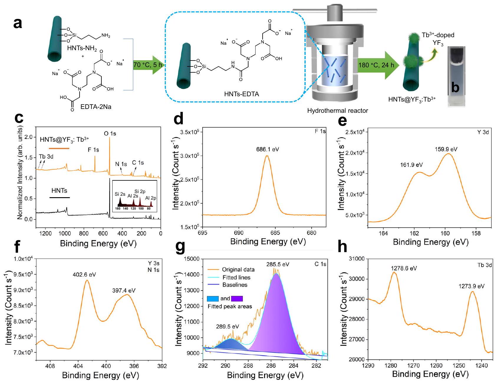
من أجل الاستفادة من الخصائص الفريدة لـ X-PLNPs، قمنا بتثبيت وكيل الخلب حمض الإيثيلين ثنائي الأمين رباعي الأسيتيك ثنائي الصوديوم (EDTA-2Na) على سطح HNTs، مما يسهل تشكيل مجمعات X-PLNP (الشكل 2أ). تضمنت هذه التعديلات
معالجة HNTs الأمينية ( ) مع فائض من EDTA-2Na، مما أدى إلى الارتباط التساهمي لجزء حمض الإيثيلين ثنائي الأمين رباعي الأسيتيك (EDTA) بالأنبوب النانوي من خلال تفاعل استر تقليدي. أكدت التوصيفات الشاملة باستخدام الرنين المغناطيسي النووي في الحالة الصلبة (NMR، الشكل التكميلي 3أ و ب)، وتحليل الطيف الكهرومغناطيسي (FTIR، الشكل التكميلي 4)، والتحليل الحراري (TGA، الشكل التكميلي 5) نجاح توليد HNTs المشتقة من EDTA (HNTsEDTA) مع الحفاظ على الهيكل الأساسي والتركيب للأنابيب النانوية.
لتصنيع HNTs@YF3: المركبات النانوية (الشكل 2أ)، استخدمنا تفاعل هيدروحراري تم إجراؤه عند درجة حرارة وضغط مرتفعين. من الضروري الحفاظ على بيئة حمضية قليلاً طوال العملية لضمان نجاح تشكيل المركب النانوي. كانت مجموعة و بمثابة سوابق لتخليق, المادة الحاضنة، بينما كان المضاف النادرعملت كمنشط. الناتج HNTs@YFأظهرت تشتتًا مائيًا ممتازًا (الشكل 2ب).
لتأكيد التركيب الكيميائي لـ HNTs@YF3:Tbقمنا بإجراء تحليل طيف الكترونيات الأشعة السينية (XPS). مقارنة بين طيف XPS لـ HNTs و HNTs@YF3:Tbكشفت تغييرات ملحوظة تتماشى مع تشكيل النانو مركب (الشكل 2c والجدول التكميلي 1). بعد التعديل والمعالجة الهيدروحرارية، كانت نسبة Si/Al في HNTs@YF3:تم تحديده ليكون 1.42، مما يشير إلى تعديل ناجح عبر عامل الربط السيلاني. تحليل XPS لـ HNTs@عرضت قمم جديدة تتوافق مع وجود الفلور (F1s، الشكل 2d) واليتريوم (Y 3d، الشكل 2e). ومن الجدير بالذكر أن طيف XPS كشف أيضًا عن وجود قمة N 1s عند طاقة ربط تبلغ 397.4 eV، ناتجة عن مجموعة EDTA، وقمة Y 3s عند 402.6 eV (الشكل 2f). بالإضافة إلى ذلك، تم رصد تغييرات في منطقة C 1s، تتوافق معسندات و تمت ملاحظة الروابط (289.5 eV) في طيف XPS لـ HNTs@YF (الشكل 2g). وجود التيربيوم في HNTs@YFتم تأكيده من خلال القمة المزدوجة عند 1278.6 و 1273.9 إلكترون فولت، والتي تت correspond إلى إشارات Tb 3d (الشكل 2h). علاوة على ذلك، وجودتم تأكيده من خلال تحليل حيود الأشعة السينية بالمسحوق (PXRD) لـ HNTs@ (الشكل التوضيحي الإضافي 6 والجداول الإضافية 2 و3)، التي أظهرت أنماطًا تتماشى مع بيانات المرجع القياسية. بشكل جماعي، تؤكد هذه التوصيفات الشاملة بشكل قاطع نجاح التخليق لـجزيئات نانوية.
الخصائص الهيكلية والبصرية
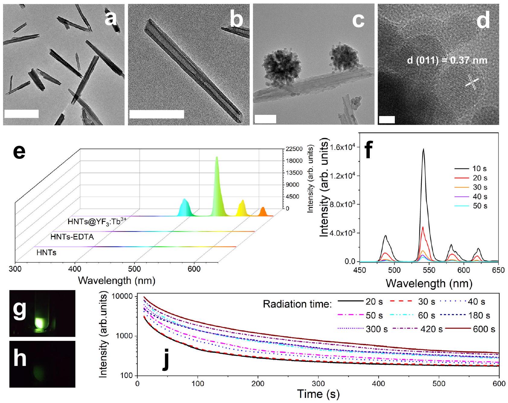
للحصول على رؤى حول هيكل HNTs و HNTs@ YFللنانو جزيئات، استخدمنا مجهر الإلكترون الناقل الماسح (STEM) بالتزامن مع مطياف الأشعة السينية المشتتة للطاقة (EDX). تم ملاحظة شكل مميز لـ HNTs (الشكل 3a، b)، والذي اختلف عن ذلك لـ (الشكل 3ج). من الجدير بالذكر أن HNTs@ أظهرت الجسيمات النانوية كثافة إضافية مرتبطة بهيكل HNT (الشكل 3c). المسافة بين المستويات المتجاورة في الحواف الشبكية للجسيمات النانوية المرتبطة (الشكل 3d)، تم قياسها على أنهايتطابق عن كثب مع المسافة بين الطبقات (011) الطائرات. هذه الملاحظة تشير بقوة إلى أن هذه الكثافة تتوافق مع ارتباط الجسيمات النانوية، مدعومة أيضًا بصور مجال الظلام الحلقي العالي الزاوية (HAADF) من المجهر الإلكتروني النافذ (STEM) (الأشكال التكميلية 7 و 8).
لتحقيق هذا الاكتشاف، قمنا بإجراء رسم خرائط العناصر باستخدام مجهر الإلكترون الناقل الماسح – تحليل الأشعة السينية المشتتة للطاقة (STEM-EDX) لـوتمت ملاحظة توزيعات مميزة لعناصر مختلفة (الأشكال التكميلية 7-9). وُجد أن إشارات الألمنيوم والسيليكون والأكسجين موزعة على طول مكون HNT، بينما كانت إشارات الإيتريوم والفلور والتيربيوم محصورة في الكيانات المرفقة. تؤكد هذه التحليلات الخرائطية العنصرية بشكل قاطع أن هيكل HNT مزين بـالجسيمات، والاندماج الناجح لـ Tb فيشبكة.
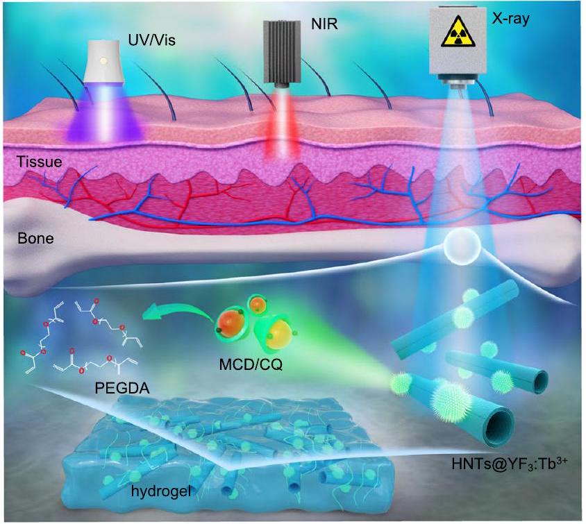
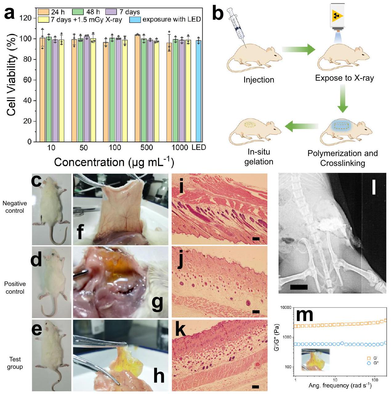
الشكل 1 | تحليل مقارن لعمق اختراق الأنسجة الذي تم تحقيقه باستخدام مصادر تحفيز مختلفة. يظهر الضوء فوق البنفسجي/المرئي (UV/Vis) اختراقًا محدودًا للأنسجة، حيث يصل عادةً إلى بضع مليمترات فقط، مما يعيق بشدة استخدامه في الأنظمة البيولوجية. يمكن أن يعزز ضوء الأشعة تحت الحمراء القريبة (NIR) ذو الأطوال الموجية الأطول عمق الاختراق إلى حد ما. ومع ذلك، فإن طرق الربط الضوئي الحالية لا تحقق اختراقًا عميقًا للأنسجة، خاصة داخل الهياكل العظمية. بالمقارنة مع الضوء، تتمتع الأشعة السينية، وهي شكل من أشكال الموجات الكهرومغناطيسية المستخدمة على نطاق واسع في التطبيقات الطبية الحيوية، بقدرات اختراق ملحوظة عبر كل من العظام والأنسجة البشرية. هنا، نقدم نظام ربط بوليمري محكوم بالأشعة السينية يمثل اختراقًا يتكون من بولي إيثيلين جلايكول داياكريلات (PEGDA)، معقد من كامفوركوينون وميثيل--سيكلوديكسترين (MCD/CQ)، وأنابيب الهالويزيت النانوية المعتمدة على الأشعة السينية التي تنشط الفوسفور المتلألئ المرئي المستمر )، المعروف باسم Xcrosslinking، الذي يحقق تشكيل الهيدروجيل داخل الأنسجة العميقة وهياكل العظام مع مزايا تشمل التحكم المكاني العالي، المحفزات الخارجية غير التلامسية، والمتطلبات غير الحرجة للبيئة.
قمنا بإجراء طيف انبعاث اللمعان المثارة بالأشعة السينية لدراسة خصائص الانبعاث لـ HNTs و HNTs-EDTA و HNTs@YF.كما هو موضح في الشكل 3e، لم تظهر كل من HNTs وHNTs-EDTA أي توهج عند تعرضها لمصدر إشعاع أشعة سينية بقدرة 30 مللي أمبير و40 كيلو فولت. وعلى النقيض من ذلك، فإن طيف انبعاث التوهج الناتج عن تحفيز الأشعة السينية لـ HNTs@YFكشف عن أربعة قمم مميزة عند 490 و 545 و 588 و 623 نانومتر، مما يتوافق مع، ، و الانتقالات، على التوالي.
لمزيد من استكشاف خصائص اللمعان المستمر الطويل الناتج عن الأشعة السينية لـ HNTs@YFقمنا بتسجيل منحنيات تلاشي اللمعان المستمر بعد التعرض للإشعاع باستخدام جهاز إشعاع الأشعة السينية لمدة دقيقة واحدة. ومن المRemarkably، استمر التوهج اللاحق لمدة تصل إلى 50 ثانية مع تراجع تدريجي (الشكل 3f). كان التوهج المرئي واضحًا للعين المجردة بعد التعرض للأشعة السينية (الشكل 3g) وظل مرئيًا لمدة دقيقة واحدة (الشكل 3h). علاوة على ذلك، حصلنا على منحنى تلاشي اللمعان المستمر لـ HNTs@YF3:تمت المراقبة عند 545 نانومتر لمدد إشعاعية متغيرة (من 20 إلى 600 ثانية). كما هو موضح في الشكل 3j، انخفضت شدة التوهج اللاحق بسرعة في البداية خلال الستين ثانية الأولى، تلتها فترة انحسار بطيء. ومن الجدير بالذكر أن إطالة مدة الإشعاع أدت إلى زيادة ملحوظة في شدة التوهج اللاحق. تعتمد مدة إشعاع الأشعة السينية على ) تم التحقيق في شدة التوهج بعد 600 ثانية. الرسوم البيانية بين اللوغاريتم وتم ملاءمة شدة التوهج اللاحق عند 600 ثانية خطيًا، حيث كان معامل الارتباط (R) 0.983 (الشكل التكميلي 10).
تُبرز دراساتنا الشاملة الهيكلية والبصرية الإمكانيات المتميزة لأنابيب النانو الصلبة (HNTs) كدعامة لـترابط، مما يؤدي إلى هيكل نانوي يظهر توهجا مرئيا قويا. علاوة على ذلك، فإن الانبعاث من HNTs@YFلم يتضمن أي أشعة فوق بنفسجية مكونات خفيفة تحت تفعيل الأشعة السينية. هذا لا يقلل فقط من المخاوف المتعلقة بالسلامة المرتبطة بالأشعة فوق البنفسجية في التطبيقات الحية، بل يضع أيضًا HNTs@YFكمصدر ضوء حي قابل للتطبيق لبدء بلمرة الهلامات الهيدروجيلية المتقاطعة بالضوء.
تحقق من البلمرة المثارة بالأشعة السينية لتفاعل الجيلاتين
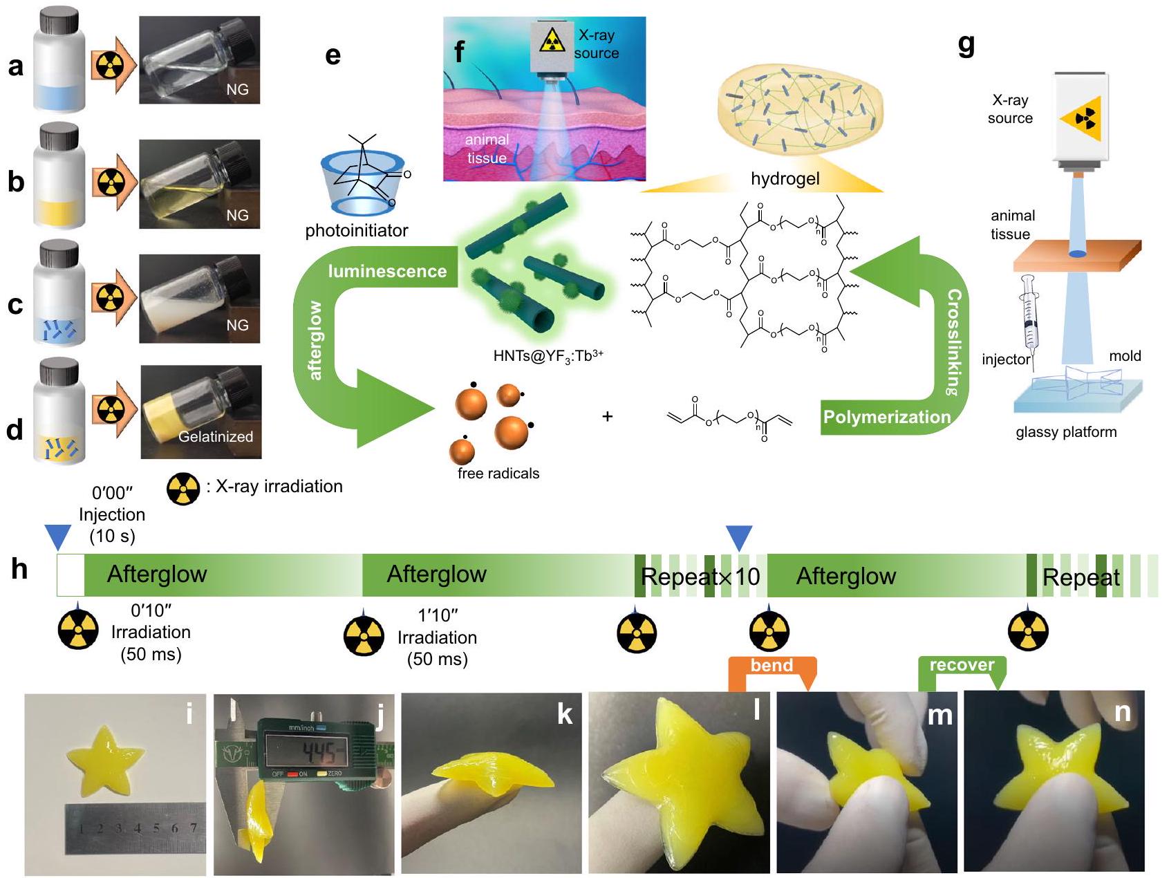
لتقييم سعة HNTs@YFفي تحفيز الربط الضوئي المتقاطع، قمنا بدمجه مع الكامفوركوينون (CQ)، وهو محفز ضوئي مستخدم على نطاق واسع في المجال الطبي الحيوي ويعرف بتوافقه مع منطقة الضوء المرئي.. علاوة على ذلك، كانت طيف الامتصاص لـ CQ تتداخل بشكل وثيق مع اللمعان والتوهج اللاحق لـلزيادة النشاط الضوئي وقابلية الذوبان في الماء لـ CQ، قمنا بتغليفه داخل التجويف الكاره للماء للميثيل-سيكلوديكسترين (MCD). التكوين الناجح لمركب الكامفوركوينون والميثيل-تم تأكيد وجود -سيكلوديكسترين (MCD/CQ) من خلال دراسات NMR (الشكل التكميلي 11). بالإضافة إلى ذلك، استخدمنا بولي إيثيلين جلايكول داياكريلات (PEGDA) المعتمد من قبل إدارة الغذاء والدواء.كمونومر ثنائي الفينيل و ثلاثي إيثانول أمين كمانح للهيدروجين. ثلاثي إيثانول أمين هو مساعد مشترك يستخدم بشكل شائع في البلمرة الضوئية وقد تم إثبات أنه يظهر جهد أكسدة منخفض وحاجز طاقة منخفض.تم إجراء تجارب متوازية للتحقيق في تكوين الهلام تحت ظروف مختلفة (الشكل 4 أ-د). لمحاكاة بيئة الأنسجة العميقة، وضعنا نسيج حيواني بسمك 2 سم بين مصدر الأشعة السينية ووعاء التفاعل. تم ملاحظة تكوين الهلام فقط عند التعرض للأشعة السينية.عندما كانت جميع المكونات الرئيسية موجودة (PEGDA، MCD/CQ، ثلاثي إيثانول أمين، و HNTs@YF3: ).
للتحقق من تشكيل الهيدروجيل تحت هذه الظروف، استخدمنا كروماتوغرافيا نفاذ الهلام (GPC) مقترنة بكاشف مؤشر الانكسار (RI) (الشكل التكميلي 12). تم توضيح الخصائص الريولوجية للهيدروجيل الناتج من خلال معامل التخزين ( ) أكبر من معامل الفقد ( ) فوق تردد متوسط ( نطاق (الشكل التكميلي 13). مجتمعة، تؤكد هذه النتائج أن الجمع بين جميع المكونات المطلوبة، مع تفعيل الأشعة السينية لـ HNTs@YF3: ، يؤدي إلى تشكيل هلام ناجح (الشكل 4e، f). PEGDA بوزن جزيئي منيعتبر المكون الأمثل للدراسات الإضافية لأنه يمكن إذابته في الماء بأي نسبة وقادر على تحقيق حالة الجل في وقت أقصر من ذلك الخاص بالوزن الجزيئي الأعلى.
تم إجراء تجارب موازية إضافية باستخدام أوليفين أحادي الفينيل، ميثيل إيثر أكريلات بولي (إيثيلين جلايكول) (PEGMA)، للتحقق مما إذا كان يمكن أن يحدث سلوك البلمرة باستخدام هذه المكونات: v- PEGMA؛ vi: PEGMA، MCD/CQ، وثلاثي إيثانول أمين؛ vii: PEGMA و HNTs@YF.; الثامن: PEGMA، MCD/ CQ، و HNTs@YF; ix: PEGMA، MCD/CQ، ثلاثي إيثانول أمين، وتم حساب تحويلات المونومر (%) للمجموعات المذكورة أعلاه (v، vi، vii، وviii) جميعها تقريبًا.استنادًا إلى تحليل NMR (الأشكال التكميلية 15 و 16)، مما يشير إلى أن البلمرة لا يمكن أن تحدث عند استخدام المجموعات المذكورة أعلاه التي تحتوي على مكونات مختلفة. تتطابق النتائج بشكل جيد مع النتائج المستخلصة من منحنيات GPC. لا يمكن توليد الجذور من الأشعة السينية / CQ أو تجاوز المركب، والمساعد المشترك ثلاثي إيثانول أمين ضروري أيضًا لنظام الربط المتقاطع. فقط من خلال الجمع بين جميع المكونات المطلوبة (المجموعة التاسعة)، جنبًا إلى جنب مع تنشيط الأشعة السينية لـ HNTs@YF3:يمكن أن يؤدي إلى بلمرة الجذور الحرة الناجحة. تم حساب تحويل المونومر (%) للمجموعة (ix) كـ، و حيث تراوحت مدة الإشعاع من، و10 دقائق، على التوالي (الشكل التوضيحي 17). تزيد نسبة تحويل المونومر (%) مع زيادة وقت الإشعاع. يمكن ملاحظة فرق كبير بين نسبة تحويل المونومر (%) لمدة دقيقة واحدة و3 دقائق. بعد 3 دقائق، تتغير نسبة تحويل المونومر (%) ببطء مع الزيادة المستمرة في وقت الإشعاع.
الشكل 2 | التخليق والتوصيف. أ تمثيل تخطيطي لتخليق أنابيب النانو هالوسيت المشتقة من حمض الإيثيلين ثنائي الأمين رباعي الأسيتيك (HNTs-EDTA) والفوسفوريات المضيئة المستمرة المرئية المعتمدة على أنابيب النانو هالوسيت التي تنشط بواسطة الأشعة السينية (صورة فوتوغرافية لـ HNTs@YFفي الماء. أنماط طيف الإلكترون السيني (XPS) لأنابيب الهالوسيت النانوية HNTs@YFفي المنطقة من 1280 إلى 0 إلكترون فولت. الصورة المرفقة هي المنطقة المكبرة المتعلقة بإشارات الألمنيوم والسيليكون من نمط XPS لـ HNTs. منطقة F 1s لـ HNTs@YF. منطقة Y 3d لـ HNTs@YF. مناطق Y 3 s و N 1 s لـ. منطقة g C 1 sمنطقة لـ.
لقد حفزتنا اللمعان الناتج عن الأشعة السينية، والتوهج اللاحق، وسلوك الجيلاتنة لاستكشاف نظام تفاعل الربط المتقاطع القائم على X-PLNPs، الذي أطلقنا عليه اسم Xcrosslinking. يوفر هذا النظام ميزة قابلية التطبيق في الأنسجة العميقة في الأنظمة غير الشفافة. لتقليل التعرض للأشعة السينية، اعتمدنا نهج الدوران المتقطع. مستفيدين من التوهج المستمر لـ، استخدمناه كمصدر ضوء دائم لنظام الربط المتقاطع. في إعدادنا التجريبي، كما هو موضح في الشكل 4 ج، تم استخدام طابعة تعتمد على نهج الإسقاط من الأعلى إلى الأسفل. تم وضع كوليماتور الأشعة السينية AL01C II (النوع: 5234954؛ الرقم التسلسلي: 7597؛ تيار الأنبوب: 100 مللي أمبير؛ جهد الأنبوب: 50 كيلو فولت؛ الطاقة: 5.0 مللي أمبير في الثانية) في الأعلى، بينما تم وضع طبقة من الأنسجة الحيوانية تحت مصدر الأشعة السينية. قدمت منصة زجاجية في أسفل النظام دعمًا للعينات. كانت المحلول الذي يحتوي على PEGDA (، )، MCD/CQ ( )، ثلاثي إيثانول أمين ( )، و تم تحميله في حقنة. كانت الحقنة، القابلة للتحرك بشكل أفقي، تقوم بتوصيل HNTs@YF.-مليئة بالحلول في القالب. تم تثبيت فترة الإشعاع عند 60 ثانية، كما هو موضح في الشكل 4 ح. في ظل هذه الظروف، نجحنا في تصنيع هيدروجيل على شكل نجمة خماسية قائم بذاته يظهر الخصائص المرنة المطلوبة (الشكل 4ي-ك). ومن الجدير بالذكر أن الهيدروجيل الناتج أظهر سلامة هيكلية ممتازة وخصائص ميكانيكية حتى عند تمديده وثنيه إلى زوايا شديدة (الشكل 41-ن).
بشكل جماعي، تسلط نتائجنا الضوء على جدوى وجودة المادة الهلامية العالية التي تم إنتاجها من خلال استخدام نظام الربط المتقاطع. علاوة على ذلك، تشير تجاربنا التي أجريت في وجود نسيج حيواني بسمك 2 سم إلى إمكانية تطبيق هذه الاستراتيجية في الجسم الحي.
توصيف تفاعل الجيلاتينية في الجسم الحي
لتحقيق إمكانيات HNTs@YFللتطبيقات الحية، قمنا أولاً بتقييم سميته الخلوية باستخدام اختبار (3-(4,5-dimethyl-thiazol-2-yl)-2,5-diphenyltetrazolium bromide) (MTT) في خلايا الفيبروبلاست الفأرية L929. ومن المRemarkably، HNTs@YFأظهرت سمية بيولوجية ضئيلة، حيث تظل قابلية الخلايا للحياة فوقحتى عند تركيزات مرتفعة نسبيًا ) بعد ، و7 أيام من العلاج (الشكل 5أ). الـ HNTs@YFتمت معالجة خلايا L929 بالتعرض لأشعة X (1.5 مللي غراي) وأظهرت أيضًا ارتفاعًا في بقاء الخلايا. ). علاوة على ذلك، قمنا أيضًا بتقييم تأثير الضوء الأخضر المنبعث من الصمام الثنائي الباعث للضوء (LED) على بقاء الخلايا. يمكن تحقيق بقاء جيد للخلايا (>90%). تشير النتائج إلى أن النتائج التي تم الحصول عليها -الأشعة، والضوء الأخضر الناتج يظهر سمية بيولوجية ضئيلة على خلايا الفيبروبلاست L929 للفئران. مشجعين بهذه النتائج الإيجابية في المختبر، نتابع لتقييم النظام في نموذج الفئران من نوع سبرايج داولي (الشكل 5ب).
الشكل 3 | الخصائص الميكروية والبصرية. أ صورة مجهر إلكتروني ناقل (TEM) لأنابيب الهالوسيت النقية (HNTs)، شريط القياس:صورة TEM لنانوتيوبس هيدروكسيد الألمنيوم النقي، شريط القياس:صورة TEM لفسفورات مضيئة مستمرة مرئية نشطة بالأشعة السينية تعتمد على أنابيب الهالوسيت النانوية (HNTs@YF) )، شريط القياس: صورة مجهر إلكتروني ناقل عالي الدقة (HETEM) لـ HNTs@YF3:Tbمقياس الرسم: 5 نانومتر (تم تكرار كل تجربة في الأجزاء أ-د ثلاث مرات بشكل مستقل مع نتائج مشابهة). e طيف انبعاث اللمعان المثارة بالأشعة السينية لـ HNTs، حمض الإيثيلينديامين تتراسيتيك
أنابيب النانو هالوسيت المشتقة من الحمض (HNTs-EDTA)، و HNTs@YFتصل ذروة الانبعاثات عند، و 623 نانومتر التي تم تخصيصها لـ ، ، و الانتقالات، على التوالي.طيف التوهج اللاحقمسجل في، و50 ثانية من تلاشي الوهج. صورة فوتوغرافية لـ HNTs @ تم أخذها تحت الأشعة السينية؛صورة فوتوغرافية لـتم التقاطها بعد دقيقة واحدة من وقت التوهج بعد الإشعاع بالأشعة السينية لمدة دقيقة واحدة. ج شدة التوهج من HNTs@YFتمت مراقبته عند 545 نانومتر كدالة للوقت.
في البيئات السريرية، عادة ما يقتصر تعرض الأشعة السينية على فترات زمنيةاستغلال الخصائص البصرية لـ HNTs@YF، افترضنا أن هذه الجسيمات النانوية يمكن أن تعمل كمصدر ضوء دائم قابل لإعادة الشحن للتجليد داخل الجسم في ظروف آمنة سريرياً. للتحقق من هذه الفرضية، قمنا بتقسيم جرذان سبرايج داولي عشوائياً إلى ثلاث مجموعات: (i) مجموعة التحكم السلبية بدون أي علاج (الشكل 5c)، (ii) مجموعة التحكم المعالجة بـ HNTs@YFتعليق ) يحتوي على PEGDA و MCD/CQ و ثلاثي إيثانول أمين دون التعرض لأشعة X (الشكل 5d)، و (iii) مجموعة الاختبار المعالجة بـ HNTs@YF تعليق ) يحتوي على PEGDA و MCD/CQ و ثلاثي إيثانول أمين، وتم تعريضه لأشعة X (الشكل 5e). بالنسبة للفئران التي تلقت إشعاع أشعة X (المجموعة iii)، تم تعريض منطقة الظهر للفأر بجرعة تعادل الجرعة الروتينية للتصوير لأصابع الإنسان. تم تكرار الإشعاع عشر مرات، مع فترات زمنية تبلغ 60 ثانية بين كل تكرار، مما أدى إلى جرعة إجمالية قدرها 1.5 مللي غراي، وهي ضمن الحدود المعتادة لتطبيقات الأشعة السينية السريرية.
شكلعرضت الأجزاء المقطوعة من الظهر. لاحظنا تشكيل الهيدروجيل حصريًا في مجموعات الاختبار، كما يتضح من وجود هلام أصفر (المجموعة الثالثة)، في حين أن لم تظهر مجموعات التحكم أي تشكيل للجل (أظهرت المجموعة الثانية وجود المحلول المطبق، ولكن دون جل). علاوة على ذلك، كشفت التحليلات الهيستومورفولوجية لنسج الجلد التي أجريت بعد 7 أيام من العلاج عن اختلافات طفيفة بين الحيوانات الضابطة والحيوانات المختبرة، دون وجود دليل على مراحل التهابية (الشكل 5i-k والشكل التكميلي 19). وهذا يشير إلى التوافق الحيوي الممتاز للمواد المحقونة والهلاميات الناتجة.
للحصول على مزيد من الفهم للعملية، أكدنا وجود HNTs@YF3 المحقونة:تعليق في صور الأشعة السينية (الشكل 51). ومن الجدير بالذكر أن ذلك يرجع إلى تأثير التباين القوي الناتج عن HNTs@YFلم نتمكن من تتبع عملية الجيلاتين مباشرة بناءً على أفلام الأشعة السينية ذات الزمن المحدد (الشكل التكميلي 20). للتحقق من أن الهيكل الغروي الأصفر يتوافق بالفعل مع الجل، قمنا بعزل هذه المادة من الحيوان وخضعناها للتحقيق الريولوجي. تظهر نتائجنا أن المادة الغروية الصفراء المستخلصة تظهر خصائص هيدروجيل نموذجية (الشكل 5م).
بشكل عام، تؤكد نتائجنا أن اللمعان والتوهج اللاحق المنبعث منتمكين تشكيل الصورة-
الشكل 4 | ظروف الجيلاتنة وخصائص الهيدروجيل. أ صورة فوتوغرافية توضح سلوك الجيلاتنة للمحلول الذي يحتوي على بولي إيثيلين جليكول داياكريلات (PEGDA) في تجربة متوازية (NG هو اختصار لغير الجيلاتين).صورة فوتوغرافية توضح سلوك الجيلاتنة للمحلول الذي يحتوي على PEGDA، معقد الكامفوركوينون والميثيل-سيكلوديكسترين (MCD/CQ) وثلاثي إيثانول أمين في تجربة متوازية. صورة فوتوغرافية توضح سلوك الجيلاتنة للمحلول الذي يحتوي على PEGDA وأنابيب الهالوسيت النانوية المعتمدة على الفوسفوريات المضيئة المستمرة التي تنشط بواسطة الأشعة السينية. ) في تجربة متوازية. د تصويري صورة توضح سلوك الجيلاتنة للمحلول الذي يحتوي على PEGDA و MCD/ CQ و ثلاثي إيثانول أمين وفي تجربة متوازية. آلية البلمرة والتشابك في نظام التشابك X. توضيح لمصدر الأشعة السينية المستخدم في دراسات الجيلاتين. توضيح لجهاز الطباعة الذي يستخدم قالبًا على شكل خماسي الأضلاع بأبعاد (الطول) و0.45 سم (الارتفاع). تمثيل تخطيطي لعملية الدورة التشغيلية المتقطعة باستخدام الأشعة السينية كمصدر للطاقة. تم تثبيت فترة الإشعاع عند 60 ثانية.صور تعرض الهيدروجيل المُعد. 1-n عرض سلوكيات الانحناء والتعافي التي يظهرها الهيدروجيل المُستخلص. رابط الهلاميات المائية داخل الأنظمة البيولوجية تحت ظروف إشعاع الأشعة السينية الآمنة سريرياً.
تحويل الجيلاتين عند الطلب داخل العظم
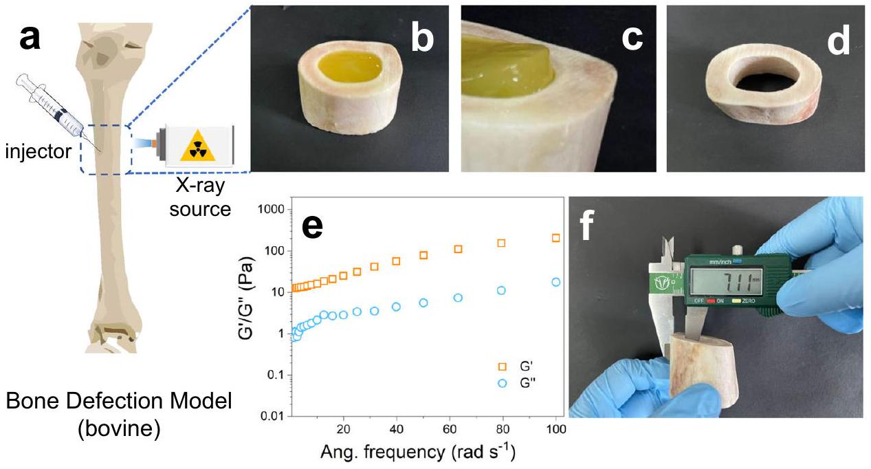
لقد حفزتنا القدرة الملحوظة لأشعة X على اختراق الأنسجة للتحقيق في إمكانيات نظامنا داخل الأنسجة العظمية، والتي تحمل وعدًا كبيرًا لعلاج حالات مثل هشاشة العظام – وهي حالة شائعة تتميز بفقدان العظام النظامي الذي يؤثر على ملايين الأفراد في جميع أنحاء العالم. لفحص قدرة نظامنا على إصلاح العظام، أنشأنا نموذج عيب عظمي عن طريق إزالة النخاع من عظمة بقرية. بعد ذلك، حقنّا محلولًا يحتوي على PEGDA ( ), MCD/CQ (CQ: ثلاثي إيثانول أمين ( )، و إلى منطقة العيب (توضح الشكل 6a الإجراء التخطيطي). بعد التعرض للأشعة السينية (بعد 20 دقيقة، استخرجنا عينة العظم المشع (الشكل 6ب).
من المRemarkably، لاحظنا تشكيل هيدروجيل داخل العظم (الشكل 6c)، والذي يمكن فصله بسهولة عن نسيج العظم (الشكل 6d). أظهر الهيدروجيل المعزول الخصائص الريولوجية المتوقعة، معكمتوسط تردد (تراوحت بين (الشكل 6e). من المهم أن سمك العظام المقاس تجاوز 7.0 مم (الشكل 6f) على كل جانب. بشكل جماعي، تسلط هذه النتائج الضوء على قدرة نهجنا على تمكين تشكيل الهلاميات الهيدروجينية المعتمدة على الربط الضوئي داخل أنسجة العظام السميكة.
لوحظ أنه في بحثنا، استخدمنا نموذجًا سميكًا وكثيفًا من عظام الأبقار، حيث تم بوليمرة التجويف بالكامل، لتقديم مثال توضيحي. تم اختيار عظام الأبقار، المعروفة بكثافتها الأعلى وقوة امتصاصها للأشعة السينية مقارنة بالعظام البشرية، لتمثيل سيناريو صعب. في التطبيقات الطبية العملية، غالبًا ما تقع عيوب العظام في طبقات أكثر سطحية، وعادة ما تكون المنطقة المتأثرة أصغر من نموذجنا.لذلك، يجب أن تكون جرعة الأشعة السينية المطلوبة في التطبيقات الواقعية أقل من تلك المستخدمة في هذه الدراسة المفاهيمية.
باختصار، تقدم دراستنا استراتيجية ربط مبتكرة تتجاوز قيود الطرق الحالية من خلال تمكين تشكيل الهلاميات المترابطة بالضوء داخل نسيج العظام. يتم تحقيق هذا الإنجاز من خلال الاستفادة من جزيئات X-PLNPs كمصدر ضوء مستمر، والتي يتم تثبيتها على HNTs لإنشاء نظام متوافق حيوياً وآمن بحجم موحد وتشتت مائي ممتاز. عند تعرضها لإشعاع الأشعة السينية بجرعة منخفضة، فإن الناتج
الشكل 5 | دراسات السمية الخلوية والتجليد في الجسم الحي. أ بيانات بقاء الخلايا في خلايا ليفية فئران L929 ( ). يتم تقديم البيانات كقيم متوسطة – الانحراف المعياري (SD). LED هو اختصار لثنائي الفينيل العضوي المضيء. لم يتم العثور على فرق ذو دلالة إحصائية بين مجموعة التحكم ومجموعات الاختبار.-قيم المجموعة لمدة 24 ساعة (من اليسار إلى اليمين):، و -قيم المجموعة لمدة 48 ساعة (من اليسار إلى اليمين):، و -قيم مجموعة 7 أيام (من اليسار إلى اليمين):، و -قيم لمجموعة من-أشعة (من اليسار إلى اليمين):، و -قيم مجموعة LED: 0.546.مخطط تخطيطي لعملية الجيلاتينية في الجسم الحي باستخدام إشعاع الأشعة السينية على منطقة الظهر (تيار الأنبوب: 100 مللي أمبير؛ جهد الأنبوب: 50 كيلو فولت؛ زمن التعرض: 50 مللي ثانية؛ الطاقة: 5.0 مللي أمبير-ثانية؛ مجال الرؤية (FOV):; الارتفاع: 1.0 م). ج صورة فوتوغرافية للفأر في مجموعة التحكم السلبية قبل التشريح. د صورة فوتوغرافية للفأر في مجموعة التحكم الإيجابية قبل صورة فوتوغرافية للفأر في مجموعة الاختبار قبل التشريح.صورة فوتوغرافية للفأر في مجموعة التحكم السلبية بعد التشريح. صورة فوتوغرافية للفأر في مجموعة التحكم الإيجابية بعد التشريح. صورة فوتوغرافية للفأر في مجموعة الاختبار بعد التشريح. أنسجة جلدية ملونة بصبغة الهيماتوكسيلين والإيوزين (H&E) من الفأر في مجموعة التحكم السلبية، مقياس الرسم:أنسجة جلدية ملونة بصبغة H&E من الجرذ في مجموعة التحكم الإيجابية، مقياس الرسم:أنسجة جلدية ملونة بصبغة H&E من الجرذ في مجموعة الاختبار، مقياس الرسم: (كل تجربة في تم تكرار ذلك 3 مرات بشكل مستقل مع نتائج مشابهة). 1 فيلم أشعة سينية لفئران حية بعد حقن أنابيب الهالوسيت النانوية المعتمدة على الفوسفور المتلألئ المستمر المرئي المنشط بالأشعة السينية ( ) تعليق، شريط القياس: الخصائص الريولوجية للهيدروجيل المتكون. الصورة المرفقة هي من الصورة الفوتوغرافية للفأر في مجموعة التحكم بعد التشريح. المادة النانوية تظهر توهجا أخضر قويا وتألقا بعد الانتهاء، مما يؤدي بفعالية إلى تجلط الأنسجة العميقة. من المهم أننا نثبت توافق استراتيجيتنا داخل الهياكل العظمية السميكة، محققين اختراقا ملحوظا في الأنسجة العظمية بسمك يتجاوز 7 مم.
يمثل هذا العمل تقدمًا كبيرًا في هذا المجال، حيث يقدم نهجًا تحويليًا لتحقيق الربط الضوئي العميق في الأنسجة واختراق العظام. إن استخدام X-PLNPs كمصدر ضوء مستمر يوفر فرصًا لتشكيل الهيدروجيل بدقة وتحكم، متجاوزًا قيود الطرق الحالية. إن التنفيذ الناجح لاستراتيجيتنا يفتح آفاقًا للتطبيقات السريرية المستقبلية، لا سيما في مجالات إصلاح الأنسجة، والطب التجديدي، وهندسة الأنسجة العظمية. يظهر نظامنا إمكانيات للربط الضوئي العميق في الأنسجة واختراق العظام. علاوة على ذلك، تمتد إمكانيات هذه الطريقة إلى ما هو أبعد من التطبيقات العظمية. كما أنها قابلة للتطبيق في المجالات غير الطبية. سيناريوهات، مثل إصلاح العيوب العميقة في المواد مثل الخرسانة، والسبائك، والمركبات، حيث تكون الطريقة الحالية المعتمدة على الضوء محدودةتتمتع هذه النتائج بتداعيات واسعة على التقدم في مختلف التطبيقات الكيميائية والبيولوجية الطبية. ستركز الدراسات المستقبلية على تطبيق نظام الربط المتقاطع المقترح على سيناريوهات تطبيق محددة من خلال تخليق المزيد من أنواع X-PLNPs ذات خصائص الإشعاع الضوئي المتنوعة. يتم تلخيص سيناريوهات التطبيق المحتملة (الشكل 7) على النحو التالي: من المتوقع أن تعمل الطباعة ثنائية أو ثلاثية الأبعاد غير الجراحية على إصلاح الأنسجة العميقة المعيبة أو التالفة في الموقع بطريقة محكومة مكانيًا، مما يمكن أن يتخلص من العمليات الجراحية. يمكن تحفيز X-PLNPs بواسطة الأشعة السينية قبل حقنها في العيب، ويمكن استخدام الإشعاع الناتج كمصدر ضوء نانوي لتحفيز عملية الشفاء للراتنجات الحساسة للضوء لإصلاح الأسنان، مما يفيد في كسر قيود العمق للراتنجات السنية المعالجة بالضوء المرئي، وتقليل وقت فتح الفم، وقد تكون تقنية مكملة واعدة للراتنجات السنية المعالجة بالضوء المرئي.
الشكل 6 | دراسات الجيلاتنة في نموذج عيب العظام. أ رسم تخطيطي للنموذج. تحتوي المحلول في الحقنة على بولي إيثيلين جليكول داي أكريلات (PEGDA)، مجمع الكامفوركوينون والميثيل--سيكلوديكسترين (MCD/CQ) (CQ: ، )، ثلاثي إيثانول أمين ( وأنابيب الهالويزيت النانوية المعتمدة على الأشعة السينية الفوسفوريات المضيئة المستمرة المرئية ). ب) مقطع مقطوع من نموذج عيب العظام بعد الإشعاع. ج) المادة القابلة للإزالة التي تشكلت داخل العظم. د) المقطع المقطوع بعد إزالة الهيدروجيل. الخصائص الريولوجية للهيدروجيل المتكون حديثًا.اختبار السماكة.
الشكل 7 | ملخص لسيناريوهات التطبيق المحتملة لنظام الربط المتقاطع. أ إصلاح الأنسجة العميقة. ب إصلاح عيوب الأسنان العميقة. ج العلاج بالحقن على الأورام. د إصلاح عيوب الغضاريف.
لإصلاح العيوب العميقة. من المتوقع استخدام نظام الربط المتقاطع في العلاج بالانصمام داخل الأوعية للأورام الخبيثة. بعد حقن تدخلي دقيق في مواقع الأورام والتعرض لـيمكن أن تحدث الجيلاتينية داخل أوعية الورم. يمكن أن تحقق الجيلاتينية تأثير العلاج بالانسداد وبالتالي تقمع النمو السرطاني بشكل فعال. قد يتم أيضًا تحقيق إصلاح عيوب الغضروف من خلال نظام الربط المتقاطع المقترح. يمكن التحكم في درجة الربط وسلوك التورم من خلال تغيير وقت الإشعاع وجرعات الأشعة السينية لتلبية متطلبات الخصائص الميكانيكية لاستخدام الغضروف.
طرق
بيان أخلاقي
تمت مراجعة البروتوكول الخاص بتجارب الحيوانات في هذه الدراسة والموافقة عليه من قبل لجنة الأخلاقيات ورفاهية الحيوان في جامعة خبي (رقم الموافقة: IACUC-2021XG008، التاريخ: مارس). 2021).
تركيب أنابيب الهالوسيت المشتقة من حمض الإيثيلينديامين تتراسيتيك
الـ HNTs-تم تحضيره عن طريق معالجة HNTs (22.0 جرام) ومادة خالية من الماءفيتم تحريك الخليط وتسخينه في لمدة 48 ساعة. HNTs-تم الحصول عليه من خلال جمع المتبقي بعد التصديق. 1.08 جرام مجففتم إضافة إلى محلول الماء الذي يحتوي على 1.35 جرام من EDTA-2Na. ثم تم نقل النانو معلق إلى دورق ذو عنق واحد سعة 150 مل وتسخينه عندمع التحريك المغناطيسي المتوتر لمدة 5 ساعات. بعد التبريد إلى درجة حرارة الغرفة، تم جمع الرواسب عن طريق الطرد المركزي فيثم تم غسلها بالإيثانول والأسيتون لثلاث دورات. تم تجميد الترسيب الناتج بواسطة النيتروجين السائل ثم تم تجفيفه في الفراغ. تم الحصول على المنتج HNTs-EDTA كصلب أبيض.
تركيب الفوسفوريات المنبعثة من اللمعان المستمر المرئي المعتمد على أنابيب الهالوسيت النانوية المنشّطة بأشعة إكس
تم وزن 1.70 جرام من HNTs-EDTA بدقة وأضيف إلى المحلول المائي الذي يحتوي على و مع نسبة المولات منتم تحريك النظام على مستوى منخفض لمدة 30 دقيقة لتحقيق تعليق متجانس.تمت إضافته إلى النظام ثم تم ضبط قيمة الرقم الهيدروجيني إلى 5.0 بواسطة حمض النيتريك. استمرت عملية التحريك لمدة 30 دقيقة أخرى. تم إدخال التعليق الناتج في مفاعل هيدروحراري بمعدل تعبئة قدرهثم تم تسخينه عندلمدة 24 ساعة. بعد التبريد إلى درجة حرارة الغرفة، تم جمع الرواسب عن طريق الطرد المركزي فيثم تم غسلها بالإيثانول والأسيتون لثلاث دورات. تم تجميد الترسيب الناتج بواسطة النيتروجين السائل ثم تم تجفيفه في الفراغ. المنتج HNTs@YFتم الحصول عليه كصلب أبيض.
تحضير المحفز الضوئي
باختصار، تم إضافة كمية زائدة من الكامفوركوينون الصلب إلى الميثيل-محاليل ميثانول السيكلوديكسترين، تلاها تعليق تم تحريكه وتطبيق الموجات فوق الصوتية في درجة حرارة الغرفة لمدة 0.5 ساعة. ثم تم تحريك المحلول لمدة 24 ساعة أخرى. بعد ذلك تم تصفيته من خلالتم تجميد الحل الناتج وتجفيفه للحصول على المنتج الصلب الأصفر الباهت MCD/CQ.
دراسة الجيلاتنة في المختبر
HNTs@YFتم تعليقها في محلول مائي ) يحتوي على PEGDA ( ), MCD/CQ (CQ: ) وثلاثي إيثانول أمين ( تم إغلاق الخليط في قوارير زجاجية ووضعها على المنصة أمام مصدر الأشعة السينية (تم وضع نسيج حيواني بسمك 2.0 سم بين مصدر الأشعة السينية والقارورة. مدة الإشعاع الكلية هي 10 دقائق.
زراعة الخلايا
تم استخدام خلايا سرطان عنق الرحم البشرية (HeLa) وخلايا الألياف العضلية للفأر 1929 في هذه الدراسة، والتي تم شراؤها من الأكاديمية الصينية للعلوم الطبية، كلية بكين للطب (رقم الكتالوج: 1101HUM-PUMCO00011) ومن بنك الخلايا التابع للأكاديمية الصينية للعلوم في شنغهاي (رقم الكتالوج: BFN60805937)، على التوالي. تم الحفاظ على الخلايا في وسط دلبوكو المعدل من إيغلي (DMEM) وتم تزويدها بـمصل الجنين البقري (FBS) داخل بيئة رطبة ) يحتوي على 5% و مضادات حيوية بنسلين/ستربتوميسين.
دراسة السمية الخلوية
تم زراعة خلايا هيلا في صفيحة 96 بئرخلايا ) ثم تم حضنه مع فيمع التركيز يتراوح من 10 إلى. بعد 12 و 24 و 48 ساعة، محلول MTT (، ) تم إضافة إلى كل بئر، وتمت متابعة عملية الحضانة لمدة 4 ساعات. ثم تم إزالة الوسط وتم إضافة DMSO. بعد الخلط الجيد، تم قياس امتصاص كل بئر عند 490 نانومتر بواسطة جهاز قراءة الميكرو بلايت ELX-800 (قارئ ELISA). تم استخدام الآبار التي لم تُضاف إليها عينات كتحكم فارغ. تم حساب نسبة حيوية الخلايا (%) من خلال نسبة امتصاص الاختبار إلى التحكم. دقيقتم حساب قيمة – بناءً على اختبار المقارنات المتعددة توكي ANOVA أحادي الاتجاه.
حيوية الخلايا لـمع التركيز يتراوح من 10 إلىلـ وتم تقييم 7 أيام باستخدام خلايا الفيبروبلاست الفأرية L929 باتباع طريقة MTT مشابهة للحالة السابقة. تم التحقيق في تأثير إشعاع الأشعة السينية على بقاء الخلايا من خلال وضع لوحة 96 بئر مليئة بالخلايا مع إضافة HNTs@YF.مع التركيز يتراوح من 10 إلى تحت جهاز تحديد الأشعة السينية AL01C II (النوع: 5234954؛ الرقم التسلسلي: 7597؛ تيار الأنبوب: 100 مللي أمبير؛ جهد الأنبوب: 50 كيلو فولت؛ زمن التعرض: 50 مللي ثانية؛ الطاقة: 5.0 مللي أمبير في الثانية؛ مجال الرؤية: ; الارتفاع: 1.0 م ) عند بجرعة إجمالية قدرها 1.5 مللي غراي، مماثلة لتلك المستخدمة في دراسات الجيلاتين في الجسم الحي. ثم تم معالجة صفيحة الـ 96 بئر كما هو معتاد لمدة 7 أيام. لفحص تأثير الضوء المرئي على حيوية الخلايا، تم تعريض صفيحة الـ 96 بئر المملوءة بالخلايا لمصباح LED أخضر لمدة ساعة واحدة، والذي يمكنه إصدار ضوء بكثافة أعلى بكثير من اللمعان الناتج عن HNTs@YF3.تحت الأشعة السينية. ثم تم معالجة صفيحة الـ 96 بئر كما هو معتاد لمدة 7 أيام. تم عزل مجموعات التحكم عن الأشعة السينية أو ضوء LED بواسطة لوحة رصاص. تم فحص حيوية الخلايا باستخدام نفس طريقة MTT كما في خلايا هيلا. دقيقتم حساب قيمة – بناءً على اختبار المقارنات المتعددة توكي ANOVA أحادي الاتجاه.
دراسة الجيلاتينية في الجسم الحي
فئران سبرايج-داولي الذكورتم شراء الفئران (عمرها أسابيع) وفقًا للبروتوكولات المعتمدة من مركز الحيوانات التجريبية في مقاطعة خبي، شيجياتشوانغ، الصين. تم مراجعة البروتوكول والموافقة عليه من قبل لجنة الأخلاقيات ورفاهية الحيوان في جامعة خبي (رقم الموافقة: IACUC-2021XG008، التاريخ: 11 مارس 2021). تم إطعام فئران سبرايج-داولي بالطعام العادي وتوفير كمية كافية من الماء. تم وضعها في أقفاص بلاستيكية قياسية تحت درجة حرارة معينة.تم تقسيم فئران سبرايج-داولي عشوائيًا إلى مجموعة التحكم السلبية ( )، مجموعة التحكم الإيجابية ( ) ومجموعة الاختبار ( تم صيام جميع الفئران لمدة 12 ساعة قبل التجارب. تم الاحتفاظ بالفئران في مجموعة التحكم السلبية كما هو معتاد دون أي علاج آخر. بالنسبة لمجموعة التحكم الإيجابية، تم تعليق في محلول مائي يحتوي على PEGDA و MCD/CQ و triethanolamine ثم تم حقنه في ظهر الفئران (). بالنسبة لمجموعة الاختبار، تم حقن تعليق HNTs@YF3: Tb أيضًا في الفئران بنفس الطريقة. تم تثبيت الفئران على لوح خشبي للفئران ثم تعرضت لأشعة X عبر
كوليماتور أشعة X Collimalor AL01C II. ثم تم علاج الفئران كما هو معتاد. بعد 30 دقيقة، تم التضحية بأحد الفئران من كل مجموعة بواسطة مخدر وتم تشريحه. تم التحقيق بعناية في مواقع الحقن. تم ترك الفئران الأخرى في مجموعة التحكم الإيجابية ومجموعة الاختبار سليمة. في اليوم السابع، تم التضحية بجميع الفئران بواسطة مخدر وتم تشريحها. تم جمع أنسجة الجلد من مواقع الحقن، وثبتت في 10% فورمالين، وتمت معالجتها بشكل روتيني إلى بارافين، وتم صبغها بصبغة الهيماتوكسيلاين والإيوزين (H&E) وتم فحص علم الأمراض بواسطة كاميرا ميكروسكوب أوليمبوس DP 72.
نموذج عيب العظام
تم استخدام قطعة واحدة من عظم البقر، تم شراؤها من شركة Alibaba Co. Ltd.، لإنشاء نموذج عيب العظام. تم تنظيف العظم وقطعه إلى قطعتين في المنتصف، وتم استخدام واحدة منها في مزيد من الاستخدام وغمرها في ماء منزوع الأيونات لمدة 30 دقيقة. تم إزالة الأنسجة الداخلية في موقع الفتحة بواسطة مثقاب وملاقط لتوفير ثقب بعمق حوالي 5 سم مع ثقب صغير بحجم حوالي 2 مم على جانب الثقب. تم غسل العظم المعالج بعناية بماء منزوع الأيونات. ثم تم إغلاق الجزء العلوي من الثقب بصفائح رصاصية لتوفير نموذج عيب العظام.
ملخص التقرير
تتوفر معلومات إضافية حول تصميم البحث في ملخص تقرير Nature Portfolio المرتبط بهذه المقالة.
توفر البيانات
البيانات التي تم إنشاؤها في هذه الدراسة متاحة في ملف المعلومات التكميلية / بيانات المصدر. تتوفر مجموعة الصور الكاملة من المؤلف المراسل عند الطلب. يتم توفير بيانات المصدر مع هذه الورقة.
References
Gu, Y. W. et al. Photoswitching topology in polymer networks with metal-organic cages as crosslinks. Nature 560, 65-69 (2018).
Hua, Y. J. et al. Ultrafast, tough, and adhesive hydrogel based on hybrid photocrosslinking for articular cartilage repair in water-filled arthroscopy. Sci. Adv. 7, eabg0628 (2021).
Kalayci, K., Frisch, H., Truong, V. X. & Barner-Kowollik, C. Green light triggered [2+2] cycloaddition of halochromic styrylquinoxalinecontrolling photoreactivity by pH. Nat. Commun. 11, 4193 (2020).
Liu, N. N., Dai, M. J., Saka, S. K. & Yin, P. Super-resolution labelling with Action-PAINT. Nat. Chem. 11, 1001-1008 (2019).
Zhang, W. J. et al. Promoting oral mucosal wound healing with a hydrogel adhesive based on a phototriggered s-nitrosylation coupling reaction. Adv. Mater. 33, 2105667 (2021).
Zhang, J. et al. A pulsatile release platform based on photo-induced imine-crosslinking hydrogel promotes scarless wound healing. Nat. Commun. 12, 1670 (2021).
GhavamiNejad, A. et al. Glucose-responsive composite microneedle patch for hypoglycemia-triggered delivery of native glucagon. Adv. Mater. 31, 1901051 (2019).
Hong, Y. et al. A strongly adhesive hemostatic hydrogel for the repair of arterial and heart bleeds. Nat. Commun. 10, 2060 (2019).
Lee, T. T. et al. Light-triggered in vivo activation of adhesive peptides regulates cell adhesion, inflammation and vascularization of biomaterials. Nat. Mater. 14, 352-360 (2015).
Qian, Y. et al. Immunoregulation in diabetic wound repair with a photoenhanced glycyrrhizic acid hydrogel scaffold. Adv. Mater. 34, 2200521 (2022).
Loebel, C., Mauck, R. L. & Burdick, J. A. Local nascent protein deposition and remodelling guide mesenchymal stromal cell mechanosensing and fate in three-dimensional hydrogels. Nat. Mater. 18, 883-891 (2019).
Balu, R., Dutta, N. K., Dutta, A. K. & Choudhury, N. R. Resilinmimetics as a smart biomaterial platform for biomedical applications. Nat. Commun. 12, 149 (2021).
Wu, J. J. et al. Rapid digital light 3D printing enabled by a soft and deformable hydrogel separation interface. Nat. Commun. 12, 6070 (2021).
Ge, Q. et al. 3D printing of highly stretchable hydrogel with diverse UV curable polymers. Sci. Adv. 7, eaba4261 (2021).
Bowen, J. J., Rose, M. A., Konda, A. & Morin, S. A. Surface molding of microscale hydrogels with microactuation functionality. Angew. Chem. Int. Ed. 57, 1236-1240 (2018).
Zhu, H. Y. et al. Spatiotemporally controlled photoresponsive hydrogels: design and predictive modeling from processing through application. Adv. Funct. Mater. 30, 2000639 (2020).
Widyaya, V. T., Riga, E. K., Muller, C. & Lienkamp, K. Submicrometersized, 3D surface-attached polymer networks by microcontact printing: using UV-cross-linking efficiency to tune structure height. Macromolecules 51, 1409-1417 (2018).
Xie, H., Yang, K. K. & Wang, Y. Z. Photo-cross-linking: apowerful and versatile strategy to develop shape-memory polymers. Prog. Polym. Sci. 95, 32-64 (2019).
Li, B. H., Lu, L. F., Zhao, M. Y., Lei, Z. H. & Zhang, F. An efficient 1064 nm NIR-II excitation fluorescent molecular dye for deep-tissue highresolution dynamic bioimaging. Angew. Chem. Int. Ed. 57, 7483-7487 (2018).
Martincorena, I. et al. High burden and pervasive positive selection of somatic mutations in normal human skin. Science 348, 880-886 (2015).
Guedes, G. et al. Dual-crosslinked dynamic hydrogel incorporating {Mo-154} with pH and NIR responsiveness for chemo-photothermal therapy. Adv. Mater. 33, 2007761 (2021).
Samadian, H., Maleki, H., Allahyari, Z. & Jaymand, M. Natural polymers-based light-induced hydrogels: promising biomaterials for biomedical applications. Coord. Chem. Rev. 420, 213432 (2020).
Chen, Y. W. et al. Noninvasive in vivo 3D bioprinting. Sci. Adv. 6, eaba7406 (2020).
Shim, W. S., Yoo, J. S., Bae, Y. H. & Lee, D. S. Novel injectable pH and temperature sensitive block copolymer hydrogel. Biomacromolecules 6, 2930-2934 (2005).
Raman, R. et al. Light-degradable hydrogels as dynamic triggers for gastrointestinal applications. Sci. Adv. 6, eaay0065 (2020).
Zhang, S. et al. A pH-responsive supramolecular polymer gel as an enteric elastomer for use in gastric devices. Nat. Mater. 14, 1065-1071 (2015).
Eelkema, R. & Pich, A. Pros and cons: supramolecular or macromolecular: what is best for functional hydrogels with advanced properties? Adv. Mater. 32, 1906012 (2020).
Chen, X. F., Song, J. B., Chen, X. Y. & Yang, H. H. X-ray-activated nanosystems for theranostic applications. Chem. Soc. Rev. 48, 3073-3101 (2019).
Fan, W. P. et al. Breaking the depth dependence by nanotechnology-enhanced X-ray-excited deep cancer theranostics. Adv. Mater. 31, 1806381 (2019).
Wang, X. et al. Organic phosphors with bright triplet excitons for efficient X-ray-excited luminescence. Nat. Photonics 15, 187-192 (2021).
Cunningham, R. et al. Keyhole threshold and morphology in laser melting revealed by ultrahigh-speed X-ray imaging. Science 363, 849-852 (2019).
Gan, N. et al. Organic phosphorescent scintillation from copolymers by X-ray irradiation. Nat. Commun. 13, 3995 (2022).
Ma, W. et al. Thermally activated delayed fluorescence (TADF) organic molecules for efficient X-ray scintillation and imaging. Nat. Mater. 21, 210-216 (2022).
Shi, T. H. et al. X-ray-induced persistent luminescence promotes ultrasensitive imaging and effective inhibition of orthotopic hepatic tumors. Adv. Funct. Mater. 30, 2001166 (2020).
Xue, Z. L., Jiang, M. Y., Liu, H. R., Zeng, S. J. & Hao, J. H. Low dose soft X-ray-controlled deep-tissue long-lasting NO release of persistent luminescence nanoplatform for gas-sensitized anticancer therapy. Biomaterials 263, 120384 (2020).
Glotov, A., Vutolkina, A., Pimerzin, A., Vinokurov, V. & Lvov, Y. Clay nanotube-metal core/shell catalysts for hydroprocesses. Chem. Soc. Rev. 50, 9240-9277 (2021).
Zhao, P. X., Feng, Y., Zhou, Y. Q., Tan, C. Y. & Liu, M. X. Gold@halloysite nanotubes-chitin composite hydrogel with antibacterial and hemostatic activity for wound healing. Bioac. Mater. 20, 355-367 (2023).
Lvov, Y., Wang, W. C., Zhang, L. Q. & Fakhrullin, R. Halloysite clay nanotubes for loading and sustained release of functional compounds. Adv. Mater. 28, 1227-1250 (2016).
Feng, Y. et al. A ferroptosis-targeting ceria anchored halloysite as orally drug delivery system for radiation colitis therapy. Nat. Commun. 14, 5083 (2023).
Zhao, S. L. et al. A dual-surface amidoximated halloysite nanotube for high-efficiency economical uranium extraction from seawater. Angew. Chem. Int. Ed. 58, 14979-14985 (2019).
Tonooka, K. & Nishimura, O. Spectral changes of fluorescence in borosilicate glasses. J. Lumin. 87-89, 679-681 (2000).
Dadashi-Silab, S., Aydogan, C. & Yagci, Y. Shining a light on an adaptable photoinitiator: advances in photopolymerizations initiated by thioxanthones. Polym. Chem. 6, 6595-6615 (2015).
Teh, D. B. L. et al. A flexi-PEGDA upconversion implant for wireless brain photodynamic therapy. Adv. Mater. 32, 2001459 (2020).
Benezery, K. et al. Clinical response assessment after contact X-Ray brachytherapy and chemoradiotherapy for organ preservation in rectal cancer T2-T3 MO: The time/dose factor influence. Clin. Transl. Radiat. Oncol. 24, 92-98 (2020).
Gérard, J.-P. et al. Contact X-ray therapy for rectal cancer: experience in centre Antoine-Lacassagne, nice, 2002-2006. Int. J. Radiat. Oncol. 72, 665-670 (2008).
Ravetz, B. D. et al. Photoredox catalysis using infrared light via triplet fusion upconversion. Nature 565, 343-346 (2019).
الشكر والتقدير
يقر هايلين زانغ بالدعم المالي من المؤسسة الوطنية للعلوم الطبيعية في الصين (رقم 22102045)، والتوجيه المركزي على صندوق تطوير العلوم والتكنولوجيا المحلية في مقاطعة خبي (رقم 226Z1301G)، وصندوق الرئيس لجامعة خبي (رقم XZJJ202210)، والمشروع الرئيسي من المؤسسة الوطنية للعلوم الطبيعية في مقاطعة خبي (رقم B2O2O201072). يقر Y.Y بالدعم من المؤسسة الوطنية للعلوم الطبيعية في الصين (رقم 11974097). تم دعم G.H و K.H بشكل حصري من قبل جامعة ماساتشوستس. يشكر هايلين زانغ بعمق البروفيسور شين وو با على اقتراحاته القيمة لهذه الورقة.
مساهمات المؤلفين
أجرى هايلين زانغ معظم التجارب، وأجرى تحليل البيانات، وصمم جزءًا من المشروع وكتب المخطوطة الأصلية. أجرى B.T. جزءًا من تجارب التخليق. أجرى B.Z. تحليل الجسيمات النانوية. أجرى K.H. تحليل البيانات. قام S.L. و Y.Z. بإجراء الاختبارات الإضافية في مرحلة المراجعة. هايسونغ زانغ مسؤول عن الدراسات الحيوانية. ساهم L.B. في التحليل الرسمي. أجرى Y.W. جزءًا من تجارب التخليق. Y.C. مسؤول عن تحليل الخلايا. صمم Y.Y مسار التفاعل وأشرف على المشروع. تصور G.H. وأشرف على المشروع والتجارب وكتب المخطوطة.
يجب توجيه المراسلات وطلبات المواد إلى هايلين زانغ، يانمين يانغ أو غانغ هان.
تشكر مجلة Nature Communications جيانغتاو شو، هوانغهاو يانغ والمراجعين الآخرين المجهولين على مساهمتهم في مراجعة الأقران لهذا العمل. يتوفر ملف مراجعة الأقران.
تتوفر معلومات إعادة الطبع والأذونات على http://www.nature.com/reprints
ملاحظة الناشر تظل Springer Nature محايدة فيما يتعلق بالمطالبات القضائية في الخرائط المنشورة والانتماءات المؤسسية.
كلية الكيمياء وعلوم المواد، جامعة خبي، باودينغ 071002، جمهورية الصين الشعبية. قسم الكيمياء الحيوية وعلم الأدوية الجزيئية، كلية الطب بجامعة ماساتشوستس، وورسيستر، ماساتشوستس، MA 01605، الولايات المتحدة الأمريكية. المستشفى التابع لجامعة خبي، باودينغ 071000، جمهورية الصين الشعبية. كلية علوم الفيزياء والتكنولوجيا، معهد علوم الحياة والتنمية الخضراء، مختبر خبي الرئيسي للمعلومات والمواد البصرية الإلكترونية، جامعة خبي، باودينغ 071002، جمهورية الصين الشعبية. البريد الإلكتروني: zhanghailei@hbu.edu.cn; mihuyym@163.com; Gang.Han@umassmed.edu
X-ray-activated polymerization expanding the frontiers of deep-tissue hydrogel formation
Received: 2 July 2023
Accepted: 4 April 2024
Published online: 15 April 2024
(A) Check for updates
Hailei Zhang Boyan Tang , Bo Zhang , Kai Huang , Shanshan Li , Yuangong Zhang , Haisong Zhang , Libin Bai , Yonggang Wu ¹, Yongqiang Cheng , Yanmin Yang Gang Han
Photo-crosslinking polymerization stands as a fundamental pillar in the domains of chemistry, biology, and medicine. Yet, prevailing strategies heavily rely on ultraviolet/visible (UV/Vis) light to elicit in situ crosslinking. The inherent perils associated with UV radiation, namely the potential for DNA damage, coupled with the limited depth of tissue penetration exhibited by UV/ Vis light, severely restrict the scope of photo-crosslinking within living organisms. Although near-infrared light has been explored as an external excitation source, enabling partial mitigation of these constraints, its penetration depth remains insufficient, particularly within bone tissues. In this study, we introduce an approach employing X-ray activation for deep-tissue hydrogel formation, surpassing all previous boundaries. Our approach harnesses a low-dose X-ray-activated persistent luminescent phosphor, triggering on demand in situ photo-crosslinking reactions and enabling the formation of hydrogels in male rats. A breakthrough of our method lies in its capability to penetrate deep even within thick bovine bone, demonstrating unmatched potential for bone penetration. By extending the reach of hydrogel formation within such formidable depths, our study represents an advancement in the field. This application of X-ray-activated polymerization enables precise and safe deep-tissue photo-crosslinking hydrogel formation, with profound implications for a multitude of disciplines.
Photo-crosslinking has emerged as a powerful technique for fabricating three-dimensional polymer networks with broad applications across diverse fields, including biomedicine . It offers precise spatial and temporal control over hydrogel synthesis, making it invaluable for wound healing , drug delivery , tissue repair , bioengineering , and bioimaging . The hallmark advantage lies in the ability to trigger polymerization on-demand using light, enabling highly controlled synthesis . However, the current approach relies on ultraviolet/
visible (UV/Vis) light ( ) for activation, which poses significant limitations . UV/Vis light exhibits restricted tissue penetration, typically reaching only a few millimeters , thus severely hampering its utility in biological systems. Moreover, concerns surrounding DNA damage associated with UV light raise critical safety considerations, particularly for patient applications . Recent attempts have explored the use of near-infrared (NIR) light with longer wavelengths to enhance penetration depth to some extent . There are
several other mechanisms to confer injectability upon hydrogels and facilitate their gelation within deep tissues, largely relying on tem-perature-, pH, magnet-, shearing-, or chemical-based stimuli. These stimuli, while well suited for use in some target applications, usually suffer from a range of disadvantages that render them inappropriate for use in many in vivo applications. The use of injectable temperaturesensitive hydrogels usually suffers from unexpected gelation inside the needles warmed by the body temperature, especially for the injection in deep tissues . Magnetic stimuli can interfere with some clinical therapeutics and medical imaging technologies . pH stimuli are restricted in the regions that match the material’s pH -responsive range . Chemical stimuli require contact with the material and are difficult to spatially control . Shear-thinning hydrogels can be injected under the shear stress during injection and rapidly return to their elastic behavior after removal of shear , while the gelation process is difficult to control after being injected into the body.
Nonetheless, existing crosslinking methods fall short in achieving deep-tissue penetration with spatial and temporal control, especially within bone structures. Consequently, the repair and regeneration of bone tissue have remained formidable challenges within the realm of photo-crosslinking hydrogel systems. Therefore, the need for other approaches that address these limitations and enable deep-tissue hydrogel formation is imperative for advancing biomedical research and therapeutic interventions. Compared to light, X-rays, a form of electromagnetic waves extensively utilized in biomedical applications, exhibit remarkable penetration capabilities through both bones and human tissue (Fig. 1) . Moreover, clinical X-ray radiation offers precise control over exposure time and location , crucial factors for enabling deep tissue cross-linking reactions necessary for biomedical hydrogel formation.
In this study, we present a breakthrough X-ray-controlled polymer crosslinking method, referred to as Xcrosslinking, that achieves hydrogel formation within deep tissues and bone structures with advantages including highly spatial controllability, noncontact external stimuli, and uncritical demand for the environment. To accomplish this, we develop a biocompatible X-ray-activated visible persistent luminescent emitting phosphors (X-PLNPs) serving as an in situ energy transducer , initiating photo-crosslinking reactions at deep tissue sites, including within bones. Specifically, we utilize halloysite nanotubes (HNTs), natural tubular nanomaterials with good water dispersibility , charge on the external surface , low toxicity , and ease of modification , as templates for the synthesis of HNTs-based XPLNPs (HNTs@YF3:Tb ) through a facile hydrothermal method. These -doped X-PLNPs exhibited well-controlled size, desirable waterdispersity, intense persistent luminescence, excellent biocompatibility, and robust stability. We demonstrate that HNTs@YF3:Tb effectively store energy upon low-dose X-ray excitation, releasing visible luminescence as a persistent light source. This emitted visible light from HNTs@YF serves as a trigger for on-demand, in situ photo-crosslinking reactions, enabling hydrogel formation. We validate the performance of our system through in vitro experiments and animal models. Our results demonstrate high biocompatibility and the potential safety of this approach, as it enables in situ crosslinking activation in deep tissue. This groundbreaking strategy holds tremendous promise for various biomedical applications requiring deep tissue hydrogel formation, offering a frontier in the field of X-rayactivated polymer crosslinking.
Results and discussion
Harnessing persistent luminescent emitting phosphors for enhanced functionality
In order to leverage the unique properties of X-PLNPs, we strategically immobilized the chelate agent ethylenediaminetetraacetic acid disodium salt (EDTA-2Na) onto the surface of HNTs, facilitating the formation of X-PLNP complexes (Fig. 2a). This modification involved the
treatment of aminated HNTs ( ) with excess EDTA-2 Na , resulting in the covalent attachment of the ethylenediaminetetraacetic acid (EDTA) moiety to the nanotube through a conventional esterification reaction. Comprehensive characterization using solid-state nuclear magnetic resonance (NMR, Supplementary Fig. 3a and b), Fourier transform infrared spectroscopy (FTIR, Supplementary Fig. 4), and thermogravimetric analysis (TGA, Supplementary Fig. 5) confirmed the successful generation of EDTA-derivatized HNTs (HNTsEDTA) while preserving the fundamental structure and composition of the nanotubes.
To fabricate HNTs@YF3: nanocomposites (Fig. 2a), we employed a hydrothermal reaction conducted at elevated temperature and pressure. Crucially, a slightly acidic environment was maintained throughout the process to ensure successful nanocomposite formation. The combination of and served as precursors for the synthesis of , the host material, while the rare-earth dopant acted as the activator. The resulting HNTs@YF exhibited excellent water dispersibility (Fig. 2b).
To confirm the chemical composition of HNTs@YF3:Tb , we performed X-ray photoelectron spectroscopy (XPS) analysis. A comparison between the XPS spectra of HNTs and HNTs@YF3:Tb revealed discernible changes in accordance with the formation of the nanocomposite (Fig. 2c and Supplementary Table 1). After modification and hydrothermal treatment, the Si/Al ratio of HNTs@YF3: was determined to be 1.42 , indicating successful modification via the silane coupling agent. The XPS analysis of HNTs@ exhibited new peaks corresponding to the presence of fluorine (F1s, Fig. 2d) and yttrium (Y 3d, Fig. 2e). Notably, the XPS spectrum also revealed the presence of an N 1s peak at a binding energy of 397.4 eV , originating from the EDTA moiety, and a Y 3s peak at 402.6 eV (Fig. 2f). Additionally, changes in the C 1 s region, corresponding to bonds and bonds ( 289.5 eV ), were observed in the XPS spectrum of HNTs@YF (Fig. 2g). The existence of terbium in HNTs@YF was confirmed by the doublet peak at 1278.6 and 1273.9 eV , corresponding to the Tb 3d signals (Fig. 2h). Furthermore, the presence of was confirmed by powder X-ray diffractometry (PXRD) analysis of HNTs@ (Supplementary Fig. 6 and Supplementary Tables 2 and 3), which exhibited patterns consistent with standard reference data. Collectively, these comprehensive characterizations conclusively confirm the successful synthesis of nanoparticles.
Structural and optical characterizations
To gain insights into the structure of HNTs and HNTs@ YF nanoparticles, we employed scanning transmission electron microscopy (STEM) in conjunction with energy dispersive X-ray spectroscopy (EDX). A distinct morphology was observed for HNTs (Fig. 3a, b), which differed from that of (Fig. 3c). Notably, the HNTs@ nanoparticles exhibited an additional density attached to the HNT scaffold (Fig. 3c). The interplanar distance between adjacent lattice fringes in the attached nanoparticles (Fig. 3d), measured as , closely matched the interplanar distance of (011) planes. This observation strongly suggests that this density corresponds to the attachment of nanoparticles, further supported by the high-angle annular dark-field (HAADF) STEM images (Supplementary Figs. 7 and 8).
To validate this finding, we conducted scanning transmission electron microscope-energy dispersive X-ray (STEM-EDX) elemental mapping of and observed distinct distributions of different elements (Supplementary Figs. 7-9). Signals from Al, Si, and O were found to be distributed along the HNT component, while the Y, F, and Tb signals were confined to the attached entities. This elemental mapping analysis unequivocally confirms that the HNT scaffold is decorated with particles, and the successful integration of Tb into the lattice.
Fig. 1 | A comparative analysis of tissue penetration depths achieved with different excitation sources. Ultraviolet/visible (UV/Vis) light exhibits restricted tissue penetration, typically reaching only a few millimeters, thus severely hampering its utility in biological systems. Near infrared ray (NIR) light with longer wavelengths can enhance penetration depth to some extent. Nonetheless, existing photo-crosslinking methods fall short in achieving deep-tissue penetration, especially within bone structures. Compared to light, X-rays, a form of electromagnetic waves extensively utilized in biomedical applications, exhibit remarkable penetration capabilities through both bones and human tissue. Herein, we present a breakthrough X-ray-controlled polymer crosslinking system consisted of containing polyethyleneglycoldiacrylate (PEGDA), complex of camphorquinone and methyl- -cyclodextrin (MCD/CQ), and halloysite nanotubes-based X-ray-activated visible persistent luminescent emitting phosphors ( ), referred to as Xcrosslinking, that achieves hydrogel formation within deep tissues and bone structures with advantages including highly spatial controllability, noncontact external stimuli, and uncritical demand for the environment.
We conducted X-ray-excited luminescence emission spectroscopy to investigate the emission properties of HNTs, HNTs-EDTA, and HNTs@YF . As shown in Fig. 3e, both HNTs and HNTs-EDTA exhibited no luminescence when exposed to a 30 mA 40 kV X-ray irradiator. In striking contrast, the X-ray-excited luminescence emission spectrum of HNTs@YF revealed four distinct peaks at 490 , 545,588 , and 623 nm , corresponding to the , , and transitions, respectively .
To further explore the X -ray-induced long persistent luminescence properties of HNTs@YF , we recorded persistent luminescence decay curves after irradiation with an X-ray irradiator for 1 min . Remarkably, the afterglow persisted for up to 50 s with gradual attenuation (Fig. 3f). The visible afterglow was clearly observable to the naked eye following X-ray irradiation (Fig. 3g) and remained visible for 1 min (Fig. 3h). Furthermore, we obtained the persistent luminescence decay curve of HNTs@YF3: monitored at 545 nm for varying irradiation times (20 to 600 s). As depicted in Fig. 3j, the afterglow intensity initially declined rapidly within the first 60 s , followed by a slow decay. Notably, prolonging the irradiation time led to a noticeable increase in afterglow intensity. The dependence of X-ray irradiation time ( ) on the intensity of afterglow at 600 s was investigated. The plots between log and the intensity of afterglow on 600 s were fitted linearly, in which the correlation index (R) was 0.983 (Supplementary Fig. 10).
Our comprehensive structural and optical studies highlight the outstanding potential of HNTs as a scaffold for attachment, resulting in a nanostructure exhibiting robust visible luminescence. Moreover, the emission from HNTs@YF did not include any UV
light components under X-ray activation. This not only mitigates safety concerns associated with UV radiation in in vivo applications but also positions HNTs@YF as a viable in vivo light source for initiating the polymerization of photo-crosslinking hydrogels.
Validation of X-ray-excited polymerization for gelatinization reaction
To assess the capacity of HNTs@YF in inducing photo-crosslinking, we combined it with camphorquinone (CQ), a widely used photoinitiator in the biomedical field known for its compatibility with the visible light region . Furthermore, the absorption spectra of CQ closely overlapped with the luminescence and afterglow of . To enhance the photo-activity and water-solubility of CQ , we encapsulated it within the hydrophobic cavity of methyl- cyclodextrin (MCD). The successful formation of the complex of camphorquinone and methyl- -cyclodextrin (MCD/CQ) was confirmed through NMR studies (Supplementary Fig. 11). Additionally, we employed a FDA-approved polyethyleneglycoldiacrylate (PEGDA) as the diolefinic monomer and triethanolamine as the hydrogen donor. Triethanolamine is a commonly-used co-initiator in photopolymerizations and has been demonstrated to show low oxidation potential and the low energy barrier . Parallel experiments were conducted to investigate gel formation under different conditions (Fig. 4a-d). To simulate a deep tissue environment, we placed a 2 cm thick animal tissue between the X-ray source and the reaction vial. Gel formation was solely observed upon X-ray exposure ( ) when all key components were present (PEGDA, MCD/CQ, triethanolamine, and HNTs@YF3: ).
To validate the formation of hydrogel under these conditions, we employed gel permeation chromatography (GPC) coupled with a refractive index (RI) detector (Supplementary Fig. 12). The rheological characteristics of the resulting hydrogel were demonstrated by the storage modulus ( ) being greater than the loss modulus ( ) over an average frequency ( ) range of (Supplementary Fig. 13). Collectively, these results confirm that the combination of all required components, along with X-ray activation of HNTs@YF3: , leads to successful gel formation (Fig. 4e, f). PEGDA with a molecular weight of is regarded as the optimal component for further studies because it can be dissolved in water at any proportion and is able to achieve a gelation state in a shorter time than that of a higher molecular weight.
Additional parallel experiments were also conducted by using a mono-vinyl olefin, poly(ethylene glycol) methyl ether acrylate (PEGMA), to investigate whether the polymerization behavior can take place by using these components: v- PEGMA; vi: PEGMA, MCD/CQ, and triethanolamine; vii: PEGMA and HNTs@YF ; viii: PEGMA, MCD/ CQ , and HNTs@YF ; ix: PEGMA, MCD/CQ, triethanolamine, and . The monomer conversions (%) for the abovementioned groups (v, vi, vii, and viii) are all calculated as ca. based on the NMR analysis (Supplementary Figs. 15 and 16), indicating the polymerization cannot take place when using the abovementioned groups containing different components. The results match well with the findings from GPC curves. The radical cannot be generated from X-ray/CQ or bypassing the composite, and the coinitiator triethanolamine is also essential for the Xcrosslinking system. Only a combination of all required components (group ix), along with X-ray activation of HNTs@YF3: , can lead to successful free radical polymerization. The monomer conversion (%) of the group (ix) was calculated as , and as the irradiation time ranged from , and 10 min , respectively (Supplementary Fig. 17). The monomer conversion (%) increases with the increase of the irradiation time. A significant difference can be found between the monomer conversion (%) of 1 min and 3 min . After 3 min , the monomer conversion (%) changes slowly with the continuous increase of irradiation time.
Fig. 2 | Synthesis and characterization. a Schematic representation of synthesis of ethylenediaminetetraacetic acid-derivatized halloysite nanotubes (HNTs-EDTA) and halloysite nanotubes-based X-ray-activated visible persistent luminescent emitting phosphors ( ). b Photographic image of HNTs@YF in water. c X-ray photoelectron spectroscopy (XPS) patterns of halloysite nanotubes
and HNTs@YF in the region from 1280 to 0 eV . The inset picture is the enlarged region relating to Al and Si signals from the XPS pattern of HNTs. d F 1s region for HNTs@YF . e Y 3d region for HNTs@YF . f Y 3 s and N 1 s regions for . g C 1 s region for region for .
The X-ray-induced luminescence, afterglow, and gelatinization behavior have motivated us to explore a crosslinking reaction system based on X-PLNPs, which we have named Xcrosslinking. This system offers the advantage of deep tissue applicability in nontransparent systems. To minimize X-ray exposure, we adopted an on-off-on circulation approach. Leveraging the persistent afterglow of , we utilized it as a long-lasting light source for the Xcrosslinking system. In our experimental setup, as illustrated in Fig. 4 g , a printer employing an up-bottom projection approach was employed. The AL01C II X-Ray Collimator (Type: 5234954; S. N. 7597; Tube current: 100 mA ; Tube voltage: 50 kV ; Energy: 5.0 mAs ) was positioned on top, while a layer of animal tissue was placed beneath the X-ray source. A glass platform at the bottom of the system provided support for the samples. The solution containing PEGDA ( , ), MCD/CQ ( ), triethanolamine ( ), and was loaded into an injector. The injector, movable in a horizontal manner, delivered the HNTs@YF -filled solution into the mold. The irradiation interval was fixed at 60 s , as depicted in Fig. 4 h . Under these conditions, we successfully fabricated self-standing pentagram-shaped hydrogels exhibiting the desired elastomeric properties (Fig. 4i-k). Notably, the obtained hydrogel demonstrated excellent structural integrity and mechanical properties even when stretched and bent to extreme angles (Fig. 41-n).
Collectively, our findings highlight the feasibility and high-quality gel material produced through the utilization of the Xcrosslinking system. Furthermore, our experiments conducted in the presence of 2 cm thick animal tissue suggest the potential applicability of this strategy in vivo.
Characterization of in vivo gelatinization reaction
To investigate the potential of HNTs@YF for in vivo applications, we first assessed its cytotoxicity using a (3-(4,5-dimethyl-thiazol-2-yl)-2,5-diphenyltetrazolium bromide) (MTT) assay in mouse fibroblast L929 cells. Remarkably, HNTs@YF demonstrated negligible biological toxicity, as cell viability remains above even at relatively high concentrations ( ) after , and 7 days of treatment (Fig. 5a). The HNTs@YF . treated L929 cells with the exposure to X-ray ( 1.5 mGy ) also exhibited high cell viability ( ). Moreover, we also evaluated the impact of green light emitted by light-emitting diode (LED) on cell viability. A good cell viability (>90%) can also be achieved. The results indicate that the obtained -ray, and the generated green light exhibit negligible biological toxicity on mouse fibroblast L929 cells. Encouraged by these favorable in vitro results, we proceed to evaluate the system in a Sprague Dawley rat model (Fig. 5b).
Fig. 3 | Micromorphological and optical characterizations. a Transmission electron microscope (TEM) image of pristine halloysite nanotubes (HNTs), scale bar: TEM image of pristine HNTs, scale bar: TEM image of halloysite nanotubes-based X-ray-activated visible persistent luminescent emitting phosphors (HNTs@YF ), scale bar: High resolution transmission electron microscope (HETEM) image of HNTs@YF3:Tb , scale bar: 5 nm (Each experiment in a-d was repeated for 3 times independently with similar results). e X-ray-excited luminescence emission spectra of HNTs, ethylenediaminetetraacetic
acid-derivatized halloysite nanotubes (HNTs-EDTA), and HNTs@YF . The emission peaks at , and 623 nm that are assigned to the , , and transitions, respectively. Afterglow spectra of recorded at , and 50 s of afterglow attenuation. g Photographic image of HNTs @ taken under X-ray; Photographic image of taken at 1 min afterglow time after irradiation with X -rays for 1 min. j Afterglow intensity from HNTs@YF monitored at 545 nm as a function of time.
In clinical settings, X-ray irradiation is typically limited to durations of . Leveraging the optical properties of HNTs@YF , we hypothesized that these nanoparticles can serve as a rechargeable persistent light source for in vivo gelatinization under clinically safe conditions. To validate this hypothesis, we randomly divided Sprague Dawley rats into three groups: (i) the negative control group without any treatment (Fig. 5c), (ii) the control group treated with HNTs@YF suspension ( ) containing PEGDA, MCD/CQ, and triethanolamine without exposure to X-rays (Fig. 5d), and (iii) the test group treated with HNTs@YF suspension ( ) containing PEGDA, MCD/CQ, and triethanolamine, and exposed to X-rays (Fig. 5e). For rats receiving X-ray irradiation (group iii), the back area of the rat was irradiated at a dose equivalent to the routine imaging dose for human fingers. The irradiation was repeated ten times, with 60 s intervals between each repetition, resulting in a total dose of 1.5 mGy , well within the limits of typical clinical X-ray applications.
Figure showcase the dissected backs of the rates. We observed the formation of hydrogel exclusively in the test groups, as indicated by the presence of a yellow gel (group iii), whereas the
control groups did not exhibit any gel formation (group ii showed the presence of the applied solution, but no gel). Furthermore, histomorphological analysis of the skin tissue conducted 7 days posttreatment reveal minimal differences between the control and test animals, with no evidence of inflammatory phases (Fig. 5i-k and Supplementary Fig. 19). This suggests the excellent biocompatibility of the injected substances and the resulting hydrogels.
To gain further insight into the process, we confirmed the presence of the injected HNTs@YF3: suspension in the X-ray images (Fig. 51). Notably, due to the strong contrast effect from HNTs@YF , we were unable to directly track the gelatinization process based on time-resolved X-ray films (Supplementary Fig. 20). To verify that the yellow colloidal structure indeed corresponds to a gel, we isolated this substance from the animal and subjected it to rheological investigation. Our findings demonstrate that the obtained yellow colloidal substance exhibits typical hydrogel properties (Fig. 5m).
Overall, our results establish that the luminescence and afterglow emitted by enable the formation of photo-
Fig. 4 | Gelatinization conditions and hydrogel properties. a Photographic image depicting gelatinization behavior of the solution containing polyethyleneglycoldiacrylate (PEGDA) in parallel experiment (NG is abbreviated from not gelatinized). Photographic image depicting gelatinization behavior of the solution containing PEGDA, complex of camphorquinone and methyl- cyclodextrin (MCD/CQ), and triethanolamine in parallel experiment. c Photographic image depicting gelatinization behavior of the solution containing PEGDA and halloysite nanotubes-based X-ray-activated visible persistent luminescent emitting phosphors ( ) in parallel experiment. d Photographic
image depicting gelatinization behavior of the solution containing PEGDA, MCD/ CQ , triethanolamine, and in parallel experiment. e Polymerization and crosslinking mechanism in the Xcrosslinking system. Illustration of the X-ray source used in gelatinization studies. g Illustration of the printing apparatus utilizing a pentagram-shaped mold with dimensions of (length) and 0.45 cm (height). Schematic representation of the on-off-on circulation process employing X-ray as the energy source. The irradiation interval was fixed at 60 s . Photographs showcasing the prepared hydrogel. 1-n Demonstration of the bending and recovering behaviors exhibited by the obtained hydrogel.
crosslinking hydrogels within biological systems under clinically safe X-ray irradiation conditions.
On-demand gelatinization inside the bone
The remarkable tissue penetration ability of X-rays has motivated us to investigate the potential of our system within bone tissues, which holds significant promise for the treatment of conditions such as osteoporosis-a prevalent condition characterized by systemic bone loss affecting millions of individuals worldwide. To examine the capacity of our system for bone repair, we created a bone defect model by removing the marrow from a bovine bone. Subsequently, we injected a solution containing PEGDA ( ), MCD/CQ (CQ: v ), triethanolamine ( ), and into the defect region (Fig. 6a illustrates the schematic procedure). Following X-ray exposure ( , for 20 min ), we retrieved the radiated bone specimen (Fig. 6b).
Remarkably, we observed the formation of a hydrogel within the bone (Fig. 6c), which can be readily separated from the bone tissue (Fig. 6d). The isolated hydrogel exhibited the expected rheological properties, with as the average frequency ( ) ranged from (Fig. 6e). Importantly, the measured bone thickness exceeded 7.0 mm (Fig. 6f) on each side. Collectively, these findings highlight the capability of our approach to enable the formation of photo-crosslinking hydrogels within thick bone tissue.
Noted that in our research, we employed a thick, dense bovine bone model, fully polymerizing the entire cavity, to provide an illustrative example. Bovine bone, known for its higher density and stronger X-ray attenuation compared to human bones, was chosen to represent a challenging scenario. In practical medical applications, bone defects are often located in more superficial layers, and the affected area is typically smaller than our model . Therefore, the X-ray dose required in real-world applications be lower than that used in this conceptual study.
In summary, our study presents a pioneering Xcrosslinking strategy that overcomes the limitations of current methods by enabling the formation of photo-crosslinked hydrogels within bone tissue. This breakthrough is achieved by leveraging X-PLNPs as a persistent light source, which are immobilized onto HNTs to create a biocompatible and safe system with uniform size and excellent water dispersity. Upon low-dose X-ray irradiation, the resulting
Fig. 5 | Cytotoxicity and in vivo gelatinization studies. a Cell viability data in mouse fibroblast L929 cells ( ). Data are presented as mean values – standard deviation (SD). LED is abbreviated from light-emitting diode. No significant difference was found between the control group and test groups. -values for the group of 24 h (left to right): , and -values for the group of 48 h (left to right): , and -values for the group of 7 days (left to right): , and -values for the group of -ray (left to right): , and -values for LED group: 0.546 . Schematic diagram of the in vivo gelatinization process using X-ray irradiation to the back area (Tube current: 100 mA ; Tube voltage: 50 kV ; Exposure time: 50 m Sec; Energy: 5.0 mAs ; Field of view (FOV): ; Height: 1.0 m ). c Photographic image of the rat in negative control group before dissection. d Photographic image of the rat in positive control group before
dissection. e Photographic image of the rat in test group before dissection. Photographic image of the rat in negative control group after dissection. g Photographic image of the rat in positive control group after dissection. h Photographic image of the rat in test control group after dissection. i Hematoxylin & eosin (H&E)-stained skin tissues from rat in negative control group, scale bar: H&E-stained skin tissues from rat in positive control group, scale bar: H&E-stained skin tissues from rat in test group, scale bar: (Each experiment in was repeated for 3 times independently with similar results). 1 X -ray film of living rats after injection of halloysite nanotubesbased X-ray-activated visible persistent luminescent emitting phosphors ( ) suspension, scale bar: Rheological property of the asformed hydrogel. The inset picture is from the photographic image of rat in test control group after dissection. nanomaterial exhibits strong green luminescence and afterglow, effectively initiating deep tissue gelatinization. Importantly, we demonstrate the compatibility of our strategy within thick bone structures, achieving remarkable deep-tissue penetration in bone tissues with a thickness exceeding 7 mm .
This work represents a significant advancement in the field, introducing a transformative approach for achieving deep-tissue and bone-penetrating photo-crosslinking. The utilization of X-PLNPs as a persistent light source offers opportunities for precise and controlled hydrogel formation, overcoming the limitations of current methods. The successful implementation of our strategy opens up avenues for future clinical applications, particularly in the fields of tissue repair, regenerative medicine, and bone tissue engineering. Our system demonstrates potential for deep-tissue and bone-penetrating photocrosslinking polymerization. Moreover, the potential of this method extends beyond bone applications. It is also applicable in non-medical
scenarios, such as repairing deep defects in materials like concretes, alloys, and composites, where the current light based method is limited . These findings have broad implications for advancements in various chemical and biomedical applications. Further studies will focus on applying the proposed Xcrosslinking system to specific application scenarios by synthesizing more types of X-PLNPs with diverse radioluminescence properties. Potential application scenarios (Fig. 7) are summarized as follows: Noninvasive 2D or 3D printing is expected to repair defective or damaged deep tissues in situ in a spatially controlled manner, which can get rid of surgical operations. X-PLNPs can be excited by X-ray before being injecting into the defect and the generated luminescence can be used as a nanolight source to trigger the curing process of the photosensitive resins for dental repairs, which is beneficial to break the depth limitation of the visible light-cured dental resin, shorten the mouth opening time, and maybe a promising supplementary technique to visible light-cured dental resin
Fig. 6 | Gelatinization studies in bone defect model. a Schematic diagram of the model. Solution in injector contains polyethyleneglycoldiacrylate (PEGDA) , ), complex of camphorquinone and methyl- -cyclodextrin (MCD/CQ) (CQ: , ), triethanolamine ( ), and halloysite nanotubes-based X-ray-activated
visible persistent luminescent emitting phosphors ( ).
b Sawed section of the bone defect model after irradiation. c The formed removable substance inside the bone. d The sawed section after removing the hydrogel. Rheological property of the as-formed hydrogel. Thickness test.
Fig. 7 | Summary of the potential application scenarios of the Xcrosslinking system. a Deep tissue repairs. b Deep dental defect repairs. c Emnolotherapy on tumors. d Cartilage defect repairs.
for repairing deep defects. The Xcrosslinking system is expected to be used in endovascular embolotherapy on malignant tumors. Following a precise interventional injection on the tumor sites and exposure to ray, gelatinization can take place inside the tumor vessels. The gelatinization can achieve an embolotherapy effect and thereby effectively suppress the cancerous growth. Cartilage defect repairing may also be achieved by the proposed X-crosslinking system. The crosslinking degree of swelling behavior can be controlled by changing the irradiation time and X-ray doses to meet the requirement of the mechanical properties for cartilage use.
Methods
Ethical statement
The protocol about the animal experiments in this study has been reviewed and approved by the Animal Ethical and Welfare Committee of Hebei University (Approval No. IACUC-2021XG008, Data: Mar. 2021).
Synthesis of ethylenediaminetetraacetic acid-derivatized halloysite nanotubes
The HNTs- was prepared by treating HNTs ( 22.0 g ) and anhydrous in . The mixture was stirred and heated at for 48 h . HNTs- was afforded by collecting the residue after certification. 1.08 g dried was added into the water solution containing 1.35 g EDTA-2Na. The nanosuspension was then transferred into a 150 mL single neck flask and heated at with tensely magnetic stirring for 5 h . After cooling to room temperature, the residue was collected by centrifugation at and then washed by ethanol and acetone for three cycles. The obtained precipitation was frozen via liquid nitrogen and then dried in vacuum. The product HNTs-EDTA was obtained as white solid.
Synthesis of halloysite nanotubes-based X-ray-activated visible persistent luminescent emitting phosphors
1.70 g HNTs-EDTA was accurately weighed and added to the aqueous solution containing and with the molar ratio of . The system was stirred in low level for 30 min to achieve a homogeneous suspension. was added into the system and then the pH value was adjusted to 5.0 by nitric acid. The agitation was continued for another 30 min . The obtained suspension was subjected into hydrothermal reactor with a filling rate of and then heated at for 24 h . After cooling to room temperature, the residue was collected by centrifugation at and then washed by ethanol and acetone for three cycles. The obtained precipitation was frozen via liquid nitrogen and then dried in vacuum. The product HNTs@YF was obtained as white solid.
Preparation of photoinitiator
Briefly, excess solid camphorquinone was added to methyl- cyclodextrin methanol solutions, followed by the suspension was stirred and sonicated at room temperature for 0.5 h . Then the solution was stirred for another 24 h . After filtered through a membrane, the resulting solution was frozen-dried to give the faint yellow solid product MCD/CQ.
In vitro gelatinization study
HNTs@YF was suspended in aqueous solution ( ) containing PEGDA ( ), MCD/CQ (CQ: ), and triethanolamine ( ). The mixture was sealed in glassy vials and placed on
the platform in front of an X-ray source ( ). A 2.0 cm animal tissue was placed between the X-ray source and vial. The total irradiation duration is 10 min .
Cell culture
Human cervical cancer cells (HeLa) and mouse fibroblast 1929 cells were used in this study, which were purchased from Chinese Academy of Medical Sciences, Peking Union Medical College (catalog number: 1101HUM-PUMCO00011) and the Cell Bank of the Shanghai Chinese Academy of Science (catalog number: BFN60805937), respectively. The cells were maintained in Dulbecco’s modified Eagle’s medium (DMEM) and supplemented with fetal bovine serum (FBS) within a humidified environment ( ) containing 5% and penicillin/ streptomycin antibiotics.
Cytotoxicity study
HeLa cells were seeded in 96-wellplate ( cells ) and then incubated with at with the concertation ranging from 10 to . After 12, 24 and 48 h , MTT solution ( , ) was added to each well, another incubation process was continued for 4 h . Then the medium was removed and DMSO was added. After homogenizing well, the absorbance of each well was measured at 490 nm by the ELX-800 microplate reader (ELISA Reader). Wells without the addition of samples were used as blank control. The cell viability (%) was calculated by the absorbance percentage of test to control. Exact -value was calculated based on the one-way ANOVA Tukey’s multiple comparisons test.
The cell viability of with the concertation ranging from 10 to for and 7 days was evaluated using mouse fibroblast L929 cells following a similar MTT method to the former case. The effect of X-ray radiation on cell viability was investigated by placing the cell-filled 96-wellplate with the addition of HNTs@YF with the concertation ranging from 10 to under the AL01C II X-Ray Collimator (Type: 5234954; S. N. 7597; Tube current: 100 mA ; Tube voltage: 50 kV ; Exposure time: 50 mSec ; Energy: 5.0 mAs ; FOV: ; Height: 1.0 m ) at with a total dose of 1.5 mGy , identical to that of in vivo gelatinization studies. Then the 96 -wellplate were treated as usual for 7 days. To examine the effect of visible light on cell viability, the cell-filled 96-wellplate was exposed to green LED lamp for 1 h , which can emit light with obviously higher intensity than the luminescence generated by HNTs@YF3: under X-ray. Then the 96 -wellplate were also treated as usual for 7 days. The control groups were isolated from the X-ray or LED light by a lead plate. The cell viability was examined following the same MTT method as Hela cells. Exact -value was also calculated based on the one-way ANOVA Tukey’s multiple comparisons test.
In vivo gelatinization study
Sprague-Dawley male rats ( weeks old) were purchased with protocols approved from the Experimental Animal Center of Hebei Province, Shijiazhuang, China. The protocol has been reviewed and approved by the Animal Ethical and Welfare Committee of Hebei University (Approval No. IACUC-2021XG008, Data: Mar. 11st 2021). Sprague-Dawley rats were fed with regular food and enough water. They were housed in standard plastic cages and place under a certain temperature ( ). The Sprague-Dawley rats were randomly divided into negative control group ( ), positive control group ( ) and test group ( ). All the rats were fasted for 12 h prior to experiments. The rats in negative control group were kept as usual without any other treatment. For the positive control group, the was suspended in aqueous solution containing PEGDA, MCD/CQ, and triethanolamine and then injected into the back of the rats ( ). For test group, the HNTs@YF3: Tb suspension was also injected into to rats following the same manner. The rats were fastened on the wooden rat board and then were exposed to X-ray via a
Collimalor AL01C II X-Ray Collimator. Then the rats were treated as usual. After 30 min , one of the rats from each group was sacrificed by anesthetic and dissected. The injection sites were carefully investigated. Other rats in the positive control group and test group were left intact. On day 7, the rats were all sacrificed by anesthetic and dissected. The skin tissue from the injection sites were harvested, fixed in 10% formalin, processed routinely into paraffin, stained with hematoxylin and eosin (H&E) and pathology are examined by an Olympus DP 72 microscope camera.
Bone defect model
One piece of bovine bone, purchased from Alibaba Co. Ltd., was used to establish the bone defect model. The bone was cleaned and sawed into two pieces in the middle, one of which was subjected to further use and immersed in deionized water for 30 min . The internal tissues in the opening site were removed by drill and tweezer to afford a hole with a depth of ca. 5 cm with a pinhole of ca. 2 mm at the side of the hole. The treated bone was carefully washed by deionized water. Then the top of the hole was sealed with a lead plate to afford a bone defect model.
Reporting summary
Further information on research design is available in the Nature Portfolio Reporting Summary linked to this article.
Data availability
The data generated in this study are provided in Supplementary Information/Source Data file. The full image dataset is available from the corresponding author upon request. Source data are provided with this paper.
References
Gu, Y. W. et al. Photoswitching topology in polymer networks with metal-organic cages as crosslinks. Nature 560, 65-69 (2018).
Hua, Y. J. et al. Ultrafast, tough, and adhesive hydrogel based on hybrid photocrosslinking for articular cartilage repair in water-filled arthroscopy. Sci. Adv. 7, eabg0628 (2021).
Kalayci, K., Frisch, H., Truong, V. X. & Barner-Kowollik, C. Green light triggered [2+2] cycloaddition of halochromic styrylquinoxalinecontrolling photoreactivity by pH. Nat. Commun. 11, 4193 (2020).
Liu, N. N., Dai, M. J., Saka, S. K. & Yin, P. Super-resolution labelling with Action-PAINT. Nat. Chem. 11, 1001-1008 (2019).
Zhang, W. J. et al. Promoting oral mucosal wound healing with a hydrogel adhesive based on a phototriggered s-nitrosylation coupling reaction. Adv. Mater. 33, 2105667 (2021).
Zhang, J. et al. A pulsatile release platform based on photo-induced imine-crosslinking hydrogel promotes scarless wound healing. Nat. Commun. 12, 1670 (2021).
GhavamiNejad, A. et al. Glucose-responsive composite microneedle patch for hypoglycemia-triggered delivery of native glucagon. Adv. Mater. 31, 1901051 (2019).
Hong, Y. et al. A strongly adhesive hemostatic hydrogel for the repair of arterial and heart bleeds. Nat. Commun. 10, 2060 (2019).
Lee, T. T. et al. Light-triggered in vivo activation of adhesive peptides regulates cell adhesion, inflammation and vascularization of biomaterials. Nat. Mater. 14, 352-360 (2015).
Qian, Y. et al. Immunoregulation in diabetic wound repair with a photoenhanced glycyrrhizic acid hydrogel scaffold. Adv. Mater. 34, 2200521 (2022).
Loebel, C., Mauck, R. L. & Burdick, J. A. Local nascent protein deposition and remodelling guide mesenchymal stromal cell mechanosensing and fate in three-dimensional hydrogels. Nat. Mater. 18, 883-891 (2019).
Balu, R., Dutta, N. K., Dutta, A. K. & Choudhury, N. R. Resilinmimetics as a smart biomaterial platform for biomedical applications. Nat. Commun. 12, 149 (2021).
Wu, J. J. et al. Rapid digital light 3D printing enabled by a soft and deformable hydrogel separation interface. Nat. Commun. 12, 6070 (2021).
Ge, Q. et al. 3D printing of highly stretchable hydrogel with diverse UV curable polymers. Sci. Adv. 7, eaba4261 (2021).
Bowen, J. J., Rose, M. A., Konda, A. & Morin, S. A. Surface molding of microscale hydrogels with microactuation functionality. Angew. Chem. Int. Ed. 57, 1236-1240 (2018).
Zhu, H. Y. et al. Spatiotemporally controlled photoresponsive hydrogels: design and predictive modeling from processing through application. Adv. Funct. Mater. 30, 2000639 (2020).
Widyaya, V. T., Riga, E. K., Muller, C. & Lienkamp, K. Submicrometersized, 3D surface-attached polymer networks by microcontact printing: using UV-cross-linking efficiency to tune structure height. Macromolecules 51, 1409-1417 (2018).
Xie, H., Yang, K. K. & Wang, Y. Z. Photo-cross-linking: apowerful and versatile strategy to develop shape-memory polymers. Prog. Polym. Sci. 95, 32-64 (2019).
Li, B. H., Lu, L. F., Zhao, M. Y., Lei, Z. H. & Zhang, F. An efficient 1064 nm NIR-II excitation fluorescent molecular dye for deep-tissue highresolution dynamic bioimaging. Angew. Chem. Int. Ed. 57, 7483-7487 (2018).
Martincorena, I. et al. High burden and pervasive positive selection of somatic mutations in normal human skin. Science 348, 880-886 (2015).
Guedes, G. et al. Dual-crosslinked dynamic hydrogel incorporating {Mo-154} with pH and NIR responsiveness for chemo-photothermal therapy. Adv. Mater. 33, 2007761 (2021).
Samadian, H., Maleki, H., Allahyari, Z. & Jaymand, M. Natural polymers-based light-induced hydrogels: promising biomaterials for biomedical applications. Coord. Chem. Rev. 420, 213432 (2020).
Chen, Y. W. et al. Noninvasive in vivo 3D bioprinting. Sci. Adv. 6, eaba7406 (2020).
Shim, W. S., Yoo, J. S., Bae, Y. H. & Lee, D. S. Novel injectable pH and temperature sensitive block copolymer hydrogel. Biomacromolecules 6, 2930-2934 (2005).
Raman, R. et al. Light-degradable hydrogels as dynamic triggers for gastrointestinal applications. Sci. Adv. 6, eaay0065 (2020).
Zhang, S. et al. A pH-responsive supramolecular polymer gel as an enteric elastomer for use in gastric devices. Nat. Mater. 14, 1065-1071 (2015).
Eelkema, R. & Pich, A. Pros and cons: supramolecular or macromolecular: what is best for functional hydrogels with advanced properties? Adv. Mater. 32, 1906012 (2020).
Chen, X. F., Song, J. B., Chen, X. Y. & Yang, H. H. X-ray-activated nanosystems for theranostic applications. Chem. Soc. Rev. 48, 3073-3101 (2019).
Fan, W. P. et al. Breaking the depth dependence by nanotechnology-enhanced X-ray-excited deep cancer theranostics. Adv. Mater. 31, 1806381 (2019).
Wang, X. et al. Organic phosphors with bright triplet excitons for efficient X-ray-excited luminescence. Nat. Photonics 15, 187-192 (2021).
Cunningham, R. et al. Keyhole threshold and morphology in laser melting revealed by ultrahigh-speed X-ray imaging. Science 363, 849-852 (2019).
Gan, N. et al. Organic phosphorescent scintillation from copolymers by X-ray irradiation. Nat. Commun. 13, 3995 (2022).
Ma, W. et al. Thermally activated delayed fluorescence (TADF) organic molecules for efficient X-ray scintillation and imaging. Nat. Mater. 21, 210-216 (2022).
Shi, T. H. et al. X-ray-induced persistent luminescence promotes ultrasensitive imaging and effective inhibition of orthotopic hepatic tumors. Adv. Funct. Mater. 30, 2001166 (2020).
Xue, Z. L., Jiang, M. Y., Liu, H. R., Zeng, S. J. & Hao, J. H. Low dose soft X-ray-controlled deep-tissue long-lasting NO release of persistent luminescence nanoplatform for gas-sensitized anticancer therapy. Biomaterials 263, 120384 (2020).
Glotov, A., Vutolkina, A., Pimerzin, A., Vinokurov, V. & Lvov, Y. Clay nanotube-metal core/shell catalysts for hydroprocesses. Chem. Soc. Rev. 50, 9240-9277 (2021).
Zhao, P. X., Feng, Y., Zhou, Y. Q., Tan, C. Y. & Liu, M. X. Gold@halloysite nanotubes-chitin composite hydrogel with antibacterial and hemostatic activity for wound healing. Bioac. Mater. 20, 355-367 (2023).
Lvov, Y., Wang, W. C., Zhang, L. Q. & Fakhrullin, R. Halloysite clay nanotubes for loading and sustained release of functional compounds. Adv. Mater. 28, 1227-1250 (2016).
Feng, Y. et al. A ferroptosis-targeting ceria anchored halloysite as orally drug delivery system for radiation colitis therapy. Nat. Commun. 14, 5083 (2023).
Zhao, S. L. et al. A dual-surface amidoximated halloysite nanotube for high-efficiency economical uranium extraction from seawater. Angew. Chem. Int. Ed. 58, 14979-14985 (2019).
Tonooka, K. & Nishimura, O. Spectral changes of fluorescence in borosilicate glasses. J. Lumin. 87-89, 679-681 (2000).
Dadashi-Silab, S., Aydogan, C. & Yagci, Y. Shining a light on an adaptable photoinitiator: advances in photopolymerizations initiated by thioxanthones. Polym. Chem. 6, 6595-6615 (2015).
Teh, D. B. L. et al. A flexi-PEGDA upconversion implant for wireless brain photodynamic therapy. Adv. Mater. 32, 2001459 (2020).
Benezery, K. et al. Clinical response assessment after contact X-Ray brachytherapy and chemoradiotherapy for organ preservation in rectal cancer T2-T3 MO: The time/dose factor influence. Clin. Transl. Radiat. Oncol. 24, 92-98 (2020).
Gérard, J.-P. et al. Contact X-ray therapy for rectal cancer: experience in centre Antoine-Lacassagne, nice, 2002-2006. Int. J. Radiat. Oncol. 72, 665-670 (2008).
Ravetz, B. D. et al. Photoredox catalysis using infrared light via triplet fusion upconversion. Nature 565, 343-346 (2019).
Acknowledgements
Hailei Zhang acknowledges the financially support from the National Natural Science Foundation of China (No. 22102045), Central Guidance on Local Science and Technology Development Fund of Hebei Province (No. 226Z1301G), the President Foundation of Hebei University (No. XZJJ202210), and the Key Project from Natural Science Foundation of Hebei Province (No. B2O2O201072). Y.Y acknowledges the support from the National Natural Science Foundation of China (No. 11974097). G.H and K.H. were exclusively supported by University of Massachusetts. Hailei Zhang greatly thanks to Prof. Xinwu Ba for his valuable suggestions for this paper.
Author contributions
Hailei Zhang conducted the majority of experiments, ran the data analysis, conceived part of the project and wrote the original manuscript. B.T. ran a portion of synthesis experiments. B.Z. ran the characterization of nanoparticles. K.H. ran the data analysis. S.L. and Y.Z. performed the additional tests in revision stage. Haisong Zhang is responsible for animal studies. L.B. contributed formal analysis. Y.W. ran a portion of synthesis experiments. Y.C. is responsible for cell analysis. Y.Y. conceived the reaction route and supervised the project. G.H. conceptualized, conceived and supervised the project and experiments and wrote the manuscript
Correspondence and requests for materials should be addressed to Hailei Zhang, Yanmin Yang or Gang Han.
Peer review information Nature Communications thanks Jiangtao Xu, Huanghao Yang and the other, anonymous, reviewer(s) for their contribution to the peer review of this work. A peer review file is available.
Reprints and permissions information is available at http://www.nature.com/reprints
Publisher’s note Springer Nature remains neutral with regard to jurisdictional claims in published maps and institutional affiliations.
College of Chemistry & Materials Science, Hebei University, Baoding 071002, P. R. China. Department of Biochemistry and Molecular Pharmacology, University of Massachusetts Medical School, Worcester, Massachusetts, MA 01605, USA. Affiliated Hospital of Hebei University, Baoding 071000, P. R. China. College of Physics Science and Technology, Institute of Life Science and Green Development, Hebei Key Lab of Optic-electronic Information and Materials, Hebei University, Baoding 071002, P. R. China. e-mail: zhanghailei@hbu.edu.cn; mihuyym@163.com; Gang.Han@umassmed.edu