المغنيسيوم يعزز تكوين الأوعية الدموية والتكامل العظمي في حالات السكري Magnesium promotes vascularization and osseointegration in diabetic states

المجلة: International Journal of Oral Science، المجلد: 16، العدد: 1
DOI: https://doi.org/10.1038/s41368-023-00271-y
PMID: https://pubmed.ncbi.nlm.nih.gov/38296940
تاريخ النشر: 2024-01-31

المغنيسيوم يعزز تكوين الأوعية الدموية والتكامل العظمي في حالات السكري

لينفينغ ليو , فاييو وانغ , وي سونغ , دانتينغ تشانغ , ويمن لين , تشي يين , تشيان وانغ , هانوين لي , كوان يوان وشي وين تشانغ

الملخص

لقد اعتُبر السكري لفترة طويلة عامل خطر في علاج الزرع وضعف شفاء الجروح في الأنسجة الفموية الرخوة والصلبة. لقد ثبت أن المغنيسيوم يعزز شفاء العظام في الظروف الطبيعية. هنا، نوضح الآلية التي من خلالها يعزز تكوين الأوعية الدموية والتكامل العظمي في حالة السكري. أنشأنا نموذج فئران سكري وأظهرنا أن شفاء العظام الفكية كان متأثراً، مع انخفاض كبير في تكوين الأوعية الدموية. ثم طورنا زراعة مغطاة بالمغنيسيوم باستخدام التخليق الهيدروحراري. هذه الزرعات حسنت بنجاح تكوين الأوعية الدموية والتكامل العظمي في حالة السكري. ميكانيكياً، عززت تحلل بروتين Keap1 المرتبط بالبروتينات الشبيهة بـ Kelch وبدء عامل النسخ Nrf2 من خلال زيادة التعبير عن SESN2 في الخلايا البطانية، مما يقلل من مستويات الإجهاد التأكسدي المرتفعة في الميتوكوندريا ويخفف من خلل وظيفة الخلايا البطانية تحت ارتفاع السكر في الدم. بشكل عام، تشير بياناتنا إلى أن عززت تكوين الأوعية الدموية والتكامل العظمي في الفئران السكري من خلال تنظيم استقلاب الميتوكوندريا في الخلايا البطانية.

المجلة الدولية لعلوم الفم (2024)16:10 ;https://doi.org/10.1038/s41368-023-00271-y

المقدمة

كخيار مفضل لاستعادة الأسنان المفقودة، أصبحت زراعة الأسنان شائعة بشكل متزايد في الممارسة السريرية. ومع ذلك، لا يزال فشل الزرع يحدث بسبب أسباب مرضية أو فسيولوجية مثل الالتهاب، والأمراض الجهازية، والشيخوخة، ومستويات الهرمونات. من بين العوامل، اعتُبر السكري لفترة طويلة عامل خطر في علاج الزرع وضعف شفاء الجروح في الأنسجة الفموية الرخوة والصلبة، والتي لا تزال تفتقر إلى حلول فعالة. للأسف، وفقًا لأبحاث الاتحاد الدولي للسكري (IDF) في عام 2021، يعاني حوالي 573 مليون بالغ (10.5%) من السكري في العالم. تشير الأدلة الكبيرة إلى أن السكري يتداخل سلبًا مع عملية شفاء الأنسجة لشفاء العظام، مما يؤدي إلى ضعف تجديد العظام وتأخير وقت الشفاء. لا يزال تطوير استراتيجيات علاجية مناسبة، مثل الابتكار في مواد الزرع، تحديًا.
يلعب تكوين الأوعية الدموية دورًا حاسمًا في التكامل العظمي للزرع، الذي ينقل الهرمونات والأكسجين والمواد الغذائية لتكوين العظام وإعادة تشكيلها في البيئة الدقيقة المحلية، مما يوفر موطنًا بيئيًا لخلايا الجذع الهيكلية من التكاثر إلى التمايز العظمي. ومع ذلك، فإن تلف الأوعية الدموية له دور أساسي في العديد من التغيرات المرضية الناتجة عن السكري. يؤدي السكري مع الأمراض الوعائية المرتبطة وخلل وظيفة الخلايا البطانية إلى تأخير شفاء الجروح من خلال تثبيط تكوين الأوعية الدموية بسبب حالات الالتهاب المفرط واضطرابات الأيض في الخلايا البطانية. مؤخرًا، أظهرت دراسات ضخمة تسريع شفاء الجروح السكري من خلال تعزيز تكوين الأوعية الدموية في حالات السكري.
كواحد من العناصر المعدنية الأساسية في جسم الإنسان، يلعب المغنيسيوم دورًا حاسمًا في الحفاظ على الأيض الطبيعي وقد أكدت الدراسات الواسعة أن يعزز تكوين العظام. من المثير للاهتمام، أن الدراسات الحديثة أثبتت أن تحسن وظيفة الخلايا البطانية وتقلل من مستويات الإجهاد التأكسدي في الخلايا البطانية، لكن الآلية التي من خلالها تنظم الخلايا البطانية تحت ارتفاع السكر في الدم لا تزال مفقودة. بالإضافة إلى ذلك، ركزت مجموعة متزايدة من الأدبيات على استخدام مكملات المغنيسيوم الفموية لتحسين استقلاب الجلوكوز لدى الأشخاص المصابين بالسكري أو أولئك المعرضين لخطر السكري، مما يشير إلى أن نقص المغنيسيوم مرتبط ارتباطًا وثيقًا بتقدم السكري.
هنا، سعينا لاستكشاف تأثير الزرعات المغطاة بالمغنيسيوم على تكوين الأوعية الدموية والتكامل العظمي في الفئران السكري من خلال فحص التركيز في المختبر وبناء زرعات مغطاة بالمغنيسيوم. العمليات البيولوجية عند واجهة نسيج الزرع حاسمة لتكامل الزرع العظمي. ومع ذلك، من الصعب تقديم واجهة ارتباط الزرع والبيئة الدقيقة المحيطة بها بشكل شامل وثلاثي الأبعاد من خلال تقنيات الأنسجة التقليدية. لحل المشكلة، اعتمدنا طريقة PEGASOS لتنظيف الأنسجة. من خلال غمر العينات في مواد كيميائية مختلفة، تصبح واجهة الزرع والعظام شفافة مع الحفاظ على الهياكل الداخلية وسلامة الفلورسنت. من خلال دمج طريقة PEGASOS لتنظيف الأنسجة مع الفئران المعدلة وراثيًا المعلّمة بالفلور، يمكننا تتبع عمليات تكوين الأوعية الدموية وتكوين العظام عند واجهة الزرع والعظام في حالات السكري.
تاريخ الاستلام: 28 أكتوبر 2023 تاريخ المراجعة: 21 ديسمبر 2023 تاريخ القبول: 24 ديسمبر 2023
نُشر على الإنترنت: 31 يناير 2024
الشكل 1 السكري (DM) يعيق تكوين الأوعية الدموية وتجديد العظام في شفاء العظام الفكية. أ صور تمثيلية لـ إعادة البناء وصبغة H&E للفراغات بعد 7 أيام من استخراج الأسنان في الفئران NG وDM. مقياس الرسم . ب تحليلات كمية للعظام الجديدة المتكونة في الفراغات ( ). ج صور ثلاثية الأبعاد تعتمد على تنظيف الأنسجة للفراغات بعد 7 أيام من الجراحة مع إشارة tdTomato تعرض الأوعية الدموية (حمراء) وإشارة التوليد الثاني (SHG) تعرض العظام (خضراء). مقياس الرسم . د تحليلات كمية لحجم الأوعية وSHG في الصور ثلاثية الأبعاد للفراغات. هـ صور ثلاثية الأبعاد تعتمد على تنظيف الأنسجة لفئران Cdh5-Cre ;tdTomato أظهرت تكوين الأوعية الدموية وتكوين العظام حول الزرعات في الفئران NG وDM بعد 7 أيام من الجراحة. مقياس الرسم . تحليلات كمية لحجم الأوعية وSHG حول الزرعات

النتائج

السكري (DM) يعيق تكوين الأوعية الدموية وتجديد العظام في شفاء العظام الفكية
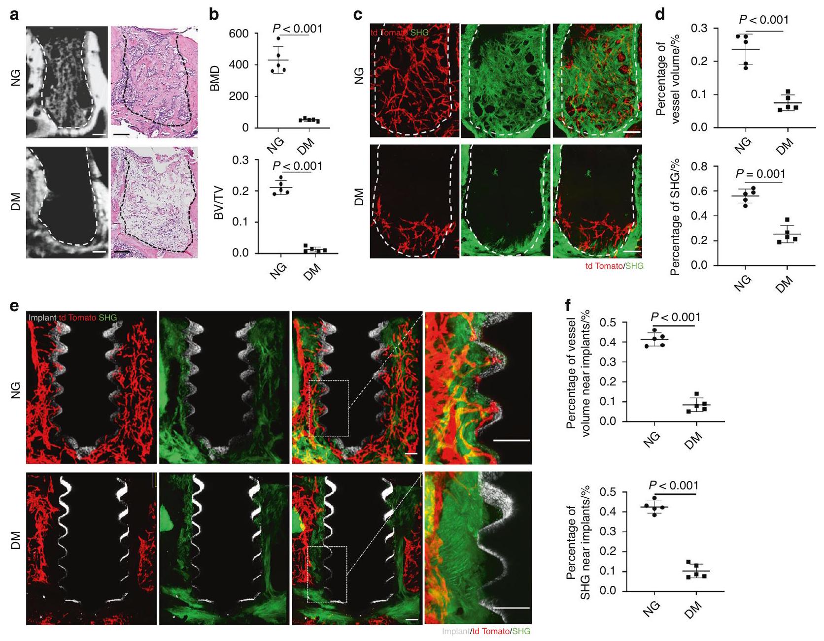
لكشف تأثير السكري على شفاء العظام الفكية، أنشأنا نموذج فئران سكري عن طريق حقن STZ واستخرجنا الأضراس السفلية الأولى. أظهر تحليل Micro-CT أن الفئران DM كانت لديها انخفاض كبير في تكوين العظام مقارنة بالفئران NG. أكدت صبغة H&E للعظام الفكية المنزوعة الكالسيوم الشكل النسيجي للفراغات الفارغة في الفئران DM مقارنة بتكوين العظام الواضح في الفئران NG (الشكل 1أ، ب).
بعد ذلك، سعينا للتحقيق في تأثير السكري على تكوين الأوعية الدموية في شفاء العظام الفكية. أنشأنا فئران معدلة وراثيًا Cdh5Cre ;tdTomato لتعليم الخلايا البطانية. تم معالجة العينات بطريقة تنظيف الأنسجة PEGASOS وتم مسحها بواسطة ميكروسكوب متعدد الفوتونات. لاحظنا أن الفراغات كانت مليئة بخلايا الطماطم الخلايا البطانية في الفئران NG. كانت هذه الخلايا متصلة لتشكيل شبكة ثلاثية الأبعاد وسافرت عبر الكولاجين العظمي المتكون حديثًا، والذي كان باللون الأخضر بواسطة إشارات التوليد الثاني (SHG) (الشكل 1ج). ومع ذلك، في الفئران السكري، كانت خلايا الطماطم منتشرة فقط في قاع الفراغ (الشكل 1ج). كانت نسبة الأوعية الدموية في الفراغ
منخفضة بشكل كبير وكانت إشارات الكولاجين شبه غائبة (الشكل 1ج، د).
وبالمثل، من خلال التصوير العميق لبيئة الزرع، وجدنا أن هناك تجمعًا كبيرًا لخلايا الطماطم ، وتكوين عظام جديدة حول برغي الزرع في الفئران NG، بينما كانت خلايا الطماطم وإشارات الكولاجين الخضراء قد انخفضت أيضًا بشكل كبير في الفئران السكري (الشكل 1هـ، و).

يحسن وظيفة الخلايا البطانية تحت ارتفاع السكر في الدم

بعد ذلك، سعينا للتحقيق في تأثير على الخلايا البطانية من الوريد السري البشري (HUVECs) تحت ارتفاع السكر في الدم. أظهر اختبار مجموعة عد الخلايا 8 (CCK-8) أن تكاثر الخلايا البطانية كان مثبطًا بشكل كبير في اليوم الأول واليوم الثالث في وسط الجلوكوز العالي (HG). في اليوم الأول، أظهرت الخلايا البطانية المعرضة لوسط HG الذي يحتوي على قدرة واضحة على تعزيز تكاثر الخلايا البطانية. بدلاً من ذلك، عندما وصل تركيز إلى 20 مليمول، تم تثبيط تكاثر الخلايا. كانت هذه الظاهرة أكثر وضوحًا في اليوم الثالث ( ) (الشكل 2أ).
للاستكشاف بشكل أكبر التركيز الأمثل لـ لتكوين الأوعية الدموية البطانية تحت ارتفاع السكر في الدم، تم تشكيل أنبوب
الشكل يحسن من خلل وظيفة الخلايا البطانية تحت ارتفاع سكر الدم. اختبار تكاثر الخلايا لـ HUVECs بواسطة اختبار CCK-8 ). ب التحليلات الكمية لطول الأنبوب ( ). صور تمثيلية في اختبار تشكيل الأنابيب أظهرت تكوين الأوعية الدموية في المختبر. مقياس الرسم = تحليلات qRT-PCR للتعبير عن HIF1-a و VEGFA و FGF2 و EGF ). صور تمثيلية لهجرة خلايا HUVECs في اختبار شفاء الجروح. مقياس الرسم . تحليلات كمية لهجرة الخلايا مع نسبة شفاء الجروح )
تم إجراء اختبار لتقييم تكوين الأوعية الدموية في المختبر. مقارنةً بمجموعة الجلوكوز العادي (NG)، أظهرت مجموعة الجلوكوز العالي (HG) تشكيل أنابيب أقل بشكل واضح. ). على الرغم من ، و جميعها قد سهلت تشكيل الأنابيب تحت ارتفاع مستوى السكر في الدم )، الـ أظهرت المجموعة ترقية أعلى مقارنة بالمجموعتين الأخريين (الشكل 2ب، ج). أشارت نتائج qPCR إلى أن يمكن أن ينقذ جزئيًا تعبير الجينات المرتبطة بتكوين الأوعية الدموية، مثل HIF1-α وVEGFA وEGF وFGF2 التي تم قمعها في مجموعة HG (الشكل 2d). بالإضافة إلى ذلك، قمنا بتبني تجارب شفاء الجروح وراقبنا
أن تسارع هجرة خلايا البطانية تحت ارتفاع سكر الدم (الشكل 2e، f).
تركيب وتوصيف زرعات الطلاء بالمغنيسيوم
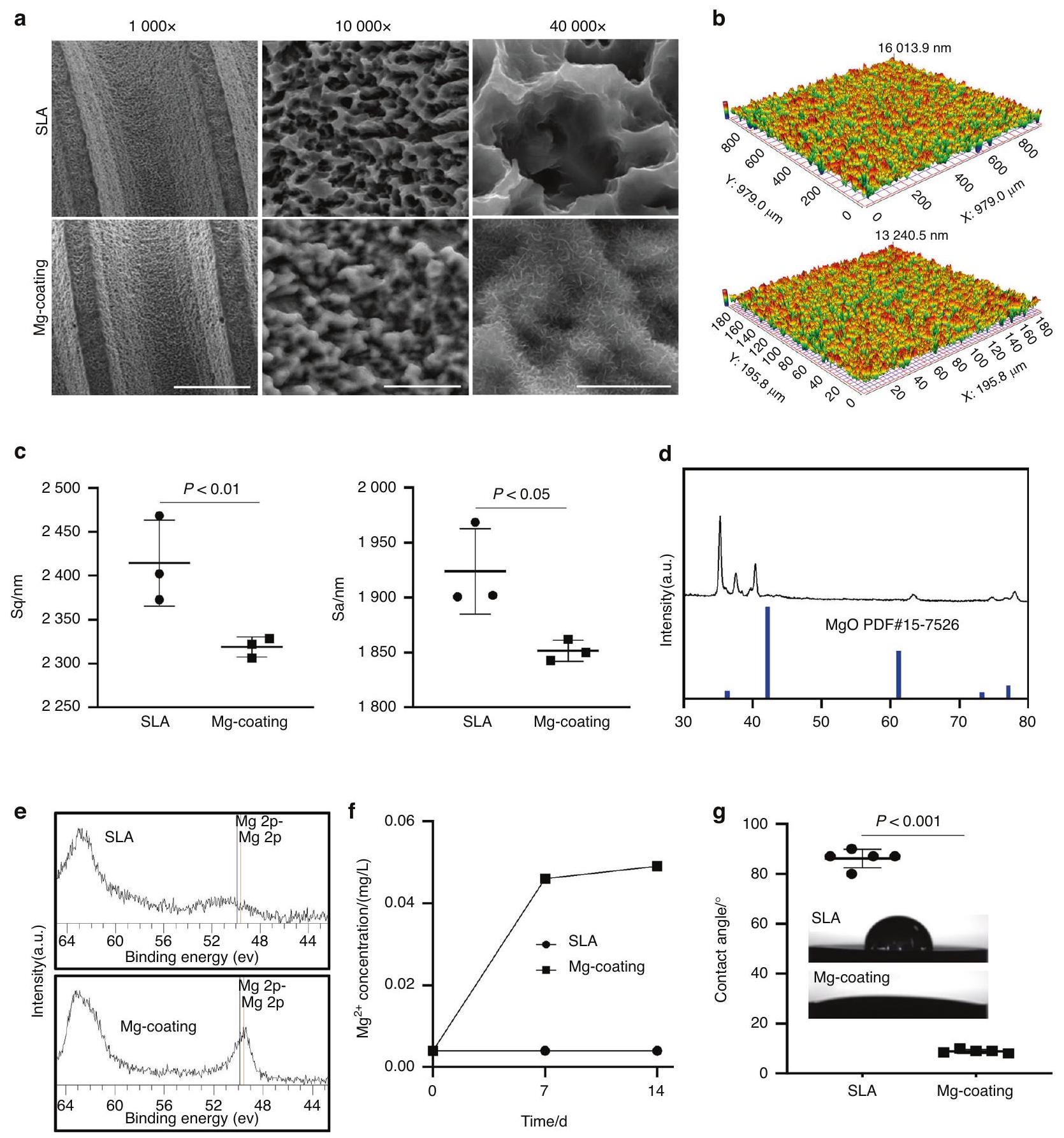
بعد ذلك، قمنا بتطوير زراعة وأوراق مغطاة بمادة المغنيسيوم من خلال التخليق الهيدروحراري. أظهرت مسح SEM أن الطلاء احتفظ بهيكل البناء الملولب للزراعة بينما تم ترسيب أملاح غير عضوية متجانسة على سطح الزراعة في هيكل يشبه النانو، لكنها لم تسد مسام سطح الزراعة SLA تحت تكبير أعلى (الشكل 3أ). السطح
الشكل 3 تخليق وتوصيف زرعات الطلاء المغنيسيوم. أ صور مسح SEM للزرعات. مقياس بدوره. ب، ج. الطبوغرافيا السطحية للعناصر. د، هـ. أظهرت تقنية XPS و XRD التركيب العنصري للعناصر. إصدار . زاوية الاتصال وصور تمثيلية للقطرات على عينات مختلفة )
تم تقليل خشونة المادة قليلاً من إعادة بناء وقياس شكل السطح (الشكل 3ب، ج). تم التحقق من هيكل الطلاء بشكل أكبر بواسطة حيود الأشعة السينية (XRD) حيث تم ملاحظة قمم مميزة نموذجية لـ MgO (PDF#15-7526) عند تم الكشف عنها (الشكل 3d). وبالتالي، تم تأكيد نوعية الطلاءات Mg -coatings، MgO. أكدت تقنية XPS التزيين الناجح لعناصر Mg على الزرعات (الشكل 3e). أظهرت تجارب إطلاق الأيونات التحرر من الطلاءات في سوائل الجسم المحاكاة التي بلغت ذروتها خلال 7 أيام (الشكل 3f). بينما انخفض زاوية تماس الماء (WCA) من إلى في مجموعة الطلاء بمغنيسيوم (الشكل 3 ج)، وقد تم إثبات أن تحسين المحبة للماء يعزز التصاق الخلايا وانتشارها.
أظهرت مجموعة الطلاء بالمغنيسيوم (Mg) زيادة أفضل في تكاثر خلايا البطانية تحت حالة ارتفاع السكر في الدم (الشكل 4أ). وبالمثل، من خلال صبغة الهيكلي الخلوي هوشست/فالايدين، لاحظنا وجود عدد أقل من الخلايا في مجموعة SLA بينما أنقذت مجموعة الطلاء بالمغنيسيوم جزئيًا هذه الظاهرة تحت حالة ارتفاع السكر في الدم (الشكل 4ب). بالإضافة إلى تكوين الأوعية الدموية الذي تساهم فيه خلايا البطانية، فإن تجنيد خلايا سلف العظم حول الزرع وتمايزها إلى خلايا عظمية لعب أيضًا دورًا حاسمًا في الاندماج العظمي. أثبتت النتائج التجريبية أن مجموعة الطلاء بالمغنيسيوم تمتلك بروزًا أفضل للأقدام الكاذبة، وتكاثرًا خلويًا، وتمايزًا عظميًا لخلايا MC3T3-E1 العظمية (الشكل 4ج-ز).
الشكل 4 الاختبار البيولوجي للزرعات. أ اختبار تكاثر الخلايا لخلايا بطانة الأوعية الدموية البشرية (HUVECs) المزروعة على عينات مغطاة بالمغنيسيوم بواسطة اختبار CCK-8 ). ب صور تمثيلية لشكل خلايا HUVECs على عينات مختلفة تحت ظروف NG أو HG. مقياس الرسم . شكل SEM لخلايا MC3T3-E1 العظمية على سطح التيتانيوم وسطح التيتانيوم المطلي بالمغنيسيوم تحت ظروف NG أو HG. مقياس الرسم (علوي)، (أدنى). اختبار تكاثر الخلايا لخلية MC3T3-E1 العظمية بواسطة CCK-8 . e, صور تمثيلية لعلم الأشكال وعدد الشعيرات في خلايا MC3T3-E1 العظمية على سطح التيتانيوم وسطح التيتانيوم المغطى بالمغنيسيوم تحت ظروف الجلوكوز المنخفضة أو العالية. مقياس الرسم . صبغة ALP. h نشاط ALP
يعزز التوعية الدموية والتكامل العظمي في الفئران المصابة بالسكري
للتحقق من تأثير زراعة الطلاء المغنيسيوم على التوعية الدموية والتكامل العظمي في الفئران المصابة بالسكري، قمنا بإدخال الزرعات في Cdh ;فئران tdTomato. بصريًا، أظهر تحليل الأشعة المقطعية أن زراعة الطلاء المغنيسيوم أظهرت معدل أعلى من الاتصال بين العظم والغرسة (BIC) وحجم العظم/حجم الأنسجة (BV/TV) في فئران السكري (الشكل 5 أ، ب). كما أكدت التحليلات الهيستومورفومترية أن هناك المزيد من الجديد
تم ملء نسيج العظام في المنطقة المجاورة لسطح الغرسات في مجموعة الطلاء بالمغنيسيوم (الشكل 5 ج، د). بدوره، المزيد من الطماطم تم تجنيد الخلايا حول الزرعات المطلية بالمغنيسيوم في الفئران المعلّمة بالفلوريسنت. وأشارت هذه الخلايا المتزايدة إلى زيادة في تكوين الأوعية الدموية في الفئران المصابة بالسكري. بينما تم تصور المزيد من الكولاجين العظمي باللون الأخضر كما هو موضح من خلال إشارة SHG حول الزرعات المطلية بالمغنيسيوم في حالات السكري (الشكل 5e، f). بشكل عام، من خلال التحقق من نموذج الحي، أظهرنا قدرة الطلاء بالمغنيسيوم.
شكل. يعزز التوعية الدموية والتكامل العظمي في الفئران المصابة بالسكري. صور تمثيلية لـ إعادة بناء العظم الجديد المتكون (باللون الأزرق) مع الزرعات وبدون الزرعات (باللون الرمادي) بعد 7 أيام من الجراحة. شريط القياس . ب التحليلات الكمية لمعدل الاتصال بين العظم والغرسة (BIC) وحجم العظم/حجم الأنسجة (BV/TV) ( صور تمثيلية مقطعية نسيجية، باللون الأحمر للعظم. مقياس الرسم تحليلات كمية لـ BIC و BV/TV في المقاطع النسيجية أظهرت الصور ثلاثية الأبعاد المعتمدة على إزالة الأنسجة حول الزرعات تكوين الأوعية الدموية وتكوين العظام في اليوم السابع بعد الجراحة، مع عرض إشارة tdTomato للأوعية الدموية (باللون الأحمر)، وإشارة التوليد التوافقي الثاني (SHG) للعظام (باللون الأخضر) والزرعة (باللون الرمادي). شريط القياس . تحليلات كمية لحجم الأوعية والـ SHG حول الزرعات )
زراعة لتعزيز التوعية الوعائية المثبطة والتكامل العظمي في الحالات السكرية.
ومع ذلك، في الفحوصات السابقة، وجدنا تغييرات في التضاريس السطحية والمحبة للماء للمادة. قمنا بإنشاء زراعة مغطاة بالتيتانيوم وأثبتنا أن التضاريس السطحية، والخشونة، وقابلية الرطوبة لم تكن مختلفة عن زراعة مغطاة بالمغنيسيوم (الشكل S2a-e). علاوة على ذلك، أثبتت التجارب الحية أن زراعة المغنيسيوم لا تزال تعزز المحيط حول الزرع.
الاندماج العظمي تحت تأثير السكري مقارنة بزراعة الغرسات المطلية بالتيتانيوم، مؤكدًا التأثير العظمي لـ في حالات السكري مستقلة عن الخشونة وقابلية البلل (الشكل S2f، g).
يقلل من مستويات الإجهاد التأكسدي لخلايا البطانية تحت ارتفاع سكر الدم
تم إجراء تحليل تسلسل RNA لاستكشاف الآلية التي من خلالها تحسين وظيفة الخلايا البطانية تحت
شكل. يقلل من مستويات الإجهاد التأكسدي لخلايا البطانية تحت فرط سكر الدم. أ تحليل إثراء مجموعة الجينات (GSEA) للاستجابة للإجهاد التأكسدي. ب تم تحليل خلايا HUVECs الملونة بـ DCFH-DA بواسطة قياس التدفق الخلوي. تم تحليل خلايا HUVECs الملونة بـ MitoSOX بواسطة تحليل تدفق الخلايا تم تحليل خلايا HUVECs الملونة بمادة Mitotracker بواسطة تحليل تدفق الخلايا ). صور الفلورسنت التمثيلية لتلوين MitoSOX لروابط الأكسجين التفاعلية الميتوكوندرية. شريط القياس . صور الفلورسنت التمثيلية وقياس صبغة ميتوتراكر. شريط القياس تقدير شدة التألق المناعي لتلوين MitoSOX ). ت quantification شدة التألق المناعي لصبغة ميتوتراكر. ). الكشف عن نشاط SOD
ارتفاع سكر الدم. أشار تحليل GESA إلى أن تنظيم الاستجابة للإجهاد التأكسدي وعملية الأيض للجذور الحرة للأكسجين كانت مرتفعة. مجموعة (الشكل 6أ). أظهر تحليل التدفق الخلوي باستخدام ثنائي كلوريد ثنائي هيدروفلوريسئين داي أستات (DCFH-DA) أن مستوى الإجهاد التأكسدي في خلايا البطانة في مجموعة الجلوكوز العالي (HG) قد زاد بشكل ملحوظ، بينما تعافى هذا الارتفاع بعد إضافة (الشكل 6ب). تم إنتاج أنواع الأكسجين التفاعلية داخل الخلايا (ROS) بشكل رئيسي بواسطة الميتوكوندريا. وأشارت المزيد من اكتشافات ROS الميتوكوندري إلى قدرة التطهير على على تراكم ROS الميتوكوندري بواسطة تحليل تدفق الخلايا وصبغ المناعة الفلورية (الشكل 6c، e، g). مرتفع
تؤدي أنواع الأكسجين التفاعلية إلى اضطرابات استقلابية وحتى موت الخلايا من خلال هجوم الميتوكوندريا. وقد أثبت قياس جودة الميتوكوندريا أن أنقذ الكتلة الميتوكوندرية التالفة تحت ارتفاع سكر الدم (الشكل 6د، ف، ح). أظهرت إنزيمات سوبر أكسيد ديسموتاز (SOD)، وهو إنزيم مضاد للأكسدة مهم يتم تنظيمه بواسطة Nrf2 المنوي، نشاطًا معززًا في مجموعة تحت ارتفاع سكر الدم ( ) (الشكل 6i).
عند التعمق في الآلية، وجدنا أن SESN2 كان مرتفعًا بشكل ملحوظ في تسلسل RNA (الشكل 7 أ، ب) والذي ثبت أنه يعزز تحلل Keap1 وينشط نواة Nrf2 لحماية الخلايا من الإجهاد التأكسدي.
شكل. يزيد من تنظيم SESN2 في خلايا HUVEC تحت ارتفاع مستوى السكر في الدم. بركان من الجينات المعبر عنها بشكل مختلف. خريطة حرارية للجينات التمثيلية المعنية بالاستجابة للإجهاد التأكسدي. ج. نتائج Western blot لـ SESN2 و KEAP1 و NRF2 النووي و NRF2 الكلي في خلايا HUVECs. د. صور فلورية تمثيلية لتلوين MitoSOX ل ROS الميتوكوندري. شريط القياس اختبار تكاثر الخلايا لـ HUVECs بواسطة اختبار CCK-8 ). أظهرت الصور التمثيلية والتحليلات الكمية في اختبار تشكيل الأنابيب تكوين الأوعية الدموية في المختبر. مقياس الرسم ( )
ثم قمنا بتقليل SESN2 في خلايا HUVECs باستخدام الأحماض النووية الصغيرة المتداخلة (siRNA) وأكدت تقنية الويسترن بلوت انخفاض Keap1 وزيادة تنظيم Nrf2 النووي في تم عكس المجموعة بعد استنفاد SESN2 (الشكل 7c). كما أثبتت صبغة المناعة الفلورية تراكم ROS الميتوكوندري (الشكل 7d) وكانت وظائف الخلايا بما في ذلك التكاثر وتكوين الأوعية الدموية في المختبر متأثرة أيضًا في المجموعة بعد استنفاد SESN2 (الشكل 7e، f).

نقاش

إعادة التوعية بعد الصدمة هي خطوة حاسمة في شفاء العظام، حيث توفر الأكسجين والمواد الغذائية والأيونات اللازمة لتمييز الخلايا الجذعية إلى خلايا عظمية وتعدين المصفوفة من أجل
إصلاح الأضرار. في عملية تطوير العظام وإصلاحها، يرتبط تكوين الأوعية الدموية ارتباطًا وثيقًا بتكوين العظام. ومع ذلك، يؤدي السكري إلى مضاعفات وعائية متنوعة ويعيق تكوين الأوعية الدموية، مما يؤدي إلى ضعف شفاء الجروح. تشير بياناتنا أيضًا إلى أن مرض السكري يؤثر بشكل خطير على التوعية وتجديد العظام في شفاء العظام السنخية، مما يدعم بقوة العمل الذي قدم تأخير الشفاء وزيادة تدمير العظام السنخية من قبل ديفلين وزملائه. في السنوات الأخيرة، تم إجراء العديد من الدراسات لتعزيز شفاء الأنسجة الرخوة والصلبة لدى مرضى السكري من خلال تعزيز تكوين الأوعية الدموية.
لقد تم الاعتراف به على نطاق واسع في تعزيز تكوين العظام. مؤخراً، قام هوانغ وآخرون (2021) بإنشاء لاصقة ميكرونيدل متعددة الوظائف تعتمد على إطار عضوي من المغنيسيوم لتسريع شفاء الجروح لدى مرضى السكري من خلال تسريع تكوين الأوعية الدموية. ليو
قام نت وآخرون (2023) أيضًا بإنشاء هيدروجيل يحتوي على المغنيسيوم لتعزيز تكوين الأوعية الدموية في الجروح السكرية لتعزيز الشفاء. ومع ذلك، في الوقت الحاضر، سواء يمكن أن يعزز التوعية الوعائية حول الزرع والتكامل العظمي تحت تأثير السكري، وآليته الداخلية لا تزال غير معروفة. في دراستنا، تم إثبات ذلك لأول مرة لتحسين التوعية الوعائية حول الزرع والتكامل العظمي في الفئران المصابة بالسكري.
تُعتبر خلايا البطانة (ECs) من ناحية أخرى القوة الدافعة وراء تكوين الأوعية الدموية، بسرعة بدء تشكيل أوعية دموية جديدة أثناء شفاء الجروح. في مرض السكري، تؤدي وظيفة الخلايا البطانية المتضررة إلى اضطراب في تكوين الأوعية الدموية، وقد أظهرت تجاربنا في المختبر أن يمكن أن يحسن تكاثر خلايا البطانية، وتشكيل الأنابيب، والهجرة تحت ظروف ارتفاع سكر الدم.
تم إثبات أن اضطرابات خلايا البطانية الناتجة عن ارتفاع مستوى السكر في الدم مرتبطة بشكل رئيسي بزيادة مستويات الإجهاد التأكسدي داخل الخلايا ووجود فائض من السوبر أكسيد داخل الخلايا. مما أدى إلى خلل وظيفي. بينما كانت الميتوكوندريا مصدرًا مهمًا للجذور الحرة داخل الخلايا، زيادة تدرج البروتونات داخل الميتوكوندريا في حالة فرط سكر الدم تعيق نقل الإلكترونات من الإنزيم المساعد Q المختزل (يوبيكوينون) إلى معقد سلسلة نقل الإلكترون III. بدلاً من ذلك، يتم نقل الإلكترونات إلى الأكسجين الجزيئي، مما يؤدي إلى إنتاج سوبر أكسيد. تدمر الجذور الحرة الزائدة البروتينات الخلوية، والدهون، والميتوكوندريا، والحمض النووي، مما يؤدي إلى أضرار قاتلة للخلايا.
أظهرت تسلسلات RNA لدينا أن إضافة زاد من عملية الأيض لجذور الأكسجين التفاعلية في خلايا البطانية، بينما دعمت اكتشاف ROS في خلايا البطانية القدرة على لتقليل مستوى الإجهاد التأكسدي الميتوكوندري المرتفع تحت حالة فرط سكر الدم.
من خلال استكشاف نتائج التسلسل بشكل أعمق، وجدنا أن تعبير SESN2 و ATF4 في الـ زادت المجموعة بشكل كبير. ATF4 هو جين رئيسي في تنظيم الإجهاد الميتوكوندري ويشارك في نسخ الجينات للاستجابات المضادة للأكسدة، والالتهام الذاتي، وتخليق الأحماض الأمينية، والنقل. أظهرت الدراسات أن زيادة تنظيم ATF4 يمكن أن تخفف من إنتاج ROS الميتوكوندري والأضرار الميتوكوندرية الناتجة عن ROS. SESN2 هو بروتين مستحث بسبب الإجهاد يمكنه زيادة الانتقال النووي لـ Nrf2 وتحسين النشاط النسخي الذي يتوسطه Nrf2/عنصر الاستجابة لمضادات الأكسدة (ARE)، مما يؤدي إلى تعزيز القدرة المضادة للأكسدة. Nrf2 هو عامل نسخ مهم في تنظيم الإجهاد التأكسدي الخلوي وموحد مركزي للحفاظ على توازن الأكسدة والاختزال في الخلايا. في الظروف العادية، سيتم تعديل Nrf2 بواسطة يوبكويتين Keap1 المعقد وتفكيكه في السيتوبلازم، بعد أن يتم نقل Nrf2 إلى النواة، سيشكل معقدًا مع المحفزات ويرتبط بمناطق المحفز (AREs)، مما يؤدي إلى تحفيز وتنظيم التعبير التكويني والقابل للتحفيز لسلسلة من البروتينات المضادة للأكسدة، والتي يمكن أن تقلل من تلف الخلايا الناجم عن أنواع الأكسجين التفاعلية والإلكتروفيلات، وتحافظ على الخلايا في حالة مستقرة، وتحافظ على توازن الأكسدة والاختزال.
باستخدام الأحماض النووية الصغيرة المتداخلة (siRNA) قمنا بإزالة SESN2 في خلايا HUVECs. وأكدت زيادة تعبير Keap1 وانخفاض Nrf2 النووي انخفاض Keap1 بعد إزالة SESN2 في أظهر المجموعة بقوة أن تنظم تحلل Keap1 وتكوين Nrf2 من خلال زيادة SESN2. وأثبتت المزيد من الكشف عن زيادة ROS الميتوكوندري وخلل في تكاثر الخلايا وتكوين الأوعية الدموية في المختبر. استجابت مضادات الأكسدة المتوسطة وأصلحت خلل الخلايا البطانية من خلال SESN2. ومع ذلك، لم يمنع غياب SESN2 تمامًا تأثير على خلايا البطانة تحت ارتفاع سكر الدم، مما يشير إلى أن آليات تنظيمية أخرى مثل ATF4 أو SLC7A11 قد تؤثر. في بيانات التسلسل، تم أيضًا زيادة تعبير عائلة ناقلات المواد الذائبة 7 العضو 11 (SLC7A11)، وهو أحد أهم المنظمين العلويين لعملية تكوين الحديد، بشكل ملحوظ. بالاقتران مع بياناتنا التجريبية التي عزز بقاء خلايا البطانية عالية الجلوكوز، قلل من الخلوية
المغنيسيوم يعزز تكوين الأوعية الدموية والتكامل العظمي في حالات السكري ليو وآخرون.
مستويات الإجهاد التأكسدي، تحسين جودة الميتوكوندريا، زيادة نواة Nrf2 upstream من SLC7A11 وزيادة مستويات ATF4، افترضنا أن قد يعيق تكوين الحديد في خلايا بطانة الأوعية الدموية ذات الجلوكوز العالي، مما يتطلب أيضًا مزيدًا من التحقق.
في الوقت نفسه، تعتبر واجهة الزرع والأنسجة قضية رئيسية في أبحاث الزرع. نظرًا لقيود الوسائل التقنية التقليدية، كان من الصعب ملاحظة التغيرات في واجهة الزرع والتكامل العظمي بصريًا في ثلاثة أبعاد. استنادًا إلى تقنية توضيح الأنسجة التي طورتها مجموعتنا البحثية سابقًا، لقد لاحظنا بصريًا التغيرات في البيئة الميكروية ثلاثية الأبعاد المحيطة بالزرع في الفئران المصابة بالسكري للمرة الأولى من خلال الحفاظ على تعبير الفلورسنت الداخلي. أثبتت بياناتنا بشكل عميق أن السكري يؤثر بشكل خطير على التوعية وتجديد العظام في شفاء العظام السنخية، بينما كانت أيضًا المرة الأولى التي يتم فيها عرض ذلك بشكل شامل وثلاثي الأبعاد حيث تم تثبيط تكوين الأوعية الدموية في تجويف استخراج الأسنان لدى مرضى السكري واندماج الزرع العظمي. بالطبع، في بناء المادة قمنا أيضًا بتغيير خشونة السطح وقابلية البلل، والتي تم الإبلاغ عن تأثيرها على تكوين الأوعية الدموية وتكوين العظام. من أجل استبعاد تأثيرات الخشونة وقابلية البلل، استخدمنا ثاني أكسيد التيتانيوم كطلاء لتحقيق خشونة وقابلية بلل مشابهة لطلاء المغنيسيوم. في التجارب الحية، وجدنا أن زيادة قابلية البلل يمكن أن تزيد قليلاً من تكوين العظام، لكن طلاء المغنيسيوم لا يزال يظهر تأثيرًا كبيرًا في تعزيز الاندماج العظمي حول الزرع. بالاقتران مع تجاربنا في علم الخلايا، تمت إضافتها إلى الوسط لتأكيد تأثير المغنيسيوم على الخلايا البطانية. وقد أكدنا أيضًا أن لعب دورًا حاسمًا في تعزيز تكوين الأوعية الدموية وتكوين العظام، مستقلًا عن العوامل الأخرى. ومع ذلك، في هذه الدراسة، ركزنا على تعزيز التوعية الدموية وتكوين العظام في حالات السكري، لذلك لم يكن هناك مجموعة ضابطة من الحيوانات ذات مستويات السكر الطبيعية مع زراعة مغطاة بالمغنيسيوم. كما أننا افتقرنا إلى التجارب التي تستهدف حذف SESN2 أو NRF2 في الفئران لدعم نتائجنا بشكل أكبر، مما يتطلب مزيدًا من الاستكشاف.
باختصار، أظهرنا أن عززت نواة Nrf2 من خلال زيادة تنظيم SESN2، مما أدى إلى تقليل مستوى الإجهاد التأكسدي الميتوكوندري في خلايا البطانية تحت ارتفاع مستوى السكر في الدم في المختبر وتعزيز تكوين الأوعية الدموية حول الزرع والتكامل العظمي في الفئران المصابة بالسكري.

المواد والطرق

فئران

تتوافق الدراسة مع إرشادات ARRIVE. تم مراجعة جميع البروتوكولات لرعاية الحيوانات والتجارب والموافقة عليها من قبل اللجنة الفرعية للبحث ورعاية الحيوانات في جامعة سيتشوان. كانت جميع الفئران من سلالة C57BL/6 وتم إيواؤها في بيئة خالية من مسببات الأمراض المحددة مع دورة ضوء وظلام مدتها 12 ساعة. كانت Rosa26-tdTomato (007905) من مختبر جاكسون. Cdh5 تم توفير الفئران بلطف من قبل مختبر البروفيسور بي-سن دينغ (المختبر الوطني الرئيسي للعلاج الحيوي، جامعة سيتشوان). Cdh5Cre تم إنتاج فئران tdTomato عن طريق تزاوج Cdh5-Cre تم حقن 1.5 ملغ من التاموكسيفين لكل 10 جرام من الفئران Rosa26-tdTomato عن طريق البطن يوميًا لمدة يومين متتاليين لإضاءة الفلورية قبل الجراحة.

بناء نموذج السكري

تم حقن STZ (شركة شنغهاي ماوكينغ للتكنولوجيا الحيوية المحدودة) داخل الصفاق تم حقنها في الفئران بشكل متتالي لمدة 5 أيام لتحفيز نموذج. مع وصول مستوى الجلوكوز في المصل إلى لمدة أسبوعين، تم إجراء الجراحة (الشكل S1).

الجراحة

تمت إزالة الأضراس السفلية الأولى تحت التخدير باستخدام إبرة حقن 26 G وملاقط. من أجل وضع الزرع الفوري، تم استخدام زرع تيتانيوم مصمم ذاتيًا معالج بالرمل، بحبيبات كبيرة ومحمض (SLA). قطر؛ WEGO، الصين) كان
تم تثبيتها في المقبس بعد الاستخراج تحت المجهر الاستريو. في اليوم السابع بعد الاستخراج أو الزرع، تم جمع الفكوك.
عملية تنظيف الأنسجة PEGASOS
تم تنفيذ عملية تنظيف الأنسجة PEGASOS كما تم الإبلاغ عنها سابقًا.
اكتساب الصور وتحليلها
تم الحصول على الصور من خلال مجهر متعدد الفوتونات (Lecia SP8 DIVE). تم الحصول على إشارات TdTomato تحت خط ليزر تحفيز بطول موجي 561 نانومتر. تم الحصول على الزرع باستخدام ليزر ياقوتي بطول موجي 950 نانومتر. تم الحصول على تصوير الإشارات التوافقية الثانية (SHG) باستخدام كاشف غير مزال. تم استخدام برنامج IMARIS (الإصدار 9.1.2؛ أكسفورد إنسترومنتس) لتحليل البيانات ومعالجة الصور. منطقة تم استخدام المنطقة القريبة من أخاديد الخيوط لكل زرعة للتحليلات. لكل عينة، تم استخدام ثلاث مناطق مختارة عشوائيًا للتquantification.
صور وتحليلات التصوير المقطعي المحوسب الدقيق
تم تثبيت الفكوك المحصودة مع الزرعات في 4% PFA عند لمدة 24 ساعة ثم تم تخزينه في يرجى العثور على قبل أن يتم مسحها. تم مسح العينات باستخدام نظام (Scanco Medical) بدقة متوسطة وحجم فوكسل من تم استخدام برنامج IMARIS (الإصدار 9.1.2؛ أكسفورد إنسترومنتس) لتحليل البيانات ومعالجة الصور.
تحضير الأنسجة وصبغها
الفكوك مع الزرعات كانت مثبتة في يرجى الاطلاع على المرفق لمدة يوم واحد، تليها 4 أسابيع من إزالة الكالسيوم في 10% EDTA (pH 7.4) عند مع الاهتزاز. تم تجفيف الفكوك لصبغة الهيماتوكسيلين والإيوزين (H&E) في إيثانول متدرج وتم تضمينها في البارافين. تم قطع كتل البارافين بواسطة ميكروتوم (لايكا RM2255) إلى -شرائح سميكة. تم إجراء صبغ H&E وفقًا لتعليمات الشركة المصنعة (Biosharp). منطقة تم استخدام المنطقة القريبة من أخاديد الخيوط لكل زرعة للتحليل. لكل عينة، تم استخدام ثلاث مناطق مختارة عشوائيًا للتquantification.
زراعة الخلايا
تم زراعة خلايا بطانة الوريد السري البشري (HUVECs) المقدمة من المختبر الرئيسي للدولة للعلاج الحيوي، مستشفى غرب الصين الثاني الجامعي، جامعة سيتشوان، في وسط دلبوكو المعدل (DMEM) (هايكلاون، مختبرات، لوغان، يوتا، الولايات المتحدة الأمريكية) يحتوي على 10% من مصل الجنين البقري (جيبكو، غراند آيلاند، نيويورك، الولايات المتحدة الأمريكية)، البنسلين (هايكلاون، سايتيفا، مارلبورو، ماساتشوستس، الولايات المتحدة الأمريكية) و ستربتوميسين (هايكلاون، سايتيفا، مارلبورو، ماساتشوستس، الولايات المتحدة الأمريكية). تم تغيير وسائط الثقافة كل يومين. تم زراعة خلايا الأوعية الدموية البشرية المأخوذة من الحبل السري في ثلاثة ظروف مختلفة: جلوكوز طبيعي ( ) ، سكر مرتفع ( )، و ، الصين).
تم زراعة خلايا العظم MC3T3-E1 في وسط إيغلي المعدل ألفا (a-MEM) (Hyclone، المختبرات، لوغان، يوتا، الولايات المتحدة الأمريكية) وتم تغييره كل يومين.
مجموعة عد الخلايا – اختبار 8
تم زراعة خلايا HUVECs في لوحة 96 بئر بكثافة 5000 خلية لكل بئر. ثم تم إضافة وسط NG أو وسط HG أو وسط HG المحتوي على ، أو تمت الإضافة. تم إجراء اختبار مجموعة عد الخلايا-8 (CCK-8) بعد 24 ساعة و72 ساعة بشكل منفصل. تم قياس قيم الكثافة الضوئية (OD) بواسطة مطياف ضوئي (ثيرمو فيشر ساينتيفيك، وولثام، ماساتشوستس، الولايات المتحدة الأمريكية) عند 450 نانومتر.
اختبار تشكيل الأنبوب
تم إضافة هلام ماتريجيل المخفف بعوامل النمو (BD Biosciences، الولايات المتحدة الأمريكية) إلى كل بئر من آبار لوحة 96 بئر، ثم تم وضعها في تم وضعها في الحاضنة لمدة 30 دقيقة حتى التصلب. بعد ذلك، تم زراعة خلايا HUVECs (5000 خلية لكل بئر) على ركائز ماتريجيل المتصلبة وزراعتها في وسط NG الخالي من المصل، أو وسط HG، أو وسط HG.
يحتوي على ، و . بعد 6 ساعات من الحضانة ( تم تصوير الخلايا باستخدام ميكروسكوب فلوري مقلوب (أوليمبوس، اليابان). تم قياس طول الأنبوب باستخدام برنامج ImageJ (NIH، الولايات المتحدة الأمريكية).

تفاعل البوليميراز المتسلسل العكسي الكمي (qRT-PCR)

تم زراعة خلايا HUVECs في وسط NG أو وسط HG أو وسط HG يحتوي على لمدة 24 ساعة. ثم تم استخراج RNA الكلي من خلايا HUVECs باستخدام كاشف Trizol (Invitrogen، كارلسباد، كاليفورنيا، الولايات المتحدة الأمريكية). تم إجراء النسخ العكسي للحمض النووي المكمل (cDNA) من من RNA باستخدام مجموعة PrimeScript RT (تاكارا بيو، أوساكا، اليابان)، وتم قياس الجينات المعنية بواسطة SYBR Premix Ex Taq II (تاكارا) في LightCycler 96 (روش، بازل، سويسرا). تم تحليل التعبير النسبي للميكرو RNA (mRNA) باستخدام دورة العتبة المقارنة دلتا دلتا. طريقة عن طريق التطبيع باستخدام إنزيم الجليسرالديهيد-3-فوسفات ديهيدروجيناز (GADPH).

اختبار شفاء الجروح الخدش

تم زراعة خلايا HUVECs في طبق 6 آبار وزرعها حتى التقاء، و تم استبدال وسط FBS بوسط 1% FBS لزراعة الجوع بعد 24 ساعة من الخدوش. تم استخدام طرف الماصة لتتبع طول مركز الثقب على طول المسطرة، مما ترك خدوشًا متساوية ومستقيمة. بعد ذلك، تم استبدال الوسط بوسط NG أو وسط HG أو وسط HG المحتوي على تمت ملاحظة وإلتقاط صورة لإغلاق الجرح بعد 12 ساعة و 24 ساعة. تم استخدام برنامج ImageJ لحساب نسبة منطقة الشفاء.

خفض الجين

قمنا بتخفيف ثنائي السلسلة من siRNA إلى DMEM كحل ومخفف ليبوفيكتامين آر إن إيه آي ماكس (إنفيتروجين، الولايات المتحدة الأمريكية) في DMEM للحل . و تم خلطها وحضانتها لمدة 30 دقيقة عند . عندما وصلت الخلايا إلى 40 إلى التقاء تم إضافة المزيج إلى 2 مل من وسط النمو العادي بدون مضادات حيوية لمدة 12 ساعة في .

تصنيع زرعات بطبقة مغنيسيوم وطبقة تيتانيوم

للتخليق الهيدروحراري، محلول مائي من MgO (أو ) وتم تحضير NaOH في ماء منزوع الأيونات. يتم خلط المتفاعلات تمامًا وتم دمج المحاليل ثم تم تحريكها في درجة حرارة الغرفة لمدة 5 دقائق. تم نقل المحلول النهائي إلى مفاعل هيدروحراري وتم وضع التيتانيوم المعالج في المفاعل في نفس الوقت. تم إغلاق الأوتوكلاف الهيدروحراري وإعداده لـ لمدة 12 ساعة. ثم تم غسل الأطباق، على التوالي، بالماء المقطر والإيثانول مرتين لمدة 10 دقائق. تم وضع الأطباق في فرن وتسخينها عند لمدة ساعة واحدة للتجفيف.

توصيف المواد للعينات

تم مسح التضاريس السطحية للعينات بواسطة مجهر إلكتروني ذو انبعاث ميداني (FE-SEM؛ JSM-7500 F، JEOL، اليابان). تم إجراء التحليل العنصري بواسطة EDS. تم استخدام Superview W1 (الصين) للكشف عن التضاريس السطحية. تم استخدام XRD (LabX، شيمادزو، اليابان) لمسح هياكل العينات. تم قياس التركيب العنصري والحالة الكيميائية للأسطح بواسطة XPS (AXIS Supra، كراتوس، الولايات المتحدة الأمريكية). تم قياس زاوية تماس الماء بواسطة جهاز WCA (Zhongchen Digital Technic Apparatus Co.، شنغهاي، الصين). تم استخدام مطيافية انبعاث الذرة بالتحليل الطيفي البلازمي المقترن (ICP-AES، 5100 SVDV، الولايات المتحدة الأمريكية) لتقييم الإفراج عن في PBS.

اختبار حيوية الخلايا

خلايا HUVEC أو خلايا MC3T3-E1 العظمية تم زراعة (خلايا لكل بئر) على ألواح NG + SLA و HG + SLA و HG + ألواح الطلاء Mg في ألواح 48 بئر لمدة 24 ساعة. تم إجراء اختبار مجموعة عد الخلايا-8 (CCK-8) كما في السابق.
تم استخدام صبغة الفلويودين (شركة شنغهاي ماوكينغ للتكنولوجيا الحيوية المحدودة) لمراقبة شكل الخلايا والهيكل الخلوي. بعد 24 ساعة من الحضانة، كانت الخلايا
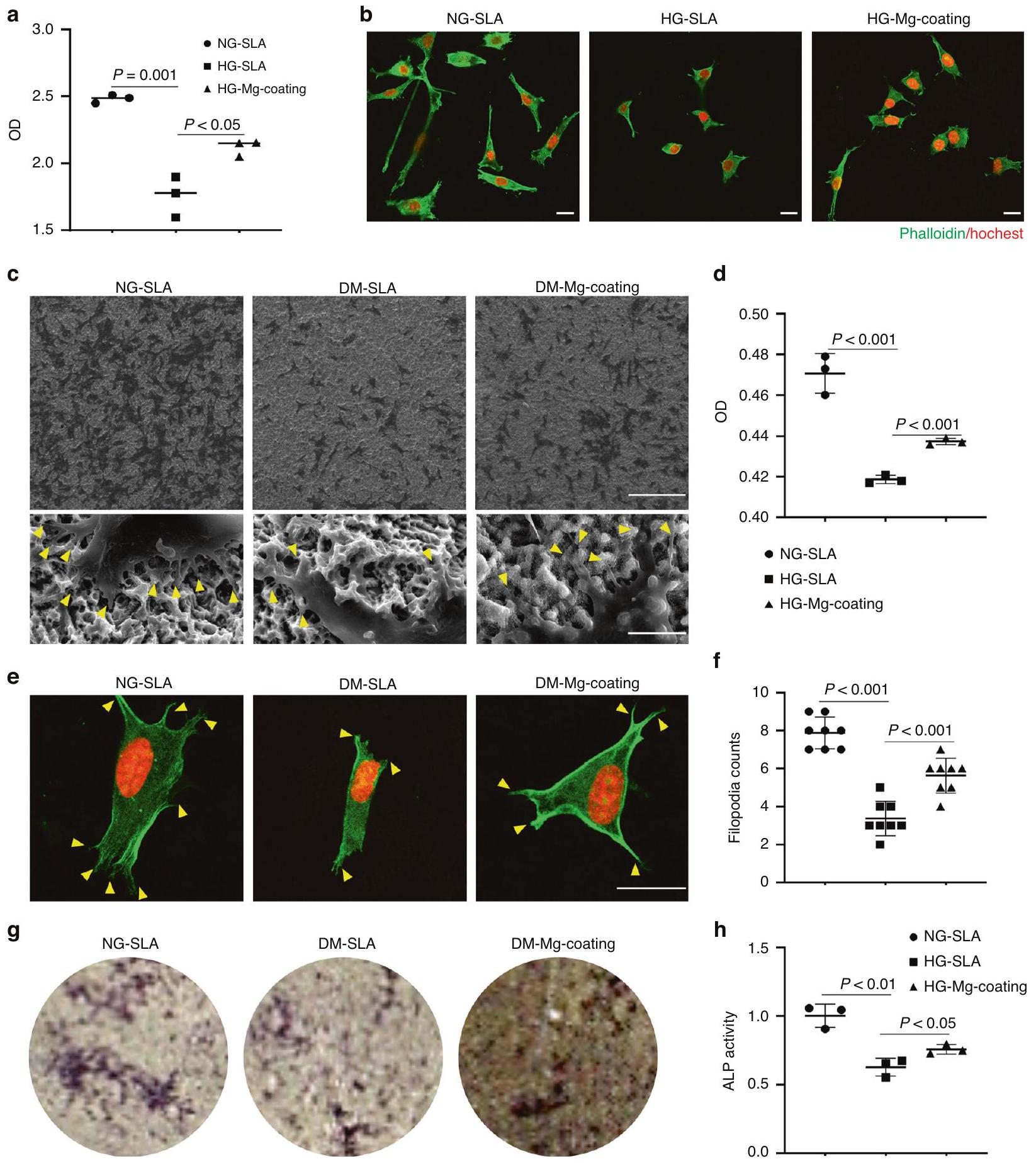
تم تثبيت الخلايا باستخدام 4% PFA لمدة 10 دقائق وتم صبغ الهيكل الخلوي بواسطة الفالودين وصبغ النوى بواسطة DAPI. تم الكشف عن التصوير الفلوري للخلايا بواسطة المجهر الضوئي الماسح بالليزر (FV3000؛ أوليمبوس، اليابان). بعد يوم واحد من الزراعة على الركائز، تم تثبيت الخلايا طوال الليل باستخدام جلوتارالدهيد. ثم، تم تجفيف العينات بشكل متسلسل في إيثانول متدرج ( ، ) لمدة 15 دقيقة لمراقبة SEM.

صبغة الفوسفاتاز القلوية

تم زراعة خلايا MC3T3-E1 العظمية في وسط تكوين العظام على الركائز لمدة 7 أيام وثُبتت باستخدام 4% PFA لمدة 20 دقيقة. ثم تم إجراء الصبغ باستخدام مجموعة اختبار الفوسفاتاز القلوي التجارية (Beyotime Biotechnology، شنغهاي، الصين). تم قياس نشاط ALP باستخدام مجموعة تجارية (Beyotime Biotechnology، شنغهاي، الصين) وتم الكشف عنه بواسطة مقياس الطيف الضوئي (Thermo Fisher Scientific، الولايات المتحدة الأمريكية) عند 450 نانومتر.

تحليلات نسيجية وهستومورفومترية

بعد 7 أيام من جراحة الزرع، تم جمع الفكوك التي تحتوي على الزرعات وتثبيتها في فورمالين مخفف لمدة أسبوع واحد. تم تجفيف العينات بواسطة تركيزات متزايدة من الإيثانول ( ) ومثبتة في راتنج الإيبوكسي القابل للتصلب بالضوء (TechnoVit7200VLC؛ هيريوس-كولزر، ويرهايم، ألمانيا). تم قطع العينات بشكل متوازي مع المحور الطويل للزرع. ثم تم طحن العينات إلى حوالي سمك باستخدام آلة طحن (ExaktApparatebau، نوردشتيدت، ألمانيا). تم صبغ المقاطع بصبغة ستيفينيل الزرقاء وصبغة فان جيزون، ثم تم ملاحظتها تحت المجهر الضوئي (أوليمبوس، اليابان).

تسلسل RNA وتحليل البيانات

تم معالجة خلايا HUVECs بوسط HG أو وسط HG يحتوي على لمدة 48 ساعة ( في كل مجموعة). تم استخراج RNA الكلي باستخدام كاشف Trizol (Invitrogen، كارلسباد، كاليفورنيا، الولايات المتحدة الأمريكية) وتم تنقيته باستخدام كرات مغناطيسية مرتبطة بـ poly-T oligo. تم استخدام FastQC (SAndrews، الإصدار 0.11.5) لضمان جودة بيانات تسلسل RNA (RNA-Seq) ومن خلال استخدام HISAT2 (DaehwanKimLab، الإصدار 2.0.5) قمنا بتوجيهها إلى جينومات مرجعية لموس موسكولوس. تم اعتبار الجينات معبرة بشكل مختلف بشكل كبير إذا أظهرت تغييرًا في الطي و -قيمة لتحليل إثراء مجموعة الجينات (GSEA)، تم تنزيل قوائم الجينات من قاعدة بيانات GSEA.www.gsea-msigdb.org/تمت معالجة GSEA باستخدام برنامج GSEA (https:// www.gsea-msigdb.org/gsea/index.jsp).

تحليل تدفق الخلايا

تم معالجة خلايا HUVECs بوسط NG، أو وسط HG، أو وسط HG يحتوي على لمدة 48 ساعة. تم إجراء صبغ – ثنائي كلوريد ثنائي هيدروفلوريسئين داي أستات (DCFH-DA) وصبغ ميتو سوكس وفقًا لبروتوكول معدل. ثم تم اختبار العينات بواسطة تحليل تدفق الخلايا (BD Biosciences، الولايات المتحدة الأمريكية)، وتم تحليل بيانات تحليل تدفق الخلايا باستخدام برنامج فلو جو.

البلوت الغربي

تم معالجة خلايا HUVECs بوسط HG أو وسط HG يحتوي على لمدة 48 ساعة. تم استخراج البروتينات باستخدام مجموعة استخراج البروتين (Signalway Antibody، غرينبيلت، ماريلاند، الولايات المتحدة الأمريكية). تم قياس تركيزات البروتينات باستخدام مجموعة اختبار بروتين BCA (Pierce، روكفورد، إلينوي، الولايات المتحدة الأمريكية؛ 23225). تم فصل البروتين بالتساوي في هلام بولي أكريلاميد SDS (SDS-PAGE) ونُقل إلى أغشية فلوريد البولي فينيليدين (PVDF) (ثيرمو فيشر ساينتيفيك، وولثام، ماساتشوستس، الولايات المتحدة الأمريكية). بعد الحجب في تم غمر الأغشية في حليب خالي من الدسم مع الأجسام المضادة الأولية والأجسام المضادة الثانوية المقابلة. تم تطوير البروتينات بواسطة نظام تصوير الهلام (Bio-Rad Laboratories، هيركوليس، كاليفورنيا، الولايات المتحدة الأمريكية) مع مواد Immobilon (Millipore، بيليريكا، ماساتشوستس، الولايات المتحدة الأمريكية).

اختبار نشاط SOD

تم إجراء اختبار نشاط SOD كما هو موضح (بيوتايم، الصين).

التحليلات الإحصائية

تم التعبير عن جميع البيانات كمتوسط الانحراف المعياري (SD). تم إجراء ثلاثة تجارب مستقلة على الأقل لكل مجموعة مع عينات مكررة. تم إجراء التحليل الإحصائي باستخدام تحليل التباين الأحادي (ANOVA) متبوعًا باختبار المقارنات المتعددة بونفيروني. اعتُبر ذا دلالة إحصائية.

توفر البيانات والمواد

تم إيداع مجموعة بيانات تسلسل RNA في قاعدة بيانات NCBI مع المعرف GSE236139.

شكر وتقدير

يشكر المؤلفون الدكتور تشيانغ قوه من المختبر الوطني الرئيسي لأمراض الفم، جامعة سيتشوان، على المساعدة في تصوير الميكرو-سي تي. نشكر الدكتور تشاويانغ تشانغ من المختبر الوطني الرئيسي لأمراض الفم، جامعة سيتشوان، على المساعدة في الميكروسكوب الإلكتروني الماسح. نشكر الدكتور يينغ هوي وين والدكتور مانلو وانغ من المختبر الوطني الرئيسي لأمراض الفم، جامعة سيتشوان، على المساعدة في الميكروسكوب متعدد الفوتونات. تم دعم هذا العمل من خلال منح من المؤسسة الوطنية للعلوم الطبيعية في الصين (رقم 81901042)، وبرنامج العلوم والتكنولوجيا في سيتشوان (رقم 2022NSFSC1384)، وفريق دولي لزراعة الأسنان (رقم 1477_2020).

مساهمات المؤلفين

ساهم L. Liu في الفكرة، التصميم، جمع البيانات، التحليل والتفسير، وصاغ وراجع المخطوطة بشكل نقدي؛ ساهم W.S. في تفسير البيانات، وصاغ وراجع المخطوطة بشكل نقدي؛ ساهم D.Z. في تفسير البيانات، وصاغ وراجع المخطوطة بشكل نقدي؛ ساهم W.L. في تحليل البيانات، والتفسير، وصاغ وراجع المخطوطة بشكل نقدي؛ ساهم Q. Yin في تحليل البيانات، والتفسير، وصاغ وراجع المخطوطة بشكل نقدي؛ ساهم Q.W. في تحليل البيانات، وصاغ وراجع المخطوطة بشكل نقدي؛ ساهم H.L. في تحليل البيانات، وصاغ وراجع المخطوطة بشكل نقدي؛ ساهم Q. Yuan في الفكرة، التصميم، وصاغ وراجع المخطوطة بشكل نقدي؛ ساهم S. Zhang في الفكرة، التصميم، وصاغ وراجع المخطوطة بشكل نقدي. جميع المؤلفين أعطوا الموافقة النهائية ووافقوا على تحمل المسؤولية عن جميع جوانب العمل.

معلومات إضافية

معلومات إضافية النسخة الإلكترونية تحتوي على مواد إضافية متاحة فيhttps://doi.org/10.1038/s41368-023-00271-y.
المصالح المتنافسة: يعلن المؤلفون عدم وجود مصالح متنافسة.

REFERENCES

  1. Buser, D., Sennerby, L. & De Bruyn, H. Modern implant dentistry based on osseointegration: 50 years of progress, current trends and open questions. Periodontology 73, 7-21 (2017).
  2. De Angelis, F. et al. Implant survival and success rates in patients with risk factors: results from a long-term retrospective study with a 10 to 18 years follow-up. Rev. Med. Pharmacol. Sci. 21, 433-437 (2017).
  3. de Oliveira, P. et al. Obesity/metabolic syndrome and diabetes mellitus on periimplantitis. Trends Endocrinol. Metab. 31, 596-610 (2020).
  4. Javed, F. & Romanos, G. E. Chronic hyperglycemia as a risk factor in implant therapy. Periodontology 81, 57-63 (2019).
  5. Quan, Y. (ed.) Dental Implant Treatment in Medically Compromised Patients. (Springer Press, 2020).
  6. Ko, K. I., Sculean, A. & Graves, D. T. Diabetic wound healing in soft and hard oral tissues. Transl. Res.: J. Lab. Clin. Med. 236, 72-86 (2021).
  7. Claes, L., Recknagel, S. & Ignatius, A. Fracture healing under healthy and inflammatory conditions. Nat. Rev. Rheumatol. 8, 133-143 (2012).
  8. Stegen, S., van Gastel, N. & Carmeliet, G. Bringing new life to damaged bone: the importance of angiogenesis in bone repair and regeneration. Bone 70, 19-27 (2015).
  9. Tuckermann, J. & Adams, R. H. The endothelium-bone axis in development, homeostasis and bone and joint disease. Nat. Rev. Rheumatol. 17, 608-620 (2021).
  10. Hu, X. F., Wang, L., Xiang, G., Lei, W. & Feng, Y. F. Angiogenesis impairment by the NADPH oxidase-triggered oxidative stress at the bone-implant interface: Critical mechanisms and therapeutic targets for implant failure under hyperglycemic conditions in diabetes. Acta Biomater. 73, 470-487 (2018).
  11. Beckman, J. A. & Creager, M. A. Vascular complications of diabetes. Circul. Res. 118, 1771-1785 (2016).
  12. Xu, Z. et al. Thermosensitive hydrogel incorporating Prussian blue nanoparticles promotes diabetic wound healing via ROS scavenging and mitochondrial function restoration. ACS Appl. Mater. Interfaces 14, 14059-14071 (2022).
  13. de Baaij, J. H., Hoenderop, J. G. & Bindels, R. J. Magnesium in man: implications for health and disease. Physiol. Rev. 95, 1-46 (2015).
  14. Zhang, Y. et al. Implant-derived magnesium induces local neuronal production of CGRP to improve bone-fracture healing in rats. Nat. Med. 22, 1160-1169 (2016).
  15. Lin, S. et al. A magnesium-enriched 3D culture system that mimics the bone development microenvironment for vascularized bone regeneration. Adv. Sci. (Weinh.) 6, 1900209 (2019).
  16. Han, H. S. et al. Biodegradable magnesium alloys promote angio-osteogenesis to enhance bone repair. Adv. Sci. (Weinh.) 7, 2000800 (2020).
  17. Zhu, D., You, J., Zhao, N. & Xu, H. Magnesium regulates endothelial barrier functions through TRPM7, MagT1, and S1P1. Adv. Sci. (Weinh.) 6, 1901166 (2019).
  18. Yin, M. et al. Multifunctional magnesium organic framework-based microneedle patch for accelerating diabetic wound healing. ACS Nano 15, 17842-17853 (2021).
  19. Sales, C. H. & Pedrosa Lde, F. Magnesium and diabetes mellitus: their relation. Clin. Nutrition (Edinburgh, Scotland) 25, 554-562 (2006).
  20. WA, E. L., Naser, I. A., Taleb, M. H. & Abutair, A. S. The effects of oral magnesium supplementation on glycemic response among type 2 diabetes patients. Nutrients 11, 44 (2018).
  21. Veronese, N. et al. Oral magnesium supplementation for treating glucose metabolism parameters in people with or at risk of diabetes: a systematic review and metaanalysis of double-blind randomized controlled trials. Nutrients 13, 4074 (2021).
  22. He, T. et al. A comparison of micro-CT and histomorphometry for evaluation of osseointegration of PEO-coated titanium implants in a rat model. Sci. Rep. 7, 16270 (2017).
  23. Calvo-Guirado, J. L. et al. Histological and histomorphometric evaluation of immediate implant placement on a dog model with a new implant surface treatment. Clin. Oral Implants Res. 21, 308-315 (2010).
  24. Jing, D. et al. Tissue clearing and its application to bone and dental tissues. J. Dental Res. 98, 621-631 (2019).
  25. Jing, D. et al. Tissue clearing of both hard and soft tissue organs with the PEGASOS method. Cell Res. 28, 803-818 (2018).
  26. Sivaraj, K. K. & Adams, R. H. Blood vessel formation and function in bone. Development (Cambridge, England) 143, 2706-2715 (2016).
  27. Lin, W. et al. Mapping the immune microenvironment for mandibular alveolar bone homeostasis at single-cell resolution. Bone Res. 9, 17 (2021).
  28. Kusumbe, A. P., Ramasamy, S. K. & Adams, R. H. Coupling of angiogenesis and osteogenesis by a specific vessel subtype in bone. Nature 507, 323-328 (2014).
  29. Peng, Y., Wu, S., Li, Y. & Crane, J. L. Type H blood vessels in bone modeling and remodeling. Theranostics 10, 426-436 (2020).
  30. Brem, H. & Tomic-Canic, M. Cellular and molecular basis of wound healing in diabetes. J. Clin. Investig. 117, 1219-1222 (2007).
  31. Orasanu, G. & Plutzky, J. The pathologic continuum of diabetic vascular disease. J. Am. College Cardiol. 53, S35-S42 (2009).
  32. Devlin, H., Garland, H. & Sloan, P. Healing of tooth extraction sockets in experimental diabetes mellitus. J. Oral Maxillofac. Surg. 54, 1087-1091 (1996).
  33. Xiong, Y. et al. A whole-course-repair system based on neurogenesisangiogenesis crosstalk and macrophage reprogramming promotes diabetic wound healing. Adv. Mater. (Deerfield Beach, Fla.), e2212300, https://doi.org/ 10.1002/adma. 202212300 (2023).
  34. Rohlenova, K., Veys, K., Miranda-Santos, I., De Bock, K. & Carmeliet, P. Endothelial cell metabolism in health and disease. Trends Cell Biol. 28, 224-236 (2018).
  35. Potente, M., Gerhardt, H. & Carmeliet, P. Basic and therapeutic aspects of angiogenesis. Cell 146, 873-887 (2011).
  36. Sawada, N. et al. Endothelial PGC-1a mediates vascular dysfunction in diabetes. Cell Metab. 19, 246-258 (2014).
  37. Giacco, F. & Brownlee, M. Oxidative stress and diabetic complications. Circulation Res. 107, 1058-1070 (2010).
  38. Eelen, G., de Zeeuw, P., Simons, M. & Carmeliet, P. Endothelial cell metabolism in normal and diseased vasculature. Circulation Res. 116, 1231-1244 (2015).
  39. Brownlee, M. Biochemistry and molecular cell biology of diabetic complications. Nature 414, 813-820 (2001).
  40. Wang, Y. et al. Integrated regulation of stress responses, autophagy and survival by altered intracellular iron stores. Redox Biol. 55, 102407 (2022).
  41. Ding, S., Ma, N., Liu, H., Tang, M. & Mei, J. Sesn2 attenuates the damage of endothelial progenitor cells induced by angiotensin II through regulating the Keap1/Nrf2 signal pathway. Aging 12, 25505-25527 (2020).
  42. Muri, J. & Kopf, M. Redox regulation of immunometabolism. Nat. Rev. Immunol. 21, 363-381 (2021).
  43. Rupp, F., Liang, L., Geis-Gerstorfer, J., Scheideler, L. & Hüttig, F. Surface characteristics of dental implants: a review. Dental Mater. 34, 40-57 (2018).
Open Access This article is licensed under a Creative Commons Attribution 4.0 International License, which permits use, sharing, adaptation, distribution and reproduction in any medium or format, as long as you give appropriate credit to the original author(s) and the source, provide a link to the Creative Commons license, and indicate if changes were made. The images or other third party material in this article are included in the article’s Creative Commons license, unless indicated otherwise in a credit line to the material. If material is not included in the article’s Creative Commons license and your intended use is not permitted by statutory regulation or exceeds the permitted use, you will need to obtain permission directly from the copyright holder. To view a copy of this license, visit http:// creativecommons.org/licenses/by/4.0/.
© The Author(s) 2024

  1. State Key Laboratory of Oral Diseases & National Center for Stomatology & National Clinical Research Center for Oral Diseases & West China Hospital of Stomatology, Sichuan University, Chengdu, China and State Key Laboratory of Oral Diseases & National Center for Stomatology & National Clinical Research Center for Oral Diseases & Department of Oral Implantology, West China Hospital of Stomatology, Sichuan University, Chengdu, China
    Correspondence: Quan Yuan (yuanquan@scu.edu.cn) or Shiwen Zhang (sw.zhang2018@scu.edu.cn)
    These authors contributed equally: Linfeng Liu, Feiyu Wang

Journal: International Journal of Oral Science, Volume: 16, Issue: 1
DOI: https://doi.org/10.1038/s41368-023-00271-y
PMID: https://pubmed.ncbi.nlm.nih.gov/38296940
Publication Date: 2024-01-31

Magnesium promotes vascularization and osseointegration in diabetic states

Linfeng Liu , Feiyu Wang , Wei Song , Danting Zhang , Weimin Lin , Qi Yin , Qian Wang , Hanwen Li , Quan Yuan and Shiwen Zhang

Abstract

Diabetes has long been considered a risk factor in implant therapy and impaired wound healing in soft and hard oral tissues. Magnesium has been proved to promote bone healing under normal conditions. Here, we elucidate the mechanism by which promotes angiogenesis and osseointegration in diabetic status. We generated a diabetic mice model and demonstrated the alveolar bone healing was compromised, with significantly decreased angiogenesis. We then developed Mg-coating implants with hydrothermal synthesis. These implants successfully improved the vascularization and osseointegration in diabetic status. Mechanically, promoted the degradation of Kelch-like ECH-associated protein 1 (Keap1) and the nucleation of nuclear factor erythroid 2-related factor 2 (Nrf2) by up-regulating the expression of sestrin 2 (SESN2) in endothelial cells, thus reducing the elevated levels of oxidative stress in mitochondria and relieving endothelial cell dysfunction under hyperglycemia. Altogether, our data suggested that promoted angiogenesis and osseointegration in diabetic mice by regulating endothelial mitochondrial metabolism.

International Journal of Oral Science (2024)16:10 ; https://doi.org/10.1038/s41368-023-00271-y

INTRODUCTION

As the preferred option for restoring missing teeth, dental implants are becoming widely popularized in clinical practice. However, implant failure still occurs due to pathological or physiological reasons such as inflammation, systemic diseases, aging, and hormone levels. Among the factors, diabetes has long been considered a risk factor in implant therapy and impaired wound healing in soft and hard oral tissues, for which effective solutions are still lacking. Unfortunately, according to the research of the International Diabetes Federation (IDF) in 2021, about 573 million adults (10.5%) have diabetes in the world. Substantial evidence suggests that diabetes negatively interferes with the tissue healing process of bone healing, resulting in poor bone regeneration and delayed healing time. Developing corresponding treatment strategies, such as innovation in implant materials, is still a challenge.
Neovascularization plays a critical role in implant osseointegration, which transports hormones, oxygen, and nutrients for bone formation and remodeling in the local microenvironment, providing an ecological niche for skeletal stem cells from proliferation to osteogenic differentiation. Nevertheless, vascular damage is instrumental in many diabetic pathological changes. Diabetes along with associated vascular ailments and endothelial cell dysfunction leads to delayed wound healing by inhibiting angiogenesis due to hyperinflammatory states and metabolic disorders of endothelial cells. Recently, massive studies have demonstrated to accelerate diabetic wound healing by promoting angiogenesis in diabetic states.
As one of the indispensable mineral elements of the human body, magnesium plays a critical role in maintaining normal metabolism and extensive studies have confirmed that promotes osteogenesis. Interestingly, recent studies have proved that improves endothelial cell function and reduces the endothelial cell oxidative stress levels, but the mechanism of regulating endothelial cells under hyperglycemia is still lacking. In addition, a growing body of literature has focused on the use of oral magnesium supplements to improve glucose metabolism in people with diabetes or those at risk of diabetes, suggesting that magnesium deficiency is strongly associated with the progression of diabetes.
Here, we sought to explore the effect of Mg-coating implants on vascularization and osseointegration in diabetic mice by screening concentration in vitro and constructing Mg coating implants. Biological processes at the implant tissue interface are critical for implant osseointegration. However, it is difficult to comprehensively and three-dimensionally present the implant bonding interface and the surrounding microenvironment through traditional histological techniques. To solve the problem we adopted the PEGASOS tissue-clearing method. By immersing samples into various chemical substances, the implant-bone interface becomes transparent with internal structures intact and fluorescence preserved. Combining the PEGASOS tissue-clearing method with fluorescently labeled transgenic mice, we could trace angiogenesis and osteogenesis processes at the implant-bone interface in diabetic states.
Received: 28 October 2023 Revised: 21 December 2023 Accepted: 24 December 2023
Published online: 31 January 2024
Fig. 1 Diabetes mellitus (DM) impairs vascularization and bone regeneration in alveolar bone healing. a Representative images of reconstruction and H&E staining of the sockets 7 days post tooth extraction in NG and DM mice. Scale bar . b Quantitative analyses of newly formed bone in the sockets ( ). c Tissue clearing-based 3 -dimensional images of extraction sockets on day 7 after surgery with tdTomato signal displaying blood vessels (red) and second harmonic generation (SHG) signal displaying bone (green). Scale bar . d Quantitative analyses of vessel volume and SHG in three-dimensional images of the sockets. e Tissue clearing-based threedimensional images of Cdh5-Cre ;tdTomato mice showed angiogenesis and osteogenesis around implants in NG and DM mice on day 7 after surgery. Scale bar . Quantitative analyses of vessel volume and SHG around implants

RESULTS

Diabetes mellitus (DM) impairs vascularization and bone regeneration in alveolar bone healing
To detect the effect of diabetes on the healing of alveolar bone, we generated a diabetic mice model by STZ injection and extracted the first mandibular molars. Micro-CT analysis showed that DM mice had a significant reduction in bone formation compared to normoglycemic (NG) mice. H&E staining of the decalcified alveolar bone confirmed the empty extraction socket histomorphology in DM mice in contrast to obvious bone formation in NG mice (Fig. 1a, b).
Next, we sought to investigate the impact of diabetes mellitus on angiogenesis in the alveolar bone healing. We generated Cdh5Cre ;tdTomato transgenic mice to label endothelial cells. Samples were treated with PEGASOS tissue clearing method and scanned by a multiphoton microscope. We observed that the extraction sockets were filled with Tomato endothelial cells in NG mice. These cells were connected to form a 3D network and traveled through newly generated bone collagen, which was in green by second harmonic signals (SHG) (Fig. 1c). However, in diabetic mice, Tomato cells were only scattered at the bottom of the socket (Fig. 1c). The proportion of blood vessels in the socket
was significantly reduced and the collagen signals was almost absent (Fig. 1c, d).
Similarly, through deep imaging of the peri-implant environment, we found that there were significant Tomato cells aggregation, and new bone formation around the implant screw in NG mice, while Tomato cells and green collagen signals were also largely reduced in diabetic mice (Fig. 1e, f).

ameliorates endothelial cell dysfunction under hyperglycemia

Next, we sought to investigate the effect of on human umbilical vein endothelial cells (HUVECs) under hyperglycemia. The cell counting kit 8 (CCK-8) assay showed that the proliferation of endothelial cells was significantly inhibited on day 1 and day 3 in the high glucose (HG) medium. On day 1, endothelial cells exposed to HG medium containing showed an obvious ability to promote endothelial cell proliferation. Instead, when the concentration reached 20 mM , cell proliferation was inhibited. This phenomenon was even more pronounced on day 3 ( ) (Fig. 2a).
To further explore the optimal concentration for the endothelial angiogenesis under hyperglycemia, a tube formation
Fig. ameliorates endothelial cell dysfunction under hyperglycemia. a Cell proliferation assay of HUVECs by CCK-8 test ( ). b Quantitative analyses of tube length ( ). c Representative images in tube formation assay showed the angiogenesis in vitro. Scale bar = qRT-PCR analyses of the expression of HIF1-a, VEGFA, FGF2, EGF ( ). e Representative images of the migration of HUVECs in a wound-healing assay. Scale bar . Quantitative analyses of cell migration with the wound healing percentage ( )
assay was performed to evaluate angiogenesis in vitro. Compared to the normal glucose (NG) group, the HG group showed apparent less tube formation ( ). Though , and all had facilitated tube formation under hyperglycemia ( ), the group exhibited upper promotion compared to the other two groups (Fig. 2b, c). qPCR results indicated that could partially rescue the expression of angiogenesis-related genes, such as HIF1-a, VEGFA, EGF, and FGF2 that were suppressed in the HG group (Fig. 2d). In addition, we adopted wound-healing experiments and observed
that accelerated endothelial cell migration under hyperglycemia (Fig. 2e, f).
Synthesis and characterization of Mg-coating implants
Next, we developed Mg-coating implants and sheets by hydrothermal synthesis. SEM scanning showed that the coating retained threaded construction structure of the implant while homogeneous inorganic salts were deposited on the implant surface in a nanopattern-like structure, but did not block the pores of the SLA implant surface under higher magnification (Fig. 3a). The surface
Fig. 3 Synthesis and characterization of Mg-coating implants. a SEM scanning images of the implants. Scale in turn. b, c Surface topography of the samples. d, e XPS and XRD showed the elemental composition of the samples. The release of . Contact angle and representative images of droplets on different samples ( )
roughness of the material was slightly reduced from the surface contour reconstruction and measurement (Fig. 3b, c). The coating structure was further validated by X-ray diffraction (XRD) in which typical characteristic peaks of MgO (PDF#15-7526) at were detected (Fig. 3d). Hence, the modality of Mg -coatings, MgO , was corroborated. XPS confirmed the successful embellishment of Mg elements on the implants (Fig. 3e). Ion release experiments exhibited the liberation from the coatings in simulated body fluids which peaked within 7 days (Fig. 3f). While the water contact angle (WCA) reduced from to in the Mg-coating group (Fig. 3 g ), and the improvement of hydrophilicity has been proved to promote cell adhesion and spread.
Mg-coating group exhibited better endothelial cell proliferation under hyperglycemia (Fig. 4a). Similarly, by hoechst/ phalloidin cytoskeleton staining, we observed fewer cells in SLA group while the Mg -coating group partly rescued this phenomenon under hyperglycemia (Fig. 4b). In addition to endothelial cell-mediated angiogenesis, osteoblastic precursor cells recruiting around the implant and differentiating to osteoblasts also played a crucial role in osseointegration. Experimental consequences evidenced Mg -coating group possessed more excellent pseudopodia protrusion, cell proliferation, and osteogenic differentiation of MC3T3-E1 osteoblasts (Fig. 4c-g).
Fig. 4 Biological testing of implants. a Cell proliferation assay of HUVECs cultured on Mg-coating samples by CCK-8 test ( ). b Representative images of the morphology of HUVECs on different samples under NG or HG. Scale bar . c SEM morphology of MC3T3-E1 osteoblasts on titanium and Mg-coating titanium surface under NG or HG. Scale bar (upper), (lower). Cell proliferation assay of MC3T3-E1 osteoblasts by CCK-8 . e, Representative images morphology and filopodia counts of MC3T3-E1 osteoblasts on titanium and Mg-coating titanium surface under NG or HG. Scale bar . g ALP staining. h ALP activity
promotes vascularization and osseointegration in diabetic mice
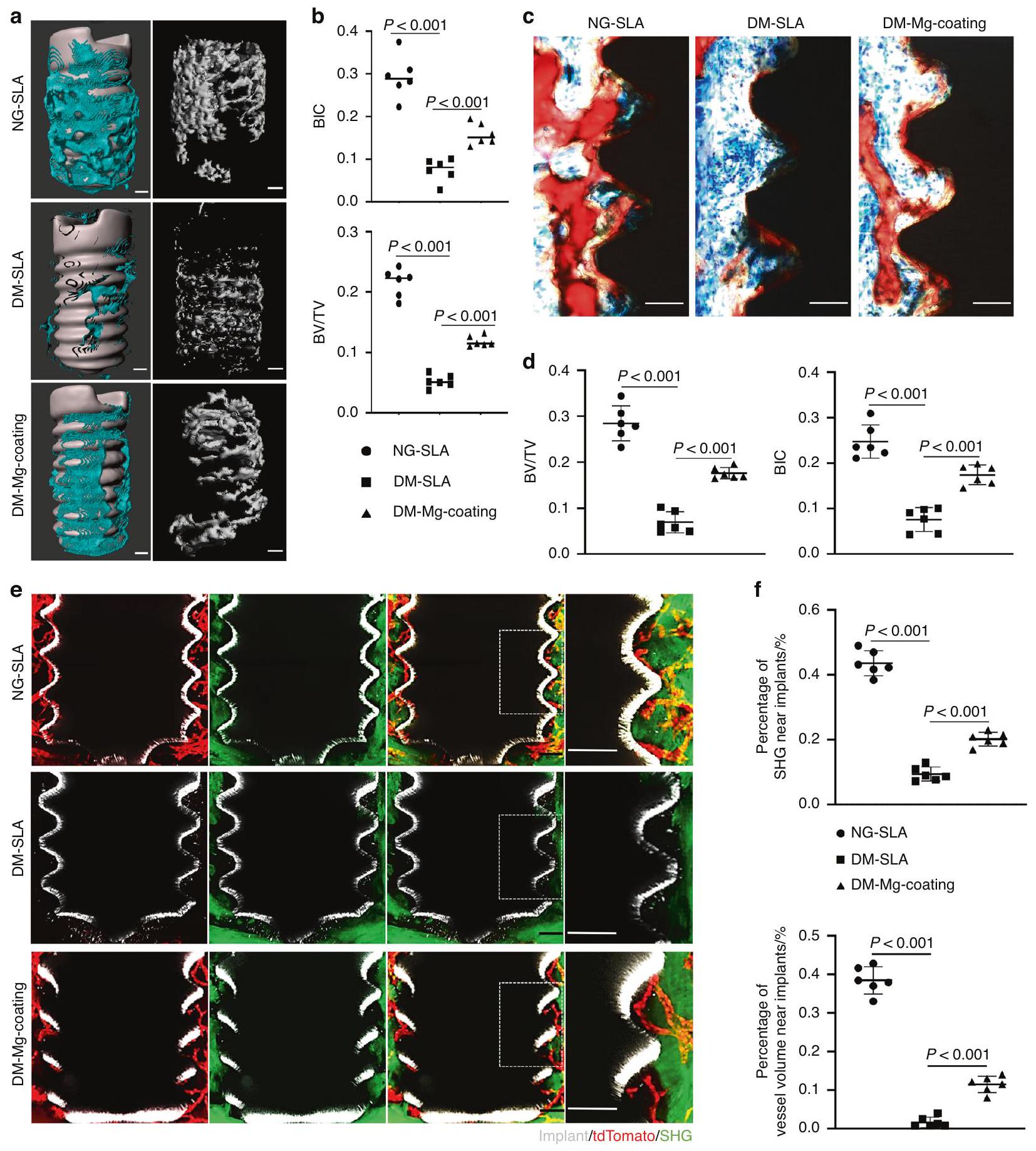
To verify the effect of Mg -coating implants on vascularization and osseointegration in diabetic mice, we inserted implants in Cdh ;tdTomato mice. Visually, CT analysis revealed the Mg coating implants exhibited higher bone-implant contact rate (BIC) and bone volume/tissue volume (BV/TV) in DM mice (Fig. 5a, b). Histomorphometric analyses further corroborated that more new
bone tissue was filled in the neighboring area to the surface of the implants in Mg-coating group (Fig. 5c, d). In turn, more Tomato cells were recruited around the Mg-coating implants in fluorescently labeled mice. These increased cells indicated enriched angiogenesis in DM mice. While more bone collagen in green shown by the SHG signal was visualized around the Mg-coating implants in diabetic states (Fig. 5e, f). Altogether, through in vivo model validation, we demonstrated the ability of Mg-coating
Fig. promotes vascularization and osseointegration in diabetic mice. a Representative images of reconstruction of the newly formed bone (blue) with implants and without implants (gray) 7 days post-surgery. Scale bar . b Quantitative analyses of bone-implant contact rate (BIC) and bone volume/tissue volume (BV/TV) ( ). c Representative images of the histological sections, red for bone. Scale bar . d Quantitative analyses of BIC and BV/TV in histological sections . e Tissue clearing-based three-dimensional images around implants showed angiogenesis and osteogenesis on day 7 after surgery, with tdTomato signal displaying blood vessels (red), second harmonic generation (SHG) signal displaying bone (green) and implant (gray). Scale bar . Quantitative analyses of vessel volume and SHG around implants ( )
implants to promote inhibited vascularization and osseointegration in diabetic states.
However, in previous inspections, we found changes in the surface topography and hydrophilicity of the material. We constructed titanium-coating implants and proved the surface topography, roughness, and wettability were no different from magnesiumcoating implants (Fig.S2a-e). Furthermore, in vivo experiments proved that Mg-coating implants still promoted peri-implant
osseointegration under diabetes compared to titanium-coatings implants, confirming the osteogenic effect of in diabetic states independent of roughness and wettability (Fig. S2f, g).
reduces endothelial cell oxidative stress levels under hyperglycemia
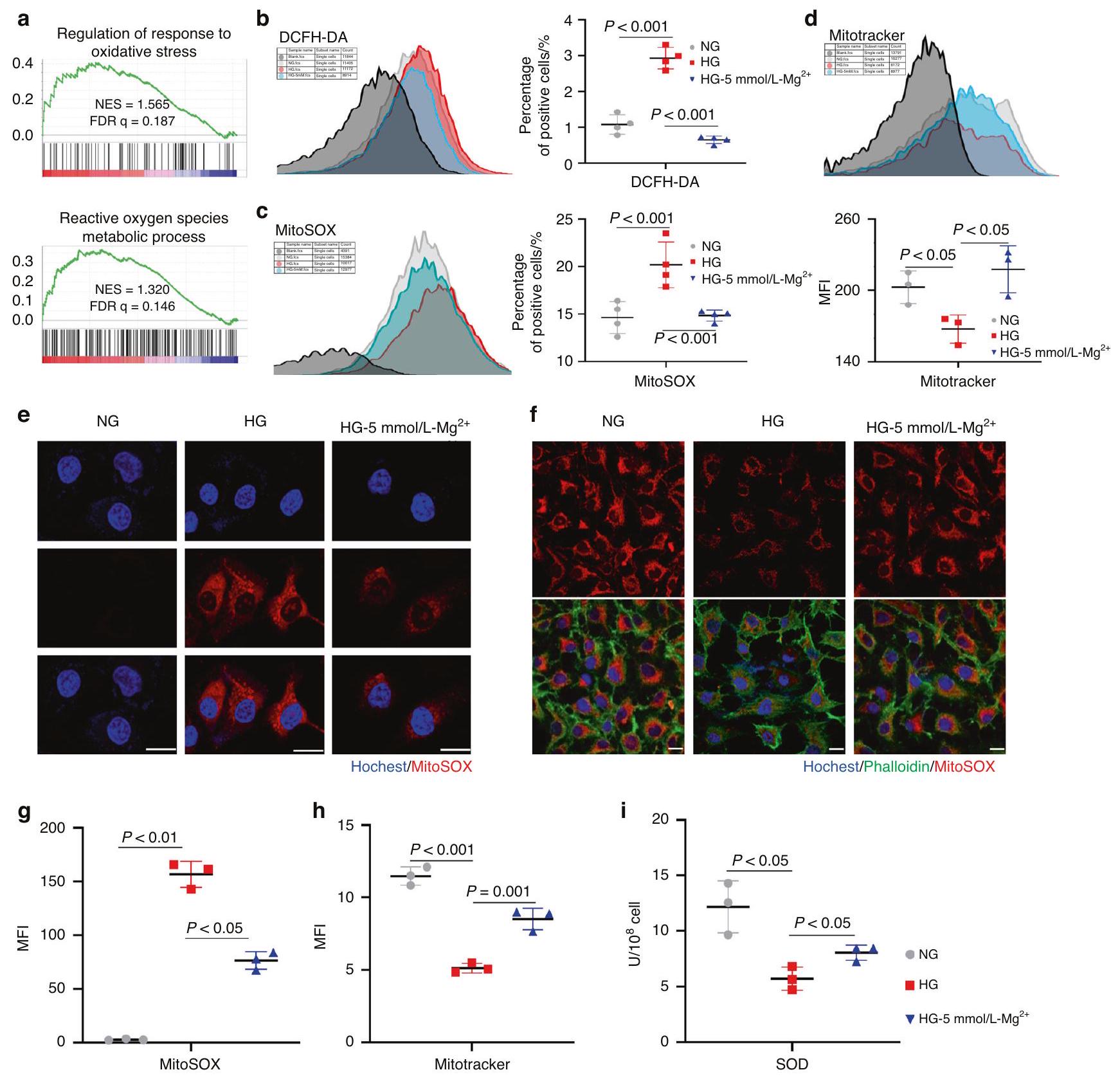
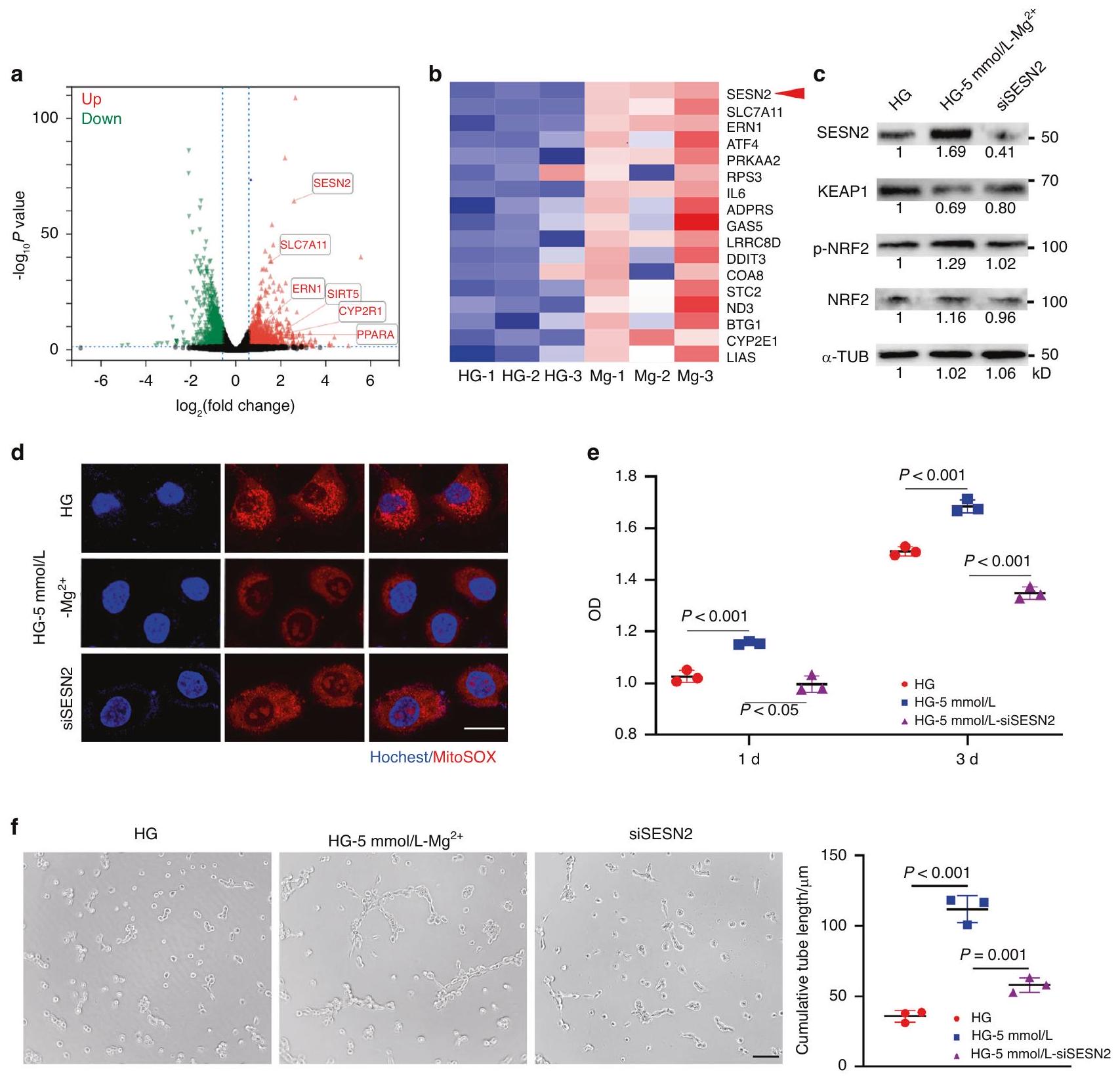
RNA-sequencing analysis was performed to explore the mechanism by which improved endothelial cell function under
Fig. reduces endothelial cell oxidative stress levels under hyperglycemia. a Gene set enrichment analysis (GSEA) for response to oxidative stress. b HUVECs stained with DCFH-DA was analyzed by flow cytometry ( ). c HUVECs stained with MitoSOX was analyzed by flow cytometry ( ). d HUVECs stained with Mitotracker was analyzed by flow cytometry ( ). e Representative fluorescence images of MitoSOX staining for mitochondrial ROS. Scale bar . Representative fluorescence images and quantification of Mitotracker staining. Scale bar . g Immunofluorescence intensity quantification of MitoSOX staining ( ). Immunofluorescence intensity quantification of Mitotracker staining. ( ). I SOD activity detection
hyperglycemia. GESA analysis suggested the regulation of response to oxidative stress and the reactive oxygen species metabolic process were upregulated in the group (Fig. 6a). -dichlorodihydrofluorescein diacetate (DCFH-DA) flow cytometry showed that the level of oxidative stress in endothelial cells in the HG group was significantly increased, while this increase recovered after the addition of (Fig. 6b). Intracellular reactive oxygen species (ROS) were mainly produced by mitochondria. Further detection of mitochondrial ROS indicated the purging ability of on mitochondrial ROS accumulation by flow cytometry and immunofluorescent staining (Fig. 6c, e, g). High
reactive oxygen species result in metabolic disorders and even death of cells by mitochondrial attack. Mitochondrial quality measuring proved that rescued the damaged mitochondrial mass under hyperglycemia (Fig. 6d, f, h). Superoxide dismutase (SOD), a crucial antioxidant metalloenzyme modulated by nucleated Nrf2, manifested enhanced activity in the group under hyperglycemia ( ) (Fig. 6i).
Digging deeper into the mechanism, we found that SESN2 was significantly upregulated in RNA sequencing (Fig. 7a, b) which has been proved to promote Keap1 degradation and activate Nrf2 nucleation to protect cells from oxidative stress.
Fig. upregulates SESN2 in HUVECs under hyperglycemia. a Volcano of differentially expressed genes. Heatmap of representative genes involved in response to oxidative stress. c Western blots of SESN2, KEAP1, nucleated NRF2 and total NRF2 in HUVECs. d Representative fluorescence images of MitoSOX staining for mitochondrial ROS. Scale bar . e Cell proliferation assay of HUVECs by CCK-8 test ( ). F Representative images and quantitative analyses in tube formation assay showed the angiogenesis in vitro. Scale bar ( )
Then, we depleted SESN2 in HUVECs using small interfering RNAs (siRNA) and western blot confirmed the decline of Keap1 and the upregulation of nucleated Nrf2 in the group were reversed after depleting SESN2 (Fig. 7c). Immunofluorescent staining also proved mitochondrial ROS accumulated (Fig. 7d) and cell functions including proliferation and angiogenesis in vitro were also impaired in group after depleting SESN2 (Fig. 7e, f).

DISCUSSION

Post-traumatic revascularization is a critical step in bone healing, providing oxygen, nutrients, and ions necessary for stem cells to differentiate into osteoblasts and matrix mineralization for
damage repair. In the process of bone development and repair, angiogenesis is closely related to osteogenesis. However, diabetes leads to various vascular complications and inhibits angiogenesis, resulting in poor wound healing. Our data also suggested that diabetes seriously impaired vascularization and bone regeneration in alveolar bone healing, which strongly supported the work presenting delayed healing and increased alveolar destruction from Devlin et al. . In recent years, numerous studies have been conducted to promote the healing of diabetic soft and hard tissues by promoting angiogenesis.
has been widely recognized in promoting osteogenesis. Recently, Huang et al. (2021) created a multifunctional magnesium organic framework-based microneedle patch for accelerating diabetic wound healing through accelerating angiogenesis. Liu
et al. (2023) also constructed magnesium-containing hydrogels to strengthen diabetic wound angiogenesis to promote healing. However, at present, whether could promote peri-implant vascularization and osseointegration under diabetes and its intrinsic mechanism are still unknown. In our study, was demonstrated for the first time to improve peri-implant vascularization and osseointegration in diabetic mice.
Endothelial cells (ECs), on the other hand, are thought to be the driving force behind angiogenesis, quickly initiating the formation of new blood vessels during wound healing. In diabetes, impaired endothelial cell function leads to disturbed angiogenesis, and our in vitro experiments demonstrated that could improve endothelial cell proliferation, tube formation, and migration under hyperglycemia.
Hyperglycemia-induced endothelial cell disorders were proved to be mainly related to increased levels of intracellular oxidative stress and excess intracellular superoxide, resulting in dysfunction. While mitochondria were an important source of intracellular ROS, increased intracellular mitochondrial proton gradient in a hyperglycemic state inhibits electron transfer from reduced coenzyme Q (ubiquinone) to electron transport chain complex III. Instead, electrons are transferred to molecular oxygen, leading to the production of superoxide. Excess ROS destroys cellular proteins, lipids, mitochondria, and DNA, leading to fatal damage to cells.
Our RNA sequencing reflected that the addition of increased the reactive oxygen species metabolic process of endothelial cells, while the detection of ROS in endothelial cells supported the ability of to reduce the level of mitochondrial oxidative stress elevated under hyperglycemia.
Through further exploration of the sequencing results, we found that the expression of SESN2 and ATF4 in the group increased significantly. ATF4 is a key gene in mitochondrial stress regulation and is involved in gene transcription for antioxidant responses, autophagy, amino acid biosynthesis, and transport. Studies have shown that the upregulation of ATF4 could mitigate mitochondrial ROS production and ROS-mediated mitochondrial damage. SESN2 is a stress-induced protein that can increase the nuclear translocation of Nrf2 and improve the transcriptional activity mediated by Nrf2/antioxidant response element (ARE), thereby exerting antioxidant capacity. Nrf2 is an important transcription factor in regulating cellular oxidative stress and a central regulator for maintaining redox homeostasis in cells. Under normal circumstances, Nrf2 will be modified by Keap1 complex ubiquitination and degradation in the cytoplasm, after Nrf2 is transferred to the nucleus, it will form a complex with coactivators and bind to promoter regions (AREs), thereby inducing and regulating the compositional and inducible expression of a series of antioxidant proteins, which can reduce cell damage caused by reactive oxygen species and electrophiles, keep cells in a stable state, and maintain redox homeostasis.
Using small interfering RNAs (siRNA) we eliminated SESN2 in HUVECs. The upregulation of Keap1 and the decline of nucleated Nrf2 confirmed the decline of Keap1 after eliminating SESN2 in the group strongly demonstrated that regulates the degradation of Keap1 and the nucleation of Nrf2 through upregulating SESN2. Further detection of elevated mitochondrial ROS and impaired cell proliferation and angiogenesis in vitro proved mediated antioxidant response and repaired endothelial cell dysfunction through SESN2. However, absence of SESN2 did not completely inhibit the effect of on endothelial cells under hyperglycemia, suggesting other regulatory mechanisms such as ATF4 or SLC7A11 might make an impact. In the sequencing data that the expression of Solute Carrier Family 7 Member 11 (SLC7A11), one of the most critical upstream regulators of ferrozogenesis, was also significantly upregulated. Combined with our experimental data that promoted the survival of high-glucose endothelial cells, reduced cellular
Magnesium promotes vascularization and osseointegration in diabetic states Liu et al.
oxidative stress levels, improved mitochondrial quality, increased Nrf2 nucleation upstream of SLC7A11 and upregulate ATF4 levels, we speculated that might inhibit ferrozogenesis in highglucose endothelial cells, which also requires further verification.
At the same time, the implant-tissue interface is a key issue in implant research. Due to the limitations of traditional technical means, it was difficult to visually observe the changes in the implant-osseointegration interface in three dimensions. Drawing on the tissue clearing technology previously developed by our research group, we visually observed the changes in the periimplant three-dimensional microenvironment of diabetic mice for the first time by preserving endogenous fluorescence expression. Our data profoundly proved that diabetes seriously impaired the vascularization and bone regeneration in alveolar bone healing while it was also the first comprehensively and threedimensionally display in which angiogenesis was inhibited in diabetic tooth extraction socket and implant osseointegration. Of course, in the construction of the material we also changed the surface roughness and wettability, which had been reported to have an effect on angiogenesis and bone formation. In order to exclude the effects of roughness and wettability, we used titanium dioxide as the coating to achieve roughness and hydrophilicity similar to Mg-coating. In vivo experiments, we found that the increase of wettability could slightly increase bone formation, but the Mg-coating still showed a significant effect of promoting periimplant osseointegration. Combined with our cytology experiments, were added to the medium to confirm the effect of magnesium on endothelial cells. We further confirmed that played a crucial role in promoting vascularization and osteogenesis, independent of other factors. However, in this study we focused on promoting vascularization and osteogenesis in diabetic states, therefore control group of normoglycemic animals with Mg-coating implants was absent. We also lacked the experiments to knock out SESN2 or NRF2 in mice to further support our results, which needed further exploration.
In summary, we demonstrated that promoted Nrf2 nucleation through SESN2 upregulation, thereby reducing the level of mitochondrial oxidative stress in endothelial cells under hyperglycemia in vitro and promoting peri-implant vascularization and osseointegration in diabetic mice.

MATERIALS AND METHODS

Mice

The study conforms to the ARRIVE Guidelines. All protocols for animal care and experiments were reviewed and approved by the Subcommittee on Research and Animal Care of Sichuan University. All mice were C57BL/6 background and housed in a specific pathogen-free environment with a 12 h light-dark cycle. Rosa26-tdTomato (007905) was from Jackson Laboratory. Cdh5 mice were kindly provided by Prof. Bi-Sen Ding’s lab (State Key Laboratory of Biotherapy, Sichuan University). Cdh5Cre ;tdTomato mice were generated by mating Cdh5-Cre mice with Rosa26-tdTomato mice. 1.5 mg per 10 g tamoxifen was injected intraperitoneally daily for 2 days consecutively to light up fluorescence before surgery.

Construction of the diabetic model

STZ (Shanghai Maokang Biotechnology Co., Ltd.) was intraperitoneally injected into mice consecutively for 5 days to induce a model. With serum glucose level reaching for 2 weeks, surgery was performed (Fig. S1).

Surgery

First mandibular molars were removed under anesthesia with a 26 G syringe needle and forceps. For immediate implant placement, a self-designed sandblasted, large grit and acid-etched (SLA) titanium implant ( diameter; WEGO, China) was
screwed in the socket after extraction under the stereomicroscope. On day 7 after extraction or implantation, mandibles were harvested.
PEGASOS tissue clearing process
The PEGASOS tissue clearing process was performed as reported before.
Image acquisition and analyses
Images were acquired through a multiphoton microscope (Lecia SP8 DIVE). TdTomato signals were acquired under a 561 nm excitation laser line. The implant was acquired with a sapphire laser at 950 nm wavelength. Second harmonic signals (SHG) imaging was acquired with a non-descanned detector. IMARIS software (version 9.1.2; Oxford Instruments) was used for data analysis and image processing. A region near the thread grooves of each implant was used for analyses. For each sample, three randomly selected regions were used for quantification.
Micro-computed tomography images and analyses
The harvested mandibles with implants were fixed in 4% PFA at for 24 h and then stored in PFA at before being scanned. The samples were scanned with a system (Scanco Medical) at a medium resolution and a voxel size of . IMARIS software (version 9.1.2; Oxford Instruments) was used for data analysis and image processing.
Histologic preparation and staining
The mandibles with implants were fixed in PFA at for 1 day, followed by 4 weeks of decalcification in 10% EDTA (pH 7.4) at with shaking. Mandibles for hematoxylin and eosin (H&E) staining were dehydrated in graded ethanol and embedded in paraffin. The paraffin blocks were cut by a microtome (Leica RM2255) into -thick sections. H&E staining was performed according to the manufacturer’s instructions (Biosharp). A region near the thread grooves of each implant was used for analyses. For each sample, three randomly selected regions were used for quantification.
Cell culture
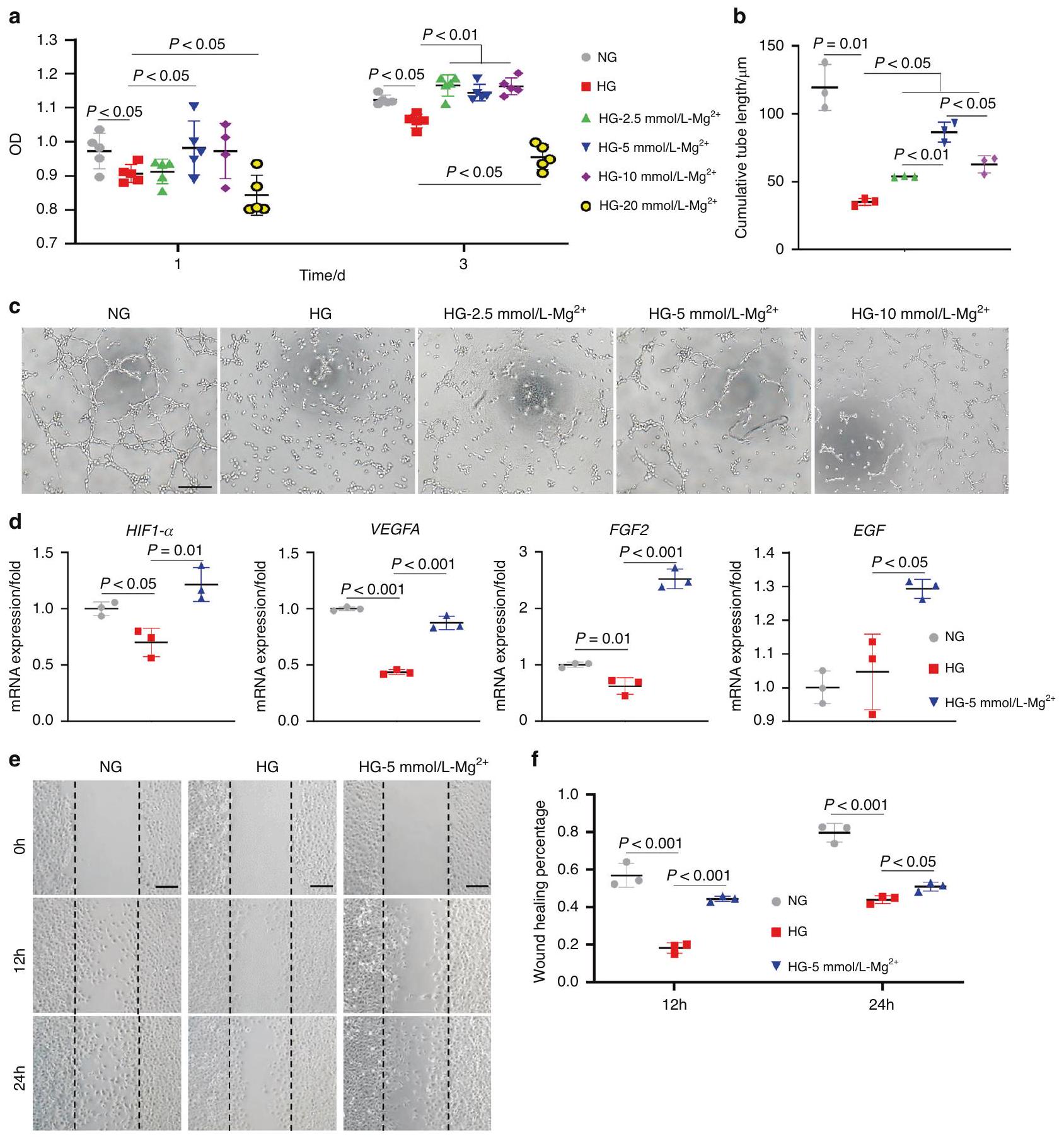
Human Umbilical Vein Endothelial Cells (HUVECs) provided by State Key Laboratory of Biotherapy, West China Second University Hospital, Sichuan University, were cultured in Dulbecco’s modified Eagle medium (DMEM) (Hyclone, Laboratories, Logan, UT, USA) containing 10% fetal bovine serum (Gibco, Grand Island, NY, USA), penicillin (Hyclone, Cytiva, Marlborough, MA, USA) and streptomycin (Hyclone, Cytiva, Marlborough, MA, USA). The culture media were changed every other day. HUVEC were cultured in three different conditions: normal glucose ( ), high glucose ( ), and , China).
MC3T3-E1 osteoblasts were cultured in alpha-modified Eagle medium (a-MEM) (Hyclone, Laboratories, Logan, UT, USA) and changed every other day.
Cell counting kit-8 assay
HUVECs were seeded in a 96-well plate at a density of 5000 cells per well. Then NG medium, HG medium, or HG medium containing , or was added. Cell counting kit-8 (CCK-8) assay was conducted after 24 h and 72 h separately. Optical density (OD) values were measured by a spectrophotometer (Thermo Fisher Scientific, Waltham, MA, USA) at 450 nm .
Tube formation test
growth-factor-reduced Matrigel (BD Biosciences, USA) was added into each well of the 96 -well plate and then place in a incubator for 30 min until solidification. Next, HUVECs ( 5000 cells per well) were seeded on the solidified Matrigel substrates and cultured in serum-free NG medium, HG medium, or HG medium
containing , and . After 6 h of incubation ( ), the cells were imaged using an inverted fluorescence microscope (Olympus, Japan). The tube length was measured using ImageJ software (NIH, USA).

Quantitative reverse-transcription PCR (qRT-PCR)

HUVECs were cultured in NG medium, HG medium, or HG medium containing for 24 h . Then total RNA of HUVECs was extracted by Trizol Reagent (Invitrogen, Carlsbad, CA, USA). Reverse transcription complementary DNA (cDNA) from of RNA using the PrimeScript RT Kit (TaKaRa Bio, Otsu, Japan), and the genes of interest were measured by SYBR Premix Ex Taq II (TaKaRa) in LightCycler 96 (Roche, Basel, Switzerland). Relative microRNA (mRNA) expression was analyzed using a delta delta comparative threshold cycle method by normalizing with glyceraldehyde-3-phosphate dehydrogenase (GADPH).

Scratch wound healing assay

HUVECs were seeded in a 6 -well plate and cultured till confluence, and the FBS medium was replaced with 1% FBS medium for starvation culture 24 h after scratches. A pipette tip was used to trace along the center of the hole along the ruler, leaving even and straight scratches. Subsequently, the medium was replaced with NG medium, HG medium, or HG medium containing . The wound closure was observed and pictured after 12 h and 24 h . ImageJ software was used to calculate the proportion of the healing area.

Gene knockdown

We diluted siRNA duplex into DMEM as solution and diluted Lipofectamine RNAiMAX (Invitrogen, USA) into DMEM for solution . and were mixed and incubated 30 min at . When the cells reached 40 to confluence, mixture was added to 2 mL of normal growth medium without antibiotics for 12 h at .

Fabrication of Mg-coating and Ti-coating implants

For hydrothermal synthesis, an aqueous solution of MgO (or ) and NaOH was prepared in ddwater. Mix the reactants completely and the solutions were combined and subsequently stirred at room temperature for 5 min . The final stock solution was transferred to a hydrothermal reactor and the processed titanium was put in the reactor at the same time. The hydrothermal autoclave was sealed and set up to for 12 h . Then the plates were washed, respectively, by ddwater and ethanol twice for 10 min . The plates were placed in an oven and heated at for 1 h for drying.

Materials characterization of samples

The surface topography of the samples was scanned by a fieldemission SEM (FE-SEM; JSM-7500 F, JEOL, Japan). Elemental analysis was performed by EDS. Superview W1 (China) were used for detect surface topography. XRD (LabX, Shimadzu, Japan) was used to scan the structures of the samples. The elemental composition and chemical state of surfaces were measured by XPS (AXIS Supra, Kratos, USA). The water contact angle was performed by the WCA apparatus (Zhongchen Digital Technic Apparatus Co., Shanghai, China). The inductively coupled plasma atomic emission spectroscopy (ICP-AES, 5100 SVDV, USA) was used to assess the release of in PBS.

Cell viability assay

HUVECs or MC3T3-E1 osteoblasts ( cells per well) were seeded onto NG + SLA, HG + SLA, and HG + Mg-coating plates in the 48 -well plates for 24 h . Cell counting kit-8 (CCK-8) assay was conducted as before.
Cell morphology and immunofluorescence staining Phalloidin (Shanghai Maokang Biotechnology Co., Ltd.) was used to observe the cytoskeleton. After 24 h of incubation, cells were
fixed with 4% PFA for 10 min and the cell cytoskeleton was stained by phalloidin and the nuclei stained by DAPI. Fluorescent imaging of cells was detected by laser scanning confocal microscopy (FV3000; Olympus, Japan). After 1 day of culture on substrates, cells were fixed overnight with glutaraldehyde. Then, the samples were dehydrated sequentially in gradient ethanol ( , ) for 15 min for SEM observation.

Alkaline phosphatase staining

MC3T3-E1 osteoblasts were cultured with the osteogenic medium on substrates for 7 days and fixed with 4% PFA for 20 min . Then staining was performed with a commercial Alkaline Phosphatase Assay Kit (Beyotime Biotechnology, Shanghai, China). ALP activity quantitation was performed with a commercial kit (Beyotime Biotechnology, Shanghai, China) and detected by a spectrophotometer (Thermo Fisher Scientific, USA) at 450 nm .

Histologic and histomorphometric analyses

7 days after implant surgery, mandibles containing implants were collected and fixed in buffered formalin at for 1 week. Samples were dehydrated by increasing concentrations of ethanol ( ) and embedded in light-curing epoxy resin (TechnoVit7200VLC; Hereaus-Kulzer, Wehrheim, Germany). The embedded specimens were cut parallel to the long axis of the implant. Then, the specimens were ground to around thickness using a grinding machine (ExaktApparatebau, Norderstedt, Germany). Sections were stained with Stevenel’s blue and Van Gieson’s stain and then observed under a light microscope (Olympus, Japan).

RNA-Sequencing and data analysis

HUVECs were treated with HG medium or HG medium containing for 48 h ( in each group). Total RNA was extracted with Trizol reagent (Invitrogen, Carlsbad, CA, USA) and purified with poly-T oligo-attached magnetic beads. FastQC (SAndrews, v0.11.5) was used to guarantee the quality of RNASequencing (RNA-Seq) data and by using HISAT2 (DaehwanKimLab,v.2.0.5) we mapped them to Mus musculus reference genomes. Genes were considered significantly differentially expressed if showing fold change and -value . For Gene Set Enrichment Analysis (GSEA), the gene lists were downloaded from the GSEA database (www.gsea-msigdb.org/ gsea/). GSEA was processed with GSEA software (https:// www.gsea-msigdb.org/gsea/index.jsp).

Flow cytometry analysis

HUVECs were treated with NG medium, HG medium, or HG medium containing for 48 h . -dichlorodihydrofluorescein diacetate (DCFH-DA), and MitoSOX staining were performed following a modified protocol. The samples were then tested by flow cytometry (BD Biosciences, USA), and the flow cytometry data were analyzed using Flowjo software.

Western blot

HUVECs were treated with HG medium or HG medium containing for 48 h . Total proteins were extracted by protein extraction kit (Signalway Antibody, Greenbelt, MD, USA). Proteins concentrations were measured with BCA Protein Assay Kit (Pierce, Rockford, IL, USA;23225). Protein was separated equally in SDS-polyacrylamide gel electrophoresis (SDS-PAGE) gels and transferred to polyvinylidene fluoride (PVDF) membranes (Thermo Fisher Scientific, Waltham, MA, USA). After blocking in skim milk, membranes were immersed with primary antibodies and corresponding secondary antibodies. Proteins were developed by a gel imaging system (Bio-Rad Laboratories, Hercules, CA, USA) with Immobilon Reagents (Millipore, Billerica, MA, USA).

SOD activity assay

SOD activity assay was performed as directed (Beyotime, China).

Statistical analyses

All data were expressed as mean standard deviation (SD). For each group, at least three independent experiments were conducted with duplicate samples. Statistical analysis was done with a one-way ANOVA followed by Bonferroni’s multiple comparison test. was considered statistically significant.

DATA AND MATERIALS AVAILABILITY

The RNA sequencing data set has been deposited into the NCBI database with the identifier GSE236139.

ACKNOWLEDGEMENTS

The authors thank Dr. Qiang Guo from the State Key Laboratory of Oral Diseases, Sichuan University, for assistance with micro-CT scanning. We thank Dr. Chaoyang Zhang from the State Key Laboratory of Oral Diseases, Sichuan University, for assistance with SEM. We thank Dr. Yinghui Wen and Dr. Manlu Wang from the State Key Laboratory of Oral Diseases, Sichuan University, for assistance with the multiphoton microscope. This work was supported by grants from the National Natural Science Foundation of China (No. 81901042), the Sichuan Science and Technology Program (No. 2022NSFSC1384), and International Team for Implantology (No. 1477_2020).

AUTHOR CONTRIBUTIONS

L. Liu contributed to the conception, design, data acquisition, analysis and interpretation, drafted and critically revised the manuscript; W.S. contributed to data interpretation, drafted and critically revised the manuscript; D.Z. contributed to data interpretation, drafted and critically revised the manuscript; W.L. contributed to data analysis, and interpretation, drafted and critically revised the manuscript; Q. Yin contributed to data analysis, and interpretation, drafted and critically revised the manuscript; Q.W. contributed to data analysis, drafted and critically revised the manuscript; H.L. contributed to data analysis, drafted and critically revised the manuscript; Q. Yuan contributed to the conception, design, drafted and critically revised the manuscript; S. Zhang contributed to the conception, design, drafted and critically revised the manuscript. All authors gave final approval and agreed to be accountable for all aspects of the work.

ADDITIONAL INFORMATION

Supplementary information The online version contains supplementary material available at https://doi.org/10.1038/s41368-023-00271-y.
Competing interests: The authors declare no competing interests.

REFERENCES

  1. Buser, D., Sennerby, L. & De Bruyn, H. Modern implant dentistry based on osseointegration: 50 years of progress, current trends and open questions. Periodontology 73, 7-21 (2017).
  2. De Angelis, F. et al. Implant survival and success rates in patients with risk factors: results from a long-term retrospective study with a 10 to 18 years follow-up. Rev. Med. Pharmacol. Sci. 21, 433-437 (2017).
  3. de Oliveira, P. et al. Obesity/metabolic syndrome and diabetes mellitus on periimplantitis. Trends Endocrinol. Metab. 31, 596-610 (2020).
  4. Javed, F. & Romanos, G. E. Chronic hyperglycemia as a risk factor in implant therapy. Periodontology 81, 57-63 (2019).
  5. Quan, Y. (ed.) Dental Implant Treatment in Medically Compromised Patients. (Springer Press, 2020).
  6. Ko, K. I., Sculean, A. & Graves, D. T. Diabetic wound healing in soft and hard oral tissues. Transl. Res.: J. Lab. Clin. Med. 236, 72-86 (2021).
  7. Claes, L., Recknagel, S. & Ignatius, A. Fracture healing under healthy and inflammatory conditions. Nat. Rev. Rheumatol. 8, 133-143 (2012).
  8. Stegen, S., van Gastel, N. & Carmeliet, G. Bringing new life to damaged bone: the importance of angiogenesis in bone repair and regeneration. Bone 70, 19-27 (2015).
  9. Tuckermann, J. & Adams, R. H. The endothelium-bone axis in development, homeostasis and bone and joint disease. Nat. Rev. Rheumatol. 17, 608-620 (2021).
  10. Hu, X. F., Wang, L., Xiang, G., Lei, W. & Feng, Y. F. Angiogenesis impairment by the NADPH oxidase-triggered oxidative stress at the bone-implant interface: Critical mechanisms and therapeutic targets for implant failure under hyperglycemic conditions in diabetes. Acta Biomater. 73, 470-487 (2018).
  11. Beckman, J. A. & Creager, M. A. Vascular complications of diabetes. Circul. Res. 118, 1771-1785 (2016).
  12. Xu, Z. et al. Thermosensitive hydrogel incorporating Prussian blue nanoparticles promotes diabetic wound healing via ROS scavenging and mitochondrial function restoration. ACS Appl. Mater. Interfaces 14, 14059-14071 (2022).
  13. de Baaij, J. H., Hoenderop, J. G. & Bindels, R. J. Magnesium in man: implications for health and disease. Physiol. Rev. 95, 1-46 (2015).
  14. Zhang, Y. et al. Implant-derived magnesium induces local neuronal production of CGRP to improve bone-fracture healing in rats. Nat. Med. 22, 1160-1169 (2016).
  15. Lin, S. et al. A magnesium-enriched 3D culture system that mimics the bone development microenvironment for vascularized bone regeneration. Adv. Sci. (Weinh.) 6, 1900209 (2019).
  16. Han, H. S. et al. Biodegradable magnesium alloys promote angio-osteogenesis to enhance bone repair. Adv. Sci. (Weinh.) 7, 2000800 (2020).
  17. Zhu, D., You, J., Zhao, N. & Xu, H. Magnesium regulates endothelial barrier functions through TRPM7, MagT1, and S1P1. Adv. Sci. (Weinh.) 6, 1901166 (2019).
  18. Yin, M. et al. Multifunctional magnesium organic framework-based microneedle patch for accelerating diabetic wound healing. ACS Nano 15, 17842-17853 (2021).
  19. Sales, C. H. & Pedrosa Lde, F. Magnesium and diabetes mellitus: their relation. Clin. Nutrition (Edinburgh, Scotland) 25, 554-562 (2006).
  20. WA, E. L., Naser, I. A., Taleb, M. H. & Abutair, A. S. The effects of oral magnesium supplementation on glycemic response among type 2 diabetes patients. Nutrients 11, 44 (2018).
  21. Veronese, N. et al. Oral magnesium supplementation for treating glucose metabolism parameters in people with or at risk of diabetes: a systematic review and metaanalysis of double-blind randomized controlled trials. Nutrients 13, 4074 (2021).
  22. He, T. et al. A comparison of micro-CT and histomorphometry for evaluation of osseointegration of PEO-coated titanium implants in a rat model. Sci. Rep. 7, 16270 (2017).
  23. Calvo-Guirado, J. L. et al. Histological and histomorphometric evaluation of immediate implant placement on a dog model with a new implant surface treatment. Clin. Oral Implants Res. 21, 308-315 (2010).
  24. Jing, D. et al. Tissue clearing and its application to bone and dental tissues. J. Dental Res. 98, 621-631 (2019).
  25. Jing, D. et al. Tissue clearing of both hard and soft tissue organs with the PEGASOS method. Cell Res. 28, 803-818 (2018).
  26. Sivaraj, K. K. & Adams, R. H. Blood vessel formation and function in bone. Development (Cambridge, England) 143, 2706-2715 (2016).
  27. Lin, W. et al. Mapping the immune microenvironment for mandibular alveolar bone homeostasis at single-cell resolution. Bone Res. 9, 17 (2021).
  28. Kusumbe, A. P., Ramasamy, S. K. & Adams, R. H. Coupling of angiogenesis and osteogenesis by a specific vessel subtype in bone. Nature 507, 323-328 (2014).
  29. Peng, Y., Wu, S., Li, Y. & Crane, J. L. Type H blood vessels in bone modeling and remodeling. Theranostics 10, 426-436 (2020).
  30. Brem, H. & Tomic-Canic, M. Cellular and molecular basis of wound healing in diabetes. J. Clin. Investig. 117, 1219-1222 (2007).
  31. Orasanu, G. & Plutzky, J. The pathologic continuum of diabetic vascular disease. J. Am. College Cardiol. 53, S35-S42 (2009).
  32. Devlin, H., Garland, H. & Sloan, P. Healing of tooth extraction sockets in experimental diabetes mellitus. J. Oral Maxillofac. Surg. 54, 1087-1091 (1996).
  33. Xiong, Y. et al. A whole-course-repair system based on neurogenesisangiogenesis crosstalk and macrophage reprogramming promotes diabetic wound healing. Adv. Mater. (Deerfield Beach, Fla.), e2212300, https://doi.org/ 10.1002/adma. 202212300 (2023).
  34. Rohlenova, K., Veys, K., Miranda-Santos, I., De Bock, K. & Carmeliet, P. Endothelial cell metabolism in health and disease. Trends Cell Biol. 28, 224-236 (2018).
  35. Potente, M., Gerhardt, H. & Carmeliet, P. Basic and therapeutic aspects of angiogenesis. Cell 146, 873-887 (2011).
  36. Sawada, N. et al. Endothelial PGC-1a mediates vascular dysfunction in diabetes. Cell Metab. 19, 246-258 (2014).
  37. Giacco, F. & Brownlee, M. Oxidative stress and diabetic complications. Circulation Res. 107, 1058-1070 (2010).
  38. Eelen, G., de Zeeuw, P., Simons, M. & Carmeliet, P. Endothelial cell metabolism in normal and diseased vasculature. Circulation Res. 116, 1231-1244 (2015).
  39. Brownlee, M. Biochemistry and molecular cell biology of diabetic complications. Nature 414, 813-820 (2001).
  40. Wang, Y. et al. Integrated regulation of stress responses, autophagy and survival by altered intracellular iron stores. Redox Biol. 55, 102407 (2022).
  41. Ding, S., Ma, N., Liu, H., Tang, M. & Mei, J. Sesn2 attenuates the damage of endothelial progenitor cells induced by angiotensin II through regulating the Keap1/Nrf2 signal pathway. Aging 12, 25505-25527 (2020).
  42. Muri, J. & Kopf, M. Redox regulation of immunometabolism. Nat. Rev. Immunol. 21, 363-381 (2021).
  43. Rupp, F., Liang, L., Geis-Gerstorfer, J., Scheideler, L. & Hüttig, F. Surface characteristics of dental implants: a review. Dental Mater. 34, 40-57 (2018).
Open Access This article is licensed under a Creative Commons Attribution 4.0 International License, which permits use, sharing, adaptation, distribution and reproduction in any medium or format, as long as you give appropriate credit to the original author(s) and the source, provide a link to the Creative Commons license, and indicate if changes were made. The images or other third party material in this article are included in the article’s Creative Commons license, unless indicated otherwise in a credit line to the material. If material is not included in the article’s Creative Commons license and your intended use is not permitted by statutory regulation or exceeds the permitted use, you will need to obtain permission directly from the copyright holder. To view a copy of this license, visit http:// creativecommons.org/licenses/by/4.0/.
© The Author(s) 2024

  1. State Key Laboratory of Oral Diseases & National Center for Stomatology & National Clinical Research Center for Oral Diseases & West China Hospital of Stomatology, Sichuan University, Chengdu, China and State Key Laboratory of Oral Diseases & National Center for Stomatology & National Clinical Research Center for Oral Diseases & Department of Oral Implantology, West China Hospital of Stomatology, Sichuan University, Chengdu, China
    Correspondence: Quan Yuan (yuanquan@scu.edu.cn) or Shiwen Zhang (sw.zhang2018@scu.edu.cn)
    These authors contributed equally: Linfeng Liu, Feiyu Wang