تحسين النشاط المضاد للأكسدة المعتمد على تأثير الحجم لجزيئات السيلينيوم النانوية التي تنظم مسارات Anti-PI3K-mTOR و Ras-MEK لعلاج إصابة الحبل الشوكي لتجنب قمع المناعة الناتج عن صدمة الهرمونات Size effect-based improved antioxidant activity of selenium nanoparticles regulating Anti-PI3K-mTOR and Ras-MEK pathways for treating spinal cord injury to avoid hormone shock-induced immunosuppression

المجلة: Journal of Nanobiotechnology، المجلد: 23، العدد: 1
DOI: https://doi.org/10.1186/s12951-024-03054-7
PMID: https://pubmed.ncbi.nlm.nih.gov/39815246
تاريخ النشر: 2025-01-15

تحسين النشاط المضاد للأكسدة المعتمد على تأثير الحجم لجزيئات السيلينيوم النانوية التي تنظم مسارات Anti-PI3K-mTOR و Ras-MEK لعلاج إصابة الحبل الشوكي لتجنب قمع المناعة الناتج عن صدمة الهرمونات

بيكسين ، شياودونغ ، زيهاو وو ، كوي شين ، زهاوفينغ ، شياوي لي ، كويفينغ وو ، ليونغ تشان ، تشونغ زانغ ، يوتونغ وو ، ليوين ليو ، تيانفينغ تشين و يي تشين

الملخص

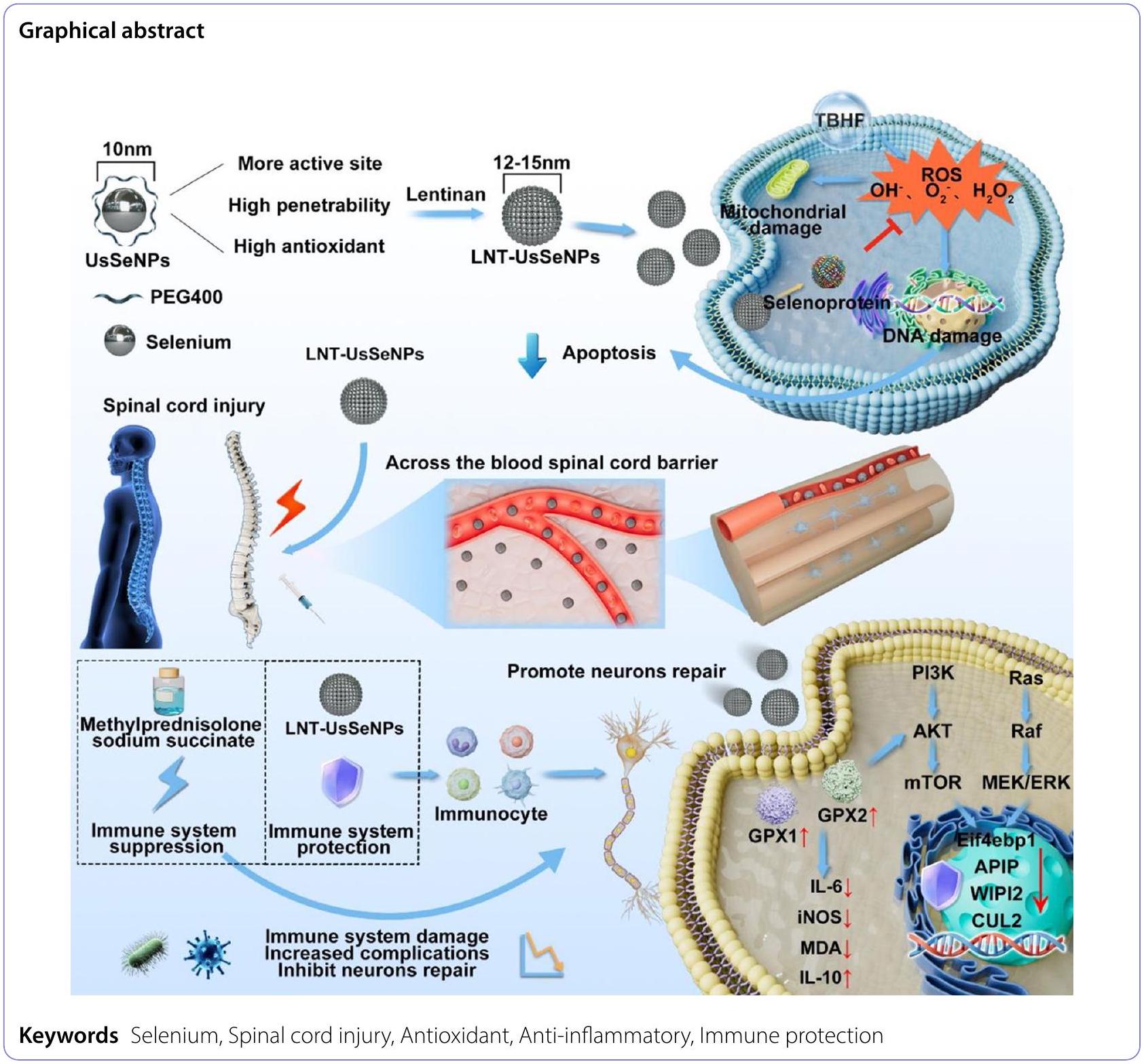
إصابة الحبل الشوكي (SCI) هي حالة حرجة تؤثر على الجهاز العصبي المركزي وغالبًا ما تكون لها عواقب دائمة ومعوقة، بما في ذلك الإصابات الثانوية. تعتبر الأضرار التأكسدية والالتهاب عوامل حاسمة في العمليات المرضية الثانوية. لقد أظهرت جزيئات السيلينيوم النانوية خصائص مضادة للأكسدة ومضادة للالتهابات بشكل كبير عبر مسار غير مثبط للمناعة؛ ومع ذلك، كانت تطبيقاتها السريرية محدودة بسبب عدم استقرارها ووظيفتها غير الكافية لعبور حاجز الدم-الحبل الشوكي (BSCB). اقترحت هذه الدراسة طريقة تخليق لجزيئات السيلينيوم النانوية ذات القطر الصغير للغاية (LNT-UsSeNPs) مع قدرات كبيرة في التخلص من أنواع الأكسجين التفاعلية (ROS) مقارنة بجزيئات السيلينيوم النانوية التقليدية (LNT-SeNPs). هذه المركبات حمت خلايا PC-12 بشكل فعال من السمية الناتجة عن الإجهاد التأكسدي، وقللت من خلل الميتوكوندريا، وخفضت من موت الخلايا. أظهرت الدراسات الحية أن LNT-UsSeNPs اخترقت BSCB بكفاءة وكبحت بشكل فعال موت خلايا الحبل الشوكي. في النهاية، نظمت LNT-UsSeNPs مباشرة مسارات الإشارة PI3K-AKT-mTOR و Ras-Raf-MEK-ERK عن طريق تنظيم السيلينوبروتينات لتحقيق علاج مضاد للالتهابات غير مثبط للمناعة. نظرًا لصغر حجمها للغاية، أظهرت LNT-UsSeNPs اختراقًا قويًا للحاجز الشوكي وتأثيرات مضادة للأكسدة ومضادة للالتهابات قوية دون المساس بوظيفة المناعة. تشير هذه النتائج إلى أن LNT-UsSeNPs هي مرشحة واعدة لمزيد من التطوير في النانوميدicine لعلاج فعال لإصابة الحبل الشوكي.

ساهم بيكسين ليو، شياودونغ ليو وزيهاو وو بالتساوي في هذا العمل.
*المراسلة:
تيانفينغ تشين
tchentf@jnu.edu.cn
يي تشين
qinyi0225@163.com
القائمة الكاملة لمعلومات المؤلف متاحة في نهاية المقالة

المقدمة

إصابة الحبل الشوكي (SCI) بمعدلات عالية من الإعاقة وتلف الأنسجة هي اضطراب شائع وخطير في الجهاز العصبي المركزي، وترتبط في النهاية بحدوث كبير ووفيات. يتم الإبلاغ عن حوالي 700,000 حالة جديدة من SCI سنويًا في جميع أنحاء العالم [1]. عادة ما تكون SCI ناتجة عن صدمة عالية الشدة، بما في ذلك السقوط من المركبات، الحوادث، العنف، ولكن يمكن أن تنتج أيضًا عن العدوى، الأورام، الأمراض التنكسية في العمود الفقري، إصابة نقص التروية-إعادة التروية، والعيوب الوعائية [2، 3]. تؤثر SCI على الوظائف الحركية والحسية واللاإرادية مع الإعاقة المؤقتة أو الدائمة، مما يفرض عبئًا نفسيًا واقتصاديًا كبيرًا على المرضى وعائلاتهم. من الناحية المرضية، الإصابة الأولية هي كدمة أولية أو
قطع ناتج عن ضغط ميكانيكي فوري أو مستمر أو سحب على الحبل الشوكي [4-8]. الإصابة الثانوية هي استجابة الجسم والخلايا للإصابة الأولية وتنطوي على تغييرات معقدة في الجهاز المناعي، والجهاز العصبي، والدورة الدموية. تشمل هذه التغييرات نقص تروية الحبل الشوكي، موت الخلايا العصبية، الإجهاد التأكسدي، الاستجابات الالتهابية، موت الخلايا العصبية، إزالة الميالين المحوري، وتكوين ندبات دهنية. الإصابة الثانوية هي عامل رئيسي يؤدي إلى خلل في الحبل الشوكي ويعيق الشفاء، حيث تلعب الالتهابات والإجهاد التأكسدي أدوارًا حاسمة في هذه العملية. أظهرت العديد من الدراسات أن الإجهاد التأكسدي حاسم لظهور وتقدم SCI. ينتج الإجهاد التأكسدي في موقع الإصابة عن عدم التوازن بين إنتاج وحياد ROS، والذي يشمل
جزيئات الأكسجين التفاعلية مثل ، و [9، 10]. أكدت الأبحاث أن الإجهاد التأكسدي والالتهاب يعززان بعضهما البعض في بيئة التهابية، حيث تطلق الخلايا الالتهابية كميات كبيرة من ROS، مما يؤدي إلى تفاقم الأضرار التأكسدية [11]. بعد SCI، تطلق الخلايا العصبية النخرية وغيرها من الخلايا مستويات عالية من ROS. نظرًا لمستويات عالية من الأحماض الدهنية غير المشبعة، والنشاط الأيضي التأكسدي، وسعة مضادة للأكسدة محدودة، فإن الخلايا العصبية والخلايا الدبقية عرضة للإجهاد التأكسدي، مما يضر بالخلايا العصبية المجاورة الباقية. بالإضافة إلى ذلك، يؤدي نقص التروية المحلي ونقص الأكسجة بعد SCI إلى خلل في الميتوكوندريا في الخلايا العصبية، مما يزيد من إنتاج ROS وزيادة مستويات الإجهاد التأكسدي المحلي. أظهرت دراسات متعددة أن تثبيط الإجهاد التأكسدي يمكن أن يحسن معدلات بقاء الخلايا العصبية، ويستعيد الوظيفة العصبية، ويكبح تنشيط الخلايا الدبقية، ويقلل من التعبير عن عوامل الالتهاب [12-16]. وبالتالي، فإن ROS، والإجهاد التأكسدي، والالتهاب مرتبطة ارتباطًا وثيقًا بـ SCI، ويعد تثبيط الإجهاد التأكسدي الثانوي بعد SCI أمرًا حاسمًا لاستعادة الوظيفة العصبية.
حاليًا، تستخدم الأساليب السريرية بشكل أساسي الجلوكوكورتيكويدات، مثل ميثيل بريدنيزولون (MP)، لتثبيط إنتاج عوامل الالتهاب بعد SCI، ومنع موت الخلايا العصبية الثانوية، وتثبيط أكسدة الدهون لتقليل إنتاج ROS وتخفيف الوذمة [17، 18]. ومع ذلك، يمكن أن تسبب العلاجات الهرمونية عالية الجرعة اضطرابات في الجهاز المناعي وزيادة خطر العدوى ومضاعفات أخرى، مما يؤثر على شفاء المرضى. مع التقدم التكنولوجي الأخير، تم دراسة المواد النانوية بشكل مكثف في تطبيقات علاج الأمراض وقد أظهرت باستمرار فعالية كبيرة. خاصة في النانوميدicine، غالبًا ما تظهر الجزيئات النانوية تأثيرات تحفيزية عالية نظرًا لأحجام جزيئاتها الفريدة ومواقعها النشطة الغنية على السطح. لذلك، أصبح تطوير النانوميدicine ذات الحجم الصغير والنشاط التحفيزي العالي هدفًا للعلماء. السيلينيوم (Se)، عنصر تتبع غير معدني أساسي في البشر والحيوانات، يلعب دورًا حيويًا في الحفاظ على الصحة. لديه سلامة حيوية عالية وتوافق. كعنصر رئيسي في نظام مضادات الأكسدة في الجسم، يشارك Se في تخليق البروتينات المحتوية على Se، من بينها إنزيم الجلوتاثيون بيروكسيداز (GPX) وإنزيم ثيوريدوكسين ريدوكتاز ( ) هما الأكثر شيوعًا؛ هذه البروتينات تظهر تأثيرات مضادة للأكسدة ممتازة، وتنظم الاستجابات المناعية لكبح ردود الفعل الالتهابية الضارة [19-21]. تلعب بروتينات عائلة GPX وTrxR أدوارًا حاسمة في تثبيط الإجهاد التأكسدي بعد إصابة الجهاز العصبي المركزي، وكبح التفاعلات التأكسدية الناتجة عن الجذور الحرة، وتقليل الأضرار الجسدية، وكبح الالتهاب الناتج عن الإجهاد التأكسدي. هذه البروتينات حاسمة للتطور السليم والوظائف المستمرة للخلايا العصبية. وجدت الدراسات المتعلقة بـ SCI أن Se يمكن أن تثبط تنشيط الخلايا الدبقية
وتحرير عوامل الالتهاب بعد SCI، مما يقلل بشكل كبير من مستويات التعبير عن مجموعة متنوعة من علامات الإجهاد التأكسدي، مثل سوبر أكسيد ديسموتاز (SOD) ومالونديالديهايد (MDA). كما يقلل Se من موت الخلايا العصبية من خلال مسارات تشمل Bax وBcl-2 وcaspase-3 [22]. بالإضافة إلى ذلك، يمكن أن يؤدي مكمل Se إلى استقطاب البلعميات إلى النوع M2 مما يؤدي إلى تقليل التعبير عن إنترلوكين 6 (IL-6) وإنزيم أكسيد النيتريك المحفز (iNOS) وعامل نخر الورم (TNF- ) لقمع الالتهاب، ومنع الإجهاد التأكسدي المحلي ومستويات الالتهاب بعد إصابة الحبل الشوكي [23]. أظهرت الدراسات السريرية أن تحليل الدم من مرضى إصابة الحبل الشوكي الذين تعرضوا لصدمة مع أو بدون تعافي عصبي كشف عن تركيزات أعلى بكثير من السيلينوبروتين في مجموعة التعافي مقارنة بمجموعة عدم التعافي [24]. تشير النتيجة إلى أن مستويات السيلينيوم في الدم وحالة السيلينوبروتين تعكس تعافي المرضى الذين يعانون من إصابة الحبل الشوكي وأن السيلينيوم يساعد في تعافيهم العصبي. تمتلك جزيئات السيلينيوم النانوية (SeNPs)، وهي نوع جديد من السيلينيوم، أنشطة مضادة للأكسدة، ومضادة للشيخوخة، ومضادة للأورام مع سمية منخفضة، وسعة تحميل دوائي عالية، وتوافق حيوي متفوق، وقابلية للتحلل. أظهرت الدراسات السابقة آثارها العلاجية على إصابة الحبل الشوكي [25-30]. ومع ذلك، فإن حاجز الدم-الحبل الشوكي (BSCB) هو العامل الرئيسي الذي يؤثر على توزيع وفعالية الأدوية النانوية في العلاج السريري لإصابة الحبل الشوكي. بسبب الوصلات الضيقة المستمرة بين خلايا بطانة الشعيرات الدموية، وغشاء قاعدي سليم، وتكوين غشاء عصبي دبقي بواسطة عمليات قدم الخلايا النجمية، فإن BSCB تعيق مرور جزيئات SeNPs التي يبلغ حجمها حوالي 100 نانومتر من مجرى الدم إلى موقع الإصابة. لذلك، هناك حاجة سريرية ملحة لتحسين اختراق BSCB لجزيئات SeNPs التي لا تؤثر على فعاليتها وسلامتها.
بعض الأدبيات أفادت بتعديل الببتيدات عبر الغشاء، وتحسين الذوبان في الدهون، والتحكم في حجم الجسيمات، مما يعزز اختراق الحاجز للأدوية النانوية. وبالتالي، تم إعداد جزيئات السيلينيوم فائقة الصغر لاختراق BSCB بشكل أفضل، والتجمع بفعالية في موقع الإصابة، وتحسين فعالية علاج إصابة الحبل الشوكي بشكل كبير. على الرغم من أن الحجم الفائق الصغر يسمح لجزيئات السيلينيوم النانوية باختراق BSCB بشكل أفضل، إلا أنه يقدم مشاكل جديدة، مثل عمر نصف قصير واستقرار ضعيف. تظهر بوليسكريات اللنتينان (LNT) توافقًا حيويًا ممتازًا وتمتلك بنية جزيئية غنية بالهيدروكسيل ومجموعات وظيفية أخرى. تتفاعل هذه المجموعات مع أسطح الأدوية النانوية لتشكيل روابط كيميائية مستقرة أو طبقات امتصاص فيزيائية. تمنع هذه التفاعلات تجمع الأدوية النانوية في المحاليل المائية أو البيئات الفسيولوجية، مما يعزز قابلية تشتتها واستقرارها. هنا، قمنا بإعداد نظام نانوي قائم على السيلينيوم فائق الصغر مع تعديل LNT (LNT-UsSeNPs) لتحقيق اختراق عالي لـ BSCB وثبات حيوي مناسب.
من المثير للاهتمام أن حجمها النانوي يقدم مزايا عند عبور BSCB، مما يقلل بشكل فعال من تأثير الحاجز على الفعالية العلاجية. علاوة على ذلك، يمكن للأدوية القائمة على السيلينيوم ذات الحالة الصفرية والتي تتمتع بقدرة اختزال قوية أن تعادل ROS وتثبط الاستجابة الالتهابية التي يسببها إصابة الإجهاد التأكسدي الثانوي، مما يقلل من موت الخلايا العصبية. أخيرًا، توفر فوائد وقائية لجهاز المناعة في الحيوان من خلال تنظيم السيلينوبروتينات، مما يتجنب الآثار المثبطة للمناعة الناتجة عن الجلوكوكورتيكويدات. لذلك، يمثل النظام النانوي القائم على السيلينيوم الفائق الصغر، الذي يمتلك وظائف مضادة للأكسدة، ومضادة للالتهابات، ووظائف وقائية من المناعة، استراتيجية علاجية واعدة غير مثبطة للمناعة لإصابة الحبل الشوكي.

النتائج والمناقشة

تصميم وتخليق LNT-UsSeNPs

أولاً، قمنا بتخليق نوعين من جزيئات السيلينيوم النانوية بنجاح: LNT-SeNPs وLNT-UsSeNPs. تم الكشف عن LNT-SeNPs الموزعة بشكل موحد بحجم متوسط من بواسطة صور المجهر الإلكتروني الناقل (TEM). بالمقابل، أظهرت LNT-UsSeNPs تشتتًا جيدًا وشكلًا كرويًا، وكانت أصغر بشكل ملحوظ، بمتوسط قطر يبلغ 10 نانومتر (الشكل 1A وB). علاوة على ذلك، أظهرت نتائج تحليل حجم الجسيمات المائية وإمكانات زيتا أن LNT-UsSeNPs لها متوسط قطر جسيم يبلغ
، أصغر بكثير من LNT-SeNPs، التي يبلغ متوسط حجمها . كانت إمكانات زيتا لـ LNT-SeNPs ، بينما أظهرت LNT-UsSeNPs إمكانات زيتا تبلغ مللي فولت (الشكل 1C وD). بعد ذلك، قمنا بتقييم استقرار الجسيمات النانوية من خلال مراقبة التغيرات في إمكانات زيتا الخاصة بها في محاليل مختلفة. ظلت إمكانات زيتا لـ LNT-SeNPs مستقرة بين -3 و-5 مللي فولت لمدة تصل إلى 96 ساعة، بعد ذلك، أشار زيادة في الإمكانات السلبية إلى زيادة في الاستقرار. كان هذا النمط واضحًا بشكل خاص في محلول ملحي (pH 5.0). في الوقت نفسه، ظلت إمكانات زيتا لـ LNT-UsSeNPs مستقرة بين -5 و-10 مللي فولت لمدة تصل إلى 96 ساعة، على الرغم من أنه تم ملاحظة زيادة في القيم السلبية عند 96 ساعة في ، ووسيط Eagle المعدل من دولبيكو (DMEM). ومع ذلك، في محلول ملحي عازل pH 7.4، ظلت إمكانات زيتا لـ LNT-UsSeNPs مستقرة بين -6 و-8 مللي فولت، مما يشير إلى تعزيز الاستقرار في ظل الظروف الفسيولوجية ويسلط الضوء على التطبيقات الطبية المحتملة (الشكل 1H، S1، المعلومات الداعمة). علاوة على ذلك، أشار تحليل EDS إلى أن المكون الهيكلي الرئيسي لكل من LNT-SeNPs وLNT-UsSeNPs كان السيلينيوم (الشكل 1E). تم تحديد الهياكل الكيميائية لـ LNT-SeNPs وLNT-UsSeNPs باستخدام مطياف الأشعة تحت الحمراء لتحويل فورييه (FT-IR) ومطياف الامتصاص للأشعة فوق البنفسجية-المرئية (UV-Vis). أظهر مطياف الامتصاص للأشعة فوق البنفسجية-المرئية أن كل من LNT-SeNPs وLNT-UsSeNPs
الشكل 1 توصيف LNT-SeNPs وLNT-UsSeNPs. (A، B) صور TEM لـ LNT-SeNPs وLNT-UsSeNPs. (C، D) توزيع الحجم وإمكانات زيتا لـ LNT-SeNPs وLNT-UsSeNPs في محلول مائي. (E) رسم خرائط EDS لـ LNT-UsSeNPs. (F) طيف FT-IR وطيف UV-Vis (G) لـ LNT-SeNPs وLNT-UsSeNPs. (H) إمكانات زيتا السطحية لـ LNT-UsSe NPs مع مرور الوقت في محاليل مختلفة. (I) صور TEM لـ LNT-SeNPs وLNT-UsSeNPs بعد الحضانة مع محلول 1% TBHP لمدة 3 ساعات. (J): الآلية والعملية التي من خلالها تقوم LNT-UsSeNPs بإزالة الجذور الحرة في المختبر
كانت لها قمم امتصاص عالية عند 250 نانومتر و350 نانومتر، على التوالي. بالإضافة إلى ذلك، تم ملاحظة اهتزازات الشد عند لـ لـ ، و لـ مجموعات الكربوكسيل، التي ظهرت في الأطياف لكل من LNT-SeNPs وLNT-UsSeNPs، مما أكد أن LNT تم تعديله بنجاح على سطح UsSeNPs وSeNPs (الشكل 1F وG). علاوة على ذلك، تم تحلل LNT-UsSeNPs بعد حضانة لمدة 3 ساعات في بيئة بيروكسيد التيرت-بيوتيل (TBHP)، مما يشير إلى أن LNT-UsSeNPs يمكن أن تتفاعل مع TBHP. أشارت هذه النتائج إلى أن LNT-UsSeNPs، بفضل حجم جسيماتها الأصغر وأبعادها النانوية، أظهرت خصائص مضادة للأكسدة متفوقة ويمكن أن تزيل TBHP بسرعة (الشكل 1I).

تقييم قدرة إزالة ROS لـ LNT-UsSeNPs

تُعرف الأدوية والمواد القائمة على السيلينيوم عمومًا بأنها مضادات أكسدة فعالة تساعد في عكس إصابة الحبل الشوكي الناتجة عن ROS الضارة. وبالتالي، تم استخدام اختبار إزالة ABTS لتقييم قدرة إزالة ROS خارج الخلية لـ LNT-SeNPs وLNT-UsSeNPs. أشارت النتائج إلى أن LNT-UsSeNPs أظهرت معدل إزالة ABTS أعلى من LNT-SeNPs، مع تأثير واضح يعتمد على التركيز (الشكل 2A). مع تركيز قدره وفترة حضانة مدتها 5 دقائق، حققت LNT-UsSeNPs معدل إزالة ABTS يتجاوز ، أعلى بكثير من الذي لوحظ مع LNT-SeNPs.
تمت دراسة تأثيرات إزالة الجذور الحرة ( ⋅ OH) لـ LNT-SeNPs وLNT-UsSeNPs بشكل أكبر باستخدام مطياف الرنين المغناطيسي الإلكتروني (EPR) (الشكل 2B). تم توليد ⋅ OH بواسطة نظام /TBHP عبر تفاعل Fenton، والذي تم اكتشافه باستخدام 5،5′ -ثنائي ميثيل-1-بيرولين N-أكسيد (DMPO). كشف طيف EPR عن الإشارة المميزة لمركب DMPOOH، مما يدل على النجاح في توليد •OH. عند إضافة LNT-SeNPs وLNT-UsSeNPs إلى في نظام /TBHP، انخفضت شدة الإشارة بشكل حاد، لا سيما في النظام الذي تم إضافة LNTUsSeNPs إليه. كما تم قياس نشاط التقاط • OH للجزيئات النانوية باستخدام مطيافية الأشعة فوق البنفسجية والمرئية. كما هو موضح في الشكل 2C، تم ملاحظة قمة مميزة عند 520 نانومتر في طيف الأشعة فوق البنفسجية والمرئية، والتي تعزى إلى تفاعل حمض الساليسيليك مع الجذور الحرة • OH الناتجة من تفاعل فينتون. كما هو متوقع، قامت LNT-UsSeNPs بفعالية بالتقاط الجذور الحرة • OH، كما يتضح من انخفاض امتصاصها عند 520 نانومتر. تسلط هذه النتائج الضوء على القدرة القوية للجزيئات النانوية LNT-UsSeNPs على التقاط الجذور الحرة خارج الخلوية، مما يشير إلى أن النظام النانوي قد يقلل بفعالية من الإجهاد التأكسدي ويقلل من الضرر التأكسدي في الكائنات الحية من خلال القضاء بكفاءة على الجذور الحرة.

تأثيرات استنزاف الجذور الحرة وتأثيرات الحماية لـ LNTUsSeNPs على إصابة الإجهاد التأكسدي في خلايا PC-12

تم استخدام خلايا PC-12، وهي خط خلايا مشتق من ورم الكروموسيتوم في الجرذان، على نطاق واسع كنماذج في الدراسات المتعلقة بالسمية العصبية والأمراض التنكسية العصبية. للتحقيق في التأثيرات الوقائية لـ LNT-SeNPs و LNT-UsSeNPs على خلايا PC-12، قمنا بإنشاء نموذج إصابة الحبل الشوكي في المختبر باستخدام TBHP، وهو محفز معروف للإجهاد التأكسدي. لقد حظيت SeNPs باهتمام كبير بسبب انخفاض سميتها الخلوية. أولاً، تم فحص تأثيرات إزالة الجذور الحرة لـ LNT-SeNP و LNT-UsSeNPs على خلايا PC-12. تم تقييم مستويات الجذور الحرة داخل الخلايا باستخدام المجس الفلوري الحساس للجذور الحرة. -ديكلوريد ثنائي هيدروفلوريسئين داي أستات (DCFH-DA) [32]. كما هو موضح في الشكل 2D، فإن الحضانة مع TBHP لمدة 10 دقائق زادت من مستويات ROS في خلايا PC-12 بـ وظلت مرتفعة عند بعد 120 دقيقة مقارنة بمجموعة التحكم، التي تم تحديدها عند من الجدير بالذكر أن LNTUsSeNPs قد منعت بشكل كبير الزيادة في ROS داخل الخلايا، مما خفضها إلى عند 120 دقيقة، وهو ما كان أقل من تمت ملاحظته مع LNT-SeNPs. وقد أظهر ذلك أن LNT-UsSeNPs لديها قدرة متفوقة على القضاء على ROS داخل الخلايا. بعد ذلك، قمنا بالتحقيق في التأثيرات الواقية للخلايا لـ LNT-UsSeNPs بعد القضاء على ROS. في البداية، تم تحديد نطاقات الجرعات الآمنة لـ LNT-SeNPs و LNT-UsSeNPs في خلايا PC-12. أشارت النتائج إلى عدم وجود سمية خلوية كبيرة ضمن نطاق التركيز من 0 إلى (الشكل S2، المعلومات الداعمة).
قمنا أيضًا بتحليل محتوى السيلينيوم في خلايا PC-12 في نقاط زمنية مختلفة بعد الإدارة باستخدام مطيافية الكتلة مع البلازما المقترنة بالحث (ICP-MS). كما هو موضح في الشكل 2E، وصلت تركيزات السيلينيوم داخل الخلايا لمجموعتي LNT-SeNPs و LNT-UsSeNPs إلى مستوى أعلى بعد الحضانة لمدة ساعتين، مما يشير إلى أن LNT-SeNPs و LNT-UsSeNPs كان لهما قدرة أفضل على حماية خلايا PC-12 بعد الإدارة المسبقة لمدة ساعتين. وبالتالي، أظهر اختبار CCK-8 أن التعرض لـ خفضت TBHP من حيوية خلايا PC-12 إلى 65-68%. عالجت كل من LNT-SeNPs و LNT-UsSeNPs هذا التأثير. على وجه التحديد، عند تركيز سيلينيوم قدره 40 نانومتر، زادت LNTUsSeNPs من معدل بقاء خلايا PC-12 من 65 إلى ، والتي كانت أعلى بكثير من معدل البقاء على قيد الحياة لـ تم تحقيق ذلك باستخدام LNT-SeNPs (الشكل 2F). باختصار، يعطي تأثير الحجم LNT-UsSeNPs قدرة مضادة للأكسدة أفضل، مما يحسن قدرتها على التخفيف من إصابة الإجهاد التأكسدي.
تم تقييم توقف دورة الخلية والموت الخلوي لاستكشاف الآليات الوقائية المحتملة لـ LNTSeNPs و LNT-UsSeNPs على خلايا PC-12، وهي الآليات الأساسية التي من خلالها يؤثر الإجهاد التأكسدي على نمو الخلايا ويؤدي إلى موت الخلايا [33]. جهد غشاء الميتوكوندريا ( )، وهو علامة مبكرة في مسار الموت الخلوي المعتمد على الميتوكوندريا، تم تقييمه
الشكل 2 LNT-UsSeNPs عكس الضرر الناتج عن TBHP على خلايا PC-12. (A) تم تحديد النشاط المضاد للأكسدة في المختبر لـ LNT-SeNPs و LNT-UsSeNPs باستخدام اختبار امتصاص الجذور ABTS. (B) تحليل الطيف الإلكتروني المغناطيسي لـ LNT-SeNPs و LNT-UsSeNPs لامتصاص الجذور. تم إنتاجه من خلال تفاعل فينتون باستخدام نظام TBHP وتم الكشف عنه باستخدام DMPO. (C) طيف الأشعة فوق البنفسجية-المرئية لحمض الساليسيليك بعد تفاعله مع جذر ⋅ OH الذي تم توليده بواسطة تفاعل فنتون باستخدام نظام TBHP لمدة 10 دقائق. (D) تم الكشف عن التغيرات في مستويات ROS الكلية في خلايا PC-12 باستخدام صبغة DCFH-DA. (E) امتصاص LNT-SeNPs و LNT-UsSeNPs بواسطة خلايا PC-12 في أوقات مختلفة كما تم الكشف عنها عبر ICP-MS. (F) تم حضن خلايا PC-12 مع تركيزات مختلفة من LNT-SeNPs و LNT-UsSeNPs لمدة 24 ساعة، ثم تم حضنها مع TBHP. لمدة 24 ساعة أخرى. الـ تم الكشف عن خلايا PC-12 عبر صبغة JC-1. (H) خلايا PC-12 في صورة فلورية باستخدام JC-1. (I، J) تم إجراء تحليل تدفق الخلايا لفحص تأثير LNT-SeNPs و LNT-UsSeNPs على توزيع نسبة دورة الخلية في خلايا PC-12 المعالجة بـ TBHP. (K، L) تم استخدام صبغة مزدوجة Annexin V-FITC/PI لتقييم تأثير LNT-SeNPs و LNT-UsSeNPs على موت خلايا PC-12 بعد معالجة TBHP. (M) الآلية الكامنة وراء النشاط المثبط لـ LNT-UsSeNPs ضد الضرر التأكسدي في خلايا PC-12. تم التعبير عن البيانات كمتوسط ± انحراف معياري من ثلاث تجارب مستقلة. تمثل الأشرطة ذات الميزات المميزة اختلافات ذات دلالة إحصائية عند مستوى
استخدام صبغة JC-1 في تحليل تدفق الخلايا. كما هو موضح في الشكل 2G، العلاج بـ قلل LNT-UsSeNPs من نسبة الخلايا التي تعاني من إزالة استقطاب الميتوكوندريا من 34.98 إلى بينما خفضت LNT-SeNPs ذلك إلى . وبالتالي، فإن LNT-UsSeNPs أكثر فعالية في تقليل الضرر الميتوكوندري.
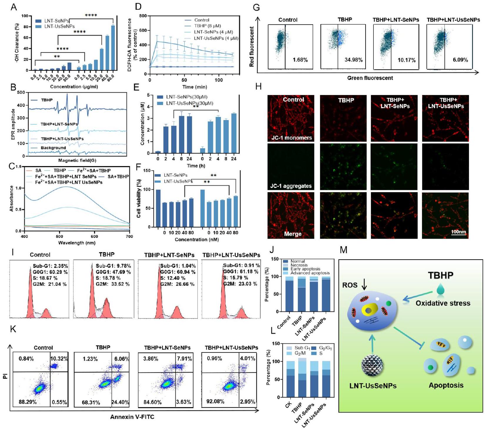
أكدت صور الفلورية هذه النتائج، حيث أظهرت أن LNT-UsSeNPs عكست تأثير TBHP المسبب
انخفاض في نسبة الفلورية الحمراء إلى الخضراء (الشكل 2H). وبالتالي، فإن LNT-UsSeNPs خفضت بشكل فعال تلف الخلايا الناتج عن TBHP. بالإضافة إلى ذلك، أظهرت تحليل دورة الخلية (الشكل 2I وJ) أن معالجة TBHP زادت بشكل كبير من احتجاز الخلايا في مرحلة S في خلايا PC-12. وقد زادت مرحلة Sub-G1 من 2.35 إلى بينما ارتفعت نسبة الخلايا في مرحلة G2/M من 21.04 إلى . علاج LNT-UsSeNPs
تخفيف فعّال لزيادة مرحلة Sub-G1 الناتجة عن TBHP (من 9.78 إلى ) وقام بتطبيع نسبة خلايا مرحلة G2/M إلى حوالي تم تقييم الاستماتة باستخدام صبغة Annexin V-FITC واليوديد البروبيوم (PI)، تلاها تحليل بواسطة قياس التدفق الخلوي. كما هو موضح في الشكل 2 K و L، كانت نسبة الخلايا المستميتة المبكرة قد زادت بشكل ملحوظ من 0.55 إلى بعد حضانة TBHP. وعلى العكس، تم تقليل عدد الخلايا المبرمجة للموت المبكر لعلاج LNT-UsSeNPs بشكل كبير إلى ، التي أبرزت الخصائص القوية المضادة للاستماتة لـ LNT-UsSeNPs. تشير هذه النتائج بقوة إلى أن LNT-UsSeNPs تمارس تأثيرات واقية على الميتوكوندريا وتقدم دورة الخلية الطبيعية من خلال التخلص من ROS الناتجة عن TBHP في خلايا PC-12، مما يثبط الاستماتة (الشكل 2M).

التقييم الأيضي وسلامة الاستخدام لـ LNT-SeNPs و LNTUsSeNPs

للتحقيق في الأيض الحي والسلامة الحيوية لـ LNT-SeNPs و LNT-UsSeNPs، استخدمنا ICP-MS لتحليل حركيتها الدوائية في الجرذان. تم تحديد نصف عمر LNT-UsSeNPs في مجرى الدم للجرذان ليكون 65.3 ساعة، وهو أطول من 45.6 ساعة التي لوحظت لـ LNT-SeNPs (الشكل 3A وB). وهذا يشير إلى أن LNT-UsSeNPs حافظت على تركيزات عالية من الدواء في البلازما لفترات طويلة، مما قد يعزز توصيل الدواء إلى مواقع إصابة الحبل الشوكي. علاوة على ذلك، كشفت تحليل ICP-MS أن الكلى أظهرت أعلى تراكم لهذه الجسيمات النانوية، مما يشير إلى الأيض القائم على الكلى لكل من LNT-SeNPs و LNT-UsSeNPs. ومن الجدير بالذكر أن تركيز السيلينيوم في مجموعة LNT-UsSeNPs كان مرتفعًا في الحبل الشوكي، مما يشير إلى تحسين الامتصاص بواسطة الأنسجة الشوكية، مما قد يكون مفيدًا لعلاج إصابة الحبل الشوكي. يكشف تحليل ICP-MS لمختلف الأعضاء أن تركيزات السيلينيوم في مجموعة LNT-SeNPs، باستثناء الحبل الشوكي، أعلى مقارنة بتلك الموجودة في مجموعة LNT-UsSeNPs. تشير هذه الملاحظة إلى أنه عند إعطائها بتركيزات متساوية، تظهر LNT-UsSeNPs امتصاصًا أقل من الأعضاء غير المرتبطة بإصابة الحبل الشوكي، مما يحافظ على مستويات مرتفعة في الدورة الدموية النظامية لفترات طويلة. تزيد هذه الدورة الممتدة من احتمالية عبور LNT-UsSeNPs لحاجز الدم في الحبل الشوكي والوصول إلى الموقع المستهدف لإصابة الحبل الشوكي. (الشكل 3C وH).
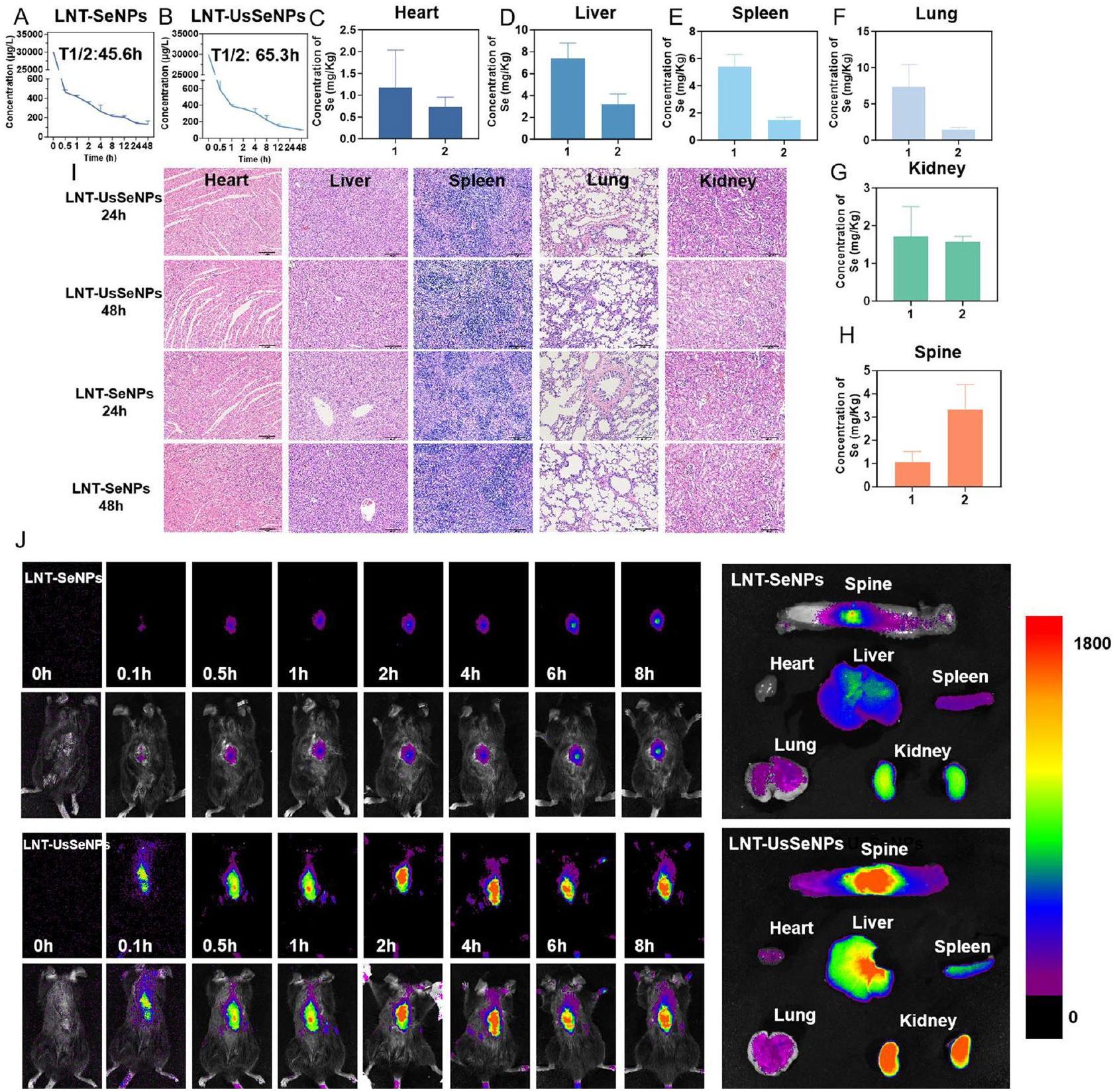
لتقييم السمية الحية للجزيئات النانوية، أجرينا فحوصات نسيجية باستخدام صبغة الهيماتوكسيلين والإيوزين (H&E) لشرائح من الأعضاء الرئيسية بعد الحقن. أظهرت النتائج في الشكل 3I عدم وجود ضرر نسيجي كبير للأعضاء الرئيسية، مما يؤكد السلامة الحيوية الكافية لـ LNT-SeNPs و LNT-UsSeNPs ضمن نطاق الجرعة المعطاة.
علاوة على ذلك، استكشفنا كفاءة التوزيع ومعالجة LNT-UsSeNPs في الجسم الحي. بعد حقن الجزيئات النانوية السيلينيوم المعلّمة بالفلوريسنت،
كانت شدة الفلورسنت في موقع SCI في مجموعة LNTUsSeNPs أعلى بشكل ملحوظ في نقاط زمنية مختلفة مقارنة بمجموعة LNT-SeNPs، كما لوحظ في صور الفلورسنت للأحشاء الفأرية. علاوة على ذلك، أظهرت صور مجموعة LNT-UsSeNPs شدة فلورسنت أعلى بشكل ملحوظ في موقع SCI مقارنة بمجموعة LNT-SeNPs، مع ملاحظات لشدة نسبية عالية أيضًا في الكبد والطحال والكلى (الشكل 3J). تشير هذه النتائج إلى أن LNT-UsSeNPs يمكن أن تعبر بشكل أكثر فعالية عن BSCB من خلال الدورة الدموية للوصول إلى موقع SCI في الفئران، مما يحقق تركيز دوائي محلي فعال علاجياً قد يسهل العلاج اللاحق لـ SCI.

تحسين الوظيفة الحركية وبقاء الخلايا العصبية في الفئران المصابة بـ SCI عند العلاج بـ LNT-SeNPs و LNTUsSeNPs

لتقييم التأثيرات العصبية الواقية لـ LNT-SeNPs و LNT-UsSeNPs في الجسم الحي، قمنا بتقييم استعادة وظيفة الأطراف الخلفية الحركية في الفئران بعد الإجراء الجراحي. باستخدام مقياس باسو للفئران (BMS) واختبارات المنحدر، سجلنا الأداء في الأيام , و 28 . أظهرت النتائج الأولية، المعروضة في الشكل 4A، شلل الأطراف الخلفية في جميع المجموعات باستثناء مجموعة الجراحة الوهمية، مع درجة BMS تبلغ 0 في اليوم الأول. مع مرور الوقت، لاحظنا تحسنًا تدريجيًا في درجات BMS عبر جميع مجموعات العلاج، حيث أظهرت مجموعة LNT-UsSeNPs تحسينًا كبيرًا بحلول اليوم 10 . كما أظهرت هذه المجموعة زوايا تسلق محسنة مقارنة بمجموعة الجراحة الوهمية (الشكل 4B). أظهرت تحليل المشي أن مجموعة SCI أظهرت حركة غير منسقة ومقاومة كبيرة لحركة الأطراف الخلفية. ومن الجدير بالذكر أن مجموعة LNT-UsSeNPs أظهرت استعادة كبيرة لمشية الأطراف الخلفية والتنسيق الحركي، كما هو موضح في الشكل 4C.
أدى التحقيق الإضافي في حركة الأطراف الخلفية للفأر أثناء المشي إلى تقسيم الحركات إلى سلسلة من المراحل – الوقوف، الدفع، الرفع، التأرجح، والوقوف – لإكمال دورة واحدة، كما هو موضح في الشكل 4D. أظهرت مجموعة SCI شلل الأطراف الخلفية وسحبًا مستمرًا. في المقابل، أظهرت الفئران المعالجة بـ LNT-UsSeNPs تحسينات ملحوظة، مثل استعادة وضعية الوقوف (قدرة الوقوف)، وزيادة دعم الوزن، كما يتضح من زيادة ارتفاع قمة الحوض، ونطاق موسع من حركة المفاصل، مما يسهل حركات الدفع والتأرجح الفعالة.

آثار LNT-SeNPs و LNT-UsSeNPs على علم الأمراض النسيجية للحبل الشوكي وبقاء الخلايا العصبية

لتقييم تأثير علاجات LNT-SeNPs و LNT-UsSeNPs على علم الأمراض النسيجية للحبل الشوكي، تم إجراء تجارب صبغ H&E وNissl وصبغ المناعية المزدوجة. أظهرت صبغة H&E أن هياكل الحبل الشوكي في مجموعة الجراحة الوهمية
الشكل 3 التحليل الحي للسلامة الحيوية والدوائية لـ LNT-SeNPs و LNT-UsSeNPs. (A) التحليل الدوائي لـ LNT-SeNPs و LNT-UsSeNPs (B) في الفئران SD. (C-H) تركيزات السيلينيوم في مختلف أعضاء الفئران بعد 24 و48 ساعة من حقن LNT-SeNPs و LNT-UsSeNPs. (I) تحليل صبغة H&E للأعضاء الرئيسية للفئران بعد العلاج بـ LNT-SeNPs و LNT-UsSeNPs. (J) تصوير الفلورسنت الحي لـ LNT-SeNPs و LNT-UsSeNPs في النقطة الزمنية النهائية
بقيت سليمة، مع عرض خلايا عصبية منظمة جيدًا مع نوى محددة بوضوح. في المقابل، أظهرت مجموعة SCI مادة رمادية غير منظمة، تجاويف نخرية، أشكال عصبية غير منتظمة، انكماش نووي، وفقدان خلايا عصبية حركية. خفف العلاج بـ LNT-UsSeNPs بشكل كبير من هذه التغيرات المرضية، مما يشير إلى تأثير واقي (الشكل 4E). أظهرت صبغة Nissl، التي تستخدم لتقييم سلامة الخلايا العصبية [34]، انخفاضًا ملحوظًا في أجسام Nissl في مجموعة SCI، بينما سهل علاج LNT-UsSeNPs استعادة أجسام Nissl
(الشكل 4F). (عدد الخلايا العصبية: وهمية: 9؛ SCI: 4؛ LNT-SeNPs: 7؛ LNT-UsSeNPs: 8) قيّمت صبغة المناعية المزدوجة تعبير بروتين نوى الخلايا العصبية (Neun)، الذي يعد مهمًا لتوليد المشابك وتوازن الدوائر العصبية، و -توبولين [35، 36]، الذي يعد ضروريًا لتوجيه المحاور والنضوج. لاحظنا زيادة في تعبير Neun وانخفاض عدد تجاويف الصبغ في مجموعة LNT-UsSeNPs، مما يشير إلى انخفاض موت الخلايا العصبية وتحسين البقاء (الشكل 4G).
الشكل 4 تأثير التحسين لـ LNT-UsSeNPs على استعادة الحركة وبقاء الخلايا العصبية في الفئران المصابة بـ SCI. (A) درجات BMS واختبار المنحدر (B) نتائج الفئران في أوقات مختلفة. (C) تم إجراء تحليل آثار الأقدام لكل مجموعة من الفئران لتقييم المشية ووظيفة الحركة. (D) لقطة من الفيديو المسجل تظهر تسلسل حركات الأطراف الخلفية لكل مجموعة من الفئران أثناء المشي. تم استخدام النقاط والخطوط لتمثيل قمة الحوض، مفصل الركبة، ومفصل الكاحل. يتم الإشارة إلى اتجاه حركة الأطراف الخلفية بواسطة سهم. (E) تم إجراء صبغة H&E و(F) صبغة Nissl على أنسجة الحبل الشوكي من كل مجموعة، مع الأسهم السوداء تشير إلى وجود أجسام Nissl. (G) صبغة المناعية للحبل الشوكي في كل مجموعة (Neun، -توبولين، بروتين المايلين الأساسي، وGFAP). تم التعبير عن التواريخ كمتوسط ± SD من ثلاث تجارب مستقلة. تمثل القضبان ذات الميزات المميزة اختلافات ذات دلالة إحصائية عند المستوى
تم تقييم سلامة المايلين في الحبل الشوكي باستخدام صبغة بروتين المايلين الأساسي [37]. في مجموعة SCI، كانت إزالة المايلين واسعة النطاق وظهرت كسور محورية بارزة. ومع ذلك، أظهرت مجموعة LNT-UsSeNPs تجاويف أصغر بكثير وأضرار محورية أقل، مما يشير إلى الحفاظ على سلامة المحاور والمايلين. بالإضافة إلى ذلك، قمنا بتقييم تعبير بروتين GFAP الحمضي الليفي الدبقي
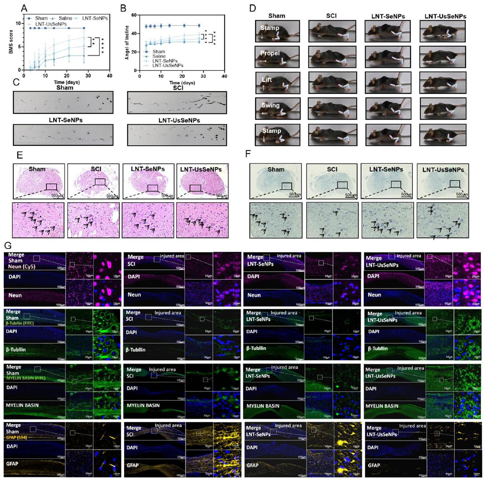
(GFAP) لتحليل استجابات الخلايا الدبقية بعد الإصابة [38]. عادةً ما يؤدي SCI إلى تحفيز الخلايا الدبقية لإنتاج GFAP، مما يؤدي إلى تكوين ندبة دبقية تعيق تجديد الخلايا العصبية. أظهرت صبغة GFAP أن تعبير GFAP كان أقل بكثير في مجموعة LNT-UsSeNPs، مما يشير إلى انخفاض الإجهاد التأكسدي، الالتهاب، وتكوين ندبات دبقية أقل. مجتمعة، تؤكد هذه النتائج
أن LNT-UsSeNPs توفر تأثيرات واقية عصبية، وتثبط إزالة المايلين المحوري، وتقلل من تكوين الندبات الدبقية في الجسم الحي، مما يوفر فوائد علاجية كبيرة لعلاج SCI.

تنظم LNT-UsSeNPs السيلينوزيمات لتخفيف

الإجهاد التأكسدي وتعزيز الحماية المناعية بعد SCI
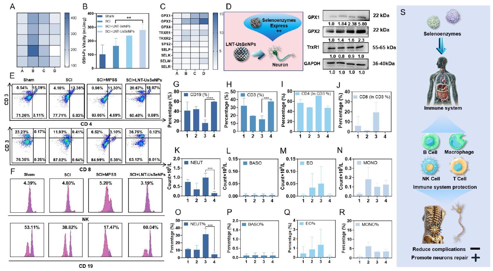
MDA، وهو منتج ثانوي لعملية أكسدة الدهون، يعمل كعلامة حيوية للإجهاد التأكسدي ويكون شائعًا في الحبل الشوكي الغني بالدهون، حيث من المحتمل أن تحدث أكسدة الدهون بعد الإصابة [39]. لذلك، قمنا بتقييم محتوى MDA في الحبل الشوكي، ووجدنا أنه انخفض بشكل ملحوظ في مجموعة LNT-UsSeNPs مقارنة بمجموعة SCI (الشكل 5A). تظهر هذه النتائج أن LNT-UsSeNPs تظهر قدرة مضادة للأكسدة تفوق LNT-SeNPs، بما يتماشى مع الدراسات السابقة المضادة للأكسدة في المختبر.
يحتوي جسم الإنسان على ما لا يقل عن 25 بروتينًا يحتوي على السيلينيوم، يعمل العديد منها كإنزيمات مضادة للأكسدة تساهم في تخفيف الأضرار الناتجة عن الإجهاد التأكسدي [40، 41]. لذلك، قمنا بتقييم تعبير mRNA للبروتين السيلينوزيم المضاد للأكسدة مثل الجلوتاثيون
البيروكسي داز (GSH-Px)، TrxR، السيلينوبروتين K (SelK)، والسيلينوبروتين T (SelT). كما هو موضح في الشكل 5B، فإن العلاج بكل من LNT-SeNPs وLNT-UsSeNPs زاد بشكل كبير من نشاط GSH-Px، حيث أظهرت LNT-UsSeNPs نشاطًا أعلى بشكل ملحوظ من LNT-SeNPs. علاوة على ذلك، قمنا بالتحقيق في تأثير LNT-UsSeNPs على التعبير عن السيلينوبروتينات المضادة للأكسدة باستخدام تحليل qPCR وتحليل Western blot. أظهرت نتائج qPCR (الشكل 5C) أن العلاج بـ LNT-UsSeNPs زاد بشكل كبير من التعبير عن mRNA لـ GPX1 وGPX2 وSelW وTrxR1 مقارنة بمجموعة SCI. وأكد تحليل Western blot اللاحق أن LNT-UsSeNPs زادت من التعبير عن GPX1 وGPX2 وTrxR1 في أنسجة الحبل الشوكي. تشير هذه النتائج إلى أن LNT-UsSeNPs تخفف من الإجهاد التأكسدي الناتج عن إصابة الحبل الشوكي عن طريق تنظيم السيلينوبروتينات المضادة للأكسدة، مما يؤدي إلى تأثيرات وقائية عصبية في الفئران المصابة بإصابة الحبل الشوكي. كما أكدت نتائج Western blot نتائج qPCR (الشكل 5D).
العلاج السريري الشائع لإصابة الحبل الشوكي يتضمن العلاج بجرعات عالية من الكورتيكوستيرويدات، وبشكل خاص مع ميثيل بريدنيزولون صوديوم سكسينات (MPSS)، الذي يثبط وظيفة المناعة على الرغم من فوائده المضادة للالتهابات ومضادات الأكسدة. يمكن أن يؤدي هذا التثبيط إلى
الشكل 5 التأثير المناعي الوقائي لـ LNT-UsSeNPs من خلال تنظيم تعبير السيلينوزيم. (A) تم الكشف عن مستويات MDA لتحديد مستويات أكسدة الدهون (مليمول) في نسيج الحبل الشوكي بعد الإصابة في الفئران من مجموعات مختلفة (A: مجموعة الشام، B: إصابة الحبل الشوكي، C: إصابة الحبل الشوكي + LNT-SeNPs، D: إصابة الحبل الشوكي + LNT-UsSeNPs). (B) تم استخدام اختبار GSH-Px للكشف عن نشاط GSH-Px في مجموعات مختلفة من نسيج الحبل الشوكي بعد الإصابة. (C) نتائج الكشف بواسطة qPCR للبروتينات المحتوية على السيلينيوم المرتبطة بمضادات الالتهاب ومضادات الأكسدة بين المجموعات المختلفة. (A: مجموعة الشام، B: إصابة الحبل الشوكي، C: إصابة الحبل الشوكي + LNT-SeNPs، D: إصابة الحبل الشوكي + LNT-UsSeNPs) (D) LNT-UsSeNPs تزيد من تعبير السيليناز في الخلايا العصبية. (E-J) تحليل تدفق الخلايا لتناسب خلايا المناعة الطحالية في مجموعات مختلفة من الفئران المصابة بإصابة الحبل الشوكي (1: مجموعة الشام، 2: إصابة الحبل الشوكي، 3: إصابة الحبل الشوكي + MPSS، 4: إصابة الحبل الشوكي + LNT-UsSeNPs). (K-R) تصنيف وإحصائيات كريات الدم البيضاء في الفئران المصابة بإصابة الحبل الشوكي (1: مجموعة الشام، 2: إصابة الحبل الشوكي، 3: إصابة الحبل الشوكي + MPSS، 4: إصابة الحبل الشوكي + LNT-UsSeNPs). (S) تأثير مناعة الجسم على إصلاح إصابة الحبل الشوكي. تم التعبير عن التواريخ كمتوسط ± الانحراف المعياري من ثلاث تجارب مستقلة. تمثل الأشرطة ذات الميزات المميزة اختلافات ذات دلالة إحصائية عند مستوى
المضاعفات مثل قرحات الفراش والعدوى البولية والتنفسية، تعيق بشكل كبير تعافي المرضى وتزيد من مدة العلاج وتكاليفه. لذلك، قمنا بالتحقيق فيما إذا كانت LNT-UsSeNPs، التي تظهر أيضًا خصائص مضادة للالتهابات ومضادة للأكسدة، تؤثر على وظيفة المناعة باستخدام MPSS كمقارن سريري. أظهر تحليل الخلايا الطيفي التدفق للخلايا الطحالية تغييرات ملحوظة في تجمعات خلايا المناعة. بعد علاج MPSS، كان هناك انخفاض ملحوظ في نسبة الخلايا اللمفاوية B (CD19+) مقارنة بالمجموعات الوهمية، بينما أظهرت مجموعة LNT-UsSeNPs زيادة في نسبة الخلايا اللمفاوية B. لم توجد اختلافات ملحوظة بين المجموعات في نسبة خلايا NK. أظهر تحليل خلايا T أن نسبة خلايا CD4+ انخفضت بعد علاج MPSS، بينما زادت نسبة خلايا CD8+ في مجموعة MPSS. على العكس، أظهرت مجموعة LNT-UsSeNPs زيادة في نسبة خلايا CD4+، دون تغييرات ملحوظة في خلايا CD8+. نسبة CD4+/CD8+، وهي مؤشر رئيسي لوظيفة المناعة، تنخفض عمومًا عندما يتم قمع المناعة الخلوية وتتعرض وظيفة المناعة للخطر. كانت النسبة أقل بكثير لمجموعة MPSS مقارنة بالمجموعة الوهمية، مما يشير إلى وجود قمع للمناعة. ومع ذلك، زادت نسبة CD4+/CD8+ في مجموعة LNT-UsSeNPs، مما يشير إلى أن LNT-UsSeNPs لا تقمع وظيفة المناعة ولكن تمارس تأثيرات مضادة للأكسدة ومضادة للالتهابات. دعمت نتائج تعداد الدم الكامل هذه النتائج. أظهرت مجموعة MPSS انخفاضًا في نسبة الخلايا اللمفاوية بين كريات الدم البيضاء مقارنة بالمجموعات الوهمية والضابطة، بينما أظهرت مجموعة LNT-UsSeNPs زيادة في نسبة الخلايا اللمفاوية، متجاوزة تلك الخاصة بالمجموعة الوهمية. بالإضافة إلى ذلك، وجدنا أن عدد ونسبة الخلايا المتعادلة في مجموعة MPSS كانت أعلى بكثير من تلك الموجودة في المجموعات الأخرى، مما يشير إلى أن الفئران التي تم حقنها بجرعة عالية من MPSS تعرضت لدرجة معينة من العدوى البكتيرية. وهذا يعكس اضطرابًا في نظام المناعة لديهم، مما يؤدي إلى حدوث عدوى بكتيرية. ومع ذلك، لم يُلاحظ مثل هذه الظاهرة في مجموعة LNT-UsSeNPs. لذلك، فإن حماية نظام المناعة وتقليل المضاعفات المتعلقة بالمناعة أثناء علاج إصابة الحبل الشوكي أمران حاسمان لتعافي المرضى.

تؤثر LNT-UsSeNPs بشكل مضاد للأكسدة ومضاد للالتهابات لتثبيط موت الخلايا العصبية عبر مسارات PI3K-AKT-mTOR-elF4EBP1 و Ras-Raf-MEK-WIPI2

لاستكشاف ما إذا كانت LNT-UsSeNPs تنظم مسارات الإشارة لتثبيط موت الخلايا العصبية، استخدمنا وسم العلامات الكتلية المتسلسلة (TMT) للتحليل البروتيني الكمي لتحديد الفروق في تعبير البروتين بين المجموعات التجريبية. شامل
تحليل إثراء البيانات البروتينية TMT حدد البروتينات المعبر عنها بشكل مختلف بين مجموعتي LNTUsSeNPs و SCI التي كانت لها أهمية بيولوجية كبيرة (الشكل 6A). أشار تحليل الموقع الفرعي الخلوي إلى أن الغالبية العظمى من هذه البروتينات المعبر عنها بشكل مختلف كانت موجودة بشكل أساسي في السيتوبلازم، والمصفوفة خارج الخلوية، والنواة، مما يشير إلى أنها تلعب أدوارًا حاسمة في مختلف أقسام الخلية (الشكل 6B). حدد تحليل مخطط البركان عددًا من البروتينات ذات التعبير المختلف بشكل كبير (الشكل 6C)، مما يسلط الضوء على الآليات الجزيئية المحتملة التي تكمن وراء تأثيرات LNT-UsSeNPs. باستخدام تحليل إثراء مسار موسوعة كيوتو للجينات والجينومات (KEGG)، قمنا بتعليق وظائف البروتينات التي أظهرت تعبيرًا متغيرًا وأجرينا تحليلًا مقارنًا للتمييز بين تلك التي تم تنظيمها لأعلى وتلك التي تم تنظيمها لأسفل (الشكل 6D و F). أظهرت النتائج أن LNT-UsSeNPs زادت بشكل كبير من تنظيم البروتينات الرئيسية في مسارات الإشارات PI3K-AKT-mTOR و Ras-Raf-MEK-ERK، مما يعزز نمو الخلايا، وتكاثرها، وتمايزها. بعد إصابة الحبل الشوكي، تساهم الإجهاد التأكسدي والالتهاب بشكل كبير في الضرر الثانوي، وإصابة الخلايا العصبية، وفقدان الوظيفة. تعتبر مسارات PI3K-AKT-mTOR و Ras-Raf-MEK-ERK حاسمة في التخفيف من الإجهاد التأكسدي والالتهاب. يعزز مسار PI3K-AKT-mTOR إصلاح الأعصاب من خلال تنظيم بقاء الخلايا وكبح الإجهاد التأكسدي والالتهاب. تنشيط AKT يقلل من الجذور الحرة من خلال زيادة تنظيم الإنزيمات المضادة للأكسدة مثل SOD، بينما يكبح أيضًا إطلاق العوامل الالتهابية مثل TNF- و IL-6. يدعم مسار mTOR الحماية العصبية من خلال تعزيز تخليق البروتين وتجديد الخلايا، مما يقلل من تدمير الأنسجة. يساعد مسار Ras-Raf-MEK-ERK، الضروري لتكاثر الخلايا وبقائها، في إصلاح الأنسجة من خلال تعزيز بقاء الخلايا العصبية وتكاثر الخلايا الدبقية. يقلل تنشيط ERK من الإجهاد التأكسدي من خلال زيادة التعبير الجيني لمضادات الأكسدة والحد من تراكم الجذور الحرة، بينما يمنع أيضًا إنتاج العوامل المؤيدة للالتهابات، مما يمنع المزيد من الضرر العصبي ويعزز الإصلاح العصبي. لذلك، افترضنا أن LNT-UsSeNPs يمكن أن تعزز بشكل فعال بقاء الخلايا العصبية بعد إصابة الحبل الشوكي من خلال تنشيط هذه المسارات الإشارية، وبالتالي حمايتها من الموت المبرمج الناتج عن الإجهاد التأكسدي الثانوي والالتهاب. يؤدي ذلك في النهاية إلى تحسين معدل بقاء الخلايا العصبية واستعادة جزئية لوظيفة الحركة. أظهرت التحليلات الإضافية للعلامات الخلوية أن البروتينات المرتبطة بالخلايا العصبية هي التي dominated البروتينات المختلفة (الشكل 6G)، وهو ما يتماشى مع فرضيتنا الأولية ويعزز من تأثيرات LNT-UsSeNPs الخاصة بالخلايا العصبية. بالإضافة إلى ذلك، اقترحت تحليل مسار KEGG أن LNT-UsSeNPs قد تعزز من تأثيراتها الحامية العصبية من خلال
الشكل 6 LNT-UsSeNPs تمنع موت الخلايا العصبية من خلال ممارسة تأثيرات مضادة للأكسدة ومضادة للالتهابات. (A) مخطط تفاعل تحليل المكونات الرئيسية لمجموعات SCI و LNT-UsSeNPs (3D). (B) الموقع الخلوي للبروتينات المعبر عنها بشكل مختلف بين مجموعات SCI و LNT-UsSeNPs. (C-F) مخطط بركاني يوضح 27 جينًا معبرًا عنه بشكل مختلف بين مجموعات SCI و LNT-UsSeNPs جنبًا إلى جنب مع تحليل إثراء مسار KEGG للجينات المتداخلة ( : خريطة بركانية؛ D: تحليل إثراء الجينات المختلفة؛ E: تحليل إثراء الجينات المرتفعة التعبير؛ F: تحليل إثراء الجينات المنخفضة التعبير). (G) تحليل العلامات الخلوية للجينات المعبر عنها بشكل مختلف. (H) الآلية الجزيئية التي تمنع بها LNT-UsSeNPs موت الخلايا العصبية. (I) تحليل Western blot للبروتينات المعبر عنها بشكل مختلف التي تم تحديدها باستخدام بروتيوميات TMT (1: Sham، 2: SCI، 3: SCI + MPSS، 4: SCI + LNT-UsSeNPs)
تعديل تعبير البروتينات المرتبطة بعمليات مضادة للأكسدة، مضادة للالتهابات، وموت الخلايا المرتبطة بمسارات الإشارة PI3K-AKT-mTOR و Ras-Raf-MEK-ERK (الشكل 6I). تقدم هذه النتائج وجهات نظر جديدة حول الآليات المحتملة لعمل LNT-UsSeNPs في علاج إصابة الحبل الشوكي وتضع الأساس لمزيد من الأبحاث المتعمقة.
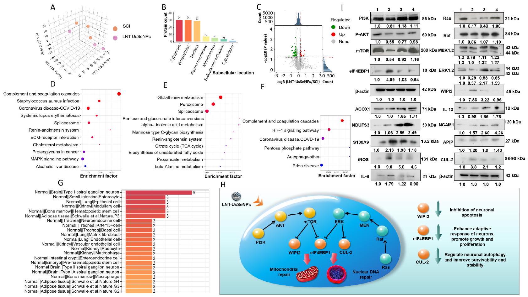
تم إجراء تحليل Western blot بعد ذلك للتحقق من بيانات البروتيوم (الشكل 6H). كشفت النتائج أن LNT-UsSeNPs زادت من تعبير البروتينات داخل مسار الإشارة PI3K-AKT-mTOR وفي الوقت نفسه قيدت تعبير مثبط بدء الترجمة، بروتين 4E المرتبط بعامل بدء الترجمة حقيقي النواة 1 (eIF4EBP1). عزز هذا العمل المزدوج ترجمة mRNA وتخليق البروتين في الخلايا العصبية. تشير بياناتنا إلى أن زيادة التعبير عن eIF4EBP1/2 تنشط عامل بدء الترجمة حقيقي النواة 4E (eIF4E)، مما يبدأ ترجمة mRNA المعتمدة على الكاب. هذه العملية ضرورية لتطور الخلايا العصبية ونمو الاتصالات المشبكية [45]. بالإضافة إلى ذلك، خفضت LNT-UsSeNPs تعبير البروتين المرتبط بالموت الخلوي WIPI2، مما يعزز بقاء الخلايا العصبية ويقلل من الموت الخلوي. في الوقت نفسه، زادت من تنظيم
مسار Ras-Raf-MEK-ERK يمنع تعبير Cullin 2، وهو مكون من مجمع اليوبكويتين، مما يقيد موت الخلايا العصبية.
علاوة على ذلك، أظهرت LNT-UsSeNPs أنها تقلل من مستويات تعبير البروتينات المؤيدة للالتهابات، مثل S100A9 و iNOS و IL-6، وتعزز في الوقت نفسه تعبير البروتين المضاد للالتهابات IL-10. فيما يتعلق بالخصائص المضادة للأكسدة، عززت LNT-UsSeNPs تعبير بروتين عامل تنشيط البروتياز المرتبط بالموت الخلوي-1 (APIP)، مما يقلل من موت الخلايا العصبية الناتج عن الإجهاد التأكسدي. علاوة على ذلك، زادت LNT-UsSeNPs من مستويات بروتين نازع هيدروجين NADH الحديدي-الكبريتي 3 (NDUFS3)، مما يحافظ على الأيض الهوائي الطبيعي في الخلايا العصبية وزادت من تعبير أكسيداز أسيل CoA 1 (ACOX1)، مما يثبت الأيض الدهني داخل الخلايا. كما زادت معالجة LNT-UsSeNPs من تعبير جزيء التصاق الخلايا العصبية 1 (NCAM1)، مما يسهل التجديد العصبي ونقل الإشارات عبر الغشاء، وهو ما يفيد في إصلاح وظيفة الأعصاب بعد إصابة الحبل الشوكي.
بشكل جماعي، تظهر هذه النتائج بشكل قوي أن LNT-UsSeNPs تخفف من موت الخلايا العصبية من خلال زيادة تنظيم مسارات PI3K-AKT-mTOR و Ras-Raf-MEK-ERK وتعديل تعبير البروتينات ذات الصلة
المضادة للأكسدة والمضادة للالتهابات. تقلل هذه التأثيرات من الإجهاد التأكسدي المحلي والالتهاب بينما تعزز تعبير البروتينات المشاركة في الإصلاح العصبي وتعزز التعافي الوظيفي. بشكل عام، أظهرت LNT-UsSeNPs تأثيرات مضادة للالتهابات ومضادة للأكسدة دون تثبيط الجهاز المناعي، كما يتضح من التجارب المتعلقة بالمناعة وتجارب البروتينات المرتبطة بالأنسجة. هذه ميزة كبيرة مقارنة بالعلاج بالجلوكوكورتيكويد، حيث تتجنب الآثار الجانبية المعدية المرتبطة.

الاستنتاجات

في هذه الدراسة، قمنا بتخليق LNT-UsSeNPs فائقة الصغر، والتي أظهرت تأثيرات علاجية واضحة ضد إصابة الحبل الشوكي بفضل تصميمها الهيكلي المحسن وتعديلات السطح. تم استخدام LNT كعامل تعديل، مما أنتج LNT-UsSeNPs لتحسين الاستقرار وامتصاص الخلايا. تمتلك LNT-UsSeNPs مساحة سطح محددة أكبر من LNT-SeNPs، مما يؤدي إلى زيادة القدرة المضادة للأكسدة، مما يقلل بشكل فعال من تلف الخلايا الناتج عن ROS. بالإضافة إلى ذلك، عكست LNT-UsSeNPs بشكل فعال توقف دورة الخلية G2/M الناتج عن TBHP، وتعطيل الميتوكوندريا، والموت الخلوي. في الجسم الحي، زادت LNT-UsSeNPs بشكل كبير من وظيفة الحركة في الأطراف الخلفية للفئران المتأثرة بإصابة الحبل الشوكي. كشفت التحليلات النسيجية عن التأثيرات الحامية العصبية لـ LNT-UsSeNPs في الفئران المصابة بإصابة الحبل الشوكي. أكدت تحليلات البروتيوم وWestern blot أن هذه الجسيمات النانوية نشطت مسارات PI3K و MAPK، وزادت من تعبير السيلينوبروتين، وقللت من الضرر التأكسدي والالتهاب، وحمت الخلايا العصبية من المزيد من الإصابة. علاوة على ذلك، تظهر خصائص مضادة للأكسدة ومضادة للالتهابات دون التأثير على الجهاز المناعي. بشكل عام، تقدم هذه الجسيمات النانوية LNT-UsSeNPs فائقة الصغر العديد من المزايا. توفر حجم الجسيمات الأصغر لـ LNT-UsSeNPs ميزة في عبور حاجز BSCB، مما يقلل بشكل فعال من تأثير الحاجز على تراكم الدواء ويعزز الفعالية العلاجية. بالإضافة إلى ذلك، يمكن أن تحيد الخصائص الاختزالية القوية للسيلينيوم الصفري في الجسيمات النانوية ROS وتنظم مستويات السيلينوبروتين، مما يمنع الإجهاد التأكسدي الثانوي والاستجابات الالتهابية المرتبطة، مما يقلل في النهاية من موت الخلايا العصبية الناتج عن الإصابة الثانوية. علاوة على ذلك، أظهرت LNT-UsSeNPs تأثيرات واقية على الجهاز المناعي، متجنبة التأثيرات المثبطة للمناعة التي تسببها عادة الأدوية المضادة للالتهابات مثل الجلوكوكورتيكويدات. باختصار، تمتلك LNT-UsSeNPs إمكانات واعدة كأدوية نانوية غير مثبطة للمناعة لعلاج إصابة الحبل الشوكي وقد تكون استراتيجية علاجية جديدة لإدارة الإجهاد التأكسدي والالتهاب من خلال تنظيم السيلينوبروتين.

المواد والأساليب
تحضير LNT-UsSeNPs

لتحضير جسيمات نانوية من السيلينيوم فائقة الصغر (UsSeNPs)، بدأنا بخلط 20 ملغ من مسحوق السيلينيوم مع 10 مل من بولي إيثيلين جلايكول 400 (PEG 400) في درجة حرارة الغرفة ( ). تم تسخين الخليط الناتج عند تحت التحريك المستمر بسرعة 360 دورة في الدقيقة لمدة ساعتين لضمان التشتت الشامل وتكوين الجسيمات النانوية. عند الانتهاء من التفاعل، تم السماح لمعلق UsSeNPs الناتج بالبرودة ثم تم تخزينه في درجة حرارة الغرفة للاستخدام لاحقًا. لإنشاء LNT-UsSeNPs، قمنا أولاً بإذابة 20 ملغ من LNT في 8.4 مل من الماء النقي لإنتاج محلول LNT متجانس. بعد ذلك، تم إدخال معلق UsSeNPs (1.6 مل) تدريجيًا إلى محلول LNT قطرة قطرة، مع الحفاظ على معدل متحكم فيه من لكل قطرة. تم الاستمرار في التحريك عند 500 دورة في الدقيقة عند درجة حرارة الغرفة لمدة 12 ساعة لتعزيز تفاعل الجسيمات النانوية مع الرابطة وتكوين طلاء موحد. لتأكيد نجاح تشكيل الجسيمات النانوية وقياس محتوى السيلينيوم داخل LNT-UsSeNPs، تم إجراء تحليل ICP-MS.

تحضير LNT-SeNPs

تم بدء تخليق LNT-SeNPs عن طريق إذابة 20 ملغ من LNT في 9 مل من الماء النقي للغاية، مع ضمان الذوبان الكامل في درجة حرارة الغرفة. ). بعد ذلك، 0.5 مل من محلول سيلينيت الصوديوم بتركيز 100 مللي مولار ( تم إضافة محلول ) تدريجياً إلى محلول LNT. بعد ذلك، تم إضافة 0.5 مل من محلول حمض الأسكوربيك (فيتامين C) بتركيز 400 مللي مول إلى الخليط. تم إجراء إضافة حمض الأسكوربيك تدريجياً لضمان عملية اختزال محكومة، مما يعزز التكوين المتجانس لجزيئات السيلينيوم النانوية داخل محلول LNT. تم تحريك الخليط باستمرار عند لمدة 12 ساعة، مما يسمح بوقت كافٍ للاختزال الكامل واستقرار الجسيمات. لإزالة أي مكونات غير متفاعلة ونواتج ثانوية، تم غسيل المحلول الناتج ضد الماء النقي للغاية لمدة 48 ساعة. تم تحديد تركيز السيلينيوم في المنتج النهائي، LNT-SeNPs، باستخدام ICP-MS.

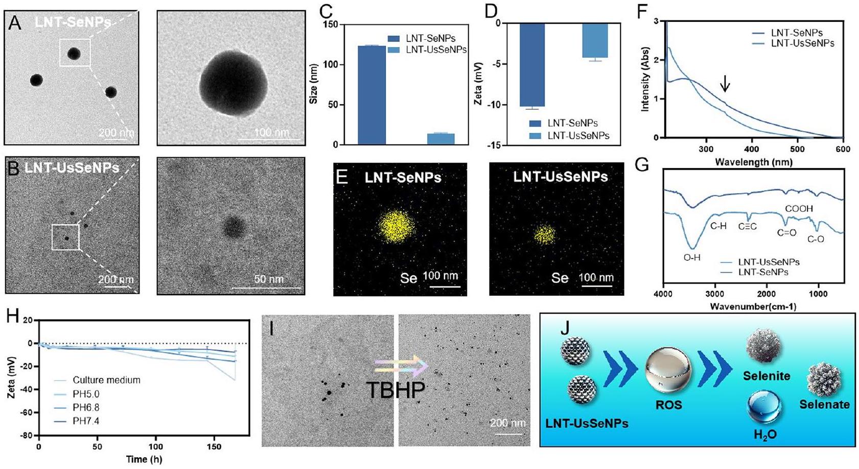
توصيف LNT-SeNPs و LNT-UsSeNPs

تم فحص الخصائص الميكروهيكلية لـ LNT-SeNPs و LNT-UsSeNPs باستخدام TEM، مما يوفر تصورًا تفصيليًا لشكل الجسيمات النانوية. تم تحليل حجم الجسيمات، والجهد الكهربائي، واستقرار LNT-SeNPs و LNT-UsSeNP بواسطة جهاز Malvern Zetasizer Nano ZS. من أجل تقييم استقرار LNT-SeNPs و LNT-UsSeNP تحت ظروف فسيولوجية مختلفة، تم إجراء تجربة الاستقرار في فترات زمنية مختلفة في و 7.4، محلول فوسفات البفر (PBS) و DMEM تمت دراسة الهياكل الكيميائية للجسيمات النانوية باستخدام مطيافية الأشعة فوق البنفسجية والمرئية (UV-Vis) ومطيافية الأشعة تحت الحمراء (FT-IR). هذه الاختبارات ضرورية لتقييم
إمكانات هذه الجسيمات النانوية للتطبيقات الطبية الحيوية، لا سيما من حيث استقرارها وسلامتها في بيئات بيولوجية متنوعة.

تقييم نشاط إزالة الجذور الحرة

تم تقييم قدرات LNT-SeNPs و LNTUsSeNPs المضادة للأكسدة بناءً على قدرتها على القضاء على الجذور الحرة للأكسجين (ROS) باستخدام اختبار ABTS. تم تنشيط محلول ABTS (5 مللي مول) عن طريق إضافة 10 ملغ من ثاني أكسيد المنغنيز. وتصفية من خلال ورق الترشيح ومرشح غشائي. تم تخزين محلول ABTS المنشط في الظلام عند لمدة 12 ساعة قبل الاستخدام. في الإعداد التجريبي، تم استخدام تركيزات مختلفة من الجسيمات النانوية تم تحضيرها في PBS. تم استخدام حجم متساوي من هذه المحاليل النانوية ( تم خلطه مع ABTS ) حل في تنسيق ميكرو بلايت. تم بعد ذلك حضن الخلطات في تحت ظروف مظلمة لـ تم أخذ قراءات الامتصاص عند 734 نانومتر بفواصل زمنية مدتها دقيقة واحدة على مدى 15 دقيقة. تم تحديد معدل إزالة الجذور الحرة باستخدام الصيغة التالية:

تقييم ⋅ OH و

أولاً، تم تحضير محلول 1% من TBHP عن طريق إذابة 70% من TBHP في ماء نقي للغاية. يخضع TBHP في المحلول للتحلل، مما يؤدي إلى توليد • في نظام تفاعل TBHP، يمكن أن يتحد الأكسجين الناتج عن تحلل TBHP مع الإلكترونات لتكوين . بعد ذلك، تم إنتاجها من خلال تفاعل فينتون باستخدام نظام TBHP، بتركيز 1.6 مليمول و TBHP لمدة 15 دقيقة. قدرات التجميع لـ LNT-SeNPs و LNT-UsSeNPs ضد • OH و – تم تحليلها بعد ذلك باستخدام طيفية الرنين المغناطيسي الإلكتروني. باختصار، LNT-SeNPs و LNT-UsSeNPs ( ) أُضيفت إلى المحاليل التي تحتوي على الـ ⋅ OH المتولد أو الجذور الحرة وتم تحضينها لمدة 30 دقيقة. ثم، تم إضافة DMPO واستخدمت تقنية EPR للكشف عن قدرة LNT-SeNPs و LNT-UsSeNPs على القضاء على و الجذور الحرة. تم قياس قدرات التقاط ⋅ OH لـ LNT-SeNPs و LNTUsSeNPs بشكل غير مباشر من خلال الكشف عن تكوين حمض 2،3-ديهيدروكسي بنزويك من حمض الساليسيليك. و الجذور الحرة، التي تظهر امتصاصًا مميزًا عند 510 نانومتر في طيف الأشعة فوق البنفسجية والمرئية. تم استخدام مجس فلوري DHE للكشف عن قدرة LNT-SeNPs و LNT-UsSeNPs على امتصاص الأكسجين.

زراعة خلايا PC-12 وتقييم حيوية الخلايا

تم الحصول على خلايا PC-12 من ATCC (ماناساس، فيرجينيا، الولايات المتحدة الأمريكية) وزرعها في DMEM معززة بمصل جنين البقر. ) والبنسلين-ستربتوميسين (1%) عند في بيئة تحتوي على . تم زراعة خلايا PC-12 في أطباق 96 بئر. بعد حضانة الدواء، تم إضافة CCK-8 لتقييم حيوية الخلايا. للتحقيق في ديناميات امتصاص الخلايا لـ LNT-SeNPs و LNT-UsSeNPs، تم حضن خلايا PC-12 مع هذه الجسيمات النانوية تحت ظروف محكومة. باختصار، PC-12 ( “خلايا/مل” تم زراعتها في أطباق الثقافة. تم إعطاء كلا النوعين من الجسيمات النانوية للخلايا، وتم تحديد فترات الحضانة عند ، و24 ساعة لالتقاط امتصاص الوقت المعتمد. بعد الحضانة مع LNT-SeNPs وLNT-UsSeNPs على التوالي، تم جمع الخلايا، وتم تعريض التعليق الخلوي لهضم حمض النيتريك لتحضيره للتحليل العنصري. تم قياس تركيزات السيلينيوم في الخلايا عند كل نقطة زمنية بشكل كمي باستخدام ICP-MS. سمح هذه الطريقة بالتحديد الدقيق لكمية السيلينيوم التي تم امتصاصها بواسطة الخلايا على مدى فترات محددة، مما يوفر رؤى حول حركية امتصاص الجسيمات النانوية بواسطة خلايا شبيهة بالخلايا العصبية. هذه الطريقة حاسمة لفهم التوافر البيولوجي والفعالية العلاجية المحتملة للجسيمات النانوية المعتمدة على السيلينيوم في سياق عصبي. لتقييم نطاق الجرعة الآمنة من LNT-SeNPs وLNT-UsSeNPs لخلايا PC-12، تم تعريض الخلايا لتركيزات مختلفة من LNT-SeNPs وLNT-UsSeNPs لمدة 24 ساعة. بعد ذلك، قمنا بتقييم حيوية خلايا PC-12 بعد 24 ساعة باستخدام اختبار CCK-8. لتحديد تأثير LNT-SeNPs وLNT-UsSeNPs على عكس الضرر التأكسدي الناتج عن TBHP على حيوية الخلايا، تم معالجة خلايا PC-12 بـ LNT-SeNPs وLNT-UsSeNPs لمدة 24 ساعة، تلاها إزالة الدواء والتعرض اللاحق لـ TBHP لمدة 24 ساعة أخرى. بعد ذلك، تم تقييم حيوية خلايا PC-12 بعد 24 ساعة من العلاج باستخدام اختبار CCK-8.

تحليل تدفق الخلايا لدورة الخلية، الموت المبرمج، و

تم زراعة خلايا PC-12 في أطباق 6 آبار. في البداية، تم معالجة الخلايا بـ LNT-SeNPs و LNTUsSeNPs لمدة 24 ساعة، وبعد ذلك تم معالجتها بـ TBHP لمدة 24 ساعة إضافية. بعد ذلك، تم إجراء صبغ البروب والتدفق الخلوي لتحليل الخلايا. عند إجراء تحليل دورة الخلية، تم تثبيت خلايا PC-12 أولاً باستخدام 70% إيثانول وتم تخزينها في لمدة 48 ساعة. بعد ذلك، تم صبغ الخلايا بـ PI في درجة حرارة الغرفة، تتراوح من 20 إلى ، لمدة 30 دقيقة. لتقييم عملية الموت الخلوي المبرمج (الابوبتوز)، تم صبغ الخلايا باستخدام Annexin V و PI، وفقًا للبروتوكول المحدد في مجموعة كشف الابوبتوز باستخدام Annexin V-FITC/PI. بالإضافة إلى ذلك، تم استخدام مجس JC-1 بتركيز تم استخدامه لتلوين خلايا PC-12،
التي تم حضنها بعد ذلك عند لمدة 30 دقيقة. تم استخدام المجهر الفلوري بعد ذلك لالتقاط صور الفلورية لـ JC-1 للخلايا، مما يسهل تحليل الـ .

كشف مستويات ROS داخل الخلايا الكلية

تم زراعة خلايا PC-12 في البداية في لوحة 96 بئر خلايا بعد فترة 24 ساعة، خضعت الخلايا للمعالجة المسبقة بـ تركيزات كل من LNT-SeNPs و LNT-UsSeNPs لمدة 6 ساعات. بعد ذلك، تم صبغ الخلايا عن طريق الحضانة مع DCFDA قبل التعرض لـ TBHP. تم قياس شدة الفلورية الناتجة عند الأطوال الموجية لـ باستخدام جهاز قراءة الصور BioTek Cytation5.

دراسات الأيض السريرية والسمية

تم الحصول على جرذان سبرايج-داولي التي تتراوح أعمارها بين 6-8 أسابيع من مركز حيوانات المختبر في مقاطعة قوانغدونغ وتم توزيعها عشوائيًا على مجموعتين. تم حقن كل مجموعة عن طريق الوريد بـ 1 مل من إما LNT-SeNPs أو LNT-UsSeNPs بتركيز تم جمع عينات الدم المدارية قبل الحقن وعند ، و 48 ساعة بعد الحقن. تم قياس تركيزات السيلينيوم في الدم باستخدام ICP-MS. بعد 24 و 48 ساعة من الحقن، تم euthanized اثنان من الجرذان من كل مجموعة، وتم جمع طحالهم وكليتيهم ورئتيهم وكبديهم وقلوبهم في بارافورمالدهيد، يليه تضمين في البارافين وتقطيع. صبغ الأنسجة بصبغة H&E للفحص المجهري لتقييم السمية الحشوية الحادة للجزيئات النانوية.

إنشاء نموذج حيواني SCI

تم الحصول على إناث من فئران C57/BL6J بوزن 20-22 جرام (6-8 أسابيع) من مركز قوانغدونغ جيكوي ياوكينغ للحيوانات. تم مراجعة بروتوكول تجربة الحيوانات والموافقة عليه من قبل لجنة رفاهية وأخلاقيات الحيوانات في جامعة جينان (رقم الموافقة الأخلاقية: IACUC-20230919-06). تم تخدير الفئران باستخدام التريبروموإيثانول وخضعت لعملية استئصال الفقرات في مستوى T10 تحت ظروف معقمة. تم استخدام جهاز التأثير نموذج III-NYU (W.M. Keck/الولايات المتحدة الأمريكية) لتحفيز إصابة الحبل الشوكي مع ضبط المعلمات على عمق تأثير 0.6 مم، سرعة التأثير، ومدة الإقامة 80 مللي ثانية. خضعت الحيوانات التي أجريت لها عملية وهمية لاستئصال الصفيحة الفقرية دون التسبب في تلف الحبل الشوكي. ثم تم خياطة الجروح في طبقات وتم حقنها يوميًا بسيفترياكسون (0.3 جرام لكل فأر) لمدة سبعة أيام متتالية. تم تقسيم الفئران عشوائيًا إلى أربع مجموعات (وهمية، إصابة الحبل الشوكي، LNT-SeNPs، LNT-UsSeNPs). تم إعطاء LNT-SeNPs و LNT-UsSeNPs عن طريق الوريد. تم إعطاء مجموعات الشام و SCI أحجامًا مكافئة من محلول الملح. تم إعطاء جميع العلاجات مرة واحدة يوميًا لمدة سبعة أيام متتالية، بدءًا من يوم الإصابة في الحبل الشوكي.
تم إجراء تقييمات سلوكية باستخدام مقياس باسو للفأر (BMS) واختبارات المنحدر بانتظام. في اليوم الثامن والعشرين، تم إجراء تحليل للمشي، تلاه euthanasia للفئران واستخراج الحبل الشوكي.

التقييمات السلوكية

تم إجراء تقييمات سلوكية بواسطة مراقبين مدربين ومخفين باستخدام مقياس BMS، واختبار السطح المائل، وتحليل آثار الأقدام، وتحليل دورة الحركة للأطراف الخلفية. مقياس BMS هو مقياس من 9 نقاط يمثل استعادة وظيفة الأطراف الخلفية، يتراوح من 0 (عدم وجود حركة ملحوظة في الأطراف الخلفية) إلى 9 (حركة طبيعية في الأطراف الخلفية). قياس اختبار السطح المائل أقصى زاوية للسطح التي يمكن أن يحتفظ بها الفأر في وضعه لمدة خمس ثوانٍ. بالنسبة لتحليل آثار الأقدام، تم طلاء الفئران بصبغة سوداء على أقدامها وسمح لها بالمشي على ممر ضيق بطول 0.5 متر مبطن بورق أبيض لتسجيل آثار أقدامها. بالإضافة إلى ذلك، تم استخدام تسجيلات الفيديو للفئران وهي تمشي على طول الممر لتحليل دورة الحركة للأطراف الخلفية، والتي تضمنت مراحل الاتصال، والدفع، والإقلاع، والتأرجح، والعودة إلى الاتصال.

صبغة نسيجية

بعد المعالجة المسبقة اللازمة، تم صبغ الأنسجة باستخدام صبغة H&E، وصبغة نيسل، وازدواجية التألق المناعي (Neun/DAPI، -توبولين/DAPI، المايلين الأساسي/DAPI، أو GFAP/DAPI). تم شراء جميع الأجسام المضادة من Abcam، الولايات المتحدة الأمريكية.

تحليل استقلاب LNT-SeNPs و LNT-UsSeNPs في الفئران

باختصار، تم خلط 2.4 مل من محلول LNT-SeNPs بتركيز 2.5 مليمول/لتر و2.4 مل من محلول LNT-UsSeNPs بتركيز 2.5 مليمول/لتر مع 6 ملغ من إندوسيانيين الأخضر-ثيول (ICG-SH) وتم تحريك المزيج لمدة 24 ساعة للحصول على محلولات ICG-SH-LNT-SeNPs وICG-SH-LNT-UsSeNPs، على التوالي. ثم تم اختيار اثني عشر فأراً من نوع C57 تتراوح أعمارها بين 6-8 أسابيع، وتم تخديرها وإخضاعها لنموذج إصابة الحبل الشوكي. تم اختيار ستة فئران عشوائياً وتم حقنها بـ من ICG-SH-LNT-SeNPs و ICG-SH-LNT-UsSeNPs عن طريق الحقن الوريدي. ثم، تم إجراء تصوير الفلورية على الفئران قبل و بعد 8 ساعات من حقن الدواء، تم تحليل وتسجيل شدة الفلورسنت المنبعثة من الأدوية داخل الفئران. بعد 8 ساعات، تم euthanize الفئران التي تم حقنها بـ ICG-SH-LNT-SeNPs و ICG-SH-LNTUsSeNPs، وتم قياس تصوير الفلورسنت للأعضاء الرئيسية، بما في ذلك الكبد، القلب، الرئتين، الطحال، الكلى، والعمود الفقري، لتحليل وتسجيل شدة الفلورسنت للأدوية. أخيرًا، تم هضم الكبد، القلب، الرئتين، الطحال، الكلى، والعمود الفقري باستخدام الماء الملكي، وتم تحديد كميتها وتحليل تركيزات السيلينيوم باستخدام ICP-MS.

تحليل القدرة المضادة للأكسدة في الجسم الحي

تم تقييم الخصائص المضادة للأكسدة لـ LNTSeNPs و LNTUsSeNPs في الجسم الحي من خلال قياس نشاط GSH-Px ومستويات MDA في مستخلصات الحبل الشوكي. أولاً، تم خلط العينات مع محلول تفكيك RIPA، الذي تم تعزيزها بمثبطات البروتياز. ثم، تم استخدام مجموعة اختبار بروتين BCA لقياس محتوى البروتين داخل هذه المستخلصات. تم تحديد نشاط GSH-Px ومحتوى MDA باستخدام مجموعات اختبار محددة (Beyotime، الصين) وفقًا للبروتوكولات القياسية.

تحليل qPCR للتعبير عن السيلينوبروتين

تم استخدام كاشف Trizol (تاكارا بيوتيك، اليابان) لاستخراج RNA الكلي من الحبل الشوكي. بعد ذلك، تم إجراء عملية النسخ العكسي باستخدام PrimeScript RT Master Mix (تاكارا بيوتيك، اليابان). ثم تم تضخيم cDNA عبر qPCR باستخدام SYBR Premix Ex Taq II، وهو منتج آخر من تاكارا بيوتيك، اليابان، على نظام الكشف عن PCR في الوقت الحقيقي CFX Connect، الذي تصنعه Bio-Rad، الولايات المتحدة الأمريكية. بعد ظروف الدورة الحرارية، تم استخدام تحليل التعبير الجيني النسبي -أكتين كتحكم داخلي.

تحليل البروتينات

تم اختيار ثلاثة فئران عشوائيًا من كل مجموعة بعد ثلاثة أيام من نمذجة إصابة الحبل الشوكي. تم euthanize الحيوانات عن طريق خلع العنق، وتم جمع أنسجة الحبل الشوكي وتخزينها في قوارير تجميد، والتي تم تجميدها بعد ذلك في النيتروجين السائل. ثم تم إجراء تحليل البروتيوم TMT على العينات. الخطوات التجريبية هي كما يلي:

توسيم TMT

تم إعادة إذابة الببتيدات في 100 مللي مولار TEAB وتم وسمها بمستحضرات TMT (الببتيد ). تم إجراء تفاعل التوسيم في لـ .

فصل الكروماتوغرافيا السائلة عالية الضغط بالطور العكسي (HPLC)

تم دمج الببتيدات من كل عينة وحقنها في عمود C18 للفصل عن طريق الإيلاج المتدرج. ) في (قمم الإيلاution: 214 نانومتر)، مع جمع عشر فصائل، تم دمجها بناءً على الكروماتوغرامات، وتجفيفها بالتجميد.

الكشف الكمي بواسطة nano-LC-MS/MS

تم إعادة تكوين الببتيدات المجففة في FA، تم الطرد المركزي عند ، وتم تحميله على عمود C18 ذاتي التعبئة. تم إجراء الفصل على جهاز Thermo EASY-nLC نظام بمعدل تدفق ، باستخدام إزاحة تدرجية من 8 إلى المذيب B على مدى 62 دقيقة. تم تأين الببتيدات عبر nano-ESI وتم تحليلها على جهاز Orbitrap Exploris. جهاز مطياف الكتلة في DDA
وضع (نطاق MS: ، الدقة: 120,000). تم إجراء التفتت الثانوي باستخدام HCD في طاقة مع استبعاد ديناميكي (60 ثانية) ودقة 45,000.

التحليل الإحصائي

تم تحديد عتبات التعبير التفاضلي عند تغيير بمقدار 1.2 مرة بالنسبة للخط الأساسي تم إجراء توقعات تحديد المواقع تحت الخلوية باستخدام قاعدة بيانات WoLFPSORT، بينما تم تحديد وظائف البروتين المرتبطة من خلال UniProt ومراجعة الأدبيات ذات الصلة. تم استخدام قاعدة بيانات KEGG لإجراء تحليل إثراء على البروتينات المعبر عنها بشكل مختلف.

تحليل البقعة الغربية

تم إجراء اختبار BCA لقياس تركيز البروتين، وإجمالي البروتين. تم فصلها على هلام SDS-PAGE ). تم نقل البروتينات إلى غشاء PVDF (Millipore، الولايات المتحدة الأمريكية) باستخدام جهاز نقل دقيق (Bio-Rad، الولايات المتحدة الأمريكية). الغشاء بعد محلول BSA ( تم تحضين الحجب مع الأجسام المضادة الأولية طوال الليل في . ثم تم معالجته بأجسام مضادة ثانوية موسومة بالإنزيم ( ). GAPDH و -أكتين عمل كضوابط داخلية.
المتفاعلات الأساسية: GPX1، GPX2، TrxR1، PI3K، P-AKT، mTOR، eIF4EBP1، Ras، Raf، MEK، ERK1/2، WIPI2، ACOX1، S100A9، iNOS، IL-6، IL-10، NCAM1، APIP، -أكتين، وGAPDH (أبكام، الولايات المتحدة الأمريكية).
الأجسام المضادة الثانوية: مضاد IgG ضد الفأر (تكنولوجيا الإشارة الخلوية، الولايات المتحدة الأمريكية).

تحليل مناعي في الجسم الحي في الفئران بعد علاج LNT-UsSeNPs

تم استخدام فئران C57/BL6J للتحليل المناعي. تم إعطاء LNT-UsSeNPs عن طريق الوريد بتركيز سيلينيوم قدره يومياً لمدة ثلاثة أيام متتالية، بدءًا من يوم إدخال SCI. تم إعطاء MPSS كعلاج نبضي بجرعة عالية: 0.5 ساعة بعد ما بعد SCI، تم حقن MPSS عن طريق الوريد، تلاه حقن ثانٍ بعد ، وحقن ثالث من بعد 4 ساعات. في اليوم التالي، تم إعطاء MPSS كل 4 ساعات لمجموع ثلاث جرعات. تلقت كل من مجموعتي الشام و SCI محلول ملحي. تم علاج جميع الفئران مرة واحدة يوميًا. في اليوم الثالث، تم جمع الدم عن طريق ثقب مداري لإجراء تعداد دم كامل مع العد التفريقي، وتم قتل الفئران عن طريق خلع الرقبة. ثم تم حصاد الطحال، وتجانسه، وتحليله. تم حصاد خلايا المناعة الطحالية عن طريق الطرد المركزي، وتم وضع علامات عليها بأجسام مضادة محددة، وتم تقييمها باستخدام قياس التدفق الخلوي.

معلومات إضافية

تحتوي النسخة الإلكترونية على مواد إضافية متاحة علىhttps://doi.org/10.1186/s12951-024-03054-7.

المادة الإضافية 1

الشكر والتقدير

نحن نقدر جهود جميع أعضاء مختبر كلية الكيمياء وعلوم المواد.

مساهمات المؤلفين

ساهم PL و XL و ZW بالتساوي في هذا العمل. قام PL و LC بتصور وتصميم هذا المشروع؛ ساهم PL و XL و ZW في التجارب الرئيسية؛ قام LC بمراجعة المخطوطة؛ قدم QW و KS و XL و LC المساعدة في التجارب؛ ساعد ZL و LL و YW في تحليل البيانات ومراجعة المخطوطة؛ كتب PL المخطوطة مع مدخلات ومراجعة من جميع المؤلفين. قرأ جميع المؤلفين ووافقوا على المخطوطة النهائية.

التمويل

تم دعم هذا العمل من قبل البرنامج الوطني الرئيسي للبحث والتطوير في الصين (2023YFC3402800)، وصندوق العلوم الوطنية للعلماء الشباب المتميزين (82225025)، ومؤسسة العلوم الطبيعية الوطنية في الصين (82174142، 21877049، 32171296، 32101044)، ومؤسسة العلوم الطبيعية لمقاطعة قوانغدونغ في الصين (2021A1515410001، 2020B1515120043)، ومؤسسة K. C. Wong التعليمية ومؤسسة العلوم ما بعد الدكتوراه في الصين (2022M711341).

توفر البيانات

لم يتم إنشاء أو تحليل أي مجموعات بيانات خلال الدراسة الحالية.

الإعلانات

تمت الموافقة على هذه الدراسة من قبل لجنة رعاية واستخدام الحيوانات المؤسسية في جامعة جينان (IACUC-20230919-06). تم إجراء جميع إجراءات الحيوانات وفقًا للإرشادات الأخلاقية الوطنية والمؤسسية لرعاية واستخدام الحيوانات المخبرية.
غير قابل للتطبيق.

المصالح المتنافسة

يعلن المؤلفون عدم وجود مصالح متنافسة.

تفاصيل المؤلف

قسم جراحة العظام، كلية الطب في زهوهاي (مستشفى الشعب في زهوهاي)، المختبر الوطني الرئيسي للجزيئات النشطة بيولوجيًا وتقييم القابلية للعلاج، كلية الكيمياء وعلوم المواد، جامعة جينان، زهوهاي 519000، الصين
قسم جراحة العظام في المستشفى الأول التابع لجامعة جينان، قوانغتشو 510632، الصين
جامعة قوانغدونغ الطبية، زهانجيانغ 524000، الصين
كلية الأعمال، جامعة ماكاو للعلوم والتكنولوجيا، ماكاو 999078، الصين
تاريخ الاستلام: 24 أكتوبر 2024 / تاريخ القبول: 30 نوفمبر 2024
تم النشر عبر الإنترنت: 16 يناير 2025

References

  1. Kumar R, Lim J, Mekary RA, Rattani A, Dewan MC, Sharif SY, Osorio-Fonseca E, Park KB. Traumatic spinal Injury: Global Epidemiology and Worldwide volume. World Neurosurg. 2018;113:e345-63.
  2. Hu X, Xu W, Ren Y, Wang Z, He X, Huang R, Ma B, Zhao J, Zhu R, Cheng L. Spinal cord injury: molecular mechanisms and therapeutic interventions. Signal Transduct Target Ther. 2023;8:245.
  3. Kong G, Xiong W, Li C, Xiao C, Wang S, Li W, Chen X, Wang J, Chen S, Zhang Y, et al. Treg cells-derived exosomes promote blood-spinal cord barrier repair
    and motor function recovery after spinal cord injury by delivering miR-2861. J Nanobiotechnol. 2023;21:364.
  4. Nie X, Liu Y, Yuan T, Yu T, Yun Z, Xue W, Yu T, An J, Dai A, Wu K, Liu Q. Plateletrich plasma-derived exosomes promote blood-spinal cord barrier repair and attenuate neuroinflammation after spinal cord injury. J Nanobiotechnol. 2024;22:456.
  5. Karsy M, Hawryluk G. Modern Medical Management of spinal cord Injury. Curr Neurol Neurosci Rep. 2019;19:65.
  6. Gao P, Yi J, Chen W, Gu J, Miao S, Wang X, Huang Y, Jiang T, Li Q, Zhou W, et al. Pericyte-derived exosomal miR-210 improves mitochondrial function and inhibits lipid peroxidation in vascular endothelial cells after traumatic spinal cord injury by activating JAK1/STAT3 signaling pathway. J Nanobiotechnol. 2023;21:452.
  7. Cavone L, McCann T, Drake LK, Aguzzi EA, Oprisoreanu AM, Pedersen E, Sandi S, Selvarajah J, Tsarouchas TM, Wehner D, et al. A unique macrophage subpopulation signals directly to progenitor cells to promote regenerative neurogenesis in the zebrafish spinal cord. Dev Cell. 2021;56:1617-e16301616.
  8. Sandler AN, Tator CH. Effect of acute spinal cord compression injury on regional spinal cord blood flow in primates. J Neurosurg. 1976;45:660-76.
  9. Liu Z, Yao X, Jiang W, Li W, Zhu S, Liao C, Zou L, Ding R, Chen J. Advanced oxidation protein products induce microglia-mediated neuroinflammation via MAPKs-NF-kappaB signaling pathway and pyroptosis after secondary spinal cord injury. J Neuroinflammation. 2020;17:90.
  10. Li H, Sun D, Zhao Z, Fang J, Li M, Lv C, Zhou W, Li N, Guo Y, Cao Z, et al. Neutrophil membrane-derived nanoparticles protect traumatic brain injury via inhibiting calcium overload and scavenging ROS. J Nanobiotechnol. 2024;22:477.
  11. McGarry T, Biniecka M, Veale DJ, Fearon U. Hypoxia, oxidative stress and inflammation. Free Radic Biol Med. 2018;125:15-24.
  12. Zrzavy T, Schwaiger C, Wimmer I, Berger T, Bauer J, Butovsky O, Schwab JM, Lassmann H, Hoftberger R. Acute and non-resolving inflammation associate with oxidative injury after human spinal cord injury. Brain. 2021;144:144-61.
  13. Hou Y, Luan J, Huang T, Deng T, Li X, Xiao Z, Zhan J, Luo D, Hou Y, Xu L, Lin D. Tauroursodeoxycholic acid alleviates secondary injury in spinal cord injury mice by reducing oxidative stress, apoptosis, and inflammatory response. J Neuroinflammation. 2021;18:216.
  14. Liu C, Hu F, Jiao G, Guo Y, Zhou P, Zhang Y, Zhang Z, Yi J, You Y, Li Z, et al. Dental pulp stem cell-derived exosomes suppress M1 macrophage polarization through the ROS-MAPK-NFkappaB P65 signaling pathway after spinal cord injury. J Nanobiotechnol. 2022;20:65.
  15. Yang Y, Fan S, Chen Q, Lu Y, Zhu Y, Chen X, Xia L, Huang Q, Zheng J, Liu X. Acute exposure to gold nanoparticles aggravates lipopolysaccharideinduced liver injury by amplifying apoptosis via ROS-mediated macrophagehepatocyte crosstalk. J Nanobiotechnol. 2022;20:37.
  16. Zhao P, Hu HZ, Chen XT, Jiang QY, Yu XZ, Cen XL, Lin SQ, Mai SQ, Pang WL, Chen JX, Zhang Q. Mild hyperthermia enhanced synergistic uric acid degradation and multiple ROS elimination for an effective acute gout therapy. J Nanobiotechnol. 2024;22:275.
  17. Costa DD, Beghi E, Carignano P, Pagliacci C, Faccioli F, Pupillo E, Messina P, Gorio A, Redaelli T. Tolerability and efficacy of erythropoietin (EPO) treatment in traumatic spinal cord injury: a preliminary randomized comparative trial vs. methylprednisolone (MP). Neurol Sci. 2015;36:1567-74.
  18. Liu Z, Yang Y, He L, Pang M, Luo C, Liu B, Rong L. High-dose methylprednisolone for acute traumatic spinal cord injury: a meta-analysis. Neurology. 2019;93:e841-50.
  19. Xu Y, Lai H, Pan S, Pan L, Liu T, Yang Z, Chen T, Zhu X. Selenium promotes immunogenic radiotherapy against cervical cancer metastasis through evoking P53 activation. Biomaterials. 2024;305:122452.
  20. Li Y, Liu T, Zheng R, Lai J, Su J, Li J, Zhu B, Chen T. Translational selenium nanoparticles boost GPx1 activation to reverse HAdV-14 virus-induced oxidative damage. Bioact Mater. 2024;38:276-91.
  21. Zou B, Xiong Z, Yu Y, Shi S, Li X, Chen T. Rapid Selenoprotein activation by Selenium nanoparticles to suppresses osteoclastogenesis and pathological bone loss. Adv Mater. 2024;36:e2401620.
  22. Liu C, Wang W, Lai H, Chen Y, Li L, Li H, Zhan M, Chen T, Cao W, Li X. Biosynthesis of fungus-based oral selenium microcarriers for radioprotection and immuno-homeostasis shaping against radiation-induced heart disease. Bioact Mater. 2024;37:393-406.
  23. Liu X, Mao Y, Huang S, Li W, Zhang W, An J, Jin Y, Guan J, Wu L, Zhou P. Selenium nanoparticles derived from Proteus mirabilis YC801 alleviate oxidative stress and inflammatory response to promote nerve repair in rats with spinal cord injury. Regen Biomater. 2022;9:rbac042.
  24. Rao S, Lin Y, Lin R, Liu J, Wang H, Hu W, Chen B, Chen T. Traditional Chinese medicine active ingredients-based selenium nanoparticles regulate antioxidant selenoproteins for spinal cord injury treatment. J Nanobiotechnol. 2022;20:278.
  25. Hao M, Zhang D, Wang W, Wei Z, Duan J, He J, Kong X, Sang Y, Feng S, Liu H. HAp Thermosensitive Nanohydrogel cavities Act as Brood pouches to Incubate and Control-Release NSCs for Rapid spinal cord Injury Therapy. Adv Funct Mater. 2022, 32.
  26. Xiong Z, Lin H, Li H, Zou B, Xie B, Yu Y, He L, Chen T. Chiral selenium Nanotherapeutics regulates selenoproteins to Attenuate Glucocorticoid-Induced osteoporosis. Adv Funct Mater. 2023, 33.
  27. Xie B, Zeng D, Yang M, Tang Z, He L, Chen T. Translational selenium nanoparticles to Attenuate allergic dermatitis through Nrf2-Keap1-Driven activation of Selenoproteins. ACS Nano. 2023;17:14053-68.
  28. Hu Y, Liu T, Li J, Mai F, Li J, Chen Y, Jing Y, Dong X, Lin L, He J, et al. Selenium nanoparticles as new strategy to potentiate gammadelta T cell anti-tumor cytotoxicity through upregulation of tubulin-alpha acetylation. Biomaterials. 2019;222:119397.
  29. Lai H, Xu L, Liu C, Shi S, Jiang Y, Yu Y, Deng B, Chen T. Universal selenium nanoadjuvant with immunopotentiating and redox-shaping activities inducing high-quality immunity for SARS-CoV-2 vaccine. Signal Transduct Target Ther. 2023;8:88.
  30. Ouyang J, Deng B, Zou B, Li Y, Bu Q, Tian Y, Chen M, Chen W, Kong N, Chen T, Tao W. Oral hydrogel microbeads-mediated in situ synthesis of selenoproteins for regulating intestinal immunity and microbiota. J Am Chem Soc. 2023;145:12193-205.
  31. Oprea D, Sanz CG, Barsan MM, Enache TA. PC-12 cell line as a neuronal cell model for Biosensing Applications. Biosens (Basel) 2022, 12.
  32. Liu X, Chen Z, Bai J, Li X, Chen X, Li Z, Pan H, Li S, Gao Q, Zhao N, et al. Multifunctional Hydrogel Eye drops for synergistic treatment of ocular inflammatory disease. ACS Nano. 2023;17:25377-90.
  33. Liu M, Wu X, Cui Y, Liu P, Xiao B, Zhang X, Zhang J, Sun Z, Song M, Shao B, Li Y. Mitophagy and apoptosis mediated by ROS participate in -induced MC3T3-E1 cell dysfunction. Food Chem Toxicol. 2021;155:112388.
  34. Fujita K, Ito H, Nakano S, Kinoshita Y, Wate R, Kusaka H. Immunohistochemical identification of messenger RNA-related proteins in basophilic inclusions of adult-onset atypical motor neuron disease. Acta Neuropathol. 2008;116:439-45.
  35. Ungari M, Manotti L, Tanzi G, Varotti E, Ferrero G, Gusolfino MD, Trombatore M, Cavazzuti L, Tolomini M. NeuN, a DNA-binding neuron-specific protein
    expressed by Merkel cell carcinoma: analysis of 15 cases. Pathologica. 2021;113:421-6.
  36. Sanyal C, Pietsch N, Ramirez Rios S, Peris L, Carrier L, Moutin MJ. The dety-rosination/re-tyrosination cycle of tubulin and its role and dysfunction in neurons and cardiomyocytes. Semin Cell Dev Biol. 2023;137:46-62.
  37. Valdivia AO, Farr V, Bhattacharya SK. A novel myelin basic protein transcript variant in the murine central nervous system. Mol Biol Rep. 2019;46:2547-53.
  38. Mandwie M, Piper JA, Gorrie CA, Keay KA, Musumeci G, AI-Badri G, Castorina A. Rapid GFAP and Iba1 expression changes in the female rat brain following spinal cord injury. Neural Regen Res. 2022;17:378-85.
  39. Haro Giron S, Monserrat Sanz J, Ortega MA, Garcia-Montero C, Fraile-Martinez O, Gomez-Lahoz AM, Boaru DL, de Leon-Oliva D, Guijarro LG, Atienza-Perez M et al. Prognostic value of Malondialdehyde (MDA) in the temporal progression of chronic spinal cord Injury. J Pers Med. 2023, 13.
  40. Li F, Shi Z, Cheng M, Zhou Z, Chu M, Sun L, Zhou JC. Biology and roles in diseases of Selenoprotein I characterized by ethanolamine phosphotransferase activity and antioxidant potential. J Nutr. 2023;153:3164-72.
  41. Hariharan , Dharmaraj S. Selenium and selenoproteins: it’s role in regulation of inflammation. Inflammopharmacol. 2020;28:667-95.
  42. Liu C, Xi L, Liu Y, Mak JCW, Mao S, Wang Z, Zheng Y. An inhalable hybrid Biomimetic Nanoplatform for Sequential Drug Release and Remodeling Lung Immune Homeostasis in Acute Lung Injury Treatment. ACS Nano. 2023;17:11626-44.
  43. Murugaiah V, Yasmin H, Pandit H, Ganguly K, Subedi R, Al-Mozaini M, Madan T, Kishore U. Innate Immune Response against HIV-1. Adv Exp Med Biol. 2021;1313:23-58.
  44. Zahorec R. Neutrophil-to-lymphocyte ratio, past, present and future perspectives. Bratisl Lek Listy. 2021;122:474-88.
  45. Jin H, Xu W, Rahman R, Na D, Fieldsend A, Song W, Liu S, Li C, Rosbash M. TRIBE editing reveals specific mRNA targets of elF4E-BP in Drosophila and in mammals. Sci Adv. 2020;6:eabb8771.

ملاحظة الناشر

تظل Springer Nature محايدة فيما يتعلق بالمطالبات القضائية في الخرائط المنشورة والانتماءات المؤسسية.

Journal: Journal of Nanobiotechnology, Volume: 23, Issue: 1
DOI: https://doi.org/10.1186/s12951-024-03054-7
PMID: https://pubmed.ncbi.nlm.nih.gov/39815246
Publication Date: 2025-01-15

Size effect-based improved antioxidant activity of selenium nanoparticles regulating Anti-PI3K-mTOR and Ras-MEK pathways for treating spinal cord injury to avoid hormone shock-induced immunosuppression

Peixin , Xiaodong , Zihao Wu , Kui Shen , Zhaofeng , Xiaowei Li , Qifeng Wu , Leung Chan , Zhong Zhang , Yutong Wu , Liwen Liu , Tianfeng Chen and Yi Qin

Abstract

Spinal cord injury (SCI) is a critical condition affecting the central nervous system that often has permanent and debilitating consequences, including secondary injuries. Oxidative damage and inflammation are critical factors in secondary pathological processes. Selenium nanoparticles have demonstrated significant antioxidative and anti-inflammatory properties via a non-immunosuppressive pathway; however, their clinical application has been limited by their inadequate stability and functionality to cross the blood-spinal cord barrier (BSCB). This study proposed a synthesis method for ultra-small-diameter lentinan Se nanoparticles (LNT-UsSeNPs) with significantly superior reactive oxygen species (ROS) scavenging capabilities compared to conventional lentinan Se nanoparticles (LNT-SeNPs). These compounds effectively protected PC-12 cells from oxidative stress-induced cytotoxicity, alleviated mitochondrial dysfunction, reduced apoptosis. In vivo studies indicated that LNT-UsSeNPs efficiently penetrated the BSCB and effectively inhibited the apoptosis of spinal neurons. Ultimately, LNT-UsSeNPs directly regulated the PI3K-AKT-mTOR and Ras-Raf-MEK-ERK signaling pathways by regulating selenoproteins to achieve non-immunosuppressive anti-inflammatory therapy. Owing to their ultra-small size, LNT-UsSeNPs exhibited strong spinal barrier penetration and potent antioxidative and anti-inflammatory effects without compromising immune function. These findings suggest that LNT-UsSeNPs are promising candidates for further development in nanomedicine for the effective treatment of SCI.

Peixin Liu, Xiaodong Liu and Zihao Wu contributed equally to this work.
*Correspondence:
Tianfeng Chen
tchentf@jnu.edu.cn
Yi Qin
qinyi0225@163.com
Full list of author information is available at the end of the article

Introduction

Spinal cord injury (SCI) with the high rates of disability and tissue damage is a prevalent and serious disorder of the central nervous system, ultimately and is associated with considerable incidence and mortality. Approximately 700,000 new cases of SCI are reported annually worldwide [1]. SCI is typically caused by high-intensity trauma, including vehicular falls, accidents, violence, but can also result from infections, tumors, degenerative spinal diseases, ischemia-reperfusion injury, and vascular abnormalities [2, 3]. SCI effect motor, sensory, and autonomic functions with the temporary or permanent impairment, imposing a substantial psychological and economic burden on patients and their families. Pathologically, the primary injury is an initial contusion or
transection caused by immediate or sustained mechanical compression or traction on the spinal cord [4-8]. Secondary injury is the response of the body and cells to primary injury and involves complex changes in the immune system, nervous system, and blood circulation. These changes include spinal cord ischemia, neuronal death, oxidative stress, inflammatory responses, neuronal apoptosis, axonal demyelination, and glial scar formation. Secondary injury is a key factor leading to spinal cord dysfunction and impedes recovery, with inflammation and oxidative stress playing critical roles in the process. Many studies have showed that oxidative stress is crucial for the onset and progression of SCI. Oxidative stress at the injury site results from a disparity between the production and neutralization of ROS, which encompasses
reactive oxygen molecules like , and [9, 10]. Research has confirmed that oxidative stress and inflammation mutually promote each other in an inflammatory environment, where inflammatory cells release significant amounts of ROS, exacerbating oxidative damage [11]. Following SCI, necrotic neurons and other cells release high levels of ROS. Owing to high levels of polyunsaturated fatty acids, oxidative metabolic activity, and limited antioxidant capacity, neurons and glial cells are prone to oxidative stress, damaging adjacent surviving neurons. Additionally, local ischemia and hypoxia following SCI lead to mitochondrial dysfunction in neurons, further increasing ROS production and increasing local oxidative stress levels. Multiple studies have demonstrated that inhibiting oxidative stress can improve neuronal survival rates, restore neurological function, suppress glial cell activation, and reduce the expression of inflammatory factors [12-16]. Thus, ROS, oxidative stress, and inflammation are closely related to SCI, and the inhibition of secondary oxidative stress post-SCI is crucial for the recovery of neurological function.
Currently, clinical approaches primarily use glucocorticoids, such as methylprednisolone (MP), to inhibit the production of inflammatory factors after SCI, prevent secondary neuronal death, and inhibit lipid peroxidation to reduce ROS generation and alleviate edema [17, 18]. However, high-dose hormone therapy can cause immune system disorders and increase the risk of infection and other complications, thereby affecting patient recovery. With recent technological advancements, nanomaterials have been extensively studied in disease treatment applications and have consistently demonstrated significant efficacy. Particularly in nanomedicine, nanoparticles often exhibit high catalytic effects owing to their unique particle sizes and rich surface-active sites. Hence, developing nanomedicines with small size and high catalytic activity has become a goal for scientists. Selenium (Se), an essential non-metallic trace element in humans and animals, plays a vital role in maintaining health. It has high biological safety and compatibility. As a key component of the body’s antioxidant system, Se is involved in the synthesis of Se-containing proteins, among which glutathione peroxidase (GPX) and thioredoxin reductase ( ) are the most common; these proteins exhibit excellent antioxidant effects, regulating immune responses to inhibit harmful inflammatory reactions [19-21]. GPX and TrxR family proteins play crucial roles in inhibiting oxidative stress following central nervous system injury, suppressing oxidative reactions caused by free radicals, reducing body damage, and inhibiting inflammation induced by oxidative stress. These proteins are crucial for the proper development and ongoing functionality of neurons. Studies related to SCI have found that Se can inhibit astrocyte activation
and inflammatory factor release after SCI, significantly down-regulating the expression levels of variety oxidative stress markers, such as superoxide dismutase (SOD) and malondialdehyde (MDA). Se also reduces neuronal apoptosis through pathways involving Bax, Bcl-2, and caspase-3 [22]. Additionally, Se supplementation can cause macrophage polarizing to M2 type results in the attenuated expression of interleukin 6 (IL-6), inducible nitric oxide synthase (iNOS) and tumor necrosis factor (TNF- ) to suppress inflammation, and inhibit local oxidative stress and inflammation levels following SCI [23]. Clinical studies have showed that analyzing blood from SCI patients who experienced trauma with or without neurological recovery revealed significantly higher selenoprotein concentrations in the recovery group than in the non-recovery group [24]. The result indicates that blood Se levels and selenoprotein status reflect the recovery of patients with SCI and that Se aids in their neurological recovery. Selenium nanoparticles (SeNPs), a novel Se species, possess excellent antioxidant, anti-aging, and antitumor activities with low toxicity, high drug-loading capacity, superior biocompatibility, and degradability. Previous studies have demonstrated their therapeutic effects on SCI [25-30]. However, the blood-spinal cord barrier (BSCB) is the main factor influencing nanodrug distribution and efficacy in the clinical treatment of SCI. Owing to the continuous tight junctions between capillary endothelial cells, an intact basement membrane, and the formation of a neuroglial membrane by astrocyte foot processes, the BSCB hinders the passage of SeNPs approximately 100 nm in size from the bloodstream to the injury site. Therefore, there is an urgent clinical need for improving the BSCB penetration of SeNPs that do not affect its effect and safety.
Some literatures reported the modification of transmembrane peptides, the improvement of fat solubility and the controlled particles size both advance the barrier penetration of nanodrugs. Thus, ultra-small Se particles have been prepared to better penetrate the BSCB, accumulate effectively at the injury site, and significantly improve SCI treatment efficacy. Although the ultra-small size allows Se nanoparticles to have better BSCB penetration, it introduces new problems, such as a short half-life and poor stability. Lentinan polysaccharides (LNT) exhibit excellent biocompatibility and possess a molecular structure rich in hydroxyl and other functional groups. These groups interact with the surfaces of nanomedicines to form stable chemical bonds or physical adsorption layers. This interaction prevents the aggregation of nanomedicines in aqueous solutions or physiological environments, enhancing their dispersibility and stability. Herein, we prepared an ultra-small Se-based nanosystem with LNT modification (LNT-UsSeNPs) to achieve high BSCB penetration and suitable biostability.
Interestingly, its nanoscale size offers advantages when crossing the BSCB, effectively minimizing the impact of the barrier on therapeutic efficacy. Moreover, zerovalence Se-based drugs with strong reducing ability can neutralize ROS and inhibit the inflammatory response triggered by secondary oxidative stress injury, thereby reducing neuronal apoptosis. Finally, they provide protective benefits to an animal’s immune system by regulating selenoproteins, thus avoiding the immunosuppressive effects caused by glucocorticoids. Therefore, the ultrasmall Se-based nanosystem, which possesses antioxidant, anti-inflammatory, and immune-protective functions, represents a promising non-immunosuppressive therapeutic strategy for SCI.

Results and discussion

Design and synthesis of LNT-UsSeNPs

Firstly, we successfully synthesized two types of Se nanoparticles: LNT-SeNPs and LNT-UsSeNPs. Uniformly dispersed LNT-SeNPs with an average size of were revealed by transmission electron microscopy (TEM) images. Conversely, good monodispersity and a spherical shape were showed by LNTUsSeNPs, which were notably smaller, averaging 10 nm in diameter (Fig. 1A and B). Furthermore, the results of hydrated particle size and zeta potential analysis showed that LNT-UsSeNPs have an average particle diameter of
, considerably smaller than LNT-SeNPs, which have an average size of . The zeta potential of LNT-SeNPs was , whereas LNT-UsSeNPs exhibited a zeta potential of mV (Fig. 1C and D). Next, we assessed the stability of the nanoparticles by monitoring changes in their zeta potential in various solutions. The zeta potential of LNT-SeNPs remained stable between -3 and -5 mV for up to 96 h , after which an increase in negative potential indicated increase in stability. This pattern was particularly evident in a saline solution ( pH 5.0 ). Meanwhile, the zeta potential of LNT-UsSeNPs remained stable between -5 and -10 mV for up to 96 h , although an increase in negative values was observed at 96 h in , and Dulbecco’s modified Eagle medium (DMEM). However, in a pH 7.4 buffer saline solution, the zeta potential of LNT-UsSeNPs remained stable between -6 and -8 mV , indicating enhanced stability under physiological conditions and highlighting potential biomedical applications (Fig. 1H, S1, Supporting Information). Moreover, EDS analysis indicated that the main structural component of both LNT-SeNPs and LNT-UsSeNPs was Se (Fig. 1E). The chemical structures of LNT-SeNPs and LNT-UsSeNPs were characterized using Fourier-transform infrared spectroscopy (FT-IR) and ultraviolet-visible (UV-Vis) absorption spectroscopy. UV-Vis absorption spectroscopy showed that both LNT-SeNPs and LNT-UsSeNPs
Fig. 1 Characterization of LNT-SeNPs and LNT-UsSeNPs. (A, B) TEM images of LNT-SeNPs and LNT-UsSeNPs. (C, D) Size distribution and zeta potential of LNT-SeNPs and LNT-UsSeNPs in aqueous solution. (E) EDS mapping of LNT-UsSeNPs. (F) FT-IR spectra and UV-Vis spectra (G) of LNT-SeNPs and LNTUsSeNPs. (H) Surface zeta potential of LNT-UsSe NPs over time in different solutions. (I) TEM images of LNT-SeNPs and LNT-UsSeNPs after incubating with 1% TBHP solution for 3 h. (J): The mechanism and process by which LNT-UsSeNPs scavenge free radicals in vitro
had high absorption peaks at 250 nm and 350 nm , respectively. Additionally, the stretching vibrations were observed at for for , and for carboxyl groups, which appeared in the spectra of both LNT-SeNPs and LNT-UsSeNPs, confirmed that LNT was successfully modified on the surface of UsSeNPs and SeNPs (Fig. 1F and G). Moreover, LNT-UsSeNPs were degraded after a 3 h incubation in a tert-butyl hydroperoxide (TBHP) environment, suggesting that LNT-UsSeNPs can react with TBHP. These results indicated that LNT-UsSeNPs, owing to their smaller particle size and nanoscale dimensions, exhibited superior antioxidant properties and could quickly remove TBHP (Fig. 1I).

Assessment of the ROS scavenging capacity of LNTUsSeNPs

Selenium-based drugs and materials are commonly recognized as effective antioxidants that aid in reversing SCI caused by harmful ROS. Thus, an ABTS scavenging assay was used to assess the extracellular ROS scavenging capacity of LNT-SeNPs and LNT-UsSeNPs. The results indicated that LNT-UsSeNPs exhibited a higher ABTS scavenging rate than LNT-SeNPs, with a clear concen-tration-dependent effect (Fig. 2A). With a concentration of and an incubation period of 5 min , LNTUsSeNPs achieved an ABTS scavenging rate exceeding , significantly higher than the observed with LNT-SeNPs.
The hydroxyl radical ( ⋅ OH)-scavenging effects of LNTSeNPs and LNT-UsSeNPs were further investigated using electron paramagnetic resonance (EPR) spectroscopy (Fig. 2B). The /TBHP system generated ⋅ OH via the Fenton reaction, which was detected using 5,5′ -dimethyl-1-pyrroline N-oxide (DMPO). The EPR spectrum revealed the characteristic signal of the DMPOOH adduct, indicating the successful generation of •OH. Upon the addition of LNT-SeNPs and LNT-UsSeNPs to the /TBHP system, the signal intensity decreased sharply, particularly in the system with added LNTUsSeNPs. The ⋅ OH scavenging activity of the nanoparticles was also measured using UV-Vis spectrophotometry. As showed in Fig. 2C, A characteristic peak at 520 nm in the UV-Vis spectrum was observed, attributed to the reaction of salicylic acid with • OH radicals produced through the Fenton reaction. As expected, LNT-UsSeNPs effectively scavenged • OH radicals, as indicated by their reduced absorbance at 520 nm . These findings highlight the strong extracellular free radical scavenging ability of LNT-UsSeNPs, suggesting that the nanosystem may effectively reduce oxidative stress and minimize oxidative damage in vivo by efficiently eliminating free radicals.

ROS scavenging effects and protective effects of LNTUsSeNPs on oxidative stress injury in PC-12 cells

PC-12 cells, a rat-derived pheochromocytoma cell line, are widely used as models in studies on neurotoxicity and neurodegenerative diseases. To investigate the protective influences of LNT-SeNPs and LNT-UsSeNPs on PC-12 cells, we established an in vitro SCI model by using TBHP, a well-known inducer of oxidative stress. SeNPs have garnered significant attention because of their low cytotoxicity [31]. First, the ROS-scavenging effects of LNT-SeNP and LNT-UsSeNPs on PC-12 cells were examined. Intracellular ROS levels were evaluated using the ROS-sensitive fluorescent probe -dichlorodihydrofluorescein diacetate (DCFH-DA) [32]. As illustrated in Fig. 2D, incubation with TBHP for 10 min increased ROS levels in PC-12 cells by and remained elevated at after 120 min compared with the control group, which was set at . Notably, LNTUsSeNPs significantly inhibited the increase in intracellular ROS, reducing it to at 120 min , which was lower than the observed with LNT-SeNPs. This demonstrated that LNT-UsSeNPs have a superior ability to scavenge intracellular ROS. Next, we investigated the cell-protective effects of LNT-UsSeNPs after ROS scavenging. Initially, the safe dosage ranges of LNT-SeNPs and LNT-UsSeNPs in PC-12 cells were determined. The results indicated a lack of significant cytotoxicity within the concentration range of 0 to (Figure S2, Supporting Information).
We also analyzed the Se content in PC-12 cells at different time points post-administration using inductively coupled plasma mass spectrometry (ICP-MS). As illustrated in Fig. 2E, the intracellular Se concentration of LNT-SeNPs and LNT-UsSeNPs groups reached a higher level after incubation for 2 h , indicating that LNT-SeNPs and LNT-UsSeNPs had better potential to protect PC-12 cells after pre-administration for 2 h . Thus, the CCK-8 assay demonstrated that exposure to TBHP reduced PC-12 cell viability to 65-68%. Treatment with both LNT-SeNPs and LNT-UsSeNPs counteracted this effect. Specifically, at a Se concentration of 40 nM , LNTUsSeNPs increased the PC-12 cell survival rate from 65 to , which was significantly higher than the survival rate of achieved using LNT-SeNPs (Fig. 2F). In summary, the size effect gives LNT-UsSeNPs better antioxidant capacity, improving their ability to mitigate oxidative stress injury.
Cell cycle arrest and apoptosis were also assessed to explore the possible protective mechanisms of LNTSeNPs and LNT-UsSeNPs on PC-12 cells, which are the primary mechanisms by which oxidative stress impairs cell growth and induces cell death [33]. Mitochondrial membrane potential ( ), an early marker in the mito-chondrial-mediated apoptotic pathway, was evaluated
Fig. 2 LNT-UsSeNPs reversed the damage of TBHP on PC-12 cell. (A) The in vitro antioxidant activity of LNT-SeNPs and LNT-UsSeNPs was determined using the ABTS radical scavenging assay. (B) EPR spectral analysis of LNT-SeNPs and LNT-UsSeNPs for scavenging was produced via the Fenton reaction using the TBHP system and was detected using DMPO. (C) UV-Vis spectra of salicylic acid after reacting with ⋅ OH radicals generated by the Fenton reaction using the TBHP system for 10 min . (D) The changes in total ROS levels in PC-12 cells were detected using DCFH-DA staining. (E) Uptake of LNT-SeNPs and LNT-UsSeNPs by PC-12 cells at different times as detected via ICP-MS. (F) PC-12 cells were incubated with different concentrations of LNT-SeNPs and LNT-UsSeNPs for 24 h , and then incubated with TBHP for another 24 h . The of PC-12 cells was detected via JC-1 staining. (H) PC-12 cells in JC-1 fluorescence image. (I, J) Flow cytometry was performed to analyze the effect of LNT-SeNPs and LNT-UsSeNPs on the proportion distribution of the cell cycle in PC-12 cells treated with TBHP. (K, L) Annexin V-FITC/PI double staining was employed to assess the impact of LNT-SeNPs and LNT-UsSeNPs on the apoptosis of PC-12 cells following TBHP treatment. (M) Mechanism underlying the inhibitory activity of LNT-UsSeNPs against oxidative damage in PC-12 cells. Dates were expressed as the mean ± SD from three independent experiments. Bars with distinct features represent statistically significant differences at the level
using JC-1 dye in flow cytometry. As showed in Fig. 2G, treatment with LNT-UsSeNPs reduced the proportion of cells with mitochondrial depolarization from 34.98 to , while LNT-SeNPs reduced it to . Thus, LNT-UsSeNPs are more effective at reducing mitochondrial damage.
Fluorescence images confirmed these findings, showing that LNT-UsSeNPs reversed the TBHP-induced
reduction in the red-to-green fluorescence ratio (Fig. 2H). Thus, LNT-UsSeNPs effectively downregulated TBHP-induced cell damage. Additionally, cell cycle analysis (Fig. 2I and J) indicated that TBHP treatment significantly increased S-phase cell cycle arrest in PC-12 cells. The Sub-G1 phase of cells was increased from 2.35 to , whereas the proportion of cells in the G2/M phase was rose from 21.04 to . LNT-UsSeNPs treatment
effectively mitigated the TBHP-induced increasing of Sub-G1 phase (from 9.78 to ) and normalized the G2/M phase cell percentage to approximately . Apoptosis was evaluated using Annexin V-FITC and propidium iodide (PI) staining, followed by flow cytometry analysis. As showed in Fig. 2 K and L , the early apoptotic cell percentage was significantly increased from 0.55 to after TBHP incubating. Conversely, the early apoptotic cell count of LNT-UsSeNPs treatment was substantially decreased to , that highlighted the strong antiapoptotic properties of LNT-UsSeNPs. These results strongly indicate that LNT-UsSeNPs exert protective effects on mitochondria and normal cell cycle progression by scavenging TBHP-induced ROS in PC-12 cells, thereby inhibiting apoptosis (Fig. 2M).

Metabolic and biosafety evaluation of LNT-SeNPs and LNTUsSeNPs

To investigate the in vivo metabolism and biosafety of LNT-SeNPs and LNT-UsSeNPs, we used ICP-MS to analyze their pharmacokinetics in rats. The half-life of LNT-UsSeNPs in the bloodstream of rats was determined to be 65.3 h , longer than the 45.6 h observed for LNT-SeNPs (Fig. 3A and B). This indicated that LNTUsSeNPs maintained high plasma drug concentrations for prolonged periods, potentially enhancing drug delivery to SCI sites. Furthermore, ICP-MS analysis revealed that the kidneys showed the highest accumulation of these nanoparticles, suggesting renal-based metabolism for both LNT-SeNPs and LNT-UsSeNPs. Notably, the Se concentration of LNT-UsSeNPs group was up-regulated in the spinal cord, suggesting improved absorption by spinal tissues, potentially beneficial for treating SCI. ICP-MS analysis of various organs reveals that selenium concentrations in the LNT-SeNPs group, with the exception of the spinal cord, are higher compared to those in the LNT-UsSeNPs group. This observation suggests that, when administered at equivalent concentrations, LNTUsSeNPs exhibit reduced absorption by non-SCI-related organs, thus maintaining elevated levels in systemic circulation over prolonged durations. Such extended circulation increases the likelihood of LNT-UsSeNPs crossing BSCB and reaching the targeted site of SCI. (Figure 3C and H).
To assess the in vivo toxicity of the nanoparticles, we conducted histological examinations using hematoxylin and eosin (H&E) staining of sections of major organs post-injection. The results in Fig. 3I indicated no significant tissue damage to the main organs, confirming the adequate biosafety of LNT-SeNPs and LNT-UsSeNPs within the administered dose range.
Furthermore, we explored the distribution efficiency and processing of LNT-UsSeNPs in vivo. After the injection of fluorescently labeled Se nanoparticles,
the fluorescence intensity at the SCI site in the LNTUsSeNPs group was significantly higher at various time points than in the LNT-SeNPs group, as observed in the fluorescence images of the mouse viscera. Furthermore, the images of LNT-UsSeNP group exhibited significantly higher fluorescence intensity at the SCI site than those of LNT-SeNPs group, with relatively high intensities also observed in the liver, spleen, and kidneys (Fig. 3J). These results indicate that LNT-UsSeNPs can more effectively cross the BSCB through blood circulation to reach the SCI site in mice, achieving a therapeutically effective local drug concentration that may facilitate subsequent treatment of SCI.

Enhancement of motor function and neuronal survival in mice with SCl upon treatment with LNT-SeNPs and LNTUsSeNPs

To further evaluate the neuroprotective effects of LNTSeNPs and LNT-UsSeNPs in vivo, we evaluated the recovery of hindlimb motor function in mice following the surgical procedure. Using the Basso Mouse Scale (BMS) and inclined plane tests, we recorded performance on days , and 28 . The initial results, displayed in Fig. 4A, presented hind limb paralysis in all groups except for the sham surgery group, with a BMS score of 0 on day one. Over time, we observed gradual improvements in the BMS scores across all treatment groups, with the LNT-UsSeNPs group demonstrating significant enhancement by day 10 . This group also showed improved climbing angles compared to the sham group (Fig. 4B). Gait analysis indicated that the SCI group displayed uncoordinated movement and significant resistance to hindlimb motion. Notably, the LNT-UsSeNPs group exhibited a substantial recovery of hind limb gait and motor coordination, as illustrated in Fig. 4C.
Further investigation of the motion of the mouse hind limbs during walking broke down the movements into a sequence of phases-stance, push-off, lift, swing, and stance-completing one cycle, as depicted in Fig. 4D. The SCI group consistently demonstrated hind limb paralysis and dragging. In contrast, mice treated with LNT-UsSeNPs showed notable improvements, such as restored standing posture (stance ability), enhanced weight support, indicated by an increase in iliac crest height, and an expanded range of joint motion, facilitating effective push and swing movements.

Effects of LNT-SeNPs and LNT-UsSeNPs on spinal cord tissue pathology and neuronal survival

To evaluate the effect of LNT-SeNPs and LNT-UsSeNPs treatments on spinal cord tissue pathology, the experiments of H&E, Nissl, and dual immunofluorescence staining were conducted. H&E staining showed that the spinal cord structures in the sham surgery group
Fig. 3 In vivo analysis of biosafety and pharmacokinetics of LNT-SeNPs and LNT-UsSeNPs. (A) Pharmacokinetic analysis of LNT-SeNPs and LNT-UsSeNPs (B) in SD rats. (C-H) Selenium concentrations in various organs of the rats at 24 and 48 h post-injection of LNT-SeNPs and LNT-UsSeNPs. (I) H&E staining analysis of the major organs of rats after treatment with LNT-SeNPs and LNT-UsSeNPs. (J) In vivo fluorescence imaging of LNT-SeNPs and LNT-UsSeNPs at the final time point
remained intact, displaying well-organized neurons with clearly defined nucleoli. In contrast, the SCI group displayed disorganized gray matter, necrotic cavities, irregular neuronal shapes, nuclear pyknosis, and motor neuron loss. Treatment with LNT-UsSeNPs significantly alleviated these pathological changes, indicating a protective effect (Fig. 4E). Nissl staining, which is used to assess neuronal integrity [34], revealed a marked decrease in Nissl bodies in the SCI group, whereas LNT-UsSeNPs treatment facilitated the restoration of Nissl bodies
(Fig. 4F). (Neuron count: Sham: 9; SCI: 4; LNT-SeNPs: 7; LNT-UsSeNPs: 8) Dual immunofluorescence staining assessed the expression of neuronal nuclei protein (Neun), which is important for synaptic generation and neural circuit balance, and -tubulin [35, 36], which is essential for axon guidance and maturation. We observed increased Neun expression and a reduced number of staining cavities in the LNT-UsSeNPs group, suggesting decreased neuronal apoptosis and improved survival (Fig. 4G).
Fig. 4 Improvement effect of LNT-UsSeNPs on motor recovery and neuronal survival in SCI mice. (A) BMS scores and slope test (B) results of mice at different times. (C) Footprint analysis was conducted for each group of mice to assess gait and locomotor function. (D) A snapshot from the recorded video shows the sequence of hind limb movements for each group of mice during walking. Points and lines are used to represent the iliac crest, knee joint, and ankle joint. The direction of hind limb movement is indicated by an arrow. (E) H&E staining and (F) Nissl staining was performed on spinal cord tissues from each group, with black arrows indicating the presence of Nissl bodies. (G) Immunofluorescence staining of the spinal cord tissue in each group (Neun, -tubulin, myelin basic, and GFAP). Dates were expressed as the mean ± SD from three independent experiments. Bars with distinct features represent statistically significant differences at the level
Myelin integrity in the spinal cord was assessed using myelin basic protein staining [37]. In the SCI group, extensive demyelination and axonal breaks were prominent. However, the LNT-UsSeNPs group exhibited significantly smaller cavities and less axonal damage, indicating preserved axonal integrity and myelination. Additionally, we evaluated glial fibrillary acidic protein
(GFAP) expression to analyze astrocytic responses postinjury [38]. SCI typically induces astrocytes to produce GFAP, leading to glial scar formation that impedes neuronal regeneration. GFAP staining revealed that GFAP expression was substantially lower in the LNT-UsSeNPs group, suggesting reduced oxidative stress, inflammation, and less glial scar formation. Collectively, these results
confirm that LNT-UsSeNPs provide neuroprotective effects, inhibit axonal demyelination, and reduce glial scar formation in vivo, thereby offering significant therapeutic benefits for SCI treatment.

LNT-UsSeNPs regulate selenoenzymes to mitigate

oxidative stress and enhance immune protection following SCI
MDA, a byproduct of lipid peroxidation, serves as a biomarker for oxidative stress and is prevalent in the lipid-rich spinal cord, where lipid peroxidation is likely to occur post-injury [39]. Thus, we evaluated the MDA content in the spinal cord, finding it notably decreased in the LNT-UsSeNPs group compared to the SCI group (Fig. 5A). These results demonstrate that LNT-UsSeNPs exhibit superior antioxidant capacity compared to LNTSeNPs, consistent with previous in vitro antioxidant studies.
The human body contains at least 25 Se -containing proteins, several of which function as antioxidant enzymes that contribute to mitigating oxidative stress-induced damage [40, 41]. Thus, we assessed the mRNA expression of the antioxidative selenoprotein as as glutathione
peroxidase (GSH-Px), TrxR, selenoprotein K (SelK), and selenoprotein T (SelT). As illustrated in Fig. 5B, treatment with both LNT-SeNPs and LNT-UsSeNPs significantly enhanced GSH-Px activity, with LNT-UsSeNPs showing notably higher activity than LNT-SeNPs. Furthermore, we investigated the effect of LNT-UsSeNPs on the expression of antioxidant selenoproteins using qPCR and western blot analyses. The qPCR results (Fig. 5C) showed that treatment with LNT-UsSeNPs significantly increased the mRNA expression of GPX1, GPX2, SelW, and TrxR1 compared to the SCI group. Subsequent western blot analysis validated that LNT-UsSeNPs upregulated the expression of GPX1, GPX2, and TrxR1 in spinal cord tissues. These findings suggest that LNT-UsSeNPs mitigate oxidative stress-induced SCI by regulating antioxidant selenoproteins, thereby exerting neuroprotective effects in mice with SCI. The western blot results further verified the qPCR results (Fig. 5D).
The common clinical treatment for SCI involves highdose corticosteroid therapy, specifically with methylprednisolone sodium succinate (MPSS), which suppresses immune function despite its anti-inflammatory and antioxidative benefits. This suppression can lead to
Fig. 5 Immunoprotective effect of LNT-UsSeNPs by regulating selenoenzyme expression. (A) MDA levels were detected to determine the levels of lipid peroxidation (mM) in the spinal cord tissue after injury in the mice of different groups (A: Sham, B: SCI, C: SCI + LNT-SeNPs, D: SCI + LNT-UsSeNPs). (B) A GSH-Px assay was used to detect the activity of GSH-Px in different groups of spinal cord tissue after injury. (C) qPCR detection results of anti-inflammatory and antioxidant-related Se-containing proteins among the different groups. (A: Sham, B: SCI, C: SCI + LNT-SeNPs, D: SCI + LNT-UsSeNPs) (D) LNT-UsSeNPs upregulate the expression of selenase in neurons. (E-J) Flow cytometry analysis of the proportion of splenic immune cells in the different groups of SCI mice (1: Sham, 2: SCI, 3: SCI + MPSS, 4: SCI + LNT-UsSeNPs). (K-R) Classification and statistics of white blood cells in SCI mice (1: Sham, 2: SCI, 3: SCI + MPSS, 4: SCI + LNT-UsSeNPs). (S) Effect of body immunity on repair of SCI. Dates were expressed as the mean ± SD from three independent experiments. Bars with distinct features represent statistically significant differences at the level
complications such as bedsores and urinary and respiratory infections, significantly hindering patient recovery and increasing both treatment duration and cost [18, 42]. Therefore, we investigated whether LNT-UsSeNPs, which also exhibit anti-inflammatory and antioxidant properties, affect immune function using MPSS as a clinical comparator. Flow cytometric analysis of splenic cells revealed significant changes in immune cell populations. After MPSS treatment, there was a notable decrease in the proportion of B lymphocytes (CD19+) compared to the sham groups, while the LNT-UsSeNPs group showed an increase in the proportion of B lymphocytes. No significant differences existed among the groups in the NK cell proportion (Fig. 5E, S4). T cell profiling showed that the proportion of CD4+cells decreased following MPSS treatment, whereas that of CD8 + cells increased in the MPSS group. Conversely, the LNT-UsSeNPs group showed an increased proportion of CD4+cells, with no significant changes in CD8 + cells. The CD4+/CD8 + ratio, a key indicator of immune function, generally decreases when cellular immunity is suppressed and immune function is compromised [43, 44]. The ratio was considerably lower for the MPSS group compared to the sham group, suggesting the presence of immunosuppression. However, the CD4+/CD8+ratio in the LNT-UsSeNPs group increased, suggesting that LNT-UsSeNPs do not suppress immune function but exert antioxidative and antiinflammatory effects (Fig. 5F and J, S5). Complete blood count results further supported these findings (Fig. 5 K and R, Table S1). The MPSS group showed a decrease in the proportion of lymphocytes among white blood cells compared to the sham and control groups, whereas the LNT-UsSeNPs group showed an increase in the lymphocyte proportion, surpassing that of the sham group. In addition, we found that the number and proportion of neutrophils in the MPSS group were significantly higher than those in the other groups, suggesting that mice injected with a high dose of MPSS experienced a certain degree of bacterial infection (Fig. 5 K and O ). This reflects a disruption in their immune system, leading to the occurrence of bacterial infection. However, no such phenomenon was observed in the LNT-UsSeNPs group. Therefore, safeguarding the immune system and minimizing immune-related complications during SCI treatment are crucial for patient recovery (Fig. 5S).

LNT-UsSeNPs exert antioxidant and anti-inflammatory effects to inhibit neuronal apoptosis via the PI3K-AKT-mTOR-elF4EBP1 and Ras-Raf-MEK-WIPI2 pathways

To explore whether LNT-UsSeNPs regulate signaling pathways to inhibit neuronal apoptosis, we employed Tandem mass tag (TMT) labeling for quantitative proteomic analysis to identify differences in protein expression among experimental groups. Comprehensive
enrichment analysis of the TMT proteomic data identified differentially expressed proteins between the LNTUsSeNPs and SCI groups that had significant biological relevance (Fig. 6A). Subcellular localization analysis indicated that the majority of these differentially expressed proteins were primarily found in the cytoplasm, extracellular matrix and nucleus, suggesting that they play critical roles in different cellular compartments (Fig. 6B). Volcano plot analysis identified a number of proteins with significant differential expression (Fig. 6C), shedding light on the potential molecular mechanisms underlying the effects of LNT-UsSeNPs. Utilizing the Kyoto Encyclopedia of Genes and Genomes (KEGG) pathway enrichment analysis, we annotated the functions of the proteins that exhibited variable expression and conducted a comparative analysis to distinguish between those that were upregulated and those that were downregulated (Fig. 6D and F). The results demonstrated that LNT-UsSeNPs significantly upregulated key proteins in the PI3K-AKT-mTOR and Ras-Raf-MEK-ERK signaling pathways, promoting cell growth, proliferation, and differentiation. After SCI, oxidative stress and inflammation contribute significantly to secondary damage, neuronal injury, and functional loss. The PI3K-AKT-mTOR and Ras-Raf-MEK-ERK pathways are crucial in mitigating oxidative stress and inflammation. The PI3K-AKT-mTOR pathway promotes neural repair by regulating cell survival and inhibiting oxidative stress and inflammation. AKT activation reduces free radicals by upregulating antioxidant enzymes like SOD, while also suppressing the release of inflammatory factors such as TNF- and IL-6. The mTOR pathway further supports neuroprotection by promoting protein synthesis and cell regeneration, reducing tissue destruction. The Ras-Raf-MEK-ERK pathway, essential for cell proliferation and survival, aids in tissue repair by enhancing neuronal survival and glial proliferation. ERK activation reduces oxidative stress by increasing antioxidant gene expression and limiting free radical accumulation, while also inhibiting pro-inflammatory factor production, thereby preventing additional neuronal damage and promoting neural repair. Therefore, we hypothesized that LNT-UsSeNPs could effectively enhance the survival of neurons following SCI by activating these signaling pathways, thereby protecting them from apoptosis induced by secondary oxidative stress and inflammation. This ultimately leads to an improved neuronal survival rate and partial recovery of motor function. Further analysis of cellular markers indicated that neuron-related proteins dominated the differential proteins (Fig. 6G), which was consistent with our initial hypothesis and further validated the neuronspecific effects of LNT-UsSeNPs. Additionally, KEGG pathway analysis suggested that LNT-UsSeNPs may further potentiate their neuroprotective effects through
Fig. 6 LNT-UsSeNPs inhibit neuronal apoptosis by exerting antioxidant and anti-inflammatory effects. (A) Principal component analysis score interaction diagram of the SCI and LNT-UsSeNPs groups (3D). (B) Subcellular localization of differentially expressed proteins between the SCI and LNT-UsSeNPs groups. (C-F) Volcano plot illustrating the 27 differentially expressed genes between the SCI and LNT-UsSeNPs groups along with the KEGG pathway enrichment analysis of overlapping genes ( : Volcano map; D: Enrichment analysis of the differential genes; E: enrichment analysis of the upregulated genes; F: Enrichment analysis of downregulated genes). (G) Analysis of cellular markers of the differentially expressed genes. (H) The molecular mechanism LNT-UsSeNPs inhibit neuronal apoptosis. (I) Western blot analysis of the differentially expressed proteins identified using TMT proteomics(1: Sham, 2: SCI, 3: SCI + MPSS, 4: SCI + LNT-UsSeNPs)
modulation of proteins expression associated with antioxidant, anti-inflammatory, and apoptotic processes linked to the PI3K-AKT-mTOR and Ras-Raf-MEK-ERK signaling pathways (Fig. 6I). These findings offer new perspectives on the potential mechanisms of action of LNTUsSeNPs in the treatment of SCI and lay the foundation for further in-depth research.
Western blotting was then performed to validate the proteomic data (Fig. 6H). The results revealed that LNTUsSeNPs increased the expression of proteins within the PI3K-AKT-mTOR signaling pathway and simultaneously suppressed the expression of the translation initiation repressor, eukaryotic translation initiation factor 4E binding protein 1 (eIF4EBP1). This dual action enhanced mRNA translation and protein synthesis in neurons. Our data suggest that the upregulation of eIF4EBP1/2 activates eukaryotic Initiation Factor 4E (eIF4E), initiating cap-dependent mRNA translation. This process is essential for neuronal development and the growth of synaptic connections [45]. Additionally, LNT-UsSeNPs downregulated the expression of the apoptosis-related protein WD repeat domain phosphoinositide-interacting protein 2 (WIPI2), enhancing neuronal cell survival and reducing apoptosis. Simultaneously, the upregulation of
the Ras-Raf-MEK-ERK pathway inhibits the expression of Cullin 2, a component of the ubiquitin complex, suppressing neuronal apoptosis.
Furthermore, LNT-UsSeNPs have been showed to reduce the expression levels of pro-inflammatory proteins, such as S100A9, iNOS, and IL-6, and concurrently enhance the expression of the anti-inflammatory protein IL-10. Regarding antioxidant properties, LNT-UsSeNPs promoted the expression of apoptosis protease activating factor-1 interacting protein (APIP), mitigating oxidative stress-induced neuronal apoptosis. Furthermore, LNTUsSeNPs enhanced the levels of NADH dehydrogenase iron-sulfur protein 3 (NDUFS3), maintaining normal aerobic metabolism in neurons and upregulated acylCoA oxidase 1 (ACOX1) expression, stabilizing intracellular lipid metabolism. LNT-UsSeNPs treatment also increased the expression of neural cell adhesion molecule 1 (NCAM1), facilitating neural regeneration and transmembrane signal transmission, which are beneficial for neural function repair post-SCI.
Collectively, these results robustly demonstrate that LNT-UsSeNPs mitigate neuronal apoptosis by upregulating the PI3K-AKT-mTOR and Ras-Raf-MEK-ERK pathways and modulating the expression of related
antioxidant and anti-inflammatory proteins. These effects reduce local oxidative stress and inflammation while enhancing the expression of proteins involved in neural repair and promoting functional recovery. In general, LNT-UsSeNPs exerted anti-inflammatory and antioxidant effects without inhibiting the immune system, as demonstrated by immune-related and tissue proteinrelated experiments. This has a significant advantage over glucocorticoid therapy, as it avoids related infectious side effects.

Conclusions

In this study, we synthesized ultra-small LNT-UsSeNPs, which exhibited obvious therapeutic effects against SCI owing to their improved structural design and surface modifications. LNT was used as a modifying agent, producing LNT-UsSeNPs to improve stability and cellular uptake. LNT-UsSeNPs possess a larger specific surface area than LNT-SeNPs, resulting in relatively enhanced antioxidant capacity, which effectively reduces ROSinduced cellular damage. In addition, LNT-UsSeNPs effectively counteracted TBHP-induced cell cycle G2/M arrest, mitochondrial disruption, and apoptosis. In vivo, LNT-UsSeNPs significantly enhanced the hind limb motor function of SCI-affected mice. Histological analyses revealed the neuroprotective effects of LNT-UsSeNPs in mice with SCI. Proteomic and western blot analyses confirmed that these nanoparticles activated the PI3K and MAPK pathways, upregulated selenoprotein expression, mitigated oxidative damage and inflammation, and protected neurons from further injury. Furthermore, they exhibit antioxidant and anti-inflammatory properties without impairing the immune system. Overall, these ultra-small LNT-UsSeNPs offer several advantages. The smaller particles size of LNT-UsSeNPs provides an advantage in crossing the BSCB, effectively reducing the impact of the barrier on drug accumulation and enhancing therapeutic efficacy. Additionally, the strong reductive properties of zero-valent selenium in the nanoparticles can neutralize ROS and regulate selenoprotein levels, thereby inhibiting secondary oxidative stress and associated inflammatory responses, ultimately reducing neuron apoptosis caused by secondary injury. Moreover, LNTUsSeNPs exhibited protective effects on the immune system, avoiding the immunosuppressive effects commonly induced by anti-inflammatory drugs such as glucocorticoids. In summary, LNT-UsSeNPs have promising potential as non-immunosuppressive nanomedicines for treating SCI and may be a novel therapeutic strategy for managing oxidative stress and inflammation via selenoprotein regulation.

Materials and methods
Preparation of LNT-UsSeNPs

To synthesize ultra-small selenium nanoparticles (UsSeNPs), we began by mixing 20 mg of selenium powder with 10 mL of polyethylene glycol 400 (PEG 400) at ambient room temperature ( ). The resulting mixture was subjected to heating at under continuous stirring at 360 rpm for 2 h to ensure thorough dispersion and nanoparticle formation. Upon completion of the reaction, the resulting UsSeNPs suspension was allowed to cool and subsequently stored at room temperature for later use. To create LNT-UsSeNPs, we first dissolved 20 mg of LNT in 8.4 mL of ultrapure water to produce a homogeneous LNT solution. Following, the UsSeNPs suspension ( 1.6 mL ) was gradually introduced to the LNT solution dropwise, maintaining a controlled rate of per drop. Stirring was sustained at 500 rpm at room temperature for 12 h to promote stable nanoparticle-ligand interaction and uniform coating formation. To confirm successful nanoparticle formation and quantify the selenium content within the LNT-UsSeNPs, ICP-MS analysis was performed.

Preparation of LNT-SeNPs

The synthesis of LNT-SeNPs was initiated by dissolving 20 mg of LNT in 9 mL of ultrapure water, ensuring complete solubilization at room temperature ( ). Following, 0.5 mL of a 100 mM sodium selenite ( ) solution was gradually added to the LNT solution. Next, 0.5 mL of a 400 mM ascorbic acid (vitamin C) solution was added into the mixture. The addition of ascorbic acid was performed gradually to ensure a controlled reduction process, promoting the uniform formation of selenium nanoparticles within the LNT solution. The mixture was stirred continuously at for 12 h , allowing sufficient time for complete reduction and particle stabilization. To remove any unreacted components and by-products, the resulting solution was dialyzed against ultrapure water for 48 h .The Se concentration in the final product, LNT-SeNPs, was determined using ICP-MS.

Characterization of LNT-SeNPs and LNT-UsSeNPs

The microstructural properties of LNT-SeNPs and LNTUsSeNPs were examined using TEM, providing a detailed visualization of the nanoparticle morphology. The particle size, zeta potential and stability of LNT-SeNPs and LNT-UsSeNP were analyzed by Malvern Zetasizer Nano ZS. In order to evaluate the stability of LNT-SeNPs and LNT-UsSeNP under different physiological conditions, stability experiment was carried out in different time periods at and 7.4, phosphate buffer (PBS) and DMEM . The chemical structures of the nanoparticles were characterized by UV-Vis and FT-IR spectroscopy. These tests are critical for assessing the
potential of these nanoparticles for biomedical applications, particularly in terms of their stability and integrity in diverse biological environments.

Evaluation of the ROS scavenging activity

The antioxidant capabilities of LNT-SeNPs and LNTUsSeNPs were assessed on the basis of their ability to scavenge ROS using the ABTS ( -azino-bis[3-ethyl-benzothiazoline-6-sulfonic acid]) assay. ABTS solution (5 mM ) was then activated by adding 10 mg of manganese dioxide and filtering through filter paper and a membrane filter. The activated ABTS solution was stored in the dark at for 12 h prior to use. In the experimental setup, various concentrations of the nanoparticles were prepared in PBS. An equal volume of these nanoparticle solutions ( ) was mixed with ABTS ( ) solution in a microplate format. The mixtures were subsequently incubated at under dark conditions for . Absorbance readings at 734 nm were taken at one-minute intervals over a 15 -minute period. The ROS scavenging rate was determined utilizing the following formula:

Evaluation of ⋅ OH and

Firstly, a 1% TBHP was prepared by dissolving 70% TBHP in ultrapure water. The TBHP in the solution undergoes decomposition, leading to the generation of • . In the TBHP reaction system, the oxygen generated from the decomposition of TBHP can combine with electrons to form . Subsequently, were produced via the Fenton reaction using a TBHP system, with 1.6 mM and TBHP for 15 min . The scavenging capacities of LNT-SeNPs and LNT-UsSeNPs against • OH and – were then analyzed using EPR spectroscopy. Briefly, LNT-SeNPs and LNT-UsSeNPs ( ) were added to solutions containing the generated ⋅ OH or radicals and incubated for 30 min . Then, DMPO was added and EPR was used to detect the ability of LNT-SeNPs and LNT-UsSeNPs to scavenge and radicals. The ⋅ OH scavenging capacities of LNT-SeNPs and LNTUsSeNPs were indirectly measured by detecting the formation of 2,3-dihydroxybenzoic acid from salicylic acid and radicals, presenting a characteristic absorption at 510 nm in UV-Vis spectroscopy. A DHE fluorescent probe was used to detect the oxygen-scavenging capacity of LNT-SeNPs and LNT-UsSeNPs.

PC-12 cell culture and assessment of cell viability

PC-12 cells were obtained from the ATCC (Manassas, VA, USA) and cultured in DMEM supplemented with fetal bovine serum ( ) and penicillin-streptomycin (1%) at in an environment containing . PC-12 cells were plated in 96-well plates. After drug incubating, of the CCK-8 was added to evaluated the cell viability. To investigate the cellular uptake dynamics of LNT-SeNPs and LNT-UsSeNPs, PC-12 cells were incubated with these nanoparticles under controlled conditions. Briefly, PC-12 ( cells/mL) were seeded in culture dishes. Both types of nanoparticles were administered to the cells, and incubation periods were set at , and 24 h to capture the time-dependent uptake. After incubation with LNT-SeNPs and LNT-UsSeNPs respectively, the cells were harvested, and the cell suspensions were subjected to nitric acid digestion to prepare them for elemental analysis. The Se concentrations in the cells at each time point were quantitatively measured using ICP-MS. This method allowed the precise determination of the amount of Se taken up by the cells over specified durations, providing insight into the kinetics of nanoparticle absorption by neural-like cells. This approach is crucial to understanding the bioavailability and potential therapeutic efficacy of Se-based nanoparticles in a neurological context. To evaluate the safe dose range of LNT-SeNPs and LNT-UsSeNPs for PC-12 cells, the cells were exposed to different concentrations of LNT-SeNPs and LNT-UsSeNPs for 24 h . Subsequently, we assessed the viability of PC-12 cells after 24 h using the CCK-8 assay. To determine the impact of LNT-SeNPs and LNT-UsSeNPs on reversing TBHP-induced oxidative damage to cell viability, PC-12 cells were treated with LNT-SeNPs and LNT-UsSeNPs for 24 h , followed by drug removal and subsequent exposure to TBHP for another 24 h . Next, the viability of PC-12 cells was evaluated 24 h post-treatment using the CCK-8 assay.

Flow cytometry analysis of the cell cycle, apoptosis, and

PC-12 cells were cultured in 6-well plates. Initially, the cells were treated with LNT-SeNPs and LNTUsSeNPs for a period of 24 h , after which they were further treated with TBHP for an additional 24 h . Afterward, probe staining and flow cytometry were conducted to analyze the cells. In conducting cell cycle analysis, PC-12 cells were first fixed using 70% ethanol and stored at for 48 h . Subsequently, the cells were stained with PI at room temperature, ranging from 20 to , for a duration of 30 min . For the assessment of apoptosis, the cells were stained with Annexin V and PI, adhering to the protocol specified in the Annexin V-FITC/PI Apoptosis Detection Kit. Additionally, the JC-1 probe at a concentration of was used to stain PC-12 cells,
which were then incubated at for 30 min . Fluorescence microscopy was subsequently utilized to capture JC-1 fluorescence images of the cells, facilitating the analysis of the .

Detection of total intracellular ROS levels

PC-12 cells were initially seeded into a 96-well plate cells . Following a 24 h period, the cells underwent pre-treatment with concentrations of both LNT-SeNPs and LNT-UsSeNPs for 6 h . Subsequently, the cells were stained by incubation with DCFDA , prior to a exposure to TBHP. The resulting fluorescence intensity was measured at the wavelengths of using the BioTek Cytation5 imaging reader.

In vivo metabolism and toxicity studies

6-8 weeks old Sprague-Dawley rats were procured from the Guangdong Provincial Laboratory Animal Center and randomly assigned to two groups. Each group was intravenously injected with 1 mL of either LNT-SeNPs or LNT-UsSeNPs at a concentration of . Orbital blood samples were collected before injection and at , and 48 h post-injection. Blood Se concentrations were measured using ICP-MS. At 24 and 48 h postinjection, two rats from each group were euthanized, and their spleens, kidneys, lungs, livers, and hearts were collected in paraformaldehyde, followed by paraffin embedding and sectioning. The H&E staining of tissue sections for microscopic examination to assess the acute visceral toxicity of the nanoparticles.

SCI Animal model establishment

Female C57/BL6J mice with 20-22 g weight (6-8 weeks) were procured from the Guangdong Jicui Yaokang Animal Center. The animal experiment protocol has been reviewed and approved by Laboratory Animal Welfare and Ethics Committee of Jinan University (Ethics Approval Number: IACUC-20230919-06). Mice were anesthetized with tribromoethanol and underwent laminectomy at the T10 level under sterile conditions. A Model III-NYU impactor (W.M. Keck/USA) was used to induce SCI with parameters set at a 0.6 mm impact depth, impact speed, and 80 ms dwell time. Sham-operated animals underwent laminectomy without inducing spinal cord damage. The wounds were then sutured in layers and were daily injected with ceftriaxone ( 0.3 g per mouse) for seven consecutive days. Mice were randomly divided into four groups (Sham, SCI, LNT-SeNPs, LNT-UsSeNPs). of LNT-SeNPs and LNT-UsSeNPs were administered intravenously. The sham and SCI groups were administered equivalent volumes of saline. All treatments were given once daily for seven consecutive days, starting on the day of SCI
modeling. Behavioral assessments Basso Mouse Scale (BMS) and inclined plane tests were conducted regularly. On day 28, gait analysis was performed, followed by euthanasia of the mice and extraction of the spinal cord.

Behavioral assessments

Behavioral assessments were conducted by trained, blinded observers using the BMS, inclined plane test, footprint analysis, and hind limb locomotor cycle analysis. The BMS is a 9-point scale representing the recovery of hind limb function, ranging from 0 (no observed hind limb movement) to 9 (normal hind limb movement). The inclined plane test measured the maximum angle of the surface at which a mouse could maintain its position for five seconds. For footprint analysis, the mice were coated with the black dye on their paws and were allowed to walk along a 0.5 -meter-long narrow corridor lined with white paper to record their footprints. Additionally, video recordings of mice walking along the corridor were used to analyze the locomotor cycle of the hind limbs, which included the phases of contact, propulsion, lift-off, swing, and back-to-contact.

Histological staining

After the necessary pretreatment, the tissue was were stained with H&E, Nissl, and dual immunofluorescence (Neun/DAPI, -tubulin/DAPI, Myelin basic/DAPI, or GFAP/DAPI). All antibodies were purchased from Abcam, USA.

Analysis of LNT-SeNPs and LNT-UsSeNPs metabolism in mice

Briefly, 2.4 mL of a 2.5 mM LNT-SeNPs solution and 2.4 mL of 2.5 mM LNT-UsSeNPs solution were each mixed with 6 mg of Indocyanine Green-Thiol (ICG-SH) and stirred for 24 h to obtain ICG-SH-LNT-SeNPs and ICG-SH-LNT-UsSeNPs solutions, respectively. Twelve 6-8 weeks-old C57 mice were then selected, anesthetized, and subjected to SCI modeling. Six mice were randomly chosen and injected with of ICG-SH-LNT-SeNPs and ICG-SH-LNT-UsSeNPs by intravenous injection. Then, the fluorescence imaging was performed on mice before and at , and 8 h after drug injection to analyze and record the fluorescence intensity emitted by the drugs within the mice. After 8 h , the mice injected with ICG-SH-LNT-SeNPs and ICG-SH-LNTUsSeNPs were euthanized, and the fluorescence imaging of major organ, including liver, heart, lungs, spleen, kidneys, and spine were measured to analyze and record the fluorescence intensity of the drugs. Finally, the liver, heart, lungs, spleen, kidneys, and spine were digested with aqua regia, quantified, and analyzed for Se concentrations using ICP-MS.

In vivo antioxidant capacity analysis

The antioxidant properties of LNTSeNPs and LNTUsSeNPs in vivo were assessed through the quantification of GSH-Px activity and MDA levels in spinal cord homogenates. Firstly, samples were mixed with RIPA lysis buffer, which was fortified with protease inhibitors. Then, the BCA Protein Assay Kit was used to measure the protein content within these homogenates. GSH-Px activity and The MDA content were determined using specific assay kits the respective assay kits (Beyotime, China) following standard protocols.

qPCR analysis of selenoprotein expression

Trizol reagent (Takara Biotechnology, Japan) was utilized to extract total RNA from the spinal cord. Subsequently, the reverse transcription process was carried out using the PrimeScript RT Master Mix (Takara Biotechnology, Japan). The cDNA was then amplified via qPCR with SYBR Premix Ex Taq II, another product of Takara Biotechnology, Japan, on a CFX Connect Real-Time PCR Detection System, which is manufactured by Bio-Rad, USA. After thermal cycling conditions, the relative gene expression analysis used -actin as internal control.

Proteomic analysis

Three mice were randomly selected from each group three days after SCI modeling. The animals were euthanized via cervical dislocation, and spinal cord tissues were harvested and stored in cryovials, which were then frozen in liquid nitrogen. TMT proteomic analysis was then performed on the samples. The experimental steps are as follows:

TMT labeling

Peptides were redissolved in 100 mM TEAB and labeled with TMT reagents (peptide ). The labeling reaction was performed at for .

Reversed-phase high-pressure liquid chromatography (HPLC) fractionation

Peptides from each sample were combined and injected into a C18 column for separation via gradient elution ( ) at (elution peaks: 214 nm ), with ten fractions collected, merged based on chromatograms, and freeze-dried.

Quantitative detection by nano-LC-MS/MS

The dried peptides were reconstituted in FA, centrifuged at , and loaded onto a self-packing C18 column. Separation was performed on a Thermo EASY-nLC system with a flow rate of , using a gradient elution from 8 to solvent B over 62 min . Peptides were ionized via nano-ESI and analyzed on an Orbitrap Exploris mass spectrometer in DDA
mode (MS range: , resolution: 120,000). Secondary fragmentation was carried out using HCD at energy with dynamic exclusion (60 s) and a resolution of 45,000.

Statistical analysis

The differential expression thresholds were set at a 1.2fold change relative to baseline . Subcellular localization predictions were conducted using the WoLFPSORT database, while the associated protein functions were identified through UniProt and a review of relevant literature. The KEGG database was used to perform enrichment analysis on the differentially expressed proteins.

Western blot analysis

BCA assay was conducted to measure the concentration of protein, and total protein ( ) was separated on SDS-PAGE gel ( ). The proteins were transferred to the membrane of PVDF (Millipore, USA) using a microtransblot device (Bio-Rad, USA). The membrane after BSA solution ( ) blocking was incubated with the primary antibody overnight at . It was then treated with an enzyme-labeled secondary antibody ( ). GAPDH and -actin served as internal controls.
Primary reactants: GPX1, GPX2, TrxR1, PI3K, P-AKT, mTOR, eIF4EBP1, Ras, Raf, MEK, ERK1/2, WIPI2, ACOX1, S100A9, iNOS, IL-6, IL-10, NCAM1, APIP, -actin, and GAPDH (Abcam, USA).
Secondary antibody: Anti-mouse IgG (Cell Signaling Technology, USA).

In vivo immunological analysis in mice after LNT-UsSeNPs treatment

The C57/BL6J mice were used for immunological analysis. LNT-UsSeNPs were administered intravenously at a Se concentration of daily for three consecutive days, starting from the day of SCI induction. MPSS was administered as a high-dose pulse therapy: 0.5 h after post-SCI, of MPSS was injected intravenously, followed by a second injection after , and a third injection of after 4 h . On the following day, MPSS was administered every 4 h for a total of three doses. Both sham and SCI groups received saline. All the mice were treated once daily. Day three, blood was collected via orbital puncture for a complete blood count with differential count, and the mice were euthanized via cervical dislocation. The spleens were then harvested, homogenized, and lysed. Splenic immune cells were harvested via centrifugation, labeled with specific antibodies, and assessed using flow cytometry.

Supplementary Information

The online version contains supplementary material available at https://doi.or g/10.1186/s12951-024-03054-7.

Supplementary Material 1

Acknowledgements

We appreciate the eforts from all the College of Chemistry and Materials Science lab members.

Author contributions

PL, XL and ZW contributed equally to this work. PL, and LC conceived and designed this project; PL, XL and ZW contributed to the major experiments; LC revised the manuscript; QW, KS, XL and LC provided help in the experiments; ZL , LL and YW helped in data analysis and manuscript draft; PL wrote the manuscript with inputs and revision from all authors. All authors read and approved the final manuscript.

Funding

This work was supported by the National Key R&D Program of China (2023YFC3402800), National Science Fund for Distinguished Young Scholars (82225025), National Natural Science Foundation of China (82174142, 21877049, 32171296, 32101044), the Natural Science Foundation of Guangdong Province of China (2021A1515410001, 2020B1515120043), K. C. Wong Education Foundation and China Postdoctoral Science Foundation (2022M711341).

Data availability

No datasets were generated or analysed during the current study.

Declarations

This study was approved by the Institutional Animal Care and Use Committee of Jinan University (IACUC-20230919-06). All animal procedures were performed under national and institutional ethic guidelines for the care and use of laboratory animals.
Not applicable.

Competing interests

The authors declare no competing interests.

Author details

Department of Orthopedics, Zhuhai Medical College (Zhuhai People’s Hospital), State Key Laboratory of Bioactive Molecules and Druggability Assessment, College of Chemistry and Materials Science, Jinan University, Zhuhai 519000, China
Department of Orthopedics of The First Affiliated Hospital, Jinan University, Guangzhou 510632, China
Guangdong Medical University, Zhanjiang 524000, China
School of Business, Macau University of Science and Technology, Macao 999078, China
Received: 24 October 2024 / Accepted: 30 November 2024
Published online: 16 January 2025

References

  1. Kumar R, Lim J, Mekary RA, Rattani A, Dewan MC, Sharif SY, Osorio-Fonseca E, Park KB. Traumatic spinal Injury: Global Epidemiology and Worldwide volume. World Neurosurg. 2018;113:e345-63.
  2. Hu X, Xu W, Ren Y, Wang Z, He X, Huang R, Ma B, Zhao J, Zhu R, Cheng L. Spinal cord injury: molecular mechanisms and therapeutic interventions. Signal Transduct Target Ther. 2023;8:245.
  3. Kong G, Xiong W, Li C, Xiao C, Wang S, Li W, Chen X, Wang J, Chen S, Zhang Y, et al. Treg cells-derived exosomes promote blood-spinal cord barrier repair
    and motor function recovery after spinal cord injury by delivering miR-2861. J Nanobiotechnol. 2023;21:364.
  4. Nie X, Liu Y, Yuan T, Yu T, Yun Z, Xue W, Yu T, An J, Dai A, Wu K, Liu Q. Plateletrich plasma-derived exosomes promote blood-spinal cord barrier repair and attenuate neuroinflammation after spinal cord injury. J Nanobiotechnol. 2024;22:456.
  5. Karsy M, Hawryluk G. Modern Medical Management of spinal cord Injury. Curr Neurol Neurosci Rep. 2019;19:65.
  6. Gao P, Yi J, Chen W, Gu J, Miao S, Wang X, Huang Y, Jiang T, Li Q, Zhou W, et al. Pericyte-derived exosomal miR-210 improves mitochondrial function and inhibits lipid peroxidation in vascular endothelial cells after traumatic spinal cord injury by activating JAK1/STAT3 signaling pathway. J Nanobiotechnol. 2023;21:452.
  7. Cavone L, McCann T, Drake LK, Aguzzi EA, Oprisoreanu AM, Pedersen E, Sandi S, Selvarajah J, Tsarouchas TM, Wehner D, et al. A unique macrophage subpopulation signals directly to progenitor cells to promote regenerative neurogenesis in the zebrafish spinal cord. Dev Cell. 2021;56:1617-e16301616.
  8. Sandler AN, Tator CH. Effect of acute spinal cord compression injury on regional spinal cord blood flow in primates. J Neurosurg. 1976;45:660-76.
  9. Liu Z, Yao X, Jiang W, Li W, Zhu S, Liao C, Zou L, Ding R, Chen J. Advanced oxidation protein products induce microglia-mediated neuroinflammation via MAPKs-NF-kappaB signaling pathway and pyroptosis after secondary spinal cord injury. J Neuroinflammation. 2020;17:90.
  10. Li H, Sun D, Zhao Z, Fang J, Li M, Lv C, Zhou W, Li N, Guo Y, Cao Z, et al. Neutrophil membrane-derived nanoparticles protect traumatic brain injury via inhibiting calcium overload and scavenging ROS. J Nanobiotechnol. 2024;22:477.
  11. McGarry T, Biniecka M, Veale DJ, Fearon U. Hypoxia, oxidative stress and inflammation. Free Radic Biol Med. 2018;125:15-24.
  12. Zrzavy T, Schwaiger C, Wimmer I, Berger T, Bauer J, Butovsky O, Schwab JM, Lassmann H, Hoftberger R. Acute and non-resolving inflammation associate with oxidative injury after human spinal cord injury. Brain. 2021;144:144-61.
  13. Hou Y, Luan J, Huang T, Deng T, Li X, Xiao Z, Zhan J, Luo D, Hou Y, Xu L, Lin D. Tauroursodeoxycholic acid alleviates secondary injury in spinal cord injury mice by reducing oxidative stress, apoptosis, and inflammatory response. J Neuroinflammation. 2021;18:216.
  14. Liu C, Hu F, Jiao G, Guo Y, Zhou P, Zhang Y, Zhang Z, Yi J, You Y, Li Z, et al. Dental pulp stem cell-derived exosomes suppress M1 macrophage polarization through the ROS-MAPK-NFkappaB P65 signaling pathway after spinal cord injury. J Nanobiotechnol. 2022;20:65.
  15. Yang Y, Fan S, Chen Q, Lu Y, Zhu Y, Chen X, Xia L, Huang Q, Zheng J, Liu X. Acute exposure to gold nanoparticles aggravates lipopolysaccharideinduced liver injury by amplifying apoptosis via ROS-mediated macrophagehepatocyte crosstalk. J Nanobiotechnol. 2022;20:37.
  16. Zhao P, Hu HZ, Chen XT, Jiang QY, Yu XZ, Cen XL, Lin SQ, Mai SQ, Pang WL, Chen JX, Zhang Q. Mild hyperthermia enhanced synergistic uric acid degradation and multiple ROS elimination for an effective acute gout therapy. J Nanobiotechnol. 2024;22:275.
  17. Costa DD, Beghi E, Carignano P, Pagliacci C, Faccioli F, Pupillo E, Messina P, Gorio A, Redaelli T. Tolerability and efficacy of erythropoietin (EPO) treatment in traumatic spinal cord injury: a preliminary randomized comparative trial vs. methylprednisolone (MP). Neurol Sci. 2015;36:1567-74.
  18. Liu Z, Yang Y, He L, Pang M, Luo C, Liu B, Rong L. High-dose methylprednisolone for acute traumatic spinal cord injury: a meta-analysis. Neurology. 2019;93:e841-50.
  19. Xu Y, Lai H, Pan S, Pan L, Liu T, Yang Z, Chen T, Zhu X. Selenium promotes immunogenic radiotherapy against cervical cancer metastasis through evoking P53 activation. Biomaterials. 2024;305:122452.
  20. Li Y, Liu T, Zheng R, Lai J, Su J, Li J, Zhu B, Chen T. Translational selenium nanoparticles boost GPx1 activation to reverse HAdV-14 virus-induced oxidative damage. Bioact Mater. 2024;38:276-91.
  21. Zou B, Xiong Z, Yu Y, Shi S, Li X, Chen T. Rapid Selenoprotein activation by Selenium nanoparticles to suppresses osteoclastogenesis and pathological bone loss. Adv Mater. 2024;36:e2401620.
  22. Liu C, Wang W, Lai H, Chen Y, Li L, Li H, Zhan M, Chen T, Cao W, Li X. Biosynthesis of fungus-based oral selenium microcarriers for radioprotection and immuno-homeostasis shaping against radiation-induced heart disease. Bioact Mater. 2024;37:393-406.
  23. Liu X, Mao Y, Huang S, Li W, Zhang W, An J, Jin Y, Guan J, Wu L, Zhou P. Selenium nanoparticles derived from Proteus mirabilis YC801 alleviate oxidative stress and inflammatory response to promote nerve repair in rats with spinal cord injury. Regen Biomater. 2022;9:rbac042.
  24. Rao S, Lin Y, Lin R, Liu J, Wang H, Hu W, Chen B, Chen T. Traditional Chinese medicine active ingredients-based selenium nanoparticles regulate antioxidant selenoproteins for spinal cord injury treatment. J Nanobiotechnol. 2022;20:278.
  25. Hao M, Zhang D, Wang W, Wei Z, Duan J, He J, Kong X, Sang Y, Feng S, Liu H. HAp Thermosensitive Nanohydrogel cavities Act as Brood pouches to Incubate and Control-Release NSCs for Rapid spinal cord Injury Therapy. Adv Funct Mater. 2022, 32.
  26. Xiong Z, Lin H, Li H, Zou B, Xie B, Yu Y, He L, Chen T. Chiral selenium Nanotherapeutics regulates selenoproteins to Attenuate Glucocorticoid-Induced osteoporosis. Adv Funct Mater. 2023, 33.
  27. Xie B, Zeng D, Yang M, Tang Z, He L, Chen T. Translational selenium nanoparticles to Attenuate allergic dermatitis through Nrf2-Keap1-Driven activation of Selenoproteins. ACS Nano. 2023;17:14053-68.
  28. Hu Y, Liu T, Li J, Mai F, Li J, Chen Y, Jing Y, Dong X, Lin L, He J, et al. Selenium nanoparticles as new strategy to potentiate gammadelta T cell anti-tumor cytotoxicity through upregulation of tubulin-alpha acetylation. Biomaterials. 2019;222:119397.
  29. Lai H, Xu L, Liu C, Shi S, Jiang Y, Yu Y, Deng B, Chen T. Universal selenium nanoadjuvant with immunopotentiating and redox-shaping activities inducing high-quality immunity for SARS-CoV-2 vaccine. Signal Transduct Target Ther. 2023;8:88.
  30. Ouyang J, Deng B, Zou B, Li Y, Bu Q, Tian Y, Chen M, Chen W, Kong N, Chen T, Tao W. Oral hydrogel microbeads-mediated in situ synthesis of selenoproteins for regulating intestinal immunity and microbiota. J Am Chem Soc. 2023;145:12193-205.
  31. Oprea D, Sanz CG, Barsan MM, Enache TA. PC-12 cell line as a neuronal cell model for Biosensing Applications. Biosens (Basel) 2022, 12.
  32. Liu X, Chen Z, Bai J, Li X, Chen X, Li Z, Pan H, Li S, Gao Q, Zhao N, et al. Multifunctional Hydrogel Eye drops for synergistic treatment of ocular inflammatory disease. ACS Nano. 2023;17:25377-90.
  33. Liu M, Wu X, Cui Y, Liu P, Xiao B, Zhang X, Zhang J, Sun Z, Song M, Shao B, Li Y. Mitophagy and apoptosis mediated by ROS participate in -induced MC3T3-E1 cell dysfunction. Food Chem Toxicol. 2021;155:112388.
  34. Fujita K, Ito H, Nakano S, Kinoshita Y, Wate R, Kusaka H. Immunohistochemical identification of messenger RNA-related proteins in basophilic inclusions of adult-onset atypical motor neuron disease. Acta Neuropathol. 2008;116:439-45.
  35. Ungari M, Manotti L, Tanzi G, Varotti E, Ferrero G, Gusolfino MD, Trombatore M, Cavazzuti L, Tolomini M. NeuN, a DNA-binding neuron-specific protein
    expressed by Merkel cell carcinoma: analysis of 15 cases. Pathologica. 2021;113:421-6.
  36. Sanyal C, Pietsch N, Ramirez Rios S, Peris L, Carrier L, Moutin MJ. The dety-rosination/re-tyrosination cycle of tubulin and its role and dysfunction in neurons and cardiomyocytes. Semin Cell Dev Biol. 2023;137:46-62.
  37. Valdivia AO, Farr V, Bhattacharya SK. A novel myelin basic protein transcript variant in the murine central nervous system. Mol Biol Rep. 2019;46:2547-53.
  38. Mandwie M, Piper JA, Gorrie CA, Keay KA, Musumeci G, AI-Badri G, Castorina A. Rapid GFAP and Iba1 expression changes in the female rat brain following spinal cord injury. Neural Regen Res. 2022;17:378-85.
  39. Haro Giron S, Monserrat Sanz J, Ortega MA, Garcia-Montero C, Fraile-Martinez O, Gomez-Lahoz AM, Boaru DL, de Leon-Oliva D, Guijarro LG, Atienza-Perez M et al. Prognostic value of Malondialdehyde (MDA) in the temporal progression of chronic spinal cord Injury. J Pers Med. 2023, 13.
  40. Li F, Shi Z, Cheng M, Zhou Z, Chu M, Sun L, Zhou JC. Biology and roles in diseases of Selenoprotein I characterized by ethanolamine phosphotransferase activity and antioxidant potential. J Nutr. 2023;153:3164-72.
  41. Hariharan , Dharmaraj S. Selenium and selenoproteins: it’s role in regulation of inflammation. Inflammopharmacol. 2020;28:667-95.
  42. Liu C, Xi L, Liu Y, Mak JCW, Mao S, Wang Z, Zheng Y. An inhalable hybrid Biomimetic Nanoplatform for Sequential Drug Release and Remodeling Lung Immune Homeostasis in Acute Lung Injury Treatment. ACS Nano. 2023;17:11626-44.
  43. Murugaiah V, Yasmin H, Pandit H, Ganguly K, Subedi R, Al-Mozaini M, Madan T, Kishore U. Innate Immune Response against HIV-1. Adv Exp Med Biol. 2021;1313:23-58.
  44. Zahorec R. Neutrophil-to-lymphocyte ratio, past, present and future perspectives. Bratisl Lek Listy. 2021;122:474-88.
  45. Jin H, Xu W, Rahman R, Na D, Fieldsend A, Song W, Liu S, Li C, Rosbash M. TRIBE editing reveals specific mRNA targets of elF4E-BP in Drosophila and in mammals. Sci Adv. 2020;6:eabb8771.

Publisher’s note

Springer Nature remains neutral with regard to jurisdictional claims in published maps and institutional affiliations.