تحليل صور موتر الانتشار على طول الفضاء المحيط بالأوعية (DTI-ALPS): إعادة النظر في معنى وأهمية الطريقة Diffusion Tensor Image Analysis ALong the Perivascular Space (DTI-ALPS): Revisiting the Meaning and Significance of the Method

المجلة: Magnetic Resonance in Medical Sciences، المجلد: 23، العدد: 3
DOI: https://doi.org/10.2463/mrms.rev.2023-0175
PMID: https://pubmed.ncbi.nlm.nih.gov/38569866
تاريخ النشر: 2024-01-01

تحليل صور موتر الانتشار على طول الفضاء المحيط بالأوعية (DTI-ALPS): إعادة النظر في معنى وأهمية الطريقة

توشياكي تاعوكا رينتارو إيتو ري ناكاميتشي توشيكي ناكان هيساشي كاواي ، وشينجي ناغاوا

الملخص

لقد مرت أكثر من 5 سنوات منذ اقتراح طريقة تحليل صور الموجات الانتشارية على طول الفضاء المحيط بالأوعية الدموية (DTI-ALPS) بهدف تقييم النظام الغليمفاتي. هذه الطريقة مفيدة بسبب عدم تدخّلها، وتوفيرها لمؤشر بسيط في صيغة مباشرة، وإمكانية التحليل الرجعي. لذلك، تم اعتماد طريقة ALPS لتقييم النظام الغليمفاتي للعديد من الاضطرابات في العديد من الدراسات. الغرض من هذه المراجعة هو النظر إلى الوراء ومناقشة طريقة ALPS في هذه اللحظة.

تم العثور على مؤشر ALPS كدليل على عدد من الحالات المتعلقة بالنظام الغليمفاتي. وبالتالي، على الرغم من أن هذا كان متوقعًا في التقرير الأصلي، إلا أن نتائج طريقة ALPS غالبًا ما تُفسر على أنها تتعلق بشكل فريد بوظيفة النظام الغليمفاتي. ومع ذلك، أشارت عدد من الدراسات اللاحقة إلى المشاكل في تفسير البيانات. كما أنهم يشيرون بشكل صحيح، فإن ارتفاع مؤشر ALPS يدل على الحركة البراونية السائدة لجزيئات الماء في الاتجاه الشعاعي عند مستوى جسم البطين الجانبي، لا أكثر ولا أقل. لحسن الحظ، أصبح مصطلح “مؤشر ALPS” شائعًا ويعرف الآن كمصطلح شائع من قبل العديد من الباحثين. لذلك، يجب التعبير عن مؤشر ALPS ببساطة على أنه مرتفع أو منخفض، وما إذا كان يعكس نظامًا غليمفاتيًا من الأفضل مناقشته بعناية. بعبارة أخرى، عندما يتم ملاحظة انخفاض في مؤشر ALPS، يجب التعبير عنه على أنه “مؤشر ALPS منخفض” وليس مباشرة كـ “خلل في النظام الغليمفاتي”. مؤخرًا، تم اقتراح طرق مختلفة لتقييم النظام الغليمفاتي. وقد أصبح من الواضح أن هذه الطرق أيضًا لا تبدو أنها تعكس كمال النظام الغليمفاتي المعقد للغاية. وهذا يعني أنه سيكون من المرغوب استخدام طرق متنوعة بشكل مشترك لتقييم النظام الغليمفاتي بطريقة شاملة.

الكلمات الرئيسية: الدماغ، تحليل صورة التنسور الانتشاري على طول الفضاء المحيط بالأوعية، النظام الغليمفاتي، التصوير بالرنين المغناطيسي، إزالة النفايات

مقدمة

في عام 2012، اقترح إيلف وآخرون فرضية النظام الغليمفاتي، التي تقترح مشاركة الخلايا الدبقية، والمساحة بين الخلايا، والسائل الدماغي الشوكي (CSF) والسائل بين الخلايا الدماغية (ISF) في التخلص من نفايات المنتجات.
في الدماغ. تم اقتراح مفهوم النظام الغليمفاتي كمسار لنظام إخراج النفايات في الدماغ. وقد كشفت دراسات متنوعة عن مشاركة أنظمة مختلفة، بما في ذلك السائل الدماغي الشوكي، والسائل بين الخلايا الدماغية، وحاجز الدم في الدماغ، والنظام اللمفاوي السحائي. في هذه المراجعة، سيتم استخدام مصطلح “النظام الغليمفاتي” كمصطلح تمثيلي لهذه الأنظمة الكاملة وأيضًا لتغطية مفهوم ديناميات السوائل العصبية.
التقرير الأول عن نظام اللمفاويات بواسطة إيلف وآخرين تم الحصول عليه باستخدام مجهر الفلورية بالتحفيز ثنائي الفوتون مع مؤشرات فلورية تم حقنها في الفضاء تحت العنكبوتية. تتميز هذه الطريقة بميزة الملاحظة المباشرة للدماغ في الجسم الحي، ولكن عمق الرؤية لا يتجاوز حوالي في عام 2014، تم تقديم تقنية لمراقبة سلوك المتتبع في جميع أنحاء الدماغ من خلال إعطاء عامل تباين قائم على الجادولينيوم (GBCA) داخل السحايا في الجرذان ومراقبته باستخدام التصوير بالرنين المغناطيسي على مدى الزمن.
الشكل 1 مفهوم طريقة DTI-ALPS. الاتجاه المفترض لنقل الكتلة داخل نسيج الدماغ موضح في (أ). لكي تصل المواد النفايات إلى الأوردة الدماغية السطحية أو الأوردة تحت البطينية، يجب أن تسير في -الاتجاه، المشار إليه بالسهم الأحمر. في نسيج الدماغ الفعلي، تسير الأوعية النخاعية أيضًا في هذا -الاتجاه. تُظهر العلاقة الموضعية بين اتجاه جري الألياف الرئيسية في المادة البيضاء خارج البطينات الجانبية وموقع الأوعية النخاعية والمساحة المحيطة بالأوعية في و . في هذا الموقع، تكون الألياف الإسقاطية والأوعية النخاعية والألياف الترابطية والأوعية النخاعية في وضع عمودي على بعضها البعض. وهذا يجعل من الممكن تقييم مكون الانتشار في اتجاه الأوعية النخاعية من خلال القضاء على تأثير المكون القوي للانتشار للألياف الإسقاطية والترابطية. يتم عرض صيغة مؤشر ALPS في يتم استخدام نسبة مكون الانتشار العمودي على كل من ألياف الإسقاط والأوعية النخاعية ومكون الانتشار العمودي على كل من ألياف الارتباط والأوعية النخاعية كمرجع داخلي لتقييم مكون الانتشار في اتجاه الأوعية النخاعية. تُظهر الصورة الخاصة بمصفوفة الانتشار الإهليلجية. تتوافق المحاور الرئيسية للكرة البيضوية مع ألياف الإسقاط (باللون الأزرق) وألياف الارتباط (باللون الأخضر)، على التوالي. البيضاوي الصغير في هو صورة لمؤشر ALPS. البيضاوي الكبير في -الاتجاه يتوافق مع مؤشر ALPS كبير، كما هو موضح في f. تحليل صورة الموتر الانتشاري على طول الفضاء المحيط بالأوعية.
هذا أتاح مراقبة ديناميات السائل الدماغي، بما في ذلك المناطق العميقة في الدماغ. في البشر، تم دراسة إعطاء GBCA عن طريق القسطرة الشوكية كعلامة بدقة في عدد محدود من المراكز. ومع ذلك، فإن الإدارة داخل السحايا لمادة GBCA هي استخدام غير معتمد وليست متاحة بشكل عام.
في عام 2017، تم تقديم طريقة تحليل صورة الموجات الانتشارية على طول الفضاء المحيط بالأوعية الدموية (DTI-ALPS). تستخدم هذه الطريقة صور موتر الانتشار لحساب نسبة الانتشار في اتجاه الفضاء المحيط بالأوعية الدموية (PVS) بالنسبة لانتشار الماء الحر في الأنسجة. على الرغم من وجود مشكلات متنوعة كما هو موضح أدناه، فإن الطبيعة غير الغازية والبسيطة إلى حد ما لهذه الطريقة قد وسعت من تطبيقها على مجموعة متنوعة من الأمراض والحالات. وقد عزز ذلك الاهتمام الأكاديمي في ديناميات السائل الدماغي البيني وإزالة النفايات، مما دفع العديد من الباحثين للاهتمام بهذا المجال. في السنوات القليلة الماضية، ليس فقط
تم تقديم طريقة DTI-ALPS بالإضافة إلى عدة طرق أخرى لتقييم ديناميات السائل بين الخلايا بشكل غير جراحي، وقد تم الإبلاغ عن تطبيقها في أمراض وحالات مختلفة. تستعرض هذه المقالة الاتجاهات في دراسة النظام الغليمفاتي، مع التركيز على طريقة ALPS. تناقش المعنى والأهمية لهذه الطريقة وتستكشف كيفية استخدامها في هذا المجال.

مفهوم DTI-ALPS

يتم تصوير مفهوم طريقة DTI-ALPS في الشكل 1. داخل نصفي الكرة المخية، يتطلب التخلص من نفايات الدماغ من النسيج الدماغي إلى الخارج نقلها إما نحو سطح الدماغ أو جدران البطين. عند أجسام البطين الجانبي، يحدث أقصر طريق مباشر نحو إما سطح الدماغ أو جدار البطين في
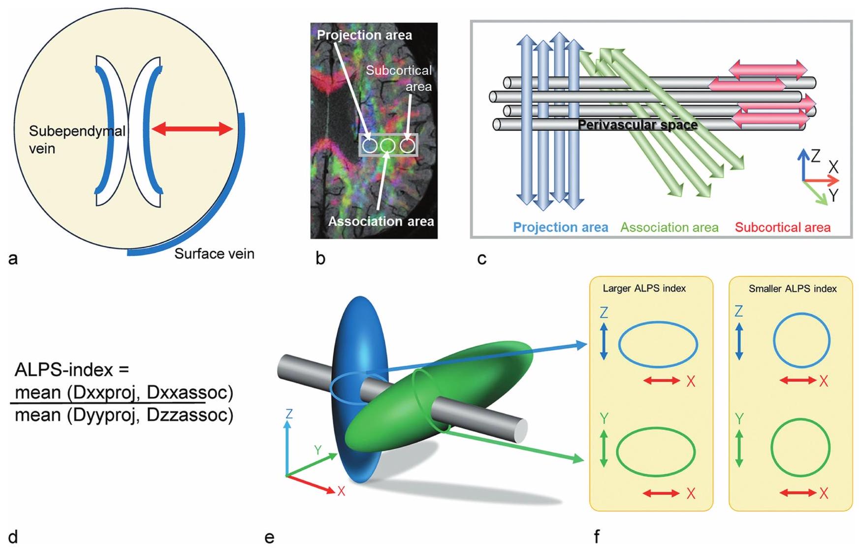
الاتجاه الشعاعي، تحديدًا اليسار-اليمين أو -الاتجاه. في الدماغ البشري، عند مستوى جسم البطينين الجانبيين، تسير الشرايين النخاعية، والأوردة النخاعية، وPVS الخاصة بها في هذا -الاتجاه. تُظهر هذه البنية للأوعية النخاعية داخل نسيج الدماغ بناءً يهدف إلى دعم النقل الكتلي الذي يتمثل في حركة المواد من وإلى خارج نسيج الدماغ. المفهوم الأساسي لطريقة ALPS هو تقييم حركة الماء في هذا -الاتجاه. هنا، تهيمن انتشار الألياف العصبية الكبيرة في صور الانتشار. من الضروري تجنب التأثير القوي على الانتشار من خلال ألياف المادة البيضاء لتقييم عوامل الانتشار الدقيقة في اتجاه الأوعية النخاعية. عند مستوى جسم البطين الجانبي، تكون الألياف الإسقاطية والارتباطية كل منها عمودية على -الاتجاه، الذي يسمح بالإزالة الهندسية لمكون المتجه لهذه الألياف البيضاء الكبيرة في -الاتجاه. في الحالات القصوى، قد يكون من الكافي تقييم الانتشار في -الاتجاه نفسه على مستوى جسم البطين الجانبي عند تقييم نقل الكتلة في هذه المنطقة من الدماغ. ومع ذلك، فإن الانتشار المحسوب في صورة الانتشار يتأثر بشكل كبير بجهاز التصوير وتسلسل التصوير، مثل وقت الانتشار. من أجل مقارنة قيم المعاملات في حالات مختلفة، من المرغوب فيه تقييم المعاملات بالنسبة إلى بعض المراجع الداخلية. لهذا السبب، تم تصميم مؤشر ALPS لتقييم نسبة الانتشار في -الاتجاه إلى النفاذية العمودية على كلا من -الاتجاه والألياف الإسقاطية بالإضافة إلى العمود الفقري لكليهما -الاتجاه والألياف المرتبطة كمرجع داخلي، على مستوى جسم البطين الجانبي. يتم حساب مؤشر ALPS بالصيغة التالية:
حيث Dxxproj هو الانتشار على محور X في منطقة ألياف الإسقاط، Dxxassoc هو الانتشار على محور Y في منطقة الألياف المرتبطة، Dyyproj هو الانتشار على محور Z في منطقة ألياف الإسقاط، و Dzzassoc هو -محور الانتشار في منطقة الألياف المرتبطة. يشير مؤشر ALPS الكبير إلى أن انتشار الماء الحر في الـ -الاتجاه، أو الحركة البراونية، هو السائد، بينما يشير مؤشر ALPS المنخفض إلى أن حركة الماء الحرة في -الاتجاه ليس سائدًا.
تم نشر التقرير الأول لطريقة ALPS كقياس في حالة مرض الزهايمر (AD). في تقييمات الحالات الطبيعية، واضطراب الإدراك الطفيف (MCI)، ومرض الزهايمر (AD)، كان مؤشر ALPS مرتبطًا بشكل كبير بنتيجة اختبار الحالة العقلية المصغرة (MMSE) ومرتبطًا بشكل عكسي كبير بالعمر. حيث أن اضطرابات النظام الغليمفاتي معروفة بأنها موجودة في مرض الزهايمر، تشير هذه النتائج إلى أن مؤشر ALPS يعكس خلل النظام الغليمفاتي في مرض الزهايمر.
عند النظر في مفهوم طريقة ALPS، هناك عدة نقاط يجب ملاحظتها. لا يمكن التمييز بين ما إذا كانت حركة الماء داخل أو خارج PVS، أو في الفضاء المحيط بالأوردة أو الفضاء المحيط بالشرايين بسبب الانخفاض النسبي.
دقة صورة الانتشار، بوحدات المليمترات. فقط اتجاه الانتشار هو القابل للتمييز. ومع ذلك، ما تحاول طريقة ALPS تقييمه هو الحركة الكلية أو الماكروسكوبية للماء في اتجاه اتجاه PVS، وليس حركة الماء في PVS نفسه. وبالتالي، فإن هذه القيود ليست عائقًا أمام هذا المفهوم. بالإضافة إلى ذلك، نظرًا لأن الطريقة تستخدم صور الانتشار، فإنها تقيم “حركة” الماء، وليس “تدفق” الماء. وبالتالي، فإن حركة الماء نحو سطح الدماغ ونحو البطينات تخضع للتقييم بشكل متساوٍ. سيتم النظر في قضايا أخرى في قسم “الجدل في طريقة DTI-ALPS”.

تطبيق DTI-ALPS

طلب لمرض الزهايمر

مرض الزهايمر هو مرض دماغي غير قابل للعكس وتدريجي يتسبب في تدمير الدماغ السليم تدريجياً، مما يؤثر على الذاكرة والقدرات المعرفية والعواطف والسلوك والمزاج. تم الإبلاغ عن ضعف ديناميات السائل الدماغي النخاعي في مرض الزهايمر في ورقة مبكرة من قبل إيلف وآخرون. أميلويد موسوم بالفوسفور تم نقلها على طول هذا المسار، وأدى حذف جين الأكوابورين-4 (AQP4) إلى تثبيط إزالة القابل للذوبان ، مما يشير إلى أن هذه المسار قد يزيل من الجهاز العصبي المركزي. وبالتالي، يُعتبر مرض الزهايمر أحد أمراض سوائل الأنسجة في الجهاز العصبي المركزي. وقد تم دراسة التقرير الأول لطريقة DTI-ALPS أيضًا في مرضى الزهايمر، مما يشير إلى انخفاض مؤشر ALPS في حالات الزهايمر.
تم الإبلاغ عن عدة دراسات متابعة لطريقة DTI-ALPS في مرض الزهايمر. قام ستيوارد وآخرون بدراسة معلمات DTI-ALPS في مجموعة من كبار السن تتكون من أشخاص طبيعيين معرفياً، ومصابين بضعف إدراكي معتدل، ومرضى الزهايمر. تم ربط قياسات MMSE، وهو اختبار قصير يستخدم لتقييم الوظيفة الإدراكية، ومقياس تقييم مرض الزهايمر – المقياس الفرعي الإدراكي (ADAS-Cog 11) بمؤشر ALPS. أظهرت النتائج اختلافات كبيرة في مؤشر DTI-ALPS الأيمن بين المجموعات الطبيعية معرفياً، ومرضى الزهايمر، ومصابين بضعف إدراكي معتدل في نموذج خطي عام أحادي المتغير تم تعديله حسب العمر والجنس والأبوليبوبروتين. هذا التقرير هو متابعة مبكرة لطريقة DTIALPS في مرض الزهايمر، وعلى الرغم من أنه تقييم لمؤشر ALPS فقط، إلا أنه تقرير يوضح فائدة طريقة ALPS، على الرغم من قيودها.
تم الإبلاغ أيضًا عن ارتباط مع ترسب بروتينات الأميلويد والتاو بواسطة تصوير الانبعاث البوزيتروني (PET). قام أوتا وآخرون بالتحقيق في العلاقة بين مؤشر ALPS و PET مع مركب بيتسبرغ 11C وتم العثور على نتائج PET لبروتين التاو باستخدام 18F-THK5351 في مرضى الزهايمر. وُجدت علاقات سلبية كبيرة بين مؤشر ALPS ونسبة قيمة الامتصاص القياسية (SUVR) لـ 11C-PiB في القشرة الصدغية الثنائية والقشرة الجدارية اليسرى والتجاعيد القذالية اليسرى في جميع الموضوعات. علاوة على ذلك، كانت هناك علاقة سلبية كبيرة بين مؤشر ALPS وSUVR لـ 18F-THK5351 في جميع الموضوعات. هذا التقرير يربط نتائج تصوير الأميلويد والتاو مع مؤشر ALPS كما يربط أيضًا مع المناطق التي تظهر
الشكل 2 تطبيق طريقة ALPS في مرض الزهايمر. يتم عرض مخططات الكمان والصندوق لمتوسط مؤشر ALPS، PVSVF-ALL، PVSVF-WM، PVSVF-BG، PVSVFHipp، وFW-WM بين المشاركين الأصحاء، والمرضى الذين يعانون من ضعف إدراكي معتدل، والمرضى الذين يعانون من مرض الزهايمر. مقارنةً بالمشاركين الأصحاء، كان لدى مرضى الزهايمر إجمالي PVSVF، WM، BG PVSVF أعلى بشكل ملحوظ (Cohen ) و FW-WM ( كوهين ) ومؤشر ALPS المنخفض (كوهين ). كانت مجموعة MCI لديها إجمالي PVSVF أعلى بشكل ملحوظ (Cohen ) و WM ( كوهين . الشكل مقتبس من المرجع 20. AD، مرض الزهايمر؛ PVSVF، نسبة حجم الفضاء المحيط بالأوعية؛ WM، المادة البيضاء؛ BG، العقد القاعدية؛ FW، الماء الحر؛ HC، مجموعة التحكم الصحية؛ MCI، ضعف إدراكي خفيف.
الإيداع. من المثير للاهتمام أن مؤشر ALPS يتوافق مع ترسب التاو والأميلويد في مناطق أخرى غير المادة البيضاء المحيطة بجسم البطين الجانبي، والتي يتم تقييمها بواسطة طريقة ALPS. من ناحية أخرى، أفاد ماتسوشيتا وآخرون أن نسبة SUVR في تصوير الأميلويد PET لم تكن مرتبطة بشكل كبير بمؤشر ALPS. أيضًا، أظهرت دراسة مقارنة مع تصوير الأميلويد باستخدام PET لتدهور الإدراك الذاتي بواسطة بارك وزملائه عدم وجود فرق كبير بين متوسط مؤشر ALPS في المجموعة الإيجابية للأميلويد ومتوسط مؤشر ALPS في المجموعة السلبية للأميلويد. قام كاماغاتا وآخرون بدراسة التغيرات في المقاييس غير الغازية للتصوير بالرنين المغناطيسي المتعلقة بالشبكة المحيطية للأوعية الدموية لدى المرضى الذين يعانون من ضعف إدراكي معتدل (MCI) ومرض الزهايمر (AD): نسبة حجم PVS (PVSVF)، وحجم الماء الحر في المادة البيضاء (FW-WM) ومؤشر ALPS. أظهرت نتائجهم أن مرضى الزهايمر كان لديهم نسبة حجم PVS الكلي، والمادة البيضاء، ونسبة حجم PVS في العقد القاعدية، وFW-WM أعلى بشكل ملحوظ، ومؤشر ALPS أقل مقارنةً بالضوابط الصحية. في حين أن القيم الإجمالية لـ PVSVF وWM كانت أعلى بشكل ملحوظ فقط في مجموعة MCI. كان مؤشر ALPS الأقل مرتبطًا بـ أقل زيادة امتصاص FDG (فلوروديوكسي جلوكوز) في السائل الدماغي الشوكي وتدهور أسوأ في عدة مجالات معرفية. كما ارتبطت زيادة FW-WM بانخفاض في السائل الدماغي الشوكي. وأسوأ وظيفة إدراكية (الشكل 2).

طلب لمرض باركنسون وأمراض التنكس الأخرى

مرض باركنسون (PD) هو اضطراب ناتج عن الألفا-سايتوكين، يتميز بتراكم الألفا-سايتوكين في الخلايا العصبية و
فقدان الخلايا العصبية الدوبامينية في المادة السوداء. على غرار مرض الزهايمر، يتميز هذا المرض العصبي التنكسي بتراكم البروتينات السامة كميزة مرضية. في السنوات الأخيرة، تم إظهار أن مرض باركنسون مرتبط أيضًا بديناميات السائل بين الخلايا غير الطبيعية المتعلقة بإزالة النفايات الأيضية، وقد تم الإبلاغ عن دراسات تستخدم طريقة DTI-ALPS.
أظهر تقرير تحليل طريقة ALPS في حالات مرض باركنسون والرعاش الأساسي انخفاض مؤشر ALPS في مرض باركنسون مقارنة بالرعاش الأساسي. في مقارنة بين مراحل مرض باركنسون (PD) والمجموعة الضابطة الطبيعية، كان لدى المرضى المصابين بمرض باركنسون مؤشر ALPS أقل من المجموعة الضابطة الطبيعية، وكان الانخفاض ملحوظًا بشكل خاص في المجموعة في المراحل المتأخرة من مرض باركنسون. كان هناك ارتباط إيجابي كبير بين مؤشر ALPS ودرجات MMSE في مجموعة مرض باركنسون المبكرة، وارتباط سلبي بين مؤشر ALPS ودرجة تضخم PVS (الشكل 3).
استخدم تشين وآخرون طريقة DTI-ALPS لتقييم ديناميات السائل بين الخلايا وعلاقتها بحالة الإجهاد التأكسدي النظامي لدى مرضى باركنسون. أظهر مجموعة مرضى باركنسون الذين يعانون من ضعف إدراكي معتدل ومجموعة مرضى باركنسون الذين يعانون من الخرف مستوى ALPSindex أقل بشكل ملحوظ من مجموعة التحكم الطبيعية. كان هناك ارتباط عكسي بين ALPS-index ومستوى الحمض النووي النووي في البلازما ومستوى الحمض النووي الميتوكوندري والدرجة الإدراكية. يدعم الارتباط بين زيادة مستويات الحمض النووي النووي في البلازما وانخفاض ALPSindex الفكرة القائلة بأن مرضى باركنسون قد يظهرون زيادة في الإجهاد التأكسدي المرتبط بتغير ديناميات السائل بين الخلايا. في تقرير آخر، وُجدت علاقة سلبية بين مؤشر ALPS وشدة الأعراض الحركية في مرض باركنسون.
الشكل 3 تطبيق طريقة ALPS في مرض باركنسون. تم مقارنة الفروق في مؤشر ALPS بين مجموعة مرض باركنسون ومجموعة الشهود الأصحاء وبين مجموعتي مرض باركنسون ومجموعة الشهود الأصحاء. تم عرض العلاقات بين مؤشر ALPS ودرجة MMSE في مجموعة مرض باركنسون المبكر (A)، والعلاقات بين مؤشر ALPS ودرجة EPVS في مجموعة مرض باركنسون المبكر (B) والعلاقات بين مؤشر ALPS والعمر في مجموعة مرض باركنسون المتأخر (C). كان هناك ارتباط إيجابي كبير بين مؤشر ALPS ودرجة MMSE. ) ( ) ، وارتباط سلبي بين مؤشر ALPS ودرجة EPVS ( ). مؤشر ALPS مرتبط سلبًا بالعمر ( ). الشكل مقتبس من المرجع 184. PD، مرض باركنسون؛ NC، التحكم الطبيعي؛ MMSE، اختبار الحالة العقلية المصغر؛ EPVS، الفضاء المحيط بالأوعية الدموية المتضخم.
مجموعة فرعية من المرضى الذين تتراوح أعمارهم بين 65 عامًا وما فوق مقارنة بالمرضى الأصغر سنًا. في دراسة طولية على مرضى باركنسون، تدهور الفريق الذي كان لديه مؤشر ALPS أقل بشكل أسرع في الجزء الثالث والجزء الثاني من مقياس تقييم مرض باركنسون الموحد من جمعية اضطرابات الحركة (MDS-UPDRS)، واختبار رموز الأرقام، واختبار التعلم اللفظي من هوبكنز. أظهرت تحليل المسار أن مؤشر ALPS عمل كوسيط مهم بين وتغير الإدراك في نتيجة اختبار رموز الأرقام في السنوات 4 و 5.
اضطراب سلوك النوم السريع غير المعروف السبب (iRBD) هو اضطراب نوم يتحرك فيه الجسم أثناء نوم حركة العين السريعة وهو شكل من أشكال الباراسومنيا. له جانب من ألفا-ساينوكليينوباثي مشابه لمرض باركنسون ويمكن أن يؤدي إلى التحول الظاهري إلى مرض باركنسون، خرف ليوي، وضمور متعدد الأنظمة. كانت نسبة التحول الإجمالية من iRBD إلى متلازمة تنكس عصبي ظاهرة في السنة. في دراسة DTI-ALPS لمرضى iRBD وPD والمجموعة الضابطة الطبيعية، أظهر كل من مرضى PD وiRBD مؤشر ALPS أقل من المجموعة الضابطة الطبيعية. كان لدى مرضى PD مؤشر ALPS أقل من مرضى iRBD، وكان هناك ارتباط بين مؤشر ALPS والانخفاض المعرفي لدى مرضى PD. في دراسة طولية عن التحول الظاهري في المرضى الذين يعانون من اضطراب سلوك النوم REM (RBD) ومرض باركنسون (PD) والمراقبين الأصحاء، انخفض خطر التحول الظاهري مع زيادة مؤشر ALPS. تم نشر تقييمات للأمراض التنكسية العصبية الأخرى غير مرض باركنسون، مثل التنكس القشري القاعدي والشلل فوق النووي التقدمي. في دراسة تقييم مؤشر ALPS وربطه بالوظيفة الحركية والمعرفية في تنكس القشرة الدماغية مع متلازمة القشرة القاعدية (CBD-CBS)، كان مؤشر ALPS لـ CBD-CBS أقل بشكل ملحوظ من ذلك لدى الضوابط الطبيعية. بالإضافة إلى ذلك، أظهر مؤشر ALPS ارتباطًا إيجابيًا كبيرًا مع MMSE وارتباطًا سلبيًا كبيرًا مع درجة MDS-UPDRS III.

طلب لأمراض الأوعية الدموية الصغيرة

مرض الأوعية الدموية الصغيرة الدماغية (SVD) يؤثر على الأوعية الدموية الصغيرة في الدماغ ويرتبط بالسكتة الدماغية، الإدراك
خلل وظيفي، وخلل حركي، وقد جذبت أيضًا الانتباه كحالة مرضية خلفية للأمراض التنكسية العصبية مثل مرض الزهايمر. اقترح بنفينست وآخرون أن فشل نقل السوائل في الدماغ عبر النظام الجليمفاتي يلعب دورًا رئيسيًا في بدء وتقدم مرض الأوعية الدموية الصغيرة. والحجة الرئيسية لهذا المفهوم هي أن المساحات المحيطة بالأوعية الدموية تُستخدم كقنوات لتدفق السائل الدماغي الشوكي. يُعتقد أن ركود النقل بواسطة السائل بين الخلايا يسبب فقدان توازن السوائل في الدماغ وقد يؤدي إلى وذمة مؤقتة في المادة البيضاء، وتوسع حول الأوعية الدموية، وفي النهاية إزالة الميالين.
في تحليل أجراه كيكوتا وآخرون يقارن بين مرضى ارتفاع ضغط الدم الأكبر سناً ومجموعة ضابطة متطابقة في العمر، كان مؤشر ALPS في مجموعة ارتفاع ضغط الدم أقل بكثير من مجموعة الضبط. بالإضافة إلى ذلك، كان مؤشر ALPS لجميع المشاركين مرتبطاً سلبياً بشكل كبير مع قيم ضغط الدم وقيم ضغط النبض. وهذا يشير إلى أن مرض الأوعية الصغيرة بسبب ارتفاع ضغط الدم كان مرتبطاً بتلف في ديناميات السائل الدماغي النخاعي. في دراسة شملت 133 مريضًا يعانون من مرض الأوعية الدموية الصغيرة، كان انخفاض مؤشر ALPS مرتبطًا بشكل مستقل بضعف الوظائف التنفيذية والانتباه والذاكرة لدى المرضى الذين يعانون من مرض الأوعية الدموية الصغيرة. في دراسة شملت مجموعة من أكثر من 2000 مقيم في المجتمع، كلما كان مؤشر ALPS أقل، كانت هناك زيادة في وجود وشدة مرض الأوعية الدموية الدقيقة الدماغية. علاوة على ذلك، كان مؤشر ALPS المنخفض مرتبطًا بعلامات تصوير عصبي ضعيفة مثل إشارة المادة البيضاء غير الطبيعية، وتضخم المساحات المحيطة بالأوعية، وانكماش الدماغ.
اعتلال الأوعية الدموية الأميلويدية الدماغية (CAA) هو نوع من الأمراض الوعائية الدماغية الدقيقة يتميز بتراكم في الأغشية الوسطى والخارجية للشرايين في القشرة الدماغية والسحايا. السبب الكامن وراء لا يزال تراكم غير معروف. بالإضافة إلى الفكرة التقليدية لـ الإنتاج الزائد، هناك أيضًا فكرة العجز مسارات التصريف. في مقارنة بين مرضى CAA ومجموعة التحكم المتطابقة في العمر، كان لدى مرضى CAA مؤشر ALPS العام أقل مقارنة بمجموعة التحكم. بالإضافة إلى ذلك، كان المؤشر المنخفض ALPS مرتبطًا بتضخم المساحات المحيطة بالأوعية في العقد القاعدية، وزيادة الفجوات، وارتفاع كثافة المادة البيضاء.
يسجل فازيكاس، انخفاض في مقياس الحالة العقلية المصغرة (MMSE)، وانخفاض في تقييم مونتريال المعرفي (MoCA-J). خلال متابعة متوسطة لمدة 4.1 سنوات، كان هناك تكرار أقل للمرض مع ارتفاع مؤشر ALPS.

طلب لفرط الضغط الدماغي الطبيعي مجهول السبب

استسقاء الرأس الطبيعي الضغط مجهول السبب (iNPH) هو متلازمة سريرية تتميز بتضخم البطينات دون زيادة في ضغط السائل الدماغي الشوكي. يحتوي iNPH على ثلاثة أعراض مميزة: اضطراب المشي، تدهور الإدراك، وسلس البول. تشمل ميزات التصوير استسقاء الرأس ذو الفضاء تحت العنكبوتي المتضخم بشكل غير متناسب (DESH) وضيق زاوية الجسم الثفني. تم الإشارة إلى iNPH كمرض تنكسي، وتثير الترافق المرضي معه مع مرض الزهايمر اهتمامًا. أبلغ إيد وآخرون عن تقييم ديناميات السائل الدماغي الشوكي وديناميات السائل بين الخلايا في مجموعة iNPH باستخدام تقنية حقن مادة التباين الجزيئي في السائل النخاعي ومراقبة التصوير بالرنين المغناطيسي على مدى الزمن. في هذه الدراسة التدخلية، أظهر مجموعة iNPH المستجيبة للتحويل استجابة أكثر وضوحًا لتدفق الصبغة إلى النظام البطيني وميلاً نحو تعزيز النسيج لفترة أطول مقارنةً بمجموعة التحكم. تشير هذه النتائج إلى أن الشذوذات في النظام الغليمفاتي قد تكون مرتبطة بتسبب iNPH.
الدراسة التي أجراها يوكوتا وآخرون على حالات iNPH هي أول دراسة متابعة لطريقة ALPS. تقوم هذه الدراسة بمقارنة مجموعة من حالات iNPH التي تم تشخيصها وفقًا لمعايير التشخيص مع مجموعة من حالات pseudo-iNPH (piNPH) التي لا تستوفي معايير التشخيص. أظهرت نتائجهم أن مؤشر ALPS كان أقل بشكل ملحوظ في حالات iNPH، وكانت المساحة تحت المنحنى في تحليل ROC هي 0.92 و 1.00 و 1.00 لمجموعة التحكم مقابل piNPH، ومجموعة التحكم مقابل iNPH، وpiNPH مقابل iNPH، مما يشير إلى أداء تشخيصي مرتفع جدًا. بين piNPH وNPH، أظهر مؤشر ALPS أداءً تشخيصيًا أعلى من مؤشر إيفانز أو زاوية الجسم الثفني. في دراسة أجراها جورجيوبولوس وآخرون، تم تحليل العلاقة بين النتائج السريرية لاختبار Time Up and Go (TUG) وMMSE المرتبطة بـ iNPH ومؤشر ALPS. كان لدى المرضى المصابين بـ iNPH متوسط مؤشر ALPS أقل بشكل ملحوظ من مجموعة التحكم الصحية. وُجدت علاقة متوسطة أسية بين متوسط مؤشر ALPS ووظيفة الحركة كما تم قياسها بالوقت المطلوب لإكمال TUG، وعدد الخطوات لإكمال TUG، ووقت المشي لمسافة 10 أمتار، وخطوات المشي لمسافة 10 أمتار. وُجدت علاقة خطية إيجابية بين متوسط درجة مؤشر ALPS ودرجة MMSE. كأداة تشخيصية لـ iNPH، يُعتبر اختبار السحب اختبارًا مهمًا للتنبؤ بفعالية التحويلة. في دراسة تقييم العلاقة بين نتائج اختبار Tap ومؤشر ALPS، كان مؤشر ALPS أقل بشكل ملحوظ في مجموعة NPH التي لم تستجب مقارنة بالمجموعة التي أظهرت تخفيف الأعراض. يعتبر التحويل علاجًا فعالًا لـ iNPH. وقد أفادت الدراسات بتغيرات في مؤشر ALPS قبل وبعد التحويل. كان متوسط مؤشر ALPS بعد جراحة التحويل أعلى من قبل جراحة التحويل. على وجه الخصوص، كان هناك زيادة ملحوظة في مؤشر ALPS بعد الجراحة في المجموعة التي شهدت
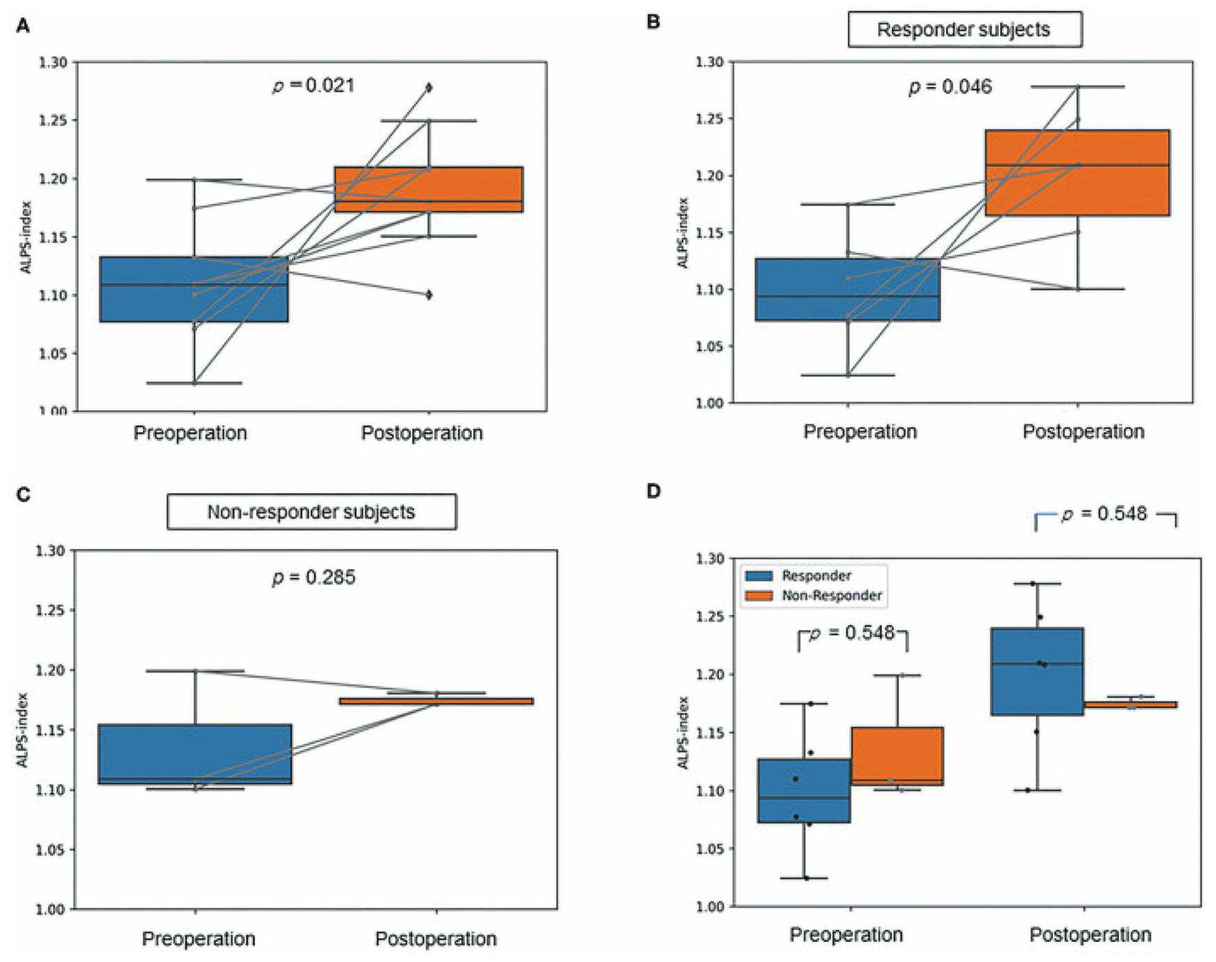
تحسنًا في الأعراض بعد جراحة التحويل، بينما لم يكن هناك زيادة ملحوظة في مؤشر ALPS بعد الجراحة في المجموعة التي لم تشهد تحسنًا في الأعراض (الشكل 4).

تطبيق لإصابة الدماغ الرضحية

تعتبر إصابة الدماغ الرضحية (TBI) عامل خطر مثبت لظهور الخرف المبكر، بما في ذلك مرض الزهايمر، وغالبًا ما تظهر الدماغ بعد الإصابة تغييرات عصبية تتكون من تجمعات بروتين التاو. في الدراسات الحيوانية على الفئران باستخدام علامات فلورية، تم تقليل وظيفة المسار الغليمفاتي بحوالي بعد إصابة الدماغ الرضحية، واستمرت هذه الإعاقة لمدة شهر على الأقل بعد الإصابة. بالإضافة إلى إصابة الدماغ المباشرة، أظهرت الدراسات الحيوانية على الرئيسيات غير البشرية أن تكوين تسرب السائل الدماغي الشوكي يؤخر دخول GBCA المحقون داخل النخاع إلى الدماغ. يشير هذا إلى أن العوامل الميكانيكية مثل ضغط الجمجمة تؤثر على ديناميات السائل الدماغي.
في البشر، أفادت عدة دراسات باستخدام طريقة ALPS غير الغازية بتغيرات غير طبيعية في ديناميات السائل الدماغي بعد إصابة الدماغ الرضحية. لقد تم الإبلاغ عن أن مؤشر ALPS ينخفض في الدماغ بعد إصابة الدماغ الرضحية. من ناحية أخرى، هناك تقارير تفيد بأن مؤشر ALPS يرتفع في المرضى الشباب الذين يعانون من إصابة دماغ رضحية خفيفة. تشمل الأسباب المحتملة زيادة التعبير عن AQP4 بعد الإصابة، وتغيرات ديناميكية دموية بعد الإصابة، وإمكانية أن يكون النظام الغليمفاتي آلية تعويضية لتقليل الإصابة الثانوية عن طريق التخلص من السموم بعد الإصابة. كحالة لا تتطور إلى إصابة رضحية، قامت بعض التقارير بتقييم مؤشر ALPS في الأشخاص الذين تعرضوا لرياضات الاحتكاك. في هذا التقرير، كان مؤشر ALPS الأيسر أقل بشكل ملحوظ في مجموعات الاحتكاك الثقيل (الرجبي، الجودو، الكاراتيه، الملاكمة، الكندو، المصارعة، وكرة القدم) والاحتكاك شبه (كرة القاعدة، كرة السلة، وكرة اليد) مقارنة بمجموعة عدم الاحتكاك (التنس، تنس الطاولة، ألعاب القوى، التزلج، الرماية، والتوجيه). كان مؤشر ALPS الأيمن يميل إلى أن يكون أقل في مجموعات الاحتكاك شبه والاحتكاك الثقيل مقارنة بمجموعة عدم الاحتكاك. أظهرت مؤشرات ALPS الثنائية ارتباطًا إيجابيًا ملحوظًا مع درجات MoCA-J.

تطبيق لمرض إزالة الميالين

التصلب المتعدد هو اضطراب مناعي مزمن في الجهاز العصبي المركزي يتميز بالالتهاب وإزالة الميالين الناتجة عن تفاعل تدمير الحاجز الدموي الدماغي، وتسلل الكريات البيضاء المحلية، وتنشيط الخلايا الدبقية، وإطلاق السيتوكينات الالتهابية. بالنظر إلى أن النفايات السامة هي سبب تلف الأنسجة، يبدو أنه من الضروري أن يكون لدى الدماغ نظام لإزالة هذه النفايات. تم الإبلاغ عن دراسة استعادية باستخدام تقنية DTI-ALPS في مرضى التصلب المتعدد. في هذه الدراسة، كان لدى مرضى التصلب المتعدد مؤشر ALPS أقل بشكل عام مقارنة بالضوابط الأصحاء. كان لدى مرضى التصلب المتعدد المتكرر والمتقدم مؤشر ALPS أقل مقارنة بالضوابط الأصحاء. كان لدى مرضى التصلب المتعدد المتقدم مؤشر ALPS أقل مقارنة بمرضى التصلب المتعدد المتكرر.
الشكل 4 تطبيق طريقة ALPS في iNPH. تم تقديم دراسة حول التغيرات في مؤشر ALPS بعد جراحة LPS في iNPH، مقارنة بين المرضى الذين استجابوا لـ LPS والذين لم يستجيبوا. كان متوسط مؤشر ALPS في المجموعة بعد الجراحة أعلى بشكل ملحوظ من المجموعة قبل الجراحة (A). علاوة على ذلك، بالنسبة للمواضيع المستجيبة، كان متوسط مؤشر ALPS في المجموعة بعد الجراحة أعلى بشكل ملحوظ من المجموعة قبل الجراحة (B). من ناحية أخرى، في مجموعة غير المستجيبين، لم يكن متوسط مؤشر ALPS في المجموعة بعد الجراحة أعلى بشكل ملحوظ من المجموعة قبل الجراحة (C). لم تكن مؤشرات ALPS في مجموعة المستجيبين مختلفة بشكل ملحوظ مقارنة بتلك الخاصة بمجموعة غير المستجيبين في كل من مجموعات ما قبل العملية وما بعد العملية (D). الشكل مقتبس من المرجع 62. iNPH، استسقاء الدماغ ذو الضغط الطبيعي مجهول السبب؛ LPS، تحويلة لامبوسبيريتونيال.
التصلب. في مرضى التصلب المتعدد، كان انخفاض مؤشر ALPS مرتبطًا بإعاقة سريرية أكثر شدة ومدة مرض أطول.
تم الإبلاغ أيضًا عن دراسة مرضى اضطراب الأجسام المضادة لبروتين الميالين (MOGAD)، وهو مرض آخر لإزالة الميالين، باستخدام طريقة ALPS. في دراسة مستقبلية لمرضى MOGAD في حالة هدوء ومطابقة من حيث العمر والجنس مع ضوابط أصحاء، كان متوسط مؤشر ALPS أقل بشكل ملحوظ في مرضى MOGAD مقارنة بالضوابط الأصحاء. كان انخفاض متوسط مؤشر ALPS مرتبطًا بشكل ملحوظ مع درجات أسوأ في مقياس حالة الإعاقة الموسعة. ومع ذلك، لم تكن أحجام المادة الرمادية القشرية والعميقة مختلفة بشكل ملحوظ بين المجموعتين،
ولا كانت مرتبطة بدرجات مقياس حالة الإعاقة الموسعة.
يُقترح أن مرضى MOGAD قد يكون لديهم وظيفة غليمفاتي معطلة كما تم قياسها بواسطة مؤشر ALPS.

تطبيق للنوم

أحد الأسباب التي جعلت نظرية النظام الغليمفاتي تجذب الكثير من الانتباه هو علاقتها بالنوم. يُعتبر تصريف النظام الغليمفاتي مكبوتًا أثناء اليقظة ومعززًا بشكل ملحوظ أثناء النوم. أفاد شي وآخرون أنه تحت الظروف الفسيولوجية، يرتبط نشاط النظام الغليمفاتي بالنوم، استنادًا إلى ملاحظات علامات فلورية تم حقنها
الشكل 5 اعتماد مؤشر ALPS على وقت اليوم. تم تقييم اعتماد إيقاع الساعة البيولوجية لمؤشر ALPS من خلال قياسات MRI المتكررة في خمس نقاط زمنية من 8:00 إلى 23:00. تم إجراء تصوير الانتشار بتقنية DTI-ALPS منخفضة التشتت، والذي يقيس تدفق SAS على طول الشريان الدماغي الأوسط، وDTI-ALPS التقليدي. تظهر A وB مسار تدفق النظام الغليمفاتي المدخل والمخرج المقاس بواسطة DTI-ALPS وDTI-ALPS، على التوالي. لم يختلف ADlow-b لسائل SAS في MCA ولا مؤشر ALPS بشكل ملحوظ في البيانات المكتسبة في النقاط الزمنية الخمس. الشكل مقتبس من المرجع 159. MRI، التصوير بالرنين المغناطيسي؛ DTI، صورة الانتشار؛ SAS، الفضاء تحت العنكبوتي؛ DTI-ALPS، تحليل صورة الانتشار على طول الفضاء المحيط بالأوعية؛ AD، مرض الزهايمر؛ MCA، الشريان الدماغي الأوسط.
إلى تجويف السائل الدماغي الشوكي للفئران باستخدام مجهر ثنائي الفوتون. أثناء النوم، هناك زيادة ملحوظة في نشاط النظام الغليمفاتي مقارنة باليقظة. وقد عزوا ذلك إلى أنه أثناء النوم، ينخفض حجم الخلايا الدبقية ويتوسع الفضاء بين الخلايا أكثر مما هو عليه أثناء اليقظة، مما يسهل النقل الكتلي في الأنسجة.
تم استخدام طريقة ALPS لتقييم النوم وديناميات السائل الدماغي في عدة دراسات منشورة. في دراسة تقييم العلاقة بين المؤشرات المتعلقة بالنوم ومؤشر ALPS في كبار السن الذين يعيشون في المجتمع والذين تتراوح أعمارهم بين 60 عامًا وما فوق، ارتبط مؤشر ALPS بمدة نوم N2 ومدة نوم N3. من ناحية أخرى، ارتبط عكسيًا بمؤشر انقطاع النفس-نقص الأكسجة. بالإضافة إلى ذلك، ارتبط بحجم المادة الرمادية. يرتبط انقطاع النفس النومي الانسدادي (OSA) بتجزئة النوم وتغير تنظيم النوم، مما قد يؤثر سلبًا على ديناميات السائل الدماغي ويزيد من خطر مرض الزهايمر. في دراسة لحالات OSA والحالات الضابطة، كانت قيم Dzz، المشتقة من مناطق الألياف الإسقاطية، وقيم Dyy وDzz من مناطق الألياف المرتبطة، بالإضافة إلى قيم ALPS وDyzmean أقل بشكل ملحوظ في حالات OSA مقارنة بالضوابط. ظهرت ارتباطات ملحوظة بين شدة المرض، وأعراض النوم، وقيم Dxy وDxx وDzz في مرضى OSA. في دراسة تقييم العلاقة بين جودة النوم ومؤشر ALPS كما تم تقييمه بواسطة مؤشر جودة النوم في بيتسبرغ (PSQI)، كان مؤشر ALPS أقل بشكل ملحوظ في مجموعة اضطرابات النوم مقارنة بمجموعة التحكم. بالإضافة إلى ذلك، أظهر مؤشر ALPS ارتباطًا سلبيًا كبيرًا مع جميع مكونات درجة PSQI. في دراسة DTI-ALPS لمرضى النوم القهري، لم يكن هناك فرق ذو دلالة إحصائية في مؤشر ALPS بين مجموعة المرضى ومجموعة التحكم. ومع ذلك، كان هناك ارتباط بين مؤشر ALPS وتخطيط النوم المتعدد.
المعلمات مرتبطة إيجابيًا مع الاستيقاظ بعد بدء النوم وطول نوم حركة العين السريعة لدى المرضى الذين يعانون من النعاس من النوع 2. كما ذُكر أعلاه، أظهرت التجارب الحيوانية باستخدام المتعقبات أن ديناميات المتعقبات تتعزز أثناء النوم. في البشر، تم الإبلاغ أيضًا عن التقييم بواسطة نموذج الموتر ذو الحُجرتين في التصوير بالانتشار. كشفت التحليل ذو الحُجرتين أن الزيادة في مقاييس الانتشار من فترة ما بعد الظهر إلى فترة ما قبل الظهر كانت مدفوعة بزيادة في نسبة حجم الماء الحر الشبيه بالسائل الدماغي الشوكي. من ناحية أخرى، في دراسة طريقة ALPS، تم تكرار قياسات الرنين المغناطيسي في خمس نقاط زمنية من الساعة 8:00 إلى 23:00 ولم يتم ملاحظة أي اعتماد على فترة زمنية في حالة اليقظة في تجربة لتقييم اعتماد إيقاع الساعة البيولوجية (الشكل 5). تمت مناقشة هذه الدراسة أيضًا في قسم “طرق أخرى لتقييم النظام الغليمفاتي” و”اعتبارات منهجية DTI-ALPS”.

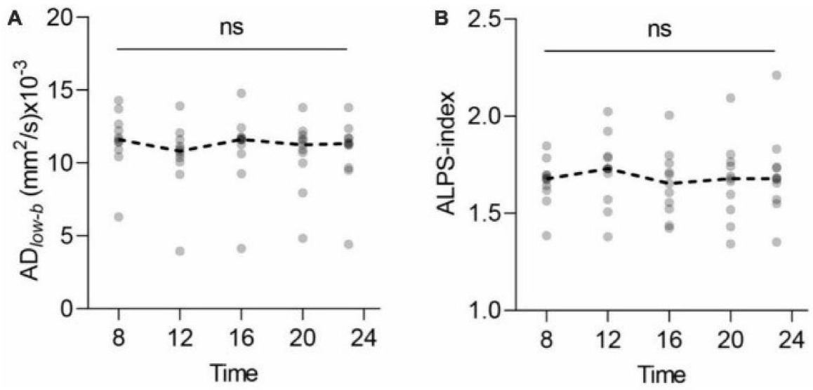
النتائج حول الشيخوخة

عدد من الدراسات التي استخدمت طريقة DTI-ALPS قد قيمت العلاقة بين العمر ومؤشر ALPS. من بين هذه التقارير، أظهرت عدد منها علاقات سلبية بين العمر ومؤشر ALPS. أظهر مكنايت وزملاؤه علاقة سلبية بين مؤشر ALPS والعمر في مجموعة من الأشخاص الذين يعانون من مرض باركنسون أو الرعاش الأساسي الذين تتراوح أعمارهم تقريبًا بين 40-80 عامًا، مع معامل ارتباط سبيرمان المرتبط. أظهر زانغ وآخرون علاقة سلبية بين العمر ومؤشر ALPS في 142 من كبار السن الأصحاء الذين تتراوح أعمارهم بين 50 عامًا وما فوق، مع معامل ارتباط قدره أظهر et al. علاقة سلبية بين العمر ومؤشر ALPS في حالات مرض باركنسون، مع معامل انحدار جزئي قياسي من وجد سيوا وآخرون أيضًا
ارتباط سلبي بين العمر ومؤشر ALPS في كبار السن الذين يعيشون في المجتمع والذين تتراوح أعمارهم من 60 عامًا وما فوق، مع معامل انحدار جزئي قياسي من قام توه وسيو بفحص حالات الورم الدبقي في نطاق واسع من الأعمار، من 18 إلى 91 عامًا. على الرغم من أن معامل الارتباط منخفض عند -0.147، إلا أن الرسم البياني الذي قدموه والذي يظهر العلاقة بين العمر ومؤشر ALPS يبدو أنه يحتوي على قمة طفيفة حول العمر دراسة مجموعة البيانات العامة بواسطة داي وآخرون للأعمار يبدو أيضًا أن هناك ذروة مماثلة حول الأربعينيات في الرسم البياني المقدم في الورقة التي تظهر العلاقة بين العمر ومؤشر ALPS، على الرغم من أن المؤلفين لم يذكروها. علاوة على ذلك، تُظهر طريقة DTI-ALPS الموضحة أدناه أيضًا أن مؤشر ALPS يصل إلى ذروته في الأربعينيات. ومع ذلك، هناك جانب من هذا يشير إلى أن مؤشر ALPS قد يتأثر بحالة ألياف المادة البيضاء، والتي ستتم مناقشتها لاحقًا. بينما كانت هناك العديد من التقارير عن ارتباطه بالتقدم في العمر لدى المرضى الأكبر سنًا، كانت هناك أيضًا تقارير عن طريقة DTI-ALPS في الحالات pediatrics. أظهر لي وآخرون علاقة سلبية بين العمر ومؤشر ALPS مع معامل ارتباط قدره -0.375 في مجموعة من المرضى الشباب الذين يعانون من الصرع الارتعاشي. سنوات. ومع ذلك، هناك نقص في التحقق من قابلية تطبيق هذه التقنية على الأطفال، الذين تكون فيها بناء ألياف المادة البيضاء غير مكتمل. تتطلب التغيرات السريعة في الحالة النمائية للمادة البيضاء مع النمو تفسيرًا دقيقًا للنتائج، خاصة عند مقارنة حالات بأعمار مختلفة. هناك حاجة إلى مزيد من الخبرة.

طرق أخرى لتقييم النظام اللمفاوي الدماغي

نظرًا لأن مؤشر ALPS يتم تحديده بواسطة صيغة بسيطة ويمكن حسابه من البيانات الاستعادية، فقد تم الإبلاغ عنه في العديد من الأمراض والاعتلالات والحالات كما هو موضح أعلاه. ومع ذلك، كما تم مناقشته في القسم “الجدل في طريقة DTI-ALPS”، هناك أيضًا مشاكل تم الإشارة إليها. عند تقييم ديناميات السائل الدماغي بين الخلايا، لا يكفي استخدام طريقة ALPS بمفردها، بل من المرغوب استخدام طرق أخرى لتقييم ديناميات السائل الدماغي بين الخلايا معًا. فيما يلي مراجعة للدراسات التي قيمت العلاقة بين طريقة ALPS وطرق أخرى لتقييم ديناميات السائل الدماغي بين الخلايا.

GBCA داخل السائل النخاعي

تم الإبلاغ عن عدد من المحاولات لاستخدام التصوير بالرنين المغناطيسي لتقييم حالة النظام الغليمفاتي (الجدول 1). ومع ذلك، فإن الدراسات الأولى حول النظام الغليمفاتي التي أجراها إيلف وآخرون تمت على الحيوانات مع الإدارة داخل السحايا لمؤشرات مضيئة وميكروسكوب ليزر ثنائي الفوتون. تتمتع هذه الطريقة بميزة السماح بالملاحظة المباشرة للدماغ الحي، ولكن فقط بالقرب من سطح الدماغ. كطريقة لمراقبة الدماغ بالكامل، تم تقديم دراسة تتبع في الحيوانات باستخدام التصوير بالرنين المغناطيسي مع مادة التباين القابلة للحقن داخل السحايا.
يعتبر استخدام دراسات المتعقب باستخدام التصوير بالرنين المغناطيسي مع مادة التباين المعطلة داخل السائل الشوكي (GBCA) هو المعيار الذهبي. ومع ذلك، يجب ملاحظة أن الوزن الجزيئي لمادة التباين المعطلة هو 0.6 كيلودالتون (Gadobtrol)، في حين أن وزن في الدماغ هو بالإضافة إلى ذلك، حيث إنه يقيم الاختراق إلى الدماغ من سطح الدماغ، فإنه يقيم المسار المعاكس لتصريف المواد waste من نسيج الدماغ. علاوة على ذلك، من الصعب تقييم حالة المادة البيضاء العميقة، التي تشغل نسبة كبيرة من حجم الدماغ. في هذه الجوانب، يجب الإشارة إلى أن هذه الطريقة محدودة للغاية بحيث لا يمكن اعتبارها معيارًا ذهبيًا لتقييم النظام الغليمفاتي.
حقن GBCA داخل السحايا في البشر هو استخدام غير معتمد، وقليل من المراكز تقوم به. وذلك لأن حقن GBCA داخل السحايا هو إجراء جراحي، وبحسب الجرعة، قد يكون غير آمن. قام إيد، ورينغستاد، وزملاؤهم بإجراء دراسات تتبع منهجية لحقن GBCA داخل السحايا في البشر. كانت أول تقرير عن حقن GBCA داخل السحايا في مريض يعاني من انخفاض ضغط الدم داخل الجمجمة بسبب تسرب السائل الدماغي الشوكي التلقائي، حيث أظهرت الأشعة بالرنين المغناطيسي توزيع GBCA في جميع أنحاء الدماغ بعد 1 و 4.5 ساعات من الحقن داخل السحايا. كانت تجاربهم في البداية تقييمات نوعية فقط للمتتبعات، لكن الدراسات الأحدث أجرت تحليلات رياضية متطورة لتحديد وقياس معلمات نقل المتتبعات في السائل الدماغي الشوكي. تشير النتائج إلى أن الانتشار خارج الخلايا وحده غير كافٍ كآلية لنقل المتتبعات في الدماغ بأكمله، وأن معدلات الإزالة المحلية تساهم في نقل المتتبعات. في دراسة أجراها دايك وآخرون على متطوعين بالغين أصحاء، بلغ تركيز GBCA في سائل النخاع الشوكي داخل الجمجمة ذروته عند وبدأ الانخفاض عند الساعة 7. على النقيض من ذلك، في بعض مناطق نسيج الدماغ، مثل القشرة الدماغية والمادة البيضاء، زادت التعزيزات بعد 11 ساعة. وبالتالي، تم اقتراح أن تعزيز إشارة الرنين المغناطيسي للدماغ بالكامل من خلال إعطاء GBCA داخل السحايا قد يشير إلى تبادل السائل الدماغي الشوكي والسائل بين الخلايا واختراق نسيج الدماغ في جميع أنحاء الدماغ.
أجريت دراسة قارنت نتائج طريقة DTI-ALPS مع تقييم وظيفة النظام الغليمفاتي باستخدام GBCA داخل السائل النخاعي. كان مؤشر ALPS مرتبطًا بشكل كبير بمعدل التغير في نسبة وحدة الإشارة من الخط الأساسي إلى 39 ساعة في ست مناطق من الدماغ.

GBCA عن طريق الوريد

يعتبر حقن GBCA عن طريق الوريد كعلامة أسهل في التنفيذ من الحقن داخل القناة الشوكية. أظهرت التجارب التي استخدمت جرعات عالية من GBCA عن طريق الوريد في الجرذان زيادة في الإشارة في البطين الرابع مباشرة بعد حقن GBCA عن طريق الوريد، مما يشير إلى أن GBCA يتم نقله بسرعة من الدم إلى السائل الدماغي الشوكي في الجرذان. وصلت منحنى الإشارة لقشرة الدماغ والنوى المخيخية العميقة إلى ذروة شدة الإشارة في وقت لاحق من البطين الرابع ولكن في وقت أبكر من تلك الخاصة بالجيب ما قبل الجسر. وبالتالي، يبدو أن توزيع GBCA إلى قشرة الدماغ والنوى المخيخية العميقة يعتمد على كل من تدفق الدم والسائل الدماغي الشوكي. في التقييمات البشرية، يكون انتقال IV-GBCA إلى السائل الدماغي الشوكي أكثر تدريجياً. في
الجدول 1 طرق التصوير بالرنين المغناطيسي التي تهدف إلى تقييم النظام الغليمفاتي للمواضيع البشرية
طريقة تسلسل الرنين المغناطيسي ظاهرة الهدف موقع الهدف الأدوار المفترضة في النظام الغليمفاتي
GBCA داخل السحايا T1WI اختراق عبر سطح الدماغ (المتتبع: GBCA) سطح الدماغ تدفق السائل الدماغي الشوكي إلى نسيج الدماغ
نفاذية الحاجز الدموي الدماغي (المتتبع: GBCA) نسيج الدماغ إنتاج ISF
GBCA عن طريق الوريد T1WI نفاذية جدار الوريد (المؤشر: GBCA) وريد سطحي حركة نفايات
تتبع GBCA وعاء لمفاوي سحائي / الفضاء المحيط بالأوعية الدموية حركة نفايات
وريدي تسلسل الحالة المستقرة نفاذية الحاجز الدموي الدماغي (المتتبع: الماء المعلم) نسيج الدماغ إنتاج ISF
دي تي آي – ألبس DTI الحركة البراونية لجزيء الماء في نسيج الدماغ المادة البيضاء العميقة المجاورة للبُطين الجانبي حركة السائل بين الأنسجة
تحليل مياه مجاني DTI وغيرها. زيادة نسبة الماء الحر نسيج الدماغ توسيع الفضاء بين الخلايا
حجم الفضاء المحيط بالأوعية 3D-T2WI وغيرها. توسع الفضاء المحيط بالأوعية الفراغ المحيط بالأوعية تراكم النفايات الأيضية
حجم المشيمية الوعائية 3D-T1WI إلخ. تضخم المشيمية الضفيرة المشيمية إزالة النفايات / رد الفعل المناعي إلخ.
ASL مع TI طويلة ASL نفاذية BBB (المؤشر: الماء المعلم) نسيج الدماغ إنتاج ISF
اقتران BOLD-CSF جريء تزامن النشاط العصبي وحركة الدماغ/السائل الدماغي الشوكي نسيج الدماغ البيئة الدقيقة للأنسجة
المرونة التصويرية المرونة التصويرية الخصائص اللزجة المرنة للدماغ نسيج الدماغ البيئة الدقيقة للأنسجة
ASL، وسم الدوران الشرياني؛ BBB، حاجز الدم – الدماغ؛ BOLD، مستوى الأكسجين في الدم المعتمد على مستوى النشاط؛ CSF، السائل الدماغي الشوكي؛ DTI، صورة موتر الانتشار؛ DTI-ALPS، تحليل صورة موتر الانتشار على طول الفضاء المحيط بالأوعية؛ GBCA، عامل تباين قائم على الجادولينيوم؛ ISF، السائل بين الخلايا الدماغية؛ T1WI، صورة مرجحة T1؛ T2WI، صورة مرجحة T2؛ TI، زمن الانعكاس.
بعد إعطاء جرعة عادية من GBCA، لوحظ تسرب من الأوردة القشرية إلى السائل الدماغي الشوكي. تم ملاحظة تعزيز داخل المساحات المحيطة بالأوعية الدموية في العقد القاعدية وحول الأوردة القشرية بعد 4 ساعات من إعطاء GBCA عن طريق الوريد. من المثير للاهتمام أن التسرب من الوريد القشري لم يُلاحظ في الأشخاص الأصغر سناً، بل فقط في الأشخاص الأكبر سناً (37 عاماً وما فوق). في دراسة تفحص العلاقة بين عدد وحجم الهياكل الكيسية حول الأوردة القشرية بالقرب من الجيب السهمي العلوي وتسرب مادة التباين القائم على الجادولينيوم حول الأوردة القشرية، تم الإبلاغ عن أن عدد وحجم الهياكل المنخفضة بالقرب من الجيب السهمي العلوي كان أكبر في الأشخاص الذين يعانون من تسرب مادة التباين إلى الفضاء تحت العنكبوتية مقارنةً بأولئك الذين لا يعانون من التسرب. يمكن أن يحدد GBCA الوريدي أيضًا نظام اللمف السحائي. في دراسة لخصائص الأوعية اللمفاوية السحائية المفترضة الموجودة في الجدار الخلفي للجيوب السجادية (PML-PSS) في الأشخاص البشريين الذين تم تصويرهم بعد إعطاء GBCA عن طريق الوريد، وُجدت PML-PSS في 23 من 42 مريضًا بعد 4 ساعات.
بعد إجراء IV-GBCA. كما درست الدراسة في الوقت نفسه تعزيز الفضاء المحيط بالأوعية الدموية في العقد القاعدية (PVS-BG)، وقد تم رؤية PVS-BG في 21 من 42 حالة.
لقد كانت هناك عدة تجارب تستخدم GBCA عن طريق الوريد لتقييم درجة تسرب الحاجز الدموي الدماغي. في دراسة استخدمت نهج باتلاك الرسومي لت quantifying معدلات تسرب الحاجز الدموي الدماغي وحجم البلازما الإقليمي على صور التباين الديناميكية بعد حقن GBCA عن طريق الوريد، كانت نسبة حجم الأنسجة الدماغية التي تحتوي على تسرب GBCA أعلى بشكل ملحوظ في في دراسة تفحص التغيرات في تسرب عوامل التباين المعتمدة على الجادولينوم خلال الدورة اليومية، تم رسم خرائط T1 للمادة الرمادية والمادة البيضاء في ، وبعد 12 ساعة من استخدام حقن التباين الوريدي. أظهرت النتائج أن التغيرات في الإشارة في المادة الرمادية الدماغية، والمادة الرمادية المخيخية، والذنب قد زادت من الدورات النهارية إلى الدورات الليلية. وهذا يشير إلى أن إزالة GBCA أكبر بعد النوم مقارنةً بفترة اليقظة خلال النهار. هذه التقنية لديها القدرة على
تقييم تسرب الحاجز الدموي الدماغي. ومع ذلك، فإن تسرب الحاجز الدموي الدماغي من عوامل التباين المعتمدة على الجادولينيوم في الأنسجة الدماغية السليمة صغير جداً لدرجة أن القياس يتم تقييمه دائماً بالقرب من مستوى الضوضاء. في هذا الصدد، من المتوقع أن تتأثر القياسات بشكل كبير بتلوث الضوضاء، وظروف التصوير، وطرق التحليل. يجب أيضاً ملاحظة أن تقييم تسرب الحاجز الدموي الدماغي لا يقيم النظام الغليمفاتي بالكامل، بل جزءاً فقط من النظام بأكمله.
حالياً، لا توجد تقارير منشورة تقارن تقييم مدى تسرب الحاجز الدموي الدماغي بواسطة الصبغة الوريدية مع ذلك بواسطة طريقة ALPS. ومع ذلك، تم الإبلاغ عن دراسة قامت بتقييم مسار الزمن للتعزيز في الجيب الجانبي باستخدام التصوير بالرنين المغناطيسي المعزز ديناميكياً مع الصبغة الوريدية GBCA بغرض تقييم خلل اللمف السحائي. كان وقت الوصول إلى القيم القصوى بعد الصبغة متأخراً بشكل ملحوظ في مجموعة الصداع النصفي المزمن مقارنة بمجموعة الصداع النصفي العرضي، مما يشير إلى خلل في اللمف السحائي. كما أفادت هذه الدراسة بأن مؤشر ALPS كان أقل بشكل ملحوظ في مجموعة الصداع النصفي المزمن مقارنة بمجموعة الصداع النصفي العرضي.

حجم الفضاء المحيط بالأوعية الدموية

لا حاجة للقول إن PVS هو هيكل تشريحي يلعب دورًا مهمًا في سلامة النظام الغليمفاتي. عادةً ما يكون PVS غير ملحوظ في صور الرنين المغناطيسي، ولكن في حالات PVS المتوسعة، يمكن رؤيته كهيكل نقطي أو خطي. يُفترض أن هذا التمدد غير الطبيعي للـ PVS مرتبط بوظيفة غليمفاتية معطلة. أحد الاقتراحات حول الآلية هو أن حجم PVS قد يعكس انخفاض التدفق في النظام الغليمفاتي مما يؤدي إما إلى زيادة تراكم نواتج الأيض أو الفشل في توصيل الركائز الأيضية اللازمة. مؤخراً، كانت هناك عدة تقارير منشورة عن محاولات لتطبيق توسيع PVS كعلامة حيوية لتلف النظام الغليمفاتي.
تم اقتراح طرق مختلفة لتقييم درجة توسع الفضاءات المحيطة بالأوعية الدموية (PVS) في التصوير بالرنين المغناطيسي. بشكل عام، فإن الأسهل استخدامًا هو التقييم البصري للصور الموزونة بتقنية T2. اقترح بوتر وزملاؤه مقياس تقييم PVS قوي وسهل الاستخدام لفحص الأهمية التشخيصية والتنبؤية لـ PVS بشكل فعال. قاموا بتقييم الفضاءات المحيطة بالأوعية الدموية في العقد القاعدية، والمركز شبه البيضاوي، والدماغ الأوسط. تم تقييم PVS في العقد القاعدية والمركز شبه البيضاوي بـ 0 (لا شيء)، ، 3 ( )، و 4 ( )، وتم تقييم PVS في الدماغ الأوسط بـ 0 (لا يوجد شيء مرئي) أو 1 (مرئي). ويشيرون إلى أن التقييمات المستندة إلى هذا المعيار أظهرت توافقًا جيدًا بين المقيمين وفيما بينهم. مؤخراً، تم تطوير نظام تقييم تلقائي للبنى اللمفاوية المحيطية باستخدام التصوير بالرنين المغناطيسي. اقترح دوبوست وآخرون طريقة انحدار الشبكة الالتفافية لقياس درجة تضخم البنى اللمفاوية المحيطية في العقد القاعدية من تصوير الدماغ ثلاثي الأبعاد. حققت هذه الطريقة معامل ارتباط داخلي قدره 0.93 ومعامل اتفاق داخلي قدره من المعروف أن الرحلات الفضائية الطويلة يمكن أن تسبب تغييرات في الدماغ والسائل الدماغي الشوكي، مما يؤدي إلى ضعف بصري يُعرف باسم الاضطراب العصبي المرتبط بالرحلات الفضائية.
اعتلال العين العصبي. أظهر رواد الفضاء الذين قضوا 6 أشهر في محطة الفضاء الدولية تضخمًا في الفضاءات المحيطة بالأوعية الدموية في العقد القاعدية والمادة البيضاء بعد رحلتهم في الفضاء. علاوة على ذلك، كان لدى رواد الفضاء الذين تطور لديهم اعتلال العين العصبي المرتبط برحلة الفضاء تضخم أكبر في الفضاءات المحيطة بالأوعية الدموية في المادة البيضاء مقارنةً بأولئك الذين لم يتطور لديهم المتلازمة، مما يشير إلى أن التغيرات طويلة الأمد في الجاذبية قد تؤثر على وظيفة النظام الغليمفاتي. فحص الصور الموزونة بتقنية T2 لأدمغة ما بعد الوفاة يظهر ارتباطًا بين ترسيب في الأوعية القشرية ودرجة فتح الفضاءات المحيطية في المادة البيضاء الأساسية، مما يشير إلى أن درجة فتح الفضاءات المحيطية قد تكون مؤشرًا على خلل في النظام الغليمفاتي.
تم تقديم عدة دراسات تقارن بين التقييم بواسطة حجم PVS والطريقة ALPS. في دراسة لحالات إصابات الدماغ الرضحية (TBI) والمجموعة الضابطة، كان مؤشر ALPS أقل بشكل ملحوظ في الأشخاص الذين يعانون من TBI مقارنة بالمجموعة الضابطة عندما تم استخدام العمر كمتغير مصاحب. أظهر مؤشر ALPS ارتباطًا سلبيًا كبيرًا مع تركيز البلازما من سلاسل النوروفيلامين الخفيفة، وهو علامة حيوية لشدة الإصابة. من ناحية أخرى، لم يختلف حجم PVS في مرضى TBI مقارنة بالمجموعة الضابطة، كما لم يرتبط مع تركيز البلازما من سلاسل النوروفيلامين الخفيفة.
في دراسة على مرضى الزهايمر، كانت نسبة حجم الأوعية الدموية المحيطة بالعقد القاعدية لدى مرضى الزهايمر أكبر بشكل ملحوظ من نسبة الحجم في الضوابط الطبيعية. أظهر مؤشر ALPS ارتباطًا سالبًا كبيرًا مع نسبة حجم الأوعية الدموية المحيطة بالعقد القاعدية لدى مرضى الزهايمر. كما ذُكر في قسم “طلب مرض الزهايمر”، تم الإبلاغ عن فحص قياسات مثل كسور حجم PVS، والحجم الكسري لـ FWWM، ومؤشر ALPS باستخدام قاعدة بيانات مبادرة تصوير الأعصاب لمرض الزهايمر. في هذه الدراسة، كان لدى المرضى المصابين بمرض الزهايمر كسور PVS أعلى بكثير في العقد القاعدية، وحجم كسري أعلى بكثير من FW-WM، ومؤشر ALPS أقل بكثير مقارنةً بالضوابط الصحية (الشكل 2). بالإضافة إلى ذلك، أفاد زانغ وآخرون أن نسبة الفضاءات المحيطة بالأوعية الدموية في العقد القاعدية لمرضى الزهايمر كانت أكبر بشكل ملحوظ من نسبة الفضاءات المحيطة بالأوعية الدموية في الأشخاص الأصحاء وكانت مرتبطة سلبًا بمؤشر DTI-ALPS. كما تم تقييم العلاقة بين مؤشر ALPS و PVS في المرضى الذين يعانون من مرض باركنسون. أظهر مؤشر DTI-ALPS الأيسر اتجاهًا سلبيًا غير ذي دلالة مع عدد PVSs في العقد القاعدية اليسرى في مجموعة مرض باركنسون المبكر، وارتباطًا سلبيًا ذا دلالة في مجموعة مرض باركنسون المتأخر.

حجم المشيمية الوعائية

الضفيرة المشيمية هي نسيج إفرازي عالي الت vascularization يقع داخل البطينات في الدماغ. كانت وظائفها الرئيسية تُعتبر تقليديًا إنتاج السائل الدماغي الشوكي وتشكيل حاجز الدم والسائل الدماغي الشوكي. ومع ذلك، مؤخرًا، تم اعتبار وظائف مثل إزالة النفايات السامة والمواد الأيضية من الجهاز العصبي المركزي، والمراقبة المناعية لـ
تم تحديد الدماغ وتنظيم تركيبة السائل الدماغي الشوكي لوظيفة الدماغ المتوازنة. في دراسة لحجم المشيمية ونتائج مسح الناقل الدوباميني (DAT) في مرضى باركنسون غير المعالجين في المراحل المبكرة، كان حجم المشيمية مرتبطًا سلبًا بتوافر DAT في العديد من المناطق الفرعية في العقد القاعدية. في نموذج الانحدار كوكس، كان حجم المشيمية الأكبر مرتبطًا بظهور مستقبلي لتجمد المشي، وفي نموذج الانحدار الخطي المختلط، كان مرتبطًا بزيادة أسرع في الأدوية الدوبامينية. وهذا يشير إلى أن حجم المشيمية قد يعمل كعلامة حيوية للعيوب الحركية الأساسية والطويلة الأمد في مرض باركنسون. إنها فرضية جذابة أن يكون توسع الفضاء المحيط بالأوعية الدموية (PVS) ناتجًا عن اضطراب في النظام الغليمفاتي. ومع ذلك، فإن الدليل المباشر على ذلك مفقود حاليًا. قد يكون توسع الفضاء المحيط بالأوعية الدموية ناتجًا أيضًا عن عوامل أخرى، مثل انخفاض حجم نسيج الدماغ. ومع ذلك، من الصعب التمييز بين توسع الفضاء المحيط بالأوعية الدموية الناتج عن عوامل أخرى وتوسع الفضاء المحيط بالأوعية الدموية الناتج عن اضطراب في النظام الغليمفاتي. في هذا الصدد، يجب أن نكون حذرين في ربط توسع الفضاء المحيط بالأوعية الدموية مباشرة باضطراب النظام الغليمفاتي.
تم تقديم عدة دراسات تناولت حجم المشيمة المشيمية وتقييمه باستخدام تقنية ALPS. في دراسة تم فيها قياس تصريف الغليمفاتي وحجم المشيمة المشيمية بواسطة تصوير 3D-T1-weighted قبل و39 ساعة بعد إعطاء صبغة داخل القناة الشوكية في المرضى الذين تم الإشارة إليهم لإجراء ثقب قطني، كان هناك ارتباط بين حجم المشيمة المشيمية الأكبر ومعدلات تصريف غليمفاتي أبطأ في جميع مناطق الدماغ. كان حجم المشيمة المشيمية الأكبر مرتبطًا بتصريف غليمفاتي أبطأ في جميع مناطق الدماغ. في هذه الورقة، تم إجراء ملاحظات متابعة طولية لمؤشر ALPS، وحجم المشيمة المشيمية، وحجم إشارة المادة البيضاء غير الطبيعية في مجموعة مختلفة. أظهرت النتائج أن حجم المشيمة المشيمية الأساسي كان مرتبطًا إيجابيًا بحجم إشارة المادة البيضاء غير الطبيعية ودرجة زيادته. علاوة على ذلك، في تحليل الوساطة، كان مؤشر ALPS يتوسط جزئيًا العلاقة بين حجم المشيمة المشيمية ومحتوى إشارة المادة البيضاء غير الطبيعية وزيادته. غالبًا ما يرتبط الفيبروميالجيا بدرجات مختلفة من اضطرابات النوم وضعف الإدراك، وقد قامت الدراسات بتقييم العلاقة بين اضطرابات النوم ووظيفة النظام الغليمفاتي في هؤلاء المرضى. أظهرت النتائج أن مرضى الفيبروميالجيا كان لديهم حجم أعلى بكثير من المشيمة المشيمية ومؤشر ALPS أقل من الضوابط المعدلة حسب العمر وحجم الجمجمة. كان حجم المشيمة المشيمية المتزايد مرتبطًا بمؤشر ALPS أقل ومدة مرض أطول. لقد جعلت الهندسة المعقدة للشبكية المشيمية قياس الحجم مرهقًا في الماضي، ولكن مؤخرًا تم تقديم قياس آلي باستخدام نماذج التعلم العميق، مما أظهر نتائج قابلة للمقارنة في تحديد الشبكية المشيمية على صور الرنين المغناطيسي غير المتباين بتقنية T1 وT2 وFLAIR. من المتوقع أن يصبح قياس حجم المشيمية أسهل في المستقبل من خلال استخدام هذه البرمجيات.

طرق الانتشار بخلاف طريقة ALPS

بالإضافة إلى طريقة ALPS التي تم مناقشتها حتى الآن، تم تقديم عدة طرق أخرى لتقييم ديناميات السائل بين الخلايا الدماغية باستخدام صور الانتشار. يحتوي الدماغ على مزيج من الماء الحر وأنسجة الدماغ، وهي السائل الدماغي الشوكي والسائل بين الخلايا.
تركز طريقة ALPS على اتجاه الانتشار دون النظر في هذه الأقسام من الانتشار. من ناحية أخرى، هناك طريقة تقيم قسم الماء الحر بما في ذلك السائل الدماغي الشوكي والسائل بين الخلايا ضمن أقسام الانتشار. واحدة من هذه الطرق هي نموذج الموتر ثنائي القسم. استخدمت الدراسات هذه الطريقة لفحص تأثير وقت اليوم على قياسات موتر الانتشار باستخدام تصميم طولي ضمن الموضوع. في هذه الدراسة، تم استخدام نموذج الموتر ثنائي القسم، الذي يسمح بالتقييم المباشر لنسبة حجم الماء الحر، لتقييم التغيرات المتعلقة بوقت اليوم، بالإضافة إلى نموذج الموتر أحادي الأسية التقليدي. تشير النتائج إلى أن متوسط الانتشار المقاس باستخدام نموذج الموتر أحادي الأسية التقليدي يميل إلى الزيادة بشكل منهجي عند واجهة المادة الرمادية/السائل الدماغي الشوكي من المسح الصباحي إلى المسح بعد الظهر. علاوة على ذلك، كشفت التحليلات ثنائية القسم أن الزيادة في قياسات الانتشار من بعد الظهر إلى الصباح ناتجة عن زيادة في نسبة حجم الماء الحر الشبيه بالسائل الدماغي الشوكي. كما ذُكر في قسم “طلب مرض الزهايمر”، تم أيضًا إجراء تحليل الماء الحر في مرض الزهايمر، وكانت نسبة الماء الحر في المادة البيضاء أعلى في مجموعة مرض الزهايمر مقارنةً بالمجموعة الطبيعية (الشكل 2). قد تكون التغيرات في نسبة الماء الحر مرتبطة بوظيفة النظام الغليمفاتي. ومع ذلك، فإن المعلومات ثابتة، ولا توجد أدلة مباشرة على كيفية ارتباط الزيادة أو النقصان في نسبة الماء الحر ديناميكيًا بعملية إخراج النفايات في الدماغ. كما أن هذه الطريقة لا تبدو أنها توفر تقييمًا للنظام الغليمفاتي بالكامل.
أحد تحليلات ديناميات السائل الدماغي الشوكي باستخدام التصوير بالانتشار هو تقييم نبض السائل الدماغي الشوكي. الصورة المبكرة المعتمدة على الانتشار (DWI) التي قدمها ليبيهان وآخرون كانت تُسمى تصوير الحركة غير المتسقة داخل الفوكسل (IVIM). القيمة المستخدمة في ذلك الوقت كانت منخفضة، حوالي وبذلك ظل الإشارة من السائل الدماغي الشوكي. لاحقًا، عند استكشاف الحركة باستخدام تدرجات عالية أصبحت قيم – متاحة، وكان إشارة السائل الدماغي الشوكي في التصوير بالرنين المغناطيسي الانتشاري مثبطة تقريبًا بالكامل ولم تؤخذ بعين الاعتبار للتقييم. تم نشر تقرير في عام 2019 حاول مرة أخرى تقييم السائل الدماغي الشوكي من خلال الجرأة في استخدام مستوى منخفض -قيمة DWI. بعد ذلك، تم نشر تقارير حول تقييم ديناميات السائل الدماغي الشوكي باستخدام عدة -قيم، بالإضافة إلى تقارير تقيم العلاقة بين نبض السائل الدماغي الشوكي ومعدل نبض الشرايين، وتقييم أكثر تفصيلاً لنبض السائل الشوكي مع إضافة معلومات الموتر. تمت محاولة التقييم من خلال قياس التدفق في نظام السائل الدماغي الشوكي (SAS) على طول الشريان الدماغي الأوسط باستخدام زمن صدى فائق الطول وتصوير الموجات فوق الصوتية بتقنية حزمة الانتشار المنخفض (DTIlow-b)؛ وقد تم اقتراح أن تعكس DTIlow-b خصائص التدفق في أحد المسارات الرئيسية لتدفق السائل الدماغي الشوكي إلى نسيج الدماغ (الشكل 5).
كانت نسبة تدفق النظام الجليمفاتي التي تم تقييمها بواسطة DTIlowb أعلى بشكل ملحوظ في المشاركين الذين تتراوح أعمارهم سنوات أكثر من المشاركين الذين تتراوح أعمارهم بين 21-38، بينما كان مؤشر ALPS أقل بشكل ملحوظ في أولئك الذين تتراوح أعمارهم سنوات. بالطبع، نظرًا لأن هذه الطريقة تقيم السائل الدماغي الشوكي خارج الدماغ، فإنها لا تقيم النظام الغليمفاتي بالكامل.

توسيم الدوران الشرياني

توسيم الدوران الشرياني (ASL) هو أحد أكثر طرق تصوير التروية غير الغازية استخدامًا في الممارسة السريرية الروتينية. مؤخرًا، تم تقديم تقنية لتقييم قنوات الماء AQP4 باستخدام هذا ASL. تم تطبيق تقنية تصوير الرنين المغناطيسي ASL متعددة الأصداء الزمنية (multi-TE) على دماغ الفأر لتقييم نفاذية الماء للحاجز الدموي الدماغي (BBB) من خلال حساب زمن التبادل، وهو الوقت الذي يتم فيه تبادل الماء داخل الأوعية الموصوم مغناطيسيًا عبر الحاجز الدموي الدماغي. أظهرت النتائج زيادة في وقت التبادل ) في الفئران التي تفتقر إلى AQP4 (Aqp4-/-) ( مقارنة بالفئران من النوع البري ( )، مما يشير إلى حساسية هذه التقنية لغياب قنوات الماء AQP4.
تم إجراء تقييمات بشرية أيضًا. باستخدام تقنية التدرج التوربيني ثلاثي الأبعاد وصدى الشرايين النبضي مع زمن انقلاب طويل (TI)، تم رسم متوسط إشارة النبض المصححة للشرايين مقابل TI لكل TI، وتم استخدام ميل الرسم كمعيار لمعدل الإزالة. أظهرت المقارنة بين الأفراد الأصحاء ومرضى الزهايمر معدل إزالة ذو دلالة إحصائية في مجموعة مرضى الزهايمر. في مجموعة مرضى الزهايمر، ارتبط معدل إزالة الدماغ بمعدل إزالة السائل الدماغي الشوكي، مما يشير إلى أن نسبة من الدم المسمى المصفى بشكل فائق تم الاحتفاظ بها في الدماغ. وهذا يوفر دليلًا غير مباشر على أن إزالة السوائل المحيطة بالأوعية الدموية تقل في مرض الزهايمر. تستخدم هذه الطريقة جزيئات الماء التي يمكن أن تمر عبر الحاجز الدموي الدماغي في الظروف الفسيولوجية كعلامات، بدلاً من الجزيئات الكبيرة كما في عوامل التباين المعتمدة على الجادولينيوم، وعند دمجها مع تقييم عوامل التباين المعتمدة على الجادولينيوم، يجب أن يكون من الممكن تقييم ديناميات السائل بين الخلايا بشكل منفصل للذائبات والمذيبات. بالطبع، هذه الطريقة هي أيضًا جزء من النظام الغليمفاتي.

تصوير حركيات أنسجة الدماغ

اقترح كيفينييمي وآخرون أن الترددات المنخفضة ( تشير إشارات الرنين المغناطيسي الوظيفي في حالة الراحة المعتمدة على مستوى الأكسجين في الدم (BOLD) إلى ديناميات السائل الشوكي وبالتالي وظيفة النظام الغليمفاتي. وقد تم الإبلاغ أيضًا عن أن الإشارة الكبيرة العالمية BOLD أثناء النوم مرتبطة بحركة قوية للسائل الدماغي الشوكي، وهو مكون مهم من النظام الغليمفاتي. عند أخذها معًا، قد تمثل النشاط الراحة العام والتعديلات الفسيولوجية المرتبطة به عمليات عصبية وفسيولوجية منسقة بشكل كبير ترتبط ارتباطًا وثيقًا بوظيفة النظام الغليمفاتي. يقيم ارتباط BOLD-CSF هذه التزامن وقد يعمل كعلامة لقياس وظيفة الغليمفاتي في الدماغ.
في دراسة حول مرض باركنسون، وُجد أن ارتباط BOLD-CSF كان أقل بشكل ملحوظ في مرضى باركنسون الذين يعانون من ضعف إدراكي معتدل مقارنةً بأولئك الذين لا يعانون من ضعف إدراكي معتدل أو الضوابط. كان الانخفاض في BOLD-CSF
كان الارتباط مرتبطًا بالتدهور المعرفي الذي لوحظ في المرضى الذين يعانون من مرض باركنسون. بالإضافة إلى ذلك، كان الارتباط المنخفض بين BOLD-CSF في مرضى باركنسون مرتبطًا بقشرة الحُقُب اليمنى الأرق. قد يكون خلل النظام الغليمفاتيكي متورطًا أيضًا في مسببات الخرف الجبهي الصدغي. في دراسة حول الخرف الجبهي الصدغي ذو الطابع السلوكي (bvFTD)، أظهر المرضى المصابون بـ bvFTD ارتباطًا أضعف بشكل ملحوظ بين BOLD-CSF. كما وجدت الدراسة ارتباطًا سالبًا بين مؤشر ALPS وBOLD-CSF، بالإضافة إلى ارتباط سالب بين مؤشر ALPS وحجم CP.
الرنين المغناطيسي المرن (MRE) هو نهج يمكن أن يوفر معلومات حيوية عن البيئة الدقيقة للأنسجة لأنه يمكنه تقييم الخصائص اللزجة المرنة للأنسجة في الجسم الحي. لقد كانت هناك تقارير عن محاولات لتقييم وظيفة النظام الغليمفاتي باستخدام التصوير بالرنين المغناطيسي الديناميكي. أظهرت التجارب في نموذج الفأر أن سرعة الموجات القصية، وهي مقياس للصلابة، تختلف بحوالي عبر القشرة بين حالات النوم واليقظة. تم الإبلاغ أيضًا عن التصوير بالرنين المغناطيسي في البشر لغرض تقييم النظام الغليمفاتي، وأظهرت التحليلات متعددة المتغيرات أن مؤشر ALPS كان مرتبطًا بشكل مستقل مع معامل القص المعقد وكان له علاقة إيجابية معه.

طرق أخرى لدراسة المتتبعين

قد تكون الدراسات حتى الآن التي تستخدم جزيئات تتبع كبيرة مثل GBCA قد قدرت بشكل منهجي تدفق السائل الشوكي تحت العنكبوتية المائي إلى الدماغ بشكل أقل من الواقع. المستقر النظير ليس مشعًا ولكنه يحتوي على نواة رباعية القطب يمكن تصويرها بواسطة الرنين المغناطيسي. التصوير المباشر لـ الإشارة صعبة بسبب نسبة الجيروسكوب المنخفضة والوفرة الطبيعية المنخفضة )، ولكن وجود يمكن اكتشافه بشكل غير مباشر بواسطة تأثير الرباعي. النواة على إشارة الرنين المغناطيسي 1H. تدفق الغليمف معروض بـ لقد تم إثبات أن المتعقب أسرع وأكثر شمولاً مقارنةً عند تصويره باستخدام متعقب GBCA.
تم الإبلاغ عن دراسات حيوانية تستخدم المعجل AQP-4 TGN-073 و تصوير الرنين المغناطيسي لتبادل البروتونات من خلال اقتران JJ القريب، والذي يمكنه تتبع جزيئات الماء التي تم توصيلها إلى الدورة الدموية. أظهرت النتائج أن تأثير تعزيز AQP-4 لـ TGN-073 زاد من دوران السائل بين الخلايا من خلال نظام AQP-4، مما أدى إلى انخفاض كبير في المحتوى في القشرة الدماغية. أيضًا في البشر، تم القيام بمحاولات لاستخدام كأداة تتبع، وقد أظهرت تقنية 3D-FLAIR الديناميكية أنها قادرة على اكتشاف تغييرات في الإشارة تعتمد على الجرعة.
تمت محاولة استخدام PET الديناميكي لتقييم النظام الغليمفاتي. تم الإبلاغ عن دراسة تستخدم PET الديناميكي مع 18 F-THK5117، وهو متتبع لعلم الأمراض التاو، لحساب معدل تصفية السائل الدماغي الشوكي البطيني (vCSF) كعلامة حيوية لإخراج النفايات.
ركزت هذه الدراسة على غسل علامة التتبع تاو. كان السائل الدماغي الشوكي المعزز (vCSF) مرتبطًا عكسيًا بتراكم الأميلويد في مرضى الزهايمر والشهود الأصحاء. في جميع المشاركين، كانت أعلى
كان تراكم المادة المشعة خارج الجمجمة في القرينات الأنفية. في المرضى الذين يعانون من تم تقليله بمقدار وتم تقليل موقع تدفق السائل الدماغي الشوكي فوق الأنفي بـ هناك تقرير يقارن بين vCSF ومؤشر ALPS مع ترسب الأميلويد في الدماغ باستخدام هذه التقنية. أظهر vCSF ارتباطًا عاليًا ( ) مع خفيف الإيداع، بينما أظهرت DTI-ALPS ارتباطًا عاليًا ( ) مع شديد الإيداع. نماذج الانحدار التي تستخدم كل من vCSF و DTI-ALPS مرتبطة بشكل أفضل مع دماغ A الإيداع. وبالتالي، قد تفسر هذان المؤشران المستقلان لتصفية الدماغ بشكل أفضل تباين الترسيب عندما يتم استخدام كلا المصطلحين بدلاً من استخدام أي منهما بمفرده. بعبارة أخرى، فإنه يقترح أن vCSF و DTI-ALPS تعكسان جوانب مكملة لوظيفة تصفية الدماغ.

اعتبار منهجية DTI-ALPS

وضع العائد على الاستثمار

في طريقة DTI-ALPS، من الضروري وضع مناطق الاهتمام (ROIs) في منطقة الألياف الإسقاطية ومنطقة الألياف المرتبطة. يعتبر إعداد هذه المناطق، إلى حد ما، نقطة الضعف في طريقة DTI-ALPS، لأن القابلية للتكرار تتأثر بشكل كبير بإعداد مناطق الاهتمام. النقطة الأكثر أهمية في وضع منطقة الاهتمام (ROI) هي التأكد من قياس جميع مجموعات الحالات بشكل موحد. من المهم أيضًا التأكد من أن الألياف الإسقاطية والارتباطية عمودية على -المحور. بشكل خاص، عند تحديد منطقة الاهتمام في منطقة ألياف الارتباط، يجب توخي الحذر لتجنب خلط الألياف تحت القشرية التي يكون محورها الرئيسي في الـ -الاتجاه. إذا تم تضمين الألياف تحت القشرية، فإن مؤشر ALPS سيكون أعلى مما ينبغي.
في الورقة الأولى حول طريقة DTI-ALPS، تم وضع منطقة اهتمام دائرية بقطر 5 مم يدويًا في مناطق الإسقاط الأيسر والألياف المرتبطة (الأشكال 1 و 6). وقد تبعت العديد من الأوراق هذا الأسلوب في وضع مناطق الاهتمام في الدماغ المسيطر. نظرًا لأن الألياف المرتبطة غالبًا ما تكون أكثر سمكًا على الجانب المسيطر (الجانب الأيسر)، فإن وضع منطقة الاهتمام على الجانب الأيسر له ميزة أنه يمكن تعيين منطقة اهتمام مستقرة، حتى لو تم ذلك يدويًا. في العديد من الأوراق الحديثة، يتم استخدام متوسط منطقة الاهتمام على كلا الجانبين. من المتوقع أن تكون هذه الطريقة قادرة على إلغاء تأثيرات ميل الرأس وعوامل أخرى. هناك أيضًا ورقة تبحث في مؤشر ALPS المحسوب لنصف الكرة الأيمن ومؤشر ALPS المحسوب لنصف الكرة الأيسر بشكل منفصل.
تستخدم بعض الدراسات وضعية قائمة على الأطلس. قام يوكوتا وآخرون بتسجيل خرائط الانتشار بشكل مشترك مع أطلس DTI-81 الخاص بالاتحاد الدولي لرسم خرائط الدماغ (ICBM) وأسسوا مناطق الاهتمام لمناطق الألياف البصرية والارتباط. في دراستهم، كان مؤشر ALPS القائم على الأطلس قادرًا على تمييز iNPH عن pseudo-iNPH بشكل أوضح من وضع مناطق الاهتمام يدويًا. (الشكل 6).
تستخدم عدد متزايد من الدراسات الحديثة إعداد منطقة الاهتمام (ROI) بشكل آلي. تستخدم معظم الدراسات نموذج معهد مونتريال للأعصاب (MNI) 152 أو الأطلس الرقمي للمواد البيضاء JHU ICBM-DTI-81.http://cmrm.med.jhmi.edu/) لأغراض التوحيد القياسي. بعد تصحيح صور كل حالة لتتناسب مع الدماغ القياسي، يمكن تحديد مناطق الاهتمام لجميع الحالات تحت نفس الظروف للقضاء على العشوائية (الشكل 6).

إمكانية إعادة إنتاج طريقة ALPS

لقد أظهرت طريقة DTI-ALPS أنها قابلة للتكرار بدرجة عالية عندما يتم توحيد ظروف التصوير مثل تسلسل التصوير، ومستوى التصوير، ووضع الرأس أثناء التصوير (دراسة شاموني). لم تؤثر اختلافات الماسحات الضوئية على قيم مؤشر ALPS عندما كانت ظروف التصوير متطابقة بدقة. بالإضافة إلى ذلك، قامت دراسة بتقييم القابلية للتكرار بين بائعي أنظمة التصوير بالرنين المغناطيسي وبين خطوط تحليل البيانات. في هذه الدراسة، تم تطوير خطين لتحليل البيانات باستخدام برنامج DSI Studio وبرنامج FSL. وقد تم التحقق من موثوقية بين البائعين، وموثوقية بين المقيمين، وموثوقية الاختبار وإعادة الاختبار. أظهر مؤشر ALPS تكرارية جيدة بين الماسحات الضوئية (معامل الارتباط داخل الفئة: 0.001)، موثوقية التقييم بين المقيمين ( )، واختبار تكرارية النتائج ( ).
عادةً ما يتم أخذ المستوى العرضي في التصوير بالرنين المغناطيسي عند خط الوصلة الأمامية-الوصلة الخلفية، وهو أيضًا المعيار للتقييم في طريقة ALPS. نظرًا لأن هذا المقطع العرضي متعامد مع المسار الهرمي، فإنه مهم لتحديد منطقة الاهتمام لحساب مؤشر ALPS. أدت التغيرات في مستوى التصوير ووضع الرأس إلى تقليل القابلية للتكرار. لتجاوز تأثير وضع الرأس على مؤشر ALPS، طور تاتيكوا وآخرون طريقة لحساب مؤشر ALPS الأصلي والمعاد توجيهه باستخدام تقنية تسجل معلومات متجهات DTI في فضاء مختلف وتخلق خريطة انتشار معاد توجيهها. تتيح هذه التقنية إعادة بناء وإنشاء خرائط الانتشار على طول -, -, و -المحاور حتى عندما تكون وضعية الرأس ومستوى التصوير غير مناسبين، مما يحسن من قابلية تكرار حساب مؤشر DTI-ALPS.
اقترح تقرير تطبيق تقنيات التوحيد في الدراسات متعددة المراكز. استخدمت إحدى الدراسات متعددة المراكز اختبار الجمع (COMBAT)، وهو تقنية توحيد تستخدم انحدار المتغيرات في إطار بايزي تجريبي، لتوحيد تباين مؤشر ALPS عبر الماسحات الضوئية والمواقع والبروتوكولات. قام COMBAT بتوحيد الفروق بين الماسحات الضوئية وحسن من قيمة كوهين. من مؤشر ALPS الأيسر-الأيمن بين AD والتحكم الطبيعي. كوسيلة أخرى لتحسين القابلية للتكرار، اقترح سايتو وزملاؤه مؤشر ALPS الذي يحتفظ بمعلومات اتجاه موتر المتجه (مؤشر vALPS). في نتائجهم، كانت الفروق المطلقة في قيم مؤشر ALPS بين المسح وإعادة المسح أكبر في مؤشر ALPS مقارنة بمؤشر vALPS بحوالي كنسبة نسبية. كوهين كانت مؤشرات ALPS لليسار واليمين بين الطرق 0.121 و 0.159 على التوالي. وبالتالي،
الشكل 6 وضع منطقة الاهتمام (ROI) لطريقة ALPS. النقطة الأكثر أهمية في وضع منطقة الاهتمام هي التأكد من قياس جميع مجموعات الحالات بشكل موحد. من المهم أيضًا التأكد من أن الألياف الإسقاطية والارتباطية عمودية على -المحور. بشكل خاص، عند تحديد منطقة الاهتمام في منطقة ألياف الارتباط، يجب توخي الحذر لتجنب خلط الألياف تحت القشرية التي يكون محورها الرئيسي في الـ -الاتجاه. إذا تم تضمين الألياف تحت القشرية، سيكون مؤشر ALPS أعلى مما ينبغي. بعد ذلك، هناك طرق مختلفة لوضع مناطق الاهتمام: فقط على الجانب الأيسر من نصف الكرة الدماغي المسيطر (أ)، التوظيف الثنائي (ب)، التوزيع القائم على الأطلس (ج) والمكانة مع التحويل إلى الدماغ القياسي (د).
مؤشر vALPS المستند إلى DTI-ALPS الذي يحافظ على معلومات اتجاه متجه التنسور لديه قابلية تكرار أعلى من مؤشر ALPS التقليدي.

تعديل طريقة ALPS

في دراسة شاموني المذكورة أعلاه، على الرغم من أن عدد MPGs (تدرجات استكشاف الحركة) أثر على قيم مؤشر ALPS، إلا أن قيم مؤشر ALPS لتقنية التصوير بالرنين المغناطيسي DTI ذات الـ 12 محوراً
وأظهرت تقنية التصوير بالرنين المغناطيسي ثلاثي المحاور ارتباطًا جيدًا. وهذا يشير إلى إمكانية استخدام طريقة DWI-ALPS مع MPG ثلاثي المحاور. في الواقع، تم الإبلاغ عن طريقة DWI-ALPS لحساب مؤشر ALPS باستخدام DWI، وهي طريقة أبسط من صور الموتر الانتشاري. تتمتع طريقة DWI-ALPS بميزة الحصول على مؤشر ALPS من DWI، الذي يمكن الحصول عليه في حوالي دقيقة واحدة، ومن الممكن دمج هذه الطريقة في التصوير بالرنين المغناطيسي السريري الروتيني.
الامتحانات. تم الإبلاغ عن فحص ALPS بواسطة طريقة DWIALPS في المرضى الذين تلقوا علاج الإشعاع الكامل للدماغ للأورام الدماغية والمجموعة الضابطة الطبيعية. كان مؤشر ALPS أقل بشكل ملحوظ في مجموعة ما بعد الإشعاع مقارنةً بمجموعة الضبط في مقارنة جميع الفئات العمرية، مما يشير إلى أن ديناميات السائل بين الخلايا تأثرت في المرضى بعد علاج الإشعاع الكامل للدماغ. ومن المثير للاهتمام، أنه لم يتم العثور على اختلافات ملحوظة في الفئة العمرية الأصغر. كان هناك ارتباط سلبي ضعيف بين ALPS وجرعة الإشعاع. عند تنفيذ طريقة DWI-ALPS، من المهم ملاحظة أنه، على عكس طريقة DTI، لا يمكن تعديل العلاقة المكانية بين ألياف المادة البيضاء ومنطقة الاهتمام (ROI) في مرحلة ما بعد المعالجة، لذا يجب أن يكون المقطع العرضي عموديًا على الألياف المعروضة في وقت التصوير.

الجدل في طريقة DTI-ALPS

يُشار إلى أن طريقة ALPS استنتاجية من الناحية النظرية. لم يتم التحقق بشكل جوهري ودقيق من العلاقة بين مؤشر ALPS ووظيفة الغليمفاتيك البشرية من خلال دراسات الفيزيولوجيا المرضية. لذلك، يجب تفسير العلاقة بين مؤشر ALPS وإزالة الغليمفاتيك بحذر. ومع ذلك، تم نشر عدد من التقارير مؤخرًا تقارن بين طريقة ALPS وطرق تقييم النظام الغليمفاتي الأخرى باستخدام تقنيات تصوير مختلفة لفحص صلاحية طريقة ALPS. وقد سلطت هذه المراجعة الضوء على عدد من هذه التقارير. ومن الأهمية الخاصة التقرير الذي أعده زانغ وزملاؤه حول مؤشر ALPS وإدارة التباين داخل السائل النخاعي لتقييم وظيفة النظام الغليمفاتي، حيث أظهر ارتباطًا قويًا بين الطريقتين.
ومع ذلك، من الحتمي أن طريقة ALPS لديها عدة مشاكل، وهذه المشاكل ستناقش أدناه. كما تم ذكره سابقًا، فإن وضع منطقة الاهتمام يدويًا هو أحد المشاكل الرئيسية لطريقة DTI-ALPS. يمكن أن يتأثر وضع منطقة الاهتمام يدويًا بالذاتية والعشوائية. إنه يعيق التوحيد في تقييم الحالات المتعددة. وقد تم الإشارة إلى ذلك في العديد من الأوراق. تم مناقشة هذا في القسم “تحديد موقع ROI” فيما يتعلق بطرق الحل المختلفة.
واحدة من المشاكل الرئيسية في DTI-ALPS هي خلط المعلومات المختلفة. هناك جانبين يجب مراعاتهما. أحدهما هو مشكلة خلط حركة الماء بسرعات مختلفة التي يتم اكتشافها في صورة الانتشار. على الرغم من أن لقد تم استخدامه في العديد من الدراسات، لكن لم يتم التحقق مما إذا كان هذا هو الأمثل قيمة لتقييم حركة الماء الحر مثل السائل الدماغي الشوكي والسائل بين الخلايا الدماغية في الأنسجة. وقد تم الإشارة إلى ذلك في عدة منشورات. استخدام واحد -القيمة بدلاً من القذائف المتعددة يجعل من المستحيل فصل مكونات سرعة الانتشار، وتعتبر المعلومات مزيجًا من مكونات الانتشار بسرعات مختلفة. يجب التحقق من هذه المسألة في المستقبل.
مشكلة خلط أخرى تتعلق بالهياكل التشريحية الموجودة في الفوكسلات. هذه المشاكل ناتجة عن حجم الفوكسل الكبير في صور الانتشار. الدقة المكانية التي يمكن تحقيقها باستخدام DTI العادي أكبر بكثير من تلك الخاصة بهياكل الأنسجة بما في ذلك PVS. تشمل مناطق الاهتمام لحساب مؤشر ALPS ليس فقط الأوعية النخاعية وPVS، ولكن أيضًا المادة البيضاء المحيطة. لذلك، لا يمكن تقييم فقط انتشار PVS على طول الأوعية النخاعية. ومع ذلك، هذه ليست عيبًا قاتلًا لطريقة ALPS. تهدف طريقة ALPS إلى تقييم نسبي لمكون الانتشار “على طول” اتجاه PVS، وليس “داخل” PVS. من المهم أن نفهم أن طريقة ALPS هي تحليل يعتمد على الاتجاه. بعبارة أخرى، لاحظ مرة أخرى أن الغرض من طريقة ALPS هو تقييم الاعتماد الاتجاهي لحركة الماء التي يمكن ملاحظتها في صورة الانتشار مع
ومع ذلك، حتى في ظل الظروف المذكورة أعلاه، تم الإشارة إلى مشاكل متنوعة. قد تتأثر مناطق الاهتمام الموضوعة بتأثيرات الحجم الجزئي لممرات الألياف الأخرى التي تسير بالقرب، مثل الجسم الثفني وقشرة الدماغ الجزرية. بالإضافة إلى ذلك، قد لا يكون من الممكن تقييم ديناميات ISF فقط بسبب اختلاط الأنسجة المرضية. على سبيل المثال، قد تحتوي حالات التصلب المتعدد على آفات نخرية داخل الفوكسل. قد يكون هناك إصابة محورية منتشرة في حالات إصابات الرأس. نظرًا لدقة التصوير بالرنين المغناطيسي ومقياس الأنسجة، فإن تقليل حجم الفوكسل، أي زيادة الدقة، لن يحل هذه المشكلة. بدلاً من ذلك، يمكن تقييمها عن طريق التعديل الإحصائي لعوامل أخرى محتملة. هناك دراسة أزالت إحصائيًا تأثيرات العوامل المربكة المحتملة المستمدة من الصور مثل الأنيسوتروبية الكسرية (FA) أو الانتشار المتوسط (MD). في دراسة تهدف إلى تقييم العلاقات بين متلازمة الأيض (MetS) بواسطة أنديكيا وآخرون، كان لدى الأفراد الذين يعانون من MetS مؤشر ALPS أقل بشكل ملحوظ ( ; كوهين’s ) مقارنةً بالضوابط الصحية، مع تضمين العمر والجنس وسنوات التعليم ومقياس فازيكاس الكلي ودرجة PSQI وحجم الجمجمة والأنيسوتروبية الكسرية والانتشار المتوسط كعوامل مربكة. تم ملاحظة التغيرات في مؤشر ALPS بعد التعديل لـ FA وMD في تحليل النموذج الخطي العام الأحادي المتغير، مما يشير إلى أن مؤشر ALPS المنخفض الذي تم العثور عليه في هذه الدراسة كان ناتجًا بشكل أساسي عن التغيرات في انتشار الماء في الفضاء المحيط بالأوعية ومن غير المحتمل أن يتأثر بالتغيرات في سلامة المادة البيضاء.
في الاضطرابات التي تحتوي على تشوهات في الدماغ، مثل iNPH، يجب تفسير طريقة DTIALPS بحذر. وذلك لأن الزوايا بين ألياف المادة البيضاء والأوعية النخاعية تتغير أيضًا عندما يتشوه الدماغ، كما هو الحال في استسقاء الرأس. في وضع وقياس مناطق الاهتمام، يجب توخي الحذر لضمان أنها متعامدة مع بعضها البعض. ومع ذلك، أفاد جورجيوبولوس وآخرون أنه على الرغم من أن اتساع البطين له تأثير قوي على مؤشر ALPS، إلا أنه لا يلغي تأثير التشخيص نفسه. مشكلة اختلاط أخرى هي تأثير عبء فرط كثافة المادة البيضاء. ومع ذلك، تم الإبلاغ عن أن الوسيط
مؤشر ALPS كان مشابهًا في المرضى الذين يعانون من فرط كثافة المادة البيضاء والذين لا يعانون منها.
مشكلة رئيسية أخرى في طريقة DTI-ALPS هي أنها يمكن أن تقيم فقط المادة البيضاء خارج البطينات الجانبية في قسم الصورة الذي يتضمن جسم البطينات الجانبية. لا يمكن لطريقة ALPS تقييم انتشار الأوعية في مناطق أخرى حيث لا يمتد PVS في اتجاهات -، أو -محور. من غير المحتمل أن تعكس التقييم بواسطة طريقة ALPS، التي هي تقييم في موقع محدود، وظيفة النظام الغليمفاتي بالكامل. لقد تم تأكيد أن نظام إزالة النفايات في الدماغ له وظائف متنوعة في مواقع مختلفة، ومن الواضح أن الظواهر القابلة للملاحظة بواسطة طريقة ALPS هي جزء فقط من هذه الوظائف. هذه أيضًا مشكلة تم الإشارة إليها في عدد من الأوراق. تعتبر هذه المشكلة ضعفًا متأصلًا في طريقة ALPS ولا يمكن حلها. ومع ذلك، فإن الطرق الأخرى التي تم مناقشتها في هذه المراجعة لا تقيم أيضًا النظام بالكامل. لذلك، من المرغوب فيه تقييم النظام الغليمفاتي، وديناميات ISF، وآلية إخراج النفايات الدماغية بالاشتراك مع طرق متنوعة أخرى. من المتوقع أن يكون الاستخدام التكميلي لطرق التقييم المتعددة هو الاتجاه في المستقبل القريب.

الخاتمة

تمت مراجعة طرق تقييم النظام الغليمفاتي مع طريقة ALPS كالمحور. على الرغم من أن فرضية الغليمفاتي كانت موضوعًا للكثير من النقاش، إلا أنها نظرية رائدة في أنها تقيم السائل الدماغي الشوكي وISF الذي يملأ الجهاز العصبي المركزي كنظام مشترك وتظهر مشاركتها في النقل الكتلي، بما في ذلك إخراج المنتجات النفايات. يبدو أنه لا توجد طريقة واحدة نهائية لتقييم ديناميات ISF الدماغية، والتي هي نظام أكثر تعقيدًا بكثير من نظام التروية بواسطة النظام الوعائي. تهدف طريقة ALPS والطرق الأخرى التي تم مناقشتها في هذه الدراسة إلى تقييم ديناميات ISF من جوانب محدودة، ومن المتوقع أن تدمج الأبحاث المستقبلية هذه الطرق المتعددة لتوضيح ديناميات ISF الشاملة في الدماغ.
مؤشر ALPS العالي هو مؤشر على الحركة البراونية السائدة من اليسار إلى اليمين للماء في المستوى عند مستوى البطينات الجانبية، لا أكثر ولا أقل. كمنشئ لهذه الطريقة، من المؤسف أن عنوان الورقة الأولى (تقييم نشاط النظام الغليمفاتي باستخدام تقنية MR الانتشار: DTI-ALPS في حالات مرض الزهايمر) ربط بوضوح النظام الغليمفاتي بطريقة ALPS. ومع ذلك، هذا صحيح أيضًا بالنسبة للطرق الأخرى لتقييم ديناميات ISF الدماغية بخلاف طريقة ALPS. بعبارة أخرى، لا توجد طريقة واحدة يمكن أن تغطي النظام بالكامل. لحسن الحظ، أصبح مصطلح مؤشر ALPS شائعًا ويعرف الآن من قبل العديد من الباحثين
كمصطلح شائع في هذا المجال البحثي. سيكون من الصحيح وصف مؤشر ALPS فقط بأنه مرتفع أو منخفض، زاد أو انخفض، وما إذا كان يعكس النظام الغليمفاتي أم لا يجب أن يعتبر موضوعًا للنقاش. بعبارة أخرى، إذا لوحظ انخفاض في مؤشر ALPS، يجب أن يقول عنوان الورقة “انخفاض في مؤشر ALPS” وليس “خلل في الغليمفاتي” مباشرة على الأقل في عنوان الورقة. فيما يتعلق بطريقة ALPS، ذكر البروفيسور إيلف أن “التغيرات في القياس تُرى في ظل ظروف حيث تتعطل وظيفة الغليمفاتي – لذا بينما ليس لدينا فهم قوي لطبيعة الإشارة بالضبط، يبدو أنها تعمل على مستوى ما” (تواصل شخصي)، ويتفق المؤلف مع رأيه.
من المتوقع أن تكشف طرق متنوعة عن النطاق الكامل لإزالة النفايات وديناميات ISF في الدماغ في المستقبل. من المتوقع أن تستمر طريقة ALPS في تقديم معلومات مفيدة كواحدة من طرق التقييم المتنوعة.

تعارض المصالح

الدراسة الحالية مدعومة من KAKENHI (21 K 07563). يتم دعم قسم التصوير الحيوي الابتكاري (iBMV) في كلية الطب بجامعة ناغويا ماليًا من قبل شركة كانون للأنظمة الطبية.

References

  1. Iliff JJ, Wang M, Liao Y, et al. A paravascular pathway facilitates CSF flow through the brain parenchyma and the clearance of interstitial solutes, including amyloid . Sci Transl Med 2012; 4:147ra111.
  2. Gaberel T, Gakuba C, Goulay R, et al. Impaired glymphatic perfusion after strokes revealed by contrast-enhanced MRI: A new target for fibrinolysis? Stroke 2014; 45:3092-3096.
  3. Ringstad G, Vatnehol SAS, Eide PK. Glymphatic MRI in idiopathic normal pressure hydrocephalus. Brain 2017; 140:2691-2705.
  4. Taoka T, Masutani Y, Kawai H, et al. Evaluation of glymphatic system activity with the diffusion MR technique: diffusion tensor image analysis along the perivascular space (DTIALPS) in Alzheimer’s disease cases. Ipn I Radiol 2017; 35:172-178.
  5. Okudera T, Huang YP, Fukusumi A, Nakamura Y, Hatazawa J, Uemura K. Micro-angiographical studies of the medullary venous system of the cerebral hemisphere. Neuropathology 1999; 19:93-111.
  6. Taoka T, Fukusumi A, Miyasaka T, et al. Structure of the medullary veins of the cerebral hemisphere and related disorders. Radiographics 2017; 37:281-297.
  7. Simon MJ, Iliff JJ. Regulation of cerebrospinal fluid (CSF) flow in neurodegenerative, neurovascular and neuroinflammatory disease. Biochim Biophys Acta Mol Basis Dis 2016; 1862:442-451.
  8. Matsuda H, Shigemoto Y, Sato N. Neuroimaging of Alzheimer’s disease: Focus on amyloid and tau PET. Jpn J Radiol 2019; 37:735-749.
  9. Nakata T, Shimada K, Iba A, et al. Differential diagnosis of MCI with Lewy bodies and MCI due to Alzheimer’s disease by visual assessment of occipital hypoperfusion on SPECT images. Jpn J Radiol 2024; 42:308-318.
  10. Thientunyakit T, Thongpraparn T, Sethanandha C, et al. Relationship between F-18 florbetapir uptake in occipital lobe and neurocognitive performance in Alzheimer’s disease. Jpn J Radiol 2021; 39:984-993.
  11. Taoka T, Naganawa S. Imaging for central nervous system (CNS) interstitial fluidopathy: Disorders with impaired interstitial fluid dynamics. Jpn J Radiol 2021; 39:1-14.
  12. Taoka T, Ito R, Nakamichi R, Nakane T, Kawai H, Naganawa S. Interstitial fluidopathy of the central nervous system: An umbrella term for disorders with impaired neurofluid dynamics. Magn Reson Med Sci 2024; 23:1-13.
  13. Sparacia G, Sakai K, Yamada K, et al. Assessment of brain core temperature using MR DWI-thermometry in Alzheimer disease patients compared to healthy subjects. Jpn J Radiol 2017; 35:168-171.
  14. Chang H-I, Huang C-W, Hsu S-W, et al. Gray matter reserve determines glymphatic system function in young-onset Alzheimer’s disease: Evidenced by DTI-ALPS and compared with age-matched controls. Psychiatry Clin Neurosci 2023; 77:401-409.
  15. Kamagata K, Andica C, Hatano T, et al. Advanced diffusion magnetic resonance imaging in patients with Alzheimer’s and Parkinson’s diseases. Neural Regen Res 2020; 15:1590-1600.
  16. Zhong J, Zhang X, Xu H, et al. Unlocking the enigma: Unraveling multiple cognitive dysfunction linked to glymphatic impairment in early Alzheimer’s disease. Front Neurosci 2023; 17:1222857.
  17. Matsushita S, Tatekawa H, Ueda D, et al. The association of metabolic brain MRI, amyloid PET, and clinical factors: A study of Alzheimer’s disease and normal controls from the open access series of imaging studies dataset. J Magn Reson Imaging 2024; 59:1341-1348.
  18. Steward CE, Venkatraman VK, Lui E, et al. Assessment of the DTI-ALPS parameter along the perivascular space in older adults at risk of dementia. J Neuroimaging 2021; 31:569-578.
  19. Ota M, Sato N, Nakaya M, et al. Relationships between the deposition of amyloid- and tau protein and glymphatic system activity in Alzheimer’s disease: Diffusion tensor image study. J Alzheimers Dis 2022; 90:295-303.
  20. Kamagata K, Andica C, Takabayashi K, et al. Association of MRI indices of glymphatic system with amyloid deposition and cognition in mild cognitive impairment and Alzheimer disease. Neurology 2022; 99:e2648-e2660.
  21. Zhang X, Wang Y, Jiao B, et al. Glymphatic system impairment in Alzheimer’s disease: Associations with perivascular space volume and cognitive function. Eur Radiol 2024; 34:1314-1323.
  22. Park CJ, Kim S-Y, Kim JH, et al. Evaluation of glymphatic system activity using diffusion tensor image analysis along the perivascular space and amyloid PET in older adults with objectively normal cognition: a preliminary study. Front Aging Neurosci 2023; 15:1221667.
  23. Dickson DW, Braak H, Duda JE, et al. Neuropathological assessment of Parkinson’s disease: refining the diagnostic criteria. Lancet Neurol 2009; 8:1150-1157.
  24. Oshima S, Fushimi Y, Miyake KK, et al. Denoising approach with deep learning-based reconstruction for neuromelaninsensitive MRI: Image quality and diagnostic performance. Jpn J Radiol 2023; 41:1216-1225.
  25. Maekawa T, Sato N, Ota M, et al. Correlations between dopamine transporter density measured by 123I-FP-CIT SPECT and regional gray matter volume in Parkinson’s disease. Jpn J Radiol 2017; 35:755-759.
  26. Shen T, Yue Y, Ba F, et al. Diffusion along perivascular spaces as marker for impairment of glymphatic system in Parkinson’s disease. NPJ Parkinsons Dis 2022; 8:174.
  27. McKnight CD, Trujillo P, Lopez AM, et al. Diffusion along perivascular spaces reveals evidence supportive of glymphatic function impairment in Parkinson disease. Parkinsonism Relat Disord 2021; 89:98-104..
  28. Ma X, Li S, Li C, et al. Diffusion tensor imaging along the perivascular space index in different stages of Parkinson’s disease. Front Aging Neurosci 2021; 13:773951.
  29. Chen H-L, Chen P-C, Lu C-H, et al. Associations among cognitive functions, plasma DNA, and diffusion tensor image along the perivascular space (DTI-ALPS) in patients with Parkinson’s disease. Oxid Med Cell Longev 2021; 2021:4034509.
  30. Cai X , Chen , et al. Diffusion along perivascular spaces provides evidence interlinking compromised glymphatic function with aging in Parkinson’s disease. CNS Neurosci Ther 2023; 29:111-121.
  31. Ruan X, Huang X, Li Y, Li E, Li M, Wei X. Diffusion tensor imaging analysis along the perivascular space index in primary Parkinson’s disease patients with and without freezing of gait. Neuroscience 2022; 506:51-57.
  32. Gu L, Dai S, Guo T, et al. Noninvasive neuroimaging provides evidence for deterioration of the glymphatic system in Parkinson’s disease relative to essential tremor. Parkinsonism Relat Disord 2023; 107:105254.
  33. Bae YJ, Kim J-M, Choi BS, et al. Glymphatic function assessment in Parkinson’s disease using diffusion tensor image analysis along the perivascular space. Parkinsonism Relat Disord 2023; 114:105767.
  34. Qin Y, He R, Chen J, et al. Neuroimaging uncovers distinct relationships of glymphatic dysfunction and motor symptoms in Parkinson’s disease. J Neurol 2023; 270:2649-2658.
  35. He P, Shi L, Li Y, et al. The association of the glymphatic function with Parkinson’s disease symptoms: Neuroimaging evidence from longitudinal and cross-sectional studies. Ann Neurol 2023; 94:672-683.
  36. Meng J-C, Shen M-Q, Lu Y-L, et al. Correlation of glymphatic system abnormalities with Parkinson’s disease progression: A clinical study based on non-invasive fMRI. J Neurol 2024; 271:457-471.
  37. Postuma RB, Iranzo A, Hu M, et al. Risk and predictors of dementia and parkinsonism in idiopathic REM sleep behaviour disorder: A multicentre study. Brain 2019; 142:744759.
  38. Si X, Guo T, Wang Z, et al. Neuroimaging evidence of glymphatic system dysfunction in possible REM sleep behavior disorder and Parkinson’s disease. NPJ Parkinsons Dis 2022; 8:54.
  39. Bae YJ, Kim J-M, Choi BS, et al. Altered brain glymphatic flow at diffusion-tensor MRI in rapid eye movement sleep behavior disorder. Radiology 2023; 307:e221848.
  40. Ota M, Sato N, Takahashi Y, et al. Correlation between the regional brain volume and glymphatic system activity in progressive supranuclear palsy. Dement Geriatr Cogn Disord 2023; 52:177-183.
  41. Saito Y, Kamagata K, Andica C, et al. Glymphatic system impairment in corticobasal syndrome: Diffusion tensor image analysis along the perivascular space (DTI-ALPS). Jpn J Radiol 2023; 41:1226-1235.
  42. Wang , Wang , Gao , et al. Characterizing the penumbras of white matter hyperintensities in patients with cerebral small vessel disease. Jpn J Radiol 2023; 41:928-937.
  43. Kikuta J, Kamagata K, Takabayashi K, et al. An investigation of water diffusivity changes along the perivascular space in elderly subjects with hypertension. AJNR Am J Neuroradiol 2022; 43:48-55.
  44. Wardlaw JM, Smith C, Dichgans M. Small vessel disease: Mechanisms and clinical implications. Lancet Neurol 2019; 18:684-696.
  45. Benveniste H, Nedergaard M. Cerebral small vessel disease: A glymphopathy? Curr Opin Neurobiol 2022; 72:15-21.
  46. Tang J, Zhang M, Liu N, et al. The association between glymphatic system dysfunction and cognitive impairment in cerebral small vessel disease. Front Aging Neurosci 2022; 14:916633.
  47. Tian , Cai , Zhou , et al. Impaired glymphatic system as evidenced by low diffusivity along perivascular spaces is associated with cerebral small vessel disease: A populationbased study. Stroke Vasc Neurol 2023; 8:e002191.
  48. Xu J, Su Y, Fu J, et al. Glymphatic dysfunction correlates with severity of small vessel disease and cognitive impairment in cerebral amyloid angiopathy. Eur J Neurol 2022; 29:2895-2904.
  49. Ishii K, Kanda T, Harada A, et al. Clinical impact of the callosal angle in the diagnosis of idiopathic normal pressure hydrocephalus. Eur Radiol 2008; 18:2678-2683.
  50. Irie R, Tsuruta K, Hori M, et al. Neurite orientation dispersion and density imaging for evaluation of corticospinal tract in idiopathic normal pressure hydrocephalus. Radiol 2017; 35:25-30.
  51. Ishii K. Diagnostic imaging of dementia with Lewy bodies, frontotemporal lobar degeneration, and normal pressure hydrocephalus. Jpn J Radiol 2020; 38:64-76.
  52. Nakajima M, Kuriyama N, Miyajima M, et al. Background risk factors associated with shunt intervention for possible idiopathic normal pressure hydrocephalus: A nationwide hospital-based survey in Japan. J Alzheimers Dis 2019; 68:735-744.
  53. Iseki C, Takahashi Y, Adachi M, et al. Prevalence and development of idiopathic normal pressure hydrocephalus: A 16year longitudinal study in Japan. Acta Neurol Scand 2022; 146:680-689.
  54. Eide PK, Lashkarivand A, Hagen-Kersten ÅA, et al. Intrathecal contrast-enhanced magnetic resonance imaging of cerebrospinal fluid dynamics and glymphatic enhancement in idiopathic normal pressure hydrocephalus. Front Neurol 2022; 13:857328.
  55. Eide PK, Pripp AH, Ringstad G. Magnetic resonance imaging biomarkers of cerebrospinal fluid tracer dynamics in idiopathic normal pressure hydrocephalus. Brain Commun 2020; 2:fcaa187.
  56. Yokota H, Vijayasarathi A, Cekic M, et al. Diagnostic performance of glymphatic system evaluation using diffusion tensor imaging in idiopathic normal pressure hydrocephalus and mimickers. Curr Gerontol Geriatr Res 2019; 2019:5675014.
  57. Georgiopoulos C, Tisell A, Holmgren RT, et al. Noninvasive assessment of glymphatic dysfunction in idiopathic normal pressure hydrocephalus with diffusion tensor imaging. Neurosurg 2023 [Online ahead of print].
  58. Ishikawa M, Hashimoto M, Mori E, Kuwana N, Kazui H. The value of the cerebrospinal fluid tap test for predicting shunt effectiveness in idiopathic normal pressure hydrocephalus. Fluids Barriers CNS 2012; 9:1.
  59. Miyati T, Mase M, Kasai H, et al. Noninvasive MRI assessment of intracranial compliance in idiopathic normal pressure hydrocephalus. J Magn Reson Imaging 2007; 26:274-278.
  60. Mase M, Miyati T, Kasai H, et al. Noninvasive estimation of intracranial compliance in idiopathic NPH using MRI. Acta Neurochir Suppl 2008; 102:115-118.
  61. Bae YJ, Choi BS, Kim J-M, Choi J-H, Cho SJ, Kim JH. Altered glymphatic system in idiopathic normal pressure hydrocephalus. Parkinsonism Relat Disord 2021; 82:56-60.
  62. Kikuta J, Kamagata K, Taoka T, et al. Water diffusivity changes along the perivascular space after lumboperitoneal shunt surgery in idiopathic normal pressure hydrocephalus. Front Neurol 2022; 28:843883.
  63. Graham NS, Sharp DJ. Understanding neurodegeneration after traumatic brain injury: From mechanisms to clinical trials in dementia. J Neurol Neurosurg Psychiatry 2019; 90:1221-1233.
  64. Iliff JJ, Chen MJ, Plog BA, et al. Impairment of glymphatic pathway function promotes tau pathology after traumatic brain injury. J Neurosci 2014; 34:16180-16193.
  65. Goulay R, Aron Badin R, Flament J, et al. Cerebrospinal fluid leakage after posterior fossa surgery may impair brain metabolite clearance. Neurochirurgie 2018; 64:422-424.
  66. Park JH, Bae YJ, Kim JS, et al. Glymphatic system evaluation using diffusion tensor imaging in patients with traumatic brain injury. Neuroradiology 2023; 65:551-557.
  67. Dai Z, Yang Z, Li Z, et al. Increased glymphatic system activity in patients with mild traumatic brain injury. Front Neurol 2023; 14:1148878.
  68. Yang D-X, Sun Z, Yu M-M, et al. Associations of MRIderived glymphatic system impairment with global white matter damage and cognitive impairment in mild traumatic brain injury: A DTI-ALPS study. J Magn Reson Imaging 2024; 59:639-647.
  69. Morita Y, Kamagata K, Andica C, et al. Glymphatic system impairment in nonathlete older male adults who played contact sports in their youth associated with cognitive decline: A diffusion tensor image analysis along the perivascular space study. Front Neurol 2023; 14:1100736.
  70. Carotenuto A, Cacciaguerra L, Pagani E, Preziosa P, Filippi M, Rocca MA. Glymphatic system impairment in multiple sclerosis: Relation with brain damage and disability. Brain 2022; 145:2785-2795.
  71. Rocca MA, Margoni M, Battaglini M, et al. Emerging perspectives on MRI application in multiple sclerosis: Moving from pathophysiology to clinical practice. Radiology 2023; 307:e221512.
  72. Hagiwara A, Tomizawa Y, Hoshino Y, et al. Glymphatic system dysfunction in myelin oligodendrocyte glycoprotein immunoglobulin G antibody-associated disorders: Association with clinical disability. AJNR Am J Neuroradiol 2024; 45:66-71.
  73. Xie L, Kang H, Xu Q, et al. Sleep drives metabolite clearance from the adult brain. Science 2013; 342:373-377.
  74. Siow TY, Toh CH, Hsu J-L, et al. Association of sleep, neuropsychological performance, and gray matter volume with glymphatic function in community-dwelling older adults. Neurology 2022; 98:e829-e838.
  75. Roy B, Nunez A, Aysola RS, Kang DW, Vacas S, Kumar R. Impaired glymphatic system actions in obstructive sleep apnea adults. Front Neurosci 2022; 16:884234.
  76. Saito Y, Hayakawa Y, Kamagata K, et al. Glymphatic system impairment in sleep disruption: diffusion tensor image analysis along the perivascular space (DTI-ALPS). Jpn J Radiol 2023; 41:1335-1343.
  77. Gumeler E, Aygun E, Tezer FI, Saritas EU, Oguz KK. Assessment of glymphatic function in narcolepsy using DTI-ALPS index. Sleep Med 2023; 101:522-527.
  78. Han G, Zhou Y, Zhang K, et al. Age- and time-of-day dependence of glymphatic function in the human brain measured via two diffusion MRI methods. Front Aging Neurosci 2023; 15:1173221.
  79. Thomas C, Sadeghi N, Nayak A, et al. Impact of time-of-day on diffusivity measures of brain tissue derived from diffusion tensor imaging. Neuroimage 2018; 173:25-34.
  80. Zhou W, Shen B, Shen W-Q, Chen H, Zheng Y-F, Fei J-J. Dysfunction of the glymphatic system might be related to iron deposition in the normal aging brain. Front Aging Neurosci 2020; 12:559603.
  81. Yang G, Deng N, Liu Y, Gu Y, Yao X. Evaluation of Glymphatic System Using Diffusion MR Technique in T2DM Cases. Front Hum Neurosci 2020; 14:300.
  82. Toh CH, Siow TY, Castillo M. Peritumoral brain edema in meningiomas may be related to glymphatic dysfunction. Front Neurosci 2021; 15:674898.
  83. Toh CH, Siow TY. Glymphatic dysfunction in patients with ischemic stroke. Front Aging Neurosci 2021; 13:756249.
  84. Toh CH, Siow TY, Castillo M. Peritumoral brain edema in metastases may be related to glymphatic dysfunction. Front Oncol 2021; 11:725354.
  85. Zhang W, Zhou Y, Wang J, et al. Glymphatic clearance function in patients with cerebral small vessel disease. Neuroimage 2021; 238:118257.
  86. Taoka T, Ito R, Nakamichi R, et al. Reproducibility of diffusion tensor image analysis along the perivascular space (DTI-ALPS) for evaluating interstitial fluid diffusivity and glymphatic function: CHanges in Alps index on Multiple conditiON acquIsition eXperiment (CHAMONIX) study. Jpn J Radiol 2022; 40:147-158.
  87. Lee H-J, Lee DA, Shin KJ, Park KM. Glymphatic system dysfunction in patients with juvenile myoclonic epilepsy. Neurol 2022; 269:2133-2139.
  88. Lee DA, Lee H-J, Park KM. Glymphatic dysfunction in isolated REM sleep behavior disorder. Acta Neurol Scand 2022; 145:464-470.
  89. Wang J, Zhou Y, Zhang K, et al. Glymphatic function plays a protective role in ageing-related cognitive decline. Age Ageing 2023; 52:afad107.
  90. Wei Y-C, Hsu C-CH, Huang W-Y, et al. Vascular risk factors and astrocytic marker for the glymphatic system activity. Radiol Med 2023; 128:1148-1161.
  91. Taoka T, Ito R, Nakamichi R, et al. Evaluation of alterations in interstitial fluid dynamics in cases of whole-brain radiation using the diffusion-weighted image analysis along the perivascular space method. NMR Biomed 2023:e5030.
  92. Kim S-T, Kim SE, Lee DA, Lee H-J, Park KM. Anti-seizure medication response and the glymphatic system in patients with focal epilepsy. Eur J Neurol 2024; 31:e16097.
  93. Toh CH, Siow TY. Factors associated with dysfunction of glymphatic system in patients with glioma. Front Oncol 2021; 11:744318.
  94. Dai , Yang , Chen , et al. The aging of glymphatic system in human brain and its correlation with brain charts and neuropsychological functioning. Cereb Cortex 2023; 33:7896-7903.
  95. Taoka T, Ito R, Nakamichi R, et al. Diffusion-weighted image analysis along the perivascular space (DWI-ALPS) for evaluating interstitial fluid status: Age dependence in normal subjects. Jpn J Radiol 2022; 40:894-902.
  96. Li X, Ruan C, Zibrila AI, et al. Children with autism spectrum disorder present glymphatic system dysfunction evidenced by diffusion tensor imaging along the perivascular space. Medicine (Baltimore) 2022; 101:e32061.
  97. Lin L-P, Su S, Hou W, et al. Glymphatic system dysfunction in pediatric acute lymphoblastic leukemia without clinically diagnosed central nervous system infiltration: A novel DTIALPS method. Eur Radiol 2023; 33:3726-3734.
  98. Chen Y, Wang M, Su S, et al. Assessment of the glymphatic function in children with attention-deficit/hyperactivity disorder. Eur Radiol 2024; 34:1444-1452.
  99. Haller S, Moy L, Anzai Y. Evaluation of diffusion tensor imaging analysis along the perivascular space as a marker of the glymphatic system. Radiology 2024; 310:e232899.
  100. Wright AM, Wu Y-C, Chen N-K, Wen Q. Exploring radial asymmetry in mr diffusion tensor imaging and its impact on the interpretation of glymphatic mechanisms. J Magn Reson Imaging 2023; jmri. 29203.
  101. Piantino JA, Iliff JJ, Lim MM, Levendovszky SR. Reader response: Association of sleep, neuropsychological performance, and gray matter volume with glymphatic function in community-dwelling older adults. Neurology 2023; 100:355356.
  102. Ringstad G. Glymphatic imaging: A critical look at the DTIALPS index. Neuroradiology 2024; 66:157-160.
  103. Chen G-F, Xu T-H, Yan Y, et al. Amyloid beta: structure, biology and structure-based therapeutic development. Acta Pharmacol Sin 2017; 38:1205-1235.
  104. Eide PK, Ringstad G. MRI with intrathecal MRI gadolinium contrast medium administration: A possible method to assess glymphatic function in human brain. Acta Radiol Open 2015; 4:2058460115609635.
  105. Vinje V, Zapf B, Ringstad G, Eide PK, Rognes ME, Mardal K-A. Human brain solute transport quantified by glymphatic MRI-informed biophysics during sleep and sleep deprivation. Fluids Barriers CNS 2023; 20:62.
  106. Dyke JP, Xu HS, Verma A, Voss HU, Chazen JL. MRI characterization of early CNS transport kinetics post intrathecal gadolinium injection: Trends of subarachnoid and parenchymal distribution in healthy volunteers. Clin Imaging 2020; 68:1-6.
  107. Taoka T, Jost G, Frenzel T, Naganawa S, Pietsch H. Impact of the glymphatic system on the kinetic and distribution of gadodiamide in the rat brain: Observations by dynamic MRI and effect of circadian rhythm on tissue gadolinium concentrations. Invest Radiol 2018; 53:529-534.
  108. Naganawa S, Nakane T, Kawai H, Taoka T. Gd-based contrast enhancement of the perivascular spaces in the basal ganglia. Magn Reson Med Sci 2017; 16:61-65.
  109. Naganawa S, Nakane T, Kawai H, Taoka T. Age dependence of gadolinium leakage from the cortical veins into the cerebrospinal fluid assessed with whole brain 3D-real inversion recovery MR imaging. Magn Reson Med Sci 2019; 18:163169.
  110. Naganawa S, Ito R, Kawai H, Taoka T, Yoshida T, Sone M. Confirmation of age-dependence in the leakage of contrast medium around the cortical veins into cerebrospinal fluid after intravenous administration of gadolinium-based contrast agent. Magn Reson Med Sci 2020; 19:375-381.
  111. Nakamichi R, Taoka T, Kawai H, Yoshida T, Sone M, Naganawa S. Magnetic resonance cisternography imaging findings related to the leakage of Gadolinium into the subarachnoid space. Jpn J Radiol 2021; 39:927-937.
  112. Naganawa S, Ito R, Nakamichi R, et al. Relationship between time-dependent signal changes in parasagittal perivenous cysts and leakage of gadolinium-based contrast agents into the subarachnoid space. Magn Reson Med Sci 2021; 20:378-384.
  113. Naganawa S, Ito R, Nakamichi R, et al. Relationship between parasagittal perivenous cysts and leakage of gadoliniumbased contrast agents into the subarachnoid space around the cortical veins after intravenous administration. Magn Reson Med Sci 2021; 20:245-252.
  114. Naganawa S, Ito R, Kawamura M, Taoka T, Yoshida T, Sone M. Association between the putative meningeal lymphatics at the posterior wall of the sigmoid sinus and delayed con-trast-agent elimination from the cerebrospinal fluid. Magn Reson Med Sci 2024; 23:80-91.
  115. Naganawa S, Taoka T, Ito R, Kawamura M. The glymphatic system in humans: Investigations with magnetic resonance imaging. Invest Radiol 2024; 59:1-12.
  116. van de Haar HJ, Burgmans S, Jansen JFA, et al. Blood-brain barrier leakage in patients with early Alzheimer disease. Radiology 2016; 281:527-535.
  117. Lee S, Yoo R-E, Choi SH, et al. Contrast-enhanced MRI T1 mapping for quantitative evaluation of putative dynamic glymphatic activity in the human brain in sleep-wake states. Radiology 2021; 300:661-668.
  118. Wu C-H, Chang F-C, Wang Y-F, et al. Impaired glymphatic and meningeal lymphatic functions in patients with chronic migraine. Ann Neurol 2023 [Online ahead of print].
  119. Wardlaw JM, Smith EE, Biessels GJ, et al. Neuroimaging standards for research into small vessel disease and its contribution to ageing and neurodegeneration. Lancet Neurol 2013; 12:822-838.
  120. Potter GM, Chappell FM, Morris Z, Wardlaw JM. Cerebral perivascular spaces visible on magnetic resonance imaging: Development of a qualitative rating scale and its observer reliability. Cerebrovasc Dis 2015; 39:224-231.
  121. Weller RO, Hawkes CA, Kalaria RN, Werring DJ, Carare RO. White matter changes in dementia: role of impaired drainage of interstitial fluid. Brain Pathol 2015; 25:63-78.
  122. Liu H, Yang S, He W, et al. Associations among diffusion tensor image along the perivascular space (DTI-ALPS), enlarged perivascular space (ePVS), and cognitive functions in asymptomatic patients with carotid plaque. Front Neurol 2022; 12:789918.
  123. Donahue EK, Foreman RP, Duran JJ, et al. Increased perivascular space volume in white matter and basal ganglia is associated with cognition in Parkinson’s Disease. Brain Imaging Behav 2023; 18:57-65.
  124. Zeng Q, Li K, Luo X, et al. The association of enlarged perivascular space with microglia-related inflammation and Alzheimer’s pathology in cognitively normal elderly. Neurobiol Dis 2022; 170:105755.
  125. Yu N, Sinclair B, Posada LMG, et al. Asymmetric distribution of enlarged perivascular spaces in centrum semiovale may be associated with epilepsy after acute ischemic stroke. CNS Neurosci Ther 2022; 28:343-353.
  126. Perosa V, Oltmer J, Munting LP, et al. Perivascular space dilation is associated with vascular amyloid- accumulation in the overlying cortex. Acta Neuropathol 2022; 143:331348.
  127. Tsai H-H, Pasi M, Tsai L-K, et al. Centrum semiovale perivascular space and amyloid deposition in spontaneous intracerebral hemorrhage. Stroke 2021; 52:2356-2362.
  128. Dubost F, Adams H, Bortsova G, et al. 3D regression neural network for the quantification of enlarged perivascular spaces in brain MRI. Med Image Anal 2019; 51:89-100.
  129. Barisano G, Sepehrband F, Collins HR, et al. The effect of prolonged spaceflight on cerebrospinal fluid and perivascular spaces of astronauts and cosmonauts. Proc Natl Acad Sci USA 2022; 119:e2120439119.
  130. Butler T, Zhou L, Ozsahin I, et al. Glymphatic clearance estimated using diffusion tensor imaging along perivascular spaces is reduced after traumatic brain injury and correlates with plasma neurofilament light, a biomarker of injury severity. Brain Commun 2023; 5:fcad134.
  131. Christensen J, Li C, Mychasiuk R. Choroid plexus function in neurological homeostasis and disorders: The awakening of the circadian clocks and orexins. J Cereb Blood Flow Metab 2022; 42:1163-1175.
  132. Jeong SH, Park CJ, Jeong H-J, et al. Association of choroid plexus volume with motor symptoms and dopaminergic degeneration in Parkinson’s disease. I Neurol Neurosurg Psychiatry 2023; 94:1047-1055.
  133. Li Y, Zhou Y, Zhong W, et al. Choroid plexus enlargement exacerbates white matter hyperintensity growth through glymphatic impairment. Ann Neurol 2023; 94:182-195.
  134. Tu Y, Li Z, Xiong F, Gao F. Decreased DTI-ALPS and choroid plexus enlargement in fibromyalgia: A preliminary multimodal MRI study. Neuroradiology 2023; 65:1749-1755.
  135. Eisma JJ, McKnight CD, Hett K, et al. Deep learning segmentation of the choroid plexus from structural magnetic resonance imaging (MRI): Validation and normative ranges across the adult lifespan. Res Sq 2023 [Preprint].
  136. Le Bihan D, Breton E, Lallemand D, Grenier P, Cabanis E, Laval-Jeantet M. MR imaging of intravoxel incoherent motions: Application to diffusion and perfusion in neurologic disorders. Radiology 1986; 161:401-407.
  137. Le Bihan D. Intravoxel incoherent motion imaging using steady-state free precession. Magn Reson Med 1988; 7:346351.
  138. Taoka T, Naganawa S, Kawai H, Nakane T, Murata K. Can low b value diffusion weighted imaging evaluate the character of cerebrospinal fluid dynamics? Ipn I Radiol 2019; 37:135-144.
  139. Taoka T, Kawai H, Nakane T, et al. Diffusion analysis of fluid dynamics with incremental strength of motion proving gradient (DANDYISM) to evaluate cerebrospinal fluid dynamics. Jpn J Radiol 2021; 39:315-323.
  140. Taoka T, Kawai H, Nakane T, et al. Evaluating the effect of arterial pulsation on cerebrospinal fluid motion in the sylvian fissure of patients with middle cerebral artery occlusion using low b-value diffusion-weighted imaging. Magn Reson Med Sci 2021; 20:371-377.
  141. Bito Y, Harada K, Ochi H, Kudo K. Low b-value diffusion tensor imaging for measuring pseudorandom flow of cerebrospinal fluid. Magn Reson Med 2021; 86:1369-1382.
  142. Ohene Y, Harrison IF, Nahavandi P, et al. Non-invasive MRI of brain clearance pathways using multiple echo time arterial spin labelling: an aquaporin-4 study. Neuroimage 2019; 188:515-523.
  143. Joseph CR, Benhatzel CM, Stern LJ, Hopper OM, Lockwood MD. Pilot study utilizing MRI 3D TGSE PASL (arterial spin labeling) differentiating clearance rates of labeled protons in the CNS of patients with early Alzheimer disease from normal subjects. MAGMA 2020; 33:559-568.
  144. Kiviniemi V, Wang X, Korhonen V, et al. Ultra-fast magnetic resonance encephalography of physiological brain activity – Glymphatic pulsation mechanisms? J Cereb Blood Flow Metab 2016; 36:1033-1045.
  145. Fultz NE, Bonmassar G, Setsompop K, et al. Coupled electrophysiological, hemodynamic, and cerebrospinal fluid oscillations in human sleep. Science 2019; 366:628-631.
  146. Han F, Brown GL, Zhu Y, et al. Decoupling of global brain activity and cerebrospinal fluid flow in Parkinson’s disease cognitive decline. Mov Disord 2021; 36:2066-2076.
  147. Jiang D, Liu L, Kong Y, et al. Regional glymphatic abnormality in behavioral variant frontotemporal dementia. Ann Neurol 2023; 94:442-456.
  148. Manduca A, Bayly PJ, Ehman RL, et al. MR elastography: Principles, guidelines, and terminology. Magn Reson Med 2021; 85:2377-2390.
  149. Ge GR, Song W, Nedergaard M, Rolland JP, Parker KJ. Theory of sleep/wake cycles affecting brain elastography. Phys Med Biol 2022; 67: 225013.
  150. Joo B, Won SY, Sinkus R, Lee S-K. Viscoelastic property of the brain assessed with magnetic resonance elastography and its association with glymphatic system in neurologically normal individuals. Korean J Radiol 2023; 24:564-573.
  151. Alshuhri MS, Gallagher L, Work LM, Holmes WM. Direct imaging of glymphatic transport using MRI. JCI Insight 2021; 6: e141159.
  152. Huber VJ, Igarashi H, Ueki S, Kwee IL, Nakada T. Aquaporin-4 facilitator TGN-073 promotes interstitial fluid circulation within the blood-brain barrier: JJVCPE MRI study. Neuroreport 2018; 29:697-703.
  153. Harada T, Kudo K, Kameda H, et al. Phase I randomized trial of -labeled water: Safety and feasibility study of indirect proton MRI for the evaluation of cerebral water dynamics. J Magn Reson Imaging 2022; 56:1874-1882.
  154. de Leon MJ, Li Y, Okamura N, et al. Cerebrospinal fluid clearance in Alzheimer disease measured with dynamic PET. J Nucl Med 2017; 58:1471-1476.
  155. Zhou L, Butler TA, Wang XH, et al. Multimodal assessment of brain fluid clearance is associated with amyloid-beta deposition in humans. J Neuroradiol 2023 [Online ahead of print].
  156. Wang A, Chen L, Tian C, et al. Evaluation of the Glymphatic System With Diffusion Tensor Imaging-Along the Perivascular Space in Cancer Pain. Front Neurosci 2022; 16:823701.
  157. Ke Z, Mo Y, Li J, et al. Glymphatic dysfunction mediates the influence of white matter hyperintensities on episodic memory in cerebral small vessel disease. Brain Sci 2022; 12:1611.
  158. Liang T, Chang F, Huang Z, Peng D, Zhou X, Liu W. Evaluation of glymphatic system activity by diffusion tensor image analysis along the perivascular space (DTI-ALPS) in dementia patients. Br J Radiol 2023; 96:20220315.
  159. Zhang Y, Zhang R, Ye Y, et al. The influence of demographics and vascular risk factors on glymphatic function measured by diffusion along perivascular space. Front Aging Neurosci 2021; 13:693787.
  160. Zhang C, Sha J, Cai L, et al. Evaluation of the glymphatic system using the DTI-ALPS index in patients with spontaneous intracerebral haemorrhage. Oxid Med Cell Longev 2022; 2022:2694316.
  161. Song H, Ruan Z, Gao L, et al. Structural network efficiency mediates the association between glymphatic function and cognition in mild VCI: A DTI-ALPS study. Front Aging Neurosci 2022; 14:974114.
  162. Zhang C, Xu K, Zhang H, et al. Recovery of glymphatic system function in patients with temporal lobe epilepsy after surgery. Eur Radiol 2023; 33:6116-6123.
  163. Zhang X , Wang W, Zhang X, et al. Normal glymphatic system function in patients with new daily persistent headache using diffusion tensor image analysis along the perivascular space. Headache 2023; 63:663-671.
  164. Wang L, Qin Y, Li X, et al. Glymphatic-system function is associated with addiction and relapse in heroin dependents undergoing methadone maintenance treatment. Brain Sci 2023; 13:1292.
  165. Qin Y, Li X, Qiao Y, et al. DTI-ALPS: An MR biomarker for motor dysfunction in patients with subacute ischemic stroke. Front Neurosci 2023; 17:1132393.
  166. Zhao X, Zhou Y, Li Y, et al. The asymmetry of glymphatic system dysfunction in patients with temporal lobe epilepsy: A DTI-ALPS study. J Neuroradiol 2023; 50:562-567.
  167. Nguchu BA, Zhao J, Wang Y, et al. Altered glymphatic system in middle-aged cART-treated patients with HIV: A diffusion tensor imaging study. Front Neurol 2022; 13:819594.
  168. Taoka T. In reply: The improvement technique for reproducibility of diffusion tensor image analysis along the perivascular space (DTI-ALPS) for evaluating interstitial fluid diffusivity and glymphatic function. Jpn J Radiol 2023; 41:1031-1032.
  169. Saito Y, Kamagata K, Uchida W, Takabayashi K, Aoki S. The improvement technique for reproducibility of diffusion tensor image analysis along the perivascular space (DTI-ALPS) for evaluating interstitial fluid diffusivity and glymphatic function. Jpn J Radiol 2023; 41:1029-1030.
  170. Saito Y, Kamagata K, Uchida W, Takabayashi K, Aoki S. Improved reproducibility of diffusion tensor image analysis along the perivascular space (DTI-ALPS) index calculated by manual and automated methods. Jpn J Radiol 2023; 41:10331034.
  171. Hsu J-L, Wei Y-C, Toh CH, et al. Magnetic resonance images implicate that glymphatic alterations mediate cognitive dysfunction in Alzheimer disease. Ann Neurol 2023; 93:164-174.
  172. Liu S, Sun X, Ren Q, et al. Glymphatic dysfunction in patients with early-stage amyotrophic lateral sclerosis. Brain 2024; 147:100-108.
  173. Zhang , Wang W, Bai X, et al. Increased glymphatic system activity in migraine chronification by diffusion tensor image analysis along the perivascular space. J Headache Pain 2023; 24:147.
  174. Liu X, Barisano G, Shao X, et al. Cross-vendor test-retest validation of diffusion tensor image analysis along the perivascular space (DTI-ALPS) for evaluating glymphatic system function. Aging Dis 2023 [Online ahead of print].
  175. Otake S, Taoka T, Maeda M, Yuh WT. A guide to identification and selection of axial planes in magnetic resonance imaging of the brain. Neuroradiol J 2018; 31:336-344.
  176. Tatekawa H, Matsushita S, Ueda D, et al. Improved reproducibility of diffusion tensor image analysis along the perivascular
    space (DTI-ALPS) index: An analysis of reorientation technique of the OASIS-3 dataset. Jpn J Radiol 2023; 41:393-400.
  177. Tatekawa H, Matsushita S, Miki Y. Reply to the letter to the editor: Improved reproducibility of diffusion tensor image analysis along the perivascular space (DTI-ALPS) index calculated by manual and automated methods. Jpn J Radiol 2023; 41:1035-1036.
  178. Saito Y, Kamagata K, Andica C, et al. Multisite harmonization of diffusion tensor image analysis along the perivascular space using the COMBined Association Test. Jpn J Radiol 2023; 41:1072-1083.
  179. Saito Y, Kamagata K, Andica C, et al. Reproducibility of automated calculation technique for diffusion tensor image analysis along the perivascular space. Jpn J Radiol 2023; 41:947-954.
  180. Agarwal N, Lewis LD, Hirschler L, et al. Current understanding of the anatomy, physiology, and magnetic resonance imaging of neurofluids: Update from the 2022 “ISMRM Imaging Neurofluids Study group” workshop in Rome. J Magn Reson Imaging 2024; 59:431-449.
  181. Andica C, Kamagata K, Takabayashi K, et al. Neuroimaging findings related to glymphatic system alterations in older adults with metabolic syndrome. Neurobiol Dis 2023; 177:105990.
  182. Heo CM, Lee DA, Park KM, et al. Glymphatic system dysfunction in patients with early chronic kidney disease. Front Neurol 2022; 13:976089.
  183. Ornello R, Bruno F, Frattale I, et al. White matter hyperintensities in migraine are not mediated by a dysfunction of the glymphatic system-A diffusion tensor imaging magnetic resonance imaging study. Headache 2023; 63:11281134.
  184. Hsiao W-C, Chang H-I, Hsu S-W, et al. Association of cognition and brain reserve in aging and glymphatic function using diffusion tensor image-along the perivascular space (DTI-ALPS). Neuroscience 2023; 524:11-20.
  185. Park KM, Kim KT, Lee DA, Motamedi GK, Cho YW. Glymphatic system dysfunction in restless legs syndrome: Evidenced by diffusion tensor imaging along the perivascular space. Sleep 2023; 46:zsad239.
  186. Taoka T, Naganawa S. Neurofluid dynamics and the glymphatic system: A Neuroimaging Perspective. Korean J Radiol 2020; 21:1199-1209.
  187. Taoka T, Naganawa S. Glymphatic imaging using MRI. J Magn Reson Imaging 2020; 51:11-24.

  1. Department of Innovative Biomedical Visualization (iBMV), Nagoya University Graduate School of Medicine, Nagoya, Aichi, Japan
    Department of Radiology, Nagoya University, Nagoya, Aichi, Japan
    Department of Radiology, Aichi Medical University, Nagakute, Aichi, Japan
    *Corresponding author: Department of Innovative Biomedical Visualization (iBMV), Nagoya University Graduate School of Medicine, 65 Tsurumai-cho, Showa-ku, Nagoya, Aichi 466-8550, Japan. Phone: +81-52-744-2328, Fax: +81-52-744-2335, E-mail: ttaoka@med.nagoya-u.ac.jp
    This work is licensed under a Creative Commons Attribution-NonCommercial-NoDerivatives International License.
    ©2024 Japanese Society for Magnetic Resonance in Medicine
    Received: December 25, 2023 | Accepted: February 22, 2024

Journal: Magnetic Resonance in Medical Sciences, Volume: 23, Issue: 3
DOI: https://doi.org/10.2463/mrms.rev.2023-0175
PMID: https://pubmed.ncbi.nlm.nih.gov/38569866
Publication Date: 2024-01-01

Diffusion Tensor Image Analysis ALong the Perivascular Space (DTI-ALPS): Revisiting the Meaning and Significance of the Method

Toshiaki Taoka , Rintaro Ito , Rei Nakamichi , Toshiki Nakane , Hisashi Kawai , and Shinji Naganawa

Abstract

More than 5 years have passed since the Diffusion Tensor Image Analysis ALong the Perivascular Space (DTI-ALPS) method was proposed with the intention of evaluating the glymphatic system. This method is handy due to its noninvasiveness, provision of a simple index in a straightforward formula, and the possibility of retrospective analysis. Therefore, the ALPS method was adopted to evaluate the glymphatic system for many disorders in many studies. The purpose of this review is to look back and discuss the ALPS method at this moment.

The ALPS-index was found to be an indicator of a number of conditions related to the glymphatic system. Thus, although this was expected in the original report, the results of the ALPS method are often interpreted as uniquely corresponding to the function of the glymphatic system. However, a number of subsequent studies have pointed out the problems on the data interpretation. As they rightly point out, a higher ALPS-index indicates predominant Brownian motion of water molecules in the radial direction at the lateral ventricular body level, no more and no less. Fortunately, the term “ALPS-index” has become common and is now known as a common term by many researchers. Therefore, the ALPS-index should simply be expressed as high or low, and whether it reflects a glymphatic system is better to be discussed carefully. In other words, when a decreased ALPS-index is observed, it should be expressed as “decreased ALPS-index” and not directly as “glymphatic dysfunction”. Recently, various methods have been proposed to evaluate the glymphatic system. It has become clear that these methods also do not seem to reflect the entirety of the extremely complex glymphatic system. This means that it would be desirable to use various methods in combination to evaluate the glymphatic system in a comprehensive manner.

Keywords: brain, Diffusion Tensor Image Analysis aLong the Perivascular Space, glymphatic system, magnetic resonance imaging, waste clearance

Introduction

In 2012, Iliff et al. proposed the glymphatic system hypothesis, which suggests the involvement of glial cells, the interstitial space, and cerebrospinal fluid (CSF) and cerebral interstitial fluid (ISF) in the elimination of waste products
in the brain. The concept of the glymphatic system was proposed as a pathway of the waste excretion system in the brain. Various studies have revealed the involvement of various systems, including CSF, cerebral ISF, blood-brain barrier ( BBB ), and meningeal lymphatic system. In this review, the term “glymphatic system” will be used as a representative term for these whole systems and also cover the concept of neurofluid dynamics.
The first report of the lymphocyte system by Iliff et al. was derived by using two-photon excitation fluorescence microscopy with fluorescent tracers injected into the subarachnoid space (SAS). This method has the advantage of direct observation of the brain in vivo, but the depth of view is only about . In 2014, a technique was introduced to observe tracer behavior throughout the brain by administering gadolinium-based contrast agent (GBCA) intrathecally in rats and observing it with MRI over time.
Fig. 1 Concept of DTI-ALPS method. The assumed direction of mass transport within the brain parenchyma is shown in a. For waste material to reach the superficial cerebral veins or the subependymal veins, it must travel in the -direction, indicated by the red arrow. In actual brain tissue, the medullary vessels also run in this -direction. The positional relationship between the running direction of major fibers in the white matter outside the lateral ventricles and the location of the medullary vessels and perivascular space is shown in and . At this location, projection fibers and medullary vessels and association fibers and medullary vessels are in an orthogonal position to each other. This makes it possible to evaluate the diffusion component in the direction of the medullary vessels by eliminating the influence of the strong diffusion component of the projection and association fibers. The formula for the ALPS-index is shown in . The ratio of the diffusion component perpendicular to both the projection fiber and the medullary vessels and the diffusion component perpendicular to both the association fiber and the medullary vessels is employed as an internal reference to evaluate the diffusion component in the direction of the medullary vessels. The image of the diffusion tensor ellipsoid is shown in . The major axes of the ellipsoid correspond to projection fibers (blue) and association fibers (green), respectively. The small oval in is the image of the ALPS-index. The oval that is large in the -direction corresponds to a large ALPS-index, as shown in f. DTI-ALPS, Diffusion Tensor Image Analysis aLong the Perivascular Space.
This enabled observation of ISF dynamics, including deep brain regions. In humans, intrathecal administration of GBCA as a tracer has been carefully studied in a limited number of centers. However, intrathecal administration of GBCA is an unapproved use and is not generally available.
In 2017, the Diffusion Tensor Image Analysis aLong the Perivascular Space (DTI-ALPS) method was introduced. This method uses diffusion tensor images to calculate the ratio of diffusion in the perivascular space (PVS) direction with respect to the diffusion of free water in the interstitium. Although there are various problems as described below, the non-invasive and rather simple nature of the method has expanded its application to a variety of diseases and conditions. This promoted academic interest in cerebral ISF dynamics and waste elimination, which led many researchers to take an interest in this area. In the past few years, not only
the DTI-ALPS method but also several other approaches to noninvasively evaluate ISF dynamics have been introduced, and their application in various diseases and conditions has been reported. This article reviews trends in the study of the glymphatic system, with a focus on the ALPS method. It discusses the meaning and significance of this method and explores how it should be utilized in the field.
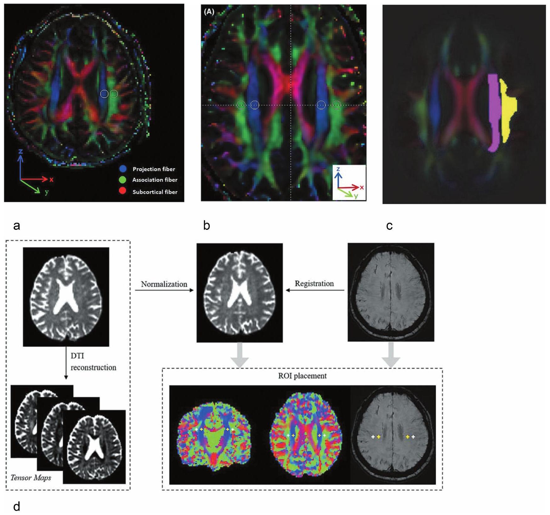
Concept of DTI-ALPS

The concept of the DTI-ALPS method is depicted in Fig. 1. Within the cerebral hemispheres, the elimination of waste products from the brain parenchyma to the exterior necessitates transport either toward the brain surface or the ventricular walls. At the lateral ventricular bodies, the most direct path to either the brain surface or the ventricular wall occurs in the
radial direction, specifically the left-right or -direction. In the human brain, at the level of the body of the lateral ventricles, the medullary arteries, medullary veins, and their PVS run in this -direction. This structure of medullary vessels within the brain parenchyma shows a construction that is purposeful in supporting the mass transportation that is the movement of material from and to the outside of the brain parenchyma. The basic concept of the ALPS method is to evaluate water movement in this -direction. Here, the diffusion of large nerve fibers dominates in diffusion images. It is necessary to avoid the strong influence on diffusion by white matter fibers for the evaluation of subtle diffusion factors in the direction of medullary vessels. At the lateral ventricular body level, the projection and association fibers are each orthogonal to the -direction, which allows the geometric elimination of the vector component of these large white matter fibers in the -direction. In the extreme, it may be sufficient to evaluate the diffusivity in the -direction itself at the level of the lateral ventricular body when assessing mass transfer in this brain region. However, the diffusivity calculated in a diffusion image is greatly affected by the imaging device and the imaging sequence, such as diffusion time. In order to compare values of coefficients in different cases, it is desirable to evaluate the coefficients in ratio to some internal reference. For this reason, the ALPS-index was designed to evaluate the ratio of diffusivity in the -direction to diffusivity perpendicular to both the -direction and the projection fibers as well as perpendicular to both the -direction and the association fibers as an internal reference, at the level of the lateral ventricular body. ALPS-index is given by following formula:
where Dxxproj is -axis diffusivity in the area of projection fiber, Dxxassoc is the -axis diffusivity in the area of association fibers, Dyyproj is the -axis diffusivity in the area of projection fiber, and Dzzassoc is the -axis diffusivity in the area of association fibers. A large ALPS-index indicates that the diffusion of free water in the -direction, or Brownian motion, is dominant, while a small ALPS-index indicates that free water movement in the -direction is not dominant.
The first report of ALPS method was published as a measurement in a case of Alzheimer’s disease (AD). In evaluations of normal, mild cognitive impairment (MCI), and AD cases, the ALPS-index was significantly correlated with Mini-Mental State Examination (MMSE) score and significantly inversely correlated with age. As glymphatic system disorders are known to be present in AD, these results suggest that the ALPS-index reflects the dysfunction of the glymphatic system in AD.
In considering the concept of the ALPS method, there are several points to note. It is not possible to distinguish whether the movement of water is inside or outside the PVS, or perivenous or peri-arterial space due to the relatively low
resolution of the diffusion image, in units of millimeters. Only the direction of the diffusion is distinguishable. However, what the ALPS method is trying to evaluate is the total or macroscopic movement of water in the direction of the PVS direction, not the movement of water in the PVS itself. Thus, these limitations are not a hindrance to this concept. In addition, since the method uses diffusion images, it evaluates the “movement” of water, not the “flow” of water. Thus, water movement toward the brain surface and toward the ventricles is equally subject to evaluation. Other issues will be considered in “Controversy in DTI-ALPS method” section.

Application of DTI-ALPS

Application for Alzheimer’s disease

AD is an irreversible, progressive brain disease that gradually damages the healthy brain, affecting memory, cognitive abilities, emotions, behavior, and mood. Impairment of cerebral ISF dynamics in AD was reported in an early paper by Iliff et al. Fluorescent-tagged amyloid was transported along this route, and deletion of the aquaporin- 4 (AQP4) gene suppressed the clearance of soluble , suggesting that this pathway may remove from the central nervous system. Thus, AD is considered one of the CNS (central nervous system) interstitial fluidopathies. And, the first report of the DTI-ALPS method was also studied in patients with AD, indicating decreased ALPS-index in the AD cases.
Several follow-up studies of the DTI-ALPS method in AD have been reported. Steward et al. examined DTI-ALPS parameters in a cohort of older adults consisting of cognitively normal, MCI, and AD. MMSE, a brief test used to assess cognitive function, and Alzheimer’s Disease Assessment Scale-Cognitive subscale (ADAS-Cog 11) measurements were correlated with ALPS-index. The results showed significant differences in the right DTI-ALPS-index between the cognitively normal, AD and MCI groups in a univariate general linear model adjusted for age, gender and apolipoprotein This report is an early follow-up of the DTIALPS method in AD, and although it is an evaluation of the ALPS-index alone, it is a report that demonstrates the utility of the ALPS method, despite its limitations.
An association with amyloid and tau protein deposition by positron emission tomography (PET) has also been reported. Ota et al. investigated the association between ALPS-index and PET with 11C-Pittsburgh Compound and tau protein PET findings with 18F-THK5351 in patients with AD. Significant negative correlations between the ALPSindex and the standard uptake value ratio (SUVR) of 11CPiB were found in bilateral temporal and left parietal cortex and left posterior cingulate gyrus in all subjects. Furthermore, there was a significant negative correlation between ALPS-index and SUVR of 18F-THK5351 in all subjects. This report correlates amyloid and tau PET results with the ALPS-index and also correlates with areas showing
Fig. 2 Application of ALPS method in AD. Violin and box plots of the mean ALPS-index, PVSVF-ALL, PVSVF-WM, PVSVF-BG, PVSVFHipp, and FW-WM among the HC participants, patients with MCI, and patients with AD are shown. Compared to HC, AD patients had significantly higher total PVSVF, WM, BG PVSVF (Cohen ) and FW-WM (Cohen ) and lower ALPS-index (Cohen ). The MCI group had significantly higher total PVSVF (Cohen ) and WM (Cohen . Figure quoted from Ref. 20. AD, Alzheimer’s disease; PVSVF, perivascular space volume fraction; WM, white matter; BG, basal ganglia; FW, free water; HC, healthy control; MCI, mild cognitive impairment.
deposition. It is interesting to note that the ALPS-index correlates with tau and amyloid deposition in regions other than the white matter surrounding the lateral ventricular body, which is evaluated by the ALPS method. On the other hand, Matsushita et al. reported that SUVR of amyloid PET was not significantly associated with ALPS-index. Also, a comparative study with amyloid PET for subjective cognitive decline by Park et al. showed no significant difference between the mean ALPS-index of the amyloid-positive group and the mean ALPS-index of the amyloid-negative group. Kamagata et al. examined changes in the following noninvasive MRI metrics related to the perivascular network in patients with MCI and AD: PVS volume fraction (PVSVF), fractional volume of free water in white matter (FW-WM) and ALPS-index. Their results showed that AD patients had significantly higher total PVSVF, WM, basal ganglia PVSVF, and FW-WM and lower ALPS-index than healthy controls. Meanwhile, only the total PVSVF and WM values were significantly higher in the MCI group. Lower ALPS-index was associated with lower , FDG (fluoro-deoxyglucose)-PET uptake in the CSF and worse impairment in several cognitive domains. High FW-WM was also associated with lower CSF and worse cognitive function (Fig. 2).

Application for Parkinson’s disease and other degenerative disease

Parkinson’s disease (PD) is an alpha-synuclein disorder characterized by deposition of alpha-synuclein in neurons and
loss of dopaminergic neurons in the substantia nigra. Similar to AD, this neurodegenerative disease is characterized by the accumulation of toxic proteins as a pathologic feature. In recent years, it has been shown that PD is also associated with abnormal ISF dynamics related to metabolic waste elimination, and studies using the DTI-ALPS method have been reported.
A report of ALPS method analysis in cases of PD and essential tremor showed a lower ALPS-index in PD than in essential tremor. In a comparison of stages of PD and normal controls, patients with PD had a lower ALPS-index than normal controls, and the decline was particularly pronounced in the group in the later stages of PD. There was a significant positive correlation between the ALPS-index and MMSE scores in the early PD group, and a negative correlation between the ALPS-index and the degree of PVS enlargement (Fig. 3).
Chen et al. used the DTI-ALPS method to evaluate ISF dynamics and its relationship to systemic oxidative stress status in PD patients. The PD group showing MCI and the PD group showing dementia had significantly lower ALPSindex than the normal control group. ALPS-index was inversely correlated with plasma nuclear DNA level, mitochondrial DNA level and cognitive score. The correlation between increased plasma nuclear DNA levels and decreased ALPSindex supports the idea that PD patients may exhibit increased oxidative stress associated with altered ISF dynamics. In another report, a negative correlation between ALPS-index and severity of motor symptoms in PD was found in a
Fig. 3 Application of ALPS method in PD. Differences in ALPS-index between the PD group and NCs and between two PD subgroups and NCs were compared. Relationships between ALPS-index and MMSE score in early PD group (A), relationships between ALPS-index and EPVS score in early PD group (B) and relationships between ALPS-index and age in late PD group (C) are shown. There was a significant positive correlation between ALPS-index and MMSE score ( ) ( ), and a negative correlation between ALPS-index and EPVS score ( ). ALPS-index negatively related with age ( ). Figure quoted from Ref. 184. PD, Parkinson’s disease; NC, normal control; MMSE, Mini-Mental State Examination; EPVS, enlarged perivascular space.
subgroup of patients aged 65 years and older than in younger patients. In a longitudinal study in patients with PD, the group with a lower ALPS-index deteriorated faster on The Movement Disorder Society Unified Parkinson’s Disease Rating Scale (MDS-UPDRS) Part III and Part II, Symbol Digit Modalities Test, and Hopkins Verbal Learning Test. Path analysis showed that ALPS-index acted as a significant mediator between and cognitive change in the Symbol Digit Modalities Test score at years 4 and 5.
Idiopathic rapid eye movement sleep behavior disorder (iRBD) is a sleep disorder in which the body moves during REM sleep and is a form of parasomnia. It has an aspect of alpha-synucleinopathy similar to PD and can result in phenoconversion to PD, Lewy body dementia, and multiple system atrophy. The overall conversion rate from iRBD to an overt neurodegenerative syndrome was per year. In the DTI-ALPS study of iRBD, PD, and normal controls, both PD and iRBD patients showed a lower ALPS-index than normal controls. PD patients had lower ALPS-index than iRBD patients, and ALPS-index correlated with cognitive decline in PD patients. In a longitudinal study of phenoconversion in patients with RBD, PD, and normal controls, the risk of phenoconversion decreased with increasing ALPS-index. Evaluations of neurodegenerative diseases other than PD, such as cortico-basal degeneration and progressive supranuclear palsy, have also been published. In a study evaluating ALPS-index and correlating it with motor and cognitive function in cerebral cortical degeneration with basal cortical syndrome (CBD-CBS), the ALPS-index of CBD-CBS was significantly lower than that of normal controls. In addition, ALPS-index showed a significant positive correlation with MMSE and a significant negative correlation with the MDS-UPDRS III score.

Application for small vessel diseases

Cerebral small vessel disease (SVD) affects small blood vessels in the brain and is associated with stroke, cognitive
dysfunction, and motor dysfunction, and has also attracted attention as a background pathology for neurodegenerative diseases such as AD. Benveniste et al. proposed that failure of brain fluid transport-via the glymphatic system plays a key role in initiation and progression of SVD. Their major case for this concept is that PVS are utilized as waterways for influx of CSF. Stagnation of transport by ISF is thought to cause loss of fluid homeostasis in the brain and may lead to transient white matter edema, perivascular dilatation, and eventually demyelination.
In an analysis by Kikuta et al. comparing older hypertensive patients with age-matched controls, the ALPS-index in the hypertensive group was significantly lower than in the control group. In addition, ALPS-index of all subjects was significantly negatively correlated with blood pressure values and pulse pressure values. This suggested that SVD due to hypertension was associated with damage in cerebral ISF dynamics. In a study of 133 patients with SVD, a lower ALPS-index was independently associated with impaired executive, attention, and memory function in patients with SVD. In a study of a cohort of more than 2000 community residents, the lower the ALPS-index, the greater the presence and severity of cerebral SVD. Furthermore, a low ALPSindex was associated with poor neuroimaging markers such as abnormal white matter signal, enlarged PVS, and brain atrophy.
Cerebral amyloid angiopathy (CAA) is a type of cerebral SVD characterized by deposition of in the middle and outer membranes of arteries in the cerebral cortex and meninges. The underlying etiology of accumulation is still unknown. In addition to the conventional idea of overproduction, there is also the idea of impaired clearance pathways. In a comparison of CAA patients and agematched controls, CAA patients had an overall lower ALPSindex compared to controls. In addition, lower ALPS-index was associated with enlarged PVS in the basal ganglia, increased lacunes, higher white matter high intensity
Fazekas scores, lower MMSE, and lower Montreal Cognitive Assessment (MoCA-J). During a median follow-up of 4.1 years, there was less recurrence of disease with a higher ALPS-index.

Application for idiopathic normal pressure hydrocephalus

Idiopathic normal pressure hydrocephalus (iNPH) is a clinical syndrome with enlarged ventricles without increased CSF pressure. iNPH has three characteristic symptoms: gait disturbance, cognitive decline, and urinary incontinence. Imaging features include Disproportionately Enlarged Subarachnoid Space Hydrocephalus (DESH) and narrowing of the callosal angle. iNPH has been pointed out as a degenerative disease, and its co-morbidity with AD is attracting attention. Eide et al. reported the evaluation of CSF dynamics and ISF dynamics in iNPH group using a technique with GBCA intrathecally injected and MRI observation over time. In this invasive study, the shunt-responsive iNPH group showed more conspicuous contrast reflux into the ventricular system and a tendency toward prolonged parenchymal enhancement compared to the control group. These results suggest that abnormalities in the glymphatic system may be related to the pathogenesis of iNPH.
The study by Yokota et al. on iNPH cases is the first follow-up study of the ALPS method. This study compares a group of iNPH cases diagnosed according to diagnostic criteria with a group of pseudo-iNPH (piNPH) cases that do not meet the diagnostic criteria. Their results showed that the ALPS-index was significantly lower in iNPH cases, and the area under the curve in ROC analysis was 0.92 , 1.00 , and 1.00 for control vs. piNPH, control vs. iNPH, and piNPH vs. iNPH, indicating very high diagnostic performance. Between piNPH and NPH, the ALPS-index showed higher diagnostic performance than the Evans index or the callosal angle. In a study by Georgiopoulos et al. the association between the clinical findings of Time Up and Go test (TUG) and MMSE associated with iNPH and the ALPS-index was analyzed. Patients with iNPH had significantly lower mean ALPSindex than healthy control. A moderate exponential correlation was found between the mean ALPS-index and motor function as measured by time required to complete TUG, number of steps to complete TUG, 10 m walking time, and 10 m walking steps. A positive linear correlation was found between mean ALPS-index score and MMSE score. As a diagnostic tool for iNPH, tap-test is an important test for predicting shunt efficacy. In a study evaluating the relationship between Tap-test findings and ALPS-index, ALPSindex was significantly lower in the NPH group that did not respond than in the group that showed symptom relief. Shunting is an effective treatment for iNPH. Studies have reported changes in ALPS-index before and after shunting. The mean ALPS-index after shunt surgery was higher than before shunt surgery. In particular, there was a significant increase in postoperative ALPS-index in the group with
symptomatic improvement after shunt surgery, whereas there was no significant increase in postoperative ALPSindex in the group without symptomatic improvement (Fig. 4).

Application for traumatic brain injury

Traumatic brain injury (TBI) is an established risk factor for early onset of dementia, including AD, and the post-traumatic brain often shows neurofibrillary changes consisting of aggregates of the protein tau. In animal studies in mice using fluorescent tracers, function of the glymphatic pathway was reduced by approximately following TBI, and this impairment persisted for at least 1 month after injury. In addition to direct brain injury, animal studies in non-human primates have shown that the formation of a CSF leak delays the entry of intrathecal injected GBCA into the brain. This indicates that mechanical factors such as intracranial pressure environment affect cerebral ISF dynamics.
In humans, several studies using the noninvasive ALPS method have reported abnormal ISF dynamics after TBI. It has been reported that ALPS-index is decreased in the brain after TBI. On the other hand, there are reports that ALPS-index is elevated in young patients with mild TBI. The possible causes include increased expression of AQP4 after trauma, hemodynamic changes after trauma, and the possibility that the glymphatic system is a compensatory mechanism to reduce secondary injury by eliminating endotoxin after trauma. As a situation that does not develop into a traumatic injury, some reports have evaluated the ALPS-index in people who have experienced contact sports. In this report, the left ALPS-index was significantly lower in the heavy contact (rugby, judo, karate, boxing, kendo, wrestling, and soccer) and semi-contact (baseball, basketball, and handball) groups than in the noncontact (tennis, table tennis, track and field, skiing, archery, and orienteering) group. The right ALPS-index tended to be lower in the semi-contact and heavy contact groups than in the non-contact group. Bilateral ALPS indices showed a significant positive correlation with MoCA-J scores.

Application for demyelinating disease

Multiple sclerosis is a chronic immune disorder of the CNS characterized by inflammation and demyelination caused by the interaction of BBB disruption, local leukocyte infiltration, microglial activation, and release of inflammatory cytokines. Considering that toxic wastes are the cause of tissue damage, it seems essential for the brain to have a system to remove these wastes. A retrospective study using the DTI-ALPS technique in patients with multiple sclerosis is reported. In this study, multiple sclerosis patients had an overall lower ALPS-index compared to healthy controls. Both relapsing-remitting and progressive multiple sclerosis patients had a lower ALPS-index versus healthy controls. Patients with advanced multiple sclerosis had a lower ALPSindex compared to patients with relapsing-remitting multiple
Fig. 4 Application of ALPS method in iNPH. A study of changes in ALPS-index after LPS surgery in iNPH is presented, comparing patients who responded to LPS with those who did not. The mean ALPS-index of the postoperative group was significantly higher than that of the preoperative group (A). Furthermore, for responding subjects, the mean ALPS-index in the postoperative group was significantly higher than in the preoperative group (B). On the other hand, in the non-responder group, the mean ALPS-index of the postoperative group was not significantly higher than that of the preoperative group (C). The mean ALPS indices of the responder group were not significantly different compared to those of the non-responder group in both the pre-operation and post-operation groups (D). Figure quoted from Ref. 62. iNPH, idiopathic normal pressure hydrocephalus; LPS, lumboperitoneal shunt.
sclerosis. In multiple sclerosis patients, lower ALPS-index was associated with more severe clinical disability and longer disease duration.
Myelin oligodendrocyte glycoprotein antibody disorder (MOGAD) patients, another demyelinating disease, have also been reported to be studied using the ALPS method. In a prospective study of MOGAD patients in remission and age- and sex-matched healthy controls, the mean ALPSindex was significantly lower in MOGAD patients than in healthy controls. Lower mean ALPS-index was significantly associated with worse Expanded Disability Status Scale scores. However, cortical and deep gray matter volumes were not significantly different between the two groups,
nor were they correlated with Expanded Disability Status Scale scores.
It is suggested that MOGAD patients may have impaired glymphatic function as measured by the ALPS-index.

Application for sleep

One of the reasons why the glymphatic system theory has attracted so much attention is its relationship to sleep. Drainage by the glymphatic system is considered to be suppressed during wakefulness and markedly enhanced during sleep. Xie et al. reported that under physiological conditions, the activity of the glymphatic system is associated with sleep, based on observations of fluorescent tracers injected
Fig. 5 Time-of-day dependency of ALPS-index. Circadian rhythm dependence of ALPS-index was assessed by repeated MRI measurements at five time points from 8:00 to 23:00. Very long echo-time low-b diffusion tensor imaging (DTIlow-b), which measures SAS flow along the middle cerebral artery, and conventional DTI-ALPS were performed. A, B show the time course of the glymphatic system influx and efflux measured by DTIlow-b and DTI-ALPS, respectively. Neither ADlow-b of the MCA SAS fluid nor the ALPS-index differed significantly in the data acquired at the five time points. Figure quoted from Ref. 159. MRI, magnetic resonance imaging; DTI, Diffusion Tensor Image; SAS, subarachnoid space; DTI-ALPS, Diffusion Tensor Image Analysis aLong the Perivascular Space; AD, Alzheimer’s disease; MCA, middle cerebral artery.
into the CSF cavity of rats using two-photon microscopy. During sleep, there is a significant increase in glymphatic system activity compared to wakefulness. They attributed this to the fact that during sleep, the volume of glial cells is reduced and the interstitial space is more dilated than during wakefulness, facilitating mass transport in the tissues.
The ALPS method has been used to evaluate sleep and cerebral ISF dynamics in several published studies. In a study evaluating the correlation between sleep-related indices and the ALPS-index in community-dwelling older adults aged 60 years and older, the ALPS-index correlated with N 2 sleep duration and N 3 sleep duration. On the other hand, it correlated inversely with the apnea-hypopnea index. In addition, it correlated with gray matter volume. Obstructive sleep apnea (OSA) is associated with sleep fragmentation and altered sleep organization, which may impair cerebral ISF dynamics and increase the risk of AD. In a study of OSA and control cases, Dzz values, derived from projection fiber areas, Dyy and Dzz values from association fiber areas, as well as ALPS and Dyzmean values were significantly reduced in OSA over controls. Significant correlations emerged between disease severity, sleep symptoms, and Dxy, Dxx, and Dzz values in OSA subjects. In a study evaluating the association between sleep quality and the ALPS-index as assessed by the Pittsburgh Sleep Quality Index (PSQI), the ALPS-index was significantly lower in the sleep disordered group than in the control group. In addition, the ALPS-index showed a significant negative correlation with all components of the PSQI score. In the DTI-ALPS study of narcolepsy patients, there was no significant difference in ALPSindex between the patient and control groups. However, the ALPS-index was correlated with polysomnography
parameters and positively correlated with wake after sleep onset and REM sleep length in patients with type 2 narcolepsy. As mentioned above, animal experiments with tracers have shown that tracer dynamics are enhanced during sleep. In humans, evaluation by the dual compartment tensor model has also been reported in diffusion imaging. The dual compartment analysis revealed that the increase in diffusivity measures from PM to AM was driven by an increase in the volume fraction of CSF-like free-water. On the other hand, in the ALPS method study, MRI measurements were repeated at five time points from 8:00 to 23:00 and no time-period dependence was observed in the awake state in an experiment to evaluate circadian rhythm dependence (Fig. 5). This study is also discussed in “Other methods for evaluation of glymphatic system” and “Consideration for the methodology of DTI-ALPS” sections.

Findings on the aging

A number of studies using the DTI-ALPS method have evaluated the association between age and ALPSindex. Among these reports, a number of them showed negative correlations between age and ALPS-index. McKnight et al. showed a negative correlation between the ALPS-index and age in a group of subjects with PD or essential tremor aged approximately 40-80 years, with a correlated Spearman’s rank correlation coefficient of Zhang et al. showed a negative correlation between age and ALPS-index in 142 healthy elderly subjects aged 50 years and older, with a correlation coefficient of et al. showed a negative correlation between age and ALPS-index in PD cases, with a standard partial regression coefficient of Siow et al. also found a
negative correlation between age and ALPS-index in com-munity-dwelling elderly aged 60 years and older, with a standard partial regression coefficient of Toh and Siow examined glioma cases in a wide age range, from 18 to 91 years of age. Although the correlation coefficient is low at -0.147 , the graph they presented showing the correlation between age and ALPS-index appears to have a slight peak around age The public dataset study by Dai et al. for ages also appears to have a similar peak around the 40s in the graph provided in the paper showing the correlation between age and ALPS-index, although the authors do not mention it. Furthermore, the DTI-ALPS method described below also shows that the ALPS-index peaks in the 40s. However, there is an aspect of this that indicates that the ALPS-index may be influenced by the status of white matter fibers, which will be discussed later. While there have been many reports of an association with aging in older patients, there have also been reports of the DTI-ALPS method in pediatric cases. Lee et al. showed a negative correlation between age and ALPS-index with a correlation coefficient of -0.375 in a group of young patients with myoclonic epilepsy aged years. However, there is a lack of validation of the applicability of this technique in children, in whom the construction of white matter fibers is incomplete. The rapid changes in the developmental state of white matter with growth require careful interpretation of the results, especially when comparing cases of different ages. Further experience is needed.

Other Methods for Evaluation of Glymphatic System

Because the ALPS-index is given by a simple formula and can be calculated from retrospective data, it has been reported on many diseases, pathologies, and conditions as described above. However, as discussed in Section “Controversy in DTI-ALPS method”, there are also problems that have been pointed out. When evaluating cerebral ISF dynamics, it is not sufficient to use the ALPS method alone, instead it is desirable to use other methods of evaluating cerebral ISF dynamics together. The following is a review of studies that have evaluated the relationship between the ALPS method and other methods of assessing cerebral ISF dynamics.

Intrathecal GBCA

A number of attempts have been reported to use MRI to evaluate the state of the glymphatic system (Table 1). However, the first studies on the glymphatic system by Iliff et al. were done in animals with intrathecal administration of fluorescent tracers and two photon laser microscope. This method has the advantage of allowing direct observation of the living brain, but only near the surface of the brain. As a method to observe the whole brain, a tracer study in animals using MR with intrathecal GBCA was introduced. In
humans, tracer studies using MR with intrathecal GBCA are considered to be the gold standard. However, it should be noted that the molecular weight of GBCA is 0.6 kDa (Gadobtrol), whereas that of in the brain is In addition, in that it evaluates penetration into the brain from the brain surface, it is evaluating the pathway opposite to the drainage of waste material from the brain parenchyma. Furthermore, it is difficult to evaluate the state of the deep white matter, which occupies a significant proportion of the brain volume. In these respects, it should be noted that this method is too limited to be considered a gold standard for evaluating the glymphatic system.
Intrathecal injection of GBCA in humans is an unapproved use, and few centers perform it. This is because GBCA intrathecal injection is invasive and, depending on the dose, unsafe. Eide, Ringstadt, and colleagues have systematically conducted tracer studies of GBCA intrathecal injection in humans. The first report was of intrathecal GBCA in a patient with intracranial hypotension due to spontaneous CSF leakage, in which MRI showed distribution of GBCA throughout the brain at 1 and 4.5 hours after intrathecal injection. Their experiments were initially only qualitative evaluations of tracers, but more recent studies have conducted sophisticated mathematical analyses to identify and quantify CSF tracer transport parameters. The results indicate that extracellular diffusion alone is not sufficient as a tracer transport mechanism for the whole brain, and that local clearance rates contribute to the tracer transport. In a study by Dyke et al. in healthy adult volunteers, GBCA in the intracranial spinal fluid space peaked at and began to decrease at 7 h . In contrast, in some areas of the brain parenchyma, such as the cerebral cortex and white matter, potentiation increased after 11 hours. Thus, it was suggested that the enhancement of the whole brain MR signal by intrathecal GBCA administration may indicate CSF/ISF exchange and brain parenchyma penetration throughout the brain.
A study compared the results of the DTI-ALPS method with the evaluation of glymphatic system function using intrathecal GBCA. The ALPS-index was significantly associated with the rate of change in signal unit ratio from baseline to 39 h in six brain regions.

Intravenous GBCA

Intravenous GBCA infusion as a tracer is easier to perform than intrathecal injection. Experiments using high-dose intravenous GBCA in rats showed an increase in signal in the fourth ventricle immediately after intravenous GBCA injection, indicating that GBCA is rapidly transferred from blood to CSF in rats. The signal curve of the cerebral cortex and deep cerebellar nuclei reached the peak signal intensity later than the fourth ventricle but earlier than that of the prepontine cistern. Thus the GBCA distribution to the cerebral cortex and deep cerebellar nuclei seemed to depend on both blood flow and CSF. In human evaluations, the transfer of IV-GBCA into the CSF is more gradual. In
Table1 MRI methods intending to evaluate glymphatic system for human subjects
Method MR sequence Target phenomenon Target location Assumed rolls in glymphatic system
Intrathecal GBCA T1WI Penetration via the brain surface (Tracer: GBCA) Brain surface CSF influx to brain parenchyma
BBB permeability (Tracer: GBCA) Brain parenchyma ISF production
Intravenous GBCA T1WI Venous wall permeability (Tracer: GBCA) Surface vein Movement of waste products
Tracking of GBCA Meningeal lymphatic vessel/ Perivascular space Movement of waste products
Intravenous Steady-state sequence BBB permeability (Tracer: labeled water) Brain parenchyma ISF production
DTI-ALPS DTI Brownian motion of the water molecule in the brain tissue Deep white matter adjacent the lateral ventricle Movement of ISF within the tissue
Free water analysis DTI etc. Increase of free water fraction Brain parenchyma Widening of interstitial space
Perivascular space volume 3D-T2WI etc. Dilatation of perivascular space Perivascular space Accumulation of metabolic waste
Choroid plexus volume 3D-T1WI etc. Enlargement of choroid plexus Choroid plexus Removal of waste/ Immune reaction etc.
ASL with long TI ASL BBB permeability (Tracer: tagged water) Brain parenchyma ISF production
BOLD-CSF coupling BOLD Synchrony of the neural activity and the brain/CSF motion Brain parenchyma Tissue microenvironment
Elastography Elastography Viscoelastic properties of the brain Brain parenchyma Tissue microenvironment
ASL, arterial spin labeling; BBB, blood -brain barrier; BOLD, blood oxygen level-dependent; CSF, cerebrospinal fluid; DTI, diffusion tensor image; DTI-ALPS, Diffusion Tensor Image Analysis aLong the Perivascular Space; GBCA, gadolinium-based contrast agent; ISF, cerebral interstitial fluid; T1WI, T1-weighted image; T2WI, T2-weighted image; TI, inversion time.
human evaluation after administration of a normal dose of GBCA, leakage from the cortical veins into the CSF was observed. An enhancement within the PVS in the basal ganglia and around the cortical veins was observed after 4 h of IV-GBCA administration. Interestingly, the leakage from the cortical vein was not observed in the younger subjects, but only in the older subjects ( 37 years and older). In a study examining the relationship between the number and size of cystic structures around the cortical veins near the superior sagittal sinus and leakage of GBCA around the cortical veins, the number and size of depressed structures near the superior sagittal sinus were reported to be larger in subjects with GBCA leakage into the SAS than in those without leakage. Intravenous GBCA can also delineate meningeal lymphatic system. In a study of the features of presumptive meningeal lymphatic vessels located in the posterior wall of the sigmoid sinus (PML-PSS) in human subjects imaged after intravenous administration of GBCA, PML-PSS was found in 23 of 42 patients 4 hours
after IV-GBCA was performed. The study also simultaneously examined the enhancement of the basal ganglia perivascular space (PVS-BG), and PVS-BG was seen in 21 of the 42 cases.
There have been several trials using intravenous GBCA to assess the degree of leakage of the BBB . In a study that used a Patlak graphical approach to quantify BBB leakage rates and regional plasma volume on dynamic contrast images after intravenous GBCA injection, the volume fraction of brain tissue with GBCA leakage was significantly higher in In a study examining changes in GBCA leakage during the diurnal cycle, T1 mapping of gray matter and white matter at , and 12 h after intravenous contrast injection was used. The results showed that signal changes in cerebral gray matter, cerebellar gray matter, and putamen increased from diurnal to nocturnal cycles. This suggests that the clearance of GBCA is greater after sleep than during daytime wakefulness. This technique has the potential to
evaluate BBB leakage. However, BBB leakage from GBCA in healthy brain tissue is so small that the measurement is always evaluated near the noise floor. In this respect, the measurements are expected to be greatly affected by noise contamination, imaging conditions, and analysis methods. It should also be noted that the evaluation of BBB leakage does not evaluate the entire glymphatic system, but only a part of the entire system.
Currently, there are no published reports comparing the assessment of the extent of BBB leakage by intravenous contrast with that by the ALPS method. However, a study has been reported that evaluated the time course of enhancement in the parasagittal sinus using dynamic contrastenhanced MRI with intravenous GBCA for the purpose of assessing meningeal lymphatic dysfunction. Time to peak values after contrast was significantly delayed in the chronic migraine group compared to the episodic migraine group, suggesting meningeal lymphatic dysfunction. This study also reported significantly lower ALPS-index in the chronic migraine group compared to the episodic migraine group.

Perivascular space volume

Needless to say, the PVS is an anatomical structure that plays an important role in the integrity of the glymphatic system. The PVS is usually unremarkable on MR images, but in dilated cases of PVS, it can be seen as a punctate or linear structure. This abnormal dilation of the PVS is hypothesized to be associated with impaired glymphatic function. One suggestion about the mechanism is that PVS volume may reflect decreased flow in the glymphatic system resulting in either increased accumulation of metabolic waste products or failure to deliver needed metabolic substrates. Recently, there have been several published reports of attempts to actively apply PVS enlargement as a biomarker of glymphatic system damage.
Various methods have been proposed to evaluate the degree of PVS dilation on MRI. In general, the easiest to use is the visual evaluation of T2-weighted images. Potter et al. proposed a robust and easy-to-use PVS assessment scale to effectively examine the diagnostic and prognostic significance of PVS. They rated basal ganglia, centrum semiovale, and midbrain PVS. Basal ganglia and centrum semiovale PVS were rated 0 (none), , 3 ( ), and 4 ( ), and midbrain PVS were rated 0 (none visible) or 1 (visible). They report that the ratings based on this criterion showed good intra- and inter-rater consistency. Recently, an automated human PVS evaluation system using MRI has been developed. Dubost et al. proposed a convolutional network regression method to quantify the degree of enlarged PVS in the basal ganglia from 3D brain MRI. This method achieved an intraclass correlation coefficient of 0.93 and an intra-rater agreement coefficient of It is known that long-term spaceflight can cause changes in the brain and CSF, resulting in visual impairment called spaceflight-related neuro-
ophthalmopathy. Astronauts who spent 6 months on the International Space Station showed enlarged PVSs in the basal ganglia and white matter after their spaceflight. Furthermore, astronauts who developed spaceflightrelated neuro-ophthalmopathy had a more enlarged PVS in the white matter than those who did not develop the syndrome, indicating that long-term changes in gravity may affect the function of the glymphatic system. Examination of T2-weighted images of postmortem brains shows a correlation between the deposition of in cortical vessels and the degree of opening of the PVS in the underlying white matter, indicating that the degree of opening of the PVS may be an indicator of dysfunction of the glymphatic system.
Several studies comparing the evaluation by PVS volume with that by the ALPS method are presented. In a study of TBI cases and controls, the ALPS-index was significantly lower in subjects with TBI compared to controls when age was used as a covariate. The ALPS-index showed a significant negative correlation with the plasma concentration of neurofilament light chains, a biomarker of injury severity. On the other hand, PVS volume did not differ in TBI patients compared to controls, nor did it correlate with the plasma concentration of neurofilament light chains.
In a study of patients with AD , the perivascular volume fraction of the basal ganglia in patients with AD was significantly larger than the volume fraction in normal controls. The ALPS-index showed a significant negative correlation with the perivascular volume fraction of the basal ganglia in AD patients. As mentioned in section “Application for Alzheimer’s disease”, using the AD Neuroimaging Initiative database, an examination of measurements such as the PVS volume fractions, fractional volume of FWWM, and ALPS-index has been reported. In this study, patients with AD had significantly higher PVS fraction of the basal ganglia, significantly higher fractional volume of FW-WM, and significantly lower ALPS-index compared to healthy controls (Fig. 2). In addition, Zhang et al. also reported that the PVS fraction in the basal ganglia of AD patients was significantly larger than the PVS fraction in control subjects and negatively correlated with the DTI-ALPS-index. A report has also evaluated the relationship between ALPS-index and PVS in patients with PD. The left DTI-ALPS-index showed a non-significant negative trend with the number of PVSs in the left basal ganglia in the early PD group, and a significant negative correlation in the late PD group.

Choroid plexus volume

The choroid plexus is a highly vascularized secretory tissue located within the ventricles of the brain. Its main functions have traditionally been considered to be the production of CSF and the formation of the blood-CSF barrier. However, recently, functions such as removal of toxic waste products and metabolites from the CNS, immune surveillance of the
brain, and regulation of CSF composition for homeostatic brain function have been identified. In a study of choroid plexus volume and dopamine transporter (DAT) scanning findings in early, untreated PD patients, choroid plexus volume was negatively correlated with DAT availability in many striatal subregions. In the Cox regression model, larger choroid plexus volume was associated with future onset of gait freezing, and in the linear mixed model, it was associated with more rapid increases in dopaminergic medications. This suggests that choroid plexus volume may serve as a biomarker for baseline and longitudinal motor deficits in PD. It is an attractive hypothesis that the PVS must be dilated by a disturbance of the glymphatic system. At present, however, direct proof is lacking. The PVS may also be dilated by other factors, such as a decrease in brain parenchymal volume. However, it is difficult to distinguish PVS dilatation due to other factors from PVS dilatation due to a glymphatic system disorder. In this regard, one should be cautious in linking PVS dilation directly to glymphatic system disorder.
Several studies have been presented that examined choroid plexus volume and its evaluation with the ALPS technique. In a study in which glymphatic clearance and choroid plexus volume were measured by 3D-T1-weighted imaging before and 39 h after intrathecal contrast administration in patients indicated for lumbar puncture, higher choroid plexus volume correlated with slower glymphatic clearance rates in all brain regions. The higher choroid plexus volume was correlated with slower glymphatic clearance in all brain regions. In this paper, longitudinal follow-up observations of ALPS-index, choroid plexus volume, and abnormal white matter signal volume are conducted in a different cohort. The results showed that baseline choroid plexus volume was positively correlated with abnormal white matter signal volume and its degree of increase. Furthermore, in a mediation analysis, the ALPS-index partially mediated the association between choroid plexus volume and abnormal white matter signal-like content and its augmentation. Fibromyalgia is often associated with various degrees of sleep disturbance and cognitive impairment, and studies have evaluated the relationship between sleep disturbance and glymphatic system function in these patients. The results showed that fibromyalgia patients had significantly higher choroid plexus volume and lower ALPS-index than age- and intracranial volume-adjusted controls. Increased choroid plexus volume was correlated with lower ALPSindex and longer disease duration. The complex geometry of the choroid plexus has made volume measurement cumbersome in the past, but recently, automated measurement using deep learning models has been introduced, showing comparable results in choroid plexus delineation on T1-weighted, T2-weighted and FLAIR non-contrast MRI images. It is expected that the measurement of choroid plexus volume will become easier in the future by using these software.

Diffusion-based methods other than ALPS method

In addition to the ALPS method discussed so far, several other methods for evaluating cerebral ISF dynamics using diffusion images have been introduced. The brain contains a mixture of both free water and brain tissue, namely CSF and ISF.
The ALPS method focuses on the directionality of diffusion without considering these compartments of diffusion. On the other hand, there is a method that evaluates the free water compartment including CSF and ISF within the diffusion compartments. One of the methods is the dual compartment tensor model. Studies have used this method to examine the effect of time of day on diffusion tensor measurements using a within-subject longitudinal design. In this study, a dualcompartment tensor model, which allows direct assessment of the free water volume fraction, was used to evaluate changes related to time of day, in addition to the conventional monoexponential tensor model. The results indicate that the mean diffusivity measured using the conventional singleexponential tensor model tends to increase systematically at the gray matter/CSF interface from the morning to the afternoon scan. Furthermore, the dual-compartment analysis revealed that the increase in diffusivity measurements from the afternoon to the morning is caused by an increase in the volume fraction of CSF-like free water. As mentioned in “Application for Alzheimer’s disease” section, free water analysis was also performed in AD, and the free water fraction of white matter was higher in the AD group than in the normal group (Fig. 2). Alterations in free water fraction may be related to glymphatic system function. However, the information is static, and there is no direct evidence as to how the increase or decrease in free water fraction is dynamically related to the process of waste excretion in the brain. This approach also does not seem to provide an assessment of the entire glymphatic system.
One of the analyses of CSF dynamics using diffusion imaging is the evaluation of CSF pulsation. The early diffusionweighted image (DWI) by LeBihan et al. was called IVIM (intravoxel incoherent motion) imaging. The -value used at that time was low, about , thus the signal from CSF remained. Later, when motion probing gradients with high -values became available, the CSF signal in DWI was almost completely suppressed and was not considered for evaluation. A report was published in 2019 that once again attempted to evaluate CSF by daring to use a low -value DWI. Subsequently, reports have been published on the evaluation of CSF dynamics using multiple -values, as well as reports evaluating the relationship between CSF pulsation and arterial pulse rate, and more detailed evaluation of spinal fluid pulsation with the addition of tensor information. Assessment by measuring flow in the SAS along the middle cerebral artery by ultra-long echo time and low-b diffusion tensor imaging (DTIlow-b) has also been attempted; DTIlow-b has been proposed to reflect flow characteristics in one of the major CSF inflow pathways to the brain parenchyma (Fig. 5).
The influx rate of the glymphatic system evaluated by DTIlowb was significantly higher in participants aged years than in participants aged 21-38, while the ALPS-index was significantly lower in those aged years. Of course, since this method evaluates CSF outside of the brain, it does not evaluate the entire glymphatic system.

Arterial spin labeling

Arterial spin labeling (ASL) is one of the most commonly used noninvasive perfusion imaging methods in routine clinical practice. Recently, a technique to evaluate AQP4 water channels using this ASL has been introduced. A multi-echo time (multi-TE) ASL MRI technique was applied to the mouse brain to evaluate the water permeability of the BBB by calculating the exchange time, which is the time that magnetically labeled intravascular water is exchanged across the BBB. The results showed a increase in exchange time ( ) in AQP4-deficient (Aqp4-/-) mice ( compared to wild-type mice ( ), indicating the sensitivity of this technique to the absence of AQP4 water channels.
Human evaluations have also been performed. Using 3D turbo gradient and spin echo pulsed arterial spin echo with long TI (inversion time), the mean arterial pulse-corrected signal versus TI was plotted for each TI, and the slope of the plot was used as the clearance rate. Comparison between healthy subjects and AD patients showed a statistically significant clearance rate in the AD group. In the AD group, brain clearance rate correlated with CSF clearance rate, indicating that a proportion of ultra-filtered labeled blood was retained in the brain. This provides indirect evidence that perivascular clearance is reduced in AD. This method uses water molecules that can pass through the BBB under physiological conditions as tracers, rather than large molecules as in GBCA, and when combined with the GBCA evaluation, it should be possible to evaluate ISF dynamics separately for solute and solvent. Of course, this method is also a part of the glymphatic system.

Imaging for brain tissue kinetics

Kiviniemi et al. suggested that low-frequency ( ) rest-ing-state fMRI blood oxygen level-dependent (BOLD) signals are associated with spinal fluid dynamics and thus glymphatic system function. It has also been reported that the large global BOLD signal during sleep is coupled with strong CSF motion, an important component of the glymphatic system. Taken together, the overall resting activity and associated physiological modulations may represent highly coordinated neural and physiological processes closely related to glymphatic system function. BOLD-CSF coupling assesses these synchrony and may serve as a marker for measuring glymphatic function in the brain.
In a study in PD, BOLD-CSF coupling was found to be significantly lower in PD patients with MCI compared to those without MCI or controls. The decrease in BOLD-CSF
coupling was associated with the cognitive decline seen in patients with PD. Additionally, lower BOLD-CSF coupling in PD patients was associated with a thinner right entorhinal cortex. Glymphatic system dysfunction may also be involved in the pathogenesis of frontotemporal dementia. In a study of behavioral variant frontotemporal dementia (bvFTD), patients with bvFTD exhibited significantly weaker BOLD-CSF coupling. The study also found a negative correlation between the ALPS-index and BOLD-CSF coupling, as well as a negative correlation between the ALPS-index and CP volume.
Magnetic resonance elastography (MRE) is an approach that can provide biomechanical information about the tissue microenvironment because it can evaluate the viscoelastic properties of tissues in vivo. There have been reports of attempts to assess glymphatic system function using MRE. Experiments in a mouse model showed that shear wave speed, a measure of stiffness, varied by approximately across the cortex between sleep and waking states. Also in humans, MRE has been reported for the purpose of evaluating the glymphatic system, and multivariate analysis showed that the ALPS-index was independently associated with the complex shear modulus and positively related to it.

Other tracer study methods

Studies to date using large tracer molecules such as GBCA may have systematically underestimated the aqueous flow of subarachnoid spinal fluid into the brain. The stable isotope is not radioactive but has a quadrupolar nucleus that can be imaged by MRI. Direct imaging of the signal is difficult due to its low gyromagnetic ratio and low natural abundance ( ), but the presence of as a tracer can be detected indirectly by the effect of the quadrupole nucleus on the 1 H MRI signal. Glymphatic flow imaged with the tracer has been demonstrated to be more rapid and extensive than when imaged with the GBCA tracer.
Animal studies have been reported using the AQP-4 accelerator TGN-073 and JJ vicinal coupling proton exchange MRI, which can track water molecules delivered into the blood circulation. The results showed that the AQP-4-promoting effect of TGN-073 increased ISF turnover through the AQP-4 system, resulting in a significant decrease in content in the cerebral cortex. Also in humans, attempts have been made to use as a tracer, and dynamic 3D-FLAIR has been shown to detect dose-dependent signal changes.
Dynamic PET has been attempted to be used for evaluation of the glymphatic system. A study using dynamic PET with 18 F-THK5117, a tracer of tau pathology, to calculate ventricular CSF clearance rate (vCSF) as a biomarker of waste excretion has been reported.
This study focused on the washout of tau tracer. The vCSF was inversely correlated with amyloid deposition in AD patients and normal controls. In all subjects, the highest
extracranial accumulation of tracer was in the nasal turbinates. In patients with was reduced by and the supranasal CSF efflux site was reduced by There is a report comparing vCSF and ALPS-index with amyloid deposition in the brain using this technique. vCSF showed a high correlation ( ) with mild deposition, while DTI-ALPS showed a high correlation ( ) with severe deposition. Regression models using both vCSF and DTI-ALPS are better associated with brain A deposition. Thus, these two independent brain clearance indices may better explain the variability of deposition when both are used rather than either term alone. In other words, it suggests that vCSF and DTI-ALPS reflect complementary aspects of brain clearance function.

Consideration for the Methodology of DTI-ALPS

Placement of ROI

In the DTI-ALPS method, it is necessary to place ROIs in the projection fiber region and the association fiber region. This ROI setting is, in a sense, the Achilles’ heel of the DTI-ALPS method, because the reproducibility is greatly affected by the ROI setting. The most important point in the placement of the ROI is to ensure that all of the subject groups of cases are measured in a uniform manner. It is also important to make sure that the projection and association fibers are perpendicular to the -axis. In particular, when setting the ROI in the area of association fibers, care should be taken to avoid mixing of subcortical fibers whose main axis is in the -direction. If subcortical fibers are included, the ALPS-index will be higher than it should be.
In the first paper on the DTI-ALPS method, a 5 mm diameter circular ROI was manually placed in the left projection and association fiber areas (Figs. 1 and 6). Many papers have followed this method of placing ROIs in the dominant brain. Since association fibers are often thicker on the dominant side (left side), placement of a ROI on the left side has the advantage that a stable ROI can be set, even if it is done manually. In many recent papers, the average of the ROI on both sides is used. This method is expected to have the potential to cancel out the effects of head tilt and other factors. There is also a paper that examines the ALPS-index calculated for the right hemisphere and the ALPS-index calculated for the left hemisphere separately.
Some studies employ atlas based placement. Yokota et al. coregistered diffusion maps to the DTI-81 atlas of the International Consortium for Brain Mapping (ICBM) and established ROIs for projection and association fiber regions. In their study, the atlas-based ALPS-index was able to distinguish iNPH from pseudo-iNPH more clearly than manual ROI placement (Fig. 6).
An increasing number of recent studies employ automated ROI setup. Most of the studies use the Montreal Neurological Institute (MNI) 152 template or the digital WM atlas JHU ICBM-DTI-81 (http://cmrm.med.jhmi.edu/) for standardization purposes. After the images of each case are corrected to the standard brain, ROIs can be set for all cases under the same conditions to eliminate arbitrariness (Fig. 6).

Reproducibility of ALPS method

The DTI-ALPS method has been shown to be highly reproducible when imaging conditions such as imaging sequence, imaging plane, and head position during imaging are standardized (CHAMONIX study). Scanner differences did not affect ALPS-index values when imaging conditions were strictly matched. In addition, a study evaluated the reproducibility between MRI system vendors and between analysis pipelines. In this study, two pipelines were developed using DSI studio and FSL software. They validated inter-vendor reliability, inter-rater reliability, and test-retest reliability. The ALPS-index showed good inter-scanner repeatability (intraclass correlation coefficient: 0.001 ), inter-rater reliability ( ), and test repeatability ( ).
The transverse plane of MRI is usually taken at the anterior commissure-posterior commissure line, which is also the standard for evaluation in the ALPS method. Since this cross-section is orthogonal to the pyramidal tract, it is important for setting the ROI for the ALPS-index calculation. Changes in imaging plane and head position reduced reproducibility. To overcome this effect of head position on the ALPS-index, Tatekawa et al. developed a method to calculate the original and reoriented ALPS-index using a technique that registers DTI vector information in a different space and creates a reoriented diffusion map. This technique enables the reconstruction and creation of diffusivity maps along the -, -, and -axes even when the head position and imaging plane are inappropriate, thus improving the reproducibility of the DTI-ALPS-index calculation.
A report has proposed the application of harmonization techniques in multi-center studies. One multi-center study used the COMBined Association Test (COMBAT), a harmonization technique using regression of covariates in an empirical Bayesian framework, to harmonize ALPS-index variation across scanners, sites, and protocols. COMBAT harmonized the scanner differences and improved Cohen’s of the left-right ALPS-index between AD and normal control. As another means of improving reproducibility, Saito et al. proposed ALPS-index maintaining tensor vector orientation information (vALPS-index). In their result, the absolute difference in ALPS-index values between scan and rescan was larger in the ALPS-index than in the vALPSindex by approximately as the relative difference. Cohen’s for the left and right ALPS indices between methods were 0.121 and 0.159 , respectively. Thus, the
Fig. 6 Placement of ROI for ALPS method. The most important point in the placement of the ROI is to ensure that all of the subject groups of cases are measured in a uniform manner. It is also important to make sure that the projection and association fibers are perpendicular to the -axis. In particular, when setting the ROI in the area of association fibers, care should be taken to avoid mixing of subcortical fibers whose main axis is in the -direction. If subcortical fibers are included, the ALPS-index will be higher than it should be. After that, there are various ways to place ROIs: only on the left side of the dominant hemisphere (a), bilateral placement (b), Atlas-based placement (c), and placement with conversion to the standard brain (d).
vALPS-index based on DTI-ALPS maintaining tensor vector orientation information has higher reproducibility than the conventional ALPS-index.

Variation of ALPS method

In the CHAMONIX study described above, although the number of MPGs (motion-probing gradients) affected ALPS-index values, the ALPS-index values of 12 -axis DTI
and 3-axis DWI showed a good correlation. This suggests the possibility of using the DWI-ALPS method with triaxial MPG. In fact, the DWI-ALPS method has been reported to calculate the ALPS-index using DWI, which is a simpler method than diffusion tensor images. The DWI-ALPS method has the advantage of obtaining the ALPS-index from DWI, which can be acquired in about 1 min , and it is possible to incorporate this method into routine clinical MRI
examinations. An examination of the ALPS by the DWIALPS method in patients who received whole brain radiotherapy for brain tumors and normal controls is reported. The ALPS-index was significantly lower in the post-irradiation group than in the control group in the comparison of all age groups, suggesting that the ISF dynamics were affected in the patients after whole-brain radiation therapy. Interestingly, no significant differences were found in the younger age group. There was a weak negative correlation between ALPS and irradiation dose. When performing the DWI-ALPS method, it is important to note that, unlike the DTI method, the positional relationship between white matter fibers and the ROI cannot be adjusted in post-processing, so the transverse section must be perpendicular to the projection fiber at the time of imaging.

Controversy in DTI-ALPS Method

It is pointed out that the ALPS method is theoretically deductive. The relationship between ALPS-index and human glymphatic function has not been substantively and rigorously validated by pathophysiological studies. Therefore, the relationship between ALPS-index and glymphatic clearance should be interpreted with caution. However, a number of reports have recently been published comparing the ALPS method with other glymphatic system evaluation methods using different imaging modalities to examine the validity of the ALPS method. This review highlighted a number of such reports. Of particular importance is the report by Zhang et al. on the ALPS-index and intrathecal contrast administration for the evaluation of glymphatic system function, showing a strong correlation between the two methods.
However, it is inevitable that the ALPS method has several problems, and these problems are discussed below. As already mentioned, manual ROI placement is one of the main problems of the DTI-ALPS method. Manual ROI placement can be influenced by subjectivity and arbitrariness. It hinders uniformity in the evaluation of multiple cases. This has been pointed out in many papers. This is discussed in section “Placement of ROI” with respect to various methods of solution.
One of the major problems with DTI-ALPS is the mixing of different information. There are two aspects to consider. One is the problem of mixing of water movement at various speeds that is being detected in the diffusion image. Although has been used in many studies, it has not been verified whether this is the optimal value to evaluate the movement of free water such as CSF and cerebral ISF in the tissues. This has been pointed out in several publications. The use of a single -value instead of multiple shells makes it impossible to separate the velocity components of diffusion, and the information is considered to be a mixture of diffusion components of different velocities. This issue needs to be verified in the future.
Another mixing problem is with respect to the anatomical structures contained in the voxels. These problems are due to the large voxel size of diffusion images. The spatial resolution achievable with normal DTI is far larger than that of tissue structures including the PVS. The ROIs for calculating the ALPS-index include not only the medullary vessels and PVS, but also the surrounding white matter. Therefore, it is not possible to evaluate only the diffusivity of the PVS along the medullary vessels. However, this is not a fatal disadvantage of the ALPS method. The ALPS method aims at a relative evaluation of the diffusion component “along” the PVS direction, not “within” the PVS. It is important to understand that the ALPS method is a direction-dependent analysis. In other words, note again that the purpose of the ALPS method is to evaluate the directional dependence of water movement that can be observed in a diffusion image with
However, even under the above conditions, various problems have been pointed out. Placed ROIs may be affected by partial volume effects of other fiber tracts traveling nearby, such as the corpus callosum and insular cortex. In addition, it may not be possible to evaluate only ISF dynamics due to mixing of pathological tissues. For example, multiple sclerosis cases may contain demyelinating lesions within the voxel. Diffuse axonal injury may be present in cases of head trauma. Given the resolution of MRI and the scale of the tissue, reducing the voxel size, i.e., increasing the resolution, will not solve this problem. Rather, it can be evaluated by statistically adjusting for other possible factors. There is a study that statistically eliminated the effects of possible confounding factors delivered from images such as fractional anisotropy (FA) or mean diffusivity (MD). In a study aims to assess the relationships between metabolic syndrome (MetS) by Andica et al., individuals with MetS had a significantly ( ; Cohen’s ) lower ALPS-index than the healthy controls, with age, sex, years of education, total Fazekas scale, PSQI score, intracranial volume, fractional anisotropy, and mean diffusivity included as confounding factors. The changes in the ALPS-index were observed after adjusting for FA and MD in general linear model univariate analysis, suggesting that the lower ALPSindex found in this study was primarily contributed by the changes in water diffusivity in the perivenous space and less likely impacted by the changes in white matter integrity.
In disorders with brain deformities, such as iNPH, the DTIALPS method should be interpreted with caution. This is because the angles between white matter fibers and medullary vessels also change when the brain is deformed, such as in hydrocephalus. In the placement and measurement of ROIs, care should be taken to ensure that they are orthogonal to each other. However, Georgiopoulos et al. report that although ventricular enlargement has a strong influence on the ALPSindex, it does not cancel out the influence of the diagnosis itself. One other mixing issue is the influence of the white matter hyper-intensity burden. However, the median
ALPS-index was reported to be similar in patients with and without white matter hyperintensities.
Another major problem with the DTI-ALPS method is that it can only evaluate the white matter outside the lateral ventricles in an image section that includes the body of the lateral ventricles. The ALPS method cannot evaluate perivascular diffusivity in other regions where the PVS does not run in the -, -, or -axis directions. It is unlikely that evaluation by the ALPS method, which is an evaluation at a limited site, reflects the function of the entire glymphatic system. The waste removal system in the brain has been confirmed to have various functions at various sites, and it is obvious that the phenomena observable by the ALPS method are only a part of these functions. This is also a problem that has been pointed out in a number of papers. This problem is an inherent weakness of the ALPS method and cannot be solved. However, other methods discussed in this review also do not evaluate the whole system. Therefore, it is desirable to evaluate the glymphatic system, ISF dynamics, and the mechanism of excretion of cerebral wastes in combination with other various methods. The complementary use of multiple evaluation methods is expected to be the trend in the near future.

Conclusion

The evaluation methods of the glymphatic system were reviewed with the ALPS method as the axis. Although the glymphatic Hypothesis has been the subject of much debate, it is a groundbreaking theory in that it evaluates the CSF and ISF that fill the central nervous system as a common system and shows their involvement in mass transport, including the excretion of waste products. There seems to be no single definitive method for the evaluation of cerebral ISF dynamics, which is a much more complex system than that of perfusion by the vascular system. The ALPS method and the other methods discussed in this study are intended to evaluate ISF dynamics in limited aspects, and future research is expected to integrate these multiple methods to elucidate the overall ISF dynamics in the brain.
A high ALPS-index is an indicator of predominant leftright Brownian motion of water in the plane at the level of the lateral ventricles, neither more nor less. As an originator of this method, it is regrettable that the title of the first paper (Evaluation of glymphatic system activity with the diffusion MR technique: DTI-ALPS in Alzheimer’s disease cases) clearly related the glymphatic system to the ALPS method. However, this is also true for other methods of assessing cerebral ISF dynamics other than the ALPS method. In other words, there is no single method that can cover the entire system. Fortunately, the term ALPS-index has become popular and is now known to many researchers
as a common term in this research area. It would be correct to describe the ALPS-index only as high or low, increased or decreased, and whether it reflects the glymphatic system or not should be considered as a subject for discussion. In other words, if a decrease in ALPS-index is observed, the title of the paper should say “decrease in ALPS-index” and not “glymphatic dysfunction” directly at least in the title of the paper. With regard to ALPS method, Professor Iliff mentioned that “Changes in the measure are seen under conditions where glymphatic function are impaired – so while we don’t have a strong handle on the exact nature of the signal it seems to work at some level” (personal communication), and the author agree with his opinion.
It is expected that various methods will reveal the full extent of waste elimination and ISF dynamics in the brain in the future. The ALPS method is expected to continue to provide useful information as one of the various evaluation methods.

Conflicts of Interest

The current study is supported by KAKENHI ( 21 K 07563 ). The Department of Innovative Biomedical Visualization (iBMV), Nagoya University Graduate School of Medicine, is financially supported by Canon Medical Systems Corporation.

References

  1. Iliff JJ, Wang M, Liao Y, et al. A paravascular pathway facilitates CSF flow through the brain parenchyma and the clearance of interstitial solutes, including amyloid . Sci Transl Med 2012; 4:147ra111.
  2. Gaberel T, Gakuba C, Goulay R, et al. Impaired glymphatic perfusion after strokes revealed by contrast-enhanced MRI: A new target for fibrinolysis? Stroke 2014; 45:3092-3096.
  3. Ringstad G, Vatnehol SAS, Eide PK. Glymphatic MRI in idiopathic normal pressure hydrocephalus. Brain 2017; 140:2691-2705.
  4. Taoka T, Masutani Y, Kawai H, et al. Evaluation of glymphatic system activity with the diffusion MR technique: diffusion tensor image analysis along the perivascular space (DTIALPS) in Alzheimer’s disease cases. Ipn I Radiol 2017; 35:172-178.
  5. Okudera T, Huang YP, Fukusumi A, Nakamura Y, Hatazawa J, Uemura K. Micro-angiographical studies of the medullary venous system of the cerebral hemisphere. Neuropathology 1999; 19:93-111.
  6. Taoka T, Fukusumi A, Miyasaka T, et al. Structure of the medullary veins of the cerebral hemisphere and related disorders. Radiographics 2017; 37:281-297.
  7. Simon MJ, Iliff JJ. Regulation of cerebrospinal fluid (CSF) flow in neurodegenerative, neurovascular and neuroinflammatory disease. Biochim Biophys Acta Mol Basis Dis 2016; 1862:442-451.
  8. Matsuda H, Shigemoto Y, Sato N. Neuroimaging of Alzheimer’s disease: Focus on amyloid and tau PET. Jpn J Radiol 2019; 37:735-749.
  9. Nakata T, Shimada K, Iba A, et al. Differential diagnosis of MCI with Lewy bodies and MCI due to Alzheimer’s disease by visual assessment of occipital hypoperfusion on SPECT images. Jpn J Radiol 2024; 42:308-318.
  10. Thientunyakit T, Thongpraparn T, Sethanandha C, et al. Relationship between F-18 florbetapir uptake in occipital lobe and neurocognitive performance in Alzheimer’s disease. Jpn J Radiol 2021; 39:984-993.
  11. Taoka T, Naganawa S. Imaging for central nervous system (CNS) interstitial fluidopathy: Disorders with impaired interstitial fluid dynamics. Jpn J Radiol 2021; 39:1-14.
  12. Taoka T, Ito R, Nakamichi R, Nakane T, Kawai H, Naganawa S. Interstitial fluidopathy of the central nervous system: An umbrella term for disorders with impaired neurofluid dynamics. Magn Reson Med Sci 2024; 23:1-13.
  13. Sparacia G, Sakai K, Yamada K, et al. Assessment of brain core temperature using MR DWI-thermometry in Alzheimer disease patients compared to healthy subjects. Jpn J Radiol 2017; 35:168-171.
  14. Chang H-I, Huang C-W, Hsu S-W, et al. Gray matter reserve determines glymphatic system function in young-onset Alzheimer’s disease: Evidenced by DTI-ALPS and compared with age-matched controls. Psychiatry Clin Neurosci 2023; 77:401-409.
  15. Kamagata K, Andica C, Hatano T, et al. Advanced diffusion magnetic resonance imaging in patients with Alzheimer’s and Parkinson’s diseases. Neural Regen Res 2020; 15:1590-1600.
  16. Zhong J, Zhang X, Xu H, et al. Unlocking the enigma: Unraveling multiple cognitive dysfunction linked to glymphatic impairment in early Alzheimer’s disease. Front Neurosci 2023; 17:1222857.
  17. Matsushita S, Tatekawa H, Ueda D, et al. The association of metabolic brain MRI, amyloid PET, and clinical factors: A study of Alzheimer’s disease and normal controls from the open access series of imaging studies dataset. J Magn Reson Imaging 2024; 59:1341-1348.
  18. Steward CE, Venkatraman VK, Lui E, et al. Assessment of the DTI-ALPS parameter along the perivascular space in older adults at risk of dementia. J Neuroimaging 2021; 31:569-578.
  19. Ota M, Sato N, Nakaya M, et al. Relationships between the deposition of amyloid- and tau protein and glymphatic system activity in Alzheimer’s disease: Diffusion tensor image study. J Alzheimers Dis 2022; 90:295-303.
  20. Kamagata K, Andica C, Takabayashi K, et al. Association of MRI indices of glymphatic system with amyloid deposition and cognition in mild cognitive impairment and Alzheimer disease. Neurology 2022; 99:e2648-e2660.
  21. Zhang X, Wang Y, Jiao B, et al. Glymphatic system impairment in Alzheimer’s disease: Associations with perivascular space volume and cognitive function. Eur Radiol 2024; 34:1314-1323.
  22. Park CJ, Kim S-Y, Kim JH, et al. Evaluation of glymphatic system activity using diffusion tensor image analysis along the perivascular space and amyloid PET in older adults with objectively normal cognition: a preliminary study. Front Aging Neurosci 2023; 15:1221667.
  23. Dickson DW, Braak H, Duda JE, et al. Neuropathological assessment of Parkinson’s disease: refining the diagnostic criteria. Lancet Neurol 2009; 8:1150-1157.
  24. Oshima S, Fushimi Y, Miyake KK, et al. Denoising approach with deep learning-based reconstruction for neuromelaninsensitive MRI: Image quality and diagnostic performance. Jpn J Radiol 2023; 41:1216-1225.
  25. Maekawa T, Sato N, Ota M, et al. Correlations between dopamine transporter density measured by 123I-FP-CIT SPECT and regional gray matter volume in Parkinson’s disease. Jpn J Radiol 2017; 35:755-759.
  26. Shen T, Yue Y, Ba F, et al. Diffusion along perivascular spaces as marker for impairment of glymphatic system in Parkinson’s disease. NPJ Parkinsons Dis 2022; 8:174.
  27. McKnight CD, Trujillo P, Lopez AM, et al. Diffusion along perivascular spaces reveals evidence supportive of glymphatic function impairment in Parkinson disease. Parkinsonism Relat Disord 2021; 89:98-104..
  28. Ma X, Li S, Li C, et al. Diffusion tensor imaging along the perivascular space index in different stages of Parkinson’s disease. Front Aging Neurosci 2021; 13:773951.
  29. Chen H-L, Chen P-C, Lu C-H, et al. Associations among cognitive functions, plasma DNA, and diffusion tensor image along the perivascular space (DTI-ALPS) in patients with Parkinson’s disease. Oxid Med Cell Longev 2021; 2021:4034509.
  30. Cai X , Chen , et al. Diffusion along perivascular spaces provides evidence interlinking compromised glymphatic function with aging in Parkinson’s disease. CNS Neurosci Ther 2023; 29:111-121.
  31. Ruan X, Huang X, Li Y, Li E, Li M, Wei X. Diffusion tensor imaging analysis along the perivascular space index in primary Parkinson’s disease patients with and without freezing of gait. Neuroscience 2022; 506:51-57.
  32. Gu L, Dai S, Guo T, et al. Noninvasive neuroimaging provides evidence for deterioration of the glymphatic system in Parkinson’s disease relative to essential tremor. Parkinsonism Relat Disord 2023; 107:105254.
  33. Bae YJ, Kim J-M, Choi BS, et al. Glymphatic function assessment in Parkinson’s disease using diffusion tensor image analysis along the perivascular space. Parkinsonism Relat Disord 2023; 114:105767.
  34. Qin Y, He R, Chen J, et al. Neuroimaging uncovers distinct relationships of glymphatic dysfunction and motor symptoms in Parkinson’s disease. J Neurol 2023; 270:2649-2658.
  35. He P, Shi L, Li Y, et al. The association of the glymphatic function with Parkinson’s disease symptoms: Neuroimaging evidence from longitudinal and cross-sectional studies. Ann Neurol 2023; 94:672-683.
  36. Meng J-C, Shen M-Q, Lu Y-L, et al. Correlation of glymphatic system abnormalities with Parkinson’s disease progression: A clinical study based on non-invasive fMRI. J Neurol 2024; 271:457-471.
  37. Postuma RB, Iranzo A, Hu M, et al. Risk and predictors of dementia and parkinsonism in idiopathic REM sleep behaviour disorder: A multicentre study. Brain 2019; 142:744759.
  38. Si X, Guo T, Wang Z, et al. Neuroimaging evidence of glymphatic system dysfunction in possible REM sleep behavior disorder and Parkinson’s disease. NPJ Parkinsons Dis 2022; 8:54.
  39. Bae YJ, Kim J-M, Choi BS, et al. Altered brain glymphatic flow at diffusion-tensor MRI in rapid eye movement sleep behavior disorder. Radiology 2023; 307:e221848.
  40. Ota M, Sato N, Takahashi Y, et al. Correlation between the regional brain volume and glymphatic system activity in progressive supranuclear palsy. Dement Geriatr Cogn Disord 2023; 52:177-183.
  41. Saito Y, Kamagata K, Andica C, et al. Glymphatic system impairment in corticobasal syndrome: Diffusion tensor image analysis along the perivascular space (DTI-ALPS). Jpn J Radiol 2023; 41:1226-1235.
  42. Wang , Wang , Gao , et al. Characterizing the penumbras of white matter hyperintensities in patients with cerebral small vessel disease. Jpn J Radiol 2023; 41:928-937.
  43. Kikuta J, Kamagata K, Takabayashi K, et al. An investigation of water diffusivity changes along the perivascular space in elderly subjects with hypertension. AJNR Am J Neuroradiol 2022; 43:48-55.
  44. Wardlaw JM, Smith C, Dichgans M. Small vessel disease: Mechanisms and clinical implications. Lancet Neurol 2019; 18:684-696.
  45. Benveniste H, Nedergaard M. Cerebral small vessel disease: A glymphopathy? Curr Opin Neurobiol 2022; 72:15-21.
  46. Tang J, Zhang M, Liu N, et al. The association between glymphatic system dysfunction and cognitive impairment in cerebral small vessel disease. Front Aging Neurosci 2022; 14:916633.
  47. Tian , Cai , Zhou , et al. Impaired glymphatic system as evidenced by low diffusivity along perivascular spaces is associated with cerebral small vessel disease: A populationbased study. Stroke Vasc Neurol 2023; 8:e002191.
  48. Xu J, Su Y, Fu J, et al. Glymphatic dysfunction correlates with severity of small vessel disease and cognitive impairment in cerebral amyloid angiopathy. Eur J Neurol 2022; 29:2895-2904.
  49. Ishii K, Kanda T, Harada A, et al. Clinical impact of the callosal angle in the diagnosis of idiopathic normal pressure hydrocephalus. Eur Radiol 2008; 18:2678-2683.
  50. Irie R, Tsuruta K, Hori M, et al. Neurite orientation dispersion and density imaging for evaluation of corticospinal tract in idiopathic normal pressure hydrocephalus. Radiol 2017; 35:25-30.
  51. Ishii K. Diagnostic imaging of dementia with Lewy bodies, frontotemporal lobar degeneration, and normal pressure hydrocephalus. Jpn J Radiol 2020; 38:64-76.
  52. Nakajima M, Kuriyama N, Miyajima M, et al. Background risk factors associated with shunt intervention for possible idiopathic normal pressure hydrocephalus: A nationwide hospital-based survey in Japan. J Alzheimers Dis 2019; 68:735-744.
  53. Iseki C, Takahashi Y, Adachi M, et al. Prevalence and development of idiopathic normal pressure hydrocephalus: A 16year longitudinal study in Japan. Acta Neurol Scand 2022; 146:680-689.
  54. Eide PK, Lashkarivand A, Hagen-Kersten ÅA, et al. Intrathecal contrast-enhanced magnetic resonance imaging of cerebrospinal fluid dynamics and glymphatic enhancement in idiopathic normal pressure hydrocephalus. Front Neurol 2022; 13:857328.
  55. Eide PK, Pripp AH, Ringstad G. Magnetic resonance imaging biomarkers of cerebrospinal fluid tracer dynamics in idiopathic normal pressure hydrocephalus. Brain Commun 2020; 2:fcaa187.
  56. Yokota H, Vijayasarathi A, Cekic M, et al. Diagnostic performance of glymphatic system evaluation using diffusion tensor imaging in idiopathic normal pressure hydrocephalus and mimickers. Curr Gerontol Geriatr Res 2019; 2019:5675014.
  57. Georgiopoulos C, Tisell A, Holmgren RT, et al. Noninvasive assessment of glymphatic dysfunction in idiopathic normal pressure hydrocephalus with diffusion tensor imaging. Neurosurg 2023 [Online ahead of print].
  58. Ishikawa M, Hashimoto M, Mori E, Kuwana N, Kazui H. The value of the cerebrospinal fluid tap test for predicting shunt effectiveness in idiopathic normal pressure hydrocephalus. Fluids Barriers CNS 2012; 9:1.
  59. Miyati T, Mase M, Kasai H, et al. Noninvasive MRI assessment of intracranial compliance in idiopathic normal pressure hydrocephalus. J Magn Reson Imaging 2007; 26:274-278.
  60. Mase M, Miyati T, Kasai H, et al. Noninvasive estimation of intracranial compliance in idiopathic NPH using MRI. Acta Neurochir Suppl 2008; 102:115-118.
  61. Bae YJ, Choi BS, Kim J-M, Choi J-H, Cho SJ, Kim JH. Altered glymphatic system in idiopathic normal pressure hydrocephalus. Parkinsonism Relat Disord 2021; 82:56-60.
  62. Kikuta J, Kamagata K, Taoka T, et al. Water diffusivity changes along the perivascular space after lumboperitoneal shunt surgery in idiopathic normal pressure hydrocephalus. Front Neurol 2022; 28:843883.
  63. Graham NS, Sharp DJ. Understanding neurodegeneration after traumatic brain injury: From mechanisms to clinical trials in dementia. J Neurol Neurosurg Psychiatry 2019; 90:1221-1233.
  64. Iliff JJ, Chen MJ, Plog BA, et al. Impairment of glymphatic pathway function promotes tau pathology after traumatic brain injury. J Neurosci 2014; 34:16180-16193.
  65. Goulay R, Aron Badin R, Flament J, et al. Cerebrospinal fluid leakage after posterior fossa surgery may impair brain metabolite clearance. Neurochirurgie 2018; 64:422-424.
  66. Park JH, Bae YJ, Kim JS, et al. Glymphatic system evaluation using diffusion tensor imaging in patients with traumatic brain injury. Neuroradiology 2023; 65:551-557.
  67. Dai Z, Yang Z, Li Z, et al. Increased glymphatic system activity in patients with mild traumatic brain injury. Front Neurol 2023; 14:1148878.
  68. Yang D-X, Sun Z, Yu M-M, et al. Associations of MRIderived glymphatic system impairment with global white matter damage and cognitive impairment in mild traumatic brain injury: A DTI-ALPS study. J Magn Reson Imaging 2024; 59:639-647.
  69. Morita Y, Kamagata K, Andica C, et al. Glymphatic system impairment in nonathlete older male adults who played contact sports in their youth associated with cognitive decline: A diffusion tensor image analysis along the perivascular space study. Front Neurol 2023; 14:1100736.
  70. Carotenuto A, Cacciaguerra L, Pagani E, Preziosa P, Filippi M, Rocca MA. Glymphatic system impairment in multiple sclerosis: Relation with brain damage and disability. Brain 2022; 145:2785-2795.
  71. Rocca MA, Margoni M, Battaglini M, et al. Emerging perspectives on MRI application in multiple sclerosis: Moving from pathophysiology to clinical practice. Radiology 2023; 307:e221512.
  72. Hagiwara A, Tomizawa Y, Hoshino Y, et al. Glymphatic system dysfunction in myelin oligodendrocyte glycoprotein immunoglobulin G antibody-associated disorders: Association with clinical disability. AJNR Am J Neuroradiol 2024; 45:66-71.
  73. Xie L, Kang H, Xu Q, et al. Sleep drives metabolite clearance from the adult brain. Science 2013; 342:373-377.
  74. Siow TY, Toh CH, Hsu J-L, et al. Association of sleep, neuropsychological performance, and gray matter volume with glymphatic function in community-dwelling older adults. Neurology 2022; 98:e829-e838.
  75. Roy B, Nunez A, Aysola RS, Kang DW, Vacas S, Kumar R. Impaired glymphatic system actions in obstructive sleep apnea adults. Front Neurosci 2022; 16:884234.
  76. Saito Y, Hayakawa Y, Kamagata K, et al. Glymphatic system impairment in sleep disruption: diffusion tensor image analysis along the perivascular space (DTI-ALPS). Jpn J Radiol 2023; 41:1335-1343.
  77. Gumeler E, Aygun E, Tezer FI, Saritas EU, Oguz KK. Assessment of glymphatic function in narcolepsy using DTI-ALPS index. Sleep Med 2023; 101:522-527.
  78. Han G, Zhou Y, Zhang K, et al. Age- and time-of-day dependence of glymphatic function in the human brain measured via two diffusion MRI methods. Front Aging Neurosci 2023; 15:1173221.
  79. Thomas C, Sadeghi N, Nayak A, et al. Impact of time-of-day on diffusivity measures of brain tissue derived from diffusion tensor imaging. Neuroimage 2018; 173:25-34.
  80. Zhou W, Shen B, Shen W-Q, Chen H, Zheng Y-F, Fei J-J. Dysfunction of the glymphatic system might be related to iron deposition in the normal aging brain. Front Aging Neurosci 2020; 12:559603.
  81. Yang G, Deng N, Liu Y, Gu Y, Yao X. Evaluation of Glymphatic System Using Diffusion MR Technique in T2DM Cases. Front Hum Neurosci 2020; 14:300.
  82. Toh CH, Siow TY, Castillo M. Peritumoral brain edema in meningiomas may be related to glymphatic dysfunction. Front Neurosci 2021; 15:674898.
  83. Toh CH, Siow TY. Glymphatic dysfunction in patients with ischemic stroke. Front Aging Neurosci 2021; 13:756249.
  84. Toh CH, Siow TY, Castillo M. Peritumoral brain edema in metastases may be related to glymphatic dysfunction. Front Oncol 2021; 11:725354.
  85. Zhang W, Zhou Y, Wang J, et al. Glymphatic clearance function in patients with cerebral small vessel disease. Neuroimage 2021; 238:118257.
  86. Taoka T, Ito R, Nakamichi R, et al. Reproducibility of diffusion tensor image analysis along the perivascular space (DTI-ALPS) for evaluating interstitial fluid diffusivity and glymphatic function: CHanges in Alps index on Multiple conditiON acquIsition eXperiment (CHAMONIX) study. Jpn J Radiol 2022; 40:147-158.
  87. Lee H-J, Lee DA, Shin KJ, Park KM. Glymphatic system dysfunction in patients with juvenile myoclonic epilepsy. Neurol 2022; 269:2133-2139.
  88. Lee DA, Lee H-J, Park KM. Glymphatic dysfunction in isolated REM sleep behavior disorder. Acta Neurol Scand 2022; 145:464-470.
  89. Wang J, Zhou Y, Zhang K, et al. Glymphatic function plays a protective role in ageing-related cognitive decline. Age Ageing 2023; 52:afad107.
  90. Wei Y-C, Hsu C-CH, Huang W-Y, et al. Vascular risk factors and astrocytic marker for the glymphatic system activity. Radiol Med 2023; 128:1148-1161.
  91. Taoka T, Ito R, Nakamichi R, et al. Evaluation of alterations in interstitial fluid dynamics in cases of whole-brain radiation using the diffusion-weighted image analysis along the perivascular space method. NMR Biomed 2023:e5030.
  92. Kim S-T, Kim SE, Lee DA, Lee H-J, Park KM. Anti-seizure medication response and the glymphatic system in patients with focal epilepsy. Eur J Neurol 2024; 31:e16097.
  93. Toh CH, Siow TY. Factors associated with dysfunction of glymphatic system in patients with glioma. Front Oncol 2021; 11:744318.
  94. Dai , Yang , Chen , et al. The aging of glymphatic system in human brain and its correlation with brain charts and neuropsychological functioning. Cereb Cortex 2023; 33:7896-7903.
  95. Taoka T, Ito R, Nakamichi R, et al. Diffusion-weighted image analysis along the perivascular space (DWI-ALPS) for evaluating interstitial fluid status: Age dependence in normal subjects. Jpn J Radiol 2022; 40:894-902.
  96. Li X, Ruan C, Zibrila AI, et al. Children with autism spectrum disorder present glymphatic system dysfunction evidenced by diffusion tensor imaging along the perivascular space. Medicine (Baltimore) 2022; 101:e32061.
  97. Lin L-P, Su S, Hou W, et al. Glymphatic system dysfunction in pediatric acute lymphoblastic leukemia without clinically diagnosed central nervous system infiltration: A novel DTIALPS method. Eur Radiol 2023; 33:3726-3734.
  98. Chen Y, Wang M, Su S, et al. Assessment of the glymphatic function in children with attention-deficit/hyperactivity disorder. Eur Radiol 2024; 34:1444-1452.
  99. Haller S, Moy L, Anzai Y. Evaluation of diffusion tensor imaging analysis along the perivascular space as a marker of the glymphatic system. Radiology 2024; 310:e232899.
  100. Wright AM, Wu Y-C, Chen N-K, Wen Q. Exploring radial asymmetry in mr diffusion tensor imaging and its impact on the interpretation of glymphatic mechanisms. J Magn Reson Imaging 2023; jmri. 29203.
  101. Piantino JA, Iliff JJ, Lim MM, Levendovszky SR. Reader response: Association of sleep, neuropsychological performance, and gray matter volume with glymphatic function in community-dwelling older adults. Neurology 2023; 100:355356.
  102. Ringstad G. Glymphatic imaging: A critical look at the DTIALPS index. Neuroradiology 2024; 66:157-160.
  103. Chen G-F, Xu T-H, Yan Y, et al. Amyloid beta: structure, biology and structure-based therapeutic development. Acta Pharmacol Sin 2017; 38:1205-1235.
  104. Eide PK, Ringstad G. MRI with intrathecal MRI gadolinium contrast medium administration: A possible method to assess glymphatic function in human brain. Acta Radiol Open 2015; 4:2058460115609635.
  105. Vinje V, Zapf B, Ringstad G, Eide PK, Rognes ME, Mardal K-A. Human brain solute transport quantified by glymphatic MRI-informed biophysics during sleep and sleep deprivation. Fluids Barriers CNS 2023; 20:62.
  106. Dyke JP, Xu HS, Verma A, Voss HU, Chazen JL. MRI characterization of early CNS transport kinetics post intrathecal gadolinium injection: Trends of subarachnoid and parenchymal distribution in healthy volunteers. Clin Imaging 2020; 68:1-6.
  107. Taoka T, Jost G, Frenzel T, Naganawa S, Pietsch H. Impact of the glymphatic system on the kinetic and distribution of gadodiamide in the rat brain: Observations by dynamic MRI and effect of circadian rhythm on tissue gadolinium concentrations. Invest Radiol 2018; 53:529-534.
  108. Naganawa S, Nakane T, Kawai H, Taoka T. Gd-based contrast enhancement of the perivascular spaces in the basal ganglia. Magn Reson Med Sci 2017; 16:61-65.
  109. Naganawa S, Nakane T, Kawai H, Taoka T. Age dependence of gadolinium leakage from the cortical veins into the cerebrospinal fluid assessed with whole brain 3D-real inversion recovery MR imaging. Magn Reson Med Sci 2019; 18:163169.
  110. Naganawa S, Ito R, Kawai H, Taoka T, Yoshida T, Sone M. Confirmation of age-dependence in the leakage of contrast medium around the cortical veins into cerebrospinal fluid after intravenous administration of gadolinium-based contrast agent. Magn Reson Med Sci 2020; 19:375-381.
  111. Nakamichi R, Taoka T, Kawai H, Yoshida T, Sone M, Naganawa S. Magnetic resonance cisternography imaging findings related to the leakage of Gadolinium into the subarachnoid space. Jpn J Radiol 2021; 39:927-937.
  112. Naganawa S, Ito R, Nakamichi R, et al. Relationship between time-dependent signal changes in parasagittal perivenous cysts and leakage of gadolinium-based contrast agents into the subarachnoid space. Magn Reson Med Sci 2021; 20:378-384.
  113. Naganawa S, Ito R, Nakamichi R, et al. Relationship between parasagittal perivenous cysts and leakage of gadoliniumbased contrast agents into the subarachnoid space around the cortical veins after intravenous administration. Magn Reson Med Sci 2021; 20:245-252.
  114. Naganawa S, Ito R, Kawamura M, Taoka T, Yoshida T, Sone M. Association between the putative meningeal lymphatics at the posterior wall of the sigmoid sinus and delayed con-trast-agent elimination from the cerebrospinal fluid. Magn Reson Med Sci 2024; 23:80-91.
  115. Naganawa S, Taoka T, Ito R, Kawamura M. The glymphatic system in humans: Investigations with magnetic resonance imaging. Invest Radiol 2024; 59:1-12.
  116. van de Haar HJ, Burgmans S, Jansen JFA, et al. Blood-brain barrier leakage in patients with early Alzheimer disease. Radiology 2016; 281:527-535.
  117. Lee S, Yoo R-E, Choi SH, et al. Contrast-enhanced MRI T1 mapping for quantitative evaluation of putative dynamic glymphatic activity in the human brain in sleep-wake states. Radiology 2021; 300:661-668.
  118. Wu C-H, Chang F-C, Wang Y-F, et al. Impaired glymphatic and meningeal lymphatic functions in patients with chronic migraine. Ann Neurol 2023 [Online ahead of print].
  119. Wardlaw JM, Smith EE, Biessels GJ, et al. Neuroimaging standards for research into small vessel disease and its contribution to ageing and neurodegeneration. Lancet Neurol 2013; 12:822-838.
  120. Potter GM, Chappell FM, Morris Z, Wardlaw JM. Cerebral perivascular spaces visible on magnetic resonance imaging: Development of a qualitative rating scale and its observer reliability. Cerebrovasc Dis 2015; 39:224-231.
  121. Weller RO, Hawkes CA, Kalaria RN, Werring DJ, Carare RO. White matter changes in dementia: role of impaired drainage of interstitial fluid. Brain Pathol 2015; 25:63-78.
  122. Liu H, Yang S, He W, et al. Associations among diffusion tensor image along the perivascular space (DTI-ALPS), enlarged perivascular space (ePVS), and cognitive functions in asymptomatic patients with carotid plaque. Front Neurol 2022; 12:789918.
  123. Donahue EK, Foreman RP, Duran JJ, et al. Increased perivascular space volume in white matter and basal ganglia is associated with cognition in Parkinson’s Disease. Brain Imaging Behav 2023; 18:57-65.
  124. Zeng Q, Li K, Luo X, et al. The association of enlarged perivascular space with microglia-related inflammation and Alzheimer’s pathology in cognitively normal elderly. Neurobiol Dis 2022; 170:105755.
  125. Yu N, Sinclair B, Posada LMG, et al. Asymmetric distribution of enlarged perivascular spaces in centrum semiovale may be associated with epilepsy after acute ischemic stroke. CNS Neurosci Ther 2022; 28:343-353.
  126. Perosa V, Oltmer J, Munting LP, et al. Perivascular space dilation is associated with vascular amyloid- accumulation in the overlying cortex. Acta Neuropathol 2022; 143:331348.
  127. Tsai H-H, Pasi M, Tsai L-K, et al. Centrum semiovale perivascular space and amyloid deposition in spontaneous intracerebral hemorrhage. Stroke 2021; 52:2356-2362.
  128. Dubost F, Adams H, Bortsova G, et al. 3D regression neural network for the quantification of enlarged perivascular spaces in brain MRI. Med Image Anal 2019; 51:89-100.
  129. Barisano G, Sepehrband F, Collins HR, et al. The effect of prolonged spaceflight on cerebrospinal fluid and perivascular spaces of astronauts and cosmonauts. Proc Natl Acad Sci USA 2022; 119:e2120439119.
  130. Butler T, Zhou L, Ozsahin I, et al. Glymphatic clearance estimated using diffusion tensor imaging along perivascular spaces is reduced after traumatic brain injury and correlates with plasma neurofilament light, a biomarker of injury severity. Brain Commun 2023; 5:fcad134.
  131. Christensen J, Li C, Mychasiuk R. Choroid plexus function in neurological homeostasis and disorders: The awakening of the circadian clocks and orexins. J Cereb Blood Flow Metab 2022; 42:1163-1175.
  132. Jeong SH, Park CJ, Jeong H-J, et al. Association of choroid plexus volume with motor symptoms and dopaminergic degeneration in Parkinson’s disease. I Neurol Neurosurg Psychiatry 2023; 94:1047-1055.
  133. Li Y, Zhou Y, Zhong W, et al. Choroid plexus enlargement exacerbates white matter hyperintensity growth through glymphatic impairment. Ann Neurol 2023; 94:182-195.
  134. Tu Y, Li Z, Xiong F, Gao F. Decreased DTI-ALPS and choroid plexus enlargement in fibromyalgia: A preliminary multimodal MRI study. Neuroradiology 2023; 65:1749-1755.
  135. Eisma JJ, McKnight CD, Hett K, et al. Deep learning segmentation of the choroid plexus from structural magnetic resonance imaging (MRI): Validation and normative ranges across the adult lifespan. Res Sq 2023 [Preprint].
  136. Le Bihan D, Breton E, Lallemand D, Grenier P, Cabanis E, Laval-Jeantet M. MR imaging of intravoxel incoherent motions: Application to diffusion and perfusion in neurologic disorders. Radiology 1986; 161:401-407.
  137. Le Bihan D. Intravoxel incoherent motion imaging using steady-state free precession. Magn Reson Med 1988; 7:346351.
  138. Taoka T, Naganawa S, Kawai H, Nakane T, Murata K. Can low b value diffusion weighted imaging evaluate the character of cerebrospinal fluid dynamics? Ipn I Radiol 2019; 37:135-144.
  139. Taoka T, Kawai H, Nakane T, et al. Diffusion analysis of fluid dynamics with incremental strength of motion proving gradient (DANDYISM) to evaluate cerebrospinal fluid dynamics. Jpn J Radiol 2021; 39:315-323.
  140. Taoka T, Kawai H, Nakane T, et al. Evaluating the effect of arterial pulsation on cerebrospinal fluid motion in the sylvian fissure of patients with middle cerebral artery occlusion using low b-value diffusion-weighted imaging. Magn Reson Med Sci 2021; 20:371-377.
  141. Bito Y, Harada K, Ochi H, Kudo K. Low b-value diffusion tensor imaging for measuring pseudorandom flow of cerebrospinal fluid. Magn Reson Med 2021; 86:1369-1382.
  142. Ohene Y, Harrison IF, Nahavandi P, et al. Non-invasive MRI of brain clearance pathways using multiple echo time arterial spin labelling: an aquaporin-4 study. Neuroimage 2019; 188:515-523.
  143. Joseph CR, Benhatzel CM, Stern LJ, Hopper OM, Lockwood MD. Pilot study utilizing MRI 3D TGSE PASL (arterial spin labeling) differentiating clearance rates of labeled protons in the CNS of patients with early Alzheimer disease from normal subjects. MAGMA 2020; 33:559-568.
  144. Kiviniemi V, Wang X, Korhonen V, et al. Ultra-fast magnetic resonance encephalography of physiological brain activity – Glymphatic pulsation mechanisms? J Cereb Blood Flow Metab 2016; 36:1033-1045.
  145. Fultz NE, Bonmassar G, Setsompop K, et al. Coupled electrophysiological, hemodynamic, and cerebrospinal fluid oscillations in human sleep. Science 2019; 366:628-631.
  146. Han F, Brown GL, Zhu Y, et al. Decoupling of global brain activity and cerebrospinal fluid flow in Parkinson’s disease cognitive decline. Mov Disord 2021; 36:2066-2076.
  147. Jiang D, Liu L, Kong Y, et al. Regional glymphatic abnormality in behavioral variant frontotemporal dementia. Ann Neurol 2023; 94:442-456.
  148. Manduca A, Bayly PJ, Ehman RL, et al. MR elastography: Principles, guidelines, and terminology. Magn Reson Med 2021; 85:2377-2390.
  149. Ge GR, Song W, Nedergaard M, Rolland JP, Parker KJ. Theory of sleep/wake cycles affecting brain elastography. Phys Med Biol 2022; 67: 225013.
  150. Joo B, Won SY, Sinkus R, Lee S-K. Viscoelastic property of the brain assessed with magnetic resonance elastography and its association with glymphatic system in neurologically normal individuals. Korean J Radiol 2023; 24:564-573.
  151. Alshuhri MS, Gallagher L, Work LM, Holmes WM. Direct imaging of glymphatic transport using MRI. JCI Insight 2021; 6: e141159.
  152. Huber VJ, Igarashi H, Ueki S, Kwee IL, Nakada T. Aquaporin-4 facilitator TGN-073 promotes interstitial fluid circulation within the blood-brain barrier: JJVCPE MRI study. Neuroreport 2018; 29:697-703.
  153. Harada T, Kudo K, Kameda H, et al. Phase I randomized trial of -labeled water: Safety and feasibility study of indirect proton MRI for the evaluation of cerebral water dynamics. J Magn Reson Imaging 2022; 56:1874-1882.
  154. de Leon MJ, Li Y, Okamura N, et al. Cerebrospinal fluid clearance in Alzheimer disease measured with dynamic PET. J Nucl Med 2017; 58:1471-1476.
  155. Zhou L, Butler TA, Wang XH, et al. Multimodal assessment of brain fluid clearance is associated with amyloid-beta deposition in humans. J Neuroradiol 2023 [Online ahead of print].
  156. Wang A, Chen L, Tian C, et al. Evaluation of the Glymphatic System With Diffusion Tensor Imaging-Along the Perivascular Space in Cancer Pain. Front Neurosci 2022; 16:823701.
  157. Ke Z, Mo Y, Li J, et al. Glymphatic dysfunction mediates the influence of white matter hyperintensities on episodic memory in cerebral small vessel disease. Brain Sci 2022; 12:1611.
  158. Liang T, Chang F, Huang Z, Peng D, Zhou X, Liu W. Evaluation of glymphatic system activity by diffusion tensor image analysis along the perivascular space (DTI-ALPS) in dementia patients. Br J Radiol 2023; 96:20220315.
  159. Zhang Y, Zhang R, Ye Y, et al. The influence of demographics and vascular risk factors on glymphatic function measured by diffusion along perivascular space. Front Aging Neurosci 2021; 13:693787.
  160. Zhang C, Sha J, Cai L, et al. Evaluation of the glymphatic system using the DTI-ALPS index in patients with spontaneous intracerebral haemorrhage. Oxid Med Cell Longev 2022; 2022:2694316.
  161. Song H, Ruan Z, Gao L, et al. Structural network efficiency mediates the association between glymphatic function and cognition in mild VCI: A DTI-ALPS study. Front Aging Neurosci 2022; 14:974114.
  162. Zhang C, Xu K, Zhang H, et al. Recovery of glymphatic system function in patients with temporal lobe epilepsy after surgery. Eur Radiol 2023; 33:6116-6123.
  163. Zhang X , Wang W, Zhang X, et al. Normal glymphatic system function in patients with new daily persistent headache using diffusion tensor image analysis along the perivascular space. Headache 2023; 63:663-671.
  164. Wang L, Qin Y, Li X, et al. Glymphatic-system function is associated with addiction and relapse in heroin dependents undergoing methadone maintenance treatment. Brain Sci 2023; 13:1292.
  165. Qin Y, Li X, Qiao Y, et al. DTI-ALPS: An MR biomarker for motor dysfunction in patients with subacute ischemic stroke. Front Neurosci 2023; 17:1132393.
  166. Zhao X, Zhou Y, Li Y, et al. The asymmetry of glymphatic system dysfunction in patients with temporal lobe epilepsy: A DTI-ALPS study. J Neuroradiol 2023; 50:562-567.
  167. Nguchu BA, Zhao J, Wang Y, et al. Altered glymphatic system in middle-aged cART-treated patients with HIV: A diffusion tensor imaging study. Front Neurol 2022; 13:819594.
  168. Taoka T. In reply: The improvement technique for reproducibility of diffusion tensor image analysis along the perivascular space (DTI-ALPS) for evaluating interstitial fluid diffusivity and glymphatic function. Jpn J Radiol 2023; 41:1031-1032.
  169. Saito Y, Kamagata K, Uchida W, Takabayashi K, Aoki S. The improvement technique for reproducibility of diffusion tensor image analysis along the perivascular space (DTI-ALPS) for evaluating interstitial fluid diffusivity and glymphatic function. Jpn J Radiol 2023; 41:1029-1030.
  170. Saito Y, Kamagata K, Uchida W, Takabayashi K, Aoki S. Improved reproducibility of diffusion tensor image analysis along the perivascular space (DTI-ALPS) index calculated by manual and automated methods. Jpn J Radiol 2023; 41:10331034.
  171. Hsu J-L, Wei Y-C, Toh CH, et al. Magnetic resonance images implicate that glymphatic alterations mediate cognitive dysfunction in Alzheimer disease. Ann Neurol 2023; 93:164-174.
  172. Liu S, Sun X, Ren Q, et al. Glymphatic dysfunction in patients with early-stage amyotrophic lateral sclerosis. Brain 2024; 147:100-108.
  173. Zhang , Wang W, Bai X, et al. Increased glymphatic system activity in migraine chronification by diffusion tensor image analysis along the perivascular space. J Headache Pain 2023; 24:147.
  174. Liu X, Barisano G, Shao X, et al. Cross-vendor test-retest validation of diffusion tensor image analysis along the perivascular space (DTI-ALPS) for evaluating glymphatic system function. Aging Dis 2023 [Online ahead of print].
  175. Otake S, Taoka T, Maeda M, Yuh WT. A guide to identification and selection of axial planes in magnetic resonance imaging of the brain. Neuroradiol J 2018; 31:336-344.
  176. Tatekawa H, Matsushita S, Ueda D, et al. Improved reproducibility of diffusion tensor image analysis along the perivascular
    space (DTI-ALPS) index: An analysis of reorientation technique of the OASIS-3 dataset. Jpn J Radiol 2023; 41:393-400.
  177. Tatekawa H, Matsushita S, Miki Y. Reply to the letter to the editor: Improved reproducibility of diffusion tensor image analysis along the perivascular space (DTI-ALPS) index calculated by manual and automated methods. Jpn J Radiol 2023; 41:1035-1036.
  178. Saito Y, Kamagata K, Andica C, et al. Multisite harmonization of diffusion tensor image analysis along the perivascular space using the COMBined Association Test. Jpn J Radiol 2023; 41:1072-1083.
  179. Saito Y, Kamagata K, Andica C, et al. Reproducibility of automated calculation technique for diffusion tensor image analysis along the perivascular space. Jpn J Radiol 2023; 41:947-954.
  180. Agarwal N, Lewis LD, Hirschler L, et al. Current understanding of the anatomy, physiology, and magnetic resonance imaging of neurofluids: Update from the 2022 “ISMRM Imaging Neurofluids Study group” workshop in Rome. J Magn Reson Imaging 2024; 59:431-449.
  181. Andica C, Kamagata K, Takabayashi K, et al. Neuroimaging findings related to glymphatic system alterations in older adults with metabolic syndrome. Neurobiol Dis 2023; 177:105990.
  182. Heo CM, Lee DA, Park KM, et al. Glymphatic system dysfunction in patients with early chronic kidney disease. Front Neurol 2022; 13:976089.
  183. Ornello R, Bruno F, Frattale I, et al. White matter hyperintensities in migraine are not mediated by a dysfunction of the glymphatic system-A diffusion tensor imaging magnetic resonance imaging study. Headache 2023; 63:11281134.
  184. Hsiao W-C, Chang H-I, Hsu S-W, et al. Association of cognition and brain reserve in aging and glymphatic function using diffusion tensor image-along the perivascular space (DTI-ALPS). Neuroscience 2023; 524:11-20.
  185. Park KM, Kim KT, Lee DA, Motamedi GK, Cho YW. Glymphatic system dysfunction in restless legs syndrome: Evidenced by diffusion tensor imaging along the perivascular space. Sleep 2023; 46:zsad239.
  186. Taoka T, Naganawa S. Neurofluid dynamics and the glymphatic system: A Neuroimaging Perspective. Korean J Radiol 2020; 21:1199-1209.
  187. Taoka T, Naganawa S. Glymphatic imaging using MRI. J Magn Reson Imaging 2020; 51:11-24.

  1. Department of Innovative Biomedical Visualization (iBMV), Nagoya University Graduate School of Medicine, Nagoya, Aichi, Japan
    Department of Radiology, Nagoya University, Nagoya, Aichi, Japan
    Department of Radiology, Aichi Medical University, Nagakute, Aichi, Japan
    *Corresponding author: Department of Innovative Biomedical Visualization (iBMV), Nagoya University Graduate School of Medicine, 65 Tsurumai-cho, Showa-ku, Nagoya, Aichi 466-8550, Japan. Phone: +81-52-744-2328, Fax: +81-52-744-2335, E-mail: ttaoka@med.nagoya-u.ac.jp
    This work is licensed under a Creative Commons Attribution-NonCommercial-NoDerivatives International License.
    ©2024 Japanese Society for Magnetic Resonance in Medicine
    Received: December 25, 2023 | Accepted: February 22, 2024