تسبب اختلال ميكروبات الأمعاء في تطوير سلوك يشبه الاكتئاب من خلال تقليم المشابك غير الطبيعي في الخلايا الدبقية بواسطة المكمل C3 Gut dysbiosis induces the development of depression-like behavior through abnormal synapse pruning in microglia-mediated by complement C3

المجلة: Microbiome، المجلد: 12، العدد: 1
DOI: https://doi.org/10.1186/s40168-024-01756-6
PMID: https://pubmed.ncbi.nlm.nih.gov/38378622
تاريخ النشر: 2024-02-20

تسبب اختلال ميكروبات الأمعاء في تطوير سلوك يشبه الاكتئاب من خلال تقليم المشابك غير الطبيعي في الخلايا الدبقية بواسطة المكمل C3

وينتشي هاو , تشينغيو ما , لو وانغ , نايجون يوان , هوا غان , ليانغليانغ هي , شياوجوان لي , جونكينغ هوانغ و جياكسو تشين

الملخص

خلفية إعادة تشكيل التوازن الميكروبي في بيئة الأمعاء قد تساهم في منع حدوث وتطور الاكتئاب. لقد أظهرت الأدلة التجريبية المتزايدة أن إشارات المكمل C3 مرتبطة بمرض الاكتئاب، وأن اضطراب ميكروبات الأمعاء قد يكون سببًا أساسيًا لتنشيط نظام المكمل. ومع ذلك، لا يزال الآلية التي يشارك بها المكمل C3 في التواصل بين الأمعاء والدماغ في مرض الاكتئاب غير معروفة. النتائج في هذه الدراسة الحالية، وجدنا أن الفئران التي تعرضت للإجهاد الخفيف غير المتوقع المزمن (CUMS) أظهرت سلوكًا واضحًا يشبه الاكتئاب بالإضافة إلى ضعف إدراكي، والذي كان مرتبطًا باختلال ميكروبات الأمعاء بشكل ملحوظ، خاصةً زيادة بكتيريا البروتيوبكتيريا وارتفاع مستويات الليبوساكاريد المشتق من الميكروبات (LPS). بالإضافة إلى ذلك، تم ملاحظة تنشيط المكمل C3 المحيطي والمركزي وتقليم المشابك غير الطبيعي في الخلايا الدبقية بواسطة C3/CR3. زراعة ميكروبات الأمعاء من فئران نموذج الاكتئاب المستحثة بـ CUMS في فئران خالية من مسببات الأمراض وفئران خالية من الجراثيم أدت إلى سلوك يشبه الاكتئاب وضعف إدراكي متزامن في الفئران المستقبلة، مصحوبة بزيادة تنشيط مسار المكمل C3/CR3 في القشرة الجبهية abnormalities في تقليم المشابك الذي تسببه الخلايا الدبقية. على العكس من ذلك، أدت مضادات الاكتئاب وزراعة ميكروبات البراز من المتبرعين المعالجين بمضادات الاكتئاب إلى تحسين سلوكيات تشبه الاكتئاب واستعادة اضطرابات ميكروبات الأمعاء في الفئران المكتئبة. في الوقت نفسه، تم ملاحظة تثبيط مسار المكمل C3/CR3، وتحسين تقليم المشابك غير الطبيعي، وزيادة تعبير بروتين السيناكسين وبروتين كثافة ما بعد المشبك 95. بشكل جماعي، كشفت نتائجنا أن اختلال ميكروبات الأمعاء يؤدي إلى تطوير سلوكيات تشبه الاكتئاب من خلال تقليم المشابك غير الطبيعي في الخلايا الدبقية بواسطة المكمل C3، وأن تثبيط تقليم المشابك غير الطبيعي هو المفتاح لاستهداف الميكروبات لعلاج الاكتئاب.

الاستنتاجات تقدم نتائجنا رؤى جديدة حول مشاركة إشارات المكمل C3/CR3 وتقليم المشابك غير الطبيعي للخلايا الدبقية الكيميائية في التواصل بين الأمعاء والدماغ في مرض الاكتئاب.
الكلمات الرئيسية المكمل C3، الاكتئاب، زراعة ميكروبات البراز، ميكروبات الأمعاء، الخلايا الدبقية، تقليم المشابك

خلفية

وفقًا لمنظمة الصحة العالمية (WHO)، كان يعيش ما يقرب من مليار شخص، بما في ذلك من المراهقين في العالم، مع اضطرابات نفسية اعتبارًا من عام 2019. يتأثر أكثر من 240 مليون شخص بالاكتئاب، وقد زاد هذا بأكثر من في السنة الأولى من جائحة COVID-19 [1]. يربط التواصل بين الأمعاء والدماغ الاضطرابات العاطفية ومراكز الإدراك في الدماغ مع التحكم المحيطي ووظيفة الجهاز الهضمي. يحدث التواصل المتبادل بين الأمعاء والدماغ عبر وظائف معقدة من الانعكاسات العصبية والهرمونية والمناعية. تحت تأثير الأدوية والمواد الكيميائية النباتية، قد تدفع الميكروبات المعوية الإشارات الحسية من الأمعاء إلى الجهاز العصبي المركزي (CNS) وتؤثر على الخلايا العصبية والمشابك والمناعة العصبية لمنع حدوث وتطور اعتلال الدماغ [2، 3]. تشير الأبحاث الحديثة إلى أن الاكتئاب مرتبط باضطراب ميكروبات الأمعاء [4]. تعتبر المستقلبات المشتقة من ميكروبات الأمعاء من المنظمين الرئيسيين لتطوير الاكتئاب الذي تسببه الميكروبات [5، 6]. بالإضافة إلى ذلك، فإن التلاعب بميكروبات الأمعاء لديه القدرة على علاج الاكتئاب، مثل زراعة ميكروبات البراز (FMT) [7].
تقليم المشابك هو حلقة مهمة في تشكيل الدوائر العصبية الصحيحة وهو أمر حاسم لتطوير الدماغ وصيانة التوازن العصبي في الاكتئاب [8، 9]. كشفت الأبحاث الحديثة أن المكملات تشارك في تقليم المشابك التطوري وفقدان المشابك المرضي، والذي يساهم في العديد من أمراض الدماغ بما في ذلك الفصام والاكتئاب والتوحد واضطرابات القلق [10،11]. في الدماغ، يشارك نظام المكمل في تنظيم تقليم المشابك في الخلايا الدبقية [12]. المكمل C 3 هو الرابط المركزي في مسار تنشيط المكمل [13]. من خلال وسم المكمل عند المشبك، يرتبط المكمل C 3 بمستقبلات المكمل، وخاصة مستقبل المكمل 3 (CR3)، على الخلايا الدبقية الكيميائية، مما يحفز الخلايا الدبقية على ابتلاع المكونات الخلوية المسمّاة [14]. تم ملاحظة أنظمة المكمل المنشطة محيطياً ومركزياً في المرضى الذين يعانون من الاكتئاب ونماذج حيوانية للاكتئاب [15]. تشير هذه النتائج إلى أن إشارات المكمل C3/CR3 مرتبطة بمرض الاكتئاب. ومع ذلك، لم يتم تحديد مصدر المكمل C 3 [15]. تعد ميكروبات الأمعاء من modulates المناعة المحيطية والمناعة العصبية للمضيف، ومشتقاتها الميكروبية هي
منشطة مهمة للمكمل C3 [16]. ومع ذلك، لا يزال غير معروف ما إذا كانت ميكروبات الأمعاء هي مصدر المكمل الذي يشارك في تقليم المشابك المركزي خلال مرض الاكتئاب وما إذا كان المكمل هو حلقة مهمة في التواصل بين الأمعاء والدماغ.
أظهرت العديد من الدراسات الوبائية أن تناول المواد الكيميائية النباتية من الأدوية العشبية في شكل أدوية أو مكملات غذائية له تأثير على خطر الاكتئاب [17، 18]. Xiaoyaosan (XYS) هو دواء يتكون من Bupleurum chinense DC.، Paeonia lactiflora Pall، Angelica sinensis (Oliv.) Diels، Atractylodes lancea (Thunb.) DC.، Wolfiporia extensa (Peck) Ginns. (مرادف. Poria cocos (Schwein.) F.A. Wolf)، Glycyrrhiza glabra L.، Mentha canadensis L.، و Zingiber officinale Roscoe بنسبة 5:5:5:5:5:4:1:5 [19، 20]. يتمتع XYS بتاريخ طويل كعلاج تقليدي للاضطرابات العاطفية في البلدان الآسيوية، بما في ذلك الصين واليابان وكوريا الجنوبية، وقد تم استخدامه أيضًا للتطبيق التقليدي كغذاء صحي أو دواء عشبي في الدول الأوروبية، بما في ذلك المملكة المتحدة وهولندا وألمانيا لمدة سنوات [21،22]. علاوة على ذلك، جميع الأعشاب المستخدمة في XYS مدرجة في دستور الأدوية الأوروبي (مدرجة جزئيًا في دستور الأدوية البريطاني)، صالحة للأكل، وتستخدم كمكملات غذائية أو منتجات صحية غذائية في جميع أنحاء العالم [22،23]. وقد تم الإبلاغ عن أن XYS يخفف الاكتئاب مصحوبًا بتنظيم ميكروبيوم الأمعاء [24]. تشير الأبحاث الحالية إلى أن ميكروبات الأمعاء تساهم في استقلاب بوليفينولات XYS لتحقيق تأثيرات مضادة للالتهابات العصبية [18]. ومع ذلك، لا يزال المسار السائد للتواصل بين الأمعاء والدماغ في تخفيف الاكتئاب الذي يسببه XYS غير واضح.
في هذه الدراسة، استخدمنا XYS، وهو دواء مضاد للاكتئاب محتمل، لاستكشاف دور ميكروبات الأمعاء في علاج الاكتئاب، بالإضافة إلى الآلية المحددة للتفاعل بين الميكروبات المعوية والجهاز العصبي المركزي. نفترض أن الاكتئاب ناتج عن اختلال ميكروبات الأمعاء وتقليم المشابك غير الطبيعي للخلايا الدبقية الذي يسببه المكمل C3. بالإضافة إلى ذلك، فإن تخفيف الاكتئاب بواسطة XYS يتم بشكل أساسي من خلال تعديل ميكروبات الأمعاء والمستقلبات الميكروبية، مما يؤدي بعد ذلك إلى تحسين نظام المكمل وتقليم المشابك غير الطبيعي. لذلك، فإن التغيرات في ميكروبات الأمعاء، ونظام المكمل، وتقليم المشابك خلال تطور الاكتئاب وتأثيرات XYS المسكنة.
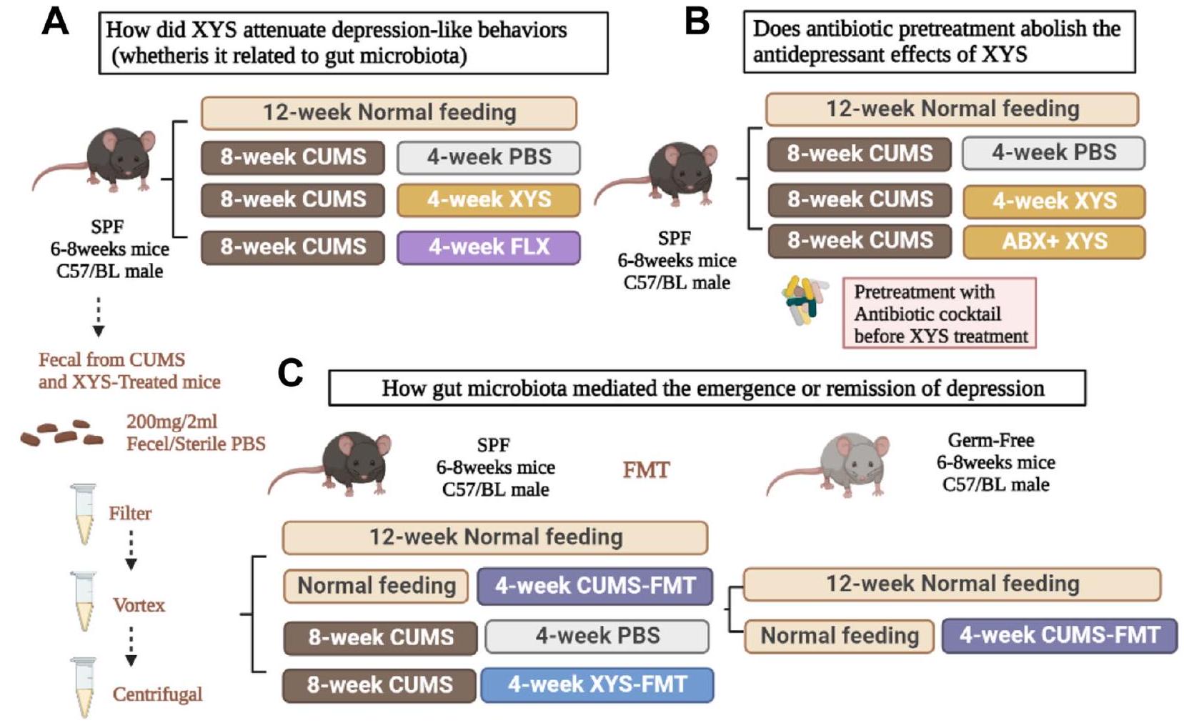
تم تقييم الإدارة في نموذج فئران من الاكتئاب الناتج عن الإجهاد الخفيف غير المتوقع المزمن (CUMS). تم استخدام التدخل بالمضادات الحيوية لاستكشاف تأثير إزالة الميكروبات على فعالية مضادات الاكتئاب. بعد ذلك، تدخلنا في الفلورا الميكروبية المعوية لفئران خالية من مسببات الأمراض المحددة (SPF) العادية والمكتئبة باستخدام CUMS-FMT وXYS-FMT، على التوالي، لاستكشاف التأثير العلاجي والآلية التنظيمية للفلورا الميكروبية المعوية على السلوك الاكتئابي. أخيرًا، قمنا بإجراء زراعة ميكروبية في فئران خالية من الجراثيم (GF) للتحقق بشكل أكبر من التفاعل بين ميكروبات الأمعاء وتقليم المشابك، مما يوضح أكثر أن ميكروبات الأمعاء أساسية في تخفيف الاكتئاب.

طرق

الحيوانات وإجراءات CUMS

ذكور فئران C57BL/6 SPF تتراوح أعمارها بين ستة إلى ثمانية أسابيع (SYXK (Yue) 2017-0174) تزن تم إخضاعهم لعملية تغذية تكيفية لمدة 7 أيام. شملت الظروف التجريبية درجة حرارة الغرفة الرطوبة النسبية لـ ، و دورة الضوء/الظلام. تم تغذية ذكور الفئران C57BL/6 الخالية من الجراثيم (SYXK (Yue) 2020-0233) البالغة من العمر ثمانية أسابيع بشكل صارم في بيئة معقمة (مركز الحيوانات المخبرية الخالية من الجراثيم التابع للأول)
المستشفى التابع لجامعة صن يات سن. تم إيواء جميع الفئران في الدراسة بشكل فردي.
تم تحفيز نموذج الاكتئاب CUMS من خلال التعرض اليومي لمؤثرات متناوبة لمدة مستمرة تبلغ 8 أسابيع، كما تم وصفه سابقًا [25]. شملت المؤثرات 24 ساعة من الحرمان من الطعام والماء، و5 دقائق من الضغط الحراري عن طريق وضع الفئران في فرن عند 5 دقائق من السباحة في الماء المثلج في تحت قيود الجسم، دقيقتان من قرص الذيل، عكس النهار والليل، ومحفزات أخرى. الهدف من استخدام نموذج CUMS في دراستنا هو تكرار الضغط المزمن منخفض الشدة الذي يتعرض له الأفراد في حياتهم اليومية. يهدف هذا النهج النمذجي إلى تقديم محفزات خفيفة مستمرة وغير متوقعة، مع التركيز الأساسي على ضمان بقاء تسلسل المحفزات عشوائيًا وغير متكرر، مما يمنع الحيوانات من توقع حدوث الضغوط. لمنع الفئران من توقع حدوث المحفزات، تم توزيع الضغوط بشكل عشوائي، وتم فصل نفس الضغوط بفترة لا تقل عن 7 أيام (الملف الإضافي 2: الجدول التكميلية 1) [25].

العلاجات وجمع العينات

يتم توضيح تصميم التجربة في الشكل 1. التغيرات في ميكروبيوم الأمعاء، نظام المكملات، و
الشكل 1 تصميم الدراسة للتجربة الكاملة. SPF خالية من مسببات الأمراض المحددة، PBS محلول ملحي معزز بالفوسفات، XYS شاي يياو، CUMS ضغط خفيف غير متوقع مزمن، FLX فلوكستين، ABX مضادات حيوية، FMT زراعة ميكروبات البراز
تم تقييم تقليم المشابك العصبية خلال تطور الاكتئاب، والدور الديناميكي الدوائي لـ XYS كعلاج مضاد للاكتئاب محتمل (الشكل 1a). بعد أسبوع من التكيف، تم تقسيم الفئران عشوائيًا إلى أربع مجموعات: مجموعة التحكم، مجموعة CUMS، مجموعة CUMS+XYS، ومجموعة CUMS+فلوكستين (FLX). باستثناء مجموعة التحكم، تعرضت المجموعات الثلاث الأخرى لمجموعة متنوعة من الضغوطات لمدة ثمانية أسابيع متتالية. كانت العلاجات كما يلي: (1) مجموعة التحكم: لم يتم إعطاء أي تحفيز للضغط وكانت بمثابة التحكم السلبي؛ (2) مجموعة CUMS: تعرضت لـ CUMS لمدة 8 أسابيع، تلتها إدارة يومية عن طريق الفم لمحلول ملحي مخفف بالفوسفات (PBS) لمدة 4 أسابيع؛ (3) مجموعة CUMS+XYS: تعرضت لـ CUMS لمدة 8 أسابيع، تلتها إدارة يومية عن طريق الفم لـ XYS ( كجم/يوم؛ رقم الدفعة: 20190724؛ مجموعة جيوزهيتانغ المحدودة) لمدة 4 أسابيع؛ و (4) مجموعة CUMS+FLX: CUMS لمدة 8 أسابيع، تليها الإدارة الفموية اليومية لـ FLX ( اليوم؛ F844356؛ ماكلين) لمدة 4 أسابيع. تم قياس وزن الجسم أسبوعياً طوال مدة الدراسة. تم ذبح الفئران على الفور بعد آخر اختبار سلوكي، وتم جمع عينات البراز من جميع الفئران بشكل معقم وتخزينها في- لتحليل مستقبلي. تم الحصول على المصل عن طريق طرد عينات الدم في جهاز الطرد المركزي بسرعة 3000 دورة في الدقيقة لمدة 15 دقيقة عند 4 درجات مئوية وتم تخزينه في تم شطف القولون بمحلول PBS وثبته في “تم استخدام الفورمالين للتحليل النسيجي اللاحق؛ وتم تثبيت القولون المتبقي في سائل المجهر الإلكتروني للتجارب اللاحقة. تم جمع أنسجة الحُُُُُُُُُُُُُُُُُُُُُُُُُُُُُُُُُُُُُُُُُُُُُُُُُُُُُُُُُُُُُُُُُُُُُُُُُُُُُُُُُُُُُُُُُُُُُُُُُُُُُُُُُُُُُُُُُُُُُُُُُُُُُُُُُُُُُُُُُُُُُُُُُُُُُُُُُُُُُُُُُُُُُُُُُُُُُُُُُُُُُُُُُُُُُُُُُُُُُُُُُُُُُُُُُُُُُُُُُُُُُُُُُُُُُُُُُُُُُُُُُُُُُُُُُُُُُُُُُُُُُُُُُُُُُُُُُُُُُُُُُُُُُُُُُُُُُُُُُُُُُُُُُُُُُُُُُُُُُُُُُُُُُُُُُُُُُُُُُُُُُُُُُُُُُُُُُُُُُُُُُُُُُُُُُُُُُُُُُُُُُُُُُُُُُُُُُُُُُُُُُُُُُُُُُُُُُُُُُُُُُُُُُُُُُُُُُُُُُُُُُُُُُُُُُُُُُُُُُُ
تم تقييم قدرة المضادات الحيوية على التداخل مع تأثيرات مضادات الاكتئاب لـ XYS (الشكل 1B). تم تقييم تأثير اضطراب الميكروبات الناتج عن المضادات الحيوية على فعالية XYS من خلال اضطراب ميكروبات الأمعاء مسبقًا باستخدام خليط من المضادات الحيوية يتكون من الأمبيسيلين ( مل؛ A9518؛ سيغما ألدريتش)، فانكومايسين ( ; V820413; ماكلين)، سيبروفلوكساسين ( ; C9371; سولاربيو)، إيميبينيم ( ; S27995)، وميترونيدازول ( ; B1976; قرد) في مياه الشرب اليومية للفئران (أسبوع واحد؛ لمنع الإجهاد الناتج عن التغذية الفموية في الفئران) قبل إدارة XYS لمدة 4 أسابيع.
تم تقييم التأثيرات العلاجية للميكروبات المعوية البرازية في الاكتئاب باستخدام ذكور فئران C57BL/6 من نوع SPF وGF تتراوح أعمارها بين 6 إلى 8 أسابيع (الشكل 1C). بعد أسبوع من التكيف، تم تقسيم فئران SPF عشوائيًا إلى أربع مجموعات على النحو التالي: (1) مجموعة التحكم: لم تتلق أي تحفيز للضغط، وكانت بمثابة التحكم السلبي؛ (2) مجموعة التحكم + FMT (CUMS): لم تتلق أي تحفيز للضغط لمدة 8 أسابيع، تلتها FMT يوميًا (براز من مجموعة CUMS في التجربة 1) لمدة 4 أسابيع؛ (3) مجموعة CUMS: تعرضت لـ CUMS لمدة 8 أسابيع، تلتها إدارة يومية عن طريق الفم لـ
PBS لمدة 4 أسابيع؛ و(4) مجموعة CUMS+FMT(XYS): CUMS لمدة 8 أسابيع، تليها FMT يوميًا (براز من مجموعة CUMS + XYS في التجربة 1) لمدة 4 أسابيع. بعد أسبوع من التكيف، تم تقسيم الفئران الخالية من الجراثيم عشوائيًا إلى مجموعتين كما يلي: (1) مجموعة GF: لم تتلق أي تحفيز للضغط، وكانت بمثابة مجموعة التحكم السلبية؛ و(2) مجموعة GF+FMT(CUMS): لم تتلق أي تحفيز للضغط لمدة 8 أسابيع، تليها FMT يوميًا (براز من مجموعة CUMS في التجربة 1) لمدة 4 أسابيع.
تم قياس وزن الجسم أسبوعياً طوال فترة الدراسة. تم ذبح الفئران على الفور بعد الاختبار السلوكي النهائي، وتم جمع عينات البراز من جميع الفئران وتخزينها في لتحليل مستقبلي. تم الحصول على المصل عن طريق الطرد المركزي لعينات الدم بسرعة 3000 دورة في الدقيقة لمدة 15 دقيقة عند 4 درجات مئوية وتم تخزينه عند -80 درجة مئوية. تم شطف القولون بمحلول PBS وثبته في “تم استخدام الفورمالين للتحليل النسيجي اللاحق؛ وتم تثبيت القولون المتبقي في سائل المجهر الإلكتروني للتجارب اللاحقة. تم جمع أنسجة الحُُُُُُُُُُُُُُُُُُُُُُُُُُُُُُُُُُُُُُُُُُُُُُُُُُُُُُُُُُُُُُُُُُُُُُُُُُُُُُُُُُُُُُُُُُُُُُُُُُُُُُُُُُُُُُُُُُُُُُُُُُُُُُُُُُُُُُُُُُُُُُُُُُُُُُُُُُُُُُُُُُُُُُُُُُُُُُُُُُُُُُُُُُُُُُُُُُُُُُُُُُُُُُُُُُُُُُُُُُُُُُُُُُُُُُُُُُُُُُُُُُُُُُُُُُُُُُُُُُُُُُُُُُُُُُُُُُُُُُُُُُُُُُُُُُُُُُُُُُُُُُُُُُُُُُُُُُُُُُُُُُُُُُُُُُُُُُُُُُُُُُُُُُُُُُُُُُُُُُُُُُُُُُُُُُُُُُُُُُُُُُُُُُُُُُُُُُُُُُُُُُُُُُُُُُُُُُُُُُُُُُُُُُُُُُُُُُُُُُُُُُُُُُُُُُُُُُُُُُ

اختبارات سلوكية

اختبار تفضيل السكروز (SPT)

تم قياس قابلية الفئران للمكافآت باستخدام اختبار تفضيل السكروز (SPT) لتقييم درجة الاكتئاب. يتضمن اختبار SPT فترتين، التدريب والاختبار [26]. في هذه الدراسة، شمل اختبار SPT فترة تدريب مدتها 72 ساعة. في البداية، تم تزويد الفئران بزجاجتين من محلول السكروز لمدة 24 ساعة. بعد ذلك، تم تقديم زجاجة واحدة من محلول السكروز وزجاجة واحدة من الماء النقي لمدة 24 ساعة القادمة. أخيرًا، تم تبديل مواقع زجاجات السكروز والماء لمدة 24 ساعة إضافية. بعد ذلك تجربة التدريب و24 ساعة من الحرمان من الطعام والماء، تم منح الفئران الوصول إلى زجاجة من الماء النقي وزجاجة من تم تحضير محلول السكروز في نفس الوقت. بعد ساعة، تم تسجيل أحجام الماء النقي المتبقي ومحاليل السكروز. تم إجراء اختبار SPT قبل تعريض الفئران لـ CUMS (كقياس أساسي) وبعد الانتهاء من تجربة CUMS.

اختبار الحقل المفتوح (OFT)

تُستخدم طريقة اختبار السلوك في الحقل المفتوح لمراقبة السلوك الذاتي، والسلوك الاستكشافي، والتوتر في الحيوانات التجريبية في بيئة غير مألوفة. تم إجراء اختبار الحقل المفتوح في الأسابيع 0 و8 و12 في دراستنا. تم وضع الفئران في غرفة تشغيل سلوكية لمدة 10 دقائق للتكيف، ثم تم نقلها إلى المنطقة المركزية. تم بدء تسجيل الكاميرا وتوقيته، وتمت مراقبة سلوك الفئران لمدة 5 دقائق. مباشرة بعد كل تجربة، تم تنظيف الصناديق بـ الكحول. وفقًا لدراسة سابقة، تم إجراء اختبار التفضيل المفتوح تحت ظروف إضاءة خافتة،
تم إجراء اختبار السلوك في ظروف إضاءة محددة تبلغ 60 لوكس [27]. تم تنفيذ اختبار السلوك باستخدام برنامج تحليل السلوك المعترف به دوليًا (نظام تحليل برنامج إيثو فيجن، تكنولوجيا نولدوس، ليسبرغ، فيرجينيا، الولايات المتحدة الأمريكية) وشمل تحليل المسافة الإجمالية للحركة لكل مجموعة من الفئران.

اختبار تعليق الذيل (TST)

تم تعليق الفئران على قضيب أفقي بارتفاع 50 سم عن الأرض، مع تثبيت ذيولها باستخدام شريط لاصق. استمرت فترة التجربة لمدة 6 دقائق، تم خلالها تسجيل نشاط كل فأر في الدقائق الأربع الأخيرة. تم تعريف وقت عدم الحركة (بالثواني) على أنه الوقت الذي تحتاجه الفئران للاستسلام وعدم الحركة تمامًا. مباشرة بعد كل تجربة، تم تنظيف الصناديق بـ الكحول. تم تحليل بيانات التجربة TST باستخدام برنامج تحليل السلوك (نظام تحليل برنامج EthoVision، تكنولوجيا المعلومات نولدوس، ليسبرغ، فيرجينيا، الولايات المتحدة الأمريكية).

اختبار المتاهة المرفوعة (EPM)

تم إجراء تجربة EPM باستخدام جهاز متاهة على شكل زائد يتكون من ذراعين مفتوحين وذراعين مغلقين ومنصة مركزية. كانت أبعاد الذراعين المفتوحين والمغلقين كلاهما ؛ كانت الأذرع المغلقة مغطاة بـ حواجز على كلا الجانبين. وفقًا للدراسات السابقة، تم إجراء تجارب EPM تحت ظروف إضاءة خافتة، بدقة عند شدة 60 لوكس [27]. تم وضع الفئران بشكل فردي في المنطقة المركزية ( )، على ارتفاع 60 سم فوق الأرض، وتم تسجيل حركاتهم لمدة 5 دقائق. بعد كل تجربة، تم تنظيف الأقفاص على الفور باستخدام محلول كحول 75%. تم تحليل نتائج اختبار EPM باستخدام برنامج تحليل السلوك (نظام تحليل برنامج EthoVision، تكنولوجيا المعلومات نولدوس، ليسبرغ، فيرجينيا، الولايات المتحدة الأمريكية).

اختبار التعرف على الأجسام الجديدة (NORT)

يتم استخدام اختبار التعرف على الكائنات الجديدة لتحديد قدرات التعلم والذاكرة في القوارض. تم تقسيم التجربة إلى ثلاث مراحل: التدريب، تدريب التعرف، والاختبار. في المرحلة الأولى، تم إخراج الفأر من القفص، ووضعه في منتصف صندوق الحقل المفتوح مع ظهره مواجهًا للمشغل، وسمح له بالاستكشاف بحرية لمدة 10 دقائق. في المرحلة الثانية، تم وضع كائنين متطابقين (كائنات قديمة) في حدود الكائن النسبي لصندوق الحقل المفتوح، وتم إخراج الفأر من القفص بعد 24 ساعة من المرحلة الأولى، ووضعه على نفس الكائن مثل الكائنين، وسمح له بالاستكشاف بحرية لمدة 10 دقائق في وسط صندوق الحقل المفتوح من مسافة. في المرحلة الثالثة، تم وضع كائن (كائن قديم) وكائن جديد في صندوق الحقل المفتوح عند حد الكائن لتدريب التعرف. بعد تدريب التعرف لمدة 24 ساعة، تم
تم وضعها بين الجسم الجديد والقديم وسمح لها بالاستكشاف بحرية لمدة 10 دقائق. بعد كل جلسة تجريبية، تم تنظيف الأقفاص على الفور باستخدام محلول كحول بنسبة 75%. تم تحليل بيانات NORT باستخدام برنامج تحليل السلوك (نظام تحليل برنامج EthoVision، Noldus Information Technology، Leesburg، VA، الولايات المتحدة الأمريكية). تم تسجيل الوقت الذي قضته الفأرة في استكشاف كل جسم (الوقت الذي قضته في استكشاف الجسم المألوف، N1؛ الوقت الذي قضته في استكشاف الجسم الجديد، N2). تم حساب نسبة التمييز (DR، %) على النحو التالي: .

مهمة متاهة Y

تُستخدم مهمة المتاهة على شكل Y لتحديد القدرة على التعرف المكاني والذاكرة، وقد تم استخدامها لاختبار الذاكرة قصيرة المدى في هذه التجربة. المتاهة على شكل Y هي متاهة أفقية ذات ثلاثة أذرع (طولها 30 سم، وعرضها 8 سم، وارتفاعها 15 سم) حيث يتم فصل الأذرع الثلاثة بشكل متماثل عند تم وضع الفئران في غرفة عمليات سلوكية لمدة 10 دقائق للتكيف، ثم تم وضعها في نهاية أحد الأذرع، مع حجب أحد الأذرع والسماح بالتنقل الحر بين الذراعين الآخرين. بدأت التجربة بعد ساعتين، وتم فتح جميع الأذرع الثلاثة (حيث تم تعريف الذراع المحجوبة سابقًا على أنها ذراع الجدة). تم بدء تسجيل الكاميرا وتوقيته، وتمت مراقبة سلوك الفئران لمدة 5 دقائق. تم تنظيف الصناديق بـ الكحول مباشرة بعد كل تجربة. تم تنفيذ مهمة المتاهة على شكل حرف Y باستخدام برنامج تحليل السلوك (نولدس تكنولوجيا المعلومات).

الاختبارات الحيوية
صبغة الهيماتوكسيلين والإيوزين (HE)

تم تثبيت عينات من نسيج القولون في تم معالجة الأنسجة بالفورمالين، وإزالة الكالسيوم، وتجفيفها، وجعلها شفافة، ثم غمرها وتضمينها في البارافين. تم إعداد عينات الأنسجة بسمك خمسة ميكرومترات باستخدام الميكروتوم. تم إزالة الشمع من الشرائح باستخدام الزيلين، وتم تمريرها عبر سلسلة من الإيثانول المائي، وتم صبغها بصبغة HE، وتمت ملاحظتها تحت المجهر [28].

المورفولوجيا فوق الدقيقة للقولون

تم جمع قطعة من القولون القريب وثبتت على الفور في محلول الجلوتارالدهيد في تم نقع الأجزاء طوال الليل. تم شطف الأجزاء ثلاث مرات في محلول فوسفات مخفف 1 م لمدة 15 دقيقة في كل مرة، وتم تجفيفها باستخدام إيثانول متدرج، وتم نقعها في أسيتات الإيزوأميل لمدة 20 دقيقة مرتين، وتم تجفيفها ومعالجتها بشكل روتيني. تم الحصول على صور البنية الدقيقة للقولون باستخدام مجهر إلكتروني (SU8100؛ هيتاشي).

اختبار الامتصاص المناعي المرتبط بالإنزيم (ELISA)

مصل الإنترلوكين (IL)-6، IL-10، IL-1 عامل نخر الورم- (TNF- )، الليبوبوليسكاريد (LPS)، ومستويات C3
تم تحديدها باستخدام مجموعة كشف ELISA (كوسابيو، ووهان، الصين). IL-6، IL-10، IL-1 TNF- تم تحديد مستويات LPS و C3 من خلال قياس الامتصاص باستخدام قارئ الميكرو بلايت ورسم منحنى قياسي.

تلطيخ المناعة الفلورية (IF)

تم تحضين أقسام من القشرة الجبهية (PFC) مع الأجسام المضادة الأولية طوال الليل ثم مع الأجسام المضادة الثانوية في الظلام لمدة ساعة واحدة. كانت الأجسام المضادة المستخدمة في صبغة IF كما يلي: مضاد IBA-1 (1:200، GB12105، Servicebio)، مضاد CD68 (1:100، GB113109، Servicebio)، مضاد CR3 (1:500، GB11058، Servicebio)، جسم مضاد ثانوي من الماعز CY3 مضاد للفأر (1:300، GB21303، Servicebio)، وجسم مضاد ثانوي من الماعز 448 AffiniPure Fab Fragment مضاد للأرنب (1:500، GB25303، Servicebio). خضعت شرائح الدماغ في دراستنا لخطوة حجب باستخدام مصل الماعز العادي [29]. تم تلوين العينات بعامل مضاد مع 4،6-دياميدينو-2-فينيل إندول (DAPI) لمدة 10 دقائق ثم تم تصويرها باستخدام ميكروسكوب فلوري (نيكون إكليبس C1، اليابان).

التحليل الغربي (WB)

“تم إجراء التحليل الغربي لتحديد تعبير بروتينات C3 (1:1000، EPR19394، Abcam)، والسيناپسين (SYN) (1:1000، EPR23531-50، Abcam)، وبروتين كثافة ما بعد المشبك 95 (PSD95) (1:1000، EPR23124-118، Abcam) في الحُُُُُُُُُُُُُُُُُُُُُُُُُُُُُُُُُُُُُُُُُُُُُُُُُُُُُُُُُُُُُُُُُُُُُُُُُُُُُُُُُُُُُُُُُُُُُُُُُُُُُُُُُُُُُُُُُُُُُُُُُُُُُُُُُُُُُُُُُُُُُُُُُُُُُُُُُُُُُُُُُُُُُُُُُُُُُُُُُُُُُُُُُُُُُُُُُُُُُُُُُُُُُُُُُُُُُُُُُُُُُُُُُُُُُُُُُُُُُُُُُُُُُُُُُُُُُُُُُُُُُُُُُُُُُُُُُُُُُُُُُُُُُُُُُُُُُُُُُُُُُُُُُُُُُُُُُُُُُُُُُُُُُُُُُُُُُُُُُُُُُُُُُُُُُُُُُُُُُُُُُُُُُُُُُُُُُُُُُُُُُُُُُُُُُُُُُُُُُُُُُُُُُُُُُُُُ . بعد الرحلان الكهربائي والاختراق الكهربائي، تم حجب الغشاء المحول بـ مسحوق الحليب الخالي من الدسم لمدة ساعة، وغسل بمحلول TBST، وت incubated مع الأجسام المضادة الأولية طوال الليل عند تم تحضين الغشاء الكهربائي مع جسم مضاد ثانوي لمدة ساعة واحدة، ثم تم غسله بمحلول TBST، وتم تحفيز اللمعان باستخدام مادة كيميائية مضيئة (Millipore، بيليريكا، ماساتشوستس، الولايات المتحدة الأمريكية). تم مسح غشاء فصل البروتين وتحليله باستخدام محلل صور (Bio-Rad، هيركوليس، كاليفورنيا، الولايات المتحدة الأمريكية).

استخراج الحمض النووي البكتيري، تسلسل 16S rRNA، والتحليلات

تم استخراج الحمض النووي الميكروبي من محتويات القولون باستخدام E.Z.N.A. مجموعة الحمض النووي (أوميغا بايو-تيك، نوركروس، جورجيا، الولايات المتحدة)، وفقًا لتعليمات الشركة المصنعة. تم تحديد التركيز النهائي للحمض النووي ونقاوته باستخدام مقياس الطيف الضوئي NanoDrop 2000 UV-vis (ثيرمو ساينتيفيك، ويلمنجتون، الولايات المتحدة)، وتم تقييم جودة الحمض النووي بواسطة الرحلان الكهربائي لهلام الأجاروز.
تم تضخيم المناطق المتغيرة عالية التغير V3-V4 من جين الرنا الريباسي 16S البكتيري باستخدام بادئات 341F ( -CCT AYGGGRBGCASCAG-3′) و 806R ( -GGACTA CNNGGGTATCTAAT-3′) باستخدام نظام PCR من نوع ثيرموسايكلر (GeneAmp 9700، ABI، الولايات المتحدة الأمريكية). تم استخراج منتجات PCR من هلام الأجاروز باستخدام مجموعة استخراج الحمض النووي من هلام AxyPrep (Axygen Biosciences، يونيون سيتي، كاليفورنيا، الولايات المتحدة الأمريكية) وتم قياسه باستخدام QuantiFluor -ST (بروماجا، الولايات المتحدة الأمريكية)، وفقًا لتعليمات الشركة المصنعة. تم حساب مقاييس التنوع باستخدام ملحق التنوع الأساسي داخل QIIME2. مستوى الميزة -تم حساب مؤشرات التنوع، مثل مقدر غنى تشاو 1 المرصود ومؤشر تنوع شانون، لتقدير التنوع الميكروبي داخل العينات الفردية. -تم إجراء قياسات المسافة التنوع، بما في ذلك Bray-Curtis و UniFrac غير الموزون و UniFrac الموزون، لتحديد التباين الهيكلي في المجتمعات الميكروبية عبر العينات، ثم تم تصورها بواسطة تحليل المكونات الرئيسية (PCoA). تم تقدير الوفرة النسبية للأنواع الميكروبية على مستويات تصنيف مختلفة باستخدام حزمة R “vegan”. تم تقديم خدمات التسلسل وتحليل البيانات من قبل مجموعة Wekemo Tech Co.، Ltd.، شنتشن، الصين.

تحليلات الميتابولوم

تم إجراء تحليلات الميتابولوميات على مستخلصات الميتابوليتات الميكروبية باستخدام نظام ACQUITY UPLC I-Class PLUS/Xevo G2-XS QToF (شركة ووترز، ميلفورد، ماساتشوستس، الولايات المتحدة الأمريكية) مع عمود Waters Acquity UPLC HSS T3. ). عينات ( 1 تم حقنها وإخراجها باستخدام طور متنقل يتكون من مذيب ( حمض الفورميك) ( حمض الفورميك – الأسيتونيتريل) بمعدل تدفق قدره و فرن العمود. كان برنامج التدرج الإلوتي كما يلي: ; ب؛ ; و . تم توصيل المصب بجهاز مطياف الكتلة ESI-triple quadrupole linear ion trap (QQQ-LIT) المزود بواجهة ESI Turbo IonSpray التي تعمل في كلا الوضعين الموجب والسالب، وتم تشغيله باستخدام برنامج Analyst. كانت معلمات تشغيل مصدر ESI كما يلي: درجة حرارة مصدر الأيون، درجة حرارة غاز إزالة المذيبات، ; جهد الشعيرات الدموية، 2000 فولت؛ جهد المخروط، 30 فولت؛ معدل تدفق غاز المخروط، ؛ ومعدل تدفق غاز إزالة المذيبات، تم الحصول على مسح كامل في نطاق المسح من بتردد مسح قدره 0.2 ثانية، وتم جمع البيانات بواسطة MassLynx (الإصدار 4.2، Waters) وتم التعرف عليها بواسطة Progenesis QI ضمن انحراف كتلي قدره 100 جزء في المليون. لم تختلف تركيزات المستقلبات الكلية بشكل كبير بين العينات. تم إجراء تحليل المكونات الرئيسية (PCA) للمستقلبات باستخدام برنامج الإحصاءات الحفرية.
تم إجراء تحليل التمييز باستخدام المربعات (PLS-DA) باستخدام SIMCA برنامج لتحسين التصور. تم رسم خريطة البراكين للمواد الأيضية الكلية ورسم المواد الأيضية المختلفة من خلال تحليل إثراء موسوعة كيوتو للجينات والجنوم (KEGG) باستخدام حزمة R “ggplot2”. تم تقييم نموذج PLS-DA باستخدام اختبار هوتينغ T2. تم تقييم درجة LAD باستخدام اختبارات كروسكال-واليس وويليكوكسون.

تحليل ارتباط البيانات

تم إجراء تحليل الشبكة الميكروبية باستخدام تحليل ارتباط رتب سبيرمان وفقًا للشروط المحددة (الارتباط تم إجراء تحليل إثراء المستقلبات التفاضلية باستخدام اختبار هايبرجومتري. تم إجراء تحليلات الارتباط بين الميكروبيوم والمستقلبات باستخدام ارتباط بيرسون؛ القيم المطلقة لـ و تم تضمينها. تم تحديد الدلالة الإحصائية عند ميكروبات الأمعاء، السلوكيات، ومستويات IL-6، IL-10، IL-1 ، وTNF- تم تقييم الفئران المكتئبة الناتجة عن CUMS من خلال تحليل الارتباط لبيرسون. تم إجراء جميع التحليلات الإحصائية باستخدام IBM SPSS (الإصدار 22) و PAST (الإصدار 4.03) و SIMCA. (الإصدار 13.0). تم إنشاء جميع الرسوم البيانية باستخدام GraphPad Prism 7 (لا جولا، كاليفورنيا، الولايات المتحدة الأمريكية)، PAST (الإصدار 4.03)، SIMCA (الإصدار 13.0)، Tbtools (الإصدار 1.049)، Cytoscape (الإصدار 3.7.2)، و (الإصدار 4.0.2).

التحليل الإحصائي

تُعرض البيانات كمتوسط حسابي الخطأ المعياري للمتوسط (SEM) باستخدام برنامج SPSS (الإصدار 25.0؛ شيكاغو، إلينوي، الولايات المتحدة الأمريكية). تم تحليل البيانات الناتجة عن القياسات المتكررة أولاً باستخدام تحليل التباين المتكرر (ANOVA) إذا كانت البيانات موزعة بشكل طبيعي ومتجانسة. بالإضافة إلى ذلك، تم إجراء اختبار ما بعد hoc. إذا لم تكن البيانات موزعة بشكل طبيعي أو كانت التباين غير متجانسة، تم استخدام اختبار غير معلمي لعينات K المستقلة للتحليل الإحصائي بند بند. تم تحديد الدلالة الإحصائية عند تم إنشاء الرسوم البيانية باستخدام برنامج GraphPad Prism 7.

النتائج

XYS، كعلاج محتمل للاكتئاب، خفف من سلوكيات الاكتئاب/القلق الناتجة عن الإجهاد المزمن المعتمد على الميكروبات المعوية، وكذلك من ضعف الإدراك.
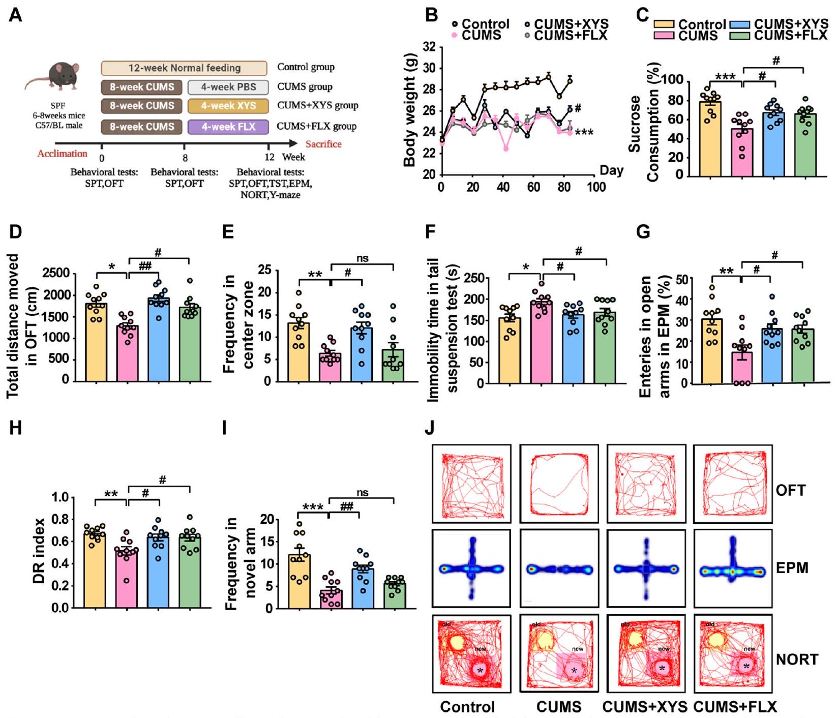
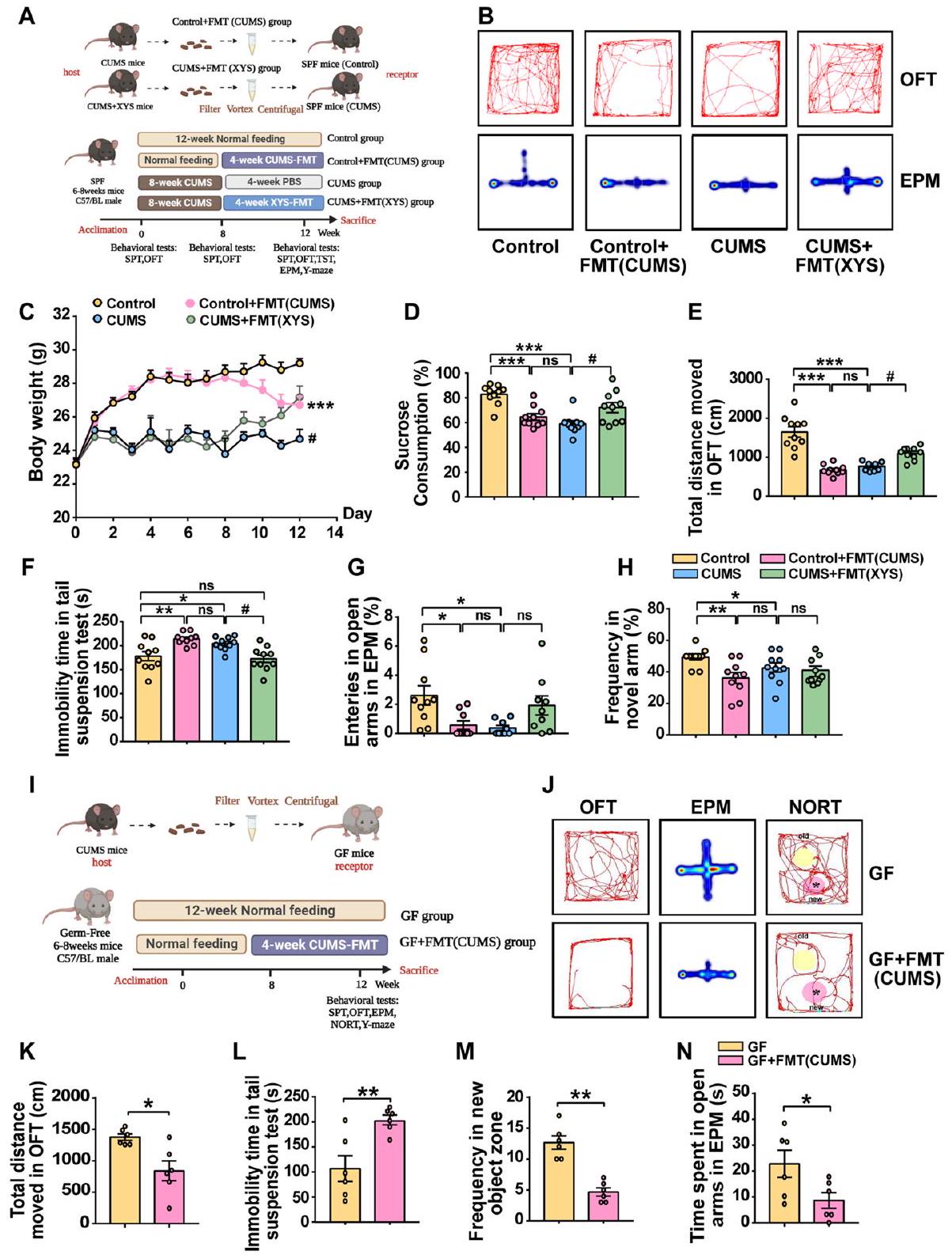
تم تقييم النشاط المحتمل لمضادات الاكتئاب لـ XYS على سلوكيات الاكتئاب الناتجة عن CUMS من خلال تحفيز سلوكيات شبيهة بالاكتئاب في الفئران عن طريق إعطاء CUMS لمدة 8 أسابيع، تليها 4 أسابيع من الإدارة اليومية عن طريق الفم لـ XYS (الشكل 2A). لم تُلاحظ أي اختلافات في وزن الجسم، أو تفضيل السكروز، أو الوظيفة الحركية بين المجموعات قبل التجربة (الشكل S1a-c). سلوكيات شبيهة بالاكتئاب ملحوظة.
تمت ملاحظتها في الفئران التي تلقت تدخل CUMS لمدة 8 أسابيع (الشكل S1d-g). بعد النجاح في إنشاء نموذج الفأر CUMS، تم فحص تأثير تدخل XYS على سلوك الفئران. خفف تدخل XYS بشكل كبير من سلوكيات الاكتئاب الناتجة عن CUMS مقارنة بمجموعة CUMS، كما يتضح من انخفاض ملحوظ في فقدان الوزن، وزيادة التفضيل في اختبار SPT، وزيادة المسافة الإجمالية وتكرار الدخول في اختبار OFT، وانخفاض وقت السكون في اختبار TST (الشكل 2B-F). كما حسّن علاج XYS بشكل كبير من سلوكيات القلق والضعف الإدراكي في الفئران التي تعاني من سلوكيات الاكتئاب الناتجة عن CUMS. أظهرت الفئران التي تلقت تدخل XYS زيادة في تكرار الدخول إلى الذراع المفتوحة في اختبار EPM (الشكل 2G) وزيادة في استكشاف المناطق الجديدة في اختبارات NORT (الشكل 2H) واختبار المتاهة Y (الشكل 2I). تظهر مسارات حركة الفئران في اختبارات OFT وEPM وNORT في الشكل 2J. تشير النتائج أعلاه إلى أن XYS له تأثيرات مضادة للاكتئاب كبيرة ويمكن دراستها بشكل أكبر كعلاج محتمل للاكتئاب.
لاستكشاف ما إذا كان تدمير ميكروبات الأمعاء يؤثر على فعالية XYS في تخفيف الاكتئاب، تم استخدام مزيج من المضادات الحيوية لتحضير الفئران قبل إعطاء XYS عن طريق الفم (الشكل S1h). يمكن أن يؤثر استخدام المضادات الحيوية لتعطيل الفلورا المعوية على تحسين سلوكيات الاكتئاب الشبيهة في الفئران بواسطة XYS، مما يشير إلى أن فعالية XYS تعتمد إلى حد ما على الفلورا المعوية (الشكل S1i-j).
تشير هذه النتائج مجتمعة إلى أن CUMS يمكن أن تحفز سلوكيات شبيهة بالاكتئاب والقلق بشكل كبير، بالإضافة إلى ضعف إدراكي مصاحب في الفئران. علاوة على ذلك، تم تحسين أعراض الاكتئاب بواسطة XYS، لكن المعالجة المسبقة بالمضادات الحيوية خففت من تأثير مضادات الاكتئاب، مما يشير إلى أن XYS خفف من سلوكيات الاكتئاب/القلق وضعف الإدراك الناتج عن CUMS بطريقة تعتمد على ميكروبات الأمعاء.

السلوكيات الشبيهة بالاكتئاب مصحوبة بخلل في ميكروبيوم الأمعاء

يتكون التوازن المعوي من الميكروبيوم المعوي والحاجز المخاطي (بما في ذلك المناعة المخاطية المعوية). لمراقبة التغيرات في التوازن المعوي خلال الاكتئاب، تم تحديد التهاب القولون ووظيفة الحاجز، وتنوع الميكروبات المعوية، والمواد الأيضية الميكروبية.
لفهم التغيرات في علم الأمراض القولونية، وظيفة الحاجز، والالتهاب الجهازي خلال تطور الاكتئاب، تم تقييم التحليل النسيجي، الشكل المجهري للقولون، ومستوى السيتوكينات الالتهابية في المصل. أظهرت الفئران التي تعاني من سلوكيات مشابهة للاكتئاب التهابًا قولونيًا واضحًا وزيادة ملحوظة في التغيرات المرضية.
الشكل 2 XYS، كعلاج محتمل للاكتئاب، خفف من سلوكيات الاكتئاب/القلق الناتجة عن CUMS، وكذلك من ضعف الإدراك. A بعد أسبوع من التكيف، تم تقسيم الفئران عشوائيًا إلى أربع مجموعات كما يلي: مجموعة التحكم، CUMS، CUMS + XYS، وCUMS + FLX. B التغيرات اليومية في وزن الجسم طوال مدة الدراسة. C اختبار تفضيل السكروز (SPT). D اختبار الحقل المفتوح (OFT) (إجمالي المسافة المقطوعة). E OFT (التكرار في المنطقة المركزية). F اختبار تعليق الذيل (TST). G متاهة الارتفاع (EPM). H اختبار التعرف على الكائن الجديد (NORT). I متاهة Y. J مسارات حركة الفئران في OFT، EPM وNORT. تمثل البيانات المتوسط. SEM لكل مجموعة). مقابل مجموعة التحكم؛ ، مقابل مجموعة CUMS
تمت مقارنة الدرجات مع الفئران الضابطة (الشكل 3A، B). كشفت المجهرية الإلكترونية الناقلة عن انخفاض في حجم وعدد الزغابات الدقيقة ومظهر غير طبيعي لحاجز الأمعاء في الفئران التي تظهر سلوكيات مشابهة للاكتئاب (الشكل 3C). يمكن أن تؤدي تفاقم التهاب القولون واضطراب وظيفة الحاجز إلى التأثير على مستويات العوامل الالتهابية الطرفية. تم تثبيط تعبير IL-10 في نموذج CUMS، وزادت مستويات IL-1. IL-6 و TNF- تمت ملاحظتها في مصل الفئران المصابة باضطرابات شبيهة بالاكتئاب (الشكل 3D-G). يمكن أن يؤدي إعطاء مضادات الاكتئاب إلى تحسين وظيفة حاجز القولون وعلم الأمراض الالتهابي. بعد XYS
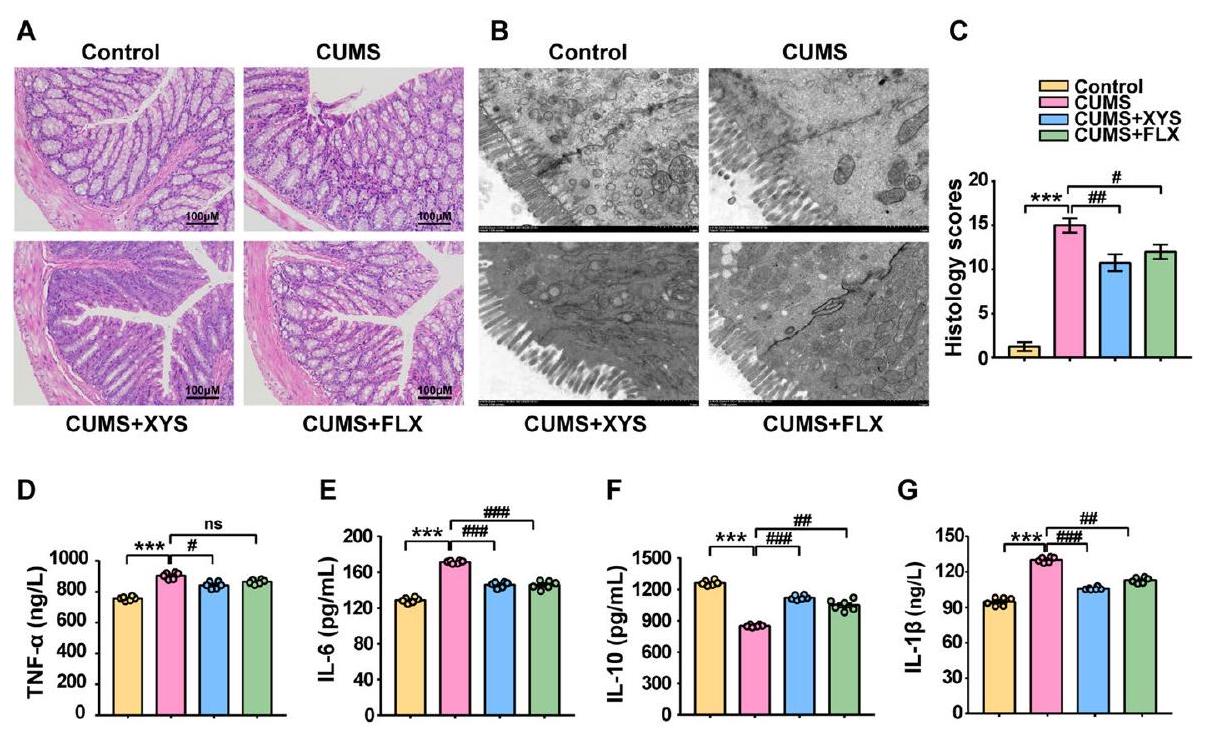
تم تحسين التهاب الأمعاء، وزاد حجم وعدد الزغابات المعوية القولونية، وتم عكس المحاذاة غير الطبيعية (الشكل 3A-C). بالإضافة إلى ذلك، قلل XYS من مستويات IL- IL-6 و TNF- وزاد من مستوى IL-10 في مصل الفئران التي تظهر سلوكيات مشابهة للاكتئاب (الشكل 3D-G).
تم تقييم التغيرات في ميكروبيوتا الأمعاء والمواد الأيضية المعوية والاستجابة لمضادات الاكتئاب في نموذج الاكتئاب من خلال تسلسل جين 16S rRNA وتحليل UHPLC-QTOF-MS/MS. أظهرت نتائج التنوع التي أشار إليها مؤشر شانون أن تنوع الميكروبات المعوية قد انخفض.
الشكل 3 XYS قمع الاستجابة الالتهابية القولونية الناتجة عن CUMS، وحسن وظيفة الحاجز. مقاطع القولون الملونة بصبغة H&E ( ” ). ب المجهر الإلكتروني الناقل ( درجات الأنسجة في القولون. مستوى TNF-α في المصل. تمثل البيانات المتوسط SEM لكل مجموعة). مقابل مجموعة التحكم؛ مقابل مجموعة CUMS
في الفئران التي تظهر سلوكيات مشابهة للاكتئاب ويمكن استعادتها بواسطة مضادات الاكتئاب الفموية (XYS أو FLX) (الشكل 4A؛ الشكل S2a-c: -التنوع الذي أظهره مؤشرات تشاو والملاحظة وسيمبسون). كشفت تحليل المكونات الرئيسية (PCoA) بناءً على مسافة براى-كورتيس عن اختلاف في هيكل الميكروبيوم المعوي بين مجموعة التحكم والفئران ذات السلوكيات الشبيهة بالاكتئاب (الشكل 4B). لوحظ اختلاف كبير في هيكل الفلورا المعوية بين مجموعتي CUMS + XYS وCUMS ( )، مما يشير إلى أن هيكل الميكروبات المعوية في الفئران التي تظهر سلوكيات مشابهة للاكتئاب تأثر بشكل كبير بالإعطاء الفموي لـ XYS.
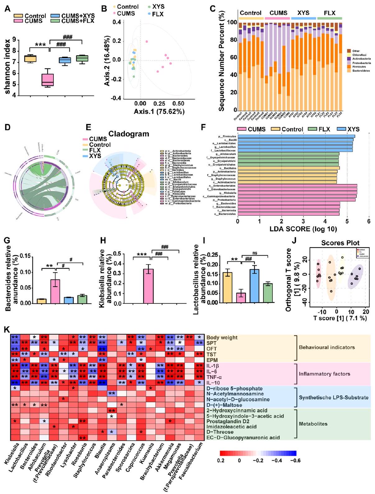
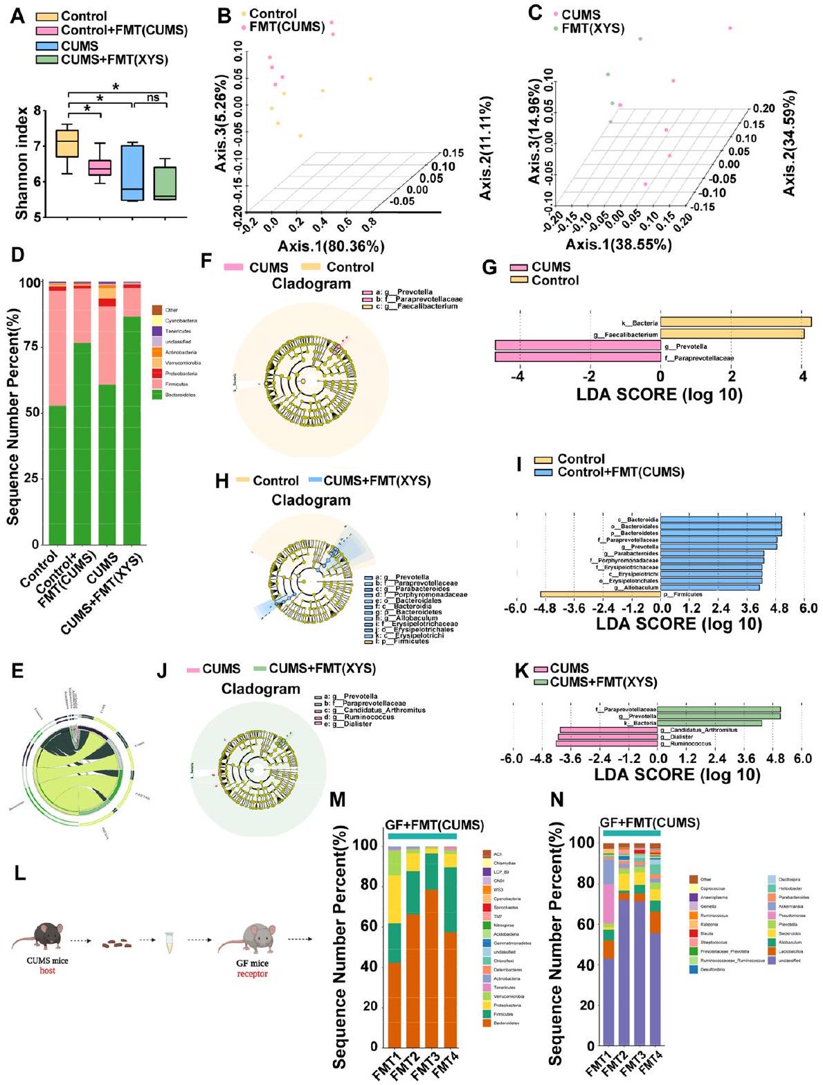
تم تحديد ما مجموعه 37 شعبة في هذه الدراسة. كانت Firmicutes وBacteroidetes وProteobacteria هي الشعب السائدة في الميكروبيوم البرازي للفئران. كانت Proteobacteria هي الشعبة السائدة في الميكروبيوم البرازي للفئران التي تظهر سلوكيات مشابهة للاكتئاب. قلل تدخل XYS بشكل كبير من وفرة Proteobacteria مقارنة بفئران CUMS التي لم تتلقَ علاجًا (الشكل 4C، D؛ الشكل S2d). أظهر مخطط شجري تم إنشاؤه بواسطة تحليل LEfSe لبيانات الميكروبيوم (الشكل 4E، F) تسعة وخمسة عائلات ذات وفرة مختلفة على مستوى العائلة في مجموعتي CUMS وXYS، على التوالي. ، LDA على مستوى الجنس (الشكل 4GI؛ الشكل S4e)، كانت وفرة البكتيريا Bacteroides وKlebsiella مرتفعة بشكل ملحوظ في مجموعة CUMS، بينما أدى الإعطاء الفموي لـ XYS إلى تقليل وفرة Bacteroides وKlebsiella النسبية بشكل ملحوظ مقارنة بمجموعة التحكم. و ، على التوالي). كان لإدارة XYS تأثير كبير
زادت الوفرة النسبية لبكتيريا اللاكتوباسيلوس في فئران CUMS ( ).
لاستكشاف ما إذا كانت التغيرات في المستقلبات المعوية مرتبطة بتكوين سلوكيات مشابهة للاكتئاب وتنظيم مضادات الاكتئاب، تم تحليل محتويات القولون لدى الفئران باستخدام UHPLC-QTOF-MS/MS في أوضاع إيجابية وسلبية. تم تطبيق طريقة PLS-DA للتحقيق في فصل مجموعات التحكم، CUMS، CUMS + XYS، وCUMS + FLX. لوحظ الفصل بين المجموعات الأربع، مما يشير إلى تغيرات في المستقلبات في تطور سلوكيات مشابهة للاكتئاب أو تناول مضادات الاكتئاب (الشكل 4J). لتوضيح هذه التغيرات المحددة بشكل أكبر، تم استخدام نموذج تحليل التمييز الجزئي المتعدد المتغيرات (OPLS-DA) المراقب لتمييز بين المتغيرات المختلفة. تم فصل مجموعة CUMS عن مجموعات التحكم وCUMS + XYS (الشكل S2f-h). بالإضافة إلى ذلك، تم تحديد 2106 مستقلبات باستخدام قواعد البيانات عبر الإنترنت. تم تحديد ما مجموعه 359 مستقلبًا مختلفًا في المقارنة بين مجموعتي التحكم وCUMS. )، حيث تم زيادة 219 منها وتم تقليل 140 بعد تحفيز CUMS. بالإضافة إلى ذلك، تم تحديد 240 مستقلبًا مختلفًا في المقارنة بين مجموعتي XYS و CUMS ( )، حيث تم زيادة 79 منها وتم تقليل 161 بعد إدارة XYS. تم استخدام تحليل تخطيط KEGG لتحديد المسارات التي كانت غنية بين المجموعات (الشكل S2i، j). من بينها، استقلاب فيتامين B6 (قيمة التأثير ; ) وتخليق الليسين (قيمة التأثير ; )
الشكل 4 سلوكيات مشابهة للاكتئاب مصحوبة بخلل في توازن الأمعاء، ومضادات الاكتئاب تستعيد توازن الأمعاء. أ تنوع ألفا كما هو موضح بواسطة مؤشر شانون. ب تحليل المكونات الرئيسية. ج تغير ميكروبات الأمعاء على مستوى الفصيلة (رسم بياني عمودي). د تغير ميكروبات الأمعاء على مستوى الفصيلة (دائرة). هـ، و شجرة النسب (تغير ميكروبات الأمعاء على مستوى العائلة). النسب المئوية لـ ج: أنواع بكتيرويدس؛ ح: أنواع كليبسيلا؛ ط: لاكتوباسيلس. ي: التغيرات في المستقلبات الميكروبية المعوية (PLS-DA). ك: تحليل ارتباط سبيرمان بين الميكروبيوم والمستقلبات. تمثل البيانات المتوسط. SEM لكل مجموعة). مقابل مجموعة التحكم؛ مقابل مجموعة CUMS
أظهرت اختلافات كبيرة (الشكل S2i، j). بالإضافة إلى ذلك، قمنا بإجراء تحليل ارتباط سبيرمان للميكروبيوم والمواد الأيضية في عينات محتوى القولون. كانت التغيرات في ميكروبات الأمعاء مرتبطة بشكل كبير بمصادر LPS الحيوية (الشكل 4K). تغيرت تركيزات الركائز المنتجة لـ LPS بشكل كبير مع زيادة البكتيريا سالبة الجرام الناتجة عن CUMS. لذلك، ركزنا على التركيزات.
المواد الناتجة عن LPS (الشكل S2k). مادتان ناتجتان عن LPS (D-( + )-مانوز و تم تقليل (N-acetylmannosamine) بشكل كبير بواسطة CUMS، وتم ملاحظة انخفاض اثنين من الركائز الناتجة عن LPS (D-ribose 5-phosphate و glucose-6-phosphate) (الشكل S2l-o). يشير الانخفاض في الركيزة الاصطناعية إلى زيادة في الاستهلاك الاصطناعي. للتحقق من موثوقية الكشف الأيضي، تم استخدام LPS
تم تقييم المحتوى في المصل. كانت تركيزات LPS في مصل الفئران التي تظهر سلوكيات مشابهة للاكتئاب مرتفعة بشكل ملحوظ مقارنة بالفئران الضابطة (الشكل 5A). ومع ذلك، فإن إدارة XYS خففت من مستويات LPS، وهو ما يتماشى مع نتائج التسلسل التي لوحظت في تحليلات الميتابولوميات.
تشير هذه النتائج مجتمعة إلى أن تطور سلوك يشبه الاكتئاب يترافق مع اختلال في توازن الأمعاء (بما في ذلك الاستجابة الالتهابية القولونية، وتدمير وظيفة الحاجز القولوني، وتقليل تنوع الميكروبات المعوية، وتراكم المستقلبات الميكروبية – LPS). ومع ذلك، فإن مضادات الاكتئاب تثبط الاستجابة الالتهابية القولونية الناتجة عن الإجهاد المزمن، وتحسن وظيفة الحاجز، وتنظم تركيبة وعمليات الأيض للميكروبات المعوية (خاصة تثبيط وفرة البكتيريا سالبة الجرام ومستويات LPS).
تنشيط المكمل C3/CR3 والتقليم الشاذ المشترك بواسطة الميكروغليا المرتبطة بالمكمل هي مظاهر مرضية مهمة للاكتئاب. كانت حدوث سلوكيات شبيهة بالاكتئاب مصحوبة باضطرابات في التركيب والبنية والتمثيل الغذائي لميكروبات الأمعاء، وكان XYS قادرًا على استعادة توازن الأمعاء. ومع ذلك، لا يزال غير واضح ما إذا كان التقليم المشترك في المشابك المركزية الذي يتم بوساطة نظام المكمل مرتبطًا بظهور سلوكيات شبيهة بالاكتئاب وتنظيم المكمل بواسطة مضادات الاكتئاب. كمسار مهم لتنشيط نظام المكمل، تم إظهار أن LPS معبر عنه بشكل مرتفع في مصل الفئران التي تعاني من سلوكيات شبيهة بالاكتئاب (الشكل 5A). تم استكشاف التغيرات المحتملة في أنظمة المكمل المحيطية والمركزية والتقليم المشبكي. لوحظ ارتفاع كبير في مستويات المكمل C3 في عينات المصل (الشكل 5B). بالإضافة إلى ذلك، تم قياس تعبير C3.
الشكل 5 تنشيط المكمل C3/CR3 والتقليم الشاذ المشترك بواسطة الميكروغليا المتضمنة في المكمل هي مظاهر مرضية مهمة للاكتئاب. A LPS في المصل. B C3 في المصل. C مستوى التعبير البروتيني لـ C3 في القشرة الجبهية. D التألق المناعي للميكروغليا وCR3. E تعبير بروتين IBA-1 في القشرة الجبهية. F تعبير بروتين CR3 في القشرة الجبهية. G مستوى التعبير البروتيني لـ SYN وPSD95 في القشرة الجبهية. تمثل البيانات المتوسط. SEM لكل مجموعة للاختبار المناعي المرتبط بالإنزيم (ELISA)؛ لكل مجموعة لـ IF و WB). مقابل مجموعة التحكم؛ ، مقابل مجموعة CUMS
“تم قياس ذلك في الجهاز العصبي المركزي. تم تقييم مستويات تعبير البروتين لمكمل C3 في الحُُُُُُُُُُُُُُُُُُُُُُُُُُُُُُُُُُُُُُُُُُُُُُُُُُُُُُُُُُُُُُُُُُُُُُُُُُُُُُُُُُُُُُُُُُُُُُُُُُُُُُُُُُُُُُُُُُُُُُُُُُُُُُُُُُُُُُُُُُُُُُُُُُُُُُُُُُُُُُُُُُُُُُُُُُُُُُُُُُُُُُُُُُُُُُُُُُُُُُُُُُُُُُُُُُُُُُُُُُُُُُُُُُُُُُُُُُُُُُُُُُُُُُُُُُُُُُُُُُُُُُُُُُُُُُُُُُُُُُُُُُُُُُُُُُُُُُُُُُُُُُُُُُُُُُُُُُُُُُُُُُُُُُُُُُُُُُُُُُُُُُُُُُُُُُُُُُُُُُُُُُُُُُُُُُُُُُُُُُُُُُُُُُُُُُُُُُُُُُُُُُُُُُُُُُُُُُُُُُُُُُُُُُُُُُُُُُُُُُُُُُُُُُُُُُُُُُُُُُُُُُُُُُُُُُُُُُُُُُُُُُُُُُُُُُُُُُُُُُُُُُُُُُُُُُُُُُُُُُُُُُُُُُُُُُُُُُُُُُُُُُُُُُُُُُُُُُُُُُُُُُُُُُُُُُُُُُُُُُُُُُُُُُُُُُُُُُُُ
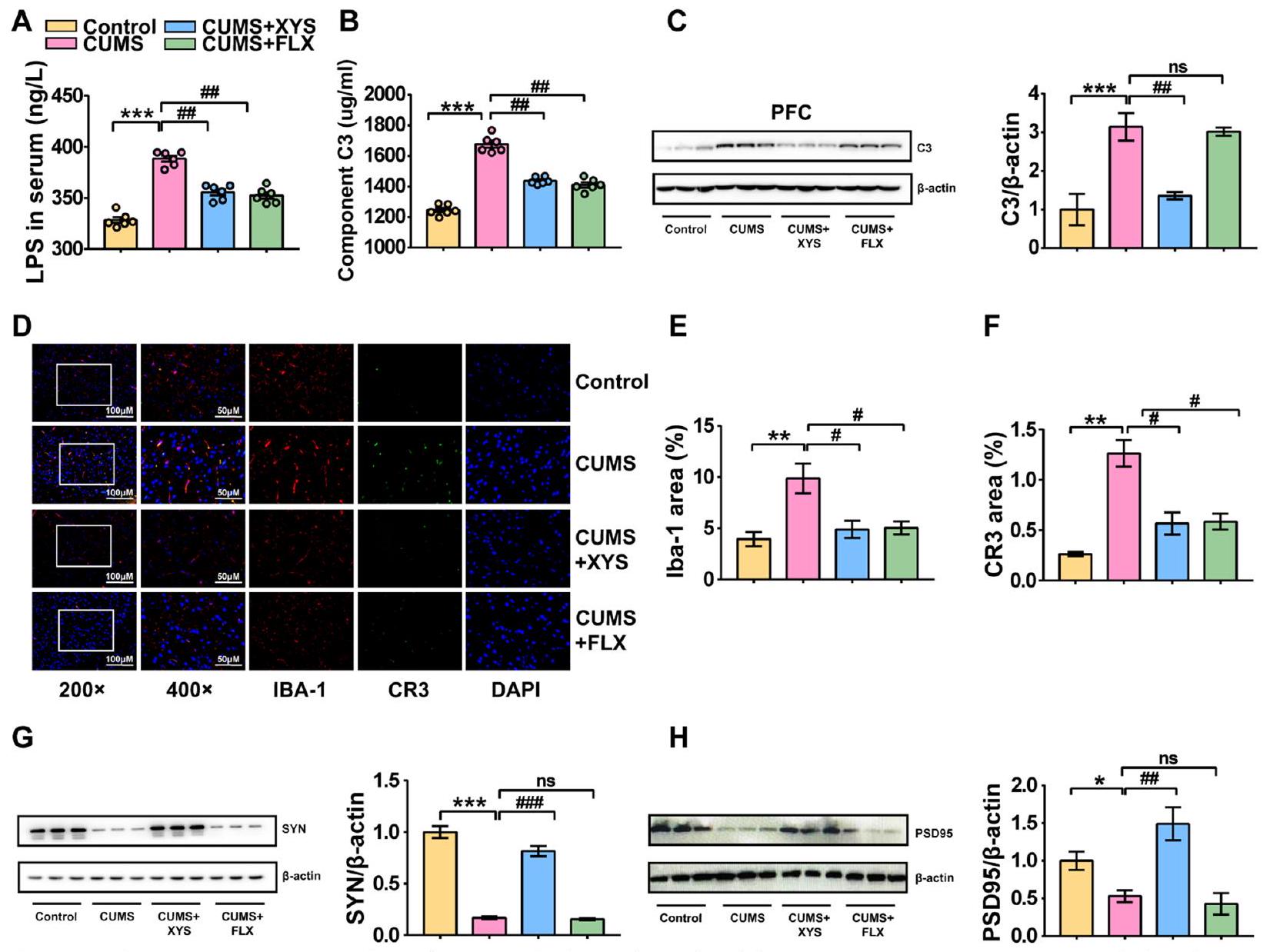
تشير هذه النتائج مجتمعة إلى أن تنشيط المكمل C3/CR3 والتقليم الشاذ للمشابك الذي تسببه الخلايا الدبقية الصغيرة هي مظاهر مرضية مهمة للاكتئاب. في الوقت نفسه، تقوم مضادات الاكتئاب بتثبيط تنشيط المكمل C3/CR3 الذي تسببه الضغوط المزمنة، وكذلك التقليم الشاذ للمشابك الذي تشارك فيه الخلايا الدبقية الصغيرة، مما يساعد على الحفاظ على النمو الطبيعي للخلايا العصبية المشبكية.

زراعة ميكروبات الأمعاء غير المنظمة تحفز تطور سلوكيات مشابهة للاكتئاب

تم التحقق من العلاقة بين ميكروبيوتا الأمعاء وبداية الاكتئاب. تم تحديد تأثير ميكروبيوتا CUMS على الفئران SPF وGF من خلال زراعة ميكروبيوتا البراز المشتقة من الفئران التي تم تحفيزها بواسطة CUMS (الشكل 6A). تم تقييم سلوك الحيوان قبل زراعة ميكروبيوتا البراز (الشكل S4a-f). كل من SPF وGF
أظهرت الفئران سلوكيات ملحوظة تشبه الاكتئاب والقلق بعد زراعة البراز، والتي كانت مصحوبة بضعف في الذاكرة المعرفية. الفئران SPF التي تلقت ميكروبيوتا البراز من الفئران التي تم تحفيزها بواسطة CUMS أظهرت فقدان الوزن، وانخفاض تفضيل السكروز، وانخفاض المسافة الإجمالية في اختبار فتح المجال، وانخفاض الرغبة في التجديد في اختبار تفضيل الأشياء الجديدة ومتاهة Y مقارنة بالفئران الضابطة؛ وهذا مشابه للفئران بعد تحفيز CUMS لمدة 8 أسابيع. تم ملاحظة نتائج مماثلة في الفئران GF. الفئران GF التي تلقت ميكروبيوتا البراز من فئران CUMS أظهرت انخفاضًا كبيرًا في المسافة الإجمالية للحركة في اختبار فتح المجال، وزيادة في وقت السكون في اختبار السكون، وضعف إدراكي في اختبار تفضيل الأشياء الجديدة ومتاهة Y. تشير هذه النتائج إلى أنه في ظل ظروف مستقلة عن الضغط، يمكن أن يؤدي تلقي زراعة براز من مضيف CUMS بسلوك يشبه الاكتئاب وضعف إدراكي إلى حدوث شذوذات سلوكية، بما في ذلك سلوكيات تشبه الاكتئاب.
علاوة على ذلك، تم تقييم تأثير الميكروبيوتا المعتمدة على مضادات الاكتئاب على الاكتئاب من خلال زراعة ميكروبيوتا البراز المأخوذة من الفئران التي تم إعطاؤها XYS في فئران تم تحفيزها بواسطة CUMS. كانت الفئران المحفزة بواسطة CUMS التي تلقت ميكروبيوتا البراز من الفئران المعالجة بـ XYS قد أظهرت تحسنًا ملحوظًا في زيادة الوزن وتفضيل السكروز مقارنة بالفئران المحفزة بواسطة CUMS دون علاج (الشكل 6BE). ومع ذلك، لم يتم ملاحظة تحسينات في سلوك القلق الشبيه والسلوك المعرفي في الفئران التي تلقت ميكروبيوتا البراز المأخوذة من الفئران المعالجة بـ XYS (الشكل 6F، G). التغيرات السلوكية المرتبطة اقترحت اتجاهًا نحو الشفاء دون دلالة إحصائية.
تشير هذه النتائج إلى أن زراعة ميكروبات الأمعاء من نموذج الإجهاد المزمن (CUMS-FMT) ساهمت في سلوكيات مشابهة للاكتئاب، وسلوكيات مشابهة للقلق، وضعف الإدراك، بينما ساهمت زراعة ميكروبات الأمعاء من نموذج XYS (XYS-FMT) في تخفيف الاكتئاب، ولكن ليس في تخفيف سلوكيات القلق وضعف الإدراك. وهذا يشير إلى أن اختلال ميكروبات الأمعاء يحفز تطور سلوكيات مشابهة للاكتئاب، في حين أن زراعة ميكروبات الأمعاء المعالجة بمضادات الاكتئاب لها تأثير مثبط على سلوكيات مشابهة للاكتئاب.

اختلال ميكروبيوم الأمعاء أدى إلى التهاب القولون وتدمير الحاجز

تم إجراء زراعة ميكروبيوم البراز من مجموعة CUMS إلى الفئران الضابطة ومن مجموعة XYS+CUMS إلى الفئران المعرضة لـ CUMS للتحقيق في الأنشطة الحيوية لـ CUMS-FMT و XYS-FMT على التهاب القولون ووظيفة حاجز الأمعاء. تم استخدام صبغة HE، وخصائص المورفولوجيا فوق الدقيقة للقولون، ومستويات السيروم من IL-1. IL-6، TNF تم قياس IL-10. أظهرت كل من الفئران الخالية من الجراثيم والفئران المحصنة التي تلقت ميكروبيوتا البراز من الفئران التي تم تحفيزها بواسطة CUMS متلازمات التهابية قولونية ملحوظة مقارنة بالفئران الضابطة (الشكل 7A). أظهرت المجهرية الإلكترونية الناقلة أن الزغابات المعوية كانت أقل حجمًا وعددًا وظهرت بشكل غير طبيعي.
الشكل 6 زراعة ميكروبيوتا الأمعاء غير المنظمة تحفز مباشرة تطور سلوكيات مشابهة للاكتئاب. أ بعد أسبوع من التكيف، تم تقسيم الفئران عشوائيًا إلى أربع مجموعات كما يلي: مجموعة التحكم، مجموعة التحكم + زراعة ميكروبيوتا (CUMS)، CUMS وCUMS + زراعة ميكروبيوتا (XYS). ب وزن الجسم (فئران SPF). ج اختبار السلوك الاجتماعي (فئران SPF). د اختبار النشاط المفتوح (المسافة الكلية المقطوعة) (فئران SPF). هـ اختبار الغمر في الماء (فئران SPF). و اختبار المتاهة المعقدة (فئران SPF). ز اختبار المتاهة على شكل Y (فئران SPF). ح مسارات حركة الفئران في اختبار النشاط المفتوح، اختبار المتاهة المعقدة (فئران SPF). ط تصميم الدراسة لفئران GF. ي اختبار النشاط المفتوح (فئران GF). ك اختبار الغمر في الماء (فئران GF). ل اختبار الذاكرة الجديدة (فئران GF). م اختبار المتاهة المعقدة (فئران GF). ن مسارات حركة الفئران في اختبار النشاط المفتوح، اختبار المتاهة المعقدة (فئران GF). تمثل البيانات المتوسط. SEM لكل مجموعة من فئران SPF؛ لكل مجموعة من فئران GF). ، مقابل مجموعة التحكم؛ مقابل مجموعة CUMS
المظهر (الشكل 7B). انخفاض تركيزات IL-10 وزيادة مستويات IL-1 IL-6 و TNF- تمت ملاحظتها أيضًا في الفئران المحمية من الأمراض (SPF) والفئران الخالية من الجراثيم (GF) التي تلقت ميكروبيوتا البراز من الفئران التي تم تحفيزها بواسطة الإجهاد المزمن (CUMS) (الشكل 7C-F، G، H).
تم التحقق من تأثير الميكروبيوتا المعتمدة على مضادات الاكتئاب على الاكتئاب من خلال زراعة ميكروبيوتا البراز المأخوذة من الفئران التي تم إعطاؤها XYS في نموذج الاكتئاب الناتج عن الإجهاد المزمن.
الفئران. في فئران SPF، تم عكس الالتهاب القولوني الناتج عن CUMS بواسطة XYS-FMT (الشكل 7A). أظهرت المجهرية الإلكترونية الناقلة أن الزغابات المعوية كانت أقل حجمًا وعددًا وظهرت بمظهر غير طبيعي في مجموعة CUMS. زاد حجم وعدد الزغابات المعوية وتم عكس الترتيب غير الطبيعي بواسطة XYS-FMT (الشكل 7B). زاد XYS-FMT من
الشكل 7 ساهمت زراعة ميكروبات الأمعاء من الفئران المعرضة للإجهاد المزمن (CUMS-FMT) في الاستجابة الالتهابية القولونية، وتلف حاجز الأمعاء؛ بينما قامت زراعة ميكروبات الأمعاء من الفئران المعالجة بالأعشاب الصينية (XYS-FMT) بتثبيط الاستجابة الالتهابية القولونية الناتجة عن CUMS، وتحسين وظيفة الحاجز. أ صبغة HE (فئران SPF). ب المجهر الإلكتروني الناقل (فئران SPF). ج IL-1 (فئران SPF). د IL-6 (فئران SPF). هـ TNF-a (فئران SPF). و IL-10 (فئران SPF). ز HE (فئران GF). ح المجهر الإلكتروني الناقل (فئران GF). ط IL-1 (فئران خالية من الجراثيم). ج IL-6 (فئران خالية من الجراثيم). ك TNF-a (فئران خالية من الجراثيم). ل IL-10 (فئران خالية من الجراثيم). البيانات تمثل المتوسط SEM لكل مجموعة). مقابل مجموعة التحكم؛ ، مقابل مجموعة CUMS
مستويات IL-10 وIL-1 المثبطة IL-6 و TNF- التعبير مقارنةً بتلك الخاصة بمجموعة CUMS (الشكل 7C-F).
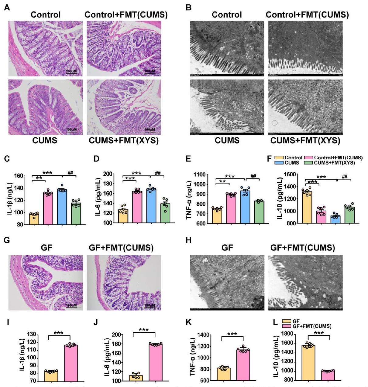
تشير هذه النتائج إلى أن CUMS-FMT تساهم في الاستجابة الالتهابية القولونية وتلف حاجز الأمعاء. بالإضافة إلى ذلك، فإن XYS-FMT خفف من الاستجابة الالتهابية القولونية الناتجة عن CUMS وحسن وظيفة الحاجز. وهذا يشير إلى أن ميكروبات الأمعاء غير المنظمة تتوسط الالتهاب القولوني وتفكك الحاجز، بينما كان لعلاج FMT مع مضادات الاكتئاب تأثير مثبط على الالتهاب القولوني وتفكك الحاجز.

توسط CUMS-FMT اضطراب ميكروبات الأمعاء واستعاد XYS-FMT التوازن المعوي

تم استكشاف تأثير CUMS-FMT وXYS-FMT على تركيب ميكروبات الأمعاء لدى الفئران باستخدام تسلسل جين 16S rRNA. -التنوع، كما هو موضح
من خلال مؤشر شانون، تأثر بـ FMT (الشكل 8A؛ الشكل S5a-c: -التنوع كما هو موضح بواسطة مؤشرات سيمبسون، الملاحظة، وتشاو). أظهر PCoA بناءً على مسافة براى-كورتيس انفصالًا في هيكل ميكروبات الأمعاء بين الفئران الضابطة مع أو بدون CUMS-FMT (الشكل 8B). كما لوحظ انفصال في هيكل ميكروبات الأمعاء بين مجموعتي CUMS وCUMS + FMT (XYS)، مما يشير إلى أن الهيكل الميكروبي المعوي في الفئران المصابة بالاكتئاب تأثر بـ XYSFMT (الشكل 8C).
بالإضافة إلى ذلك، تم استكشاف تأثيرات الضغط وFMT على هيكل الميكروبات المعوية لدى الفئران. أولاً، كررت نتائجنا التجريبية تنظيم ميكروبات الأمعاء بواسطة الضغط في التجربة 1. كان هذا متسقًا مع النتائج الموضحة في الشكل 8 D وE؛ زادت وفرة Proteobacteria في الفئران التي تعرضت لـ CUMS على مستوى الشعبة مقارنةً بتلك الموجودة في
الشكل 8 CUMS-FMT وسطاء اضطراب ميكروبات الأمعاء في الفئران؛ نظم XYS-FMT تركيب ميكروبات الأمعاء. أ التنوع الألفا كما هو موضح بواسطة مؤشر شانون. ب PCoA (التحكم مقابل التحكم + FMT(CUMS)). ج PCoA (CUMS مقابل CUMS + FMT(XYS)). د تغيير ميكروبات الأمعاء على مستوى الشعبة (مخطط شريطي). هـ تغيير ميكروبات الأمعاء على مستوى الشعبة (دائرة). و، ز مخطط شجري (التحكم مقابل CUMS)). ح، ط (التحكم مقابل التحكم + FMT(CUMS)). ي، ك مخطط شجري (CUMS مقابل CUMS + FMT(XYS)). ل تغيير ميكروبات الأمعاء على مستوى الشعبة والنوع من GF بعد FMT. تمثل البيانات المتوسط SEM ( لكل مجموعة). مقابل مجموعة التحكم؛ مقابل مجموعة CUMS
مجموعة التحكم، مما يشير إلى اتساق التغيرات في الميكروبات تحت نفس ظروف الضغط. بالإضافة إلى ذلك، لوحظت زيادة في وفرة الشعبة Verrucomicrobia. تم إنشاء مخطط شجري بواسطة تحليل LEfSe لبيانات الميكروبيوم (الشكل 8F، G) وأظهر مجموعتين مختلفتين من الأنواع بوفرة مختلفة على مستوى النوع في مجموعة CUMS، ومجموعتين مختلفتين من الأنواع بوفرة مختلفة في مجموعة التحكم ( ). على مستوى النوع، أظهرت الفئران المصابة بـ CUMS انخفاضًا كبيرًا
في وفرة Lactobacillus وزيادة كبيرة في وفرة Prevotella (Prevotellaceae) مقارنةً بتلك الخاصة بمجموعة التحكم (الشكل S5d).
بعد ذلك، تم تقييم تأثير FMT على ميكروبات الأمعاء لدى المضيف. أشارت النتائج السابقة إلى أن الفئران SPF التي تتلقى CUMS-FMT تظهر سلوكيات شبيهة بالاكتئاب بشكل ملحوظ. وبالمثل، أظهرت الفئران SPF التي تتلقى CUMS-FMT أيضًا هيكلًا وتركيبًا ميكروبيًا معويًا متغيرًا. على مستوى الشعبة، كانت
وفرة Bacteroidetes قد زادت بشكل كبير في الفئران التي تتلقى CUMS-FMT مقارنةً بتلك الخاصة بمجموعة التحكم، بينما لم يكن هناك فرق كبير في وفرة Proteobacteria (الشكل 8D، E). ومع ذلك، على مستوى النوع، تم تحفيز تأثير كبير بواسطة FMT. تم إنشاء مخطط شجري بواسطة تحليل LEfSe لبيانات الميكروبيوم (الشكل 8H، I) وأظهر 11 مجموعة من الأنواع بوفرة مختلفة على مستوى النوع في مجموعة CUMS-FMT ومجموعة واحدة من الأنواع بوفرة مختلفة على مستوى النوع في مجموعة التحكم ( ). علاوة على ذلك، على مستوى النوع، كانت وفرة Lactobacillus وAkkermansia قد انخفضت بشكل كبير في ميكروبات الأمعاء للفئران بعد علاج CUMS-FMT (الشكل S5f). وهذا يشير إلى أنه في الفئران SPF، قد لا يعيد FMT إنتاج توقيع الميكروبات المضيفة بسبب مقاومة الميكروبات المعوية الخاصة بالموضوع، بل يعدل وفرة أنواع معينة في شكل اضطرابات ميكروبية تؤدي إلى اختلالات ميكروبية معوية.
تم تقييم تأثير XYS-FMT في الفئران المصابة بـ CUMS. أظهرت التجارب السابقة أن إدارة XYS وXYS-FMT يمكن أن تعدل سلوك الاكتئاب في الفئران المصابة بـ CUMS. وبالمثل، أظهرت الفئران المصابة بـ CUMS التي تتلقى XYS-FMT أيضًا هيكلًا وتركيبًا ميكروبيًا معويًا متغيرًا. على مستوى الشعبة، كانت وفرة Proteobacteria قد انخفضت بشكل كبير في الفئران التي تتلقى XYS-FMT مقارنةً بتلك الموجودة في مجموعة CUMS، وهو ما يتماشى مع التغيرات التي تلي تناول XYS (الشكل 8D، E). تم إنشاء مخطط شجري بواسطة تحليل LEfSe لبيانات الميكروبيوم (الشكل 8J، K) وأظهر ثلاث مجموعات من الأنواع بوفرة مختلفة على مستوى النوع في مجموعة XYS-FMT، وثلاث مجموعات من الأنواع بوفرة مختلفة على مستوى النوع في مجموعة CUMS ( , LDA ). علاوة على ذلك، على مستوى النوع، كانت وفرة Lactobacillus وAkkermansia قد زادت بشكل كبير في ميكروبات الأمعاء للفئران بعد XYS-FMT (الشكل S5f).
بالإضافة إلى ذلك، لوحظت تغييرات في ميكروبات الفئران GF التي تلقت CUMS-FMT. لا تحتوي الفئران GF على أي ميكروبات معروفة في أمعائها؛ لذلك، كانت التغييرات في ميكروباتها أكثر وضوحًا من تلك الخاصة بالفئران SPF. على مستوى الشعبة، كانت Proteobacteria واحدة من الشُعب السائدة في الفئران بعد CUMS-FMT، مشابهةً لميكروبات الفئران التي تعرضت لـ CUMS. بشكل متسق، على مستوى النوع، كانت وفرة Bacteroides تتوافق مع ميكروبات الفئران المعرضة للضغط (الشكل 8L).

تسبب اختلال ميكروبات الأمعاء في تنشيط المكمل C3 وتقليم المشابك الشاذ بواسطة الخلايا الدبقية

ظهور سلوك شبيه بالاكتئاب وآلية مضادات الاكتئاب تتضمن تغييرات في ميكروبات الأمعاء، المكمل C3/CR3، وتقليم المشابك
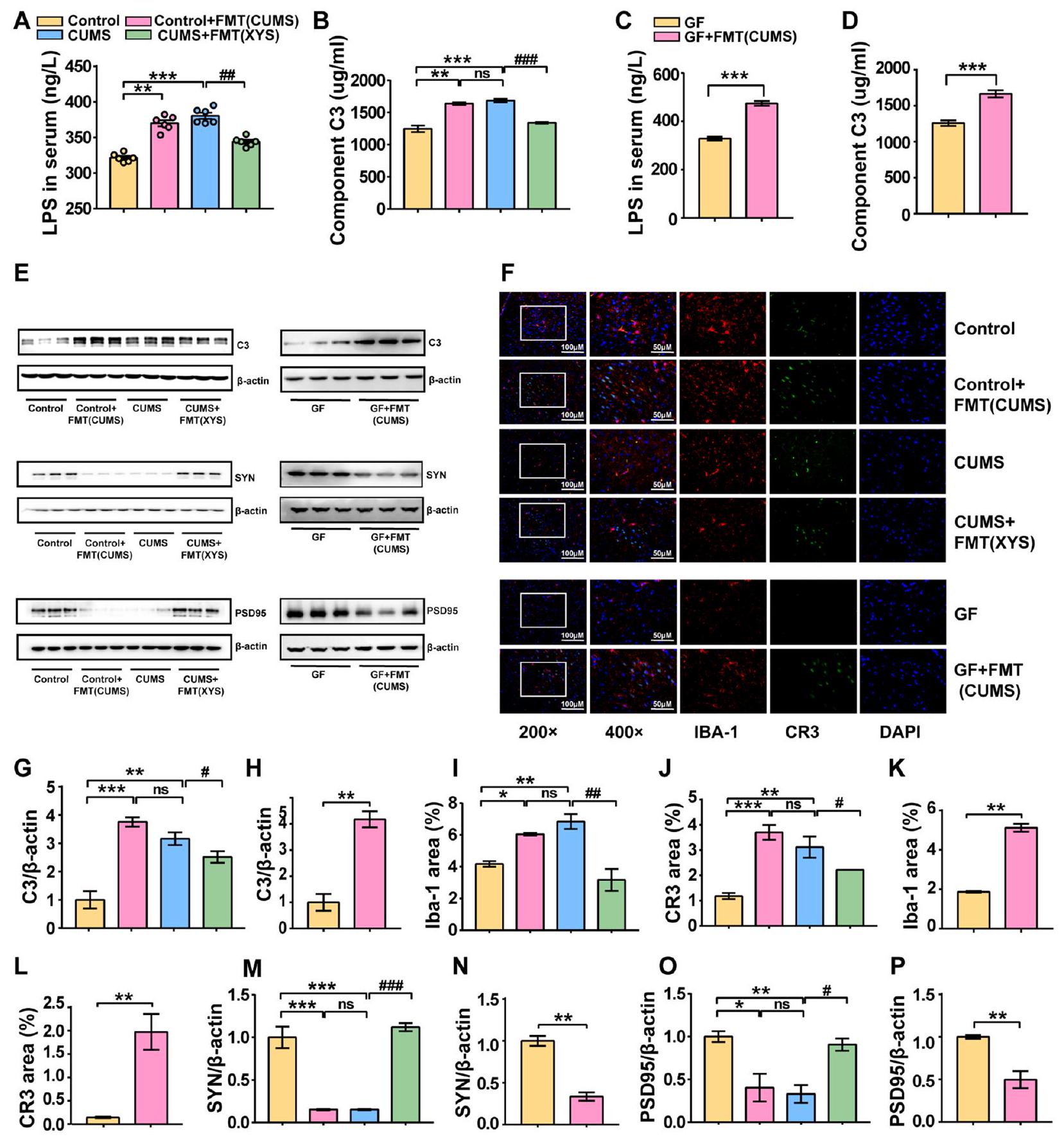
ومع ذلك، ليس من الواضح ما إذا كان تنشيط المكمل C3/CR3 في PFC وتقليم المشابك الشاذ بواسطة الخلايا الدبقية مرتبطًا مباشرةً بتغييرات في ميكروبات الأمعاء. لاستكشاف ما إذا كان استعمار الميكروبات البرازية بدون تحفيز ضغط يمكن أن يؤدي إلى تغييرات في المكمل C3 وتقليم المشابك الشاذ بواسطة الخلايا الدبقية، تم إجراء FMT من الفئران المستحثة بـ CUMS إلى الفئران SPF وGF. كانت مستويات LPS وC3 في مصل الفئران قد زادت بشكل كبير بعد تلقي ميكروبات البراز من الفئران المستحثة بـ CUMS مقارنةً بتلك الخاصة بالفئران SPF الضابطة (الشكل 9A، B). تم الإبلاغ عن نتائج متسقة أيضًا للفئران GF (الشكل 9C، D). علاوة على ذلك، متسقة مع تحفيز CUMS، لوحظت زيادة كبيرة في تعبير البروتين للمكمل C3 في أنسجة PFC لكل من الفئران SPF وGF بعد تلقي ميكروبات البراز من الفئران المستحثة بـ CUMS (الشكل 9E). بالإضافة إلى ذلك، أظهرت نتائج المناعية الفلورية المستندة إلى CR3 وIBA-1 أن CUMS-FMT حفزت تنشيط الخلايا الدبقية في PFC وزادت من تعبير CR3، مما يشير إلى أن CUMS-FMT يمكن أن تحفز مباشرةً تنشيط مسار C3/CR3 (الشكل 9F-L). كانت التغييرات التي أحدثها LPS في مستويات تعبير CD68 وIBA-1 متسقة أيضًا مع ضغط CUMS (الشكل S3d-i). لوحظت مستويات تعبير منخفضة لـ SYN وPSD95، مما يشير أيضًا إلى تلف عصبي ناتج عن تقليم مشابك غير طبيعي (الشكل 9M-P؛ الملف الإضافي 3).
قمنا أيضًا بتقييم تأثير FMT المضاد للاكتئاب على تقليم المشابك في الفئران المصابة بـ CUMS. أظهرت أبحاثنا أن إدارة XYS يمكن أن تعدل تقليم المشابك الشاذ الذي يتوسطه C3 في الفئران المصابة بـ CUMS. وبالمثل، قلل XYS-FMT بشكل كبير من مستويات LPS وC3 في مصل الفئران المصابة بـ CUMS، وأوقف تنشيط الخلايا الدبقية ومسار C3/CR3 في PFC، وزاد من مستويات تعبير SYN وPSD95. تشير هذه النتائج إلى أن تدخل ميكروبات الأمعاء مع XYS له أهمية كبيرة لتأثيراته المضادة للاكتئاب (الشكل 9A-P). أشارت نتائج المناعية الفلورية المستندة إلى CD68 وIBA-1 إلى أن تنشيط الخلايا الدبقية الناتج عن CUMS في PFC يمكن عكسه بواسطة XYSFMT (الشكل S3d، e؛ الملف الإضافي 3).
تشير هذه النتائج مجتمعة إلى أن ميكروبيوم الأمعاء غير المنظم يتوسط تنشيط المكمل C3 وتقليم المشابك غير الطبيعي الذي يتوسطه الخلايا الدبقية الصغيرة، بينما عالج علاج زراعة البراز مع مضادات الاكتئاب تنشيط المكمل C3 الناتج عن CUMS وتقليم المشابك غير الطبيعي المتعلق بالمكمل.

المناقشة

في الدراسة الحالية، أظهرنا أن اختلال ميكروبيوم الأمعاء يحفز تنشيط نظام C3/CR3 لتعزيز تقليم المشابك وتهيئة سلوكيات شبيهة بالاكتئاب. تم تقديم أدلة على أن الإدارة
الشكل 9 أدى اختلال ميكروبيوم الأمعاء إلى تنشيط المكمل C3 وتقليم المشابك الشاذ الذي يتوسطه الخلايا الدبقية الصغيرة. أ LPS في المصل (فئران SPF). ب C3 في المصل (فئران SPF). ج LPS في المصل (فئران GF). د C3 في المصل (فئران GF). هـ WB. و التألق المناعي للخلايا الدبقية الصغيرة و CR3. ز تعبير بروتين C3 (فئران SPF). ح تعبير بروتين C3 (فئران GF). ط تعبير بروتين IBA-1 (فئران SPF). ي تعبير بروتين CR3 (فئران SPF). ك تعبير بروتين IBA-1 (فئران GF). ل تعبير بروتين CR3 (فئران GF). م مستوى تعبير بروتين SYN (فئران SPF). ن مستوى تعبير بروتين SYN (فئران GF). O مستوى تعبير بروتين PSD95 (فئران SPF). P مستوى تعبير بروتين PSD95 (فئران GF). تمثل البيانات المتوسط SEM ( لكل مجموعة لـ ELISA؛ لكل مجموعة لـ IF و WB). , , مقابل مجموعة التحكم؛
مقابل مجموعة CUMS
لدواء مضاد للاكتئاب (XYS) حسّن سلوكيات شبيهة بالاكتئاب، وسلوكيات شبيهة بالقلق، والضعف الإدراكي في الفئران وكذلك منع اختلال المكمل الناتج عن اختلال ميكروبيوم الأمعاء والاضطرابات في تقليم المشابك المتعلق بالمكمل. علاوة على ذلك، أكدت زراعة البراز CUMS-FMT في الفئران SPF و GF وجود ارتباط مباشر بين ميكروبيوم الأمعاء ونظام المكمل وتقليم المشابك الدبقية الصغيرة. حسّن XYSFMT بشكل كبير سلوكيات شبيهة بالاكتئاب في الفئران، مما يشير إلى أن الآليات الأساسية لفعالية مضادات الاكتئاب XYS تعتمد بشكل رئيسي على تنظيم التفاعل بين الأمعاء والدماغ. بالإضافة إلى ذلك، فإن استخدام المضادات الحيوية واسعة الطيف يلغي تحسين السلوك الناتج عن مضادات الاكتئاب، مما يبرز الدور الأساسي لميكروبيوم الأمعاء في التوسط في سلوكيات شبيهة بالاكتئاب. يعتبر محور الأمعاء-الميكروبيوم-الدماغ منظمًا رئيسيًا لوظيفة الأعصاب ويلعب دورًا رئيسيًا في مسببات الاكتئاب [30، 31]. يعتبر التلاعب بالميكروبات المعوية علاجًا محتملاً للاكتئاب [32]. كتركيبة نباتية تستخدم على نطاق واسع في آسيا وأوروبا، تحتوي XYS على مجموعة متنوعة من المواد الفعالة وتعتبر لها خصائص شبيهة بالبريبايوتيك [20]. في الدراسة الحالية، تم استخدام XYS، وهو دواء مضاد للاكتئاب محتمل، لاستكشاف دور ميكروبيوم الأمعاء في علاج الاكتئاب والآليات المحددة التي تكمن وراء التفاعل بين الميكروبات المعوية والجهاز العصبي المركزي. تم استخدام اختبارات سلوكية بما في ذلك SPT و OFT و TST و EPM و NORT واختبارات المتاهة Y لاستكشاف تأثير التحسين لـ XYS على الاكتئاب وأعراضه المصاحبة، مثل سلوكيات شبيهة بالقلق والضعف الإدراكي، لتأكيد إمكانيته كمضاد للاكتئاب. بالإضافة إلى ذلك، باستخدام تسلسل RNA 16S وتحليل UHPLC-QTOF-MS/MS الأيضي، تم إظهار أن التحسين في سلوكيات شبيهة بالاكتئاب كان مصحوبًا بتنظيم الميكروبات المعوية والمواد الأيضية الميكروبية. بعد ذلك، للتحقق مما إذا كانت الخصائص الشبيهة بالبريبايوتيك لمضادات الاكتئاب تلعب دورًا رئيسيًا في تحسين سلوكيات شبيهة بالاكتئاب، تم استخدام تدخلات ABX و FMT لاستكشاف تأثير مضاد الاكتئاب لـ XYS في بيئات ميكروبية معوية مختلفة. تم خلط مجموعة متنوعة من المضادات الحيوية في مياه الشرب اليومية للفئران وتم السماح للفئران بالشرب بحرية لمدة 7 أيام. تم اختيار طريقة التدخل بالمضادات الحيوية المذكورة أعلاه لأن الدراسات السابقة أظهرت أن الحقن داخل الصفاق بالمضادات الحيوية أو التدخل طويل الأمد (
أسابيع) مع مضادات حيوية مختلطة يمكن أن يؤدي إلى سلوكيات شبيهة بالاكتئاب في الفئران [33، 34]. بالإضافة إلى ذلك، اقترحت نتائجنا أن علاج المضادات الحيوية عن طريق الفم لمدة أسبوع واحد لا يؤثر على سلوك SPF (تم عرض نتائج الدراسة في الملف الإضافي 4). لذلك، لتجنب تأثير تدخل المضادات الحيوية على الحالة الاكتئابية للفئران، تم إعطاء تدخل مضاد حيوي قصير الأمد (7 أيام) لإزالة وتلاعب الميكروبات المعوية للفئران دون تفاقم سلوكيات شبيهة بالاكتئاب للفئران. تم القضاء على تأثير مضاد الاكتئاب لـ XYS بعد إعطاء ABX لإزالة ميكروبيوم الأمعاء، مما يشير إلى أن وجود الميكروبات المعوية ضروري لـ XYS لتحقيق تأثيره المضاد للاكتئاب. من ناحية، يؤدي اختفاء الميكروبيوم إلى تثبيط امتصاص واستقلاب المواد الفعالة المضادة للاكتئاب في XYS وعدم فعالية الأنشطة المضادة للاكتئاب اللاحقة؛ من ناحية أخرى، بسبب التغيرات في البيئة الميكروبية المعوية، لا يمكن تحقيق التأثيرات الشبيهة بالبريبايوتيك لـ XYS المرتبطة بمضادات الاكتئاب. لذلك، تم استخدام طريقة FMT لاستكشاف تحسين سلوكيات شبيهة بالاكتئاب في الفئران المعالجة ببكتيريا براز مضادة للاكتئاب (ميكروبيوم الأمعاء، مع أو بدون مواد أيضية). تشير نتائجنا إلى أن زراعة براز من المضيف المعالج بمضادات الاكتئاب في الفئران المكتئبة حسنت سلوكيات شبيهة بالاكتئاب في الفئران المعرضة لـ CUMS. وهذا يشير إلى أن XYS يمكن أن يحسن أعراض الاكتئاب مباشرة من خلال استعمار الفلورا المعوية. تؤكد هذه النتائج أن العمل الديناميكي الدوائي لـ XYS كمضاد للاكتئاب محتمل يعتمد بشكل رئيسي على خصائصه الشبيهة بالبريبايوتيك.
يعتقد أن اختلال الميكروبيوم المعوي يدفع مسببات الاكتئاب. أكدت النتائج من 16 دراسة رصدية و 11 دراسة سريرية أن المرضى الذين يعانون من اضطرابات شبيهة بالاكتئاب الكبرى يظهرون تغييرات في الميكروبيوم المعوي [35]. لوحظت نتائج مماثلة في القوارض ذات سلوكيات شبيهة بالاكتئاب [36]. بعد التأكد من أن الخصائص الشبيهة بالبريبايوتيك لـ XYS حاسمة لتحسين سلوكيات شبيهة بالاكتئاب، تم استكشاف التغييرات المحددة في ميكروبيوم الأمعاء والمواد الأيضية الميكروبية. في الدراسة الحالية، حسنت إدارة XYS التحول في تركيب ميكروبيوم الأمعاء الناتج عن CUMS. أظهرت دراسات الميكروبيوم باستخدام تسلسل جين 16S rRNA أن وفرة شعبة Proteobacteria في مجموعة XYS قد انخفضت بشكل كبير، بينما زادت وفرة شعبة Bacteroidetes مقارنة بتلك الموجودة في الفئران الناتجة عن CUMS. تعتبر Proteobacteria علامة ميكروبية على اختلال التوازن في ميكروبيوم الأمعاء [37]. بشكل عام، توجد البكتيريا المتعايشة من Proteobacteria في أمعاء الثدييات الصحية. إذا كانت هذه البكتيريا موجودة بأعداد قليلة، فإنها تبدو غير ضارة؛ إذا زادت وفرتها بشكل كبير، فإنها تصبح ميكروبات معوية مسببة للالتهابات [38]. لقد كشفت الأدلة الواسعة أن زيادة Proteobacteria هي عامل خطر للاضطرابات النفسية مثل الاكتئاب والقلق [39]. يؤدي التعرض للمضادات الحيوية إلى إلحاق ضرر شديد بميكروبيوم الأمعاء وزيادة وفرة Proteobacteria بشكل كبير، مما يؤدي إلى القلق والاكتئاب.
[40]. بشكل متسق، في الدراسة الحالية، كانت وفرة البكتيريا البروتيوبكتيرية مرتفعة بشكل ملحوظ في نموذج الاكتئاب الناتج عن CUMS. أظهرت تحليل LDA أن مكملات XYS قللت من وفرة البروتيوبكتيريا على مستوى الفيلوم، وكذلك من الأنواع الأدنى، مثل Enterobacteriales وEnterobacteriaceae وKlebsiella على مستويات الطلب والعائلة والجنس، على التوالي، في الفئران المعرضة لـ CUMS. أظهرت الدراسات السابقة أن إعطاء Klebsiella oxytoca وEscherichia coli وCronobacter sakazakii، التي تنتمي إلى Enterobacteriaceae، بشكل منفصل أو مجتمعة، تسبب سلوكيات شبيهة بالاكتئاب في كل من الفئران GF وSPF، مما يشير إلى أن البروتيوبكتيريا مرتبطة ارتباطًا وثيقًا بالاكتئاب [41]. تم ربط الميكروبيوم الذي ينتمي إلى الفيلوم Bacteroidetes بالإدراك والأمراض التنكسية العصبية. أظهرت الدراسات السريرية أن الرضع الذين لديهم مستويات عالية من Bacteroides في الأمعاء يظهرون أداءً إدراكيًا أعلى في سن 1 و2 [42]. في الدراسة الحالية، وجدنا أن XYS زاد من وفرة Bacteroidetes على مستوى الفيلوم، مما يشير إلى أنه قد يكون مسؤولًا عن تحسين العجز الإدراكي. علاوة على ذلك، على مستوى الجنس، قلل XYS من وفرة Bacteroides وزاد من وفرة Lactobacillus. يُعتقد أن Bacteroides مرتبطة باضطراب الاكتئاب الرئيسي (MDD) [43]. كانت مستويات عالية من Bacteroides spp. موجودة في عينات البراز من 156 مريضًا يعانون من MDD، مقارنة بالأفراد الأصحاء [44]. يُعتبر Lactobacillus بروبيوتيك له تأثير مثبط على الاكتئاب [45]. وقد تم الإبلاغ عن أن إنتاج بيروكسيد الهيدروجين بواسطة Lactobacillus قد يمنع سلوكيات الاكتئاب الناتجة عن CUMS عن طريق تثبيط إنزيم الإندولامين-2،3-ديوكسيجيناز المعوي مباشرة [46]. وجدنا أن إعطاء XYS قلل بشكل ملحوظ من تعبير Bacteroides وزاد من تعبير Lactobacillus. بشكل عام، تدعم هذه النتائج أن مكملات XYS تحسن من تركيب الميكروبيوم المعوي، وخاصةً من خلال قمع البروتيوبكتيريا وتعزيز Bacteroidetes، مما قد يساهم في الوقاية من سلوكيات شبيهة بالاكتئاب والعجز الإدراكي المصاحب في الفئران التي تعاني من الاكتئاب الناتج عن الإجهاد المزمن. بالإضافة إلى ذلك، فإن تنوع الميكروبيوم البرازي أقل في المرضى الذين يعانون من الاكتئاب مقارنةً بالضوابط الأصحاء. تم تقليل تنوع الميكروبيوم المعوي في فئران CUMS، ومنعت مكملات XYS هذه التغيرات في الميكروبيوم المعوي. لذلك، يُفترض أن تنظيم مضادات الاكتئاب على تركيب وتنوع الميكروبيوم المعوي قد يكون حاسمًا لتحسين الاكتئاب.
بالإضافة إلى ذلك، أكدت FMT في الفئران SPF وGF دور الميكروبات المعوية في تطوير وعلاج الاكتئاب. من خلال إجراء FMT في الفئران الصحية SPF وGF، تم ملاحظة تأثير الميكروبيوم المعوي من الفئران المكتئبة على الميكروبيوم المعوي وتنظيم السلوك
للمضيفين الأصحاء. بالنسبة لـ FMT في الفئران SPF، تم إزعاج ميكروبيوم الفئران الضابطة، بدلاً من استبداله بالميكروبيوم من فئران CUMS. على مستوى الفيلوم، وبما يتعارض مع التغيرات الناتجة عن الإجهاد في الميكروبيوم المعوي، لم يُلاحظ أي زيادة ملحوظة في وفرة البروتيوبكتيريا بعد CUMS-FMT. ومع ذلك، على مستوى الجنس، لوحظ انخفاض في وفرة Lactobacillus وزيادة في وفرة Bacteroides، وهو ما يتماشى مع التغيرات في الفئران المعرضة لـ CUMS. قد يكون سبب هذه الظاهرة هو أنه خلال عملية FMT، يكون للميكروبيوم المعوي الأصلي للمضيف مقاومة معينة للميكروبيوم CUMS؛ لذلك، فإن التغيرات على مستوى الفيلوم ليست متسقة تمامًا مع التغيرات في ميكروبيوم فئران CUMS. لذلك، قمنا بإجراء CUMS-FMT في الفئران GF. بما يتماشى مع توقعاتنا، تم تكرار توقيع الميكروبات المعوية (بما في ذلك زيادة وفرة البروتيوبكتيريا على مستوى الفيلوم وBacteroides على مستوى الجنس) لفئران CUMS في الفئران GF بسبب غياب الميكروبات الساكنة في الأمعاء. أظهرت كل من الفئران SPF وGF سلوكيات شبيهة بالاكتئاب بعد علاج CUMS-FMT. وهذا يؤكد أكثر أن الاضطرابات في الميكروبيوم، حتى على مستوى الجنس، كافية للتأثير على عدم توازن الميكروبيوم المعوي بشكل عام وتحفيز ظهور سلوكيات شبيهة بالاكتئاب. في هذه الدراسة، تم إجراء FMT حصريًا من فئران CUMS إلى فئران GF؛ لم يتم إجراء FMT من فئران SPF الضابطة إلى فئران GF. تشير المراجع الحديثة عالية الجودة باستمرار إلى أن FMT من الحيوانات غير المجهدة لا تحفز سلوكيات الاكتئاب في الفئران GF [47-49]. أشارت الدراسات السابقة إلى أن الفئران GF لم تظهر سلوكيات شبيهة بالاكتئاب حتى بعد تلقي بكتيريا برازية من فئران ضابطة أو أفراد أصحاء. وهذا يشير إلى أن البكتيريا البرازية المزروعة من الفئران أو الأفراد غير المجهدين لم تؤثر على سلوك الفئران GF [47.48]. بعد ذلك، تم ملاحظة تأثير XYSFMT على الميكروبات المعوية في فئران CUMS. على مستوى الفيلوم، قلل XYS-FMT من وفرة البروتيوبكتيريا وزاد من وفرة Bacteroidetes، وهو ما يتماشى مع تنظيم الميكروبيوم المعوي بواسطة إعطاء XYS. على مستوى الجنس، لوحظ زيادة في وفرة Lactobacillus. والأهم من ذلك، تم ملاحظة تغييرات في Prevotellaceae على مستوى العائلة وPrevotella على مستوى الجنس. يمكن لأعضاء Prevotellaceae، المعروفين كمتعايشين معويين، إنتاج SCFAs من خلال تخمير الألياف الغذائية والمخاط المعوي [50]. أظهرت الدراسات أن تقليل Prevotellaceae يؤدي إلى زيادة التعرض لنظام الإندوتوكسين البكتيري وزيادة نفاذية الأمعاء، مما يحفز الالتهاب [51]. في هذه الدراسة، أظهر تحليل LDA أن XYS-FMT زاد من وفرة Prevotellaceae على مستوى العائلة و
Prevotella على مستوى الجنس، مما يشير إلى أن XYS-FMT يحسن من هيكل وتنوع الميكروبات المعوية في CUMS. كما لوحظت زيادة في Prevotellaceae على مستوى العائلة في مجموعة CUMS-FMT، لكن هذه الزيادة لم تؤد إلى اختفاء سلوكيات شبيهة بالاكتئاب. السبب هو أن التغيرات في Prevotellaceae لم تكن سائدة في التغيرات الميكروبية لمجموعة CUMS-FMT، خاصة مع انخفاض Lactobacillus وزيادة Bacteroides؛ أدى النظام البيئي الميكروبي المعقد وغير المتوازن إلى ظهور سلوكيات شبيهة بالاكتئاب. بالإضافة إلى ذلك، قد تؤدي التباينات في Prevotellaceae على مستوى الأنواع إلى وظائف متنوعة. على سبيل المثال، في قرود السيمونولغوس التي تعاني من سلوكيات شبيهة بالاكتئاب الناتجة عن CUMS، كانت هناك 11 نوعًا متزايدًا من متغيرات تسلسل الأمبليكون (ASVs) و7 ASVs متناقصة تنتمي إلى Prevotellaceae، مما يشير إلى تعقيد التغيرات على مستوى الأنواع في Prevotellaceae في سلوكيات شبيهة بالاكتئاب [52]. ومع ذلك، في XYS-FMT، كانت التغيرات في Prevotellaceae سائدة في التغيرات الميكروبية، مما يشير إلى أنها قد تكون لها تأثير مثبط أكبر على الالتهاب. في الختام، ترتبط التغيرات في تركيب وتنوع الميكروبات المعوية ارتباطًا وثيقًا بتقدم وعلاج سلوكيات شبيهة بالاكتئاب. يتم تحقيق تأثير المكملات الميكروبية الخارجية على الميكروبات المعوية الذاتية بشكل رئيسي من خلال تنظيم البيئة الميكروبية العامة وإعادة تشكيل وفرة العديد من البكتيريا السائدة بدلاً من التأثير فقط على جنس واحد.
يتجلى التداخل بين ميكروبات الأمعاء وسلوك المضيف أيضًا في تعطيل وظيفة حاجز الأمعاء وتنظيم المستقلبات الميكروبية. يتم الحفاظ على التوازن المعوي من خلال التداخل بين الميكروبيوتا وحاجز الأمعاء والجهاز المناعي. غالبًا ما يؤدي عدم التوازن في الميكروبيوم إلى تدمير حاجز الأمعاء والجهاز المناعي. في ظل الظروف الفسيولوجية الطبيعية، تدعم خلايا الظهارة المعوية والميكروبيومات والخلايا المناعية حالة مستقرة في النظام المعوي. تتلقى خلايا الظهارة المعوية إشارات من الميكروبيوم، مثل المستقلبات الميكروبية أو الميكروبات، للحفاظ على الحاجز المخاطي. كما ينظم الميكروبيوم مناعة المضيف من خلال مستقلباته والسموم الداخلية. علاوة على ذلك، يمكن أن تؤثر الخلايا المناعية بشكل مباشر أو غير مباشر على الميكروبيوم من خلال إفراز السيتوكينات أو الكيموكينات. ومع ذلك، يؤدي عدم التوازن في الميكروبيوم إلى تدمير حاجز الأمعاء وزيادة نشاط المناعة المعوية. أظهرت الدراسات أن تغييرًا في هيكل الميكروبيوم يؤدي إلى زيادة في بروتينات الوصلات الضيقة المعوية ونفاذية الأمعاء. هذه الأحداث تحفز الالتهاب في القولون وتنتج سيتوكينات مؤيدة للالتهاب مثل IL-6 وIL-1. ، وTNF- [25]. في هذه الدراسة، لاحظنا
أن CUMS أو CUMS-FMT يمكن أن تؤدي إلى تعطيل وظيفة حاجز القولون، وإنتاج التهاب القولون، وارتفاع مستوى السيتوكينات البروالتهابية في المصل IL-6 وIL-1 “، وTNF- ، بينما يمكن لـ XYS-FMT استعادة وظيفة حاجز الأمعاء وتثبيط التهاب القولون وعوامل الالتهاب في المصل. يرتبط الأيض ارتباطًا وثيقًا بالتوازن الوظيفي لحاجز الأمعاء. الأيض هو مسار رئيسي يؤثر من خلاله الفلورا المعوية على الاكتئاب عبر محور الدماغ-الأمعاء، خاصة من خلال التغيرات المباشرة في مستويات المستقلبات الرئيسية والتغيرات غير المباشرة في المستقلبات الدموية المتداولة، مما يؤثر بشكل أكبر على التغيرات في الجهاز العصبي المركزي التي تنظم السلوكيات الاكتئابية [57]. يتماشى ذلك مع التغيرات في الميكروبيوتا، حيث لوحظت اضطرابات في المستقلبات الميكروبية تحت حالتين مختلفتين (الضغط والاضطراب الميكروبي). أظهر أيض فيتامين ب6 ومسارات تخليق الليسين اختلافًا كبيرًا في الثراء في الدراسة الحالية. يرتبط نقص فيتامين ب6 بالاكتئاب ووظيفة عصبية سلبية [58]. بالإضافة إلى ذلك، أظهرت الأبحاث أن تناول فيتامين ب6 قبل العلاج يمكن أن يمنع الاكتئاب الناتج عن الديكساميثازون ويظهر تأثيرات مضادة للاكتئاب [59]. يعتبر الليسين عاملًا مهمًا في الاكتئاب الذي يتوسطه الميكروبيوتا المعوية. اقترحت دراسة سابقة أن التغير في أسيتيل الليسين قد يلعب دورًا رئيسيًا في تطور الاكتئاب وقد يكون آلية من خلالها تعدل الميكروبيوتا المعوية وظيفة الدماغ والأنماط السلوكية [60]. في الدراسة الحالية، وجدنا أن تقدم سلوك الاكتئاب كان مصحوبًا باضطرابات في أيض فيتامين ب6 وتخليق الليسين، وكشفت تحليل الارتباط أيضًا عن العلاقة بين المستقلبات الميكروبية والسلوك والفلورا المعوية. باستخدام التغيرات في وفرة البروتيوبكتيريا كميزة للاضطرابات الميكروبية، اكتشفت التحليلات المعتمدة على UPLC-MS تغييرات في الركائز لإنتاج عدة سموم بكتيرية (LPS). LPS، وهو مادة (غليكوليبيد) تتكون من الدهون والسكريات المتعددة، هو مكون من جدار الخلية الخارجي للبكتيريا سالبة الجرام [61]. يُعتقد أن المستويات العالية من LPS في الدم المحيطي والجهاز العصبي المركزي مرتبطة بالاكتئاب [62]. وقد تم الإبلاغ عن زيادة مستويات الإندوتوكسين ثلاثة أضعاف في الدم ومرتين أو ثلاث مرات في أدمغة المرضى الذين يعانون من الاكتئاب. D-(+)-مانوز، N-acetylmannosamine، D-ribose 5-phosphate، و glucose-6-phosphate هي ركائز مهمة لتخليق LPS. في الدراسة الحالية، لوحظ زيادة في استهلاك هذه الركائز، مما يشير إلى زيادة في إنتاج LPS. يتماشى ذلك مع تقرير سابق عن زيادة تعبير جينات تخليق LPS في المرضى الذين يعانون من القلق والاكتئاب [62]. بالإضافة إلى ذلك، مع تعطيل وظيفة حاجز الأمعاء، لوحظ مستوى عالٍ من تعبير LPS في المحيط
الدم، الذي يتماشى مع الزيادة في وفرة البكتيريا المنتجة لـ LPS في ميكروبيوم الأمعاء. كما أكدت تحليل الارتباط أن الميكروبات المعوية كانت مرتبطة بشكل كبير بإنتاج LPS.
التعرض المفرط لـ LPS يحفز التعبير عن نظام المكملات وتنشيط الخلايا الدبقية الصغيرة. المكملات، وهو بروتين في المصل، له نشاط إنزيمي ويقوم بوساطة الاستجابات المناعية والالتهابية. يعتبر المكمل C3 بروتينًا رئيسيًا في نظام المكملات. تم ملاحظة مستويات مرتفعة من C3 في أدمغة المرضى الذين يعانون من الاكتئاب ونماذج الاكتئاب الحيوانية، ويعتقد أنها مرتبطة إيجابيًا بشدة الأعراض الاكتئابية. وقد وجد الباحثون أن الإجهاد المزمن يمكن أن يؤدي إلى زيادة التعبير عن C3 في القشرة الجبهية الأمامية للفئران، وأن الفئران التي تفتقر إلى C3 لا تطور سلوكيات مشابهة للاكتئاب، مما يشير إلى أن مستوى مرتفع من C3 هو شرط مهم لظهور الاكتئاب. للأسف، لا يزال مصدر المكمل C3 (سواء من الجهاز العصبي المركزي مباشرة أو عبر الهجرة الالتهابية من المحيط إلى الدماغ) غير واضح. يُعتقد أن LPS هو منشط رئيسي للمكمل C3، حيث يقوم بوساطة مستويات المكمل C3 في المصل المحيطي.
[66]. بعد ملاحظة ارتفاع LPS المنظم بواسطة ميكروبات الأمعاء، تم فحص تعبير C3 في مصل الدم والقشرة الجبهية الأمامية للفئران. يتماشى ذلك مع التغيرات الناتجة عن الإجهاد في المكملات، حيث يشير التعبير العالي عن C3 في القشرة المحيطية والجبهية الأمامية بعد زراعة البراز إلى أن الاضطرابات الميكروبية المعوية قد تكون محفزًا وعاملاً مفاقمًا لتنشيط C3. بالإضافة إلى ذلك، تم ملاحظة تنشيط الميكروغليا الذي تم تحفيزه بشكل خاص بواسطة LPS في الجهاز العصبي المركزي. كخلايا فعالة مناعية فطرية في الجهاز العصبي المركزي، تلعب الميكروغليا دورًا مهمًا في الحفاظ على تطور الخلايا العصبية ووظيفة الأعصاب الطبيعية [67]. من بينها، يعتبر تقليم المشابك الطريقة الرئيسية للميكروغليا للتخلص من المشابك وهو آلية مهمة لتقليم الخلايا العصبية بشكل انتقائي للحفاظ على الأنظمة العصبية الطبيعية [68]. التفاعل مع نظام المكملات، الذي يتوسط تقليم المشابك بواسطة الميكروغليا، أمر حاسم لتنظيم شكل المشابك بواسطة الميكروغليا [69]. أظهرت الأبحاث الحديثة أن الإفراط في التعبير عن المكمل C3 يؤثر على البلعمة وتقليص المشابك للميكروغليا، مما يؤدي إلى الاكتئاب وسلوك إدراكي غير طبيعي [70]. CR3 هو مستقبل المكمل C3 الذي يتم التعبير عنه بواسطة الميكروغليا، وترابطه مع
الشكل 10 إن تثبيط التقليم المشبكي غير الطبيعي هو المفتاح لاستهداف الميكروبات لعلاج الاكتئاب. تساهم ميكروبات الأمعاء في تنظيم محور الأمعاء-الدماغ والحفاظ على الصحة، بينما يرتبط تغييرها (زيادة بكتيريا البروتيوبكتيريا وانخفاض تنوع الميكروبات) بسبب الإجهاد المزمن غير المتقطع بالاكتئاب وعواقبه السلبية على الإدراك. يُعتقد أن مكمل XYS يقلل من بكتيريا البروتيوبكتيريا وينظم تركيب وعمليات ميكروبات الأمعاء (1)، وبالتالي يساهم في وظيفة حاجز الأمعاء وتوازن المناعة (2)؛ مما يقلل من انتقال مكونات البكتيريا سالبة الجرام (3)، مما يقلل من نبرة الالتهاب المحيطي ويثبط تنشيط C3/CR3 (4) وتقليص المشبك غير الطبيعي الذي تسببه الخلايا الدبقية في الجهاز العصبي المركزي (5). لذلك، يؤثر مكمل XYS بشكل إيجابي على الاكتئاب، من خلال استعادة ميكروبات الأمعاء ودورها التنظيمي في محور الأمعاء-الدماغ، مما يشير إلى أن تثبيط التقليم المشبكي غير الطبيعي هو المفتاح لاستهداف الميكروبات لعلاج الاكتئاب.
المكمل C3 يوجه الميكروغليا لابتلاع المشابك العصبية المعلّمة [71]. في هذه الدراسة، يمكن أن يؤدي إما الضغط النفسي أو زراعة ميكروبيوتا الأمعاء غير المنظمة إلى تنشيط مسار C3/CR3 في القشرة الجبهية الأمامية وتقليل مستويات التعبير البروتيني لـ SYN و PSD95. يمكن لمضادات الاكتئاب أو زراعة ميكروبيوتا الأمعاء من مضادات الاكتئاب أن تثبط تنشيط مسار C3/CR3 وتمنع تقليل تعبير SYN و PSD95. يُعتبر SYN و PSD95 مؤشرات مهمة تعكس وظيفة المشابك العصبية للخلايا العصبية، ويعتبر انخفاض تعبير SYN و PSD95 سمة مرضية مهمة للاكتئاب [72].
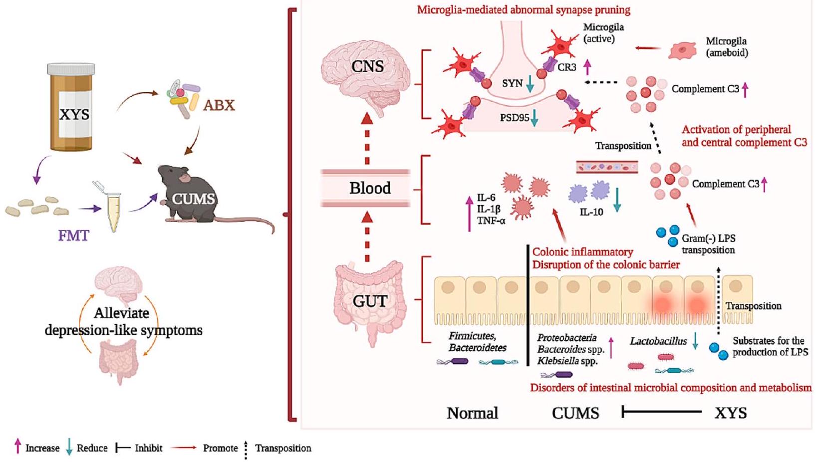
تشير نتائجنا بشكل جماعي إلى أن اختلال التوازن الميكروبي في الأمعاء يسبب سلوكيات مشابهة للاكتئاب من خلال تقليم المشابك غير الطبيعي في الخلايا الدبقية الصغيرة، والذي يتم بوساطة المكمل C3. علاوة على ذلك، تلعب التغيرات التي تحدثها مضادات الاكتئاب في الميكروبيوم، وخاصة تثبيط البكتيريا البروتيوبكتيرية، دورًا رئيسيًا في تخفيف الاكتئاب. من خلال تنظيم المكمل C3 والخلايا الدبقية الصغيرة المعنية في مسار التواصل بين الأمعاء والدماغ في مسببات الاكتئاب، يصبح تثبيط التقليم المشبكي غير الطبيعي هو المفتاح لاستهداف الميكروبات لعلاج الاكتئاب. تم تلخيص هذه النتائج في الشكل 10.

الخاتمة

باختصار، توفر نتائجنا دليلاً على أن استعادة ميكروبيوتا الأمعاء يمكن أن تنظم تقليم المشابك المعتمد على C3/CR3 وتظهر نشاطًا مضادًا للاكتئاب من خلال تفاعلات الدماغ والأمعاء. تقلل مضادات الاكتئاب من الاكتئاب الناتج عن CUMS، وتوحي تأثيراتها المختلفة على تركيبة ومواد ميكروبيوتا الأمعاء بدور مركزي لميكروبيوتا الأمعاء في الوساطة بين الآثار المفيدة. يُعتقد أن التحسينات في سلوكيات مشابهة للاكتئاب تتم بشكل رئيسي من خلال تعديل ميكروبيوتا الأمعاء، وكبح وفرة البكتيريا المنتجة لـ LPS، والتثبيط اللاحق لإنتاج LPS. أدت هذه التغيرات في ميكروبيوتا الأمعاء بعد ذلك إلى تثبيط تنشيط المكمل C3، مما يحسن بدوره برنامج اضطراب المكمل وتقليم المشابك غير الطبيعي المعتمد على المكمل في الخلايا الدبقية الصغيرة، مما يؤدي إلى تقليل أعراض الاكتئاب. توفر هذه النتائج رؤى جديدة حول تخفيف الاكتئاب المعتمد على ميكروبيوتا الأمعاء وستسهل تطوير استراتيجيات علاجية ووقائية للاكتئاب وغيرها من الاضطرابات النفسية.

الاختصارات

ABX المضادات الحيوية
C3 المكون 3
كومز ضغط خفيف مزمن غير متوقع
CR3 مستقبلات المكمل 3
الجهاز العصبي المركزي الجهاز العصبي المركزي
إي بي إم اختبار المتاهة المرتفعة زائد
إليزا اختبار الامتصاص المناعي المرتبط بالإنزيم
FMT زراعة ميكروبات البراز
فلكس فلوكستين
صديقة خالي من الجراثيم
هو الهيماتوكسيلين والإيوزين
إذا المناعية الفلورية
إل إنترلوكين
كيج موسوعة كيوتو للجينات والجينومات
ليفس حجم تأثير تحليل التمييز الخطي
LPS ليبوبوليسكاريد
نورت اختبار التعرف على الأجسام الجديدة
OFT اختبار الحقل المفتوح
تحليل التمييز القائم على النماذج الجزئية تحليل التمييز باستخدام المربعات الصغرى المتعامدة الجزئية
PBS محلول ملحي معزز بالفوسفات
تحليل المكونات الرئيسية باستخدام الانحدار الجزئي تحليل التمييز باستخدام المربعات الصغرى الجزئية
تحليل المكونات الرئيسية تحليل المكونات الرئيسية
PFC القشرة الجبهية الأمامية
PSD95 كثافة ما بعد المشبك 95
SPF خالي من مسببات الأمراض المحددة
SPT اختبار تفضيل السكروز
سين سيناپسين
TST اختبار تعليق الذيل
TNF عامل نخر الورم
UHPLC-QTOF-MS الكروماتوغرافيا السائلة عالية الأداء – مطياف الكتلة رباعي القطب بتقنية وقت الطيران
منظمة الصحة العالمية منظمة الصحة العالمية
WB التحليل الغربي
XYS شياوياوسان

معلومات إضافية

تحتوي النسخة الإلكترونية على مواد إضافية متاحة علىhttps://doi. org/10.1186/s40168-024-01756-6.

ملف إضافي 2.

الملف الإضافي 3.

ملف إضافي 4.

الملف الإضافي 5: الشكل التكميلية 1. أدى العلاج المسبق بالمضادات الحيوية إلى تقليل تأثير XYS المضاد للاكتئاب. أ: وزن الجسم في الأسبوع : SPT في الأسبوع 0. C: OFT في الأسبوع 0. D: وزن الجسم في الأسبوع 8. E: SPT في الأسبوع 8. F: OFT في الأسبوع 8. G: مسارات حركة الفئران في OFT في الأسبوع 0 والأسبوع 8. H: تصميم الدراسة للمضادات الحيوية التي تتداخل مع تأثير مضادات الاكتئاب لـ XYS. I: SPT. J: OFT. البيانات تمثل المتوسط SEM لكل مجموعة). مقابل مجموعة التحكم؛ 0.001 مقابل مجموعة CUMS.
الملف الإضافي 6: الشكل التوضيحي 2. السلوكيات الشبيهة بالاكتئاب مصحوبة بخلل في توازن الأمعاء، ومضادات الاكتئاب تستعيد توازن الأمعاء. تنوع ألفا لـ A: مؤشر تشاو1؛ B: المؤشر المرصود؛ C: مؤشر سيمبسون. D: تحليل الفروق في الأنواع الميكروبية على مستوى الفيلوم كما هو موضح بواسطة LEfSe (تحليل تمييزي خطي مقترن بقياسات حجم التأثير). E: مجتمع الميكروبات في الجرذان على مستوى الجنس، معدلات الوفرة النسبية لكل مجموعة بكتيرية. يتم الإشارة إلى تغير الوفرة النسبية لكل جنس في المجموعة الطبيعية، مجموعة CUMS، مجموعة XYS ومجموعة FLX بواسطة تدرج من اللون الأخضر (منخفض) إلى الأحمر (مرتفع). البكتيريا البرازية موزعة بواسطة تحليل التمييز الجزئي الأقل تربيعًا (PLS-DA) بناءً على مسافة UniFrac الموزونة، بين F: CUMS مقابل CUMS+XYS. G: السيطرة مقابل CUMS. H: CUMS مقابل CUMS+FLX. نتائج إثراء KEGG للمواد الأيضية المختلفة في المجموعة الطبيعية، مجموعة CUMS، مجموعة XYS ومجموعة FLX من I: الوضع الإيجابي؛ J: الوضع السلبي. K: الركائز الناتجة عن LPS. L: D-(+)-مانوز. M: N-أسيتيلمانوزامين. N: جلوكوز-6-فوسفات. O: D-ribose5-phosphate. تمثل البيانات المتوسط. SEM لكل مجموعة). مقابل مجموعة التحكم؛ مقابل مجموعة CUMS.
الملف الإضافي 7: الشكل التكميلي 3. التنشيط غير الطبيعي للخلايا الدبقية الصغيرة هو من المظاهر المرضية المهمة للاكتئاب. A: تنشيط الخلايا الدبقية الصغيرة المعلّمة بـ CD68 و iba-1 (التحكم، CUMS، CUMS+XYS و CUMS+FLX). B: تنشيط الخلايا الدبقية الصغيرة المعلّمة بـ CD68 و iba-1 (التحكم، التحكم+FMT(CUMS)، CUMS و CUMS+FMT(XYS)). C: تنشيط الخلايا الدبقية الصغيرة المعلّمة بـ CD68 و iba-1 (GF، GF+FMT(CUMS)). تمثل البيانات
يعني SEM لكل مجموعة). *** مقابل مجموعة التحكم؛ 0.001 مقابل مجموعة CUMS.
الملف الإضافي 8: الشكل التكميلي 4. زراعة ميكروبات الأمعاء غير المنظمة تحفز مباشرة تطور سلوكيات مشابهة للاكتئاب. A: تصميم الدراسة. B: اختبار سكرينغ في الأسبوع 0. C: اختبار سكرينغ في الأسبوع 8. D: اختبار الاستكشاف المفتوح في الأسبوع 0. E: اختبار الاستكشاف المفتوح في الأسبوع 8. F: مسارات حركة الفئران في اختبار الاستكشاف المفتوح. تمثل البيانات المتوسط. SEM لكل مجموعة). مقابل مجموعة التحكم؛ مقابل مجموعة CUMS.
الملف الإضافي 9: الشكل التوضيحي 5. اضطراب ميكروبيوم الأمعاء في الفئران الناتج عن CUMS-FMT؛ تنظيم XYS-FMT لتكوين ميكروبيوم الأمعاء. تنوع ألفا موضح بواسطة A: مؤشر سيمبسون. B: المؤشر المرصود. C: مؤشر تشاو1. D: تغيير ميكروبيوم الأمعاء على مستوى الجنس (التحكم مقابل CUMS). E: تغيير ميكروبيوم الأمعاء على مستوى الجنس (التحكم مقابل التحكم+FMT(CUMS)). F: تغيير ميكروبيوم الأمعاء على مستوى الجنس (CUMS مقابل CUMS+FMT(XYS)).

شكر وتقدير

أعرب المؤلفون عن امتنانهم لدعم مركز الحيوانات المخبرية الخالية من الجراثيم في المستشفى الأول التابع لجامعة صن يات سن و مجموعة ويكيمو للتكنولوجيا المحدودة، شنتشن، الصين، لخدمات التسلسل وتحليل البيانات.

مساهمات المؤلفين

قام W.Z.H. و J.Q.H. بإجراء البحث، وتحليل البيانات، وكتابة المخطوطة الرئيسية. قام Q.Y.M. و X.J.L. بتحليل البيانات وكتابة المخطوطة. قام J.Q.H. و J.X.C. بتصميم دراسة البحث، وتحليل وتفسير البيانات. ساهم L.W. و N.J.Y و H.G. و L.L.H. في تصميم التجربة وصياغة ومراجعة المخطوطة بشكل نقدي. قرأ المؤلفون ووافقوا على المخطوطة النهائية.

تمويل

تم دعم هذا العمل من قبل المؤسسة الوطنية للعلوم الطبيعية في الصين (رقم 82074300، 82174278)، برنامج البحث والتطوير في المجالات الرئيسية لمقاطعة قوانغدونغ (رقم 2020B1111100001)، مختبر قوانغدونغ الإقليمي الرئيسي لمعلومات الطب الصيني التقليدي (رقم 2021B1212040007)، مؤسسة العلوم ما بعد الدكتوراه في الصين (رقم 2023M741396)، البرنامج الوطني للتمويل للباحثين ما بعد الدكتوراه (رقم: GZB20230269)، مشروع تخطيط العلوم والتكنولوجيا في قوانغتشو (رقم 202201020052)، مؤسسة قوانغدونغ للبحث الأساسي والبحث التطبيقي الأساسي (رقم 2021A1515010869؛ 2022A1515011699)، مختبر قوانغتشو الرئيسي لنمط الصيغة للطب الصيني التقليدي (رقم 202102010014)، وصندوق أبحاث هوانغ زيندونغ للطب الصيني التقليدي في جامعة جينان (201911).

توفر البيانات والمواد

بيانات تسلسل جين 16SrRNA في هذه الدراسة متاحة في أرشيف قراءة التسلسل (SRA) تحت رقم المشروع PRJNA913473.

الإعلانات

تمت مراجعة وتجديد تجارب الحيوانات في هذه الدراسة من قبل لجان الأخلاقيات الحيوانية (IACUC-20211210-06; 2021-598)، وتوافق جميع التجارب مع إرشادات رفاهية الحيوانات الحالية.
غير قابل للتطبيق.

المصالح المتنافسة

يعلن المؤلفون عدم وجود مصالح متنافسة.

تفاصيل المؤلف

مختبر قوانغتشو الرئيسي لنمط الصيغة للطب الصيني التقليدي، جامعة جينان، قوانغتشو، الصين. مدرسة الطب الصيني التقليدي، جامعة بكين للطب الصيني، بكين، الصين.
تاريخ الاستلام: 19 ديسمبر 2022 تاريخ القبول: 4 يناير 2024
تم النشر على الإنترنت: 20 فبراير 2024

References

  1. Wolf J, Padberg F, Nenov-Matt T, Amann BL, Barton BB, Tang J, et al. Suicidal behaviors are associated with loneliness and decrease during inpatient CBASP treatment for persistent depressive disorder. J Psychiatr Res. 2022;154:139-44.
  2. Sender R, Fuchs S, Milo R. Are we really vastly outnumbered? Revisiting the ratio of bacterial to host cells in humans. Cell. 2016;164:337-40.
  3. Dinan TG, Cryan JF. Gut instincts: microbiota as a key regulator of brain development, ageing and neurodegeneration. J Physiol. 2017;595:489-503.
  4. Wu X, Xia Y, He F, Zhu C. Intestinal mycobiota in health and diseases: from a disrupted equilibrium to clinical opportunities. Microbiome. 2021;9:1-18.
  5. Tian T, Mao Q, Xie J, Wang Y, Shao WH, Zhong Q, et al. Multi-omics data reveals the disturbance of glycerophospholipid metabolism caused by disordered gut microbiota in depressed mice. J Adv Res. 2022;39:135-45.
  6. Simkin DR. Microbiome and mental health, specifically as it relates to adolescents. Curr Psychiatry Rep. 2019;21:1-12.
  7. Lahtinen P, Jalanka J, Hartikainen A, Mattila E, Hillilä M, Punkkinen J, et al. Randomised clinical trial: faecal microbiota transplantation versus autologous placebo administered via colonoscopy in irritable bowel syndrome. Aliment Pharmacol Ther. 2020;51:1321-31.
  8. Zhang YQ, Lin WP, Huang LP, Zhao B, Zhang CC, Yin DM. Dopamine D2 receptor regulates cortical synaptic pruning in rodents. Nat Commun. 2021;12:1-17.
  9. Duman RS, Aghajanian GK. Synaptic dysfunction in depression: potential therapeutic targets. Science. 2012;338:68-72.
  10. Hong S, Beja-Glasser VF, Nfonoyim BM, Frouin A, Li S, Ramakrishnan S, et al. Complement and microglia mediate early synapse loss in Alzheimer mouse models. Science. 2016;352:712-6.
  11. Zhang MM, Guo MX, Zhang QP, Chen XQ, Li NZ, Liu Q, et al. IL-1R/C3aR signaling regulates synaptic pruning in the prefrontal cortex of depression. Cell Biosci. 2022;12:1-18.
  12. Boulanger LM. Immune proteins in brain development and synaptic plasticity. Neuron. 2009;64:93-109.
  13. Geisbrecht BV, Lambris JD, Gros P. Complement component C3: a structural perspective and potential therapeutic implications. Semin Immunol. 2022;59: 101627.
  14. Cornell J, Salinas S, Huang HY, Zhou M. Microglia regulation of synaptic plasticity and learning and memory. Neural Regen Res. 2022;17:705.
  15. Crider A, Feng T, Pandya CD, Davis T, Nair A, Ahmed AO, et al. Complement component 3a receptor deficiency attenuates chronic stressinduced monocyte infiltration and depressive-like behavior. Brain Behav Immun. 2018;70:246-56.
  16. Berkowitz S, Chapman J, Dori A, Gofrit SG, et al. Complement and coagulation system crosstalk in synaptic and neural conduction in the central and peripheral nervous systems. Biomedicines. 2021;9:1950.
  17. Bi C, Guo S, Hu S, Chen J, Ye M, Liu Z. The microbiota-gut-brain axis and its modulation in the therapy of depression: comparison of efficacy of conventional drugs and traditional Chinese medicine approaches. Pharmacol Res. 2022;183: 106372.
  18. Hao W, Gan H, Wang L, Huang J, Chen J. Polyphenols in edible herbal medicine: targeting gut-brain interactions in depression-associated neuroinflammation. Crit Rev Food Sci Nutr. 2023;63(33):12207-23.
  19. Hao W, Wu J, Yuan N, Gong L, Huang J, Ma Q, et al. Xiaoyaosan improves antibiotic-induced depressive-like and anxiety-like behavior in mice through modulating the gut microbiota and regulating the NLRP3 inflammasome in the colon. Front Pharmacol. 2021;12: 619103.
  20. Chen J, Lei C, Li X, Wu Q, Liu C, Ma Q, et al. Research progress on classical traditional Chinese medicine formula xiaoyaosan in the treatment of depression. Front Pharmacol. 2022;13: 925514.
  21. Chan K, Lee H. The progress of Chinese medicine in the United Kingdom[M]//The Way Forward for Chinese Medicine. Routledge; 2001. p. 317-344.
  22. Wang Q, Gao S, Zhang W, Zhao Y, He Y, Sun W, et al. Traditional use and safety evaluation of combination Traditional Chinese Medicine in European registration: with XiaoYao Tablets as an example. Pharmazie. 2022;77:125-30.
  23. Commission GBM, GM. Council, British pharmacopoeia. General Medical Council. 1864.
  24. Zhu HZ, Liang YD, Ma QY, Hao WZ, Li XJ, Wu MS, et al. Xiaoyaosan improves depressive-like behavior in rats with chronic immobilization stress through modulation of the gut microbiota. Biomed Pharmacother. 2019;112: 108621.
  25. Hao WZ, Ma QY, Tao G, Huang JQ, Chen JX. Oral coniferyl ferulate attenuated depression symptoms in mice via reshaping gut microbiota and microbial metabolism. Food Funct. 2021;12:12550-64.
  26. O’Leary OF, Felice D, Galimberti S, Savignac HM, Bravo JA, Crowley T, El Yacoubi M, Vaugeois JM, Gassmann M, Bettler B, Dinan TG, Cryan JF. GABAB(1) receptor subunit isoforms differentially regulate stress resilience. Proc Natl Acad Sci U S A. 2014;111(42):15232-7.
  27. van de Wouw M, Walsh AM, Crispie F, van Leuven L, Lyte JM, et al. Distinct actions of the fermented beverage kefir on host behaviour, immunity and microbiome gut-brain modules in the mouse. Microbiome. 2020;8(1):67.
  28. Wu Z, Huang S, Li T, Li N, Han D, et al. Gut microbiota from green tea polyphenol-dosed mice improves intestinal epithelial homeostasis and ameliorates experimental colitis. Microbiome. 2021;9(1):184.
  29. Shi H, Yu Y, Lin D, Zheng P, Zhang P, et al. -glucan attenuates cognitive impairment via the gut-brain axis in diet-induced obese mice. Microbiome. 2020;8(1):143.
  30. Gershon MD, Margolis KG. The gut, its microbiome, and the brain: connections and communications. J Clin Invest. 2021;131: 143768.
  31. Palepu MSK, Dandekar MP. Remodeling of microbiota gut-brain axis using psychobiotics in depression. Eur J Pharmacol. 2022;931: 175171.
  32. Schaub AC, Schneider E, Vazquez-Castellanos JF, Schweinfurth N, Kettelhack C, Doll JPK, et al. Clinical, gut microbial and neural effects of a probiotic add-on therapy in depressed patients: a randomized controlled trial. Transl Psychiatry. 2022;12:1-10.
  33. Guida F, Turco F, lannotta M, De Gregorio D, Palumbo I, Sarnelli G, et al. Antibiotic-induced microbiota perturbation causes gut endocannabinoidome changes, hippocampal neuroglial reorganization and depression in mice. Brain Behav Immun. 2018;67:230-45.
  34. Ilgin S, Can OD, Atli O, Ucel UI, Sener E, Guven I. Ciprofloxacin-induced neurotoxicity: evaluation of possible underlying mechanisms. Toxicol Mech Methods. 2015;25:374-81.
  35. Łoniewski I, Misera A, Skonieczna-Żydecka K, Kaczmarczyk M, Kaźmierczak-Siedlecka K, Misiak B, et al. Major depressive disorder and gut microbiota-association not causation. A scoping review. Prog Neuropsychopharmacol Biol Psychiatry. 2021;106:110111.
  36. Kosuge A, Kunisawa K, Arai S, Sugawara Y, Shinohara K, lida T, et al. Heat-sterilized Bifidobacterium breve prevents depression-like behavior and interleukin- expression in mice exposed to chronic social defeat stress. Brain Behav Immun. 2021;96:200-11.
  37. Shin NR, Whon TW, Bae JW. Proteobacteria: microbial signature of dysbiosis in gut microbiota. Trends Biotechnol. 2015;33:496-503.
  38. Morgan XC, Tickle TL, Sokol H, Gevers D, Devaney KL, Ward DV, et al. Dysfunction of the intestinal microbiome in inflammatory bowel disease and treatment. Genome Biol. 2012;13:1-18.
  39. Simpson CA, Mu A, Haslam N, Schwartz OS, Simmons JG. Feeling down? A systematic review of the gut microbiota in anxiety/depression and irritable bowel syndrome. J Affect Disord. 2020;266:429-46.
  40. Zhang Y, Liang H, Wang Y, Cheng R, Pu F, Yang Y, et al. Heat-inactivated Lacticaseibacillus paracasei N1115 alleviates the damage due to brain function caused by long-term antibiotic cocktail exposure in mice. BMC Neurosci. 2022;23:1-14.
  41. Jang HM, Kim JK, Joo MK, Shin YJ, Lee KE, Lee CK, et al. Enterococcus faecium and Pediococcus acidilactici deteriorate Enterobacteriaceaeinduced depression and colitis in mice. Sci Rep. 2022;12:1-13.
  42. Tamana SK, Tun HM, Konya T, Chari RS, Field CJ, Guttman DS, et al. Bacteroides-dominant gut microbiome of late infancy is associated with enhanced neurodevelopment. Gut Microbes. 2021;13:1930875.
  43. Zhang Y, Fan Q, Hou Y, Zhang X, Yin Z, Cai X, et al. Bacteroides species differentially modulate depression-like behavior via gut-brain metabolic signaling. Brain Behav Immun. 2022;102:11-22.
  44. Yang J, Zheng P, Li Y, Wu J, Tan X, Zhou J, et al. Landscapes of bacterial and metabolic signatures and their interaction in major depressive disorders. Sci Adv. 2020;6:8555.
  45. Romijn AR, Rucklidge JJ, Kuijer RG, Frampton C. A double-blind, randomized, placebo-controlled trial of Lactobacillus helveticus and Bifidobacterium longum for the symptoms of depression. Aust N Z J Psychiatry. 2017;51:810-21.
  46. Zheng P, Zeng B, Zhou C, Liu M, Fang Z, Xu X, et al. Gut microbiome remodeling induces depressive-like behaviors through a pathway mediated by the host’s metabolism. Mol Psychiatry. 2016;21:786-96.
  47. De Palma G, Blennerhassett P, Lu J, Deng Y, Park AJ, et al. Microbiota and host determinants of behavioural phenotype in maternally separated mice. Nat Commun. 2015;6:7735.
  48. Zheng P, Zeng B, Zhou C, Liu M, Fang Z, et al. Gut microbiome remodeling induces depressive-like behaviors through a pathway mediated by the host’s metabolism. Mol Psychiatry. 2016;21(6):786-96.
  49. Luo YY, Zeng BH, Zeng L, Du XY, Li B, et al. Gut microbiota regulates mouse behaviors through glucocorticoid receptor pathway genes in the hippocampus. Transl Psychiatry. 2018;8(1):187.
  50. Sorboni SG, Moghaddam HS, Jafarzadeh-Esfehani R, Soleimanpour S. A comprehensive review on the role of the gut microbiome in human neurological disorders. Clin Microbiol Rev. 2022;35:33820.
  51. Rosario D, Bidkhori G, Lee S, Bedarf J, Hildebrand F, Le Chatelier E, et al. Systematic analysis of gut microbiome reveals the role of bacterial folate and homocysteine metabolism in Parkinson’s disease. Cell Rep. 2021;34: 108807.
  52. Teng T, Clarke G, Maes M, Jiang Y, Wang J, Li X, et al. Biogeography of the large intestinal mucosal and luminal microbiome in cynomolgus macaques with depressive-like behavior. Mol Psychiatry. 2022;27:1059-67.
  53. Krautkramer KA, Fan J, Bäckhed F. Gut microbial metabolites as multikingdom intermediates. Nat Rev Microbiol. 2021;19:77-94.
  54. Kayama H, Okumura R, Takeda K. Interaction between the microbiota, epithelia, and immune cells in the intestine. Annu Rev Immunol. 2020;38:23-48.
  55. Nadjsombati MS, McGinty JW, Lyons-Cohen MR, Jaffe JB, DiPeso L, Schneider C, et al. Detection of succinate by intestinal tuft cells triggers a type 2 innate immune circuit. Immunity. 2018;49:33-41.
  56. Levy M, Kolodziejczyk AA, Thaiss CA, Elinav E. Dysbiosis and the immune system. Nat Rev Immunol. 2017;17:219-32.
  57. Hao WZ, Li XJ, Zhang PW, Chen JX. A review of antibiotics, depression, and the gut microbiome. Psychiatry Res. 2020;284: 112691.
  58. Berkins S, Schiöth HB, Rukh G. Depression and vegetarians: association between dietary vitamin B6, B12 and folate intake and global and subcortical brain volumes. Nutrients. 2021;13:1790.
  59. Mesripour A, Alhimma F, Hajhashemi V. The effect of vitamin B6 on dexamethasone-induced depression in mice model of despair. Nutr Neurosci. 2019;22:744-9.
  60. Yu Y, Wang H, Rao X, Liu L, Zheng P, Li W, et al. Proteomic profiling of lysine acetylation indicates mitochondrial dysfunction in the hippocampus of gut microbiota-absent mice. Front Mol Neurosci. 2021;14: 594332.
  61. Raetz CR, Whitfield C. Lipopolysaccharide endotoxins. Annu Rev Biochem. 2002;71:635-700.
  62. Stevens BR, Goel R, Seungbum K, Richards EM, Holbert RC, Pepine CJ, et al. Increased human intestinal barrier permeability plasma biomarkers zonulin and FABP2 correlated with plasma LPS and altered gut microbiome in anxiety or depression. Gut. 2018;67:1555-7.
  63. Bodea LG, Wang Y, Linnartz-Gerlach B, Kopatz J, Sinkkonen L, Musgrove R, et al. Neurodegeneration by activation of the microglial complementphagosome pathway. J Neurosci. 2014;34:8546-56.
  64. Götze O , Müller-Eberhard HJ. The C3-activator system: an alternate pathway of complement activation. J Exp Med. 1971;134:90-108.
  65. Tao H, Chen X, Zhou H, Fu J, Yu Q, Liu Y. Changes of serum melatonin, Interleukin-6, Homocysteine, and Complement C3 and C4 levels in patients with depression. Front Psychol. 2020;11:1271.
  66. Morrison DC, Kline LF. Activation of the classical and properdin pathways of complement by bacterial lipopolysaccharides (LPS). J Immunol. 1977;118:362-8.
  67. Hong S, Dissing-Olesen L, Stevens B. New insights on the role of microglia in synaptic pruning in health and disease. Curr Opin Neurobiol. 2016;36:128-34.
  68. Paolicelli RC, Bolasco G, Pagani F, Maggi L, Scianni M, Panzanelli P, et al. Synaptic pruning by microglia is necessary for normal brain development. Science. 2011;333:1456-8.
  69. Stephan AH, Barres BA, Stevens B. The complement system: an unexpected role in synaptic pruning during development and disease. Annu Rev Neurosci. 2012;35:369-89.
  70. Germann M, Brederoo SG, Sommer IEC. Abnormal synaptic pruning during adolescence underlying the development of psychotic disorders. Curr Opin Psychiatry. 2021;34:222.
  71. Presumey J, Bialas AR, Carroll MC. Complement system in neural synapse elimination in development and disease. Adv Immunol. 2017;135:53-79.
  72. Vatandoust SM, Meftahi GH. The effect of sericin on the cognitive impairment, depression, and anxiety caused by learned helplessness in male mice. J Mol Neurosci. 2022;72:963-74.

ملاحظة الناشر

تظل Springer Nature محايدة فيما يتعلق بالمطالبات القضائية في الخرائط المنشورة والانتماءات المؤسسية.

  1. وينتشي هاو وكينغيو ما هما المؤلفان الرئيسيان المشاركان.
    *المراسلة:
    شياوجوان لي
    lixiaojuan@jnu.edu.cn
    جونكينغ هوانغ
    jqhuang@jnu.edu.cn
    جياسو تشين
    chenjiaxu@hotmail.com
    القائمة الكاملة لمعلومات المؤلف متاحة في نهاية المقال

Journal: Microbiome, Volume: 12, Issue: 1
DOI: https://doi.org/10.1186/s40168-024-01756-6
PMID: https://pubmed.ncbi.nlm.nih.gov/38378622
Publication Date: 2024-02-20

Gut dysbiosis induces the development of depression-like behavior through abnormal synapse pruning in microglia-mediated by complement C3

Wenzhi Hao , Qingyu Ma , Lu Wang , Naijun Yuan , Hua Gan , Liangliang He , Xiaojuan Li , Junqing Huang and Jiaxu Chen

Abstract

Background Remodeling eubiosis of the gut microenvironment may contribute to preventing the occurrence and development of depression. Mounting experimental evidence has shown that complement C3 signaling is associated with the pathogenesis of depression, and disruption of the gut microbiota may be an underlying cause of complement system activation. However, the mechanism by which complement C3 participates in gut-brain crosstalk in the pathogenesis of depression remains unknown. Results In the present study, we found that chronic unpredictable mild stress (CUMS)-induced mice exhibited obvious depression-like behavior as well as cognitive impairment, which was associated with significant gut dysbiosis, especially enrichment of Proteobacteria and elevation of microbiota-derived lipopolysaccharides (LPS). In addition, peripheral and central complement C3 activation and central C3/CR3-mediated aberrant synaptic pruning in microglia have also been observed. Transplantation of gut microbiota from CUMS-induced depression model mice into specific pathogen-free and germ-free mice induced depression-like behavior and concomitant cognitive impairment in the recipient mice, accompanied by increased activation of the complement C3/CR3 pathway in the prefrontal cortex and abnormalities in microglia-mediated synaptic pruning. Conversely, antidepressants and fecal microbiota transplantation from antidepressant-treated donors improved depression-like behaviors and restored gut microbiome disturbances in depressed mice. Concurrently, inhibition of the complement C3/CR3 pathway, amelioration of abnormal microglia-mediated synaptic pruning, and increased expression of the synapsin and postsynaptic density protein 95 were observed. Collectively, our results revealed that gut dysbiosis induces the development of depression-like behaviors through abnormal synapse pruning in microglia-mediated by complement C3, and the inhibition of abnormal synaptic pruning is the key to targeting microbes to treat depression.

Conclusions Our findings provide novel insights into the involvement of complement C3/CR3 signaling and aberrant synaptic pruning of chemotactic microglia in gut-brain crosstalk in the pathogenesis of depression.
Keywords Complement C3, Depression, Fecal microbiota transplantation, Gut microbiota, Microglia, Synaptic pruning

Background

According to the World Health Organization (WHO), nearly a billion people, including of the world’s adolescents, were living with mental disorders as of 2019. More than 240 million people are affected by depression, and this has increased by more than in the first year of the COVID-19 pandemic [1]. Gut-brain crosstalk links emotional disorders and cognitive centers of the brain with peripheral control and function of the gastrointestinal tract. Cross-communication between the gut and brain occurs via complex functions of neuronal, hormonal, and immune reflexes. Under the effects of medicines and phytochemicals, intestinal microbes may drive sensory signals from the gut to the central nervous system (CNS) and influence neurons, synapses, and neuroimmunity to inhibit the occurrence and progression of encephalopathy [2, 3]. Recent research suggests that depression is associated with dysregulation of the gut microbiota [4]. Gut microbiota-derived metabolites are key regulators of the microbial-mediated development of depression [5, 6]. In addition, manipulation of the gut microbiota has the potential for depression therapy, such as fecal microbiota transplantation (FMT) [7].
Synaptic pruning is an important link in the formation of correct neural circuits and is crucial for brain development and maintenance of neural homeostasis in depression [8, 9]. Recent research has revealed that complement is involved in developmental synaptic pruning and pathological synapse loss, the latter of which contributes to various brain diseases including schizophrenia, depression, autism, and anxiety disorders [10,11]. In the brain, the complement system participates in the regulation of synaptic pruning in microglia [12]. Complement C 3 is the central link in the complement activation pathway [13]. Through complement labeling at the synapse, complement C 3 binds to complement receptors, mainly complement receptor 3 (CR3), on chemotactic microglia, inducing microglia to engulf the labeled cellular components [14]. Both peripherally and centrally activated complement systems have been observed in patients with depression and animal models of depression [15]. These results indicate that complement C3/CR3 signaling is associated with the pathogenesis of depression. However, the source of complement C 3 has not been determined [15]. The gut microbiota modulates host peripheral and neuroimmunity, and its microbial metabolites are
important activators of complement C3 [16]. However, whether the gut microbiota is the source of the complement that mediates central synaptic pruning during the pathogenesis of depression and whether the complement is an important link in gut-brain crosstalk is still unknown.
Numerous epidemiological studies have shown that the intake of phytochemicals from herbal medicines in the form of pharmaceuticals or nutraceuticals has an impact on the risk of depression [17, 18]. Xiaoyaosan (XYS) is a medicine comprising Bupleurum chinense DC., Paeonia lactiflora Pall, Angelica sinensis (Oliv.) Diels, Atractylodes lancea (Thunb.) DC., Wolfiporia extensa (Peck) Ginns. (syn. Poria cocos (Schwein.) F.A. Wolf), Glycyrrhiza glabra L., Mentha canadensis L., and Zingiber officinale Roscoe in a 5:5:5:5:5:4:1:5 ratio [19, 20]. XYS has a long history as a traditional treatment for emotional disorders in Asian countries, including China, Japan, and South Korea, and has also been used for traditional application as a health food or herbal medicine in European counties, including the UK, the Netherlands, and Germany for years [21,22]. Furthermore, all herbs used in XYS are listed in the European Pharmacopoeia (partially listed in the British Pharmacopoeia), edible, and used as dietary supplements or nutritional health products worldwide [22,23]. It has been reported that XYS alleviates depression accompanied by regulation of gut microecology [24]. Current research suggests that gut microbes contribute to the metabolism of XYS polyphenols to exert anti-neuroinflammatory effects [18]. However, the dominant pathway of gut-brain crosstalk in XYS-mediated alleviation of depression is still unclear.
In this study, we used XYS, a potential antidepressant drug, to explore the role of gut microbiota in the treatment of depression, as well as the specific mechanism of the interaction between gut microbes and the CNS. We hypothesize that depression is caused by dysbiosis of the gut microbiota and abnormal synaptic pruning of microglia mediated by complement C3. In addition, the attenuation of depression by XYS is primarily mediated through modulation of the gut microbiota and microbial metabolites, which subsequently leads to amelioration of the complement system and abnormal synaptic pruning. Therefore, the changes in the gut microbiota, complement system, and synaptic pruning during the development of depression and palliative effects of XYS
administration in a murine model of chronic unpredicted mild stress (CUMS)-induced depression were evaluated. Antibiotic intervention was used to explore the effect of microbiota clearance on antidepressant efficacy. Next, we interfered with the intestinal microbial flora of normal and depressed specific pathogen-free (SPF) mice using CUMS-FMT and XYS-FMT, respectively, to explore the therapeutic effect and regulatory mechanism of intestinal microbial flora on depressive behavior. Finally, we performed FMT in germ-free (GF) mice to further verify the crosstalk between gut microbes and synaptic pruning, further demonstrating that the gut microbiota is integral to depression remission.

Methods

Animals and CUMS procedure

Six- to eight-week-old SPF male C57BL/6 mice (SYXK (Yue) 2017-0174) weighing were subjected to a 7 -day adaptive feeding process. The experimental conditions included a room temperature of , relative humidity of , and light/dark cycle. Eight-week-old GF male C57BL/6 mice (SYXK (Yue) 2020-0233) were strictly fed in a sterile environment (Germ-free Laboratory Animal Center of the First
Affiliated Hospital of Sun Yat-sen University). All mice in the study were individually housed.
The CUMS depression model was induced by daily exposure to alternating stressors for a continuous period of 8 weeks, as previously described [25]. The stressors included 24 h of food and water deprivation, 5 min of heat stress by placing the mice in an oven at , 5 min of ice-water swimming at under body restraint, 2 min of tail pinching, day and night reversal, and other stimuli. The objective of employing CUMS modeling in our study is to replicate the chronic lowintensity stress experienced by individuals in their daily lives. This modeling approach aims to introduce continuous and unpredictable mild stimuli, with the key emphasis on ensuring that the sequence of stimuli remains random and non-repetitive, thereby preventing animals from predicting the occurrence of stressors. To prevent mice from predicting the occurrence of the stimuli, the stressors were randomly distributed, and the same stressors were separated by at least 7 days (Additional file 2: Supplementary Table 1) [25].

Treatments and sample collection

The experimental design is illustrated in Fig. 1. The changes in the gut microbiota, complement system, and
Fig. 1 Study design for the whole experiment. SPF Specific pathogen-free, PBS Phosphate-buffered saline, XYS Xiaoyaosan, CUMS Chronic unpredicted mild stress, FLX Fluoxetine, ABX Antibiotics, FMT Fecal microbiota transplantation
synaptic pruning during the development of depression, and the pharmacodynamic role of XYS as a potential antidepressant were assessed (Fig. 1a). After 1 week of acclimation, mice were randomly divided into four groups: control, CUMS, CUMS+XYS, and CUMS+ fluoxetine (FLX). Except for the control group, the other three groups were subjected to a variety of stressors for eight consecutive weeks. The treatments were as follows: (1) control group: no stress stimulation was administered and served as a negative control; (2) CUMS group: CUMS for 8 weeks, followed by daily oral administration of phosphate-buffered saline (PBS) for 4 weeks; (3) CUMS+XYS group: CUMS for 8 weeks, followed by daily oral administration of XYS ( kg/day; batch number: 20190724; Jiuzhitang Group Co. Ltd) for 4 weeks; and (4) CUMS+FLX group: CUMS for 8 weeks, followed by daily oral administration of FLX ( day; F844356; Macklin) for 4 weeks. Body weight was measured weekly during the entire duration of the study. The mice were immediately sacrificed after the last behavioral test, and fecal samples of all mice were collected sterilely and stored at- for future analysis. Serum was obtained by centrifuging blood samples at 3000 rpm for 15 min at 4 C and stored at . Colons were flushed with PBS and fixed in formalin for subsequent histological analysis; the remaining colon was fixed in electron microscopy fluid for subsequent experiments. Hippocampus, hypothalamus, and prefrontal cortex tissues were collected for future analysis. Fresh feces (gut microbiota with or without metabolites) in the CUMS and CUMS + XYS groups were collected for subsequent FMT (Fig. 1A).
The ability of antibiotics to interfere with the antidepressant effects of XYS was assessed (Fig. 1B). The effect of antibiotic-induced microbiota disruption on the efficacy of XYS was assessed by pre-disrupting gut microbes with an antibiotic cocktail consisting of ampicillin ( mL; A9518; Sigma Aldrich), vancomycin ( ; V820413; Macklin), ciprofloxacin ( ; C9371; Solarbio), imipenem ( ; S27995), and metronidazole ( ; B1976; Ape) in the mice’s daily drinking water (1 week; to prevent stress induced by oral gavage in mice) prior to the 4-week XYS administration.
The therapeutic effects of fecal microbiota in depression were evaluated using 6- to 8-week-old SPF and GF male C57BL/6 mice (Fig. 1C). After 1 week of acclimation, SPF mice were randomly divided into four groups as follows: (1) control group: no stress stimulation was received, and served as a negative control; (2) Control+FMT(CUMS) group: no stress stimulation for 8 weeks, followed by daily FMT (feces from CUMS group in experiment 1) for 4 weeks; (3) CUMS group: CUMS for 8 weeks, followed by daily oral administration of
PBS for 4 weeks; and (4) CUMS+FMT(XYS) group: CUMS for 8 weeks, followed by daily FMT (feces from CUMS + XYS group in experiment 1) for 4 weeks. After 1 week of acclimation, GF mice were randomly divided into two groups as follows: (1) GF group: no stress stimulation was received, and served as a negative control; and (2) GF+FMT(CUMS) group: no stress stimulation for 8 weeks, followed by daily FMT (feces from CUMS group in experiment 1) for 4 weeks.
Body weight was measured weekly during the entire study. The mice were immediately sacrificed after the final behavioral test, and fecal samples from all mice were collected and stored at for future analysis. Serum was obtained by centrifuging blood samples at 3000 rpm for 15 min at 4 C and stored at -80 C . Colons were flushed with PBS and fixed in formalin for subsequent histological analysis; the remaining colon was fixed in electron microscopy fluid for subsequent experiments. Hippocampus, hypothalamus, and prefrontal cortex tissues were collected for future analysis.

Behavioral tests

Sucrose preference test (SPT)

The susceptibility of mice to rewards was measured using a sucrose preference test (SPT) to assess the degree of depression. The SPT includes two periods, training and testing [26]. In this study, the SPT included a 72-h training period. Initially, mice were provided with two bottles of sucrose solution for 24 h . Subsequently, mice were presented with one bottle of sucrose solution and one bottle of pure water for the next 24 h . Finally, the positions of the sucrose and water bottles were switched for an additional 24 h . After a training experiment and 24 h of food and water deprivation, the mice were given access to a bottle of pure water and a bottle of sucrose solution simultaneously. One hour later, the volumes of the remaining pure water and sucrose solutions were recorded. The SPT was conducted before subjecting mice to CUMS (as a baseline measurement) and after the CUMS experiment was completed.

Open field test (OFT)

The open field behavioral test method is used to observe autonomous behavior, exploratory behavior, and stress in experimental animals in an unaccustomed environment. The OFT was performed at weeks 0,8 , and 12 in our study. The mice were placed in a behavioral operating room for 10 min for adaptation and then moved to the center zone. The camera recording was initiated and timed, and the behavior of the mice was observed for 5 min . Immediately after each experiment, the boxes were cleaned with alcohol. According to previous study, OFT was conducted under dim light conditions,
specifically at 60 lx [27]. The OFT was performed using the internationally recognized Behavior Analysis software (EthoVision software analysis system, Noldus Information Technology, Leesburg, VA, USA) and included the analysis of the total movement distance of each group of mice.

Tail suspension test (TST)

Mice were suspended on a horizontal rod 50 cm from the ground, with their tails fixed using adhesive tape. The experimental period lasted for 6 min , of which the activity of each mouse was recorded in the final 4 min . Immobility time (s) was defined as the time required for mice to give up struggling and remain completely motionless. Immediately after each experiment, the boxes were cleaned with alcohol. The TST experimental data were analyzed using Behavior Analysis software (EthoVision software analysis system, Noldus Information Technology, Leesburg, VA, USA).

Elevated plus-maze test (EPM)

The EPM experiment was performed using a plus-maze device consisting of two open arms, two closed arms, and a central platform. The dimensions of the open and closed arms were both ; the closed arms were covered by baffles on both sides. In accordance with previous studies, the EPM experiments were conducted under dim light conditions, precisely at an intensity of 60 lx [27]. Mice were placed individually in the central area ( ), 60 cm above the ground, and their movements were recorded for 5 min . Following each experiment, the enclosures were promptly cleaned using a 75% alcohol solution. The results of the EPM test were analyzed using Behavior Analysis software (EthoVision software analysis system, Noldus Information Technology, Leesburg, VA, USA).

Novel object recognition test (NORT)

The novel object recognition test is used to determine learning and memory abilities in rodents. The experiment was divided into three phases: training, recognition training, and testing. In stage 1, the mouse was taken out of the cage, placed in the middle of the open field box with its back facing the operator, and allowed to explore freely for 10 min . In stage 2, two identical objects (old objects) were placed in the relative object limit of the open field box, the mouse was removed from the cage 24 h after phase 1, placed on the same object as the two objects, and allowed to explore freely for 10 min in the center of the open field box from a distance. In stage 3, an object (old object) and a novel object were placed in the open field box at the object limit for recognition training. After recognition training for 24 h , the mouse was
placed between the new and old object and allowed to explore freely for 10 min . Subsequent to each experimental session, the enclosures were promptly cleaned using a 75% alcohol solution. NORT data were analyzed using Behavior Analysis software (EthoVision software analysis system, Noldus Information Technology, Leesburg, VA, USA). The time the mouse spent exploring each object (time spent exploring the familiar object, N1; time spent exploring the novel object, N2) was recorded. The discrimination ratio (DR, %) was calculated as follows: .

Y-maze task

The Y-maze task is used to determine spatial recognition and memory ability and was used to test short-term memory in this experiment. The Y-maze is a three-arm horizontal maze ( 30 cm long, 8 cm wide, and 15 cm high) in which the three arms are symmetrically separated at . Mice were placed in a behavioral operating room for 10 min for adaptation and then placed at the end of one arm, blocking one of the arms and allowing free shuttling between the other two arms. The experiment begun 2 h later, and all three arms were opened (the previously blocked arm was defined as the novelty arm). The camera recording was initiated and timed, and the behavior of the mice was observed for 5 min . The boxes were cleaned with alcohol immediately after each experiment. The Y-maze task was performed using Behavior Analysis software (Noldus Information Technology).

Bioassays
Hematoxylin and eosin (HE) staining

Samples of colonic tissue were fixed in formalin, decalcified, dehydrated, made transparent, and then dipped, and embedded in paraffin. Five-micrometerthick tissue samples were prepared using a microtome. Sections were dewaxed with xylene, passed through an aqueous ethanol series, stained with HE, and observed under a microscope [28].

Colon ultrastructural morphology

A segment of the proximal colon was collected and immediately fixed in glutaraldehyde solution at overnight. Segments were rinsed three times in 1 M phosphate-buffered solution for 15 min each time, dehydrated using graded ethanol, soaked in isoamyl acetate for 20 min twice, and routinely dried and processed. Colonic ultrastructural images were obtained using an electron microscope (SU8100; Hitachi).

Enzyme-linked immunosorbent assay (ELISA)

Serum interleukin (IL)-6, IL-10, IL-1 , tumor necrosis factor- (TNF- ), lipopolysaccharide (LPS), and C3 levels
were determined using an ELISA detection kit (Cusabio, Wuhan, China). The IL-6, IL-10, IL-1 , TNF- , LPS, and C3 levels were determined by measuring the absorbance using a microplate reader and plotting a standard curve.

Immunofluorescence (IF) staining

Sections of the prefrontal cortex (PFC) were incubated with primary antibodies overnight and then secondary antibodies in the dark for 1 h . The antibodies used for IF staining were as follows: anti-IBA-1 (1:200, GB12105, Servicebio), anti-CD68 (1:100, GB113109, Servicebio), anti-CR3 (1:500, GB11058, Servicebio), CY3 goat antimouse secondary antibody (1:300, GB21303, Servicebio), and 448 AffiniPure Fab Fragment goat anti-rabbit secondary antibody (1:500, GB25303, Servicebio). The brain slices in our study underwent a blocking step using goat normal serum [29]. Samples were counterstained with 4,6-diamidino-2-phenylindole (DAPI) for 10 min and then visualized using a fluorescence microscope (Nikon Eclipse C1, Japan).

Western blotting (WB)

WB was performed to determine the expression of C3 (1:1000, EPR19394, Abcam), synapsin (SYN) (1:1000, EPR23531-50, Abcam), and postsynaptic density protein 95 (PSD95) (1:1000, EPR23124-118, Abcam) proteins in the hippocampus, hypothalamus, and PFC. A tissue protein extraction kit was used to extract total protein from the hippocampus, hypothalamus, and PFC of each mouse. A BCA protein assay (Beyotime) was performed using the protein extracts. The gel used for Western blotting in our study had a percentage of . After electrophoresis and electroporation, the converted membrane was blocked with skimmed milk powder for 1 h , washed with TBST buffer, and incubated with the primary antibody overnight at . The electrophoretic membrane was incubated with a secondary antibody for 1 h , washed with TBST buffer, and luminescence induced using a chemiluminescence reagent (Millipore, Billerica, MA, USA). The protein separation membrane was scanned and analyzed using an image analyzer (Bio-Rad, Hercules, California, USA).

Bacterial DNA extraction, 16 S rRNA sequencing, and analyses

Microbial DNA was extracted from colonic contents using the E.Z.N.A. DNA Kit (Omega Bio-Tek, Norcross, GA, U.S.), according to the manufacturer’s instructions. The final DNA concentration and purity were determined using a NanoDrop 2000 UV-vis spectrophotometer (Thermo Scientific, Wilmington, USA), and DNA quality was evaluated by agarose gel electrophoresis.
The V3-V4 hypervariable regions of the bacterial 16S rRNA gene were amplified with primers 341F ( -CCT AYGGGRBGCASCAG-3′) and 806R ( -GGACTA CNNGGGTATCTAAT-3′) using a thermocycler PCR system (GeneAmp 9700, ABI, USA). The PCR products were extracted from a agarose gel using the AxyPrep DNA Gel Extraction Kit (Axygen Biosciences, Union City, CA, USA) and quantified using QuantiFluor -ST (Promega, USA), according to the manufacturer’s instructions. Diversity metrics were calculated using the core-diversity plugin within QIIME2. Feature-level -diversity indices, such as the observed Chao1 richness estimator and Shannon diversity index, were calculated to estimate microbial diversity within individual samples. The -diversity distance measurements, including Bray-Curtis, unweighted UniFrac, and weighted UniFrac, were performed to determine the structural variation in microbial communities across samples and then visualized by principal coordinate analysis (PCoA). The relative abundances of microbial species at different taxa levels were estimated using the R package “vegan”. The sequencing and data analysis services were provided by Wekemo Tech Group Co., Ltd., Shenzhen, China.

Metabolomic analyses

Metabolomics analyses were performed on microbial metabolite extracts using an ACQUITY UPLC I-Class PLUS/Xevo G2-XS QToF system (Waters Corporation, Milford, MA, USA) with a Waters Acquity UPLC HSS T3 column ( ). Samples ( 1 ) were injected and eluted with a mobile phase comprising solvent ( formic acid) and ( formic acid-acetonitrile) at a flow rate of and column oven. The eluting gradient program was as follows: ; B; ; and . The effluent was connected to an ESI-triple quadrupole linear ion trap (QQQ-LIT) mass spectrometer equipped with an ESI Turbo IonSpray interface operating in both positive and negative ion modes and operated using Analyst software. The ESI source operating parameters were as follows: ion source temperature, ; desolventizing gas temperature, ; capillary voltage, 2000 V ; cone voltage, 30 V ; cone-gas flow rate, ; and desolventizing gas flow rate, . Full scans were acquired in a scan range of with a scan frequency of 0.2 s , and data were collected by MassLynx (v. 4.2, Waters) and identified by Progenesis QI within a mass deviation of 100 ppm . The total metabolite concentration did not differ significantly between the samples. Principal component analysis (PCA) of the metabolites was performed using PAleontological STatistics software. Partial least
squares-discriminant analysis (PLS-DA) was performed using SIMCA software to improve visualization. The volcano map of total metabolites and mapping of differential metabolites via Kyoto Encyclopedia of Genes and Genomes (KEGG) enrichment analysis were drawn using the R package “ggplot2”. The PLS-DA model was assessed using Hotelling’s T2 test. The LAD score was evaluated using Kruskal-Wallis and Wilcoxon tests.

Data correlation analysis

Microbial network analysis was performed using Spearman’s rank correlation analysis according to set conditions (correlation ). Differential metabolite enrichment analysis was performed by using a hypergeometric test. Microbiome and metabolome correlation analyses were conducted using Pearson’s correlation; absolute values of and were included. Statistical significance was set at . Gut microbiota, behaviors, and levels of IL-6, IL-10, IL-1 , and TNF- in CUMS-induced depressed mice were evaluated by Pearson correlation analysis. All statistical analyses were performed using IBM SPSS (version 22), PAST (version 4.03), and SIMCA (version 13.0). All graphs were constructed using GraphPad Prism 7 (La Jolla, CA, USA), PAST (version 4.03), SIMCA (version 13.0), Tbtools (version 1.049), Cytoscape (version 3.7.2), and (version 4.0.2).

Statistical analysis

Data are presented as the arithmetic mean standard error of the mean (SEM) using SPSS software (version 25.0; Chicago, IL, USA). Data produced by repeated measurements were first analyzed using repeated analysis of variance (ANOVA) if the data were normally distributed and homogenous. Besides, the post hoc test were conducted. If the data were not normally distributed or the variance was not uniform, a non-parametric test of K-independent samples was used for item-by-item statistical analysis. Statistical significance was set at . The graphs were constructed using GraphPad Prism 7.

Results

XYS, as potential antidepressant, alleviated CUMS-induced depression/anxiety-like behaviors, and cognitive impairment in a gut microbe-dependent manner
The potential antidepressant activity of XYS on CUMSinduced depression-like behaviors was evaluated by inducing depression-like behaviors in mice by administering 8 weeks of CUMS, followed by 4 weeks of daily oral administration of XYS (Fig. 2A). No differences were observed in body weight, sucrose preference, or motor function among the groups before the experiment (Fig. S1a-c). Significant depression-like behaviors
were observed in the mice that received the 8 -week CUMS intervention (Fig. S1d-g). Following the successful establishment of the CUMS mouse model, the effects of XYS intervention on mouse behavior were examined. XYS intervention significantly alleviated CUMS-induced depression-like behaviors compared with the CUMS group, as evidenced by markedly reduced weight loss, increased preference in the SPT, increased total distance and entry frequency in the OFT and decreased immobility time in the TST (Fig. 2B-F). XYS treatment also significantly improved anxiety-like behaviors and cognitive impairment in mice with CUMS-induced depressionlike behaviors. Mice receiving XYS intervention showed an increased frequency of open arm entry in the EPM (Fig. 2G) and increased exploration of novel areas in the NORT (Fig. 2H) and Y-maze tests (Fig. 2I). The movement trajectories of the mice in the OFT, EPM, and NORT are shown in Fig. 2J. The above results indicate that XYS has significant antidepressant effects and could be further studied as a potential antidepressant.
To further explore whether the destruction of gut microbiota affects the effectiveness of XYS in alleviating depression, an antibiotic cocktail was used to pretreat the mice before oral administration of XYS (Fig. S1h). The use of antibiotics to disrupt the intestinal flora can affect the improvement of depression-like behaviors by XYS in mice, indicating that the efficacy of XYS depends on the intestinal flora to a certain extent (Fig. S1i-j).
Collectively, these results indicate that CUMS can induce significant depression-like and anxiety-like behaviors and concomitant cognitive impairment in mice. Furthermore, depression symptoms were ameliorated by XYS, but pretreatment with antibiotics attenuated the antidepressant effect, suggesting that XYS alleviated CUMS-induced depression/anxiety-like behaviors and cognitive impairment in a gut microbe-dependent manner.

Depression-like behaviors are accompanied by gut dysbiosis

Intestinal homeostasis is composed of the intestinal microecology and mucosal barrier (including intestinal mucosal immunity). To observe the changes in intestinal homeostasis during the depression, colonic inflammation and barrier function, intestinal microbial diversity, and microbial metabolites were determined.
To understand changes in colonic pathology, barrier function, and systemic inflammation during the development of depression, histological analysis, colon ultrastructural morphology, and the level of inflammatory cytokines in serum were evaluated. The mice with depression-like behaviors exhibited obvious colonic inflammation and significantly increased pathological
Fig. 2 XYS, as potential antidepressant, alleviated CUMS-induced depression/anxiety -like behaviors, and cognitive impairment. A After 1 week of acclimation, mice were randomly divided into four groups as follows: Control, CUMS, CUMS + XYS, and CUMS + FLX. B Daily body weight changes throughout the entire duration of the study. C Sucrose preference test (SPT). D Open field test(OFT) (Total distance moved). E OFT (Frequency in center zone). F Tail suspension test (TST). G Elevated plus-maze (EPM). H Novel object recognition test (NORT). I Y-maze. J Movement trajectories of mice in OFT, EPM and NORT. Data represent the mean SEM ( per group). versus the Control group; , versus the CUMS group
scores compared with control mice (Fig. 3A, B). Transmission electron microscopy revealed a reduction in the size and number of microvilli and an abnormal appearance of the gut barrier in mice with depression-like behaviors (Fig. 3C). Aggravation of colonic inflammation and disruption of barrier function can affect the levels of peripheral inflammatory factors. IL-10 expression was inhibited in the CUMS model, and increased levels of IL-1 , IL-6, and TNF- were observed in the sera of the CUMS mice with depression-like behaviors (Fig. 3D-G). Administration of antidepressants can improve colonic barrier function and inflammatory pathology. After XYS
administration, intestinal inflammation was ameliorated, the size and number of colonic microvilli increased, and abnormal alignment was reversed (Fig. 3A-C). In addition, XYS reduced the levels of IL- , IL-6, and TNF- and increased the level of IL-10 in the sera of the mice with depression-like behaviors (Fig. 3D-G).
The changes in the gut microbiota and intestinal metabolites and the response to antidepressants in a depression model were evaluated via 16S rRNA gene sequencing and UHPLC-QTOF-MS/MS analysis. The -diversity results shown by the Shannon index indicated that the gut microbial diversity was reduced
Fig. 3 XYS suppressed CUMS-induced colonic inflammatory response, improved the barrier function. A H&E-stained colon sections ( ). B Transmission electron microscopy ( um). C Histological scores of colons. The level of TNF-a; in the serum. Data represent the mean SEM ( per group). versus the Control group; versus the CUMS group
in mice with depression-like behaviors and could be restored by oral antidepressants (XYS or FLX) (Fig. 4A; Fig. S2a-c: -diversity shown by Chao, observed, and Simpson indices). Principal coordinate analysis (PCoA) based on the Bray-Curtis distance revealed a difference in the gut microbiota structure between control and mice with depression-like behaviors (Fig. 4B). A significant difference in the intestinal flora structure was observed between the CUMS + XYS and CUMS groups ( ), indicating that the fecal microbiota structure in mice exhibiting depression-like behaviors was significantly affected by oral administration of XYS.
A total of 37 phyla were identified in this study. Firmicutes, Bacteroidetes, and Proteobacteria were the dominant phyla in the fecal microbiota of the mice. Proteobacteria was the dominant phylum in the fecal microbiota of mice with depression-like behaviors. XYS intervention significantly reduced the abundance of Proteobacteria compared with the CUMS mice without treatment (Fig. 4C, D; Fig. S2d). A cladogram generated by LEfSe analysis of the microbiome data (Fig. 4E, F) showed nine and five differentially abundant clades at the family level in the CUMS and XYS groups, respectively ( , LDA ). At the genus level (Fig. 4GI; Fig. S4e), the abundance of Bacteroides and Klebsiella was significantly increased in the CUMS group, while oral administration of XYS significantly decreased the relative abundance of Bacteroides and Klebsiella compared with the control group ( and , respectively). XYS administration significantly
increased the relative abundance of Lactobacillus in the CUMS mice ( ).
To further explore whether changes in gut metabolites are involved in the formation of depression-like behaviors and regulation of antidepressants, the colonic contents of the mice were analyzed using UHPLC-QTOF-MS/MS in positive and negative modes. The PLS-DA method was applied to investigate the separation of the control, CUMS, CUMS + XYS, and CUMS + FLX groups. Separation was observed among the four groups, indicating metabolite changes in the development of depression-like behavior or ingestion of antidepressants (Fig. 4J). To further clarify these specific changes, a supervised multivariate orthogonal partial least squares-discriminant analysis (OPLS-DA) model was used to distinguish between the different variables. The CUMS group was separated from the control and CUMS + XYS groups (Fig. S2f-h). In addition, 2106 metabolites were identified using online databases. A total of 359 differential metabolites were identified in the comparison between the control and CUMS groups ( ), of which 219 were increased and 140 were decreased after CUMS induction. In addition, 240 different metabolites were identified in the comparison between the XYS and CUMS groups ( ), of which 79 were increased and 161 were decreased after XYS administration. KEGG topology analysis was used to identify pathways that were enriched between the groups (Fig. S2i, j). Among them, vitamin B6 metabolism (impact value ; ) and lysine biosynthesis (impact value ; )
Fig. 4 Depression-like behaviors are accompanied by dysbiosis in gut homeostasis, and antidepressants restore gut homeostasis. A Alpha diversity shown by Shannon index. B PCoA. C Gut microbiota change at phylum level (barplot). D Gut microbiota change at phylum level (circos). E,F Cladogram (Gut microbiota change at the family level). The relative abundances of G: Bacteroides spp.; H Klebsiella spp.; I Lactobacillus. J Changes in gut microbial metabolites(PLS-DA). K Spearman’s correlation analysis of microbiome and metabolome. Data represent the mean SEM ( per group). versus the Control group; versus the CUMS group
showed significant differences (Fig. S2i, j). In addition, we performed Spearman’s correlation analysis of the microbiome and metabolites in colon content samples. Changes in the gut microbiota were most significantly associated with LPS biosubstrates (Fig. 4K). The concentration of LPS-produced substrates changed significantly as CUMS-induced gram-negative bacteria increased. Therefore, we focused on the concentrations
of LPS-generated substrates (Fig. S2k). Two LPS-generated substrates (D-( + )-mannose and -acetylmannosamine) were significantly diminished by CUMS, and two other LPS-generated substrates (D-ribose 5-phosphate and glucose-6-phosphate) were observed to decline (Fig. S2l-o). A decrease in the synthetic substrate indicates an increase in synthetic consumption. To verify the reliability of the metabolic detection, the LPS
content in the serum was evaluated. The concentration of LPS in the serum of mice with depression-like behaviors was significantly increased compared with control mice (Fig. 5A). However, XYS administration attenuated LPS levels, which is consistent with the sequencing results observed in the metabolomics analyses.
Collectively, these results indicate that the development of depression-like behavior is accompanied by dysbiosis in gut homeostasis (including colonic inflammatory response, destruction of colonic barrier function, reduction in intestinal microbial diversity, and accumulation of microbial metabolite-LPS). However, antidepressants suppress the CUMS-induced colonic inflammatory response, improve barrier function and regulate the composition and metabolism of gut microbiota (especially the inhibition of gram-negative bacteria abundance and levels of LPS).
Complement C3/CR3 activation and complement-involved microglia-mediated aberrant synaptic pruning are important pathological manifestations of depression The occurrence of depression-like behavior was accompanied by disturbances in the composition, structure, and metabolism of the gut microbiota and XYS was able to restore gut homeostasis. However, whether complement system-mediated central synaptic pruning is involved in the emergence of depression-like behavior and regulation of complement by antidepressants remains unclear. As an important pathway for complement system activation, LPS was shown to be highly expressed in the sera of mice with depression-like behaviors (Fig. 5A). Possible changes in the peripheral and central complement systems and synaptic pruning were explored. A significant elevation in complement C3 levels was observed in serum samples (Fig. 5B). In addition, the expression of C3
Fig. 5 Complement C3/CR3 activation and complement-involved microglia-mediated aberrant synaptic pruning are important pathological manifestations of depression. A LPS in serum. B C3 in serum. C The protein expression level of C3 in PFC. D Immunofluorescence of microglia and CR3. E The expression of IBA-1 protein in PFC. F The expression of CR3 protein in PFC. G The protein expression level of SYN and PSD95 in PFC. Data represent the mean SEM ( per group for ELISA; per group for IF and WB). versus the Control group; , versus the CUMS group
in the CNS was measured. The protein expression levels of complement C3 in the hippocampus, hypothalamus, and prefrontal cortex were evaluated. Complement C3 expression was significantly increased in the prefrontal cortex (Fig. 5C). The complement system is involved in the regulation of microglial synaptic pruning in the brain. Complement C3 induces microglia to engulf labeled synapses by directing the growth of chemotactic microglia and binding to the complement receptor on microglia. To evaluate synaptic pruning in the central microglia, the changes in LPS-induced CD68 and IBA-1 expression levels were determined. The immunofluorescence results indicated that CUMS-induced PFC microglia exhibited specific activation of LPS (Fig. S3a, b). Besides, the size of the microglial soma was increased in CUMS-induced mice with a more ramified phenotype (shown in Additional file 3). In addition, the expression and localization of complement C3 receptor-CR3 in the PFC were observed using immunofluorescence. CUMS induced an increase in CR3 expression in the PFC. This suggests that the activation of C3/CR3 signaling is co-localized with microglia in the cytoplasm during the development of depression (Fig. 5D-F). The synaptic proteins SYN and PSD95 are important indicators that reflect the growth and maintenance of synapses. The protein expression of SYN and PSD95 was significantly decreased in the prefrontal cortex of mice subjected to CUMS, suggesting impaired synaptic function during the depression (Fig. 5G). Meanwhile, XYS administration reduced the expression of complement C3 in the peripheral and prefrontal cortices, inhibited the activation of the C3/CR3 pathway, and increased the expression levels of synaptic proteins SYN and PSD95, indicating that antidepressants suppress complement-involved microglia-mediated abnormal synaptic pruning, maintaining the normal growth of synaptic neurons.
Collectively, these results indicate that complement C3/CR3 activation and microglia-mediated aberrant synaptic pruning are important pathological manifestations of depression. Meanwhile, antidepressants suppress the CUMS-induced activation of complement C3/CR3 and complement-involved microglia-mediated abnormal synaptic pruning, thereby maintaining the normal growth of synaptic neurons.

Transplantation of dysregulated gut microbiota induces the development of depression-like behaviors

The connection between gut microbiota and the onset of depression was verified. The impact of CUMSmediated microbiota on SPF and GF mice was determined by transplanting fecal microbiota derived from CUMS-induced mice (Fig. 6A). Animal behavior was evaluated before FMT (Fig. S4a-f). Both SPF and GF
mice exhibited significant depression-like and anxietylike behaviors after FMT, which were accompanied by impairments in cognitive memory. SPF mice receiving the fecal microbiota of CUMS-induced mice exhibited weight loss, decreased sucrose preference, decreased total distance in the OFT, and decreased desire for novelty in the NORT and Y-maze compared with the control mice; this is similar to mice after 8-week CUMS stimulation (Fig. 6B-H). Similar results were observed in GF mice. GF mice receiving the fecal microbiota of CUMS mice showed a significant reduction in total movement distance in the OFT, increased immobility time in the TST (Fig. 6I-K), and cognitive impairment in the NORT and Y-maze (Fig. 6L-N). These results suggest that under stress-independent conditions, receiving a FMT from a CUMS host with depressive-like behavior and cognitive impairment can lead to behavioral abnormalities, including depression-like behaviors.
Furthermore, the impact of antidepressant-mediated microbiota on depression was evaluated by transplanting fecal microbiota derived from mice administered XYS into CUMS-induced mice. CUMS mice receiving fecal microbiota from XYS-treated mice had significantly improved weight gain and sucrose preference compared with CUMS-induced mice without treatment (Fig. 6BE). However, improvements in anxiety-like behavior and cognitive impairment were not observed in mice receiving fecal microbiota derived from XYS-treated mice (Fig. 6F,G). The associated behavioral changes suggested a trend toward remission without statistical significance.
These results indicate that CUMS-FMT contributed to depressive-like behavior, anxiety-like behavior, and cognitive impairment, while XYS-FMT contributed to depression alleviation, but not alleviation of anxiety-like behavior and cognitive impairment. This suggests that gut dysbiosis induces the development of depression-like behaviors, whereas FMT treated with antidepressants has an inhibitory effect on depression-like behaviors.

Dysregulated gut microbiota mediated colonic inflammation and barrier disruption

FMTs from the CUMS group to control mice and the XYS+CUMS group to CUMS-induced mice were performed to investigate the bioactivities of CUMS-FMT and XYS-FMT on colonic inflammation and intestinal barrier function. HE staining, colon ultrastructural morphology, and serum levels of IL-1 , IL-6, TNF , and IL-10 were measured. Both GF and SPF mice receiving fecal microbiota from CUMS-induced mice exhibited significant colonic inflammatory syndromes compared with control mice (Fig. 7A). Transmission electron microscopy showed that the microvilli were reduced in size and number and exhibited an abnormal
Fig. 6 Transplantation of dysregulated gut microbiota directly induces development of depression-like behaviors. A After 1 week of acclimation, mice were randomly divided into four groups as follows: Control, Control + FMT(CUMS), CUMS and CUMS + FMT(XYS). B Body weight (SPF mice). C SPT (SPF mice). D OFT (Total distance moved) (SPF mice). E TST (SPF mice). F EPM (SPF mice). G Y-maze (SPF mice). H Movement trajectories of mice in OFT, EPM (SPF mice). I Study design for the GF mice. J OFT (GF mice). K TST (GF mice). L NORT (GF mice). M EPM (GF mice). N Movement trajectories of mice in OFT, EPM (GF mice). Data represent the mean SEM ( per group for SPF mice; per group for GF mice). , versus the Control group; versus the CUMS group
appearance (Fig. 7B). Decreased concentrations of IL-10 and increased levels of IL-1 , IL-6, and TNF- were also observed in SPF and GF mice receiving fecal microbiota from CUMS-induced mice (Fig. 7C-F, G,H).
The impact of antidepressant-mediated microbiota on depression was validated by transplanting fecal microbiota derived from mice administered XYS into CUMS-induced
mice. In SPF mice, the CUMS-induced colonic inflammation was reversed by XYS-FMT (Fig. 7A). Transmission electron microscopy showed that the microvilli were reduced in size and number and exhibited an abnormal appearance in the CUMS group. The size and number of microvilli increased and the abnormal arrangement was reversed by XYS-FMT (Fig. 7B). XYS-FMT increased the
Fig. 7 CUMS-FMT contributed to colonic inflammatory response, intestinal barrier damage; XYS-FMT suppressed CUMS-induced colonic inflammatory response, improved the barrier function. A HE (SPF mice). B Transmission electron microscopy (SPF mice). C IL-1 (SPF mice). D IL-6 (SPF mice). E TNF-a (SPF mice). F IL-10 (SPF mice). G HE (GF mice). H Transmission electron microscopy (GF mice). I IL-1 (GF mice). J IL-6 (GF mice). K TNF-a (GF mice). L IL-10 (GF mice). Data represent the mean SEM ( per group). versus the Control group; , versus the CUMS group
levels of IL-10 and inhibited IL-1 , IL-6, and TNF- expression compared with that of the CUMS group (Fig. 7C-F).
These results indicate that CUMS-FMT contributes to colonic inflammatory response and intestinal barrier damage. In addition, XYS-FMT suppressed the CUMS-induced colonic inflammatory response and improved the barrier function. This suggests that dysregulated gut microbiota mediates colonic inflammation and barrier disruption, while FMT treatment with antidepressants has an inhibitory effect on colonic inflammation and barrier disruption.

CUMS-FMT mediated the disturbance of gut microbiota and XYS-FMT restored the intestinal balance

The impact of CUMS-FMT and XYS-FMT on the gut microbiota composition of mice was further explored using 16S rRNA gene sequencing. The -diversity, shown
by the Shannon index, was impacted by FMT (Fig. 8A; Fig. S5a-c: -diversity shown by Simpson, observed, and Chao indices). PCoA based on the Bray-Curtis distance showed a separation in the gut microbiota structure among control mice with or without CUMS-FMT (Fig. 8B). A separation in the gut microbiota structure was also observed between the CUMS and CUMS + FMT (XYS) groups, which indicated that the gut microbial structure in mice with depression was affected by XYSFMT (Fig. 8C).
In addition, the effects of stress and FMT on the gut microbial structure of mice were explored. First, our experimental results replicated the regulation of gut microbiota by stress in Experiment 1. This was consistent with the results shown in Fig. 8 D and E ; the abundance of Proteobacteria in mice subjected to CUMS increased at the phylum level compared with that in the
Fig. 8 CUMS-FMT mediated disturbance of the gut microbiota in mice; XYS-FMT regulated the composition of gut microbiota. A Alpha diversity shown by Shannon index. B PCoA (Control versus Control + FMT(CUMS)). C PCoA (CUMS versus CUMS + FMT(XYS)). D Gut microbiota change at phylum level (barplot). E Gut microbiota change at phylum level (circos). F,G Cladogram (Control versus CUMS)). H,I (Control versus Control + FMT(CUMS)). J,K Cladogram (CUMS versus CUMS + FMT(XYS)). L Gut microbiota change at phylum and genus level of GF after FMT. Data represent the mean SEM ( per group). versus the Control group; versus the CUMS group
control group, indicating the consistency of changes in the microbiota under the same stress conditions. Additionally, an increased abundance of the phylum Verrucomicrobia was observed. A cladogram was generated by LEfSe analysis of microbiome data (Fig. 8F, G) and showed two differentially abundant clades at the genus level in the CUMS group, and two differentially abundant clades in the control group ( ). At the genus level, CUMS mice showed a significant decrease
in the abundance of Lactobacillus and the significant increase in the abundance of Prevotella (Prevotellaceae) compared with that of the control group (Fig. S5d).
Next, the effects of FMT on the host gut microbiota were assessed. Previous results have indicated that SPF mice receiving CUMS-FMT exhibit pronounced depres-sion-like behaviors. Correspondingly, SPF mice receiving CUMS-FMT also exhibited an altered gut microbial structure and composition. At the phylum level, the
abundance of Bacteroidetes was significantly increased in mice receiving CUMS-FMT compared with that of the control group, while there was no significant difference in the abundance of Proteobacteria (Fig. 8D, E). However, at the genus level, a significant effect was induced by FMT. A cladogram was generated by LEfSe analysis of microbiome data (Fig. 8H, I) and showed 11 differentially abundant clades at the genus level in the CUMS-FMT group and one differentially abundant clade at the genus level in the control group ( ). Furthermore, at the genus level, the abundance of Lactobacillus and Akkermansia was significantly reduced in the gut microbiota of mice after CUMS-FMT treatment (Fig. S5e). This suggests that in SPF mice, FMT may not reproduce the host microbial signature due to the subject’s own gut microbial resistance, but rather modulate the abundance of specific genera in the form of microbial perturbations that induce gut microbial imbalances.
The effect of XYS-FMT was assessed in CUMS mice. Previous experiments have shown that XYS administration and XYS-FMT can modulate depression-like behavior in CUMS mice. Correspondingly, CUMS mice receiving XYS-FMT also exhibited an altered gut microbial structure and composition. At the phylum level, the abundance of Proteobacteria was significantly decreased in mice receiving XYS-FMT compared to that in the CUMS group, which is consistent with the changes following XYS ingestion (Fig. 8D, E). A cladogram was generated by LEfSe analysis of microbiome data (Fig. 8J, K) and showed three differentially abundant clades at the genus level in the XYS-FMT group, and three differentially abundant clades at the genus level in the CUMS group ( , LDA ). Furthermore, at the genus level, the abundance of Lactobacillus and Akkermansia was significantly increased in the gut microbiota of mice after XYS-FMT (Fig. S5f).
Additionally, changes in the microbiota of GF mice that received CUMS-FMT were observed. GF mice do not harbor any known microbes in their intestines; therefore, the changes in their microbiota were more pronounced than those of SPF mice. At the phylum level, Proteobacteria was one of the dominant phyla in mice following CUMS-FMT, similar to the microbiota of mice subjected to CUMS. Consistently, at the genus level, the abundance of Bacteroides corresponded to the microbiota of mice exposed to stress (Fig. 8L).

Dysregulated gut microbiota-induced complement C3 activation and microglia-mediated aberrant synaptic pruning

The emergence of depression-like behavior and the mechanism of antidepressants involves changes in the gut microbiota, complement C3/CR3, and synaptic
pruning. However, it is unclear whether the activation of complement C3/CR3 in the PFC and microglia-mediated aberrant synaptic pruning are directly associated with changes in gut microbiota. To explore whether fecal microbial colonization without stressful stimulation could induce alterations in complement C3 and aberrant microglia-mediated synaptic pruning, FMT was performed from CUMS-induced mice into SPF and GF mice. The LPS and C3 levels in mice sera were significantly increased after receiving the fecal microbiota of CUMSinduced mice compared with that of control SPF mice (Fig. 9A, B). Consistent results were also reported for GF mice (Fig. 9C, D). Furthermore, consistent with CUMS induction, significantly increased protein expression of complement C3 was observed in the PFC tissues of both SPF and GF mice after receiving the fecal microbiota of CUMS-induced mice (Fig. 9E). In addition, immunofluorescence results based on CR3 and IBA-1 demonstrated that CUMS-FMT induced the activation of microglia in the PFC and increased CR3 expression, suggesting that CUMS-FMT could directly induce the activation of the C3/CR3 pathway (Fig. 9F-L). LPS-induced changes in CD68 and IBA-1 expression levels were also consistent with CUMS-induced stress (Fig. S3d-i). Decreased expression levels of SYN and PSD95 were observed, which also indicated neuronal damage caused by abnormal synaptic pruning (Fig. 9M-P; Additional file 3).
We also assessed the effect of antidepressant-FMT on synaptic pruning in CUMS mice. Our research showed that XYS administration can modulate C3-mediated aberrant synaptic pruning in CUMS mice. Correspondingly, XYS-FMT significantly reduced the levels of LPS and C3 in the sera of CUMS mice, inhibited the activation of microglia and the C3/CR3 pathway in the PFC, and increased SYN and PSD95 expression levels. These results suggest that gut microbiota intervention with XYS is of great significance for its antidepressant effects (Fig. 9A-P). The immunofluorescence results based on CD68 and IBA-1 indicated that the CUMS-induced activation of microglia in the PFC could be reversed by XYSFMT (Fig. S3d, e; Additional file 3).
Collectively, these results indicate that dysregulated gut microbiota mediates complement C3 activation and microglia-mediated abnormal synaptic pruning, whereas FMT treatment with antidepressants suppressed CUMSinduced activation of complement C3 and complementinvolved microglia-mediated abnormal synaptic pruning.

Discussion

In the present study, we demonstrated that gut microbiota dysbiosis induces C3/CR3 system activation to promote synaptic pruning and precipitate depression-like behaviors. Evidence is presented that the administration
Fig. 9 Dysregulated gut microbiota induced complement C3 activation and microglia-mediated aberrant synaptic pruning. A LPS in serum (SPF mice). B C3 in serum (SPF mice). C LPS in serum (GF mice). D C3 in serum (GF mice). E WB. F Immunofluorescence of microglia and CR3. G The expression of C3 protein (SPF mice). F The expression of C3 protein (GF mice). I The expression of IBA-1 protein (SPF mice). J The expression of CR3 protein (SPF mice). K The expression of IBA-1 protein (GF mice). L The expression of CR3 protein (GF mice). M The protein expression level of SYN (SPF mice). N The protein expression level of SYN (GF mice). O The protein expression level of PSD95 (SPF mice). P The protein expression level of PSD95 (GF mice). Data represent the mean SEM ( per group for ELISA; per group for IF and WB). , versus the Control group; versus the CUMS group
of an antidepressant (XYS) ameliorated depressionlike behaviors, anxiety-like behaviors, and cognitive impairment in mice as well as prevented gut microbiota imbalance-induced complement disturbances and abnormalities in complement-mediated synaptic pruning. Furthermore, CUMS-FMT in SPF and GF mice confirmed a direct association between gut microbiota, the complement system, and microglial synapse pruning. XYSFMT significantly improved depressive-like behaviors in mice, suggesting that the underlying mechanisms of XYS-antidepressant efficacy mainly rely on regulating the interaction between the gut and brain. In addition, using broad-spectrum antibiotics nullifies antidepres-sant-induced behavioral improvement, which further highlights the essential role of the gut microbiota in mediating depression-like behaviors.
The microbiota-gut-brain axis is considered a key regulator of neural function and plays a key role in the pathogenesis of depression [30, 31]. Manipulation of gut microbes is considered a potential treatment for depression [32]. As a botanical formulation widely used in Asia and Europe, XYS contains a variety of active substances and is considered to have prebiotic-like properties [20]. In the current study, XYS, a potential antidepressant drug, was used to explore the role of gut microbiota in the treatment of depression and the specific mechanisms underlying the interaction between gut microbes and the CNS. Behavioral tests including SPT, OFT, TST, EPM, NORT, and Y-maze tests were used to explore the improvement effect of XYS on depression and its accompanying symptoms, such as anxiety-like behavior and cognitive impairment, to confirm its potential as an antidepressant. In addition, using 16 S RNA sequencing and UHPLC-QTOF-MS/MS metabolic analysis, the improvement in depressive-like behaviors was shown to be accompanied by the regulation of gut microbes and microbial metabolites. Next, to verify whether the prebi-otic-like properties of antidepressants play a key role in the improvement of depressive-like behaviors, ABX and FMT interventions were used to explore the antidepressant effect of XYS in different gut microbial environments. A variety of antibiotics were mixed into the daily drinking water of the mice and mice were allowed to drink freely for 7 days. The abovementioned antibiotic intervention method was chosen because previous studies have shown that intraperitoneal injection of antibiotics or long-term intervention ( weeks) with mixed antibiotics can lead to depressive-like behaviors in mice [33, 34]. Besides, our findings suggested that 1 week of oral antibiotic treatment does not impact SPF behavior (The study results were shown in additional file 4). Therefore, to avoid the effect of antibiotic intervention on the depressive state of mice, a short-term ( 7 days) antibiotic
intervention was administered to eliminate and manipulate the gut microbes of mice without aggravating the depressive-like behavior of mice. The antidepressant effect of XYS was eliminated after administering ABX for gut microbiota clearance, suggesting that the presence of gut microbes is necessary for XYS to exert its antidepressant effect. On the one hand, the disappearance of the microbiome results in the absorption and metabolism of the antidepressant active substances in XYS being inhibited and the subsequent antidepressant activities being ineffective; on the other hand, due to the changes in the intestinal microenvironment, the prebiotic-like effects of XYS associated with anti-depression cannot be exerted. Therefore, the FMT method was used to explore the improvement of depression-like behavior in mice treated with XYS-treated host fecal bacteria (gut microbiota, with or without metabolites). Our results indicate that transplanting feces from the antidepressant-treated host into depressed mice ameliorated depression-like behaviors in mice subjected to CUMS. This suggests that XYS can directly ameliorate depression symptoms through the colonization of the intestinal flora. These results confirm that the pharmacodynamic action of XYS as a potential antidepressant is mainly based on its prebioticlike properties.
An imbalance of the intestinal microbiome is believed to drive the pathogenesis of depression. The results from 16 observational and 11 clinical studies confirmed that patients with major depression-like disorders exhibit changes in the intestinal microbiome [35]. Similar results were observed in rodents with depression-like behavior [36]. After confirming that the prebiotic-like properties of XYS are critical to improving depression-like behaviors, the specific changes in gut microbiota and microbial metabolites were explored. In the current study, XYS administration ameliorated the shift in the gut microbiota composition induced by CUMS. Microbiome studies using 16S rRNA gene sequencing have demonstrated that the abundance of the phylum Proteobacteria in the XYS group was significantly decreased, while the abundance of the phylum Bacteroidetes was increased compared with that in CUMS-induced mice. Proteobacteria is a microbial signature of dysbiosis in the gut microbiota [37]. Generally, commensal bacteria of Proteobacteria are present in the gut of healthy mammals. If these bacteria are present in low numbers, they appear benign; if their abundance is significantly elevated, they become inflammation-inducing gut microbes [38]. Extensive evidence has revealed that elevated Proteobacteria is a risk factor for psychiatric disorders such as depression and anxiety [39]. Antibiotic exposure severely damages the gut microbiota and significantly increases the abundance of Proteobacteria, leading to anxiety and depression
[40]. Consistently, in the present study, the abundance of Proteobacteria was significantly elevated in the CUMSinduced depression model. The LDA analysis showed that XYS supplementation decreased not only the Proteobacteria at the phylum level, but also its lower taxa, such as Enterobacteriales, Enterobacteriaceae, and Klebsiella at the order, family, genus levels, respectively, in the CUMS-induced mice. Previous studies have shown that oral gavages of Klebsiella oxytoca, Escherichia coli, and Cronobacter sakazakii belonging to Enterobacteriaceae, in isolation or combination, cause depression-like behaviors in both GF and SPF mice, suggesting that Proteobacteria are closely associated with depression [41]. The microbiota belonging to the phylum Bacteroidetes has been associated with cognition and neurodegenerative diseases. Clinical studies have shown that infants with high levels of Bacteroides in the gut exhibit higher cognitive performance at 1 and 2 years of age [42]. In the present study, we found that XYS increased the abundance of Bacteroidetes at the phylum level, suggesting that it may be responsible for improving cognitive impairment. Furthermore, at the genus level, XYS decreased the abundance of Bacteroides and increased the abundance of Lactobacillus. Bacteroides are thought to be associated with major depressive disorder (MDD) [43]. High levels of Bacteroides spp. were present in fecal samples from 156 patients with MDD, compared with healthy individuals [44]. Lactobacillus is regarded as a probiotic with an inhibitory effect on depression [45]. It has been reported that the production of hydrogen peroxide by Lactobacillus may prevent CUMS-induced depressive behaviors by directly inhibiting intestinal indoleamine-2,3-dioxygenase [46]. We found that XYS administration significantly reduced the expression of Bacteroides and increased the expression of Lactobacillus. Overall, these findings support that XYS supplementation improves gut microbiota composition, particularly for the suppression of Proteobacteria and promotion of Bacteroidetes, which may contribute to the prevention of depression-like behaviors and concomitant cognitive impairment in mice with chronic stress-induced depression. In addition, the diversity of fecal microbiota is lower in patients with depression than in healthy controls. The gut microbiota diversity was reduced in CUMS mice, and XYS supplementation prevented these alterations in the gut microbiota. Therefore, it is speculated that the regulation of antidepressants on the composition and diversity of the gut microbiota may be crucial for improving depression.
In addition, FMT in SPF and GF mice confirmed the role of gut microbes in the development and treatment of depression. By performing FMT in healthy SPF and GF mice, the effect of the gut microbiota from depressed mice on the gut microbiome and behavioral regulation
of healthy hosts was observed. For FMT in SPF mice, the microbiota of control mice was disturbed, rather than replaced by the microbiota from CUMS mice. At the phylum level, inconsistent with the stress-induced changes in the gut microbiota, no significant increase was observed in the abundance of Proteobacteria after CUMS-FMT. However, at the genus level, a decrease in Lactobacillus abundance and an increased abundance of Bacteroides was observed, which is consistent with changes in CUMS-induced mice. The reason for this phenomenon may be that during the FMT process, the host’s original gut microbiota has a certain resistance to the CUMS microbiota; therefore, the changes at the phylum level are not completely consistent with the changes in the microbiota of CUMS mice. Therefore, we performed CUMS-FMT in GF mice. Consistent with our expectations, the gut microbial signature (including the increased abundance of Proteobacteria at the phylum level and Bacteroides at the genus level) of CUMS mice was replicated in GF mice owing to the absence of resident microbiota in the gut. Both SPF and GF mice exhibited obvious depression-like behavior after CUMS-FMT treatment. This further confirms that perturbations in the microbiota, even at the genus level, are sufficient to affect the overall gut microbiome imbalances and induce the emergence of depressive-like behaviors. In this study, FMT was exclusively performed from CUMS mice to GF mice; FMT from control SPF mice to GF mice was not conducted. Recent high-quality references consistently demonstrate that FMT from unstressed animals does not induce depressive behavior in GF mice [47-49]. Earlier studies have indicated that GF mice did not display depression-like behaviors even after receiving fecal bacteria from control mice or healthy individuals. This suggests that fecal bacteria transplanted from unstressed mice or individuals did not influence the behavior of GF mice [47.48]. Next, the modulating effect of XYSFMT on gut microbes was observed in CUMS mice. At the phylum level, XYS-FMT reduced the abundance of Proteobacteria and increased the abundance of Bacteroidetes, which is consistent with the regulation of gut microbiota by XYS administration. At the genus level, an increase in the abundance of Lactobacillus was observed. More importantly, changes in Prevotellaceae at the family level and Prevotella at the genus level were observed. Members of Prevotellaceae, known as gut symbionts, can produce SCFAs through dietary fiber fermentation and intestinal mucins [50]. Studies have shown that the reduction of Prevotellaceae results in increased exposure to the bacterial endotoxin system and increased intestinal permeability, inducing inflammation [51]. In this study, the LDA analysis showed that XYS-FMT increased the abundance of Prevotellaceae at the family level and
Prevotella at the genus level, suggesting that XYS-FMT improves gut microbial structure and diversity in CUMS. An increase in Prevotellaceae at the family level was also observed in the CUMS-FMT group, but this increase did not lead to the disappearance of depression-like behaviors. The reason is that the changes in Prevotellaceae were not dominant in the microbial changes of the CUMS-FMT group, especially with the decrease of Lactobacillus and increase of Bacteroides; the complex and unbalanced micro-ecosystem led to the appearance of depression-like behaviors. In addition, the heterogeneity of Prevotellaceae at the species level may lead to diverse functions. For example, in cynomolgus monkeys with depression-like behaviors induced by CUMS, 11 increased amplicon sequence variants (ASVs) and 7 decreased ASVs belonged to Prevotellaceae, suggesting the complexity of Prevotellaceae species-level changes in depression-like behaviors [52]. However, in XYS-FMT, changes in Prevotellaceae were dominant in the microflora changes, which indicated that it may have a more significant inhibitory effect on inflammation. In conclusion, alterations in the gut microbial composition and diversity are strongly associated with the progression and treatment of depression-like behaviors. The effect of exogenous microbial supplementation on endogenous gut microbiota is mainly achieved by regulating the overall microbial environment and reshaping the abundance of several dominant bacteria rather than only affecting a single genus.
Crosstalk between gut microbes and host behavior is also reflected in the disruption of gut barrier function and regulation of microbial metabolites [53]. Intestinal homeostasis is maintained by crosstalk between the microbiota, intestinal barrier, and immune system [54]. An imbalance in the microbiome often leads to the destruction of the intestinal barrier and immune system. Under normal physiological conditions, intestinal epithelial cells, microbiomes, and immune cells support a steady state in the intestinal system. Intestinal epithelial cells receive signals from the microbiome, such as microbial metabolites or microbes, to preserve the mucosal barrier [55]. The microbiome also regulates host immunity through its metabolites and endotoxins. Furthermore, immune cells can directly or indirectly affect the microbiome by releasing cytokines or chemokines [56]. However, an imbalance in the microbiome leads to the destruction of the intestinal barrier and the overactivation of intestinal immunity. Studies have shown that a shift in microbiome structure leads to an increase in intestinal tight junction proteins and intestinal permeability. These events induce inflammation in the colon and produce proinflammatory cytokines such as IL-6, IL-1 , and TNF- [25]. In this study, we observed
that CUMS or CUMS-FMT could lead to the disruption of colonic barrier function, production of colonic inflammation, and elevation of serum proinflammatory cytokines IL-6, IL-1 , and TNF- , whereas XYS-FMT can restore intestinal barrier function and inhibit colon inflammation and inflammatory factors in sera. Metabolism is closely related to homeostasis of the intestinal barrier function. Metabolism is a key pathway through which the intestinal flora affects depression via the braingut axis, especially through direct changes in the levels of key metabolites and indirect alterations in circulating serum metabolites, thereby further affecting changes in the CNS that regulate depressive behaviors [57]. Consistent with the changes in the microbiota, disturbances in microbial metabolites were observed under two different conditions (stress and microbial perturbation). Vitamin B6 metabolism and lysine biosynthesis pathways showed significantly different enrichment in the current study. Vitamin B6 deficiency is associated with depression and adverse neurological function [58]. In addition, research has shown that premedication with vitamin B6 could prevent dexamethasone-induced depression and demonstrate antidepressant effects [59]. Lysine is an important factor in gut microbiota-mediated depression. A previous study suggested that altered lysine acetylation may play a key role in the development of depression and may be a mechanism by which the gut microbiota modulates brain function and behavioral phenotypes [60]. In the current study, we found that the progression of depres-sion-like behavior was accompanied by disturbances in vitamin B6 metabolism and lysine biosynthesis, and correlation analysis also revealed the connection among microbial metabolites, behavior, and intestinal flora. Using changes in the abundance of Proteobacteria as a feature of microbial disturbances, UPLC-MS-based analysis detected changes in substrates for the production of multiple bacterial endotoxins (LPS). LPS, a substance (glycolipid) composed of lipids and polysaccharides, is a constituent of the outer cell wall of gram-negative bacteria [61]. High levels of LPS in the peripheral blood and CNS are thought to be associated with depression [62]. It has been reported that endotoxin levels increase threefold in the blood and two- or three-fold in the brains of patients with depression. D-(+)-Mannose, N-acetylmannosamine, D-ribose 5-phosphate, and glucose-6-phosphate are important substrates for LPS synthesis. In the present study, an increase in the consumption of these substrates was observed, suggesting an increase in the production of LPS. This is consistent with a previous report of increased LPS-biosynthesis gene expression in patients with anxiety and depression [62]. In addition, with the disruption of the intestinal barrier function, a high level of LPS expression was observed in peripheral
blood, which is consistent with the increased abundance of LPS-producing bacteria in the gut microbiota. Correlation analysis also confirmed that the gut microbes were highly correlated with LPS production.
Overexposure to LPS induces expression of the complement system and activation of microglia [63]. Complement, a serum protein, has enzymatic activity and mediates immune and inflammatory responses. Complement C3 is a key protein in the complement system [64]. Elevated levels of C3 have been observed in the brains of patients with depression and animal depression models and are thought to be positively correlated with the severity of depressive symptoms [65]. Researchers have found that chronic stress can lead to increased expression of C 3 in the PFC of mice, and C3-KO mice do not develop depression-like behaviors, suggesting that a high level of C3 is an important condition for the onset of depression [15]. Unfortunately, the source of complement C3 (either directly from the CNS or via inflammatory migration from the periphery to the brain) remains unclear. LPS is thought to be a key activator of complement C3, mediating the levels of complement C3 in peripheral sera
[66]. After observing gut microbiota-regulated LPS elevation, C3 expression in the sera and PFC of mice were examined. Consistent with stress-induced complement changes, high expression of C3 in the peripheral and prefrontal cortices after fecal transplantation suggests that gut microbial disturbances may be a trigger and aggravating factor for C3 activation. In addition, the activation of microglia specifically induced by LPS in the CNS was observed. As innate immune effector cells in the CNS, microglia play an important role in maintaining neuronal development and normal neural function [67]. Among them, synapse pruning is the main method for microglia to eliminate synapses and is an important mechanism for the selective pruning of neurons to maintain normal neuronal systems [68]. Crosstalk with the complement system, which mediates microglial synapse pruning, is critical for the regulation of synaptic morphology by microglia [69]. Recent research has shown that overexpression of complement C3 affects phagocytosis and synaptic pruning of microglia, leading to depression and abnormal cognitive behavior [70]. CR3 is a complement C3 receptor expressed by microglia, and its binding to
Fig. 10 Inhibition of abnormal synaptic pruning is the key to targeting microbes to treat depression. Gut microbiota contributes to regulating the gut-brain axis and maintaining health, while its alteration (increase of Proteobacteria and decreased of microbial diversity) due to CUMS is related to depression and its adverse consequences on cognition. XYS supplementation is thought to decrease Proteobacteria and regulated the composition and metabolism of gut microbiota (1), thereby, contribute to gut barrier function and immune homeostasis (2); this attenuates the translocation of components of Gram-negative bacteria (3), which decreases the peripheral inflammatory tone and inhibits activation of C3/ CR3 (4) and microglia-mediated abnormal synaptic pruning in the CNS (5). Therefore, the supplement of XYS beneficially impacts on depression, via restoration of gut microbiota and its regulatory role in the gut-brain axis, suggesting that inhibition of abnormal synaptic pruning is the key to targeting microbes to treat depression
complement C3 guides microglia to phagocytose-labeled neuronal synapses [71]. In this study, either stress or transplantation of dysregulated gut microbiota could induce activation of the C3/CR3 pathway in the PFC and reduce the protein expression levels of SYN and PSD95. Antidepressants or antidepressant-FMT can inhibit the activation of the C3/CR3 pathway and prevent the reduction of SYN and PSD95 expression. SYN and PSD95 are important indicators reflecting the synaptic function of neurons, and low SYN and PSD95 expression is considered an important pathological feature of depression [72].
Collectively, our results suggest that gut dysbiosis induces depression-like behaviors through abnormal synapse pruning in microglia, mediated by complement C3. Furthermore, antidepressant-mediated alterations in the microbiome, especially inhibition of Proteobacteria, play a key role in the alleviation of depression. By regulating complement C3 and microglia involved in the gut-brain crosstalk pathway in the pathogenesis of depression, the inhibition of abnormal synaptic pruning becomes the key to targeting microbes to treat depression. These results are summarized in Fig. 10.

Conclusion

In summary, our results provide evidence that gut microbiota restoration can regulate C3/CR3-mediated synaptic pruning and exhibit antidepressant activity through brain-gut interactions. Antidepressants attenuate CUMSinduced depression, and their different impacts on the composition and metabolites of gut microbiota imply a central role of gut microbiota in mediating beneficial effects. Improvements in depression-like behaviors are believed to be mediated mainly by modulation of the gut microbiota, suppression of the abundance of LPSproducing bacteria, and subsequent inhibition of LPS production. These changes in the gut microbiota subsequently led to the inhibition of complement C3 activation, which in turn ameliorates the program of complement disturbance and central complement-mediated abnormal synaptic pruning in microglia, resulting in the attenuation of depression symptoms. These findings provide novel insights into gut microbiota-mediated alleviation of depression and will facilitate the development of therapeutic and preventive strategies for depression and other mental disorders.

Abbreviations

ABX Antibiotics
C3 Component 3
CUMS Chronic unpredictable mild stress
CR3 Complement receptors 3
CNS Central nervous system
EPM Elevated plus-maze test
ELISA Enzyme-linked immunosorbent assay
FMT Fecal microbiota transplantation
FLX Fluoxetine
GF Germ-free
HE Hematoxylin and eosin
IF Immunofluorescence
IL Interleukin
KEGG Kyoto Encyclopedia of Genes and Genomes
LEfSe Linear discriminant analysis effect size
LPS Lipopolysaccharide
NORT Novel object recognition test
OFT Open field test
OPLS-DA Orthogonal partial least squares-discriminant analysis
PBS Phosphate-buffered saline
PLS-DA Partial least squares-discriminant analysis
PCoA Principal coordinate analysis
PFC Prefrontal cortex
PSD95 Postsynaptic density 95
SPF Specific pathogen-free
SPT Sucrose preference test
SYN Synapsin
TST Tail suspension test
TNF Tumor necrosis factor
UhPLC-QTOF-MS Ultra-high performance liquid chromatography-quadrupole time-of-flight mass spectrometry
WHO World Health Organization
WB Western blotting
XYS Xiaoyaosan

Supplementary Information

The online version contains supplementary material available at https://doi. org/10.1186/s40168-024-01756-6.

Additional file 2.

Additional file 3.

Additional file 4.

Additional file 5: Supplement Fig. 1. Pretreatment with antibiotics attenuated the antidepressant effect of XYS. A: Body weight in week : SPT in week 0. C: OFT in week 0. D: Body weight in week 8. E: SPT in week 8. F: OFT in week 8. G: Movement trajectories of mice in OFT in week 0 and week 8. H: The study design of antibiotics interfering with the antidepressant effect of XYS. I: SPT. J: OFT. Data represent the mean SEM ( per group). versus the Control group; 0.001 versus the CUMS group.
Additional file 6: Supplement Fig. 2. Depression-like behaviors are accompanied by dysbiosis in gut homeostasis, and antidepressants restore gut homeostasis. Alpha diversity of A: chao1 index; B: observed index; C: simpson index. D: Analysis of differences in the microbial taxa at phylum level shown by LEfSe (linear discriminant analysis coupled with effect size measurements). E: Microbiota community of rats at the genus level, the relative abundance rates of each bacterial group. The relative abundance variation of each genus in normal group, CUMS group, XYS group and FLX group is indicated by a gradient of color from green (low) to red (high). Fecal bacteria distributed by Partial Least Squares Discriminant Analysis (PLS-DA) based on weighted UniFrac distance, between F: CUMS versus CUMS+XYS. G: Control versus CUMS. H: CUMS versus CUMS+FLX. KEGG enrichment results of differential metabolites in normal group, CUMS group, XYS group and FLX group of I: positive mode; J: negative mode. K: LPS-generated substrates. L: D-(+)-Mannose. M: N-Acetylmannosamine. N: Glucose-6-phosphate. O: D-ribose5-phosphate. Data represent the mean SEM ( per group). versus the Control group; versus the CUMS group.
Additional file 7: Supplement Fig. 3. Abnormal activation of microglia is important pathological manifestations of depression. A: Activation of CD68 and iba-1-labeled microglia (Control, CUMS, CUMS+XYS and CUMS+FLX). B: Activation of CD68 and iba-1-labeled microglia (Control, Control+FMT(CUMS), CUMS and CUMS+FMT(XYS)). C: Activation of CD68 and iba-1-labeled microglia (GF, GF+FMT(CUMS)). Data represent the
mean SEM ( per group). *** versus the Control group; 0.001 versus the CUMS group.
Additional file 8: Supplement Fig. 4. Transplantation of dysregulated gut microbiota directly induces development of depression-like behaviors. A: Study design. B: SPT in week 0. C: SPT in week 8. D: OFT in week 0. E: OFT in week 8. F: Movement trajectories of mice in OFT. Data represent the mean SEM ( per group). versus the Control group; versus the CUMS group.
Additional file 9: Supplement Fig. 5. CUMS-FMT mediated disturbance of the gut microbiota in mice; XYS-FMT regulated the composition of gut microbiota. Alpha diversity shown by A: simpson index. B: observed index. C: chao1 index. D: Gut microbiota change at genus level (Control versus CUMS). E: Gut microbiota change at genus level (Control versus Control+FMT(CUMS)). F: Gut microbiota change at genus level (CUMS versus CUMS+FMT(XYS)).

Acknowledgements

The authors gratefully acknowledged the support of Germ-free Laboratory Animal Center of the First Affiliated Hospital of Sun Yat-sen University and the Wekemo Tech Group Co., Ltd., Shenzhen, China for the sequencing and data analysis services.

Authors’ contributions

W.Z.H., J.Q.H. performed the research, analyzed the data, and wrote the main manuscript. Q.Y.M., X.J.L. analyzed the data and wrote the manuscript. J.Q.H., J.X.C. designed the research study, analyzed and interpreted the data. L.W., N.J.Y, H.G., and L.L.H. contributed to the experimental design and drafting and critical revision of the manuscript. The authors read and approved the final manuscript.

Funding

This work was supported by the National Natural Science Foundation of China (No. 82074300, 82174278), Key-Area Research and Development Program of Guangdong Province (No. 2020B1111100001), Guangdong Provincial Key Laboratory of Traditional Chinese Medicine Informatization (No. 2021B1212040007), China Postdoctoral Science Foundation (No. 2023M741396), National Funding Program for Postdoctoral Researchers (No:GZB20230269), Guangzhou Science and Technology Planning Project (No.202201020052), the Guangdong Basic and Applied Basic Research Foundation (No. 2021A1515010869; 2022A1515011699), the Guangzhou Key Laboratory of Formula-Pattern of Traditional Chinese Medicine (No.202102010014), and the Huang Zhendong Research Fund for Traditional Chinese Medicine of Jinan University (201911).

Availability of data and materials

The sequencing data of the 16 SrRNA gene in this study are available in the Sequence Read Archive (SRA) under project number PRJNA913473.

Declarations

The animal experiments in this study were reviewed and approved by animal ethics committees (IACUC-20211210-06; 2021-598), and all experiments complied with current animal welfare guidelines.
Not applicable.

Competing interests

The authors declare no competing interests.

Author details

Guangzhou Key Laboratory of Formula-Pattern of Traditional Chinese Medicine, Jinan University, Guangzhou, China. School of Traditional Chinese Medicine, Beijing University of Chinese Medicine, Beijing, China.
Received: 19 December 2022 Accepted: 4 January 2024
Published online: 20 February 2024

References

  1. Wolf J, Padberg F, Nenov-Matt T, Amann BL, Barton BB, Tang J, et al. Suicidal behaviors are associated with loneliness and decrease during inpatient CBASP treatment for persistent depressive disorder. J Psychiatr Res. 2022;154:139-44.
  2. Sender R, Fuchs S, Milo R. Are we really vastly outnumbered? Revisiting the ratio of bacterial to host cells in humans. Cell. 2016;164:337-40.
  3. Dinan TG, Cryan JF. Gut instincts: microbiota as a key regulator of brain development, ageing and neurodegeneration. J Physiol. 2017;595:489-503.
  4. Wu X, Xia Y, He F, Zhu C. Intestinal mycobiota in health and diseases: from a disrupted equilibrium to clinical opportunities. Microbiome. 2021;9:1-18.
  5. Tian T, Mao Q, Xie J, Wang Y, Shao WH, Zhong Q, et al. Multi-omics data reveals the disturbance of glycerophospholipid metabolism caused by disordered gut microbiota in depressed mice. J Adv Res. 2022;39:135-45.
  6. Simkin DR. Microbiome and mental health, specifically as it relates to adolescents. Curr Psychiatry Rep. 2019;21:1-12.
  7. Lahtinen P, Jalanka J, Hartikainen A, Mattila E, Hillilä M, Punkkinen J, et al. Randomised clinical trial: faecal microbiota transplantation versus autologous placebo administered via colonoscopy in irritable bowel syndrome. Aliment Pharmacol Ther. 2020;51:1321-31.
  8. Zhang YQ, Lin WP, Huang LP, Zhao B, Zhang CC, Yin DM. Dopamine D2 receptor regulates cortical synaptic pruning in rodents. Nat Commun. 2021;12:1-17.
  9. Duman RS, Aghajanian GK. Synaptic dysfunction in depression: potential therapeutic targets. Science. 2012;338:68-72.
  10. Hong S, Beja-Glasser VF, Nfonoyim BM, Frouin A, Li S, Ramakrishnan S, et al. Complement and microglia mediate early synapse loss in Alzheimer mouse models. Science. 2016;352:712-6.
  11. Zhang MM, Guo MX, Zhang QP, Chen XQ, Li NZ, Liu Q, et al. IL-1R/C3aR signaling regulates synaptic pruning in the prefrontal cortex of depression. Cell Biosci. 2022;12:1-18.
  12. Boulanger LM. Immune proteins in brain development and synaptic plasticity. Neuron. 2009;64:93-109.
  13. Geisbrecht BV, Lambris JD, Gros P. Complement component C3: a structural perspective and potential therapeutic implications. Semin Immunol. 2022;59: 101627.
  14. Cornell J, Salinas S, Huang HY, Zhou M. Microglia regulation of synaptic plasticity and learning and memory. Neural Regen Res. 2022;17:705.
  15. Crider A, Feng T, Pandya CD, Davis T, Nair A, Ahmed AO, et al. Complement component 3a receptor deficiency attenuates chronic stressinduced monocyte infiltration and depressive-like behavior. Brain Behav Immun. 2018;70:246-56.
  16. Berkowitz S, Chapman J, Dori A, Gofrit SG, et al. Complement and coagulation system crosstalk in synaptic and neural conduction in the central and peripheral nervous systems. Biomedicines. 2021;9:1950.
  17. Bi C, Guo S, Hu S, Chen J, Ye M, Liu Z. The microbiota-gut-brain axis and its modulation in the therapy of depression: comparison of efficacy of conventional drugs and traditional Chinese medicine approaches. Pharmacol Res. 2022;183: 106372.
  18. Hao W, Gan H, Wang L, Huang J, Chen J. Polyphenols in edible herbal medicine: targeting gut-brain interactions in depression-associated neuroinflammation. Crit Rev Food Sci Nutr. 2023;63(33):12207-23.
  19. Hao W, Wu J, Yuan N, Gong L, Huang J, Ma Q, et al. Xiaoyaosan improves antibiotic-induced depressive-like and anxiety-like behavior in mice through modulating the gut microbiota and regulating the NLRP3 inflammasome in the colon. Front Pharmacol. 2021;12: 619103.
  20. Chen J, Lei C, Li X, Wu Q, Liu C, Ma Q, et al. Research progress on classical traditional Chinese medicine formula xiaoyaosan in the treatment of depression. Front Pharmacol. 2022;13: 925514.
  21. Chan K, Lee H. The progress of Chinese medicine in the United Kingdom[M]//The Way Forward for Chinese Medicine. Routledge; 2001. p. 317-344.
  22. Wang Q, Gao S, Zhang W, Zhao Y, He Y, Sun W, et al. Traditional use and safety evaluation of combination Traditional Chinese Medicine in European registration: with XiaoYao Tablets as an example. Pharmazie. 2022;77:125-30.
  23. Commission GBM, GM. Council, British pharmacopoeia. General Medical Council. 1864.
  24. Zhu HZ, Liang YD, Ma QY, Hao WZ, Li XJ, Wu MS, et al. Xiaoyaosan improves depressive-like behavior in rats with chronic immobilization stress through modulation of the gut microbiota. Biomed Pharmacother. 2019;112: 108621.
  25. Hao WZ, Ma QY, Tao G, Huang JQ, Chen JX. Oral coniferyl ferulate attenuated depression symptoms in mice via reshaping gut microbiota and microbial metabolism. Food Funct. 2021;12:12550-64.
  26. O’Leary OF, Felice D, Galimberti S, Savignac HM, Bravo JA, Crowley T, El Yacoubi M, Vaugeois JM, Gassmann M, Bettler B, Dinan TG, Cryan JF. GABAB(1) receptor subunit isoforms differentially regulate stress resilience. Proc Natl Acad Sci U S A. 2014;111(42):15232-7.
  27. van de Wouw M, Walsh AM, Crispie F, van Leuven L, Lyte JM, et al. Distinct actions of the fermented beverage kefir on host behaviour, immunity and microbiome gut-brain modules in the mouse. Microbiome. 2020;8(1):67.
  28. Wu Z, Huang S, Li T, Li N, Han D, et al. Gut microbiota from green tea polyphenol-dosed mice improves intestinal epithelial homeostasis and ameliorates experimental colitis. Microbiome. 2021;9(1):184.
  29. Shi H, Yu Y, Lin D, Zheng P, Zhang P, et al. -glucan attenuates cognitive impairment via the gut-brain axis in diet-induced obese mice. Microbiome. 2020;8(1):143.
  30. Gershon MD, Margolis KG. The gut, its microbiome, and the brain: connections and communications. J Clin Invest. 2021;131: 143768.
  31. Palepu MSK, Dandekar MP. Remodeling of microbiota gut-brain axis using psychobiotics in depression. Eur J Pharmacol. 2022;931: 175171.
  32. Schaub AC, Schneider E, Vazquez-Castellanos JF, Schweinfurth N, Kettelhack C, Doll JPK, et al. Clinical, gut microbial and neural effects of a probiotic add-on therapy in depressed patients: a randomized controlled trial. Transl Psychiatry. 2022;12:1-10.
  33. Guida F, Turco F, lannotta M, De Gregorio D, Palumbo I, Sarnelli G, et al. Antibiotic-induced microbiota perturbation causes gut endocannabinoidome changes, hippocampal neuroglial reorganization and depression in mice. Brain Behav Immun. 2018;67:230-45.
  34. Ilgin S, Can OD, Atli O, Ucel UI, Sener E, Guven I. Ciprofloxacin-induced neurotoxicity: evaluation of possible underlying mechanisms. Toxicol Mech Methods. 2015;25:374-81.
  35. Łoniewski I, Misera A, Skonieczna-Żydecka K, Kaczmarczyk M, Kaźmierczak-Siedlecka K, Misiak B, et al. Major depressive disorder and gut microbiota-association not causation. A scoping review. Prog Neuropsychopharmacol Biol Psychiatry. 2021;106:110111.
  36. Kosuge A, Kunisawa K, Arai S, Sugawara Y, Shinohara K, lida T, et al. Heat-sterilized Bifidobacterium breve prevents depression-like behavior and interleukin- expression in mice exposed to chronic social defeat stress. Brain Behav Immun. 2021;96:200-11.
  37. Shin NR, Whon TW, Bae JW. Proteobacteria: microbial signature of dysbiosis in gut microbiota. Trends Biotechnol. 2015;33:496-503.
  38. Morgan XC, Tickle TL, Sokol H, Gevers D, Devaney KL, Ward DV, et al. Dysfunction of the intestinal microbiome in inflammatory bowel disease and treatment. Genome Biol. 2012;13:1-18.
  39. Simpson CA, Mu A, Haslam N, Schwartz OS, Simmons JG. Feeling down? A systematic review of the gut microbiota in anxiety/depression and irritable bowel syndrome. J Affect Disord. 2020;266:429-46.
  40. Zhang Y, Liang H, Wang Y, Cheng R, Pu F, Yang Y, et al. Heat-inactivated Lacticaseibacillus paracasei N1115 alleviates the damage due to brain function caused by long-term antibiotic cocktail exposure in mice. BMC Neurosci. 2022;23:1-14.
  41. Jang HM, Kim JK, Joo MK, Shin YJ, Lee KE, Lee CK, et al. Enterococcus faecium and Pediococcus acidilactici deteriorate Enterobacteriaceaeinduced depression and colitis in mice. Sci Rep. 2022;12:1-13.
  42. Tamana SK, Tun HM, Konya T, Chari RS, Field CJ, Guttman DS, et al. Bacteroides-dominant gut microbiome of late infancy is associated with enhanced neurodevelopment. Gut Microbes. 2021;13:1930875.
  43. Zhang Y, Fan Q, Hou Y, Zhang X, Yin Z, Cai X, et al. Bacteroides species differentially modulate depression-like behavior via gut-brain metabolic signaling. Brain Behav Immun. 2022;102:11-22.
  44. Yang J, Zheng P, Li Y, Wu J, Tan X, Zhou J, et al. Landscapes of bacterial and metabolic signatures and their interaction in major depressive disorders. Sci Adv. 2020;6:8555.
  45. Romijn AR, Rucklidge JJ, Kuijer RG, Frampton C. A double-blind, randomized, placebo-controlled trial of Lactobacillus helveticus and Bifidobacterium longum for the symptoms of depression. Aust N Z J Psychiatry. 2017;51:810-21.
  46. Zheng P, Zeng B, Zhou C, Liu M, Fang Z, Xu X, et al. Gut microbiome remodeling induces depressive-like behaviors through a pathway mediated by the host’s metabolism. Mol Psychiatry. 2016;21:786-96.
  47. De Palma G, Blennerhassett P, Lu J, Deng Y, Park AJ, et al. Microbiota and host determinants of behavioural phenotype in maternally separated mice. Nat Commun. 2015;6:7735.
  48. Zheng P, Zeng B, Zhou C, Liu M, Fang Z, et al. Gut microbiome remodeling induces depressive-like behaviors through a pathway mediated by the host’s metabolism. Mol Psychiatry. 2016;21(6):786-96.
  49. Luo YY, Zeng BH, Zeng L, Du XY, Li B, et al. Gut microbiota regulates mouse behaviors through glucocorticoid receptor pathway genes in the hippocampus. Transl Psychiatry. 2018;8(1):187.
  50. Sorboni SG, Moghaddam HS, Jafarzadeh-Esfehani R, Soleimanpour S. A comprehensive review on the role of the gut microbiome in human neurological disorders. Clin Microbiol Rev. 2022;35:33820.
  51. Rosario D, Bidkhori G, Lee S, Bedarf J, Hildebrand F, Le Chatelier E, et al. Systematic analysis of gut microbiome reveals the role of bacterial folate and homocysteine metabolism in Parkinson’s disease. Cell Rep. 2021;34: 108807.
  52. Teng T, Clarke G, Maes M, Jiang Y, Wang J, Li X, et al. Biogeography of the large intestinal mucosal and luminal microbiome in cynomolgus macaques with depressive-like behavior. Mol Psychiatry. 2022;27:1059-67.
  53. Krautkramer KA, Fan J, Bäckhed F. Gut microbial metabolites as multikingdom intermediates. Nat Rev Microbiol. 2021;19:77-94.
  54. Kayama H, Okumura R, Takeda K. Interaction between the microbiota, epithelia, and immune cells in the intestine. Annu Rev Immunol. 2020;38:23-48.
  55. Nadjsombati MS, McGinty JW, Lyons-Cohen MR, Jaffe JB, DiPeso L, Schneider C, et al. Detection of succinate by intestinal tuft cells triggers a type 2 innate immune circuit. Immunity. 2018;49:33-41.
  56. Levy M, Kolodziejczyk AA, Thaiss CA, Elinav E. Dysbiosis and the immune system. Nat Rev Immunol. 2017;17:219-32.
  57. Hao WZ, Li XJ, Zhang PW, Chen JX. A review of antibiotics, depression, and the gut microbiome. Psychiatry Res. 2020;284: 112691.
  58. Berkins S, Schiöth HB, Rukh G. Depression and vegetarians: association between dietary vitamin B6, B12 and folate intake and global and subcortical brain volumes. Nutrients. 2021;13:1790.
  59. Mesripour A, Alhimma F, Hajhashemi V. The effect of vitamin B6 on dexamethasone-induced depression in mice model of despair. Nutr Neurosci. 2019;22:744-9.
  60. Yu Y, Wang H, Rao X, Liu L, Zheng P, Li W, et al. Proteomic profiling of lysine acetylation indicates mitochondrial dysfunction in the hippocampus of gut microbiota-absent mice. Front Mol Neurosci. 2021;14: 594332.
  61. Raetz CR, Whitfield C. Lipopolysaccharide endotoxins. Annu Rev Biochem. 2002;71:635-700.
  62. Stevens BR, Goel R, Seungbum K, Richards EM, Holbert RC, Pepine CJ, et al. Increased human intestinal barrier permeability plasma biomarkers zonulin and FABP2 correlated with plasma LPS and altered gut microbiome in anxiety or depression. Gut. 2018;67:1555-7.
  63. Bodea LG, Wang Y, Linnartz-Gerlach B, Kopatz J, Sinkkonen L, Musgrove R, et al. Neurodegeneration by activation of the microglial complementphagosome pathway. J Neurosci. 2014;34:8546-56.
  64. Götze O , Müller-Eberhard HJ. The C3-activator system: an alternate pathway of complement activation. J Exp Med. 1971;134:90-108.
  65. Tao H, Chen X, Zhou H, Fu J, Yu Q, Liu Y. Changes of serum melatonin, Interleukin-6, Homocysteine, and Complement C3 and C4 levels in patients with depression. Front Psychol. 2020;11:1271.
  66. Morrison DC, Kline LF. Activation of the classical and properdin pathways of complement by bacterial lipopolysaccharides (LPS). J Immunol. 1977;118:362-8.
  67. Hong S, Dissing-Olesen L, Stevens B. New insights on the role of microglia in synaptic pruning in health and disease. Curr Opin Neurobiol. 2016;36:128-34.
  68. Paolicelli RC, Bolasco G, Pagani F, Maggi L, Scianni M, Panzanelli P, et al. Synaptic pruning by microglia is necessary for normal brain development. Science. 2011;333:1456-8.
  69. Stephan AH, Barres BA, Stevens B. The complement system: an unexpected role in synaptic pruning during development and disease. Annu Rev Neurosci. 2012;35:369-89.
  70. Germann M, Brederoo SG, Sommer IEC. Abnormal synaptic pruning during adolescence underlying the development of psychotic disorders. Curr Opin Psychiatry. 2021;34:222.
  71. Presumey J, Bialas AR, Carroll MC. Complement system in neural synapse elimination in development and disease. Adv Immunol. 2017;135:53-79.
  72. Vatandoust SM, Meftahi GH. The effect of sericin on the cognitive impairment, depression, and anxiety caused by learned helplessness in male mice. J Mol Neurosci. 2022;72:963-74.

Publisher’s Note

Springer Nature remains neutral with regard to jurisdictional claims in published maps and institutional affiliations.

  1. Wenzhi Hao and Qingyu Ma are co-first authors.
    *Correspondence:
    Xiaojuan Li
    lixiaojuan@jnu.edu.cn
    Junqing Huang
    jqhuang@jnu.edu.cn
    Jiaxu Chen
    chenjiaxu@hotmail.com
    Full list of author information is available at the end of the article