رؤى جديدة في العلاجات النانوية لالتهاب اللثة: كونشيرتو ثلاثي للنشاط المضاد للميكروبات، تعديل المناعة وتجديد اللثة New insights into nanotherapeutics for periodontitis: a triple concerto of antimicrobial activity, immunomodulation and periodontium regeneration

المجلة: Journal of Nanobiotechnology، المجلد: 22، العدد: 1
DOI: https://doi.org/10.1186/s12951-023-02261-y
PMID: https://pubmed.ncbi.nlm.nih.gov/38178140
تاريخ النشر: 2024-01-04

رؤى جديدة في العلاجات النانوية لالتهاب اللثة: كونشيرتو ثلاثي للنشاط المضاد للميكروبات، تعديل المناعة وتجديد اللثة

جياشين ، يوكسياو وانغ ، ماوماو تانغ ، تشينغدونغ تشانغ ، ياشين في ، مينغ لي ، منغجي ، شوانغيينغ جيل وجيان قوه

الملخص

التهاب اللثة هو مرض التهابي مزمن ناتج عن الميكروبيوم المحلي واستجابة الجهاز المناعي للمضيف، مما يؤدي إلى تلف بنية اللثة وحتى فقدان الأسنان. التنظيف العميق وتخطيط الجذور مع استخدام المضادات الحيوية هي الوسائل التقليدية للعلاج غير الجراحي لالتهاب اللثة، لكنها غير كافية للشفاء الكامل بسبب التصاق البكتيريا المستعصي ومقاومة الأدوية. الخيارات العلاجية الجديدة والفعالة في العلاج الدوائي السريري لا تزال نادرة. تحقق العلاجات النانوية استهدافًا خلويًا مستقرًا، واحتفاظًا فمويًا، وإطلاقًا ذكيًا من خلال المرونة الكبيرة في تغيير التركيب الكيميائي أو الخصائص الفيزيائية للجسيمات النانوية. في الوقت نفسه، توفر الحماية ونسبة السطح إلى الحجم العالية للجسيمات النانوية تحميلًا عاليًا للأدوية، مما يضمن فعالية علاجية ملحوظة. حاليًا، يُعد الجمع بين الجسيمات النانوية المتقدمة والاستراتيجيات العلاجية الجديدة أكثر المجالات البحثية نشاطًا في علاج التهاب اللثة. في هذا الاستعراض، نقدم أولاً مسببات التهاب اللثة، ثم نلخص أحدث استراتيجيات العلاج النانوي القائمة على التناغم الثلاثي للنشاط المضاد للبكتيريا، وتنظيم المناعة، وتجديد اللثة، مع التركيز بشكل خاص على آلية العلاج والتصميم المبتكر للأدوية النانوية. أخيرًا، نناقش التحديات وآفاق العلاج النانوي لالتهاب اللثة من منظور مشاكل العلاج الحالية واتجاهات التطوير المستقبلية.

الكلمات المفتاحية التهاب دواعم السن، العلاجات النانوية، النشاط المضاد للميكروبات، تعديل المناعة، تجديد دواعم السن، العلاج التآزري

مقدمة

التهاب اللثة هو مرض التهابي مزمن يتميز بأعراض سريرية مثل نزيف اللثة، وتورم وألم متكرر، وامتصاص عظم السنخي. إذا لم يُعالج بشكل كافٍ، يمكن أن يؤدي إلى ترخّي وفقدان الأسنان، وفقدان وظيفة المضغ، وحتى تأثيرات على الجهاز الهضمي [1]. في العالم، يؤثر التهاب اللثة على من السكان العالميين، مع معدلات انتشار تتراوح بين 8 إلى في البلدان النامية و في البلدان المتقدمة [2]. التهاب اللثة أكثر شيوعًا بين كبار السن بسبب الإصابة المزمنة والمتراكمة. ثلثا الأشخاص الذين تزيد أعمارهم عن 65 عامًا في الولايات المتحدة يعانون من التهاب اللثة المزمن [3]. في العقود الأخيرة، شهدت معدلات الإصابة والانتشار بالتهاب اللثة الحاد في الدول الآسيوية، مثل الهند والصين واليابان، ارتفاعًا، و
قد يكون العمر عاملاً حاسماً في الاتجاه المتزايد [4]. بالإضافة إلى ذلك، هناك أدلة سريرية متزايدة تشير إلى أن التهاب اللثة يشكل عبئًا كبيرًا على نظام الرعاية الصحية العامة بسبب الروابط الوثيقة مع أمراض أخرى مثل السكري، مرض الزهايمر، التهاب المفاصل الروماتويدي، التهاب القولون وحتى السرطان [5-7].
معظم المرضى الذين يعانون من التهاب اللثة يحتاجون إلى علاج بالتنظيف العميق وكشط الجذور لإزالة البلاك أو الجير [8]. ومع ذلك، أظهرت الدراسات السريرية أن التنظيف العميق وكشط الجذور لا يمكنهما تعديل تركيبة الأغشية الحيوية للكائنات الدقيقة تحت اللثوية بشكل كافٍ، لذلك تم اقتراح علاجات دوائية مساعدة غير جراحية لتعزيز نتائج العلاج، بما في ذلك التوصيل الموضعي للأدوية، والمضادات الحيوية الجهازية، وعوامل تعديل الاستجابة الجهازية.
أظهرت عدة تحليلات تلويّة أن المضادات الحيوية الجهازية وعوامل تعديل المضيف الجهازية مفيدة في السيطرة على التهاب اللثة [9، 10]. في العيادة، يمكن استخدام المضادات الحيوية واسعة الطيف بمفردها أو بالاشتراك مع مضادات حيوية أخرى للبكتيريا سالبة الجرام لمدة تتراوح بين 1-3 أسابيع [11]. قد يرتبط الاستخدام طويل الأمد للمضادات الحيوية الجهازية وعوامل تعديل المضيف بمخاطر مقاومة المضادات الحيوية واضطرابات الميكروبات وتفاعلات الأدوية. يُعد التوصيل الموضعي للأدوية داخل جيوب اللثة مفيدًا في علاج المرضى الذين يعانون من جيوب عميقة أو التهاب اللثة المتكرر، وتشمل خيارات الإعطاء أنظمة توصيل الجسيمات النانوية، الهلام المائي، المسحوق، والألياف.
تتمتع الجسيمات النانوية بوعد كبير في معالجة تحديات توصيل الأدوية لعلاج أمراض اللثة. توفر الجسيمات النانوية، بما في ذلك الليبوسومات، والجسيمات النانوية البوليمرية، والميسيلات البوليمرية، والألياف النانوية، مرونة كبيرة في تغيير التركيب الكيميائي والحجم والسطح
الشحنة والخصائص الأخرى، التي يمكن أن تضمن استهدافًا مستقرًا للخلايا واحتفاظًا فمويًا. تشير البيانات الحالية إلى أن الجسيمات النانوية تحمي الأدوية من تأثير الرقم الهيدروجيني والتحلل الإنزيمي في آفة اللثة [12]. من المهم أن يتم تصميم هيكل الجسيمات النانوية للاستجابة لأنواع الأكسجين التفاعلية (ROS)، أو الرقم الهيدروجيني، أو آليات الاستجابة للإنزيمات في البيئة الميكروية المرضية من أجل إطلاق دوائي محكم. نلخص استراتيجيات أنظمة التوصيل النانوية لعلاج التهاب اللثة من حيث ثلاثة جوانب رئيسية: العلاج المضاد للبكتيريا، العلاج المناعي المعدل وتجديد الأنسجة (الشكل 1). بالإضافة إلى ذلك، فإن النسبة العالية بين مساحة السطح والحجم للجسيمات النانوية تمكن من تحميل عالي للدواء أو تركيبة الأدوية، مما يؤدي إلى فعالية علاجية تآزرية من خلال الجمع بين جوانب علاجية متعددة. على سبيل المثال، يمكن للجسيمات النانوية المعدنية مثل الفضة والذهب والسيريوم أن تعقم مباشرة أو
الشكل 1 نظرة تخطيطية عامة للعلاجات النانوية عبر الثلاثي المتناغم للنشاط المضاد للميكروبات، تعديل المناعة وتجديد اللثة
تعزيز ذوبان الدواء في الماء لنقله إلى البكتيريا، مع القضاء في الوقت نفسه على مقاومة البكتيريا من خلال الجمع بين العلاج الضوئي الديناميكي. تطلق الجسيمات النانوية مكونات نشطة طبيعية (الكيرسيتين، البايكالين، إستر حمض الكافيين فينيل إيثيل) في جيب اللثة لعكس الاستجابة المناعية المفرطة أو إصلاح نسيج اللثة التالف [13، 14]. استخدم وو وآخرون جسيمات نانوية مسامية من اللون الأزرق البروسي المحملة بالبايكالين لتطوير استراتيجية علاجية نانوية متزامنة مضادة للميكروبات ومنظمة للمناعة لعلاج التهاب اللثة [15]. ستوسع أنظمة توصيل الجسيمات النانوية الجديدة، مثل الجسيمات النانوية المحاكاة لغشاء الخلية والحويصلات الخارجية، من الاستراتيجيات المتاحة لاستخدام الجسيمات النانوية في علاج التهاب اللثة. في هذا السياق، نقدم مراجعة منهجية لتطبيق أنظمة التوصيل النانوية في علاج التهاب اللثة. ستسلط هذه المراجعة الضوء على استراتيجيات العلاج وتقدم التصميم المنطقي لأنظمة التوصيل النانوية. كما ستقوم هذه المراجعة
ناقش التحديات المستقبلية واتجاهات البحث في أنظمة النانو لعلاج التهاب اللثة.

علم الأمراض الفيزيولوجي لالتهاب اللثة

مقدمة عن الخلفية المرضية لالتهاب اللثة ضرورية لفهم استراتيجيات العلاج النانوي. في نسيج اللثة السليم، هناك حالة توازن داخلي بين المجتمع الميكروبي والمناعة المضيفة تشمل عددًا ضئيلاً من العدلات، وخلايا البلازما، وغيرها [16، 17]. ومع ذلك، في أنسجة التهاب اللثة، توجد أغشية حيوية ممرضة عنيدة مصحوبة باستجابة التهابية مستمرة وتلف في عظم الفك (الشكل 2). لم يتضح تمامًا مرضية التهاب اللثة، لكن الدراسات أكدت أن العوامل الوراثية والأغشية الحيوية للبلاك هي عوامل محفزة أساسية. البكتيريا الممرضة المرتبطة بالتهاب اللثة تشمل بورفيروموناس جينجيفاليس (Pg)، بريفوتيلا إنتيرميديا، أكتينوباكتير أكتينوميسيتيمكوميتانس، فيسوبكتيريوم نوكلياتوم.
الشكل 2 العوامل الممرضة لالتهاب اللثة
(Fn)، تانيريللا فورسيثيا، وتريبونيما دينتيكولا، تريبونيما، بريفوتيلا، سيلينوموناس، بيبوستريبتوكوكوس، أنايروجلوبوس، أنواع ديسولفوبلبوس ولاخنوسبيراسيا [18، 19]. تم تحديد ما يصل إلى 800 نوع مختلف في البلاك السني [20]. تتجمع البكتيريا المذكورة أعلاه وتلتصق على سطح السن لتشكل مجتمعًا منظمًا يُسمى غشاء البلاك السني الحيوي. يمكن للبكتيريا الممرضة استعمار سطح الأسنان عن طريق إفراز إنزيمات خارجية لإنتاج السكريات الخارجية من السكروز، مما يسرع استعمار الكائنات الدقيقة المحلية ويؤسس شبكة من مصفوفة الغشاء الحيوي [21]. تتكون مصفوفة غشاء البلاك السني الحيوي من مواد بوليمرية معقدة مثل السكريات الخارجية (جلوكان، فركتان بوليسكاريدات)، البروتينات (أميلويدات، مواد لاصقة، بروتينات ربط الحمض النووي)، الحمض النووي الخارجي، البروتيوغليكانات، الجليكوبروتينات (طبقات، مواد لاصقة)، الدهون، الليبوبوليسكاريدات (LPS) وجزيئات كبيرة أخرى مشتقة من الكائنات الدقيقة أو المضيفين [20]. في الوقت نفسه، البيئة الدقيقة الحمضية ( ) لسطح السن يتكون من تخمر البكتيريا داخل مصفوفة الأغشية الحيوية، مما يؤدي إلى تكاثر مزدهر للبكتيريا الممرضة [21]. قد يوفر التعرف على هذه العمليات الممرضة المعقدة فرصًا جديدة لعلاج التهاب اللثة من خلال التحكم في الأغشية الحيوية السنية. ومن الجدير بالذكر أن الأغشية الحيوية للترسبات ليست العامل الحاسم في تلف اللثة. على الرغم من أن العدوى البكتيرية شرط ضروري لتطور التهاب اللثة، فإن الاستجابة المناعية للمضيف هي العامل الحاسم في تطور المرض وتدمير الأنسجة [22]. تشير الإجماع السريري إلى أن “ يحدث تدمير الأنسجة نتيجة التنشيط الالتهابي عبر مناعة المضيف [23].
العدلات، كخط الدفاع الأول في المناعة غير النوعية ضد البكتيريا الممرضة، تحاول ابتلاع أو قتل الممرضات، لكنها تغلبت عليها أعداد كبيرة من الممرضات في التهاب اللثة [24]. كخلايا مقدمة للمستضدات، تعمل الخلايا التغصنية (DCs) كجسر بين الاستجابات المناعية الفطرية والتكيفية في التهاب اللثة [25]. تمتلك الخلايا التغصنية غير الناضجة قدرة عالية على البلعمة وقادرة على التقاط الكائنات الدقيقة الغازية بسرعة [26]. تعرض الخلايا التغصنية الناضجة المستضدات على البداية الخلية [22]. تمايز الخلايا الأولية الخلايا إلى خلايا T المساعدة (Th)1، Th2، Th17 وخلايا T التنظيمية (Treg) [27]. الإنترفيرون- (إنترفيرون- ) والإنترلوكين (IL)-12 الذي تفرزه خلايا DC يحفز تكوين خلايا Th1 في البيئة الالتهابية [28]. كشفت الدراسات أن خلايا Th1 تشارك في تقدم تكون الخلايا الهادمة للعظم وفقدان العظم السنخي. يتم تحفيز تكوين خلايا Th2 بواسطة IL-4، وIL-4 الذي تفرزه خلايا Th2 يؤدي بدوره إلى تنشيط خلايا B وإنتاج الأجسام المضادة [29]. IFN- التي تفرزها خلايا Th1 يمكن أن تمنع تكاثر خلايا Th2، وتركيزات عالية من IL-4 يمكنها أيضًا أن تمنع تكوين خلايا Th1 [30]. بالإضافة إلى ذلك، اعتمادًا على الإشارة الميكروبية التي تستقبلها مستقبلات التعرف على الأنماط، تقوم خلايا DCs
تعزيز تمايز خلايا Th 17 أو Treg عن طريق إفراز السيتوكينات لعامل نخر الورم (عامل نخر الورم- )/IL- 6 أو عامل النمو المحول (TGF- )/IL-10، على التوالي [31، 32]. يمكن لخلايا Th17 أن تحفز الإشارات الالتهابية، وتستقطب وتنشط العدلات الحبيبية، وتزيد من تعبير العوامل المضادة للميكروبات، وتعزز إزالة البكتيريا خارج الخلايا [33]. علاوة على ذلك، تفرز خلايا Th17 النشطة IL-17 لتنشيط سلسلة إشارات كيناز جانوس/ناقل الإشارة ومنشط النسخ وزيادة تعبير مستقبل منشط عامل نواة كابا-بي (RANKL)، مما يؤدي في النهاية إلى تسريع تكون الخلايا الهادمة للعظم [34]. تنتج خلايا Treg النشطة السيتوكينات المضادة للالتهاب IL-10 وTGF- “، التي تثبط الاستجابة المرتبطة بالخلايا Th17 وتساهم في الحفاظ على التوازن المناعي [35]. كما أن الاضطرابات المناعية تعزز تجمع البلاعم واستقطابها من النوع M1 [36]. تعمل البلاعم من النوع M1 على تفاقم الالتهاب عن طريق إفراز بروتينات كيميائية تجذب الخلايا الوحيدة، وIL-6، وTNF- و ROS. بالإضافة إلى ذلك، تعمل البلاعم من النوع M1 على خلايا Th17، مما يؤدي في النهاية إلى إصابة نسيج العظام [37، 38]. في الوقت نفسه، تعاني اللثة، الملاط، ورباط اللثة في اللثة الداعمة أيضًا من تأثيرات مدمرة مماثلة، مما يؤدي معًا إلى تفاقم التهاب اللثة.

الاستراتيجيات النانوية لعلاج التهاب اللثة

البكتيريا الممرضة هي العامل الأولي في التهاب اللثة، والاستجابة المناعية المفرطة للنظام المناعي للمضيف هي المحدد لتفاقم التهاب اللثة. يؤدي اختلال التوازن بين البكتيريا الممرضة والمناعة المضيفة إلى استمرار الالتهاب وتدمير أنسجة اللثة. لذلك، تعتبر العوامل المضادة للبكتيريا، وتنظيم المناعة، وتجديد اللثة هي الاستراتيجيات الرئيسية لعلاج التهاب اللثة.

استراتيجيات العلاج النانوي المضاد للميكروبات

لقد كان العلاج المضاد للميكروبات لالتهاب اللثة موضوع نقاش مستمر بين الباحثين. تُعد المضادات الحيوية استراتيجية علاجية مهمة لالتهاب اللثة. ومع ذلك، قد تؤدي الجرعات العالية من المضادات الحيوية إلى مشاكل مقاومة، مما يقلل من تأثيرها العلاجي على المدى الطويل [39، 40]. لذلك، فإن تطوير استراتيجيات مضادة للميكروبات مبتكرة ذو أهمية كبيرة. في العقود الأخيرة، تم تطوير أنظمة توصيل الجسيمات النانوية المصممة للتغلب على مقاومة المضادات الحيوية أو الجسيمات النانوية المعدنية التي تعمل بمفردها كعوامل مضادة للميكروبات فعالة للعلاج المضاد للميكروبات في علاج التهاب اللثة. بالإضافة إلى ذلك، يمكن للمواد النانوية أو أنظمة التوصيل النانوية المحملة بالعوامل الحساسة للضوء أن تمارس تأثيرات قاتلة للبكتيريا من خلال العلاج الحراري الضوئي (PTT) والعلاج الدينامي الضوئي (PDT). تعتمد العلاجات الحالية القائمة على الجسيمات النانوية على الخصائص المضادة للبكتيريا، وتنظيم المناعة، ودعم أنسجة اللثة.
تم تلخيص استراتيجيات علاج التجديد لالتهاب اللثة (الجدول 1).

مضاد حيوي

يمكن للمضادات الحيوية أن تسبب موت البكتيريا عن طريق تثبيط تخليق البروتين، أو التدخل في تخليق الحمض النووي الريبوزي منقوص الأكسجين، أو تعطيل جدار الخلية [40، 41]. على سبيل المثال، يمكن للتتراسيكلينات (مينوسكلين، هيدروكلوريد الهيغرومايسين) أن ترتبط بالوحدة الفرعية 30S من الريبوسوم في البكتيريا وبالتالي تثبط تخليق البروتين. يمكن للكينولونات (هيدروكلوريد الموكسيفلوكساسين، هيدروكلوريد السيبروفلوكساسين) أن تتداخل مع تخليق الحمض النووي الريبوزي منقوص الأكسجين عن طريق تثبيط نشاط الجيراز، مما يسبب ضررًا لا رجعة فيه في كروموسومات البكتيريا [42، 43]. استخدام المواد النانوية كحاملات لتحميل المضادات الحيوية يمكن أن يقلل من جرعة المضادات الحيوية المعطاة، ويقلل من تكرار الإعطاء، ويخفض من احتمالية مقاومة البكتيريا، وهو استراتيجية فعالة لعلاج التهاب دواعم السن.
قام ياو وزملاؤه بتحضير أنظمة توصيل المينوسكلين المحملة بجسيمات نانوية من بولي (إيثيلين جليكول)-بولي (حمض اللاكتيك) (MIN-NPs) باستخدام طريقة الاستحلاب/تبخر المذيب. يمكن للجسيمات النانوية أن تخترق بعمق في الجيب اللثوي تحت اللثة بسبب صغر حجمها.
الحجم، مما يحسن فعالية المضاد الميكروبي للإدارة الموضعية [44، 45]. يمكن لجسيمات مينوسكلين النانوية توفير إطلاق مستمر للدواء، وتقليل تكرار الجرعات وتجنب الإفراز المفاجئ للدواء. أظهر الملف الدوائي أن جسيمات مينوسكلين النانوية كان لها أطول مدة تأثير في الأخدود اللثوي، مقارنةً بمرهم هيدروكلوريد المينوسكلين ومحلول المينوسكلين. انخفض تركيز جسيمات مينوسكلين النانوية في الأخدود اللثوي ببطء، مما حافظ على الدواء بتركيز فعال ( ) بعد 12 يومًا. أظهرت الديناميكا الدوائية في الجسم الحي في كلاب البيجل المصابة بالتهاب اللثة تحسنات كبيرة في عمق جيب اللثة، ومؤشر البلاك، ومؤشر اللثة بعد 6 أيام من التطبيق الموضعي.
تم استخدام هيدروكلوريد موكسيفلوكساسين أيضًا داخل الأنظمة النانوية لتحسين فعالية التهاب دواعم السن. قام ساروار بيغ وآخرون بتحضير جزيئات نانوية من بولي (D, L-لاكتيد-كو-جليكوليد) (PLGA) محملة بهيدروكلوريد موكسيفلوكساسين (MOX-PLGA) داخل هلام بولوكسامر 407 لإنتاج هلام في الموقع [46]. أظهرت نتائج ملف إطلاق الدواء في المختبر أن الجزيئات النانوية المحملة داخل الهلام في الموقع أدت إلى إطلاق دواء أقل بكثير من في أول 12 ساعة مقارنة بالمجموعة بدون جل
الجدول 1 استراتيجيات العلاج النانوي المضاد للميكروبات لعلاج التهاب اللثة
جسيمات نانوية أنظمة التوصيل المخدرات النتيجة المراجع
العبارات الاسمية المصغرة الجسيمات النانوية البوليمرية مينوسكلين تم تحسين معايير اللثة السريرية (عمق جيب اللثة، مؤشر البلاك، ومؤشر اللثة) بشكل ملحوظ. [45]
جسيم نانوي MOX-PLGA الجسيمات النانوية البوليمرية هيدروكلوريد موكسيفلوكساسين الاحتفاظ المطول وإطلاق الدواء لنظام هلامي موضعي محمل بالنانو من الموكسيفلوكساسين في جيوب اللثة [46]
حويصلات مزدوجة التاج محملة بكلوريد سيبروفلوكساسين الحويصلات هيدروكلوريد سيبروفلوكساسين استخدمت الحويصلات ذات التاج المزدوج كحاملات للأدوية، حيث يمكن أن يحقق 50% من الجرعة العادية من المضادات الحيوية الأغراض المضادة للبكتيريا، مما سيقلل من احتمال مقاومة المضادات الحيوية. [47]
جسيمات الذهب النانوية عناقيد الذهب النانوية / تقليل احتمال مقاومة البكتيريا [50]
PCL-OTCz الجسيمات النانوية البوليمرية هيدروكلوريد الأوكسي تتراسايكلين وجسيمات أكسيد الزنك النانوية النشاط المضاد للبكتيريا الممتاز لكلوريد أوكسيتتراسيكلين بالتزامن مع أكسيد الزنك ضد مزارع مختلطة من البكتيريا اللاهوائية سالبة الجرام [64]
جسيمات نانوية مينو-أكسيد الزنك على الألب نانو-ألبومين جسيمات أكسيد الزنك النانوية والمينوسكلين العمل المضاد للبكتيريا التآزري، تقليل جرعة المينوسكلين وتجنب تطور مقاومة الدواء [65]
هلام جسيمات الذهب النانوية المغطاة بثاني أكسيد السيليكون في نقاط البيع الجديدة مينوسكلين القضاء على مسببات الأمراض اللثوية في جيوب اللثة، العلاج الضوئي الحراري للحفاظ على انخفاض احتباس البكتيريا بعد الدواء [68]
-سيلان@Ce6/C6 جسيمات مغناطيسية نانوية نظام التوصيل البوليمري Ce6 و C6 نشاط مضاد للبكتيريا قوي ضد الأغشية الحيوية للبلاك [73]
NaYF4-Mn@Ce6@سيلان الجسيمات النانوية البوليمرية سي6 تحقيق تحويل انبعاث الضوء وتعزيز تأثير العلاج الضوئي الديناميكي [74]
جسيمات نانوية TAT-Ce6/TDZ الجسيمات النانوية البوليمرية سي6 عزز اختراق غشاء الخلية البكتيرية من خلال وسيطة ببتيد TAT [76]
جسيمات نانوية “sPDMA@ICG” نظام التوصيل البوليمري اللجنة الدولية للحماية عزز بشكل كبير امتصاص واختراق ICG إلى الخلايا البكتيرية، مظهراً خصائص مضادة للبكتيريا بتآزر بين العلاج الضوئي الحراري (PTT) والعلاج الضوئي الكيميائي (PDT) [٧٩]
الدمج، مما يشير إلى أن الهلام قلل بشكل كبير من الإفراز المفاجئ للدواء. أظهرت التصوير الومضي الجاما في الجسم الحي أن الدواء في MOX-PLGA بقي في اللثة دون الدخول إلى الدورة الدموية في الجسم، على عكس محلول موكسيفلوكساسين هيدروكلوريد، مما سهل التأثير المضاد للميكروبات المحلي لموكسيفلوكساسين هيدروكلوريد في اللثة.
اقترح شي وزملاؤه تغليف هيدروكلوريد السيبروفلوكساسين في نانوفيسكلات مزدوجة التاج لتقليل جرعة المضاد الحيوي ومقاومة الميكروبات (الشكل 3أ) [47]. -ب- -ستات-فين تم تصنيع الحويصلات التاجية بواسطة مونومرات Z-Lys-NCA و Phe-NCA. ثم، تم تصنيع حويصلات التاج PEO من البوليمر الكتلي باستخدام تم تحضير -كابرولاكتون و وتم تجميع PEO و PEO/البوليببتيد معًا في حويصلات مزدوجة التاج بواسطة طريقة تبديل المذيب. توفر تيجان البوليببتيد المضاد للبكتيريا P (Lys-stat-Phe) للحويصلات شحنات موجبة للالتصاق بسطح البكتيريا. تظهر تيجان PEO قدرة على استبعاد البروتينات واختراق المواد البوليمرية خارج الخلية لمساعدة المضادات الحيوية على اختراق الأغشية الحيوية. أظهرت النتائج أن أقل تركيزات لازمة لإزالة الأغشية الحيوية من هيدروكلوريد السيبروفلوكساسين لأغشية الإشريكية القولونية والمكورات العنقودية الذهبية كانت و ، على التوالي، في حين انخفضت الحويصلات التاجية المزدوجة المحملة بـ CIP إلى و ، على التوالي. بالإضافة إلى ذلك، أظهرت صور المجهر الإلكتروني الماسح (SEM) أن هيكل الغشاء الحيوي تغير من متماسك إلى متفرق وأن عدد البكتيريا انخفض بشكل كبير. تقريبًا يمكن قتل البكتيريا خلال ساعة واحدة.

عامل مضاد للبكتيريا نانوي معدني

يُعد سوء استخدام المضادات الحيوية في علاج التهاب اللثة مشكلة متزايدة بسبب مشكلة مقاومة البكتيريا في الصحة العامة العالمية. لذلك، هناك حاجة ملحة لبدائل جديدة لتحل محل العلاج التقليدي المضاد للبكتيريا، وقد خلق تطوير المواد النانوية المعدنية فرصة جديدة للعلاج المضاد للبكتيريا. من بينها، تمتلك جزيئات الذهب والفضة وثاني أكسيد التيتانيوم وأكسيد الزنك (ZnO) وعوامل نانوية مضادة للبكتيريا أخرى إمكانات كبيرة في العلاج المضاد للميكروبات. في هذا القسم، نقدم فقط الخصائص الممثلة للعوامل النانوية المعدنية المضادة للبكتيريا. سيتم تقديم التأثيرات العلاجية المتنوعة لجزيئات المعادن النانوية بشكل أوسع في الفصل الخاص باستراتيجيات العلاج النانوي التآزري.
بالمقارنة مع المضادات الحيوية، تظهر جزيئات الذهب النانوية (AuNPs) خصائص فيزيائية فريدة، تشمل القدرة على التفاعل مباشرة مع طبقة الفسفوليبيد الثنائية، والارتباط بالبروتينات السيتوبلازمية، وتكوين أنواع الأكسجين التفاعلية (ROS)، وبالتالي إنتاج نشاط مضاد للبكتيريا كبير [48، 49]. مؤخرًا، حاول الباحثون تقليل سمية
جسيمات الذهب النانوية من خلال التحكم في حجمها أو إعادة تصميم شكلها وكيمياء سطحها، مما يجعلها أفضل للاستخدام في الطب. تم تحضير عناقيد الذهب النانوية فائقة الصغر (AuNCs) بواسطة طريقة بسيطة في وعاء واحد، تتكون من 25 ذرة ذهب و18 رابط ثيولاتي بهيكل فائق الصغر (الشكل 3ب) [50]. أظهرت مجهرية الإلكترون النافذة عالية الدقة (TEM) أن عناقيد الذهب النانوية كانت ذات أشكال كروية تقريبًا مع توزيع متجانس ومبعثر جيدًا، وكان حجم جسيمات عناقيد الذهب النانوية يتراوح من 1.5 إلى 4.0 نانومتر بمتوسط قطر يبلغ كان الجهد الزتاي لـ AuNCs -38.8 مللي فولت. أظهرت النتائج أن نمو Fn تم تثبيطه بشكل كبير بواسطة AuNCs عند تركيز 0.2 مليمول، وتم تلف سلامة جدار الخلية. أشارت هذه النتائج إلى أن AuNCs تسبب تحلل غشاء Fn، حيث أن جهد الغشاء ضروري لعملية الأيض الطاقي للبكتيريا وهو علامة مبكرة على تلف الغشاء. تم تقليل نسب الفلورسنس الأحمر/الأخضر بحوالي بعد حضانة AuNCs، وأشار انخفاض جهد الغشاء إلى أن علاج AuNCs تسبب في تلف خطير لأغشية الخلايا، مما قد يعيق نمو البكتيريا عبر آلية إزالة استقطاب الغشاء. بالإضافة إلى ذلك، زاد مستوى أنواع الأكسجين التفاعلية (ROS) في Fn بعد علاج AuNCs. سيكون من الصعب على البكتيريا تطوير مقاومة لـ AuNCs بناءً على آلياتها المضادة للميكروبات.
جسيمات نانوية أكسيد الزنك (ZnO) تتمتع بسلامة ممتازة وتوافق حيوي مع آليات مضادة للبكتيريا متعددة، في مجالات هندسة الأنسجة، وتغليف الأغذية، ومواد طب الأسنان مع مجموعة واسعة من الأدوار [51-53]. تشمل آليات مضادة للبكتيريا لجسيمات نانوية أكسيد الزنك (1) التفاعل الكهروستاتيكي مع جدار الخلية [54-56] (2) الامتزاز على أسطح البكتيريا، مما يغير نفاذية الغشاء [57-59] (3) إنتاج أنواع الأكسجين التفاعلية لممارسة تأثيرات مضادة للبكتيريا [60، 61]، وبالتالي تمتلك جزيئات أكسيد الزنك النانوية إمكانات هائلة في علاج التهاب اللثة. قام إليسو أ. مونشوف وزملاؤه بتحميل جزيئات أكسيد الزنك النانوية على أغشية ليفية مزيجة من بولي(هاس-لاكتيد)/الجيلاتين بواسطة الغزل الكهروستاتيكي [62]. أظهرت نتائج تجارب انتشار الأغار أن جزيئات أكسيد الزنك النانوية المحملة تثبط نمو Pg وFn بمناطق تثبيط تتراوح من 6 إلى 15 مم في القطر، مما يدل على تأثيرات مضادة للبكتيريا مفيدة. علاوة على ذلك، تم الإبلاغ أن الفيلم البوليمري المغزول كهربائياً المحمل بأكسيد الزنك لا يمتلك فقط خصائص مضادة للبكتيريا، بل إن إنتاجه لأنواع الأكسجين التفاعلية يمكن أن يعزز تكاثر الخلايا وشفاء الجروح بوساطة عوامل النمو، مما يفتح إمكانية علاج التهاب اللثة [63].
أصبح الجمع بين المضادات الحيوية ومواد نانوية من أكسيد الزنك استراتيجية نشطة للاستكشاف في علاج التهاب اللثة. على سبيل المثال، تم اختيار ألياف النانو بولي كابرو لاكتون كحاملات للأدوية محملة بهيدروكلوريد أوكسيتتراسيكلين وجسيمات نانوية من أكسيد الزنك.
الشكل 3 توضيح تخطيطي لعلاج التهاب اللثة بواسطة عامل مضاد للميكروبات ونانوعامل مضاد للبكتيريا معدني. أ مخطط تخطيطي لحويصلات التاج المزدوج المحملة بهيدروكلوريد السيبروفلوكساسين. معاد طباعته بإذن من المرجع [47] حقوق الطبع والنشر للجمعية الكيميائية الأمريكية. ب مخطط تخطيطي لجسيمات الذهب النانوية مع استراتيجيات العلاج النانوي المضاد للميكروبات. معاد طباعته بإذن من المرجع [50] حقوق الطبع والنشر لدار نشر سبرينغر نيتشر.
(PCL-OTCz) [64]. تم اختيار أربعة بكتيريا سالبة الجرام لا هوائية (Pg، Aggregatibacter actinomycetemcomitans، Prevotella intermedia، Treponema denticola) لـ
تشكيل مستعمرات بكتيرية مختلطة. أظهر PCL-OTCz نشاطًا مضادًا للبكتيريا قويًا مقارنة بهيدروكلوريد أوكسيتتراسايكلين أو جزيئات أكسيد الزنك النانوية، بشكل فردي.
في دراسة أخرى، تم تحميل جزيئات نانوية من أكسيد الزنك والمينوسكلين معًا على ناقل نانوي من الألبومين (Mino-ZnO@ Alb NPs) ودمجها في هلام مائي كاربوبول 940 حساس لدرجة الحموضة [65]. أظهرت جزيئات Mino-ZnO@Alb NPs خصائص استجابة لدرجة الحموضة مع إطلاق المينوسكلين عند الرقم الهيدروجيني 6.5، بينما فقط تقريبًا تم إطلاق المينوسكلين عند الرقم الهيدروجيني 8.5، ربما بسبب زيادة تقسيم المينوسكلين تحت الظروف الحمضية. أظهرت نتائج طريقة دائرة التثبيط أن جزيئات MinoZnO@Alb النانوية يمكنها تثبيط تكاثر البكتيريا Pg وStreptococcus oralis وStreptococcus sanguis والبكتيريا المسببة لأمراض اللثة بشكل كبير. تشير هذه النتائج إلى أن الجمع بين جزيئات أكسيد الزنك النانوية والمضادات الحيوية هو استراتيجية محتملة مضادة للبكتيريا لعلاج التهاب اللثة.

العلاج الضوئي الحراري/العلاج الضوئي الديناميكي

تم اقتراح العلاج الضوئي الحراري (PTT) والعلاج الضوئي الديناميكي (PDT) للعلاج المضاد للميكروبات في التهاب اللثة. بالمقارنة مع العلاج التقليدي بالمضادات الحيوية، فإن PTT وPDT أقل احتمالاً للتسبب في مقاومة البكتيريا المسببة لالتهاب اللثة. ثانيًا، يتمتع طول موجة مصدر الضوء المستخدم في العلاج الضوئي الحراري المضاد للميكروبات بقدرة اختراق أعمق للأنسجة مقارنة بالعلاجات الأخرى المعتمدة على الضوء، مما يسمح له بالاختراق بشكل أعمق في اللثة [66، 67]. وأخيرًا، من خلال التحكم في المنطقة المحددة وشدة التعرض للضوء، يمكن تقليل الآثار الجانبية للعلاج الضوئي الحراري المضاد للميكروبات على الأنسجة الطبيعية.
تستخدم علاج مضاد للميكروبات بالحرارة الضوئية (PTT) مادة حرارية ضوئية لإحداث تأثير قاتل على البكتيريا عن طريق التسخين تحت الإشعاع. استكشف لين وآخرون استراتيجية علاجية بالحرارة الضوئية من خلال تصنيع مادة حرارية ضوئية تستجيب للأشعة تحت الحمراء القريبة من نوع نانو ثنائي الهرم من الذهب (Au NBPs) [68]. ثم تم تعديل السطح الخارجي لـ Au NBPs بطبقة من السيليكا المسامية المحملة بالدواء مينوسكلين، و Au NBPs@ تم خلطها أخيرًا مع ميثاكريلات الجيلاتين لتحضير هيدروجيل هجين (Gel MA-Au NBPs@SiO2). درجة حرارة Au NBPs@ زاد تحت إشعاع الأشعة تحت الحمراء القريبة (NIR). علاوة على ذلك، تسارعت عملية الانتشار وإطلاق المينوسكلين مع زيادة قوة الإشعاع. مقارنةً بعدم وجود إشعاع NIR، كانت نسبة النشاط المضاد للبكتيريا لـ Gel MA-Au NBPs@SiO ضد Pg زاد إلى من خلال زيادة قوة إشعاع الأشعة تحت الحمراء القريبة. أظهرت نتائج تلوين الخلايا الحية/الميتة أن التأثير المضاد للبكتيريا لـ Pg كان يصل إلى في اليوم الثالث فقط في اليوم السابع، في حين يمكن زيادة التأثير المضاد للبكتيريا إلى في اليوم الثامن بعد علاج NIR. هذا يشير إلى أن الضوء والحرارة يمكن أن يتعاونا مع المضادات الحيوية للعلاج المضاد للبكتيريا دون تطوير مقاومة.
يشير العلاج المضاد للميكروبات PDT إلى استخدام مادة حساسة للضوء تنتج أنواع الأكسجين التفاعلية استجابة لتحفيز الضوء، مما يؤدي إلى أكسدة سريعة للدهون في البكتيريا.
وتدمير الأغشية الدهنية الهشة في علاج التهاب دواعم الأسنان [69، 70]. يُستخدم كلورين e6 (Ce6) على نطاق واسع في العلاج الضوئي الديناميكي بسبب اختراقه القوي للأنسجة، وتوافقه الحيوي العالي، وإنتاجه العالي للأكسجين المفرد [71، 72]. قام صن وآخرون بتحضير نظام توصيل دوائي نانوي بهيكل نواة-قشرة ( -سيلان@Ce6/C6) بما في ذلك جسيمات نانوية، Ce 6 وكومارين 6، للعلاج الضوئي المضاد للميكروبات [73]. صور المجهر الإلكتروني الناقل (TEM) لـ تُظهر الجسيمات النانوية قطرًا يقارب 8 نانومتر و -سيلان@ قطر حوالي -سيلان@Ce6/ C 6 المنتج تحت إشعاع الضوء وكان له تأثير قاتل على Streptococcus sanguis و Pg و Fn تحت إشعاع الضوء، -silane@Ce6/C6 خفض وحدات تكوين مستعمرات الأغشية الحيوية لـ Fn و Pg بحوالي 4 لوغ و 5 لوغ على التوالي. ومن الجدير بالذكر، – يمكن تحفيز -سيلان@Ce6/ C6 مغناطيسيًا للتركيز في مواقع اللويحات الحيوية لتعزيز التأثير المضاد للبكتيريا بشكل أكبر. تشير هذه النتائج إلى أن العلاج الضوئي الديناميكي (PDT) المدمج مع الجسيمات النانوية المستهدفة مغناطيسيًا لديه إمكانات للعلاج المضاد للميكروبات لالتهاب اللثة.
على الرغم من أن العلاج الضوئي الديناميكي (PDT) قد حقق تقدمًا مهمًا في علاج أمراض اللثة، إلا أن هناك بعض المشاكل الخطيرة التي لا تزال بحاجة إلى حل قبل التطبيق السريري. أهم قيود العلاج الضوئي الديناميكي التقليدي هو ضعف اختراق الأنسجة للضوء فوق البنفسجي أو الضوء المرئي. لذلك، من المرغوب بشدة تصميم وتحضير نظام علاج ضوئي ديناميكي يعتمد على ضوء الأشعة تحت الحمراء، الذي يمكنه اختراق الأنسجة العميقة. بناءً على ذلك، تم اقتراح استراتيجية لدمج المادة الحساسة للضوء Ce6 مع جزيئات النانو المحولة للضوء (UCNPs) NaYF4:Yb, Er [74]. تم تحقيق دمج Ce6 مع UCNPs عبر تقنية تعديل السيليان الأمفيبيلية (NaYF4@Ce6@silane NPs)، والتي شملت التفاعل بين السلاسل الجانبية الكارهة للماء للسيليان والمجموعات الكارهة للماء على سطح جزيئات UCNPs [75]. بالإضافة إلى ذلك، نظرًا لأن وظيفة العلاج الضوئي الديناميكي لجزيء Ce6 يجب أن تُفعّل بواسطة إثارة الضوء الأحمر، فقد تم تضمين التلدين بالمنغنيز في هذا العمل، مما يحسن بشكل كبير من احتمال انتقال الانبعاث الأحمر (NaYF4-Mn@ Ce6@silane) (الشكل 4A). أظهرت صورة المجهر الإلكتروني النافذ (TEM) أن توزيع حجم جزيئات NaYF4@Ce6@silane NPs كان حوالي 30 نانومتر، وطبقة رقيقة من السيليان (حوالي ) لوحظ على سطح الجسيمات النانوية بعد تغليفها بالسيلان. أظهرت النتيجة أن تم اختيار “للتطعيم في جزيئات UCNPs لتحقيق تعزيز انبعاث الضوء الأحمر، ويمكن لانبعاث الضوء الأحمر المحسن بالتحويل العكسي أن يحسن بشكل أكبر تأثير العلاج الضوئي الديناميكي (PDT). وحدات تكوين المستعمرات لـ” و . انخفضت المادة الوسيطة بعد جزيئات NaYF4-Mn@Ce6@silane النانوية مع إشعاع 980 نانومتر بأكثر من ، وتم تدمير مصفوفة الأغشية الحيوية بسهولة مع اختراق أعمق للأشعة تحت الحمراء
الشكل 4 توضيح تخطيطي لعلاج التهاب اللثة بواسطة العلاج الضوئي الحراري/الضوئي الديناميكي. أ المخطط التخطيطي لـ NaYF4-Mn@Ce6@ silane. معاد طباعته بإذن من المرجع [74] حقوق النشر معهد النشر الرقمي متعدد التخصصات. ب تحضير وجسيمات TAT-Ce6/TDZ ضد التهاب اللثة. معاد طباعته بإذن من المرجع [76] حقوق النشر الجمعية الكيميائية الأمريكية. ج تحضير جسيمات sPDMA@ICG وعلاج التآزر بين العلاج الضوئي الحراري والعلاج الضوئي الديناميكي على التهاب اللثة. د صور المجهر الإلكتروني النافذ للبكتيريا بعد المعالجات بـ PBS (المجموعة الضابطة)، وجسيمات sPDMA@ICG مع وبدون تعرض للليزر. هـ صور لأغشية البلاك الحيوية بعد معالجات مختلفة. و مقارنة متوسط شدة الفلورسنس الناتجة عن DCF في اللثة. صور حرارية بالأشعة تحت الحمراء للثة أثناء تعرضها لليزر بعد إعطاء PBS (المجموعة الضابطة) وجسيمات sPDMA@ICG. ** تشير إلى p < 0.01 مقارنة بالمجموعة الضابطة (+)؛ ## تشير إلى p < 0.01 للمقارنة بين مجموعتين. معاد طباعته بإذن من المرجع [79] حقوق النشر Springer Nature.
الضوء. يجب أن يُعزى هذا الفعالية العالية ضد الأغشية الحيوية المرتبطة بالتهاب اللثة إلى السطح عالي المحبة للماء بعد تعديل السيلان، وكذلك إلى العلاج الضوئي الدينامي المحفز بالتلألؤ العكسي. يمكن لهذا التصميم للعلاج الضوئي الدينامي بالتلألؤ العكسي أن يتغلب على مشاكل العلاج الضوئي الدينامي التقليدي ويوفر استراتيجيات نانوية فعالة لعلاج التهاب اللثة.
ومع ذلك، لا تحقق المعالجة الضوئية الديناميكية دائمًا النتائج العلاجية المرجوة نظرًا لأن المحسسات الضوئية Ce6 تتمتع بخواص كارهة للماء بشدة ولا يتم امتصاصها بكفاءة من قبل بكتيريا أمراض اللثة الممرضة. قام لي وآخرون بتصميم نظام نانوي لتحسين ذوبانية Ce6 وتعزيز امتصاصه البكتيري من خلال تعزيز تفاعله مع جدران الخلايا ذات الشحنة السالبة واختراقه عبر أغشية الخلايا [76]. أولاً، قاموا بتعديل Ce6 ليصبح محبًا للماء عبر اقترانه بببتيد TAT، وهو ببتيد موجب الشحنة قادر على اختراق الخلايا (TAT-Ce6). ثم تم تحميل TAT-Ce6 بعامل المضاد الحيوي تينيدازول لتحضير جزيئات نانوية ذاتية التجميع (TAT-Ce6/TDZ NPs) لتحقيق تأثيرات مضادة لالتهاب اللثة بتآزر من خلال الجمع بين المعالجة الضوئية الديناميكية والعلاج بالمضادات الحيوية (الشكل 4B). أظهرت صورة المجهر الإلكتروني النافذ أن جزيئات TAT-Ce6/TDZ NPs كانت ذات شكل كروي منتظم
شكل وأظهر بنية داخلية أكثر تماسكًا. كان حجم جزيئات جزيئات TAT-Ce6/TDZ النانوية ، وكانت الجهد الزتاوي حوالي +40.1 مللي فولت، مما أكد التوزيع السطحي للببتيد TAT المشحون إيجابيًا. يقل امتصاص الأشعة فوق البنفسجية لـ ABDA عندما يتحلل ABDA بواسطة أنواع الأكسجين التفاعلية (ROS) المنتجة في محلول العينة، وترتبط درجة التوهين إيجابيًا بمستوى توليد ROS. أظهرت النتائج أن معدل تلاشي امتصاص ABDA في محلول جزيئات TAT-Ce6/TDZ النانوية كان أسرع بكثير من Ce6 الحر خلال 20 دقيقة من التعرض للليزر، مما يشير إلى أن جزيئات TAT-Ce6/TDZ النانوية كانت ذات كفاءة أعلى بكثير في العلاج الضوئي الكيميائي (PDT). زادت الجهود الزتاوية لـ Pg من -11.2 إلى +5.86 مللي فولت بعد ساعة واحدة من الحضانة مع TAT-Ce6. أشارت النتائج السابقة إلى أن TAT-Ce6 عزز بشكل كبير اختراق غشاء الخلية البكتيرية من خلال ببتيد TAT. أظهرت جزيئات TAT-Ce6/TDZ النانوية وTAT-Ce6 نشاطًا أقوى بكثير في قتل البكتيريا، نظرًا لامتصاصها الأكثر كفاءة بواسطة الأغشية الحيوية للترسبات السنية عبر وسيط ببتيد TAT. والأهم من ذلك، أظهرت جزيئات TAT-Ce6/TDZ النانوية نشاطًا أقوى بكثير في قتل البكتيريا مقارنةً بـ…
جسيمات TAT-Ce6 النانوية مع التعرض للليزر، مما يؤكد بشكل أكبر فعاليتها التآزرية المضادة للبكتيريا من خلال الجمع بين العلاج الضوئي الديناميكي والعلاج بالمضادات الحيوية.
الأخضر الإندوستيانيني (ICG)، وهو مادة حساسة للضوء ذات خصائص العلاج الضوئي الديناميكي (PDT)، تمت الموافقة عليه للاستخدام السريري من قبل إدارة الغذاء والدواء الأمريكية [77]. استكشف ناجاهارا وآخرون لأول مرة العلاج الضوئي الديناميكي باستخدام المادة الحساسة للضوء الأخضر الإندوستيانيني، التي تمتلك امتصاصًا عاليًا عند أطوال موجية تتراوح بين 800-805 نانومتر [78]. قاموا بتصميم جسيمات نانوية من PLGA محملة بـ ICG ومغطاة بالكيتوزان (ICGNano/c) واستكشفوا العلاج الضوئي الديناميكي لـ ICGNano/c في Pg. أظهرت الدراسة أن ICG-Nano/c مع ليزر ديود منخفض المستوى ( ) أظهر الإشعاع تأثيرًا مشابهًا للعلاج الضوئي الديناميكي (PDT)، والذي قد يكون مفيدًا للعلاج الضوئي الديناميكي المحتمل لالتهاب اللثة. مؤخرًا، أدى العلاج المشترك باستخدام العلاج الضوئي الحراري (PTT) والعلاج الضوئي الديناميكي (PDT) إلى تحسين كفاءة علاج التهاب اللثة بشكل أكبر. ومع ذلك، بسبب شحنته السالبة وذوبانيته في الماء، يواجه ICG صعوبة في المرور عبر أغشية الخلايا البكتيرية. لمعالجة هذه المشكلة، قام شي وزملاؤه بإدخاله في جزيئات نانوية فرشاة متعددة الكاتيونات موجبة الشحنة (sPDMA@ICG NPs) (الشكل 4C) [79]. تم تصنيع CD-Br عن طريق إستر… -سيكلودكسترين مع 2-بروموأيزوبيوتيريل بروميد عبر تفاعل إستر. بعد ذلك، تم تخليق فرشاة بولي كاتيونية على شكل نجمة بولي (2-(ثنائي ميثيل أمينو) إيثيل ميثاكريلات) بواسطة تفاعل بلمرة جذور نقل الذرة باستخدام CD-Br كمحفز. أخيرًا، تم تحضير جزيئات نانوية sPDMA@ICG محملة بـ ICG بواسطة طريقة الترسيب النانوي. كان متوسط حجم الجسيمات لجزيئات sPDMA@ICG حوالي 206 نانومتر، وكان الجهد الزتاوي حوالي +18.4 مللي فولت. من ناحية، ارتفعت درجة حرارة محلول جزيئات sPDMA@ICG من 22 إلى بعد التعرض للإشعاع بواسطة ليزر بطول موجي 808 نانومتر ( )، مما يعكس أداءً ممتازًا في اختبار زمن التخثر الجزئي (PTT). من ناحية أخرى، تُظهر جسيمات sPDMA@ICG النانوية أيضًا تأثير العلاج الضوئي الديناميكي (PDT)
الخصائص. أشارت النتائج إلى أن جزيئات sPDMA@ICG النانوية يمكنها إنتاج أنواع الأكسجين التفاعلية (ROS) بعد التعرض لإشعاع الليزر، كما تم الكشف عنها بواسطة SOSG. تُظهر صور المجهر المجهري التوافقي أن جزيئات sPDMA@ICG النانوية تتراكم بفعالية داخل الخلايا البكتيرية بعد إعطاء جزيئات sPDMA@ICG النانوية. أظهرت صور المجهر الإلكتروني النافذ أن جزيئات sPDMA@ICG النانوية كانت واضحة على سطح بكتيريا Pg، وأن الغشاء البكتيري تمزق وتفككت الخلايا البكتيرية بعد التعرض لإشعاع الليزر (الشكل 4D). بالإضافة إلى ذلك، قللت جزيئات sPDMA@ICG النانوية مع التعرض لإشعاع الليزر من مساحة نمو الأغشية الحيوية للبلاك المستمدة من نموذج الفئران لالتهاب دواعم السن (الشكل 4E). بعد إعطاء جزيئات sPDMA@ICG النانوية والتعرض لإشعاع الليزر، ارتفعت درجة الحرارة ومستويات ROS في الفئران المصابة بالتهاب دواعم السن، مما يشير إلى أن جزيئات sPDMA@ICG النانوية تمارس تأثيرات علاج حراري ضوئي (PTT) وعلاج ضوئي تفاعلي (PDT) متآزرة في الجسم الحي (الشكل 4F).

استراتيجيات العلاج النانوي المعدلة للمناعة

في التهاب دواعم السن، يمكن لوجود الكائنات الدقيقة في البلاك ومنتجاتها أن تنشط استجابة الجهاز المناعي للمضيف [80]. يؤدي رد الفعل المناعي المفرط المحلي للمضيف إلى زيادة الالتهاب واضطراب التوازن الداخلي، مما يزيد من تفاقم آفات دواعم السن. معظم تلف الأنسجة في التهاب دواعم السن ناتج عن استجابة الجهاز المناعي للمضيف بدلاً من الكائن الممرض نفسه [81، 82]. لذلك، من حيث الاستراتيجيات العلاجية، يمكن فحص أهداف مناعية مناسبة لتعديل جهاز المناعة لدى المضيف لتخفيف الاستجابة الالتهابية. مؤخرًا، تم تصميم العديد من الأنظمة النانوية لتعديل وظيفة خلايا المناعة والسيتوكينات المرتبطة بالالتهاب لتخفيف التهاب دواعم السن، وقد حققت هذه الأنظمة النانوية نتائج علاجية ممتازة سواء في المختبر أو في الجسم الحي (الجدول 2).
الجدول 2 استراتيجيات العلاج النانوي المناعي لالتهاب اللثة
جسيمات نانوية أنظمة التوصيل المخدرات النتيجة المراجع
ليبو-آر إس في الليبوسوم ريسفيراترول إعادة برمجة البلاعم من النمط M1 إلى نمط يشبه M2، تعديل البيئة المناعية الدقيقة [89]
DPSC-Exo/CS الحويصلات الخارجية / قمع التهاب اللثة عن طريق تعزيز تحويل البلاعم من النمط الالتهابي إلى النمط المضاد للالتهاب في نسيج اللثة لدى الفئران المصابة بالتهاب اللثة [90]
CeO2@QU جسيمات نانوية الكيرسيتين استُخلصت ROS ونُظمت عملية تحويل البلاعم ذات النمط M1 إلى النمط M2 لتنظيم البيئة المناعية الدقيقة [14]
الدعائم الخارجية ثلاثية الأبعاد الحويصلات الخارجية / تخفيف اختلال توازن خلايا Th17/Treg وتقليل الالتهاب [99]
PDLSC-إكسوس الحويصلات الخارجية / نقلت جسيمات PDLSC-exos الصغيرة miR-155-5p إلى خلايا CD4+ T لتؤثر على توازن Th17/Treg. [100]
نانوالبي إيه/بي إي الجسيمات النانوية البوليمرية بايكالين/بايكالين خفف التعبير عن II-1 – السيتوكينات الالتهابية المحفزة وتنظيم البيئة المناعية الدقيقة [104]
ممرضات الممارسات المتقدمة (PDA) جسيمات نانوية من بوليدوبامين / يقضي بفعالية على أنواع الأكسجين التفاعلية في الجسم، يخفف من الإجهاد التأكسدي ويقلل من الالتهاب الموضعي في اللثة [105]

إعادة تشكيل استقطاب البلاعم

في الجهاز المناعي، تلعب الخلايا المناعية مثل البالعات الكبيرة دورًا مهمًا كخط الدفاع الأول للمضيف ضد الكائنات الدقيقة. عند تحفيزها بواسطة عوامل مختلفة، تستقطب البالعات الكبيرة وتتطور إلى أنماط ظاهرية مختلفة، مثل M1 وM2، وكلاهما يشارك في تنظيم الاستجابة المناعية [83]. تنتج البالعات الكبيرة من النوع M1 السيتوكينات IL-6 وTNF- “، التي تعزز الاستجابة الالتهابية [84، 85]. يمكن تصنيف البلاعم M2 بشكل إضافي إلى البلاعم المنشّطة بشكل بديل (M2a)، والبلاعم من النوع 2 (M2b)، والبلاعم المعطلة (M2c)، والبلاعم الشبيهة بـ M2 (M2d) بواسطة محفزات ومستويات نسخ مختلفة [86، 87]. يتم تحفيز البلاعم M2a بواسطة IL-4 وIL-13 وتفرز عوامل محفزة للتليف مثل TGF- وعامل النمو الشبيه بالأنسولين، والفبرونيكتين يساهمان في إصلاح الأنسجة. تعبر خلايا M2b وتفرز كميات كبيرة من السيتوكين المضاد للالتهابات IL-10 ومستويات منخفضة من IL-12، وهو التحول الوظيفي لخلايا M1. يتم تحفيز البلاعم M2c بواسطة IL-10 وتظهر نشاطات مضادة للالتهابات بشكل قوي من خلال إفراز كميات كبيرة من IL-10 [88]. تُعرف البلاعم M2d أيضًا بالبلاعم المرتبطة بالأورام. باختصار، تمارس البلاعم من النوع M2 تأثيرات مضادة للالتهابات ومحفزة لتكوين الأوعية الدموية وتعزز إصلاح الأنسجة وشفاء الجروح. يوفر تعديل أنماط البلاعم من خلال الأنظمة النانوية استراتيجية علاجية واعدة لالتهاب اللثة.
طور شي وآخرون ليبوزوم محملاً بالريسفيراترول (Lipo-RSV) لاستقطاب البلاعم من النمط M1 إلى النمط M2 (الشكل 5A) [89]. قام Lipo-RSV بزيادة مستويات التعبير عن الحمض النووي الريبوزي المرسال للعلامات المرتبطة بالنمط M2 (CD206، Arg-1 وChil3)، وخفض مستويات التعبير عن الحمض النووي الريبوزي المرسال لعلامات البلاعم من النمط M1 (CD86، iNOS وCCR7) في البلاعم المنشّطة. زاد علاج Lipo-RSV من نسبة الفئات الفرعية الشبيهة بالنمط M2 ( ) بواسطة ، في حين أن نسبة الفئات الفرعية الشبيهة بـ M1 ( ) انخفضت بمقدار أظهرت النتائج الميكانيكية أن Lipo-RSV قام بتثبيط فسفرة STAT1 وعزز فسفرة STAT3 في البلاعم المشتقة من نخاع العظم. بالإضافة إلى ذلك، تم إفراز السيتوكينات المؤيدة للالتهاب (IL-1) ، IL-6، TNF- ، ومستوى أنواع الأكسجين التفاعلية (ROS) تم تخفيفه، في حين تم زيادة السيتوكين المضاد للالتهابات IL-10 بعد علاج Lipo-RSV. وهذا يشير إلى أن Lipo-RSV لديه القدرة على علاج التهاب اللثة من خلال تنظيم استقطاب البلاعم من النوع M1 إلى البلاعم من النوع M2.
الجسيمات الخارجية هي جسيمات نانوية الحجم ( ) الحويصلات التي تفرزها أنواع مختلفة من الخلايا والتي، بالإضافة إلى عملها كحاملات لتوصيل الأدوية، قد تعدل أنماط الخلايا البلعمية. قام شين وآخرون بعزل خلايا جذعية من لب الأسنان من أسنان متساقطة من متبرعين أصحاء، وزرعوها في وسط ثم
تم جمع راسب الإكسوسومات عن طريق الطرد المركزي لعلاج الفئران المصابة بالتهاب دواعم السن [90]. لتعزيز استقرار الإكسوسومات، تم تحضير هلامات الكيتوزان المحملة بإكسوسومات خلايا جذعية من لب الأسنان (DPSCs-Exo/CS) في ثقافة مختلطة مع هلامات الكيتوزان عند في نسبة الحجم. تم تنظيم حوالي 7351 جينًا في اللثة لدى فئران التهاب اللثة بعد علاجها بـ DPSC-Exo. CD206 تُعتبر الخلايا من البالعات المضادة للالتهابات التي تعزز شفاء التهاب دواعم السن. أظهرت الصور المزيد من… الخلايا في اللثة للفئران المعالجة بـ DPSC-Exo/CS. بعد إعطاء DPSCs-Exo/CS، زاد تعبير العلامات المضادة للالتهاب ( ) زاد بشكل ملحوظ، وتعبير علامات الالتهاب المؤيدة ( ، ) تم تثبيطها. أشارت نتائج تحليل إثراء مصطلحات علم الجينات الأنطولوجي إلى أن DPSC-Exo/CS قام بتثبيط كل من الاستجابة الالتهابية والاستجابة المناعية في الفئران المصابة بالتهاب دواعم السن. لاستكشاف الآلية الكامنة التي من خلالها يسهل DPSC-Exo تحويل النمط الالتهابي للبلعميات، أظهرت بيانات تسلسل miRNA (RNA-seq) أن miR-1246 شكلت من إجمالي قراءات miRNA. أشارت هذه النتائج إلى أن DPSC-Exo/CS يمكن أن يعزز تحويل البلاعم من النمط الظاهري المؤيد للالتهاب إلى النمط الظاهري المضاد للالتهاب، وقد يكون آلية تحويل النمط الظاهري مرتبطة بـ miR-1246 في DPSC-Exo.
إنتاج أنواع الأكسجين التفاعلية (ROS) هو عملية بيولوجية رئيسية في البلاعم المنشّطة تشارك في قتل الكائنات الدقيقة المبتلعة [91]. ومع ذلك، قد يؤدي الإفراط في إنتاج ROS إلى دفع البلاعم نحو النمط الظاهري M1 لتفاقم تطور الالتهاب [92]. أكسيد السيريوم ( ) هو نانوإنزيم يمكنه التقاط و بكفاءة من خلال تحويل (مخفض)/ (المؤكسدة) الأشكال، وعرض أنشطة مقلدة لـ SOD وCAT [93]. ومن الجدير بالذكر أن الهياكل الاختزالية الجوهرية، مثل مجموعة الكاتيكول، ورابطة مزدوجة في الموضع 2,3، واستبدال الهيدروكسيل في الحلقة الحلقية غير المتجانسة، تسمح للكويرسيتين بالتقاط أنواع الأكسجين التفاعلية (ROS). لذلك، قام وانغ وآخرون ببناء نانوكمبلكس ( ) التي تدمج الأنزيمات النانوية المزالة لجذور الأكسجين التفاعلية والفلافونويدات الطبيعية المضادة للأكسدة [14]. في دراستهم، تم تحضيره أولاً بطريقة التحليل المائي الحراري؛ تم استخدام 3-أمينوبروبيلترايثوكسيسيلان لتفعيل الأمين ، وإضافة الكيرسيتين مع التحريك أسفر عن (الشكل 5ب). أظهر @QU مورفولوجيا ثمانية السطوح بحجم جسيم متوسط يبلغ 120 نانومتر. أظهرت النتائج وجود خلايا إيجابية لـ CD86 أقل (40.5%) وزيادة كبيرة في تعبير الخلايا الإيجابية لـ CD206 ( ) بعد العلاج. أظهرت الصور أن تعبير العلامة الحيوية M2 تم تنظيمه تصاعديًا. خضعت الفئران لوضع الرباط و
الشكل 5 توضيح تخطيطي لعلاج التهاب اللثة من خلال إعادة تشكيل استقطاب البلاعم. أ تمثيل تخطيطي لتنظيم Lipo-RSV لنمط البلاعم من خلال تنشيط p-STAT3 وتثبيط p-STAT1. معاد طباعته بإذن من المرجع [89] حقوق النشر محفوظة لـ Springer Nature. ب توضيح تخطيطي لتحضير وآلية العلاج لـ الجسيمات النانوية. أعيد نشرها بإذن من المرجع [14] حقوق الطبع والنشر لشركة جون وايلي وأولاده
حقن بكتيري لمدة 3 أيام لإنشاء نموذج حيواني لالتهاب اللثة. أظهرت النتائج انخفاض الفلورة للأكسجين التفاعلي في الجسم الحي بعد ذلك العلاج، والتعبير عن العلامة الحيوية M1 المؤيدة للالتهاب تم تقليله أيضًا بعد تمت المعالجة. أكدت هذه النتائج أن هذا النظام لديه القدرة على تنظيم البيئة الدقيقة المناعية من خلال
التقاط أنواع الأكسجين التفاعلية وتنظيم تحويل البلاعم من النمط M1 إلى النمط M2.

استعادة توازن خلايا Th17/Treg

بعد تلقي التحفيز المستضدي، الأولي يمكن للخلايا أن تتمايز إلى أنواع فرعية مختلفة من خلايا T تحت ظروف مختلفة. من بين هذه، التوازن
التوازن بين خلايا Th17 وخلايا Treg أمر حاسم في الاستجابة المناعية اللثوية [94، 95]. يؤدي رد فعل مفرط لخلايا Th17 تجاه الممرضات إلى زيادة تعبير السيتوكينات المؤيدة للالتهاب IL-17 و IL-22 [96]. لقد ثبت أن IL-17 يعزز تعبير RANKL بينما يثبط تعبير OPG في خلايا الرباط اللثوي، مما قد يفسر سبب تعزيز خلايا Th17 لفقدان العظم السنخي [95]. ومع ذلك، يمكن لخلايا Tregs أن تثبط الاستجابة المناعية للمضيف بالتوازن مع خلايا Th17 في اللثة [97، 98]. حتى الآن، لم يتم الإبلاغ عن أي دواء/نظام نانوي ينظم مباشرة توازن خلايا Th17/Treg، لكن تم العثور على أن الإكسوسومات المستمدة من خلايا جذعية من الرباط اللثوي لديها القدرة على تنظيم خلايا Th17/Treg.
وجد تشانغ وآخرون أن الإكسوسومات المستمدة من الخلايا الجذعية الميزنكيمية (3D-exos) يمكن أن تنظم توازن خلايا Th17/Tregs [99]. ومن الجدير بالذكر أن استبدال نظام الزراعة التقليدي ثنائي الأبعاد (2D) بنظام ثلاثي الأبعاد (3D) يمكن أن يزيد من إنتاج الإكسوسومات. كان متوسط حجم الجسيمات للإكسوسومات ثلاثية الأبعاد (3D-exos) هو لوحظ انخفاض كبير في خلايا Th17 وارتفاع في خلايا Treg في اللثة بعد العلاج بـ 3D-exos في فئران التهاب اللثة. أظهر تحليل علم الجينات الوظيفي أن الجينات المعبر عنها بشكل مختلف في الفئران المعالجة بـ 3D-exo المصابة بالتهاب اللثة كانت غنية بعوامل جذب خلايا T. تشير هذه النتائج إلى أن 3D-exos يمكن أن ينظم بشكل إضافي خلايا Th17 وTregs في نموذج فئران التهاب اللثة. علاوة على ذلك، أشارت نتائج تسلسل RNA وTargetScan إلى أن miR-1246 هو أكثر جزيء miRNA معبر عنه بشكل مختلف في 3D-exo، والذي يستهدف Nfat5. يعد Nfat5 عاملاً رئيسياً يوسّط استقطاب خلايا Th17 بطريقة تعتمد على التسلسل. لذلك، يقوم 3D-exo بكبح تمايز خلايا Th17 بواسطة miR-1246 من خلال تقليل تعبير جين Nfat5.
حقق تشنغ وآخرون في تأثير الإكسوسومات المستخلصة من خلايا جذعية لغشاء اللثة (PDLSC-exos) على توازن Th17/Treg [100]. تم زيادة التعبير عن عامل النسخ المرتبط بـ Th17 وهو مستقبل الارتباط اليتيم C المرتبط بـ RAR، وتم تقليل التعبير عن عامل النسخ المرتبط بـ Treg وهو الصندوق الأمامي P3 في مرضى التهاب اللثة. هذا يعني أن نسبة Th17/Treg غير متوازنة في مرضى التهاب اللثة. أظهرت النتائج انخفاض CD4 المرتبط بـ Th17. التعبير وزيادة المرتبطة بالخلايا التنظيمية التائية التعبير بعد علاج إكسوسومات خلايا جذعية رباط اللثة السنية (PDLSC-exos) في خلايا T، مؤكدة التأثير التنظيمي لجسيمات PDLSCs-exos على توازن Th17/Treg. بالإضافة إلى ذلك، أظهرت النتائج الميكانيكية أن جسيمات PDLSC-exos تنقل miR-155-5p إلى الخلايا، التي بدورها تنظم تعبير بروتين هيستون ديأسيتلاز في خلايا T، وبالتالي تؤثر على التوازن بين Th17 وTreg. لذلك، قد يكون miR-155-5p هدفًا واعدًا لعلاج اختلال التوازن المناعي في التهاب اللثة. خلاصة القول، الحويصلات خارج الخلوية هي نظام توصيل دوائي نانوي محتمل لتنظيم توازن Th17 وTreg.
الخلايا، وإمكاناتها في علاج التهاب اللثة تحتاج إلى مزيد من الاستكشاف.

تنظيم إفراز السيتوكينات المحفزة/المضادة للالتهابات

السيتوكينات الالتهابية التي تفرزها الخلايا المناعية أو خلايا الأنسجة هي منظم رئيسي لعملية الاستجابة المناعية، وعدم توازن السيتوكينات المحفزة/المضادة للالتهاب هو عامل مهم في تفاقم التهاب اللثة [37]. السيتوكينات، مثل IL-1 ، IL-6 و TNF- ، تنشيط عوامل النسخ المرتبطة بالالتهاب أو تنشيط مسارات الإشارة ذات الصلة، مما يسرع بذلك عملية التهاب اللثة. السيتوكينات المضادة للالتهاب مثل IL-10 وTGF- و IL-11، يقللان من مستويات التعبير لعوامل الالتهاب المفرط، يحميان اللثة ويمنعان تطور التهاب اللثة [101-103]. من المهم للغاية تعديل إفراز السيتوكينات المؤيدة والمضادة للالتهاب لاستعادة التوازن المناعي في علاج التهاب اللثة.
قام لي وآخرون بتحضير جزيئات نانوية من السيليكا المسامية المحملة بالبايكالين والبايكالين (Nano-BA و NanoBE) لتنظيم إفراز السيتوكينات الالتهابية [104]. تم تعديل جزيئات السيليكا المسامية بواسطة 3-أمينوبروبيل-تري إيثوكسي سيلان. تم إنشاء نموذج خلوي للالتهاب باستخدام خلايا الظهارة اللثوية البشرية الأولية المعالجة مسبقًا بـ IL-1. التحفيز. قامت نانو-BA ونانو-BE بخفض تنظيم السيتوكينات المشاركة في الاستجابة الالتهابية المناعية. من بينها، الببتيد المنشط للعدلات 78 المشتق من الخلايا الظهارية، وبروتين جذب الخلايا الوحيدة-1، وIL-8 التي تعمل ككيموكينات تؤدي إلى الالتهاب أو تلف الأنسجة، في حين أن عامل تحفيز مستعمرات الخلايا الحبيبية وعامل تحفيز مستعمرات الخلايا الحبيبية-البلعمية يحفزان تمايز وتكاثر خلايا جذعية دموية مناعية.
في دراسة أخرى، تم تصنيع جزيئات نانوية من بوليدوبامين (PDA NPs) عبر التبلمر الذاتي باستخدام هيدروكلوريد الدوبامين ومحلول يحتوي على والإيثانول [105]. تقلل جسيمات PDA النانوية بشكل فعال من مستويات TNF- و IL- الوسطاء الالتهابيون. بعد علاج الفئران بجسيمات PDA النانوية، انخفضت مستويات السيتوكينات من TNF- ، إنترفيرون ، و IL-1 في المصل تعافت إلى القيم الطبيعية. علاوة على ذلك، كانت جميع مستويات ألانين أمينوترانسفيراز، أسبارتات أمينوترانسفيراز والفوسفاتاز القلوي (ALP) أيضًا ضمن النطاقات المرجعية الطبيعية. ومن الجدير بالذكر أن جزيئات PDA النانوية خفضت بكفاءة مستوى أنواع الأكسجين التفاعلية (ROS) في الإشارات الفلورية العالية المحلية التي يسببها LPS. تشير النتائج السابقة إلى أن تقليل مستويات ROS قد يساعد في تنظيم مستويات السيتوكينات الالتهابية أثناء علاج التهاب اللثة.

استراتيجيات العلاج النانوي لتجديد اللثة

يتكون اللثة الداعمة للأسنان من اللثة، الرباط اللثوي، العظم السنخي والاسمنت، والتي توفر الدعم الفيزيائي والميكانيكي للأسنان [106،
107]. يؤدي التهاب اللثة الشديد إلى فقدان التعلق اللثوي، وهو أحد الأسباب الرئيسية لفقدان الأسنان لدى البالغين. لذلك، الهدف الرئيسي من علاج التهاب اللثة هو تقليل تدمير اللثة، وتحقيق تجديد اللثة في النهاية. في السنوات الأخيرة، اقترح الباحثون سلسلة من استراتيجيات العلاج النانوي المتقدمة لتحقيق تجديد اللثة من خلال تنظيم تمايز الخلايا وتعطيل تكوين الخلايا العظمية الهادمة (الجدول 3).

تعزيز تمايز خلايا جذعية لغشاء اللثة

خلايا جذعية لغشاء اللثة (PDLSCs)، وهي فئة فرعية من الخلايا الجذعية الميزنشيمية، تمتلك خصائص التجديد الذاتي وتنظيم المناعة [108]. علاوة على ذلك، يمكن لخلايا PDLSCs إصلاح اللثة التالفة بشكل محدد [109]. يمكن لخلايا PDLSCs أن تتمايز إلى الأرومات الليفية، وخلايا شبيهة بالأوستيو بلاست، وخلايا شبيهة بالأوستيوكلاست السنية لتوليد النسيج الضام والنسيج العظمي السني بواسطة النانوميديسين (الشكل 6أ) [110]. لذلك، فإن تنظيم تمايز خلايا PDLSCs هو استراتيجية واعدة لإصلاح اللثة.
تعمل جزيئات الذهب النانوية على تعزيز تكاثر خلايا جذور اللثة البشرية عبر مسار Wnt الكلاسيكي -مسار إشارة البروتين المرتبط [111]. بناءً على ذلك، قام تشانغ وآخرون بمزيد من التحقيق في قدرة جزيئات الذهب النانوية (AuNPs) على تعزيز التمايز العظمي لخلايا الرباط السني اللثوي (PDLCs) [112]. تم تحضير جزيئات الذهب النانوية عن طريق الاختزال الكيميائي باستخدام حمض التتراكلوروأوريك كمادة خام. بعد ذلك، تم تعديل L/D-السيستين على سطح جزيئات الذهب النانوية. أظهرت النتائج أن L-السيستين كان أكثر ملاءمة لامتصاص جزيئات الذهب النانوية مقارنة بـ D-السيستين. قامت جزيئات الذهب النانوية المعدلة بـ L-السيستين (L-Cys-AuNPs) بتحفيز تكاثر خلايا الرباط السني اللثوي البشرية ورفعت مستويات الجينات العظمية ALP، والكولاجين من النوع الأول (COL-1)، والأوستيوكالسين (OCN) و
عامل النسخ المرتبط بـ Runt 2 (RUNX2). كما زاد التنشيط الدوائي للبلاعم الذاتية بشكل ملحوظ من خلال نشاط تمايز الخلايا العظمية. أظهرت النتائج أن تعبير الجينات المرتبطة بالبلاعم الذاتية ومستويات LC3 وSQSTM1 زادت في خلايا PDLSCs البشرية بعد علاجها بجسيمات الذهب النانوية المغلفة بـ L-Cys. لوحظت كمية كبيرة من العظام السنخية المتكونة حديثًا والأربطة اللثوية المتكونة حديثًا في نموذج فأر يعاني من عيب في اللثة بعد علاجها بـ L-Cys-AuNPs. أظهرت نتائج التصوير المقطعي المجهري (micro-CT) للحجم العظمي (BV)، نسبة حجم العظم إلى حجم النسيج (BV/TV)، عدد التراكيب العظمية (Tb.N)، و…سمك العظمأشارت النتائج إلى أن جسيمات الذهب النانوية المعدلة بـ L-Cys كانت أكثر قدرة على تجديد العظام مقارنة بجسيمات الذهب النانوية المعدلة بـ D-Cys. توفر هذه الدراسة نهجًا جديدًا لعلاج التهاب اللثة باستخدام الجسيمات النانوية المعدلة بالتشكيل الحلزوني.
طور ليو وآخرون جزيئات نانوية من فلوريد الكالسيوم (Nano-CaF2) لتحفيز التمايز العظمي لخلايا جذور اللثة السنية (PDLSCs). أيونات الكالسيوم ( ) مهمة طوال دورة تكوين العظام حتى النضج ولها القدرة على المساهمة في تمايز العظام [113]. يمكن لأيونات الفلورايد أن تمنع تلف الأسنان وتلعب دورًا هامًا في كبح تسوس الأسنان من خلال تقليل إزالة المعادن وزيادة إعادة تمعدنها. نانو- تم تحضير بحجم جسيم متوسط يبلغ 53 نانومتر بواسطة مجفف بالرش. تم قياس مقاومة الانحناء، معامل المرونة والصلابة للنانو- كانوا و ، على التوالي، والتي تجاوزت تلك الخاصة بمركب الأسنان التجاري. نشاط ALP بعد Nano-CaF كان علاج خلايا PDLSCs البشرية أعلى بمقدار 57 و78 و55 مرة من المجموعة الضابطة في الأيام 7 و14 و21 على التوالي. نانو- عزز التكوّن العظمي
الجدول 3 استراتيجيات العلاج النانوي لتجديد اللثة لعلاج التهاب اللثة
جسيمات نانوية أنظمة التوصيل المخدرات النتيجة المراجع
جسيمات الذهب النانوية L-Cys جسيمات الذهب النانوية / عزز التكاثر المحفز لخلايا PDLCs البشرية، وزاد من نشاط ALP لديها، ورفع مستويات mRNA لجينات التكوين العظمي [112]
نانو- الجسيمات النانوية البوليمرية / زيادة مستويات خلايا PDLSCs البشرية وجينات التكوين العظمي في عظم الأسنان تعزز التمايز العظمي وتساهم في تجديد اللثة [113]
الحويصلات خارج الخلية لخلايا جذعية لب اللبة السنية البشرية الحويصلات الخارجية / زيادة تنظيم مستويات الرنا المرسال لـ Runx2 و OCN في خلايا PDLSCs تعيد التمايز العظمي في خلايا PDLSCs [116]
PLA/CS الألياف النانوية من PLA/CS / عزز التكاثر والتمايز العظمي لخلايا جذعية نخاع العظم وزاد من مستوى التعبير عن الجينات العظمية [121]
بولي لاكتيك أسيد/كالسيوم الألياف النانوية من PLA/CA / زيادة مستويات التعبير عن جينات تمعدن الخلايا وتكوين الوصلات المعدنية في خلايا جذعية نخاع العظم، مما يعزز التمايز العظمي [122]
M2-إكسوس الحويصلات الخارجية / زيادة مستويات التعبير عن تكوين العظام في خلايا جذعية نخاع العظم (BMSCs) مع تقييد مستويات التعبير عن تكوين الخلايا الهادمة للعظم في خلايا بلعمية نخاع العظم (BMDM) [125]
AMG-487 NP ليبوزومي AMG-487 NP قلل من عدد الخلايا البانية للعظم ويمنع فقدان عظم السن [129]
فيبرين-ACP جسيمات نانوية من الكيتوزان -حمض أمينوهيكسانويك عزز تمايز خلايا الأسمنت [134]
الشكل 6 أ خلايا جذعية من رباط اللثة السني (PDLSCs) من خلال التجديد الذاتي، تتمايز إلى خلايا بانية للعظم وألياف، وتنظم الاستجابة المناعية للمضيف للحفاظ على التوازن اللثوي. معاد طباعته بإذن من المرجع [34] حقوق الطبع والنشر محفوظة لجامعة أكسفورد. ب توضيح تخطيطي لإكسوسومات M2 تعزز التمايز العظمي لخلايا نسيج نخاع العظم وتثبط تكوين الخلايا الهادمة للعظم من البلاعم المشتقة من نخاع العظم. معاد طباعته بإذن من المرجع [125] حقوق الطبع والنشر محفوظة لشركة Springer Nature.
والتحفيز السمنتوجيني لخلايا PDLSCs البشرية عن طريق زيادة التعبير عن بروتين الالتصاق بالسمنتوم، بروتين السمنتوم 1 وبروتين العظم السياليني. علاوة على ذلك، النانو- رفع التعبير الجيني العظمي لـ RUNX2 و COL-1 في خلايا PDLSCs البشرية.
تُعتبر الإكسوسومات المستخلصة من خلايا اللثة السليمة (PDLSCs) مكونات محتملة في التمايز العظمي [114، 115]. قام لي وآخرون بعزل إكسوسومات h-PDLSCs من نسيج الرباط اللثوي المأخوذ من متبرعين أصحاء [116]. كان قطر إكسوسومات h-PDLSCs يبلغ 121.4 نانومتر. قامت إكسوسومات h-PDLSCs برفع مستويات mRNA لكل من RUNX2 وOCN في أربطة اللثة الملتهبة لدى مرضى التهاب اللثة، وعززت تعبير بروتينات OCN المرتبطة بتكوين العظم. كشفت الدراسات الآلية أن إكسوسومات h-PDLSCs تعزز التمايز العظمي من خلال التدخل في مسار إشارات Wnt. كانت مستويات mRNA لكل من Wnt1 وWnt3a وWnt10a و… – انخفضت مستويات الكاتينين في الأربطة اللثوية الملتهبة لمرضى التهاب اللثة بعد علاجها بإكسوسومات خلايا h-PDLSCs. ومع ذلك، أشار التقرير إلى أن البيئة الالتهابية تؤدي إلى فرط تنشيط إشارة Wnt في خلايا PDLSCs، مما يعيق تمايز خلايا PDLSCs في التهاب اللثة [117]. بناءً على التقرير، يعتقدون أن النتائج المتناقضة قد تعود بشكل رئيسي إلى المصادر المختلفة وظروف الزراعة المختلفة لخلايا PDLSCs. تشير هذه النتائج إلى أن الإكسوسومات المستمدة من خلايا PDLSCs يمكن أن تعيد القدرة العظمية لخلايا PDLSCs، مما يوفر استراتيجية جديدة لعلاج فقدان عظم الفك في التهاب اللثة.

تعزيز علاج تمايز خلايا جذعية نخاع العظم

خلايا جذعية من نخاع العظم (BMSCs)، التي يمكن عزلها وتوسيعها في المختبر، تظهر قدرة على التمايز وقد حظيت باهتمام واسع في تجديد اللثة [118، 119]. ساهم الحقن الموضعي لخلايا BMSCs في عكس تعبير مستقبل منشط عامل نخر الورم-كبيتا بي/أوستيوبرtegerin (RANKL/OPG) في تجديد اللثة لدى الفئران المصابة بالتهاب اللثة [120]. نظام توصيل نانوي يتكون من جزيئات نانوية من الكيتوزان مدمجة في ألياف نانوية من حمض البولي لاكتيك (CS/PLA nanofibers) عزز التمايز العظمي لخلايا BMSCs وعزز تمعدن المصفوفة خارج الخلوية [121]. إضافة جزيئات الكيتوزان النانوية زادت من معامل يونغ والإجهاد والانفعال عند الكسر لمركبات الألياف لمقاومة قوى المضغ مع الحفاظ على مساحة التجديد. يوفر هيكل السقالة لألياف CS/PLA النانوية إشارات طبوغرافية للخلايا الملتصقة، مما يؤدي إلى ترتيب الخلايا على طول محاور الهيكل. القدرة على التحكم في محاذاة الخلايا على السقالات مفيدة لتجديد الأنسجة في اتجاه متخصص، مثل الرباط اللثوي. أشارت نتيجة تلوين الأحمر الأليزاريني إلى أن CS/
يمكن لألياف PLA النانوية أن تحفز خلايا جذعية نخاع العظم (BMSCs) على تكوين عقيدات متكلسة. قامت ألياف CS/PLA النانوية برفع مستويات التعبير عن mRNA لعوامل مرتبطة بالخلايا العظمية، مثل RUNX2 وOPG وRANKL. في دراسة أخرى، أنتجت ألياف PLA/ألغينات الكالسيوم (PLA/CA) النانوية نتائج مماثلة لألياف CS/PLA النانوية من حيث تعزيز تمايز خلايا BMSCs العظمية [122]. يمكن الاستنتاج أن دمج مركبات متعددة السكريات مع ألياف PLA النانوية لتعزيز تمايز خلايا BMSCs هو استراتيجية محتملة لعلاج التهاب دواعم السن.

تعطيل تكون الخلايا الهادمة للعظم

ميزة مهمة في التهاب اللثة هي امتصاص عظم السنخ، والذي يتفاقم بسبب تكوين الخلايا الهادمة للعظم [123]. يؤثر التوازن بين الخلايا الهادمة للعظم والخلايا البانية للعظم أيضًا على عملية إعادة تشكيل العظم [124]. لذلك، فإن تثبيط تكوين الخلايا الهادمة للعظم مهم لتجديد عظم السنخ والحفاظ على توازن العظم في اللثة.
استخدم تشين وآخرون الإكسوسومات الماكروفاجية الشبيهة بـ M2 المرممة (M2-Exos) لتعطيل تكوين الخلايا العظمية في علاج التهاب اللثة (الشكل 6ب) [125]. كانت إكسوسومات M2 ذات شكل حويصلة مستديرة مع غشاء دهني مزدوج وقطرها كان ثبطت جزيئات M2-Exos التعبير عن الجينات المرتبطة بتكوين الخلايا الهادمة للعظم في البلاعم المستمدة من نخاع العظم (BMDMs). تم تقليل المسافة المتوسطة من قمة العظم السنخي (ABC) إلى تقاطع الميناء مع الأسمنت (CEJ) بشكل ملحوظ في الفئران المصابة بالتهاب اللثة بعد علاجها بـ M2-Exos. بالإضافة إلى ذلك، أظهرت صورة التألق المناعي انخفاض تعبير الفوسفاتاز الحمضي المقاوم للتارتارات (TRAP) في أنسجة اللثة، مما يشير إلى عدد الخلايا الهادمة للعظم. يُعتبر IL-10 وسيطًا محتملاً للحفاظ على توازن العظام في التهاب اللثة. يمكن لـ IL-10 تعزيز تكوين العظم من خلايا جذعية نخاع العظم (BMSCs) وتثبيط تكوين الخلايا الهادمة للعظم من البلاعم (BMDMs). أظهرت الدراسات الآلية الإضافية أن جزيئات M2-Exos رفعت من تعبير IL-10 وقمعت تكوين الخلايا الهادمة للعظم عن طريق توصيل mRNA الخاص بـ IL-10 إلى البلاعم على التوالي. على الرغم من أن تنظيم M2-Exos لـ IL-10 يوفر هدفًا جديدًا لإصلاح الأنسجة، إلا أنه لا يزال لديه بعض القيود. على سبيل المثال، قدرة البلاعم على إنتاج الجسيمات الخارجية غير كافية للترجمة السريرية. لذلك، لا تزال هناك حاجة إلى دراسات إضافية لتعزيز الإنتاج، مثل زيادة إنتاج الجسيمات الخارجية التي تفرزها الخلايا عن طريق الزراعة ثلاثية الأبعاد أو تقنيات أخرى.
يمكن تقليل تلف عظم اللثة الناجم عن LPS عن طريق حذف مستقبلات كيموكين C-X-C motif ligand (CXCL) 9 و CXCL 10 أو عن طريق حجب مستقبل كيموكين C-X-C motif receptor 3 (CXCR3) من خلال الإعطاء الجهازي لمضاد CXCR3 [126، 127]. قامت سارة هياري وزملاؤها بإدخال جزيئات AMG-487 النانوية (جزيئات مضادة لمستقبل CXCR3) في الليبوسوم.
جسيمات نانوية تتجمع ذاتياً من حمض البالمتيك والكوليسترول لتعطيل تكوين الخلايا الهادمة للعظم [128، 129]. أظهرت نتائج تلوين TRAP أن تكوين الخلايا الهادمة للعظم تم تثبيطه بعد علاج الليبوزومات المحملة بجسيمات AMG-487 النانوية. أكدت تحليلات إعادة البناء باستخدام الميكرو-CT ذلك. انخفاض في مستوى فقدان العظام بعد أسبوع واحد من العلاج. قد تكون مثبطات CXCR3 هدفًا محتملاً لعلاج فقدان العظام من خلال تثبيط ومنع تكوين الخلايا الهادمة للعظام في علاج التهاب اللثة.

تسريع تمايز خلايا الأسمنت

الأسمنت هو طبقة من النسيج الضام الصلب تغطي سطح الجذور المضغوطة، وتتكون من خلايا ومصفوفة بين خلوية متكلسة، وهو هيكل مهم للحفاظ على الاتصال بين السن والنسج المحيطة به [130]. الدور الرئيسي للأسمنت هو أن يكون موقعًا لارتباط ألياف الكولاجين الرئيسية (ألياف شارب) [131]. على الرغم من محاولة استخدام طرق بيولوجية أو هندسية مختلفة لتجديد النسج المحيطة بالسن في التطبيقات قبل السريرية والسريرية، إلا أن تجديد الأسمنت لا يزال يمثل تحديًا في التهاب دواعم السن بسبب غياب الأوعية الدموية وخلايا المصفوفة. أظهرت الأبحاث أن تعزيز وظيفة خلايا الأسمنت (الأسمنتوبلاستات) يمكن أن يعزز بشكل فعال تكوين الأسمنت لأن هذه الخلايا يمكنها الهجرة إلى جذر السن المتضرر وتكوين أسمنت جديد [132، 133]. تؤدي البروتيازات المصفوفية إلى تحلل الفبرين وموت خلايا الأسمنت OCCM30 بالموت المبرمج. مثبط البروتياز لـ -حمض الأمينوهيكسانوic يمكن أن يعكس هذه الظاهرة [134]. بناءً على هذه النتائج، استخدم تشان هو باركا وآخرون الفيبرين المدمج مع جزيئات نانوية من الكيتوزان محملة بـ -حمض الأمينوهيكسانوic (فيبرين-ACP). أظهرت صورة المجهر الإلكتروني الماسح أن فيبرين-ACP بأقطار أدى إلى تأخير كبير في تحلل الفبرين بطريقة تعتمد على التركيز. عزز فبرين-ACP تمايز خلايا الأسمنت في المختبر، وهو ما تم تأكيده من خلال التعبير عن علامات نضج خلايا الأسمنت والعلامات المرتبطة بالتعدين الحيوي مثل RUNX2، الأوستيوكالسين، وبروتين العظم السيالي. زاد فبرين-ACP بشكل ملحوظ من مستويات تعبير البيريوستين في واجهات الرباط الدوري للأسنان ومواقع إدخال ألياف شارب على أسطح الأنسجة المعدنية في الجسم الحي. يتم إدخال فبرين-ACP عبر ألياف شارب لتشكيل التكامل الهيكلي لمجمع الرباط الدوري للأسنان-السمحاق-العظم. أظهرت صور الميكرو-CT أن فبرين-ACP يمكن أن يحفز تجديد العظم لتغطية سطح جذر السن في موقع العيب المُنشأ. في الوقت نفسه، عزز Fibrin-ACP تجديد عظم الفك عن طريق زيادة حجم العظم وتغطية جذر قمم العيوب المتفرعة. بالإضافة إلى ذلك، حسّن Fibrin-ACP بشكل إحصائي تكامل الأنسجة مع النسيج الضام الليفي والملاط لتطوير هياكل داعمة للسن. أشارت النتائج السابقة إلى
يُظهر Fibrin-ACP قدرة قوية على إصلاح أنسجة اللثة من خلال تسريع تجديد الأسمنت.

استراتيجيات العلاج النانوي التآزري

مع الاستكشاف المستمر لآلية مرض التهاب اللثة، يتم توضيح العلاقة الوثيقة بين البلاك، استجابة الجهاز المناعي للمضيف، وتلف أنسجة اللثة من خلال عدة وجهات نظر. بالإضافة إلى ذلك، فإن التهاب اللثة هو نتيجة متعددة العوامل، وأظهرت الدراسات أن الاستراتيجيات المضادة للميكروبات أو المعدلة للمناعة بمفردها غير كافية للسيطرة الكاملة على تقدم التهاب اللثة. يحتاج الباحثون إلى استكشاف استراتيجيات علاجية نانوية تآزرية لتحسين التأثير العلاجي الشامل لالتهاب اللثة. تم تلخيص استراتيجيات العلاج التآزري الحالية باستخدام الجسيمات النانوية لالتهاب اللثة في الجدول 4.

العلاج المضاد للبكتيريا والمعدل للمناعة

الغشاء الحيوي لللويحة هو العامل الأولي في التهاب اللثة، لكن الاستجابة المناعية للمضيف التي يحفزها الغشاء الحيوي لللويحة هي السبب الرئيسي في تفاقم التهاب اللثة [135]. بعد استعمار الكائنات الدقيقة الممرضة لسطح السن، يتم كسر التوازن بين الكائنات الدقيقة الممرضة وجهاز المناعة لدى المضيف، وتصبح المجتمع الميكروبي بأكمله ممرضًا. في هذه الحالة، يتم أيضًا تنشيط الاستجابة المناعية للمضيف بشكل مفرط مصحوبًا بإفراز مجموعة متنوعة من العوامل الالتهابية وأنواع الأكسجين التفاعلية (ROS). يمكن للسيتوكينات الالتهابية وأنواع الأكسجين التفاعلية القضاء على البكتيريا، لكنها تزيد من التهاب أنسجة اللثة. لمعالجة هذه المشكلة المرضية، فإن تطوير عوامل علاجية نانوية يمكنها التآزر بين النشاط المضاد للبكتيريا وتنظيم المناعة هو استراتيجية فعالة لعلاج التهاب اللثة.
طور وو وآخرون استراتيجية علاجية نانوية تآزرية مضادة للبكتيريا ومنظمة للمناعة باستخدام جزيئات نانوية من بلو برشين المسامية المحملة بالبايكالين (MPB-BA) عبر العلاج الضوئي الحراري (الشكل 7A) [15]. أظهر MPB-BA تأثيرات مضادة للبكتيريا بالحرارة الضوئية تحت الأشعة تحت الحمراء القريبة، وأطلق البايكالين لتنظيم نمط الخلايا البلعمية. كان MPB-BA مكعبًا بحجم موحد بقطر متوسط يبلغ 133.7 نانومتر. أظهر MPB-BA زيادة ملحوظة تعتمد على القدرة مع ليزر NIR بطول موجي 808 نانومتر. بالإضافة إلى ذلك، عرض MPB-BA قابلية عكسية ممتازة للحرارة الضوئية واستقرارًا في التكرار، وكانت كفاءة التحويل الحراري الضوئي لـ MPB-BA… تمكنت MPB-BA من تعطيل سلامة Pg و Fn تحت الأشعة تحت الحمراء القريبة (NIR). بالإضافة إلى ذلك، قللت MPB-BA من إنتاج أنواع الأكسجين التفاعلية (ROS) بمقدار 6.8 مرة مقارنة بمجموعة التحكم، وزادت من تعبير جينات مضادات الأكسدة SOD-1 و CAT و NQO-1 و HO-1 في الخلايا البلعمية. كما لوحظ تحول في النمط الظاهري للخلايا البلعمية من M1 إلى M2. أظهرت الدراسات الآلية أن MPB-BA
الجدول 4 استراتيجيات نانوية علاجية تآزرية لعلاج التهاب اللثة
جسيمات نانوية أنظمة التوصيل المخدرات النتيجة المراجع
العلاج المضاد للبكتيريا والمعدل للمناعة
MPB-BA وزارة المالية بايكالين زيادة تنظيم تعبير جينات مضادات الأكسدة (SOD-1، CAT وHO-1) لالتقاط أنواع الأكسجين التفاعلية (ROS) وانخفاض تنظيم عوامل مضادة للالتهابات (TGF- ، IL-10) [15]
AuAg@PC-Fe جسيمات نانوية من AuAg / العلاج الضوئي الحراري المضاد للميكروبات والمعدل للمناعة لالتهاب دواعم الأسنان [136]
MZ@PNM جسيمات نانوية مغطاة بغشاء البلاعم ميترونيدازول تداخل مع ارتباط Pg بالبلعميات ومنع تحوير Pg لاستجابة الجهاز المناعي المضيف في اللثة [137]
العلاج المضاد للبكتيريا وتجديد اللثة
PPZF-JNF الألياف النانوية جسيمات نانوية ZIF-8 و FK506 الأيونات التي تتفاعل مع سطح البكتيريا لتحفيز تشوه الخلايا وتحللها؛ زيادة تنظيم التعبير عن الجينات المرتبطة بتكوين العظام RUNX2، ALP، OCN [140]
جيل MA-Z هيدروجيل ZIF-8 أوقف منحنى نمو المكورات العنقودية الذهبية وزاد من مستويات التعبير عن الجينات العظمية (RUNX2، ALP، OCN، COL-1) [143]
CTP-SA نظام التوصيل البوليمري / يمكن إنتاج ROS تحت إشعاع الضوء الأزرق لممارسة تأثيرات مضادة للبكتيريا؛ مناسب يسهل تكاثر وتمايز الخلايا الجذعية لنخاع العظم إلى خلايا عظمية [146]
العلاج المناعي المعدل وتجديد اللثة
جسيمات الذهب النانوية (AuNPs) جسيمات الذهب النانوية / زيادة تعبير مؤشرات M2؛ انخفاض نسبة RANKL/OPG وتعزيز التمايز العظمي [152]
جسيمات نانوية PssL-NAC الجسيمات النانوية البوليمرية نَك انخفاض معدل الاستماتة المحفزة بواسطة LPS ونشاط الخلايا الهادمة للعظم [153]
الجسيمات النانوية الممغنطة غشاء خلية Treg جسيمات نانوية من بولي (حمض اللاكتيك-كو-حمض الجليكوليك) مستهدف على البلعمات الكبيرة لكبح تكون الخلايا الهادمة للعظم [154]
العلاج التآزري الثلاثي النمط
جسيمات نانوية CeO2@Ce6 الجسيمات النانوية البوليمرية سي6 تنظيم استقطاب البلاعم لتحسين البيئة الدقيقة وزيادة مستويات التعبير الجيني لمؤشرات التكوين العظمي [155]
جسيمات DPPLM النانوية نظام التوصيل البوليمري مينوسايكلين حمض ألفا ليبويك خفض مستوى أنواع الأكسجين التفاعلية داخل الخلايا وتعزيز تعبير الجينات العظمية، وتقليل فقدان عظم السنخ [12]
تنظيم استقطاب البلاعم من خلال تعزيز فسفرة Nrf2 وانتقاله إلى النواة.
استنادًا إلى استراتيجية العلاج التآزري المضاد للميكروبات والمناعي الحراري الضوئي، قام وانغ وزميله بتصميم شبكات معدنية-فينولية (MPNs) محملة بجسيمات نانوية من الذهب والفضة (AuAg@PC-Fe) (الشكل 7ب) [136]. تتكون شبكات MPNs من خلال تنسيق أيونات المعادن والبوليفينولات. وتشكل البروسيانيدينات فيلمًا مستقرًا من PC-Fe مغطى بجسيمات نانوية من الذهب والفضة (AuAg NPs)، مما يعزز الخصائص الضوئية الحرارية. كان متوسط سمك AuAg@PC-Fe 10.61 نانومتر، وانخفض الجهد السطحي لـ AuAg@PC-Fe من -13.3 إلى -25.6 مللي فولت بعد ارتباط نواة AuAg بفيلم PC-Fe. وصل AuAg@PC-Fe إلى بعد التعرض للإشعاع بطول موجي 808 نانومتر في الأشعة تحت الحمراء القريبة، وكانت كفاءة التحويل الضوئي الحراري لـ AuAg @ PC-Fe ، مما يشير إلى أن AuAg@PC-Fe أظهر خصائص ضوئية حرارية فعالة. كانت القدرة المضادة للبكتيريا لـ AuAg@PC-Fe أقوى من تلك الخاصة بـ AuAg بسبب بعض الخصائص المضادة للبكتيريا لطلاء PC-Fe. انخفض عدد وحدات تكوين المستعمرات لطبقات Fn وPg الحيوية بمقدار سبع درجات بعد تعرض AuAg@PC-Fe للإشعاع بضوء الأشعة تحت الحمراء القريبة.
بالإضافة إلى ذلك، انخفض مستوى أنواع الأكسجين التفاعلية (ROS) بمقدار ثمانية أضعاف في الخلايا البلعمية بعد علاج AuAg@PC-Fe. قام AuAg@PC-Fe بتثبيط تعبير CD86 وتعزيز تعبير CD206 في الخلايا البلعمية، مما يشير إلى أن AuAg@PC-Fe يحفز بفعالية تحول الخلايا البلعمية من النوع M1 إلى النوع M2. أظهرت الدراسات الآلية أن AuAg@PC-Fe يعزز فسفرة Nrf2 من خلال تنشيط مسار الإشارة PI3K/Akt، ويقضي على أنواع الأكسجين التفاعلية، ويمنع انتقال P65 إلى النواة في مسار الإشارة NF-κB لتنظيم الاستجابة المناعية.
بالإضافة إلى القضاء المباشر على البكتيريا الممرضة، يُعد التدخل في التفاعل بين البكتيريا وخلايا المناعة استراتيجية تآزرية قابلة للتنفيذ. تقوم بكتيريا Pg بتثبيط القدرة القاتلة للبكتيريا والبلعمة لدى البلاعم من خلال مسارات الإشارة المعتمدة على مستقبل TLR2/1 ومستقبل مكون المكمل 5a (C5aR). استنادًا إلى ذلك، قام يان وزملاؤه بتحضير جزيئات نانوية محملة بالميترونيدازول ومغطاة بغشاء البلاعم (MZ@PNM) لاستهداف بكتيريا Pg واستعادة الوظائف المضادة للميكروبات لخلايا المناعة المحلية (الشكل 7C) [137]. أظهرت صور المجهر الإلكتروني النافذ أن حجم MZ@PNM تراوح بين
الشكل 7 توضيح تخطيطي لعلاج التهاب اللثة بواسطة العلاج المضاد للبكتيريا والمعدل للمناعة. أ توضيح تخطيطي لآلية مضادة للأكسدة، مضادة للالتهابات وتأثيرات مضادة للبكتيريا لـ MPB-BA. معاد طباعته بإذن من المرجع [15] حقوق النشر KeAi. ب توضيح تخطيطي للتأثيرات المضادة للبكتيريا، مضادة للأكسدة ومضادة للالتهابات لـ AuAg@PC-Fe. معاد طباعته بإذن من المرجع [136] حقوق النشر Elsevier. ج توضيح تخطيطي لـ MZ@PNM لعلاج التهاب اللثة. معاد طباعته بإذن من المرجع [137] حقوق النشر الجمعية الكيميائية الأمريكية.
إلى و – تراوحت الجهد الكهربائي من +40 إلى -26 ملي فولت بعد تغليف غشاء الخلية بنجاح. اخترق MZ@PNM غشاء البكتيريا Pg عبر اندماج الغشاء، وتم إطلاق الجسيمات النانوية الموجبة الشحنة التي تحتوي على الميترونيدازول. كان للجسيمات النانوية الموجبة الشحنة للميترونيدازول تأثير مدمر قوي على غشاء البكتيريا. بالإضافة إلى ذلك، يرتبط MZ@PNM تنافسياً بـ Pg، ويتداخل مع ارتباط Pg بالبلعميات. يمنع MZ@PNM Pg من تنشيط استجابة مناعية مفرطة للمضيف عن طريق تحييد C5a على سطح Pg عبر C5aR. تم تثبيط تعبير بروتين PI3K وزيادة تعبير MyD88 في البلعميات بعد إضافة MZ@PNM. تشير هذه النتائج إلى أن قطع الرابط بين البكتيريا الممرضة والخلايا المناعية هو استراتيجية تستحق الاستكشاف لتحقيق تأثيرات مضادة للبكتيريا ومعدلة للمناعة بشكل تآزري.

العلاج المضاد للبكتيريا وتجديد اللثة

الليبوبوليسكاريدات على الأغشية الحيوية البكتيرية سامة للغاية للنسيج المحيط بالأسنان، حيث تثبط نمو الأرومات الليفية وتنشط نشاط الخلايا الهادمة للعظم. تقوم الكولاجينازات التي ينتجها الممرض بتدمير النسيج الضام اللثوي، مما يؤدي إلى فقدان التعلق وتحلل كولاجين العظم. لذلك، من الممكن تعزيز التأثير العلاجي لالتهاب اللثة من خلال الجمع بين استراتيجيات مكافحة البكتيريا وتجديد النسيج المحيط بالأسنان. في السنوات الأخيرة، ظهرت بعض المواد النانوية المضادة للبكتيريا الجديدة، مثل جزيئات الزنك النانوية، تم إدخال الجسيمات النانوية والألياف النانوية في استراتيجية العلاج المركب لمكافحة البكتيريا وتجديد اللثة.
هيكل الزئوليت إيميدازول-8 (ZIF-8) يتكون من أيونات الزنك و2-ميثيل إيميدازول. يطلق ZIF-8 و 2-ميثيل إيميدازول لممارسة خصائص مضادة للميكروبات في البيئة الميكروية الحمضية للعدوى البكتيرية. في سلسلة من الدراسات الحديثة، أظهرت جزيئات نانوية من ZIF-8 فعاليتها كدواء نانوي أو ناقل نانوي للعلاج المضاد للميكروبات وتجديد الأنسجة [138، 139]. طور صن وآخرون نظام توصيل دوائي متعدد الوظائف (PPZF JNF) يمارس تأثيرات نشطة على تكوين العظام مع منع العدوى البكتيرية (الشكل 8A) [140]. في دراستهم، تم حفظ جزيئات ZIF-8 النانوية و FK506 في ألياف نانوية. تم تحضير الألياف النانوية بواسطة طريقة الغزل الكهروستاتيكي باستخدام بولي كابرولاكتون وبولي فينيل بيروليدون. أكدت صور المجهر الإلكتروني النافذ أن جزيئات ZIF-8 النانوية و FK506 كانت موزعة في أماكن مختلفة من الألياف النانوية. تم تقليل عدد بكتيريا الإشريكية القولونية والمكورات العنقودية الذهبية بشكل كبير بعد العلاج بـ PPZF JNF. مستفيدين من التأثير المضاد للبكتيريا الممتاز لـ PPZF JNF، تم تعزيز النشاط العظمي في اللثة. زادت مستويات التعبير عن الجينات العظمية (RUNX2، ALP، OCN) في الخلايا الجذعية لنخاع العظم. تقلصت المسافة بين CEJ-ABC. في فئران التهاب اللثة بعد PPZF JNF
الزرع. بالإضافة إلى ذلك، زاد نسبة العظم إلى الحجم (BV/TV) بمقدار وزاد عدد العظام الإسفنجية (Tb.N) بمقدار تم تقليل فصل العظم الشبكي (Tb.sp) بمقدار 0.16 ضعف. تلعب العوامل النانوية المعدنية المضادة للبكتيريا من ZIF-8 دورًا هامًا في إصلاح نسيج عظم اللثة من خلال تأثيراتها المضادة للبكتيريا.
بالإضافة إلى التأثيرات غير المباشرة، يمكن لـ ZIF-8 تعزيز نشاط الخلايا العظمية من خلال الإطلاق المحلي لـ الأيونات للوصول إلى العتبة الفعالة. في نطاق تركيز مناسب، ينشط إفراز عوامل نمو مختلفة، مثل عامل نمو البشرة، وعامل نمو يشبه الإنسولين-1، وعوامل نمو بطانة الأوعية الدموية [141]. يمكن للغشاء المحتوي على ZIF-8 المغلف بالبولي كابرولاكتون/الكولاجين أن يعزز بشكل ملحوظ تكوين العظام في المختبر وفي الجسم الحي [142]. في دراسة أخرى، قام ليو وزملاؤه ببناء هلام هيدروجيلي مركب قابل للحقن وحساس للضوء (Gel MA-Z) عن طريق تحميل جزيئات ZIF-8 النانوية في ميثاكريلات الجيلاتين [143]. تم تصنيع جزيئات ZIF-8 النانوية أولاً بواسطة طريقة التحليل المائي الحراري ثم تم توزيعها في محلول ميثاكريلات الجيلاتين لتحضير Gel MA-Z. كجل ذكي حساس للضوء، يمكن حقن Gel MA-Z بشكل غير جراحي في الجيب اللثوي غير المنتظم ويتحول إلى حالة هلامية تحت ضوء الأشعة فوق البنفسجية. زاد الركيزة الجيلاتينية الميثاكريلاتية من احتفاظ ZIF-8 في اللثة وقللت من تحلل جزيئات ZIF-8 النانوية لتؤدي تأثيرًا وقائيًا. زاد التعبير عن جينات تكوين العظام (ALP، RUNX2، COL-1، OCN) بعد التعايش مع Gel MA-Z. تم تثبيط منحنى نمو المكورات العنقودية الذهبية بواسطة Gel MA-Z وكانت نتائج مجموعة تلوين البكتيريا الحية/الميتة متوافقة مع هذه النتيجة. في نموذج الفئران المصابة بالتهاب اللثة المعطى Gel MA-Z، أظهرت نتائج التصوير المقطعي الدقيق أن المسافة بين CEJ-ABC كانت أقرب إلى تلك في المجموعة الفارغة. في الختام، تمتلك جزيئات ZIF-8 النانوية آفاق تطبيق واسعة في العلاج التآزري المضاد للبكتيريا وتجديد اللثة.
تم استخدام مواد النانو الذهبية على نطاق واسع في العلاج المضاد للميكروبات بسبب خصائصها البصرية الفريدة وتوافقها الحيوي العالي. قام دونغ وآخرون بتصميم هيدروجيل معدل بجسيمات نانوية من الذهب مع إبيجالوكاتشين جالات. ) لتحقيق علاج تآزري قاتل للبكتيريا وتجديد اللثة [144]. كان E-Au@H قادرًا على توفير تأثيرات حرارية ضوئية مستقرة للعلاج الحراري الضوئي. زاد E-Au@H درجة الحرارة إلى بليزر NIR بطول موجي 808 نانومتر. كانت معدلات المضادة للبكتيريا لـ E-Au@H ضد الإشريكية القولونية والمكورات العنقودية الذهبية و ، على التوالي. أظهر المجهر الإلكتروني الماسح أن شكل غشاء خلية المكورات العنقودية الذهبية تعرض لتلف كبير وأن السطح انهار بوضوح بعد معالجة E-Au@H مع إشعاع ليزر الأشعة تحت الحمراء القريبة. علاوة على ذلك، زادت نشاط الفوسفاتاز القلوي (ALP) بمقدار خمسة أضعاف، وزاد عدد العقيدات المتكلسة بمقدار ثلاثة أضعاف في الخلايا الجذعية لنخاع العظم (BMSCs) بعد معالجة E-Au@H مع الأشعة تحت الحمراء القريبة. كما زادت مستويات التعبير عن mRNA لجين RUNX2،
الشكل 8 توضيح تخطيطي لعلاج التهاب اللثة بواسطة العلاج المضاد للبكتيريا والمعدل للمناعة. أ توضيح تخطيطي لتحضير وتطبيق ألياف نانوية جانوس PPZF. معاد الطبع بإذن من المرجع [140] حقوق الطبع محفوظة لجمعية الكيمياء الملكية. ب توضيح تخطيطي لبناء CTP-SA والخصائص المضادة للبكتيريا والمعدلة للمناعة لـ CTP-SA تحت الضوء الأزرق + الأشعة تحت الحمراء القريبة. معاد الطبع بإذن من المرجع [146] حقوق الطبع محفوظة للجمعية الكيميائية الأمريكية.
تم زيادة كل من ALP و OCN في خلايا BMSCs، وقد يكون ذلك بسبب الإطلاق المستمر للإبيغالوكاتشين جالاتي الذي يحفزه E-Au@H من خلال ضوء الأشعة تحت الحمراء القريبة (NIR). بالمقارنة مع الـ… في المجموعة، كانت مسافة ABC-CEJ في مجموعة E-Au@H المعرضة للأشعة تحت الحمراء القريبة أصغر، وكانت ألياف الكولاجين مرتبة بشكل أكثر انتظامًا وكثافة. أظهرت هذه النتائج أن المواد النانوية الذهبية أظهرت قدرات مضادة للبكتيريا وتعزيز إصلاح نسيج العظام تحت تأثير إشعاع الضوء تحت الأحمر القريب.
أيون النحاس Cu الناتج عن دوران ROS ( ) الأكسدة هي استراتيجية واعدة للجمع بين الخصائص المضادة للبكتيريا وتجديد اللثة [145]. اقترح شو وآخرون هيدروجيل هلامي قابل للحقن مكون من ألغينات الصوديوم المركب (CTP-SA) يتألف من أكسيد النحاس الأحادي المكعب ( ) وثاني أكسيد التيتانيوم المغلف بالبوليدوبامين ( جسيمات نانوية @PDA) (الشكل 8ب) [146]. يمكن أن ينتج أنواع الأكسجين التفاعلية تحت إشعاع الضوء الأزرق، مما يحفز أكسدة إلى ، و كان مفيدًا في تكاثر الخلايا الجذعية الميزنشيمية للعظام وتمايزها العظمي. تم زيادة التأثير المضاد للبكتيريا لـ CTP-SA ضد المكورات العنقودية الذهبية، الإشريكية القولونية والمكورات العقدية الطافرة إلى و ، على التوالي تحت الضوء الأزرق. لوحظ أقل عدد من المستعمرات في فئران التهاب اللثة بعد علاج CTP-SA + الضوء الأزرق. ارتفعت درجة حرارة CTPSA إلى خلال 30 دقيقة تحت إشعاع الأشعة تحت الحمراء القريبة (NIR)، وهو قيمة مناسبة لتكوين العظام. أظهرت نتائج التصوير المقطعي الدقيق (micro-CT) أن المجموعة المعالجة بـ CTP-SA + الضوء الأزرق/الأشعة تحت الحمراء القريبة أظهرت أقصر مسافة بين قمة العظم والتقاء المينا والعاج (CEJ)، مما أكد أيضًا أن نظام التوصيل عزز إصلاح عظم السن.

العلاج المناعي المعدل وتجديد اللثة

كما ذُكر سابقًا، بعض خلايا المناعة (البلعميات الكبيرة M1، خلايا Th1، خلايا Th17)، والسيتوكينات المؤيدة للالتهاب، وأنواع الأكسجين التفاعلية (ROS)، والكيموكينات، وغيرها من الوسطاء المناعيين يمكن أن تسهم بشكل مباشر أو غير مباشر في تدمير اللثة. على سبيل المثال، تحفز خلايا Th17 إنتاج IL-17، الذي بدوره يزيد من التعبير عن mRNA لإنزيمي MMP-1 وMMP-3 في اللثة [147]. هذا يؤدي إلى تحلل الكولاجين والبروتيوغليكان، مما يعزز في النهاية تدمير نسيج اللثة [148]. تفرز البلعميات الكبيرة M1 سيتوكينات مؤيدة للالتهاب، مثل TNF- ، إلينوي – ، IL-6، IL-12، و IL-23 [149]. يقوم TNF بخفض تنظيم مصفوفة العظام وتعبير RUNX2 في الخلايا البانية للعظام ويرفع تنظيم تعبير RANKL في الخلايا السلفية للخلايا الهادمة للعظام والخلايا البانية للعظام، مما يؤدي في النهاية إلى تدمير اللثة [37]. IL-1 يعزز إنتاج الكولاجيناز في الخلايا العظمية ويزيد من تحلل الكولاجين في اللثة [150]. يرفع IL-6 من تعبير مستقبل منشط RANKL في الخلايا العظمية ويحفز تمايز الخلايا الهادمة للعظم وامتصاص عظم الفك [151].
لذلك، من المتوقع أن تعمل الاستراتيجية التآزرية لتنظيم المناعة وتجديد اللثة بواسطة الأدوية النانوية على تحسين التأثير العلاجي لالتهاب اللثة بشكل كبير.
بالإضافة إلى الخصائص المضادة للبكتيريا الفوتوترمية الممتازة، يمكن لجسيمات الذهب النانوية تعديل أنماط الخلايا البلعمية وتقليل نشاط الخلايا الهادمة للعظم، مما يجعلها جسيمات نانوية علاجية متعددة الوظائف لعلاج التهاب اللثة [152]. قللت جسيمات الذهب النانوية من مستويات الرنا المرسال لعوامل مرتبطة بالخلايا البلعمية من النوع M1 (TNF- ، IL-6)، وعزز العوامل المرتبطة بالبلعميات من النوع M2 (Arg-1، IL-10 وTGF- ). من الجدير بالذكر أن الوسط المشروط للماكروفاج من النوع M2 المعالج بجسيمات الذهب النانوية (AuNPs) عزز التمايز العظمي لخلايا جذور اللثة البشرية (PDLSCs). كما زادت مستويات نسخ mRNA لعوامل التكوين العظمي ALP وCOL-1 في خلايا PDLSCs البشرية في الوسط المشروط للماكروفاج المعدل بجسيمات الذهب النانوية. وبالمثل، ساعد الوسط المشروط للماكروفاج المعدل بجسيمات الذهب النانوية أيضًا في تكوين العقيدات المعدنية في خلايا PDSLCs البشرية، كما أظهر تلوين فون كوسا. بالإضافة إلى ذلك، انخفضت نسبة RANKL/OPG داخل الخلايا، مما يشير إلى ضعف النشاط المسبب لتكوين الخلايا العظمية الهادمة. أظهر تلوين H&E وMasson أن الألياف المرنة وألياف الكولاجين كانت أكثر كثافة وتنظيمًا في اللثة بعد علاج AuNP. وأكدت نتائج التصوير المقطعي الدقيق (micro-CT) أن جسيمات الذهب النانوية يمكن أن تعزز إصلاح عظم الفك عن طريق تنظيم وظيفة الماكروفاج.
يساهم تقليل السيتوكينات وأنواع الأكسجين التفاعلية (ROS) التي تفرزها الخلايا المناعية في التمايز العظمي لخلايا جذور اللثة الداعمة للأسنان (PDLSCs). بناءً على ذلك، تم تصميم جزيئات نانوية بوليمرية أمفيبيلية قابلة للتحلل بواسطة ROS لإزالة ROS (PssL-NAC NPs) باستخدام N-أسيتيل سيستئين (NAC، وهو مزيل لأنواع الأكسجين التفاعلية) (الشكل 9A) [153]. ترتبط الروابط الثيوكيتالية في هيكل جزيئات PssL-NAC NPs بالضغط التأكسدي في البيئات الدقيقة، ثم يتم إطلاق NAC لتنظيم مستويات ROS داخل الخلايا. كانت جزيئات PssL-NAC NPs ذات أشكال كروية متجانسة ومتوسط القطر هو… كانت نسب المراحل المتأخرة والمبكرة للموت الخلوي المبرمج و ، على التوالي في خلايا جذور اللثة البشرية (PDLSCs) مع زيادة تركيز LPS، مما يشير إلى التأثير الضار من المستويات العالية للالتهاب وأنواع الأكسجين التفاعلية (ROS). انخفضت نسبة الخلايا المبرمجة للموت (الاستماتة) في خلايا جذور اللثة البشرية بعد علاجها بجسيمات PssL-NAC النانوية بسبب نشاط إزالة أنواع الأكسجين التفاعلية لجسيمات PssL-NAC النانوية. بالإضافة إلى ذلك، أظهرت النتائج أن جسيمات PssL-NAC النانوية زادت من تعبير mRNA لمؤشرات التكوين العظمي (BMP-2، RUNX2). تم استعادة ارتفاع العظم السنخي ومواقع التفرع في فئران التهاب اللثة بعد العلاج بجسيمات PssL-NAC النانوية. ومن الجدير بالذكر أن المستويات المنخفضة من أنواع الأكسجين التفاعلية قد تكون شجعت التمايز العظمي لخلايا جذور اللثة. التحفيز بـ زاد LPS نشاط ALP، في حين أن التحفيز بـ انخفض نشاط الفوسفاتاز القلوي (ALP) بواسطة LPS
الشكل 9 توضيح تخطيطي لعلاج التهاب اللثة بواسطة تعديل المناعة وتجديد اللثة. أ مخطط جزيئات PssL-NAC النانوية التي تنظم مستويات الجذور الحرة وتؤثر بشكل مختلف على تجديد الأنسجة. معاد طباعته بإذن من المرجع [153] حقوق النشر لشركة Elsevier. ب توضيح تخطيطي لجزيئات TNPs التي تثبط تمايز البلعميات العظمية، وتكبح نضج الخلايا التغصنية والخلايا التائية التنظيمية الفعالة. معاد طباعته بإذن من المرجع [154] حقوق النشر لشركة Elsevier.
في خلايا جذور الأسنان اللبية الدائمة (PDLSCs). تشير هذه النتائج إلى أن الحفاظ على توازن الجذور الحرة (ROS) في اللثة، بدلاً من القضاء التام على الجذور الحرة، أمر ضروري لتجديد اللثة.
تحافظ جزيئات النانو المغلفة بغشاء خلايا Treg من بولي (حمض اللاكتيك-كوليكوليك) (TNPs) على بروتينات الغشاء الجوهرية لخلايا Treg، ويمكنها التفاعل مباشرة مع الخلايا المناعية المفرطة النشاط لتثبيط تمايز البلعميات العظمية (الشكل 9B) [154]. أظهرت صور المجهر الإلكتروني النافذ أن غشاء Treg كان ملفوفًا بالكامل حول نواة بولي (حمض اللاكتيك-كوليكوليك) بشكل نواة-قشرة. تستهدف TNPs مستقبلات CD80/86 على البلعميات لتثبيط تكوين العظم. يدعم التثبيط في التعبير الجيني المرتبط بالبلعميات العظمية، بما في ذلك العامل النووي للخلايا التائية النشطة 1 (NFATc-1) وRANKL، التأثير المثبط لـ TNPs على تمايز البلعميات العظمية. تم تثبيط فقدان العظم السنخي في الفئران المصابة بالتهاب اللثة والمعالجة بـ TNPs، وتم تقليل التعبير عن جينات الالتهاب والبلعميات العظمية بشكل ملحوظ في مقاطع نسيج اللثة. بالإضافة إلى ذلك، تقوم TNPs بتثبيط… تكاثر خلايا T من خلال التأثير على ارتباط جزيئات التحفيز المشترك. في الوقت نفسه، تقوم جزيئات TNPs بخفض تعبير CD70 وCD80 وCD86 على سطح خلايا التغصن عبر بروتينات سطح خلايا Treg، مما يثبط بشكل أكبر نضج خلايا التغصن. أكدت هذه النتائج الإمكانات التكنولوجية النانوية المحاكاة للغشاء الخلوي لتحقيق علاج تآزري لتنظيم المناعة وتجديد اللثة في التهاب اللثة.

العلاج التآزري الثلاثي النمط

في الوقت الحاضر، لم يعد علاج التهاب اللثة مقتصرًا على استراتيجية واحدة فقط. يتم استكشاف أنظمة التوصيل النانوية مع علاج تآزري ثلاثي الوضعيات يشمل مضادات البكتيريا، تعديل المناعة، وتجديد اللثة.
تنتج العلاج الضوئي الديناميكي كميات كبيرة من أنواع الأكسجين التفاعلية (ROS) لممارسة تأثيرات مضادة للميكروبات، لكن الإفراط في ROS يمكن أن يسبب خللاً في الجهاز المناعي وتلفًا في اللثة. استنادًا إلى العلاقة بين التأثير المضاد للبكتيريا لأنواع الأكسجين التفاعلية وتأثير تنظيم المناعة على تجديد اللثة، قام دونغ وآخرون بإعداد نظام توصيل نانوي متعدد الوظائف عن طريق تعديل المحفز الضوئي Ce 6 على سطح جسيمات نانوية ( جسيمات @Ce6 النانوية) (الشكل 10أ) [155]. تم تقليل الأغشية الحيوية لـ Pg و Fn بحوالي 4 أوامر من الحجم بعد التعرض لإشعاع الضوء الأحمر مع الجسيمات الاسمية. تقوم الجسيمات النانوية أيضًا بتثبيط تعبير جينات الزغابات والبروتياز السيستينية بشكل كبير في الأغشية الحيوية. قامت جزيئات @Ce6 النانوية بتثبيط تكاثر البكتيريا من خلال أنواع الأكسجين التفاعلية (ROS)، ثم قامت بالتقاط أنواع الأكسجين التفاعلية المتبقية بواسطة . أدى انخفاض أنواع الأكسجين التفاعلية إلى تحفيز تحول البلاعم من النوع M1 إلى النوع M2. ومن الجدير بالذكر أن تطبيق جسيمات نانوية Ce6 إلى خلايا MC3T3-E1 الفأرية
(السلفيات العظمية) أظهرت قدرة تكوين عظمية كبيرة. تم زيادة تعبير الرنا المرسال للعوامل المرتبطة بتكوين العظم (ALP، COL-1، RUNX-2) في خلايا MC3T3E1 بعد علاج @Ce6.
صمم وانغ وزملاؤه نظام توصيل نانوي ذكي يستجيب لكل من الرقم الهيدروجيني والإنزيمات (DSPE-PEG-PAMAM/ALA/Mino) للعلاج التآزري الثلاثي الوضع (الشكل 10ب) [12]. يتكون DSPE-PEG-PAMAM/ALA/Mino من مكونين رئيسيين: بولي(أمينوأمين) (PAMAM) محمّل بعامل مضاد للميكروبات وهو مينوسكلين هيدروكلوريد، و1,2-ديستيارويل-سن-جليسيرو-3-فوسفوأيثانولامين-بولي إيثيلين جلايكول (DSPE-PEG) محمّل بمضاد الأكسدة حمض ألفا ليبويك (ALA). PAMAM حساس للرقم الهيدروجيني ويستخدم لتحفيز إطلاق الدواء. DSPE-PEG، وهو بوليمر دهني مغطى بالـ PEG، حساس لإنزيمات البكتيريا. لذلك، يمكن لـ DSPE-PEG-PAMAM/ALA/Mino إطلاق مينوسكلين هيدروكلوريد وALA بذكاء تحت ظروف مرض التهاب اللثة. أظهرت الدراسات الدوائية أن عددًا قليلاً فقط من البكتيريا كانت متفرقة بعد علاج DSPE-PEG-PAMAM/ALA/Mino، مما يدل على نشاط مضاد للبكتيريا إيجابي. قام DSPE-PEG-PAMAM/ALA/Mino بتثبيط العملية الالتهابية عن طريق تقليل الإفراط في إنتاج أنواع الأكسجين التفاعلية وإنزيم أكسيد النيتريك التحفيزي. لوحظ انخفاض ملحوظ في مسافة CEJ-ABC بمتوسط انخفاض قدره 0.357 مم بعد علاج DSPE-PEG-PAMAM/ALA/Mino. أشارت الدراسات الميكانيكية أيضًا إلى أن DSPE-PEG-PAMAM/ALA/Mino يرفع من التعبير عن mRNA لكل من ALP وOCN لتمايز الخلايا الجذعية لنخاع العظم إلى خلايا عظمية. بشكل عام، تظهر هذه الأنظمة النانوية الذكية استجابة ثلاثية الوظائف تشمل مضاد للبكتيريا، تعديل مناعي، وإصلاح اللثة في علاج التهاب اللثة.

التحديات والآفاق المستقبلية

حققت الاستراتيجيات النانوية العلاجية تقدمات في علاج التهاب اللثة، مثل تحسين ذوبان الدواء، وإطلاق الدواء المستمر، واستهداف خلايا محددة، والتأثيرات العلاجية التآزرية. ومع ذلك، لا تزال هناك بعض التحديات والمشاكل العملية التي يجب حلها لتحسين فعالية وترجمة هذه الاستراتيجيات النانوية العلاجية سريريًا في علاج التهاب اللثة.

الإزالة أم التوازن؟

يوجد ما يصل إلى حوالي 700 نوع من الكائنات الدقيقة في تجويف الفم البشري [156]. توفر الأسنان، الأخدود اللثوي، والغشاء المخاطي الفموي بيئات مناسبة لاستعمار هذه الكائنات الدقيقة، مكونة مجتمعًا بيئيًا ميكروبيًا معقدًا. يُعتبر بكتيريا بوريلا جنجيفاليس (Pg) العامل الممرض الرئيسي في تطور التهاب اللثة [157]. بالإضافة إلى ذلك، يُشتبه في تورط كل من أجريجاتيباكتر أكتينوميسيتيمكوميتانس، تانيريلا فورسيثيا، تريبونيما دينتيكولا، وفيليفاكتور ألوكيس في التهاب اللثة [158].
الشكل 10 توضيح تخطيطي لعلاج التهاب اللثة بواسطة العلاج التآزري الثلاثي المتمثل في مضاد للبكتيريا، تعديل المناعة وتجديد اللثة. أ توضيح تخطيطي لـ في التوليف، آلية المضاد للبكتيريا وتعديل استقطاب البلاعم. أعيد نشره بإذن من المرجع [155] حقوق النشر لشركة Elsevier. ب رسم تخطيطي تحضيري لجسيمات DPPLM النانوية. أعيد نشره بإذن من المرجع [12] حقوق النشر لشركة KeAi
تعمل معظم استراتيجيات العلاج النانوية المضادة للبكتيريا الحالية، مثل المضادات الحيوية الطيفية أو العلاج الضوئي الديناميكي (PDT)، على تثبيط تطور التهاب اللثة من خلال القضاء على جميع البكتيريا الموجودة في اللثة. ومع ذلك، أُثبت أن بعض البروبيوتيك، مثل اللاكتوباسيلس والستربتوكوكوس دينتيساني، يمكنها تحييد أو عرقلة العوامل المحركة لأمراض اللثة [159]. تشمل الآليات المحتملة للاكتوباسيلس قمع مسببات الأمراض، ومنع ارتباط البكتيريا الممرضة بشكل تنافسي، ومكافحة الأغشية الحيوية البكتيرية [160]. أظهر جونغ وآخرون أن المستخلصات الخلوية الخالية من الخلايا المحضرة من Lactobacillus curvatus MG5246 قد خفضت
عامل نخر الورم- “، تعبير جينات IL-6 والسيكلوأكسجيناز-2 في الأرومات الليفية اللثوية البشرية المحفزة بـ Pg-LPS، مما يفتح نافذة لاستراتيجيات علاجية جديدة لمنع التهاب اللثة [161]. يقوم S. dentisani بتثبيط استعمار Pg وFn في الأرومات الليفية اللثوية البشرية، ويقلل من إنتاج السيتوكينات المؤيدة للالتهاب التي تفرزها Pg وFn [162]. وبالتالي، قد نتمكن من استخدام أنظمة نانوية مستهدفة محددة أو استراتيجيات تحاكي الطبيعة لممارسة تأثيرات مضادة للميكروبات بشكل انتقائي دون الإضرار بالبكتيريا المفيدة وخلايا الأنسجة الطبيعية. استعادة التوازن الداخلي لميكروبيوتا الفم بدلاً من القضاء على جميع الكائنات الدقيقة هو…
الاتجاه المستقبلي للعلاج المضاد للميكروبات باستخدام أنظمة التوصيل النانوية.
في البيئة الدقيقة لمرض اللثة، تقوم الخلايا البلعمية المفرطة النشاط أو بعض خلايا T بإنتاج كمية كبيرة من أنواع الأكسجين التفاعلية (ROS)، ويؤدي تراكم هذه الأنواع إلى تنشيط مسارات الإشارات الالتهابية المرتبطة ونضوج الخلايا الهادمة للعظم، مما يؤدي في النهاية إلى تلف أنسجة اللثة. تهدف استراتيجيات العلاج النانوي الحالية إلى القضاء على أنواع الأكسجين التفاعلية كهدف نهائي. ومع ذلك، وجدت عدة دراسات أن المستويات المنخفضة من أنواع الأكسجين التفاعلية قد تحمي القدرة التجديدية للخلايا الجذعية [163]. ويرجع ذلك إلى أن المستويات المنخفضة من أنواع الأكسجين التفاعلية داخل الخلايا تتوسط التنشيط المؤقت لبروتين كيناز c-Jun N-terminal وتساعد على بقاء الخلية من خلال تنشيط عوامل النسخ activator protein-1 والجينات المضادة للموت المبرمج [163، 164]. لذلك، نحن نفكر فيما إذا كان يمكن الحفاظ على أنواع الأكسجين التفاعلية في اللثة عند مستوى منخفض مقبول لتعزيز تجديد اللثة ولعب دور مضاد للبكتيريا دون توليد استجابة مناعية أو إجهاد تأكسدي.

فرص العلاج النانوي المستهدف لالتهاب اللثة

معظم استراتيجيات العلاج النانوي الحالية لالتهاب اللثة هي أنظمة توصيل غير موجهة، ولا يمكنها استهداف خلايا أو بكتيريا محددة. من الممكن تحسين التأثير العلاجي لنظام توصيل الدواء النانوي من خلال التنظيم الدقيق للخلايا الرئيسية في اللثة. مقارنة بالأفراد الأصحاء، يعبر مرضى التهاب اللثة عن مستويات أعلى من الإنتغرينات، والتي تزداد أكثر في المراحل المتقدمة من التهاب اللثة [165، 166]. يرتبط ببتيد RGD (أرجينين-جليسين-أسبارتات) بشكل خاص بالإنتغرينات ويعزز الالتصاق بين الخلايا المحفزة وحوامل الدواء، مما يمكن أن يوسّط التصاق الجسيمات النانوية بالخلايا الظهارية الالتهابية ويسمح بالاحتفاظ طويل الأمد بالجسيمات النانوية في جيوب اللثة [167، 168]. قام ياو وآخرون بتصنيع نظام توصيل جسيمات نانوية موجهة للخلايا الظهارية يعتمد على جسيمات نانوية محملة بالمينوسكلين من بولي (إيثيلين جليكول)-بولي (حمض اللاكتيك)، باستخدام تعديل ببتيد RGD المرتبط (RGD-NP-MIN) [169]. كان امتصاص RGD-NP-MIN أكبر بمقدار 3.1 مرات من الجسيمات النانوية غير المعدلة. كما أظهرت اختبارات التصاق الخلايا أن RGD-NP-MIN ارتبط بخلايا Calu-3 بمقدار 5.0 مرات أكثر من الجسيمات النانوية غير المعدلة. أظهرت النتائج الدوائية الحية تحسناً ملحوظاً في المعايير السريرية للثة مثل مؤشر اللويحات، مؤشر اللثة وعمق جيوب اللثة. تشير هذه النتائج إلى أن الجسيمات النانوية الموجهة للخلايا الظهارية توفر استراتيجية علاجية فعالة لالتهاب اللثة. مؤخراً، وردت تقارير عن استراتيجيات علاج التهاب اللثة التي تستهدف البلاعم وPg [170]. طورت مجموعتنا الليبوزومات المعدلة بالفولات المحملة بالجينيستين لاستهداف البلاعم. تم تحويل البلاعم من النمط M1 إلى النمط M2 في التهاب اللثة.
النسيج. يمكن للليبوسوم المحمّل بالجينستين المعدل بالفولات تنظيم محور TLR4/MyD88/NF-кВ في الخلايا البلعمية وتعزيز التمايز العظمي لخلايا جذور اللثة الجذعية [171]. بالإضافة إلى ذلك، قد تكون بعض الخلايا المناعية الأخرى (الخلايا التائية، الخلايا البائية، العدلات) وخلايا نسيج اللثة (خلايا جذور اللثة الجذعية، الأرومات الليفية اللثوية، إلخ) أهدافًا محتملة لتنظيم مستهدف لأنظمة توصيل الأدوية النانوية في علاج التهاب اللثة.

فرص الأدوية النانوية المتكاملة مع منصات متعددة

الوظائف الفسيولوجية لتجويف الفم، مثل إفراز اللعاب، والمضغ، والنطق، قد تؤدي إلى الإزالة السريعة للأدوية النانوية في منطقة الآفة عند أخصائي أمراض اللثة. يمكن أن يؤدي الجمع بين الأدوية النانوية ومنصات التوصيل إلى تحسين كبير في وقت بقاء الأدوية النانوية في جيب اللثة وتركيز الدواء المحلي في منطقة الآفة، وهو اتجاه قابل للتطبيق سريريًا.
في الوقت الحاضر، تم دمج الأدوية النانوية في منصات “الهيدروجيل الذكي” لعلاج أمراض اللثة. قمنا سابقًا بمراجعة منهجية لتطوير الهيدروجيل الذكي لعلاج التهاب اللثة [172]. تسمح المجموعات المستجيبة في هيكل الهيدروجيل الذكي بالانتقال الموضعي بين الحالة السائلة والصلبة في جيب اللثة. قام لي وآخرون بتحضير هيدروجيل حساس للضوء قابل للحقن عن طريق تحميل جسيمات نانوية من ZIF-8 محملة بالديكساميثازون في مصفوفة متصالبة ضوئيًا من ميثاكريلات البولي فوسفات وGelMA لعلاج التهاب اللثة [173]. بالإضافة إلى ذلك، يمكن للهيدروجيل الذكي التحكم في إطلاق الأدوية النانوية من خلال الاستجابات التحفيزية بما في ذلك أنواع الأكسجين التفاعلية (ROS)، الحموضة (pH)، الضوء، الإنزيمات، وغيرها. تم دمج MZ@PNM مع الكيتوزان/الصوديوم… – نظام الجليسرول فوسفات لخصائص الإفراز المستجيبة لدرجة الحموضة بسبب مجموعات الأمين المتبقية على مصفوفة الكيتوزان. انهار هيكل الهلام الهيدروجيلي بشكل كبير بعد الحضانة عند درجة حموضة 4.0 مقارنة بدرجة حموضة 7.4، مما يمكن أن يطلق الجسيمات النانوية في البيئة الحمضية للثة [137].
بالإضافة إلى ذلك، تتمتع الإسفنجات الحيوية ذات الهياكل المسامية بخصائص تحميل مثالية، والتوافق الحيوي، والقابلية للتحلل الحيوي، مما يجذب الانتباه في مجال إيقاف النزيف وشفاء الجروح. قامت مجموعة وانغ بتحضير إسفنجات حيوية تعتمد على كيتوسان كربوكسي ميثيل/ حمض بولي-غاما-جلوتاميك/ بلازما غنية بالصفائح الدموية تلتصق بخلايا الدم الحمراء وتساعد على تخثر الدم من خلال إطلاق عامل نمو البشرة وعامل نمو بطانة الأوعية الدموية [174]. في مجال علاج التهاب اللثة، تعتبر منصات الإسفنج الحيوي قابلة للامتصاص وخاملة نسبياً أثناء تجديد العظام. لقد عزز إسفنجة الكولاجين المحملة بحويصلات الخلايا الجذعية الوسيطية تكوين عظام جديدة وتجديد الرباط اللثوي من خلال…
زيادة هجرة وتكاثر خلايا الرباط اللثوي [175].
لقد جذبت الإلكتروليتات اهتمامًا واسع النطاق في تجديد العظام وتوصيل الأدوية بسبب أدائها الممتاز في التحفيز الكهربائي الداخلي لتعزيز تكاثر الخلايا وتمايزها [176]. قام يو وآخرون بتصميم مولد نانوي بيولوجي مرتبط بالمضيف يعتمد على الإلكتروليت من خلال التحفيز الكهربائي لزيادة أيونات الكالسيوم في السيتوبلازم لتنشيط التمايز العظمي. أظهرت النتائج أن الإلكتروليت يعزز بشكل كبير التمايز العظمي لخلايا جذعية مسامية نخاع العظم في المختبر وتجديد العظام في الجسم الحي [177]. تطوير أنظمة توصيل نانوية مركبة تستفيد من مزايا احتفاظ الشحنة والكثافة العالية للشحنة السطحية لمواد الإلكتروليت النانوية مثل أكسيد الزنك و ، قد تكون استراتيجية علاجية محتملة لعلاج التهاب اللثة.
فرص علاج الأمراض المصاحبة لالتهاب اللثة
يرتبط التهاب اللثة بمرض السكري، والتهاب المفاصل الروماتويدي، ومرض الزهايمر، وارتفاع ضغط الدم، وأمراض الأمعاء الالتهابية، وحتى الأمراض المناعية الذاتية والسرطان (الشكل 11) [178-180]. لذلك، تمتلك أنظمة التوصيل النانوية تطبيقات واعدة في علاج التهاب اللثة مع الأمراض المصاحبة. على سبيل المثال، هناك ارتباط ثنائي الاتجاه بين التهاب اللثة ومرض السكري [181]. قد يزيد التهاب اللثة من انتشار مرض السكري ويؤثر على السيطرة الفعالة على مستوى الجلوكوز في الدم [182، 183]. من ناحية أخرى، تؤدي الاضطرابات الأيضية في مرضى السكري إلى إنتاج مفرط للجذور الحرة للأكسجين، والتي لها تأثير ضار على العظم السنخي. كان الضرر في أنسجة اللثة لدى مرضى السكري المصابين بالتهاب اللثة أكثر تدميراً من المرضى المصابين بالتهاب اللثة فقط. لمعالجة هذه المشكلة، طور تشاو وآخرون نظام توصيل دوائي يستجيب للجذور الحرة للأكسجين محمّل…
الشكل 11 التهاب اللثة والأمراض الالتهابية المصاحبة. أعيد نشره بإذن من المرجع [186] حقوق النشر محفوظة لشركة Springer Nature
مع كل من الدوكسيسيكلين والميتفورمين اللذين أثبتا فعاليتهما في علاج التهاب اللثة مع مرض السكري [184]. قام وانغ وآخرون بتحضير هلامات نانوية قابلة للحقن باستخدام جزيئات السيليكا المسامية النانوية التي تحتوي على بولي(د، ل-لاكتيد)-بلوك-بولي(إيثيلين جليكول)-بلوك-بولي(د، ل-لاكتيد) لنمذجة سلسلة “تجنيد الخلايا الجذعية الميزنكيمية – التكوّن العظمي” لتجديد عظم اللثة [185]. ستوفر الاستراتيجيات النانوية العلاجية المزيد من الفرص لعلاج مضاعفات التهاب اللثة.

القدرة العلاجية النانوية للمكونات النشطة الطبيعية

للمضادات الحيوية والعوامل المضادة للالتهابات غير الستيرويدية مشاكل محتملة في علاج التهاب دواعم السن تشمل مقاومة الأدوية، اختلال التوازن البكتيري، والآثار الجانبية على الجهاز الهضمي. المكونات الطبيعية المستخلصة من المصادر الطبيعية تجذب حالياً اهتمام الباحثين. المكونات الطبيعية التي ثبتت تأثيراتها العلاجية على التهاب دواعم السن تشمل الكيرسيتين، الريسفيراترول، البايكالين، الكركمين، وغيرها [187].
الكيرسيتين له تأثير وقائي محتمل ضد التهاب اللثة المزمن المرتبط بالتهاب دواعم السن من خلال تثبيط مسار Akt/AMPK/mTOR [188]. في دراسة أخرى، وُجد أن الكيرسيتين يقلل من فقدان العظم السنخي عن طريق تثبيط الالتهاب في فئران التهاب دواعم السن [189]. الريسفيراترول يحمي من تلف الأنسجة الناجم عن التهاب دواعم السن من خلال تعزيز HO-1 عبر الإشارة التي يوسطها Nrf2 [190]. الكركمين خفض بشكل كبير تعبير TNF- و IL-6 عن طريق تثبيط فسفرة p38 MAPK وتقليل الاستجابة الالتهابية في البلاعم [191]. يقوم البلومباجين بخفض تنظيم تعبير mRNA للسيتوكينات المؤيدة للالتهاب TNF- ، إلينوي – و IL-6 في اللثة، مما يبطئ تطور الالتهاب [192].
لقد حققت المكونات الطبيعية نتائج تجريبية ممتازة في علاج التهاب دواعم السن. ومع ذلك، فإن معظم المكونات الطبيعية تعاني من ضعف الذوبان ومشاكل في السلامة. بالإضافة إلى ذلك، هناك معرفة قليلة بالدراسات الدوائية الحركية داخل الجسم للمكونات النشطة الطبيعية، مما يحد من الترجمة السريرية لهذه المكونات في علاج التهاب دواعم السن. من المتوقع أن يحل استخدام أنظمة توصيل الأدوية النانوية المتقدمة مشاكل تكوين الأدوية للمكونات الطبيعية في المستقبل.

الخاتمة

الدراسة المتعمقة لعلم الأمراض وتطوير هندسة المواد النانوية تعزز تطوير استراتيجيات العلاج النانوي لالتهاب اللثة. الخصائص الفيزيائية والكيميائية المتنوعة وخصائص الاستهداف لأنظمة التوصيل النانوية تخلق منصة ملائمة لتوصيل الدواء لعلاج التهاب اللثة. في هذه المقالة، نستعرض استراتيجيات العلاج النانوي لالتهاب اللثة لتوفير إلهام للتقدمات المستقبلية في علاج التهاب اللثة.
العلاج والابتكارات في تصميم أنظمة التوصيل النانوية. بشكل عام، أظهرت العلاجات النانوية إمكانات كبيرة على المستويات قبل السريرية، لكن أداؤها السريري لا يزال بحاجة إلى التقييم. هناك حاجة إلى المزيد من العمل لتحسين تطوير استراتيجيات علاجية نانوية جديدة. نعتقد أن الاستراتيجيات العلاجية النانوية ستوفر قريبًا فرصًا جديدة لعلاج التهاب دواعم الأسنان، مما يخفف من معاناة المرضى والعبء الطبي على المجتمع.

الاختصارات

ABC حدبة العظم السنخي
ALP الفوسفاتاز القلوي
Au NBPs الهرمونات النانوية الذهبية
AuNPs جسيمات الذهب النانوية
BMDM البلعميات المشتقة من نخاع العظم
BMSCs خلايا جذعية من نخاع العظم
BV/TV حجم العظم/حجم النسيج
C5aR مستقبل المكون 5a
Ce6 كلورين e6
CEJ وصلة الميناء بالأسمنت
جسيمات أكسيد السيريوم النانوية
CXCL رابطة كيموكين C-X-C
COL-1 الكولاجين النوع الأول
CXCR3 مستقبل كيموكين C-X-C 3
DCs الخلايا التغصنية
Fn فيوزوباكتيريوم نوكلاتوم
Gel MA جيلاتين ميثاكريلات
ICG الأخضر الإندوكاينيني
IFN- إنترفيرون-
IL إنترلوكين
LPS ليبوبوليساكاريد
Micro-CT التصوير المقطعي المحوسب الدقيق
NF-kB عامل النسخ النووي-كابا-بي
NIR الأشعة تحت الحمراء القريبة
OCN أوستيوكالسين
OPG أوستيوبرتينجرين
PDLSCs خلايا جذعية من غشاء دواعم السن
PDT العلاج الضوئي الديناميكي
Pg بورفيروموناس جينجيفاليس
PLGA بولي (D, L-لاكتيد-كو-جليكوليد)
PTT العلاج الحراري الضوئي
RANKL المحفز لمستقبل عامل النسخ النووي كابا-بي
RNA-seq تسلسل الحمض النووي الريبي
ROS أنواع الأكسجين التفاعلية
RUNX2 عامل النسخ المرتبط بـ Runt 2
SEM المجهر الإلكتروني الماسح
STAT ناقل الإشارة ومنشط النسخ
Tb.N عدد العظام الإسفنجية
TEM المجهر الإلكتروني الناقل
TGF- عامل النمو المحول-
Th- خلايا T المساعدة
TNF-a عامل نخر الورم-ألفا
TRAP الفوسفاتاز الحمضي المقاوم للطرطرات
Treg خلايا T التنظيمية
ZIF-8 هيكل الزيوليت الإيميدازولي-8
ZnO أكسيد الزنك

الشكر والتقدير

تم دعم هذا البحث ماليًا من قبل المؤسسة الوطنية للعلوم الطبيعية في الصين (رقم 81873019)، وفريق المواهب الرائدة بجامعة أنهوي (رقم 6)، والمشروع الرئيسي لأبحاث العلوم الطبيعية في جامعات مقاطعة أنهوي (KJ2021A0538، 2023AH050785)، وبرنامج دعم المواهب عالية المستوى بجامعة أنهوي للطب الصيني (رقم 2022rczd003)، ومؤسسة التعليم العالي للدراسات العليا بكلية الصيدلة بجامعة أنهوي للطب الصيني (رقم 21 pyjj05).

مساهمات المؤلفين

كتب JL المقال الأصلي؛ رسم JL و YW و MT الصور؛ جمع JL و YF و CZ و ML البيانات؛ ساهم ML و SG و JG في المراجعات النقدية والتحرير. قرأ جميع المؤلفين ووافقوا على النسخة المنشورة من المخطوطة.

توفر البيانات والمواد

غير قابل للتطبيق.

الإعلانات

غير قابل للتطبيق.
غير قابل للتطبيق.

المصالح المتنافسة

يعلن المؤلفون أنه لا توجد لديهم مصالح مالية أو علاقات شخصية معروفة قد تؤثر على العمل المبلغ عنه في هذه الورقة.

تفاصيل المؤلف

¹قسم الصيدلة، جامعة أنهوي للطب الصيني، هيفي 230012، أنهوي، الصين. معهد الصيدلة، أكاديمية أنهوي للطب الصيني، هيفي 230012، أنهوي، الصين. المختبر الرئيسي لتقنية تحضير الأدوية وتطبيقها في مقاطعة أنهوي، هيفي 230012، أنهوي، الصين. مركز أبحاث تكنولوجيا الهندسة للصيدلة الحديثة، إدارة التعليم في أنهوي، هيفي 230012، أنهوي، الصين.
تاريخ الاستلام: 3 سبتمبر 2023، تاريخ القبول: 12 ديسمبر 2023
نشر عبر الإنترنت: 04 يناير 2024

References

  1. Peres , Macpherson LMD, Weyant RJ, Daly B, Venturelli R, Mathur MR, Listl S, Celeste RK, Guarnizo-Herreño CC, Kearns C, et al. Oral diseases: a global public health challenge. Lancet. 2019;394:249-60.
  2. Jiao J, Jing W, Si Y, Feng X, Tai B, Hu D, Lin H, Wang B, Wang C, Zheng S, et al. The prevalence and severity of periodontal disease in Mainland China: data from the Fourth National Oral Health Survey (2015-2016). J Clin Periodontol. 2021;48:168-79.
  3. Eke PI, Wei L, Borgnakke WS, Thornton-Evans G, Zhang XY, Lu H, Mcguire LC, Genco RJ. Periodontitis prevalence in adults years of age, in the USA. Periodontol. 2000;2016(72):76-95.
  4. Luo LS, Luan HH, Wu L, Shi YJ, Wang YB, Huang Q, Xie WZ, Zeng XT. Secular trends in severe periodontitis incidence, prevalence and disability-adjusted life years in five Asian countries: a comparative study from 1990 to 2017. J Clin Periodontol. 2021;48:627-37.
  5. Hajishengallis G. Periodontitis: from microbial immune subversion to systemic inflammation. Nat Rev Immunol. 2015;15:30-44.
  6. Zieba M, Chaber P, Duale K, Martinka Maksymiak M, Basczok M, Kowalczuk M, Adamus G. Polymeric carriers for delivery systems in the treatment of chronic periodontal disease. Polymers (Basel). 2020;12:1574.
  7. Hajishengallis G. Interconnection of periodontal disease and comorbidities: Evidence, mechanisms, and implications. Periodontol 2000. 2022;89:9-18.
  8. Kim WJ, Soh Y, Heo SM. Recent advances of therapeutic targets for the treatment of periodontal disease. Biomol Ther (Seoul). 2021;29:263-7.
  9. Sgolastra F, Petrucci A, Ciarrocchi I, Masci C, Spadaro A. Adjunctive systemic antimicrobials in the treatment of chronic periodontitis: a systematic review and network meta-analysis. J Periodontal Res. 2020;56:236-48.
  10. Kherul Anuwar AH, Saub R, Safii SH, Ab-Murat N, Mohd Taib MS, Mamikutty R, Ng CW. Systemic antibiotics as an adjunct to subgingival debridement: a network meta-analysis. Antibiotics (Basel). 2022;11:1716.
  11. Kinane DF, Stathopoulou PG, Papapanou PN. Periodontal diseases. Nat Rev Dis Primers. 2017;3:17038.
  12. Wang L, Li Y, Ren M, Wang X, Li L, Liu F, Lan Y, Yang S, Song J. pH and lipase-responsive nanocarrier-mediated dual drug delivery system to treat periodontitis in diabetic rats. Bioact Mater. 2022;18:254-66.
  13. Ding Y, Wang Y, Li J, Tang M, Chen H, Wang G, Guo J, Gui S. Microemul-sion-thermosensitive gel composites as in situ-forming drug reservoir for periodontitis tissue repair through alveolar bone and collagen regeneration strategy. Pharm Dev Technol. 2023;28:30-9.
  14. Wang Y, Li C, Wan Y, Qi M, Chen Q, Sun Y, Sun X, Fang J, Fu L, Xu L, et al. Quercetin-loaded ceria nanocomposite potentiate dual-directional immunoregulation via macrophage polarization against periodontal inflammation. Small. 2021;17:e2101505.
  15. Tian Y, Li Y, Liu J, Lin Y, Jiao J, Chen B, Wang W, Wu S, Li C. Photothermal therapy with regulated Nrf2/NF-kappaB signaling pathway for treating bacteria-induced periodontitis. Bioact Mater. 2022;9:428-45.
  16. Hajishengallis G, Chavakis T, Lambris JD. Current understanding of periodontal disease pathogenesis and targets for host-modulation therapy. Periodontol 2000. 2020;84:14-34.
  17. Lamont RJ, Koo H, Hajishengallis G. The oral microbiota: dynamic communities and host interactions. Nat Rev Microbiol. 2018;16:745-59.
  18. Abdulkareem AA, AI-Taweel FB, AI-Sharqi AJB, Gul SS, Sha A, Chapple ILC. Current concepts in the pathogenesis of periodontitis: from symbiosis to dysbiosis. J Oral Microbiol. 2023;15:2197779.
  19. Pai SI, Matheus HR, Guastaldi FPS. Effects of periodontitis on cancer outcomes in the era of immunotherapy. Lancet Healthy Longev. 2023;4:e166-75.
  20. Dewhirst FE, Chen T, Izard J, Paster BJ, Tanner AC, Yu WH, Lakshmanan A, Wade WG. The human oral microbiome. J Bacteriol. 2010;192:5002-17.
  21. Benoit DSW, Sims KR Jr, Fraser D. Nanoparticles for oral biofilm treatments. ACS Nano. 2019;13:4869-75.
  22. Su X, Zhang J, Qin X. CD40 up-regulation on dendritic cells correlates with Th17/Treg imbalance in chronic periodontitis in young population. Innate Immun. 2020;26:482-9.
  23. Wei Y, Deng Y, Ma S, Ran M, Jia Y, Meng J, Han F, Gou J, Yin T, He H, et al. Local drug delivery systems as therapeutic strategies against periodontitis: a systematic review. J Control Release. 2021;333:269-82.
  24. Uriarte SM, Edmisson JS, Jimenez-Flores E. Human neutrophils and oral microbiota: a constant tug-of-war between a harmonious and a discordant coexistence. Immunol Rev. 2016;273:282-98.
  25. Zhu Y, Winer D, Goh C, Shrestha A. Injectable thermosensitive hydrogel to modulate tolerogenic dendritic cells under hyperglycemic condition. Biomater Sci. 2023;11:2091-102.
  26. Wilensky A, Segev H, Mizraji G, Shaul Y, Capucha T, Shacham M, Hovav AH . Dendritic cells and their role in periodontal disease. Oral Dis. 2014;20:119-26.
  27. Zou J, Zeng Z, Xie W, Zeng Z. Immunotherapy with regulatory T and B cells in periodontitis. Int Immunopharmacol. 2022;109:108797.
  28. Arun KV, Talwar A, Kumar TS. T-helper cells in the etiopathogenesis of periodontal disease: a mini review. J Indian Soc Periodontol. 2011;15:4-10.
  29. Jiang , Huang W, Huang R, Zhao , Chen C. mTOR signaling in the regulation of CD4 +T cell subsets in periodontal diseases. Front Immunol. 2022;13: 827461.
  30. Kidd P. Th1/Th2 balance: the hypothesis, its limitations, and implications for health and disease. Altern Med Rev. 2003;8:223-46.
  31. El-Awady AR, Elashiry M, Morandini AC, Meghil MM, Cutler CW. Dendritic cells a critical link to alveolar bone loss and systemic disease risk in periodontitis: immunotherapeutic implications. Periodontol 2000. 2022;89:41-50.
  32. Meghil MM, Ghaly M, Cutler CW. A tale of two fimbriae: how invasion of dendritic cells by Porphyromonas gingivalis disrupts DC maturation and depolarizes the T-cell-mediated immune response. Pathogens. 2022;11:328.
  33. Fu J, Huang Y, Bao T, Liu C, Liu X, Chen X. The role of Th17 cells/IL-17A in and the strategic therapy targeting on IL-17A. J Neuroinflammation. 2022;19:98.
  34. Zhang Z, Deng M, Hao M, Tang J. Periodontal ligament stem cells in the periodontitis niche: inseparable interactions and mechanisms. J Leukoc Biol. 2021;110:565-76.
  35. Cafferata EA, Terraza-Aguirre C, Barrera R, Faundez N, Gonzalez N, Rojas C, Melgar-Rodriguez S, Hernandez M, Carvajal P, Cortez C, et al. Interleukin-35 inhibits alveolar bone resorption by modulating the Th17/Treg imbalance during periodontitis. J Clin Periodontol. 2020;47:676-88.
  36. Cheng R, Wu Z, Li M, Shao M, Hu T. Interleukin-1 beta is a potential therapeutic target for periodontitis: a narrative review. Int J Oral Sci. 2020;12:2.
  37. Pan W, Wang Q, Chen Q. The cytokine network involved in the host immune response to periodontitis. Int J Oral Sci. 2019;11:30.
  38. Vaquette C, Pilipchuk SP, Bartold PM, Hutmacher DW, Giannobile WV, Ivanovski S. Tissue engineered constructs for periodontal regeneration: current status and future perspectives. Adv Healthc Mater. 2018;7:e1800457.
  39. Makabenta JMV, Nabawy A, Li CH, Schmidt-Malan S, Patel R, Rotello VM. Nanomaterial-based therapeutics for antibiotic-resistant bacterial infections. Nat Rev Microbiol. 2021;19:23-36.
  40. Mi G, Shi D, Wang M, Webster TJ. Reducing bacterial infections and biofilm formation using nanoparticles and nanostructured antibacterial surfaces. Adv Healthc Mater. 2018;7:e1800103.
  41. Mehta D, Saini V, Aggarwal B, Khan A, Bajaj A. Unlocking the bacterial membrane as a therapeutic target for next-generation antimicrobial amphiphiles. Mol Aspects Med. 2021;81:100999.
  42. Guentsch A, Jentsch H, Pfister W, Hoffmann T, Eick S. Moxifloxacin as an adjunctive antibiotic in the treatment of severe chronic periodontitis. J Periodontol. 2008;79:1894-903.
  43. Mudgil M, Pawar PK. Preparation and in vitro/ex vivo evaluation of moxifloxacin-loaded PLGA nanosuspensions for ophthalmic application. Sci Pharm. 2013;81:591-606.
  44. Pinon-Segundo E, Ganem-Quintanar A, Alonso-Perez V, QuintanarGuerrero D. Preparation and characterization of triclosan nanoparticles for periodontal treatment. Int J Pharm. 2005;294:217-32.
  45. Yao W, Xu P, Pang Z, Zhao J, Chai Z, Li X, Li H, Jiang M, Cheng H, Zhang B, Cheng N. Local delivery of minocycline-loaded PEG-PLA nanoparticles for the enhanced treatment of periodontitis in dogs. Int J Nanomed. 2014;9:3963-70.
  46. Beg S, Dhiman S, Sharma T, Jain A, Sharma RK, Jain A, Singh B. Stimuli responsive in situ gelling systems loaded with PLGA nanoparticles of moxifloxacin hydrochloride for effective treatment of periodontitis. AAPS PharmSciTech. 2020;21:76.
  47. Xi Y, Wang Y, Gao J, Xiao Y, Du J. Dual corona vesicles with intrinsic antibacterial and enhanced antibiotic delivery capabilities for effective treatment of biofilm-induced periodontitis. ACS Nano. 2019;13:13645-57.
  48. Gold K, Slay B, Knackstedt M, Gaharwar AK. Antimicrobial activity of metal and metal-oxide based nanoparticles. Adv Therap. 2018;1:1700033.
  49. Wang Y, Malkmes MJ, Jiang C, Wang P, Zhu L, Zhang H, Zhang Y, Huang H, Jiang L. Antibacterial mechanism and transcriptome analysis of ultra-small gold nanoclusters as an alternative of harmful antibiotics against Gram-negative bacteria. J Hazard Mater. 2021;416:126236.
  50. Zhang Y, Chen R, Wang Y, Wang P, Pu J, Xu X, Chen F, Jiang L, Jiang Q, Yan F. Antibiofilm activity of ultra-small gold nanoclusters against Fusobacterium nucleatum in dental plaque biofilms. J Nanobiotechnol. 2022;20:470.
  51. Amna T, Hassan MS, Sheikh FA, Lee HK, Seo KS, Yoon D, Hwang IH. Zinc oxide-doped poly(urethane) spider web nanofibrous scaffold via one-step electrospinning: a novel matrix for tissue engineering. Appl Microbiol Biotechnol. 2013;97:1725-34.
  52. Anitha S, Brabu B, John Thiruvadigal D, Gopalakrishnan C, Natarajan TS. Optical, bactericidal and water repellent properties of electrospun nano-composite membranes of cellulose acetate and ZnO . Carbohydr Polym. 2013;97:856-63.
  53. Kasraei S, Sami L, Hendi S, Alikhani MY, Rezaei-Soufi L, Khamverdi Z. Antibacterial properties of composite resins incorporating silver and zinc oxide nanoparticles on Streptococcus mutans and Lactobacillus. Restor Dent Endod. 2014;39:109-14.
  54. Krol A, Pomastowski P, Rafinska K, Railean-Plugaru V, Buszewski B. Zinc oxide nanoparticles: synthesis, antiseptic activity and toxicity mechanism. Adv Colloid Interface Sci. 2017;249:37-52.
  55. Liu Y, He L, Mustapha A, Li H, Hu ZQ, Lin M. Antibacterial activities of zinc oxide nanoparticles against Escherichia coli O157:H7. J Appl Microbiol. 2009;107:1193-201.
  56. Sirelkhatim A, Mahmud S, Seeni A, Kaus NHM, Ann LC, Bakhori SKM, Hasan H, Mohamad D. Review on zinc oxide nanoparticles: antibacterial activity and toxicity mechanism. Nanomicro Lett. 2015;7:219-42.
  57. Adams LK, Lyon DY, Alvarez PJJ. Comparative eco-toxicity of nanoscale , and ZnO water suspensions. Water Res. 2006;40:3527-32.
  58. Pasquet J, Chevalier Y, Pelletier J, Couval E, Bouvier D, Bolzinger M-A. The contribution of zinc ions to the antimicrobial activity of zinc oxide. Colloids Surf A Physicochem Eng Asp. 2014;457:263-74.
  59. Sanchez-Lopez E, Gomes D, Esteruelas G, Bonilla L, Lopez-Machado AL, Galindo R, Cano A, Espina M, Ettcheto M, Camins A, et al. Metal-based nanoparticles as antimicrobial agents: an overview. Nanomaterials (Basel). 2020;10:292.
  60. Lakshmi Prasanna V, Vijayaraghavan R. Insight into the mechanism of antibacterial activity of ZnO : surface defects mediated reactive oxygen species even in the dark. Langmuir. 2015;31:9155-62.
  61. Madhumitha G, Elango G, Roopan SM. Biotechnological aspects of ZnO nanoparticles: overview on synthesis and its applications. Appl Microbiol Biotechnol. 2016;100:571-81.
  62. Munchow EA, Albuquerque MT, Zero B, Kamocki K, Piva E, Gregory RL, Bottino MC. Development and characterization of novel ZnO-loaded electrospun membranes for periodontal regeneration. Dent Mater. 2015;31:1038-51.
  63. Augustine R, Dominic EA, Reju I, Kaimal B, Kalarikkal N, Thomas S. Electrospun polycaprolactone membranes incorporated with ZnO nanoparticles as skin substitutes with enhanced fibroblast proliferation and wound healing. RSC Adv. 2014;4:24777-85.
  64. Dias AM, da Silva FG, Monteiro APF, Pinzon-Garcia AD, Sinisterra RD, Cortes ME. Polycaprolactone nanofibers loaded oxytetracycline hydrochloride and zinc oxide for treatment of periodontal disease. Mater Sci Eng C Mater Biol Appl. 2019;103:109798.
  65. Mou J, Liu Z, Liu J, Lu J, Zhu W, Pei D. Hydrogel containing minocycline and zinc oxide-loaded serum albumin nanopartical for periodontitis application: preparation, characterization and evaluation. Drug Deliv. 2019;26:179-87.
  66. Chen Y, Gao Y, Chen Y, Liu L, Mo A, Peng Q. Nanomaterials-based photothermal therapy and its potentials in antibacterial treatment. J Control Release. 2020;328:251-62.
  67. Zheng Y, Wei M, Wu H, Li F, Ling D. Antibacterial metal nanoclusters. J Nanobiotechnol. 2022;20:328.
  68. Lin J, He Z, Liu F, Feng J, Huang C, Sun X, Deng H. Hybrid hydrogels for synergistic periodontal antibacterial treatment with sustained drug release and NIR-responsive photothermal effect. Int J Nanomed. 2020;15:5377-87.
  69. Zhong Y, Zheng XT, Zhao S, Su X, Loh XJ. Stimuli-activable metal-bearing nanomaterials and precise on-demand antibacterial strategies. ACS Nano. 2022;16:19840-72.
  70. Jia Q, Song Q, Li P, Huang W. Rejuvenated photodynamic therapy for bacterial infections. Adv Healthc Mater. 2019;8:e1900608.
  71. Zhao J, Zhang Y, Zhang J, Wu H, Li J, Zhao Y, Zhang L, Zou D, Li Z, Wang S. Synthetic and biodegradable molybdenum(IV) diselenide triggers the cascade photo- and immunotherapy of tumor. Adv Healthc Mater. 2022;11:2200524.
  72. Fu X, Yang Z, Deng T, Chen J, Wen Y, Fu X, Zhou L, Zhu Z, Yu C. A natural polysaccharide mediated MOF-based Ce6 delivery system with improved biological properties for photodynamic therapy. J Mater Chem B. 2020;8:1481-8.
  73. Sun X, Wang L, Lynch CD, Sun X, Li X, Qi M, Ma C, Li C, Dong B, Zhou Y, Xu HHK. Nanoparticles having amphiphilic silane containing Chlorin e6 with strong anti-biofilm activity against periodontitis-related pathogens. J Dent. 2019;81:70-84.
  74. Zhang T, Ying D, Qi M, Li X, Fu L, Sun X, Wang L, Zhou Y. Anti-biofilm property of bioactive upconversion nanocomposites containing chlorin e6 against periodontal pathogens. Molecules. 2019;24:2692.
  75. Chen B, Dong B, Wang J, Zhang S, Xu L, Yu W, Song H. Amphiphilic silane modified NaYF4:Yb, Er loaded with Eu(TTA,3(TPPO,2 nanoparticles and their multi-functions: dual mode temperature sensing and cell imaging. Nanoscale. 2013;5:8541-9.
  76. Li Z, Pan W, Shi E, Bai L, Liu H, Li C, Wang Y, Deng J, Wang Y. A multifunctional nanosystem based on bacterial cell-penetrating photosensitizer for fighting periodontitis via combining photodynamic and antibiotic therapies. ACS Biomater Sci Eng. 2021;7:772-86.
  77. Zhang H, Zhang X, Zhu X, Chen J, Chen Q, Zhang H, Hou L, Zhang Z. NIR light-induced tumor phototherapy using photo-stable ICG delivery system based on inorganic hybrid. Nanomedicine. 2018;14:73-84.
  78. Nagahara A, Mitani A, Fukuda M, Yamamoto H, Tahara K, Morita I, Ting CC, Watanabe T, Fujimura T, Osawa K, et al. Antimicrobial photodynamic therapy using a diode laser with a potential new photosensitizer, indocyanine green-loaded nanospheres, may be effective for the clearance of Porphyromonas gingivalis. J Periodontal Res. 2013;48:591-9.
  79. Shi E, Bai L, Mao L, Wang H, Yang X, Wang Y, Zhang M, Li C, Wang Y. Selfassembled nanoparticles containing photosensitizer and polycationic brush for synergistic photothermal and photodynamic therapy against periodontitis. J Nanobiotechnol. 2021;19:413.
  80. Yang B, Pang X, Li Z, Chen Z, Wang Y. Immunomodulation in the treatment of periodontitis: progress and perspectives. Front Immunol. 2021;12:781378.
  81. Balta MG, Papathanasiou E, Blix IJ, Van Dyke TE. Host modulation and treatment of periodontal disease. J Dent Res. 2021;100:798-809.
  82. Xu XW, Liu X, Shi C, Sun HC. Roles of immune cells and mechanisms of immune responses in periodontitis. Chin J Dent Res. 2021;24:219-30.
  83. Sun X, Gao J, Meng X, Lu X, Zhang L, Chen R. Polarized macrophages in periodontitis: characteristics, function, and molecular signaling. Front Immunol. 2021;12:763334.
  84. Funes SC, Rios M, Escobar-Vera J, Kalergis AM. Implications of macrophage polarization in autoimmunity. Immunology. 2018;154:186-95.
  85. Boutilier AJ, Elsawa SF. Macrophage polarization states in the tumor microenvironment. Int J Mol Sci. 2021;22:6995.
  86. Chanmee T, Ontong P, Konno K, Itano N. Tumor-associated macrophages as major players in the tumor microenvironment. Cancers (Basel). 2014;6:1670-90.
  87. Huang . Polarizing macrophages in vitro. In: Rousselet G, editor. Macrophages. Methods in molecular biology. New York: Springer; 2018. p. 119-26.
  88. Wang LX, Zhang SX, Wu HJ, Rong XL, Guo J. M2b macrophage polarization and its roles in diseases. J Leukoc Biol. 2019;106:345-58.
  89. Shi J, Zhang Y, Zhang X, Chen R, Wei J, Hou J, Wang B, Lai H, Huang Y. Remodeling immune microenvironment in periodontitis using resveratrol liposomes as an antibiotic-free therapeutic strategy. J Nanobiotechnol. 2021;19:429.
  90. Shen Z, Kuang S, Zhang Y, Yang M, Qin W, Shi X, Lin Z. Chitosan hydrogel incorporated with dental pulp stem cell-derived exosomes alleviates periodontitis in mice via a macrophage-dependent mechanism. Bioact Mater. 2020;5:1113-26.
  91. Figueiredo RDA, Ortega AC, Gonzalez Maldonado LA, Castro RD, Avila-Campos MJ, Rossa C, Aquino SG. Perillyl alcohol has antibacterial effects and reduces ROS production in macrophages. J Appl Oral Sci. 2020;28:e20190519.
  92. Kim J, Kim HY, Song SY, Go SH, Sohn HS, Baik S, Soh M, Kim K, Kim D, Kim HC, et al. Synergistic oxygen generation and reactive oxygen species scavenging by manganese ferrite/ceria co-decorated nanoparticles for rheumatoid arthritis treatment. ACS Nano. 2019;13:3206-17.
  93. Yang B, Chen Y, Shi J. Reactive oxygen species (ROS)-based nanomedicine. Chem Rev. 2019;119:4881-985.
  94. Deng J, Lu C, Zhao Q, Chen K, Ma S, Li Z. The Th17/Treg cell balance: crosstalk among the immune system, bone and microbes in periodontitis. J Periodontal Res. 2022;57:246-55.
  95. Kini V, Mohanty I, Telang G, Vyas N. Immunopathogenesis and distinct role ofTh17 in periodontitis: a review. J Oral Biosci. 2022;64:193-201.
  96. Mousset CM, Hobo W, Woestenenk R, Preijers F, Dolstra H, van der Waart AB. Comprehensive phenotyping of T cells using flow cytometry. Cytometry A. 2019;95:647-54.
  97. Gaffen SL, Moutsopoulos NM. Regulation of host-microbe interactions at oral mucosal barriers by type 17 immunity. Sci Immunol. 2020;5:eaau4594.
  98. Karthikeyan B. Talwar, Arun KV, Kalaivani S: Evaluation of transcription factor that regulates helper 17 and regulatory cells function in periodontal health and disease. J Pharm Bioallied Sci. 2015;7:S672-676.
  99. Zhang Y, Chen J, Fu H, Kuang S, He F, Zhang M, Shen Z, Qin W, Lin Z, Huang S. Exosomes derived from 3D-cultured MSCs improve therapeutic effects in periodontitis and experimental colitis and restore the Th17 cell/Treg balance in inflamed periodontium. Int J Oral Sci. 2021;13:43.
  100. Zheng Y, Dong C, Yang J, Jin Y, Zheng W, Zhou Q, Liang Y, Bao L, Feng G, Ji J, et al. Exosomal microRNA-155-5p from PDLSCs regulated Th17/Treg balance by targeting sirtuin-1 in chronic periodontitis. J Cell Physiol. 2019;234:20662-74.
  101. Prasad R, Suchetha A, Lakshmi P, Darshan MB, Apoorva SM, Ashit GB. Interleukin-11 – its role in the vicious cycle of inflammation, periodontitis and diabetes: a clinicobiochemical cross-sectional study. J Indian Soc Periodontol. 2015;19:159-63.
  102. Toker H, Gorgun EP, Korkmaz EM, Yuce HB, Poyraz O. The effects of IL-10 gene polymorphism on serum, and gingival crevicular fluid levels of IL-6 and IL-10 in chronic periodontitis. J Appl Oral Sci. 2018;26:e20170232.
  103. Zhang Q, Chen B, Yan F, Guo J, Zhu X, Ma S, Yang W. Interleukin-10 inhibits bone resorption: a potential therapeutic strategy in periodontitis and other bone loss diseases. Biomed Res Int. 2014;2014:284836.
  104. Li X, Luo W, Ng TW, Leung PC, Zhang C, Leung KC, Jin L. Nanoparticleencapsulated baicalein markedly modulates pro-inflammatory response in gingival epithelial cells. Nanoscale. 2017;9:12897-907.
  105. Bao X, Zhao J, Sun J, Hu M, Yang X. Polydopamine nanoparticles as efficient scavengers for reactive oxygen species in periodontal disease. ACS Nano. 2018;12:8882-92.
  106. Liu X, He X, Jin D, Wu S, Wang H, Yin M, Aldalbahi A, El-Newehy M, Mo X, Wu J. A biodegradable multifunctional nanofibrous membrane for periodontal tissue regeneration. Acta Biomater. 2020;108:207-22.
  107. Liang Y, Luan X, Liu X. Recent advances in periodontal regeneration: a biomaterial perspective. Bioact Mater. 2020;5:297-308.
  108. Liu J, Wang H, Zhang L, Li X, Ding X, Ding G, Wei F. Periodontal ligament stem cells promote polarization of M2 macrophages. J Leukoc Biol. 2022;111:1185-97.
  109. Lee JS, Lee JB, Cha JK, Choi EY, Park SY, Cho KS, Kim CS. Chemokine in inflamed periodontal tissues activates healthy periodontal-ligament stem cell migration. J Clin Periodontol. 2017;44:530-9.
  110. Zhou M, Liu N, Zhang Q, Tian T, Ma Q, Zhang T, Cai X. Effect of tetrahedral DNA nanostructures on proliferation and osteogenic differentiation of human periodontal ligament stem cells. Cell Prolif. 2019;52:e12566.
  111. Li C, Li Z, Zhang Y, Fathy AH, Zhou M. The role of the Wnt/beta-catenin signaling pathway in the proliferation of gold nanoparticle-treated human periodontal ligament stem cells. Stem Cell Res Ther. 2018;9:214.
  112. Zhang S, Zhou H, Kong N, Wang Z, Fu H, Zhang Y, Xiao Y, Yang W, Yan F. l-cysteine-modified chiral gold nanoparticles promote periodontal tissue regeneration. Bioact Mater. 2021;6:3288-99.
  113. Liu J, Dai Q, Weir MD, Schneider A, Zhang C, Hack GD, Oates TW, Zhang K, Li A, Xu HHK. Biocompatible nanocomposite enhanced osteogenic and cementogenic differentiation of periodontal ligament stem cells in vitro for periodontal regeneration. Materials (Basel). 2020;13:4951.
  114. Xie L, Chen J, Ren X, Zhang M, Thuaksuban N, Nuntanaranont T, Guan Z. Alteration of circRNA and IncRNA expression profile in exosomes derived from periodontal ligament stem cells undergoing osteogenic differentiation. Arch Oral Biol. 2021;121:104984.
  115. Zhang Z, Shuai Y, Zhou F, Yin J, Hu J, Guo S, Wang Y, Liu W. PDLSCs regulate angiogenesis of periodontal ligaments via VEGF transferred by exosomes in periodontitis. Int J Med Sci. 2020;17:558-67.
  116. Lei F, Li M, Lin T, Zhou H, Wang F, Su X. Treatment of inflammatory bone loss in periodontitis by stem cell-derived exosomes. Acta Biomater. 2022;141:333-43.
  117. Liu W, Konermann A, Guo T, Jager A, Zhang L, Jin Y. Canonical Wnt signaling differently modulates osteogenic differentiation of mesenchymal stem cells derived from bone marrow and from periodontal ligament under inflammatory conditions. Biochim Biophys Acta. 2014;1840:1125-34.
  118. Calabrese EJ. Hormesis and bone marrow stem cells: enhancing cell proliferation, differentiation and resilience to inflammatory stress. Chem Biol Interact. 2022;351:109730.
  119. Wang Y, Li J, Zhou J, Qiu Y, Song J. Low-intensity pulsed ultrasound enhances bone marrow-derived stem cells-based periodontal regenerative therapies. Ultrasonics. 2022;121:106678.
  120. Lu L, Liu Y, Zhang X, Lin J. The therapeutic role of bone marrow stem cell local injection in rat experimental periodontitis. J Oral Rehabil. 2020;47(Suppl 1):73-82.
  121. Shen R, Xu W, Xue Y, Chen L, Ye H, Zhong E, Ye Z, Gao J, Yan Y. The use of chitosan/PLA nano-fibers by emulsion eletrospinning for periodontal tissue engineering. Artif Cells Nanomed Biotechnol. 2018;46:419-30.
  122. Ye Z, Xu W, Shen R, Yan Y. Emulsion electrospun PLA/calcium alginate nanofibers for periodontal tissue engineering. J Biomater Appl. 2020;34:763-77.
  123. AIQranei MS, Chellaiah MA. Osteoclastogenesis in periodontal diseases: possible mediators and mechanisms. J Oral Biosci. 2020;62:123-30.
  124. Kitaura H, Marahleh A, Ohori F, Noguchi T, Shen WR, Qi J, Nara Y, Pramusita A, Kinjo R, Mizoguchi I. Osteocyte-related cytokines regulate osteoclast formation and bone resorption. Int J Mol Sci. 2020;21:5169.
  125. Chen X, Wan Z, Yang L, Song S, Fu Z, Tang K, Chen L, Song Y. Exosomes derived from reparative M 2 -like macrophages prevent bone loss in murine periodontitis models via IL-10 mRNA. J Nanobiotechnol. 2022;20:110.
  126. Bakheet SA, Alrwashied BS, Ansari MA, Nadeem A, Attia SM, Alanazi MM, Aldossari AA, Assiri MA, Mahmood HM, Al-Mazroua HA, Ahmad SF. CXC chemokine receptor 3 antagonist AMG487 shows potent anti-arthritic effects on collagen-induced arthritis by modifying B cell inflammatory profile. Immunol Lett. 2020;225:74-81.
  127. Hiyari S, Green E, Pan C, Lari S, Davar M, Davis R, Camargo PM, Tetradis S, Lusis AJ, Pirih FQ. Genomewide association study identifies Cxcl family members as partial mediators of LPS-induced periodontitis. J Bone Miner Res. 2018;33:1450-63.
  128. Cui ZK, Bastiat G, Jin C, Keyvanloo A, Lafleur M. Influence of the nature of the sterol on the behavior of palmitic acid/sterol mixtures and their derived liposomes. Biochim Biophys Acta. 2010;1798:1144-52.
  129. Lari S, Hiyari S, de Araujo Silva DN, de Brito BB, Ishii M, Monajemzadeh S, Cui ZK, Tetradis S, Lee M, Pirih FQ. Local delivery of a CXCR3 antagonist decreases the progression of bone resorption induced by LPS injection in a murine model. Clin Oral Investig. 2022;26:5163-9.
  130. Menicanin D, Hynes K, Han J, Gronthos S, Bartold PM. Cementum and periodontal ligament regeneration. Adv Exp Med Biol. 2015;881:207-36.
  131. Bozbay E, Dominici F, Gokbuget AY, Cintan S, Guida L, Aydin MS, Mariotti A, Pilloni A. Preservation of root cementum: a comparative evaluation of power-driven versus hand instruments. Int J Dent Hyg. 2018;16:202-9.
  132. Wang H, Wang X, Ma L, Huang X, Peng Y, Huang H, Gao X, Chen Y, Cao Z. PGC-1 alpha regulates mitochondrial biogenesis to ameliorate hypoxia-inhibited cementoblast mineralization. Ann N Y Acad Sci. 2022;1516:300-11.
  133. Wang Y, Li Y, Shao P, Wang L, Bao X, Hu M. IL1 beta inhibits differentiation of cementoblasts via microRNA-325-3p. J Cell Biochem. 2020;121:2606-17.
  134. Park CH, Oh JH, Jung HM, Choi Y, Rahman SU, Kim S, Kim TI, Shin HI, Lee YS, Yu FH, et al. Effects of the incorporation of epsilon-aminocaproic acid/chitosan particles to fibrin on cementoblast differentiation and cementum regeneration. Acta Biomater. 2017;61:134-43.
  135. Chen E, Wang T, Tu Y, Sun Z, Ding Y, Gu Z, Xiao S. ROS-scavenging biomaterials for periodontitis. J Mater Chem B. 2023;11:482-99.
  136. Wang H, Wang D, Huangfu H, Lv H, Qin Q, Ren S, Zhang Y, Wang L, Zhou Y. Branched AuAg nanoparticles coated by metal-phenolic networks for treating bacteria-induced periodontitis via photothermal antibacterial and immunotherapy. Mater Des. 2022;224:111401.
  137. Yan N, Xu J, Liu G, Ma C, Bao L, Cong Y, Wang Z, Zhao Y, Xu W, Chen C. Penetrating macrophage-based nanoformulation for periodontitis treatment. ACS Nano. 2022;16:18253-65.
  138. Chen J, Zhang X, Huang C, Cai H, Hu S, Wan Q, Pei X, Wang J. Osteogenic activity and antibacterial effect of porous titanium modified with metal-organic framework films. J Biomed Mater Res A. 2017;105:834-46.
  139. Zhong L, Chen J, Ma Z, Feng H, Chen S, Cai H, Xue Y, Pei X, Wang J, Wan Q. 3D printing of metal-organic framework incorporated porous scaffolds to promote osteogenic differentiation and bone regeneration. Nanoscale. 2020;12:24437-49.
  140. Sun M, Liu Y, Jiao K, Jia W, Jiang K, Cheng Z, Liu G, Luo Y. A periodontal tissue regeneration strategy via biphasic release of zeolitic imidazolate framework-8 and FK506 using a uniaxial electrospun Janus nanofiber. J Mater Chem B. 2022;10:765-78.
  141. Liu Y, Zhu Z, Pei X, Zhang X, Cheng X, Hu S, Gao X, Wang J, Chen J, Wan Q. ZIF-8-modified multifunctional bone-adhesive hydrogels promoting angiogenesis and osteogenesis for bone regeneration. ACS Appl Mater Interfaces. 2020;12:36978-95.
  142. Xue Y, Zhu Z, Zhang X, Chen J, Yang X, Gao X, Zhang S, Luo F, Wang J, Zhao W, et al. Accelerated bone regeneration by MOF modified multifunctional membranes through enhancement of osteogenic and angiogenic performance. Adv Healthc Mater. 2021;10:e2001369.
  143. Liu Y, Li T, Sun M, Cheng Z, Jia W, Jiao K, Wang S, Jiang K, Yang Y, Dai Z, et al. ZIF-8 modified multifunctional injectable photopolymerizable GelMA hydrogel for the treatment of periodontitis. Acta Biomater. 2022;146:37-48.
  144. Dong Z, Lin Y, Xu S, Chang L, Zhao X, Mei X, Gao X. NIR-triggered tea polyphenol-modified gold nanoparticles-loaded hydrogel treats periodontitis by inhibiting bacteria and inducing bone regeneration. Mater Des. 2023;225:111487.
  145. Burghardt I, Lüthen F, Prinz C, Kreikemeyer B, Zietz C, Neumann H-G, Rychly J. A dual function of copper in designing regenerative implants. Biomaterials. 2015;44:36-44.
  146. Xu Y, Zhao S, Weng Z, Zhang W, Wan X, Cui T, Ye J, Liao L, Wang X. Jelly-inspired injectable guided tissue regeneration strategy with shape auto-matched and dual-light-defined antibacterial/osteogenic pattern switch properties. ACS Appl Mater Interfaces. 2020;12:54497-506.
  147. Liu J, Ouyang Y, Zhang Z, Wen S, Pi Y, Chen D, Su Z, Liang Z, Guo L, Wang Y. The role of Th17 cells: explanation of relationship between periodontitis and COPD? Inflamm Res. 2022;71:1011-24.
  148. Beklen A, Ainola M, Hukkanen M, Gurgan C, Sorsa T, Konttinen YT. MMPs, IL-1, and TNF are regulated by IL-17 in periodontitis. J Dent Res. 2007;86:347-51.
  149. Cavalla F, Hernandez M. Polarization profiles of T lymphocytes and macrophages responses in periodontitis. Adv Exp Med Biol. 2022;1373:195-208.
  150. Papathanasiou E, Conti P, Carinci F, Lauritano D, Theoharides TC. IL-1 superfamily members and periodontal diseases. J Dent Res. 2020;99:1425-34.
  151. Plemmenos G, Evangeliou E, Polizogopoulos N, Chalazias A, Deligianni M , Piperi C. Central regulatory role of cytokines in periodontitis and targeting options. Curr Med Chem. 2021;28:3032-58.
  152. Ni C, Zhou J, Kong N, Bian T, Zhang Y, Huang X, Xiao Y, Yang W, Yan F. Gold nanoparticles modulate the crosstalk between macrophages and periodontal ligament cells for periodontitis treatment. Biomaterials. 2019;206:115-32.
  153. Qiu X, Yu Y, Liu H, Li X, Sun W, Wu W, Liu C, Miao L. Remodeling the periodontitis microenvironment for osteogenesis by using a reactive oxygen species-cleavable nanoplatform. Acta Biomater. 2021;135:593-605.
  154. Li S, Wang L, Gu Y, Lin L, Zhang M, Jin M, Mao C, Zhou J, Zhang W, Huang , et al. Biomimetic immunomodulation by crosstalk with nanoparticulate regulatory T cells. Matter. 2021;4:3621-45.
  155. Sun Y, Sun X, Li X, Li W, Li C, Zhou Y, Wang L, Dong B. A versatile nanocomposite based on nanoceria for antibacterial enhancement and protection from aPDT-aggravated inflammation via modulation of macrophage polarization. Biomaterials. 2021;268:120614.
  156. Zhang Y, Wang X, Li H, Ni C, Du Z, Yan F. Human oral microbiota and its modulation for oral health. Biomed Pharmacother. 2018;99:883-93.
  157. Fan R, Zhou Y, Chen X, Zhong X, He F, Peng W, Li L, Wang X, Xu Y. Porphyromonas gingivalis outer membrane vesicles promote apoptosis via msRNA-regulated DNA methylation in periodontitis. Microbiol Spectr. 2023;11:e0328822.
  158. Bustamante M, Oomah BD, Mosi-Roa Y, Rubilar M, Burgos-Diaz C. Probiotics as an adjunct therapy for the treatment of halitosis, dental caries and periodontitis. Probiotics Antimicrob Proteins. 2020;12:325-34.
  159. Zidar A, Kristl J, Kocbek P, Zupancic S. Treatment challenges and delivery systems in immunomodulation and probiotic therapies for periodontitis. Expert Opin Drug Deliv. 2021;18:1229-44.
  160. Wang J, Liu Y, Wang W, Ma J, Zhang M, Lu X, Liu J, Kou Y. The rationale and potential for using Lactobacillus in the management of periodontitis. J Microbiol. 2022;60:355-63.
  161. Jung JI, Kim YG, Kang CH, Imm JY. Effects of Lactobacillus curvatus MG5246 on inflammatory markers in Porphyromonas gingivalis lipopol-ysaccharide-sensitized human gingival fibroblasts and periodontitis rat model. Food Sci Biotechnol. 2022;31:111-20.
  162. Esteban-Fernández A, Ferrer MD, Zorraquín-Peña I, López-López A. Mira MVM-AA: in vitro beneficial effects of Streptococcus dentisani as potencial oral probiotic for periodontal diseases. J Periodontol. 2019;90:1346-55.
  163. Mittal M, Siddiqui MR, Tran K, Reddy SP, Malik AB. Reactive oxygen species in inflammation and tissue injury. Antioxid Redox Signal. 2014;20:1126-67.
  164. Tan DQ, Suda T. Reactive oxygen species and mitochondrial homeostasis as regulators of stem cell fate and function. Antioxid Redox Signal. 2018;29:149-68.
  165. Del Castillo LF, Schlegel Gomez R, Pelka M, Hornstein OP, Johannessen AC, von den Driesch P. Immunohistochemical localization of very late activation integrins in healthy and diseased human gingiva. J Periodontal Res. 1996;31:36-42.
  166. Haapasalmi K, Mäkelä M, Oksala O, Heino J, Yamada KM, Uitto VJ, Larjava H. Expression of epithelial adhesion proteins and integrins in chronic inflammation. Am J Pathol. 1995;147:193.
  167. Hersel U, Dahmen C, Kessler H. RGD modified polymers: biomaterials for stimulated cell adhesion and beyond. Biomaterials. 2003;24:4385-415.
  168. Ruoslahti E. RGD and other recognition sequences for integrins. Annu Rev Cell Dev Biol. 1996;12:697-715.
  169. Yao W, Xu P, Zhao J, Ling L, Li X, Zhang B, Cheng N, Pang Z. RGD functionalized polymeric nanoparticles targeting periodontitis epithelial cells for the enhanced treatment of periodontitis in dogs. J Colloid Interface Sci. 2015;458:14-21.
  170. He XT, Li X, Zhang M, Tian BM, Sun LJ, Bi CS, Deng DK, Zhou H, Qu HL, Wu C, Chen FM. Role of molybdenum in material immunomodulation and periodontal wound healing: targeting immunometabolism and mitochondrial function for macrophage modulation. Biomaterials. 2022;283:121439.
  171. Wang G, Peng C, Tang M, Wang Y, Li J, Chen H, Chang X, Shu Z, He N , Guo J, Gui S. Simultaneously boosting inflammation resolution and osteogenic differentiation in periodontitis using folic acidmodified liposome-thermosensitive hydrogel composites. Mater Des. 2023;234:112314.
  172. Wang Y, Li J, Tang M, Peng C, Wang G, Wang J, Wang X, Chang X, Guo J, Gui S. Smart stimuli-responsive hydrogels for drug delivery in periodontitis treatment. Biomed Pharmacother. 2023;162:114688.
  173. Li N, Xie L, Wu Y, Wu Y, Liu Y, Gao Y, Yang J, Zhang X, Jiang L. Dexametha-sone-loaded zeolitic imidazolate frameworks nanocomposite hydrogel with antibacterial and anti-inflammatory effects for periodontitis treatment. Mater Today Bio. 2022;16:100360.
  174. Tang J, Yi W, Yan J, Chen Z, Fan H, Zaldivar-Silva D, Agüero L, Wang S. Highly absorbent bio-sponge based on carboxymethyl chitosan/ poly- -glutamic acid/platelet-rich plasma for hemostasis and wound healing. Int J Biol Macromol. 2023;247:125754.
  175. Chew JRJ, Chuah SJ, Teo KYW, Zhang S, Lai RC, Fu JH, Lim LP, Lim SK, Toh WS. Mesenchymal stem cell exosomes enhance periodontal ligament cell functions and promote periodontal regeneration. Acta Biomater. 2019;89:252-64.
  176. Zhang X, Zhao J, Xie P, Wang S. Biomedical applications of electrets: recent advance and future perspectives. J Funct Biomater. 2023;14:320.
  177. Yu B, Qiao Z, Cui J, Lian M, Han Y, Zhang X, Wang W, Yu X, Yu H, Wang X , Lin K. A host-coupling bio-nanogenerator for electrically stimulated osteogenesis. Biomater. 2021;276:120997.
  178. Gonzalez-Febles J, Sanz M. Periodontitis and rheumatoid arthritis: what have we learned about their connection and their treatment? Periodontol 2000. 2021;87:181-203.
  179. Ray RR. Periodontitis: an oral disease with severe consequences. Appl Biochem Biotechnol. 2023;195:17-32.
  180. Newman KL, Kamada N. Pathogenic associations between oral and gastrointestinal diseases. Trends Mol Med. 2022;28:1030-9.
  181. Li B, Xin Z, Gao S, Li Y, Guo S, Fu Y, Xu R, Wang D, Cheng J, Liu L, et al. SIRT6-regulated macrophage efferocytosis epigenetically controls inflammation resolution of diabetic periodontitis. Theranostics. 2023;13:231-49.
  182. Guru SR, Aghanashini S. Impact of scaling and root planing on salivary and serum plasminogen activator inhibitor-1 expression in patients with periodontitis with and without type 2 diabetes mellitus. J Periodontol. 2023;94:20-30.
  183. Wu CZ, Yuan YH, Liu HH, Li SS, Zhang BW, Chen W, An ZJ, Chen SY, Wu YZ, Han B, et al. Epidemiologic relationship between periodontitis and type 2 diabetes mellitus. BMC Oral Health. 2020;20:204.
  184. Zhao X, Yang Y, Yu J, Ding R, Pei D, Zhang Y, He G, Cheng Y, Li A. Injectable hydrogels with high drug loading through B-N coordination and ROS-triggered drug release for efficient treatment of chronic periodontitis in diabetic rats. Biomaterials. 2022;282:121387.
  185. Wang H, Chang X, Ma Q, Sun B, Li H, Zhou J, Hu Y, Yang X, Li J, Chen X, Song J. Bioinspired drug-delivery system emulating the natural bone healing cascade for diabetic periodontal bone regeneration. Bioact Mater. 2023;21:324-39.
  186. Hajishengallis G, Chavakis T. Local and systemic mechanisms linking periodontal disease and inflammatory comorbidities. Nat Rev Immunol. 2021;21:426-40.
  187. Inchingolo AD, Inchingolo AM, Malcangi G, Avantario P, Azzollini D, Buongiorno S, Viapiano F, Campanelli M, Ciocia AM, De Leonardis N, et al. Effects of resveratrol, curcumin and quercetin supplementation on bone metabolism-a systematic review. Nutrients. 2022;14:3519.
  188. Cao JH, Xue R, He B. Quercetin protects oral mucosal keratinocytes against lipopolysaccharide-induced inflammatory toxicity by suppressing the AKT/AMPK/mTOR pathway. Immunopharmacol Immunotoxicol. 2021;43:519-26.
  189. Taskan MM, Gevrek F. Quercetin decreased alveolar bone loss and apoptosis in experimentally induced periodontitis model in wistar rats. Antiinflamm Antiallergy Agents Med Chem. 2020;19:436-48.
  190. Bhattarai G, Poudel SB, Kook SH, Lee JC. Resveratrol prevents alveolar bone loss in an experimental rat model of periodontitis. Acta Biomater. 2016;29:398-408.
  191. Li Y, Jiao J, Qi Y, Yu W, Yang S, Zhang J, Zhao J. Curcumin: a review of experimental studies and mechanisms related to periodontitis treatment. J Periodontal Res. 2021;56:837-47.
  192. Zheng XY, Mao CY, Qiao H, Zhang X, Yu L, Wang TY, Lu EY. Plumbagin suppresses chronic periodontitis in rats via down-regulation of TNF-alpha, IL-1 beta and IL-6 expression. Acta Pharmacol Sin. 2017;38:1150-60.

ملاحظة الناشر

تظل Springer Nature محايدة فيما يتعلق بالمطالبات القضائية في الخرائط المنشورة والانتماءات المؤسسية.

هل أنت مستعد لتقديم بحثك؟ اختر BMC واستفد من:

  • تقديم سريع ومريح عبر الإنترنت
  • مراجعة دقيقة من قبل باحثين ذوي خبرة في مجالك
  • نشر سريع عند القبول
  • دعم لبيانات البحث، بما في ذلك أنواع البيانات الكبيرة والمعقدة
  • الوصول المفتوح الذهبي الذي يعزز التعاون الأوسع وزيادة الاقتباسات
  • أقصى قدر من الرؤية لبحثك: أكثر من 100 مليون مشاهدة للموقع سنويًا
في BMC، البحث دائمًا في تقدم.
تعرف أكثر biomedcentral.com/submissions
BMC

  1. ساهم جياكسين لي ويوشياو وانغ بالتساوي في الورقة.
    *للتواصل:
    منغجي لي
    limj@ahtcm.edu.cn
    شوانغيينغ جوي
    guishy0520@126.com
    جيان قوه
    guoj0719@126.com
    القائمة الكاملة لمعلومات المؤلف متاحة في نهاية المقالة

Journal: Journal of Nanobiotechnology, Volume: 22, Issue: 1
DOI: https://doi.org/10.1186/s12951-023-02261-y
PMID: https://pubmed.ncbi.nlm.nih.gov/38178140
Publication Date: 2024-01-04

New insights into nanotherapeutics for periodontitis: a triple concerto of antimicrobial activity, immunomodulation and periodontium regeneration

Jiaxin , Yuxiao Wang , Maomao Tang , Chengdong Zhang , Yachen Fei , Meng Li , Mengjie , Shuangying Guil and Jian Guo

Abstract

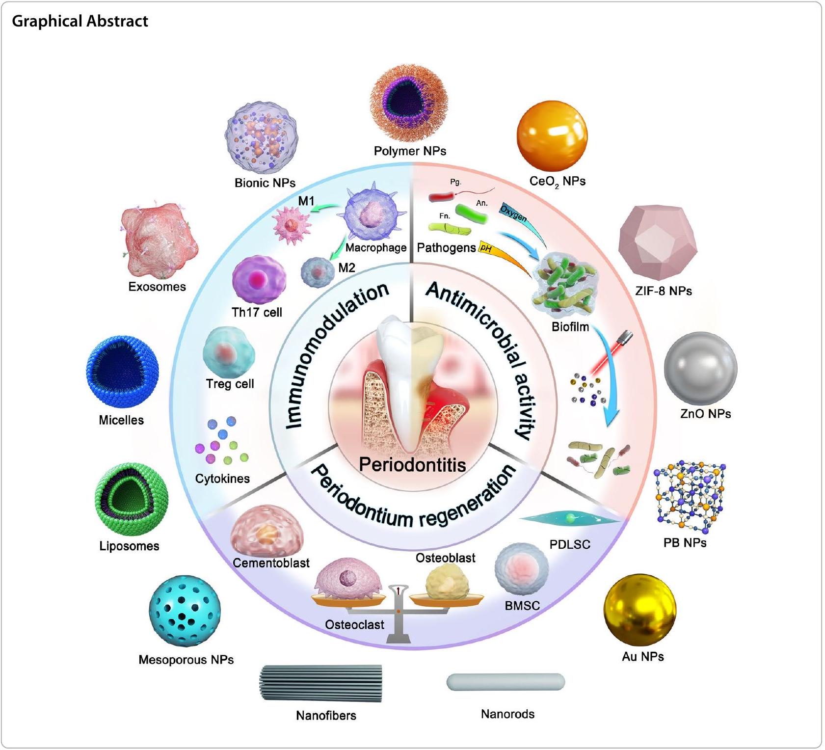
Periodontitis is a chronic inflammatory disease caused by the local microbiome and the host immune response, resulting in periodontal structure damage and even tooth loss. Scaling and root planning combined with antibiotics are the conventional means of nonsurgical treatment of periodontitis, but they are insufficient to fully heal periodontitis due to intractable bacterial attachment and drug resistance. Novel and effective therapeutic options in clinical drug therapy remain scarce. Nanotherapeutics achieve stable cell targeting, oral retention and smart release by great flexibility in changing the chemical composition or physical characteristics of nanoparticles. Meanwhile, the protectiveness and high surface area to volume ratio of nanoparticles enable high drug loading, ensuring a remarkable therapeutic efficacy. Currently, the combination of advanced nanoparticles and novel therapeutic strategies is the most active research area in periodontitis treatment. In this review, we first introduce the pathogenesis of periodontitis, and then summarize the state-of-the-art nanotherapeutic strategies based on the triple concerto of antibacterial activity, immunomodulation and periodontium regeneration, particularly focusing on the therapeutic mechanism and ingenious design of nanomedicines. Finally, the challenges and prospects of nano therapy for periodontitis are discussed from the perspective of current treatment problems and future development trends.

Keywords Periodontitis, Nanotherapeutics, Antimicrobial activity, Immunomodulation, Periodontium regeneration, Synergistic therapy

Introduction

Periodontitis is a chronic inflammatory disease with clinical manifestations such as bleeding gums, recurrent swelling and pain, and resorption of alveolar bone. If inadequately treated, it can lead to loosening and loss of teeth, chewing function, and even effects on the digestive system [1]. In the world, periodontitis affects of the global population, with prevalence rates ranging from 8 to in developing countries and in developed countries [2]. Periodontitis is more common in the aged due to chronic and cumulative injury. Two thirds of people over 65 years old in the United States have chronic periodontitis [3]. In recent decades, the incidence and prevalence of severe periodontitis in Asian countries, such as India, China and Japan, has been on the rise, and
age may be a critical factor for the increasing trend [4]. In addition, there is growing clinical evidence that periodontitis places a huge burden on the public health care system due to the close links to other diseases such as diabetes, Alzheimer’s disease, rheumatoid arthritis, colitis and even cancer [5-7].
Most patients with periodontitis need to be treated with scaling and root planning to remove plaque or calculus [8]. However, clinical studies have shown that scaling and root planning cannot adequately alter the composition of the subgingival microorganism biofilm, thus nonsurgical adjunctive drug therapies have been proposed to enhance treatment outcomes including the local delivery of drugs, systemic antibiotics and systemic host modulation agents.
Several meta-analyses showed that systemic antibiotics and systemic host modulation agents are beneficial for periodontitis control [9, 10]. In the clinic, broad-spectrum antibiotics can be used alone or in combination with other antibiotics for Gram-negative bacteria within 1-3 weeks [11]. The long-term use of systemic antibiotics and host modulation agents may be associated with the risk of antibiotic resistance, microbial disorders and drug interactions. Local delivery of drugs into periodontal pockets is beneficial in treating patients with deep pockets or recurrent periodontitis, and administration options include nanoparticle, hydrogel, powder and fiber delivery systems.
Nanoparticles have significant promise for addressing the challenges of periodontal drug delivery. Nanoparticles, including liposomes, polymeric nanoparticles, polymeric micelles and nanofibers, provide great flexibility in changing chemical composition, size, surface
charge and other characteristics, which can ensure stable cell targeting and oral retention. Existing data suggest that nanoparticles protect drugs from pH influence and enzymatic degradation in the periodontal lesion [12]. Importantly, the nanoparticle structure can be designed to respond to reactive oxygen species (ROS), pH , or enzyme-response mechanisms in the pathological microenvironment for controlled drug release. We summarize nano delivery systems strategies for the treatment of periodontitis in terms of three main aspects: antibacterial therapy, immunomodulatory therapy and tissue regeneration (Fig. 1). In addition, the high surface area to volume ratio of nanoparticles enables high drug or drug combination loading, resulting in synergistic therapeutic efficacy by combining multiple treatment aspects. For example, metal nanoparticles such as silver, gold, and cerium, can directly sterilize or
Fig. 1 Schematic overview of nanotherapeutics via triple concerto of antimicrobial activity, immunomodulation and periodontium regeneration
enhance drug water solubility for transport to bacteria, simultaneously eliminating bacterial resistance through the combination of photodynamic therapy. The nanoparticles release natural active ingredients (quercetin, baicalin, caffeic acid phenethyl ester) into the periodontal pocket to reverse excessive immune response or repair the damaged periodontal tissue [13, 14]. Wu et al. used baicalein-loaded mesoporous Prussian blue nanoparticles to develop an antimicrobial and immunomodulatory synergistic nanotherapeutic strategy for the treatment of periodontitis [15]. Novel nanoparticle delivery systems, such as cell membrane biomimetic nanoparticles and exosomes, will further expand the available strategies for using nanoparticles in periodontitis treatment. In this context, we present a systematic review of the application of nanodelivery systems in the treatment of periodontitis. This review will highlight the treatment strategies and introduce the rational design of nanodelivery systems. This review will also
discuss the future challenges and research directions of nano systems for periodontitis treatment.

Pathophysiology of periodontitis

An introduction to the pathological background of periodontitis is necessary for the understanding of nanotherapeutic strategies. In healthy periodontal tissue, there is a state of homeostatic balance between the microbial community and host immunity involving a minimal number of neutrophils, plasma cells, etc. [16, 17]. However, in periodontitis tissues, intractable pathogenic biofilms are present accompanied by a persistent inflammatory response and alveolar bone damage (Fig. 2). The pathogenesis of periodontitis is not completely clear, but studies have confirmed that genetic factors and plaque biofilms are underlying inducing factors. The pathogenic bacteria related to periodontitis include Porphyromonas gingivalis (Pg), Prevotella intermedia, Actinobacillus actinomycetemcomitans, Fusobacterium nucleatum
Fig. 2 The pathogenic factors of periodontitis
(Fn), Tannerella forsythia, and Treponema denticola, Treponema, Prevotella, Selenomonas, Peptostreptococcus, Anaeroglobus, Desulfobulbus speciesand Lachnospiraceae [18, 19]. As many as 800 different species have been identified in dental plaque [20]. The above bacteria gather and adhere on the tooth surface to form a structured community, which is called dental plaque biofilm. Pathogenic bacteria can colonize the surface of teeth by releasing exoenzymes to produce exopolysaccharide from sucrose, which accelerates local colonization of microorganisms and establishes a meshwork of biofilm matrix [21]. The dental plaque biofilm matrix is composed of complex polymeric substances such as exopolysaccharides (glucan, fructan polysaccharides), proteins (amyloids, adhesions, DNA binding proteins), eDNA, proteoglycans, glycoproteins (Slayers, adhesins), lipids, lipopolysaccharides (LPS) and other macromolecules derived from microorganisms or hosts [20]. Meanwhile, the acidic microenvironment ( ) of the tooth surface is formed by the fermentation of bacteria within the biofilm matrix, which leads to a flourishing proliferation of pathogenic bacteria [21]. Identifying these intricate pathogenic processes may provide new opportunities for periodontitis treatment by dental biofilm control. It is worth noting the plaque biofilms are not the decisive factor for periodontal damage. Although bacterial infection is a necessary condition for the development of periodontitis, the immune response of the host is crucial for the development and tissue destruction of periodontitis [22]. The clinical consensus suggests that of tissue destruction is caused by inflammatory activation via host immunity [23].
Neutrophils, as the first line of nonspecific immunity against pathogenic bacteria, attempt to engulf or kill pathogens, but they are overwhelmed by a large number of pathogens in periodontitis [24]. As antigen-presenting cells, dendritic cells (DCs) bridge the innate and adaptive immune responses in periodontitis [25]. Immature DCs have a high phagocytosis capacity and are able to rapidly capture invading microorganisms [26]. Mature DCs present antigens to the initial cell [22]. The differentiation of initial cells into T helper (Th)1, Th2, Th17 and regulatory T (Treg) cells [27]. Interferon- (IFN- ) and interleukin (IL)-12 secreted by DCs induce Th1 cell formation in the inflammatory environment [28]. Studies have revealed that Th1 cells are involved in the progression of osteoclastogenesis and alveolar bone loss. Th2 cells are induced to form by IL-4, and IL-4 secreted by Th 2 cells further leads to B cell activation and antibody production [29]. IFN- secreted by Th1 cells can block the proliferation of Th2 cells, and high concentrations of IL-4 can also block the generation of Th1 cells [30]. In addition, depending on the microbial signal received by pattern recognition receptors, DCs
promote Th 17 or Treg cells differentiation by releasing the cytokines of tumor necrosis factor (TNF- )/IL- 6 or transforming growth factor (TGF- )/IL-10, respectively [31, 32]. Th17 cells can trigger pro-inflammatory signals, recruit and activate neutrophil granulocytes, upregulate the expression of antimicrobial factors, and promote the clearance of extracellular bacteria [33]. Moreover, activated Th17 cells secrete IL-17 to activate the Janus Kinase/signal transducer and activator of transcription signaling cascade and increase the expression of receptor activator of nuclear factor kappa-B ligand (RANKL), eventually leading to accelerated osteoclastogenesis [34]. Activated Treg cells produce the anti-inflammatory cytokines IL-10 and TGF- , which inhibit the Th17related response and contribute to the maintenance of immune homoeostasis [35]. Immune disorders also promote macrophage aggregation and M1-type polarization [36]. M1-type macrophages exacerbate inflammation by secreting monocyte chemotactic proteins, IL-6, TNF- and ROS. In addition, M1-type macrophages further act on Th17 cells, eventually leading to bone tissue injury [37, 38]. At the same time, the gingiva, cementum and periodontal ligament in the periodontium also suffer from similar destructive effects, which together lead to the aggravation of periodontitis.

Nanotherapeutic strategies for periodontitis

Pathogenic bacteria are the initial factor in periodontitis, and the overactivated host immune response is the determinant of periodontitis exacerbation. The imbalance of pathogenic bacteria and host immunity leads to the persistence of inflammation and the destruction of periodontal tissues. Therefore, antibacterial agents, immune regulation and periodontium regeneration are the main strategies for periodontitis treatment.

Antimicrobial nanotherapeutic strategies

Antimicrobial therapy for periodontitis has been a topic of constant discussion among researchers. Antibiotics are an important therapeutic strategy for periodontitis. However, high doses of antibiotics may create resistance problems, decreasing their therapeutic effect in the long term [39, 40]. Therefore, the development of innovative antimicrobial strategies is significant importance. In recent decades, nanoparticle delivery systems designed to overcome antibiotic resistance or metallic nanoparticles acting alone as effective antimicrobial agents have been developed for antimicrobial treatment in periodontitis therapy. In addition, nanomaterials or nanodelivery systems loaded with photosensitizers can exert bactericidal effects through photothermal therapy (PTT) and photodynamic therapy (PDT). The current nanoparticle-based antibacterial, immune regulation and periodontium
regeneration treatment strategies for periodontitis are summarized (Table 1).

Antibiotic

Antibiotics can induce bacterial death by inhibiting protein synthesis, interfering with deoxyribonucleic acid synthesis or disrupting the cell wall [40, 41]. For example, tetracyclines (minocycline, hygromycin hydrochloride) can attach to the 30 S ribosomal subunit of bacteria and thus inhibit protein synthesis. Quinolones (moxifloxacin hydrochloride, ciprofloxacin hydrochloride) can interfere with deoxyribonucleic acid synthesis by inhibiting gyrase activity, causing irreversible damage to bacterial chromosomes [42, 43]. The use of nanomaterials as carriers for loading antibiotics can reduce the dose of antibiotics administered, reduce the frequency of administration and decrease the likelihood of bacterial resistance, which is an effective strategy for the treatment of periodontitis.
Yao and colleagues prepared poly (ethylene glycol)-poly (lactic acid) nanoparticle-loaded minocycline delivery systems (MIN-NPs) using an emulsion/solvent evaporation method. Nanoparticles can penetrate deeply into the periodontal pocket below the gingiva due to their small
size, thus improving the antimicrobial efficacy of topical administration [44, 45]. MIN-NPs can provide sustained drug release, reduce the frequency of administration and avoid burst release of the drug. The pharmacokinetic profile showed that MIN-NPs had the longest duration of action in the gingival sulcus, compared to minocycline hydrochloride ointment and minocycline solution. The concentration of MIN-NPs in the gingival sulcus decreased slowly, maintaining the drug at an effective concentration ( ) after 12 days. In vivo pharmacodynamics in beagle dogs with periodontitis showed significant improvements in periodontal pocket depth, plaque index and gingival index after 6 days of topical administration.
Moxifloxacin hydrochloride has also been used within nanosystems to improve the efficacy of periodontitis. Sarwar Beg et al. prepared poly (D, L-lactide-co-glycolide) (PLGA) nanoparticles loaded with moxifloxacin hydrochloride (MOX-PLGA) into a poloxamer 407 hydrogel to produce an in-situ gel [46]. The in vitro release profile results showed that the nanoparticles loaded into the in-situ gel resulted in a much lower drug release of in the first 12 h compared to the group without gel
Table 1 Antimicrobial nanotherapeutic strategies for periodontitis treatment
Nanoparticles Delivery systems Drugs Outcome References
MIN-NPs Polymeric nanoparticles Minocycline Clinical periodontal parameters (periodontal pocket depth, plaque index and gingival index) were significantly improved [45]
MOX-PLGA nanoparticle Polymeric nanoparticles Moxifloxacin hydrochloride Prolonged retention and drug release of a nanoloaded moxifloxacin in situ gel system in periodontal pockets [46]
Ciprofloxacin hydrochloride loaded dual corona vesicles Vesicles Ciprofloxacin hydrochloride Used double crown vesicles as drug carriers, 50% of the normal dose of antibiotics can achieve antibacterial purposes, which will reduce the possibility of antibiotic resistance [47]
AuNCs Au nanoclusters / Reduce the possibility of bacterial resistance [50]
PCL-OTCz Polymeric nanoparticles Oxytetracycline hydrochloride and ZnO NPs Excellent antibacterial activity of oxytetracycline hydrochloride in synergy with ZnO against mixed cultures of Gram-negative anaerobic bacteria [64]
Mino-ZnO@Alb NPs Nano-albumin ZnO NPs and minocycline Synergistic antibacterial action, reduced the dosage of Minocycline and avoiding the development of drug resistance [65]
Gel MA-Au NBPs@SiO2 Au NBPs Minocycline Eliminated periodontal pathogens in periodontal pockets, photothermal treatment to maintain low bacterial retention after medication [68]
-silane@Ce6/C6 MNPs Polymeric delivery system Ce6 and C6 Strong antibacterial activity against plaque biofilms [73]
NaYF4-Mn@Ce6@silane Polymeric nanoparticles Ce6 Realized the conversion of light emission and enhance the PDT effect [74]
TAT-Ce6/TDZ NPs Polymeric nanoparticles Ce6 Promoted the penetration of the bacterial cell membrane through the mediation of TAT peptide [76]
sPDMA@ICG NPs Polymeric delivery system ICG Significantly promoted the adsorption and penetration of ICG into bacterial cells, exhibiting synergistic antibacterial properties of PTT and PDT [79]
incorporation, indicating that the gel greatly reduced the burst release of the drug. In vivo gamma scintigraphy showed that the drug in MOX-PLGA remained in the periodontium without entering the body circulation in contrast to the moxifloxacin hydrochloride solution, which facilitated the local antimicrobial effect of moxifloxacin hydrochloride in the periodontium.
Xi and his colleagues proposed the encapsulate the ciprofloxacin hydrochloride in dual corona nanovesicles to reduce the antibiotic dose and antimicrobial resistance (Fig. 3A) [47]. The -b- -stat-Phe corona vesicles were synthesized by Z-Lys-NCA and Phe-NCA monomers. Then, PEO corona vesicles of the block copolymer using -caprolactone were prepared and and PEO were co-assembled into PEO/polypeptide dual corona vesicles by the solvent-switch method. The antibacterial polypeptide P (Lys-stat-Phe)] coronas provide vesicles with positive charges to attach to the bacterial surface. PEO coronas exhibit protein exclusion ability and penetrate extracellular polymeric substances to help antibiotics penetrate biofilms. The results showed that the minimum biofilm clearance concentrations of ciprofloxacin hydrochloride for Escherichia coli and Staphylococcus aureus biofilms were and , respectively, while the CIP-loaded dual corona vesicles decreased to and , respectively. In addition, scanning electron microscopy (SEM) images showed that the biofilm structure changed from tight to sparse and that the number of bacteria decreased dramatically. Nearly bacteria could be killed at1 hour.

Metallic nano-antibacterial agent

The misuse of antibiotics in the treatment of periodontitis is a growing concern due to the problem of bacterial resistance in global public health. Therefore, there is an urgent need for new alternatives to replace traditional antibacterial therapy, and the development of metallic nanomaterials has created a new opportunity for antibacterial therapy. Among them, gold, silver, titanium dioxide, zinc oxide ( ZnO ) and other nano-antibacterial agents have great potential in antimicrobial therapy. In this section, we introduce only the representative antibacterial properties of metallic nano-antibacterial agents. The diverse therapeutic effects of metal nanoparticles will be further introduced in the chapter on synergistic nanotherapeutic strategies.
Compared to antibiotics, Au nanoparticles (AuNPs) exhibit unique physical properties, which include the ability to interact directly with phospholipid bilayer, bind to cytoplasmic proteins, form ROS, and thus produce significant antibacterial activity [48, 49]. Recently, researchers have tried to reduce the toxicity of
Au nanoparticles by controlling their size or redesigning their shape and surface chemistry, making them better for use in medicine. Ultra-small gold nanoclusters (AuNCs) were prepared by a simple one-pot method, which composed of 25 Au atoms and 18 thiolate ligands with ultra-small structure (Fig. 3B) [50]. High-resolutiontransmission electron microscopy (TEM) showed that AuNCs exhibited roughly spherical shapes with a homogeneous and well-dispersed distribution, and the particle size of AuNCs ranged from 1.5 to 4.0 nm with an average diameter of . The zeta potential of AuNCs was -38.8 mV . The results showed that the growth of Fn was significantly inhibited by AuNCs at a concentration of 0.2 mM , and cell wall integrity was damaged. These results indicated that AuNCs induced lysis of the Fn membrane potential is crucial for bacterial energy metabolism and is an early sign of membrane damage. The red/ green fluorescence ratios were reduced by approximately after AuNCs incubation, and the membrane potential reduction indicated that AuNCs treatment caused serious damage to cell membranes, possibly inhabiting bacterial growth via a membrane depolarization mechanism. In addition, the level of ROS in Fn was enhanced after AuNCs treatment. It would be difficult for bacteria to develop resistance to AuNCs based on their antimicrobial mechanisms.
ZnO nanoparticles have excellent safety and biocompatibility with multiple antibacterial mechanisms, in the tissue engineering, food packaging and dental materials fields with a wide range of roles [51-53]. The antibacterial mechanisms of ZnO nanoparticles include (1) electrostatic interaction with the cell wall [54-56] (2) adsorption of on bacterial surfaces, altering membrane permeability [57-59] (3) production of ROS to exert antibacterial effects [60, 61], and ZnO nanoparticles thus have enormous potential in periodontitis treatment. Eliseu A. Münchow and his colleagues loaded ZnO nanoparticles onto poly(has-lactide)/gelatine blend fibrous membranes by electrostatic spinning [62]. The results of agar diffusion experiments showed that the loaded ZnO nanoparticles inhabited Pg and Fn with inhibition zones ranging from 6 to 15 mm in diameter, showing beneficial antibacterial effects. Furthermore, it was reported that the electrospun polymer film loaded with ZnO not only has antibacterial properties, but its production of reactive oxygen species can promote cell proliferation and wound healing mediated by growth factors, which offers the possibility of treating periodontitis [63].
The combination of antibiotics and ZnO nanomaterials has become an actively explored strategy for periodontitis treatment. For instance, the polycaprolactone nanofibers were selected as drug carriers loaded with oxytetracycline hydrochloride and ZnO nanoparticles
Fig. 3 Schematic illustration of periodontitis treatment by antimicrobial and metallic nano-antibacterial agent. A Schematic diagram of ciprofloxacin hydrochloride-loaded dual corona vesicles. Reprinted with permission from Ref. [47] Copyright American Chemical Society. B Schematic diagram of AuNCs with antimicrobial nanotherapeutic strategies. Reprinted with permission from Ref. [50] Copyright Springer Nature
(PCL-OTCz) [64]. Four Gram-negative anaerobic bacteria (Pg, Aggregatibacter actinomycetemcomitans, Prevotella intermedia, Treponema denticola) were selected to
form mixed bacterial cultures. PCL-OTCz showed powerful antibacterial activity compared with oxytetracycline hydrochloride or ZnO nanoparticles, individually.
In another study, ZnO nanoparticles and minocycline were coloaded on a nano-albumin carrier (Mino-ZnO@ Alb NPs) and incorporated into a pH-responsive Carbopol 940 hydrogel [65]. Mino-ZnO@Alb NPs showed pH-responsive characteristics with release of minocycline at pH 6.5 , while only approximately of minocycline was released at pH 8.5 , probably due to higher minocycline partitioning under acidic conditions. The results of the inhibition circle method showed that MinoZnO@Alb NPs could significantly inhibit the bacterial proliferation of Pg, Streptococcus oralis, Streptococcus sanguis and Periodontopathic bacteria. These results indicate that the combination of zinc oxide nanoparticles and antibiotics is a potential antibacterial strategy for the treatment of periodontitis.

Photothermal/photodynamic therapy

PTT and PDT have been proposed for the antimicrobial treatment of periodontitis. In comparison to conventional antibiotic treatment, PTT and PDT are less likely to cause resistance in periodontitis-causing bacteria. Second, the wavelength of the light source used for photothermal antimicrobial treatment has deeper tissue penetration than that of other light-induced treatments, allowing it to penetrate deeper into the periodontium [66, 67]. Finally, by controlling the specific area and intensity of light exposure, the side effects of photothermal antimicrobial treatment on normal tissues can be reduced.
PTT antimicrobial therapy uses photothermal material to produce a lethal effect on bacteria by heating up under irradiation. Lin et al. explored a PTT therapeutic strategy by first synthesizing a near-infrared responsive photothermal material of Au nano bipyramids (Au NBPs) [68]. Then minocycline-loaded mesoporous silica was modified on the Au NBPs surface, and Au NBPs@ were finally mixed with gelatine methacrylate to prepare a hybrid hydrogel (Gel MA-Au NBPs@SiO 2 ). The temperature of Au NBPs@ increased under near infrared (NIR) irradiation. Furthermore, the diffusion and release of minocycline accelerated with increasing power of irradiation. Compared to non-NIR irradiation, the antibacterial rate of Gel MA-Au NBPs@SiO against Pg increased to via the increase in NIR irradiation power. The live/dead staining results showed that the antibacterial effect of Pg was up to on the third day but only on the seventh day, while the antibacterial effect could be increased to on the eighth day after NIR treatment. This suggests that light and heat can cooperate with antibiotics for antibacterial treatment without developing resistance.
PDT antimicrobial therapy refers to the use of a photosensitizer that produces ROS in response to light stimulation, thus leading to rapid oxidation of lipids in bacteria,
and the destruction of fragile lipid membranes in periodontitis treatment [69, 70]. Chlorin e6 (Ce6) is widely used in PDT therapy because of its strong tissue penetration, great biocompatibility and high yield of singlet oxygen [71, 72]. Sun et al. prepared a core-shell structure nanodrug delivery system ( -silane@Ce6/C6) including nanoparticles, Ce 6 and coumarin 6, for PDT antimicrobial therapy [73]. Transmission electron microscopy (TEM) images of nanoparticles show a diameter of approximately 8 nm and -silane@ a diameter of about -silane@Ce6/ C 6 produced under light irradiation and had a killing effect on Streptococcus sanguis, Pg and Fn Under light irradiation, -silane@Ce6/C6 reduced Fn and Pg biofilm colony formation units by approximately 4 log and 5 log, respectively. Notably, -silane@Ce6/ C6 can be magnetically induced to concentrate at plaque biofilm sites to further enhance the antibacterial effect. These results indicate that PDT combined with magnetic targeted nanoparticles has potential for antimicrobial therapy for periodontitis.
Although PDT has made important progress in the treatment of periodontal disease, there are some serious problems that still need to be solved before clinical application. The most important limitation of conventional PDT is the weak tissue penetration of ultraviolet or visible light. Therefore, it is highly desirable to design and prepare a PDT system that relies on with the infrared irradiation light, which can perform deep-tissue penetration. Based on this, a strategy was proposed to combine the photosensitizer Ce6 with upconversion nanoparticles (UCNPs) NaYF4:Yb, Er [74]. The combination of Ce6 UCNPs was realized via an amphiphilic silane modification technique (NaYF4@Ce6@silane NPs), which involved the hydrophobic-hydrophobic interaction between the hydrophobic side chain of the silane and hydrophobic groups on the surface of UCNPs [75]. In addition, because the PDT function of the Ce6 molecule should be triggered by excitation by red light, Mn doping is involved in this work, which greatly improves the probability of the red emission transition (NaYF4-Mn@ Ce6@silane) (Fig. 4A). The TEM image showed that the size distribution of NaYF4@Ce6@silane NPs was approximately 30 nm , and a thin layer of silane (approximately ) was observed on the surface of the nanoparticles after silane coating. The result showed that was selected to dope into UCNPs to realize the enhancement of red-light emission, and the enhanced upconversion red emission can further improve the PDT effect. The colony forming units of and . intermedia after NaYF4-Mn@Ce6@silane NPs with 980 nm irradiation decreased by more than , and the biofilm matrix was easily disrupted with deeper penetration of infrared
Fig. 4 Schematic illustration of periodontitis treatment by photothermal/photodynamic therapy. A The schematic diagram of NaYF4-Mn@Ce6@ silane. Reprinted with permission from Ref. [74] Copyright Multidisciplinary Digital Publishing Institute. B Preparation and against periodontitis of TAT-Ce6/TDZ NPs. Reprinted with permission from Ref. [76] Copyright American Chemical Society. C Preparation of sPDMA@ICG NPs and treatment of synergistic PTT and PDT on periodontitis. D TEM images of bacteria after processions of PBS (the control), sPDMA@ICG NPs with and without laser irradiation. E Photos of plaque biofilms after various processions. F Comparison for the mean fluorescence intensities of produced DCF in periodontium. Infrared thermal images of periodontium during laser irradiation after administration of PBS (the control) and sPDMA@ICG NPs. **indicates p < 0.01 compared to the control (+); ##indicates p < 0.01 for comparison between two groups. Reprinted with permission from Ref. [79] Copyright Springer Nature
light. This highly efficacy against periodontitis-related biofilms should be attributed to the high hydrophilic surface after silane modification, as well as to the upconversion luminescence triggered PDT. This upconversion PDT design can overcome the problems of conventional PDT and provide effective nano strategies for the treatment of periodontitis.
However, PDT does not always achieve the desired therapeutic outcomes since photosensitizers Ce6 have strong hydrophobic properties and are not taken up efficiently by periodontal pathogenic bacteria. Li et al. designed a nanosystem to improve Ce 6 solubility and enhance its bacterial adsorption by promoting its interaction with negatively charged cell walls and penetration through cell membranes [76]. They first hydrophilicallymodified Ce6 via conjugation with TAT peptide, a cationic cell-penetrating peptide (TAT-Ce6). Then, TAT-Ce6 was loaded with the antibiotic agent tinidazole to prepare self-assembled nanoparticles (TAT-Ce6/TDZ NPs) to achieve synergistic anti-periodontitis effects by combining PDT and antibiotic therapy (Fig. 4B). TEM image showed that TAT-Ce6/TDZ NPs had a regular spherical
shape and exhibited a more compact inner structure. The particle size of TAT-Ce6/TDZ NPs was , and the zeta potential was approximately +40.1 mV , which confirmed the surface distribution of the positively charged TAT peptide. The UV absorption of ABDA attenuates when ABDA is decomposed by the ROS produced in the sample solution, and the degree of attenuation is positively correlated with the ROS generation level. The results showed that the attenuation rate of ABDA absorption in the solution of TAT-Ce6/TDZ NPs was much faster than that of free Ce 6 during 20 min of laser irradiation, indicating that TAT-Ce6/TDZ NPs had a much higher PDT efficiency. The zeta potentials of Pg were increased from -11.2 to +5.86 mV after 1 h of incubation with TAT-Ce6. The above results indicated that TAT-Ce6 significantly promoted the penetration of the bacterial cell membrane through the TAT peptide. TATCe6/TDZ NPs and TAT-Ce6 exhibited much stronger bacterial killing activity, owing to their more efficient absorption by the plaque biofilms via the mediation of TAT peptide. More importantly, TAT-Ce6/TDZ NPs exhibited much stronger bacterial killing activity than
TAT-Ce6 NPs with laser irradiation, further confirming their synergistic antibacterial efficacy through combining PDT and antibiotic therapy.
Indocyanine green (ICG), a photosensitizer with PDT properties, has been approved for clinical use by the US Food and Drug Administration [77]. Nagahara et al. first explored PDT of photosensitizer indocyanine green, which has high absorption at a wavelengths of 800805 nm [78]. They designed ICG-loaded PLGA nanospheres coated with chitosan (ICGNano/c) and explored the PDT of ICGNano/c in Pg. The study showed that ICG-Nano/c with low-level diode laser ( ) irradiation showed a PDT-like effect, which might be useful for potential photodynamic periodontal therapy. Recently, combined treatment with PTT and PDT has further improved the efficiency of periodontitis treatment. However, due to its negative charge and water solubility, ICG has difficulty passing through bacterial cell membranes. To address this problem, Shi and his colleagues incorporated it into positively charged polycationic brush nanoparticles (sPDMA@ICG NPs) (Fig. 4C) [79]. CD-Br was synthesized by esterifying -cyclodextrin with 2-bromoisobutyryl bromide via an esterification reaction. Next, star-shaped polycationic brush poly (2-(dimethyl amino) ethyl methacrylate) was synthesized by an atom transfer radical polymerization reaction using CD-Br as an initiator. Finally, sPDMA@ ICG NPs loaded with ICG were prepared by the nanometer precipitation method. The average particle size of sPDMA@ICG NPs was 206 nm and the zeta potential was approximately +18.4 mV . On the one hand, the temperature of the sPDMA@ICG NPs solution was increased from 22 to after irradiation with an 808 nm laser ( ), which reflects an excellent PTT performance. On the other hand, sPDMA@ICG NPs also exert PDT
properties. The results suggested that sPDMA@ICG NPs can produce ROS after laser irradiation, as detected by SOSG. Confocal microscopy images show that sPDMA@ ICG NPs are effectively accumulate in bacterial cells after the administration of sPDMA@ICG NPs. TEM images showed that sPDMA@ICG NPs were clearly visible on the surface of Pg, and that the bacterial film ruptured and bacterial cells disintegrated after laser irradiation (Fig. 4D). In addition, sPDMA@ICG NPs with laser irradiation reduced the growing area of plaque biofilms derived derive from a rat model of periodontitis (Fig. 4E). After sPDMA@ICG NPs administration and laser irradiation, temperature and ROS levels were increased in rats with periodontitis, indicating that sPDMA@ICG NPs exert synergistic PTT and PDT effects in vivo (Fig. 4F).

Immunomodulatory nanotherapeutic strategies

In periodontitis, the presence of plaque microorganisms and their products can activate the host immune response [80]. Local host immune overreaction then, leads to increased inflammation and disruption of homeostasis, exacerbating periodontium lesions. Most tissue damage within periodontitis is caused by the host immune response rather than directly by the infecting microorganism [81, 82]. Therefore, in terms of therapeutic strategies, suitable immunomodulatory targets can be screened to modulate the host immune system to mitigate the inflammatory response. Recently, many nanosystems have been designed to modulate the function of immune cells and inflammation-associated cytokines to alleviate periodontal inflammation, and these nanosystems have achieved excellent therapeutic results both in vitro and in vivo (Table 2).
Table 2 Immunomodulatory nanotherapeutic strategies for periodontitis treatment
Nanoparticles Delivery systems Drugs Outcome References
Lipo-RSV Liposome Resveratrol Reprogramed the macrophages from M1- to M2-like phenotype, adjustment of the immune microenvironment [89]
DPSC-Exo/CS Exosomes / Suppressed periodontal inflammation by promoting the conversion of macrophages from a pro-inflammatory to an anti-inflammatory phenotype in the periodontium of mice with periodontitis [90]
CeO2@QU Nanoparticles Quercetin Scavenged ROS and regulated the conversion of M1 phenotype macrophages to the M2 phenotype to regulate the immune microenvironment [14]
3D-exos Exosomes / Alleviated Th17 cell/Treg imbalance and reduce inflammation [99]
PDLSC-exos Exosomes / PDLSC-exos transferred miR-155-5p into CD4 + T cells to affecting the Th17/Treg homeostasis [100]
Nano-BA/BE Polymeric nanoparticles Baicalein/Baicalin Alleviated the expression of II-1 -induced pro-inflammatory cytokines and regulating the immune microenvironment [104]
PDA NPs Polydopamine nanoparticles / Effectively scavenges ROS in the body, relieves oxidative stress and reduces local periodontal inflammation [105]

Remodeling macrophage polarization

In the immune system, immune cells such as macrophages play an important role as the host’s first line of defenses against microorganisms. When induced by different factors, macrophages polarize and develop different phenotypes, such as M1 and M2, both of which are involved in regulating the immune response [83]. M1-type macrophages produce the cytokines IL-6 and TNF- , which promote the inflammatory response [84, 85]. M2 macrophages can be further classified into alternatively activated macrophages (M2a), type 2 macrophages (M2b), deactivated macrophages (M2c), and M2-like macrophages (M2d) by different stimuli and transcription levels [86, 87]. M2a macrophages are induced by IL-4 and IL-13 and secrete profibrotic factors such as TGF- and insulin-like growth factor, and fibronectin contributes to tissue repair. M2b cells express and secrete substantial amounts of the anti-inflammatory cytokine IL-10 and low levels of IL-12, which is the functional conversion of M1 cells. M2c macrophages are induced by IL-10 and strongly exhibit anti-inflammatory activities by releasing large amounts of IL-10 [88]. M2d macrophages are also known as tumor-associated macrophages. In a word, M2 type macrophages exert antiinflammatory and angiogenic effects and promote tissue repair and wound healing. Modulation of macrophage phenotypes through nanosystems provides a promising therapeutic strategy for periodontitis.
Shi et al. developed a liposome loaded with resveratrol (Lipo-RSV) to polarize macrophages from the M1 to M2 phenotype (Fig. 5A) [89]. Lipo-RSV upregulated the mRNA expression levels of M2-related markers (CD206, Arg-1 and Chil3), and downregulated the mRNA expression levels of M1 macrophage markers (CD86, iNOS and CCR7) in activated macrophages. Lipo-RSV treatment increased the percentage of M2-like subpopulations ( ) by , while the percentage of M1-like subpopulations ( ) decreased by . The mechanistic results showed that Lipo-RSV inhibited the phosphorylation of STAT1 and promoted the phosphorylation of STAT3 in bone marrow-derived macrophages. In addition, the secretion of proinflammatory cytokines (IL-1 , IL-6, TNF- , and IL-12) and the level of ROS were attenuated, while the anti-inflammatory cytokine IL-10 was upregulated after Lipo-RSV treatment. This suggests that Lipo-RSV has the potential to treat periodontitis by regulating the polarization of M1 macrophages into M2-type macrophages.
Exosomes are nanosized ( ) vesicles secreted by various cell types that, in addition to acting as carriers to deliver drugs, may modulate macrophage phenotypes. Shen et al. isolated dental pulp stem cells from shed teeth from healthy donors, cultured them in medium and then
collected their exosome pellets by centrifugation for the treatment of mice with periodontitis [90]. To enhance the stability of the exosomes, chitosan hydrogels loaded with dental pulp stem cell exosomes (DPSCs-Exo/CS) were prepared in mixed culture with chitosan hydrogels at in a volume ratio. Approximately 7351 genes were regulated in the periodontium of periodontitis mice after DPSC-Exo treatment. CD206 cells are considered anti-inflammatory macrophages that promote the healing of periodontitis. The images showed more cells in the periodontium of DPSC-Exo/CS-treated mice. After DPSCs-Exo/CS administration, the expression of anti-inflammatory markers ( ) was significantly increased, and the expression of pro-inflammatory markers ( , ) was downregulated. The Gene Ontology term enrichment analysis results further indicated that DPSC-Exo/CS downregulated both the inflammatory response and immune response in mice with periodontitis. To explore the underlying mechanism by which DPSC-Exo facilitates the conversion of the macrophage inflammatory phenotype, miRNA sequencing (RNA-seq) data showed that miR-1246 accounted for of the total miRNA reads. These results suggested that DPSC-Exo/CS can promote the conversion of macrophages from a pro-inflammatory phenotype to an anti-inflammatory phenotype, and the mechanism of phenotype conversion might be linked with miR-1246 in DPSC-Exo.
ROS production is a major biological process in stimulated macrophages involved in the killing of phagocytized microorganisms [91]. However, excessive ROS may drive macrophages to the M1 phenotype to exacerbate the development of inflammation [92]. Cerium oxide ( ) is a nanoenzyme that can scavenge and efficiently by shifting the (reduced)/ (oxidized) forms, and presenting SOD- and CATmimetic activities [93]. Notably, the intrinsic reductive structures, such as a catechol group, a 2,3-double bond, and hydroxyl substitution in the heterocyclic ring, allow quercetin to scavenge ROS. Therefore, Wang et al. constructed a nanocomplex ( ) that integrates ROS-scavenging nanoenzymes and antioxidant natural flavonoids [14]. In their study, was first prepared by a hydrothermal method; 3-aminopropyltriethoxysilane was used to amino-functionalize , and the addition of quercetin with stirring yielded (Fig. 5B). @QU showed an octahedral morphology with an average particle size of 120 nm . The results showed fewer CD86-positive cells (40.5%) and significantly increased expression of CD206-positive cells ( ) after treatment. The images showed that the expression of the M2 biomarker was upregulated. Rats underwent ligature placement and
Fig. 5 Schematic illustration of periodontitis treatment by remodeling macrophage polarization. A Schematic representation of Lipo-RSV regulating macrophage phenotype through activation of p-STAT3 and downregulation of p-STAT1. Reprinted with permission from Ref. [89] Copyright Springer Nature. B Schematic illustration of preparation and therapeutic mechanism of nanoparticles. Reprinted with permission from Ref. [14] Copyright John Wiley and Sons
bacterial injection for 3 d to establish an animal model of periodontal inflammation. The results showed low fluorescence of ROS in vivo after treatment, and the expression of pro-inflammatory M1 biomarker also was downregulated after treated. These results confirmed that this system has the potential to regulate the immune microenvironment by
scavenging ROS and regulating the conversion of M1 phenotype macrophages to the M2 phenotype.

Restoring Th17/Treg cells balance

After receiving antigenic stimulation, primary cells can differentiate into different subtypes of T cells under different conditions. Among these, the balance
between Th17 and Treg cells is crucial in the periodontal immune response [94, 95]. Overreaction of Th17 cells to pathogens leads to increased expression of the proinflammatory cytokines IL-17 and IL-22 [96]. IL-17 has been shown to boost RANKL expression while inhibiting OPG expression in periodontal ligament cells, which might explain why Th17 cells promote alveolar bone loss [95]. However, Tregs can suppress the host immune response in equilibrium with Th17 cells in the periodontium [97, 98]. To date, no drug/nanosystem has been reported to directly regulate the Th17/Treg cells balance, but exosomes derived from periodontal ligament stem cells have been found to have the potential to regulate Th17/Treg cells.
Zhang et al. found that exosomes derived from mesenchymal stem cells (3D-exos) could regulate Th17/Tregs cell balance [99]. Notably, replacing the traditional 2D culture system with a 3D system could increase exosome production. The average particle size of the 3D-exos was . A significant Th17 reduction and Treg elevation were observed in the periodontium after treatment with 3D-exos in periodontitis mice. Gene Ontology analysis showed that differentially expressed genes in 3D-exo-treated mice with periodontitis were enriched for T-cell chemotaxis. These results suggested that 3D-exos can further regulate Th17/Tregs cells in a periodontitis mice model. Furthermore, RNA-seq and TargetScan results indicated that miR-1246 is the most differentially expressed miRNA in 3D-exo, which targets Nfat5. Nfat5 is a key factor that mediates Th17 cell polarization in a sequence-dependent manner. Therefore, 3D-exo suppresses Th17 cell differentiation by miR-1246 through downregulation of Nfat5 gene expression.
Zheng et al. investigated the effect of exosomes from periodontal membrane stem cells (PDLSC-exos) on Th17/Treg balance [100]. The expression of the Th17related transcription factor RAR-related orphan receptor C was upregulated and the Treg-related transcription factor forkhead Box P3 was down-regulated in periodontitis patients. This means that the Th17/Treg ratio is unbalanced in patients with periodontitis. The results showed lower Th17-related CD4 expression and increased Treg-related expression after treatment of PDLSC-exos in T cells, confirming the regulatory effect of PDLSCs-exos on the Th17/ Treg balance. In addition, the mechanistic results showed that PDLSC-exos transfer miR-155-5p into cells, which in turn regulates the expression of histone deacetylase protein in T cells, thus affecting the Th17/Treg homeostasis. Therefore, miR-155-5p may be a promising target for the treatment of immune imbalance in periodontitis. In summary, exosomes are a potential nano drug delivery system to regulate the balance of Th17 and Treg
cells, and their potential in the treatment of periodontitis needs to be further explored.

Regulating pro-/anti-inflammatory cytokine secretion

Inflammatory cytokines secreted from immune or tissue cells are key regulators of the immune response process, and pro-/anti-inflammatory cytokine imbalance is an important factor in the aggravation of periodontitis [37]. Cytokines, such as IL-1 , IL-6 and TNF- , activate inflammation-related transcription factors or activate related signaling pathways, thereby accelerating the process of periodontitis. The anti-inflammatory cytokines of IL-10, TGF- and IL-11, downregulate the expression levels of pro-inflammatory factors, protect the periodontium and inhibit the development of periodontitis [101-103]. It is extremely important to modulate pro-/ anti-inflammatory cytokine secretion for restore immune balance in periodontitis treatment.
Li et al. prepared baicalin and baicalein-loaded mesoporous silica nanoparticles (Nano-BA and NanoBE) to regulate inflammatory cytokine secretion [104]. The mesoporous silica nanoparticles were modified by 3-aminopropyl-triethoxy silane. An inflammation cell model was established by primary human gingival epithelial cells pretreated with IL-1 stimulation. Nano-BA and Nano-BE downregulated cytokines involved in the immune inflammatory response. Among them, epithelial cell-derived neutrophil-activating peptide 78, monocyte chemoattractant protein-1, and IL-8 function as chemokines leading to inflammation or tissue damage, while granulocyte colony-stimulating factor and granulo-cyte-macrophage colony-stimulating factor stimulate the differentiation and proliferation of hematopoietic stem cell immune cells.
In another study, Polydopamine nanoparticles (PDA NPs) were synthesized via self-polymerization with dopamine hydrochloride and a solution containing and ethanol [105]. PDA NPs effectively reduce the levels of TNF- and IL- inflammatory mediators. After PDA NPs treatment in mice, the cytokine levels of TNF- , IFN , and IL-1 in serum recovered to normal values. Moreover, all the levels of alanine aminotransferase, aspartate aminotransferase and alkaline phosphatase (ALP) were also in the reference normal ranges. Notably, PDA NPs efficiently reduced the level of ROS in LPS-induced local high fluorescence signals. The above results suggest that reducing ROS levels may help to regulate inflammatory cytokine levels during periodontitis treatment.

Periodontium regeneration nanotherapeutics strategies

The periodontium consists of the gingiva, periodontal ligament, alveolar bone and cementum, which provide physical and mechanical support for the teeth [106,
107]. Severe periodontitis leads to the loss of periodontal attachment, which is one of the major causes of tooth loss in adults. Therefore, the main goal of periodontitis treatment is to reduce the destruction of the periodontium, finally achieving periodontium regeneration. In recent years, researchers have proposed a series of advanced nanotherapeutic strategies to achieve periodontium regeneration by regulating cell differentiation and disturbing osteoclastogenesis (Table 3).

Promoting periodontal membrane stem cell differentiation

Periodontal membrane stem cells (PDLSCs), a subpopulation of mesenchymal stem cells, have self-renewal and immunomodulatory properties [108]. Moreover, PDLSCs can specifically repair the damaged periodontium [109]. PDLSCs can differentiate into fibroblasts, osteoblast-like cells and dental osteoclast-like cells to generate connective and dental osteoid tissue by nanomedicine (Fig. 6A) [110]. Therefore, regulating the differentiation of PDLSCs is a promising strategy for periodontium repair.
AuNPs promote the proliferation of human periodontal membrane stem cells via the classical Wnt -linked protein signaling pathway [111]. On this basis, Zhang et al. further investigated the potential of AuNPs to promote osteogenic differentiation of PDLCs [112]. AuNPs were prepared by chemical reduction using tetrachloroauric acid as the raw material. Next, L/D-cystine was modified on the surface of AuNPs. The results showed that L-cystine was more favorable AuNPs uptake than D-cystine. L-Cys-AuNPs promoted the proliferation of human PDLCs and upregulated the levels of the osteogenic genes ALP, collagen type I (COL-1), osteocalcin (OCN) and
Runt-related transcription factor 2 (RUNX2). Pharmacological activation of autophagy was also significantly increased via osteoblast-differentiation activity. The results showed that the expression of autophagy-related genes and the levels of LC3 and SQSTM1 were increased in human PDLSCs cells after L-Cys-AuNPs treatment. A large amount of newly-formed alveolar bones and newlyformed periodontal ligaments were observed in a peri-odontal-defect rat model after L-Cys-AuNP-treatment. The microcomputed tomography (micro-CT) results of BV, bone volume/tissue volume (BV/TV), trabecular number (Tb.N), and Tb.Th indicated that L-Cys-AuNPs had more favour bone regeneration capacity than D-CysAuNPs. This study provides a new approach for the treatment of periodontitis with chiral-modified nanoparticles.
Liu et al. developed calcium fluoride nanoparticles (Nano-CaF2) for the osteogenic differentiation of PDLSCs. Calcium ions ( ) are important throughout the cycle of bone formation to maturation and have the potential to contribute to bone differentiation [113]. Fluoride ions can inhibit tooth damage and play an important role in the suppression of dental caries by decreasing demineralization and increasing of remineralization. Nano- with an average particle size of 53 nm was prepared by a spray dryer. The flexural strength, elastic modulus and hardness of Nano- were and , respectively, which exceeded those of a commercial dental composite. The ALP activity after Nano-CaF treatment of human PDLSCs was 57, 78 and 55-fold higher than that of the control group on days 7,14 and 21 , respectively. Nano- promoted the osteogenic
Table 3 Periodontium regeneration nanotherapeutic strategies for periodontitis treatment
Nanoparticles Delivery systems Drugs Outcome References
L-Cys-AuNPs Au nanoparticles / Promoted proliferation of human PDLCs, enhances their ALP activity and upregulated mRNA levels of osteogenic genes [112]
Nano- Polymeric nanoparticles / Increased levels of human PDLSCs and osteogenic genes in dental bone promote osseous differentiation and contribute to periodontium regeneration [113]
h-PDLSCs-exosomes Exosomes / Upregulated of Runx2 and OCN mRNA levels in PDLSCs restores osteogenic differentiation in PDLSCs [116]
PLA/CS PLA/CS nanofiber / Promoted proliferation and osteogenic differentiation of BMSCs and upregulated the expression level of osteogenic genes [121]
PLA/CA PLA/CA nanofiber / Increased expression levels of cell mineralization genes and formation of mineralized junctions BMSCs, promoting osteogenic differentiation [122]
M2-Exos Exosomes / Increased expression levels of osteogenesis in BMSCs while restrained expression levels of osteoclast formation in BMDM [125]
AMG-487 NP Liposomal AMG-487 NP Reduced the number of osteoblasts and inhibits alveolar bone loss [129]
Fibrin-ACP Chitosan nanoparticles -aminohexanoic acid Promoted the differentiation of cementoblasts [134]
Fig. 6 A PDLSCs through self-renewal, differentiating into osteoblasts and fibroblasts and regulating the host immune response to maintain the periodontal homeostasis. Reprinted with permission from Ref. [34] Copyright Oxford University Press. B Schematic illustration of M2-Exos promoting osteogenic differentiation of bone marrow stromal cells and inhibiting osteoclast formation of bone marrow-derived macrophages. Reprinted with permission from Ref. [125] Copyright Springer Nature
and cementogenic induction of human PDLSCs by upregulating the expression of cementum adherence protein, cementum protein 1 and bone sialoprotein. Furthermore, Nano- upregulated the osteogenic gene expression of RUNX2 and COL-1 in human PDLSCs.
Exosomes derived from healthy PDLSCs are potential components of osteogenic differentiation [114, 115]. Lei et al. isolated h-PDLSCs-exosomes from periodontal ligament tissue derived from healthy donors [116]. The diameter of h-PDLSCs-exosomes was 121.4 nm . h-PDLSCs-exosomes upregulated the mRNA levels of RUNX2 and OCN in the inflammatory periodontal ligaments of patients with periodontitis and promoted the expression of OCN osteogenesis-related proteins. Mechanistic studies found that h-PDLSCs-exosomes promote osteogenic differentiation by intervening Wnt signaling pathway. The mRNA levels of Wnt1, Wnt3a, Wnt10a, and -Catenin were decreased in the inflamed periodontal ligaments of periodontitis patients after the treatment of h-PDLSCs-exosomes. However, the report indicated that the inflammatory environment leads to overactivation of Wnt signaling in PDLSCs, which inhibits the differentiation of PDLSCs in periodontitis [117]. Based on report, they believe that the contradictory results might be attributable mainly to the different sources and different culture conditions of PDLSCs. These results suggested that exosomes derived from PDLSCs can rescue the osteogenic capacity of PDLSCs, providing a new strategy for the treatment of alveolar bone loss in periodontitis.

Promoting bone marrow stem cell differentiation therapy

Bone marrow stem cells (BMSCs), which can be isolated and expanded in vitro, show differentiation capacity and have received widespread attention in periodontium regeneration [118, 119]. The local injection of BMSCs reverse receptor activator of NF-кВ ligand/Osteoprotegerin (RANKL/OPG) expression contributed to the regeneration of periodontium in periodontitis mice [120]. A nanodelivery system consisting of chitosan nanoparticles incorporated into polylactic acid nanofibers (CS/PLA nanofibers) promoted the osteogenic differentiation of BMSCs and enhanced extracellular matrix mineralization [121]. The addition of chitosan nanoparticles increased the Young’s modulus and the stress and strain at break of the composites of fibers to resist masticatory forces while maintaining regenerative space. The scaffold architecture of CS/PLA nanofibers provides topographic cues to adherent cells, leading to cells along the axes of the architecture. The ability to control cellular alignment on scaffolds is advantageous for tissue regeneration in a specialized direction, such as the periodontal ligament. The result of alizarin red staining suggested that CS/
PLA nanofibers could induce BMSCs to form mineralized nodules. CS/PLA nanofibers upregulated the mRNA expression levels of osteoblast-related factors, such as RUNX2, OPG, and RANKL. In another study, PLA/calcium alginate (PLA/CA) nanofibers produced similar results as to CS/PLA nanofibers by promoting the bone differentiation of BMSCs [122]. It can be concluded that combining polysaccharide composites with PLA nanofibers to promote BMSCs differentiation is a potential periodontitis treatment strategy.

Disturbing osteoclastogenesis

An important feature of periodontitis is alveolar bone resorption, which is exacerbated by the formation of osteoclasts [123]. The balance between osteoclasts and osteoblasts also influences the bone remodeling process [124]. Therefore, inhibiting osteoclast formation is important for alveolar bone regeneration and bone homeostasis in the periodontium.
Chen et al. extracted restorative M2-like macrophage exosomes (M2-Exos) to disrupt osteoclastogenesis in periodontitis treatment (Fig. 6B) [125]. M2-Exos had a round vesicle morphology with a double lipid membrane and their diameter was . M2-Exos inhibited the expression of osteoclastogenic-related genes in bone marrow-derived macrophages (BMDMs). The average distance from the alveolar bone crest ( ABC ) to the cement-enamel junction (CEJ) was reduced significantly in periodontitis mice after M2-Exos treatment. In addition, the immunofluorescence image showed that tartrate resistant acid phosphatase (TRAP) expression was decreased in the periodontal tissues, which indicated the number of osteoclasts. IL-10 is considered a potential mediator of bone homeostasis in periodontitis. IL-10 can promote bone formation of BMSCs and inhibit osteoclast formation of BMDMs. Further mechanistic studies showed that M2-Exos upregulated the expression of IL-10 and inhibited osteoclast formation by delivering IL-10 mRNA to BMDMs, respectively. Although M2-Exos regulation of IL-10 provides a new target for tissue repair, it still has some limitations. For example, the ability of macrophages to yield exosomes is insufficient for clinical translation. Therefore, further studies on yield promotion are still needed, for example the production of exosomes secreted by cells can be increased by 3D culture or other techniques.
LPS-induced periodontal bone damage can be reduced by deleting the C-X-C motif chemokine ligand (CXCL) 9 and CXCL 10 receptors or by blocking the C-X-C motif chemokine receptor 3 (CXCR3) receptor with systemic administration of a CXCR3 antagonist [126, 127]. Sarah Hiyari and colleagues incorporated AMG-487 NPs (CXCR3 antagonist nanoparticles) into liposome
nanoparticles self-assembled from palmitic acid and cholesterol to disturb ostecoclastogenesis [128, 129]. TRAP staining results showed that ostecoclastogenesis was inhibited after AMG-487 NPs-loaded liposome treatment. Micro-CT reconstruction analysis confirmed a reduction in the level of bone loss after 1 week of administration. CXCR3 blockers may be a potential target for the treatment of bone loss by inhabiting and inhibiting ostecoclastogenesis in periodontitis treatment.

Accelerating cementoblast differentiation

The cementum is a layer of hard connective tissue covering the surface of compressed roots, consisting of cells and mineralized intercellular matrix, and is an important structure for maintaining the connection between the tooth and periodontium [130]. The major role of cementum is to serve as the site of attachment for principal collagen fibers (Sharpey’s fibers) [131]. Although various biological or engineering approaches have been attempted for periodontium neogenesis in preclinical and clinical applications, cementum regeneration remains a challenge in periodontitis due to the absence of blood vessels and matrix cells. Research has reported that enhancing the function of cementoblasts can efficiently promote cementum formation because cementoblasts can migrate to the impaired tooth root and form new cementum [132, 133]. Matrix proteases leading to fibrin degradation and apoptosis of OCCM30 cementoblasts. The protease inhibitor of -aminohexanoic acid can reverse this phenomenon [134]. Based on these findings, Chan Ho Parka et al. used fibrin incorporated with chitosan nanoparticles loaded with -aminohexanoic acid (Fibrin-ACP). SEM image showed that Fibrin-ACP with diameters of produced a significant delay in fibrin degradation in a concentration dependent manner. Fibrin-ACP promoted the differentiation of cementoblasts in vitro, which was confirmed by the expression of cementoblast maturation- and biomineralization-related markers of RUNX2, osteocalcin, and bone sialoprotein. Fibrin-ACP strikingly increased periostin expression levels in PDL interfaces and Sharpey’s fiber insertions on mineralized tissue surfaces in vivo. Fibrin-ACP is inserted through Sharpey fibers to form the structural integration of the cementum-periosteum ligament-bone complex. The micro-CT images showed that Fibrin-ACP can induce bone regeneration to cover the tooth-root surface in the created defect site. Meanwhile, Fibrin-ACP promoted alveolar bone regeneration by increasing bone volume and root coverage of forked defect tops. In addition, Fibrin-ACP statistically enhanced tissue integration with fibrous connective tissue and cementum to develop tooth-supportive structures. The above results indicated
that Fibrin-ACP exhibits powerful periodontal tissue repair potential by accelerating cementum regeneration.

Synergistic nanotherapeutic strategies

With the continuous exploration of the pathogenesis of periodontitis, the close relationship between plaque, host immune response and periodontal tissue damage is being elucidated through multiple perspectives. In addition, periodontitis is a multifactorial outcome, and studies have shown that antimicrobial or immunomodulatory strategies alone are not sufficient to completely control the progression of periodontitis. Researchers need to explore synergistic nanotherapeutic strategies to improve the comprehensive treatment effect of periodontitis. The current synergistic treatment strategies with nanoparticles for periodontitis are summarized in Table 4.

Antibacterial and immunomodulatory therapy

Plaque biofilm is the initial factor in periodontitis, but the host immune response induced by plaque biofilm is the main reason for the aggravation of periodontitis [135]. After pathogenic microorganisms colonize the tooth surface, the balance between the pathogenic microorganisms and the host immune system will be broken, and the whole microbial community becomes pathogenic. In this case, the host immune response is also overactivated accompanied by the release of a variety of inflammatory factors and ROS. Inflammatory cytokines and ROS can eliminate bacteria, but further aggravate the inflammation of periodontal tissue. To address this pathological problem, the development of nanotherapeutic agents that can synergize antibacterial activity and immune regulation is an effective strategy for the treatment of periodontitis.
Wu et al. developed an antibacterial and immunomodulatory synergistic nanotherapeutic strategy using mesoporous Prussian blue nanoparticles loaded with baicalein (MPB-BA) via photothermal therapy (Fig. 7A) [15]. MPB-BA exerted photothermal antibacterial effects under NIR, and released baicalein to modulate the macrophage phenotype. MPB-BA had a uniform size cube with an average diameter of 133.7 nm . MPB-BA exhibited a significant power-dependent increase with an 808 nm NIR laser. In addition, MPB-BA displayed excellent photothermal reversibility and cycling stability of MPB-BA, and the photothermal conversion efficiency of MPB-BA was . MPB-BA was able to disrupt the integrity of Pg and Fn under NIR. In addition, MPB-BA reduced 6.8 -fold ROS compared with the control group, and increased the expression of the antioxidant genes SOD-1, CAT, NQO-1 and HO-1 in macrophages. A phenotypic shift in macrophages from M1 to M2 was also observed. Mechanistic studies showed that MPB-BA
Table 4 Synergistic nanotherapeutic strategies for periodontitis treatment
Nanoparticles Delivery systems Drugs Outcome References
Antibacterial and immunomodulatory therapy
MPB-BA MOF Baicalein Up-regulated of antioxidant genes (SOD-1, CAT and HO-1) expression to scavenge ROS and down-regulation of anti-inflammatory factors (TGF- , IL-10) [15]
AuAg@PC-Fe AuAg nanoparticles / Photothermal antimicrobial and immunomodulatory treatment periodontitis [136]
MZ@PNM Macrophage membrane coating nanoparticles Metronidazole Interfered with the binding of Pg to macrophages and preventing Pg subversion of periodontal host immune response [137]
Antibacterial and periodontium regeneration therapy
PPZF-JNF Nanofibers ZIF-8 NPs and FK506 ions that interact with the bacterial surface to induce cellular deformation and lysis; up-regulation of the expression of osteo-genic-related genes RUNX2, ALP, OCN [140]
Gel MA-Z Hydrogel ZIF-8 Inhibited the growth curve of Staphylococcus aureus and increased the expression levels of the osteogenic genes (RUNX2, ALP, OCN, COL-1) [143]
CTP-SA Polymeric delivery system / ROS can be produced under blue light irradiation to exert antibacterial effects; appropriate facilitates the proliferation and osteogenic differentiation of BMSCs [146]
Immunomodulatory and periodontium regeneration therapy
AuNPs Au nanoparticles / Enhanced expression of M2 markers; reduced RANKL/OPG ratio and promoted osteogenic differentiation [152]
PssL-NAC NPs Polymeric nanoparticles NAC Reduced LPS-induced apoptosis rate and osteoclast activity [153]
TNPs Treg cell membrane poly (lactic-co-glycolic acid) nanoparticulate Targeted on macrophages to inhibit osteoclastogenesis [154]
Trimodal synergistic therapy
CeO2@Ce6 NPs Polymeric nanoparticles Ce6 Regulated of macrophage polarization to improve microenvironment and increase gene expression levels of osteogenic markers [155]
DPPLM-NPs Polymeric delivery system Minocycline Alpha lipoic acid Reduced the level of intracellular ROS and promote the expression of osteogenic genes, reduce alveolar bone loss [12]
regulated macrophage polarization by promoting Nrf2 phosphorylation and nuclear translocation.
Based on the photothermal antimicrobial and immunomodulatory synergistic treatment strategy, Wang and his colleague designed metal-phenolic networks (MPNs) loaded with AuAg nanoparticles (AuAg@PC-Fe) (Fig. 7B) [136]. MPNs are formed by the coordination of metal ions and polyphenols. and procyanidins form a stable PC-Fe film coated with AuAg NPs, which enhances photothermal properties. The average thickness of AuAg@PC-Fe was 10.61 nm , and the zeta potential of AuAg@PC-Fe decreased from – 13.3 to -25.6 mV after the AuAg core bonded with the PC-Fe film. AuAg@ PC-Fe reached after irradiation with 808 nm NIR, and the photothermal conversion efficiency of AuAg @ PC-Fe was , which suggested that AuAg@PC-Fe exhibited efficient photothermal properties. The antibacterial ability of AuAg@PC-Fe was stronger than that of AuAg due to certain antibacterial properties of the PC-Fe coating. The colony forming units of the Fn and Pg biofilms decreased by seven orders of magnitude after AuAg@PC-Fe was irradiated with NIR light treated. In
addition, the level of ROS decreased eightfold in macrophages after AuAg@PC-Fe treatment. AuAg@PC-Fe inhibited the expression of CD86 and promoted the expression of CD206 in macrophages which suggested that AuAg@PC-Fe effectively induced the transformation of M1 macrophages into M2 macrophages. Mechanistic studies showed that AuAg@PC-Fe promoted Nrf2 phosphorylation through the activation of the PI3K/Akt signaling pathway, eliminated ROS, and inhibited the nuclear translocation of P65 in the NF-кB signaling pathway to regulate the immune response.
In addition to the direct elimination of pathogenic bacteria, intervention in the interaction between bacteria and immune cells is a feasible synergistic strategy. Pg inhibits the bactericidal capacity and phagocytosis of macrophages through TLR2/1 and complement component 5a receptor (C5aR)-dependent signaling pathways. Based on this, Yan et al. prepared metronidazole-loaded nanoparticles with a macrophage membrane coating (MZ@PNM) to target Pg and restore the antimicrobial functions of local immune cells (Fig. 7C) [137]. TEM images showed that the size of MZ@PNM ranged from
Fig. 7 Schematic illustration of periodontitis treatment by antibacterial and immunomodulatory therapy. A Schematic illustration of antioxidant, anti-inflammatory mechanism and antibacterial effects of MPB-BA. Reprinted with permission from Ref. [15] Copyright KeAi. B Schematic illustration of antibacterial, antioxidant and anti-inflammatory effects of AuAg@PC-Fe. Reprinted with permission from Ref. [136] Copyright Elsevier. C Schematic illustration of the MZ@PNM for periodontitis treatment. Reprinted with permission from Ref. [137] Copyright American Chemical Society
to and the -potential ranged from +40 to -26 mV after the cell membrane was successfully coated. MZ@PNM penetrated the bacterial membrane of Pg via membrane fusion, and cationic nanoparticles containing metronidazole were released. Cationic metronidazole nanoparticles had a strong destructive effect on the bacterial membrane. In addition, MZ@PNM competitively binds to Pg , and interferes with the binding of Pg to macrophages. MZ@PNM prevents Pg from activating an excessive host immune response by neutralizing C 5 a on the Pg surface via C5aR. The protein expression of PI3K was inhibited and that of MyD88 was increased in macrophages after the addition of MZ@PNM. These results indicate that cutting off the link between pathogenic bacteria and immune cells is a strategy worth exploring for synergistic antibacterial and immunomodulatory effects.

Antibacterial and periodontium regeneration therapy

Lipopolysaccharides on bacterial biofilms are highly toxic to the periodontium, inhibiting the growth of fibroblasts and activating osteoclast activity. Collagenases produced by the pathogen destroy periodontal connective tissue, leading to attachment loss and bone collagen degradation. Therefore, it is possible to enhance the therapeutic effect of periodontitis by combining anti-bacteria and periodontium regeneration strategies. In recent years, some new antibacterial nanomaterials, such as Zn nanoparticles, nanoparticles and nanofibers, have been introduced into the combined treatment strategy for antibacterial and periodontium regeneration.
Zeolite imidazole skeleton-8 (ZIF-8) consists of zinc ions and 2-methylimidazole. ZIF-8 releases and 2-methylimidazole to exert antimicrobial properties in the acidic microenvironment of bacterial infections. In a series of recent studies, ZIF-8 nanoparticles have been shown to be effective nanodrug or nanocarrier for antimicrobial therapy and tissue regeneration [138, 139]. Sun et al. developed a multifunctional drug delivery system (PPZF JNF) that exerts active osteogenic effects while preventing bacterial infection (Fig. 8A) [140]. In their study, ZIF-8 nanoparticles and FK506 were preserved in nanofibers. Nanofibers were prepared via the electrostatic spinning method by polycaprolactone and polyvinylpyrrolidone. The TEM images confirmed that the ZIF-8 NPs and FK506 were distributed in different spaces of the nanofibers. The number of Escherichia coli and Staphylococcus aureus were significantly reduced after treatment with PPZF JNF. Benefiting from by the excellent antibacterial effect of PPZF JNF, the osteogenic activity was enhanced in the periodontium. The expression levels of osteogenic genes (RUNX2, ALP, OCN) were increased in BMSCs. The distance of the CEJ-ABC contracted in periodontitis rats after PPZF JNF
implantation. In addition, BV/TV increased by and Tb.N increased by . Trabecular separation (Tb.sp) was reduced by 0.16 -fold. The metal nano-antibacterial agents of ZIF-8 play an important role in periodontal bone tissue repair via antibacterial effects.
In addition to indirect effects, ZIF-8 can enhance osteoblast activity through the local release of ions to reach the effective threshold. in a suitable concentration range activates the secretion of various growth factors, such as epidermal growth factor, insulin-like growth factor-1, vascular endothelium growth factors [141]. ZIF-8 encased in a polycaprolactone/collagen membrane can markedly enhance osteogenesis in vitro and in vivo [142]. In another study, Liu and his colleagues constructed an injectable photo polymeric composite hydrogel (Gel MA-Z) by loading ZIF-8 nanoparticles into gelatine methacrylate [143]. ZIF-8 NPs were first synthesized via a hydrothermal method and then dispersed in gelatine methacrylate solution to prepare Gel MA-Z. As a smart photosensitive gel, Gel MA-Z could be noninvasively injected into the irregular periodontal pocket and transformed into a gel state under UV light. The methacrylate gelatine substrate increased the retention of ZIF-8 in the periodontium and reduced the hydrolysis of ZIF-8 NPs to exert a protective effect. The expression of osteogenic genes (ALP, RUNX2, COL-1, OCN) was increased after co-culture with Gel MA-Z. The growth curve of Staphylococcus aureus was inhibited by Gel MA-Z and the results of the live/dead bacterial staining kit were consistent with this result. In the periodontitis rat model given Gel MA-Z, the micro-CT results showed that the CEJ-ABC distance was closer to that of the blank group. In conclusion, ZIF-8 nanoparticles have broad application prospects in antibacterial and periodontium regeneration synergistic therapy.
Au nanomaterials have been widely used in antimicrobial therapy because of their unique optical properties and high biocompatibility. Dong et al. designed a hydrogel modified by Au nanoparticles with epigallocatechin gallate ( ) to achieve bactericidal and periodontium regeneration synergistic therapy [144]. E-Au@H was capable of providing stable photothermal effects for photothermal therapy. E-Au@H increased the temperature to with 808 nm NIR laser. The antibacterial rates of E-Au@H against Escherichia coli and Staphylococcus aureus were and , respectively. SEM showed that the shape of the Staphylococcus aureus cell membrane was greatly damaged and that the surface collapsed obviously after treatment of E-Au@H with NIR laser irradiation. Furthermore, the activity of ALP was increased fivefold, and the number of calcified nodules was increased threefold in BMSCs after E-Au@H treatment with NIR. The mRNA expression levels of RUNX2,
Fig. 8 Schematic illustration of periodontitis treatment by antibacterial and immunomodulatory therapy. A Schematic illustration for the preparation and application of PPZF Janus nanofibers. Reprinted with permission from Ref. [140] Copyright Royal Society of Chemistry. B Schematic illustration of the construction of CTP-SA and antibacterial and immunomodulatory of CTP-SA under blue + NIR. Reprinted with permission from Ref. [146] Copyright American Chemical Society
ALP and OCN were all increased in BMSCs, which may be due to the continuous release of epigallocatechin gallate triggered by E-Au@H through NIR light. Compared with the group, the ABC-CEJ distance in the E-Au@H group irradiated by NIR was smaller, and the collagen fibers were more neatly arranged and dense. These results showed that the Au nanomaterials exhibited antibacterial and bone tissue repair-promoting abilities under NIR light irradiation.
ROS cycling-induced copper Cu ion ( ) oxidation is a promising strategy for combined antibacterial and periodontium regeneration [145]. Xu et al. proposed an injectable sodium alginate hydrogel composite (CTP-SA) composed of cubic cuprous oxide ( ) and polydopamine coated titanium dioxide ( @PDA) nanoparticles (Fig. 8B) [146]. could produce ROS under blue light irradiation, inducing the oxidation of to , and was beneficial to bone mesenchymal stem cell proliferation and osteogenic differentiation. The antibacterial effect of CTP-SA against Staphylococcus aureus, Escherichia coli and Streptococcus mutans was increased to and , respectively under blue light. The fewest colonies were observed in the periodontitis rats after CTP-SA + blue light treatment. The temperature of CTPSA increased to within 30 min under NIR irradiation, which is a suitable value for osteogenesis. The micro-CT results showed that the CTP-SA + blue light/ NIR treated group showed the shortest distance between the bone crest and CEJ, which also confirmed that the delivery system promoted alveolar bone repair.

Immunomodulatory and periodontium regeneration therapy

As mentioned earlier, some immune cells (M1 macrophages, Th1 cells, Th17 cells), proinflammatory cytokines, ROS, chemokines, and other immune mediators can directly or indirectly contribute to the destruction of the periodontium. For example, Th17 cells induce the production of IL-17, which in turn upregulates the mRNA expression of MMP-1 and MMP-3 in the periodontium [147]. This leads to collagen and proteoglycan degradation, ultimately promoting periodontal tissue destruction [148]. M1 macrophages release proinflammatory cytokines, such as TNF- , IL- , IL-6, IL-12, and IL-23 [149]. TNF-downregulates bone matrix and RUNX2 expression in osteoblasts and upregulates RANKL expression in osteoclast precursors and osteoblasts, ultimately leading to periodontium destruction [37]. IL-1 promotes collagenase production in osteoblasts and increases collagen degradation in the periodontium [150]. IL-6 upregulates RANKL receptor activator expression in osteoblasts and induces osteoclast differentiation and alveolar bone resorption [151].
Therefore, the synergistic strategy of immunomodulation and periodontium regeneration by nanomedicines is expected to significantly improve the therapeutic effect of periodontitis.
In addition to excellent photothermal antibacterial properties, AuNPs can modulate macrophage phenotypes and attenuate osteoclast activity, making them a multifunctional therapeutic nanoparticle for periodontitis treatment [152]. AuNPs decreased the mRNA levels of M1-type macrophage-related factors (TNF- , IL-6), and promoted M2-type macrophage-related factors (Arg-1, IL-10 and TGF- ). Notably, AuNPs-treated M2-type-macrophage conditioned medium promoted the osteogenic differentiation of human PDLSCs. The mRNA transcription levels of the osteogenic factors ALP and COL-l in human PDLSCs were all increased in AuNPs-modulated macrophage conditioned medium. Similarly, AuNPs-modulated macrophage-conditioned medium also facilitated the formation of mineralized nodules in human PDSLCs, as shown by Von Kossa staining. In addition, the intracellular RANKL/OPG ratio was reduced, which suggested weakened osteoclastogenic activity. H&E and Masson staining showed that the elastic fibers and collagenous fibers were denser and more well-organized in the periodontium after AuNP treatment. The micro-CT results further confirmed that AuNPs could promote alveolar bone repair by regulating macrophage function.
The reduction in cytokines and ROS secreted by immune cells contributes to the osteogenic differentiation of PDLSCs. Based on this, tailor-made ROS-cleavable amphiphilic polymer nanoparticles for removing ROS (PssL-NAC NPs) were designed by N-acetylcysteine (NAC, a ROS scavenger) (Fig. 9A) [153]. Thioketal bonds of the PssL-NAC NPs structure respond to oxidative stress in microenvironments, and then NAC is released to regulate intracellular ROS levels. The PssL-NAC NPs had homogeneously spherical morphologies and the average diameter was . The late-stage and early apoptosis ratios were and , respectively in human PDLSCs as the concentration of LPS increased, which suggests the damaging effect from the high levels of inflammation and ROS. The apoptotic ratio of human PDLSCs was decreased after PssL-NAC NPs treatment due to the ROS scavenging activity of PssL-NAC NPs. In addition, the results showed that PssL-NAC NPs increased the mRNA expression of osteogenic markers (BMP-2, RUNX2). Alveolar bone height and bifurcation sites were restored in periodontitis rats after treatment with PssL-NAC NPs. Notably, low ROS levels may have promoted osteogenic differentiation of PDLSCs. Stimulation with LPS increased ALP activity, while stimulation with LPS decreased ALP activity
Fig. 9 Schematic illustration of periodontitis treatment by immunomodulatory and periodontium regeneration. A Schematic of PssL-NAC NPs regulates ROS levels exerts different effects on tissue regeneration. Reprinted with permission from Ref. [153] Copyright Elsevier. B Schematic illustration of TNPs inhibit macrophage-osteoclast differentiation, suppress DCs maturation and regulatory effector T cells. Reprinted with permission from Ref. [154] Copyright Elsevier
in PDLSCs. These results suggest that maintaining ROS homeostasis in the periodontium, rather than eliminating ROS completely, is essential for periodontium regeneration.
Treg cell membrane-encapsulated poly (lactic-coglycolic acid) nanoparticles (TNPs) retain the intrinsic membrane proteins of Treg cells, and can directly interact with overactive immune cells to inhibit mac-rophage-osteoclast differentiation (Fig. 9B) [154]. TEM images showed that the Treg membrane was completely wrapped around the poly (lactic-co-glycolic acid) nucleus with a core-shell morphology. TNPs target CD80/86 on macrophages to inhibit osteoclastogenesis. The downregulation of osteoclast-related genes, including nuclear factor of activated T cells 1 (NFATc-1) and RANKL, further supports the inhibitory effect of TNPs on osteoclast differentiation. Alveolar bone loss was inhibited in TNPs-treated periodontitis mice, and the expression of inflammation and osteoclast-related genes was significantly downregulated in gingival tissue sections. In addition, TNPs inhibit T cell proliferation by affecting co-stimulatory molecule binding. Simultaneously, TNPs downregulate the expression of CD70, CD80 and CD86 on the surface of dendritic cells via Treg cell surface proteins, thereby further inhibiting the maturation of dendritic cells. These results confirmed the potential of biomimetic nanotechnology of the cell membrane to achieve immunomodulatory and periodontium regeneration synergistic therapy in periodontitis.

Trimodal synergistic therapy

At present, the treatment of periodontitis is no longer limited to a single strategy. Nanodelivery systems with trimodal synergistic therapy of antibacterial, immunomodulatory and periodontium regeneration are being explored.
PDT produces large amounts of ROS to exert antimicrobial effects, but excessive ROS can cause immune imbalance and damage to the periodontium. Based on the correlation between the antibacterial effect of ROS and the immune regulation effect on periodontium regeneration, Dong et al. prepared a multifunctional nanodelivery system by modifying the photosensitizer Ce 6 on the surface of nanoparticles ( @Ce6 NPs) (Fig. 10A) [155]. Pg and Fn biofilms were reduced by approximately 4 orders of magnitude after red light irradiation with NPs. NPs also significantly inhibit fimbriae and cysteine protease gene expression in biofilms. @Ce6 NPs inhibited bacterial proliferation through ROS, and then scavenging of residual ROS by . The decreased ROS promoted the transformation of macrophages from M1 to M2. Noticeably, the application of @Ce6 NPs to murine MC3T3-E1 cells
(osteoblast progenitors) exhibited significant osteogenic potential. The mRNA expression of osteogenic-related factors (ALP, COL-1, RUNX-2) was increased in MC3T3E1 cells after @Ce6 treatment.
Wang and his colleagues designed a dual pH – and enzyme-responsive nanodelivery system (DSPE-PEGPAMAM/ALA/Mino) for trimodal synergistic therapy (Fig. 10B) [12]. DSPE-PEG-PAMAM/ALA/Mino consists of two main components: poly(aminoamine) (PAMAM) loaded with the antimicrobial agent minocycline hydrochloride and 1,2-distearoyl-sn-glycero-3-phosphoetha-nolamine-polyethylene glycol (DSPE-PEG) loaded with the antioxidant alpha lipoic acid (ALA). PAMAM is sensitive to pH and is used to induce drug release. DSPEPEG, a PEGylated lipid polymer, is sensitive to bacterial enzymes. Therefore, DSPE-PEG-PAMAM/ALA/Mino could intelligently release minocycline hydrochloride and ALA under periodontitis pathologic conditions. Pharmacodynamic studies showed that only a small number of bacteria were scattered after DSPE-PEG-PAMAM/ALA/ Mino treatment, showing positive antibacterial activity. DSPE-PEG-PAMAM/ALA/Mino inhibited the inflammatory process by reducing the overproduction of ROS and inducible nitric oxide synthase. A significant reduction in the CEJ-ABC distance was observed with a mean reduction of 0.357 mm after DSPE-PEG-PAMAM/ALA/ Mino treatment. Mechanistic studies further indicated that DSPE-PEG-PAMAM/ALA/Mino upregulate the mRNA expression of ALP and OCN for BMSCs osteogenic differentiation. Overall, these smart-response nanosystems exhibit a trimodal role of antibacterial, immunomodulatory and periodontium repair in periodontitis treatment.

Challenges and future prospects

Nanotherapeutic strategies have made breakthroughs in the treatment of periodontitis, such as enhanced drug solubility, sustained drug release, targeting of specific cells, and synergistic therapeutic effects. However, there are still some challenges and practical problems to be solved to further improve the efficacy and clinical translation of nanotherapeutic strategies in periodontitis.

Elimination or balance?

There are up to approximately 700 species of microorganisms in the human oral cavity [156]. Teeth, gingival sulcus and oral mucosa provide habitats for the colonization of these microorganisms, forming a complex microbial ecological community. Pg is considered the keystone pathogen in the development of periodontitis [157]. In addition, Aggregatibacter actinomycetemcomitans, Tannerella forsythia, Treponema denticola, and Filifactor alocis are suspected in periodontitis [158].
Fig. 10 Schematic illustration of periodontitis treatment by trimodal synergistic therapy of antibacterial, immunomodulatory and periodontium regeneration. A Schematic illustration of in synthesis, the antibacterial mechanism and modulating the polarization of macrophages. Reprinted with permission from Ref. [155] Copyright Elsevier. B Schematic illustration of preparation of DPPLM NPs. Reprinted with permission from Ref. [12] Copyright KeAi
Most current nano-antibacterial treatment strategies, such as spectral antibiotics or PDT, inhibit the development of periodontitis by eliminating all bacteria present in the periodontium. However, it has been shown that certain probiotics, such as Lactobacillus and Streptococcus dentisani can neutralize or hinder the driving factors of periodontal disease [159]. The potential mechanisms of Lactobacillus include the suppression of pathogens, the competitive inhibition of pathogenic bacteria attachments and combating bacterial biofilms [160]. Jung et al. showed that cell free supernatants prepared form Lactobacillus curvatus MG5246 decreased
TNF- , IL-6 and cyclooxygenase-2 gene expression in Pg-LPS-stimulated human gingival fibroblasts, which opens a window for new therapeutic strategies to prevent periodontitis [161]. S. dentisani inhibits the colonization of Pg and Fn in human gingival fibroblasts, and decreases the production of pro-inflammatory cytokines secreted by Pg and Fn [162]. Thus, we may be able to employ specific targeted nanosystems or biomimetic strategies to exert selective antimicrobial effects without harming beneficial bacteria and normal tissue cells. Restoring the homeostasis of the oral microbiota balance instead of eliminating all microorganisms is the
future direction of antimicrobial therapy with nanodelivery systems.
In the microenvironment of periodontal pathology, overactive macrophages or some T cells produce a large amount of ROS, and the accumulation of ROS activates related inflammatory signaling pathways and osteoclast maturation, ultimately leading to periodontal tissue damage. Current nanotherapeutic strategies aim to eliminate ROS as the ultimate goal. However, several studies have found that low levels of ROS may protect the regenerative potential of stem cells [163]. This is because the low levels of intracellular ROS mediate the transient activation of c-Jun N-terminal protein kinase and aid cell survival by activating activator protein-1 transcription factors and anti-apoptotic genes [163, 164]. Therefore, we are considering whether ROS in the periodontium can be maintained at an acceptably low level to promote periodontium regeneration and play an antibacterial role without generating an immune response or oxidative stress.

Opportunities of nano-targeted therapy for periodontitis

Most current nanotherapeutic strategies for periodontitis are non-targeted delivery systems, which cannot target specific cells or bacteria. It is possible to further improve the therapeutic effect of nanodrug delivery system through the precise regulation of key cells in the periodontium. Compared to healthy individuals, patients with periodontitis express higher levels of integrins, which is further upregulated in the advanced stages of periodontitis [165, 166]. The RGD peptide (arginine-glycine-aspartate) specifically binds to integrins and enhances the adhesion between stimulated cells and drug carriers, which can mediate nanoparticle adhesion to inflammatory epithelial cells and allow long-term retention of nanoparticles in periodontal pockets [167, 168]. Yao et al. synthesized an epithelial cell-targeted nanoparticle delivery system based on minocycline-loaded poly (ethylene glycol)-poly (lactic acid) nanoparticles, using coupled RGD peptide modification (RGD-NP-MIN) [169]. The uptake of RGD-NP-MIN was 3.1-fold that of unmodified nanoparticles. Cell attachment assays also showed that RGD-NP-MIN bound 5.0-fold more Calu-3 cells than unmodified nanoparticles. In vivo pharmacodynamic results showed significant improvements in the clinical periodontal parameters of the plaque index, gingival index and periodontal pocket depth. These results suggested that epithelial cell-targeted nanoparticles provide an effective therapeutic strategy for periodontitis. Recently, there have been reports on periodontitis treatment strategies targeting macrophages and Pg [170]. Our group developed the folate-modified genistein-loaded liposomes to target macrophages. Macrophages were transformed from M1 to M2 phenotype in periodontitis
tissue. Folate-modified genistein-loaded liposome could regulate the TLR4/MyD88/NF-кВ axis in macrophages and promote osteogenic differentiation of PDLSCs [171]. In addition, some other immune cells ( T cells, B cells, neutrophils) and periodontal tissue cells (periodontal stem cells, gingival fibroblasts, etc.) may be potential targets for the targeted regulation of nanodrug delivery systems in periodontitis treatment.

Opportunities of nanomedicines integrated with multi-platforms

The physiological functions of the oral cavity, such as saliva secretion, mastication, and vocalization, may lead to the quickly elimination of nanomedicines in the lesion area of periodontist. The combination of nanomedicines and delivery platforms can significantly improve the retention time of nanomedicines in the periodontal pocket and local drug concentration in the lesion area, which is a feasible direction for clinical translation.
At present, the nanomedicines were integrated into the “smart hydrogel” platforms for periodontist therapy. We previously systematically reviewed the development of smart hydrogel for periodontitis treatment [172]. The responsive groups in the smart hydrogel structure allow in-situ phase transition between the solution and solid state in the periodontal pocket. Li et al. prepared an injectable photosensitive hydrogel by doping dex-amethasone-loaded ZIF-8 nanoparticles into a photo crosslinked matrix of polyphosphate methacrylate and GelMA for periodontitis treatment [173]. In addition, the smart hydrogel can control release nanomedicines through the stimuli-responses including ROS, pH , light, enzymes, etc. MZ@PNM was integrated with chitosan/ sodium -glycerophosphate system for pH -responsive release properties due to the remaining amino groups on chitosan matrix. The hydrogel structure was significantly collapse after incubation at pH 4.0 than at pH 7.4 , which could release the nanoparticles in the acidic periodontal environment [137].
In addition, bio-sponges with porous structures have ideal loading properties, biocompatibility and biodegradability, which gain attention in the field of hemostasis and wound healing. Wang’s group prepared bio-sponges based on carboxymethyl chitosan/ poly-gamma-glutamic acid/platelet-rich plasma that adhere and coagulate red blood cells to accelerate blood clotting via releasing epidermal growth factor and vascular endothelial growth factor [174]. In the field of periodontitis treatment, bio-sponge platforms are absorbable and relatively inert during bone regeneration. Mesenchymal stem cell exosome-loaded collagen sponge promoted newlyformed bone and periodontal ligament regeneration by
increasing periodontal ligament cell migration and proliferation [175].
Electrets have attracted widespread interest in bone regeneration and drug delivery due to the great performance in endogenous electrical stimulation for enhancing cell proliferation and differentiation [176]. Yu et al. designed an electret-based host-coupled biological nanogenerator through electrical stimulation to increase cytoplasmic calcium ions to activate osteogenic differentiation. The result showed that electret significantly promoted the osteogenic differentiation of bone marrow mesenchymal stem cells in vitro and bone regeneration in vivo [177]. The development of nanocomposite delivery systems utilizing the advantages of charge retention and high surface charge density of electrets nano-materials such as ZnO and , may be a potential therapeutic strategy for the treatment of periodontitis.
Opportunities of periodontitis comorbidities treatment
Periodontitis is associated with diabetes, rheumatoid arthritis, Alzheimer’s disease, hypertension, inflammatory bowel disease, and even autoimmune diseases and cancer (Fig. 11) [178-180]. Therefore, nanodelivery systems also have promising applications in the treatment of periodontitis with comorbidities. For instance, there is a bidirectional association between periodontitis and diabetes [181]. Periodontitis may increase the prevalence of diabetes and affect the effective control of blood glucose [182, 183]. On the other hand, metabolic disorders in diabetic patients lead to excessive production of ROS, which has a damaging effect on alveolar bone. The damage to periodontal tissue in diabetic patients with periodontitis was more destructive than that in patients with periodontitis alone. To address this problem, Zhao et al. developed a ROS-responsive drug delivery system loaded
Fig. 11 Periodontitis and associated inflammatory comorbidities. Reprinted with permission from Ref. [186] Copyright Springer Nature
with both doxycycline and metformin that worked effectively in periodontitis with diabetes [184]. Wang et al. prepared injectable nano-hydrogels using mesoporous silica nanoparticles incorporating poly(d, l-lactide)-block-poly(ethylene glycol)-block-poly(d, l-lactide) to model the mesenchymal stem cells “recruitment-osteogenic” cascade for periodontal bone regeneration [185]. Nanotherapeutic strategies will provide more opportunities for the treatment of periodontitis complications.

Nanotherapeutic potential of natural active ingredients

Antibiotics and non-steroidal anti-inflammatory agents have potential problems in the treatment of periodontitis including drug resistance, dysbacteriosis and gastrointestinal adverse effects. Natural components derived from natural sources are currently attracting interest from researchers. Natural ingredients with proven therapeutic effects on periodontitis include quercetin, resveratrol, baicalin, curcumin, etc. [187].
Quercetin has a potential protective effect against chronic inflammation-related periodontitis by suppressing the Akt/AMPK/mTOR pathway [188]. In another study, quercetin was found to reduce alveolar bone loss by inhibiting inflammation in periodontitis rats [189]. Resveratrol protects against periodontitis-induced tissue damage by augmenting HO-1 via Nrf2-mediated signaling [190]. Curcumin significantly reduced the expression of TNF- and IL-6 by inhibiting the phosphorylation of p38 MAPK and reducing the inflammatory response in macrophages [191]. Plumbagin down-regulating the mRNA expression of the pro-inflammatory cytokines TNF- , IL- and IL-6 in periodontium, thereby retarding the development of inflammation [192].
Natural ingredients have achieved excellent experimental results in the treatment of periodontitis. However, most of the natural ingredients have poor solubility and safety problems. In addition, little is known about the in vivo pharmacokinetic studies of natural active ingredients, which limits the clinical translation of natural active ingredients in the treatment of periodontitis. The use of advanced nanodrug delivery systems is expected to solve the problems of drug formation of natural ingredients in the future.

Conclusion

The in-depth study of pathogenesis and the development of nanomaterial engineering promote the development of nanotherapeutic strategies for periodontitis. The diverse physicochemical properties and targeting properties of nanodelivery systems create a favourable platform for drug delivery to treat periodontitis. In this article, we review the nanotherapeutic strategies for periodontitis to provide inspiration for future advances in periodontitis
treatment and innovations in the design of nanodelivery systems. Overall, nanotherapeutics have shown great potential at preclinical levels, but their clinical performance remains to be evaluated. More work is needed to refine the development of novel nanotherapeutic strategies. We believe that nanotherapeutic strategies will soon provide new opportunities for the treatment of periodontitis, thereby alleviating patient suffering and the medical burden on society.

Abbreviations

ABC Alveolar bone crest
ALP Alkaline phosphatase
Au NBPs Au nano bipyramids
AuNPs Au nanoparticles
BMDM Bone marrow derived macrophage
BMSCs Bone marrow stem cells
BV/TV Bone volume/tissue volume
C5aR Component 5a receptor
Ce6 Chlorin e6
CEJ Cement-enamel junction
Cerium oxide nanoparticles
CXCL C-X-C motif chemokine ligand
COL-1 Collagen type I
CXCR3 C-X-C motif chemokine receptor 3
DCs Dendritic cells
Fn Fusobacterium nucleatum
Gel MA Gelatin methacrylate
ICG Indocyanine green
IFN- Interferon-
IL Interleukin
LPS Lipopolysaccharide
Micro-CT Micro computed tomography
NF-kB Nuclear transcription factor-kB
NIR Near infrared
OCN Osteocalcin
OPG Osteoprotegerin
PDLSCs Periodontal membrane stem cells
PDT Photodynamic therapy
Pg Porphyromonas gingivalis
PLGA Poly (D, L-lactide-co-glycolide)
PTT Photothermal therapy
RANKL Receptor activator of nuclear factor kappa-B ligand
RNA-seq RNA sequencing
ROS Reactive oxygen species
RUNX2 Runt-related transcription factor 2
SEM Scanning electron microscopy
STAT Signal transducer and activator of transcription
Tb.N Trabecular number
TEM Transmission electron microscopy
TGF- Transforming growth factor-
Th- Thelper
TNF-a Tumor necrosis factor-a
TRAP Tartrate resistant acid phosphatase
Treg Regulatory T cell
ZIF-8 Zeolite imidazole skeleton-8
ZnO Zinc oxide

Acknowledgements

This research was financially supported by the National Natural Science Foundation of China (No. 81873019), the Anhui Province University Leading Talents Team (No. 6), the Key Project of Natural Science Research in Universities of Anhui Province (KJ2021A0538, 2023AH050785), the High- level Talent Support Program of Anhui University of Chinese Medicine (No. 2022rczd003), and the Postgraduate Education Foundation of College of Pharmacy of Anhui University of Chinese Medicine (No. 21 pyjj05).

Author contributions

JL wrote the original article; JL, YW and MT draw the pictures; JL, YF, CZ and ML acquired the date; ML, SG and JG contributed with critical revisions and editing. All authors have read and agreed to the published version of the manuscript.

Availability of data and materials

Not applicable.

Declarations

Not applicable.
Not applicable.

Competing interests

The authors declare that they have no known competing financial interests or personal relationships that could have appeared to influence the work reported in this paper.

Author details

¹Department of Pharmacy, Anhui University of Chinese Medicine, Hefei 230012, Anhui, China. Institute of Pharmaceutics, Anhui Academy of Chinese Medicine, Hefei 230012, Anhui, China. Anhui Province Key Laboratory of Pharmaceutical Preparation Technology and Application, Hefei 230012, Anhui, China. Engineering Technology Research Center of Modernized Pharmaceutics, Anhui Education Department, Hefei 230012, Anhui, China.
Received: 3 September 2023 Accepted: 12 December 2023
Published online: 04 January 2024

References

  1. Peres , Macpherson LMD, Weyant RJ, Daly B, Venturelli R, Mathur MR, Listl S, Celeste RK, Guarnizo-Herreño CC, Kearns C, et al. Oral diseases: a global public health challenge. Lancet. 2019;394:249-60.
  2. Jiao J, Jing W, Si Y, Feng X, Tai B, Hu D, Lin H, Wang B, Wang C, Zheng S, et al. The prevalence and severity of periodontal disease in Mainland China: data from the Fourth National Oral Health Survey (2015-2016). J Clin Periodontol. 2021;48:168-79.
  3. Eke PI, Wei L, Borgnakke WS, Thornton-Evans G, Zhang XY, Lu H, Mcguire LC, Genco RJ. Periodontitis prevalence in adults years of age, in the USA. Periodontol. 2000;2016(72):76-95.
  4. Luo LS, Luan HH, Wu L, Shi YJ, Wang YB, Huang Q, Xie WZ, Zeng XT. Secular trends in severe periodontitis incidence, prevalence and disability-adjusted life years in five Asian countries: a comparative study from 1990 to 2017. J Clin Periodontol. 2021;48:627-37.
  5. Hajishengallis G. Periodontitis: from microbial immune subversion to systemic inflammation. Nat Rev Immunol. 2015;15:30-44.
  6. Zieba M, Chaber P, Duale K, Martinka Maksymiak M, Basczok M, Kowalczuk M, Adamus G. Polymeric carriers for delivery systems in the treatment of chronic periodontal disease. Polymers (Basel). 2020;12:1574.
  7. Hajishengallis G. Interconnection of periodontal disease and comorbidities: Evidence, mechanisms, and implications. Periodontol 2000. 2022;89:9-18.
  8. Kim WJ, Soh Y, Heo SM. Recent advances of therapeutic targets for the treatment of periodontal disease. Biomol Ther (Seoul). 2021;29:263-7.
  9. Sgolastra F, Petrucci A, Ciarrocchi I, Masci C, Spadaro A. Adjunctive systemic antimicrobials in the treatment of chronic periodontitis: a systematic review and network meta-analysis. J Periodontal Res. 2020;56:236-48.
  10. Kherul Anuwar AH, Saub R, Safii SH, Ab-Murat N, Mohd Taib MS, Mamikutty R, Ng CW. Systemic antibiotics as an adjunct to subgingival debridement: a network meta-analysis. Antibiotics (Basel). 2022;11:1716.
  11. Kinane DF, Stathopoulou PG, Papapanou PN. Periodontal diseases. Nat Rev Dis Primers. 2017;3:17038.
  12. Wang L, Li Y, Ren M, Wang X, Li L, Liu F, Lan Y, Yang S, Song J. pH and lipase-responsive nanocarrier-mediated dual drug delivery system to treat periodontitis in diabetic rats. Bioact Mater. 2022;18:254-66.
  13. Ding Y, Wang Y, Li J, Tang M, Chen H, Wang G, Guo J, Gui S. Microemul-sion-thermosensitive gel composites as in situ-forming drug reservoir for periodontitis tissue repair through alveolar bone and collagen regeneration strategy. Pharm Dev Technol. 2023;28:30-9.
  14. Wang Y, Li C, Wan Y, Qi M, Chen Q, Sun Y, Sun X, Fang J, Fu L, Xu L, et al. Quercetin-loaded ceria nanocomposite potentiate dual-directional immunoregulation via macrophage polarization against periodontal inflammation. Small. 2021;17:e2101505.
  15. Tian Y, Li Y, Liu J, Lin Y, Jiao J, Chen B, Wang W, Wu S, Li C. Photothermal therapy with regulated Nrf2/NF-kappaB signaling pathway for treating bacteria-induced periodontitis. Bioact Mater. 2022;9:428-45.
  16. Hajishengallis G, Chavakis T, Lambris JD. Current understanding of periodontal disease pathogenesis and targets for host-modulation therapy. Periodontol 2000. 2020;84:14-34.
  17. Lamont RJ, Koo H, Hajishengallis G. The oral microbiota: dynamic communities and host interactions. Nat Rev Microbiol. 2018;16:745-59.
  18. Abdulkareem AA, AI-Taweel FB, AI-Sharqi AJB, Gul SS, Sha A, Chapple ILC. Current concepts in the pathogenesis of periodontitis: from symbiosis to dysbiosis. J Oral Microbiol. 2023;15:2197779.
  19. Pai SI, Matheus HR, Guastaldi FPS. Effects of periodontitis on cancer outcomes in the era of immunotherapy. Lancet Healthy Longev. 2023;4:e166-75.
  20. Dewhirst FE, Chen T, Izard J, Paster BJ, Tanner AC, Yu WH, Lakshmanan A, Wade WG. The human oral microbiome. J Bacteriol. 2010;192:5002-17.
  21. Benoit DSW, Sims KR Jr, Fraser D. Nanoparticles for oral biofilm treatments. ACS Nano. 2019;13:4869-75.
  22. Su X, Zhang J, Qin X. CD40 up-regulation on dendritic cells correlates with Th17/Treg imbalance in chronic periodontitis in young population. Innate Immun. 2020;26:482-9.
  23. Wei Y, Deng Y, Ma S, Ran M, Jia Y, Meng J, Han F, Gou J, Yin T, He H, et al. Local drug delivery systems as therapeutic strategies against periodontitis: a systematic review. J Control Release. 2021;333:269-82.
  24. Uriarte SM, Edmisson JS, Jimenez-Flores E. Human neutrophils and oral microbiota: a constant tug-of-war between a harmonious and a discordant coexistence. Immunol Rev. 2016;273:282-98.
  25. Zhu Y, Winer D, Goh C, Shrestha A. Injectable thermosensitive hydrogel to modulate tolerogenic dendritic cells under hyperglycemic condition. Biomater Sci. 2023;11:2091-102.
  26. Wilensky A, Segev H, Mizraji G, Shaul Y, Capucha T, Shacham M, Hovav AH . Dendritic cells and their role in periodontal disease. Oral Dis. 2014;20:119-26.
  27. Zou J, Zeng Z, Xie W, Zeng Z. Immunotherapy with regulatory T and B cells in periodontitis. Int Immunopharmacol. 2022;109:108797.
  28. Arun KV, Talwar A, Kumar TS. T-helper cells in the etiopathogenesis of periodontal disease: a mini review. J Indian Soc Periodontol. 2011;15:4-10.
  29. Jiang , Huang W, Huang R, Zhao , Chen C. mTOR signaling in the regulation of CD4 +T cell subsets in periodontal diseases. Front Immunol. 2022;13: 827461.
  30. Kidd P. Th1/Th2 balance: the hypothesis, its limitations, and implications for health and disease. Altern Med Rev. 2003;8:223-46.
  31. El-Awady AR, Elashiry M, Morandini AC, Meghil MM, Cutler CW. Dendritic cells a critical link to alveolar bone loss and systemic disease risk in periodontitis: immunotherapeutic implications. Periodontol 2000. 2022;89:41-50.
  32. Meghil MM, Ghaly M, Cutler CW. A tale of two fimbriae: how invasion of dendritic cells by Porphyromonas gingivalis disrupts DC maturation and depolarizes the T-cell-mediated immune response. Pathogens. 2022;11:328.
  33. Fu J, Huang Y, Bao T, Liu C, Liu X, Chen X. The role of Th17 cells/IL-17A in and the strategic therapy targeting on IL-17A. J Neuroinflammation. 2022;19:98.
  34. Zhang Z, Deng M, Hao M, Tang J. Periodontal ligament stem cells in the periodontitis niche: inseparable interactions and mechanisms. J Leukoc Biol. 2021;110:565-76.
  35. Cafferata EA, Terraza-Aguirre C, Barrera R, Faundez N, Gonzalez N, Rojas C, Melgar-Rodriguez S, Hernandez M, Carvajal P, Cortez C, et al. Interleukin-35 inhibits alveolar bone resorption by modulating the Th17/Treg imbalance during periodontitis. J Clin Periodontol. 2020;47:676-88.
  36. Cheng R, Wu Z, Li M, Shao M, Hu T. Interleukin-1 beta is a potential therapeutic target for periodontitis: a narrative review. Int J Oral Sci. 2020;12:2.
  37. Pan W, Wang Q, Chen Q. The cytokine network involved in the host immune response to periodontitis. Int J Oral Sci. 2019;11:30.
  38. Vaquette C, Pilipchuk SP, Bartold PM, Hutmacher DW, Giannobile WV, Ivanovski S. Tissue engineered constructs for periodontal regeneration: current status and future perspectives. Adv Healthc Mater. 2018;7:e1800457.
  39. Makabenta JMV, Nabawy A, Li CH, Schmidt-Malan S, Patel R, Rotello VM. Nanomaterial-based therapeutics for antibiotic-resistant bacterial infections. Nat Rev Microbiol. 2021;19:23-36.
  40. Mi G, Shi D, Wang M, Webster TJ. Reducing bacterial infections and biofilm formation using nanoparticles and nanostructured antibacterial surfaces. Adv Healthc Mater. 2018;7:e1800103.
  41. Mehta D, Saini V, Aggarwal B, Khan A, Bajaj A. Unlocking the bacterial membrane as a therapeutic target for next-generation antimicrobial amphiphiles. Mol Aspects Med. 2021;81:100999.
  42. Guentsch A, Jentsch H, Pfister W, Hoffmann T, Eick S. Moxifloxacin as an adjunctive antibiotic in the treatment of severe chronic periodontitis. J Periodontol. 2008;79:1894-903.
  43. Mudgil M, Pawar PK. Preparation and in vitro/ex vivo evaluation of moxifloxacin-loaded PLGA nanosuspensions for ophthalmic application. Sci Pharm. 2013;81:591-606.
  44. Pinon-Segundo E, Ganem-Quintanar A, Alonso-Perez V, QuintanarGuerrero D. Preparation and characterization of triclosan nanoparticles for periodontal treatment. Int J Pharm. 2005;294:217-32.
  45. Yao W, Xu P, Pang Z, Zhao J, Chai Z, Li X, Li H, Jiang M, Cheng H, Zhang B, Cheng N. Local delivery of minocycline-loaded PEG-PLA nanoparticles for the enhanced treatment of periodontitis in dogs. Int J Nanomed. 2014;9:3963-70.
  46. Beg S, Dhiman S, Sharma T, Jain A, Sharma RK, Jain A, Singh B. Stimuli responsive in situ gelling systems loaded with PLGA nanoparticles of moxifloxacin hydrochloride for effective treatment of periodontitis. AAPS PharmSciTech. 2020;21:76.
  47. Xi Y, Wang Y, Gao J, Xiao Y, Du J. Dual corona vesicles with intrinsic antibacterial and enhanced antibiotic delivery capabilities for effective treatment of biofilm-induced periodontitis. ACS Nano. 2019;13:13645-57.
  48. Gold K, Slay B, Knackstedt M, Gaharwar AK. Antimicrobial activity of metal and metal-oxide based nanoparticles. Adv Therap. 2018;1:1700033.
  49. Wang Y, Malkmes MJ, Jiang C, Wang P, Zhu L, Zhang H, Zhang Y, Huang H, Jiang L. Antibacterial mechanism and transcriptome analysis of ultra-small gold nanoclusters as an alternative of harmful antibiotics against Gram-negative bacteria. J Hazard Mater. 2021;416:126236.
  50. Zhang Y, Chen R, Wang Y, Wang P, Pu J, Xu X, Chen F, Jiang L, Jiang Q, Yan F. Antibiofilm activity of ultra-small gold nanoclusters against Fusobacterium nucleatum in dental plaque biofilms. J Nanobiotechnol. 2022;20:470.
  51. Amna T, Hassan MS, Sheikh FA, Lee HK, Seo KS, Yoon D, Hwang IH. Zinc oxide-doped poly(urethane) spider web nanofibrous scaffold via one-step electrospinning: a novel matrix for tissue engineering. Appl Microbiol Biotechnol. 2013;97:1725-34.
  52. Anitha S, Brabu B, John Thiruvadigal D, Gopalakrishnan C, Natarajan TS. Optical, bactericidal and water repellent properties of electrospun nano-composite membranes of cellulose acetate and ZnO . Carbohydr Polym. 2013;97:856-63.
  53. Kasraei S, Sami L, Hendi S, Alikhani MY, Rezaei-Soufi L, Khamverdi Z. Antibacterial properties of composite resins incorporating silver and zinc oxide nanoparticles on Streptococcus mutans and Lactobacillus. Restor Dent Endod. 2014;39:109-14.
  54. Krol A, Pomastowski P, Rafinska K, Railean-Plugaru V, Buszewski B. Zinc oxide nanoparticles: synthesis, antiseptic activity and toxicity mechanism. Adv Colloid Interface Sci. 2017;249:37-52.
  55. Liu Y, He L, Mustapha A, Li H, Hu ZQ, Lin M. Antibacterial activities of zinc oxide nanoparticles against Escherichia coli O157:H7. J Appl Microbiol. 2009;107:1193-201.
  56. Sirelkhatim A, Mahmud S, Seeni A, Kaus NHM, Ann LC, Bakhori SKM, Hasan H, Mohamad D. Review on zinc oxide nanoparticles: antibacterial activity and toxicity mechanism. Nanomicro Lett. 2015;7:219-42.
  57. Adams LK, Lyon DY, Alvarez PJJ. Comparative eco-toxicity of nanoscale , and ZnO water suspensions. Water Res. 2006;40:3527-32.
  58. Pasquet J, Chevalier Y, Pelletier J, Couval E, Bouvier D, Bolzinger M-A. The contribution of zinc ions to the antimicrobial activity of zinc oxide. Colloids Surf A Physicochem Eng Asp. 2014;457:263-74.
  59. Sanchez-Lopez E, Gomes D, Esteruelas G, Bonilla L, Lopez-Machado AL, Galindo R, Cano A, Espina M, Ettcheto M, Camins A, et al. Metal-based nanoparticles as antimicrobial agents: an overview. Nanomaterials (Basel). 2020;10:292.
  60. Lakshmi Prasanna V, Vijayaraghavan R. Insight into the mechanism of antibacterial activity of ZnO : surface defects mediated reactive oxygen species even in the dark. Langmuir. 2015;31:9155-62.
  61. Madhumitha G, Elango G, Roopan SM. Biotechnological aspects of ZnO nanoparticles: overview on synthesis and its applications. Appl Microbiol Biotechnol. 2016;100:571-81.
  62. Munchow EA, Albuquerque MT, Zero B, Kamocki K, Piva E, Gregory RL, Bottino MC. Development and characterization of novel ZnO-loaded electrospun membranes for periodontal regeneration. Dent Mater. 2015;31:1038-51.
  63. Augustine R, Dominic EA, Reju I, Kaimal B, Kalarikkal N, Thomas S. Electrospun polycaprolactone membranes incorporated with ZnO nanoparticles as skin substitutes with enhanced fibroblast proliferation and wound healing. RSC Adv. 2014;4:24777-85.
  64. Dias AM, da Silva FG, Monteiro APF, Pinzon-Garcia AD, Sinisterra RD, Cortes ME. Polycaprolactone nanofibers loaded oxytetracycline hydrochloride and zinc oxide for treatment of periodontal disease. Mater Sci Eng C Mater Biol Appl. 2019;103:109798.
  65. Mou J, Liu Z, Liu J, Lu J, Zhu W, Pei D. Hydrogel containing minocycline and zinc oxide-loaded serum albumin nanopartical for periodontitis application: preparation, characterization and evaluation. Drug Deliv. 2019;26:179-87.
  66. Chen Y, Gao Y, Chen Y, Liu L, Mo A, Peng Q. Nanomaterials-based photothermal therapy and its potentials in antibacterial treatment. J Control Release. 2020;328:251-62.
  67. Zheng Y, Wei M, Wu H, Li F, Ling D. Antibacterial metal nanoclusters. J Nanobiotechnol. 2022;20:328.
  68. Lin J, He Z, Liu F, Feng J, Huang C, Sun X, Deng H. Hybrid hydrogels for synergistic periodontal antibacterial treatment with sustained drug release and NIR-responsive photothermal effect. Int J Nanomed. 2020;15:5377-87.
  69. Zhong Y, Zheng XT, Zhao S, Su X, Loh XJ. Stimuli-activable metal-bearing nanomaterials and precise on-demand antibacterial strategies. ACS Nano. 2022;16:19840-72.
  70. Jia Q, Song Q, Li P, Huang W. Rejuvenated photodynamic therapy for bacterial infections. Adv Healthc Mater. 2019;8:e1900608.
  71. Zhao J, Zhang Y, Zhang J, Wu H, Li J, Zhao Y, Zhang L, Zou D, Li Z, Wang S. Synthetic and biodegradable molybdenum(IV) diselenide triggers the cascade photo- and immunotherapy of tumor. Adv Healthc Mater. 2022;11:2200524.
  72. Fu X, Yang Z, Deng T, Chen J, Wen Y, Fu X, Zhou L, Zhu Z, Yu C. A natural polysaccharide mediated MOF-based Ce6 delivery system with improved biological properties for photodynamic therapy. J Mater Chem B. 2020;8:1481-8.
  73. Sun X, Wang L, Lynch CD, Sun X, Li X, Qi M, Ma C, Li C, Dong B, Zhou Y, Xu HHK. Nanoparticles having amphiphilic silane containing Chlorin e6 with strong anti-biofilm activity against periodontitis-related pathogens. J Dent. 2019;81:70-84.
  74. Zhang T, Ying D, Qi M, Li X, Fu L, Sun X, Wang L, Zhou Y. Anti-biofilm property of bioactive upconversion nanocomposites containing chlorin e6 against periodontal pathogens. Molecules. 2019;24:2692.
  75. Chen B, Dong B, Wang J, Zhang S, Xu L, Yu W, Song H. Amphiphilic silane modified NaYF4:Yb, Er loaded with Eu(TTA,3(TPPO,2 nanoparticles and their multi-functions: dual mode temperature sensing and cell imaging. Nanoscale. 2013;5:8541-9.
  76. Li Z, Pan W, Shi E, Bai L, Liu H, Li C, Wang Y, Deng J, Wang Y. A multifunctional nanosystem based on bacterial cell-penetrating photosensitizer for fighting periodontitis via combining photodynamic and antibiotic therapies. ACS Biomater Sci Eng. 2021;7:772-86.
  77. Zhang H, Zhang X, Zhu X, Chen J, Chen Q, Zhang H, Hou L, Zhang Z. NIR light-induced tumor phototherapy using photo-stable ICG delivery system based on inorganic hybrid. Nanomedicine. 2018;14:73-84.
  78. Nagahara A, Mitani A, Fukuda M, Yamamoto H, Tahara K, Morita I, Ting CC, Watanabe T, Fujimura T, Osawa K, et al. Antimicrobial photodynamic therapy using a diode laser with a potential new photosensitizer, indocyanine green-loaded nanospheres, may be effective for the clearance of Porphyromonas gingivalis. J Periodontal Res. 2013;48:591-9.
  79. Shi E, Bai L, Mao L, Wang H, Yang X, Wang Y, Zhang M, Li C, Wang Y. Selfassembled nanoparticles containing photosensitizer and polycationic brush for synergistic photothermal and photodynamic therapy against periodontitis. J Nanobiotechnol. 2021;19:413.
  80. Yang B, Pang X, Li Z, Chen Z, Wang Y. Immunomodulation in the treatment of periodontitis: progress and perspectives. Front Immunol. 2021;12:781378.
  81. Balta MG, Papathanasiou E, Blix IJ, Van Dyke TE. Host modulation and treatment of periodontal disease. J Dent Res. 2021;100:798-809.
  82. Xu XW, Liu X, Shi C, Sun HC. Roles of immune cells and mechanisms of immune responses in periodontitis. Chin J Dent Res. 2021;24:219-30.
  83. Sun X, Gao J, Meng X, Lu X, Zhang L, Chen R. Polarized macrophages in periodontitis: characteristics, function, and molecular signaling. Front Immunol. 2021;12:763334.
  84. Funes SC, Rios M, Escobar-Vera J, Kalergis AM. Implications of macrophage polarization in autoimmunity. Immunology. 2018;154:186-95.
  85. Boutilier AJ, Elsawa SF. Macrophage polarization states in the tumor microenvironment. Int J Mol Sci. 2021;22:6995.
  86. Chanmee T, Ontong P, Konno K, Itano N. Tumor-associated macrophages as major players in the tumor microenvironment. Cancers (Basel). 2014;6:1670-90.
  87. Huang . Polarizing macrophages in vitro. In: Rousselet G, editor. Macrophages. Methods in molecular biology. New York: Springer; 2018. p. 119-26.
  88. Wang LX, Zhang SX, Wu HJ, Rong XL, Guo J. M2b macrophage polarization and its roles in diseases. J Leukoc Biol. 2019;106:345-58.
  89. Shi J, Zhang Y, Zhang X, Chen R, Wei J, Hou J, Wang B, Lai H, Huang Y. Remodeling immune microenvironment in periodontitis using resveratrol liposomes as an antibiotic-free therapeutic strategy. J Nanobiotechnol. 2021;19:429.
  90. Shen Z, Kuang S, Zhang Y, Yang M, Qin W, Shi X, Lin Z. Chitosan hydrogel incorporated with dental pulp stem cell-derived exosomes alleviates periodontitis in mice via a macrophage-dependent mechanism. Bioact Mater. 2020;5:1113-26.
  91. Figueiredo RDA, Ortega AC, Gonzalez Maldonado LA, Castro RD, Avila-Campos MJ, Rossa C, Aquino SG. Perillyl alcohol has antibacterial effects and reduces ROS production in macrophages. J Appl Oral Sci. 2020;28:e20190519.
  92. Kim J, Kim HY, Song SY, Go SH, Sohn HS, Baik S, Soh M, Kim K, Kim D, Kim HC, et al. Synergistic oxygen generation and reactive oxygen species scavenging by manganese ferrite/ceria co-decorated nanoparticles for rheumatoid arthritis treatment. ACS Nano. 2019;13:3206-17.
  93. Yang B, Chen Y, Shi J. Reactive oxygen species (ROS)-based nanomedicine. Chem Rev. 2019;119:4881-985.
  94. Deng J, Lu C, Zhao Q, Chen K, Ma S, Li Z. The Th17/Treg cell balance: crosstalk among the immune system, bone and microbes in periodontitis. J Periodontal Res. 2022;57:246-55.
  95. Kini V, Mohanty I, Telang G, Vyas N. Immunopathogenesis and distinct role ofTh17 in periodontitis: a review. J Oral Biosci. 2022;64:193-201.
  96. Mousset CM, Hobo W, Woestenenk R, Preijers F, Dolstra H, van der Waart AB. Comprehensive phenotyping of T cells using flow cytometry. Cytometry A. 2019;95:647-54.
  97. Gaffen SL, Moutsopoulos NM. Regulation of host-microbe interactions at oral mucosal barriers by type 17 immunity. Sci Immunol. 2020;5:eaau4594.
  98. Karthikeyan B. Talwar, Arun KV, Kalaivani S: Evaluation of transcription factor that regulates helper 17 and regulatory cells function in periodontal health and disease. J Pharm Bioallied Sci. 2015;7:S672-676.
  99. Zhang Y, Chen J, Fu H, Kuang S, He F, Zhang M, Shen Z, Qin W, Lin Z, Huang S. Exosomes derived from 3D-cultured MSCs improve therapeutic effects in periodontitis and experimental colitis and restore the Th17 cell/Treg balance in inflamed periodontium. Int J Oral Sci. 2021;13:43.
  100. Zheng Y, Dong C, Yang J, Jin Y, Zheng W, Zhou Q, Liang Y, Bao L, Feng G, Ji J, et al. Exosomal microRNA-155-5p from PDLSCs regulated Th17/Treg balance by targeting sirtuin-1 in chronic periodontitis. J Cell Physiol. 2019;234:20662-74.
  101. Prasad R, Suchetha A, Lakshmi P, Darshan MB, Apoorva SM, Ashit GB. Interleukin-11 – its role in the vicious cycle of inflammation, periodontitis and diabetes: a clinicobiochemical cross-sectional study. J Indian Soc Periodontol. 2015;19:159-63.
  102. Toker H, Gorgun EP, Korkmaz EM, Yuce HB, Poyraz O. The effects of IL-10 gene polymorphism on serum, and gingival crevicular fluid levels of IL-6 and IL-10 in chronic periodontitis. J Appl Oral Sci. 2018;26:e20170232.
  103. Zhang Q, Chen B, Yan F, Guo J, Zhu X, Ma S, Yang W. Interleukin-10 inhibits bone resorption: a potential therapeutic strategy in periodontitis and other bone loss diseases. Biomed Res Int. 2014;2014:284836.
  104. Li X, Luo W, Ng TW, Leung PC, Zhang C, Leung KC, Jin L. Nanoparticleencapsulated baicalein markedly modulates pro-inflammatory response in gingival epithelial cells. Nanoscale. 2017;9:12897-907.
  105. Bao X, Zhao J, Sun J, Hu M, Yang X. Polydopamine nanoparticles as efficient scavengers for reactive oxygen species in periodontal disease. ACS Nano. 2018;12:8882-92.
  106. Liu X, He X, Jin D, Wu S, Wang H, Yin M, Aldalbahi A, El-Newehy M, Mo X, Wu J. A biodegradable multifunctional nanofibrous membrane for periodontal tissue regeneration. Acta Biomater. 2020;108:207-22.
  107. Liang Y, Luan X, Liu X. Recent advances in periodontal regeneration: a biomaterial perspective. Bioact Mater. 2020;5:297-308.
  108. Liu J, Wang H, Zhang L, Li X, Ding X, Ding G, Wei F. Periodontal ligament stem cells promote polarization of M2 macrophages. J Leukoc Biol. 2022;111:1185-97.
  109. Lee JS, Lee JB, Cha JK, Choi EY, Park SY, Cho KS, Kim CS. Chemokine in inflamed periodontal tissues activates healthy periodontal-ligament stem cell migration. J Clin Periodontol. 2017;44:530-9.
  110. Zhou M, Liu N, Zhang Q, Tian T, Ma Q, Zhang T, Cai X. Effect of tetrahedral DNA nanostructures on proliferation and osteogenic differentiation of human periodontal ligament stem cells. Cell Prolif. 2019;52:e12566.
  111. Li C, Li Z, Zhang Y, Fathy AH, Zhou M. The role of the Wnt/beta-catenin signaling pathway in the proliferation of gold nanoparticle-treated human periodontal ligament stem cells. Stem Cell Res Ther. 2018;9:214.
  112. Zhang S, Zhou H, Kong N, Wang Z, Fu H, Zhang Y, Xiao Y, Yang W, Yan F. l-cysteine-modified chiral gold nanoparticles promote periodontal tissue regeneration. Bioact Mater. 2021;6:3288-99.
  113. Liu J, Dai Q, Weir MD, Schneider A, Zhang C, Hack GD, Oates TW, Zhang K, Li A, Xu HHK. Biocompatible nanocomposite enhanced osteogenic and cementogenic differentiation of periodontal ligament stem cells in vitro for periodontal regeneration. Materials (Basel). 2020;13:4951.
  114. Xie L, Chen J, Ren X, Zhang M, Thuaksuban N, Nuntanaranont T, Guan Z. Alteration of circRNA and IncRNA expression profile in exosomes derived from periodontal ligament stem cells undergoing osteogenic differentiation. Arch Oral Biol. 2021;121:104984.
  115. Zhang Z, Shuai Y, Zhou F, Yin J, Hu J, Guo S, Wang Y, Liu W. PDLSCs regulate angiogenesis of periodontal ligaments via VEGF transferred by exosomes in periodontitis. Int J Med Sci. 2020;17:558-67.
  116. Lei F, Li M, Lin T, Zhou H, Wang F, Su X. Treatment of inflammatory bone loss in periodontitis by stem cell-derived exosomes. Acta Biomater. 2022;141:333-43.
  117. Liu W, Konermann A, Guo T, Jager A, Zhang L, Jin Y. Canonical Wnt signaling differently modulates osteogenic differentiation of mesenchymal stem cells derived from bone marrow and from periodontal ligament under inflammatory conditions. Biochim Biophys Acta. 2014;1840:1125-34.
  118. Calabrese EJ. Hormesis and bone marrow stem cells: enhancing cell proliferation, differentiation and resilience to inflammatory stress. Chem Biol Interact. 2022;351:109730.
  119. Wang Y, Li J, Zhou J, Qiu Y, Song J. Low-intensity pulsed ultrasound enhances bone marrow-derived stem cells-based periodontal regenerative therapies. Ultrasonics. 2022;121:106678.
  120. Lu L, Liu Y, Zhang X, Lin J. The therapeutic role of bone marrow stem cell local injection in rat experimental periodontitis. J Oral Rehabil. 2020;47(Suppl 1):73-82.
  121. Shen R, Xu W, Xue Y, Chen L, Ye H, Zhong E, Ye Z, Gao J, Yan Y. The use of chitosan/PLA nano-fibers by emulsion eletrospinning for periodontal tissue engineering. Artif Cells Nanomed Biotechnol. 2018;46:419-30.
  122. Ye Z, Xu W, Shen R, Yan Y. Emulsion electrospun PLA/calcium alginate nanofibers for periodontal tissue engineering. J Biomater Appl. 2020;34:763-77.
  123. AIQranei MS, Chellaiah MA. Osteoclastogenesis in periodontal diseases: possible mediators and mechanisms. J Oral Biosci. 2020;62:123-30.
  124. Kitaura H, Marahleh A, Ohori F, Noguchi T, Shen WR, Qi J, Nara Y, Pramusita A, Kinjo R, Mizoguchi I. Osteocyte-related cytokines regulate osteoclast formation and bone resorption. Int J Mol Sci. 2020;21:5169.
  125. Chen X, Wan Z, Yang L, Song S, Fu Z, Tang K, Chen L, Song Y. Exosomes derived from reparative M 2 -like macrophages prevent bone loss in murine periodontitis models via IL-10 mRNA. J Nanobiotechnol. 2022;20:110.
  126. Bakheet SA, Alrwashied BS, Ansari MA, Nadeem A, Attia SM, Alanazi MM, Aldossari AA, Assiri MA, Mahmood HM, Al-Mazroua HA, Ahmad SF. CXC chemokine receptor 3 antagonist AMG487 shows potent anti-arthritic effects on collagen-induced arthritis by modifying B cell inflammatory profile. Immunol Lett. 2020;225:74-81.
  127. Hiyari S, Green E, Pan C, Lari S, Davar M, Davis R, Camargo PM, Tetradis S, Lusis AJ, Pirih FQ. Genomewide association study identifies Cxcl family members as partial mediators of LPS-induced periodontitis. J Bone Miner Res. 2018;33:1450-63.
  128. Cui ZK, Bastiat G, Jin C, Keyvanloo A, Lafleur M. Influence of the nature of the sterol on the behavior of palmitic acid/sterol mixtures and their derived liposomes. Biochim Biophys Acta. 2010;1798:1144-52.
  129. Lari S, Hiyari S, de Araujo Silva DN, de Brito BB, Ishii M, Monajemzadeh S, Cui ZK, Tetradis S, Lee M, Pirih FQ. Local delivery of a CXCR3 antagonist decreases the progression of bone resorption induced by LPS injection in a murine model. Clin Oral Investig. 2022;26:5163-9.
  130. Menicanin D, Hynes K, Han J, Gronthos S, Bartold PM. Cementum and periodontal ligament regeneration. Adv Exp Med Biol. 2015;881:207-36.
  131. Bozbay E, Dominici F, Gokbuget AY, Cintan S, Guida L, Aydin MS, Mariotti A, Pilloni A. Preservation of root cementum: a comparative evaluation of power-driven versus hand instruments. Int J Dent Hyg. 2018;16:202-9.
  132. Wang H, Wang X, Ma L, Huang X, Peng Y, Huang H, Gao X, Chen Y, Cao Z. PGC-1 alpha regulates mitochondrial biogenesis to ameliorate hypoxia-inhibited cementoblast mineralization. Ann N Y Acad Sci. 2022;1516:300-11.
  133. Wang Y, Li Y, Shao P, Wang L, Bao X, Hu M. IL1 beta inhibits differentiation of cementoblasts via microRNA-325-3p. J Cell Biochem. 2020;121:2606-17.
  134. Park CH, Oh JH, Jung HM, Choi Y, Rahman SU, Kim S, Kim TI, Shin HI, Lee YS, Yu FH, et al. Effects of the incorporation of epsilon-aminocaproic acid/chitosan particles to fibrin on cementoblast differentiation and cementum regeneration. Acta Biomater. 2017;61:134-43.
  135. Chen E, Wang T, Tu Y, Sun Z, Ding Y, Gu Z, Xiao S. ROS-scavenging biomaterials for periodontitis. J Mater Chem B. 2023;11:482-99.
  136. Wang H, Wang D, Huangfu H, Lv H, Qin Q, Ren S, Zhang Y, Wang L, Zhou Y. Branched AuAg nanoparticles coated by metal-phenolic networks for treating bacteria-induced periodontitis via photothermal antibacterial and immunotherapy. Mater Des. 2022;224:111401.
  137. Yan N, Xu J, Liu G, Ma C, Bao L, Cong Y, Wang Z, Zhao Y, Xu W, Chen C. Penetrating macrophage-based nanoformulation for periodontitis treatment. ACS Nano. 2022;16:18253-65.
  138. Chen J, Zhang X, Huang C, Cai H, Hu S, Wan Q, Pei X, Wang J. Osteogenic activity and antibacterial effect of porous titanium modified with metal-organic framework films. J Biomed Mater Res A. 2017;105:834-46.
  139. Zhong L, Chen J, Ma Z, Feng H, Chen S, Cai H, Xue Y, Pei X, Wang J, Wan Q. 3D printing of metal-organic framework incorporated porous scaffolds to promote osteogenic differentiation and bone regeneration. Nanoscale. 2020;12:24437-49.
  140. Sun M, Liu Y, Jiao K, Jia W, Jiang K, Cheng Z, Liu G, Luo Y. A periodontal tissue regeneration strategy via biphasic release of zeolitic imidazolate framework-8 and FK506 using a uniaxial electrospun Janus nanofiber. J Mater Chem B. 2022;10:765-78.
  141. Liu Y, Zhu Z, Pei X, Zhang X, Cheng X, Hu S, Gao X, Wang J, Chen J, Wan Q. ZIF-8-modified multifunctional bone-adhesive hydrogels promoting angiogenesis and osteogenesis for bone regeneration. ACS Appl Mater Interfaces. 2020;12:36978-95.
  142. Xue Y, Zhu Z, Zhang X, Chen J, Yang X, Gao X, Zhang S, Luo F, Wang J, Zhao W, et al. Accelerated bone regeneration by MOF modified multifunctional membranes through enhancement of osteogenic and angiogenic performance. Adv Healthc Mater. 2021;10:e2001369.
  143. Liu Y, Li T, Sun M, Cheng Z, Jia W, Jiao K, Wang S, Jiang K, Yang Y, Dai Z, et al. ZIF-8 modified multifunctional injectable photopolymerizable GelMA hydrogel for the treatment of periodontitis. Acta Biomater. 2022;146:37-48.
  144. Dong Z, Lin Y, Xu S, Chang L, Zhao X, Mei X, Gao X. NIR-triggered tea polyphenol-modified gold nanoparticles-loaded hydrogel treats periodontitis by inhibiting bacteria and inducing bone regeneration. Mater Des. 2023;225:111487.
  145. Burghardt I, Lüthen F, Prinz C, Kreikemeyer B, Zietz C, Neumann H-G, Rychly J. A dual function of copper in designing regenerative implants. Biomaterials. 2015;44:36-44.
  146. Xu Y, Zhao S, Weng Z, Zhang W, Wan X, Cui T, Ye J, Liao L, Wang X. Jelly-inspired injectable guided tissue regeneration strategy with shape auto-matched and dual-light-defined antibacterial/osteogenic pattern switch properties. ACS Appl Mater Interfaces. 2020;12:54497-506.
  147. Liu J, Ouyang Y, Zhang Z, Wen S, Pi Y, Chen D, Su Z, Liang Z, Guo L, Wang Y. The role of Th17 cells: explanation of relationship between periodontitis and COPD? Inflamm Res. 2022;71:1011-24.
  148. Beklen A, Ainola M, Hukkanen M, Gurgan C, Sorsa T, Konttinen YT. MMPs, IL-1, and TNF are regulated by IL-17 in periodontitis. J Dent Res. 2007;86:347-51.
  149. Cavalla F, Hernandez M. Polarization profiles of T lymphocytes and macrophages responses in periodontitis. Adv Exp Med Biol. 2022;1373:195-208.
  150. Papathanasiou E, Conti P, Carinci F, Lauritano D, Theoharides TC. IL-1 superfamily members and periodontal diseases. J Dent Res. 2020;99:1425-34.
  151. Plemmenos G, Evangeliou E, Polizogopoulos N, Chalazias A, Deligianni M , Piperi C. Central regulatory role of cytokines in periodontitis and targeting options. Curr Med Chem. 2021;28:3032-58.
  152. Ni C, Zhou J, Kong N, Bian T, Zhang Y, Huang X, Xiao Y, Yang W, Yan F. Gold nanoparticles modulate the crosstalk between macrophages and periodontal ligament cells for periodontitis treatment. Biomaterials. 2019;206:115-32.
  153. Qiu X, Yu Y, Liu H, Li X, Sun W, Wu W, Liu C, Miao L. Remodeling the periodontitis microenvironment for osteogenesis by using a reactive oxygen species-cleavable nanoplatform. Acta Biomater. 2021;135:593-605.
  154. Li S, Wang L, Gu Y, Lin L, Zhang M, Jin M, Mao C, Zhou J, Zhang W, Huang , et al. Biomimetic immunomodulation by crosstalk with nanoparticulate regulatory T cells. Matter. 2021;4:3621-45.
  155. Sun Y, Sun X, Li X, Li W, Li C, Zhou Y, Wang L, Dong B. A versatile nanocomposite based on nanoceria for antibacterial enhancement and protection from aPDT-aggravated inflammation via modulation of macrophage polarization. Biomaterials. 2021;268:120614.
  156. Zhang Y, Wang X, Li H, Ni C, Du Z, Yan F. Human oral microbiota and its modulation for oral health. Biomed Pharmacother. 2018;99:883-93.
  157. Fan R, Zhou Y, Chen X, Zhong X, He F, Peng W, Li L, Wang X, Xu Y. Porphyromonas gingivalis outer membrane vesicles promote apoptosis via msRNA-regulated DNA methylation in periodontitis. Microbiol Spectr. 2023;11:e0328822.
  158. Bustamante M, Oomah BD, Mosi-Roa Y, Rubilar M, Burgos-Diaz C. Probiotics as an adjunct therapy for the treatment of halitosis, dental caries and periodontitis. Probiotics Antimicrob Proteins. 2020;12:325-34.
  159. Zidar A, Kristl J, Kocbek P, Zupancic S. Treatment challenges and delivery systems in immunomodulation and probiotic therapies for periodontitis. Expert Opin Drug Deliv. 2021;18:1229-44.
  160. Wang J, Liu Y, Wang W, Ma J, Zhang M, Lu X, Liu J, Kou Y. The rationale and potential for using Lactobacillus in the management of periodontitis. J Microbiol. 2022;60:355-63.
  161. Jung JI, Kim YG, Kang CH, Imm JY. Effects of Lactobacillus curvatus MG5246 on inflammatory markers in Porphyromonas gingivalis lipopol-ysaccharide-sensitized human gingival fibroblasts and periodontitis rat model. Food Sci Biotechnol. 2022;31:111-20.
  162. Esteban-Fernández A, Ferrer MD, Zorraquín-Peña I, López-López A. Mira MVM-AA: in vitro beneficial effects of Streptococcus dentisani as potencial oral probiotic for periodontal diseases. J Periodontol. 2019;90:1346-55.
  163. Mittal M, Siddiqui MR, Tran K, Reddy SP, Malik AB. Reactive oxygen species in inflammation and tissue injury. Antioxid Redox Signal. 2014;20:1126-67.
  164. Tan DQ, Suda T. Reactive oxygen species and mitochondrial homeostasis as regulators of stem cell fate and function. Antioxid Redox Signal. 2018;29:149-68.
  165. Del Castillo LF, Schlegel Gomez R, Pelka M, Hornstein OP, Johannessen AC, von den Driesch P. Immunohistochemical localization of very late activation integrins in healthy and diseased human gingiva. J Periodontal Res. 1996;31:36-42.
  166. Haapasalmi K, Mäkelä M, Oksala O, Heino J, Yamada KM, Uitto VJ, Larjava H. Expression of epithelial adhesion proteins and integrins in chronic inflammation. Am J Pathol. 1995;147:193.
  167. Hersel U, Dahmen C, Kessler H. RGD modified polymers: biomaterials for stimulated cell adhesion and beyond. Biomaterials. 2003;24:4385-415.
  168. Ruoslahti E. RGD and other recognition sequences for integrins. Annu Rev Cell Dev Biol. 1996;12:697-715.
  169. Yao W, Xu P, Zhao J, Ling L, Li X, Zhang B, Cheng N, Pang Z. RGD functionalized polymeric nanoparticles targeting periodontitis epithelial cells for the enhanced treatment of periodontitis in dogs. J Colloid Interface Sci. 2015;458:14-21.
  170. He XT, Li X, Zhang M, Tian BM, Sun LJ, Bi CS, Deng DK, Zhou H, Qu HL, Wu C, Chen FM. Role of molybdenum in material immunomodulation and periodontal wound healing: targeting immunometabolism and mitochondrial function for macrophage modulation. Biomaterials. 2022;283:121439.
  171. Wang G, Peng C, Tang M, Wang Y, Li J, Chen H, Chang X, Shu Z, He N , Guo J, Gui S. Simultaneously boosting inflammation resolution and osteogenic differentiation in periodontitis using folic acidmodified liposome-thermosensitive hydrogel composites. Mater Des. 2023;234:112314.
  172. Wang Y, Li J, Tang M, Peng C, Wang G, Wang J, Wang X, Chang X, Guo J, Gui S. Smart stimuli-responsive hydrogels for drug delivery in periodontitis treatment. Biomed Pharmacother. 2023;162:114688.
  173. Li N, Xie L, Wu Y, Wu Y, Liu Y, Gao Y, Yang J, Zhang X, Jiang L. Dexametha-sone-loaded zeolitic imidazolate frameworks nanocomposite hydrogel with antibacterial and anti-inflammatory effects for periodontitis treatment. Mater Today Bio. 2022;16:100360.
  174. Tang J, Yi W, Yan J, Chen Z, Fan H, Zaldivar-Silva D, Agüero L, Wang S. Highly absorbent bio-sponge based on carboxymethyl chitosan/ poly- -glutamic acid/platelet-rich plasma for hemostasis and wound healing. Int J Biol Macromol. 2023;247:125754.
  175. Chew JRJ, Chuah SJ, Teo KYW, Zhang S, Lai RC, Fu JH, Lim LP, Lim SK, Toh WS. Mesenchymal stem cell exosomes enhance periodontal ligament cell functions and promote periodontal regeneration. Acta Biomater. 2019;89:252-64.
  176. Zhang X, Zhao J, Xie P, Wang S. Biomedical applications of electrets: recent advance and future perspectives. J Funct Biomater. 2023;14:320.
  177. Yu B, Qiao Z, Cui J, Lian M, Han Y, Zhang X, Wang W, Yu X, Yu H, Wang X , Lin K. A host-coupling bio-nanogenerator for electrically stimulated osteogenesis. Biomater. 2021;276:120997.
  178. Gonzalez-Febles J, Sanz M. Periodontitis and rheumatoid arthritis: what have we learned about their connection and their treatment? Periodontol 2000. 2021;87:181-203.
  179. Ray RR. Periodontitis: an oral disease with severe consequences. Appl Biochem Biotechnol. 2023;195:17-32.
  180. Newman KL, Kamada N. Pathogenic associations between oral and gastrointestinal diseases. Trends Mol Med. 2022;28:1030-9.
  181. Li B, Xin Z, Gao S, Li Y, Guo S, Fu Y, Xu R, Wang D, Cheng J, Liu L, et al. SIRT6-regulated macrophage efferocytosis epigenetically controls inflammation resolution of diabetic periodontitis. Theranostics. 2023;13:231-49.
  182. Guru SR, Aghanashini S. Impact of scaling and root planing on salivary and serum plasminogen activator inhibitor-1 expression in patients with periodontitis with and without type 2 diabetes mellitus. J Periodontol. 2023;94:20-30.
  183. Wu CZ, Yuan YH, Liu HH, Li SS, Zhang BW, Chen W, An ZJ, Chen SY, Wu YZ, Han B, et al. Epidemiologic relationship between periodontitis and type 2 diabetes mellitus. BMC Oral Health. 2020;20:204.
  184. Zhao X, Yang Y, Yu J, Ding R, Pei D, Zhang Y, He G, Cheng Y, Li A. Injectable hydrogels with high drug loading through B-N coordination and ROS-triggered drug release for efficient treatment of chronic periodontitis in diabetic rats. Biomaterials. 2022;282:121387.
  185. Wang H, Chang X, Ma Q, Sun B, Li H, Zhou J, Hu Y, Yang X, Li J, Chen X, Song J. Bioinspired drug-delivery system emulating the natural bone healing cascade for diabetic periodontal bone regeneration. Bioact Mater. 2023;21:324-39.
  186. Hajishengallis G, Chavakis T. Local and systemic mechanisms linking periodontal disease and inflammatory comorbidities. Nat Rev Immunol. 2021;21:426-40.
  187. Inchingolo AD, Inchingolo AM, Malcangi G, Avantario P, Azzollini D, Buongiorno S, Viapiano F, Campanelli M, Ciocia AM, De Leonardis N, et al. Effects of resveratrol, curcumin and quercetin supplementation on bone metabolism-a systematic review. Nutrients. 2022;14:3519.
  188. Cao JH, Xue R, He B. Quercetin protects oral mucosal keratinocytes against lipopolysaccharide-induced inflammatory toxicity by suppressing the AKT/AMPK/mTOR pathway. Immunopharmacol Immunotoxicol. 2021;43:519-26.
  189. Taskan MM, Gevrek F. Quercetin decreased alveolar bone loss and apoptosis in experimentally induced periodontitis model in wistar rats. Antiinflamm Antiallergy Agents Med Chem. 2020;19:436-48.
  190. Bhattarai G, Poudel SB, Kook SH, Lee JC. Resveratrol prevents alveolar bone loss in an experimental rat model of periodontitis. Acta Biomater. 2016;29:398-408.
  191. Li Y, Jiao J, Qi Y, Yu W, Yang S, Zhang J, Zhao J. Curcumin: a review of experimental studies and mechanisms related to periodontitis treatment. J Periodontal Res. 2021;56:837-47.
  192. Zheng XY, Mao CY, Qiao H, Zhang X, Yu L, Wang TY, Lu EY. Plumbagin suppresses chronic periodontitis in rats via down-regulation of TNF-alpha, IL-1 beta and IL-6 expression. Acta Pharmacol Sin. 2017;38:1150-60.

Publisher’s Note

Springer Nature remains neutral with regard to jurisdictional claims in published maps and institutional affiliations.

Ready to submit your research? Choose BMC and benefit from:

  • fast, convenient online submission
  • thorough peer review by experienced researchers in your field
  • rapid publication on acceptance
  • support for research data, including large and complex data types
  • gold Open Access which fosters wider collaboration and increased citations
  • maximum visibility for your research: over 100 M website views per year
At BMC, research is always in progress.
Learn more biomedcentral.com/submissions
BMC

  1. Jiaxin Li and Yuxiao Wang contributed equally to the paper.
    *Correspondence:
    Mengjie Li
    limj@ahtcm.edu.cn
    Shuangying Gui
    guishy0520@126.com
    Jian Guo
    guoj0719@126.com
    Full list of author information is available at the end of the article