روبوت ناعم متعدد الطبقات مغناطيسي للالتصاق المستهدف عند الطلب A magnetic multi-layer soft robot for on-demand targeted adhesion

المجلة: Nature Communications، المجلد: 15، العدد: 1
DOI: https://doi.org/10.1038/s41467-024-44995-9
PMID: https://pubmed.ncbi.nlm.nih.gov/38245517
تاريخ النشر: 2024-01-20

روبوت ناعم متعدد الطبقات مغناطيسي للالتصاق المستهدف عند الطلب

تاريخ الاستلام: 22 يوليو 2023
تم القبول: 4 يناير 2024
نُشر على الإنترنت: 20 يناير 2024
(ط) تحقق من التحديثات

زيهينغ تشين © ييبين وانغ هوي تشين جونغوي لو (ب) هوايان بو (ب) شاورونغ شيا (1) فنج دوآن يو صن (1) نا ليو (1) و جيانغفان يو (ب)

الملخص

أظهرت الروبوتات اللينة المغناطيسية إمكانيات كبيرة للتطبيقات الطبية الحيوية بفضل قدرتها العالية على إعادة تشكيل الشكل، ومرونة الحركة، وتعدد الوظائف في البيئات الفسيولوجية. يمكن أن تعزز الروبوتات اللينة المغناطيسية ذات الهياكل متعددة الطبقات سعة التحميل وتعقيد الوظائف للتوصيل المستهدف. ومع ذلك، لم يتم التحقيق بشكل كامل في التفاعلات بين الكيانات اللينة، وبالتالي فإن تجميع الروبوتات اللينة المغناطيسية مع أوضاع حركة حسب الطلب من طبقات متعددة تشبه الأفلام لا يزال يمثل تحديًا. هنا، نقوم بنمذجة وتخصيص التفاعل المغناطيسي بين طبقات الأفلام اللينة ذات الهياكل المختلفة في المستوى، ثم نحقق روبوتات لينة متعددة الطبقات قادرة على أداء حركات مرنة والتصاق مستهدف. تتكون كل طبقة من الروبوت من ركيزة مغناطيسية لينة وفيلم لاصق. يتم توصيف الخصائص الميكانيكية وأداء الالتصاق للأفلام اللاصقة بشكل منهجي. الروبوت قادر على أداء وضعين للحركة، وهما الحركة الانتقالية وحركة التدوير، وكذلك الفصل حسب الطلب مع التصاق طبقة واحدة بالأنسجة. يتم تقديم نتائج المحاكاة، التي تتوافق نوعيًا بشكل جيد مع النتائج التجريبية. تم التحقق من جدوى استخدام الروبوت لأداء التصاق متعدد الأهداف في المعدة في كل من التجارب خارج الجسم وفي الجسم.

تعتبر الروبوتات اللينة المغناطيسية مرشحة واعدة للتطبيقات الطبية الحيوية يمكن برمجتها مغناطيسيًا مع الملفات الشخصية المطلوبة، ولديها درجات عالية من الحرية في تشويه الشكل. ، وهو ضروري للحركات البارعة، مثل الإمساك والزحف من خلال تطبيق مجالات مغناطيسية خارجية، يمكن للروبوتات اللينة ذات ملفات المغنطة المصممة خصيصًا أن تعبر التضاريس المعقدة عن طريق تغيير أوضاع الحركة، مثل التدحرج. زحف والسباحة من خلال تصميم طريقة للتنشيط وتحسين انفجار الطاقة، تم تحقيق حركة القفز للروبوتات الناعمة المغناطيسية أيضًا في البيئات المائية الأرضية غير المنظمة. علاوة على ذلك، يمكن تحقيق الحركة في بيئات ثلاثية الأبعاد غير المنظمة من خلال أخذ الهياكل الدقيقة للسطح في الاعتبار.
وتغطية سطح الروبوتات، مثل المسامير الدقيقة وفيلم لاصق مخاطي محملة بواسطة روبوتات ناعمة مغناطيسية.
بسبب قدرتها العالية على التحكم في الحركة وإعادة تشكيل الشكل، تم استخدام الروبوتات اللينة المغناطيسية لأداء مهام طبية حيوية مثل العمليات الجراحية minimally-invasive. وتوصيل مستهدف تم إثبات الطباعة الحيوية بطريقة minimally invasive على كبد الجرذ في الجسم الحي وقد تم تحقيق المساعدة في التبول من خلال تطبيق الضغط الميكانيكي على المثانات غير النشطة. في الوقت نفسه، تم استخدام الروبوتات اللينة المغناطيسية لتوصيل الشحنات في أعضاء وتجويفات مختلفة، مثل الجهاز الهضمي. وعاء دموي تم الإبلاغ عن إطلاق الأدوية عن طريق ضغط الروبوتات اللينة المغناطيسية على شكل كبسولات في دراسة خارج الجسم.
معدة الخنزير روبوت ناعم مغناطيسي معياري يمكّن من توصيل لاصقة علاجية بنجاح إلى قرحة معدة خنزير خارج الجسم. علاوة على ذلك، تم اقتراح دعامة مغناطيسية لينة لاسلكية تتضمن هياكل متعددة لتحميل الأدوية لأداء إطلاق الأدوية عند الطلب وبشكل محلي في النماذج. .
قرحة المعدة هي مرض شائع يحدث في مواقع مختلفة في الجهاز الهضمي، مثل زاوية المعدة، والجزء البوابي من المعدة، والقلب. يمكن أن تحدث قرحات معدية متعددة في نفس الفترة تستخدم الأدوية الفموية على نطاق واسع لعلاج القرحة. إن الفعالية العلاجية للأدوية الفموية محدودة بسبب استقرارها الكيميائي المنخفض في السائل المعدي وعمق اختراقها الضحل في الغشاء المخاطي. منصات لاصقة حيوية، مثل اللصقات والهلاميات المائية تم الإبلاغ عن أنها تطيل احتفاظ الدواء من خلال تحسين التصاق الأنسجة في معدة الخنازير. توفر الروبوتات اللينة المغناطيسية نهجًا فعالًا وغير جراحي لتوصيل المنصات الحيوية اللاصقة إلى قرحات المعدة، مما يمكن أن يخفف من التآكل ويعزز تأثير الشفاء من خلال تغطية قرحات المعدة. . علاوة على ذلك، فإن استخدام روبوت ناعم مغناطيسي ذو هياكل متعددة الطبقات لديه القدرة على الالتصاق عند مواقع القرحة المختلفة عند الطلب من خلال الفصل بين طبقات الروبوت. ومع ذلك، حتى الآن، لم يتم التحقيق بشكل كامل في التفاعلات بين طبقات الروبوت، مما يشكل تحديًا لتحقيق روبوت ناعم متعدد الطبقات مغناطيسي مع أوضاع حركة عند الطلب.
في هذا البحث، نصمم روبوتًا ناعمًا متعدد الطبقات مغناطيسيًا من خلال تعديل التفاعل المغناطيسي بين الطبقات، والذي يمكنه أداء الحركة الموجهة على الأنسجة البيولوجية والالتصاق المستهدف في مواقع متعددة عند الطلب. يتكون الروبوت من ثلاث طبقات، وكل طبقة تتكون من ركيزة مغناطيسية ناعمة مع اتجاه المغنطة عمودي على سطحها وطبقة لاصقة قادرة على تشكيل الالتصاق بالأنسجة الرطبة من خلال
روابط الهيدروجين. يتم تحديد أداء الالتصاق للفيلم اللاصق على الأنسجة المعدية الخنزيرية خارج الجسم. يتم استكشاف وضعين للحركة للروبوت على الأنسجة المعدية ونظهر الفصل عند الطلب بين الروبوت والطبقة الجانبية الملتصقة من خلال تطبيق عزم مغناطيسي كافٍ. يتم التحقق من جدوى استخدام روبوتات لينة متعددة الطبقات مغناطيسية للالتصاق المستهدف عند الطلب في بيئة غير منظمة في كل من الأنسجة المعدية خارج الجسم والمعدة المملوءة بالسوائل. كما تم إجراء تجارب داخل الجسم للالتصاق متعدد الأهداف للتحقق من الاستراتيجية المقترحة. يقدم هذا العمل تصميمًا فعالًا لروبوتات لينة متعددة الطبقات مغناطيسية، مما يمهد الطريق لفرص سريرية واعدة، خاصة للعمليات في الجهاز الهضمي.

النتائج

تصميم روبوت ناعم متعدد الطبقات مغناطيسي

الروبوت اللين متعدد الطبقات المغناطيسي يحتوي على ثلاث طبقات، أي طبقة مركزية وطبقتين جانبيتين، وكل طبقة تتكون من ركيزة مغناطيسية لينة وفيلم لاصق قادر على تشكيل الالتصاق مع الأنسجة الرطبة (الشكل 1أ). يتم مغنطة كل طبقة في نفس الاتجاه، أي عموديًا على سطحها، من أجل تجميع مستقر من خلال الجذب المغناطيسي بين الطبقات. لتحقيق الالتصاق بين الطبقة الجانبية والأنسجة، وكذلك الفصل بين الطبقات في الروبوت، تم تصميم ركائز مغناطيسية لينة بهياكل داخلية متميزة. تحتوي الركيزة المغناطيسية للطبقة الجانبية على إطار مغناطيسي وقاعدة غير مغناطيسية، بينما تحتوي الطبقة المركزية على إطار غير مغناطيسي وقاعدة مغناطيسية. يمكن أن تمارس القاعدة المغناطيسية للطبقة المركزية قوى جذب مغناطيسية تجاه الطبقات الجانبية، من أجل الحفاظ على سلامة الروبوت أثناء الحركة. في الوقت نفسه، لتوليد قوى ضغط مغناطيسية بين الطبقة الجانبية والأنسجة عندما
الشكل 1 | مخططات لروبوت ناعم متعدد الطبقات مغناطيسي وتطبيقه في الالتصاق المتعدد المستهدف عند الطلب. أ مخططات لروبوت ناعم متعدد الطبقات مغناطيسي. يحتوي الركيزة المغناطيسية الناعمة للطبقة الجانبية على إطار مغناطيسي وقاعدة غير مغناطيسية، بينما تحتوي تلك الخاصة بالطبقة المركزية على إطار غير مغناطيسي وقاعدة مغناطيسية. عملية تخطيطية للالتصاق المستهدف عند الطلب لقرحة المعدة. ج روبوت في حالة الإفراج وحالة محاطة. د نظرة عامة على الأهداف المتعددة
الالتصاق باستخدام الروبوت في المعدة تحت تتبع تصوير الموجات فوق الصوتية. تمثل الأسهم البرتقالية المتقطعة اتجاه حركة الروبوت الانتقالية. تمثل الأسهم السوداء المتقطعة اتجاه حركة قلب الروبوت. e التعبير التخطيطي لهياكل الروبوت من النوع الأول والنوع الثاني والنوع الثالث. تم إنشاء أجزاء من (d) باستخدامبايو ريندر.كوم.
يصل الروبوت إلى الهدف لتسريع تشكيل الالتصاق، وهو أيضًا هدف. علاوة على ذلك، فإن التفاعل المغناطيسي الذي يتم تحفيزه بين الأجزاء المغناطيسية للطبقة الجانبية والطبقة المركزية، أي الإطار المغناطيسي والقاعدة المغناطيسية، أضعف من التفاعل اللاصق الذي يتشكل بين الطبقة الجانبية والأنسجة، مما يسهل الفصل بين طبقات الروبوت. بالإضافة إلى مبادئ التصميم المذكورة أعلاه، تم اقتراح استراتيجية للاصطدام والفصل، كما هو موضح في الشكل 1ب. يقترب الروبوت من القرحة المستهدفة على نسيج المعدة من خلال أداء حركة انتقالية، ثم يلتصق الفيلم اللاصق للطبقة الجانبية بنسيج المعدة عن طريق تطبيق قوة تدرج مغناطيسي. من خلال تغيير اتجاه المجال المغناطيسي، يتغلب العزم المغناطيسي المطبق على الطبقة الوسطى على الجذب بين الطبقات، مما يتسبب في انقلاب الروبوت بينما لا يزال الالتصاق بين الطبقة الجانبية والأنسجة محفوظًا. في هذا العمل، يتكون الركيزة المغناطيسية اللينة من جزيئات مغناطيسية حديدية NdFeB وبوليديميثيلسيلوكسان (PDMS)، بينما يتكون الفيلم اللاصق من كاربوبول، الذي يعمل كمادة لاصقة مخاطية. يتم أيضًا إضافة بولوكسامر وهيدروكسي بروبيل ميثيل السليلوز (HPMC) إلى المادة اللاصقة المخاطية، مما يساهم في تشكيل الأفلام. تم شرح تفاصيل تصنيع الروبوت في الشكل التوضيحي 1 والطرق.
يمكن ثني الروبوت وتغليفه (الشكل 1c)، مما يسهل ابتلاعه للوصول إلى الجهاز الهضمي. عندما يدخل الروبوت المعدة، يمكن توجيهه نحو الأهداف من خلال تغذية تصوير بالموجات فوق الصوتية وتحقيق الالتصاق المستهدف عند الطلب من خلال تنفيذ استراتيجية الالتصاق والفصل. يتم توضيح عملية الالتصاق المتعدد الأهداف، من الهدف I إلى الهدف III، بشكل تخطيطي في الشكل 1d. يحقق الروبوت الالتصاق المستهدف في كل مرحلة، وتتناقص الطبقات المكونة للروبوت تدريجياً، مما يترك في النهاية طبقة مركزية حرة للالتصاق بالهدف III. نحن هنا نسمي المراحل الثلاث للروبوتات متعددة الطبقات بالروبوت من النوع I مع هيكل ثلاثي الطبقات، والروبوت من النوع II مع هيكل ثنائي الطبقات، والروبوت من النوع III مع هيكل أحادي الطبقة (الشكل 1e).

الخصائص الميكانيكية لفيلم اللصق

يتم توضيح عملية إطلاق الروبوت في السائل المعدي المحاكي (SGF) من كبسولة في الشكل 2a. تُظهر المخططات آلية الالتصاق للفيلم اللاصق في الشكل 2b. يتصل الجانب الجانبي من الروبوت ويلتصق بالأنسجة باستخدام فيلم لاصق. تسهل الروابط الهيدروجينية بين الجزيئات الثلاث (كاربوبول، بولوكسايمر، وHPMC) تشكيل فيلم لاصق. يلعب كاربوبول، وهو بوليمر حمض الأكريليك، دورًا حاسمًا في خصائص الالتصاق. يساهم تشكيل الروابط الهيدروجينية بين مجموعة الحمض الكربوكسيلي لكاربوبول ومكون الجليكوبروتين من الغشاء المخاطي بشكل كبير في عملية الالتصاق. يمكن للفيلم اللاصق أن يشكل التصاقًا بالأنسجة المعدية الرطبة من خلال الروابط الهيدروجينية المتقاطعة الفيزيائية تحت الضغط.
تُقاس الخصائص الميكانيكية لفيلم اللصق من خلال اختبارات الشد واختبارات المسح السعوي (الطرق). خمس مجموعات من أفلام اللصق بنسب وزن مختلفة. ، من إلى تم إعداد خليط من كاربوبول مع خليط HPMC-Poloxamer (أي، خليط HPMC وبولوكسامر). تكشف النتائج التجريبية لاختبارات الشد أن الفيلم اللاصق لديه أقصى إجهاد وكسر وامتداد مع من 2: 3، أي، و “، على التوالي (الشكل 2c). تُعرض نتائج اختبارات مسح السعة في الشكل 2d، لتظهر تغيير معامل التخزين ( ) ومعامل الفقدان ( ) من الفيلم اللاصق مع الضغط المطبق ( من إلى الإجهاد). ضمن القياسات التجريبية، الـ أعلى من الـ متى يبقى كما هو، مما يشير إلى أن الأفلام اللاصقة لم تتعرض لكسر القص . من بين المجموعات الخمس، تم تصنيع الفيلم اللاصق باستخدام من عرضت أعلى و .

أداء الالتصاق

لتقييم أداء الالتصاق للفيلم اللاصق بشكل كمي، يتم إجراء اختبار القص اللاب والاختبار T-peel لتوصيف قوة القص والصلابة الواجهة (الشكل 3a والطرق).
الشكل 2 | تصميم وتوصيف الفيلم اللاصق. أ عملية إطلاق روبوت متعدد الطبقات مغناطيسي من الحالة المغلقة. آلية الالتصاق للفيلم اللاصق. يتم ربط الفيلم اللاصق بشكل رئيسي بواسطة روابط هيدروجينية بين الجزيئات بين الكاربوبول، والبولوكسايمر، وHPMC. يتم تشكيل الالتصاق بين الفيلم اللاصق وسطح الأنسجة المعدية من خلال روابط هيدروجينية. ج منحنيات إجهاد الشد للفيلم اللاصق المُعد بخمسة نسب وزن مختلفة من خليط الكاربوبول وHPMC-بولوكسايمر. الخطأ
تم الحصول على القضبان من 3 تجارب في كل حالة. د تغيير معامل التخزين ومعامل الفقد للأفلام اللاصقة مع الإجهاد. تم إعداد الأفلام بخمسة نسب وزن مختلفة بين خليط الكاربوبول وHPMC-بولوكسايمر. في جميع الأفلام اللاصقة، يتم إعداد خليط HPMC-بولوكسايمر بنسبة وزن من (HPMC وبولوكسامر). تُعرض البيانات كقيم متوسطة SD. يتم توفير بيانات المصدر كملف بيانات مصدر.
الشكل 3 | توصيف أداء الالتصاق. أ مخططات اختبار الشد الجانبي واختبار التقشير على شكل T. قوة القص والصلابة البينية للأفلام اللاصقة المعدة بخمسة نسب مختلفة من خليط كاربوبول وHPMC-بولوكزمر. ج قوة القص والصلابة البينية للأفلام اللاصقة في أوقات ضغط مختلفة. د معامل التخزين ومعامل الفقد للأفلام اللاصقة في أوقات غمر مختلفة تم تسجيلها في اختبارات سحب السعة. اختبارات الإجهاد (يسار) واختبارات تردد المسح ( العلاقة بين قوة القص، والصلابة الواجهة، وسمك أفلام اللصق.
مخططات اختبار الشد المتبادل. وقت الاحتفاظ بالبوليمر الملتصق بالأنسجة في اختبار الشد العكسي بترددات مختلفة. يتم تحضير الأفلام اللاصقة بنسبة 1:2 (خليط كاربوبول و HPMC-بولوكسايمر) لـ (c، d، e، و g). في جميع الأفلام اللاصقة، يتم تحضير خليط HPMC-بولوكسايمر بنسبة وزن (HPMC: بولوكسا مير). تم الحصول على أشرطة الخطأ من 3 تجارب في كل حالة. تُعرض البيانات كقيم متوسطة. SD. يتم توفير بيانات المصدر كملف بيانات المصدر.
تم تقديم قوة القص المختبرة والصلابة الواجهة في الشكل 3ب، مما يشير إلى قوة الالتصاق للأفلام اللاصقة المعدة بنسب وزن مختلفة. في الاختبارات، يتم وضع فيلم لاصق بين نسيجين معديين مع تحميل قدره 1 كيلو باسكال. لفترة تلامس مدتها دقيقة واحدة. تظهر النتائج التجريبية أن قوة القص والصلابة الواجهة تزداد في البداية ثم تنخفض مع زيادة محتوى الكاربوبول. تشير أعلى قيم قوة القص والصلابة الواجهة إلى أقصى قوة لالتصاق الفيلم اللاصق التي يمكن الوصول إليها، والقيم هي و ، على التوالي. نسبة من يتم الحفاظ على (كاربوبول: خليط HPMC-بولوكسايمر) في هذه الحالة. وبالتالي يتم إعداد الفيلم اللاصق بنسبة (مزيج كاربوبول: HPMC بولوكسايمر) في التجارب اللاحقة ما لم يُذكر خلاف ذلك.
يمكن أن يؤثر حالة ترطيب الأفلام اللاصقة على قوة التصاقها، ويرجع ذلك أساسًا إلى الهيكل المسامي لفيلم اللاصق (الشكل التوضيحي 2). عند ضغط فيلم لاصق على نسيج معدي رطب، يتم عرض العلاقة بين قوة الالتصاق ومدة الضغط في الشكل 3c. تزداد قوة القص والصلابة البينية في البداية ثم تنخفض مع زيادة مدة الضغط، حيث تصل إلى أعلى قيمها عند الدقيقة 1. ثم يتم دراسة القوة التماسك للأفلام اللاصقة ذات حالات الترطيب المختلفة باستخدام اختبارات الريولوجيا (الطرق). الحد الأقصى للتخزين
المودول يتم تقليل لزوجة الفيلم اللاصق بشكل ملحوظ مع زيادة وقت الغمر (الشكل 3د). مع زيادة وقت الغمر من 1 دقيقة إلى 1 ساعة، فإن معامل التخزين ( ) تم تقليله من 36.534 كيلو باسكال إلى 16.766 كيلو باسكال في اختبارات سحب السعة (0.01-100 % إجهاد) ومن 56.947 كيلو باسكال إلى 26.686 كيلو باسكال في اختبارات سحب التردد , على التوالي. في هذه الأثناء، فإن معامل التخزين ( ) أعلى من معامل التخزين الفقدي مع نفس وقت الغمر، مما يدل على أن الشبكة المتقاطعة التي تشكلت بواسطة الروابط الهيدروجينية لفيلم اللاصق محفوظة.
الشكل 3e يقدم العلاقة بين قوة القص وسمك أفلام اللاصق، والعلاقة بين المتانة الواجهة وسمك الفيلم، من أجل اختبار قوة الالتصاق لأفلام اللاصق ذات السماكات المختلفة. لوحظت علاقة إيجابية بين السمك وقوة القص، وتلك بين السمك والمتانة الواجهة. تصل قيم قوة القص والمتانة الواجهة إلى هضبة عند سمك , أي، و ، على التوالي. للتحقيق بشكل أكبر في تأثير سمك الفيلم على أداء الالتصاق، يتم تحديد وقت الاحتفاظ باستخدام اختبار حركة تمدد عكسي، كما هو موضح بشكل تخطيطي في الشكل 3f (الطرق). في هذا الاختبار، يتم لصق طبقة جانبية من الروبوت، أي، مونومر، مع سماكات مختلفة من فيلم اللاصق على قطعة من نسيج المعدة
، ويتم غمرها في SGF. يتم استخدام المشابك لتثبيت كلا طرفي نسيج المعدة، ويتم تحريك جانب واحد من النسيج لتمديده بشكل عكسي بين الموضع I والموضع II. يتم تثبيت المشبك الأيمن في الموضع III. تم إعداد خمس مجموعات من المونومرات، ويتم تقييم وقت الاحتفاظ تحت تردد الحركة من 0.25 هرتز إلى 1.25 هرتز. يتجاوز تردد الحركة العكسي 0.25 هرتز تردد الحركة الدودية للمعدة البشرية، أي، حوالي . وقت الاحتفاظ أطول من 25 ساعة لجميع المونومرات تحت تردد الحركة 0.25 هرتز، ويقل وقت الاحتفاظ مع زيادة تردد الحركة (الشكل 3g)، لأن زيادة تردد الحركة قد تتسبب في تلف الهيكل الميكانيكي لفيلم اللاصق بشكل أسرع. على سبيل المثال، ينخفض وقت الاحتفاظ من إلى مع زيادة التردد من 0.25 هرتز إلى 1.25 هرتز عند تطبيق مونومر مع فيلم لاصق بسمك . وبالتالي، يظهر فيلم اللاصق المستخدم في هذا العمل أداءً لاصقًا جديرًا بالثناء، مما يوضح إمكانيته للعمل كلاصق حيوي للالتصاق المستهدف.

التشغيل المغناطيسي للروبوت

يمكن تشغيل الروبوت بدقة بواسطة مجالات مغناطيسية خارجية باستخدام كل من العزوم والقوى المغناطيسية. يتم توضيح المخططات لحركة الروبوت الانتقالية على نسيج المعدة في الشكل 4a. يقوم الروبوت بأداء حركة انتقالية بسرعة يتم تشغيلها عن طريق تطبيق تدرج مجال مغناطيسي خارجي. يتم عرض ومناقشة محاكاة وتحليل القوى المؤثرة على الروبوت لحركته الانتقالية على نسيج المعدة (الشكل 4b والملاحظة التكميلية 1). تحت تشغيل القوة المغناطيسية ، يحقق الروبوت حركة انتقالية متغلبًا على قوة الاحتكاك بين الروبوت والأنسجة (الشكل التكميلية 3). وبالتالي، يمكن للروبوت تحقيق حركة انتقالية بمجرد أن تلبي القوة الخارجية المتراكمة في الاتجاه الأفقي المعايير:
حيث تتناسب عكسيًا مع (الملاحظة التكميلية 1). لتقييم تأثير المسافة الرأسية على الحركة الانتقالية
الشكل 4 | أوضاع الحركة وأداء الروبوت. أ مخططات الحركة الانتقالية للروبوت المدعوم بمغناطيس. يتم تعريف نظام إحداثيات كارتيسي مع مركز المغناطيس كنقطة الأصل . تشير النقطة إلى مركز الروبوت. المسافة الرأسية بين الأسطح المجاورة للمغناطيس والروبوت هي . المسافة الأفقية بين مراكز الروبوت والمغناطيس هي . تمثل الأسهم البرتقالية اتجاه حركة الروبوت والمغناطيس. توضيح تخطيطي للقوى المؤثرة على الروبوت أثناء أداء الحركة الانتقالية على نسيج المعدة. محاكاة المجالات المغناطيسية الناتجة عن مغناطيس ( ). ج العلاقة بين القوة الخارجية المتراكمة المؤثرة على الروبوت (روبوت من النوع الأول)
أداء الحركة الانتقالية والمسافة الأفقية عند مسافات رأسية مختلفة . تمثل الخطوط الصلبة القوة المغناطيسية ، وتمثل الخطوط المنقطة قوة الاحتكاك . د سرعة الحركة الانتقالية للروبوت عندما يتحرك المغناطيس بسرعة عند مسافات رأسية مختلفة. هـ الحركة الانتقالية للروبوت على نسيج المعدة عند مسافة رأسية . و حركة التدوير للروبوت على نسيج المعدة. ز العلاقة بين تردد الخروج للروبوت أثناء أداء حركة التدوير وقوة المجال المغناطيسي. العلاقة بين سرعة الروبوت أثناء أداء حركة التدوير وتردد المجال المغناطيسي. تم الحصول على أشرطة الخطأ من 3 تجارب في كل حالة. يتم تقديم البيانات كقيم متوسطة SD. تم توفير بيانات المصدر كملف بيانات مصدر.
بالنسبة للروبوت، يتم نمذجة ومحاكاة القوة الخارجية المتراكمة المؤثرة على الروبوت (روبوت من النوع الأول) في الاتجاه الأفقي (الشكل 4c). عند ، تتجاوز القوة الخارجية المتراكمة المؤثرة على الروبوت الصفر عندما تتغير المسافة الأفقية بين مراكز الروبوت والمغناطيس من 3.5 مم إلى 36 مم، وبالتالي يمكن للروبوت تحقيق حركة انتقالية. كما يوضح التحليل النظري أن نطاق المسافة الأفقية التي يمكن للروبوت تحقيق حركة انتقالية يقل مع زيادة المسافة الرأسية (الجدول التكميلية S1). عندما تكون المسافة الرأسية 30 مم، تكون القوة الخارجية المتراكمة صفرًا لذا يبقى الروبوت ثابتًا (الشكل 4c والشكل التكميلية 4). علاوة على ذلك، يتم تحديد سرعة الحركة الانتقالية على نسيج المعدة للروبوت المدفوع بمغناطيس بسرعة (الشكل 4d). يتم عرض الحركة الانتقالية للروبوت من النوع الأول على نسيج المعدة في الشكل 4e. يتم تقديم الحركة الانتقالية للروبوتات من النوعين الثاني والثالث في الشكل التكميلية 5a، b.
تظهر حركة التدوير للروبوت من النوع الأول تحت مجال مغناطيسي دوار بقوة 20 مللي تسلا وتردد 0.2 هرتز في الشكل 4f. تظهر حركة التدوير للنوعين الآخرين من الروبوتات في الشكل التكميلية 5c، d. يتم تحليل القوى والعزوم المؤثرة على الروبوت عندما يؤدي حركة التدوير في الشكل التكميلية 6 والملاحظة التكميلية 2. يمكن للروبوت تحقيق حركة تدوير مستقرة عندما يبقى تردد دوران المجال المطبق أقل من تردد الخروج الخاص به، والذي يمكن التعبير عنه كالتالي:
هنا، هو التخميد في حركة التدوير، هو تردد دوران المجال المغناطيسي، و هو الإزاحة الزاوية للروبوت. يتم تحديد تردد الخروج وسرعة الحركة الانتقالية للروبوت أثناء أداء حركة التدوير في الشكل 4g، h، على التوالي. تشير النتائج التجريبية إلى وجود علاقة إيجابية بين تردد الخروج لحركة التدوير وقوة المجال المطبق (الشكل 4g). في هذه الأثناء، يمكن للروبوت الذي يحتوي على المزيد من الطبقات الوصول إلى تردد خروج أعلى عند نفس قوة المجال ( ). علاوة على ذلك، تزداد سرعة الحركة الانتقالية للروبوت أثناء أداء حركة التدوير مع تردد المجال المطبق في البداية، ثم تنخفض بعد أن يصل تردد المجال إلى تردد الخروج (الشكل 4h)، لأن الروبوت لا يمكنه تحقيق حركة متزامنة مع المجال المغناطيسي الدوار.

الفصل عند الطلب بواسطة حركة التدوير

الالتصاق والفصل عند الطلب للروبوت أمر ضروري للالتصاق متعدد الأهداف. تم نمذجة ومحاكاة تأثير قوة المجال المطبق على فيلم مغناطيسي ناعم، من أجل التحقيق في عملية قلب الروبوت والفصل. تم زيادة قوة المجال المغناطيسي من 0 مللي تسلا إلى 50 مللي تسلا، بينما تم تثبيت اتجاه المجال المغناطيسي، المحدد كزاوية بين المجال المغناطيسي و محور -. (الشكل 5أ والشكل التوضيحي 7أ). مع زيادة عزم الدوران المغناطيسي المطبق على الفيلم المغناطيسي تدريجياً، ينحني طبقة الروبوت وتنفصل عن الطبقة الجانبية الملتصقة حتى يتماشى مغنطيسيتها الصافية مع اتجاه المجال المغناطيسي الخارجي. تشير نتائج المحاكاة إلى أن مجالاً مغناطيسياً بقوة 50 مللي تسلا كافٍ لتنفيذ عملية فصل الروبوت. ثم يتم محاكاة عملية الفصل عن طريق تغيير اتجاه المجال المغناطيسي مع الحفاظ على قوة المجال المغناطيسي ثابتة (الشكل 5ب والشكل التوضيحي 7ب). عندما يتم الحفاظ على قوة المجال المغناطيسي عند 50 مللي تسلا ويتغير اتجاه المجال المغناطيسي من إلى ، يقوم الروبوت بالتقلب حول نقطة الاتصال استجابةً للمجال المغناطيسي. تشير توزيع الإجهاد المحاكى الموضح في خرائط الألوان في الشكل 5a و 5b إلى أن زيادة قوة المجال المغناطيسي واتجاه المجال المغناطيسي يمكن أن تؤدي إلى تشوه كبير للروبوت، مما يساهم في عملية التقلب والانفصال. العلاقة بين الإجهاد، قوة المجال المغناطيسي، والمجال المغناطيسي
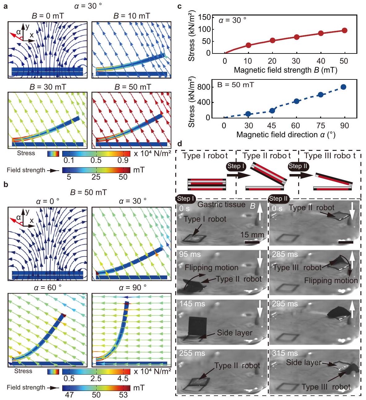
الاتجاه موجود في الشكل 5c، مما يظهر أن الإجهاد يزداد مع قوة المجال واتجاه المجال. العلاقة بين قوى الطبقة-الطبقة، والعزم، واتجاه المجال المغناطيسي أثناء الفصل موجودة في الشكل التكميلية 7c، مما يظهر أن القوة الأفقية تزداد مع اتجاه المجال، بينما القوة الرأسية والعزم المغناطيسي يتناقصان تدريجياً.
يتم فصل الروبوتات في خطوتين، كما هو موضح في المخططات في الشكل 5d، بما في ذلك فصل الروبوت من النوع الثاني عن الروبوت من النوع الأول والفصل الإضافي للروبوت من النوع الثاني. تظهر النتائج التجريبية لاستراتيجية الفصل المقترحة على الأنسجة المعدية الرطبة في الشكل 5d والفيلم التكميلي 1. يتم وضع مغناطيس تحت الأنسجة المعدية على مسافة عمودية تبلغ 15 مم للتحكم في الروبوت. في البداية، يتم تشكيل الالتصاق بين الطبقة الجانبية للروبوت والأنسجة من خلال تطبيق قوة جذب مغناطيسية عمودية لمدة تقارب 2 دقيقة. يتم تحقيق الحركة الأولى لقلب الروبوت من النوع الثاني وفصل الروبوت من النوع الأول عن طريق عكس اتجاه المجال المغناطيسي، مما يترك طبقة جانبية من الروبوت ملتصقة بالأنسجة. ). يتم تحقيق فصل الروبوت من النوع الثاني باستخدام نفس استراتيجية التشغيل، مما يؤدي إلى وجود طبقة الجانب الملتصقة الثانية وروبوت من النوع الثالث حر ( ).

التصاق متعدد الأهداف لقرحات المعدة خارج الجسم

تجعل الهيكل متعدد الطبقات واستراتيجية الفصل عند الطلب الروبوت منصة واعدة للالتصاق متعدد الأهداف بقرحات المعدة. يتم عرض مخطط سير العمل للالتصاق متعدد الأهداف باستخدام الروبوت أولاً في الشكل 6a. يتصل الروبوت من النوع الأول بالأنسجة المعدية ويتم توجيهه لتغطية قرحة المعدة من خلال الحركة الانتقالية. سيتم تشكيل الالتصاق بين الطبقة الجانبية للروبوت والأنسجة المعدية. يفصل الروبوت من النوع الثاني عن الطبقة الجانبية الملتصقة من خلال حركة قلب، ثم يتم توجيهه بعد ذلك إلى القرحة التالية للتغطية والالتصاق. يتم تحقيق فصل الروبوت من النوع الثاني باستخدام نفس الاستراتيجية، ويمكن استخدام الروبوت من النوع الثالث الناتج للالتصاق بالقرحة الثالثة من خلال حركة قلب.
تم إجراء تجربة خارج الجسم للالتصاق متعدد الأهداف باستخدام الروبوت على نسيج معدي خنزير (الشكل 6ب والفيلم التكميلي 2). تم إنشاء ثلاثة قرحات صناعية، أي الهدف I – III. يتم توصيل روبوت من النوع الأول إلى الهدف I بواسطة مجال مغناطيسي خارجي. ويتم تشكيل الالتصاق بين الطبقة الجانبية للروبوت والأنسجة المعدية من خلال تطبيق قوة جذب مغناطيسية عمودية لمدة تقارب دقيقتين. بعد ذلك، ينفصل الروبوت من النوع الثاني عن الطبقة الجانبية الملتصقة عن طريق تغيير اتجاه المجال المغناطيسي. ويتم التنقل إلى الهدف الثاني للالتزام به . أخيرًا، يفصل الروبوت من النوع الثالث عن الطبقة الجانبية الثانية الملتصقة ( ) وينتقل إلى الهدف الثالث للالتصاق الثالث ( يمكن أيضًا تطبيق الالتصاق المتعدد باستخدام الروبوت لتغطية القرحة الأكبر. يمكن أن تزيد هذه الطريقة من المنطقة الإجمالية القابلة للتغطية للروبوت من خلال تنفيذ الالتصاق المتعدد بشكل متجاور. هنا، نوضح أن قرحة طويلة بطول 35 مم وعرض 5 مم يمكن تغطيتها بواسطة الروبوت كما هو موضح في الشكل التوضيحي 8. خلال عملية التسليم والالتصاق الثانية، يتطلب الأمر وقتًا أطول للبقاء من أجل أن يتشكل الالتصاق بين الطبقة الجانبية للروبوت والأنسجة، وهو ما يمكن أن يُعزى إلى المخاط الذي التصق بالروبوت خلال عملية الملاحة والتسليم. تم الحصول على النتائج التجريبية حول الوقت المطلوب للالتصاق من التجارب على الأنسجة المعدية (الشكل التوضيحي 9). الوقت المطلوب للالتصاق الأول والثاني هو و ثم يتم غمر نسيج المعدة الذي تم لصقه بثلاث طبقات روبوتية في SGF لتقييم أداء الاحتفاظ للفيلم اللاصق على النسيج الخارجي. بعد 12 ساعة، لا يزال التصاق الفيلم اللاصق موجودًا (الشكل التوضيحي التكميلي 10).
علاوة على ذلك، يتم إجراء تجربة خارج الجسم للالتصاق متعدد الأهداف باستخدام الروبوت داخل معدة خنزير مع قرحات صناعية، ويتم استخدام التصوير بالموجات فوق الصوتية للتنقل (الشكل 7 أ والفيلم التكميلي 3). تظهر ميزات القرح والروبوتات في صور الموجات فوق الصوتية في الشكل التكميلي 11. إعداد
الشكل 5 | الفصل عند الطلب للروبوت. أ محاكاة لعملية الفصل لفيلم مغناطيسي ناعم يتم تشغيله بواسطة حقل مغناطيسي مع زيادة في شدة الحقل (من 0 مللي تسلا إلى 50 مللي تسلا) واتجاه حقل مغناطيسي ثابت. ب محاكاة لعملية الفصل لفيلم مغناطيسي ناعم يتم تشغيله بواسطة حقل مغناطيسي مع تغيير في اتجاه الحقل (من إلى ) وقوة المجال المغناطيسي الثابتة.
ج نتائج المحاكاة لإجهاد الفيلم مع شدة واتجاه المجال المغناطيسي. د المخططات والعرض التجريبي لاستراتيجية الفصل، من الروبوت من النوع الأول إلى الروبوت من النوع الثاني (الخطوة الأولى، اليسار)، ومن الروبوت من النوع الثاني إلى الروبوت من النوع الثالث (الخطوة الثانية، اليمين). تم توفير بيانات المصدر كملف بيانات مصدر.
تظهر التجربة في الشكل التكميلي 12 (الطرق). في صور الموجات فوق الصوتية، يضر القرحة (الإهليلج الأزرق المتقطع، الشكل 7أ) و تكسر الغشاء المخاطي للمعدة (الخط الأبيض المتقطع في الشكل 7أ، ). يتم وضع الروبوتات في مستطيلات حمراء متقطعة. يتم إدخال روبوت من النوع الأول إلى المعدة عبر المريء ويتم تتبعه باستخدام التصوير بالموجات فوق الصوتية ( ). يتصل الروبوت بالغشاء المخاطي للمعدة ويتم توجيهه لتغطية القرحة I ( ). يتم تشكيل الالتصاق من خلال exerting قوة إجهاد مغناطيسية على الروبوت نحو الغشاء المخاطي لمدة تقارب 5 دقائق. ينفصل الروبوت من النوع الثاني عن الطبقة الجانبية الملتصقة الأولى من خلال حركة قلبية عن طريق عكس اتجاه المجال المغناطيسي، كما يتضح من انفصال نمطين أبيضين محددين بالمستطيلات الحمراء المتقطعة ( ). يتم بعد ذلك توضيح عملية التوجيه والالتصاق لروبوت من النوع الثاني إلى القرحة الثانية ( ). أخيرًا، يفصل الروبوت من النوع الثالث عن الطبقة الجانبية الملتصقة الثانية ويتم توجيهه إلى القرحة III لإكمال الالتصاق الثالث ( ).
تم إجراء فحص بالموجات فوق الصوتية بعد العملية على القرحات الثلاثة لتقييم نتائج الالتصاق متعدد الأهداف (الشكل 7ب). تشير صور الموجات فوق الصوتية إلى أن الطبقات الثلاثة من الروبوت تغطي وتلتصق بنجاح بالقرحات الثلاثة. ثم يتم تشريح المعدة، ويتم إجراء ملاحظة بسيطة لاماكن القرحة (الشكل 7ج). جميع القرحات مغطاة بطبقات الروبوت، بما في ذلك تلك التي تقع بالقرب من طيات المعدة وتلك التي تقع في المساحة المحدودة.
بين طيات اثنتين. تُظهر المخططات المرفقة في الشكل 7c المواقع الأولية للقرحات في معدة الخنزير.
ثم يتم إجراء تحليل كمي للتوافق الحيوي للروبوت (الشكل 7d والشكل التكميلي 13). يتم إجراء اختبارات في المختبر عن طريق زراعة الفيلم اللاصق والركيزة المغناطيسية اللينة للروبوت مع خلايا الظهارة المخاطية المعدية البشرية (GES-1) بشكل منفصل لمدة 24 ساعة (الطرق). تظهر النتائج أن نسبة بقاء الخلايا المزروعة مع الفيلم اللاصق والركيزة المغناطيسية اللينة هي و ، على التوالي، ويتم مقارنتها بمجموعة التحكم التي لديها نسبة بقاء خلايا قدرها .

الالتصاق متعدد الأهداف في معدة خنزير حية

تضيف المعدة الحية تحديات وتعقيدات مقارنة بالمعدة خارج الجسم. بينما تكون المعدة الحية والمعدة خارج الجسم متشابهتين من الناحية الهيكلية، فإن البيئة الداخلية لمعدة الخنزير الحية تجلب تعقيدات كبيرة، مثل الحركة الدودية الديناميكية والإفراز المستمر للمخاط المعدي. تشكل الحركة الدودية للمعدة الحية تحديات في مراقبة وتحديد موقع الروبوت بدقة داخل البيئة المعدية. بالإضافة إلى ذلك، فإن الإفراز المستمر للمخاط المعدي يجعل الاتصال الفعال بين الفيلم اللاصق والأنسجة صعبًا، مما قد يمنع تشكيل التصاق مستقر. علاوة على ذلك، تحتوي المعدة الحية على المزيد من الطيات على السطح. لذلك، فإن التجارب الحية
الشكل 6 | عرض الالتصاق متعدد الأهداف على الأنسجة المعدية خارج الجسم. أ مخططات الالتصاق متعدد الأهداف على الأنسجة المعدية. نتائج تجريبية للالتصاق متعدد الأهداف باستخدام الروبوت على الأنسجة المعدية خارج الجسم. شريط القياس 15 مم.
ضرورية. يتم إجراء الالتصاق متعدد الأهداف باستخدام الروبوت في معدة خنزير حية (الشكل 8 والفيلم التكميلي 4). يتم عرض مخططات التجربة الحية في الشكل 8a (الطرق). باستخدام التصوير بالموجات فوق الصوتية في الوقت الحقيقي للتتبع، يكون الروبوت قادرًا على الاقتراب والتغطية والالتصاق بالقرحات التي تحركها الحقول المغناطيسية (الشكل 8b). يلتصق الطبقة الجانبية بالأنسجة المحيطة بالقرحة I، ثم ينفصل الروبوت من النوع II عنها. يغطي الروبوت من النوع II والروبوت من النوع III بنجاح ويعلقان على القرحة II والقرحة III، على التوالي، بناءً على ردود الفعل من التصوير بالموجات فوق الصوتية. ثم يتم تشريح المعدة لتقييم دقة الطبقة الملتصقة بالروبوت. لوحظ أن طبقات الروبوت تلتصق بثلاثة مواقع على الغشاء المخاطي المعدي المطوي (الشكل 8c)، وبعد إزالة الطبقات، يتم الكشف عن جميع القرح (الشكل 8d). تظهر النتائج التجريبية أن الروبوت المقترح يمكنه مواجهة التحدي وتحقيق الالتصاق متعدد الأهداف بدقة عالية داخل معدة حية مع المخاط.

المناقشة

باختصار، لقد طورنا روبوتًا مغناطيسيًا ناعمًا يتكون من عدة طبقات ذات هياكل ووظائف مختلفة لتحقيق الالتصاق عند الطلب مع آفات مختلفة. تم أخذ المواد المصممة والتفاعلات بين الطبقات في الاعتبار. يتكون الروبوت من ثلاث طبقات، بما في ذلك طبقة مركزية وطبقتين جانبيتين. تتكون كل طبقة من ركيزة مغناطيسية لينة وفيلم لاصق مدمج. لتحقيق الالتصاق بالأنسجة الرطبة، نقوم بتحضير الفيلم اللاصق القائم على الكاربوبول الذي يمكن أن يلتصق بالأنسجة من خلال الروابط الهيدروجينية. يتم تقييم وتحسين أداء الالتصاق للفيلم اللاصق. علاوة على ذلك، يتم ضبط التفاعل بين الطبقات للروبوت من خلال تصميم كل طبقة بركيزة مختلفة. تتكون الطبقة الجانبية من قاعدة غير مغناطيسية وإطار مغناطيسي، بينما تتكون الطبقة المركزية من قاعدة مغناطيسية وإطار غير مغناطيسي. يتم مغنطة الطبقات الثلاث في نفس
الاتجاه العمودي على مستواها، مما يوفر جاذبية مغناطيسية بين الطبقات. بمجرد أن تلتصق الطبقة الجانبية بالأنسجة الرطبة، يمكن استخدام العزم المغناطيسي لقلب الروبوت وفصله عن الطبقة الجانبية الملتصقة. من خلال الاستفادة من استراتيجية الالتصاق والفصل عند الطلب، نتحقق من تنفيذ الروبوت للالتصاق متعدد الأهداف في البيئات خارج الجسم وداخل الجسم، مما يظهر إمكانيته في علاج القرح المعدية. باختصار، من خلال دمج المواد الوظيفية في الروبوتات اللينة، يمكن توفير المزيد من الإمكانيات لتوسيع التطبيقات.
بالنظر إلى الاحتياجات المحددة المطلوبة من قبل نظام تشغيل الحقل المغناطيسي للسيناريوهات الطبية، يمكن توسيع مسافة التشغيل الفعالة للروبوت من خلال زيادة قوة الحقل المغناطيسي والتدرج. بالنسبة للأنظمة المدمجة مع المغناطيس الدائم، يمكن أن يؤدي زيادة حجم المغناطيس إلى تعزيز كل من قوة الحقل المغناطيسي والتدرج، بينما بالنسبة للملفات الكهرومغناطيسية، فإن زيادة سعة التيار في الملف الكهرومغناطيسي وتنفيذ معدات تبريد المياه يمكن أن تكون أيضًا طريقة واعدة. لاستهداف المناطق العميقة في التصوير الطبي، يمكننا تطبيق هذه الاستراتيجية تحت إشراف تصوير الأشعة السينية. يمكن أن يساعد تناول وجبة الباريوم عن طريق الفم في تحديد موقع القرح ، ويمكن توجيه الروبوت لتغطيتها والالتصاق بها. يُلاحظ أن ذراع الروبوت مع مغناطيس دائم يعمل كأداة نهائية يمكن أن تحقق أيضًا التشغيل عند الطلب للروبوتات (الشكل التكميلي 14). بالإضافة إلى ذلك، فإن تحميل الأدوية في الفيلم اللاصق للروبوت سيساعد في تحويل التكنولوجيا إلى تطبيقات سريرية تحافظ على أدائها في الالتصاق وحركتها (الأشكال التكملية 15 و16).

الطرق

المواد

تم الحصول على كاربوبول 971 P من CHINE INTERNATIONAL (شنغهاي، الصين). تم شراء بولوكسايمر 407 من Solarbio LIFE SCIENCES (بكين، الصين). تم شراء هيدروكسي بروبيل ميثيل السليلوز (HPMC)
الشكل 7 | الالتصاق متعدد الأهداف في معدة خنزير خارج الجسم. أ الالتصاق متعدد الأهداف باستخدام الروبوت في معدة خنزير خارج الجسم مليئة بالسائل تحت تتبع الوقت الحقيقي للتصوير بالموجات فوق الصوتية. تمثل المستطيلات الحمراء المتقطعة، والخطوط البيضاء المتقطعة، والبيضاوات الزرقاء المتقطعة الروبوت، والغشاء المخاطي المعدي، والقرحة، على التوالي. تمثل الأسهم البرتقالية اتجاه الحركة الانتقالية للروبوت. شريط القياس 5 مم. فحص ما بعد الجراحة باستخدام التصوير بالموجات فوق الصوتية. شريط القياس 5 مم. ج المعدة المقطوعة بعد الالتصاق متعدد الأهداف. توضح المخططات المواقع الأولية لل
القرحات في معدة الخنزير. نسبة بقاء الخلايا (يسار) لخلايا الظهارة المخاطية المعدية البشرية (GES-1) في ثلاث مجموعات، أي مجموعة التحكم، مجموعة الفيلم اللاصق (AF)، ومجموعة الركيزة المغناطيسية اللينة (MF). بالنسبة لمجموعة التحكم، لم يتم إضافة أي شيء إلى طبق الزراعة. يتم زراعة الفيلم اللاصق والركيزة المغناطيسية اللينة مع الخلايا للمجموعات التجريبية، على التوالي. صورة صبغة الحياة/الموت لخلايا GES-1 في ثلاث مجموعات علاجية بعد 24 ساعة من الزراعة المشتركة (اليمين). يتم تقديم البيانات كقيم متوسطة SD. يتم توفير بيانات المصدر كملف بيانات مصدر.
من MACKLIN (شنغهاي، الصين). جميع المواد الكيميائية الأخرى من درجة كاشف نقية إضافية وتستخدم كما هي.

تصنيع الروبوت الناعم متعدد الطبقات المغناطيسي

كما هو موضح في الشكل التكميلي 1a، يتم تحضير الفيلم المغناطيسي عن طريق الطلاء الدوراني لمعلق مغناطيسي، والذي يتم تحضيره عن طريق خلط جزيئات النيوديميوم الحديد البورون المغناطيسية الصلبة (NdFeB) مع بوليديميثيل سيليكون (PDMS) بنسبة كتلة 2: 1.
يتم تحضير الفيلم اللاصق من كاربوبول وبولوكسايمر وHPMC. يتم إذابة كاربوبول وHPMC وبولوكسايمر في خليط من الإيثانول والماء ( ). يتم خلط رودامين B جسديًا في المحلول لتحضير الفيلم الوردي لتحسين الرؤية. يتم صب المحلول المسبق في طبق بتري، ويتم الحصول على الفيلم اللاصق عن طريق تبخير المحلول المتبقي باستخدام الحرارة.
يتم الحصول على كل مكون من الطبقة من أفلام مختلفة عن طريق القطع بالليزر وتجميعها في الطبقات الجانبية والمركزية على التوالي
بعد معالجة البلازما (الشكل التكميلي 1 أ، ب). يتم برمجة الطبقات بنفس اتجاه المغنطة العمودية على مستواها تحت مجال مغناطيسي نبضي (الشكل التكميلي 1 ج). أخيرًا، يتم تجميع الروبوت تحت جذب مغناطيسي بين الطبقات. يتم عرض معلومات مفصلة عن الروبوتات في الجدول التكميلي S2.

اختبارات ميكانيكية

تم قياس صلابة الكسر للأفلام اللاصقة المعدة بنسب وزن مختلفة من خليط الكاربوبول و HPMC-بولوكزمر باستخدام اختبارات الشد لعينات مستطيلة رقيقة (20 مم في الطول، 15 مم في العرض) باستخدام آلة اختبار ميكانيكية (خلايا تحميل 0.1 كيلو نيوتن). يتم إجراء جميع الاختبارات بسرعة شد ثابتة قدرها . يتم حساب صلابة الكسر للفيلم اللاصق باستخدام طريقة موثقة.
تُجرى اختبارات الريولوجيا باستخدام ريو متر (Anton Paar MCR302e، النمسا) مع برنامج ريوكومباس. المعاملات الديناميكية
الشكل 8 | الالتصاق المستهدف عند الطلب في معدة خنزير حية. أ مخططات التجربة. تمثل الخطوط البيضاء المتقطعة والبيضاوات الزرقاء الغشاء المخاطي المعدي والقرحة، على التوالي. التنقل في الوقت الحقيقي لروبوت في معدة خنزير حية بعد إفراغ المعدة من أجل التصاق متعدد الأهداف. تشمل الخطوات التنقل المستهدف، والتغطية، والالتصاق. تمثل المستطيلات الحمراء المتقطعة الروبوت. تمثل الأسهم البرتقالية
اتجاه الحركة الانتقالية للروبوت. شريط القياس هو 5 مم. ج المعدة المقطوعة بعد الالتصاق متعدد الأهداف. وجهات نظر مفصلة للقرحات المغطاة وتلك التي تم الكشف عنها بعد تقشير طبقات الروبوت. شريط القياس هو 5 مم. اللوحة (أ) تم إنشاؤها جزئيًا باستخدام فن سيرفيير الطبي، المقدم من سيرفيير، المرخص بموجب ترخيص المشاع الإبداعي 3.0 غير المنقول.
(معامل التخزين، ومعامل الفقد، ) للأفلام اللاصقة تم الحصول عليها. يتم إجراء اختبارات سحب السعة عن طريق تغيير الإجهاد من 0.01 إلى بتردد 1 هرتز. يتم إجراء اختبارات سحب التردد عن طريق تغيير التردد من 0.1 إلى 10 هرتز مع إجهاد قدره .

اختبارات الالتصاق في المختبر

تُجرى اختبارات قوة الالتصاق النسيجي الكمية، بما في ذلك اختبارات القص اللاب والقص T، وفقًا لمعايير ASTM F2255 و F2256. يتم استخدام نسيج معدي خنزير كمادة نسيجية حيوية. يتم وضع النسيجين في آلة اختبار عالمية (إنستران، أمريكا) لتحميل الشد بمعدل إجهاد قدره . يتم وضع الفيلم اللاصق بين النسيجين وضغطه تحت 1 كيلو باسكال لفترة زمنية مختلفة.
لقياس قوة القص، يتم إعداد عينات ملتصقة (الفيلم اللاصق) بمساحة لاصقة بعرض 15 مم وطول 15 مم
وتم اختبارها بواسطة اختبار القص اللاب القياسي (ASTM F2255) باستخدام آلة اختبار ميكانيكية (خلايا تحميل 0.1 كيلو نيوتن). يتم تحديد قوة القص عن طريق قسمة القوة القصوى على مساحة الالتصاق.
لقياس صلابة الواجهة، يتم إعداد عينات ملتصقة (الفيلم اللاصق) بعرض 15 مم واختبارها بواسطة اختبار التقشير بزاوية 180 درجة القياسي (ASTM F2256) باستخدام آلة اختبار ميكانيكية (خلايا تحميل 0.1 كيلو نيوتن). تصل القوة المقاسة إلى مستوى ثابت عندما تدخل عملية التقشير في حالة مستقرة. يتم تحديد صلابة الواجهة عن طريق قسمة مرتين على قوة المستوى الثابت (لاختبار التقشير بزاوية 180 درجة) على عرض عينة النسيج.
لتمييز وقت الاحتفاظ بالفيلم اللاصق تحت تحميل دوري، يتم غمر نسيج معدي خنزير ملتصق بمونومر في SGF. تقوم معدات الشد (KeyFactor، الصين) بشد نسيج المعدة الخنزير بترددات مختلفة ( , 1.25 هرتز) وتسجيل وقت الاحتفاظ بالمونومر.

اختبار التوافق الحيوي في المختبر

لتقييم التوافق الحيوي في المختبر والسمية الخلوية لطبقة اللاصق، يتم استخدام اختبار LIVE/DEAD لتقييم خط خلايا الظهارة المعدية البشرية (GES-1، شركة شيامن إيموكيل للتكنولوجيا الحيوية المحدودة). يتم قطع الفيلم اللاصق إلى وزراعته مع GES-1، دون إضافة الفيلم اللاصق كتحكم. يتم زراعة خلايا GES-1 في أطباق 96 بئر بكثافة قدرها خلية ( لكل مجموعة). ثم يتم حضانة الخلايا عند لمدة 24 ساعة في جو. يتم تحديد حيوية الخلايا بواسطة مجموعة حيوية/سمية LIVE/DEAD للخلايا الثديية (ثيرمو فيشر العلمية) عن طريق إضافة 4 م كالسيتين وإيثيديم هوموديمر-1 إلى وسط الثقافة. يتم استخدام ميكروسكوب (نيكون ECLIPSE Ti2) لتصوير الخلايا الحية مع إثارة/انبعاث عند والخلايا الميتة عند ، على التوالي. يتم حساب حيوية الخلايا عن طريق عد الخلايا الحية (الفلوريسنت الأخضر) والميتة (الفلوريسنت الأحمر) باستخدام ImageJ (الإصدار 2.1.0).

تصوير بالموجات فوق الصوتية للاختبارات خارج الجسم وفي الجسم

في هذه الدراسة، يتم استخدام التصوير بالموجات فوق الصوتية لتتبع الروبوتات والقرحات. تحت صور الموجات فوق الصوتية، يظهر الغشاء المخاطي كخط أبيض مستمر في بيئة مملوءة بالماء. يمكن ملاحظة تغيير واضح في الشكل عند موقع القرحة. في البيئات التي تحتوي على هواء، تظهر صور الموجات فوق الصوتية وجود إشارات بيضاء غير منتظمة، والتي يمكن أن تكون بمثابة مؤشر على موقع القرحات. في معدة مملوءة بالسوائل أو في معدة فارغة، يتم تحديد موقع ونمط الروبوت من خلال تتبع الخطوط البيضاء المتراكمة في صور الموجات فوق الصوتية. يقل سمك الكومة عندما ينتقل الروبوت من النوع الأول إلى النوع الثالث. بشكل خاص، يمكن استخدام الموقع الذي تختفي فيه الإشارة البيضاء (المستطيلات الصفراء المتقطعة، الشكل التكميلي 11) لتحديد موقع الروبوت من النوع الثالث في بيئة الهواء، مما يسمح بتتبع الروبوت من النوع الثالث أثناء تحركه في معدة خنزير حية بعد إفراغ المعدة.

إجراءات التجارب خارج الجسم وفي الجسم

يمكن سرد إجراءات التجارب خارج الجسم وفي الجسم كما يلي. يتعاون مشغلان لتحقيق التصاق متعدد الأهداف للروبوت. يقوم المشغل 1 بتشغيل محول الموجات فوق الصوتية لتحديد موقع الروبوت. بمجرد تحديد موقع الروبوت، يقوم المشغل 2 بتحريك الروبوت باستخدام مغناطيس أسطواني. يقوم المشغلان 1 و 2 بتحريك المحول والمغناطيس بشكل متزامن لتحريك الروبوت نحو ثم تغطية الهدف الأول. بعد ذلك، بمجرد تشكيل الالتصاق بين الفيلم اللاصق والنسج تحت الجذب المغناطيسي. يقوم المشغل 2 بعد ذلك بعكس المغناطيس لفصل الروبوت من النوع الثاني عن الطبقة الجانبية الملصقة. يتم تطبيق هذه الطريقة المنهجية أيضًا لتغطية الأهداف الثانية والثالثة.

تحضير معدة الخنزير للاختبارات خارج الجسم

تم شراء جميع معدة الخنزير خارج الجسم من تاوباو، الصين. يتم إفراغ الأعضاء وتنظيفها بشكل طفيف بالماء دون إتلاف الغشاء المخاطي. يتم إنشاء قرحات معدية ناتجة عن حمض الأسيتيك باستخدام بروتوكول تم الإبلاغ عنه سابقًا . يتم استخدام مقص جراحي لقطع ثلاث مناطق من على سطح معدة الخنزير، و يتم حقن حمض الأسيتيك في الشق ويتم الحفاظ عليه لمدة 120 ثانية. بالنسبة لمعدة الخنزير خارج الجسم، يتم إنشاء ثلاث قرحات صناعية على جوانب مختلفة من المعدة. بالنسبة للاختبارات خارج الجسم، تكون أبعاد الروبوت (الروبوت من النوع الأول) هي والوزن هو .

اختبارات في الجسم في خنزير

بعد العمق المناسب من التخدير والتسكين، يتم كشف المعدة بواسطة شق جراحي. يتم عمل شق صغير في المريء باستخدام سكين جراحي لإزالة محتويات المعدة. ثم يتم غسل المعدة بمحلول ملحي فسيولوجي. يتم استخدام مقص جراحي لإنشاء ثلاث قرحات في الغشاء المخاطي المعدي، كل منها بحجم حوالي 1 سم في القطر، ويتم تحديد موقع القرحات باستخدام التصوير بالموجات فوق الصوتية. يتم إدخال الروبوت إلى المعدة من خلال الشق ويتم توجيهه باستخدام الموجات فوق الصوتية. يتم ضبط اتجاه ومسافة
المجال المغناطيسي للسماح للروبوت بتشكيل الالتصاق مع النسيج والانفصال عن الطبقة الجانبية. يتم فحص نتيجة الالتصاق النهائية باستخدام التصوير بالموجات فوق الصوتية. تحت التخدير العميق، يتم بعد ذلك قتل الحيوان عن طريق حقن كلوريد البوتاسيوم في الوريد، ويتم تشريح المعدة لملاحظة نتيجة الالتصاق النهائية. بالنسبة للاختبارات في الجسم، تكون أبعاد الروبوت (الروبوت من النوع الأول) هي والوزن هو .

محاكاة

محاكاة توزيع الإجهاد عندما قام الروبوت بأداء حركة انتقالية على البطن والفصل بين فيلم مغناطيسي ناعم والطبقة الجانبية الملتصقة بالأنسجة تحت مجالات مغناطيسية باتجاهات وقوة مختلفة باستخدام البرنامج التجاري Comsol. معامل يونغ والمغنطة المقاسة من التجربة تُستخدم كمعلمات إدخال.

التحليل الإحصائي

البيانات التي تم الحصول عليها في هذه الدراسة تم الإبلاغ عنها جميعًا كمتوسط الانحراف المعياري (SD) من ثلاثة تجارب منفصلة على الأقل. يتم مقارنة القيم من مجموعتين باستخدام اختبار ستودنت. اختبار. أ تعتبر القيمة التي تبلغ 0.05 أو أقل ذات دلالة إحصائية. , ؛ غير دال.

ملخص التقرير

معلومات إضافية حول تصميم البحث متاحة في ملخص تقارير مجموعة نيتشر المرتبط بهذه المقالة.

توفر البيانات

البيانات التي تم توليدها في هذه الدراسة متاحة في ملف المعلومات التكميلية/بيانات المصدر. البيانات الإضافية متاحة من المؤلف المراسل عند الطلب. بيانات المصدر للرسوم البيانية متاحة مع هذه الورقة. بيانات المصدر متاحة مع هذه الورقة.

References

  1. Soon, R. H. et al. On-demand anchoring of wireless soft miniature robots on soft surfaces. Proc. Natl. Acad. Sci. 119, e2207767119 (2022).
  2. Ren, Z. et al. Soft-bodied adaptive multimodal locomotion strategies in fluid-filled confined spaces. Sci. Adv. 7, eabh2022 (2021).
  3. Gu, H. et al. Self-folding soft-robotic chains with reconfigurable shapes and functionalities. Nat. Commun. 14, 1263 (2023).
  4. Zheng, Z. et al. lonic shape-morphing microrobotic end-effectors for environmentally adaptive targeting, releasing, and sampling. Nat. Commun. 12, 411 (2021).
  5. Xu, T., Huang, C., Lai, Z. & Wu, X. Independent control strategy of multiple magnetic flexible millirobots for position control and path following. IEEE Trans. Robot. 38, 2875-2887 (2022).
  6. Ebrahimi, N. et al. Magnetic actuation methods in bio/soft robotics. Adv. Funct. Mater. 31, 2005137 (2021).
  7. Zhang, J. et al. Voxelated three-dimensional miniature magnetic soft machines via multimaterial heterogeneous assembly. Sci. Robot. 6, eabf0112 (2021).
  8. Deng, H. et al. Laser reprogramming magnetic anisotropy in soft composites for reconfigurable 3D shaping. Nat. Commun. 11, 6325 (2020).
  9. Gu, H., Boehler, Q., Ahmed, D. & Nelson, B. J. Magnetic quadrupole assemblies with arbitrary shapes and magnetizations. Sci. Robot. 4, eaax8977 (2019).
  10. Xu, T., Zhang, J., Salehizadeh, M., Onaizah, O. & Diller, E. Millimeterscale flexible robots with programmable three-dimensional magnetization and motions. Sci. Robot. 4, eaav4494 (2019).
  11. Richter, M. et al. Magnetic soft helical manipulators with local dipole interactions for flexibility and forces. Soft Robot. 10, 647-659 (2023).
  12. Hu, W., Lum, G. Z., Mastrangeli, M. & Sitti, M. Small-scale softbodied robot with multimodal locomotion. Nature 554, 81-85 (2018).
  13. Won, S., Kim, S., Park, J. E., Jeon, J. & Wie, J. J. On-demand orbital maneuver of multiple soft robots via hierarchical magnetomotility. Nat. Commun. 10, 4751 (2019).
  14. Li, W. et al. Self-vectoring electromagnetic soft robots with high operational dimensionality. Nat. Commun. 14, 182 (2023).
  15. Lu, H. et al. A bioinspired multilegged soft millirobot that functions in both dry and wet conditions. Nat. Commun. 9, 3944 (2018).
  16. Huang, H.-W. et al. Adaptive locomotion of artificial microswimmers. Sci. Adv. 5, eaau1532 (2019).
  17. Wang, Y. et al. Amphibious miniature soft jumping robot with ondemand in-flight maneuver. Adv. Sci. 10, 2207493 (2023).
  18. Wu, Y., Dong, X., Kim, J., Wang, C. & Sitti, M. Wireless soft millirobots for climbing three-dimensional surfaces in confined spaces. Sci. Adv. 8, eabn3431 (2022).
  19. Wang, C., Wu, Y., Dong, X., Armacki, M. & Sitti, M. In situ sensing physiological properties of biological tissues using wireless miniature soft robots. Sci. Adv. 9, eadg3988 (2023).
  20. Wang, C., Mzyk, A., Schirhagl, R., Misra, S. & Venkiteswaran, V. K. Biocompatible film-coating of magnetic soft robots for mucoadhesive locomotion. Adv. Mater. Technol. 8, 2201813 (2023).
  21. Zhou, C. et al. Ferromagnetic soft catheter robots for minimally invasive bioprinting. Nat. Commun. 12, 5072 (2021).
  22. Yang, Y. et al. Magnetic soft robotic bladder for assisted urination. Sci. Adv. 8, eabq1456 (2022).
  23. Kim, Y. et al. Telerobotic neurovascular interventions with magnetic manipulation. Sci. Robot. 7, eabg9907 (2022).
  24. Son, D., Gilbert, H. & Sitti, M. Magnetically actuated soft capsule endoscope for fine-needle biopsy. Soft Robot. 7, 10-21 (2020).
  25. Zong, Z., Zhang, S. & Wu, Z. Anisotropic contraction in a magnetically hard but mechanically ultra-soft foam for precise drug delivery. Adv. Mater. Technol. 8, 2201255 (2023).
  26. Zhang, H. et al. Dual-responsive biohybrid neutrobots for active target delivery. Sci. Robot. 6, eaaz9519 (2021).
  27. Dong, X. et al. Bioinspired cilia arrays with programmable nonreciprocal motion and metachronal coordination. Sci. Adv. 6, eabc9323 (2020).
  28. Lee, J., Kim, D., Bang, S. & Park, S. Drug-loaded mucoadhesive patch with active delivery and controlled releasing ability. Adv. Intell. Syst. 4, 2100203 (2022).
  29. Zheng, Z. et al. Programmable aniso-electrodeposited modular hydrogel microrobots. Sci. Adv. 8, eade6135 (2022).
  30. Ze, Q. et al. Spinning-enabled wireless amphibious origami millirobot. Nat. Commun. 13, 3118 (2022).
  31. Mair, L. O. et al. Soft capsule magnetic millirobots for regionspecific drug delivery in the central nervous system. Front. Robot. Al 8, 702566 (2021).
  32. Yang, X. et al. An agglutinate magnetic spray transforms inanimate objects into millirobots for biomedical applications. Sci. Robot. 5, eabc8191 (2020).
  33. Dong, Y. et al. Untethered small-scale magnetic soft robot with programmable magnetization and integrated multifunctional modules. Sci. Adv. 8, eabn8932 (2022).
  34. Wang, T. et al. Adaptive wireless millirobotic locomotion into distal vasculature. Nat. Commun. 13, 4465 (2022).
  35. Ze, Q. et al. Soft robotic origami crawler. Sci. Adv. 8, eabm7834 (2022).
  36. Cheng, Z. et al. Fabrication of ulcer-adhesive oral keratin hydrogel for gastric ulcer healing in a rat. Regen. Biomater. 8, rbab008 (2021).
  37. Brătucu, M. N. et al. Unusual complicated gastric ulcers. Medicina 57, 1345 (2021).
  38. Chu, J.N., Traverso, G. Foundations of gastrointestinal-based drug delivery and future developments. Nat. Rev. Gastroenterol. Hepatol. 19, 219-238 (2022).
  39. Anthis, A. H. C. et al. Modular stimuli-responsive hydrogel sealants for early gastrointestinal leak detection and containment. Nat. Commun. 13, 1-15 (2022).
  40. Wu, S. J., Yuk, H., Wu, J., Nabzdyk, C. S. & Zhao, X. A multifunctional origami patch for minimally invasive tissue sealing. Adv. Mater. 33, 2007667 (2021).
  41. Wu, J. et al. An off-the-shelf bioadhesive patch for sutureless repair of gastrointestinal defects. Sci. Transl. Med. 14, eabh2857 (2022).
  42. Xu, X. et al. Bioadhesive hydrogels demonstrating pH-independent and ultrafast gelation promote gastric ulcer healing in pigs. Sci. Transl. Med. 12, eaba8014 (2020).
  43. Chen, X. et al. Hydrogel bioadhesives with extreme acid-tolerance for gastric perforation repairing. Adv. Funct. Mater. 32, 2202285 (2022).
  44. Wang, H. et al. An integrally formed janus hydrogel for robust wettissue adhesive and anti-postoperative adhesion. Adv. Mater. 35, 2300394 (2023).
  45. Chun, M.-K., Bhusal, P. & Choi, H.-K. Application of Carbopol/PVP interpolymer complex to prepare mucoadhesive floating granule. Arch. Pharm. Res. 36, 745-751 (2013).
  46. Li, T., Bao, Q., Shen, J., Lalla, R. V. & Burgess, D. J. Mucoadhesive in situ forming gel for oral mucositis pain control. Int. J. Pharm. 580, 119238 (2020).
  47. Patel, M. M. et al. Mucin/Poly(acrylic acid) Interactions: a spectroscopic investigation of mucoadhesion. Biomacromolecules 4, 1184-1190 (2003).
  48. Deng, T. et al. A natural biological adhesive from snail mucus for wound repair. Nat. Commun. 14, 396 (2023).
  49. Yuk, H. et al. Dry double-sided tape for adhesion of wet tissues and devices. Nature 575, 169-174 (2019).
  50. Schulze, K. Imaging and modelling of digestion in the stomach and the duodenum. Neurogastroenterol. Motil. 18, 172-183 (2006).
  51. Marciani, L. et al. Assessment of antral grinding of a model solid meal with echo-planar imaging. Am. J. Physiol. -Gastrointest. Liver Physiol. 280, G844-G849 (2001).
  52. Debi, U. et al. Role of barium study in various gastric malignancies. Trop. Gastroenterol. 10, 246-255 (2019).

شكر وتقدير

نشكر كايوان فانغ وقو تشينغ تشين على مساعدتهما في جراحات الحيوانات. تم دعم هذا العمل ماليًا من قبل البرنامج الوطني الرئيسي للبحث والتطوير في الصين تحت رقم المشروع 2022YFA1207100، ومؤسسة العلوم الطبيعية الوطنية في الصين تحت رقم المشروع 62103347، رقم 62073208 ورقم 61933008، وبرنامج الابتكار من لجنة التعليم البلدية في شنغهاي تحت منحة 2021-01-07-00-09E0013، وبرنامج العلوم والتكنولوجيا في شنتشن تحت رقم المنحة RCBS20210609103155061، ومؤسسة غوانغدونغ للبحث الأساسي والتطبيقي تحت رقم المشروع 2022A1515110499، ومعهد شنتشن للذكاء الاصطناعي والروبوتات من أجل المجتمع تحت رقم المشروع AC01202101109، ومشروع خطة العلوم والتكنولوجيا في شنغهاي تحت منحة 23ZR1422300.

مساهمات المؤلفين

Z.C. و Y.W. و N.L. و J.Y. تصوروا الدراسة وصمموا التجارب. كتب Z.C. و Y.W. و J.L. و N.L. و J.Y. المخطوطة. قام Z.C. و Y.W. و H.C. بإجراء التجارب وتحليل البيانات. قدم Z.C. و Y.W. مدخلات مفاهيمية وتقنية لمحاكاة الأرقام. ساعد Z.C. و Y.W. و F.D. و H.C. في تجارب التصوير السريري. ساعد H.P. و S.X. و Y.S. و N.L. و J.Y. في الإشراف على التجارب وحرروا المخطوطة. أشرف N.L. و J.Y. على المشروع. ناقش جميع المؤلفين النتائج وراجعوا المخطوطة.

المصالح المتنافسة

يعلن المؤلفون عدم وجود مصالح متنافسة.

معلومات إضافية

معلومات إضافية النسخة الإلكترونية تحتوي على مواد إضافية متاحة علىhttps://doi.org/10.1038/s41467-024-44995-9.
يجب توجيه المراسلات والطلبات للحصول على المواد إلى نا ليو أو جيانغفان يوي.
معلومات مراجعة الأقران تشكر مجلة Nature Communications سوخو بارك والمراجعين الآخرين المجهولين على مساهمتهم في مراجعة هذا العمل. يتوفر ملف مراجعة الأقران.
معلومات إعادة الطباعة والتصاريح متاحة على
http://www.nature.com/reprints
ملاحظة الناشر: تظل شركة سبرينجر ناتشر محايدة فيما يتعلق بالمطالبات القضائية في الخرائط المنشورة والانتماءات المؤسسية.
الوصول المفتوح هذه المقالة مرخصة بموجب رخصة المشاع الإبداعي النسب 4.0 الدولية، التي تسمح بالاستخدام والمشاركة والتكيف والتوزيع وإعادة الإنتاج بأي وسيلة أو صيغة، طالما أنك تعطي الائتمان المناسب للمؤلفين الأصليين والمصدر، وتوفر رابطًا لرخصة المشاع الإبداعي، وتوضح ما إذا تم إجراء تغييرات. الصور أو المواد الأخرى من طرف ثالث في هذه المقالة مشمولة في رخصة المشاع الإبداعي الخاصة بالمقالة، ما لم يُشار إلى خلاف ذلك في سطر الائتمان للمواد. إذا لم تكن المادة مشمولة في رخصة المشاع الإبداعي الخاصة بالمقالة وكان استخدامك المقصود غير مسموح به بموجب اللوائح القانونية أو يتجاوز الاستخدام المسموح به، فستحتاج إلى الحصول على إذن مباشرة من صاحب حقوق الطبع والنشر. لعرض نسخة من هذه الرخصة، قم بزيارةhttp://creativecommons.org/رخصة/بواسطة/4.0/.
© المؤلف(ون) 2024

  1. كلية هندسة الميكاترونيك والأتمتة، جامعة شنغهاي، شنغهاي 200444، الصين. كلية العلوم والهندسة، جامعة الصين في هونغ كونغ، شنتشن 518172، الصين. معهد شنتشن للذكاء الاصطناعي والروبوتات من أجل المجتمع، شنتشن 518172، الصين. قسم الهندسة الميكانيكية والصناعية، جامعة تورونتو، تورونتو، أونتاريو M5S 3G8، كندا. كلية هندسة الحاسوب والعلوم، جامعة شنغهاي، شنغهاي 200444، الصين. قسم الأشعة التداخلية، مستشفى جيش التحرير الشعبي الصيني العام، بكين 100853، الصين. كلية الطب، جامعة الصين في هونغ كونغ، شنتشن 518172، الصين. ساهم هؤلاء المؤلفون بالتساوي: زيهينغ تشين، ييبين وانغ.

Journal: Nature Communications, Volume: 15, Issue: 1
DOI: https://doi.org/10.1038/s41467-024-44995-9
PMID: https://pubmed.ncbi.nlm.nih.gov/38245517
Publication Date: 2024-01-20

A magnetic multi-layer soft robot for on-demand targeted adhesion

Received: 22 July 2023
Accepted: 4 January 2024
Published online: 20 January 2024
(i) Check for updates

Ziheng Chen © , Yibin Wang , Hui Chen , Junhui Law (B) , Huayan Pu (B) , Shaorong Xie (1) , Feng Duan , Yu Sun (1) , Na Liu (1) & Jiangfan Yu (B)

Abstract

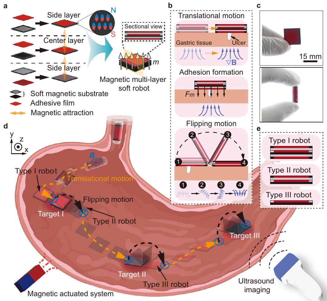
Magnetic soft robots have shown great potential for biomedical applications due to their high shape reconfigurability, motion agility, and multifunctionality in physiological environments. Magnetic soft robots with multilayer structures can enhance the loading capacity and function complexity for targeted delivery. However, the interactions between soft entities have yet to be fully investigated, and thus the assembly of magnetic soft robots with ondemand motion modes from multiple film-like layers is still challenging. Herein, we model and tailor the magnetic interaction between soft film-like layers with distinct in-plane structures, and then realize multi-layer soft robots that are capable of performing agile motions and targeted adhesion. Each layer of the robot consists of a soft magnetic substrate and an adhesive film. The mechanical properties and adhesion performance of the adhesive films are systematically characterized. The robot is capable of performing two locomotion modes, i.e., translational motion and tumbling motion, and also the ondemand separation with one side layer adhered to tissues. Simulation results are presented, which have a good qualitative agreement with the experimental results. The feasibility of using the robot to perform multi-target adhesion in a stomach is validated in both ex-vivo and in-vivo experiments.

Magnetic soft robots are promising candidates for biomedical applications . They can be magnetically programmed with required profiles, and have high degrees of freedom in shape deformation , which is necessary for dexterous motions, such as grasping and crawling . By applying external magnetic fields, the soft robots with tailored magnetization profiles can traverse complex terrains by altering locomotion modes, such as rolling , crawling , and swimming . By designing an actuation method and optimizing energy bursting, the jumping motion of magnetic soft robots has also been achieved in unstructured aquatic-terrestrial environments . Moreover, locomotion can be realized in unstructured threedimensional environments by considering surface microstructures
and surface coating of the robots, such as microspikes and mucoadhesive film loaded by magnetic soft robots.
Because of their high motion controllability and shape reconfigurability, magnetic soft robots have been used to perform biomedical tasks like minimally-invasive operation and targeted delivery . Bioprinting with a minimally invasive manner on a rat liver in vivo has been demonstrated , and the assistance of urination via applying mechanical compression to the underactive bladders has been achieved . Meanwhile, magnetic soft robots have been utilized for cargo delivery in different organs and cavities, such as gastrointestinal (GI) tract and blood vessel . Releasing drugs by compressing capsule-shaped magnetic soft robots has been reported in an ex-vivo
pig stomach . A magnetic modular soft robot enables the successful delivery of a therapeutic patch onto an ex-vivo porcine stomach ulcer . Furthermore, a wireless magnetic soft stent incorporating multiple drug-loading structures has been proposed to perform ondemand and local release of drugs in phantoms .
Gastric ulcer is a common disease occurring in different locations in the GI tract, such as the gastric angle, gastric antrum, and cardia . Multiple gastric ulcers can occur at the same period , and oral-taking drugs are widely used to treat ulcers. The therapeutic efficacy of oraltaking drugs is limited due to their low chemical stability in gastric fluid and shallow penetration in mucosa . Bioadhesive platforms, such as patches and hydrogels , have been reported to prolong drug retention by improving tissue adhesion in the porcine stomach. Magnetic soft robots offer an efficient and noninvasive approach for delivering bioadhesive platforms to gastric ulcers, which can mitigate erosion and enhance the healing effect by covering gastric ulcers . Furthermore, using a magnetic soft robot with multi-layer structures has the potential in on-demand adhesion at different ulcer sites through the separation between layers of the robot. However, to date, the interactions between layers of the robot have yet to be fully investigated, challenging the realization of a magnetic multi-layer soft robot with on-demand motion modes.
Herein, we design a magnetic multi-layer soft robot by tailoring the magnetic interaction between layers, which is capable of performing navigated locomotion on biological tissues and multiple ondemand targeted adhesion at different sites. The robot consists of three layers, and each layer comprises a soft magnetic substrate with the magnetization direction perpendicular to its surface and an adhesive film capable of forming adhesion to wet tissues through
hydrogen bonds. The adhesion performance of the adhesive film on ex-vivo porcine gastric tissues are characterized. Two locomotion modes of the robot on gastric tissues are explored and we demonstrate the on-demand separation between the robot and the adhered side layer through applying sufficient magnetic torque. The feasibility of using a magnetic multi-layer soft robot for on-demand targeted adhesion in an unstructured environment is validated in both ex-vivo gastric tissue and the stomach filled with fluid. The in-vivo trials of multi-target adhesion are also performed to validate the proposed strategy. This work presents an effective design of magnetic multilayer soft robots, paving the way for promising clinical opportunities, specially for the operations in GI tract.

Results

Design of a magnetic multi-layer soft robot

The magnetic multi-layer soft robot has three layers, i.e., one center layer and two side layers, and each layer consists of a soft magnetic substrate and an adhesive film capable of forming adhesion with wet tissues (Fig. 1a). Each layer is magnetized with the same direction, i.e., perpendicular to its surface, for stable assembly through interlayer magnetic attraction. To achieve the adhesion between the side layer and the tissue, and also the layer-layer separation of the robot, soft magnetic substrates with distinct in-plane structures are designed. The soft magnetic substrate of the side layer has a magnetic frame and a nonmagnetic base, while the center layer has a nonmagnetic frame and a magnetic base. The magnetic base of the center layer can exert magnetic attraction forces to the side layers, in order to maintain the integrity of the robot during locomotion. Meanwhile, to generate magnetic pressing forces between the side layer and the tissue when
Fig. 1 | Schematics of a magnetic multi-layer soft robot and its application for on-demand multi-targeted adhesion. a Schematics of a magnetic multi-layer soft robot. The soft magnetic substrate of the side layer has a magnetic frame and a nonmagnetic base, while that of the center layer has a nonmagnetic frame and a magnetic base. Schematic process of on-demand targeted adhesion to gastric ulcer. c A robot in released state and encapsulated state. d Overview of multi-target
adhesion using the robot in a stomach under the tracking of ultrasound imaging. The orange dashed arrows represent the translational motion direction of the robot. The black dashed arrows represent the flipping motion direction of the robot. e Schematic expression of the structures of Type I, Type II and Type III robot. Parts of (d) are created with BioRender.com.
the robot reaches the target to accelerate the formation of adhesion is also a purpose. Furthermore, the magnetic interaction induced between the magnetic parts of the side layer and the center layer, i.e., the magnetic frame and the magnetic base, is weaker than the adhesive interaction formed between the side layer and the tissue, which facilitates the separation between layers of the robot. Besides the above design principles, an adhesion and separation strategy is proposed, as shown in Fig. 1b. The robot approaches the targeted ulcer on a gastric tissue through performing translational motion, and the adhesive film of the side layer then adheres to the gastric tissue by applying magnetic gradient force . By altering the direction of the magnetic field, the magnetic torque exerted on the center layer overcomes the interlayer attraction, causing the robot to flip over while the adhesion between the side layer and the tissues is still maintained. In this work, the soft magnetic substrate comprises ferromagnetic particles NdFeB and polydimethylsiloxane (PDMS), while the adhesive film consists of Carbopol, which serves as mucoadhesive material . Poloxamer and Hydroxypropylmethylcellulose (HPMC) are also added to the mucoadhesive material, contributing to the formation of films. Fabrication details of the robot are explained in Supplementary Fig. 1 and Methods.
The robot can be curled and encapsulated (Fig. 1c), facilitating its ingestion for accessing the gastrointestinal (GI) tract. When the robot enters the stomach, it can be navigated to targets by ultrasound imaging feedback and achieve on-demand targeted adhesion by implementing the adhesion and separation strategy. The process of multitarget adhesion, from target I to target III, is schematically illustrated in Fig. 1d. The robot achieves targeted adhesion at each stage, and the layers forming the robot are gradually reduced, ultimately leaving a free center layer for the adhesion to target III. We hereby term the three stages of the multi-layer robots as the Type I robot with a threelayer structure, the Type II robot with a two-layer structure, and the Type III robot with a single-layer structure (Fig. 1e).

Mechanical properties of the adhesive film

The release process of the robot in the simulated gastric fluid (SGF) from a capsule is demonstrated in Fig. 2a. Schematics of the adhesion mechanism of the adhesive film is shown in Fig. 2b. The side layer of the robot contacts and adheres to the tissue using an adhesive film. The intermolecular hydrogen bonds between the three components (Carbopol, Poloxamer, and HPMC) facilitate the formation of an adhesive film. Carbopol, a polyacrylic acid polymer commonly used for mucoadhesive, plays a crucial role in the adhesive properties. The formation of hydrogen bonds between the carboxylic acid functional group of Carbopol and the glycoprotein component of the mucosa significantly contributes to the adhesion process . The adhesive film can form adhesion to wet gastric tissue through physical cross-linking hydrogen bonds under pressure.
The mechanical properties of the adhesive film are measured by tensile and amplitude scanning tests (Methods). Five groups of adhesive films with different weight ratios , from to , of Carbopol to HPMC-Poloxamer mixture (i.e., the mixture of HPMC and Poloxamer) are prepared. The experimental results of tensile tests reveal that, the adhesive film has the maximum fracture stress and strain with a of 2: 3, i.e., and , respectively (Fig. 2c). The results of amplitude scanning tests are presented in Fig. 2d, to show the change of the storage modulus ( ) and loss modulus ( ) of the adhesive film with applied strain (from to strain). Within the experimental measurements, the is higher than the when is kept the same, indicating the adhesive films have not undergone shear fracture . Among the five groups, the adhesive film fabricated with a of exhibited the highest and .

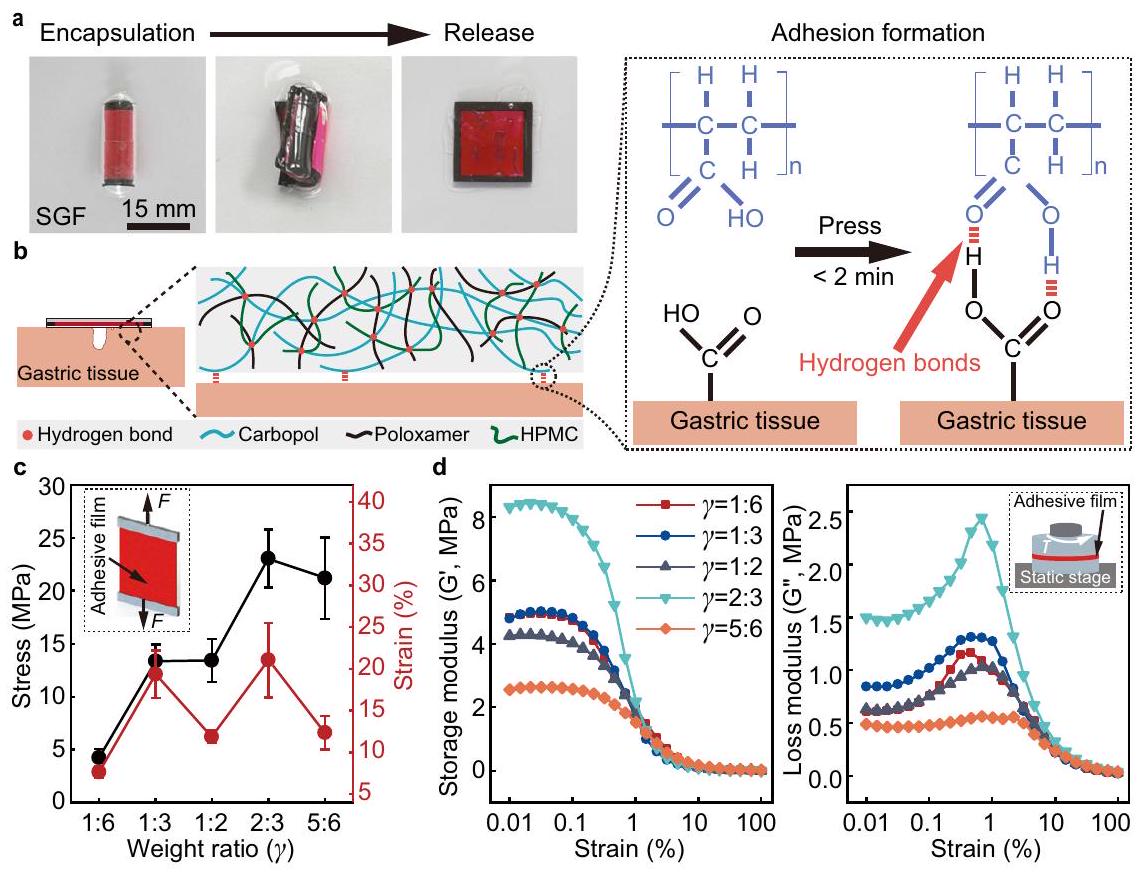
Adhesion performance

To quantitatively evaluate the adhesion performance of the adhesive film, Lap-shear test and T-peel test are conducted to characterize the shear strength and interfacial toughness (Fig. 3a and Methods). The
Fig. 2 | Design and characterization of the adhesive film. a The releasing process of a magnetic multi-layer soft robot from the encapsulated state. Adhesion mechanism of the adhesive film. The adhesive film is mainly crosslinked by intermolecular hydrogen bonds between Carbopol, Poloxamer, and HPMC. The adhesion between the adhesive film and gastric tissue surface is formed through hydrogen bonds. c Tensile stress-strain curves of the adhesive film prepared with five different weight ratios of Carbopol and HPMC-Poloxamer mixture. The error
bars are obtained from 3 trials in each condition. d The change of storage modulus and loss modulus of the adhesive films with strain. The films are prepared with five different weight ratios between Carbopol and HPMC-Poloxamer mixture. In all adhesive films, the HPMC-Poloxamer mixture is prepared with a weight ratio of (HPMC and Poloxamer). Data are presented as mean values SD. Source data are provided as a Source Data file.
Fig. 3 | Characterization of the adhesion performance. a Schematics of the Lapshear test and T-peel test. Shear strength and interfacial toughness of the adhesive films prepared with five different ratios of Carbopol and HPMC-Poloxamer mixture. c Shear strength and interfacial toughness of the adhesive films at different pressing time. d Storage modulus and loss modulus of the adhesive film at different immersion time recorded in the amplitude sweep tests ( strain, left) and frequency sweep tests ( , right). e The relationship between shear strength, interfacial toughness, and the thickness of the adhesive films.
Schematics of reciprocal stretching test. The retention time of the monomer adhered to the tissue in the reciprocal stretching test with different frequencies. The adhesive films are prepared with a ratio of 1:2 (Carbopol and HPMC-Poloxamer mixture) for (c, d, e, and g). In all adhesive films, HPMC-Poloxamer mixture is prepared with the weight ratio of (HPMC: Poloxamer). The error bars are obtained from 3 trials in each condition. Data are presented as mean values SD. Source data are provided as a Source Data file.
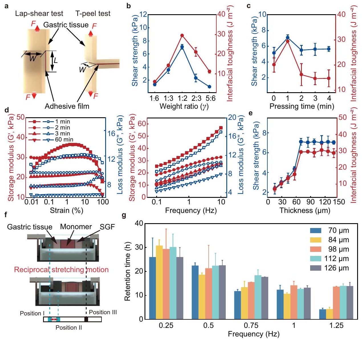
tested shear strength and interfacial toughness are presented in Fig. 3b, indicating the adhesion strength of the adhesive films prepared with different weight ratios. In the tests, an adhesive film is placed between two gastric tissues with 1 kPa loaded for a contact time of 1 min . The experimental results demonstrate that the shear strength and interfacial toughness initially increase and then decrease with the increase of the Carbopol content. The highest values of the shear strength and interfacial toughness indicates the maximum adhesion strength of the adhesive film that can be reached, and the values are and , respectively. A ratio of (Carbopol: HPMC-Poloxamer mixture) is maintained in this case. The adhesive film is thus prepared with a ratio of (Carbopol: HPMCPoloxamer mixture) in the subsequent experiments unless otherwise specified.
The hydration state of the adhesive films can influence their adhesion strength, primarily due to the porous structure of the adhesive film (Supplementary Fig. 2). Pressing an adhesive film on wet gastric tissue, the relationship between the adhesion strength and the pressing time is shown in Fig. 3c. The shear strength and interfacial toughness initially increase and then decrease with the increasing pressing time, reaching their highest values at 1 min . The cohesive strength of the adhesive films with different hydration states is then investigated using rheological tests (Methods). The maximum storage
modulus of the adhesive film is significantly decreased with the increase of immersion time (Fig. 3d). As the immersion time increases from 1 min to 1 h , the storage modulus ( ) is decreased from 36.534 kPa to 16.766 kPa in amplitude sweep tests (0.01-100 % strain) and from 56.947 kPa to 26.686 kPa in frequency sweep tests , respectively. Meanwhile, the storage modulus ( ) is higher than the loss storage with the same immerse time, indicating that the cross-linked network formed by hydrogen bonds of the adhesive film is maintained.
Figure 3e presents the relationship between the shear strength and the thickness of the adhesive films, and the relationship between the interfacial toughness and film thickness, in order to testify the adhesion strength of the adhesive films with different thicknesses. Positive correlation between thickness and shear strength, and that between thickness and interfacial toughness are observed. The values of the shear strength and the interfacial toughness reach a plateau at the thickness of , i.e., and , respectively. To further investigate the influence of the film thickness on the adhesion performance, the retention time is characterized using a reciprocal stretching motion test, as schematically shown in Fig. 3f (Methods). In this test, a side layer of the robot, i.e., a monomer, with different thicknesses of the adhesive film is adhered to a piece of gastric
tissue, and they are submerged in SGF. Clamps are used to fix both ends of the gastric tissue, and one side of the tissue is actuated to reciprocally stretch between position I and position II. The right clamp is fixed at position III. Five groups of monomers are prepared, and the retention time is evaluated under the motion frequency from 0.25 Hz to 1.25 Hz . The reciprocal motion frequency of 0.25 Hz exceeds the gastric peristalsis frequency of the human stomach, i.e., approximately . The retention time is longer than 25 h for all monomers under the motion frequency of 0.25 Hz , and the retention time decreases with the increase of the motion frequency (Fig. 3g), because the increased motion frequency could potentially damage the mechanical structure of the adhesive film more quickly. For instance, the retention time decreases from to as the frequency increases from 0.25 Hz to 1.25 Hz when a monomer with a -thick adhesive film is applied. The adhesive film used in this work thus exhibits commendable adhesion performance, demonstrating its potential to serve as a bioadhesive for targeted adhesion.

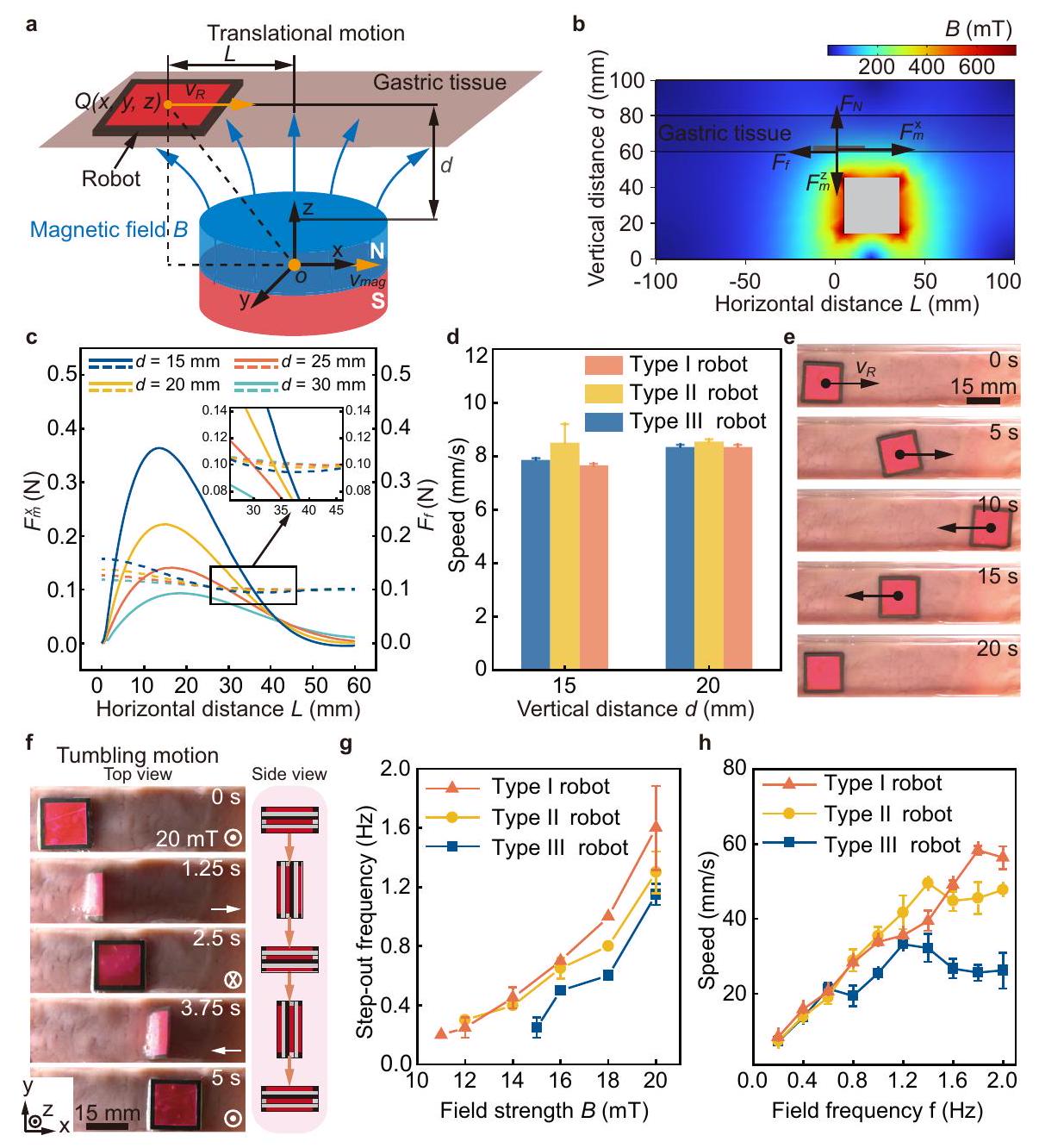
Magnetic actuation of the robot

The robot can be accurately actuated by external magnetic fields using both magnetic torques and forces. The schematics of the translational motion of the robot on a gastric tissue is demonstrated in Fig. 4a. The robot performs translational motion at the speed of actuated by applying an external magnetic field gradient. Simulation and analysis of the forces exerted on the robot for its translational motion on gastric tissue are shown and discussed (Fig. 4b and Supplementary Note 1). Under the actuation of the magnetic force , the robot realizes translational motion overcoming the friction force between the robot and tissues (Supplementary Fig. 3). Hence, the robot can achieve translational motion once the superposed external force along horizontal direction satisfy the criterion:
where is inversely proportional to (Supplementary Note 1). To evaluate the influence of vertical distance on the translational motion
Fig. 4 | Locomotion modes and performance of the robot. a Schematics of translational motion of the robot actuated by a magnet. A cartesian coordinate system is defined with the center of the magnet as the origin . Point refers to the center of the robot. The vertical distance between the adjacent surfaces of the magnet and the robot is . The horizontal distance between the centers of the robot and the magnet is . The orange arrows represent the motion direction of the robot and the magnet. Schematic illustration of the forces exerted on the robot performing translational motion on the gastric tissue. Simulation of the magnetic fields generated a magnet ( ). c Relationship between the simulated superposed external force exerted on the robot (Type I robot)
performing translational motion and the horizontal distance at different vertical distances . The solid lines represent the magnetic force , and the dashed lines represent the friction force . d Translational speed of the robot when a magnet moves at at different vertical distances. e Translational motion of the robot on the gastric tissue at a vertical distance . f Tumbling motion of the robot on gastric tissue. g Relationship between the step-out frequency of the robot performing tumbling motion and the magnetic field strength. Relationship between the speed of the robot performing tumbling motion and the frequency of the magnetic field. The error bars are obtained from 3 trials in each condition. Data are presented as mean values SD. Source data are provided as a Source Data file.
of the robot, the superposed external force exerted on the robot (Type I robot) along the horizontal direction is modeled and simulated (Fig. 4c). At , the superposed external force exerted on the robot exceeds zero when the horizontal distance between the centers of the robot and the magnet, changes from 3.5 mm to 36 mm , and thus the robot can achieve translational motion. The theoretical analysis also demonstrates that the range of the horizontal distance that the robot can achieve translational motion reduces with increasing the vertical distance (Supplementary Table S1). When the vertical distance is 30 mm , the superposed external force is zero so the robot remains static (Fig. 4c and Supplementary Fig. 4). Moreover, the translational speed on the gastric tissue of the robot driven by a magnet with a speed of is characterized (Fig. 4d). The translational motion of the Type I robot on the gastric tissue is shown in Fig. 4e. The translational motion of the Type II and Type III robots are presented in Supplementary Fig. 5a, b.
The tumbling motion of the Type I robot under a rotating magnetic field with a strength of 20 mT and a frequency of 0.2 Hz is shown in Fig. 4f. The tumbling motion of the other two types of robots is shown in Supplementary Fig. 5c, d. The forces and torques exerted on the robot when it performs tumbling motion are analyzed in Supplementary Fig. 6 and Supplementary Note 2. The robot can achieve stable tumbling motion when the applied field rotation frequency keeps lower than its step-out frequency, which can be expressed as:
Here, is the tumbling damping, is the rotation frequency of the magnetic field, and is the angular displacement of the robot. The step-out frequency and translational speed of the robot performing tumbling motion are characterized in Fig. 4 g , h, respectively. The experimental results indicate a positive correlation between the stepout frequency of the tumbling motion and the applied field strength (Fig. 4 g ). Meanwhile, the robot with more layers can reach a higher step-out frequency at the same field strength ( ). Moreover, the translational speed of the robot performing tumbling motion increases with the applied field frequency initially, and then decreases after the field frequency reaches the step-out frequency (Fig. 4h), because the robot cannot achieve synchronized motion with the rotating magnetic field.

On-demand separation by flipping motion

On-demand adhesion and separation of the robot is essential for multitarget adhesion. The influence of the applied field strength on a soft magnetic film is modeled and simulated, in order to investigate the robot flipping and separation process. The magnetic field strength is increased from 0 mT to 50 mT , while the magnetic field direction, defined as the angle between the magnetic field and the -axis, is fixed as (Fig. 5a and Supplementary Fig. 7a). As the magnetic torque exerted on the magnetic film gradually increases, the robot layer bends and separates from the adhered side layer until its net magnetization align with the direction of the external magnetic field. The simulation results indicate that a magnetic field with a strength of 50 mT is sufficient to perform the separation of the robot. The separation process is then simulated by changing magnetic field direction and keeping the magnetic field strength unchanged (Fig. 5b and Supplementary Fig. 7b). When the magnetic field strength is maintained at 50 mT and the magnetic field direction changes from to , the robot flips around the contact point in response to the magnetic field. The simulated stress distribution shown in the color maps of Fig. 5a, b indicates that, increasing the magnetic field strength and magnetic field direction can result in large deformation of the robot, contributing to the flipping and separation process. The relationship between the stress, the magnetic field strength, and the magnetic field
direction is present in Fig. 5c, showing that stress increases with the field strength and the field direction. The relationship between the layer-layer forces, torque, and the magnetic field direction during separation is present in Supplementary Fig. 7c, showing that horizontal force increases with the field direction, while the vertical force and magnetic torque gradually decrease.
The separation of the robots is performed in two steps, as shown in the schematics in Fig. 5d, including the separation of the Type II robot from the Type I robot and the further separation of the Type II robot. The experimental results of the proposed separation strategy on wet gastric tissue are shown in Fig. 5d and Supplementary Movie 1. A magnet is placed underneath the gastric tissue at a vertical distance of 15 mm to manipulate the robot. Initially, the adhesion between the side layer of the robot and the tissue is formed through applying a vertical magnetic attraction force for approximately 2 min . The first flip motion of a Type II robot and separation of a Type I robot is realized by reversing the magnetic field direction, leaving a side layer of the robot adhered on the tissue ( ). The separation of the Type II robot is realized using the same actuation strategy, resulting in the second adhered side layer and a free Type III robot ( ).

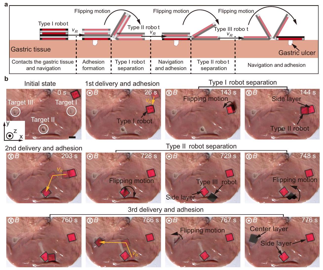
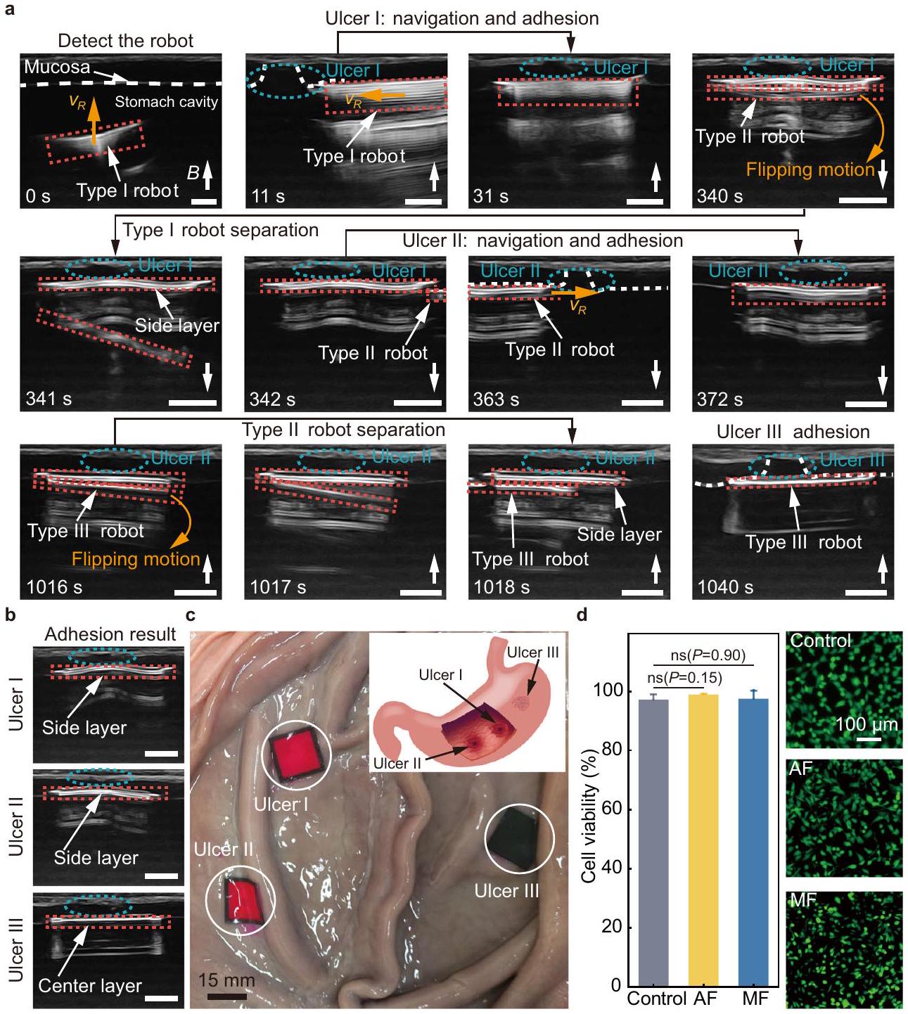
Multi-target adhesion for ex-vivo gastric ulcers

The multi-layer structure and on-demand separation strategy make the robot a promising platform for multi-target adhesion to gastric ulcers. The schematic workflow of the multi-target adhesion using the robot is firstly shown in Fig. 6a. A Type I robot contacts the gastric tissue and is navigated to cover the gastric ulcer through translational motion. Adhesion will be formed between the side layer of the robot and the gastric tissue. A Type II robot separates from the adhered side layer through a flipping motion, and it is subsequently navigated to the next ulcer for coverage and adhesion. The separation of the Type II robot is realized using the same strategy, and the generated Type III robot can be used for the third ulcer adhesion through a flipping motion.
The ex-vivo experiment of multi-target adhesion using the robot is conducted on porcine gastric tissue (Fig. 6b and Supplementary Movie 2). Three artificial ulcers are created, i.e., Target I – III. A Type I robot is delivered to Target I actuated by an external magnetic field , and the adhesion between the side layer of the robot and the gastric tissue is formed by applying a vertical magnetic attraction force for approximately 2 min . Subsequently, a Type II robot separates from the adhered side layer by changing the direction of the magnetic field and is navigated to Target II to adhere to it . Finally, a Type III robot separates from the second adhered side layer ( ) and moves to Target III for the third adhesion ( ). Multiple adhesion using the robot can also be applied to cover the larger ulcer. This method can enlarge the total coverable region of the robot by implementing multiple adhesion adjacently. Herein, we demonstrate that a long ulcer with a length of 35 mm and a width of 5 mm can be covered by the robot as shown in Supplementary Fig. 8. During the second delivery and adhesion, a longer indwelling time is required for the side layer of the robot to form adhesion with the tissue, which could be attributed to the mucus adhered to the robot during the navigation and delivery process. The experimental results on the time required for adhesion are obtained from the trials on the gastric tissue (Supplementary Fig. 9). The time required for the first and second adhesion are and , respectively. The gastric tissue with three robot layers adhered is then immersed in SGF to evaluate the retention performance of the adhesive film on the ex-vivo tissue. After 12 h , the adhesion of the adhesive film still remains (Supplementary Fig. 10).
Moreover, the ex-vivo experiment of multi-target adhesion using the robot is performed inside a porcine stomach with artificial ulcers, and ultrasound imaging is used for navigation (Fig.7a and Supplementary Movie 3). The features of the ulcers and the robots in ultrasound images are shown in Supplementary Fig. 11. The setup of the
Fig. 5 | On-demand separation of the robot. a Simulation of the separation process of a soft magnetic film actuated by a magnetic field with increased field strength (from 0 mT to 50 mT ) and constant magnetic field direction. b Simulation of the separation process of a soft magnetic film actuated by a magnetic field with changed field direction (from to ) and constant magnetic field strength.
c The simulation results of the stress of the film with magnetic field strength and direction. d Schematics and experimental demonstration of the separation strategy, from the Type I robot to the Type II robot (Step I, left), and from the Type II robot to the Type III robot (Step II, right). Source data are provided as a Source Data file.
experiment is shown in Supplementary Fig. 12 (Methods). In the ultrasound images, the ulcer (the blue dashed ellipse, Fig. 7a) harms and breaks the gastric mucosa (the white dashed line in Fig. 7a, ). The robots are labeled using red dashed rectangles. A Type I robot is placed into the stomach through the esophagus and tracked using ultrasound imaging ( ). The robot contacts the gastric mucosa and is navigated to cover the ulcer I ( ). Adhesion is formed through exerting a magnetic stressing force on the robot towards the mucosa for approximately 5 min . A Type II robot separates from the first adhered side layer through a flipping motion by reversing the magnetic field direction, as shown by the separation of two white patterns labeled by the red dashed rectangles ( ). The navigation-adhesion process of a Type II robot to ulcer II is subsequently demonstrated ( ). Finally, a Type III robot separates from the second adhered side layer and is navigated to ulcer III to complete the third adhesion ( ).
A postoperative ultrasound scan is performed on the three ulcers to evaluate the results of multi-target adhesion (Fig. 7b). The ultrasound images indicate that the three layers of the robot successfully cover and adhere to the three ulcers. The stomach is then dissected, and a straightforward observation of the ulcer spots is made (Fig. 7c). All ulcers are covered with the robot layers, including the one locates near the folds of the stomach and the one locates in the confined space
between two folds. The schematics inset of in Fig. 7c shows the initial positions of the ulcers in the porcine stomach.
A quantitative analysis of the biocompatibility of the robot is then conducted (Fig. 7d and Supplementary Fig. 13). In-vitro tests are conducted by separately co-culturing the adhesive film and the soft magnetic substrate of the robot with human gastric mucosal epithelial cells (GES-1) for 24 h (Methods). The results show that the viabilities of the cells co-cultured with the adhesive film and the soft magnetic substrate are and , respectively, and they are compared to the control group with a cell viability of .

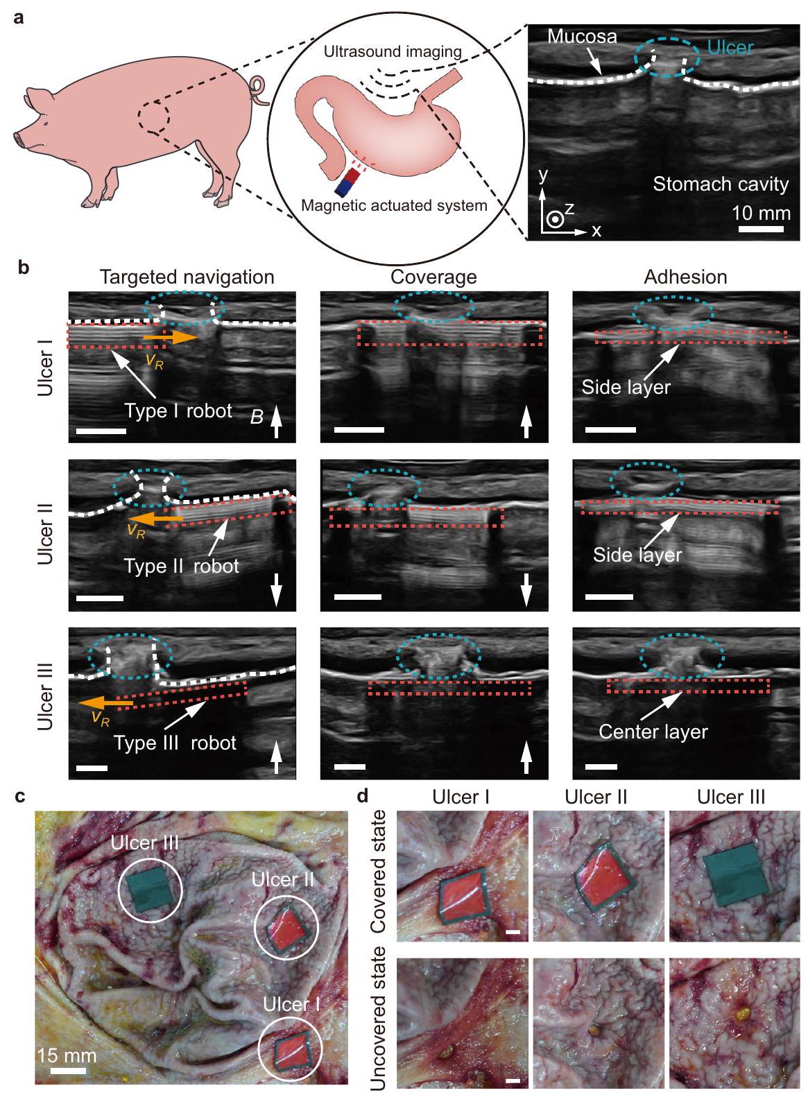
Multi-target adhesion in an in-vivo porcine stomach

In-vivo stomachs add challenges and complexities compared with exvivo ones. While the in-vivo stomach and the ex-vivo stomach are structurally similar, the internal environment of the living porcine stomach brings significant complexities, such as the dynamic peristalsis and the consistent secretion of gastric mucus. The peristaltic motion of the living stomach poses challenges in monitoring and precisely locating the robot within the gastric environment. Additionally, the continuous secretion of gastric mucus makes effective contact difficult between the adhesive film and the tissue, which could prevent the formation of stable adhesion. Furthermore, the in-vivo stomach has more folds on the surface. Therefore, in-vivo experiments
Fig. 6 | Demonstration of multi-target adhesion on ex-vivo gastric tissue. a Schematics of the multi-target adhesion on the gastric tissue. Experimental results of the multi-target adhesion using the robot on ex-vivo gastric tissue. The scale bar is 15 mm .
are necessary. The multi-target adhesion using the robot in an in-vivo porcine stomach is performed (Fig. 8 and Supplementary Movie 4). The schematics of the in-vivo experiment is demonstrated in Fig. 8a (Methods). Using real-time ultrasound imaging for tracking, the robot is capable of approaching, covering and adhering to the ulcers actuated by magnetic field (Fig. 8b). The side layer adheres to the surrounding tissue of ulcer I, and then the Type II robot separates from it. The Type II robot and the Type III robot separated from it successfully cover and adhere to ulcer II and ulcer III, respectively, based on the ultrasound imaging feedback. The stomach is then dissected to evaluate the accuracy of the adhered layer of the robot. It is observed that the layers of the robot adhere to three positions on the folded gastric mucosa (Fig. 8c), and after removing the layers, all the ulcers are revealed (Fig. 8d). The experimental results demonstrate that the proposed robot can tackle the challenge and achieve multi-target adhesion with high precision inside an in-vivo stomach with mucus.

Discussion

In summary, we have developed a magnetic soft robot consisting of multiple layers with distinct in-plane structures and functions to achieve on-demand adhesion to different lesions. Designed materials interface and tailored interlayer interactions are taken into consideration. The robot consists of three layers, including one center layer and two side layers. Each layer is composed of a soft magnetic substrate and an embedded adhesive film. To achieve adhesion to wet tissue, we prepare the Carbopol-based adhesive film that can adhere to the tissue through hydrogen bonds. The adhesion performance of the adhesive film is evaluated and optimized. Furthermore, the interlayer interaction of the robot is adjusted by designing each layer with a different substrate. The side layer consists of a nonmagnetic base and a magnetic frame, while the center layer consists of a magnetic base and a nonmagnetic frame. The three layers are magnetized in the same
direction perpendicular to their plane, providing interlayer magnetic attractions. Once the side layer adheres to the wet tissue, the magnetic torque can be used to flip the robot and separate it from the adhered side layer. By leveraging the on-demand adhesion and separation strategy, we validate the implementation of the robot for multi-target adhesion in ex-vivo and in-vivo environments, demonstrating its potential in treating gastric ulcers. In summary, by integrating functional materials into soft robots, more possibilities could be provided to broaden the applications.
Considering the specific needs required by a magnetic field actuation system for medical scenarios, the effective operation distance of the robot can be extended by increasing magnetic field strength and gradient. For systems integrated with permanent magnet, increasing the volume of the magnet can enhance both the magnetic field strength and gradient, while for electromagnetic coils, boosting the current amplitude in the electromagnetic coil and implementing water-cooling equipment could also be a promising method. For medical imaging targeting deep regions, we can apply this strategy under the guidance of X-ray imaging. Oral-taking barium meal can locate the ulcers , and the robot can be navigated to cover and adhere to them. It is noted that a robot arm with a permanent magnet serving as its end effector can also realize the on-demand actuation of the robots (Supplementary Fig. 14). In addition, loading drugs into the adhesive film of the robot will help further transform the technology into clinical applications that maintain its own adhesion performance and movement (Supplementary Figs. 15 and 16).

Methods

Materials

Carbopol 971 P is obtained from CHINE INTERNATIONAL (Shanghai, China). Poloxamer 407 is purchased from Solarbio LIFE SCIENCES (Beijing, China). Hydroxypropylmethylcellulose (HPMC) is purchased
Fig. 7 | Multiple-target adhesion in an ex-vivo porcine stomach. a The multitarget adhesion using the robot in an ex-vivo porcine stomach filled with liquid under real-time tracking of ultrasound imaging. The red dashed rectangles, the white dashed lines and the blue dashed ellipses represent the robot, the gastric mucosa and the ulcer, respectively. The orange arrows represent the translational motion direction of the robot. The scale bar is 5 mm . Postoperative check with ultrasound imaging. The scale bar is 5 mm . c The dissected porcine stomach after the multi-target adhesion. The schematics illustrates the initial positions of the
ulcers in the porcine stomach. The cell viability (left) of human gastric mucosal epithelial cells (GES-1) in three groups, i.e., control group, adhesive film (AF) group, and soft magnetic substrate (MF) group. For the control group, nothing is added to the culture dish. The adhesive film and the soft magnetic substrate are co-cultured with the cells for the experimental groups, respectively. Live/dead staining image of GES-1 cells in three treatment groups after 24-h co-culture (Right). Data are presented as mean values SD. Source data are provided as a Source Data file.
by MACKLIN (Shanghai, China). All other chemicals are of extra pure reagent grade and are used as received.

Fabrication of magnetic multi-layer soft robot

As shown in Supplementary Fig. 1a, the magnetic film is prepared by spin-coating magnetic slurry, which is prepared by mixing hardmagnetic neodymium-iron-boron (NdFeB) microparticles and polydimethylsiloxane (PDMS) in a mass ratio of 2: 1 .
The adhesive film is prepared from Carbopol, Poloxamer, and HPMC. The Carbopol, HPMC, and Poloxamer are dissolved in an ethanol/water mixture ( ). Rhodamine B is physically mixed into the solution to prepare the pink film for better visualization. The obtained pre-solution is poured into the petri dish, and the adhesive film is obtained by evaporating the remaining solution with heat.
Each component of the layer is obtained from different films by laser cutting and assembled into side and center layers respectively
after plasma treatment (Supplementary Fig. 1a, b). The layers are programmed with the same magnetization direction perpendicular to their plane under a pulsed magnetic field (Supplementary Fig. 1c). Last, the robot is assembled under interlayer magnetic attraction. Detailed information of the robots is shown in Supplementary Table S2.

Mechanical tests

The fracture toughness of the adhesive films prepared with different weight ratios of the Carbopol and HPMC-Poloxamer mixture is measured using tensile tests of thin rectangular samples ( 20 mm in length, 15 mm in width) with a mechanical testing machine ( 0.1 kN load-cell). All tests are conducted with a constant tensile speed of . The fracture toughness of the adhesive film is calculated using a reported method.
Rheological tests are performed using a rheometer (Anton Paar MCR302e, Austria) with rheocompass software. The dynamic moduli
Fig. 8 | On-demand targeted adhesion in an in-vivo porcine stomach. a Schematics of the experiment. The white dashed line and the blue dashed ellipses represent the gastric mucosa and the ulcer, respectively. Real-time navigation of a robot in an in-vivo porcine stomach after gastric emptying for multi-target adhesion. The steps include targeted navigation, coverage, and adhesion. The red dashed rectangles represent the robot. The orange arrows represent the
translational motion direction of the robot. The scale bar is 5 mm . c The dissected porcine stomach after the multi-target adhesion. Detailed views of the covered ulcers and those revealed after peeling the robot layers. The scale bar is 5 mm . Panel (a) is partly generated using Servier Medical Art, provided by Servier, licensed under a Creative Commons Attribution 3.0 unported license.
(Storage modulus, and Loss modulus, ) of the adhesive films are obtained. Amplitude sweep tests are conducted by varying the strain from 0.01 to with a frequency of 1 Hz . Frequency sweep tests are conducted by varying the frequency from 0.1 to 10 Hz with a strain of .

In vitro adhesion tests

Quantitative tissue adhesion strength tests, including the Lap-shear and T-peel tests, are performed according to the ASTM F2255 and F2256. The pig gastric tissue is used as biological tissue material. The two tissues are placed into a universal testing machine (Instron, America) for tensile loading at a strain rate of . The adhesive film is placed between the tissues and pressed under 1 kPa for different time.
To measure shear strength, adhered samples (the adhesive film) with an adhesion area of width 15 mm and length 15 mm are prepared
and tested by the standard lap-shear test (ASTM F2255) with a mechanical testing machine ( 0.1 kN load-cell). Shear strength is determined by dividing the maximum force by the adhesion area.
To measure interfacial toughness, adhered samples (the adhesive film) with a width of 15 mm are prepared and tested by the standard 180-degree peel test (ASTM F2256) using a mechanical testing machine ( 0.1 kN load-cell). The measured force reaches a plateau as the peeling process enters the steady state. Interfacial toughness is determined by dividing two times the plateau force (for a 180 -degree peel test) by the width of the tissue sample.
To characterize the retention time of the adhesive film under cyclic loading, porcine gastric tissue adhered with a monomer is immersed in SGF. The stretching equipment (KeyFactor, China) stretches the pig gastric tissue at different frequencies ( , 1.25 Hz ) and records the retention time of the monomer.

In vitro biocompatibility test

To evaluate in-vitro biocompatibility and cytotoxicity of the adhesive layer, LIVE/DEAD assay is used to assess Human gastric epithelial cell line (GES-1, Xiamen Immocell Biotechnology Co.,Ltd.). Cut the adhesive film to and co-culture it with GES-1, without adding the adhesive film as a control. GES-1 cells are seeded in 96well plates at a density of cells ( per each group). The cells are then incubated at for 24 h in atmosphere. The cell viability is determined by a LIVE/DEAD viability/cytotoxicity kit for mammalian cells (Thermo Fisher Scientific) by adding 4 M calcein and ethidium homodimer-1 into the culture media. A microscope (Nikon ECLIPSE Ti2) is used to image live cells with excitation/emission at and dead cells at , respectively. The cell viability is calculated by counting live (green fluorescence) and dead (red fluorescence) cells by using ImageJ (version 2.1.0).

Ultrasound imaging for ex-vivo and in-vivo tests

In this study, ultrasound imaging is employed to track the robots and the ulcers. Under ultrasound images, the mucosa appears as a continuous white line in the water-filling environment. An obvious change in the contour can be observed at the site of the ulcer. In environments with air, the ultrasound images show the presence of irregular white signals, which can serve as an indication of the location of ulcers. In a stomach filled with fluid or in an empty stomach, the location and pattern of the robot are identified by tracking the stacked white lines in the ultrasound images. The thickness of the stack decreases when a Type I robot transits into a Type III robot. In particular, the location where the white signal (the yellow dashed rectangles, Supplementary Fig. 11) disappears can be used to locate the Type III robot in the air environment, allowing for tracking of the Type III robot as it moves in an in-vivo porcine stomach after gastric emptying.

Experimental procedure of ex-vivo and in-vivo tests

Experimental procedures of ex-vivo and in-vivo experiments can be listed as follows. Two operators collaborate to achieve multi-target adhesion of the robot. Operator 1 operates the ultrasound transducer to locate the robot. Once the robot is located, Operator 2 manipulates the robot using a cylindrical magnet. Operators 1 and 2 move the transducer and magnet synchronously to actuate the robot toward and then cover target I. Subsequently, once the adhesion between the adhesive film and the tissue is formed under magnetic attraction. Operator 2 then reverses the magnet to separate Type II robot from the adhered side layer. This systematic approach is also applied to cover targets II and III.

Preparation of porcine stomach for ex-vivo tests

All ex-vivo pig stomachs are purchased from Taobao, China. The organs are emptied and minimally cleaned by water without damaging the mucosa. Acetic acid-induced gastric ulcers are established using a previously reported protocol . Use surgical scissors to cut out three areas of on the surface of the pig stomach, and acetic acid is injected into the incision and maintained for 120 s . For ex-vivo porcine stomach, three artificial ulcers are created on different sides of the stomach. For ex-vivo tests, the dimensions of the robot (Type I robot) are and the weight is .

In-vivo tests in a pig

After the appropriate depth of anesthesia and analgesia, the stomach is exposed by surgical laparotomy. A small incision is made in the esophagus using a surgical knife to remove the stomach contents. The stomach is then washed with physiological saline. A surgical scissor is used to create three ulcers at the gastric mucosa, each about 1 cm in diameter, and the location of the ulcers is determined using ultrasound imaging. The robot is delivered into the stomach through the incision and navigated using ultrasound. The direction and distance of the
magnetic field are adjusted to allow the robot to form adhesion with the tissue and separate from the side layer. The final adhesion result is examined using ultrasound imaging. Under deep anesthesia, the animal is then euthanized by intravenous injection of potassium chloride, and the stomach is dissected to observe the final adhesive result. For in-vivo tests, the dimensions of the robot (Type I robot) are and the weight is .

Simulation

Simulation of the stress distribution when the robot performed translational motion on the stomach and the separation between a soft magnetic film and the adhered side layer to the tissue under magnetic fields with different directions and strength used the commercial software Comsol. Young’s modulus and magnetization measured from the experiment are used as the input parameters.

Statistical analysis

The data obtained in this study are all reported as mean standard deviation (SD) from at least three separate experiments. Values from two groups are compared using Student’s test. A value of 0.05 or less is considered to be statistically significant. , ; ns, not significant.

Reporting summary

Further information on research design is available in the Nature Portfolio Reporting Summary linked to this article.

Data availability

The data generated in this study are provided in the Supplementary Information/Source Data file. Additional data are available from the corresponding author on request. Source data for the figures are provided with this paper. Source data are provided with this paper.

References

  1. Soon, R. H. et al. On-demand anchoring of wireless soft miniature robots on soft surfaces. Proc. Natl. Acad. Sci. 119, e2207767119 (2022).
  2. Ren, Z. et al. Soft-bodied adaptive multimodal locomotion strategies in fluid-filled confined spaces. Sci. Adv. 7, eabh2022 (2021).
  3. Gu, H. et al. Self-folding soft-robotic chains with reconfigurable shapes and functionalities. Nat. Commun. 14, 1263 (2023).
  4. Zheng, Z. et al. lonic shape-morphing microrobotic end-effectors for environmentally adaptive targeting, releasing, and sampling. Nat. Commun. 12, 411 (2021).
  5. Xu, T., Huang, C., Lai, Z. & Wu, X. Independent control strategy of multiple magnetic flexible millirobots for position control and path following. IEEE Trans. Robot. 38, 2875-2887 (2022).
  6. Ebrahimi, N. et al. Magnetic actuation methods in bio/soft robotics. Adv. Funct. Mater. 31, 2005137 (2021).
  7. Zhang, J. et al. Voxelated three-dimensional miniature magnetic soft machines via multimaterial heterogeneous assembly. Sci. Robot. 6, eabf0112 (2021).
  8. Deng, H. et al. Laser reprogramming magnetic anisotropy in soft composites for reconfigurable 3D shaping. Nat. Commun. 11, 6325 (2020).
  9. Gu, H., Boehler, Q., Ahmed, D. & Nelson, B. J. Magnetic quadrupole assemblies with arbitrary shapes and magnetizations. Sci. Robot. 4, eaax8977 (2019).
  10. Xu, T., Zhang, J., Salehizadeh, M., Onaizah, O. & Diller, E. Millimeterscale flexible robots with programmable three-dimensional magnetization and motions. Sci. Robot. 4, eaav4494 (2019).
  11. Richter, M. et al. Magnetic soft helical manipulators with local dipole interactions for flexibility and forces. Soft Robot. 10, 647-659 (2023).
  12. Hu, W., Lum, G. Z., Mastrangeli, M. & Sitti, M. Small-scale softbodied robot with multimodal locomotion. Nature 554, 81-85 (2018).
  13. Won, S., Kim, S., Park, J. E., Jeon, J. & Wie, J. J. On-demand orbital maneuver of multiple soft robots via hierarchical magnetomotility. Nat. Commun. 10, 4751 (2019).
  14. Li, W. et al. Self-vectoring electromagnetic soft robots with high operational dimensionality. Nat. Commun. 14, 182 (2023).
  15. Lu, H. et al. A bioinspired multilegged soft millirobot that functions in both dry and wet conditions. Nat. Commun. 9, 3944 (2018).
  16. Huang, H.-W. et al. Adaptive locomotion of artificial microswimmers. Sci. Adv. 5, eaau1532 (2019).
  17. Wang, Y. et al. Amphibious miniature soft jumping robot with ondemand in-flight maneuver. Adv. Sci. 10, 2207493 (2023).
  18. Wu, Y., Dong, X., Kim, J., Wang, C. & Sitti, M. Wireless soft millirobots for climbing three-dimensional surfaces in confined spaces. Sci. Adv. 8, eabn3431 (2022).
  19. Wang, C., Wu, Y., Dong, X., Armacki, M. & Sitti, M. In situ sensing physiological properties of biological tissues using wireless miniature soft robots. Sci. Adv. 9, eadg3988 (2023).
  20. Wang, C., Mzyk, A., Schirhagl, R., Misra, S. & Venkiteswaran, V. K. Biocompatible film-coating of magnetic soft robots for mucoadhesive locomotion. Adv. Mater. Technol. 8, 2201813 (2023).
  21. Zhou, C. et al. Ferromagnetic soft catheter robots for minimally invasive bioprinting. Nat. Commun. 12, 5072 (2021).
  22. Yang, Y. et al. Magnetic soft robotic bladder for assisted urination. Sci. Adv. 8, eabq1456 (2022).
  23. Kim, Y. et al. Telerobotic neurovascular interventions with magnetic manipulation. Sci. Robot. 7, eabg9907 (2022).
  24. Son, D., Gilbert, H. & Sitti, M. Magnetically actuated soft capsule endoscope for fine-needle biopsy. Soft Robot. 7, 10-21 (2020).
  25. Zong, Z., Zhang, S. & Wu, Z. Anisotropic contraction in a magnetically hard but mechanically ultra-soft foam for precise drug delivery. Adv. Mater. Technol. 8, 2201255 (2023).
  26. Zhang, H. et al. Dual-responsive biohybrid neutrobots for active target delivery. Sci. Robot. 6, eaaz9519 (2021).
  27. Dong, X. et al. Bioinspired cilia arrays with programmable nonreciprocal motion and metachronal coordination. Sci. Adv. 6, eabc9323 (2020).
  28. Lee, J., Kim, D., Bang, S. & Park, S. Drug-loaded mucoadhesive patch with active delivery and controlled releasing ability. Adv. Intell. Syst. 4, 2100203 (2022).
  29. Zheng, Z. et al. Programmable aniso-electrodeposited modular hydrogel microrobots. Sci. Adv. 8, eade6135 (2022).
  30. Ze, Q. et al. Spinning-enabled wireless amphibious origami millirobot. Nat. Commun. 13, 3118 (2022).
  31. Mair, L. O. et al. Soft capsule magnetic millirobots for regionspecific drug delivery in the central nervous system. Front. Robot. Al 8, 702566 (2021).
  32. Yang, X. et al. An agglutinate magnetic spray transforms inanimate objects into millirobots for biomedical applications. Sci. Robot. 5, eabc8191 (2020).
  33. Dong, Y. et al. Untethered small-scale magnetic soft robot with programmable magnetization and integrated multifunctional modules. Sci. Adv. 8, eabn8932 (2022).
  34. Wang, T. et al. Adaptive wireless millirobotic locomotion into distal vasculature. Nat. Commun. 13, 4465 (2022).
  35. Ze, Q. et al. Soft robotic origami crawler. Sci. Adv. 8, eabm7834 (2022).
  36. Cheng, Z. et al. Fabrication of ulcer-adhesive oral keratin hydrogel for gastric ulcer healing in a rat. Regen. Biomater. 8, rbab008 (2021).
  37. Brătucu, M. N. et al. Unusual complicated gastric ulcers. Medicina 57, 1345 (2021).
  38. Chu, J.N., Traverso, G. Foundations of gastrointestinal-based drug delivery and future developments. Nat. Rev. Gastroenterol. Hepatol. 19, 219-238 (2022).
  39. Anthis, A. H. C. et al. Modular stimuli-responsive hydrogel sealants for early gastrointestinal leak detection and containment. Nat. Commun. 13, 1-15 (2022).
  40. Wu, S. J., Yuk, H., Wu, J., Nabzdyk, C. S. & Zhao, X. A multifunctional origami patch for minimally invasive tissue sealing. Adv. Mater. 33, 2007667 (2021).
  41. Wu, J. et al. An off-the-shelf bioadhesive patch for sutureless repair of gastrointestinal defects. Sci. Transl. Med. 14, eabh2857 (2022).
  42. Xu, X. et al. Bioadhesive hydrogels demonstrating pH-independent and ultrafast gelation promote gastric ulcer healing in pigs. Sci. Transl. Med. 12, eaba8014 (2020).
  43. Chen, X. et al. Hydrogel bioadhesives with extreme acid-tolerance for gastric perforation repairing. Adv. Funct. Mater. 32, 2202285 (2022).
  44. Wang, H. et al. An integrally formed janus hydrogel for robust wettissue adhesive and anti-postoperative adhesion. Adv. Mater. 35, 2300394 (2023).
  45. Chun, M.-K., Bhusal, P. & Choi, H.-K. Application of Carbopol/PVP interpolymer complex to prepare mucoadhesive floating granule. Arch. Pharm. Res. 36, 745-751 (2013).
  46. Li, T., Bao, Q., Shen, J., Lalla, R. V. & Burgess, D. J. Mucoadhesive in situ forming gel for oral mucositis pain control. Int. J. Pharm. 580, 119238 (2020).
  47. Patel, M. M. et al. Mucin/Poly(acrylic acid) Interactions: a spectroscopic investigation of mucoadhesion. Biomacromolecules 4, 1184-1190 (2003).
  48. Deng, T. et al. A natural biological adhesive from snail mucus for wound repair. Nat. Commun. 14, 396 (2023).
  49. Yuk, H. et al. Dry double-sided tape for adhesion of wet tissues and devices. Nature 575, 169-174 (2019).
  50. Schulze, K. Imaging and modelling of digestion in the stomach and the duodenum. Neurogastroenterol. Motil. 18, 172-183 (2006).
  51. Marciani, L. et al. Assessment of antral grinding of a model solid meal with echo-planar imaging. Am. J. Physiol. -Gastrointest. Liver Physiol. 280, G844-G849 (2001).
  52. Debi, U. et al. Role of barium study in various gastric malignancies. Trop. Gastroenterol. 10, 246-255 (2019).

Acknowledgements

We thank Kaiwen Fang and Guoqing Chen for their assistance in animal surgeries. This work was financially supported by the National Key R&D Program of China under Project No. 2022YFA1207100, the National Natural Science Foundation of China under Project No. 62103347, No. 62073208 and No. 61933008, the Innovation Program of Shanghai Municipal Education Commission under Grant 2021-01-07-00-09E0013, Shenzhen Science and Technology Program under Grant No. RCBS20210609103155061, Guangdong Basic and Applied Basic Research Foundation under Project No. 2022A1515110499, and the Shenzhen Institute of Artificial Intelligence and Robotics for Society under Project No. AC01202101109, Shanghai Science and Technology plan project under Grant 23ZR1422300.

Author contributions

Z.C., Y.W., N.L. and J.Y. conceived the study, designed the experiments. Z.C., Y.W., J.L., N.L. and J.Y. wrote the manuscript. Z.C., Y.W. and H.C. performed the experiments and analyzed data. Z.C. and Y.W. provided conceptual and technical input for the numerical simulations. Z.C., Y.W., F.D. and H.C. assisted with the experiments of clinical imaging. H.P., S.X., Y.S., N.L. and J.Y. assisted with the supervision of experiments and edited the manuscript. N.L. and J.Y. supervised the project. All authors discussed the results and reviewed the manuscript.

Competing interests

The authors declare no competing interests.

Additional information

Supplementary information The online version contains supplementary material available at https://doi.org/10.1038/s41467-024-44995-9.
Correspondence and requests for materials should be addressed to Na Liu or Jiangfan Yu.
Peer review information Nature Communications thanks Sukho Park, and the other, anonymous, reviewers for their contribution to the peer review of this work. A peer review file is available.
Reprints and permissions information is available at
http://www.nature.com/reprints
Publisher’s note Springer Nature remains neutral with regard to jurisdictional claims in published maps and institutional affiliations.
Open Access This article is licensed under a Creative Commons Attribution 4.0 International License, which permits use, sharing, adaptation, distribution and reproduction in any medium or format, as long as you give appropriate credit to the original author(s) and the source, provide a link to the Creative Commons license, and indicate if changes were made. The images or other third party material in this article are included in the article’s Creative Commons license, unless indicated otherwise in a credit line to the material. If material is not included in the article’s Creative Commons license and your intended use is not permitted by statutory regulation or exceeds the permitted use, you will need to obtain permission directly from the copyright holder. To view a copy of this license, visit http://creativecommons.org/ licenses/by/4.0/.
© The Author(s) 2024

  1. School of Mechatronics Engineering and Automation, Shanghai University, Shanghai 200444, China. School of Science and Engineering, The Chinese University of Hong Kong, Shenzhen 518172, China. Shenzhen Institute of Artificial Intelligence and Robotics for Society, Shenzhen 518172, China. Department of Mechanical and Industrial Engineering, University of Toronto, Toronto, ON M5S 3G8, Canada. School of Computer Engineering and Science, Shanghai University, Shanghai 200444, China. Department of Interventional Radiology, Chinese PLA General Hospital, Beijing 100853, China. School of Medicine, The Chinese University of Hong Kong, Shenzhen 518172, China. These authors contributed equally: Ziheng Chen, Yibin Wang.