ظهور الديدان القوية المقاومة لدوائين في الماعز: أول دليل ظاهري وجيني من محافظة راتشابوري، وسط تايلاند Emergence of dual drug-resistant strongylids in goats: first phenotypic and genotypic evidence from Ratchaburi Province, central Thailand

المجلة: BMC Veterinary Research، المجلد: 21، العدد: 1
DOI: https://doi.org/10.1186/s12917-025-04700-4
PMID: https://pubmed.ncbi.nlm.nih.gov/40181391
تاريخ النشر: 2025-04-04

ظهور الديدان القوية المقاومة لدوائين في الماعز: أول دليل ظاهري وجيني من محافظة راتشابوري، وسط تايلاند

أبيجيل هوي إن تشان شانيزارا كانكايو والوب باكدي سيفابونغ سونغبراديت وأوروسا ثينكهام

الملخص

خلفية توفر هذه الدراسة رؤى حاسمة حول انتشار وأنماط مقاومة الأدوية للديدان المعوية القوية (GINs) في الماعز في تايلاند، مع تسليط الضوء على المقاومة للألبندازول والليفاميزول. تشكل الديدان القوية، وخاصة Haemonchus sp. وTrichostrongylus sp.، تهديدًا كبيرًا لصحة الماعز. مع الزيادة العالمية لمقاومة الأدوية المضادة للديدان، فإن اكتشاف مقاومة متعددة الأدوية في سكان الماعز في تايلاند يثير القلق، نظرًا للتجارة المتكررة في استيراد وتصدير الماعز. تتحدى هذه المقاومة استراتيجيات السيطرة الفعالة على الطفيليات. هدفت هذه الدراسة إلى تحديد أنواع الديدان القوية باستخدام كل من الطرق الشكلية والجينية، وتقييم المقاومة للألبندازول والليفاميزول من خلال الأساليب الظاهرة والجزيئية. النتائج أظهرت عينات البراز من 30 مزرعة ماعز في محافظة راتشابوري انتشارًا عاليًا للعدوى بالديدان القوية (87%)، مع اكتشاف Haemonchus sp. وTrichostrongylus sp. في 100% و96% من المزارع، على التوالي. أظهرت الاختبارات الظاهرة مقاومة كبيرة للأدوية، حيث كانت 90% و71% من المزارع تحتوي على تجمعات ديدان قوية مقاومة للألبندازول والليفاميزول، على التوالي. أظهر التحليل الجيني لليرقات المعدية المجمعة أن 100% من المزارع كانت تحتوي على تجمعات ديدان قوية مقاومة للألبندازول، مع 31% مقاومة متماثلة و69% مقاومة غير متماثلة، وTrichostrongylus sp. أظهرت 48% مقاومة متماثلة و52% مقاومة غير متماثلة. بالنسبة لمقاومة الليفاميزول، كانت 92% من المزارع تحتوي على تجمعات ديدان قوية مقاومة، مع ظهور Haemonchus sp. متماثل الزيجوت و مقاومة هتيروزيجوت. الاستنتاجات تقدم هذه الدراسة التقييم الشامل الأول للمقاومة الظاهرية والجينية في الديدان الشريطية في محافظة راتشابوري، مما يعالج فجوة جغرافية رئيسية في بيانات المقاومة في تايلاند. تسلط النتائج الضوء على الحاجة الملحة لإعادة تقييم ممارسات إدارة GIN وتطوير استراتيجيات مستدامة للتخفيف من المقاومة. علاوة على ذلك، فإن هذه النتائج لها تداعيات كبيرة على صحة الماشية عبر الحدود، مما يبرز ضرورة الجهود التعاونية لمكافحة التحدي المتزايد للأدوية المضادة للديدان.

الكلمات الرئيسية: الديدان الشعرية الماعزية، مقاومة الأدوية الطاردة للديدان، اختبار في المختبر، تحديد النمط الجيني

الخلفية

تشكل عدوى الديدان الخيطية المعوية (GIN) تهديدًا كبيرًا للماشية المجترة، مما يساهم في خسارة اقتصادية عالمية تقدر بـ 7 مليارات دولار أمريكي سنويًا [1]. الماشية الصغيرة، وخاصة الماعز، عرضة بشدة لعدوى GIN، مما يؤثر بشكل كبير على الإنتاجية والصحة العامة. تعاني الماعز المصابة من فقدان الوزن، وانخفاض إنتاج الحليب واللحم، وتدهور الصحة العامة [2،3]. بالإضافة إلى ذلك، فإن تكلفة العلاجات المضادة للديدان تزيد من تفاقم الخسائر الاقتصادية، مما يقلل من ربحية تربية الماعز في جميع أنحاء العالم [4].
الديدان الأسطوانية القوية، بما في ذلك الأجناس مثل هيمونشوس، تريشوتراستونغيلوس، كوبرية، شابيرتيا، وأوزوفاجوستوموم، هي الأكثر شيوعًا من الديدان المعوية المعدية التي تصيب الماعز. من بين هذه الأنواع، تشكل هيمونشوس كونتورتوس وأنواع تريشوتراستونغيلوس أكبر تهديد لصحة الماعز. . كونتورتوس تسبب فقر الدم الشديد والوفيات. حالات متفرقة من العدوى البشرية مع تم الإبلاغ عن . contortus و Trichostrongylus sp. في دول مختلفة مثل تايلاند، جمهورية لاو الديمقراطية الشعبية، كوريا الجنوبية، إيران، وأستراليا [7-10]، مما يظهر إمكانيتهما الانتقالية بين الأنواع، لا سيما في المناطق التي يتقارب فيها البشر والماعز.
ظهور مقاومة الأدوية الطاردة للديدان يشكل تحديًا كبيرًا للمزارعين والأطباء البيطريين، مما يؤثر على صحة وإنتاجية الماعز. حيث أن المقاومة تقوض فعالية العلاج، فإن استراتيجيات الإدارة العاجلة مطلوبة للحفاظ على تربية الماعز. تفاقم ممارسات الإدارة السيئة والجرعات غير الصحيحة من إصابات الديدان المعوية [11]. تم الإبلاغ عن مقاومة الديدان الأسطوانية للأدوية الطاردة للديدان، بما في ذلك مقاومة متعددة الأدوية عبر الفئات الثلاث الرئيسية للأدوية الطاردة للديدان – البنزيميدازولات، الإيميدازوثيازولات، واللاكتونات الدائرية، من أوروبا وأستراليا وآسيا [12، 13]. يعتمد الكشف عن مقاومة الأدوية الطاردة للديدان تقليديًا على الطرق الظاهرية، بما في ذلك اختبار فقس البيض (EHT) واختبار تطوير اليرقات (LDT)، بينما يعتمد تقييم فعالية الأدوية على اختبار تقليل عدد البيض في البراز (FECRT). ومع ذلك، فإن الأساليب الجزيئية تكمل بشكل متزايد الطرق الظاهرية، مما يسمح بالكشف الدقيق عن الأليلات المرتبطة بالمقاومة. على سبيل المثال، يتم تحديد مقاومة البنزيميدازول (الألبندازول) من خلال تعدد أشكال النوكليوتيدات المفردة (SNPs) في بيتا-توبولين ( جين الأيزوتيب 1 من -توبولين، مع اختبارات PCR الكمية (qPCR) التي تستهدف الطفرات الرئيسية F200Y وF167Y وE198A – خاصة في H. contortus. وبالمثل، تم تطوير اختبار qPCR للكشف عن مقاومة الإيميدازوثيازول (ليفاميسول) من خلال تحديد حذف بطول 63 قاعدة في جين Hco-acr-8 لعزلات H. contortus. تعزز هذه الأدوات الجزيئية التشخيص الجيني، مما يوفر طرقًا قيمة للكشف عن الأليلات المقاومة للأدوية وتحسين استراتيجيات إدارة المقاومة.
تم الإبلاغ عن إصابات GIN في الماعز عبر عدة مقاطعات في تايلاند، بما في ذلك ناخون باتوم، كانشانابوري، خون كاين، وساتون، مع دراسات توثق انتشارًا عاليًا للإصابات بالسترونغيلات [1821]. وقد أكدت التحليلات الجزيئية وجود Haemonchus sp. وOesophagostomum sp. وTrichostrongylus sp.، بالإضافة إلى جينات مقاومة البنزيميدازول في . كونتورتوس [22]. بالإضافة إلى ذلك، تم تحديد مقاومة لجميع فئات الأدوية المضادة للطفيليات الثلاثة الرئيسية أيضًا في محافظة سينغ بوري [23]. تسلط هذه النتائج الضوء على الانتشار الواسع للديدان الشديدة GINs والتحدي المتزايد لمقاومة الأدوية المضادة للطفيليات في سكان الماعز في تايلاند. ومع ذلك، هناك حاجة إلى مزيد من البحث لتقييم ديناميات عدوى GIN ومقاومة الأدوية في مناطق أخرى، لا سيما في المحافظات التي تشارك في استيراد وتصدير صغار الماعز، حيث قد يكون لانتقال الأمراض آثار عبر الحدود أوسع [24].
تتركز هذه الدراسة على محافظة راتشابوري في وسط تايلاند، حيث أبلغت الطرق الظاهرية مؤخرًا عن مقاومة مضادات الديدان [25]. أهدافنا هي تحديد أنواع الديدان المعدية التي تصيب الماعز في راتشابوري واكتشاف الجينات المقاومة للأدوية للألبندازول والليفاميزول باستخدام تقنيات ظاهرة وجزيئية. ستوفر هذه النتائج رؤى قيمة للمزارعين والأطباء البيطريين بينما تساهم في الجهود العالمية لإدارة عدوى الديدان المعدية ومكافحة مقاومة مضادات الديدان.

طرق

بيان الأخلاقيات

تمت الموافقة على الدراسة المتعلقة بمزارع الماعز من قبل اللجنة الأخلاقية بكلية العلوم البيطرية، جامعة ماهيدول (الأكواد المعتمدة المخصصة من قبل FVS-MUIACUC: رقم COA. MUVS-2023-08-54 ورقم COA. MUVS-2024-03-24).

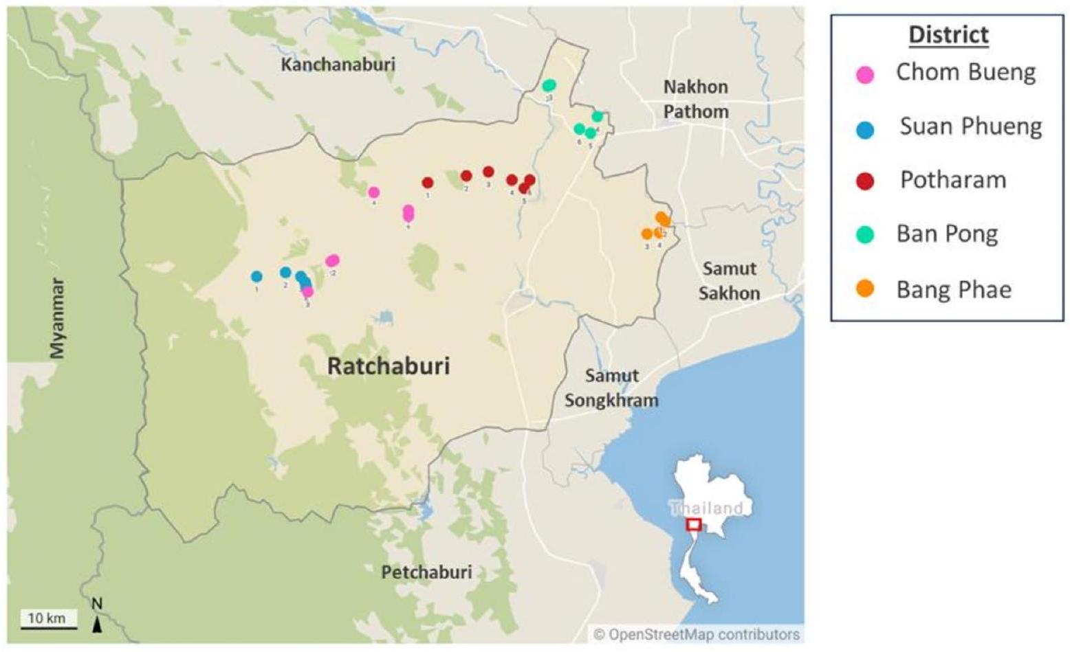
مزارع الماعز وجمع العينات

تم جمع عينات من البراز من 173 ماعزًا عبر 30 مزرعة في خمسة مناطق في محافظة راتشابوري بين نوفمبر 2023 (موسم الجفاف) ويونيو 2024 (موسم الأمطار) (الشكل 1). شملت المناطق التي تم مسحها تشوم بوانغ، وسوان فوانغ، وبوتارام، وبان بونغ، وبانغ فاي. تضمنت المزارع المختارة عمليات لتربية الماعز الحلوب واللحوم، حيث اعتمد معظمها نهج تربية بنظام مفتوح، حيث ترعى الماعز بحرية خلال النهار وتُحتجز في الداخل ليلاً. كما تم تسجيل تاريخ استخدام الأدوية المضادة للطفيليات في كل مزرعة.
تم جمع عينات البراز من المستقيم لكل ماعز باستخدام قفازات نظيفة ووُضعت في أنابيب فالكون سعة 50 مل. ثم تم نقل العينات إلى قسم الديدان الطفيلية، كلية الطب الاستوائي، جامعة ماهيدول، بانكوك، لإجراء مزيد من التحليل.
الشكل 1 مواقع مزارع الماعز حيث تم جمع العينات. تشير الدوائر الملونة إلى 30 مزرعة موزعة عبر خمسة مناطق، بواقع ست مزارع لكل منطقة.

تحديد وكمية الديدان الطفيلية المعوية الفحص المجهري

تم إجراء تحديد GIN وقياس بيض الستروغيليد في عينات براز الماعز باستخدام تقنيات الطفو البسيطة وماكماسٹر [26، 27]. تم تجانس حوالي 4 جرام من البراز مع الماء وتصفيته من خلال منخل إلى دورق سعة 500 مل. تم السماح للمستحلب بالترسب، وتم تصفية السائل العلوي. تم استخدام كلوريد الصوديوم المشبع. تمت إضافة ( )، وتم نقل المستحلب إلى أنابيب فالكون سعة 15 مل. ثم تمت إضافة NaCl إضافي إلى كل أنبوب لتشكيل قوس مقلوب في الأعلى. تم وضع شريحة زجاجية بعناية فوق فتحة الأنبوب وتركها دون إزعاج لمدة 15 دقيقة. لتحديد بيض الديدان الخيطية، تمت إزالة الشريحة الزجاجية ووضعها على شريحة ميكروسكوبية للفحص تحت المجهر الضوئي.
للت quantification، تم سحب عينة متجانسة من أنبوب فالكون باستخدام ماصة زجاجية ونقلها إلى غرف عد مكماستر. تم ترك الشرائح لتستقر لمدة 5 دقائق قبل فحصها تحت المجهر الضوئي. تم عد بيض الستروغيليد باستخدام تم حساب عدد البيض لكل جرام (EPG) عن طريق ضرب العدد الإجمالي لبيض السستوديات في 50.

التعرف الجزيئي

بعد الفحص المجهري، تم غسل البيض وتجميعه من قبل المزرعة، وحفظه في الإيثانول عند حتى تحديد الهوية الجزيئية لبيض الستروغيلد. تم استخراج الحمض النووي الجينومي من بيض الستروغيلد المجمعة باستخدام مجموعة الحمض النووي الجينومي الصغيرة (Geneaid Biotech Ltd.، تايبيه، تايوان) وفقًا لتعليمات الشركة المصنعة. قبل استخراج الحمض النووي، تم تجانس البيض باستخدام كرات السيليكا في محلول التحلل باستخدام جهاز TissueLyser LT (Qiagen، هيلدن، ألمانيا) لتسهيل تكسير الخلايا.
تم إجراء تفاعل البوليميراز المتسلسل المتداخل باستخدام منطقة الفاصل الداخلي النسخي النووي 2 (ITS2) وجين الرنا الريبوسومي 16S الميتوكوندري (rRNA) لتحديد بيض السستوديات على المستوى الجزيئي. استهدفت البرايمرات النوعية للجنس Haemonchus وTrichostrongylus وOesophagostomum. تبعت برايمرات ITS2 دراسة Income وآخرون (2021)، بينما كانت برايمرات جولة 16S rRNA الأولى مستندة إلى دراسة Chan وآخرون (2020). تم تصميم برايمرات نوعية للجنس لتفاعل البوليميراز المتسلسل المتداخل في هذه الدراسة [20، 28]. باختصار، تم الحصول على تسلسلات جين 16S rRNA المرجعية لـ Haemonchus sp. وTrichostrongylus sp. وOesophagostomum sp. من قاعدة بيانات NCBI وتم محاذاتها باستخدام ClustalX 2.1 [29]. ثم تم تصميم البرايمرات النوعية للجنس ضمن تسلسل منتج PCR من الجولة الأولى من PCR. تم تقييم الخصائص (محتوى GC، حجم المنتج، درجة انصهار، تشكيل حلقة الشعر) والخصوصية للبرايمرات الجديدة.
تم تقييم البرايمرات المصممة باستخدام OligoCalc الإصدار 3.27 و FastPCR [30، 31]. تلخص الجدول 1 البرايمرات المحددة للنوع 16 S المستخدمة في هذه الدراسة، مع درجات حرارة التهجين وأحجام الأمبليكون الخاصة بها.
تم إجراء تضخيم PCR باستخدام جهاز T100 جهاز التفاعل الحراري (بايو راد، كاليفورنيا، الولايات المتحدة الأمريكية) في حجم تفاعل نهائي قدره تكون الماسترمكس من من 2 X i-Taq™ ماسترمكس (iNtRON Biotechnology، غيونغجي، كوريا الجنوبية)، جنبًا إلى جنب مع من كل بادئ ITS2 أو لكل بادئ 16S للجولة الأولى من تفاعل البوليميراز المتسلسل (PCR). بالنسبة للجولة الثانية من PCR، تم تخفيف المنتجات من الجولة الأولى عشرة أضعاف في ماء خالٍ من النوكلياز واستخدمت كقوالب DNA، مع اتباع نفس ظروف تحضير الماسترميكس لكل بادئ محدد للجنس. كانت ظروف التدوير الحراري لـ PCR كما يلي: الت denaturation الأولي عند لمدة دقيقتين؛ 35 دورة من لمدة 30 ثانية، درجة حرارة التلدين المعنية لـ لمدة 30 ثانية، تليها فترة تمديد نهائية عند لمدة 10 دقائق. تم فحص الأمبليكونات عبر الرحلان الكهربائي لهلام الأجاروز الملون بـ SYBR تم إرسال الأمبليكونات التمثيلية بعد ذلك للتسلسل باستخدام علامة الشريط بواسطة مزود تجاري (Celemics، سيول، كوريا الجنوبية) للتعرف على الأنواع.

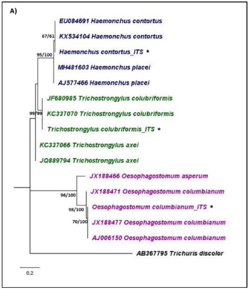
تحليل النشوء والتطور

تم تحليل المخططات الكهربائية لتسلسلات ITS2 و 16S rRNA باستخدام Bioedit 7.0 [32]. تم إجراء محاذاة متعددة للتسلسلات مع التسلسلات المرجعية من قاعدة بيانات NCBI باستخدام ClustalX 2.1 لكل علامة جينية [29]. التسلسلات المرجعية المستخدمة موضحة في الملف الإضافي 1. تم إجراء تحليلات النشوء والتطور باستخدام طرق الجوار الأقرب (NJ) والاحتمالية القصوى (ML) في MEGA X [33]. لتقييم دعم هيكل الشجرة، تم إجراء 1,000 تكرار bootstrap، وتم اختيار أفضل نموذج لاستبدال النوكليوتيدات لتحليل ML. تم استخدام Trichuris discolor و Strongyloides ratti و Strongyloides stercoralis كأصناف خارجية لتأصيل أشجار النشوء والتطور. تم تصور الأشجار الناتجة وتوضيحها باستخدام FigTree 1.3.1 [34].

كشف مقاومة مضادات الديدان

كشف المقاومة الظاهرية للألبندازول من خلال اختبار فقس البيض

تم إجراء اختبار فقس البيض للكشف عن مقاومة الألبندازول في السستوديات وفقًا لبروتوكول كولز وآخرون. وتم جمع عينات براز الماعز والحفاظ عليها في ظروف لاهوائية حتى التجربة. باختصار، تم وضع عينات البراز في أنابيب فالكون بسعة 50 مل مع غطاء لولبي تحتوي على كرات زجاجية بقطر و ماء الصنبور. تم هز الأنابيب بقوة لتفريق المادة البرازية قبل نقلها إلى المختبر. تم إجراء التجربة خلال سبعة أيام من جمع العينة. تم تجانس كل عينة (5 جرام من البراز في 42 مل من الماء)، ثم تم غربلتها وتركها لترسب لمدة لا تقل عن 30 دقيقة. بعد تصفية السائل العلوي، تم إضافة NaCl مشبع إلى المزيج، الذي تم نقله بعد ذلك إلى أنابيب فالكون سعة 15 مل. تم إضافة NaCl إضافي لإنشاء قوس مقلوب في الأعلى، وتم وضع شريحة تغطية فوق الفتحة. تم ترك الأنابيب دون إزعاج لمدة 15 دقيقة قبل المعالجة الإضافية.
لعزل البيض، تم شطف الشرائح الزجاجية بالماء المقطر، وتم نقل حوالي 100 بيضة في 2 مل من الماء إلى كل بئر من صفيحة 24 بئر. تم إعداد تخفيفات الألبندازول عن طريق إذابة 400 ملغ من زينتيل. أقراص (غلاكسو سميث كلاين، لندن، المملكة المتحدة) في ثنائي ميثيل سلفوكسيد (DMSO) (سيغما ألدريتش، ميزوري، الولايات المتحدة الأمريكية). كانت التركيزات النهائية المستخدمة في اختبار فقس البيض هي ، و . تم اختبار كل تركيز في ثلاث نسخ، مع وجود ضوابط تحتوي على 1% DMSO. تم حضن لوحة الـ 24 بئرًا في لمدة 48 ساعة للسماح بفقس البيض. بعد الحضانة، تم إضافة 0.1 مل من يود لوغول إلى كل بئر، وتم عد العدد الإجمالي من البيض واليرقات تحت المجهر المقلوب.

كشف المقاومة الظاهرية لليفاميزول من خلال اختبار تطوير اليرقات

وفقًا لبروتوكول معدل من شاغاس وآخرون (2016) وأراوجو-فيليو وآخرون (2021) [38، 39]، حوالي 80 إلى 100 بيضة معلقة في من الماء أضيفت إلى كل بئر من صفيحة تحتوي على 24 بئرًا. تم حضانة الألواح في لمدة 24 ساعة للسماح بفقس البيض. بعد
الجدول 1: بادئات جين 16S rRNA الخاصة بالنوع المستخدمة في التعرف الجزيئي، مع أحجام الأمبليكون ودرجات حرارة التهجين المقابلة لها
علامة وراثية وجنس اسم البرايمر والتسلسل (5́-3′) درجة حرارة التلدين حجم الأمبليكون
16S هيمونشوس 16S Hae-F: كاتاتتراتكاجاوا 116 قاعدة أساسية
16S Hae-R: CGTTAAATTAAATAAWAG
16S تريكوسرونجيليس 16S تريشو-F: CTAGGGTAGAATATTATT 173 نقطة أساس
16S تريشو-R: AAAGAAGAACAGTCTTAAT
16S أوزوفاجوستوموم 16S أوزو-ف: CTTCGGAAATTCTTTTTTGG 205 قاعدة زوجية
16S أوزو-R: CTTCTCCCTCTTTAACAAAC
من تعليق الخميرة المذابة في و تم إضافة تخفيفات ليفاميسول إلى كل بئر. تم إذابة مسحوق هيدروكلوريد ليفاميسول (سيغما ألدريتش، ميزوري، الولايات المتحدة الأمريكية) في دي ميثيل سلفوكسيد (سيغما ألدريتش، ميزوري، الولايات المتحدة الأمريكية) لتحضير التركيزات النهائية إلى ، و . تم اختبار كل تركيز دوائي في ثلاث نسخ، مع وجود آبار تحكم تحتوي على دي إم إس أو. ثم تم حضن الأطباق لمدة 6 أيام إضافية. بعد الحضانة، تم إضافة يود لوغول إلى كل بئر لإنهاء الاختبار. تم عد العدد الإجمالي للبيض واليرقات من المرحلة الأولى (L1) واليرقات من المرحلة الثالثة (L3) تحت المجهر المقلوب.

كشف المقاومة الجينية للألبندازول والليفاميزول عبر تفاعل البوليميراز المتسلسل الكمي باستخدام صبغة SYBR الخضراء الخاصة بالأليلات

تم جمع اليرقات L3 باستخدام تقنية باردمان [40]، وتم استخراج الحمض النووي الجيني من تجمعات اليرقات (لكل مزرعة) وفقًا للبروتوكول الموصوف سابقًا. للكشف عن مقاومة الألبندازول المرتبطة بالطفرات TAC عند F200Y من الـ جين -توبولين في Haemonchus sp. و Trichostrongylus sp.، تم إجراء اختبار qPCR باستخدام بادئات من Silvestre و Humbert (2000) [14]. بالنسبة لـ Haemonchus sp.، كانت البادئات المستهدفة للأليل المقاوم هي Ph 2 (5′-GATCAGCATTCAGCTGT CCA-3′) و Ph3 ( -TGGTAGAGAACACCGATGAAA CATA-3′)، بينما كانت البرايمرات للأليل المحدد للحساسية هي Ph1 (5′-GGAACGATGGACTCCTTTCG-3′) و Ph4 (5′-ATACAGAGCTTCGTTGTCAATACAG A-3′). بالنسبة لـ Trichostrongylus sp.، كانت البرايمرات المستهدفة للأليل المحدد للمقاومة هي Pc2 (5′-GGGAATCGGAG GCAAGTCGT-3′) و Pc3 (5′-CTGGTAGAGAATACC GATGAAACATA-3′)، بينما كانت البرايمرات للأليل المحدد للحساسية هي Pc1 (5′-GGAACAATGGATTC CGTTCG-3′) و Pc4 (5′-ATACAGAGCTTCGTTATCG ATGCAGA-3′). تم إجراء اختبارات qPCR بشكل منفصل لكل زوج من البرايمرات لتمييز بين الأليلات المقاومة والحساسة.
لكشف مقاومة اللفاميزول في Haemonchus sp. المرتبطة بحذف 63 قاعدة في Hco-acr- الجين، تم إجراء اختبار qPCR وفقًا للبرايمرات والبروتوكول الموصوف من قبل سانتوس وآخرون (2019) [41]. البرايمر الأمامي LevF2 ( تم استخدام الشريط الجيني -TAACCTTACCTATACACC CGTC-T-3′) لكل من الأليلات المقاومة والحساسة. تم استخدام الشريط العكسي LevResR2 (5′-ATCTT TGACAGTAATCAGCGTTG-3′) للكشف عن الأليل المقاوم، بينما تم استخدام LevSusR2 (5′-CGGCGATATAAC AGCAGTTAAC-3′) للكشف عن الأليل الحساس. تم إجراء اختبارات qPCR بشكل منفصل لكل زوج من الشريطين للتمييز بين الأليلات المقاومة والحساسة.
تم إجراء جميع تفاعلات qPCR باستخدام جهاز CFX96 Touch نظام PCR في الوقت الحقيقي (بايو راد، كاليفورنيا، الولايات المتحدة الأمريكية). تم إجراء كل تفاعل في حجم نهائي من يتكون من من لونا كيو بي سي آر العالمي
خليط رئيسي (نيو إنجلاند بيولوجيكس، ماساتشوستس، الولايات المتحدة الأمريكية)، لكل مجموعة من البرايمرات، و من قالب gDNA. تضمنت ظروف التفاعل الحراري إنكار أولي في لمدة 60 ثانية، تليها 45 دورة من لمدة 15 ثانية و لمدة 30 ثانية. تم إجراء تحليل منحنى الانصهار النهائي من إلى ، مع زيادة في كل دورة. بالإضافة إلى ذلك، تم تصور الأمبليكونات على هلام الأجاروز لتقييم تكوين البرايمر والتضخيم غير المحدد.

تحليل البيانات

بالنسبة للاختبارات الظاهرية، نسبة البيض المفقوس و تم حساب القيم لـ EHT، بينما كانت نسبة اليرقات التي تطورت إلى مرحلة L3 و LD تم تحديد القيم لـ LDT. تم إجراء تحليل لوغاريتمي باستخدام MedCalc الإصدار 22 [42]. تم تعريف مقاومة الألبندازول بواسطة قيمة تتجاوز بينما تم الإشارة إلى مقاومة ليفاميسول بواسطة قيمة أعلى .
للكشف عن النمط الجيني، تم حساب نسبة المزارع في كل منطقة التي تحتوي على برك يرقات تظهر أنماط جينية مقاومة متماثلة (RR)، حساسة متماثلة (SS)، ومختلطة (RS). أظهرت المزارع المصنفة كـ RR تضخيمًا فقط مع مجموعة البرايمر المقاومة، بينما أظهرت مزارع SS تضخيمًا فقط مع مجموعة البرايمر الحساسة. أظهرت مزارع RS تضخيمًا مع كلا مجموعتي البرايمر. بالإضافة إلى ذلك، تم حساب ترددات الأليلات المقاومة والحساسة لكل منطقة من خلال تحديد عدد حدوث كل أليل في السكان وقسمته على العدد الإجمالي للأليلات.

النتائج

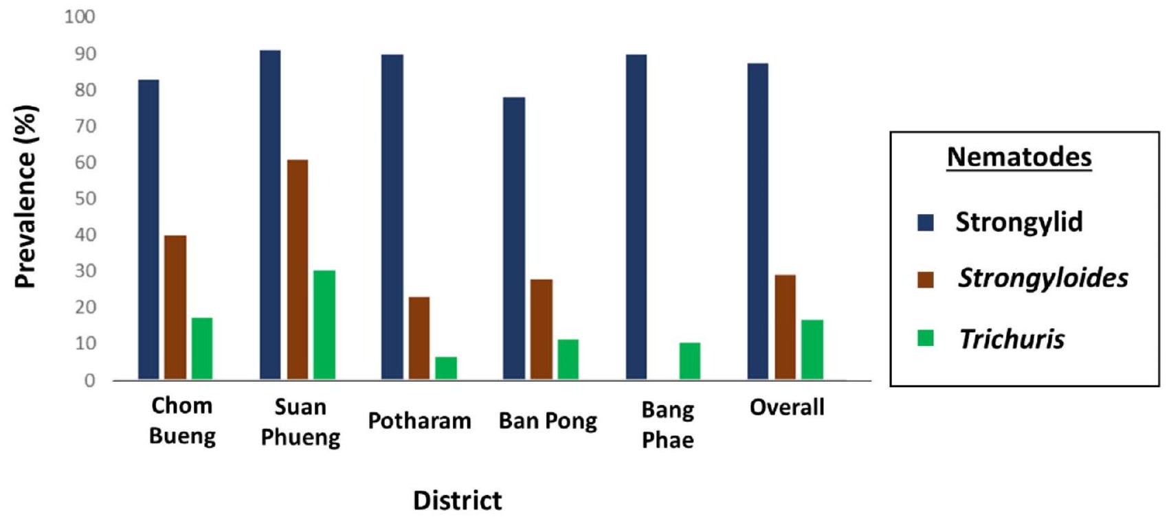
تحديد وانتشار وشدة الديدان الخيطية المعوية

أظهر تحليل البراز لعينات الماعز وجود بيض للستروغيليدات، وStrongyloides sp.، وTrichuris sp. وكانت الستروغيليدات هي الأكثر انتشارًا بشكل عام عند “، تليها Strongyloides sp. و Trichuris sp. تم الكشف عن الديدان القوية في جميع المناطق الخمس، مع أعلى انتشار ( تم تسجيل Trichuris sp. في جميع المناطق، بينما تم العثور على Strongyloides sp. في تشوم بوانغ، سوان فوانغ، بوثارام، وبان بونغ. توضح الشكل 2 انتشار عدوى الديدان المعوية في الماعز من هذه المناطق. بالإضافة إلى الانتشار العالي لعدوى الديدان القوية، كان متوسط عدد EPG العام 670 (الجدول 2). تم تسجيل أعلى EPG في مزرعة في بوثارام، بمتوسط 6,125 EPG. كان لدى بوثارام أعلى متوسط EPG، تليها بانغ فاي.

التعرف الجزيئي على السستوديات

تم تحديد السستوديات باستخدام منطقة ITS2 النووية وجين 16S rRNA الميتوكوندري، مما يؤكد
الشكل 2 انتشار عدوى الديدان الخيطية المعوية في الماعز
وجود Haemonchus sp. و Trichostrongylus sp. و Oesophagostomum sp. (الجدول 3). كان Haemonchus sp. الأكثر انتشارًا، حيث تم اكتشافه في جميع المزارع. ) إيجابي لبيض الستروغيليدات. كانت الأنواع Trichostrongylus موجودة في المزارع عبر جميع المناطق، وتشكل من العينات الإيجابية للستروغيلد، بينما تم العثور على Oesophagostomum sp. في مزارع من تشوم بوانغ، وبوثارام، وبان بونغ. وقد حدد تسلسل الحمض النووي للعينة التمثيلية الستروغيلدات على أنها H. contortus وTrichostrongylus colubriformis وOesophagostomum columbianum. وكشفت التحليلات النشوية المستندة إلى منطقة ITS2 أن التسلسلات التمثيلية كانت مطابق لـ H. contortus و T. colubriformis و O. columbianum. تجمع Haemonchus contortus في مجموعة أحادية النمط مع H. placei، بينما شكل O. columbianum مجموعة أحادية النمط متميزة مع أنواع أخرى من Oesophagostomum. دعمت التحليلات النشئية لجين 16S rRNA هذه التعريفات الجزيئية بشكل أكبر، مع . contortus، O. columbianum، و T. colubriformis تسلسلات مرتبطة ارتباطًا وثيقًا بأنواع أخرى ضمن أجناسها المعنية. الشكل 3 يوضح النشوء الجزيئي الذي تم الحصول عليه.

حالة مقاومة الألبندازول للستروغيليدات التي تصيب الماعز

أظهرت نتائج EHT من 20 مزرعة أن احتوت على تجمعات قوية من الديدان الشريطية المقاومة (الجدول 4). علاوة على ذلك، تم اكتشاف تجمعات من الديدان الشريطية المقاومة في المزارع عبر جميع المناطق الخمس. تراوحت القيم لعدد السكان المقاومة من نوع سترونغيلد القوي بين إلى ، مع وجود العديد من القيم التي تتجاوز بشكل كبير ، مما يشير إلى مستويات مقاومة عالية. من بين 18 مزرعة تحتوي على تجمعات قوية من الديدان المستديرة المقاومة، أظهرت 16 منها أكثر من بيض يفقس في
جرعة تمييزية من ، مما يؤكد بشكل أكبر درجة المقاومة الكبيرة للألبندازول في الديدان الشريطية التي تصيب الماعز.
متسقًا مع نتائج EHT الظاهرية، كشفت التحليلات الجينية أن كانت برك اليرقات مقاومة للألبندازول بالنسبة لـ Haemonchus sp. و Trichostrongylus sp. توضح الشكل 4 نسبة المزارع لكل منطقة مع الجينات المتجانسة المقاومة (RR) والمتغايرة (RS) والمتجانسة الحساسة (SS). بالنسبة لـ Haemonchus sp., من المزارع التي عرضت النمط الجيني RR، بينما كانت RS، في المقابل، كانت نسبة الأنماط الجينية RR في Trichostrongylus sp. أعلى، مع المزارع المصنفة كـ RR و كنسبة RS. تم ملاحظة أعلى نسبة من أنماط جينات Trichostrongylus sp. RR في منطقة سوان فوينغ. المزارع)، في حين أظهرت أنواع هايمونشوس أعلى نسبة من RR في منطقة بان فاي. من المزارع). كانت تردد الأليل المقاوم في مجموعة Haemonchus sp. هو 0.65، بينما كان تردد الأليل القابل للإصابة 0.35. وبالمثل، أظهرت مجموعة Trichostrongylus sp. تردد أليل مقاوم قدره 0.74 وتردد أليل قابل للإصابة قدره 0.26. توفر الجدول 5 تحليلًا مفصلًا لترددات الأليلات المقاومة والقابلة للإصابة في المجموعة.

حالة مقاومة ليفاميزول في الديدان الشريطية التي تصيب الماعز

من بين المزارع السبعة التي تم اختبارها لمقاومة اللفاميزول باستخدام LDT، كانت خمسة ( ) كانت تحتوي على تجمعات من الديدان الشريطية المقاومة لليفاميزول (الجدول 4). تم اكتشاف تجمعات من الديدان الشريطية المقاومة لليفاميزول في مناطق سوان فوينغ، وبوثارام، وبان بونغ. تراوحت القيم لسكان الساكنات المقاومة من 3.37 إلى .
الجدول 2 شدة عدوى الستروغيلد في الماعز
منطقة مزرعة عدد العينات بيضة لكل جرام (المتوسط ± الانحراف المعياري) الحد الأدنى – الحد الأقصى
تشوم بوانغ 1 ٤ 0-150
2 ٣ 0-400
٣ ٧ 0-950
٤ ٦ 0-200
٥ 9 0-200
٦ ٦ ٧٠٠-١٣٥٠
إجمالي ٣٥ 0-1350
سوان فوينغ 1 ٥ 0-550
2 ٦ 100-1750
٣ ٣ ٣٥٠-٥٠٠
٤ ٨ 0-2050
٥ ٨ 0-1150
٦ ٣ 0-550
إجمالي ٣٣ 0-2050
بوتارام 1 10 50-19,650
2 ٧ ٥٠-٣٠٠
٣ ٣ 0 0
٤ 10 0-250
٥ 10 0-1900
٦ ٨ 50-1900
إجمالي ٤٨ 0-19,650
بان بونغ 1 ٣ 0-50
2 1 100 100
٣ 2 ٢٥٠
٤ 1 ٧٠٠ ٧٠٠
٥ ٥ 0-50
٦ ٦ 0-600
إجمالي ١٨ 0-700
بانغ فاي 1 ٣ 0-150
٢ ٥ 50-450
٣ ٨ ٣٥٠-٢٢٥٠
٤ 11 100-800
٥ ٧ 0-50
٦ ٥ 0-200
إجمالي ٣٩ 0-2250
بشكل عام 173 0-19,650
الجدول 3 نسبة الأجناس القوية المكتشفة في كل منطقة بناءً على منطقة ITS2 النووية وجين الرنا الريبوسومي 16S الميتوكوندري.
منطقة أجناس
هيمنشوس تريشوتراستونغيلوس أيوسوفاجوستوموم
تشوم بوانغ 100.00 ٨٣.٣٣ ٣٣.٣٣
سوان فوانغ 100.00 100.00 0
بوتارام 100.00 100.00 ٦٦.٦٦
بان بونغ 100.00 100.00 ٤٠.٠٠
بانغ فاي 100.00 100.00 0
بشكل عام 100.00 ٩٦.٦٧ ٢٨.٠٠
كشف فحص النمط الجيني لمقاومة ليفاميزول في هيمونشوس أن من بين 26 مزرعة تحمل أليلات مقاومة (RR أو RS)، بينما فقط كان لديها النمط الجيني المتجانس القابل للإصابة (الشكل 4). المزارع القابلة للإصابة بالليفاميزول ( تم التعرف على ( ) بشكل حصري في سوان فوينغ، بينما أظهرت جميع المزارع في المناطق الأربعة المتبقية مقاومة لليفاميزول. كانت المزارع غير المتجانسة (RS) سائدة في بوثارام وبانغ فاي، بينما تم اكتشاف المزارع المتجانسة المقاومة (RR) في تشوم بوانغ وسوان فوينغ وبان بونغ. كانت التردد العام للأليل المقاوم لليفاميزول 0.52، بينما كان تردد الأليل القابل للإصابة 0.48 (الجدول 5).
بالإضافة إلى ذلك، أظهر الفحص الجيني لمقاومة الأدوية حساسية أكبر من الطرق الظاهرية. بالنسبة للألبندازول والليفاميزول، حدد التحليل الجزيئي نسبة أعلى من المزارع التي تحتوي على تجمعات قوية مقاومة مقارنةً بالاختبارات الظاهرية. تم تأكيد المزارع التي تم تصنيفها في البداية على أنها حساسة بناءً على الاختبارات الظاهرية لاحقًا على أنها مقاومة (أنماط جينية RR أو RS) من خلال الفحص الجيني. على سبيل المثال، كانت المزرعة رقم 4 في منطقة سوان فوينغ مقاومة للألبندازول عند التحليل الجيني، بينما كانت المزرعة رقم 6 في تشوم بوانغ والمزرعة رقم 3 في بانغ فاي مقاومة لليفاميزول على الرغم من تصنيفها على أنها حساسة في الاختبارات الظاهرية.

نقاش

كشفت التحليلات الشكلية والجزيئية عن انتشار مرتفع للديدان المعوية، وخاصة الديدان الشريطية، التي تصيب الماعز في محافظة راتشابوري. تم تأكيد مقاومة مزدوجة للديدان الشريطية للبنزيميدازول (الألبندازول) والإيميدازوثيازول (الليفاميزول) من خلال كل من الطرق الظاهرية والجينية. ومن الجدير بالذكر أن هذه الدراسة تقدم أول دليل جزيئي على مقاومة الليفاميزول في الديدان الشريطية التي تصيب الماعز في تايلاند.
انتشار مرتفع تمت ملاحظة إصابة قوية بالديدان الشريطية في الماعز من محافظة راتشابوري، مما يبرز التهديد الكبير الذي تشكله هذه الديدان الأسطوانية على صحة الماعز وإنتاجية المزارع. بالمقابل، أظهرت دراسة سابقة أجريت في محافظة راتشابوري بين عامي 2018 و2019 انتشارًا أقل، حيث تم الكشف عن Haemonchus sp. وTrichostrongylus sp. في و ، على التوالي [43]. قد يُعزى الزيادة الملحوظة في الانتشار إلى عوامل مثل استيراد الماعز من مقاطعات أخرى أو تراجع فعالية الأدوية المضادة للطفيليات في السنوات الأخيرة. بالإضافة إلى السستوديات، تم تحديد أنواع أخرى من الديدان المعوية، بما في ذلك سترونجيلايدس وتريشوريس، مما يؤكد تنوع عدوى الديدان الخيطية في الماعز. وبالمثل، أفاد جونزري وآخرون (2021) أيضًا بوجود سترونجيلايدس وتريشوريس في مزارع الماعز في مقاطعة راتشابوري [43].
تم الإبلاغ عن انتشار مرتفع لعدوى الستروغيلد في الماعز في جنوب تايلاند، وخاصة في ساتون، سونغخلا، باتالونغ، يالا، باتاني، وناراتيوات.

الشكل 3 شجرة النسب باستخدام أقصى احتمال ) منطقة ITS2 النووية (K2) و B) جين الرنا الريباسي 16 S الميتوكوندري (TN+G). الأرقام عند العقد تشير إلى قيم البوتستراب (ML/NJ). التسلسلات التمثيلية التي تم إنشاؤها من هذه الدراسة موضحة بعلامة ‘*’
الجدول 4 حالة مقاومة الألبندازول والليفاميزول التي تم تحديدها بواسطة اختبار فقس البيض واختبار تطور اليرقات، على التوالي
منطقة مزرعة ألبندازول (EHT) ليفاميزول (LDT)
حالة المقاومة % الفقس في DC حالة المقاومة % من L1 تتطور إلى L3 في DC
تشوم بوانغ 2 مقاوم 40
٦ مقاوم ٩٨ عرضة* 0
سوان فوانغ 1 مقاوم ٤٧
2 مقاوم 100
٣ عرضة 41
٤ عرضة* 21 مقاوم ٥٦
بوتارام 1 مقاوم 91 مقاوم ٥٩
2 مقاوم 93 مقاوم 63
٣ مقاوم 98
٤ مقاوم 100 مقاوم 68
٥ مقاوم 98
٦ مقاوم 99
بان بونغ 1 مقاوم 97
2 مقاوم 99
٣ مقاوم 96
٤ مقاوم 91
٦ مقاوم 90 مقاوم 73
بانغ فاي 2 مقاوم 97
٣ مقاوم 97 عرضة* ٨
٦ مقاوم 100
الشكل 4 حالة المقاومة الجينية للألبندازول (Haemonchus sp. و Trichostrongylus sp.) والليفاميزول (Haemonchus sp.) لكل منطقة. يمثل الرسم البياني العمودي المكدس نسبة المزارع في كل منطقة التي كانت إما RR أو RS أو SS للألبندازول (Haemonchus sp. و Trichostrongylus sp.) والليفاميزول (Haemonchus sp.)
الجدول 5 ترددات الأليلات المقاومة والحساسة بين تجمعات Haemonchus sp. و Trichostrongylus sp. في محافظة راتشابوري
الحي/الأليل ألبندازول (هيمنشوس) ألبندازول (تريشوتراستونغيلوس) ليفاميزول (هيمونشوس)
مقاوم عرضة مقاوم عرضة مقاوم عرضة
تشوم بوانغ 0.70 0.30 0.88 0.12 0.60 0.40
سوان فوانغ 0.70 0.30 0.90 0.10 0.40 0.60
بوتارام 0.60 0.40 0.60 0.40 0.50 0.50
بان بونغ 0.50 0.50 0.70 0.30 0.60 0.40
بانغ فاي 0.75 0.25 0.67 0.33 0.50 0.50
بشكل عام 0.65 0.35 0.74 0.26 0.52 0.48
المحافظات، مع انتشار يتراوح من 40 إلى [19، 44]. بالإضافة إلى المنطقة الجنوبية، فإن عدوى الديدان الشريطية شائعة أيضًا في المناطق الشمالية الشرقية والوسطى، بما في ذلك محافظات ناخون باتوم، ساموت ساخون، ساموت سونغخرام، بتشابوري، كانشانابوري، خون كاين، وفيتسانولوك [20، 21، 45، 46]. يتوافق ذلك مع نتائجنا، حيث تعتبر أنواع هايمونشوس وأنواع تريشوتروستونغيلوس هي الأجناس السائدة من الديدان الشريطية التي تصيب الماعز في تايلاند، وكلاهما شديد pathogenic [20، 44]. إن الانتشار العالي لهذه الديدان الخيطية يبرز الدور الحاسم للأطباء البيطريين في مساعدة المزارعين في الحفاظ على صحة الحيوانات المثلى وإنتاجية المزارع. بالإضافة إلى تأثيرها على صحة الماعز، تم الإبلاغ أيضًا عن عدوى T. colubriformis في البشر في محافظات خون كاين، روي إت، لوي، وساكون ناخون [10، 47]. يجب ألا يتم تجاهل هذه المخاطر zoonotic، خاصة في مزارع الماعز حيث تتواجد تجمعات البشر والحيوانات بالقرب من بعضها البعض (غالبًا ما تكون مناطق السكن والزراعة قريبة).
ظهور مقاومة الأدوية الطاردة للديدان يشكل تهديدًا كبيرًا لصحة واستدامة صناعة تربية الماعز الاقتصادية، مما يعيق السيطرة الفعالة والوقاية من العدوى الطفيلية [12، 48]. في تايلاند، تم رصد مقاومة الألبندازول في البالغين تم الإبلاغ عن إصابة الماعز بـ .contortus في خمس مناطق، مع ملاحظة أعلى مستويات المقاومة في المنطقة الجنوبية
المحافظات [22]. علاوة على ذلك، تم توثيق مقاومة الأدوية المتعددة في أجزاء مختلفة من البلاد [23، 25، 49]. بخلاف مقاومة الألبندازول، تم الإبلاغ عن مقاومة الإيفرمكتين في قطعان الماعز في محافظات سينغ بوري، وخون كاين، وتشايابوم من خلال اختبارات الشكل الظاهري [23، 49]. في محافظة راتشابوري، تم اكتشاف مقاومة الألبندازول والإيفرمكتين في (9/11) من مزارع الماعز باستخدام اختبار FECRT [25]. نظرًا لعدم تطوير أي اختبارات جزيئية للكشف عن مقاومة الإيفرمكتين، تظل الاختبارات الظاهرية هي الطريقة التشخيصية الرئيسية. كشفت تحليلاتنا الجزيئية المستندة إلى تجمعات اليرقات عن انتشار واسع لمقاومة مزدوجة للأدوية (الألبندازول والليفاميزول) عبر معظم المزارع. تم الكشف عن مقاومة الألبندازول في جميع المزارع. في المقابل، أشارت البيانات الظاهرية التي أبلغ عنها بادوانغ وثونغثا (2020) إلى عدم وجود مقاومة لليفاميزول بين المزارع المأخوذة من عينة في محافظة راتشابوري [25].
تثير التقارير الواسعة عن مقاومة الأدوية الطفيلية عبر جميع فئات الأدوية الثلاثة، إلى جانب الأدلة الداعمة من مناطق مختلفة في تايلاند، مخاوف كبيرة. بدون تنظيم صارم لاستخدام الأدوية الطفيلية وحركة الماعز بين المقاطعات، قد يواجه قطاع الماعز المحلي عواقب وخيمة. علاوة على ذلك، تلعب تايلاند دورًا حيويًا في التجارة العالمية للماعز، حيث تستورد وتصدر من وإلى جنوب شرق آسيا وأفريقيا والولايات المتحدة وأستراليا. ضمن
البلد، تعتبر البنزيميدازولات واللاكتونات الدائرية من أكثر الأدوية المضادة للطفيليات التي يتم استخدامها لعلاج عدوى GIN في المجترات الصغيرة، والتي يستخدمها كل من المزارعين والأطباء البيطريين [50]. وفقًا لبيانات استخدام الأدوية من مزارعي الماعز في هذه الدراسة، كانت الألبندازول والإيفرمكتين هما الأدوية الرئيسية المستخدمة، بينما تم إعطاء اللفاميسول فقط في منطقة تشوم بوانغ. ومع ذلك، على الرغم من استخدامه المحدود، تم اكتشاف مقاومة اللفاميسول. قد يكون أحد العوامل المساهمة في انتشار المقاومة هو حركة الماعز بين المناطق. لم تكن الماعز التي تم فحصها في هذه الدراسة من محافظة راتشابوري فقط، بل تم الحصول عليها أيضًا من محافظات أخرى في شمال شرق، وغرب، ووسط تايلاند. قد يسهل استيراد وتحرك الماعز بين المحافظات انتشار الأليلات المقاومة للأدوية، مما يزيد من تفاقم مقاومة الأدوية المضادة للطفيليات. وبالتالي، ستستمر فعالية العلاجات المضادة للطفيليات لعدوى GIN في الماعز في الانخفاض، مما يؤدي إلى زيادة تكرار الأليلات المقاومة في سكان السترانجيليد.
حجم العينة الصغيرة ( مزارع) حد من اكتشاف مقاومة اللفاميسول عبر اختبار LDT بسبب عدم كفاية بيض السترانجيليد. تم إعطاء الأولوية لاكتشاف مقاومة الألبندازول باستخدام اختبار EHT. وبالتالي، قد يكون حجم العينة المنخفض قد ضخم النسبة النهائية للمزارع المقاومة لللفاميسول. بالإضافة إلى ذلك، لم يتم إجراء طرق حية مثل FECRT لتقييم فعالية الدواء. تم إجراء اكتشاف المقاومة لتحليل النمط الجيني باستخدام عينات يرقات مجمعة لكل مزرعة بدلاً من الديدان الفردية بسبب العائد المحدود من الحمض النووي من اليرقات الفردية. نتيجة لذلك، كانت النسبة لحالات مقاومة الألبندازول واللفاميسول والنمط الجيني وتردد الأليلات تعتمد على عينات اليرقات المجمعة بدلاً من الديدان الفردية. علاوة على ذلك، تم إجراء اكتشاف النمط الجيني لمقاومة اللفاميسول فقط لأنواع هايمونشوس. أخيرًا، قد يؤدي عدم وجود خطوة الطرد المركزي أثناء تجانس البراز إلى فقدان بيض السترانجيليد وتقدير خاطئ للحمولة الطفيلية الحقيقية.

الخاتمة

تقدم هذه الدراسة أول دليل نمطي جيني لمقاومة اللفاميسول في الماعز وهي الأولى في تايلاند التي تدمج بين الطرق الظاهرية والجينية لاكتشاف مقاومة الأدوية المضادة للطفيليات. تم تحديد سكان السترانجيليد المقاومة للألبندازول واللفاميسول في معظم المزارع من خلال الاختبارات الظاهرية، مع تأكيد التحليل الجيني للمقاومة في سكان H. contortus وT. colubriformis. إن الانتشار العالي للديدان الخيطية التي تصيب الماعز في محافظة راتشابوري، جنبًا إلى جنب مع المقاومة المزدوجة للألبندازول واللفاميسول، يبرز الحاجة الملحة لاستراتيجيات بديلة ومستدامة لمكافحة الطفيليات. يجب أن تركز الدراسات المستقبلية على مراقبة المقاومة
الاتجاهات من خلال الأبحاث الطولية، وتوضيح آليات المقاومة، وتقييم المركبات البديلة المضادة للطفيليات لضمان فعالية السيطرة على الديدان وتقليل مقاومة الأدوية المضادة للطفيليات في الماعز.
الاختصارات
DC الجرعة المميزة
EHT اختبار فقس البيض
EPG البيض لكل جرام
FECRT اختبار تقليل عدد بيض البراز
GIN الديدان الخيطية المعوية
ITS2 المسافة الداخلية المنسوخة 2
LDT اختبار تطوير اليرقات
ML الاحتمالية القصوى
NJ الانضمام الجار
qPCR تفاعل البوليميراز المتسلسل الكمي
rRNA RNA الريبوسومي
SNP تعدد أشكال النوكليوتيد المفرد

معلومات إضافية

تحتوي النسخة الإلكترونية على مواد إضافية متاحة علىhttps://doi.or g/10.1186/s12917-025-04700-4.
المادة الإضافية 1

الشكر والتقدير

نشكر قسم الطفيليات، كلية الطب الاستوائي، جامعة ماهيدول، بانكوك، على الدعم الفني. كما نشكر أصحاب مزارع الماعز ومكتب الثروة الحيوانية في محافظة راتشابوري على مساعدتهم وتعاونهم خلال جمع عينات براز الماعز في المناطق المحلية.

مساهمات المؤلفين

قام AC بتنفيذ منهجية التحقيق، التحليل الرسمي، وكتابة المسودة الأصلية. قام CK بتنفيذ منهجية التحقيق، الموارد، ومراجعة المسودة. قام WP بتنفيذ التحقيق، المنهجية، الموارد، ومراجعة المسودة. قام SS بتنفيذ التصور، التحقيق، الموارد، ومراجعة المسودة. قام UT بتنفيذ التصور، التحقيق، المنهجية، التحليل الرسمي، ومراجعة المسودة. قرأ جميع المؤلفين ووافقوا على المخطوطة النهائية.

التمويل

تم توفير تمويل الوصول المفتوح من قبل جامعة ماهيدول. تم تمويل هذا البحث، كليًا أو جزئيًا، من قبل مشروع التعاون من أجل التميز جامعة ماهيدول- NSTDA [MU-NSTDA-2566-01].

توفر البيانات

تتضمن مجموعة البيانات التي تدعم استنتاجات هذه المقالة ضمن المقالة وملفها الإضافي.

الإعلانات

تمت الموافقة على هذه الدراسة من قبل اللجنة الأخلاقية لكلية العلوم البيطرية، جامعة ماهيدول (رموز الموافقة المعينة من FVS-MU-IACUC: COA. رقم MUVS-2023-08-54 وCOA. رقم MUVS-2024-03-24). تم الحصول على موافقة مستنيرة لجمع عينات براز الماعز، وكشف جميع أصحاب مزارع الماعز ومكتب الثروة الحيوانية في محافظة راتشابوري عن النتائج المعنية.
غير قابل للتطبيق.

المصالح المتنافسة

يعلن المؤلفون عدم وجود مصالح متنافسة.
تاريخ الاستلام: 27 ديسمبر 2024 / تاريخ القبول: 20 مارس 2025
تم النشر عبر الإنترنت: 04 أبريل 2025

References

  1. Grisi L, Leite RC, Martins JR, Barros AT, Andreotti R, Cançado PH, et al. Reassessment of the potential economic impact of cattle parasites in Brazil. Rev Bras Parasitol Vet. 2014;23:150-6.
  2. Githigia SM, Thamsborg SM, Munyua WK, Maingi N. Impact of gastrointestinal helminths on production in goats in Kenya. Small Rumin Res. 2001;42:21-9.
  3. Hoste H, Torres-Acosta JFJ. Alternative or improved methods to limit gastro-intestinal parasitism in grazing sheep and goats. Small Rumin Res. 2008;77:159-73.
  4. Charlier J, Rinaldi L, Musella V, Ploeger HW, Chartier C, Vineer HR, et al. Initial assessment of the economic burden of major parasitic helminth infections to the ruminant livestock industry in Europe. Prev Vet Med. 2020;182:105103.
  5. Maurizio A, Perrucci S, Tamponi C, Scala A, Cassini R, Rinaldi L, et al. Control of gastrointestinal helminths in small ruminants to prevent anthelmintic resistance: the Italian experience. Parasitology. 2023;150:1105-18.
  6. Zarlenga DS, Hoberg EP, Tuo W. The identification of Haemonchus species and diagnosis of haemonchosis. Adv Parasitol. 2016;93:145-80.
  7. Ghadirian E, Arfaa F. First report of human infection with Haemonchus contortus, Ostertagia ostertagi, and Marshallagia marshalli (family Trichostrongylidae) in Iran. J Parasitol. 1973;59:1144-5.
  8. Boreham RE, McCowan M, Ryan AE, Allworth A, Robson J. Human trichostrongyliasis in Queensland. Pathology. 1995;27:182-5.
  9. Watthanakulpanich D, Pongvongsa T, Sanguankiat S, Nuamtanong S, Maipanich W, Yoonuan T, et al. Prevalence and clinical aspects of human Trichostrongylus colubriformis infection in Lao PDR. Acta Trop. 2013;126:37-42.
  10. Phosuk I, Intapan P, Sanpool O, Janwan P, Thanchomnang T, Sawanyawisuth K, et al. Molecular evidence of Trichostrongylus colubriformis and Trichostrongylus axei infections in humans from Thailand and Lao PDR. Am J Trop Med Hyg. 2013;89:376-9.
  11. Fissiha W, Kinde MZ. Anthelmintic resistance and its mechanism: A review. Infect Drug Resist. 2021;14:5403-10.
  12. Dey AR, Begum N, Anisuzzaman MA, Alam MZ. Multiple anthelmintic resistance in gastrointestinal nematodes of small ruminants in Bangladesh. Parasitol Int. 2020;77:102105.
  13. Potârniche AV, Mickiewicz M, Olah D, Cerbu C, Spînu M, Hari A, et al. First report of anthelmintic resistance in gastrointestinal nematodes in goats in Romania. Animals. 2021;11:2761.
  14. Silvestre A, Humbert JF. A molecular tool for species identification and benzimidazole resistance diagnosis in larval communities of small ruminant parasites. Exp Parasitol. 2000;95:271-6.
  15. Silvestre A, Cabaret J. Mutation in position 167 of isotype 1 beta-tubulin gene of trichostrongylid nematodes: role in benzimidazole resistance? Mol Biochem Parasitol. 2002;120:297-300.
  16. Santos JML, Monteiro JP, Ribeiro WL, Macedo IT, Camurça-Vasconcelos AL, Vieira LS, et al. Identification and quantification of benzimidazole resistance polymorphisms in Haemonchus contortus isolated in Northeastern Brazil. Vet Parasitol. 2014;199:160-4.
  17. Santos JML, Monteiro JP, Ribeiro WL, Macedo IT, Filho JV, Andre WP, et al. High levels of benzimidazole resistance and -tubulin isotype 1 SNP F167Y in Haemonchus contortus populations from Ceará State, Brazil. Small Rumi Res. 2017;146:48-52.
  18. Ratanapob N, Arunvipas P, Kasemsuwan S, Phimpraphai W, Panneum S. Prevalence and risk factors for intestinal parasite infection in goats raised in Nakhon pathom Province, Thailand. Trop Anim Health Prod. 2012;44:741-5.
  19. Kaewnoi D, Kaewmanee S, Wiriyaprom R, Prachantasena S, Pitaksakulrat O, Ngasaman R. Prevalence of zoonotic intestinal parasites in meat goats in Southern Thailand. Vector Borne Zoonotic Dis. 2024;24:111-7.
  20. Income N, Tongshoob J, Taksinoros S, Adisakwattana P, Rotejanaprasert C, Maneekan P. Helminth infections in cattle and goats in Kanchanaburi, Thailand, with focus on Strongyle nematode infections. Vet Sci. 2021;8:324.
  21. Rerkyusuke S, Lerk-U-Suke S, Mektrirat R, Wiratsudakul A, Kanjampa P, Chaimongkol , et al. Prevalence and associated risk factors of gastrointestinal parasite infections among meat goats in Khon Kaen Thailand. Vet Med Int. 2024;2024:3267028.
  22. Pitaksakulrat O, Chaiyasaeng M, Artchayasawat A, Eamudomkarn C, Thongsahuan S , Boonmars T. The first molecular identification of benzimidazole resistance in Haemonchus contortus from goats in Thailand. Vet World. 2021;14:764-8.
  23. Ratanapob N , Thuamsuwan N , Thongyuan S . Anthelmintic resistance status of goat gastrointestinal nematodes in Sing Buri Province, Thailand. Vet World. 2022;15:83-90.
  24. Sherman DM. The spread of pathogens through trade in small ruminants and their products. Rev Sci Tech. 2011;30:207-17.
  25. Paduang S, Thongtha R. Study of anthelmintic resistance of gastrointestinal nematodes in goats in Ratchaburi Province. J Kasetsart Vet. 2020;30:1.
  26. Fisheries MAFF. Food, reference book: manual of veterinary parasitological laboratory techniques. Ministry Agric. 1986;5.
  27. Cringoli G, Rinaldi L, Veneziano V, Capelli G, Scala A. The influence of flotation solution, sample dilution and the choice of McMaster slide area (volume) on the reliability of the McMaster technique in estimating the faecal egg counts of gastrointestinal strongyles and Dicrocoelium dendriticum in sheep. Vet Parasitol. 2004;123:121-31.
  28. Chan AHE, Chaisiri K, Morand S, Saralamba N, Thaenkham U. Evaluation and utility of mitochondrial ribosomal genes for molecular systematics of parasitic nematodes. Parasit Vectors. 2020;13:364.
  29. Thompson JD, Gibson TJ, Higgins DG. Multiple sequence alignment using ClustalW and ClustalX. Curr Protoc Bioinf. 2002;Chap. 2:Unit 2.3.
  30. Kibbe WA. OligoCalc: an online oligonucleotide properties calculator. Nucleic Acids Res. 2007;35:W43-6.
  31. Kalendar R, Khassenov B, Ramankulov Y, Samuilova O, Ivanov KI. FastPCR: an in Silico tool for fast primer and probe design and advanced sequence analysis. Genomics. 2017;109:312-9.
  32. Hall TA, BioEdit. A user-friendly biological sequence alignment editor and analysis program for windows 95/98/NT. Nucl Acids Symp Ser. 1999;41:95-8.
  33. Kumar S, Stecher G, Li M, Knyaz C, Tamura K. MEGA X: molecular evolutionary genetics analysis across computing platforms. Mol Biol Evol. 2018;35:1547-9.
  34. Rambaut A. FigTree v1.3.1. Institute of Evolutionary Biology, University of Edinburgh, Edinburgh. 2010. http://tree.bio.ed.ac.uk/software/figtree/
  35. Coles GC, Jackson F, Pomroy WE, Prichard RK, von Samson-Himmelstjerna G, Silvestre A, et al. The detection of anthelmintic resistance in nematodes of veterinary importance. Vet Parasitol. 2006;136:167-85.
  36. Coles GC, Bauer C, Borgsteede F, Geerts S, Klei T, Taylor M, et al. World association for the advancement of veterinary parasitology (W.A.A.V.P.) methods for the detection of anthelmintic resistance in nematodes of veterinary importance. Vet Parasitol. 1992;44:35-44.
  37. von Samson-Himmelstjerna G, Coles GC, Jackson F, Bauer C, Borgsteede F, Cirak V, et al. Standardization of the egg hatch test for the detection of benzimidazole resistance in parasitic nematodes. Parasitol Res. 2009;105:825-34.
  38. Araújo-Filho JV, Ribeiro WLC, André WPP, Cavalcante GS, Santos JMLD, Monteiro JP, et al. Phenotypic and genotypic approaches for detection of anthelmintic resistant sheep gastrointestinal nematodes from Brazilian Northeast. Revista Brasileira de parasitologia veterinaria = brazilian. J Veterinary Parasitology: Orgao Oficial Do Colegio Brasileiro De Parasitol Vet. 2021;30:e005021.
  39. Chagas ACS, Domingues LF, Gaínza YA, Barioni-Júnior W, Esteves SN, Niciura SCM. Target selected treatment with levamisole to control the development of anthelmintic resistance in a sheep flock. Parasitol Res. 2016;115:1131-9.
  40. Baerman G, Wetzal R, Helminths. 1st ed. 1953. Black well Scientific, Oxford.
  41. Santos JML, Vasconcelos JF, Frota GA, Freitas EP, Teixeira M, Vieira LDS, et al. Quantitative molecular diagnosis of levamisole resistance in populations of Haemonchus contortus. Exp Parasitol. 2019;205:107734.
  42. Schoonjans F, Zalata A, Depuydt CE, Comhaire FH. MedCalc: a new computer program for medical statistics. Comput Methods Programs Biomed. 1995;48:257-62.
  43. Junsiri W, Tapo P, Chawengkirttidul R, Watthanadirek A, Poolsawat N, Minsakorn , et al. The occurrence of gastrointestinal parasitic infections of goats in Ratchaburi, Thailand. Thai J Vet Med. 2021;51:151-60.
  44. Jittapalapong S, Saengow S, Pinyopanuwat N, Chimnoi W, Khachaeram W, Stich RW. Gastrointestinal helminthic and protozoal infections of goats in Satun, Thailand. J Trop Med Parasitol. 2012;35:48-54.
  45. Azrul LM, Poungpong K, Jittapalapong S, Prasanpanich S. Descriptive prevalence of gastrointestinal parasites in goats from farms in Bangkok and vicinity and the associated risk factors. Annual Res Rev Biology. 2017;16:2.
  46. Wuthijaree K, Tatsapong P, Lambertz C. The prevalence of intestinal parasite infections in goats from smallholder farms in Northern Thailand. Helminthologia. 2022;59:64-73.
  47. Phosuk I, Intapan PM, Prasongdee TK, Changtrakul Y, Sanpool O, Janwan P, et al. Human trichostrongyliasis: A hospital case series. Southeast Asian J Trop Med. 2015;46:191-7.
  48. Mickiewicz M, Czopowicz M, Kawecka-Grochocka E, Moroz A, SzaluśJordanow O, Várady M, et al. The first report of multidrug resistance in gastrointestinal nematodes in goat population in Poland. BMC Vet Res. 2020;16:270.
  49. Rerkyusuke S, Lamul P, Thipphayathon C, Kanawan K, Porntrakulpipat S. Caprine roundworm nematode resistance to macrocylic lactones in Northeastern Thailand. Vet Integr Sci. 2023;21:623-34.
  50. Department of Livestock Development. Annual Report 2016. Department of Livestock Development, Ministry of Agricultural and Cooperatives, Bangkok, Thailand. 2016.

ملاحظة الناشر

تظل Springer Nature محايدة فيما يتعلق بالمطالبات القضائية في الخرائط المنشورة والانتماءات المؤسسية.

  1. *المراسلة:
    أوروسا ثينكام
    urusa.tha@mahidol.ac.th
    قسم الطفيليات، كلية الطب الاستوائي، جامعة ماهيدول، بانكوك، تايلاند
    قسم العلوم الحيوانية التطبيقية والسريرية، كلية العلوم البيطرية، جامعة ماهيدول، ناخون باتوم، تايلاند
  2. تشير علامة الطرح (-) إلى عدم الحصول على بيانات بسبب عدم كفاية البيض المجمعة. تشير علامة النجمة (*) إلى أن نتيجة النمط الجيني المتحصل عليها تتعارض مع حالة مقاومة النمط الظاهري
    تشير DC (الجرعة المميزة) إلى الجرعة من الدواء التي من المفترض أن تؤدي إلى وفاة الكائن المستهدف. تشير DC التي تشير إلى مقاومة الألبندازول إلى ، بينما تشير مقاومة اللفاميسول إلى

Journal: BMC Veterinary Research, Volume: 21, Issue: 1
DOI: https://doi.org/10.1186/s12917-025-04700-4
PMID: https://pubmed.ncbi.nlm.nih.gov/40181391
Publication Date: 2025-04-04

Emergence of dual drug-resistant strongylids in goats: first phenotypic and genotypic evidence from Ratchaburi Province, central Thailand

Abigail Hui En Chan , Chanisara Kaenkaew , Wallop Pakdee , Sivapong Sungpradit and Urusa Thaenkham

Abstract

Background This study provides crucial insights into the prevalence and drug resistance patterns of strongylid gastrointestinal nematodes (GINs) in goats in Thailand, highlighting resistance to albendazole and levamisole. Strongylids, particularly Haemonchus sp. and Trichostrongylus sp., pose a significant threat to goat health. With the global rise of anthelmintic resistance, the detection of multidrug resistance in Thailand’s goat population is concerning, given the frequent import and export of goats. This resistance challenges effective parasite control strategies. This study aimed to identify strongylid species using both morphological and genetic methods and to assess resistance to albendazole and levamisole through phenotypic and molecular approaches. Results Fecal samples from 30 goat farms in Ratchaburi Province revealed a high prevalence of strongylid infection (87%), with Haemonchus sp. and Trichostrongylus sp. detected on 100% and 96% of farms, respectively. Phenotypic assays demonstrated significant drug resistance, with 90% and 71% of farms harboring strongylid populations resistant to albendazole and levamisole, respectively. Genotypic analysis of pooled infective larvae showed that 100% of farms had albendazole-resistant strongylid populations, with 31% homozygous and 69% heterozygous resistance, and Trichostrongylus sp. showing 48% homozygous and 52% heterozygous resistance. For levamisole resistance, 92% of farms contained resistant strongylid populations, with Haemonchus sp. exhibiting homozygous and heterozygous resistance. Conclusions This study provides the first comprehensive evaluation of phenotypic and genotypic resistance in strongylid nematodes in Ratchaburi Province, addressing a key geographical gap in Thailand’s resistance data. The findings highlight the urgent need to reassess GIN management practices and develop sustainable strategies to mitigate resistance. Furthermore, these results have significant implications for transboundary livestock health, emphasizing the necessity of collaborative efforts to combat the growing challenge to anthelmintic drugs.

Keywords Caprine strongylids, Anthelmintic resistance, In vitro test, Genotyping

Background

Gastrointestinal nematode (GIN) infections pose a significant threat to ruminants, contributing to an estimated global economic loss of USD 7 billion annually [1]. Small ruminants, particularly goats, are highly susceptible to GIN infections, severely impacting productivity and overall health. Infected goats experience weight loss, decreased milk and meat yield, and general health deterioration [2,3]. Additionally, the cost of anthelmintic treatments exacerbates economic losses, further reducing the profitability of goat farming worldwide [4].
Strongylid nematodes, including genera such as Haemonchus, Trichostrongylus, Cooperia, Chabertia, and Oesophagostomum, are the most common GINs infecting goats [5]. Among these, Haemonchus contortus and Trichostrongylus species pose the greatest threat to goat health [6], with . contortus causing severe anemia and mortality. Sporadic cases of human infections with . contortus and Trichostrongylus sp. have been reported in various countries such as Thailand, Lao People’s Democratic Republic, South Korea, Iran, and Australia [7-10], demonstrating their zoonotic potential, particularly in regions where humans and goats share close contact.
The emergence of anthelmintic resistance poses a significant challenge for farmers and veterinarians, impacting goat health and productivity. As resistance undermines treatment efficacy, urgent management strategies are required to sustain goat farming. Poor management practices and improper dosing further exacerbate GIN infections [11]. Resistance of strongylid nematodes to anthelmintics, including multiple-drug resistance across the three main anthelmintic classes – benzimidazoles, imidazothiazoles, and macrocyclic lactones, have been reported from Europe, Australia, and Asia [12, 13]. Detection of anthelmintic resistance traditionally relies on phenotypic methods, including the egg hatch test (EHT) and larval development test (LDT), while the evaluation of drug efficacy relies on the fecal egg count reduction test (FECRT). However, molecular approaches increasingly complement phenotypic methods, allowing for precise detection of resistance-associated alleles. For instance, benzimidazole (albendazole) resistance is identified through single nucleotide polymorphisms (SNPs) in the beta-tubulin ( -tubulin) isotype 1 gene, with quantitative PCR (qPCR) assays targeting key mutationsF200Y, F167Y, and E198A-especially in H. contortus [14-17]. Similarly, a qPCR assay has been developed to detect imidazothiazole (levamisole) resistance by identifying a 63 bp deletion in the Hco-acr-8 gene of H. contortus isolates [18]. These molecular tools enhance genotypic diagnosis, offering valuable approaches for detecting drug-resistant alleles and improving resistance management strategies.
GIN infections in goats have been reported across several provinces in Thailand, including Nakhon Pathom, Kanchanaburi, Khon Kaen, and Satun, with studies documenting a high prevalence of strongylid infections [1821]. Molecular analyses have confirmed the presence of Haemonchus sp., Oesophagostomum sp., and Trichostrongylus sp., along with benzimidazole-resistant genes in . contortus [22]. Additionally, resistance to all three major anthelmintic drug classes has also been identified in Sing Buri Province [23]. These findings highlight the widespread prevalence of strongylid GINs and the escalating challenge of anthelmintic resistance in Thailand’s goat population. However, further research is needed to assess GIN infection dynamics and drug resistance in other regions, particularly in provinces engaged in importing and exporting goat kids, where disease transmission may have broader transboundary implications [24].
This study focuses on Ratchaburi Province in central Thailand, where phenotypic methods have recently reported anthelmintic resistance [25]. Our objectives are to identify the GIN species infecting goats in Ratchaburi and detect drug-resistant genes for albendazole and levamisole using phenotypic and molecular techniques. This finding will provide valuable insights for farmers and veterinarians while contributing to global efforts to manage GIN infection and combat anthelmintic resistance.

Methods

Ethics statement

The study on goat farms was approved by the Ethical Committee of the Faculty of Veterinary Science, Mahidol University (Approved Codes assigned by FVS-MUIACUC: COA. No. MUVS-2023-08-54 and COA. No. MUVS-2024-03-24).

Goat farms and sample collection

Fecal samples were collected from 173 goats across 30 farms in five districts in Ratchaburi Province between November 2023 (dry season) and June 2024 (wet season) (Fig. 1). The districts surveyed included Chom Bueng, Suan Phueng, Potharam, Ban Pong, and Bang Phae. The selected farms comprised both dairy and meat goat operations, with most adopting an open-system husbandry approach, where goats graze freely during the day and are housed indoors at night. The history of anthelmintic drug usage on each farm was also recorded.
Fecal samples were collected from each goat’s rectum using clean gloves and placed in 50 ml Falcon tubes. The samples were then transported to the Department of Helminthology, Faculty of Tropical Medicine, Mahidol University, Bangkok, for further analysis.
Fig. 1 Locations of goat farms where samples were collected. The colored circles indicate the 30 farms distributed across five districts, with six per district

Gastrointestinal helminth identification and quantification Microscopic examination

GIN identification and strongylid egg quantification in the goat fecal samples were conduced using simple flotation and the McMaster techniques [26, 27]. Approximately 4 g of feces were homogenized with water and filtered through a sieve into a 500 ml beaker. The homogenate was allowed to sediment, and the supernatant was decanted. Saturated sodium chloride ( ) was added, and the homogenate was transferred into 15 ml Falcon tubes. Additional NaCl was then added to each tube to form a reverse meniscus at the top. A coverslip was carefully placed over the tube opening and left undisturbed for 15 min . For nematode egg identification, the coverslip was removed and placed on a microscope slide for examination under a light microscope.
For quantification, a homogenized sample from one Falcon tube was withdrawn using a glass pipette and transferred to McMaster counting chambers. The slides were left to settle for 5 min before being examined under a light microscope. Strongylid eggs were counted using a objective lens, and the eggs per gram (EPG) were calculated by multiplying the total number of strongylid eggs by 50 .

Molecular identification

Following microscopic examination, the eggs were washed, pooled by the farm, and preserved in ethanol at until molecular identification of strongylid eggs. Genomic DNA was then extracted from the pooled strongylid eggs using the Genomic DNA mini kit (Geneaid Biotech Ltd., Taipei, Taiwan) according to the manufacturer’s instructions. Before DNA extraction, the eggs were homogenized with silica beads in lysis buffer using a TissueLyser LT (Qiagen, Hilden, Germany) to facilitate cell disruption.
Nested PCR was conducted using the nuclear internal transcribed spacer 2 (ITS2) region and the mitochondrial 16 S ribosomal RNA (rRNA) gene to molecularly identify strongylid eggs. Genus-specific primers targeted Haemonchus, Trichostrongylus, and Oesophagostomum. The ITS2 primers followed Income et al. (2021), while the first-round 16 S rRNA primers were based on Chan et al. (2020). Genus-specific primers for nested-PCR were newly designed in this study [20, 28]. Briefly, reference 16S rRNA gene sequences of Haemonchus sp., Trichostrongylus sp., and Oesophagostomum sp. were obtained from the NCBI database and aligned using ClustalX 2.1 [29]. Genus-specific primers were then designed within the PCR amplicon sequence from the first round of PCR. The properties (GC content, amplicon size, melting temperature, hairpin formation) and specificity of the newly
designed primers were evaluated using OligoCalc version 3.27 and FastPCR [30, 31]. Table 1 summarizes the 16 S genus-specific primers used in this study, along with their respective annealing temperature and amplicon sizes.
PCR amplification was carried out using a T100 thermocycler (Bio-Rad, California, USA) in a final reaction volume of . The mastermix consisted of 2 X i-Taq™ mastermix (iNtRON Biotechnology, Gyeonggi, South Korea), along with of each ITS2 primer or of each 16S primer for the first round of PCR. For the second PCR round, amplicons from the first round were diluted tenfold in nuclease-free water and used as the DNA templates, following the same mastermix preparation conditions for each genus-specific primer. The PCR thermocycling conditions were as follows: initial denaturation at for 2 min ; 35 cycles of for 30 s , the respective annealing temperature for for 30 s , followed by a final extension at for 10 min . Amplicons were examined via agarose gel electrophoresis stained with SYBR safe (Invitrogen, Massachusetts, USA). Representative amplicons were then sent for Barcode Tag sequencing by a commercial provider (Celemics, Seoul, South Korea) for species-level identification.

Phylogenetic analysis

Electropherograms of the ITS2 and 16S rRNA sequences were analyzed using Bioedit 7.0 [32]. Multiple sequence alignments with reference sequences from the NCBI database were performed using ClustalX 2.1 for each genetic marker [29]. The reference sequences used are provided in Additional File 1. Phylogenetic analyses were conducted using the neighbor-joining (NJ) and maximum-likelihood (ML) methods in MEGA X [33]. To assess tree topology support, 1,000 bootstrap replicates were performed, and the best-fit nucleotide substitution model was selected for the ML analysis. Trichuris discolor, Strongyloides ratti, and Strongyloides stercoralis were used as outgroups to root the phylogenetic trees. The resulting trees were visualized and annotated using FigTree 1.3.1 [34].

Anthelmintic resistance detection

Phenotypic resistance detection of albendazole via egg hatch test

The egg hatch assay for detecting albendazole resistance in strongylids was performed following the protocol of Coles et al. and von Samson-Himmelstjerna et al. (2009) [35-37]. Goat fecal samples were collected and maintained under anaerobic conditions until the experiment. Briefly, the fecal samples were placed in 50 ml screw-top Falcon tubes containing diameter glass beads and tap water. The tubes were vigorously shaken to disperse the fecal material before being transported to the laboratory. The experiment was conducted within seven days of sample collection. Each sample (5 g of feces in 42 ml of water) was homogenized, sieved, and allowed to sediment for at least 30 min . After decanting the supernatant, saturated NaCl was added to the homogenate, which was then transferred to 15 ml Falcon tubes. Additional NaCl was added to create a reverse meniscus at the top, and a coverslip was placed over the opening. The tubes were left undisturbed for 15 min before further processing.
To isolate the eggs, coverslips were rinsed with distilled water, and approximately 100 eggs in 2 ml of water were transferred to each well of a 24 -well plate. Albendazole dilutions were prepared by dissolving 400 mg Zentel tablets (GlaxoSmithKline, London, UK) in dimethyl sulfoxide (DMSO) (Sigma Aldrich, MO, USA). The final concentrations used for the egg hatch test were , and . Each concentration was tested in triplicates, with controls containing 1% DMSO included. The 24 -well plate was incubated at for 48 h to allow egg hatching. After incubation, of Lugol’s iodine was added to each well, and the total number of eggs and larvae was counted under an inverted microscope.

Phenotypic resistance detection of levamisole via larva development test

Following a modified protocol from Chagas et al. (2016) and Araújo-Filho et al. (2021) [38, 39], approximately 80 to 100 eggs suspended in of water were added to each well of a 24 -well plate. The plates were incubated at for 24 h to allow egg hatching. After
Table 1 Genus-specific 16S rRNA gene primers used for molecular identification, along with their corresponding amplicon sizes and annealing temperatures
Genetic marker and genus Primer name and sequence (5́-3′) Annealing temperature Amplicon size
16S Haemonchus 16S Hae-F: CATATTTRATCCAGAWG 116 bp
16S Hae-R: CGTTAAATTAAATAAWAG
16S Trichostrongylus 16S Tricho-F: CTAGGGTAGAATATTATT 173 bp
16S Tricho-R: AAAGAAGAACAGTCTTAAT
16S Oesophagostomum 16S Oeso-F: CTTCGGAAATTCTTTTTTGG 205 bp
16S Oeso-R: CTTCTCCCTCTTTAACAAAC
of yeast suspension dissolved in and of levamisole dilutions were added to each well. Levamisole hydrochloride power (Sigma Aldrich, MO, USA) was dissolved in DMSO (Sigma Aldrich, MO, USA) to prepare final concentrations to , and . Each drug concentration was tested in triplicates, with control wells containing DMSO. The plates were then incubated for an additional 6 days. After incubation, Lugol’s iodine was added to each well to terminate the assay. The total number of eggs, first-stage larvae (L1), and third-stage larvae (L3) were counted under an inverted microscope.

Genotypic resistance detection of albendazole and levamisole via allele-specific SYBR green qPCR

L3 larvae were collected using the Baermann technique [40], and genomic DNA was extracted from larval pools (per farm) following the previously described protocol. To detect albendazole resistance associated with the TAC mutation at F200Y of the -tubulin gene in Haemonchus sp. and Trichostrongylus sp., a qPCR assay was performed using primers from Silvestre and Humbert (2000) [14]. For Haemonchus sp., the primers targeting the resistantspecific allele were Ph 2 (5′-GATCAGCATTCAGCTGT CCA-3′) and Ph3 ( -TGGTAGAGAACACCGATGAAA CATA-3′), while the primers for the susceptible-specific allele were Ph1 (5′-GGAACGATGGACTCCTTTCG-3′) and Ph4 (5′-ATACAGAGCTTCGTTGTCAATACAG A-3′). For Trichostrongylus sp., the primers targeting the resistant-specific allele were Pc2 (5′-GGGAATCGGAG GCAAGTCGT-3′) and Pc3 (5′-CTGGTAGAGAATACC GATGAAACATA-3′), while the primers for the suscep-tible-specific allele were Pc1 (5′-GGAACAATGGATTC CGTTCG-3′) and Pc4 (5′-ATACAGAGCTTCGTTATCG ATGCAGA-3′). qPCR assays were conducted separately for each primer pair to distinguish between resistant and susceptible alleles.
To detect levamisole resistance in Haemonchus sp. associated with the 63 bp deletion in the Hco-acr- gene, a qPCR assay was performed following the primers and protocol described by Santos et al. (2019) [41]. The forward primer LevF2 ( -TAACCTTACCTATACACC CGTC-T-3′) was used for both resistant and susceptiblespecific alleles. The reverse primer LevResR2 (5′-ATCTT TGACAGTAATCAGCGTTG-3′) was used to detect the resistant allele, while LevSusR2 (5′-CGGCGATATAAC AGCAGTTAAC-3′) was used to detect the susceptible allele. qPCR assays were conducted separately for each primer pair to differentiate between resistant and susceptible alleles.
All qPCR reactions were performed using a CFX96 Touch Real-Time PCR system (Bio-Rad, California, USA). Each reaction was carried out in a final volume of , comprising of the Luna Universal qPCR
master mix (New England Biolabs, MA, USA), of each primer set, and of gDNA template. The thermocycling conditions included an initial denaturation at for 60 s , followed by 45 cycles of for 15 s and for 30 s . A final melt curve analysis was performed from to , with a increment per cycle. Additionally, amplicons were visualized on a agarose gel to assess primer formation and non-specific amplification.

Data analysis

For the phenotypic assays, the percentage of hatched eggs and values were calculated for EHT, while the percentage of larvae developing to the L3 stage and LD values were determined for the LDT. Log-probit analysis was performed using MedCalc version 22 [42]. Albendazole resistance was defined by an value exceeding , while levamisole resistance was indicated by an value above .
For genotype detection, the percentage of farms in each district with larval pools exhibiting homozygous resistant (RR), homozygous susceptible (SS), and heterozygous (RS) genotypes were calculated. Farms classified as RR showed amplification only with the resistant primer set, while SS farms showed amplification only with the susceptible primer set. RS farms exhibited amplification with both primer sets. Additionally, the resistant and susceptible allele frequencies were calculated for each district by determining the number of occurrences of each allele in the population and dividing by the total number of alleles.

Results

Identification, prevalence, and intensity of gastrointestinal nematodes

The fecal analysis of goat samples identified eggs of strongylids, Strongyloides sp., and Trichuris sp. Strongylids exhibited the highest overall prevalence at , followed by Strongyloides sp. and Trichuris sp. Strongylids were detected across all five districts, with the highest prevalence ( ) recorded in Suan Phueng. Trichuris sp. was also present in all districts, while Strongyloides sp. was found in Chom Bueng, Suan Phueng, Potharam, and Ban Pong. Figure 2 illustrates the prevalence of gastrointestinal nematode infections in goats from these districts. In addition to the high prevalence of strongylid infections, the overall mean EPG count was 670 (Table 2). The highest EPG was recorded in farm in Potharam, with a mean of 6,125 EPG. Potharam had the highest mean EPG, followed by Bang Phae.

Molecular identification of strongylids

Strongylids were identified using the nuclear ITS2 region and the mitochondrial 16 S rRNA gene, confirming the
Fig. 2 Prevalence of gastrointestinal nematode infection in goats
presence of Haemonchus sp., Trichostrongylus sp., and Oesophagostomum sp. (Table 3). Haemonchus sp. was the most prevalent, detected in all farms ( ) positive for strongylid eggs. Trichostrongylus sp. was present in farms across all districts, comprising of the strongylidpositive samples, while Oesophagostomum sp. was found in farms from Chom Bueng, Potharam, and Ban Pong. DNA sequencing of representative samples further identified the strongylids as H. contortus, Trichostrongylus colubriformis, and Oesophagostomum columbianum. The phylogenetic analysis based on the ITS2 region revealed that the representative sequences were identical to H. contortus, T. colubriformis, and O. columbianum. Haemonchus contortus clustered in a monophyletic clade with H. placei, while O. columbianum formed a distinct monophyletic clade with other Oesophagostomum species. Phylogenetic analysis of the 16 S rRNA gene further supported these molecular identifications, with . contortus, O. columbianum, and T. colubriformis sequences closely related to other species within their respective genera. Figure 3 illustrates the molecular phylogeny obtained.

Albendazole resistance status of strongylids infecting goats

EHT results from 20 farms revealed that harbored resistant strongylid populations (Table 4). Moreover, resistant strongylid populations were detected in farms across all five districts. The values for resistant strongylid populations ranged from to , with several values significantly exceeding , indicating high resistance levels. Among the 18 farms with resistant strongylid populations, 16 exhibited more than eggs hatching at the
discriminating dose of , further confirming the substantial degree of albendazole resistance in the strongylids infecting goats.
Consistent with the phenotypic EHT results, the genotypic analysis revealed that of the larval pools were resistant to albendazole for Haemonchus sp. and Trichostrongylus sp. Figure 4 illustrates the proportion of farms per district with homozygous resistant (RR), heterozygous (RS), and homozygous susceptible (SS) genotypes. For Haemonchus sp., of the farms exhibited the RR genotype, while were RS, In contrast, Trichostrongylus sp. had a higher proportion of RR genotypes, with of the farms classified as RR and as RS . The highest proportion of Trichostrongylus sp. RR genotypes were observed in Suan Phueng district ( of farms), whereas Haemonchus sp. showed the highest RR proportion in Bang Phae district ( of farms). The overall resistant allele frequency in the Haemonchus sp. population was 0.65 , while the susceptible allele frequency was 0.35 . Similarly, the Trichostrongylus sp. population exhibited a resistant allele frequency of 0.74 and a susceptible allele frequency of 0.26 . Table 5 provides a detailed breakdown of resistant and susceptible allele frequencies in the population.

Levamisole resistance status of strongylids infecting goats

Among the seven farms tested for levamisole resistance using the LDT, five ( ) harbored strongylid populations resistant to levamisole (Table 4). Levamisoleresistant strongylid populations were detected in Suan Phueng, Potharam, and Ban Pong districts. The values for resistant strongylid populations ranged from 3.37 to .
Table 2 Intensity of strongylid infection in goats
District Farm No. of samples Egg per gram (mean ± SD) Min – max
Chom Bueng 1 4 0-150
2 3 0-400
3 7 0-950
4 6 0-200
5 9 0-200
6 6 700-1350
Total 35 0-1350
Suan Phueng 1 5 0-550
2 6 100-1750
3 3 350-500
4 8 0-2050
5 8 0-1150
6 3 0-550
Total 33 0-2050
Potharam 1 10 50-19,650
2 7 50-300
3 3 0 0
4 10 0-250
5 10 0-1900
6 8 50-1900
Total 48 0-19,650
Ban Pong 1 3 0-50
2 1 100 100
3 2 250
4 1 700 700
5 5 0-50
6 6 0-600
Total 18 0-700
Bang Phae 1 3 0-150
2 5 50-450
3 8 350-2250
4 11 100-800
5 7 0-50
6 5 0-200
Total 39 0-2250
Overall 173 0-19,650
Table 3 Percentage of strongylid genera detected in each district based on the nuclear ITS2 region and the mitochondrial 16 S rRNA gene
District Genera
Haemonchus Trichostrongylus Oesophagostomum
Chom Bueng 100.00 83.33 33.33
Suan Phueng 100.00 100.00 0
Potharam 100.00 100.00 66.66
Ban Pong 100.00 100.00 40.00
Bang Phae 100.00 100.00 0
Overall 100.00 96.67 28.00
Genotype screening for levamisole resistance in Haemonchus revealed that of the 26 farms carried resistant alleles (RR or RS), while only had the homozygous susceptible genotype (Fig. 4). Levamisole-susceptible farms ( ) were identified exclusively in Suan Phueng, whereas all farms in the remaining four districts exhibited levamisole resistance. Heterozygous (RS) farms were predominant in Potharam and Bang Phae, while homozygous resistant (RR) farms were detected in Chom Bueng, Suan Phueng, and Ban Pong. The overall frequency of the levamisole-resistant allele was 0.52 , whereas the susceptible allele frequency was 0.48 (Table 5).
Additionally, genotypic screening of drug resistance demonstrated greater sensitivity than phenotypic methods. For albendazole and levamisole, molecular analysis identified a higher percentage of farms with resistant strongylid populations than phenotypic tests. Farms initially classified as susceptible based on phenotypic assays were later confirmed as resistant (RR or RS genotypes) through genotypic screening. For example, farm number 4 in Suan Phueng district tested resistant to albendazole upon genotypic analysis, while farm number 6 in Chom Bueng and farm number 3 in Bang Phae were found to be resistant to levamisole despite being classified as susceptible in phenotypic tests.

Discussion

Morphological and molecular analyses revealed a high prevalence of GINs, particularly strongylids, infecting goats in Ratchaburi Province. Dual resistance of strongylids to benzimidazole (albendazole) and imidazothiazole (levamisole) was confirmed through both phenotypic and genotypic methods. Notably, this study provides the first molecular evidence of levamisole resistance in strongylids infecting goats in Thailand.
A high prevalence of strongylid infection was observed in goats from Ratchaburi Province, highlighting the significant threat these nematodes pose to goat health and farm productivity. In contrast, a previous study conducted in Ratchaburi Province between 2018 and 2019 reported a lower prevalence, with Haemonchus sp. and Trichostrongylus sp. detected at and , respectively [43]. The observed increase in prevalence may be attributed to factors such as the importation of goats from other provinces or the declining efficacy of anthelmintic drugs in recent years. In addition to strongylids, other GINs, including Strongyloides and Trichuris, were identified, further confirming the diversity of nematode infections in goats. Similarly, Junsri et al. (2021) also reported the presence of Strongyloides and Trichuris in goat farms in Ratchaburi Province [43].
A high prevalence of strongylid infection in goats has been reported in southern Thailand, particularly in Satun, Songkhla, Pattalung, Yala, Pattani, and Narathiwat

Fig. 3 Maximum likelihood phylogeny using the ) nuclear ITS2 region (K2) and B) mitochondrial 16 S rRNA gene (TN+G). Numbers at nodes indicate bootstrap values (ML/NJ). Representative sequences generated from this study are indicated with an ‘*’
Table 4 Albendazole and levamisole resistance status determined by egg hatch test and larva development test, respectively
District Farm Albendazole (EHT) Levamisole (LDT)
Resistance status % hatch at DC Resistance status % of L1 develop to L3 at DC
Chom Bueng 2 Resistant 40
6 Resistant 98 Susceptible* 0
Suan Phueng 1 Resistant 47
2 Resistant 100
3 Susceptible 41
4 Susceptible* 21 Resistant 56
Potharam 1 Resistant 91 Resistant 59
2 Resistant 93 Resistant 63
3 Resistant 98
4 Resistant 100 Resistant 68
5 Resistant 98
6 Resistant 99
Ban Pong 1 Resistant 97
2 Resistant 99
3 Resistant 96
4 Resistant 91
6 Resistant 90 Resistant 73
Bang Phae 2 Resistant 97
3 Resistant 97 Susceptible* 8
6 Resistant 100
Fig. 4 Genotypic resistance status for albendazole (Haemonchus sp. and Trichostrongylus sp.) and levamisole (Haemonchus sp.) for each district. The stacked bar chart represents the proportion of farms per district that were either RR, RS, or SS for albendazole (Haemonchus sp. and Trichostrongylus sp.) and levamisole (Haemonchus sp.)
Table 5 Resistant and susceptible allele frequencies among the Haemonchus sp. and Trichostrongylus sp. populations in Ratchaburi Province
District/allele Albendazole (Haemonchus) Albendazole (Trichostrongylus) Levamisole (Haemonchus)
Resistant Susceptible Resistant Susceptible Resistant Susceptible
Chom Bueng 0.70 0.30 0.88 0.12 0.60 0.40
Suan Phueng 0.70 0.30 0.90 0.10 0.40 0.60
Potharam 0.60 0.40 0.60 0.40 0.50 0.50
Ban Pong 0.50 0.50 0.70 0.30 0.60 0.40
Bang Phae 0.75 0.25 0.67 0.33 0.50 0.50
Overall 0.65 0.35 0.74 0.26 0.52 0.48
Provinces, with prevalence ranging from 40 to [19, 44]. Beyond the southern region, strongylid infections are also prevalent in the northeastern and central Provinces, including Nakhon Pathom, Samut Sakhon, Samut Songkhram, Petchaburi, Kanchanaburi, Khon Kaen, and Phitsanulok provinces [20, 21, 45, 46]. Congruent with our findings, Haemonchus sp. and Trichostrongylus sp. are the predominant strongylid genera infecting goats in Thailand, both of which are highly pathogenic [20, 44]. The high prevalence of these nematodes underscored the crucial role of veterinarians in assisting farmers in maintaining optimal animal health and farm productivity. Beyond their impact on goats health, T. colubriformis infections have also been reported in humans in Khon Kaen, Roi Et, Loei, and Sakon Nakhon Provinces [10, 47]. These zoonotic risks should not be overlooked, especially in goat farms where human and animal populations frequently coexist nearby (residence and farming areas are often nearby).
The emergence of anthelmintic resistance poses a significant threat to the health and economic sustainability of the goat farming industry, hindering effective control and prevention of helminthic infections [12, 48]. In Thailand, albendazole resistance in adult . contortus infecting goats has been reported across five regions, with the highest resistance levels observed in the southern
provinces [22]. Moreover, multiple drug resistance has been documented in various parts of the country [23, 25, 49]. Aside from albendazole resistance, ivermectin resistance has been reported in goat herds in Sing Buri, Khon Kaen, and Chaiyaphum Provinces through phenotypic tests [23, 49]. In Ratchaburi Province, albendazole and ivermectin resistance was detected in (9/11) of goat farms using the FECRT [25]. As no molecular assays have been developed to detect ivermectin resistance, phenotypic tests remain the primary diagnostic method. Our molecular analysis based on larval pools revealed widespread dual drug resistance (albendazole and levamisole) across most farms. Albendazole resistance was detected in all farms. In contrast, phenotypic data reported by Paduang and Thongtha (2020) indicated no levamisole resistance among the sampled farms in Ratchaburi Province [25].
Widespread reports of anthelmintic resistance across all three drug classes, along with supporting evidence from various regions in Thailand, raise significant concerns. Without stringent regulation of anthelmintic drug use and goat movement between provinces, the local goat industry could face severe consequences. Moreover, Thailand plays a crucial role in the global goat trade, importing and exporting to and from Southeast Asia, Africa, the United States, and Australia. Within the
country, benzimidazoles and macrocyclic lactones are the most commonly administered anthelmintic drugs for GIN infections in small ruminants, used by both farmers and veterinarians [50]. According to drug usage data from goat farmers in this study, albendazole and ivermectin were the primary anthelmintics used, with levamisole being administered only in Chom Bueng district. However, despite its limited use, levamisole resistance was still detected. A possible contributing factor to the spread of resistance is the movement of goats between regions. The goats examined in this study were not solely from Ratchaburi Province but were also sourced from other provinces in the Northeast, Western, and Central parts of Thailand. The importation and interprovincial movement of goats may facilitate the dissemination of drugresistant alleles, exacerbating anthelmintic resistance. Consequently, the efficacy of anthelmintic treatments for GIN infections in goats will continue to decline, leading to an increased frequency of resistant alleles in strongylid population.
The small sample size ( farms) limited the detection of levamisole resistance via the LDT due to insufficient strongylid eggs. Priority was given to albendazole resistance detection using the EHT. Consequently, the reduced sample size may have inflated the final percentage of levamisole-resistant farms. Additionally, in vivo methods such as FECRT were not performed to assess drug efficacy. Resistance detection was conducted for genotype analysis using pooled larval samples per farm rather than individual worms due to the limited DNA yield from single larvae. As a result, the proportion for albendazole and levamisole resistance genotypic statuses and allele frequencies was based on the pooled larval samples rather than individual worms. Moreover, genotypic detection of levamisole resistance was only performed for Haemonchus species. Lastly, a lack of centrifugation step during fecal homogenization could have led to the loss of strongylid eggs and underestimate true parasite load.

Conclusion

This study presents the first genotypic evidence of levamisole resistance in goats and is the first in Thailand to integrate both phenotypic and genotypic methods for anthelmintic resistance detection. Albendazole- and levamisole-resistant strongylid populations were identified in majority of the farms through phenotypic testing, with genotypic analysis further confirming resistance in H. contortus and T. colubriformis populations. The high prevalence of strongylid nematodes infecting goats in Ratchaburi Province, coupled with dual resistance to albendazole and levamisole, underscores the urgent need for alternative and sustainable parasite control strategies. Future studies should focus on monitoring resistance
trends through longitudinal research, elucidating resistance mechanisms, and evaluating alternative anthelmintic compounds to ensure effective helminth control and mitigate anthelmintic resistance in goats.
Abbreviations
DC Discriminating dose
EHT Egg hatch test
EPG Eggs per gram
FECRT Fecal egg count reduction test
GIN Gastrointestinal nematode
ITS2 Internal transcribed spacer 2
LDT Larval development test
ML Maximum-likelihood
NJ Neighbor-joining
qPCR Quantitative PCR
rRNA Ribosomal RNA
SNP Single nucleotide polymorphism

Supplementary Information

The online version contains supplementary material available at https://doi.or g/10.1186/s12917-025-04700-4.
Supplementary Material 1

Acknowledgements

We acknowledge the Department of Helminthology, Faculty of Tropical Medicine, Mahidol University, Bangkok, for technical support. We also thank the goat farm owners and the Ratchaburi Provincial Livestock Office for their assistance and cooperation during the collection of goat fecal sample in the local areas.

Author contributions

AC performed investigation methodology, formal analysis, and writing the original draft. CK performed Investigation methodology, resources, and reviewing the draft. WP performed investigation, methodology, resources, and reviewing the draft. SS performed conceptualization, investigation, resources, and reviewing the draft. UT performed conceptualization, investigation, methodology, formal analysis, and reviewing the draft. All authors read and approved the final manuscript.

Funding

Open access funding provided by Mahidol University This research was funded, in whole or in part, by the Cooperation for Excellence Project Mahidol University- NSTDA [MU-NSTDA-2566-01].

Data availability

The dataset supporting the conclusions of this article is included within the article and its additional file.

Declarations

This study was approved by the Ethical Committee of the Faculty of Veterinary Science, Mahidol University (Approved Codes assigned by FVS-MU-IACUC: COA. No. MUVS-2023-08-54 and COA. No. MUVS-2024-03-24). Informed consent was obtained to collect goat fecal samples, and all goat farm owners and the Ratchaburi Provincial Livestock Office disclosed the respective results.
Not applicable.

Competing interests

The authors declare no competing interests.
Received: 27 December 2024 / Accepted: 20 March 2025
Published online: 04 April 2025

References

  1. Grisi L, Leite RC, Martins JR, Barros AT, Andreotti R, Cançado PH, et al. Reassessment of the potential economic impact of cattle parasites in Brazil. Rev Bras Parasitol Vet. 2014;23:150-6.
  2. Githigia SM, Thamsborg SM, Munyua WK, Maingi N. Impact of gastrointestinal helminths on production in goats in Kenya. Small Rumin Res. 2001;42:21-9.
  3. Hoste H, Torres-Acosta JFJ. Alternative or improved methods to limit gastro-intestinal parasitism in grazing sheep and goats. Small Rumin Res. 2008;77:159-73.
  4. Charlier J, Rinaldi L, Musella V, Ploeger HW, Chartier C, Vineer HR, et al. Initial assessment of the economic burden of major parasitic helminth infections to the ruminant livestock industry in Europe. Prev Vet Med. 2020;182:105103.
  5. Maurizio A, Perrucci S, Tamponi C, Scala A, Cassini R, Rinaldi L, et al. Control of gastrointestinal helminths in small ruminants to prevent anthelmintic resistance: the Italian experience. Parasitology. 2023;150:1105-18.
  6. Zarlenga DS, Hoberg EP, Tuo W. The identification of Haemonchus species and diagnosis of haemonchosis. Adv Parasitol. 2016;93:145-80.
  7. Ghadirian E, Arfaa F. First report of human infection with Haemonchus contortus, Ostertagia ostertagi, and Marshallagia marshalli (family Trichostrongylidae) in Iran. J Parasitol. 1973;59:1144-5.
  8. Boreham RE, McCowan M, Ryan AE, Allworth A, Robson J. Human trichostrongyliasis in Queensland. Pathology. 1995;27:182-5.
  9. Watthanakulpanich D, Pongvongsa T, Sanguankiat S, Nuamtanong S, Maipanich W, Yoonuan T, et al. Prevalence and clinical aspects of human Trichostrongylus colubriformis infection in Lao PDR. Acta Trop. 2013;126:37-42.
  10. Phosuk I, Intapan P, Sanpool O, Janwan P, Thanchomnang T, Sawanyawisuth K, et al. Molecular evidence of Trichostrongylus colubriformis and Trichostrongylus axei infections in humans from Thailand and Lao PDR. Am J Trop Med Hyg. 2013;89:376-9.
  11. Fissiha W, Kinde MZ. Anthelmintic resistance and its mechanism: A review. Infect Drug Resist. 2021;14:5403-10.
  12. Dey AR, Begum N, Anisuzzaman MA, Alam MZ. Multiple anthelmintic resistance in gastrointestinal nematodes of small ruminants in Bangladesh. Parasitol Int. 2020;77:102105.
  13. Potârniche AV, Mickiewicz M, Olah D, Cerbu C, Spînu M, Hari A, et al. First report of anthelmintic resistance in gastrointestinal nematodes in goats in Romania. Animals. 2021;11:2761.
  14. Silvestre A, Humbert JF. A molecular tool for species identification and benzimidazole resistance diagnosis in larval communities of small ruminant parasites. Exp Parasitol. 2000;95:271-6.
  15. Silvestre A, Cabaret J. Mutation in position 167 of isotype 1 beta-tubulin gene of trichostrongylid nematodes: role in benzimidazole resistance? Mol Biochem Parasitol. 2002;120:297-300.
  16. Santos JML, Monteiro JP, Ribeiro WL, Macedo IT, Camurça-Vasconcelos AL, Vieira LS, et al. Identification and quantification of benzimidazole resistance polymorphisms in Haemonchus contortus isolated in Northeastern Brazil. Vet Parasitol. 2014;199:160-4.
  17. Santos JML, Monteiro JP, Ribeiro WL, Macedo IT, Filho JV, Andre WP, et al. High levels of benzimidazole resistance and -tubulin isotype 1 SNP F167Y in Haemonchus contortus populations from Ceará State, Brazil. Small Rumi Res. 2017;146:48-52.
  18. Ratanapob N, Arunvipas P, Kasemsuwan S, Phimpraphai W, Panneum S. Prevalence and risk factors for intestinal parasite infection in goats raised in Nakhon pathom Province, Thailand. Trop Anim Health Prod. 2012;44:741-5.
  19. Kaewnoi D, Kaewmanee S, Wiriyaprom R, Prachantasena S, Pitaksakulrat O, Ngasaman R. Prevalence of zoonotic intestinal parasites in meat goats in Southern Thailand. Vector Borne Zoonotic Dis. 2024;24:111-7.
  20. Income N, Tongshoob J, Taksinoros S, Adisakwattana P, Rotejanaprasert C, Maneekan P. Helminth infections in cattle and goats in Kanchanaburi, Thailand, with focus on Strongyle nematode infections. Vet Sci. 2021;8:324.
  21. Rerkyusuke S, Lerk-U-Suke S, Mektrirat R, Wiratsudakul A, Kanjampa P, Chaimongkol , et al. Prevalence and associated risk factors of gastrointestinal parasite infections among meat goats in Khon Kaen Thailand. Vet Med Int. 2024;2024:3267028.
  22. Pitaksakulrat O, Chaiyasaeng M, Artchayasawat A, Eamudomkarn C, Thongsahuan S , Boonmars T. The first molecular identification of benzimidazole resistance in Haemonchus contortus from goats in Thailand. Vet World. 2021;14:764-8.
  23. Ratanapob N , Thuamsuwan N , Thongyuan S . Anthelmintic resistance status of goat gastrointestinal nematodes in Sing Buri Province, Thailand. Vet World. 2022;15:83-90.
  24. Sherman DM. The spread of pathogens through trade in small ruminants and their products. Rev Sci Tech. 2011;30:207-17.
  25. Paduang S, Thongtha R. Study of anthelmintic resistance of gastrointestinal nematodes in goats in Ratchaburi Province. J Kasetsart Vet. 2020;30:1.
  26. Fisheries MAFF. Food, reference book: manual of veterinary parasitological laboratory techniques. Ministry Agric. 1986;5.
  27. Cringoli G, Rinaldi L, Veneziano V, Capelli G, Scala A. The influence of flotation solution, sample dilution and the choice of McMaster slide area (volume) on the reliability of the McMaster technique in estimating the faecal egg counts of gastrointestinal strongyles and Dicrocoelium dendriticum in sheep. Vet Parasitol. 2004;123:121-31.
  28. Chan AHE, Chaisiri K, Morand S, Saralamba N, Thaenkham U. Evaluation and utility of mitochondrial ribosomal genes for molecular systematics of parasitic nematodes. Parasit Vectors. 2020;13:364.
  29. Thompson JD, Gibson TJ, Higgins DG. Multiple sequence alignment using ClustalW and ClustalX. Curr Protoc Bioinf. 2002;Chap. 2:Unit 2.3.
  30. Kibbe WA. OligoCalc: an online oligonucleotide properties calculator. Nucleic Acids Res. 2007;35:W43-6.
  31. Kalendar R, Khassenov B, Ramankulov Y, Samuilova O, Ivanov KI. FastPCR: an in Silico tool for fast primer and probe design and advanced sequence analysis. Genomics. 2017;109:312-9.
  32. Hall TA, BioEdit. A user-friendly biological sequence alignment editor and analysis program for windows 95/98/NT. Nucl Acids Symp Ser. 1999;41:95-8.
  33. Kumar S, Stecher G, Li M, Knyaz C, Tamura K. MEGA X: molecular evolutionary genetics analysis across computing platforms. Mol Biol Evol. 2018;35:1547-9.
  34. Rambaut A. FigTree v1.3.1. Institute of Evolutionary Biology, University of Edinburgh, Edinburgh. 2010. http://tree.bio.ed.ac.uk/software/figtree/
  35. Coles GC, Jackson F, Pomroy WE, Prichard RK, von Samson-Himmelstjerna G, Silvestre A, et al. The detection of anthelmintic resistance in nematodes of veterinary importance. Vet Parasitol. 2006;136:167-85.
  36. Coles GC, Bauer C, Borgsteede F, Geerts S, Klei T, Taylor M, et al. World association for the advancement of veterinary parasitology (W.A.A.V.P.) methods for the detection of anthelmintic resistance in nematodes of veterinary importance. Vet Parasitol. 1992;44:35-44.
  37. von Samson-Himmelstjerna G, Coles GC, Jackson F, Bauer C, Borgsteede F, Cirak V, et al. Standardization of the egg hatch test for the detection of benzimidazole resistance in parasitic nematodes. Parasitol Res. 2009;105:825-34.
  38. Araújo-Filho JV, Ribeiro WLC, André WPP, Cavalcante GS, Santos JMLD, Monteiro JP, et al. Phenotypic and genotypic approaches for detection of anthelmintic resistant sheep gastrointestinal nematodes from Brazilian Northeast. Revista Brasileira de parasitologia veterinaria = brazilian. J Veterinary Parasitology: Orgao Oficial Do Colegio Brasileiro De Parasitol Vet. 2021;30:e005021.
  39. Chagas ACS, Domingues LF, Gaínza YA, Barioni-Júnior W, Esteves SN, Niciura SCM. Target selected treatment with levamisole to control the development of anthelmintic resistance in a sheep flock. Parasitol Res. 2016;115:1131-9.
  40. Baerman G, Wetzal R, Helminths. 1st ed. 1953. Black well Scientific, Oxford.
  41. Santos JML, Vasconcelos JF, Frota GA, Freitas EP, Teixeira M, Vieira LDS, et al. Quantitative molecular diagnosis of levamisole resistance in populations of Haemonchus contortus. Exp Parasitol. 2019;205:107734.
  42. Schoonjans F, Zalata A, Depuydt CE, Comhaire FH. MedCalc: a new computer program for medical statistics. Comput Methods Programs Biomed. 1995;48:257-62.
  43. Junsiri W, Tapo P, Chawengkirttidul R, Watthanadirek A, Poolsawat N, Minsakorn , et al. The occurrence of gastrointestinal parasitic infections of goats in Ratchaburi, Thailand. Thai J Vet Med. 2021;51:151-60.
  44. Jittapalapong S, Saengow S, Pinyopanuwat N, Chimnoi W, Khachaeram W, Stich RW. Gastrointestinal helminthic and protozoal infections of goats in Satun, Thailand. J Trop Med Parasitol. 2012;35:48-54.
  45. Azrul LM, Poungpong K, Jittapalapong S, Prasanpanich S. Descriptive prevalence of gastrointestinal parasites in goats from farms in Bangkok and vicinity and the associated risk factors. Annual Res Rev Biology. 2017;16:2.
  46. Wuthijaree K, Tatsapong P, Lambertz C. The prevalence of intestinal parasite infections in goats from smallholder farms in Northern Thailand. Helminthologia. 2022;59:64-73.
  47. Phosuk I, Intapan PM, Prasongdee TK, Changtrakul Y, Sanpool O, Janwan P, et al. Human trichostrongyliasis: A hospital case series. Southeast Asian J Trop Med. 2015;46:191-7.
  48. Mickiewicz M, Czopowicz M, Kawecka-Grochocka E, Moroz A, SzaluśJordanow O, Várady M, et al. The first report of multidrug resistance in gastrointestinal nematodes in goat population in Poland. BMC Vet Res. 2020;16:270.
  49. Rerkyusuke S, Lamul P, Thipphayathon C, Kanawan K, Porntrakulpipat S. Caprine roundworm nematode resistance to macrocylic lactones in Northeastern Thailand. Vet Integr Sci. 2023;21:623-34.
  50. Department of Livestock Development. Annual Report 2016. Department of Livestock Development, Ministry of Agricultural and Cooperatives, Bangkok, Thailand. 2016.

Publisher’s note

Springer Nature remains neutral with regard to jurisdictional claims in published maps and institutional affiliations.

  1. *Correspondence:
    Urusa Thaenkham
    urusa.tha@mahidol.ac.th
    Department of Helminthology, Faculty of Tropical Medicine, Mahidol University, Bangkok, Thailand
    Department of Pre-Clinic and Applied Animal Science, Faculty of Veterinary Science, Mahidol University, Nakhon Pathom, Thailand
  2. A dash (-) indicates no data was obtained due to insufficient eggs collected. An asterisk (*) indicates that the genotype result obtained contrasted with the phenotype resistance status
    DC (discriminating dose) indicates the dose of the drug that is supposed to result in the mortality of the target organism. The DC indicating albendazole resistance is , while for levamisole is