فئة جديدة من الأدوية الطاردة للديدان الطبيعية تستهدف استقلاب الدهون A new class of natural anthelmintics targeting lipid metabolism

المجلة: Nature Communications، المجلد: 16، العدد: 1
DOI: https://doi.org/10.1038/s41467-024-54965-w
PMID: https://pubmed.ncbi.nlm.nih.gov/39746976
تاريخ النشر: 2025-01-02

فئة جديدة من الأدوية الطاردة للديدان الطبيعية تستهدف استقلاب الدهون

تاريخ الاستلام: 17 مايو 2024
تم القبول: 26 نوفمبر 2024
نُشر على الإنترنت: 02 يناير 2025

هالة زهر الدين فهس فاطمة س. رفاعي سما جوبينادان ياسمين موسى هين هارك جان يامانابا هونا شال (د) جينارو باتاغليا (1) باتريشيا ج. سيبرياني كلير سيانسا نبيل رحيمان ستيفان كرييمب¹، شين شيا © يانث إي. بييرسون ريك م. ميزيلز جنارو إسبوزيتو أنتوني ب. بيج كريستين سي. غونزالس وفابيو بيانو .

الملخص

الديدان الطفيلية هي تهديد صحي عالمي رئيسي، حيث تصيب ما يقرب من خُمس السكان البشر وتسبب خسائر كبيرة في الثروة الحيوانية والمحاصيل. تزداد مقاومة الأدوية المضادة للديدان القليلة. هنا، نبلغ عن مجموعة من الكحوليات/الأسيتات الدهنية من الأفوكادو (AFAs) التي تظهر نشاطًا قاتلًا للديدان ضد أربعة أنواع من الديدان الطفيلية البيطرية: Brugia pahangi وTeladorsagia circumcincta وHeligmosomoides polygyrus، بالإضافة إلى سلالة مقاومة متعددة الأدوية (UGA) من Haemonchus contortus. تظهر AFA فعالية كبيرة في الفئران المصابة بـ H. polygyrus. في C. elegans، يؤثر التعرض لـ AFA على جميع مراحل التطور، مما يسبب الشلل، وضعف التنفس الميتوكوندري، وزيادة إنتاج أنواع الأكسجين التفاعلية، وتلف الميتوكوندريا. في الأجنة، تخترق AFAs قشرة البيضة وتسبب توقفًا سريعًا في التطور. تكشف الاختبارات الجينية والبيوكيميائية أن AFAs تثبط POD-2، الذي يشفر أسيتيل الكربوكسيلاز، الإنزيم المحدد لمعدل تخليق الدهون. تكشف هذه النتائج عن فئة جديدة من الأدوية المضادة للديدان تؤثر على استقلاب الدهون.

أكثر من 1.5 مليار شخص مصابون بالطفيليات الدودية، التي تؤثر بشكل أساسي على الفئات المهمشة في البلدان ذات الدخل المنخفض والمتوسط، وفقًا لمنظمة الصحة العالمية. تنتقل هذه الطفيليات عن طريق يرقات تخترق الجلد أو عن طريق البيض الذي يتم التخلص منه في براز الإنسان وي contaminates التربة في المناطق التي تعاني من ضعف في الصرف الصحي. تؤثر عدوى الديدان الطفيلية أيضًا بشكل مدمر على الحيوانات الأليفة وإنتاج الماشية على مستوى العالم، وتلحق الضرر بجميع المحاصيل الرئيسية، مما يتسبب في خسائر اقتصادية ضخمة تقدر بأكثر من مليار في السنة .
تتنافس الديدان الطفيلية مع الحشرات كأكثر مجموعة حيوانية ناجحة تطوريًا على الكوكب، حيث استعمرت تقريبًا جميع البيئات المعروفة، بما في ذلك العديد من البيئات القاسية. تستخدم الديدان الطفيلية الحيوانات أو النباتات كعائل لها، مما يؤدي إلى تأثيرات مباشرة وغير مباشرة.
آثار سلبية على الصحة العامة، والأنظمة الزراعية، وبيئة وحفظ الأنواع البرية تعد عدوى الديدان الطفيلية تحديًا خاصًا في العلاج دون التسبب في سمية للمضيفات الثديية، وذلك بسبب التشابه الكبير في فسيولوجيتها ووجود العديد من الآليات التي تمنع امتصاص أو استقلاب أو طرد السموم الخارجية.
على الرغم من التحديات العالمية التي تطرحها عدوى الديدان الطفيلية لدى الحيوانات والبشر، فإن عدد الأدوية المستخدمة لعلاجها يقتصر على عدد قليل فقط من الفئات الكيميائية والمسارات المستهدفة. تشمل هذه الفئات البنزيميدازولات، التي تستهدف بيتا توبولين (مثل الألبندازول)؛ والماكرولاكتونات، التي تستهدف قنوات الكلوريد المستجيبة للجلوتامات (مثل الإيفرمكتين)؛ والإيميدازوثيازولات، التي تستهدف مستقبلات الأستيل كولين النيكوتينية (مثل الليفاميزول)؛ والبيرازينوإيزوكينولونات، التي تستهدف
قنوات الكالسيوم (مثل البرازكوانتيل)؛ الساليسيلانيليدات، مفككات الفسفرة التأكسدية (مثل الكلوسانتيل)؛ وبيبيرازين، وهو ناهض لمستقبلات GABA مع تزايد مقاومة الأدوية بين الديدان الطفيلية لذا، هناك حاجة ماسة إلى فئات كيميائية جديدة بآليات عمل مبتكرة للتغلب على تهديد المقاومة. لقد كانت الطبيعة مصدرًا قيمًا لمركبات مضادة للطفيليات، كما يتضح من اكتشاف الأفيرمكتين في السبعينيات، وهو منتج طبيعي من بكتيريا التربة ستربتوميس أفيرميتيليس، ومشتقه المستخدم على نطاق واسع الإيفرمكتين. على وجه الخصوص، تطورت النباتات لتكون لديها مجموعة متنوعة من جزيئات الدفاع ضد مسببات الأمراض، وبالتالي فهي مصدر محتمل لوكلاء مضادين طبيعيين جديدين ضد الديدان الخيطية.
تعتبر عملية الفحص المباشر عالي الإنتاجية (HTS) للأدوية ضد الديدان الطفيلية تحديًا، لأن دورة حياتها تتطلب مرحلة تطورية في المضيف، مما يضيف طبقات من التعقيد إلى عملية الفحص. لقد تم تأسيس الدودة الخيطية الحرة Caenorhabditis elegans كنموذج لاكتشاف الأدوية المضادة للطفيليات بسبب سهولة زراعتها في المختبر، وقابليتها للاختبار عالي الإنتاجية (HTS) من أجل الفحص الظاهري السريع لكامل الكائن الحي، وتوفر طرق وراثية شاملة للأمام والخلف. توفر الأساليب الجينية الجزيئية، المدمجة مع الاضطراب الكيميائي، مجموعة أدوات قوية لدراسات آلية العمل داخل الكائن الحي لتحديد الأهداف الجزيئية. علاوة على ذلك، يمكن أن تساعد الفحوصات المتوازية للـ HTS عبر أنواع متعددة بما في ذلك Pristionchus pacificus في تحديد المرشحين ذوي النشاط واسع الطيف. يمكن أن تساعد الفحوصات المضادة لتحديد السمية المنخفضة في خلايا الإنسان بعد ذلك في تركيز الجهود الإضافية على المركبات المحتملة التي يتم تحملها بسهولة من قبل البشر.
هنا، نصف نهج HTS متعدد الأنواع باستخدام C. elegans و . pacificus لفحص مكتبة جزيئات صغيرة تجارية
تحتوي على أدوية معتمدة من إدارة الغذاء والدواء ومنتجات طبيعية لنشاط مضاد للديدان واسع الطيف محتمل (البيانات التكميلية 1). من المتوقع أن تكون معظم الجزيئات في هذه المكتبة متوافقة بشكل جيد مع البشر، وهو ما أكدناه من خلال قياس السمية في خط خلايا بشرية. من بين المركبات النشطة بيولوجيًا التي تم تحديدها في الفحص كانت مجموعة من مركبات الكحول الدهني ذات الـ 17 كربون المشابهة هيكلًا الموجودة في مستخلصات الأفوكادو (Persea americana). وجدنا أن هذه المركبات، التي نشير إليها بشكل جماعي باسم كحوليات/أسيتات الأفوكادو (AFAs)، تسبب فتكًا يعتمد على الجرعة في الديدان الطفيلية وتستهدف POD-2 في C. elegans، وهو إنزيم أسيتيل-CoA كربوكسيلاز (ACC) الذي يعد الإنزيم المحدد للسرعة في تخليق الدهون.

النتائج

فئة جديدة من الأدوية المضادة للطفيليات

لتحديد مركبات جديدة ذات إمكانيات كمضادات ديدان، قمنا بفحص مكتبة تحتوي على 2300 جزيء صغير تشمل أدوية معتمدة من إدارة الغذاء والدواء، ومركبات حيوية معروفة أخرى، ومنتجات طبيعية (مجموعة سبيكتروم، أنظمة اكتشاف مايكروسورس، إنك.) (البيانات التكميلية 1). نظرًا لأن الديدان الطفيلية ليست مناسبة للفحص عالي الإنتاجية (HTS) بسبب دورة حياتها المعقدة، بحثنا عن مركبات تؤثر على نوعين من الأنواع النموذجية الحرة المترابطة بشكل بعيد، C. elegans و P. pacificus (الشكل 1أ). لتجنب السمية الخلوية غير المحددة، خاصة في خلايا الإنسان، طلبنا أيضًا سمية منخفضة في خلايا U2-OS البشرية من ساركوما العظام التي نستخدمها وغيرنا بانتظام لتحليل الصور بسبب شكلها المسطح (الشكل التكميلية 1). .
تم إجراء الفحص الأولي عن طريق معالجة يرقات L1 المتزامنة بـ المركبات والحضانة لمدة أيام (انظر
الشكل 1 | شاشة تستخدم C. elegans و P. pacificus تحدد مركبات جديدة مضادة للديدان. أ العلاج باستخدام AFA أدى إلى نمو بطيء، عقم، فتك باليرقات، توقف اليرقات، فتك بالبالغين أو شلل أو فتك جنيني ( التكرارات البيولوجية (BRs) مع 2 تكرارات تقنية (TRs) لكل منها) (شريط القياس ). ب التركيب الكيميائي للمركبات المستخلصة من الأفوكادو. جميعها باستثناء الفورانات تظهر
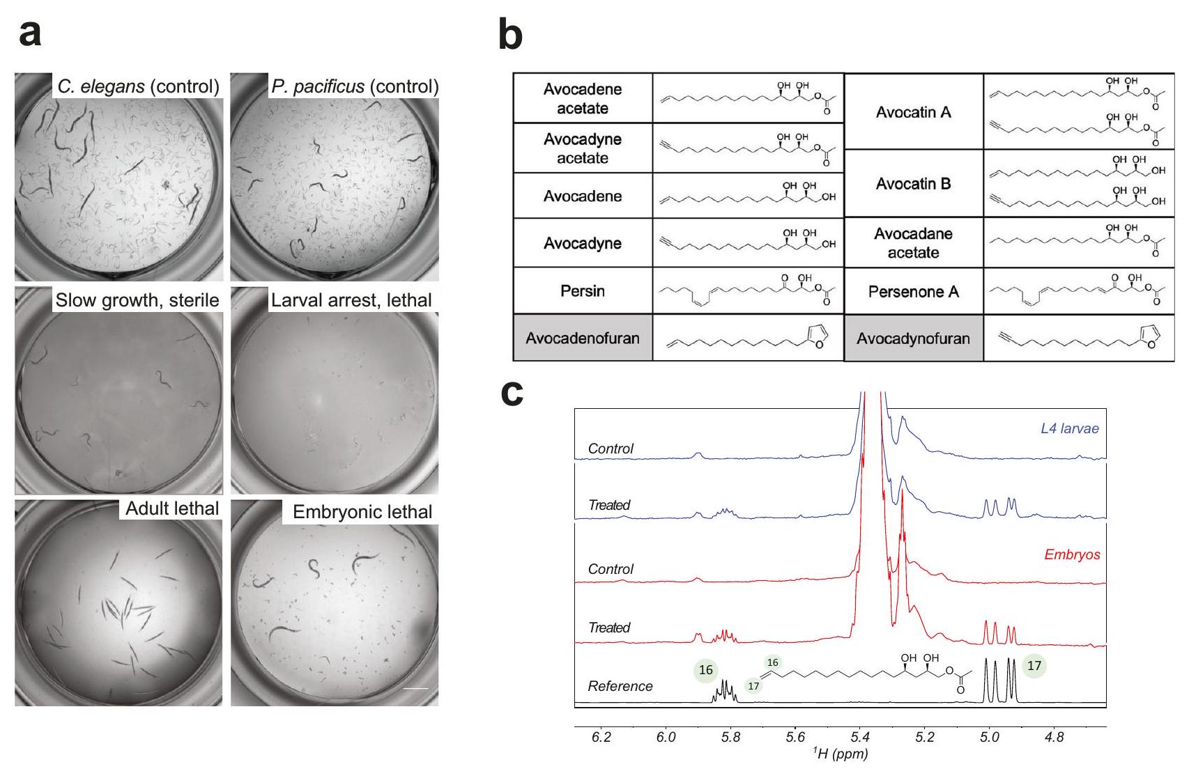
نشاط مضاد للديدان ضد C. elegans و P. pacificus. ج إدماج أسيتات الأفوكادين في أجنة و يرقات . elegans كما تم الكشف عنها بواسطة طيف الرنين المغناطيسي النووي للمركب المرجعي النقي مقارنةً بالطيف المقابل لـ مستخلصات من الأجنة واليرقات L4، مع العلاج وبدونه أو أسيتات الأفوكادين، على التوالي.
طرق، البيانات التكميلية 2) وأسفرت عن تحديد عائلة من المركبات ذات الصلة المعزولة من الأفوكادو Persea americana التي تسبب مجموعة متنوعة من الظواهر الشديدة في C. elegans (الشكل 1a). تمثل هذه المركبات AFA كحوليات دهنية مشابهة هيكليًا تحتوي على 17 كربونًا ونسخها الأسيتات (الشكل 1b): الأفوكادين (( (-1,2,4-ثلاثي هيدروكسي هبتاديس-16-ين)، أفوكادين ((2R،4R)-1،2،4-ثلاثي هيدروكسي هبتاديس-16-ين)، أسيتات أفوكادين ((2R،4R)-2،4-ثنائي هيدروكسي هبتاديس-16-ين-1-يل أسيتات)، أسيتات أفوكادين ((2R،4R)-2،4-ثنائي هيدروكسي هبتاديس-16-ين-1-يل أسيتات)، أفوكاتين (خليط من الأفوكادين والأفوكادين، وأفوكاتين) (خليط من أشكال الأسيتات الخاصة بهم).
تُعرف الديدان الخيطية بأنها مقاومة للغاية للظروف البيئية القاسية. تحاط المراحل المعدية والبالغة بطبقة سميكة من الكيوتيكل، وتكون الأجنة محمية بعدة طبقات تشمل طبقة الكيتين، وطبقة الفيتيلين، وطبقة البروتيوغليكان الكوندرويتيني. باستخدام نموذج C. elegans، تساءلنا عما إذا كانت AFAs تتجمع داخل الأجنة أو المراحل ما بعد الجنينية. أظهر تحليل مستخلصات الأجنة واليرقات بواسطة الرنين المغناطيسي النووي البروتوني أن كلاً من الأجنة واليرقات
احتوت على كميات كبيرة من AFAs بعد العلاجات، كما يتضح من وجود الإشارات المميزة لمجموعة الأليفين الطرفية لأسيتات الأفوكادين (الشكل 1c).
لتوصيف ملف نشاط AFAs، قمنا باختبارها بتركيزات مختلفة (من إلى ) في الثقافة السائلة عبر مراحل تطور C. elegans، في 3 تكرارات بيولوجية ( ) و 4 تكرارات فنية ( ) (الشكل 2). وجدنا أن جميع هذه المركبات الستة (الأفوكادين، الأفوكادين، أشكالها الأسيتات، والمزيجين الأفوكاتين A و B) أظهرت تأثيرًا سامًا يعتمد على التركيز على تطور اليرقات L1 (الشكل 2a)، بقاء البالغين (الشكل 2b) وفقس البيض (الشكل 2c، الفيلم التكميلي 1، الفيلم التكميلي 2). بالمقابل، لم تُظهر مركبان مرتبطان هيكليًا، الأفوكادينوفوران والأفوكادينوفوران، اللذان يحتويان على سلسلة كربونية مكونة من 17 ذرة كربون ولكن يختلفان بوجود حلقة الفوران (الشكل 1b)، أي نشاط حيوي في أي من اختباراتنا عند تركيزات تصل إلى (الشكل 2أ-د). مقارنة لـ أظهرت القيم أن أسيتات الأفوكادين، وأسيتات الأفوكادين، وأفوكاتين A أظهرت قوة مشابهة وأظهرت تأثيرات أقوى من الأشكال غير الأسيتاتية (الشكل 2d).
الشكل 2 | تؤثر AFAs سلبًا على التطور والبقاء في C. elegans و P. pacificus. أ-ج منحنيات الاستجابة للجرعة لـ 8 مركبات AFA. أ نسبة الحيوانات التي تطورت بعد مرحلة L1 بعد 5 أيام من العلاج الذي بدأ في مرحلة L1. ب نسبة البالغين الذين بقوا على قيد الحياة بعد 48 ساعة من العلاج. ج نسبة البيض الذي فقس بعد 48 ساعة من العلاج. د محسوب القيم (التركيز الذي تم فيه تقليل التنمية أو البقاء بـ نسبة البيض الذي فقس في N2 و PS312 بعد 48 ساعة من العلاج بمضادات الديدان الموجودة وأسيتات الأفوكادين بتركيز 5 و
تتوافق الحدود السفلية والعلوية للصندوق مع الربع الأول والربع الثالث (النسبة المئوية 25 و75)، وتمتد الشعيرات إلى أكبر أو أصغر قيمة لا تتجاوز 1.5 مرة من نطاق الربع بين القيم (IQR). منحنيات الجرعة والاستجابة لأسيتات الأفوكادين على نسبة بقاء الدوار N2 ودوار daf-2 بعد 48 ساعة من التعرض للمركب. كل نقطة بيانات تمثل المتوسط ± الانحراف المعياري؛ لـ (أ-ج، و) BRs مع 4 TRs لكل منها دود/تركيز؛ لـ (هـ) BRs مع 3 TRs كل منها يحتوي على أكثر من 100 بيضة فردية/تركيز. في جميع التجارب، تم استخدام DMSO كتحكم سلبي. تم توفير بيانات المصدر كملف بيانات مصدر.
لاستكشاف هذه الفئة من المركبات بشكل أعمق، تساءلنا عما إذا كانت الجزيئات ذات الصلة ستؤثر بشكل مشابه. وجدنا أن جزيئين آخرين تم تنقيتهما من الأفوكادو، وهما برسون A ((2 R، 5E، 12Z، 15Z)-2-هيدروكسي-4-أوكسيهينيكوسا-5،12،15-تريينيل أسيتات) وبيرسين ((2 R، 12Z، 15Z)-2-هيدروكسي-4-أوكسيهينيكوسا-12،15-دينيل أسيتات)، بالإضافة إلى جزيء صناعي مشبع بالكامل، أسيتات الأفوكادين ((2 R، 4 R)-2،4-ديهيدروكسيهبتادكان-1-يل أسيتات) (الشكل 1ب)، كانت لها أنشطة مشابهة (الشكل التكميلي 2). نظرًا لأن البيرسين ومشتقاته معروفة بأنها سامة للحيوانات. ركزنا على الروايات الجديدة التي وجدناها في الشاشة.
تم إجراء اختبار فقس البيض على الأجنة المحصودة، وأكدنا وجود AFAs داخل الأجنة المعالجة باستخدام NMR (الشكل 1c)، مما يوضح أن هذه الكحوليات الدهنية والأسيتات يمكن أن تعبر حاجز قشرة البيض. لمقارنة الفعالية، قمنا بقياس C. elegans و فقس بيض pacificus في وجود أسيتات الأفوكادين أو مجموعة من الأدوية الطاردة للديدان الأخرى (الشكل 2e). وجدنا أن الألبندازول، الميبيندازول، والليفاميزول لم يكن لها تأثير على فقس البيض في أي من النوعين؛ بينما خفض البرازكوانتيل (المستخدم لعلاج عدوى الدودة النافذة للجلد شستوسوما) فقس البيض بـ ( ; غير متزاوج -اختبار، ) في C. elegans و بواسطة ; اختبار t غير المتزاوج، ) في . الباسيفيكس في كلاهما و ، وقلل الإيفرمكتين من فقس البيض بنسبة فقط في كلا النوعين ( و 751، على التوالي؛ غير متزاوج -اختبار، في المقابل، أدى أسيتات الأفوكادين إلى تثبيط فقس البيض في كلا نموذجَي الديدان في ( في C. elegans و في P. pacificus). وبالتالي، تُظهر AFAs قدرة نادرة بين الأدوية المعروفة لمكافحة الديدان على التدخل مباشرة في تكوين الأجنة (بغض النظر عن توفير الدهون من الأم خلال تكوين البويضات)، وهو ما سيكون مفيدًا للغاية لعلاج الديدان الطفيلية.
يمكن أن تدخل الديدان الخيطية مثل C. elegans مرحلة الداور، حيث تقلل من معدل الأيض لديها لتتكيف مع البيئات القاسية مثل الجفاف، وانخفاض المغذيات، وتغير درجة الحرارة.
يُعتقد أن مرحلة الداور تشبه مراحل L3 المعدية للعديد من الديدان الطفيلية. لتحديد ما إذا كانت يرقات الداور حساسة أيضًا لمركبات AFA، قمنا بمعالجة الداور من كل من النوع البري N2 (الذي تم الحصول عليه عن طريق التجويع) وسلالة daf-2(m41) (التي تحفز الداور في مع أسيتات الأفوكادين لمدة 48 ساعة عند ، ووجدت أنها أثارت الفتك في يرقات الداور N2 و daf-2(m41) مع من و على التوالي (الشكل 2f).

تظهر AFAs نمطًا جديدًا من العمل

لاختبار تأثير مركبات AFA على الديدان الطفيلية، أجرينا اختبارات على أربعة طفيليات بيطرية: H. contortus و T. circumcincta و H. polygyrus و B. pahangi. أفوكاتين B عند كلا و أدت إلى وفاة اليرقات بشكل قوي في جميع الأنواع الطفيلية الثلاثة التي تم اختبارها: H. contortus و T. circumcincta و H. polygyrus (الجدول 1، الجدول التكميلي 1). علاوة على ذلك، كانت AFAs نشطة أيضًا في مقاومة الأدوية المتعددة. يرقات وبيض .contortus UGA في و على التوالي (الجدول 1). من المهم أن الأفوكاتين A ليس فقط فعالًا ضد مراحل L3 والميكروفيلاريا، ولكنه يعمل أيضًا كمبيد للبالغات، حيث يقتل البالغين. . باهينجي تبدأ عند كانت الإناث البالغات أكثر حساسية من الذكور (الجدول 2). في H. contortus، بينما تسببت جميع المركبات الستة النشطة حيوياً من الفحص الأولي في إعاقة فقس البيض وتسببت في فتك اليرقات، فإن الأشكال الأسيتاتية أثارت أقوى الظواهر (الجدول التكميلي 1). مع النتائج من الفحص الأولي، أكدت هذه التحليلات أن الأشكال الأسيتاتية تحفز أعلى تأثير من حيث القوة. لذلك، قمنا بإجراء التجارب اللاحقة باستخدام إما أسيتات الأفوكادين أو أفوكاتين A، وهو مزيج من الشكلين الأسيتاتيين.
لاختبار إمكانية أن تمثل AFAs فئة جديدة من الأدوية المضادة للطفيليات، قمنا بفحصها في طفرات C. elegans التي تقاوم الأدوية المضادة للطفيليات المعروفة الإيفرمكتين/الأبامكتين، ليفاميزول، البنزيميدازولات ومشتق الأحماض الأمينية-الأستونيتريل (AAD) مونيبانتيلي. كانت جميع سلالات الطفرات حساسة لسمية AFA بنفس المستوى كما
الجدول 1 | الأدوية المضادة للفطريات فعالة ضد سلالة من الديدان الطفيلية المقاومة لمتعدد الأدوية
خلاصة تي. سيركومسينكتا L2 H. polygyrus L2 H. contortus (UGA) L2 بيض H. contortus (UGA)
إجمالي % الفتك إجمالي % الفتك إجمالي % الفتك إجمالي % الفتك
DMSO 1% 32 18.8 52 21.2 215 113 ٣٣.٦
أفو ب ١١٩ 40 100 ٢٢٣ 89 31.5
١١٩ 69 100 ٢٠٩ 30 100
أفو أ ٥٥ 100 ٢٢٣ 66 ٣٦.٤
76 100 162 75 100
معدلات فتك اليرقات من الديدان الطفيلية بعد العلاج مع أفوكاتين A أو B. تم الحصول على أعداد T. circumcincta L2 من ثلاث تجارب بيولوجية. ) وعدد L2 من H. contortus (UGA) من النسخ البيولوجية المكررة تستند عدادات بيض H. polygyrus L2 و H. contortus (UGA) إلى ملاحظة واحدة فقط.
الخلايا التي تحتوي على ‘-‘ تشير إلى أن الاختبار لم يتم.
الجدول 2 | الأدوية المضادة للفيلاريات فعالة ضد الديدان الخيطية ب. باهاني
ب. فاهانجي DMSO
إجمالي البالغين 40 32 32 31 ٣٦ ٣٤ 37
% الفتك/الشلل 0 0 0
إجمالي الإناث 10 10 10 10 10 10 10
% الفتك/الشلل 0 0 0 0 20 50 100
إجمالي الذكور 10 10 10 10 10 10 10
% الفتك/الشلل 0 0 0 20 30 90 100
إجمالي L3 61 68 69
% الفتك 0 100 100
إجمالي Mfs ١٠٤ 154 131 150 ١٢٦ 147 163
% الفتك/الشلل 1.9 5.2 9.2 100 100 100 100
معدلات فتك اليرقات للبالغات من B. pahangi، الإناث والذكور، L3 والميكروفيلاريا (mfs) بعد العلاج بالأفوكاتين A. تم الحصول على أعداد البالغين من B. pahangi من 5 تكرارات بيولوجية. الإناث، الذكور، L3 والميكروفيلاريا من ملاحظة واحدة فقط.
الخلايا التي تحتوي على ‘-‘ تشير إلى أن الاختبار لم يتم.
الجدول 3 | الأدوية المضادة للطفيليات فعالة ضد سلالات C. elegans المقاومة للأدوية
التركيب الجيني مقاومة أسيتات الأفوكادو
النوع البري لا شيء
تشا-1(ب1152) الكاربامات/الفوسفات العضوية
avr-14(ad1302)؛ avr15(ad1051) glc-1(pk54) إيفرمكتين
دي إف 17 (أو إكس 175) إيفرمكتين/ألبندازول
unc-63(ok1075) ليفاميزول / مواد مضادة للطفيليات / تيتراهيدروبيريميدينات
unc-29(e193) ليفاميزول
acr-23(ok2804) مونيبانتيلي
داف-16(مو86) أبيجينين
بن-1(إي1880) بنزيميدازول
سلو-1(js379) إيموديبسيد
محسوب قيم مركبات AFA في سلالات الطفيل المقاوم للأدوية من C. elegans المعالجة في مرحلة L1. البيانات مستمدة من 3 تكرارات بيولوجية ( ).
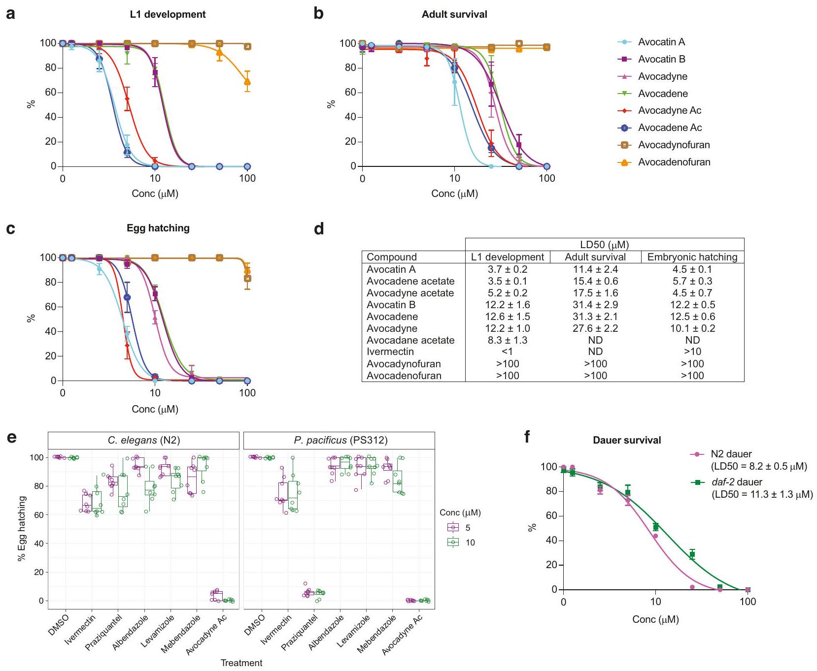
النوع البري N2 عند معالجته في مرحلة L1، مما يشير إلى أن مركبات AFA تعمل من خلال آلية مختلفة (الجدول 3، الشكل التكميلي 3). لتوصيف طريقة عمل AFAs بشكل أكبر، قمنا بإجراء طفرات عشوائية في C. elegans باستخدام إيثيل ميثان سلفونات (EMS). على الرغم من إجراء عدة جولات كبيرة من الفحص واختبار مقاومة مليون جينوم، لم نتمكن من استعادة أي سلالة بمستوى قابل للتكرار من المقاومة. تشير نتائج هذا الفحص الجيني الأمامي إلى أن تطور المقاومة للأدوية المضادة للطفيليات قد يتطلب تغييرات في مواقع متعددة، أو أن تأثيراتها المضادة للطفيليات تنشأ من الأضرار الفيزيائية للهياكل الخلوية.

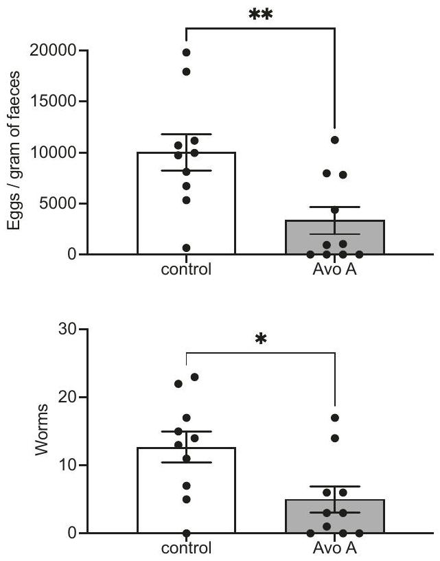
AFAs نشطة في الجسم الحي

لتحديد ما إذا كانت التأثيرات المضادة للطفيليات لـ AFAs التي تم اختبارها مباشرة على الديدان الخيطية ستترجم إلى نظام ديدان طفيلي في الجسم الحي، استخدمنا الفئران المصابة بـ H. polygyrus، وهو نموذج للعدوى المعوية. قمنا باختبار تأثير أفوكاتين A على الفئران المصابة التي تم تلقيحها مسبقًا وسمح لها بتطوير عيار عالٍ من العدوى لمدة أسبوع. أظهرت الفئران المصابة التي تم علاجها بأفوكاتين A انخفاضًا مستمرًا وملحوظًا مقارنةً بمجموعة التحكم التي تلقت دواءً وهميًا في كلا عدد بيض البراز (FEC) لديدان البوليجيروس وعدد وصحة الديدان البالغة التي تم تشريحها من الفئران في اليوم 21 و غير متزاوج -اختبار) (الشكل 3). تم شفاء ثلاثة من بين عشرة فئران تم اختبارها من عدوى H. polygyrus بشكل أساسي خلال فترة التجربة التي استمرت أسبوعين، ولم تنتج أربعة من عشرة أي بيض.

تثبط AFAs التنفس في C. elegans

لاستكشاف آلية عمل AFAs، قمنا بفحص التأثيرات الظاهرية لعلاج أجنة وراشدات C. elegans بأسيتات الأفوكادين، وهو أحد أقوى مركبات AFA. أظهرت الراشدات شللًا يعتمد على الجرعة وتثبيط ضخ البلعوم ضمن دقائق العلاج (الشكل 4 أ-ج، الشكل التوضيحي 4). في الأجنة، العلاج الحاد مع أدى إلى توقف النمو في جميع المراحل الجنينية داخل دقائق (الشكل 5f، الشكل التوضيحي 5).
لتوصيف الشلل بشكل أكبر، قمنا بقياس معدلات استهلاك الأكسجين (OCR) في الديدان الخيطية الكاملة في مرحلة L4، باستخدام جهاز Seahorse XFe96 (الشكل 4f). كان قياس التنفس الميتوكوندري الأساسي (Basal OCR) أقل بشكل ملحوظ في الديدان المعالجة بتركيزات دون قاتلة من أسيتات الأفوكادين مقارنةً مع ضوابط DMSO (غير متطابقة -اختبار، ) (الشكل 4f). بعد ذلك، قمنا بقياس أقصى معدل لاستهلاك الأكسجين (OCR) في وجود كربونيل سيانيد-بيترفلوروميثوكسي فينيل هيدرازون (FCCP). FCCP هو مثبط لفسفرة الأكسدة الميتوكوندرية الذي ينهار تدرج البروتون في الغشاء الداخلي للميتوكوندريا، مما يمنع تخليق ATP ويتسبب في عمل سلسلة نقل الإلكترون بكامل طاقتها.
الشكل 3 | الأحماض الدهنية النشطة في الجسم الحي. يتم تقليل عدد البيض لكل جرام من البراز وعدد الديدان البالغة بعد تناول الأفوكاتين A ) العلاج في الفئران المصابة بـ H. polygyrus. تم إصابة فئران C57BL/6 بـ 200 يرقانة من H. polygyrus وتم إعطاؤها Avocatin A أو مركب التحكم في أيام متناوبة من اليوم 9 إلى 19؛ وتم عد البيض وأعداد الديدان البالغة في اليوم 21 بعد الإصابة. تمثل البيانات نتائج مجمعة من تجربتين مستقلتين، كل منهما تحتوي على 5 فئران لكل مجموعة، وتم تحليلها بواسطة اختبار غير متزاوج ذو طرفين. -اختبار ( للبيض و لديدان). حيثما ينطبق، تمثل البيانات المتوسط ± الانحراف المعياري. يتم تقديم بيانات المصدر كملف بيانات مصدر.
سعة أظهرت هذه التجارب أن الديدان المعالجة بتركيزات غير قاتلة من أسيتات الأفوكادين أظهرت انخفاضًا ملحوظًا في أقصى معدل تنفس والقدرة الاحتياطية (أقصى معدل استهلاك الأكسجين – معدل استهلاك الأكسجين الأساسي) مقارنةً بالمجموعة الضابطة (غير المتزاوجة). -اختبار، و ) (الشكل 4f، الشكل التوضيحي 6a).
سلسلة نقل الإلكترون هي المصدر الرئيسي للجذور الحرة التفاعلية في الخلية، ومن المعروف أن العيوب في التنفس الميتوكوندري تؤدي إلى الإجهاد التأكسدي. قمنا بقياس مستويات ROS في الحيوانات في مرحلة L4 في وجود أسيتات الأفوكادين باستخدام جهازين مختلفين للكشف عن الفلورسنت: مستشعر عام لـ ROS، وهو كلوروميثيل-2’،7′ ثنائي كلوريد ثنائي هيدروفلوريسئين داي أستات، وMitoSOX Red، الذي يستشعر بشكل خاص السوبر أكسيد داخل الميتوكوندريا. يتم استهداف MitoSOX إلى البصلة البلعومية الغنية بالميتوكندريا عند ابتلاعها بواسطة C. elegans، حيث تظهر الفلورسنت الحمراء عند الأكسدة بواسطة السوبر أكسيد. بالنسبة لمجموعة التحكم المعالجة بـ DMSO، أظهر مستشعر CM-H2DCFDA زيادة في ROS (غير متزاوج) -اختبار، ) (الشكل 4e)، بينما أظهر MitoSOX زيادة في شدة الفلورية في منطقة البصلة البلعومية الخلفية (غير متزاوجة) -اختبار، ) (شكل تشير المستويات المرتفعة من أنواع الأكسجين التفاعلية التي تم ملاحظتها باستخدام كلا الاختبارين إلى أن العلاج بأسيتات الأفوكادين يؤدي إما إلى زيادة الإنتاج أو ضعف تحييد أنواع الأكسجين التفاعلية.
استكشفنا بعد ذلك ما إذا كان الإجهاد التأكسدي مساهمًا مهمًا في تأثير الأدوية المضادة للطفيليات (AFAs) من خلال معالجة الحيوانات مسبقًا بمضادات الأكسدة. نظرًا لأن الجلوتاثيون (GSH) هو مضاد أكسدة معروف وركيزة للعديد من إنزيمات مضادات الأكسدة، فقد عالجنا الديدان مسبقًا بمقدمة الجلوتاثيون N-acetylcysteine (NAC)، التي تعزز إنتاج الجلوتاثيون وتحييد بعض الأنواع المؤكسدة مباشرة. أظهرت ديدان L4 المعالجة مسبقًا بـ 10 مللي مول NAC خفض في فتك اليرقات الناتج عن الأسيتات الأفوكادو مقارنةً بالمجموعة الضابطة (تحليل التباين الأحادي، -قيمة ) (الشكل 4j، الشكل التوضيحي 6a، b). كما قمنا بمعالجة الحيوانات مسبقًا بفيتامين B12 الأساسي، الذي أظهر أنه يقلل من مستويات ROS في كل من السيتوبلازم والميتوكوندريا. .
الشكل 5 | تستهدف AFAs عملية الأيض للدهون الدهنية. أ قيم الأسيتات الأفوكادين في البود-2 (ye60) وN2 التحكم (مرحلة L4)، في وجود أو غياب مكملات مالونيل CoA. تمثل البيانات ثلاثة تكرارات بيولوجية. تجارب مع 4 نسخ فنية مكررة الحيوانات) كل منها. رسم بياني مكدس لتقنية الرنين المغناطيسي النووي يبرز انخفاض قمة الميثيل الأسيتيل لـ الأسيتيل- عند 2.2 جزء في المليون، بعد إضافة . جزء بروتين الإيليغانس الذي يحتوي على POD-2 إلى خليط تفاعل قياسي (“مرجع”: AcCoA في ATP ). مسار الزمن لـ -تركيزات AcCoA المعيرة التي تم تقييمها من طيف الرنين المغناطيسي النووي بعد إضافة POD-2. تشير المنحنيات المبلغ عنها إلى تركيز AcCoA المعير تحت 4 ظروف تركيبية مختلفة، وهي محلول المرجع (أحمر)، المرجع مع (أرجواني) أو (الأخضر) أسيتات الأفوكادو (Avo Ac)، والمرجع الخالي من ATP (الأرجواني). كل منحنى يعرض بيانات شدة الإشارة من عينة واحدة ( تُمثل أشرطة الخطأ أقصى عدم اليقين التجريبي الذي يؤثر على مناطق إشارة NMR. استهلاك AcCoA المعتمد على NMR بالنسبة إلى محلول المرجع، تم قياسه بعد 180 دقيقة من POD-2.

بالإضافة إلى ذلك. رمز اللون وأشرطة الخطأ هي نفسها كما في (ج). كل شريط يمثل عينة واحدة. ) مع التكملة للوحدة من بيانات تركيز 180 دقيقة المبلغ عنها في (ج)، بعد التطبيع على القيمة المقابلة لمحلول المرجع. e النمذجة الهيكلية لأفوكادين أسيتات وأفوكادين أسيتات المعقد مع POD-2. (ط) نموذج الهوموديمر لمنطقة CT من POD-2 (البقايا 1438-2165) مع البيوتين و CoA في الموقع النشط (المحدد بإطار). تم نمذجة مواقع الربط للديمر والليغاند باستخدام معقدات ديمر الخميرة CT المحلولة (معرف PDB: ). (ii, iii) أعلى 3 تشكيلات، مرتبة حسب الألفة، لـ (ii) أسيتات الأفوكادين و (iii) أسيتات الأفوكادين المعقدة مع POD-2. (iv) تداخل البيوتين و CoA مع أعلى تشكيل لأفوكادين أسيتات في موقع النشاط CT يوضح كيف تمنع المركبات المشتقة من الأفوكادو نشاط POD-2. صور DIC تظهر أن مثبطات ACC CP-640186 و sethoxydim تؤثر سلبًا على التطور الجنيني مقياس تم توفير بيانات المصدر كملف بيانات المصدر.
كانت هذه التأثيرات أعلى بشكل ملحوظ بشكل عام مقارنةً بالضوابط المعالجة بـ DMSO (اختبار كاي-تربيع) ) (الشكل 4د، هـ).

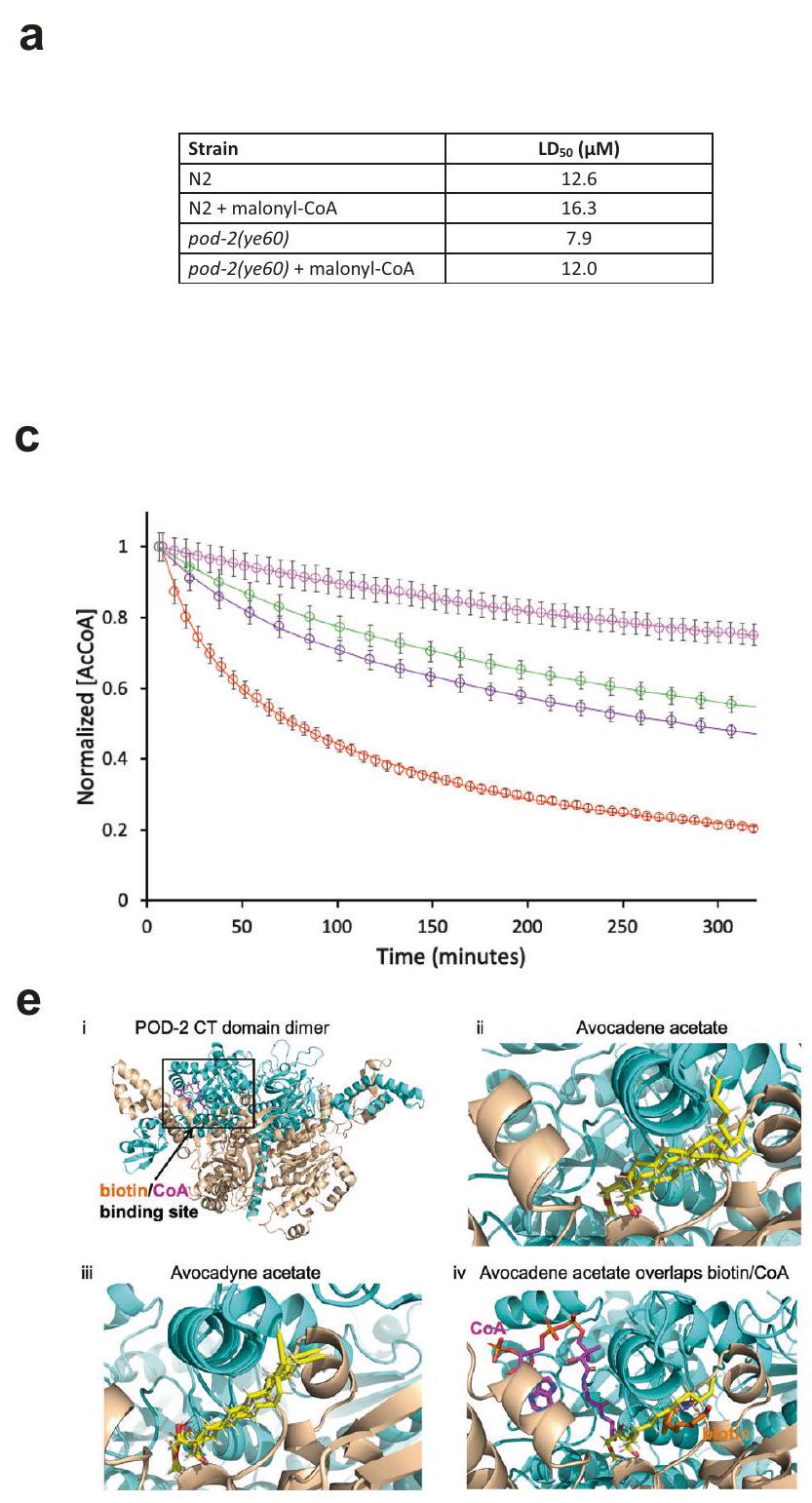
تستهدف AFAs ACC/POD-2

تشير هياكل مركبات AFA، إلى جانب ظواهرها الميتوكوندرية وتأثيراتها على استهلاك الأكسجين وROS، إلى إمكانية أنها قد تتداخل مع الإنزيمات المعنية في استقلاب الدهون. علاوة على ذلك، تشير الأعمال السابقة التي استخدمت سرطان الدم النقوي الحاد
تشير خلايا (AML) إلى تورط الأفوكاتين B في استقلاب الدهون وتفاعل مباشر للأفوكادين مع إنزيم أسيل-CoA ديهيدروجيناز السلسلة الطويلة جداً (VLCAD) البشري لاختبار هذه الفكرة في الديدان، قمنا بإجراء فحص جيني كيميائي مع جينات مرشحة في مسارات تخليق الأحماض الدهنية (FAS) وأكسدة الأحماض الدهنية (FAO).
مجموعة الجينات الأولى التي اختبرناها تتعلق بنقل الأحماض الدهنية إلى الميتوكوندريا. الأحماض الدهنية متوسطة وطويلة السلسلة (حتى
يتم تنشيطها أولاً بواسطة سينثاز أسيل-CoA في السيتوسول (ACS-2 في C. elegans) ثم يتم نقلها بنشاط إلى الميتوكوندريا بواسطة نظام نقل الكارنيتين. يتكون هذا النظام من بروتين غشاء الميتوكوندريا الخارجي CPT1 (كارنيتين بالميتويل ترانسفيراز I، الذي له خمسة نظائر في C. elegans) واثنين من مكونات الغشاء الداخلي، ناقل الكارنيتين-أسيلكارنيتين (CACT، DIF-1 في C. elegans) وكارنيتين بالميتويل ترانسفيراز II (CPT2، CPT-2 في C. elegans). بمجرد دخولها إلى الميتوكوندريا، تخضع الأحماض الدهنية لعدة جولات من الأكسدة بيتا، مما ينتج عنه ناقلات إلكترون للتفسخ التأكسدي وأستيل-CoA، الذي يمكن تحويله إلى مستقلبات أخرى واستخدامه لاستخراج طاقة إضافية عبر دورة حمض ثلاثي الكربوكسيليك (TCA). .
إذا كانت مركبات AFA تتداخل مع استقلاب الأحماض الدهنية، فإن تعطيل مسار النقل سيؤثر على الأنماط الظاهرية الناتجة عن AFA. متسقًا مع هذا التوقع، وجدنا أن تقليل التعبير الجيني باستخدام RNAi لعدد من الجينات الرئيسية في نقل الكارنيتين – بما في ذلك acs-2، ونظير CPT1 cpt-4، وdif-1، وcpt-2 – أنقذ جزئيًا تأثير أسيتات الأفوكادين (اختبار مان-ويتني يو، ، ) (الشكل التوضيحي 7). ومع ذلك، لم يكن لتقنية RNAi الخاصة بنظير CPT1 cpt-1 تأثير كبير (الشكل التوضيحي 7، الجدول التوضيحي 2). تشير هذه النتائج إلى أن تقليل نشاط ناقل الكارنيتين بواسطة RNAi يمكن أن يقلل، ولكن لا يلغي، الحساسية لمركبات AFA.
استكشفنا بعد ذلك التفاعلات الكيميائية الجينية لمركبات AFA مع مجموعة فرعية من الجينات المشاركة في تفاعلات أكسدة الأحماض الدهنية اللاحقة. الخطوة الأولى في كل دورة من دورات الأكسدة بيتا تتم بوساطة إنزيمات أكسيداز أسيل-CoA طويلة ومتوسطة وقصيرة السلسلة. لم يكن لعملية RNAi لـ 12 جينًا معروفًا من جينات acdh في دودة C. elegans (acdh-1 إلى 4 و acdh-6 إلى 13) أي تأثير على سمية أسيتات الأفوكادين (الجدول التكميلي 2)، مما يشير إلى أنه إذا كانت مركبات AFA تثبط مباشرة واحدًا أو أكثر من هذه الجينات، فإن ذلك يمكن تعويضه بمستوى معين من التكرار الوظيفي. كما لم نجد أي تفاعل جيني باستخدام الطفرة acdh-12(gk5631) من دودة C. elegans، وهي النظير الذي يتمتع بأعلى تشابه تسلسلي مع إنزيم VLCAD البشري. . وبالمثل، لم نتمكن من الكشف إلا عن تأثيرات غير متسقة عند اختبار مسارات البروبيونات، والتي يمكننا تفسيرها جزئيًا بمستوى B12 في الطعام (الجدول التكميلي 2). يتماشى ذلك مع إمكانية عدم استقلاب AFAs، حيث أظهرت دراسات NMR أن لم يتم تحلل الأسيتات المسمى بالأفوكادو في C. elegans (الشكل التوضيحي 8).
في عملية اختبار التفاعلات الجينية مع الإنزيمات المشاركة في استقلاب الأحماض الدهنية، وجدنا أن إنزيم أسيتيل CoA كاربوكسيلاز (ACC) في دودة C. elegans pod-2(ye60) حساس لدرجة الحرارة. المتحور أكثر عرضة بشكل ملحوظ لعلاج أسيتات الأفوكادين مقارنة بالنمط البري (الشكل 5أ، الشكل التوضيحي 9أ، ب)، مما يظهر من مقابل لـ WT (غير متزاوج -اختبار، سلالة تحمل دغرون مستحث بواسطة الأوكسين (AID) مرتبط ببروتين POD-2 (pod2::AID) كان أيضًا أكثر حساسية بشكل ملحوظ لأسيتات الأفوكادين: الشباب الذين تم تحضينهم مع الأوكسين لمدة 24 ساعة لاستنفاد بروتين POD-2 أظهروا من مقابل في الضوابط غير المعالجة (غير المقترنة) -اختبار، ) (الشكل التوضيحي التكميلي 9c). يتم تعديل POD-2، مثل جميع الكربوكسيلازات، بشكل تساهمي بواسطة BPL-1 (ليغاز بروتين البيوتين 1) لتحفيز الخطوة الأولى المحددة بمعدل الاعتماد على ATP في تخليق الأحماض الدهنية من خلال كربوكسيلاسيون الأسيتيل-CoA إلى مالونيل-CoA. يتماشى مع عمل AFAs من خلال تثبيط POD-2، تقليل مستويات BPL-1 عن طريق معالجة ديدان bpl-1::AID بالأوكسين عزز تأثير AFA من مقابل في الضوابط غير المعالجة) (اختبار t غير المتزاوج، ) (الشكل التوضيحي التكميلي 9c).
مالونيل-CoA، المنتج من إنزيم ACC/POD-2، يلعب دورًا رئيسيًا في التنسيق بين تخليق وأكسدة الأحماض الدهنية. إذا كانت الأحماض الدهنية غير المشبعة تثبط تخليق مالونيل-CoA، فإن الإضافة الخارجية لمالونيل-CoA يمكن أن تعاكس تأثيرها. في الواقع، أدى مكمل مالونيل-CoA إلى تقليل سمية أسيتات الأفوكادين في الحيوانات في المرحلة L4 في كل من النوع البري والطفرات pod-2(ye60)، مما أدى إلى تغيير الـ من حتى في WT ومن حتى في سلالة pod2(ye60) (الشكل 5أ، الشكل التكميلية 9أ، ب).
تم الإبلاغ عن أن ACC البشري، الذي تم الحفاظ عليه من خلال محاذاة التسلسل والنمذجة الهيكلية مع POD-2 (الشكل التكميلي 10)،
تكون مثبطة بواسطة نفس الأدوية المضادة للفيروسات في المختبر . لذلك اختبرنا ما إذا كانت AFAs يمكن أن تثبط مباشرة POD-2 من C. elegans عن طريق تنقية بروتين POD-2::6xHis المسمى من الديدان وقياس النشاط الإنزيمي بواسطة الوقت الحقيقي NMR. يقوم POD-2 بتحويل الأسيتيل-CoA إلى مالونيل-CoA بطريقة تعتمد على ATP، وأدى إضافة أسيتات الأفوكادين إلى خليط التفاعل القياسي (“المرجع”) إلى تثبيط كبير لاستنفاد الأسيتيل-CoA بطريقة تعتمد على التركيز. في أي اختبار ويلكوكسون المقترن مع المرجع فقط) (الشكل 5ب-د، الأشكال التكميلية 11 و12، الجدول التكميلية 3). بسبب توازن الكيتو-إنولي والتDeuteration الناتج في لم نتمكن من قياس ظهور بروتونات الميثيلين لمجموعة المالونيل باستخدام الرنين المغناطيسي النووي. ومع ذلك، يمكن تأكيد إنتاج المالونيلCoA نوعيًا بواسطة مطيافية الكتلة (الشكل التكميلية 13).
POD-2/ACC يعمل كديمر متجانس من خلال عملية من خطوتين موجهة بواسطة مجالات البيوتين كربوكسيلاز (BC) وكربوكسيليترانسفيراز (CT) لتحويل الأسيتيل- CoA إلى مالونيل- نظرًا لأن مجالات BC و CT تتأثر كلاهما بمثبطات ACC الأخرى قمنا بإجراء بحث قائم على الدوكينغ باستخدام تقنيات الحوسبة للبحث عن أهداف المركبات المستخلصة من الأفوكادو في كلا المجالين (الطرق). باستخدام المحاذاة الهيكلية مع مجمعات ACC في الخميرة التي تم حلها (PDB: 1w96، 5csl)، قمنا بتركيز بحثنا في محيط مواقع ارتباط السورافين A والبيوتين في مجالي BC و CT، على التوالي. توقعت النماذج الحاسوبية لمركبات أسيتات الأفوكادين وأسيتات الأفوكادين في موقع CT (الشكل 5e) التآلفات ( قيم) و ، على التوالي، أقوى بمقدار مرتين من القيم المتوقعة في موقع BC لدى البيرسين تقارب متوقع مشابه للرابط بينما لا يرتبط الأفوكادينوفوران بشكل أساسي بهذه المناطق من POD-2 يتماشى ذلك مع النتائج الوظيفية (الأشكال 1 و 2). معًا، تشير نتائج الربط الحاسوبي إلى أن موقع النشاط الإنزيمي هو الهدف الرئيسي لمثبطات الأفوكادين/أسيتات الأفوكادين و البيرسين. نظرًا لموقع مجموعة الأسيتات، لم تتمكن هذه التحليل من الكشف عن الطفرات التي من شأنها الحفاظ على النشاط الإنزيمي وتقليل ارتباط الأفوكادين/أسيتات الأفوكادين بشكل كبير في الوقت نفسه. كما كشفت تحليلات الربط أن CP640186 و sethoxydim، وهما مثبطان معروفان لـ ACC، يمكن أن يؤثرا على POD-2 بنفس الطريقة. وجدنا أن كلا المركبين قاما بتثبيط قوي لتطور الأجنة، وأن CP640186 تقريبًا قام بتقليد تأثير علاج AFA (الشكل 5f). ومع ذلك، لا يبدو أن CP640186 أو sethoxydim يعبران حاجز قشرة البيضة: كانت الأنماط الظاهرة الناتجة عن الأدوية تتطلب كسر قشرة البيضة عند وضع الأجنة تحت شريحة زجاجية للمجهر، ولم تُلاحظ في الثقافات السائلة للأجنة، ولا في اليرقات. تدعم هذه البيانات معًا الاستنتاج بأن هذه التدخلات الصيدلانية المختلفة تؤثر على الديدان بطريقة مشابهة بشكل ملحوظ، لكنها تختلف في قدرتها على اختراق قشرة البيضة و الجلد الخارجي للديدان.

نقاش

نظرًا لوجود فئات محدودة من أدوية مضادات الديدان المستخدمة، فإن مقاومة مضادات الديدان المنتشرة شديدة بشكل خاص في صناعة الثروة الحيوانية على مستوى العالم وقد تم توثيقها أيضًا في البشر والحيوانات الأليفة. هنا، نقدم فئة جديدة من الكحوليات الدهنية الطبيعية المرتبطة، AFAs، التي تظهر تأثيرات مضادة للديدان في المختبر وفي الكائنات الحية. تعتبر AFAs فعالة في إحداث القتل عبر جميع مراحل الحياة التي تم اختبارها عبر عدة أنواع، بما في ذلك سلالة H. contortus المقاومة المتعددة للأدوية المستمدة من الميدان. عند اختبارها في الكائنات الحية، تسبب علاج Avocatin A في تقليل كبير في حمل الطفيليات لكل من البالغين والبيض. لم نلاحظ أي سمية واضحة للمضيف في الفئران المعالجة بـ Avocatin A، وهو ما يتماشى مع بياناتنا في خطوط الخلايا، بالإضافة إلى التقارير المنشورة التي تصف ملف أمان مواتٍ لـ Avocatin B (الإصدار غير الأسيتيلاتي من Avocatin A) في الفئران والنتائج الأولية من التجارب السريرية في المرحلة الأولى. .
مستخلصات الأفوكادو هي مصدر غني بالمركبات ذات الأنشطة الحيوية المتنوعة، والخصائص المضادة للميكروبات والطبية، وملفات الأمان. تم مؤخرًا إظهار أن الأفوكادين والأفوكادين المعزولين لهما نشاط قاتل ليرقات البعوض. بينما لم يتم تنقية AFAs
30 دقيقة، تليها معالجة باستخدام أسيتات الأفوكادين أو التحكم DMSO لمدة تقارب 5 دقائق. تم تحميل الديدان في خرطوشة SC40 باستخدام حقنة وتركها تستقر لمدة 30-60 ثانية. تم أخذ تسجيلات EPG الخاصة بهم لمدة 60 ثانية باستخدام برنامج NemAquire (InVivoBiosystems). تم مراقبة العملية بالكامل تحت المجهر المقلوب Leica M125C لضمان أن الدودة كانت موضوعة بشكل صحيح بين الأقطاب وأن دودة واحدة فقط تشغل القناة في كل مرة. تم أخذ قراءات لـ 15 حيوانًا لكل معالجة. تم حساب متوسط ترددات المضخات بواسطة برنامج NemAnalysis (InVivoBiosystems).
تقييم شكل الميتوكوندريا. تم استخدام سلالة C. elegans SJ4103، التي تعبر عن بروتين GFP في مصفوفة الميتوكوندريا في عضلات جدار الجسم، لقياس التغيرات في شكل الميتوكوندريا المرتبطة بعلاج أسيتات الأفوكادين. تم معالجة الحيوانات في مرحلة L1 بـ دي ميثيل سلفوكسيد أسيتات الأفوكادو لمدة 48 ساعة. تم نقل الحيوانات إلى وسادات أغاروز تحتوي على 1 مللي مول من ليفاميسول، مغطاة بشريحة غطاء، وتم تصويرها باستخدام مجهر ليكا TCS SP8 المجهري. عدسة موضوعية وليزر مسح بطول موجي 488 نانومتر. تم إجراء التقييم وفقًا لطريقة منشورة. .

قياسات معدل استهلاك الأكسجين (OCR) باستخدام محلل Seahorse XFe96

تم معالجة الديدان الخيطية L1 المتزامنة بـ 0.05% DMSO أو أسيتات الأفوكادو في ، حتى تطورت إلى مرحلة L4 ( ساعات). تم غسل الحيوانات ثلاث مرات باستخدام M9، و20-25 حيوانًا في تم نقل M9 إلى كل بئر من آبار لوحة زراعة الخلايا Seahorse المكونة من 96 بئرًا تحتوي على من M9. تم تحميل الأدوية FCCP و نترات الصوديوم مسبقًا إلى منافذ حقن الخرطوشة بتركيز نهائي من و 40 مللي مول على التوالي. تم قياس التنفس الأساسي في البداية لمدة 30 دقيقة تلتها حقن FCCP (لقياس التنفس الأقصى) وأزيد الصوديوم لقياس التنفس غير الميتوكوندري (تم اشتقاق التنفس الأساسي من خلال طرح استهلاك الأكسجين غير الميتوكوندري الناتج عن أزيد الصوديوم من مستويات التنفس الأساسي المقاسة). تم تحديد التنفس الأساسي، والتنفس الأقصى، والسعات الاحتياطية لكل بئر وتم تطبيعها على عدد الديدان في كل بئر.

قياس ROS

تم قياس مستويات ROS داخل الخلايا باستخدام كلوروميثيل-2،7′ ثنائي كلوريد ثنائي هيدروفلوريسئين داي أستات. تم معالجة L1s المتزامنة بـ DMSO. ) أو أسيتات الأفوكادو ( ) لمدة 48 ساعة في تم غسل الديدان لإزالة OP50 وتم حضن 10-15 حيوانًا مع تم استخدام CM-H2DCFDA لمدة 4 ساعات في لوحة 96 بئر. تم قياس الفلورية المنبعثة عند 535 نانومتر عند التحفيز عند 480 نانومتر باستخدام جهاز قراءة الألواح المتعددة EnSpire. تم تطبيع مستويات الفلورية وفقًا لعدد الحيوانات من خلال قياس الامتصاص عند 600 نانومتر. بلغت الفلورية ذروتها بعد 4 ساعات، لذا تم مقارنة قيم الامتصاص في هذه النقطة الزمنية بين المجموعات الضابطة والمعالجة.

صبغة ميتو سوكس™ الحمراء

تم معالجة الحيوانات في مرحلة L1 بـ DMSO (0.1%) أو أسيتات الأفوكادين. لمدة 42 ساعة. ثم تم غسلها من الأطباق وتم إجراء صبغة ميتو سوكس باستخدام حوالي 100 حيوان في M9 + كوليسترول و من OP50. تم حضانة الحيوانات مع ميتوسوكس (التركيز النهائي في هزازة عند في الظلام لمدة 24 ساعة. تم غسلهم 3 مرات بمحلول M9، وتم السماح لهم بالتغذية على طبق OP50 جديد لمدة ساعة واحدة، وتم تصوير منطقة البلعوم باستخدام ميكروسكوب لايكا DMi8، باستخدام عدسة بصرية مع تعرض لمدة 100 مللي ثانية باستخدام فلاتر فلورية 510/580.

علاج NAC وفيتامين ب12

تم معالجة حوالي 10 دودة من مرحلة L1 لكل بئر في صفائح 96 بئر مع أو بدون 10 مللي مول من ن-أسيتيل سيستين (NAC) حتى وصلت إلى مرحلة L4. ثم تم معالجة الحيوانات بـ الأفوكادو
تمت معالجة الديدان في مرحلة L1 بوجود أو غياب 64 نانومتر من فيتامين B12، تلاها العلاج بـ و من أسيتات الأفوكادو عند الوصول إلى مرحلة L4.

تجارب RNAi

تم الحصول على جميع نسخ RNAi من مكتبة أهرينجر (Source BioScience) وتم تأكيد تسلسلها. تم إجراء التجارب على ديدان المرحلة L1 باستخدام نفس الطريقة الموضحة في قسم ‘الفحص الكيميائي’، باستخدام ثقافات بكتيرية RNAi المحفزة بـ 1 مللي مول من IPTG لمدة 4 ساعات في بدلاً من OP50. بالنسبة لشاشات أفوكادين أسيتات و acs-2/ cpt-1/ cpt-4/ dif-1/ cpt-2/ acdh-1/ acdh-2/ acdh-3/ acdh-4/ acdh-6/ acdh-7/ acdh-8/ acdh-9/ acdh-10/ acdh-11/ acdh-12/ acdh-13، تم استخدام أسيتات الأفوكادين.

تجارب إنقاذ مالونيل-CoA على WT و pod-2(ye60)

تم تربية الحيوانات الحساسة للبرودة N 2 و pod-2(ye60) في . عندما وصلوا إلى المرحلة L3، تم نقلهم إلى لمدة 16 ساعة. بعد ذلك، تعرضت الحيوانات لتركيزات مختلفة من أسيتات الأفوكادين. بالإضافة إلى ذلك، تلقت مجموعة تجريبية واحدة مكملات من مالونيل-CoA. ثم تم عد الحيوانات الباقية بعد مرور 48 ساعة.

تجارب تحفيز التحلل بواسطة الأوكسين (AID)

تم استخدام سلالات AX8465 و AX8273 و N2 في هذا الاختبار. تم تربية يرقات L1 المتزامنة في العمر على OP50 بكثافة تقارب 10^15 دودة لكل بئر في صفائح 96 بئر كما هو موضح أعلاه. عند الوصول إلى مرحلة اليرقة L4، تم إضافة 1 مللي مول من الأوكسين (حمض الإندول-3-أسيتيك) إلى الصفائح التي تحتوي على AX8273 وتم حضنها لمدة 48 ساعة، تلاها إضافة تركيزات مختلفة من أسيتات الأفوكادين. بالنسبة لسلالة AX8465، تم علاج الأوكسين بتركيز حدث ذلك في مرحلة الشباب البالغ وتم إضافة أسيتات الأفوكادو بعد 24 ساعة. تم قياس الديدان الحية والميتة بعد 48 ساعة.

الكيمياء الحيوية POD-2

تم استخدام سلالة C. elegans، PHX1772 pod-2(syb1772[pod-2::His1O]) II في تجارب السحب. تم إعداد مستخلص بروتين الديدان من 500,000 دودة في مرحلة L4، نمت في ثقافة سائلة. تم جمع الديدان، وغسلها 3 مرات بمحلول M9 وإعادة تعليقها في محلول التحلل. ثم أضيفت suspension الديدان بالتنقيط إلى هاون مبرد مسبقًا مع النيتروجين السائل. تم طحن الكريات المجمدة إلى مسحوق ناعم. تم إذابة هذا المسحوق مرة أخرى وتجانسه باستخدام جهاز QSonica. ثم تم طرد العينة مركزيًا عند باستخدام جهاز الطرد المركزي الفائق بيكمان كولتر أوبتيما XPN-90 مع دوار نوع 90 Ti عند ثم تم نقل السائل الشفاف إلى عمود راتنج نيتريد النيكل-نTA المتوازن وتم تحضيره لمدة ساعة واحدة عند بعد جمع السائل المتدفق، تم غسل العمود 5 مرات باستخدام محلول الغسيل وتم إجراء استبعاد البروتين باستخدام محلول الاستبعاد الذي يحتوي على 250 مليمول من الإيميدازول. ثم تم تركيز الكمية المستبعدة عن طريق تمريرها عبر عمود ترشيح مركزي (بقطع وزن جزيئي 100 كيلودالتون) وتم استخدام الكمية المنقاة في اختبارات الرنين المغناطيسي النووي.

مطيافية الكتلة والبروتيوميات

تحضير الهضمات في المحلول. تم إجراء اختبار برادفورد لقياس تركيز البروتين. ثلاثون تم تقليل البروتين لكل عينة باستخدام 20 مللي مولار DTT (ثيوثريتيول، سيغما) في درجة حرارة الغرفة (RT) لمدة ساعة واحدة وتمت عملية S-ألكيليت باستخدام 50 مللي مولار IAA (يودو-أسيتاميد، سيغما) لمدة ساعة واحدة في غرفة مظلمة. تم إخماد IAA المتبقي باستخدام 20 مللي مولار DTT لمدة ساعة واحدة في الظلام. قبل الهضم، تم تخفيف اليوريا (سيغما) إلى تركيز 1 مولار باستخدام ، سيغما) لتحضير المذيب. تم هضم البروتينات باستخدام مزيج من التربسين/لايسين-C بدرجة MS بنسبة 1:50 (وزن/وزن إنزيم:بروتين) (ثيرمو فيشر) لمدة 24 ساعة عند درجة حرارة الغرفة. تم إيقاف عملية الهضم عن طريق خفض الرقم الهيدروجيني باستخدام حمض الفورميك (فيشر) وتم إزالة الأملاح من العينة باستخدام ZipTip C18 (ميرك-ميليبور).
عمود. تم إعادة تعليق العينات في من حمض الفورميك قبل LC-MS تحليل الكروماتوغرافيا السائلة المترافقة مع مطياف الكتلة.
كروماتوغرافيا السائل المتصلة بمطياف الكتلة تحليل. تم إجراء الكروماتوغرافيا السائلة على جهاز EASYnLC1200 الأوتوماتيكي بالكامل (ثيرمو ساينتيفيك). تم تجهيز النظام بعمود C18 (بيب ماب RSLC، ثيرمو ساينتيفيك) بقطر داخلي من وطول 15 سم. تم الحفاظ على العمود عند درجة حرارة ثابتة من تكونت الأطوار المتنقلة من حمض الفورميك كمذيب A و حمض الفورميك في أسيتونيتريل كحل للمذيب B. تم تحميل العينات في المذيب A. تم ضبط تدرج خطي كما يلي: لمدة 3 دقائق ثم تدرج إلى في 45 دقيقة و في 60 دقيقة. غسيل لمدة 10 دقائق في تم استخدامه لمنع الانتقال، ومدة توازن قدرها 15 دقيقة مع أكمل B التدرج. تم ربط نظام LC بجهاز Q-Exactive عالي المجال Orbitrap (Thermo Scientific) مزود بمصدر أيون Easy spray وعمل في وضع الأيونات الموجبة. تم ضبط جهد الرش على 1.8 كيلو فولت، ومستوى RF للعدسة S إلى 35، وأنبوب نقل الأيونات إلى “. تم الحصول على المسح الكامل في محلل الكتلة Orbitrap الذي يغطي نسبة الكتلة إلى الشحنة ( نطاق 350-1800 بدقة 120,000. تم تعيين هدف AGC إلى 3E6 ووقت الأيون الأقصى إلى 50 مللي ثانية. تم إجراء التحليل تحت وضع يعتمد على البيانات لتفكيك أعلى 20 مقدمة الأكثر كثافة باستخدام تفكيك HCD. الـ MS تم تعيين المعلمات على النحو التالي: الدقة، 15,000؛ هدف AGC، 1E5؛ الحد الأدنى لهدف AGC، 8.0E3؛ عتبة الكثافة، 4.0E5؛ الحد الأقصى لوقت الأيون، 20 مللي ثانية؛ عرض العزل، ; حالة شحنة السلف، 2-5؛ تطابق الببتيد، مفضل؛ الاستبعاد الديناميكي، 30 ثانية، وكتلة أولى ثابتة من تم تعيين طاقة الاصطدام المعيارية لـ HCD إلى .
تم تحديد هويات البروتينات باستخدام برنامج ثيرمو بروتين ديسكفري 2.4.1.15http://thermofisher.com/) برنامج مع الإعدادات التالية: إنزيم، تريبسين؛ جهاز، أوربيتراب؛ تفتت، HCD؛ اكتساب، DDA دون دمج المسحات؛ وتحمل الكتلة للسابقة والقطع، 10 جزء في المليون و0.2 دالتون، على التوالي. تم الكشف عن السابقت الصحيحة باستخدام الكتلة فقط. تم إجراء تحديدات الببتيد باستخدام قاعدة بيانات UniProtKB_SwissSprot (الإصدار 2021_02، https://www.uniprot.org/uniprot) مع تصنيف C. elegans. تم استخدام أداة البحث Proteome Discoverer للبحث عن الببتيدات مع تثبيت الكارباميدوميثيلاشن وتعديلات الأكسدة المتغيرة للميثيونين (M) والأسيتيل عند الطرف N. تم تعيين عتبة FDR إلى تم تعيين تغيير الطي إلى 2، مع استخدام 2 ببتيد فريد لكل بروتين لتصفية قوائم البروتينات المحددة.
بعد إجراء تحليل NMR في الوقت الحقيقي، تم تجفيف العينات بالتجميد وإعادة تعليقها في من الماء وخلط جيدًا عند نسبة الحجم مع محلول المصفوفة (حمض 2,5-ديهيدروكسي بنزويك) (ميرك كيه جي إيه، دارمشتات، ألمانيا). واحد تم إيداع كل عينة على لوحة الهدف الخاصة بـ MALDI (التحليل الطيفي بالليزر المساعد لإزالة الأيونات) وترك البقع لتجف في ظروف محيطة قبل تحليل FT-ICR-MS (التحليل الطيفي الكتلي باستخدام تحويل فورييه لتذبذب الأيونات). يسمح FT-ICR-MS بجمع بيانات عالية الدقة، وكتلة دقيقة، وبنية نظيرية دقيقة لتحديد المركب مباشرة. تم إجراء القياسات على جهاز تحليل الطيف الكتلي Bruker SolariX FT-ICR (Bruker Daltonics GmbH، ألمانيا). يتكون النظام من مصدر أيونات مزدوج ESI/MALDI ومزود بمغناطيس فائق التوصيل بقوة 7.0 تسلا (Magnex Scientific Inc.، المملكة المتحدة). تم إجراء القياسات في وضع الأيونات الموجبة لـ MALDI. تم الحصول على الطيف بمدة زمنية قدرها 4 ميغا كلمة على مدى نطاق من 100 إلى 900. كانت معلمات الليزر لتوليد الأيونات: الطول الموجي، ; القوة النسبية، ; لقطات، 20؛ إعداد التركيز، ‘متوسط’؛ التردد، 200 هرتز. تم ضبط إزاحة اللوحة ولوحة الانحراف عند 100 و 220 فولت، على التوالي. تم جمع البيانات باستخدام برنامج ftmsControl وتم معالجتها لاحقًا باستخدام برنامج DataAnalysis (Bruker Daltonics). تم الحصول على مئة مسح لكل عينة.

النمذجة الحاسوبية

نمذجة هياكل POD-2. استخدمنا هيكل POD-2 (النسخة isoform c) الذي تم التنبؤ به بواسطة AlphaFold (تم تنزيله من UniProt). النسخة isoform c،
مع 2165 بقايا، تشمل مجالات BC و CT و BCCP. تم نمذجة ثنائي POD-2 والليغاندات المرتبطة به البيوتين و CoA من خلال محاذاة هيكلية باستخدام مجمعات ACC المتماثلة من الخميرة (معرف PDB: ). على وجه التحديد، ينحرف نموذج مجال POD-2 CT (البقايا 1438-2165) عن 1 و 2 x و 5 csl بمقدار 2.8 و Åعلى التوالي، مما يشير إلى درجة عالية من التشابه الهيكلي مع الهياكل المحلولة.
استخدمنا برنامج شروتينجر لإجراء ربط التحفيز للتركيبات الكيميائية مع مستقبلات البروتين. . تأخذ طريقة الربط بالتكيف المحفز في الاعتبار مرونة الأشكال التوافقية للربائط وموقع مستقبل البروتين للعثور على مجمعات ذات طاقة ربط منخفضة. يتم تحديد أهمية النتائج من خلال الطاقة الحرة للربط ( ) والتناسق الهيكلي للمجمعات ذات الطاقة المنخفضة. ثابت التفكك القابل للقياس تجريبيًا يمكن حسابه من الطاقة الحرة للربط ) باستخدام المعادلة حيث R هو ثابت الغاز و T هو درجة الحرارة . بالنسبة للمركبات المشتقة من الأفوكادو، قمنا بتعيين معلمة حجم الليغاند إلى 25 Å لضمان أن تكون مساحة البحث حول الهدف كبيرة بما فيه الكفاية. بالإضافة إلى ذلك، تم إجراء عمليات الربط باستخدام المعلمات الافتراضية. تم تنزيل الهياكل الجزيئية لليغاندات المعنية من PubChem ثم تم إعدادها للربط باستخدام وحدة LigPrep من شرويدنجر لتوليد هياكل منخفضة الطاقة. استخدمنا الطريقة لإعادة إنتاج 3 مجمعات ليغاند/ACC تم حلها لـ CP-640186 (معرف PDB: 1w2x)، ND-646 (5kkn) و soraphen A (1w96). تم إعادة إنتاج تكوينات المركبات بدقة عالية (RMSD للمجمع ~ 0.2 أنغستروم)، وكانت الألفة المتوقعة المواتية هي 6 و 36 و 139 نانومتر على التوالي، لـ CP-640186 و ND-646 و soraphen A.

الإحصائيات وإمكانية التكرار

للاستجابة للجرعة، فقس البيض، والفحوصات الكيميائية والتركيبية وRNAi، تم إجراء تكرارات بيولوجية مستقلة، مع أكثر من 50 فردًا لكل تكرار. تم إجراء تصورات البيانات والمقارنات الإحصائية بين المجموعات باستخدام Graphpad Prism (الإصدار 8.3.0) أو R. تم تحديد أفضل منحنيات استجابة الجرعة و تم توليد القيم باستخدام تحليل الانحدار غير الخطي. اختبار غير متزاوج ذو طرفين تم استخدام الاختبارات لمقارنة المجموعات و -قيمة اعتُبر ذا دلالة إحصائية.

ملخص التقرير

معلومات إضافية حول تصميم البحث متاحة في ملخص تقارير مجموعة ناتشر المرتبط بهذه المقالة.

توفر البيانات

جميع البيانات متاحة في النص الرئيسي، والمعلومات التكميلية. تم توفير بيانات المصدر مع هذه الورقة.

References

  1. Imtiaz, R. et al. Insights from quantitative analysis and mathematical modelling on the proposed WHO 2030 goals for soil-transmitted helminths [version 1; peer review: 2 approved]. Gates Open Res. 3, 1632 (2019).
  2. Bryant, A. S. & Hallem, E. A. Temperature-dependent behaviors of parasitic helminths. Neurosci Lett 687, 290-303 (2018).
  3. Learmount, J. et al. Three-year evaluation of best practice guidelines for nematode control on commercial sheep farms in the UK. Vet Parasitol 226, 116-123 (2016).
  4. Charlier, J. et al. Initial assessment of the economic burden of major parasitic helminth infections to the ruminant livestock industry in Europe. Prev Vet Med. 182, 105103 (2020).
  5. Siddique, S. & Grundler, F. M. Parasitic nematodes manipulate plant development to establish feeding sites. Curr Opin Microbiol 46, 102-108 (2018).
  6. Selzer, P. M. & Epe, C. Antiparasitics in animal health: quo vadis? Trends Parasitol 37, 77-89 (2021).
  7. Blaxter, M. & Koutsovoulos, G. The evolution of parasitism in Nematoda. Parasitology 142, S26-S39 (2015).
  8. Shih, P. Y. et al. Newly Identified Nematodes from Mono Lake Exhibit Extreme Arsenic Resistance. Curr Biol. 29, 3339-3344.e3334 (2019).
  9. Gang, S. S. & Hallem, E. A. Mechanisms of host seeking by parasitic nematodes. Mol Biochem Parasitol 208, 23-32 (2016).
  10. Aleuy, O. A. & Kutz, S. Adaptations, life-history traits and ecological mechanisms of parasites to survive extremes and environmental unpredictability in the face of climate change. Int J Parasitol Parasites Wildl 12, 308-317 (2020).
  11. Nixon, S. A. et al. Where are all the anthelmintics? Challenges and opportunities on the path to new anthelmintics. Int J Parasitol Drugs Drug Resist 14, 8-16 (2020).
  12. Gasbarre, L. C. Anthelmintic resistance in cattle nematodes in the US. Vet Parasitol 204, 3-11 (2014).
  13. Geurden, T. et al. Anthelmintic resistance to ivermectin and moxidectin in gastrointestinal nematodes of cattle in Europe. International Journal for Parasitology: Drugs and Drug Resistance 5, 163-171 (2015).
  14. Prichard, R. K. & Geary, T. G. Perspectives on the utility of moxidectin for the control of parasitic nematodes in the face of developing anthelmintic resistance. Int J Parasitol Drugs Drug Resist 10, 69-83 (2019).
  15. Miller, T. W. et al. Avermectins, new family of potent anthelmintic agents: isolation and chromatographic properties. Antimicrob Agents Chemother 15, 368-371 (1979).
  16. Holden-Dye, L. & Walker, R. J. (2014) Anthelmintic drugs and nematicides: studies in Caenorhabditis elegans. WormBook, 1-29. https://doi.org/10.1895/wormbook.1.143.2.
  17. Burns, A. R. et al. Caenorhabditis elegans is a useful model for anthelmintic discovery. Nat Commun 6, 7485 (2015).
  18. Hahnel, S. R., Dilks, C. M., Heisler, I., Andersen, E. C. & Kulke, D. Caenorhabditis elegans in anthelmintic research-Old model, new perspectives. International Journal for Parasitology: Drugs and Drug Resistance 14, 237-248 (2020).
  19. Min, J. et al. Forward chemical genetic approach identifies new role for GAPDH in insulin signaling. Nat Chem Biol. 3, 55-59 (2007).
  20. Burns, A. R. et al. The novel nematicide wact- 86 interacts with aldicarb to kill nematodes. PLoS Negl Trop Dis. 11, e0005502 (2017).
  21. Bray, M.-A. et al. Cell Painting, a high-content image-based assay for morphological profiling using multiplexed fluorescent dyes. Nat Protoc. 11, 1757-1774 (2016).
  22. Pearson, Y. E. et al. A statistical framework for high-content phenotypic profiling using cellular feature distributions. Communications Biology 5, 1409 (2022).
  23. Bond, A. T. & Huffman, D. G. Nematode eggshells: a new anatomical and terminological framework, with a critical review of relevant literature and suggested guidelines for the interpretation and reporting of eggshell imagery. Parasite 30, 6 (2023).
  24. Oelrichs, P. B. et al. Isolation and identification of a compound from avocado (Persea americana) leaves which causes necrosis of the acinar epithelium of the lactating mammary gland and the myocardium. Natural Toxins 3, 344-349 (1995).
  25. Crook, M. The dauer hypothesis and the evolution of parasitism: 20 years on and still going strong. Int J Parasitol 44, 1-8 (2014).
  26. Robertson, E. et al. Demonstration of the anthelmintic potency of marimastat in the heligmosomoides polygyrus rodent model. Journal of parasitology 104, 705-709 (2018).
  27. Koopman, M. et al. A screening-based platform for the assessment of cellular respiration in Caenorhabditis elegans. Nat Protoc. 11, 1798-1816 (2016).
  28. Weisiger, R. A. & Fridovich, I. Superoxide dismutase: organelle specificity. Journal of Biological Chemistry 248, 3582-3592 (1973).
  29. Dingley, S. et al. Mitochondrial respiratory chain dysfunction variably increases oxidant stress in Caenorhabditis elegans. Mitochondrion 10, 125-136 (2010).
  30. Lasram, M. M., Dhouib, I. B., Annabi, A., El Fazaa, S. & Gharbi, N. A review on the possible molecular mechanism of action of N -acetylcysteine against insulin resistance and type-2 diabetes development. Clin Biochem. 48, 1200-1208 (2015).
  31. Bito, T. et al. Vitamin B12 deficiency results in severe oxidative stress, leading to memory retention impairment in Caenorhabditis elegans. Redox biology 11, 21-29 (2017).
  32. Revtovich, A. V., Lee, R. & Kirienko, N. V. Interplay between mitochondria and diet mediates pathogen and stress resistance in Caenorhabditis elegans. PLoS genetics 15, e1008011 (2019).
  33. Hartman, J. H. et al. Swimming Exercise and Transient Food Deprivation in Caenorhabditis elegans Promote Mitochondrial Maintenance and Protect Against Chemical-Induced Mitotoxicity. Sci Rep. 8, 8359 (2018).
  34. Lee, E. A. et al. Targeting Mitochondria with Avocatin B Induces Selective Leukemia Cell Death. Cancer Res 75, 2478-2488 (2015).
  35. Tcheng, M. et al. Structure-activity relationship of avocadyne. Food & function 12, 6323-6333 (2021).
  36. Tcheng, M., Minden, M. D. & Spagnuolo, P. A. Avocado-derived avocadyne is a potent inhibitor of fatty acid oxidation. Journal of Food Biochemistry 46, e13895 (2022).
  37. Tcheng, M. et al. Very long chain fatty acid metabolism is required in acute myeloid leukemia. Blood 137, 3518-3532 (2021).
  38. Qu, Q., Zeng, F., Liu, X., Wang, Q. J. & Deng, F. Fatty acid oxidation and carnitine palmitoyltransferase I: emerging therapeutic targets in cancer. Cell Death Dis. 7, e2226 (2016).
  39. Melone, M. A. B. et al. The carnitine system and cancer metabolic plasticity. Cell death & disease 9, 228 (2018).
  40. Demarquoy, J. & Le Borgne, F. Crosstalk between mitochondria and peroxisomes. World journal of biological chemistry 6, 301 (2015).
  41. Au, V. et al. CRISPR/Cas9 methodology for the generation of knockout deletions in Caenorhabditis elegans. G3: Genes, Genomes, Genetics 9, 135-144 (2019).
  42. Artan, M., Hartl, M., Chen, W. & De Bono, M. Depletion of endogenously biotinylated carboxylases enhances the sensitivity of TurbolD-mediated proximity labeling in Caenorhabditis elegans. Journal of Biological Chemistry 298, 102343 (2022).
  43. Rappleye, C. A., Tagawa, A., Le Bot, N., Ahringer, J. & Aroian, R. V. Involvement of fatty acid pathways and cortical interaction of the pronuclear complex in Caenorhabditis elegans embryonic polarity. BMC developmental biology 3, 1-15 (2003).
  44. Watts, J. S., Morton, D. G., Kemphues, K. J. & Watts, J. L. The biotinligating protein BPL-1 is critical for lipid biosynthesis and polarization of the Caenorhabditis elegans embryo. Journal of Biological Chemistry 293, 610-622 (2018).
  45. Hashimura, H., Ueda, C., Kawabata, J. & Kasai, T. Acetyl-CoA carboxylase inhibitors from avocado (Persea americana Mill) fruits. Bioscience, biotechnology, and biochemistry 65, 1656-1658 (2001).
  46. Wei, J. & Tong, L. Crystal structure of the 500-kDa yeast acetyl-CoA carboxylase holoenzyme dimer. Nature 526, 723-727 (2015).
  47. Ahmed, N. et al. Avocatin B Protects Against Lipotoxicity and Improves Insulin Sensitivity in Diet-Induced Obesity. Mol Nutr Food Res. 63, e1900688 (2019).
  48. Bangar, S. P. et al. Avocado seed discoveries: Chemical composition, biological properties, and industrial food applications. Food Chemistry X, 100507 (2022).
  49. Rodríguez-Sánchez, D. G. et al. Chemical profile and safety assessment of a food-grade acetogenin-enriched antimicrobial extract from avocado seed. Molecules 24, 2354 (2019).
  50. Louis, M. L. M. et al. Mosquito larvicidal activity of compounds from unripe fruit peel of avocado (Persea americana Mill.). Applied Biochemistry and Biotechnology 195, 2636-2647 (2023).
  51. Soldera-Silva, A. et al. Assessment of anthelmintic activity and bioguided chemical analysis of Persea americana seed extracts. Vet Parasitol 251, 34-43 (2018).
  52. Rosa, S. S. S. et al. In vitro anthelmintic and cytotoxic activities of extracts of Persea willdenovii Kosterm (Lauraceae). J Helminthol 92, 674-680 (2018).
  53. Taylor, C. M. et al. Discovery of anthelmintic drug targets and drugs using chokepoints in nematode metabolic pathways. PLoS Pathog 9, e1003505 (2013).
  54. Gutbrod, P. et al. Inhibition of acetyl-CoA carboxylase by spirotetramat causes growth arrest and lipid depletion in nematodes. Scientific Reports 10, 12710 (2020).
  55. Starich, T. A., Bai, X. & Greenstein, D. Gap junctions deliver malonylCoA from soma to germline to support embryogenesis in Caenorhabditis elegans. Elife 9, e58619 (2020).
  56. Hashimshony, T., Feder, M., Levin, M., Hall, B. K. & Yanai, I. Spatiotemporal transcriptomics reveals the evolutionary history of the endoderm germ layer. Nature 519, 219-222 (2015).
  57. Zhang, H., Tweel, B., Li, J. & Tong, L. Crystal structure of the carboxyltransferase domain of acetyl-coenzyme A carboxylase in complex with CP-640186. Structure 12, 1683-1691 (2004).
  58. Piotto, M., Saudek, V. & Sklenář, V. Gradient-tailored excitation for single-quantum NMR spectroscopy of aqueous solutions. Journal of biomolecular NMR 2, 661-665 (1992).
  59. Hwang, T.-L. & Shaka, A. Water suppression that works. Excitation sculpting using arbitrary wave-forms and pulsed-field gradients. Journal of Magnetic Resonance, Series A 112, 275-279 (1995).
  60. Jecock, R. M. & Devaney, E. Expression of small heat shock proteins by the third-stage larva of Brugia pahangi. Molecular and biochemical parasitology 56, 219-226 (1992).
  61. Burns, A. R. et al. High-throughput screening of small molecules for bioactivity and target identification in Caenorhabditis elegans. Nat Protoc. 1, 1906-1914 (2006).
  62. Johnston, C. J. et al. Cultivation of Heligmosomoides polygyrus: an immunomodulatory nematode parasite and its secreted products. JoVE 98, e52412 (2015).
  63. Spensley, M., Del Borrello, S., Pajkic, D. & Fraser, A. G. Acute effects of drugs on Caenorhabditis elegans movement reveal complex responses and plasticity. G3: Genes, Genomes, Genetics 8, 2941-2952 (2018).
  64. Sherman, W., Day, T., Jacobson, M. P., Friesner, R. A. & Farid, R. Novel procedure for modeling ligand/receptor induced fit effects. Journal of medicinal chemistry 49, 534-553 (2006).

شكر وتقدير

نشكر الدكتور روبرت وايت ومنصات التكنولوجيا الأساسية في جامعة نيويورك أبوظبي، وبشكل خاص نيكولاس غياكوميديس، لورانس توريس ورزا روشان، على المساعدة في مرفق الفحص عالي الإنتاجية في جامعة نيويورك أبوظبي، ورشيد رزقي على المساعدة في المجهر ومارك أرنو على المساعدة في تسلسل الجينوم الكامل للديدان المتحورة. نشكر نزار درو من مركز المعلوماتية الحيوية في جامعة نيويورك أبوظبي على المساعدة في تحليل تسلسل الجينوم. نشكر مالكوم كينيدي على رؤاه حول استراتيجية اختبار الفيلاريا، وآلان هيلي على المناقشات المفيدة حول كيمياء الجزيئات الصغيرة، ودون موييرمان على مشاركة المعلومات، وإيناس قيديمات، وسوزان هافليس، وبيريهان بورا، وتيفاني كيلفيذر على المساعدة الإدارية. تم تنفيذ هذا العمل تحت منحة معهد أبحاث جامعة نيويورك أبوظبي لمركز الجينوميات وعلوم النظم (ADHPG-CGSB). تم تمويل A.P. من قبل مجلس أبحاث العلوم البيولوجية والتكنولوجيا في بريطانيا (BBSRC) منحة رقم BB/ROO711X/. تم تمويل C.C. وR.M. من قبل جائزة محقق من ويلكوم ترست (المرجع 219530) ومركز ويلكوم المدعوم من ويلكوم ترست لعلم الطفيليات التكاملي (المرجع 104111). تم توفير سلالات الديدان من قبل CGC، الذي يتم تمويله من قبل مكتب برامج البنية التحتية للبحوث في المعاهد الوطنية للصحة (P40 OD010440).

مساهمات المؤلفين

تم إجراء جميع تجارب الديدان الخيطية الحرة المعيشة، وخلايا الإنسان، والرنين المغناطيسي النووي، والكيمياء الحيوية في مختبر K.C.G و F.P.
تم إعداد شاشة مضادة للديدان بواسطة H.Z.F. وF.R. وP.G.C. قامت F.R. بإجراء تجارب استجابة الجرعة للديدان الخيطية الحرة، وRNAi، وتجارب الحركة وضخ البلعوم وتحليل طفرات الديجون الأوكسينية. قامت S.G. بإجراء تجارب OCR وROS وتوصيف الميتوكوندريا وتجارب تنقية POD-2. قام Y.M. وF.R. وH.Z.F. بإجراء شاشات وراثية EMS للمتغيرات المقاومة. قام Y.M. بإجراء شاشات الطفرات المضادة للديدان وتجارب المجهر الزمني. أعدت S.G. وY.M. عينات لتجارب NMR. قام G.E. وY.H. وG.B. بإجراء تجارب استخراج المركبات وNMR وMass Spectroscopy وتحليل جميع البيانات ذات الصلة. تم إجراء تجارب استجابة الجرعة للديدان الطفيلية في مختبر A.P. تم التخطيط لتجارب الفئران الحية بواسطة H.F. وA.P. وتم تنفيذها بواسطة C.C. في مختبر R.M. الذي ساهم في التحليلات. تم إجراء نمذجة هيكلية للمعلومات الحيوية بواسطة H.H.G. تم إجراء تجارب خلايا الثدييات بواسطة S.K. وX.X. قام Y.P. بإجراء التحليل الإحصائي. قام N.R. بتطوير وتنفيذ قواعد بيانات الفحص وواجهات تسجيل الظواهر. ساعد G.L.B. في التحليل النقدي لتحديد الأهداف. تم تصور المشروع بواسطة H.Z.F. وK.C.G. وF.P. تم كتابة المخطوطة بواسطة H.Z.F. وK.C.G. وF.P.

المصالح المتنافسة

يعلن المؤلفون H.Z.F. وK.C.G. وF.P. عن المصالح المتنافسة التالية: تسعى جامعة نيويورك للحصول على حماية براءة اختراع تتعلق باستخدام AFAs كمواد نيماتودية، حيث إن طلب براءة الاختراع الأمريكية رقم 63/509,493 قيد المراجعة حاليًا ويُدرج H.Z.F. وK.C.G. وF.P. كمخترعين وجامعة نيويورك كطالب. المؤلفون الآخرون لا يعلنون عن أي مصالح متنافسة.

معلومات إضافية

معلومات إضافية النسخة الإلكترونية تحتوي على مواد إضافية متاحة فيhttps://doi.org/10.1038/s41467-024-54965-w.
يجب توجيه المراسلات والطلبات للحصول على المواد إلى كريستين سي. غونزالوس أو فابيو بيانو.
معلومات مراجعة الأقران تشكر مجلة Nature Communications جون تشان وبيتر روي على مساهمتهما في مراجعة هذا العمل. يتوفر ملف مراجعة الأقران.
معلومات إعادة الطباعة والتصاريح متاحة علىhttp://www.nature.com/reprints
ملاحظة الناشر: تظل شركة سبرينغر ناتشر محايدة فيما يتعلق بالمطالبات القضائية في الخرائط المنشورة والانتماءات المؤسسية.
الوصول المفتوح هذه المقالة مرخصة بموجب رخصة المشاع الإبداعي النسبية-غير التجارية-بدون اشتقاقات 4.0 الدولية، التي تسمح بأي استخدام غير تجاري، ومشاركة، وتوزيع، وإعادة إنتاج في أي وسيلة أو صيغة، طالما أنك تعطي الائتمان المناسب للمؤلفين الأصليين والمصدر، وتوفر رابطًا لرخصة المشاع الإبداعي، وتوضح إذا قمت بتعديل المادة المرخصة. ليس لديك إذن بموجب هذه الرخصة لمشاركة المواد المعدلة المشتقة من هذه المقالة أو أجزاء منها. الصور أو المواد الأخرى من طرف ثالث في هذه المقالة مشمولة في رخصة المشاع الإبداعي الخاصة بالمقالة، ما لم يُشار إلى خلاف ذلك في سطر الائتمان للمادة. إذا لم تكن المادة مشمولة في رخصة المشاع الإبداعي الخاصة بالمقالة وكان استخدامك المقصود غير مسموح به بموجب اللوائح القانونية أو يتجاوز الاستخدام المسموح به، فستحتاج إلى الحصول على إذن مباشرة من صاحب حقوق الطبع والنشر. لعرض نسخة من هذه الرخصة، قم بزيارة http:// creativecommons.org/licenses/by-nc-nd/4.0/.

  1. ¹مركز الجينوميات وعلم الأحياء النظامية، جامعة نيويورك أبوظبي، جزيرة السعديات، أبوظبي، الإمارات العربية المتحدة. ²مركز الجينوميات وعلم الأحياء النظامية، قسم البيولوجيا، جامعة نيويورك، نيويورك، نيويورك، الولايات المتحدة الأمريكية. ³قسم العلوم الكيميائية، جامعة نابولي “فيديريكو الثاني”، 80138 نابولي، إيطاليا. مدرسة العدوى والمناعة، جامعة غلاسكو، اسكتلندا، المملكة المتحدة. المعهد الوطني للبنية الحيوية والأنظمة الحيوية، 00136 روما، إيطاليا. مدرسة التنوع البيولوجي، الصحة الواحدة والطب البيطري، جامعة غلاسكو، اسكتلندا، المملكة المتحدة. ساهم هؤلاء المؤلفون بالتساوي: فاطمة س. رفاعي، سومة جوبيناثان. البريد الإلكتروني:kcg1@nyu.edu; fp1@nyu.edu
  2. (ج) المؤلف(ون) 2024

Journal: Nature Communications, Volume: 16, Issue: 1
DOI: https://doi.org/10.1038/s41467-024-54965-w
PMID: https://pubmed.ncbi.nlm.nih.gov/39746976
Publication Date: 2025-01-02

A new class of natural anthelmintics targeting lipid metabolism

Received: 17 May 2024
Accepted: 26 November 2024
Published online: 02 January 2025

Hala Zahreddine Fahs , Fathima S. Refai , Suma Gopinadhan , Yasmine Moussa , Hin Hark Gan , Yamanappa Hunashal (D) , Gennaro Battaglia (1) , Patricia G. Cipriani , Claire Ciancia , Nabil Rahiman , Stephan Kremb¹, Xin Xie © , Yanthe E. Pearson Rick M. Maizels , Gennaro Esposito , Antony P. Page , Kristin C. Gunsalus & Fabio Piano .

Abstract

Parasitic helminths are a major global health threat, infecting nearly one-fifth of the human population and causing significant losses in livestock and crops. Resistance to the few anthelmintic drugs is increasing. Here, we report a set of avocado fatty alcohols/acetates (AFAs) that exhibit nematocidal activity against four veterinary parasitic nematode species: Brugia pahangi, Teladorsagia circumcincta and Heligmosomoides polygyrus, as well as a multidrug resistant strain (UGA) of Haemonchus contortus. AFA shows significant efficacy in H. polygyrus infected mice. In C. elegans, AFA exposure affects all developmental stages, causing paralysis, impaired mitochondrial respiration, increased reactive oxygen species production and mitochondrial damage. In embryos, AFAs penetrate the eggshell and induce rapid developmental arrest. Genetic and biochemical tests reveal that AFAs inhibit POD-2, encoding an acetyl carboxylase, the rate-limiting enzyme in lipid biosynthesis. These results uncover a new anthelmintic class affecting lipid metabolism.

More than 1.5 billion people are infected with helminth parasites, primarily affecting marginalized populations in low- and middle-income countries, according to the World Health Organization . These parasites are transmitted by skin-penetrating larvae or by eggs that are shed in human faeces and contaminate soil in areas where sanitation is poor . Helminth infections also have a devastating impact on companion animals, livestock production globally and are damaging to all major crops, causing huge economic losses estimated at over billion per year .
Helminths compete with insects as the most evolutionarily successful animal clade on the planet, having colonized almost all known environments, including several extreme ones . Parasitic helminths use animals or plants as their host, resulting in both direct and indirect
negative impacts on public health, agricultural systems, and the ecology and conservation of wild species . Helminth infections are particularly challenging to treat without causing toxicity to mammalian hosts, due to strong similarities in their physiology and a multitude of mechanisms to block uptake, metabolize or expel exogenous poisons.
Despite the global challenges posed by animal and human helminth infections, the number of drugs used to treat them is limited to only a few chemical classes and target pathways. These include the benzimidazoles, targeting beta tubulin (e.g. albendazole); the macrocyclic lactones, targeting glutamate-gated chloride channels (e.g. ivermectin); the imidazothiazoles, targeting nicotinic acetylcholine receptors (e.g. levamisole); the pyrazinoisoquinolones, targeting
calcium channels (e.g. praziquantel); the salicylanilides, uncouplers of oxidative phosphorylation (e.g. closantel); and piperazine, a GABA receptor agonist . As drug resistance among parasitic helminths is on the rise , new chemical classes with novel mechanisms of action are thus urgently needed to overcome the threat of resistance . Nature has been a valuable source of anthelmintic compounds, exemplified by the discovery in the 1970s of avermectin, a natural product of the soil bacterium Streptomyces avermitilis, and its widely used derivative ivermectin . In particular, plants have evolved a multiplicity of defense molecules against pathogens and therefore are a likely source of new, natural anti-nematode agents.
Direct high-throughput screening (HTS) of drugs for activity against parasitic helminths is challenging because their life cycle requires a developmental phase in the host, adding layers of complexity to the screening process . The free-living nematode Caenorhabditis elegans has already been established as a model for anthelmintic drug discovery due to its ease of laboratory culture, amenability to HTS for rapid whole-organism phenotypic screening, and the availability of extensive forward and reverse genetic methods . Molecular genetic approaches, combined with chemical perturbation, provide a powerful toolbox for in vivo mode of action studies to identify molecular targets . Moreover, parallel HTS across multiple species including Pristionchus pacificus could help identify candidates with broad-spectrum activity. Counter-screening for low toxicity in human cells can then help focus further efforts on potential lead compounds that are readily tolerated by humans.
Here, we describe a multispecies HTS approach using C. elegans and . pacificus to screen a commercial small molecule library
containing FDA-approved drugs and natural products for potential broad-spectrum anthelmintic activity (Supplementary data 1). Most molecules in this library are expected to be well tolerated in humans, which we confirmed by measuring toxicity in a human cell line. Among the bioactive compounds identified in the screen was a group of structurally similar 17-carbon fatty alcohol compounds present in extracts of the avocado Persea americana. We found that these compounds, which we collectively refer to as avocado-derived fatty alcohols/acetates (AFAs), elicit dose-dependent lethality in parasitic nematodes and target C. elegans POD-2, an acetyl-CoA carboxylase (ACC) that is the rate limiting enzyme in lipid biosynthesis.

Results

Novel class of anthelmintics

To identify new compounds with potential as anthelmintics, we screened a library of 2300 small molecules containing FDA-approved drugs, other known bioactive compounds, and natural products (Spectrum Collection, MicroSource Discovery Systems, Inc.) (Supplementary data 1). Since parasitic nematodes are not amenable to high throughput screening (HTS) due to their complex life cycle, to identify potential broadspectrum anthelmintics we looked for compounds that affect both of two distantly related free-living model species, C. elegans and P. pacificus (Fig. 1a). To avoid non-specific cytotoxicity, especially in human cells, we additionally required low toxicity in U2-OS human bone osteosarcoma cells that we and others use routinely for image analysis due to their flattened morphology (Supplementary Fig. 1) .
The primary screen was performed by treating synchronized L1 larvae with compounds and incubating for days (see
Fig. 1 | A screen using C. elegans and P. pacificus identifies novel anthelmintic compounds. a Treatment with AFA hits induced slow growth, sterility, larval lethality, larval arrest, adult lethality or paralysis or embryonic lethality ( biological replicates (BRs) with 2 technical replicates (TRs) each) (scale bar ). b Chemical structure of avocado-derived compounds. All except the furans show
anthelmintic activity against C. elegans and P. pacificus. c Incorporation of avocadene acetate in . elegans embryos and larvae as detected by the NMR spectrum of the pure reference compound compared with the corresponding spectra of fraction extracts from embryos and L4 larvae, with and without treatment with or avocadene acetate, respectively.
methods, Supplementary data 2) and resulted in the identification of a family of related compounds isolated from the avocado Persea americana that cause a variety of severe phenotypes in C. elegans (Fig. 1a). These AFA compounds represent structurally similar 17-carbon fatty alcohols and their acetate variants (Fig. 1b): avocadene (( )-1,2,4-trihydroxyheptadec-16-ene), avocadyne ((2 R,4 R)-1,2,4-trihydrox-yheptadec-16-yne), avocadene acetate ((2R,4R)-2,4-dihydrox-yheptadec-16-en-1-yl acetate), avocadyne acetate ((2 R,4 R)-2,4-dihydroxyheptadec-16-yn-1-yl acetate), avocatin (a mixture of avocadene and avocadyne), and avocatin (a mixture of their respective acetate forms).
Nematodes are known to be highly resistant to harsh environmental conditions. Infective and adult stages are surrounded by a thick protective cuticle and embryos are protected by multiple layers that include a chitin layer, a vitelline layer and a chondroitin proteoglycan layer . Using the model C. elegans, we asked if AFAs accumulate inside embryos or post-embryonic stages. Analysis of embryo and larval extracts by proton NMR revealed that both embryos and larvae
contained significant amounts of AFAs following treatments, as shown by the presence of the characteristic signals of the terminal olefinic group of avocadene acetate (Fig. 1c).
To characterize the activity profile of AFAs, we assayed them at different concentrations (from to ) in liquid culture across C. elegans developmental stages, in 3 biological replicates ( ) and 4 technical replicates ( ) (Fig. 2). We found that all six of these (avocadene, avocadyne, their acetate forms, and the two mixtures avocatin A and B) yielded a concentration-dependent toxic effect on L1 larval development (Fig. 2a), adult survival (Fig. 2b) and egg hatching (Fig. 2c, Supplementary movie 1, Supplementary movie 2). In contrast, two structurally related compounds, avocadenofuran and avocadynofuran, which contain a 17 -carbon chain but differ in having a furan ring (Fig. 1b), showed no bioactivity in any of our assays at concentrations up to (Fig. 2a-d). Comparison of values showed that avocadene acetate, avocadyne acetate, and avocatin A exhibited similar potency and showed stronger effects than the nonacetate forms (Fig. 2d).
Fig. 2 | AFAs impair development and survival in C. elegans and P. pacificus. a-c Dose-response curves of the 8 AFA compounds. a Percentage of animals developing past the L1 stage after 5 days of treatment beginning at the L1 stage. b Percentage of adults surviving after 48 hours of treatment. c Percentage of eggs hatched after 48 hours of treatment. d Calculated values (concentration at which development or survival was reduced by ) of AFA compounds and ivermectin in each assay. e Percentage of eggs hatched in N2 and PS312 after 48 hours of treatment with existing anthelmintics and avocadyne acetate at 5 and
. Lower and upper bounds of the box correspond to the first and third quartiles (25th and 75th percentile), whiskers extend to the largest or smallest value no further than 1.5 times the inter-quartile range (IQR). Dose-response curves of avocadyne acetate on the percentage survival of N 2 dauers and daf-2 dauers upon 48 hours of exposure to the compound. Each data point represents mean ± SEM; for (a-c,f) BRs with 4 TRs each with worms/concentration; for (e) BRs with 3 TRs each with >100 individual eggs/concentration. In all experiments, DMSO was used as a negative control. Source data are provided as a Source Data file.
To further explore this class of compounds, we asked if related molecules would have similar effects. We found that two other purified molecules extracted from Avocado, persenone A ((2 R,5E,12Z,15Z)-2-hydroxy-4-oxoheneicosa-5,12,15-trienyl acetate) and persin ((2 R, 12Z,15Z)-2-hydroxy-4-oxoheneicosa-12,15-dienyl acetate), as well as a fully saturated synthetic molecule, avocadane acetate ((2 R,4 R)-2,4-dihydroxyheptadecan-1-yl acetate) (Fig. 1b), had similar activities (Supplementary Fig. 2). Since persin and its derivatives are known to be toxic to animals , we focused on the novel AFAs we found in the screen.
The egg hatching assay was performed on harvested embryos, and we confirmed the presence of AFAs within treated embryos using NMR (Fig. 1c), demonstrating that these lipid alcohols and acetates can cross the eggshell barrier. To compare potency, we measured C. elegans and . pacificus egg hatching in the presence of avocadyne acetate or a selection of other anthelmintics (Fig. 2e). We found that albendazole, mebendazole, and levamisole had no effect on egg hatching in either species; praziquantel (used to treat infections of the skin-penetrating worm Schistosoma) reduced egg hatching by ( ; unpaired -test, ) in C. elegans and by ; unpaired t-test, ) in . pacificus at both and , and ivermectin decreased egg hatching by only in both species ( and 751, respectively; unpaired -test, ). In contrast, avocadyne acetate led to inhibition of egg hatching in both worm models at ( in C. elegans and in P. pacificus). AFAs thus show a rare ability among known anthelmintics to directly interfere with embryogenesis (independently of the maternal provisioning of fat molecules during oogenesis), which would be highly favorable for the treatment of parasitic nematodes.
Nematodes such as C. elegans can enter a dauer stage, during which they reduce their metabolism in order to adapt to harsh environments such as dryness, reduced nutrients, and altered temperature.
The dauer stage is thought to resemble the infective L3 stages of many parasitic nematodes . To determine whether dauer larvae are also sensitive to AFAs, we treated dauers from both wild-type N2 (obtained by starvation) and the daf-2(m41) strain (which induces dauers at with avocadyne acetate for 48 h at , and found that it elicited lethality in N 2 and daf-2(m41) dauer larvae with an of and , respectively (Fig. 2f).

AFAs show a new mode of action

To test the effect of the AFA compounds on parasitic nematodes, we conducted assays in four veterinary parasites: H. contortus, T. circumcincta, H. polygyrus and B. pahangi. Avocatin B at both and induced strong larval lethality in all three parasitic species tested: H. contortus, T. circumcincta, and H. polygyrus (Table 1, Supplementary Table 1). Furthermore, AFAs were also active in the multidrug resistant . contortus UGA larvae and eggs at and , respectively (Table 1). Importantly, avocatin A not only is effective against L3 and microfilarial stages, but also acts as macrofilaricidal, as it kills adult . pahangi starting at , with adult females being more sensitive than males (Table 2). In H. contortus, while all six bioactive AFA compounds from the primary screen impaired egg hatching and caused larval lethality, the acetate forms elicited the strongest phenotypes (Supplementary table 1). Together with results from the primary screen, this analysis confirmed that the acetate forms induce the highest potency effect. Therefore, we performed the subsequent experiments using either avocadene acetate or avocatin A, which is a mix of the two acetate forms.
To test the possibility that AFAs represent a new class of anthelmintics, we screened them in C. elegans mutants that are resistant to the known anthelmintics ivermectin/abamectin, levamisole, benzimidazoles and amino-acetonitrile derivative (AAD) monepantel. All mutant strains were susceptible to AFA toxicity at the same level as
Table 1 | AFAs are effective against a multi-drug resistant parasitic nematode strain
Conc. T. circumcincta L2 H. polygyrus L2 H. contortus (UGA) L2 H. contortus (UGA) eggs
Total % lethality Total % lethality Total % lethality Total % lethality
DMSO 1% 32 18.8 52 21.2 215 113 33.6
Avo B 119 40 100 223 89 31.5
119 69 100 209 30 100
Avo A 55 100 223 66 36.4
76 100 162 75 100
Rates of larval lethality of parasitic worms after treatment with avocatin A or B. T. circumcincta L2 counts are derived from biological triplicates ( ) and H. contortus (UGA) L2 counts from biological duplicates . H. polygyrus L2 and H. contortus (UGA) eggs counts are derived from a singlet observation.
Cells with ‘-‘ indicate that the assay was not performed.
Table 2 | AFAs are effective against filarial nematode B. pahangi
B. pahangi DMSO
Total adults 40 32 32 31 36 34 37
% lethality/paralysis 0 0 0
Total females 10 10 10 10 10 10 10
% lethality/paralysis 0 0 0 0 20 50 100
Total males 10 10 10 10 10 10 10
% lethality/paralysis 0 0 0 20 30 90 100
Total L3 61 68 69
% lethality 0 100 100
Total Mfs 104 154 131 150 126 147 163
% lethality/paralysis 1.9 5.2 9.2 100 100 100 100
Rates of larval lethality of B. pahangi adults, females and males, L3 and microfilariae (mfs) after treatment with avocatin A. B. pahangi adult counts are derived from 5 biological replicates ( ). Females, males, L3 and microfilaria are from a singlet observation.
Cells with ‘-‘ indicate that the assay was not performed.
Table 3 | AFAs are effective against C. elegans anthelmintic resistant strains
Genotype Resistance Avocadene Acetate
wild-type None
cha-1(p1152) Carbamates/organophosphates
avr-14(ad1302); avr15(ad1051) glc-1(pk54) Ivermectin
dyf-17 (ox175) Ivermectin/albendazole
unc-63(ok1075) Levamizole/anti-parasitic reagents/ Tetrahydropyrimidines
unc-29(e193) Levamizole
acr-23(ok2804) Monepantel
daf-16(mu86) Apigenin
ben-1(e1880) Benzimidazole
slo-1(js379) Emodepside
Calculated values of AFA compounds in the anthelmintic-resistant C. elegans mutant strains treated at L1 stage. Data derived from 3 biological replicates ( ).
wild type N2 when treated at the L1 stage, indicating that AFA compounds act through a different mechanism (Table 3, Supplementary Fig. 3). To further characterize the mode of action of AFAs, we performed random mutagenesis in C. elegans using ethylmethanesulfonate (EMS). Despite performing several large rounds of screening and testing the resistance of million genomes, we were unable to recover any strain with a reproducible level of resistance. The results of this forward genetic screen suggest either that evolving resistance to AFAs may require changes in multiple loci, or that their anthelmintic effects arise from physical damage to cellular structures.

AFAs are active in vivo

To determine whether the anthelmintic effects of AFAs tested directly on nematodes would translate to an in vivo parasitic nematode system, we used mice infected by H. polygyrus, a model for gastrointestinal infection . We tested the effect of Avocatin A on infected mice that were pre-inoculated and allowed to develop a high titer of infection for a week. Infected mice treated with Avocatin A showed consistent and significant reduction compared with a placebo control in both . polygyrus fecal egg count (FEC) and in the number and health of adult worms dissected from the mice on day 21 ( and , unpaired -test) (Fig. 3). Three of the ten tested mice were essentially cured of the H. polygyrus infection within the two-week trial period and four out of ten produced no eggs.

AFAs inhibit respiration in C. elegans

To explore the mode of action of AFAs, we examined the phenotypic effects of treating C. elegans embryos and adults with avocadene acetate, one of the most potent AFA compounds. Adults displayed dose-dependent paralysis and inhibition of pharyngeal pumping within minutes of treatment (Fig. 4a-c, Supplementary Fig. 4). In embryos, acute treatment with led to developmental arrest of all embryonic stages within minutes (Fig. 5f, Supplementary Fig. 5).
To further characterize the paralysis, we measured oxygen consumption rates (OCR) in whole nematodes at the L4 stage, using a Seahorse XFe96 Analyzer (Fig. 4f) . Basal OCR, a measure of baseline mitochondrial respiration, was significantly lower in worms treated with sub-lethal concentrations of avocadene acetate in comparison with DMSO controls (unpaired -test, ) (Fig. 4f). We next measured maximal OCR in the presence of carbonyl cyanide-ptrifluoromethoxy phenylhydrazone (FCCP). FCCP is an uncoupler of mitochondrial oxidative phosphorylation that collapses the mitochondrial inner membrane proton gradient, which inhibits ATP synthesis and causes the electron transport chain to work at full
Fig. 3 | AFAs are active in vivo. Eggs per gram of faeces and adult worm counts are reduced after avocatin A ( ) treatment in mice infected with H. polygyrus. C57BL/6 mice were infected with 200 H . polygyrus larvae and were given Avocatin A or control vehicle on alternate days from day 9 to 19; egg counts and adult worm numbers were enumerated at day 21 post infection. Data represent pooled results from two independent experiments, each with 5 mice per group, and analyzed by unpaired two-tailed -test ( for eggs and for worms). Where applicable, data represents mean ± SEM. Source data are provided as a Source Data file.
capacity . These experiments showed that worms treated with sublethal concentrations of avocadene acetate exhibited significantly lower maximal respiration and spare capacity (maximal OCR-basal OCR) in comparison with controls (unpaired -test, and ) (Fig. 4f, Supplementary Fig. 6a).
The electron transport chain is the main source of ROS in the cell, and defects in mitochondrial respiration are known to lead to oxidative stress . We measured ROS levels in L4 stage animals in the presence of avocadene acetate using two different fluorescent reporters: a general ROS sensor, chloromethyl-2′,7′ dichlorodihydrofluorescein diacetate (CM-H2DCFDA), and MitoSOX Red, which specifically senses superoxide within mitochondria. MitoSOX is targeted to the mitochondria-rich pharyngeal bulb upon ingestion by C. elegans, where it displays red fluorescence upon oxidation by superoxide . In comparison with DMSO-treated controls, the CM-H2DCFDA sensor showed a increase in ROS (unpaired -test, ) (Fig. 4e), while MitoSOX showed a increase in fluorescence intensity in the posterior pharyngeal bulb region (unpaired -test, ) (Fig. ). The elevated levels of ROS observed using both assays indicate that treatment with avocadene acetate leads to either increased production or impaired neutralization of ROS.
We next explored whether oxidative stress is an important contributor to the anthelmintic effect of AFAs by pre-treating animals with antioxidants. Since glutathione (GSH) is a well-known antioxidant and a substrate of several antioxidant enzymes, we pre-treated worms with the GSH precursor N-acetylcysteine (NAC), which both promotes production of GSH and neutralizes certain oxidizing species directly . L4 worms pre-treated with 10 mM NAC showed a reduction in avocadene acetate-induced larval lethality compared to controls (one way-ANOVA, -value ) (Fig. 4j, Supplementary Fig. 6a, b). We also pre-treated animals with the essential vitamin B12, which has been shown to reduce ROS levels in both the cytoplasm and mitochondria .
Fig. 5 | AFAs target fatty lipid metabolism. a values of avocadene acetate in pod-2(ye60) and N2 control (L4 stage), in presence or absence of malonylCoA supplementation. Data represent three biological replicate ( ) experiments with 4 technical replicates ( animals) each. NMR stacked plot highlighting the decrease of the acetyl methyl peak of acetyl- at 2.2 ppm , after addition of a . elegans protein fraction containing POD-2 to a standard reaction mixture (“Reference”: AcCoA in ATP ). c Time course of the -normalized AcCoA concentrations assessed from NMR spectra after POD-2 addition. The reported curves refer to the normalized AcCoA concentration under 4 different composition conditions, namely Reference solution (red), the Reference with (purple) or (green) avocadene acetate (Avo Ac), and the Reference devoid of ATP (violet). Each curve reports the signal intensity data from a single sample ( ). The error bars represent the maximal experimental uncertainty affecting the NMR signal areas. d NMR-based consumption of AcCoA relative to the Reference solution, measured 180 minutes after POD-2

addition. The color code and the error bars are the same as in (c). Each bar represents a single sample ( ) with the complement to unity of the 180 -minutes concentration data-point reported in (c), after normalization over the corresponding value of the Reference solution. e Structural modeling of avocadene acetate and avocadyne acetate complexed with POD-2. (i) Model homodimer of POD-2 CT domain (residues 1438-2165) with biotin and CoA in the active site (boxed). Dimer and ligand bind sites were modeled using solved yeast CT dimer complexes (PDB ID: ). (ii, iii) Top 3 conformations, as ranked by affinity, of (ii) avocadene acetate and (iii) avocadyne acetate complexed with POD-2. (iv) Overlap of biotin and CoA with the top avocadene acetate conformation at the CT active site illustrates how avocado-derived compounds block POD-2 activity. DIC images showing that ACC inhibitors CP-640186 and sethoxydim impair embryonic development , (scale bar ). Source data are provided as a Source Data file.
these effects was significantly higher overall in comparison with DMSOtreated controls (Chi-square test ) (Fig. 4d, e).

AFAs target ACC/POD-2

The structures of AFA compounds, coupled with their mitochondrial phenotypes and effects on oxygen consumption and ROS, suggested the possibility that they may interfere with enzymes involved in lipid metabolism. Moreover, previous work using acute myeloid leukemia
(AML) cells suggested an involvement of avocatin B in lipid metabolism and a direct interaction of avocadyne with the human very long chain acyl-CoA dehydrogenase (VLCAD) . To test this idea in worms, we performed a chemical genetic screen with candidate genes in the fatty acid synthesis (FAS) and fatty acid oxidation (FAO) pathways.
The first set of genes we tested related to the shuttling of fatty acids into the mitochondria. Medium- and long-chain fatty acids (up to
) are first activated by acyl-CoA synthetase in the cytosol (ACS-2 in C. elegans) and are then actively translocated into mitochondria by the carnitine shuttle. This system comprises the outer mitochondrial membrane protein CPT1 (carnitine palmitoyl transferase I, which has five homologs in C. elegans) and two inner membrane components, carnitine-acylcarnitine translocase (CACT, DIF-1 in C. elegans) and carnitine palmitoyl transferase II (CPT2, C. elegans CPT-2). Once inside mitochondria, fatty acids undergo multiple rounds of beta-oxidation, producing electron carriers for oxidative phosphorylation and acetylCoA , which can be converted to other metabolites and used to extract additional energy via the tricarboxylic acid (TCA) cycle .
If AFA compounds interfere with fatty acid metabolism, then disrupting the shuttle pathway would impact AFA-induced phenotypes. Consistent with this expectation, we found that RNAi knockdown of key carnitine shuttle genes-including acs-2, the CPT1 homolog cpt-4, dif-1, and cpt-2-partially rescued the effect of avocadyne acetate (Mann-Whitney U test, , ) (Supplementary Fig. 7). However, RNAi of the CPT1 homolog cpt-1 had no significant effect (Supplementary Fig. 7, Supplementary table 2). These results suggest that lowering the activity of the carnitine shuttle by RNAi can reduce, but not eliminate, sensitivity to AFA compounds.
We next explored chemical genetic interactions of AFAs with a subset of genes involved in downstream FAO reactions. The first step in each cycle of beta-oxidation is mediated by long-, medium-, and shortchain acyl-CoA dehydrogenases. RNAi of 12 known C. elegans acdh genes (acdh-1 to 4 and acdh-6 to 13) had no impact on avocadyne acetate lethality (Supplementary table 2), suggesting that if AFA compounds directly inhibit one or more of these, this can be compensated by some level of functional redundancy. We also found no genetic interaction using the acdh-12(gk5631) C. elegans mutant, the homolog with highest sequence similarity to the human VLCAD . Similarly, we could only uncover inconsistent effects when testing the propionate pathways, which we could partly explain by the level of B12 in the food (Supplementary table 2). Consistent with the possibility that AFAs are not metabolized, NMR studies showed that -labeled avocadene acetate was not degraded in C. elegans (Supplementary Fig. 8).
In the process of testing genetic interactions with enzymes involved in fatty acid metabolism, we found that the C. elegans Acetyl CoA Carboxylase (ACC) pod-2(ye60) temperature-sensitive ( ) mutant is significantly more susceptible to avocadene acetate treatment than wild type (Fig. 5a, Supplementary Fig. 9a, b), showing an of vs. for WT (unpaired -test, ). A strain carrying an auxin-induced degron (AID) fused to POD-2 protein (pod2::AID) was also significantly more sensitive to avocadene acetate: young adults incubated with auxin for 24 h to deplete the POD-2 protein showed an of vs. in untreated controls (unpaired -test, ) (Supplementary Fig. 9c). POD-2, like all carboxylases, is covalently biotinylated by BPL-1 (biotin protein ligase 1) to catalyze the ATP-dependent, rate-limiting first step in fatty acid synthesis through the carboxylation of acetyl-CoA to malonyl-CoA . Consistent with AFAs acting through inhibition of POD-2, reducing BPL-1 levels by treating bpl-1::AID worms with auxin enhanced the AFA effect ( of vs. in untreated controls) (unpaired t-test, ) (Supplementary Fig. 9c).
Malonyl-CoA, the product of the ACC/POD-2 enzyme, plays a major coordinating function in both synthesis and oxidation of fatty acids. If AFAs inhibit the synthesis of malonyl-CoA, exogenous addition of malonyl-CoA could counteract their effect. Indeed, malonyl-CoA supplementation lowered avocadene acetate toxicity in L4 stage animals in both wild type and pod-2(ye60) mutants, shifting the from up to in WT and from up to in a pod2(ye60) strain (Fig. 5a, Supplementary Fig. 9a, b).
Human ACC, which is conserved by sequence alignment and structural modeling with POD-2 (Supplementary Fig. 10), is reported to
be inhibited by the same AFAs in vitro . We therefore tested whether AFAs could directly inhibit C. elegans POD-2 by purifying a POD-2::6xHis-tagged protein from worms and measuring the enzymatic activity by real-time NMR. POD-2 converts acetyl-CoA to malonylCoA in an ATP-dependent manner, and the addition of avocadene acetate to the standard (“Reference”) reaction mixture significantly inhibited acetyl-CoA depletion in a concentration-dependent manner ( in any Wilcoxon paired test with Reference alone) (Fig. 5b-d, Supplementary Figs. 11 and 12, Supplementary Table 3). Because of the keto-enolic equilibrium and the ensuing deuteration in , we could not quantify the appearance of the malonyl moiety methylene protons using NMR. However, the production of malonylCoA could be qualitatively confirmed by mass spectrometry (Supplementary Fig. 13).
POD-2/ACC functions as a homodimer via a two-step process guided by the biotin carboxylase (BC) and carboxyltransferase (CT) domains to carboxylate acetyl- CoA into malonyl- . Given that the BC and CT domains are both affected by other ACC inhibitors , we performed docking-based in silico searches for targets of avocadoderived compounds in both domains (Methods). Using structural alignment with the solved yeast ACC complexes (PDB: 1w96, 5csl), we centered our search in the vicinity of the soraphen A and biotin binding sites in the BC and CT domains, respectively. Computational models of avocadene acetate and avocadyne acetate compounds at the CT site (Fig. 5e) predicted affinities ( values) of and , respectively, two orders of magnitude stronger than predicted values at the BC site . Persin has a similar predicted binding affinity , whereas avocadynofuran essentially does not bind these areas of POD-2 , consistent with the functional results (Figs. 1 and 2). Together, the in silico docking results suggest that the CT active site is the primary target of avocadene/avocadyne acetate and persin inhibitors. Due to the position of the acetate group, this analysis could not reveal mutations that would simultaneously maintain enzymatic activity and significantly reduce avocadene/avocadyne acetate binding. Docking analyses also revealed that CP640186 and sethoxydim, two known ACC inhibitors, could potentially act on POD-2 in the same manner. We found that both compounds strongly inhibited embryonic development, and CP640186 essentially phenocopied AFA treatment (Fig. 5f). However, neither CP640186 nor sethoxydim appears to cross the eggshell barrier: drug-induced developmental phenotypes required breaking the eggshell when mounting embryos under a coverslip for microscopy and were not observed in liquid cultures of embryos, nor in larvae. Together these data support the conclusion that these different pharmacological interventions affect the worms in a remarkably similar way, but they differ in their ability to penetrate the worm eggshell and cuticle.

Discussion

Due to the limited classes of anthelmintic drugs in use, widespread anthelmintic resistance is particularly severe in the livestock industry worldwide and has also been documented in humans and companion animals . Here, we present a novel class of related natural lipid alcohols, AFAs, that show anthelmintic effects in vitro and in vivo. AFAs are effective in eliciting lethality across all life stages tested across multiple species, including a field-derived H. contortus multi-drug resistant strain. When tested in vivo, Avocatin A treatment caused a significant parasite load reduction of both adults and eggs. We observed no overt host toxicity in mice treated with Avocatin A, consistent with our data in cell lines, as well as published reports describing a favorable safety profile for Avocatin B (the non-acetylated version of Avocatin A) in mice and preliminary results from phase I clinical trials .
Avocado extracts are a rich source of compounds with diverse bioactivities, antimicrobial and medicinal properties, and safety profiles . Isolated avocadene and avocadyne were recently shown to have mosquito larvicidal activity . While purified AFAs have not
30 minutes, followed by treatment with avocadene acetate or DMSO control for approximately 5 minutes. Worms were loaded into the SC40 cartridge using a syringe and allowed to settle for 30-60 seconds. Their EPG recordings were taken for 60 seconds using the NemAquire software (InVivoBiosystems). The entire process was monitored under the Leica M125C inverted microscope to ensure that the worm was correctly positioned between the electrodes and only one worm occupied the channel at a time. Readings of 15 animals were taken for each treatment. Mean pump frequencies were calculated by the NemAnalysis software (InVivoBiosystems).
Mitochondrial morphology scoring. C. elegans strain SJ4103, expressing mitochondrial matrix GFP in body wall muscles, was used to measure the mitochondrial morphology changes associated with avocadene acetate treatment. Animals at the L1 stage were treated with DMSO or avocadene acetate for 48 hours. The animals were transferred to agarose pads containing 1 mM levamisole, covered with a cover slip, and imaged using a Leica TCS SP8 confocal microscope with a objective and a 488 nm scanning laser. Scoring was performed according to a published method .

Oxygen consumption rate (OCR) measurements using the Seahorse XFe96 analyzer

Synchronized L1 nematodes were treated with 0.05% DMSO or avocadene acetate at , until they developed to the L4 stage ( hours). Animals were washed three times with M9, and 20-25 animals in M9 were transferred to each well of a 96-well Seahorse cell culture plate containing of M9. The drugs FCCP and sodium azide were preloaded to the cartridge injection ports to a final concentration of and 40 mM respectively. Baseline respiration was measured initially for 30 minutes followed by injection of FCCP (to measure maximal respiration) and sodium azide to measure nonmitochondrial respiration (Basal OCR was derived by subtracting the non-mitochondrial oxygen consumption induced by sodium azide from the measured baseline respiration levels). Basal respiration, maximal respiration and spare capacities were determined for each well and normalized to the number of worms per well.

ROS measurement

Intracellular ROS levels were measured using chloromethyl-2,7′ dichlorodihydrofluorescein diacetate (CM-H2DCFDA). Synchronized L1s were treated with DMSO ( ) or avocadene acetate ( ) for 48 hours at . Worms were washed to remove OP50 and 10-15 animals were incubated with CM-H2DCFDA for 4 hours in a 96well plate. Fluorescence emitted at 535 nm upon excitation at 480 nm was measured using an EnSpire Multimode Plate Reader. Fluorescence levels were normalized to animal numbers by measuring absorbance at 600 nm . Fluorescence peaked after 4 hours, so absorbance values at this time point were compared among the control and treated groups.

MitoSOX™ red staining

Animals at the L1 stage were treated with DMSO (0.1%) or avocadene acetate for 42 hours. They were then washed off plates and MitoSOX staining was performed using around 100 animals in M9 + cholesterol and of OP50. The animals were incubated with MitoSOX (final concentration ) in a rocker at in the dark for 24 hours. They were washed 3 times with M9, allowed to forage in a fresh OP50 plate for 1 hour and imaging of the pharyngeal region was performed using a Leica DMi8 microscope, using a objective with 100 ms exposure using fluorescent filters 510/580.

NAC and vitamin B12 treatment

Approximately 10 L1 stage worms per well in 96-well plates were treated with or without 10 mM N-acetyl cysteine (NAC) until they reached the L4 stage. Animals were then treated with avocadene
acetate or DMSO and survival was scored after 2 days. Similarly, L1 stage worms were treated in the presence or absence of 64 nM vitamin B12, followed by treatment with and of avocadene acetate upon reaching L4 stage.

RNAi experiments

All RNAi clones were obtained from the Ahringer library (Source BioScience) and their sequence confirmed. The experiments were carried out on L1 stage worms using the same method as described in the ‘Chemical screening’ section, using overnight RNAi bacterial cultures induced with 1 mM IPTG for 4 hours at , instead of OP50. For avocadyne acetate and acs-2/ cpt-1/ cpt-4/ dif-1/ cpt-2/ acdh-1/ acdh-2/ acdh-3/ acdh-4/ acdh-6/ acdh-7/ acdh-8/ acdh-9/ acdh-10/ acdh-11/ acdh-12/ acdh-13 screens, avocadyne acetate was used.

Malonyl-CoA rescue experiments on WT and pod-2(ye60)

N 2 and pod-2(ye60) cold-sensitive animals were grown at . When they reached the L3 stage, they were transferred to for a duration of 16 hours. Subsequently, the animals were exposed to different concentrations of avocadene acetate. Additionally, one experimental set received a supplementation of Malonyl-CoA. The surviving animals were then counted after a span of 48 hours.

Auxin-inducible degron (AID) experiments

AX8465, AX8273 and N2 strains were used in this assay. Age synchronized L1 larvae were grown on OP50 at a density of approximately 10 15 worms per well in 96-well plates as described above. Upon reaching the L4 larval stage, 1 mM auxin (indole-3-acetic acid) was added to plates containing AX8273 and incubated for 48 hours, followed by the addition of various concentrations of avocadene acetate. For AX8465 strain, treatment of auxin at a concentration of took place at the young adult stage and avocadene acetate was added 24 hours later. Live and dead worms were quantified after 48 hours.

POD-2 biochemistry

C. elegans strain, PHX1772 pod-2(syb1772[pod-2::His1O]) II was used for the pull down experiments. Worm protein extract was prepared from 500,000 L4 stage worms, grown in liquid culture. The worms were pelleted, washed 3 times with M9 and resuspended in the lysis buffer. The worm suspension was then added dropwise to a pre-cooled mortar with liquid nitrogen. The frozen beads were ground to a fine powder. This powder was thawed again and homogenized using a QSonica sonicator. The sample was then centrifuged at using a Beckman Coulter optima XPN-90 Ultracentrifuge using Type 90 Ti rotor at . The clear supernatant was then transferred to equilibrated Ni-NTA agarose resin slurry column and the mix was incubated for 1 hour at . After collecting the flow through, the column was washed 5 times with the wash buffer and the elution of the protein was done using the elution buffer containing 250 mM imidazole. The eluted fraction was then concentrated by passing through a centrifugal filtration column ( 100 K molecular weight cut off) and the purified fraction was used for NMR assays.

Mass spectrometry and proteomics

Preparation of in-solution digests. Bradford assay was performed to measure the protein concentration. Thirty protein per sample were reduced with 20 mM DTT (dithiothreitol, Sigma), at room temperature (RT), for 1 h and S-alkylated with 50 mM IAA (iodo-acetamide, Sigma), for 1 h in dark room. The remaining IAA was quenched with 20 mM DTT for 1 h in the dark. Prior to digestion, urea (Sigma) was diluted to a concentration of 1 M with , Sigma) to prepare the solvent. The proteins were digested with 1:50 (w/w enzyme:protein) MS-grade trypsin/lys-C mix (Thermo Scientific) for 24 h at RT. The digestion was quenched by lowering the pH with formic acid (Fisher) and the sample was desalted with a ZipTip C18 (Merck-Millipore)
column. Samples were resuspended in of formic acid prior LC-MS (Liquid Chromatography tandem Mass Spectrometry) analysis.
LC-MS analysis. The LC was performed on a fully automated EASYnLC1200 (Thermo Scientific). The system was fitted with a C18 column (PepMap RSLC, Thermo Scientific) with an inner diameter of and length of 15 cm . The column was kept at a constant temperature of . Mobile phases consisted of formic acid for solvent A and formic acid in acetonitrile for solvent B. Samples were loaded in solvent A . A linear gradient was set as follows: for 3 minutes and then a gradient up to in 45 minutes and to in 60 minutes. A 10 -minute wash at was used to prevent carryover, and a 15 minute equilibration with B completed the gradient. The LC system was coupled to a Q-Exactive high field orbitrap (Thermo Scientific) equipped with an Easy spray ion source and operated in positive ion mode. The spray voltage was set to 1.8 kV , S-lens RF level to 35 and ion transfer tube to . The full scans were acquired in the orbitrap mass analyzer that covered a mass-over-charge ( ) range of 350-1800 at a resolution of 120,000. The AGC target was set to 3E6 and maximum ion time to 50 ms . The analysis was performed under data-dependent mode to fragment the top 20 most intense precursors using HCD fragmentation. The MS parameters were set as follows: resolution, 15,000; AGC target, 1E5; minimum AGC target, 8.0E3, intensity threshold, 4.0E5; maximum ion time, 20 ms ; isolation width, ; precursor charge state, 2-5; peptide match, preferred; dynamic exclusion, 30 s , and fixed first mass of . The normalized collision energy for HCD was set to .
Protein identifications were determined using Thermo Proteome Discoverer 2.4.1.15 (http://thermofisher.com/) software with the following settings: enzyme, trypsin; instrument, orbitrap; fragmentation, HCD; acquisition, DDA without merging the scans; and precursor and fragment mass tolerance, 10 ppm and 0.2 Da , respectively. Correct precursor was detected using mass only. Peptide identifications were performed using the UniProtKB_SwissSprot database (release 2021_02, https://www.uniprot.org/uniprot) with C. elegans taxonomy. The Proteome Discoverer search tool was used to search for peptides with fixed carbamidomethylation and variable oxidation of methionine (M) and N-terminal acetylation modifications. FDR threshold was set to , fold change was set to 2 , with 2 unique peptides per protein used to filter the identified protein lists.
After undergoing real-time NMR analysis, the samples were lyophilized, resuspended in of water and thoroughly mixed at volumetric ratio with matrix solution (2,5-Dihydroxybenzoic acid) (Merck KGaA, Darmstadt, Germany). One of each sample was deposited onto the MALDI (Matrix Assisted Laser Desorption Ionization) target plate and the spots were left to dry at ambient condition prior FT-ICR-MS (Fourier Transform Ion Cyclotron Resonance Mass Spectrometry) analysis. FT-ICR-MS allows the collection of high-resolution, accurate mass and isotopic fine structure for direct determination of a compound. The measurements were carried out on a Bruker SolariX FT-ICR mass spectrometer (Bruker Daltonics GmbH, Germany). The system consists of an ESI/MALDI dual ion source and is equipped with a 7.0 T superconducting magnet (Magnex Scientific Inc., UK). Measurements were carried out in the MALDI positive ion mode. The spectra were acquired with a time domain of 4 mega words over a range of 100 to 900 . The laser parameters for ion generation were: wavelength, ; relative power, ; shots, 20 ; focus setting, ‘medium’; frequency, 200 Hz . The plate offset and deflector plate were set at 100 and 220 V , respectively. Data were acquired using ftmsControl software and subsequently processed with DataAnalysis software (Bruker Daltonics). One hundred scans were acquired for each sample.

Computational modeling

Modeling of POD-2 structures. We used the POD-2 (isoform c) structure predicted by AlphaFold (downloaded from UniProt). Isoform c,
with 2165 residues, includes the BC, CT and BCCP domains. The POD-2 dimer and its bound ligands biotin and CoA were modeled by structural alignment using solved yeast ACC homodimer complexes (PDB ID: ). Specifically, the model POD-2 CT domain (residues 1438-2165) deviates from 1 w 2 x and 5 csl by 2.8 and , respectively, suggesting a high degree of structural similarity to solved structures.
We used the Schrodinger software to perform induced-fit docking of chemical compounds to protein receptors . The induced-fit docking method accounts for the flexibility of ligand and protein receptor site conformations to find low binding-energy complexes. The significance of the results is determined by the binding free energy ( ) and structural consistency of the low-energy complexes. The experimentally measurable dissociation constant can be computed from the binding free energy ( ) using the formula , where R is the gas constant and T is the temperature . For avocadoderived compounds, we set the ligand size parameter to 25 Å to ensure that the search space around the target is sufficiently large. Besides this, dockings were performed using default parameters. Molecular structures of the ligands of interest were downloaded from PubChem and then prepared for docking using Schrodinger’s LigPrep module to generate energy-minimized structures. We used the method to reproduce 3 solved ligand/ACC complexes for CP-640186 (PDB ID: 1 w 2 x ), ND-646 ( 5 kkn ) and soraphen A (1w96). The compound conformations were reproduced with high fidelity (complex RMSD ~ 0.2 Angstrom), and the predicted favorable affinities are 6,36 and 139 nM , respectively, for CP-640186, ND-646 and soraphen A.

Statistics and reproducibility

For dose-response, egg hatching and combinatorial chemical and RNAi screens, independent biological replicates were performed, with >50 individuals per replicate. Data visualizations and statistical comparisons between groups were performed using Graphpad Prism (v8.3.0) or R. Best fit dose-response curves and values were generated using nonlinear regression analysis. Unpaired two-tailed tests were used to compare groups and -value was considered as statistically significant.

Reporting summary

Further information on research design is available in the Nature Portfolio Reporting Summary linked to this article.

Data availability

All data are available in the main text, the Supplementary Information. Source data are provided with this paper.

References

  1. Imtiaz, R. et al. Insights from quantitative analysis and mathematical modelling on the proposed WHO 2030 goals for soil-transmitted helminths [version 1; peer review: 2 approved]. Gates Open Res. 3, 1632 (2019).
  2. Bryant, A. S. & Hallem, E. A. Temperature-dependent behaviors of parasitic helminths. Neurosci Lett 687, 290-303 (2018).
  3. Learmount, J. et al. Three-year evaluation of best practice guidelines for nematode control on commercial sheep farms in the UK. Vet Parasitol 226, 116-123 (2016).
  4. Charlier, J. et al. Initial assessment of the economic burden of major parasitic helminth infections to the ruminant livestock industry in Europe. Prev Vet Med. 182, 105103 (2020).
  5. Siddique, S. & Grundler, F. M. Parasitic nematodes manipulate plant development to establish feeding sites. Curr Opin Microbiol 46, 102-108 (2018).
  6. Selzer, P. M. & Epe, C. Antiparasitics in animal health: quo vadis? Trends Parasitol 37, 77-89 (2021).
  7. Blaxter, M. & Koutsovoulos, G. The evolution of parasitism in Nematoda. Parasitology 142, S26-S39 (2015).
  8. Shih, P. Y. et al. Newly Identified Nematodes from Mono Lake Exhibit Extreme Arsenic Resistance. Curr Biol. 29, 3339-3344.e3334 (2019).
  9. Gang, S. S. & Hallem, E. A. Mechanisms of host seeking by parasitic nematodes. Mol Biochem Parasitol 208, 23-32 (2016).
  10. Aleuy, O. A. & Kutz, S. Adaptations, life-history traits and ecological mechanisms of parasites to survive extremes and environmental unpredictability in the face of climate change. Int J Parasitol Parasites Wildl 12, 308-317 (2020).
  11. Nixon, S. A. et al. Where are all the anthelmintics? Challenges and opportunities on the path to new anthelmintics. Int J Parasitol Drugs Drug Resist 14, 8-16 (2020).
  12. Gasbarre, L. C. Anthelmintic resistance in cattle nematodes in the US. Vet Parasitol 204, 3-11 (2014).
  13. Geurden, T. et al. Anthelmintic resistance to ivermectin and moxidectin in gastrointestinal nematodes of cattle in Europe. International Journal for Parasitology: Drugs and Drug Resistance 5, 163-171 (2015).
  14. Prichard, R. K. & Geary, T. G. Perspectives on the utility of moxidectin for the control of parasitic nematodes in the face of developing anthelmintic resistance. Int J Parasitol Drugs Drug Resist 10, 69-83 (2019).
  15. Miller, T. W. et al. Avermectins, new family of potent anthelmintic agents: isolation and chromatographic properties. Antimicrob Agents Chemother 15, 368-371 (1979).
  16. Holden-Dye, L. & Walker, R. J. (2014) Anthelmintic drugs and nematicides: studies in Caenorhabditis elegans. WormBook, 1-29. https://doi.org/10.1895/wormbook.1.143.2.
  17. Burns, A. R. et al. Caenorhabditis elegans is a useful model for anthelmintic discovery. Nat Commun 6, 7485 (2015).
  18. Hahnel, S. R., Dilks, C. M., Heisler, I., Andersen, E. C. & Kulke, D. Caenorhabditis elegans in anthelmintic research-Old model, new perspectives. International Journal for Parasitology: Drugs and Drug Resistance 14, 237-248 (2020).
  19. Min, J. et al. Forward chemical genetic approach identifies new role for GAPDH in insulin signaling. Nat Chem Biol. 3, 55-59 (2007).
  20. Burns, A. R. et al. The novel nematicide wact- 86 interacts with aldicarb to kill nematodes. PLoS Negl Trop Dis. 11, e0005502 (2017).
  21. Bray, M.-A. et al. Cell Painting, a high-content image-based assay for morphological profiling using multiplexed fluorescent dyes. Nat Protoc. 11, 1757-1774 (2016).
  22. Pearson, Y. E. et al. A statistical framework for high-content phenotypic profiling using cellular feature distributions. Communications Biology 5, 1409 (2022).
  23. Bond, A. T. & Huffman, D. G. Nematode eggshells: a new anatomical and terminological framework, with a critical review of relevant literature and suggested guidelines for the interpretation and reporting of eggshell imagery. Parasite 30, 6 (2023).
  24. Oelrichs, P. B. et al. Isolation and identification of a compound from avocado (Persea americana) leaves which causes necrosis of the acinar epithelium of the lactating mammary gland and the myocardium. Natural Toxins 3, 344-349 (1995).
  25. Crook, M. The dauer hypothesis and the evolution of parasitism: 20 years on and still going strong. Int J Parasitol 44, 1-8 (2014).
  26. Robertson, E. et al. Demonstration of the anthelmintic potency of marimastat in the heligmosomoides polygyrus rodent model. Journal of parasitology 104, 705-709 (2018).
  27. Koopman, M. et al. A screening-based platform for the assessment of cellular respiration in Caenorhabditis elegans. Nat Protoc. 11, 1798-1816 (2016).
  28. Weisiger, R. A. & Fridovich, I. Superoxide dismutase: organelle specificity. Journal of Biological Chemistry 248, 3582-3592 (1973).
  29. Dingley, S. et al. Mitochondrial respiratory chain dysfunction variably increases oxidant stress in Caenorhabditis elegans. Mitochondrion 10, 125-136 (2010).
  30. Lasram, M. M., Dhouib, I. B., Annabi, A., El Fazaa, S. & Gharbi, N. A review on the possible molecular mechanism of action of N -acetylcysteine against insulin resistance and type-2 diabetes development. Clin Biochem. 48, 1200-1208 (2015).
  31. Bito, T. et al. Vitamin B12 deficiency results in severe oxidative stress, leading to memory retention impairment in Caenorhabditis elegans. Redox biology 11, 21-29 (2017).
  32. Revtovich, A. V., Lee, R. & Kirienko, N. V. Interplay between mitochondria and diet mediates pathogen and stress resistance in Caenorhabditis elegans. PLoS genetics 15, e1008011 (2019).
  33. Hartman, J. H. et al. Swimming Exercise and Transient Food Deprivation in Caenorhabditis elegans Promote Mitochondrial Maintenance and Protect Against Chemical-Induced Mitotoxicity. Sci Rep. 8, 8359 (2018).
  34. Lee, E. A. et al. Targeting Mitochondria with Avocatin B Induces Selective Leukemia Cell Death. Cancer Res 75, 2478-2488 (2015).
  35. Tcheng, M. et al. Structure-activity relationship of avocadyne. Food & function 12, 6323-6333 (2021).
  36. Tcheng, M., Minden, M. D. & Spagnuolo, P. A. Avocado-derived avocadyne is a potent inhibitor of fatty acid oxidation. Journal of Food Biochemistry 46, e13895 (2022).
  37. Tcheng, M. et al. Very long chain fatty acid metabolism is required in acute myeloid leukemia. Blood 137, 3518-3532 (2021).
  38. Qu, Q., Zeng, F., Liu, X., Wang, Q. J. & Deng, F. Fatty acid oxidation and carnitine palmitoyltransferase I: emerging therapeutic targets in cancer. Cell Death Dis. 7, e2226 (2016).
  39. Melone, M. A. B. et al. The carnitine system and cancer metabolic plasticity. Cell death & disease 9, 228 (2018).
  40. Demarquoy, J. & Le Borgne, F. Crosstalk between mitochondria and peroxisomes. World journal of biological chemistry 6, 301 (2015).
  41. Au, V. et al. CRISPR/Cas9 methodology for the generation of knockout deletions in Caenorhabditis elegans. G3: Genes, Genomes, Genetics 9, 135-144 (2019).
  42. Artan, M., Hartl, M., Chen, W. & De Bono, M. Depletion of endogenously biotinylated carboxylases enhances the sensitivity of TurbolD-mediated proximity labeling in Caenorhabditis elegans. Journal of Biological Chemistry 298, 102343 (2022).
  43. Rappleye, C. A., Tagawa, A., Le Bot, N., Ahringer, J. & Aroian, R. V. Involvement of fatty acid pathways and cortical interaction of the pronuclear complex in Caenorhabditis elegans embryonic polarity. BMC developmental biology 3, 1-15 (2003).
  44. Watts, J. S., Morton, D. G., Kemphues, K. J. & Watts, J. L. The biotinligating protein BPL-1 is critical for lipid biosynthesis and polarization of the Caenorhabditis elegans embryo. Journal of Biological Chemistry 293, 610-622 (2018).
  45. Hashimura, H., Ueda, C., Kawabata, J. & Kasai, T. Acetyl-CoA carboxylase inhibitors from avocado (Persea americana Mill) fruits. Bioscience, biotechnology, and biochemistry 65, 1656-1658 (2001).
  46. Wei, J. & Tong, L. Crystal structure of the 500-kDa yeast acetyl-CoA carboxylase holoenzyme dimer. Nature 526, 723-727 (2015).
  47. Ahmed, N. et al. Avocatin B Protects Against Lipotoxicity and Improves Insulin Sensitivity in Diet-Induced Obesity. Mol Nutr Food Res. 63, e1900688 (2019).
  48. Bangar, S. P. et al. Avocado seed discoveries: Chemical composition, biological properties, and industrial food applications. Food Chemistry X, 100507 (2022).
  49. Rodríguez-Sánchez, D. G. et al. Chemical profile and safety assessment of a food-grade acetogenin-enriched antimicrobial extract from avocado seed. Molecules 24, 2354 (2019).
  50. Louis, M. L. M. et al. Mosquito larvicidal activity of compounds from unripe fruit peel of avocado (Persea americana Mill.). Applied Biochemistry and Biotechnology 195, 2636-2647 (2023).
  51. Soldera-Silva, A. et al. Assessment of anthelmintic activity and bioguided chemical analysis of Persea americana seed extracts. Vet Parasitol 251, 34-43 (2018).
  52. Rosa, S. S. S. et al. In vitro anthelmintic and cytotoxic activities of extracts of Persea willdenovii Kosterm (Lauraceae). J Helminthol 92, 674-680 (2018).
  53. Taylor, C. M. et al. Discovery of anthelmintic drug targets and drugs using chokepoints in nematode metabolic pathways. PLoS Pathog 9, e1003505 (2013).
  54. Gutbrod, P. et al. Inhibition of acetyl-CoA carboxylase by spirotetramat causes growth arrest and lipid depletion in nematodes. Scientific Reports 10, 12710 (2020).
  55. Starich, T. A., Bai, X. & Greenstein, D. Gap junctions deliver malonylCoA from soma to germline to support embryogenesis in Caenorhabditis elegans. Elife 9, e58619 (2020).
  56. Hashimshony, T., Feder, M., Levin, M., Hall, B. K. & Yanai, I. Spatiotemporal transcriptomics reveals the evolutionary history of the endoderm germ layer. Nature 519, 219-222 (2015).
  57. Zhang, H., Tweel, B., Li, J. & Tong, L. Crystal structure of the carboxyltransferase domain of acetyl-coenzyme A carboxylase in complex with CP-640186. Structure 12, 1683-1691 (2004).
  58. Piotto, M., Saudek, V. & Sklenář, V. Gradient-tailored excitation for single-quantum NMR spectroscopy of aqueous solutions. Journal of biomolecular NMR 2, 661-665 (1992).
  59. Hwang, T.-L. & Shaka, A. Water suppression that works. Excitation sculpting using arbitrary wave-forms and pulsed-field gradients. Journal of Magnetic Resonance, Series A 112, 275-279 (1995).
  60. Jecock, R. M. & Devaney, E. Expression of small heat shock proteins by the third-stage larva of Brugia pahangi. Molecular and biochemical parasitology 56, 219-226 (1992).
  61. Burns, A. R. et al. High-throughput screening of small molecules for bioactivity and target identification in Caenorhabditis elegans. Nat Protoc. 1, 1906-1914 (2006).
  62. Johnston, C. J. et al. Cultivation of Heligmosomoides polygyrus: an immunomodulatory nematode parasite and its secreted products. JoVE 98, e52412 (2015).
  63. Spensley, M., Del Borrello, S., Pajkic, D. & Fraser, A. G. Acute effects of drugs on Caenorhabditis elegans movement reveal complex responses and plasticity. G3: Genes, Genomes, Genetics 8, 2941-2952 (2018).
  64. Sherman, W., Day, T., Jacobson, M. P., Friesner, R. A. & Farid, R. Novel procedure for modeling ligand/receptor induced fit effects. Journal of medicinal chemistry 49, 534-553 (2006).

Acknowledgements

We thank Dr. Robert White and the NYUAD Core Technology Platforms, in particular Nikolas Giakoumidis, Lawrence Torres and Reza Rowshan, for assistance with the NYUAD high-throughput screening facility, Rachid Rezgui for assistance with microscopy and Marc Arnoux for assistance with whole genome sequencing of mutagenized worms. We thank Nizar Drou of the NYUAD CGSB Bioinformatics Core for assistance with genome sequence analysis. We thank Malcolm Kennedy for insights on the Filariae testing strategy, Alan Healy for insightful discussions on the chemistry of small molecules, Don Moerman for sharing information and Enas Quideimat, Susan Havlis, Perihan Bouran and Tiffany Kilfeather for administrative help. This work was taken under the NYU Abu Dhabi Research Institute award to the NYUAD Center for Genomics and Systems Biology (ADHPG-CGSB). A.P. was funded by the Biotechnology and Biological Sciences Research Council of Great Britain (BBSRC) grant No. BB/ROO711X/. C.C. and R.M. were funded by a Wellcome Trust Investigator Award (Ref 219530) and the Wellcome Trust core-funded Wellcome Centre for Integrative Parasitology (Ref 104111). Worm strains were provided by the CGC, which is funded by NIH Office of Research Infrastructure Programs (P40 OD010440).

Author contributions

All free-living nematode, human cells, NMR, and biochemistry experiments were performed in the laboratory of K.C.G and F.P. The
anthelmintic screen was set up by H.Z.F., F.R. and P.G.C. F.R. carried out the free-living nematode dose-response experiments, RNAi, motility and pharyngeal pumping assays and auxin degron mutants analysis. S.G. performed the OCR, ROS, mitochondrial characterization, and POD-2 purification experiments. Y.M., F.R. and H.Z.F. performed EMS genetics screens for resistant mutants. Y.M. performed anthelmintic mutant screens and time-lapse microscopy. S.G. and Y.M. prepared samples for NMR experiments. G.E., Y.H. and G.B. carried out compound extraction, NMR and Mass Spectroscopy experiments and analyzed all related data. Parasitic worm dose-response experiments were carried out in the laboratory of A.P. In vivo mouse experiments were planned by H.F. and A.P. and performed by C.C. in the laboratory of R.M, who contributed to the analyses. Bioinformatics structural modeling was performed by H.H.G. Mammalian cell experiments were performed by S.K. and X.X. Y.P. conducted statistical analysis. N.R. developed and implemented screening databases and phenotype scoring user interfaces. G.L.B. helped with critical analysis for target identification. The project was conceived by H.Z.F., K.C.G. and F.P. The manuscript was written by H.Z.F., K.C.G. and F.P.

Competing interests

The authors H.Z.F., K.C.G. and F.P. declare the following competing interests: New York University is pursuing patent protection related to the use of AFAs as nematicides for which a US Patent Application No. 63/ 509,493 is currently under review and lists H.Z.F., K.C.G. and F.P. as inventors and New York University as applicant. The remaining authors declare no competing interests.

Additional information

Supplementary information The online version contains supplementary material available at https://doi.org/10.1038/s41467-024-54965-w.
Correspondence and requests for materials should be addressed to Kristin C. Gunsalus or Fabio Piano.
Peer review information Nature Communications thanks John Chan and Peter Roy for their contribution to the peer review of this work. A peer review file is available.
Reprints and permissions information is available at http://www.nature.com/reprints
Publisher’s note Springer Nature remains neutral with regard to jurisdictional claims in published maps and institutional affiliations.
Open Access This article is licensed under a Creative Commons Attribution-NonCommercial-NoDerivatives 4.0 International License, which permits any non-commercial use, sharing, distribution and reproduction in any medium or format, as long as you give appropriate credit to the original author(s) and the source, provide a link to the Creative Commons licence, and indicate if you modified the licensed material. You do not have permission under this licence to share adapted material derived from this article or parts of it. The images or other third party material in this article are included in the article’s Creative Commons licence, unless indicated otherwise in a credit line to the material. If material is not included in the article’s Creative Commons licence and your intended use is not permitted by statutory regulation or exceeds the permitted use, you will need to obtain permission directly from the copyright holder. To view a copy of this licence, visit http:// creativecommons.org/licenses/by-nc-nd/4.0/.

  1. ¹Center for Genomics and Systems Biology, New York University Abu Dhabi, Saadiyat Island, Abu Dhabi, United Arab Emirates. ²Center for Genomics and Systems Biology, Department of Biology, New York University, New York, NY, USA. ³Dipartimento di Scienze Chimiche, Università di Napoli “Federico II”, 80138 Naples, Italy. School of Infection and Immunity, University of Glasgow, Scotland, UK. Istituto Nazionale Biostrutture e Biosistemi, 00136 Rome, Italy. School of Biodiversity, One Health and Veterinary Medicine, University of Glasgow, Scotland, UK. These authors contributed equally: Fathima S. Refai, Suma Gopinadhan. e-mail: kcg1@nyu.edu; fp1@nyu.edu
  2. (c) The Author(s) 2024