قدرة تقليل النترات في الميكروبات الفموية متأثرة في التهاب اللثة: الآثار المحتملة لتوافر أكسيد النيتريك النظامي Nitrate reduction capacity of the oral microbiota is impaired in periodontitis: potential implications for systemic nitric oxide availability

المجلة: International Journal of Oral Science، المجلد: 16، العدد: 1
DOI: https://doi.org/10.1038/s41368-023-00266-9
PMID: https://pubmed.ncbi.nlm.nih.gov/38177101
تاريخ النشر: 2024-01-05

قدرة تقليل النترات في الميكروبات الفموية متأثرة في التهاب اللثة: الآثار المحتملة لتوافر أكسيد النيتريك النظامي

بوب ت. روزيير (د) , ويليام جونستون , ميغيل كاردّا-دييغيز ([1) أنابيل سيمبسون , إيلينا كابيلو-ييفيس , كريستينا بيلا , روبرت رايلي , أليخاندرو أرتاتشو , كريس إيستون , ميا بيرلي , شونا كولشو و أليكس ميرا

الملخص

تم اقتراح أن تقليل النترات إلى نتريت بواسطة الميكروبات الفموية مهم لصحة الفم وينتج عنه تكوين أكسيد النيتريك الذي يمكن أن يحسن الظروف القلبية الأيضية. تشير الدراسات حول التركيب البكتيري في اللويحة تحت اللثوية إلى أن البكتيريا المقللة للنترات مرتبطة بصحة اللثة، لكن تأثير التهاب اللثة على قدرة تقليل النترات (NRC) وبالتالي توافر أكسيد النيتريك لم يتم تقييمه. كانت الدراسة الحالية تهدف إلى تقييم كيف يؤثر التهاب اللثة على NRC للميكروبات الفموية. أولاً، تم تحليل بيانات تسلسل 16 S rRNA من خمس دول مختلفة، مما كشف أن البكتيريا المقللة للنترات كانت أقل بشكل ملحوظ في اللويحة تحت اللثوية لمرضى التهاب اللثة مقارنة بالأفراد الأصحاء ( في جميع مجموعات البيانات الخمس مع عينات لكل مجموعة بيانات). ثانياً، تم الحصول على عينات من اللويحة تحت اللثوية، واللعاب، والبلازما من 42 مريضًا بالتهاب اللثة قبل وبعد العلاج اللثوي. تم تحديد NRC الفموية في المختبر عن طريق حضانة اللعاب مع نترات (تركيز موجود في اللعاب بعد تناول الخضروات الغنية بالنترات) ومقارنته مع NRC لـ 15 فردًا صحيًا. وُجد أن NRC اللعابي كان منخفضًا في مرضى التهاب اللثة قبل العلاج ( ) لكنه استعاد مستويات صحية بعد 90 يومًا من العلاج. بالإضافة إلى ذلك، زادت مستويات البكتيريا المقللة للنترات تحت اللثة بعد العلاج وترتبط سلبًا مع البكتيريا المرتبطة بالتهاب اللثة ( ). لم يتم العثور على تأثير كبير للعلاج اللثوي على مستويات النترات والنتريت في اللعاب والبلازما الأساسية، مما يشير إلى أن الاختلافات في NRC قد لا تظهر إلا بعد تناول النترات. تشير نتائجنا إلى أن NRC المتأثرة في التهاب اللثة قد تحد من مستويات أكسيد النيتريك المشتقة من النترات الغذائية، ويجب استكشاف التأثير على الصحة النظامية في الدراسات المستقبلية.

المجلة الدولية لعلوم الفم (2024)16:1
; https://doi.org/10.1038/s41368-023-00266-9

المقدمة

يمكن أن يسهل تراكم اللويحة السنية بسبب نقص النظافة الفموية تطور التهاب اللثة. التهاب اللثة عادة ما يكون قابلاً للعكس، لكن الحلقات الطويلة أو المتكررة من التهاب اللثة، خاصة في الأفراد المعرضين، يمكن أن تؤدي إلى تطور التهاب اللثة – وهو مرض التهابي مزمن ومدمر حيث يتم فقدان أنسجة المضيف. في التهاب اللثة، تتطور جيوب لثوية حيث يتحول تركيب الميكروبات في اللويحة تحت اللثوية نحو تكوين مرتبط بالمرض، بما في ذلك زيادة الأنواع اللاهوائية، والبروتوليتية، والمتحملة للالتهابات، و/أو القلوية.
بالإضافة إلى زيادة البكتيريا المرتبطة بالمرض، تم ملاحظة انخفاض في البكتيريا المرتبطة بالصحة. عادة ما يرتبط بيئة تحت اللثة الصحية بوجود الكائنات الهوائية أو اللاهوائية الاختيارية. ومع ذلك، يكشف التحقيق الأقرب في هذه المجتمعات الميكروبية الصحية أنها تشمل جميع الأجناس التي تم تأكيدها حتى الآن لتقليل النترات من خلال القياسات الفسيولوجية، وهي روثيا، نيسيريا، أكتينوميسيس، فيلونيلة، كينجيلا، وبروبيونيباكتيريوم. من بين هذه الأجناس، تعتبر روثيا و
نيسيريا هي البكتيريا الأكثر ارتباطًا بالنترات، حيث تزداد في معظم (إن لم يكن جميع) الدراسات التي تتعرض فيها المجتمعات الفموية للنترات. تم تقدير أننا نحصل على أكثر من من النترات الغذائية من الخضروات – مجموعة غذائية مرتبطة بشكل قوي بفوائد الصحة النظامية. ومع ذلك، لا يزال يتعين توضيح العلاقة بين التهاب اللثة، والأطعمة الغنية بالنترات، وعواقب الصحة النظامية.
لقد عُرف لقرون أن تأثير التهاب اللثة ليس معزولًا في تجويف الفم، حيث يمكن أن يسبب التهابًا، ونزيفًا، ورائحة فم كريهة، وفقدان الأسنان. تجويف الفم هو بداية الجهاز التنفسي والجهاز الهضمي وهو متصل مباشرة بالدورة الدموية عبر الأنسجة الفموية ذات الأوعية الدموية العالية. يرتبط التهاب اللثة بزيادة خطر الإصابة بالسكري، والتهاب المفاصل الروماتويدي، وتصلب الشرايين، وارتفاع ضغط الدم، ومضاعفات الحمل، ومرض الزهايمر، من بين أمور أخرى. هذا الرابط بين اللثة والنظام أعطى فكرة الطب اللثوي، الذي تم شرحه من وجهة نظر ميكانيكية كأثر للبكتيريا المرتبطة بالتهاب اللثة، ومنتجاتها، و/أو الجزيئات الالتهابية المنتجة في اللثة الملتهبة
تصل إلى أجزاء مختلفة من الجسم وتسبب مضاعفات من خلال حالة التهاب نظامي.
على النقيض من ذلك، يمكن أن تؤدي بعض منتجات الميكروبات الفموية إلى فوائد صحية. على سبيل المثال، تقوم البكتيريا المقللة للنترات في الفم بتقليل النترات إلى نتريت، وفي بعض الحالات، إلى أكسيد النيتريك. يمكن أن يدخل النتريت وأكسيد النيتريك المنتجين بواسطة البكتيريا الفموية إلى مجرى الدم مباشرة عبر الغشاء المخاطي الفموي. يعادل طريقة عمل أقراص النيتروجليسرين (محرر أكسيد النيتريك) التي توضع تحت اللسان للدخول المباشر إلى مجرى الدم، يمكن أن يدخل النتريت وأكسيد النيتريك المنتجين بواسطة البكتيريا تحت اللثة إلى الدورة الدموية عبر الشق اللثوي. في الواقع، في دراسة حديثة أجراها جوه وآخرون، كان إنتاج النتريت من اللويحة تحت اللثوية مرتبطًا بصحة القلب والأوعية الدموية، مما يشير إلى أن هذه المجتمع يمكن أن يساهم في مستويات أكسيد النيتريك النظامية. بالإضافة إلى ذلك، يتم ابتلاع النتريت المنتج بواسطة الميكروبات الفموية وتحويله إلى أكسيد النيتريك في المعدة (عن طريق التحلل الحمضي للنتريت) وأنسجة المضيف (على سبيل المثال، من خلال تفاعل النتريت مع الهيمين). يُعرف هذا المسار المعروف باسم مسار النترات-نتريت-أكسيد النيتريك بأنه يعتمد على الميكروبات الفموية ويمكن أن يكون له العديد من الفوائد القلبية الأيضية عند تحفيزه بالنترات الغذائية، بما في ذلك خفض ضغط الدم، وتحسين وظيفة البطانة، وعكس متلازمة الأيض، وتأثيرات مضادة للسكري، وتحسين أداء التمارين في ظل ظروف معينة. لذلك، من الضروري تقييم ما إذا كان، بالإضافة إلى التأثير المؤيد للالتهابات للعوامل الممرضة اللثوية، قد يحدث انخفاض في القدرة على استخدام النترات الغذائية خلال التهاب اللثة، مما قد يساهم أيضًا في التأثيرات النظامية لهذا المرض.
بالإضافة إلى الفوائد النظامية، يبدو أن استقلاب النترات بواسطة الميكروبات الفموية مهم لصحة الفم (راجع بواسطة روزيير وآخرون. ). يمتلك أكسيد النيتريك خصائص مضادة للميكروبات، حيث يقتل الأنواع الحساسة، بما في ذلك البكتيريا اللاهوائية المرتبطة بالتهاب اللثة. على النقيض من ذلك، تزداد ممثلات روثيا ونيسيريا في وجود النترات وترتبط بغياب الالتهاب. يمكن أن يشير أكسيد النيتريك أيضًا مباشرة إلى خلايا الظهارة، مما قد يساهم في توازن اللثة من خلال زيادة تدفق الدم وسمك المخاط مع تقليل الالتهاب.
في العمل الحالي، نهدف إلى تحديد ما إذا كان مسار النترات-النتريت-أكسيد النيتريك قد يتعرض للتعطيل في التهاب اللثة. بالإضافة إلى ذلك، نهدف إلى تحديد تأثير التهاب اللثة والعلاج اللثوي على الميكروبات تحت اللثة التي تقلل النترات. لهذا، تم تحليل بيانات تسلسل 16S rRNA لعينات تحت اللثة لتحديد ما إذا كانت البكتيريا المقللة للنترات أقل في التهاب اللثة مقارنة بصحة اللثة. بالإضافة إلى ذلك، تم جمع عينات من اللويحات تحت اللثة، واللعاب، والبلازما من مرضى التهاب اللثة قبل العلاج و90 يومًا بعد العلاج اللثوي. تم قياس النترات والنتريت في اللعاب والبلازما، وتم تحديد قدرة تقليل النترات في اللعاب (NRC) عن طريق حضانة اللعاب مع تركيز نترات فيزيولوجي. لفحص تأثير العلاج اللثوي على البكتيريا المقللة للنترات في اللويحات تحت اللثة، تم تحديد مستويات الأنواع المقللة للنترات باستخدام بيانات تسلسل إلومينا والنوع روثيا، الذي يمكن اعتباره علامة حيوية لتقليل النترات، تم قياسه بواسطة qPCR.

المواد والأساليب

تحليل المعلومات الحيوية مقارنة بين الصحة والتهاب اللثة
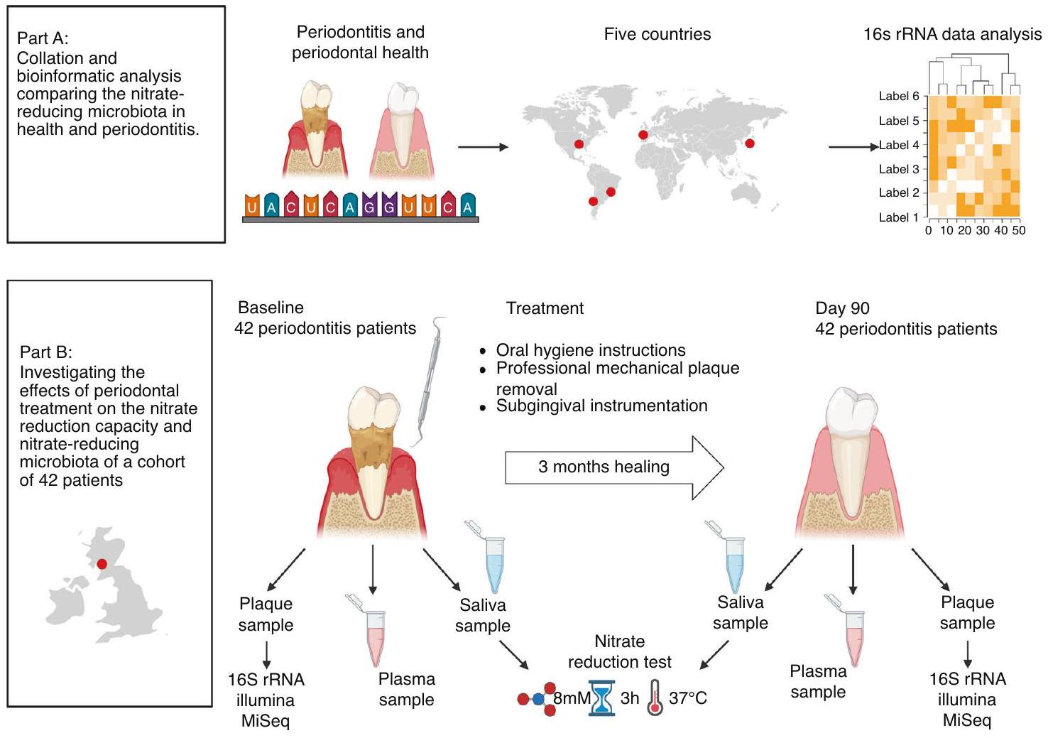
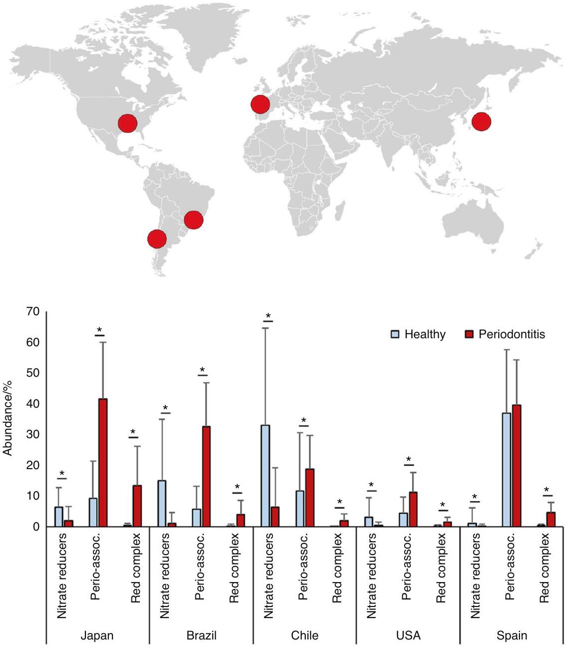
لتقييم ما إذا كان المستوى الإجمالي للبكتيريا المقللة للنترات يمكن أن ينخفض في التهاب اللثة، تم إجراء تحليل معلومات حيوية لخمس مجموعات بيانات للعثور على اختلافات في الميكروبات المقللة للنترات بين صحة اللثة والتهاب اللثة (الشكل 1A). تم اختيار دراسات حيث كانت متوسط عمق الاستكشاف (الفم بالكامل أو المواقع المأخوذة) للأفراد المصابين بالتهاب اللثة على الأقل مرتين أعلى من المجموعة الصحية (الجدول التكميلي 1). تم تنزيل مجموعات البيانات التي تحتوي على بيانات تسلسل 16S rRNA من عينات اللويحات تحت اللثة من قاعدة بيانات NCBI SRA وشملت أفرادًا من دول مختلفة (اليابان، البرازيل، تشيلي، الولايات المتحدة الأمريكية، وإسبانيا، ) مع الأخذ في الاعتبار أن بلد المنشأ يمكن أن يكون له
أثر كبير على تركيب الميكروبات. عدد الأفراد والعينات، بالإضافة إلى المعايير لوصف الصحة والتهاب اللثة في هذه الدراسات، موضحة في الجدول التكميلي 1. تم معالجة ملفات FastQ كما هو موضح سابقًا باستخدام حزمة إحصائيات DADA2 R (v1.20.0). باختصار، تم تقليم قراءات R1 وR2 حسب الطول، وتمت إزالة القراءات التي تحتوي على أكثر من 5 أخطاء. تم إزالة تكرار القراءات للحصول على متغيرات تسلسل حقيقية تم دمجها بعد ذلك (الحد الأدنى من التداخل 15 نقطة أساسية) وتم التعليق عليها في قاعدة بيانات SILVA v.138.1. تم تصنيف البكتيريا كمنتجة للنتريت أو مقللة للنترات مؤكدة بناءً على روسيير وآخرون. بالإضافة إلى ذلك، تم تصنيف الأنواع كمرتبطة بالتهاب اللثة، بما في ذلك المجمعات الحمراء والبرتقالية التي حددها سوكرانسكي وآخرون. وبكتيريا مرتبطة بالتهاب اللثة التي حددها بيريز-تشابارو وآخرون. أو مجرد المجمع الأحمر. يمكن العثور على القائمة الدقيقة للأنواع في المجموعات الأربعة (منتجات النتريت، مقللات النترات المؤكدة، المرتبطة بالتهاب اللثة، والمجمع الأحمر) في الجدول التكميلي 2.
تركيب البكتيريا في مرضى التهاب اللثة قبل وبعد العلاج اللثوي
لدراسة تأثير العلاج اللثوي على الميكروبات المقللة للنترات، تم استخدام البيانات والعينات من دراسة تم وصفها سابقًا بواسطة دافيسون وآخرون. وجونستون وآخرون. تم إجراء الدراسة وفقًا لإعلان هلسنكي (2013) وحصلت على موافقة أخلاقية (لجنة الأخلاقيات البحثية في لندن-ستانمور، المرجع: 14/LO/2064). تم تجنيد المرضى في مستشفى غلاسكو لطب الأسنان وتم تعريف التهاب اللثة على أنه عمق جيوب استكشافية على 2 أو أكثر من الأسنان في مواقع غير متجاورة باستثناء الأضراس الثالثة. وقع جميع المرضى على موافقة مستنيرة كتابية قبل المشاركة. تم وصف معايير الإدراج الأخرى وجمع العينات في جونستون وآخرون. باختصار، تم تضمين 42 مريضًا وتلقوا علاجًا لثويًا، بما في ذلك إزالة اللويحات الميكانيكية الاحترافية (PMPR) وأدوات تحت اللثة في جميع المواقع التي تتطلب ذلك (المشار إليها سابقًا بالعلاج اللثوي غير الجراحي، NSPT )، بواسطة فني أسنان ذو خبرة واحدة في زيارة واحدة إلى ست زيارات. أظهر التحليل اللاحق لهؤلاء الأفراد باستخدام إرشادات التهاب اللثة الحديثة، أن الغالبية تم تشخيصها بالتهاب اللثة العام ( )، معظمها من المرحلة الثانية أو الثالثة ( ). تم أخذ عينات من اللويحات تحت اللثة، واللعاب المتساقط، وعينات البلازما قبل أي زيارات علاجية (الخط الأساسي، BL) و90 يومًا بعد آخر زيارة علاجية (اليوم 90، D90). تم اختيار فترة 90 يومًا بناءً على الوقت اللازم للشفاء وإعادة تأسيس المجتمعات تحت اللثة، مدعومًا بإرشادات العلاج الحالية. في اليوم 90، تحسنت معايير اللثة بشكل كبير (على سبيل المثال، انخفضت مساحة السطح الملتهب للثة، PISA، بشكل كبير من وسيلة في BL إلى في D90). بالإضافة إلى ذلك، تم جمع عينات لعاب التحكم الصحي من المتطوعين في الدراسة “تفاعلات المضيف والميكروبات في صحة الفم والمرض”، رقم المشروع 2011002. حصلت دراسة التحكم الصحي على موافقة أخلاقية من لجنة الأخلاقيات في جامعة غلاسكو MVLS. تم تخزين جميع العينات في قبل الاستخدام.
تم إجراء استخراج الحمض النووي وتسلسل إلومينا في مؤسسة FISABIO (فالنسيا، إسبانيا) كما هو موضح سابقًا بواسطة جونستون وآخرون. تم استخراج الحمض النووي من عينات تحت اللثة (الخط الأساسي واليوم 90) باستخدام مجموعة عزل الحمض النووي MagNA Pure LC (روش داياغنوستكس، مانهايم، ألمانيا) مع إضافة خطوة تحلل كيميائية باستخدام كوكتيل إنزيمي يحتوي على الليزوزيم، موتانوليسين وليزوسافين، وفقًا لروسير وآخرون. كما هو موضح سابقًا بواسطة جونستون وآخرون. تم قياس تركيزات الحمض النووي باستخدام جهاز QubitTM 3 فلورومتر (ثيرموفيشر، وولثام، ماساتشوستس، الولايات المتحدة الأمريكية). تم إعداد مكتبة أمبليكون إلومينا وفقًا لبروتوكول إعداد مكتبة تسلسل الجين 16S rRNA (الجزء #15,044,223 Rev. A). كانت تسلسلات البرايمر المستخدمة في هذا البروتوكول؛ Illumina_16S_341F (TCGTCGGCAGCGTCAGATGTGTATAAGAGACAGCCTACGGGNGGCW GCAG) وIllumina_16S_805R (GTCTCGTGGGCTCGGAGATGTGT ATAAGAGACAGGACTACHVGGGTATCTAATCC) التي تستهدف
قدرة تقليل النترات في الميكروبات الفموية متأثرة في…
روسير وآخرون.
منطقة 16S V3 وV4. بعد التضخيم، تم تسلسل الحمض النووي باستخدام جهاز تسلسل إلومينا MiSeq وفقًا لتعليمات الشركة المصنعة باستخدام بروتوكول نهاية مزدوجة متطابقة. للتصنيف الضريبي، تم الحصول على جدول متغير تسلسل الأمبليكون (ASV) باستخدام خط أنابيب DADA2 في تم تعيين التصنيف الضريبي من خلال المقارنة مع قاعدة بيانات SILVA، حيث تم استخدام المصنف البايزي الساذج لتعيين التسلسلات على مستوى الأنواع. في الدراسة الحالية، يتم تقديم تحليل جديد يتم فيه تصنيف الأنواع البكتيرية كـ مقللات نترات مؤكدة أو منتجات نتريت (الجدول التكميلي 2).
qPCR لروثيا في اللويحات تحت اللثة
تم استخدام الحمض النووي من اللويحات تحت اللثة لقياسات PCR الكمي (qPCR) لتحديد الكمية الإجمالية لخلايا روثيا. على وجه التحديد، تم تضخيم جين اختزال النترات narG لروثيا كما هو موضح بواسطة روسير وآخرون. تم تصميم تسلسلات البرايمر لتكون محددة لنوع روثيا، باستخدام مناطق محفوظة من narG من روثيا موكليجينوزا، . دنتوكاريوسا، و . أيريا. كانت تسلسلة البرايمر الأمامية 5′-ACA CCA TYA AGT ACT ACGG-3′ والعكس -TAC CAG TCG TAG AAG CTG-3′. تمت إضافة تفاعلات لكل بئر من لوحة qPCR، تتكون من من مزيج Light Cycler 480 SYBR Green I Master (روش لايف ساينس، بنزبرغ، ألمانيا)، من كل برايمر محدد ( ), ماء و من الحمض النووي القالب (الحمض النووي المعزول من عينات اللويحات تحت اللثة). تمت إضافة كل عينة في نسختين، وتمت القياسات باستخدام نظام PCR الزمني الحقيقي Light Cycler 480 (روش لايف ساينس) مع الشروط التالية: لمدة دورات من لمدة لمدة 20 ثانية، و لمدة 25 ثانية. تمت إضافة ضوابط سلبية، بالإضافة إلى منحنى قياسي، يتكون من سلسلة تخفيف من مزيج الحمض النووي المتساوي المولية من ثلاثة أنواع روثيا (R. موكليجينوزا DSM-20746، R. دنتوكاريوسا DSM-43762، R. أيريا DSM-14556) تم قياسها باستخدام جهاز QubitTM 3 فلورومتر (ثيرموفيشر). بناءً على أحجام الجينوم، تم حساب عدد خلايا روثيا، مع افتراض وجود نسخة واحدة من جين narG لكل خلية. لم يتبقَ أي DNA في عينات خمسة أفراد من أصل 42 فردًا بعد إجراء تسلسل الحمض النووي، مما أدى إلى بيانات qPCR لـ 37 فردًا.
اختبار تقليل النترات في اللعاب
معدل النخر في المرضى الذين يعانون من أمراض اللثة قبل وبعد العلاج اللثوي ) والأشخاص الأصحاء ( تم تحديد ذلك عن طريق إذابة عينات اللعاب على الثلج وحضانتها لمدة 3 ساعات عند في حضور نترات. من أجل هذا، من الماء مع تم إضافة نترات الصوديوم (سيغما) إلى من اللعاب في أنبوب إيبندورف.
قياسات النترات والنيتريت ودرجة الحموضة في اللعاب
تم استخدام جهاز RQflex 10 Reflectoquant (ميرك ميلبورو، برلنغتون، ماساتشوستس، الولايات المتحدة الأمريكية) لقياس النترات والنتريت ودرجة الحموضة في اللعاب كما وصفه روسيير وآخرون. كانت شرائط الاختبار (Reflectoquant، Merck Millipore) للنترات تتراوح بين شرائط النيتريت تتراوح من ، وشرائط قياس الرقم الهيدروجيني تتراوح من تم تأكيد دقة هذه الطرق باستخدام محلولات قياسية (ميرك ميليبور) ذات تركيزات معروفة من النترات والنيتريت أو مستويات pH. بالنسبة لقياسات pH، تم استخدام اللعاب غير المخفف. أما بالنسبة لقياسات النترات والنيتريت، فقد تم استخدام اللعاب مباشرة أو تخفيفه بمعدل 5-10 أضعاف حسب تركيزات المركبات. تم إضافة خمسة عشر ميكروليتراً من اللعاب (المخفف) إلى كل من الرقعتين التفاعليتين على الشريط، وتم إزالة السائل الزائد عن طريق إمالة جانب الشريط على مناديل.
قبل قياسات النترات، تم معالجة السائل الفائق المخفف بحمض الأميدوسلفونيك (سيغما-ألدريتش) بناءً على تعليمات الشركة المصنعة. من أجل ذلك، تم خلط من السائل الرائق المخفف مع محلول حمض الأميدوسولفوريك تم إجراء قياسات النترات والنيتريت ودرجة الحموضة قبل الحضانة في المختبر (المستويات الأساسية) وبعد 3 ساعات من الحضانة لتحديد NRC (المستويات بعد الحضانة).
قياسات النترات والنيتريت في البلازما
تم تحديد مستويات النترات والنيتريت في البلازما القاعدية قبل وبعد العلاج اللثوي باستخدام الكيمياء الضوئية المعتمدة على الأوزون كما وصفه ليدل وآخرون. لقياس نيتريت البلازما، مادة ثلاثي اليود تم وضع كمية من مادة مضادة للرغوة في وعاء تطهير تم تسخينه إلى تم إنتاج منحنى قياسي عن طريق حقن محاليل النيتريت ( ) وعينة تحكم ( ). بعد ذلك، تم إذابة عينات البلازما في حمام مائي عند لمدة 3 دقائق، و تم حقن العينة في وعاء التطهير بشكل مزدوج. ثم تم قياس تركيز NO الذي تم تفكيكه خلال التفاعل بواسطة محلل NO (Sievers NOA 280i، Analytix، المملكة المتحدة)، لقياس نترات البلازما، وكاشف الفاناديوم، و تم وضع كمية من مادة مضادة للرغوة في وعاء التطهير الزجاجي، وتم تسخينها إلى تم إنتاج منحنى قياسي عن طريق حقن محاليل النترات ( ) وعينة تحكم ( تم إذابة عينات البلازما وإزالة البروتينات. بعد ذلك، تم حقن عينة في وعاء التطهير بشكل مزدوج، وتم حساب نترات البلازما كما تم وصفه سابقًا لاختبار النيتريت.
التحليل الإحصائي
تم إجراء تحليل إحصائي للنترات والنيتريت في اللعاب والبلازما وخلايا روثيا في اللويحة تحت اللثة (المحددة بواسطة qPCR) باستخدام اختبار ويلكوكسون غير المعلمي باستخدام برنامج IBM SPSS للإحصائيات (الإصدار 27) أو GraphPad (الإصدار 9.5.1) واعتُبر ذا دلالة إحصائية عند -قيمة .
لتحليل المجموعات البكتيرية (أي، منتجي النيتريت، المخفضين المؤكدين للنترات، المرتبطين بالتهاب اللثة، والمجمع الأحمر)، تم استخدام لغة البرمجة R (الإصدار 3.4+) للحسابات الإحصائية. تم توحيد وفرة الأنواع باستخدام ANCOM-BC. تمت مقارنة مجموعات البيانات التركيبية القياسية للبكتيريا في مجموعات مختلفة باستخدام اختبارات رتبة ويلكوكسون الموقعة (دالة Wilcox.test من مكتبة stats في R) واعتبرت ذات دلالة إحصائية عند التعديل. -قيمة (أي، تم تصحيحها للمقارنات المتعددة باستخدام معدل الاكتشاف الخاطئ (FDR) لبنجاميني-هوشبرغ بنسبة 5%).
تم تحديد الارتباطات داخل وبين الوفرة النسبية لمجموعات البكتيريا والمعلمات الفسيولوجية باستخدام معامل سبيرمان (دالة cortest من مكتبة stats في R)، جنبًا إلى جنب مع التعديلات المرتبطة. -القيم. بخلاف معلمات هذه الدراسة، توجد ارتباطات مع السيتوكينات اللعابية [عامل نخر الورم ألفا (TNFa)، إنترلوكين-6 (IL-6) وإنترلوكين-1 (IL-1 )] ومعايير سريرية [عمق جيوب اللثة (PPD)، مستوى الارتباط السريري (CAL)، درجة النزيف في الفم بالكامل (FMBS)، درجة اللويحات في الفم بالكامل (FMPS) وPISA] التي حصل عليها جونستون وآخرون. تم استكشافها.
تم إنشاء الأشكال 1 و 2 والشكل التكميلي 1 في Microsoft Excel و/أو باستخدام BioRender، وتم تجميع جميع الأشكال الأخرى باستخدام GraphPad PRISM (الإصدار 9.5.1).

النتائج

مستويات بكتيريا تقليل النترات لدى مرضى اللثة تم تحليل مجموعة بيانات من خمس دول مختلفة، حيث تم إجراء تسلسل جين 16S rRNA لعينات تحت اللثة (توجد المعلومات السريرية للأفراد في كل مجموعة بيانات في الجدول التكميلي 1). شملت هذه المجموعات أفرادًا من اليابان ( عينات من التهاب اللثة و10 عينات صحية)، إسبانيا ( عينات التهاب اللثة و 22 عينة صحية)، الولايات المتحدة الأمريكية ( عينات التهاب اللثة و29 عينة صحية)، البرازيل ( عينات من التهاب اللثة و21 عينة صحية)، وتشيلي ( تم العثور على عينات من التهاب اللثة و 17 عينة صحية). كما هو متوقع، تم العثور على نسبة أعلى من البكتيريا المعقدة الحمراء المرتبطة مباشرة بالتهاب اللثة في المرضى المصابين بأمراض اللثة مقارنة بالأفراد الأصحاء في جميع البلدان الخمسة (الشكل 2). فيما يتعلق بالميكروبات المنزعة للنيتروجين، كانت النسبة النسبية للبكتيريا المخفضة للنترات تختلف بين مجموعات البيانات. بغض النظر عن
الشكل 1 نظرة عامة على الدراسة. الجزء أ: تحليل المعلومات الحيوية مقارنةً بالصحة والتهاب اللثة. بيانات تسلسل 16S rRNA لخمس دراسات من خمس دول مختلفة (اليابان، البرازيل تشيلي الولايات المتحدة الأمريكية وإسبانيا، تم تحليل عينات لكل دراسة باستخدام خط أنابيب Dada2 لمقارنة البكتيريا المختزلة للنترات في الصحة والتهاب اللثة. الجزء ب) تحليل عينات دراسة علاج التهاب اللثة. تم جمع عينات من اللويحات تحت اللثة، واللعاب، والبلازما من 42 مريضًا يعانون من التهاب اللثة في غلاسكو (اسكتلندا، المملكة المتحدة) قبل 90 يومًا وبعد العلاج اللثوي. تم تحديد تركيبة البكتيريا في اللويحات تحت اللثة باستخدام تسلسل Illumina لجين 16S rRNA، وتم تحديد كمية الجنس المختزل للنترات، روثيا، بواسطة qPCR. تم إجراء قياسات للنترات والنيتريت في اللعاب والبلازما وتم تحديد قدرة اختزال النترات في اللعاب (NRC) بعد ثلاث ساعات من الحضانة في المختبر ومقارنتها مع NRC لـ 15 فردًا صحيًا.
كانت نسبة البكتيريا المقللة للنترات أقل بكثير في المرضى الذين يعانون من أمراض اللثة في جميع الحالات مقارنة بمستويات الأفراد الأصحاء في كل بلد. في جميع البلدان) (الشكل 2). عند النظر في جميع البكتيريا المعروفة التي تنتج النيتريت (بعضها قد ينتج النيتريت بطرق أخرى غير اختزال النترات)، ظل النمط والأهمية الإحصائية (الشكل التكميلي 1).
مستويات البكتيريا المختزلة للنترات قبل وبعد علاج اللثة
أظهر التركيب البكتيري لعينات تحت اللثة قبل (BL) وبعد (D90) العلاج اللثوي نمطًا متسقًا عبر الأفراد. بالنسبة لخط الأساس، كان هناك زيادة ملحوظة في البكتيريا القادرة على إنتاج النيتريت (الشكل التوضيحي 2)، بما في ذلك البكتيريا المؤكدة التي تقلل النترات (الشكل 3A)، مع نمط معكوس للجراثيم المسببة لأمراض اللثة، بما في ذلك الأنواع الثلاثة من المجمع الأحمر والقائمة الموسعة من البكتيريا المرتبطة بالتهاب اللثة، كما أبلغ عنه جونستون وآخرون سابقًا. هذا يشير إلى أن القدرة على استقلاب النترات بواسطة اللويحات تحت اللثة تتحسن بعد العلاج اللثوي. تم تأكيد الزيادة في مستويات البكتيريا المخفضة للنترات بواسطة qPCR باستخدام بادئات محددة للعلامة الحيوية المخفضة للنترات روثيا، والتي أظهرت زيادة كبيرة في الخلايا لكل عينة (أي، المستويات المطلقة) (الشكل 3B). كما بدت المستويات المعدلة لهذا الكائن (خلايا روثيا المحددة بواسطة qPCR لكل نانوغرام من الحمض النووي) تزداد، لكن هذه الزيادة لم تكن ذات دلالة إحصائية ( )(الشكل 3C). من الجدير بالذكر أنه تم إظهار سابقًا أن الوفرة النسبية لروثيا التي تم تحديدها بواسطة تسلسل إيلومينا قد زادت بعد العلاج اللثوي. كلا من المستويات المطلقة والمعدلة لروثيا التي تم تحديدها بواسطة qPCR
ترتبط بوفرة البكتيريا المخفضة للنترات ( و , على التوالي، كلاهما عند تجميع BL و D90) (البيانات التكميلية).
عند تجميع عينات BL و D90، وُجدت علاقة سلبية كبيرة بين مستويات البكتيريا المخفضة للنترات ومستويات البكتيريا المرتبطة بالتهاب اللثة ( )(البيانات التكميلية). عند النظر فقط في عينات BL، لم تكن هذه العلاقة ذات دلالة إحصائية (الشكل 3D)، ولكن في عينات D90، كانت هناك علاقة سلبية واضحة بين البكتيريا المخفضة للنترات والبكتيريا المرتبطة بالتهاب اللثة ( )(الشكل 3E). كانت هناك علاقتان سلبيتان مثيرتان للاهتمام عند تجميع عينات BL و D90 بين البكتيريا المخفضة للنترات و FMPS ( , )، بالإضافة إلى المستويات المطلقة أو الإجمالية لروثيا و FMBS (كلاهما إلى 0.33، كلاهما ). أخيرًا، كانت مجموعة جميع البكتيريا المنتجة للنترات (الجدول التكميلية 2) مرتبطة سلبًا مع IL-1 اللعابي عند الخط الأساسي ( )(البيانات التكميلية).

NRC في الصحة والتهاب اللثة

تم اعتبار استخدام النترات خلال حضانة عينات اللعاب مع النترات المضافة لفترة ثلاث ساعات تقديرًا لـ NRC في كل فرد. كشفت البيانات أن تقليل النترات كان أعلى في الأفراد الأصحاء مقارنة بالمرضى اللثويين عند الخط الأساسي (الشكل 4A): في مرضى التهاب اللثة، لم تتغير مستويات النترات بشكل كبير خلال فترة الحضانة التي استمرت 3 ساعات ( )، بينما في الأفراد الأصحاء، حدثت ( ). وفقًا لبيانات تسلسل 16 S rRNA التي أظهرت أن البكتيريا المخفضة للنترات زادت في اللويحات تحت اللثة، فإن NRC
الشكل 2 أكد البكتيريا المخفضة للنترات في التهاب اللثة والصحة. تظهر الرسوم البيانية الشريطية الوفرة النسبية للبكتيريا في عينات اللويحات تحت اللثة من دول مختلفة، كما تم تقديرها بواسطة تسلسل عالي الإنتاجية لجين 16 S rRNA. تم تجميع البكتيريا في أنواع مخفضة للنترات مؤكدة، مرتبطة بالتهاب اللثة أو معقدة حمراء وفقًا لـ Rosier وآخرون. , بيريز-تشابارو وآخرون. , وسوكرا نسكي وآخرون. على التوالي (تم إدراج الأنواع البكتيرية في كل مجموعة في الجدول التكميلية 2). تم مقارنة الأفراد الأصحاء (الأشرطة الزرقاء) مع الأفراد المصابين بالتهاب اللثة (الأشرطة الحمراء). في الشكل التكميلية 1، يتم عرض جميع البكتيريا المعروفة المنتجة للنترات (بعضها قد ينتج النترات عبر مسارات أخرى غير تقليل النترات) بنفس النمط وتظهر فرقًا كبيرًا بين الصحة والتهاب اللثة. تشمل مجموعات البيانات أفرادًا من اليابان وإسبانيا والولايات المتحدة والبرازيل وتشيلي ( لكل مجموعة بيانات، انظر الجدول التكميلية 1 لمزيد من المعلومات). يجب ملاحظة أن مستويات مجموعات البكتيريا في دراسات مختلفة تتأثر بالمعايير المستخدمة لوصف التهاب اللثة، والعوامل الإقليمية والمضيف، بالإضافة إلى طرق استخراج الحمض النووي أو تقنيات التسلسل المستخدمة في الدراسات الأصلية. *تم تعديل من بيانات التركيب القياسية بواسطة ANCOM-BC وتمت مقارنتها باختبار ويلكوكسون
في المرضى اللثويين تم استعادتها بعد العلاج اللثوي، مع انخفاض كبير في مستويات النترات بعد الحضانة ( ).
تم تأكيد النشاط المخفض للنترات (عملية معروفة لتخفيف الرقم الهيدروجيني) بشكل غير مباشر بواسطة قياسات الرقم الهيدروجيني، التي أظهرت انخفاضًا طفيفًا أكبر في الرقم الهيدروجيني لدى المرضى اللثويين عند الخط الأساسي مقارنة بعد العلاج (عند BL انخفض الرقم الهيدروجيني من 7.33 إلى 7.13، وعند D90، انخفض من 7.49 إلى 7.41، كلاهما )(الشكل 4C).
تتفق مع ما سبق، لم تختلف مستويات النترات في عينات التهاب اللثة الأساسية قبل وبعد الحضانة مع النترات (الشكل 4B). بعد العلاج اللثوي، لوحظ انخفاض كبير في النترات بعد الحضانة. نظرًا لاستهلاك النترات خلال هذه الفترة التي استمرت ثلاث ساعات، كانت مستويات النترات المنخفضة على الأرجح نتيجة لمزيد من استقلاب النترات. في الأفراد الأصحاء، زادت مستويات النترات بعد الحضانة، لكن هذه الزيادة لم تكن ذات دلالة إحصائية ( ) وكان هناك تركيز أولي أقل بكثير من النترات مقارنة بالتهاب اللثة قبل ( ) وبعد ( ) العلاج.
مستويات البلازما من النترات والنترات
تتفق مع مستويات اللعاب الأساسية من النترات والنترات، كشفت تركيزات النترات والنترات الأساسية في عينات الدم المجمعة من المرضى اللثويين عند الخط الأساسي و 90 يومًا بعد العلاج اللثوي عن عدم وجود اختلافات إحصائية بين هذين الوقتين (الشكل 5A، B). كانت متوسط مستويات البلازما من النترات عند BL و عند D 90 (غير مختلفة بشكل كبير). كانت مستويات النترات ( عند BL و عند D90) حوالي 350 مرة أقل من النترات (الشكل 5C). لم تكن هناك علاقات ذات دلالة إحصائية بين مجموعات البكتيريا والنترات أو النترات في البلازما. كانت أعلى علاقة سبيرمان بين المستويات المطلقة أو المعدلة لروثيا في اللويحات تحت اللثة والنترات في البلازما، لكن هذه العلاقة لم تكن ذات دلالة إحصائية (كلاهما , كلاهما عند تجميع BL و D90) (البيانات التكميلية). ستترجم زيادة NRC التي وُجدت في اللعاب مع النترات المضافة إلى مستويات أعلى من النترات أو أكسيد النيتريك في البلازما بعد تناول النترات الغذائية، والتي لم يتم تقييمها في هذه الدراسة.
الشكل 3 أكد الأنواع المخفضة للنترات والبكتيريا المرتبطة بالمرض قبل و 90 يومًا بعد العلاج اللثوي. أ. الوفرة النسبية للبكتيريا المخفضة للنترات المؤكدة، المعقد الأحمر، والبكتيريا المرتبطة بالتهاب اللثة قبل (الخط الأساسي، BL) و 90 يومًا بعد العلاج (D90) لـ 42 مريضًا بالتهاب اللثة. *تم تعديل من بيانات التركيب القياسية بواسطة ANCOM-BC وتمت مقارنتها باختبار ويلكوكسون. ب، ج. خلايا روثيا المحددة بواسطة qPCR قبل (BL) و 90 يومًا بعد (D90) العلاج لكل عينة (الكمية المطلقة، )(ب) أو المعدلة لكل نانوغرام من الحمض النووي (ج). تم تحديدها بواسطة اختبار ويلكوكسون. د، هـ. العلاقات بين وفرة مسببات الأمراض اللثوية والبكتيريا المخفضة للنترات عند الخط الأساسي (BL) و 90 يومًا بعد العلاج (D90). **تم تعديل من علاقة ترتيب سبيرمان (n.s. = غير دالة). في الشكل التكميلية 2، يتم عرض المقارنات والعلاقات لجميع البكتيريا المعروفة المنتجة للنترات (بعضها قد ينتج النترات عبر مسارات أخرى غير تقليل النترات)

النقاش

يمكن أن تؤدي مسار النترات-النترات-أكسيد النيتريك إلى فوائد قلبية وعائية وميتابولية عند تحفيزها بالنترات الغذائية. في هذه الدراسة، نوضح أن تقليل النترات (أي، التركيز الموجود في اللعاب بعد تناول الخضار) بواسطة الكائنات الحية الدقيقة في اللعاب كان معاقًا في التهاب اللثة. تم استعادة هذا NRC المعاق إلى مستويات صحية بعد العلاج اللثوي. بما يتماشى مع ذلك، نوضح أن مستويات البكتيريا المخفضة للنترات في اللويحات تحت اللثة أقل في التهاب اللثة واستعادت بعد العلاج اللثوي. تشير نتائجنا إلى أن NRC المعاق في التهاب اللثة قد يحد من التأثيرات الإيجابية لأكسيد النيتريك المشتق من النترات الغذائية. من الجدير بالذكر أن الحالات النظامية المختلفة المرتبطة بالتهاب اللثة مرتبطة أيضًا بنقص في أكسيد النيتريك (مثل الأمراض القلبية الوعائية والسكري). لذلك، يجب أن تستكشف الدراسات المستقبلية تأثير التهاب اللثة والعلاج اللثوي على المعايير القلبية الأيضية بعد تناول النترات الغذائية.
تأثير التهاب اللثة والعلاج اللثوي على الميكروبات الفموية المخفضة للنترات
لمقارنة مستويات البكتيريا المختزلة للنترات تحت اللثة في الصحة والتهاب اللثة، استخدمنا مجموعات بيانات تسلسل 16S من خمس دول مختلفة (اليابان، البرازيل، تشيلي، الولايات المتحدة، وإسبانيا). اتبعت جميع الدول نفس النمط المتمثل في انخفاض الأنواع المرتبطة بالتهاب اللثة وزيادة في البكتيريا المنتجة للنتريت، بما في ذلك الأنواع المؤكدة المختزلة للنترات. هذه النتيجة تتفق مع الدراسات السابقة. على سبيل المثال، فريس وآخرون. تمت مراجعة النتائج بشكل منهجي من
دراسات التسلسل التي تقارن اللويحات تحت اللثة في الصحة والتهاب اللثة، وكذلك التهاب اللثة قبل وبعد العلاج، وجدت أن الأنواع الشائعة المنتجة للنيتريت كانت مرتبطة بصحة اللثة (مثل: ستربتوكوكوس spp.، نيسيريا لونغاتي، نيسيريا سوبفلافا، روثيا أيريا، فيلونيلة بارفولا وجرانوليكاتيلا أديانس). بالإضافة إلى ذلك، وجد فيريس وآخرون أن روثيا كانت الجنس الأكثر ارتباطًا بصحة اللثة، ومع انخفاض الالتهاب بعد العلاج اللثوي، تليها أجناس أخرى منتجة للنيتريت (مثل: نيسيريا، أكتينوميسيس وستربتوكوكوس). تدعم هذه النتائج أيضًا التحليلات المعلوماتية الحيوية لمورك وآخرون. ربط فيلونيللا، نيسيريا، روثيا، كورينباكتيريوم، وأكتينوميسيس (جميعها أجناس تحتوي على ممثلين ينتجون النيتريت) بصحة اللثة، وتشين وآخرون. الذي أبلغ أن ستربتوكوكوس سانغوينيس، أكتينوميس نيسلونديا، روثيا أيريا، غرانوليكاتيلا أدياسينس، روثيا دنتوكاريوسا وستربتوكوكوس ميتيس (جميعها أنواع تنتج النيتريت) كانت ستة من بين أعلى سبعة أنواع مرتبطة بالصحة التي تُستخدم لحساب مؤشر اختلال الميكروبات تحت اللثة (SMDI).
تتطلب الآليات الأساسية التي يبدو أن تقليل النترات بواسطة البكتيريا الفموية مفيد لصحة الفم استكشافًا. يمكن للبكتيريا الفموية أن تقلل أيضًا النيتريت إلى أكسيد النيتريك، وهو جذر حر له خصائص مضادة للميكروبات قادرة على تثبيط الأنواع الحساسة مثل اللاهوائيات المرتبطة بالتهاب اللثة. بالإضافة إلى ذلك، يمكن أن يشير أكسيد النيتريك المستمد من نزع النيتروجين بواسطة البكتيريا الفموية مباشرة إلى خلايا الإنسان، مما يقلل الالتهاب ويحفز إنتاج المخاط الواقي وتدفق الدم. في دراستنا، كانت بكتيريا تقليل النترات مرتبطة
الشكل 4 قدرة تقليل النترات (NRC) في الصحة والتهاب اللثة. أ، ب تمثل الرسوم البيانية الشريطية تركيز النترات ( ) ونتريت ( ) من عينات اللعاب المزروعة في المختبر لمدة 3 ساعات في وجود نترات في قبل (الخط الأساسي، BL) و90 يومًا بعد العلاج اللثوي ) من مرضى اللثة، مقارنةً بالأشخاص الأصحاء ( ). تُظهر الأعمدة مستوى الحموضة (pH) خلال فترة الحضانة هذه لنفس مجموعات الأفراد. تمثل النجوم الفروقات ذات الدلالة الإحصائية (*P<0.05، ) تم تحديده بواسطة اختبار ويلكوكسون (المقترن) أو اختبار مان ويتني U (غير المقترن)
الشكل 5 نترات ونتريت البلازما قبل و90 يومًا بعد العلاج اللثوي. تُظهر البيانات من 42 مريضًا بالتهاب اللثة قبل (BL) وبعد (D90) العلاج. أ. نتريت البلازما. ب. نترات البلازما. ج. نسبة النترات/النتريت.
سلبياً مع البكتيريا المرتبطة بالتهاب اللثة، مما يدعم فكرة أن هذه الوظيفة قد تكون مهمة لصحة الفم. كانت هذه العلاقة السلبية أوضح في 42 مريضاً بالتهاب اللثة بعد العلاج اللثوي. تم وصف تقليل الالتهاب في هؤلاء المرضى بعد العلاج سابقاً من قبل جونستون وآخرين. وتكونت من تحسين واضح وملحوظ في جميع المعايير السريرية لالتهاب اللثة. في دراستنا الحديثة في المختبر، أدى إضافة النترات إلى اللويحة السنية إلى إنتاج النيتريت (وهو مؤشر على إنتاج أكسيد النيتريك)، وانخفاض في الأنواع المرتبطة بالتهاب اللثة، وانخفاض في SMDI. في دراستنا الحالية، كانت مستويات البكتيريا المختزلة للنترات في اللويحة تحت اللثة مرتبطة سلبًا مع درجة اللويحة الفموية الكاملة (FMPS)، بينما يعتبر أكسيد النيتريك أيضًا بيوفيلم.
إشارة انتشار لبعض البكتيريا التي يمكن أن تقلل من تراكم اللويحات. باختصار، قد يؤدي ضعف NRC في التهاب اللثة إلى منح ميزة انتقائية للأنواع المرتبطة بالتهاب اللثة من خلال فقدان إنتاج أكسيد النيتريك وربما آليات مضادة للميكروبات الأخرى المرتبطة بالصحة. يجب أن يتم التحقيق بشكل أكبر في تأثير استهلاك النترات وNRC للمرضى على كفاءة علاج التهاب اللثة في الدراسات المستقبلية.
أحد النتائج الرئيسية لهذه الدراسة هو أن نسبة الكورتيزول في عينات اللعاب تنخفض في حالة التهاب اللثة. يحتوي اللعاب غير المحفز على البكتيريا لكل مل التي تشتتت من المجتمعات الفموية، مما يجعل هذا عينة غير جراحية يمكن استخدامها لدراسة نشاط الميكروبات الفموية. بالإضافة إلى ذلك، من خلال التجميد
عند وعند الذوبان على الجليد، يبقى جزء كبير من البكتيريا على قيد الحياة، كما يتضح من التغيرات في المعايير الأيضية بعد 3 ساعات من الحضانة عند في هذه الدراسة (على سبيل المثال، انخفاض طفيف ولكنه مهم في درجة الحموضة بسبب تخمر البكتيريا لمكونات اللعاب). من خلال إضافة تم العثور على نشاط تقليل النترات بشكل ملحوظ في لعاب 15 فردًا صحيًا. )، وهو ما يتماشى مع البيانات السابقة التي تم الحصول عليها من مجموعات صغيرة من السكان الأصحاء ( ). من الجدير بالذكر أنه لم يتم ملاحظة أي تقليل كبير في النترات في لعاب 42 مريضًا بالتهاب اللثة قبل العلاج. ). ومع ذلك، بعد 90 يومًا من العلاج، تم استعادة NRC للأفراد الذين يعانون من التهاب اللثة ( إلى مستويات قابلة للمقارنة مع الأفراد الأصحاء. بالإضافة إلى ذلك، تم تقليل المزيد من النيتريت بعد العلاج اللثوي، مما يشير إلى أنه يمكن زيادة إنتاج أكسيد النيتريك. يمكن تفسير ذلك من خلال الملاحظة أن البكتيريا الشائعة التي تقلل النترات، والتي زادت في العدد، تمتلك أيضًا جينات تقليل النيتريت.
لم يكن لعلاج اللثة تأثير كبير على مستويات النترات والنيتريت في البلازما الأساسية، بينما لم يتم تقييم تأثير تناول النترات على البلازما في هذه الدراسة. على وجه التحديد، كانت متوسط تركيزات النترات في البلازما الأساسية لدى الأفراد الذين يعانون من التهاب اللثة في BL و عند D90 بعد العلاج. كانت مستويات النيتريت المتوسطة حوالي 350 مرة أقل ( في BL و في دراستين مع أفراد أصحاء نظامياً تم تجنيدهم في نفس الموقع (غلاسكو، اسكتلندا، المملكة المتحدة)، وُجدت مستويات مماثلة من النترات والنيتريت في حالة الصيام: بورلي وآخرون. وجدت المتوسطات لـ نترات و النيتريت في 25 فردًا، بينما ليدل وآخرون. وجدت المتوسطات لـ نترات و النتريت في 34 فردًا. من المعروف أن تناول النترات يزيد من تركيزات النترات والنتريت في البلازما عدة مرات، مما يشير إلى أنه في توقيت جمع العينات في دراستنا، لم يتم الكشف عن أي استقلاب للنيترات الغذائي. يجب أن تحدد الدراسات المستقبلية في مجموعات سكانية أكبر ما إذا كانت التهاب اللثة يمكن أن تؤثر على مستويات النيتريت والنيترات في البلازما أثناء الصيام أو بعد تناول النيترات.
باختصار، لم تتأثر المستويات الأساسية من النترات والنيتريت في البلازما واللعاب بالعلاج اللثوي، ولكن تم الكشف عن اختلاف في NRC عندما تم إضافة النترات إلى اللعاب. تركيز النترات بثمانية مليمول هو تركيز موجود في اللعاب بعد تناول الخضروات. مما يشير إلى أن التأثيرات الإيجابية الفموية والقلبية الأيضية التي تحفزها النترات الغذائية قد تتأثر في حالة التهاب اللثة. لذلك، يجب استكشاف تأثير العلاج اللثوي على المعايير الفموية والنظامية بعد تناول النترات في الدراسات المستقبلية.
كابيل وآخرون تم تحديد NRC من خلال السماح للأفراد بتمرير محلول يحتوي على نترات في أفواههم. بالإضافة إلى ذلك، يمكن قياس NRC في عينات البايوفيلم الفموي التي تم تطبيعها حسب الوزن أو تركيزات الحمض النووي. يجب أن تقارن الدراسات المستقبلية بين طرق مختلفة لتحديد NRC، حيث يمكن أن يمثل هذا علامة مفيدة لصحة اللثة، وكيفية ارتباطه بالمعايير النظامية.
تماشيًا مع نتائج NRC، أظهرنا أن الميكروبات المخفضة للنترات تحت اللثة تنخفض في التهاب اللثة (مقارنة بصحة اللثة) وتزداد بعد العلاج اللثوي. على وجه التحديد، أظهرنا ذلك بالنسبة للوفرة النسبية للبكتيريا المعروفة المنتجة للنتريت، بما في ذلك البكتيريا المخفضة للنترات المؤكدة. الفرق بين هاتين المجموعتين هو أن العزلات المنتجة للنتريت يتم اكتشافها عن طريق حضانة البكتيريا مع النترات وقياس إنتاج النتريت. في معظم الأوقات، ينتج هذا النتريت عن تقليل النترات، ولكن هناك مسارات أخرى يمكن أن تؤدي إلى إنتاج النتريت (على سبيل المثال، أكسدة أكسيد النيتريك أو الأمونيوم). لذا يجب تأكيد NRC للبكتيريا الفموية من خلال قياسات فسيولوجية للنترات. في دراستنا، أظهرت المجموعتان (المنتجة للنتريت والمخفضة للنترات المؤكدة) دلالة إحصائية مشابهة (أي، بقيت عندما
نقارن بين هذه المجموعات بين التهاب اللثة والصحة أو قبل وبعد العلاج اللثوي). بالإضافة إلى ذلك، أكدت قياسات qPCR للعلامة الحيوية المخفضة للنترات والنوع المرتبط بصحة اللثة روثيا قبل وبعد علاج التهاب اللثة زيادة في خلايا روثيا لكل عينة ( ). في دراستنا السابقة، وُجد أن مستويات روثيا في هذه العينات، التي تم تحديدها بواسطة تسلسل إيلومينا لجين 16 S rRNA، كانت أعلى بعد العلاج. تؤكد بياناتنا أن البكتيريا المخفضة للنترات تنخفض في اللويحة تحت اللثة في ظل ظروف الالتهاب والخلل الميكروبي المرتبطة بالتهاب اللثة. ومن المثير للاهتمام أن مستويات روثيا ارتبطت جيدًا بنسبة البكتيريا المخفضة للنترات (ورقة بيانات إضافية)، مما يشير إلى أن هذا الجنس هو علامة حيوية محتملة للأنواع المخفضة للنترات. أظهر درجة التهاب اللثة، كما هو موضح بواسطة درجة نزيف الفم الكامل (FMBS)، ارتباطًا سلبيًا مع مستويات روثيا المحددة بواسطة qPCR. وهذا يدعم دورًا محتملاً لروثيا في صحة اللثة وتقييمه كمنتج بروبيوتيك محتمل للثة، كما تم اقتراحه مؤخرًا.
الآثار المحتملة لتقليل NRC على الصحة النظامية تشير بياناتنا إلى أن التهاب اللثة قد يحد من تقليل النترات في وجود تركيزات النترات الموجودة بعد تناول الخضروات. إن تقليل النترات بواسطة البكتيريا الفموية هو خطوة أساسية في مسار النترات-النتريت-أكسيد النيتريك الذي يساهم في مستويات أكسيد النيتريك النظامية. عند تعقيم نسبة كبيرة من الميكروبات الفموية باستخدام الكلورهيكسيدين في الأفراد الصائمين، تم إعاقة NRC للميكروبات الفموية، وانخفضت مستويات النتريت في البلازما وزاد ضغط الدم. على العكس، فإن تحفيز مسار النترات-النتريت-أكسيد النيتريك عن طريق استهلاك عصائر الخضروات أو أملاح النترات يمكن أن يؤدي إلى انخفاض ضغط الدم وتحسين وظيفة البطانة. ارتبط تناول النترات أيضًا بعكس متلازمة الأيض وبتأثيرات مضادة للسكري، بينما وُجد أن استخدام غسول الفم المتاح بدون وصفة طبية يرتبط بتطور السكري وما قبل السكري. يبدو إذن أن الظروف التي يتم فيها العثور على نقص في أكسيد النيتريك، تستفيد من تحفيز تقليل النترات بواسطة الميكروبات الفموية. ومن المRemarkably، يرتبط التهاب اللثة بأمراض القلب والأوعية الدموية والسكري. لذا يجب أن تحدد الدراسات المستقبلية ما إذا كان التهاب اللثة يمكن أن يساهم في أمراض القلب والأوعية الدموية والسكري وغيرها من الحالات النظامية المرتبطة بأكسيد النيتريك من خلال الحد من الآثار المفيدة لأكسيد النيتريك المشتق من النترات الغذائية. تدعم هذه الإمكانية النتيجة التي تفيد بأن القدرة الجينية للويحة تحت اللثة على إنتاج النتريت مرتبطة بمستويات أقل من خطر القلب والأوعية الدموية.
تعتبر تسمم الحمل مثالًا آخر على حالة مرتبطة بكل من انخفاض توفر أكسيد النيتريك وإلتهاب اللثة، بينما أشارت دراسة حديثة إلى أن هذه الحالة كانت مرتبطة بانخفاض في البكتيريا المخفضة للنترات الفموية. في هذا الصدد، يبدو أن العلاج اللثوي يقلل من المضاعفات لدى النساء الحوامل المصابات بالتهاب اللثة، ربما عن طريق تقليل التعرض الالتهابي والبكتيري للأجنة. في دراستنا، وجدنا أن العلاج اللثوي استعاد NRC إلى مستويات صحية في وجود النترات. يجب استكشاف التأثير المحتمل لتحسين تقليل النترات الغذائية الناتج عن العلاج اللثوي على تسمم الحمل. يرتبط العلاج اللثوي أيضًا بتحسين طويل الأمد لوظيفة البطانة. في دراسة حديثة على الفئران، أظهر أن النترات غير العضوية تحمي من وتستطيع جزئيًا عكس وظيفة البطانة المتأثرة مسبقًا بسبب التهاب اللثة من خلال استعادة مستويات النتريت، وبالتالي، مستويات أكسيد النيتريك. تظهر هذه النتائج أن كل من استهلاك النترات والعلاج اللثوي يمكن أن يؤدي إلى تحسين وظيفة البطانة. لذا يجب استكشاف تناول النترات كعلاج مساعد لتحسين المعايير النظامية لدى المرضى المصابين بالتهاب اللثة.

قيود الدراسة وآفاق المستقبل

تتمثل قيود هذه الدراسة في أن الأفراد لم يكونوا صائمين عند التبرع بعينات اللعاب والبلازما ولم يتلقوا تعليمات بشأن عادات أخرى قد تتداخل مع مستويات النترات والنتريت في اللعاب والبلازما (مثل عادات التمرين، ومستويات اللياقة البدنية، وأشعة الشمس، ومرحلة الدورة الشهرية، واستهلاك الماء). نظرًا للتأثير الكبير للنظام الغذائي على مستويات النترات والنتريت في البلازما واللعاب، لم يكن تصميم الدراسة مثاليًا لرؤية كيف يؤثر التهاب اللثة على المستويات الأساسية لهذه الجزيئات. قد يفسر هذا لماذا لم تكن مستويات اللعاب والبلازما الأساسية من النترات والنتريت مختلفة بشكل كبير قبل وبعد العلاج اللثوي. ومع ذلك، عند حضانة اللعاب مع النترات، وُجدت اختلافات في NRC لمرضى التهاب اللثة قبل وبعد العلاج وبين مرضى التهاب اللثة قبل العلاج والأفراد الأصحاء. في vivo، يعني هذا أن تركيزات النترات اللعابية التي تم الحصول عليها من استهلاك الخضروات تقل بكفاءة أقل في التهاب اللثة، مما يقلل من الآثار المفيدة للنترات الغذائية. في الدراسة الحالية، لم نجمع عينات دم بعد ظروف توفر النترات العالية، وهو الوقت الذي يُتوقع أن يكون فيه تأثير مجتمع البكتيريا المخفضة للنترات الصحي أكثر صلة. لذا، نقترح أن تختبر الدراسات المستقبلية ما إذا كان التعرض للنترات (على سبيل المثال، عن طريق استهلاك عصير الشمندر) له تأثيرات مختلفة على مستويات النترات والنتريت في اللعاب والبلازما، فضلاً عن آثارها النظامية المستمدة (مثل انخفاض ضغط الدم) لدى مرضى التهاب اللثة (قبل وبعد العلاج اللثوي) والأفراد الأصحاء. أخيرًا، في هذه الدراسة، تم دراسة تكوين اللويحة تحت اللثة لأنها الأكثر تأثرًا بالتهاب اللثة. بالإضافة إلى ذلك، من المعروف أن كميات نوع معين في عينات فموية مختلفة قد ترتبط ببعضها البعض (على سبيل المثال، يرتبط بكتيريا Porhyromonas gingivalis في اللويحة تحت اللثة مع P. gingivalis في اللعاب )، مما يشير إلى أن التغيرات في اللويحة تحت اللثة تؤثر على مجتمعات أخرى إلى حد ما. في الأعمال المستقبلية، يجب استكشاف تأثير هذه الحالة على تكوين مجتمعات ميكروبية أخرى تشارك في تقليل النترات (مثل ميكروبات اللسان).

ملاحظات ختامية

في الختام، تظهر بياناتنا أن التهاب اللثة يضر بالميكروبات المخفضة للنترات ويعيق تقليل تركيزات النترات الموجودة في اللعاب بعد تناول الخضروات. ومع ذلك، يتم استعادة القدرة المخفضة للنترات إلى مستويات صحية بعد العلاج اللثوي. نظرًا لأن القدرة المخفضة للنترات المنقوصة أو المفقودة، على سبيل المثال، بواسطة غسول الفم المطهر، تؤدي إلى انخفاض مستويات النتريت في البلازما، يمكن أن تشير أمراض اللثة إلى نقص في النيتريت الدوري. يجب أن تحقق الدراسات المستقبلية فيما إذا كان، بالإضافة إلى الرابط الفموي الجهازي الناتج عن التأثيرات الالتهابية، يمكن أن تسهم قدرة تقليل النترات المعاقة لدى مرضى اللثة في المزيد من الحالات الجسدية المتعددة التي تتأثر بتوافر أكسيد النيتريك. مع الأخذ في الاعتبار أن تناول النترات يزيد من البكتيريا المرتبطة بصحة اللثة، يجب تحديد ما إذا كانت التدخلات الغذائية الغنية بالنترات يمكن أن تحسن نتائج علاج اللثة، بما في ذلك المعايير الفموية والجهازية.

توفر البيانات

تم إيداع قراءات التسلسل لـ 42 مريضًا بالتهاب اللثة قبل (BL) وبعد (D90) العلاج في أرشيف قراءات التسلسل NCBI (SRA) تحت BioProject PRJNA725103. أي بيانات إضافية متاحة عند الطلب المعقول من المؤلف المقابل.

الشكر والتقدير

يود المؤلفون أن يعبروا عن امتنانهم للمساعدة المقدمة من مرفق البحث السريري بمستشفى غلاسكو لطب الأسنان، وخاصة كلير براون وديبي مكينزي. كما نود أن نشكر ساندرا غارسيا-استيبان في FISABIO على مساعدتها في التجربة المعملية.

التمويل

تم تمويل دراسة علاج التهاب اللثة من خلال منح من EU Marie Curie ITN RAPID (رقم المنحة 290246)، Versus Arthritis (رقم المنحة 20823)، وBBSRC (BB/P504567/1). ويليام جونستون مدعوم من منحة دراسية من جامعة غلاسكو وDentsply Sirona (رقم المشروع 300881). تم دعم A.M. من خلال منحة من صندوق التنمية الإقليمي الأوروبي ووزارة العلوم والابتكار والجامعات الإسبانية بالمرجع RTI2018-102032-B-I00، بالإضافة إلى منحة من وكالة الابتكار في فالنسيا بالمرجع INNVAL20/19/006. تم دعم BR من خلال زمالة FPI من وزارة العلوم والابتكار والجامعات الإسبانية بالمرجع Bio2015-68711-R.

معلومات إضافية

المعلومات التكميلية النسخة الإلكترونية تحتوي على مواد تكميلية متاحة علىhttps://doi.org/10.1038/s41368-023-00266-9.
المصالح المتنافسة: أعلن المؤلفون عن وجود تضارب محتمل في المصالح فيما يتعلق بالبحث، والتأليف، و/أو نشر هذه المقالة: A.M. وB.T. Rosier هما مخترعان مشتركين في طلب براءة اختراع معلق مملوك لمعهد FISABIO، الذي يحمي استخدام النترات كمواد مسببة للبروبيوتيك وبعض بكتيريا تقليل النترات كالبروبيوتيك. المؤلفون الآخرون لا يعلنون عن أي مصالح متنافسة.
بيان الموافقة الأخلاقية: تم إجراء الدراسة وفقًا لإعلان هلسنكي (2013). بالنسبة للدراسة التي تشمل مرضى التهاب اللثة، منح لجنة الأخلاقيات البحثية في لندن-ستانمور (المرجع: 14/LO/206) التابعة للخدمة الصحية الوطنية (NHS، إنجلترا، المملكة المتحدة) الموافقة الأخلاقية. بالنسبة للدراسة التي تشمل عينات اللعاب من المرضى الأصحاء، منحت لجنة الأخلاقيات MVLS (رقم المشروع: 2011002) من جامعة غلاسكو (اسكتلندا، المملكة المتحدة) الموافقة الأخلاقية.

REFERENCES

  1. Theilade, E. The non-specific theory in microbial etiology of inflammatory periodontal diseases. J. Clin. Periodontol. 13, 905-911 (1986).
  2. Rosier, B. T., Marsh, P. D. & Mira, A. Resilience of the oral microbiota in health: mechanisms that prevent dysbiosis. J. Dent. Res. 97, 371-380 (2018).
  3. Marsh, P. D. Are dental diseases examples of ecological catastrophes? Microbiology 149, 279-294 (2003).
  4. Mira, A., Simon-Soro, A. & Curtis, M. A. Role of microbial communities in the pathogenesis of periodontal diseases and caries. J. Clin. Periodontol. 44, S23-S38 (2017).
  5. Feres, M., Retamal-Valdes, B., Gonçalves, C., Cristina Figueiredo, L. & Teles, F. Did Omics change periodontal therapy? Periodontol 2000 85, 182-209 (2021).
  6. Chen, T., Marsh, P. D. & Al-Hebshi, N. N. SMDI: An index for measuring subgingival microbial dysbiosis. J. Dent. Res. https://doi.org/10.1177/00220345211035775 (2021).
  7. Rosier, B. T. et al. The importance of nitrate reduction for oral health. J. Dent. Res. 101, 887-897 (2022).
  8. Vanhatalo, A. et al. Nitrate-responsive oral microbiome modulates nitric oxide homeostasis and blood pressure in humans. Free Radic. Biol. Med. 124, 21-30 (2018).
  9. Rosier, B. T., Buetas, E., Moya-Gonzalvez, E. M., Artacho, A. & Mira, A. Nitrate as a potential prebiotic for the oral microbiome. Sci. Rep. 10, 12895 (2020).
  10. Hajishengallis, G. Periodontitis: from microbial immune subversion to systemic inflammation. Nat. Rev. Immunol. 15, 30-44 (2015).
  11. Hajishengallis, G. & Chavakis, T. Local and systemic mechanisms linking periodontal disease and inflammatory comorbidities. Nat. Rev. Immunol. 21, 426-440 (2021).
  12. Beck, J. D., Papapanou, P. N., Philips, K. H. & Offenbacher, S. Periodontal medicine: 100 Years of progress. J. Dent. Res. 98, 1053-1062 (2019).
  13. Schreiber, F. et al. Denitrification in human dental plaque. BMC Biol. 8, 24 (2010).
  14. Hyde, E. R. et al. Metagenomic analysis of nitrate-reducing bacteria in the oral cavity: implications for nitric oxide homeostasis. PLOS ONE 26, 3 (2014).
  15. Aerts, A., Dendale, P., Strobel, G. & Block, P. Sublingual nitrates during head-up tilt testing for the diagnosis of vasovagal syncope. Am. Heart J. 133, 504-507 (1997).
  16. Goh, C. E. et al. Nitrite generating and depleting capacity of the oral microbiome and cardiometabolic risk: results from ORIGINS. J. Am. Heart Assoc. 11, e023038 (2022).
  17. Hezel, M. P. & Weitzberg, E. The oral microbiome and nitric oxide homoeostasis. Oral. Dis. 21, 7-16 (2015).
  18. Lundberg, J. O., Carlström, M. & Weitzberg, E. Metabolic effects of dietary nitrate in health and disease. Cell Metab. 28, 9-22 (2018).
Nitrate reduction capacity of the oral microbiota is impaired in…
Rosier et al.
10
19. Morou-Bermúdez, E., Torres-Colón, J. E., Bermúdez, N. S., Patel, R. P. & Joshipura, K. J. Pathways linking oral bacteria, nitric oxide metabolism, and health. J. Dent. Res. 101, 623-631 (2022).
20. Kapil, V. et al. Physiological role for nitrate-reducing oral bacteria in blood pressure control. Free Radic. Biol. Med. 55, 93-100 (2013).
21. Backlund, C. J., Sergesketter, A. R., Offenbacher, S. & Schoenfisch, M. H. Antibacterial efficacy of exogenous nitric oxide on periodontal pathogens. J. Dent. Res. 93, 1089-1094 (2014).
22. Lanas, A. Role of nitric oxide in the gastrointestinal tract. Arthritis Res. Ther. 10, S4 (2008).
23. Schairer, D. O., Chouake, J. S., Nosanchuk, J. D. & Friedman, A. J. The potential of nitric oxide releasing therapies as antimicrobial agents. Virulence 3, 271-279 (2012).
24. Rosier, B. T. et al. A single dose of nitrate increases resilience against acidification derived from sugar fermentation by the oral microbiome. Front. Cell Infect. Microbiol. 11, 692883 (2021).
25. Ikeda, E. et al. Japanese subgingival microbiota in health vs disease and their roles in predicted functions associated with periodontitis. Odontology 108, 280-291 (2020).
26. Pérez-Chaparro, P. J. et al. Do different probing depths exhibit striking differences in microbial profiles? J. Clin. Periodontol. 45, 26-37 (2018).
27. Abusleme, L. et al. The subgingival microbiome in health and periodontitis and its relationship with community biomass and inflammation. ISME J. 7, 1016-1025 (2013).
28. Griffen, A. L. et al. Distinct and complex bacterial profiles in human periodontitis and health revealed by 16 S pyrosequencing. ISME J. 6, 1176-1185 (2012).
29. Camelo-Castillo, A. J. et al. Subgingival microbiota in health compared to periodontitis and the influence of smoking. Front. Microbiol. 6, 119 (2015).
30. Arredondo, A. et al. Comparative 16 S rRNA gene sequencing study of subgingival microbiota of healthy subjects and patients with periodontitis from four different countries. J. Clin. Periodontol. 50, 1176-1187 (2023).
31. Callahan, B. J. et al. DADA2: High-resolution sample inference from llumina amplicon data. Nat. Methods 13, 581-583 (2016).
32. Team, R. C. R: A Language and Environment for Statistical Computing http:// www.R-project.org/, http://www.R-project.org/ (2014).
33. Quast, C. et al. The SILVA ribosomal RNA gene database project: improved data processing and web-based tools. Nucleic Acids Res. 41, D590-D596 (2013).
34. Yilmaz, P. et al. The SILVA and “All-species Living Tree Project (LTP)” taxonomic frameworks. Nucleic Acids Res. 42, D643-D648 (2014).
35. Socransky, S. S., Haffajee, A. D., Cugini, M. A., Smith, C. & Kent, R. L. Jr. Microbial complexes in subgingival plaque. J. Clin. Periodontol. 25, 134-144 (1998).
36. Pérez-Chaparro, P. J. et al. Newly identified pathogens associated with periodontitis: a systematic review. J. Dent. Res. 93, 846-858 (2014).
37. Davison, E. et al. The subgingival plaque microbiome, systemic antibodies against bacteria and citrullinated proteins following periodontal therapy. Pathogens 10, 193 (2021).
38. Johnston, W. et al. Mechanical biofilm disruption causes microbial and immunological shifts in periodontitis patients. Sci. Rep. 11, 9796 (2021).
39. Sanz, M. et al. EFP Workshop participants and methodological consultants. Treatment of stage I-III periodontitis-the EFP S3 level clinical practice guideline. J. Clin. Periodontol. 47, 4-60 (2020).
40. Papapanou, P. N. et al. Periodontitis: consensus report of workgroup 2 of the 2017 World Workshop on the classification of periodontal and peri-implant diseases and conditions. J. Periodontol. 89, S173-S182 (2018).
41. Johnston, W. The Host and Microbial Response to Non-surgical Periodontal Therapy. (University of Glasgow, 2021).
42. Darcey, J. & Ashley, M. See you in three months! The rationale for the three monthly periodontal recall interval: a risk based approach. Br. Dent. J. 211, 379-385 (2011).
43. Herrera, D. et al. EFP workshop participants and methodological consultant treatment of stage IV periodontitis: the EFP S3 level clinical practice guideline. J. Clin. Periodontol. 49, 4-71 (2022).
44. Rosier, B. T., Moya-Gonzalvez, E. M., Corell-Escuin, P. & Mira, A. Isolation and characterization of nitrate-reducing bacteria as potential probiotics for oral and systemic health. Front. Microbiol. 11, 555465 (2020).
45. Liddle, L. et al. Variability in nitrate-reducing oral bacteria and nitric oxide metabolites in biological fluids following dietary nitrate administration: an assessment of the critical difference. Nitric Oxide 82, 1-10 (2019).
46. Lin, H. & Peddada, S. D. Analysis of compositions of microbiomes with bias correction. Nat. Commun. 11, 3514 (2020).
47. Meuric, V. et al. Signature of microbial dysbiosis in periodontitis. Appl Environ. Microbiol. 83, e00462-17 (2017).
48. Mazurel, D. et al. Nitrate and a nitrate-reducing Rothia aeria strain as potential prebiotic or synbiotic treatments for periodontitis. NPJ Biofilms Microbiomes 9, 40 (2023).
49. Mantilla Gomez, S. M. et al. Tongue coating and salivary bacterial counts in healthy/gingivitis subjects and periodontitis patients. J. Clin. Periodontol. 28, 970-978 (2001).
50. Burleigh, M. C. et al. Salivary nitrite production is elevated in individuals with a higher abundance of oral nitrate-reducing bacteria. Free Radic. Biol. Med. 120, 80-88 (2018).
51. Liddle, L. et al. Reduced nitric oxide synthesis in winter: a potential contributing factor to increased cardiovascular risk. Nitric Oxide 127, 1-9 (2022).
52. Webb, A. J. et al. Acute blood pressure lowering, vasoprotective, and antiplatelet properties of dietary nitrate via bioconversion to nitrite. Hypertension 51, 784-790 (2008).
53. Larsen, F. J., Ekblom, B., Sahlin, K., Lundberg, J. O. & Weitzberg, E. Effects of dietary nitrate on blood pressure in healthy volunteers. N. Engl. J. Med. 355, 2792-2793 (2006).
54. Fernandes, D. et al. Local delivery of nitric oxide prevents endothelial dysfunction in periodontitis. Pharm. Res. 188, 106616 (2022).
55. Bahadoran, Z., Ghasemi, A., Mirmiran, P., Azizi, F. & Hadaegh, F. Beneficial effects of inorganic nitrate/nitrite in type 2 diabetes and its complications. Nutr. Metab. 12, 16 (2015).
56. Joshipura, K. J., Muñoz-Torres, F. J., Morou-Bermudez, E. & Patel, R. P. Over-thecounter mouthwash use and risk of pre-diabetes/diabetes. Nitric Oxide 71, 14-20 (2017).
57. Ghasemi, A. & Jeddi, S. Anti-obesity and anti-diabetic effects of nitrate and nitrite. Nitric Oxide 70, 9-24 (2017).
58. Preshaw, P. M. et al. Periodontitis and diabetes: a two-way relationship. Diabetologia 55, 21-31 (2012).
59. Kebschull, M., Demmer, R. T. & Papapanou, P. N. Gum bug, leave my heart alone!”-epidemiologic and mechanistic evidence linking periodontal infections and atherosclerosis. J. Dent. Res. 89, 879-902 (2010).
60. Tashie, W. et al. Altered bioavailability of nitric oxide and L-arginine is a key determinant of endothelial dysfunction in preeclampsia. Biomed. Res. Int. 2020, 3251956 (2020).
61. Altemani, F., Barrett, H. L., Callaway, L. K., McIntyre, H. D., & Dekker Nitert, M. Reduced abundance of nitrate-reducing bacteria in the oral microbiota of women with future preeclampsia. Nutrients 14, 1139 (2022).
62. Kim, A. J., Lo, A. J., Pullin, D. A., Thornton-Johnson, D. S. & Karimbux, N. Y. Scaling and root planing treatment for periodontitis to reduce preterm birth and low birth weight: a systematic review and meta-analysis of randomized controlled trials. J. Periodontol. 83, 1508-1519 (2012).
63. Tonetti, M. S. et al. Treatment of periodontitis and endothelial function. N. Engl. J. Med. 356, 911-920 (2018).
64. Monaghan, C. et al. The effects of two different doses of ultraviolet-A light exposure on nitric oxide metabolites and cardiorespiratory outcomes. Eur. J. Appl. Physiol. 118, 1043-1052 (2018).
65. Liddle, L. et al. Variability in nitrate-reducing oral bacteria and nitric oxide metabolites in biological fluids following dietary nitrate administration: an assessment of the critical difference. Nitric Oxide 83, 1-10 (2019).
66. Jiang, Y. et al. Comparison of red-complex bacteria between saliva and subgingival plaque of periodontitis patients: a systematic review and meta-analysis. Front. Cell Infect. Microbiol. 11, 727732 (2021).
Open Access This article is licensed under a Creative Commons
Attribution 4.0 International License, which permits use, sharing, adaptation, distribution and reproduction in any medium or format, as long as you give appropriate credit to the original author(s) and the source, provide a link to the Creative Commons license, and indicate if changes were made. The images or other third party material in this article are included in the article’s Creative Commons license, unless indicated otherwise in a credit line to the material. If material is not included in the article’s Creative Commons license and your intended use is not permitted by statutory regulation or exceeds the permitted use, you will need to obtain permission directly from the copyright holder. To view a copy of this license, visit http:// creativecommons.org/licenses/by/4.0/.
© The Author(s) 2024, corrected publication 2024

  1. Department of Genomics and Health, FISABIO Foundation, Center for Advanced Research in Public Health, Valencia, Spain; Department of Biological and Biomedical Sciences, Glasgow Caledonian University, Glasgow, UK; Oral Sciences, University of Glasgow Dental School, School of Medicine, Dentistry and Nursing, College of Medical, Veterinary and Life Sciences, University of Glasgow, Glasgow, UK; Sport and Physical Activity Research Institute, University of the West of Scotland, Blantyre, Scotland; Instituto de Biomedicina de Valencia, Consejo Superior de Investigaciones Científicas (IBV-CSIC), Valencia, Spain and CIBER Center for Epidemiology and Public Health, Madrid, Spain Correspondence: Alex Mira (mira_ale@gva.es)
    These authors contributed equally: Bob T. Rosier, William Johnston

Journal: International Journal of Oral Science, Volume: 16, Issue: 1
DOI: https://doi.org/10.1038/s41368-023-00266-9
PMID: https://pubmed.ncbi.nlm.nih.gov/38177101
Publication Date: 2024-01-05

Nitrate reduction capacity of the oral microbiota is impaired in periodontitis: potential implications for systemic nitric oxide availability

Bob T. Rosier (D) , William Johnston , Miguel Carda-Diéguez ([1) Annabel Simpson , Elena Cabello-Yeves , Krystyna Piela , Robert Reilly , Alejandro Artacho , Chris Easton , Mia Burleigh , Shauna Culshaw and Alex Mira

Abstract

The reduction of nitrate to nitrite by the oral microbiota has been proposed to be important for oral health and results in nitric oxide formation that can improve cardiometabolic conditions. Studies of bacterial composition in subgingival plaque suggest that nitrate-reducing bacteria are associated with periodontal health, but the impact of periodontitis on nitrate-reducing capacity (NRC) and, therefore, nitric oxide availability has not been evaluated. The current study aimed to evaluate how periodontitis affects the NRC of the oral microbiota. First, 16 S rRNA sequencing data from five different countries were analyzed, revealing that nitratereducing bacteria were significantly lower in subgingival plaque of periodontitis patients compared with healthy individuals ( in all five datasets with samples per dataset). Secondly, subgingival plaque, saliva, and plasma samples were obtained from 42 periodontitis patients before and after periodontal treatment. The oral NRC was determined in vitro by incubating saliva with nitrate (a concentration found in saliva after nitrate-rich vegetable intake) and compared with the NRC of 15 healthy individuals. Salivary NRC was found to be diminished in periodontal patients before treatment ( ) but recovered to healthy levels 90 days post-treatment. Additionally, the subgingival levels of nitrate-reducing bacteria increased after treatment and correlated negatively with periodontitis-associated bacteria ( ). No significant effect of periodontal treatment on the baseline saliva and plasma nitrate and nitrite levels was found, indicating that differences in the NRC may only be revealed after nitrate intake. Our results suggest that an impaired NRC in periodontitis could limit dietary nitrate-derived nitric oxide levels, and the effect on systemic health should be explored in future studies.

International Journal of Oral Science (2024)16:1
; https://doi.org/10.1038/s41368-023-00266-9

INTRODUCTION

The accumulation of dental plaque due to insufficient oral hygiene can facilitate the development of gingivitis. Gingivitis is mostly reversible, but long or repeated episodes of gingivitis, especially in susceptible individuals, can lead to the development of period-ontitis-a chronic and destructive inflammatory disease in which host tissue is lost. In periodontitis, periodontal pockets develop in which the subgingival plaque microbiota shifts towards a diseaseassociated composition, including an increase of anaerobic, proteolytic, inflammation-tolerant, and/or alkalophilic species.
Along with an increase in disease-associated bacteria, a decrease in health-associated bacteria has also been observed. A healthy subgingival environment has normally been associated with the dominance of aerobic or facultatively anaerobic organisms. However, closer investigation of these healthy microbial populations reveals that they include all genera that to date have been confirmed to reduce nitrate by physiological measurements, namely Rothia, Neisseria, Actinomyces, Veillonella, Kingella, and Propionibacterium. Of these genera, Rothia and
Neisseria are the bacteria with the strongest association with nitrate, increasing in most (if not all) studies in which oral communities are exposed to nitrate. It has been estimated that we obtain over of dietary nitrate from vegetables-a food group strongly associated with systemic health benefits. However, the relationship between periodontitis, nitrate-rich foods, and systemic health consequences has yet to be elucidated.
For centuries, it has been known that the impact of periodontitis is not isolated to the oral cavity, where it can cause inflammation, bleeding, halitosis, and tooth loss. The oral cavity is the beginning of the respiratory system and gastrointestinal tract and is directly connected to the bloodstream via highly vascularized oral tissues. Periodontitis is associated with an increased risk of diabetes, rheumatoid arthritis, atherosclerosis, hypertension, pregnancy complications, and Alzheimer’s disease, among others. This periodontal-systemic link gave rise to the concept of periodontal medicine, which, from a mechanistic point of view, has been explained as the effect of periodontitis-associated bacteria, their products, and/or inflammatory molecules produced in the inflamed
gingiva reaching different parts of the body and causing complications through a systemic inflammation state.
In contrast, some oral microbiota products can result in health benefits. For example, nitrate-reducing bacteria in the mouth reduce nitrate to nitrite and, in some cases, further to nitric oxide. The nitrite and nitric oxide produced by oral bacteria can enter the bloodstream directly via the oral mucosa. Equivalent to the mode of action of nitroglycerin (a nitric oxide liberator) tablets, which are placed below the tongue for direct entrance to the bloodstream, nitrite and nitric oxide produced by subgingival bacteria can enter the circulation via the gingival crevice. Indeed, in a recent study by Goh et al., the nitrite production of subgingival plaque was associated with cardiovascular health, indicating that this community could contribute to systemic nitric oxide levels. Additionally, nitrite produced by the oral microbiota is swallowed and converted into nitric oxide in the stomach (by acidic decomposition of nitrite) and host tissue (e.g., by the reaction of nitrite with hemin). This so-called nitrate-nitrite-nitric oxide pathway is oral microbiota dependent and can have several cardio-metabolic benefits when stimulated with dietary nitrate, including lowering of blood pressure, improved endothelial function, reversal of metabolic syndrome, antidiabetic effects, and enhanced exercise performance under certain circumstances. Therefore, it is crucial to evaluate if, in addition to the pro-inflammatory effect of periodontal pathogens, a lowered capacity to utilize dietary nitrate could be taking place during periodontitis which could also contribute to the systemic effects of this disease.
In addition to systemic benefits, nitrate metabolism by the oral microbiota appears to be important for oral health (reviewed by Rosier et al. ). Nitric oxide has antimicrobial properties, killing sensitive species, including anaerobic bacteria associated with periodontitis. In contrast, representatives of Rothia and Neisseria increase in the presence of nitrate and are associated with the absence of inflammation. Nitric oxide could also directly signal to epithelial cells, possibly contributing to gingival homeostasis by increasing blood flow and mucus thickness while decreasing inflammation.
In the current work, we aim to establish if the nitrate-nitrite-nitric oxide pathway could be impaired in periodontitis. Additionally, we aim to determine the effect of periodontitis and periodontal treatment on the nitrate-reducing subgingival microbiota. For this, 16 S rRNA sequencing data of subgingival samples were analyzed to determine if nitrate-reducing bacteria were lower in periodontitis compared to periodontal health. Additionally, subgingival plaque, saliva and plasma samples were collected from periodontitis patients before and 90 days after periodontal treatment. Nitrate and nitrite were measured in saliva and plasma, and the salivary nitrate reduction capacity (NRC) was determined by incubating saliva with a physiological nitrate concentration. To examine the effect of periodontal treatment on nitrate-reducing bacteria in subgingival plaque, the levels of nitrate-reducing species were determined using Illumina sequencing data and the genus Rothia, which can be considered a biomarker of nitrate reduction, was measured by qPCR .

MATERIALS AND METHODS

Bioinformatic analysis comparing health and periodontitis
To evaluate if the total level of nitrate-reducing bacteria could decrease in periodontitis, bioinformatic analysis of five datasets was performed to find differences in the nitrate-reducing microbiota between periodontal health and periodontitis (Fig. 1A). Specifically, studies were selected where the average probing depth (entire mouth or sampled sites) of the periodontitis individuals was at least two times higher than the healthy group (Supplementary Table 1). Datasets containing 16S rRNA sequencing data from subgingival plaque samples were downloaded from the NCBI SRA Database and included individuals from different countries (Japan, Brazil, Chile, USA , and Spain ), taking into account that the country of origin can have a
significant effect on microbiota composition. The number of individuals and samples, as well as the criteria to describe health and periodontitis in these studies, are presented in Supplementary Table 1. The FastQ files were processed as previously described using the DADA2 R Statistics package (v1.20.0). In summary, R1 and R2 reads were trimmed by length, and reads with more than 5 errors were removed. Reads were dereplicated to obtain true sequence variants that were then merged (min. overlap 15 bp ) and annotated to the SILVA v.138.1 database. Bacteria were classified as nitrite-producers or confirmed nitrate reducers based on Rosier et al. Additionally, species were classified as periodontitis-associated, including the red and orange complexes identified by Socransky et al. and periodontitis-associated bacteria identified by Perez-Chaparo et al. or just red complex. The exact list of species in the four groups (nitrite producers, confirmed nitrate reducers, periodontitis-associated, and red complex) can be found in Supplementary Table 2.
Bacterial composition in periodontitis patients before and after periodontal treatment
To study the effect of periodontal treatment on the nitrate-reducing microbiota, data and samples were used from a study previously described by Davison et al. and Johnston et al. The study was conducted in accordance with the Declaration of Helsinki (2013) and received ethical approval (London-Stanmore Research Ethics Committee, Reference: 14/LO/2064). The patients were recruited at the Glasgow Dental Hospital and periodontitis was defined as probing pocket depths on 2 or more teeth at non-adjacent sites excluding third molars. All patients signed a written informed consent prior to participation. Other inclusion criteria and sample collection are described in Johnston et al. In short, 42 patients were included and received periodontal treatment, including professional mechanical plaque removal (PMPR) and subgingival instrumentation at all sites requiring this (previously referred to as non-surgical periodontal treatment, NSPT ), by a single experienced dental hygienist in one to six visits. Post hoc analysis of these individuals using recent periodontitis guidelines, revealed that the majority were diagnosed with generalized periodontitis ( ), mostly of stage II or III ( ). Subgingival plaque, drooling saliva, and plasma samples were taken before any treatment visits (baseline, BL) and 90 days following the last treatment visit (day 90 , D90). A period of 90 days was selected based on the time needed for healing and reestablishment of subgingival communities, supported by current treatment guidelines. At day 90, periodontal parameters had improved significantly (e.g., the periodontal inflamed surface area, PISA, decreased significantly from a median of at BL to at D90). Additionally, healthy control saliva samples were collected from volunteers under the study “Host-microbiota interactions in oral health and disease”, project number 2011002. The healthy control study received ethical approval from the University of Glasgow MVLS ethical committee. All samples were stored at prior to usage.
The DNA extraction and Illumina sequencing were performed at the FISABIO Foundation (Valencia, Spain) as previously described by Johnston et al. DNA was extracted from subgingival samples (baseline and day 90) using the MagNA Pure LC DNA isolation kit (Roche Diagnostics, Mannheim, Germany) with the addition of a chemical lysis step with an enzymatic cocktail containing lysozyme, mutanolysin and lysostaphin, following Rosier et al. As previously described by Johnston et al. DNA concentrations were measured using a QubitTM 3 Fluorometer (Thermofisher, Waltham, Massachusetts, USA). An Illumina amplicon library was prepared following the 16S rRNA gene Metagenomic Sequencing library preparation Illumina protocol (Part #15,044,223 Rev. A). The primer sequences used in this protocol were; Illumina_16S_341F (TCGTCGGCAGCGTCAGATGTGTATAAGAGACAGCCTACGGGNGGCW GCAG) and Illumina_16S_805R (GTCTCGTGGGCTCGGAGATGTGT ATAAGAGACAGGACTACHVGGGTATCTAATCC) which target the
Nitrate reduction capacity of the oral microbiota is impaired in…
Rosier et al.
16 S V3 and V4 region. Following amplification, DNA was sequenced with an Illumina MiSeq Sequencer according to the manufacturer’s instructions using the base paired-ends protocol. For taxonomic classification, an amplicon sequence variant (ASV) table was obtained using the DADA2 pipeline in Taxonomy was assigned by comparison to the SILVA database, where the naive Bayesian classifier was used to assign sequences at the species level. In the current study, a new analysis is presented in which bacterial species were classified as confirmed nitrate-reducers or nitriteproducers (Supplementary Table 2).
qPCR of Rothia in subgingival plaque
The subgingival plaque DNA was used for quantitative PCR (qPCR) measurements to determine the total amount of Rothia cells. Specifically, the Rothia nitrate reductase narG gene was amplified as described by Rosier et al. Primers sequences were designed to be specific for the Rothia genus, using conserved regions of narG from Rothia mucilaginosa, . dentocariosa, and . aeria. The forward primer sequence was 5′-ACA CCA TYA AGT ACT ACGG-3′ and the reverse -TAC CAG TCG TAG AAG CTG-3′. Reactions of were added per well of a qPCR plate, consisting of of Light Cycler 480 SYBR Green I Master mix (Roche Life Science, Penzberg, Germany), of each specific primer ( ), water and of template DNA (DNA isolated from subgingival plaque samples). Each sample was added in duplicate, and measurements were performed using a Light Cycler 480 RealTime PCR System (Roche Life Science) with the following conditions: for cycles of for for 20 s , and for 25 s . Negative controls were added, as well as a standard curve, consisting of a series dilution of an equimolar DNA mix of three Rothia species (R. mucilaginosa DSM-20746, R. dentocariosa DSM- 43762, R. aeria DSM-14556) quantified with a QubitTM 3 Fluorometer (Thermofisher). Based on genome sizes, the number of Rothia cells was calculated, assuming a single copy of the narG gene per cell. The samples of five individuals out of 42 individuals had no DNA left after the sequencing procedure, leading to qPCR data of 37 individuals.
Salivary nitrate reduction test
The NRC in periodontal patients before and after periodontal treatment ( ) and healthy controls ( ) was determined by defrosting saliva samples on ice and incubating them for 3 h at in the presence of nitrate. For this, of water with sodium nitrate (Sigma) was added to of saliva in an Eppendorf tube.
Nitrate, nitrite, and pH measurements in saliva
For nitrate, nitrite and pH measurements in saliva, the RQflex 10 Reflectoquant (Merck Millipore, Burlington, Massachusetts, USA) reflectometer was used as described by Rosier et al. The test strips (Reflectoquant, Merck Millipore) for nitrate had a range of , the strips for nitrite a range of , and the strips for pH a range from . The accuracy of these reflectometer methods was confirmed using standard solutions (Merck Millipore) with known concentrations of nitrate and nitrite or pH levels. For pH measurements, undiluted saliva was used. For nitrate and nitrite measurements, the saliva was used directly or diluted 5-10 fold depending on the compound concentrations. Fifteen microlitres of (diluted) saliva were added to each of the two reactive patches on a strip, and excess liquid was removed by tipping the side of the strip on a tissue.
Before nitrate measurements, the diluted supernatants were treated with amidosulfuric acid (Sigma-Aldrich) based on the manufacturer’s instructions. For this, of diluted supernatant was mixed with amidosulfuric acid solution ( ). The nitrate, nitrite, and pH measurements were performed before in vitro incubation (basal levels) and after 3 hours of incubation to determine the NRC (post-incubation levels).
Nitrate and nitrite measurements in plasma
The basal plasma nitrate and nitrite levels were determined before and after periodontal treatment using ozone-based chemiluminescence as described by Liddle et al. For the measurement of plasma nitrite, tri-iodide reagent and of anti-foaming agent were placed into a purge vessel which was heated to . A standard curve was produced by injecting of nitrite solutions ( ) and a control sample ( ). Following this, plasma samples were thawed in a water bath at for 3 min , and of the sample was injected into the purge vessel in duplicate. The concentration of NO cleaved during the reaction was then measured by the NO analyzer (Sievers NOA 280i, Analytix, UK), for the measurement of plasma nitrate, vanadium reagent, and of the anti-foaming agent were placed into the glass purge vessel, and heated to . A standard curve was produced by injecting of nitrate solutions ( ) and a control sample ( ). Plasma samples were thawed and de-proteinised. Subsequently, of the sample was injected into the purge vessel in duplicate, and plasma nitrate was calculated as previously described for the nitrite assay.
Statistical analysis
Statistical analysis of nitrate and nitrite in saliva and plasma and Rothia cells in subgingival plaque (determined by qPCR) was performed using a nonparametric Wilcoxon test using IBM SPSS statistics (version 27) or GraphPad (version 9.5.1) and considered statistically significant at -value .
For analysis of the bacterial groups (i.e., nitrite producers, confirmed nitrate reducers, periodontitis-associated, and red complex), R programming language (v3.4+) was used for statistical computing. The abundance of species was standardized using ANCOM-BC. The sums of standardized compositional data of bacteria in different groups were then compared using the Wilcoxon signed rank tests (Wilcox.test function of stats library of R ) and considered statistically significant at adjusted -value (i.e., corrected for multiple comparisons using the Benjamini-Hochberg false discovery rate (FDR) of 5%).
Correlations within and between the relative abundances of groups of bacteria and physiological parameters were determined with Spearman’s rho (cortest function of stats library of R), along with associated adjusted -values. Apart from the parameters of this study, correlations with salivary cytokines [tumor necrosis factor a (TNFa), interleukin-6 (IL-6) and interleukin-1 (IL-1 )] and clinical parameters [Periodontal pocket depths (PPD), clinical attachment level (CAL), full mouth bleeding score (FMBS), full mouth plaque score (FMPS) and PISA] obtained by Johnston et al. were explored.
Figures 1 and 2 and Supplementary Fig. 1 were created in Microsoft Excel and/or using BioRender, and all other Figures were assembled using GraphPad PRISM (version 9.5.1).

RESULTS

Levels of nitrate-reducing bacteria in periodontal patients A total of datasets of five different countries were analyzed, where 16 S rRNA gene sequencing of subgingival samples was performed (clinical information of the individuals in each dataset is found in Supplementary Table 1). These datasets spanned individuals in Japan ( periodontitis samples and 10 healthy samples), Spain ( periodontitis samples and 22 healthy samples), USA ( periodontitis samples and 29 healthy samples), Brazil ( periodontitis samples and 21 healthy samples), and Chile ( periodontitis samples and 17 healthy samples). As expected, a higher proportion of red complex bacteria directly involved in periodontitis was found in periodontal patients compared to healthy individuals in all five countries (Fig. 2). In relation to the denitrifying microbiota, the relative proportion of nitrate-reducing bacteria varied between datasets. Regardless of the
Fig. 1 Study overview. Part A: Bioinformatic analysis comparing health and periodontitis. 16S rRNA sequencing data of five studies from five different countries (Japan, Brazil, Chile, USA and Spain, samples per study) were analyzed with the Dada2 pipeline to compare nitrate-reducing bacteria in health and periodontitis. Part B) Sample analysis of periodontitis treatment study. Subgingival plaque, saliva, and plasma samples from 42 periodontitis patients located in Glasgow (Scotland, United Kingdom) were collected before and 90 days after periodontal treatment. The subgingival plaque bacterial composition was determined using Illumina sequencing of the 16S rRNA gene, and the quantity of the nitrate-reducing biomarker genus Rothia was determined by qPCR. Measurements of nitrate and nitrite in saliva and plasma were performed and the salivary nitrate reduction capacity (NRC) was determined after three hours of incubation in vitro and compared with the NRC of 15 healthy individuals
levels in healthy individuals in each country, the proportion of nitrate-reducing bacteria was significantly lower in periodontal patients in all cases ( in all countries) (Fig. 2). When considering all known nitrite-producing bacteria (some of which may produce nitrite by other pathways than nitrate reduction), the pattern and statistical significance remained (Supplementary Fig. 1).
Levels of nitrate-reducing bacteria before and after periodontal treatment
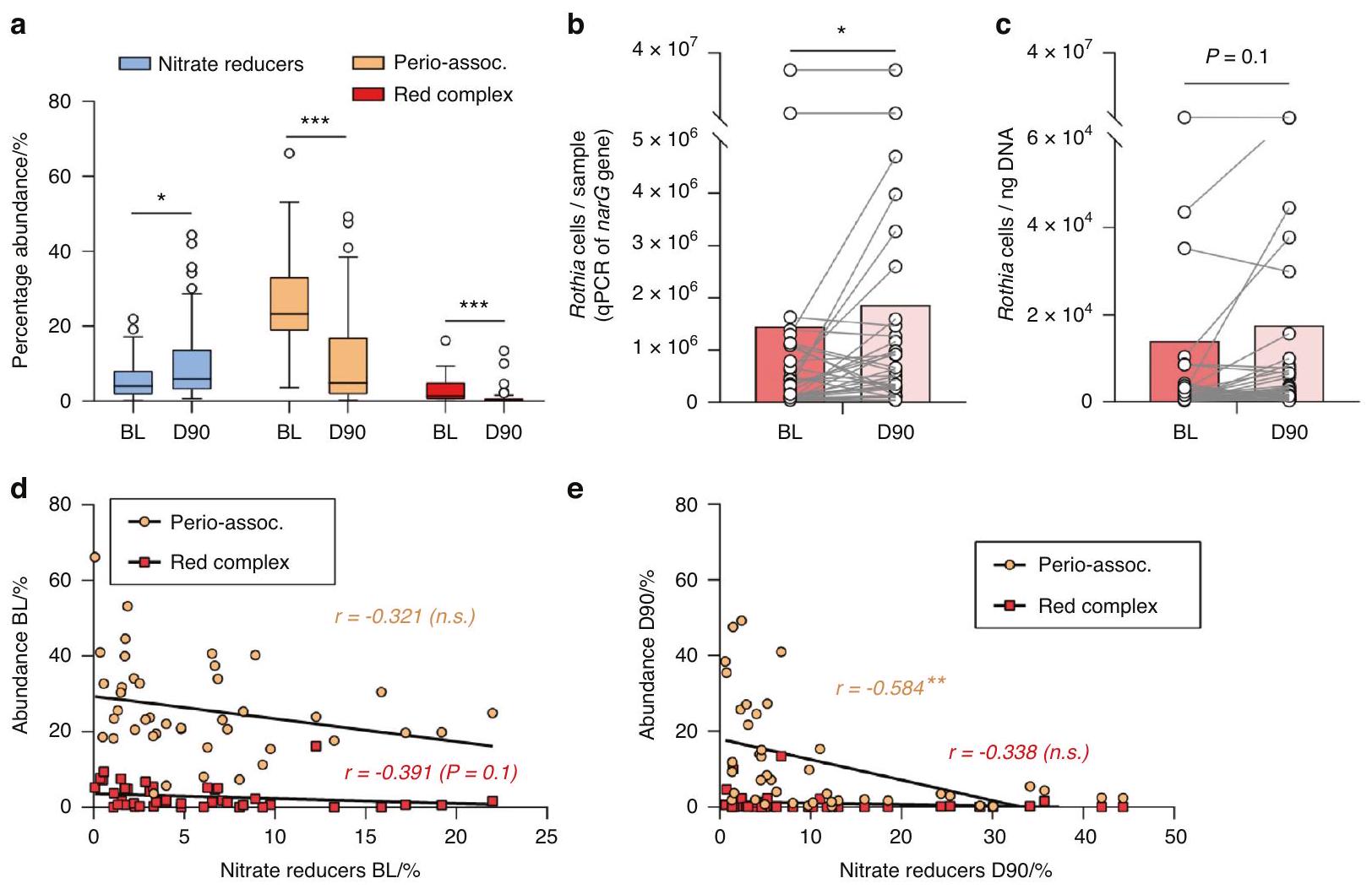
The bacterial composition of subgingival samples before (BL) and after (D90) periodontal treatment showed a consistent pattern across individuals. Relative to baseline, there was a significant increase in bacteria capable of producing nitrite (Supplementary Fig. 2), including confirmed nitrate-reducing bacteria (Fig. 3A), with an opposite pattern for periodontal pathogens, including both the three species of the red complex and the extended list of periodontitis-associated bacteria, as reported previously by Johnston et al. This suggests that the capacity to metabolize nitrate by subgingival plaque is improved after periodontal treatment. The increase in the levels of nitrate-reducing bacteria was confirmed by qPCR using specific primers for the nitratereducing biomarker Rothia, which showed a significant increase in cells per sample (i.e., absolute levels) (Fig. 3B). The normalized levels of this organism (Rothia cells determined by qPCR per ng DNA) also appeared to increase, but this increase was not significant ( ) (Fig. 3C). Notably, it was previously shown that the relative abundance of Rothia determined by Illumina sequencing did increase after periodontal treatment. Both the absolute and normalized levels of Rothia determined with qPCR
correlated with the abundance of nitrate-reducing bacteria ( and , respectively, both when grouping BL and D90) (Supplementary Datasheet).
When grouping the BL and D90 samples, a significant negative correlation was found between the levels of nitrate-reducing bacteria and the levels of periodontitis-associated bacteria ( ) (Supplementary Datasheet). When considering the BL samples only, this correlation was not significant (Fig. 3D), but in the D90 samples, there was a clear negative correlation between nitrate-reducing bacteria and periodontitis-associated bacteria ( ) (Fig. 3E). Two other interesting negative correlations when grouping the BL and D90 samples were between nitrate-reducing bacteria and FMPS ( , ), as well as absolute or total Rothia levels and FMBS (both to 0.33 , both ). Finally, the group of all nitriteproducing bacteria (Supplementary Table 2) correlated negatively with salivary IL-1 at baseline ( ) (Supplementary Datasheet).

NRC in health and periodontitis

Nitrate utilization during the incubation of saliva samples with added nitrate for a period of three hours was considered an estimate of the NRC in each individual. The data revealed that nitrate reduction was higher in healthy individuals compared to periodontal patients at baseline (Fig. 4A): in periodontitis patients, nitrate levels did not change significantly over the 3 -h incubation period ( ), while in healthy individuals, it did ( ). In accordance with the 16 S rRNA sequencing data that showed that nitrate-reducing bacteria increased in subgingival plaque, the NRC
Fig. 2 Confirmed nitrate-reducing bacteria in periodontitis and health. Bar graphs show the relative abundance of bacteria in subgingival plaque samples from different countries, as estimated by high-throughput sequencing of the 16 S rRNA gene. Bacteria were grouped in confirmed nitrate-reducing species, periodontitis-associated or red complex according to Rosier et al. , Pérez-Chaparro et al. , and Socransky et al. respectively (the bacterial species in each group are listed in Supplementary Table 2). Healthy individuals (blue bars) were compared with individuals with periodontitis (red bars). In Supplementary Fig. 1, all known nitrite-producing bacteria (some of which may produce nitrite by pathways other than nitrate reduction) are shown following the same pattern and showing a significant difference between health and periodontitis. The datasets include individuals from Japan, Spain, the USA, Brazil, and Chile ( per dataset, see Supplementary Table 1 for additional information). It should be noted that the levels of groups of bacteria in different studies are affected by the criteria used to describe periodontitis, regional and host factors, as well as the DNA extraction methods or the sequencing techniques used in the original studies. *adjusted of compositional data standardized by ANCOM-BC and compared with a Wilcoxon test
in periodontal patients was restored after the periodontal treatment, with a significant depletion in nitrate levels after the incubation ( ).
The nitrate-reducing activity (a known pH buffering process) was also confirmed indirectly by pH measurements, which showed a slightly larger pH drop in periodontal patients at baseline than after treatment (at BL the pH dropped from 7.33 to 7.13, and at D90, it dropped from 7.49 to 7.41, both ) (Fig. 4C).
In agreement with the above, nitrite levels did not differ in baseline periodontal samples before and after the incubation with nitrate (Fig. 4B). After periodontal treatment, a significant decrease in nitrite was observed after the incubation. Given the nitrate consumption during this three-hour period, the lower nitrite levels were probably a consequence of further nitrite metabolization. In healthy individuals, nitrite levels did increase after the incubation, but this increase was not significant ( ) and a much lower initial concentration of nitrite was present compared with periodontitis before ( ) and after ( ) treatment.
Plasma levels of nitrate and nitrite
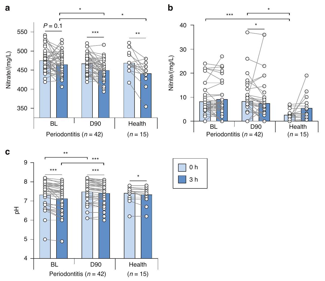
In agreement with the basal salivary levels of nitrate and nitrite, the basal nitrate and nitrite concentrations in blood samples collected in periodontal patients at baseline and 90 days after the periodontal treatment revealed no statistical differences between these two-time points (Fig. 5A, B). The average plasma nitrate levels were at BL and at D 90 (not significantly different). Nitrite levels ( at BL and at D90) were around 350 times lower than nitrate (Fig. 5C). There were no significant correlations between bacterial groups and plasma nitrate or nitrite. The highest Spearman correlation was between absolute or normalized Rothia levels in subgingival plaque and plasma nitrite, but this correlation was not significant (both , both when grouping BL and D90) (Supplementary Datasheet). The increased NRC that was found in saliva with added nitrate would translate into higher plasma nitrite or nitric oxide levels after dietary intake of nitrate, which was not evaluated in this study.
Fig. 3 Confirmed nitrate-reducing species and disease-associated bacteria before and 90 days after periodontal treatment. a Relative abundances of confirmed nitrate-reducing bacteria, red complex, and periodontitis-associated bacteria before (baseline, BL) and 90 days after treatment (D90) of 42 periodontitis patients. *adjusted of compositional data standardized by ANCOM-BC and compared with a Wilcoxon test. b, c Rothia cells determined by qPCR before (BL) and 90 days after (D90) treatment per sample (absolute amount, ) (b) or normalized per ng of DNA (c). determined by a Wilcoxon test. d, e Correlations between the abundance of periodontal pathogens and nitrate-reducing bacteria at baseline (BL) and 90 days after treatment (D90). **adjusted of Spearman’s rank correlation (n.s. = not significant). In Supplementary Fig. 2, the comparisons and correlations of all known nitrite-producing bacteria (some of which may produce nitrite by other pathways than nitrate reduction) are shown

DISCUSSION

The nitrate-nitrite-nitric oxide pathway can result in cardiovascular and metabolic benefits when stimulated with dietary nitrate. In this study, we show that the reduction of nitrate (i.e., a concentration found in saliva after vegetable intake) by microorganisms in saliva was impaired in periodontitis. This impaired NRC was recovered to healthy levels after periodontal treatment. In line with this, we show that the levels of nitrate-reducing bacteria in subgingival plaque are lower in periodontitis and recovered after periodontal treatment. Our results indicate that an impaired NRC in periodontitis could limit the positive effects of dietary nitrate-derived nitric oxide. Notably, different systemic conditions associated with periodontitis are also associated with a deficit in nitric oxide (e.g., cardiovascular diseases and diabetes). Therefore, future studies should explore the effect of periodontitis and periodontal treatment on cardiometabolic parameters after dietary nitrate intake.
The effect of periodontitis and periodontal treatment on the nitrate-reducing oral microbiota
To compare the subgingival levels of nitrate-reducing bacteria in health and periodontitis, we used 16 S sequencing datasets of five different countries (Japan, Brazil, Chile, USA, and Spain). All countries followed the same pattern of a decrease in periodontitis-associated species and an increase in nitriteproducing bacteria, including confirmed nitrate-reducing species. This finding is in agreement with previous studies. For example, Feres et al. systemically reviewed findings from
sequencing studies comparing subgingival plaque in health and periodontitis, as well as periodontitis before and after treatment and found that common nitrite-producing species were associated with periodontal health (e.g., Steptococcus spp., Neisseria longate, Neisseria subflava, Rothia aeria, Veilonella Parvula and Granulicatella adiacens). Additionally, Feres et al. found that Rothia was the genus with the strongest association with periodontal health, and with reduced inflammation after periodontal treatment, followed by other nitrite-producing genera (e.g., Neisseria, Actinomyces and Steptococcus). These results are further supported by bioinformatic analyses of Meuric et al. associating Veillonella, Neisseria, Rothia, Corynebacterium, and Actinomyces (all genera with nitrite-producing representatives) with periodontal health, and Chen et al. who reported that that Steptococus sanguinis, Actinomyces naeslundii, Rothia aeria, Granulicatella adiacens, Rothia dentocariosa and Streptococcus mitis (all nitrite-producing species) were six of the top seven most health-associated species that are used to calculate the subgingival microbial dysbiosis index (SMDI).
The underlying mechanisms by which nitrate reduction by oral bacteria appears to be beneficial for oral health requires exploration. Oral bacteria can further reduce nitrite to nitric oxide, a free radical with antimicrobial properties capable of inhibiting sensitive species such as anaerobes associated with periodontitis. Additionally, nitric oxide derived from denitrification by oral bacteria could directly signal to human cells, reducing inflammation and stimulating protective mucus production and blood flow. In our study, nitrate-reducing bacteria correlated
Fig. 4 Nitrate reduction capacity (NRC) in health and periodontitis. a, b Bar graphs represent the concentration of nitrate ( ) and nitrite ( ) of saliva samples cultured in vitro for 3 h in the presence of nitrate at before (Baseline, BL ) and 90 days after periodontal treatment ( ) of periodontal patients, compared to healthy controls ( ). c Bars show the pH during this incubation period for the same groups of individuals. Asterisks represent statistically significant differences (*P<0.05, ) determined by a Wilcoxon test (paired) or a Mann-Whitney U-test (unpaired)
Fig. 5 Plasma nitrate and nitrite before and 90 days after periodontal treatment. Data is shown from 42 periodontitis patients before (BL) and after (D90) treatment. a Plasma nitrite. b Plasma nitrate. c Ratio nitrate/nitrite
negatively with periodontitis-associated bacteria, supporting the idea that this function could be important for oral health. This negative correlation was clearest in the 42 periodontitis patients after periodontal treatment. The reduction of inflammation in these patients after treatment was previously described by Johnston et al. and consisted of a clear and significant improvement of all periodontal clinical parameters. In our recent in vitro study, the addition of nitrate to periodontal plaque resulted in nitrite production (an indication of nitric oxide production), a decrease in periodontitis-associated species, and a lower SMDI. In our current study, the levels of nitrate-reducing bacteria in subgingival plaque correlated negatively with the fullmouth plaque score (FMPS), while nitric oxide is also a biofilm
dispersal signal for some bacteria that could reduce plaque accumulation. In summary, the impairment of the NRC in periodontitis could give a selective advantage to periodontitisassociated species by losing nitric oxide production and potentially other antimicrobial mechanisms of health-associated bacteria. The effect of nitrate consumption and the patients’ NRC on the efficiency of periodontal treatment should be further investigated in future studies.
One of the main results of this study is that the NRC of saliva samples decreases in periodontitis. Unstimulated saliva contains bacteria per mL that have dispersed from oral communities, making this a non-invasive sample that can be used to study the activity of the oral microbiota. Additionally, by freezing
at and thawing on ice, a significant part of the bacteria survives, as demonstrated by the changes in metabolic parameters after 3 h of incubation at in this study (e.g., a minor but significant decrease in pH by bacterial fermentation of salivary components). By adding nitrate to these saliva samples, significant nitrate-reduction activity was found in the saliva of 15 healthy individuals ( ), which is commensurate with previous data obtained in small cohorts of healthy populations ( ). Notably, no significant nitrate reduction was observed in the saliva of 42 periodontitis patients before treatment ( ). However, 90 days after treatment, the NRC of the individuals with periodontitis was restored ( ) to levels comparable with healthy individuals. Additionally, more nitrite was reduced after periodontal treatment, indicating that nitric oxide production could be increased. This could be explained by the observation that common nitrate-reducing bacteria, which increased in number, also have nitrite-reduction genes.
The periodontal treatment had no significant effect on basal plasma nitrate and nitrite levels, while the effect of nitrate intake on plasma was not evaluated in this study. Specifically, the average basal plasma concentrations of nitrate in individuals with periodontitis were at BL and at D90 after treatment. The average nitrite levels were around 350 times lower ( at BL and at D 90 ). In two studies with systemically healthy individuals recruited at the same location (Glasgow, Scotland, United Kingdom), similar plasma nitrate and nitrite levels were found in a fasting state: Burleigh et al. found averages of nitrate and nitrite in 25 individuals, while Liddle et al. found averages of nitrate and nitrite in 34 individuals. It is known that nitrate intake increases the plasma nitrate and nitrite concentrations multiple times, indicating that at the timing of sample collection in our study, no dietary nitrate metabolism was detected. Future studies in larger populations should determine if periodontitis could affect the levels of plasma nitrite and nitrate while fasting or after nitrate intake.
In summary, the basal levels of nitrate and nitrite in plasma and saliva were not affected by periodontal treatment, but a difference in NRC was revealed when nitrate was added to saliva. Eight millimolar nitrate is a concentration found in saliva after vegetable intake, indicating that the positive oral and cardiometabolic effects stimulated by dietary nitrate could be impaired in periodontitis. The effect of periodontal treatment on oral and systemic parameters after nitrate intake should thus be explored in future studies.
Kapil et al. determined the NRC by letting individuals rinse their mouths with a nitrate-containing solution. Additionally, the NRC could be measured in oral biofilm samples normalized by weight or DNA concentrations. Future studies should compare different methods to determine the NRC, as this could represent a helpful marker of gingival health, and how it correlates with systemic parameters.
In line with the NRC results, we showed that the subgingival nitrate-reducing microbiota decreases in periodontitis (compared with periodontal health) and increases after periodontal treatment. Specifically, we showed this for the relative abundance of known nitrite-producing bacteria, including confirmed nitrate-reducing bacteria. The difference between these two groups is that nitrite-producing isolates are detected by incubating bacteria with nitrate and measuring nitrite production. Most of the time, this nitrite results from nitrate reduction, but there are other pathways that can lead to nitrite production (for example, the oxidation of nitric oxide or ammonium). The NRC of oral bacteria should thus be confirmed by physiological measurements of nitrate. In our study, both groups (nitriteproducing and confirmed nitrate-reducing) showed similar statistical significance (i.e., remained when
comparing these groups between periodontitis and health or before and after periodontal treatment). Additionally, qPCR measurements of the nitrate-reducing biomarker and periodontal-health-associated genus Rothia before and after periodontitis treatment confirmed an increase of Rothia cells per sample ( ). In our previous study, it was found that the Rothia levels in these samples, which were determined by Illumina sequencing of the 16 S rRNA gene, were higher after treatment. Our data confirms that nitrate-reducing bacteria decrease in the subgingival plaque under the conditions of inflammation and dysbiosis associated with periodontitis. It is also interesting that Rothia levels correlated well with the proportion of nitrate-reducing bacteria (Supplementary Datasheet), indicating that this genus is a potential biomarker of nitrate-reducing species. The degree of periodontitis, as indicated by the full mouth bleeding score (FMBS), showed a negative correlation with Rothia levels determined by qPCR. This supports a possible role of Rothia in periodontal health and its assessment as a potential periodontal probiotic, as recently proposed.
Potential implications of hampered NRC for systemic health Our data indicate that periodontitis could limit nitrate reduction in the presence of nitrate concentrations found after vegetable intake. The reduction of nitrate by oral bacteria is an essential step in the nitrate-nitrite-nitric oxide pathway that contributes to systemic nitric oxide levels. When sterilizing a significant proportion of the oral microbiota with chlorhexidine in fasting individuals, the NRC of the oral microbiota was impaired, plasma nitrite levels dropped and blood pressure increased. Conversely, stimulating the nitrate-nitrite-nitric oxide pathway by consuming vegetable juices or nitrate salts can lead to a decrease in blood pressure and improved endothelial function. Nitrate intake has also been associated with a reversal of metabolic syndrome and with antidiabetic effects, while the use of over-the-counter mouthwash was found to correlate with diabetes and pre-diabetes development. It thus appears that conditions in which a deficit of nitric oxide is found, benefit from stimulating nitrate reduction by the oral microbiota. Remarkably, periodontitis is associated with cardiovascular diseases and diabetes. Future studies should therefore determine if periodontitis can contribute to cardiovascular diseases, diabetes, and other nitric oxide-related systemic conditions by limiting the beneficial effects of dietary nitrate-derived nitric oxide. This possibility is supported by the finding that the genetic capacity of subgingival plaque to produce nitrite is associated with lower levels of cardiometabolic risk.
Pre-eclampsia is another example of a condition associated with both reduced nitric oxide availability and periodontitis, while a recent study indicated that this condition was associated with a decrease in oral nitrate-reducing bacteria. In relation to this, periodontal treatment appears to reduce complications in pregnant women with periodontitis, possibly by reducing inflammatory and bacterial exposure to unborn babies. In our study, we found that periodontal treatment recovered the NRC to healthy levels in the presence of nitrate. The potential effect of improved dietary nitrate reduction resulting from periodontal treatment on pre-eclampsia should be explored. Periodontal treatment is also associated with a long-term improvement of endothelial function. In a recent study in mice, it was shown that inorganic nitrate protects against and can partially reverse pre-existing, periodontitis-induced endothelial function through restoration of nitrite and, thus, nitric oxide levels. These findings show that both nitrate consumption and periodontal treatment can result in improved endothelial function. Nitrate intake should thus be explored as an adjunct treatment to improve systemic parameters in patients with periodontitis.

Study limitations and future perspectives

Limitations of this study are that individuals were not fasting when donating saliva and plasma samples and did not receive instructions regarding other habits that could interfere with salivary and plasma nitrate and nitrite levels (e.g., exercise habits, and fitness levels, sunlight, menstrual cycle stage, water consumption). Considering the large effect of the diet on plasma and salivary nitrate and nitrite levels, the study design was not ideal to see how periodontitis affects the basal levels of these molecules. This could explain why the basal saliva and plasma levels of nitrate and nitrite were not significantly different before and after periodontal treatment. However, when incubating saliva with nitrate, differences were found in the NRC of periodontitis patients before and after treatment and between periodontitis patients before treatment and healthy individuals. In vivo, this would mean that the salivary nitrate concentrations obtained by vegetable consumption are reduced less efficiently in periodontitis, decreasing the beneficial effects of dietary nitrate. In the current study, we did not collect blood samples after conditions of high nitrate availability, which is when the effect of a healthy nitrate-reduction bacterial community is expected to be most relevant. Thus, we propose that future studies should test if exposure to nitrate (e.g., by beetroot juice consumption) has different effects on salivary and plasma nitrate and nitrite levels, as well as their derived systemic effects (e.g., blood pressure reduction) in periodontitis patients (before and after periodontal treatment) and healthy individuals. Finally, in this study, the composition of subgingival plaque was studied because it is most affected by periodontitis. Additionally, it is known that the amounts of a given taxon in different oral samples may correlate with each other (for example, Porhyromonas gingivalis in subgingival plaque correlates with P. gingivalis in saliva ), indicating that changes in subgingival plaque affect other communities to some extent. In future work, the effect of this disease on the composition of other microbial communities involved in nitrate reduction (e.g., the tongue microbiota) should be explored.

Concluding remarks

In conclusion, our data show that periodontitis compromises the nitrate-reducing microbiota and impairs the reduction of nitrate concentrations that are found in saliva after vegetable intake. However, the impaired nitrate reduction capacity is recovered to healthy levels after periodontal treatment. Given that a diminished or eliminated nitrate reduction capacity, for instance, by antiseptic mouthwash, derives in lower plasma nitrite levels, periodontal disease could imply a deficit in circulatory nitrite. Future studies should investigate whether, in addition to the oral-systemic link derived from inflammatory effects, a hampered nitrate reduction capacity in periodontal patients could further contribute to the multiple systemic conditions that are affected by nitric oxide availability. Taking into account that nitrates intake increases periodontal health-associated bacteria, it should be determined if nitrate-rich dietary interventions could improve periodontal treatment outcomes, including oral and systemic parameters.

DATA AVAILABILITY

The sequencing reads of the 42 periodontitis patients before (BL) and after (D90) treatment are deposited in the NCBI Sequencing Read Archive (SRA) under BioProject PRJNA725103. Any further data is available upon reasonable request from the corresponding author.

ACKNOWLEDGEMENTS

The authors are grateful for the help provided from Glasgow Dental Hospital Clinical Research Facility, in particular Clare Brown and Debbie McKenzie. We would also like to thank Sandra García-Esteban at FISABIO for her help with the in vitro experiment.

FUNDING

The periodontitis treatment study was funded by grants from EU Marie Curie ITN RAPID (grant number 290246), Versus Arthritis (Grant Number 20823), and the BBSRC (BB/P504567/1). William Johnston is supported by a student stipend from the University of Glasgow and Dentsply Sirona (Project Number 300881). A.M. was supported by a grant from the European Regional Development Fund and the Spanish Ministry of Science, Innovation and Universities with the reference RTI2018-102032-B-I00, as well as a grant from the Valencian Innovation Agency with the reference INNVAL20/19/006. BR was supported by an FPI fellowship from the Spanish Ministry of Science, Innovation, and Universities with the reference Bio2015-68711-R.

ADDITIONAL INFORMATION

Supplementary information The online version contains supplementary material available at https://doi.org/10.1038/s41368-023-00266-9.
Competing interests: The authors declared the following potential conflicts of interest with respect to the research, authorship, and/or publication of this article: A.M. and B.T. Rosier are coinventors in a pending patent application owned by the FISABIO Institute, which protects the use of nitrate as a prebiotic and certain nitratereducing bacteria as probiotics. The remaining authors declare no competing interests.
Statement of ethical approval: The study was conducted in accordance with the Declaration of Helsinki (2013). For the study involving periodontitis patients, the London-Stanmore Research Ethics Committee (Reference: 14/LO/206) of the National Health Service (NHS, England, United Kingdom) gave ethical approval. For the study involving saliva samples of healthy patients, the MVLS ethical committee (project number: 2011002) of the University of Glasgow (Scotland, United Kingdom) gave ethical approval.

REFERENCES

  1. Theilade, E. The non-specific theory in microbial etiology of inflammatory periodontal diseases. J. Clin. Periodontol. 13, 905-911 (1986).
  2. Rosier, B. T., Marsh, P. D. & Mira, A. Resilience of the oral microbiota in health: mechanisms that prevent dysbiosis. J. Dent. Res. 97, 371-380 (2018).
  3. Marsh, P. D. Are dental diseases examples of ecological catastrophes? Microbiology 149, 279-294 (2003).
  4. Mira, A., Simon-Soro, A. & Curtis, M. A. Role of microbial communities in the pathogenesis of periodontal diseases and caries. J. Clin. Periodontol. 44, S23-S38 (2017).
  5. Feres, M., Retamal-Valdes, B., Gonçalves, C., Cristina Figueiredo, L. & Teles, F. Did Omics change periodontal therapy? Periodontol 2000 85, 182-209 (2021).
  6. Chen, T., Marsh, P. D. & Al-Hebshi, N. N. SMDI: An index for measuring subgingival microbial dysbiosis. J. Dent. Res. https://doi.org/10.1177/00220345211035775 (2021).
  7. Rosier, B. T. et al. The importance of nitrate reduction for oral health. J. Dent. Res. 101, 887-897 (2022).
  8. Vanhatalo, A. et al. Nitrate-responsive oral microbiome modulates nitric oxide homeostasis and blood pressure in humans. Free Radic. Biol. Med. 124, 21-30 (2018).
  9. Rosier, B. T., Buetas, E., Moya-Gonzalvez, E. M., Artacho, A. & Mira, A. Nitrate as a potential prebiotic for the oral microbiome. Sci. Rep. 10, 12895 (2020).
  10. Hajishengallis, G. Periodontitis: from microbial immune subversion to systemic inflammation. Nat. Rev. Immunol. 15, 30-44 (2015).
  11. Hajishengallis, G. & Chavakis, T. Local and systemic mechanisms linking periodontal disease and inflammatory comorbidities. Nat. Rev. Immunol. 21, 426-440 (2021).
  12. Beck, J. D., Papapanou, P. N., Philips, K. H. & Offenbacher, S. Periodontal medicine: 100 Years of progress. J. Dent. Res. 98, 1053-1062 (2019).
  13. Schreiber, F. et al. Denitrification in human dental plaque. BMC Biol. 8, 24 (2010).
  14. Hyde, E. R. et al. Metagenomic analysis of nitrate-reducing bacteria in the oral cavity: implications for nitric oxide homeostasis. PLOS ONE 26, 3 (2014).
  15. Aerts, A., Dendale, P., Strobel, G. & Block, P. Sublingual nitrates during head-up tilt testing for the diagnosis of vasovagal syncope. Am. Heart J. 133, 504-507 (1997).
  16. Goh, C. E. et al. Nitrite generating and depleting capacity of the oral microbiome and cardiometabolic risk: results from ORIGINS. J. Am. Heart Assoc. 11, e023038 (2022).
  17. Hezel, M. P. & Weitzberg, E. The oral microbiome and nitric oxide homoeostasis. Oral. Dis. 21, 7-16 (2015).
  18. Lundberg, J. O., Carlström, M. & Weitzberg, E. Metabolic effects of dietary nitrate in health and disease. Cell Metab. 28, 9-22 (2018).
Nitrate reduction capacity of the oral microbiota is impaired in…
Rosier et al.
10
19. Morou-Bermúdez, E., Torres-Colón, J. E., Bermúdez, N. S., Patel, R. P. & Joshipura, K. J. Pathways linking oral bacteria, nitric oxide metabolism, and health. J. Dent. Res. 101, 623-631 (2022).
20. Kapil, V. et al. Physiological role for nitrate-reducing oral bacteria in blood pressure control. Free Radic. Biol. Med. 55, 93-100 (2013).
21. Backlund, C. J., Sergesketter, A. R., Offenbacher, S. & Schoenfisch, M. H. Antibacterial efficacy of exogenous nitric oxide on periodontal pathogens. J. Dent. Res. 93, 1089-1094 (2014).
22. Lanas, A. Role of nitric oxide in the gastrointestinal tract. Arthritis Res. Ther. 10, S4 (2008).
23. Schairer, D. O., Chouake, J. S., Nosanchuk, J. D. & Friedman, A. J. The potential of nitric oxide releasing therapies as antimicrobial agents. Virulence 3, 271-279 (2012).
24. Rosier, B. T. et al. A single dose of nitrate increases resilience against acidification derived from sugar fermentation by the oral microbiome. Front. Cell Infect. Microbiol. 11, 692883 (2021).
25. Ikeda, E. et al. Japanese subgingival microbiota in health vs disease and their roles in predicted functions associated with periodontitis. Odontology 108, 280-291 (2020).
26. Pérez-Chaparro, P. J. et al. Do different probing depths exhibit striking differences in microbial profiles? J. Clin. Periodontol. 45, 26-37 (2018).
27. Abusleme, L. et al. The subgingival microbiome in health and periodontitis and its relationship with community biomass and inflammation. ISME J. 7, 1016-1025 (2013).
28. Griffen, A. L. et al. Distinct and complex bacterial profiles in human periodontitis and health revealed by 16 S pyrosequencing. ISME J. 6, 1176-1185 (2012).
29. Camelo-Castillo, A. J. et al. Subgingival microbiota in health compared to periodontitis and the influence of smoking. Front. Microbiol. 6, 119 (2015).
30. Arredondo, A. et al. Comparative 16 S rRNA gene sequencing study of subgingival microbiota of healthy subjects and patients with periodontitis from four different countries. J. Clin. Periodontol. 50, 1176-1187 (2023).
31. Callahan, B. J. et al. DADA2: High-resolution sample inference from llumina amplicon data. Nat. Methods 13, 581-583 (2016).
32. Team, R. C. R: A Language and Environment for Statistical Computing http:// www.R-project.org/, http://www.R-project.org/ (2014).
33. Quast, C. et al. The SILVA ribosomal RNA gene database project: improved data processing and web-based tools. Nucleic Acids Res. 41, D590-D596 (2013).
34. Yilmaz, P. et al. The SILVA and “All-species Living Tree Project (LTP)” taxonomic frameworks. Nucleic Acids Res. 42, D643-D648 (2014).
35. Socransky, S. S., Haffajee, A. D., Cugini, M. A., Smith, C. & Kent, R. L. Jr. Microbial complexes in subgingival plaque. J. Clin. Periodontol. 25, 134-144 (1998).
36. Pérez-Chaparro, P. J. et al. Newly identified pathogens associated with periodontitis: a systematic review. J. Dent. Res. 93, 846-858 (2014).
37. Davison, E. et al. The subgingival plaque microbiome, systemic antibodies against bacteria and citrullinated proteins following periodontal therapy. Pathogens 10, 193 (2021).
38. Johnston, W. et al. Mechanical biofilm disruption causes microbial and immunological shifts in periodontitis patients. Sci. Rep. 11, 9796 (2021).
39. Sanz, M. et al. EFP Workshop participants and methodological consultants. Treatment of stage I-III periodontitis-the EFP S3 level clinical practice guideline. J. Clin. Periodontol. 47, 4-60 (2020).
40. Papapanou, P. N. et al. Periodontitis: consensus report of workgroup 2 of the 2017 World Workshop on the classification of periodontal and peri-implant diseases and conditions. J. Periodontol. 89, S173-S182 (2018).
41. Johnston, W. The Host and Microbial Response to Non-surgical Periodontal Therapy. (University of Glasgow, 2021).
42. Darcey, J. & Ashley, M. See you in three months! The rationale for the three monthly periodontal recall interval: a risk based approach. Br. Dent. J. 211, 379-385 (2011).
43. Herrera, D. et al. EFP workshop participants and methodological consultant treatment of stage IV periodontitis: the EFP S3 level clinical practice guideline. J. Clin. Periodontol. 49, 4-71 (2022).
44. Rosier, B. T., Moya-Gonzalvez, E. M., Corell-Escuin, P. & Mira, A. Isolation and characterization of nitrate-reducing bacteria as potential probiotics for oral and systemic health. Front. Microbiol. 11, 555465 (2020).
45. Liddle, L. et al. Variability in nitrate-reducing oral bacteria and nitric oxide metabolites in biological fluids following dietary nitrate administration: an assessment of the critical difference. Nitric Oxide 82, 1-10 (2019).
46. Lin, H. & Peddada, S. D. Analysis of compositions of microbiomes with bias correction. Nat. Commun. 11, 3514 (2020).
47. Meuric, V. et al. Signature of microbial dysbiosis in periodontitis. Appl Environ. Microbiol. 83, e00462-17 (2017).
48. Mazurel, D. et al. Nitrate and a nitrate-reducing Rothia aeria strain as potential prebiotic or synbiotic treatments for periodontitis. NPJ Biofilms Microbiomes 9, 40 (2023).
49. Mantilla Gomez, S. M. et al. Tongue coating and salivary bacterial counts in healthy/gingivitis subjects and periodontitis patients. J. Clin. Periodontol. 28, 970-978 (2001).
50. Burleigh, M. C. et al. Salivary nitrite production is elevated in individuals with a higher abundance of oral nitrate-reducing bacteria. Free Radic. Biol. Med. 120, 80-88 (2018).
51. Liddle, L. et al. Reduced nitric oxide synthesis in winter: a potential contributing factor to increased cardiovascular risk. Nitric Oxide 127, 1-9 (2022).
52. Webb, A. J. et al. Acute blood pressure lowering, vasoprotective, and antiplatelet properties of dietary nitrate via bioconversion to nitrite. Hypertension 51, 784-790 (2008).
53. Larsen, F. J., Ekblom, B., Sahlin, K., Lundberg, J. O. & Weitzberg, E. Effects of dietary nitrate on blood pressure in healthy volunteers. N. Engl. J. Med. 355, 2792-2793 (2006).
54. Fernandes, D. et al. Local delivery of nitric oxide prevents endothelial dysfunction in periodontitis. Pharm. Res. 188, 106616 (2022).
55. Bahadoran, Z., Ghasemi, A., Mirmiran, P., Azizi, F. & Hadaegh, F. Beneficial effects of inorganic nitrate/nitrite in type 2 diabetes and its complications. Nutr. Metab. 12, 16 (2015).
56. Joshipura, K. J., Muñoz-Torres, F. J., Morou-Bermudez, E. & Patel, R. P. Over-thecounter mouthwash use and risk of pre-diabetes/diabetes. Nitric Oxide 71, 14-20 (2017).
57. Ghasemi, A. & Jeddi, S. Anti-obesity and anti-diabetic effects of nitrate and nitrite. Nitric Oxide 70, 9-24 (2017).
58. Preshaw, P. M. et al. Periodontitis and diabetes: a two-way relationship. Diabetologia 55, 21-31 (2012).
59. Kebschull, M., Demmer, R. T. & Papapanou, P. N. Gum bug, leave my heart alone!”-epidemiologic and mechanistic evidence linking periodontal infections and atherosclerosis. J. Dent. Res. 89, 879-902 (2010).
60. Tashie, W. et al. Altered bioavailability of nitric oxide and L-arginine is a key determinant of endothelial dysfunction in preeclampsia. Biomed. Res. Int. 2020, 3251956 (2020).
61. Altemani, F., Barrett, H. L., Callaway, L. K., McIntyre, H. D., & Dekker Nitert, M. Reduced abundance of nitrate-reducing bacteria in the oral microbiota of women with future preeclampsia. Nutrients 14, 1139 (2022).
62. Kim, A. J., Lo, A. J., Pullin, D. A., Thornton-Johnson, D. S. & Karimbux, N. Y. Scaling and root planing treatment for periodontitis to reduce preterm birth and low birth weight: a systematic review and meta-analysis of randomized controlled trials. J. Periodontol. 83, 1508-1519 (2012).
63. Tonetti, M. S. et al. Treatment of periodontitis and endothelial function. N. Engl. J. Med. 356, 911-920 (2018).
64. Monaghan, C. et al. The effects of two different doses of ultraviolet-A light exposure on nitric oxide metabolites and cardiorespiratory outcomes. Eur. J. Appl. Physiol. 118, 1043-1052 (2018).
65. Liddle, L. et al. Variability in nitrate-reducing oral bacteria and nitric oxide metabolites in biological fluids following dietary nitrate administration: an assessment of the critical difference. Nitric Oxide 83, 1-10 (2019).
66. Jiang, Y. et al. Comparison of red-complex bacteria between saliva and subgingival plaque of periodontitis patients: a systematic review and meta-analysis. Front. Cell Infect. Microbiol. 11, 727732 (2021).
Open Access This article is licensed under a Creative Commons
Attribution 4.0 International License, which permits use, sharing, adaptation, distribution and reproduction in any medium or format, as long as you give appropriate credit to the original author(s) and the source, provide a link to the Creative Commons license, and indicate if changes were made. The images or other third party material in this article are included in the article’s Creative Commons license, unless indicated otherwise in a credit line to the material. If material is not included in the article’s Creative Commons license and your intended use is not permitted by statutory regulation or exceeds the permitted use, you will need to obtain permission directly from the copyright holder. To view a copy of this license, visit http:// creativecommons.org/licenses/by/4.0/.
© The Author(s) 2024, corrected publication 2024

  1. Department of Genomics and Health, FISABIO Foundation, Center for Advanced Research in Public Health, Valencia, Spain; Department of Biological and Biomedical Sciences, Glasgow Caledonian University, Glasgow, UK; Oral Sciences, University of Glasgow Dental School, School of Medicine, Dentistry and Nursing, College of Medical, Veterinary and Life Sciences, University of Glasgow, Glasgow, UK; Sport and Physical Activity Research Institute, University of the West of Scotland, Blantyre, Scotland; Instituto de Biomedicina de Valencia, Consejo Superior de Investigaciones Científicas (IBV-CSIC), Valencia, Spain and CIBER Center for Epidemiology and Public Health, Madrid, Spain Correspondence: Alex Mira (mira_ale@gva.es)
    These authors contributed equally: Bob T. Rosier, William Johnston