كيف تمنع البروبيوتيك، والبريبايوتيك، والسينيبيوتيك، والبروستبيوتيك تسوس الأسنان: منظور ميكروبيوم الفم How probiotics, prebiotics, synbiotics, and postbiotics prevent dental caries: an oral microbiota perspective

المجلة: npj Biofilms and Microbiomes، المجلد: 10، العدد: 1
DOI: https://doi.org/10.1038/s41522-024-00488-7
PMID: https://pubmed.ncbi.nlm.nih.gov/38402294
تاريخ النشر: 2024-02-24

كيف تمنع البروبيوتيك، والبريبايوتيك، والسينيبيوتيك، والبروستبيوتيك تسوس الأسنان: منظور ميكروبيوم الفم

(T) تحقق من التحديثات

سي-تشين لو سي-مين وي شين-تاو لو تشيوغ تشيوغ يانغ كا-هينغ وونغ بيتر سي. كيه. تشيونغ وبو-بو زانغ

الملخص

تسوس الأسنان، وهو مرض فموي شائع للغاية، يؤثر على جزء كبير من السكان العالميين. الطرق التقليدية التي تقضي بشكل عشوائي على الميكروبات تعطل التوازن الطبيعي للميكروبات الفموية. على النقيض من ذلك، تهدف استراتيجيات التدخل الحيوي إلى استعادة هذا التوازن من خلال إدخال ميكروبات مفيدة أو تثبيط تلك المسببة للتسوس. على مدى الثلاثين عامًا الماضية، حظيت التحضيرات الميكروبية باهتمام كبير في أبحاث طب الأسنان للوقاية من تسوس الأسنان وعلاجه. ومع ذلك، على عكس الأمراض ذات الصلة في الجهاز الهضمي، والمهبل، والجهاز التنفسي، يحدث تسوس الأسنان على الأنسجة الصلبة مثل مينا الأسنان ويرتبط ارتباطًا وثيقًا بالإفراط المحلي في إنتاج الأحماض الذي تسهله الأغشية الحيوية المسببة للتسوس. لذلك، فإنه من غير الكافي الاعتماد فقط على الآليات السابقة لتحديد دور التحضيرات الميكروبية في تجويف الفم. يجب أن تتضمن وجهة نظر أكثر شمولاً النظر في مفاهيم الأغشية الحيوية المسببة للتسوس. توضح هذه المراجعة أحدث تقدم في الأبحاث، وآليات العمل، والتحديات، واتجاهات البحث المستقبلية المتعلقة بالبروبيوتيك، والبربيوتيك، والسنبيوتيك، والبوستبيوتيك للوقاية من تسوس الأسنان وعلاجه، مع الأخذ في الاعتبار الآليات المرضية الفريدة لتسوس الأسنان. مع فهم معزز للميكروبات الفموية، ستظهر العلاجات الميكروبية الشخصية كاتجاه بحثي حاسم في المستقبل.

تمثل تسوس الأسنان تحديًا كبيرًا وملحًا في مجال الصحة العامة على مستوى العالم، حيث يؤثر على عدد هائل من الأفراد في جميع أنحاء العالم. على وجه التحديد، يُقدّر عدد حالات الأسنان الدائمة بـ 64.6 مليون حالة، بالإضافة إلى 62.9 مليون حالة من الأسنان الأولية. ستربتوكوكوس موتانس، أحد البكتيريا الرئيسية المسببة لتسوس الأسنان والتي تعبر عن بروتين ربط الكولاجين، يمكن أن تغزو بفعالية خلايا بطانة الوريد السري البشري. مما يؤدي إلى إمكانية تطور التهاب الشغاف المعدي.
نظرًا لأن تسوس الأسنان يتم عادةً بواسطة الغشاء الحيوي، فقد أصبحت التدخلات التي تستهدف الغشاء الحيوي استراتيجية رئيسية للوقاية. إن تعديل تناول الركائز القابلة للتخمر في النظام الغذائي، وخاصة السكروز، هو نهج فعال. . يتميز البيئة الغذائية الحديثة بتوفر الأطعمة المعالجة بشكل كبير والمليئة بالسكر، مما يخلق تحديًا كبيرًا في الامتناع تمامًا عن الأطعمة المسببة للتسوس. تشمل التدخلات الأخرى التنظيف الجسدي (مثل: الفرشاة أو استخدام أدوات التنظيف بين الأسنان)، والتثبيط الكيميائي (مثل: استخدام الكلورهيكسيدين أو بوفيدون-يود)، والتدخلات البيولوجية (مثل: استخدام البروبيوتيك) . بشكل فعال
لمنع تسوس الأسنان، يجب أن تهدف الاستراتيجيات الحالية إلى كبح نمو البكتيريا المسببة للتسوس من خلال استهداف عوامل ضراوتها، مع تعزيز ميكروبيوتا سكانية متنوعة وصحية. من بين هذه التدخلات، حازت التحضيرات الميكروبية مثل البروبيوتيك، والبرابيتيك، والسينيبيوتيك، والبوستبيوتيك على اهتمام كبير لأنها تقدم نهجًا أكثر استهدافًا وودّية مقارنةً بالتخلص الفيزيائي والتثبيط الكيميائي.
ميرمان وزملاؤه كانوا روادًا في إدخال البروبيوتيك في مجال طب الأسنان. مع مرور الوقت، حظيت التحضيرات الميكروبية باهتمام كعلاجات مساعدة محتملة للوقاية من تسوس الأسنان وعلاجه. لقد أظهرت هذه التحضيرات فعالية كبيرة في تثبيط نمو وتكوين الأغشية الحيوية للبكتيريا المسببة للتسوس.
تقدم هذه المقالة أولاً لمحة عامة عن الخلفية والآليات المسببة للأمراض للتسوس السني، مع التركيز على عوامل الضراوة للبكتيريا المسببة للتسوس S. mutans. ثم تلخص أحدث تقدم في الأبحاث، وآلية العمل، وحالة التطبيق، والتحديات المرتبطة بها.
مع استخدام البروبيوتيك، والبريبايوتيك، والسنبيوتيك، والبستبيوتيك في الوقاية من تسوس الأسنان. أخيرًا، يقترح هذا المقال اتجاهات مستقبلية لتطوير هذا المجال لتوفير إرشادات علمية ومعيارية وفعالة أكثر للوقاية من تسوس الأسنان من منظور أكاديمي.

خلفية تسوس الأسنان

تعتبر تجويف الفم نظامًا بيئيًا معقدًا يتميز بوجود ميكروبيئات دافئة ورطبة متنوعة توفر ظروفًا مثالية لنمو الميكروبات. دراسة حديثة تحلل الميكروبيوم الفموي حددت ما مجموعه 1591 نوعًا ميكروبيًا، بما في ذلك البكتيريا والفطريات والعتائق والفيروسات والأوليات. ، الثاني فقط من حيث التعقيد بعد القولون تظل الميكروبات الفموية الأساسية في الأفراد الأصحاء مستقرة نسبيًا على مدى سبع سنوات. بينما يمكن أن يؤدي اختلال توازن الميكروبات الفموية إلى تسوس الأسنان وأمراض فموية أخرى علاوة على ذلك، فإن تنوع المجتمع الميكروبي الفموي في تسوس الأسنان الشديد أقل بكثير من ذلك في الأفراد الأصحاء. تنشأ تسوس الأسنان من عدم التوازن في الميكروبات الفموية نتيجة لتفاعل معقد بين المضيف والنظام الغذائي والميكروبات. .
من بين هذه العوامل، تم تحديد الكربوهيدرات القابلة للتخمر، التي توجد عادة في الأطعمة المحلاة، كمساهمات غذائية مهمة بشكل خاص في تسوس الأسنان. يمكن أن يؤدي استهلاك الأطعمة المحلاة إلى زيادة سريعة في تركيز الكربوهيدرات في تجويف الفم، مما يؤدي إلى انخفاض حاد في قيم الرقم الهيدروجيني للبيوفيلم إلى 4 أو حتى أقل. لقد كشفت الأبحاث عن وجود علاقة دقيقة بين مناطق إزالة المعادن الحادة على سطح المينا والمناطق ذات الرقم الهيدروجيني الحمضي العالي التي تخلقها الأفلام الحيوية. يُنسب ذلك إلى أن الانخفاضات المتكررة في درجة الحموضة المحلية يمكن أن تعطل التوازن بين التمعدن وإزالة المعادن في البيئة الدقيقة المغلقة للبيوفيلم. . ونتيجة لذلك، يؤدي ذلك إلى فقدان المعادن في الأسنان، مما ينتج عنه بقع بيضاء، وتجاويف، والتهابات في اللب، وحتى فقدان الأسنان .
تمت ملاحظة اختلافات في الميكروفلورا الفموية بين الأفراد ذوي الأسنان الصحية وأولئك الذين يعانون من تسوس الأسنان. على سبيل المثال، تشير نتائج دراسة حول الميكروبيوم الفموي للأطفال إلى أن الأجناس روثيا ونييسيريا وهموفيلوس، التي تعد من أول المستعمرات في تجويف الفم بعد الولادة. مرتبطة بصحة الأسنان على النقيض من ذلك، فإن أنواع Prevotella وS. mutans وفيروس الهربس البشري 4 (فيروس EB) توجد بشكل أكثر شيوعًا في الأطفال الذين يعانون من تسوس الأسنان. أكتينوبكتيريا (35.8%) وباسيليوتا ( كانت الفصائل الأكثر شيوعًا في آفات تسوس العاج العميق، وكان Lactobacillus هو الجنس الأكثر وفرة فقط من الآفات التسوسية هناك اعتراف متزايد بأن تسوس الأسنان ناتج عن اختلال التوازن في الميكروبيوم في الغشاء الحيوي، المعروف أيضًا بالبلاك السني، بدلاً من كونه ناتجًا عن مُمْرِض واحد. .

الميكروبات المرتبطة بتسوس الأسنان

تظهر البكتيريا المسببة للتسوس درجات متفاوتة من المساهمة في تطور تسوس الأسنان. لعقود، تم التعرف على S. mutans وStreptococcus sobrinus على أنهما العاملان الرئيسيان المسببان للتسوس. من الجدير بالذكر أن S. sobrinus أظهر قدرة أعلى على إنتاج الأحماض وتحمل الأحماض مقارنةً بـ S. mutans، لكنه أظهر قدرة أقل على التكيف مع بيئة الأغشية الحيوية. البكتيريا المسببة للتسوس الموجودة في الميكروبات الفموية لا توجد ككيانات معزولة، بل تتفاعل وتؤثر على بعضها البعض. إلى حد ما، تقوم S. mutans بخلق بيئة غنية بحمض اللبنيك في الآفات التسوسية مما يسهل تكاثر أنواع Veillonella. التي أظهرت أنها تعزز نمو S. mutans في دراسات الأغشية الحيوية .
بالإضافة إلى ذلك، ظهرت أنواع الكانديدا، كممثل فطري نموذجي للميكروبات المسببة للتسوس، كعوامل ثانوية قوية للتسوس، معزولة من إلى تسوس الأسنان لدى البالغين والأطفال كانديدا هي خميرة قوية من نوع الكائنات الحية الدقيقة الانتهازية التي تعتمد على إنتاج الأحماض الكربوكسيلية قصيرة السلسلة والبروتيناز، بالإضافة إلى قدرتها على الالتصاق بالأسطح غير الحية وتشكيل الأغشية الحيوية. أكثر أشكال التواصل شيوعًا بين الفطريات والبكتيريا في تجويف الفم هو التفاعل المتبادل بين الكانديدا البيض (Candida albicans) وS. mutans. وجود الكانديدا البيض يعزز نمو S. mutans، مما يؤدي إلى تغييرات ملحوظة في التعبير الجيني وزيادة في عملية الأيض للكربوهيدرات. . بشكل ملحوظ، مقارنة
في الفيلم الحيوي أحادي النوع الذي يتكون فقط من S. mutans، هناك 393 جينًا معبرًا عنه بشكل مختلف في S. mutans داخل الفيلم الحيوي ثنائي النوع. يمكن للغلوكوزيل ترانسفيراز (Gtf) التي تفرزها S. mutans أن ترتبط بـ C. albicans وتساعد في تحويل السكروز إلى بوليمر خارجي (EPS)، مما يوفر مواقع ارتباط لـ S. mutans. .
أفادت دراسة أن الميكروبات الأساسية لتسوس الأسنان في مرحلة الطفولة المبكرة (ECC) قد تشمل فيلونيلة بارفولا، وفوسوبكتيريوم نوكليتوم، وبريفوتيلا دينتيكولا، وليبتوتريشيا وادي. من ناحية، تعزز هذه الميكروبات الأساسية في ECC نمو وقدرة S. mutans على إنتاج الأحماض، وتساعد في تشكيل الأغشية الحيوية، على الرغم من قدرتها المحدودة على إنتاج الأحماض. . من ناحية أخرى، فإنه يعزز أيضًا إزالة المعادن من المينا في المختبر ويزيد من القدرة على التسبب في تسوس الأسنان للمينا في الجسم الحي . بالإضافة إلى ذلك، وفقًا لبعض النتائج الميتاجينومية، فإن الأنواع التالية مرتبطة ارتباطًا وثيقًا بتسوس الأسنان: ستربتوكوكوس غوردوني، ليبتوتريشيا باكالي، V. بارفولا، أكتينوميسيس جيرينسيريا، بروبيونيباكتيريوم أسيليفاسيينس، هاليلا مالتي ساكاريفوركس، وباراسكاردوفيا دنتيكولنز .
من بين الكائنات الدقيقة المرتبطة بتسوس الأسنان، يُعتبر S. mutans واحدًا من الأنواع الأكثر دراسة في هذا المجال. نظرًا لأن S. mutans كان يُعتقد في البداية أنه سبب رئيسي لتسوس الأسنان. ليس من المستغرب أن تستهدف معظم استراتيجيات الوقاية هذه البكتيريا بشكل محدد .

ستربتوكوكوس موتانس

عند مقارنتها مع بكتيريا الاستعمار الأصلية الأخرى، تظهر S. mutans سمات أكثر فائدة من خلال تطوير غشاء حيوي مضغوط وعوامل ضراوة مميزة لها. علاوة على ذلك، تحت السيطرة المنظمة لنظام استشعار الكمية، تصبح S. mutans في النهاية واحدة من البكتيريا الرئيسية المسببة للتسوس. الفيلم الحيوي في تسوس الأسنان في الأسنان الأولية هو هيكل كروي ثلاثي الأبعاد (3D)، حيث تكون S. mutans هي النواة وتكون البكتيريا الأخرى هي الطبقة الخارجية. . هذه المنطقة المحلية تخلق بيئة حامضية لدرجة الحموضة، مما يؤدي إلى إزالة المعادن من المينا بشكل شديد مع تقدم تسوس الأسنان، يصبح تنوع الميكروبات الفموية محدودًا. . هذا الاختلال الميكروبي يؤدي في النهاية إلى حدوث وتطور تسوس الأسنان. وقد تم تأكيد العلاقة الوثيقة بين S. mutans وتسوس الأسنان. على الرغم من أن S. mutans هو مقيم طبيعي في تجويف الفم البشري زيادة في مستويات يجب أن تكون المتحورات مصدر قلق لأنها قد تشير إلى سابقة سريرية لتسوس الأسنان. الفئران المصابة بسلالة S. mutans المشتقة من الإنسان تتطور لديها تسوس الأسنان وقد يرتبط S. mutans بعودة شديدة لتسوس الأسنان المبكر تشير هذه إلى الدور الحاسم لبكتيريا S. mutans في حدوث وتطور تسوس الأسنان. لذلك، فإن توضيح الآليات المسببة للأمراض لبكتيريا S. mutans (الشكل 1) أمر ضروري لتطوير استراتيجيات فعالة للوقاية من التسوس وعلاجه. في الأقسام التالية، سنناقش عوامل الضراوة ونظام استشعار الكثافة لبكتيريا S. mutans.
عوامل الضراوة. يمكن تصنيف عوامل الضراوة في S. mutans إلى أربع مجموعات رئيسية، تشمل تخليق EPS، الالتصاق، إنتاج الحمض، ومقاومة الحمض.
تركيب EPS. تُعزى قدرة S. mutans على إظهار مرضيتها إلى حد كبير إلى إنتاج EPS. EPS، وهو مكون رئيسي من الأغشية الحيوية. يتكون من بروتينات خارج الخلية، ودي إن أي خارج الخلية، وحمض ليبوتيكويك. المكون الرئيسي لـ EPS هو الجلوكوز، الذي يتم تصنيعه بواسطة Gtf. توفير مواقع ربط للميكروبات . يساهم EPS في تشكيل حواجز كيميائية وفيزيائية منظمة للغاية داخل مصفوفة الأغشية الحيوية، مما يسهل التعلق الميكروبي بالأسطح غير الحية مقاومة إجهادات القص السائلة تجنب استجابات المناعة لدى المضيف تحمل العوامل المضادة للميكروبات وفي النهاية، إنشاء والحفاظ على بيئة ميكروبية حمضية في تجويف الفم تفضل تطور مجتمعات الأغشية الحيوية المرتبطة بتسوس الأسنان من الصعب إزالة الأغشية الحيوية الناضجة ميكانيكياً بسبب اللزوجة المرنة المعززة التي توفرها المواد خارج الخلوية (EPS). قد تحقق EPS التهرب المناعي من خلال التوسط في التهرب من المكمل. وتقييد دخول جزيئات التأثير من الجهاز المناعي الفطري والمكتسب إلى مصفوفة البيوفيلم الكلورهيكسيدين، وهو عامل مضاد للميكروبات يُستخدم بشكل شائع في العناية الفموية، لديه قدرة محدودة على التغلغل في طبقات البايوفيلم العميقة بسبب شحنته الإيجابية، بينما “الوقود” (السكروز)، الذي يفتقر إلى الشحنة،
الشكل 1 | تشكيل الفيلم الحيوي المسبب للتسوس. EPS السكريات المتعددة الخارجية، Gbps بروتينات ربط الجلوكوز. ترميز Gbps، مرتبط بالالتصاق، Gtfs جلوكوزيل ترانسفيراز، gtf ترميز Gtfs، مرتبط بتخليق EPS، eno ترميز إنولاز البكتيريا، مرتبط بامتصاص الجلوكوز، LDH لاكتات ديهيدروجيناز، ترميز LDH، مرتبط بإنتاج الحمض، PTS فوسفات النقل، نظام امتصاص الجلوكوز، PEP فوسفوإنول البيروفات، atpD ترميز -ATPase، مرتبط بمقاومة الحمض، ATP أدينوزين ثلاثي الفوسفات، ADP أدينوزين ثنائي الفوسفات، AgDs نظام دياميناز الأجمتين، aguD ترميز AgDs، مرتبط بمقاومة الحمض. تتطور الأسنان الصحية إلى تسوس الأسنان بسبب التفاعلات المعقدة بين المضيف، النظام الغذائي، والميكروبات. تعتمد الميكروبيولوجيا للأسنان الصحية على التوازن بين الأنشطة الميكروبية المولدة للأحماض والأنشطة المولدة للقلويات، بالإضافة إلى التوازن بين عمليات إزالة المعادن وإعادة المعادن. عندما تصبح الميكروبات المولدة للأحماض هي السائدة،
تؤدي التركيزات العالية والمتكررة من الحمض محليًا إلى إزالة المعادن من مينا الأسنان، مما يؤدي إلى تكوين التجاويف. على المستوى المجهري، تتضمن الخطوة الأولية التصاق بعض الكائنات الدقيقة البدائية المستعمرة بمينا الأسنان. تتضمن الخطوة الثانية إنتاج المواد البوليمرية خارج الخلوية بواسطة الكائنات الدقيقة، مما يشكل غشاء حيويًا. في الخطوة الثالثة، تؤسس المجتمعات الميكروبية المنتجة للحمض والمتحملة للحمض، التي تهيمن عليها بشكل رئيسي S. mutans، بيئة ميكروية شديدة الحموضة، مما يؤدي إلى إزالة المعادن من مينا الأسنان. في الخطوة الرابعة، تمنح البيئة الميكروية الشديدة الحموضة ميزة نمو لمجموعات الكائنات الدقيقة التي تهيمن عليها S. mutans. مع أخذ عوامل الضراوة في الاعتبار . موتانس كمثال، يتم التوسط في تخليق EPS بشكل أساسي بواسطة Gtfs. يتم تسهيل عملية الالتصاق بشكل رئيسي بواسطة Gbps. تشمل عملية إنتاج الحمض مشاركة الإنولاز و LDH. تعتمد عمليات تحمل الحمض بشكل أساسي على مشاركة -ATPاز و AguD.
يمكن أن ينتشر بسهولة، بفضل الشحنة السلبية لـ تجمع السطح المشحون سلبياً لخلايا S. mutans المغلفة بـ EPS البروتونات، كما أن تأثير الغربلة لهيكل الجلوكوز يلعب دورًا أيضًا. . من ناحية، يقوم EPS بالتقاط وتراكم البروتونات الناتجة خارجيًا أو بواسطة الأحماض.
الميكروبات، التي تساعد في احتباس وتراكم الحمض داخل الغشاء الحيوي من ناحية أخرى، بمجرد أن يتم استقطاب البروتونات إلى سطح الخلية، فإنها تحفز استجابة التكيف الحمضي، مما يسمح للميكروبات بمواجهة الأضرار الناتجة عن الحمض بشكل استباقي. تعطيل واحد أو أكثر
تقلل الجينات بشكل كبير من شدة الضراوة لـ S. mutans، في نماذج تسوس الأسنان لدى القوارض باختصار، يلعب EPS دورًا حاسمًا في تمكين S. mutans من ممارسة إمكانيته المسببة للتسوس. لذلك، قد يمثل تثبيط تخليق EPS استراتيجية وقائية قابلة للتطبيق ضد تسوس الأسنان. .
الالتصاق. تستخدم بكتيريا S. mutans مسارات مستقلة عن السكروز وأخرى تعتمد على السكروز للالتصاق بالأسنان. تتم عملية الالتصاق الأولية بشكل أساسي من خلال المسار المستقل عن السكروز، والذي يتم تعزيزه بعد ذلك من خلال تحفيز تخليق الجلوكان عبر المسار المعتمد على السكروز، مما يؤدي في النهاية إلى تشكيل الأغشية الحيوية. تساعد بروتينات ربط الجلوكانيات في ربط الجلوكانيات التي يتم تصنيعها من السكروز من خلال ناقلات الجلوكوز. من بين هذه البروتينات، يظهر GbpA ارتباطًا قويًا مع القدرة على التسبب في تسوس الأسنان. من ناحية، يساهم في تشكيل هيكل قوي للبيوفيلم ويعتبر بروتينًا مهمًا يحدد هيكل البيوفيلم. من ناحية أخرى، يلعب دورًا أساسيًا في ربط جزيئات الجلوكوز ويشارك في عملية التصاق البكتيريا بالأسنان.
إنتاج الأحماض. بعد استقلاب الجلوكوز، تنتج الكربوهيدرات الغذائية الطاقة والأحماض العضوية كمنتجات ثانوية لعملية الأيض. نشاط إنتاج الحمض . المتحولة ليست فقط عاملًا حاسمًا يساهم في قدرتها الممرضة، ولكنها أيضًا سمة حاسمة تؤدي إلى تسوس الأسنان. إنزيم الإينولاز، وهو إنزيم مشفر بواسطة الجين eno، هو مكون رئيسي في نظام الفوسفاتransferase، الذي يتحمل مسؤولية امتصاص الجلوكوز. من خلال النشاط التحفيزي السريع لإنزيم اللاكتات ديهيدروجيناز (LDH)، وهو بروتين مشفر بواسطة جين ldh، يقوم S. mutans UA159 بتخمير الجلوكوز إلى أحماض عضوية. .
مقاومة الحمض. تستخدم S. mutans بعض الآليات المقاومة للحمض للتعامل مع ضغط زيادة إنتاج الحمض. -ATPase، مضخة بروتون مشفرة بواسطة atp لا تقوم فقط بضخ البروتونات داخل الخلايا للحفاظ على درجة الحموضة داخل الخلايا، بل تنتج أيضًا ATP لتعزيز نمو البكتيريا وبقائها. أدى تثبيط تعبير atpD في S. mutans UA159 إلى انخفاض كبير في التكيف مع الحمض وزيادة في حموضة السيتوبلازم. بالإضافة إلى ذلك، تنتج بكتيريا S. mutans القلويات لتحييد الأحماض، وكذلك تصدرها خارج الخلايا. يلعب نظام دييميناز الأغماتين دورًا حاسمًا في إنتاج القلويات للتغلب على ضغط الأحماض. . من بين مكوناته، الناقل المضاد للأغمتين-البوتريسين (AguD)، المشفر بواسطة الجين، له أهمية خاصة لأنه يسهل النقل داخل الخلايا للأغمتين الحر. تراكم البروتونات على سطح خلايا البكتيريا المغلفة بـ EPS يلعب دورًا مهمًا في مقاومة الحمض لـ S. mutans، كما ذُكر في قسم “تخليق EPS”. .
نظام استشعار الحشد (QS). ينظم نظام QS الضراوة وتكوين الأغشية الحيوية من خلال إطلاق واستشعار والتفاعل مع جزيئات الانتشار. استنادًا إلى كثافة الخلايا في البيئة المحيطة . تستخدم المتحولات هذا النظام للتواصل مع بعضها البعض كمجموعة بدلاً من كأفراد منفصلين. الآلية الرئيسية لتغذية الإشارة الراجعة هي من خلال أنظمة نقل الإشارة ذات المكونين (TCSTS)، التي تمكن البكتيريا من تنظيم تعبير جيناتها تحتوي S. mutans على عدة أنواع من TCSTS، من بينها VicRKX و ComCDE اللذان يلعبان دورًا حاسمًا في تنظيم تشكيل الأغشية الحيوية، ومقاومة الأحماض، وإنتاج الأحماض استجابةً للإشارات البيئية. إذا فشلت هذه الأنظمة التنظيمية في العمل بشكل صحيح، فقد يؤدي ذلك إلى انخفاض في قدرة S. mutans على التسبب في تسوس الأسنان.

تدابير الوقاية من تسوس الأسنان – التدخلات البيولوجية

على الرغم من أن اللويحات هي ظاهرة طبيعية في الأسنان من منظور تطوري وبيولوجي وتغذوي، إلا أن عدم التوازن في الميكروبيوم في الفيلم الحيوي الفموي المرضي يمكن أن يؤدي إلى تطور تسوس الأسنان. تتسبب البكتيريا المسببة للتسوس التي تنتج الأحماض، وخاصة S. mutans، في تلف هياكل الأسنان الصلبة في وجود الكربوهيدرات القابلة للتخمر. .
في السنوات الأخيرة، تطور مجال التدخل البيولوجي بعض الاستراتيجيات الجديدة. تتضمن إحدى الطرق استخدام المفترسات، مثل Bdellovibrio وBacteriovorax وPeredibacter، للقضاء على البكتيريا سالبة الجرام اللاهوائية.
البكتيريا التي هي مسببات الأمراض اللثوية نظرًا لأن البكتيريا المفيدة هي في الغالب موجبة الجرام بالإضافة إلى ذلك، تشمل التدخلات البيولوجية أيضًا استخدام مثبطات محددة لبروتينات S. mutans، والتطعيم، واستراتيجيات المناعة السلبية باستخدام بكتيريا محايدة. على الرغم من ظهور بعض استراتيجيات التدخل البيولوجي المبتكرة مثل تلك المذكورة أعلاه، فإن استخدام التحضيرات الميكروبية، مثل البروبيوتيك، والبريبايوتيك، والسنبيوتيك، والبوسيبوتيك، هو نهج أكثر رسوخًا وشعبية في الوقاية من تسوس الأسنان.

البروبيوتيك

خلفية

تم اكتشاف البروبيوتيك من قبل العلماء في وقت مبكر من ، ومنذ ذلك الحين تطور مجال دراسة التأثيرات الصحية للبروبيوتيك على المضيف تدريجياً. في عام 2013، عرّفت الجمعية العلمية الدولية للبروبيوتيك والبربيوتيك (ISAPP) البروبيوتيك بأنه “ميكروبات حية، وعند إعطائها بكميات كافية، توفر فائدة صحية للمضيف”. اليوم، تُستخدم البروبيوتيك بشكل شائع من قبل البشر للحفاظ على صحتهم العامة. على الرغم من أن فعاليتها في تعزيز صحة الجهاز الهضمي معروفة جيدًا، إلا أن الأبحاث أظهرت أيضًا أن البروبيوتيك يمكن أن تكون فعالة في الوقاية من وعلاج مجموعة متنوعة من الأمراض الفموية، مثل تسوس الأسنان، التهاب الغشاء المخاطي الفموي، ورائحة الفم الكريهة. .
في طب الأسنان، تم تقديم البروبيوتيك لأول مرة من قبل ميوتمان وزملائه ، الذي وجد أن Lactobacillus rhamnosus GG ATCC 53103 يمكن أن يستعمر الفم البشري. مع المزيد من البحث، تم العثور على أن البروبيوتيك له قدرة ملحوظة على منع تسوس الأسنان. على سبيل المثال، استكشفت إحدى الدراسات تأثير Lactobacillus الخاص بالمواضيع على . موتانس . أظهرت الدراسة أن اللاكتوباسيلس المعزول من تجويف الفم لدى الأشخاص يمكن أن يثبط بشكل فعال نمو S. mutans. وُجد أن الأنواع الأكثر فعالية من S. mutans هي Lacticaseibacillus paracasei و Lactiplantibacillus plantarum، والتي تُعتبر أيضًا أكثر العزلات شيوعًا. أخيرًا، وُجد أن استخدام البروبيوتيك في علاج الأمراض الفموية يعيد التوازن الميكروبي الفموي ويقلل من المستويات من . موتانس في اللويحات السنية واللعاب .
مع وجود سلالات بروبيوتيك مختلفة تظهر خصائص فريدة، فإن فهم تفاصيل كل سلالة أمر بالغ الأهمية عند الوقاية وعلاج تسوس الأسنان. على سبيل المثال، L. rhamnosus GG هو لاكتوباسيلوس متخمر أحادي لا يعتبر مسببًا للتسوس لأنه لا يمكنه تخمير السكروز أو اللاكتوز. ليموسيلاكتوباسيلوس ريوتيري هو نوع متطلب للتخمر المختلط. التي يمكن أن تنتج مضادات ميكروبية واسعة الطيف ذات استقرار جيد في الأحماض والقواعد، مثل رويترين وريترسيكلين بالإضافة إلى Lactobacillus spp.، قد تكون Bifidobacteria spp. أيضًا بروبيوتيك محتمل للوقاية من تسوس الأسنان وعلاجه. وقد تم الإبلاغ عن أن الزبادي الذي يحتوي على Bifidobacterium DN-173010 يقلل بشكل كبير من مستوى S. mutans. .

آليات للوقاية من تسوس الأسنان

آلية منع البروبيوتيك لتسوس الأسنان مشابهة لتلك الموجودة في الجهاز الهضمي. تشمل الآليات المثبطة الرئيسية تخليق المستقلبات النشطة، تثبيط الأغشية الحيوية الميكروبية المسببة للتسوس، الالتصاق التنافسي والاستعمار، التكتل مع مسببات الأمراض، وتنظيم الجهاز المناعي (الشكل 2).

إنتاج المستقلبات النشطة

البكتيريوسين. البكتيريوسين هو ببتيد مضاد للبكتيريا موجب الشحنة يتم تصنيعه بواسطة الريبوسوم. وتم اكتشافه لأول مرة يمكن تقسيم البكتيريوسينات إلى أربع فئات مختلفة، حيث تكون الفئتان الأولى والثانية هي التركيز الرئيسي لمعظم أبحاث البروبيوتيك. نينسين، وهو بكتيريوسين من النوع A في الفئة I، يُعرف على نطاق واسع بأنه بروتين صغير مشحون إيجابياً. ) الذي يحفز الخلايا المستهدفة على تشكيل ثقوب في الغشاء تقوم البكتيريوسينات من الفئة الثانية بقتل البكتيريا عن طريق زيادة نفاذية الغشاء وتسرب محتويات البكتيريا المستهدفة. بالإضافة إلى الآليات المذكورة أعلاه، يمكن أن تثبط البكتيريوسينات أيضًا تخليق الأغشية الحيوية وجدار الخلية، وتمارس أنشطة DNase وRNase، وتنظم الميكروبات. .
تفاعل البروتين-بروتين بين بروتينات GtfB و LuxS . المتحولات والبكتيريوسين من SD1 في L. paracasei وُجد أنها تقلل من
الشكل 2 | آلية البروبيوتيك لمنع تسوس الأسنان. يتم تقسيمها تقريبًا إلى خمسة أجزاء. A إنتاج المستقلبات النشطة: البروبيوتيك تثبط مباشرة مسببات الأمراض المسببة للتسوس من خلال المستقلبات النشطة (مثل البكتيريوسين، الإنزيمات، المواد السطحية الحيوية، الأحماض العضوية، وبيروكسيد الهيدروجين)، التي تمتلك بدورها نشاطًا مثبطًا للبكتيريا. B تثبيط الأغشية الحيوية الميكروبية المسببة للتسوس: يمكن للبروبيوتيك تثبيط أو إزالة الأغشية الحيوية للميكروبات المسببة للتسوس في الفم. C الالتصاق التنافسي والاستعمار: لا تشغل البروبيوتيك المواقع المستعمرة في تجويف الفم فحسب، بل أيضًا
تثبيط قدرة الالتصاق للميكروبات المسببة للتسوس. د التكتل مع مسببات الأمراض: تثبط البروبيوتيك استعمار الميكروبات المسببة للتسوس في تجويف الفم من خلال التكتل. هـ تنظيم الجهاز المناعي: تنشط البروبيوتيك أو تعدل الجهاز المناعي للمضيف، مما يعزز الاستجابة المناعية للميكروبات المسببة للتسوس (يزيد من مستويات اللعاب من ببتيدات العدلات البشرية 1-3).
تكوين الأغشية الحيوية وكثافة الكائنات الدقيقة، كما تم الكشف عنه في دراسة محاكاة بعد تحليل شامل للمعلومات الحيوية والتوصيف، وُجد أن البكتيريوسين في هذه الدراسة آمن للبشر. يقوم البكتيريوسين ميرساكيدين بممارسة نشاطه القاتل للبكتيريا من خلال تشكيل معقد مع الليبيد II، مما يعيق تخليق جدار الخلية. تتيح أنشطة DNase و RNase للكاليسينات من E2 إلى E9 لها القدرة على تحلل الحمض النووي والبروتينات البكتيرية بشكل غير محدد. من بين هذه المواد، يظهر الكوليسين E2 نشاطًا قاتلًا للبكتيريا قويًا وطويل الأمد، ومن المثير للاهتمام أنه يمكنه استهداف البكتيريا بشكل محدد في الأغشية البيولوجية المعقدة. البكتيريوسينات قادرة على تعزيز استعمار بكتيريا المنتج في أماكن محددة على مدى عقد من الزمن، وتنظيم تركيبة الميكروبيوتا وتأثيرها على جهاز المناعة لدى المضيف. يمكن لكل من النيسين والبروبيوتيك المنتج للنيسين تقليل مستوى مسببات الأمراض في الأغشية الحيوية واستعادة تنوع السلالات إلى مستوى صحي. .
نظرًا لأن البكتيريوسينات هي بولي ببتيدات وبروتينات، فإن التحكم في درجة الحرارة أمر حاسم لضمان نشاطها أثناء الإنتاج والاستخدام. على سبيل المثال، يتم تركيز ستربتوكوكوس أوراليس تحت النوع دنتيساني 7746 (AB-Dentisanium) بشكل مثالي عند 30 و ، مع انخفاض طفيف في نشاط البكتيريوسين عند تؤكد هذه الاعتبارات الحرجة على أهمية تنفيذ استراتيجيات تنظيم درجة الحرارة المناسبة في تطوير واستخدام البكتيريوسينات لمختلف التطبيقات.
إنزيم. بالإضافة إلى البكتيريوسينات، تقوم البروبيوتيك بتخليق مجموعة متنوعة من الإنزيمات التي تمنح تأثيرات مفيدة من خلال تحلل الأغشية الحيوية وتأثيرها على نشاط البكتيريوسين. على سبيل المثال، يمكن لبكتيريا Lactobacillus acidophilus إفراز الليباز لتحلل الأغشية الحيوية. . بالمثل، يعبر ستربتوكوكوس ساليفاريوس JH عن إنزيم ديكستراناز يمكنه تحليل البوليمرات الخارجية لـ S. mutans وزيادة النشاط المثبط ضد S. mutans للزوزين A، وهو بكتيريوسين ميواليتك. . مثال آخر هو Streptococcus sp. A12، الذي ينتج بروتينات شبيهة بالشاليسين التي تثبط إنتاج البكتيريوسينات بواسطة . موتانس بالإضافة إلى ذلك، ينتج S. salivarius M18 اليورياز والدكستراناز لتحييد حموضة اللعاب وتقليل تكوين اللويحات، على التوالي. .
المستحلبات الحيوية (BS). المستحلبات الحيوية هي مواد أمفيلية تنتجها عمليات الأيض الميكروبية تحتوي على مجموعات كارهة للماء ومحبّة للماء، وتتكون بشكل رئيسي من البروتينات والسكريات والدهون. يمكن تحديد هيكل BS باستخدام تقنيات مختلفة مثل الكروماتوغرافيا الطبقية الرقيقة، ومطياف الأشعة تحت الحمراء بتحويل فورييه، والرنين المغناطيسي النووي. لم يقم خمسون في المئة من تقارير البيوسورفكتانت البالغ عددها 40 بمراجعة هيكلها، على الأرجح بسبب تعقيد الهياكل. السورفاكتين و/أو المواد السطحية البيولوجية الشبيهة بالبروتينات ) يتم إنتاجها بشكل شائع بواسطة Lactobacillus، مع دراسات حول الجليكوبروتينات غليكوليبيد وغلائكوبيبتيد إنتاجه نادر.
عادةً ما تنتج اللاكتوباسيلس أنواعًا من البايوسورفكتانتس من نوع السورفاكتين، والتي تحتوي على نسبة عالية من البروتين ويمكن أن تثبط بشكل كبير التصاق مسببات الأمراض، مما يجعلها مثيرة للاهتمام بشكل متزايد بسبب خصائصها الفريدة المضادة للالتصاق ومضادة للأغشية الحيوية. . على سبيل المثال، تم إنتاجه بواسطة . ريوتيري DSM 17938، لقد منعت بكتيريا acidophilus DDS-1 و L. rhamnosus ATCC 53103 و L. paracasei B21060 التصاق S. mutans و Streptococcus oralis وتكوين الأغشية الحيوية بطريقة تعتمد على الجرعة على أسطح التيتانيوم. بمزيد من التفصيل، الأسيدوفيلوس DSM 20079 ينتج بروتين من نوع BS، والذي يمكن أن يقصر طول السلسلة من . موتانس، تتداخل مع تشكيل الغشاء الحيوي الخاص بها على شرائح الزجاج، وتقلل من تعبير الجينات لـ و . تم إنتاجه بواسطة يمكن أن يدمر Rhamnosus الهيكل الفيزيائي أو تكوين البروتين للغشاء الحيوي، مما يؤدي إلى تحلل الخلايا. بالإضافة إلى النشاط المضاد للميكروبات الجذاب المذكور أعلاه، يظهر BS خصائص انخفاض السمية الخلوية وارتفاع الاستقرار. يُظهر BS المستمد من Lactobacillus spp. سمية خلوية منخفضة مقارنة بالرمانوليبيدات، التي تُعتبر عمومًا منتجات غير سامة. قد يظهر BS استقرارًا أفضل مقارنةً بالمواد المضادة للبكتيريا الأخرى التي تنتجها البروبيوتيك. غودينه et al. تم عزل مستحضر مستقر من L. paracasei. كان هذا المستحضر مستقرًا في نطاق pH وحافظت على النشاط السطحي بعد الحضانة عند لمدة 120 ساعة.
الأحماض العضوية. الأحماض العضوية، مثل حمض اللبنيك وحمض الزبد. التي تنتجها Lactobacillus في الجهاز الهضمي البشري وأجزاء أخرى من الجسم، تم التعرف عليها على نطاق واسع كمواد مفيدة. قد يكون لهذه الأحماض العضوية تأثير بكتيريوستاتي على الكائنات الدقيقة المسببة للأمراض الفموية إلى حد معين. على سبيل المثال، ينتج L. paracasei Lpc-37 الحمض الذي يمكن أن يعيق نمو وتكوين الأغشية الحيوية لـ S. mutans. . ومع ذلك، بالنظر إلى الارتباط القوي بين إزالة المعادن من الأسنان والتعرض المتكرر لتركيزات عالية من الحمض يثير ذلك تساؤلات حول كيفية تأثير الأحماض التي تنتجها البروبيوتيك على الأحماض التي تنتجها البكتيريا المسببة للتسوس، بما في ذلك S. mutans.
قد تحتاج هذه الأسئلة إلى أن تُتناول من منظور بيئة التسوس العامة. تخلق الكائنات الدقيقة المسببة للتسوس حاجزًا حمضيًا منظمًا للغاية. التعرض المطول لتركيزات عالية موضعية من الحمض يؤدي إلى إزالة المعادن الموضعية بدلاً من إزالة المعادن المتوسطة من الأسنان. إذا كانت الأحماض العضوية التي تنتجها البروبيوتيك قادرة على تثبيط البكتيريا المسببة للتسوس، بما في ذلك S. mutans، و/أو الأغشية الحيوية الخاصة بها، فقد تعطل هذه الحواجز الحمضية وتمنع تراكم الحمض. نظرًا لوجود توازنها الخاص من الأحماض والقواعد الميكروبية في تجويف الفم. الأحماض التي لا تتراكم بل تساهم بدلاً من ذلك في توازن الحمض والقاعدة في البيئة الفموية تبدو أنها تشكل تهديدًا أقل.
بيروكسيد الهيدروجين. تنتج بعض الأنواع البروبيوتيك، بما في ذلك بيفيدوباكتيريوم بيفيدوم، لاكتوباسيلوس جونسوني، لاكتوباسيلوس كريسباتوس، ولاكتوباسيلوس جنسنii، بيروكسيد الهيدروجين لتحقيق تأثيرات مضادة للبكتيريا. يعمل بيروكسيد الهيدروجين على الظهارة البكتيرية المسببة للأمراض، مما يؤدي إلى موتها. علاوة على ذلك، فإن بيروكسيد الهيدروجين لديه القدرة على تنظيم تركيبة الأنواع داخل تجويف الفم. من الجدير بالذكر أن الأنواع المسببة للتسوس مثل S. mutans تكون حساسة للغاية لسمية بيروكسيد الهيدروجين. . ومع ذلك، فإن النشاط المضاد للبكتيريا لـ تم تقليل السائل الخالي من الخلايا (CFS) لبكتيريا باراكاسي بشكل كبير بعد معالجة الكاتالاز، مما يشير إلى دور بيروكسيد الهيدروجين في تأثيراته البكتيريا المثبطة. يجب ملاحظة أن بيروكسيد الهيدروجين قد لا يحتفظ بوظيفته البكتيريا الساكنة بعد عمليات مثل التجفيف بالتجميد، بسبب سهولة تحلله. .
تثبيط الأغشية الحيوية المسببة للتسوس. يتم عادةً الوساطة في تسوس الأسنان بواسطة الأغشية الحيوية. خاصية حاسمة للبروبيوتيك هي القدرة على تثبيط أو القضاء على نمو الأغشية الحيوية والميكروبات المسببة للأمراض في تجويف الفم. تم الإبلاغ عن بعض السلالات غالبًا لنشاطها المضاد للأغشية الحيوية، بما في ذلك S. oralis 89a وLimosilactobacillus fermentum TCUESC01 وL. acidophilus 4A وBifidobacterium longum subsp. longum. على سبيل المثال، لاكتيسيباسيلوس كاسي ATCC 393، قد تثبط بيوفيلمات L. reuteri ATCC 23272 و L. plantarum ATCC 14917 و Ligilactobacillus salivarius ATCC 11741 . موتانس عن طريق تقليل تنظيم الجينات مثل ، و في . . من المثير للاهتمام أن البروبيوتيك قادر على تثبيط الفطريات من الانتقال إلى
أشكال ممرضة. لقد تم إثبات أن . رهمنوسوس LR32، . كيسي L 324 م ، و تظهر سلالات الأسيودوفيلوس NCFM القدرة على إعاقة المراحل الأولية لتكوين الخيوط، وهي خطوة حاسمة في مسببات مرض الكانديدا البيض. تركيبة بروبيوتيك تتكون من Lactobacillus helveticus CBS N116411 وL. plantarum SD5870 وS. salivarius DSM 14685 خفضت بشكل كبير من تعبير الجينات المرتبطة بانتقال الخميرة إلى الهيفا في C. albicans، بما في ذلك EFG1 (منشط الجين الخاص بالهيفا)، SAP5 (بروتياز مُفرز)، ALS3 (التصاق) ) وبروتين جدار الهيف (HWP1) في المختبر، تتطور نماذج الأغشية الحيوية باستمرار لتقليد الظروف الموجودة داخل جسم الإنسان بشكل أكثر دقة. بناءً على الأهداف البحثية المحددة، يمكن للباحثين اختيار نماذج تهمهم، مثل دعامة تجريبية تحاكي البنية الكلية والميكروية لزرع الأسنان. .
تركيبة تم العثور على أن رهمنوسوس وببتيدات الكولاجين يزيدان بشكل كبير من درجة حموضة الوسط في المراحل المبكرة من تشكيل الأغشية الحيوية. أظهرت نتائج qPCR أن هذا المزيج خفض من تعبير عدة جينات حيوية مرتبطة بإنتاج الحمض وتحمل الحمض، بما في ذلك eno و ldh و atpD. علاوة على ذلك، قد تؤثر البروبيوتيك أيضًا على التأثيرات المضادة للبكتيريا من خلال التدخل في نظام الإشارات الكمي. أظهرت دراسة أن comD و vicR و vic تم تقليل تنظيم الجينات في الأشكال العائمة والبيوفيلم لـ . موتانس عند تعرضه لسوائل الفم مع اللاكتوباسيلس . قد يفسر هذا التأثير انخفاض الالتصاق وتكوين الأغشية الحيوية لـ . المتحولات تم ملاحظتها في تجارب المجهر الإلكتروني الماسح.
الالتصاق التنافسي والاستعمار. واحدة من الخصائص الرئيسية للبروبيوتيك التي تساهم في آثارها الصحية هي قدرتها على التفوق على مسببات الأمراض الفموية فيما يتعلق بالالتصاق والاستعمار. على سبيل المثال، أظهرت سلالة L. reuteri LR6 أكبر قدر من قدرات الالتصاق بين ثمانية سلالات بروبيوتيك تم اختبارها، مما يتوافق مع قدرة أعلى على تثبيط التصاق مسببات الأمراض بخلايا Caco-2. أدى تعزيز فعالية الاستعمار بواسطة S. salivarius M18 إلى زيادة نشاط مضاد للتسوس كما يتضح من انخفاض درجات اللويحات و مستويات .mutans ليفيلاكتوباسيلوس بريفيس KCCM 202399 منع الالتصاق بـ . موتانس KCTC 5458 من خلال تقليل التجمع الذاتي، وخصائص السطح الخلوي الكارهة للماء، وإنتاج EPS لـ S. mutans .
من المثير للاهتمام أن البروبيوتيك يمكن أن يقلل من التصاق مسببات الأمراض حتى بدون اتصال مباشر. وقد أظهرت الدراسات أن اللعاب المعالج بالبروبيوتيك يقلل من التصاق S. mutans بأسطح الهيدروكسيباتيت (وهو نموذج للمينا). أظهرت الدراسات الإضافية أن الغشاء اللعابي المذكور أعلاه يفتقر إلى بروتينين: اللكتين اللعابي gp340، وهو المستقبل الرئيسي لبكتيريا S. mutans في الغشاء اللعابي، والبيروكسيداز اللعابي، وهو عامل دفاعي فطري موجود في اللعاب البشري. .
التجمع المشترك مع الممرضات. يُعتبر التجمع المشترك من الخصائص المفيدة للبروبيوتيك لأنه يسمح لها بتشكيل حاجز يعيق استعمار الممرضات. في دراسة، وُجد أن ستة من بين 624 نوعًا من بكتيريا حمض اللبنيك تظهر تكتلًا محددًا مع S. mutans في المختبر. تم تحديد هذه الأنواع على أنها L. paracasei و L. rhamnosus. وقد تم اكتشاف أن آلية التكتل هذه تتمتع بمرونة عالية تجاه كل من الحرارة المفرطة والبروتياز، ولا تعتمد على الليكتينات، كما أنها لا تتأثر باللعاب.
تنظيم الجهاز المناعي. بالإضافة إلى تأثيراتها المباشرة على الكائنات الدقيقة المسببة للأمراض أو الأغشية الحيوية، من المعروف أن البروبيوتيك تنشط وتعدل جهاز المناعة لدى المضيف. لقد أظهرت الدراسات السريرية أن الاستهلاك اليومي أو ثلاث مرات أسبوعيًا من يساهم سلالة الباراكاسي SD1 في المرضى الذين يعانون من تسوس الأسنان الشديد بشكل كبير في زيادة مستويات الببتيدات البشرية المحايدة 1-3 في اللعاب، والتي تتمتع بنشاط مضاد للبكتيريا واسع النطاق، ويقلل من مستويات S. mutans، مما قد يبطئ من تقدم التسوس. . علاوة على ذلك، فإن استهلاك الحليب الذي يحتوي على L. paracasei SD1 لمدة ستة أشهر زاد من مستويات الأجسام المضادة IgA في اللعاب، وهذا الارتفاع مرتبط إيجابياً بعبء باراكاسي .
تقوم سلالات معينة من ستربتوكوكوس ثيرموفيلوس، مثل ST1342 وST1275 وST285، بتنشيط الاستجابة المناعية الفطرية وتحفيز الإفراز.
إنترلوكين- عامل نخر الورم- إنترلوكين-6، وإنترفيرون- بواسطة الخلايا الوحيدة، مما يساهم في القضاء على الممرضات ل. باراكاسي DG التجارية لها نشاط منبه للمناعة من خلال تعزيز عامل نخر الورم- تعبيرات الإنترلوكين-6 و كيموكين (C-C motif) ligand 20 في خلايا سرطان الدم الأحادية البشرية تشير هذه النتائج إلى أن البروبيوتيك يمكن أن تعزز استجابة الجهاز المناعي للمضيف ضد الكائنات الدقيقة المسببة للأمراض، مما يوفر نهجًا محتملاً للوقاية من الأمراض المعدية وعلاجها.

سيارة التطبيق

قد يتأثر استعمار البروبيوتيك في تجويف الفم باختيار وسيلة التوصيل. يتوفر مجموعة من المركبات لتوصيل البروبيوتيك، بما في ذلك منتجات الألبان، الآيس كريم، الحبوب، اللهايات، العلكة، الجبنة، العصير، وغسول الفم (الجدول 1). كما يذكر الجدول 1 الأنواع المختبرة، الجرعة، والفعالية.
من بين هذه المركبات، تعتبر منتجات الألبان ناقلات مثالية بسبب خصائصها المفيدة الفطرية. بين منتجات الألبان، وُجد أن الركائز السائلة، مثل الحليب والزبادي، كانت أكثر فعالية في تقليل مستويات S. mutans. بالنسبة للأفراد الذين يعانون من حساسية تجاه منتجات الألبان، يمكن اختيار ناقلات بديلة، كما هو موضح في الجدول 1. تساعد القدرة العازلة للحليب في تقليل إنتاج الحمض، بينما يبدو أن طبيعته الغروية تحمي المينا. . بالإضافة إلى ذلك، يحتوي الحليب على الكالسيوم ولاكتات الكالسيوم، والتي قد يكون لها تأثير وقائي ضد التسوس ويمكن أن يقلل من استعمار الكائنات الدقيقة المسببة للأمراض علاوة على ذلك، يعزز الحليب والجبن هيمنة ببتيدات الفوسفات الكازينية المعروفة بدورها الرئيسي في التمعدن الحيوي. أظهرت مراجعة منهجية وتحليل تلوي أن منتجات الألبان التي تحتوي على البروبيوتيك كان لها تأثير كبير في تقليل S. mutans وزيادة اللعاب. .
من المثير للاهتمام أن الإفراج البطيء عن البروبيوتيك يمكن تحقيقه أيضًا باستخدام مواد التضمين المناسبة. على سبيل المثال، تم العثور مؤخرًا على تركيبات L. paracasei 28.4-جيلان التي تطلق البروبيوتيك لأكثر من . باراكاسي في هذه الحالة كانت قادرة على تثبيط S. موتانس في كل من الحالة العائمة وحالة الأغشية الحيوية، وتقليل إنتاج EPS بشكل كبير، وتنظيم مستوى luxS بشكل منخفض، ، و الجينات.

جدل

لقد اتخذ بعض الباحثين وجهة نظر نقدية تجاه فكرة أن البروبيوتيك يمكن أن يمنع تسوس الأسنان، مع التركيز بشكل أساسي على سلامته وآثاره المحتملة المسببة للتسوس. الغالبية العظمى من البروبيوتيك ليست مستمدة من الميكروبات الفموية ولكن من عينات برازية، وبعضها يأتي حتى من الحيوانات. لذلك، من الضروري تقييم سلامتها بدقة قبل الاستخدام السريري. قد تشكل البروبيوتيك خطرًا على الأفراد الذين يعانون من تلف في الحواجز أو ضعف المناعة، مثل تعفن الدم. لأن التركيزات العالية من الإدارة هي المفتاح للعلاج. فيما يتعلق بمسألة تسوس الأسنان، بعد إجراء تحليل ميتا لـ 50 تجربة تتعلق بتسوس الأسنان وأمراض اللثة، خلص غرنر وزملاؤه إلى أن هناك أدلة غير كافية لدعم استخدام البروبيوتيك في علاج تسوس الأسنان. أظهرت الموضوعات التي تعاني من تسوس الأسنان النشط مستويات أعلى من S. mutans و Actinomyces sp. السلالة B19SC و Lactobacillus spp. كما تم الكشف عنها بواسطة طرق تعتمد على تفاعل البوليميراز المتسلسل (PCR). تشمل بكتيريا حمض اللبنيك الرئيسية التي تم تحديدها من الآفات التسوسية، بما في ذلك البالغين والأطفال، L. fermentum وL. casei/paracasei وL. salivarius وL. rhamnosus وL. plantarum وL. gasseri. تم اعتبار بيفيدوباكتيريوم دينتيوم علامة متأخرة لتقدم تسوس الأسنان، ولم يتم العثور عليه في تجويف الفم لدى الأفراد الخاليين من التسوس، ولكنه تم اكتشافه في 30.8% من حالات التسوس في دراسة شملت 56 مشاركًا. .
من غير المعقول أن نستنتج علاقة سببية بين بكتيريا حمض اللبنيك وتسوس الأسنان إذا كانت هناك علاقة قوية بين بكتيريا حمض اللبنيك ودرجات التسوس. تمتلك بكتيريا حمض اللبنيك تقاربًا منخفضًا نسبيًا للأسنان، وقدرتها على تشكيل الأغشية الحيوية في المختبر أضعف بكثير من تلك الخاصة بـ . موتانس يتطلب ارتباط وانتشار الغزاة الثانويين بما في ذلك بيفيدوباكتيريوم وبكتيريا حمض اللاكتيك بدء تسوس الأسنان بواسطة المحفزات الرئيسية للتسوس بما في ذلك S. mutans لإنشاء بيئة لاهوائية حمضية غنية بالكربوهيدرات. تدمير العاج ليس كافيًا بواسطة حمض اللبنيك وحده، بل يتطلب أيضًا نشاطًا بروتينيًا، لأن المكون الرئيسي للعاج أكثر من المصفوفة خارج الخلوية
تسيطر عليها الكولاجين من النوع الأول. ومع ذلك، فإن بكتيريا حمض اللبنيك، بما في ذلك ل. رhamnosus، ل. casei/paracasei، ل. salivarius، Lactobacillus vaginalis، Lactobacillus gasseri، Limosilactobacillus oris، وL. fermentum، لديها ميل أكبر للارتباط بالكولاجين بدلاً من تحليله بناءً على التحليل الجينومي. .
في الختام، تظل فعالية البروبيوتيك في الوقاية من تسوس الأسنان موضوعًا مثيرًا للجدل. من المفيد الوقاية من تسوس الأسنان من خلال استكشاف أدوار كل ميكروفلورا في الانتقال من ميكروبيوتا الفم الصحية إلى ميكروبيوتا مسببة للتسوس. بشكل خاص، من منظور الميكروبيولوجيا الفموية، يجب أخذ تركيبة النظام الغذائي، وبيئة المناعة لدى المضيف، والخصائص الفيزيائية والكيميائية لتجويف الفم، وخاصة الأسنان، بعين الاعتبار بشكل كامل. لذلك، في السنوات الأخيرة، أصبح الباحثون مهتمين بشكل متزايد باستكشاف فوائد البريبايوتيك، والسنبيوتيك، والبوسيبوتيك في الوقاية من تسوس الأسنان، خاصة من حيث المزايا مقارنة بالبروبيوتيك.

البريبايوتيك

في عام 1995، تم تعريف البروبيوتيك على أنها “مكونات غذائية غير قابلة للهضم تؤثر بشكل إيجابي على المضيف من خلال تحفيز نمو و/أو نشاط عدد محدود من البكتيريا المقيمة بالفعل في القولون”. “. ومع ذلك، مع التقدم في البحث العلمي، اعتبرت ISAPP تعريف البروبيوتيك بأنه “ركيزة يتم استخدامها بشكل انتقائي من قبل الكائنات الدقيقة المضيفة مما يمنح فائدة صحية” أكثر ملاءمة في تقدم البروبيوتيك بديلاً آمناً وفعالاً للتدخل بالبروبيوتيك لأنها ليست بكتيريا حية وأقل عرضة للعوامل البيئية التي تؤثر على بقاء البروبيوتيك وفعاليتها. ستتناول الفقرة التالية البروبيوتيك وفقًا لأنواعها المختلفة، بما في ذلك آليات عملها وفعاليتها والجوانب الأخرى ذات الصلة.

سكر

من المثير للاهتمام أن بعض السكريات تظهر خصائص بروبيوتيك. D-tagatose، وهو سكر غير مسبب للتسوس، هو بروبيوتيك محتمل يقدم سعرات حرارية أقل ومؤشر جلايسيمي أقل من السكروز. من الجدير بالذكر أن لعاب الأفراد ذوي الصحة الفموية الجيدة غني بـ D-tagatose. قد يثبط D-tagatose نمو . موتانس و S. غوردوني عن طريق التأثير على التحلل السكري وأيضه اللاحق، لكنه لا يؤثر على S. أوراليس من المشجع أن مضغ العلكة التي تحتوي على D-tagatose قد أظهر أنها تثبط نمو S. mutans. .
بالإضافة إلى D-tagatose، تعتبر سكريات أخرى مثل الزيكوز والأرابينوز مواد بروبيوتيك محتملة، لديها القدرة على عدم فقط تثبيط نمو S. mutans ولكن أيضًا تعزيز نمو Lactobacillus. . هذه الفعالية المزدوجة مفيدة بشكل خاص، حيث قد تساعد في استعادة توازن الميكروبيوم الفموي. نظرًا للخصائص البريبايوتيك الواعدة لهذه السكريات، هناك حاجة إلى مزيد من البحث لتقييم فعاليتها في الجسم وآثارها الجانبية المحتملة.

الكحول السكري

لقد أظهرت الكحوليات السكرية، مثل زيلتول وسوربيتول ومالتيتول وإريثريتول، خصائص بروبيوتيك يمكن أن تعزز صحة الفم. زيلتول، وهو محلي يحتوي على خمسة كربونات. يعتبر بروبيوتيك خاص بالفم وفقًا للتعريف الجديد الذي تم وضعه في . إنه يقدم العديد من الفوائد، بما في ذلك تعزيز إعادة التمعدن، وتقليل درجة حموضة اللويحات السنية، وتقليل مستوى . موتانس في اللعاب، تقليل الدكستران غير القابل للذوبان في الغشاء الحيوي لـ . موتانس، وتقليل حدوث تسوس الأسنان . ومع ذلك، يفقد الإكسيليتول تأثيره في وجود الفركتوز أو السكروز لقد أظهرت الكحوليات السكرية الأخرى، مثل السوربيتول، المالتيتول، والإريثريتول، أنها تمنع تسوس الأسنان. .

أوليجوسكريدات

بالإضافة إلى الكحوليات السكرية، يتم أيضًا دراسة الأوليغوسكريدات كمواد مسببة للبروبيوتيك. الأوليغوسكريدات الموجودة في حليب الأم (HMOs)، وهي المكون الثالث الأكثر وفرة في حليب الأم، تُضاف غالبًا إلى حليب الأطفال. تم العثور على الجالاكتو أوليغوسكاريد (GOS) و2′-فوكوسيللاكتوز، وهما الأكثر وفرة من HMOs، على تقليل التصاق S. mutans DSM 20523 بسطح الزجاج الذي يتم بوساطة EPS، مما يشير إلى إمكانيتهما كمواد بروبيوتيك لتعزيز صحة الفم. . بالإضافة إلى ذلك، GOS، هيدروكسيلاز الجلوكومانان، والمانوز
الجدول 1 | وسيلة التطبيق وتأثيرات البروبيوتيك العلاجية
مركبة سلالة الاختبار جرعة تردد عينة نتيجة مرجع
حليب بودرة ل. باراكاسسي CFU مرة واحدة يومياً لمدة 3 أشهر 124 طفلًا تتراوح أعمارهم بين 1.5 إلى 5 سنوات قلل من عدد S. mutans في اللعاب وأخر تطور تسوس الأسنان الجديد 187
حليب ل. باراكاسي CFU مرة واحدة يومياً لمدة 4 أسابيع 30 مريضًا مصابًا بشق الشفة والحنك غير المتلازمي تم علاجهم تقويمياً بمتوسط عمر 19 عامًا قلل من العدد . موتانس، مع زيادة عدد اللاكتوباسيلوس والاستعمار 188
زبادي ب. حيواني مرة واحدة يوميًا لمدة أسبوعين 49 طفلًا صحيًا تتراوح أعمارهم بين 6-12 عامًا لم يتمكن من تقليل مستويات سلالات S. mutans و Lactobacillus في اللعاب 189
زبادي ب. لاكتيس غير واضح مرة واحدة يومياً لمدة أسبوعين 30 فردًا تتراوح أعمارهم بين 10-30 عامًا يخضعون لعلاج تقويم الأسنان تقليل العدد الإجمالي للميكروبات في اللويحات السنية 190
زبادي ب. لاكتيس BB12 CFU 300 جرام يوميًا لمدة أسبوعين 66 طالبًا تتراوح أعمارهم بين 18-30 عامًا في المراحل الأولية من تسوس الأسنان قلل عدد S. mutans و Lactobacillus في مجموعة البروبيوتيك 191
جبن L. acidophilus NCFM أو L. rhamnosus Lr-32 (ديبونت دانيشكو ساو باولو، البرازيل) كل سلالة 50 جرام يوميًا لمدة 16 أسبوعًا 60 من مرتدي أطقم الأسنان المسنين قلل من استعمار الكانديدا الفموية 192
جبن ل. رهمنوسوس GG و ل. رهمنوسوس LC705 كل سلالة يوميًا لمدة 3 أسابيع بعد الوجبة أو الوجبة الخفيفة 74 بالغًا تتراوح أعمارهم بين 18 و35 عامًا قلل من عدد S. mutans خلال فترة ما بعد العلاج 193
جبن اللاكتوباسيلس كاسي LAFTIL26 50 جرام مرتين يومياً لمدة أسبوعين مع وجبتي الإفطار والعشاء 60 بالغًا بمتوسط عمر 28 لم يتمكن من تقليل عدد S. mutans و Lactobacillus في مجموعة البروبيوتيك 194
آيس كريم ب. لاكتيس Bb-12 و ل. أسيودوفيلوس La-5 CFU لكل سلالة مرة واحدة يومياً لمدة 7 أيام 60 طفلًا صحيًا تتراوح أعمارهم بين 6-12 عامًا قلل عدد بكتيريا S. mutans في اللعاب 195
حبوب الإفطار L. paracasei F19 CFU مرة واحدة يومياً لمدة 9 أشهر 179 رضيعًا تتراوح أعمارهم بين 4 أشهر لا تأثير على تكرار تسوس الأسنان أو المكورات العقدية المسببة للتسوس أو اللبنيّات. 196
مصاصة جديدة بإطلاق بطيء ب. حيوانات اللبن BB-12 CFU مرتين يوميًا لمدة عامين 106 رضيعًا تتراوح أعمارهم بين 1-2 شهر لا تأثير على الاستعمار الفموي لبكتيريا B. animalis lactis BB-12 والستربتوكوكوس المتحول في الإدارة المبكرة ١٩٧
علكة L. reuteri ATCC 55730 و ATCC PTA 1 مطاط كل سلالة ثلاث مرات يومياً بعد الوجبات لمدة 3 أسابيع 80 بالغًا صحيًا تتراوح أعمارهم بين 21 و 24 عامًا قلل بشكل كبير من مستويات المكورات العقدية المسببة للتسوس في اللعاب 198
جبن قريش ل. أسيدوفيلوس و ب. لاكتيس BB12 (مشتقات حليب الأم ب-أكتيف بلس) ) غير واضح مرة واحدة يومياً لمدة 7 أيام قبل الإفطار 60 بالغًا خاليًا من التسوس تتراوح أعمارهم بين 20-25 عامًا تحسين كبير في درجة حموضة اللعاب وتقليل عدد بكتيريا S. mutans في اللعاب 199
جبن قريش L. acidophilus-SD 5221 (أكتيف بلس؛ نستله، تشيناي، الهند) CFU مع غداءهم لمدة 30 يومًا 60 مريضًا تقويم الأسنان تتراوح أعمارهم بين 14 و29 عامًا خفضت بشكل كبير مستويات . موتانس في اللويحة حول الأقواس ٢٠٠
عصير الجزر والأناناس (جيفيلوس) ل. رهمنوسوس GG خمسة أيام في الأسبوع لمدة 7 أشهر 530 طفلًا صحيًا تتراوح أعمارهم بين 3-6 سنوات قلل من العدد . موتانس وخطر تسوس الأسنان ٢٠١
غسول الفم (بروبيورا ) س. أوراليس KJ3sm، س. أوبرس KJ2sm، وس. راتوس JH145 أو CFU لكل سلالة مرتين يومياً لمدة 4 أسابيع 20 بالغين أصحاء تتراوح أعمارهم بين 21 و35 عامًا خفضت مستويات S. mutans ٢٠٢
لقد أظهرت أيضًا أنها تثبط التصاق العوامل الممرضة بالخلايا الظهارية من خلال الارتباط بالليكتينات/الشعيرات الخاصة بالعوامل الممرضة. ومع ذلك، يجب ملاحظة أن الكحوليات السكرية والأوليغوسكريدات غير القابلة للهضم و/أو غير القابلة للامتصاص، على الرغم من فوائدها الصحية، فإن تناولها بكميات مفرطة قد يؤدي إلى إسهال كبير. لذلك، هناك حاجة إلى مزيد من البحث لتحديد الجرعة المثلى ومدة الاستهلاك لتقليل مثل هذه الآثار السلبية.

أرجينين

الأرجينين هو بروبيوتيك فموي تم دراسته على نطاق واسع وقد أظهر تأثيرات مفيدة متنوعة على صحة الفم. . تشمل هذه الفوائد تعزيز إنتاج المواد القلوية، والتخفيف من إزالة المعادن من الأسنان، وكبح تكوين الأغشية الحيوية. على وجه التحديد، يمكن أن يثبط الأرجينين نمو الكانديدا وتقليل إزالة المعادن من المينا . علاوة على ذلك، وُجد أن L-أرجينين يعزز القدرة على إنتاج القلويات للبكتيريا القابلة للذوبان في الأرجينين، مثل ستربتوكوكوس سانغوينيس و S. غوردوني ، مما يجعل بيئة الأغشية الحيوية غير مناسبة للميكروبات المسببة للتسوس عن طريق زيادة الرقم الهيدروجيني. ومن المثير للاهتمام، وُجد أن L-أرجينين يقلل بشكل كبير من كمية EPS غير القابلة للذوبان بمقدار 3 مرات، مستهدفًا .
استخدمت العديد من الدراسات معجون الأسنان الذي يحتوي على الأرجينين لاستكشاف الآلية الكامنة وراء تأثيراته البريبايوتيك على صحة الفم بعمق. وقد كشفت دراسة حية أجريت على النظام البيئي الفموي أن وجود الأرجينين في معجون الأسنان يعزز القدرة الأرجينوليتية لعاب الإنسان بينما يقلل من نشاطه الأيضي للسكروز . بالإضافة إلى ذلك، فإنه يعزز تحولًا في تكوين الميكروبات اللعابية نحو حالة بيئية أكثر صحة. ومن الجدير بالذكر أن أنابيب معجون الأسنان التي تحتوي على الأرجينين والفلورايد كانت أكثر فعالية في منع وعكس آفات التسوس المبكرة وزادت بشكل كبير من إعادة التمعدن مقارنة بمعجون الأسنان الذي يحتوي على الفلورايد فقط . كما ارتبط استخدام معجون الأسنان الذي يحتوي على الفلورايد والأرجينين بزيادة في التعبير الجيني المرتبط بمسار الأرجينين ديميناز، وفقًا للبيانات الميتاجينومية والميتاجينومية . علاوة على ذلك، وُجد أن استخدام معجون الأسنان الذي يحتوي على الفلورايد والأرجينين يقلل من بكتيريا التسوس ويعزز المجتمعات الميكروبية الأكثر صحة.
ومن المثير للاهتمام، أن الأرجينين قد يحفز إنتاج مواد فعالة بواسطة البروبيوتيك. وُجد أن الأرجينين الخارجي يزيد من التعبير عن جين S. غوردوني spxB، الذي يشفر أكسيد البيروفات ، مما يعزز إنتاج بيروكسيد الهيدروجين . بالإضافة إلى ذلك، استخدام كعامل مساعد للنشاط التحفيزي لـ SpxB قد أظهر زيادة في إنتاج وتعزيز وفرة SpxB في . سانغوينيس و S. غوردوني .

اليوريا والنترات

تمت دراسة اليوريا والنترات كمواد بريبايوتيك محتملة للفم. تعتبر اليوريا بريبايوتيك بسبب قدرتها على التحول إلى الأمونيا أو أيون الأمونيوم وبيكربونات بواسطة البكتيريا التي تمتلك اليورياز، مثل . ساليفاريوس، أكتينوميسيس نيسلونديا، وهيموفيلوس spp.، مما يؤدي إلى تحييد الأحماض في تجويف الفم . تقوم البكتيريا المختزلة للنترات في تجويف الفم بتحويل نترات اللعاب إلى نيتريت، والذي يتم تقليله بعد ذلك إلى أكسيد النيتريك . لقد أظهرت المركبات الثلاثة قدرة على تقييد نمو البكتيريا المسببة للأمراض . أظهرت النترات القدرة على تقليل حدوث التسوس وكبح البكتيريا المرتبطة عادة بالتسوس، مثل S. موتانس NCTC 10499، L. كاسي، و A. نيسلونديا، بالإضافة إلى بكتيريا مرتبطة بأمراض اللثة بما في ذلك F. نوكليتوم، إيكينيللا كورودنس، و Porphyromonas gingivalis . ومع ذلك، يمكن أن يؤدي ذلك إلى زيادة مستويات النييسرية وروثيا، وهما جنسان مرتبطان بصحة الفم وتقليل النترات . يدعم نترات اللعاب تنفس النترات بواسطة الكائنات الحية الدقيقة اللاهوائية، مما يزيد في النهاية من الرقم الهيدروجيني الفموي من خلال آليات مختلفة. تشمل هذه الآليات التنافس على مصادر الكربون مع عمليات التخمير المنتجة للأحماض، وتوليد أيونات الهيدروكسيل، وتحلل النترات إلى الأمونيوم لاستهلاك الأحماض العضوية . بالإضافة إلى تأثيره على تسوس الأسنان، لا تفيد النترات الغذائية صحة الفم فقط من خلال تقليل التهاب اللثة بشكل كبير ولكنها تساهم أيضًا في الصحة العامة من خلال خفض ضغط الدم النظامي ، وتعزيز صحة الأوعية الدموية ، وحتى تحسين وظيفة الأوعية الدموية لدى المرضى الذين يعانون من فرط كوليسترول الدم . بالنظر إلى الصحة النظامية
تأثيرات النترات، فإن المزيد من التحقيقات ضرورية لاستكشاف آليات أخرى من خلالها يمكن أن تمنع النترات تسوس الأسنان.
كلا من البروبيوتيك والبريبايوتيك لهما تأثيرات مفيدة على الصحة، ويمثل الجمع بينهما بشكل مناسب للإدارة المشتركة نهجًا آخر لإدارة الأدوية. ستتناول الفقرة التالية هذا الجمع بمزيد من التفصيل.

السنبيوتيك

أظهر الاستخدام المشترك للبروبيوتيك والبريبايوتيك تأثيرات علاجية متفوقة مقارنة باستخدامها . تم صياغة مصطلح “السنبيوتيك” لأول مرة في عام 1995 بواسطة جيبسون وآخرين لوصف الجمع بين البروبيوتيك والبريبايوتيك . قامت ISAPP بتحديث تعريف السنبيوتيك في عام 2020، مشيرة إلى أنه مزيج يتكون من كائنات حية دقيقة حية وركيزة (ركائز) يتم استخدامها بشكل انتقائي بواسطة الكائنات الحية الدقيقة المضيفة التي تمنح فائدة صحية للمضيف . ليس من المستغرب أن السنبيوتيك قد تلقوا اهتمامًا أقل مقارنة بالبروبيوتيك، حيث تم اقتراحه في وقت لاحق نسبيًا.
أشارت عدة دراسات إلى فوائد محتملة لصحة الفم مرتبطة باستخدام السنبيوتيك. على سبيل المثال، أظهر نونبان وآخرون أن السنبيوتيك المكون من . أسيودوفيليس بالاشتراك مع GOS والفركتوالغوساكاريد يمكن أن يثبط بشكل كبير نمو S. موتانس. وجد تيستر والغازوي أن السنبيوتيك المكون من هيدروكسيلايت كونجاك غلوكو مانان و . أسيودوفيليس قلل من مستويات . موتانس في المختبر. في دراسة أخرى، اقترح كوجيما وآخرون سلالة جديدة من السنبيوتيك . قاموا بفحص خمسة سلالات من اللاكتوباسيلوس باستخدام اختبارات استيعاب السكر مع 12 سكرًا مختلفًا، من بينها وُجد أن الثلاثة الأكثر وعدًا من البريبايوتيك هم الأربيانوز، والزيلوز، والزيلتول. وقد أظهرت اللاكتوباسيلوس المختارة تثبيطًا كبيرًا لإنتاج الجلوكوز غير القابل للذوبان في الماء بواسطة S. موتانس .
نظرًا للدور الكبير للحمض الزائد في تطور تسوس الأسنان، فإن اختيار السنبيوتيك الذي يتكون من بريبايوتيك قادر على الحفاظ على بيئة فموية ذات pH مرتفع يمثل نهجًا مبتكرًا وذكيًا. السنبيوتيك المكون من -أرجينين و . رامنوسوس لم يقلل فقط من الكتلة الحيوية لـ . موتانس الأغشية الحيوية ولكن أيضًا قلل من محتوى اللاكتات في الوسائط المستهلكة، مما أدى إلى عدم وجود انخفاض كبير في الرقم الهيدروجيني داخل . وهذا يشير إلى أن السنبيوتيك تعدل بيئة اللويحات السنية. ومن المثير للاهتمام، أن هذه الدراسة لاحظت أيضًا أن إضافة L-أرجينين عزز الاستخدام الإيجابي لمسارات تخليق الأحماض الأمينية بواسطة . رامنوسوس، مما يسهل تكاثره. تشير هذه النتائج إلى أن اختيار السنبيوتيك القادر على تنظيم الرقم الهيدروجيني قد يقدم مزايا أقوى في تجويف الفم.
توفر هذه النتائج دليلًا واعدًا لتطوير السنبيوتيك كنهج جديد لتحسين صحة الفم. هناك حاجة إلى مزيد من البحث لتحديد سلامة وفعالية تركيبات السنبيوتيك المختلفة. يأمل أن تؤدي هذه التقدمات إلى تطوير استراتيجيات مبتكرة وفعالة لتعزيز صحة الفم، وبالتالي تحسين نتائج الصحة العامة.

البوستبيوتيك

في عام 2021، عرفت ISAPP “البوستبيوتيك” بأنه “تحضير كائنات حية دقيقة غير حية و/أو مكوناتها التي تمنح فائدة صحية للمضيف” ، مستبعدة بشكل أساسي المستقلبات المنقاة مثل حمض الزبد . قبل ذلك، لم يكن المصطلح وتعريف البوستبيوتيك موحدين بشكل رسمي. كما تم الإشارة إلى البوستبيوتيك باسم “بارابروبيوتيك”، “بروبيوتيك ميت”، “بروبيوتيك شبح”، “بروبيوتيك غير قابلة للحياة” و”مستخلصات بكتيرية” . على مدى السنوات القليلة الماضية، حظيت اتجاهات البحث في البوستبيوتيك باهتمام متزايد من الباحثين وأصبحت تدريجيًا موضوع بحث ساخن. تعتبر البوستبيوتيك متفوقة على البروبيوتيك بسبب استقرارها الجيد في الأحماض والقواعد والحرارة، وسهولة التخزين والاستخدام، وارتفاع السلامة، حيث أن العديد من البروبيوتيك حساسة للأكسجين والحرارة . تتيح خاصية البوستبيوتيك إضافتها إلى منتجات شائعة مثل معجون الأسنان، والعلكة، والناتو، ورقائق البطاطس، والفشار، والحلويات القابلة للمص . ميزة أخرى للبوستبيوتيك هي أنه لا يمكن عزل الكائنات الحية الدقيقة من المنتجات التجارية، مما يمكّن مطوري المنتجات من الحفاظ على ملكية مكوناتها .
الطرق الرئيسية لتحضير البوستبيوتيك هي التثبيط الحراري للخلايا البكتيرية وتحضير السائل الخلوي المصفى. بالإضافة إلى ذلك، هناك طرق أخرى للتثبيط.
التقنيات، مثل المجال الكهربائي، والتسونيد بالموجات فوق الصوتية، والضغط العالي، والأشعة السينية، وتفريغ الكهرباء عالي الجهد، وتسخين المجال المغناطيسي، والمجال المغناطيسي المعتدل، وتكنولوجيا البلازما، متاحة أيضًا يجب ملاحظة أن طريقة التعطيل قد تؤثر على نشاط ما بعد البروبيوتيك إلى حد ما. على سبيل المثال، في دراسة واحدة، كان نشاط مستخلص الثقافة المتسمة من S. oralis subsp. dentisani 7746 مركّزًا عند فقدت، بينما تركزت CFS في أو احتفظت بنشاطها قدرة الكائنات الميتة بالحرارة كان الالتصاق والقدرة على تثبيط مسببات الأمراض لـ L. reuteri أقل بشكل ملحوظ مقارنة بـ L. reuteri. ربما بسبب تغيير الخصائص الفيزيائية والكيميائية الناتجة عن المعالجة الحرارية .
على الرغم من أن المستحضرات ما بعد الحيوية لا تحتوي على ميكروبات حية، إلا أن هذا لا يعني أن البكتيريا المعطلة قد فقدت تمامًا جميع الخصائص المفيدة. على سبيل المثال، يمكن لبكتيريا بيفيدوباكتيريوم أنيماليس BB12 الميتة بالحرارة أن تقلل من قدرة البكتيريا المسببة للتسوس في البيئات المخبرية. مما يدل على أن البكتيريا الميتة بالحرارة لم تفقد تمامًا جميع خصائصها المفيدة. كمثال آخر، . باراكاسي DSMZ16671 حافظت على قدرتها على التكتل مع S. mutans بعد معالجة القتل الحراري (تم تعقيمها في الأوتوكلاف عند لمدة 20 دقيقة . كان هناك حتى دراسة أظهرت أن كان لدى رهمنوس CNCM-I-3698 و Companilactobacillus farciminis CNCM-I-3699 قدرة أكبر على استبعاد مسببات الأمراض والالتصاق بعد المعالجة بالحرارة الميتة. قد تؤدي عملية التعطيل إلى تعطيل هياكل الخلايا البكتيرية، مما يجعل الجزيئات النشطة حيوياً أكثر تعرضاً وسهولة للاستخدام. يمكن أن تؤثر المستحضرات الحيوية، التي هي مزيج من مكونات مختلفة، على صحة الفم من خلال آليات متعددة. يمكن أن تكون هذه الآليات مستقلة أو متعاونة وقد تشبه الآليات التي تم وصفها سابقًا لتأثيرات البروبيوتيك الصحية. من أجل الإيجاز، لن نعيد ذكر هذه الآليات هنا.
في سياق الوقاية من تسوس الأسنان وعلاجه، تشمل أنواع البوستبيوتيك المستخدمة عادةً CFS والبروبيوتيك الميت بالحرارة. ومع ذلك، يجدر بالذكر أن الأبحاث في مجالات تتجاوز تجويف الفم قد استكشفت مجموعة أوسع من أنواع البوستبيوتيك، مثل البيبتيدوجليكانات، والليبوبوليسكاريد، والشعيرات. تُعتبر التحقيقات الإضافية في الآليات التي تعمل بها أنواع أخرى من البوستبيوتيك فيما يتعلق بتسوس الأسنان ذات إمكانيات لتوفير أساليب مبتكرة للوقاية من هذه الحالة وعلاجها. علاوة على ذلك، هناك حاجة إلى مزيد من الدراسات لتقييم سلامة وفعالية البوستبيوتيك، مما يساهم في تحسين النتائج الصحية العامة.

الخاتمة وآفاق المستقبل

تعتبر الوقاية من تسوس الأسنان وعلاجه قضية حيوية في رعاية صحة الفم. لقد ركزت طرق الوقاية والعلاج التقليدية بشكل أساسي على الإزالة الفيزيائية والتثبيط الكيميائي. بينما يمكن أن تعالج الترميمات السنية الأقل تدخلاً التجاويف، إلا أنها تفشل في معالجة الأسباب الكامنة وراء تكوين تسوس جديد. وبالتالي، هناك حاجة ملحة لاستراتيجيات وقائية وعلاجية أكثر أمانًا وفعالية وشخصية. تم إجراء أبحاث واسعة حول تطبيق التحضيرات الميكروبية، مثل البروبيوتيك، والبريبايوتيك، والسنبيوتيك، والبوسيبوتيك، في الوقاية من تسوس الأسنان وعلاجه، مع نتائج واعدة. يمكن أن تعدل هذه التحضيرات الميكروبية توازن الميكروبات الفموية من خلال إدخال ميكروبات مفيدة أو تثبيط الميكروبات المسببة للأمراض. تهدف هذه المراجعة إلى المساعدة في التغلب على العقبات النظرية أمام التطبيق السريري الناجح للتحضيرات الميكروبية في الوقاية من تسوس الأسنان وعلاجه.
في الختام، فإن التحقيق في التطبيقات المحتملة للتحضيرات الميكروبية في الوقاية من تسوس الأسنان وعلاجه هو مجال بحث أساسي في علم الميكروبيولوجيا الفموية. يمكن أن توفر التطبيق الناجح للتحضيرات الميكروبية في الإعدادات السريرية دعمًا حاسمًا وضمانًا أكبر لصحة الفم. يرتبط تطور تسوس الأسنان ارتباطًا وثيقًا بتوازن الميكروبات الفموية. لذلك، يجب أن تركز الأبحاث المستقبلية على الحصول على فهم أعمق لخصائص وترابطات مختلف المجموعات الميكروبية المفيدة والضارة. مع مثل هذا البحث الشامل، من المفيد تطوير تحضيرات ميكروبية أكثر فعالية وطرق تخصيص مثلى. إن تطوير خطط علاج شخصية هو الاتجاه الحالي، ومن خلال ابتكار تحضيرات ميكروبية فعالة لمجموعات مختلفة من مخاطر تسوس الأسنان ومخاطر الأدوية، يمكن تحقيق الوقاية الشخصية و
يمكن تحقيق علاج تسوس الأسنان. علاوة على ذلك، فإن خطط العلاج الشخصية ليست محدودة بالتحضيرات الميكروبية الفردية ويمكن استخدامها جنبًا إلى جنب مع أساليب العلاج الأخرى لتعزيز النتائج العلاجية العامة وتحقيق هدف شفاء تسوس الأسنان. تحسين الفعالية والسلامة هو اتجاه حاسم للبحوث المستقبلية. استنادًا إلى استكشاف أعمق لآليات عمل التحضيرات الميكروبية، يمكن أن يؤدي تحسين وتعديل التركيبات وفقًا لنتائج البحث إلى تعزيز التأثيرات العلاجية وسلامة التحضيرات الميكروبية.
تاريخ الاستلام: 7 ديسمبر 2023؛ تاريخ القبول: 12 فبراير 2024؛
نُشر على الإنترنت: 24 فبراير 2024

References

  1. Wen, P. Y. F., Chen, M. X., Zhong, Y. J., Dong, Q. Q. & Wong, H. M. Global burden and inequality of dental caries, 1990 to 2019. J. Dent. Res. 101, 392-399 (2022).
  2. Nomura, R. et al. Potential involvement of Streptococcus mutans possessing collagen binding protein Cnm in infective endocarditis. Sci. Rep. 10, 19118 (2020).
  3. Philip, N., Suneja, B. & Walsh, L. J. Ecological approaches to dental caries prevention: paradigm shift or shibboleth? Caries Res. 52, 153-165 (2018).
  4. Yu, O. Y., Lam, W. Y., Wong, A. W., Duangthip, D. & Chu, C. H. Nonrestorative management of dental caries. Dent. J. 9, 121 (2021).
  5. Marsh, P. D., Head, D. A. & Devine, D. A. Ecological approaches to oral biofilms: control without killing. Caries Res. 49, 46-54 (2015).
  6. Meurman, J. H., Antila, H. & Salminen, S. Recovery of Lactobacillus strain GG (ATCC 53103) from saliva of healthy volunteers after consumption of yoghurt prepared with the bacterium. Microb. Ecol. Health Dis. 7, 295-298 (1994).
  7. Chattopadhyay, I. et al. Can metagenomics unravel the impact of oral bacteriome in human diseases? Biotechnol. Genet. Eng. Rev. 39, 85-117 (2022).
  8. Achtman, M. & Zhou, Z. Metagenomics of the modern and historical human oral microbiome with phylogenetic studies on Streptococcus mutans and Streptococcus sobrinus. Philos. Trans. R. Soc. B Biol. Sci. 375, 20190573 (2020).
  9. Wade, W. G. The oral microbiome in health and disease. Pharmacol. Res. 69, 137-143 (2013).
  10. Rosier, B. T., Marsh, P. D. & Mira, A. Resilience of the oral microbiota in health: mechanisms that prevent dysbiosis. J. Dent. Res. 97, 371-380 (2018).
  11. Kilian, M. The oral microbiome-friend or foe? Eur. J. Oral. Sci. 126, 5-12 (2018).
  12. Kanasi, E. et al. Clonal analysis of the microbiota of severe early childhood caries. Caries Res. 44, 485-497 (2010).
  13. Hajishengallis, E., Parsaei, Y., Klein, M. I. & Koo, H. Advances in the microbial etiology and pathogenesis of early childhood caries. Mol. Oral Microbiol. 32, 24-34 (2017).
  14. Forssten, S. D., Bjorklund, M. & Ouwehand, A. C. Streptococcus mutans, caries and simulation models. Nutrients 2, 290-298 (2010).
  15. Gong, Y. et al. Global transcriptional analysis of acid-inducible genes in Streptococcus mutans: multiple two-component systems involved in acid adaptation. Microbiology 155, 3322-3332 (2009).
  16. Kim, D. et al. Spatial mapping of polymicrobial communities reveals a precise biogeography associated with human dental caries. Proc. Natl. Acad. Sci. USA 117, 12375-12386 (2020).
  17. Peres, M. A. et al. Oral diseases: a global public health challenge. Lancet 394, 249-260 (2019).
  18. Palmer, R. J. et al. Interbacterial adhesion networks within early oral biofilms of single human hosts. Appl. Environ. Microbiol. 83, e00407-e00417 (2017).
  19. Baker, J. L. et al. Deep metagenomics examines the oral microbiome during dental caries, revealing novel taxa and co-occurrences with host molecules. Genome Res. 31, 64-74 (2021).
  20. Liu, G., Wu, C., Abrams, W. R. & Li, Y. Structural and functional characteristics of the microbiome in deep-dentin caries. J. Dent. Res. 99, 713-720 (2020).
  21. Jenkinson, H. F. & Lamont, R. J. Oral microbial communities in sickness and in health. Trends Microbiol. 13, 589-595 (2005).
  22. Kazemtabrizi, A., Haddadi, A., Shavandi, M. & Harzandi, N. Metagenomic investigation of bacteria associated with dental lesions: a cross-sectional study. Med. Oral Patol. Oral Cir. Bucal 25, e240-e251 (2020).
  23. Peterson, S. N., Snesrud, E., Schork, N. J. & Bretz, W. A. Dental caries pathogenicity: a genomic and metagenomic perspective. Int. Dent. J. 61, 11-22 (2011).
  24. Kluytmans, J., van Belkum, A. & Verbrugh, H. Nasal carriage of Staphylococcus aureus: epidemiology, underlying mechanisms, and associated risks. Clin. Microbiol. Rev. 10, 505-520 (1997).
  25. Sivamaruthi, B. S., Kesika, P. & Chaiyasut, C. A review of the role of probiotic supplementation in dental caries. Probiotics Antimicrob. Proteins 12, 1300-1309 (2020).
  26. He, J. et al. RNA-Seq reveals enhanced sugar metabolism in Streptococcus mutans co-cultured with Candida albicans within mixed-species biofilms. Front. Microbiol. 8, 1036 (2017).
  27. Priya, A., Selvaraj, A., Divya, D., Karthik Raja, R. & Pandian, S. K. In vitro and in vivo anti-infective potential of thymol against early childhood caries causing dual species Candida albicans and Streptococcus mutans. Front. Pharmacol. 12, 760768 (2021).
  28. Chen, J. et al. Core microbiota promotes the development of dental caries. Appl. Sci. 11, 3638 (2021).
  29. Belda-Ferre, P. et al. The oral metagenome in health and disease. ISME J. 6, 46-56 (2012).
  30. Pang, L. et al. Metagenomic analysis of dental plaque on pit and fissure sites with and without caries among adolescents. Front. Cell. Infect. Microbiol. 11, 740981 (2021).
  31. Loesche, W. J. Role of Streptococcus mutans in human dental decay. Microbiol. Rev. 50, 353-380 (1986).
  32. Legenova, K. & Bujdakova, H. The role of Streptococcus mutans in the oral biofilm. Epidemiol. Mikrobiol. Imunol. 64, 179-187 (2015).
  33. Gross, E. L. et al. Bacterial 16 S sequence analysis of severe caries in young permanent teeth. J. Clin. Microbiol. 48, 4121-4128 (2010).
  34. Nicolas, G. G. & Lavoie, M. C. Streptococcus mutans and oral streptococci in dental plaque. Can. J. Microbiol. 57, 1-20 (2011).
  35. Balakrishnan, M., Simmonds, R. S. & Tagg, J. R. Dental caries is a preventable infectious disease. Aust. Dent. J. 45, 235-245 (2000).
  36. Bowen, W. H. Rodent model in caries research. Odontology 101, 9-14 (2013).
  37. Palmer, C. A. et al. Diet and caries-associated bacteria in severe early childhood caries. J. Dent. Res. 89, 1224-1229 (2010).
  38. Lin, Y., Chen, J., Zhou, X. & Li, Y. Inhibition of Streptococcus mutans biofilm formation by strategies targeting the metabolism of exopolysaccharides. Crit. Rev. Microbiol. 47, 667-677 (2021).
  39. Klein, M. I., Hwang, G., Santos, P. H. S., Campanella, O. H. & Koo, H. Streptococcus mutans-derived extracellular matrix in cariogenic oral biofilms. Front. Cell. Infect. Microbiol. 5, 10 (2015).
  40. Pleszczynska, M., Wiater, A., Janczarek, M. & Szczodrak, J. (1->3)- -D-glucan hydrolases in dental biofilm prevention and control: a review. Int. J. Biol. Macromol. 79, 761-778 (2015).
  41. Poulin, M. B. & Kuperman, L. L. Regulation of biofilm exopolysaccharide production by cyclic di-guanosine monophosphate. Front. Microbiol. 12, 730980 (2021).
  42. Bowen, W. H., Burne, R. A., Wu, H. & Koo, H. Oral biofilms: pathogens, matrix and polymicrobial interactions in microenvironments. Trends Microbiol. 26, 229-242 (2018).
  43. Alves, L. A. et al. CovR regulates Streptococcus mutans susceptibility to complement immunity and survival in blood. Infect. Immun. 84, 3206-3219 (2016).
  44. Goodman, S. D. et al. Biofilms can be dispersed by focusing the immune system on a common family of bacterial nucleoidassociated proteins. Mucosal Immunol. 4, 625-637 (2011).
  45. Xiao, J. et al. The exopolysaccharide matrix modulates the interaction between 3D architecture and virulence of a mixedspecies oral biofilm. PLoS Pathog. 8, e1002623 (2012).
  46. Guo, L., McLean, J. S., Lux, R., He, X. & Shi, W. The wellcoordinated linkage between acidogenicity and aciduricity via insoluble glucans on the surface of Streptococcus mutans. Sci. Rep. 5, 18015 (2015).
  47. Banas, J. A. Virulence properties of Streptococcus mutans. Front. Biosci. Landmark 9, 1267-1277 (2004).
  48. Koo, H., Allan, R. N., Howlin, R. P., Stoodley, P. & Hall-Stoodley, L. Targeting microbial biofilms: current and prospective therapeutic strategies. Nat. Rev. Microbiol. 15, 740-755 (2017).
  49. Matsumi, Y. et al. Contribution of glucan-binding protein A to firm and stable biofilm formation by Streptococcus mutans. Mol. Oral Microbiol. 30, 217-226 (2015).
  50. Abranches, J. et al. Biology of oral streptococci. Microbiol. Spectr. 6, https://doi.org/10.1128/microbiolspec.GPP3-0042-2018 (2018).
  51. Xu, X., Zhou, X. D. & Wu, C. D. The tea catechin epigallocatechin gallate suppresses cariogenic virulence factors of Streptococcus mutans. Antimicrob. Agents Chemother. 55, 1229-1236 (2011).
  52. Ma, Q. et al. Acetylation of lactate dehydrogenase negatively regulates the acidogenicity of Streptococcus mutans. mBio 13, e0201322 (2022).
  53. Cotter, P. D. & Hill, C. Surviving the acid test: responses of grampositive bacteria to low pH. Microbiol. Mol. Biol. Rev. 67, 429-453 (2003).
  54. Liu, Y.-L., Nascimento, M. & Burne, R. A. Progress toward understanding the contribution of alkali generation in dental biofilms to inhibition of dental caries. Int. J. Oral Sci. 4, 135-140 (2012).
  55. Li, Y. H. & Tian, X. L. Quorum sensing and bacterial social interactions in biofilms. Sensors 12, 2519-2538 (2012).
  56. Matsumoto-Nakano, M. Role of Streptococcus mutans surface proteins for biofilm formation. Jpn. Dent. Sci. Rev. 54, 22-29 (2018).
  57. Lei, L. et al. Modulation of biofilm exopolysaccharides by the Streptococcus mutans vicX gene. Front. Microbiol. 6, 1432 (2015).
  58. Sadeghinejad, L. et al. Mechanistic, genomic and proteomic study on the effects of BisGMA-derived biodegradation product on cariogenic bacteria. Dent. Mater. 33, 175-190 (2017).
  59. Woelber, J. P., Al-Ahmad, A. & Alt, K. W. On the pathogenicity of the oral biofilm: a critical review from a biological, evolutionary, and nutritional point of view. Nutrients 14, 2174 (2022).
  60. Dashiff, A. & Kadouri, D. E. Predation of oral pathogens by Bdellovibrio bacteriovorus 109J. Mol. Oral Microbiol. 26, 19-34 (2011).
  61. Van Essche, M. et al. Killing of anaerobic pathogens by predatory bacteria. Mol. Oral Microbiol. 26, 52-61 (2011).
  62. Zarco, M. F., Vess, T. J. & Ginsburg, G. S. The oral microbiome in health and disease and the potential impact on personalized dental medicine. Oral Dis. 18, 109-120 (2012).
  63. Mercenier, A., Pavan, S. & Pot, B. Probiotics as biotherapeutic agents: present knowledge and future prospects. Curr. Pharm. Des. 9, 175 (2003).
  64. Hill, C. et al. The international scientific association for probiotics and prebiotics consensus statement on the scope and appropriate use of the term probiotic. Nat. Rev. Gastroenterol. Hepatol. 11, 506-514 (2014).
  65. Saiz, P., Taveira, N. & Alves, R. Probiotics in oral health and disease: a systematic review. Appl. Sci. 11, 8070 (2021).
  66. Simark-Mattsson, C. et al. Lactobacillus-mediated interference of mutans streptococci in caries-free vs. caries-active subjects. Eur. J. Oral Sci. 115, 308-314 (2007).
  67. Inchingolo, A. D. et al. Oralbiotica/oralbiotics: the impact of oral microbiota on dental health and demineralization: a systematic review of the literature. Children 9, 1014 (2022).
  68. Teughels, W., Van Essche, M., Sliepen, I. & Quirynen, M. Probiotics and oral healthcare. Periodontology 48, 111-147 (2008). 2000.
  69. Talarico, T. L., Casas, I. A., Chung, T. C. & Dobrogosz, W. J. Production and isolation of reuterin, a growth inhibitor produced by Lactobacillus reuteri. Antimicrob. Agents Chemother. 32, 1854-1858 (1988).
  70. Gänzle, M. G., Höltzel, A., Walter, J., Jung, G. & Hammes, W. P. Characterization of reutericyclin produced by Lactobacillus reuteri LTH2584. Appl. Environ. Microbiol. 66, 4325-4333 (2000).
  71. Caglar, E. et al. Effect of yogurt with Bifidobacterium DN-173 010 on salivary mutans streptococci and lactobacilli in young adults. Acta Odontol. Scand. 63, 317-320 (2005).
  72. Darbandi, A. et al. Bacteriocins: properties and potential use as antimicrobials. J. Clin. Lab. Anal. 36, e24093 (2022).
  73. Rogers, L. A. The inhibiting effect of Streptococcus lactis on Lactobacillus bulgaricus. J. Bacteriol. 16, 321-325 (1928).
  74. Heng, B. C. Reluctance of medical professionals in adopting natural-cycle and minimal ovarian stimulation protocols in human clinical assisted reproduction. Reprod. Biomed. Online 15, 9-11 (2007).
  75. Wang, Y., Qin, Y., Zhang, Y., Wu, R. & Li, P. Antibacterial mechanism of plantaricin LPL-1, a novel class lla bacteriocin against Listeria monocytogenes. Food Control 97, 87-93 (2019).
  76. Surachat, K., Sangket, U., Deachamag, P. & Chotigeat, W. In silico analysis of protein toxin and bacteriocins from Lactobacillus paracasei SD1 genome and available online databases. PLoS One 12, e0183548 (2017).
  77. Nagao, J. et al. Lantibiotics: insight and foresight for new paradigm. J. Biosci. Bioeng. 102, 139-149 (2006).
  78. Yang, S.-C., Lin, C.-H., Sung, C. T. & Fang, J.-Y. Antibacterial activities of bacteriocins: application in foods and pharmaceuticals. Front. Microbiol. 5, 241 (2014).
  79. Jin, X., An, S., Kightlinger, W., Zhou, J. & Hong, S. H. Engineering Escherichia coli to produce and secrete colicins for rapid and selective biofilm cell killing. AIChE J. 67, e17466 (2021).
  80. Dobson, A., Cotter, P. D., Ross, R. P. & Hill, C. Bacteriocin production: a probiotic trait? Appl. Environ. Microbiol. 78, 1-6 (2012).
  81. Radaic, A. et al. Modulation of pathogenic oral biofilms towards health with nisin probiotic. J. Oral. Microbiol. 12, 1809302 (2020).
  82. Conrads, G., Westenberger, J., Luerkens, M. & Abdelbary, M. M. H. Isolation and bacteriocin-related typing of Streptococcus dentisani. Front. Cell Infect. Microbiol. 9, 110 (2019).
  83. Jaffar, N., Ishikawa, Y., Mizuno, K., Okinaga, T. & Maeda, T. Mature biofilm degradation by potential probiotics: Aggregatibacter actinomycetemcomitans versus Lactobacillus spp. PLoS One 11, e0159466 (2016).
  84. Walker, G. V. et al. Salivaricin E and abundant dextranase activity may contribute to the anti-cariogenic potential of the probiotic candidate Streptococcus salivarius JH. Microbiology 162, 476-486 (2016).
  85. Huang, X. et al. A highly arginolytic Streptococcus species that potently antagonizes Streptococcus mutans. Appl. Environ. Microbiol. 82, 2187-2201 (2016).
  86. Di Pierro, F., Zanvit, A., Nobili, P., Risso, P. & Fornaini, C. Cariogram outcome after 90 days of oral treatment with Streptococcus salivarius M18 in children at high risk for dental caries: results of a randomized, controlled study. Clin. Cosmet. Investig. Dent. 7, 107-113 (2015).
  87. Satpute, S. K. et al. Biosurfactant/s from lactobacilli species: properties, challenges and potential biomedical applications. J. Basic Microbiol. 56, 1140-1158 (2016).
  88. Sharma, D., & Singh Saharan, B. Simultaneous production of biosurfactants and bacteriocins by probiotic Lactobacillus casei MRTL3. Int. J. Microbiol. 2014, 698713 (2014).
  89. Rodrigues, L. R., Teixeira, J. A. & Oliveira, R. Low-cost fermentative medium for biosurfactant production by probiotic bacteria. Biochem. Eng. J. 32, 135-142 (2006).
  90. Saravanakumari, P. & Mani, K. Structural characterization of a novel xylolipid biosurfactant from Lactococcus lactis and analysis of antibacterial activity against multi-drug resistant pathogens. Bioresour. Technol. 101, 8851-8854 (2010).
  91. Thavasi, R., Jayalakshmi, S. & Banat, I. M. Effect of biosurfactant and fertilizer on biodegradation of crude oil by marine isolates of Bacillus megaterium, Corynebacterium kutscheri and Pseudomonas aeruginosa. Bioresour. Technol. 102, 772-778 (2011).
  92. Ciandrini, E. et al. Characterization of biosurfactants produced by Lactobacillus spp. and their activity against oral streptococci biofilm. Appl. Microbiol. Biotechnol. 100, 6767-6777 (2016).
  93. Tahmourespour, A., Salehi, R. & Kasra Kermanshahi, R. Lactobacillus acidophilus-derived biosurfactant effect on gtfB and gtfC expression level in Streptococcus mutans biofilm cells. Braz. J. Microbiol. 42, 330-339 (2011).
  94. Tan, Y., Leonhard, M., Moser, D. & Schneider-Stickler, B. Inhibition activity of Lactobacilli supernatant against fungal-bacterial multispecies biofilms on silicone. Microb. Pathog. 113, 197-201 (2017).
  95. Gudina, E. J., Teixeira, J. A. & Rodrigues, L. R. Isolation and functional characterization of a biosurfactant produced by Lactobacillus paracasei. Colloids Surf. B Biointerfaces 76, 298-304 (2010).
  96. Özcelik, S., Kuley, E. & Özogul, F. Formation of lactic, acetic, succinic, propionic, formic and butyric acid by lactic acid bacteria. LWT Food Sci. Technol. 73, 536-542 (2016).
  97. Lin, X., Chen, X., Chen, Y., Jiang, W. & Chen, H. The effect of five probiotic lactobacilli strains on the growth and biofilm formation of Streptococcus mutans. Oral Dis. 21, E128-E134 (2015).
  98. Bustamante, M., Oomah, B. D., Mosi-Roa, Y., Rubilar, M. & BurgosDiaz, C. Probiotics as an adjunct therapy for the treatment of halitosis, dental caries and periodontitis.Probiotics Antimicrob. Proteins 12, 325-334 (2020).
  99. Redanz, S. et al. Live and let die: hydrogen peroxide production by the commensal flora and its role in maintaining a symbiotic microbiome. Mol. Oral Microbiol. 33, 337-352 (2018).
  100. Herrero, E. R. et al. Antimicrobial effects of commensal oral species are regulated by environmental factors. J. Dent. 47, 23-33 (2016).
  101. El Oirdi, S. et al. Isolation and identification of Lactobacillus plantarum 4F, a strain with high antifungal activity, fungicidal effect, and biopreservation properties of food. J. Food Process. Preserv. 45, e15517 (2021).
  102. Lai, W.-K. et al. Developing lactic acid bacteria as an oral healthy food. Life 11, 268 (2021).
  103. Barzegari, A. et al. The battle of probiotics and their derivatives against biofilms. Infect. Drug Resist. 13, 659-672 (2020).
  104. Wasfi, R., Abd El-Rahman, O. A., Zafer, M. M. & Ashour, H. M. Probiotic Lactobacillus sp. inhibit growth, biofilm formation and gene expression of caries-inducing Streptococcus mutans. J. Cell. Mol. Med. 22, 1972-1983 (2018).
  105. Matsubara, V. H., Wang, Y., Bandara, H. M. H. N., Mayer, M. P. A. & Samaranayake, L. P. Probiotic lactobacilli inhibit early stages of Candida albicans biofilm development by reducing their growth, cell adhesion, and filamentation. Appl. Microbiol. Biotechnol. 100, 6415-6426 (2016).
  106. James, K. M., MacDonald, K. W., Chanyi, R. M., Cadieux, P. A. & Burton, J. P. Inhibition of Candida albicans biofilm formation and modulation of gene expression by probiotic cells and supernatant. J. Med. Microbiol. 65, 328-336 (2016).
  107. Cortes-Acha, B. et al. Development and viability of biofilms grown on experimental abutments mimicking dental implants: an in vivo model. Med. Oral Patol. Oral. Cir. Bucal 24, e511-e517 (2019).
  108. Jung, H.-Y. et al. Collagen peptide in a combinatorial treatment with Lactobacillus rhamnosus inhibits the cariogenic properties of Streptococcus mutans: an in vitro study. Int. J. Mol. Sci. 23, 1860 (2022).
  109. Lin, T.-H., Lin, C.-H. & Pan, T.-M. The implication of probiotics in the prevention of dental caries. Appl. Microbiol. Biotechnol. 102, 577-586 (2018).
  110. Singh, T. P., Kaur, G., Kapila, S. & Malik, R. K. Antagonistic activity of Lactobacillus reuteri strains on the adhesion characteristics of selected pathogens. Front. Microbiol. 8, 486 (2017).
  111. Burton, J. P. et al. Influence of the probiotic Streptococcus salivarius strain M18 on indices of dental health in children: a randomized double-blind, placebo-controlled trial. J. Med. Microbiol. 62, 875-884 (2013).
  112. Ha Kim, J., Jang, H. J., Lee, N.-K. & Paik, H.-D. Antibacterial and antibiofilm effect of cell-free supernatant of Lactobacillus brevis KCCM 202399 isolated from korean fermented food against Streptococcus mutans KCTC 5458. J. Microbiol. Biotechnol. 32, 56-63 (2022).
  113. Haukioja, A., Loimaranta, V. & Tenovuo, J. Probiotic bacteria affect the composition of salivary pellicle and streptococcal adhesion in vitro. Oral Microbiol. Immunol. 23, 336-343 (2008).
  114. Tenovuo, J. Antimicrobial function of human saliva-how important is it for oral health? Acta Odontol. Scand. 56, 250-256 (1998).
  115. Boris, S., Suárez, J. E. & Barbés, C. Characterization of the aggregation promoting factor from Lactobacillus gasseri, a vaginal isolate. J. Appl. Microbiol. 83, 413-420 (1997).
  116. Lang, C. et al. Specific Lactobacillus/mutans Streptococcus coaggregation. J. Dent. Res. 89, 175-179 (2010).
  117. Sliepen, I. et al. Microbial interactions influence inflammatory host cell responses. J. Dent. Res. 88, 1026-1030 (2009).
  118. Wattanarat, O. et al. Significant elevation of salivary human neutrophil peptides 1-3 levels by probiotic milk in preschool children with severe early childhood caries: a randomized controlled trial. Clin. Oral Investig. 25, 2891-2903 (2021).
  119. Pahumunto, N., Sophatha, B., Piwat, S. & Teanpaisan, R. Increasing salivary IgA and reducing Streptococcus mutans by probiotic Lactobacillus paracasei SD1: a double-blind, randomized, controlled study. J. Dent. Sci. 14, 178-184 (2019).
  120. Balzaretti, S. et al. A novel rhamnose-rich hetero-exopolysaccharide isolated from Lactobacillus paracasei DG activates THP-1 human monocytic cells. Appl. Environ. Microbiol. 83, e02702-e02716 (2017).
  121. Amargianitakis, M., Antoniadou, M., Rahiotis, C. & Varzakas, T. Probiotics, prebiotics, synbiotics and dental caries. new perspectives, suggestions, and patient coaching approach for a cavity-free mouth. Appl. Sci. 11, 5472 (2021).
  122. Nadelman, P., Magno, M. B., Masterson, D., da Cruz, A. G. & Maia, L. C. Are dairy products containing probiotics beneficial for oral health? a systematic review and meta-analysis. Clin. Oral Investig. 22, 2763-2785 (2018).
  123. Gedalia, I. et al. Enamel softening with Coca-Cola and rehardening with milk or saliva. Am. J. Dent. 4, 120-122 (1991).
  124. Kashket, S. & Yaskell, T. Effectiveness of calcium lactate added to food in reducing intraoral demineralization of enamel. Caries Res. 31, 429-433 (1997).
  125. Schüpbach, P., Neeser, J. R., Golliard, M., Rouvet, M. & Guggenheim, B. Incorporation of caseinoglycomacropeptide and caseinophosphopeptide into the salivary pellicle inhibits adherence of mutans streptococci. J. Dent. Res. 75, 1779-1788 (1996).
  126. Swarna, S. K. & Nivedhitha, M. S. Probiotics in prevention of dental caries-a literature review. Biosci. Biotechnol. Res. Commun. 13, 517-526 (2020).
  127. de Alvarenga, J. A. et al. Probiotic effects of lactobacillus paracasei 28.4 to inhibit Streptococcus mutans in a gellan-based formulation. Probiotics Antimicrob. Proteins 13, 506-517 (2021).
  128. Yelin, I. et al. Genomic and epidemiological evidence of bacterial transmission from probiotic capsule to blood in ICU patients. Nat. Med. 25, 1728-1732 (2019).
  129. Gruner, D., Paris, S. & Schwendicke, F. Probiotics for managing caries and periodontitis: systematic review and meta-analysis. J. Dent. 48, 16-25 (2016).
  130. Corby, P. M. et al. Microbial risk indicators of early childhood caries. J. Clin. Microbiol. 43, 5753-5759 (2005).
  131. Wen, Z. T., Huang, X., Ellepola, K., Liao, S. & Li, Y. Lactobacilli and human dental caries: more than mechanical retention. Microbiology 168, 001196 (2022).
  132. Henne, K., Rheinberg, A., Melzer-Krick, B. & Conrads, G. Aciduric microbial taxa including Scardovia wiggsiae and Bifidobacterium spp. in caries and caries free subjects. Anaerobe 35, 60-65 (2015).
  133. Caufield, P. W., Schön, C. N., Saraithong, P., Li, Y. & Argimón, S. Oral lactobacilli and dental caries: a model for niche adaptation in humans. J. Dent. Res. 94, 110S-118S (2015).
  134. Newhouse, M. T. & Dolovich, M. Spacer devices for asthma. J. Pediatr. 109, 913-914 (1986).
  135. Gibson, G. R. & Roberfroid, M. B. Dietary modulation of the human colonic microbiota: introducing the concept of prebiotics. J. Nutr. 125, 1401-1412 (1995).
  136. Gibson, G. R. et al. Expert consensus document: the international scientific association for probiotics and prebiotics (ISAPP) consensus statement on the definition and scope of prebiotics. Nat. Rev. Gastroenterol. Hepatol. 14, 491-502 (2017).
  137. Guerrero-Wyss, M., Durán Agüero, S. & Angarita Dávila, L. D-tagatose is a promising sweetener to control glycaemia: a new functional food. Biomed. Res. Int. 2018, e8718053 (2018).
  138. Mayumi, S. et al. Potential of prebiotic D-tagatose for prevention of oral disease. Front. Cell Infect. Microbiol. 11, 767944 (2021).
  139. Nagamine, Y. et al. D-tagatose effectively reduces the number of Streptococcus mutans and oral bacteria in healthy adult subjects: a chewing gum pilot study and randomized clinical trial. Acta Med. Okayama 74, 307-317 (2020).
  140. Kojima, Y., Ohshima, T., Seneviratne, C. J. & Maeda, N. Combining prebiotics and probiotics to develop novel synbiotics that suppress oral pathogens. J. Oral Biosci. 58, 27-32 (2016).
  141. Söderling, E. & Pienihäkkinen, K. Effects of xylitol and erythritol consumption on mutans streptococci and the oral microbiota: a systematic review. Acta Odontol. Scand. 78, 599-608 (2020).
  142. Gibson, G. R., Probert, H. M., Loo, J. V., Rastall, R. A. & Roberfroid, M. B. Dietary modulation of the human colonic microbiota: updating the concept of prebiotics. Nutr. Res. Rev. 17, 259-275 (2004).
  143. Roberfroid, M. et al. Prebiotic effects: metabolic and health benefits. Br. J. Nutr. 104, S1-S63 (2010).
  144. Cocco, F. et al. The caries preventive effect of 1 -year use of low-dose xylitol chewing gum. a randomized placebo-controlled clinical trial in high-caries-risk adults. Clin. Oral Investig. 21, 2733-2740 (2017).
  145. Söderling, E., Alaräisänen, L., Scheinin, A. & Mäkinen, K. K. Effect of xylitol and sorbitol on polysaccharide production by and adhesive properties of Streptococcus mutans. Caries Res. 21, 109-116 (1987).
  146. Watthanasaen, S. et al. Xylitol-containing chewing gum for caries prevention in students with disabilities: a randomised trial. Oral Health Prev. Dent. 15, 519-527 (2017).
  147. Gauthier, L., Vadeboncoeur, C. & Mayrand, D. Loss of sensitivity to xylitol by Streptococcus mutans LG-1. Caries Res. 18, 289-295 (1984).
  148. Falony, G. et al. Long-term effect of erythritol on dental caries development during childhood: a posttreatment survival analysis. Caries Res. 50, 579-588 (2016).
  149. Thabuis, C. et al. Effects of maltitol and xylitol chewing-gums on parameters involved in dental caries development. Eur. J. Paediatr. Dent. 14, 303-308 (2013).
  150. Salli, K., Söderling, E., Hirvonen, J., Gürsoy, U. K. & Ouwehand, A. C. Influence of -fucosyllactose and galacto-oligosaccharides on the growth and adhesion of Streptococcus mutans. Br. J. Nutr. 124, 824-831 (2020).
  151. Sharon, N. Carbohydrates as future anti-adhesion drugs for infectious diseases. Biochim. Biophys. Acta 1760, 527-537 (2006).
  152. Oku, T. & Nakamura, S. Threshold for transitory diarrhea induced by ingestion of xylitol and lactitol in young male and female adults. J. Nutr. Sci. Vitaminol. 53, 13-20 (2007).
  153. Koopman, J. E. et al. Stability and resilience of oral microcosms toward acidification and Candida outgrowth by arginine supplementation. Microb. Ecol. 69, 422-433 (2015).
  154. Bacali, C. et al. Oral microbiome: getting to know and befriend neighbors, a biological approach. Biomedicines 10, 671 (2022).
  155. Zheng, X. et al. Ecological effect of arginine on oral microbiota. Sci. Rep. 7, 7206 (2017).
  156. He, J. et al. L-arginine modifies the exopolysaccharide matrix and thwarts Streptococcus mutans outgrowth within mixed-species oral biofilms. J. Bacteriol. 198, 2651-2661 (2016).
  157. Koopman, J. E. et al. Changes in the oral ecosystem induced by the use of 8% arginine toothpaste. Arch. Oral Biol. 73, 79-87 (2017).
  158. Yin, W. et al. The anti-caries efficacy of a dentifrice containing arginine and 1450 ppm fluoride as sodium monofluorophosphate assessed using quantitative light-induced fluorescence (QLF). J. Dent. 41, S22-S28 (2013).
  159. Bijle, M. N. A., Ekambaram, M., Lo, E. C. & Yiu, C. K. Y. The combined enamel remineralization potential of arginine and fluoride toothpaste. J. Dent. 76, 75-82 (2018).
  160. Carda-Diéguez, M., Moazzez, R. & Mira, A. Functional changes in the oral microbiome after use of fluoride and arginine containing dentifrices: a metagenomic and metatranscriptomic study. Microbiome 10, 159 (2022).
  161. Cheng, X . et al. Magnesium-dependent promotion of production increases ecological competitiveness of oral commensal streptococci. J. Dent. Res. 99, 847-854 (2020).
  162. Burne, R. A. & Marquis, R. E. Alkali production by oral bacteria and protection against dental caries. FEMS Microbiol. Lett. 193, 1-6 (2000).
  163. Zaura, E. & Twetman, S. Critical appraisal of oral pre- and probiotics for caries prevention and care. Caries Res. 53, 514-526 (2019).
  164. Sánchez, G. A., Miozza, V. A., Delgado, A. & Busch, L. Total salivary nitrates and nitrites in oral health and periodontal disease. Nitric Oxide 36, 31-35 (2014).
  165. Doel, J. J. et al. Protective effect of salivary nitrate and microbial nitrate reductase activity against caries. Eur. J. Oral Sci. 112, 424-428 (2004).
  166. Green, S. J. Nitric oxide in mucosal immunity. Nat. Med. 1, 515-517 (1995).
  167. Allaker, R. P., Silva Mendez, L. S., Hardie, J. M. & Benjamin, N. Antimicrobial effect of acidified nitrite on periodontal bacteria. Oral Microbiol. Immunol. 16, 253-256 (2001).
  168. Rosier, B. T., Buetas, E., Moya-Gonzalvez, E. M., Artacho, A. & Mira, A. Nitrate as a potential prebiotic for the oral microbiome. Sci. Rep. 10, 12895 (2020).
  169. Li, H. et al. Salivary nitrate-an ecological factor in reducing oral acidity. Oral Microbiol. Immunol. 22, 67-71 (2007).
  170. Jockel-Schneider, Y. et al. Stimulation of the nitrate-nitrite-NOmetabolism by repeated lettuce juice consumption decreases gingival inflammation in periodontal recall patients: a randomized, double-blinded, placebo-controlled clinical trial. J. Clin. Periodontol. 43, 603-608 (2016).
  171. Gee, L. C. & Ahluwalia, A. Dietary nitrate lowers blood pressure: epidemiological, pre-clinical experimental and clinical trial evidence. Curr. Hypertens. Rep. 18, 17 (2016).
  172. Vanhatalo, A. et al. Nitrate-responsive oral microbiome modulates nitric oxide homeostasis and blood pressure in humans. Free Radic. Biol. Med. 124, 21-30 (2018).
  173. Velmurugan, S. et al. Dietary nitrate improves vascular function in patients with hypercholesterolemia: a randomized, double-blind, placebo-controlled study. Am. J. Clin. Nutr. 103, 25-38 (2015).
  174. Markowiak, P. & Śliżewska, K. Effects of probiotics, prebiotics, and synbiotics on human health. Nutrients 9, 1021 (2017).
  175. Swanson, K. S. et al. The international scientific association for probiotics and prebiotics (ISAPP) consensus statement on the definition and scope of synbiotics. Nat. Rev. Gastroenterol. Hepatol. 17, 687-701 (2020).
  176. Nunpan, S., Suwannachart, C. & Wayakanon, K. Effect of prebioticsenhanced probiotics on the growth of Streptococcus mutans. Int. J. Microbiol. 2019, 4623807 (2019).
  177. Tester, R. & AI-Ghazzewi, F. A preliminary study of the synbiotic effects of konjac glucomannan hydrolysates (GMH) and lactobacilli on the growth of the oral bacterium Streptococcus mutans. Nutr. Food Sci. 41, 234-237 (2011).
  178. Bijle, M. N., Neelakantan, P., Ekambaram, M., Lo, E. C. M. & Yiu, C. K. Y. Effect of a novel synbiotic on Streptococcus mutans. Sci. Rep. 10, 7951 (2020).
  179. Salminen, S. et al. The international scientific association of probiotics and prebiotics (ISAPP) consensus statement on the definition and scope of postbiotics. Nat. Rev. Gastroenterol. Hepatol. 18, 649-667 (2021).
  180. Barros, C. P. et al. Paraprobiotics and postbiotics: concepts and potential applications in dairy products. Curr. Opin. Food Sci. 32, 1-8 (2020).
  181. Moradi, M. et al. Postbiotics produced by lactic acid bacteria: the next frontier in food safety. Compr. Rev. Food Sci. Food Saf. 19, 3390-3415 (2020).
  182. Holz, C. et al. Lactobacillus paracasei DSMZ16671 reduces mutans Streptococci: a short-term pilot study. Probiotics Antimicrob. Proteins 5, 259-263 (2013).
  183. Moradi, M., Molaei, R. & Guimarães, J. T. A review on preparation and chemical analysis of postbiotics from lactic acid bacteria. Enzym. Microb. Technol. 143, 109722 (2021).
  184. el-Nezami, H., Kankaanpää, P., Salminen, S. & Ahokas, J. Physicochemical alterations enhance the ability of dairy strains of lactic acid bacteria to remove aflatoxin from contaminated media. J. Food Prot. 61, 466-468 (1998).
  185. Schwendicke, F., Horb, K., Kneist, S., Dörfer, C. & Paris, S. Effects of heat-inactivated Bifidobacterium BB12 on cariogenicity of Streptococcus mutans in vitro. Arch. Oral Biol. 59, 1384-1390 (2014).
  186. Tareb, R., Bernardeau, M., Gueguen, M. & Vernoux, J.-P. In vitro characterization of aggregation and adhesion properties of viable and heat-killed forms of two probiotic Lactobacillus strains and interaction with foodborne zoonotic bacteria, especially Campylobacter jejuni. J. Med. Microbiol. 62, 637-649 (2013).
  187. Pahumunto, N. et al. Reducing mutans streptococci and caries development by Lactobacillus paracasei SD1 in preschool children: a randomized placebo-controlled trial. Acta Odontol. Scand. 76, 331-337 (2018).
  188. Ritthagol, W., Saetang, C. & Teanpaisan, R. Effect of probiotics containing Lactobacillus paracasei SD1 on salivary mutans streptococci and lactobacilli in orthodontic cleft patients: a doubleblinded, randomized, placebo-controlled study. Cleft Palate Craniofac. J. 51, 257-263 (2014).
  189. Nozari, A., Motamedifar, M., Seifi, N., Hatamizargaran, Z. & Ranjbar, M. A. The effect of Iranian customary used probiotic yogurt on the children’s salivary cariogenic microflora. J. Dent. 16, 81-86 (2015).
  190. Pinto, G. S., Cenci, M. S., Azevedo, M. S., Epifanio, M. & Jones, M. H. Effect of yogurt containing Bifidobacterium animalis subsp. lactis DN-173010 probiotic on dental plaque and saliva in orthodontic patients. Caries Res. 48, 63-68 (2014).
  191. Zare Javid, A. et al. Effects of the consumption of probiotic yogurt containing Bifidobacterium lactis Bb12 on the levels of Streptococcus mutans and lactobacilli in saliva of students with initial stages of dental caries: a double-blind randomized controlled trial. Caries Res. 54, 68-74 (2020).
  192. Miyazima, T., Ishikawa, K., Mayer, M., Saad, S. & Nakamae, A. Cheese supplemented with probiotics reduced the Candida levels in denture wearers-RCT. Oral Dis. 23, 919-925 (2017).
  193. Ahola, A. J. et al. Short-term consumption of probiotic-containing cheese and its effect on dental caries risk factors. Arch. Oral Biol. 47, 799-804 (2002).
  194. Mortazavi, S. & Akhlaghi, N. Salivary Streptococcus mutans and Lactobacilli levels following probiotic cheese consumption in adults: a double blind randomized clinical trial*. J. Res. Med. Sci. 17, 57-66 (2012).
  195. Ashwin, D. et al. Effect of probiotic containing ice-cream on salivary mutans streptococci (SMS) levels in children of 6-12 years of age: a randomized controlled double blind study with six-months follow up. J. Clin. Diagn. Res. 9, ZC06-ZC09 (2015).
  196. Hasslof, P., West, C. E., Videhult, F. K., Brandelius, C. & StecksenBlicks, C. Early intervention with probiotic Lactobacillus paracasei F19 has no long-term effect on caries experience. Caries Res. 47, 559-565 (2013).
  197. Taipale, T., Pienihakkinen, K., Salminen, S., Jokela, J. & Soderling, E. Bifidobacterium animalis subsp. lactis BB-12 administration in early childhood: a randomized clinical trial of effects on oral colonization by mutans streptococci and the probiotic. Caries Res. 46, 69-77 (2012).
  198. Caglar, E. et al. Effect of chewing gums containing xylitol or probiotic bacteria on salivary mutans streptococci and lactobacilli. Clin. Oral Investig. 11, 425-429 (2007).
  199. Srivastava, S., Saha, S., Kumari, M. & Mohd, S. Effect of probiotic curd on salivary pH and Streptococcus mutans: a double blind parallel randomized controlled trial. J. Clin. Diagn. Res. 10, ZC13-ZC16 (2016).
  200. Jose, J. E., Padmanabhan, S. & Chitharanjan, A. B. Systemic consumption of probiotic curd and use of probiotic toothpaste to reduce Streptococcus mutans in plaque around orthodontic brackets. Am. J. Orthod. Dentofac. Orthop. 144, 67-72 (2013).
  201. Pohjavuori, S. et al. Effect of consumption of Lactobacillus rhamnosus GG and calcium, in carrot-pineapple juice on dental caries risk in children. Int. J. Probiotics Prebiotics 5, 221-228 (2010).
  202. Zahradnik, R. T. et al. Preliminary assessment of safety and effectiveness in humans of ProBiora , a probiotic mouthwash. J. Appl. Microbiol. 107, 682-690 (2009).

شكر وتقدير

تم دعم هذا العمل من قبل المؤسسة الوطنية للعلوم الطبيعية في الصين [32072184]، ومؤسسة العلوم الطبيعية في مقاطعة قوانغدونغ [2023A1515011798]، وصندوق بدء البحث في جامعة شانتو [NTF20003، NTF22003].

مساهمات المؤلفين

سي-تشين لو: التصور، إعداد المسودة الأصلية، المراجعة والتحرير. سي-مين وي: البحث عن الأدبيات، الكتابة-المراجعة والتحرير. شين-تاو لو: البحث عن الأدبيات. تشيونغ-تشيونغ يانغ: الكتابة والتحرير. كا-هينغ وونغ: الكتابة-المراجعة. بيتر سي كي تشيونغ: الكتابة والتحرير. بو-بو زانغ: التصور، الإشراف، والكتابة والمراجعة والتحرير.

المصالح المتنافسة

يعلن المؤلفون عدم وجود مصالح متنافسة.

معلومات إضافية

يجب توجيه المراسلات والطلبات للحصول على المواد إلى بو-بو تشانغ.
معلومات إعادة الطباعة والتصاريح متاحة على
http://www.nature.com/reprints
ملاحظة الناشر: تظل شركة سبرينجر ناتشر محايدة فيما يتعلق بالمطالبات القضائية في الخرائط المنشورة والانتماءات المؤسسية.
الوصول المفتوح هذه المقالة مرخصة بموجب رخصة المشاع الإبداعي النسب 4.0 الدولية، التي تسمح بالاستخدام والمشاركة والتكيف والتوزيع وإعادة الإنتاج بأي وسيلة أو صيغة، طالما أنك تعطي الائتمان المناسب للمؤلفين الأصليين والمصدر، وتوفر رابطًا لرخصة المشاع الإبداعي، وتوضح ما إذا تم إجراء تغييرات. الصور أو المواد الأخرى من طرف ثالث في هذه المقالة مشمولة في رخصة المشاع الإبداعي الخاصة بالمقالة، ما لم يُشار إلى خلاف ذلك في سطر الائتمان للمادة. إذا لم تكن المادة مشمولة في رخصة المشاع الإبداعي الخاصة بالمقالة وكان استخدامك المقصود غير مسموح به بموجب اللوائح القانونية أو يتجاوز الاستخدام المسموح به، فستحتاج إلى الحصول على إذن مباشرة من صاحب حقوق الطبع والنشر. لعرض نسخة من هذه الرخصة، قم بزيارةhttp://creativecommons.org/licenses/by/4.0/.
© المؤلف(ون) 2024

  1. ¹مختبر قوانغدونغ الإقليمي الرئيسي لعلم الأحياء البحرية، قسم الأحياء، كلية العلوم، جامعة شانتو، شانتو 515063 قوانغدونغ، جمهورية الصين الشعبية.
    معهد الأبحاث للغذاء المستقبلي، قسم علوم الغذاء والتغذية، جامعة بوليتكنك هونغ كونغ، هونغ كونغ، جمهورية الصين الشعبية. كلية علوم الحياة، الجامعة الصينية في هونغ كونغ، شاتين، المناطق الجديدة، هونغ كونغ، جمهورية الصين الشعبية. البريد الإلكتروني: bbzhang@stu.edu.cn

Journal: npj Biofilms and Microbiomes, Volume: 10, Issue: 1
DOI: https://doi.org/10.1038/s41522-024-00488-7
PMID: https://pubmed.ncbi.nlm.nih.gov/38402294
Publication Date: 2024-02-24

How probiotics, prebiotics, synbiotics, and postbiotics prevent dental caries: an oral microbiota perspective

(T) Check for updates

Si-Chen Luo , Si-Min Wei , Xin-Tao Luo , Qiong-Qiong Yang , Ka-Hing Wong , Peter C. K. Cheung & Bo-Bo Zhang

Abstract

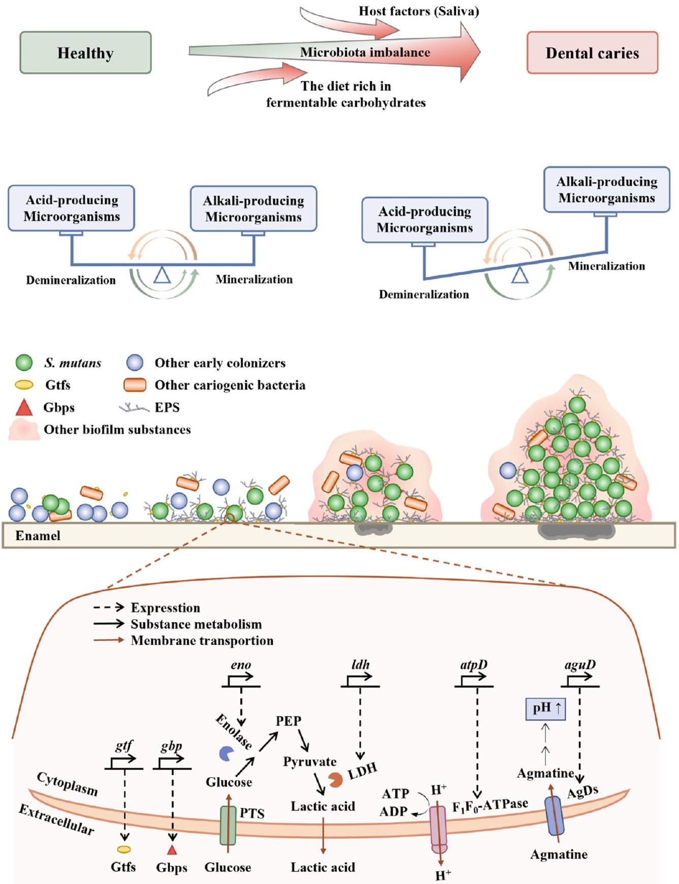
Dental caries, a highly prevalent oral disease, impacts a significant portion of the global population. Conventional approaches that indiscriminately eradicate microbes disrupt the natural equilibrium of the oral microbiota. In contrast, biointervention strategies aim to restore this balance by introducing beneficial microorganisms or inhibiting cariogenic ones. Over the past three decades, microbial preparations have garnered considerable attention in dental research for the prevention and treatment of dental caries. However, unlike related pathologies in the gastrointestinal, vaginal, and respiratory tracts, dental caries occurs on hard tissues such as tooth enamel and is closely associated with localized acid overproduction facilitated by cariogenic biofilms. Therefore, it is insufficient to rely solely on previous mechanisms to delineate the role of microbial preparations in the oral cavity. A more comprehensive perspective should involve considering the concepts of cariogenic biofilms. This review elucidates the latest research progress, mechanisms of action, challenges, and future research directions regarding probiotics, prebiotics, synbiotics, and postbiotics for the prevention and treatment of dental caries, taking into account the unique pathogenic mechanisms of dental caries. With an enhanced understanding of oral microbiota, personalized microbial therapy will emerge as a critical future research trend.

Dental caries represents a substantial and pressing global public health challenge, affecting a staggering number of individuals worldwide. Specifically, there are an estimated 64.6 million cases of permanent dentition and an additional 62.9 million cases of primary dentition . Streptococcus mutans, one of the major causative bacteria of dental caries that expresses collagenbinding protein, can effectively invade human umbilical vein endothelial cells , thereby leading to the potential development of infective endocarditis.
As dental caries is typically mediated by biofilm, interventions targeting biofilm have become a major strategy for prevention. Adjusting the intake of fermentable substrates in the diet, especially sucrose is an effective approach . The modern dietary environment is characterized by the widespread availability of highly processed and sugary foods, creating a significant challenge in completely abstaining from cariogenic foods. Other interventions include physical clearance (e.g., brushing or using interdental cleaning tools), chemical inhibition (e.g., using chlorhexidine or povidoneiodine), and biological interventions (e.g., using probiotics) . To effectively
prevent dental caries, current strategies should aim to suppress the overgrowth of specific cariogenic bacteria by targeting their virulence factors, while also promoting a diverse and healthy resident microbiota . Among these interventions, microbial preparations such as probiotics, prebiotics, synbiotics, and postbiotics have gained significant attention as they offer a more targeted and friendly approach than physical clearance and chemical inhibition.
Meurman and colleagues were pioneers in introducing probiotics into the field of dentistry. Over time, microbial preparations have gained attention as potential adjunctive therapies for preventing and treating dental caries. These preparations have demonstrated significant effectiveness in inhibiting the growth and biofilm formation of cariogenic bacteria.
This article first provides an overview of the background and pathogenic mechanisms of dental caries, focusing on the virulence factors of cariogenic bacteria S. mutans. It then summarizes the latest research progress, mechanism of action, application status, and challenges associated
with the use of probiotics, prebiotics, synbiotics, and postbiotics in the prevention of dental caries. Lastly, this article proposes future directions for the development of this field to provide more scientific, standardized, and effective guidelines for the prevention of dental caries from an academic perspective.

Dental caries Background

The oral cavity is a complex ecosystem characterized by various warm, moist microenvironments that provide ideal conditions for microbial growth . A recent study analyzing the oral microbiome identified a total of 1591 microbial species, including bacteria, fungi, archaea, viruses, and protozoa , second only in complexity to the colon . The core oral microbiota in healthy individuals remains relatively stable over seven years , while an imbalanced oral microbiota can lead to dental caries and other oral diseases . Moreover, the diversity of the oral microbial community in severe dental caries is considerably lower than that in healthy individuals . Dental caries arises from an imbalance in the oral microbiota resulting from a complex interplay between the host, diet, and microorganisms .
Of these factors, fermentable carbohydrates, which are commonly found in sweetened foodstuffs, have been identified as particularly important dietary contributors to dental caries . Consumption of sweetened foodstuffs can rapidly increase the concentration of carbohydrates in the oral cavity, leading to a sharp decline in the pH values of biofilm to 4 or even lower . Research has revealed a precise correlation between the areas of acute demineralization on the enamel surface and the highly acidic pH zones created by biofilms . This is attributed to the frequent local pH decreases can disrupt the balance between tooth mineralization and demineralization in the closed microenvironment of the biofilm . Consequently, this leads to mineral loss in teeth, resulting in white spots, cavitation, pulp infections, and even tooth loss .
Differences in oral microflora have been observed between individuals with healthy teeth and those with dental caries. For example, the findings of a study on the oral microbiome of children indicate that the genera Rothia, Neisseria, and Haemophilus, which are among the first colonizers of the oral cavity following birth , are associated with dental health . In contrast, Prevotella spp., S. mutans, and Human herpesvirus 4 (EB virus) are more commonly found in children with dental caries . Actinomycetota (35.8%) and Bacillota ( ) were the most common phyla in deep dentin carious lesions, and Lactobacillus was the most abundant genus in only of the carious lesions . There is increasing recognition that dental caries is caused by the imbalanced microbiota in the biofilm, also known as dental plaque, rather than by a single pathogen .

Microorganisms associated with dental caries

The cariogenic bacteria exhibit varying degrees of contribution to the development of dental caries. For decades, S. mutans and Streptococcus sobrinus have been widely recognized as the major cariogenic agents . It is noteworthy that S. sobrinus exhibited superior acidogenicity and aciduricity compared to S. mutans, but showed lesser adaptability to the biofilm environment . The cariogenic bacteria within the oral microbiota do not exist as isolated entities but rather interact and influence each other. To a certain extent, S. mutans creates a lactic acid-rich environment in carious lesions that facilitates the proliferation of Veillonella species , which have been shown to promote the growth of S. mutans in biofilm studies .
Additionally, Candida species, as a typical fungal representative of cariogenic microorganisms, have emerged as potent secondary cariogenic agents, isolated from to of adult and pediatric caries . Candida is a powerful opportunistic caries yeast that relies on the production of shortchain carboxylic acids and proteinases, as well as its ability to adhere to abiotic surfaces and form biofilm . The most common communication between fungi and bacteria in the oral cavity is the mutual interaction between Candida albicans and S. mutans. The presence of C. albicans promotes the growth of S. mutans, eliciting notable changes in gene expression and enhancing carbohydrate metabolism . Notably, compared
to the mono-species biofilm comprising solely S. mutans, there are 393 differentially expressed genes in S. mutans within the dual-species biofilm . The glucosyltransferases (Gtf) secreted by S. mutans can bind to C. albicans and facilitate the conversion of sucrose into exopolysaccharide (EPS), thereby providing binding sites for S. mutans .
One study reported that the core microbiota of early childhood caries (ECC) may include Veillonella parvula, Fusobacterium nucleatum, Prevotella denticola, and Leptotrichia wadei . On the one hand, this ECC core microbiota promotes the growth and acidogenicity of S. mutans, and promotes biofilm formation, albeit with limited acidogenic capacity . On the other hand, it also promotes enamel demineralization in vitro and increases the cariogenic potential of enamel in vivo . Additionally, according to some metagenomic results, the following species are closely associated with dental caries: Streptococcus gordonii, Leptotrichia buccalis, V. parvula, Actinomyces gerencseriae, Propionibacterium acidifaciens, Hallella multisaccharivorax, and Parascardovia denticolens .
Of the microorganisms associated with dental caries, S. mutans is one of the most extensively studied species in this field. Given that S. mutans was initially thought to be a major cause of dental caries , it is not surprising that most prevention strategies target this bacterium specifically .

Streptococcus mutans

When compared with other original colonizing bacteria, S. mutans exhibits more advantageous traits by developing a compact biofilm and its distinctive virulence factors . Furthermore, under the regulated control of the quorum-sensing system, S. mutans ultimately becomes one of the major cariogenic bacteria. The biofilm in dental caries of primary teeth is a threedimensional (3D) spherical structure, with S. mutans as the core and other bacteria forming the outer layer . This localized area creates an acidic pH environment, leading to severe enamel demineralization . As dental caries progresses, the diversity of the oral microbiota becomes limited . This microbial imbalance eventually leads to the occurrence and development of dental caries. The close association between S. mutans and dental caries has been confirmed. Although S. mutans is a natural resident of the human oral cavity , an increase in the levels of . mutans should be of concern as it may indicate a clinical precursor to dental caries . Rats infected with humanderived S. mutans develop dental caries , and S. mutans may be associated with severe-ECC recurrence . These suggest the crucial role of S. mutans in the occurrence and development of dental caries. Therefore, further elucidating the pathogenic mechanisms of S. mutans (Fig. 1) is essential for the development of effective strategies for caries prevention and treatment. In the following sections, we will discuss the virulence factors and quorumsensing system aspect of S. mutans.
Virulence factors. The virulence factor of S. mutans can be categorized into four major groups, comprising EPS synthesis, adhesion, acid production, and acid resistance.
The synthesis of EPS. The ability of S. mutans to exert its pathogenicity is largely attributed to the production of EPS. EPS, a major component of biofilms , consists of extracellular proteins, extracellular DNA, and lipoteichoic acid . The primary component of EPS is glucan, which is synthesized by Gtf , providing binding sites for microorganisms . EPS contributes to the formation of highly organized chemical and physical barriers within the biofilm matrix, facilitating microbial adherence to non-living surfaces , resisting fluid shear stresses , evading host immune responses , tolerating antimicrobial agents , and ultimately establishing and maintaining acidic microenvironment in the oral cavity that favors the development of dental caries-associated biofilm communities . Mature biofilms are difficult to remove mechanically due to the enhanced viscoelasticity conferred by EPS . EPS may achieve immune evasion by mediating complement evasion and limiting the entry of effector molecules from the innate and adaptive immune systems into the biofilm matrix . Chlorhexidine, a commonly used antimicrobial agent in oral care, has limited penetration into deep biofilm layers due to its positive charge, whereas “the fuel” (sucrose), lacking charge,
Fig. 1 | Cariogenic biofilm formation. EPS exopolysaccharides, Gbps Glucanbinding proteins, encode Gbps, be related to adhesion, Gtfs Glucosyltransferases, gtf encode Gtfs, be related to the synthesis of EPS, eno encode Bacteria enolase, be related to glucose uptake, LDH lactate dehydrogenase, encode LDH , be related to acid production, PTS phosphotransferase, the glucose uptake system, PEP phosphoenolpyruvate, atpD encode -ATPase, be related to acid resistance, ATP adenosine triphosphate, ADP adenosine diphosphate, AgDs agmatine deiminase system, aguD encode AgDs , be related to acid resistance. Healthy teeth develop dental caries due to the complex interactions between the host, diet, and microorganisms. The microecology of healthy teeth is based on the balance between acidogenic and alkalinogenic microbial activities, as well as the balance between demineralization and remineralization processes. When acidogenic microorganisms become predominant,
frequent and high concentrations of acid locally lead to net demineralization of dental enamel, resulting in the formation of cavities. At the micro level, the initial step involves the adhesion of some primitive colonizing microorganisms to the dental enamel. The second step involves the production of EPS by the microorganisms, forming a biofilm. In the third step, acidogenic and acid-tolerant microbial communities, mainly dominated by S. mutans, establish a highly acidic microenvironment, leading to demineralization of the dental enamel. In the fourth step, the highly acidic microenvironment confers a growth advantage to S. mutans-dominated microbial populations. Taking the virulence factors of . mutans as an example, the synthesis of EPS is primarily mediated by Gtfs. The adhesion process is mainly facilitated by Gbps. Acid production involves the participation of enolase and LDH. Acid tolerance processes primarily rely on the involvement of -ATPase and AguD.
can easily diffuse, facilitated by the negative charge of . The negatively charged surface of S. mutans cells enveloped by EPS accumulates protons, and the sieving effect of the glucan structure also plays a role . On one hand, EPS captures and accumulates protons produced externally or by acidogenic
microorganisms, aiding in the retention and accumulation of acid within the biofilm . On the other hand, once protons are recruited to the cell surface, they trigger an acid adaptation response, allowing the microorganisms to preemptively counteract acid damage . Deactivation of one or more
genes significantly reduces the virulence of S. mutans, in rodent caries models . In summary, EPS plays a crucial role in enabling S. mutans to exert its cariogenic potential. Therefore, inhibiting EPS synthesis may represent a feasible preventive strategy against dental caries .
Adhesion. S. mutans employs both sucrose-independent and sucrosedependent pathways to adhere to teeth . The initial adhesion process is primarily mediated by the sucrose-independent pathway, which is subsequently reinforced by the stimulation of glucan synthesis via the sucrosedependent pathway, ultimately culminating in the formation of biofilms . Glucan-binding proteins facilitate the binding of glucans synthesized from sucrose through glucose transferases. Of these proteins, GbpA exhibits a strong correlation with cariogenicity . On the one hand, it contributes to the formation of strong biofilm structure and is an important protein determining the structure of biofilm. On the other hand, it plays an essential role in linking glucan molecules and is involved in the bacterial adhesion process to teeth.
Acid production. After glucose metabolism, dietary carbohydrates produce energy and organic acids as metabolic by-products . The acid-producing activity of . mutans is not only a critical factor contributing to its pathogenicity but also a crucial characteristic leading to dental caries. Bacteria enolase, an enzyme encoded by the gene eno, is a primary component of the phosphotransferase system, which is responsible for glucose uptake . Through the rapid catalytic activity of lactate dehydrogenase (LDH), a protein encoded by the ldh gene, S. mutans UA159 ferments glucose into organic acids .
Acid resistance. S. mutans employs some acid-resistant mechanisms to cope with the stress of increasing acid production. -ATPase, a proton pump encoded by atp , not only pumps out intracellular protons to maintain intracellular pH but also produces ATP to promote bacterial growth and survival . Inhibition of atpD expression in S. mutans UA159 resulted in a significant decrease in acid adaptation and an increase in cytoplasmic acidity . Additionally, S. mutans produces alkali to neutralize acids, as well as export them out of the cells. The agmatine deiminase system plays a crucial role in producing alkalis to overcome acid stress . Amongst its components, the agmatine-putrescine antiporter (AguD), encoded by the gene, is of particular importance as it facilitates the intracellular transport of free agmatine . The accumulation of protons on the surface of bacterial cells enveloped by EPS plays a significant role in the acid resistance of S. mutans, as mentioned in the “synthesis of EPS” section .
Quorum-sensing (QS) system. The QS system regulates virulence and biofilm formation by releasing, sensing, and interacting with diffusion molecules based on cell density in the surrounding environment . mutans utilize this system to communicate with each other as a group rather than as separate individuals. The main mechanism for signal feedback is via the two-component signal transduction systems (TCSTS), which enable bacteria to regulate their gene expression . S. mutans contains several types of TCSTS, among which VicRKX and ComCDE are critical in the regulation of biofilm formation, acid resistance, and acid production in response to environmental signals . If these regulatory systems fail to function properly, it may lead to a decrease in the cariogenicity of S. mutans.

Dental caries prevention measures-biological interventions

Although plaque is a natural occurrence in teeth from an evolutionary, biological, and nutritional perspective, an imbalance in the microbiome of the oral pathological biofilm can lead to the development of dental caries . Acid-producing cariogenic bacteria, especially S. mutans, damage the hard tooth structures in the presence of fermentable carbohydrates .
In recent years, the field of biological intervention has developed some novel strategies. One approach involves using predators, such as Bdellovibrio, Bacteriovorax, and Peredibacter, to eliminate anaerobic Gram-negative
bacteria that are periodontal pathogens . Given that beneficial bacteria are mostly Gram-positive . Additionally, biological interventions also include the use of specific inhibitors of S. mutans proteins, vaccination, and passive immunization strategies with neutralizing bacteria . Although some innovative biological intervention strategies such as those mentioned above have emerged, the use of microbial preparations, such as probiotics, prebiotics, synbiotics, and postbiotics, is a more established and popular approach for preventing dental caries.

Probiotics

Background

Probiotics were discovered by scholars as early as , and since then the field of studying the health effects of probiotics on the host has gradually developed. In 2013, The International Scientific Association of Probiotics and Prebiotics (ISAPP) defined probiotics as “live microorganisms that, when administered in adequate amounts, confer a health benefit on the host” . Today, probiotics are commonly used by humans to maintain their overall well-being. Although their effectiveness in promoting gastrointestinal health is well-known, research has also shown that probiotics can be effective in preventing and treating various oral diseases, such as dental caries, oral mucositis, and halitosis .
In dentistry, probiotics were first introduced by Meurman and colleagues , who found that Lacticaseibacillus rhamnosus GG ATCC 53103 could colonize the human mouth. With further research, probiotics have been found to have a remarkable ability to prevent dental caries. For instance, one study explored the effect of subjects’ own Lactobacillus on . mutans . The study has shown that Lactobacillus isolated from the oral cavity of subjects can effectively inhibit the growth of S. mutans. The most effective species of S. mutans were found to be Lacticaseibacillus paracasei and Lactiplantibacillus plantarum, which are also the most common isolates. Finally, the use of probiotics in the treatment of oral diseases has been found to restore oral microbial balance and reduce the levels of . mutans in dental plaque and saliva .
With different probiotic strains exhibiting unique characteristics, understanding the specifics of each strain is crucial to when prevention and treatment of dental caries. For instance, L. rhamnosus GG is a homofermentative Lactobacillus that is not considered to be cariogenic because it cannot ferment sucrose or lactose . Limosilactobacillus reuteri is an obligate heterofermentative species that can produce broad-spectrum antimicrobials with good acid-base stability, such as reuterin and reutericyclin . In addition to Lactobacillus spp., Bifidobacteria spp. may also be a potential probiotic for preventing and treating dental caries. Yogurt containing Bifidobacterium DN-173010 has been reported to significantly reduce the level of S. mutans .

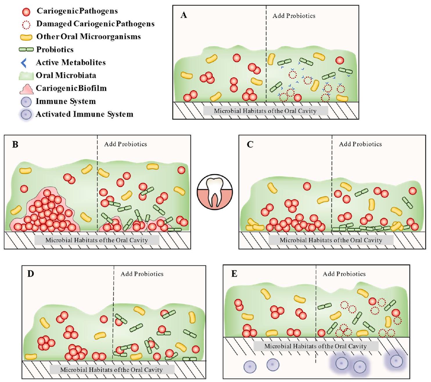
Mechanisms to prevent dental caries

The mechanism through which probiotics can prevent dental caries is similar to that found in the gastrointestinal tract. The principal inhibitory mechanisms include the synthesis of active metabolites, inhibition of cariogenic microbial biofilm, competitive adhesion and colonization, coaggregation with pathogens, and regulation of the immune system (Fig. 2).

Production of active metabolites

Bacteriocin. Bacteriocin is a cationic antibacterial peptide synthesized by the ribosome and was first discovered . Bacteriocins can be divided into four different classes, with Classes I and II being the primary focus of most probiotics research . Nisin, a bacteriocin of Type A in Class I, is widely recognized as a small positively charged protein ( ) that induces target cells to form membrane pores . Class II bacteriocins kill bacteria by increasing membrane permeability and leaking target bacterial contents . In addition to the above mechanisms, bacteriocins can also inhibit the synthesis of biofilm and cell wall, exert the activities of DNase and RNase, and regulate microbiota .
Protein-protein interaction between the GtfB and LuxS proteins of . mutans and bacteriocin of SD1 in L. paracasei was found to reduce the
Fig. 2 | The mechanism of probiotics to prevent dental caries. It is roughly divided into five parts. A Production of active metabolites: probiotics directly inhibit cariogenic pathogens by active metabolites (e.g., bacteriocin, enzyme, biosurfactants, organic acids, and hydrogen peroxide), which themselves have bacteriostatic activity. B Inhibition of cariogenic microbial biofilm: probiotics can inhibit or remove the biofilm of oral cariogenic microorganisms. C Competitive adhesion and colonization: probiotics not only occupy the colonized sites in the oral cavity but also
inhibit the adhesion ability of cariogenic microorganisms. D Coaggregation with pathogens: probiotics inhibit cariogenic microorganisms colonization in the oral cavity through co-aggregation. E Regulation of the immune system: probiotics activate or modulate the host immune system, thereby enhancing the immune response to cariogenic microorganisms (enhances salivary levels of human neutrophil peptides 1-3).
formation of biofilm and the density of microorganisms, as revealed in a simulation study . Following comprehensive bioinformatics analysis and characterization, the bacteriocin in this study was found to be safe for humans. The bacteriocin Mersacidin exerts its bactericidal activity by forming a complex with lipid II, which inhibits cell wall synthesis . The DNase and RNase activities of colicins from E2 to E9 enable them to nonspecifically degrade bacterial DNA and RNA . Among these, colicin E2 exhibits potent and long-lasting bactericidal activity, and interestingly, it can specifically target bacteria in complex biological membranes . Bacteriocins are capable of promoting the colonization of producer bacteria in specific niches over a decade, regulating the composition of the microbiota and affecting the host immune system . Both nisin and nisin-producing probiotics can reduce the level of pathogens in biofilm and restore the diversity of strains to a healthy level .
As bacteriocins are polypeptides and proteins, temperature control is crucial to ensure their activity during production and use. Streptococcus oralis subsp. dentisani 7746 (AB-Dentisanium), for instance, is optimally concentrated at 30 and , with a small reduction in bacteriocin activity at . This critical consideration underscores the importance of implementing appropriate temperature regulation strategies in the development and use of bacteriocins for various applications.
Enzyme. In addition to bacteriocins, probiotics synthesize a diverse range of enzymes that confer beneficial effects by decomposing biofilms and affecting bacteriocin activity. For instance, Lactobacillus acidophilus can secrete lipase to degrade biofilm . Similarly, Streptococcus salivarius JH expresses a dextranase enzyme that can hydrolyze the EPS of S. mutans and increase the anti-S. mutans inhibitory activity of zoocin A, a muralytic bacteriocin . Another example is Streptococcus sp. A12, which produces challisin-like proteases that inhibit the production of bacteriocins by . mutans . Additionally, S. salivarius M18 produces urease and dextranase to neutralize salivary acidity and reduce plaque formation, respectively .
Biosurfactants (BS). Biosurfactants are amphiphilic substances produced by microbial metabolism that contain both hydrophobic and hydrophilic groups, mainly composed of proteins, sugars, and lipids . The structure of BS can be identified using various techniques such as thin layer chromatography, Fourier Transform Infrared Spectrometer, and Nuclear Magnetic Resonance . Fifty percent of the 40 biosurfactant reports reviewed did not analyze their structure, likely due to the complexity of the structures . Surfactin and/or protein-like biosurfactants ( ) are most commonly produced by Lactobacillus, with studies on glycoproteins , glycolipid , and glycolipopeptide production being rare.
Lactobacillus typically produces surfactin-type biosurfactants, which are protein-rich and can significantly inhibit pathogen adhesion, making them increasingly interesting due to their unique anti-adhesion and antibiofilm properties . For example, BS produced by . reuteri DSM 17938, . acidophilus DDS-1, L. rhamnosus ATCC 53103, and L. paracasei B21060 inhibited S. mutans and Streptococcus oralis adhesion and biofilm formation in a dose-dependent manner on titanium surfaces . In more detail, . acidophilus DSM 20079 produces protein-type BS, which can shorten the chain length of . mutans, interfere with its biofilm formation on glass slides, and down-regulate the gene expression of and . BS produced by . rhamnosus can destroy the physical structure or protein conformation of biofilm, leading to cell lysis . In addition to the appealing antimicrobial activity mentioned above, BS exhibits characteristics of low cytotoxicity and high stability. BS derived from Lactobacillus spp. demonstrate comparable low cytotoxicity to rhamnolipids, which are generally regarded as non-toxic products . BS may exhibit better stability compared to other antibacterial substances produced by probiotics. Gudinahe et al. isolated stable BS from L. paracasei. This BS was pH stable within a range of and maintained surface activity after incubation at for 120 h .
Organic acids. Organic acids, such as lactic acid and butyric acid , produced by Lactobacillus in the human gastrointestinal tract and other body parts, have widely been recognized as beneficial substances. These organic acids may have a bacteriostatic effect on oral pathogenic microorganisms to a certain degree. For instance, L. paracasei Lpc-37 produces the acid that can restrain the growth and biofilm formation of S. mutans . However, considering the strong association between dental demineralization and frequent exposure to high concentrations of acid , it raises questions about how the acids produced by probiotics counteract the acids produced by cariogenic bacteria, including S. mutans.
These questions may need to be approached from the perspective of the overall caries environment. Cariogenic microorganisms create a highly organized acidic barrier . Prolonged exposure to localized high concentrations of acid leads to localized demineralization rather than average demineralization of the teeth. If the organic acids produced by probiotics can inhibit cariogenic bacteria, including S. mutans, and/or their biofilms, they may disrupt this acidic barrier and prevent acid accumulation. Given the presence of its own acid-base microbial balance in the oral cavity , acids that do not accumulate but instead contribute to the acid-base equilibrium in the oral environment appear to pose a lesser threat.
Hydrogen peroxide. Certain probiotic species, including Bifidobacterium bifidum, Lactobacillus johnsonii, Lactobacillus crispatus, and Lactobacillus jensenii, produce hydrogen peroxide to exert antibacterial effects . Hydrogen peroxide acts on the pathogenic bacteria’s epithelium, leading to their death . Moreover, hydrogen peroxide has the potential to regulate species composition within the oral cavity . Notably, cariogenic species such as S. mutans are highly susceptible to hydrogen peroxide toxicity . However, the antibacterial activity of . paracasei cell-free supernatant (CFS) was significantly reduced after catalase treatment, indicating the involvement of hydrogen peroxide in its bacteriostatic effects . It should be noted that hydrogen peroxide may not retain its bacteriostatic function after processes such as freeze-drying, owing to the ease of its decomposition .
Inhibition of cariogenic microbial biofilm. Dental caries is commonly mediated by biofilm. A crucial property of probiotics is the ability to inhibit or eliminate the growth of biofilms and pathogenic microorganisms in the oral cavity. Some strains have often been reported for their anti-biofilm activity, including S. oralis 89a, Limosilactobacillus fermentum TCUESC01, L. acidophilus 4A, and Bifidobacterium longum subsp. longum . For instance, Lacticaseibacillus casei ATCC 393, . reuteri ATCC 23272, L. plantarum ATCC 14917, and Ligilactobacillus salivarius ATCC 11741 may suppress the biofilms of . mutans by downregulating genes such as , and in . . Interestingly, probiotics are capable of inhibiting fungi from transitioning into
pathogenic forms. It has been demonstrated that . rhamnosus LR32, . casei L 324 m , and . acidophilus NCFM exhibit the ability to impede the initial stages of hyphal formation, which is a crucial step in the pathogenesis of C. albicans . A probiotic combination consisting of Lactobacillus helveticus CBS N116411, L. plantarum SD5870, and S. salivarius DSM 14685 significantly down-regulated the expression of genes involved in yeast-hypha transition in C. albicans, including EFG1 (hyphae-specific gene activator), SAP5 (secreted protease), ALS3 (adhe ) and HWP1 (hyphal wall protein) . In vitro, biofilm models are continuously improving to replicate more closely the conditions found within the human body. Based on the specific research objectives, researchers can opt for models of interest, such as an experimental abutment mimicking the macro- and microstructure of a dental implant .
The combination of . rhamnosus and collagen peptides was found to significantly increase the pH of the medium in the early stages of biofilm formation . The qPCR results showed that this combination downregulated several crucial genes linked to acid production and acid tolerance, including eno, ldh, and atpD. Moreover, probiotics may also exert antibacterial effects by interfering with QS. A study revealed that comD, vicR, and vic genes were down-regulated in planktonic and biofilm forms of . mutans when exposed to CFS with Lactobacillus . This effect may explain the reduced adherence and biofilm formation of . mutans observed in scanning electron microscopy experiments.
Competitive adhesion and colonization. One of the key characteristics of probiotics contributing to their health effects is their capacity for outcompeting oral pathogens concerning adhesion and colonization . For example, L. reuteri LR6 displayed the most substantial adhesion capabilities among eight tested probiotic strains, which corresponded to a higher ability for inhibiting the adherence of pathogens to Caco-2 cells . Enhanced colonization efficacy by S. salivarius M18 resulted in stronger anti-caries activity as evidenced by a reduction in plaque scores and . mutans levels . Levilactobacillus brevis KCCM 202399 inhibited the adherence of . mutans KCTC 5458 by reducing the self-aggregation, cell surface hydrophobicity, and EPS production of S. mutans .
Interestingly, probiotics can reduce pathogen adhesion even without direct contact. Saliva treated with probiotics was shown to reduce the adhesion of S. mutans to hydroxyapatite surfaces (a model for enamel) . Further studies showed that the above salivary membrane lacked two proteins: salivary lectin gp340, the primary receptor for S. mutans in the salivary membrane, and salivary peroxidase, an innate defense factor found in human saliva .
Coaggregation with pathogens. Coaggregation is among the advantageous properties of probiotics as it allows them to form a barrier that impedes pathogen colonization . In a study, six out of 624 lactic acid bacteria were found to exhibit specific coaggregation with S. mutans in vitro . These species were identified as L. paracasei and L. rhamnosus. It was discovered that this coaggregation mechanism is highly resilient to both hyperthermia and protease, and does not rely on lectins, nor is it impacted by saliva.
Regulation of the immune system. In addition to their direct effects on pathogenic microorganisms or biofilm, probiotics are known to activate and modulate the host’s immune system . Clinical studies have shown that daily or tri-weekly consumption of . paracasei SD1 in patients with severe ECC significantly enhances salivary levels of human neutrophil peptides 1-3 with a broad bactericidal activity and reduces S. mutans levels, potentially slowing the progression of caries . Furthermore, the consumption of milk containing L. paracasei SD1 for six months increased salivary immunoglobulin A levels, and this increase is positively correlated with a load of . paracasei .
Certain strains of Streptococcus thermophilus, such as ST1342, ST1275, and ST285 activate the innate immune response and stimulate the secretion
of interleukin- , tumor necrosis factor- , interleukin- 6 , and interferon- by monocytes, thereby contributing to the elimination of pathogens . Commercial L. paracasei DG has immunostimulatory activity by boosting tumor necrosis factor- , interleukin-6, and Chemokine (C-C motif) ligand 20 expressions in human monocyte leukemia cell . These findings suggest that probiotics can enhance the host’s immune response against pathogenic microorganisms, providing a potential approach to preventing and treating infectious diseases.

The application vehicle

The colonization of probiotics in the oral cavity may be influenced by the choice of the delivery vehicle . A range of vehicles is available for delivering probiotics, including dairy products, ice cream, cereal, pacifiers, chewing gum, curd, juice, and mouth wash (Table 1). Table 1 also mentions the test species, dose, and efficacies.
Among these vehicles, dairy products are considered ideal carriers due to their inherent beneficial characteristics . Among the dairy products, liquid substrates, such as milk and yogurt, were found to be more effective in reducing S. mutans levels . For individuals who are allergic to dairy products, alternative carriers may be selected, as illustrated in Table 1. The buffering capacity of milk helps to reduce acid production, while its colloidal nature appears to protect enamel . In addition, milk contains calcium and calcium lactate, which may have a preventive effect against caries , and can reduce the colonization of pathogenic microorganisms . Furthermore, milk and cheese promote the dominance of casein phosphopeptides which are known to play a key role in biomineralization . A systematic review and meta-analysis showed that dairy products containing probiotics had a significant impact in reducing S. mutans and raising salivary .
Interestingly, the slow release of probiotics can also be achieved using appropriate embedding materials. For instance, L. paracasei 28.4-gellan formulations were recently found to release the probiotic for over . paracasei in this state was able to inhibit S. mutans in both the floating and biofilm states, significantly reduce the generation of EPS, and downregulate the luxS, , and genes.

Controversy

Some researchers have taken a critical view of the idea that probiotics can prevent dental caries, primarily focusing on its safety and potential cariogenic effects. The majority of probiotics are not derived from the oral microbiota but from fecal samples, and some even come from animals . Therefore, it is necessary to thoroughly evaluate their safety before clinical use. Probiotics may pose a risk to individuals with damaged barriers or low immunity, such as bacteremia because high concentrations of administration are key to medication. Regarding the cariogenic issue, after conducting a meta-analysis of 50 experiments related to dental caries and periodontal diseases, Gruner and colleagues concluded that there is insufficient evidence to support the use of probiotics in treating dental caries . Subjects with active dental caries showed higher levels of S. mutans, Actinomyces sp. strain B19SC, and Lactobacillus spp. as detected by PCR-based methods . The major lactic acid bacteria identified from carious lesions, including both adults and children, include L. fermentum, L. casei/paracasei, L. salivarius, L. rhamnosus, L. plantarum, and L. gasseri . Bifidobacterium dentium, considered a late marker for dental caries progression, was not found in the oral cavity of caries-free individuals but was detected in 30.8% of caries cases in a study of 56 participants .
It is unreasonable to conclude a causal relationship between lactic acid bacteria and caries if there is a strong correlation between lactic acid bacteria and caries scores . Lactic acid bacteria have a relatively low affinity for teeth, and their ability to form biofilms in vitro is much weaker than that of . mutans . The attachment and proliferation of secondary invaders including Bifidobacterium and lactic acid bacteria requires initiation of caries by major caries promoters including S. mutans to create an anaerobic acidic environment rich in carbohydrates . The destruction of dentin is not enough by lactic acid alone but also requires proteolytic activity, because the main component of dentin is more than the extracellular matrix
dominated by type I collagen. However, lactic acid bacteria, including . rhamnosus, L. casei/paracasei, L. salivarius, Lactobacillus vaginalis, Lactobacillus gasseri, Limosilactobacillus oris, and L. fermentum, have a greater propensity to bind to collagen rather than degrade it based on genomic analysis .
In conclusion, the effectiveness of probiotics in preventing dental caries remains controversial. It is beneficial to prevent dental caries by exploring the roles of each microflora in the transition from oral health microbiota to cariogenic microbiota. In particular, from the perspective of oral microecology, the diet composition, host immune environment, and the physical and chemical characteristics of the oral cavity, especially the teeth, should be fully considered. Therefore, in recent years, researchers have become increasingly interested in exploring the benefits of prebiotics, synbiotics, and postbiotics in preventing dental caries, especially in terms of advantages over probiotics.

Prebiotics

In 1995, prebiotics were defined as “non-digestible food ingredient that beneficially affects the host by selectively stimulating the growth and/or activity of one or a limited number of bacteria already resident in the colon” . However, with advancements in scientific research, ISAPP deemed the definition of prebiotics as “a substrate that is selectively utilized by host microorganisms conferring a health benefit” more appropriate in . Prebiotics present a safe and effective alternative to probiotic intervention since they are not live bacteria and are less susceptible to environmental factors affecting probiotic survival and efficacy. The following section will discuss prebiotics according to their different types, including their mechanisms of action, efficacies, and other related aspects.

Sugar

Interestingly, certain sugars exhibit prebiotic properties. D-tagatose, a noncariogenic sugar, is a potential prebiotic that offers lower calories and a lower glycemic index than sucrose . Notably, the saliva of individuals with good oral health is rich in D-tagatose . D-tagatose may inhibit the growth of . mutans and S. gordonii by affecting glycolysis and its downstream metabolism, but it does not affect S. oralis . Encouragingly, chewing gum containing D-tagatose has been shown to inhibit the growth of S. mutans .
In addition to D-tagatose, other sugars such as xylose and arabinose are considered potential prebiotics, with the capacity to not only inhibit the growth of S. mutans but also promote the growth of Lactobacillus . This dual action is particularly advantageous, as it may help restore the balance of the oral microbiome. Given the promising prebiotic properties of these sugars, further research is needed to assess their effectiveness in vivo and their potential side effects.

Sugar alcohol

Sugar alcohols, such as xylitol, sorbitol, maltitol, and erythritol, have been shown to exhibit prebiotic properties that can enhance oral health. Xylitol, a five-carbon polyol sweetener , is considered an oral-specific prebiotic according to the new definition established in . It offers numerous benefits, including the enhancement of remineralization, decrease of the pH of dental plaque, reduction of the level of . mutans in saliva, reduction of the insoluble dextran in the biofilm of . mutans, and reduction of dental caries incidence . However, xylitol loses its effect in the presence of fructose or sucrose . Other sugar alcohols, such as sorbitol, maltitol, and erythritol, have also been shown to inhibit dental caries .

Oligosaccharides

In addition to sugar alcohols, oligosaccharides are also being investigated as prebiotics. Human milk oligosaccharides (HMOs), the third most abundant ingredient in human milk, are often added to infant formula . Galactooligosaccharides (GOS) and 2 ‘-fucosyllactose, the most abundant HMOs, were found to reduce the EPS-mediated adhesion of S. mutans DSM 20523 to the glass surface, indicating their potential as prebiotics for oral health promotion . In addition, GOS, glucomannan hydrolysates, and mannose
Table 1 | Application vehicle and therapeutic effects of probiotics
Vehicle Test strain Dose Frequency Sample Result Reference
Milk powder L. paracasssei CFU once daily for 3 months 124 children aged 1.5-5 reduced the count of S. mutans in saliva and delayed the development of new dental caries 187
Milk L. paracasei CFU once daily for 4 weeks 30 orthodontically treated nonsyndromic cleft lip and palate patients with a mean age of 19 reduced the count of . mutans, while increasing the count of Lactobacillus and the colonization 188
Yogurt B. animalis once daily for 2 weeks 49 healthy children aged 6-12 could not reduce the levels of salivary S. mutans and Lactobacillus 189
Yogurt B. lactis unclear once daily for 2 weeks 30 individuals aged 10-30 undergoing orthodontic treatment reduced total microbial counts in dental plaque 190
Yogurt B. lactis BB12 CFU 300 g daily for 2 weeks 66 students aged 18-30 with initial stages of dental caries reduced the count of S. mutans and Lactobacillus in the probiotic group 191
Cheese L. acidophilus NCFM or L. rhamnosus Lr-32 (DuPont Danisco , São Paulo, Brazil) each strain 50 g daily for 16 weeks 60 elderly denture wearers reduced the colonization of oral Candida 192
Cheese L. rhamnosus GG and L. rhamnosus LC705 each strain daily for 3 weeks after a meal or snack 74 adults aged 18-35 reduced the count of S. mutans during the posttreatment period 193
Cheese L. casei LAFTIL26 50 g twice daily for 2 weeks with breakfast and dinner meals 60 adults with a mean age of 28 could not reduce the count of S. mutans and Lactobacillus in the probiotic group 194
Ice cream B. lactis Bb-12 and L. acidophilus La-5 CFU each strain once daily for 7 days 60 healthy children aged 6-12 reduced the count of salivary S. mutans 195
Cereal L. paracasei F19 CFU once daily for 9 months 179 infants aged 4 months no impact on the frequency of dental caries, mutans streptococci, or lactobacilli 196
Novel slow-release pacifier B. animalis lactis BB-12 CFU twice daily for 2 years 106 infants aged 1-2 months no impact on the oral colonization of B. animalis lactis BB-12 and mutans streptococci in the early administration 197
Chewing gum L. reuteri ATCC 55730 and ATCC PTA 1 gum each strain three times daily after meals for 3 weeks 80 healthy adults aged 21-24 significantly reduced the levels of salivary mutans streptococci 198
Curd L. acidophilus and B. lactis BB12 (Mother dairy b-activ Plus ) unclear once daily for 7 days before breakfast 60 caries-free adults aged 20-25 Significantly improved salivary pH and reduced the count of salivary S. mutans 199
Curd L. acidophilus-SD 5221 (Active Plus; Nestle, Chennai, India) CFU with their lunch for 30 days 60 orthodontic patients aged 14-29 significantly reduced the levels of . mutans in the plaque around the brackets 200
Carrot-pineapple juice (Gefilus’) L. rhamnosus GG five times a week for 7 months 530 healthy children aged 3-6 reduced the count of . mutans and the risk of dental caries 201
Mouthwash (ProBiora ) S. oralis KJ3sm, S. uberis KJ2sm, and S. rattus JH145 or CFU each strain twice daily for 4 weeks 20 healthy adults aged 21-35 reduced the levels of S. mutans 202
have also been shown to inhibit pathogen adhesion to epithelial cells by binding to the pathogen’s lectins/pili . However, it should be noted that non-digestible and/or non-absorbable sugar alcohols and oligosaccharides, though beneficial for health, excessive intake may lead to significant diarrhea . Therefore, further research is needed to determine the optimal dosage and duration of intake to minimize such adverse effects.

Arginine

Arginine is a widely studied oral prebiotic that has been shown to exhibit various beneficial effects on oral health . These benefits include the promotion of alkaline substance production, the mitigation of tooth demineralization, and the suppression of biofilm formation. Specifically, arginine can inhibit the growth of Candida and reduce enamel demineralization . Furthermore, L -arginine has been found to enhance the alkali-producing ability of arginine-solubilizing bacteria, such as Streptococcus sanguinis and S. gordonii , thereby making the biofilm environment unsuitable for cariogenic microflora by increasing the pH . Interestingly, L -arginine was found to significantly reduce the amount of insoluble EPS by 3 -fold, targeting .
Numerous studies have used toothpaste containing arginine to explore the mechanism underlying its prebiotic effects on oral health in depth. An in vivo study conducted on the oral ecosystem has revealed that the presence of arginine in toothpaste enhances the arginolytic capacity of human saliva while reducing its sucrose metabolic activity . Additionally, it promotes a shift in the composition of salivary microbiota towards a healthier ecological state. Notably, tubes of toothpaste containing arginine and fluoride were more effective at preventing and reversing early caries lesions and significantly increased re-mineralization compared with fluoride-only toothpaste . Using toothpaste containing fluoride and arginine was also associated with an increase in gene expression associated with the arginine deiminase pathway, according to metagenomic and metagenomic data . Moreover, the use of toothpaste containing fluoride and arginine was found to reduce caries bacteria and promote healthier microbial communities.
Interestingly, arginine may stimulate the production of effective substances by probiotics. Exogenous arginine has been found to increase the expression of the S. gordonii spxB gene, which encodes a pyruvate oxidase , thereby promoting hydrogen peroxide production . Additionally, using as a cofactor of the catalytic activity of SpxB has been shown to increase the production of and promote the abundance of SpxB in . sanguinis and S. gordonii .

Urea and nitrates

Urea and nitrates have been investigated as potential oral prebiotics. Urea is considered a prebiotic due to its ability to be converted into ammonia or ammonium and bicarbonate ion by bacteria possessing urease, such as . salivarius, Actinomyces naeslundii, and Haemophilus spp., thereby neutralizing acids in the oral cavity . Nitrate-reducing bacteria in the oral cavity convert salivary nitrate to nitrite, which is subsequently reduced to nitric oxide . All three compounds have been shown to restrict the growth of pathogenic bacteria . Nitrate has demonstrated the ability to reduce caries incidence and inhibit bacteria commonly associated with dental caries, such as S. mutans NCTC 10499, L. casei, and A. naeslundii, as well as periodontal disease-related bacteria including F. nucleatum, Eikenella corrodens, and Porphyromonas gingivalis . However, it can lead to an increase in the levels of Neisseria and Rothia, genera associated with oral health and nitrate reduction . Salivary nitrate supports nitrate respiration by anaerobic microorganisms, ultimately increasing oral pH through various mechanisms. These mechanisms include competition for carbon sources with acid-producing fermentation processes, generation of hydroxyl ions, and dissimilation of nitrate into ammonium to consume organic acids . In addition to its impact on dental caries, dietary nitrate not only benefits oral health by significantly reducing gum inflammation but also contributes to overall health by lowering systemic blood pressure , promoting vascular health , and even potentially improving vascular function in patients with hypercholesterolemia . Considering the systemic health
effects of nitrate, further investigation is warranted to explore other mechanisms through which nitrate can prevent dental caries.
Both probiotics and prebiotics have beneficial effects on health and combining them appropriately for co-administration represents another approach to drug administration. The following section will further elaborate on this combination.

Synbiotics

The combined use of probiotics and prebiotics has demonstrated superior therapeutic effects compared to their utilization . The term “synbiotics” was first coined in 1995 by Gibson et al. to describe the combination of probiotics and prebiotics . ISAPP updated the definition of synbiotics in 2020, stating that it is a mixture comprising live microorganisms and substrate(s) selectively utilized by host microorganisms that confers a health benefit on the host . It is not surprising that synbiotics have received less attention compared to probiotics, as it was proposed relatively later.
Several studies have indicated potential oral health benefits associated with synbiotic use. For instance, Nunpan et al. demonstrated that the synbiotic composed of . acidophilus in combination with GOS and fructooligosaccharides can significantly inhibit the growth of S. mutans. Tester and Al-Ghazzewi found that synbiotics composed of Konjac glucomannan hydrolysates and . acidophilus reduced the levels of . mutans in vitro. In another study, Kojima et al. proposed a novel symbiotic . They screened five strains of lactobacilli using sugar assimilation tests with 12 different saccharides, among which the three most promising prebiotics were found to be arabinose, xylose, and xylitol. The selected lactobacilli significantly inhibited the production of water-insoluble glucan by S. mutans .
Considering the significant role of excessive acid in dental caries development, the selection of synbiotics comprising prebiotics capable of maintaining a high pH oral environment represents an innovative and intelligent approach. Synbiotics composed of -arginine and . rhamnosus not only reduced the biomass of . mutans biofilm but also decreased lactate content in spent media, resulting in no significant decline in pH within . This suggests that synbiotics modulate the ecology of dental plaque. Remarkably, this study also observed that the addition of L-arginine promoted the positive utilization of amino acid biosynthetic pathways by . rhamnosus, thus facilitating its proliferation. These findings indicate that the selection of synbiotics capable of pH regulation may offer stronger advantages in the oral cavity.
These findings provide promising evidence for the development of synbiotics as a novel approach to improving oral health. Further research is needed to establish the safety and efficacy of different synbiotic combinations. It is hoped that these advances will lead to the development of innovative yet effective strategies for promoting oral health, thereby improving overall health outcomes.

Postbiotics

In 2021, ISAPP defined “postbiotics” as “the preparation of inanimate microorganisms and/or their components that confer a health benefit on the host” , excluding essentially purified metabolites such as butyric acid . Before this, the term and definition of postbiotics were not officially standardized and unified. Postbiotics have also been referred to as “paraprobiotics”, “heat-killed probiotics”, “ghost probiotics”, “non-viable probiotics” and “bacterial lysates” . Over the past few years, the research direction of postbiotics has garnered increasing attention from researchers and has gradually become a hot research topic. Postbiotics are considered superior to probiotics due to their good acid-base and thermal stability, ease of storage and use, and high safety, as many probiotics are sensitive to oxygen and heat . The feature of postbiotics enables them to be added to common products such as toothpaste, chewing gum, natto, potato chips, popcorn, and suckable candies . Another advantage of postbiotics is that microorganisms cannot be isolated from commercial products, thus enabling product developers to maintain ownership of their components .
The main methods of preparing postbiotics are heat inactivation of bacterial cells and preparation of CFS. In addition, other inactivated
technologies, such as electric field, ultrasonication, high pressure, X-rays, high voltage electrical discharge, magnetic field heating, moderate magnetic field, and plasma technology, are also available . It should be noted that the mode of inactivation may affect the activity of postbiotics to some extent. For example, in one study, the activity of CFS of S. oralis subsp. dentisani 7746 concentrated at was lost, while CFS concentrated at or retained its activity . The ability of heat-killed . reuteri to adhere to and inhibit pathogens was significantly reduced compared to L. reuteri , possibly due to the alteration of physical and chemical properties caused by heat treatment .
Although postbiotics do not contain live microorganisms, this does not imply that inactivated bacteria have completely lost all beneficial properties. For example, heat-killed Bifidobacterium animalis BB12 still can reduce the cariogenicity of biofilm in vitro , indicating that heat-killed bacteria did not completely lose all their beneficial properties. As another example, . paracasei DSMZ16671 maintained its ability to co-aggregate with S. mutans after heat-killed treatment (autoclaved at for 20 min . There was even a study that showed that . rhamnosus CNCM-I-3698 and Companilactobacillus farciminis CNCM-I-3699 had a greater ability to exclude pathogens and adhesion after heat-killed treatment . The inactivation process may lead to the disruption of bacterial cell structures, making bioactive molecules more exposed and accessible for utilization . Postbiotics, which are mixtures of various components, may exert their health effects on the oral cavity through multiple mechanisms. These mechanisms can be independent or cooperative and may resemble the previously described mechanisms of probiotic health effects. In the interest of brevity, we will not reiterate these mechanisms here.
In the context of dental caries prevention and treatment, the types of postbiotics commonly utilized include CFS and heat-killed probiotics. However, it is worth mentioning that research in fields beyond the oral cavity has explored a broader range of postbiotic types, such as peptidoglycans, lipopolysaccharides, and pili . Additional investigations into the mechanisms by which other types of postbiotics act concerning dental caries have the potential to provide innovative approaches for the prevention and treatment of this condition. Furthermore, further studies are warranted to assess the safety and effectiveness of postbiotics, thereby contributing to the improvement of overall health outcomes.

Conclusion and future perspectives

Dental caries prevention and treatment is a critical issue in oral healthcare. The traditional prevention and treatment methods have mainly focused on physical removal and chemical inhibition. While minimally invasive dental restoration can treat cavities, it fails to address the underlying causes of new caries formation. Thus, safer, more effective, and personalized preventive and treatment strategies are urgently needed. Extensive research has been conducted on the application of microbial preparations, such as probiotics, prebiotics, synbiotics, and postbiotics, in the prevention and treatment of dental caries, with promising outcomes. These microbial preparations can modulate the balance of oral microbiota by introducing beneficial microorganisms or inhibiting pathogenic ones. This review aims to help overcome theoretical obstacles to the successful clinical application of microbial preparations in preventing and treating dental caries.
In conclusion, investigating the potential applications of microbial preparations in the prevention and treatment of dental caries is an essential research avenue in the field of oral microbiology. The successful application of microbial preparations in the clinical setting can provide crucial support and greater assurance for oral health. Dental caries pathogenesis is closely associated with the balance of oral microbiota. Therefore, future research should focus on obtaining a deeper understanding of the characteristics and interrelationships among various beneficial and harmful microbial populations. With such comprehensive research, it is advantageous to develop more effective microbial preparations and optimal allocation methods. Developing personalized treatment plans is the current trend, and by devising microbial preparations that are effective for different dental caries risk and medication risk populations, personalized prevention and
treatment of dental caries can be achieved. Furthermore, personalized treatment schemes are not limited to single microbial preparations and can be used alongside other treatment modalities to enhance the overall therapeutic outcomes and achieve the goal of curing dental caries. Improving efficacy and safety is a critical direction for future research. Based on a further exploration of the mechanisms of action of microbial preparations, refining and optimizing formulations according to research findings can enhance the therapeutic effects and safety of microbial preparations.
Received: 7 December 2023; Accepted: 12 February 2024;
Published online: 24 February 2024

References

  1. Wen, P. Y. F., Chen, M. X., Zhong, Y. J., Dong, Q. Q. & Wong, H. M. Global burden and inequality of dental caries, 1990 to 2019. J. Dent. Res. 101, 392-399 (2022).
  2. Nomura, R. et al. Potential involvement of Streptococcus mutans possessing collagen binding protein Cnm in infective endocarditis. Sci. Rep. 10, 19118 (2020).
  3. Philip, N., Suneja, B. & Walsh, L. J. Ecological approaches to dental caries prevention: paradigm shift or shibboleth? Caries Res. 52, 153-165 (2018).
  4. Yu, O. Y., Lam, W. Y., Wong, A. W., Duangthip, D. & Chu, C. H. Nonrestorative management of dental caries. Dent. J. 9, 121 (2021).
  5. Marsh, P. D., Head, D. A. & Devine, D. A. Ecological approaches to oral biofilms: control without killing. Caries Res. 49, 46-54 (2015).
  6. Meurman, J. H., Antila, H. & Salminen, S. Recovery of Lactobacillus strain GG (ATCC 53103) from saliva of healthy volunteers after consumption of yoghurt prepared with the bacterium. Microb. Ecol. Health Dis. 7, 295-298 (1994).
  7. Chattopadhyay, I. et al. Can metagenomics unravel the impact of oral bacteriome in human diseases? Biotechnol. Genet. Eng. Rev. 39, 85-117 (2022).
  8. Achtman, M. & Zhou, Z. Metagenomics of the modern and historical human oral microbiome with phylogenetic studies on Streptococcus mutans and Streptococcus sobrinus. Philos. Trans. R. Soc. B Biol. Sci. 375, 20190573 (2020).
  9. Wade, W. G. The oral microbiome in health and disease. Pharmacol. Res. 69, 137-143 (2013).
  10. Rosier, B. T., Marsh, P. D. & Mira, A. Resilience of the oral microbiota in health: mechanisms that prevent dysbiosis. J. Dent. Res. 97, 371-380 (2018).
  11. Kilian, M. The oral microbiome-friend or foe? Eur. J. Oral. Sci. 126, 5-12 (2018).
  12. Kanasi, E. et al. Clonal analysis of the microbiota of severe early childhood caries. Caries Res. 44, 485-497 (2010).
  13. Hajishengallis, E., Parsaei, Y., Klein, M. I. & Koo, H. Advances in the microbial etiology and pathogenesis of early childhood caries. Mol. Oral Microbiol. 32, 24-34 (2017).
  14. Forssten, S. D., Bjorklund, M. & Ouwehand, A. C. Streptococcus mutans, caries and simulation models. Nutrients 2, 290-298 (2010).
  15. Gong, Y. et al. Global transcriptional analysis of acid-inducible genes in Streptococcus mutans: multiple two-component systems involved in acid adaptation. Microbiology 155, 3322-3332 (2009).
  16. Kim, D. et al. Spatial mapping of polymicrobial communities reveals a precise biogeography associated with human dental caries. Proc. Natl. Acad. Sci. USA 117, 12375-12386 (2020).
  17. Peres, M. A. et al. Oral diseases: a global public health challenge. Lancet 394, 249-260 (2019).
  18. Palmer, R. J. et al. Interbacterial adhesion networks within early oral biofilms of single human hosts. Appl. Environ. Microbiol. 83, e00407-e00417 (2017).
  19. Baker, J. L. et al. Deep metagenomics examines the oral microbiome during dental caries, revealing novel taxa and co-occurrences with host molecules. Genome Res. 31, 64-74 (2021).
  20. Liu, G., Wu, C., Abrams, W. R. & Li, Y. Structural and functional characteristics of the microbiome in deep-dentin caries. J. Dent. Res. 99, 713-720 (2020).
  21. Jenkinson, H. F. & Lamont, R. J. Oral microbial communities in sickness and in health. Trends Microbiol. 13, 589-595 (2005).
  22. Kazemtabrizi, A., Haddadi, A., Shavandi, M. & Harzandi, N. Metagenomic investigation of bacteria associated with dental lesions: a cross-sectional study. Med. Oral Patol. Oral Cir. Bucal 25, e240-e251 (2020).
  23. Peterson, S. N., Snesrud, E., Schork, N. J. & Bretz, W. A. Dental caries pathogenicity: a genomic and metagenomic perspective. Int. Dent. J. 61, 11-22 (2011).
  24. Kluytmans, J., van Belkum, A. & Verbrugh, H. Nasal carriage of Staphylococcus aureus: epidemiology, underlying mechanisms, and associated risks. Clin. Microbiol. Rev. 10, 505-520 (1997).
  25. Sivamaruthi, B. S., Kesika, P. & Chaiyasut, C. A review of the role of probiotic supplementation in dental caries. Probiotics Antimicrob. Proteins 12, 1300-1309 (2020).
  26. He, J. et al. RNA-Seq reveals enhanced sugar metabolism in Streptococcus mutans co-cultured with Candida albicans within mixed-species biofilms. Front. Microbiol. 8, 1036 (2017).
  27. Priya, A., Selvaraj, A., Divya, D., Karthik Raja, R. & Pandian, S. K. In vitro and in vivo anti-infective potential of thymol against early childhood caries causing dual species Candida albicans and Streptococcus mutans. Front. Pharmacol. 12, 760768 (2021).
  28. Chen, J. et al. Core microbiota promotes the development of dental caries. Appl. Sci. 11, 3638 (2021).
  29. Belda-Ferre, P. et al. The oral metagenome in health and disease. ISME J. 6, 46-56 (2012).
  30. Pang, L. et al. Metagenomic analysis of dental plaque on pit and fissure sites with and without caries among adolescents. Front. Cell. Infect. Microbiol. 11, 740981 (2021).
  31. Loesche, W. J. Role of Streptococcus mutans in human dental decay. Microbiol. Rev. 50, 353-380 (1986).
  32. Legenova, K. & Bujdakova, H. The role of Streptococcus mutans in the oral biofilm. Epidemiol. Mikrobiol. Imunol. 64, 179-187 (2015).
  33. Gross, E. L. et al. Bacterial 16 S sequence analysis of severe caries in young permanent teeth. J. Clin. Microbiol. 48, 4121-4128 (2010).
  34. Nicolas, G. G. & Lavoie, M. C. Streptococcus mutans and oral streptococci in dental plaque. Can. J. Microbiol. 57, 1-20 (2011).
  35. Balakrishnan, M., Simmonds, R. S. & Tagg, J. R. Dental caries is a preventable infectious disease. Aust. Dent. J. 45, 235-245 (2000).
  36. Bowen, W. H. Rodent model in caries research. Odontology 101, 9-14 (2013).
  37. Palmer, C. A. et al. Diet and caries-associated bacteria in severe early childhood caries. J. Dent. Res. 89, 1224-1229 (2010).
  38. Lin, Y., Chen, J., Zhou, X. & Li, Y. Inhibition of Streptococcus mutans biofilm formation by strategies targeting the metabolism of exopolysaccharides. Crit. Rev. Microbiol. 47, 667-677 (2021).
  39. Klein, M. I., Hwang, G., Santos, P. H. S., Campanella, O. H. & Koo, H. Streptococcus mutans-derived extracellular matrix in cariogenic oral biofilms. Front. Cell. Infect. Microbiol. 5, 10 (2015).
  40. Pleszczynska, M., Wiater, A., Janczarek, M. & Szczodrak, J. (1->3)- -D-glucan hydrolases in dental biofilm prevention and control: a review. Int. J. Biol. Macromol. 79, 761-778 (2015).
  41. Poulin, M. B. & Kuperman, L. L. Regulation of biofilm exopolysaccharide production by cyclic di-guanosine monophosphate. Front. Microbiol. 12, 730980 (2021).
  42. Bowen, W. H., Burne, R. A., Wu, H. & Koo, H. Oral biofilms: pathogens, matrix and polymicrobial interactions in microenvironments. Trends Microbiol. 26, 229-242 (2018).
  43. Alves, L. A. et al. CovR regulates Streptococcus mutans susceptibility to complement immunity and survival in blood. Infect. Immun. 84, 3206-3219 (2016).
  44. Goodman, S. D. et al. Biofilms can be dispersed by focusing the immune system on a common family of bacterial nucleoidassociated proteins. Mucosal Immunol. 4, 625-637 (2011).
  45. Xiao, J. et al. The exopolysaccharide matrix modulates the interaction between 3D architecture and virulence of a mixedspecies oral biofilm. PLoS Pathog. 8, e1002623 (2012).
  46. Guo, L., McLean, J. S., Lux, R., He, X. & Shi, W. The wellcoordinated linkage between acidogenicity and aciduricity via insoluble glucans on the surface of Streptococcus mutans. Sci. Rep. 5, 18015 (2015).
  47. Banas, J. A. Virulence properties of Streptococcus mutans. Front. Biosci. Landmark 9, 1267-1277 (2004).
  48. Koo, H., Allan, R. N., Howlin, R. P., Stoodley, P. & Hall-Stoodley, L. Targeting microbial biofilms: current and prospective therapeutic strategies. Nat. Rev. Microbiol. 15, 740-755 (2017).
  49. Matsumi, Y. et al. Contribution of glucan-binding protein A to firm and stable biofilm formation by Streptococcus mutans. Mol. Oral Microbiol. 30, 217-226 (2015).
  50. Abranches, J. et al. Biology of oral streptococci. Microbiol. Spectr. 6, https://doi.org/10.1128/microbiolspec.GPP3-0042-2018 (2018).
  51. Xu, X., Zhou, X. D. & Wu, C. D. The tea catechin epigallocatechin gallate suppresses cariogenic virulence factors of Streptococcus mutans. Antimicrob. Agents Chemother. 55, 1229-1236 (2011).
  52. Ma, Q. et al. Acetylation of lactate dehydrogenase negatively regulates the acidogenicity of Streptococcus mutans. mBio 13, e0201322 (2022).
  53. Cotter, P. D. & Hill, C. Surviving the acid test: responses of grampositive bacteria to low pH. Microbiol. Mol. Biol. Rev. 67, 429-453 (2003).
  54. Liu, Y.-L., Nascimento, M. & Burne, R. A. Progress toward understanding the contribution of alkali generation in dental biofilms to inhibition of dental caries. Int. J. Oral Sci. 4, 135-140 (2012).
  55. Li, Y. H. & Tian, X. L. Quorum sensing and bacterial social interactions in biofilms. Sensors 12, 2519-2538 (2012).
  56. Matsumoto-Nakano, M. Role of Streptococcus mutans surface proteins for biofilm formation. Jpn. Dent. Sci. Rev. 54, 22-29 (2018).
  57. Lei, L. et al. Modulation of biofilm exopolysaccharides by the Streptococcus mutans vicX gene. Front. Microbiol. 6, 1432 (2015).
  58. Sadeghinejad, L. et al. Mechanistic, genomic and proteomic study on the effects of BisGMA-derived biodegradation product on cariogenic bacteria. Dent. Mater. 33, 175-190 (2017).
  59. Woelber, J. P., Al-Ahmad, A. & Alt, K. W. On the pathogenicity of the oral biofilm: a critical review from a biological, evolutionary, and nutritional point of view. Nutrients 14, 2174 (2022).
  60. Dashiff, A. & Kadouri, D. E. Predation of oral pathogens by Bdellovibrio bacteriovorus 109J. Mol. Oral Microbiol. 26, 19-34 (2011).
  61. Van Essche, M. et al. Killing of anaerobic pathogens by predatory bacteria. Mol. Oral Microbiol. 26, 52-61 (2011).
  62. Zarco, M. F., Vess, T. J. & Ginsburg, G. S. The oral microbiome in health and disease and the potential impact on personalized dental medicine. Oral Dis. 18, 109-120 (2012).
  63. Mercenier, A., Pavan, S. & Pot, B. Probiotics as biotherapeutic agents: present knowledge and future prospects. Curr. Pharm. Des. 9, 175 (2003).
  64. Hill, C. et al. The international scientific association for probiotics and prebiotics consensus statement on the scope and appropriate use of the term probiotic. Nat. Rev. Gastroenterol. Hepatol. 11, 506-514 (2014).
  65. Saiz, P., Taveira, N. & Alves, R. Probiotics in oral health and disease: a systematic review. Appl. Sci. 11, 8070 (2021).
  66. Simark-Mattsson, C. et al. Lactobacillus-mediated interference of mutans streptococci in caries-free vs. caries-active subjects. Eur. J. Oral Sci. 115, 308-314 (2007).
  67. Inchingolo, A. D. et al. Oralbiotica/oralbiotics: the impact of oral microbiota on dental health and demineralization: a systematic review of the literature. Children 9, 1014 (2022).
  68. Teughels, W., Van Essche, M., Sliepen, I. & Quirynen, M. Probiotics and oral healthcare. Periodontology 48, 111-147 (2008). 2000.
  69. Talarico, T. L., Casas, I. A., Chung, T. C. & Dobrogosz, W. J. Production and isolation of reuterin, a growth inhibitor produced by Lactobacillus reuteri. Antimicrob. Agents Chemother. 32, 1854-1858 (1988).
  70. Gänzle, M. G., Höltzel, A., Walter, J., Jung, G. & Hammes, W. P. Characterization of reutericyclin produced by Lactobacillus reuteri LTH2584. Appl. Environ. Microbiol. 66, 4325-4333 (2000).
  71. Caglar, E. et al. Effect of yogurt with Bifidobacterium DN-173 010 on salivary mutans streptococci and lactobacilli in young adults. Acta Odontol. Scand. 63, 317-320 (2005).
  72. Darbandi, A. et al. Bacteriocins: properties and potential use as antimicrobials. J. Clin. Lab. Anal. 36, e24093 (2022).
  73. Rogers, L. A. The inhibiting effect of Streptococcus lactis on Lactobacillus bulgaricus. J. Bacteriol. 16, 321-325 (1928).
  74. Heng, B. C. Reluctance of medical professionals in adopting natural-cycle and minimal ovarian stimulation protocols in human clinical assisted reproduction. Reprod. Biomed. Online 15, 9-11 (2007).
  75. Wang, Y., Qin, Y., Zhang, Y., Wu, R. & Li, P. Antibacterial mechanism of plantaricin LPL-1, a novel class lla bacteriocin against Listeria monocytogenes. Food Control 97, 87-93 (2019).
  76. Surachat, K., Sangket, U., Deachamag, P. & Chotigeat, W. In silico analysis of protein toxin and bacteriocins from Lactobacillus paracasei SD1 genome and available online databases. PLoS One 12, e0183548 (2017).
  77. Nagao, J. et al. Lantibiotics: insight and foresight for new paradigm. J. Biosci. Bioeng. 102, 139-149 (2006).
  78. Yang, S.-C., Lin, C.-H., Sung, C. T. & Fang, J.-Y. Antibacterial activities of bacteriocins: application in foods and pharmaceuticals. Front. Microbiol. 5, 241 (2014).
  79. Jin, X., An, S., Kightlinger, W., Zhou, J. & Hong, S. H. Engineering Escherichia coli to produce and secrete colicins for rapid and selective biofilm cell killing. AIChE J. 67, e17466 (2021).
  80. Dobson, A., Cotter, P. D., Ross, R. P. & Hill, C. Bacteriocin production: a probiotic trait? Appl. Environ. Microbiol. 78, 1-6 (2012).
  81. Radaic, A. et al. Modulation of pathogenic oral biofilms towards health with nisin probiotic. J. Oral. Microbiol. 12, 1809302 (2020).
  82. Conrads, G., Westenberger, J., Luerkens, M. & Abdelbary, M. M. H. Isolation and bacteriocin-related typing of Streptococcus dentisani. Front. Cell Infect. Microbiol. 9, 110 (2019).
  83. Jaffar, N., Ishikawa, Y., Mizuno, K., Okinaga, T. & Maeda, T. Mature biofilm degradation by potential probiotics: Aggregatibacter actinomycetemcomitans versus Lactobacillus spp. PLoS One 11, e0159466 (2016).
  84. Walker, G. V. et al. Salivaricin E and abundant dextranase activity may contribute to the anti-cariogenic potential of the probiotic candidate Streptococcus salivarius JH. Microbiology 162, 476-486 (2016).
  85. Huang, X. et al. A highly arginolytic Streptococcus species that potently antagonizes Streptococcus mutans. Appl. Environ. Microbiol. 82, 2187-2201 (2016).
  86. Di Pierro, F., Zanvit, A., Nobili, P., Risso, P. & Fornaini, C. Cariogram outcome after 90 days of oral treatment with Streptococcus salivarius M18 in children at high risk for dental caries: results of a randomized, controlled study. Clin. Cosmet. Investig. Dent. 7, 107-113 (2015).
  87. Satpute, S. K. et al. Biosurfactant/s from lactobacilli species: properties, challenges and potential biomedical applications. J. Basic Microbiol. 56, 1140-1158 (2016).
  88. Sharma, D., & Singh Saharan, B. Simultaneous production of biosurfactants and bacteriocins by probiotic Lactobacillus casei MRTL3. Int. J. Microbiol. 2014, 698713 (2014).
  89. Rodrigues, L. R., Teixeira, J. A. & Oliveira, R. Low-cost fermentative medium for biosurfactant production by probiotic bacteria. Biochem. Eng. J. 32, 135-142 (2006).
  90. Saravanakumari, P. & Mani, K. Structural characterization of a novel xylolipid biosurfactant from Lactococcus lactis and analysis of antibacterial activity against multi-drug resistant pathogens. Bioresour. Technol. 101, 8851-8854 (2010).
  91. Thavasi, R., Jayalakshmi, S. & Banat, I. M. Effect of biosurfactant and fertilizer on biodegradation of crude oil by marine isolates of Bacillus megaterium, Corynebacterium kutscheri and Pseudomonas aeruginosa. Bioresour. Technol. 102, 772-778 (2011).
  92. Ciandrini, E. et al. Characterization of biosurfactants produced by Lactobacillus spp. and their activity against oral streptococci biofilm. Appl. Microbiol. Biotechnol. 100, 6767-6777 (2016).
  93. Tahmourespour, A., Salehi, R. & Kasra Kermanshahi, R. Lactobacillus acidophilus-derived biosurfactant effect on gtfB and gtfC expression level in Streptococcus mutans biofilm cells. Braz. J. Microbiol. 42, 330-339 (2011).
  94. Tan, Y., Leonhard, M., Moser, D. & Schneider-Stickler, B. Inhibition activity of Lactobacilli supernatant against fungal-bacterial multispecies biofilms on silicone. Microb. Pathog. 113, 197-201 (2017).
  95. Gudina, E. J., Teixeira, J. A. & Rodrigues, L. R. Isolation and functional characterization of a biosurfactant produced by Lactobacillus paracasei. Colloids Surf. B Biointerfaces 76, 298-304 (2010).
  96. Özcelik, S., Kuley, E. & Özogul, F. Formation of lactic, acetic, succinic, propionic, formic and butyric acid by lactic acid bacteria. LWT Food Sci. Technol. 73, 536-542 (2016).
  97. Lin, X., Chen, X., Chen, Y., Jiang, W. & Chen, H. The effect of five probiotic lactobacilli strains on the growth and biofilm formation of Streptococcus mutans. Oral Dis. 21, E128-E134 (2015).
  98. Bustamante, M., Oomah, B. D., Mosi-Roa, Y., Rubilar, M. & BurgosDiaz, C. Probiotics as an adjunct therapy for the treatment of halitosis, dental caries and periodontitis.Probiotics Antimicrob. Proteins 12, 325-334 (2020).
  99. Redanz, S. et al. Live and let die: hydrogen peroxide production by the commensal flora and its role in maintaining a symbiotic microbiome. Mol. Oral Microbiol. 33, 337-352 (2018).
  100. Herrero, E. R. et al. Antimicrobial effects of commensal oral species are regulated by environmental factors. J. Dent. 47, 23-33 (2016).
  101. El Oirdi, S. et al. Isolation and identification of Lactobacillus plantarum 4F, a strain with high antifungal activity, fungicidal effect, and biopreservation properties of food. J. Food Process. Preserv. 45, e15517 (2021).
  102. Lai, W.-K. et al. Developing lactic acid bacteria as an oral healthy food. Life 11, 268 (2021).
  103. Barzegari, A. et al. The battle of probiotics and their derivatives against biofilms. Infect. Drug Resist. 13, 659-672 (2020).
  104. Wasfi, R., Abd El-Rahman, O. A., Zafer, M. M. & Ashour, H. M. Probiotic Lactobacillus sp. inhibit growth, biofilm formation and gene expression of caries-inducing Streptococcus mutans. J. Cell. Mol. Med. 22, 1972-1983 (2018).
  105. Matsubara, V. H., Wang, Y., Bandara, H. M. H. N., Mayer, M. P. A. & Samaranayake, L. P. Probiotic lactobacilli inhibit early stages of Candida albicans biofilm development by reducing their growth, cell adhesion, and filamentation. Appl. Microbiol. Biotechnol. 100, 6415-6426 (2016).
  106. James, K. M., MacDonald, K. W., Chanyi, R. M., Cadieux, P. A. & Burton, J. P. Inhibition of Candida albicans biofilm formation and modulation of gene expression by probiotic cells and supernatant. J. Med. Microbiol. 65, 328-336 (2016).
  107. Cortes-Acha, B. et al. Development and viability of biofilms grown on experimental abutments mimicking dental implants: an in vivo model. Med. Oral Patol. Oral. Cir. Bucal 24, e511-e517 (2019).
  108. Jung, H.-Y. et al. Collagen peptide in a combinatorial treatment with Lactobacillus rhamnosus inhibits the cariogenic properties of Streptococcus mutans: an in vitro study. Int. J. Mol. Sci. 23, 1860 (2022).
  109. Lin, T.-H., Lin, C.-H. & Pan, T.-M. The implication of probiotics in the prevention of dental caries. Appl. Microbiol. Biotechnol. 102, 577-586 (2018).
  110. Singh, T. P., Kaur, G., Kapila, S. & Malik, R. K. Antagonistic activity of Lactobacillus reuteri strains on the adhesion characteristics of selected pathogens. Front. Microbiol. 8, 486 (2017).
  111. Burton, J. P. et al. Influence of the probiotic Streptococcus salivarius strain M18 on indices of dental health in children: a randomized double-blind, placebo-controlled trial. J. Med. Microbiol. 62, 875-884 (2013).
  112. Ha Kim, J., Jang, H. J., Lee, N.-K. & Paik, H.-D. Antibacterial and antibiofilm effect of cell-free supernatant of Lactobacillus brevis KCCM 202399 isolated from korean fermented food against Streptococcus mutans KCTC 5458. J. Microbiol. Biotechnol. 32, 56-63 (2022).
  113. Haukioja, A., Loimaranta, V. & Tenovuo, J. Probiotic bacteria affect the composition of salivary pellicle and streptococcal adhesion in vitro. Oral Microbiol. Immunol. 23, 336-343 (2008).
  114. Tenovuo, J. Antimicrobial function of human saliva-how important is it for oral health? Acta Odontol. Scand. 56, 250-256 (1998).
  115. Boris, S., Suárez, J. E. & Barbés, C. Characterization of the aggregation promoting factor from Lactobacillus gasseri, a vaginal isolate. J. Appl. Microbiol. 83, 413-420 (1997).
  116. Lang, C. et al. Specific Lactobacillus/mutans Streptococcus coaggregation. J. Dent. Res. 89, 175-179 (2010).
  117. Sliepen, I. et al. Microbial interactions influence inflammatory host cell responses. J. Dent. Res. 88, 1026-1030 (2009).
  118. Wattanarat, O. et al. Significant elevation of salivary human neutrophil peptides 1-3 levels by probiotic milk in preschool children with severe early childhood caries: a randomized controlled trial. Clin. Oral Investig. 25, 2891-2903 (2021).
  119. Pahumunto, N., Sophatha, B., Piwat, S. & Teanpaisan, R. Increasing salivary IgA and reducing Streptococcus mutans by probiotic Lactobacillus paracasei SD1: a double-blind, randomized, controlled study. J. Dent. Sci. 14, 178-184 (2019).
  120. Balzaretti, S. et al. A novel rhamnose-rich hetero-exopolysaccharide isolated from Lactobacillus paracasei DG activates THP-1 human monocytic cells. Appl. Environ. Microbiol. 83, e02702-e02716 (2017).
  121. Amargianitakis, M., Antoniadou, M., Rahiotis, C. & Varzakas, T. Probiotics, prebiotics, synbiotics and dental caries. new perspectives, suggestions, and patient coaching approach for a cavity-free mouth. Appl. Sci. 11, 5472 (2021).
  122. Nadelman, P., Magno, M. B., Masterson, D., da Cruz, A. G. & Maia, L. C. Are dairy products containing probiotics beneficial for oral health? a systematic review and meta-analysis. Clin. Oral Investig. 22, 2763-2785 (2018).
  123. Gedalia, I. et al. Enamel softening with Coca-Cola and rehardening with milk or saliva. Am. J. Dent. 4, 120-122 (1991).
  124. Kashket, S. & Yaskell, T. Effectiveness of calcium lactate added to food in reducing intraoral demineralization of enamel. Caries Res. 31, 429-433 (1997).
  125. Schüpbach, P., Neeser, J. R., Golliard, M., Rouvet, M. & Guggenheim, B. Incorporation of caseinoglycomacropeptide and caseinophosphopeptide into the salivary pellicle inhibits adherence of mutans streptococci. J. Dent. Res. 75, 1779-1788 (1996).
  126. Swarna, S. K. & Nivedhitha, M. S. Probiotics in prevention of dental caries-a literature review. Biosci. Biotechnol. Res. Commun. 13, 517-526 (2020).
  127. de Alvarenga, J. A. et al. Probiotic effects of lactobacillus paracasei 28.4 to inhibit Streptococcus mutans in a gellan-based formulation. Probiotics Antimicrob. Proteins 13, 506-517 (2021).
  128. Yelin, I. et al. Genomic and epidemiological evidence of bacterial transmission from probiotic capsule to blood in ICU patients. Nat. Med. 25, 1728-1732 (2019).
  129. Gruner, D., Paris, S. & Schwendicke, F. Probiotics for managing caries and periodontitis: systematic review and meta-analysis. J. Dent. 48, 16-25 (2016).
  130. Corby, P. M. et al. Microbial risk indicators of early childhood caries. J. Clin. Microbiol. 43, 5753-5759 (2005).
  131. Wen, Z. T., Huang, X., Ellepola, K., Liao, S. & Li, Y. Lactobacilli and human dental caries: more than mechanical retention. Microbiology 168, 001196 (2022).
  132. Henne, K., Rheinberg, A., Melzer-Krick, B. & Conrads, G. Aciduric microbial taxa including Scardovia wiggsiae and Bifidobacterium spp. in caries and caries free subjects. Anaerobe 35, 60-65 (2015).
  133. Caufield, P. W., Schön, C. N., Saraithong, P., Li, Y. & Argimón, S. Oral lactobacilli and dental caries: a model for niche adaptation in humans. J. Dent. Res. 94, 110S-118S (2015).
  134. Newhouse, M. T. & Dolovich, M. Spacer devices for asthma. J. Pediatr. 109, 913-914 (1986).
  135. Gibson, G. R. & Roberfroid, M. B. Dietary modulation of the human colonic microbiota: introducing the concept of prebiotics. J. Nutr. 125, 1401-1412 (1995).
  136. Gibson, G. R. et al. Expert consensus document: the international scientific association for probiotics and prebiotics (ISAPP) consensus statement on the definition and scope of prebiotics. Nat. Rev. Gastroenterol. Hepatol. 14, 491-502 (2017).
  137. Guerrero-Wyss, M., Durán Agüero, S. & Angarita Dávila, L. D-tagatose is a promising sweetener to control glycaemia: a new functional food. Biomed. Res. Int. 2018, e8718053 (2018).
  138. Mayumi, S. et al. Potential of prebiotic D-tagatose for prevention of oral disease. Front. Cell Infect. Microbiol. 11, 767944 (2021).
  139. Nagamine, Y. et al. D-tagatose effectively reduces the number of Streptococcus mutans and oral bacteria in healthy adult subjects: a chewing gum pilot study and randomized clinical trial. Acta Med. Okayama 74, 307-317 (2020).
  140. Kojima, Y., Ohshima, T., Seneviratne, C. J. & Maeda, N. Combining prebiotics and probiotics to develop novel synbiotics that suppress oral pathogens. J. Oral Biosci. 58, 27-32 (2016).
  141. Söderling, E. & Pienihäkkinen, K. Effects of xylitol and erythritol consumption on mutans streptococci and the oral microbiota: a systematic review. Acta Odontol. Scand. 78, 599-608 (2020).
  142. Gibson, G. R., Probert, H. M., Loo, J. V., Rastall, R. A. & Roberfroid, M. B. Dietary modulation of the human colonic microbiota: updating the concept of prebiotics. Nutr. Res. Rev. 17, 259-275 (2004).
  143. Roberfroid, M. et al. Prebiotic effects: metabolic and health benefits. Br. J. Nutr. 104, S1-S63 (2010).
  144. Cocco, F. et al. The caries preventive effect of 1 -year use of low-dose xylitol chewing gum. a randomized placebo-controlled clinical trial in high-caries-risk adults. Clin. Oral Investig. 21, 2733-2740 (2017).
  145. Söderling, E., Alaräisänen, L., Scheinin, A. & Mäkinen, K. K. Effect of xylitol and sorbitol on polysaccharide production by and adhesive properties of Streptococcus mutans. Caries Res. 21, 109-116 (1987).
  146. Watthanasaen, S. et al. Xylitol-containing chewing gum for caries prevention in students with disabilities: a randomised trial. Oral Health Prev. Dent. 15, 519-527 (2017).
  147. Gauthier, L., Vadeboncoeur, C. & Mayrand, D. Loss of sensitivity to xylitol by Streptococcus mutans LG-1. Caries Res. 18, 289-295 (1984).
  148. Falony, G. et al. Long-term effect of erythritol on dental caries development during childhood: a posttreatment survival analysis. Caries Res. 50, 579-588 (2016).
  149. Thabuis, C. et al. Effects of maltitol and xylitol chewing-gums on parameters involved in dental caries development. Eur. J. Paediatr. Dent. 14, 303-308 (2013).
  150. Salli, K., Söderling, E., Hirvonen, J., Gürsoy, U. K. & Ouwehand, A. C. Influence of -fucosyllactose and galacto-oligosaccharides on the growth and adhesion of Streptococcus mutans. Br. J. Nutr. 124, 824-831 (2020).
  151. Sharon, N. Carbohydrates as future anti-adhesion drugs for infectious diseases. Biochim. Biophys. Acta 1760, 527-537 (2006).
  152. Oku, T. & Nakamura, S. Threshold for transitory diarrhea induced by ingestion of xylitol and lactitol in young male and female adults. J. Nutr. Sci. Vitaminol. 53, 13-20 (2007).
  153. Koopman, J. E. et al. Stability and resilience of oral microcosms toward acidification and Candida outgrowth by arginine supplementation. Microb. Ecol. 69, 422-433 (2015).
  154. Bacali, C. et al. Oral microbiome: getting to know and befriend neighbors, a biological approach. Biomedicines 10, 671 (2022).
  155. Zheng, X. et al. Ecological effect of arginine on oral microbiota. Sci. Rep. 7, 7206 (2017).
  156. He, J. et al. L-arginine modifies the exopolysaccharide matrix and thwarts Streptococcus mutans outgrowth within mixed-species oral biofilms. J. Bacteriol. 198, 2651-2661 (2016).
  157. Koopman, J. E. et al. Changes in the oral ecosystem induced by the use of 8% arginine toothpaste. Arch. Oral Biol. 73, 79-87 (2017).
  158. Yin, W. et al. The anti-caries efficacy of a dentifrice containing arginine and 1450 ppm fluoride as sodium monofluorophosphate assessed using quantitative light-induced fluorescence (QLF). J. Dent. 41, S22-S28 (2013).
  159. Bijle, M. N. A., Ekambaram, M., Lo, E. C. & Yiu, C. K. Y. The combined enamel remineralization potential of arginine and fluoride toothpaste. J. Dent. 76, 75-82 (2018).
  160. Carda-Diéguez, M., Moazzez, R. & Mira, A. Functional changes in the oral microbiome after use of fluoride and arginine containing dentifrices: a metagenomic and metatranscriptomic study. Microbiome 10, 159 (2022).
  161. Cheng, X . et al. Magnesium-dependent promotion of production increases ecological competitiveness of oral commensal streptococci. J. Dent. Res. 99, 847-854 (2020).
  162. Burne, R. A. & Marquis, R. E. Alkali production by oral bacteria and protection against dental caries. FEMS Microbiol. Lett. 193, 1-6 (2000).
  163. Zaura, E. & Twetman, S. Critical appraisal of oral pre- and probiotics for caries prevention and care. Caries Res. 53, 514-526 (2019).
  164. Sánchez, G. A., Miozza, V. A., Delgado, A. & Busch, L. Total salivary nitrates and nitrites in oral health and periodontal disease. Nitric Oxide 36, 31-35 (2014).
  165. Doel, J. J. et al. Protective effect of salivary nitrate and microbial nitrate reductase activity against caries. Eur. J. Oral Sci. 112, 424-428 (2004).
  166. Green, S. J. Nitric oxide in mucosal immunity. Nat. Med. 1, 515-517 (1995).
  167. Allaker, R. P., Silva Mendez, L. S., Hardie, J. M. & Benjamin, N. Antimicrobial effect of acidified nitrite on periodontal bacteria. Oral Microbiol. Immunol. 16, 253-256 (2001).
  168. Rosier, B. T., Buetas, E., Moya-Gonzalvez, E. M., Artacho, A. & Mira, A. Nitrate as a potential prebiotic for the oral microbiome. Sci. Rep. 10, 12895 (2020).
  169. Li, H. et al. Salivary nitrate-an ecological factor in reducing oral acidity. Oral Microbiol. Immunol. 22, 67-71 (2007).
  170. Jockel-Schneider, Y. et al. Stimulation of the nitrate-nitrite-NOmetabolism by repeated lettuce juice consumption decreases gingival inflammation in periodontal recall patients: a randomized, double-blinded, placebo-controlled clinical trial. J. Clin. Periodontol. 43, 603-608 (2016).
  171. Gee, L. C. & Ahluwalia, A. Dietary nitrate lowers blood pressure: epidemiological, pre-clinical experimental and clinical trial evidence. Curr. Hypertens. Rep. 18, 17 (2016).
  172. Vanhatalo, A. et al. Nitrate-responsive oral microbiome modulates nitric oxide homeostasis and blood pressure in humans. Free Radic. Biol. Med. 124, 21-30 (2018).
  173. Velmurugan, S. et al. Dietary nitrate improves vascular function in patients with hypercholesterolemia: a randomized, double-blind, placebo-controlled study. Am. J. Clin. Nutr. 103, 25-38 (2015).
  174. Markowiak, P. & Śliżewska, K. Effects of probiotics, prebiotics, and synbiotics on human health. Nutrients 9, 1021 (2017).
  175. Swanson, K. S. et al. The international scientific association for probiotics and prebiotics (ISAPP) consensus statement on the definition and scope of synbiotics. Nat. Rev. Gastroenterol. Hepatol. 17, 687-701 (2020).
  176. Nunpan, S., Suwannachart, C. & Wayakanon, K. Effect of prebioticsenhanced probiotics on the growth of Streptococcus mutans. Int. J. Microbiol. 2019, 4623807 (2019).
  177. Tester, R. & AI-Ghazzewi, F. A preliminary study of the synbiotic effects of konjac glucomannan hydrolysates (GMH) and lactobacilli on the growth of the oral bacterium Streptococcus mutans. Nutr. Food Sci. 41, 234-237 (2011).
  178. Bijle, M. N., Neelakantan, P., Ekambaram, M., Lo, E. C. M. & Yiu, C. K. Y. Effect of a novel synbiotic on Streptococcus mutans. Sci. Rep. 10, 7951 (2020).
  179. Salminen, S. et al. The international scientific association of probiotics and prebiotics (ISAPP) consensus statement on the definition and scope of postbiotics. Nat. Rev. Gastroenterol. Hepatol. 18, 649-667 (2021).
  180. Barros, C. P. et al. Paraprobiotics and postbiotics: concepts and potential applications in dairy products. Curr. Opin. Food Sci. 32, 1-8 (2020).
  181. Moradi, M. et al. Postbiotics produced by lactic acid bacteria: the next frontier in food safety. Compr. Rev. Food Sci. Food Saf. 19, 3390-3415 (2020).
  182. Holz, C. et al. Lactobacillus paracasei DSMZ16671 reduces mutans Streptococci: a short-term pilot study. Probiotics Antimicrob. Proteins 5, 259-263 (2013).
  183. Moradi, M., Molaei, R. & Guimarães, J. T. A review on preparation and chemical analysis of postbiotics from lactic acid bacteria. Enzym. Microb. Technol. 143, 109722 (2021).
  184. el-Nezami, H., Kankaanpää, P., Salminen, S. & Ahokas, J. Physicochemical alterations enhance the ability of dairy strains of lactic acid bacteria to remove aflatoxin from contaminated media. J. Food Prot. 61, 466-468 (1998).
  185. Schwendicke, F., Horb, K., Kneist, S., Dörfer, C. & Paris, S. Effects of heat-inactivated Bifidobacterium BB12 on cariogenicity of Streptococcus mutans in vitro. Arch. Oral Biol. 59, 1384-1390 (2014).
  186. Tareb, R., Bernardeau, M., Gueguen, M. & Vernoux, J.-P. In vitro characterization of aggregation and adhesion properties of viable and heat-killed forms of two probiotic Lactobacillus strains and interaction with foodborne zoonotic bacteria, especially Campylobacter jejuni. J. Med. Microbiol. 62, 637-649 (2013).
  187. Pahumunto, N. et al. Reducing mutans streptococci and caries development by Lactobacillus paracasei SD1 in preschool children: a randomized placebo-controlled trial. Acta Odontol. Scand. 76, 331-337 (2018).
  188. Ritthagol, W., Saetang, C. & Teanpaisan, R. Effect of probiotics containing Lactobacillus paracasei SD1 on salivary mutans streptococci and lactobacilli in orthodontic cleft patients: a doubleblinded, randomized, placebo-controlled study. Cleft Palate Craniofac. J. 51, 257-263 (2014).
  189. Nozari, A., Motamedifar, M., Seifi, N., Hatamizargaran, Z. & Ranjbar, M. A. The effect of Iranian customary used probiotic yogurt on the children’s salivary cariogenic microflora. J. Dent. 16, 81-86 (2015).
  190. Pinto, G. S., Cenci, M. S., Azevedo, M. S., Epifanio, M. & Jones, M. H. Effect of yogurt containing Bifidobacterium animalis subsp. lactis DN-173010 probiotic on dental plaque and saliva in orthodontic patients. Caries Res. 48, 63-68 (2014).
  191. Zare Javid, A. et al. Effects of the consumption of probiotic yogurt containing Bifidobacterium lactis Bb12 on the levels of Streptococcus mutans and lactobacilli in saliva of students with initial stages of dental caries: a double-blind randomized controlled trial. Caries Res. 54, 68-74 (2020).
  192. Miyazima, T., Ishikawa, K., Mayer, M., Saad, S. & Nakamae, A. Cheese supplemented with probiotics reduced the Candida levels in denture wearers-RCT. Oral Dis. 23, 919-925 (2017).
  193. Ahola, A. J. et al. Short-term consumption of probiotic-containing cheese and its effect on dental caries risk factors. Arch. Oral Biol. 47, 799-804 (2002).
  194. Mortazavi, S. & Akhlaghi, N. Salivary Streptococcus mutans and Lactobacilli levels following probiotic cheese consumption in adults: a double blind randomized clinical trial*. J. Res. Med. Sci. 17, 57-66 (2012).
  195. Ashwin, D. et al. Effect of probiotic containing ice-cream on salivary mutans streptococci (SMS) levels in children of 6-12 years of age: a randomized controlled double blind study with six-months follow up. J. Clin. Diagn. Res. 9, ZC06-ZC09 (2015).
  196. Hasslof, P., West, C. E., Videhult, F. K., Brandelius, C. & StecksenBlicks, C. Early intervention with probiotic Lactobacillus paracasei F19 has no long-term effect on caries experience. Caries Res. 47, 559-565 (2013).
  197. Taipale, T., Pienihakkinen, K., Salminen, S., Jokela, J. & Soderling, E. Bifidobacterium animalis subsp. lactis BB-12 administration in early childhood: a randomized clinical trial of effects on oral colonization by mutans streptococci and the probiotic. Caries Res. 46, 69-77 (2012).
  198. Caglar, E. et al. Effect of chewing gums containing xylitol or probiotic bacteria on salivary mutans streptococci and lactobacilli. Clin. Oral Investig. 11, 425-429 (2007).
  199. Srivastava, S., Saha, S., Kumari, M. & Mohd, S. Effect of probiotic curd on salivary pH and Streptococcus mutans: a double blind parallel randomized controlled trial. J. Clin. Diagn. Res. 10, ZC13-ZC16 (2016).
  200. Jose, J. E., Padmanabhan, S. & Chitharanjan, A. B. Systemic consumption of probiotic curd and use of probiotic toothpaste to reduce Streptococcus mutans in plaque around orthodontic brackets. Am. J. Orthod. Dentofac. Orthop. 144, 67-72 (2013).
  201. Pohjavuori, S. et al. Effect of consumption of Lactobacillus rhamnosus GG and calcium, in carrot-pineapple juice on dental caries risk in children. Int. J. Probiotics Prebiotics 5, 221-228 (2010).
  202. Zahradnik, R. T. et al. Preliminary assessment of safety and effectiveness in humans of ProBiora , a probiotic mouthwash. J. Appl. Microbiol. 107, 682-690 (2009).

Acknowledgements

This work was supported by the National Natural Science Foundation of China [32072184], the Natural Science Foundation of Guangdong Province [2023A1515011798], and the Research Start-up Foundation of Shantou University [NTF20003, NTF22003].

Author contributions

Si-Chen Luo: Conceptualization, Writing-Original draft preparation, Reviewing and Editing. Si-Min Wei: Search for literature, Writing-Reviewing and Editing. Xin-Tao Luo: Search for literature. Qiong-Qiong Yang: WritingEditing. Ka-Hing Wong: Writing-Reviewing. Peter CK Cheung: WritingEditing. Bo-Bo Zhang: Conceptualization, Supervision, and WritingReviewing and Editing.

Competing interests

The authors declare no competing interests.

Additional information

Correspondence and requests for materials should be addressed to Bo-Bo Zhang.
Reprints and permissions information is available at
http://www.nature.com/reprints
Publisher’s note Springer Nature remains neutral with regard to jurisdictional claims in published maps and institutional affiliations.
Open Access This article is licensed under a Creative Commons Attribution 4.0 International License, which permits use, sharing, adaptation, distribution and reproduction in any medium or format, as long as you give appropriate credit to the original author(s) and the source, provide a link to the Creative Commons licence, and indicate if changes were made. The images or other third party material in this article are included in the article’s Creative Commons licence, unless indicated otherwise in a credit line to the material. If material is not included in the article’s Creative Commons licence and your intended use is not permitted by statutory regulation or exceeds the permitted use, you will need to obtain permission directly from the copyright holder. To view a copy of this licence, visit http://creativecommons.org/licenses/by/4.0/.
© The Author(s) 2024

  1. ¹Guangdong Provincial Key Laboratory of Marine Biology, Department of Biology, College of Science, Shantou University, Shantou 515063 Guangdong, PR China.
    Research Institute for Future Food, Department of Food Science and Nutrition, The Hong Kong Polytechnic University, Hong Kong, PR China. School of Life Sciences, The Chinese University of Hong Kong, Shatin, New Territories, Hong Kong, PR China. e-mail: bbzhang@stu.edu.cn