نظام توصيل نانوي مسامي محمّل بالكيرسيتين يعيد تشكيل الميكروبيئة العظمية المناعية لتجديد العظم السنخي في التهاب اللثة عبر مسار miR-21a-5p/PDCD4/NF-κB Quercetin-loaded mesoporous nano-delivery system remodels osteoimmune microenvironment to regenerate alveolar bone in periodontitis via the miR-21a-5p/PDCD4/NF-κB pathway

المجلة: Journal of Nanobiotechnology، المجلد: 22، العدد: 1
DOI: https://doi.org/10.1186/s12951-024-02352-4
PMID: https://pubmed.ncbi.nlm.nih.gov/38449005
تاريخ النشر: 2024-03-06

نظام توصيل نانوي مسامي محمّل بالكيرسيتين يعيد تشكيل الميكروبيئة العظمية المناعية لتجديد العظم السنخي في التهاب اللثة عبر مسار miR-21a-5p/PDCD4/NF-κB

شي يوان يانغ ، يوي هو ، ران تشاو ، يو نينغ تشو ، يو زوانغ ، يان زو ، شياو لي جي ، تينغ وي لو و يوان جين شيو

الملخص

الخلفية: تساهم ضعف تكوين العظام/الأوعية الدموية، والالتهاب المفرط، وعدم التوازن في التوازن المناعي العظمي في مسببات عيب العظم السنخي الناتج عن التهاب اللثة. للأسف، لا تزال هناك نقص في استراتيجيات علاجية مثالية لالتهاب اللثة يمكن أن تجدد العظم السنخي بينما تعيد تشكيل البيئة الدقيقة المناعية العظمية. الكيرسيتين، كفلافونويد أحادي، له أنشطة دوائية متعددة، مثل التأثيرات المعززة للتجديد، ومضادة للالتهابات، ومعدلة للمناعة. على الرغم من طيفه الواسع من الأنشطة الدوائية، فإن التطبيق السريري للكيرسيتين محدود بسبب ذوبانه الضعيف في الماء وتوافره البيولوجي المنخفض. النتائج: في هذه الدراسة، قمنا بتصنيع نظام توصيل نانو محمّل بالكيرسيتين زجاج حيوي مسامي (Quercetin/MBG) مع وظيفة الإفراج المستمر عن الكيرسيتين، والذي يمكن أن يعزز بشكل أفضل تجديد العظام وينظم البيئة الدقيقة المناعية في عيب العظم السنخي مع التهاب اللثة مقارنةً بعلاج MBG النقي. على وجه الخصوص، قلل هذا النظام من تكرار حقن الكيرسيتين مع تحقيق نتائج علاجية مواتية. نظرًا للتأثيرات العلاجية الممتازة التي تحققت من خلال الإفراج المستمر عن الكيرسيتين، قمنا بمزيد من التحقيق في آلياته العلاجية. أظهرت نتائجنا أنه تحت بيئة التهاب اللثة، يمكن أن يستعيد تدخل الكيرسيتين القدرة على تكوين العظام/الأوعية الدموية لخلايا جذعية الرباط السني (PDLSCs)، ويحفز تنظيم المناعة للخلايا البالعة ويؤثر على تأثير تعديل المناعة العظمية. علاوة على ذلك، وجدنا أيضًا أن التأثيرات المعدلة للمناعة العظمية للكيرسيتين عبر الخلايا البالعة يمكن أن تُحجب جزئيًا من خلال التعبير المفرط عن microRNA رئيسي——miR-21a-5p، الذي يعمل من خلال تثبيط تعبير PDCD4 وتنشيط مسار إشارة NF-kB. الخلاصة: باختصار، تُظهر دراستنا أن نظام توصيل النانو المحمّل بالكيرسيتين لديه القدرة على أن يكون نهجًا علاجيًا لإعادة بناء عيوب العظم السنخي في التهاب اللثة. علاوة على ذلك، فإنه يقدم أيضًا

وجهة نظر جديدة لعلاج عيوب العظم السنخي في التهاب اللثة من خلال تثبيط تعبير miR-21a-5p في الخلايا البالعة وبالتالي خلق بيئة دقيقة مناعية عظمية مواتية.
الكلمات الرئيسية: التهاب اللثة، كيرسيتين، تجديد العظم السنخي، تعديل المناعة العظمية، زجاج حيوي مسامي
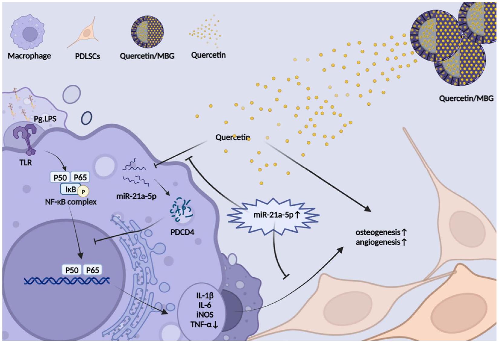
الملخص الرسومي


الخلفية

التهاب اللثة هو ثاني أكثر الأمراض الفموية شيوعًا في العالم، مع انتشار يتجاوز [1]. كمرض التهابي مزمن ومدمر يؤثر على الأنسجة الداعمة للثة، فإن فقدان العظم السنخي هو سمة رئيسية للمرض، والتي قد تؤدي في النهاية إلى فقدان الأسنان [2]. أظهرت الدراسات حول الآليات المرضية أن ضعف قدرة إصلاح العظام وعدم توازن البيئة الدقيقة المناعية العظمية هي عقبات رئيسية أمام تجديد العظم السنخي التالف في التهاب اللثة [3]. حاليًا، كانت المعالجة السريرية لعيوب العظم السنخي الناتجة عن التهاب اللثة تعتمد بشكل أساسي على إزالة المهيجات المحلية، والعلاج بالمضادات الحيوية، وجراحة العظم السنخي، بينما لم تتمكن هذه العلاجات من تجديد العظم السنخي في نفس الوقت وإعادة تشكيل البيئة الدقيقة المناعية المثلى [1،4]. لذلك، هناك حاجة إلى استراتيجية علاجية جديدة لتحفيز تجديد العظم السنخي وإدارة البيئة الدقيقة المناعية في نفس الوقت لتحقيق إعادة بناء العظم السنخي في الموقع في التهاب اللثة.
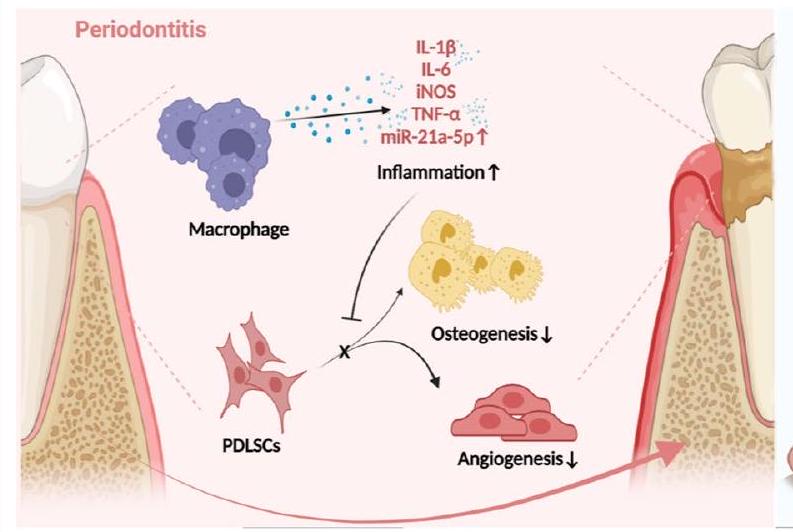
تعتبر PDLSCs، نوع من خلايا الجذعية الميزانشيمية (MSCs)، مرتبطة ارتباطًا وثيقًا بإصلاح العظم السنخي، ولديها إمكانيات واعدة في مجال تجديد اللثة [5]. يمكن أن تعيق الالتهابات المزمنة في التهاب اللثة تكوين العظام/الأوعية الدموية لـ PDLSCs، مما يؤدي إلى اضطراب تجديد اللثة [6]. كخلايا مناعية رئيسية في تطور التهاب اللثة،
تساهم الخلايا البالعة بشكل كبير في التوازن المناعي العظمي [7]. عمومًا، يتم تصنيف الخلايا البالعة عادةً إلى نوعين بناءً على حالة تنشيطها ووظيفتها: النوع M1 المؤيد للالتهابات والنوع M2 المضاد للالتهابات. خلال المرحلة الأولية من الاستجابة الالتهابية في التهاب اللثة، يكون تنشيط النوع M1 مفيدًا للدفاع عن الأنسجة، ولكن إذا استمرت هذه الحالة بعد هذه الفترة، فإنها ستؤدي إلى تدمير لا يمكن عكسه للأنسجة اللثوية [8]. أدى الاستقطاب المفرط للنوع M1 من الخلايا البالعة، كما لوحظ في الدراسات المتعلقة بالتهاب اللثة، إلى تفاقم الاضطرابات المناعية المحلية وأدى إلى تدمير الأنسجة اللثوية، بينما زاد أيضًا من ضعف قدرة PDLSCs على تكوين العظام/الأوعية الدموية وبالتالي تفاقم عدم توازن البيئة الدقيقة المناعية العظمية [9-11]. لذلك، كان من الضروري من الناحية الخلوية استعادة تمايز PDLSCs إلى تكوين العظام/الأوعية الدموية وإعادة تشكيل التوازن المناعي العظمي عبر الخلايا البالعة لإصلاح عيوب العظم السنخي في التهاب اللثة.
الكيرسيتين، مركب بوليفينولي طبيعي موجود في نباتات متنوعة، له عدة تأثيرات دوائية بما في ذلك تعزيز تكوين العظام/الأوعية الدموية، ومضاد للالتهابات وتنظيم المناعة [12-15]. كشفت أبحاثنا السابقة أن الكيرسيتين يمكن أن يعزز تمايز PDLSCs إلى تكوين العظام/الأوعية الدموية في بيئة هشاشة العظام ويثبط الاستجابة الالتهابية في التهاب المفاصل [16،
17]. على الرغم من أنه تم العثور على الكيرسيتين ليكون مثبطًا لامتصاص العظم السنخي الالتهابي في نماذج التهاب اللثة التجريبية على الجرذان والفئران، إلا أنه لا توجد تحقيقات متعمقة حول قدرته التجديدية وآليته الخلوية المحددة[18-20]. باختصار، من غير المؤكد ما إذا كان الكيرسيتين يمكن أن يجدد العظم السنخي في بيئة التهاب اللثة، وهناك حاجة إلى مزيد من التحقيق.
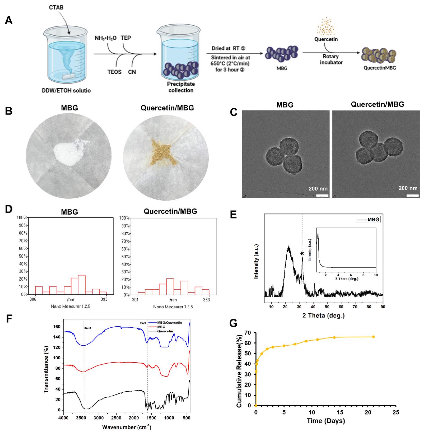
ومع ذلك، كدواء صغير جزيئي أحادي، فإن ذوبان الكيرسيتين الضعيف في الماء وتوافره البيولوجي المنخفض يحدان من استخدامه السريري[12]. لذلك، من الضروري إنشاء نظام توصيل دوائي فعال يمكنه الحفاظ على تركيزات مستقرة ومناسبة من الكيرسيتين في الأنسجة المستهدفة للاستخدام السريري. مؤخرًا، يتم اعتبار جزيئات MBG النانوية كحاملات دوائية محتملة لمواقع عيوب العظام، بسبب مساميتها العالية، ومساحة سطحها المحددة، وقدرتها القوية على تحفيز تجديد أنسجة العظام[21]. علاوة على ذلك، أشارت الأبحاث إلى أنه عند استخدام MBG كنظام توصيل دوائي لإصلاح العظام، لم يثير استجابة مناعية غير مواتية في الخلايا البالعة[22،23]. ومن ثم، يُفترض أن نظام توصيل النانو Quercetin/MBG قد يكون له تطبيق محتمل في علاج عيوب العظم السنخي في التهاب اللثة.
تعتبر microRNAs (miRNAs) فئة من RNAs الصغيرة غير المشفرة المحفوظة تطوريًا والتي تنظم التعبير الجيني على مستوى الترجمة[24]. في السنوات الأخيرة، أفادت العديد من الدراسات بوجود علاقة وثيقة بين التهاب اللثة وmiRNAs، مثل miR-21، miR-144، miR-146a، miR-128 وغيرها[25]. علاوة على ذلك، تم الإبلاغ عن أن الكيرسيتين يمكن أن يحفز العديد من miRNAs لتعزيز التمايز العظمي وكذلك تعديل الاستجابة المناعية[26-28]. لذلك، يُفترض أن تلعب miRNAs دورًا محوريًا في تأثيرات تعديل المناعة العظمية للكيرسيتين على التهاب اللثة.
لقد أكدنا أن نظام توصيل الكيرسيتين/MBG النانوي أطلق الكيرسيتين بشكل مستمر وأدى إلى تحسين تجديد العظم السنخي وإعادة تشكيل البيئة الميكروية المناعية العظمية مقارنة بـ MBG النقي. ثم بحثنا في الآلية الخلوية التي من خلالها تدخل الكيرسيتين في تجديد العظم السنخي تحت بيئة التهاب اللثة. لاحظنا أن الكيرسيتين لم يستعد فقط القدرة على تكوين العظام/الأوعية الدموية لخلايا PDLSCs، بل أعاد أيضًا برمجة البلعميات من خلال مسار miR-21a-5p/PDCD4/NF-κB مما أدى إلى إعادة تشكيل البيئة الميكروية المناعية العظمية. باختصار، تشير هذه الدراسة إلى أن نظام توصيل الكيرسيتين المحمّل بالمسام النانوية يحمل إمكانيات كعلاج سريري لإصلاح عيوب العظم السنخي في التهاب اللثة. علاوة على ذلك، فإن التلاعب بـ miR-21a-5p في البلعميات لإعادة تشكيل البيئة الميكروية المناعية العظمية في اللثة يقدم استراتيجية علاجية جديدة لإعادة بناء العظم السنخي في التهاب اللثة.

النتائج

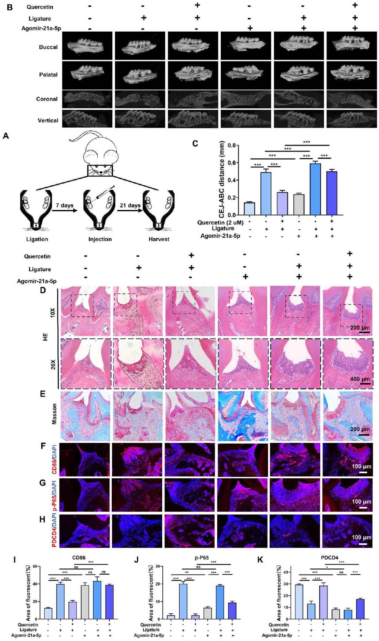
نظام الإفراز المستدام للكويرسيتين عزز تجديد العظم السنخي وخفف الالتهاب المحلي في نموذج عيب العظم السنخي لدى الجرذان مع التهاب اللثة.

في العقود الأخيرة، تم إثبات أن المواد الحاملة للأدوية (MBGs) لديها القدرة على إصلاح عيوب العظام، وزيادة تركيزات الأدوية المحلية، وتحسين فعالية الأدوية، وتقليل تكرار الجرعات، وتقليل الآثار الجانبية. الكيرسيتين، كعامل نشط بيولوجيًا غير قابل للذوبان مع توافر حيوي فموي منخفض وتأثير أولي واسع. لتحسين التوافر الحيوي المحلي وتحقيق إطلاق مستدام طويل الأمد للكيرسيتين في مواقع تلف اللثة مع التهاب اللثة. لتحسين التوافر الحيوي المحلي وتحقيق إطلاق مستدام طويل الأمد للكيرسيتين في مواقع تلف اللثة مع التهاب اللثة، قمنا بدمج MBG والكيرسيتين لإطلاق مستدام للكيرسيتين غير القابل للذوبان.
الشكل 1A أظهر عملية تحضير MBG وكيرسيتين/MBG. الشكل 1B أظهر أن تحميل الكيرسيتين تسبب في تحول MBG الأبيض الأصلي إلى لون الزنجبيل. أكدت تقنية المجهر الإلكتروني الناقل (TEM) أن MBG له شكل ميكروسفير مجوف مع قشور مسامية، وأن إضافة الكيرسيتين كان لها تأثير ضئيل على التغيرات الشكلية لـ MBG (الشكل 1C). ومقارنةً بقطر MBG ( قطر الكيرسيتين / MBG تغيرت قليلاً، مما يدل على أن الكيرسيتين تم تحميله في النواة الداخلية لـ MBG (الشكل 1D). كما هو موضح في الشكل 1E، أشار طيف حيود الأشعة السينية بزاوية واسعة (XRD) إلى تكوين السيليكا غير المتبلورة لـ MBG وتم تطبيق طيف XRD بزاوية صغيرة لتأكيد الهيكل المسامي. بالإضافة إلى ذلك، بسبب تكوين هيدروكسيباتيت الكربونات، تم رؤية قمة قوية عند في الطيف[31]. أظهرت طيف مطياف تحويل فورييه للأشعة تحت الحمراء (FTIR) في الشكل 1F أن الكيرسيتين/MBG يحتوي على قمم مميزة للكيرسيتين وMBG، مما يشير إلى تحميل ناجح للكيرسيتين في MBG. بالإضافة إلى ذلك، تم قياس سعة التحميل وكفاءة الت encapsulation للكيرسيتين لتكون و ، على التوالي. أظهر ملف إطلاق نظام توصيل كيرسيتين / MBG نانو انفجارًا أوليًا للإطلاق خلال ثلاث ساعات، تلاه إطلاق مستمر على مدى 21 يومًا (الشكل 1G).
الشكل 2A يوضح عملية نمذجة نموذج عيب العظم السنخي في الجرذان مع التهاب اللثة. الشكل 2B قدم صور إعادة بناء باستخدام الميكرو-CT التي تشير إلى عدم وجود تجديد ملحوظ للعظم في جميع المجموعات بعد أسبوع واحد. ومع ذلك، كان هناك زيادة ملحوظة في كتلة العظم بعد 4 و8 أسابيع في كل من مجموعتي MBG وQuercetin/MBG، مع ملاحظة زيادة أكبر في مجموعة Quercetin/MBG. كانت نتائج حجم العظم/حجم الأنسجة (BV/TV) متسقة مع
الشكل 1 توصيف نظام توصيل النانو كيرسيتين/MBG. A مخطط بروتوكول التحضير لـ MBG وكيرسيتين/MBG. B-D الفحص البصري، TEM، ورسم توزيع حجم الجسيمات لـ MBG وكيرسيتين/MBG. E الأشعة السينية بزاوية واسعة وXRD لـ MBG. F FITR لـ MBG وكيرسيتين/MBG. G منحنى إطلاق الدواء لكيرسيتين/MBG
(انظر الشكل في الصفحة التالية.)
الشكل 2 نظام إطلاق الكيرسيتين المستدام عزز تجديد العظام السنخية وخفف الالتهاب المحلي في نموذج عيب العظام السنخية في الجرذان مع التهاب اللثة. أ رسم توضيحي لتصميم التجربة المستخدمة في الدراسة الحيوانية. ب صور إعادة بناء الميكرو-CT لنسيج العظام السنخية. ج تحليل كمي لنسيج العظام السنخية المتكون حديثًا. د صبغة المناعية الفلورية لـ iNOS في العينات المجمعة بعد أسبوع واحد. هـ صبغة المناعية الفلورية لـ CD31 و OPN في العينات المجمعة بعد 8 أسابيع. و تحليل كمي لمتوسط شدة الفلورة لـ iNOS و CD31 و OPN وفقًا لأنماط الصبغة المناعية الموضحة في (د و هـ). (ns، لا يوجد فرق ذو دلالة إحصائية؛ ; ; يتم تمثيل البيانات كمتوسط )
الشكل 2 (انظر الشرح في الصفحة السابقة.)
تحليلات إعادة بناء الميكرو-CT السابقة (الشكل 2C). أكدت التحليلات النسيجية (صبغة H&E وصبغة ماسون) لعينات الأسبوع الثامن أن مكملات الكيرسيتين حسنت من تعزيز تجديد العظام الهوائية بواسطة MBG (الملف الإضافي 1: الشكل S1A، B). أظهرت نتيجة صبغة المناعة الفلورية لعينات الأسبوع الأول تعبيرًا عاليًا عن إنزيم أكسيد النيتريك الاصطناعي (iNOS) في كل من مجموعتي Blank وMBG، ولكن لم يُلاحظ أي فرق ذو دلالة إحصائية بين هاتين المجموعتين. على العكس، كان تعبير iNOS منخفضًا بشكل ملحوظ في مجموعة Quercetin/MBG، مما يدل على الخصائص المضادة للالتهابات الجديرة بالثناء للكيرسيتين. لتقييم ما إذا كان الإفراج المستمر عن الكيرسيتين يمكن أن يعزز تكوين العظام/الأوعية الدموية، قمنا بإجراء صبغة مناعية فلورية لمؤشرات تكوين العظام والأوعية الدموية (OPN وCD31). أشارت نتائج الدراسة إلى أن تعبير OPN وCD31 كان أعلى في مجموعة المعالجة بالجزيئات النانوية مقارنةً بمجموعة التحكم، ولكن لم يُلاحظ أي فرق كبير بين مجموعة MBG ومجموعة Quercetin/MBG (الشكل 2D-F).
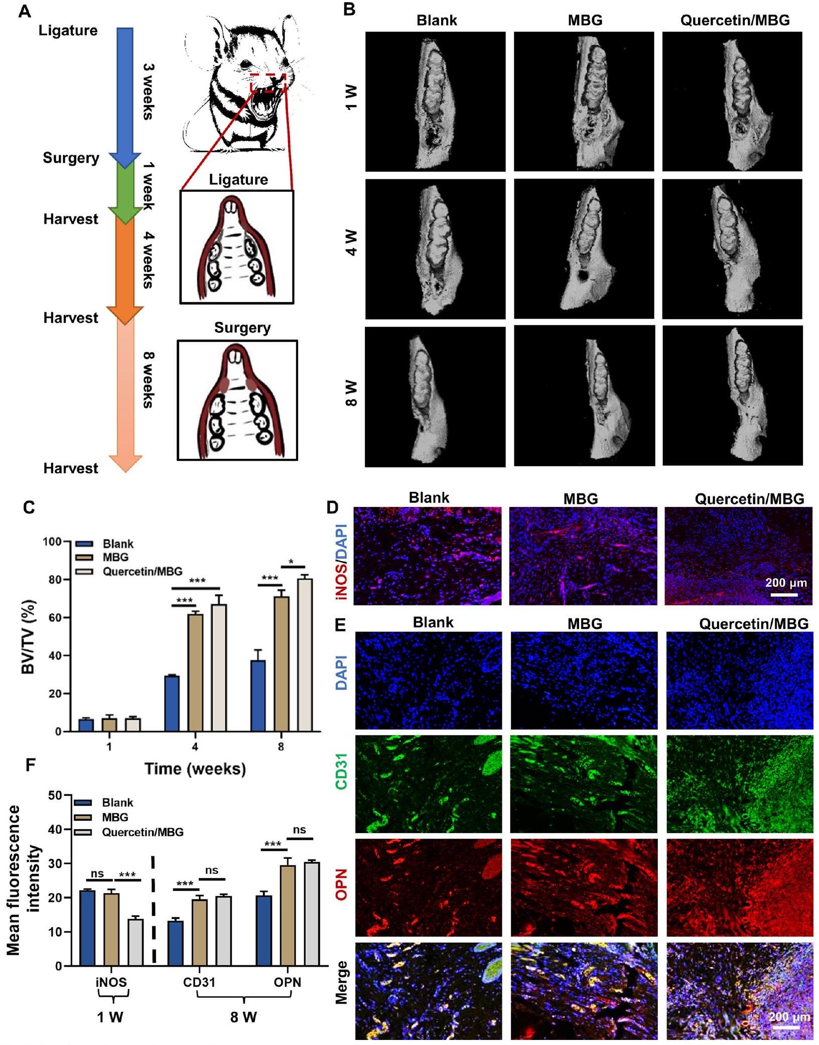
في الختام، أشارت نتائجنا إلى أن الكيرسيتين يمكن أن يعزز بشكل فعال تجديد العظام في مناطق عيوب العظام السنخية في اللثة بينما يتحكم في الالتهاب اللثوي. ثم قمنا بالتحقيق في الآلية الأساسية التي من خلالها يعزز الكيرسيتين تجديد العظام السنخية وينظم الالتهاب في بيئة التهاب اللثة.

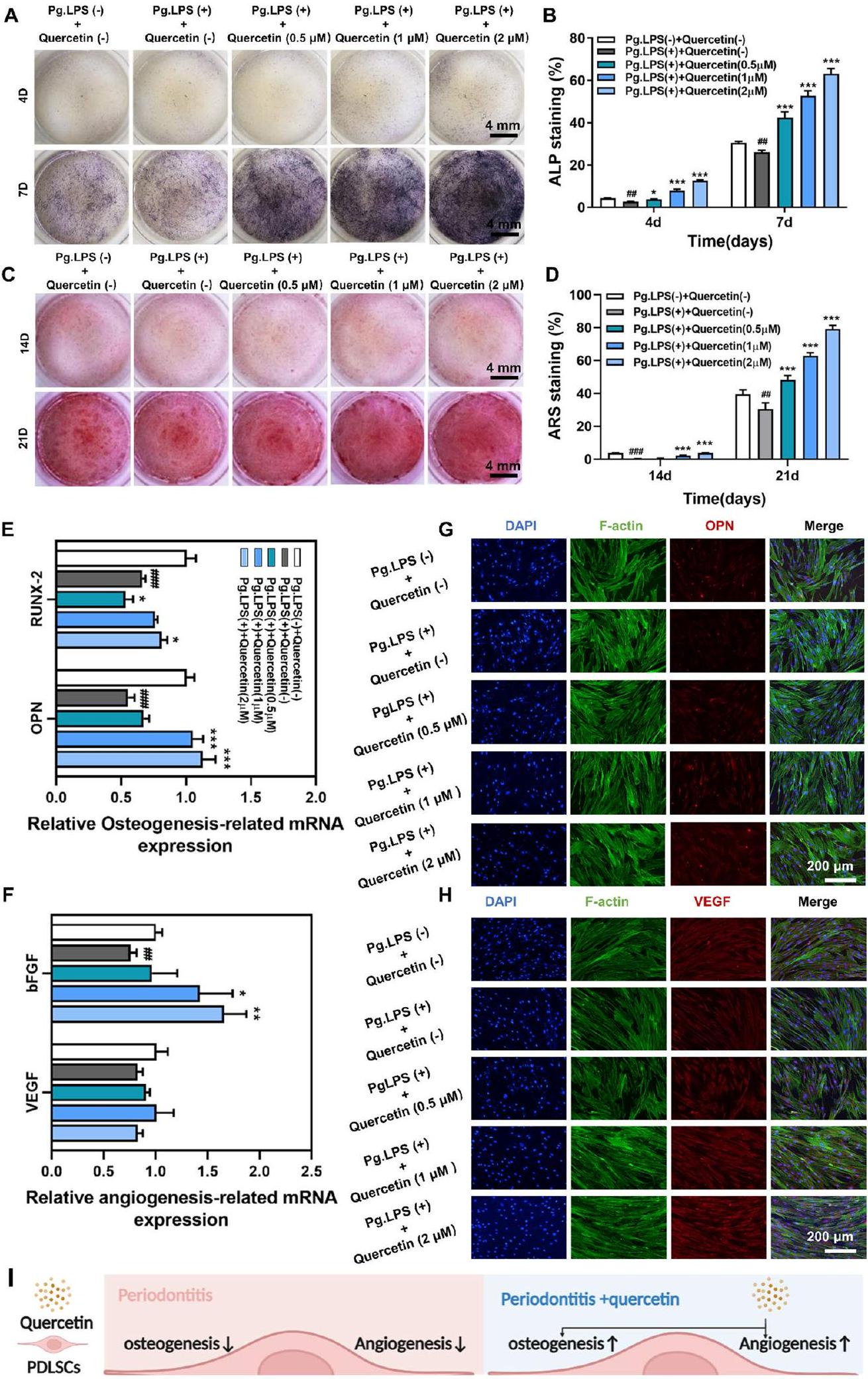
أدى الكيرسيتين إلى تعزيز تكوين العظام والأوعية الدموية ومكافحة الالتهاب في خلايا جذع الأنسجة الداعمة للأسنان تحت بيئة التهاب اللثة.

لتقييم السمية الخلوية للكويرسيتين على خلايا PDLSCs، تم تعريض الخلايا لتركيزات مختلفة من الكويرسيتين. ، و ) لمدة 1 و 4 و 7 أيام. أظهرت نتائج اختبار مجموعة عد الخلايا-8 (CCK-8) أن الكيرسيتين كان له تأثير سمي كبير على الخلايا عند تركيزات تصل إلى في اليوم الرابع والسابع، بغض النظر عن غياب أو وجود الليببوليسكاريد المستمد من P. gingivalis (Pg.LPS) (الملف الإضافي 1: الشكل S2A، B). بالإضافة إلى ذلك، أكدت استخدام صبغة الخلايا الحية/الميتة أن الكيرسيتين ( زاد بشكل كبير عدد الخلايا الميتة تحت بيئة التهابية (إضافي
الملف 1: الشكل S2C). استنادًا إلى هذه النتائج، اخترنا تركيزات الكيرسيتين من ، و لإجراء تجارب إضافية.
بالإضافة إلى ذلك، تم تقييم القدرة على تكوين العظام/الأوعية الدموية لخلايا PDLSCs عند تركيزات مختلفة و ) في وجود Pg.LPS ( كما هو موضح في الشكل 3A-D، أظهرت صبغة الفوسفاتاز القلوي (ALP) وصبغة الأليزارين الأحمر S (ARS) انخفاضًا في القدرة على تكوين العظام في وجود Pg.LPS، بينما أظهر الكيرسيتين زيادة تعتمد على التركيز في القدرة على تكوين العظام، حيث أظهر أعلى نشاط للفوسفاتاز القلوي وأكبر عدد من العقيدات الكالسيومية في وجود Pg.LPS. كويرسيتين مجموعة. علاوة على ذلك، أظهرت تعبيرات الجينات المرتبطة بتكوين العظام (OPN وعامل النسخ من عائلة RUNX-2، RUNX-2) والجين المرتبط بتكوين الأوعية (عامل نمو الألياف الأساسية، bFGF) التي تم الكشف عنها من خلال تحليل qRT-PCR، بالإضافة إلى تعبير البروتينات المرتبطة بتكوين العظام (OPN) والبروتينات المرتبطة بتكوين الأوعية (عامل نمو البطانة الوعائية، VEGF) التي تم اكتشافها من خلال تحليل المناعة الفلورية، اتجاهًا مشابهًا لذلك الذي لوحظ في تجارب ALP وARS، بينما لم يظهر تعبير VEGF اختلافًا كبيرًا بين جميع المجموعات على مستوى الجين (الشكل 3E-H). علاوة على ذلك، أظهر تحليل Western blot أن إضافة Pg.LPS منعت تعبير بروتين VEGF، بينما زادت إضافة الكيرسيتين من تعبيره بطريقة تعتمد على التركيز (الملف الإضافي 1: الشكل S3A-B). تشير هذه النتائج إلى أن الكيرسيتين لديه القدرة على تعزيز قدرات تكوين العظام/الأوعية في PDLSCs (الشكل 3I).
علاوة على ذلك، قمنا بتقييم القدرة المضادة للالتهابات للكيرسيتين في PDLSCs كما هو موضح في الملف الإضافي 1: الشكل S4A-C، والذي كان مفيدًا في إعادة تشكيل البيئة الدقيقة اللثوية وتعزيز تجديد العظام السنخية. أشارت نتائج qRT-PCR وصبغة المناعة الفلورية على التوالي إلى أن الكيرسيتين يمكن أن يعيق الاستجابات الالتهابية على كل من مستوى الجين والبروتين. علاوة على ذلك، وُجد أن الكيرسيتين يثبط بشكل كبير الاستجابة المناعية النشطة التي تسببها Pg.LPS عند تركيز .
أكدت هذه الجزء أن الكيرسيتين عزز بشكل كبير تكوين العظام/الأوعية، بينما قام في الوقت نفسه بتثبيط الالتهاب في PDLSCs، مما يشير إلى
الشكل 3 (انظر التسمية في الصفحة السابقة.)
الخصائص الاستثنائية للكيرسيتين في تكوين العظام/الأوعية ومكافحة الالتهابات.

أدى الكيرسيتين إلى مكافحة الالتهاب في البلعميات تحت بيئة التهاب اللثة

خلال هذه الدراسة، قمنا بزراعة خلايا RAW264.7 مع تركيزات مختلفة من الكيرسيتين ( ) و Pg.LPS ( ) لفحص حيوية الخلايا وكذلك مستويات الجين والبروتين للإنترلوكين-1 (IL-1 )، الإنترلوكين-6
(IL-6)، iNOS وعامل نخر الورم- (TNF- ). أظهرت البيانات المقدمة في الشكل 4A، B أن تركيزات مختلفة من الكيرسيتين ( و ) لم تظهر أي تأثيرات سامة للخلايا في غياب أو وجود Pg.LPS. بالإضافة إلى ذلك، كشفت ملاحظاتنا أن التعرض لـ Pg.LPS زاد بشكل ملحوظ من مستوى mRNA وإطلاق السيتوكينات لـ IL-1 ، IL-6، iNOS وTNF- في خلايا RAW 264.7، والتي انخفضت بواسطة علاج الكيرسيتين بطريقة تعتمد على الجرعة (الشكل 4C-J). أظهرت نتائج هذه الدراسة أن الكيرسيتين يمكن أن يثبط
الشكل 4 أدى الكيرسيتين إلى مكافحة الالتهاب في البلعميات تحت بيئة التهاب اللثة. A، B نتائج تكاثر خلايا RAW264.7 التي تم حضنها مع الكيرسيتين لمدة يوم واحد تحت بيئة فسيولوجية وبيئة التهاب اللثة من خلال اختبار CCK-8. C-F تأثير الكيرسيتين على تعبير الجينات الالتهابية (IL-1 ، iNOS وTNF- ) في RAW264.7 من خلال qRT-PCR. G-J تأثير الكيرسيتين على تعبير السيتوكينات الالتهابية (IL-1 ، IL-6، iNOS وTNF-a) في RAW264.7 من خلال اختبارات Elisa. K مخطط لآلية عمل الكيرسيتين على البلعميات تحت بيئة التهاب اللثة. ( و مقارنة بمجموعة Pg.LPS (-) + Quercetin (-) ، و مقارنة بمجموعة Pg.LPS(+)+Quercetin (-)؛ البيانات ممثلة كمتوسط SEM، )
الالتهاب الناتج عن Pg.LPS في خلايا RAW264.7 و تم اختيار تركيز الكيرسيتين كتركيز مركزي للتجارب اللاحقة (الشكل 4K).

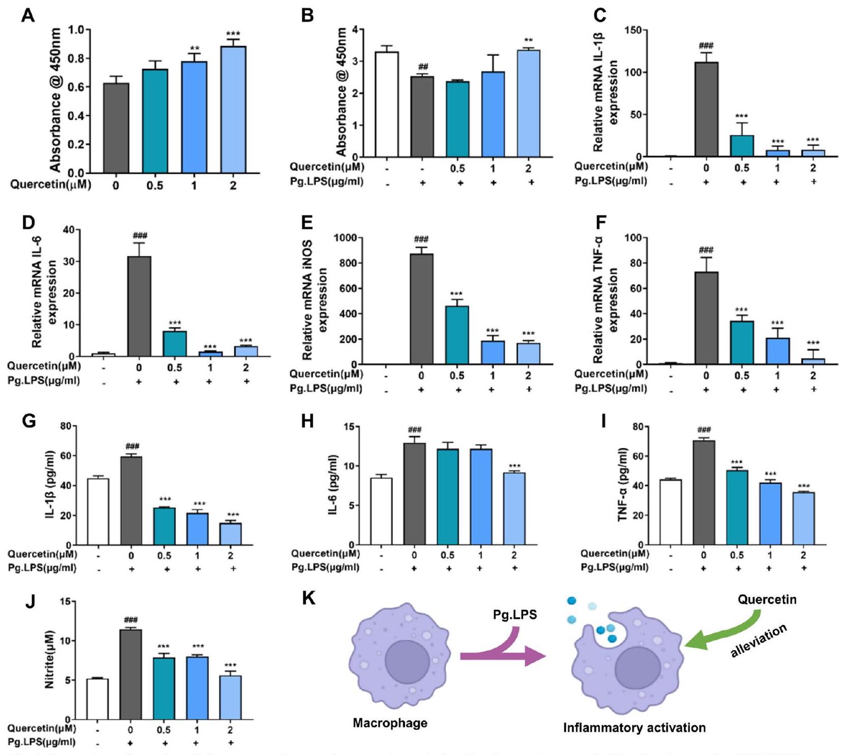
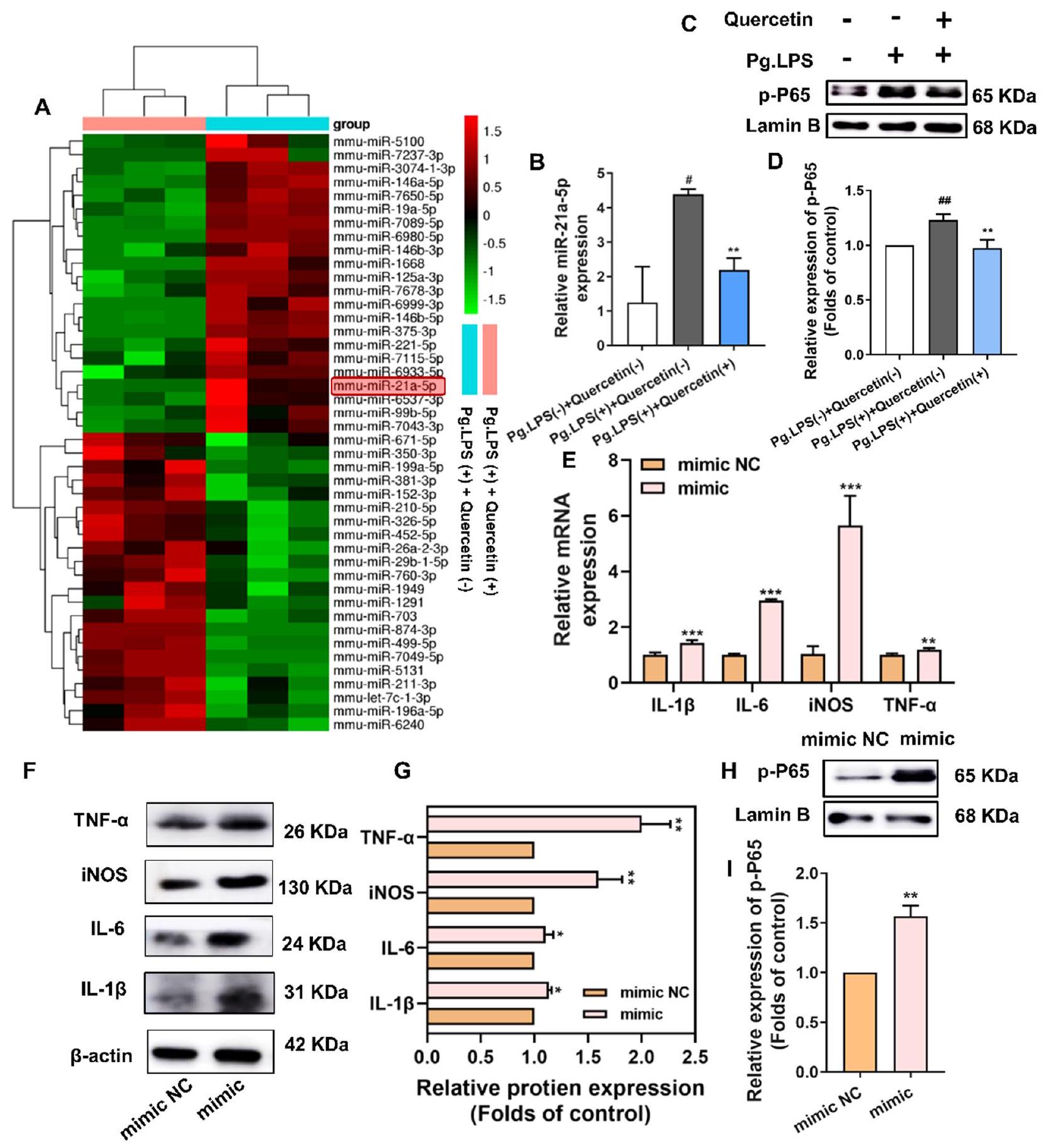
آلية مكافحة الالتهاب للكيرسيتين على البلعميات تحت بيئة التهاب اللثة أوقف الكيرسيتين التفاعل الالتهابي في البلعميات المحفزة بواسطة Pg.LPS من خلال تقليل تعبير miR-21a-5p

لتحديد الآلية، أجرينا اختبار مصفوفة miRNA لتقييم ما إذا كان الكيرسيتين يمكن أن يؤثر على تعبير miRNA في خلايا RAW264.7. حدد تحليل التجميع الهرمي الخاضع للإشراف 224 miRNAs معبر عنها بشكل مختلف (تغيير مضاعف ومعدل الاكتشاف الكاذب [FDR] < 5%). من بين 224 miRNAs المحددة، لوحظ أن 22 miRNAs كانت منخفضة التعبير في خلايا RAW264.7 في وجود الكيرسيتين (الشكل 5A). نظرًا للعلاقة بين miR-21a-5p والالتهاب، اخترنا هذا miRNA للبحث الإضافي. تم إجراء مزيد من التحقق باستخدام اختبار qRT-PCR لتأكيد أن الكيرسيتين يمكن أن يثبط ارتفاع تعبير miR-21a-5p الناتج عن Pg.LPS (الشكل 5B). بالإضافة إلى ذلك، أظهر إجراء تحليل Gene Ontology (GO) بناءً على الجينات المستهدفة المتوقعة أن الكيرسيتين قد ينظم مسار إشارة NF-кB في البلعميات المحفزة بواسطة Pg.LPS (الملف الإضافي 1: الشكل S5A). أشار تجربة المناعة الفلورية إلى أن تم نقله إلى النواة في مجموعة Pg.LPS (+)+Quercetin (-)، وكشف عن الموقع السيتوبلازمي لـ NF-кB في مجموعة Pg.LPS Quercetin (-). ومع ذلك، أعاق علاج الكيرسيتين النقل النووي لـ p-P65 الناتج عن تحفيز Pg.LPS، مما يشير إلى أن الكيرسيتين قلل من النقل النووي لـ p-P65 الناتج عن Pg.LPS (الملف الإضافي 1: الشكل S5B). تم استخدام Western blot للكشف عن تعبير p-P65 في نواة هذه المجموعات. متسقة مع نتائج المناعة الفلورية، كان تعبير p-P65 في نوى مجموعة Pg.LPS (+)+Quercetin (-) مرتفعًا بشكل ملحوظ مقارنة بمجموعة Pg.LPS Quercetin (-) ، بينما قلل علاج الكيرسيتين بشكل كبير من الزيادة في مستويات p-P65 الناتجة عن Pg.LPS (الشكل 5 C-D). كشفت نتائج هذا البحث أن الكيرسيتين يمكن أن يعيق على الأرجح تنشيط مسار إشارة NF-кB الناتج عن Pg.LPS من خلال تقليل النقل النووي لـ p-P65.
أولاً، قمنا بزيادة وتنظيم مستويات miR-21a-5p في خلايا RAW264.7 باستخدام محاكي miR-21a-5p، المثبط، وNC السلبي (الملف الإضافي 1: الشكل S5C). أدى محاكي miR-21a-5p إلى زيادة ملحوظة في مستويات العلامات الالتهابية، بما في ذلك IL-1 ، IL-6، iNOS وTNF- ، على كل من مستوى الجين والبروتين، مقارنة بمجموعة المحاكي NC، كما هو موضح في الشكل 5E-G بواسطة اختبارات qRT-PCR وWestern blot
. تشير هذه النتائج إلى أن الإفراط في التعبير عن miR-21a-5p يمكن أن يعزز الاستجابة الالتهابية للبلعميات. لاستكشاف دور miR-21a-5p بشكل أكبر، قمنا بنقل خلايا RAW264.7 باستخدام محاكي miR-21a-5p. كما هو موضح في الشكل 5H، I، زاد نقل محاكي miR-21a-5p بشكل كبير من تعبير p-P65 في النواة، مما أدى إلى تنشيط مسار إشارة NF-кB.

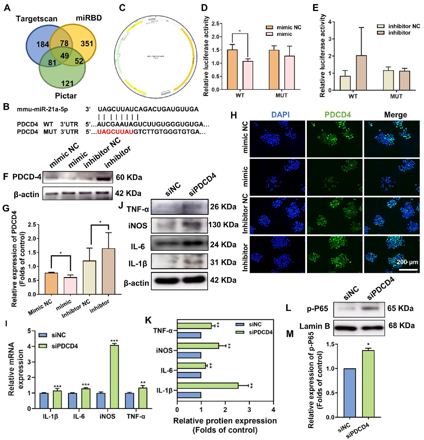
كان PDCD4 هدفًا سفليًا لـ miR-21a-5p ونظم المزيد من الاستجابات الالتهابية للبلعميات من خلال مسار إشارة NF-кB

كـ RNA غير مشفر ذو خيط واحد، يمكن أن تنظم miRNAs mRNA من خلال التفاعل مع تسلسلات القاعدة التكميلية لـ mRNA. قمنا بالتحقق من ثلاثة مواقع توقع هدف miRNA (Targets can، mired، وPictar) للتنبؤ بالأهداف المحتملة لـ miR-21a-5p. حددنا 49 هدفًا محتملاً لـ miR-21a-5p، من بينها كان PDCD4 هدفًا سفليًا موثقًا جيدًا لـ miR-21a-5p كما هو مذكور في الأدبيات (الشكل 6A) [32]. لذلك، قمنا بإجراء تجارب qRT-RCR، Western blot وصبغة المناعة الفلورية، ثم وجدنا أن تعبير PDCD4 انخفض بعد إضافة Pg.LPS وزاد بعد إضافة الكيرسيتين (الملف الإضافي 1: الشكل S5D-G). علاوة على ذلك، اخترنا siPDCD4 الأكثر فعالية (siPDCD4-3) باستخدام Western blot (الملف الإضافي 1: الشكل S5H، I).
لتحديد ما إذا كان miR-21a-5p يستهدف بروتين PDCD4، قمنا بتوقع موقع الهدف لـ miR-21a-5p داخل تسلسل PDCD4 باستخدام برامج المعلوماتية الحيوية وصممنا البلازميدات المقابلة (الشكل 6B، C). لتحديد ما إذا كان miR-21a-5p يمكن أن يستهدف 3′-UTR من PDCD4، تم إجراء اختبار تقرير اللوكيفيراز. أظهرت النتائج أن الإفراط في التعبير عن miR-21a-5p يمكن أن يقلل بشكل ملحوظ من نشاط اللوكيفيراز في 3′-UTR من النوع البري (WT) لـ PDCD4 (الشكل 6D، E). بعد ذلك، أكدت تجارب التWestern blot وصبغ المناعة الفلورية أن زيادة تعبير miR-21a-5p قد منعت التعبير البروتيني لـ PDCD4 في خلايا RAW264.7، والعكس صحيح (الشكل 6F-H). قدمت نتائج هذه الدراسة دليلاً على أن miR-21a-5p يستهدف بشكل محدد PDCD4، مما يؤدي إلى انخفاض في تعبير PDCD4.
لاستكشاف دور PDCD4 في تنظيم استجابة الماكروفاج الالتهابية بواسطة الكيرسيتين، تم إجراء تحليلات qRTPCR وWestern blot، مما كشف أن قمع تعبير PDCD4 يمكن أن يزيد من تعبير الجينات والبروتينات الالتهابية بالإضافة إلى تنشيط مسار NF-кB (الشكل 6I-M).
أيدت نتائج هذه الدراسة الفكرة القائلة بأن miR-21a-5p يستهدف بشكل محدد بروتين PDCD4 ويقلل من تعبير PDCD4، بينما الانخفاض في
الشكل 5 أوقف الكيرسيتين التفاعل الالتهابي في البلعميات المنشطة بواسطة Pg.LPS من خلال تقليل تعبير miR-21a-5p. أ خريطة حرارية للجينات المختلفة بين Pg.LPS (+)+كيرسيتين (-) و Pg.LPS (+)+كيرسيتين (+). ب تعبير الجينات لـ miR-21a-5p بين مجموعات Pg.LPS (-) + كيرسيتين (-) و Pg.LPS (+) + كيرسيتين (-) و Pg.LPS (+) + كيرسيتين (+) من خلال qRT-PCR. ج، د التحليل الغربي و تحليله الكمي لـ p-P65 بعد حضانة البلعميات مع الكيرسيتين لمدة يوم واحد. هـ تعبير الجينات الالتهابية (IL-1 IL-6، iNOS و TNF- ) من خلال qRT-PCR في البلعميات التي تم نقلها بمحاكاة miR-21a-5p NC ومحاكاة miR-21a-5p. F-I تعبير البروتينات الالتهابية (IL-1 ، IL-6 و iNOS و TNF-a) و p-P65 من خلال تقنية الويسترن بلوت في خلايا RAW264.7 التي تم نقلها بواسطة miR-21a-5p ميميك NC و miR-21a-5p ميميك. (في الشكل 5B-D، و مقارنة بمجموعة Pg.LPS (-) + كيرسيتين (-) و و مقارنة بمجموعة Pg.LPS (+)+Quercetin (-)؛ في الشكل 5E-1، *p < 0.05، **p < 0.01 و ***p < 0.001؛ البيانات ممثلة كمتوسط SEM )
الشكل 6 كان PDCD4 هدفًا سفليًا لـ miR-21a-5p ونظم استجابات الالتهاب في البلعميات من خلال مسار إشارة NF-kB. A مخطط فين للجينات المستهدفة المتداخلة المقدمة في مجموعات Pg.LPS (-) + كيرسيتين (-) و Pg.LPS (+) + كيرسيتين (-). B مواقع الأهداف لـ miR-21a-5p في تسلسل PDCD4 التي تم التنبؤ بها بواسطة برامج المعلوماتية الحيوية. C ملف بناء متجه pLUC-PDCD4، الذي يحتوي على مواقع أهداف miR-21a-5p في 3′-UTR لـ PDCD4. D، E نتائج اختبار تقرير ثنائي اللمعان بعد التداخل المشترك لـ miR-21a-5p المقلد، المثبط، وNC الخاص بهم مع PDCD4-WT-3′-UTR أو PDCD4- النوع الطافر (MUT)-3′-UTR. F-H التحليل الغربي وصبغة المناعية للبروتين المستهدف PDCD4 في خلايا RAW264.7 بعد التداخل مع miR-21a-5p المقلد، المثبط، وNC الخاص بهم. I-K تأثير الكيرسيتين على تعبير الجينات والبروتينات الالتهابية (IL-1 IL-6 و iNOS و TNF-a) من خلال qRT-PCR و western blot في البلعميات التي تم نقلها بـ siNC و siPDCD4. L، M تحليل western blot الكمي لـ p-P65 بعد نقل البلعميات بـ siNC و siPDCD4. و ; يتم تمثيل البيانات كمتوسط )
أدى تعبير PDCD4 إلى تعزيز الاستجابة الالتهابية وتنشيط مسار NF-кB في خلايا RAW264.7.

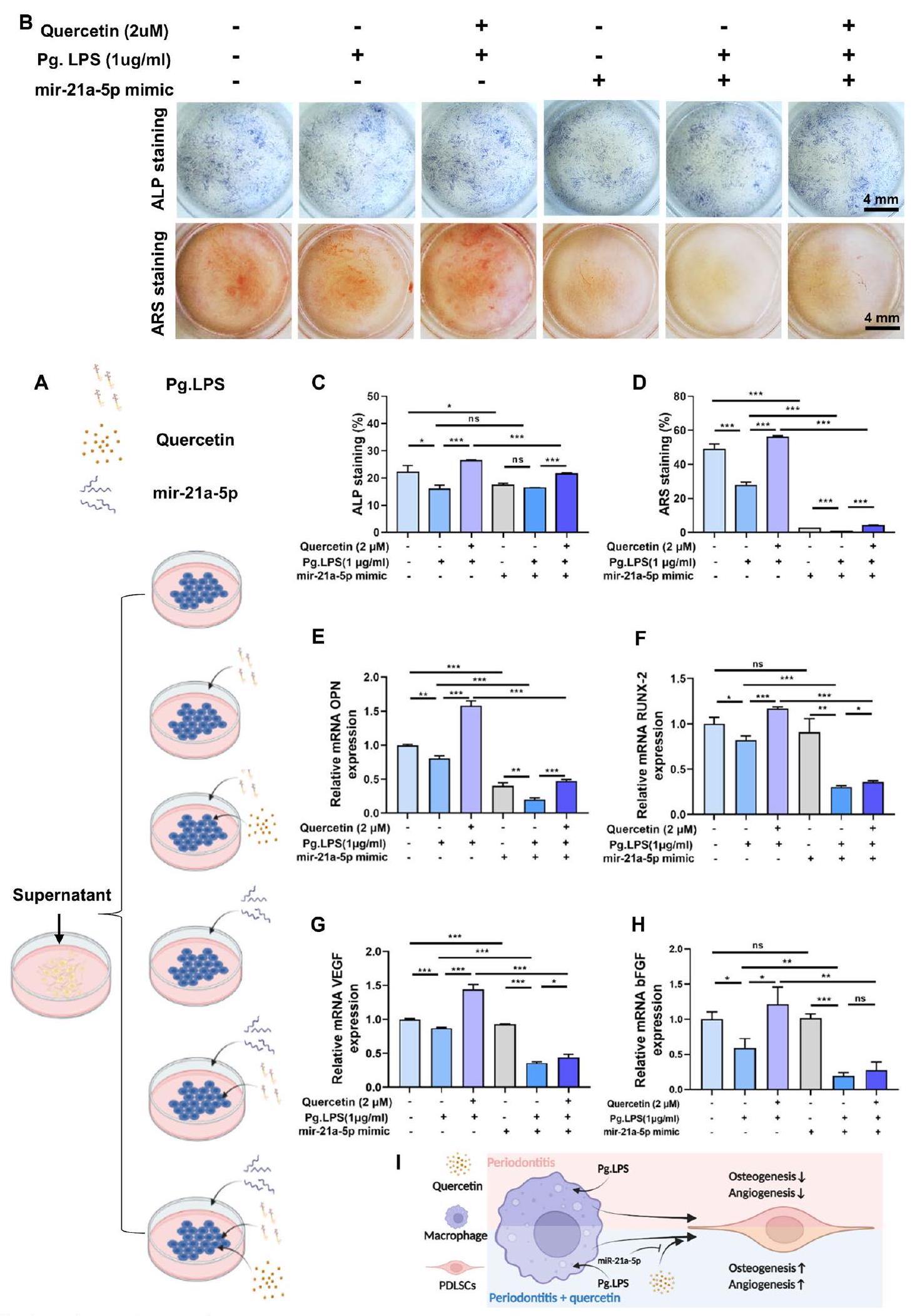
أظهر الكيرسيتين تأثيرات غير مباشرة مؤيدة لتكوين العظام/الأوعية الدموية عبر miR-21a-5p

لتأكيد ما إذا كان التأثير المناعي العظمي للماكروفاجات المعالجة بالكيرسيتين من خلال miR-21a-5p، تم جمع الوسط الشرطي (CM) من الماكروفاجات المزروعة مع أو بدون كيرسيتين تحت بيئة التهابية تم معالجتها مسبقًا بمحاكي miR-21a-5p أو محاكي NC كما هو موضح في الشكل 7A. تم استخدام CM لزراعة PDLSCs لتقييم قدرتها على التكوين العظمي/ الوعائي من خلال صبغة ALP، وصبغة ARS بالإضافة إلى أظهرت نتائج صبغة ALP وصبغة ARS وتحليلاتها الكمية لخلايا PDLSCs الموضحة في الشكل 7B-D أن معالجة الكيرسيتين زادت من تمايز خلايا PDLSCs العظمية التي تم إضعافها بواسطة المعالجة المسبقة مع Pg.LPS. بالإضافة إلى ذلك، لم يُلاحظ أي فرق ذي دلالة إحصائية بين المعالجات بالكيرسيتين. بي.جي. إل. بي. إس محاكي miR-21a-5p وكيرسيتين بي.جي. إل. بي. إس مقلد miR-21a-5p في تجربة ALP، كانت القدرة على تكوين العظام لخلايا PDLSCs أضعف تحت ظروف معالجة المقلد miR-21a-5p مقارنة بتلك المعالجة بالمقلد NC لـ miR-21a-5p. علاوة على ذلك، لم يظهر أي فرق كبير في جين OPN (الكيرسيتين بي.جي. إل. بي. إس محاكي miR-21a-5p مقابل كيرسيتين صفحة LPS ( ) (-) + مقلد miR-21a-5p (+)) وجين bFGF (كويرسيتين بي.جي. إل. بي. إس محاكاة miR-21a-5p (-) مقابل كيرسيتين ( ) (-) + Pg.LPS ( محاكي miR-21a-5p وكيرسيتين بي.جي. إل. بي. إس محاكي miR-21a-5p مقابل كيرسيتين بي.جي. إل. بي. إس المحاكاة لـ miR-21a-5p (+)، كانت اتجاهات التعبير للجينات العظمية (OPN و Runx-2) والجينات الوعائية (VEGF و bFGF) متوافقة بشكل عام مع تلك التي لوحظت في صبغة ARS (الشكل 7E-H). مجتمعة، تشير النتائج المذكورة أعلاه إلى أن تعديل المناعة العظمية للماكروفاجات المزروعة مع الكيرسيتين كان له تأثير إيجابي على التمايز العظمي/الوعائي لخلايا PDLSCs، بينما يمكن أن تمنع التعبير المفرط لـ miR-21a-5p التأثيرات غير المباشرة للكيرسيتين في تعزيز العظم/الأوعية (الشكل 7I).

قدم الكيرسيتين تأثيرًا وقائيًا في التهاب اللثة عبر miR-21a-5p

لتقييم التأثير المناعي لتنظيم الكيرسيتين في التهاب اللثة وتحديد اعتماده على miR-21a-5p، تم ربط فئران C57BL/6 أولاً لإنشاء نموذج التهاب لثة تجريبي ثم تم حقنها إما بـ agomiR-21a-5p أو التحكم العشوائي له في غياب أو وجود الكيرسيتين كما هو موضح في الشكل 8A. أظهر تحليل الميكرو-CT أن الكيرسيتين يمكن أن يثبط بشكل واضح امتصاص العظام في التهاب اللثة ويقلل بشكل كبير من المسافة بين تقاطع المينا والعظم السنخي (CEJ-ABC)، بينما يمكن أن يثبط العلاج بـ agomiR-21a-5p جزئيًا تأثيرات علاج الكيرسيتين (الشكل 8B، C). كشفت صبغات H&E وMasson أن مجموعة المعالجة بالكيرسيتين قللت من تسلل البلعميات مقارنة بمجموعة الربط، بينما عززت إضافة agomiR-21a-5p التأثيرات بما في ذلك زيادة عدد الخلايا الالتهابية بالإضافة إلى حجب التأثير الوقائي للكيرسيتين في امتصاص العظام الالتهابي (الشكل 8D، E).
للمزيد من التحقيق في النشاط المناعي في الجسم الحي للكينسيتين وmiR-21a-5p، تم إجراء صبغة المناعية الفلورية لـ CD86 وp-P65 وتحليلها الكمي (الشكل 8F-G، I-J). زادت مستويات بروتين CD86 وp-P65 بشكل ملحوظ في مجموعة الكينسيتين (-) + الربط (+) + agomiR-21a-5p (-) وانخفضت في مجموعة الكينسيتين (+) + الربط (+) + agomiR-21a-5p (-)، مما يشير إلى أن علاج الكينسيتين يمكن أن يقلل بشكل فعال من الاستجابة الالتهابية ويثبط تنشيط مسار NF-кB. علاوة على ذلك، زاد العلاج بـ agomiR-21a-5p من تعبير هذين البروتينين، والذي لم يمكن تقليله بواسطة حقن الكينسيتين. ومع ذلك، لم يتم ملاحظة فرق كبير بين المجموعة المعالجة بالكينسيتين (+) + الربط (+) + agomiR-21a-5p (-) وتلك المعالجة بالكينسيتين (+) + الربط (+) + agomiR-21a-5p (+)، وقد يكون ذلك لأن إجراء الربط قد أدى بالفعل إلى تعبير كبير عن علامات الالتهاب المحلية ولم يكن بإضافة agomiR-21a-5p تعزيز ذلك بشكل أكبر. تشير هذه الاكتشافات إلى أن miR يمكن أن ينشط الحالة الالتهابية ويعيق القدرة المضادة للالتهابات للكيرسيتين. علاوة على ذلك، يعتبر PDCD4 الهدف الجيني السفلي لـ miR-21a-5p،
الشكل 7 (انظر الشرح في الصفحة السابقة.)
الشكل 8. قدم الكيرسيتين التأثير الوقائي في التهاب اللثة عبر miR-21a-5p. أ رسم توضيحي تجريبي للحيوانات. ب إعادة بناء ثلاثي الأبعاد وصور مقطع عرضي ثنائي الأبعاد من خلال تحليل Micro-CT. ج نتائج تحليل مسافة CEJ-ABC. د-ح صبغات H&E وMasson وصبغة المناعة الفلورية لـ CD86 وp-P65 وPDCD4. ط-ك التحليل الكمي للمنطقة الفلورية لـ CD86 وp-P65 وPDCD4 وفقًا للشكل 8 F-H. (ns، لا يوجد فرق ذو دلالة إحصائية؛ ; يتم تمثيل البيانات كمتوسط )
تم ملاحظة انخفاضه استجابةً للبيئة الالتهابية والتعبير العالي عن miR-21a-5p، بالإضافة إلى الزيادة باستخدام الكيرسيتين، بينما يمكن أن يؤدي إضافة agomiR-21a-5p إلى حجب جزئي للتأثير المعزز للكيرسيتين على تعبير PDCD4 (الشكل 8H، K). باختصار، أظهرت نتائجنا أن الكيرسيتين يمتلك تأثيرًا مضادًا للالتهابات فائقًا في الجسم عن طريق تثبيط مسار NF-кB، بينما يمكن حجب هذه الوظيفة بواسطة التعبير العالي عن miR-21a-5p.

التقييم في المختبر لتأثير MBG على التأثيرات البيولوجية للكويرسيتين

لتقييم ما إذا كان توصيل الكيرسيتين بواسطة MBG سيغير التأثير البيولوجي للكيرسيتين، قمنا بتقييم تأثيرات MBG، الكيرسيتين، وكيرسيتين/MBG على التمايز العظمي/الوعائي لخلايا PDLSCs والاستجابة الالتهابية للبلاعم في بيئة التهاب اللثة. قامت الدراسة بتقييم التمايز العظمي/الوعائي لخلايا PDLSCs من خلال صبغة ALP وqRT-PCR. أظهرت الإضافات 1: الشكل S6A، B أن كل من مجموعتي MBG والكيرسيتين كانتا قادرتين على زيادة نشاط ALP، الذي تم تثبيطه بواسطة Pg.LPS. علاوة على ذلك، أظهرت مجموعة كيرسيتين/MBG فعالية أفضل من أي منهما بشكل فردي. كانت نتائج qRT-PCR متسقة مع اتجاه صبغة ALP، باستثناء أن تعبير جين VEGF زاد فقط في مجموعة كيرسيتين/MBG (الإضافات 1: الشكل S6C-F). تم تقييم الاستجابة الالتهابية للبلاعم باستخدام qRT-PCR. أشارت النتائج إلى أن MBG لم يكن له تأثير على تعبير الجينات الالتهابية (IL-1 IL-6، iNOS و TNF- ) التي تم تنشيطها في بيئة الميكروبيوم لالتهاب اللثة. ومع ذلك، فقد خفض كل من الكيرسيتين والكيرسيتين/MBG بشكل كبير من التعبير عن الجينات الالتهابية، في حين لم يكن هناك فرق كبير بين المجموعتين (الملف الإضافي 1: الشكل S6G-J).
أظهرت الدراسة أعلاه أن الكيرسيتين exerted تأثيرات مناعية من خلال miRNA رئيسي (miR-21a-5p) في البلعميات. لتقييم ما إذا كان MBG يمكن أن يؤثر على الآلية المناعية للكيرسيتين، تم إجراء qRT-PCR. أظهرت النتائج أن MBG لم يؤثر على تعبير miR-21a-5p. علاوة على ذلك، فإن مجموعتي الكيرسيتين ومجموعة الكيرسيتين/MBG قد قمعتا بشكل كبير تعبير miR-21a-5p، الذي تم تنظيمه بشكل زائد بواسطة Pg.LPS، بينما لم يكن هناك فرق كبير بين المجموعتين (الملف الإضافي 1: الشكل S6K).
باختصار، يمكن أن يعزز MBG تكوين العظام/الأوعية الدموية دون التأثير على الاستجابة الالتهابية، بينما يمكن أن تعزز إضافة الكيرسيتين تكوين العظام/الأوعية الدموية لخلايا PDLSCs وتمنح خصائص تعديل المناعة لـ MBG (الملف الإضافي 1: الشكل S6L). علاوة على ذلك، أشارت نتائجنا أيضًا إلى أن استخدام MBG
لم تتداخل مع الآلية المناعية للكيرسيتين.

نقاش

تعتبر ضعف تكوين العظام والأوعية الدموية، والنشاط الالتهابي المفرط، والاضطراب اللاحق في توازن المناعة العظمية الأسباب الرئيسية لصعوبة شفاء عيوب العظام السنخية في التهاب اللثة. ومع ذلك، فإن العلاجات الحالية لالتهاب اللثة لا تجدد العظام السنخية بشكل مرضٍ. لقد جذبت مادة الكيرسيتين، كدواء فلافونويد طبيعي، اهتمامًا واسعًا في علاج الأمراض الالتهابية بسبب قدراتها الممتازة في التجديد وتعديل المناعة. ومع ذلك، فإن التطبيق السريري لهذه المادة محدود في علاج عيوب العظام السنخية المرتبطة بالتهاب اللثة بسبب ضعف ذوبانها في الماء وانخفاض توافرها الحيوي. مؤخرًا، كانت أنظمة توصيل الأدوية ذات الإطلاق المنضبط استراتيجية واعدة لعلاج التهاب اللثة، حيث يمكن أن تتحكم في إطلاق الدواء وتوفر فعالية أعلى وآثار جانبية أقل. وقد أفادت الأبحاث السابقة أن MBG يمكن أن يحمل الفلافونويد بفعالية لعلاج التهاب اللثة. لذلك، في هذه الدراسة، قمنا بإنتاج نظام إطلاق مستدام للكيرسيتين – كيرسيتين/MBG، الذي يحتاج فقط إلى زراعة واحدة توضع في نفس وقت الجراحة لتحقيق إطلاق مستدام فعال على المدى الطويل للكيرسيتين في المنطقة المحلية لعيوب العظام السنخية المرتبطة بالتهاب اللثة. علاوة على ذلك، أظهرت تجاربنا الحية أن علاج كيرسيتين/MBG كان أفضل من علاج MBG النقي في كل من تجديد العظام السنخية وإدارة الميكروبيئة المناعية العظمية في التهاب اللثة. بناءً على النتائج المذكورة أعلاه، يمكن اقتراح أن استخدام نظام إطلاق مستدام يحتوي على الكيرسيتين قد يكون نهجًا جديدًا نحو تجديد العظام السنخية في التهاب اللثة.
أظهرت الدراسات الحديثة حول التهاب اللثة أن اختلال التوازن في المجتمعات الميكروبية المحلية قد يؤدي إلى تحفيز الالتهاب المحلي، في حين أن التنشيط المفرط لاستجابة المناعة لدى المضيف يمكن أن يعزز الالتهاب، ويزيد من تلف الأنسجة، وفي النهاية يسبب التهاب اللثة غير القابل للعكس. لذلك، فإن إعادة تشكيل البيئة الميكروبية المناعية في اللثة أمر بالغ الأهمية لعلاج التهاب اللثة. تشمل البيئة الميكروبية المناعية في اللثة مجموعة متنوعة من خلايا المضيف، بما في ذلك خلايا جذعية متعددة القدرات وخلايا مناعية مختلفة. تلعب خلايا جذعية اللثة، وخاصة خلايا جذعية الأنسجة اللثوية، دورًا رئيسيًا في إصلاح وتجديد عيوب العظام اللثوية في التهاب اللثة. ومن المثير للاهتمام أن خلايا جذعية الأنسجة اللثوية لا يمكن أن تساعد فقط بشكل مباشر في تجديد العظام السنخية، ولكنها يمكن أن تفرز أيضًا مواد التهابية تفاقم البيئة الالتهابية لالتهاب اللثة. التعبير العالي عن عوامل الالتهاب مثل IL-6 و TNF- يمكن أن تحفز الخلايا الجذعية المستمدة من الأسنان إنتاج الماتريكس ميتالوبروتيناز في المراحل التالية، مما يؤدي إلى التحلل المرضي.
المصفوفة خارج الخلوية اللثوية والتمزق المتسارع للأنسجة الداعمة اللثوية الملتهبة. أكدت هذه الدراسة أن الكيرسيتين يمتلك القدرة على استعادة القدرة على التمايز العظمي/الأوعية الدموية بالإضافة إلى تثبيط إنتاج العوامل المؤيدة للالتهابات بما في ذلك IL-6 و TNF- في خلايا السليولوز الجذعية المحورية الالتهابية، والتي كانت مفيدة لتجديد العظام السنخية في التهاب اللثة.
تعتبر البلعميات مجموعة هامة من خلايا المناعة في تطور التهاب اللثة، حيث تلعب دورًا في البلعمة وتنظيم المناعة. تشير الأدلة الحالية إلى أن نسبة الأنماط الظاهرية المسببة للالتهابات M1 كانت مرتبطة بشكل إيجابي مع تقدم النشاط الالتهابي في اللثة، مما قد يؤدي إلى التهاب مستمر وتلف الأنسجة من خلال إفراز السيتوكينات المسببة للالتهابات، مثل IL-1. IL-6، iNOS، TNF- ، إلخ. [43-45]. أظهرت دراسة ليو وآخرون أن الأسبرين قام بتثبيط تنشيط البلعميات M1، مما أدى إلى تحسين كبير في تجديد العظم السنخي [46]. وبالتالي، للسيطرة على التهاب اللثة، فإن استراتيجية فعالة هي تثبيط التنشيط المستمر للبلعميات M1. أظهرت نتائجنا في التحقيق في المختبر أن الكيرسيتين قام بتثبيط البلعميات بشكل فعال
استقطاب M1. بالإضافة إلى ذلك، يمكن أن تؤثر البلعميات بشكل غير مباشر على سلوك ووظيفة خلايا PDLSCs من خلال إفراز عوامل باراكرين، مما يؤثر على إصلاح اللثة. وبالتالي، لا يمكن تجاهل أهمية البلعميات في تعديل المناعة العظمية خلال تجديد العظام. يمكن أن يثبط السائل الثقافي المستخلص من البلعميات M1 بشكل كبير التمايز العظمي/الوعائي من خلال إطلاق عوامل مؤيدة للالتهاب. في دراستنا، كشفنا أن السائل الثقافي المأخوذ من البلعميات M1 المستحثة بواسطة Pg.LPS يمكن أن يثبط بالفعل التمايز العظمي/الوعائي لخلايا PDLSCs، كما أن تطبيق الكيرسيتين عكس التأثيرات المثبطة المذكورة أعلاه، مما يوفر بيئة مثلى لتعديل المناعة العظمية لخلايا PDLSCs في المختبر ويؤدي إلى تعزيز المناعة في التمايز العظمي/الوعائي.
الميكرو RNA، بسبب وظيفته الحاسمة في تعديل الجينات بعد النسخ، كـ RNA داخلي غير مشفر، قد حظي باهتمام كبير في التطبيقات السريرية مثل علاج الالتهابات، وعلاج الأورام، وما إلى ذلك. [52]. وقد أفادت الدراسات أن miR-21 كانت متورطة في التهاب اللثة وأن نقص miR-21 يمكن أن يخفف من تآكل العظم السنخي.
الشكل 9 مخطط لآلية الخلايا للكيرسيتين في تنظيم البلعميات والخلايا الجذعية السنية المحيطية لتعزيز تجديد العظام السنخية في التهاب اللثة
فقدان في فترة التجارب المتعلقة بالتهاب اللثة يتماشى مع نتائجنا [53، 54]. علاوة على ذلك، كانت هناك أدلة متزايدة تشير إلى أن مسار الإشارات NF-кB لعب دورًا حاسمًا في تطور التهاب اللثة، بالإضافة إلى أن تثبيط إشارات NF-кB يمكن أن يوفر تأثيرات وقائية ومناعية ضد التهاب اللثة [9، 55]. أظهرت النتائج السابقة أن إشارات NF-кB يمكن أن تتزايد بواسطة miR-21 [56، 57]. علاوة على ذلك، تم الإبلاغ في بعض الأدبيات عن وظيفة PDCD4 كهدف معروف لـ miR-21 في تنظيم مسارات إشارات NF-кB بأنها مثبطة [56، 58]. يتماشى مع دراستنا، أن التعبير المفرط عن miR-21a-5p بالإضافة إلى تقليل PDCD4 قد نشط بشكل كبير ردود الفعل الالتهابية ومسار إشارات NF-кB في البلعميات، بينما يمكن أن يقلل الكيرسيتين من تعبير miR-21a-5p، مما يقلل بشكل أكبر من الهدف السفلي PDCD4 وفي النهاية يثبط تنشيط مسار إشارات NF-кB. بالإضافة إلى ذلك، أبلغنا للمرة الأولى أن التعبير المفرط عن miR-21 لا يمكن أن يولد فقط بيئة ميكروية مناعية غير مواتية لتجديد العظام، ولكن أيضًا يمكن أن يعيق البيئة الميكروية المثلى المناعية للعظام التي أنشأها الكيرسيتين من خلال البلعميات في المختبر وفي الجسم الحي. علاوة على ذلك، أظهرنا أيضًا أن الجمع بين نظام توصيل MBG والكيرسيتين يكمل كل منهما الآخر: (1) يمكن أن يساعد MBG الكيرسيتين على تعزيز التمايز العظمي/الوعائي لخلايا PDLSCs في البيئة الميكروية للثة؛ (2) يمكن أن يوفر إضافة الكيرسيتين لـ MBG خصائص مناعية، دون التدخل في الخصائص المناعية الممتازة الخاصة بالكيرسيتين.
على الرغم من أن تأثير نظام توصيل الكيرسيتين / MBG النانوي على تجديد العظام اللثوية قد تم تقييمه في نموذج عيب العظام السنخية في الجرذان مع التهاب اللثة، إلا أن نماذج القوارض لا يمكن أن تعكس بشكل كامل الوضع الحقيقي للمرض البشري وتلبية احتياجات الترجمة السريرية للمواد الحيوية. لذلك، في الدراسة المستقبلية، نخطط لتقديم حيوانات كبيرة لتقييم التأثير العلاجي لنظام توصيل الكيرسيتين النانوي في تجديد عيوب العظام اللثوية تحت التهاب اللثة.

الخاتمة

باختصار، قمنا بتأكيد أن التسليم المحلي للكويرسيتين يمكن أن يعزز بشكل فعال تجديد العظام الوعائية وإعادة تشكيل الميكروبيئة المناعية العظمية في عيوب العظام السنخية مع التهاب اللثة. بالإضافة إلى ذلك، أوضحنا أيضًا بوضوح الآليات الدوائية التي يمكن أن يستعيد بها الكويرسيتين التمايز العظمي/الوعائي لخلايا PDLSCs، ويثبط الاستقطاب M1 للبلاعم عبر
محور الإشارات miR-21a-5p/PDCD4/NF-кB وتحسين الميكروبيئة العظمية المناعية عبر البلعميات. وبالتالي، تكشف هذه الدراسة أن نظام التوصيل النانوي القائم على الكيرسيتين يظهر وعدًا كاستراتيجية فعالة لعلاج عيوب العظام السنخية المرتبطة بالتهاب اللثة (الشكل 9).

معلومات إضافية

تحتوي النسخة الإلكترونية على مواد إضافية متاحة علىhttps://doi. org/10.1186/s12951-024-02352-4.
الملف الإضافي 1: الجدول S1. تسلسلات بادئات qRT-PCR المستخدمة في هذه الدراسة. الجدول S2. تسلسلات بادئات qRT-PCR المستخدمة في هذه الدراسة. الشكل S1. (A-B) صبغة H & E وصبغة ماسون للعينات المجمعة في 8 أسابيع. الشكل S2. (A-B) نتائج تكاثر PDLSCs المرباة مع الكيرسيتين لمدة 1 و 4 و 7 أيام في كل من البيئة الفسيولوجية وبيئة التهاب اللثة باستخدام اختبار CCK-8. (C) صبغة الحياة/الموت لـ PDLSCs في اليوم الأول تحت تحفيز الكيرسيتين في بيئة التهاب اللثة. (#p < 0.05، ##p < 0.01 و ###p < 0.001 مقارنة بـ Pg.LPS (-) + كيرسيتين (-)؛ *p < 0.05، **p < 0.01 و < 0.001 مقارنة بـ Pg.LPS (+) + كيرسيتين (-)؛ البيانات ممثلة كمتوسط ). الشكل S3. (A-B) مستوى بروتين PDCD4 في خلايا PDLSCs المرباة مع الكيرسيتين لمدة 7 أيام في بيئة التهاب اللثة بواسطة تقنية الويسترن بلوت. (#p < 0.05، ##p < 0.01 و ###p < 0.001 مقارنة بـ Pg.LPS (-) + كيرسيتين (-); و 0.001 مقارنة بـ Pg.LPS (+) + كيرسيتين (-)؛ البيانات ممثلة كمتوسط SEM ). الشكل S4. (A-C) تعبير الجينات والبروتينات المرتبطة بالالتهاب في خلايا PDLSCs تحت بيئة التهاب اللثة بعد الحضانة مع الكيرسيتين لمدة يوم واحد. (#p < 0.05، ##p < 0.01 و ### < 0.001 مقارنة بـ Pg.LPS (-) + الكيرسيتين (-); و < 0.001 مقارنة بـ Pg.LPS (+) + كيرسيتين (-)؛ البيانات ممثلة كمتوسط SEM ). الشكل S5. (A) تحليل إثراء Go لمجموعة Pg.LPS ( + ) + كيرسيتين (-) مقابل Pg.LPS (+) + كيرسيتين (+). (B) صور المناعة الفلورية للتعبير عن بروتين p-P65 في البلعميات. (C) تعبير miR-21a-5p في خلايا RAW264.7 التي تم نقلها بواسطة محاكي mir-21a-5p NC، المحاكي، مثبط NC والمثبط. (D-G) qRT-PCR، وويسترن بلوت وصبغة المناعة الفلورية لـ PDCD4 بين مجموعات Pg.LPS (-) + كيرسيتين (-)، Pg.LPS (+) + كيرسيتين (-) و Pg.LPS (+) + كيرسيتين (+). (H-I) مستوى بروتين PDCD4 في البلعميات التي تم نقلها بواسطة siNC، siPDCD4-1-، siPDCD4-2- أو siPDCD4-3- كما تم تحديده بواسطة ويسترن بلوت. (في الشكل S5 D-F، #p < 0.05، ##p < 0.01 و ###p < 0.001 مقارنة بمجموعة Pg.LPS (-) + كيرسيتين (-) و و مقارنة بمجموعة Pg.LPS (+) + كيرسيتين (-). في الشكل S5 C والشكل S6 I، و ; يتم تمثيل البيانات كمتوسط SEM ). الشكل S6. (A-B) صبغة ALP ونتائجها الكمية بعد حضانة خلايا PDLSCs مع MBG وQuercetin وQuercetin/MBG لمدة 7 أيام تحت بيئة التهاب اللثة. (C-F) تأثير MBG وQuercetin وQuercetin/MBG على تعبير الجينات المرتبطة بتكوين العظام (OPN وRUNX-2) والجينات المرتبطة بتكوين الأوعية الدموية (VEGF وbFGF) لخلايا PDLSCs تحت بيئة التهاب اللثة من خلال qRT-PCR. (G-J) تأثير MBG وQuercetin وQuercetin/MBG على تعبير الجينات الالتهابية (IL-1 ، IL-6، iNOS و TNF-a) من RAW264.7 تحت بيئة الميكروبيوم لالتهاب اللثة من خلال qRT-PCR. (K) تأثير MBG، كيرسيتين وكيرسيتين/MBG على تعبير miR-21a-5p من RAW264.7 تحت بيئة الميكروبيوم لالتهاب اللثة من خلال qRT-PCR. (L) مخطط لكيرسيتين/MBG على PDLSCs و RAW264.7 تحت بيئة الميكروبيوم لالتهاب اللثة. (ns، لا يوجد فرق ذو دلالة إحصائية؛ *p < 0.05، **p < 0.01 و ***p < 0.001؛ البيانات ممثلة كمتوسط ).

شكر وتقدير

الملخص الرسومي، جزء من عدة أشكال (الأشكال 1A، 2A، 3I، 4K، 7A، I، 8A و9) تم رسمه بواسطةبايو ريندر.كوم.

مساهمات المؤلفين

صمم SYY و YH الدراسة بالكامل وقاما بإعداد المسودة الأصلية. ساعد RZ و YNZ و YZ في مراجعة هذه المقالة. ساعد YZ و XLG و TWL في التجربة الحيوانية. قدم KKL و XYJ الإرشادات الفنية والدعم المالي. قرأ جميع المؤلفين ووافقوا على النسخة النهائية.

تمويل

تم دعم هذه الدراسة من قبل المؤسسة الوطنية للعلوم الطبيعية في الصين و 81771038)، برنامج قائد أبحاث التكنولوجيا في شنغهاي (23XD1430800)، لجنة العلوم والتكنولوجيا لبلدية شنغهاي (21490711700)، صندوق الأبحاث متعددة التخصصات لمستشفى الشعب التاسع في شنغهاي، كلية الطب بجامعة جياو تونغ في شنغهاي (JYJC202219)، مركز الأبحاث ذو الأولوية القصوى في شنغهاي (2022ZZ01017) وصندوق الابتكار للعلوم الطبية في CAMS (CIFMS، 2019-I2M-5-037).

توفر البيانات والمواد

تتوفر مجموعات البيانات المستخدمة و/أو التي تم تحليلها خلال الدراسة الحالية من المؤلف المراسل عند الطلب المعقول.

الإعلانات

تمت الموافقة على دراسة الحيوانات من قبل لجنة الأخلاقيات المستقلة لمستشفى الشعب التاسع في شنغهاي التابع لجامعة جياوتونغ في شنغهاي، كلية الطب (SH9H-2020-A469-1).
وافق جميع المؤلفين على نشر هذه المقالة.

المصالح المتنافسة

لا توجد مصالح متنافسة معروفة.

تفاصيل المؤلف

قسم جراحة الفم، مستشفى الشعب التاسع في شنغهاي، كلية الطب بجامعة شنغهاي جياو تونغ، 639 طريق زيزاوجو، شنغهاي 200011، الصين. كلية طب الأسنان، المركز الوطني لطب الأسنان، المركز الوطني للبحوث السريرية لأمراض الفم، مختبر شنغهاي الرئيسي لطب الأسنان، جامعة شنغهاي جياو تونغ، معهد شنغهاي للبحوث في طب الأسنان، شنغهاي، الصين. قسم أمراض الغشاء المخاطي الفموي، مستشفى الشعب التاسع في شنغهاي، كلية الطب بجامعة شنغهاي جياو تونغ، شنغهاي، الصين. قسم جراحة الفم والوجه والفكين، مستشفى الشعب التاسع في شنغهاي، كلية الطب بجامعة شنغهاي جياو تونغ، شنغهاي، الصين.
تاريخ الاستلام: 13 أغسطس 2023 تاريخ القبول: 20 فبراير 2024
تم النشر على الإنترنت: 06 مارس 2024

References

  1. Kinane DF, Stathopoulou PG, Papapanou PN. Periodontal diseases. Nat Rev Dis Primers. 2017;3:17038.
  2. Slots J. Periodontitis: facts, fallacies and the future. Periodontol. 2000;2017(75):7-23.
  3. Gruber R. Osteoimmunology: inflammatory osteolysis and regeneration of the alveolar bone. J Clin Periodontol. 2019;46(Suppl 21):52-69.
  4. Yang B, Pang X, Li Z, Chen Z, Wang Y. Immunomodulation in the treatment of periodontitis: progress and perspectives. Front Immunol. 2021;12:781378.
  5. Lin L, Li S, Hu S, Yu W, Jiang B, Mao C, et al. UCHL1 impairs periodontal ligament stem cell osteogenesis in periodontitis. J Dent Res. 2023;102:61-71.
  6. Zhang Z, Deng M, Hao M, Tang J. Periodontal ligament stem cells in the periodontitis niche: inseparable interactions and mechanisms. J Leukoc Biol. 2021;110:565-76.
  7. Cui Y, Hong S, Xia Y, Li X, He X, Hu X, et al. Melatonin engineering M2 macrophage-derived exosomes mediate endoplasmic reticulum stress and immune reprogramming for periodontitis therapy. Adv Sci. 2023.
  8. Metcalfe S, Anselmi N, Escobar A, Visser MB, Kay JG. Innate phagocyte polarization in the oral cavity. Front Immunol. 2021;12:768479.
  9. Sun X, Gao J, Meng X, Lu X, Zhang L, Chen R. Polarized macrophages in periodontitis: characteristics, function, and molecular signaling. Front Immunol. 2021;12:763334.
  10. Zhu LF, Li L, Wang XQ, Pan L, Mei YM, Fu YW, et al. M1 macrophages regulate TLR4/AP1 via paracrine to promote alveolar bone destruction in periodontitis. Oral Dis. 2019;25:1972-82.
  11. Shi J, Zhang Y, Zhang X, Chen R, Wei J, Hou J, et al. Remodeling immune microenvironment in periodontitis using resveratrol liposomes as an antibiotic-free therapeutic strategy. J Nanobiotechnology. 2021;19:429.
  12. Wang Y, Tao B, Wan Y, Sun Y, Wang L, Sun J, et al. Drug delivery based pharmacological enhancement and current insights of quercetin with therapeutic potential against oral diseases. Biomed Pharmacother. 2020;128:110372.
  13. Zhang W, Jia L, Zhao B, Xiong Y, Wang YN, Liang J, et al. Quercetin reverses TNF-a induced osteogenic damage to human periodontal ligament stem cells by suppressing the NF-kB/NLRP3 inflammasome pathway. Int J Mol Med. 2021;47:39.
  14. Mooney EC, Holden SE, Xia XJ, Li Y, Jiang M, Banson CN, et al. Quercetin preserves oral cavity health by mitigating inflammation and microbial dysbiosis. Front Immunol. 2021;12:774273.
  15. Wong SK, Chin KY, Ima-Nirwana S. Quercetin as an agent for protecting the bone: a review of the current evidence. Int J Mol Sci. 2020;21:6448.
  16. Zhou Y, Wu Y, Ma W, Jiang X, Takemra A, Uemura M, et al. The effect of quercetin delivery system on osteogenesis and angiogenesis under osteoporotic conditions. J Mater Chem B. 2017;5:612-25.
  17. Hu Y, Gui Z, Zhou Y, Xia L, Lin K, Xu Y. Quercetin alleviates rat osteoarthritis by inhibiting inflammation and apoptosis of chondrocytes, modulating synovial macrophages polarization to M2 macrophages. Free Radic Biol Med. 2019;145:146-60.
  18. Cheng WC, Huang RY, Chiang CY, Chen JK, Liu CH, Chu CL, et al. Ameliorative effect of quercetin on the destruction caused by experimental periodontitis in rats. J Periodontal Res. 2010;45:788-95.
  19. Kador PF, O’Meara JD, Blessing K, Marx DB, Reinhardt RA. Efficacy of structurally diverse aldose reductase inhibitors on experimental periodontitis in rats. J Periodontol. 2011;82:926-33.
  20. Napimoga MH, Clemente-Napimoga JT, Macedo CG, Freitas FF, Stipp RN, Pinho-Ribeiro FA, et al. Quercetin inhibits inflammatory bone resorption in a mouse periodontitis model. J Nat Prod. 2013;76:2316-21.
  21. Arcos D, Portolés MT. Mesoporous bioactive nanoparticles for bone tissue applications. Int J Mol Sci. 2023;24:3249.
  22. Feito MJ, Casarrubios L, Oñaderra M, Gómez-Duro M, Arribas P, PoloMontalvo A, et al. Response of RAW 264.7 and J774A. 1 macrophages to particles and nanoparticles of a mesoporous bioactive glass: a comparative study. Colloids Surfaces B: Biointerfaces. 2021;208:112110.
  23. Gómez-Cerezo N, Casarrubios L, Morales I, Feito MJ, Vallet-Regí M, Arcos D, et al. Effects of a mesoporous bioactive glass on osteoblasts, osteoclasts and macrophages. J Colloid Interface Sci. 2018;528:309-20.
  24. Shi J, Zhou T, Chen Q. Exploring the expanding universe of small RNAs. Nat Cell Biol. 2022;24:415-23.
  25. Luan X, Zhou X, Fallah P, Pandya M, Lyu H, Foyle D, et al. MicroRNAs: harbingers and shapers of periodontal inflammation. Semin Cell Dev Biol. 2022;124:85-98.
  26. Galleggiante V, De Santis S, Liso M, Verna G, Sommella E, Mastronardi M, et al. Quercetin-induced miR-369-3p suppresses chronic inflammatory response targeting C/EBP- . Mol Nutr Food Res. 2019;63:e1801390.
  27. Zhang Q, Chang B, Zheng G, Du S, Li X. Quercetin stimulates osteogenic differentiation of bone marrow stromal cells through miRNA-206/connexin 43 pathway. Am J Transl Res. 2020;12:2062-70.
  28. Wang D, Sun-Waterhouse D, Li F, Xin L, Li D. MicroRNAs as molecular targets of quercetin and its derivatives underlying their biological effects: a preclinical strategy. Crit Rev Food Sci Nutr. 2019;59:2189-201.
  29. Vallet-Regí M, Colilla M, Izquierdo-Barba I, Vitale-Brovarone C, Fiorilli S. Achievements in mesoporous bioactive glasses for biomedical applications. Pharmaceutics. 2022;14:2636.
  30. Gupta S, Majumdar S, Krishnamurthy S. Bioactive glass: a multifunctional delivery system. J Control Release. 2021;335:481-97.
  31. Chen , Meng Y, Wang Y, Du C, Yang C. A Biomimetic material with a high bio-responsibility for bone reconstruction and tissue engineering. J Biomater Sci Polym Ed. 2011;22:153-63.
  32. Matsuhashi S, Manirujjaman M, Hamajima H, Ozaki I. Control mechanisms of the tumor suppressor PDCD4: expression and functions. Int J Mol Sci. 2019;20:2304.
  33. Jing S, Chen H, Liu E, Zhang M, Zeng F, Shen H, et al. Oral pectin/oligochitosan microspheres for colon-specific controlled release of quercetin to treat inflammatory bowel disease. Carbohydr Polym. 2023;316:121025.
  34. Pan E, Chen H, Wu X, He N, Gan J, Feng H, et al. Protective effect of quercetin on avermectin induced splenic toxicity in carp: resistance to inflammatory response and oxidative damage. Pestic Biochem Physiol. 2023;193:105445.
  35. Adepu S, Ramakrishna S. Controlled drug delivery systems: current status and future directions. Molecules. 2021;26:5905.
  36. Wei Y, Deng Y, Ma S, Ran M, Jia Y, Meng J, et al. Local drug delivery systems as therapeutic strategies against periodontitis: a systematic review. J Control Release. 2021;333:269-82.
  37. Casarrubios L, Gómez-Cerezo N, Feito MJ, Vallet-Regí M, Arcos D, Portolés MT. Ipriflavone-loaded mesoporous nanospheres with potential applications for periodontal treatment. Nanomaterials. 2020;10:2573.
  38. Pan W, Wang Q, Chen Q. The cytokine network involved in the host immune response to periodontitis. Int J Oral Sci. 2019;11:30.
  39. Han N, Liu Y, Du J, Xu J, Guo L, Liu Y. Regulation of the host immune microenvironment in periodontitis and periodontal bone remodeling. Int J Mol Sci. 2023;24:3158.
  40. Queiroz A, Albuquerque-Souza E, Gasparoni LM, de França BN, Pelissari C, Trierveiler M, et al. Therapeutic potential of periodontal ligament stem cells. World J Stem Cells. 2021;13:605-18.
  41. Wang L, Li X, Song Y, Zhang L, Ye L, Zhou X, et al. NELL1 augments osteogenesis and inhibits inflammation of human periodontal ligament stem cells induced by BMP9. J Periodontol. 2021;93:977-87.
  42. Plemmenos G, Evangeliou E, Polizogopoulos N, Chalazias A, Deligianni M, Piperi C. Central regulatory role of cytokines in periodontitis and targeting options. Curr Med Chem. 2021;28:3032-58.
  43. Yin L, Li X, Hou J. Macrophages in periodontitis: a dynamic shift between tissue destruction and repair. Jpn Dent Sci Rev. 2022;58:336-47.
  44. Guo X, Wu Z. GABARAP ameliorates IL-1 -induced inflammatory responses and osteogenic differentiation in bone marrow-derived stromal cells by activating autophagy. Sci Rep. 2021;11:11561.
  45. Wimalawansa SJ. Nitric oxide and bone. Ann N Y Acad Sci. 2010;1192:391-403.
  46. Liu Y, Fang S, Li X, Feng J, Du J, Guo L, et al. Aspirin inhibits LPS-induced macrophage activation via the NF-kB pathway. Sci Rep. 2017;7:11549.
  47. Zhao Y, Bai L, Zhang Y, Yao R, Sun Y, Hang R, et al. Type I collagen decorated nanoporous network on titanium implant surface promotes osseointegration through mediating immunomodulation, angiogenesis, and osteogenesis. Biomaterials. 2022;288:121684.
  48. Ni C, Zhou J, Kong N, Bian T, Zhang Y, Huang X, et al. Gold nanoparticles modulate the crosstalk between macrophages and periodontal ligament cells for periodontitis treatment. Biomaterials. 2019;206:115-32.
  49. Takeuchi T, Yoshida H, Tanaka S. Role of interleukin-6 in bone destruction and bone repair in rheumatoid arthritis. Autoimmun Rev. 2021;20:102884.
  50. Mao CY, Wang YG, Zhang X, Zheng XY, Tang TT, Lu EY. Double-edgedsword effect of IL-1 on the osteogenesis of periodontal ligament stem cells via crosstalk between the NF-kB, MAPK and BMP/Smad signaling pathways. Cell Death Dis. 2016;7:e2296.
  51. Wang S, Xiao L, Prasadam I, Crawford R, Zhou Y, Xiao Y. Inflammatory macrophages interrupt osteocyte maturation and mineralization via regulating the Notch signaling pathway. Mol Med. 2022;28:102.
  52. Lee SWL, Paoletti C, Campisi M, Osaki T, Adriani G, Kamm RD, et al. MicroRNA delivery through nanoparticles. J Control Release. 2019;313:80-95.
  53. Chen N, Sui BD, Hu CH, Cao J, Zheng CX, Hou R, et al. microRNA-21 contributes to orthodontic tooth movement. J Dent Res. 2016;95:1425-33.
  54. Venugopal P, Koshy T, Lavu V, Ranga Rao S, Ramasamy S, Hariharan S, et al. Differential expression of microRNAs let-7a, miR-125b, miR-100, and miR-21 and interaction with NF-kB pathway genes in periodontitis pathogenesis. J Cell Physiol. 2018;233:5877-84.
  55. Sirisereephap K, Maekawa T, Tamura H, Hiyoshi T, Domon H, Isono T, et al. Osteoimmunology in periodontitis: local proteins and compounds to alleviate periodontitis. Int J Mol Sci. 2022;23:5540.
  56. Hu M, Lu Y, Zeng H, Zhang Z, Chen S, Qi Y, et al. MicroRNA-21 maintains hematopoietic stem cell homeostasis through sustaining the NF-kB signaling pathway in mice. Haematologica. 2021;106:412-23.
  57. Chang WT, Shih JY, Lin YW, Huang TL, Chen ZC, Chen CL, et al. miR-21 upregulation exacerbates pressure overload-induced cardiac hypertrophy in aged hearts. Aging. 2022;14:5925-45.
  58. Li C, Sun Y, Jiang C, Cao H, Zeng W, Zhang X, et al. Porcine circovirus type 2 infection activates NF-kB pathway and cellular inflammatory responses through circPDCD4/miR-21/PDCD4 axis in porcine kidney 15 cell. Virus Res. 2021;298:198385.
  59. Gui Q, Lyons DJ, Deeb JG, Belvin BR, Hoffman PS, Lewis JP. Non-human primate macaca mulatta as an animal model for testing efficacy of amixicile as a targeted anti-periodontitis therapy. Front Oral Health. 2021;2:752929.

ملاحظة الناشر

تظل Springer Nature محايدة فيما يتعلق بالمطالبات القضائية في الخرائط المنشورة والانتماءات المؤسسية.

  1. ساهم شي-يوان يانغ ويوه هو بالتساوي في هذا العمل.
    *المراسلة:
    كاي-لي لين
    linkaili@sjtu.edu.cn; Iklecnu@aliyun.com
    يوان-جين شو
    drxuyuanjin@126.com
    القائمة الكاملة لمعلومات المؤلف متاحة في نهاية المقالة
  2. (انظر الشكل في الصفحة التالية.)
    الشكل 3 أظهر الكيرسيتين تأثيرات إيجابية على تكوين العظام/تكوين الأوعية ومضاد للالتهابات في خلايا PDLSCs تحت بيئة التهاب اللثة. أ، ب تلوين ALP ونتائجه الكمية بعد حضانة PDLSCs مع الكيرسيتين لمدة 4 و7 أيام تحت بيئة التهاب اللثة. ج، د تلوين ARS ونتائجه الكمية بعد حضانة PDLSCs مع الكيرسيتين لمدة 14 و21 يومًا تحت بيئة التهاب اللثة. هـ-ز تأثير الكيرسيتين على التعبير عن الجينات المرتبطة بتكوين العظام (OPN وRUNX-2) والجينات المرتبطة بتكوين الأوعية (VEGF وbFGF) في PDLSCs من خلال qRT-PCR. ح تأثير الكيرسيتين على التعبير عن البروتين المرتبط بتكوين العظام (OPN) والبروتين المرتبط بتكوين الأوعية (VEGF) في PDLSCs من خلال تلوين المناعية الفلورية. ط مخطط آلية عمل الكيرسيتين على PDLSCs تحت بيئة التهاب اللثة. (” و مقارنة بمجموعة Pg.LPS (-) + الكيرسيتين (-) ، و مقارنة بمجموعة Pg.LPS (+) + الكيرسيتين (-)؛ البيانات ممثلة كمتوسط SEM، )
  3. (انظر الشكل في الصفحة التالية.)
    الشكل 7 أظهر الكيرسيتين تأثيرات غير مباشرة إيجابية على تكوين العظام/تكوين الأوعية عبر miR-21a-5p. أ توضيح تخطيطي لتجربة الزراعة المشتركة. ب-د تلوين ALP، تلوين ARS ونتائجها الكمية لـ PDLSCs المزروعة مع CM المجمعة من البلعميات لمدة 7 و21 يومًا. هـ-ح التعبير عن الجينات المرتبطة بتكوين العظام (OPN وRUNX-2) والجينات المرتبطة بتكوين الأوعية (VEGF وbFGF) في PDLSCs المزروعة مع CM المجمعة من البلعميات من خلال qRT-PCR. ط مخطط آلية عمل الكيرسيتين وmiR-21a-5p على تعديل المناعة العظمية بين البلعميات وPDLSCs تحت بيئة التهاب اللثة. (ns، لا يوجد فرق ذو دلالة؛ و ؛ البيانات ممثلة كمتوسط SEM، )

Journal: Journal of Nanobiotechnology, Volume: 22, Issue: 1
DOI: https://doi.org/10.1186/s12951-024-02352-4
PMID: https://pubmed.ncbi.nlm.nih.gov/38449005
Publication Date: 2024-03-06

Check for updates
Quercetin-loaded mesoporous nano-delivery system remodels osteoimmune microenvironment to regenerate alveolar bone in periodontitis via the miR-21a-5p/PDCD4/ NF-кB pathway

Shi-Yuan Yang , Yue Hu , Ran Zhao , Yu-Ning Zhou , Yu Zhuang , Yan Zhu , Xiao-Li Ge , Ting-Wei Lu and Yuan-Jin Xu

Abstract

Background Impaired osteo-/angiogenesis, excessive inflammation, and imbalance of the osteoimmune homeostasis are involved in the pathogenesis of the alveolar bone defect caused by periodontitis. Unfortunately, there is still a lack of ideal therapeutic strategies for periodontitis that can regenerate the alveolar bone while remodeling the osteoimmune microenvironment. Quercetin, as a monomeric flavonoid, has multiple pharmacological activities, such as pro-regenerative, anti-inflammatory, and immunomodulatory effects. Despite its vast spectrum of pharmacological activities, quercetin’s clinical application is limited due to its poor water solubility and low bioavailability. Results In this study, we fabricated a quercetin-loaded mesoporous bioactive glass (Quercetin/MBG) nano-delivery system with the function of continuously releasing quercetin, which could better promote the bone regeneration and regulate the immune microenvironment in the alveolar bone defect with periodontitis compared to pure MBG treatment. In particular, this nano-delivery system effectively decreased injection frequency of quercetin while yielding favorable therapeutic results. In view of the above excellent therapeutic effects achieved by the sustained release of quercetin, we further investigated its therapeutic mechanisms. Our findings indicated that under the periodontitis microenvironment, the intervention of quercetin could restore the osteo-/angiogenic capacity of periodontal ligament stem cells (PDLSCs), induce immune regulation of macrophages and exert an osteoimmunomodulatory effect. Furthermore, we also found that the above osteoimmunomodulatory effects of quercetin via macrophages could be partially blocked by the overexpression of a key microRNA——miR-21a-5p, which worked through inhibiting the expression of PDCD4 and activating the NF-kB signaling pathway. Conclusion In summary, our study shows that quercetin-loaded mesoporous nano-delivery system has the potential to be a therapeutic approach for reconstructing alveolar bone defects in periodontitis. Furthermore, it also offers

a new perspective for treating alveolar bone defects in periodontitis by inhibiting the expression of miR-21a-5p in macrophages and thereby creating a favorable osteoimmune microenvironment.
Keywords Periodontitis, Quercetin, Alveolar bone regeneration, Osteoimmunomodulation, Mesoporous bioactive glass

Graphical Abstract


Background

Periodontitis is the second most common oral disease worldwide, with a prevalence exceeding [1]. As a chronic, destructive inflammatory disease affecting the periodontal supporting tissues, loss of alveolar bone is a key feature of the disease, which may eventually lead to tooth loss [2]. Studies on pathological mechanisms have demonstrated that impaired bone repair capacity and the imbalanced osteoimmune microenvironment are major impediments to regenerating damaged alveolar bone in periodontitis [3]. Currently, the clinical treatment of alveolar bone defects caused by periodontitis was primarily based on local irritant removal, antibiotic therapy, and alveolar bone surgery, while these therapies could not simultaneously regenerate alveolar bone and remodel optimal osteoimmune microenvironment [ 1,4 ]. Thus, a novel therapeutic strategy is required to stimulate alveolar bone regeneration and simultaneously manage the osteoimmune microenvironment to achieve in situ alveolar bone reconstruction in periodontitis.
PDLSCs, a type of mesenchymal stem cells (MSCs), are closely associated with alveolar bone repair, which have promising potential in the field of periodontal regeneration [5]. The osteo-/angiogenesis of PDLSCs could be hindered by the chronic inflammation in periodontitis, leading to periodontal regeneration disorder [6]. As a key immune cell in the development of periodontitis,
macrophages contribute significantly to osteoimmune homeostasis [7]. Generally, macrophages are typically classified into two phenotypes based on their activation status and function: the pro-inflammatory M1 phenotype and the anti-inflammatory M2 phenotype. During the initial phase of the inflammatory response in periodontitis, the activation of M1 phenotype is beneficial for tissue defense, but if this state persists beyond this period, it would lead to irreversible destruction of periodontal tissue [8]. Excessive M1 polarization of macrophages, as observed in periodontitis-related studies, worsened local immune disorders and led to destruction of periodontal tissue, while also further weakening the already delicate osteo-/angiogenesis capacity of PDLSCs and thereby exacerbating the osteoimmune microenvironment imbalance [9-11]. Therefore, restoring osteo-/angiogenic differentiation of PDLSCs and remodeling osteoimmune homeostasis via macrophages were cytologically critical for repairing alveolar bone defects in periodontitis.
Quercetin, a natural polyphenolic compound found in various plants, has several pharmacological effects including promoting osteo-/angiogenesis, anti-inflammation and immune regulation [12-15]. Our previous researches revealed that quercetin could promote the osteo-/angiogenic differentiation of bone mesenchymal stem cells (BMSCs) under osteoporosis environment and inhibit the inflammatory response in osteoarthritis [16,
17].Although quercetin has been found to inhibit inflam- matory alveolar bone absorption in experimental rat and murine periodontitis models,there is a lack of in-depth investigation into its regenerative capacity and specific cytological mechanism[18-20].In summary,it is uncer- tain if quercetin can regenerate alveolar bone in peri- odontitis microenvironment and further investigation is necessary.
However,as a monomeric small molecule drug,querce- tin's poor water solubility and low bioavailability limit its clinical use[12].Thus,it is essential to create an effective drug delivery system that can sustain stable and suitable concentrations of quercetin in targeted tissues for clini- cal use.Recently,MBG nanoparticles are being consid- ered as potential drug carriers for bone defect sites,due to their high porosity,specific surface area,and strong ability to stimulate bone tissue regeneration[21].Fur- thermore,researches indicated that when MBG was uti- lized as a drug delivery system for bone repair,it did not elicit an unfavorable immune response in macrophages [22,23].Hence,it is assumed that the Quercetin/MBG nano-delivery system may have potential application in the treatment of alveolar bone defects in periodontitis.
MicroRNAs(miRNAs)are a class of evolutionarily conserved non-coding small RNAs that regulate gene expression at the translational level[24].In recent years, many studies have reported a close relationship between periodontitis and miRNAs,such as miR-21、miR-144、miR-146a、miR-128 and etc.[25].Furthermore,it has been reported that quercetin can induce various miRNAs to promote osteogenic differentiation as well as modulate the immune response[26-28].Therefore,it is hypothe- sized that miRNAs play a pivotal role in the osteoimmu- noregulatory effects of quercetin on periodontitis.
We confirmed that the Quercetin/MBG nano-deliv- ery system consistently released quercetin and resulted in better alveolar bone regeneration and osteoimmune microenvironment remodeling compared to pure MBG. Then,we investigated the cytological mechanism by which quercetin intervened in regenerating alveolar bone under the periodontitis microenvironment.We observed that quercetin not only restored the osteo-/ angiogenic capacity of PDLSCs,but also reprogrammed macrophages through the miR-21a-5p/PDCD4/NF-кВ pathway thereby remodeling the osteoimmune micro- environment.In summary,this study suggests that the quercetin-loaded mesoporous nano-delivery system holds potential as a clinical therapy for repairing alveo- lar bone defects in periodontitis.Moreover,manipulating miR-21a-5p in macrophages to remodel the periodontal osteoimmune microenvironment offers a novel thera- peutic strategy for rebuilding the alveolar bone in periodontitis.

Results

Quercetin sustained-release system strengthened alveolar bone regeneration and relieved local inflammation in a rat alveolar bone defect model with periodontitis

In recent decades,it has been demonstrated that MBGs as drug carriers have the potential to repair bone defects, augment local drug concentrations,improve drug effi- cacy,reduce dosing frequency,and minimize side effects [29,30].Quercetin,as a hydrophobic biologically active agent with low oral bioavailability and broad first-pass effect.To improve local bioavailability and achieve long- term sustained release of quercetin at sites of periodontal damage with periodontitis.To improve local bioavailabil- ity and achieve long-term sustained release of quercetin at sites of periodontal damage with periodontitis,we combined MBG and quercetin for sustained release of insoluble quercetin.
Figure 1A showed the preparation process of MBG and Quercetin/MBG.Figure 1B showed that the loading of quercetin caused the originally white MBG to take on a ginger color.Transmission electron microscopy(TEM) detection confirmed that MBG had a hollow microsphere morphology with mesoporous shells,and the addi- tion of quercetin had little effect on the morphological changes of MBG(Fig.1C).And compared to diameter of MBG( ),the diameter of Quercetin/ MBG( )changed little,indicating that quercetin was loaded in the inner core of MBG(Fig.1D). As shown in Fig.1E,wide-angle X-ray diffraction(XRD) spectrum indicated a composition of amorphous silica of MBG and small-angle XRD spectrum was applied to fur- ther confirm the mesoporous structure.Besides,due to the formation of the carbonate hydroxyapatite,a strong peak was seen at in the spectrum[31].The fou- rier transform infrared spectrometer(FTIR)spectra in Fig.1F showed that Quercetin/MBG contained char- acteristic peaks of quercetin and MBG,indicating suc- cessful loading of quercetin into MBG.In addition, the loading capacity and encapsulation efficiency of quercetin were measured to be and , respectively.The release profile of Quercetin/MBG nano- delivery system showed an initial burst of release within three hours,followed by sustained release over a span of 21 days(Fig.1G).
Figure 2A illustrated the modeling process of the rat alveolar bone defect model with periodontitis.Fig- ure 2 B presented Micro-CT reconstruction images that indicated the non-significant regeneration of bone in all groups at 1 week.However,there was a significant increase in bone mass at 4 and 8 weeks in both the MBG and Quercetin/MBG groups,with a greater increase observed in the Quercetin/MBG group.Results of bone volume/tissue volume(BV/TV)were consistent with
Fig. 1 Characterization of Quercetin/MBG nano-delivery system. A Scheme of preparation protocol for MBG and Quercetin/MBG. B-D Visual inspection, TEM, and particle size distribution diagram of MBG and Quercetin/MBG. E Wide-angle and wide-angle XRD of MBG. F FITR of MBG and Quercetin/MBG. G Drug release curve of Quercetin/MBG
(See figure on next page.)
Fig. 2 Quercetin sustained-release system strengthened alveolar bone regeneration and relieved local inflammation in a rat alveolar bone defect model with periodontitis. A Scheme illustration of the experimental design used for the animal study. B Micro-CT reconstruction images of the alveolar bone tissue. C Quantitative analysis of the newly formed alveolar bone tissue. D Immunofluorescence staining of iNOS in samples collected in 1 week. E Immunofluorescence staining of CD31 and OPN in samples collected in 8 weeks. F Quantitative analysis of the mean fluorescence intensity of iNOS, CD31and OPN according to the immunostaining patterns shown in (D and E). (ns, no significant difference; ; ; Data are represented as the mean )
Fig. 2 (See legend on previous page.)
prior Micro-CT reconstruction analyses (Fig. 2C). Histological analysis (H&E and Masson staining) of 8 week samples confirmed that quercetin supplementation improved the promotion of alveolar bone regeneration by MBG (Additional file 1: Fig. S1A, B). The immunofluorescence staining result of the 1 week samples showed high expression of inducible nitric oxide synthase (iNOS) in both the Blank and MBG groups, but no statistically significant difference was observed between these two groups. Conversely, the expression of iNOS was significantly reduced in the Quercetin/MBG group, indicating the commendable anti-inflammatory properties of quercetin. To evaluate whether continuous release of quercetin could promote osteo-/angiogenesis, we performed immunofluorescent staining for osteogenic and angiogenic markers (OPN and CD31). The study results indicated that the expression of OPN and CD31 was higher in the nanoparticle-treated group compared to the blank group, but no significant difference was observed between the MBG group and the Quercetin/MBG group (Fig. 2D-F).
In conclusion, our findings suggested that quercetin could effectively promote bone regeneration in areas of periodontal alveolar bone defects while controlling periodontal inflammation. We then investigated the underlying mechanism by which quercetin promoted alveolar bone regeneration and regulated inflammation in the periodontitis microenvironment.

Quercetin performed pro-osteo-/angiogenesis and anti-inflammation of PDLSCs under periodontitis microenvironment

To assess the cytotoxicity of quercetin on PDLSCs, the cells were subjected to different concentrations of quercetin ( , and ) for 1,4 and 7 days. Results from the cell counting kit-8 (CCK-8) assay indicated that quercetin had a significant cytotoxic effect at concentrations up to on day 4 and 7, regardless of the absence or presence of the lipopolysaccharide derived from P . gingivalis (Pg.LPS) (Additional file 1: Fig. S2A, B). Additionally, the use of live/dead cell staining confirmed that quercetin ( ) significantly increased the number of dead cells under inflammatory environment (Additional
file 1: Fig. S2C). Based on these findings, we selected quercetin concentrations of , and for further experiments.
In addition, the osteo-/angiogenic potential of PDLSCs was evaluated at varying concentrations ( and ) in the presence of Pg.LPS ( ). As demonstrated in Fig. 3A-D, the Alkaline phosphatase (ALP) and Alizarin red S (ARS) staining revealed diminished osteogenic capacity in the presence of Pg.LPS, while quercetin exhibited a concentration-dependent upregulation of the osteogenic capacity, exhibiting the highest ALP activity and the greatest number of calcium nodules in the Pg.LPS Quercetin group. Furthermore, the expression of osteogenic-related genes (OPN and RUNX family transcription factor-2, RUNX-2) and angiogenic-related gene (Basic fibrobast growth factor, bFGF) revealed by qRT-PCR analysis as well as the expression of osteogenicrelated protein (OPN) and angiogenic-related protein (Vascular endothelial growth factor, VEGF) detected through immunofluorescence analysis showed a similar trend to that observed in the ALP and ARS experiments, while the expression of VEGF did not exhibit a significant difference among all groups at the gene level (Fig. 3E-H). Furthermore, the Western blot analysis revealed that the addition of Pg.LPS inhibited the expression of VEGF protein, whereas the addition of quercetin increased its expression in a concentration-dependent manner (Additional file 1: Fig. S3A-B). These results suggested that quercetin had the potential to promote osteo-/angiogenic abilities of PDLSCs (Fig. 3I).
Furthermore, we evaluated the anti-inflammatory potential of quercetin in PDLSCs as illustrated in Additional file 1: Fig. S4A-C, which was conducive to remodeling the periodontal microenvironment and promoting alveolar bone regeneration. The results of qRT-PCR and immunofluorescence staining respectively indicated that quercetin could impede inflammatory responses at both the gene and protein levels. Further, quercetin was found to maximally suppress the activated immune response induced by Pg.LPS at a concentration of .
This part confirmed that quercetin significantly promoted osteo-/angiogenesis, while simultaneously suppressing inflammation in PDLSCs, indicating the
Fig. 3 (See legend on previous page.)
exceptional osteo-/angiogenic and anti-inflammatory properties of quercetin.

Quercetin performed anti-inflammation of macrophages under periodontitis microenvironment

During this study, we cultured RAW264.7 cells with different concentrations of quercetin ( ) and Pg.LPS ( ) to examine cell viability as well as gene and protein levels of Interleukin-1 (IL-1 ), Interleukin-6
(IL-6), iNOS and Tumor necrosis factor- (TNF- ). The data presented in Fig. 4A, B indicated that various concentrations of quercetin ( and ) exhibited no cytotoxic effects in the absence or presence of Pg.LPS. Additionally, our observations revealed that exposure to Pg.LPS notably heightened the mRNA level and cytokine release of IL-1 , IL-6, iNOS and TNF- in RAW 264.7 cells, which were decreased by quercetin treatment in a dose-dependent manner (Fig. 4C-J). The findings of this study displayed that quercetin could suppress
Fig. 4 Quercetin performed anti-inflammation of macrophages under periodontitis microenvironment. A, B Proliferation results of RAW264.7 cells incubated with quercetin for 1 day under physiological and periodontitis environment through CCK-8 assay. C-F Effect of quercetin on the expression of inflammatory genes (IL-1 , iNOS and TNF- ) of RAW264.7 through qRT-PCR. G-J Effect of quercetin on the expression of inflammatory cytokines (IL-1 , IL-6, iNOS and TNF-a) of RAW264.7 through Elisa tests. K Schematic of the pharmacological mechanism of quercetin on macrophages under periodontitis microenvironment. ( and compared to Pg.LPS (-) + Quercetin (-) group, and compared to Pg.LPS(+)+Quercetin (-) group; Data are represented as the mean SEM, )
Pg.LPS-induced inflammation in RAW264.7 cells and of quercetin was selected as the focus concentration for the subsequent experiments (Fig. 4K).

Anti-inflammatory mechanism of quercetin on macrophages under periodontitis microenvironment Quercetin hindered the inflammatory reaction in Pg.LPS -stimulated macrophages through decreasing the expression of miR-21a-5p

To determine the mechanism, we conducted a miRNA microarray assay to assess whether quercetin could impact miRNA expression in RAW264.7 cells. Supervised hierarchical clustering analysis identified 224 differentially expressed miRNAs (fold-change and false discovery rate [FDR] < 5%). Out of the 224 miRNAs identified, 22 miRNAs were observed to be downregulated in RAW264.7 cells in the presence of quercetin (Fig. 5A). Given the correlation between miR-21a-5p and inflammation, we selected this miRNA for additional research. Further validation was conducted using qRT-PCR test to confirm that quercetin could inhibit the elevation of miR-21a-5p expression induced by Pg.LPS (Fig. 5B). Additionally, performing Gene Ontology (GO) analysis based on predicted target genes indicated that quercetin may regulate the NF-кB signaling pathway in Pg.LPS-stimulated macrophages (Additional file 1: Fig. S5A). Immunofluorescence experiment indicated that was translocated to the nucleus in Pg.LPS (+)+Quercetin (-) group, and exposed cytoplasmic localization of NF-кB in Pg.LPS Quercetin (-) group. However, quercetin treatment obstructed the nuclear translocation of p-P65 caused by Pg.LPS stimulation, indicating that quercetin reduced the nuclear translocation of Pg.LPS-induced p-P65 (Additional file 1: Fig. S5B). Western blot was used to detect the expression of p-P65 in the nucleus of these groups. Consistent with the immunofluorescence results, p-P65 expression in the nuclei of the Pg.LPS (+)+Quercetin (-) group was significantly elevated compared to the Pg.LPS Quercetin (-) group, while quercetin treatment significantly reduced the increase in Pg.LPS-induced levels of p-P65 expression (Fig. 5 C-D). The findings from this research revealed that quercetin could potentially hinder the activation of the Pg.LPS-induced NF-кB signaling pathway through the reduction of nuclear translocation of p-P65.
Firstly, we effectively upregulated and downregulated the levels of miR-21a-5p in RAW264.7 cells using miR-21a-5p mimic, inhibitor, and their negative control (NC) (Additional file 1: Fig. S5C). miR-21a-5p mimic yielded significantly increased levels of pro-inflammatory markers, including IL-1 , IL-6, iNOS and TNF- , at both gene and protein levels, compared to the mimic NC group, as demonstrated in Fig. 5E-G by qRT-PCR and western blot
tests. These findings suggest that the overexpression of miR-21a-5p could strengthen the inflammatory response of macrophages. To investigate miR-21a-5p’s role further, we transfected RAW264.7 cells with miR-21a-5p mimic. As shown in Fig. 5H, I, the transfection of miR-21a-5p mimic significantly increased the expression of p-P65 in the nucleus, resulting in the activation of the NF-кB signaling pathway.

PDCD4 was a downstream target of miR-21a-5p and further regulated inflammatory responses of macrophages through the NF-кB signaling pathway

As non-coding single-stranded RNA, miRNAs can regulate mRNA by interacting with complementary base sequences of mRNA. We cross-referenced among three miRNA target prediction sites (Targets can, mired, and Pictar) to predict the potential targets of miR-21a-5p. We identified 49 potential targets of miR-21a-5p, of which PDCD4 was a well-documented downstream target of miR-21a-5p as reported in the literature (Fig. 6A) [32]. Therefore, we performed qRT-RCR, western blot and immunofluorescence staining experiments, and then found that PDCD4 expression decreased after adding Pg.LPS and subsequently increased after adding quercetin (Additional file 1: Fig. S5D-G). Furthermore, we choose the most effective siPDCD4 (siPDCD4-3) by using western blot (Additional file 1: Fig. S5H, I).
To determine whether miR-21a-5p targets the PDCD4 protein, we predicted the target site of miR-21a-5p within the PDCD4 sequence using bioinformatics software and designed the corresponding plasmids (Fig. 6B, C). To determine whether miR-21a-5p could target 3′-UTR of PDCD4, the luciferase reporter assay was carried out. The results showed that the overexpression of miR-21a-5p could remarkably attenuate the luciferase activity of the PDCD4 wild-type (WT) 3′-UTR (Fig. 6D, E). Subsequently, western blot and immunofluorescence staining experiments further confirmed that the up-regulation of miR-21a-5p inhibited the protein expression of PDCD4 in RAW264.7 cells, and vice versa (Fig. 6F-H). The outcomes of this study provided evidence that miR-21a-5p specifically targeted the PDCD4, leading to a decline in the PDCD4 expression.
To explore the role of PDCD4 in quercetin-mediated regulation of macrophage inflammatory response, qRTPCR and western blot analyses were conducted, revealing that the suppression of PDCD4 expression could increase the expression of inflammatory genes and proteins as well as activate the NF-кB pathway (Fig. 6I-M).
The findings of this study supported the notion that miR-21a-5p specifically targeted the PDCD4 protein and downregulated PDCD4 expression, while the decline in
Fig. 5 Quercetin hindered the inflammatory reaction in Pg.LPS-stimulated macrophages through decreasing the expression of miR-21a-5p. A Heatmaps of differential genes between Pg.LPS (+)+Quercetin (-) and Pg.LPS (+)+Quercetin (+). B Gene expression of miR-21a-5p among Pg.LPS (-) + Quercetin (-), Pg.LPS (+) + Quercetin (-) and Pg.LPS (+) + Quercetin (+) groups through qRT-PCR. C, D Western blot and its quantitative analysis of p-P65 following incubation of macrophages with quercetin for 1 day. E The expression of inflammatory genes (IL-1 , IL- 6 , iNOS and TNF- ) through qRT-PCR in macrophages transfected with miR-21a-5p mimic NC and miR-21a-5p mimic. F-I The expression of inflammatory proteins (IL-1 , IL-6, iNOS and TNF-a) and p-P65 through western blot in RAW264.7 transfected with miR-21a-5p mimic NC and miR-21a-5p mimic. (In Fig. 5B-D, and compared to Pg.LPS (-) + Quercetin (-) group and and compared to Pg.LPS (+)+Quercetin (-) group; In Fig. 5E-1, *p < 0.05, **p < 0.01 and ***p < 0.001; Data are represented as the mean SEM, )
Fig. 6 PDCD4 was a downstream target of miR-21a-5p and further regulated inflammatory responses of macrophages through the NF-kB signaling pathway. A Venn diagram of the overlapping target genes presented in Pg.LPS (-) + Quercetin (-) and Pg.LPS (+) + Quercetin (-) groups. B The target sites of miR-21a-5p in the PDCD4 sequence predicted by bioinformatic software. C The construction profile of the pLUC-PDCD4 vector, which contained the miR-21a-5p target sites in the PDCD4 3′-UTR. D, E The results of dual-luciferase reporter assay after the co-transfection of the miR-21a-5p mimic, inhibitor, and their NC with PDCD4-WT-3′-UTR or PDCD4- mutant-type (MUT)-3′-UTR. F-H Western blot and immunofluorescence staining of the target protein PDCD4 in RAW264.7 cells after the transfection of the miR-21a-5p mimic, inhibitor, and their NC. I-K Effect of quercetin on the expression of inflammatory genes and proteins (IL-1 , IL-6, iNOS and TNF-a) through qRT-PCR and western blot in macrophages transfected with siNC and siPDCD4. L, M Western blot and its quantitative analysis of p-P65 following transfection of macrophages with siNC and siPDCD4. ( and ; Data are represented as the mean )
PDCD4 expression promoted the inflammatory response and the activation of NF-кB pathway in RAW264.7 cells.

Quercetin performed indirect pro-osteo-/angiogenic effects via miR-21a-5p

To confirm whether the osteoimmunomodulatory effect of macrophages treated with quercetin through miR-21a-5p, the condition medium (CM) was collected from macrophages cultured with or without quercetin under inflammatory microenvironment pre-treated with miR-21a-5p mimic or mimic NC as shown in Fig. 7A. The CM was used to culture PDLSCs to evaluate their osteo-/ angiogenic capacity via ALP staining, ARS staining as well as . The results of ALP staining, ARS staining and their quantitative analyses of PDLSCs shown in Fig. 7B-D demonstrated that the treatment of quercetin upregulated the osteogenic differentiation of PDLSCs which had been weakened by pre-treatment with Pg.LPS. Additionally, except no significant difference observed between treatments Quercetin Pg.LPS miR-21a-5p mimic and Quercetin Pg.LPS miR-21a-5p mimic in the ALP experiment, the osteogenic ability of PDLSCs was weaker under miR-21a-5p mimic treated conditions compared to those treated with miR-21a-5p mimic NC. Furthermore, aside from no significant difference shown in OPN gene (Quercetin Pg.LPS miR-21a-5p mimic vs Quercetin Pg. LPS ( ) (-) + miR-21a-5p mimic (+)) and bFGF gene (Quercetin Pg.LPS miR-21a-5p mimic (-) vs Quercetin ( ) (-) + Pg.LPS ( miR-21a-5p mimic and Quercetin Pg.LPS miR-21a-5p mimic vs Quercetin Pg.LPS miR-21a-5p mimic (+)), the expression trends of the osteogenic (OPN and Runx-2) and angiogenic (VEGF and bFGF ) genes were generally congruent with those observed in ARS staining (Fig. 7E-H). Taken together, the above findings suggested that the osteoimmunomodulation of macrophages cultured with quercetin had a positive impact on osteo-/angiogenic differentiation of PDLSCs, while the overexpression of miR-21a-5p could block the quercetin’s indirect pro-osteo-/angiogenic effects (Fig. 7I).

Quercetin provided the protective effect in periodontitis via miR-21a-5p

To assess the immunoregulatory effect of quercetin in periodontitis and determine its miR-21a-5p dependence, C57BL/6 mice were first ligatured to establish an experimental periodontitis model and then were injected with either agomiR-21a-5p or its scrambled control in the absence or presence of quercetin as shown in Fig. 8A. Micro-CT analysis demonstrated that quercetin could obviously inhibit the bone resorption of periodontitis and substantially decrease the distance of cemento-enamel junction-alveolar bone crest (CEJ-ABC), whereas treatment with agomiR-21a-5p could partially inhibit the treatment effects of quercetin (Fig. 8B, C). H&E and Masson staining revealed that the quercetin-treated group reduced macrophage infiltration versus ligation group, while the addition of agomiR-21a-5p strengthened effects including increasing the number of inflammatory cells as well as blocking the protective effect of quercetin in inflammatory bone absorption (Fig. 8D, E).
To further investigate the in vivo immunomodulatory activity of the quercetin and miR-21a-5p, immunofluorescence staining of CD86, p-P65 and its quantitative analysis were performed (Fig. 8F-G, I-J). The CD86 and p-P65 protein level remarkably increased in the Quercetin (-) + ligation ( + ) + agomiR-21a-5p (-) group and downregulated in Quercetin (+)+ ligation (+) + agomiR-21a-5p (-) group, indicating the treatment of quercetin could effectively downregulate inflammatory response and inhibit the activation of NF-кB pathway. Furthermore, treatment with agomiR-21a-5p increased the expression of these two proteins, which could not be reduced by quercetin injection. However, there was no significant difference observed between the group treated with Quercetin (+)+ligation (+)+agomiR-21a-5p (-) and the one treated with Quercetin (+)+ligation ( + ) + agomiR-21a-5p ( + ), which may be because the ligation procedure has already resulted in a significant expression of local inflammatory markers and the addition of agomiR-21a-5p could not be unable to further enhance it. This discovery suggested that miR could activate inflammatory status and block the anti-inflammatory capacity of quercetin. Moreover, PDCD4, as the downstream target gene of miR-21a-5p,
Fig. 7 (See legend on previous page.)
Fig 8. Quercetin provided the protective effect in periodontitis via miR-21a-5p. A Schematic illustration of the animal experiment. B The 3D reconstruction and 2D cross-section images through Micro-CT analysis. C The results of CEJ-ABC distance analysis. D-H H&E, Masson and immunofluorescence staining of CD86, p-P65 and PDCD4. I-K Quantitative analysis of the fluorescent area of CD86, p-P65 and PDCD4 according to Fig. 8 F-H. (ns, no significant difference; ; Data are represented as the mean )
was observed to decrease in response to the inflammatory environment and the high expression of miR-21a-5p as well as increase using quercetin, while the addition of agomiR-21a-5p could partially block the upregulatory effect of quercetin on PDCD4 expression (Fig. 8H, K). In summary, our results showed that quercetin possessed a superior in vivo anti-inflammatory effect by inhibiting the NF-кB pathway, while this function could be blocked by the high expression of miR-21a-5p.

In vitro evaluation of MBG’s influence on quercetin’s biological effects

To evaluate whether delivering quercetin by MBG would alter the biological effect of quercetin, we assessed the effects of MBG, Quercetin, and Quercetin/MBG on the osteo-/angiogenic differentiation of PDLSCs and the inflammatory response of macrophages under periodontitis microenvironment. The study evaluated the osteo-/ angiogenic differentiation of PDLSCs through ALP staining and qRT-PCR. Additional file 1: Fig. S6A, B showed that both the MBG and Quercetin groups were able to increase ALP activity, which had been inhibited by Pg.LPS. Moreover, the Quercetin/MBG group showed better efficacy than either of them individually. The qRT-PCR results were consistent with the ALP staining trend, except that VEGF gene expression was increased only in the Quercetin/MBG group (Additional file 1: Fig. S6C-F). The inflammatory response of macrophages was assessed using qRT-PCR. The findings suggested that MBG did not have an impact on the expression of inflammatory genes (IL-1 , IL-6, iNOS and TNF- ) that were activated in the periodontitis microenvironment. However, both Quercetin and Quercetin/MBG significantly down-regulated inflammatory genes, while there was no significant difference between the two groups (Additional file 1: Fig. S6G-J).
The study above demonstrated that quercetin exerted immunomodulatory effects through a key miRNA (miR-21a-5p) in macrophages. To evaluate whether MBG could affect the immunomodulatory mechanism of quercetin, qRT-PCR was performed. The result indicatde that MBG did not affect the expression of miR -21a-5p. Moreover, both the Quercetin and the Quercetin/MBG groups significantly suppressed the expression of miR-21a-5p, which was up-regulated by Pg.LPS, while there was no significant difference between the two groups (Additional file 1: Fig. S6K).
In summary, MBG could promote osteo-/angiogenesis without affecting the inflammatory response, while the addition of quercetin could further enhance osteo-/ angiogenesis of PDLSCs and confer immunomodulatory properties to MBG (Additional file 1: Fig. S6L). Furthermore, our findings also indicated that the use of MBG
did not interfere the immunomodulatory mechanism of quercetin.

Discussion

Impaired osteo-/angiogenesis, excessive inflammatory activation, and subsequent disruption of osteoimmune homeostasis are the main reasons for the difficult healing of alveolar bone defects in periodontitis [4]. However, current treatments for periodontitis do not satisfactorily regenerate alveolar bone in periodontitis. Quercetin, as a natural flavonoid drug, has attracted wide attention in the treatment of inflammatory diseases due to its excellent regenerative and immunomodulatory abilities [33, 34]. The clinical application of this drug is limited in treating alveolar bone defects with periodontitis due to its poor water solubility and low bioavailability [35]. Recently, controlled release drug delivery systems were a promising strategy for treating periodontitis, which could control drug release as well as provide higher efficacy and fewer side effects [36]. Previous research has already reported that MBG could effectively load the flavonoid for the treatment of periodontitis [37]. Therefore, in this study, we produced a quercetin sustained-release system-Quercetin/MBG, which needed only one implant to be placed at the same time as surgery to achieve long-term effective sustained release of quercetin in the local area of alveolar bone defects with periodontitis. Moreover, our in vivo experiments showed that Quercetin/MBG treatment outperformed pure MBG treatment in both regenerating alveolar bone and managing osteoimmune microenvironment in periodontitis. Based on the aforementioned findings, it could be suggested that the use of a sustained-release system containing quercetin could be a novel approach towards regenerating alveolar bone in periodontitis.
Recent studies on periodontitis have shown that dysbiosis of local microbial communities may trigger local inflammation, whereas excessive activation of the host immune response can amplify inflammation, worsen tissue damage, and eventually cause irreversible periodontitis [38]. Therefore, remodeling the periodontal immune microenvironment is crucial for treating periodontitis. The periodontal immune microenvironment involves a variety of host cells, including MSCs and various immune cells. MSCs, especially PDLSCs, play a key role in the repair and regeneration of periodontal bone defects in periodontitis [39]. Interestingly, PDLSCs not only could directly aided in regenerating alveolar bone but also could excrete inflammatory substances that exacerbate the inflammatory microenvironment of periodontitis [40, 41]. High expression of inflammatory factors such as IL-6 and TNF- by PDLSCs can stimulate downstream production of matrix metalloproteinases, leading to pathological degradation
of periodontal extracellular matrix and the accelerated rupture of inflamed periodontal supporting tissues [42]. This study affirmed that quercetin possessed the ability to restore osteo-/angiogenic differentiation capacity as well as inhibit the pro-inflammatory factors’ production including IL-6 and TNF- in inflammatory PDLSCs, which was beneficial for alveolar bone regeneration in periodontitis.
Macrophages are an important subset of immune cells in the development of periodontitis, which play roles in phagocytosis and immune regulation. Current evidence suggested that the proportion of pro-inflammatory M1 phenotypes was positively correlated with the progression of periodontal inflammatory activity, which could lead to sustained inflammation and tissue damage through the secretion of pro-inflammatory cytokines, such as IL-1 , IL-6, iNOS, TNF- , etc. [43-45]. The study by Liu Y et al. showed that aspirin inhibited the activation of M1 macrophages, thereby significantly improving the regeneration of alveolar bone [46]. Thus, to control periodontitis, it is an effective strategy to inhibit the continuous activation of M1 macrophages. Our findings in vitro investigation demonstrated that quercetin effectively suppressed macrophage
M1 polarization. Besides, macrophages could indirectly influence the behavior and function of PDLSCs via the secretion of paracrine factors, thereby affecting periodontal repair [47]. Thus, the importance of macrophages in osteoimmunomodulation during bone regeneration cannot be overlooked [48]. CM derived from M1 macrophages could significantly inhibit osteo-/angiogenic differentiation by releasing pro-inflammatory factors [49-51]. In our study, we revealed that the CM collected from M1 macrophages induced by Pg.LPS indeed could inhibit osteo-/angiogenesis of PDLSCs as well as the application of quercetin reversed the above inhibitory effects, providing an optimal osteoimmunomodulatory microenvironment for PDLSCs in vitro and leading to immune-enhanced osteo-/ angiogenesis.
miRNA, due to its critical function in post-transcriptional gene modification, as a non-coding endogenous RNA, has gained tremendous attention in clinical applications such as inflammation therapy, tumor treatment etc. [52]. Studies have reported that miR-21 were involved in the periodontitis and the deficiency of miR-21 could alleviate alveolar bone
Fig. 9 Schematic of the cellular mechanism of quercetin regulating macrophages and PDLSCs to promote alveolar bone regeneration in periodontitis
loss in the experimental periodontitis consistent with our results [53, 54]. Furthermore, there was mounting evidence to suggest that the NF-кB signaling pathway played a crucial role in the development of periodontitis as well as inhibiting NF-кB signaling could provide protective and immunomodulatory effects against periodontitis [9, 55]. Previous findings have demonstrated that the NF-кB signaling could be upregulated by miR-21 [56,57]. Moreover, the function of PDCD4 as a recognized target for miR-21 in regulating NF-кB signaling pathways has been reported in some literatures to be inhibitory [56, 58]. Consistent with our study, the overexpression of miR-21a-5p as well as the knockdown of PDCD4 significantly activated inflammatory reactions and the NF-кB signaling pathway in macrophages, while the quercetin could downregulate the miR-21a-5p expression, further decrease the downstream target PDCD4 and ultimately inhibit NF-кB signaling pathway activation. Additionally, we have reported for the first time that overexpression of miR-21 not only could generate an immune microenvironment that is unfavorable for bone regeneration but also could hinder the optimal osteoimmune microenvironment created by quercetin through macrophages in vitroand vivo. Moreover, we further demonstrated that the combination of the MBG delivery system and quercetin complemented each other: (1) MBG could assist quercetin to further enhance the osteo-/angiogenic differentiation of PDLSCs in the periodontal microenvironment; (2) The addition of quercetin could provide MBG with immunomodulatory properties, without interfering with quercetin’s own excellent immunomodulatory properties.
Although the effect of Quercetin /MBG nano-delivery system on periodontal bone regeneration has been evaluated in a rat alveolar bone defect model with periodontitis, rodent models cannot fully reflect the real human disease situation and meet the needs of clinical translation of biomaterials [59]. Therefore, in the future study, we plan to introduce large animals to further evaluate the therapeutic effect of the quercetin nano-delivery system in regenerating periodontal bone defects under periodontitis.

Conclusion

In summary, we certified that local delivery of quercetin could effectively promote vascularized bone regeneration and remodeling the osteoimmune microenvironment in alveolar bone defects with periodontitis. Besides, we also clearly clarified the pharmacological mechanisms that quercetin could restore the osteo-/angiogenic differentiation of PDLSCs, inhibit the M1 polarization of macrophages via the
miR-21a-5p/PDCD4/NF-кB signaling axis and optimize the osteoimmune microenvironment via macrophages. Thus, this study reveals that nano-delivery system based on quercetin shows promise as an effective strategy for the treatment of alveolar bone defects with periodontitis (Fig. 9).

Supplementary Information

The online version contains supplementary material available at https://doi. org/10.1186/s12951-024-02352-4.
Additional file 1: Table S1. qRT-PCR primer sequences used in this study. Table S2. qRT-PCR primer sequences used in this study. Fig S1. (A-B) The H & E and Masson staining of samples collected in 8 weeks. Fig S2. (A-B) Proliferation results of PDLSCs incubated with quercetin for 1,4 and 7 days in both physiological and periodontitis environment by using CCK-8 assay. (C) Live/Dead staining of PDLSCs on day 1 under the stimulation of quercetin under periodontitis environment. (#p < 0.05, ##p < 0.01 and ###p < 0.001 compared to Pg.LPS (-) + Quercetin (-); *p < 0.05, **p < 0.01 and < 0.001 compared to Pg.LPS (+) + Quercetin (-); Data re represented as the mean ). Fig S3. (A-B) The protein level of PDCD4 in PDLSCs incubated with quercetin for 7 days under periodontitis environment by western blot. (#p < 0.05, ##p < 0.01 and ###p < 0.001 compared to Pg.LPS (-) +Quercetin (-); and 0.001 compared to Pg.LPS (+) +Quercetin (-); Data re represented as the mean SEM, ). Fig S4. (A-C) The expression of inflammation-related genes and protein of PDLSCs under periodontitis microenvironment after incubation with quercetin for 1 day. (#p < 0.05, ##p < 0.01 and ### < 0.001 compared to Pg.LPS (-) +Quercetin (-); and < 0.001 compared to Pg.LPS (+) +Quercetin (-); Data re represented as the mean SEM, ). Fig S5. (A) The Go enrichment analysis of Pg.LPS ( + ) + Quercetin (-) vs Pg.LPS (+) + Quercetin (+) group. (B)Immunofluorescence images of the p-P65 protein expression in macrophages. (C) The miR-21a-5p expression in RAW264.7 transfected by mir-21a-5p mimic NC, mimic, inhibitor NC and inhibitor. (D-G) qRT-PCR, western blot and immunofluorescence staining of the PDCD4 among Pg.LPS (-) +Quercetin (-), Pg.LPS (+) +Quercetin (-) and Pg.LPS (+) +Quercetin (+) groups. (H-I) The protein level of PDCD4 in siNC, siPDCD4-1-, siPDCD4-2- or siPDCD4-3-transfected macrophages determined by western blot. (In Fig. S5 D-F, #p < 0.05, ##p < 0.01 and ###p < 0.001 compared to Pg.LPS (-) + Quercetin (-) group and and compared to Pg.LPS (+) + Quercetin (-) group. In Fig. S5 C and Fig. S6 I, and ; Data are represented as the mean SEM, ). Fig S6. (A-B) ALP staining and its quantitative results after incubation of PDLSCs with MBG, Quercetin and Quercetin/MBG for 7 days under periodontitis microenvironment. (C-F) Effect of MBG, Quercetin and Quercetin/MBG on the expression of osteogenic-related genes (OPN and RUNX-2) and angiogenic-related genes (VEGF and bFGF) of PDLSCs under periodontitis microenvironment through qRT-PCR. (G-J) Effect of MBG, Quercetin and Quercetin/MBG on the expression of inflammatory genes (IL-1 , IL-6, iNOS and TNF-a) of RAW264.7 under periodontitis microenvironment through qRT-PCR. (K) Effect of MBG, Quercetin and Quercetin/MBG on the expression of miR-21a-5p of RAW264.7 under periodontitis microenvironment through qRT-PCR. (L) Schematic of Quercetin/MBG on PDLSCs and RAW264.7 under periodontitis microenvironment. (ns, no significant difference; *p < 0.05, **p < 0.01 and ***p < 0.001; Data are represented as the mean ).

Acknowledgements

Graphical abstract, part of several figures (Figs 1A, 2A, 3I, 4K, 7A, I, 8A and 9) was drew by BioRender.com.

Author contributions

SYY and YH designed the whole study and carried out the original draft. RZ, YNZ, YZ helped with the review of this article. YZ, XLG and TWL helped with the animal experiment. KKL and XYJ provided technical guidance and financial support. All authors read and approved the final manuscript.

Funding

This study was supported by the National Natural Science Foundation of China and 81771038 ), Program of Shanghai Technology Research Leader (23XD1430800), Science and Technology Commission of Shanghai Municipality (21490711700), Cross Disciplinary Research Fund of Shanghai Ninth People’s Hospital, Shanghai Jiao Tong University School of Medicine (JYJC202219), Shanghai’s Top Priority Research Center (2022ZZ01017) and CAMS Innovation Fund for Medical Sciences (CIFMS, 2019-I2M-5-037).

Availability of data and materials

The datasets used and/or analyzed during the current study are available from the corresponding author on reasonable request.

Declarations

The animal study was approved by the Independent Ethics Committee of Shanghai Ninth People’s Hospital affiliated with Shanghai JiaoTong University, School of Medicine (SH9H-2020-A469-1).
All authors have consented to the publication of this article.

Competing interests

There are no known competing interests.

Author details

Department of Oral Surgery, Shanghai Ninth People’s Hospital, Shanghai Jiao Tong University School of Medicine, 639 Zhizaoju Road, Shanghai 200011, China. College of Stomatology, National Center for Stomatology, National Clinical Research Center for Oral Diseases, Shanghai Key Laboratory of Stomatology, Shanghai Jiao Tong University, Shanghai Research Institute of Stomatology, Shanghai, China. Department of Oral Mucosal Diseases, Shanghai Ninth People’s Hospital, Shanghai Jiao Tong University School of Medicine, Shanghai, China. Department of Oral and Cranio-Maxillofacial Surgery, Shanghai Ninth People’s Hospital, Shanghai Jiao Tong University School of Medicine, Shanghai, China.
Received: 13 August 2023 Accepted: 20 February 2024
Published online: 06 March 2024

References

  1. Kinane DF, Stathopoulou PG, Papapanou PN. Periodontal diseases. Nat Rev Dis Primers. 2017;3:17038.
  2. Slots J. Periodontitis: facts, fallacies and the future. Periodontol. 2000;2017(75):7-23.
  3. Gruber R. Osteoimmunology: inflammatory osteolysis and regeneration of the alveolar bone. J Clin Periodontol. 2019;46(Suppl 21):52-69.
  4. Yang B, Pang X, Li Z, Chen Z, Wang Y. Immunomodulation in the treatment of periodontitis: progress and perspectives. Front Immunol. 2021;12:781378.
  5. Lin L, Li S, Hu S, Yu W, Jiang B, Mao C, et al. UCHL1 impairs periodontal ligament stem cell osteogenesis in periodontitis. J Dent Res. 2023;102:61-71.
  6. Zhang Z, Deng M, Hao M, Tang J. Periodontal ligament stem cells in the periodontitis niche: inseparable interactions and mechanisms. J Leukoc Biol. 2021;110:565-76.
  7. Cui Y, Hong S, Xia Y, Li X, He X, Hu X, et al. Melatonin engineering M2 macrophage-derived exosomes mediate endoplasmic reticulum stress and immune reprogramming for periodontitis therapy. Adv Sci. 2023.
  8. Metcalfe S, Anselmi N, Escobar A, Visser MB, Kay JG. Innate phagocyte polarization in the oral cavity. Front Immunol. 2021;12:768479.
  9. Sun X, Gao J, Meng X, Lu X, Zhang L, Chen R. Polarized macrophages in periodontitis: characteristics, function, and molecular signaling. Front Immunol. 2021;12:763334.
  10. Zhu LF, Li L, Wang XQ, Pan L, Mei YM, Fu YW, et al. M1 macrophages regulate TLR4/AP1 via paracrine to promote alveolar bone destruction in periodontitis. Oral Dis. 2019;25:1972-82.
  11. Shi J, Zhang Y, Zhang X, Chen R, Wei J, Hou J, et al. Remodeling immune microenvironment in periodontitis using resveratrol liposomes as an antibiotic-free therapeutic strategy. J Nanobiotechnology. 2021;19:429.
  12. Wang Y, Tao B, Wan Y, Sun Y, Wang L, Sun J, et al. Drug delivery based pharmacological enhancement and current insights of quercetin with therapeutic potential against oral diseases. Biomed Pharmacother. 2020;128:110372.
  13. Zhang W, Jia L, Zhao B, Xiong Y, Wang YN, Liang J, et al. Quercetin reverses TNF-a induced osteogenic damage to human periodontal ligament stem cells by suppressing the NF-kB/NLRP3 inflammasome pathway. Int J Mol Med. 2021;47:39.
  14. Mooney EC, Holden SE, Xia XJ, Li Y, Jiang M, Banson CN, et al. Quercetin preserves oral cavity health by mitigating inflammation and microbial dysbiosis. Front Immunol. 2021;12:774273.
  15. Wong SK, Chin KY, Ima-Nirwana S. Quercetin as an agent for protecting the bone: a review of the current evidence. Int J Mol Sci. 2020;21:6448.
  16. Zhou Y, Wu Y, Ma W, Jiang X, Takemra A, Uemura M, et al. The effect of quercetin delivery system on osteogenesis and angiogenesis under osteoporotic conditions. J Mater Chem B. 2017;5:612-25.
  17. Hu Y, Gui Z, Zhou Y, Xia L, Lin K, Xu Y. Quercetin alleviates rat osteoarthritis by inhibiting inflammation and apoptosis of chondrocytes, modulating synovial macrophages polarization to M2 macrophages. Free Radic Biol Med. 2019;145:146-60.
  18. Cheng WC, Huang RY, Chiang CY, Chen JK, Liu CH, Chu CL, et al. Ameliorative effect of quercetin on the destruction caused by experimental periodontitis in rats. J Periodontal Res. 2010;45:788-95.
  19. Kador PF, O’Meara JD, Blessing K, Marx DB, Reinhardt RA. Efficacy of structurally diverse aldose reductase inhibitors on experimental periodontitis in rats. J Periodontol. 2011;82:926-33.
  20. Napimoga MH, Clemente-Napimoga JT, Macedo CG, Freitas FF, Stipp RN, Pinho-Ribeiro FA, et al. Quercetin inhibits inflammatory bone resorption in a mouse periodontitis model. J Nat Prod. 2013;76:2316-21.
  21. Arcos D, Portolés MT. Mesoporous bioactive nanoparticles for bone tissue applications. Int J Mol Sci. 2023;24:3249.
  22. Feito MJ, Casarrubios L, Oñaderra M, Gómez-Duro M, Arribas P, PoloMontalvo A, et al. Response of RAW 264.7 and J774A. 1 macrophages to particles and nanoparticles of a mesoporous bioactive glass: a comparative study. Colloids Surfaces B: Biointerfaces. 2021;208:112110.
  23. Gómez-Cerezo N, Casarrubios L, Morales I, Feito MJ, Vallet-Regí M, Arcos D, et al. Effects of a mesoporous bioactive glass on osteoblasts, osteoclasts and macrophages. J Colloid Interface Sci. 2018;528:309-20.
  24. Shi J, Zhou T, Chen Q. Exploring the expanding universe of small RNAs. Nat Cell Biol. 2022;24:415-23.
  25. Luan X, Zhou X, Fallah P, Pandya M, Lyu H, Foyle D, et al. MicroRNAs: harbingers and shapers of periodontal inflammation. Semin Cell Dev Biol. 2022;124:85-98.
  26. Galleggiante V, De Santis S, Liso M, Verna G, Sommella E, Mastronardi M, et al. Quercetin-induced miR-369-3p suppresses chronic inflammatory response targeting C/EBP- . Mol Nutr Food Res. 2019;63:e1801390.
  27. Zhang Q, Chang B, Zheng G, Du S, Li X. Quercetin stimulates osteogenic differentiation of bone marrow stromal cells through miRNA-206/connexin 43 pathway. Am J Transl Res. 2020;12:2062-70.
  28. Wang D, Sun-Waterhouse D, Li F, Xin L, Li D. MicroRNAs as molecular targets of quercetin and its derivatives underlying their biological effects: a preclinical strategy. Crit Rev Food Sci Nutr. 2019;59:2189-201.
  29. Vallet-Regí M, Colilla M, Izquierdo-Barba I, Vitale-Brovarone C, Fiorilli S. Achievements in mesoporous bioactive glasses for biomedical applications. Pharmaceutics. 2022;14:2636.
  30. Gupta S, Majumdar S, Krishnamurthy S. Bioactive glass: a multifunctional delivery system. J Control Release. 2021;335:481-97.
  31. Chen , Meng Y, Wang Y, Du C, Yang C. A Biomimetic material with a high bio-responsibility for bone reconstruction and tissue engineering. J Biomater Sci Polym Ed. 2011;22:153-63.
  32. Matsuhashi S, Manirujjaman M, Hamajima H, Ozaki I. Control mechanisms of the tumor suppressor PDCD4: expression and functions. Int J Mol Sci. 2019;20:2304.
  33. Jing S, Chen H, Liu E, Zhang M, Zeng F, Shen H, et al. Oral pectin/oligochitosan microspheres for colon-specific controlled release of quercetin to treat inflammatory bowel disease. Carbohydr Polym. 2023;316:121025.
  34. Pan E, Chen H, Wu X, He N, Gan J, Feng H, et al. Protective effect of quercetin on avermectin induced splenic toxicity in carp: resistance to inflammatory response and oxidative damage. Pestic Biochem Physiol. 2023;193:105445.
  35. Adepu S, Ramakrishna S. Controlled drug delivery systems: current status and future directions. Molecules. 2021;26:5905.
  36. Wei Y, Deng Y, Ma S, Ran M, Jia Y, Meng J, et al. Local drug delivery systems as therapeutic strategies against periodontitis: a systematic review. J Control Release. 2021;333:269-82.
  37. Casarrubios L, Gómez-Cerezo N, Feito MJ, Vallet-Regí M, Arcos D, Portolés MT. Ipriflavone-loaded mesoporous nanospheres with potential applications for periodontal treatment. Nanomaterials. 2020;10:2573.
  38. Pan W, Wang Q, Chen Q. The cytokine network involved in the host immune response to periodontitis. Int J Oral Sci. 2019;11:30.
  39. Han N, Liu Y, Du J, Xu J, Guo L, Liu Y. Regulation of the host immune microenvironment in periodontitis and periodontal bone remodeling. Int J Mol Sci. 2023;24:3158.
  40. Queiroz A, Albuquerque-Souza E, Gasparoni LM, de França BN, Pelissari C, Trierveiler M, et al. Therapeutic potential of periodontal ligament stem cells. World J Stem Cells. 2021;13:605-18.
  41. Wang L, Li X, Song Y, Zhang L, Ye L, Zhou X, et al. NELL1 augments osteogenesis and inhibits inflammation of human periodontal ligament stem cells induced by BMP9. J Periodontol. 2021;93:977-87.
  42. Plemmenos G, Evangeliou E, Polizogopoulos N, Chalazias A, Deligianni M, Piperi C. Central regulatory role of cytokines in periodontitis and targeting options. Curr Med Chem. 2021;28:3032-58.
  43. Yin L, Li X, Hou J. Macrophages in periodontitis: a dynamic shift between tissue destruction and repair. Jpn Dent Sci Rev. 2022;58:336-47.
  44. Guo X, Wu Z. GABARAP ameliorates IL-1 -induced inflammatory responses and osteogenic differentiation in bone marrow-derived stromal cells by activating autophagy. Sci Rep. 2021;11:11561.
  45. Wimalawansa SJ. Nitric oxide and bone. Ann N Y Acad Sci. 2010;1192:391-403.
  46. Liu Y, Fang S, Li X, Feng J, Du J, Guo L, et al. Aspirin inhibits LPS-induced macrophage activation via the NF-kB pathway. Sci Rep. 2017;7:11549.
  47. Zhao Y, Bai L, Zhang Y, Yao R, Sun Y, Hang R, et al. Type I collagen decorated nanoporous network on titanium implant surface promotes osseointegration through mediating immunomodulation, angiogenesis, and osteogenesis. Biomaterials. 2022;288:121684.
  48. Ni C, Zhou J, Kong N, Bian T, Zhang Y, Huang X, et al. Gold nanoparticles modulate the crosstalk between macrophages and periodontal ligament cells for periodontitis treatment. Biomaterials. 2019;206:115-32.
  49. Takeuchi T, Yoshida H, Tanaka S. Role of interleukin-6 in bone destruction and bone repair in rheumatoid arthritis. Autoimmun Rev. 2021;20:102884.
  50. Mao CY, Wang YG, Zhang X, Zheng XY, Tang TT, Lu EY. Double-edgedsword effect of IL-1 on the osteogenesis of periodontal ligament stem cells via crosstalk between the NF-kB, MAPK and BMP/Smad signaling pathways. Cell Death Dis. 2016;7:e2296.
  51. Wang S, Xiao L, Prasadam I, Crawford R, Zhou Y, Xiao Y. Inflammatory macrophages interrupt osteocyte maturation and mineralization via regulating the Notch signaling pathway. Mol Med. 2022;28:102.
  52. Lee SWL, Paoletti C, Campisi M, Osaki T, Adriani G, Kamm RD, et al. MicroRNA delivery through nanoparticles. J Control Release. 2019;313:80-95.
  53. Chen N, Sui BD, Hu CH, Cao J, Zheng CX, Hou R, et al. microRNA-21 contributes to orthodontic tooth movement. J Dent Res. 2016;95:1425-33.
  54. Venugopal P, Koshy T, Lavu V, Ranga Rao S, Ramasamy S, Hariharan S, et al. Differential expression of microRNAs let-7a, miR-125b, miR-100, and miR-21 and interaction with NF-kB pathway genes in periodontitis pathogenesis. J Cell Physiol. 2018;233:5877-84.
  55. Sirisereephap K, Maekawa T, Tamura H, Hiyoshi T, Domon H, Isono T, et al. Osteoimmunology in periodontitis: local proteins and compounds to alleviate periodontitis. Int J Mol Sci. 2022;23:5540.
  56. Hu M, Lu Y, Zeng H, Zhang Z, Chen S, Qi Y, et al. MicroRNA-21 maintains hematopoietic stem cell homeostasis through sustaining the NF-kB signaling pathway in mice. Haematologica. 2021;106:412-23.
  57. Chang WT, Shih JY, Lin YW, Huang TL, Chen ZC, Chen CL, et al. miR-21 upregulation exacerbates pressure overload-induced cardiac hypertrophy in aged hearts. Aging. 2022;14:5925-45.
  58. Li C, Sun Y, Jiang C, Cao H, Zeng W, Zhang X, et al. Porcine circovirus type 2 infection activates NF-kB pathway and cellular inflammatory responses through circPDCD4/miR-21/PDCD4 axis in porcine kidney 15 cell. Virus Res. 2021;298:198385.
  59. Gui Q, Lyons DJ, Deeb JG, Belvin BR, Hoffman PS, Lewis JP. Non-human primate macaca mulatta as an animal model for testing efficacy of amixicile as a targeted anti-periodontitis therapy. Front Oral Health. 2021;2:752929.

Publisher’s Note

Springer Nature remains neutral with regard to jurisdictional claims in published maps and institutional affiliations.

  1. Shi-Yuan Yang and Yue Hu are contributed equally to this work.
    *Correspondence:
    Kai-Li Lin
    linkaili@sjtu.edu.cn; Iklecnu@aliyun.com
    Yuan-Jin Xu
    drxuyuanjin@126.com
    Full list of author information is available at the end of the article
  2. (See figure on next page.)
    Fig. 3 Quercetin performed pro-osteo-/angiogenesis and anti-inflammation of PDLSCs under periodontitis microenvironment. A, B ALP staining and its quantitative results after incubation of PDLSCs with quercetin for 4 and 7 days under periodontitis microenvironment. C, D ARS staining and its quantitative results after incubation of PDLSCs with quercetin for 14 and 21 days under periodontitis microenvironment. E-G Effect of quercetin on the expression of osteogenic-related genes (OPN and RUNX-2) and angiogenic-related genes (VEGF and bFGF) of PDLSCs through qRT-PCR. H Effect of quercetin on the expression of osteogenic-related protein (OPN) and angiogenic-related protein (VEGF) of PDLSCs through immunofluorescence staining. I Schematic of the pharmacological mechanism of quercetin on PDLSCs under periodontitis microenvironment. (” and compared to Pg.LPS (-) + Quercetin (-) group, and compared to Pg.LPS (+) + Quercetin (-) group; Data are represented as the mean SEM, )
  3. (See figure on next page.)
    Fig. 7 Quercetin performed indirect pro-osteo-/angiogenic effects via miR-21a-5p. A Schematic illustration of the co-culture experiment. B-D ALP staining, ARS staining and their quantitative results of PDLSCs cultured with the CM collected from macrophages for 7 and 21 days. E-H The expression of osteogenic-related (OPN and RUNX-2) and angiogenic-related (VEGF and bFGF) genes of PDLSCs cultured with the CM collected from macrophages through qRT-PCR. I Schematic of the pharmacological mechanism of quercetin and miR-21a-5p on osteoimmunomodulation between macrophages and PDLSCs under periodontitis microenvironment. (ns, no significant difference; and ; Data are represented as the mean SEM, )