وجهة نظر جديدة تعتمد على التعلم العميق لرقم الأسنان واكتشاف التسوس A novel deep learning-based perspective for tooth numbering and caries detection

المجلة: Clinical Oral Investigations، المجلد: 28، العدد: 3
DOI: https://doi.org/10.1007/s00784-024-05566-w
PMID: https://pubmed.ncbi.nlm.nih.gov/38411726
تاريخ النشر: 2024-02-27

وجهة نظر جديدة تعتمد على التعلم العميق لرقم الأسنان واكتشاف التسوس

باتورالپ أيهان ® إينس أيان ® يوسف بايركتار (د)

تاريخ الاستلام: 15 نوفمبر 2023 / تاريخ القبول: 17 فبراير 2024 / تاريخ النشر على الإنترنت: 27 فبراير 2024
© المؤلف(ون) 2024

الملخص

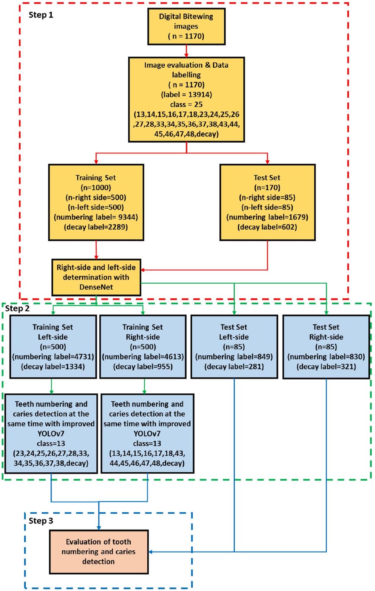
الأهداف كان الهدف من هذه الدراسة هو الكشف التلقائي عن الأسنان وترقيمها في صور الأشعة الرقمية للعضة الجانبية المأخوذة من المرضى، وتقييم الكفاءة التشخيصية للأسنان المتسوسة في الوقت الحقيقي، باستخدام خوارزميات التعلم العميق. الطرق تتكون مجموعة البيانات من 1170 صورة أشعة رقمية للعضة الجانبية تم الحصول عليها عشوائيًا من أرشيفات الكلية. بعد عملية تقييم الصور ووضع العلامات، تم تقسيم مجموعة البيانات إلى مجموعات تدريب واختبار. اقترحت هذه الدراسة بنية أنبوبية شاملة تتكون من ثلاث مراحل لمطابقة أرقام الأسنان وآفات التسوس لتعزيز نتائج العلاج ومنع المشكلات المحتملة. في البداية، تم استخدام شبكة عصبية تلافيفية مسبقة التدريب (CNN) لتحديد جانب صور العض. ثم، تم اقتراح نموذج CNN المحسن YOLOv7 لترقيم الأسنان وكشف التسوس. في المرحلة النهائية، قامت الخوارزمية التي طورناها بتقييم الأسنان التي تحتوي على تسوس من خلال مقارنة الأسنان المرقمة مع التسوس المكتشف، باستخدام قيمة التقاطع على الاتحاد لعملية المطابقة. النتائج وفقًا لنتائج الاختبار، كانت قيم الاسترجاع والدقة ودرجة F1 هي و 0.99 لاكتشاف الأسنان، و0.979 لرقم الأسنان، و و0.822 لاكتشاف التسوس، على التوالي. بالنسبة لأداء مطابقة ترقيم الأسنان واكتشاف التسوس؛ كانت قيم الدقة، والاسترجاع، والخصوصية، والدقة، وF1-Score هي 0.934، و 0.842، على التوالي. الاستنتاجات أظهر النموذج المقترح تحقيقًا جيدًا، مما يبرز الاستخدام المحتمل للشبكات العصبية التلافيفية (CNNs) في اكتشاف الأسنان، وترقيمها، واكتشاف التسوس، في الوقت نفسه. الأهمية السريرية يمكن أن توفر الشبكات العصبية التلافيفية دعمًا قيمًا للأطباء من خلال أتمتة اكتشاف وترقيم الأسنان، بالإضافة إلى اكتشاف التسوس في الأشعة السينية للعض. من خلال تحسين الأداء العام، تمتلك هذه الخوارزميات القدرة على توفير الوقت بكفاءة وتلعب دورًا مهمًا في عملية التقييم.

الكلمات الرئيسية الذكاء الاصطناعي الأشعة السينية الرقمية للعضة التعلم العميق الكشف الت numbering تسوس الأسنان

مقدمة

تسوس الأسنان هو مرض مزمن قابل للتجنب ومتعدد العوامل ومعدٍ، وهو شائع في العديد من السكان حول العالم. يمكن أن تكون الآفات التسوسية، التي تتقدم عادة ببطء، مشكلة صحية خطيرة قد تؤدي إلى الألم والعدوى وفقدان الأسنان إذا لم يتم علاجها في المراحل المبكرة. بينما كانت النظرة إلى علاج التسوس أكثر تدخلاً في الماضي، فقد تم استبدال هذه النظرة اليوم في طب الأسنان الحديث بالتشخيص المبكر مع طرق العلاج الوقائية والحد الأدنى من التدخل. إن الكشف المبكر والدقيق عن آفات التسوس هو خطوة مهمة جداً توجه التخطيط السريري من خلال تحديد العلاج الذي سيتم تطبيقه. لذلك، يلعب التشخيص في الوقت المناسب والدقيق دوراً رئيسياً في نجاح العلاج. على الرغم من وجود طرق مختلفة في تشخيص آفات التسوس، فإن الفحص البصري اللمسي…
الطريقة التي تعتبر غير كافية في تشخيص الكشف المبكر عن تسوس الأسنان الجانبي هي واحدة من الأساسيات [5، 6]. بالإضافة إلى طريقة الفحص البصري واللمسي، يلعب الفحص الإشعاعي، الذي يُستخدم عادة في التصوير داخل الفم، دورًا حاسمًا في ممارسة طب الأسنان. في هذا الصدد، يتم استخدام الأشعة السينية البانورامية، والأشعة السينية القمية، وأشعة البايتوينغ بانتظام في الممارسة. تعتبر رؤية وتقييم الحالة السنية على الأشعة السينية واحدة من أهم الخطوات في تشخيص الأمراض [7، 8]. اليوم، بفضل الأشعة السينية الرقمية للبايتوينغ، التي تتمتع بقدرة عالية على الاسترجاع والخصوصية في الكشف عن تسوس الأسنان الجانبي، يتم الكشف عن آفات التسوس بسهولة وبتكلفة منخفضة ونجاح مقارنة بالعديد من الطرق الأخرى [9، 10].
كما يمكن فهمه من كل هذا، فإن التصوير الإشعاعي وطرق الفحص البصري واللمسي ضرورية لاكتشاف الآفات التسوسية. في بعض الحالات، اعتمادًا على خبرة واهتمام طبيب الأسنان، يمكن أن يحدث تشخيص خاطئ أو تشخيص غير كافٍ في العيادات المزدحمة. يمكن أن تساعد أنظمة الكمبيوتر المساعدة الأطباء خلال عملية التشخيص. أيضًا، يمكن أن توفر للطبيب النجاح من خلال المساعدة في تخطيط العلاج وتنفيذه.
مصطلح الذكاء الاصطناعي (AI)، الذي أصبح شائعًا جدًا في حياتنا ويؤثر على العديد من الصناعات ويعد نقطة تحول حقيقية. الذكاء الاصطناعي هو مصطلح عام يشير إلى الأنظمة أو الآلات أو الحواسيب التي تحاكي سلوكيات وذكاء البشر. يشمل مجالات بحثية مختلفة وخوارزميات. التعلم العميق (DL) هو أحد هذه المجالات البحثية التي اكتسبت شعبية في السنوات الأخيرة. تتكون خوارزميات التعلم العميق من شبكات عصبية اصطناعية متنوعة وفقًا لأنواع المشاكل المختلفة مثل معالجة اللغة الطبيعية، ونماذج اللغة الكبيرة، ورؤية الكمبيوتر. الشبكات العصبية التلافيفية (CNNs) هي خوارزميات تعلم عميق خاضعة للإشراف وهي شبكات عصبية اصطناعية مستوحاة من النظام البصري الثديي. تُستخدم الشبكات العصبية التلافيفية بنشاط في العديد من مشاكل رؤية الكمبيوتر في طب الأسنان والطب لتحسين خدمات الصحة وزيادة الكفاءة.
إن اعتماد الشبكات العصبية التلافيفية (CNNs) في تشخيص تسوس الأسنان في طب الأسنان هو طريقة ناجحة كما يمكن فهمه من الدراسات التي أجريت. هناك العديد من الدراسات حول الشبكات العصبية التلافيفية والشبكات العصبية المماثلة. تُستخدم الأشعة السينية للعضة غالبًا من قبل أطباء الأسنان كأداة مساعدة لتشخيص تسوس الأسنان. في بعض الأبحاث، تم إجراء دراسات تشخيص تسوس الأسنان على الأشعة السينية للعضة. يمكن أن يقلل الكشف الصحيح عن الأسنان وترقيمها أثناء الفحص من وقت الفحص ويوفر تشخيصًا أفضل. بحث ياسا وآخرون في الكشف عن الأسنان وترقيمها في الأشعة السينية للعضة. أيضًا، تم البحث في دراسات مماثلة ضمن الأشعة السينية البانورامية أو الأشعة السينية للعضة. أظهرت هذه الدراسات أن التطورات المستندة إلى التعلم العميق في صناعة الصحة يمكن أن تكون مساعدة ومزودًا لرأي ثانٍ في العيادات اليومية.
ممارسة الطبيب [35]. إلى أفضل معرفتنا، لم يتم التحقيق في أي دراسة تتعلق بالكشف عن الأسنان، والترقيم، وكشف التسوس في نفس الوقت في الأشعة السينية الرقمية باستخدام نهج الشبكات العصبية التلافيفية المعتمد على التعلم العميق. تجعل الطريقة المقترحة هذه الدراسة فريدة مقارنة بالأدبيات الموجودة.
كان هدف هذه الدراسة هو الكشف التلقائي عن الأسنان وترقيمها في صور الأشعة الرقمية للعضة المأخوذة من المرضى، وتقييم الكفاءة التشخيصية للأسنان المتسوسة في الوقت الحقيقي، باستخدام بنية خط أنابيب تعتمد على الشبكات العصبية التلافيفية. وبالتالي، من المخطط تقليل وقت الجلوس، ومساعدة الطبيب، وزيادة فعالية العلاج من خلال جعل التشخيص أكثر فعالية أثناء الفحص.
يقترح الاقتراح الناجح بنية جديدة قائمة على التعلم العميق لرقم الأسنان واكتشاف التسوس. تعزز هذه البنية الحفاظ على المعلومات الحيوية في صور البايتوينغ من خلال دمج وحدات انتباه الكتل التلافيفية (CBAMs) وطبقات التجميع المتوسطة في نموذج YOLOv7. تؤدي هذه التنفيذ إلى أداء أكثر كفاءة في رقم الأسنان واكتشاف التسوس مقارنةً بنموذج YOLOv7 الأصلي. بالإضافة إلى ذلك، تم تطوير خوارزمية لتقييم الأسنان التي تحتوي على تسوس من خلال ربط الأسنان المرقمة بالتسوس المكتشف.

المواد والطرق

تحديد حجم العينة

تم تحديد حجم العينة لمجموعة الاختبار باستخدام اختبار فيشر الدقيق. تم استخدام تصميم يتضمن التجميع لأن كل صورة شعاعية رقمية تحتوي في المتوسط على 16 سطحًا، والتي تم حسابها بواسطة تأثير التصميم (DE). تأثير التصميم (DE)، الذي يقيم أداء طريقة أخذ عينات معينة مقارنة بعينة عشوائية بسيطة، هو أداة قيمة في النظر في دقة عينة التجميع. معادلة DE هي ICC ، حيث يمثل m حجم التجمع، و ICC هو معامل الارتباط داخل الفئة. تم افتراض ICC بقيمة 0.2 بناءً على دراسة ماسود وآخرون [36]. وفقًا لتحليل قوة الدراسة السابقة، تم ضرب وحدات مجموعة الاختبار المطلوبة مع DE [37]. عندما يجب أن يكون العدد الإجمالي للأسطح المطلوبة 2136 أو 134 صورة شعاعية رقمية. في الدراسة الحالية، احتوت مجموعة الاختبار على 170 صورة شعاعية رقمية.

مجموعة الصور

تم جمع ما مجموعه 1170 صورة شعاعية رقمية للمرضى البالغين الذين تزيد أعمارهم عن 18 عامًا من أرشيف الأشعة في جامعة كيريكالي، كلية طب الأسنان لهذه الدراسة.
تضمنت الصور الشعاعية الرقمية التي تم الحصول عليها من المرضى الذين جاءوا للفحص و/أو العلاج بين يناير 2018 ويناير 2023 من خلال الفحص. لم يتم الكشف عن تفاصيل إضافية، مثل الجنس أو العمر أو تاريخ الحصول على الصور، حيث تم جمع الصور الشعاعية بشكل مجهول. في الدراسة، لم تشمل الصور الشعاعية الرقمية التي تحتوي على أخطاء في وضعية المريض، أو أخطاء تقنية، أو ترميمات ثابتة، أو تقويمات ثابتة قد تعقد أو تمنع التشخيص تمامًا. تم الحصول على موافقة لهذه الدراسة من اللجنة الأخلاقية للبحوث السريرية غير التدخلية في جامعة كيريكالي. (رقم القرار: 2022.11.14) التزمت الدراسة بالإرشادات الأخلاقية المذكورة في إعلان هلسنكي.
تم التقاط جميع الصور الشعاعية الرقمية بواسطة جهاز Gendex Expert DC (أنظمة جينديكس للأسنان، إلينوي، الولايات المتحدة الأمريكية) الموجود في العيادات بقيمة بقعة بؤرية تبلغ 0.04 مم، جهد أنبوب 65 كيلوفولت، تيار أنبوب 7 مللي أمبير. يمكن أن يكون وقت التعرض والمسافة وجودة الصور الشعاعية متغيرة، لأن الصور الشعاعية الرقمية تم التقاطها بواسطة موظفين مختلفين عبر أقسام مختلفة. تم إخفاء هوية كل صورة شعاعية رقمية وحفظها بتنسيق .jpg. كانت الأقواس السنية العلوية والسفلية متميزة
الجدول 1 ملخص مجموعة البيانات الكاملة
تدريب اختبار
تدهور 2289 602
ترقيم 9344 1679
إجمالي الأسطح 16,674 2842
انتشار الآفات 24% 21.16%
إجمالي الصورة 1000 170
مرئية على الصور الشعاعية الرقمية. شمل التقييم جميع الأسنان المرئية، باستثناء الترميمات الموجودة على الأسطح القريبة من الأسنان. لأن هدف هذه الدراسة كان تقييم أداء النموذج المقترح في اكتشاف تسوس الأسنان القريب وترقيم الأسنان. عرضت الصور الشعاعية الرقمية المجمعة دقة غير متجانسة من حيث أحجامها. لهذا السبب، تم ضبط جميع الدقات إلى بكسل، مثل دراسة سابقة [19]. بعد هذه العملية، تم تقسيم مجموعة البيانات عشوائيًا كما يلي: للتدريب والتحقق (1000 صورة شعاعية) و 15% للاختبار (170 صورة شعاعية). وبالتالي، تم استخدام 1000 صورة شعاعية رقمية تم الحصول عليها بتوزيع عشوائي لتدريب نموذج CNN المقترح. تم استخدام 170 صورة شعاعية رقمية المتبقية لتقييم النموذج. في مجموعة بيانات التدريب، تم ترقيم 9344 سنًا. هناك 16674 سطحًا قريبًا و 2289 منها بها تسوس. انتشار الآفات في بيانات التدريب هو . تفاصيل مجموعة البيانات موضحة في الجدول 1 وعينات الصور من مجموعة البيانات موضحة في الشكل 1.

تصميم الدراسة

في هذه الدراسة، تم اقتراح بنية خط أنابيب يمكنها اكتشاف ترقيم الأسنان والتسوس في صور الشعاعية الرقمية في نفس الوقت. تتكون البنية المقترحة من 3 خطوات أساسية. تم تقديم مخطط يوضح تطبيق البنية المقترحة في الشكل 2.

تقييم الصورة ووضع العلامات على البيانات

قام متخصص في الترميم (المؤلف 3) لديه 13 عامًا من الخبرة ومساعد بحث (المؤلف 1) لديه 4 سنوات من
الشكل 1 عينات الصور من مجموعة البيانات
الشكل 2 مخطط تنفيذ البنية المقترحة
الخبرة بإنشاء تعليقات حقيقية للصور باستخدام برنامج تم تطويره بلغة برمجة بايثون. لذلك، قام المعلقون بوضع علامات على صناديق الحدود حول جميع الأسنان مع تقديم تصنيف لكل صندوق مع رقم السن وفقًا
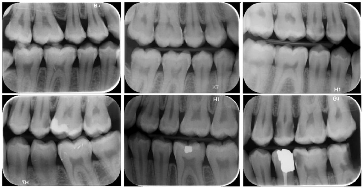
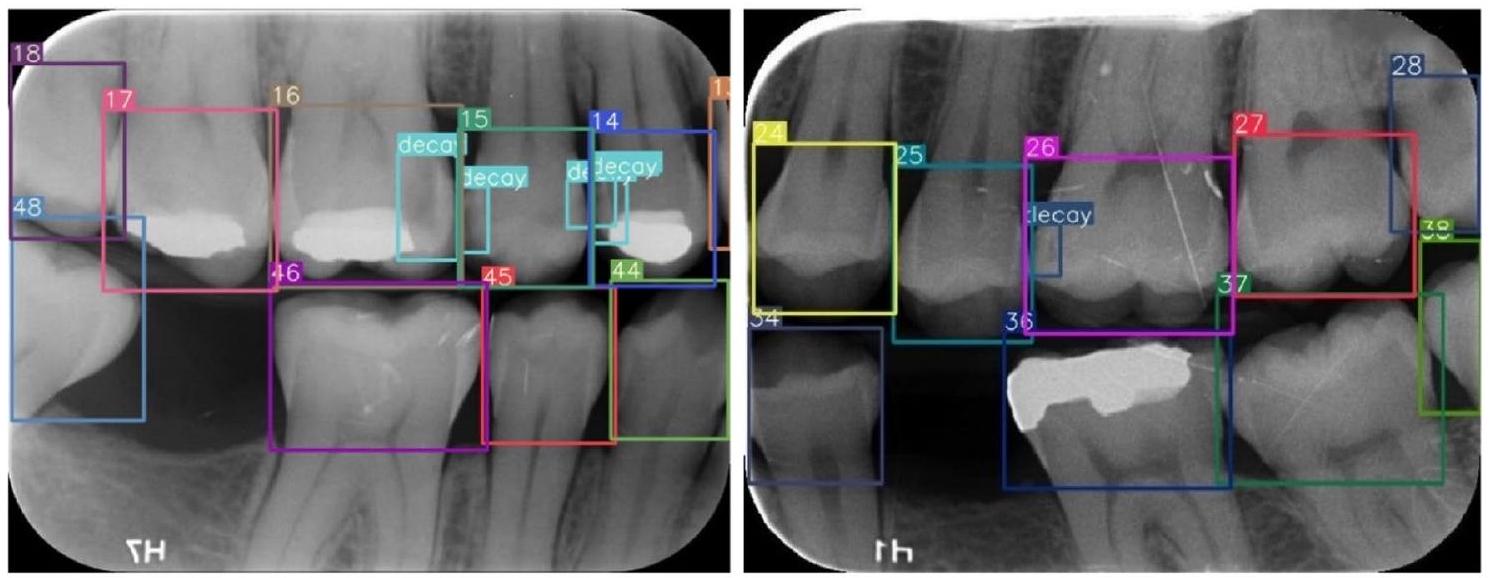
لنظام ترقيم الأسنان التابع للاتحاد الدولي لطب الأسنان (FDI) (16-17-18-26-27-28-36-37-38-46-47-48 (الأضراس)، 14-15-24-25-34-35-44-45 (الأنياب) و 13-23-33-43 (الأنياب)). أيضًا، تم وضع علامات على آفات التسوس كـ تدهور في الصور الشعاعية الرقمية. بينما في إجراء وضع العلامات على البيانات,
راجع المعلقون الصور الشعاعية في نفس الوقت ونتيجة للاتفاق، تمت معالجة وضع العلامات (الشكل 3).
بعد هذه المرحلة، تم وضع علامات على آفات التسوس القريبة على أسطح الأسنان بواسطة المعلقين بنفس إجراء الاتفاق. تم تحويل البرنامج المستخدم لوضع العلامات تلقائيًا معلومات الصندوق والعلامات وفقًا للاحتياجات لتدريب YOLO.

كشف جانب الصورة الشعاعية الرقمية عبر الشبكة العصبية التلافيفية

كانت الخطوة الأولى من بنية خط الأنابيب المقترحة هي الكشف تلقائيًا عما إذا كانت الصور الشعاعية الرقمية تنتمي إلى الجانب الأيسر أو الأيمن. كانت هذه الخطوة مهمة جدًا لخطوة ترقيم الأسنان. لأن معدل نجاح عملية الترقيم وُجد أنه منخفض في التدريب الأول دون فصل الجانبين الأيمن والأيسر. لهذا الغرض، تم استخدام بنية DenseNet CNN، التي تم تقديمها في عام 2016 بواسطة هوانغ وآخرون [38]. تستخدم بنية DenseNet اتصالات كثيفة، تربط كل طبقة بطبقات أخرى بطريقة أمامية. تحتوي الاتصالات الكثيفة على فوائد، مثل تقليل معلمات النموذج، وتقليل مشكلة التدرج المتلاشي، وتشجيع إعادة استخدام الميزات، وتقوية انتشار الميزات عبر الطبقات. توجد أربع بنى DenseNet في الأدبيات (DenseNet-121، DenseNet-169، DenseNet-201، DenseNet-264). في هذه الدراسة، تم استخدام بنية DenseNet-121 المدربة مسبقًا على مجموعة بيانات ImageNet وتم إعادة تدريبها باستخدام طرق التعلم الانتقالي والتعديل لاكتشاف ما إذا كانت الصور تنتمي إلى الجانب الأيسر أو الأيمن. السبب الرئيسي لاختيار DenseNet-121 هو أنه أخف وزنًا من البنى الأخرى، لذا يمكن تدريبه بشكل أسرع. يحتوي نموذج DenseNet-121 المعدل على معلمات، طبقة متصلة بالكامل مع 512 خلية عصبية وخلية عصبية واحدة للإخراج
مع دالة تنشيط سيغمويد. تم تدريب النموذج المقترح لمدة 30 دورة مع حجم دفعة 32، ومعدل تعلم أولي 0.001. تم استخدام الانتروبيا المتقاطعة الثنائية كدالة خطأ وتم تفضيل خوارزمية آدم كأداة تحسين.
في عملية التدريب، تم استخدام 1000 صورة شعاعية رقمية، 500 يسار و500 يمين. من بيانات التدريب تم استخدامها للتحقق. تم استخدام 170 صورة شعاعية رقمية (85 يسار و85 يمين) لتقييم أداء التصنيف للنموذج. وفقًا لنتيجة الاختبار، حقق نموذج CNN دقة %100 على مجموعة بيانات الاختبار. تم تقديم مخطط يلخص هذه المرحلة في الشكل 2.

تحسين بنية YOLOv7، ترقيم الأسنان واكتشاف التسوس
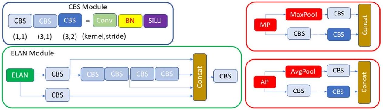
بنية YOLOv7 الأصلية

كانت المرحلة الثانية من الدراسة هي ترقيم الأسنان واكتشاف التسوس من صور البايتوينغ. في هذه الخطوة، تم أخذ خوارزميات الكشف عن الكائنات المعتمدة على التعلم العميق في الاعتبار نظرًا لطبيعة المشكلة التي يجب حلها. توجد نوعان من خوارزميات الكشف عن الكائنات المعتمدة على التعلم العميق: المعتمدة على المناطق (مرحلتين) والمعتمدة على الانحدار (مرحلة واحدة). في الخوارزميات المعتمدة على المناطق، يتم أولاً تحديد المناطق المرشحة التي تحتوي على كائنات محتملة، ثم يتم تصنيف هذه المناطق لتحديد أنواع الكائنات الموجودة فيها. تعتبر خوارزميات Fast-RCNN [39] وFaster-RCNN [40] وMask-RCNN [41] بعض الأمثلة على هذه الفئة. من ناحية أخرى، تقوم الخوارزميات ذات المرحلة الواحدة بالكشف عن الكائنات وتصنيفها في نفس الوقت. الخوارزميات في هذه الفئة هي YOLO (You Only Look Once) وإصداراتها المختلفة مثل YOLO9000 وYOLOv3 وYOLOv4 وYOLOv5 وYOLOv6 وYOLOv7 [42، 43]. تقدم الخوارزميات ذات المرحلة الواحدة سرعة كشف أسرع من تلك ذات المرحلتين، مما يجعلها شائعة في مشاكل كشف الكائنات.
الشكل 3 عينات الصور من عملية التسمية
في YOLOv7، أحد أحدث إصدارات سلسلة YOLO، تم إجراء بعض التحسينات في دالة الخسارة، والشبكة الأساسية، ودالة التنشيط. بهذه الطريقة، حقق النموذج دقة كشف محسنة وسرعة كشف متسارعة. لهذا السبب، تم اختيار خوارزمية YOLOv7 للاستخدام في الدراسة. تتكون بنية YOLOv7 الأصلية من أربعة وحدات: الإدخال، الشبكة الأساسية، العنق والرأس. تقوم وحدة الإدخال بتغيير حجم الصورة المدخلة إلى حجم بكسل موحد لتلبية مواصفات حجم الإدخال لوحدة الشبكة الأساسية. تتكون الشبكة الأساسية من بنى التلافيف، وعملية تطبيع الدفعات، ودالة تنشيط SiLU (CBS)، وشبكة الانتباه الطويلة المدى الممتدة (E-ELAN) ووحدات Max-Pool-1 (MP1). تتمثل مهمة الجزء الأساسي في استخراج خرائط الميزات من الصورة المدخلة. تتكون وحدة CBS من ثلاثة مكونات: كتل التلافيف، وعملية تطبيع الدفعات (BN)، ودالة تنشيط SiLU (دالة تنشيط وحدة خطية موزونة بالسيغمويد) (الشكل 5). تتكون وحدة ELAN من عدة وحدات CBS التي تضمن بقاء أحجام الميزات المدخلة والمخرجة دون تغيير. من خلال توجيه الكتل الحسابية لمجموعات الميزات المختلفة لتعلم ميزات متنوعة، يتم تعزيز قدرة الشبكة على التعلم مع الحفاظ على سلامة مسار التدرج الأصلي (الشكل 5). تنقسم وحدة MP1 إلى فرعين، أحدهما فرع علوي
وفرع سفلي ويتكون من وحدات CBS وطبقة التجميع الأقصى (MaxPool) (الشكل 4). في هذه الوحدة، يستخدم الفرع العلوي MaxPool لالتقاط معلومات القيمة القصوى من مناطق محلية صغيرة، بينما تستخرج وحدات CBS في الفرع السفلي جميع المعلومات القيمة من مناطق محلية صغيرة. تعزز هذه الطريقة المزدوجة قدرة الشبكة على استخراج الميزات.
تتكون وحدة العنق من وحدات ELAN-L وUpsample وMP2 وSPPCSPC باستخدام بنية شبكة تجميع ميزات هرمية (PAFPN). تجمع وحدة العنق خرائط الميزات التي تم الحصول عليها من الطبقات المختلفة لوحدة الشبكة الأساسية وترسلها كمدخلات إلى وحدة الرأس. تتكون وحدة SPPCSPC من وحدات CBS وطبقات MaxPool بأحجام مختلفة من التجميع الأقصى المتوازي (الشكل 5). تحتوي وحدة ELAN-L على هيكل مشابه لوحدة ELAN، لكنها تجمع بين خمسة مخرجات مختلفة (الشكل 5). تتكون وحدة Up-sample من وحدة CBS وتستخدم لزيادة حجم الصورة. تتضمن وحدة الرأس كتلة REP لتحديد عدد قنوات الصورة لمخرجات الميزات من جزء العنق. يتبع ذلك تطبيق وحدة التلافيف، التي تتنبأ بالفئة، والثقة، وصندوق الحدود. تستلهم وحدة REP من RepVGG [44] وتستخدم عملية تلافيف مبسطة خلال مرحلة الاختبار (الشكل 5). تقلل هذه التبسيط بشكل فعال من تعقيد الشبكة دون التأثير على الأداء.
الشكل 4 تمثيل وحدات CBS وELAN وMP
الشكل 5 تمثيل وحدات ELAN-L وREP وSPPCSPC

تحسين YOLOv7 مع وحدة انتباه الكتلة التلافيفية

تعترف المجتمع العلمي على نطاق واسع بالدور الكبير لآليات الانتباه في الرؤية البشرية. تتمثل سمة أساسية في النظام البصري البشري في قدرته على التركيز انتقائيًا على الجوانب البارزة من المشهد بدلاً من معالجة المجال البصري بالكامل في وقت واحد. يمكّن هذا الانتباه الانتقائي من التقاط تفاصيل بصرية محسنة [45]. في السنوات الأخيرة، طور الباحثون آليات انتباه انتقائية متنوعة للشبكات العصبية. تمكّن هذه الآليات الشبكات العصبية من تعلم “أين” أو “ماذا” يجب التركيز عليه أكثر، باستخدام الاعتماد الصريح بين القنوات والمناطق المكانية الموزونة. في هذا السياق، تم تقديم وحدة انتباه الكتلة التلافيفية (CBAM) من قبل وو وآخرون [46] كآلية انتباه فعالة مصممة لشبكات CNN. في هذا السياق، اقترح وو وآخرون [46] وحدة انتباه الكتلة التلافيفية (CBAM)، وهي آلية انتباه فعالة لشبكات CNN. تتضمن وحدة CBAM وحدة انتباه القناة (CAM) ووحدة انتباه المكان (SAM). تركز CAM على الجوانب ذات الصلة من صورة الإدخال من خلال استغلال العلاقات بين القنوات للميزات. يتم إنشاء وصفي سياق مكاني في الوحدة باستخدام طريقتين مختلفتين للتجميع (التجميع المتوسط والتجميع الأقصى). يُستخدم التجميع المتوسط (AP) بشكل أساسي لجمع المعلومات المكانية، بينما يحتفظ التجميع الأقصى بمزيد من التفاصيل السياقية المتعلقة
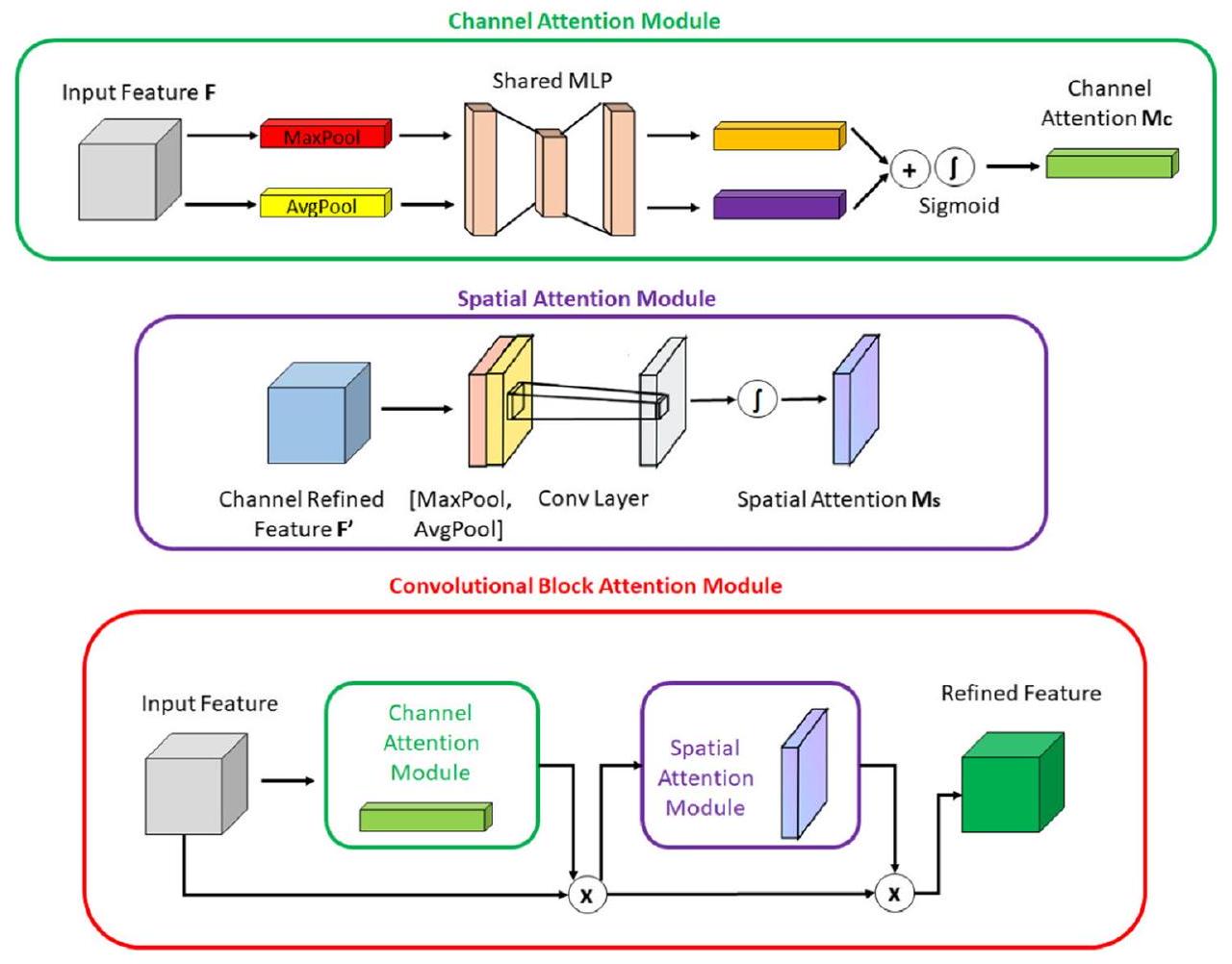
بالكائن داخل الصورة، مما يمكّن من انتباه القناة بدقة أكبر. يتم توجيه هذه الوصفات السياقية المولدة إلى شبكة متعددة الطبقات مشتركة. بعد جمع المتجه الناتج من الشبكة متعددة الطبقات، يتم إنشاء خريطة انتباه القناة من خلال تطبيق دالة السيغمويد. داخل وحدة CBAM، تستهدف وحدة انتباه المكان (SAM) بشكل خاص تحديد مواقع المناطق الأكثر معلوماتية داخل صورة الإدخال. تتضمن حسابات انتباه المكان داخل وحدة SAM تطبيق تقنيات التجميع المتوسط والتجميع الأقصى على طول محور القناة للصورة المدخلة. يتم إخضاع الصورة ذات القناتين الناتجة لعمليات تلافيفية لتحويلها إلى خريطة ميزات ذات قناة واحدة، والتي يتم دمجها وإرسالها إلى طبقة تلافيفية قياسية لإنشاء وصف ميزات فعال. يتم تمرير الناتج من الطبقة التلافيفية عبر طبقة تنشيط سيغمويد. تقوم دالة السيغمويد، التي تعتمد على الاحتمالية، بتحويل جميع القيم إلى نطاق بين 0 و1، مما يكشف عن خريطة انتباه المكان. باختصار، تجمع CBAM بين وحدتي الانتباه الموصوفتين أعلاه لتحديد ما هو مهم في صورة الإدخال وتحديد المناطق الوصفية داخل الصورة. يتم تقديم أشكال وحدات CBAM وCAM وSAM في الشكل 6.
عند مراجعة الدراسات في الأدبيات، يتبين أن هناك تركيبات ناجحة لوحدة CBAM في شبكات CNN في حل مشاكل الرؤية الحاسوبية [47، 48]. ومع ذلك، فإن إنشاء شبكة عصبية هو عملية معقدة تتطلب النظر في مجموعة البيانات المستخدمة،
الشكل 6 تمثيل وحدات CBAM وCAM وSAM
اختيار المقاييس المحسّنة، وتوافر الموارد الحاسوبية.
في هذه الدراسة، قمنا بحقن طبقات CBAM في نموذج YOLOv7 نتيجة لتجربتنا مع الشبكات العصبية التلافيفية (CNNs) والاختبارات المختلفة التي أجريناها. بعد إجراء تجارب متنوعة، تم وضع ثلاثة وحدات CBAM في ثلاثة مواقع محددة حيث تتقاطع الرقبة والرأس في بنية YOLOv7. تم اختيار عدد كتل CBAM المدمجة أثناء دمج وحدات CBAM بدقة لتقليل أي زيادة كبيرة في الحمل الحسابي على الشبكة. لقد ظهر دمج وحدات CBAM في بنية YOLOv7 من قبل الباحثين لتطوير تصاميم جديدة لحل مشاكل متنوعة كمنطقة بحث شائعة جدًا [49، 50]. لذلك، فإن تحديد الموقع، وعدد الكتل، ودمجها مع مكونات الشبكة الأخرى هي قرارات تصميم يمكن أن تختلف اعتمادًا على المشكلة المحددة قيد النظر. على حد علمنا، لا توجد حاليًا دراسة معروفة تتناول المنطقة التي قمنا بإدراجها في بحثنا. الشكل 7 يقدم توضيحًا للنموذج المحسن وتفاصيل حول الموقع الذي تم وضع وحدات CBAM فيه.
تحسين آخر قمنا به في نموذج YOLOv7 المقترح هو استخدام التجميع المتوسط (AP) بدلاً من التجميع الأقصى (MP) في طبقات أخذ العينات. في YOLOv7 الأصلي، يتم أخذ العينات في أربعة مواقع مختلفة. في هذه العينات، تم استخدام طريقة التجميع المتوسط بدلاً من التجميع الأقصى.
تستخدم طريقة التجميع المتوسط بدلاً من التجميع الأقصى. يقوم التجميع الأقصى بإرجاع القيمة القصوى في كل نافذة، بينما يقوم التجميع المتوسط بإرجاع القيمة المتوسطة في كل نافذة. فائدة استخدام التجميع المتوسط بدلاً من التجميع الأقصى هي أنه يمكن أن يساعد في تقليل الإفراط في التكيف. من المرجح أن يقوم التجميع الأقصى بالتذكر لأنه يحتفظ فقط بأقوى ميزة في كل نافذة قد لا تمثل النافذة بالكامل. من ناحية أخرى، يأخذ التجميع المتوسط في الاعتبار جميع الميزات في كل نافذة، مما يمكن أن يساعد في الحفاظ على مزيد من المعلومات. بالإضافة إلى ذلك، يمكن أن يساعد التجميع المتوسط في تنعيم المخرجات، مما يمكن أن يكون مفيدًا عندما تكون المدخلات صاخبة أو متقلبة. في هذا السياق، التغييرات التي أجريناها موضحة في الشكل 7. لقد لوحظ أن هذا التحسين له تأثير إيجابي على أداء الكشف.
تم استخدام بنية YOLOv7-AP-CBAM التي تم الحصول عليها مع التحديثات التي أجريت في بنية YOLOv7 في ترقيم الأسنان وفي الكشف عن التسوس. في هذا السياق، تم تدريب نموذجين مختلفين للأجزاء اليمنى واليسرى من الفم. نظرًا لأن أداء الترقيم لنموذج واحد ظل منخفضًا في الاختبارات، تم استخدام طريقتين مختلفتين لتدريب النموذج. تم تدريب النماذج لكلا الجانبين مع المعلمات الفائقة التالية: حجم الدفعة 16، عدد الدورات 20، حجم صورة الإدخال (640،640)، معدل التعلم الأولي 0.01، خوارزمية تحسين SGD، الزخم 0.937، تآكل الوزن 0.005. يتم تقديم مخطط تدفق يلخص هذه المرحلة في الشكل 7.
الشكل 7 YOLOv7 المحسن مع وحدات CBAM و AP

تقييم ترقيم الأسنان ومطابقة الكشف عن التسوس

في المرحلة الأخيرة من البنية المقترحة، كان الهدف هو إعطاء مخرجات النظام التي تحدد أي سن لديه تسوس من خلال مطابقة الأسنان المرقمة والتسوس المكتشف. تم تحديد ما إذا كان هناك مربع محيط مرسوم للتسوس في المربعات المحيطة المرسومة لترقيم الأسنان. إذا كان هناك تسوس في مربع المحيط المرقم، تم الإبلاغ عن أي سن لديه تسوس. تم استخدام قيمة التقاطع على الاتحاد (IoU) للكشف عن التسوس في مربع المحيط المرقم (انظر الشكل 8).
إذا كانت قيمة IoU أعلى من ، تم قبول أن رقم السن الذي يحمل علامة التسوس تم تحديده بشكل صحيح. يتم تقديم مخطط تدفق الخوارزمية التي طورناها لهذا الغرض في الشكل 9.

الإعداد التجريبي

تم تنفيذ جميع التجارب في هذه الدراسة على وحدة معالجة الرسوميات NVIDIA 1080 Ti بحجم ذاكرة فيديو 12 جيجابايت واثنين من معالجات Intel Xenon CPU مع 64 جيجابايت من الذاكرة العشوائية على نظام تشغيل Ubuntu. تشمل بيئة التشغيل التجريبية لغة البرمجة بايثون، وإطارات DL Pytorch [51]، Keras [52]، ومكتبة رؤية الكمبيوتر OpenCV [53].

تحليل تقييم الأداء

لتقييم نجاح النموذج المقترح، تم استخدام مصفوفة الارتباك، التي تلخص الحالات الفعلية والمتوقعة في جدول، مع الإجراءات والمعايير التالية. تم تعديل مصفوفة الارتباك التي استخدمناها لتقييم أداء ترقيم الأسنان وكشف التسوس
الشكل 8 صيغة IoU. A تمثل مربع الترقيم، بينما B تمثل مربع التسوس
التقاطع على الاتحاد (IoU) =
الشكل 9 مخطط قرار مطابقة ترقيم الأسنان وكشف التسوس
المطابقة موضحة أدناه. أيضًا، تم تقييم أداء كشف الأسنان، والترقيم، وكشف التسوس بشكل فردي.
Adaptation of the confusion matrix for evaluating performance of
teeth numbering and caries detection matching
TP: the output in which the model correctly predicts the positive
    class (teeth correctly detected, correctly numbered, caries correctly
    detected on bitewing radiographs)
FP: the output in which the model incorrectly predicts the positive
    class (teeth correctly detected, correctly numbered, but caries pre-
    diction on sound surface; teeth correctly detected, but regardless of
    caries incorrectly numbered)
FN: the output in which the model incorrectly predicts the negative
    class (teeth correctly detected, correctly numbered, but no caries
    prediction on decayed surface)
TN: the output in which the model correctly predicts the negative
    class (teeth correctly detected, correctly numbered, no caries pre-
    diction when there was no caries)
في البداية، تم تحديد معدلات الإيجابيات الحقيقية (TP)، والإيجابيات الكاذبة (FP)، والسلبيات الكاذبة (FN) والسلبيات الحقيقية (TN). ثم تم حساب الدقة (Eq. 1)، والاسترجاع (Eq. 2)، والخصوصية (Eq. 3)، والدقة (Eq. 4)، ودرجة F1 (Eq. 5) ودرجات IoU (Eq. 6). يتم تعريف IoU على أنه مقياس التداخل بين مربع المحيط الفعلي المسمى من قبل المعلق لشيء ما ومربع المحيط المتوقع من قبل النموذج. من خلال مقارنة قيمة IoU مع القيمة العتبية المحددة مسبقًا قبل تدريب النموذج، من الممكن تفسير نجاح النموذج المدرب في كشف الأشياء. تعتبر درجة F1، التي تعتبر المتوسط التوافقي للدقة والاسترجاع، شائعة الاستخدام لتوفير تقييم متوازن لأداء نموذج التصنيف.
تظهر المعادلات أدناه (Eqs. 1-6).

النتائج

في مجموعة بيانات الاختبار التي تشمل 170 صورة شعاعية رقمية، كان هناك 1679 سنًا، و2842 سطحًا تقريبيًا، و1679 علامة ترقيم، و602 علامة تسوس. كانت نسبة انتشار الآفة في بيانات الاختبار هي .
دون تقسيم إلى الجانبين الأيمن والأيسر، كانت قيم الاسترجاع، والدقة، ودرجة F1 هي 0.409، 0.236، و0.299 لترقيم الأسنان، و ، و0.691 لكشف التسوس، على التوالي (الجدول 5).
بعد تقسيمها إلى الجانبين الأيمن والأيسر، كانت قيم الاسترجاع، والدقة، ودرجة F1 هي و0.99 لكشف الأسنان، و و0.979 لترقيم الأسنان، و و0.822 لكشف التسوس، على التوالي (الجدول 2 و3).
بالنسبة لأداء مطابقة ترقيم الأسنان وكشف التسوس؛ كانت قيم الدقة، والاسترجاع، والخصوصية، والدقة ودرجة F1 هي و0.842، على التوالي (الجدول 4).
أداءت نماذج YOLOv7-AP-CBAM المحسنة بشكل جيد في كشف وترقيم الأسنان، بالإضافة إلى كشف التسوس في نفس الوقت على الصور الشعاعية الرقمية (الشكل 10).

تجارب الإزالة

أجرت هذه الدراسة تجارب إزالة لتقييم تأثير التحسينات المختلفة على أداء النموذج. في البداية، تم استخدام نموذج YOLOv7 الأصلي لاستخراج الميزات دون تقسيم إلى الجانبين الأيمن والأيسر. بعد ذلك، تم تعديل نموذج YOLOv7 الأصلي ليشمل التقسيم إلى الجانبين الأيمن والأيسر، بالإضافة إلى دمج AP وMP مع CBAM. أخيرًا، تم تطبيق AP مع CBAM. تحسن استرجاع ترقيم الأسنان وكشف التسوس، والدقة، ودرجة F1 للنموذج الذي استخدم وحدات AP وCBAM بشكل كبير مقارنةً بالبداية لهذه الدراسة. يتم تقديم النتائج التجريبية في الجدول 5.
في الشبكات العصبية التلافيفية، يتم وصف التعقيد من خلال العدد الإجمالي للمعلمات في النموذج وعدد عمليات النقطة العائمة جيجا (GFLOPs) [54]. قمنا بمقارنة النسخة الأساسية من YOLOv7 مع النسخة المحسنة من YOLOv7-AP-CBAM. يحتوي YOLOv7 الأصلي على 314
الجدول 2 معايير تقييم نموذج كشف الأسنان
الاسترجاع الدقة درجة F1
الجانب الأيسر 0.995 0.987 0.99
الجانب الأيمن 0.993 0.988 0.99
الكل 0.994 0.987 0.99
الجدول 3 معايير تقييم نموذج ترقيم الأسنان وكشف التسوس
الاسترجاع الدقة درجة F1
ترقيم الأسنان في الجانب الأيسر 0.976 0.978 0.977
ترقيم الأسنان في الجانب الأيمن 0.972 0.993 0.982
كشف التسوس في الجانب الأيسر 0.857 0.833 0.844
كشف التسوس في الجانب الأيمن 0.810 0.900 0.800
ترقيم وكشف التسوس في الجانب الأيسر 0.966 0.966 0.966
ترقيم وكشف التسوس في الجانب الأيمن 0.959 0.985 0.968
ترقيم وكشف التسوس في الجانبين الأيسر والأيمن – الكل 0.962 0.975 0.967
ترقيم الأسنان – الكل 0.974 0.985 0.979
كشف التسوس – الكل 0.833 0.866 0.849
الجدول 4 معايير تقييم نموذج مطابقة ترقيم الأسنان وكشف التسوس
TP FP FN TN دقة استدعاء خصوصية دقة درجة F1
الجانب الأيسر ٢٤٠ ٤٨ ٤٧ ١١٠٢ 0.933 0.836 0.958 0.833 0.834
الجانب الأيمن ٢٦٠ 30 66 1049 0.931 0.797 0.972 0.896 0.843
كل ٥٠٠ 78 113 2151 0.932 0.815 0.965 0.865 0.839
الشكل 10 عينات الصور لاكتشاف وترقيم الأسنان، بالإضافة إلى اكتشاف التسوس باستخدام نموذج YOLOv7-AP-CBAM المقترح على الأشعة السينية الرقمية للعض. أ تسميات نموذج YOLOv7-AP-CBAM المحسن، علامات الحقيقة الأرضية، صورة خام
طبقات مليون معلمة، 103.7 GFLOPS، 75.1 ميجابايت حجم في الذاكرة. من ناحية أخرى، يحتوي النموذج المقترح YOLOv7-AP-CBAM على 350 طبقة. مليون
معلمات 103.8 GFLOPS وحجم 75.2 ميجابايت في الذاكرة. تم تحقيق زيادة مرضية في الأداء مع زيادة صغيرة في التعقيد في النموذج.
الجدول 5 مقارنة الإزالة لتحسينات الأداء على النموذج
استدعاء دقة درجة F1
YOLOv7-MP الأصلي بدون تقسيم إلى اليمين واليسار تحلل الكل 0.676 تحلل الكل 0.707 تحلل الكل 0.691
ترقيم الكل 0.٤٠٩ ترقيم الكل 0.236 ترقيم الكل 0.299
YOLOv7-MP الأصلي مع تقسيم إلى اليمين واليسار تآكل الجانب الأيمن 0.659 تآكل الجانب الأيمن 0.803
تسوس الجانب الأيسر 0.696 تسوس الجانب الأيسر 0.848
تآكل الكل 0.677 تحلل الكل 0.825 تحلل الكل 0.743
ترقيم الجانب الأيمن 0.971 ترقيم الجانب الأيمن 0.974
ترقيم الجانب الأيسر 0.979 ترقيم الجانب الأيسر 0.978
ترقيم الكل 0.975 ترقيم الكل 0.976 ترقيم الكل 0.975
YOLOv7-AP تآكل الجانب الأيمن 0.626 تآكل الجانب الأيمن 0.773
تآكل الجانب الأيسر 0.797 تسوس الجانب الأيسر 0.775
تحلل الكل 0.711 تحلل الكل 0.774 تحلل الكل 0.741
ترقيم الجانب الأيمن 0.967 ترقيم الجانب الأيمن 0.977
ترقيم الجانب الأيسر 0.981 ترقيم الجانب الأيسر 0.983
ترقيم الكل 0.974 ترقيم الكل 0.980 ترقيم الكل 0.976
YOLOv7-MP-CBAM تآكل الجانب الأيمن 0.579 تآكل الجانب الأيمن 0.838
تسوس الجانب الأيسر 0.754 تآكل الجانب الأيسر 0.832
تحلل الكل 0.666 تحلل الكل 0.836 تحلل الكل 0.741
ترقيم الجانب الأيمن 0.974 ترقيم الجانب الأيمن 0.983
ترقيم الجانب الأيسر 0.981 ترقيم الجانب الأيسر 0.978
ترقيم الكل 0.977 ترقيم الكل 0.980 ترقيم الكل 0.978
YOLOv7-AP-CBAM (خاص بنا) تآكل الجانب الأيمن 0.810 تآكل الجانب الأيمن 0.900 كل تدهور 0.849
تدهور الجانب الأيسر 0.857 تآكل الجانب الأيسر 0.833
كل تدهور 0.833 كل تدهور 0.866
ترقيم الجانب الأيمن 0.972 ترقيم الجانب الأيمن 0.993
ترقيم الجانب الأيسر 0.976 ترقيم الجانب الأيسر 0.978
ترقيم الكل 0.974 ترقيم الكل 0.985 ترقيم الكل 0.979

نقاش

في طب الأسنان، يعتبر تحقيق تشخيص دقيق وواضح للتسوس السني خطوة أساسية نحو تنفيذ العلاج بنجاح. تهدف طرق تشخيص التسوس السني إلى تحسين عملية التشخيص من خلال تسهيل الكشف المبكر عن التسوس أو تمكين تحديده بشكل موضوعي. على الرغم من أن اكتشاف الأسنان وترقيمها قد يبدو بسيطًا، إلا أن دقة ترقيم الأسنان واكتشاف التسوس يمكن أن تتأثر بالمعرفة المحدودة والمتنوعة للأطباء المبتدئين، مما قد يؤدي إلى تشخيصات غير متسقة وطرق علاج غير كافية. بالإضافة إلى الخبرة، يمكن أن تؤثر عدم انتباه الممارسين أو أعباء العمل الثقيلة أيضًا على نتائج التشخيص. لمنع كل هذه التحديات وتسهيل عملية التشخيص، يمكن استخدام نظام مدعوم بالكمبيوتر يعتمد على طرق التعلم العميق. لقد اكتسب التعلم العميق شعبية كبيرة في المجال الطبي.
لإمكاناتها المتميزة في حل المشكلات. في هذا السياق، تم تطوير العديد من هياكل التعلم العميق، حيث تبرز الشبكات العصبية التلافيفية بشكل خاص لأدائها المذهل في مهام التعرف على الصور. من خلال استخدام نماذج الشبكات العصبية التلافيفية، يمكن للممارسين تعزيز كفاءة العمل بشكل فعال وتحقيق نتائج دقيقة في عملية التشخيص الدقيقة.
في الدراسة الحالية، تم تقديم كفاءة نموذج CNN المحسن YOLOv7-AP-CBAM للكشف التلقائي عن الأسنان numbering، بالإضافة إلى الكشف عن التسوس في الأشعة الرقمية للعضة، بشكل متزامن. حتى الآن، تم إجراء دراسات متنوعة في مجال طب الأسنان، باستخدام CNNs للكشف، التصنيف، والتجزئة لمختلف الكيانات مثل الأجسام، الأسنان، التسوس، الترقيم، كسور الجذور، الآفات المحيطية وفقدان العظام اللثوي [34، 58-63]. وفقًا لهذه الدراسات، أظهرت الأنظمة المعتمدة على CNN نتائج واعدة في التغلب على صعوبات التشخيص ومساعدة الممارسين.
تم استخدام أنظمة تصوير الأسنان المختلفة لهذه الدراسات. تُستخدم تقنية الأشعة السينية الرقمية بشكل واسع نظرًا لعمليتها ومزاياها. متطلبات جرعات الإشعاع الأقل، وكفاءة الوقت، وتخزين الصور، وبساطة تحسين ومعالجة الصور هي بعض من المزايا والأسباب التي تجعل تقنية الأشعة السينية الرقمية مفضلة [64]. في الأوراق البحثية، كان هناك عدد من الدراسات التي تستخدم الأشعة السينية الرقمية [19، 20، 65-69]. إنها تقنية أكثر فائدة من الطرق الأخرى التي تحتوي على الأشعة السينية لتقييم التاج ومستويات العظم السنخي للأسنان الخلفية في كل من القوس السني العلوي والسفلي، وكشف التسوس، خاصة في تسوس الأسنان القريب، واكتشاف الأسنان وترقيمها [10، 70، 71].
كما تم إجراء دراسات حول تطبيق الشبكات العصبية التلافيفية (CNN) على صور الأشعة السينية للعضة، والأشعة البانورامية، والأشعة السينية المحيطية، وأشعة التصوير المقطعي المحوسب باستخدام شعاع المخروط وطرق تشخيصية متنوعة [34، 72، 73]. قام Zhang وآخرون [73] بتطبيق طريقة التعلم العميق (DL) المعتمدة على الشبكات العصبية التلافيفية (CNN) لاكتشاف وتصنيف الأسنان في الأشعة السينية المحيطية. كانت درجات الدقة والاسترجاع مرتفعة. و على التوالي. في دراستنا، حقق النموذج المحسن استرجاعاً قدره ودقة في صور الأشعة الرقمية للعضة. في دراسة أخرى على صور الأشعة المقطعية المحوسبة (CBCT) تم الإبلاغ عن أن تنفيذ نموذج شبكة U-Net ثلاثية الأبعاد أظهر نتائج مرضية لتقسيم وتصنيف الأسنان. علاوة على ذلك، أدى تقسيم الأسنان جميعها في مسح واحد باستخدام الذكاء الاصطناعي إلى تحسين السرعة بمقدار 1800 مرة مقارنةً بخبرة خبير [72]. استخدم توزوف وآخرون [34] نموذج Faster R-CNN لاكتشاف الأسنان ونموذج VGG-16 Net لترقيم الأسنان على صور الأشعة البانورامية. أظهرت النماذج المعتمدة على الشبكات العصبية الالتفافية (CNN) قيم استرجاع وخصوصية عالية لمهام اكتشاف وترقيم الأسنان، مقارنةً بالخبراء. ومع ذلك، أظهر نموذجهم تصنيفات خاطئة في الحالات التي كانت فيها الأسنان مفقودة، بينما نموذجنا المحسن اكتشف هذه الحالات بدقة. تم تقديم عينات الصور لهذه الحالات في الشكل 11.
تم اقتراح طريقة DL Faster R-CNN لاكتشاف الأسنان وترقيمها في الأشعة السينية المحيطية بواسطة تشين وآخرين [57]. من أجل تحسين دقة الاكتشاف، اقترحوا ثلاث تقنيات لمعالجة ما بعد الاكتشاف لتكملة نموذج Faster R-CNN الأساسي. حقق نموذجهم دقة قدرها واسترجاع للكشف، ودقة واسترجاع للتعداد. في دراستهم، كان النموذج غير قادر على التنبؤ بدقة بالأسنان الموجودة في زوايا صورة البايتوينغ. ومع ذلك، أظهر نموذجنا المحسن دقة قدرها واسترجاع لـ للكشف، بالإضافة إلى دقة واسترجاع لـ للت numbering.
في الأدبيات، يستفيد الباحثون عمومًا من تقنيات تحسين الصور والقص لتحسين الدقة. قام ماو وآخرون [74] بتحسين نموذج أليكس نت الجديد، وهو نهج قائم على الشبكات العصبية التلافيفية للكشف عن التسوس والترميم في صور العض، من خلال استخدام تقنيات تحسين الصور والقص. أظهر النموذج دقة قدرها لكشف التسوس و للكشف عن الاستعادة. ومع ذلك، من المهم ملاحظة أن درجات الدقة العالية وحدها قد لا تشير إلى موثوقية الطريقة. إذا قام نموذج CNN بتصنيف كل سطح على أنه “متعفن”، يمكن أن يحقق دقة مثالية ولكنه يفتقر إلى نتائج ذات مغزى. لمعالجة ذلك، تم الإبلاغ عن درجات الاسترجاع والخصوصية في دراستنا، اتباعًا للسابقة التي وضعتها الأبحاث السابقة، لتوفير تقييم أكثر شمولاً دون التضحية بالقدرة على التفسير. بالإضافة إلى ذلك، لم نستخدم أي طرق لتحسين الصور أو القص.
في دراسة سابقة، تم تطوير نظام مساعدة تلقائي للكشف عن آفات التسوس في الأشعة السينية للعض باستخدام نموذج U-Net. تم تقييم أداء النظام من خلال تحليل مقارن مع أطباء الأسنان. وفقًا لنتائج هذه الدراسة، أظهر النموذج دقة عامة قدرها بينما كانت دقة الممارسين المتوسطة كانت قيم الاسترجاع، والخصوصية، ودرجة F1 أيضًا مرتفعة، عند ، و على التوالي
الشكل 11 عينة من ترقيم الأسنان الصحيح في الحالات التي كانت فيها سن مفقودة
[20]. على الرغم من أن نتائج النموذج في هذه الدراسة لم تتم مقارنتها مباشرة بأداء الخبراء البشريين، إلا أنها أظهرت نتائج واعدة في اكتشاف آفات التسوس، محققة قيم استرجاع ودقة ودرجة F1 من ، و ، على التوالي. هذه النتائج مشابهة جداً لتلك التي تم الإبلاغ عنها في دراسة كانتو وزملائه. بالإضافة إلى ذلك، قامت الدراسة الحالية بتقييم أداء ترقيم الأسنان بالتزامن مع اكتشاف آفات التسوس.
اقترح ياسا وآخرون [29] نموذج Faster R-CNN لاكتشاف وتعداد الأسنان في صور الأشعة السينية للعض. قاموا بإجراء مقارنة بين نموذجين مختلفين لتعداد الأسنان. في البداية، لم تكن الأسنان مفصولة إلى الجانبين الأيمن والأيسر. ومع ذلك، بعد تقسيم وتدريب النموذج بهذه الطريقة، تم الحصول على نتائج واعدة، مع قيم الاسترجاع والدقة ودرجة F 1 من ، ، و ، على التوالي. في دراستنا، اعتمدنا نهج فصل الأسنان إلى الجانبين الأيمن والأيسر، متبعين منهجية مشابهة. من خلال اعتماد هذا النهج، أظهر نموذجنا المحسن أداءً واعدًا، كما يتضح من قيم الاسترجاع والدقة ودرجة F1 الملحوظة ، و ، على التوالي.
في دراسة أخرى، أفاد بايركتار وأيان [19] بالكشف عن آفات تسوس الأسنان بين الأسنان في صور الأشعة الرقمية باستخدام نموذج YOLOv3. في تلك الدراسة، كانت نسبة الاسترجاع الدقة كانت وفقًا لهذه النتائج الواعدة، يمكن أن يكون استخدام نظام قائم على الشبكات العصبية التلافيفية مفيدًا في تشخيص آفات التسوس. في الدراسة الحالية، نموذجنا المحسن هو نموذج YOLOv7-AP-CBAM مع تنفيذ وحدة CBAM. من خلال وحدة CBAM، يمكن للنموذج أن يركز بشكل أكثر دقة. بهذه الطريقة، وصل النموذج إلى استرجاع التي هي أعلى من الدراسة السابقة. ومع ذلك، كانت قيم الدقة متقاربة تقريبًا وهي و .
أظهرت الدراسة الحالية، نموذج YOLOv7-AP-CBAM نتائج واعدة من حيث المعايير التي تم تقييمها. في مصفوفة الالتباس التي أنشأناها، بينما كانت درجة الخصوصية أعلى، لوحظ تأثير عكسي في درجة الاسترجاع. أظهر هذا النتيجة أن النموذج المحسن يمكنه التنبؤ بدقة بأرقام الأسنان بينما يكتشف أيضًا الأسطح السليمة، مشابهًا للدراسة التي أجراها لي وآخرون [22]. كانت قيم الدقة العامة، والاسترجاع، والخصوصية، والدقة، ودرجة F1 هي 0.934. و0.842، على التوالي.
أداء نموذج الشبكة العصبية التلافيفية (CNN) يعتمد بشكل مباشر على كمية بيانات التدريب. إن استخدام مجموعات بيانات كبيرة ومتنوعة في عملية التدريب يحسن من اتساق ودقة نماذج الشبكة العصبية التلافيفية. لذلك، تلعب جودة وكمية بيانات التدريب دورًا حيويًا في ضمان فعالية الشبكات العصبية التلافيفية. في هذه الدراسة، كانت مجموعة بيانات التدريب صغيرة نسبيًا. على الرغم من أن مجموعة بيانات التدريب تحتوي على عدد متساوٍ من صور الأشعة الجانبية اليمنى واليسرى، إلا أن النموذج كان أكثر نجاحًا في الجانب الأيسر في اكتشاف التسوس في النتائج النهائية. يمكن تفسير ذلك بوجود كمية أكبر من التسوس في الأشعة الجانبية.
على الجانب الأيسر في مجموعة التدريب. لتجنب هذه المشكلة، يمكن إضافة صور جديدة للعضة تشمل تسوس الأسنان إلى مجموعة التدريب على الجانب الأيمن. انحياز مجموعة البيانات هو مشكلة أخرى تحدث عندما يتم تمثيل عينات معينة في مجموعة البيانات بشكل مفرط أو ناقص. لتجنب هذه المشكلة، تم توليد مجموعة البيانات باستخدام صور مأخوذة من نفس الأجهزة في عيادات مختلفة بدقة وأوقات تعرض مختلفة. ومع ذلك، قد تؤثر الصور المأخوذة من آلات الأشعة السينية المختلفة ومن أشخاص ذوي خصائص ديموغرافية مختلفة على النتائج. تم تحديد معلمات النموذج المقترح باستخدام بيانات التحقق خلال مرحلة التدريب، وتم تقييم أداء النموذج على بيانات غير مرئية باستخدام بيانات اختبار مختلفة عن بيانات التدريب والتحقق. تتطلب نماذج الشبكات العصبية التلافيفية متطلبات عتادية عالية، خاصة في مرحلة التدريب. في دراستنا، قمنا بإجراء التدريب والاختبارات على بطاقة الرسوميات 1080 Ti. ستكون الأداء الحاسوبي أعلى على بطاقات الرسوميات الأكثر تقدمًا. تم الكشف عن آفات تسوس الأسنان التي لم يتم تقييمها باستخدام الطريقة القياسية الذهبية للتقييم النسيجي وتم تحديدها ووضع علامات عليها من قبل المعلقين على الصور الشعاعية. واجه النموذج تحديات في التنبؤ بدقة بالأسنان الموجودة في زوايا صورة العض، على عكس الأسنان الأخرى. قد يكون هذا مرتبطًا بانخفاض وضوح هذه الأسنان المحددة وكمية البيانات المحدودة في مجموعة التدريب. أيضًا، لم يتم مقارنة نموذج YOLOv7-AP-CBAM المحسن مع خبير. على الرغم من قيود الدراسة، من الجدير بالذكر أن هناك العديد من النقاط القوية المهمة أيضًا. قدمت الدراسة الحالية تحليلًا شاملاً من خلال دمج نتائج اكتشاف الأسنان، والترقيم، واكتشاف التسوس. سهل هذا النهج الشامل فهمًا أكثر تماسكًا للنتائج. بالإضافة إلى ذلك، شمل التصميم التجريبي تقسيم البيانات إلى الجانبين الأيمن والأيسر، مما أتاح تحليلًا أكثر تفصيلًا. عزز هذا النهج مقاييس أداء نتائجنا. علاوة على ذلك، من خلال اعتماد نهج متعدد التخصصات، قدمت دراستنا منظورًا جديدًا وتساهم بمساهمة مبتكرة في الأوراق السابقة.

الخاتمة

في هذه الدراسة، تم اقتراح نموذج محسّن YOLOv7-AP-CBAM للكشف الدقيق عن الأسنان، وترقيمها وكشف التسوس، مع تقييم متزامن للأسنان التي تحتوي على تسوس من خلال مطابقة الأسنان المرقمة والتسوس المكتشف. بالنسبة لأداء مطابقة ترقيم الأسنان وكشف التسوس؛ تم قياس القيم الخاصة بالدقة، والاسترجاع، والخصوصية، والدقة، وقيمة F1. و 0.842، على التوالي. أظهر النموذج دقة عالية في الاسترجاع، والخصوصية، والدقة، ودرجة F1، مما يبرز إمكانيات الشبكات العصبية التلافيفية. هذه النتيجة
كشفت أن الشبكات العصبية التلافيفية يمكن أن تقدم دعمًا قيمًا للأطباء من خلال أتمتة اكتشاف وترقيم الأسنان، بالإضافة إلى اكتشاف التسوس في الأشعة السينية للعض. يمكن أن يسهم ذلك في التقييم، وتحسين الأداء العام، وفي النهاية توفير الوقت الثمين.
شكر وتقدير لم تتلقَ هذه البحث أي منحة محددة من وكالات التمويل في القطاعات العامة أو التجارية أو غير الربحية.
مساهمة المؤلف ب.أ: جمع البيانات وتصنيفها، الاختبارات، التحليل الرسمي، كتابة المسودة الأصلية، كتابة المراجعة والتحرير.
إ.أ: التصور، البرمجيات، التصوير، تنظيم البيانات، التحليل الرسمي، كتابة – المسودة الأصلية، كتابة – المراجعة والتحرير.
ي.ب: التصور، المنهجية، جمع البيانات وتوسيمها، الكتابة – المراجعة والتحرير.
تم توفير تمويل الوصول المفتوح من قبل مجلس البحث العلمي والتكنولوجي في تركيا (TUBITAK). لم يتم الحصول على تمويل لهذه الدراسة.

الإعلانات

موافقة الأخلاقيات جميع الإجراءات التي تم تنفيذها في الدراسات التي تشمل المشاركين البشريين كانت وفقًا للمعايير الأخلاقية للجنة البحث المؤسسية و/أو الوطنية ومع إعلان هلسنكي لعام 1964 وتعديلاته اللاحقة أو معايير أخلاقية مماثلة. تمت الموافقة على الدراسة من قبل اللجنة الأخلاقية بجامعة كيركالي (التاريخ: 09/11/2022، رقم القرار: 2022.11.14).
الموافقة على المشاركة في هذا النوع من الدراسة، لا يتطلب الحصول على موافقة رسمية.
المصالح المتنافسة يعلن المؤلفون عدم وجود مصالح متنافسة.
الوصول المفتوح هذه المقالة مرخصة بموجب رخصة المشاع الإبداعي النسب 4.0 الدولية، التي تسمح بالاستخدام والمشاركة والتكيف والتوزيع وإعادة الإنتاج بأي وسيلة أو صيغة، طالما أنك تعطي الائتمان المناسب للمؤلفين الأصليين والمصدر، وتوفر رابطًا لرخصة المشاع الإبداعي، وتوضح إذا ما تم إجراء تغييرات. الصور أو المواد الأخرى من طرف ثالث في هذه المقالة مشمولة في رخصة المشاع الإبداعي الخاصة بالمقالة، ما لم يُشار إلى خلاف ذلك في سطر الائتمان للمواد. إذا لم تكن المادة مشمولة في رخصة المشاع الإبداعي الخاصة بالمقالة وكان استخدامك المقصود غير مسموح به بموجب اللوائح القانونية أو يتجاوز الاستخدام المسموح به، فستحتاج إلى الحصول على إذن مباشرة من صاحب حقوق الطبع والنشر. لعرض نسخة من هذه الرخصة، قم بزيارةhttp://creativecommons.org/licenses/by/4.0/.

References

  1. Featherstone JDB (2000) The science and practice of caries prevention. J Am Dent Assoc 131:887-899. https://doi.org/10.14219/ jada.archive.2000.0307
  2. Mortensen D, Dannemand K, Twetman S, Keller MK (2014) Detection of non-cavitated occlusal caries with impedance spectroscopy and laser fluorescence: an in vitro study. Open Dent J 8:28-32. https://doi.org/10.2174/1874210601408010028
  3. Pitts NB (2004) Are we ready to move from operative to nonoperative/preventive treatment of dental caries in clinical practice? Caries Res 38:294-304. https://doi.org/10.1159/000077769
  4. Baelum V, Heidmann J, Nyvad B (2006) Dental caries paradigms in diagnosis and diagnostic research. Eur J Oral Sci 114:263-277. https://doi.org/10.1111/j.1600-0722.2006.00383.x
  5. Pitts NB, Stamm JW (2004) International consensus workshop on caries clinical trials (ICW-CCT)-final consensus statements: agreeing where the evidence leads. J Dent Res 83:125-128. https://doi.org/10.1177/154405910408301s27
  6. Selwitz RH, Ismail AI, Pitts NB (2007) Dental caries. The Lancet 369:51-59. https://doi.org/10.1016/S0140-6736(07)60031-2
  7. Chan M, Dadul T, Langlais R, Russell D, Ahmad M (2018) Accuracy of extraoral bite-wing radiography in detecting proximal caries and crestal bone loss. J Am Dent Assoc 149:51-58. https://doi. org/10.1016/j.adaj.2017.08.032
  8. Vandenberghe B, Jacobs R, Bosmans H (2010) Modern dental imaging: a review of the current technology and clinical applications in dental practice. Eur Radiol 20:2637-2655. https://doi.org/ 10.1007/s00330-010-1836-1
  9. Baelum V (2010) What is an appropriate caries diagnosis? Acta Odontol Scand 68:65-79. https://doi.org/10.3109/00016350903530786
  10. Kamburoğlu K, Kolsuz E, Murat S, Yüksel S, Özen T (2012) Proximal caries detection accuracy using intraoral bitewing radiography, extraoral bitewing radiography and panoramic radiography. Dentomaxillofacial Radiol 41:450-459. https:// doi.org/10.1259/dmfr/30526171
  11. Hung K, Montalvao C, Tanaka R, Kawai T, Bornstein MM (2020) The use and performance of artificial intelligence applications in dental and maxillofacial radiology: A systematic review. Dentomaxillofacial Radiol 49:20190107. https://doi. org/10.1259/dmfr. 20190107
  12. Suzuki K (2017) Overview of deep learning in medical imaging. Radiol Phys Technol 10:257-273. https://doi.org/10.1007/ s12194-017-0406-5
  13. Rodrigues JA, Krois J, Schwendicke F (2021) Demystifying artificial intelligence and deep learning in dentistry. Braz Oral Res 35. https://doi.org/10.1590/1807-3107bor-2021.vol35.0094
  14. Barr A, Feigenbaum EA, Cohen PR (1981) The handbook of artificial intelligence. In: Artificial Intelligence, William Kaufman Inc, California, pp 3-11
  15. Lee J-G, Jun S, Cho Y-W, Lee H, Kim GB, Seo JB, Kim N (2017) Deep learning in medical imaging: general overview. Korean J Radiol 18:570. https://doi.org/10.3348/kjr.2017.18.4.570
  16. LeCun Y, Bengio Y, Hinton G (2015) Deep learning. Nature 521:436-444. https://doi.org/10.1038/nature14539
  17. Hubel DH, Wiesel TN (1968) Receptive fields and functional architecture of monkey striate cortex. J Physiol 195:215-243. https://doi.org/10.1113/jphysiol.1968.sp008455
  18. Schwendicke F, Samek W, Krois J (2020) Artificial intelligence in dentistry: chances and challenges. J Dent Res 99:769-774. https:// doi.org/10.1177/0022034520915714
  19. Bayraktar Y, Ayan E (2022) Diagnosis of interproximal caries lesions with deep convolutional neural network in digital bitewing radiographs. Clin Oral Investig 26:623-632. https://doi.org/ 10.1007/s00784-021-04040-1
  20. Cantu AG, Gehrung S, Krois J, Chaurasia A, Rossi JG, Gaudin R, Elhennawy K, Schwendicke F (2020) Detecting caries lesions of different radiographic extension on bitewings using deep learning. J Dent 100:103425. https://doi.org/10.1016/j.jdent.2020.103425
  21. Choi J, Eun H, Kim C (2018) Boosting proximal dental caries detection via combination of variational methods and convolutional neural network. J Signal Process Syst 90:87-97. https://doi. org/10.1007/s11265-016-1214-6
  22. Lee J-H, Kim D-H, Jeong S-N, Choi S-H (2018) Detection and diagnosis of dental caries using a deep learning-based convolutional neural network algorithm. J Dent 77:106-111. https://doi. org/10.1016/j.jdent.2018.07.015
  23. Moutselos K, Berdouses E, Oulis C, Maglogiannis I (2019) Recognizing occlusal caries in dental intraoral images using deep learning. In: 2019 41st Annu Int Conf IEEE Eng Med Biol Soc EMBC, IEEE, pp. 1617-1620. https://doi.org/10.1109/EMBC.2019.8856553
  24. Prajapati SA, Nagaraj R, Mitra S (2017) Classification of dental diseases using CNN and transfer learning. In: 2017 5th Int Symp Comput Bus Intell ISCBI, IEEE, pp. 70-74. https://doi.org/10. 1109/ISCBI.2017.8053547
  25. Vidnes-Kopperud S, Tveit AB, Espelid I (2011) Changes in the treatment concept for approximal caries from 1983 to 2009 in Norway. Caries Res 45:113-120. https://doi.org/10.1159/000324810
  26. Moran M, Faria M, Giraldi G, Bastos L, Oliveira L, Conci A (2021) Classification of approximal caries in bitewing radiographs using convolutional neural networks. Sensors 21:5192. https://doi. org/10.3390/s21155192
  27. Srivastava MM, Kumar P, Pradhan L, Varadarajan S (2017) Detection of tooth caries in bitewing radiographs using deep learning. http://arxiv.org/abs/1711.07312. Accessed 7 July 2023
  28. Leite AF, Gerven AV, Willems H, Beznik T, Lahoud P, GaêtaAraujo H, Vranckx M, Jacobs R (2021) Artificial intelligencedriven novel tool for tooth detection and segmentation on panoramic radiographs. Clin Oral Investig 25:2257-2267. https://doi. org/10.1007/s00784-020-03544-6
  29. Yasa Y, Çelik Ö, Bayrakdar IS, Pekince A, Orhan K, Akarsu S, Atasoy S, Bilgir E, Odabaş A, Aslan AF (2021) An artificial intelligence proposal to automatic teeth detection and numbering in dental bite-wing radiographs. Acta Odontol Scand 79:275-281. https://doi.org/10.1080/00016357.2020.1840624
  30. Bilgir E, Bayrakdar İŞ, Çelik Ö, Orhan K, Akkoca F, Sağlam H, Odabaş A, Aslan AF, Ozcetin C, Kıllı M, Rozylo-Kalinowska I (2021) An artificial intelligence approach to automatic tooth detection and numbering in panoramic radiographs. BMC Med Imaging 21:124. https://doi.org/10.1186/s12880-021-00656-7
  31. Kaya E, Gunec HG, Gokyay SS, Kutal S, Gulum S, Ates HF (2022) Proposing a CNN method for primary and permanent tooth detection and enumeration on pediatric dental radiographs. J Clin Pediatr Dent 46:293. https://doi.org/10.22514/ 1053-4625-46.4.6
  32. Kılıc MC, Bayrakdar IS, Çelik Ö, Bilgir E, Orhan K, Aydın OB, Kaplan FA, Sağlam H, Odabaş A, Aslan AF, Yılmaz AB (2021) Artificial intelligence system for automatic deciduous tooth detection and numbering in panoramic radiographs. Dentomaxillofacial Radiol 50:20200172. https://doi.org/10.1259/ dmfr. 20200172
  33. Tekin BY, Ozcan C, Pekince A, Yasa Y (2022) An enhanced tooth segmentation and numbering according to FDI notation in bitewing radiographs. Comput Biol Med 146:105547. https://doi.org/ 10.1016/j.compbiomed.2022.105547
  34. Tuzoff DV, Tuzova LN, Bornstein MM, Krasnov AS, Kharchenko MA, Nikolenko SI, Sveshnikov MM, Bednenko GB (2019) Tooth detection and numbering in panoramic radiographs using convolutional neural networks. Dentomaxillofacial Radiol 48:20180051. https://doi.org/10.1259/dmfr.20180051
  35. Topol EJ (2019) High-performance medicine: the convergence of human and artificial intelligence. Nat Med 25:44-56. https://doi. org/10.1038/s41591-018-0300-7
  36. Masood M, Masood Y, Newton JT (2015) The clustering effects of surfaces within the tooth and teeth within individuals. J Dent Res 94:281-288. https://doi.org/10.1177/0022034514559408
  37. Chen X, Guo J, Ye J, Zhang M, Liang Y (2022) Detection of proximal caries lesions on bitewing radiographs using deep learning method. Caries Res 56:455-463. https://doi.org/10.1159/00052 7418
  38. Huang G, Liu Z, van der Maaten L, Weinberger KQ (2016) densely connected convolutional networks. https://doi.org/10. 48550/ARXIV. 1608.06993
  39. Girshick R (2015) Fast R-CNN, In: 2015 IEEE Int. Conf. Comput. Vis. ICCV, IEEE, Santiago, Chile, pp. 1440-1448.https://doi.org/ 10.1109/ICCV.2015.169
  40. Ren S, He K, Girshick R, Sun J (2017) Faster R-CNN: towards real-time object detection with region proposal networks. IEEE Trans Pattern Anal Mach Intell 39:1137-1149. https://doi.org/10. 1109/TPAMI.2016.2577031
  41. He K, Gkioxari G, Dollar P, Girshick R (2017) Mask R-CNN, In: 2017 IEEE Int Conf Comput Vis ICCV, IEEE, Venice, pp. 2980-2988.https://doi.org/10.1109/ICCV.2017.322
  42. Terven J, Cordova-Esparza D (2023) A Comprehensive Review of YOLO: From YOLOv1 and Beyond. http://arxiv.org/abs/2304. 00501 (accessed July 7, 2023)
  43. Wang C-Y, Bochkovskiy A, Liao H-YM (2022) YOLOv7: Trainable bag-of-freebies sets new state-of-the-art for real-time object detectors. https://doi.org/10.48550/ARXIV.2207.02696
  44. Ding X, Zhang X, Ma N, Han J, Ding G, Sun J (2021) RepVGG: Making VGG-style ConvNets Great Again, In: 2021 IEEECVF Conf Comput Vis Pattern Recognit CVPR, IEEE, Nashville, TN, USA, pp. 13728-13737.https://doi.org/10.1109/CVPR46437. 2021.01352
  45. Larochelle H, Hinton G (2010) Learning to combine foveal glimpses with a third-order Boltzmann machine. Adv Neural Inform Process Syst 23:1243-1251
  46. Woo S, Park J, Lee J-Y, Kweon IS (2018) CBAM: Convolutional Block Attention Module, In: Ferrari V, Hebert M, Sminchisescu C, Weiss Y (Eds.). Comput Vis-ECCV 2018, Springer International Publishing, Cham, pp. 3-19. https://doi.org/10.1007/ 978-3-030-01234-2_1
  47. Shi J, Yang J, Zhang Y (2022) Research on steel surface defect detection based on YOLOv5 with attention mechanism. Electronics 11:3735. https://doi.org/10.3390/electronics11223735
  48. Xue Z, Xu R, Bai D, Lin H (2023) YOLO-tea: a tea disease detection model improved by YOLOv5. Forests 14:415. https:// doi.org/10.3390/f14020415
  49. De Moraes JL, De Oliveira Neto J, Badue C, Oliveira-Santos T, De Souza AF (2023) Yolo-papaya: a papaya fruit disease detector and classifier using cnns and convolutional block attention modules. Electronics 12:2202. https://doi.org/10.3390/elect ronics12102202
  50. Yan J, Zhou Z, Zhou D, Su B, Xuanyuan Z, Tang J, Lai Y, Chen J, Liang W (2022) Underwater object detection algorithm based on attention mechanism and cross-stage partial fast spatial pyramidal pooling. Front Mar Sci 9:1056300. https://doi.org/10. 3389/fmars.2022.1056300
  51. Paszke A, Gross S, Massa F, Lerer A, Bradbury J, Chanan G, Killeen T, Lin Z, Gimelshein N, Antiga L, Desmaison A, Kopf A, Yang E, DeVito Z, Raison M, Tejani A, Chilamkurthy S, Steiner B, Fang L, Bai J, Chintala S (2019) PyTorch: an imperative style, high-performance deep learning library. Adv Neural Inf Process Syst 32:8024-8035
  52. Chollet F et al (2015) Keras. https://github.com/fchollet/keras. Accessed 7 July 2023
  53. Bradski G (2000) The OpenCV library. Dr. Dobb’s Journal: Software Tools for the Professional Programmer 25:120-123
  54. Zhang Y, Li J, Fu W, Ma J, Wang G (2023) A lightweight YOLOv7 insulator defect detection algorithm based on DSC-SE. PLoS One 18:e0289162. https://doi.org/10.1371/journal.pone. 0289162
  55. Gomez J (2015) Detection and diagnosis of the early caries lesion. BMC Oral Health 15:S3. https://doi.org/10.1186/ 1472-6831-15-S1-S3
  56. Yasaka K, Akai H, Kunimatsu A, Kiryu S, Abe O (2018) Deep learning with convolutional neural network in radiology. Jpn J Radiol 36:257-272. https://doi.org/10.1007/s11604-018-0726-3
  57. Chen H, Zhang K, Lyu P, Li H, Zhang L, Wu J, Lee C-H (2019) A deep learning approach to automatic teeth detection and numbering based on object detection in dental periapical films. Sci Rep 9:3840. https://doi.org/10.1038/s41598-019-40414-y
  58. Casalegno F, Newton T, Daher R, Abdelaziz M, Lodi-Rizzini A, Schürmann F, Krejci I, Markram H (2019) Caries detection with near-infrared transillumination using deep learning. J Dent Res 98:1227-1233. https://doi.org/10.1177/0022034519871884
  59. Fukuda M, Inamoto K, Shibata N, Ariji Y, Yanashita Y, Kutsuna S, Nakata K, Katsumata A, Fujita H, Ariji E (2020) Evaluation of an artificial intelligence system for detecting vertical root fracture on panoramic radiography. Oral Radiol 36:337-343. https://doi. org/10.1007/s11282-019-00409-x
  60. Lee J-H, Kim D, Jeong S-N, Choi S-H (2018) Diagnosis and prediction of periodontally compromised teeth using a deep learn-ing-based convolutional neural network algorithm. J Periodontal Implant Sci 48:114. https://doi.org/10.5051/jpis.2018.48.2.114
  61. Murata M, Ariji Y, Ohashi Y, Kawai T, Fukuda M, Funakoshi T, Kise Y, Nozawa M, Katsumata A, Fujita H, Ariji E (2019) Deeplearning classification using convolutional neural network for evaluation of maxillary sinusitis on panoramic radiography. Oral Radiol 35:301-307. https://doi.org/10.1007/s11282-018-0363-7
  62. Park J-H, Hwang H-W, Moon J-H, Yu Y, Kim H, Her S-B, Srinivasan G, Aljanabi MNA, Donatelli RE, Lee S-J (2019) Automated identification of cephalometric landmarks: Part 1-Comparisons between the latest deep-learning methods YOLOV3 and SSD. Angle Orthod 89:903-909. https://doi.org/10.2319/022019-127.1
  63. Pauwels R, Brasil DM, Yamasaki MC, Jacobs R, Bosmans H, Freitas DQ, Haiter-Neto F (2021) Artificial intelligence for detection of periapical lesions on intraoral radiographs: Comparison between convolutional neural networks and human observers, Oral Surg. Oral Med Oral Pathol Oral Radiol 131:610-616. https://doi. org/10.1016/j.oooo.2021.01.018
  64. Rockenbach MI, Veeck EB, da Costa NP (2008) Detection of proximal caries in conventional and digital radiographs: an in vitro study. Stomatologija 10:115-120
  65. Abdinian M, Razavi SM, Faghihian R, Samety AA, Faghihian E (2015) Accuracy of digital bitewing radiography versus different views of digital panoramic radiography for detection of proximal caries. J Dent Tehran Iran 12:290-297
  66. Hintze H (1993) Screening with conventional and digital bitewing radiography compared to clinical examination alone for caries detection in low-risk children. Caries Res 27:499-504. https:// doi.org/10.1159/000261588
  67. Peker İ, ToramanAlkurt M, Altunkaynak B (2007) Film tomography compared with film and digital bitewing radiography for
    proximal caries detection. Dentomaxillofacial Radiol 36:495-499. https://doi.org/10.1259/dmfr/13319800
  68. Schaefer G, Pitchika V, Litzenburger F, Hickel R, Kühnisch J (2018) Evaluation of occlusal caries detection and assessment by visual inspection, digital bitewing radiography and near-infrared light transillumination. Clin Oral Investig 22:2431-2438. https:// doi.org/10.1007/s00784-018-2512-0
  69. Wenzel A (2004) Bitewing and digital bitewing radiography for detection of caries lesions. J Dent Res 83:72-75. https://doi.org/ 10.1177/154405910408301s14
  70. Mahoor MH, Abdel-Mottaleb M (2005) Classification and numbering of teeth in dental bitewing images. Pattern Recognit 38:577-586. https://doi.org/10.1016/j.patcog.2004.08.012
  71. Reed BE, Polson AM (1984) Relationships between bitewing and periapical radiographs in assessing Crestal Alveolar Bone Levels. J Periodontol 55:22-27. https://doi.org/10.1902/jop.1984.55.1.22
  72. Shaheen E, Leite A, Alqahtani KA, Smolders A, Van Gerven A, Willems H, Jacobs R (2021) A novel deep learning system for multi-class tooth segmentation and classification on cone beam computed tomography. A validation study. J Dent 115:103865. https://doi.org/10.1016/j.jdent.2021.103865
  73. Zhang K, Wu J, Chen H, Lyu P (2018) An effective teeth recognition method using label tree with cascade network structure. Comput Med Imaging Graph 68:61-70. https://doi.org/10.1016/j. compmedimag.2018.07.001
  74. Mao Y-C, Chen T-Y, Chou H-S, Lin S-Y, Liu S-Y, Chen Y-A, Liu Y-L, Chen C-A, Huang Y-C, Chen S-L, Li C-W, Abu PAR, Chiang W-Y (2021) Caries and restoration detection using bitewing film based on transfer learning with CNNs. Sensors 21:4613. https:// doi.org/10.3390/s21134613
  75. Koppanyi Z, Iwaszczuk D, Zha B, Saul CJ, Toth CK, Yilmaz A (2019) Multimodal semantic segmentation: fusion of rgb and depth data in convolutional neural networks, in: multimodal scene underst., Elsevier, pp. 41-64. https://doi.org/10.1016/B978-0-12-817358-9.00009-3
  76. Subka S, Rodd H, Nugent Z, Deery C (2019) In vivo validity of proximal caries detection in primary teeth, with histological validation. Int J Paediatr Dent 29:429-438. https://doi.org/10.1111/ipd. 12478
Publisher’s Note Springer Nature remains neutral with regard to jurisdictional claims in published maps and institutional affiliations.

  1. Baturalp Ayhan
    baturalpayhan@kku.edu.tr
    Enes Ayan
    enesayan@kku.edu.tr
    Yusuf Bayraktar
    yusufbayraktar@kku.edu.tr
    Department of Restorative Dentistry, Faculty of Dentistry, Kırıkkale University, Kırıkkale, Turkey
    Department of Computer Engineering, Faculty of Engineering and Architecture, Kırıkkale University, Kırıkkale, Turkey

Journal: Clinical Oral Investigations, Volume: 28, Issue: 3
DOI: https://doi.org/10.1007/s00784-024-05566-w
PMID: https://pubmed.ncbi.nlm.nih.gov/38411726
Publication Date: 2024-02-27

A novel deep learning-based perspective for tooth numbering and caries detection

Baturalp Ayhan ® Enes Ayan ® Yusuf Bayraktar (D)

Received: 15 November 2023 / Accepted: 17 February 2024 / Published online: 27 February 2024
© The Author(s) 2024

Abstract

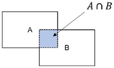
Objectives The aim of this study was automatically detecting and numbering teeth in digital bitewing radiographs obtained from patients, and evaluating the diagnostic efficiency of decayed teeth in real time, using deep learning algorithms. Methods The dataset consisted of 1170 anonymized digital bitewing radiographs randomly obtained from faculty archives. After image evaluation and labeling process, the dataset was split into training and test datasets. This study proposed an end-to-end pipeline architecture consisting of three stages for matching tooth numbers and caries lesions to enhance treatment outcomes and prevent potential issues. Initially, a pre-trained convolutional neural network (CNN) utilized to determine the side of the bitewing images. Then, an improved CNN model YOLOv7 was proposed for tooth numbering and caries detection. In the final stage, our developed algorithm assessed which teeth have caries by comparing the numbered teeth with the detected caries, using the intersection over union value for the matching process. Results According to test results, the recall, precision, and F1-score values were and 0.99 for teeth detection, and 0.979 for teeth numbering, and and 0.822 for caries detection, respectively. For teeth numbering and caries detection matching performance; the accuracy, recall, specificity, precision and F1-Score values were 0.934 , and 0.842 , respectively. Conclusions The proposed model exhibited good achievement, highlighting the potential use of CNNs for tooth detection, numbering, and caries detection, concurrently. Clinical significance CNNs can provide valuable support to clinicians by automating the detection and numbering of teeth, as well as the detection of caries on bitewing radiographs. By enhancing overall performance, these algorithms have the capacity to efficiently save time and play a significant role in the assessment process.

Keywords Artificial intelligence Digital bitewing radiography Deep learning Detection Numbering Dental caries

Introduction

Dental caries is a preventable, chronic, multifactorial, infectious disease that is common in many populations worldwide [1]. Carious lesions, which usually progress slowly, can be a serious health problem that can lead to pain, infection and tooth loss if not treated in early stages [2]. While the perspective on caries treatment was more invasive in the past, today in modern dentistry, this perspective has been replaced by early diagnosis with preventive and minimally invasive treatment methods [3]. Early and accurate detection of caries lesions is a very important step that guides clinical planning by determining the treatment to be applied. Therefore, timely and accurate diagnosis has a key role in a successful treatment [4]. Although there are different methods in the diagnosis of caries lesions, the visual-tactile examination
method which is insufficient in the diagnosis of early approximal caries detection is one of the most basic of them [5, 6]. In addition to the visual-tactile examination method, radiological examination which is commonly employed in intraoral imaging plays a crucial role in dental practice. In this regard, panoramic, periapical and bitewing radiography are regularly utilized in practice. Visualization and evaluation of dental status on radiographs are one of the most important steps in disease diagnosis [7, 8]. Today, with the help of digital bitewing radiographs, which have high recall and specificity in the detection of approximal caries, the detection of caries lesions is done easily, cheaply and successfully compared to many methods [9, 10].
As can be understood from all these, radiological imaging and visual-tactile examination methods are essential for detecting carious lesions. In some situations, depending on the experience and attention of the dentist, misdiagnosis or insufficient diagnosis can be made in bustling clinics. The use of computer aided systems can help to dentists during diagnostic process. Also, it can provide the physician to achieve success by assisting with treatment planning and implementation [11, 12].
Artificial intelligence term (AI), which has become very popular in our lives and affects many industries and promises to be a real turning point [13]. AI is a general term that refers to systems, machines or computers that mimic human behaviors and intelligence [14]. It includes different areas of research fields and algorithms. Deep learning (DL) is one of these research areas that has gained popularity in recent years [15]. DL algorithms consist of various artificial neural networks according to the different types of problems such as natural language processing, large language models, computer vision [16]. Convolutional neural networks (CNNs) are supervised deep learning algorithms that are artificial neural networks inspired by the mammalian visual system [17]. CNNs are actively used in many computer vision problems in dentistry and medicine for improving health services and increasing efficiency [13, 18].
The adoption of CNNs in the diagnosis of caries in dentistry is a successful method as can be understood from the studies carried out. There are many studies on CNN and similar neural networks [19-24]. Bitewing radiographs are often used by dentists as an assistant tool to diagnose dental caries [25]. In some of the researches, caries diagnosis studies were performed on bitewing radiographs [19, 20, 26, 27]. Correct detection and numbering of teeth during the examination can reduced the examination time and provide a better diagnosis [28]. Yasa et al. [29] researched teeth detection and numbering in bitewing radiographs. Also, there are similar studies have been searched with in panoramic or bitewing radiographs [30-34]. These studies showed that the developments based on DL in the health industry can be an assistant and a second opinion provider in the daily clinical
practice of the physician [35]. To the best of our knowledge, no study has been investigated that teeth detection, numbering, and caries detection at the same time in digital bitewing radiographs utilizing a CNN approach based on DL. The proposed method makes this study unique compared to existing literature.
The aim of this study was to automatically detecting and numbering teeth in digital bitewing radiographs obtained from patients, and to evaluate the diagnostic efficiency of decayed teeth in real time, with a pipeline architecture using CNNs. Thus, it is planned to reduce the chair time, to assist the physician and to increase the effectiveness of the treatment by making the diagnosis more effective during the examination.
A successful proposal introduces a novel DL-based pipeline architecture for teeth numbering and caries detection. The architecture enhances the preservation of crucial information in bitewing images by incorporating convolutional block attention modules (CBAMs) and average pooling layers into the YOLOv7 model. This implementation results in a more efficient performance in teeth numbering and decay detection compared to the original YOLOv7 model. Additionally, an algorithm has been developed to assess teeth with caries by correlating the numbered teeth with the detected caries.

Material and methods

Sample size determination

The determination of the sample size for the test dataset was performed with Fisher’s exact test A design incorporating clustering was employed because of every digital bitewing radiograph contained averagely 16 surfaces which was calculated by design effect (DE). Design effect (DE), which assesses the performance of a particular sampling method compared to that of a simple random sample, is a valuable tool in considering the precision of a cluster sample. Equation of DE is ICC , in where m represents size of cluster, and ICC is the intraclass correlation coefficient. ICC of 0.2 was assumed based on Masood et al. [36]. According to previous study’s power analysis, required test set units multiplied with DE [37]. When , the total surfaces required should be 2136 or 134 bitewing radiographs. In the present study, test dataset contained 170 bitewing radiographs.

Image dataset

A total of 1170 digital bitewing radiographs of adult patients aged over 18 were collected from radiology archive of Kırıkkale University, Faculty of Dentistry for this study.
Bitewing radiographs which obtained from patients who came for examination and/or treatment between January-2018 and January-2023 included by examining. No further details, such as gender, age or acquisition date of images was revealed, since the radiographs were collected anonymously. In the study, bitewing radiographs containing patient positioning errors, technical errors, fixed prosthetic restoration and fixed orthodontic braces that would complicate or completely prevent the diagnosis did not include in the dataset. Approval for this study was obtained from the non-interventional Clinical Research Ethical Committee of Kırıkkale University. (Decision Number: 2022.11.14) The study adhered to the ethical guidelines stated in the Declaration of Helsinki.
All digital bitewing radiographs were taken by a Gendex Expert DC (Gendex Dental Systems, IL, USA) device that is located in the clinics with a focal spot value of 0.04 mm , tube voltage 65 kVp , tube current 7 mA . Exposure time, distance, and quality of radiographs can be variable, because of the digital bitewing radiographs were captured by various personnel across different departments. Each digital bitewing radiograph was anonymized and saved as.jpg format. Maxillary and mandibulary dental arches were distinctly
Table 1 Summary of whole dataset
Train Test
Decay 2289 602
Numbering 9344 1679
Total surfaces 16,674 2842
Lesion prevalence 24% 21.16%
Total image 1000 170
visible on digital bitewing images. The assessment included all visible teeth, excluding restorations present on the proximal surfaces of the teeth. Because of the aim of this study was evaluating the proposed model performance for detecting primary approximal caries and numbering teeth. The collected digital bitewing radiographs displayed heterogeneous resolutions in terms of their sizes. For this reason, all resolutions were adjusted to pixels, such as a previous study [19]. After this process, the dataset was randomly split as follows: for training and validation (1000 bitewings) and 15% for testing (170 bitewings). Thus, 1000 digital bitewing radiographs that obtained with random distribution were used for training the proposed CNN model. The remaining 170 digital bitewing radiographs were used for evaluating the model. In the training dataset, 9344 teeth were numbered. There are 16674 approximal surfaces and 2289 of them are caries. The lesion prevalence of the training data is . Details of the dataset are given in Table 1 and image samples from the dataset are given in Fig. 1.

Study design

In this study, a pipeline architecture that can simultaneously detect teeth numbering and caries in bitewing images is proposed. The proposed architecture consists of 3 basic steps. A diagram illustrating the application of the proposed architecture is given in Fig. 2.

Image evaluation and data labeling

A restorative specialist (author 3) with 13 years of experience and a research assistant (author 1) with 4 years of
Fig. 1 Image samples from the dataset
Fig. 2 Diagram of the implementation of the proposed architecture
experience generated ground truth annotations for the images using software program which developed in Python programming language. For that, annotators labeled bounding boxes around all teeth while simultaneously providing a class label for each box with the tooth number according
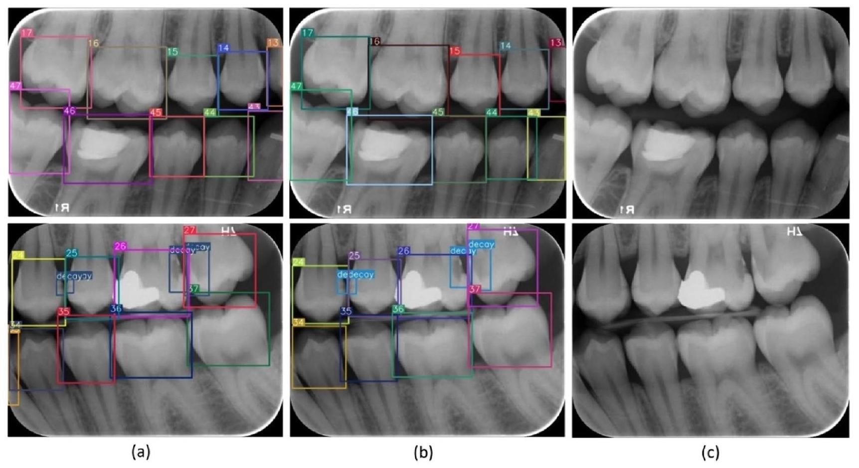
to Federation Dentaire International (FDI) tooth notation system (16-17-18-26-27-28-36-37-38-46-47-48 (molars), 14-15-24-25-34-35-44-45 (premolars) and 13-23-33-43 (canines)). Also, caries lesions labeled as decay in digital bitewing images. While in the data labeling procedure,
annotators reviewed the bitewing images at the same time and as a result of the agreement, labeling was processed (Fig. 3).
After this part, approximal caries lesions on the tooth surfaces have been labeled with bounding boxes by the annotators with the same agreement procedure. The software that used for labeling was automatically converted the box information and labels according to the needs for training of YOLO.

Detecting bitewing image’s side via convolutional neural network

The first step of the proposed pipeline architecture was to automatically detect whether the bitewing images belong to the left or right sides. This step was very important for the numbering step of the teeth. Because the success rate of the numbering process was found to be low in the first training without separating the right and left sides. For this purpose, a DenseNet CNN architecture was utilized, which was introduced in 2016 by Huang et al. [38]. DenseNet architecture uses dense connections, which connect each layer to other layers in a forward manner. Dense connections have benefits, such as reducing model parameters, decreasing vanishing-gradient problem, encouraging feature reuse, and strengthening feature propagation along layers. Four DenseNet architectures exist in the literature (DenseNet-121, DenseNet-169, DenseNet-201, DenseNet-264). In this study, a pre-trained DenseNet-121 architecture on the ImageNet dataset was utilized and was re-trained using transfer learning and finetuning methods to detect whether the images belong to the left or right sides. The main reason for selecting the DenseNet-121 is that it is more lightweight than other architectures, so it can be trained faster. The fine-tuned DenseNet-121 model has parameters, one fully connected layer with 512 neurons and one neuron output
with sigmoid activation function. The proposed model trained for 30 epochs with 32 batch size, 0.001 initial learning rate. Binary cross entropy was used as the error function and Adam algorithm was preferred for the optimizer.
In training process, 1000 bitewing images, 500 left and 500 right, were used. of the training data was used for validation. 170 bitewing images ( 85 left and 85 right) were used to evaluate classification performance of the model. According to the test result, CNN model was achieved %100 accuracy on the test dataset. A diagram summarizing this phase is given in Fig. 2.

Improved YOLOv7 architecture, teeth numbering and caries detection

The architecture of original YOLOv7

The second stage of the study was teeth numbering and caries detection from the bitewing images. In this step, DLbased object detection algorithms were taken into consideration due to the nature of the problem to be solved. There exist two types of DL-based object detection algorithms: region-based (two stage) and regression-based (one stage). In region-based algorithms, firstly, candidate regions containing possible objects in identifies, then these regions are classified to determine object types in them. Fast-RCNN [39], Faster-RCNN [40], Mask-RCNN [41] algorithms are some examples of this category. On the other hand, onestage algorithms detect and classify objects at the same time. The algorithms in this category are YOLO (You Only Look Once) and its different versions such as YOLO9000, YOLOv3, YOLOv4, YOLOv5, YOLOv6, YOLOv7 [42, 43]. One-stage algorithms offer a faster detection speed than twostage ones, this advantage makes them popular in object detection problems.
Fig. 3 Image samples from the labeling process
In YOLOv7, one of the latest versions of the YOLO series, some improvements have been made in the loss function, backbone network and activation function. In this way, the model has achieved improved detection accuracy and accelerated detection speed. For this reason, the YOLOv7 algorithm was chosen to be used in the study. The original YOLOv7 architecture consists of four modules: input, backbone, neck and head. The input module resizes the input image to a standardized pixel size to fulfill the input size specifications of the backbone module. The backbone of the original architecture consists of convolutional blocks, batch normalization process, and SiLU activation function (CBS), extended efficient long-range attention network (E-ELAN) and Max-Pool-1 (MP1) modules. The task of the backbone part is to extract feature maps from the input image. The CBS module consists of three components: convolution blocks, batch normalization process (BN), and SiLU activation function (sigmoid-weighted linear unit activation function) (Fig. 5). The ELAN module comprises multiple CBS modules that ensure the input and output feature sizes remain unchanged. By guiding the computational blocks of different feature groups to learn various features, the network’s learning capacity is enhanced while preserving the integrity of the original gradient path (Fig. 5). The MP1 module is divided into two branches specifically an upper branch
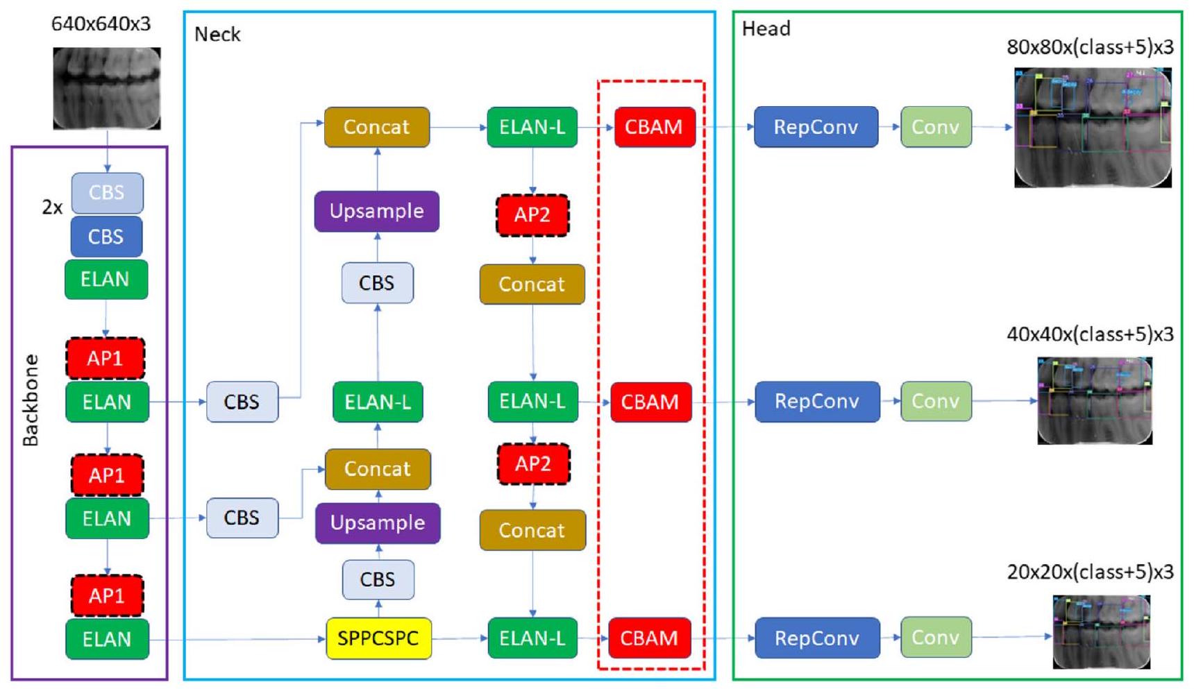
and a lower branch and consisting of CBS modules and the max pooling (MaxPool) layer (Fig. 4). In this module, the upper branch utilizes MaxPool to capture the maximum value information from small local areas, while the CBS modules in the lower branch extract all valuable information from small local areas. This dual approach enhances the network’s feature extraction capability.
The neck module consists of ELAN-L, Upsample, MP2 and SPPCSPC modules using the PAFPN Path Aggregation Feature Pyramid Network (PAFPN) architecture. The neck part combines the feature maps obtained from the different layers of the backbone module and sends them as input to the head module. The SPPCSPC module consists of CBS modules and MaxPool layers with different sizes of parallel max pooling (Fig. 5). The ELAN-L module has a similar structure to the ELAN module, but it combines five different outputs (Fig. 5). The Up-sample module consists of the CBS module and is used for upsampling. The Head module includes of a Rep block to set the number of image channels for the features output from the neck part. This is followed by the application of convolution module, which predicts the category, confidence, and bounding box. The REP module is inspired by RepVGG [44] and employs a simplified convolution process during the testing phase (Fig. 5). This simplification effectively reduces network complexity without compromising performance.
Fig. 4 CBS, ELAN, and MP modules representation
Fig. 5 ELAN-L, Rep and SPPCSPC modules representation

Improved YOLOv7 with convolutional block attention module

The scientific community widely acknowledges the significant role of attentional mechanisms in human vision. A fundamental characteristic of the human visual system is its ability to selectively focus on salient aspects of a scene rather than processing the entire visual field simultaneously. This selective attention enables enhanced capture of visual details [45]. In recent years, researchers have developed various selective attention mechanisms for neural networks. These mechanisms enable neural networks to learn “where” or “what” to focus more on, using explicit dependencies between channels and weighted spatial regions. In this context, the convolutional block attention module (CBAM) was introduced by Woo et al. [46] as an efficient attention mechanism designed for CNNs. Within this context, Woo et al. [46] proposed the convolutional block attention module (CBAM), which is an effective attention mechanism for CNNs. The CBAM module includes of a channel attention module (CAM) and a spatial attention module (SAM). The CAM is focused on the relevant aspects of an input image by exploiting the inter-channel relationships of features. Two spatial context descriptors are generated in the module using two separate pooling methods (average pooling and max pooling). Average pooling (AP) is mainly used to gather spatial information, whereas max pooling retains significantly more contextual details pertaining to
the object within the image, thereby enabling more precise channel attention. These generated context descriptors are forwarded to a shared multi-layer network. After the vector obtained from the multilayer network is collected, the channel attention map is generated by applying the sigmoid function. Within the CBAM module, the spatial attention module (SAM) specifically targets the localization of the most informative regions within the input image. Spatial attention computation within the SAM module involves the application of average pooling and max pooling techniques along the channel axis of the input image. The obtained twochannel image is subjected to convolutional processing to convert it into a single-channel feature map, which is then merged and forwarded to a standard convolutional layer to create an efficient feature descriptor. The output from the convolutional layer is passed through a sigmoid activation layer. The Sigmoid function, which is a probability-based activation, transforms all values to a range between 0 and 1 , thereby revealing the spatial attention map. In summary, CBAM combines the two attention modules described above to determine what is important in the input image and to locate the descriptive regions within the image. Figures of CBAM, CAM and SAM modules are given in Fig. 6.
When reviewing the studies in the literature, it is seen that there are successful combinations of the CBAM module in CNNs in the solution of computer vision problems [47, 48]. However, creating a neural network is a complex process that requires consideration of the utilized dataset,
Fig. 6 CBAM, CAM and SAM modules representation
the selection of optimized metrics, and the availability of computational resources.
In this study, we injected CBAM layers in the YOLOv7 model as a result of our experience with CNNs and various tests we performed. After conducting various experiments, three CBAM modules have been placed at three specific locations where the intersections of the neck and head in the YOLOv7 architecture. The selection of the number of integrated CBAM blocks during the incorporation of CBAM modules was done meticulously to minimize any significant increase in the computational overhead on the network. The integration of CBAM modules into the YOLOv7 architecture by researchers to develop novel designs for solving various problems has emerged as a highly popular research area [49, 50]. Therefore, the determination of position, number of blocks, and their integration with other network components are design decisions that can vary depending on the specific problem under consideration. To the best of our knowledge, there is currently no known study that specifically addresses the region we have incorporated in our research. Figure 7 provides an illustration of the improved model and details about the location where CBAM modules are positioned.
Another improvement we have made in our proposed YOLOv7 model is to use average pooling (AP) instead of max pooling (MP) in subsampling layers. In the original YOLOv7, subsampling is done at four different locations. In these subsamples, the average pooling
method was used instead of the max pooling. Max pooling returns the maximum value in each window, while average pooling returns the average value in each window. The benefit of using average pooling instead of max pooling is that it can help reduce overfitting. Max pooling is more likely to memorize as it preserves only the strongest feature in each window that may not represent the entire window. On the other hand, average pooling considers all features in each window, which can help preserve more information. Additionally, average pooling can help smooth out the output, which can be useful when the input is noisy or volatile. In this context, the changes we made are given in Fig. 7. It has been observed that this improvement has a positive effect on detection performance.
The architecture of YOLOv7-AP-CBAM obtained with the updates made in the YOLOv7 architecture was used in the numbering of teeth and in the detection of caries. In this context, two different models were trained for the right and left parts of the mouth. Since the numbering performance of a single model remained low in the tests, two different model training methods were used. The models for both sides were trained with the following hyperparameters: batch size 16, epochs 20, input image size ( 640,640 ), initial learning rate 0.01 , optimizer SGD algorithm, momentum 0.937 , weight decay 0.005 . A flowchart summarizing this stage is given in Fig. 7.
Fig. 7 Improved YOLOv7 with CBAM, and AP modules

Evaluation of teeth numbering and caries detection matching

In the last stage of the proposed architecture, it was aimed to give the output of the system which tooth had caries by matching the numbered teeth and the detected caries. It has been determined whether there was a bounding box drawn for caries in the bounding boxes drawn for tooth numbering. If there was caries in the numbered bounding box, it was reported which tooth had caries. The Intersection Over Union (IoU) value was used to detect caries in the numbered bounding box (see Fig. 8).
If the IoU value was above , it was accepted that the number of the tooth with caries label was determined correctly. The flow chart of the algorithm we developed for this purpose is given in Fig. 9.

Experimental setup

All experiments in this study are executed on NVIDIA 1080 Ti GPU with 12 G video memory size and two Intel Xenon CPU processors with 64 GB ram on the Ubuntu operating system. The experimental running environment includes Python programing language, Pytorch [51], Keras [52] DL frameworks, and OpenCV computer vision library [53].

Performance evaluation analysis

To evaluate the success of the proposed model, a confusion matrix, that summarizes the actual and predicted situations in a table, was used together with the following procedures and metrics. The confusion matrix that we adapted for evaluating performance of teeth numbering and caries detection
Fig. 8 The formula of IoU. A represents the numbering box, while B represents the caries box
Intersection Over Union(IoU) =
Fig. 9 Diagram of teeth numbering and caries detection matching decision
matching is given below. Also, the performances of teeth detection, numbering, and caries detection were evaluated individually.
Adaptation of the confusion matrix for evaluating performance of
teeth numbering and caries detection matching
TP: the output in which the model correctly predicts the positive
    class (teeth correctly detected, correctly numbered, caries correctly
    detected on bitewing radiographs)
FP: the output in which the model incorrectly predicts the positive
    class (teeth correctly detected, correctly numbered, but caries pre-
    diction on sound surface; teeth correctly detected, but regardless of
    caries incorrectly numbered)
FN: the output in which the model incorrectly predicts the negative
    class (teeth correctly detected, correctly numbered, but no caries
    prediction on decayed surface)
TN: the output in which the model correctly predicts the negative
    class (teeth correctly detected, correctly numbered, no caries pre-
    diction when there was no caries)
Initially, True Positive (TP), False Positive (FP), False Negative (FN) and True Negative (TN) rates were determined. Then, Accuracy (Eq. 1), Recall (Eq. 2), Specificity (Eq. 3), Precision (Eq. 4), F1 – Score (Eq. 5) and IoU (Eq. 6) scores were calculated. IoU is defined as the measure of overlap between the actual bounding box labeled by the annotator for an object and the bounding box predicted by the model. By comparing the IoU value with the threshold value determined prior to model training, it is possible to interpret the success of the trained model in object detection. The F1 score, which is considered the harmonic average of precision and recall, is commonly used for providing a balanced evaluation of a classification model’s performance.
The equations are shown below (Eqs. 1-6).

Results

In the testing dataset which include 170 digital bitewing radiographs, there were 1679 teeth, 2842 approximal surfaces, 1679 numbering labels, 602 caries labels. The lesion prevalence of the testing data is .
Without splitting into right and left sides, the Recall, precision, and F1-score values were 0.409 , 0.236 , and 0.299 for teeth numbering, and , and 0.691 for caries detection, respectively (Table 5).
After splitting into right and left sides, the Recall, precision, and F 1 -score values were and 0.99 for teeth detection, and 0.979 for teeth numbering, and and 0.822 for caries detection, respectively (Table 2 and 3).
For teeth numbering and caries detection matching performance; the Accuracy, recall, specificity, precision and F1-Score values were and 0.842, respectively (Table 4).
The improved YOLOv7-AP-CBAM models performed well in detecting and numbering teeth, as well as detecting caries at the same time on digital bitewing radiographs (Fig. 10).

Ablation Experiments

This study performed ablation experiments to evaluate the impact of various enhancements on the model’s performance. Initially, the original YOLOv7 model was employed to extract features without splitting into right and left sides. Subsequently, the original YOLOv7 model was modified to include the split into right and left sides, as well as the integration of AP and MP with CBAM. Finally, AP with CBAM was applied. The teeth numbering and caries detection recall, precision, and F1-score of the model with used AP and CBAM modules significantly improved compared to initial of this study. The experimental results are presented in Table 5.
In CNNs, complexity is described by the total number of parameters of the model and the number of Giga Floating Point Operations (GFLOPs) [54]. We compared basic version of YOLOv7 with improved version of YOLOv7-AP-CBAM. The original YOLOv7 has 314
Table 2 Evaluation metrics of the teeth detection model
Recall Precision F1-score
Left side 0.995 0.987 0.99
Right side 0.993 0.988 0.99
All 0.994 0.987 0.99
Table 3 Evaluation metrics of the teeth numbering and caries detection model
Recall Precision F1-score
Left side teeth numbering 0.976 0.978 0.977
Right side teeth numbering 0.972 0.993 0.982
Left side caries detection 0.857 0.833 0.844
Right side caries detection 0.810 0.900 0.800
Left side-numbering and caries detection 0.966 0.966 0.966
Right side-numbering and caries detection 0.959 0.985 0.968
Left and right-side numbering and caries detec-tion-All 0.962 0.975 0.967
Teeth numbering-All 0.974 0.985 0.979
Caries detection-All 0.833 0.866 0.849
Table 4 Evaluation metrics of the teeth numbering and caries detection matching model
TP FP FN TN Accuracy Recall Specificity Precision F1-score
Left side 240 48 47 1102 0.933 0.836 0.958 0.833 0.834
Right side 260 30 66 1049 0.931 0.797 0.972 0.896 0.843
All 500 78 113 2151 0.932 0.815 0.965 0.865 0.839
Fig. 10 Image samples for detecting and numbering teeth, as well as detecting caries with the proposed YOLOv7-AP-CBAM model on digital bitewing radiographs. a labels of improved YOLOv7-AP-CBAM model, labels of ground truth, raw image
layers million parameters, 103.7 GFLOPS, 75.1 MB size in memory. On the other hand, the proposed YOLOv7-AP-CBAM has 350 layers million
parameters 103.8 GFLOPS and 75.2 MB size in memory. A satisfactory performance increase was achieved with a small increase in complexity in the model.
Table 5 Ablation comparison of performance enhancements on the model
Recall Precision F1-score
Original YOLOv7-MP without splitting into right and left Decay all 0.676 Decay all 0.707 Decay all 0.691
Numbering all 0.409 Numbering all 0.236 Numbering all 0.299
Original YOLOv7-MP with splitting into right and left Right side decay 0.659 Right side decay 0.803
Left side decay 0.696 Left side decay 0.848
Decay all 0.677 Decay all 0.825 Decay all 0.743
Right side numbering 0.971 Right side numbering 0.974
Left side numbering 0.979 Left side numbering 0.978
Numbering all 0.975 Numbering all 0.976 Numbering all 0.975
YOLOv7-AP Right side decay 0.626 Right side decay 0.773
Left side decay 0.797 Left side decay 0.775
Decay all 0.711 Decay all 0.774 Decay all 0.741
Right side numbering 0.967 Right side numbering 0.977
Left side numbering 0.981 Left side numbering 0.983
Numbering all 0.974 Numbering all 0.980 Numbering all 0.976
YOLOv7-MP-CBAM Right side decay 0.579 Right side decay 0.838
Left side decay 0.754 Left side decay 0.832
Decay all 0.666 Decay all 0.836 Decay all 0.741
Right side numbering 0.974 Right side numbering 0.983
Left side numbering 0.981 Left side numbering 0.978
Numbering all 0.977 Numbering all 0.980 Numbering all 0.978
YOLOv7-AP-CBAM (ours) Right side decay 0.810 Right side decay 0.900 All decay 0.849
Left side decay 0.857 Left side decay 0.833
All decay 0.833 All decay 0.866
Right side numbering 0.972 Right side numbering 0.993
Left side numbering 0.976 Left side numbering 0.978
Numbering all 0.974 Numbering all 0.985 Numbering all 0.979

Discussion

In dentistry, achieving an accurate and precise diagnosis of dental caries is essential for the primary step towards successful treatment implementation. Dental caries diagnosis methods aim to improve the diagnostic process by facilitating the early detection of caries or enabling its objective identification [55]. Although tooth detection and numbering may seem straightforward, the accuracy of tooth numbering and caries detection can be affected by the limited and diverse knowledge of novice clinicians, potentially resulting in inconsistent diagnoses and inadequate treatment approaches. In addition to experience, practitioners’ inattentiveness or demanding workloads can also have an impact on the diagnostic results. To prevent all these challenges and facilitate the diagnostic process, a computeraided system based on DL methods can be employed. DL has gained significant popularity in the medical field due
to its outstanding problem-solving capabilities. Within this context, numerous DL architectures have been developed, with especially CNNs standing out for their impressive performance in image recognition tasks [56]. By utilizing CNN models, practitioners can effectively enhance work efficiency and achieve precise outcomes in the accurate diagnostic process [34, 57].
In the current study, the efficiency of the improved CNN model YOLOv7-AP-CBAM is presented for automatically detecting and numbering teeth, as well as detecting caries in digital bitewing radiographs, concurrently. So far, various studies have been conducted in the field of dentistry, utilizing CNNs for the detection, classification, and segmentation of various entities such as objects, tooth, caries, numbering, root fracture, periapical lesions and periodontal bone loss [34, 58-63]. According to these studies, CNN-based systems showed promising results in overcoming diagnosing difficulties and assisting practitioners.
Various dental imaging systems have been used for these studies. Digital bitewing radiography technique is widely employed due to its practicality and advantages. Less radiation doses requirement, time efficiency, image storing, simplicity of image enhancing and processing are some of the advantages and reason for preference of digital bitewing radiography technique [64]. In the research papers, there were numbers of study utilizing digital bitewing radiography [19, 20, 65-69]. It is a more beneficial technique than other x-ray containing methods for the simultaneous evaluation of the crowns and alveolar bone levels of posterior teeth in both upper and lower dental arch, caries detection that especially in approximal caries, and teeth detection and numbering [10,70,71].
Also, there have been studies of the implementation of CNN to bitewing, panoramic, periapical, cone beam computed tomography radiographs and various diagnostic methods [34, 72, 73]. Zhang et al. [73] applied a DL method based on CNN for teeth detection and classification in periapical radiographs. Both precision and recall scores were high and, and , respectively. In our study, the improved model achieved recall of and precision of , in digital bitewing radiographs. In another study on CBCT radiographs reported that implementing a 3D U-Net network model showed satisfactory results for the segmentation and classification of teeth. Furthermore, an AI-based segmentation of all teeth in a single scan resulted in a speed improvement of 1800 times compared to that of an expert [72]. Tuzoff et al. [34] utilized the Faster R-CNN model for tooth detection and the VGG-16 Net model for tooth numbering on panoramic radiographs. The CNN-based models demonstrated high recall and specificity values for teeth detection and numbering tasks, comparable to experts. However, their model exhibited misclassifications in cases where a tooth was missing, whereas our improved model accurately detected these cases. Image samples for these cases are given in Fig. 11.
A DL method Faster R-CNN proposed for tooth detection and numbering in periapical radiographs by Chen et al. [57]. In order to improve the detection accuracy, they proposed three post-processing techniques to complement the baseline Faster R-CNN. Their model achieved precision of and recall of for detection, and precision of and recall of for numbering. In their study, the model was unable to accurately predict teeth located at the corners of the bitewing image. However, our improved model demonstrated a precision of and a recall of for detection, as well as a precision of and a recall of for numbering.
In literature, researchers generally benefit image enhancement and cropping techniques to improve accuracy. Mao et al. [74] improved the new Alexnet model, a CNN-based approach for caries and restoration detection on bitewing images, by utilizing image enhancement and cropping techniques. The model showed accuracy of for caries detection and for restoration detection. However, it is important to note that high accuracy scores alone may not indicate the reliability of a method. If a CNN model labels every surface as “decayed,” it can achieve perfect accuracy but lack meaningful results. To address this, recall and specificity scores are reported in our study, following the precedent set by previous research, to provide a more comprehensive evaluation without sacrificing interpretability. Besides, we did not use any image enhancement or cropping methods.
In a previous study, an automatic assistance system developed for detecting caries lesions in bitewing radiographs using a U-Net model. The system’s performance was evaluated through a comparative analysis with dentists. According to the findings of this study, the model demonstrated an overall accuracy of , while the mean accuracy of practitioners was . The recall, specificity, and F1-score values were also high, at , and , respectively
Fig. 11 Sample of correct teeth numbering in cases where a tooth was missing
[20]. Although the results of the model in this study were not directly compared with the performance of human experts, it showed promising results in the detection of caries lesions, achieving recall, precision and F1 score values of , and , respectively. These results are very similar to those reported in the study by Cantu et al. In addition, the present study evaluated the performance of tooth numbering simultaneously with the detection of caries lesions.
Yasa et al. [29] proposed a Faster R-CNN model for the detection and numbering of teeth in bitewing radiographs. They conducted a comparison between two different models for tooth numbering. Initially, the teeth were not separated into right and left sides. However, after splitting and training the model in this manner, promising results were obtained, with recall, precision, and F 1 -score values of , , and , respectively. In our study, we adopted the approach of separating teeth into right and left sides, following a similar methodology. Through the adoption of this approach, our improved model exhibited promising performance, as indicated by notable recall, precision, and F1-score values of , and , respectively.
In another study, Bayraktar and Ayan [19] reported detection of interproximal caries lesions in digital bitewing radiographs with YOLOv3 model. In that study, the recall was , precision was . According to these promising results, the use of CNN-based system could be useful in diagnosing caries lesions. In the current study, our improved model is YOLOv7-AP-CBAM model with implementation of CBAM module. Through to CBAM module, the model could focus more precisely. In this way, the model reached recall of which is higher than the previous study. However, the precision values were nearly same which are and .
The present study, YOLOv7-AP-CBAM model exhibited promising results in terms of the evaluated parameters. In the confusion matrix that we created, while the specificity score is higher, contrary effect was noted in the recall score. This result showed that the improved model can accurately predict tooth numbering while also detecting sound surfaces, similar to the study by Lee et al. [22]. The overall accuracy, recall, specificity, precision and F1-score values were 0.934 , and 0.842 , respectively.
The performance of a CNN model is directly dependent on the amount of training data. The use of large and diverse data sets in the training process improves the consistency and accuracy of the CNN models. Therefore, the quality and quantity of training data play a vital role in ensuring the effectiveness of CNNs [75]. In this study, training dataset was relatively small. Although the training dataset contains an equal number of right and left bitewing images, the model was more successful on the left side in detecting caries in the final results. This can be explained by the higher amount of caries in the bitewing radiographs
on the left side in the training group. To avoid this problem, new bitewing images including caries may be added to the right-side training set. Dataset bias is another problem that occurs when specific samples in a dataset are over or under-represented. In order to avoid this problem, the dataset was generated images with taken from the same devices in various clinics with different resolutions and exposure times. However, the images obtained from different x-ray machines and from people with different demographic characteristics may affect the results. The proposed model’s parameters were determined using validation data during the training phase, and the model’s performance on unseen data was evaluated using different test data from the training and validation data. CNN models require high hardware requirements, especially in the training phase. In our study, we performed our training and tests on 1080 Ti graphic card. Computational performance will be higher on more advanced graphics cards. The dental caries lesions that were not evaluated using the gold standard method of histological assessment detected and labeled by the annotators on the radiographic images [76]. The model encountered challenges in accurately predicting teeth positioned at the corners of the bitewing image, as opposed to other teeth. This could be related to the reduced clarity of these specific teeth and the limited amount of data in the training set. Also, the improved YOLOv7-AP-CBAM model was not compared with an expert. Despite the limitations of the study, it is worth noting that there are several significant strengths present as well. The present study offered a comprehensive analysis by integrating the results of tooth detection, numbering, and caries detection. This comprehensive approach facilitated a more cohesive comprehension of the outcomes. Additionally, the experimental design included the split of data into right and left sides, enabling a more detailed analysis. This approach enhanced the performance metrics of our results. Furthermore, by adopting an interdisciplinary approach, our study introduced a novel perspective and makes an innovative contribution to previous papers.

Conclusion

In this study, an improved YOLOv7-AP-CBAM model was proposed for accurate teeth detection, numbering and caries detection, concurrently assessing with which tooth had caries by matching the numbered teeth and the detected caries. For teeth numbering and caries detection matching performance; the accuracy, recall, specificity, precision and F1Score values and 0.842 , respectively. The model exhibited high recall, specificity, precision, and F1-score, highlighting the potential of CNNs. This result
revealed that CNNs can provide valuable support to clinicians by automating the detection and numbering of teeth, as well as detection of caries on bitewing radiographs. This can potentially contribute for assessment, improve overall performance, and ultimately save precious time.
Acknowledgements This research did not receive any specific grant from funding agencies in the public, commercial, or not-for-profit sectors.
Author contribution B.A: Data acquisition and labeling, Tests, Formal analysis, Writing – original draft, Writing – review & editing.
E.A: Conceptualization, Software, Visualization, Data curation, Formal analysis, Writing – original draft, Writing – review & editing.
Y.B: Conceptualization, Methodology, Data acquisition and labeling, Writing – review & editing.
Funding Open access funding provided by the Scientific and Technological Research Council of Türkiye (TUBITAK). No funding was obtained for this study.

Declarations

Ethics approval All procedures performed in studies involving human participants were in accordance with ethical standards of the institutional and/or national research committee and with the 1964 Helsinki Declaration and its later amendments or comparable ethical standards. The study was approved by Ethical Committee of Kirikkale University (Date: 09/11/2022, Decision Number: 2022.11.14).
Consent to participate For this type of study, formal consent is not required.
Competing interests The authors declare no competing interests.
Open Access This article is licensed under a Creative Commons Attribution 4.0 International License, which permits use, sharing, adaptation, distribution and reproduction in any medium or format, as long as you give appropriate credit to the original author(s) and the source, provide a link to the Creative Commons licence, and indicate if changes were made. The images or other third party material in this article are included in the article’s Creative Commons licence, unless indicated otherwise in a credit line to the material. If material is not included in the article’s Creative Commons licence and your intended use is not permitted by statutory regulation or exceeds the permitted use, you will need to obtain permission directly from the copyright holder. To view a copy of this licence, visit http://creativecommons.org/licenses/by/4.0/.

References

  1. Featherstone JDB (2000) The science and practice of caries prevention. J Am Dent Assoc 131:887-899. https://doi.org/10.14219/ jada.archive.2000.0307
  2. Mortensen D, Dannemand K, Twetman S, Keller MK (2014) Detection of non-cavitated occlusal caries with impedance spectroscopy and laser fluorescence: an in vitro study. Open Dent J 8:28-32. https://doi.org/10.2174/1874210601408010028
  3. Pitts NB (2004) Are we ready to move from operative to nonoperative/preventive treatment of dental caries in clinical practice? Caries Res 38:294-304. https://doi.org/10.1159/000077769
  4. Baelum V, Heidmann J, Nyvad B (2006) Dental caries paradigms in diagnosis and diagnostic research. Eur J Oral Sci 114:263-277. https://doi.org/10.1111/j.1600-0722.2006.00383.x
  5. Pitts NB, Stamm JW (2004) International consensus workshop on caries clinical trials (ICW-CCT)-final consensus statements: agreeing where the evidence leads. J Dent Res 83:125-128. https://doi.org/10.1177/154405910408301s27
  6. Selwitz RH, Ismail AI, Pitts NB (2007) Dental caries. The Lancet 369:51-59. https://doi.org/10.1016/S0140-6736(07)60031-2
  7. Chan M, Dadul T, Langlais R, Russell D, Ahmad M (2018) Accuracy of extraoral bite-wing radiography in detecting proximal caries and crestal bone loss. J Am Dent Assoc 149:51-58. https://doi. org/10.1016/j.adaj.2017.08.032
  8. Vandenberghe B, Jacobs R, Bosmans H (2010) Modern dental imaging: a review of the current technology and clinical applications in dental practice. Eur Radiol 20:2637-2655. https://doi.org/ 10.1007/s00330-010-1836-1
  9. Baelum V (2010) What is an appropriate caries diagnosis? Acta Odontol Scand 68:65-79. https://doi.org/10.3109/00016350903530786
  10. Kamburoğlu K, Kolsuz E, Murat S, Yüksel S, Özen T (2012) Proximal caries detection accuracy using intraoral bitewing radiography, extraoral bitewing radiography and panoramic radiography. Dentomaxillofacial Radiol 41:450-459. https:// doi.org/10.1259/dmfr/30526171
  11. Hung K, Montalvao C, Tanaka R, Kawai T, Bornstein MM (2020) The use and performance of artificial intelligence applications in dental and maxillofacial radiology: A systematic review. Dentomaxillofacial Radiol 49:20190107. https://doi. org/10.1259/dmfr. 20190107
  12. Suzuki K (2017) Overview of deep learning in medical imaging. Radiol Phys Technol 10:257-273. https://doi.org/10.1007/ s12194-017-0406-5
  13. Rodrigues JA, Krois J, Schwendicke F (2021) Demystifying artificial intelligence and deep learning in dentistry. Braz Oral Res 35. https://doi.org/10.1590/1807-3107bor-2021.vol35.0094
  14. Barr A, Feigenbaum EA, Cohen PR (1981) The handbook of artificial intelligence. In: Artificial Intelligence, William Kaufman Inc, California, pp 3-11
  15. Lee J-G, Jun S, Cho Y-W, Lee H, Kim GB, Seo JB, Kim N (2017) Deep learning in medical imaging: general overview. Korean J Radiol 18:570. https://doi.org/10.3348/kjr.2017.18.4.570
  16. LeCun Y, Bengio Y, Hinton G (2015) Deep learning. Nature 521:436-444. https://doi.org/10.1038/nature14539
  17. Hubel DH, Wiesel TN (1968) Receptive fields and functional architecture of monkey striate cortex. J Physiol 195:215-243. https://doi.org/10.1113/jphysiol.1968.sp008455
  18. Schwendicke F, Samek W, Krois J (2020) Artificial intelligence in dentistry: chances and challenges. J Dent Res 99:769-774. https:// doi.org/10.1177/0022034520915714
  19. Bayraktar Y, Ayan E (2022) Diagnosis of interproximal caries lesions with deep convolutional neural network in digital bitewing radiographs. Clin Oral Investig 26:623-632. https://doi.org/ 10.1007/s00784-021-04040-1
  20. Cantu AG, Gehrung S, Krois J, Chaurasia A, Rossi JG, Gaudin R, Elhennawy K, Schwendicke F (2020) Detecting caries lesions of different radiographic extension on bitewings using deep learning. J Dent 100:103425. https://doi.org/10.1016/j.jdent.2020.103425
  21. Choi J, Eun H, Kim C (2018) Boosting proximal dental caries detection via combination of variational methods and convolutional neural network. J Signal Process Syst 90:87-97. https://doi. org/10.1007/s11265-016-1214-6
  22. Lee J-H, Kim D-H, Jeong S-N, Choi S-H (2018) Detection and diagnosis of dental caries using a deep learning-based convolutional neural network algorithm. J Dent 77:106-111. https://doi. org/10.1016/j.jdent.2018.07.015
  23. Moutselos K, Berdouses E, Oulis C, Maglogiannis I (2019) Recognizing occlusal caries in dental intraoral images using deep learning. In: 2019 41st Annu Int Conf IEEE Eng Med Biol Soc EMBC, IEEE, pp. 1617-1620. https://doi.org/10.1109/EMBC.2019.8856553
  24. Prajapati SA, Nagaraj R, Mitra S (2017) Classification of dental diseases using CNN and transfer learning. In: 2017 5th Int Symp Comput Bus Intell ISCBI, IEEE, pp. 70-74. https://doi.org/10. 1109/ISCBI.2017.8053547
  25. Vidnes-Kopperud S, Tveit AB, Espelid I (2011) Changes in the treatment concept for approximal caries from 1983 to 2009 in Norway. Caries Res 45:113-120. https://doi.org/10.1159/000324810
  26. Moran M, Faria M, Giraldi G, Bastos L, Oliveira L, Conci A (2021) Classification of approximal caries in bitewing radiographs using convolutional neural networks. Sensors 21:5192. https://doi. org/10.3390/s21155192
  27. Srivastava MM, Kumar P, Pradhan L, Varadarajan S (2017) Detection of tooth caries in bitewing radiographs using deep learning. http://arxiv.org/abs/1711.07312. Accessed 7 July 2023
  28. Leite AF, Gerven AV, Willems H, Beznik T, Lahoud P, GaêtaAraujo H, Vranckx M, Jacobs R (2021) Artificial intelligencedriven novel tool for tooth detection and segmentation on panoramic radiographs. Clin Oral Investig 25:2257-2267. https://doi. org/10.1007/s00784-020-03544-6
  29. Yasa Y, Çelik Ö, Bayrakdar IS, Pekince A, Orhan K, Akarsu S, Atasoy S, Bilgir E, Odabaş A, Aslan AF (2021) An artificial intelligence proposal to automatic teeth detection and numbering in dental bite-wing radiographs. Acta Odontol Scand 79:275-281. https://doi.org/10.1080/00016357.2020.1840624
  30. Bilgir E, Bayrakdar İŞ, Çelik Ö, Orhan K, Akkoca F, Sağlam H, Odabaş A, Aslan AF, Ozcetin C, Kıllı M, Rozylo-Kalinowska I (2021) An artificial intelligence approach to automatic tooth detection and numbering in panoramic radiographs. BMC Med Imaging 21:124. https://doi.org/10.1186/s12880-021-00656-7
  31. Kaya E, Gunec HG, Gokyay SS, Kutal S, Gulum S, Ates HF (2022) Proposing a CNN method for primary and permanent tooth detection and enumeration on pediatric dental radiographs. J Clin Pediatr Dent 46:293. https://doi.org/10.22514/ 1053-4625-46.4.6
  32. Kılıc MC, Bayrakdar IS, Çelik Ö, Bilgir E, Orhan K, Aydın OB, Kaplan FA, Sağlam H, Odabaş A, Aslan AF, Yılmaz AB (2021) Artificial intelligence system for automatic deciduous tooth detection and numbering in panoramic radiographs. Dentomaxillofacial Radiol 50:20200172. https://doi.org/10.1259/ dmfr. 20200172
  33. Tekin BY, Ozcan C, Pekince A, Yasa Y (2022) An enhanced tooth segmentation and numbering according to FDI notation in bitewing radiographs. Comput Biol Med 146:105547. https://doi.org/ 10.1016/j.compbiomed.2022.105547
  34. Tuzoff DV, Tuzova LN, Bornstein MM, Krasnov AS, Kharchenko MA, Nikolenko SI, Sveshnikov MM, Bednenko GB (2019) Tooth detection and numbering in panoramic radiographs using convolutional neural networks. Dentomaxillofacial Radiol 48:20180051. https://doi.org/10.1259/dmfr.20180051
  35. Topol EJ (2019) High-performance medicine: the convergence of human and artificial intelligence. Nat Med 25:44-56. https://doi. org/10.1038/s41591-018-0300-7
  36. Masood M, Masood Y, Newton JT (2015) The clustering effects of surfaces within the tooth and teeth within individuals. J Dent Res 94:281-288. https://doi.org/10.1177/0022034514559408
  37. Chen X, Guo J, Ye J, Zhang M, Liang Y (2022) Detection of proximal caries lesions on bitewing radiographs using deep learning method. Caries Res 56:455-463. https://doi.org/10.1159/00052 7418
  38. Huang G, Liu Z, van der Maaten L, Weinberger KQ (2016) densely connected convolutional networks. https://doi.org/10. 48550/ARXIV. 1608.06993
  39. Girshick R (2015) Fast R-CNN, In: 2015 IEEE Int. Conf. Comput. Vis. ICCV, IEEE, Santiago, Chile, pp. 1440-1448.https://doi.org/ 10.1109/ICCV.2015.169
  40. Ren S, He K, Girshick R, Sun J (2017) Faster R-CNN: towards real-time object detection with region proposal networks. IEEE Trans Pattern Anal Mach Intell 39:1137-1149. https://doi.org/10. 1109/TPAMI.2016.2577031
  41. He K, Gkioxari G, Dollar P, Girshick R (2017) Mask R-CNN, In: 2017 IEEE Int Conf Comput Vis ICCV, IEEE, Venice, pp. 2980-2988.https://doi.org/10.1109/ICCV.2017.322
  42. Terven J, Cordova-Esparza D (2023) A Comprehensive Review of YOLO: From YOLOv1 and Beyond. http://arxiv.org/abs/2304. 00501 (accessed July 7, 2023)
  43. Wang C-Y, Bochkovskiy A, Liao H-YM (2022) YOLOv7: Trainable bag-of-freebies sets new state-of-the-art for real-time object detectors. https://doi.org/10.48550/ARXIV.2207.02696
  44. Ding X, Zhang X, Ma N, Han J, Ding G, Sun J (2021) RepVGG: Making VGG-style ConvNets Great Again, In: 2021 IEEECVF Conf Comput Vis Pattern Recognit CVPR, IEEE, Nashville, TN, USA, pp. 13728-13737.https://doi.org/10.1109/CVPR46437. 2021.01352
  45. Larochelle H, Hinton G (2010) Learning to combine foveal glimpses with a third-order Boltzmann machine. Adv Neural Inform Process Syst 23:1243-1251
  46. Woo S, Park J, Lee J-Y, Kweon IS (2018) CBAM: Convolutional Block Attention Module, In: Ferrari V, Hebert M, Sminchisescu C, Weiss Y (Eds.). Comput Vis-ECCV 2018, Springer International Publishing, Cham, pp. 3-19. https://doi.org/10.1007/ 978-3-030-01234-2_1
  47. Shi J, Yang J, Zhang Y (2022) Research on steel surface defect detection based on YOLOv5 with attention mechanism. Electronics 11:3735. https://doi.org/10.3390/electronics11223735
  48. Xue Z, Xu R, Bai D, Lin H (2023) YOLO-tea: a tea disease detection model improved by YOLOv5. Forests 14:415. https:// doi.org/10.3390/f14020415
  49. De Moraes JL, De Oliveira Neto J, Badue C, Oliveira-Santos T, De Souza AF (2023) Yolo-papaya: a papaya fruit disease detector and classifier using cnns and convolutional block attention modules. Electronics 12:2202. https://doi.org/10.3390/elect ronics12102202
  50. Yan J, Zhou Z, Zhou D, Su B, Xuanyuan Z, Tang J, Lai Y, Chen J, Liang W (2022) Underwater object detection algorithm based on attention mechanism and cross-stage partial fast spatial pyramidal pooling. Front Mar Sci 9:1056300. https://doi.org/10. 3389/fmars.2022.1056300
  51. Paszke A, Gross S, Massa F, Lerer A, Bradbury J, Chanan G, Killeen T, Lin Z, Gimelshein N, Antiga L, Desmaison A, Kopf A, Yang E, DeVito Z, Raison M, Tejani A, Chilamkurthy S, Steiner B, Fang L, Bai J, Chintala S (2019) PyTorch: an imperative style, high-performance deep learning library. Adv Neural Inf Process Syst 32:8024-8035
  52. Chollet F et al (2015) Keras. https://github.com/fchollet/keras. Accessed 7 July 2023
  53. Bradski G (2000) The OpenCV library. Dr. Dobb’s Journal: Software Tools for the Professional Programmer 25:120-123
  54. Zhang Y, Li J, Fu W, Ma J, Wang G (2023) A lightweight YOLOv7 insulator defect detection algorithm based on DSC-SE. PLoS One 18:e0289162. https://doi.org/10.1371/journal.pone. 0289162
  55. Gomez J (2015) Detection and diagnosis of the early caries lesion. BMC Oral Health 15:S3. https://doi.org/10.1186/ 1472-6831-15-S1-S3
  56. Yasaka K, Akai H, Kunimatsu A, Kiryu S, Abe O (2018) Deep learning with convolutional neural network in radiology. Jpn J Radiol 36:257-272. https://doi.org/10.1007/s11604-018-0726-3
  57. Chen H, Zhang K, Lyu P, Li H, Zhang L, Wu J, Lee C-H (2019) A deep learning approach to automatic teeth detection and numbering based on object detection in dental periapical films. Sci Rep 9:3840. https://doi.org/10.1038/s41598-019-40414-y
  58. Casalegno F, Newton T, Daher R, Abdelaziz M, Lodi-Rizzini A, Schürmann F, Krejci I, Markram H (2019) Caries detection with near-infrared transillumination using deep learning. J Dent Res 98:1227-1233. https://doi.org/10.1177/0022034519871884
  59. Fukuda M, Inamoto K, Shibata N, Ariji Y, Yanashita Y, Kutsuna S, Nakata K, Katsumata A, Fujita H, Ariji E (2020) Evaluation of an artificial intelligence system for detecting vertical root fracture on panoramic radiography. Oral Radiol 36:337-343. https://doi. org/10.1007/s11282-019-00409-x
  60. Lee J-H, Kim D, Jeong S-N, Choi S-H (2018) Diagnosis and prediction of periodontally compromised teeth using a deep learn-ing-based convolutional neural network algorithm. J Periodontal Implant Sci 48:114. https://doi.org/10.5051/jpis.2018.48.2.114
  61. Murata M, Ariji Y, Ohashi Y, Kawai T, Fukuda M, Funakoshi T, Kise Y, Nozawa M, Katsumata A, Fujita H, Ariji E (2019) Deeplearning classification using convolutional neural network for evaluation of maxillary sinusitis on panoramic radiography. Oral Radiol 35:301-307. https://doi.org/10.1007/s11282-018-0363-7
  62. Park J-H, Hwang H-W, Moon J-H, Yu Y, Kim H, Her S-B, Srinivasan G, Aljanabi MNA, Donatelli RE, Lee S-J (2019) Automated identification of cephalometric landmarks: Part 1-Comparisons between the latest deep-learning methods YOLOV3 and SSD. Angle Orthod 89:903-909. https://doi.org/10.2319/022019-127.1
  63. Pauwels R, Brasil DM, Yamasaki MC, Jacobs R, Bosmans H, Freitas DQ, Haiter-Neto F (2021) Artificial intelligence for detection of periapical lesions on intraoral radiographs: Comparison between convolutional neural networks and human observers, Oral Surg. Oral Med Oral Pathol Oral Radiol 131:610-616. https://doi. org/10.1016/j.oooo.2021.01.018
  64. Rockenbach MI, Veeck EB, da Costa NP (2008) Detection of proximal caries in conventional and digital radiographs: an in vitro study. Stomatologija 10:115-120
  65. Abdinian M, Razavi SM, Faghihian R, Samety AA, Faghihian E (2015) Accuracy of digital bitewing radiography versus different views of digital panoramic radiography for detection of proximal caries. J Dent Tehran Iran 12:290-297
  66. Hintze H (1993) Screening with conventional and digital bitewing radiography compared to clinical examination alone for caries detection in low-risk children. Caries Res 27:499-504. https:// doi.org/10.1159/000261588
  67. Peker İ, ToramanAlkurt M, Altunkaynak B (2007) Film tomography compared with film and digital bitewing radiography for
    proximal caries detection. Dentomaxillofacial Radiol 36:495-499. https://doi.org/10.1259/dmfr/13319800
  68. Schaefer G, Pitchika V, Litzenburger F, Hickel R, Kühnisch J (2018) Evaluation of occlusal caries detection and assessment by visual inspection, digital bitewing radiography and near-infrared light transillumination. Clin Oral Investig 22:2431-2438. https:// doi.org/10.1007/s00784-018-2512-0
  69. Wenzel A (2004) Bitewing and digital bitewing radiography for detection of caries lesions. J Dent Res 83:72-75. https://doi.org/ 10.1177/154405910408301s14
  70. Mahoor MH, Abdel-Mottaleb M (2005) Classification and numbering of teeth in dental bitewing images. Pattern Recognit 38:577-586. https://doi.org/10.1016/j.patcog.2004.08.012
  71. Reed BE, Polson AM (1984) Relationships between bitewing and periapical radiographs in assessing Crestal Alveolar Bone Levels. J Periodontol 55:22-27. https://doi.org/10.1902/jop.1984.55.1.22
  72. Shaheen E, Leite A, Alqahtani KA, Smolders A, Van Gerven A, Willems H, Jacobs R (2021) A novel deep learning system for multi-class tooth segmentation and classification on cone beam computed tomography. A validation study. J Dent 115:103865. https://doi.org/10.1016/j.jdent.2021.103865
  73. Zhang K, Wu J, Chen H, Lyu P (2018) An effective teeth recognition method using label tree with cascade network structure. Comput Med Imaging Graph 68:61-70. https://doi.org/10.1016/j. compmedimag.2018.07.001
  74. Mao Y-C, Chen T-Y, Chou H-S, Lin S-Y, Liu S-Y, Chen Y-A, Liu Y-L, Chen C-A, Huang Y-C, Chen S-L, Li C-W, Abu PAR, Chiang W-Y (2021) Caries and restoration detection using bitewing film based on transfer learning with CNNs. Sensors 21:4613. https:// doi.org/10.3390/s21134613
  75. Koppanyi Z, Iwaszczuk D, Zha B, Saul CJ, Toth CK, Yilmaz A (2019) Multimodal semantic segmentation: fusion of rgb and depth data in convolutional neural networks, in: multimodal scene underst., Elsevier, pp. 41-64. https://doi.org/10.1016/B978-0-12-817358-9.00009-3
  76. Subka S, Rodd H, Nugent Z, Deery C (2019) In vivo validity of proximal caries detection in primary teeth, with histological validation. Int J Paediatr Dent 29:429-438. https://doi.org/10.1111/ipd. 12478
Publisher’s Note Springer Nature remains neutral with regard to jurisdictional claims in published maps and institutional affiliations.

  1. Baturalp Ayhan
    baturalpayhan@kku.edu.tr
    Enes Ayan
    enesayan@kku.edu.tr
    Yusuf Bayraktar
    yusufbayraktar@kku.edu.tr
    Department of Restorative Dentistry, Faculty of Dentistry, Kırıkkale University, Kırıkkale, Turkey
    Department of Computer Engineering, Faculty of Engineering and Architecture, Kırıkkale University, Kırıkkale, Turkey