أنماط الشكل الخارجي لجمجمة البيسون الأوروبي (Bison bonasus) Morphological patterns of the European bison (Bison bonasus) skull

المجلة: Scientific Reports، المجلد: 15، العدد: 1
DOI: https://doi.org/10.1038/s41598-025-85654-3
PMID: https://pubmed.ncbi.nlm.nih.gov/39789289
تاريخ النشر: 2025-01-09

افتح

أنماط الشكل الخارجي لجمجمة البيسون الأوروبي (Bison bonasus)

أوزان غونديمير & توماس زارا ®

الملخص

هدفت هذه الدراسة إلى التحقيق في تأثير العوامل البيئية، والاختيار الجنسي، والتنوع الجيني على شكل الجمجمة من خلال فحص بنية جمجمة البيسون الأوروبي، وهو نوع مهدد بالانقراض، ومقارنته بأنواع أخرى من البقريات. كانت جمجمة البيسون الأوروبي أكبر بكثير من جمجمة الأنواع الأخرى من قبيلة Bovini، وكشفت النتائج عن اختلافات شكلية كبيرة في شكل الجمجمة مقارنة بعينات أخرى من Bovini. أظهرت جمجمة البيسون شكلًا أوسع في المنطقة الجبهية وعملية قرنية موجهة بشكل جانبي أكثر. كما أثر العظم الجبهي بشكل كبير على تنوع شكل الجمجمة داخل الأنواع الفرعية للبيسون الأوروبي. أيضًا، أشارت النتائج إلى أن حجم العمليات القرنية أثر بشكل كبير على شكل الجمجمة. كان لدى ذكور البيسون جماجم أكبر ونتوءات قرنية أكثر تطورًا، ويعتقد أن هذه التغيرات الشكلية في المنطقة الجبهية أثرت أيضًا على المنطقة القفوية، والمنطقة الفكية، وعظام الوجه. علاوة على ذلك، قد تعكس الاختلافات في حجم الجمجمة التي لوحظت بين سلالات Lowland-Pszczyna وLowland-Białowieża، التي تشترك في أصل أقرب من سلالات Lowland-Carpathian، التكيفات البيئية والجينية على مر الزمن. توفر هذه الأبحاث نقطة مرجعية للدراسات المستقبلية حول العوامل البيئية والتطورية التي تؤثر على تنوع جمجمة البيسون، مع آثار كبيرة على الحفاظ على هذا النوع.

الكلمات الرئيسية: Bovidae، Bovini، العملية القرنية، العظم الجبهي، القياسات الشكلية الهندسية، شكل الجمجمة
البيسون الأوروبي (Bison bonasus)، المعروف باسم الويسنت، هو أكبر ثديي في أوروبا. بعد الحرب العالمية الأولى، واجه هذا النوع خطر الانقراض في البرية، حيث تم قتل آخر فرد في غابة بيلوفيزا في . ومع ذلك، نجا هذا النوع في عدد قليل من حدائق الحيوان والأقفاص في جميع أنحاء أوروبا. بفضل برنامج تربية دولي بدأ في عشرينيات وثلاثينيات القرن الماضي، بدأت أعداد السكان في التعافي، بدءًا من إعادة تأسيس قطيع في بيلوفيزا. منذ ذلك الحين، تم إنشاء عدة مجموعات حرة، حيث وصلت الأعداد الآن إلى عشرات الآلاف. في العصور التاريخية، كان هناك نوعان فرعيان من البيسون. احتل البيسون المنخفض (Bison bonasus bonasus) المناطق الغابية في وسط وشرق أوروبا، وكان البيسون القوقازي (Bison bonasus caucasicus) موجودًا في جبال القوقاز. تم جلب قطيع البيسون من بيلوفيزا إلى سيليزيا في عام 1865، وقد حددت مجموعة البيسون الأوروبي نوعين جينيين متميزين: سلالة Lowland-Carpathian وسلالة Lowland-Białowieża . في خريف عام 1865، تم نقل أربعة بيسون أوروبي منخفض من بيلوفيزا إلى موركي، وبعد عام، تم إطلاق سراحهم في قفص كبير في غابة بيسكزينا. هذه المجموعة، التي تباينت جينيًا مع مرور الوقت، معترف بها الآن كسلالة Lowland-Pszczyna . كأحد ممثلي الميجافونا الأوروبية، يلعب البيسون الأوروبي دورًا حيويًا في نظامه البيئي. يتم تصنيف هذا النوع على أنه مهدد بالقرب (NT) في القائمة الحمراء للاتحاد الدولي لحفظ الطبيعة ، مما يبرز الحاجة الملحة للحفاظ عليه.
تلعب الجمجمة وميزاتها الشكلية دورًا رئيسيًا في النظاميات والتطور في المجترات . تعكس الاختلافات في شكل الجمجمة وهياكلها القدرة على الاستجابة لمجموعة واسعة من الضغوط الانتقائية . القرون، الموجودة في كل من الذكور والإناث من البقريات، هي سمة تمييز جنسي واضحة . يرتبط حجم العمليات القرنية في الأفراد الناميين إيجابيًا بحجم الجمجمة وكتلة الجسم، حيث توفر الجماجم الأكبر وكتل الجسم الأثقل دعمًا هيكليًا أكبر لتطوير عمليات قرنية أكبر. تظهر جمجمة البيسون من كلا الجنسين تطورًا مكثفًا حتى سن 5 سنوات، بعد ذلك يتباطأ معدل النمو بشكل كبير، مما يشير على الأرجح إلى اكتمال تشكيل الهيكل القحفي الرئيسي والانتقال إلى شكل بالغ أكثر استقرارًا .
كواحد من آخر الأنواع الباقية من الميجافونا في أوروبا، يعد البيسون الأوروبي حاسمًا في الحفاظ على التنوع البيولوجي وتعزيز صحة النظام البيئي. يسمح دراسة بيولوجيته، وشكله، وجيناته للحفاظيين بالحصول على
رؤى أعمق حول استراتيجيات بقائه وعمليات التكيف التطورية . من خلال فحص هياكله التشريحية، ولا سيما ميزات مثل شكل الجمجمة واختلافات الحجم، والتحقيق في التنوع الجيني داخل وبين السكان، يمكن للعلماء فهم كيف تكيف هذا النوع مع بيئته على مر الزمن .
تهدف هذه الدراسة إلى التحقيق في التكيفات التطورية والتنوع الشكلاني لجمجمة البيسون الأوروبي. هذه البنية المعقدة والتطورية حاسمة في فهم العمليات التنموية في التغيير التطوري . تفترض الأبحاث أن شكل الجمجمة للبيسون الأوروبي قد تأثر بالتزاوج الداخلي على المدى الطويل والعوامل الجينية بسبب عزل هذا النوع في موائل محمية مثل غابة بيلوفيزا. في هذه المجموعات المعزولة، من المتوقع أن يؤدي تدفق الجينات المحدود إلى دفع التنوع الشكلاني. تركز الجزء الأول من الدراسة على وضع البيسون الأوروبي ضمن قبيلة Bovini الأوسع من خلال مقارنة شكل جمجمته مع الأنواع الأخرى ضمن العائلة. ستوفر هذه التحليل المقارن إطارًا تطوريًا، مما يقدم رؤى حول الصفات السلفية المشتركة والتكيفات البيئية المتباينة التي أثرت على شكل الجمجمة عبر سلالة Bovini. من خلال ذلك، ستقوم الدراسة بتقييم كيف شكلت الضغوط التطورية والبيئات البيئية كل من القواسم المشتركة والسمات الفريدة الموجودة في جمجمة البيسون الأوروبي. في الجزء الثاني من الدراسة، يتحول التركيز إلى التباين داخل الأنواع في مجموعة البيسون الأوروبي، مستكشفًا الاختلافات الشكلية بين الجنسين والأنواع الفرعية. فرضية رئيسية هي أن ذكور البيسون ستظهر اختلافات شكلية عن الإناث لأن عملياتهم القرنية المتطورة بشكل كبير معروفة بأنها تفرض ضغوطًا تطورية على الجمجمة.

النتائج

حجم الجمجمة والعملية القرنية

توضح الشكل 1 توزيع أحجام الجمجمة لجميع العينات المستخدمة في الدراسة، بينما توفر الجدول 1 مقارنات بين المجموعات. كانت جمجمة البيسون الأوروبي أكبر بشكل ملحوظ مقارنة بأعضاء آخرين من عائلة Bovini ( ). عرضت جماجم البيسون الأوروبي توزيعًا متجانسًا نسبيًا، مشابهًا لذلك الذي لوحظ في عينات Bos. في المقابل، يتميز أعضاء جنس Bubalus بأصغر الجماجم، مع تباين كبير في الحجم. قد يُعزى هذا التباين إلى العدد الأكبر من الأنواع الفرعية داخل مجموعة Bubalus، والتي تضمنت أربعة أنواع فرعية متميزة، مقارنةً بنوعين فقط في مجموعات Bison وBos . من المحتمل أن تكون هذه التنوعات التصنيفية الأوسع قد ساهمت في التباين في الحجم الملحوظ. كان الفرق في الحجم عبر عائلة Bovini ذا دلالة إحصائية عند تقييم مجموعة البيانات الكاملة ( ).
داخل عينات البيسون الأوروبي، كشفت مقارنات حجم الجمجمة أن سلالة Lowland-Białowieża كانت لديها جماجم أكبر بكثير من السلالتين الأخريين ( ). في المقابل، أظهرت سلالات Lowland-Pszczyna وLowland-Carpathian أحجام جمجمة مماثلة، دون وجود اختلافات ذات دلالة إحصائية بينها. أسفرت مقارنات حجم العملية القرنية عن نتائج متسقة مع تلك الخاصة بحجم الجمجمة. في كلا الحالتين، ظهر الجنس كعامل مميز، حيث أظهرت الذكور جماجم أكبر وعمليات قرنية أكثر بروزًا من الإناث .
الشكل 1. رسم بياني لتوزيع حجم المركز بين المجموعات. (أ) أحجام المركز لقبيلة Bovini، (ب) أحجام المركز لثلاث سلالات مختلفة من البيسون الأوروبي.
المجموعة القياس الزوج الفرق الحد الأدنى CI الحد الأقصى CI – القيمة
Bovini حجم الجمجمة (CS) Bison-Syncerus 151.4489 83.4597 219.4382
Bison -Bos 56.8076 18.2346 95.3807
Bison -Bubalus 320.4568 258.1099 382.8037
Syncerus – Bos 94.6413 24.2511 165.0315
Syncerus – Bubalus 169.0079 83.2522 254.7636
Bos – Bubalus 263.6492 198.6924 328.6059
البيسون الأوروبي (السلالة) حجم الجمجمة (CS) Białowieża – Pszczyna 64.3619 ٢٧.٤٣٦٧ ١٠١.٢٨٧
بيالوفيزا – الكاربات ٣٧.٠٧٧٦ ٠.٨٩١٦ ٧٣.٢٦٣٦
بشينا – الكاربات ٢٧.٢٨٤٣ -١٣.٦٤٥٣ ٦٨.٢١٣٩ ص: ٠.١١١
أنثى ذكر ٨٨.٥٠٨٦ ٧٢.٢٢٨٨ ١٠٤.٧٨٨٤
حجم العملية القرنية (CS) بيالوفيزا – بشينا ٦٤.٣٦١٩ ٢٧.٤٣٦٧ ١٠١.٢٨٧
بيالوفيزا – الكاربات ٣٧.٠٧٧٦ ٠.٨٩١٦ ٧٣.٢٦٣٦
بشينا – الكاربات ٢٧.٢٨٤٣ -١٣.٦٤٥٣ ٦٨.٢١٣٩ ص: ٠.٢٨٢
أنثى ذكر ٨٨.٥٠٨٦ ٧٢.٢٢٨٨ ١٠٤.٧٨٨٤
الجدول ١. نتائج تحليل التباين.
الشكل ٢. (أ) نتائج تحليل المكونات الرئيسية (الدوائر تمثل فترات الثقة حول المتوسطات)؛ (ب) التصنيف الحيواني للبيسون الأوروبي في عائلة البوفيد .

تغيرات شكل الجمجمة

يتم تقديم توزيع قبيلة البوفيني وفقًا لنتائج تحليل المكونات الرئيسية في الشكل ٢أ. بالمقابل، يوضح الشكل ٢ب أحجام العينات (أرقام n)، أسماء الأنواع المستخدمة في الدراسة، وشجرة الأنواع التي تتتبع السلالة التطورية من عائلة البوفيد إلى الأنواع التي تم تحليلها. كشفت التحليل (PC1 و PC2) أن شكل جمجمة البيسون يظهر تباينًا مورفولوجيًا مميزًا مقارنة بأعضاء آخرين من البوفيني. أظهرت جميع عينات البيسون قيم PC1 إيجابية، بينما أظهرت جميع العينات الأخرى، باستثناء خمسة عينات من سينسيروس، قيم PC1 سلبية. وهذا يشير إلى أن قيم PC1 وحدها كانت كافية إحصائيًا لتمييز جمجمة البيسون عن الأنواع الأخرى . فيما يتعلق بنتائج PC2، أظهر البيسون قيم PC2 إيجابية مشابهة لتلك الخاصة بالبوس، مما يميز جمجمة البيسون عن أجناس البوبالوس وسينسيروس.
كانت قيمة PC1 الإيجابية، التي تمثل أكبر تباين في الشكل، مرتبطة بجمجمة أوسع، خاصة في المنطقة الجبهية، وعملية قرنية موجهة بشكل جانبي أكثر. بالمقابل، تشير قيم
PC1 السلبية إلى جمجمة أضيق وأكثر استطالة، مع وجود العملية القرنية بالقرب من المنطقة القفوية ومنطقة جبهية أضيق. كما تشير قيم PC2 الإيجابية إلى منطقة جبهية أكبر، بينما كانت قيم PC2 السلبية مرتبطة بقاعدة عملية قرنية أكثر قوة، مما يساهم بشكل أكبر في تمييز الأنواع.
تظهر نتائج تحليل المكونات الرئيسية (PCA) لجمجمة البيسون الأوروبي ضمن مجموعتها في الشكل ٣أ. كانت تغيرات شكل الجمجمة بين المجموعات ذات الخصائص الجينية المختلفة مشابهة، مما يشير إلى أن هذه التغيرات في البيسون الأوروبي أكثر فردية من أن تكون مرتبطة بقوة بالسلالة الجينية. ومع ذلك، كان للجنس تأثير واضح وملحوظ على شكل الجمجمة. كشفت نتائج PC1 و PC2 عن فصل كامل بين العينات الذكرية والأنثوية، مما يشير إلى أن الجنس كان محددًا حاسمًا في مورفولوجيا الجمجمة.
توضح التغيرات الشكلية المحددة المرتبطة بالقيم السلبية والإيجابية للمكونين الرئيسيين الأولين في الشكل ٣ب. كانت قيمة PC1 السلبية مرتبطة بجمجمة أضيق وأكثر استطالة، بينما كانت قيمة PC1 الإيجابية تمثل جمجمة أوسع، خاصة السقيفة الجبهية. بالإضافة إلى ذلك، في تكوين PC1 الإيجابي، كانت السطح القفوي أكثر اتساعًا بشكل ملحوظ، وكان الجزء الوجه من الجمجمة يظهر توسعًا جانبيًا. واحدة من أكثر التغيرات المورفولوجية لفتًا للنظر التي لوحظت مع قيمة PC1 الإيجابية كانت الجزء الأوسط المرتفع من السقيفة الجبهية، مما خلق بروزًا شبيهًا بالكتلة. بالمقابل، كانت قيمة PC1 السلبية تتوافق مع سقف عظمة جبهية أكثر تسطحًا.
فيما يتعلق بـ PC2، كانت القيمة الإيجابية تتوافق مع جمجمة أكثر بيضاوية. علاوة على ذلك، كانت العمليات القرنية أكثر سمكًا في العينات ذات القيم الإيجابية لـ PC2 مقارنة بتلك ذات القيم السلبية لـ PC2. كانت السطح القفوي الأوسع ميزة بارزة أخرى مرتبطة بالقيم الإيجابية لـ PC2، مما يساهم بشكل أكبر في التباينات الشكلية الكبيرة عبر الجماجم.

اختلافات الشكل بين المجموعات وداخلها

كشف تحليل بروكروستس ANOVA عن اختلافات مورفولوجية بين المجموعات المحددة مسبقًا (بيسون، سينسيروس، بوس، وبوبالوس) ( ). أظهرت المقارنات الزوجية أن أبرز الفروق كانت بين البيسون وسينسيروس وبين سينسيروس وبوس. بالمقابل، لم تُلاحظ اختلافات ملحوظة بين البيسون وبوس، أو بين بوس وبوبالوس.
وفقًا لنتيجة تحليل بروكروستس ANOVA، أظهرت المقارنة بين خطوط بيالوفيزا والكاربات تأثيرًا كبيرًا ( )، مع نسبة كبيرة من التباين في الشكل تُعزى إلى عضوية المجموعة ( ). بالمقابل، كشفت المقارنة بين بيالوفيزا وبشينا عن عدم وجود فرق كبير ( )، مما يشير إلى أن التباين المورفولوجي بين هذين الخطين ضئيل . من ناحية أخرى، أظهرت المقارنة بين بشينا والكاربات تباينًا مورفولوجيًا كبيرًا ( )، مع نسبة كبيرة من تباين الشكل تُعزى إلى المجموعة .

الحجم والشكل

يوضح الشكل ٤ التفاعلات بين العمر، حجم العملية القرنية، حجم الجمجمة، والمكونين الرئيسيين الأولين. كشفت التحليل أولاً أن العمر لم يكن له تأثير كبير على حجم الجمجمة. أيضًا، لم يبدو أن العمر يؤثر بشكل كبير على شكل الجمجمة في البيسون الأوروبي. ومع ذلك، كانت هناك علاقة ذات دلالة إحصائية
الشكل ٣. المكونان الرئيسيان الأولان من تحليل جماجم البيسون الأوروبي. (أ) مخططات انتشار المكونين الرئيسيين الأولين من جمجمة ثلاث مجموعات متميزة: خط السهول-الكرابات، خط السهول-بيالوفيزا، وخط السهول-بشينا (٤ خطوط عينة ذكور غير معروفة)؛ (ب) تغيرات الشكل السلبية والإيجابية للمكونين الرئيسيين الأولين. نتائج PCA للمحاور الأولين تمثل من التباين الكلي. يتم عرض تغيرات الشكل من خلال تشويه ثلاثي الأبعاد.
الشكل ٤. العلاقات بين العمر، حجم الجمجمة، حجم العملية القرنية، والمكونين الرئيسيين الأولين.
بين حجم الجمجمة وحجم العملية القرنية ( ) وقيم PC1 المقابلة لها ( ، F: )، والتي تفسر أكبر تباين في الشكل في هذه القياسات.
كما هو متوقع، كان حجم العملية القرنية مرتبطًا إيجابيًا بحجم الجمجمة ( )، مما يشير إلى أن الجماجم الأكبر عمومًا كانت تحتوي على عمليات قرنية أكبر. وهذا يشير إلى أن تطور العملية القرنية يتناسب مع نمو الجمجمة بشكل عام. علاوة على ذلك، كان لحجم الجمجمة تأثير كبير إحصائيًا على PC1 و PC2 ، مما يشير إلى أن تغيرات شكل الجمجمة مرتبطة ارتباطًا وثيقًا بحجم الجمجمة. مع زيادة حجم الجمجمة، أظهرت العينات ميزات PC1 و PC2 إيجابية أكثر وضوحًا. فيما يتعلق بالمورفولوجيا، كانت الجماجم الأكبر تميل إلى أن تكون لها أبعاد عرضية أوسع (كما يتضح من قيم PC1 الإيجابية) وعرضت أشكالًا أكثر بيضاوية أو مدورة (كما تشير إليه قيم PC2 الإيجابية).

المناقشة

تقدم نتائج هذه الدراسة رؤى جديدة حول العلاقات المورفولوجية بين حجم الجمجمة، حجم العملية القرنية، وتباين الشكل في قبيلة البوفيني، مع تركيز خاص على البيسون الأوروبي. واحدة من أكثر النتائج لفتًا للنظر هي العلاقة الكبيرة بين حجم الجمجمة وحجم العملية القرنية، والتي كانت نتيجة متوقعة. تتماشى هذه النتيجة مع الدراسات السابقة على البوفيد، مؤكدة أن الجماجم الأكبر تدعم عمليات قرنية أكبر وأكثر تطورًا. عدم وجود علاقة كبيرة بين العمر وحجم الجمجمة وحجم العملية القرنية يشير إلى أنه، بعد نقطة معينة في التطور، يكون النمو في هذه الصفات مستقلًا إلى حد كبير عن العمر. الهيمنة الجنسية أكثر وضوحًا في تحديد هذه الميزات المورفولوجية، كما يتضح من الفصل الواضح بين الذكور والإناث. بالإضافة إلى ذلك، يبرز تأثير الحجم على تباين الشكل، خاصة كما تم التقاطه بواسطة المكونات الرئيسية، أهمية الألو مترية في مورفولوجيا الجمجمة. تميل الجماجم الأكبر إلى أن تكون لها أبعاد عرضية أوسع وأشكال أكثر استدارة، مما يشير إلى أن الحجم يساهم بشكل كبير في الشكل العام، بغض النظر عن العوامل الأخرى. توفر هذه النتائج فهمًا أكثر تفصيلًا للتباين المورفولوجي داخل قبيلة البوفيني، خاصة في سياق البيسون الأوروبي. يمكن أن تتوسع الأبحاث المستقبلية في هذه النتائج من خلال استكشاف العوامل الجينية والبيئية التي تحرك هذا التباين وتأثيراته على تصنيف الأنواع وجهود الحفظ.
جمجمة البيسون الأوروبي أكبر بكثير من تلك الخاصة بأعضاء آخرين من قبيلة البوفيني. تشير هذه الفروق في الحجم إلى هيكل عظمي قوي يمكنه دعم المتطلبات البدنية لبيئة البيسون، بما في ذلك دوره كعشب كبير في موائل مختلفة عبر أوروبا. يمكن أن تُعزى الاختلافات الشكلية الكبيرة في شكل الجمجمة مقارنةً ببقية البوفيني إلى التكيفات التطورية التي شكلتها النظام الغذائي، والموطن، والسلوك الاجتماعي. يظهر البيسون الأوروبي جمجمة أوسع، مما يميزه عن الأنواع الأخرى. قد يوفر هذا مزايا في التغذية، مما يمكّن البيسون من الرعي بشكل أكثر كفاءة على أنواع مختلفة من النباتات. يدعم الجمجمة الأوسع أيضًا عضلات المضغ الأكبر، مما يسهل معالجة المواد النباتية الصلبة، وهو جانب مهم من نظام غذاء البيسون. بالإضافة إلى شكل الجمجمة، تميز العمليات القرنية للبيسون الأوروبي عن بوبالوس (جاموس الماء) وسينسيروس (جاموس أفريقيا). قاعدة العملية القرنية في بوبالوس وسينسيروس أكثر ضخامة، مما يشير إلى تركيز أقوى على نمو القرون لأغراض مثل عرض الهيمنة، الدفاع عن الإقليم، أو المنافسات في التزاوج. . على النقيض من ذلك، يُلاحظ أن العمليات القرنية للبيسون الأوروبي مفصولة عن القشرة الجبهية ولها قاعدة أرق. قد تشير هذه الاختلافات التشريحية إلى أنه، بينما تلعب العمليات القرنية دورًا في الديناميات الاجتماعية للبيسون، فإن شكلها قد تطور ليخدم وظائف مختلفة مقارنةً بقرون البقر الجاموس وSyncerus الأكثر قوة. الشكل
قد تعكس عملية قرون البيسون أيضًا التكيفات مع البيئات التي تعيش فيها هذه الأنواع. تعيش البيسون في المراعي المفتوحة وأطراف الغابات، حيث قد لا تعتمد استراتيجياتها الدفاعية الأساسية بشكل كبير على الأسلحة. . بدلاً من ذلك، قد تلعب هياكلهم الاجتماعية وسلوكياتهم – مثل تشكيل القطيع – دورًا أكثر أهمية في الحماية من المفترسات. نتيجة لذلك، قد تكون الضغوط التطورية على البيسون قد أدت إلى تطوير شكل الجمجمة الذي يفضل استراتيجيات البقاء الأخرى على العمليات القرنية القوية التي تُرى في أقاربهم. تبرز الاختلافات الشكلية الكبيرة بين قبيلة البوفيني التكيفات المتنوعة التي تطورت استجابةً للضغوط البيئية، واستراتيجيات التغذية، والديناميات الاجتماعية. يوفر فهم هذه الصفات الشكلية رؤى قيمة في التاريخ التطوري للبيسون الأوروبي وأقاربه وأدوارهم البيئية في مواطنهم الخاصة.
سولونيا حدد أنماطًا مميزة في شكل ما قبل الفك تتوافق مع العادات الغذائية، حيث يرتبط الفك الأمامي الضيق والمدبب عادةً بالعواشب، بينما يتميز الفك الأمامي الأوسع والأكثر تسطحًا بالرعاة. هذا التمايز الشكلي مهم جدًا للعواشب، حيث يؤثر بشكل مباشر على قدرتها على اختيار العلف، والمضغ بكفاءة، وزيادة استهلاكها من العناصر الغذائية. تينانت أكدت الدراسة أن شكل الخرطوم يلعب دورًا مهمًا في تحديد كفاءة البحث عن الطعام، حيث يسمح الخرطوم المدبب للعواشب بأن تكون أكثر انتقائية. بالمقابل، يمكّن الخرطوم الأكثر استدارة من قطف النباتات بشكل عام وبكميات كبيرة. في الدراسة الحالية، كان الجزء الأمامي من فك البيسون الأوروبي مستديرًا وعريضًا، مما يتوافق مع تصنيف النوع كراعٍ صارم. تتغذى المجترات، مثل البيسون الأوروبي، بشكل أساسي على العشب، مما يتطلب آلية قص عامة أكثر لاستهلاك كميات كبيرة من العلف منخفض الجودة. تعزز مورفولوجيا ما قبل الفك التي لوحظت في البيسون هذه التكيف البيئي. مقارنةً بأنواع البوفيني الأخرى في الدراسة، أظهر البيسون خصائص أكثر وضوحًا كراعٍ، كما يتضح من شكل ما قبل الفك لديه. هذه النتيجة مهمة لأنها تعزز الرابط بين مورفولوجيا الجمجمة والتكيف الغذائي، كما تشير إلى أنه ضمن قبيلة البوفيني، قد يكون البيسون الأوروبي أكثر تخصصًا للرعي من بعض أقاربه. على سبيل المثال، في قبيلة أخرى من البوفيني، قد تشير الأشكال الأنفية الأكثر حدة إلى ميل أكبر نحو التغذية المختلطة أو التصفح. في المقابل، يدعم شكل ما قبل الفك العريض والمسطح للبيسون تكيفه مع البيئات التي يكون فيها التغذية ذات الحجم الكبير ومنخفضة الانتقائية ضرورية للبقاء، مثل المراعي.
تم ملاحظة أن العظم الجبهي كان واحدًا من أهم العوامل المساهمة في تنوع شكل الجمجمة في البيسون الأوروبي. تتماشى هذه البروز للعظم الجبهي في تشكيل مورفولوجيا الجمجمة مع دوره الحاسم في احتواء ودعم هياكل القرون (العمليات القرنية)، والتي تعتبر أساسية للسلوكيات المحددة للأنواع مثل القتال والدفاع والهرمية الاجتماعية في عائلة البوفيد. يعتبر التنوع في العظم الجبهي ملحوظًا بشكل خاص من حيث العرض والسماكة، كما يتضح من القيم الإيجابية لـ PC1، حيث كانت الجمجمة موسعة جانبيًا، خاصة في المنطقة الجبهية. يمكن ربط ذلك بالتمييز الجنسي، حيث يميل الذكور إلى امتلاك هياكل قرون أكثر اتساعًا وتطورًا، مما يتطلب عظم جبهي أكثر قوة لاستيعاب الضغط الميكانيكي المتزايد الناتج عن نمو القرون واستخدامها خلال التفاعلات الجسدية. يساهم الحجم الأكبر للعظم الجبهي في الذكور في التمايز الجنسي العام في شكل الجمجمة بين الذكور والإناث. هذه الفروق هي عامل مميز حاسم، حيث يمتلك الذكور عادةً أسلحة أكبر وأكثر تطورًا، بينما تظهر الإناث قرونًا أصغر أو أقل وضوحًا. علاوة على ذلك، فإن هذا التباين الشكلي للعظم الجبهي ليس معزولًا؛ بل له أيضًا تأثيرات متتالية على مناطق أخرى من الجمجمة. على سبيل المثال، تصبح المنطقة القفوية، التي تدعم عضلات الرقبة، أكثر تطورًا في ذكر البيسون الأوروبي لموازنة الوزن والحجم الإضافي للأسلحة. قد يؤثر التطور المتزايد في الجزء الذيل من العظم الجبهي أيضًا على شكل العظام المجاورة، مثل العظام الجدارية والزمنية، مما يساهم في الفروق في الهيكل القحفي العام. علاوة على ذلك، فإن التغيير في العظم الجبهي له أيضًا تداعيات على عظام الوجه. مع توسع العظم الجبهي وتغيره شكليًا لاستيعاب الأسلحة، فإنه يمارس ضغطًا على الفك العلوي والعظام الأنفية، مما قد يؤدي إلى تعديلات ثانوية في الهيكل العظمي للوجه.
خط سهل بوزينا، الذي كان في البداية يتميز وراثيًا بخط سهل بياوويزا، طور سمات وراثية مميزة بعد انفصاله عن القطيع الرئيسي في عام 1865. يُعتقد أن هذا الانحراف ناتج عن مزيج من الانجراف الوراثي والتأثيرات البيئية على مر الزمن. على الرغم من الاختلافات الجينية، فإن تنوع شكل الجمجمة بين الخطين يظل متشابهًا نسبيًا. ومع ذلك، كان هناك فرق ذو دلالة إحصائية في حجم الجمجمة، حيث أظهرت سلالات السهول-بيالوفيزا جمجمة أكبر. قد تعكس هذه الفجوة في الحجم تأثير العوامل البيئية على مدى الـ 150 عامًا الماضية. تتماشى هذه الملاحظة مع اقتراح بيرغمان، الذي يقترح أن تجمعات نفس النوع في المناخات الباردة تميل إلى أن تكون أكبر من تلك الموجودة في المناطق الأكثر دفئًا، حيث يساعد حجم الجسم الأكبر في الحفاظ على الحرارة. قد تكون خط سكة الحديد لوولاند-بشينا، الواقع في بيئة أكثر دفئًا قليلاً، قد تكيف مع هذا الضغط البيئي من خلال تطور جماجم أصغر وحجم جسم إجمالي مقارنةً بخط لوولاند-بياوويزا. ومع ذلك، بينما تقدم هذه الفرضية منظورًا مثيرًا، فإن البيانات والأمثلة الأكثر شمولاً مطلوبة لاستخلاص استنتاجات نهائية حول هذا التكيف. ومع ذلك، توفر هذه النتائج الأولية أساسًا قيمًا للدراسات المستقبلية حول التأثيرات البيئية والوراثية على مورفولوجيا البيسون.
لقد درست العديد من الدراسات الخصائص الألو مترية لجمجمة الماشية، مما أسفر عن نتائج مماثلة. تُعزى النتائج المتسقة لهذه الدراسات عمومًا إلى القيود المفروضة من خلال الألو مترية الشكل والحجم المحفوظة بعمق لجمجمة البوفيد، كما أبرز بيوبي وتايلر. . وقد تم اقتراح أن هذه العلاقة الألو مترية المحفوظة تتأثر أيضًا بوجود وتطور القرون. تيديير تؤكد الدراسة أن التدرج الألومتري النمائي قد تطور على الأرجح بين الأنواع استجابةً للتغيرات المتعلقة بالحجم في ضغوط الاختيار، التي تؤثر بشكل رئيسي على طول القرون وكتلة الجسم. نظرًا لأن طول القرون غالبًا ما يرتبط بالثنائية الجنسية، يمكن أن تختلف التأثيرات الألومترية بين الأفراد الذكور والإناث. على سبيل المثال، باكارد في دراسته الألو مترية عن الماعز الجبلي، لاحظ أن الذكور أظهرت مسارًا تصاعديًا حادًا في المعادلة الألو مترية. في الوقت نفسه، اتبعت الإناث مسارات أكثر اعتدالًا، مما يدعم فرضية التباين الألو مترية المرتبطة بالجنس. يتم ملاحظة تباين ألو مترية مشابه في جمجمة البيسون الأوروبي، تمامًا كما هو الحال في أنواع البقر الأخرى. ومع ذلك، فإن تأثير الألو مترية والشكل لعملية القرون يتطلب اهتمامًا خاصًا في البيسون الأوروبي. في الأفراد الذكور، تؤثر الأسلحة الأكثر تطورًا بشكل كبير على الجمجمة.
علم الشكل عن طريق exerting الضغط على العظم الجبهي. وهذا بدوره يؤثر على عظام الوجه، مما يساهم في الاختلافات الشكلية المميزة بين الذكور والإناث. تعزز العمليات القرنية المتطورة جيدًا في الذكور نموها غير المتساوي، مما يميز شكل وحجم الجمجمة في أنماط ثنائية الشكل جنسيًا. تشير هذه النتائج إلى أن الضغوط التطورية على تطوير عملية القرن تساهم في الشكل العام للجمجمة وتشكل المسارات الألومترية المميزة بين الجنسين. إن فهم هذه الديناميكية يوفر رؤى أساسية حول كيفية تأثير ضغوط الاختيار، مثل الحجم والاختلاف الجنسي، على التطور الشكلي للبيسون الأوروبي وأنواع البقر الأخرى.
تمديد الجمجمة والوجه هو جانب موثق جيدًا من تطور الجمجمة بعد الولادة في الثدييات، حيث يكون لدى حديثي الولادة عادة وجوه صغيرة نسبيًا ورؤوس مستديرة. مع نضوج الثدييات، تنمو منطقة الوجه بشكل أسرع من تجويف الدماغ، مما يؤدي إلى شكل رأس أقل كروية وفكوك أكثر بروزًا. ومع ذلك، في حالة البيسون الأوروبي، لم يؤثر العمر بشكل كبير على حجم أو شكل الجمجمة في هذه الدراسة. يمكن أن يُعزى هذا النقص في التباين المرتبط بالعمر إلى حقيقة أن جميع العينات المستخدمة كانت لأفراد أكبر سناً. كانت إحدى العينات تبلغ من العمر 4 سنوات، بينما كانت العينات الأخرى جميعها فوق 5 سنوات من العمر – وهو عمر يُتوقع فيه أن يكون البيسون الأوروبي قد أكمل تطوره الهيكلي. نظرًا لأن نمو الوجه والجمجمة يتباطأ أو يتوقف بعد النضج الكامل، فلا عجب أن العمر لم يكن له تأثير قابل للقياس على المعايير التي تم فحصها في هذه الدراسة. قد تستكشف الدراسات المستقبلية تأثيرات العمر بشكل شامل من خلال تضمين عينات أصغر أو يافعة.
في الختام، تبرز هذه الدراسة الدور الهام لعظم الجبهة وعمليات القرون في تشكيل جماجم البيسون الأوروبي، لا سيما في تمييز جماجم الذكور والإناث. تؤكد التباينات الشكلية الملحوظة، خاصة في المناطق الجبهية والوجهية، على تأثير تطور القرون على الشكل العام للجماجم. بالإضافة إلى ذلك، قد تكون العوامل البيئية قد ساهمت في اختلاف الأحجام بين سلالتي بيلوفيزا المنخفضة وبسزينا المنخفضة، مما قد يدعم قاعدة بيرغمان. تقترح قاعدة بيرغمان أن تجمعات الأنواع نفسها تميل إلى أن تكون أكبر في المناخات الباردة مقارنة بالمناطق الأكثر دفئًا، حيث يساعد حجم الجسم الأكبر في الحفاظ على الحرارة.
تضع هذه الدراسة الأساس للبحوث المستقبلية في عدة مجالات رئيسية. من خلال تقديم تحليل مفصل لهيكل جمجمة البيسون الأوروبي، مع التركيز بشكل رئيسي على تأثيرات التباين الجنسي، والألومتري، والتكيف البيئي، فإنها تقدم إطارًا لمزيد من الاستكشاف في العوامل الجينية والبيئية التي تؤثر على هذه الصفات الشكلية. يمكن للدراسات المستقبلية أن تبني على هذه النتائج للتحقيق في الأسس الجينية للتvariations الجمجمية الملحوظة، من خلال فحص كيفية تأثير التزاوج الداخلي، وتدفق الجينات، والعزلة على شكل وحجم الجمجمة.

طريقة المواد

عينات

تم استخدام ما مجموعه 57 جمجمة من البيسون الأوروبي (31 ذكرًا و26 أنثى) ذات سلالات معروفة من المتحف العظمي التابع لقسم العلوم الشكلية، معهد الطب البيطري، جامعة وارسو للعلوم الحياتية (SGGW)، في هذه الدراسة. كانت هذه الجماجم تعود لحيوانات توفيت بين عامي 1950 و2014، تتراوح أعمارها بين 3 و20 عامًا. من بين العينة، جاء 26 من سلالة السهول-بيالوفيزا (9 إناث، 17 ذكور)، و15 من سلالة السهول-بشينا (8 إناث، 7 ذكور)، و16 من سلالة السهول-الكارباتية (9 إناث، 7 ذكور).
لتحليل المقارنة لجمجمة البيسون الأوروبي، تم الحصول على 38 جمجمة من الماشية المحلية، تحديدًا من سلالتي سيمينتال وهولشتاين، من مجموعة المراجع في جامعة إسطنبول-جيراه باشا (IUC)، كلية الطب البيطري، قسم التشريح. بالإضافة إلى ذلك، تم أخذ عينات من 2 من الماشية، و9 من الجاموس، و9 من عينات السنكيروس من الدراسة التي أجراها بيبي وتايلور. . بالمقابل، تم توفير 2 من الجاموس و4 من البيسون الأمريكي من قبل SGGW. تم استبعاد العينات التي تحتوي على جمجمة مرضية أو مكسورة من الدراسة، على الرغم من أنه تم تضمين الجماجم التي تحتوي على كسور في القرون أو الأسنان، طالما أن هذه الكسور لم تؤثر على المعالم المستخدمة. أيضًا، تم استبعاد جميع الجماجم اليافعة من عينة الدراسة. بعد استيفاء جميع المعايير، تم تضمين 123 جمجمة في الدراسة. تم استخدام العينات البالغة فقط لضمان عدم تأثر نتائج التحليل بالتغيرات المحتملة في شكل وحجم الجمجمة الناتجة عن النمو والتطور.

رقمنة العينات وتحديد المعالم
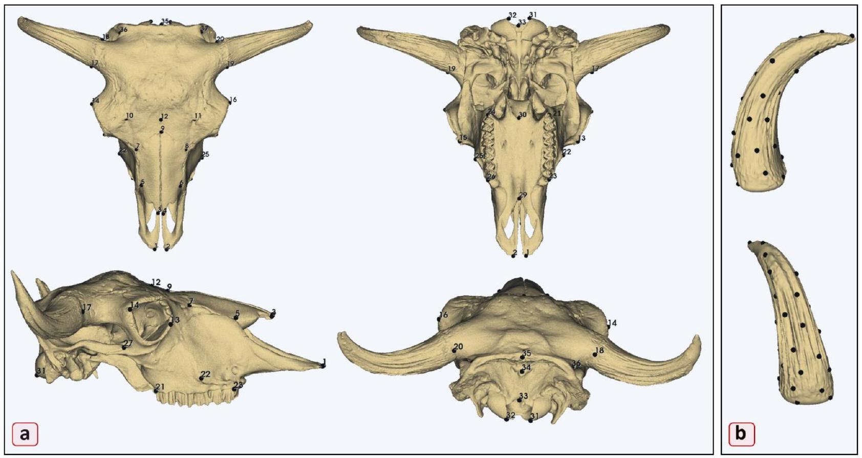
أين سكان برو ثلاثي الأبعاد المتألق تم استخدام ماسح ثلاثي الأبعاد (Shining 3D، هانغتشو، الصين) لإنشاء نماذج ثلاثية الأبعاد للجمجمة من SGGW ومجموعة المرجع من IUC. تم استخدام EXScan Pro (الإصدار 4.0.0.4)، الذي قدمه مصنع الماسح (Shining 3D، هانغتشو، الصين)، لإعادة بناء النموذج ثلاثي الأبعاد. جميع العينات، بما في ذلك تلك من دراسة بيبي وتايلور. تم حفظها بتنسيق “.ply”. تظهر الدراسات أن البيانات من أجهزة المسح المختلفة (Artec Spider وEva وEinScan Pro 2X) قابلة للمقارنة، مع اختلافات طفيفة في الدقة لم تؤثر على نتائج الدراسة. .
كانت العمليات القرنية والأسنان إما مفقودة أو تالفة في العديد من العينات، لذا تم استبعاد هذه المناطق من التحليل القائم على المعالم. تم اختيار ما مجموعه 37 معلمًا تشريحيًا (الجدول 2) استنادًا إلى دراسات سابقة. لقد تم استخدام هذه المعالم على نطاق واسع في الدراسات الأثرية ويمكن تكرارها بسهولة من قبل باحثين آخرين. تم استخدام برنامج 3D Slicer (الإصدار 5.2.2) لتحديد المعالم. في البداية، تم إنشاء مجموعة مسودات من المعالم، والتي تم تطبيقها بعد ذلك بشكل متسق على جميع العينات من قبل نفس الباحث لضمان الدقة في الرقمنة (الجدول التكميلي 1، الجدول التكميلي 2). لم تكن هناك أي معالم مفقودة من التحليل، وتم تطبيق جميع المعالم الـ 37 بنجاح على كل عينة (الشكل 5).
تم استخدام نهج GMM القائم على المعالم لتحليل حجم العملية القرنية وتحديد حجم المركز (الجدول التكميلي 2). غالبًا ما تفشل المقاييس التقليدية، مثل الطول أو السماكة، في فهم شامل للحجم والأبعاد. على سبيل المثال، قد يؤدي قياس طول عملية قرنية ذات هيكل منحني بشكل كبير إلى التقليل من طولها الفعلي بسبب هندستها المعقدة. وبالمثل، قد لا تعكس قياسات السماكة في بعض النقاط المختارة الحجم الكلي أو الشكل المعقد. لمعالجة هذه القيود، استخدمنا المعالم شبه المنتشرة عبر سطح العملية القرنية لالتقاط حجمها بشكل أكثر فعالية. تم استخدام وحدة PseudoLMGenerator في برنامج 3D Slicer (الإصدار 5.2.2). تم اختيار عينة البيسون الأوروبي الذكر، “بلوتكارز”، كنموذج مرجعي لمجموعة المعالم الأولية.
جمجمة منطقة رقم المعلم وصف المعلم
عظام الوجه منطقة الفك العلوي 1,2 بروستيون
21,24 بعد الأسنان
٢٢,٢٥ نتوءات الوجه
٢٣،٢٦ ضرس
منطقة الأنف 3,4 طرف الأنف من عظام الأنف
٥،٦ شَقّ الأنف القاطع
٧،٨ أقصى نهاية أمامية من الغرز الجبهية
9 ناسيون
منطقة الحنك ٢٩ النقطة الأنفية من الحنك الصلب
30 نقطة النهاية للحنك الصلب
المنطقة المدارية 13,15 المدار الخارجي
14,16 المدار الداخلي
عظام الدماغ العصبي المنطقة الجبهية 10,11 الثقب فوق المدار
12 نقطة المنتصف للثقب فوق المدار
المنطقة القرنية 17,19 النقطة الأمامية لعملية القرنية
18,20 نقطة النهاية لعملية القرنية
المنطقة القذالية 31,32 النهاية السفلية للنتوءات القذالية
33 أوبستيون
34 النتوء القذالي الخارجي
35 قمة العنق
36,37 الحدود الذيلية للحفرة الزمنية
المنطقة الزمنية 27,28 النقطة الأمامية للعملية الزمنية
الجدول 2. وصف المعالم المأخوذة على الجماجم للتحليل الهندسي المورفومتري.
الشكل 5. المعالم. (أ) 37 معلمًا تشريحيًا على الجمجمة؛ (ب) 34 معلمًا شبه على عملية القرنية.
عينة المرجع قدمت أساسًا لتكوين ترتيب المعالم. تم تطبيق نطاق تحمل من مما سمح بالمرونة في وضع المعالم بالنسبة لعلم التشكل لعملية القرنية عبر عينات مختلفة. شكل القالب حدد الشكل والتخطيط للقالب المقدر، مما يضمن أن المعالم يمكن أن تتكيف مع اختلافات الشكل الفردية مع الحفاظ على الاتساق. بمجرد إنشاء القالب، تم وضع المعالم على سطح عملية القرنية، مما يضمن تمثيلًا دقيقًا لخصائصها المورفولوجية. في هذه المرحلة، تم تعديل انتظام وتوزيع
نقاط العينة لضمان تباعد متسق، مما يعزز دقة وقابلية إعادة إنتاج وضع المعالم. تم إنشاء مجموعة المعالم النهائية، التي تتكون من 34 معلمًا شبه، (كما هو موضح بالنقاط السوداء في الشكل 5B). عملية آلية وسعت مجموعة المعالم عبر مجموعة البيانات بأكملها. تم تطبيق مجموعة المسودة على جميع عينات الجمجمة باستخدام توليد المعالم التلقائي عبر محاذاة سحابة النقاط وتحليل المطابقة (ALPACA) . سهلت وحدة ALPACA داخل برنامج 3D Slicer هذه العملية، مما سمح بالتطبيق الدقيق والفعال للمعالم شبه على كل عينة.

الهندسة المورفومترية

تم إجراء تحليلات GPA وGMM اللاحقة باستخدام وحدة SlicerMorph داخل برنامج 3D Slicer (الإصدار 5.2.2) . GPA هي طريقة مستخدمة على نطاق واسع في تحليل الشكل لأنها توحد بيانات المعالم عبر العينات، مما يزيل المتغيرات غير المتعلقة بالشكل مثل الموقع، الاتجاه، والحجم. تم توفير ملفات المعالم لكل عينة في الملف التكميلي.
تمت ترجمة تكوينات المعالم من كل جمجمة أولاً إلى الأصل، مما يعني أنه تم نقلها في الفضاء بحيث كان كل تكوين مركزيًا. بعد ذلك، تم قياس المعالم إلى حجم مركزها، مما يضمن أن جميع الجماجم موحدة في الحجم ثم تم تدويرها بشكل مثالي. تم استخدام معيار المربعات الصغرى لتقليل المسافة بين المعالم المتجانسة عبر جميع العينات، مما يسمح للجماجم بأن تكون متراصة بشكل مثالي لمزيد من المقارنة. هذه العملية، المعروفة باسم تراكب بروكروستس، تزيل بشكل فعال التباين الناتج عن الحجم، الدوران، والترجمة بحيث يركز التحليل فقط على اختلافات الشكل. بعد GPA، كانت جميع العينات موجودة ضمن نفس “فضاء الشكل”، مركزة على شكل متوسط. وهذا يوفر نقطة مرجعية مشتركة يمكن من خلالها قياس الانحرافات، أو اختلافات الشكل.

تحليل الفضاء المورفولوجي: الفروق بين المجموعات وبين الأفراد

لتحقيق أنماط اختلاف الشكل وتقييم الفروق المورفولوجية بين المجموعات، أجرينا تحليل ANOVA بروكروستس على تكوينات المعالم المتراصة . اختبر هذا التحليل ما إذا كانت المجموعات المحددة مسبقًا (مجموعات البقر والمجموعات الأوروبية) تظهر اختلافات كبيرة في أشكالها المتوسطة. تم استخدام نهج قائم على التبديل مع 1000 تكرار لتقييم الأهمية الإحصائية. بالإضافة إلى ذلك، قمنا بإجراء مقارنات ANOVA بروكروستس بين أزواج المجموعات لتقييم الفروق المورفولوجية بشكل أكبر. سمحت لنا هذه المقارنات الزوجية بتقييم حجم اختلاف الشكل والأهمية الإحصائية بين مجموعات معينة. تم إجراء جميع التحليلات باستخدام حزمة Geomorph (الإصدار 4.0.9) في R (RStudio 2024.09.1) ، والتي تسهل معالجة بيانات (GMM)، تراكب بروكروستس، والمقارنات الإحصائية للبيانات المعتمدة على المعالم.

عملية القرنية، الحجم، والعمر

هدفت الدراسة إلى استكشاف العلاقة بين حجم الجمجمة، العمر، حجم عملية القرنية، واختلافات الشكل من خلال تحليل تأثير أول مكونين رئيسيين (PC1 وPC2) على هذه العوامل. تم اختيار PC1 وPC2 لقدرتهما على تفسير أكبر التغيرات في مورفولوجيا الجمجمة، مما يجعلها حاسمة لفهم التأثيرات التطورية والتنموية على شكل جمجمة البيسون. تم حساب حجم الجمجمة وحجم عملية القرنية باستخدام حجم المركز (CS)، وهو مقياس مشتق من إحداثيات شكل بروكروستس التي تمثل الحجم بدقة من خلال التركيز على المركز الهندسي للكتلة. تم استخدام وحدة SlicerMorph داخل برنامج 3D Slicer (الإصدار 5.2.2) لاستخراج البيانات ذات الصلة، بما في ذلك قيم حجم المركز لكل من الجمجمة وعملية القرنية. استغلت الدراسة هذه القياسات لتقييم تأثير العمر وحجم عملية القرنية على مورفولوجيا وحجم الجمجمة بشكل عام.

التحليل الإحصائي

تم تطبيق إسقاط عمودي في فضاء تانجنت خطي، وضمان تحويل رياضي يضمن أن التحليلات الإحصائية اللاحقة، مثل PCA، يمكن أن تتم على إحداثيات فضاء التانجنت، مما يجعل البيانات مناسبة للتحليل متعدد المتغيرات. تم إجراء تحليلين PCA متميزين لاستكشاف اختلاف الشكل في الجماجم. PCA هي أداة إحصائية قوية تقلل من تعقيد مجموعة البيانات من خلال تحديد المكونات الرئيسية (PCs)، أو المحاور، التي تمثل أكبر تباين في الشكل بين العينات. تساعد هذه العملية في تصور وقياس الاختلاف عبر مجموعة بيانات كبيرة مع تسليط الضوء على العوامل الرئيسية التي تدفع الاختلافات في مورفولوجيا الجمجمة.
في التحليل الأول، تم إجراء PCA على إحداثيات شكل بروكروستس لمجموعة البيانات الكاملة المكونة من 123 جمجمة، والتي تضمنت أربعة تجمعات فرعية مختلفة من القبيلة الأوسع Bovini. كان الهدف من هذا التحليل هو تصور توزيع الأنواع في فضاء مورفولوجي قحفي منخفض الأبعاد، وهو في الأساس فضاء رياضي حيث يمثل كل نقطة شكل جمجمة مختلف. من خلال فحص كيفية تجمع أو انتشار جماجم البيسون الأوروبي ضمن هذا الفضاء المورفولوجي مقارنة بأعضاء آخرين من القبيلة Bovini (مثل الماشية والجاموس)، كان من الممكن الحصول على رؤى حول الميزات المورفولوجية المميزة للبيسون الأوروبي. ركز التحليل الثاني بشكل حصري على البيسون الأوروبي. شمل ذلك PCA على مجموعة بيانات مكونة من 57 جمجمة للتحقيق في الاختلافات داخل النوع، خاصة عبر الجنسين والتجمعات الفرعية داخل النوع.
تم اختبار اختلاف الحجم باستخدام ANOVAs على بيانات حجم المركز. بعد ANOVA، تم تطبيق اختبار Tukey’s HSD كتحليل لاحق لتحديد الفروق المحددة بين المجموعات. تم إجراء تحليل الانحدار الخطي متعدد المتغيرات للتحقيق في العلاقة بين العمر، حجم عملية القرنية، وحجم الجمجمة. بالإضافة إلى ذلك، تم استخدام أول مكونين رئيسيين (PC1 وPC2)، اللذان يفسران أكبر اختلاف في شكل الجمجمة، للتقييم الألومتري. من خلال فحص العلاقة بين هذه المكونات الرئيسية وحجم المركز، استكشفت الدراسة كيف ترتبط اختلافات شكل الجمجمة بالتغيرات في الحجم (الألومتري). تم إجراء هذه التحليلات باستخدام برنامج PAST الإحصائي .

توفر البيانات

تم توفير البيانات ضمن المخطوطة أو ملفات المعلومات التكميلية.
تاريخ الاستلام: 23 أكتوبر 2024؛ تاريخ القبول: 6 يناير 2025
تم النشر عبر الإنترنت: 09 يناير 2025

References

  1. Larska, M., Tomana, J., Krzysiak, M. K., Pomorska-Mól, M. & Socha, W. Prevalence of coronaviruses in European bison (Bison bonasus) in Poland. Sci. Rep. 14, 12928 (2024).
  2. Olech, W. The number of ancestors and their contribution to European bison (Bison bonasus L.) population. Anim. Sci. 35, 111117 (1999).
  3. Slatis, H. M. An analysis of inbreeding in the European bison. Genetics 45, 275 (1960).
  4. Parusel, J. B. What next with Pless line of European bison from Pszczyna-a question on the 155 th anniversary of restitution and breeding? Eur. Bison Conserv. Newsl. 13, 39-56 (2021).
  5. Plumb, G., Kowalczyk, R. & Hernandez-Blanco, J. A. Bison bonasus. IUCN red list. Threatened Species. https://doi.org/10.2305/IU CN.UK.2020-3.RLTS.T2814A45156279.en (2020).
  6. Haber, A. Phenotypic covariation and morphological diversification in the ruminant skull. Am. Nat. 187(5), 576-591 (2016).
  7. Janis, C. M. & Theodor, J. M. Cranial and postcranial morphological data in ruminant phylogenetics. Zitteliana 15-31 (2014).
    1. Stankowich, T., & Caro, T. Evolution of weaponry in female bovids. Proc. Royal Soc. B: Biol. Sci. 276(1677), 4329-4334 (2009).
  8. Krasińska, M., Szuma, E., Kobryńczuk, F. & Szara, T. Morphometric variation of the skull during postnatal development in the Lowland European bison Bison bonasus bonasus. Acta Theriol. 53, 193-216 (2008).
  9. Mysterud, A. et al. Population ecology and conservation of endangered megafauna: The case of European bison in Białowieża Primeval Forest, Poland. Anim. Conserv. 10, 77-87 (2007).
  10. Zalewski, D., Szczepański, W. & Wołkowycka, M. Characteristics of closed breeding stations of Bison (Bison Bonasus L.) in Poland in the years 1997-2000. Acta Sci. Pol. Silv Colendar Rat. Ind. Lignar. 4, 121-130 (2005).
  11. Figueirido, B., Tseng, Z. J. & Martín-Serra, A. Skull shape evolution in durophagous carnivorans. Evolution 67(7), 1975-1993 (2013).
  12. Tokita, M., Yano, W., James, H. F. & Abzhanov, A. Cranial shape evolution in adaptive radiations of birds: Comparative morphometrics of Darwin’s finches and Hawaiian honeycreepers. Proc. R. Soc. B 372, 20150481 (2017).
  13. Wagner, G. P. What is the promise of developmental evolution? Part I: Why is developmental biology necessary to explain evolutionary innovations? J. Exp. Zool. 288(2), 95-98 (2000).
  14. Bertossa, R. C. Morphology and behaviour: Functional links in development and evolution. Philosophical Trans. Royal Soc. B: Biol. Sci. 366(1574), 2056-2068 (2011).
  15. Janis, C. M., Constable, E. C., Houpt, K. A., Streich, W. J. & Clauss, M. Comparative ingestive mastication in domestic horses and cattle: A pilot investigation. J. Anim. Physiol. Anim. Nutr. 94, 402-409 (2010).
  16. Sanson, G. The biomechanics of browsing and grazing. Am. J. Bot. 93, 1531-1545 (2006).
  17. Stankowich, T. & Caro, T. Evolution of weaponry in female bovids. Proc. R. Soc. B. 276, 4329-4334 (2009).
  18. Kowalczyk, R. et al. Movements of European bison (Bison bonasus) beyond the Białowieża Forest (NE Poland): Range expansion or partial migrations? Acta Theriol. 58, 391-401 (2013).
  19. Solounias, N. & Moelleken, S. M. Dietary adaptation of some extinct ruminants determined by premaxillary shape. J. Mammal 74, 1059-1971 (1993).
  20. Tennant, J. P. & MacLeod, N. Snout shape in extant ruminants. PLoS One 9, e112035 (2014).
  21. Craine, J. M., Towne, E. G., Miller, M. & Fierer, N. Climatic warming and the future of bison as grazers. Sci. Rep. 5, 16738 (2015).
  22. Nippert, J. B., Culbertson, T. S., Orozco, G. L., Ocheltree, T. W. & Helliker, B. R. Identifying the water sources consumed by bison: Implications for large mammalian grazers worldwide. Ecosphere 4, 1-13 (2013).
  23. Caro, T. M., Graham, C. M., Stoner, C. J. & Flores, M. M. Correlates of horn and antler shape in bovids and cervids. Behav. Ecol. Sociobiol. 55, 32-41 (2003).
  24. Tidière, M., Lemaître, J. F., Pélabon, C., Gimenez, O. & Gaillard, J. M. Evolutionary allometry reveals a shift in selection pressure on male horn size. J. Evol. Biol. 30, 1826-1835 (2017).
  25. Willisch, C. S., Biebach, I., Marreros, N., Ryser-Degiorgis, M. P. & Neuhaus, P. Horn growth and reproduction in a long-lived male mammal: No compensation for poor early-life horn growth. Evol. Biol. 42, 1-11 (2015).
  26. Buttar, I. & Solounias, N. Specialized position of the horns, and frontal and parietal bones in Bos taurus (Bovini, Artiodactyla), and notes on the evolution of Bos. Ann. Zool. 58, 49-63 (2021).
  27. Lundrigan, B. Morphology of horns and fighting behavior in the family Bovidae. J. Mammal. 77, 462-475 (1996).
  28. Bergmann, C. Über die Verhältnisse Der Wärmeökonomie Der Thiere zu Ihrer Grösse. Vandenhoeck Und Ruprecht (1848).
  29. Bibi, F. & Tyler, J. Evolution of the bovid cranium: Morphological diversification under allometric constraint. Commun. Biol. 5, 69 (2022).
  30. Tidière, M. et al. Variation in the ontogenetic allometry of horn length in bovids along a body mass continuum. Ecol. Evol. 10, 4104-4114 (2020).
  31. Packard, G. C. Evolutionary allometry of horn length in the mammalian family Bovidae reconciled by non-linear regression. Biol. J. Linn. Soc. 125, 657-663 (2018).
  32. Cardini, A. & Polly, P. D. Larger mammals have longer faces because of size-related constraints on skull form. Nat. Commun. 4, 2458 (2013).
  33. Kobryńczuk, F., Krasińska, M. & Szara, T. Sexual dimorphism in skulls of the lowland European bison, Bison bonasus bonasus. Ann. Zool. 45, 335-340 (2008).
  34. Szara, T., Klich, D., Wójcik, A. M. & Olech, W. Temporal trends in skull morphology of the European bison from the 1950s to the present day. Diversity 15, 377 (2023).
  35. Morena, S., Barba, S. & Álvaro-Tordesillas, A. Shining 3D EinScan-Pro, application and validation in the field of cultural heritage, from the Chillida-Leku Museum to the archaeological museum of Sarno. Int. Arch. Photogramm. ISPRS Arch. 42, 135-142 (2019).
  36. Bookstein, F. L. Morphometric Tools for Landmark data: Geometry and Biology (Cambridge University Press, 1997).
  37. Çakar, B. et al. Comparison of skull morphometric characteristics of simmental and holstein cattle breeds. Animals 14, 2085 (2024).
  38. Özkan, E., Siddiq, A. B., Kahvecioğlu, K. O., Öztürk, M. & Onar, V. Morphometric analysis of the skulls of domestic cattle (Bos taurus L.) and water buffalo (Bubalus bubalis L.) in Turkey. Turk. J. Vet. Anim. Sci. 43, 532-539 (2019).
  39. Porto, A., Rolfe, S. & Maga, A. M. ALPACA: A fast and accurate computer vision approach for automated landmarking of threedimensional biological structures. Methods Ecol. Evol. 12, 2129-2144 (2021).
  40. Beinat, A. & Crosilla, F. Generalised Procrustes Analysis for size and Shape 3-D Object Reconstructions 345-353 (Optical, 2001).
  41. Stuart, M. A geometric approach to principal components analysis. Am. Stat. 36, 365-367 (1982).
  42. Rolfe, S. et al. SlicerMorph: An open and extensible platform to retrieve, visualize and analyse 3D morphology. Methods Ecol. Evol. 12, 1816-1825 (2021).
  43. Viacava, P., Baker, A. M., Blomberg, S. P., Phillips, M. J. & Weisbecker, V. Using 3D geometric morphometrics to aid taxonomic and ecological understanding of a recent speciation event within a small Australian marsupial (Antechinus: Dasyuridae). Zool. J. Linn. Soc. 196(3), 963-978 (2022).
  44. Adams, D. C. & Otárola-Castillo, E. Geomorph: An R package for the collection and analysis of geometric morphometric shape data. Methods Ecol. Evol. 4(4), 393-399 (2013).
  45. Hammer, Ø. & Harper, D. A. Past: Ualeontological statistics software package for educaton and data anlysis. Palaeont Electr. 4, 1 (2001).
  46. Minervino, A. H. H., Zava, M., Vecchio, D. & Borghese, A. Bubalus bubalis: A short story. Front. vet. sci. 7, 570413 (2020).

الشكر والتقدير

تم تمويل هذه الدراسة من قبل مجلس البحث العلمي والتكنولوجي في تركيا (TUBITAK 22191059B192301667). تم تمويل النشر من قبل صندوق تطوير العلوم في جامعة وارسو للعلوم الزراعية – SGGW.

مساهمات المؤلفين

O.G. و T.S. وضعوا تصور الدراسة. O.G. قام بإجراء رقمنة المعالم والتحليلات الإحصائية. O.G. و T.S. أعدوا المقال. ساهم جميع المؤلفين بشكل كبير في تفسير البيانات والنتائج وكتابة المخطوطة.

الإعلانات

المصالح المتنافسة

يعلن المؤلفون عدم وجود مصالح متنافسة.

معلومات إضافية

المعلومات التكميلية النسخة الإلكترونية تحتوي على مواد تكميلية متاحة على https://doi.org/1 .
يجب توجيه المراسلات وطلبات المواد إلى T.S.
معلومات إعادة الطبع والتصاريح متاحة على www.nature.com/reprints.
ملاحظة الناشر تظل Springer Nature محايدة فيما يتعلق بالمطالبات القضائية في الخرائط المنشورة والانتماءات المؤسسية.
الوصول المفتوح هذه المقالة مرخصة بموجب رخصة المشاع الإبداعي للاستخدام غير التجاري بدون اشتقاقات 4.0 الدولية، والتي تسمح بأي استخدام غير تجاري، ومشاركة، وتوزيع، وإعادة إنتاج في أي وسيلة أو صيغة، طالما أنك تعطي الائتمان المناسب للمؤلفين الأصليين والمصدر، وتوفر رابطًا لرخصة المشاع الإبداعي، وتوضح إذا قمت بتعديل المادة المرخصة. ليس لديك إذن بموجب هذه الرخصة لمشاركة المواد المعدلة المشتقة من هذه المقالة أو أجزاء منها. الصور أو المواد الأخرى من طرف ثالث في هذه المقالة مشمولة في رخصة المشاع الإبداعي للمقالة، ما لم يُشار إلى خلاف ذلك في سطر الائتمان للمادة. إذا لم تكن المادة مشمولة في رخصة المشاع الإبداعي للمقالة واستخدامك المقصود غير مسموح به بموجب اللوائح القانونية أو يتجاوز الاستخدام المسموح به، ستحتاج إلى الحصول على إذن مباشرة من صاحب حقوق الطبع والنشر. لعرض نسخة من هذه الرخصة، قم بزيارة http://creativecommo ns.org/licenses/by-nc-nd/4.0/.
© المؤلفون 2025

  1. قسم التشريح، كلية الطب البيطري، جامعة إسطنبول-جيراه باشا، 34320 إسطنبول، تركيا.
    قسم العلوم الشكلية، معهد الطب البيطري، جامعة وارسو لعلوم الحياة SGGW، 02-776 وارسو، بولندا. البريد الإلكتروني: tomasz_szara@sggw.edu.pl

Journal: Scientific Reports, Volume: 15, Issue: 1
DOI: https://doi.org/10.1038/s41598-025-85654-3
PMID: https://pubmed.ncbi.nlm.nih.gov/39789289
Publication Date: 2025-01-09

OPEN

Morphological patterns of the European bison (Bison bonasus) skull

Ozan Gündemir & Tomasz Szara ®

Abstract

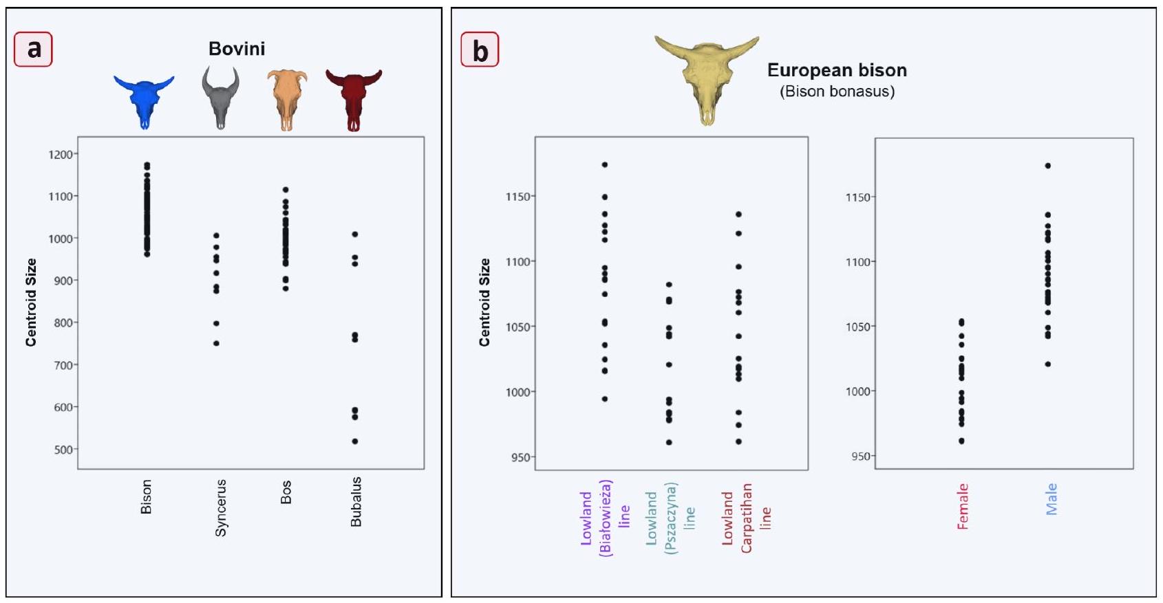
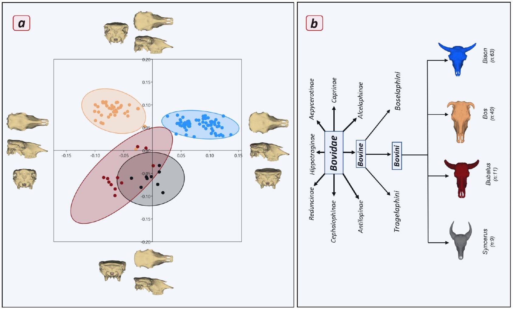
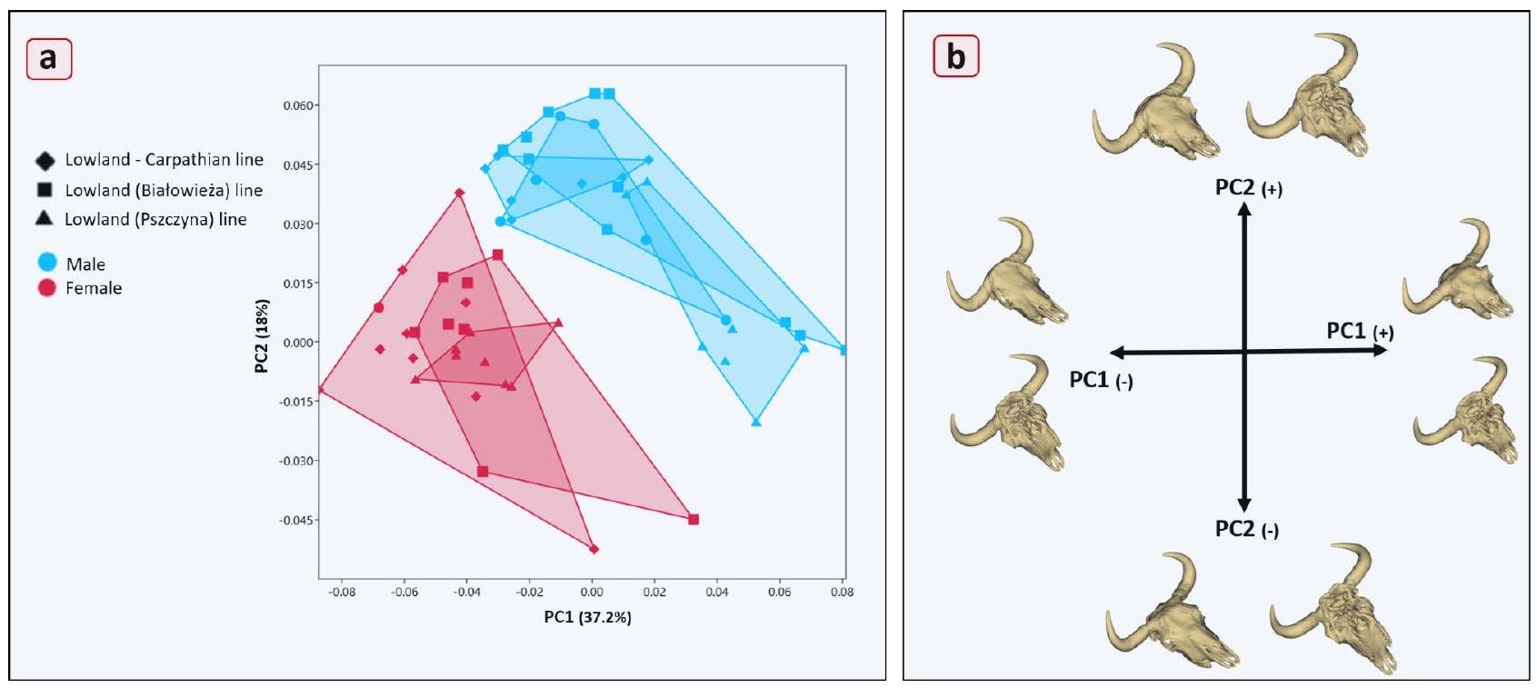
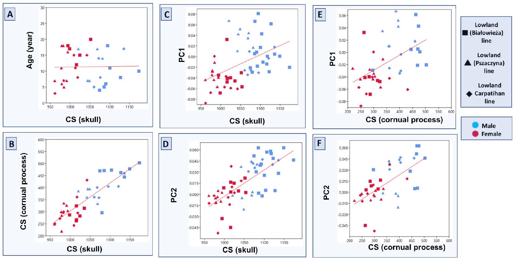
This study aimed to investigate the effects of environmental factors, sexual selection, and genetic variation on skull morphology by examining the skull structure of the European bison, a species at risk of extinction, and comparing it to other bovid species. The skull of the European bison was significantly bigger than that of other species of the tribe Bovini, and the results revealed considerable morphological differences in skull shape compared to other Bovini samples. The bison skull exhibited a broader shape in the frontal region and a more laterally oriented cornual process. The frontal bone also significantly influenced skull shape variation within the European bison subspecies. Also, the findings indicated that cornual processes size significantly affected skull shape. Male Bison had larger skulls and more developed cornual ridges, and these morphological changes in the frontal region are thought to have also influenced the nuchal region, maxillary region, and facial bones. Furthermore, the differences in skull size observed between the Lowland-Pszczyna and Lowland-Białowieża lines, which share a closer origin than the Lowland-Carpathian lines, may reflect environmental and genetic adaptations over time. This research provides a reference point for future studies on the ecological and evolutionary factors influencing bison skull variations, with significant implications for the conservation of this species.

Keywords Bovidae, Bovini, Cornual process, Frontal bone, Geometric morphometrics, Skull morphology
The European bison (Bison bonasus), known as the wisent, is Europe’s largest mammal. Following World War I, the species faced extinction in the wild, with the last individual being killed in the Białowieża Forest in . However, the species survived in a few Zoos and enclosures across Europe. Thanks to an international breeding program initiated in the 1920s and 1930s, the population began to recover, starting with re-establishing a herd in Białowieża. Since then, multiple free-ranging populations have been created, with numbers now reaching tens of thousands. In historical times, there were two subspecies of bison. The Lowland bison (Bison bonasus bonasus) occupied the forest areas of central and eastern Europe, and the Caucasian bison (Bison bonasus caucasicus) was found in the Caucasus Mountains. The Pszczyna bison herd, brought to Silesia in 1865 from the Białowieża the European bison population has identified two genetically distinct lines: the Lowland-Carpathian line and the Lowland-Białowiezaline . In the autumn of 1865, four Lowland European bison were transported from Białowieża to Murcki, and a year later, they were released into a large enclosure in the Pszczyna Forest. This population, having diverged genetically over time, is now recognized as the Lowland-Pszczyna line . As a representative of the European megafauna, the European bison serves as an umbrella species, playing a crucial role in its ecosystem. The species is listed as Near Threatened (NT) on the IUCN Red List , underscoring the urgent need for its conservation.
The skull and its morphological features play a key role in the systematics and phylogeny of ruminants . Variations in skull shape and its structures reflect the potential to respond to a wide range of selective pressures . Horns, present in both male and female bovids, are a solid sexual differentiating feature . The size of the cornual processes in growing individuals positively correlates with skull size and body mass, as more giant skulls and heavier body masses provide greater structural support for the development of larger cornual processes. The skull of bison of both sexes shows intensive development until the age of 5, after which the growth rate slows down significantly, likely indicating the completion of major cranial structural formation and the transition to a more stable adult morphology .
As one of Europe’s last surviving megafauna species, the European bisonis critical in maintaining biodiversity and promoting ecosystem health. Studying its biology, morphology, and genetics allows conservationists to gain
deeper insights into its survival strategies and evolutionary adaptation processes . By examining its anatomical structures, notably features like skull shape and size variations, and investigating genetic diversity within and between populations, scientists can better understand how this species has adapted to its environment over time .
This study aims to investigate the evolutionary adaptations and morphological diversity of the European bisonskull. This complex and phylogenetic structure is crucial in understanding developmental processes in evolutionary change . The research assumes that the skull morphology of the European bison has been shaped by long-term inbreeding and genetic factors due to the species’ isolation in protected habitats like the Białowieża Forest. In these isolated populations, restricted gene flow is expected to drive morphological diversity. The first part of the study focuses on situating the European bison within the broader tribe Bovini by comparing its skull morphology with other species within the family. This comparative analysis will provide an evolutionary framework, offering insights into shared ancestral traits and divergent ecological adaptations that have influenced skull morphology across the Bovini lineage. Through this, the study will assess how evolutionary pressures and ecological niches have shaped both the commonalities and unique traits found in the European bison’s skull. In the second part of the study, the focus shifts to intraspecific variation within the European bison population, exploring morphological differences between sexes and subspecies. A key hypothesis is that male bison will exhibit morphological differences from females because their highly developed cornual processes are known to impose developmental pressures on the skull.

Results

Skull and cornual process size

Figure 1 illustrates the distribution of skull sizes for all samples used in the study, while Table 1 provides pairwise group comparisons. The skull of the European bison was notably larger compared to other members of the Bovini family ( ). The European bison skulls displayed a relatively homogeneous distribution, similar to that observed in the Bos samples. In contrast, members of the Bubalus genus are characterized by the smallest skulls, with substantial variability in size. This variation may be attributed to the more significant number of subspecies within the Bubalus group, which included four distinct subspecies, compared to only two in the Bison and Bos groups . This broader taxonomic diversity likely contributed to the observed size variability. The size difference across the Bovini family was statistically significant when assessing the entire dataset ( ).
Within the European bison samples, skull size comparisons revealed that the Lowland-Białowieza line had significantly larger skulls than the other two ( ). In contrast, the Lowland-Pszczyna and LowlandCarpathian lines exhibited similar skull sizes, with no statistically significant differences between them. Comparisons of cornual process size yielded results consistent with those for skull size. In both cases, sex emerged as a distinguishing factor, with males exhibiting larger skulls and more prominent cornual processes than females .
Fig. 1. Jitter Plot of centroid size between the groups. (a) Centroid sizes for tribe Bovini, (b) Centroid sizes for 3 different lines of European bison.
Group Measurement Pair Difference Lower CI Upper CI -value
Bovini Skull size (CS) Bison-Syncerus 151.4489 83.4597 219.4382
Bison -Bos 56.8076 18.2346 95.3807
Bison -Bubalus 320.4568 258.1099 382.8037
Syncerus – Bos 94.6413 24.2511 165.0315
Syncerus – Bubalus 169.0079 83.2522 254.7636
Bos – Bubalus 263.6492 198.6924 328.6059
European bison (Line) Skull size (CS) Białowieża – Pszczyna 64.3619 27.4367 101.287
Białowieża – Carpathian 37.0776 0.8916 73.2636
Pszczyna – Carpathian 27.2843 -13.6453 68.2139 p: 0.111
Female Male 88.5086 72.2288 104.7884
Cornual process Size (CS) Białowieża – Pszczyna 64.3619 27.4367 101.287
Białowieża – Carpathian 37.0776 0.8916 73.2636
Pszczyna – Carpathian 27.2843 -13.6453 68.2139 p: 0.282
Female Male 88.5086 72.2288 104.7884
Table 1. Results of ANOVA.
Fig. 2. (a) Results of the PCA (Ellipses represent confidence intervals around the means); (b) Zoological classification of the European bison in the family Bovidae .

Skull shape variations

The distribution of tribe Bovini according to the PCA results is presented in Fig. 2A. In contrast, Fig. 2B illustrates the sample sizes (n numbers), species names used in the study, and a species tree tracing the evolutionary lineage from the Bovidae family to the species analyzed. The analysis revealed (PC1 and PC2) that the Bison skull shape displayed distinct morphological variation compared to other members of Bovini. All Bison samples exhibited positive PC1 values, whereas all others, except five Syncerus specimens, showed negative PC1 values. This indicates that even the PC1 values alone were statistically sufficient to distinguish the Bison skull from other species . Regarding PC2 results, Bison showed positive PC2 values similar to Bos’s, further distinguishing the Bison skull from the Bubalus and Syncerus genera.
The positive PC1 value, which accounted for the most significant shape variation, was associated with a broader skull, especially in the frontal region, and a more laterally oriented cornual process. In contrast, negative
PC1 values indicated a narrower, more elongated skull, with the cornual process positioned closer to the nuchal region and a narrower frontal region. Positive PC2 values also indicated a larger frontal region, while negative PC2 values were linked to a significantly more robust cornual process base, contributing further to species differentiation.
The principal component analysis (PCA) results for European bison skulls within their group are depicted in Fig. 3A. The skull shape variations among groups with different genetic characteristics were similar, suggesting that these variations in European bison are more individualistic than strongly linked to genetic lineage. However, sex had a clear and significant impact on skull shape. The PC1 and PC2 results revealed a complete separation between male and female specimens, indicating that sex was a crucial determinant in skull morphology.
The specific shape changes associated with negative and positive values of the first two principal components are illustrated in Fig. 3B. A negative PC1 value was associated with a narrower, elongated skull, whereas a positive PC1 value represented a broader skull, particularly the frontal squama. Additionally, in the positive PC1 configuration, the nuchal plane was significantly more extensive, and the facial part of the skull exhibited lateral expansion. One of the most striking morphological changes observed with a positive PC1 value was the raised middle part of the frontal squama, creating a lump-like prominence. In contrast, a negative PC1 value corresponded to a flatter frontal bone roof.
Regarding PC2, a positive value corresponded to a more oval skull. Furthermore, the cornual processes were thicker in specimens with positive PC2 values than those with negative PC2 values. A broader nuchal plane was another prominent feature associated with positive PC2 values, further contributing to significant shape variations across the skulls.

Inter- and intragroup shape differences

The Procrustes ANOVA revealed morphological differences among the predefined groups (Bison, Syncerus, Bos, and Bubalus) ( ). Pairwise comparisons showed that the most notable distinctions were between Bison and Syncerus and between Syncerus and Bos. In contrast, no significant differences were observed between Bison and Bos, or between Bos and Bubalus.
According to the result of Procrustes ANOVA, the comparison between Białowieża and Carpathian lines showed a significant effect ( ), with a substantial proportion of the variation in shape attributed to group membership ( ). In contrast, the Białowieża and Pszczyna comparison revealed no significant difference ( ), suggesting that the morphological variation between these two lines is minimal . On the other hand, the Pszczyna and Carpathian comparison showed a highly significant morphological divergence ( ), with a large proportion of shape variation accounted for by the group .

Size and shape

Figure 4 illustrates the interactions between age, cornual process size, skull size, and the two first principal components. The analysis first revealed that age had no significant effect on skull size. Also, age did not appear to affect skull shape in the European bison significantly. However, there was a statistically significant relationship
Fig. 3. The first two principal components of the analysis of European bison’ skulls. (a) Scatterplots of the first two principal components from the skull of three distinct populations: Lowland-Carpathian line, LowlandBiałowieża line, and Lowland-Pszczyna line ( 4 male sample lines are unknown); (b) Negative and positive shape variations for the first two principal components. PCA results of the first two axes account for of the total variance. Shape changes are shown by 3D warping.
Fig. 4. Relationships between age, skull size, cornual process size, and the first two principal components.
between skull size and cornual process size ( ) and their corresponding PC1 ( , F: ) values, which explains the most substantial shape variation in these measurements.
As anticipated, the cornual process size was positively correlated with skull size ( ), indicating that larger skulls generally had larger cornual processes. This suggests that the development of the cornual process is proportionate to overall skull growth. Furthermore, skull size statistically significantly impacted PC1 and PC2 , indicating that skull shape changes are closely linked to skull size. As skull size increased, the specimens exhibited more prominent positive PC1 and PC2 features. Regarding morphology, more giant skulls tended to have wider transverse dimensions (as reflected by positive PC1 values) and displayed more oval or rounded shapes (as indicated by positive PC2 values).

Discussion

The findings of this study provide new insights into the morphological relationships between skull size, cornual process size, and shape variation in the tribe Bovini, with a particular focus on the European bison. One of the most striking results is the significant correlation between skull size and cornual process size, which was an expected outcome. This finding aligns with previous studies on bovids, confirming that larger skulls support larger, more developed cornual processes. The lack of a significant relationship between age and both skull and cornual process size suggests that, beyond a certain point in development, growth in these traits is largely independent of age. Sexual dimorphism is more dominant in determining these morphological features, as evidenced by the clear separation of males and females. Additionally, the effect of size on shape variation, particularly as captured by the principal components, highlights the importance of allometry in skull morphology. Larger skulls tend to have wider transverse dimensions and more rounded shapes, indicating that size contributes significantly to the overall shape, independent of other factors. These results provide a more detailed understanding of the morphological variation within the tribe Bovini, particularly in the context of the European bison. Future research could expand on these findings by exploring the genetic and ecological factors that drive this variation and its implications for species classification and conservation efforts.
The skull of the European bison is significantly larger than that of other members of the tribe Bovini. This size difference indicates a robust skeletal structure that can support the physical demands of the bison’s environment, including its role as a large herbivore in various habitats across Europe. The considerable morphological differences in skull shape compared to other Bovini can be attributed to evolutionary adaptations shaped by diet, habitat, and social behavior. The European bisonexhibits a broader skull, distinguishing it from other species. This may provide advantages in feeding, enabling the bison to graze more efficiently on various vegetation . The broader skull also supports larger masticatory muscles, facilitating the processing of rigid plant material, a significant aspect of the bison’s diet. In addition to its skull shape, the cornual processes of the European bison differentiate it from Bubalus (water buffalo) and Syncerus (African buffalo). The base of the cornual process in Bubalus and Syncerusis more massive, suggesting a stronger emphasis on horn growth for purposes such as dominance displays, territorial defense, or mating competitions . In contrast, the cornual processes of the European bison are observed to be separated from the frontal squama and have a thinner base. This anatomical difference may indicate that, while cornual processes play a role in bison social dynamics, their morphology has evolved to serve different functions compared to the more robust horns of Bubalus and Syncerus. The shape
of the bison’s cornual process could also reflect adaptations to the environments in which these species live. Bison inhabit open grasslands and forest edges, where their primary defensive strategies may not rely heavily on weapons . Instead, their social structures and behaviors-such as forming herds-may play a more crucial role in protection against predators. As a result, the evolutionary pressures on bison may have led to the development of skull morphology that prioritizes other survival strategies over the robust cornual processes seen in their relatives. The significant morphological differences among the tribe Bovini highlight the diverse adaptations that have evolved in response to environmental pressures, feeding strategies, and social dynamics. Understanding these morphological traits provides valuable insights into the evolutionary history of the European bison and its relatives and their ecological roles in their respective habitats.
Solounias identified distinct patterns in premaxillary morphology that correspond to dietary habits, with narrower, pointed premaxillae typically associated with browsers and flatter, wider premaxillae characteristic of grazers. This morphological differentiation is crucial for herbivores, as it directly influences their ability to select forage, chew efficiently, and maximize their intake of nutrients. Tennant further emphasized that snout shape plays a significant role in determining foraging efficiency, as a pointed snout allows herbivores to be more selective. In contrast, a blunter snout enables a more generalized, high-volume cropping of vegetation. In the present study, the European bison’s premaxilla was blunt and broad, corresponding with the species’ classification as a strict grazer . Ruminants, such as European bison, primarily feed on grass, which requires a more generalized cropping mechanism to consume large quantities of low-quality forage. The premaxillary morphology observed in the bison reinforces this ecological adaptation. Compared to other Bovini species in the study, the bison displayed more pronounced grazer characteristics, as reflected in its premaxilla shape. This finding is significant because it not only reinforces the link between skull morphology and dietary adaptation but also suggests that within the tribe Bovini, the European bison may be more specialized for grazing than some of its relatives. For instance, in another tribe of Bovini, more pointed rostral shapes may indicate a greater tendency toward mixed feeding or browsing. In contrast, the bison’s blunt and broad premaxilla supports its adaptation to environments where high-volume, low-selectivity feeding is essential for survival, such as grasslands.
The frontal bone was observed to be one of the most significant contributors to skull shape variation in the European bison. This prominence of the frontal bone in shaping skull morphology aligns with its critical role in housing and supporting the horn structures (cornual processes), which are essential for species-specific behaviors such as combat, defense, and social hierarchy in the family Bovidae. The variation in the frontal bone is particularly notable in terms of width and thickness, as seen in the positive PC1 values, where the skull was laterally expanded, especially in the frontal region. This could be linked to sexual dimorphism, as males tend to have more extensive and more developed horn structures, necessitating a more robust frontal bone to accommodate the increased mechanical stress imposed by horn growth and use during physical interactions . The larger size of the frontal bone in males contributes to the overall sexual differentiation in skull morphology between males and females. This difference is a critical distinguishing factor, as males typically possess larger and more developed weapons, while females exhibit smaller or less pronounced horns. Furthermore, this morphological variation of the frontal bone is not isolated; it also has cascading effects on other skull regions. For instance, the nuchal region, which supports the neck muscles, becomes more developed in male European bisonto counterbalance the added weight and size of the weapons. The increased development in the caudal part of the frontal bone may also influence the shape of adjacent bones, such as the parietal and temporal bones, contributing to differences in the overall cranial structure . Moreover, the alteration in the frontal bone also has implications for the facial bones. As the frontal bone expands and morphologically changes to accommodate the weapons, it exerts pressure on the maxilla and nasal bones, possibly leading to secondary adjustments in the facial skeleton.
The Lowland-Pszczyna line, initially genetically characteristic of the Lowland-Białowieżaline, developed distinct genetic traits after being separated from the main herd in 1865. This divergence is thought to result from a combination of genetic drift and environmental influences over time . Despite the genetic differences, skull shape variation between the two lines remains relatively similar. However, there was a statistically significant difference in skull size, with the Lowland-Białowieżaline exhibiting larger skulls. This size discrepancy may reflect the influence of environmental factors over the past 150 years. This observation aligns with Bergmann’s proposal, suggesting that populations of the same species in colder climates tend to be larger than those in warmer regions, as larger body size aids heat conservation . The Lowland-Pszczyna line, located in a slightly warmer environment, may have adapted to this environmental pressure by evolving smaller skulls and overall body size compared to the Lowland-Białowieża line. However, while this hypothesis offers an exciting perspective, more extensive data and examples are required to draw definitive conclusions about this adaptation. Nevertheless, these preliminary findings provide a valuable foundation for future studies on environmental and genetic influences on bison morphology.
Numerous studies have examined the allometric properties of the cattle skull, yielding similar findings. The consistent results of these studies are generally attributed to the constraints imposed by a deeply conserved sizeshape allometry of the Bovidskull, as highlighted by Bibi and Tyler . This conserved allometric relationship has been further suggested to be influenced by the presence and development of horns. Tidière’s study underscores that the ontogenetic allometric gradient has likely evolved among species in response to size-related changes in selection pressures, mainly acting on horn length and body mass. Since horn length is often linked to sexual dimorphism, the allometric effects can vary between male and female individuals. For instance, Packard , in his allometric study on mountain goats, noted that males exhibited a steep upward trajectory in the allometric equation. At the same time, females followed much shallower paths, thus supporting the hypothesis of sexrelated allometric divergence. Similar allometric variation is observed in the skull of the European bison, much like in other bovid species. However, the allometric and shape influence of the cornual process requires special attention in the European bison. In male individuals, the more developed weapons significantly influence skull
morphology by exerting pressure on the frontal bone. This, in turn, affects the facial bones, contributing to the distinct morphological differences between males and females. The well-developed cornual processes in males reinforce their allometric growth, further differentiating skull shape and size in sexually dimorphic patterns . These findings suggest that the evolutionary pressures on cornual process development contribute to overall skull morphology and shape the distinct allometric trajectories between sexes. Understanding this dynamic offers essential insights into how selection pressures, such as size and sexual dimorphism, have shaped the morphological evolution of European bison and other Bovid species.
Craniofacial elongation is a well-documented aspect of postnatal skull development in mammals, where newborns typically have relatively small faces and rounded heads . As mammals mature, the facial region grows faster than the braincase, resulting in a less spherical head shape and more prominent jaws. However, in the case of European bison, age did not significantly impact skull size or shape in this study. This lack of age-related variation can be attributed to the fact that all specimens used were older individuals. One specimen was 4 years old, while the others were all over 5 years of age-an age at which European bisonare expected to have completed their skeletal development . Since craniofacial growth and skull development slow down or cease after full maturity, it is not surprising that age had no measurable effect on the parameters examined in this study. Further studies may comprehensively explore age effects by including younger or juvenile specimens.
In conclusion, this study highlights the frontal bone’s and cornual processes’ significant role in shaping European bison’s skulls, particularly in distinguishing male and female skulls. The morphological variations observed, particularly in the frontal and facial regions, emphasize the impact of horn development on overall skull shape. Additionally, environmental factors may have contributed to size differences between the LowlandBiałowieża and Lowland-Pszczyna lines, possibly supporting Bergmann’s rule. Bergmann’s rule suggests that populations of the same species tend to be larger in colder climates than in warmer regions, as a larger body size aids in conserving heat.
This study lays the foundation for future research in several key areas. By providing a detailed analysis of the European bison’s skull morphology, mainly focusing on the effects of sexual dimorphism, allometry, and environmental adaptation, it offers a framework for further exploration into the genetic and ecological factors driving these morphological traits. Future studies can build on these findings to investigate the genetic underpinnings of the observed cranial variations, examining how inbreeding, gene flow, and isolation have influenced skull shape and size.

Material method

Samples

A total of 57 European bison skulls ( 31 males and 26 females) of known pedigrees from the Osteological Museum of the Department of Morphological Sciences, Institute of Veterinary Medicine, Warsaw University of Life Sciences (SGGW), were used in this study. These skulls belonged to animals that died between 1950 and 2014, aged 3 to 20. Among the sample, 26 came from the Lowland-Białowieza line ( 9 females, 17 males), 15 from the Lowland-Pszczyna line ( 8 females, 7 males), and 16 from the Lowland-Carpathian line ( 9 females, 7 males).
For the comparative analysis of the European bison skulls, 38 domestic cattle skulls, specifically from Simmental and Holstein breeds, were obtained from the reference collection at Istanbul University-Cerrahpasa (IUC), Faculty of Veterinary Medicine, Department of Anatomy. Additionally, 2 cattle, 9 buffaloes, and 9 Syncerus samples were taken from the study by Bibi and Taylor . In contrast, the remaining 2 buffalo and 4 American bison specimens were provided by the SGGW. Specimens with pathological or fractured skulls were excluded from the study, although skulls with horn or tooth fractures were included, as long as these fractures did not affect the landmarks used. Also, all juvenile skulls were excluded from the study sample. After meeting all criteria, 123 skulls were included in the study. Only adult specimens were used to ensure that the analysis results were not affected by potential changes in skull shape and size caused by growth and development.

Specimen digitization and landmarking

A Shining 3D EinScan Pro 3D scanner (Shining 3D, Hangzhou, China) was used to create 3D models of the skulls from the SGGW and the reference set from IUC. EXScan Pro (v4.0.0.4), provided by the scanner manufacturer (Shining 3D, Hangzhou, China), was used for 3D model reconstruction. All samples, including those from the study by Bibi and Taylor , were saved in “.ply” format. Studies show that the data from the different scanning devices (Artec Spider, Eva, and EinScan Pro 2X) are comparable, with minimal differences in accuracy that did not affect the study’s results .
Cornual processes and teeth were either missing or damaged in many samples, so these regions were excluded from the landmark-based analysis. A total of 37 anatomical landmarks (Table 2) were selected based on previous studies . These landmarks have been widely used in archaeological studies and are easily replicable by other researchers . 3D Slicer software (version 5.2.2) was used for landmarking . Initially, a draft set of landmarks was created, which was then consistently applied to all samples by the same researcher to ensure accuracy in digitization (Supplementary Table 1, Supplementary Table 2). No landmarks were missing from the analysis, and all 37 landmarks were successfully applied to every sample (Fig. 5).
A landmark-based GMM approach was used to analyze cornual process size and determine centroid size (Supplementary Table 2). Traditional metrics, such as length or thickness, often fail to comprehensively understand the volume and dimensions. For instance, measuring the length of a cornual process with a highly curved structure may underestimate its actual length due to its complex geometry. Similarly, thickness measurements at a few select points may not reflect the overall volume or intricate shape. To address these limitations, we employed semilandmarks distributed across the surface of the cornual process to capture its size more effectively. The PseudoLMGenerator module in the 3D Slicer program (version 5.2.2) was utilized. The male European bison specimen, “Plotkarz,” was selected as the reference model for the initial landmark set. This
Skull Area Landmark No Landmark description
Facial bones Maxillary area 1,2 Prosthion
21,24 Postdentale
22,25 facial tuberosities
23,26 Premolar
Nasal area 3,4 Rostral end of nasal bones
5,6 Naso-incisive notch
7,8 The most rostral end of the frontal suture
9 Nasion
Palate area 29 The rostral point of the hard palate
30 The end point of the hard palate
Orbital area 13,15 Entorbitale
14,16 Ectorbitale
Neurocranial bones Frontal area 10,11 Supraorbital foramen
12 Middle point of the supraorbital foramen
Cornual area 17,19 The rostral point of the cornual process
18,20 The end point of the cornual process
Occipital area 31,32 Lower end of the occipital condyles
33 Opisthion
34 The external occipital protuberance
35 Nuchal crest
36,37 Caudal border of temporal fossa
Temporal area 27,28 The rostral point of the temporal process
Table 2. Description of landmarks taken on skulls for geometric morphometric analysis.
Fig. 5. Landmarks. (a) 37 anatomical landmarks on the skull; (b) 34 Semilandmarks on the cornual process.
reference sample provided a basis for the configuration of the landmark arrangement. A range tolerance of was applied, which allowed for flexibility in the landmark placement relative to the morphology of the cornual process across different specimens. The template geometry determined the shape and layout of the estimated sampling template, ensuring that the landmarks could adapt to individual shape variations while maintaining consistency. Once the template was created, the landmarks were placed on the surface of the cornual process, ensuring an accurate representation of its morphological features. At this stage, the regularity and distribution
of the sampling points were adjusted to ensure consistent spacing, enhancing the precision and reproducibility of the landmark placement. The final landmark set, comprising 34 semilandmarks, was generated (as shown by black dots in Fig. 5B). An automated process extended the landmark set across the entire dataset. The draft set was applied to all skull specimens using Automatic Landmark Generation via Point Cloud Alignment and Correspondence Analysis (ALPACA) . The ALPACA module within 3D Slicer facilitated this process, allowing for the accurate and efficient application of the Semilandmarks to each specimen.

Geometric morphometrics

GPA and subsequent GMM analyses were performed using the SlicerMorph module within the 3D Slicer software (version 5.2.2) . GPA is a widely used method in shape analysis because it standardizes landmark data across specimens, removing non-shape-related variables such as position, orientation, and size. Landmark files for each specimen are provided in Supplementary File.
Landmark configurations from each skull were first translated to the origin, meaning they were shifted in space so that each configuration was centered. Next, the landmarks were scaled to their centroid size, ensuring that all skulls were standardized in size and then optimally rotated. The least-squares criterion was used to minimize the distance between homologous landmarks across all specimens, which allows the skulls to be optimally aligned for further comparison. This process, known as Procrustes superimposition, effectively removes variation due to size, rotation, and translation so the analysis focuses solely on shape differences. After the GPA, all specimens were situated within the same “shape space,” centered on a mean shape. This provides a common reference point from which deviations, or shape variations, can be measured.

Morphospace analysis: inter- and intragroup differences

To investigate patterns of shape variation and assess morphological differences among groups, we conducted a Procrustes ANOVA on the aligned landmark configurations . This analysis tested whether predefined groups (Bovine and European bisongroups) exhibited significant differences in their mean shapes. A permutation-based approach with 1000 iterations was used to evaluate statistical significance. Additionally, we performed pairwise Procrustes ANOVA comparisons between group pairs to assess morphological differences further. These pairwise comparisons allowed us to evaluate shape variation’s magnitude and statistical significance between specific groups. All analyses were conducted using the Geomorph package (v.4.0.9) in R (RStudio 2024.09.1) , which facilitates (GMM) data processing, Procrustes superimposition, and statistical comparisons of landmarkbased data.

Cornual process, size, and age

The study aimed to explore the relationship between skull size, age, cornual process size, and shape variations by analyzing the effect of the first two principal components (PC1 and PC2) on these factors. PC1 and PC2 were selected for their ability to explain the most significant changes in skull morphology, making them crucial for understanding the evolutionary and developmental influences on bison skull shape. The skull and cornual process size was calculated using the centroid size (CS), a measure derived from Procrustes shape coordinates that accurately represent size by focusing on the geometric center of mass. The SlicerMorph module within the 3D Slicer software (version 5.2.2) was used to extract the relevant data, including the centroid size values for both the skull and the cornual process. The study leveraged these measurements to assess the impact of age and cornual process size on overall skull morphology and size.

Statistical analysis

An orthogonal projection into a linear tangent space was applied, and a mathematical transformation ensures that subsequent statistical analyses, such as PCA, can be performed on the tangent space coordinates, making the data suitable for multivariate analysis. Two distinct PCA analyses were conducted to explore the shape variation in the skulls. PCA is a powerful statistical tool that reduces the complexity of the dataset by identifying the principal components (PCs), or axes, that account for the most variation in shape among the specimens. This dimensionality reduction helps visualize and quantify the variation across a large dataset while highlighting the key factors driving the differences in skull morphology.
In the first analysis, PCA was performed on the Procrustes shape coordinates for the full dataset of 123 skulls, which included four different subspecies clusters from the broader tribe Bovini. This analysis aimed to visualize the distribution of species in a low-dimensional cranial morphospace, which is essentially a mathematical space where each point represents a different skull shape. By examining how the European bison skulls clustered or spread out within this morphospace compared to other members of the tribe Bovini (such as cattle and buffalo), it was possible to gain insights into the distinctive morphological features of the European bison. The second analysis focused exclusively on the European bison. It involved PCA on a dataset of 57 skulls to investigate intraspecific variation, particularly across sexes and subspecies within the species.
Size variation was tested using ANOVAs on centroid size data. Following the ANOVA, a Tukey’s HSD test was applied as a post hoc analysis to pinpoint specific group differences. Multivariate linear regression was performed to investigate the relationship between age, cornual process size, and skull size. Additionally, the first two principal components (PC1 and PC2), which explain the most significant variation in skull shape, were used for allometric assessment. By examining the relationship between these principal components and centroid size, the study explored how skull shape variations correlate with changes in size (allometry). These analyses were conducted using the PAST statistical program .

Data availability

Data is provided within the manuscript or supplementary information files.
Received: 23 October 2024; Accepted: 6 January 2025
Published online: 09 January 2025

References

  1. Larska, M., Tomana, J., Krzysiak, M. K., Pomorska-Mól, M. & Socha, W. Prevalence of coronaviruses in European bison (Bison bonasus) in Poland. Sci. Rep. 14, 12928 (2024).
  2. Olech, W. The number of ancestors and their contribution to European bison (Bison bonasus L.) population. Anim. Sci. 35, 111117 (1999).
  3. Slatis, H. M. An analysis of inbreeding in the European bison. Genetics 45, 275 (1960).
  4. Parusel, J. B. What next with Pless line of European bison from Pszczyna-a question on the 155 th anniversary of restitution and breeding? Eur. Bison Conserv. Newsl. 13, 39-56 (2021).
  5. Plumb, G., Kowalczyk, R. & Hernandez-Blanco, J. A. Bison bonasus. IUCN red list. Threatened Species. https://doi.org/10.2305/IU CN.UK.2020-3.RLTS.T2814A45156279.en (2020).
  6. Haber, A. Phenotypic covariation and morphological diversification in the ruminant skull. Am. Nat. 187(5), 576-591 (2016).
  7. Janis, C. M. & Theodor, J. M. Cranial and postcranial morphological data in ruminant phylogenetics. Zitteliana 15-31 (2014).
    1. Stankowich, T., & Caro, T. Evolution of weaponry in female bovids. Proc. Royal Soc. B: Biol. Sci. 276(1677), 4329-4334 (2009).
  8. Krasińska, M., Szuma, E., Kobryńczuk, F. & Szara, T. Morphometric variation of the skull during postnatal development in the Lowland European bison Bison bonasus bonasus. Acta Theriol. 53, 193-216 (2008).
  9. Mysterud, A. et al. Population ecology and conservation of endangered megafauna: The case of European bison in Białowieża Primeval Forest, Poland. Anim. Conserv. 10, 77-87 (2007).
  10. Zalewski, D., Szczepański, W. & Wołkowycka, M. Characteristics of closed breeding stations of Bison (Bison Bonasus L.) in Poland in the years 1997-2000. Acta Sci. Pol. Silv Colendar Rat. Ind. Lignar. 4, 121-130 (2005).
  11. Figueirido, B., Tseng, Z. J. & Martín-Serra, A. Skull shape evolution in durophagous carnivorans. Evolution 67(7), 1975-1993 (2013).
  12. Tokita, M., Yano, W., James, H. F. & Abzhanov, A. Cranial shape evolution in adaptive radiations of birds: Comparative morphometrics of Darwin’s finches and Hawaiian honeycreepers. Proc. R. Soc. B 372, 20150481 (2017).
  13. Wagner, G. P. What is the promise of developmental evolution? Part I: Why is developmental biology necessary to explain evolutionary innovations? J. Exp. Zool. 288(2), 95-98 (2000).
  14. Bertossa, R. C. Morphology and behaviour: Functional links in development and evolution. Philosophical Trans. Royal Soc. B: Biol. Sci. 366(1574), 2056-2068 (2011).
  15. Janis, C. M., Constable, E. C., Houpt, K. A., Streich, W. J. & Clauss, M. Comparative ingestive mastication in domestic horses and cattle: A pilot investigation. J. Anim. Physiol. Anim. Nutr. 94, 402-409 (2010).
  16. Sanson, G. The biomechanics of browsing and grazing. Am. J. Bot. 93, 1531-1545 (2006).
  17. Stankowich, T. & Caro, T. Evolution of weaponry in female bovids. Proc. R. Soc. B. 276, 4329-4334 (2009).
  18. Kowalczyk, R. et al. Movements of European bison (Bison bonasus) beyond the Białowieża Forest (NE Poland): Range expansion or partial migrations? Acta Theriol. 58, 391-401 (2013).
  19. Solounias, N. & Moelleken, S. M. Dietary adaptation of some extinct ruminants determined by premaxillary shape. J. Mammal 74, 1059-1971 (1993).
  20. Tennant, J. P. & MacLeod, N. Snout shape in extant ruminants. PLoS One 9, e112035 (2014).
  21. Craine, J. M., Towne, E. G., Miller, M. & Fierer, N. Climatic warming and the future of bison as grazers. Sci. Rep. 5, 16738 (2015).
  22. Nippert, J. B., Culbertson, T. S., Orozco, G. L., Ocheltree, T. W. & Helliker, B. R. Identifying the water sources consumed by bison: Implications for large mammalian grazers worldwide. Ecosphere 4, 1-13 (2013).
  23. Caro, T. M., Graham, C. M., Stoner, C. J. & Flores, M. M. Correlates of horn and antler shape in bovids and cervids. Behav. Ecol. Sociobiol. 55, 32-41 (2003).
  24. Tidière, M., Lemaître, J. F., Pélabon, C., Gimenez, O. & Gaillard, J. M. Evolutionary allometry reveals a shift in selection pressure on male horn size. J. Evol. Biol. 30, 1826-1835 (2017).
  25. Willisch, C. S., Biebach, I., Marreros, N., Ryser-Degiorgis, M. P. & Neuhaus, P. Horn growth and reproduction in a long-lived male mammal: No compensation for poor early-life horn growth. Evol. Biol. 42, 1-11 (2015).
  26. Buttar, I. & Solounias, N. Specialized position of the horns, and frontal and parietal bones in Bos taurus (Bovini, Artiodactyla), and notes on the evolution of Bos. Ann. Zool. 58, 49-63 (2021).
  27. Lundrigan, B. Morphology of horns and fighting behavior in the family Bovidae. J. Mammal. 77, 462-475 (1996).
  28. Bergmann, C. Über die Verhältnisse Der Wärmeökonomie Der Thiere zu Ihrer Grösse. Vandenhoeck Und Ruprecht (1848).
  29. Bibi, F. & Tyler, J. Evolution of the bovid cranium: Morphological diversification under allometric constraint. Commun. Biol. 5, 69 (2022).
  30. Tidière, M. et al. Variation in the ontogenetic allometry of horn length in bovids along a body mass continuum. Ecol. Evol. 10, 4104-4114 (2020).
  31. Packard, G. C. Evolutionary allometry of horn length in the mammalian family Bovidae reconciled by non-linear regression. Biol. J. Linn. Soc. 125, 657-663 (2018).
  32. Cardini, A. & Polly, P. D. Larger mammals have longer faces because of size-related constraints on skull form. Nat. Commun. 4, 2458 (2013).
  33. Kobryńczuk, F., Krasińska, M. & Szara, T. Sexual dimorphism in skulls of the lowland European bison, Bison bonasus bonasus. Ann. Zool. 45, 335-340 (2008).
  34. Szara, T., Klich, D., Wójcik, A. M. & Olech, W. Temporal trends in skull morphology of the European bison from the 1950s to the present day. Diversity 15, 377 (2023).
  35. Morena, S., Barba, S. & Álvaro-Tordesillas, A. Shining 3D EinScan-Pro, application and validation in the field of cultural heritage, from the Chillida-Leku Museum to the archaeological museum of Sarno. Int. Arch. Photogramm. ISPRS Arch. 42, 135-142 (2019).
  36. Bookstein, F. L. Morphometric Tools for Landmark data: Geometry and Biology (Cambridge University Press, 1997).
  37. Çakar, B. et al. Comparison of skull morphometric characteristics of simmental and holstein cattle breeds. Animals 14, 2085 (2024).
  38. Özkan, E., Siddiq, A. B., Kahvecioğlu, K. O., Öztürk, M. & Onar, V. Morphometric analysis of the skulls of domestic cattle (Bos taurus L.) and water buffalo (Bubalus bubalis L.) in Turkey. Turk. J. Vet. Anim. Sci. 43, 532-539 (2019).
  39. Porto, A., Rolfe, S. & Maga, A. M. ALPACA: A fast and accurate computer vision approach for automated landmarking of threedimensional biological structures. Methods Ecol. Evol. 12, 2129-2144 (2021).
  40. Beinat, A. & Crosilla, F. Generalised Procrustes Analysis for size and Shape 3-D Object Reconstructions 345-353 (Optical, 2001).
  41. Stuart, M. A geometric approach to principal components analysis. Am. Stat. 36, 365-367 (1982).
  42. Rolfe, S. et al. SlicerMorph: An open and extensible platform to retrieve, visualize and analyse 3D morphology. Methods Ecol. Evol. 12, 1816-1825 (2021).
  43. Viacava, P., Baker, A. M., Blomberg, S. P., Phillips, M. J. & Weisbecker, V. Using 3D geometric morphometrics to aid taxonomic and ecological understanding of a recent speciation event within a small Australian marsupial (Antechinus: Dasyuridae). Zool. J. Linn. Soc. 196(3), 963-978 (2022).
  44. Adams, D. C. & Otárola-Castillo, E. Geomorph: An R package for the collection and analysis of geometric morphometric shape data. Methods Ecol. Evol. 4(4), 393-399 (2013).
  45. Hammer, Ø. & Harper, D. A. Past: Ualeontological statistics software package for educaton and data anlysis. Palaeont Electr. 4, 1 (2001).
  46. Minervino, A. H. H., Zava, M., Vecchio, D. & Borghese, A. Bubalus bubalis: A short story. Front. vet. sci. 7, 570413 (2020).

Acknowledgements

This study was funded by the Scientific and Technological Research Council of Turkey (TUBITAK 22191059B192301667). The publication was financed by the Science Development Fund of the Warsaw University of Life Sciences – SGGW.

Author contributions

O.G. and T.S. conceived the study. O.G. performed landmark digitization and the statistical analyses. O.G. and T.S. prepared the article. All authors contributed significantly to interpreting the data and results and writing the manuscript.

Declarations

Competing interests

The authors declare no competing interests.

Additional information

Supplementary Information The online version contains supplementary material available at https://doi.org/1 .
Correspondence and requests for materials should be addressed to T.S.
Reprints and permissions information is available at www.nature.com/reprints.
Publisher’s note Springer Nature remains neutral with regard to jurisdictional claims in published maps and institutional affiliations.
Open Access This article is licensed under a Creative Commons Attribution-NonCommercial-NoDerivatives 4.0 International License, which permits any non-commercial use, sharing, distribution and reproduction in any medium or format, as long as you give appropriate credit to the original author(s) and the source, provide a link to the Creative Commons licence, and indicate if you modified the licensed material. You do not have permission under this licence to share adapted material derived from this article or parts of it. The images or other third party material in this article are included in the article’s Creative Commons licence, unless indicated otherwise in a credit line to the material. If material is not included in the article’s Creative Commons licence and your intended use is not permitted by statutory regulation or exceeds the permitted use, you will need to obtain permission directly from the copyright holder. To view a copy of this licence, visit http://creativecommo ns.org/licenses/by-nc-nd/4.0/.
© The Author(s) 2025

  1. Department of Anatomy, Faculty of Veterinary Medicine, Istanbul University-Cerrahpasa, 34320 Istanbul, Turkey.
    Department of Morphological Sciences, Institute of Veterinary Medicine, Warsaw University of Life SciencesSGGW, 02-776 Warsaw, Poland. email: tomasz_szara@sggw.edu.pl